Organization, Integration, and Assembly of Genetic and Epigenetic Regulatory Machinery in Nuclear...
13
Organization, Integration and Assembly of Genetic and Epigenetic Regulatory Machinery in Nuclear Microenvironments: Implications for Biological Control in Cancer Gary S. Stein a,e , Sayyed K. Zaidi a , Janet L. Stein a , Jane B. Lian a , Andre van Wijnen a , Martin Montecino b , Daniel W. Young c , Amjad Javed a,f , Jitesh Pratap a , Je-Yong Choi d , Syed A. Ali a , Sandhya Pande a , and Mohammad Q. Hassan a aUniversity of Massachusetts Medical School, Department of Cell Biology, Worcester, MA 01655 bUniversidad de Concepcion, Facultad de Ciencias Biologicas - Departamento de Bioquimica y Biologia Molecular, Barrio Universitario s/n Concepcion, Chile dKyungpook National University, School of Medicine, Dept of Biochemistry, Daegu 700-422, Korea Abstract There is growing awareness that the fidelity of gene expression necessitates coordination of transcription factor metabolism and organization of genes and regulatory proteins within the three dimensional context of nuclear architecture. The regulatory machinery that governs genetic and epigenetic control of gene expression is compartmentalized in nuclear microenvironments. Temporal and spatial parameters of regulatory complex organization and assembly are functionally linked to biological control and are compromised with the onset and progression of tumorigenesis. High throughput imaging of cells, tissues and tumors, including live cell analysis, are expanding capabilities to translate components of nuclear organization to novel strategies for cancer diagnosis and therapy. Keywords nuclear microenvironments; epigenetic control; transcription; nuclear matrix; chromatin Introduction There is growing appreciation that control of gene expression is combinatorial and related in an obligatory manner to localization within the nucleus. For several decades it has been recognized that parameters of biological control as well as the onset and progression of tumorigenesis are mediated by multicomponent regulatory complexes and cross talk between activities that are independent gene promoter elements. And, with the development of high throughput genomic, proteomic and bioinformatic approaches, the combinatorial underpinning of gene expression has been increasingly apparent. The emerging recognition for integrated networks where signaling pathways intersect reflects the convergence of queues that mediate the multidirectional flow and integration of regulatory information. Equally relevant from a regulatory perspective are longstanding observations that nuclear morphology and the content eCorresponding Author: University of Massachusetts Medical School, Department of Cell Biology, 55 Lake Avenue North, Worcester, MA 01655. Telephone: 508-856-5625; Fax: 508-856-6800; E-mail: E-mail: [email protected]. C Present address: Wolf, Greenfield & Sacks, P.C., Boston, MA 02210-2206 f Present address: University of Alabama at Birmingham School of Dentistry, Institute of Oral Health Research, Birmingham, AL 35294 NIH Public Access Author Manuscript Ann N Y Acad Sci. Author manuscript; available in PMC 2010 February 1. Published in final edited form as: Ann N Y Acad Sci. 2009 February ; 1155: 4–14. doi:10.1111/j.1749-6632.2009.03697.x. NIH-PA Author Manuscript NIH-PA Author Manuscript NIH-PA Author Manuscript
-
Upload
independent -
Category
Documents
-
view
1 -
download
0
Transcript of Organization, Integration, and Assembly of Genetic and Epigenetic Regulatory Machinery in Nuclear...
Organization, Integration and Assembly of Genetic and EpigeneticRegulatory Machinery in Nuclear Microenvironments:Implications for Biological Control in Cancer
Gary S. Steina,e, Sayyed K. Zaidia, Janet L. Steina, Jane B. Liana, Andre van Wijnena, MartinMontecinob, Daniel W. Youngc, Amjad Javeda,f, Jitesh Pratapa, Je-Yong Choid, Syed A.Alia, Sandhya Pandea, and Mohammad Q. Hassana
aUniversity of Massachusetts Medical School, Department of Cell Biology, Worcester, MA 01655
bUniversidad de Concepcion, Facultad de Ciencias Biologicas - Departamento de Bioquimica y BiologiaMolecular, Barrio Universitario s/n Concepcion, Chile
dKyungpook National University, School of Medicine, Dept of Biochemistry, Daegu 700-422, Korea
AbstractThere is growing awareness that the fidelity of gene expression necessitates coordination oftranscription factor metabolism and organization of genes and regulatory proteins within the threedimensional context of nuclear architecture. The regulatory machinery that governs genetic andepigenetic control of gene expression is compartmentalized in nuclear microenvironments. Temporaland spatial parameters of regulatory complex organization and assembly are functionally linked tobiological control and are compromised with the onset and progression of tumorigenesis. Highthroughput imaging of cells, tissues and tumors, including live cell analysis, are expandingcapabilities to translate components of nuclear organization to novel strategies for cancer diagnosisand therapy.
Keywordsnuclear microenvironments; epigenetic control; transcription; nuclear matrix; chromatin
IntroductionThere is growing appreciation that control of gene expression is combinatorial and related inan obligatory manner to localization within the nucleus. For several decades it has beenrecognized that parameters of biological control as well as the onset and progression oftumorigenesis are mediated by multicomponent regulatory complexes and cross talk betweenactivities that are independent gene promoter elements. And, with the development of highthroughput genomic, proteomic and bioinformatic approaches, the combinatorial underpinningof gene expression has been increasingly apparent. The emerging recognition for integratednetworks where signaling pathways intersect reflects the convergence of queues that mediatethe multidirectional flow and integration of regulatory information. Equally relevant from aregulatory perspective are longstanding observations that nuclear morphology and the content
eCorresponding Author: University of Massachusetts Medical School, Department of Cell Biology, 55 Lake Avenue North, Worcester,MA 01655. Telephone: 508-856-5625; Fax: 508-856-6800; E-mail: E-mail: [email protected] address: Wolf, Greenfield & Sacks, P.C., Boston, MA 02210-2206fPresent address: University of Alabama at Birmingham School of Dentistry, Institute of Oral Health Research, Birmingham, AL 35294
NIH Public AccessAuthor ManuscriptAnn N Y Acad Sci. Author manuscript; available in PMC 2010 February 1.
Published in final edited form as:Ann N Y Acad Sci. 2009 February ; 1155: 4–14. doi:10.1111/j.1749-6632.2009.03697.x.
NIH
-PA Author Manuscript
NIH
-PA Author Manuscript
NIH
-PA Author Manuscript
as well as arrangement of nucleic acids and proteins are modified in the nuclei of cancer cells.Such changes that are associated with transformation and tumor progression includefragmented nucleoli and chromosomal aberrations in solid tumors as well as an increasedrepresentation of PML bodies and immature but altered nuclear structure in certain leukemias.Mechanistically, the perturbations in the architectural properties of cancer cell nuclei reflectmodifications in the composition and organization of regulatory machinery for geneexpression, replication and repair that have the potential to support novel strategies for tumordiagnosis and therapy.
I: An Architectural Perspective of Gene ExpressionA. Molecular landscape of the nucleus
Fidelity of gene expression necessitates integrating a broad spectrum of regulatory signals thatgovern proliferation, differentiation and maintenance of cell and tissue phenotypes. Toaccommodate the requirements for short term and sustained expression of cell growth andtissue-specific genes, it is necessary to identify and functionally characterize the promoterregulatory elements as well as cohorts of protein/DNA and protein/protein interactions thatdetermine the extent to which genes are transcribed. However, it is becoming increasinglyevident that the catalogue of regulatory elements and proteins is insufficient to supporttranscriptional control in the nucleus of intact cells in an in vivo environment. Rather, generegulatory mechanisms must be understood in relation to the subnuclear organization of nucleicacids and regulatory proteins.
There is growing appreciation that transcriptional control requires multiple levels of nuclearorganization. It is essential to package 2.5 yards of DNA as chromatin within the limitedconfines of the nucleus. Gene promoter elements must be rendered competent for protein/DNAand protein/protein interactions in a manner that permits binding and functional activities ofprimary transcription factors as well as co-activators and co-repressors. Less understood butpivotally relevant to physiologic control is the localization of the regulatory machinery forgene expression, replication, and repair at subnuclear sites where the macromolecularcomplexes that support DNA and RNA synthesis are localized (Reviewed in 1-17).
While the mechanisms that govern gene expression remain to be formally defined, there isgrowing awareness that the fidelity of gene expression necessitates the coordination oftranscription factor metabolism and the spatial organization of genes and regulatory proteinswithin the three dimensional context of nuclear architecture. The components of nuclearorganization include the sequence of gene regulatory elements, chromatin structure and higherorder organization of the transcriptional regulatory machinery in subnuclear domains. All ofthese parameters involve mechanisms that include transcription factor synthesis, nuclearimport and retention (reviewed in 18-20), post translational modification of factors, anddirecting factors to subnuclear sites that support the organization 21,22 and assembly ofregulatory machinery for gene expression. Remodeling of chromatin and nucleosomeorganization to accommodate requirements for protein/DNA and protein/protein interactions,at promoter elements are essential modifications for both activation and suppression of genesand physiological control of transcription (Reviewed in 23-27). This is a key component ofepigenetic control that mediates competency for gene activation or suppression and conveysphenotype and lineage commitment to progeny cells during mitotic division. Thereconfiguration of gene promoters and assembly of specialized subnuclear domains reflect theorchestration of both regulated and regulatory mechanisms. There are analogous and complexregulatory requirements for processing of gene transcripts. Here it has been similarlydemonstrated that the regulatory components of splicing and export of messenger RNA to thecytoplasm are dependent on the architectural organization of nucleic acids and regulatoryproteins 28-30). There is growing evidence that the focal localization of regulatory machinery
Stein et al. Page 2
Ann N Y Acad Sci. Author manuscript; available in PMC 2010 February 1.
NIH
-PA Author Manuscript
NIH
-PA Author Manuscript
NIH
-PA Author Manuscript
in nuclear microenvironments supports the integration of regulatory signals in a manner thatfacilitates competency for physiological responsiveness (Reviewed in 1-15). The biologicalrelevance of nuclear microenvironments is reflected by the punctate subnuclear localization offactors that mediate transcription, processing of gene transcripts, DNA replication and DNArepair at discreet domains and retention of regulatory factors with target gene promoters duringmitosis to epigenetically maintain phenotype in progeny cells (Figure 1).
From a biological perspective, each parameter of factor metabolism requires stringent controland must be linked to structure-function interrelationships that mediate transcription andprocessing of gene transcripts. However, rather than representing regulatory obstacles, thecomplexities of nuclear biochemistry and morphology provide the required specificity forphysiological responsiveness to a broad spectrum of signaling pathways to modulatetranscription under diverse circumstances. Equally important, evidence is accruing thatmodifications in nuclear architecture and nuclear structure-function interrelationshipsaccompany and appear to be causally related to compromised gene expression underpathological conditions, particularly in cancer, providing a platform for novel dimensions todiagnosis and treatment.
B. Compartmentalization of regulatory machinery in nuclear microenvironmentsRunx transcription factors provide a paradigm for the focal organization and assembly oftranscriptional regulatory machinery in nuclear microenvironments. These lineage-specificmaster regulatory proteins 31-39 control hematopoietic (Runx1), osteogenic (Runx2)40-43,and gastrointestinal/neural (Runx3) differentiation at two levels of nuclear organization.Activity is mediated by interactions with multiple sites of target gene promoters where theystrategically provide scaffolds for the recruitment and integration of regulatory signals (e.g.,TGFβ, SRC), as well as the recruitment of histone modifying enzymes and chromatinremodeling factors (e.g., HATs, HDAC, SWI/SNF) to influence promoter accessibility andplacement of a broad spectrum of coregulatory proteins that contribute to transcriptionalactivation and suppression. Relevance for promoter localization of Runx transcription factorshas been provided by loss or decline of biological activity when promoter binding sites of targetgenes are mutated or when functional domains of the Runx transcription factors are selectivelymutated 44. Gene expression within the three dimensional context of nuclear architecture isadditionally supported by the organization of Runx regulatory machinery in punctateintranuclear domains 21,22,45. Here the necessity for fidelity of location within the nucleus issupported by the identification of a Runx-specific intranuclear targeting signal that is requiredfor the execution of regulatory signals, Runx-dependent histone modifications and chromatinremodeling and differentiation both in vitro and in vivo 44,46-48.
Beyond the pivotal role for intranuclear organization of Runx regulatory complexes to supportdifferentiation and development (e.g., osteogenesis and myeloid differentiation), there is arequirement for subnuclear localization of Runx proteins to initiate and sustain transformationand tumor progression. Localization of Runx2 within the nucleus is required for metastaticbreast cancer and prostate cancer cells to form osteolytic lesions in bone 49 and competencyfor Runx1 intranuclear trafficking is necessary for myeloid differentiation and mutations thatprevent intranuclear localization of Runx1 in myeloid progenitor cells results in a leukemicphenotype 50.
Despite the compelling evidence for a focal organization of regulatory machinery within thenucleus to support biological activity, as illustrated by Runx regulatory complexes, there arekey parameters of control that are essential to be clarified. The model for focal organizationof factors to establish threshold concentrations for interactions with coregulatory proteins andtarget genes remains to be formally demonstrated. Rate limiting constituents of regulatorycomplex formation must be determined. It is essential to discriminate between colocalization
Stein et al. Page 3
Ann N Y Acad Sci. Author manuscript; available in PMC 2010 February 1.
NIH
-PA Author Manuscript
NIH
-PA Author Manuscript
NIH
-PA Author Manuscript
and functional interactions. Determinants for the turnover and modifications of components inregulatory complexes should be identified and characterized. The extent to which targetingand retention are the definitive determinants for focal formation and stability of regulatorydomains is open ended. The involvement of intranuclear trafficking and dynamic self assemblyin the organization and turnover of regulatory sites for gene expression should be furtherexplored. Checkpoints that monitor the subnuclear distribution of regulatory factors and thesorting steps that ensure structural and functional fidelity of nuclear domains must be definedbiochemically and mechanistically. However, there is growing support for informationalcontent to organization of nuclear domains that is illustrated by the subnuclear organization ofRunx regulatory machinery. Recently, mathematical algorithms designated intranuclearinformatics, have been developed to identify and assign unique quantitative signatures thatdefine regulatory protein localization within the nucleus 51. Quantitative parameters that canbe assessed include nuclear size and variability in domain number, size, spatial randomness,and radial positioning.
The significance and implications of intranuclear informatics can be shown by three distinctbiological examples. Regulatory proteins with different activities can be subjected tointranuclear informatics analysis, which assigns each protein a unique architectural signature.The overlap between the architectural signatures of different proteins is often correlated to theirfunctional overlap. Alternatively, the subnuclear organization of the protein domain can belinked with subnuclear targeting, biological function and disease. For example, Runx2, and itssubnuclear targeting defective mutant (mSTD) show distinct architectural signatures,indicating that the biological activity of a protein can be defined and quantified as subnuclearorganization. Finally, the data can be used to define functional conservation. For example, thistechnique can be used to show that the post-mitotic restoration of the spatially ordered Runxsubnuclear organization is functionally conserved. From the signatures that reflect regulatoryprotein localization within the nucleus and modifications that are associated with physiologicalresponsiveness, transformation and tumorigenesis, a quantitative basis is provided for definingphenotype and detection/diagnosis of disease. It is also realistic to incorporate such signaturesin strategies for novel dimensions to therapy.
C. Focal organization of transcription factors within the nucleus support networks forintegration of regulatory cues
The biological significance of focally organized regulatory complexes in nuclearmicroenvironments may reflect defined nuclear domains where threshold concentrations ofregulatory factors for optimal formation of macromolecular complexes reside. The complexityof nuclear organization and nuclear structure-gene expression relationships ensures biologicalresponsiveness. Each architecture-linked regulatory parameter is vulnerable to perturbationsthat can compromise control of cell growth, proliferation, and differentiation. However, eachof these parameters is a potential target for therapy. An adjuvant therapeutic approach mightbe based on changes in radio- and chemo-sensitivity as a consequence of hypothermia-inducedchanges in the composition, assembly, and architectural organization of regulatory machinerywithin the cancer cell nucleus 52. Challenges include: (i) methods of quantitative analysis thatreproducibly capture subtle differences in subnuclear protein localization between normal andcancer cells; and (ii) development of small molecule inhibitors that specifically and selectivelytarget components of nuclear organization that are perturbed during tumorigenesis.
These challenges can in part be overcome by an integrated biological approach. Theheterogeneity of interactions that are supported by Runx transcription factors as scaffoldingproteins serves as a basis for mechanisms that can accommodate diverse parameters ofbiological control. Architectural signatures that are derived from mathematical algorithms suchas intranuclear informatics have the potential to discriminate between intranuclear localization
Stein et al. Page 4
Ann N Y Acad Sci. Author manuscript; available in PMC 2010 February 1.
NIH
-PA Author Manuscript
NIH
-PA Author Manuscript
NIH
-PA Author Manuscript
of proteins in normal and cancer cells. Intranuclear informatics can be combined withproteomics (changes in protein-DNA and protein-protein interactions) and genomics (alteredgene expression profiles) to develop a novel platform for identification and targeting ofperturbed regulatory pathways in cancer cells (Figure 2). The convergence and integration ofsignaling networks in nuclear microenvironments provides an architecturally-based option forselectively targeting cancer-related changes in control of transcription, replication, and repair.
II. Architectural Parameters of Epigenetic ControlA. Runx transcription factors contribute to epigenetic regulation
Runx proteins illustrate a key parameter of epigenetic control that supports physiologicalresponsiveness. The location of Runx transcription factors at proximal and upstream sites oftargeted gene promoters supports the placement of histone-modifying and chromatinremodeling factors at regulatory domains which control basal and enhancer-mediated activity44,47,48. Serving as scaffolds for assembling cohorts of regulatory factors that reconfigurechromatin organization and selectively modulate accessibility of promoter sequences toregulatory signals and proteins, an important component of biological control is provided thatis based on a signature which does not depend on DNA sequences. This is an example ofepigenetic regulatory information that establishes promoter landscape as architecturallyassembled regulatory cues that can be conveyed to progeny cells during cell division. From abiological perspective such “epigenetic signatures” can sustain gene expression that establishesand ensures the persistence of phenotypes during development and tissue remodeling. A basisis also provided to support transformation and tumor progression in a manner where the tumorphenotype is retained as the cell population expands and the disease progresses.
There has been an evolution in our appreciation for the informational content of epigeneticcontrol. Initial approaches focused on the chromatin organization of candidate genes and thelocalization of enzymology for histone modifications in the proximity of sequences wherechromatin structure supports a phenotype. Runx transcription factor interactions with basal,tissue defining and upstream enhancer sequences of the bone specific osteocalcin gene providescaffolds for the placement of HATs and HDACs 53,54. This mechanism supports epigeneticcontrol by a master regulatory factor that is required for skeletogenesis and bone remodeling.Similarly, it is a requirement for Runx-mediated epigenetic control of skeletal genes inmetastatic breast cancer and prostate cancer cells that are functionally linked to formation ofosteolytic or osteoblastic lesions in bone 55-57.
Recently, genome-wide profiling strategies have been developed that permit a globalassessment of parameters for chromatin organization 58,59. These global approaches providecomplex but instructive signatures for epigenetic parameters of genome structure andorganization. At the level of individual genes, the architectural context in which specific genesare embedded is revealed. Epigenetic control is not restricted to histone and chromatinsignatures. DNA methylation is an additional, well documented, component of epigeneticregulatory mechanisms 60. As with histone modifications, genome-wide profiling hasenhanced understanding of epigenetic control that is functionally linked to biological regulationas well as to a broad spectrum of diseases that include cancer. Beyond the insight into regulatorymechanisms that are supported by histone modifications and DNA methylation, thesecomponents of epigenetic control serve as a basis for tumor diagnosis. Equally as relevant,HDAC inhibitors and DNA methylation inhibitors are being effectively used for cancerchemotherapy 60,61.
Stein et al. Page 5
Ann N Y Acad Sci. Author manuscript; available in PMC 2010 February 1.
NIH
-PA Author Manuscript
NIH
-PA Author Manuscript
NIH
-PA Author Manuscript
B. Mitotic retention and segregation of transcriptional regulatory machineryPost-mitotic gene expression necessitates restoration of nuclear organization. Regulatorycomplexes must be assembled in progeny cells as they emerge from cell division. There is animmediate and stringent requirement for expression of cell cycle, cell growth, and phenotypicgenes. Using the focal nuclear organization of Runx transcription factors as a paradigm,immunofluorescence microscopy has directly shown that Runx transcription factors are focallyretained on mitotic chromosomes and partitioned to progeny cells 62-65. The symmetricallocalization of Runx transcription factors on mitotic chromosomes (Figure 3) and confirmationby chromatin immunoprecipitation analysis, indicate that Runx transcription factors remainassociated with target genes as cells progress to mitosis 62-64.
Consequently the regulatory machinery for Runx control of gene expression remains in placeduring cell division rendering genes competent to reinitiate a program of transcription post-mitotically. The key question is the extent to which mitotic retention and segregation ofregulatory proteins is a general regulatory mechanism. Several lines of evidence from geneexpression profiling studies indicate mitotic retention of Runx transcription factors with morethan 30 target gene promoters that are components of mechanisms which support multipleparameters of biological control 62-64. Association of regulatory factors that include SP1 66,C/EBP, TBP, and TTF2 67-69 with chromosomes and/or genes during mitosis establishes thegenerality of this mechanism as a component of epigenetic control beyond histonemodifications and DNA methylation.
Despite the compelling evidence for mitotic retention of transcription factors as a parameterof epigenetic control, there are numerous fundamental questions that must be resolved. Howis association of transcription factors with target genes compatible with the global repressionof genes during mitosis? Are transcription factors alone or transcription factors that arecomplexed with cohorts of co-regulatory proteins retained at target genes and conveyed toprogeny cells? Are unique mechanisms in place to support association of transcription factorswith target genes that are compatible with conformational properties of genes that areassociated with chromatin condensation and decondensation during the entry and exit frommitosis? Are gene-associated regulatory proteins determinants for formation of interphasechromosomal territories? Resolution of these questions should reveal additional dimensionsto nuclear structure – gene expression relationships that relate to epigenetic control.
C. Epigenetic control coordinates regulation of proliferation, cell growth, and phenotypeSeveral lines of evidence support association of transcription factors and co-regulatory proteinswith RNA polymerase I and RNA polymerase II target genes during mitosis 62-64.Involvement in epigenetic control of gene expression for cell fate and lineage commitment issuggested by mitotic retention of tissue-specific regulatory proteins with promoters that arefunctionally linked to the establishment and maintenance of cell phenotype 62-65. In additionto mitotic retention of phenotypic genes, regulatory factors remain associated with genes thatencode key components of signaling pathways, cell cycle control, and growth control 62-64.Occupancy of ribosomal gene promoters with key regulatory factors indicates that a majorcomponent of the regulatory machinery for protein synthesis is poised for resumption ofexpression when cells emerge from mitosis.
Recent results implicate phenotypic transcription factors in epigenetically mediatingcoordinate regulation of proliferation, cell cycle, and growth control. The Runx2 skeletaltranscription factor associates with promoters of genes that support tissue-specific geneexpression and expression of cell cycle regulatory genes that are transcribed by RNApolymerase II 62,70,71. In addition, Runx2 controls DNA polymerase I-mediated ribosomalgene transcription 63. During mitosis Runx2 resides at large discrete foci at nucleolar
Stein et al. Page 6
Ann N Y Acad Sci. Author manuscript; available in PMC 2010 February 1.
NIH
-PA Author Manuscript
NIH
-PA Author Manuscript
NIH
-PA Author Manuscript
organizing regions where the ribosomal genes are located. The Runx2-UBF foci transition tonucleoli at sites of ribosomal RNA synthesis during interphase (Figure 3). Functional studiesdirectly establish Runx control of ribosomal gene transcription and protein synthesis 63.Similarly, the hematopoietic Runx1 and gastrointestinal/neural Runx3 transcription factors co-localize with ribosomal genes during mitosis and interphase to regulate protein synthesis. Asimilar mechanism is operative for control of ribosomal genes by MyoD during myogenesisand by C/EBP during adipogenesis 65.
Interrelationships between epigenetic control of tissue-specific genes, cell cycle and growthcontrol appear to be operative in sustaining the transformed phenotype. The translocation offusion protein AML/ETO associates with ribosomal genes during interphase and mitosis andcontributes ribosomal gene expression and regulation of protein synthesis. Taken together,these findings are consistent with a critical molecular link between cell fate, proliferation andgrowth control.
III. Nuclear Microenvironments in Biological Control and CancerThe compartmentalization of regulatory machinery for gene expression is becomingincreasingly evident. The focal organization of nucleic acids and regulatory proteins thatsupport RNA polymerase I and RNA polymerase II-mediated transcription during interphaseand mitosis are consistent with an architectural basis for contributions of genetic and epigeneticcontrol to lineage-specific coordination of cell cycle and, cell growth, and phenotype regulationin nuclear microenvironments. What are the challenges and opportunities? Advances intechnology for high throughput imaging of cells, tissues and tumors, including live cell analysisare expanding capabilities to translate components of nuclear organization to parameters offunction. Temporal and spatial organization of regulatory machinery in nuclearmicroenvironments conveys insight into mechanisms that support biological control. Equallyrelevant, a basis is provided for novel dimensions to understanding parameters of nuclearorganization that are compromised in tumors can serve as a platform for diagnosis and therapy.
AcknowledgementsGrants Sponsors: This work was supported by grants from the National Institutes of Health (CA82834, AR48818).The authors thank Patricia Jamieson for editorial assistance with the preparation of the manuscript.
References1. Taatjes DJ, et al. Regulatory diversity among metazoan co-activator complexes. Nat Rev Mol Cell
Biol 2004;5:403–410. [PubMed: 15122353]2. Isogai Y, Tjian R. Targeting genes and transcription factors to segregated nuclear compartments. Curr
Opin Cell Biol 2003;15:296–303. [PubMed: 12787771]3. Zaidi SK, et al. The dynamic organization of gene-regulatory machinery in nuclear microenvironments.
EMBO Rep 2005;6:128–133. [PubMed: 15689940]4. Zaidi SK, et al. Nuclear microenvironments in biological control and cancer. Nat Rev Cancer
2007;7:454–463. [PubMed: 17522714]5. Misteli T. Spatial positioning; a new dimension in genome function. Cell 2004;119:153–156. [PubMed:
15479633]6. Wei X, et al. Segregation of transcription and replication sites into higher order domains. Science
1998;281:1502–1505. [PubMed: 9727975]7. Htun H, et al. Visualization of glucocorticoid receptor translocation and intranuclear organization in
living cells with a green fluorescent protein chimera. Proc Natl Acad Sci U S A 1996;93:4845–4850.[PubMed: 8643491]
8. DeFranco DB. Navigating Steroid Hormone Receptors through the Nuclear Compartment. MolEndocrinol 2002;16:1449–1455. [PubMed: 12089341]
Stein et al. Page 7
Ann N Y Acad Sci. Author manuscript; available in PMC 2010 February 1.
NIH
-PA Author Manuscript
NIH
-PA Author Manuscript
NIH
-PA Author Manuscript
9. Handwerger KE, Gall JG. Subnuclear organelles: new insights into form and function. Trends CellBiol 2006;16:19–26. [PubMed: 16325406]
10. Kosak ST, Groudine M. Gene order and dynamic domains. Science 2004;306:644–647. [PubMed:15499009]
11. Cremer T, et al. Chromosome territories--a functional nuclear landscape. Curr Opin Cell Biol2006;18:307–316. [PubMed: 16687245]
12. Shav-Tal Y, et al. Gene expression within a dynamic nuclear landscape. EMBO J 2006;25:3469–3479. [PubMed: 16900099]
13. Branco MR, Pombo A. Chromosome organization: new facts, new models. Trends Cell Biol2007;17:134.
14. Cai S, et al. Tissue-specific nuclear architecture and gene expression regulated by SATB1. Nat Genet2003;34:42–51. [PubMed: 12692553]
15. Bissell MJ, et al. Tissue structure, nuclear organization, and gene expression in normal and malignantbreast. Cancer Res 1999;59:1757–1763s. [PubMed: 10197593]
16. Ghule PN, et al. Cell cycle dependent phosphorylation and subnuclear organization of the histonegene regulator p220NPAT in human embryonic stem cells. J Cell Physiol 2007;213:9–17. [PubMed:17520687]
17. Mitra P, et al. HiNF-P is a bifunctional regulator of cell cycle controlled histone H4 gene transcription.J Cell Biochem 2007;101:181–191. [PubMed: 17163457]
18. Henderson B. Nuclear transport as a target for cancer therapies. Drug Discov Today 2003;8:249.[PubMed: 12623236]
19. Yashiroda Y, Yoshida M. Nucleo-cytoplasmic transport of proteins as a target for therapeutic drugs.Curr Med Chem 2003;10:741–748. [PubMed: 12678777]
20. Kau TR, et al. Nuclear transport and cancer: from mechanism to intervention. Nat Rev Cancer2004;4:106–117. [PubMed: 14732865]
21. Zeng C, et al. Identification of a nuclear matrix targeting signal in the leukemia and bone-relatedAML/CBFα transcription factors. Proc Natl Acad Sci USA 1997;94:6746–6751. [PubMed: 9192636]
22. Zeng C, et al. Intranuclear targeting of AML/CBFα regulatory factors to nuclear matrix-associatedtranscriptional domains. Proc Natl Acad Sci USA 1998;95:1585–1589. [PubMed: 9465059]
23. Drobic B, et al. Abnormalities of chromatin in tumor cells. EXS 2006:25–47. [PubMed: 16383013]24. Singh H, et al. Chromatin and cancer: causes and consequences. J Cell Biochem Suppl 2000:61–68.
[PubMed: 11389533]25. de la Serna I, et al. Chromatin remodelling in mammalian differentiation: lessons from ATP-
dependent remodellers. Nat Rev Genet 2006;7:461–473. [PubMed: 16708073]26. Nakamura T, et al. ALL-1 is a histone methyltransferase that assembles a supercomplex of proteins
involved in transcriptional regulation. Mol Cell 2002;10:1119–1128. [PubMed: 12453419]27. Carrozza MJ, et al. The diverse functions of histone acetyltransferase complexes. Trends Genet
2003;19:321–329. [PubMed: 12801725]28. Spector DL. Nuclear domains. J Cell Sci 2001;114:2891–2893. [PubMed: 11686292]29. Smith KP, et al. Processing of endogenous pre-mRNAs in association with SC-35 domains is gene
specific. J Cell Biol 1999;144:617–629. [PubMed: 10037785]30. Shopland LS, et al. Evidence that all SC-35 domains contain mRNAs and that transcripts can be
structurally constrained within these domains. J Struct Biol 2002;140:131–139. [PubMed: 12490161]31. Speck NA, Gilliland DG. Core-binding factors in haematopoiesis and leukaemia. Nat Rev Cancer
2002;2:502–513. [PubMed: 12094236]32. Galindo M, et al. The bone-specific expression of RUNX2 oscillates during the cell cycle to support
a G1 related anti-proliferative function in osteoblasts. J Biol Chem 2005;280:20274–20285.[PubMed: 15781466]
33. Barseguian K, et al. Multiple subnuclear targeting signals of the leukemia-related AML1/ETO andETO repressor proteins. Proc Natl Acad Sci U S A 2002;99:15434–15439. [PubMed: 12427969]
34. McNeil S, et al. The t(8;21) chromosomal translocation in acute myelogenous leukemia modifiesintranuclear targeting of the AML1/CBFalpha2 transcription factor. Proc Natl Acad Sci U S A1999;96:14882–14887. [PubMed: 10611307]
Stein et al. Page 8
Ann N Y Acad Sci. Author manuscript; available in PMC 2010 February 1.
NIH
-PA Author Manuscript
NIH
-PA Author Manuscript
NIH
-PA Author Manuscript
35. Ito K, et al. RUNX3, a novel tumor suppressor, is frequently inactivated in gastric cancer by proteinmislocalization. Cancer Res 2005;65:7743–7750. [PubMed: 16140942]
36. Durst KL, Hiebert SW. Role of RUNX family members in transcriptional repression and genesilencing. Oncogene 2004;23:4220–4224. [PubMed: 15156176]
37. Huang G, et al. PU.1 is a major downstream target of AML1 (RUNX1) in adult mouse hematopoiesis.Nat Genet. 2007
38. Lian JB, et al. Regulatory controls for osteoblast growth and differentiation: role of Runx/Cbfa/AMLfactors. Crit Rev Eukaryot Gene Expr 2004;14:1–41. [PubMed: 15104525]
39. Westendorf JJ, Hiebert SW. Mammalian runt-domain proteins and their roles in hematopoiesis,osteogenesis, and leukemia. J Cell Biochem 1999:51–58. [PubMed: 10629103]
40. Carvallo L, et al. 1alpha,25-dihydroxy vitamin D3-enhanced expression of the osteocalcin geneinvolves increased promoter occupancy of basal transcription regulators and gradual recruitment ofthe 1alpha,25-dihydroxy vitamin D3 receptor-SRC-1 coactivator complex. J Cell Physiol2008;214:740–749. [PubMed: 17786964]
41. Montecino M, et al. Vitamin D control of gene expression: temporal and spatial parameters fororganization of the regulatory machinery. Crit Rev Eukaryot Gene Expr 2008;18:163–172. [PubMed:18304030]
42. Bruna C, et al. Crystallization and preliminary X-ray analysis of a domain in the Runx2 transcriptionfactor that interacts with the 1alpha,25 dihydroxy vitamin D3 receptor. J Cell Biochem2007;101:785–789. [PubMed: 17226779]
43. Jackson RA, et al. Heparan sulfate regulates the anabolic activity of MC3T3-E1 preosteoblast cellsby induction of Runx2. J Cell Physiol 2007;210:38–50. [PubMed: 17051597]
44. Gutierrez S, et al. The vitamin D response element in the distal osteocalcin promoter contributes tochromatin organization of the proximal regulatory domain. J Biol Chem 2004;279:43581–43588.[PubMed: 15299011]
45. Zaidi SK, et al. A specific targeting signal directs Runx2/Cbfa1 to subnuclear domains and contributesto transactivation of the osteocalcin gene. J Cell Sci 2001;114:3093–3102. [PubMed: 11590236]
46. Choi JY, et al. Subnuclear targeting of Runx/Cbfa/AML factors is essential for tissue-specificdifferentiation during embryonic development. Proc Natl Acad Sci, USA 2001;98:8650–8655.[PubMed: 11438701]
47. Gutierrez J, et al. Chromatin remodeling by SWI/SNF results in nucleosome mobilization topreferential positions in the rat osteocalcin gene promoter. J Biol Chem 2007;282:9445–9457.[PubMed: 17272279]
48. Javed A, et al. Multiple Cbfa/AML sites in the rat osteocalcin promoter are required for basal andvitamin D responsive transcription and contribute to chromatin organization. Mol Cell Biol1999;19:7491–7500. [PubMed: 10523637]
49. Javed A, et al. Impaired intranuclear trafficking of Runx2 (AML3/CBFA1) transcription factors inbreast cancer cells inhibits osteolysis in vivo. Proc Natl Acad Sci, USA 2005;102:1454–1459.[PubMed: 15665096]
50. Vradii D, et al. Point mutation in AML1 disrupts subnuclear targeting, prevents myeloiddifferentiation, and effects a transformation-like phenotype. Proc Natl Acad Sci, USA2005;102:7174–7179. [PubMed: 15870195]
51. Young DW, et al. Quantitative signature for architectural organization of regulatory factors usingintranuclear informatics. J Cell Sci 2004;117:4889–4896. [PubMed: 15367579]
52. Coffey DS, et al. Hyperthermic biology and cancer therapies: a hypothesis for the “Lance Armstrongeffect”. JAMA 2006;296:445–448. [PubMed: 16868303]
53. Westendorf JJ, et al. Runx2 (Cbfa1, AML-3) interacts with histone deacetylase 6 and represses thep21(CIP1/WAF1) promoter. Mol Cell Biol 2002;22:7982–7992. [PubMed: 12391164]
54. Yang G, et al. Histone deacetylase inhibitors induce the degradation of the t(8;21) fusion oncoprotein.Oncogene 2007;26:91–101. [PubMed: 16799637]
55. Barnes GL, et al. Fidelity of Runx2 activity in breast cancer cells is required for the generation ofmetastases associated osteolytic disease. Cancer Res 2004;64:4506–4513. [PubMed: 15231660]
Stein et al. Page 9
Ann N Y Acad Sci. Author manuscript; available in PMC 2010 February 1.
NIH
-PA Author Manuscript
NIH
-PA Author Manuscript
NIH
-PA Author Manuscript
56. Barnes GL, et al. Osteoblast-related transcription factors Runx2 (Cbfa1/AML3) and MSX2 mediatethe expression of bone sialoprotein in human metastatic breast cancer cells. Cancer Res2003;63:2631–2637. [PubMed: 12750290]
57. Pratap J, et al. The Runx2 osteogenic transcription factor regulates matrix metalloproteinase 9 in bonemetastatic cancer cells and controls cell invasion. Mol Cell Biol 2005;25:8581–8591. [PubMed:16166639]
58. Liu CL, et al. Single-nucleosome mapping of histone modifications in S. cerevisiae. PLoS Biol2005;3:e328. [PubMed: 16122352]
59. Hajkova P, et al. Chromatin dynamics during epigenetic reprogramming in the mouse germ line.Nature 2008;452:877–881. [PubMed: 18354397]
60. Yoo CB, Jones PA. Epigenetic therapy of cancer: past, present and future. Nat Rev Drug Discov2006;5:37–50. [PubMed: 16485345]
61. Marks PA, et al. Histone deacetylase inhibitors. Adv Cancer Res 2004;91:137–168. [PubMed:15327890]
62. Zaidi SK, et al. Mitotic partitioning and selective reorganization of tissue specific transcription factorsin progeny cells. Proc Natl Acad Sci USA 2003;100:14852–14857. [PubMed: 14657346]
63. Young DW, et al. Mitotic occupancy and lineage-specific transcriptional control of rRNA genes byRunx2. Nature 2007;445:442–446. [PubMed: 17251981]
64. Young DW, et al. Mitotic retention of gene expression patterns by the cell fate determiningtranscription factor Runx2. Proc Natl Acad Sci USA 2007;104:3189–3194. [PubMed: 17360627]
65. Ali SA, et al. Phenotypic transcription factors epigenetically mediate cell growth control. Proc NatlAcad Sci U S A 2008;105:6632–6637. [PubMed: 18445650]
66. He S, Davie JR. Sp1 and Sp3 foci distribution throughout mitosis. J Cell Sci 2006;119:1063–1070.[PubMed: 16492704]
67. Tang QQ, et al. CCAAT/enhancer-binding protein beta is required for mitotic clonal expansion duringadipogenesis. Proc Natl Acad Sci U S A 2003;100:850–855. [PubMed: 12525691]
68. Jiang Y, et al. Involvement of transcription termination factor 2 in mitotic repression of transcriptionelongation. Mol Cell 2004;14:375–385. [PubMed: 15125840]
69. Segil N, et al. Mitotic regulation of TFIID: inhibition of activator-dependent transcription and changesin subcellular localization. Genes Dev 1996;10:2389–2400. [PubMed: 8843192]
70. Rajgopal A, et al. Mitotic control of RUNX2 phosphorylation by both CDK1/cyclin B kinase andPP1/PP2A phosphatase in osteoblastic cells. J Cell Biochem 2007;100:1509–1517. [PubMed:17171635]
71. Becker KA, et al. Establishment of histone gene regulation and cell cycle checkpoint control in humanembryonic stem cells. J Cell Physiol 2007;210:517–526. [PubMed: 17096384]
Stein et al. Page 10
Ann N Y Acad Sci. Author manuscript; available in PMC 2010 February 1.
NIH
-PA Author Manuscript
NIH
-PA Author Manuscript
NIH
-PA Author Manuscript
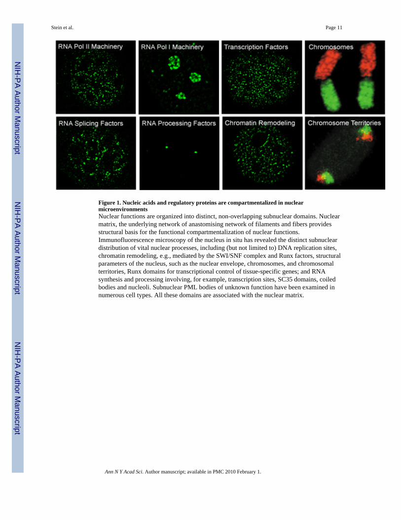
Figure 1. Nucleic acids and regulatory proteins are compartmentalized in nuclearmicroenvironmentsNuclear functions are organized into distinct, non-overlapping subnuclear domains. Nuclearmatrix, the underlying network of anastomising network of filaments and fibers providesstructural basis for the functional compartmentalization of nuclear functions.Immunofluorescence microscopy of the nucleus in situ has revealed the distinct subnucleardistribution of vital nuclear processes, including (but not limited to) DNA replication sites,chromatin remodeling, e.g., mediated by the SWI/SNF complex and Runx factors, structuralparameters of the nucleus, such as the nuclear envelope, chromosomes, and chromosomalterritories, Runx domains for transcriptional control of tissue-specific genes; and RNAsynthesis and processing involving, for example, transcription sites, SC35 domains, coiledbodies and nucleoli. Subnuclear PML bodies of unknown function have been examined innumerous cell types. All these domains are associated with the nuclear matrix.
Stein et al. Page 11
Ann N Y Acad Sci. Author manuscript; available in PMC 2010 February 1.
NIH
-PA Author Manuscript
NIH
-PA Author Manuscript
NIH
-PA Author Manuscript
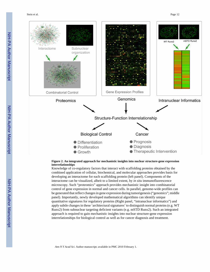
Figure 2. An integrated approach for mechanistic insights into nuclear structure-gene expressioninterrelationshipsKnowledge of co-regulatory factors that interact with scaffolding proteins obtained by thecombined application of cellular, biochemical, and molecular approaches provides basis fordeveloping an interactome for each scaffolding protein (left panel). Components of theinteractome can be visualized, albeit to a limited extent, by in situ immunofluorescencemicroscopy. Such “proteomics” approach provides mechanistic insight into combinatorialcontrol of gene expression in normal and cancer cells. In parallel, genome-wide profiles canbe generated that reflect changes in gene expression during tumorigenesis (“genomics”; middlepanel). Importantly, newly developed mathematical algorithms can identify uniquequantitative signatures for regulatory proteins (Right panel, “intranuclear informatics”) andapply subtle changes in these ‘architectural signatures’ to distinguish normal proteins (e.g. WTRunx2) from subnuclear targeting deficient variants (e.g. mSTD Runx2). Such an integratedapproach is required to gain mechanistic insights into nuclear structure-gene expressioninterrelationships for biological control as well as for cancer diagnosis and treatment.
Stein et al. Page 12
Ann N Y Acad Sci. Author manuscript; available in PMC 2010 February 1.
NIH
-PA Author Manuscript
NIH
-PA Author Manuscript
NIH
-PA Author Manuscript
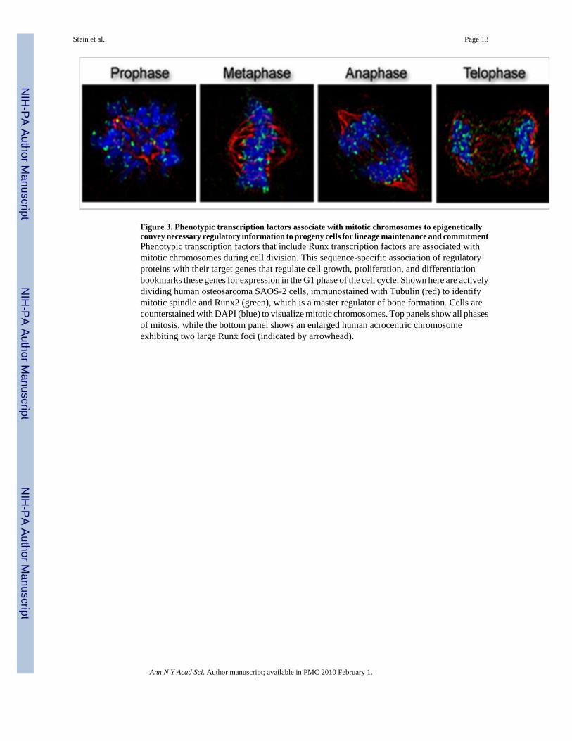
Figure 3. Phenotypic transcription factors associate with mitotic chromosomes to epigeneticallyconvey necessary regulatory information to progeny cells for lineage maintenance and commitmentPhenotypic transcription factors that include Runx transcription factors are associated withmitotic chromosomes during cell division. This sequence-specific association of regulatoryproteins with their target genes that regulate cell growth, proliferation, and differentiationbookmarks these genes for expression in the G1 phase of the cell cycle. Shown here are activelydividing human osteosarcoma SAOS-2 cells, immunostained with Tubulin (red) to identifymitotic spindle and Runx2 (green), which is a master regulator of bone formation. Cells arecounterstained with DAPI (blue) to visualize mitotic chromosomes. Top panels show all phasesof mitosis, while the bottom panel shows an enlarged human acrocentric chromosomeexhibiting two large Runx foci (indicated by arrowhead).
Stein et al. Page 13
Ann N Y Acad Sci. Author manuscript; available in PMC 2010 February 1.
NIH
-PA Author Manuscript
NIH
-PA Author Manuscript
NIH
-PA Author Manuscript