Estimation of the pressure gradient across the fetal ductus venosus based on doppler velocimetry
Upload
independentCategory
view
3download
0
OFFICIAL PUBLICATION OF THE IRANIAN HEART ASSOCIATION
Volume 9, Number 2
Tabestan 1387, Summer2008
CONTENTS: ORIGINAL ARTICLES: CLINICAL SCIENCE
□ Aortic Arch Replacement Using Selective Cerebral Perfusion: Three Years’ Experience
Saeid Hosseini, Mehdi Hadadzadeh, Mohammad Baqer Tabatabaee, and Alireza Alizadeh Ghavidel
6
□ Predictors of Postoperative Atrial Fibrillation after Heart Valve Surgery Hossein Ali Bassiri MD, Khadijeh Ghanbarian MD and Majid Haghjoo MD
10
□ Effect of Preoperative Aspirin Use on Postoperative Bleeding and Perioperative Myocardial Infarction in Patients Undergoing Coronary Artery Bypass Surgery
Mohammad Hassan Ghaffarinejad MD, Amir Farjam Fazelifar MD and Shahram Mohajer Shirvani MD
18
□ Oral Ibuprofen Therapy for Patent Ductus Arteriosus in Very Low Birth Weight Infants Fatemeh Haji Ebrahim Tehrani MD, Hadi Kazemi MD, and Saied Mojtahedzadeh MD
23
□ Prediction of Left Ventricular Dysfunction on Basis of Ventricular Depolarization Time and Electrical Axis in Patients with Left Bundle Branch Block
Farzad Jalali MD, Seyyed Mohammad Miri MD and Pegah Karimi Elizei
29
□ Risk Factors for Silent Myocardial Ischemia in Type II Diabetic Patients Afsaneh Forood MD and Mohammad Masomi MD
37
□ Applying the Logistic Regression Model to Predict the Stenosis in Carotid Artery Using the Sequential Color Doppler Ultrasound Image Processing
M. Mokhtari-Dizaji PhD, P. Abdolmalek PhD, H. Saberi MD and T. Rahmani MSc
43
CASE REPORT
□ Interesting Presentation of Aberrant Origin of the Right Subclavian Artery in an 8-Year-Old Child M. Hasan Kalantar Motamedi MD, Ali Hemmat MD and Pooya Kalani, MD
51
□ Aortic Aneurysm in Takayasu’s Syndrome Ali Sadeghpour Tabaee MD, Shahryar Mali MD, Jalal Vahedian MD and Soheila Arefi MD
55
□ Anomalous Origin of Left Anterior Descending Coronary Artery from Right Coronary Artery Associated with Hypertrophic Cardiomyopathy
M. Ebrahimi, M. Dargahy and S. Bajouri
59
□ Papillary Fibroelastoma of the Tricuspid Valve: Case Report and Review of the Literature Maryam Esmaeilzadeh MD, Maryam Moshkani Farahani MD and Mohammad Jafar Hashemi MD
62
FORTHCOMING MEETINGS
65
INSTRUCTIONS FOR AUTHORS 76
SUBSCRIPTION FORM 79

Aortic Arch Replacement Using Selective Cerebral Perfusion:
Three Years’ Experience
Saeid Hosseini MD, Mehdi Hadadzadeh MD, Mohammad Baqer Tabatabaee MS, and Alireza Alizadeh Ghavidel MD
Abstract
Background- The present study was conducted to report our clinical experience with aortic arch
replacement using selective cerebral perfusion (SCP) to evaluate the safety and usefulness of this technique.
Methods- From October 2003 to April 2007, 10 patients (mean age 51.2 years) underwent arch replacement for acute type A dissection involving the aortic arch. Operations were performed with hypothermic cardiopulmonary bypass using antegrade selective cerebral perfusion during the arch surgery. Seven patients (70%) have a history of hypertension. Six patients (60%) underwent total arch replacement and the other four (40%) had semiarch replacement. Associated coronary artery bypass graft surgery (CABG) was performed in 2 patients (20%). The mean follow-up period was 10.39 months (ranging from 1 to 42 months).
Results- Mean aortic cross-clamp time, CPB time and partial circulatory arrest time with antegrade cerebral perfusion were 121.4 (95-165), 257.7 (230-290) and 16.5 (13-22) minutes, respectively. There were two hospital mortalities and one cerebral complication. All in-hospital mortalities were in our five first cases, indicating perhaps a learning curve for this operation. During the follow-up period, no patient underwent reoperation because of recurrence of dissection. All surviving patients are still alive and free from any serious events at the time of this writing.
Conclusions- Selective cerebral perfusion is a reliable technique for cerebral protection and it facilitates the complex and time-consuming total arch replacement (Iranian Heart Journal 2008; 9 (2):6-9).
Key words: aortic arch surgery ■ selective cerebral perfusion
Cerebral protection is one of the most important concerns during aortic arch repair, and various methods to accomplish it have
been introduced. Deep hypothermia with circulatory arrest (DHCA) is a well-established technique; it provides both good cerebral protection, even though time-limited, and a dry operative field.1-5 However, it requires prolonged cardiopulmonary bypass (CPB) time and is often associated with coagulopathy and pulmonary complications.
Retrograde cerebral perfusion has been introduced to improve cerebral protection and to prolong the "safe" time of circulatory arrest, though the mechanisms of the protective effect are not entirely understood.6-9
Moreover, the complications resulting from deep hypothermia remain largely unchanged with this method. In 2003, we began using antegrade selective cerebral perfusion (SCP) with moderate hypothermia during aortic arch operations.
Received May 8, 2008; Accepted for publication Aug. 21, 2008. From the Dept. of Cardiovascular Surgery and Cardiology, Shaheed Rajaee Cardiovascular Medical Center, Mellat Park, Vali Asr Ave. Tehran, Iran; Correspondence to: S. Hosseini, MD, Dept. of Cardiovascular Surgery, Shaheed Rajaee Cardiovascular Medical Center, Mellat Park, Vali Asr Ave. Tehran, Iran Tel : +982123922589

Here we present our experience with this method in 10 consecutive patients.
Methods
From October 2003 to April 2007, 10 consecutive patients underwent surgical treatment of type A aortic dissection involving the aortic arch with the intimal tear extending into the transverse aortic arch. There were eight men (80%) and two women (20%) with a mean age of 51.2 ± 10.02 years (± SD, range 36 to 66 years). All patients had emergency operation. Associated disease included hupertension in seven patients (70%), coronary artery disease in two (20%) and chronic renal dysfunction (defined as a serum creatinine level exceeding 2 mg/dL) in one (10%). Operative techniques Median sternotomy incision was used. The aorta, the epiaortic vessels and the innominate vein (wich was always preserved) were exposed. After systemic heparinization, the arterial cannula was inserted into one of the femoral arteries (selection based on preoperative CT scan or angiography), and a single two-stage venous cannula was placed in the right atrium. After the innominate artery (IA) was exposed from its origin to the biforcation, IA was directly cannulated using a 16F side-holes cannula and connected by Y connector to the femoral artery cannula line and CPB was established. After cross clamping of the aorta, the left side of the heart was vented through the right superior
pulmonary vein. Myocardial protection was provided with cold crystalloid cardioplegia and topical cooling. As the patients were cooled to a nasopharyngeal temperature of 22° to 25°C, the IA was gently clamped, the
systemic circulation was arrested and pump flow to IA was started with 5ml/(kg min) and adjusted to maintain a right radial artery pressure of 40-70 mmHg. Brain perfusate was maintained at the temprature of 20°c. Left common carotid and left subclavian arteries
were occluded with a foley catheter. Graft replacement was used for aortic reconstruction in all patients. With respect to the extent of dissection, six patients (60%) underwent total arch replacement and the other four (40%) received semiarch replacement. Composite graft implantation (modified Bentall procedure) was performed in three patients (30%) and coronary artery bypass grafting in two patients (20%). Cardiopulmonary bypass data The mean CPB time was 257.7±19.4 minutes (range: 230 to 290 minutes), and the mean aortic cross-clamp time was 121.4 ± 20.6 minutes (range: 95 to 165 minutes). The mean SCP time was 16.5 ± 2 minutes (range: 13 to 22 minutes)
Results There were two in- hospital mortalities. All in hospital mortalities were in our first five cases which could be a reflection of the learning curve for this operation. Causes of death were low cardiac output in both cases. No permanent neurologic dysfunction (PND stroke, coma) occurred in our series, and only one patient presented transient neurologic dysfunction (TND) with complete resolution before discharge. Mean postoperative bleeding in the first 48 h was 620±210cc. No resternotomy for bleeding was required. Mean postoperative hospital stay was 8±4 days. The mean follow-up period was 10.39 months (ranging from 1 to 42 months). During the follow-up period, no patient underwent reoperation because of the recurrence of a dissection . All patients are still alive and free from any serious events at this writing.
Discussion
In the last decades, technical improvements in CPB, myocardial protection, and intensive care have reduced the mortality and the morbidity associated with operations on the aortic arch. Neurologic injuries are the most

feared complications resulting from decreased cerebral circulation. To prevent these complications, various methods have been widely used. Cerebral protection methods currently used are DHCA with or without RCP, and antegrade SCP. Several experimental and clinical studies indicate that antegrade SCP presents several advantages
compared with DHCA with or without RCP. Antegrade SCP can extend the safe duration of circulatory arrest up to 90 minutes5, allowing meticulous aortic arch repair and facilitating the complex and time-consuming TAR. SCP obviates the need for deep
hypothermia, thus reducing pump time and the risk of hypothermia-related complications such as pulmonary insufficiency and coagulopathy. SCP is more effective in supplying oxygenated blood to the brain, thus ensuring a more physiologic brain energy metabolism. SCP is therefore considered to be the most reliable method of preventing
ischemic injury to the brain. Selective cerebral perfusion has considerably prolonged the
"safe" time of circulatory arrest, thereby allowing more complex and time-consuming aortic arch reconstructions. In a series of 100 patients, Kazui and colleagues11 reported only one postoperative stroke, which occurred in a patient with a duration of cerebral perfusion
longer than 90 minutes. In other reports12-14, the incidence of stroke ranged from 3.7% to 10.5%. In the present study, transient neurologic dysfunction occurred in just 1 patients (10%). In the literature, the hospital mortality rate ranged from 0% in the series of Veeragandham and associates13 to 25.2% in that of Hayashi and co-workers.14 In a 1995 study, Kazui and colleagues11 reported a hospital mortality rate of 16.1%. Similar results have been reported using DHCA with
or without retrograde cerebral perfusion.2-8 In our series, we have two mortalities, both of which were in our five first cases, reflecting the effect of learning curve for this operation. In our series, the site of arch vessel cannulation for SCP was the innominate artery because IA cannulation, better than
RAA cannulation, allows the surgeon to perform the entire procedure through the standard sternotomy incision without the need for adjunctive incisions which may be complicated by infections, brachial plexus injuries or vascular compromise. IA cannulation site is always within the surgeon's field of view, with reduced risk of annoying complications such as blood loss in the operative field and/or kinking of the cannules.10 In conclusion, the results of this study are very encouraging. The mortality rate was similar to that obtained with other techniques, and no permanent neurologic deficits occurred. We believe SCP with innominate artery cannulation is an optimal technique of cerebral protection. It extends the "safe" period of circulatory arrest and obviates the problems caused by deep hypothermia. Also the IA cannulation can be performed with a simple, fast and safe technique.
References 1 Pierangeli A., Colì G., Mikus PM. Sostituzione
dell’arco aortico in ipotermia profonda per aneurisma. Bull Scienze Med 1974; 2: 1-16.
2 Pierangeli A, Colì G, Donati A, Galli R, Mikus
PM, Turinetto B. Treatment of aortic arch aneurysm with deep hypothermia and circulatory arrest. J Cardiovasc Surg (Torino) 1975; 16: 409-414.
3 Svensson LG, Crawford ES, Hess KR. Deep
hypothermia with circulatory arrest. Determinants of stroke and early mortality in 656 patients. J Thorac Cardiovasc Surg 1993; 106: 19-31.
4 Crawford ES, Svensson LG, Coselli JS, Safi HJ,
Hess KR. Surgical treatment of aneurysm and/or dissection of the ascending aorta, transverse aortic arch, and ascending aorta and transverse aortic arch. Factors influencing survival in 717 patients. J Thorac Cardiovasc Surg 1989; 98: 659-674.
5 Ergin MA, Galla JD, Lansman SL, Quintana C,
Bodian C, Griepp R. Hypothermic circulatory arrest in operations on the thoracic aorta. J Thorac Cardiovasc Surg 1994; 107: 788-799.

6 Ueda Y, Miki S, Kusuhara K, Okita Y, Tahata T, Yamanaka K. Surgical treatment of aneurysm or dissection involving the ascending aorta and aortic arch, utilizing circulatory arrest and retrograde cerebral perfusion. J Cardiovasc Surg (Torino) 1990; 31: 553-558 .
7 Safi HJ, Brien HW, Winter JN. Brain protection
via cerebral retrograde perfusion during aortic arch aneurysm repair. Ann Thorac Surg 1993; 56: 270-276.
8 Yasuura K, Ogawa Y, Okamoto H. Clinical
application of total body retrograde perfusion to operation for aortic dissection. Ann Thorac Surg 1992; 53: 655-658.
9 Pagano D, Boivin CM, Faroqui MH, Bonser RS.
Retrograde perfusion through the superior vena cava perfuses the brain in human beings. J Thorac Cardiovasc Surg 1996; 111: 270-272.
10 Marco D, Michel Ciano, Giuseppe Labrio.
Cannulation of the innominate artery during surgery of the thoracic aorta. Europian J of cardio thoracic surgery 2007; 32: 270-273.
11 Kazui T, Kimura N, Komatsu S. Surgical
treatment of aortic arch aneurysms using selective cerebral perfusion. Experience with 100 patients. Eur J Cardio-thorac Surg 1995; 9: 491-495.
12 Bachet J, Guilmet D, Goudot B. Cold
cerebroplegia. A new technique of cerebral protection during operations on the transverse aortic arch. J Thorac Cardiovasc Surg 1991; 102: 85-94.
13 Veeragandham RS, Hamilton IN, Jr, O’Connor C,
Rizzo V, Najafi H. Experience with antegrade bihemispheric cerebral perfusion in aortic arch operations. Ann Thorac Surg 1998; 66: 493-499.
14 Hayashi JI, Eguchi S, Yasuda K, et al. Aortic
arch operation using selective cerebral perfusion for nondissecting thoracic aneurysm. Ann Thorac Surg 1997; 63: 88-92.
Predictors of Postoperative Atrial Fibrillation after Heart Valve Surgery
Hossein Ali Bassiri MD, Khadijeh Ghanbarian MD
and Majid Haghjoo MD*
Abstract Background- Atrial fibrillation (AF) is the most common complication after cardiac surgery and a
major cause of morbidity and increased cost of care. Suitable treatment and prevention of postoperative AF are important for patients’ improved health and rehabilitation. This study evaluates the risk factors of paroxysmal AF in patients who underwent valvular heart surgery.
Method- Between April and October 2006, 392 patients who underwent heart valve surgery at our center were included in this prospective study. All relevant clinical, echocardiographic, and laboratory data were gathered in all the patients.
Results- Postoperative AF occurred in 52 (13.3%) patients. In the univariate analysis, the presence of aortic valve disease, mitral valve disease, dyslipidemia, preoperative digoxin consumption, postoperative adrenergic use, intra-aortic balloon pump (IABP) insertion in post-surgery intensive care unit, and large left atrium were significantly associated with the occurrence of postoperative AF (all P<0.05). However, in the stepwise logistic regression model, dyslipidemia (OR: 2.39, 95% CI: 1.12-5.09, P=0.020), left atrium dimension (OR: 0.12, 95% CI: 0.76-0.28, P<0.001), IABP (OR: 7.10, 95% CI: 1.98-25.47, P=0.001), preoperative digoxin use (OR: 2.55, 95% CI: 1.38-4.71, P=0.002), postoperative adrenergic use (OR:3.70, 95% CI: 1.77-7.73, P<0.001), aortic valve replacement (OR:0.38, 95% CI: 0.20-0.69, P=0.0001), and mitral valve replacement (OR:3.53, 95% CI: 1.75-7.10, P<0.001) remained independently predictive of postoperative AF.
Conclusions- The result of this study showed that dyslipidemia, left atrium dimension, mitral valve replacement, aortic valve replacement, IABP, and adrenergic use in ICU and digoxin use preoperatively were the independent predictors of AF after valvular surgery. Therefore, clinical data and echocardiography may be useful in preoperative risk stratification of high-risk patients for the occurrence of postoperative AF(Iranian Heart Journal 2008; 9 (2):10-17).
Key words: atrial fibrillation ■ postoperative arrhythmia ■ heart valve surger
Atrial fibrillation (AF) is one of the most common complications after cardiac surgery.1,2 The incidence of arrhythmia has not changed despite improvements in anesthetic and surgical techniques, and evidence suggests its incidence may be increasing.3
According to previous publications, it occurs in 10 to 65% of patients after cardiac surgery.1-8 The rate of AF after cardiac surgery in 1970 was about 10% and is now consistently at least 30% and much higher in that undergoing heart valve surgery.
Received May 8, 2008; Accepted for publication Aug. 21, 2008. From the Department of Cardiology and *Department of Cardiac Pacing and Electrophysiology, Shaheed Rajaie Cardiovascular Medical and Research Center, Tehran, Iran Correspondence and reprint requests to: Majid Haghjoo, MD, Department of Pacemaker and Electrophysiology, Shaheed Rajaie Cardiovascular Medical and Research Center, Mellat Park, Vali-E-Asr Avenue, P.O. Box 15745-1341, Tehran 1996911151, Iran. Tel: 0098-21-23922931 Fax: 0098-21-22048174 Email: [email protected]

Although AF is considered a serious problem, acknowledgement of AF as a potentially serious arrhythmia has recently increased. AF usually occurs 2-4 days after surgery1,3 and often returns during the first 30 days of the postoperative period.1 In a minority of cases, it may result in inappropriate tachycardia, hypotension, heart failure, and a possible increase in the risk of cerebrovascular accidents.3 Methods Between April 2006 and October 2006, 392 consecutive patients who were scheduled to undergo valvular heart surgery were included in our study. The study was approved by the local ethics committee, and written informed consent was obtained from all the patients. Previous history of AF or atrial flutter rhythm, use of antiarrhythmic drugs other than beta-blockers, uncontrolled heart failure, end-stage renal disease, and presence of an implanted pacemaker were the exclusion criteria. Patients were also excluded if they underwent any operation other than heart valve surgery or if sustained ventricular tachyarrhythmia, or cardiogenic shock, or death in the operating room occurred. For each patient, a form including data related to the preoperative and postoperative periods was completed. A standard 12-lead ECG, transthoracic echocardiography, laboratory tests, and blood pressure measurement were performed in all the patients. A careful medical history including sex, age, risk factors (hypertension, diabetes, dyslipidemia, and cigarette smoking), drug history (antiarrhythmics, anticoagulants, and antiplatelet agents), history of previous cardiac surgery (valvular or non-valvular) was taken, and echocardiographic data including left ventricular end-diastolic diameter (LVEDD), left ventricular end-systolic diameter (LVESD), and left atrial diameter (LAD) were registered. In the postoperative state, the patients were followed first in the ICU for at least 3 days and then in the
surgical wards. The type of surgery (AVR, MVR, TVR, PVR, and multivalvular), duration of ICU admission, use of IABP and adrenergic drugs, and BUN/Cr status were registered. Postoperative care After the operation, the patients were followed-up in the ICU and were weaned off the ventilator when they fulfilled the following criteria: hemodynamic stability, peripheral temperature >32°C, cooperativity, and no major bleeding. Chest drains were removed on the first postoperative day, and the patients were moved to the surgical ward. All the patients were continuously monitored postoperatively during the ICU stay. After transfer to the ward, all the patients were connected to monitors for continuous ECG monitoring up to the fifth postoperative day. The ward monitor stored the ECG recordings for subsequent analyses. The recordings were analyzed off-line. A 12-lead ECG recording was done, if necessary, to confirm AF episodes. One electrophysiologist and one cardiology fellow who were blinded to other data reviewed these data on a daily basis. Preoperative beta-blockers, calcium channel blockers, and digoxin were continued for the entire hospital stay. The endpoint of the study was the occurrence of the new-onset AF during the first five days following valvular surgery. AF was defined as absent P waves before the QRS complex, together with irregular ventricular rhythm on the rhythm strips. Only AF episodes lasting longer than 5 minutes were counted. Abnormal P-wave morphology is defined as P-wave duration of more than 110 ms with inter-peak notch of more than 40 ms and duration of terminal negative P-wave deflection in lead V1 of more than 40 ms. Statistical analysis All the continuous variables are presented as mean±SD. The other variables are presented in the percentage of population having a specific value. We tested the association of
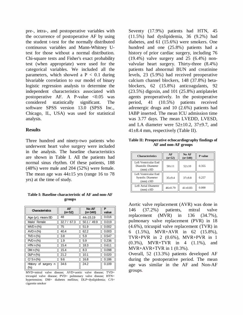
pre-, intra-, and postoperative variables with the occurrence of postoperative AF by using the student t-test for the normally distributed continuous variables and Mann-Whitney U-test for those without a normal distribution. Chi-square tests and Fisher's exact probability test (when appropriate) were used for the categorical variables. We included all the parameters, which showed a P < 0.1 during bivariable correlation to our model of binary logistic regression analysis to determine the independent characteristics associated with postoperative AF. A P-value <0.05 was considered statistically significant. The software SPSS version 13.0 (SPSS Inc., Chicago, IL, USA) was used for statistical analysis. Results Three hundred and ninety-two patients who underwent heart valve surgery were included in the analysis. The baseline characteristics are shown in Table I. All the patients had normal sinus rhythm. Of these patients, 188 (48%) were male and 204 (52%) were female. The mean age was 44±15 yrs (range 16 to 78 yrs) at the time of study.
Table I: Baseline characteristic of AF and non-AF
groups
Characteristics AF (n=52)
No AF (n=340)
P value
Age (yr); mean±SD 48 44±15.19 0.016
Male/ Female 32.7 / 67.3 50.1 / 49.9 0.019 MVD n (%) 75 51.9 0.002 AVD n (%) 40.4 62.2 0.003 TVD n (%) 3.8 5.9 0.547 PVD n (%) 1.9 5.9 0.236 HTN n (%) 15.4 18.3 0.611 DM n (%) 15.4 8.3 0.098 DLP n (%) 21.2 10.1 0.020 C/S n (%) 9.6 16.8 0.186 History of surgery n (%)
34.6 24.2 0.109
MVD=mitral valve disease; AVD=aortic valve disease; TVD= tricuspid valve disease; PVD= pulmonary valve disease; HTN= hypertension; DM= diabetes mellitus; DLP=dyslipidemia; C/S= cigarette smoker
Seventy (17.9%) patients had HTN, 45 (11.5%) had dyslipidemia, 36 (9.2%) had diabetes, and 61 (15.6%) were smokers. One hundred and one (25.8%) patients had a history of prior cardiac surgery, including 76 (19.4%) valve surgery and 25 (6.4%) non-valvular heart surgery. Thirty-three (8.4%) patients had abnormal BUN and creatinine levels, 23 (5.9%) had received preoperative calcium channel blockers, 148 (37.8%) beta-blockers, 62 (15.8%) anticoagulants, 92 (23.5%) digoxin, and 101 (25.8%) antiplatelet agents preoperatively. In the postoperative period, 41 (10.5%) patients received adrenergic drugs and 10 (2.6%) patients had IABP inserted. The mean ICU admission time was 3.77 days. The mean LVEDD, LVESD, and LA diameter were 52±10.2, 37±9.7, and 41±8.4 mm, respectively (Table II). Table II: Preoperative echocardiography findings of
AF and non-AF groups
Characteristics AF (n=52)
No AF (n=340) P value
Left Ventricular End Diastolic Diameter
(mm) ±SD 50±11 52±10 0.355
Left Ventricular End Systolic Diameter
(mm) ±SD 35±9.4 37±9.8 0.257
Left Atrial Diameter (mm) ±SD 46±0.79 41±0.83 0.000
Aortic valve replacement (AVR) was done in 146 (37.2%) patients, mitral valve replacement (MVR) in 136 (34.7%), pulmonary valve replacement (PVR) in 18 (4.6%), tricuspid valve replacement (TVR) in 6 (1.5%), MVR+AVR in 62 (15.8%), TVR+PVR in 2 (0.6%), MVR+PVR in 1 (0.3%), MVR+TVR in 4 (1.1%), and MVR+AVR+TVR in 1 (0.3%). Overall, 52 (13.3%) patients developed AF during the postoperative period. The mean age was similar in the AF and Non-AF groups.
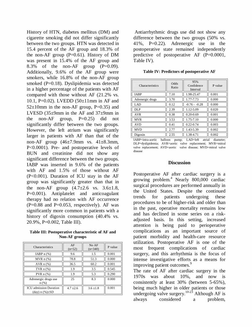
History of HTN, diabetes mellitus (DM) and cigarette smoking did not differ significantly between the two groups. HTN was detected in 15.4 percent of the AF group and 18.3% of the non-AF group (P=0.61). History of DM was present in 15.4% of the AF group and 8.3% of the non-AF group (P=0.09). Additionally, 9.6% of the AF group were smokers, while 16.8% of the non-AF group smoked (P=0.18). Dyslipidemia was detected in a higher percentage of the patients with AF compared with those without AF (21.2% vs. 10.1, P=0.02). LVEDD (50±11mm in AF and 52±10mm in the non-AF group, P=0.35) and LVESD (35±9mm in the AF and 37±9mm in the non-AF group, P=0.25) did not significantly differ between the two groups. However, the left atrium was significantly larger in patients with AF than that of the non-AF group (46±7.9mm vs. 41±8.3mm, P<0.0001). Pre- and postoperative levels of BUN and creatinine did not show any significant difference between the two groups. IABP was inserted in 9.6% of the patients with AF and 1.5% of those without AF (P<0.001). Duration of ICU stay in the AF group was significantly greater than that in the non-AF group (4.7±2.6 vs. 3.6±1.8, P=0.001). Antiplatelet and anticoagulant therapy had no relation with AF occurrence (P=0.88 and P=0.053, respectively). AF was significantly more common in patients with a history of digoxin consumption (40.4% vs. 20.9%, P=0.002, Table III).
Table III: Postoperative characteristic of AF and Non-AF groups
Characteristics AF (n=52)
No AF (n=340) P value
IABP n (%) 9.6 1.5 0.001 MVR n (%) 78.8 51.3 0.000 AVR n (%) 36.5 60.2 0.001 TVR n (%) 1.9 3.5 0.545 PVR n (%) 1.9 5.3 0.290
Adrenergic drugs use n (%)
25 8.3 0.000
ICU admission Duration (day) n (%)±SD
4.7 ±2.6 3.6 ±1.8 0.001
Antiarrhythmic drugs use did not show any difference between the two groups (50% vs. 41%, P=0.22). Adrenergic use in the postoperative state remained independently predictive of postoperative AF (P=0.0001, Table IV).
Table IV: Predictors of postoperative AF
Characteristics Odds Ratio
95% Confidence
Interval P value
IABP 7.10 1.98-25.47 0.001 Adrenergic drugs 3.70 1.77-7.73 0.000 LAD 0.12 -0.76 - -0.28 0.000 DLP 2.39 1.12-5.09 0.020 AVR 0.38 0.20-0.69 0.001 MVR 3.53 1.75-7.10 0.000 AVD 0.41 0.22-0.74 0.003 MVD 2.77 1.43-5.39 0.002 Digoxin 2.55 1.38-4.71 0.002
IABP=intra-aortic balloon pump; LAD=left atrial diameter; DLP=dyslipidemia; AVR=aortic valve replacement; MVR=mitral valve replacement; AVD=aortic valve disease; MVD=mitral valve disease
Discussion
Postoperative AF after cardiac surgery is a growing problem.4 Nearly 800,000 cardiac surgical procedures are performed annually in the United States. Despite the continued trends for patients undergoing these procedures to be of higher-risk and older than in the past, operative mortality remains low and has declined in some series on a risk-adjusted basis. In this setting, increased attention is being paid to perioperative complications as an important source of patient morbidity and health-care resource utilization. Postoperative AF is one of the most frequent complications of cardiac surgery, and this arrhythmia is the focus of intense investigative efforts as a means for improving patient outcomes.9 The rate of AF after cardiac surgery in the 1970s was about 10%, and now is consistently at least 30% (between 5-65%), being much higher in older patients or those undergoing valve surgery.10-23 Although AF is always considered a problem,

acknowledgement of AF as a potentially serious arrhythmia has increased; there have been more than 100 trials, multiple metaanalyses, and three sets of practice guidelines for the prevention of postoperative AF in cardiac surgery.4,5 Although reports5,14 indicate that AF occurs within four days1,2,5 postoperatively, it can occur at any point in the recovery period. According to Steven et al.,6 AF is a common complication after MVR surgery, occurring in one of four patients without a prior history of AF and in sinus rhythm at surgery. In addition, early AF (within the first 2 weeks after operation) occurs more frequently after MVR than repair, and is associated with a high late recurrence rate.5
Although outpatient monitoring with cardiac event recording is useful in detecting asymptomatic episodes of AF, monitoring all patients after discharge may not be cost effective.2 Other investigators13-21,22-35 have evaluated risk factors for postoperative AF. Age over 65 yrs, history of intermittent AF, use of atrial pacing in the postoperative period, male sex,10 white race, IABP, 9 and not having hyperlipidemia were independent predictors of AF.2 However, others have found that HTN,3 left atrial dimension,2,6,9 creatinine clearance,1,36 postoperative withdrawal of beta-blockers, chronic obstructive pulmonary disease, history of myocardial infarction, history of cardiopulmonary bypass, cross-clamp times, postoperative respiratory compromise,2,9 mechanical ventilation more than 24 hours,9 and intraoperative and postoperative application of adrenergics1 are significantly associated with postoperative AF. We excluded patients with a history of AF in our study. In previous investigations,27-29 patients with a history of AF were excluded because they were expected to be at greater risk. Other researchers13,33,37 have also found that patients with a history of AF are at an increased risk for postoperative AF. Mathew et al.29 found that a history of AF increased the risk of AF in the postoperative state
approximately 2-fold, and Margorine et al.2 showed this risk to be about 6-fold. In contrast, Deliargyris et al.4 reported that postoperative AF was 19 times more likely in patients with a history of AF than in those without such a history. Male sex has inconsistently been associated with postoperative AF. Some researchers50, 54,
56 have found that being male is associated with AF, whereas others14, 25 have not. Our study chimed in with the latter. Creswell et al.12 reported a significant relationship between real ethnicity and postoperative AF. Our study showed that dyslipidemia is an independent risk factor. Marjorie et al.2 suggested that not having hyperlipidemia was an independent predictor of postoperative AF. No previous investigators have examined the presence or absence of hyperlipidemia as a predictor of AF. In addition, a double-blind study showed that prophylactic treatment with atorvastatin significantly lowered the incidence of AF after open heart surgery. The development of AF after cardiac surgery results in a longer stay in the ICU and in the hospital, together with a significantly higher (two-to three-fold) risk of postoperative stroke.38-40 Postoperative AF has also been shown to predict postoperative delirium and neuro-cognitive decline.4,41,42 Increasing age is the most consistent predictor of postoperative AF.2,4,21,23-35 Age-related changes in the atria such as dilation, muscle atrophy, and decreased conduction may explain the strong association. Some authors have reported an increasing incidence of AF in recent years,12 which may be attributed, at least in part, to the frequent use of continuous postoperative rhythm monitoring, the rapid improvement of anesthesia and surgical technology, and major advances in the practice of percutaneous coronary revascularization procedures, resulting in the referral of significantly older and sicker patients to cardiac surgery compared to patients referred for open-heart surgical procedures 10 years ago.

Since increasing age has been a consistent independent predictor for AF after cardiac surgical procedures2,12 referral of older patients for open-heart surgery results in a higher incidence of AF postoperatively.2 Twenty-nine trials have evaluated the length of stay,4 and three trials5 have tested multiple interventions. Only amiodarone and pacing had a significant effect on the length of stay.4,43 Also, amiodarone was the only single intervention that showed a significantly reduced stroke rate, Ninety-four trials of prevention of postoperative AF have been identified by standard search methods and analyzed by standard meta-analysis techniques. All five commonly tested interventions, beta-blockers, sotalol, amiodarone, magnesium, and atrial pacing were effective in preventing AF.4 Despite the existence of unique guidelines from the American Heart Association, European Society of Cardiology, and American College of Cardiology, there are still doubts as to the selection of the best antiarrhythmic drugs, timing of therapy, duration of treatment, and prevention of renewed occurrence.1 Similar to prior reports, we found a significant relation between postoperative AF and postoperative adrenergic use. Salaria et al.36,44 investigated the influence of postoperative adrenergic use in 199 patients after cardiac surgery. These investigators showed that adrenergic use was an independent predictor of postoperative AF (OR 3.35, 95% CI: 1.38-8.12, P=0.016). Our study showed dyslipidemia as an independent predictor of postoperative AF (OR 2.39, 95% CI: 1.12-5.09). Recent studies in widely varied populations emphasize the role of left atrial size as a major marker of adverse cardiovascular events.6,44 Left atrial dimension was a predictor of postoperative AF in our study. Ascher et al. attributed the greater susceptibility to AF after valve surgery to structural and hemodynamic abnormalities, such as left atrial enlargement and pathologic changes in the atria.3,9
Conclusions
The results of the present study demonstrated that IABP, postoperative adrenergic use, left atrial dimension, dyslipidemia, AVR, MVR, mitral valve disease, aortic valve disease, and digoxin use preoperatively were independent predictors of AF after valvular surgery. Therefore, clinical data, discontinuation of digoxin, and treatment of dyslipidemia may be useful in the preoperative risk stratification of high-risk patients for the occurrence of AF.
References
1. Banach M, Goch A, Misztal M, Rysz J, Jaszewski
R, Goch H. Predictors of paroxysmal AF in patients undergoing aortic valve replacement. J Thorac cardiovas Surg 2007; 134: 1569-1576.
2. Funk M, Richards S, Desjardins J, Bebon C,
Wilcox H. Incidence, timing, symptoms, and risk factors for AF after cardiac surgery. Am J Critical Care 2003; 12: 424-433.
3. Auer J, Weber T, Berent R, Keung C, Lamm G,
Eber B. Risk factors of postoperative AF after cardiac surgery. J Card Surg 2005; 2: 425-431.
4. David C, Michael J, Anthony C. Interventions for
prevention of postoperative AF and its complications after cardiac surgery: a meta-analysis. Eur Heart J 2006; 27: 2846-2857.
5. Kerstein J, Soodan A, Qamar M, Majid M,
Lichstein E, Hollander G, et al. Giving IV and oral amiodarone perioperatively for the prevention of postoperative AF in patients undergoing coronary artery bypass surgery. Chest 2004; 126: 716-724.
6. Steven J, Vuyisile T, David M, Bernard J, Thoralf
M, Christofer G, et al. AF after surgical correction of mitral regurgitation in sinus rhythm. Circulation 2004; 110: 2320-2325.
7. Emile G, Adam S, Chingman D, Rajiva G, Deeb
M, Booling S, et al. Preoperative amiodarone as prophylaxis against AF after heart surgery. NEJM 1997; 337: 1785-1791.
8. Fuster V, Wayne A, O'Rourke R(eds.) Atrial
fibrillation. Hurst’s the Heart 2004; Ch 29: 825-8 .

9. Charles W, Hogue J, Lawrence L, David D, et al Epidemiology, mechanisms and risks. American College of Chest Physician guidelines for the prevention and management of postoperative AF after cardiac surgery. Chest 2005; 128; 615-645.
10. Joel D, Peter M. Are the American College of
Chest Physicians guidelines for the prevention and management of AF after cardiac surgery already obsolete? Chest 2006; 129: 1112-1113.
11. Mathew J, Fontes M, Tudor I, Ramsay J, Duke P,
Mazer D. et al. Investigators of the ischemic research and education foundation. JAMA 2004; 291: 1720-1729.
12. Creswell L, Schuessler R, Rosenbloon M.
Hazards of postoperative atrial arrhythmias. Ann Thorac Surg 1993; 56: 539-549.
13. Borzak S, Tisdale J, Amin N, Goldberg D, Frank
D. AF after bypass surgery: does the arrhythmia or the characteristics of the patients prolong hospital stay? Chest 1998; 113: 1489-1491.
14. Cagli K, Keles T. Risk factors associated with
development of AF early after coronary artery bypass grafting. Am J Cardiol 2000; 85: 1259-1261.
15. Quader M, McCarthy P, Gillinov A, Alster J.
Does preoperative AF reduce survival after coronary artery bypass grafting? Ann Thorac Surg 2004; 77: 1514-1522.
16. Deliargyris E, Raymond R, Guzzo J. Preoperative
factors predisposing to early postoperative AF after isolated coronary artery bypass grafting. Am J Cardiol 2000; 85: 763-4.
17. Jideus L, Blomstorm P, Nilsson L, Stridsberg M.
Tachyarrhythmias and triggering factors for AF after coronary artery bypass operation. Ann Thorac Surg 2000; 69: 1064-1069.
18. Kalman J, Muawar M, Howes L, Louis w,
Buxton B, Gutteridge G, et al. AF after coronary artery bypass operation is associated with sympathetic activation. Ann Thorac Surg 1995; 60: 1709-1715.
19. Mathew J, Fontes M, Tudor I, Ramsay J, Duck P,
Mazer D, et al. A multicenter risk index for AF after cardiac surgery. JAMA 2004; 291: 1720-1729.
20. Passman R, Beshai J, Pavri B, Kimmel S. Predicting post-coronary bypass surgery atrial arrhythmia from the preoperative ECG. Am Heart J 2001; 142: 806-810.
21. Skubas N, Brazilia B, Hogue C. AF after
coronary artery bypass graft surgery is unrelated to cardiac abnormalities detected by TEE. Anesth Analg 2001; 93: 14-19.
22. Tamis J, Steinberg J. AF independently prolongs
hospital stay after coronary artery bypass surgery. Clin Cardiol 2000; 23: 155-159.
23. Aranki S, Shaw D, Adams D, Rizzo R, Couper G,
Vandervliet M, et al. Predictors of AF after coronary artery surgery: Current trends and impact on hospital resources. Circulation 1996; 94: 390-397.
24. Asher C, Miller D, Grimm R, Cosgrow D.
Analysis of risk factors for development of AF early after cardiac valvular surgery. Am J Cardiol 1948; 82: 892-895.
25. Crosby L, Pifalo W, Woll K, Burkholder J. risk
factors for AF after coronary artery bypass grafting. Am J Cardiol 1990; 66: 1520-1522.
26. Dimmer C, Tavernier R, Gjorgou N, Vannootern
G. Variations of autonomic tone preceding onset of AF after coronary artery bypass grafting. Am J Cardiol 1998; 82: 22-25.
27. Fuller J, Adams G, Buxton B. AF after coronary
artery bypass grafting: is it a disorder of the elderly? J Thorac Cardiovas Surg 1989; 97: 821-825.
28. Leitch J, Thomson D, Baird D, Harris P. The
importance of age as a predictor of AF and flutter after coronary artery bypass grafting. J. Thorac Cardiovasc Surg 1990; 100: 338-342.
29. Mathew J, Parks P, Savino J. AF following
coronary artery bypass graft surgery: predictors, outcomes, and resource utilization. JAMA 1996; 276: 300-306.
30. Stamou S, Dangas G, Hill P. AF after beating
heart surgery. Am J Cardiol 2000; 86:64-67. 31. Harvank M, Hoffman L, Saal M, Zullo T.
Predictors and impact of AF after isolated coronary artery bypass grafting. Crit Care Med 2002; 30: 330-337.

32. Almassi G, Schwalter T, Nicolosi A. AF after cardiac surgery: a major morbid event? Ann Surg 1997; 226: 501-510.
33. Duceschi V, D’Andreg A, Liccardo B.
Perioperative clinical predictors of AF occurrence following coronary artery surgery. Eur J Cardiothorac Surg 1999; 16: 435-439.
34. Frost L, Molgaard H, Christiansen E, Jacobsen C,
Allermand H. Low vagal tone and supraventricular ectopic activity predict AF and flutter after coronary artery bypass grafting. Eur Heart J 1995; 16: 825-831.
35. Azfar G, Archbold R, Helft G, Elizabeth A,
Nicholas P, Peter G. AF after coronary artery bypass surgery: a model for preoperative risk stratification. Circulation 2000; 101: 1403-1408.
36. Vikrant S, Nirav J, Syed Abdul-Aziz, Syed M.
Role of postoperative vasopressor use in occurrence of AF after CABG. Am J Cardiol 2005; 95: 247-249.
37. Halonen J, Hakalat T, Auvinen T, Karjalainen J,
Turpeinen A, Unsaro A. et al. Intravenous administration of metoprolol is more effective than oral administration in the prevention of AF after cardiac surgery. Circulation 2006; 114: 1-4.
38. Singer D, Albers G, Dalen G, Go A, Halperin J,
Manning W. Antithrombotic therapy in AF. Chest 2004; 126: 429-456.
39. Villareal R, Hariharan R, Liu B. Postoperative
AF and mortality after coronary artery bypass surgery. J Am Coll Cardiol 2004; 43: 742-748.
40. Reed G, Singer D, Picard E, DeSanctis R. Stroke
following coronary artery bypass surgery. A case control estimate of the risk from carotid bruits. NEJM 1988; 319: 1246-1250.
41. Roach G, Kanchuger M, Mangano C, Newman
M, Nussmeier N, Wolman R, et al. Adverse cerebral outcomes after coronary bypass surgery. NEJM 1996; 335: 1857-1863.
42. Bucerius J, Gummert J, Borger M. Walther T.
Predictors of delirium after cardiac surgery: Effect of beating-heart (off-pump) surgery. J Thorac Cardiovasc Surg 2004; 127: 57-64.
43. Michael H, Michael D, Morady F, Buckman D, Lucille R, Hallock R, et al. Effect of postoperative AF on length of stay after cardiac surgery (PACS2). Am J Cardiol 2001; 87: 881-885.
44. Haghioo, M, Bassiri H, Salek M, Sadr Ameli M,
Kargar F, Raissi K, et al. Predictors of postoperative AF after coronary artery bypass graft surgery. Indian Pacing Electrophysiology 2008; 8 (2): 94-101.

Effect of Preoperative Aspirin Use on Postoperative Bleeding and Perioperative Myocardial Infarction in
Patients Undergoing Coronary Artery Bypass Surgery
Mohammad Hassan Ghaffarinejad MD, Amir Farjam Fazelifar MD,* Shahram Mohajer Shirvani MD,* Esmaeel Asdaghpoor MD,
Farzad Fazeli MD and Freidoun Noohi MD*
Abstract Background- Continuation or discontinuation of aspirin use in the preoperative period for patients
scheduled for elective cardiac surgery has continued to be controversial. In this study, we tried to evaluate clinical outcomes (mortality, postoperative bleeding and perioperative myocardial infarction) in patients who underwent first elective coronary artery bypass grafting and received aspirin during the preoperative period.
Methods- The study was a prospective, randomized and single-blinded clinical trial. Two-hundred patients were included in the study and divided into two groups. One group received aspirin 80-160 mg and in the other group, aspirin was stopped at least for seven days before operation. The primary end points of the study were in-hospital mortality rate and hemorrhage-related complications (postoperative blood loss in the intensive care unit, reexploration for bleeding and red blood cell and non-red blood cell transfusion requirements). The secondary end point was perioperative myocardial infarction.
Results- There were no differences in patients’ characteristics among aspirin users and non-aspirin users. We found a significant difference between postoperative blood loss (608±359.7 ml vs. 483±251.5 ml; P=0.005) and red blood cell product requirements (1.32±0.97 units packed cells vs. 0.94±1.02 units packed cells; P=0.008) in the two groups. There was no significant difference between the two groups regarding platelet requirements and the rate of in-hospital mortality and reexploration for bleeding. Similarly, we found no significant difference in the incidence of definite and probable perioperative myocardial infarction (P=0.24 and P=0.56, respectively) and in-hospital mortality between the two groups.
Conclusion- Preoperative aspirin administration increased postoperative bleeding and red blood cell requirements with no effect on mortality, reexploration rate and perioperative myocardial infarction (Iranian Heart Journal 2008; 9 (2):18-22).
Key words: aspirin ■ postoperative bleeding ■ perioperative myocardial infarction
We designed a prospective, randomized and single-blinded study for evaluation of preoperative aspirin use on in-hospital mortality, postoperative bleeding and perioperative myocardial infarction.
We found that preoperative aspirin use increases postoperative bleeding, red blood cell and fresh frozen plasma requirements, without a beneficial effect on perioperative myocardial infarction.
Received May 8, 2008; Accepted for publication Aug. 21, 2008. From the Department of Cardiovascular Surgery and *Cardiology, Shaheed Rajaie Cardiovascular Medical Center, Iran University of Medical Sciences. Tehran, Iran. Address for correspondence: Mohammad Hassan Ghaffarinejad, MD , Dept. of Cardiovascular Surgery, Shaheed Rajaie Cardiovascular Medical Center, Iran University of Medical Sciences. Mellat Park, Vali-E-Asr Avenue, Tehran, 1996911151, P.O.Box:15745-1341, Iran. Tel: 0098 21 23922931 Fax: 009821-88784618 Email: [email protected]
Aspirin is an effective therapy in the management of stable and unstable coronary artery diseases.1 Early initiation of aspirin after coronary artery bypass graft (CABG) surgery reduces risk of graft occlusion.2 Aspirin has been implicated in platelet dysfunction and prolongation of bleeding time but its effect on postoperative bleeding, reexploration and blood products requirements is controversial.3-12 In this study we tried to determine effect of preoperative aspirin use on in-hospital mortality, postoperative bleeding, blood transfusion requirements and perioperative myocardial infarction (MI).
Methods We conducted a prospective study on two-hundred patients (67 male aspirin users vs. 70 male non-aspirin users, P=0.761, mean age 56.9±9.14 years in aspirin users vs. 56.9±9.59 years in non-aspirin users, P=0.83), who underwent CABG surgery in our department between November 2003 and December 2004. We received ethical approval for the study and the patients were enrolled in the study with informed written consent. We included patients who underwent elective CABG for the first time. Our exclusion criteria were: 1) need for concomitant valvular, aortic or aneurysmectomy surgery, 2) concomitant antiplatelet drug consumption (clopidogrel, ticlopidine, glucocorticoids, non-steroidal anti-inflammatory drugs, etc). We routinely used the left internal mammary artery as a conduit, total grafts were less than five and all operations were done by one surgical team. The patients’ characteristics are summarized in Table I. The patients were randomly assigned into one of the two groups: group 1 received aspirin preoperatively and in group 2, aspirin was stopped at least seven days before CABG. All patients received a single dose of aprotinin (2,000,000 units kallikrein inhibitor) once during surgery. Aspirin was started post-
operatively within 6 hours after CABG in the two groups.
Table I. Patients’ characteristics
Variable Aspiri
n group
Non aspirin group
P-valu
e
LV ejection Fraction
41.7±11.6
42.6±11.3
0.69
Cigarette smoking Yes 36 36 No 64 64 1.0
Dyslipidemia Yes 53 44 No 47 56 0.26
Hypertension Yes 40 36 No 60 64 0.66
Left ventricular hypertrophy
Yes 11 15 No 89 85 0.53
Diabetes mellitus Yes 34 23 No 66 77 0.12
Immediate postoperative care of the patients was provided by the cardiac surgery intensive care unit (ICU) staff. Pericardial and pleural chest tube output was monitored frequently within the first few days after surgery and recorded in the patient’s file. Extubated stable patients were transferred to the cardiac surgery step-down unit, usually on the second postoperative day. The date of all transfusions was entered into the hospital central computer from the respective laboratories and this data was available by using the patients’ hospital identification number. The use of red blood products or non-red blood products like fresh frozen plasma (FFP) and platelets was left to the surgical team’s discretion.

Electrocardiograms (ECG) were recorded preoperatively and on the first to fifth days after surgery. Appearance of any new Qs wave in ECGs was recorded in the patient’s file. Cardiac enzyme marker (CK-MB) samples were collected preoperatively and at least five times during the first and second day after CABG. Cardiac enzyme marker more than 30 IU/L was considered for probable myocardial injury during CABG. In all patients two-dimensional echocardiography was performed on the second and fifth day after CABG for detection of new regional wall motion abnormality (RWMA) in patients with new Qs wave on ECG or cardiac enzyme markers more than 30 IU/L. The primary study end points were in- hospital mortality, re-exploration rate, excessive pericardial and pleural tube bleeding and excessive requirements for red blood cells and non-red blood cell product requirements. The study was also extended to evaluate preoperative aspirin use on perioperative myocardial infarction rate. Definitions Definite perioperative myocardial infarction is defined as: new Qs wave on ECG and new RWMA on echo with or without CK-MB >30 IU/L and the definition of probable perioperative myocardial infarction is CK-MB>30 IU/L with new Qs on ECG or new RWMA on echo.13
Statistical analysis Statistical analysis was performed using SPSS® 11.5 (SPSS Inc., Chicago, IL, USA) for data storage and analysis. Continuous data were expressed as mean values ±SD. Comparison of baseline categorical data was done by chi-square and continuous data by standard t-test. In all analyses with a 95% confidence interval (CI), P<0.05 was considered statistically significant.
Results
One hundred (50%) individuals received aspirin and it was discontinued in the other group at least seven days before CABG. Neither the mean age nor sex was significantly different between the two groups. Patient's characteristics are summarized in Table I. Aspirin users had more postoperative bleeding (608±359.7 ml vs. 483±251.5 ml; P=0.005) and were transfused more red blood cell products (1.32±0.97 units packed cells vs. 0.94±1.02 units packed cells; P=0.008) and fresh frozen plasma (2±1.84 vs. 1.46±1.64; P=0.03) early after surgery, although platelet transfusion was not significantly different between groups (0.45±1.32 vs. 0.28±0.84 units platelets, P=0.25). No in-hospital mortality was observed in the groups. Regarding the secondary end points of the study, aspirin users had a significantly lower incidence of new Qs pattern on ECG after CABG (1% vs. 10%, P=0.013), but cardiac enzyme markers (CK-MB) and new regional wall motion abnormality (RWMA) were not different significantly between the two groups. (Table II). There was no significant difference in the incidence of definite or probable perioperative myocardial infarction. Definite MI occurred in 0% of aspirin users vs. 3% of non-aspirin users (P=0.24) and probable MI occurred in 5% in group 1 vs. 8% in group 2, (P=0.56).
Discussion This study indicated that use of aspirin before CABG is associated with a higher risk of postoperative bleeding, with increased requirements for red blood cell products and fresh frozen plasma (FFP) transfusion. This finding was contradictory to other studies that showed patients receiving aspirin were no more likely to receive blood products.7-12
Tuman and coworkers showed preoperative aspirin consumption dose not increase allogeneic blood transfusion in reoperative

coronary artery surgery.7 In another study, Vuylsteke et al. evaluated the effect of aspirin in coronary artery bypass grafting and they showed that aspirin therapy did not appear to increase blood loss, re-sternotomy for bleeding or blood products usage requirements during the hospital stay.8 On the other hand, there are studies that confirm our finding.3-6 Ferraris et al. evaluated aspirin and postoperative bleeding after CABG. Their findings supported the hypothesis that aspirin is associated with a greater likelihood of postoperative bleeding.6
In our study, re-exploration rate for bleeding was 3% in each group, without significant difference (P=NS). Decey et al. found no significant difference in the rate of re-exploration for hemorrhage between patients who did and did not receive aspirin.10 Another study confirmed that preoperative aspirin use had no effect on reexploration rate due to increased bleeding,8 although Bashein et al. concluded that aspirin exposure within seven days before coronary bypass surgery is associated with an increased rate of reoperation for bleeding and that reoperation is associated with large increases in transfusion requirements and intensive care unit and hospital stays.5 In the most recent study, Babee et al. showed aspirin usage within the five days preceding coronary artery bypass surgery is associated with a lower risk of postoperative in-hospital mortality and appears to be safe without an associated increased risk of reoperation for bleeding or need for blood product transfusions.14 Reductions in the rate of perioperative MI have been reported in aspirin users undergoing CABG. Klein et al. showed a reduction in the rate of perioperative myocardial infarction in patients receiving preoperative aspirin.11 We evaluated the occurrence of definite perioperative MI and probable perioperative MI in the two groups. New Qs wave in ECG traces was significantly lower in aspirin users (P=0.013) but no significant difference was found for CK-MB rise or appearance of new RWMA (Table II).
Risk of definite or probable perioperative MI was reduced with aspirin use before CABG, but did not achieve statistical significance.
Table II. Perioperative myocardial
infarction markers evaluation.
Variable Aspirin user
Non aspirin user P-value
New QS pattern Yes 1 10 No 99 90 0.013
Rise in CK-MB Yes 11 18 No 89 82 0.23
New RWMA Yes 5 8 No 95 92
0.57
Limitations This study was designed to evaluate aspirin’s effect on postoperative bleeding. With respect to the fact that our study involves a small number of patients, therefore we might achieve statistical significance in the rate reduction of perioperative MI in larger groups.
Conclusion
We found that aspirin use in patients undergoing elective CABG is associated with marked elevation in postoperative bleeding and requirements for red blood cells and FFP transfusion. We also found no significant reduction in the rate of definite or probable perioperative MI. Therefore we prefer to discontinue aspirin consumption for at least seven days before elective CABG surgery.
References 1. Willard JE, Lange RA, Hillis LD. The use of
aspirin in ischemic heart disease. N Engl J Med 1992; 327: 175-181.
2. Goldman S, Copeland J, Moritz T, Henderson W,
Zadina K, Ovitt T. Long-term graft patency (3 years) after coronary artery surgery. Effects of

aspirin: results of a VA Cooperative study, Circulation 1994; 89: 1138-1143.
3. Taggart DP, Siddiqui A, Wheatley DJ. Low-dose
preoperative aspirin therapy, postoperative blood loss and transfusion requirements. Ann Thorac Surg 1990:50:424-428.
4. Kallis P, Tooze JA, Talbot S, Cowans D, Bevan
DH, Treasure T. Preoperative aspirin decreases postoperative blood loss; a prospective, randomized, placebo-controlled, double-blind clinical trial in 100 patients with chronic stable angina. Eur J Cardiothorac Surg 1994; 8: 404-409.
5. Bashein G, Nessly ML, Rice AL, Counts RB,
Misbach GA. Preoperative aspirin therapy and reoperation for bleeding after coronary artery bypass surgery. Arch Intern Med 1991; 151: 89-93.
6. Ferraris VA, Ferraris SP, Lough FC, Berry WR.
Preoperative aspirin ingestion increases operative blood loss after coronary artery bypass grafting. Ann Thorac Surg 1998; 45: 71-74.
7. Tuman KJ, McCarthy RJ, O’Connor CJ, McCarthy
WE, Ivanchovich AD. Aspirin does not increase allogeneic blood transfusion in reoperative coronary artery surgery. Anesth Analg 1996; 83: 1178-1184.
8. Vuylsteke A, Oduro A, Cardan E, Latimer RD.
Effect of aspirin in coronary artery bypass grafting. J Cardiothorac Vas Anesth 1997; 11: 831-834.
9. Reich DL, Patel GC, Vela-Cantos F, Bodian C,
Lansman S. Aspirin does not increase homologous blood requirements in elective coronary bypass surgery. Anesth Analg 1994; 79: 4-8.
10. Dacey LJ, Munoz JI, Johnson ER, Leavitt BJ,
Maloney CT, Morton JR, Olmstead EM, Birkmeyer JD, O’Connor GT. Effect of preoperative aspirin use on mortality in coronary artery bypass grafting patients. Ann Thorac Surg 2001; 72; 1797-1798.
11. Klein M, Keith PR, Dauben HP, Schulte HD,
Beckmann H, Mayer G, Elert O, Games E. Aprotinin counterbalances an increased risk of perioperative hemorrhage in CABG patients pretreated with aspirin. Eur J Cardiothorac Surg 1998; 14: 360-366.
12. Rawitscher RE, Jones JW, McCoy TA, Lindsley
DA. A prospective study of aspirin's effect on red blood cell loss in cardiac surgery. J Cardiovasc Surg (Torino) 1991; 32: 1-7.
13. Adams DH, Antman EM. Medical management of
the patient undergoing cardiac surgery. In: Branwald E, Zipes DP, Libby P, (eds). Heart Disease, A Textbook Cardiovascular Medicine. 6th eds. Philadelphia, W. B. Saunders Company, 2001, p. 2070.
14. Babee KA, Powell PD, Valeti U, Rosales G,
Kopecky SL, Mullany C, Wright S. Preoperative aspirin therapy is associated with improved postoperative outcomes in patients undergoing coronary artery bypass grafting. Circulation 2005; 112: 286-292.

Anomalous Origin of Left Anterior Descending Coronary Artery from Right Coronary Artery Associated with
Hypertrophic Cardiomyopathy
M. Ebrahimi, M. Dargahy and S. Bajouri
Abstract The anomalous origin of the left anterior descending (LAD) coronary artery from the right coronary artery (RCA) is a rare congenital anomaly. Herein we report an adult male referred to our hospital for an evaluation of his chest pain. Echocardiography revealed hypertrophic cardiomyopathy. Coronary angiography revealed an anomalous origin of the LAD from the RCA. Such an association constitutes an extremely rare congenital condition (Iranian Heart Journal 2008; 9 (2):59-61).
Key words: anomalous coronary artery ■ hypertrophic cardiomyopathy
Case report A 35-year-old male was referred for an evaluation of his chest pain to the cardiology ward. His chest pain was atypical for ischemia, and he had no history of hypertension, diabetes mellitus or smoking, but he had hypercholesterolemia. Cardiac auscultation revealed an S4 sound. A twelve-lead-electrocardiogram showed sinus rhythm and T-wave inversion in the precordial leads. Two-dimensional echocardiography showed hypertrophy of both ventricles with no gradient in the left ventricular outflow tract. No regional wall motion abnormality was found (Fig. 1). The patient underwent angiography for diagnostic clarification. Contrast injection in the left coronary artery showed a normal left circumflex coronary artery, but the left anterior descending (LAD) coronary artery was not visualized in its normal course (Fig. 2).
Fig. 1. Apical four-chamber view echocardiography revealing biventricular hypertrophy. Right coronary artery (RCA) angiography revealed a normal RCA as well as an LAD which originated from the ostial part of the RCA (Fig. 3).
Received Jan. 2, 2007; Accepted for publication Oct . 24, 2007. From the Department of Interventional Cardiology, Faculty of Medicine, Mashhad University of Medical Sciences, Mashhad, Iran * Address for correspondence: Mahmood Ebrahimi, MD, Cardiology Ward, Imam Reza (AS) Hospital, www.erh.ir, Mashhad, Iran Tel: +98-915-111-8714 Email: [email protected]
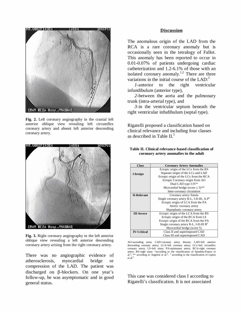
Fig. 2. Left coronary angiography in the cranial left anterior oblique view revealing left circumflex coronary artery and absent left anterior descending coronary artery.
Fig. 3. Right coronary angiography in the left anterior oblique view revealing a left anterior descending coronary artery arising from the right coronary artery. There was no angiographic evidence of atherosclerosis, myocardial bridge or compression of the LAD. The patient was discharged on β-blockers. On one year’s follow-up, he was asymptomatic and in good general status.
Discussion The anomalous origin of the LAD from the RCA is a rare coronary anomaly but is occasionally seen in the tetralogy of Fallot. This anomaly has been reported to occur in 0.01-0.07% of patients undergoing cardiac catheterization and 1.2-6.1% of those with an isolated coronary anomaly.1,2 There are three variations in the initial course of the LAD:3 1-anterior to the right ventricular infundibulum (anterior type), 2-between the aorta and the pulmonary trunk (intra-arterial type), and 3-in the ventricular septum beneath the right ventricular infudibulum (septal type).
Rigatelli proposed a classification based on clinical relevance and including four classes as described in Table II.5
Table II. Clinical relevance-based classification of
coronary artery anomalies in the adult
AO=ascending aorta; CAD=coronary artery disease; LAD=left anterior descending coronary artery; LCA=left coronary artery; LCx=left circumflex coronary artery; LS=left sinus; PA=pulmonary artery; RCA=right coronary artery; RS=right sinus. *according to the classification of Spindola-Franco et al.4; ** according to Angelini et al.8; § according to the classification of Lipton et al.9
This case was considered class I according to Rigatelli’s classification. It is not associated
Class Coronary Artery Anomalies
I-benign Ectopic origin of the LCx from the RS Separate origin of the LCx and LAD
Ectopic origin of the LCx from the RCA Ectopic Coronary origin from AO
Dual LAD type I-IV* Myocardial bridge (score ≤ 5)**
Inter-coronary circulation II-Relevant Coronary artery fistula
Single coronary artery R-L, I-II-III, A-P§
Ectopic origin of LCA from the PA Atretic coronary artery
Hypoplastic coronary artery III-Severe Ectopic origin of the LCA from the RS
Ectopic origin of the RCA from LS Ectopic origin of the RCA from the PA Single coronary artery R-L, I-II-III B§
Myocardial bridge (score 5) IV-Critical Class II and superimposed CAD
Class III and superimposed CAD

with invariable events in the absence of coronary atherosclerosis.6,7 In the present report, we found a rare anomalous coronary artery in association with hypertrophic cardiomyopathy. To our knowledge, this combination has not been previously reported. In the present case, there was no angiographic evidence of myocardial bridge, compression on the LAD or any atherosclerotic lesion. The mechanism proposed by the authors to explain the chest pain in this patient is that responsible for hypertrophic cardiomyopathy.
References 1. Donaldson RM, Raphael M, Radley-Smith R.
Angiographic identification of primary coronary anomalies causing impaired myocardial perfusion. Catheterization and Cardiovascular Diagnostics 1983; 9: 237-249.
2. Yamanaka O, Hobbs RE. Coronary artery
anomalies in 126,595 patients undergoing coronary arteriography. Catheterization and Cardiovascular Diagnostics 1990; 21: 28-40.
3. Barth III, CW, Roberts WC. Left main coronary
artery originating from the right sinus of Valsalva and coursing between the aorta and pulmonary trunk. Journal of the American College of Cardiology 1986; 7: 366-373.
4. Spindola-Franco H, Grose R, Solomon N. Dual
left anterior descending coronary artery: angiographic description of important variants
and surgical implications. American Heart Journal 1983; 105: 445-455.
5. Rigatelli GL, Rigatelli G. Coronary artery
anomalies: what we know and what we have to learn. A proposal for a new clinical classification. Italian Heart Journal 2003; 4: 305-310.
6. Rigatelli GL, Gemelli M, Gianfranco F, Rigatelli
G. Double is better: type IV dual left anterior descending coronary artery and superimposed atherosclerosis. Italian Heart Journal 2001; 2: 68-69.
7. Rigatelli G, Franco G, Gemelli M, Zamboni A,
Visentin M, Bovolon D, Rigatelli G. Recurrent unstable angina after revascularization in a case of dual left anterior descending coronary artery without risk factors: casualness or destiny? International Journal of Cardiology. 2004; 97: 133-134.
8. Angelini P, Trivellato M, Donis J, Leachman RD. Myocardial bridges: a review. Progress in Cardiovascular Diseases 1983; 26: 75-80.
9. Lipton MJ, Barry WH, Orbez I, Silverman JF,
Wexler L. Isolated single coronary artery: diagnosis, angiographic classification, and clinical significance. Radiology 1979; 130: 39-47.

Prediction of Left Ventricular Dysfunction on Basis of Ventricular Depolarization Time and Electrical Axis in
Patients with Left Bundle Branch Block
Farzad Jalali MD,a Seyyed Mohammad Miri MDb and Pegah Karimi Elizei c
Abstract Background- Prolongation of ventricular depolarization time (QRS duration), particularly in left
bundle branch block (LBBB), is commonly associated with many cardiac diseases. We propose that the QRS duration and degree of left-axis deviation (LAD) identify significant left ventricular (LV) systolic dysfunction in patients with LBBB.
Methods- In this prospective study conducted in the cardiac ward, CCU and out-patient clinic of our department in Babol from 2000 to 2003, 150 patients with a diagnosis of LBBB were divided into two groups (QRS ≥160 and QRS<160 milliseconds). Then the relationship between QRS duration, left axis deviation (LAD; axis between –30° and –90°) and echocardiographic LV ejection fraction (EF) were derived by T-test, chi-square and linear regression analysis in step-wise method.
Results- There was no significant difference in age and sex among the patients with or without LAD and QRS duration less or greater than 160 milliseconds (p>0.05). The EF of patients with LAD (n=64) and without LAD (n=86) was 48.64±14.63% and 52.10±13.98%, respectively (p=0.143). The mean±SD EF (54.5±10.545%) of the patients with a QRS duration of ≥160 milliseconds (n=19) was significantly more than the mean±SD EF (23.89±5.466%) of the patients with a QRS duration of <160 milliseconds (n=131, p<0.001). The QRS duration also had a significant (p<0.001) inverse correlation with EF (R = 0.926, adjusted R2 = 0.857, SE of estimate = 5.42). However, the QRS axis was not significantly correlated with EF and did not have added predictive value.
Conclusion- The QRS duration has a significant inverse relationship with EF and prolongation of QRS duration (≥160 milliseconds) in the presence of LBBB is a marker of significant left ventricular systolic dysfunction. The presence of LAD in LBBB does not signify a further decrease in EF (Iranian Heart Journal 2008; 9 (2):29-36).
Key words: QRS duration ■ electrical axis ■ LV dysfunction ■ ejection fraction ■ left bundle branch block
eft bundle branch block (LBBB) is commonly associated with coronary
artery disease (CAD), cardiomyopathy, and hypertension.1-2
Echocardiographic studies have revealed that patients with even mildly prolonged QRS duration (≥120 milliseconds) resulting from intraventricular conduction delay is
L
Received April 16, 2007; Accepted for publication May 4, 2008. a; Associate Professor of Cardiology, Department of Internal Medicine and Cardiology, Babol University of Medical Sciences, Babol, Iran, b; Research Assistant, MD, Department of Internal Medicine, Baghiatollah Hospital, Baghiatollah University of Medical Sciences, Tehran, Iran c; Research Assistant, MD, Babol University of Medical Sciences, Babol, Iran. Address for correspondence: 11th West Floor, Baghiatollah Hospital, Department of Internal Medicine, Baghiatollah University of Medical Sciences, Tehran, Iran. Tel: +98 21 88211000 Fax: +98 21 88055752 Email: [email protected]

Associated with left ventricular (LV) dysfunction.3-9 LBBB is responsible for a greater degree of asynchrony in LV contraction as a result of alteration in the sequence of LV depolarization.1,2,5,10-16 Therefore LBBB may be a marker of both LV systolic and diastolic dysfunction because of alteration in LV depolarization and prolongation of the QRS duration. LBBB is also associated with increased mortality in patients with congestive heart failure (CHF).3,4,17-27 Overall, LBBB is associated with a poor prognosis.1,2,10 It has also been suggested that left axis deviation (LAD) in the presence of LBBB may be associated with either left anterior fascicular block (LAFB) or loss of inferiorly directed forces from myocardial scarring.5,12 It has also been shown to have a higher incidence of cardiomegaly, CHF, diffuse conduction system disease, and sudden cardiac death.18,28-30 Therefore it is the impression among clinical cardiologists that LAD with LBBB identifies patients with severe LV systolic dysfunction. This study was designated to prove or refute this clinical observation, and we postulated that LAD and/or prolonged QRS duration (QRS ≥160 milliseconds) in the presence of LBBB is associated with poor LV function.
Methods In this prospective, cross-sectional research we studied 150 patients with LBBB in the cardiac ward, CCU and out-patient clinic of Shaheed Beheshti Hospital, Babol Medical Sciences University from September 2000 to December 2003. Patient demographics including age and sex were collected. The criterion for LBBB was a QRS duration of ≥120 milliseconds.3-5,23 An RSR´ pattern in leads I, V5, and V6 with secondary ST-T wave changes were supportive findings for LBBB. Acute prolongation of QRS duration was a strong indicator of LBBB. LAD was defined as a QRS axis between –30 and –90 degrees.1,5,14,16 Heart rates >100
beats/min were excluded from the study because of the possibility of tachycardia-related disorders. The patients with intraventricular conduction defects, right bundle branch block, or pacemaker rhythm were also excluded. The ejection fraction (EF) was determined by Simpson’s method on a Hewlett-Packard model Sonus 1500 echocardiography machine. Statistical analysis The demographic parameters among the patients with a QRS duration ≥160 milliseconds and the patients with a QRS duration <160 milliseconds were analyzed by 2 methods and their EFs were compared by the 2-tailed type II Student t test. Descriptive statistics were also calculated for each variable (QRS duration and LAD). Medians, quartiles, and ranges were derived for the QRS duration, QRS axis, and EF in these patients. Simple linear and multiple regression analysis were used to compare relationships among variables (QRS duration and LAD). Raw data were input into a case-wise multiple regression model.
Results
One hundred fifty patients were found to have LBBB, of which most of them were male (56.7% vs. 43.3%). The mean (±SD) age of patients was 53.39±8.29 years, of which there was no significant difference between males and females (p>0.05). Of the 150 patients included in the analysis, prolonged QRS duration (≥160 milliseconds) was found in 19 patients (12.7%) and short QRS duration (<160 milliseconds) was found in 131 (87.3%). There was no significant difference in age and sex among the patients with or without prolonged QRS duration (respectively: p=0.908; p=0.964, OR (95% CI) = 0.944 (0.356-2.501). The mean (±SD) EF of the patients with QRS duration of ≥160 milliseconds was significantly lower than that of the patients with a QRS duration of <160 milliseconds (54.5%±10.54% vs. 23.89% ±

5.46%, p<0.001). Also, this difference had been reported between males and females (p< 0.001). The mean (±SD) EF (48.64%±14.63%) of the patients with LBBB and LAD (n= 64) was not significantly different compared with the mean (±SD) EF (52.1%±13.97%) of the patients with LBBB and without LAD (n= 86, p=0.143). There was no significant difference in age and sex among the patients with or without LAD (p>0.05). Relationships uncovered among the variables (QRS duration, QRS axis, and EF) are illustrated in Table III. According to first model [EF= 182.059- 936.759 QRS duration + .046 LAD] and final model [EF= 185.279- 966.87 QRS duration], we found that the QRS duration had a significant (p<0.001) inverse correlation with the EF (R = 0.926, adjusted R2= 0.857, SE of estimate = 5.42). However, the axis was not significantly correlated with EF and added no predictive value to the model.
Fig. 1. Multivariate analysis showing negative correlation of prolonged QRS duration in presence of LBBB with EF. EF= 185.279- 966.87*QRS duration Pearson correlation: r (EF, QRS dur.)= 0.926; sig. = 0.000.
Fig. 2. Correlation of QRS axis in presence of LAD with EF derived by multivariate regression analysis. EF= 54.73+ 0.194*QRS duration Pearson correlation: r (EF, LAD)=0.378 ; sig. = 0.006.
Discussion Heart failure is often misdiagnosed or underdiagnosed in primary care. Assessment of left ventricular function in patients with suspected heart failure leads to more effective diagnosis and treatment of this disorder.20-
22,25,26,31 Intraventricular conduction disturbance is common in congestive heart failure, which is characterized by a wide QRS complex.23,24,27,32 Up to one-half of advanced CHF patients have prolonged QRS duration, which has been identified as an independent prognostic factor.4 Left ventricular dysfunction predicted by standard 12-lead electrocardiography would be clinically useful. Left bundle branch block is commonly associated with structural heart disease and LV dysfunction.5,10,12 In our study, we found that the role of age and sex is not correlated to QRS duration and LAD (p>0.05), but there is a significant difference between left ventricular ejection fraction and prolonged QRS duration. Our findings in similar roles of age and sex
0
10
20
30
40
50
60
70
80
0.12 0.13 0.14 0.15 0.16 0.17 0.18 0.19 0.2
QRS duration (millisecond)
Eje
ctio
n Fr
actio
n(%
)
10
20
30
40
50
60
70
80
-100 -80 -60 -40 -20 0 20
Left Axis Deviation (degree)
Eje
ctio
n Fr
actio
n(%
)

between patients with and without prolonged QRS duration are consistent with the findings of Nastasiou et al.30, Pastore et al.2, and Recke et al.15 In our study, we divided QRS duration into two groups (≥160 or <160 milliseconds), but Sandhu et al.32 divided this duration on the basis of 120 milliseconds, and Bode-Schnurbus et al.,18 on the basis of 150 milliseconds. The probable cause of this difference is due to different sample size and measurement methods. Tabuchi et al. estimated LV systolic function based on the ECG in cases with LBBB and reported that patients with underlying mild hypertensive heart disease may have a favorable LV systolic function. Thus, LV systolic function in patients with LBBB may be suspected by observing these electrocardiographic findings.33 The usefulness of spatial dispersion of QRS duration in predicting mortality in patients with mild to moderate chronic heart failure was studied by Yamada et al.27 They studied 114 consecutive stable outpatients with radionuclide left ventricular ejection fraction <40% and concluded that spatial dispersion of QRS duration is a powerful prognostic marker of the mortality in patients with mild to moderate CHF.27 Our results in LAD and LVEF do not agree with the findings of Yamada and coworkers. It is mentioned that they divided QRS duration according to a scale of 120 milliseconds. Furthermore, several studies of QRS duration have shown that a prolonged QRS (>170 milliseconds) is associated with LV dysfunction.3-5,7-9,18,21,23,24,27,29 Our data indicate that the presence of complete LBBB is related to LV dysfunction and prolonged QRS duration is correlated with poor systolic function. Das and coworkers5 analyzed the data of 300 patients to determine the relationship between prolonged QRS duration (QRS ≥170 ms) and left axis deviation (LAD) in the presence of LBBB. They concluded that there was no significant difference in age, sex, presence of valvular heart disease, and
EF among the patients with or without LAD.5 As in our study, Das et al. concluded that the QRS duration has a significant inverse relationship with EF and prolongation of QRS duration (≥170 milliseconds) in the presence of LBBB. The presence of LAD in LBBB does not signify a further decrease in EF.5 Murkofsky et al.8 studied 270 consecutive patients referred for radionuclide ventriculography, and concluded that prolonged QRS duration (>0.10 s) obtained from a standard resting 12-lead ECG is a specific, but relatively insensitive indicator of decreased LV systolic function. Further prolongation of the QRS had a higher specificity for decreased LVEF and a higher positive likelihood ratio for predicting abnormal LVEF. In our study, LAD in the presence of LBBB was not associated with further deterioration of LV function. With complete LBBB, the right ventricle is activated by the right bundle branch in its usual fashion. The impulse to the LV crosses the interventricular septum at one or more sites and then appears to enter the LV distal Purkinje system and is distributed throughout.13,33 The mean frontal QRS axis in LBBB presumably reflects the site or sites of impulse crossing the septum and the distribution of the impulse within the left ventricle.14 LAD in patients with LBBB presumably reflects the abnormality in the activation of the LV, which could reflect septal, distal Purkinje system, or LV tissue abnormalities. The thinner left anterior fascicle could be more prone to injury because of ventricular stretch, which might be more severe in the group with LAD.1,5 Thus the proposed mechanism of LAD in the presence of LBBB is partial LAFB or loss of inferiorly directed forces from myocardial scarring that interfere in some way with distribution of the impulse.14,16 However, our study confirms that there is no significant difference in the severity of LV dysfunction in patients with LBBB in

association with either LAD or a normal QRS axis. We speculate that it simply signifies theanisotropy of impulse propagation in a myopathic ventricle or the presence of decreased inferoposterior depolarization leading to left superior orientation of the main vector force.34 Similarly to this conclusion, Spurrell et al.16 performed a study of intraventricular conduction times in patients with left bundle-branch block and LAD using His bundle electrograms. Our conclusion is similar to Parharidis’s findings.14 The aim of their study was to elucidate the diagnostic significance of LAD in patients with LBBB.
They concluded that the presence of LAD had a low sensitivity for the presence of organic heart disease.1
Conclusion In the presence of LBBB, the QRS duration has a significant inverse relationship with EF and prolongation of the QRS duration (≥160 milliseconds) is a marker of significant LV systolic dysfunction. However, the degree of LAD in LBBB does not correlate with EF and also does not signify a further decrease in EF.
QRS duration (millisecond)
< 160 (n= 131)
≥ 160 (n= 19)
Total (n= 150) p-value OR (95% CI)
Age (year)* 53.38±8.43 53.47±7.538 53.39±8.299 0.964 **
Gender Male 74 (56.5%) 11 (57.9%) 85 (56.7%) 0.908 *** 0.944 (0.356-2.501) † Female 57 (43.5%) 8 (42.1%) 65 (43.3%) Ejection Fraction (%)*
Male 54.77±9.947 23.91±4.949 50.78±14.053 0.000 **
Female 54.16±11.356 23.88±6.468 50.43±14.763 0.000 **
Total 54.5±10.545 23.89±5.466 50.63±14.317 0.000 **
* (Mean±Std. deviation); **Estimate from independent sample T-test and p-value less than 0.05 is significant ***Estimate from Pearson Chi-square Test and p-value less than 0.05 is significant; † OR (95% CI) estimate from Mantel-Henszel for QRS duration< 160 millisec/ QRS duration ≥ 160 millisec.)

Table II. Comparison among LAD with age, gender, and EF in presence of complete LBBB
Left Axis Deviation (degree) *
No (n= 86)
Yes (n= 64)
Total (n= 150) p-value OR (95% CI)
Age (year) ** 53.58±7.974 53.14±8.774 53.39±8.299 0.749 *** Gender Male 49 (57%) 36 (56.2%) 85 (56.7%) 0.929 † 1.03 (0.536-1.979) †* Female 37 (43%) 28 (43.8%) 65 (43.3%) Ejection Fraction (%) ** Male 51.94±13.874 49.14±14.336 50.78±14.053 0.377 *** Female 52.32±14.304 47.93±15.244 50.43±14.763 0.237 *** Total 52.10±13.978 48.64±14.635 50.63±14.317 0.143 ***
*LAD = (axis between -30 degrees and -90 degrees); **(Mean±Std. deviation); *** Estimate from independent sample T-test and p-value less than 0.05 is significant; †Estimate from Pearson Chi-square test and p-value less than 0.05 is significant; †*OR (95% CI) estimate from Mantel-Henszel
for: (LAD-/ LAD+).
Table III. Multivariate analysis showing correlations of prolonged QRS duration and LAD in presence of LBBB
with EF
Models * First Model ** Final Model *** (Constant) QRS duration LAD (Constant) QRS duration Coefficients
Unstandardized B 182.059 -936.759 .046 185.279 -966.870 Std. Error 4.576 33.430 .016 4.531 32.379
Standardized Beta -.897 .090 -.926 T-test† 39.782 -28.021 2.801 40.892 -29.861
Sig. † .000 .000 .006 .000 .000 Pearson Correlation r (EF, LAD)= 0.378 ; sig. = 0.006 r (EF, QRS dur.)= 0.926; sig. = 0.000 (N=150) r (EF, QRS dur.)= 0.926; sig. = 0.000 ANOVA ††
Mean Square 13206.927 26193.553 F 470.392 891.687 Sig. .000(b) .000(a)
Model Summary †* EF= 182.059- 936.759 QRS dur.+.046 LAD EF= 185.279- 966.87 QRS dur. R .930 .926 R Square .865 .858 Adjusted R Square .863 .857 Std. Error of the Estimate 5.299 5.420
*Estimate from linear regression analysis with stepwise method; **Predictors in linear regression model are (constant), LBBB, LAD; ***Predictors in linear regression model are (constant), LBBB; †Estimate from t-test among correlation coefficients and p-value less than 0.05 is significant; ††The ANOVA table tests the acceptability of the model from a statistical perspective. The significance value of the F statistic is less than 0.05, which means that the variation explained by the model is not due to chance. Dependent variable in linear regression models is: EF †*The model summary table reports the strength of the relationship between the model and the dependent variable. R, the multiple correlation coefficient, is the linear correlation between the observed and model-predicted values of the dependent variable. Its large value indicates a strong relationship. R Square, the coefficient of determination, is the squared value of the multiple correlation coefficient. It shows what proportion of the variation in time is explained by the model. Dependent variable in linear regression models is: EF
References
1. Gressard A. Left bundle branch block with left-axis deviation: an electrophysiologic approach. Am J Cardiol 1983; 52(8): 1013-1016.
2. Pastore CA, Moffa PJ, Tobias NM, de Moraes
AP, Kaiser E, Cuoco MA et al. Left bundle branch block analysis by body surface mapping. Comparison with electrocardiographic and vectocardiographic findings. Arq Bras Cardiol 1996; 66(5): 253-256.
3. Aranda JM, Carlson ER, Pauly DF, Curtis AB,
Conti CR, Ariet M et al. QRS duration variability in patients with heart failure. Am J Cardiol 2002; 90(3): 335-337.
4. Bleeker GB, Schalij MJ, Molhoek SG, Verwey
HF, Holman ER, Boersma E et al. Relationship between QRS duration and left ventricular dyssynchrony in patients with end-stage heart failure. J Cardiovasc Electrophysiol 2004; 15(5): 544-549.

5. Das MK, Cheriparambil K, Bedi A, Kassotis J, ES D, Reddy CV et al. Prolonged QRS duration (QRS >/=170 ms) and left axis deviation in the presence of left bundle branch block: A marker of poor left ventricular systolic function? Am Heart J 2001; 142(5): 756-759.
6. Ghio S, Constantin C, Klersy C, Serio A, Fontana
A, Campana C et al. Interventricular and intraventricular dyssynchrony are common in heart failure patients, regardless of QRS duration. Eur Heart J 2004; 25(7): 5 ۷۱-۵۷۸.
7. Hofmann M, Bauer R, Handrock R, Weidinger G,
Goedel-Meinen L. Prognostic value of the QRS duration in patients with heart failure: a subgroup analysis from 24 centers of Val-HeFT. J Card Fail 2005; 11(7): 523-528.
8. Murkofsky RL, Dangas G ,Diamond JA, Mehta
D, Schaffer A, Ambrose JA. A prolonged QRS duration on surface electrocardiogram is a specific indicator of left ventricular dysfunction [see comment]. J Am Coll Cardiol 1998; 32(2): 476-482.
9. Shamim W, Yousufuddin M, Cicoria M, Gibson
DG, Coats AJ, Henein MY. Incremental changes in QRS duration in serial ECGs over time identify high risk elderly patients with heart failure. Heart 2002; 88(1): 47-51.
10. Haskell RJ, Ginzton LE, Laks MM.
Electrocardiographic diagnosis of left ventricular hypertrophy in the presence of left bundle branch block. J Electrocardiol 1987; 20(3): 227-232.
11. Kang SJ, Song JK, Yang HS, Song JM, Kang
DH, Rhee KS et al. Systolic and diastolic regional myocardial motion of pacing-induced versus idiopathic left bundle branch block with and without left ventricular dysfunction. Am J Cardiol 2004; 93(10): 1243-1246.
12. Kountouris E, Korantzopoulos P, Karanikis P,
Pappa E, Dimitroula V, Ntatsis A et al. QRS dispersion: an electrocardiographic index of systolic left ventricular dysfunction in patients with left bundle branch block. Int J Cardiol 2004; 97(2): 321-322.
13. Mehta A, Jain AC, Mehta MC, Billie M.
Usefulness of left atrial abnormality for
predicting left ventricular hypertrophy in the presence of left bundle branch block. Am J Cardiol 2000; 85(3): 354-359.
14. Parharidis G, Nouskas J, Efthimiadis G, Styliadis
J, Gemitzis K, Hatzimiltiadis S et al. Complete left bundle branch block with left QRS axis deviation: defining its clinical importance .Acta Cardiol 1997; 52(3): 295-303.
15. Recke SH, Esperer HD, Eberlein U, Gansser R,
von der EJ. Assessment of left ventricular function from the electrocardiogram in left bundle branch block. Int J Cardiol 1989; 24(3): 297-304.
16. Spurrell RA, Krikler DM, Sowton E. Study of
intraventricular conduction times in patients with left bundle-branch block and left axis deviation and in patients with left bundle-branch block and normal QRS axis using His bundle electrograms. Br Heart J 1972; 34(12):1244-12.
17. Bax JJ, Marwick TH, Molhoek SG, Bleeker GB,
VAN EL, Boersma E et al. Left ventricular dyssynchrony predicts benefit of cardiac resynchronization therapy in patients with end-stage heart failure before pacemaker implantation. Am J Cardiol 2003; 10(92): 1038-40.
18. Bode-Schnurbus L, Bocker D, Block M, Gradaus
R, Heinecke A, Breithardt G et al. QRS duration: a simple marker for predicting cardiac mortality in ICD patients with heart failure. Heart 2003; 89(10): 1157-1162.
19. Davies M, Hobbs F, Davis R, Kenkre J, Roalfe
AK, Hare R et al. Prevalence of left-ventricular systolic dysfunction and heart failure in the Echocardiographic Heart of England Screening study: a population based study. Lancet 2001; 358(9280): 439-444.
20. Gupta SN, Jose VJ, Chandy ST. Heart failure:
what proportion of patients satisfy the electrocardiographic criteria for cardiac resynchronization therapy? Indian Heart J 2003; 55(6): 619-623.
21. Kalra PR, Sharma R, Shamim W, Doehner W,
Wensel R, Bolger AP et al. Clinical characteristics and survival of patients with

chronic heart failure and prolonged QRS duration. Int J Cardiol 2002; 86(2-3): 225-231.
22. Kearney MT, Zaman A, Eckberg DL, Lee AJ,
Fox KA, Shah AM et al. Cardiac size, autonomic function, and 5-year follow-up of chronic heart failure patients with severe prolongation of ventricular activation. J Card Fail 2003; 9(2): 93-99.
23. Molhoek SG, VAN EL, Bootsma M, Steendijk P,
Van Der Wall EE, Schalij MJ. QRS duration and shortening to predict clinical response to cardiac resynchronization therapy in patients with end-stage heart failure. Pacing Clin Electrophysiol 2004; 27(3): 308-313.
24. Shenkman HJ, Pampati V, Khandelwal AK,
McKinnon J, Nori D, Kaatz S et al. Congestive heart failure and QRS duration: establishing prognosis study. Chest 2002; 122(2): 528-534.
25. Tarantini L, Faggiano P, Senni M, Lucci D,
Bertoli D, Porcu M et al. Clinical features and prognosis associated with a preserved left ventricular systolic function in a large cohort of congestive heart failure outpatients managed by cardiologists. Data from the Italian Network on Congestive Heart Failure. Ital Heart J 2002; 3(11): 656-664.
26. Varela-Roman A, Gonzalez-Juanatey JR, Basante
P, Trillo R, Garcia-Seara J, Martinez-Sande JL et al. Clinical characteristics and prognosis of hospitalised inpatients with heart failure and preserved or reduced left ventricular ejection fraction. Heart 2002; 88(3): 249-254.
27. Yamada T, Shimonagata T, Misaki N, Asai M,
Makino N, Kioka H et al .Usefulness of spatial dispersion of QRS duration in predicting mortality in patients with mild to moderate chronic heart failure. Am J Cardiol 2004; 94(7): 960-963.
28. Freudenberger R, Sikora JA, Fisher M, Wilson A,
Gold M. Electrocardiogram and clinical characteristics of patients referred for cardiac transplantation: implications for pacing in heart failure. Clin Cardiol 2004; 27(3): 151-153.
29. Iuliano S, Fisher SG, Karasik PE, Fletcher RD,
Singh SN. QRS duration and mortality in
patients with congestive heart failure. Am Heart J 2002; 143(6): 1085-1091.
30. Nastasiou-Nana MI, Nanas JN, Karagounis LA,
Tsagalou EP, Alexopoulos GE, Toumanidis S et al. Relation of dispersion of QRS and QT in patients with advanced congestive heart failure to cardiac and sudden death mortality. Am J Cardiol 2000; 85(10): 1212-1217.
31. Sparrow N, Adlam D, Cowley A, Hampton JR.
The diagnosis of heart failure in general practice: implications for the UK National Service Framework. Eur J Heart Fail 2003; 5(3): 349-354.
32. Sandhu R, Bahler RC. Prevalence of QRS
prolongation in a community hospital cohort of patients with heart failure and its relation to left ventricular systolic dysfunction. Am J Cardiol 2004; 93(2): 244-246.
33. Tabuchi H, Kawai N, Sawayama T] .Estimation
of left ventricular systolic function based on the electrocardiograms in cases with left bundle branch block]. J Cardiol 1998; 31(1): 23-30.
34. Kong CW, Hsu TG, Lu FJ, Chan WL, Tsai K.
Leukocyte mitochondria depolarization and apoptosis in advanced heart failure: clinical correlations and effect of therapy. J Am Coll Cardiol 2001; 38(6): 1693-1700.

Risk Factors for Silent Myocardial Ischemia in Type II Diabetic Patients
Afsaneh Forood MD* and Mohammad Masomi MD
Abstract
Background- Silent myocardial ischemia is more common in diabetic patients than others. Early
detection plays an important role in the prevention of acute myocardial infarction and sudden cardiac death. Routine screening of all diabetics is costly. The aim of this study was to estimate the prevalence of silent myocardial ischemia in type 2 diabetes and define these high-risk patients by routine screening tests.
Methods- Between May 2004 and May 2006, this cross-sectional study was performed on 500 type 2 diabetic patients referred to Kerman internal medicine and cardiovascular clinics. Inclusion criteria were age between 35 and 70 years, absence of symptoms and resting electrocardiographic signs of ischemia, evidence of retinopathy or peripheral vascular disease, or at least one major atherogenic risk factor (except diabetes). All the patients underwent treadmill exercise test or thallium scintigraphy with exercise or dipyridamole injection. Data were analyzed with chi-square, t-test, and Mann-Whitney U tests.
Results- Five hundred patients, comprised of 232 men and 268 women, were evaluated. Screening tests were positive in 86 (17.2%) patients. There was a significant statistical relation between the duration of diabetes, low density lipoprotein cholesterol, family history of coronary artery disease (CAD), retinopathy, and peripheral vascular disease with silent myocardial ischemia (P<0.05). The prevalence of silent ischemia was not significantly different between the males and females (P>0.05). Among the patients with silent ischemia, body mass index was higher in the females and cigarette smoking was more common in the males (P<0.05).
Conclusion- With regard to the high frequency of silent myocardial ischemia in type 2 diabetes mellitus, routine silent ischemia screening by exercise stress test should be recommended in type 2 diabetes if any of these conditions are present: duration of diabetes more than ten years, family history of CAD, LDL cholesterol higher than 160 mg/dL, retinopathy, or peripheral vascular disease (Iranian Heart Journal 2008; 9 (2):37-42).
Key words: type 2 diabetes ■ silent myocardial ischemia ■ coronary artery disease
Coronary artery disease (CAD) is the major cause of morbidity and mortality in patients with diabetes.1 More than half of all diabetic patients die from CAD.2,3
Diabetics have at least a two- to four-fold increased risk in cardiovascular events compared with age-matched controls.4,5 One of the late diagnostic signs of CAD is silent myocardial ischemia, where
Received Nov . 18, 2007; Accepted for publication June 24, 2008. From the Department of Cardiology and Internal Medicine, Kerman University of Medical Sciences, Kerman, Iran.
Address correspondence to: A. Forood, MD, Department of Cardiology, Kerman University of Medical Sciences, Kerman Iran. Email: [email protected] Fax: +(98341)2115803 Tel: (0098341)2445335

asymptomatic subjects show ischemia in resting electrocardiogram or exercise stress test.6 For this reason and others, the occurrence of major cardiac events such as acute myocardial infarction, unstable angina, ischemic cardiomyopathy, and sudden death are often the first manifestations of CAD in diabetic patients.4 Silent myocardial ischemia is more frequent in diabetic patients than non-diabetics probably because of diabetic neuropathy.6 Silent myocardial ischemia with ST-segment depression in treadmill exercise stress test is associated with an increase of five times in cardiovascular mortality risk.7 Early diagnosis of myocardial ischemia can reduce morbidity and mortality in diabetes and improve quality of life.8 Different studies show conflicting data about the prevalence of silent myocardial ischemia in type II diabetes.4,6,9 The role of major cardiovascular risk factors, micro- and macroangiopathy in the presentation of silent myocardial ischemia is also different.10-13 On the other hand, routine screening tests for the detection of silent myocardial ischemia in all diabetics is costly. We sought to investigate the frequency of silent myocardial ischemia in type 2 diabetes in the city of Kerman. Furthermore, by determining the role of major atherogenic risk factors in silent myocardial ischemia, we tried to define the high-risk group of diabetic patients for whom screening tests would be helpful.
Methods The present cross-sectional study included 500 patients with type 2 diabetes who were referred to the cardiovascular clinics between May 2004 and May 2006 in Kerman.
The inclusion criteria were a normal 12-lead resting electrocardiography, no history of CAD or congestive heart failure, and presence of at least one of the major atherogenic risk factors in addition to diabetes such as
hypertension (blood pressure >140/90mmHg or antihypertensive treatment), dyslipidemia (serum total cholesterol >200mg/dL, triglycerides >150 mg/dL, LDL* >160mg/dL, HDL**<35mg/dL or lipid lowering treatment), cigarette smoking, obesity, family history of premature CAD (before 60 years in first-degree relatives), retinopathy, and peripheral artery disease.
The diagnosis of type 2 diabetes was based on the American Diabetes Association (ADA) criteria. Peripheral artery disease was diagnosed when peripheral pulses were abolished or weak and/or when previous history of vascular surgery and/or intermittent claudication was present. Diabetic retinopathy was diagnosed if at least one microaneurysm or hemorrhage or exudate in one eye was found on fundoscopy.
All the blood samples were taken in the morning after at least 12 hours of fasting and measured by an automatic analyzer (Alcyon 300, Abbott, USA).
Written consent was obtained from each patient in accordance with the guidelines set by the ethical committee of Kerman University of Medical Sciences.
Each patient underwent a treadmill exercise test.
Before the exercise test, the patients were informed about the test and any drugs affecting the test results were discontinued before 48 hours. A maximal symptom-limited exercise protocol was used with a treadmill (Esaoate, DST 1000, Italy) according to the Bruce protocol. A twelve-lead electrocardiogram was recorded every minute, and blood pressure was measured at rest and at the end of each step during exercise.
The test was stopped when one of the following end-points was reached: target heart rate 85% of the predicted heart rate (220
* LDL: low density lipoprotein, ** HDL: high density lipoprotein

beats/min minus age in years), severe fatigue, systolic blood pressure reduction more than 20mmHg from control-values, hypertensive response (BP>220/120 mmHg), severe chest pain, or serious cardiac arrhythmia.
The exercise test was defined as maximal if the patient reached 85% of the predicted heart rate for the age. This test was positive for myocardial ischemia if horizontal or down sloping ST-segment depression of 1 mm or more calculated at 0.08 s after the J-point was present and was negative if the patient reached 85% of the predicted heart rate for age without change in the ST-segment. The test was considered not diagnostic or incomplete if the patient did not reach 85% of the predicted heart rate for age or the test was interrupted for any of the above-mentioned reasons.
If the patient had conditions that prohibited maximal exercise test (such as amputation, foot wound, severe obesity, intermittent claudication, serious cardiac arrhythmia, left bundle branch block at resting ECG, progressive valvular heart disease, and uncontrolled hypertension) or maximal exercise test was incomplete, we performed thallium myocardial scintigraphy in association with exercise testing and/or dipyridamole injection. One mCi thallium 201 was injected for each 25kg body weight. Early imaging was performed at 5 to 10 minutes, and late imaging was carried out 4 hours after injection.
This test was positive if fixed or transient uptake defects were observed between stress and rest imaging (4 hours after the end of the dipyridamole test). Silent myocardial ischemia was defined as positive exercise test or positive thallium myocardial scintigraphy.
The presence of left ventricular ejection fraction more than 50% in echocardiography ruled out heart failure.
Statistical analysis The results are expressed as mean ± SD. The means of the two groups were compared using the student’s t-test or Mann-Whitney U test. The rate of proportion was compared using the χ2 test. P value < 0.05 was considered significant.
Results Five hundred cases, comprised of 232 men and 268 women aged between 40 and 70 years, were studied. In total, 357 patients were able to perform the maximal exercise test, 312 of whom reached 85% of the predicted heart rate for age. Results were positive in 64 patients and negative in 248 patients. Thallium myocardial scintigraphy was carried out in 45 patients who could not complete the exercise test. Among them, six had positive and 39 had negative results. One hundred forty-three cases who were unable to perform exercise test had thallium myocardial scintigraphy carried out as a primary procedure. Results were positive in 16 patients and negative in 127 patients. All the patients were subdivided into those having ischemia and those having no ischemia. Of the 500 patients, 86 (17.2%) had silent myocardial ischemia and 414 (82.8%) patients did not have silent myocardial ischemia. The characteristics of the two groups are shown in Table I.
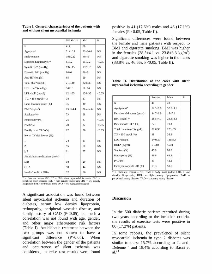
Table I. General characteristics of the patients with and without silent myocardial ischemia
NO SMI** SMI P
N 414 86
Age (yrs)* 51±10.1 52±10.6 NS
Male/Female 191/222 40/46 NS
Diabetes duration (yrs)* 8±5.2 15±7.2 <0.05
Systolic BP* (mmHg) 134±15 137±15 NS
Diastolic BP* (mmHg) 80±6 80±8 NS
Anti-HTN tx (%) 65 69 NS
Total chol* (mg/dl) 216±40 220±35 NS
HDL chol* (mmHg) 54±16 50±14 NS
LDL chol* (mg/dl) 124±35 138±33 <0.05
TG > 150 mg/dl (%) 38 37 NS
Lipid lowering drugs (%) 36 40 NS
BMI* (kg/m2) 25.1±4.4 26.4±4.6 NS
Smokers (%) 73 68 NS
Retinopathy (%) 25 37 <0.05
PAD (%) 20 32 <0.05
Family hx of CAD (%) 12 26 <0.05
No. of CV risk factors (%):
1 24 23 NS
2 55 50 NS
≥ 3 21 27 NS
Antidiabetic medications (no,%)
Diet 18 20 NS
OHA 50 44 NS
Insulin/insulin + OHA 32 36 NS
* = Data are means ±SD; ** = SMI, silent myocardial ischemia; PAD = peripheral artery disease; HDL = high density lipoprotein; LDL = low density lipoprotein; BMI = body mass index; OHA = oral hypoglycemic agents
A significant association was found between silent myocardial ischemia and duration of diabetes, serum low density lipoprotein, retinopathy, peripheral vascular disease, and family history of CAD (P<0.05), but such a correlation was not found with age, gender, and other major atherogenic risk factors (Table I). Antidiabetic treatment between the two groups was not shown to have a significant difference (P>0.05). When correlation between the gender of the patients and occurrence of silent ischemia was considered, exercise test results were found
positive in 41 (17.6%) males and 46 (17.1%) females (P> 0.05, Table II). Significant differences were found between the female and male patients with respect to BMI and cigarette smoking. BMI was higher in the females (28.5±4.1 vs. 23.8±3.3 kg/m2) and cigarette smoking was higher in the males (88.8% vs. 46.6%, P<0.05, Table II).
Table II. Distribution of the cases with silent myocardial ischemia according to gender
Female Male P
N 46 40
Age (years)* 52.5±8.8 52.3±9.6
Duration of diabetes (years)* 14.7±6.9 15±7.2
BMI (kg/m2)* 28.5±4.1 23.8±3.3
Patients with HTN (%) 72.1 70.4
Total cholesterol* (mg/dl) 225±36 223±35
TG > 150 mg/dl (%) 38 36.8
LDL* (mg/dl) 140±30 136±32
HDL* (mg/dl) 53±10 56±9
Smokers (%) 46.6 88.8
Retinopathy (%) 66.6 63.8
PAD (%) 45 43.1
Family history of CAD (%) 53.3 50.8
* = Data are means ± SD; BMI = body mass index; LDL = low density lipoprotein; HDL = high density lipoprotein; PAD = peripheral artery disease; CAD = coronary artery disease
Discussion In the 500 diabetic patients recruited during two years according to the inclusion criteria, the results of exercise tests were positive in 86 (17.2%) patients. In some reports, the prevalence of silent myocardial ischemia in type 2 diabetes was similar to ours: 15.7% according to Janand-Delenne 6 and 18.4% according to Bacci et al.14

Although male sex and BMI are risk factors for CAD,8 in our study this association was not observed. Perhaps this is because diabetes leads to the loss of female protection from cardiovascular disease and severe obesity was not seen in our population. These data were similar to some other reports in the literature.11,12 In our study, the correlation of diabetes duration, serum LDL cholesterol, family history of CAD, retinopathy, and peripheral artery disease with silent myocardial ischemia was stronger than the other risk factors.
In the Wackers et al. study, major atherogenic risk factors did not emerge as significantly predictive of silent myocardial ischemia; this may be due to the generally increased levels of these risk factors in their study population or might reflect the impact of treatment with statins, ACE inhibitors, and aggressive blood pressure and glucose control.15
A significant association between diabetes duration with silent myocardial ischemia has been seen in many studies,15-17 which chimes in with our results. Various mechanisms have been explained for the premature onset of CAD in type 2 diabetes. Among them, the loss of normal endothelial function is an early warning sign of the atherogenic process. This event occurs concomitantly with hyperglycemia, hypertension, dyslipidemia, and hyperinsulinemia.18 Endothelial dysfunction might thus be an explanation of the elevated proportion of patients with silent myocardial ischemia, as assessed by exercise test, but with non-significant coronary artery stenosis on coronary angiography.17 Microangiopathy such as retinopathy is a sensitive marker of generalized endothelial dysfunction, which is common in diabetes.17 The association of macroangiopathy and microangiopathy with silent myocardial ischemia was significant in the Janand-Delenne study,6 which is in agreement with our findings. In another study, however, this correlation was not seen probably because
severe retinopathy was considered to be an exclusion factor.13 BMI in the women with silent myocardial ischemia was higher than that in the men, because our women did not perform adequate regular exercise. Cigarette smoking in the male gender with this ischemia was significantly higher than that in the women, because smoking was an uncommon habit in our women. These results were similar to those in the Sargin study.16 Antidiabetic treatment among our studied population did not reveal a significant difference, but in the Sargin study,16 insulin usage was significantly higher in the silent myocardial ischemia group, probably because our patients did not like to take insulin injections.
Conclusion In light of our results, we would advise routine screening for silent myocardial ischemia in type 2 diabetes in the following cases:
• retinopathy, • family history of CAD (<60 years
in first-degree relatives) • peripheral vascular disease, • LDL >160 mg/dl, • diabetes duration ≥10 yrs (>5 yrs
if additional risk factors are present).
Further studies with larger sample sizes are needed to investigate the role of new atherogenic risk factors such as high-sensitivity CRP, fibrinogen, lipoprotein (a), and homocysteine in silent myocardial ischemia in diabetes.
Acknowledgements

This work was supported by the Physiology Research Unit of Kerman University of Medical Sciences.
References 1. Stamler J, Vaccaro O, Neston JD, Wentworth D.
Diabetes, other risk factors and 12-yr cardiovascular mortality for men screened in the multiple risk factor intervention trial. Diabetes Care 1993; 16: 434-444.
2. Fuller JH. Mortality trends and causes of death in diabetic patients. Diabet Metab 1993; 19: 96-99.
3. Panzram G. Mortality and survival in type 2 diabetes mellitus. Diabetologia 1987; 30: 123-131.
4. Cosson E, Guimfack M, Paries J, Valensi P. Are silent coronary stenoses predictable in diabetic patients and predictive of cardiovascular events? Diabetes Metab 2003; 29: 470-6.
5. Kannel WB, Wilson PWF. Risk factors that attenuate the female coronary disease advantage. Arch intern Med 1995; 155: 57-61.
6. Janand-Delenne B, Savin B, Habib G, Bory M, Vogue P. Silent myocardial ischemia in patients with diabetes. Diabetes Care 1999; 22(9): 1396-1400.
7. Laukkanen JA, Korl S, Lakka TA. Exercise-induced silent myocardial ischemia and coronary morbidity and mortality in middle – aged men. J Am Coll Cardiol 2001; 38: 72-79.
8. Braunwald D, Zipes P, Bono W. In: Braunwald’s Heart Disease, A Textbook of Cardiovascular Medicine. 7th ed., 2005; Elseivier and Saunders Company, pp. 939- 953.
9. Zharov E, Kazankov IN, Grigor EV. Silent myocardial ischemia in patients with diabetes mellitus without the clinical manifestation of ischemic heart disease. Kardiologia. 1993; 33: 16-18.
10. Fredenrich A, Castello-Ros S, Hieronimus S, Baudouy M. Screening for silent myocardial
ischemia in diabetic patients. Diabetes Care 2000; 23(4): 563-64.
11. Gazzaruso C, Garzaniti A, Falcone C. Assessment of asymptomatic coronary artery disease in apparently uncomplicated type 2 diabetic patients. Diabetes Care 2002; 25(8): 1418-1424.
12. Koistinen MJ. Prevalence of asymptomatic myocardial ischemia in diabetics subjects. BMJ 1990; 301(6743): 92-5.
13. Milan Study on Atherosclerosis and Diabetes (MISAD) Group: prevalence of unrecognized silent myocardial ischemia and its association with atherosclerotic risk factors in non-insulin-dependent diabetes mellitus. Am J Cardiol 1997; 79: 134-139.
14. Bacci S, Villella M, Villella A, Grill M, Fanelli R. Screening for silent myocardial ischemia in type 2 diabetic patients with additional atherogenic risk factors. Eur J Endocrinology 2002; 147: 649-654.
15. Wackers F, Young L, Barrett E, Wittlin S. Detection of silent myocardial ischemia in asymptomatic diabetic subjects. Diabetes Care 2004; 27(8): 1954-1961.
16. Sargin H, Ozisik M, Nurcin C, Seven O. The prevalence of silent ischemia in Turkish patients with type 2 diabetes mellitus. Tohoku J Exp Med 2005; 205: 351-355.
17. Sultan A, Piot Ch, Rasamisa O, Renard E. Risk factors for silent myocardial ischemia in high-risk type 1 diabetic patients. Diabetes Care 2004; 27(7): 1745-1747.
18. William SB, Cusco JA, Roddy MA, Creager MA. Impaired nitric oxide mediated vasodilatation in patients with non-insulin dependent diabetes mellitus J Am Coll Cardiol 1996; 27: 567-574.
Applying the Logistic Regression Model to Predict the Stenosis in Carotid Artery Using the Sequential Color
Doppler Ultrasound Image Processing
M. Mokhtari-Dizaji PhD, P. Abdolmaleki2 PhD, H. Saberi3 MD, and T. Rahmani1 MSc
Abstract
Background- Early detection of stenosis in carotid artery is essential because it directly affects the
patients' clinical management and is of prognostic value. Therefore, estimating mechanical properties of this artery in normal and atherosclerosis cases is important as far as medical treatment is concerned. We applied a logistic regression model to predict carotid artery stenosis in a group of patients based on the quantitative features extracted from the processing of the conventional color Doppler ultrasound images.
Methods- Our database includes 128 patient records consisting 10 quantitative features. The database is then randomly divided into the training and validation samples including 98 and 30 patient records respectively. The training and validation samples are used to construct the logistic regression model and to validate its performance. Finally, important criteria such as sensitivity, specificity, accuracy and receiver operating characteristic curve (ROC) analysis for this method are evaluated.
Results- Our results show that the logistic regression model is able to classify correctly 28 out of 30 cases presented in the validation sample. The output of this method showed a high positive predictive value of 94%.
Conclusion- We have established a logistic discriminator approach which is able to predict the probability of stenosis in the carotid artery using features extracted from ultrasonic measurements on ultrasound imaging (Iranian Heart Journal 2008; 9 (2):43-50).
Key words: color Doppler ultrasound ■ carotid artery stenosis ■ mechanical properties ■ logistic regression analysis
troke is one of the most common causes of death and disability in industrialized
nations. Approximately 80% of ischemic strokes are due to athero-thromboembolic infarction, which is caused by atherosclerotic lesions at the carotid bifurcation.1, 2 Currently, the Doppler ultrasound technique is widely recognized as the best non-invasive screening test for carotid artery stenosis.
Ultrasound does not calculate the degree of arterial narrowing directly, but relies on extrapolating changes in flow parameters to an anatomical stenosis. Despite the availability of several qualitative and quantitative methods for the assessment of carotid artery stenosis, accurate diagnosis of stenosis remains a clinically difficult task.
S
Received May 8. 2008; Accepted for publication Aug 21, 2008. 1. Department of Medical Physics, Tarbiat Modarres University, 2. Department of Biophysics, Tarbiat Modarres University, 3. Department of Radiology, Tehran Medical Sciences University, Tehran, Islamic Republic of Iran. Address correspondence to: Dr. M. Mokhtari-Dizaji, University of Tarbiat Modarres, Department of Medical Physics, Tehran, Iran. Email: [email protected]

Conventional catheter angiography is therefore often performed for clinical confirmation. Although specific, catheter angiography is an invasive, costly, and psychologically stressful procedure. Estimation of arterial diameter throughout the carotid cycle has been conducted increasingly to study the mechanical properties of the arterial wall and changes associated with disease. Arterial diameter, wall thickness, cross section changes, and blood pressure measurements can be used to calculate the stiffness indices of the common carotid artery. Several indices are used to evaluate the elastic properties of the common carotid artery including: distensibility, compliance, Young’s modulus, and pressure-strain elastic modulus and stiffness indices.3-5 Recently, there has been much interest on the relationship between arterial stiffness and cardiovascular diseases.3, 6-14 Consequently a computerized second opinion in the form of a logistic regression model could be useful for the differentiation of atherosclerosis from normal. In this study we intended to establish a logistic regression model to work as a tool for radiologist to predict atherosclerotic disease in carotid artery using physical and mechanical parameters extracted by processing the sequential color Doppler ultrasound images. The performance of the established model was then evaluated using the common statistical index positive predictive value and ROC analysis.16, 17
Methods Our goal was to apply the logistic discriminant analysis to the data collected in a study designed to predict the carotid artery stenosis on the basis of features that had been extracted from measuring the ultrasonic tissue characteristics consisted of diastolic diameter, systolic diameter, pulse pressure, arterial strain, static pressure change on wall, peak
systolic velocity, end diastolic velocity, pressure-strain elastic modulus, static pressure-strain elastic modulus17 and stiffness. Our study group consisted of 128 men with mean age 66±11 years. These patients were studied from Nov 2000 to April 2004. Patients were categorized into two groups based on the results of angiography as well as clinical diagnosis. These two groups were; normal subjects (n=55) with no history of cardiovascular disease, cerebrovascular disease, hypertension and diabetes, and the patients of mild (<40%) or severe stenosis (>40%) with angiographically documented (n=73). All subjects underwent B-mode and color Doppler ultrasonography. For each patient in our database we performed a complete examination including common, external and internal carotids. The ultrasonic examination of right common carotid artery was performed after at least 15 minutes rest in the supine position when the heart rate and blood pressure had reached a steady state. High-resolution B-mode ultrasound and color Doppler images from right common carotid artery were obtained with a 7.5 MHz linear array transducer attached to ultrasound machine (GE-logic-500 MD version 4, USA). A data acquisition system consisting of personal computer and multimedia board (Video-blaster SE Creative Technology) were used for monitoring and grabbing the changes in cross sectional area. For each ultrasound examination, matching longitudinal views of the common carotid artery were located. The frames which represent a minimum of two cardiac cycles were then grabbed. The cross sectional area of right common carotid artery was measured by color Doppler imaging throughout cardiac cycle. The computerized multi-frame image processing method was then applied to all of the obtained frames. This generated a sequence of cross sectional area of carotid artery measurement over two cardiac cycles. The maximum and minimum cross sectional area and wall thickness at the point of minimum cross sectional area of right common carotid artery were determined over

each cardiac cycle. The carotid diameter was defined as the mean value of the maximum systolic diameter (Ds) and the minimum diastolic diameter (Dd). The diameters were calculated at a point approximately 2 cm proximal to the bifurcation based on the cross-section images of the right common carotid artery (Fig. 1) with Image Tool software (UTHSCSA Image Tool for Windows, version 3, USA).
Fig. 1. B-mode image from cross section of common carotid artery throughout cardiac cycle. These data was then used to calculate the arterial characteristics according to the papers.5,18 Indeed, since the static pressure changes in arteries is associated with variation in blood flow velocities through cardiac cycle; we estimated the detailed static pressure changes (ΔPs) by measuring the peak systolic velocity (PSV) (Vs) and end diastolic velocity (EDV) (Vd). From the strain and the pulse pressure measurements, pressure-strain elastic modulus (Ep) were defined as described by Peterson,18 and from the strain and the static pressure changes, static pressure-strain elastic modulus (Eps) were estimated. The value of the stiffness (β) was therefore established as an estimate of vascular compliance,19 using arterial relative diameter changes data. The details of measurement have been previously reported elsewhere.17
Table I shows all the parameters in our database, which represented ultrasound measurements. Table I. The extracted quantitative parameters the sequential color Doppler ultrasound image processing which used as input into the logistic regression model.
We used logistic regression model as a classifier to predict the carotid artery stenosis. The training and validation samples were used to build and validate the logistic regression model, respectively. Briefly, the logistic regression analysis is a statistical technique through which one examines the relationship between a dependent variable (result of angiography represented by Y) and a set of independent variables (10 ultrasonic features represented by X1 to X10).
Then the independent variables, which could provide the best prediction, will be selected.
Range Mean ± S.D. Indexes (unit)
6.0 7.5± 0.9 Minimum diastolic diameter (mm)
1.2 0.9±0.2 Maximum systolic diameter (mm)
13436.0 6708.7±2122.2 Pulse pressure (Pa)
16.9 7.9±3.5 Arterial strain percent
671 244.0±121.1 Static pressure change (N/m2)
103.7 70.3±19.3 Peak systolic velocity (cm/s)
94.5 22.74±14.3 End diastolic velocity (cm/s)
735676.1 117678.7±996
35.4
Pressure-strain elastic modulus
(N/m2)
10473.1 3668.1±2295.7 Static pressure-strain elastic
modulus (N/m2)
21.8 6.8±3.6 Stiffness
{ })10...1exp(1
)10...1exp(
141
141
XX
XXpYE
ββββ
α
α
++++
+++==

This approach is commonly applied to predict membership in two groups using a set of predictors (n=10). Suppose we have two populations with different prior probabilities. Using the cases presented in the training samples (n=98) as well as the prior probability the posterior probabilities for each group was obtained. Then, the cases presented in the validation samples (n=30) are separated based on the obtained posterior probability associated with variables. The simplest optimizing method of discrimination is to maximize the posterior probability of correct allocation. To obtain the posterior probability the logit coefficients could be estimated using the Maximum Likelihood Estimation.20 Allocation of new cases can be performed using logit function, which could be obtained using the natural logarithm of the ratio of the calculated posterior probabilities [21]:
( ) 10X14...1X1)p(LogitP1PLn β++β+α==−
If the outcome of the logit function is positive (with the assumption of equal prior probabilities) the individual is allocated to class one (group which have the stenosis). On the other hand, if the outcome is negative, the case is allocated to class two (normal group). The features that entered into the allocation rule were selected by Wald statistics. It is the square of the ratio of the unstandardized logit coefficients to its standard error, which has a chi-square distribution.21 We addressed a brief detail of logistic regression theory elsewhere.22 We initially used logistic regression analysis to predict the outcome of stenosis using a data base consisted of 128 patients' characteristics. We randomly selected 76% (n=98) of patient's records (including 56 from the initial stenosis group and 42 from normal group) to compose the estimation samples. To prepare the validation samples, the rest of data 24% (n=30) of patient's records (including 16 from the initial stenosis group and 14 from normal group) were selected. The dependent
(criterion) variable was the dichotomized result of catheter angiography defined as normal (0) or stenosis.1 The independent variables entered into the logistic regression equation were all evaluated parameters which represented in Table I. We computed a covariance matrix containing all continuous variables to fulfill the established guideline for feature selection. The analysis generated Wald statistics, regression coefficients, standard errors, confidence intervals, Nagelkerke R2, Hosmer-Lemeshow goodness-of-fit chi square, and predicted group membership. The Nagelkerke R2 attempts to quantify the proportion of explained variance in the logistic regression model, similar to the R2 in linear regression, although the variation in a logistic regression model must be defined differently. Nagelkerke23 proposed a modification to the Cox and Snell R2 so that the value of 1 could be achieved. Ultimately, we built logistic regression models using forward stepwise procedure in SPSS statistical package (Version 10) based on MLE method. Receiver Operating Characteristic (ROC) analysis is widely used to evaluate diagnostic performance of logistic discriminant. An ROC curve provides a concise description of trade-offs available between sensitivity and specificity. The area under an ROC curve, denoted ZA when the ROC curve is fitted with the conventional binomial model is often used to summarize the diagnostic performance described by entire ROC curve.15,16,24 After logistic discriminator approach had been established perfectly the validation samples was presented to the model giving two posterior probabilities. Taking into consideration the posterior probability of presence of stenosis, the diagnostic performance of the logistic discriminator ap-proach was estimated. In this regard, the true positive and the false-positive fractions were determined. These data were then used to plot the ROC curves. Ultimately, the area under the ROC curve (Az) was used to compare the

performance of the logistic discriminator ap-proach on validation samples (n=30) during the testing procedure. To evaluate the performance of the logistic discriminator ap-proach, the obtained posterior probability of stenosis was classified into the five categories: output in the range of (0.0-0.2)=normal, (0.2-0.4)=slightly stenosis, (0.4-0.6)=mild stenosis, (0.6-0.8)=marked stenosis, (0.8-1) = severe stenosis.
Results
We ran a logistic regression model using a forward stepwise procedure. We used the likelihood-ratio (LR) test to enter variables into the model. Ten variables were entered into the training model before the forward stepwise procedure was terminated. Variables included in the logistic regression model were presented in Table I. The Nagelkerke R2 for the logistic regression model was 0.909 at step 2 suggesting that 91% of the variance associated with result of angiography was accounted for in the model. The Hosmer-Lemeshow chi-square was 0.467 (df= 8, P=0.998). Table II shows the maximum likelihood estimates of the parameters, standard errors, Wald statistic and corresponding p-values of the logistic regression models fitted to the estimation samples (n=98) for the significant features at last step (step 2). The logistic regression equation for the statistically significant predictors was: Logit(p) = -24.504+0.631×Strain percent + 0.007× the Static pressure-strain elastic modulus (N/m2) Based on the established logit function an individual case presented in the validation samples is allocated to the stenosis group if the logit (p)>0; otherwise to normal group. The output of the logistic discriminant analysis on validation samples (Table II) showed a high positive predictive value (TP/TP+FP) of 94%.
Table II. Indicating the maximum likelihood estimates, standard errors, Wald statistic and corresponding p-values of the logistic regression model fitted to the estimation samples (n=98) for the significant parameters.
A receiver operating characteristic (ROC) curve was computed using the predicted probabilities for group membership from the logistic regression model. Each point on the curve represents the true-positive rate (sensitivity) and the false-positive rate (1_specificity) for a single value. The area under the ROC curve (Fig. 2) based on the logistic regression model is 0.9471 (SE= 0.0276). Fig. 2. Resulting ROC curve for the logistic regression discriminant model (LDM) on validation samples (n=30) demonstrating the diagnostic performance.
Variable Estimate Standard
Error
Wald
Chi-
Square
Pr >
Chi-
Square
Intercept -24.504 8.196 8.939 .003
Strain percent .631 .278 5.153 .023
Static pressure-
strain elastic
modulus
.007 .002 9.944 .002
LRM
00.2
0.40.6
0.81
0 0.5 1
FPF
TPF
LRM
Discussion There is an ongoing effort in field of radiology to establish non-invasive method for evaluation the risk of stenosis in order to avoid the disadvantages associated with the invasive methods such as the conventional catheter angiography. The emergence of new imaging techniques such as Doppler ultrasound technique that result in the production of large amounts of quantitative data justify more attention to be spent in this field. Currently, Doppler ultrasound technique is well-recognized as the best non-invasive screening test for carotid artery stenosis. Ultrasound calculates the degree of arterial narrowing indirectly by measuring the changes happened in flow parameters. It is currently the only modality to image arterial walls in real time with reasonable resolution. This will allow better observation of morphological, hemodynamic and elastic properties of tissue.25 In addition, high- resolution B-mode imaging prepares better measure for the observation of carotid arterial structures including wall thickness, arterial diameter and stenosis. Numerous articles exist regarding the Doppler criteria for diagnosing stenosis/ occlusion. Furthermore, since the Doppler waveforms are affected by pathophysiological properties of the arterial system, they could therefore produce more useful information for cardiovascular assessment.3, 26 Many reports support this hypothesis that many of these systematic risk factors predispose to atherosclerosis development and progression. This suggests that biomechanical factors, such as static pressure, wall shear stress, blood viscosity and flow velocity, may be responsible for the localization and progression of atherosclerosis. Arterial diameter changes and blood pressure measurements can be used to calculate elastic properties of common carotid artery. Several features mentioned in the literature which have some importance to evaluate elastic properties of the common carotid artery. These features were distensibility, compliance, Young’s modulus, pressure-strain elastic modulus and stiffness indices.25, 27, 28 In the present study we assumed that applying the objective features extracted from ultrasonic measurement on ultrasound imaging and analyzed by a logistic discriminator approach can possibly help radiologist to predict the risk
of stenosis. This assumption is justified because of the previous reports suggested the potential usefulness of the logistic discriminant analysis in making association between many independent continuous and qualitative features.29 This happened by establishing similarities among evaluated features in the estimation samples during the estimation process by addressing them as proportional parameters. The estimated parameters were then used during the validation process to evaluate the probability of stenosis for the cases that have not been previously presented to the model. The best positive predictive value of the LRM on validation samples (n=30) was 94%. In conclusion, we have established a logistic discriminantor approach which is able to predict the probability of stenosis in carotid artery using features extracted from ultrasonic measurement on ultrasound imaging. Four of the chief attractions of logistic discrimination are: (i) The model is simple and few distributional assumption are made. (ii) It is applicable with either continuous or discrete predictor variables, or both. (iii) It is very easy to use with fewer computation demands. (iv) Once the parameters have been estimated, the allocation of fresh individuals requires only the calculation of a linear function.
References
1. Johnson MB, Wilkinson ID, Wattam J. Comparison of Doppler ultrasound, magnetic resonance angiographic techniques and catheter angiography in evaluation of carotid stenosis. Clin Radiol 55: 912-920, 2000.
2. Cohn JN. New approaches to screening for vascular and cardiac risk. AJH 14: 218-220, 2001.
3. Nagaiy Y, Mutsumoto M, Metter EJ. The carotid artery as a noninvasive window for cardiovascular risk in apparently healthy individuals. Ultrasound Med Biol 28: 1231-1238, 2002.
4. Benetos A, Waeber B, Izzo J, Mitchell G, Resnick L, Asmar R, et al. Influence of age, risk factors and cardiovascular and renal disease on arterial stiffness: clinical application. AJH 15: 1101-1108, 2002.

5. Reneman RS, Hoeks AP. Noninvasive vascular ultrasound: an asset in vascular medicine. Cardiovasc Res 45: 27-35, 2002.
6. Hayoz D, Rutschmann B, Perret F. Conduit artery compliance and distensibility are not necessarily reduced in hypertension. Am J Hypertension 20: 1-6, 1992.
7. Simon A, Levenson J. Use of arterial compliance for evaluation of hypertension. Am J Hypertension 4: 97-105, 1991.
8. Merode TV, Hick P. Carotid artery wall properties in normotensive and borderline hypertensive subjects of various ages. Ultrasound Med Biol 14: 563- 569, 1998.
9. Zanchetti A, Bond MG, Hennig M. Risk factors associated with alteration in carotid intima- media thickness in hypertension: baseline data from the European lacidipine study on atherosclerosis. J Hypertens 16: 949-961, 1998.
10. Airaksinen KE, Salmela PI. Diminished arterial elasticity in diabetes; association with fluorescent advanced glycosylation end products in collagen. Cardiovasc Res 27: 942-945, 1998.
11. Hickler RB. Aortic and large artery stiffness: current methodology and clinical correlations. Clin Cardiol 13: 317-322, 1990.
12. Howard G, Sharrett AR, Heiss G. Carotid artery intimal-media thickness distribution in general populations as evaluated by B-mode ultrasound. ARIC investigators. Stroke 24: 1297-1304, 1993.
13. Alva F, Samaniego V, Gonzalez V. Structural and dynamic changes in the elastic arteries due to arterial hypertension and hypercholesterolemia. Clin Cardiol 16: 614-618, 1993.
14. Tomochika Y, Okuda F, Tanaka N. Improvement of atherosclerosis and stiffness of thoracic descending aorta with cholesterol-lowering therapies in familial hypercholesterolemia. Arterioscler Thromb Vasc Biol 16: 955-962, 1996.
15. Metz CE. Some practical issues of experimental design and data analysis in radiological ROC studies. Invest Radiol 24: 234-245, 1989.
16. Metz CE. ROC methodology in radiologic imaging. Invest Radiol 21: 720-733, 1986.
17. Mokhtari-Dizaji M, Nikanjam N, Saberi H. Detection of initial symptoms of atherosclerosis
using estimation of local static pressure by ultrasound. Atherosclerosis 178: 123-128, 2005.
18. Peterson LH, Jensen RE, Parnell J. Mechanical properties of arteries in vivo. Circ Res 8: 622-639, 1960.
19. Hayashi K, Handa H, Nagasawa S. Stiffness and elastic behavior of human intracranial and extracranial arteries. J Biomech 13: 175-184, 1980.
20. Anderson JA. Separate sample logistic discrimination. Biometrika 1972.
21. Hosmer D W, Lemeshow S. Applied logistic regression; Wiley, New York, 1989.
22. Abdolmaleki P, Yarmohammadi M, Gity M. Comparison of logistic regression and neural network models in predicting the outcome of biopsy in breast cancer from MRI findings. Iran J Radiat Res 1: 217-228, 2004.
23. Nagelkerke NJD. A note on general definition of the coefficient of determination. Biometrika 78: 691–692, 1991.
24. Jiang Y, Metz CE, Nishikawa RM. A receiver operating characteristic partial area index for highly sensitive diagnostic tests. Radiol 201: 745-750, 1996.
25. Trucksass AS, Grathwohl D, Schmid A, Boragk R, Upmeier C, Keul J and et al. Assessment of carotid wall motion and stiffness with tissue Doppler imaging. Ultrasound Med Biol 24: 639- 646, 1998.
26. Giannoglou GD, Saulis JV, Farmakis TM, Farmakis DM, Louridas GE. Haemodynamic factors and the important role of local low static pressure in coronary wall thickening. Int J Cardiology 86: 27-40, 2002.
27. Savely VS, Yablokov EG. Ultrasound assessment of atherosclerosis dynamic under treatment of dislipoproteidemia. Eur J Ultrasound 6: 19, 1997.
28. Mokhtari-Dizaji M, Nikanjam N, Babapoor B. Estimation of elastic modulus, stiffness distensibility, compliance and Young modulus in atherosclerosis human common carotid artery. Iran Heart J 4: 68-74, 2003.
29. Chou YH, Tiu CM, Hung GS, Wu SC, Chang TY, Chiang HK. Stepwise logistic regression analysis of tumor contour features for breast ultrasound diagnosis. Ultrasound Med Biol 27: 1493-498, 2001.

Copyright © 2022 FDOKUMEN


![Condensed bridgehead nitrogen heterocyclic system: Synthesis and pharmacological activities of 1,2,4-triazolo-[3,4- b]-1,3,4-thiadiazole derivatives of ibuprofen and biphenyl-4-yloxy](https://static.fdokumen.com/doc/165x107/632834412089eb31f609dd2b/condensed-bridgehead-nitrogen-heterocyclic-system-synthesis-and-pharmacological.jpg)