ORAL CONTRACEPTIVES, COMBINED 1. Exposure - IARC ...
290
ORAL CONTRACEPTIVES, COMBINED 1. Exposure Combined oral contraceptives consist of the steroid hormone oestrogen in combi- nation with a progestogen, taken primarily to prevent pregnancy. The same hormones can also be used in other forms for contraception. Combined oral contraceptive pills generally refer to pills in which an oestrogen and a progestogen are given concurrently in a monthly cycle. In contrast, a cycle of sequential oral contraceptive pills includes oestrogen-only pills followed by five to seven days of oestrogen plus progestogen pills. Sequential oral contraceptive pills were removed from the consumer market in the late 1970s; they are covered in an IARC monograph (IARC, 1979, 1987). Combined oral contraceptives are thus usually administered as a pill containing oestrogen and progestogen, which is taken daily for 20–22 days, followed by a seven-day pill-free interval (or seven days of placebo), during which time a withdrawal bleed is expected to occur. The most commonly used oestrogen is ethinyloestradiol, although mestranol is used in some formulations. The pro- gestogens most commonly used in combined oral contraceptives are derived from 19-nor- testosterone and include norethisterone, norgestrel and levonorgestrel, although many others are available (Kleinman, 1990) (see Annex 2, Table 1). Chemical and physical data and information on the synthesis, production, use and regulations and guidelines for hormones used in combined oral contraceptives are given in Annex 1. Annex 2 (Table 1) lists the trade names of many contemporary combined oral contraceptives with their formulations. Combined oral contraceptives are currently available in monophasic, biphasic and triphasic preparations, the terms referring to the number of different doses of progestogen they contain. Monophasic pills maintain a constant dose of oestrogen and progestogen, while multiphasic pills allow a lower total dose of progestogen to be given by reducing the amount of progestogen early in the 20–22-day period of exposure. Biphasic pills contain a lower dose of progestogen early in the cycle followed by a higher dose in the last 11 days. Triphasic pills consist of three doses of progestogen, increasing through the cycle, which may or may not be accompanied by variations in the dose of oestrogen (Kleinman, 1990). Sequential pills contain only oestrogen during the first part of the cycle and an oestrogen and progestogen thereafter. In older regimens, oestrogen was given alone for the first 16 days of the cycle, followed by five days of combined oestrogen and progestogen. These preparations were withdrawn from use in many countries in the 1970s after concern about their association with endometrial cancer (IARC, 1974, 1979). The sequential combined oral contraceptive regimens available currently include oestrogen alone for a –49–
-
Upload
khangminh22 -
Category
Documents
-
view
1 -
download
0
Transcript of ORAL CONTRACEPTIVES, COMBINED 1. Exposure - IARC ...

ORAL CONTRACEPTIVES, COMBINED
1. Exposure
Combined oral contraceptives consist of the steroid hormone oestrogen in combi-nation with a progestogen, taken primarily to prevent pregnancy. The same hormones canalso be used in other forms for contraception. Combined oral contraceptive pills generallyrefer to pills in which an oestrogen and a progestogen are given concurrently in a monthlycycle. In contrast, a cycle of sequential oral contraceptive pills includes oestrogen-onlypills followed by five to seven days of oestrogen plus progestogen pills. Sequential oralcontraceptive pills were removed from the consumer market in the late 1970s; they arecovered in an IARC monograph (IARC, 1979, 1987). Combined oral contraceptives arethus usually administered as a pill containing oestrogen and progestogen, which is takendaily for 20–22 days, followed by a seven-day pill-free interval (or seven days of placebo),during which time a withdrawal bleed is expected to occur. The most commonly usedoestrogen is ethinyloestradiol, although mestranol is used in some formulations. The pro-gestogens most commonly used in combined oral contraceptives are derived from 19-nor-testosterone and include norethisterone, norgestrel and levonorgestrel, although manyothers are available (Kleinman, 1990) (see Annex 2, Table 1).
Chemical and physical data and information on the synthesis, production, use andregulations and guidelines for hormones used in combined oral contraceptives are givenin Annex 1. Annex 2 (Table 1) lists the trade names of many contemporary combined oralcontraceptives with their formulations.
Combined oral contraceptives are currently available in monophasic, biphasic andtriphasic preparations, the terms referring to the number of different doses of progestogenthey contain. Monophasic pills maintain a constant dose of oestrogen and progestogen,while multiphasic pills allow a lower total dose of progestogen to be given by reducing theamount of progestogen early in the 20–22-day period of exposure. Biphasic pills contain alower dose of progestogen early in the cycle followed by a higher dose in the last 11 days.Triphasic pills consist of three doses of progestogen, increasing through the cycle, whichmay or may not be accompanied by variations in the dose of oestrogen (Kleinman, 1990).
Sequential pills contain only oestrogen during the first part of the cycle and anoestrogen and progestogen thereafter. In older regimens, oestrogen was given alone for thefirst 16 days of the cycle, followed by five days of combined oestrogen and progestogen.These preparations were withdrawn from use in many countries in the 1970s after concernabout their association with endometrial cancer (IARC, 1974, 1979). The sequentialcombined oral contraceptive regimens available currently include oestrogen alone for a
–49–

shorter interval, usually one week, followed by combined oestrogen and progestogen(Wharton & Blackburn, 1988; Kleinman, 1990).
Combined oral contraceptives act primarily by preventing ovulation, by inhibitingpituitary follicle-stimulating hormone and luteinizing hormone and by abolishing thepre-ovulatory surge in luteinizing hormone. The progestogen component renders thecervical mucus relatively impenetrable to sperm and may also reduce the receptivity ofthe endometrium to implantation (Williams & Stancel, 1996). Together, these actionsmake combined oral contraceptives very effective in preventing pregnancy, with fewerthan one pregnancy per 100 users in the first year of use, when used correctly.
1.1 Historical overviewIn the late nineteenth century, researchers noted that follicular development and ovu-
lation were suppressed during pregnancy and that extracts of the corpus luteum inhibitedovulation in laboratory animals. In 1921, Ludwig Haberlandt proposed that extracts ofthe ovary itself could act as a contraceptive (Kleinman, 1990).
Three oestrogens were identified in 1929 and 1930, and progesterone was identified in1934; however, there were no readily available oral equivalents until 1941, when RussellMarker synthesized diosgenin from extracts of the Mexican yam. Further experimentationyielded the synthesis of norethisterone (norethindrone in the United States) by Carl Djerassiin 1950 and norethynodrel by Frank B. Colton in 1952. These compounds were namedprogestogens (or progestins) due to their progesterone-like actions (Kleinman, 1990).
In the early 1950s, John Rock investigated the combination of oestrogen and proges-togen for the treatment of infertility and found that women who were taking this com-pound did not ovulate. During 1956, Gregory Pincus, Celso-Ramon Garcia, John Rockand Edris Rice-Wray initiated clinical trials in Puerto Rico of the use of oral nore-thynodrel as a contraceptive. It was noted that the preparations containing the oestrogenmestranol as a contaminant were more effective in suppressing ovulation than thosecontaining pure norethynodrel. In 1957, the combination of mestranol and norethynodrelwas made available in the United States for regulation of menstruation, and in May 1960it was approved as an oral contraceptive (McLaughlin, 1982; Kleinman, 1990). It wasmarketed as Enovid® and contained 150 μg mestranol and 9.35 mg norethynodrel(Thorogood & Villard-Mackintosh, 1993). Oral norethisterone (Norlutin®) was approvedfor menstrual regulation, but was not approved as an oral contraceptive until 1962, whenit was combined with mestranol, as Ortho-Novum® (Drill, 1966). Interestingly, in 1959,about 500 000 women in the United States were taking Enovid® or Norlutin® for thetreatment of ‘menstrual disorders’ (McLaughlin, 1982). Enovid® became available in theUnited Kingdom in 1960 (Thorogood & Villard-Mackintosh, 1993). Combined oralcontraceptives were introduced throughout Europe and Latin America in the mid- to late1960s, while use in many countries of Asia, Africa and the Middle East began in the1970s and early 1980s (Wharton & Blackburn, 1988).
Figure 1 shows sales data for 1964–87 which have been converted into estimates ofthe percentages of women aged 15–44 buying the combined oral contraceptive pill from
IARC MONOGRAPHS VOLUME 7250

ORAL CONTRACEPTIVES, COMBINED 51
Figure 1. Estimated percentages of women aged 15–44 buying oral contraceptivesfrom pharmacies
Adapted from Wharton and Blackburn (1988)

pharmacies. It shows the rapid increase in the use of the combined oral contraceptive pillin North America, Australia, New Zealand and many European countries in the late1960s and early 1970s, as well as the decline in use in some countries in the late 1970s,corresponding to the period when the adverse cardiovascular effects of the combined oralcontraceptive pill were becoming apparent. They also show the lower but generallyincreasing rates of combined oral contraceptive use over that time in Latin America, Asiaand Africa, although it is important to bear in mind that these figures do not includecombined oral contraceptives donated by aid agencies, which constitute up to a third ofuse in these places (Wharton & Blackburn, 1988).
From the first combined oral contraceptive pill to those available at the time ofwriting, the doses of oestrogen and progestogen have decreased by at least threefold, andthe compositions of treatments have changed, as has the timing of administration of thevarious component hormones (Piper & Kennedy, 1987). As noted above, the first com-bined oral contraceptive contained 150 μg mestranol (oestrogen) and 9.35 mg nore-thynodrel (progestogen); in 1963, just under 50% of combined oral contraceptive pillsused by a sample of British women contained 100 μg oestrogen and the remainder con-tained at least 50 μg oestrogen (Thorogood & Villard-Mackintosh, 1993). Nausea, head-aches, vomiting and other side-effects were already thought to be related to high oestrogenlevels when research in Britain in the late 1960s linked high oestrogen doses to thrombo-embolic disease. This finding resulted in the development and prescription of lower-dosepills in the 1970s and 1980s, with the eventual phasing out of those containing more than50 μg of oestrogen. These lower-dose combined oral contraceptives were found to be justas effective in preventing pregnancy as the high-dose pills, but with fewer side-effects(Wharton & Blackburn, 1988). Most of the combined oral contraceptives prescribed nowcontain less than 50 μg oestrogen (Wharton & Blackburn, 1988), a dose of 30–35 μg beingstandard and doses of 20 μg being available (Kleinman, 1990).
The dose of progestogen has also decreased over time, and many different types havebeen developed (see Annex 2, Table 1). Use of combined oral contraceptives containing ahigh dose of progestogen peaked in 1972 in the United States, with gradual decreasessince, facilitated by the introduction of biphasic and triphasic pills in the 1980s, whichallowed the use of even lower doses of progestogen (Piper & Kennedy, 1987; Wharton &Blackburn, 1988). The so-called ‘new-generation’ progestogens (desogestrel, gestodeneand norgestimate) were introduced in the mid-1980s, promising lower doses with equi-valent efficacy. Studies published around 1995 showed these compounds to be associatedwith higher rates of venous thromboembolism than those seen with other progestogens(Jick et al., 1995; Farley et al., 1996), resulting in a decrease in the number of prescrip-tions of combined oral contraceptives containing new-generation progestogens.
1.2 Patterns of use of combined oral contraceptivesOver 200 million women worldwide have used combined oral contraceptives since
1960 (Kleinman, 1990), and over 60 million are using them currently (Wharton &Blackburn, 1988). The prevalence of combined oral contraceptive use varies enormously
IARC MONOGRAPHS VOLUME 7252

by country and region. Table 1 shows the percentage of married women or women inunion aged 15–49 using any form of contraception (including traditional methods) and thepercentage taking oral contraceptives. Although progestogen-only oral contraceptives aregenerally included in this figure, they constitute a relatively small proportion of use, evenin the countries where they are most commonly used (see the monograph on ‘Hormonalcontraceptives, progestogens only’). The percentages are derived mainly from theDemographic and Health Surveys conducted by the United States Aid to InternationalDevelopment.
In 1988, the highest rates of combined oral contraceptive use were found in Europe,with over 40% of women in union of reproductive age using combined oral contraceptivesin Belgium, Germany, Hungary and the Netherlands; in most other western Europeancountries and in Australia and New Zealand, current use was 20–40%. Lower rates of usewere found in Mediterranean Europe, including Spain, Italy and Greece. Use in theAmericas and South-East Asia was generally intermediate, representing around 10–20%of eligible women, while countries in North Africa and the Middle East showed consi-derable variation in rates of use. The low rates of use of combined oral contraceptives inmany countries of sub-Saharan Africa probably reflect low rates of contraceptive useoverall and are in keeping with the large ‘ideal family size’ reported in those countries(Wharton & Blackburn, 1988). The low use in many eastern European and former SovietUnion countries probably reflects reliance on other methods of birth control, includingabortion, and use of intrauterine devices (Popov et al., 1993). Use of combined oralcontraceptives is also uncommon in the Indian sub-continent. They are not licenced forcontraceptive use in Japan, although high-dose preparations are available for the treatmentof menstrual problems (Kleinman, 1996).
Patterns of use also vary from country to country. Table 2 shows the percentages ofwomen who have ever used combined oral contraceptives by year of birth. The figures arethose for the controls of population-based studies of use of combined oral contraceptivesand breast cancer. Clearly, in the birth cohorts examined, any use of the pill depends on theage of the woman at the time combined oral contraceptives were introduced into a countryas well as the overall prevalence and pattern of use. It is also clear that, in many countriesin Europe and in Australia, New Zealand and North America, the vast majority of womenborn more recently will have taken combined oral contraceptives at some stage. In 1981,81% of Swedish women aged 25–30 had ever used combined oral contraceptive pills,whereas in 1990–91, 88% of women born in 1960–65 had ever used them; 77% had begunuse before the age of 20 (Ranstam & Olsson, 1993). In a United States survey conductedbetween 1976 and 1980, 15% of 15–19-year-olds and 34% of 20–24-year-olds werecurrently using combined oral contraceptives (Russell-Briefel et al., 1985). In this context,it is important to note that women in high-prevalence countries who have never takencombined oral contraceptives may have particular characteristics, such as psychiatricillness. Indeed, in Sweden, women who have taken combined oral contraceptives are morelikely to smoke, drink alcohol, be cohabiting, be older at their first full-term pregnancy andyounger at menarche than women who have never taken them (Ranstam & Olsson, 1993).
ORAL CONTRACEPTIVES, COMBINED 53

IARC MONOGRAPHS VOLUME 7254
Table 1. Contraceptive use among married women or women in union,aged 15–49, by country
Country or region Year ofsurvey
Anymethod(%)
Oralcontra-ceptives(%)
No. ofwomen(in thousands,1990)
Calculated no.of oral contra-ceptive users(thousands)
AfricaAlgeria 1986–87 36 27
1992 51 39 3 300 1 287Benin 1982 27 0
1996 16 1 800 8Botswana 1984 28 10
1988 33 15 100 15Burkina Faso 1993 8 2 1 600 32Burundi 1987 9 0.25 800 2Cameroon 1978 3 0
1991 16 1 1 600 19Central African Republic 1994 15 1 500 5Comoros 1996 21 3 75 2.2Côte d’Ivoire 1980–81 4 1
1994 11 2 1 900 42Egypt 1980 24 16
1984 30 171988 38 151991 48 161992 47 131995 48 10 8 300 863
Eritrea 1995 8 2Ethiopia 1990 4 2 8 300 158Gambia 1990 12 3 100 3Ghana 1979–80 12 3
1988 13 21993 20 31995 28 7 2 300 161
Kenya 1977–78 7 21984 17 31989 27 51993 33 10 3 100 298
Lesotho 1977 7 21991–92 23 7 200 14
Liberia 1986 6 3 400 13Madagascar 1992 17 2 1 700 26Malawi 1984 7 1
1992 13 2 1 400 31Mali 1987 5 1
1995–96 7 3 1 900 59

ORAL CONTRACEPTIVES, COMBINED 55
Table 1 (contd)
Country or region Year ofsurvey
Anymethod(%)
Oralcontra-ceptives(%)
No. ofwomen(in thousands,1990)
Calculated no.of oral contra-ceptive users(thousands)
Africa (contd)
Mauritania 1981 1 01990 4 1 300 3
Mauritius 1975 46 211985 75 211991 75 21 200 42
Morocco 1970 1 11971 3 21972 4 31973 6 51974 7 61979 16 131979–80 19 131983–84 26 161987 36 231992 42 281995 50 32 3 300 1 063
Namibia 1989 26 71992 29 8 100 8.3
Niger 1992 4 2 1 300 20Nigeria 1981–82 6 0
1990 6 1 18 100 217Réunion 1990 73 40 100 40Rwanda 1983 10 0
1992 21 3 900 27Senegal 1978 4 0
1986 11 11992 7 2 1 200 26
South Africa 1975–76 50 141981–82 48 141988 50 13 4 300 568
Sudan 1979 5 31989–90 9 41992–93 10 5 3 700 185
Swaziland 1988 20 5 100 5.5Togo 1988 34 0 600 2.4Tunisia 1978 31 7
1983 41 51988 50 91994–95 60 7 1 100 80

IARC MONOGRAPHS VOLUME 7256
Table 1 (contd)
Country or region Year ofsurvey
Anymethod(%)
Oralcontra-ceptives(%)
No. ofwomen(in thousands,1990)
Calculated no.of oral contra-ceptive users(thousands)
Africa (contd)Uganda 1988–89 5 1
1995 15 3 2 600 68Zambia 1992 15 4 1 200 52Zimbabwe 1979 14 5
1984 38 231988 43 311994 48 33 1 400 463
EuropeAustria 1981–82 71 40 1 200 480Belgium 1966 72 5
1975 87 301982 81 321991 80 47 1 700 792
Bulgaria 1976 76 2 1 600 32Czech Republic 1993 69 8 1 700 138Denmark 1970 67 25
1975 63 221988 78 26 700 182
Finland 1971 77 201977 80 111989 70 151994 79 31 700 214
France 1972 64 111978 79 271988 80 271994 75 37 8 500 3 137
Germany 1985 78 341992 75 59 12 000 7 080
Hungary 1966 67 01974 74 271977 73 361986 73 391993 84 41 1 800 742
Italy 1979 78 14 9 600 1 344Lithuania 1994–95 66 5 600 28

ORAL CONTRACEPTIVES, COMBINED 57
Table 1 (contd)
Country or region Year ofsurvey
Anymethod(%)
Oralcontra-ceptives(%)
No. ofwomen(in thousands,1990)
Calculated no.of oral contra-ceptive users(thousands)
Europe (contd)Netherlands 1969 59 27
1975 75 501977 73 401982 69 391985 72 401988 70 431993 74 47 2 200 1 034
Norway 1977 71 131988 76 18 500 89
Poland 1972 60 21977 75 7 6 400 448
Portugal 1979–80 66 19 1 800 344Romania 1978 58 1
1993 57 3 3 800 122Slovakia 1991 74 5 1 000 50Slovenia 1989 92 25Spain 1977 50 12
1985 59 16 6 400 992Sweden 1981 78 23 1 200 276Switzerland 1980 71 28
1994 82 34 1 000 341United Kingdom 1970 75 19
1975 76 301976 77 321983 83 241986 81 191989 72 25 9 300 2 325
North AmericaCanada 1984 73.1 11 4 200 462United States 1965 63 15
1973 70 251976 68 231982 70 131988 74 151990 71 15 35 800 5 191
Latin America and the CaribbeanBolivia 1983 24 3
1989 30 21994 45 3 1 000 28

IARC MONOGRAPHS VOLUME 7258
Table 1 (contd)
Country or region Year ofsurvey
Anymethod(%)
Oralcontra-ceptives(%)
No. ofwomen(in thousands,1990)
Calculated no.of oral contra-ceptive users(thousands)
Latin America and the Caribbean (contd)Brazil 1986 66 25
1996 77 21 23 700 4 906Colombia 1969 28 5
1976 43 141978 46 171980 49 171984 55 211986 65 161990 66 141995 72 13 4 700 606
Costa Rica 1976 68 231978 64 251981 65 211984 65 231986 68 191992–93 75 18 400 72
Cuba 1987 70 10 1 900 190Dominican Republic 1975 32 8
1977 31 81980 42 91983 28 51986 50 91991 56 101996 64 13 1 000 129
Ecuador 1979 35 101982 40 101987 44 91989 53 91994 57 10 1 700 173
El Salvador 1975 22 71976 20 61978 34 91985 47 71988 47 81993 53 9 700 61
Guadeloupe 1976 44 10 100 10Guatemala 1978 19 6
1983 25 51987 23 41995 31 4 1 300 49

ORAL CONTRACEPTIVES, COMBINED 59
Table 1 (contd)
Country or region Year ofsurvey
Anymethod(%)
Oralcontra-ceptives(%)
No. ofwomen(in thousands,1990)
Calculated no.of oral contra-ceptive users(thousands)
Latin America and the Caribbean (contd)Guyana 1975 32 10 200 20Haiti 1977 19 3
1983 7 21987 8 31989 10 41994 18 3 1000 31
Honduras 1981 27 121984 35 131987 41 13 700 94
Jamaica 1975–76 41 131979 55 241983 51 271989 55 201993 62 22 400 86
Martinique 1976 51 17 100 17Mexico 1973 13 11
1976 29 121978 26 91979 38 151982 50 141987 53 10 13 000 1 261
Nicaragua 1981 27 111992–93 49 13 500 65
Panama 1976 57 191979 61 191984 58 12 300 35
Paraguay 1977 29 121979 32 101987 45 131990 48 141995–96 56 14 600 81
Peru 1969–70 26 31977–78 41 51981 41 51986 46 71991–92 59 61996 64 6 300 19

IARC MONOGRAPHS VOLUME 7260
Table 1 (contd)
Country or region Year ofsurvey
Anymethod(%)
Oralcontra-ceptives(%)
No. ofwomen(in thousands,1990)
Calculated no.of oral contra-ceptive users(thousands)
Latin America and the Caribbean (contd)Puerto Rico 1968 60 11
1974 62 201976 65 131982 70 91995–96 78 10 500 49
Trinidad and Tobago 1970–71 44 171977 54 191987 53 14 200 28
Venezuela 1977 60 19 2 700 506
AsiaBahrain 1989 53 13 100 13Bangladesh 1975 8 3
1977 9 21979 13 41980 12 41981 20 41983 19 31985 25 51989 31 91991 40 141993 45 17 21 400 3 724
Burma 1991 17 4China 1982 70 6
1988 71 41992 77 3 222 700 5 968
Hong Kong 1969 42 161972 54 201977 77 281982 77 211984 72 221987 81 16 900 148
India 1980 32 11988 43 11992–93 41 1 159 000 1 908
Indonesia 1973 9 31976 26 151979 21 111980 26 141985 39 15

ORAL CONTRACEPTIVES, COMBINED 61
Table 1 (contd)
Country or region Year ofsurvey
Anymethod(%)
Oralcontra-ceptives(%)
No. ofwomen(in thousands,1990)
Calculated no.of oral contra-ceptive users(thousands)
Asia (contd)Indonesia (contd) 1987 51 18
1991 50 151994 55 17 31 400 5 369
Iran 1978 23 201992 65 23 9 200 2 116
Iraq 1974 14 81989 14 5 2 500 117.5
Japan 1969 52 11971 53 11973 59 11975 61 21977 60 21979 62 21984 57 11986 64 11988 56 1 18 600 186
Jordan 1972 21 131976 25 121983 26 81985 27 61990 35 5 500 23
Kuwait 1987 35 24 300 72Malaysia 1966–67 9 4
1970 16 121974 36 181979 36 251981 42 171984 51 121988 48 15 2 600 390
Nepal 1976 3 11981 7 11986 15 11991 25 11996 29 1 3 500 49
Oman 1988 9 2 200 4.8Pakistan 1975 4 1
1980 6 11984–85 9 11990–91 12 1 18 100 127

IARC MONOGRAPHS VOLUME 7262
Table 1 (contd)
Country or region Year ofsurvey
Anymethod(%)
Oralcontra-ceptives(%)
No. ofwomen(in thousands,1990)
Calculated no.of oral contra-ceptive users(thousands)
Asia (contd)Philippines 1968 15 1
1972 8 51973 18 71976 22 111977 22 111978 37 51979 37 61980 45 51981 48 161983 33 61988 36 71993 40 91995 53 111996 48 12 9 700 1 125
Quatar 1987 32 13 100 13Republic of Korea 1991 79 3 7 600 228Singapore 1970 45 38
1973 60 221977 71 171978 71 171982 74 12 500 57.9
Sri Lanka 1975 32 21982 55 31987 62 4 2 700 110.7
Thailand 1970 14 41973 26 111975 33 141978 53 221981 59 201984 65 201985 59 211987 66 19 9 000 1 674
Turkey 1963 22 11968 32 21973 38 41978 50 81983 51 81988 63 61993 63 5 9 400 461

Sales figures for 1987 show that more than 40% of oral contraceptives purchased bypharmacies in most ‘developed’ countries were monophasic preparations, containing lessthan 50 μg oestrogen; approximately 35% were triphasic preparations, 10% were mono-phasic preparations containing 50 μg oestrogen, about 8% were biphasic preparations con-taining less than 50 μg oestrogen, about 3% were sequential combined preparations, andaround 2% contained progestogen alone. In ‘developing’ countries, just under 50% of pre-parations bought by pharmacies were monophasic preparations containing less than 50 μgoestrogen, approximately 10% were triphasic preparations and around 42% were mono-phasic preparations containing 50 μg oestrogen (Wharton & Blackburn, 1988). Most of theoral contraceptives provided by major aid organizations (United States Aid to InternationalDevelopment, United Nations Family Planning Agency, International Planned ParenthoodFederation) contain 30 μg ethinyloestradiol and 150 μg levonorgestrel.
1.3 Exposure to other combinations of oestrogen and progestogen Injectable combined hormonal contraceptives were first developed in the late 1960s
and consist of a depot progestogen and oestrogen administered monthly. Formulations andbrands of such preparations are listed in Table 3, with a list of some of the countries inwhich they are available. They are used in parts of Latin America, China, Spain, Portugal,Thailand, Indonesia and Singapore, although, as can be seen from Table 1 in the mono-graph on ‘Hormonal contraceptives, progestogens only’, they are unlikely to constitute alarge proportion of the contraceptive use in these countries.
In Latin America, at least 1 million women use dihydroxyprogesterone acetophenideand oestradiol oenanthate, and the combination of dihydroxyprogesterone acetophenide
ORAL CONTRACEPTIVES, COMBINED 63
Table 1 (contd)
Country or region Year ofsurvey
Anymethod(%)
Oralcontra-ceptives(%)
No. ofwomen(in thousands,1990)
Calculated no.of oral contra-ceptive users(thousands)
Asia (contd)Viet Nam 1988 53 0
1994 65 2 10 000 210Yemen 1979 1 1
1991–92 7 3 1 700 54
OceaniaAustralia 1986 76 24 2 600 624New Zealand 1976 70 29 400 114
From Population Council (1994, 1995); Phai et al. (1996); Population Council (1996a,b); UnitedNations (1996); Population Council (1997a,b,c,d,e,f; 1998a,b); United States Census Bureau(1998)

IARC M
ON
OG
RAPH
S VO
LUM
E 7264
Table 2. Percentages of women who have ever used oral contraceptives, by year of birth
Country Year of birth
< 1915 1915–19 1920–24 1925–29 1930–34 1935–39 1940–44 1945–49
Australia 0 3 18 36 55 69 80 85Canada 1 6 26 42 53 67 79 84China – – 1 2 19 36 39 39Denmark – 0 4 21 35 46 66 75France – – – 7 16 38 61 69Germany – – – – 40 58 75 86Italy 0 0.4 0.2 2 3 8 15 25Netherlands – 5 16 35 49 69 84 90New Zealand – – – 50 61 75 84 91Norway – – – – – – – 45Sweden – – – – – – 65 82United Kingdom – 3 15 27 41 51 68 83United States 1 4 14 28 43 60 75 85
From Collaborative Group on Hormonal Factors in Breast Cancer (1996a) Appendix 5

ORA
LCO
NTRA
CEPTIVES, CO
MBIN
ED65
Table 3. Injectable contraceptives containing oestrogen and progesterone given monthly
Brand name Composition Dose (mg) Availability
Anafertin, Yectames Oestradiol oenanthateDihydroxyprogesterone acetophenide
5 75
Many Latin American countries andSpain
Chinese injectable No. 1 Oestradiol valerate17α-Hydroxyprogesterone caproate
5250
China
Chinese injectable No. 2 OestradiolMegestrol acetate
3.5 25
China
Cicnor, Damix, Progesterol, Segutalmes Oestradiol oenanthateMedroxyprogesterone acetate
10150
Portugal
Ciclofem, Ciclofemina, Cyclofem,Cyclo Geston
Oestradiol cypionateMedroxyprogesterone acetate
525
Registered in Guatamala, Indonesia,Mexico, Peru and Thailand
Chinese injectable No. 3, Mesigyna,Norigynon
Oestradiol valerateNorethisterone oenanthate
550
Argentina, Brazil and Mexico
Agurin, Ciclovar, Deproxone, Exuna,Horprotal, Neolutin, Normagest, Novular,Perlutal, Perlutale, Perlutan, Proter,Topasel, Uno Ciclo
Oestradiol oenanthateDihydroxyprogesterone acetophenide
10150
Many Latin American countries andSpain
Redimen, Soluna, Unijab Oestradiol benzoateDihydroxyprogesterone acetophenide
10150
Peru and Singapore
Unalmes Oestradiol oenanthateAlfasona acetophenide
10120
Chile and Paraguay
From Kleinman (1990); Lande (1995)

and hydroxyprogesterone caproate (Chinese injectable No. 1) has been used by about1 million women in China (Lande, 1995).
A relatively high dose of oestrogen and progestogen can be administered up to 72 h afterunprotected intercourse as ‘emergency contraception’. It is often given as 100 μg ethinyl-oestradiol and 0.5 mg levonorgestrel (or 1 mg norgestrel), as two tablets, immediately and afurther equal dose 12 h later (Kleinman, 1990). A progestogen-only regimen is also available(see the monograph on ‘Hormonal contraceptives, progestogens only’).
2. Studies of Cancer in Humans
2.1 Breast cancerThe relationship between the use of combined oral contraceptives and the risk for
breast cancer was reviewed by a working group convened by IARC in 1979 (IARC,1979). At the time, the results from several follow-up (Royal College of General Practi-tioners, 1974; Ory et al., 1976; Vessey et al., 1976) and case–control studies (Vesseyet al., 1972, 1975; Paffenbarger et al., 1977; Sartwell et al., 1977; Kelsey et al., 1978;Lees et al., 1978) had been published. The data were sparse even for the analysis of use.The Group concluded that there was no clear evidence that use of combined oral contra-ceptives influences the risk for breast cancer.
In the two decades since the 1979 report, oral contraceptive formulations have beenchanged: The doses of oestrogen and progestogen have been lowered, the componentsused have changed, cyclic preparations with different doses at different times during themenstrual cycle have been introduced, and progestogen-only formulations have becomeavailable.
Various aspects of the use of combined oral contraceptives in relation to the incidenceof breast cancer have been assessed in numerous epidemiological studies conducted since1979. Several detailed reviews of the epidemiological evidence have been published(Prentice & Thomas, 1987; Olsson, 1989; Romieu et al., 1990; Malone, 1991; Thomas,1991a; WHO, 1992; Malone et al., 1993; Schlesselman, 1995). In addition, a pooled ana-lysis of the individual data from 54 studies was reported (Collaborative Group on Hor-monal Factors in Breast Cancer, 1996a,b); the analyses covered an estimated 90% of thedata available at that time.
Studies in which cases of breast cancer occurring before 1980 were analysed providelimited information on many aspects of the use of combined oral contraceptives that areof interest, notably use at a young age, long duration of use, recent use and use followedby a long latent period (Ravnihar et al., 1979; Jick et al., 1980; Brinton et al., 1982;Harris et al., 1982; Vessey et al., 1982; Janerich et al., 1983; Hennekens et al., 1984;Schildkraut et al., 1990; Morabia et al., 1993). The early studies have been reviewed indetail (Thomas, 1991a). The studies considered here are based on data collected since1979 and are limited to those reported in English.
IARC MONOGRAPHS VOLUME 7266

The follow-up studies are summarized in Table 4, the case–control studies in whichhospitalized controls were used are summarized in Table 5 and the case–control studiesin which controls from other sources were used are summarized in Table 6. When severalreports are available on the same study, all are listed; however, the data shown are takenfrom the report (marked with an asterisk) that was based on the largest numbers. Thestudies are listed in order of the year of the first publication of results. Thus, follow-updata have been published from the Nurses’ Health Study (Colditz et al., 1994), in whichdata on the use of combined oral contraceptives and risk factors were collected by postalquestionnaire and the diagnoses were verified from hospital records.
A variety of methods was used in the case–control studies. The data on use of com-bined oral contraceptives and other risk factors for breast cancer were obtained almostexclusively by personal interview; the diagnoses of breast cancer were generally verifiedfrom hospital or cancer registry records. In virtually all of the studies, relative risks wereestimated after control for important potential confounding factors, such as reproductivevariables and socioeconomic status. The Collaborative Group on Hormonal Factors inBreast Cancer (1996a,b) analysed all of the published and unpublished studies availableto them, for a combined total of some 53 000 cases and 100 000 controls. Individual datafrom each of the studies were analysed centrally; combined relative risk estimates wereobtained by a modification of the Mantel-Haenszel procedure, with stratification onstudy, age at diagnosis, parity and age at the birth of the first child.
Comparisons of any use of combined oral contraceptives (‘ever use’) with no use(‘never use’) yielded overall relative risk estimates close to 1.0 in most studies. In theanalysis of the Collaborative Group on Hormonal Factors in Breast Cancer (1996a,b),the relative risk estimate was 1.17 [95% confidence interval [CI], 1.1–1.24] on the basisof data from hospital-based case–control studies, 1.0 [95% CI, 0.97–1.1] from case–control studies with population controls and 1.07 [95% CI, 1.00–1.14] from follow-upstudies. These estimates were not significantly different. The characteristics of womenwho had ever used oral contraceptives varied, however, from study to study and changedover time: there was a tendency to use combined oral contraceptives at younger ages andfor longer.
In the early and mid-1980s, a number of associations between the use of combinedoral contraceptives and an increased risk for breast cancer were observed in subgroupsof some epidemiological studies, and hypotheses were raised (and later refuted) toexplain those observations. In 1981, Pike et al. observed that the risk for breast cancermore strongly tended to increase with increasing duration of use of combined oralcontraceptives before the first full-term pregnancy than after, raising the hypothesis thatuse of these contraceptives before the first full-term pregnancy is more harmful. A fewsubsequent studies provided some support for this hypothesis (McPherson et al., 1987;Rohan & McMichael, 1988; Olsson et al., 1989, 1991a), but most studies did not (Meiriket al., 1986, 1989; Romieu et al., 1989; Stanford et al., 1989; UK National Case–ControlStudy Group, 1989; Paul et al., 1990; WHO Collaborative Study of Neoplasia andSteroid Contraceptives, 1990; Weinstein et al., 1991; Wingo et al., 1991; Ewertz, 1992;
ORAL CONTRACEPTIVES, COMBINED 67

IARC M
ON
OG
RAPH
S VO
LUM
E 7268
Table 4. Follow-up studies of breast cancer associated with use of combined oral contraceptives
Reference Country Age atrecruitment(years)
Size ofcohort
Period offollow-up
No. ofcases
Loss tofollow-up (%)
Any use(%)
RR (95% CI),any versus none
RR (95% CI) forlongest duration
Lipnick et al. (1986);Romieu et al. (1989);Colditz et al. (1994)*
(Nurses’ HealthStudy)
UnitedStates
30–55 118 273 1976–86 1 799 5 48 1.1 (0.97–1.2) Not reported
Kay & Hannaford(1988)a (incidence)
UnitedKingdom
Notreported
47 000 1968–85 239 [61] Notreported
Former use (99 cases in134 079 person–years),1.2 (0.9–1.6)Current use (44 cases in104 505 person–years),1.2 (0.84–1.9)
≥ 10 years, 1.4(0.91–2.3)
Mills et al. (1989) UnitedStates
≥ 25 20 341 1976–82 215 1 27 1.5 (0.94–2.5) based on29 cases in 31 188person–years amongwomen ≤ 45 years ofage in 1960
≥ 10 years, 1.4 (0.34–6.0)based on 2 cases in 1660person–years amongwomen ≤ 45 years of agein 1960
Vessey et al. (1989a) UnitedKingdom
25–39 17 032 1968–87 189 0.3 peryear
Notreported
Not reported Ages 25–44, ≥ 10 years,0.65/1000 person–years(14 cases) versus0.62/1000 person–yearsfor no use(49 cases) [RR, 1.0]Ages ≥ 45, ≥ 10 years,1/1000 person–yearsversus (8 cases) 2.2/1000person–years for no use(50 cases) [RR, 0.48]

ORA
LCO
NTRA
CEPTIVES, CO
MBIN
ED69
Table 4 (contd)
Reference Country Age atrecruitment(years)
Size ofcohort
Period offollow-up
No. ofcases
Loss tofollow-up (%)
Any use(%)
RR (95% CI),any versus none
RR (95% CI) forlongest duration
Beral et al. (1999)a UnitedKingdom
Notreported
46 000 1968–93 259(deathsb)
25 63 1.1 (0.82–1.4) ≥ 10 years, 1.4 (0.86–2.1)(26 deaths)
Collaborative Group(1996b)
– – – – 6 806 – [38] 1.07 [1.00–1.14] ≥ 15 years, 1.1 [0.96–1.2]
RR, relative risk; CI, confidence interval* Report from which data are takena Data from Royal College of General Pracitioners (1974)b 154 deaths for any use, 105 deaths for no use

IARC M
ON
OG
RAPH
S VO
LUM
E 7270
Table 5. Case–control studies of use of combined oral contraceptives and breast cancer with hospital controls
Reference Country No. ofcases
No. ofcontrols
Participationrate (%)
Any use(%)
Yearsof casediagnosis
Age(years)
Cases/Controls Cases/Controls
RR (95% CI),ever versus never
RR (95% CI),longest duration
Vessey et al. (1983) UnitedKingdom
1968–80 16–50 1 176 1 176 Not reported 46/47 0.98 (0.81–1.2) ≥ 97 monthsversus never0.99 (0.67–1.4)
Rosenberg et al. (1984);Miller et al. (1986, 1989);Rosenberg et al. (1996)*(surveillance study)
UnitedStates
1977–92 25–59 3 540 4 488 (white women)
95 ≥ 1 year of use[29/30]
≥ 1 year versus< 1 year1.1 (1.0–1.3)
≥ 10 yearsversus < 1 year0.9 (0.7–1.1)
Talamini et al. (1985) Italy 1980–83 26–79 368 373 99 4/6 0.7 (0.4–1.4) Not reported
Ellery et al. (1986) Australia 1980–82 25–64 141 279 Not reported [48/42] 0.9 (0.6–1.5) ≥ 6 years1.3 (0.7–2.7)
La Vecchia et al. (1986,1989); Tavani et al.(1993a)*
Italy 1983–91 < 60 2 309 1 928 98/97 16/14 1.2 (1.0–1.4) ≥ 60 monthsversus never0.8 (0.5–1.0)
McPherson et al. (1987) UnitedKingdom
1980–84 16–44 351 351 Not reported 68/65 Not reported ≥ 12 yearsversus never1.8 (0.82–3.9)
≥ 45 774 774 Not reported 24/27 Not reported ≥ 12 yearsversus never0.84 (0.39–1.8)
Ravnihar et al. (1988) Slovenia 1980–83 25–54 534 1 989 Not reported 30/24 1.6 (1.3–2.1) > 7 yearsversus never2.4 (1.5–3.8)
Harris et al. (1990) UnitedStates
1979–81 All 401 519 Not reported 19/23 0.8 (0.6–1.2) ≥ 5 years(age < 50)0.4 (0.2–0.8)

ORA
LCO
NTRA
CEPTIVES, CO
MBIN
ED71
Table 5 (contd)
Reference Country No. ofcases
No. ofcontrols
Participationrate (%)
Any use(%)
Yearsof casediagnosis
Age(years)
Cases/Controls Cases/Controls
RR (95% CI),ever versus never
RR (95% CI),longest duration
WHO Collaborative Study(1990)*; Ebeling et al.(1991); Thomas (1991b);Thomas et al. (1991, 1992,1994)
10 countries:3 developed,7 developing
1979–86 < 62 2 116 13 072 Not reported 34/34 1.2 (1.0–1.3) > 8 years1.6 (1.2–2.0)
Clavel et al. (1991) France 1983–87 25–56 464 542 99/99 [51/44] 1.5 (1.1–2.1) ≥ 21 yearsversus never1.2 (0.4–3.9)
Bustan et al. (1993) Indonesia 1990–91 25–55 119 258 90 32/21 1.8 (1.1–3.0) > 5 years1.1 (0.6–2.1)
Gomes et al. (1995) Brazil 1978–87 25–75 300 600 Not reported 21/15 1.8 (1.2–2.9) Not reported
La Vecchia et al. (1995) Italy 1991–94 < 65 1 991 1 899 96/96 18/14 1.1 (0.9–1.4) > 8 yearsversus never1.2 (0.7–1.9)
Lipworth et al. (1995) Greece 1989–91 All 820 795 95/93 4/4 ≤ 45 years of age1.1 (0.60–2.0)> 45 years of age1.6 (0.82–3.3)
≤ 45 years ofage, ≥ 3 years0.47 (0.13–1.70)45 years of age,≥ 3 years1.2 (0.32–4.2)
Palmer et al. (1995)(surveillance)
United States 1977–92 25–59 524 1 021(black women)
95 [31/27] ≥ 1 year versus< 1 year1.6 (1.2–2.1)
≥ 10 yearsversus < 1 year1.1 (0.6–2.0)
Levi et al. (1996) Switzerland 1990–95 < 70 206 424 85 37/32 1.5 (1.1–2.3) ≥ 10 yearsversus never2.4 (1.4–4.2)

IARC M
ON
OG
RAPH
S VO
LUM
E 7272
Table 5 (contd)
Reference Country No. ofcases
No. ofcontrols
Participationrate (%)
Any use(%)
Yearsof casediagnosis
Age(years)
Cases/Controls Cases/Controls
RR (95% CI),ever versus never
RR (95% CI),longest duration
Tomasson & Tomasson(1996)
Iceland 1965–89 25–69 1 062 5 622(cancer detection
clinic)
Not reported Not reported Not reported > 8 years0.96 (0.69–1.3)
Tryggvadóttir et al. (1997) Iceland 1975–95 18–43 204 1 183(cancer detection
clinic)
Not reported 79/81 Not reported > 8 years1.3 (p = 0.55)
Collaborative Group(1996a)a
– – – 15 030 34 565 – 26/31 1.17 [1.1–1.24] ≥ 15 yearsversus never1.1 [0.96–1.2]
RR, relative risk; CI, confidence interval* Report from which data are takena Includes all studies mentioned above

ORA
LCO
NTRA
CEPTIVES, CO
MBIN
ED73
Table 6. Case–control studies of use of combined oral contraceptives and breast cancer with controls other than hospitalizedpatients
Reference Country No. ofcases
No. ofcontrols
Participationrate (%)
Any use(%)
Yearsof casediagnosis
Age(years)
Cases/Controls Cases/Controls
RR (95% CI),ever versusnever
RR (95% CI),longest duration
Pike et al. (1981, 1983);Bernstein et al. (1990)*
UnitedStates
1972–83 < 37 439(popu-lation-based)
439(neigh-bours)
68/not reported 85/85 Not reported > 8 years versusnever 1.7(ptrend
< 0.01)
Centers for Disease ControlCancer and Steroid HormoneStudy (1983a); Stadel et al.(1985); Cancer and SteroidStudy (1986)*; Schlesselmannet al. (1987, 1988); Stadelet al. (1988); Wingo et al.(1991); Mayberry & Stoddard-Wright (1992) (CASH Study)
UnitedStates
1980–82 20–54 4 711(popu-lation-based)
4 676(random-digitdialling)
80/83 [63/64] 1.0 (0.9–1.1) ≥ 15 yearsversus never0.9 (0.8–1.1)
Meirik et al. (1986)*; Lundet al. (1989); Meirik et al.(1989); Holmberg et al. (1994)(Sweden–Norway JointNational Study)
Sweden,Norway
1984–85 < 45 422 722(population-based)
89/81 77/78 Not reported ≥ 12 yearsversus never2.2 (1.2–4.0)
Paul et al. (1986, 1990*, 1995)(New Zealand National Study)
NewZealand
1983–87 25–54 891(popu-lation-based)
1 864(electo-ral rolls)
95/90 77/83 1.0 (0.82–1.3) ≥ 14 yearsversus never1.1 (0.78–1.7)

IARC M
ON
OG
RAPH
S VO
LUM
E 7274
Table 6 (contd)
Reference Country No. ofcases
No. ofcontrols
Participationrate (%)
Any use(%)
Yearsof casediagnosis
Age(years)
Cases/Controls Cases/Controls
RR (95% CI),ever versusnever
RR (95% CI),longest duration
Rohan & McMichael (1988) Australia 1982–84 20–69 395(popu-lation-based)
386(electo-ral rolls)
[81/72] 49/49 1.1 (0.70–1.6) > 7 years versusnever0.67 (0.38–1.2)
Yuan et al. (1988) China 1984–85 20–69 534 534(population-based)
94/99 [19/18] 1.1 (0.74–1.5) ≥ 10 yearsversus never1.4 (0.62–3.2)
Jick et al. (1989) UnitedStates
1975–83 < 43 127(healthplan)
174(healthplan)
Not reported 61/71 0.9 (0.4–1.9) ≥ 10 years1.4 (0.4–4.6)
Olsson et al. (1989*, 1991a) Sweden 1979–801982–85
≤ 46 174(hospi-tal)
459(popula-tion-based)
100/92 82/72 [1.8] Not reported
Stanford et al. (1989) UnitedStates
1973–80 All 2 022 2 183(screening
programme)
78/83 24/24 1.0 (0.9–1.2) ≥ 15 yearsversus never0.65 (0.3–1.6)
UK National Case–ControlStudy Group (1989*, 1990);Chilvers et al. (1994) (UnitedKingdom National Study)
UnitedKingdom
1980–85 < 36 755(popu-lation-based)
755(generalpractice)
72/89 [91/89] Not reported > 8 years versusnever 1.7(ptrend < 0.001)
Weinstein et al. (1991) UnitedStates
1984–86 20–70 1 067(popu-lation-based)
1 066(drivers’licensefiles)
66/41 26/23 1.2 (0.98–1.5) ≥ 4 years versusnever1.2 (0.82–1.6)

ORA
LCO
NTRA
CEPTIVES, CO
MBIN
ED75
Table 6 (contd)
Reference Country No. ofcases
No. ofcontrols
Participationrate (%)
Any use(%)
Yearsof casediagnosis
Age(years)
Cases/Controls Cases/Controls
RR (95% CI),ever versusnever
RR (95% CI),longest duration
Ewertz (1992) Denmark 1983–84 < 40 203 212 90/88 [81/79] 1.2 (0.73–1.9) ≥ 12 yearsversus
40–59 856 778(population-based)
89/80 36/37 Not reported < 4 years1.3 (0.82–2.0)
Rosenberg et al. (1992) Canada 1982–86 < 70 607(cancerhospital)
1 214(neigh-bour-hood)
79/65 43/45 Not reported ≥ 15 yearsversus never0.9 (0.4–1.7)
Ursin et al. (1992) UnitedStatesandCanada
1935–89 < 50 149(2 regis-tries)
243(sisters)
Not reported [42/30] 1.7 (1.0–2.9) ≥ 7 years2.0 (0.93–4.2)
Rookus et al. (1994) Nether-lands
1986–89 20–54 918 918(population-based)
60/72 85/85 1.1 (0.8–1.4) ≥ 12 yearsversus never1.3 (0.9–1.9)
White et al. (1994) UnitedStates
1983–90 21–45 747(popu-lation-based)
961(random-digitdialling)
83/78 78 (≥ 1 year)/76 (≥ 1 year)
1.0 (0.71–1.5) ≥ 10 yearsversus never1.3 (0.92–1.9)
Brinton et al. (1995) UnitedStates
1990–92 20–45 1 648(popu-lation-based)
1 505(random-digitdialling)
86/78 76/71(≥ 6 months)
Not reported≥ 6 months to< 5 years versus< 6 months1.3 (1.1–1.5)
≥ 10 yearsversus< 6 months1.3 (1.0–1.6)

IARC M
ON
OG
RAPH
S VO
LUM
E 7276
Table 6 (contd)
Reference Country No. ofcases
No. ofcontrols
Participationrate (%)
Any use(%)
Yearsof casediagnosis
Age(years)
Cases/Controls Cases/Controls
RR (95% CI),ever versusnever
RR (95% CI),longest duration
Primic-�akelj et al. (1995) Slovenia 1988–90 25–54 624(hospi-tal)
624(popu-lation-based)
94/83 48/48 1.1 (0.85–1.4) > 8 years versusnever1.2 (0.76–1.7)
Newcomb et al. (1996) UnitedStates
1988–91 < 75 6 751(popu-lation-based)
9 311(drivers’licensesor Medi-care)
81/84 38/39 1.1 (1.0–1.2) ≥ 15 yearsversus never1.0 (0.8–1.4)
Rossing et al. (1996) UnitedStates
1988–90 50–64 537(popu-lation-based)
545(random-digitdialling)
81/73 [47/41] 1.1 (0.8–1.4) > 10 yearsversus never0.8 (0.5–1.3)
Collaborative Group (1996b)a – – – 31 089 37 676 – [48/49] 1.0 [0.97–1.1] ≥ 15 years1.1 (0.96–1.2)
RR, relative risk; CI, confidence interval* Reports from which data are takena Includes all studies

Tavani et al., 1993a; White et al., 1994; Brinton et al., 1995; Palmer et al., 1995; Primic-�akelj et al., 1995; Collaborative Group on Hormonal Factors in Breast Cancer, 1996b;Levi et al., 1996; Newcomb et al., 1996; Rosenberg et al., 1996). When the study fromwhich the hypothesis arose was completed, with larger numbers, the effect was no longerseen (Bernstein et al., 1990). Rather, the data now suggested that the increase in risk wasrelated to use before the age of 25. Some subsequent evidence has suggested that the riskis greater the younger the woman is when she first uses combined oral contraceptives(White et al., 1994), but most studies have not supported this idea (Meirik et al., 1986;Ravnihar et al., 1988; Rohan & McMichael, 1988; Stanford et al., 1989; UK NationalCase–Control Study Group, 1989; Paul et al., 1990; WHO Collaborative Study ofNeoplasia and Steroid Contraceptives, 1990; Clavel et al., 1991; Weinstein et al., 1991;Wingo et al., 1991; Ewertz, 1992; Rosenberg et al., 1992; Tavani et al., 1993a; Brintonet al., 1995; Palmer et al., 1995; Primic-�akelj et al., 1995; Levi et al., 1996; Newcombet al., 1996; Rosenberg et al., 1996; Rossing et al., 1996). The study of Rookus et al.(1994) suggested an increased risk for breast cancer before the age of 35 for women whostarted to use combined oral contraceptives at an early age but no increased risk betweenthe ages of 36 and 45. The Collaborative Group on Hormonal Factors in Breast Cancer(1996a,b) provided little support for the idea that the effect of combined oralcontraceptives is modified by the timing in relation to the first pregnancy (Figure 2) orthe age at first use, except perhaps that the relative risks were somewhat higher in currentor recent users who began use before the age of 20 (Figure 3). There is, however, noevidence of any persistent excess risk many years after use has ceased for women whobegan use before the age of 20.
Data on the use of combined oral contraceptives in relation to age at the time ofdiagnosis of breast cancer are shown in Table 7. As evidence has accumulated, a relativelyconsistent finding has been an increased risk for breast cancer occurring before the age of45, and particularly before 35, among users of combined oral contraceptives (Meiriket al., 1986; McPherson et al., 1987; Stanford et al., 1989; UK National Case–ControlStudy Group, 1989; Bernstein et al., 1990; Paul et al., 1990; WHO Collaborative Studyof Neoplasia and Steroid Contraceptives, 1990; Weinstein et al., 1991; Wingo et al., 1991;Rookus et al., 1994; Brinton et al., 1995; Palmer et al., 1995; La Vecchia et al., 1995;Newcomb et al., 1996; Rosenberg et al., 1996). Some studies have not shown such anincrease, however (Vessey et al., 1983; Ravnihar et al., 1988; Ewertz, 1992; Rosenberget al., 1992; Tavani et al., 1993a; White et al., 1994; Primic-�akelj et al., 1995). Most ofthe studies show no overall increase in risk for older women, although the relative riskestimates were increased for older women in some studies (Vessey et al., 1983; Ravniharet al., 1988; Rookus et al., 1994). The Collaborative Group on Hormonal Factors in BreastCancer (1996a,b) found little difference in risk according to the age at diagnosis of breastcancer once recency of use had been taken into account (Figure 4).
Data on the recency of use of combined oral contraceptives are shown in Table 8. Arelatively consistent finding is that the risk for breast cancer is increased among womenwho have used these oral contraceptives recently, within the previous five to 10 years
ORAL CONTRACEPTIVES, COMBINED 77

IARC MONOGRAPHS VOLUME 7278
__________________________________________________________________________
Use of combined oral RRa ± SD Cases/controls Relative risk of contraceptives in relation breast cancerto childbearing (RRa & 99% CI)__________________________________________________________________________
Never used 1.00 ± 0.018 28 200/55 220
Nulliparous womenCurrent user 1.30 ± 0.098 516/883Last use 1–4 years previously 1.13 ± 0.092 418/639
5–9 years previously 1.02 ± 0.082 472/61010–14 years previously 0.99 ± 0.086 411/477≥ 15 years previously 1.02 ± 0.099 338/432
Parous women who began use before the birth of their first childCurrent user 1.33 ± 0.081 605/862Last use 1–4 years previously 1.36 ± 0.076 744/1 046
5–9 years previously 1.10 ± 0.054 1 072/1 58210–14 years previously 1.04 ± 0.051 1 159/1 595≥ 15 years previously 1.07 ± 0.055 1 106/1 509
Parous women who began use after the birth of their first childCurrent user 1.21 ± 0.054 1 142/2 317Last use 1–4 years previously 1.11 ± 0.045 1 448/2 827
5–9 years previously 1.11 ± 0.036 2 473/4 62510–14 years previously 0.97 ± 0.032 2 514/5 040≥ 15 years previously 1.00 ± 0.034 2 611/5 140
Figure 2. Relative risk for breast cancer by time since last use of combinedoral contraceptives and in relation to childbearing
Adapted from Collaborative Group on Hormonal Factors in Breast Cancer (1996a,b)a Relative risk (RR) given with 99% confidence interval (CI) relative to no use, stratified by study, age atdiagnosis, parity and, where appropriate, age when first child was born and age when the risk for con-ceiving ceasedSize of square indicates the number of cases

ORAL CONTRACEPTIVES, COMBINED 79
__________________________________________________________________________
RRa ± SD Cases/controls RRa & 99% CI__________________________________________________________________________
Never used 1.00 ± 0.014 28 200/55 220
Current userDuration ≤ 12 months 1.18 ± 0.122 176/621
1–4 years 1.27 ± 0.079 489/1 1585–9 years 1.21 ± 0.061 794/1 338≥ 10 years 1.29 ± 0.060 882/1 156
Last use 1–4 years previouslyDuration ≤ 12 months 1.05 ± 0.080 359/1 021
1–4 years 1.12 ± 0.064 649/1 2405–9 years 1.26 ± 0.059 908/1 369≥ 10 years 1.14 ± 0.060 746/1 045
Last use 5–9 years previouslyDuration ≤ 12 months 1.05 ± 0.056 757/1 712
1–4 years 1.05 ± 0.043 1 280/2 1865–9 years 1.13 ± 0.044 1 340/2 067≥ 10 years 1.14 ± 0.062 714/1 060
Last use 10–14 years previouslyDuration ≤ 12 months 1.00 ± 0.044 1 160/2 337
1–4 years 0.97 ± 0.037 1 581/2 6395–9 years 0.99 ± 0.046 1 075/1 681≥ 10 years 1.01 ± 0.083 332/598
Last use ≥ 15 years previouslyDuration ≤ 12 months 1.05 ± 0.036 1 999/3 470
1–4 years 1.04 ± 0.041 1 533/2 5745–9 years 0.87 ± 0.064 483/946≥ 10 years 0.90 ± 0.146 83/196
Figure 3. Relative risk for breast cancer for various indices of the timingof combined oral contraceptive use within categories of time since last use
(a) Relative risk for breast cancer by duration of use and timesince last use of combined oral contraceptives

IARC MONOGRAPHS VOLUME 7280
__________________________________________________________________________
RRa ± SD Cases/controls RRa & 99% CI__________________________________________________________________________
Never used 1.00 ± 0.018 28 200/55 220
Current userAge at first use < 20 years 1.59 ± 0.093 565/945
20–24 years 1.17 ± 0.065 679/1 33625–29 years 1.16 ± 0.077 421/895≥ 30 years 1.25 ± 0.069 676/1 097
Last use 1–4 years previouslyAge at first use < 20 years 1.49 ± 0.093 503/794
20–24 years 1.15 ± 0.060 768/1 37925–29 years 1.09 ± 0.072 483/906≥ 30 years 1.11 ± 0.055 908/1 596
Last use 5–9 years previouslyAge at first use < 20 years 1.07 ± 0.070 560/938
20–24 years 1.09 ± 0.046 1 224/2 03925–29 years 1.01 ± 0.052 803/1 551≥ 30 years 1.18 ± 0.046 1 504/2 497
Last use 10–14 years previouslyAge at first use < 20 years 1.13 ± 0.072 555/771
20–24 years 0.93 ± 0.041 1 249/2 08825–29 years 1.06 ± 0.051 1 001/1 705≥ 30 years 0.95 ± 0.042 1 343/2 691
Last use ≥ 15 years previouslyAge at first use < 20 years 1.14 ± 0.077 524/714
20–24 years 1.01 ± 0.045 1 305/1 98825–29 years 1.01 ± 0.051 1 035/1 854≥ 30 years 0.99 ± 0.046 1 234/2 630
Figure 3 (contd)(b) Relative risk for breast cancer by age at first use and time since
last use of combined oral contraceptives

ORAL CONTRACEPTIVES, COMBINED 81
__________________________________________________________________________
RRa ± SD Cases/controls RRa & 99% CI__________________________________________________________________________
Never used 1.00 ± 0.015 28 200/55 220
Current userFirst use < 10 years previously 1.22 ± 0.058 947/2 390
10–14 years previously 1.34 ± 0.065 823/1 16315–19 years previously 1.18 ± 0.079 461/588≥ 20 years previously 1.18 ± 0.165 110/132
Last use 1–4 years previouslyFirst use < 10 years previously 1.12 ± 0.054 967/2 180
10–14 years previously 1.23 ± 0.059 869/1 35015–19 years previously 1.16 ± 0.066 650/902≥ 20 years previously 1.11 ± 0.121 176/243
Last use 5–9 years previouslyFirst use < 10 years previously 1.12 ± 0.053 941/1 915
10–14 years previously 1.11 ± 0.043 1 424/2 41615–19 years previously 1.10 ± 0.045 1 246/1 894≥ 20 years previously 0.97 ± 0.068 480/800
Last use 10–14 years previouslyFirst use < 10 years previously Not applicable
10–14 years previously 0.95 ± 0.038 1 433/2 87615–19 years previously 1.01 ± 0.036 1 739/2 785≥ 20 years previously 0.99 ± 0.049 976/1 594
Last use ≥ 15 years previouslyFirst use < 15 years previously Not applicable
15–19 years previously 0.98 ± 0.038 1 523/2 672≥ 20 years previously 1.03 ± 0.034 2 575/4 513
Figure 3 (contd)(c) Relative risk for breast cancer by time since first use and time
since last use of combined oral contraceptives
Adapted from Collaborative Group on Hormonal Factors in Breast Cancer (1996a,b)Of 15 tests for heterogeneity, one within each time since last use category, two are statistically significant:age at first use in current users (χ2 = 12.7, degrees of freedom (d.f.) = 3, p = 0.005) and age at first use bywomen whose last use was 1–4 years previously (χ2 = 12.6, d.f. = 3, p = 0.006).a Relative risk (given with 99% confidence interval) relative to no use, stratified by study, age at diagnosis,parity, and, where appropriate, the age when first child was born and age when risk for conceiving ceased.

IARC M
ON
OG
RAPH
S VO
LUM
E 7282Table 7. Use of combined oral contraceptives and risk for breast cancer risk according to age at diagnosis
Reference Comparison Users RR 95% CIYears ofdiagnosis
Age atdiagnosis(years) No. of
casesNo. of controlsor person–years
Vessey et al. (1983) 1968–80 Ever versus never < 36 210 210 0.94 0.57–1.536–40 257 257 0.86 0.56–1.341–45 388 388 0.72 0.51–1.046–50 321 321 1.5 1.0–2.2
Meirik et al. (1986) 1984–85 ≥ 12 years versus never < 45 39 23 2.2 1.2–4.0
McPherson et al. 1980–84 ≥ 12 years versus never < 45 21 20 1.8 0.82–3.9(1987) > 45 13 23 0.84 0.39–1.8
Ravnihar et al. 1980–83 Ever versus never < 35 31 96 1.5 0.68–3.4(1988) 35–44 84 249 1.7 1.2–2.4
45–54 57 122 1.5 1.0–2.3
Romieu et al. 1976–86 Current versus never 30–34 3 8 090a 0.71 0.19–2.6(1989) Past versus never 18 51 417a 0.67 0.3–1.4
Current versus never 35–39 6 6 674a 1.0 0.43–2.4Past versus never 100 114 278a 1.0 0.74–1.5Current versus never 40–44 13 4 369a 2.7 1.5–4.6Past versus never 153 119 882a 1.1 0.89–1.5Current versus never 45–49 8 2 635a 1.6 0.81–3.3Past versus never 196 91 394a 1.2 0.95–1.4Current versus never 50–54 2 777a 1.1 0.28–4.4Past versus never 133 61 657a 1.1 0.90–1.4Current versus never 55–59 0 72a –Past versus never 69 29 144a 1.0 0.80–1.4Current versus never 60–64 0 5a –Past versus never 16 5 056a 1.2 0.72–2.1

ORA
LCO
NTRA
CEPTIVES, CO
MBIN
ED83
Table 7 (contd)
Reference Comparison Users RR 95% CIYears ofdiagnosis
Age atdiagnosis(years) No. of
casesNo. of controlsor person–years
Stanford et al. 1973–80 Ever versus never < 40 76 92 1.0 0.5–1.9(1989) 40–44 208 235 1.4 0.9–1.9
45–49 385 377 1.1 0.8–1.450–54 425 448 0.8 0.6–1.155–59 331 366 0.99 0.6–1.5≥ 60 597 665 1.0 0.5–2.2
UK National Case–Control Study (1989)
1982–85 > 8 years versus never < 35 198 143 1.74 ptrend < 0.001
Bernstein et al.(1990)
1972–83 Ever versus never < 37 RR, 1.0per yearof use
Paul et al. (1990) 1983–87 Ever versus never 25–34 59 370 1.2 0.44–3.435–44 286 711 1.2 0.78–1.845–54 340 455 1.0 0.77–1.3
WHO 1979–86 Ever versus never < 35 160 1 613 1.3 0.95–1.7Collaborative Study(1990)
≥ 35 560 2 814 1.1 0.98–1.3
Weinstein et al. 1984–86 Ever versus never 20–49 175 145 1.4 1.0–2.0(1991) 50–70 101 95 1.1 0.79–1.5
Wingo et al. (1991) 1980–82 Ever versus never 20–34 425 547 1.4 1.0–2.135–44 1 190 1 031 1.1 0.9–1.345–54 888 991 0.9 0.8–1.0

IARC M
ON
OG
RAPH
S VO
LUM
E 7284
Table 7 (contd)
Reference Comparison Users RR 95% CIYears ofdiagnosis
Age atdiagnosis(years) No. of
casesNo. of controlsor person–years
Ewertz (1992) 1983–84 ≥ 12 years versus never < 40 20 22 1.1 0.5–2.240–59 83 67 1.3 0.82–2.0
Rosenberg et al. 1982–86 ≥ 10 years versus never < 40 13 27 0.8 0.3–2.5(1992) 40–69 46 95 0.9 0.6–1.3
Tavani et al. 1983–91 Ever versus never < 60 371 265 1.2 1.0–1.4(1993a) < 40 130 151 0.9 0.6–1.2
Rookus et al. 1986–89 ≥ 12 years versus never < 35 20 8 2.9(1994) 36–45 75 79 1.1
46–54 41 21 2.3
White et al. (1994) 1983–90 ≥ 1 year versus < 1 year < 46 583 733 1.0 0.81–1.3
Brinton et al. 1990–92 ≥ 6 months versus < 35 206 193 1.7 1.2–2.6(1995) < 6 months or never 35–39 379 336 1.4 1.0–1.8
40–44 674 545 1.1 0.9–1.445–49 203 184 1.2 0.8–1.850–54 138 142 0.94 0.6–1.4
Palmer et al. (1995) 1977–92 ≥ 3 years versus < 1 year < 45 87 142 2.2 1.5–3.245–59 27 31 1.3 0.7–2.4
Primic-�akelj et al.(1995)
1988–90 Ever versus never Pre-menopausal
250 249 1.0 0.80–1.4
Post-menopausal
48 50 1.4 0.82–2.4

ORA
LCO
NTRA
CEPTIVES, CO
MBIN
ED85
Table 7 (contd)
Reference Comparison Users RR 95% CIYears ofdiagnosis
Age atdiagnosis(years) No. of
casesNo. of controlsor person–years
Newcomb et al. 1988–91 Ever versus never < 35 139 400 1.4 0.8–2.3(1996) 35–44 723 1 155 1.0 0.8–1.3
45–54 809 1 112 1.1 0.9–1.3≥ 55 591 780 1.0 0.9–1.2
Rosenberg et al. 1977–92 ≥ 1 year versus < 1 year 25–34 184 422 1.7 1.3–2.3(1996) 35–44 455 606 0.9 0.7–1.0
45–59 389 333 1.2 1.0–1.4
Rossing et al. 1988–90 Ever versus never 50–64 253 226 1.1 0.8–1.4(1996)
RR, relative risk; CI, confidence intervala Person-years

IARC MONOGRAPHS VOLUME 7286
_____________________________________________________________________________________
Age at diagnosis of breast RRa ± SD Cases/ Age-specific relativecancer and duration of use of controls risk of breast cancercombined oral contraceptives (RRa & 99% CI)_____________________________________________________________________________________
Age < 30 at diagnosisnever user 1.00 ± 0.118 290/2 995
< 5 years since last use; < 20 at first use 1.95 ± 0.134 348/916< 5 years since last use; ≥ 20 at first use 1.14 ± 0.098 254/1 0755–9 years since last use 1.16 ± 0.143 134/412≥ 10 years since last use insufficient data 18/48
Age 30–34 at diagnosisnever user 1.00 ± 0.067 690/2 093
< 5 years since last use; < 20 at first use 1.54 ± 0.101 437/498< 5 years since last use; ≥ 20 at first use 1.13 ± 0.058 745/1 4545–9 years since last use 1.08 ± 0.060 629/1 163≥ 10 years since last use 0.96 ± 0.085 293/586
Age 35–39 at diagnosisnever user 1.00 ± 0.047 1 459/3 322
< 5 years since last use; < 20 at first use 1.27 ± 0.116 237/278< 5 years since last use; ≥ 20 at first use 1.16 ± 0.055 965/1 6605–9 years since last use 1.00 ± 0.049 899/1 582≥ 10 years since last use 1.03 ± 0.044 1 286/2 081
Age 40–44 at diagnosisnever user 1.00 ± 0.035 2 958/5 392
< 5 years since last use; < 20 at first use insufficient data 45/43< 5 years since last use; ≥ 20 at first use 1.22 ± 0.057 942/1 4435–9 years since last use 1.13 ± 0.051 1 024/1 528≥ 10 years since last use 1.01 ± 0.034 2 222/3 258
Age > 45 at diagnosisnever user 1.00 ± 0.017 22 803/41 418
< 5 years since last use; < 20 at first use insufficient data 1/4< 5 years since last use; ≥ 20 at first use 1.11 ± 0.053 1 029/1 5775–9 years since last use 1.15 ± 0.043 1 405/2 340≥ 10 years since last use 0.99 ± 0.021 4 427/8 468
Test for trend with age at diagnosis in women with:last use < 5 years ago, age at first use < 20: χ2 ( 1 d.f.) = 5.2; p = 0.02last use < 5 years ago, age at first use ≥ 20: χ2 ( 1 d.f.) = 0.0; NSlast use 5–9 years ago: χ2 ( 1 d.f.) = 1.2; NSlast use ≥ 10 years ago: χ2 (1 d.f.) = 0.1; NS
Figure 4. Age-specific relative risk for breast cancer by time since last use ofcombined oral contraceptives
Adapted from Collaborative Group on Hormonal Factors in Breast Cancer (1996a,b)d.f., degree of freedoma Relative risk (given with 99% confidence interval) relative to no use, stratified by study, age at dia-gnosis, parity and, where appropriate, the age when her first child was born and the age when her risk forconceiving ceased.

ORAL CONTRACEPTIVES, COMBINED 87
Table 8. Risk for breast cancer in relation to time since last use (recency of use)of combined oral contraceptives
Reference Years ofcasediagnosis
Time sincelast use(years)
Age(years)
Users(cases/controls)
RR (95% CI)
Vessey et al. 1968–80 ≤ 1 16–50 58/69 0.99 (0.76–1.3)(1983) > 1–≤ 4 122/119 0.95 (0.7–1.3)
> 4–≤ 8 125/101 1.3 (0.98–1.8)> 8 90/136 0.67 (0.48–0.94)
Meirik et al. 1984–85 Current use < 45 80/80 1.5 (0.8–2.8)(1986) 1–2 30/25 1.8 (0.9–3.7)
3–5 45/35 1.9 (1.0–3.3)6–8 45/49 1.4 (0.8–2.4)9–11 36/55 1.0 (0.6–1.7)≥ 12 90/127 0.9 (0.6–1.4)
Rohan & 1982–84 ≤ 8 20–69 81/71 1.2 (0.7–2.2)McMichael 9–14 51/62 0.87 (0.50–1.5)(1988) ≥ 15 55/52 1.1 (0.62–1.9)
Stanford et al. 1973–80 Current use All 47/57 0.81 (0.5–1.2)(1989) 1–3 93/96 1.0 (0.7–1.4)
4–6 102/109 1.0 (0.8–1.4)≥ 7 221/251 0.96 (0.8–1.2)
Romieu et al. 1976–86 Current use 30–64 32/22 622a 1.6 (1.1–2.2)(1989) < 1 205/129 638a 1.1 (0.97–1.3)
1–2 156/123 636a 1.1 (0.89–1.3)3–4 86/72 837a 0.97 (0.78–1.2)5–9 159/104 277a 1.1 (0.96–1.4)10–14 57/33 206a 1.1 (0.83–1.4)≥ 15 6/3 195a 1.1 (0.47–2.4)
WHO 1979–86 Current use < 62 127/747 1.7 (1.3–2.1)Collaborative 4–35 months 120/751 1.4 (1.1–1.8)Study (1990) 3–9 234/1 374 1.2 (0.98–1.4)
> 9 213/1 388 0.91 (0.77–1.1)
Clavel et al. 1983–87 Current use 20–55 41/45 1.4 (0.9–2.4)(1991) < 5 75/80 1.6 (1.0–2.5)
5–9 66/56 1.6 (1.0–2.4)≥ 10 55/55 1.5 (0.9–2.3)

IARC MONOGRAPHS VOLUME 7288
Table 8 (contd)
Reference Years ofcasediagnosis
Time sincelast use(years)
Age(years)
Users(cases/controls)
RR (95% CI)
Wingo et al. 1980–82 < 1 20–34 Not given 1.7 (1.1–2.6)(1991) 1–< 2 1.1 (0.6–2.1)
2–3 1.2 (0.7–1.9)4–5 1.8 (1.1–3.0)6–7 1.5 (0.9–2.5)8–9 1.5 (0.9–2.6)10–11 1.3 (0.7–2.4)12–13 1.0 (0.5–1.8)14–15 ptrend = 0.516–1718–19≥ 20
35–44 Not given 1.2 (0.8–1.8)1.2 (0.7–2.3)1.5 (1.0–2.2)1.2 (0.9–1.6)1.1 (0.8–1.5)1.0 (0.7–1.3)1.0 (0.7–1.3)0.9 (0.7–1.2)1.0 (0.7–1.3)1.2 (0.8–1.7)0.9 (0.5–1.5)0.6 (0.3–1.5)ptrend < 0.01
45–54 Not given 0.8 (0.4–1.5)0.8 (0.3–2.2)1.0 (0.7–1.5)1.1 (0.8–1.4)1.0 (0.8–1.3)1.3 (0.9–1.7)0.9 (0.7–1.2)0.8 (0.6–1.1)0.8 (0.6–1.1)0.6 (0.5–0.8)1.0 (0.7–1.5)0.6 (0.4–0.8)ptrend < 0.01

ORAL CONTRACEPTIVES, COMBINED 89
Table 8 (contd)
Reference Years ofcasediagnosis
Time sincelast use(years)
Age(years)
Users(cases/controls)
RR (95% CI)
Ewertz 1983–84 < 5 < 40 56/65 1.0 (0.59–1.8)(1992) 5–9 46/37 1.5 (0.81–2.8)
≥ 10 59/58 1.2 (0.68–2.1)
40–59 118/92 Reference87/70 0.97 (0.64–1.5)90/121 0.58 (0.39–0.85)
Tavani et al. 1983–91 <5 < 60 97/82 1.3 (1.0–1.9)(1993a) 5–9 105/75 1.1 (0.8–1.6)
≥ 10 166/103 1.2 (0.9–1.5)
Rookus et al.(1994)
1986–89 < 3 46–54 Not given 1.9 (0.9–4.1)
White et al. 1983–90 Current use 21–45 59/88 1.3 (0.83–1.9)(1994) < 5 102/131 1.3 (0.91–1.8)
5–9 135/171 1.0 (0.75–1.4)10–14 171/226 0.88 (0.65–1.2)≥ 15 116/111 0.96 (0.67–1.4)
Brinton et al. 1990–92 < 5 20–34 135/Not given 2.0 (1.3–3.1)(1995) 5–9 40/Not given 1.5 (0.8–2.6)
≥ 10 31/Not given 1.2 (0.6–2.2)
35–39 106/Not given 1.5 (0.9–2.2)72/Not given 1.3 (0.9–2.0)201/Not given 1.3 (0.9–1.9)
40–44 57/Not given 1.2 (0.8–2.0)91/Not given 1.2 (0.8–1.7)526/Not given 1.1 (0.9–1.4)
La Vecchia 1991–94 < 10 < 35 Not given 1.4 (0.7–2.7)et al. (1995) ≥ 10 Not given 1.6 (0.4–6.0)
35–44 Not given 1.9 (1.2–2.9)Not given 1.3 (0.9–2.0)
45–64 Not given 1.3 (0.8–2.3)Not given 1.1 (0.8–1.4)

IARC MONOGRAPHS VOLUME 7290
Table 8 (contd)
Reference Years ofcasediagnosis
Time sincelast use(years)
Age(years)
Users(cases/controls)
RR (95% CI)
Palmer et al. 1977–92 < 2 25–59 19/29 3.1 (1.5–6.3)(1995) 2–4 (women with 6/28 0.9 (0.3–2.4)
5–9 ≥ 3 years of 26/37 2.5 (1.4–4.5)10–14 use) 14/35 1.0 (0.5–2.1)≥ 15 14/7 4.7 (1.7–13)
Paul et al. 1983–87 < 1 25–34 18/147 1.3 (0.42–4.1)(1995) 1–4 17/85 1.9 (0.61–6.1)
5–9 20/96 1.4 (0.46–4.3)≥ 10 4/42 0.36 (0.08–1.6)
35–44 31/76 1.2 (0.65–2.1)40/93 1.4 (0.82–2.5)65/188 1.1 (0.65–1.7)150/354 1.2 (0.78–1.9)
45–54 21/20 1.5 (0.78–2.9)30/43 0.90 (0.53–1.5)63/78 1.0 (0.69–1.6)226/314 0.99 (0.74–1.3)
Primic-�akelj 1988–90 < 6 months 25–54 32/16 2.3 (1.2–4.5)et al. (1995) 7 months–5
years43/38 1.3 (0.78–2.1)
6–10 54/68 0.89 (0.57–1.4)11–15 94/102 1.0 (0.71–1.4)> 15 75/75 1.1 (0.76–1.7)
Levi et al. 1990–95 < 5 < 70 22/40 1.9 (0.9–3.6)(1996) 5–14 33/40 2.4 (1.4–4.4)
≥ 15 22/54 1.0 (0.6–1.8)
Newcomb 1988–91 < 2 < 35 30/109 1.3 (0.6–2.6)et al. (1996) 2–4 25/64 1.9 (0.9–3.8)
5–9 47/127 1.5 (0.8–2.7)≥ 10 37/100 1.1 (0.6–2.2)
35–44 26/21 2.0 (1.1–3.9)19/40 0.7 (0.4–1.3)108/164 1.1 (0.8–1.5)570/930 1.0 (0.8–2.1)

ORAL CONTRACEPTIVES, COMBINED 91
Table 8 (contd)
Reference Years ofcasediagnosis
Time sincelast use(years)
Age(years)
Users(cases/controls)
RR (95% CI)
Newcomb 45–54 8/8 1.4 (0.5–4.0)et al. (1996) 10/12 1.3 (0.5–3.1)(contd) 45/66 0.9 (0.6–1.4)
746/1 026 1.1 (0.9–1.3)
< 5 55–74 11/7 2.2 (0.8–5.7)5–9 24/41 0.8 (0.5–1.4)≥ 10 556/732 1.0 (0.9–1.2)
Rosenberg 1977–92 < 3 25–34 80/184 1.9 (1.3–2.8)et al. (1996) 3–4 18/57 1.8 (0.9–3.3)
5–9 53/112 1.6 (1.0–2.4)10–14 22/48 1.3 (0.7–2.3)≥ 15 0/3 –
35–44 36/94 0.7 (0.5–1.2)25/50 0.8 (0.4–1.4)92/176 0.8 (0.5–1.0)157/159 1.1 (0.8–1.5)124/99 0.8 (0.6–1.2)
45–54 16/29 0.9 (0.5–1.9)15/17 1.7 (0.8–3.8)70/80 1.0 (0.7–1.5)110/79 1.4 (1.0–1.9)159/106 1.2 (0.9–1.6)
Rossing et al. 1988–90 ≤ 10 50–64 29/24 1.1 (0.6–2.0)(1996) 11–15 57/43 1.4 (0.9–2.1)
16–20 65/57 1.1 (0.7–1.7)21–25 57/59 0.9 (0.6–1.4)≥ 26 43/42 0.9 (0.6–1.5)
RR, relative risk; CI, confidence intervala Person–years

(Meirik et al., 1986; Rohan & McMichael, 1988; Romieu et al., 1990; WHO Colla-borative Study of Neoplasia and Steroid Contraceptives, 1990; Wingo et al., 1991; Whiteet al., 1994; Brinton et al., 1995; Paul et al., 1995; Primic-�akelj et al., 1995; La Vecchiaet al., 1995; Levi et al., 1996; Newcomb et al., 1996; Rosenberg et al., 1996). In the studyof La Vecchia et al. (1995), the increase was greater for women with longer use. Anotherconsistent finding is that there is little or no increase in risk, or possibly even a decrease,among women who last used combined oral contraceptives at least 10 years previously(Meirik et al., 1986; Rohan & McMichael, 1988; Romieu et al., 1989; Stanford et al.,1989; WHO Collaborative Study of Neoplasia and Steroid Contraceptives, 1990; Wingoet al., 1991; White et al., 1994; Brinton et al., 1995; Paul et al., 1995; Primic-�akelj et al.,1995; Levi et al., 1996; Newcomb et al., 1996; Rosenberg et al., 1996; Rossing et al.,1996); however, there was little or no variation in risk with recency of use in the studiesof Vessey et al. (1983), Stanford et al. (1989), Ewertz (1992), Tavani et al. (1993a) orRossing et al. (1996). The studies of Clavel et al. (1991) and Palmer et al. (1995) showedincreased risks for users of these oral contraceptives that appeared to be unrelated to therecency of use. The estimated relative risk for breast cancer overall in the collaborativereanalysis was 1.24 (95% CI, 1.17–1.3) for current users, 1.16 (95% CI, 1.1–1.22) forusers 1–4 years after stopping, 1.07 (95% CI, 1.0–1.12) for users 5–9 years after stoppingand 1.0 (95% CI, 0.96–1.1) for users 10 or more years after stopping (Collaborative Groupon Hormonal Factors in Breast Cancer, 1996b; Figure 5).
The relationship between recency of use and the risk for breast cancer at different ageswas not assessed in many studies. Among those in which it was, that of Rookus et al.(1994) showed an increased risk associated with recent use for older but not youngerwomen. In the studies of Wingo et al. (1991), Brinton et al. (1995), Paul et al. (1995) andRosenberg et al. (1996), the increase in risk for recent users was most apparent in womenunder 35 years of age; in the study of La Vecchia et al. (1995), the increase for recent userswas greatest among women aged 35–44; in the Nurses’ Health Study (Romieu et al.,1989), the point estimates of relative risk were increased for current users aged 40–45 and45–49 and not younger users, but there were very few current users in any age group. Inthe study of Newcomb et al. (1996), the point estimates of relative risk were elevated forrecent users in every age group, from < 35 through 55 and older (see Figure 4).
Data on the duration of use have been inconsistent, some studies suggesting increasingrisk with increasing duration of use overall, before a first pregnancy or after starting at ayoung age. Long use is highly correlated with recent use, and it has been difficult todisentangle their effects. In studies in which recency of use was taken into account, therehas been no clear trend for an increased risk with increasing duration (Romieu et al., 1989;Paul et al., 1990; WHO Collaborative Study of Neoplasia and Steroid Contraceptives,1990; Wingo et al., 1991; Palmer et al., 1995; Primic-�akelj et al., 1995; Newcomb et al.,1996; Rosenberg et al., 1996). It is too early, however, to rule out a greater increase in riskfor recent users who have used combined oral contraceptives for a very long time beginningat young ages, because the data on this issue are sparse (Collaborative Group on HormonalFactors in Breast Cancer, 1996a,b; Figure 3).
IARC MONOGRAPHS VOLUME 7292

It has also been suggested that the risk for breast cancer associated with use ofcombined oral contraceptives varies according to the constituents of the formulation, e.g.that preparations with ‘high potency’ progestogens (as defined by their effect on theuterus) are the most harmful (Pike et al., 1983). It has been difficult to study individualformulations because there are many of them, there are relatively few users of anyparticular one, and women tend to use several over the course of their reproductive lives.Little support for a differential effect according to the type of oestrogen or progestogen orthe dose is provided in most studies (Vessey et al., 1983; Cancer and Steroid HormoneStudy of the Centers for Disease Control and the National Institute of Child Health andHuman Development, 1986; Ravnihar et al., 1988; UK National Case–Control StudyGroup, 1989; Clavel et al., 1991; Ebeling et al., 1991; Thomas et al., 1992; Rookus et al.,1994; Collaborative Group on Hormonal Factors in Breast Cancer, 1996a; Rosenberget al., 1996), although others indicate an effect (McPherson et al., 1987; Ewertz, 1992;White et al., 1994). Data from the Collaborative Group on Hormonal Factors in BreastCancer (1996a,b) (Figures 6–10) show that there is little variation according to type ordose of oral contraceptive.
ORAL CONTRACEPTIVES, COMBINED 93
Figure 5. Relative risk for breast cancer by time since last use of combined oralcontraceptives
From Collaborative Group on Hormonal Factors in Breast Cancer (1996b)Relative risk (given with 95% confidence interval (CI)) relative to no use, stratified by study, age atdiagnosis, parity, age at first birth and age at which risk for conceiving ceased

IARC MONOGRAPHS VOLUME 7294
____________________________________________________________________________________
Time since last use Cases/controls InRR 1 Relative riska________ ________ ______________________
var(InRR) var(InRR) RR (99% CI) RR ± SD____________________________________________________________________________________
Never 15 715/29 503 0.0 2 356.2
Last use < 5 years previouslyethinyloestradiol < 50 μg 1 494/2 217 74.7 514.0ethinyloestradiol = 50 μg 1 203/2 543 107.4 493.0mestranol = 50 μg 427/1 152 48.4 176.9mestranol > 50 μg 568/780 52.9 224.8
Last use 5–9 years previouslyethinyloestradiol < 50 μg 554/796 –3.5 235.1ethinyloestradiol = 50 μg 954/1 699 43.5 404.6mestranol = 50 μg 302/673 8.4 134.6mestranol > 50 μg 676/893 13.7 278.0
Last use ≥≥ 10 years previouslyethinyloestradiol < 50 μg 555/637 11.4 214.6ethinyloestradiol = 50 μg 1 247/2 124 –8.1 492.2mestranol = 50 μg 423/822 –2.4 186.8mestranol > 50 μg 1 548/2 073 –56.8 610.2
Trend for heterogeneity by type and dose of oestrogen in women with:Last use < 5 years previously: χ2 (3 d.f.) = 2.9; NSLast use 5–9 years previously: χ2 (3 d.f.) = 2.3; NSLast use ≥ 10 years previously: χ2 (3 d.f.) = 4.0; NS
Figure 6. Relative risk (RR) for breast cancer by time since last use andoestrogen and progestogen type and dose of combined oral contraceptives lastused
(a) Oestrogen type and dose

ORAL CONTRACEPTIVES, COMBINED 95
____________________________________________________________________________________
Time since last use Cases/controls InRR 1 Relative riska________ ________ ______________________
var(InRR) var(InRR) RR (99% CI) RR ± SD____________________________________________________________________________________
Never 15 715/29 503 0.0 2 398.5
Last use < 5 years previouslylevonorgestrel < 250 mg 931/1 613 45.6 359.9levonorgestrel > 250 mg 622/1 358 60.2 258.6norethisterone < 1000 mg 1 070/2 075 94.5 437.0norethisterone > 1000 mg 310/457 26.3 130.7other 775/1 213 73.1 316.8
Last use 5–9 years previouslylevonorgestrel < 250 mg 331/527 4.3 144.9levonorgestrel > 250 mg 509/956 24.4 218.3norethisterone < 1000 mg 726/1 242 7.6 315.7norethisterone > 1000 mg 324/401 25.4 125.1other 611/975 –5.4 271.5
Last use ≥≥ 10 years previouslylevonorgestrel < 250 mg 212/274 0.4 88.0levonorgestrel > 250 mg 633/1 095 6.6 256.0norethisterone < 1000 mg 1 157/1 683 2.3 451.5norethisterone > 1000 mg 716/960 3.0 269.7other 1 080/1 692 –74.0 483.8
Trend for heterogeneity by type and dose of progestogen in women with:Last use < 5 years previously: χ2 (4 d.f.) = 2.6; NSLast use 5–9 years previously: χ2 (4 d.f.) = 5.4; NSLast use ≥ 10 years previously: χ2 (4 d.f.) = 9.1; p = 0.003
Figure 6 (contd)(b) Progestogen type and dose
Adapted from Collaborative Group on Hormonal Factors in Breast Cancer (1996a,b)d.f., degrees of freedom; NS, not significant; CI, confidence interval; SD, standard deviationa Relative to no use, stratified by study, age at diagnosis, parity, age at first birth and age at which riskfor conceiving ceased

IARC MONOGRAPHS VOLUME 7296
____________________________________________________________________________________
Oestrogen and progestogen Cases/controls Relative riska__________________________
RR & 99% CI RR ± SD____________________________________________________________________________________
Never 15 715/29 503
Last use < 5 years previouslyethinyloestradiol and norgestrel 882/2 089ethinyloestradiol and norethisterone 782/1 185mestranol and norethisterone 839/1 610ethinyloestradiol and lynoestrenol 112/234mestranol and lynoestrenol 171/274ethinyloestradiol and ethynodiol 109/125mestranol and ethynodiol 280/409mestranol and chlormadinone or norethynodrel 308/501ethinyloestradiol and desogestrel or gestodene 32/50other 117/177
Last use 5–9 years previouslyethinyloestradiol and norgestrel 540/1 065ethinyloestradiol and norethisterone 499/688mestranol and norethisterone 697/1 120ethinyloestradiol and lynoestrenol 64/118mestranol and lynoestrenol 125/232ethinyloestradiol and ethynodiol 49/84mestranol and ethynodiol 245/353mestranol and chlormadinone acetate 76/173mestranol and norethynodrel 212/224other 90/175
Last use ≥ 10 years previouslyethinyloestradiol and norgestrel 658/1 176ethinyloestradiol and norethisterone 745/1 031mestranol and norethisterone 1 372/1 908ethinyloestradiol and lynoestrenol 55/114mestranol and lynoestrenol 210/308ethinyloestradiol and ethynodiol 69/124mestranol and ethynodiol 338/480mestranol and chlormadinone acetate 128/238mestranol and norethynodrel 430/569other 123/193
Test for heterogeneity by type of oral contraceptive in women with:Last use < 5 years previously: χ2 (9 d.f.) = 13.3; NSLast use 5–9 years previously: χ2 (9 d.f.) = 6.0; NSLast use ≥ 10 years previously: χ2 (9 d.f.) = 10.8; NS
Figure 7. Relative risk (RR) for breast cancer by time since last use and type ofoestrogen and progestogen in the oral contraceptive
(a) First used

ORAL CONTRACEPTIVES, COMBINED 97
____________________________________________________________________________________
Oestrogen and progestogen Cases/controls Relative riska__________________________
RR & 99% CI RR ± SD____________________________________________________________________________________
Never 15 715/29 503
Last use < 5 years previouslyethinyloestradiol and norgestrel 1 168/2 513ethinyloestradiol and norethisterone 835/1 307mestranol and norethisterone 807/1 644ethinyloestradiol and lynoestrenol 163/332mestranol and lynoestrenol 149/217ethinyloestradiol and ethynodiol 137/161mestranol and ethynodiol 241/320mestranol and chlormadinone or norethynodrel 181/305ethinyloestradiol and desogestrel or gestodene 30/22other 101/147
Last use 5–9 years previouslyethinyloestradiol and norgestrel 631/1 226ethinyloestradiol and norethisterone 506/723mestranol and norethisterone 729/1 176ethinyloestradiol and lynoestrenol 108/172mestranol and lynoestrenol 123/217ethinyloestradiol and ethynodiol 67/118mestranol and ethynodiol 224/307mestranol and chlormadinone acetate 66/137mestranol and norethynodrel 168/170other 85/162
Last use ≥ 10 years previouslyethinyloestradiol and norgestrel 553/1 089ethinyloestradiol and norethisterone 764/1 054mestranol and norethisterone 1 425/1 963ethinyloestradiol and lynoestrenol 83/148mestranol and lynoestrenol 335/443ethinyloestradiol and ethynodiol 82/153mestranol and ethynodiol 316/460mestranol and chlormadinone acetate 126/235mestranol and norethynodrel 398/527other 120/190
Test for heterogeneity by type of oral contraceptive in women with:Last use < 5 years previously: χ2 (9 d.f.) = 12.0; NSLast use 5–9 years previously: χ2 (9 d.f.) = 4.4; NSLast use ≥ 10 years previously: χ2 (9 d.f.) = 14.4; NS
Figure 7 (contd)(b) Most used

IARC MONOGRAPHS VOLUME 7298
____________________________________________________________________________________
Oestrogen and progestogen Cases/controls Relative riska__________________________
RR & 99% CI RR ± SD____________________________________________________________________________________
Never 15 715/29 503
Last use < 5 years previouslyethinyloestradiol and norgestrel 1 564/3 003ethinyloestradiol and norethisterone 744/1 172mestranol and norethisterone 705/1 455ethinyloestradiol and lynoestrenol 129/319mestranol and lynoestrenol 40/107ethinyloestradiol and ethynodiol 132/160mestranol and ethynodiol 196/242mestranol and chlormadinone or norethynodrel 117/216ethinyloestradiol and desogestrel or gestodene 109/118other 75/98
Last use 5–9 years previouslyethinyloestradiol and norgestrel 848/1 504ethinyloestradiol and norethisterone 481/677mestranol and norethisterone 685/1 099ethinyloestradiol and lynoestrenol 104/188mestranol and lynoestrenol 31/99ethinyloestradiol and ethynodiol 61/116mestranol and ethynodiol 199/265mestranol and chlormadinone acetate 44/82mestranol and norethynodrel 134/147other 74/126
Last use ≥ 10 years previouslyethinyloestradiol and norgestrel 856/1 388ethinyloestradiol and norethisterone 749/1 026mestranol and norethisterone 1 360/1 930ethinyloestradiol and lynoestrenol 101/183mestranol and lynoestrenol 113/217ethinyloestradiol and ethynodiol 90/143mestranol and ethynodiol 275/410mestranol and chlormadinone acetate 117/204mestranol and norethynodrel 358/474other 113/177
Test for heterogeneity by type of oral contraceptive in women with:Last use < 5 years previously: χ2 (9 d.f.) = 13.6; NSLast use 5–9 years previously: χ2 (9 d.f.) = 3.4; NSLast use ≥ 10 years previously: χ2 (9 d.f.) = 11.5; NS
Adapted from Collaborative Group on Hormonal Factors in Breast Cancer (1996a,b)CI, confidence interval; SD, standard deviation; d.f., degrees of freedom; NS, not significanta Relative to no use, stratified by study, age at diagnosis, parity, age at first birth and age at which risk forconceiving ceased
Figure 7 (contd)(c) Last used

ORAL CONTRACEPTIVES, COMBINED 99
_______________________________________________________________________
Progestogen Cases/controls Relative riska________________________________
RR & 99% CI RR ± SD_______________________________________________________________________
Ethinyloestradiol < 50 μμgLevonorgestrol < 250 mg 821/1 418Levonorgestrol ≥ 250 mg 204/222Norethisterone ≤ 1000 mg 275/369Norethisterone > 1000 mg 27/32Other 167/176
Subtotal 1 494/2 217
Ethinyloestradiol = 50 μμgLevonorgestrol < 250 mg 110/195Levonorgestrol ≥ 250 mg 418/1 136Norethisterone ≤ 1000 mg 248/430Norethisterone > 1000 mg 183/325Other 244/457
Subtotal 1 203/2 543
Mestranol = 50 μμgNorethisterone ≤ 1000 mg 427/1 151Norethisterone > 1000 mg no dataOther
Subtotal 427/1 151
Mestranol > 50 μμgNorethisterone ≤ 1000 mg 120/125Norethisterone > 1000 mg 102/100Other 364/576
Subtotal 586/801
Never 15 715/29 503
Test for heterogeneity by type and dose of progestogen within oral contraceptives containing:Ethinyloestradiol < 50 μg : χ2 (4 d.f.) = 3.2; NSEthinyloestradiol = 50 μg : χ2 (4 d.f.) = 1.7; NSMestranol > 50 μg : χ2 (2 d.f.) = 0.3; NS
Figure 8. Relative risk (RR) for breast cancer by type and dose of progestogen inthe oral contraceptive last used, grouped according to type and dose of oestrogen
(a) Last use < 5 years previously

IARC MONOGRAPHS VOLUME 72100
_______________________________________________________________________
Progestogen Cases/controls Relative riska________________________________
RR & 99% CI RR ± SD_______________________________________________________________________
Ethinyloestradiol < 50 μμgLevonorgestrol < 250 mg 277/462Levonorgestrol ≥ 250 mg 95/135Norethisterone ≤ 1000 mg 142/151Norethisterone > 1000 mg 10/11Other 30/37
Subtotal 554/796
Ethinyloestradiol = 50 μμgLevonorgestrol < 250 mg 54/65Levonorgestrol ≥ 250 mg 414/821Norethisterone ≤ 1000 mg 144/263Norethisterone > 1000 mg 171/235Other 171/315
Subtotal 954/1 699
Mestranol = 50 μμgNorethisterone ≤ 1000 mg 302/673Norethisterone > 1000 mg no dataOther
Subtotal 302/673
Mestranol > 50 μμgNorethisterone ≤ 1000 mg 138/155Norethisterone > 1000 mg 145/155Other 410/623
Subtotal 693/933
Never 15 715/29 503
Test for heterogeneity by type and dose of progestogen within oral contraceptives containing:Ethinyloestradiol < 50 μg : χ2 (4 d.f.) = 0.9; NSEthinyloestradiol = 50 μg : χ2 (4 d.f.) = 5.6; NSMestranol > 50 μg : χ2 (2 d.f.) = 1.2; NS
Figure 8 (contd)(b) Last use 5–9 years previously

ORAL CONTRACEPTIVES, COMBINED 101
_______________________________________________________________________
Progestogen Cases/controls Relative riska________________________________
RR & 99% CI RR ± SD_______________________________________________________________________
Ethinyloestradiol < 50 μμgLevonorgestrol < 250 mg 199/254Levonorgestrol ≥ 250 mg 57/72Norethisterone ≤ 1000 mg 267/271Norethisterone > 1000 mg 9/14Other 23/26
Subtotal 555/637
Ethinyloestradiol = 50 μμgLevonorgestrol < 250 mg 13/20Levonorgestrol ≥ 250 mg 576/1 023Norethisterone ≤ 1000 mg 177/264Norethisterone > 1000 mg 270/455Other 211/362
Subtotal 1 247/2 124
Mestranol = 50 μμgNorethisterone ≤ 1000 mg 423/821Norethisterone > 1000 mg no dataOther
Subtotal 423/821
Mestranol > 50 μμgNorethisterone ≤ 1000 mg 290/327Norethisterone > 1000 mg 440/492Other 847/1 306
Subtotal 1 577/2 125
Never 15 715/29 503
Test for heterogeneity by type and dose of progestogen within oral contraceptives containing:Ethinyloestradiol < 50 μg : χ2 (4 d.f.) = 1.2; NSEthinyloestradiol = 50 μg : χ2 (4 d.f.) = 1.8; NSMestranol > 50 μg : χ2 (2 d.f.) = 5.2; NS
Adapted from Collaborative Group on Hormonal Factors in Breast Cancer (1996a,b)CI, confidence interval; SD, standard deviation; d.f., degrees of freedom; NS, not significanta Relative to no use, stratified by study, age at diagnosis, parity, age at first birth and age at which riskfor conceiving ceased
Figure 8 (contd)(c) Last use ≥ 10 years previously

IARC MONOGRAPHS VOLUME 72102
___________________________________________________________________________
Oestrogen Cases/controls Relative riska_____________________________________
RR & 99% CI RR ± SD___________________________________________________________________________
Levonorgestrol < 250 mgethinyloestradiol < 50 μg 821/1 418ethinyloestradiol = 50 μg 110/195
Subtotal 931/1 613
Levonorgestrol ≥ 250 mgethinyloestradiol < 50 μg 204/222ethinyloestradiol = 50 μg 418/1 136
Subtotal 622/1 358
Norethisterone ≤ 1000 mgethinyloestradiol < 50 μg 275/369ethinyloestradiol = 50 μg 248/430mestranol = 50 μg 427/1 151mestranol > 50 μg 120/125
Subtotal 1 070/2 075
Norethisterone > 1000 mgethinyloestradiol < 50 μg 27/32ethinyloestradiol = 50 μg 183/325mestranol = 50 μg no datamestranol > 50 μg 102/100
Subtotal 312/457
Otherethinyloestradiol < 50 μg 167/176ethinyloestradiol = 50 μg 244/457mestranol = 50 μg 0/0mestranol > 50 μg 364/576
Subtotal 775/1 209
Never 15 715/29 503
Test for heterogeneity by type and dose of oestrogen within oral contraceptives containing:Levonorgestrol < 250 mg : χ2 (1 d.f.) = 3.2; NSLevonorgestrol > 250 mg : χ2 (1 d.f.) = 0.3; NSNorethisterone < 1000 mg : χ2 (3 d.f.) = 1.2; NSNorethisterone > 1000 mg : χ2 (2 d.f.) = 0.8; NSOther : χ2 (2 d.f.) = 0.1; NS
Figure 9. Relative risk (RR) for breast cancer by type and dose of oestrogen in theoral contraceptive last used, grouped according to type and dose of progestogen
(a) Last use < 5 years previously

ORAL CONTRACEPTIVES, COMBINED 103
___________________________________________________________________________________
Oestrogen Cases/controls Relative riska____________________________________
RR & 99% CI RR ± SD__________________________________________________________________________
Levonorgestrol < 250 mgethinyloestradiol < 50 μg 277/462ethinyloestradiol = 50 μg 54/65
Subtotal 331/527
Levonorgestrol ≥ 250 mgethinyloestradiol < 50 μg 95/135ethinyloestradiol = 50 μg 414/821
Subtotal 509/956
Norethisterone ≤ 1000 mgethinyloestradiol < 50 μg 142/151ethinyloestradiol = 50 μg 144/263mestranol = 50 μg 302/673mestranol > 50 μg 138/155
Subtotal 726/1 242
Norethisterone > 1000 mgethinyloestradiol < 50 μg 10/11ethinyloestradiol = 50 μg 171/235mestranol = 50 μg no datamestranol > 50 μg 145/155
Subtotal 326/401
Otherethinyloestradiol < 50 μg 30/37ethinyloestradiol = 50 μg 171/315mestranol = 50 μg 0/0mestranol > 50 μg 410/623
Subtotal 611/975
Never 15 715/29 503
Test for heterogeneity by type and dose of oestrogen within oral contraceptives containing:Levonorgestrol < 250 mg : χ2 (1 d.f.) = 1.0; NSLevonorgestrol > 250 mg : χ2 (1 d.f.) = 0.5; NSNorethisterone < 1000 mg : χ2 (3 d.f.) = 0.8; NSNorethisterone > 1000 mg : χ2 (2 d.f.) = 2.3; NSOther : χ2 (2 d.f.) = 0.2; NS
Figure 9 (contd)(b) Last use 5–9 years previously

IARC MONOGRAPHS VOLUME 72104
____________________________________________________________________________
Oestrogen Cases/controls Relative riska______________________________________
RR & 99% CI RR ± SD____________________________________________________________________________
Levonorgestrol < 250 mgethinyloestradiol < 50 μg 199/254ethinyloestradiol = 50 μg 13/20
Subtotal 212/274
Levonorgestrol ≥ 250 mgethinyloestradiol < 50 μg 57/72ethinyloestradiol = 50 μg 576/1 023
Subtotal 633/1 095
Norethisterone ≤ 1000 mgethinyloestradiol < 50 μg 267/271ethinyloestradiol = 50 μg 177/264mestranol = 50 μg 423/821mestranol > 50 μg 290/327
Subtotal 1 157/1 683
Norethisterone > 1000 mgethinyloestradiol < 50 μg 9/14ethinyloestradiol = 50 μg 270/455mestranol = 50 μg no datamestranol > 50 μg 440/492
Subtotal 719/961
Otherethinyloestradiol < 50 μg 23/26ethinyloestradiol = 50 μg 211/362mestranol = 50 μg 0/0mestranol > 50 μg 847/1 306
Subtotal 1 081/1 694
Never 15 715/29 503
Test for heterogeneity by type and dose of oestrogen within oral contraceptives containing:Levonorgestrol < 250 mg : χ2 (1 d.f.) = 0.2; NSLevonorgestrol > 250 mg : χ2 (1 d.f.) = 0.0; NSNorethisterone < 1000 mg : χ2 (3 d.f.) = 2.2; NSNorethisterone > 1000 mg : χ2 (2 d.f.) = 0.4; NSOther : χ2 (2 d.f.) = 0.6; NS
Adapted from Collaborative Group on Hormonal Factors in Breast Cancer (1996a,b)CI, confidence interval; SD, standard deviation; d.f., degrees of freedom; NS, not significanta Relative to no use, stratifed by study, age at diagnosis, parity, age at first birth and age at which risk for conceivingceased
Figure 9 (contd)
(c) Last use ≥ 10 years previously

ORAL CONTRACEPTIVES, COMBINED 105
____________________________________________________________________________
Type of oral Cases/controls InRR 1 Relative riska
contraceptive ________ ________ ___________ ___________
var(InRR) var(InRR) RR & 99% CI RR ± SD____________________________________________________________________________
Never 15 715/29 503 0.0 2 462.9
Last use < 5 years previouslyStandard 3 467/6 423 247.7 1 249.6Sequential 56/97 7.4 25.7Phasic 303/392 10.3 122.6
Last use 5–9 years previouslyStandard 2 564/4 169 75.9 1 077.1Sequential 71/112 –4.2 34.9Phasic 56/64 –0.5 22.6
Last use ≥ 10 years previouslyStandard 4 018/6 048 –62.9 1 524.1Sequential 152/201 –11.1 70.5Phasic 51/50 2.4 20.4
Test for heterogeneity by type of oral contraceptives in women with:Last use < 5 years previously : χ2 (2 d.f.) = 1.7; NSLast use 5–9 years previously: χ2 (2 d.f.) = 1.4; NSLast use ≥ 10 years previously: χ2 (3 d.f.) = 1.5; NS
Figure 10. Relative risk (RR) for breast cancer by time since last use and typeof combined oral contraceptive last used
Adapted from Collaborative Group on Hormonal Factors in Breast Cancer (1996a,b)CI, confidence interval; SD, standard deviation; d.f., degrees of freedom; NS, not significanta Relative to no use, stratified by study, age at diagnosis, parity, age at first birth and age at which riskfor conceiving ceased

A few studies indicate that the effect of use of combined oral contraceptives on therisk for breast cancer might be greater among women who have another risk factor thanamong those without the factor; however, there is no consistent evidence to suggest thatthe effect of combined oral contraceptives is modified by important risk factors such asbenign breast disease, parity and menopausal status. There has been particular concernthat a family history of breast cancer might modify an effect of the use of thesecontraceptives on the risk for breast cancer, but the results to date suggest that the risk issimilar among users of these contraceptives with and without a family history of breastcancer (see Figure 11).
Information on the relation of use of oral contraceptives to breast cancer risk amongwomen with mutations in the BrCA1 or BrCA2 gene is available from one small study inwhich 14 such women were compared with 36 women with breast cancer who did nothave the mutations (Ursin et al., 1997). A statistically significantly increased relative riskwas observed among women who had used oral contraceptives for more than two yearsbefore their first full-term pregnancy.
Information on the relationship between use of combined oral contraceptives and thespread of the breast cancer at the time of diagnosis is much sparser than information onoverall incidence. The collaborative reanalysis found that the relative risk was greater forlocalized tumours than for those that had spread beyond the breast (Collaborative Groupon Hormonal Factors in Breast Cancer, 1996b). The estimated relative risk for diseaselocalized to the breast was significantly increased for women who had used combined oralcontraceptives in the previous five years (1.2), but declined to 1.1 five to nine years afterthey had stopped use and to 1.0, 10 or more years after stopping. For cancer that hadspread beyond the breast, the relative risks were 1.1 for women who had used combinedoral contraceptives in the previous five years, 0.96 five to nine years after stopping and0.93 (significant) 10 or more years after stopping; all of these estimates were compatiblewith 1.0.
The most consistent findings to date are: a small increase in the risk for breast canceramong current and recent users of combined oral contraceptives; a decline in the riskrelative to that of women who have never used them some 10 years after stopping; andlittle or no increase in risk with increasing duration of use after recency has been takeninto account.
The possibility that biased recall might explain the observed increases was assessedin detail by the UK National Case–Control Study Group (1989) and Rookus et al. (1994).On the basis of reported use and records of use of combined oral contraceptives, theyconcluded that only a small part of the observed increase in risk could be explained byreporting bias. Data from follow-up studies are sparser than those from case–controlstudies. Greater assurance that reporting bias can be ruled out entirely, or that it plays onlya small role, will be supplied if positive associations based on larger numbers are producedby the studies now in progress. The important known risk factors for breast cancer, suchas age at first birth, parity and age at menopause, were controlled for and they seemunlikely to account for the observed increases. The increases have been observed across
IARC MONOGRAPHS VOLUME 72106

ORAL CONTRACEPTIVES, COMBINED 107
Global test for heterogeneity: χ2 (14 d.f.) = 11.0; NS
_________________________________________________________________________
Characteristic RRa ± SD Cases/controls RRa & 99% CI_________________________________________________________________________
Mother and/or sister with history of breast cancer
No 1.21 ± 0.035 3 858/7 467Yes 1.06 ± 0.153 454/279
Country of residenceDeveloped 1.18 ± 0.031 4 777/6 789Developing 1.33 ± 0.108 296/2 390
Ethnic originWhite 1.16 ± 0.036 3 511/5 553Black 1.37 ± 0.244 162/274Asian 1.35 ± 0.128 241/1 657Other 1.42 ± 0.188 155/701
Years of education< 13 years 1.24 ± 0.045 2 638/5 361> 13 years 1.12 ± 0.046 2 201/3 520
Age at menarche< 12 years 1.25 ± 0.054 2 076 ± 2 87113 years 1.24 ± 0.069 1 329/2 225> 14 years 1.27 ± 0.062 1 484/3 442
Height< 160 cm 1.18 ± 0.079 975/1 285160–169 cm 1.17 ± 0.047 2 392/2 907> 170 cm 1.19 ± 0.089 932/1 132
Weight< 60 kg 1.18 ± 0.054 2 098/2 76160–69 kg 1.19 ± 0.069 1 305/1 548> 70 kg 1.20 ± 0.098 642/864
Menopausal statusPre-menopausal 1.22 ± 0.035 4 417/7 929Post-menopausal 1.08 ± 0.087 433/881
Alcohol consumption< 50 g per week 1.23 ± 0.044 2 588/5 703> 50 g per week 1.25 ± 0.087 938/1 191
Figure 11. Relative risk (RR) for breast cancer by time since last use of combinedoral contraceptives and various characteristics of women
(a) Last use < 5 years previously

IARC MONOGRAPHS VOLUME 72108
Global test for heterogeneity: χ2 (14 d.f.) = 14.5; NS
_____________________________________________________________________
Characteristic RRa ± SD Cases/controls RRa & 99% CI_____________________________________________________________________
Mother and/or sister with history of breast cancer
No 1.07 ± 0.031 3 270/6 377Yes 1.03 ± 0.128 468/367
Country of residenceDeveloped 1.06 ± 0.028 4 036/6 354Developing 1.31 ± 0.121 203/1 334
Ethnic originWhite 1.04 ± 0.033 2 974/5 023Black 1.26 ± 0.237 159/229Asian 1.19 ± 0.140 165/954Other 1.18 ± 0.168 135/503
Years of education< 13 years 1.04 ± 0.041 2 014/4 106> 13 years 1.08 ± 0.042 2 090/3 421
Age at menarche< 12 years 1.13 ± 0.048 1 825/2 65113 years 1.18 ± 0.065 1 136/1 894> 14 years 1.03 ± 0.054 1 179/2 780
Height< 160 cm 1.17 ± 0.076 862/1 183160–169 cm 1.03 ± 0.042 1 991/2 822> 170 cm 1.13 ± 0.082 845/1 089
Weight< 60 kg 1.07 ± 0.051 1 643/2 27460–69 kg 0.99 ± 0.059 1 088/1 575> 70 kg 1.12 ± 0.081 713/1 036
Menopausal statusPre-menopausal 1.07 ± 0.035 2 987/5 175Post-menopausal 1.05 ± 0.057 903/1 836
Alcohol consumption< 50 g per week 1.07 ± 0.039 2 121/4 354> 50 g per week 1.08 ± 0.074 870/1 118
Figure 11 (contd)(b) Last use 5–9 years previously

ORAL CONTRACEPTIVES, COMBINED 109
Global test for heterogeneity: χ2 (14 d.f.) = 13.5; NS
_____________________________________________________________________
Characteristic RRa ± SD Cases/controls RRa & 99% CI_____________________________________________________________________
Mother and/or sister with history of breast cancer
No 1.02 ± 0.025 6 988/13 809Yes 0.88 ± 0.092 1 122/1 085
Country of residenceDeveloped 1.01 ± 0.023 8 554/14 642Developing 0.99 ± 0.094 264/1 825
Ethnic originWhite 0.99 ± 0.027 6 578/11 497Black 1.21 ± 0.185 339/463Asian 1.02 ± 0.109 255/1 491Other 1.10 ± 0.142 225/786
Years of education< 13 years 0.96 ± 0.034 3 613/7 189> 13 years 1.03 ± 0.033 5 030/8 905
Age at menarche< 12 years 1.08 ± 0.039 3 963/6 37313 years 0.99 ± 0.049 2 344/4 410> 14 years 0.96 ± 0.044 2 427/5 395
Height< 160 cm 1.02 ± 0.059 1 724/2 867160–169 cm 1.02 ± 0.034 4 494/6 663> 170 cm 0.90 ± 0.063 1 641/2 484
Weight< 60 kg 1.01 ± 0.044 2 958/4 41060–69 kg 1.00 ± 0.048 2 553/3 947> 70 kg 0.98 ± 0.055 1 986/3 293
Menopausal statusPre-menopausal 1.00 ± 0.031 4 967/8 018Post-menopausal 1.00 ± 0.038 2 814/5 741
Alcohol consumption< 50 g per week 0.99 ± 0.031 4 449/8 776> 50 g per week 0.99 ± 0.059 1 746/2 581
Figure 11 (contd)(c) Last use > 10 years previously
Adapted from Collaborative Group on Hormonal Factors in Breast Cancer (1996a,b)SD, standard deviation; CI, confidence interval; d.f., degrees of freedom; NS, not significanta Relative risk relative to no use, stratified by study, age at diagnosis, parity and, where appropriate, agewhen first child was born and age when risk for conceiving ceased

case–control studies of various designs, both population- and hospital-based, suggestingthat selection bias in the enrolment of cases or controls is not the explanation for theobserved increases. The associations have also been observed across different populations.If biased recall, selection bias and confounding are unlikely explanations of the findings,the remaining explanations are that the associations are real (i.e. combined oral contra-ceptives act as a tumour promoter), that they are due to detection bias (i.e. breast canceris diagnosed earlier in women who have used combined oral contraceptives) or both.There are few data on the mortality rates of users of these contraceptives, although twostudies reported estimates close to 1.0 (Colditz et al., 1994; Beral et al., 1999).
2.2 Endometrial cancerCombined and sequential oral contraceptives are discussed separately in relation to
the risk for endometrial cancer, as use of these two preparations may have differentimpacts. Most of the information on the risk for endometrial cancer in relation to use ofcombined oral contraceptives concerns monophasic pills, i.e. with fixed doses of anoestrogen and a progestogen during a cycle. There is no information about the specific,long-term risk for endometrial cancer associated with use of the multiphasic oral contra-ceptives available since the early 1980s, in which varying doses of oestrogen and proges-togen are given concurrently over one cycle.
2.2.1 Combined oral contraceptivesThe cohort studies in which use of combined oral contraceptives and the risk for
endometrial cancer have been investigated are summarized in Table 9 and the case–control studies in Table 10, with the risk associated with the duration and recency of usewhen available. Risk estimates by weight, parity (or gravidity) or use of post-menopausaloestrogen therapy are given in the text.
(a) Descriptive studies Several analyses have suggested that increased use of combined oral contraceptives
can partially explain the decreasing rates of mortality from uterine corpus cancer (i.e.excluding those from cervical cancer) seen between 1960 and the 1980s (Beral et al.,1988; Persson et al., 1990; dos Santos Silva & Swerdlow, 1995). The decrease is parti-cularly notable among women aged 55 or younger, who are most likely to have usedcombination oral contraceptives. Interpretation of these trends is complicated byimprovements in cancer treatment over time and by lack of correction for the proportionof women who have had their uterus removed and are no longer at risk for developing(or dying from) endometrial cancer. Furthermore, the rate of death from uterine corpuscancer has generally been decreasing since the early 1950s, a decade before oral contra-ceptives were available. Thus, while it is plausible that increased use of combined oralcontraceptives could have preceded and then paralleled the decrease in mortality fromendometrial cancer, the magnitude of any decrease in the rate of death from uterinecorpus cancer related to increased use of oral contraceptives remains unclear.
IARC MONOGRAPHS VOLUME 72110

ORA
LCO
NTRA
CEPTIVES, CO
MBIN
ED111
Table 9. Cohort studies of use of oral contraceptive pillsa (not otherwise specified) and risk forendometrial cancer (by duration and recency of use when available)
Reference Cohort enrolment End offollow-up
Type/measure oftherapy
No. ofcases
No. ofperson–
RR (95% CI)
Year/age Source population/response/follow-up
years
Trapido(1983)
1970/25–57years
97 300 residentsof Boston, USA,
Dec. 1976 No useAny use
7518
296 501124 851
Referent1.4 (NR)
and 14 contiguous Duration (months)towns/70% 1–11 6 33 997 1.7 (NR)
12–23 4 21 978 1.9 (NR) 24–35 3 21 437 1.6 (NR) 36–59 2 28 705 0.6 (NR) ≥ 60 3 18 734 1.5 (NR)
Beral et al. May 1968– 46 000 British April 1987 No use 16 182 866 Referent(1988, June 1969 women identified (incidence) Any use 2 257 028 0.2 (0.0–0.7)1999) by general Dec. 1993 No use 6 335 998 Referent
practitioners/NA (mortality) Any use 2 517 519 0.3 (0.1–1.4)
Vessey & 1968–74/ 17 032 patients Oct. 1993 No use 14 NR ReferentPainter 25–39 years at 17 family Any use 1 NR 0.1 (0.0–0.7)(1995) planning clinics,
UK/NA
RR, relative risk; CI, confidence interval; NR, not reported; NA, not applicablea May be use of either combined or sequential oral contraceptive pills, but the majority of women used combined

IARC M
ON
OG
RAPH
S VO
LUM
E 72112
Table 10. Case–control studies of use of oral contraceptive pills and risk for endometrial cancer (by duration and recency of use whenavailable)
Reference Location/period/age Participation (%) Type/measure of therapy No. of subjects OR (95% CI)Source ofcontrols
Ascertainmentof use
Cases Controls Cases Controls
Weiss & Washington State, General Personal 83 96 CombinedSayvetz USA/ Jan. 1975–Dec. population interviews No use, < 1 year’s use 93 173 Referent(1980) 1977/36–55 years > 1 year’s use 17 76 0.5 (0.1–1.0)
Kaufman et al. USA and Canada/ Hospital Personal 96a 96a Combined(1980) July 1976–Dec. 1979/ patients interviews No use 136 411 Referent
< 60 years Any use 16 99 [0.4 (0.2–0.8)b] Duration (years) < 1 5 14 0.8 (NR) 1–2 6 32 0.5 (NR) ≥ 3 5 53 0.3 (NR) Unknown 0 6 Recency (years) ≥ 5 12 60 0.6 (0.3–1.2) With duration ≥ 1 year 8 52 0.5 (0.2–1.0)
Kelsey et al. Connecticut, USA/ Hospital Personal 67 72 Sequential/combined(1982) July 1977–Mar. 1979/ patients interviews No use NR NR Referent
45–74 years For each + 5 years of use NA NA 0.6 (0.3–1.5) Age 45–55 years No use 31 256 Referent Duration (years) < 2.5 4 42 0.9 (NR) > 2.5 2 44 0.5 (NR)
Hulka et al. North Carolina, USA/ General Personal 90a 90a Combined(1982) Jan. 1970–Dec. 1976/ population interviews No use, < 6 months’ use 74 172 Referent
< 60 years and medical > 6 months’ use 5 31 0.4 (NR)record Recency (years)reviews < 1 0 13 0
≥ 1 5 14 0.9 (NR) Duration (years) < 5 3 14 0.6 (NR) ≥ 5 2 17 0.3 (NR)
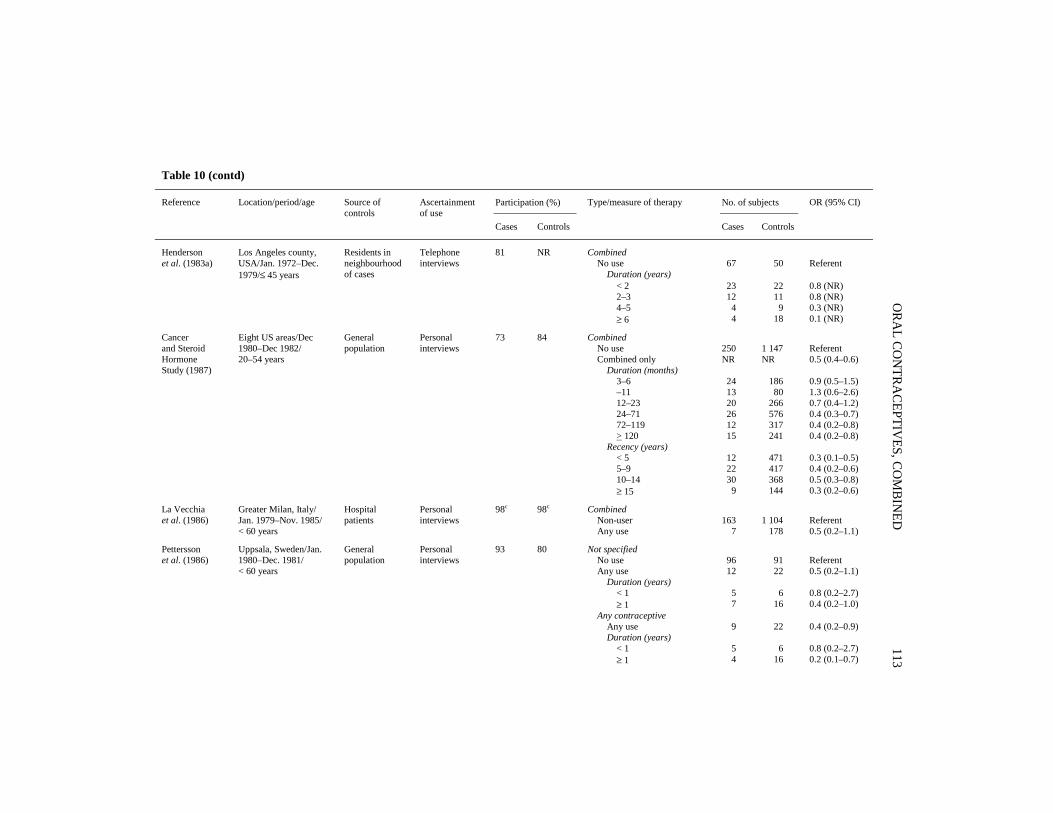
ORA
LCO
NTRA
CEPTIVES, CO
MBIN
ED113
Table 10 (contd)
Reference Location/period/age Participation (%) Type/measure of therapy No. of subjects OR (95% CI)Source ofcontrols
Ascertainmentof use
Cases Controls Cases Controls
Henderson Los Angeles county, Residents in Telephone 81 NR Combinedet al. (1983a) USA/Jan. 1972–Dec. neighbourhood interviews No use 67 50 Referent
1979/≤ 45 years of cases Duration (years) < 2 23 22 0.8 (NR) 2–3 12 11 0.8 (NR) 4–5 4 9 0.3 (NR) ≥ 6 4 18 0.1 (NR)
Cancer Eight US areas/Dec General Personal 73 84 Combinedand Steroid 1980–Dec 1982/ population interviews No use 250 1 147 ReferentHormone 20–54 years Combined only NR NR 0.5 (0.4–0.6)Study (1987) Duration (months)
3–6 24 186 0.9 (0.5–1.5) –11 13 80 1.3 (0.6–2.6) 12–23 20 266 0.7 (0.4–1.2) 24–71 26 576 0.4 (0.3–0.7) 72–119 12 317 0.4 (0.2–0.8) > 120 15 241 0.4 (0.2–0.8) Recency (years) < 5 12 471 0.3 (0.1–0.5) 5–9 22 417 0.4 (0.2–0.6) 10–14 30 368 0.5 (0.3–0.8) ≥ 15 9 144 0.3 (0.2–0.6)
La Vecchia Greater Milan, Italy/ Hospital Personal 98c 98c Combinedet al. (1986) Jan. 1979–Nov. 1985/ patients interviews Non-user 163 1 104 Referent
< 60 years Any use 7 178 0.5 (0.2–1.1)
Pettersson Uppsala, Sweden/Jan. General Personal 93 80 Not specifiedet al. (1986) 1980–Dec. 1981/ population interviews No use 96 91 Referent
< 60 years Any use 12 22 0.5 (0.2–1.1) Duration (years) < 1 5 6 0.8 (0.2–2.7) ≥ 1 7 16 0.4 (0.2–1.0) Any contraceptive Any use 9 22 0.4 (0.2–0.9) Duration (years) < 1 5 6 0.8 (0.2–2.7) ≥ 1 4 16 0.2 (0.1–0.7)

IARC M
ON
OG
RAPH
S VO
LUM
E 72114Table 10 (contd)
Reference Location/period/age Participation (%) Type/measure of therapy No. of subjects OR (95% CI)Source ofcontrols
Ascertainmentof use
Cases Controls Cases Controls
Seven countries/Jan. Hospital Personal 87 93 Combined1979–Feb. 1988/ patients interviews No use 118 687 Referent< 60 years Combined only 14 149 0.5 (0.3–1.0)
Any contraceptive No use 118 655 Referent
WHOCollaborativeStudy (1988);Rosenblatt et al.(1991)
Any use 12 180 0.5 (0.2–1.1)Combined Any contraceptive No use 182 1 072 Referent Progestogen content High Duration (months) 1–24 1 85 0.1 (0.0–0.7) ≥ 25 2 69 0.2 (0.0–0.8) Recency (months) 1–120 1 61 0.1 (0.0–0.8) ≥ 121 2 93 0.2 (0.0–0.7) Low Duration (months) 1–24 8 69 1.0 (0.5–2.4) ≥ 25 1 56 0.1 (0.0–1.1) Recency (months) 1–120 2 72 0.3 (0.0–1.1) ≥ 121 7 54 1.1 (0.5–2.8)
Koumantaki Athens, Greece/1984/ Hospital Personal 80 95 Not specifiedet al. (1989) 40–79 years patients interviews No use, ≤ 6 months’ use 80 151 Referent
> 6 months’ use 3 13 0.6 (0.2–2.0)d
Levi et al. Canton of Vaud, Hospital Personal 85a 85a Combined(1991) Switzerland/ patients interviews No use 105 227 Referent
Jan 1988–July 1990/ Any use 17 82 0.5 (0.3–0.8)32–75 years Duration (years)
< 2 9 19 1.0 (0.5–2.3) 2–5 3 18 0.5 (0.1–1.2) 5 5 45 0.3 (0.1–0.7) Recency (years) < 10 4 30 0.3 (0.1–0.9) 10–19 7 37 0.4 (0.2–1.0) > 19 5 15 0.8 (0.3–2.2)
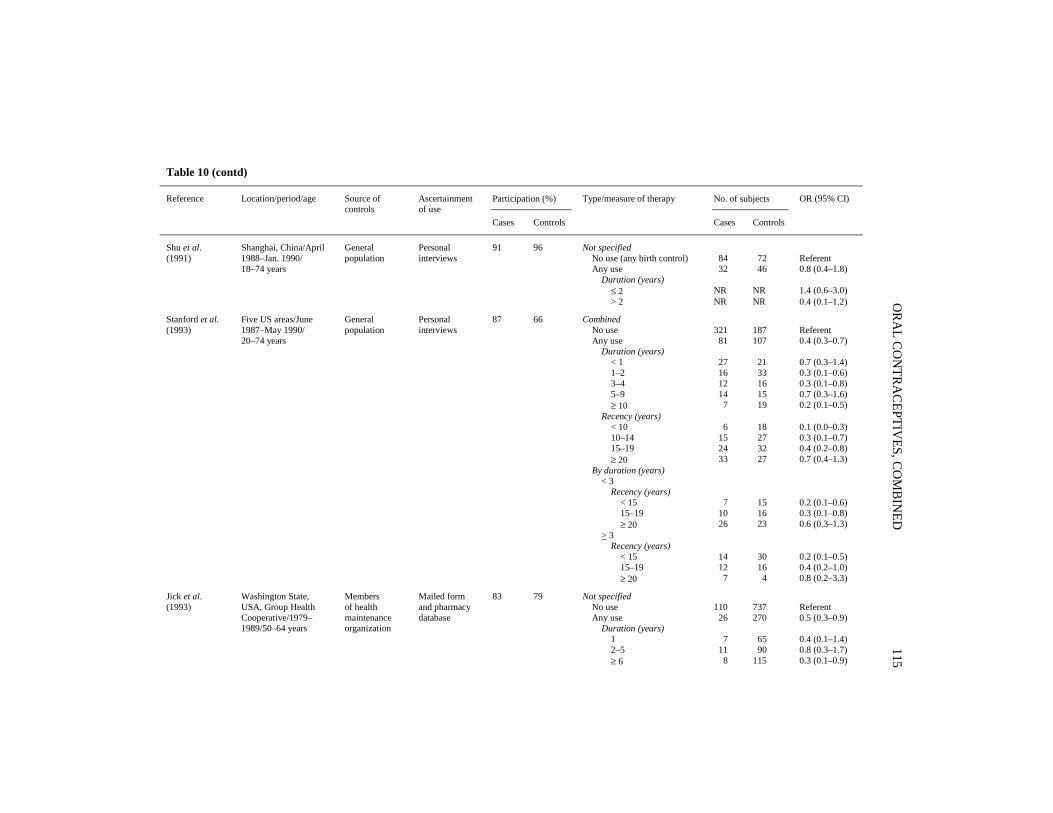
ORA
LCO
NTRA
CEPTIVES, CO
MBIN
ED115
Table 10 (contd)
Reference Location/period/age Participation (%) Type/measure of therapy No. of subjects OR (95% CI)Source ofcontrols
Ascertainmentof use
Cases Controls Cases Controls
Shu et al. Shanghai, China/April General Personal 91 96 Not specified(1991) 1988–Jan. 1990/ population interviews No use (any birth control) 84 72 Referent
18–74 years Any use 32 46 0.8 (0.4–1.8) Duration (years) ≤ 2 NR NR 1.4 (0.6–3.0) > 2 NR NR 0.4 (0.1–1.2)
Stanford et al. Five US areas/June General Personal 87 66 Combined(1993) 1987–May 1990/ population interviews No use 321 187 Referent
20–74 years Any use 81 107 0.4 (0.3–0.7) Duration (years) < 1 27 21 0.7 (0.3–1.4) 1–2 16 33 0.3 (0.1–0.6) 3–4 12 16 0.3 (0.1–0.8) 5–9 14 15 0.7 (0.3–1.6) ≥ 10 7 19 0.2 (0.1–0.5) Recency (years) < 10 6 18 0.1 (0.0–0.3) 10–14 15 27 0.3 (0.1–0.7) 15–19 24 32 0.4 (0.2–0.8) ≥ 20 33 27 0.7 (0.4–1.3) By duration (years) < 3 Recency (years) < 15 7 15 0.2 (0.1–0.6) 15–19 10 16 0.3 (0.1–0.8) ≥ 20 26 23 0.6 (0.3–1.3) > 3 Recency (years) < 15 14 30 0.2 (0.1–0.5) 15–19 12 16 0.4 (0.2–1.0) ≥ 20 7 4 0.8 (0.2–3.3)
Jick et al. 83 79 Not specified(1993) No use 110 737 Referent
Any use 26 270 0.5 (0.3–0.9) Duration (years)
Washington State,USA, Group HealthCooperative/1979–1989/50–64 years
Membersof healthmaintenanceorganization
Mailed formand pharmacydatabase
1 7 65 0.4 (0.1–1.4) 2–5 11 90 0.8 (0.3–1.7) ≥ 6 8 115 0.3 (0.1–0.9)

IARC M
ON
OG
RAPH
S VO
LUM
E 72116Table 10 (contd)
Reference Location/period/age Participation (%) Type/measure of therapy No. of subjects OR (95% CI)Source ofcontrols
Ascertainmentof use
Cases Controls Cases Controls
Jick et al. Recency (years)(1993) (contd) 1–10 5 67 0.4 (0.1–1.1)
11–15 6 82 0.4 (0.1–1.2) 16–20 4 57 0.5 (0.1–1.8) ≥ 21 9 54 0.6 (0.2–2.1)
Voigt et al. Washington State, General Personal 83 95 Combined(1994)e USA/1975–77 and population interviews and No use, < 1 year’s use 117 284 Referent
1985–87/40–59 years 73f Recency (years) > 10 Duration (years) 1–5 14 30 0.9 (0.4–1.9) > 5 4 16 0.4 (0.1–1.2) ≤ 10 Duration (years) 1–5 7 28 1.0 (0.4–2.4) > 5 7 74 0.3 (0.1–0.6) Progestogen contentg
Low Duration (years) 1–5 10 22 1.1 (0.5–2.6) > 5 3 32 0.2 (0.1–0.8) High Duration (years) 1–5 3 14 0.8 (0.2–3.1) > 5 3 28 0.3 (0.1–0.9)
Kalandidi et al. Athens, Greece/1992– Hospital Personal 83 88 Not specified(1996) 94/< 59–≥ 70 years patients interviews No use 143 293 Referent
Any use 2 5 1.3 (0.2–7.7)
OR, odds ratio; CI, confidence interval; NR, not reported; NA, not applicablea Responses reported for case and control women combinedb Crude odds ratio and 95% confidence interval calculated from data provided in the published paper by exact methodsc Methods state that less than 2% of eligible case and control women refused an interview.d 90% confidence intervale Includes women from the study of Weiss & Sayvetz (1980)f Response for controls identified 1985–1987g Classified according to subnuclear endometrial vacuolization

(b) Cohort studiesA questionnaire to derive information on oral contraceptive use was sent to approxi-
mately 97 300 married women aged 25–57 in eastern Massachusetts, United States, in1970, who were identified from the 1969 Massachusetts residence lists (Trapido, 1983).The age-adjusted rate ratio for women who had ever used oral contraceptives relative tonon-users was 1.4; there was no consistent pattern of a decreasing or increasing rate ratiowith longer or more recent use (Table 9). Among nulliparous women, the age-adjustedrate ratio for oral contraceptive users relative to non-users was 2.4 (95% CI, 0.6–9.2),whereas the analogous rate ratio for parous women was 1.4 (95% CI, 0.8–2.4). Amongwomen who also reported any use of post-menopausal oestrogen therapy, the age-adjustedrate ratio for oral contraceptive users relative to non-users was 2.0 (95% CI, 0.9–4.3). Nodistinction was made between sequential and combined oral contraceptive use, and bothpreparations were available to the cohort before and during the study follow-up.
Beral et al. (1999) followed-up approximately 23 000 oral contraceptive users and asimilar number of non-users identified in 1968 and 1969 by the Royal College of GeneralPractitioners. Use of oral contraceptives (not otherwise specified) and the occurrence ofuterine cancer were both determined from physicians’ reports. Uterine corpus cancer (i.e.excluding the cervix) was diagnosed in two of the oral contraceptive users and 16 of thenon-users, resulting in a rate ratio of 0.2 (95% CI, 0.0–0.7) after adjustment for age, parity,smoking, social class, number of previously normal Papanicolaou (‘Pap’) smears andhistory of sexually transmitted disease. In a 25-year follow-up of deaths in the cohort,there were eight deaths from endometrial cancer, two of women who had ever used oralcontraceptives and six of women who had never used them (rate ratio, 0.3; 95% CI,0.1–1.9).
The study of the Oxford Family Planning Association included 17 032 married whitewomen identified at 17 family planning clinics in England and Scotland (Vessey &Painter, 1995) who had used oral contraceptives (not otherwise specified), a diaphragmor an intrauterine device for at least five months. Information on contraceptive historyand any hospital referrals was obtained from physicians or from the women themselves(for those who stopped attending the clinics) during the study follow-up. A total of15 292 women remained under observation until the age of 45; only those who had neverused oral contraceptives (5881) or had used them for eight years or more (3520) werefollowed from then on. Endometrial cancer was diagnosed in 15 women, only one ofwhom had used oral contraceptives (age-adjusted rate ratio, 0.1; 95% CI, 0.0–0.7). In aprevious analysis of mortality in this cohort (Vessey et al., 1989b), none of the oralcontraceptive users but two of those using a diaphragm or an intrauterine device (thecomparison group) had died from uterine corpus cancer.
(c) Case–control studiesWeiss and Sayvetz (1980) compared 117 women identified from a population-based
cancer registry with 395 control women in the general population of western WashingtonState, United States. Women who had used combined oral contraceptives for one year or
ORAL CONTRACEPTIVES, COMBINED 117

more had half the risk for endometrial cancer of women who were either non-users orhad used oral contraceptives for less than one year, after adjustment for age and use ofpost-menopausal oestrogen therapy (odds ratio, 0.5; 95% CI, 0.1–1.0). No further diffe-rence in the duration of use was seen between case and control women. In stratified ana-lyses, the reduced risk was present only for women who had never used post-menopausaloestrogen therapy (odds ratio, 0.4; 95% CI, 0.1–1.1) or who had used it for two years orless (odds ratio, 0.1; 95% CI, 0.01–1.1); no reduction was noted among women who hadused it for three years or more (odds ratio, 1.3; 95% CI, 0.3–6.6).
Among 154 women with endometrial cancer and 525 control women in a hospital-based study in the United States and Canada (Kaufman et al., 1980), a 60% reduction inrisk was seen among women who used combined oral contraceptives relative to non-users,after adjustment for use of non-contraceptive hormones, parity, body mass, menopausalstatus, age at menopause, ethnic group, diabetes, education, age and area of residence. Therisk for endometrial cancer declined with increasing duration of use, and a sustainedreduction in risk was suggested for women who had stopped using oral contraceptives inthe previous five or more years. A reduction in risk was noted for women who had usedcombined oral contraceptives but had never used non-contraceptive oestrogens (oddsratio, 0.4; 95% CI, 0.2–0.8), but not for the women who had ever used both oral contra-ceptives and non-contraceptive oestrogens (odds ratio, 0.6; 95% CI, 0.3–1.6), although thelack of information on the duration of non-contraceptive oestrogen use makes it difficultto interpret this estimate.
Kelsey et al. (1982) studied women admitted to seven hospitals in Connecticut,United States. The 167 newly diagnosed cases of endometrial cancer were comparedwith 903 control women admitted for non-gynaecological surgical services. Among thestudy participants aged 45–55 years—the women who had had the opportunity to useoral contraceptives—those who had used oral contraceptives for 2.5 years or more had a50% decrease in risk.
Among 79 women treated at a hospital in North Carolina, United States, for endo-metrial cancer, 6.3% had used combined oral contraceptives for six months or more,whereas 15.3% of the 203 control women from 52 counties in the State (the main referralarea for the hospital) had done so (Hulka et al., 1982). Since only 15% of the controlwomen reported use of combined oral contraceptives, the risk estimates for more detailedaspects of oral contraceptive use are fairly imprecise (Table 10). There is a suggestionthat the risk was lower with longer use (≥ 5 years), with previous use and with use of‘progestogen-predominant’ (based on the relative proportions of oestrogens and proges-togens in their chemical composition) oral contraceptives. When oral contraceptive usewas stratified by use of post-menopausal oestrogens, both users of at least six months’duration (0 cases, 6 controls) and non-users (odds ratio, 0.6 [95% CI not provided]) ofpost-menopausal oestrogens appeared to have a reduced risk associated with use of oralcontraceptives.
Henderson et al. (1983a) identified 127 women with endometrial cancer from thepopulation-based cancer registry for Los Angeles County and matched them to control
IARC MONOGRAPHS VOLUME 72118

women of similar age who lived in the same neighbourhood as the matched case. Therisk for endometrial cancer decreased with increasing duration of use of combined oralcontraceptives, and this pattern remained after further adjustment for parity, currentweight, infertility and amenorrhoea. Neither the recency of use of oral contraceptives northe relative oestrogen and progestogen content of the oral contraceptives had a clearimpact on the risk, beyond that explained by the duration of use (data not shown). Whenthe analysis was stratified by body weight, a reduction in risk with longer duration of usewas seen among women whose current weight was less than 170 lbs [77 kg] but notamong women whose current weight was greater.
In a population-based study conducted by the Centers for Disease Control and theNational Institute of Child Health and Human Development in the United States, womenwith newly diagnosed endometrial cancer, who were 20–54 years of age, were identifiedfrom eight cancer registries (Atlanta, Detroit, San Francisco, Seattle, Connecticut, Iowa,New Mexico, and four urban counties in Utah) in the United States Surveillance,Epidemiology and End Results (SEER) Program; 3191 controls were selected from thegeneral population (Centers for Disease Control and the National Institute of ChildHealth and Human Development, Cancer and Steroid Hormone Study, 1987). Womenwho had used only combination oral contraceptives had half the risk for endometrialcancer of non-users (age-adjusted odds ratio, 0.5; 95% CI, 0.4–0.6). The risk generallydecreased with increasing duration of oral contraceptive use, the greatest reduction inrisk being seen among women who had used combined oral contraceptives for two yearsor more. The strength of the association was similar after adjustment for age alone andafter multivariate adjustment for age, parity, education, body mass, menopausal status,geographic region, exogenous oestrogen use and infertility. The risk for endometrialcancer did not vary with recency of use of oral contraceptives or time since first use; bothwomen who had ceased use of oral contraceptives 15 years or more before the studyinterview and women who had first used oral contraceptives more than 20 years beforeinterview had a lower risk than non-users (age-adjusted odds ratios, 0.3 (95% CI,0.2–0.6) and 0.4 (95% CI, 0.2–0.7), respectively). When the analysis was stratified bythe formulation of the oral contraceptive, all formulations that had been used for at leastsix months or more were associated with a decreased risk for endometrial cancer.Nulliparous women who had used combined oral contraceptives for one year or morehad a larger reduction in risk than non-users (age-adjusted odds ratio, 0.2; 95% CI,0.1–0.5), but women of high parity had little difference in risk, the age-adjusted oddsratio for women who had had five or more births being 0.8 (95% CI, 0.4–1.9). Nodifference in risk was reported with body mass, smoking, alcohol consumption, use ofexogenous oestrogens or menopausal status (data not shown) or for the different histo-logical subtypes of endometrial cancer (adenocarcinoma, adenoacanthoma and adeno-squamous carcinoma).
In a hospital-based study in the area of greater Milan, Italy, La Vecchia et al. (1986)compared the use of combined oral contraceptives by women admitted for endometrialcancer and women admitted for traumatic, orthopaedic, surgical and other conditions.
ORAL CONTRACEPTIVES, COMBINED 119

Seven (4%) of the 170 case women and 178 (14%) of the 1282 control women reporteduse of combined oral contraceptives, resulting in an odds ratio of 0.6 (95% CI, 0.2–1.3)after adjustment for age, marital status, education, parity, age at menarche, age at firstbirth, age at menopause, body mass index, cigarette smoking and use of non-contra-ceptive female hormones.
Pettersson et al. (1986) studied 254 women residing in the health care region ofUppsala (Sweden) who were referred to the Department of Gynaecologic Oncology with anewly diagnosed endometrial malignancy; each case was matched by age and county ofresidence to one control woman identified from a population registry. Use of combined oralcontraceptives was analysed for women aged 60 or less, resulting in 108 cases and 113controls. Women who had ever used these contraceptives for one year or more had a lowerrisk than non-users (odds ratios, 0.5 (95% CI, 0.2–1.1) and 0.4 (95% CI, 0.2–1.0), respec-tively). Among the women who had used combined oral contraceptives only for contra-ception, the reductions were slightly greater: odds ratios for any use versus none, 0.4(95% CI, 0.2–0.9), and for one year versus none, 0.2 (95% CI, 0.1–0.7). It is unclear fromthe published paper if the estimates were adjusted for potentially confounding factors.
A hospital-based study was conducted in Australia, Chile, China, Colombia, Israel,Kenya, Mexico, the Philippines and Thailand to compare the use of combined oralcontraceptives by 140 women with endometrial cancer and 910 women admitted to unitsother than obstetrics and gynaecology in each centre between 1979 and 1986 (WHOCollaborative Study of Neoplasia and Steroid Contraceptives, 1988). Women who hadused only combined oral contraceptives had a lower risk for endometrial cancer thannon-users (odds ratio, 0.5; 95% CI, 0.3–1.0), after adjustment for hospital, age, calendaryear of interview and race. A reduction in risk was suggested at each level of the factorsexamined, including gravidity (odds ratios, 0.7 (95% CI, 0.3–1.5) for < 5 pregnanciesand 0.3 (95% CI, 0.1–1.5) for ≥ 5 pregnancies), history of infertility (odds ratios, 0.6(95% CI, 0.3–1.2) for none and 0.4 (95% CI, 0.0–7.3) for a positive history) and use ofoestrogens for any other reason except menopausal symptoms (data not shown). Thenumbers of cases (total, 220) and control women (total, 1537) in this study continued toaccrue through 1988 and were then further evaluated by Rosenblatt et al. (1991). Amongthe women who used combined oral contraceptives for contraception only, those whoused formulations with a relatively ‘high’ dose of progestogen (on the basis of the abilityof the preparation to induce subnuclear vacuolization in human endometrium) had alower risk than non-users, regardless of the relative oestrogen dose (odds ratios, 0.2 (95%CI, 0.0–0.5) for high dose and 0 (95% CI, 0.0–1.1) for low dose). In contrast, womenwho used formulations with a relatively low dose of progestogen had little, if any,reduction in risk, regardless of the relative oestrogen dose (odds ratios, 1.1 (95% CI,0.1–9.1) for high dose and 0.6 (95% CI, 0.3–1.3) for low dose). Additionally, thereduction in risk did not vary appreciably by the duration or recency of use for thewomen who used formulations with a relatively high dose of progestogen, whereas thewomen who used formulations with a relatively low dose of progestogen had a reductionin risk with longer duration of use (odds ratio, 0.1 (95% CI, 0.0–1.1) for ≥ 2 years’ use
IARC MONOGRAPHS VOLUME 72120

versus none) or with more recent use (odds ratio, 0.3 (95% CI, 0.1–1.1) for use withinthe last 10 years versus none). Similar results were seen for first use of oral contra-ceptives within the previous 14 years. All of these estimates were adjusted for age, gravi-dity, age at menarche, centre and year of diagnosis.
Koumantaki et al. (1989) studied women with endometrial cancer admitted to twohospitals in Athens, Greece, and control women admitted to the Athens Hospital for Ortho-paedic Disorders. Only three (4%) of the 83 case women and 13 (8%) of the 164 controlshad used oral contraceptives for six or more months (odds ratio, 0.6; 90% CI, 0.2–2.0,adjusted for age, parity, age at menarche, age at menopause, menopausal oestrogen use,years of smoking, height and weight).
Among 122 women treated at a major referral hospital in the Canton of Vaud(Switzerland) for endometrial cancer, 14% had used combined oral contraceptives, as had27% of the 309 control women admitted to the same hospital for non-neoplastic, non-gynaecological conditions (Levi et al., 1991). The risk decreased from 1.0 (95% CI,0.5–2.3) for use for less than two years to 0.5 (95% CI, 0.1–1.2) for use for two to fiveyears to 0.3 (95% CI, 0.1–0.7) for use for more than five years. Oral contraceptive usewithin the previous 10 years (odds ratio, 0.3; 95% CI, 0.1–0.9) or within the previous10–20 years (odds ratio, 0.4; 95% CI, 0.2–1.0) and first use before the age of 30 (oddsratio, 0.3; 95% CI, 0.1–0.7) were all associated with a reduction in the risk for endometrialcancer. Women who had used oral contraceptives for five years or more had a reductionin risk even if use had occurred 20 or more years previously. The risk estimates wereadjusted for age, area of residence, marital status, education, parity, body mass, cigarettesmoking and use of post-menopausal oestrogen therapy. Little variation in risk was seenby categories of body mass (odds ratios, 0.6 (95% CI, 0.3–1.0) for < 25 kg/m2 and 0.2[95% CI not provided] for ≥ 25 kg/m2) or cigarette smoking (odds ratios, 0.5 (95% CI,0.2–1.2) for ever smoked and 0.6 (95% CI, 0.3–1.3) for never smoked). Stratification byuse of post-menopausal oestrogen therapy was also presented (odds ratios, 0.4 (95% CI,0.1–1.2) for ever use and 0.5 (95% CI, 0.3–1.0) for never use), but duration of post-menopausal oestrogen therapy was not analysed. While no reduction in risk was noted fornulliparous women (6 cases and 14 controls) who used oral contraceptives (age-adjustedodds ratio, 0.8; 95% CI, 0.2–2.9), the parous oral contraceptive users (11 cases and 68controls) did have a reduced cancer risk (age-adjusted odds ratio, 0.3; 95% CI, 0.1–0.7).
Shu et al. (1991) studied 268 women with endometrial cancer identified from thepopulation-based Shanghai (China) Cancer Registry and 268 age-matched controlwomen identified from the Shanghai Residents Registry. The risk for endometrial cancervaried little between users of oral contraceptives (not otherwise specified) and womenwho had never used any type of contraception, after adjustment for age, gravidity andweight (odds ratio, 0.8; 95% CI, 0.4–1.8). When the duration of use was evaluated, therewas a suggestion that oral contraceptive use for more than two years was associated witha reduction in risk (odds ratio, 0.4; 95% CI, 0.1–1.2).
In the United States, 405 women with endometrial cancer diagnosed at sevenhospitals (in Chicago, Illinois; Hershey, Pennsylvania; Irvine and Long Beach, California;
ORAL CONTRACEPTIVES, COMBINED 121

Minneapolis, Minnesota; and Winston-Salem, North Carolina) and 297 age-, race- andresidence-matched control women from the general population agreed to be interviewed(Stanford et al., 1993). Use of combined oral contraceptives was reported by 20% of thecase women and 36% of the control women (odds ratio, 0.4; 95% CI, 0.3–0.7, after adjust-ment for age, education, parity, weight and use of post-menopausal oestrogen therapy).There was no clear pattern of a decreasing risk with increasing duration of use (Table 10).Relative to non-users, a strong reduction in risk was noted for women who had used thesepreparations within the last 10 years (odds ratio, 0.1; 95% CI, 0.0–0.3) and for those whohad used them first less than 15 years previously (odds ratio, 0.1; 95% CI, 0.0–0.4); bothof these effects waned with more distant oral contraceptive use. The risk estimates variedlittle by age at first use (< 25, 25–29, 30–34, ≥ 35). When duration and recency wereevaluated jointly, use within the previous 20 years was more strongly predictive of a riskreduction than longer duration of use (≥ 3 years). In a joint evaluation with other possiblemodifying factors, three or more years of combination oral contraceptive use were asso-ciated with a reduced risk for endometrial cancer among women of high parity (odds ratiofor women with five or more births, 0.2; 95% CI, 0.0–0.6), women who weighed less than150 lbs [68 kg] (odds ratio, 0.4; 95% CI, 0.2–0.9) and women who had never (odds ratio,0.2; 95% CI, 0.1–0.6) or briefly (< 3 years) (odds ratio, 0.8; 95% CI, 0.2–3.2) used post-menopausal oestrogen therapy. No reduction and perhaps even an increase in risk wasnoted for use of combined oral contraceptives of three years or more by women who werenulliparous (odds ratio, 1.9; 95% CI, 0.3–11), weighed more than 200 lbs [91 kg] (oddsratio, 2.7; 95% CI, 0.8–8.5) or had used post-menopausal oestrogen therapy for three yearsor more (odds ratio, 4.1; 95% CI, 0.4–38). The estimates did not vary appreciably byhistory of smoking, infertility or menopausal status.
Jick et al. (1993) studied women who were members of a large health maintenanceorganization in western Washington State, United States. Women in whom endometrialcancer had been diagnosed (n = 142) were identified from the organization’s tumourregistry; the 1042 control women were also members of the organization. Both groupsincluded only women who used the pharmacies of the organization and who hadpreviously completed a questionnaire sent to all female members for a mammographystudy. Use of oral contraceptives (not otherwise specified), determined from the ques-tionnaire, was reported by 18% of case women and 26% of controls, for an odds ratio of0.5 (95% CI, 0.3–0.9), adjusted for age, enrolment date in the organization, body mass,age at menopause, parity and current use of post-menopausal oestrogen therapy. In com-parison with non-users, the reduced risk for endometrial cancer was most pronounced forwomen who had used oral contraceptives for six or more years (odds ratio, 0.3; 95% CI,0.1–0.9) or within the last 10 years (odds ratio, 0.4; 95% CI, 0.1–1.1).
Voigt et al. (1994) combined the study population described in the study of Weiss andSayvetz (1980) with a similar study population identified between 1985 and 1987 inwestern Washington State, United States. The study included 316 cases and 501 controls.When oral contraceptive use was stratified by use of unopposed oestrogen, women whohad used combined oral contraceptives for one year or more and who had also used
IARC MONOGRAPHS VOLUME 72122

unopposed oestrogens for three years or more had no reduction in risk relative to womenwho had not used oral contraceptives or women who had used them for less than one year(odds ratio, 1.1; 95% CI, 0.4–2.6), whereas a reduction was noted for women who hadnever used unopposed oestrogens or had used them for less than three years and had usedcombined oral contraceptives for more than one year (odds ratio, 0.5; 95% CI, 0.3–0.9).Thus, further analyses were restricted to women who had used unopposed oestrogensnever or for less than three years. When duration and recency of use of combined oralcontraceptives were evaluated jointly, longer use (> 5 years) was associated with areduced risk for endometrial cancer irrespective of recency (last use, ≤ 10 years ago versus> 10 years ago). When duration and the relative potency of the progestogens in the formu-lation were evaluated jointly, a longer duration of use (> 5 years), and not progestogendosage, was most predictive of a reduced risk.
Kalandidi et al. (1996) studied 145 women with endometrial cancer admitted to twohospitals in Athens, Greece, and 298 control women admitted to the major accidenthospital in Athens with bone fractures or other orthopaedic disorders. Only two (1%) ofthe case women and five (1.7%) of the controls had ever used oral contraceptives (nototherwise specified). Although a multivariate-adjusted risk estimate was presented (oddsratio, 1.3; 95% CI, 0.2–7.9), no useful inferences can be drawn from this small study.
(d) SummaryIn general, women who have taken combined oral contraceptives have about one-half
the risk for endometrial cancer of non-users (Kaufman et al., 1980; Weiss & Sayvetz,1980; Hulka et al., 1982; Kelsey et al., 1982; La Vecchia et al., 1986; Pettersson et al.,1986; Centers for Disease Control and the National Institute of Child Health and HumanDevelopment, Cancer and Steroid Hormone Study, 1987; WHO Collaborative Study ofNeoplasia and Steroid Contraceptives, 1988; Koumantaki et al., 1989; Levi et al., 1991;Jick et al., 1993; Stanford et al., 1993; Vessey & Painter, 1995; Beral et al., 1999). Thereduction first appears after two to five years of use (Kaufman et al., 1980; Hulka et al.,1982; Henderson et al., 1983a; Pettersson et al., 1986; Centers for Disease Control andthe National Institute of Child Health and Human Development, Cancer and SteroidHormone Study, 1987; Levi et al., 1991; Shu et al., 1991; Jick et al., 1993; Stanford et al.,1993; Voigt et al., 1994) and continues to decrease as the duration of oral contraceptiveuse increases (Kaufman et al., 1980; Henderson et al., 1983a; Centers for Disease Controland the National Institute of Child Health and Human Development, Cancer and SteroidHormone Study, 1987; Levi et al., 1991; Stanford et al., 1993). Some studies have showna greater reduction in risk with more recent use (Levi et al., 1991; Jick et al., 1993;Stanford et al., 1993), but others have found no difference (Kaufman et al., 1980;Henderson et al., 1983a; Centers for Disease Control and the National Institute of ChildHealth and Human Development, Cancer and Steroid Hormone Study, 1987; WHOCollaborative Study of Neoplasia and Steroid Contraceptives, 1988). When duration andrecency of use were evaluated jointly, longer use (≥ 5 years) was associated with areduced risk, irrespective of recency (Voigt et al., 1994), whereas another study showed
ORAL CONTRACEPTIVES, COMBINED 123

that recency (use within the last 15 years) and not duration of use was most predictive ofa reduced risk (Stanford et al., 1993). Some studies found that the reduction in risk maybe greatest with use of oral contraceptives in which progestogen effects predominate(Hulka et al., 1982) or that contain higher doses of progestogen (Rosenblatt et al., 1991),but another study found that a longer duration of use (≥ 5 years), and not progestogendose, was most predictive of a reduced risk (Voigt et al., 1994).
While no reduction in risk was found for women in the highest categories of bodyweight in two studies (Henderson et al., 1983a; Stanford et al., 1993), two others found areduced risk regardless of weight or body mass (Centers for Disease Control and theNational Institute of Child Health and Human Development, Cancer and Steroid HormoneStudy, 1987; Levi et al., 1991). Although one study noted a reduced risk only among oralcontraceptive users who were nulliparous (Centers for Disease Control and the NationalInstitute of Child Health and Human Development, Cancer and Steroid Hormone Study,1987), three others found that the reductions were strongest among parous women (Leviet al., 1991) or women of higher parity (≥ 5 births) (WHO Collaborative Study of Neo-plasia and Steroid Contraceptives, 1988; Stanford et al., 1993). In comparison with womenwho did not use oral contraceptives, oral contraceptive users who had also used post-meno-pausal oestrogen therapy for three or more years showed no reduction in risk in two studies(Stanford et al., 1993; Voigt et al., 1994). While four other studies did find a reduced riskamong oral contraceptive users who had ever used post-menopausal oestrogen therapy(Kaufman et al., 1980; Hulka et al., 1982; Centers for Disease Control and the NationalInstitute of Child Health and Human Development, Cancer and Steroid Hormone Study,1987; Levi et al., 1991), the inclusion of women who had used this therapy for fewer thantwo or three years could have obscured any altered relationship with longer duration of use.
2.2.2 Sequential oral contraceptives(a) Case reports
In the mid-1970s, case reports appeared in the United States of endometrialabnormalities—ranging from proliferative lesions to severe atypical hyperplasia (Lyon &Frisch, 1976; Kaufman et al., 1976; Cohen & Deppe, 1977) to endometrial cancer (Lyon,1975; Silverberg & Makowski, 1975; Silverberg et al., 1977)—among women who hadused a sequential oral contraceptive preparation, Oracon®, containing 0.1 mg ethinyl-oestradiol and 25 mg dimethisterone (Weiss & Sayvetz, 1980). In response to thesereports, sequential preparations were removed from the consumer market in the UnitedStates and Canada in 1976, but the impact of exposure to these preparations continued tobe evaluated in epidemiological studies.
(b) Case–control studiesThe epidemiological studies of sequential oral contraceptive use and endometrial
cancer are summarized in Table 11. Weiss and Sayvetz (1980) reported a seven-fold ele-vation in risk with use of Oracon®, but not with other types of sequential preparations,after adjustment for age, use of combined oral contraceptives and post-menopausal
IARC MONOGRAPHS VOLUME 72124

ORA
LCO
NTRA
CEPTIVES, CO
MBIN
ED125
Table 11. Case–control studies of use of sequential oral contraceptive pills and risk for endometrial cancer
Participation (%) No. of subjectsReference Location/period/ages Source ofcontrols
Ascertainmentof use
Cases Controls
Type/measureof therapy
Cases Controls
Odds ratio(95% CI)
Weiss & Washington State, General Personal 83 96 No use 110 376 ReferentSayvetz USA/Jan. 1975–Dec. population interviews Oracon® 6 8 7.3 (1.4–39)(1980) 1977/36–55 years Other 1 11 0.3 (0.0–2.9)
Kaufman USA and Canada/July Hospital Personal 96a 96a No use 152 516 Referentet al. (1980) 1976–Dec. 1979/ patients interviews Any use 2 9 [0.8 (0.2–2.8)]b
< 60 years Oracon® 1 3 [1.1 (0.2–6.0)]b
Henderson Los Angeles county, General Telephone 81 NR No use 116 121 Referentet al. USA/Jan. 1972–Dec. population interviews Duration (years)(1983a) 1979/< 45 years < 2 2 5 0.4 (NR)
> 2 9 1 4.6 (NR)
Cancer Eight US areas/ General Personal 73 84 No use 250 1 147 Referentand Steroid Dec. 1980–Dec. 1982/ population interviews Only sequential 7 64 0.6 (0.3–1.3)Hormone 20–54 yearsStudy (1987)
Seven countries/Jan. Hospital Personal 87 93 No use 118 687 ReferentWHOCollaborativeStudy (1988)
1979–Feb. 1986/< 60 years
patients interviews Only sequential 1 5 0.9 (0.1–8.3)
CI, confidence interval; NR, not reporteda Responses reported for case and control women combinedb Crude odds ratio and 95% CI calculated from data provided in published paper using exact methods

oestrogen therapy, among 117 women with endometrial cancer identified from a popu-lation-based cancer registry and 395 women from the general population in westernWashington State, United States.
Henderson et al. (1983a) evaluated oral contraceptive use among 127 white case–control pairs matched for age (in five-year age groups) and area of residence; the casewomen were identified from the population-based University of Southern CaliforniaCancer Surveillance Program and controls from the case’s neighbourhood of residence.An almost fivefold increase in risk was found with the use of any type of sequential oralcontraceptive for two years or more on the basis of use by nine case women and onecontrol. [The particular brand of sequential oral contraceptive, or the combination ofbrands, used is not clear from the published paper.]
A study in the United States (Atlanta, Georgia; Detroit, Michigan; San Francisco,California; Seattle, Washington; Connecticut, Iowa, New Mexico and four urban areas ofUtah; Centers for Disease Control and the National Institute of Child Health and HumanDevelopment, Cancer and Steroid Hormone Study, 1987) found that only seven of 433case women and 64 of 3191 controls had exclusively used sequential oral contraceptives,resulting in an age-adjusted odds ratio of 0.6 (95% CI, 0.3–1.3). Among the larger groupof women with any use of sequential oral contraceptives (26 cases and 152 controls), therisk for endometrial cancer for women who had used them in the previous three to 12years, for three years or more or who had used Oracon® was 1.5 times that of othersequential oral contraceptive users. No estimates of the risk for these women relative tothat of non-users was provided in the published paper.
Two other studies found neither an excess nor a decreased risk among small numbers ofwomen who had used sequential oral contraceptives. In a hospital-based study in severalmetropolitan areas in the United States and Canada, Kaufman et al. (1980) reported that onlytwo (1.3%) of the 154 case women and nine (1.7%) of the 525 control women had reporteduse of any type of sequential oral contraceptive during personal interviews; one of the casewomen and three control women reported using Oracon®.
In the international hospital-based study described on p. 120, only one of the 140case women and five of the 910 control women had exclusively used sequential oralcontraceptives (crude odds ratio, 0.9; 95% CI, 0.1–8.3); the specific preparations werenot reported (WHO Collaborative Study of Neoplasia and Steroid Contraceptives, 1988).
In summary, the case reports that preceded the epidemiological studies were importantin indicating that the risk for endometrial cancer was potentially elevated among users ofsequential oral contraceptives and specifically among users of a particular brand, whichcontained a relatively potent oestrogen, ethinyloestradiol, and a weak progestogen,dimethisterone. In contrast, it was not clear from the case–control studies whether theincrease in risk was restricted to users of this brand or included users of other sequentialpreparations. This was largely due to the low prevalence of sequential oral contraceptive usein these study populations: only 6% or less of the control women in all of the studies. Whenthe analyses were further stratified by specific preparations, the numbers of women in eachcategory were too small for useful inferences to be drawn from most of these studies.
IARC MONOGRAPHS VOLUME 72126

2.3 Cervical cancer2.3.1 Methodological considerations
(a) Stage of disease and classificationCervical cancer is a particularly difficult disease to study with respect to use of oral
contraceptives. It is generally accepted that invasive cervical cancer results from a seriesof changes in the cervical epithelium, from normal epithelial structure to various gradesof pre-invasive changes and then on to invasive cervical carcinoma. As oral contra-ceptives could act at any stage in this process to enhance progression to the next stage,studies should include separate assessment of the effects of steroid contraceptives on riskat different stages of the neoplastic process. Early studies of oral contraceptives andcervical neoplasia included a mixture of lesion types, and these are not considered in thisreview. In the studies of specific types of preneoplastic lesions, there is considerablevariation in the definition of the cases included. In addition, the systems used to classifyprecancerous cervical lesions histologically and cytologically have changed over time.Early studies included cervical dysplasia (sometimes sub-classified into mild, moderateand severe) and carcinoma in situ. In more recent studies, cases have been classified ascervical intraepithelial neoplasia (CIN), with a grading system of I–III to designate theseverity of the lesion. Lesions have also been referred to histologically as squamous intra-epithelial neoplasia and similarly graded on a scale of I–III to indicate severity. In general,the higher grades correspond roughly to carcinoma in situ and severe dysplasia, and thelower grades correspond roughly to mild and moderate dysplasia. In reviewing theliterature on non-invasive cervical neoplasia, the terms used by the authors have beenretained.
The two generally recognized histological types of invasive cervical carcinoma aresquamous-cell carcinoma and adenocarcinoma. In many studies of invasive cervicalcancer, these histological types have not been distinguished. In this review, such studiesare usually classified with those of squamous-cell carcinoma, because squamous-cellcarcinoma was the more common type at the time and in the places where the studies thatdid not distinguish them were conducted.
(b) Confounding and effect-modifying variablesAnother difficulty in assessing the effect of oral contraceptives on the risk for cervical
cancer is that the disease is caused by several types of human papillomavirus (HPV)(IARC, 1995). These viruses are sexually transmitted, and women with cervical neoplasiatend to be those whose sexual behaviour is conducive to the acquisition of sexuallytransmitted diseases, or who are married to men who have engaged in extramarital sexualrelationships conducive to the acquisition of sexually transmitted agents. In somecultures, women who use oral contraceptives tend also to be women whose sexualbehaviour is conducive to the acquisition of sexually transmitted agents. Under suchcircumstances, a spurious association between use of oral contraceptives and cervicalneoplasia could be observed, if sexual practices are not controlled for either in the studydesign or in the statistical analysis. Unless otherwise stated, studies in which the sexual
ORAL CONTRACEPTIVES, COMBINED 127

behaviour of the subjects has not been taken into consideration have been excluded fromthis review.
In recent studies, attempts have been made to control for HPV infection whenassessing possible associations between use of oral contraceptives and cervical neoplasia.To date, however, all attempts to do so have been limited by technical deficiencies. It isgenerally accepted that cervical neoplasia results from persistent infection with anoncogenic type of HPV. If a woman clears her infection, then she is unlikely to develop acervical neoplasm. If oral contraceptives were to enhance the risk for cervical cancer byincreasing the likelihood that an HPV infection will become persistent, women should beclassified according to whether they have persistent infection with an oncogenic HPV. Ina case–control study, this would require an adequate serological test for markers of HPVpersistence; to date, no such test has been developed. Another approach would be toconduct a prospective follow-up study of a large group of women who have recentlyacquired an oncogenic HPV type for the development of cervical neoplasia. This approachhas several limitations: one is that women could be monitored only until they developedmild or moderate intraepithelial lesions, since it would be unethical not to treat suchlesions and allow them to progress to more severe disease; the second problem is that suchstudies require large numbers of women and a long duration of follow-up. Studies of mildintraepithelial lesions are under way, but the results in relation to use of hormonalcontraceptives to date are limited; furthermore, the results of studies of mild lesions maynot indicate a relationship between use of oral contraceptives and more severe disease.
Another possibility is that oral contraceptives enhance the risk for cervical cancer inwomen with persistent HPV infection. In order to address this issue in case–controlstudies, analyses have been restricted to cases and controls with evidence of HPV DNAin cervical scrapings. In such studies that have been conducted to date, few controls havebeen found to have HPV, and the relative risk estimates are therefore imprecise.
(c) Studies of oral contraceptives and human papillomavirus infectionBecause oncogenic forms of HPV are involved in the etiology of cervical carcinoma,
a number of investigations have been conducted to determine whether infection withHPV is associated with the use of oral contraceptives. It has been clearly shown that thesensitivity and specificity of methods for detecting HPV differ significantly. Methodsinvolving the polymerase chain reaction (PCR) of DNA have been found to be the mostsensitive and specific when compared with other methods such as filter in situ, dot–blotand Southern blot hybridization (IARC, 1995); and epidemiological studies of cervicalcarcinoma in which methods other than PCR have been used to detect HPV should beinterpreted with the understanding of potential misclassification of HPV status. Studieson younger women have given inconsistent results for an association between theprevalence of HPV infection and oral contraceptive use. The following section is limitedto studies in which PCR-based techniques were used.
Hildesheim et al. (1993) investigated the risk factors for HPV infection in 404 cyto-logically normal low-income women in Washington DC, United States, of a median age
IARC MONOGRAPHS VOLUME 72128

of 26 years. The prevalence of HPV infection was found to be higher among currentusers of oral contraceptives (42.9%) than among women who had never used them(33.3%). Former users (prevalence, 40%) were also at increased risk of having a currentHPV infection (difference in prevalence, 2.6%; 95% CI, -10.2–15.5), although thesefindings were not significant.
Ley et al. (1991) found an increased risk for HPV infection with oral contraceptiveuse in their study of 467 university women of a mean age of 23 years. A higher pre-valence of HPV infection was associated with both past (crude odds ratio, 3.0; 95% CI,1.8–5.0) and current use (crude odds ratio, 3.3; 95% CI, 2.1–5.3).
Bauer et al. (1993) examined factors associated with HPV prevalence among 483 cyto-logically normal women of a median age of 34 years. The prevalence in non-users, formerusers and current users of oral contraceptives was 5.3, 12.8 and 34.0%, respectively, butthis difference, after adjusting for confounding factors, could have occurred by chance.
Burk et al. (1996) studied 439 sexually active women in Brooklyn, New York, UnitedStates, of an average age of 31 years. Women who had ever used oral contraceptives butwere not current users had a higher prevalence of HPV infection (21.9%) than those whohad never used them (17.1%); current users had a 14.8% rate of HPV PCR-DNA positivity.
Wheeler et al. (1993) found that oral contraceptive use was not associated with HPVinfection among 357 cytologically normal university women in New Mexico, UnitedStates, of a median age of 23 years. The prevalences of HPV infection in former users(43.9%) and current users (41.8%) were not significantly different from that in womenwho had never used them (50%) after control for other confounding factors.
Muñoz et al. (1996) investigated the association between HPV DNA positivity andrisk factors among 810 middle-aged women who were controls in case–control studiesof cervical cancer conducted in Spain, Colombia and Brazil. The mean age of thesewomen differed by site: 41.7 years in Spain, 42.8 years in Colombia and 52.7 years inBrazil. Use of oral contraceptives was not significantly associated with HPV DNA posi-tivity. When compared with non-users, women who had used contraceptives for threeyears or less (odds ratio, 0.7; 95% CI, 0.4–1.4) and more than three years (odds ratio,0.6; 95% CI, 0.3–1.2) were not at increased risk for HPV infection.
Ho et al. (1998), investigating the risk factors for the acquisition of HPV infection inuniversity women, found that oral contraceptive use was not significantly associated.
In a follow-up study of 393 women with normal cervical cytology, Hildesheim et al.(1994) found no evidence that persistence of HPV infection was associated with use oforal contraceptives.
The inconsistent results of these studies could be due to differences in the sexualbehaviour of oral contraceptive users and non-users in the studies. In the aggregate, theydo not provide direct evidence that oral contraceptives interact with HPV to causecervical cancer. Some are, nevertheless, consistent with a role for oral contraceptives inthe genesis of cervical cancer, either by enhancing the likelihood of infection or persis-tence of infection by oncogenic types or by some direct, synergistic mechanism of HPVand oral contraceptives.
ORAL CONTRACEPTIVES, COMBINED 129

(d) Influence of screeningA third problem in assessing the effect of hormonal contraceptives on the risk for
cervical cancer is the influence of the results of Pap smears. If the cases detected atscreening are those more likely to be studied, and if women are more likely to have Papsmears if they have used oral contraceptives, then the women who are studied may bemore likely to have used oral contraceptives than other cases in the population. Thiscould lead to spuriously elevated relative risks in relation to oral contraceptive use incase–control studies, particularly for studies of intraepithelial lesions, which are largelyasymptomatic and frequently detected at screening. Because of this potential bias, studiesof intraepithelial lesions in which both the cases and the controls came from the samescreening programme (the preferred design) are distinguished in this review from thosein which they were not.
Screening with Pap smears may also influence the results of studies of invasivedisease. If having a Pap smear protects against invasive disease, fewer cases will haveused oral contraceptives than in the general population, which could result in a spuriouslylow relative risk. The influence of prior Pap smears must therefore be considered inassessing the risk for both intraepithelial and invasive cervical neoplasms in relation tooral contraceptive use.
2.3.2 Descriptive studiesDoll (1985) noted that mortality rates from cervical cancer in Britain increased in
women born after 1935, corresponding to some change that took place in about 1960.This is approximately when oral contraceptives came into use, but it is also when womenbegan to change their sexual behaviour, so that the trend could be the result of increasedrates of HPV infection.
Peters et al. (1986a) reported an increase in the proportion of all newly diagnosed cer-vical adenocarcinomas in non-Hispanic white women under the age of 35 in Los AngelesCounty, United States, between 1972 and 1982. There was no increase in the risk foradenocarcinoma in older women, and there was a decreased prevalence incidence ratio forinvasive squamous-cell cervical carcinoma in women of all ages during the same timeperiod. The authors hypothesized that the trends were due to the introduction of oral contra-ceptives, which might preferentially increase the risk for adenocarcinomas over that forsquamous-cell carcinomas. Schwartz and Weiss (1986) analysed data from the UnitedStates SEER Program and also noted an increase in the risk for adenocarcinomas between1973 and 1982 in women under the age of 35. No comparable increase in the risk foradenocarcinomas was observed in older women, and no increase in the risk for adeno-squamous carcinomas or squamous-cell carcinomas was observed for the same period. Infact, the rates of squamous-cell carcinomas had decreased in all age groups during thosesame years. The results of this study are thus consistent with those of Peters et al. (1986a)and are not inconsistent with the hypothesis that use of oral contraceptives is associatedwith an increase in the risk for adenocarcinomas. Chilvers et al. (1987) reported, however,an increased risk for both adenocarcinoma and squamous-cell carcinoma in women under
IARC MONOGRAPHS VOLUME 72130

the age of 35 in three regions of England between 1968 and 1982, which would argueagainst a particularly strong increase in risk for adenocarcinomas associated with use oforal contraceptives.
Trends in the incidence rates of adenocarcinoma and adenosquamous carcinomaduring the period 1973–91 were examined by Vizcaino et al. (1998) in 60 population-based registries in 25 countries. Consistent with the results of Doll (1985), they found asignificant increase in the incidence of this condition in many countries between 1973and 1991. The authors suggested that the increase was due in part to increasedtransmission of HPV; they also suggested that it was due in part to improvements inscreening. With the introduction of the cyto-brush, more cervical adenocarcinomasin situ are being detected in some populations, which could result in a decline in the ratesof invasive cervical adenocarcinoma. The patterns of the temporal changes acrosscountries do not appear to be explained by variations in the patterns of use of oralcontraceptives among these populations; and the observation that the rates of squamousand adenocarcinoma of the cervix are highly correlated among the populations studiedsuggests that oral contraceptives do not preferentially enhance the risk for adeno-carcinomas over that for squamous-cell carcinomas.
2.3.3 Cohort studies(a) Studies of cervical dysplasia and carcinoma in situ in the absence
of assays for human papillomavirus DNAPeritz et al. (1977) reported the results of a cohort study of 17 942 women, 18–58 years
of age, who received health examinations at the Kaiser Permanente Medical Center inWalnut Creek, California, United States, between 1968 and 1972. They did not provideserial Pap smears but, between 1973 and 1975, all women in the health plan who developeddysplasia or carcinoma in situ of the cervix were identified from medical records. Aftercontrolling for age, education, marital status, number of Pap smears before entry into thecohort, smoking and selected infections, the relative risk for either cervical dysplasia orcarcinoma in situ was found to increase with the duration of oral contraceptive use.Carcinoma in situ and cervical dysplasia were combined in the estimates of relative risk, butthe inclusion of squamous dysplasia in the analyses reduced the strength of the association,suggesting that the association was stronger for carcinoma in situ than for dysplasia.
Between 1970 and 1972, approximately 32 000 15–39-year-old women were re-cruited for a study in Ljubljana, Yugoslavia, through family planning and gynaecologicalclinics (Andolsek et al., 1983). Attempts were made to collect Pap smears from womenin the cohort annually, but large numbers of women were lost during the seven-yearfollow-up period. After adjustment for years of follow-up, age at first pregnancy andnumber of Pap smears, there was no significant increase in the risk for either carcinomain situ or severe dysplasia in women who had used oral contraceptives. When the twoconditions were combined, there was no trend of increase in risk with duration of use.
The results of three cohort studies that specifically assessed the risk for cervicaldysplasia in relation to oral contraceptive use are summarized in Table 12. The study of
ORAL CONTRACEPTIVES, COMBINED 131

the Oxford Family Planning Association (Zondervan et al., 1996) covered 17 032 womenwho were recruited at 17 large family planning clinics in England and Scotland between1968 and 1974. The most recent results represent 22 years of follow-up. No increase inthe risk for cervical dysplasia was observed with duration of oral contraceptive use. Asmall increase in risk, of borderline statistical significance, was observed for currentusers; however, this possible increase did not persist 12 months after last use.
The New Zealand Contraception and Health Study Group (1994) followed a cohort of7199 women who had initially had two Pap smears showing no dysplasia for an averageof 5.5 years of follow-up. The women were screened annually for cervical abnormalities.When the cohort was established, 2469 women were using oral contraceptives, 2072women were using an intrauterine device and 1721 women were using depot medroxy-
IARC MONOGRAPHS VOLUME 72132
Table 12. Cohort studies of use of oral contraceptives and cervical dysplasia
Reference (datecohort started)
Comparisongroups
No. ofcases
Relative risk(95% CI)
Comments
Zondervan et al. No use 35 1.0(1996) Any use 124 1.1 (0.7–1.7)(1968–74) Current use 59 1.7 (1.0–2.8)
Months of use 1–12 5 0.8 (0.3–2.1) 13–24 5 0.7 (0.2–1.9) 25–48 11 0.5 (0.3–1.1) 49–72 34 1.8 (1.0–3.0) 73–96 26 1.2 (0.7–2.2) ≥ 97 43 1.1 (0.6–1.8)
– adjusted for social class,smoking, age at first birth,diaphragm use, condom use;– p value of test for trend = 0.2;– 22 years of follow-up;– no increase in risk after12 months since last use
New Zealand IUD 92 1.0Contraception and Use 125 1.2 (0.9–1.6)Health Study Group(1994)(1980–86)
– adjusted for smoking, age atfirst intercourse, number ofpartners, use of depotmedroxyprogesterone acetate;– 5.5 years of follow-up
Gram et al. (1992) No use NR 1.0(1979–80) Past use NR 1.4 (1.0–1.8)
Current use NR 1.5 (1.1–2.1)Age started > 24 1.1 (0.7–1.8) 20–24 1.5 (1.1–2.0) < 20 1.3 (0.9–1.9)
– adjusted for marital status,age group, smoking, alcoholabuse, oral contraceptive use;– 7 years’ mean follow-up ofusers;– p value of test for trend = 0.05– 354 women with CIN grade Ior II, 44 with CIN grade III, and3 with carcinoma; results notaltered when analysis restrictedto grade I or II
IUD, intrauterine device; NR, not reported; CIN, cervical intraepithelial neoplasia

progesterone acetate. In comparison with women who had used an intrauterine device,women who had used oral contraceptives were not at increased risk for cervical dysplasia.The women in the cohort had used oral contraceptives for an average of 2.5 years.
Between 1979 and 1980, 6622 women between the ages of 20 and 49 in Tromsø,Norway, were interviewed and subsequently followed-up for 10 years (Gram et al.,1992) by linking the cohort to computerized information in the pathology registry at theUniversity of Tromsø. Serial Pap smears were not taken from all women, although atleast one cytological smear was recorded for 96% of the women in the registry between1980 and 1989. As most of the cases were CIN-I or -II, this study is summarized inTable 12 with the two studies that provide information on dysplasia. The risk for diseasewas significantly increased among women who were using oral contraceptives when thecohort was established; it was somewhat lower and of borderline statistical significancefor past users. Women who first used oral contraceptives before the age of 24 were atslightly greater risk than were women who began using them later. The difference is not,however, statistically significant and could be due to differences in duration of useamong women who began using oral contraceptives at different ages. No information onduration of use was reported.
Table 13 shows the results of two cohort studies of oral contraceptives and cervicalcarcinoma in situ. In the study of the Oxford Family Planning Association (Zondervanet al., 1996), the risk of women who had used oral contraceptives for more than 96months was significantly increased, but no significant trend of increasing risk withduration of use was observed. The risk was also increased in current users of oral contra-ceptives but not in women who had stopped use for more than one year.
The study of the Royal College of General Practitioners (Beral et al., 1988) wasbegun in 1968. Over 23 000 women who were taking oral contraceptives at the time andan approximately equal number of women who had never taken oral contraceptives wererecruited by 1400 general practitioners throughout the United Kingdom, who reporteddetails of oral contraceptive use and the health status of each woman in the study twiceeach year. After 17–19 years of follow-up, a significantly increased risk for cervicalcarcinoma in situ was found for women who had ever used oral contraceptives. The riskwas also observed to increase with duration of use.
It should be noted that information on the number of sexual partners was collectedonly by the New Zealand Contraception and Health Study Group. All of the associationssummarized in Tables 12 and 13 could, therefore, be due to residual confounding bysexual variables. It should also be noted that all of the risk estimates for current userswere increased and that the risk decreased after cessation of use. These observations areconsistent with a screening bias: women taking oral contraceptives may be more likelyto have Pap smears than women who are not. On balance, the results of the cohort studiesdo not provide strong evidence that cervical dysplasia or carcinoma in situ is causallyrelated to use of oral contraceptives.
ORAL CONTRACEPTIVES, COMBINED 133

(b) Studies of cervical dysplasia in which assays for human papilloma-virus DNA were performed
Three cohort studies of a different design from those summarized in Tables 12 and13 have been conducted. Koutsky et al. (1992) followed-up a cohort of 241 women withnormal cervical cytology by cytological and colposcopic examinations every fourmonths for approximately two years. HPV DNA was detected by dot–filter hybridizationand Southern blot hybridization for confirmation. The risk for CIN-II or -III was notassociated with use of oral contraceptives.
Liu et al. (1995) assembled a cohort of 206 women with cervical dysplasia who hadbeen recruited into a randomized trial of the effect of folic acid supplementation on thecourse of cervical dysplasia; they had provided two to four cervical smears, which weretested for HPV-16 by Southern blotting. Follow-up examinations were conducted everytwo months for a total of six months. The risk for progression from low- to high-gradedysplasia was not associated with past or current use of oral contraceptives: the relativerisk for progression in HPV-16-negative women was 1.6 (95% CI, 0.8–3.1) for past usersversus never users and 1.4 (95% CI, 0.7–2.7) for current versus never users, whereas thecomparable relative risks in HPV-16-positive women were 0.8 (95% CI, 0.6–1.1) and 0.8(95% CI, 0.6–1.0), respectively. Although the differences in relative risk estimates forHPV-16-negative and -positive women could have occurred by chance, they are consistent
IARC MONOGRAPHS VOLUME 72134
Table 13. Cohort studies of use of oral contraceptives and cervical carcinomain situ
Reference (datecohort started)
Comparisongroups
No. ofcases
Relative risk(95% CI)
Comments
Zondervan et al. No use 22 1.0(1996) Any use 99 1.7 (1.0–3.0)(1968–74) Current use 45 2.2 (1.2–4.1)
Months of use 1–12 4 1.4 (0.5–4.4) 13–24 7 1.8 (0.7–4.6) 25–48 20 1.7 (0.8–3.5) 49–72 18 1.5 (0.7–3.0) 73–96 11 1.2 (0.5–2.6) ≥ 97 39 2.5 (1.3–4.7)
– adjusted for social class,smoking, age at first birth,diaphragm use, condom use;– p value of test for trend = 0.2;– 22 years of follow-up;– no significant increase in riskafter 12 months since last use
Beral et al. (1988) No use 34 1.0(1968–70) Any use 173 2.9 (2.0–4.1)
Years of use < 5 84 2.4 5–9 66 3.6 ≥ 10 23 4.8
– adjusted for age, parity,smoking, social class, number ofprior normal Pap smears;– p value of test for trend, < 0.001;– follow-up through 1987(17–19 years)
CI, confidence interval

with the hypothesis that oral contraceptives enhance progression of dysplasia in theabsence of HPV-16.
In a study of similar design (Ho et al., 1995), 70 women with cervical dysplasia werefollowed at three-month intervals for 15 months. HPV DNA was assayed by PCRtechniques. The risk for persistent dysplasia was not associated with oral contraceptiveuse after HPV status was taken into account; results stratified by HPV status were notpresented.
(c) Studies of invasive cervical carcinomaThe results of two cohort studies of the risk for invasive cervical carcinoma in relation
to oral contraceptive use are summarized in Table 14. HPV status was not considered ineither study. The study of the Oxford Family Planning Association (Zondervan et al.,1996) found an increased risk for invasive cervical carcinoma in women who had everused oral contraceptives that was of borderline statistical significance. The risk was parti-cularly enhanced for women who had used oral contraceptives within the past two years.There was no trend of increase in risk with duration of use.
The study of the Royal College of General Practitioners (Beral et al., 1988) alsoshowed an increase in risk for invasive cervical carcinoma of borderline statistical signi-ficance among women who had ever used oral contraceptives and an increase in risk withduration of use. Beral et al. (1999) also found an increase in risk for deaths due to cervicalcarcinoma. On the basis of 25 years of follow-up and 172 deaths, the relative risk for
ORAL CONTRACEPTIVES, COMBINED 135
Table 14. Cohort studies of use of oral contraceptives and invasive cervicalcarcinoma
Reference (datecohort started)
Comparison groups No. ofcases
Relative risk(95% CI)
Comments
Zondervan et al. No use 2 1.0(1996) Any use 31 4.4 (1.0–32)(1968–74) Use in past 2 years 21 6.8 (1.6–49)
Months of use1–24 4 5.5 (0.8–51)25–72 6 2.8 (0.5–23)≥ 73 21 4.7 (1.1–33)
– adjusted for social class,smoking, age at first birth,diaphragm use, condom use;– p value of test for trend, 0.8;– 22 years of follow-up;– no significant increase inrisk after 24 months since lastuse
Beral et al. (1988) No use 16 1.0(1968–70) Any use 49 1.8 (1.0–3.3)
Years of use< 5 21 1.35–9 17 2.0≥ 10 11 4.4
– adjusted for age, parity,smoking, social class, numberof prior normal Pap smears;– p value of test for trend,< 0.001;– follow-up through 1987(17–19 years)
CI, confidence interval

dying from cervical cancer among women who had ever used oral contraceptives was 1.7(95% CI, 0.9–3.2). The relative risk increased with duration of use (p value for trend, 0.03)and was 4.1 (95% CI, 1.6–11) for users of 10 or more years’ duration. The risk decreasedwith time since cessation of use and was not significantly increased 10 years afterexposure.
Because these results are for invasive cervical cancer, they are unlikely to be due topreferential screening of women taking oral contraceptives. They could, however, be dueto incomplete control of the confounding influence of sexual behaviour, since in neitherof these studies was a detailed sexual history obtained.
2.3.4 Case–control studies(a) Studies of cervical intraepithelial neoplasia not based on screening
programmesTen case–control studies of CIN in relation to use of oral contraceptives are summarized
in Table 15. In all of these studies, the cases were selected from clinics, hospitals or tumourregistries, and controls were selected from clinics, hospitals or the general population. HPVstatus was not assessed in any of these investigations. Because the cases and controls werenot selected from the same screening programme, these studies are more likely than studiesbased on screened populations to be influenced by screening bias. Nevertheless, an attemptwas made in all of the studies to control for both sexual variables and prior screening, andthey therefore provide useful information on the possible association between CIN and oralcontraceptive use. A study by Hellberg et al. (1985) is omitted from Table 15 because, thecontrols were pregnant women and, as such, were not representative of the population fromwhich the cases came with respect to contraceptive factors. Furthermore, no relative riskestimates were provided in the report of that study.
The study by Harris et al. (1980) was conducted at two hospitals in Oxford, England,between 1974 and 1979. After adjustment for pregnancy outside marriage, cigarettesmoking and numerous sexual partners, the risk for carcinoma in situ or dysplasia wasfound to increase significantly with duration of oral contraceptive use.
Clarke et al. (1985) studied women attending the dysplasia clinic of the TorontoGeneral Hospital, Canada, between 1979 and 1981 who had histologically confirmed cer-vical dysplasia. The controls were selected from the same neighbourhood as the corres-ponding cases. After controlling for number of sexual partners, the relative risk for womenwho had ever used oral contraceptives was estimated to be 1.7 (p = 0.14). Age at first sexualintercourse, smoking status and years of education were also considered as potential con-founders. No information was presented on risk in relation to duration of use.
Irwin et al. (1988) identified women with carcinoma in situ from the population-based cancer registry of Costa Rica between 1982 and 1984. The controls were selectedfrom a national survey. After adjustment for age, history of sexually transmitted diseaseor pelvic inflammatory disease, gravidity, age at first intercourse, number of sexualpartners and history of Pap smears before 1982, a significant trend of increased risk withduration of use was observed. The risk was highest for women who had used oral
IARC MONOGRAPHS VOLUME 72136

ORA
LCO
NTRA
CEPTIVES, CO
MBIN
ED137
Table 15. Case-control studies of use of oral contraceptives and cervical intraepithelial neoplasia (CIN) in which cases andcontrols were not selected from the same screening programme
Reference No. of subjects Relative risk (95% CI)a Long-term use CommentsDefinitionof cases
Cases Controls Ever Current Duration(years)
RR (95% CI)
Harris et al.(1980)
Carcinomain situ ordysplasia
237 422 Not reported ≥ 10 2.1(significant trend, p = 0.003)
Cases from 2 hospitals, controls largelyfrom gynaecological clinics of the samehospitals
Clarke et al.(1985)
Dysplasia 250 500 1.7 Not reported Cases from dysplasia clinics,neighbourhood controls
Irwin et al.(1988)
Carcinomain situ
583 938 1.6 (1.2–2.2) 2.3 (1.5–3.5) ≥ 10 2.0 (1.0–3.6) (p for trend = 0.04)
Cases from tumour registry, generalpopulation controls
Brock et al.(1989)
Carcinomain situ
117 196 1.5 (0.4–6.6) 1.8 (0.4–8.6) ≥ 6 2.3 (0.5–11) (p for trend = 0.05)
Cases from 2 hospitals, controls fromcase’s family physician’s files or filesof university-affiliated generalpractitioners
Jones et al.(1990)
Carcinomain situ
293 801 Not reported 1.8 (1.0–3.4) ≥ 10 1.4 (0.8–2.7) (p for trend = 0.04)
Cases from clinics, general populationcontrols
CIN-I 110 833 Not reported > 9 1.8 (NS)Cuzick et al.(1990) CIN-II 103 833 > 9 2.5 (NS)
CIN-III 284 833 > 9 1.3 (NS)
Cases from many clinics, controls fromgeneral practitioners and familyplanning clinics
Coker et al.(1992)
CIN-II/-III 103 258 0.7 (0.3–1.6) 1.2 (0.5–2.8) ≥ 5 0.6 (0.2–1.4) Cases from dysplasia referral clinic,controls from single family practicecentre
De Vet et al.(1993)
Dysplasia 257 705 Not reported > 10 2.3 (1.2–4.6)b Cases from 40 municipalities, controlsfrom populations of 6 of thesemunicipalities

IARC M
ON
OG
RAPH
S VO
LUM
E 72138
Table 15 (contd)
Reference No. of subjects Relative risk (95% CI)a Long-term use CommentsDefinitionof cases
Cases Controls Ever Current Duration(years)
RR (95% CI)
Kjaer et al.(1993)
Carcinomain situ
586 614 1.4 (0.9–2.1) 1.5 (1.0–2.4) ≥ 10 1.7 (1.0–2.7) (p for trend = 0.01)
Cases from tumour registry, controlsfrom general population
Ye et al.(1995)
Carcinomain situ
231 8 364 1.0 (0.8–1.4) 1.2 (0.8–1.9) > 5 1.5 (1.0–2.3) (p for trend = 0.13)
Hospitalized cases and controls;analyses restricted to cases with vaginalbleeding to minimize screening bias
CI, confidence interval; RR, relative risk; NS, not significanta Controlled for various potentially confounding variables except human papillomavirusb Among current users

contraceptives within the past year (current users); the relative risk was not increasedafter five years since cessation of use.
Brock et al. (1989) recruited women with histologically confirmed carcinoma in situwhich had been diagnosed in two hospitals in Sydney, Australia, between 1980 and 1983.The controls were selected from the same clinics from which the cases came. Afteradjustment for number of sexual partners, age at first sexual intercourse and smoking, therisk for carcinoma in situ of women who had ever used oral contraceptives was estimatedto be 1.5. The risk was somewhat higher for current users, and a trend of increasing riskwith duration of use was observed which was of borderline statistical significance.
Jones et al. (1990) recruited cases of cervical carcinoma in situ from 24 participatinghospitals in five United States cities. Controls from the same communities were ascertainedthrough random-digit dialling. After control for age, race, interval since last Pap smear,number of abnormal smears, number of sexual partners, history of non-specific genitalinfection or sores and years of cigarette smoking, the relative risk was found to increaseslightly with duration of oral contraceptive use. The risk was particularly high for currentusers of oral contraceptives (borderline statistical significance) and was not significantlyelevated in former users.
Cuzick et al. (1990) recruited women referred to the Royal Northern Hospital inLondon, England, by their local general practitioners for evaluation of an abnormalcervical smear which was histologically classified as CIN-I, -II or -III. The controls camelargely from one general practice and one family planning clinic. The relative risks forCIN were not significantly increased after more than nine years of oral contraceptive use,and no significant trends of increasing risk with duration of use were observed. Therelative risk estimates were adjusted for age, social class, age at first intercourse, numberof partners, parity and age at first birth. No information was provided on the risk ofcurrent users or risk in relation to time since last use.
Coker et al. (1992) recruited cases of CIN-II or CIN-III from a dysplasia clinic;controls were selected from a family practice centre [which might have biased the resultswith respect to hormonal contraceptive use]. No increase in risk was observed in relationto the features of oral contraceptive use considered, although the highest relative risk wasobserved for current users.
De Vet et al. (1993) studied women with dysplasia who were referred from 40 muni-cipalities in the Netherlands to participate in a randomized clinical trial of the effects ofβ-carotene on cervical dysplasia. The controls were selected from the general populationof six of these municipalities. After adjustment for the number of sexual partners,number of cigarettes smoked per day, marital status, number of children, age at firstintercourse, current frequency of intercourse and age, the risk for dysplasia was found tobe increased in current users of oral contraceptives who had used these products for over10 years. The risk was not increased for current users who had used them for a shorterperiod or for former users.
Kjaer et al. (1993) recruited women with cervical carcinoma in situ who were livingin the greater Copenhagen area between 1985 and 1986 through the Danish Cancer
ORAL CONTRACEPTIVES, COMBINED 139

Registry. The controls were recruited from the general population of Copenhagen. Aftercontrol for age, years of smoking, number of sexual partners, proportion of sexuallyactive life without use of barrier contraceptives, years of use of an intrauterine device,number of births, age at first episode of genital warts and ever having a Pap smear, therelative risk for cervical carcinoma in situ was found to increase significantly withduration of oral contraceptive use. The risk was also increased in current users of oralcontraceptives and declined with years since last use, so that the relative risk was 1.0after nine years since last exposure.
Ye et al. (1995) analysed data from the WHO Collaborative Study of Neoplasia andSteroid Contraceptives. Women hospitalized for treatment of carcinoma in situ wererecruited from one centre each in Mexico and Chile and three centres in Thailand. Thecontrols were women from the same hospitals as the cases but with diseases notconsidered to be associated with hormonal contraceptive use. Overall, women who hadever used oral contraceptives had a relative risk for cervical carcinoma in situ of 1.3(95% CI, 1.2–1.5) and a strong trend of increasing risk with months of use: the relativerisk of women who had used oral contraceptives for more than five years was 2.0 (95%CI, 1.7–2.5; p for trend < 0.001). The risk was also increased for women who had lastused oral contraceptives within the previous 12 months (relative risk, 1.7) but not forwomen who had used them in the more distant past (relative risk, 1.2). To minimize anypotential influence of screening bias, additional analyses were restricted to cases thatpresented with vaginal bleeding and were presumably not diagnosed by screening. In thissubset (shown in Table 15), the risk was not significantly increased for women who hadever used oral contraceptives, and no significant trend of risk with duration of use wasobserved; there was also no increase in the risk of current users. These relative riskestimates were adjusted for age, hospital, marital status, number of pregnancies, historyof induced abortion, number of Pap smears six months before the reference date, use ofinjectable contraceptives and use of condoms. Other potentially confounding variablesthat were considered but not found to be confounders included use of an intrauterinedevice or diaphragm, douching after intercourse, age at first sexual relationship, age atmenarche, menopausal status, number of visits to a doctor for vaginal discharge, numberof sexual relationships, history of any venereal disease or of gonorrhoea or syphilis, tuballigation, ectopic pregnancy, stillbirth, miscarriage, prior dilatation and curettage, chest X-ray and family history of cancer.
Some consistencies among the results of the studies summarized in Table 15 are gene-rally higher relative risk estimates for current users than for ever users and a tendency forthe relative risks to decline with time since use. These findings suggest a bias due toscreening in many of these studies. Nonetheless, most of the studies also found that therelative risk estimates were higher among long-term than short-term users of oralcontraceptives, and, in many instances, a significant trend of increasing use with duration ofuse was observed. This too, however, could be due to selective factors. The longer a womanuses oral contraceptives, the more likely she is to have a Pap smear and to be diagnosed withCIN. The study of Ye et al. (1995) provides evidence that this kind of bias can occur.
IARC MONOGRAPHS VOLUME 72140

(b) Studies of cervical intraepithelial neoplasia based on screeningprogrammes
The case–control studies of CIN summarized in Table 16 are those in which the casesand controls were selected from the same screening programme. Thomas (1972) comparedwomen with carcinoma in situ, dysplasia and any abnormal Pap smear (class III, IV or V)with women whose Pap smears were normal. All of the subjects were residents ofWashington County, Maryland (United States). No increase in the risk for these conditionsin relation to ever having used oral contraceptives was observed. These estimates were notappreciably altered by controlling for age, circumcision status of the husband, use ofbarrier contraceptives, smoking status, frequency of church attendance, evidence of tricho-monas on the index smear, history of vaginal discharge, education, having been divorcedor separated, having a husband who had previously been married, number of live births,conception of first child before marriage and age at first pregnancy. The risk in relation toduration of use was not reported, but the cases and controls did not differ with respect tomean cumulative dose of oestrogen or of progestogen received. They also did not differwith respect to time since first use of oral contraceptives or current use of oral contra-ceptives. The mean duration of use of oral contraceptives was slightly, but not significantly,higher for controls (21 months) than for the cases (20 months).
Worth and Boyes (1972) selected cases of carcinoma in situ from the British ColumbiaScreening Programme in Canada. The controls were women in the same medical practicesas the cases who had negative Pap smears. The proportions of cases and controls who hadever used oral contraceptives were similar [the age-adjusted relative risk was 1.1], and themean length in months of oral contraceptive use did not differ between the two groups(25.7 and 21.5 for cases and controls aged 20–24 and 33.9 and 32.0 months for womenaged 25–29, respectively). Although the relative risk estimate was not controlled for otherpotential confounders, it is unlikely that doing so would have increased the relative riskestimate to a significant level. The results of these two early studies, although reassuring,are limited by the short duration of use and a short duration of follow-up.
Molina et al. (1988) recruited women with cervical carcinoma in situ who werereferred from a screening programme to any one of three hospitals in Santiago, Chile.The controls were women with normal Pap smears who were selected from the samescreening programme. After adjustment for total number of pregnancies, history ofinduced abortion, pay status (an indicator of socioeconomic status), age at first inter-course, number of sexual partners, history of vaginal discharge and frequency of priorPap smears, no increase in the risk for cervical carcinoma in situ was observed in womenwho had ever used oral contraceptives, and no trend in risk with duration of use wasobserved. An increase in the risk of current users was found, but no increase in risk wasobserved for previous users.
Parazzini et al. (1992) recruited women with CIN from screening clinics in Milan,Italy. The controls were women with normal cervical smears selected from the samescreening clinics. No increase in the risk for either CIN-I and -II or CIN-III was observedin women who had ever used oral contraceptives. No information was presented on
ORAL CONTRACEPTIVES, COMBINED 141

IARC M
ON
OG
RAPH
S VO
LUM
E 72142Table 16. Case–control studies of use of oral contraceptives and cervical intraepithelial neoplasia (CIN) in which the cases
and controls were selected from the same screening programme
Reference Definitionof cases
No. of subjects Relative risk (95% CI)a Long-term use Comments
Cases Controls Ever Current Duration(years)
RR (95% CI)a
Carcinomain situ
104 302 0.58 (NS) Not reportedThomas (1972)
Dysplasia 105 302 1.24 (NS)Pap III, IV, V(all cases)
324 302 0.91 (NS)
Mean duration of use andcumulative doses of oestrogen andprogestogen not higher in casesthan controls
Worth & Boyes(1972)
Carcinomain situ
310 682 [1.1] (NS)b Low response rates; no adjustmentfor confounders; RR calculated forage group 25–29 years
Molina et al.(1988)
Carcinomain situ
133 254 1.0 (0.6–1.7) 3.2 (1.1–9.8) > 6 0.7 (0.2–2.0)
Low-grade SIL 208 1 423 0.9 0.8 ≥ 5 0.5Negrini et al.(1990) High-grade SIL 19 1 423 2.7 4.7 ≥ 5 4.6
Results similar in subset tested forand adjusted for HPV infection
CIN I and II 124 323 0.9 (0.6–1.4)b Not reportedParazzini et al.(1992) CIN III 138 323 1.0 (0.7–1.4)b
No adjustment for confounders
Schiffman et al.(1993)
CIN 443 439 Not reported 1.3 (0.6–2.8) Not reported Adjusted for HPV infection
CIN III Adjusted for HPV infectionMuñoz et al.(1993) Spain 249 242 1.3 (0.7–2.3) ≥ 5 1.8 (0.8–3.7)
Colombia 276 270 1.0 (0.6–1.6) ≥ 5 0.9 (0.5–1.5)
Becker et al.(1994)
High-grade dysplasia
374 651 0.4 (0.2–0.9) 0.4 (0.2–1.0) ≥ 10 0.6 (0.2–1.4) Adjusted for HPV infection
CI, confidence interval; RR, relative risk; Pap, Papanicolaou smear; NS not significant; SIL, squamous intraepithelial neoplasia; HPV, human papillomavirusa Controlled for various potentially confounding variables except HPV, unless otherwise statedb Adjusted only for age

duration of use or time since last use; however, because the cases were recruited between1981 and 1990, it can be assumed that some of the women who had used oral contra-ceptives had done so for a considerable time.
The remaining four studies summarized in Table 16 differ from the others and fromthe studies in Table 15 in that the investigators attempted to make some adjustment forHPV status. In the study of Negrini et al. (1990), women with cervical intraepitheliallesions were selected from among women who received their diagnosis in 13 clinicsassociated with three hospitals in the Washington DC area (United States). Women withnormal Pap smears were selected from the same clinics to serve as controls. Cervicalscrapings were assayed for specific types of HPV by Southern blot analysis. No increasein the risk for low-grade cervical intraepithelial lesions was observed with respect to anyuse of oral contraceptives, current use or long-term use. While the study was based onsmall numbers, after adjustment for age, interval since last Pap smear and lifetimenumber of sexual partners, the risk for high-grade squamous intraepithelial neoplasiawas found to be increased for women who had ever used oral contraceptives, for long-term users and for current users. The only estimate that had a 95% CI that included unitywas that for women who had used oral contraceptives for more than five years. Theresults for both low-grade and high-grade squamous intraepithelial neoplasia were notappreciably different from those shown in the Table after stratification on HPV status.
Schiffman et al. (1993) selected cases of CIN from a cytological screening programmeat Kaiser Permanente in Portland, Oregon (United States). The controls were women witha normal Pap smear. Specific types of HPV DNA were assayed in cervical vaginal lavagespecimens by PCR techniques. After adjustment for age and HPV infection, the risk forCIN was not significantly increased in women who had used oral contraceptives in the pastor were using them currently. No information was provided on risk in relation to durationof use.
Muñoz et al. (1993) selected women with CIN-III from hospitals, pathology labo-ratories and screening clinics in Spain and Colombia and selected controls from the sameplace of recruitment as the corresponding case but among women who had normalcytological results on the same date as the case was detected. HPV DNA in cervicalscrapings was assayed by PCR. The risk for CIN-III was not increased among womenwho had ever used oral contraceptives in either Spain or Colombia after adjustment forage, centre, number of sexual partners, age at first intercourse, HPV infection, Chlamydiatrachomatis infection, husband’s sexual partners (in Spain) and smoking status (inColombia). The risk was also not significantly increased for women who had used oralcontraceptives for more than five years, and in neither country was there a significanttrend of increasing risk with duration of use. In Spain, however, the risk was somewhatincreased in long-term users and the p of the test for trend was 0.08.
Becker et al. (1994) recruited women with high-grade dysplasia through the Uni-versity of New Mexico Women’s Health Care and Maternal and Infant Care clinics in theUnited States. Women who were referred to the University of New Mexico colposcopyclinic and found to have high-grade dysplasia were compared with controls with normal
ORAL CONTRACEPTIVES, COMBINED 143

Pap smears selected from the same clinics from which the cases came. In this study, theterm ‘high-grade dysplasia’ was used to cover moderate dysplasia, severe dysplasia andcarcinoma in situ combined. Cervical smears were assayed for specific types of HPVDNA by dot–blot hybridization and PCR techniques. The relative risk estimates wereadjusted for age, age at first intercourse, lifetime number of sexual partners, ethnicity andHPV infection as identified by PCR. The relative risks for high-grade dysplasia were notincreased among women who had ever used oral contraceptives, were current users orwere long-term users.
In the aggregate, the results of the eight studies summarized in Table 16 do notprovide convincing evidence that use of oral contraceptives enhances the risk for cervicalintraepithelial lesions. The large relative risks in the study of Negrini et al. (1990) arebased on small numbers, and the increase in the risk of current users suggests that theresults were influenced by screening bias. With this exception, the results of the studiessummarized in the Table are consistent with no influence of oral contraceptives on therisk for these lesions.
(c) Hospital-based studies of invasive squamous-cell cervical carcinomaTable 17 summarizes the results of seven hospital-based case–control studies of
invasive squamous-cell cervical carcinoma. The case group in the study of Ebeling et al.(1987) consisted of 129 women with invasive cervical carcinoma treated at a universityhospital or city hospital in Leipzig, Germany. The controls were selected from amongwomen admitted to the same hospitals for skin diseases or orthopaedic conditions. Afteradjustment for number of pregnancies, age at first pregnancy, number of sexual partners,age at first intercourse, history of vaginal discharge, smoking and months since last Papsmear, the relative risk for invasive squamous-cell carcinoma decreased from 2.1 to 1.5and was no longer statistically significant. In addition, the trend in risk with duration ofuse was reduced after adjustment to a non-significant level. The risk was higher forcurrent users than for previous users (1.2; 95% CI, 0.6–2.5). The risk was particularlyhigh for women who had begun use before the age of 25, but, after additional adjustmentfor age at first use, the relative risk of women who had used oral contraceptives for morethan seven years was further reduced to 1.3. The risk of women who had first used oralcontraceptives before the age of 25 remained statistically significant at 2.6 after adjust-ment for duration of use.
Parazzini et al. (1990) recruited 367 women under the age of 60 with invasive cervicalcancer (assumed to be largely squamous-cell) from among women admitted to four largeteaching and general hospitals in Milan, Italy. The controls were patients admitted foracute conditions to one of the hospitals in Milan and to several specialized MilanUniversity clinics. The relative risk of women who had ever used oral contraceptives was1.9 (95% CI, 1.0–3.1) after control for age, marital status, education, parity, number ofsexual partners, age at first intercourse, cigarette smoking, history of Pap smears and useof barrier methods of contraception. The risk was further increased for women who hadused oral contraceptives for more than two years, and there was a significant trend of
IARC MONOGRAPHS VOLUME 72144

ORA
LCO
NTRA
CEPTIVES, CO
MBIN
ED145
Table 17. Case–control studies of use of oral contraceptives and invasive squamous-cell cervical carcinoma: hospital controls
Reference No. of subjects Relative risk (95% CI)a Long-term use Comments
Cases Controls Ever Current Duration(years)
RR (95% CI)a p for trend
Ebeling et al.(1987)
129 275 1.5 (0.8–2.9) 2.0 (1.0–4.1) ≥ 7 1.8 (1.0–3.8) ≥ 0.10 – includes 4 adenocarcinomas;– conducted in eastern Germany;– RR for women who first used oralcontraceptives at ≤ 24 years,3.0 (1.1–8.1)
Parazzini et al.(1990)
367 323 1.9 (1.0–3.1) Not reported > 2 2.5 (1.2–5.1) 0.007 Histological type not reported
Brinton et al.(1990)
667 1 429 1.1 (0.8–1.5) 1.3 (0.9–1.9) ≥ 10 1.1 (0.6–2.0) Notreported
– conducted in 4 Latin Americancountries;– hospital and population controls;– RR estimates controlled for HPV16/18 status
WHOCollaborativeStudy (1993)
2 361 13 644 1.3 (1.2–1.5) 1.0 (0.8–1.3) > 8 2.2 (1.8–2.7) < 0.001 Conducted in 11 centres in9 countries
Eluf-Neto et al.(1994)
197 218 Not reported ≥ 5 2.5 (0.9–7.3) 0.11 – 9 adenocarcinomas, 9 adeno-squamous carcinomas and3 undifferentiated carcinomas;– conducted in Brazil;– RR estimate controlled for HPVstatus

IARC M
ON
OG
RAPH
S VO
LUM
E 72146
Table 17 (contd)
Reference No. of subjects Relative risk (95% CI)a Long-term use Comments
Cases Controls Ever Current Duration(years)
RR (95% CI)a p for trend
Chaouki et al.(1998)
107 147 1.1 (0.4–3.4) Not reported > 5 6.4 (1.3–31) 0.004 – 107 cases and 56 controls withunknown use of oral contraceptivesnot included;– includes 16 adeno- andadenosquamous carcinomas;– conducted in Morocco;– RR estimate adjusted for HPVinfection
Ngelangel et al.(1998)
323 380 Not reported ≥ 4 2.0 (0.5–7.6) (notsignificant)
– conducted in the Philippines;– RR estimate adjusted for HPVinfection
CI, confidence interval; RR, relative risk; HPV, human papillomavirusa Controlled for various potentially confounding variables except HPV, unless otherwise stated

increasing risk with duration of use. The risk decreased slightly with time since last use,from 1.7 (95% CI, 0.8–3.7) for women who had last used oral contraceptives within thepast five years to 1.5 (95% CI, 0.9–2.7) for women who had most recently used oralcontraceptives more than five years previously.
Brinton et al. (1990) conducted a case–control study in selected hospitals in Panama,Costa Rica, Bogota, Colombia, and Mexico City, Mexico, with two age-matchedcontrols selected for each case. In Panama and Costa Rica, one community and onehospital control were selected for each case, while in Bogota and Mexico City, bothcontrols were selected from the same hospital from which the case was recruited.Cervical scrapings from all study subjects were tested for HPV DNA by filter in-situhybridization. This method is now known to be of low sensitivity and specificity, so thatif HPV was found to be associated with use of combined oral contraceptives, there couldbe residual confounding by HPV infection. After adjustment for age, number of sexualpartners, age at first intercourse, interval since last Pap smear, number of births, HPV-16/-18 infection status and education, no increase in risk was seen for women who hadever used oral contraceptives. There was also no trend of increasing risk with increasingduration of use. There was, however, an increased relative risk of 1.7 (95% CI, 1.1–2.6)for women who had used oral contraceptives for more than five years and who had usedthem most recently within the past three years. The risk for users of this duration whohad last used these compounds more than three years previously was not increased.These results are based on 667 cases of squamous-cell carcinoma and 61 cases of adeno-carcinoma. When the analyses were restricted to women with squamous-cell carcinoma,the results were not appreciably different.
The cases in the WHO Collaborative Study of Neoplasia and Steroid Contraceptives(1993) were of invasive cervical squamous-cell carcinoma and were recruited from oneor more hospitals in Australia, Chile, Colombia, Israel, Kenya, Mexico, Nigeria and thePhilippines. The controls were selected from among women admitted to the samehospitals as the cases for conditions not believed to be associated with the use ofhormonal contraceptives. All of the relative risk estimates were controlled for age,centre, number of pregnancies and number of prior Pap smears. Control for additionalvariables obtained at interview did not appreciably alter the estimated relative risks. Therisk of women who had ever used oral contraceptives was estimated to be 1.3 (95% CI,1.2–1.5). A significant trend of increasing risk with duration of use was observed.Women who had used oral contraceptives in the past year (but not current users) were atincreased risk, but a trend of decreasing risk with time since last use was observed. Theincrease in risk with duration of use was evident four to five years after first exposure,and the risk declined to that of non-users eight years after discontinuation of use.
Eluf-Neto et al. (1994) recruited 199 cases of invasive cervical cancer from sevenhospitals in São Paulo, Brazil, and 225 controls from the same hospitals. HPV DNA wasassayed in cervical scrapings from the study participants by PCR-based methods. Aftercontrol for HPV status, a nonsignificantly increased risk was observed with duration oforal contraceptive use.
ORAL CONTRACEPTIVES, COMBINED 147

In a study in Rabat, Morocco, Chaouki et al. (1998) recruited 214 cases of invasivecervical cancer from a single cancer hospital and 203 controls from the same hospital ora nearby general hospital. HPV DNA was assayed in cervical specimens by a PCR-basedassay. On the basis of 107 cases and 147 controls with a known history of use of oralcontraceptives, no increase in the risk for cervical cancer was observed among womenwho had ever used oral contraceptives, after control for HPV status. A significant trendof increasing risk with duration of use was observed, however.
Ngelangel et al. (1998) recruited cases of invasive cervical cancer and controls froma single hospital in Manila, the Philippines. PCR-based assays for HPV DNA were per-formed on cervical scrapings from the study subjects. After control for HPV DNA status,a significant trend of increasing risk with duration of hormonal contraceptive use wasobserved.
(d) Population-based studies of invasive squamous-cell cervicalcarcinoma
The results of seven case–control studies of invasive squamous-cell cervical carcinomain which population controls were used are summarized in Table 18. Peters et al. (1986b)identified 200 cases of invasive squamous-cell cervical carcinoma from the Los AngelesCancer Registry, United States, and compared them with 200 neighbourhood controls. Notrend of increasing risk with increasing duration of oral contraceptive use was observed inunivariate analyses. No information on risk in relation to features of use other than durationwas reported.
Celentano et al. (1987) identified 153 cases of invasive squamous-cell cervical cancerin women who had been admitted to Johns Hopkins Hospital in Baltimore, Maryland(United States) between 1982 and 1984. The controls were selected from among womenresiding in the same neighbourhood as the cases. No increase in risk was seen for womenwho had ever used oral contraceptives after control for use of other methods of contra-ception (condom, intrauterine device, diaphragm and vaginal spermicides), age at firstintercourse, years of smoking cigarettes, frequency of Pap smears, use since last Papsmear and having visited an obstetrician–gynaecologist. No additional information wasprovided on risk in relation to various features of oral contraceptive use.
In a case–control study in the United States (Brinton et al., 1986, 1987), cases wererecruited from 24 participating hospitals in Birmingham, Chicago, Denver, Miami andPhiladelphia between 1982 and 1984. The controls were selected by random-digitdialling from the same populations from which the cases came. After control for age,ethnic origin, number of sexual partners, age at first intercourse, education, interval sincelast Pap smear and history of a non-specific genital infection or sore, the relative risk forinvasive squamous-cell cervical carcinoma among women who had used oral contra-ceptives for more than 10 years was estimated to be 1.6 (95% CI, 0.9–2.9). This result isbased on 417 women with squamous-cell carcinomas; when they were combined with 62women with adenocarcinomas or adenosquamous carcinomas, the risk increased withduration of use. Analyses of both histological types indicated a higher risk for women
IARC MONOGRAPHS VOLUME 72148
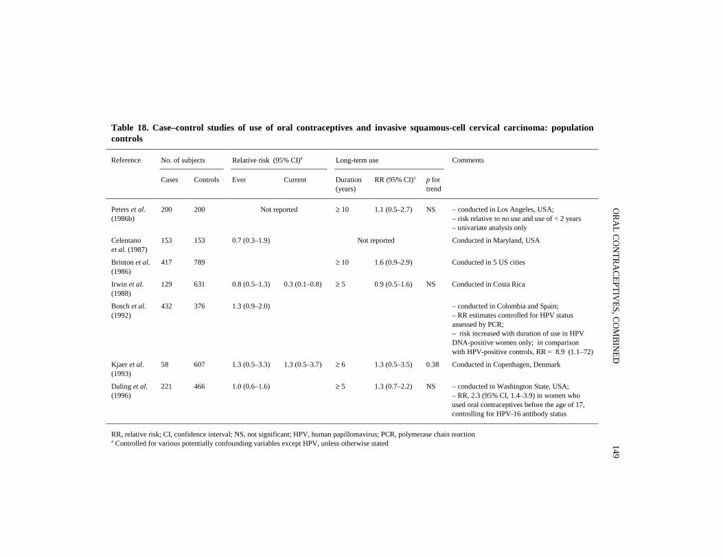
ORA
LCO
NTRA
CEPTIVES, CO
MBIN
ED149
Table 18. Case–control studies of use of oral contraceptives and invasive squamous-cell cervical carcinoma: populationcontrols
Reference No. of subjects Relative risk (95% CI)a Long-term use Comments
Cases Controls Ever Current Duration(years)
RR (95% CI)a p fortrend
Peters et al.(1986b)
200 200 Not reported ≥ 10 1.1 (0.5–2.7) NS – conducted in Los Angeles, USA;– risk relative to no use and use of < 2 years– univariate analysis only
Celentanoet al. (1987)
153 153 0.7 (0.3–1.9) Not reported Conducted in Maryland, USA
Brinton et al.(1986)
417 789 ≥ 10 1.6 (0.9–2.9) Conducted in 5 US cities
Irwin et al.(1988)
129 631 0.8 (0.5–1.3) 0.3 (0.1–0.8) ≥ 5 0.9 (0.5–1.6) NS Conducted in Costa Rica
Bosch et al.(1992)
432 376 1.3 (0.9–2.0) – conducted in Colombia and Spain;– RR estimates controlled for HPV statusassessed by PCR;– risk increased with duration of use in HPVDNA-positive women only; in comparisonwith HPV-positive controls, RR = 8.9 (1.1–72)
Kjaer et al.(1993)
58 607 1.3 (0.5–3.3) 1.3 (0.5–3.7) ≥ 6 1.3 (0.5–3.5) 0.38 Conducted in Copenhagen, Denmark
Daling et al.(1996)
221 466 1.0 (0.6–1.6) ≥ 5 1.3 (0.7–2.2) NS – conducted in Washington State, USA;– RR, 2.3 (95% CI, 1.4–3.9) in women whoused oral contraceptives before the age of 17,controlling for HPV-16 antibody status
RR, relative risk; CI, confidence interval; NS, not significant; HPV, human papillomavirus; PCR, polymerase chain reactiona Controlled for various potentially confounding variables except HPV, unless otherwise stated

who had used oral contraceptives within the past year than for those who had used themin the more distant past.
In the study conducted in Costa Rica by Irwin et al. (1988), described above, 129women with invasive cervical cancer (assumed to be squamous-cell) were comparedwith 631 controls selected from the general population of Costa Rica. No increase in riskwas seen for women who had ever used oral contraceptives, and no trend of increasingrisk with duration of use was found. Women who had used oral contraceptives within thepast year were actually at reduced risk for disease, the estimate being 0.3 (95% CI,0.1–0.8), but this estimate was based on only seven cases and 102 controls. No trend inrisk with time since last use was observed. All of the estimates were adjusted for age,history of sexually transmitted disease or pelvic inflammatory disease, gravidity, age atfirst intercourse, number of sexual partners and history of prior Pap smears.
In a study of risk factors for cervical cancer in Colombia and Spain, Bosch et al.(1992) identified 436 women with histologically confirmed squamous-cell carcinomaand selected 387 controls from the general population in which the cases arose. Noincrease in risk was observed for women who had ever used oral contraceptives. Cervicalscrapings from the study subjects were assayed for type-specific HPV DNA by PCR. Notrend of increasing risk with duration of use was observed for women who had no HPVDNA, but a trend was observed for women who had HPV DNA, and this observation wasstatistically significant (p for trend = 0.027). This observation is, however, based on verysmall numbers of HPV DNA-positive controls: 17 among women who had never usedoral contraceptives and one woman each who had used oral contraceptives for 1–9 and10 or more years. The numbers of cases in these three categories were 110, 12 and 35,respectively. The relative risks in relation to non-users were estimated to be 3.0 (95% CI,0.3–28) for users of oral contraceptives for 1–9 years and 8.9 (95% CI, 1.1–72) for usersof more than 10 years’ duration.
Kjaer et al. (1993) recruited 59 women with invasive cervical cancer and living inthe greater Copenhagen area from the Danish Cancer Registry; the controls were selectedfrom the general female population of greater Copenhagen. The risk for invasivesquamous-cell cervical cancer was not significantly increased among women who hadever used oral contraceptives, and no significant trend of increasing risk with duration ofuse was observed. The relative risk of women who had used oral contraceptives withinthe past two years was 1.7 (95% CI, 0.6–4.7). The risk decreased to 1.0 for women whohad last used oral contraceptives more than two years previously (p for trend = 0.002).The relative risks in this study were adjusted for age, years of school attendance, numberof sexual partners, proportion of sexually active life without use of barrier contra-ceptives, ever having had gonorrhoea and ever having had a Pap smear.
In a population-based case–control study conducted in Washington State, UnitedStates, Daling et al. (1996) interviewed 221 women with invasive squamous-cell cervicalcarcinoma and 466 control women selected by random-digit dialling. Serum from mostof the study subjects was tested for HPV-16 capsid antibodies. After adjustment for age,number of Pap smears in the last decade and lifetime number of sexual partners, the risk
IARC MONOGRAPHS VOLUME 72150

was not increased for women who had ever used oral contraceptives, and no trend ofincreasing risk with duration of use was observed. After control for HPV antibody status,the risks relative to that of women who first began using oral contraceptives after the ageof 20 were 1.6 (95% CI, 1.0–2.4) for women who had first used them between the agesof 18 and 19 and 2.3 (95% CI, 1.4–3.8) for women who had first begun using them atage 17 or younger. No information was given on risk in relation to time since last use oforal contraceptives.
The results of the studies summarized in Tables 17 and 18 are not totally consistent,but some generalizations can be made cautiously. If the risk is increased in women whohave ever used oral contraceptives, then the increase in risk is likely to be modest. Mostof the studies do show a small increase in risk for ‘ever users’, but the risks are small, andthe 95% CIs of the estimates in most instances include unity. The relative risk estimatesfor long-term users are generally higher in the hospital-based studies (Table 17) than inthe population-based studies (Table 18), but the estimates are not consistently higher orlower in hospital-based studies in which HPV DNA status was considered than in suchstudies in which it was not. The higher relative risks in hospital-based than in population-based studies are therefore probably not due to differences in confounding.
None of the studies indicates that risk is increased long after initial exposure to oralcontraceptives. The only possible exception is the study of Daling et al. (1996), in whichit was found that the risk for women who were first exposed to oral contraceptives beforethe age of 17 was increased. This observation requires independent confirmation.
In the three studies in which risk was considered in relation to use of oral contra-ceptives among women with and without other risk factors for cervical cancer (Brintonet al., 1986; Parazzini et al., 1990; WHO Collaborative Study of Neoplasia and SteroidContraceptives, 1993), there was some suggestion that the risk in relation to oral contra-ceptives might be greater in women with than without such sexual risk factors as a historyof non-specific genital infection or sore (Brinton et al., 1986), absence of use of barriercontraceptives (Brinton et al., 1986; Parazzini et al., 1990), having had multiple sexualpartners (Parazzini et al., 1990), a history of sexually transmitted diseases and presenceof herpes simplex virus-II antibodies (WHO Collaborative Study of Neoplasia and SteroidContraceptives, 1993). These observations are consistent with the idea that oral contra-ceptives enhance risk in the presence of a sexually transmitted oncogenic agent such ascertain strains of HPV.
Table 19 summarizes the results of the four studies (described above) in which therisk for invasive squamous-cell cervical cancer in relation to oral contraceptive use wasestimated on the basis of a comparison of cases and controls with evidence of HPV DNAin cervical cells. In each study, the relative risk estimates for women with HPV DNAwere increased, and evidence for a trend of increasing risk with duration of use ofcombined oral contraceptives is provided from three of the studies. These results shouldbe interpreted with caution, however, because few controls were found to be HPV-posi-tive and all of the estimates therefore have wide confidence limits. In addition, three ofthe four studies shown in Table 19 are hospital-based.
ORAL CONTRACEPTIVES, COMBINED 151

IARC M
ON
OG
RAPH
S VO
LUM
E 72152
Table 19. Case–control studies of use of oral contraceptives and invasive squamous-cell cervical cancer inwhich analyses were restricted to women with human papillomavirus (HPV) DNA in cervical scrapings
Reference All subjects HPV-positive subjectsUse of oralcontraceptives
No. of subjects RR (95% CI)a No. of subjects RR (95% CI)a
Cases Controls Cases Controls
Bosch et al. (1992) NeverEver
291141
270106
1.01.3 (0.9–2.0)
110 50
17 2
1.06.5 (1.3–31)
Eluf-Neto et al. (1994);Bosch et al. (1995)
Years of use None 1–4 ≥ 5
125 39 33
152 44 22
1.01.3 (0.7–2.3)2.7 (1.4–5.2)
97 30 27
21 9 2
1.01.2 (0.4–4.2)9.0 (1.4–57)
Chaouki et al. (1998) Years of use < 1 1 2–5/2–4b
> 5/≥ 5b
8 14 32 39
25 14 35 42
1.01.4 (0.2–8.1)2.8 (0.6–13)6.4 (1.3–31)
20
21 37
7
6 3
1.0
1.0 (0.2–6.6)16 (2.2–115)
Ngelangel et al. (1998) Years of use None 1–3 ≥ 4 Total
258 40 25323
277 80 23380
1.00.3 (0.1–0.7)2.0 (0.5–7.6)–
NRNRNR303
NRNRNR35
1.00.3 (0.1–0.8)2.8 (0.2–30)–
RR, relative risk; CI, confidence interval; NR, not reporteda Controlled for various potentially confounding variablesb Years of use in HPV-positive subjects

In summary, if there is an increased risk for squamous-cell cervical carcinoma inrelation to use of oral contraceptives, it is more likely to be found in relation to invasiverather than in-situ disease. The available evidence indicates that the effect of oralcontraceptives on risk probably requires the presence of HPV DNA in the cervicalepithelium.
(e) Studies of invasive cervical adeno- and adenosquamous carcinomasAn early case–control study in Milan, Italy (Parazzini et al., 1988), showed a relative
risk of 0.8 (95% CI, 0.2–2.4) for cervical adenocarcinoma among women who had everused combined oral contraceptives. Five case–control studies have been conducted toassess the risk of adenocarcinomas and adenosquamous carcinomas in relation to durationof use of oral contraceptives (Table 20). The study of Brinton et al. (1990), describedpreviously, included 41 women with adenocarcinoma and 20 women with adeno-squamous carcinoma. The risk for either neoplasm among women who had ever used oralcontraceptives was estimated to be 2.4 (95% CI, 1.3–4.6). No trend of increasing risk withduration of use was observed. The relative risk estimates were adjusted for age, numberof sexual partners, age at first sexual intercourse, interval since last Pap smear, number ofbirths, HPV-16/-18 infection status and education.
Thomas et al. (1996) analysed data from the WHO Collaborative Study of Neoplasiaand Steroid Contraceptives, described previously. A total of 271 women with adeno-carcinoma and 106 women with adenosquamous carcinoma were included in the study.The risk of women who had ever used oral contraceptives was increased for adeno-carcinoma but not for adenosquamous carcinoma. A significant trend of increasing riskfor adenocarcinoma was observed with duration of oral contraceptive use; no similartrend was observed for women with adenosquamous carcinoma, but, when both histo-logical types were combined, a significant trend of increasing risk was observed. Therisk for adenocarcinoma was highest among women who had used these products withinthe past year and generally declined with time since last use. These trends were strongestfor neoplasms that developed in women under the age of 35. The association with riskwas also somewhat stronger for formulations with high-potency progestogens than forlow-potency products.
Brinton et al. (1986, 1987) analysed data from the population-based case–controlstudy conducted in five US cities described previously to assess the risks for adeno-carcinoma, adenosquamous carcinoma and both. As in the study of Thomas et al. (1996),the risk of long-term users of oral contraceptives was more strongly related to adeno-carcinoma than to adenosquamous carcinoma.
Between 1977 and 1991, Ursin et al. (1994) identified 195 cases of adenocarcinomaand adenosquamous carcinoma from the Los Angeles Cancer Registry, United States,which were compared with 386 neighbourhood controls. After adjustment for education,household income, number of sexual partners before the age of 20, number of episodesof genital warts, months of diaphragm use and weight gain between the age of 18 and thetime of diagnosis, the relative risk of women who had ever used oral contraceptives was
ORAL CONTRACEPTIVES, COMBINED 153

IARC M
ON
OG
RAPH
S VO
LUM
E 72154Table 20. Case–control studies of use of oral contraceptives and cervical adeno- and adenosquamous carcinomas
Reference Type of case No. of subjects Ever use Long-term use Comments
Cases Controls RR (95%CI)a
Duration(years)
RR (95%CI)a
p fortrend
Brintonet al. (1990)
Adenocarcinomaand adenosquamouscarcinoma
61 1 429 2.4 (1.3–4.6) ≥ 10 1.8 (0.5–6.5) NS – hospital and population controls;– conducted in 4 Latin American countries
Adenocarcinoma 271 2 084 1.6 (1.2–2.1) ≥ 8 2.4 (1.4–4.0) 0.003Thomas et al.(1996) Adenosquamous
carcinoma106 803 1.1 (0.7–1.8) ≥ 8 1.6 (0.6–4.1) NS
Both 377 2 887 1.5 (1.1–1.9) ≥ 8 2.2 (1.4–3.5) 0.003
– hospital controls;– conducted in 10 centres in 8 countries
Brinton et al.(1986, 1987)
AdenocarcinomaAdenosquamouscarcinomaBoth
40 23
62
801 801
789
≥ 10≥ 10
≥ 10
2.4b
1.3b
3.0 (1.1–8.2)
0.150.77
– population controls;– conducted in 5 US cities;– separate analyses of adenocarcinomaand adenosquamous cancer based on verysmall numbers of women who used oralcontraceptives for 10 years or longer(5 and 2, respectively)
Ursin et al.(1994)
Adenocarcinomaand adenosquamouscarcinoma
195 386 2.1 (1.1–3.8) ≥ 12 4.4 (1.8–11) 0.04 – population controls;– conducted in Los Angeles, USA– 150 cases were adenocarcinomas;15 were adenosquamous; no pathologicalconfirmation of the other cases
Ngelangelet al. (1998)
Adenocarcinoma oradenosquamouscarcinoma
33 380 Not given ≥ 4 4.3 (0.3–57) NS – hospital controls;– conducted in the Philippines;– RR adjusted for HPV infection;– RR based on 4 exposed cases and23 exposed controls only;– use of all hormonal contraceptivesreported, but largely represents use ofcombined oral contraceptives
RR, relative risk; CI, confidence interval; NS, not significant; HPV, human papillomavirusa Controlled for various potentially confounding variables except HPV, unless otherwise statedb RR adjusted only for age and race

estimated to be 2.1 (95% CI, 1.1–3.8). The risk increased significantly with duration ofuse. The relative risk of current users of contraceptives was 1.8 (95% CI, 0.6–5.7) andwas close to unity for women who had used oral contraceptives more than one year inthe past. These results did not change when the analysis was limited to the 150 cases ofadenocarcinoma.
In the study in the Philippines (Ngelangel et al., 1998) summarized above, data for33 cases of adenocarcinoma or adenosquamous carcinoma indicated an increased risk forwomen who had used oral contraceptives for four years or more. The estimate is basedon only three exposed cases, however, and the confidence limits of the estimates are wideand include unity.
In the aggregate, the results of the five studies summarized in Table 20 suggest thatlong-term use of oral contraceptives increases the risk for cervical carcinomas withadenomatous elements, although confounding by HPV infection cannot be ruled out. Theassociation with use of oral contraceptives appears to be somewhat stronger for adeno-carcinoma than for adenosquamous carcinoma.
It has also been suggested that use of oral contraceptives is more strongly related toadenocarcinoma than to squamous-cell carcinoma of the cervix. The results summarized inTables 17, 18 and 20 are inconsistent in this regard. The study conducted in four LatinAmerican countries (Brinton et al., 1990) provided estimates for users of more than10 years of 1.1 for squamous-cell carcinoma and 1.8 for adenocarcinoma or adenosqua-mous carcinoma. The studies in Los Angeles provide estimates of 1.1 for squamous-cellcarcinoma in users of more than 10 years’ duration (Peters et al., 1986b) and 4.4 for adeno-carcinoma or adenosquamous carcinoma combined in users of more than 12 years’duration (Ursin et al., 1994); these results, however, are based on different study popu-lations. The study in the Philippines (Ngelangel et al., 1998) found relative risks of 2.0 and4.3 in users of four years’ or more duration for squamous and adenomatous carcinomas,respectively. The WHO Collaborative Study of Neoplasia and Steroid Contraceptives(1993) provided an estimate of 2.2 for both squamous-cell carcinoma and tumours withadenomatous elements (adenocarcinoma and adenosquamous carcinoma combined;Thomas et al., 1996) in women who had used oral contraceptives for more than eight years.In the study conducted in five United States cities (Brinton et al., 1986, 1987), the risks foradenocarcinoma and adenosquamous carcinoma combined of users of more than 10 years’duration was estimated to be 3.0, while the estimate for squamous-cell carcinoma was 1.6.The estimate for squamous-cell carcinoma adjusted only for age and race was 1.2. Foradenocarcinoma and adenosquamous carcinoma separately, the relative risks adjusted forage and race were 2.4 and 1.3, respectively. The results of these studies thus do not resolvethe question of whether use of oral contraceptives is more strongly related to adenocarci-noma and adenosquamous carcinoma than to squamous-cell carcinoma.
Another method that has been used to address the issue of the relative strength of theassociation between oral contraceptives and various histological types of cervical carci-noma is comparison of use by women with squamous and adenomatous cervical lesions.Persson et al. (1987) compared the oral contraceptive use of 23 women with adeno-
ORAL CONTRACEPTIVES, COMBINED 155

carcinoma with that of 46 women with squamous-cell carcinoma. The proportions ofwomen who had used oral contraceptives in each group were similar, and the duration ofuse did not differ. Jones and Silverberg (1989) similarly compared 18 cases of endo-cervical adenocarcinoma with an equal number of cases of squamous-cell carcinoma; bothgroups included both in situ and invasive disease. The proportions of women in the twogroups who had used oral contraceptives did not differ significantly. Honoré et al. (1991)compared each of 99 women with cervical adenocarcinoma with three comparable womenwith squamous-cell carcinoma, with matching on age, year of diagnosis and clinical stage.The women in the two groups did not differ with respect to any use of oral contraceptivesand, among users, the two groups did not differ with respect to age at start of use, age atdiscontinuation of use or months of use of oral contraceptives. Hopkins and Morley(1991) compared 61 women with adenocarcinoma and 206 women with squamous-cellcarcinoma who were under the age of 40. Thirty-three per cent of the women with adeno-carcinomas and 31% of those with squamous-cell carcinomas had ever used oral contra-ceptives. The results of these clinical studies do not support the hypothesis that use of oralcontraceptives is more strongly related to the development of adenocarcinoma thansquamous carcinoma of the uterine cervix.
On balance, there appears to be insufficient evidence to conclude firmly that use oforal contraceptives is related to adenocarcinoma of the uterine cervix. The associationsobserved could be due to residual confounding by HPV infection, and a firm conclusionabout the risk for adenocarcinoma of users of oral contraceptives must await the resultsof investigations that adequately control for HPV infection.
2.4 Ovarian cancer2.4.1 Descriptive studies
Younger women in several developed countries have experienced substantial declinesin the incidence and mortality rates of ovarian cancer. Cohort analyses based on data fromSwitzerland (Levi et al., 1987), England and Wales (Beral et al., 1988; dos Santos Silva &Swerdlow, 1995), Great Britain (Villard-Mackintosh et al., 1989), Sweden (Adami et al.,1990) and the Netherlands ( Koper et al., 1996) and a systematic analysis of mortalitytrends in 16 European countries (La Vecchia et al., 1992, 1998) showed that women bornafter 1920—i.e. the generations that have used combined oral contraceptives—have consis-tently reduced ovarian cancer rates. The downward trends were greater in countries wherecombined oral contraceptives have been most widely used (La Vecchia et al., 1998).
Thus, descriptive data on the incidence and mortality rates of ovarian cancer areconsistent with the hypothesis of a favourable effect of combined oral contraceptive useon subsequent ovarian cancer rates.
2.4.2 Cohort studiesThe results of cohort studies on use of combined oral contraceptives and ovarian
cancer are summarized in Table 21. Most of the evidence refers to epithelial neoplasms,unless otherwise specified. Three cohort studies conducted in the United States and the
IARC MONOGRAPHS VOLUME 72156

ORA
LCO
NTRA
CEPTIVES, CO
MBIN
ED157
Table 21. Selected cohort studies on use of combined oral contraceptives and ovarian cancer, 1980–97
Reference Relative risk (95% CI) CommentsNo. of cases(age, years)
Any use Longest use
Ramcharan et al. (1981a), USA 16 (18–64) 0.4 (0.1–1.0) – Adjusted for age only; Walnut CreekStudy on Contraception
Beral et al. (1988), UK 30 (≥ 25) 0.6 (0.3–1.4) ≥ 10 years, 0.3 Royal College of General Practitioners’cohort
Vessey & Painter (1995), UK 42 (all) 0.4 (0.2–0.8) > 8 years, 0.3 (0.1–0.7) Oxford Family Planning cohort
Hankinson et al. (1995), USA 260 (30–65) 1.1 (0.8–1.4) ≥ 5 years, 0.7 (0.4–1.1) Nurses’ Health Study
CI, confidence interval

United Kingdom provided data on a total of about 100 cases of epithelial ovarian cancer.In the Walnut Creek Study in the United States (Ramcharan et al., 1981a), 16 cases ofovarian cancer were registered between 1968 and 1977, corresponding to an age-adjustedrelative risk of 0.4 for any use of combined oral contraceptives.
The Royal College of General Practitioners’ study was based on 47 000 womenrecruited in 1968 in 1400 British general practices (Beral et al., 1988): 30 cases ofovarian cancer were observed up to 1987, corresponding to multivariate relative risks of0.6 (95% CI, 0.3–1.4) for any use of combined oral contraceptives and of 0.3 for 10 yearsof use or more. Allowance was made in the analysis for age, parity, smoking and socialclass. In a subsequent follow-up study of mortality in that cohort up to the end of 1993(Beral et al., 1999), 55 deaths from ovarian cancer were reported; there was a statisticallysignificantly reduced mortality rate from ovarian cancer among women who had everused oral contraceptives (relative risk, 0.6; 95% CI, 0.3–1.0).
The study of the Oxford Family Planning Association was based on 17 032 womenenrolled between 1968 and 1974 from various family planning clinics in the UnitedKingdom (Vessey & Painter, 1995). Up to October 1993, 42 cases of ovarian cancer wereregistered, corresponding to relative risks of 0.4 (95% CI, 0.2–0.8) for any use ofcombined oral contraceptives and 0.3 (95% CI, 0.1–0.7) for more than eight years of use.Adjustment was made for age and parity.
In the Nurses’ Health study, based on 121 700 registered nurses aged 30–55 in 1976,260 cases of ovarian cancer were observed prospectively between 1976 and 1988(Hankinson et al., 1995). The multivariate relative risk for any use, which essentiallyreflected former use, was 1.1 (95% CI, 0.8–1.4) but declined to 0.7 (95% CI, 0.4–1.1) foruse for five years or more. Adjustment was made for age, tubal ligation, age at menarche,age at menopause, smoking and body mass index.
2.4.3 Case–control studiesThe epidemiological evidence from case–control studies on use of combined oral
contraceptives and ovarian cancer is well defined and consistent: at least 20 out of21 studies published between 1980 and 1997 found relative risks below unity, the soleapparent outlier being a study conducted in China (Shu et al., 1989).
Table 22 gives the main results of case–control studies of ovarian cancer publishedbetween 1980 and 1997 which included information on use of combined oral contra-ceptives. Table 23 gives age-specific relative risks and 95% CIs, while Table 24 gives therelative risks related to time since last use for studies that provided the relevant information.The findings of two pooled analyses of case–control studies on the issue are also included.These were conducted on 971 cases and 2258 controls in three European countries(Franceschi et al., 1991a) and on 2197 cases and 8893 controls in white women from 12studies in the United States (Whittemore et al., 1992), for a total of over 3100 cases and11 000 controls.
In a pooled analysis of individual data from three hospital-based European studies(Franceschi et al., 1991a), the multivariate relative risk was 0.6 (95% CI, 0.4–0.8) for any
IARC MONOGRAPHS VOLUME 72158

ORA
LCO
NTRA
CEPTIVES, CO
MBIN
ED159
Table 22. Selected case–control studies of use of oral contraceptives and ovarian cancer, 1980–97
Reference, location Type of study Relative risk (95% CI) CommentsNo. of cases(age, years)
Any use Longest use Duration(years)
Willett et al. (1981),USA
Nested in a cohort 47 (< 60) 0.8 (0.4–1.5) 0.8 (0.3–2.1) > 3 Prevalent cases from the Nurses' Healthcohort study; adjusted for age
Hildreth et al. (1981),USA
Hospital-based 62 (45–74) 0.5 (0.1–1.7) Not reported Adjusted for age and parity; odds ratiobased on 3 cases with use of oralcontraceptives
Weiss et al. (1981a),USA
Population-based 112 (36–55) 0.6 (not reported) 0.4 (0.2–1.3) ≥ 9 Adjusted for age, demographic factorsand parity
Franceschi et al. (1982),Italy
Hospital-based 161 (19–69) 0.7 (0.4–1.1) Not reported Adjusted for age
Cramer et al. (1982),USA
Population-based 144 (< 60) 0.4 (0.2–1.0) 0.6 > 5 Adjusted for age and parity
Rosenberg et al. (1982),USA
Hospital-based 136 (< 60) 0.6 (0.4–0.9) 0.3 (0.1–0.8) ≥ 5 Protection by use of combined andsequential oral contraceptives;independent of parity; adjusted forseveral variables
Risch et al. (1983), USA Population-based 284 (20–74) [0.5] (not reported) Not reported Multivariate odds ratio approximately0.9 per year of use
Tzonou et al. (1984),Greece
Hospital-based 150 (all ages) 0.4 (0.1–1.1) Not reported Adjusted for age, parity, age atmenopause and use of oestrogenreplacement therapy
Cancer and SteroidHormone Study (1987),USA
Population-based 492 (20–54) 0.6 (0.5–0.7) 0.2 (0.1–0.4) ≥ 10 Consistent results by type of combinedoral contraceptive
Harlow et al. (1988),USA
Population-based 116 (20–79) 0.4 (0.2–0.9) 0.4 (0.2–1.0) > 4 Borderline malignancy; adjusted for ageand parity

IARC M
ON
OG
RAPH
S VO
LUM
E 72160
Table 22 (contd)
Reference, location Type of study Relative risk (95% CI) CommentsNo. of cases(age, years)
Any use Longest use Duration(years)
Wu et al. (1988), USA Hospital- andpopulation-based
299 (18–74) 0.7 (0.5–1.1) 0.4 (0.2–0.7) > 3 Combination of two studies conducted inthe 1970s and 1980s
Shu et al. (1989), China Population-based 229 (18–70) 1.8 (0.8–4.1) 1.9 (0.4–9.3) > 5 Only 23 cases and 12 controls had everused combined oral contraceptives
WHO CollaborativeStudy (1989a),7 countries
Hospital-based 368 (< 62) 0.8 (0.6–1.0) 0.5 (0.3–1.0) > 5 Similar results in developed anddeveloping countries
Hartge et al. (1989a),USA
Hospital-based 296 (20–79) 1.0 (0.7–1.7) 0.8 (0.4–1.5) > 5 Data collected between 1978 and 1981
Booth et al. (1989), UK Hospital-based 235 (< 65) 0.5 (0.3–0.9) 0.1 (0.01–1.0) > 10 Consistent results in strata of parity
Parazzini et al. (1991a),Italy
Hospital-based 505 (22–59) 0.7 (0.5–1.0) 0.5 (0.3–0.9) ≥ 2 Protective effect present in strata ofmajor risk factors for ovarian cancer
Parazzini et al. (1991b),Italy
Hospital-based 91 (23–64) 0.3 (0.2–0.6) 0.2 (0.1–0.6) ≥ 2 Borderline malignancy; adjusted for age,parity, education, age at menopause andoral contraceptive use
Polychronopoulos et al.(1993), Greece
Hospital-based 189 (< 75) 0.8 (0.2–3.7) Not reported Multivariate RR; only three cases andseven controls had ever used combinedoral contraceptives
Rosenberg et al. (1994),USA
Hospital-based 441 (< 65) 0.8 (0.6–1.0) 0.5 (0.2–0.9) ≥ 10 Association persisted as long as twodecades after stopping and was notconfined to any type of oral contraceptiveformulation

ORA
LCO
NTRA
CEPTIVES, CO
MBIN
ED161
Table 22 (contd)
Reference, location Type of study Relative risk (95% CI) CommentsNo. of cases(age, years)
Any use Longest use Duration(years)
Risch et al. (1994,1996), Canada
Population-based 450 (35–79) 0.5 (0.4–0.7) 0.3 (0.2–0.6) ≥ 10 The inverse relationship was stronger fornon-mucinous (RR, 0.9) for each year ofuse than for mucinous tumours (RR, 1.0);trend per year of use, 0.9 among allsubjects
Purdie et al. (1995),Australia
Population-based 824 (18–79) 0.5 (0.4–0.7) 0.3 (0.2–0.4) ≥ 1 Adjusted for parity
Pooled analyses
Franceschi et al.(1991a), Greece, Italy,UK
Three hospital-based studies
971 (< 65) 0.6 (0.4–0.8) 0.4 (0.2–0.7) ≥ 5 Protection was still present ≥ 15 yearsafter stopping use (odds ratio, 0.5).
Whittemore et al.(1992), USA
Pooled analysis of12 US population-and hospital-basedcase–controlstudies
2197 (all) 0.7 (0.6–0.8) 0.3 (0.2–0.4) ≥ 6 Invasive epithelial neoplasms in whitewomen; protection present in population-and hospital-based studies
Harris et al. (1992),USA
Same pooledanalysis asWhittemore et al.(1992)
327 0.8 (0.6–1.1) 0.6 (0.4–0.9) > 5 Epithelial tumours of low malignantpotential in white women
John et al. (1993), USA Pooled analysis of7 of the 12 studiesin the pooledanalysis ofWhittemore et al.(1992)
110 0.7 (0.4–1.2) 0.6 (0.2–1.6) ≥ 6 Epithelial ovarian cancers in blackwomen
CI, confidence interval; RR, relative risk

use and 0.4 (95% CI, 0.2–0.7) for longest (≥ 5 years) use. Allowance was made in theanalysis for age, other socio-demographic factors, menopausal status and parity. The pro-tection persisted for at least 15 years after use had ceased.
In a pooled analysis of individual data from 12 studies in the United States (Whittemoreet al., 1992), the corresponding values were 0.7 (95% CI, 0.6–0.8) for any use and 0.3(0.2–0.4) for use for more than six years in the population-based studies. Adjustment wasmade for age, study and parity. The results were similar when the hospital-based andpopulation-based studies were considered separately: the relative risks were 0.7 in bothtypes of study for any use of combined oral contraceptives, 0.6 in hospital-based studiesand 0.3 in population-based studies for longest use (> 6 years) and 0.95 (not significant)and 0.90 (p < 0.001), respectively, per added year of use.
An inverse association was also observed in a further analysis of seven studies of 110cases and 251 controls in black women in the United States. The relative risk was 0.7 forany use and 0.6 for use for six years or more (John et al., 1993). The United States pooled
IARC MONOGRAPHS VOLUME 72162
Table 23. Selected case–control studies on use of combinedoral contraceptives and ovarian cancer, 1980–97; age-specificrelative risks
Reference Age group(years)
Relative risk (95% CI)
Willett et al. (1981) < 3535–44≥ 45
0.3 (0.1–1.3)1.1 (0.4–3.2)1.3 (0.4–3.9)
Rosenberg et al. (1982) 18–2930–3940–4950–59
0.40.60.50.7
Centers for Disease Control(1983b)
20–2930–3940–4950–54
0.3 (0.1–1.4)0.8 (0.3–2.0)0.6 (0.4–1.1)0.6 (0.3–1.1)
Parazzini et al. (1991a) < 3535–4445–5455–59
0.4 (0.2–0.9)0.8 (0.4–1.4)0.7 (0.4–1.3)0.8 (0.1–7.6)
Rosenberg et al. (1994) < 4545–65
0.5 (0.3–0.8)0.7 (0.4–1.2)
Pooled analysis
Franceschi et al. (1991a) < 4545–5455–64
0.6 (0.3–1.0)0.5 (0.3–1.0)0.6 (0.4–0.9)
CI, confidence interval

analysis also included data on 327 cases of epithelial ovarian neoplasms of borderlinemalignancy in white women. The relative risks were 0.8 (95% CI, 0.6–1.1) for any useof combined oral contraceptives and 0.6 (0.4–0.9) for five years of use or more (Harriset al., 1992).
The most convincing aspect of the inverse relationship between use of combinedoral contraceptives and risk for ovarian cancer is the consistency of the results, inde-pendently of the type of study (hospital- or population-based), geographical area(Australia, Europe, North America and developing countries) and type of analysis,including allowance for covariates which differed from study to study, although morevariables tended to be included in most recent ones. Likewise, the inverse relationship
ORAL CONTRACEPTIVES, COMBINED 163
Table 24. Selected case–control studies on use of combinedoral contraceptives and ovarian cancer, 1980–97; resultsaccording to time since last use
Reference Time since lastuse (years)
Relative risk (95%CI)
Cramer et al. (1982) < 22–< 66–< 10≥ 10
2.1 (NR)0.7 (NR)0.7 (NR)0.3 (NR)
Rosenberg et al. (1982) < 11–45–9≥ 10
0.3 (NR)0.4 (NR)0.8 (NR)0.5 (NR)
Centers for Disease Control(1983b)
< 11–45–9≥ 10
1.0 (0.4–2.2)0.6 (0.3–1.1)0.5 (0.3–0.9)0.5 (0.3–0.9)
Harlow et al. (1988) ≤ 5> 5
0.3 (0.1–0.9)0.6 (0.3–1.4)
WHO Collaborative Study(1989a)
< 0.50.5–< 55–< 10≥ 10
0.9 (0.5–1.8)0.9 (0.5–1.5)0.8 (0.5–1.4)0.5 (0.3–0.9)
Hartge et al. (1989a) < 11–9≥ 10
0.5 (0.1–1.6)0.9 (0.5–1.6)1.4 (0.7–2.6)
Parazzini et al. (1991a) < 10≥ 10
0.5 (0.3–0.8)0.9 (0.5–1.5)
Rosenberg et al. (1994) < 1515–19≥ 20
0.4 (0.2–0.8)0.5 (0.3–1.0)0.8 (0.4–1.5)
CI, confidence interval; NR, not reported

between use of combined oral contraceptives and ovarian cancer was observed for mosttypes of formulations considered, including those with low doses (Cancer and SteroidHormone Study of the Centers for Disease Control and the National Institute of ChildHealth and Human Development, 1987; Rosenblatt et al., 1992; Rosenberg et al., 1994).
The overall estimate of protection for any use is approximately 40%, and a steadyinverse relationship exists with duration of use. The decrease in risk was over 50%, andprobably around 60% for use for more than five years; however, in contrast to thefindings of the Cancer and Steroid Hormone Study of the Centers for Disease Controland the National Institute of Child Health and Human Development (1987), no protectionwas evident after very short-term use, i.e. three to six months, in an analysis of factorsassociated with the short-term use of oral contraceptives by Gross et al. (1992) on thedata of the above study.
Willett et al. (1981) conducted a case–control study of 47 cases of ovarian cancer and470 controls nested in the Nurses’ Health Study cohort (based on 121 964 registerednurses aged 30–55 in 1986 and residing in 11 large American states). They found an age-adjusted relative risk of 0.8 (95% CI, 0.4–1.5) for any use of combined oral contra-ceptives and 0.2 (95% CI, 0.1–1.0) for women aged 35 or younger, who were more likelyto be current users.
Hildreth et al. (1981) considered 62 cases of epithelial ovarian cancer and 1068hospital controls aged 45–74 in Connecticut, United States, that had been diagnosedbetween 1977 and 1979. The response rate was 71% for both cases and controls. Themultivariate relative risk for any use of combined oral contraceptives, after allowance forage and parity, was 0.5 (95% CI, 0.2–1.7).
Weiss et al. (1981a), in a population-based case–control study of 112 cases diagnosedbetween 1975 and 1979 in Washington and Utah, United States, found a relative risk(adjusted for age, demographic factors and parity) of 0.6 for any use and 0.4 (95% CI,0.2–1.3) for longest use, which was of borderline statistical significance (p = 0.04). Theresponse rate was 66% for cases and 92% for controls.
Franceschi et al. (1982) considered data on 161 cases of epithelial ovarian cancer and561 hospital controls in women interviewed in Milan, Italy, in 1979–80. The age-adjusted relative risk for ever use was 0.7 (95% CI, 0.4–1.1).
Cramer et al. (1982) conducted a case–control study of 144 cases and 139 populationcontrols in 1978–81 in the Greater Boston area (United States) and found a relative risk,adjusted for age and parity, of 0.4 (95% CI, 0.2–1.0) for any use of combined oralcontraceptives, in the absence of a consistent duration–risk relationship (relative risk, 0.6for > 5 years). The latter may be due to the small number of cases. The response rateswere around 50% for both cases and controls.
Rosenberg et al. (1982), in a hospital-based case–control study of 136 cases and 539controls collected between 1976 and 1980 from various areas of the United States andCanada, found an age-adjusted relative risk of 0.6 (95% CI, 0.4–0.9) for any use and 0.3for use for five years or more. The response rates were 94% for both cases and controls,and the results were not materially modified by multivariate analysis.
IARC MONOGRAPHS VOLUME 72164

Risch et al. (1983) provided data from a case–control study of 284 cases and 705controls from Washington and Utah (United States) diagnosed between 1975 and 1979,giving a significant multivariate relative risk estimate of 0.9 per year for use of combinedoral contraceptives. The response rates were 68% for cases and 95% for controls.
In a case–control study conducted in 1980–81 on 150 cases and 250 hospital controlsin Athens, Greece, Tzonou et al. (1984) found a multivariate relative risk (adjusted forage, parity, age at menopause and use of post-menopausal oestrogen therapy) of 0.4 (95%CI, 0.1–1.1). The lack of significance may be due to the low frequency of use of combinedoral contraceptives in this study, which was only 2.7% in cases and 7.2% in controls.
The Centers for Disease Control Cancer and Steroid Hormone Study (1983b) and theCancer and Steroid Hormone Study of the Centers for Disease Control and NationalInstitute of Child Health and Human Development (1987) was a population-basedinvestigation conducted between December 1980 and December 1982 in eight areas ofthe United States on 546 women 20–54 years of age with ovarian cancer and 4228controls. The response rates were 71% for cases and 83% for controls. The multivariaterelative risk, adjusted for age and parity, for any use of combined oral contraceptives was0.6 (95% CI, 0.5–0.7), which decreased to 0.2 (0.1–0.4) for use for10 years or more. Theresults were consistent when specific formulations of combined oral contraceptives wereconsidered separately.
Harlow et al. (1988) provided information on use of combined oral contraceptives in116 cases of epithelial ovarian cancers of borderline malignancy diagnosed between1980 and 1985 and 158 controls. The relative risk for any use, adjusted for age andparity, was 0.4, in the absence, however, of a consistent duration–risk relationship.
Wu et al. (1988), in a hospital-based case–control study of 299 cases diagnosed in1983–85 and 752 hospital controls and 259 population-based controls from the SanFrancisco Bay area, United States, found a relative risk, adjusted for parity, of 0.7 (95%CI, 0.5–1.1) for any use and 0.4 (95% CI, 0.2–0.7) for more than three years of use. Theoverall relative risk per year of use was 0.9 (95% CI, 0.8–0.9). The response rate wasabout 70% for both cases and controls.
Shu et al. (1989), in a case–control study conducted during 1984–86 in Shanghai,China, on 229 ovarian cancer cases (172 epithelial) and an equal number of controls,found a relative risk (adjusted for education, parity, ovarian cysts and age at menarche)of 1.8 (95% CI, 0.8–4.1) for any use of combined oral contraceptives. Only 23 cases and12 controls had ever used such preparations. The response rates were 89% for cases and100% for controls. In China, use of combined oral contraceptives might have been anindication of a westernized life style.
The WHO Collaborative Study of Neoplasia and Steroid Contraceptives (1989a)included data on 368 cases of histologically confirmed cases of epithelial ovarian cancerand 2397 hospital controls. The patients were interviewed between 1979 and 1986 inseven countries, with response rates of 73% for cases and 94% for controls. The multi-variate relative risk (adjusted for age, centre, year of interview and parity) for any use ofcombined oral contraceptives was 0.8 (95% CI, 0.6–1.0) and decreased to 0.5 (95% CI,
ORAL CONTRACEPTIVES, COMBINED 165

0.3–1.0) for five years of use or more. The reduction in risk was of a similar magnitudein developed and developing countries (Thomas, 1991).
In a case–control study conducted in 1978–81 in the Washington DC area of theUnited States with 296 patients with epithelial ovarian cancer and 343 hospital controls,Hartge et al. (1989a) found relative risks (adjusted for age and race) of 1.0 (95% CI,0.7–1.7) for any use of combined oral contraceptives and 0.8 (95% CI, 0.4–1.5) for usefor more than five years. The response rates were 74% for cases and 78% for controls.
Booth et al. (1989), in a hospital-based case–control study of 235 patients and 451controls interviewed between 1978 and 1983 in London and Oxford, England, foundmultivariate relative risks of approximately 0.5 (95% CI, 0.3–0.9) for any use and 0.1(0.01–1.0) for use for more than 10 years. They reported a significant inverse trend inrisk with duration of use. Allowance was made for age, social class, gravidity and dura-tion of unprotected intercourse.
Parazzini et al. (1991a) provided data on 505 cases of epithelial ovarian cancer inwomen under 60 years of age and 1375 hospital controls interviewed between 1983 and1989 in northern Italy. The multivariate relative risk (adjusted for age, sociodemographicfactors, parity, age at menarche, lifelong menstrual pattern, menopausal status and age atmenopause) for any use of combined oral contraceptives was 0.7 (95% CI, 0.5–1.0),which decreased to 0.5 (0.3–0.9) for two years of use or more, with a significant inversetrend in risk with duration. The response rate was 98% for both cases and controls.
Parazzini et al. (1991b) also considered 91 patients with epithelial ovarian cancer ofborderline malignancy and 237 hospital controls who were interviewed between 1986and 1990 in northern Italy. The multivariate relative risk (adjusted for age, education,parity and age at menopause) for any use of combined oral contraceptives was 0.3 (95%CI, 0.2–0.6), and that for two years of use or more was 0.2 (0.1–0.6). The response ratewas 98% for both cases and controls.
In a case–control study of 189 cases and 200 controls conducted in 1989–91 ingreater Athens, Greece (Polychronopoulou et al., 1993), only three cases and sevencontrols had any use of combined oral contraceptives, corresponding to a multivariaterelative risk of 0.8 (95% CI, 0.1–3.7). The response rate for cases was about 90%.
Rosenberg et al. (1994) updated their 1982 report, providing data collected between1977 and 1991 on 441 cases of epithelial ovarian cancer and 2065 hospital controls fromvarious areas of the United States. The response rate was 94% for both cases andcontrols. The multivariate relative risk for any use (adjusted for parity, hysterectomy,monolateral oophorectomy, tubal ligation, family history of ovarian cancer and socio-demographic factors) was 0.8 (95% CI, 0.6–1.0). No significant protection was observedwith up to three years of use, but the relative risk declined to 0.5 (95% CI, 0.2–0.9) for10 years of use or more. The risk estimates were similar for various types of combinedoral contraceptive formulations.
Risch et al. (1994, 1996) provided data on 450 cases of epithelial ovarian cancer inwomen aged 35–79 diagnosed between 1989 and 1992 and 564 controls in Ontario,Canada. The response rates were 71% for cases and 65% for controls. The odds ratio,
IARC MONOGRAPHS VOLUME 72166

adjusted for age and parity, for any use of oral contraceptives was 0.5 (95% CI, 0.4–0.7);after 10 or more years of use, it was 0.3 (0.2–0.6). The overall multivariate odds ratio pereach year of use of combined oral contraceptives, adjusted for age, parity, lactation, useof postmenopausal oestrogen therapy, tubal ligation, hysterectomy and family history ofbreast cancer, was 0.90 (95% CI, 0.86–1.0), and the protection was stronger for serousand endometrioid cancers than for mucinous neoplasms.
Purdie et al. (1995) in a population-based study of 824 cases diagnosed between1990 and 1993 and 860 controls in three Australian states found a relative risk of 0.6(95% CI, 0.5–0.7) for any use, which declined to 0.3 (0.2–0.4) for 10 years of use ormore. The response rates were 90% for cases and 73% for controls. Allowance was madein the analysis for sociodemographic factors, family history of cancer, talc use, smokingand reproductive and hormonal factors.
Parity is a well-recognized protective factor for ovarian cancer (Parazzini et al.,1991c) and is a correlate of the use of combined oral contraceptives, i.e. a potentiallyrelevant confounder. The inverse relationship between use of combined oral contra-ceptives and ovarian cancer was also observed, however, after adequate allowance hadbeen made for parity and was reproduced consistently in several studies across separatestrata of parity, age and other potential covariates, including marital status, education,menopausal status, other types of contraceptive use and other selected menstrual andreproductive factors.
The association between oral contraceptive use and the risk for ovarian cancer hasbeen assessed in women with germ-line mutations in the BRCA-1 or BRCA-2 gene(Narod et al., 1998). Thus, 207 women with such mutations and ovarian cancer werecompared with 53 of their sisters who had one of these mutations. The relative risk forovarian cancer was estimated to be 0.4 (95% CI, 0.2–0.7) for women who had ever usedoral contraceptives and 0.3 (0.1–0.7) for women who had used oral contraceptives for sixor more years.
At least two studies (Harlow et al., 1988; Parazzini et al., 1991b) and the pooled ana-lysis of 12 United States studies (Harris et al., 1992) also considered epithelial ovariantumours of borderline malignancy. An inverse relationship was seen for these neoplasms,suggesting that combined oral contraceptives exert protection against the whole processof epithelial ovarian carcinogenesis.
Little information is available on the different histological types of epithelial ovariancancer. In a Canadian study (Risch et al., 1996), the inverse association was apparentlystronger for non-mucinous (odds ratio per year of use, 0.9; 95% CI, 0.85–0.93) than formucinous (odds ratio per year of use, 0.97; 0.93–1.04) tumours. This observation,however, requires confirmation.
In the case of non-epithelial ovarian cancers, 38 germ-cell neoplasms and 45 sex-cord-stromal neoplasms were identified from the collaborative analysis of four UnitedStates case–control studies (Horn-Ross et al., 1992). The multivariate relative risks forany use of combined oral contraceptives were 2.0 (95% CI, 0.8–5.1) for germ-cell cancersand 0.4 (0.2–0.8) for sex-cord-stromal neoplasms. The data were inadequate to evaluate
ORAL CONTRACEPTIVES, COMBINED 167

duration of use or any other time–risk relationship. Similarly, the few available dataindicate a consistent inverse association between use of combined oral contraceptives andbenign epithelial tumours (ovarian cysts) (Parazzini et al., 1989; Booth et al., 1992) butnot benign ovarian teratomas (Westhoff et al., 1988; Parazzini et al., 1995).
The favourable effect of use of combined oral contraceptives on the risk for epithelialovarian cancer seems to persist for at least 10–15 years after use of the contraceptives hasceased (Cancer and Steroid Hormone Study of the Centers for Disease Control andNational Institute of Child Health and Human Development, 1987; Franceschi et al.,1991a; Whittemore et al., 1992; Rosenberg et al., 1994) and is not confined to a particularformulation (Rosenblatt et al., 1992; Rosenberg et al., 1994). There is some suggestionthat formulations with lower doses of oestrogen are slightly less protective: in the WHOCollaborative Study on Neoplasia and Steroid Contraceptives (Rosenblatt et al., 1992), therelative risk for ovarian cancer associated with any use of combined oral contraceptiveswas 0.7 (95% CI, 0.4–1.1) for high-dose preparations and 0.8 (95% CI, 0.5–1.3) for low-dose ones. The available data do not provide definite evidence of an inverse associationbetween use of combined oral contraceptives with low-dose oestrogen and ovarian cancerfor longer periods or in relation to recency of use.
The suppression of ovulation induced by oral contraceptives has been suggested toexplain the inversion association, since it protects the ovarian epithelium from recurrenttrauma and contact with follicular fluid (Fathalla, 1971; Casagrande et al., 1979;Parazzini et al., 1991c). Combined oral contraceptives may also protect against ovariancancer by reducing exposure to pituitary gonadotropins, which stimulate the growth ofcell lines derived from human ovarian carcinoma (Simon et al., 1983). The lack ofapparent protection by post-menopausal oestrogen therapy, however, does not supportthe existence of a favourable role of gonadotropin stimulation on ovarian carcinogenesis.
Since the incidence of ovarian cancer is already appreciable in middle age, andsurvival from the disease is unsatisfactory, the protection attributable to use of oralcontraceptives is important and is therefore one of the major issues in any risk–benefit,public health evaluation of the use of combined oral contraceptives (Gross &Schlesselman,1994; La Vecchia et al., 1996).
2.5 Cancers of the liver and gall-bladderThe vast majority of primary liver cancers are hepatocellular carcinomas. Chronic
infection with hepatitis B (HBV) or C virus causes hepatocellular carcinoma, the relativerisk exceeding 50 in many studies (IARC, 1994). Drinking of alcoholic beverages alsocauses liver cancer (IARC, 1988). Cholangiocarcinoma is much less common, althoughit is frequent in parts of South-East Asia and can be caused by infection with liver flukes(Parkin et al., 1991).
2.5.1 Descriptive studiesForman et al. (1983) analysed the rates of mortality from primary liver cancer among
men and women in England and Wales between 1958 and 1981. The age-standardized
IARC MONOGRAPHS VOLUME 72168
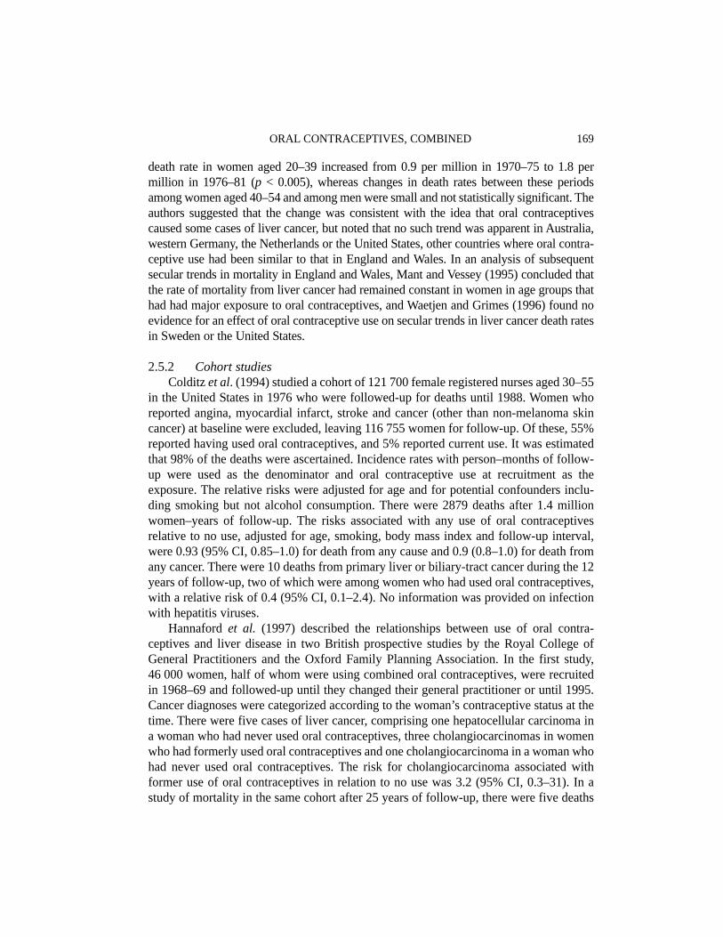
death rate in women aged 20–39 increased from 0.9 per million in 1970–75 to 1.8 permillion in 1976–81 (p < 0.005), whereas changes in death rates between these periodsamong women aged 40–54 and among men were small and not statistically significant. Theauthors suggested that the change was consistent with the idea that oral contraceptivescaused some cases of liver cancer, but noted that no such trend was apparent in Australia,western Germany, the Netherlands or the United States, other countries where oral contra-ceptive use had been similar to that in England and Wales. In an analysis of subsequentsecular trends in mortality in England and Wales, Mant and Vessey (1995) concluded thatthe rate of mortality from liver cancer had remained constant in women in age groups thathad had major exposure to oral contraceptives, and Waetjen and Grimes (1996) found noevidence for an effect of oral contraceptive use on secular trends in liver cancer death ratesin Sweden or the United States.
2.5.2 Cohort studiesColditz et al. (1994) studied a cohort of 121 700 female registered nurses aged 30–55
in the United States in 1976 who were followed-up for deaths until 1988. Women whoreported angina, myocardial infarct, stroke and cancer (other than non-melanoma skincancer) at baseline were excluded, leaving 116 755 women for follow-up. Of these, 55%reported having used oral contraceptives, and 5% reported current use. It was estimatedthat 98% of the deaths were ascertained. Incidence rates with person–months of follow-up were used as the denominator and oral contraceptive use at recruitment as theexposure. The relative risks were adjusted for age and for potential confounders inclu-ding smoking but not alcohol consumption. There were 2879 deaths after 1.4 millionwomen–years of follow-up. The risks associated with any use of oral contraceptivesrelative to no use, adjusted for age, smoking, body mass index and follow-up interval,were 0.93 (95% CI, 0.85–1.0) for death from any cause and 0.9 (0.8–1.0) for death fromany cancer. There were 10 deaths from primary liver or biliary-tract cancer during the 12years of follow-up, two of which were among women who had used oral contraceptives,with a relative risk of 0.4 (95% CI, 0.1–2.4). No information was provided on infectionwith hepatitis viruses.
Hannaford et al. (1997) described the relationships between use of oral contra-ceptives and liver disease in two British prospective studies by the Royal College ofGeneral Practitioners and the Oxford Family Planning Association. In the first study,46 000 women, half of whom were using combined oral contraceptives, were recruitedin 1968–69 and followed-up until they changed their general practitioner or until 1995.Cancer diagnoses were categorized according to the woman’s contraceptive status at thetime. There were five cases of liver cancer, comprising one hepatocellular carcinoma ina woman who had never used oral contraceptives, three cholangiocarcinomas in womenwho had formerly used oral contraceptives and one cholangiocarcinoma in a woman whohad never used oral contraceptives. The risk for cholangiocarcinoma associated withformer use of oral contraceptives in relation to no use was 3.2 (95% CI, 0.3–31). In astudy of mortality in the same cohort after 25 years of follow-up, there were five deaths
ORAL CONTRACEPTIVES, COMBINED 169
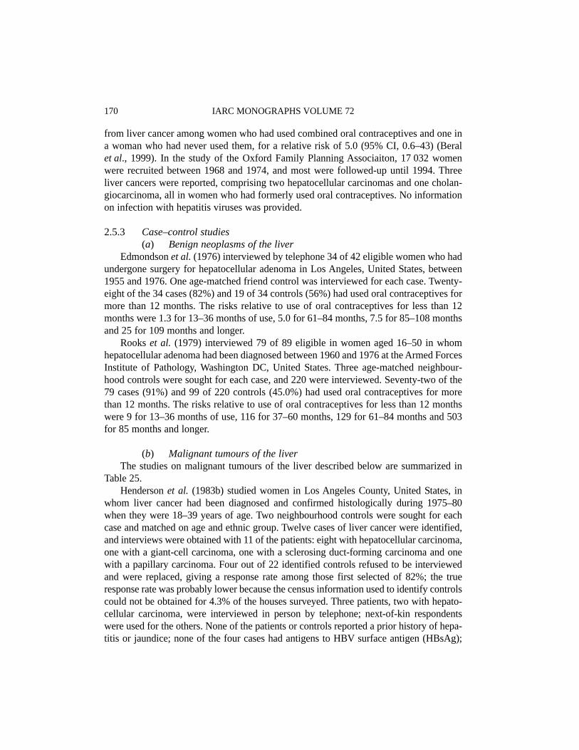
from liver cancer among women who had used combined oral contraceptives and one ina woman who had never used them, for a relative risk of 5.0 (95% CI, 0.6–43) (Beralet al., 1999). In the study of the Oxford Family Planning Associaiton, 17 032 womenwere recruited between 1968 and 1974, and most were followed-up until 1994. Threeliver cancers were reported, comprising two hepatocellular carcinomas and one cholan-giocarcinoma, all in women who had formerly used oral contraceptives. No informationon infection with hepatitis viruses was provided.
2.5.3 Case–control studies(a) Benign neoplasms of the liver
Edmondson et al. (1976) interviewed by telephone 34 of 42 eligible women who hadundergone surgery for hepatocellular adenoma in Los Angeles, United States, between1955 and 1976. One age-matched friend control was interviewed for each case. Twenty-eight of the 34 cases (82%) and 19 of 34 controls (56%) had used oral contraceptives formore than 12 months. The risks relative to use of oral contraceptives for less than 12months were 1.3 for 13–36 months of use, 5.0 for 61–84 months, 7.5 for 85–108 monthsand 25 for 109 months and longer.
Rooks et al. (1979) interviewed 79 of 89 eligible in women aged 16–50 in whomhepatocellular adenoma had been diagnosed between 1960 and 1976 at the Armed ForcesInstitute of Pathology, Washington DC, United States. Three age-matched neighbour-hood controls were sought for each case, and 220 were interviewed. Seventy-two of the79 cases (91%) and 99 of 220 controls (45.0%) had used oral contraceptives for morethan 12 months. The risks relative to use of oral contraceptives for less than 12 monthswere 9 for 13–36 months of use, 116 for 37–60 months, 129 for 61–84 months and 503for 85 months and longer.
(b) Malignant tumours of the liverThe studies on malignant tumours of the liver described below are summarized in
Table 25.Henderson et al. (1983b) studied women in Los Angeles County, United States, in
whom liver cancer had been diagnosed and confirmed histologically during 1975–80when they were 18–39 years of age. Two neighbourhood controls were sought for eachcase and matched on age and ethnic group. Twelve cases of liver cancer were identified,and interviews were obtained with 11 of the patients: eight with hepatocellular carcinoma,one with a giant-cell carcinoma, one with a sclerosing duct-forming carcinoma and onewith a papillary carcinoma. Four out of 22 identified controls refused to be interviewedand were replaced, giving a response rate among those first selected of 82%; the trueresponse rate was probably lower because the census information used to identify controlscould not be obtained for 4.3% of the houses surveyed. Three patients, two with hepato-cellular carcinoma, were interviewed in person by telephone; next-of-kin respondentswere used for the others. None of the patients or controls reported a prior history of hepa-titis or jaundice; none of the four cases had antigens to HBV surface antigen (HBsAg);
IARC MONOGRAPHS VOLUME 72170

ORA
LCO
NTRA
CEPTIVES, CO
MBIN
ED171
Table 25. Case–control studies of use of combined oral contraceptives and cancers of the liver
Cancer type Combined oral contraceptives Relative risk (95% CI) CommentsReference and studyarea (period ofdiagnosis) Use No. of
casesNo. ofcontrols
Henderson et al.(1983b)USA(1975–80)
Hepatocellular
Other
NeverEver
NeverEver
1a
7
0 3
8 8
1 5
[1.0][7.0 (0.7–71)][unmatched analysis]
No association with alcoholuse; none of the 4 cases testedhad antibodies to HBV surfaceantigen
Neuberger et al.(1986)UK(1976–85)
Hepatocellular Total groupNeverEver < 4 years 4–7 years ≥ 8 years
818 4 5 9
Expected no. 7.318.711.4 5.0 2.3
1.01.0 (0.4–2.4)0.3 (0.1–1.1)0.9 (0.3–3.4)4.4 (1.5–13)
No information on alcohol use
Excluding HBV-positiveNeverEver < 4 years 4–7 years ≥ 8 years
517 4 5 8
Expected no. 5.916.1 9.8 4.5 1.8
1.01.5 (0.5–4.4)0.5 (0.1–1.9)1.5 (0.4–6.3)7.2 (2.0–26)
Forman et al. (1986)UK(1979–82)
Hepatocellular NeverEver < 4 years 4–7 years ≥ 8 years
415 8 4 3
68795619 4
1.03.83.04.020
No information on alcohol use;cases with hepatitis or cirrhosisexcluded; no information onHBV status
Cholangio-carcinoma
NeverEver < 4 years ≥ 4 years
8 3 1 2
68795623
1.00.30.10.9

IARC M
ON
OG
RAPH
S VO
LUM
E 72172
Table 25 (contd)
Cancer type Combined oral contraceptives Relative risk (95% CI) CommentsReference and studyarea (period ofdiagnosis) Use No. of
casesNo. ofcontrols
Palmer et al. (1989)USA(1977–85)
Hepatocellular
Cholangio-carcinoma
NeverEver < 2 years 2–4 years ≥ 5 yearsNeverEver
1 8 1 4 3 0 2
29 16 7 4 5 8 2
[1.0][14 (1.7–126)]–20 (2.0–190)20 (1.6–250)
No information on alcohol useor HBV status; one case ofhepatocellular carcinoma hadcirrhosis
WHO CollaborativeStudy Group (1989b)Chile, China,Colombia, Israel,Kenya, Nigeria,Philippines, Thailand(1979–86)
Hepatocellular
Cholangio-carcinoma
NeverEver ≤ 2 years > 2 yearsNeverEver ≤ 2 years > 2 years
29 7 6 11911 6 5
197 69 45 24162 72 41 30
1.00.6 (0.2–1.6)0.8 (0.3–2.2)0.2 (0.0–1.9)1.01.2 (0.5–3.1)1.2 (0.4–3.7)1.3 (0.4–4.1)
No significant difference inalcohol drinking habits betweencases and controls; noinformation on HBV status, butall centres except one were inendemic areas
Kew et al. (1990)South Africa(< 1989)
Hepatocellular NeverEver < 4 years 4–8 years > 8 years
39 7 3 1 3
84 8 3 1 4
1.01.9 (0.6–5.6)2.1 (0.4–11)2.0 (0.1–33)1.5 (0.3–7.2)
Association unaltered byadjustment for alcohol use;19 cases had antibodies to HBVsurface antigen
Vall Mayans et al.(1990)Spain(1986–88)
Hepatocellular NeverEver
23 6
54 3
1.0[4.7 (1.1–20)]
Association unaltered byadjustment for alcohol use;none of the oral contraceptiveusers had antibodies to HBVsurface antigen

ORA
LCO
NTRA
CEPTIVES, CO
MBIN
ED173
Table 25 (contd)
Cancer type Combined oral contraceptives Relative risk (95% CI) CommentsReference and studyarea (period ofdiagnosis) Use No. of
casesNo. ofcontrols
Yu et al. (1991)USA(1984–90)
Hepatocellular NeverEver < 1 year 1–5 years > 5 years
1213 4 3 6
40 18 7 7 4
1.03.0 (1.0–9.0)2.3 (0.5–11)1.7 (0.3–9.1)5.5 (1.2–25)
Association unaltered byadjustment for alcohol; 7 caseshad antibodies to HBV orHCV: exclusion of theseincreased the association withoral contraceptives
Hsing et al. (1992)USA(1985–86)
Hepatocellular All subjectsNeverEver < 5 years 5–9 years ≥ 10 years
33391613 8
306243121 61 41
1.01.6 (0.9–2.6)1.2 (0.6–2.4)2.0 (1.0–4.4)2.0 (0.8–4.8)
Adjusted for alcohol use;subjects with cirrhosis wereexcluded; no information onHBV status
Spouse or parentrespondentNeverEver < 5 years 5–9 years ≥ 10 years
17351513 7
211180 93 48 26
1.02.7 (1.4–5.3)2.1 (0.9–4.6)3.9 (1.6–9.6)4.8 (1.7–14)
Cholangio-carcinoma
Spouse or parentrespondentNeverEver < 5 years 5–9 years ≥ 10 years
7 6 2 1 3
211180 93 48 26
1.00.8 (0.3–2.7)0.5 (0.1–2.7)0.6 (0.1–5.4)3.3 (0.7–16)

IARC M
ON
OG
RAPH
S VO
LUM
E 72174
Table 25 (contd)
Cancer type Combined oral contraceptives Relative risk (95% CI) CommentsReference and studyarea (period ofdiagnosis) Use No. of
casesNo. ofcontrols
Tavani et al. (1993b)Italy(1984–92)
Hepatocellular NeverEver ≤ 5 years > 5 years
34 9 5 2
173 21 17 4
1.02.6 (1.0–7.0)1.5 (0.5–5.0)3.9 (0.6–25)
Association unaltered byadjustment for alcohol use; noinformation on HBV status;relative risk, 4.3 (1.0–18)> 10 years after last use
Collaborative MILTSProject Team (1997)France, Germany,Greece, Italy, Spain,UK(1990–96)
Hepatocellular All subjectsNeverEver 1–2 years 3–5 years ≥ 6 yearsNo cirrhosis or HBVor HCV
145148 26 26 90
6931 086 238 201 638
1.00.8 (0.5–1.0)0.8 (0.5–1.3)0.6 (0.3–1.1)0.8 (0.5–1.1)
Association unaltered byadjustment for alcohol
NeverEver
1–2 years 3–5 years ≥ 6 years
16 35
5 5 25
250 324
74 57 193
1.0[1.7 (0.9–3.1)][unmatched analysis]1.3 (0.4–4.0)1.8 (0.5–6.0)2.8 (1.3–6.3)
CI, confidence interval; HBV, hepatitis B virus; HCV, hepatitis C virusa This case had received injections of hormones of undetermined type for nine months.

none of the patients reported exposure to any known hepatotoxin such as vinyl chloride,and there was no difference in the frequency of alcohol consumption between cases andcontrols. Smoking histories were not reported. Ten of the 11 patients (seven of the eightcases of hepatocellular carcinoma) had used oral contraceptives, and the eleventh hadreceived hormone injections of an undetermined type; 13 of the 22 controls had used oralcontraceptives. The average duration of use of oral contraceptives was 64.7 months forthe patients and 27.1 months for the controls (one-sided matched p < 0.005). [The relativerisk for any use of oral contraceptives was 7.0 (95% CI, 0.7–71) for hepatocellular carci-noma and 6.9 (0.7–64) for all liver cancers (unmatched analyses).]
Neuberger et al. (1986) studied 26 women in whom hepatocellular carcinoma hadbeen diagnosed and confirmed histologically in a non-cirrhotic liver when they wereunder the age of 50. The cases were referred from all over Britain to the Liver Unit atKing’s College School of Medicine and Dentistry, London, between 1976 and 1985. Thecontrols were 1333 women who were hospital controls in a case–control study of breastcancer and had been interviewed during 1976–80; the response rate was not given. Thesource of information on the exposures of the cases is not specified, but may have beeninterviews. The results were not adjusted for smoking or alcohol use. Eighteen of the 26case women had taken oral contraceptives. The controls were used to calculate theexpected numbers of cases for each duration of pill use, within age and calendar groups.The expected number of women who had ever used oral contraceptives was 18.7, givinga relative risk of 1.0 (95% CI, 0.4–2.4). The relative risks for durations of use were 0.3(95% CI, 0.1–1.1) for < 4 years, 0.9 (0.3–3.4) for 4–7 years and 4.4 (1.5–13) for ≥ 8 years.None of the case women had HBsAg, but one had antisurface antibodies and three hadanticore antibodies. Exclusion of these four cases changed the relative risks associatedwith oral contraceptive use to 1.5 (95% CI, 0.5–4.4) for any use, 0.5 (0.1–1.9) for< 4 years, 1.5 (0.4–6.3) for 4–7 years and 7.2 (95% CI, 2.0–26) for ≥ 8 years. Three casesin this study were also included in the study of Forman et al. (1986), described below.
Forman et al. (1986) identified all women certified to have died from liver cancer atthe age of 20–44 in England and Wales between 1979 and 1982. Deaths from secondaryliver cancer or from benign liver tumours were excluded. Two controls were selected foreach case from among women who had died from cancer of the kidney, cancer of the brainor acute myeloid leukaemia, and, for 1982 only, two further controls were selected foreach case from among women who had died as a result of a road traffic accident. Infor-mation on exposure was obtained from a questionnaire sent to the general practitioners ofcases, and information was obtained for 46 of 85 (54.1%) potential cases and for 147 of233 (63.1%) eligible controls. Further information, including pathological data, wassought for potential cases and resulted in 35 confirmed cases of primary liver cancer, ofwhich 24 were hepatocellular carcinoma and 11 were cholangiocarcinoma. Five of thedeaths from hepatocellular carcinoma were excluded from the analysis, two because theyhad had chronic active hepatitis, two because they had had severe alcoholic disease andassociated liver cirrhosis and one because she had had Down’s syndrome, which mighthave prejudiced the prescription of oral contraceptives. Eighteen of the 30 case women
ORAL CONTRACEPTIVES, COMBINED 175

(15 of the 19 with hepatocellular carcinoma) had used oral contraceptives, compared with79 of the 147 controls. Information on smoking and alcohol habits was not available. Therelative risks, adjusted for age and year of birth, were: for hepatocellular carcinoma, 3.8for any use, 3.0 for use for < 4 years, 4.0 for 4–7 years and 20.1 for ≥ 8 years; forcholangiocarcinoma, 0.3 for any use, 0.1 for < 4 years and 0.9 for ≥ 4 years. [Thepublished relative risks were adjusted for age and year of birth, but confidence intervalswere not given. The unadjusted relative risks and 95% confidence intervals, calculatedfrom the published data, were: hepatocellular carcinoma, any use, 3.2 (95% CI, 1.0–10);< 4 years, 2.4 (0.7–8.5); 4–7 years, 3.6 (0.8–16); and ≥ 8 years, 13 (2.1–78); cholangio-carcinoma, any use, 0.3 (95% CI, 0.1–1.3); < 4 years, 0.2 (0.0–1.3); ≥ 4 years, 0.7 (95%CI, 0.2–3.7).] There was no information on infection with hepatitis viruses. Three cases inthis study were also included in the study of Neuberger et al. (1986), described above.
Palmer et al. (1989) conducted a hospital-based case–control study of women inwhom liver cancer had been diagnosed when they were 19–54 years of age in five UnitedStates cities in 1977–85. They identified 12 cases of liver cancer, of which nine werehepatocellular carcinoma, two were cholangiocarcinoma and one was undetermined.None of the case women reported a history of hepatitis, nor was there mention in theirhospital discharge summaries of HBV infection; liver cirrhosis was discovered at thetime of surgery in one case of hepatocellular carcinoma. Five controls were selected foreach case and matched on hospital, age and date of interview; the diagnoses of controlswere trauma for 16, eight herniated discs, five acute respiratory infections and 31 eye,ear and gastrointestinal conditions. Information on exposure was obtained from case andcontrol women at interview. Overall, 95% of the subjects approached were interviewed.Smoking status was not reported, but alcohol intake was similar in cases and controls.Eleven of the 12 case women (eight of the nine cases of hepatocellular carcinoma) and20 of the 60 controls had used oral contraceptives. The risk for hepatocellular carcinomarelative to women who had used oral contraceptives for < 2 years was 20 (95% CI,2.0–190) for 2–4 years of use and 20 (1.6–250) for ≥ 5 years of use. [The unmatchedrelative risk for any use was 15 (95% CI, 1.7–126).]
The WHO Collaborative Study of Neoplasia and Steroid Contraceptives (1989b) wasa hospital-based case–control study conducted in eight countries. The eligible cases werethose of women in whom liver cancer was diagnosed between 1979 and 1986 and whowere born after 1924 or 1929. A total of 168 eligible cases were identified; 122 (72.6%)of the diagnoses were confirmed, and these women were interviewed. Histologicaltyping was available for 69 cases: 36 were hepatocellular carcinoma, 29 were cholangio-carcinoma, one was an adenocarcinoma and three were other types. Controls wereselected from among individuals admitted to the same hospitals as the cases with condi-tions not thought to be related to use of oral contraceptives. The aim was to select twocontrols for each case, but controls were not individually matched to cases; there wasthus a pool of over 14 000 controls, from whom up to eight were selected for each caseof liver cancer, matched on age, study centre and year of interview. The overall responserate of controls was 94.3%. All case and control women were interviewed. Information
IARC MONOGRAPHS VOLUME 72176

on smoking was not collected; there was no statistically significant difference betweencase and control women in alcohol consumption, 17.2% of the cases and 26% of thecontrols having ever drunk alcohol. The finding that 25 of the 122 cases (20.5%) and 216of the 802 controls (26.9%) had used oral contraceptives gave relative risks, adjusted fornumber of live births and occupation, of 0.7 (95% CI, 0.4–1.2) for any use, 0.8 (0.4–1.5)for use for 1–12 months, 0.7 (0.3–1.7) for 13–36 months and 0.7 (0.3–1.7) for ≥ 37months. The relative risks for any use by histological subtype were 0.6 (95% CI, 0.2–1.6)for hepatocellular carcinoma, 1.2 (0.5–3.1) for cholangiocarcinoma and 0.5 (0.2–1.3) fora clinical diagnosis with no histological confirmation. Information on prior infectionwith hepatitis viruses was not collected, but all except one of the study centres were incountries with high rates of liver cancer and where HBV infection is endemic.
Kew et al. (1990) conducted a hospital-based case–control study in Johannesburg,South Africa, among patients in whom histologically confirmed hepatocellular carcinomawas diagnosed when they were aged 19–54. Two controls per case were selected andmatched on age, race, tribe, rural or urban birth, hospital and ward. Patients with diseasesin which contraceptive steroids might be causally implicated were not considered eligibleas controls. All of the subjects were interviewed, but the response rates were not given.Smoking and alcohol intake were associated with the risk for liver cancer, but inclusionof these variables in the analysis did not alter the results. Seven of 46 cases (15.2%) andeight of 92 controls (8.7%) had used oral contraceptives, giving an overall relative risk of1.9 (95% CI, 0.6–5.6). The relative risks were 2.1 (95% CI, 0.4–11) for use for < 4 years,2.0 (0.1–33) for 4–8 years and 1.5 (95% CI, 0.3–7.2) for > 8 years. Nineteen of the46 cases were HBsAg-positive, 25 had evidence of past infection with HBV, and two hadnever been infected. The relative risk for hepatocellular carcinoma in HBsAg-negativepatients who used contraceptive steroids of any type was 0.4 (95% CI, 0.2–1.0).
Vall Mayans et al. (1990) conducted a hospital-based case–control study in Catalonia,north-eastern Spain, where 96 patients admitted to the Liver Unit of the UniversityHospital in Barcelona between 1986 and 1988 were identified, 74 of whom had histo-logically or cytologically confirmed hepatocellular carcinoma. Liver cirrhosis was presentin 83 (86.5%) cases. For the 29 cases in women, two controls were selected per case andmatched on sex, age, hospital and time of admission. Patients with diagnoses related touse of oral contraceptives were considered ineligible as controls. One control wasexcluded from the analysis because of later confirmation of liver cirrhosis. Serum fromall patients was tested for HBsAg, antibody to hepatitis B core antigen and antibody tohepatitis surface antigen. All patients were interviewed, but the response rates were notgiven. Smoking was not associated with risk, and adjustment for alcohol intake did notalter the results. Six of the 29 female cases (20.7%) and three of the 57 female controls(5.3%) had used oral contraceptives [unmatched relative risk, 4.7 (95% CI, 1.1–20)].Overall, 9.4% of cases and 2.1% of controls were HBsAg-positive, and all of the users oforal contraceptives were HBsAg-negative.
Yu et al. (1991) used a population-based cancer registry to identify cases of histo-logically confirmed hepatocellular carcinoma diagnosed in black or white non-Asian
ORAL CONTRACEPTIVES, COMBINED 177

women residents aged 18–74 in Los Angeles County, United States, between 1984 and1990. Two neighbourhood controls were sought for each case and matched on sex, yearof birth and race. Eighty-four of 412 eligible patients (20.4%) were interviewed (70.6%died before contact could be made), of which 10 were excluded from the analysisbecause the diagnosis of hepatocellular carcinoma was not confirmed. The response rateamong the controls first selected was 71%. Adjustment for smoking and alcohol did notalter the results. Thirteen of the 25 case women (52%) and 18 of the 58 controls (31%)had used oral contraceptives. The relative risks were 3.0 (95% CI, 1.0–9.0) for any use,2.3 (0.5–11) for use for ≤ 12 months, 1.7 (95% CI, 0.3–9.1) for 13–60 months and 5.5(95% CI, 1.2–25) for ≥ 61 months. For the 11 case women who had formerly used oralcontraceptives, the mean time since last use was 14.5 years. Seven case women had anti-bodies to one or more markers of hepatitis viral infection; when these cases wereexcluded, the association between use of oral contraceptives and the risk for hepato-cellular carcinoma became stronger.
Hsing et al. (1992) studied deaths from primary liver cancer among women aged25–49 in the United States (except Oregon) in 1985 and in the National MortalityFollowback Survey in 1986. Of the 203 deaths from liver cancer identified, 52 cases notspecified as primary, four cases of chronic liver disease and 29 cases with a history of livercirrhosis were excluded. This left 98 cases for analysis, of which 76 were primary livercancer and 22 were cholangiocarcinoma. Controls were selected from among women inthe National Mortality Followback Study who had died in 1986 from causes other thanliver cancer and whose next-of-kin returned the questionnaire. Potential controls withevidence of chronic liver disease or whose causes of death were thought to be associatedwith oral contraceptive use were excluded, leaving 629 controls for analysis. Informationon exposure was obtained from next-of-kin by postal questionnaire. The results werepresented both for all subjects and for subjects for whom the respondent was the spouseor parent (thought to be more reliable). The relative risks were adjusted for smoking andalcohol use. For all subjects with complete data, 39 of 72 cases (54.2%) and 243 of 549controls (44.3%) had ever used oral contraceptives; the relative risks were 1.6 (95% CI,0.9–2.6) for any use, 1.2 (0.6–2.4) for use for < 5 years, 2.0 (1.0–4.4) for 5–9 years and2.0 (0.8–4.8) for ≥ 10 years. For subjects whose spouse or parent responded, the relativerisks were 2.7 (95% CI, 1.4–5.3) for any use, 2.1 (0.9–4.6) for use for < 5 years, 3.9(1.6–9.6) for 5–9 years and 4.8 (1.7–14) for ≥ 10 years. When the four Asian cases and 10controls, from populations presumed to have a higher prevalence of HBV infection, wereexcluded from the analysis, higher risk estimates were seen for any use (2.8; 95% CI,1.4–5.5) and for long-term (≥ 10 years) use (5.2; 1.7–15). The relative risks for the 13cases of cholangiocarcinoma were 0.8 (95% CI, 0.3–2.7) for any use, 0.5 (0.1–2.7) for< 5 years of use, 0.6 (0.1–5.4) for 5–9 years and 3.3 (0.7–16) for ≥ 10 years.
Tavani et al. (1993b) conducted a hospital-based case–control study of women withhistologically or serologically confirmed hepatocellular carcinoma diagnosed at the age of28–73 in the greater Milan area, Italy, between 1984 and 1992. The controls were womenadmitted to hospital for acute non-neoplastic diseases (37% traumas, 13% other ortho-
IARC MONOGRAPHS VOLUME 72178

paedic disorders, 40% acute surgical conditions, 10% other). Since none of the womenaged 60 or over had ever used oral contraceptives, the analysis was restricted to womenunder that age. All of the participating subjects were interviewed; the response rates werenot given but were close to 100% in other reports of this study. The results were notadjusted for smoking or alcohol use. Nine of the 43 cases (20.9%) and 21 of the 194controls (10.8%) had ever used oral contraceptives. The relative risks, adjusted for age,education and parity, were 2.6 (95% CI, 1.0–7.0) for any use, 1.5 (0.5–5.0) for use for≤ 5 years and 3.9 (0.6–25) for use for > 5 years. In relation to time since oral contra-ceptives were last used, the relative risks were 1.1 (95% CI, 0.3–4.6) for ≤ 10 years and4.3 (1.0–18) for > 10 years. There was no information on infection with hepatitis viruses.
The Multicentre International Liver Tumour Study (Collaborative MILTS ProjectTeam, 1997) included women with hepatocellular carcinoma diagnosed before the age of65 between 1990 and 1996 in seven hospitals in Germany and one each in France,Greece, Italy, Spain and the United Kingdom. The diagnoses were based on histologicalexamination or on imaging and increased α-fetoprotein concentration. An average offour controls was sought for each case: two general hospital controls without cancer, onehospital control with an eligible tumour diagnosis and one population control. Thecontrols were frequency matched for age, and living controls were obtained for caseswho had died. Of the 368 eligible cases, 317 (86.1%) were included in the study,although 24 of these were excluded from the analysis because of missing information onconfounding factors. Information was obtained at interview, except for 136 case women(42.9%) who had died or who could not be interviewed for other reasons, for whom anext-of-kin was interviewed. The overall response rate for controls was not given, butthat for hospitalized patients (cases and hospital controls) varied from 68 to 100%between centres, whereas the response rate for population controls varied from 60 to80% between countries. Smoking and alcohol use were considered as confounders butwere not included in the models presented. Oral contraceptive use was reported for 148of the 293 cases (50.5%) and 1086 of the 1779 controls (61.0%). The relative risk for anyuse of oral contraceptives was 0.8 (95% CI, 0.5–1.0), and those for durations of use were0.8 (0.5–1.3) for 1–2 years, 0.6 (0.3–1.1) for 3–5 years and 0.8 (95% CI, 0.5–1.1) for≥ 6 years. For use of oral contraceptives containing cyproterone acetate, the relative riskswere 0.9 (95% CI, 0.5–1.6) for any use, 0.9 (0.4–2.4) for use for 1–2 years, 0.9 (0.3–2.4)for 3–5 years and 0.9 (95% CI, 0.4–2.0) for ≥ 6 years. When the analysis was restrictedto the 51 cases without liver cirrhosis or evidence of infection with hepatitis viruses, therelative risks were 1.3 (95% CI, 0.4–4.0) for use of any oral contraceptives for 1–2 years,1.8 (0.5–6.0) for 3–5 years and 2.8 (1.3–6.3) for ≥ 6 years.
(c) Gall-bladderYen et al. (1987) studied extrahepatic bile-duct cancers in Massachusetts and Rhode
Island, United States, between 1975 and 1979 in 27 women with histologically confirmedbile-duct cancer and 152 controls, who were patients with a variety of other cancers. Allof the subjects were interviewed. Of the women under 60 years of age, four of 10 cases
ORAL CONTRACEPTIVES, COMBINED 179

and six of 76 controls reported use of combined oral contraceptives (age-adjusted relativerisk, 7.8; 95% CI, 2.0–30).
The relationship between use of combined oral contraceptives and risk for primarygall-bladder cancer was examined in 58 case and 355 control women who were partici-pating in an international hospital-based case–control study during 1979–86 (WHO Colla-borative Study of Neoplasia and Steroid Contraceptives, 1989c). Any use of combinedoral contraceptives was not associated with risk, adjusted for age and history of gall-bladder disease (relative risk, 0.6; 95% CI, 0.3–1.3), and no increase in risk was seenamong women who had taken combined oral contraceptives for more than three years(0.6; 0.2–2.2) or more than 12 years before cancer diagnosis (0.6; 0.2–1.8).
2.6 Colorectal cancer2.6.1 Cohort studies
The Walnut Creek Contraceptive Drug Study (1981) showed no association betweenuse of combined oral contraceptives and all cancers of the digestive tract (age-adjustedrelative risk, 0.9; 95% CI, 0.4–2.0). [The majority of these cancers were probably colo-rectal cancers.] The results of cohort studies that specifically addressed colorectalcancers are shown in Table 26.
Reports on the association between use of combined oral contraceptives and the riskfor colorectal cancer were made from the the Nurses’ Health Study (Chute et al., 1991;Martinez et al., 1997). Follow-up until 1992 of 89 448 nurses for over 1 million person–years, with 501 incident cases of colorectal cancer, showed a relative risk adjusted for age,body mass index, exercise, family history of cancer, aspirin use, alcohol use, smoking,meat intake and reproductive factors of 0.8 (95% CI, 0.7–1.0). Women who had usedcombined oral contraceptives for 96 months or more were at significantly lower risk (0.6;95% CI, 0.4–0.9). The results for colon cancer were similar to those for rectal cancer.
In the Iowa Women’s Health Study cohort (Bostick et al., 1994), described in themonograph on ‘Post-menopausal oestrogen therapy’, the prevalence of any use ofcombined oral contraceptives was 17% among women with colon cancer and 19% amongwomen without colon cancer. Any use was associated with a relative risk, adjusted for age,height, parity, energy intake and vitamin intake, of 1.0 (95% CI, 0.7–1.4).
Beral et al. (1999) reported on a 25-year follow-up of 46 000 women who wererecruited in 1968-69 by general practitioners throughout Britain. At recruitment, 49% ofthe women were using combined oral contraceptives; by the end of the follow-up, 63%had used them at some time, the median duration of use being four years. The relativerisk for death from colorectal cancer among women who had ever used combined oralcontraceptives, adjusted for age, parity, social class and smoking, was 0.6 (95% CI,0.4–0.9). The trend in risk by duration of use was not significant. The relative risk ofwomen who had last used combined oral contraceptives 15 years or more previously was1.0 (95% CI, 0.5–2.0) and that for death from all cancers combined for any use was 1.0(95% CI, 0.8–1.1).
IARC MONOGRAPHS VOLUME 72180

ORA
LCO
NTRA
CEPTIVES, CO
MBIN
ED181
Table 26. Cohort studies of use of combined oral contraceptives and colorectal cancer
Reference Country RR (95% CI) (any versus no use) Duration of use Adjustment/commentsStudy population(follow-up)no. of cancers Colon–
rectumColon Rectum
Martinez et al.(1997)(Nurses HealthStudy)
USA 89 448(12 years)501
0.8(0.7–1.0)
0.6(0.4–1.0)
0.8(0.5–1.2)
Significant trend(RR for ≥ 8 years’use, 0.6; 95% CI,0.4–0.9)
Age, body mass index, exercise, family history ofcancer, aspirin use, alcohol use, smoking, meatintake and reproductive factorsPrevalence of combined oral contraceptive use,32%; mostly past use
Bostick et al.(1994)
Iowa,USA
35 215(4 years)212
– 1.0(0.7–1.4)
– Not reported Adjusted for age, height, parity, energy andvitamin intakePrevalence of combined oral contraceptive use,19%
Beral et al.(1999)
UnitedKingdom
46 000(25 years)170 deaths
0.6(0.4–0.9)
– – No trend Mortality rates, adjusted for age, parity, socialclass and smokingRR for ≥ 10 years of use, 0.3 (0.1–1.2); RR forlast use ≥ 15 years previously, 1.0 (0.5–2.0)
RR, relative risk; CI, confidence interval

2.6.2 Case–control studies(a) Colorectal polyps
In a study by Jacobson et al. (1995) in New York City, United States, described indetail in the monograph on ‘Post-menopausal oestrogen therapy’, a lower frequency ofany use of combined oral contraceptives was found among cases of colorectal polyps(72/280) than among control women (19/126) (relative risk, 0.6; 95% CI, 0.3–1.1).
Potter et al. (1996) in a study in Minnesota, United States, described in detail in themonograph on ‘Post-menopausal oestrogen therapy’, found similar proportions ofwomen who had ever used combined oral contraceptives among women with andwithout polyps; the risk associated with ≥ 5 years of use relative to that of women whohad not undergone colonoscopy was 0.8 (95% CI, 0.5–1.4) and that relative to commu-nity controls was 1.1 (0.6–1.8).
(b) Colorectal cancerSeveral of the case–control investigations on use of combined oral contraceptives
and colorectal cancer risk are also described in the monographs on ‘Post-menopausaloestrogen therapy’ and ‘Post-menopausal oestrogen–progestogen therapy’, and aresummarized only briefly here. Table 27 lists the studies summarized below.
Weiss et al. (1981b), in a study in the United States described in detail in the mono-graph on ‘Post-menopausal oestrogen therapy’, reported that any use of combined oralcontraceptives was commoner among cases (33%) than among controls (23%), but thedifference was not significant. The age-adjusted relative risks were 1.3 (95% CI, 0.5–3.1)for < 5 years of use and 2.0 (0.7–5.2) for ≥ 5 years of use. The relative risks for any usewere 1.0 for colon cancer and 2.6 (p = 0.09) for rectal cancer.
Potter and McMichael (1983), in Adelaide, Australia, found that use of combinedoral contraceptives was slightly less common among cases of colon cancer than amongcontrols, with a relative risk adjusted for reproductive variables of 0.5 (95% CI, 0.3–1.2)for any versus no use. The relative risk for rectal cancer was 0.7 (95% CI, 0.3–1.8), witha trend of decreasing risk with increasing duration of use (relative risk for ≥ 25 monthsof use, 0.2; 95% CI, 0.0–1.0).
In the case–control study of Furner et al. (1989) described in detail in the monographon ‘Post-menopausal oestrogen therapy’, the crude relationship between colorectalcancer and the use of combined oral contraceptives was 0.6 (95% CI, 0.3–1.3); only ninecase and 32 control women had ever used combined oral contraceptives.
A case–control study conducted by Kune et al. (1990) in Melbourne, Australia,between 1980 and 1981 included all local incident cases of colorectal cancer (108 colonand 82 rectum) and 200 age-matched female controls representing a random sample oflocal population. The relative risks, adjusted for reproductive factors, of women who hadever used combined oral contraceptives were 1.2 (95% CI, 0.6–2.3) for colon and 2.0(95% CI, 1.0–4.1) for rectal cancer. The relative risks associated with more than nineyears’ duration of use, however, were 0.7 for colon and 0.9 for rectal cancer [confidenceintervals not given].
IARC MONOGRAPHS VOLUME 72182

ORA
LCO
NTRA
CEPTIVES, CO
MBIN
ED183
Table 27. Case–control studies of use of combined oral contraceptives and colorectal cancer
Reference Country RR (95% CI) (any versus no use) Recency of use Adjustments/CommentsCases : controls(type ofcontrols) Colon–rectum Colon Rectum
Duration ofuse
Weisset al.(1981b)
Washington,USA
143 : 707(population)
≤ 5 years,1.3 (0.5–3.1)≥ 5 years, 2.0(0.7–5.2)
1.0 2.6(p = 0.09)
No significanttrend
Not reported AgePrevalence of combined oralcontraceptive use was about 30%(22% for ≥ 5 years’ use)
Potter &McMichael(1983)
Adelaide,Australia
155 : 311(population)
– 0.5 (0.3–1.2) 0.7 (0.3–1.8) Inverse trend(RR for > 2years’ use, 0.20;95% CI,0.0–1.0)
Not reported Reproductive variables (diet wasinfluential)Prevalence of combined oralcontraceptive use among controls,18%
Furneret al.(1989)
Chicago,USA
90 : 208(spouses)
0.6 (0.3–1.3) – – Not reported Not reported UnadjustedPrevalence of combined oralcontraceptive use among controls,6%
Kune et al.(1990)
Melbourne,Australia
190 : 200(population)
– 1.2 (0.6–2.3) 2.0 (1.0–4.1) No effect (RRfor > 9 yearsuse, 0.7 forcolon and 0.9for rectum; notsignificant)
Not reported Age, parity and age at birth of firstchildPrevalence of combined oralcontraceptive use among controls,20%
Fernandezet al.(1998a)
Italy 1232 : 2793(hospital)
0.6 (0.5–0.9) 0.7 (0.5–0.9) 0.7 (0.5–1.1) No effect Stronger protectionfrom recent use (RRfor < 10 years, 0.4;95% CI, 0.3–0.7)
Age, education, cancer familyhistory, body mass index, oestrogenreplacement therapy, parity,menopause and energy intakePrevalence of use of combined oralcontraceptives among controls, 12%

IARC M
ON
OG
RAPH
S VO
LUM
E 72184
Table 27 (contd)
Reference Country RR (95% CI) (any versus no use) Recency of use Adjustments/CommentsCases : controls(type ofcontrols) Colon–rectum Colon Rectum
Duration ofuse
Peterset al.(1990)
Los Angeles,USA
327 : 327(neighbours)
– < 5 years:1.0 (0.6–1.8);right colon,1.4 (0.6–3.3);left colon,0.7 (0.3–1.5)≥≥ 5 years:1.1 (0.4–2.9);right colon,1.3 (0.3–5.5);left colon,1.0 (0.2–4.8)
– No effect Not reported Family history of cancer, parity,exercise, fat, alcohol and calciumintakePrevalence of combined oralcontraceptive use among controls,19%
Franceschiet al.(1991b)
North-easternItaly
89 : 148(hospital)
0.2 (0.0–2.0) – – Not reported Not reported UnadjustedOnly 1 case and 9 controls had everused combined oral contraceptives
Wu-Williams(et al.(1991)
NorthAmerica andChina
395 : 1112(neighbours)
NorthAmerica: 1.2(p = 0.67);China: 0.6(p = 0.27)
NorthAmerica: 0.4(p = 0.04);China: 0.7 (p = 0.34)
No trend Not reported Unadjusted (but unaltered byexercise, saturated fat and years inthe USA)Prevalence of combined oralcontraceptive use among controls,16% in North America and 12% inChina
Jacobset al.(1994)
Seattle, USA 193 : 194(population)
– 1.2 (0.7–1.9);right colon,1.2 (0.7–2.3);left colon, 1.1(0.6–2.1)
– No trend Not reported Age, age at birth of first child andvitamin intakePrevalence of combined oralcontraceptive use among controls,27%

ORA
LCO
NTRA
CEPTIVES, CO
MBIN
ED185
Table 27 (contd)
Reference Country RR (95% CI) (any versus no use) Recency of use Adjustments/CommentsCases : controls(type ofcontrols) Colon–rectum Colon Rectum
Duration ofuse
Kampmanet al.(1997)
USA 894 : 1120(members ofmedical careprogramme)
– 0.9 (0.7–1.1) – Not reported Not reported Age, family history of colorectalcancer, aspirin use, energy intake,post-menopausal oestrogen therapyand exercisePrevalence of combined oralcontraceptive use among controls,25%
RR, relative risk; CI, confidence interval

Franceschi et al. (1991b) carried out a case–control study in north-eastern Italywhich included a very few users of combined oral contraceptives (one case and ninecontrols). The crude relative risk was 0.2, but the 95% CI (0.0–2.0) was very broad.
A pooled analysis of a case–control study from Milan (Negri et al., 1989; Fernandezet al., 1996) and a multicentre study from Italy (Talamini et al., 1998) which involved803 cases of colon cancer, 429 of rectal cancer and 2793 hospital controls (Fernandezet al., 1998) provided a relative risk estimate (adjusted for age, education, family historyof cancer, body mass index, parity, menopause, use of post-menopausal oestrogentherapy and energy intake) of 0.6 (95% CI, 0.5–0.9) for colon cancer and 0.7 (0.4–1.0)for rectal cancer for women who had ever used combined oral contraceptives. Increasingduration of use was related to a decreasing risk for colon cancer. The relative risk forrecent users (< 10 years since last use) was 0.4 (95% CI, 0.3–0.7). Similar patterns of riskwere found for various strata of age, educational level, parity, family history of colorectalcancer and body mass index.
In the case–control study of Peters et al. (1990) in Los Angeles, United States,described in detail in the monograph on ‘Post-menopausal oestrogen therapy’, use ofcombined oral contraceptives was not associated with an increased risk for colon cancer.The relative risk, adjusted for family history of cancer, parity, exercise and fat, alcoholand calcium intake, was 1.1 (95% CI, 0.4–2.9) for ≥ 5 years of use. This estimate wasbased on very few long-term users (13 cases and 15 controls).
The study by Wu-Williams et al. (1991), among Chinese women in North Americaand China, also described in the monograph on ‘Post-menopausal oestrogen therapy’,included small proportions of women who had ever used combined oral contraceptives:16% in North America and 12% in China. The crude relative risks for rectal cancer were0.4 (p = 0.04) in North America and 0.7 (p = 0.34) in China, and those for colon cancerwere 1.2 (p = 0.67) and 0.55 (p = 0.27), respectively.
In the study of Jacobs et al. (1994) in Seattle, United States, described in the mono-graph on ‘Post-menopausal oestrogen therapy’, any use of combined oral contraceptiveswas reported by about 25% of both women with colon cancer and controls. The relativerisk, adjusted for age and vitamin intake was 1.2 (95% CI, 0.7–1.9).
Kampman et al. (1997), in a study described in the monograph on ‘Post-menopausaloestrogen therapy’, did not find a significant association between the risk for coloncancer and use of combined oral contraceptives, which was reported by about 25% ofcases and controls. The relative risk, adjusted for age, family history of colorectal cancer,aspirin use and energy intake, was 0.9 (95% CI, 0.7–1.1).
2.7 Cutaneous malignant melanoma 2.7.1 Cohort studies
In the late 1960s, three large cohort studies of users of combined oral contraceptiveswere begun (Table 28). All three provided information on the risk for cutaneous mali-gnant melanoma according to use of combined oral contraceptives, but were based onsmall numbers of observed cases. Furthermore, it was not possible in any of the studies
IARC MONOGRAPHS VOLUME 72186
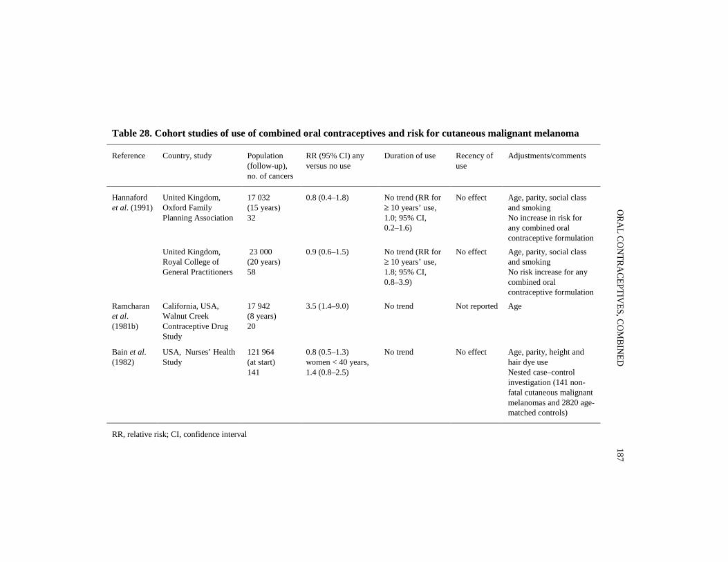
ORA
LCO
NTRA
CEPTIVES, CO
MBIN
ED187
Table 28. Cohort studies of use of combined oral contraceptives and risk for cutaneous malignant melanoma
Reference Country, study Population(follow-up),no. of cancers
RR (95% CI) anyversus no use
Duration of use Recency ofuse
Adjustments/comments
Hannafordet al. (1991)
United Kingdom,Oxford FamilyPlanning Association
17 032(15 years)32
0.8 (0.4–1.8) No trend (RR for≥ 10 years’ use,1.0; 95% CI,0.2–1.6)
No effect Age, parity, social classand smokingNo increase in risk forany combined oralcontraceptive formulation
United Kingdom,Royal College ofGeneral Practitioners
23 000(20 years)58
0.9 (0.6–1.5) No trend (RR for≥ 10 years’ use,1.8; 95% CI,0.8–3.9)
No effect Age, parity, social classand smokingNo risk increase for anycombined oralcontraceptive formulation
Ramcharanet al.(1981b)
California, USA,Walnut CreekContraceptive DrugStudy
17 942(8 years)20
3.5 (1.4–9.0) No trend Not reported Age
Bain et al.(1982)
USA, Nurses’ HealthStudy
121 964(at start)141
0.8 (0.5–1.3)women < 40 years,1.4 (0.8–2.5)
No trend No effect Age, parity, height andhair dye useNested case–controlinvestigation (141 non-fatal cutaneous malignantmelanomas and 2820 age-matched controls)
RR, relative risk; CI, confidence interval

to make allowance for major determinants of cutaneous malignant melanoma such assolar exposure and phenotypic characteristics.
Between 1968 and 1974, 17 032 white married women aged 25–39 were recruited at17 family planning clinics in the United Kingdom, in the framework of a study by theOxford Family Planning Association (Adam et al., 1981; Hannaford et al., 1991). On entry,56% of women were taking oral contraceptives, 25% were using a diaphragm and 19%were using an intrauterine device. Since, during the course of the study, each woman’s oralcontraceptive status could change, users of these preparations might have contributedperiods of observation for either current or former users. After 266 866 woman–years offollow-up, 32 new cases of cutaneous malignant melanoma were recorded, 17 of whichwere among women who had ever used oral contraceptives (relative risk, 0.8; 95% CI,0.4–1.8). None of the rates observed in any category of duration of use was materiallydifferent from that seen in women who had never used these preparations. The relativerisks, adjusted for age, parity, social class and smoking, were 0.6 (95% CI, 0.2–1.6) for< 5 years of use, 1.0 (0.4–2.6) for 5–9 years and 1.0 (0.2–3.1) for ≥ 10 years. There was norelationship between time since stopping use of oral contraceptives and the risk for cuta-neous malignant melanoma. None of the formulations resulted in a specific risk pattern.The distribution of cutaneous malignant melanomas by site was similar in users and non-users of oral contraceptives.
Between 1968 and 1969, 1400 general practitioners throughout the United Kingdomrecruited 23 000 women who were using oral contraceptives and a similar number of age-matched women who had never used them, in the framework of the study of the RoyalCollege of General Practitioners (Kay, 1981; Hannaford et al., 1991). After 482 083woman–years of follow-up, 58 new cases of cutaneous malignant melanoma had beenrecorded, 31 of which were among women who had ever used combined oral contra-ceptives; the relative risk, adjusted for age, parity, social class and smoking, was 0.9(95% CI, 0.6–1.5). No significant trend of increasing risk with duration of use was seen,the relative risk for 10 years or more of use being 1.8 (95% CI, 0.8–3.9), and the relativerisk did not vary according to recency of use, the oestrogen or progestogen content of thecontraceptives or the site of cutaneous malignant melanoma.
A cohort study of 17 942 women who were members of the Kaiser-Permanente HealthPlan, in California, United States, aged 18 and older, was established in 1970 within theWalnut Creek Contraceptive Drug Study. Ramcharan et al. (1981b) updated the prelimi-nary findings of Beral et al. (1977) to approximately eight years of follow-up and observed20 cases of cutaneous malignant melanoma (age-adjusted relative risk, 3.5; 95% CI,1.4–9.0). All five cases in women 18–39 years of age occurred among users of combinedoral contraceptives. The influence of duration and recency of use was not assessed. The per-centage distribution of hours of exposure to the sun by current, past or no use of combinedoral contraceptives was similar.
In a postal survey of 121 964 registered nurses in the United States in 1976 (Bainet al., 1982), no overall relationship was found between risk for cutaneous malignantmelanoma and use of combined oral contraceptives among 141 women with non-fatal
IARC MONOGRAPHS VOLUME 72188

cutaneous malignant melanoma and 2820 age-matched control women. The relative risk,adjusted for age, parity, height and hair dye use, was 0.8 (95% CI, 0.5–1.3). No significanttrends emerged with duration of use or time since first use. For women who were underthe age of 40 at the time cutaneous malignant melanoma was diagnosed, the relative riskwas 1.4 (95% CI, 0.8–2.5). For women under 40 who had used combined oral contra-ceptives for more than two years at least 10 years before diagnosis of cutaneous malignantmelanoma, the relative risk was 2.3 (95% CI, 0.8–6.9). An analysis restricted to the 84histologically documented cases of cutaneous malignant melanoma showed similarresults [not shown].
2.7.2 Case-control studiesThese studies are summarized in Table 29.Adam et al. (1981) investigated 169 cases of cutaneous malignant melanoma in
women aged 15–49 years that had been notified to the cancer registries of south-westernEngland during 1971–76, and 507 age-matched control women drawn from the lists ofthe same general practitioners as the cases. Data were obtained from the general practi-tioners’ records and for about 70% of the study women from postal questionnaires. Therisk for cutaneous malignant melanoma was not significantly increased among womenwho had ever used combined oral contraceptives, the unadjusted relative risk being 1.3(95% CI, 0.9–2.0) from the practitioners’ records and 1.1 (95% CI, 0.7–1.8) from thepostal questionnaires.
In an Australian investigation by Green and Bain (1985), described in detail in themonograph on ‘Post-menopausal oestrogen therapy’, there was no increased risk forcutaneous malignant melanoma in relation to use of combined oral contraceptives, withan age-adjusted relative risk of 0.7 (95% CI, 0.4–1.5), and no trend of increasing riskwith increasing duration of use, the relative risk for > 4 years’ use being 0.4 (95% CI,0.2–1.1). The risk was also not elevated among women who had first used combined oralcontraceptives 10 or more years before diagnosis of cutaneous malignant melanoma,with a relative risk of 0.9 (95% CI, 0.4–2.2).
In the case–control study of Holly et al. (1983) in Seattle, United States, described indetail in the monograph on ‘Post-menopausal oestrogen therapy’, use of combined oralcontraceptives for five years or more was commoner among cases than controls, with age-adjusted relative risks of 1.5 for 5–9 years of use and and 2.1 for ≥ 10 years’ duration (notsignificant). This relationship was seen only with long-term use of combined oralcontraceptives among women with superficial spreading melanoma, with relative risks of2.4 for 5–9 years and 3.6 for ≥ 10 years of use, and a highly significant trend (p = 0.004)with increasing duration of use. Adjustment was not made for the pattern of exposure tothe sun.
In the study of Lew et al. (1983), in Massachusetts, United States, described in detailin the monograph on ‘Post-menopausal oestrogen therapy’, no data were given on hor-monal treatment, but it was reported that cases and controls did not differ with respect touse of combined oral contraceptives.
ORAL CONTRACEPTIVES, COMBINED 189

IARC M
ON
OG
RAPH
S VO
LUM
E 72190
Table 29. Case–control studies of use of combined oral contraceptives and malignant melanoma
Reference Country Cases : controls(type ofcontrols)
Subgroup RR (95% CI)any versus nouse
Duration of use Recency of use Adjustment/Comments
Adamet al.(1981)
England 169 : 507(same generalpractitioner)
Generalpractitioners’records, postalquestionnaires
1.3 (0.9–2.0)1.1 (0.7–1.8)
No significanttrend (RR for≥ 5 years, 1.6;95% CI, 0.8–3.0)
No effect UnadjustedResponses to postal questionnaire(response rate about 70%) did notshow an association between use ofcombined oral contraceptives andexposure to the sun
Green &Bain(1985)
Queensland,Australia
91 : 91(population)
0.7 (0.4–1.5) No trend (RR for> 4 years’ use,0.4; 95% CI,0.2–1.1)
No effect (RR foruse ≥ 10 yearsbefore diagnosis,0.9; 95% CI,0.4–2.2)
AgeAfter allowance for phenotypiccharacteristics and solar exposure, RRfor > 4 years’ use, 0.4 (95% CI,0.1–2.0)
Holly et al.(1983)
Seattle, USA 87 : 863(population)
1–4 years5–9 years≥ 10 years
1.01.5 (NS)2.1 (NS)
Significant trendonly for SSM (RR≥ 10 years use,3.6)
Increased risk for≥ 12 years since firstuse: 4.4 (95% CI,2.0–9.7)
AgeNo data on solar exposure
Lew et al.(1983)
MassachusettsUSA
111 : 107(friends of cases)
– – – – No difference in combined oralcontraceptive use
Beral et al.(1984)
Sydney,Australia
287 : 574(hospital andpopulation)
1.0 (NS) No significanttrend
No significant effect Unadjusted (but altered by education,phenotype, history of sunburn andsolar exposure)Increased risk for women who hadbegun taking combined oralcontraceptives at least 10 years beforeand with ≥ 5 years’ duration of use:1.5 (95% CI, 1.0–2.1). No differenceby location, thickness or type ofCMM

ORA
LCO
NTRA
CEPTIVES, CO
MBIN
ED191
Table 29 (contd)
Reference Country Cases : controls(type ofcontrols)
Subgroup RR (95% CI)any versus nouse
Duration of use Recency of use Adjustment/Comments
Helmrichet al. (1984)
United Statesand Canada
160 : 640(hospital)
0.8 (0.5–1.3) No trend (RR for≥ 10 years’ use,1.0; 95% CI,0.4–2.9 [only age-adjusted])
No effect of timesince first use (RRfor first use ≥ 10years previously,1.1; 95% CI,0.7–1.8)
Age, area, religion, education andhormone-related variables
Holmanet al. (1984)
WesternAustralia
276 : 276 CMMSSM
1.0 (0.6–1.6)1.1 (0.6–2.2)
No significanttrend (RR for≥ 5 years use, 1.1;95% CI, 0.6–2.0)
No effect (RR for ≥10 years’ use beforediagnosis, 1.1; 95%CI, 0.7–1.7)
Age and residence
Gallagheret al. (1985)
Canada 361 : 361(members ofhealth plans)
CMM< 1 year1–4 years≥ 5 yearsSSM< 1 year1–4 years≥ 5 years
1.00.90.8
1.11.10.9
No trend No effect (RR foruse ≥ 10 years priorto diagnosis, 1.0)
Age, education, phenotype andfreckling Allowance for phenotypiccharacteristics
Østerlindet al. (1988)
Denmark 280 : 536 CMMSSM
0.8 (0.5–1.2)0.9 (0.6–1.3)
No trend (RR for≥ 10 years’ use,1.0; 95% CI,0.6–1.7)
No effect (RR foruse ≥ 10 yearsbefore diagnosis,1.3; 95% CI,0.7–2.2)
Age, phenotype and sunbathingNo difference according to type andpotency of combined oralcontraceptives
Zanettiet al. (1990)
Northern Italy 186 : 205(population)
CMMSSM
1.0 (0.5–1.9)1.3 (0.4–4.5)
No trend (RR for≥ 3 years’ use,1.0; 95% CI,0.5–2.7)
No effect Age, education, phenotype andsunbathingRisk did not change according toCMM type or location, age orcombined oral contraceptive potency
IARC M
ON
OG
RAPH
S VO
LUM
E 72192
Table 29 (contd)
Reference Country Cases : controls(type ofcontrols)
Subgroup RR (95% CI)any versus nouse
Duration of use Recency of use Adjustment/Comments
Augustssonet al. (1991)
Sweden 69 : 196(population)
Not reported No difference in combined oralcontraceptive use
Lê et al.(1992)
France 91 : 149(hospital)
< 10 years≥ 10 years
1.1 (0.6–2.0)2.1 (0.7–5.9)
No significanttrend
No effect of use15–20 years beforediagnosis (RR, 1.9;95% CI, 0.8–4.5)
Palmeret al. (1992)
Philadelphiaand New York,USA
615 : 2107 SevereNot severe
1.1 (0.8–1.5)1.5 (1.1–2.4)
No trend (RR fornot severe for≥ 10 years’ use,2.0; 95% CI,0.9–4.3)
No effect (RR forfirst use ≥ 20 yearsbefore severeCMM, 1.1; 95% CI,0.7–1.8)
Age, education, body mass index,menopause and phenotypeElevated risk among not severe casesof CMM was attributed tosurveillance bias; similar RR fordifferent types
Zaridzeet al. (1992)
Moscow,RussianFederation
96 : 96 0.04 (0.0–0.5)
Not reported Not reported Phenotype, naevi and sunbathingOnly one case and seven controls
Holly et al.(1995)
San Francisco,USA
452 : 930(population)
CMMSSM
0.7 (0.5–0.9)0.7 (0.5–1.0)
No trend (RR for≥ 10 years’ use:CMM, 0.8; 95%CI, 0.5–1.3; SSM,1.0; 95% CI,0.6–1.6)
No effect (RR foruse ≥ 17 yearsbefore diagnosis,0.6; 95% CI,0.4–0.7)
Age (unaltered by education,phenotype and solar exposure)

ORA
LCO
NTRA
CEPTIVES, CO
MBIN
ED193
Table 29 (contd)
Reference Country Cases : controls(type of controls)
Subgroup RR (95% CI)any versus nouse
Duration of use Recency of use Adjustment/Comments
Westerdahlet al.(1996)
Sweden 180 : 292(population)
1.6 (0.9–2.8) No effect (RR for> 8 years’ use,1.0; 95% CI,0.5–2.0)
No effect Phenotype, naevi and sunburnsAge at use and timing of use inrelation to first child did not influencerisk
Ocular melanoma
Hartgeet al.(1989b)
Wilmington andPhiladelphia,USA
238 : 223(detached retina)
0.9 (0.4–1.7) No trend (RR for≥ 10 years’ use,0.2; NS)
No effect Age
RR, relative risk; CI, confidence interval; NS, not significant; CMM, cutaneous malignant melanoma; SSM, superficial spreading melanoma

In the study of Beral et al. (1984) in Sydney, Australia, described in detail in themonograph on ‘Post-menopausal oestrogen therapy’, women who had ever used com-bined oral contraceptives were not at increased risk for cutaneous malignant melanoma(relative risk, 1.0). There was, however, an increased risk for women who had used theseformulations for five years or more and who had begun use at least 10 years beforediagnosis of cutaneous malignant melanoma, with a relative risk of 1.5 (95% CI, 1.0–2.1). The increase in risk persisted after control for phenotypic characteristics, number ofmoles and measures of exposure to ultraviolet light. The risk did not vary according tothe location, thickness or type of melanoma.
In a case–control study carried out in several parts of the United States and Canadabetween 1976 and 1982 (Helmrich et al., 1984), the case series consisted of 160 womenaged 20–59 years with a recent histological diagnosis of cutaneous malignant melanoma,and the controls were 640 women aged 20–59 years admitted to hospital for trauma ororthopaedic and surgical conditions. The age-adjusted relative risk for those who had everused combined oral contraceptives was 0.8 (95% CI, 0.5–1.3), and there was no trend inrisk with increasing duration of use, the relative risk for ≥ 10 years of use being 1.0(95% CI, 0.4–2.9). For the 40 case and 140 control women who had first used combinedoral contraceptives at least 10 years previously, the relative risk was 1.1 (95% CI, 0.7–1.8)and for women with more advanced cutaneous malignant melanoma (i.e. Clark’s level IVand V), the relative risk was 0.6 (95% CI, 0.2–2.3).
In the study of Gallagher et al. (1985), in Canada, described in detail in the monographon ‘Post-menopausal oestrogen therapy’, no association was seen between the risk for cuta-neous malignant melanoma and use of combined oral contraceptives in 361 cases and anequal number of controls aged 20–69. The relative risks for < 1, 1–4 and ≥ 5 years’ use,adjusted for age, phenotypic characteristics and freckling, were 1.0, 0.9 and 0.8, respectively.No association was seen between type of superficial spreading melanoma and duration ofuse or years since last use, the relative risk for women who had used combined oral contra-ceptives 10 or more years before diagnosis of cutaneous malignant melanoma being 1.0.
In the Danish study of Østerlind et al. (1988), described in detail in the monographon ‘Post-menopausal oestrogen therapy’, use of oral contraceptives was not related to therisk for cutaneous malignant melanoma (relative risk adjusted for age, phenotypiccharacteristics and sunbathing, 0.8; 95% CI, 0.5–1.2) or superficial spreading melanoma(relative risk, 0.9; 95% CI, 0.6–1.3), and there was no evidence of a dose–responserelationship, the relative risk for ≥ 10 years’ use being 1.0 (95% CI, 0.6–1.7). No specificrisk pattern was seen with the type of oral contraceptive, such as sequential, progestogenonly and high-potency combined oral contraceptives, assessed separately, but there werefew women in each group.
Zanetti et al. (1990) carried out a case–control study in Turin, northern Italy, between1984 and 1987 of 186 women aged 19–92 with histologically confirmed cutaneousmalignant melanoma out of 211 identified from the Turin Cancer Registry and 205control women aged 17–92 drawn from the National Health Service Registry (out of the300 initially contacted). Use of combined oral contraceptives, analysed only in women
IARC MONOGRAPHS VOLUME 72194
aged 60 or younger, was not associated with cutaneous malignant melanoma, the relativerisk adjusted for age, education, phenotypic characteristics and sunbathing being 1.0(95% CI, 0.5–1.9); no association was seen with superficial spreading melanoma(relative risk, 1.3; 95% CI, 0.4–4.5). Similarly, the longest duration of use (≥ 3 years: 1.0;95% CI, 0.5–2.7) or use that had started 10 or more years before the diagnosis of cuta-neous malignant melanoma was not associated with an increased risk. The relative riskswere identical for use of combined oral contraceptives containing high oestrogen doses(≥ 50 μg) and low oestrogen doses.
Augustsson et al. (1991) studied 69 cases of cutaneous malignant melanoma inSwedish women aged 30–50 and compared them with 196 controls drawn from the samepopulation. Skin type, phenotypic characteristics, number of naevi and dysplastic naeviwere taken into account. Although the relative risk was not reported, no difference in theuse of combined oral contraceptives was reported between cases and controls.
Lê et al. (1992) assessed the effect of use of combined oral contraceptives on the riskfor cutaneous malignant melanoma risk in France between 1982 and 1987. The caseswere those of 91 white women under 45 years of age who had new, histologicallyconfirmed melanomas, and the controls were 149 women consulting for diagnosis ortreatment of diseases unrelated to use of combined oral contraceptives, including skindiseases. No significant association was found between the total duration of use ofcombined oral contraceptives (relative risk, adjusted for age at menarche for ≥ 10 years’use, 2.1; 95% CI, 0.7–5.9) or the time since first use (relative risk 15–20 years since firstuse, 1.9; 95% CI, 0.8–4.5) and the risk for cutaneous malignant melanoma, and no diffe-rence was found between superficial spreading melanoma and other types of cutaneousmalignant melanoma. The relative risk for 49 case women and 78 matched controls whowere aged 30–40 and had used oral contraceptives for 10 or more years, however, wassignificantly increased: 4.4 (95% CI, 1.1–17). In a subgroup of 57 case women and 65controls for whom allowance could be made for phenotypic characteristics and solarexposure, the relative risks were similar.
A case–control study on cutaneous malignant melanoma was carried out between1979 and 1991 in Philadelphia and New York, United States (Palmer et al., 1992), inwhich the cases were in 615 women under the age of 70 (median age, 40) who hadrecently received a first diagnosis of cutaneous malignant melanoma. Patients with mela-noma in situ were not included. Two control groups of white women with a median ageof 41 years with other malignancies (610 patients) or non-malignant illnesses (1497patients) judged to be unrelated to use of combined oral contraceptives were selected. Inorder to address the possibility of selection bias due to differential surveillance ofcombined oral contraceptive users and non-users, the cases were subdivided by severity.For severe cases (thickness ≥ 0.75 mm, or Clark’s level IV or V), the relative risksadjusted for age, education, menopause and phenotypic characteristics were 1.1 (95% CI,0.8–1.5) for any use, 1.1 (0.6–2.1) for ≥ 10 years’ use and 1.1 (0.7–1.8) for ≥ 20 years’use. For non-severe cases, increased risks were found for any use (1.5; 95% CI, 1.1–2.2)and for ≥ 10 years’ use (2.0; 0.9–4.3). The relative risks did not vary by type of cutaneous
ORAL CONTRACEPTIVES, COMBINED 195
malignant melanoma. According to the authors, the increased risks seen for non-severecases of cutaneous malignant melanoma were probably due to greater surveillance ofcombined oral contraceptive users.
Zaridze et al. (1992) evaluated risk factors in 96 cases of cutaneous malignant mela-noma in Moscow, Russian Federation. Controls matched by age were recruited fromamong persons visiting cancer patients. Use of combined oral contraceptives could beanalysed for 54 women with cutaneous malignant melanoma and 54 controls and showeda strong inverse association: the relative risk, adjusted for phenotypic characteristics,naevi and sunbathing, was 0.04 (95% CI, 0.0–0.5). Only one case and seven controls,however, had ever used combined oral contraceptives.
In the study of Holly et al. (1995), described in detail in the monograph on ‘Post-meno-pausal oestrogen therapy’ (Holly et al., 1994), 72% of the cases of cutaneous malignantmelanoma and 79% of the control subjects in San Francisco, United States, reported everhaving used combined oral contraceptives. The age-adjusted relative risk was 0.7 (95% CI,0.5–0.9) for all cutaneous malignant melanoma and 0.7 (95% CI, 0.5–1.0) for superficialspreading melanoma. Examination by latency and duration of use showed no significanttrend. The relative risk for ≥ 10 years’ use was 0.8 (95% CI, 0.5–1.3) for all cutaneousmalignant melanoma and 1.0 (95% CI, 0.6–1.6) for superficial spreading melanoma. Usebeginning ≥ 17 years before diagnosis was associated with relative risks of 0.6 (95% CI,0.4–0.7) for cutaneous malignant melanoma and 0.6 (95% CI, 0.4–0.8) for superficialspreading melanoma.
In the Swedish study of Westerdahl et al. (1996), described in the monograph on‘Post-menopausal oestrogen therapy’, any use of combined oral contraceptives (40% ofcases and 37% of controls) was associated with a non-significantly elevated risk of 1.6(95% CI, 0.9–2.8) after adjustment for phenotypic characteristics, naevi and sunburns. Notrend in risk was seen with duration of use (relative risk for > 8 years’ use, 1.0; 95% CI,0.5–2.0), age at first use or age at last use.
A meta-analysis of 18 published case–control studies on cutaneous malignant mela-noma and use of combined oral contraceptives, including 17 of the papers reviewed hereand that of Beral et al. (1977), showed a pooled relative risk of 1.0 (95% CI, 0.9–1.0)(Gefeller et al., 1997). The data for 3796 cases and 9442 controls showed no significantheterogeneity of the effect of combined oral contraceptives in the different studies, andanalysis of various subgroups, defined by the design characteristics of the studies, did notmaterially alter this result.
2.8 Retinal melanomaIn a case–control study of ocular melanoma in the United States (Hartge et al.,
1989b), described in the monograph on ‘Post-menopausal oestrogen therapy’, use ofcombined oral contraceptives was reported by about 13% in both cases and controls, togive an age-adjusted relative risk of 0.9 (95% CI, 0.4–1.7). The estimated risk was notrelated to duration of use (relative risk for ≥ 10 years’ use, 0.2; 95% CI, 0.3–1.2) or tothe interval since first or last use.
IARC MONOGRAPHS VOLUME 72196

2.9 Thyroid cancerNone of the cohort studies provided information on use of combined oral contracep-
tives and the risk for thyroid cancer. The case–control studies are summarized in Table 30.In the case–control study of McTiernan et al. (1984), in Seattle, United States, des-
cribed in detail in the monograph on ‘Post-menopausal oestrogen therapy’, the use ofcombined oral contraceptives (prevalence: 93/141 cases and 130/219 controls) was asso-ciated with a slightly increased risk for thyroid cancer (1.6; 95% CI, 1.0–2.5). Themagnitude of the excess risk did not increase with increasing duration of use (relativerisk for > 3 years’ duration, 1.2). The risk was higher among women with follicularthyroid cancer (3.6; 95% CI, 1.1–12.8) and among those women who discovered theirown tumours as compared with those whose tumour was found by a physician.
Preston-Martin et al. (1987) evaluated the risk factors for thyroid cancer in womenaged 40 or less in Los Angeles, United States, between 1980 and 1981. The cases werein 108 white women with papillary, follicular or mixed thyroid cancer (out of 135identified through Southern California Cancer Surveillance Program) and controls were108 age-matched women who lived near the case women (neighbourhood controls).More cases (67/78) than controls (76/106) had ever used combined oral contraceptives(unadjusted relative risk, 2.4; 95% CI, 1.1–5.7). Cases and controls did not differ withrespect to age at first use. There was no trend of increasing risk with increasing durationof use, the relative risk for > 5 years’ duration of use being 2.4 (95% CI, 0.9–6.9).
In a study conducted in Connecticut, United States (Ron et al., 1987), described in themonograph on ‘Post-menopausal oestrogen therapy’, similar proportions of cases (55/109)and controls (110/208) had ever used combined oral contraceptives, the relative riskadjusted for age, parity, radiotherapy to the head and neck and benign thyroid disease being0.8 (not significant). For women under the age of 35 at the time of diagnosis, the relativerisk was 1.8 (not significant). Duration and latency of use were not assessed.
Franceschi et al. (1990) found relatively few users of combined oral contraceptivesamong cases of thyroid cancer in Italy (23/165 cases and 28/214 controls). The age-adjusted relative risks were 1.1 (95% CI, 0.5–2.4) for use for < 24 months and 1.1 (95%CI, 0.4–3.0) for use for ≥ 24 months.
In a case–control study in Hawaii (Kolonel et al., 1990), described in the monographon ‘Post-menopausal oestrogen therapy’, women who had ever used combined oralcontraceptives (43% among controls) showed no increased risk for thyroid cancer. Therelative risk, adjusted for age and ethnic group was 0.9 (95% CI, 0.5–1.5). The effects ofduration and latency of use were not reported.
Levi et al. (1993), in study in Switzerland, described in detail in the monograph on‘Post-menopausal oestrogen therapy’, found a prevalence of any use of combined oralcontraceptives of 56% among thyroid cancer cases and 44% among control women; therelative risk, adjusted for age and a history of benign thyroid disease, was 1.2 (95% CI,0.7–2.3). There was no trend of increasing risk with increasing duration of use, therelative risk for ≥ 5 years’ use being 1.4 (95% CI, 0.7–2.7). Analyses restricted to womenunder 45 years of age or to cases of papillary thyroid cancer yielded similar risk estimates.
ORAL CONTRACEPTIVES, COMBINED 197

IARC M
ON
OG
RAPH
S VO
LUM
E 72198
Table 30. Case–control studies on use of combined oral contraceptives and thyroid cancer
Reference Country Cases : controls(type of controls)
RR (95% CI), any versusno use
Duration of use Adjustment/comments
McTiernan et al.(1984)
Seattle, USA 141 : 319 (population) 1.6 (1.0–2.5) No trend (RR for> 3 years’ use, 1.2)
AgeGreatest risk increase seenfor follicular thyroid cancer(RR, 3.6; 95% CI, 1.1–13)
Preston-Martinet al. (1987)
Los Angeles, USA 108 : 108(population)
2.4 (1.1–5.7) No trend (RR for> 5 years’ use, 2.4; 95%CI, 0.9–6.9)
Unadjusted.Only women aged 40 or less
Ron et al. (1987) Connecticut, USA 109 : 208 (population) 0.8 Not reported Age, parity, radiotherapy to the headand neck and benign thyroid diseasesRR for women < 35 was 1.8 (notsignificant)
Franceschi et al.(1990)
Italy 165 : 214(hospital)
< 2 years, 1.1 (0.5–2.4)≥ 2 years, 1.1 (0.4–3.0)
No effect Age and area of residence
Kolonel et al. (1990) Hawaii, USA 140 : 328 (population) 0.9 (0.5–1.5) Not reported Age and ethnic groupIncreased risk for women with difficultyin conceiving(RR, 1.8; 95% CI, 1.0–3.1) and thosewho used fertility drugs (RR, 4.2; 95%CI, 1.5–11)
Levi et al. (1993) Vaud, Switzerland 91 : 306(hospital)
1.2 (0.7–2.3) No trend (RR for≥ 5 years’ use, 1.4; 95%CI, 0.7–2.7)
Age and history of benign thyroiddiseaseSimilar risk estimates for women under45 and for papillary thyroid cancer
Preston-Martinet al. (1993)
Shanghai, China 207 : 207 (population) 1.7 (1.0–3.1) No trend (RR for> 5 years’ use, 0.9; 95%CI, 0.4–2.4)
Age
Wingren et al. (1993) South-easternSweden
93 : 187(population)
No risk (RR not reported) Not reported Only papillary carcinomas

ORA
LCO
NTRA
CEPTIVES, CO
MBIN
ED199
Table 30 (contd)
Reference Country Cases : controls(type of controls)
RR (95% CI), any versusno use
Duration of use Adjustment/comments
Hallquist et al. (1994) Northern Sweden 123 : 240 (population) All, 0.8 (0.5–1.4)Papillary, 0.6 (0.3–1.2)
No trend AgeRisk did not vary by timing in relationto age at first pregnancy
Galanti et al. (1996) Sweden and Norway 191 : 341 (population) 0.9 (0.6–1.5) No trend (> 2 years’ useRR, 0.8; 95% CI, 0.5–1.3)
Age and parity
RR, relative risk; CI, confidence interval

Preston-Martin et al. (1993) carried out a study in Shanghai, China, between 1981and 1984, which included 207 women aged 18–54 listed in the Shanghai Cancer Registryas having a histologically confirmed thyroid cancer; 20% of the cases were reviewed bya pathologist. The 207 control women, matched to the cases by year of birth, were chosenrandomly from the Shanghai Residents’ Registry; over 90% of the eligible subjects wereinterviewed. Few women had used combined oral contraceptives (43/207 cases and29/207 controls), but any use of such formulations was associated with a marginallyincreased risk (unadjusted relative risk, 1.7; 95% CI, 1.0–3.1). Among users, however,there was no trend in risk with the duration of use, the relative risk for > 5 years’ usebeing 0.9 (95% CI, 0.4–2.4).
Wingren et al. (1993) studied 93 cases of thyroid cancer and 187 controls aged 20–60in south-east Sweden and reported that use of combined oral contraceptives was not asso-ciated with an increased risk. No data were shown.
Hallquist et al. (1994), in Sweden, reported that 42/123 cases and 92/240 controlshad reported any use of combined oral contraceptives, giving an age-adjusted relativerisk of 0.8 (95% CI, 0.5–1.4). The corresponding relative risk for papillary thyroid cancerwas 0.6 (95% CI, 0.3–1.2). The risk did not vary by duration of use, being 0.6 (95% CI,0.2–1.5) for ≥ 7 years’ use, or by timing of use in relation to age at first pregnancy.
Galanti et al. (1996), in a study in Sweden and Norway described in the monographon ‘Post-menopausal oestrogen therapy’, reported that 98/179 cases and 180/334 controlshad used combined oral contraceptives. No relation was found between use and the riskfor thyroid cancer; the age- and parity-adjusted relative risk for any use was 0.9 (95% CI,0.6–1.5). Use for > 2 years was associated with a relative risk of 0.8 (95% CI, 0.5–1.3).
2.10 Other cancersA 25-year follow-up of 46 000 women in Great Britain in the framework of a study
on oral contraceptives by the Royal College of General Practitioners did not show signi-ficant excess mortality from lung cancer (relative risk, 1.2; 95% CI, 0.8–1.8) or any othercancer (Beral et al., 1999).
In a study by La Vecchia et al. (1994), from Milan, Italy, described in detail in themonograph on ‘Post-menopausal oestrogen therapy’, use of combined oral contra-ceptives was not related to the risk for gastric cancer. Six of 229 cases and 19 of 614controls had ever used such formulations, giving a relative risk adjusted for age, edu-cation, a family history of cancer and dietary habits of 1.3 (95% CI, 0.5–3.5).
Chow et al. (1995) in a study in Minnesota, United States, described in detail in themonograph on ‘Post-menopausal oestrogen therapy’, found no relation between use ofcombined oral contraceptives and the risk for renal-cell cancer; the relative risk, adjustedfor age, smoking and body mass index was 0.8 (95% CI, 0.4–1.3). For use longer than10 years, the relative risk was 0.3 (95% CI, 0.1–1.0).
The risk for renal-cell cancer and use of combined oral contraceptives was also eva-luated in an international study by Lindblad et al. (1995), described in detail in the mono-graph on ‘Post-menopausal oestrogen therapy’. Any use of combined oral contraceptives
IARC MONOGRAPHS VOLUME 72200

was associated with a relative risk, adjusted for age, smoking and body mass index, of0.7 (95% CI, 0.5–0.9). There was an inverse trend in risk with increasing duration of use,the relative risk for > 10 years’ use being 0.5 (95% CI, 0.3–0.9).
3. Studies of Cancer in Experimental Animals
In this section, only relevant studies on oestrogens and progestogens alone and incombination that were published subsequent to or not included in Volume 21 of theIARC Monographs (IARC, 1979) are reviewed in detail. Studies reviewed previously aresummarized briefly.
3.1 Oestrogen–progestogen combinations3.1.1 Studies reviewed previously Mouse
The results of studies reviewed previously (Committee on Safety of Medicines,1972; IARC, 1979) on the carcinogenicity of combinations of oestrogens and proges-togens in mice are as follows:
Chlormadinone acetate in combination with mestranol tested by oral administrationto mice caused an increased incidence of pituitary adenomas in animals of each sex. Oraladministration of chlormadinone acetate in combination with ethinyloestradiol to miceresulted in an increased incidence of mammary tumours in intact and castrated males.
After oral administration of ethynodiol diacetate and mestranol to mice, increasedincidences of pituitary adenomas were observed in animals of each sex. The combinationof ethynodiol diacetate plus ethinyloestradiol, tested by oral administration to mice,increased the incidences of pituitary adenomas in animals of each sex and of malignanttumours of connective tissues of the uterus.
Lynoestrenol in combination with mestranol was tested in mice by oral administration.A slight, nonsignificant increase in the incidence of malignant mammary tumours wasobserved in females which was greater than that caused by lynoestrenol or mestranol alone.
The combination of megestrol acetate plus ethinyloestradiol, tested by oral adminis-tration to mice, caused an increased incidence of malignant mammary tumours in animalsof each sex.
Noresthisterone acetate plus ethinyloestradiol, tested by oral administration to mice,increased the incidences of pituitary adenomas in animals of each sex, but the incidenceswere comparable to those induced by ethinyloestradiol alone. The combination of nore-thisterone plus ethinyloestradiol was also tested in mice by oral administration; anincreased incidence of pituitary adenomas was observed in females. Norethisterone plusmestranol increased the incidences of pituitary adenomas in animals of each sex.
Norethynodrel in combination with mestranol was tested by oral administration inmice. Increased incidences of vaginal or cervical tumours were found in female mice andof pituitary adenomas in males and females. In female mice, an increased incidence of
ORAL CONTRACEPTIVES, COMBINED 201

malignant mammary tumours was observed, but the incidence was not greater than thatseen with norethynodrel alone. In castrated male mice, the combined treatment resultedin an increased incidence of mammary tumours.
The combination of norgestrel plus ethinyloestradiol was tested in mice by oral admi-nistration; no increase in the incidence of tumours was observed.
RatThe results of studies reviewed previously (Committee on Safety of Medicines,
1972; IARC, 1979) on the carcinogenicity in rats of several combinations are as follows:Ethynodiol diacetate plus ethinyloestradiol was tested for carcinogenicity by oral
administration to rats. The incidence of malignant mammary tumours was increased inanimals of each sex. In combination with mestranol, the incidence of mammary tumourswas increased in one study but not in another.
Lynoestrenol in combination with mestranol was tested in female rats by oral admi-nistration. No increase in tumour incidence was observed.
Megestrol acetate plus ethinyloestradiol was tested by oral administration to rats. Theincidence of benign liver tumours was increased in animals of each sex, but not to a levelgreater than that observed with ethinyloestradiol alone. In male rats, there was a smallincrease in the incidence of benign and malignant mammary tumours; females showed asmall increase in the incidence of malignant mammary tumours.
The combination of noresthisterone acetate plus ethinyloestradiol, tested by oraladministration to rats, increased the incidences of benign mammary tumours and liveradenomas in males. Norethisterone plus mestranol increased the incidence of malignantmammary tumours in female rats and increased the incidence of liver adenomas inmales.
Norethynodrel in combination with mestranol was tested by oral administration torats. In males, increased incidences of liver adenomas, pituitary adenomas and benignand malignant mammary tumours were observed, but the incidences were no greater thanthose with norethynodrel alone. In females, the incidences of pituitary adenomas andmalignant mammary tumours were increased.
Norgestrel plus ethinyloestradiol, tested for carcinogenicity in rats by oral admi-nistration, caused a small increase in the incidence of benign mammary tumours in males.
Dimethisterone and oestradiol were tested in dogs, with no increase in the incidenceof mammary tumours
3.1.2 New studies (a) Oral administration
RatSchuppler and Gunzel (1979) summarized data from a study by the Committee on
Safety of Medicines (1972) in the United Kingdom on the incidence of hepatocellularadenomas in groups of 24–124 male and female rats [strain not specified] treated orallywith combinations of various oestrogens and progestogens at doses up to 400 times the
IARC MONOGRAPHS VOLUME 72202

human contraceptive dose. The statistically significant increases indicated in their reportare indicated by a ‘+’ in Table 31.
Groups of 10 female Sprague-Dawley rats, seven weeks of age, were treated withEnovid E (100 μg mestranol + 25 mg norethynodrel) in the diet for nine months, withdaily intakes of 0.02–0.03 and 0.5–0.75 mg/kg bw. The numbers of altered γ-glutamyltranspeptidase (γ-GT)-positive hepatic foci, considered to be preneoplastic lesions, werecounted at autopsy. A statistically significant (p < 0.001) increase in the number of foci(2.8 foci/cm2) was observed in comparison with untreated controls (0.2 foci/cm2). Noincrease in the incidence of hepatic nodules or carcinomas was observed at this time(Yager & Yager, 1980).
Female Wistar rats, 15–17 weeks of age, were treated with quingestanol acetate plusquinestrol, which are 3-cyclopentyl ether derivatives of norethisterone acetate andethinyloestradiol, respectively, as a 2:1 mixture suspended in sesame oil containing pipe-ridine (0.05% v/w) by stomach tube. A group of 75 rats was treated once weekly with30 mg/kg bw, 60 rats were treated with 1.2 mg/kg bw per day, and 75 rats were used asvehicle controls; all treatments were given for 50 weeks, followed by 30 weeks ofobservation for reversibility of any lesions. Groups of 10 animals were killed at 25 and50 weeks; at 66 weeks, five rats from each treatment group and three vehicle controlswere killed, and at 80 weeks all survivors in the treated groups and 10 vehicle controlswere killed. Treatment was associated with irreversible hair loss, a reversible decrease inbody weight and reversible ataxia. The only treatment-related tumours were mammarymasses and adenocarcinomas: at 40–50 weeks, the incidences of adenocarcinomas were10/33 in rats at 30 mg/kg bw, 15/27 in those at 1.2 mg/kg bw and 1/12 in vehicle controls[statistics not specified]. After treatment was suspended, the incidences of mammaryadenocarcinomas were reduced, with 1/8 at weeks 51–66 and 0/12 at weeks 67–80 inanimals at 30 mg/kg bw, 3/7 and 2/8 at those times in animals at 1.2 mg/kg bw and 0/10and 1/13 in vehicle controls (Lumb et al., 1985).
Male and female Wistar rats, four weeks old, were given ethinyloestradiol (0.075 mg)+ norethisterone acetate (6 mg) dissolved in olive oil by gavage daily, ethinyloestradiol +
ORAL CONTRACEPTIVES, COMBINED 203
Table 31. Effects of progestogen–oestrogen combinations on theincidence of hepatocellular adenomas in rats
Progestogen Oestrogen Ratio Males Females
Norethynodrel Mestranol 66:125:1
+–
––
Norethisterone Mestranol 20:1 + –Lynestrenol Mestranol 33:1 Not tested –Megestrol acetate Ethinyloestradiol 5:1
80:1++
––
From Schuppler & Gunzel (1979)

norethisterone acetate + 10% ethanol in the drinking-water on five days a week, olive oil +ethanol or olive oil alone. The animals were treated for up to 12 months, with interim killsat two, four, six, eight and 12 months, when the livers were analysed for the presence ofhepatocellular carcinomas and hyperplastic nodules. In females, ethinyloestradiol + nore-thisterone acetate induced a 100% incidence of hyperplastic nodules by four months andhepatocellular carcinomas in 2/25 animals at 12 months. Ethanol increased the incidenceof hepatocellular carcinoma at 12 months to 9/22; no hyperplastic nodules or hepatocellularcarcinomas were seen in the controls receiving ethanol alone. In males, ethinyloestradiol +norethisterone acetate induced hyperplastic nodules in 6/20 animals at 12 months but nohepatocellular carcinomas. Ethanol increased the incidence of hyperplastic nodules to100%, beginning at four months, and that of hepatocellular carcinomas to 2/17 at 12months. Again, ethanol alone had no effect on the incidences of hyperplastic nodules orhepatocellular carcinoma, but it enhanced nuclear and cytosolic oestrogen receptors andDNA adduct formation, as detected by 32P-postlabelling (Yamagiwa et al., 1991, 1994).
MonkeyNorlestrin (50:1 norethisterone acetate + ethinyloestradiol) was given to groups of
15–17 young adult female rhesus (Macaca mulatta) monkeys weighing 2.8–5.7 kg at thebeginning of the study. Norlestrin powder was blended with soft fruit and vegetables andwas administered over 10 years as 21 consecutive daily doses followed by seven dayswithout treatment. The daily doses were 0, 0.051, 0.51 and 2.55 mg/kg bw which repre-sented 0, 1, 10 and 50 times the human contraceptive dose. There were no effects onsurvival and no treatment-related alterations in coagulation or other clinical parameters.Only a few tumours appeared but were found in all groups (Fitzgerald et al., 1982).
(b) Administration with known carcinogensMouse
Groups of 20 female B6AF1 mice, 12 weeks of age, were treated with 3-methyl-cholanthrene by insertion of an impregnated silk thread into the cervical canal andthrough the uterine wall; a control group was treated with silk thread not impregnatedwith 3-methylcholanthrene. Pellets containing steroids were then implanted subcuta-neously and renewed every three weeks for 15 weeks. The doses given every three weekswere 15 mg norethynodrel and 0.5 mg mestranol per mouse, alone and in combination.No tumours developed in mice that had not received 3-methylcholanthrene, but thesteroids caused some histopathological changes in the mucosa of the cervix, uterus andvagina. The incidences of uterine adenoacanthomas were increased (p < 0.01) by all threetreatments: control, 5/35; mestranol, 10/19; norethynodrel, 11/14; mestranol + nore-thynodrel, 10/18. Squamous-cell carcinomas of the cervix were observed in all groups,but the incidences were not statistically significantly increased in those receiving steroids(Blanzat-Reboud & Russfield, 1969).
The effects of two formulations, Ovral, consisting of 0.05 mg ethinyloestradiol + 0.5 mgnorgestrel, and Noracycline, consisting of 0.05 mg ethinyloestradiol + 1 mg lynoestrenol, on
IARC MONOGRAPHS VOLUME 72204
carcinomas of the uterine cervix induced by 3-methylcholanthrene were studied in groupsof 10–30 Swiss albino female mice, eight to nine weeks of age. The mice treated with3-methylcholanthrene received a sterile cotton thread impregnated with beeswax containingapproximately 300 μg of the carcinogen into the uterine cervix. The oral contraceptivecombinations were administered orally at doses of 1/2000th of a pill (0.025 μg ethinyl-oestradiol + 0.25 μg norgestrel), 1/200th of a pill (0.25 μg ethinyloestradiol + 2.5 μg nor-gestrel) and 1/20th of a pill (2.5 μg ethinyloestradiol + 25 μg norgestrel) of Ovral, and1/2000th of a pill (0.025 μg ethinyloestradiol + 0.5 μg lynoestrol), 1/200th of a pill (0.25 μgethinyloestradiol + 5 μg lynoestrol) and 1/20th of a pill (2.5 μg ethinyloestradiol + 50 μglynoestrol) of Noracycline. Treatment was for 30, 60 or 90 days. Animals that did notreceive 3-methylcholanthrene did not develop cervical tumours at any dose of oral contra-ceptive. In contrast, treatment with either formulation caused dose-dependent, biphasiceffects on the incidence of squamous-cell carcinomas induced by 3-methylcholanthrene. Atthe two lower doses, they were protective in comparison with treatment with the carcinogenalone (p < 0.05), while at the high dose they enhanced carcinogenesis: The incidence ofsquamous-cell carcinomas was 6/23 with 3-methylcholanthrene alone and 8/17 with thehigh dose of Ovral at 90 days, although the difference was not indicated as being statisticallysignificant. With Noracycline, the enhancement was statistically significant after both 60days (13/24 versus 2/23, p < 0.05) and 90 days (12/19 versus 6/23, p < 0.05). At all dosesand at all times, both formulations also significantly enhanced the incidence of cervicalhyperplasia (Hussain & Rao, 1992).
In a more recent study with the same model, Ovral was administered to Swiss miceat the same two lower doses as the previous study, with 3-methylcholanthrene at a higherdose of 600 μg. The cotton thread was inserted into the right uterine horn. After 90 days,the incidence of tumours in the uterine endometrium was 8/15 in mice receiving thecarcinogen alone and 1/16 in the group receiving the carcinogen + the 1/2000th dose ofOvral (p < 0.05). No tumours were seen in the group treated with 3-methylcholanthrene+ Ovral at the 1/200th dose (Chhabra et al., 1995).
RatGroups of 9–10 female Sprague-Dawley rats, seven weeks of age, were initiated by
treatment with 5 mg/kg bw N-nitrosodiethylamine (NDEA) 24 h after partial hepa-tectomy. Twenty-four hours later they were fed a diet containing mestranol + nore-thynodrel at concentrations providing 0.02–0.03 and 0.5–0.75 mg/kg bw per day,respectively. After nine months, a statistically significant (p < 0.001) increase in thenumber of γ-GT-positive altered hepatic foci was observed (7.3 versus 0.3 foci/cm2),with no significant increase in the incidence of nodules or carcinomas (Yager & Yager,1980).
Groups of female weanling Wistar rats received an intraperitoneal injection of 0 or200 mg/kg bw NDEA. One month later, half the animals in each group (5–6 rats)received 1/10th of a tablet of Ovulen-50 (5 μg ethinyloestradiol + 100 μg ethynodioldiacetate) daily orally in 0.1 mL propylene glycol for 60 weeks; the other half of the rats
ORAL CONTRACEPTIVES, COMBINED 205

in each group received the vehicle. The livers were examined histochemically for γ-GT-positive foci and histologically. None of the rats developed liver tumours. In rats that hadnot been initiated with NDEA, Ovulen-50 increased the incidences of γ-GT-positive fociand of microscopic hyperplastic nodules in all five rats; such foci and nodules were notseen in other groups. The authors speculated that the absence of foci and nodules inNDEA-initiated rats with and without treatment with Ovulen-50 may have been due toan interplay of the drug-metabolizing enzymes and that the Ovulen-50 steroids weremore rapidly metabolized by the NDEA-initiated rats (Annapurna et al., 1988). [TheWorking Group noted that opposite effects, i.e. enhancement of foci and nodules ininitiated livers by oral contraceptive steroids, have been seen in many other studies andthat the results of this study must be considered an exception to the general finding andthat they lack a mechanistic explanation.]
The results of the previous and new studies on oestrogen–progestogen combinationsare summarized in Tables 32 and 33.
3.2 Oestrogens used in combined oral contraceptives3.2.1 Studies reviewed previouslyMouse
Ethinyloestradiol administered to mice increased the incidence of pituitary adenomasand malignant mammary tumours in animals of each sex and the incidences of uterineand cervical tumours in females.
Mestranol increased the incidences of pituitary adenomas and malignant mammarytumours in animals of each sex.
RatWhen ethinyloestradiol was tested for carcinogenicity in rats, the incidences of liver
adenomas were increased in animals of each sex and that of liver carcinomas in females.Administration of mestranol increased the incidence of malignant mammary tumours
in females in one of two treated groups.
3.2.2 New studies (a) Oral administration
MouseSchuppler and Gunzel (1979) summarized data on the incidence of hepatic adenomas
in groups of 40–120 male and female mice of three strains after oral administration ofethinyloestradiol or mestranol for 20 months at up to 400 times the human contraceptivedose. Only mice of strain BDH-SPF showed a small increase in incidence after receivingethinyloestradiol.
RatSchuppler and Gunzel (1979) reported that the study of the Committee on Safety of
Medicines (1972) found no increase in the incidence of liver adenoma in female rats
IARC MONOGRAPHS VOLUME 72206

ORA
LCO
NTRA
CEPTIVES, CO
MBIN
ED207
Table 32. Effects of combinations of various progestogens and oestrogens on tumour incidence in mice
Combination Pituitary adenomas Mammary tumours Uterinetumours
Male Female Malignant
Cervical/vaginaltumoursBenign
(males)Male Female
Chlormadinone acetate + mestranol + +Chlormadinone acetate + ethinyloestradiol +/cEthynodiol diacetate + mestranol + +Ethynodiol diacetate + ethinyloestradiol + + +Lynoestranol + mestranol +/–Lynoestranol + ethinyloestradiol + 3-methylcholanthrene +a
Megestrol acetate + ethinyloestradiol + +Norethisterone acetate + ethinyloestradiol +/? +/?Norethisterone + ethinyloestradiol +Norethisterone + mestranol + +Norethynodrel + mestranol + + c +/? +Norethynodrel + mestranol + 3-methylcholanthrene + –Norgestrel + ethinyloestradiol + 3-methylcholanthrene +a
+, increased tumour incidence; +/-, slighly increased tumour incidence; +/c, increased tumour incidence in intact and castrated animals;c, increased tumour incidence in castrated animals; +/?, increased tumour incidence, but not greater than that with the oestrogen orprogestogen alonea Protection at doses 1/2000th and 1/200th that of a pill for women; enhancement at a dose of 1/20th that of a pill for women

IARC M
ON
OG
RAPH
S VO
LUM
E 72208
Table 33. Effects of combinations of various progestogens and oestrogens on tumour incidence in rats
Combination Pituitary adenomas Mammary tumours Liver
Male Female Malignant Adenoma CarcinomaBenign(males)
Male Female Male Female Male Female
Foci(females)
Ethynodiol diacetate + ethinyloestradiol
+ +
Ethynodiol diacetate + mestranol ? ?Megestrol acetate + ethinyloestradiol +/– +/– +/– +/? +/?Norethisterone acetate + ethinyloestradiol
+ + – +
Norethisterone + mestranol + + –Norethynodrel + mestranol +/? + +/? +/? + +/? – – – +Norethynodrel + mestranol + N-nitrosodiethylamine
– – +
Norgestrel + ethinyloestradiol +/–
+, increased tumour incidence; +/–, slighly increased tumour incidence; +/?, increased tumour incidence, but not greater than that with the oestrogen orprogestogen alone; ? conflicting; –, no effect

treated orally with mestranol. Of four studies on the incidences of hepatocellular adenomaand carcinoma in groups of 40–120 male and female rats treated orally with ethinyl-oestradiol at doses up to 400 times the human contraceptive dose, only one showed astatistically significant increase in the incidence of hepatocellular adenoma in males (0%controls, 15.3% treated) and in females (8% controls, 23.5% treated); the incidence ofhepatocellular carcinoma was significantly increased only in females (0% controls, 7.4%treated).
Groups of female Sprague-Dawley rats received ethinyloestradiol or mestranol in thediet from about seven weeks of age at concentrations of 0.1 or 0.5 mg/kg diet (ppm)mestranol for nine and 12 months or 0.5 ppm ethinyloestradiol for nine months. Theingested doses were approximately equivalent to 6 or 30 μg/kg bw per day or 3–15 timesthe human contraceptive dose. Ethinyloestradiol caused a statistically significant (p < 0.05)increase in the number of γ-GT-positive, altered hepatic foci, but not in the volumepercentage of liver occupied by foci, after nine months. No increase in the incidence ofnodules or carcinomas was observed. The high dose of mestranol had similar effects afternine months, but after 12 months, mestranol caused a statistically significant (p < 0.05)increase in both the number of altered hepatic foci, and the volume percentage of the liveroccupied by foci showed a significant (p < 0.05) dose–response relationship. Furthermore,after 12 months, the high dose of mestranol caused a significant (p < 0.05) increase in theincidence of hepatic nodules and carcinomas combined (4/16 compared with 0/15 incontrols) (Yager et al., 1984).
Female Wistar rats, four weeks of age, were treated with 0 (control), 75 or 750 μgethinyloestradiol in 0.5 mL olive oil by gavage daily for various times up to 12 months. Byfour months, the incidence of glutathione S-transferase-positive, altered hepatic foci,considered to be preneoplastic lesions, was 100% in both groups. At 12 months, hepato-cellular carcinomas were found in 2/23 rats at 75 μg ethinyloestradiol and 10/26 at 750 μg,with none in 24 controls. This response correlated with increased oxidative damage to livernuclear DNA. Antioxidant vitamins (vitamins C and E and β-carotene) slightly reduced theoxidative DNA damage, significantly reduced the number of altered hepatic foci andreduced the hepatocellular carcinoma incidence (Ogawa et al., 1995).
DogGroups of 15 female beagles, 10–14 months of age at the start of the experiment,
received mestranol at a dose of 0.02 or 0.05 mg/kg bw per day for cycles of 21 daysfollowed by seven days with no drug; a group of 18 bitches served as controls. All of theanimals were hysterectomized at two years of age. No mammary tumours were detectedafter five years (Kwapien et al., 1980).
(b) Subcutaneous implantationRat
Holtzman (1988) studied the effects of retinyl acetate on ethinyloestradiol-inducedmammary carcinogenesis. A group of 24 female ACI rats aged 59–65 days received sub-
ORAL CONTRACEPTIVES, COMBINED 209

cutaneously implanted ethinyloestradiol in cholesterol pellets (1 mg/20 mg pellet). Alltreated rats developed pituitary tumours, and 21/24 developed mammary gland carci-nomas within 25 weeks. Retinyl acetate did not significantly decrease the mammarytumour incidence but reduced the tumour multiplicity by about 50%.
HamsterA group of 15 male Syrian golden hamsters weighing 90–100 g received 20-mg
pellets of ethinyloestradiol in the shoulder region. The pellets were replaced at three-month intervals and oestrogen treatment was continued for seven to eight months. Threeanimals developed microscopic renal-cell carcinomas. It was also reported that all 10castrated male hamsters receiving similar treatment but fed 0.2% α-naphthoflavonedeveloped hepatocellular carcinomas compared with 0/10 hamsters receiving only α-naphthoflavone in the diet (Li & Li, 1984).
(c) Administration with known carcinogensLiver models
Mouse: Lee et al. (1989) compared the effects of several promoters, including ethinyl-oestradiol, in three strains of NDEA-initiated male mice. Six-week-old C3H/HeN (C3H),C57BL/6N (C57) and BALB/cA (BALB) mice underwent a two-thirds hepatectomy,followed 20 h later by an intraperitoneal injection of 20 mg/kg bw NDEA; 6 h later, theanimals were given a diet containing phenobarbital at a concentration of 50 mg/kg diet(ppm), clofibrate at 1000 ppm and ethinyloestradiol at 10 ppm. The animals were killedafter 20 weeks for detection of glucose 6-phosphatase-deficient, altered hepatic foci. Themouse strains differed widely with regard to the mean liver volume occupied by foci afterreceiving NDEA and in their sensitivity to promotion. The most sensitive strain was C3H.The mean volume (× 106 μm3) of the liver foci was 13.2 ± 1.8 in 18 mice that receivedNDEA only, 460 ± 72 in 20 mice given phenobarbital and 28 ± 6 in 20 mice givenclofibrate; 11 mice given ethinyloestradiol showed no effect, the mean liver volume being12 ± 10. When the data were expressed as total volume of foci/cm3 liver × 106 μm3,ethinyloestradiol was seen to be protective, reducing the value of 710 ± 128 in 18 controlsto 34 ± 22. Similar results were found in C57 and BALB mice.
Female B6C3F1 mice, 12 days of age, were treated with 5 mg/kg bw NDEA by intra-peritoneal injection. At five to seven weeks of age, the mice were randomly assigned togroups of 12 which were exposed by inhalation to unleaded gasoline at 0, 292 or 2056ppm for 6 h per day on five days per week for 16 weeks, to ethinyloestradiol in the dietat a concentration of 1 ppm or to 1 ppm ethinyloestradiol + 2056 ppm unleaded gasoline.Altered hepatic foci were determined in standard histological sections. The percentage ofthe liver volume occupied by foci was significantly reduced in ethinyloestradiol-treatedmice, from 1.1 ± 0.7 in NDEA controls to 0.26 ± 0.31; however, the volume of foci wassignificantly increased by the high dose of unleaded gasoline, to 4.31 ± 2.51, and furtherincreased to 18 ± 5 by ethinyloestradiol + the high dose of unleaded gasoline (Standevenet al., 1994).
IARC MONOGRAPHS VOLUME 72210

Rat: Ethinyloestradiol and mestranol promoted the appearance of altered hepatic fociand the development of hepatic nodules (adenomas) and carcinomas in initiated male andfemale rats (Wanless & Medline, 1982; Mayol et al., 1991; Hallstrom et al., 1996; Yager& Liehr, 1996). On the basis of dose and time responses, these synthetic oestrogens arestrong promoters of hepatocarcinogenesis (Yager et al., 1991). Selected studies thatsupport this conclusion are summarized below.
Groups of 12–18 female Sprague-Dawley rats, approximately seven weeks of age,were subjected to a two-thirds partial hepatectomy to induce cell proliferation andinitiated by intraperitoneal injection of 20 mg/kg bw NDEA; 24 h later, they were fed asemi-purified diet containing mestranol at a concentration of 0.1 or 0.5 ppm for 9 or 12months or ethinyloestradiol at 0.5 ppm for nine months. The daily intakes of mestranolwere approximately 6 and 30 μg/kg bw or 3–15 times the human contraceptive dose. Allsurvivors were killed at 9 or 12 months, and the livers were evaluated for γ-GT-positivefoci and the presence of nodules (adenomas) and carcinomas. By nine months,ethinyloestradiol and mestranol had caused a significant (p < 0.05) increase in thenumber of γ-GT foci but no increase in the incidence of nodules or hepatocellularcarcinoma. Mestranol induced a significant, dose-dependent increase in the incidence ofhepatocellular carcinomas by 12 months, with incidences of 6/15 animals given NDEAalone, 7/17 animals given NDEA plus mestranol at 0.1 ppm and 11/14 animals givenNDEA + 0.5 ppm mestranol. A similar number of foci developed in rats fed 0.5 ppmmestranol and in rats fed a diet containing 50 ppm phenobarbital (Yager et al., 1984).
Ovariectomized Sprague-Dawley rats, 70 days of age, were given a single intra-peritoneal injection of 200 mg/kg bw NDEA; beginning on day 80 and every 28 daysthereafter for various periods, the rats were treated with subcutaneous implants ofSilastic tubing containing a mixture of ethinyloestradiol and cholesterol. The doses ofethinyloestradiol delivered were calculated to be 0, 16, 37, 90 and 230 μg/kg bw per day.After 30 weeks, the proportion of the liver volume occupied by γ-GT-positive, alteredhepatic foci showed a linear increase with dose. The increase was statistically significantat 90 and 230 μg/kg bw per day. In initiated rats treated with ethinyloestradiol at90 μg/kg bw per day, the incidences of hepatic tumours (adenomas + carcinomas) weresignificantly greater (p < 0.05) than in NDEA-initiated controls with cholesterol implantsafter 30, 40 and 60 weeks of promotion (Campen et al., 1990).
Female Sprague-Dawley rat pups, five days of age, were initiated by an intra-peritoneal injection of 10 mg/kg bw NDEA or received no treatment. At weaning, groupsof 8–12 rats were fed a semi-synthetic basal diet (controls) or basal diet containingmestranol at a concentration of 0.02 or 0.2 mg/kg (0.02 and 0.2 ppm, respectively) foreight months. When administered alone, mestranol did not induce the appearance ofplacental glutathione S-transferase-positive foci; however, in NDEA-initiated rats,mestranol at a concentration of 0.2 ppm significantly increased (p < 0.05) the percentageof the liver volume occupied by foci over that in NDEA-initiated rats fed basal diet. Noincrease was observed at 0.02 ppm (Dragan et al., 1996).
ORAL CONTRACEPTIVES, COMBINED 211

Three studies have been conducted to determine whether ethinyloestradiol andmestranol initiate carcinogenesis in the liver.
Female Sprague-Dawley rats fed a semi-purified diet underwent a two-thirds hepa-tectomy and 24 h later, at the peak of regenerative DNA synthesis, groups of 10 rats weretreated by gavage with corn oil or mestranol at a dose of 100 or 500 mg/kg bw. A positivecontrol group was injected intraperitoneally with 10 mg/kg bw NDEA. After another24 h, the rats were transferred to a diet containing 0.05% phenobarbital to promote anyhepatocytes that had been initiated. The rats were killed four months later and their liversanalysed for γ-GT-positive foci. NDEA initiation caused a more than 10-fold increase inthe number of foci/cm2, but the number was not significantly increased in rats treatedwith 100 mg/kg bw mestranol. While there was an approximately fivefold increase in thenumber of foci in the group fed 500 mg/kg bw, the effect was not statistically significant(Yager & Fifield, 1982).
Male Fischer 344 rats weighing 130–165 g were given various oestrogens andprogestogens by intraperitoneal injection approximately 18 h after a two-thirds hepa-tectomy. Positive controls were treated with N-nitrosomorpholine. Two weeks later, therats were given 0.02% 2-acetylaminofluorene in the diet for two weeks and carbontetrachloride by gavage at the end of the first week. The animals were then killed, andthe numbers of γ-GT-positive foci were determined in 9–15 rats per group. Ethinyl-oestradiol at a dose of 0.05 mg/kg bw did not increase the number of γ-GT-positive fociover that in controls (Schuppler et al., 1983).
Groups of 12 female Sprague-Dawley rats weighing 140–160 g were fed a semi-purified diet containing ethinyloestradiol at a concentration of 10 ppm for six weeks andthen returned to basal diet; controls received basal diet alone. On day 7, all rats weregiven a two-thirds hepatectomy to increase cell proliferation. After one week on basaldiet (week 6–7), the rats were given 0.02% 2-acetylaminofluorene in the diet for twoweeks with carbon tetrachloride by gavage at the end of the first week to induce re-generative growth and rapid growth of any initiated foci. The rats were then killed andthe numbers of γ-GT-positive foci determined. Ethinyloestradiol caused a significant(p < 0.01) fourfold increase in the number of foci/cm2 and a sixfold increase in focal areaas a percentage of liver volume (Ghia & Mereto, 1989). [The Working Group noted thatethinyloestradiol was administered for five weeks as opposed to a single treatment, as inthe previous two studies.]
Prostate modelsEthinyloestradiol has been used in experimental models of prostate cancer to cause
reversible atrophy of the prostate. When treatment is withdrawn, the prostate undergoesregrowth and DNA synthesis, setting the stage for initiation by chemical carcinogens.Shirai et al. (1986, 1990), Takai et al. (1991) and Mori et al. (1996) used this protocol.[The Working Group was aware of these studies but did not consider them relevant forevaluating the carcinogenicity of ethinyloestradiol or combinations containing it.]
IARC MONOGRAPHS VOLUME 72212

Kidney modelsRat: Groups of 19–27 male Fischer 344 rats, six weeks of age, were fed diets
containing 0.05% N-nitrosobis(2-hydroxypropyl)amine (NDHPA), 0.1% N-nitrosoethyl-N-hydroxyethylamine (NEHEA), 0.03% N-nitrosopiperidine (NPip), 0.02% 2-acetyl-aminofluorene or 0.5% N-nitrosobutyl-N-(4-hydroxybutyl)amine (NBHBA) for twoweeks, followed by 0.001% (10 ppm) ethinyloestradiol for 49 weeks. At that time,ethinyloestradiol was found to have enhanced the incidences of liver hyperplasticnodules in rats initiated with NDHPA, NEHEA, 2-acetylaminofluorene or NPip and tohave enhanced the incidence of hepatocellular carcinoma in rats initiated with NEHEAcompared with controls; this nitrosamine also enhanced the incidence of kidney ade-nomas and renal-cell carcinomas. Tumorigenesis was inhibited in the lungs and urinarybladder of rats initiated with NDHPA or NBHBA. Ethinyloestradiol alone had no tumori-genic effect (Shirai et al., 1987).
Hamster: Syrian golden hamsters, five weeks of age, were separated into groups of 30animals that received four weekly subcutaneous injections of 10 mg/kg bw N-nitrosobis(2-oxopropyl)amine (NBOPA) to initiate renal tumorigenesis. These groups then receivedeither control diet or a diet containing 1 ppm ethinyloestradiol for 27 weeks. An additionalgroup of animals was fed the diet containing ethinyloestradiol. Ethinyloestradiol alone didnot cause renal tumours or dysplasia. Initiation with NBOPA alone caused the appearanceof nephroblastoma in 1/21 animals and 469 dysplastic tubules. Ethinyloestradiol increasedthe incidence of renal tumours in NBOPA-initiated animals to 4/27 (adenomas) comparedwith 1/21 (a nephroblastoma) and significantly (p < 0.001) increased the number of dys-plastic tubules (1602 compared with 469) (Mitsumori et al., 1994).
The results of previous and new studies on oestrogens in mice and rats are summarizedin Tables 34 and 35.
3.3 Progestogens used in combined oral contraceptives3.3.1 Studies reviewed previouslyMouse
Chlormadinone acetate tested by oral administration to mice slightly increased theincidence of benign liver tumours in treated males.
Oral administration of ethynodiol diacetate to mice increased the incidence of benignliver tumours in males and increased the incidence of mammary tumours in castrated males.
Lynoestrenol increased the incidence of benign liver tumours in males and that ofmalignant mammary tumours in females.
Megestrol acetate increased the incidence of malignant mammary tumours in females.Norethisterone acetate increased the incidence of benign liver tumours in males.Norethisterone increased the incidences of benign liver tumours in males and of
pituitary adenomas in females.Oral administration of norethynodrel increased the incidences of pituitary adenomas
in animals of each sex, of mammary tumours in castrated males and of malignantmammary tumours in females.
ORAL CONTRACEPTIVES, COMBINED 213

IARC M
ON
OG
RAPH
S VO
LUM
E 72214
Table 34. Effects of ethinyloestradiol and mestranol alone and with known carcinogens on tumour incidence inmice
Oestrogen Pituitary adenoma Liver
Male Female
Malignantmammarytumours Adenoma
Male Female
Uterinetumours
Vaginal/cervicaltumours
Male Female
Foci(females)
Ethinyloestradiol + + + + + + + +Mestranol + + + + – –Ethinyloestradiol + N-nitrosodiethylamine ProtectiveEthinyloestradiol + N-nitrosodiethylamine + unleaded gasoline
+
+, increased tumour incidence; –, no effect

ORA
LCO
NTRA
CEPTIVES, CO
MBIN
ED215
Table 35. Effects of ethinyloestradiol and mestranol alone and with known carcinogens on tumour incidence in rats
Oestrogen Liver Kidney
Adenoma Carcinoma Foci(females)
Adenoma(males)
Carcinoma(females)
Pituitaryadenoma(females)
Malignantmammarytumours(females)
Male Female Male Female
Ethinyloestradiol + + + + + +Mestranol + +/– +Ethinyloestradiol + N-nitrosoethyl- N-hydroxyethylamine
+ + +
Ethinyloestradiol + N-nitroso- diethylamine
+ + + + +a
Mestranol + N-nitrosodiethylamine + + + + +
+, increased tumour incidence; –, no effect; +/–, slightly increased tumour incidencea In one of three studies, ethinyloestradiol initiated hepatocarcinogenesis
After oral administration of norgestrel to mice, no increase in tumour incidence wasobserved.
RatIn rats, oral administration of chlormadinone acetate, megestrol acetate or norgestrel
did not increase the incidence of any tumour type.Ethynodiol diacetate, tested by oral administration to rats, increased the incidence of
benign mammary tumours in males.Lynoestrenol slightly increased the incidence of malignant mammary tumours in
females.Norethisterone increased the incidence of benign liver tumours in males and caused
small increases in the incidences of benign and malignant mammary tumours in malesand of malignant mammary tumours in females.
Norethynodrel increased the incidences of benign and malignant liver-cell tumours,pituitary adenomas and benign and malignant mammary tumours in males and increasedthe incidence of benign liver tumours in females.
3.3.2 New studies(a) Oral administration
MouseSchuppler and Gunzel (1979) summarized data from the study of the Committee
on Safety of Medicines (1972) in the United Kingdom and from additional studies on thehepatocarcinogenicity of the progestogens, norgestrel, norethisterone acetate, nore-thisterone, chlormadione acetate, ethynodiol diacetate, norethynodrel, megestrol acetateand lynoestrenol, in mice. Increased incidences of liver tumours were detected in groupsof 40–80 male CF-LP mice treated with norethisterone acetate, norethisterone, chlorma-dinone acetate or ethynodiol diacetate and in groups of 40–80 female CF-LP mice treatedorally with norethynodrel for 20 months, but the increases were not significant at the 5%level. It was also reported that megestrol acetate given orally at up to 400 times thehuman contraceptive dose caused a statistically significant increase in the incidence ofhepatocellular adenoma in females, from approximately 1% (25 mice) to 5% (73 mice)(p < 0.05). Groups of 120 male and female mice [strain not indicated] were treated orallywith lynoestrenol at doses up to 400 times the human contraceptive dose for 20 months.The incidence of hepatocellular adenomas was significantly (p < 0.05) increased (fromapproximately 1 to 8%) in males. The incidences induced by megestrol acetate andlynoestrenol were given only as the average for three dose groups, making it impossibleto determine a dose–response relationship. There were no statistically significant effectson liver tumour incidence in males or females treated orally with dl-norgestrel alone for20 months (Schuppler & Gunzel, 1979). [The Working Group noted discrepancies in thenumbers of animals and tumour incidences in these two reports but was unable to resolvethe differences in the absence of the original data.]
IARC MONOGRAPHS VOLUME 72216

Groups of 40 male and 40 female C57BL/10J mice, seven weeks of age, were fed adiet containing cyproterone acetate obtained by grinding 50-mg tablets of AndrocurTM
and mixing the powder into the diet at a concentration of 800 mg/kg (ppm) (calculatedintake, 125 mg/kg bw per day) for 104 weeks. A control group consisted of eight malesand eight females. Cyproterone acetate increased the mortality rate in both males andfemales after 40 weeks on test: no females survived past 97 weeks, and only four malessurvived to 104 weeks. The weight of the liver was increased in animals of each sex, andthe increase in males was in excess of 100%. In addition, weight gain was reduced suchthat, at the end of a separate 13-week treatment period, the cyproterone acetate-treatedmice weighed 33% less than controls. The causes of death were uterine enlargement infemale mice and neoplastic diseases in males. The liver tumour incidences are shown inTable 36. Overall, hepatocellular tumours developed in 44% of the males and 22% of thefemales. In addition, 85% of the animals developed adenomatous polyps of the pyloricantrum and pancreatic islet hyperplasia (Tucker & Jones, 1996; Tucker et al., 1996).[The Working Group noted that this study has been criticized since the dose of cypro-terone acetate administered clearly exceeded the maximum tolerated dose (Schaueret al., 1996).]
RatSchuppler and Gunzel (1979) summarized data from the study of the Committee on
Safety of Medicines (1972) on the hepatocarcinogenicity in rats of a number of proges-togens. Rats [strain unspecified] were treated orally with the progestogens for two yearsat doses up to 400 times the human contraceptive dose. Table 37 summarizes the resultspresented in their paper, which indicate statistically significant increases. Cyproteroneacetate at doses 200–400 times the human contraceptive dose did not increase theincidence of hepatocellular adenomas in another study in this report. In a further study,groups of 35 male and 35 female rats were treated orally with cyproterone acetate atdoses of 250, 1250 or 6250 times the human contraceptive dose and were observed for20 months. In males, a significant (p < 0.01) increase in the incidence of liver adenomasoccurred only at 6250 times the human contraceptive dose, while in females a significant(p < 0.01) increase was observed at both 1250 and 6250 that dose (Schuppler et al., 1977;Schuppler & Gunzel, 1979).
ORAL CONTRACEPTIVES, COMBINED 217
Table 36. Effects of cyproterone acetate (CPA) on liver tumourincidence in C57BL/10J mice
Males FemalesLiver tumour
Control CPA Control CPA
Hepatocellular adenoma 0/8 7/39 0/8 2/37Hepatocellular carcinoma 0/8 12/39 0/8 8/37
From Tucker & Jones (1996); Tucker et al. (1996)

Albino Sprague-Dawley-derived rats were fed diets containing 7.5 or 75 ppm nore-thisterone acetate, which provided intakes approximately 10 and 100 times the humancontraceptive dose. The actual progestogen intake was stated to be 0.303 mg/kg bw formales and 0.397 mg/kg bw for females at the low dose and 3.18 mg/kg bw for males and4.15 mg/kg bw for females at the high dose. Survival over the two-year study was greaterin the treated (22%) than in control (10%) rats. Dose-related effects were seen in liverenlargement, numbers of altered hepatic foci and liver neoplastic nodules (adenomas orregenerative nodules) and the incidence of uterine polyps [details not reported]. Nostatistically significant increase in the incidence of malignant tumours was observed inthe liver or other organs (Schardein, 1980).
Male Fischer 344 rats weighing 130–150 g were subjected to a partial hepatectomy 18h before treatment with a microcrystalline suspension of cyproterone acetate (purityanalytically confirmed) in saline as a single intraperitoneal injection of 100 mg/kg bw.Thirteen days later, the rats were fed a diet containing 0.02% 2-acetylaminofluorene toinhibit normal hepatocyte growth, and seven days later, the rats were given 2 mL/kg bwcarbon tetrachloride to cause hepatocyte necrosis and stimulate regenerative growth. Oneweek later, the rats were killed and their livers analysed for γ-GT-positive foci. Cypro-terone acetate did not significantly increase the number of γ-GT-positive foci over controlvalues (Schuppler et al., 1983). [The Working Group noted the use of a single dose andonly male rats.]
The tumour initiating activity of cyproterone acetate was tested in groups of sixfemale Sprague-Dawley rats, 22 days of age at the start of treatment, given 0 (vehiclecontrol), 25, 50 or 100 mg/kg bw orally in olive oil on five consecutive days. One weekafter the last treatment, the rats were given 10 mg/kg bw Clophen A50 (a technical mixtureof polychlorinated biphenyls) as a tumour promoter twice weekly for 11 weeks. Onegroup of four animals was untreated. The livers were analysed for the presence of ATPase-deficient and γ-GT-positive foci. The numbers and area of these foci were significantlyincreased in a dose-dependent manner by cyproterone acetate (Deml et al., 1993).
IARC MONOGRAPHS VOLUME 72218
Table 37. Effects of various progestogens on theincidence of hepatocellular adenomas in rats
Progestogen Males Females
Norgestrel – –Norethisterone + –Chlormadinone acetate – –Ethynodiol diacetate – –Norethynodrel + +Lynoestrenol – –Megestrol acetate – –
From Committee on Safety of Medicines (1972)

DogGroups of 16 young pure-bred beagle bitches received lynoestrenol orally in tablet
form at a dose representing 10, 50 and 125 times the human contraceptive dose daily for364 weeks; controls received a placebo tablet. The results are summarized in Table 38.A biphasic dose–response effect on mammary tumorigenesis was seen: at the low dose,lynoestrenol appeared to protect against the development of mammary tumours, but atthe intermediate and high doses, it was associated with increased incidences ofmammary nodules and carcinomas [statistics not specified] (Misdorp, 1991).
In a study to determine the six-month toxicity of the progestogen STS 557, levo-norgestrel was administered as control to four female and four male beagles, 7–12 monthsof age, at a dose of 1 mg/kg bw orally seven times a week for six months. Mammaryhyperplasia but no nodules or malignant tumours was observed (Hoffmann et al., 1983).[The Working Group noted the short duration of the study.]
(b) Administration with known carcinogensMouse
Groups of 20 female B6AF1 mice, 12 weeks of age, received a silk thread impre-gnated with 3-methylcholanthrene inserted into the cervical canal and passed through theuterine wall; a control group received unimpregnated silk thread. Pellets containing 15 mgper mouse norethynodrel and 0.5 mg per mouse mestranol, alone and in combination,were then implanted subcutaneously and were renewed every three weeks for a total of 15weeks. No tumours developed in the mice that did not receive 3-methylcholanthrene, butthe steroids caused various histopathological changes in the mucosa of the cervix, uterusand vagina. Norethynodrel alone promoted the incidence of uterine tumours (11/14compared with 5/35 in controls) but not of cervical or vaginal tumours (Blanzat-Reboud& Russfield, 1969).
ORAL CONTRACEPTIVES, COMBINED 219
Table 38. Effects of lynoestrol on mammary tumour incidence inbeagle bitches
Treatment Nodule incidence Nodule latency(weeks)
Carcinomaincidence
Control 5/16 323 1/1610 × HCD 0/16* [0]50 × HCD 16/16 191** 3/16 [NR]125 × HCD 16/16 152** 7/16 [NR]
From Misdorp (1991); HCD, human contraceptive dose; [NR], statistical analysis notreported*Significantly lower than in other groups (p < 0.05)**Significantly earlier than in controls (p < 0.05)

RatFemale Sprague-Dawley rats, seven weeks of age, were initiated with NDEA 24 h
after partial hepatectomy; 24 h later, they were fed a diet containing norethynodrel,providing intakes of 0.5–0.75 mg/kg bw per day for nine months. After four months, astatistically significant (p < 0.05), sixfold increase in the number of γ-GT-positive,altered hepatic foci was observed in comparison with rats given NDEA alone. At ninemonths, the number of foci was reduced and significantly greater than with NDEA aloneonly when one norethynodrel-treated rat with a large number of foci was deleted fromthe analysis. No significant increase in the incidence of nodules or carcinomas wasobserved after nine months (Yager & Yager, 1980).
Male Fischer 344 rats, weighing 130–150 g, were subjected to a partial hepatectomyand 18 h later were given norethynodrel or norethisterone acetate (purity confirmed ana-lytically) by intraperitoneal injection of 100 mg/kg bw as a microcrystalline suspensionin saline; 13 days later, the rats were fed a diet containing 0.02% acetylaminofluorene toinhibit normal hepatocyte growth, and seven days later the rats were given 2 mL/kg bwcarbon tetrachloride to cause hepatocyte necrosis and stimulate regenerative growth. Oneweek later, the rats were killed and their livers were analysed for γ-GT-positive foci.Neither norethynodrel nor norethisterone acetate significantly increased the number of γ-GT-positive foci over control values (Schuppler et al., 1983).
Hamster: Groups of 30 Syrian golden hamsters, five weeks of age, received fourweekly subcutaneous injections of N-nitrosobis(2-oxypropyl)amine (NBOPA) at a dose of10 mg/kg bw to initiate renal tumorigenesis and then received either control diet or a dietcontaining 10 mg/kg diet (ppm) levonorgestrel for 27 weeks. A third group of animals wasnot treated with the nitrosamine but was fed the diet containing levonorgestrol. Levo-norgestrel alone did not cause renal tumours or dysplasia. Initiation with NBOPA causednephroblastoma in 1/21 animals and 469 dysplastic tubules. Levonorgestrel did notsignificantly enhance the incidence of renal tumours in initiated animals (2/27 nephro-blastomas and 2/27 renal adenomas) or increase the total number of dysplastic tubules(747) (Mitsumori et al., 1994).
The results of previous and new studies on progestogens are summarized in Tables39–41.
4. Other Data Relevant to an Evaluation of Carcinogenicity and its Mechanisms
4.1 Absorption, distribution, metabolism and excretionThe disposition of various formulations of oral contraceptives used in humans differs.
In general, both the oestrogenic and progestogenic compounds in combined oral contra-ceptives are absorbed by the gut and metabolized largely in the liver. A fraction of theabsorbed dose of ethinyloestradiol and several progestogens is excreted in the bile during
IARC MONOGRAPHS VOLUME 72220

ORA
LCO
NTRA
CEPTIVES, CO
MBIN
ED221
Table 39. Effects of various progestogens alone and with a known carcinogen on tumour incidence in mice
Progestogen Pituitary adenoma Mammary tumours Vaginal/cervical
Liver
Male Female Adenoma CarcinomaBenign(males)
Malignant(females)
Uterinetumours
tumours
Male Female Male Female
Chlormadinone acetate +/–Cyproterone acetate +a +/–a +a +a
Ethynodiol diacetate c +/–Lynoestrenol + +Megestrol acetate + +Norethisterone acetate +/–Norethisterone + +/–Norethynodrel + + c + +/–Norethynodrel + 3-methyl- cholanthrene
+ –
+, increased tumour incidence; +/–, slightly increased tumour incidence; –, no effect; c, increased incidence in castrated malesa Dose exceeded the maximum tolerated daily dose

IARC M
ON
OG
RAPH
S VO
LUM
E 72222
Table 40. Effects of various progestogens alone and with a known carcinogen on tumour incidence in rats
Progestogen Mammary tumours Liver
Malignant Adenoma Foci
Pituitaryadenoma(males) Benign
(males)Male Female Male Female
Carcinoma(males)
Male Female
Cyproterone acetate +a +a +b
Ethynodiol diacetate +Lynoestrenol +/–Norethisterone acetate + + + + or –c
Norethisterone +/– +/– +/– +Norethynodrel + + + + + + –c
Norethynodrel + N-nitrosodiethylamine +
+, increased tumour incidence; +/–, slightly increased tumour incidence; –, no effect a Liver adenomas detected only at high dosesb Tested for initiating activity; the results were positive in one study in which it was administered for five days and negative when administered asa single dosec Tested as a single dose for initiating activity

its first transit through the liver. Although some of these compounds are partially re-absorbed via the enterohepatic circulation, a fraction may be lost in this ‘first pass’,reducing the overall bioavailability. The absorption rates are usually rapid, peak serumvalues being observed between 0.5 and 4 h after intake. Serum concentrations rise fasterwith multiple treatments than single doses and achieve higher steady-state levels, whichare still punctuated by rises after each daily dose. The rise in steady-state levels with mul-tiple doses may reflect the inhibitory effect of both oestrogens and progestogens on cyto-chrome P450 metabolic enzyme activities. Alternatively, oestrogens may induce the pro-duction of sex hormone-binding globulin, which may increase the capacity of the bloodto carry progestogens. Binding of progestogen to the sex hormone-binding globulin maydisplace oestrogens and androgens, which may then cause adverse androgenic side-effectsand alter serum lipid concentrations. The metabolism of progestogens and ethinyloestra-diol typically involves oxidative modifications. In some cases, metabolism converts aninactive pro-drug into a hormonally active compound. Oxidized metabolites are typicallyconjugated as glucuronides or sulfates, and most are eliminated rapidly, with half-lives of8–24 h.
Kopera (1985) reviewed the drug interactions associated with administration of pro-gestogens to patients receiving other medications. Progestogens adversely affect the meta-bolism of various drugs and, in turn, the metabolism of progestogens is affected by theother drugs. These effects occur presumably as a consequence of effects on the inductionof metabolic enzymes or on competition for metabolic pathways or for binding to serumcarrier proteins.
Thomas et al. (1993) studied a single menstrual cycle in 25 pre-menopausal womenwho smoked five or more cigarettes per day and 21 non-smoking women to compare theplasma concentrations of luteinizing hormone, follicle-stimulating hormone, oestradiol,progesterone, testosterone, androstenedione, dehydroepiandrosterone sulfate and sexhormone-binding globulin and urinary excretion of oestradiol, oestriol and oestrone. Nosignificant differences were found between the two groups for these parameters or in thelengths of the follicular and luteal phases.
ORAL CONTRACEPTIVES, COMBINED 223
Table 41. Effects of various progestogens on mammarytumour incidence in bitches
Progestogen Benign Malignant
Chlormadinone acetate + +Lynoestrenol +a +a
Megestrol acetate + +
+, increased tumour incidencea In this study, lynoestrenol had a biphasic effect, with protection at thelow dose (10 times the human contraceptive dose) and enhancement at50 and 125 times the human contraceptive dose.

Kuhnz and Löfberg (1995) evaluated the ratio of 6β-hydroxycortisol to cortisolexcreted in urine as a measure of drug metabolizing activity. Groups of 12–15 womenreceived combined oral contraceptives containing levonorgestrel, gestodene or cypro-terone acetate in combination with ethinyloestradiol, or levonorgestrel or gestodenealone. Little or no difference in the ratio was observed between groups.
Coenen et al. (1996) gave groups of 22 women oral monophasic combined contra-ceptives containing 35, 30, 30 or 20 μg ethinyloestradiol with 250 μg norgestimate, 75 μggestodene, 150 μg desogestrel or 150 μg desogestrel, respectively. Each woman receiveda dose once a day for 21 days of a 28-day cycle for six cycles. All of the steroidal serumparameters tested (total testosterone, free testosterone, dihydrotestosterone, androstene-dione) were significantly decreased, and the concentrations of the steroid-binding pro-teins, sex hormone-binding globulin and cortisol-binding globulin were significantlyincreased, irrespective of the oral contraceptive preparation used. Differences betweenthe groups were observed only in dehydroepiandrosterone sulfate and cortisol-bindingglobulin.
4.1.1 Ethinyloestradiol(a) Humans
Goldzieher and Brody (1990) reviewed information about the pharmacokinetics ofethinyloestradiol and mestranol given in a dose of 35 and 50 μg, respectively, in combi-nation with 1 mg norethisterone. A group of 24 women received ethinyloestradiol and 27women received mestranol. Serum ethinyloestradiol concentrations were measured aftertreatment with either oestrogen. Both treatments produced equal average serum concen-trations of about 175 pg/mL, but there was wide inter-individual variation. The maximalserum concentrations were achieved in about 1–2 h, and the half-life for eliminationranged from 13 to 27 h. Intra-individual variation in the plasma concentration of ethinyl-oestradiol derived from mestranol did not differ significantly from that observed afterethinyloestradiol treatment. The oral bioavailability of ethinyloestradiol was only 38–48%. The authors also reviewed their earlier studies of patterns of urinary conjugates,glucuronides and sulfates in women from Nigeria, Sri Lanka and the United States afteroral administration of radiolabelled ethinyloestradiol. The proportions of glucuronides andsulfates were about 70 and 18%, respectively, in each population; however, the Nigerianwomen had the lowest concentrations of oxidative metabolites and the American womenthe highest. [The basis for this diversity, whether genetic, nutritional or environmental,was unclear.]
Hümpel et al. (1990) obtained serum samples from a group of 30 women during onecycle of a combined oral contraceptive containing ethinyloestradiol and desogestrel andfrom a group of 39 women taking ethinyloestradiol and gestodene. The mean serumconcentrations were 186–226 nmol/L sex hormone-binding globulin, 89–93 mg/L cortisol-binding globulin and 280–281 μg/L cortisol. The serum concentrations of ethinyloestradiolreached mean maximum levels of 106–129 pg/mL 1.6–1.8 h after pill intake.
IARC MONOGRAPHS VOLUME 72224
Kuhnz et al. (1990a) compared the pharmacokinetics of ethinyloestradiol given as asingle dose in combination with either gestodene or desogestrel to 18 women. In contrastto previous reports that the bioavailability of ethinyloestradiol differed according to theassociated progestogen, this study showed no significant difference. The maximumconcentration of ethinyloestradiol was found 1.9 h after ingestion and reached 101 and104 pg/mL for the two combinations, respectively. The values for maximum concen-trations and for the integral of the serum concentration over time (area under the concen-tration curve) differed between individuals, but, for each individual, the concentration ofethinyloestradiol reached with the two contraceptives was usually about the same.
(b) Experimental systemsStandeven et al. (1990) studied the metabolism of ethinyloestradiol in primary
cultures of rat hepatocytes. At 4, 24 or 48 h after establishment in culture, the cells main-tained their ability to metabolize up to 90% of ethinyloestradiol substrate (4 nmol/L or2 μmol/L) to polar conjugates during a 4-h incubation. The metabolites formed werereported to differ both quantitatively and qualitatively from those formed in rats in vivo.
The major pathway of ethinyloestradiol metabolism in humans and animals is 2-hydroxylation, which is presumably catalysed by the 3A4 isoform of cytochrome P450(Guengerich, 1988; Yager & Liehr, 1996). Like catechols of oestrone and oestradiol,hydroxylated metabolites of ethinyloestradiol can also undergo redox cycling and damageDNA (Yager & Liehr, 1996).
4.1.2 Mestranol(a) Humans
The pharmacokinetics of mestranol has been investigated (Goldzieher & Brody,1990) and reviewed (Bolt, 1979; Kuhl, 1990).
Mestranol is a pro-drug that binds poorly to the oestrogen receptor until it is demethy-lated in the gastrointestinal tract to its active form, ethinyloestradiol; 54% of mestranol isconverted to ethinyloestradiol (Bolt & Bolt, 1974). Since the demethylation is not com-plete, more mestranol than ethinyloestradiol must be administered to achieve the sameeffect. The pharmacokinetics of mestranol corresponds to that of ethinyloestradiol, exceptthat the peak concentrations are lower. Since mestranol is more lipophilic than ethinyl-oestradiol, it can be stored in fatty tissues (Bolt, 1979).
In a study by Goldzieher and Brody (1990), 24 women received ethinyloestradioland 27 were given mestranol, both in combination with norethisterone. The bio-availability and maximum concentration of mestranol were about 30% lower than thoseof ethinyloestradiol. A 50-μg oral dose of mestranol was bioequivalent to a 35-μg doseof ethinyloestradiol, both administered in combination with 1 mg norethisterone. Admi-nistration of 50 μg mestranol resulted in a mean maximum concentration of ethinyl-oestradiol of 175 pg/mL at 1.9 h. Intra-individual differences in the plasma concentrationof ethinyloestradiol over 24 h were large, however, when the effects of single doses werecompared in the same individual at different times. The metabolites of mestranol found
ORAL CONTRACEPTIVES, COMBINED 225

in urine are, apart from ethinyloestradiol, 2-hydroxyethinyloestradiol, 2-methoxyethinyl-oestradiol and 2-hydroxyethinyloestradiol-3-methyl ether (reviewed by Bolt, 1979).
(b) Experimental systemsStudies in rats have shown that metabolites of mestranol undergo enterohepatic
circulation, which may be affected by antibiotics such as neomycin (Brewster et al.,1977). Further metabolism of demethylated mestranol is species-specific; for example,2-hydroxylation occurs in rats and D-homo-annulation in rabbits and guinea-pigs(Abdel-Aziz & Williams, 1969; Ball et al., 1973).
4.1.3 Chlormadinone acetate(a) Humans
The pharmacokinetics of chlormadinone acetate has been reviewed by Kuhl (1990)and in previous IARC Monographs (IARC, 1974, 1979); no recent data are available inhumans, probably because there has been no or limited use since the early 1970s.
After intravenous injection of radiolabelled chlormadinone acetate, the steroid and itsmetabolites have an initial rapid half-life of 2.4 h, followed by a slow half-life of 80.1 h.The mean metabolic clearance rate is 126 L/day for chlormadinone acetate and 42.6 L/day for chlormadinone acetate and its metabolites. The long half-life and slow elimi-nation rate are probably due to accumulation of the drug in fat tissue (Dugwekar et al.,1973).
(b) Experimental systemsThe major metabolites of chlormadinone acetate are 2α-hydroxychlormadinone
acetate and 3β-hydroxychlormadinone acetate. Incubation of chlormadinone acetate withhuman or rat liver microsomes produces mainly the 3β-hydroxy metabolite. In contrast,incubation with microsomes from phenobarbital-treated rats produces the 2α-hydroxymetabolite, indicating that the metabolite pattern is dependent on the hepatic mono-oxygenase state (Handy et al., 1974).
4.1.4 Cyproterone acetate(a) Humans
A group of eight young women were treated with a single oral dose of 100 mg cypro-terone acetate followed by a single intramuscular dose of 300 mg four weeks later, andthe plasma concentration of both parent compound and the 15β-hydroxy metabolite werequantified in seven of the women. The bioavailability of cyproterone acetate after oraladministration was about 88%; the mean maximum serum concentration reached 255ng/mL between 2 and 3 h, and thereafter decreased biphasically, reaching a terminal half-life of about 3.6 days. After intramuscular injection, the serum concentration reached 191ng/mL after two to three days and then declined, with a half-life of about 4.3 days. Theserum concentrations of the 15β-hydroxy metabolite exceeded those of the parentcompound 6 h after oral administration and four days after intramuscular injection.
IARC MONOGRAPHS VOLUME 72226

Thereafter, the concentration of the 15β-hydroxy metabolite decreased at a rate parallelto that of cyproterone acetate, indicating that the formation of this metabolite was therate-limiting metabolic step (Huber et al., 1988).
A group of 15 women was treated with a single oral dose of 2.0 mg cyproteroneacetate plus 0.035 mg ethinyloestradiol. After one week, three cycles of multipletreatments were started with the same preparation. After the single dose, the maximumconcentration of cyproterone acetate was 15.2 ng/mL, which decreased biphasically withhalf-lives of 0.8 and 54 h, respectively; 3.5% of the dose was free, while 96.5% wasbound to serum proteins. During the multiple treatment cycles, a twofold higher accumu-lation of cyproterone acetate was observed, and its half-life increased to 78 h (Kuhnzet al., 1993a).
In a study to determine the bioequivalence of one 100-mg and two 50-mg tablets andto compare two analytical methods for cyproterone acetate, 36 young men received one100-mg dose followed three weeks later by two 50-mg tablets. The mean maximumconcentrations of cyproterone acetate in serum were 200–260 ng/mL 2–3 h after dosing,followed by a second peak between 6 and 12 h. Thereafter, the concentrations decreasedbiphasically until 120 h after dosing, reaching a mean half-life of about 50 h (Baumannet al., 1996).
(b) Experimental systemsNo data were available to the Working Group.
4.1.5 Desogestrel(a) Humans
McClamrock and Adashi (1993) reported that desogestrel is metabolized rapidly andcompletely in the liver and gut wall. It is metabolized to 3-keto-desogestrel, whichmediates its progestogenic effects, and it is not metabolized further to another progestogen.The serum concentrations of 3-keto-desogestrel reached maximum levels within 2–3 hafter oral administration of desogestrel and were subsequently cleared with a half-life of12–24 h. In a review (Stone, 1995), it was reported that desogestrel reaches a steady-stateserum concentration within 8–10 days. In serum, about 5% of desogestrel circulates freely,while 65% is bound to albumin and 30% to sex hormone-binding globulin.
Madden et al. (1990) studied the metabolism of desogestrel in microsomes from sixhuman livers in vitro. The main metabolite formed was 3-keto-desogestrel; 3α-hydroxy-desogestrel and 3β-hydroxydesogestrel were also detected. The metabolism of deso-gestrel was inhibited by 50% by primaquine at a concentration of 30 μmol/L, but not bylevonorgestrel at 250 μmol/L.
Nineteen women were given three cycles of a triphasic oral contraceptive with com-binations of desogestrel and ethinyloestradiol at doses of 50 and 35 μg for the first sevendays, 100 and 30 μg for days 8–14 and 150 and 30 μg for days 15–21, respectively,followed by seven days without hormone. Multiple blood samples were taken from thewomen throughout this interval, and serum concentrations of 3-keto-desogestrel, ethinyl-
ORAL CONTRACEPTIVES, COMBINED 227

oestradiol and sex hormone-binding globulin were determined, together with the elimi-nation half-life and dose proportionality. The concentration of 3-keto-desogestrel reachedsteady-state level at each desogestrel dose, and the pharmacokinetics was proportional todose. The concentration of ethinyloestradiol also reached a steady state, and the pharma-cokinetics was constant thereafter. The concentration of sex hormone-binding globulinwas significantly increased between days 1 and 7 of the cycle but not between days 7, 14and 21 (Archer et al., 1994).
(b) Experimental systemsNo data were available to the Working Group.
4.1.6 Gestodene(a) Humans
Gestodene is an active progestogen that has an oral bioavailability of almost 100%and shows pharmacokinetics linear to dose. The serum concentrations are four timeshigher after multiple treatment cycles than after one cycle, and the area under the concen-tration curve increases by five- to eightfold after multiple cycles of gestodene plusethinyloestradiol. Gestodene is metabolized primarily in the liver by P450 CYP 3A4, andit is a strong inducer of this enzyme. Although ethinyloestradiol is also metabolized byCYP 3A4, gestodene does not appear to inhibit its metabolism. Known metabolites ofgestodene include dihydrogestodene, 3,5-tetrahydrogestodene and hydroxygestodene.After a single 75-μg dose of gestodene alone, 64% of the compound was bound to sexhormone-binding globulin in the serum, 34% was bound to albumin and about 1.3% wasfree. Clearance is dependent on the concentration of free gestodene. The half-life ofclearance and elimination is 10–18 h and is higher after multiple doses than after a singledose of gestodene plus ethinyloestradiol. Monophasic preparations typically contain75 μg gestodene plus 20 or 30 μg ethinyloestradiol, given for 21 days per 28-day cycle.Triphasic preparations contain 50, 70 or 100 μg of gestodene combined with 30, 40 and30 μg ethinyloestradiol, respectively, in phases administered for weeks 1, 2, and 3 of afour-week cycle. Gestodene does not reduce the oestrogen-induced increases in theconcentration of sex hormone-binding globulin and does not affect serum testosteronelevels (Shoupe, 1994; Kuhl et al., 1995; Wilde & Balfour, 1995). Täuber et al. (1990)found that orally administered gestodene is completely absorbed and exhibits dose-linearpharmacokinetics. The maximum serum concentrations reached 1, 3 and 5 ng/mL aftersingle doses of 25, 75 and 125 μg, respectively. Only 0.6% was not bound to protein,while 75% was bound to sex hormone-binding globulin and 24% to albumin.
Kuhnz et al. (1990b) studied the binding of gestodene to serum protein in 37 womenwho had taken a combined oral contraceptive containing gestodene plus ethinyloestradiolfor at least three months: 0.6% was free, while 24% was bound to albumin and 75% tosex hormone-binding globulin .
Kuhnz et al. (1991) examined the effects of single and multiple administrations of atriphasic combined oral contraceptive containing gestodene and ethinyloestradiol on the
IARC MONOGRAPHS VOLUME 72228

concentrations of ethinyloestradiol and testosterone in 10 women. After a single oraldose of 0.1 mg gestodene plus 0.03 mg ethinyloestradiol, the serum ethinyloestradiolconcentration reached 100 pg/mL in about 1.9 h; thereafter, the concentration declined,with a half-life of 11 h. On day 21 of the treatment cycle, the maximum concentrationsreached 140 pg/mL 1.6 h after pill intake. In comparison with pretreatment concen-trations, those of total and free testosterone were reduced by about 60%.
Kuhnz et al. (1993b) treated 14 women with a combined oral contraceptivecontaining 0.1 mg gestodene plus 0.03 mg ethinyloestradiol as a single dose or for threemonths as a triphasic regimen. The maximum serum concentrations of gestodene were4.3 ng/mL after a single dose, 15 ng/mL at the end of the first cycle and 14.4 ng/mL atthe end of three cycles, reached 30 min after dosing. A half-life for clearance of 18 h wasobserved after a single treatment, the volume of distribution being 84 L. Multipletreatments increased the clearance half-life to 20–22 h and reduced the distributionvolume to about 18 L. The serum sex hormone-binding globulin concentration increasedwith multiple treatments, presumably as an effect of ethinyloestradiol; this change inserum protein concentration is thought to account for the observed change in thedistribution of gestodene, from 1.3% free, 69% bound to sex hormone-binding globulinand 29% bound to albumin after a single treatment, to 0.6% free, 81% bound to sexhormone-binding globulin and 18% bound to albumin after multiple treatments.
Heuner et al. (1995) treated 14 women with a combined oral contraceptive containing0.1 mg gestodene plus 0.03 mg ethinyloestradiol as a single administration or for threemonths as a triphasic regimen. The serum concentrations of gestodene, ethinyloestradiol,cortisol-binding globulin, sex hormone-binding globulin and testosterone were followedafter the single treatment and through cycles 1 and 3. The serum concentration ofethinyloestradiol reached a peak of about 65 pg/mL by 1.7 h after oral administration;after multiple treatments, the maximum was as high as 90 pg/mL, but the time to reachthe maximum concentration was unchanged. The concentration of gestodene reached amaximum of 3.5 ng/mL within 0.7 h after a single dose and 8.7 ng/mL within 0.9 h aftermultiple doses. The clearance half-time for a single dose of gestodene also increased,from 12.6 h to nearly 20 h. There was a large increase in the concentration with time aftermultiple treatments. After a single dose, 1.3% of gestodene in serum was unbound, while30% was bound to albumin and 68% was bound to sex hormone-binding globulin.
(b) Experimental systemsNo data were available to the Working Group.
4.1.7 Levonorgestrel (see also the monograph on ‘Hormonal contraceptives,progestogens only’, section 4.1.2)(a) Humans
The clinical pharmacokinetics and metabolic effects of levonorgestrel have beenreviewed (Fotherby, 1995; Lachnit-Fixson, 1996). Lipid metabolism appears to be largelyunaffected by three-phasic administration of levonorgestrel, most studies showing no
ORAL CONTRACEPTIVES, COMBINED 229

significant change in the concentrations of high- or low-density lipoprotein or cholesterol.Effects on carbohydrate metabolism have been described, but the results are not consistent.Since levonorgestrel binds strongly to sex hormone-binding globulin, its pharmacokineticsis affected by the large number of factors that affect this globulin.
Stanczyk and Roy (1990) reviewed the metabolism of levonorgestrel in womentreated orally with radioactively labelled compound. Levonorgestrel was found mostlyuntransformed in serum within 1–2 h after administration, but the concentrations ofconjugated metabolites increased progressively between 4 and 24 h after ingestion. Mostof the conjugates were sulfates and glucuronides. In addition to the remaining unconju-gated levonorgestrel, considerable amounts of unconjugated and sulfate-conjugatedforms of 3α,5β-tetrahydrolevonorgestrel were found; smaller quantities of conjugatedand unconjugated 3α,5α-tetrahydrolevonorgestrel and 16β-hydroxylevonorgestrel werealso identified (Sisenwine et al., 1975a). About 45% of radioactively labelled levo-norgestrel was excreted via the urine and about 32% via the faeces. The major urinarymetabolites were glucuronides—most abundantly 3α,5β-tetrahydrolevonorgestrel glucu-ronide—and smaller quantities of sulfates (Sisenwine et al., 1975b).
Carol et al. (1992) evaluated the pharmacokinetics of levonorgestrel in groups of 11–20women given single or multiple treatments with combined oral contraceptive preparationscontaining 125 μg levonorgestrel plus 30 or 50 μg ethinyloestradiol. The serum concen-trations of levonorgestrel reached a maximum of about 4 ng/mL 1–2 h after a single treat-ment with either preparation. After 21 days of treatment, the peak and sustained concen-trations of levonorgestrel were about twice as high as those after a single treatment. Theserum concentration of sex hormone-binding globulin increased after treatment with bothcontraceptives but to a greater extent with the contraceptive containing 50 μg ethinyl-oestradiol, indicating the important role of the oestrogen in induction of this protein.
Kuhnz et al. (1994a) treated 14 women with a combined oral contraceptive con-taining 0.125 mg levonorgestrel plus 0.03 mg ethinyloestradiol as a single dose or forthree months as a triphasic regimen. The serum concentration of free levonorgestrelreached a peak of 0.06–0.08 ng/mL about 1 h after treatment. In contrast, the calculatedvalues of the area under the concentration curve more than doubled, from 0.32 to 0.75–0.77 ng × h/mL, during the first and third multiple treatment cycles. The serum concen-trations of cortisol-binding globulin and sex hormone-binding globulin more thandoubled after multiple treatments with the contraceptive. After a single dose, 1.4% of thelevonorgestrel in serum was free, while 43% was bound to albumin and 55% to sexhormone-binding globulin. After multiple treatments, only 0.9–1.0% levonorgestrel inserum was free and 25–30% was bound to albumin, while the amount bound to sexhormone-binding globulin increased to 69–74%. The concentrations of free and totaltestosterone decreased from 3 and 460 pg/mL, respectively, before treatment to 1 and270 pg/mL, respectively, at the end of one treatment cycle, but had increased again to 2and 420 pg/mL by the first day of the third cycle.
Kuhnz et al. (1992) treated groups of eight to nine women with a combined oralcontraceptive containing 0.15 mg levonorgestrel plus 0.03 mg ethinyloestradiol as a single
IARC MONOGRAPHS VOLUME 72230

dose or for three months on a monophasic regimen. The peak concentrations of levo-norgestrel were found 1 h after single or multiple treatments, but the peak serum concen-trations were 3.1 and 5.9 ng/mL, respectively. The area under the concentration curveincreased by two- to fourfold for total and free levonorgestrel when a single dose wascompared with multiple treatments. The distribution of free, albumin-bound and sexhormone-binding globulin-bound levonorgestrel was similar in women who had receivedone or multiple treatments, but the serum concentration of the globulin increased signifi-cantly after multiple treatments.
(b) Experimental systemsKuhnz et al. (1995) studied aspects of the pharmacokinetics of levonorgestrel, nor-
gestimate and levonorgestrel-oxime in rats, the last two compounds being pro-drugs oflevonorgestrel. The maximum concentration of levonorgestrel was reached about 1 hafter treatment and decreased thereafter. In animals treated with norgestimate or levonor-gestrel-oxime, the serum concentration of levonorgestrel increased up to about 8 h aftertreatment and decreased only slightly thereafter up to 24 h after treatment. The total doseingested, measured as the area under the concentration curve, for levonorgestrel duringa 24-h interval was related linearly to the administered dose of each compound.
In a trial of drugs for pregnancy maintenance, Kuhnz and Beier (1994) administeredlevonorgestrel at a dose of 10–300 μg/day or norgestimate at 30–1000 μg/day subcuta-neously to pregnant rats which had been ovariectomized on day 8 of pregnancy. These ratsalso received a daily dose of 1 μg oestrone. Doses of 300 μg/day of either compound fullymaintained pregnancy. In serum samples collected from each animal, the concentration oflevonorgestrel increased up to 2–8 h after administration and remained at a plateauthereafter up to 24 h. The area under the concentration curve during the 24-h interval afteradministration was linearly related to the administered dose of levonorgestrel.
4.1.8 Megestrol acetate(a) Humans
After administration of megestrol acetate at 160 mg/day to post-menopausal womenwith advanced breast cancer, the maximum concentration in serum was reached within2–4 h. Co-administration of megestrol acetate and aminoglutethimide decreased theserum concentration of megestrol acetate by 74% (Lundgren et al., 1990).
Megestrol acetate is hydroxylated at various positions of the steroid molecule(Cooper & Kellie, 1968; Lundgren et al., 1990). It is metabolized more slowly than pro-gesterone. The 17α-acetoxy group and the 6(7)-double bond are considered to provideresistance to metabolism by liver enzymes (Cooke & Vallance, 1965). The major routeof elimination in humans is via the urine. After administration of 4–90 mg radiolabelledmegestrol acetate to patients, 56–78% was excreted in the urine and only 7–30% infaeces; 5–8% of that in urine was present as metabolites (Cooper & Kellie, 1968).
ORAL CONTRACEPTIVES, COMBINED 231

(b) Experimental systemsNo data were available to the Working Group.
4.1.9 Norethisterone (see also the monograph on ‘Hormonal contraceptives,progestogens only, section 4.1.3)(a) Humans
Although norethisterone is absorbed almost completely, it undergoes first-pass meta-bolism, which decreases its bioavailability to an average of 64%. There is wide inter-individual variation in its absorption, which is estimated to be as high as three- to fivefold.Norethisterone is absorbed rapidly, achieving maximum serum concentrations within1–4 h. After doses of 0.5, 1 and 3 mg, the serum concentrations peaked at 2–5, 5–10 andup to 30 ng/mL, respectively. When given in combination with ethinyloestradiol, nore-thisterone reaches higher serum levels, which also increase with multiple doses until theyreach a steady state at high concentrations. The higher steady-state level has been attri-buted to a reduced rate of metabolism when norethisterone and ethinyloestradiol arecombined. Furthermore, the oestrogen induces sex hormone-binding globulin which bindsnorethisterone and changes the relative distribution of free and albumin-bound nore-thisterone. The half-life for elimination is about 8–10 h. Norethisterone is stored in varioustarget organs, and about 22% of the dose accumulates in fat (Kuhl, 1990).
The major metabolites of norethisterone are isomers of 5α-dihydronorethisteroneand tetrahydronorethisterone, which are excreted largely as glucuronides. Because ofsteric hindrance of the bulky ethinyl group at position 17α, only a small percentage ofnorethisterone metabolites are conjugated at the 17β-hydroxy group. The ethinyl groupremains intact in 90% of metabolites (Kuhl, 1990; Shenfield & Griffin, 1991).
(b) Experimental systemsNo data were available to the Working Group.
4.1.10 Lynoestrenol, ethynodiol diacetate and norethynodrel(a) Humans
Lynoestrenol, ethynodiol diacetate and norethynodrel are pro-drugs of nore-thisterone. Both lynoestrenol and norethynodrel are converted into the active steroids inthe gastrointestinal tract and liver, and the conversion is so fast that, 30 min afteringestion, ethynodiol diacetate cannot be detected in serum. The metabolic pathways oflynoestrenol and ethynodiol diacetate involve ethynodiol as the intermediate. The dispo-sition of the three progestogens is largely similar to that of norethisterone, except that theterminal half-life after ingestion of lynoestrenol is longer (Kuhl, 1990).
(b) Experimental systemsNo data were available to the Working Group.
IARC MONOGRAPHS VOLUME 72232

4.1.11 Norgestimate(a) Humans
Alton et al. (1984) studied the metabolism of 14C-labelled norgestimate in fourwomen over two weeks. An average of 36.8% of the radiolabel was recovered in faecesand 46.8% in urine. Of the urinary metabolites, 57% was released by enzymatic hydro-lysis while 12% was unconjugated. The metabolites were separated by chromatographyand shown to include norgestrel, 16β-hydroxynorgestrel, 2α-hydroxynorgestrel, 3α,5β-tetrahydronorgestrel, 3,16-dihydroxy-5-tetrahydronorgestrel and an unidentified tri-hydroxylated metabolite of norgestrel.
McGuire et al. (1990) reviewed previous studies on norgestimate and noted that the14C-labelled compound was rapidly absorbed and reached maximum levels in serumwithin 0.5–2 h. The estimated half-life for elimination was 45–71 h. The pattern of meta-bolites separated and identified by gas chromatography and mass spectroscopy indicatedthe progressive steps of metabolism: norgestimate undergoes hydrolysis at the 17 posi-tion, cleavage of the oxime at position 3, followed by reduction of the ketone, hydroxy-lation in the A and D rings, reduction of the double bond between carbons 4 and 5, andsubsequent conjugation to a sulfate or glucuronide. In a study of 10 women who receivedone or multiple oral doses of 180 μg norgestimate plus 35 μg of ethinyloestradiol, nor-gestimate was found to be absorbed rapidly, with a maximum serum concentration of100 pg/mL reached 1 h after treatment. The concentrations declined rapidly thereafter,and none was detectable by 5 h with the techniques used.
The metabolism of norgestimate was investigated in fragments of human colon and inmicrosomes isolated from human liver. Two hours after addition of labelled norgestimate tothe colon tissue, 38% unaltered norgestimate, 49% 17-deacetylnorgestimate and 8.1%conjugated metabolites were found. Five hours after addition of norgestimate to human livermicrosomes, there was some deacetylation of norgestimate to 17-deacetylnorgestimate inthe absence of NADPH; in the presence of NADPH, only 30% unaltered norgestimateremained, with 39% 17-deacetylnorgestimate, less than 2% 3-ketonorgestimate, 10% nor-gestrel and 15% unidentified metabolites. The metabolism of 17-deacetylnorgestimate byhuman liver microsomes was NADPH- and oxygen-dependent and yielded norgestrel andother metabolites (Madden & Back, 1991).
Kuhnz et al. (1994b) treated 12 women with single doses of combined oral contra-ceptives containing either 250 μg levonorgestrel plus 50 μg ethinyloestradiol or 250 μgnorgestimate plus 35 μg ethinyloestradiol. About 22% of the dose of norgestimatebecame available systemically as levonorgestrel.
(b) Experimental systemsNorgestimate is metabolized mainly to levonorgestrel. In rabbits, norgestimate had
no greater androgenic activity than progesterone in vivo or in vitro. It showed very pooraffinity for androgen receptors and did not bind to human sex hormone-binding globulin(Phillips et al., 1992).
ORAL CONTRACEPTIVES, COMBINED 233

After subcutaneous administration of norgestimate to immature, castrated male ratsand pregnant female rats, levonorgestrel was the principal metabolite. The progestationaland androgenic pharmacological responses to treatment with norgestimate were equi-valent to those observed at the concentrations of levonorgestrel achieved after that dose(Kuhnz & Beier, 1994).
4.1.12 Norgestrel(a) Humans
No data were available to the Working Group.
(b) Experimental systemsHussain et al. (1991) examined the effects of an oral contraceptive containing
ethinyloestradiol at 50 μg and norgestrel at 0.5 mg, on hepatic cytochrome P450 andcytochrome b5 activity in microsomes and glutathione S-transferase activity in cytosol.Doses spanning two orders of magnitude (1/20th–1/2000th of the pill dose) were admi-nistered to mice daily for 15 days before the study in vitro. The intermediate doses signi-ficantly decreased cytochrome P450 and cytochrome b5 activity and increased thesulfhydryl group concentration but had no effect on glutathione S-transferase activity; thehighest dose (1/20th of the pill), however, decreased the activity of this enzyme.
4.2 Receptor-mediated effects4.2.1 Combined oral contraceptives
Anderson et al. (1989) obtained tissue from breast biopsies taken from 347 pre-meno-pausal women and determined the incorporation of tritiated thymidine into the DNA ofepithelial cells. The labelling index (the percentage of cells that had incorporated tritiatedthymidine) was higher in women who used combined oral contraceptives than in womenwho did not during the first 13 days and last seven days of the menstrual cycle. Thedifference was significant for days 6–13 (approximately 80% increase for 38–44 womenper group) but not for days 21–28 (15–20% increase for 43–49 women per group). Multi-variate analysis indicated that the effect of current oral contraceptive use increased cellproliferation significantly (p < 0.01); the effect appeared to be confined to nulliparouswomen (p < 0.005). The women reported use of at least 20 different brands of oral contra-ceptive, and the heterogeneity in response in terms of labelling index was statistically signi-ficant in the multivariate analysis. There was an apparent relation between ethinyloestradioldose and labelling index, which increased from 0.66% (95% CI, 0.52–0.85; n = 83) and0.89% (95% CI, 0.65–1.2; n = 55) for users of less than 35 μg per day to 1.3% (95% CI,0.82–1.9; n = 15) for women taking 35 μg per day and 3.5% (95% CI, 3.2–3.9) for twowomen using 50 μg per day. There was no apparent effect of progestogen dose, which wasassociated with a labelling index of 0.97–0.98% in 21 women using low-progestogendoses, i.e. norgesterel or desogestrel, and 51 women using high-progestogen doses, i.e.norethisterone, lynoestrenol or ethynodiol acetate. This value was similar to the labellingindex found in 36 women using triphasic oral contraceptives (0.94%). In a study by
IARC MONOGRAPHS VOLUME 72234

Williams et al. (1991), of similar design, 49 oral contraceptive users were compared with127 women who were not. The observation of an increased breast epithelial cell labellingindex in users during the second week of the menstrual cycle was confirmed. Furthermore,throughout the menstrual cycle, fewer cells expressed oestrogen receptor in users than innon-users, the major difference also occurring during week 2. Two further studies did not,however, find an increased labelling index in breast epithelium of women using combinedoral contraceptives (Anderson et al., 1982; Going et al., 1988). In a smaller study (Olssonet al., 1996), breast epithelium staining for Ki-S5 antibody (a marker of DNA synthesis)was investigated in reduction mammoplasty samples from 58 women aged 17–47 years; 18women were current users of oral contraceptives, 34 were past users, and six had neverbeen exposed. There was no difference in labelling index in the three groups or betweenparous and nulliparous women. There was, however, a significant increase in labelling in41 women who had used oral contraceptives before a first full-term pregnancy and in 31who had used them before the age of 20 in comparison with the other women (n = 17 and27, respectively). Taken together, these studies clearly demonstrate that combined oralcontraceptive use increases breast epithelial cell proliferation; the study of Anderson et al.(1989) suggests that the dose of ethinyloestradiol influences the magnitude of this effect inthe presence of progestogens.
4.2.2 Ethinyloestradiol(a) Humans
Odlind et al. (1980) studied the effects of combined oral contraceptive use on theconcentrations of sex hormone-binding globulin in five healthy pre-menopausal women.A dose of 35 μg per day ethinyloestradiol in combination with 0.5 mg per day nore-thisterone or a dose of 50 μg per day mestranol combined with 1 mg norethisterone givenfor the duration of one menstrual cycle increased the concentrations by approximately100%. Administration of 60 μg ethinyloestradiol every other day in combination with0.5 mg per day norethisterone caused a 50% increase, and a combination of 50 μg perday ethinyloestradiol with 3 mg norethisterone acetate or 2.5 mg lynoestrenol caused a20% increase. The same dose of 50 μg per day ethinyloestradiol in combination with1 mg per day lynoestrenol increased the concentration of sex hormone-binding globulinby approximately 80%.
(b) Experimental systemsThe synthetic oestrogen ethinyloestradiol has been shown to bind to the oestrogen
receptor of calf and rabbit uterus, rat liver and human oviduct (Kappus et al., 1973;Eisenfeld et al., 1978; Muechler & Kohler, 1980; Powell-Jones et al., 1980; Aten &Eisenfeld, 1982; Lubahn et al., 1985). Its relative binding affinity to the human oviductalreceptor was about equal to that of oestradiol (Muechler & Kohler, 1980). Binding to thecalf uterine receptor was 2–2.5 times higher than that of oestradiol (Lubahn et al., 1985).
Ethinyloestradiol transiently enhanced replicative DNA synthesis (tritiated thymidineincorporation) in female rat liver. After subcutaneous implantation of time-release pellets
ORAL CONTRACEPTIVES, COMBINED 235

providing 2.5 μg/rat ethinyloestradiol per day, DNA synthesis peaked between 24 and72 h and slowly returned to control values within 7–14 days (Yager et al., 1986). Similarfindings were obtained with doses of 2 μg/kg per day to 3 mg/kg per day delivered by sub-cutaneous injection. A daily dose of 0.5 mg/kg (approximately 80 μg/rat) caused anincrease in liver weight (by about 60% in comparison with pair-fed controls, the lattershowing a 35% reduction in liver weight in comparison with control rats fed ad libitum)and in liver DNA content (by approximately 30% in comparison with pair-fed controls)(Ochs et al., 1986). For these two effects, the relationship between dose and response wasapproximately log-linear over the range of doses tested (Ochs et al., 1986; Schulte-Hermann et al., 1988). Oral administration of ethinyloestradiol was less effective thansubcutaneous injection (Ochs et al., 1986). The effects at the lower doses are probablymediated by the oestrogen receptor, because the increase in DNA synthesis was inhibitedby treatment with the anti-oestrogen tamoxifen (15 μg/rat per day), which by itself did notalter hepatic DNA synthesis (Yager et al., 1986).
Prolonged exposure of female rats to ethinyloestradiol at a dose of 2.5 or 5 μg/rat perday from time-release pellets stimulated replicative DNA synthesis in the liver during thefirst week, but strongly inhibited this process after 28 days (72% inhibition) and 42 days(88% inhibition) of ethinyloestradiol treatment in comparison with untreated controls.Treatment with 5 μg/rat per day ethinyloestradiol for 21 days inhibited the regenerativegrowth response (tritiated thymidine incorporation) usually seen during the first fourdays after partial hepatectomy. Epidermal growth factor receptor levels were decreasedafter seven days of ethinyloestradiol treatment, but had returned to control levels after 21days (Yager et al., 1994).
Moser et al. (1996) treated 12-day-old B6C3F1 mice with a single intraperitoneal doseof 5 mg/kg bw NDEA followed four weeks later by administration of ethinyloestradiol at1 mg/kg diet for 16 weeks. Treatment with NDEA and oestrogen did not change the DNAlabelling index (0.4–0.8%; bromodeoxyuridine (BrdU) incorporation) observed in normalhepatocytes, but ethinyloestradiol reduced by approximately 70% the markedly increasedlabelling index (18%) caused by NDEA in hepatic foci of cellular alteration. This ethinyl-oestradiol-induced decrease in DNA synthesis in foci was accompanied by a decrease inthe size of these foci and by a reduction in the number of foci with a decreased (as com-pared with normal hepatocytes) content of transforming growth factor (TGF)-β1 and of themannose-6-phosphatase/insulin–like growth factor-II receptor, which is involved in acti-vation of latent TGF-β1.
Vickers et al. (1989) and Vickers and Lucier (1991, 1996) gave ovariectomized ratsa single intraperitoneal dose of 200 mg/kg bw NDEA, followed by a daily dose of90 μg/kg bw ethinyloestradiol by slow-release implant for 30 weeks. This treatmentrestored the decreased liver weights to the values in intact controls, increased the uterineweights above those of intact controls, and restored the decreased total nuclear andcytosolic oestrogen receptor concentrations and the nuclear hepatic oestrogen receptoroccupancy in elutriated hepatic parenchymal cells to values greater than those in intactcontrols. Pretreatment with the chemical carcinogen slightly enhanced these effects of
IARC MONOGRAPHS VOLUME 72236

ethinyloestradiol (Vickers & Lucier, 1991). Very similar effects were found in isolatedhepatic sinusoidal endothelial and Kupffer cells enriched by centrifugal elutriation.These cell fractions, derived from female rats treated with a single intraperitoneal doseof 200 mg/kg bw NDEA with or without ethinyloestradiol at 90 μg/kg bw per day for 30weeks, showed a 5–6.5-fold increase in nuclear oestrogen receptor levels and a two- tothree-fold increase in receptor occupancy (Vickers & Lucier, 1996).
In vitro, ethinyloestradiol at 15 × 10–6 mol/L induced mitogenesis in primary culturesof female rat hepatocytes, increasing tritiated thymidine incorporation by two- to three-fold 30 h after exposure (Shi & Yager, 1989; Ni & Yager, 1994a). Although ethinyl-oestradiol by itself therefore appeared to have only weak mitogenic effects on rat hepa-tocytes, as a co-mutagen with epidermal growth factor it strongly enhanced the inductionof hepatic DNA synthesis when this factor was added during the last 12 h of the 30-hexposure to oestrogen. Thus, Shi and Yager (1989) demonstrated that 25 ng/mL epi-dermal growth factor increased tritiated thymidine incorporation by almost ninefold atan ethinyloestradiol concentration of 2.5 μmol/L, and almost 14-fold at 15 μmol/L. An18-h exposure to 2 μmol/L ethinyloestradiol doubled the number of epidermal growthfactor receptors per cell, providing a rational explanation for the increased sensitivity ofethinyloestradiol-exposed hepatocytes to epidermal growth factor. A similar effectoccurred in vivo 24 h after a single 2.5-μg dose of ethinyloestradiol given to female rats:binding of radiolabelled epidermal growth factor started to increase after 8 h and reachedtwofold maximum enhancement after 18 h. The amount of epidermal growth factorreceptor protein increased proportionally, and its half-life increased by 4.3-fold, whilereceptor mRNA synthesis was not affected by ethinyloestradiol. Thus, stabilization of theepidermal growth factor receptor protein appeared to be the mechanism by whichethinyloestradiol co-stimulated epidermal growth factor-induced mitogenesis in femalerat hepatocytes. Epidermal growth factor-induced growth of male rat hepatocytes,however, was inhibited by oestrogen treatment (Francavilla et al., 1989). Although thisresult suggests that marked sex differences exist in the mitogenic effects of epidermalgrowth factor and oestrogens on rat liver, the use of a different cell culture medium mayalso have played a role (Yager & Liehr, 1996).
In the presence of 30 nmol/L dexamethasone, ethinyloestradiol treatment at concen-trations of 1 × 10⁻⁵–3 × 10⁻⁵ mol/L for five days induced γ-GT activity in cultured rathepatocytes (Edwards & Lucas, 1985).
4.2.3 Mestranol(a) Humans
Odlind et al. (1980) studied the effects of combined oral contraceptive use on theplasma concentrations of sex hormone-binding globulin in five healthy pre-menopausalwomen. A dose of 50 μg/day mestranol in combination with 1.0 mg/day norethisteronegiven for the duration of one menstrual cycle to five pre-menopausal women increasedthe concentration of sex hormone-binding globulin by approximately 100%.
ORAL CONTRACEPTIVES, COMBINED 237

(b) Experimental systemsMestranol does not bind to the oestrogen receptor in rabbit uterus (Kappus et al.,
1973) but bound to those in calf uterus and human oviduct (Muechler & Kohler, 1980;Lubahn et al., 1985), although its relative binding affinity was about two orders ofmagnitude less than that of oestradiol or ethinyloestradiol.
Mestranol caused a threefold increase in replicative DNA synthesis in female rat liver24 h after the insertion of slow-release pellets delivering 2.5 or 5 μg/rat per day. This effectmay be mediated by the oestrogen receptor because the increase in DNA synthesisinduced by 2.5 μg per day mestranol was inhibited by concomitant treatment with15 μg/rat per day tamoxifen, which by itself did not alter hepatic DNA synthesis (Yageret al., 1986). The mestranol-induced increase in DNA synthesis was confirmed in expe-riments in which mestranol was given at a dose of 0.2 mg/kg diet to female rats for eightmonths. This treatment effectively promoted the induction of enzyme-altered foci in theliver by a single dose of NDEA, as judged from a threefold increase in the BdrU incor-poration index in these preneoplastic foci relative to the labelling index in the surroundingnormal hepatocytes, which was also increased (Dragan et al., 1996).
In vitro, mestranol at doses of 10–8–10–5 mol/L enhanced mitogenesis in HepG2 humanhepatocarcinoma cells by up to 80% in comparison with control cells (Coezy et al., 1987),and it was co-mitogenic at 10–6–10–5 mol/L in primary female rat hepatocytes cultured inthe presence of TGF-α (Ni & Yager, 1994b). In another study, however, mestranol inhibitedthe growth of Hep3B human hepatoma cells at 10–5 mol/L under conditions in which it didnot significantly affect the growth of HepG2 cells (Jiang et al., 1995). Tamoxifen at 10–6–10–5 mol/L eliminated the mestranol-induced mitogenesis in oestrogen receptor-containingHepG2 carcinoma cells, which points to an oestrogen-mediated mechanism (Coezy et al.,1987). In Hep3B hepatoma cells, which do not express oestrogen receptor, tamoxifeninhibits all cell growth. In the presence of mestranol, an additive inhibitory effect wasobserved, which suggests that the growth inhibition by mestranol observed in these cells isan oestrogen receptor-independent process (Jiang et al., 1995).
Mestranol per se induced γ-GT activity in cultured rat hepatocytes at concentrationsof 3 × 10⁻⁶–10–4 mol/L in the presence of 30 nmol/L dexamethasone (Edwards & Lucas,1985).
The combination of norethynodrel (0.5 or 5 mg/rat per day) and mestranol (7.5 or75 μg/rat per day) given as a pellet implant to female Sprague-Dawley rats, starting at45, 55, 65 or 75 days of age, caused changes in the mammary gland that resulted inprotection against induction of mammary cancer by a single dose (80 mg/kg bw) of 7,12-dimethylbenz[a]anthracene (DMBA) (Russo et al., 1989). The hormone treatment wasgiven for 21 days, followed by 21 days’ recovery, at which time some rats were killedto study mammary gland morphology, while others received DMBA. The hormonetreatment at both doses decreased the number of terminal end-buds per mammary glandand increased the number of alveolar buds, but did not alter the number of terminal ducts;cell proliferation, measured as the DNA-labelling index, was reduced in the terminalducts and alveolar buds but remained unchanged in the terminal end-buds (Russo et al.,
IARC MONOGRAPHS VOLUME 72238

1989; Russo & Russo, 1991). In these experiments, a trend was observed for thehormonal treatment to produce less effect when initiated at a later age. The reduction incell proliferation in terminal end-buds and terminal ducts, the target tissues for DMBA,may explain the protective effect of the hormone combination on the development ofmammary cancer.
In a study with rhesus monkeys (Tavassoli et al., 1988), mestranol alone at 0.02 or0.1 mg/kg per day and combinations of mestranol and ethynerone, chlorethinylnorgestrel and anagestone acetate were given for 10 years in 28-day cycles consisting of21 days of administration followed by seven days without treatment. Mestranol aloneinduced minimal to moderate proliferative and atypical alterations in the mammarygland in 8/34 animals, whereas minimal to mild changes occurred in 2/16 controls. Withthe various mestranol–progestogen combinations, mild to severe atypical hyperplasiawas observed in 22–25/52 animals, about 12% in each group showing severe lesions thatcould not be distinguished from human mammary carcinoma in situ. Minimal to severeproliferative atypia were found in 11/15 animals given one of the progestogens,ethynerone; two of these animals had a severe lesion similar to carcinoma in situ and onehad invasive breast cancer.
4.2.4 Chlormadinone acetate(a) Humans
No relevant data were available to the Working Group.
(b) Experimental systemsThe progestogen chlormadinone acetate inhibited the induction by ethinyloestradiol
of nuclear and cytoplasmic progesterone receptor in human endometrium (Kreitmannet al., 1979), and it has been found to bind strongly to the human uterine progesteronereceptor, as determined in a competitive binding assay with the 20 000 × g supernatantfraction of human endometrium and myometrium (Briggs, 1975). It reduced the bindingof oestradiol to rat uterine oestrogen receptor both in vivo and in vitro (Di Carlo et al.,1983). Chlormadinone acetate did not have any detectable oestrogenic activity whentested for induction of alkaline phosphatase activity as an indicator of oestrogen responsein oestrogen receptor-containing and oestrogen-sensitive Ishikawa human endometrialcancer cells (Botella et al., 1995).
In vitro, chlormadinone acetate at 10-6 mol/L stimulated the growth of androgen-sensitive mouse mammary carcinoma Shionogi cells, with a reduction in doubling time ofapproximately 50%. This effect could be inhibited by a 5 × 10⁻⁶ mol/L excess of theandrogen receptor-blocking anti-androgen hydroxyflutamide, which by itself did notstimulate the growth of these cells (Luthy et al., 1988). Consistent with these obser-vations, chlormadinone acetate weakly bound to the rat ventral prostate androgen receptor(Botella et al., 1987).
The growth stimulatory effect of chlormadinone acetate on Shionogi cells wasconfirmed in vivo in DD/S mice: the tumour size was increased by more than threefold
ORAL CONTRACEPTIVES, COMBINED 239

over that in controls after 21 days of treatment with two daily dose of 250 μg/mouse(Plante et al., 1988). When tested in castrated male rats at a dose of 10 mg twice dailyfor 14 days, chlormadinone acetate increased ventral prostate weight by about 50% andstimulated the activity of the cell proliferation-related enzyme ornithine decarboxylase inthe ventral prostate by almost 12-fold; effects of similar magnitude were found with 5α-dihydrotestosterone at a dose of 0.15 mg twice daily. Thus, chlormadinone acetate hasweak androgenic activity, while no evidence for anti-androgenic activity was detected inthese studies (Labrie et al., 1987).
Studies with the human breast cancer cell line ZR-75-1, which contains functionaloestrogen, progesterone and androgen receptors, suggested that chlormadinone acetateinhibited the growth of these cells by an interaction of androgen and progesteronereceptor-mediated mechanisms (Poulin et al., 1990).
Chlormadinone acetate inhibited the activity of microsomal oestrone sulfatase inhuman breast carcinoma tissue in vitro, suggesting that it may reduce the formation ofbiologically active oestrogen in human breast cancer cells in vivo (Prost-Avallet et al.,1991). It also reduced the activity of 5α-reductase and increased the activity of hepatic3α- and 3β-hydroxysteroid dehydrogenase in male and female rats (Lax et al., 1984).
4.2.5 Cyproterone acetate(a) Humans
No relevant data were available to the Working Group.
(b) Experimental systems Cyproterone acetate is an anti-androgen that has been shown to act at the level of
both the (peripheral) androgen receptor and the hypothalamus-pituitary, suppressinggonadotrophin release. Interestingly, it also had intrinsic androgenic activity when testedfor its ability to increase the weight of the ventral prostate of castrated male rats (Poyet& Labrie, 1985). In comparison with 5α-dihydrotestosterone, however, it bound onlyweakly to the rat ventral prostate androgen receptor (Botella et al., 1987). In a test systemcomprising steroid receptor-deficient CV-1 monkey kidney cells stably transfected withandrogen receptor and a reporter plasmid containing the mouse mammary tumour viruspromotor linked to the chloramphenicol acetyltransferase gene, transcriptional activationof chloramphenicol acetyltransferase has been used to show both androgenic activity ofcyproterone acetate (Warriar et al., 1993) and lack of androgenic activity (Fuhrmann etal., 1992). The human androgen receptor was used in the former study and the ratandrogen receptor in the latter, but it is not clear whether this difference was responsiblefor the discordant findings. In both studies, excess cyproterone acetate inhibited theeffect of androgens.
Cyproterone acetate stimulated the growth of androgen-sensitive mouse mammarycarcinoma Shionogi cells in vivo in DD/S mice; the tumour size was increased 11-foldover that in controls after 21 days of treatment with two daily doses of 250 μg/mouse(Plante et al., 1988).
IARC MONOGRAPHS VOLUME 72240

Cyproterone acetate did not stimulate and, indeed, even inhibited the growth of theoriginal MCF-7 human breast cancer cell line at concentrations of 10–7–10–5 mol/L, asmeasured by tritiated thymidine incorporation (Lippman et al., 1976). In a later study,stimulation of the growth of the oestrogen-sensitive breast cancer cell lines MCF-7 andEFM-19 was found at concentrations of 10–8–10–6 mol/L cyproterone acetate. This effectwas influenced by competition with 5α-dihydrotestosterone but not oestradiol, indicatinginvolvement of the androgen receptor but not the oestrogen receptor (Hackenberg et al.,1988). In contrast, studies with the human breast cancer cell line ZR-75-1, which containsfunctional oestrogen, progesterone and androgen receptors, indicated that cyproteroneacetate inhibits the growth of these cells, suggesting that this occurs via an interaction ofandrogen and progesterone receptor-mediated mechanisms (Poulin et al., 1990).
Cyproterone acetate is also a progestogen and has been demonstrated to bind to theprogesterone receptor of human uterus (Grill et al., 1985) and MCF-7 human breastcancer cells (Bergink et al., 1983). Cyproterone acetate had oestrogenic activity in ovari-ectomized mice, as was evident from the observed vaginal keratinization and increases inuterine weight and protein content (Lohiya & Arya, 1981). It did not alter the uterinehyperplastic response to conjugated equine oestrogen in ovariectomized rats (Kumasakaet al., 1994).
Cyproterone acetate has considerable effects on the rodent liver: it stimulates theproliferation of hepatocytes, resulting in liver enlargement due to hyperplasia, in theabsence of hepatotoxic effects. After three to six daily administrations by gavage of40–130 mg/kg cyproterone acetate dissolved in oil to female and male rats, the increasein the ratio of liver weight:body weight reached a plateau at 1.5 times to more than twicethe values in vehicle-treated controls, while the hepatic DNA content nearly doubled(Bursch et al., 1986; Schulte-Hermann et al., 1988; Roberts et al., 1995). A threshold doseof 5–10 mg/kg per day was found for these effects in female Wistar rats, male rats beingless sensitive and showing less pronounced growth of the liver. With a lag of 12–14 h,replicative DNA synthesis was induced by cyproterone acetate in female Wistar rats,reaching a maximum 18–24 h after the first dose, with a predominant response ofperiportal hepatocytes (Schulte-Hermann et al., 1980a). Cyproterone acetate given at adose of 125 mg/kg bw per day in the diet to C57BL/10J mice increased the BrdU nuclearlabelling index in the liver, the effect being statistically significant in females (Tucker &Jones, 1996; Tucker et al., 1996). As many as 75% of all hepatocytes responded to cypro-terone acetate with proliferation (Schulte-Hermann et al., 1980b). Several studies havedemonstrated that after cessation of cyproterone acetate treatment, the liver regresses toits normal size, due to massive induction of apoptosis (Bursch et al., 1986; Roberts et al.,1995). Cyproterone acetate induced the synthesis of TGF-β1 which is possibly involvedin the apoptotic response of hepatocytes after withdrawal of cyproterone acetate (Burschet al., 1993; Oberhammer et al., 1996).
The mitogenic activity of cyproterone acetate in rat hepatocytes is apparently a directeffect, since the compound stimulated replicative DNA synthesis in female rat hepatocytescultured in serum-free medium at non-cytotoxic concentrations of 10–7–10–4 mol/L
ORAL CONTRACEPTIVES, COMBINED 241

(Parzefall et al., 1989); however, proliferation of hepatocytes isolated from human surgicalspecimens was, on average, not increased by exposure to cyproterone acetate for 24 h atconcentrations of 10–5 mol/L. This lack of effect was seen with and without subsequentaddition of epidermal growth factor during 24 h. In contrast, cyproterone acetate andepidermal growth factor acted in an additive manner in stimulating DNA synthesis in rathepatocytes, whereas epidermal growth factor per se enhanced the growth of both humanand rat cultured liver cells (Parzefall et al., 1991). The observations with human hepa-tocytes were limited to cells obtained from seven subjects; while in most cases no effectwas observed, a dose-related increase in proliferation was induced by cyproterone acetatein hepatocytes from one of the subjects and a dose-related decrease in cells from another.More observations are therefore needed before a firm conclusion can be reached about thepossible proliferative effects of cyproterone acetate on human liver.
Cyproterone acetate caused a shift of the cell cycle of cultured rat hepatocytes fromG0 to the G1 phase (Duivenvoorden & Maier, 1994), with concomitant induction of c-mycand c-fos expression (Duivenvoorden et al., 1995).
Female rats were subjected at six weeks of age to a carcinogenic regimen of a two-thirds hepatectomy followed 20 h later by gastric intubation with 30 mg/kg NDEA and,one week later, administration of 0.1% phenobarbital in the drinking-water for four to sixmonths. In cultured hepatocytes derived from three rats, cyproterone acetate at5 × 10–6 mol/L induced a fourfold increase in replicative DNA synthesis in putativelypreneoplastic γ-GT-positive cells and a twofold increase in γ-GT-negative hepatocytes.These effects required the presence of both epidermal growth factor and insulin, whichby themselves increased proliferation 10-fold over that in controls but did not differen-tially affect the proliferation of γ-GT-positive and γ-GT-negative cells (Neumann et al.,1992). In the same series of experiments, stimulation of DNA repair synthesis by cypro-terone acetate was observed in hepatocytes from both carcinogen-treated and untreatedrats, and in medium without epidermal growth factor. This raises the possibility thatcyproterone acetate has tumour-initiating potential. Cyproterone acetate per se at concen-trations of 10–6–10–5 mol/L induced γ-GT activity in cultured rat hepatocytes (Edwards& Lucas, 1985).
The exact mechanism by which cyproterone acetate induces liver-cell proliferationand hepatic hyperplasia is not understood. Although it stimulated incorporation of tritiatedthymidine into cultured hepatocytes from carcinogen-treated female rats at concentrationsof 2 × 10⁻⁶–10–5 mol/L, another anti-androgen, flutamide, inhibited stimulation ofhepatocyte proliferation induced by epidermal growth factor and insulin (Neumann et al.,1992). These findings suggest that the hyperplastic effects of cyproterone acetate are notrelated to its anti-androgenic properties. It is, however, conceivable that the effects are, atleast in part, related to the aforementioned androgenic properties of cyproterone acetate,possibly mediated by the androgen receptor. Unlike compensatory liver cell proliferation,which occurs in rats in response to surgical or toxic reduction of the liver mass, directhepatic hyperplasia induced by cyproterone acetate in vivo did not involve up-regulationof the immediate–early response proto-oncogenes c-fos, c-jun and c-myc or induction of
IARC MONOGRAPHS VOLUME 72242

the transcription factors NF-κB and AP-1 (Coni et al., 1993; Menegazzi et al., 1997).In vitro, however, cyproterone acetate-induced hepatocyte proliferation was accompaniedby increased expression of not only c-fos but also c-myc (Duivenvoorden et al., 1995).These observations suggest that the stimulation of rat hepatocyte proliferation by cypro-terone acetate in vivo may differ from that in culture.
Cyproterone acetate also increased the activity of 5α-reductase and decreased theactivity of 3α- and 3β-hydroxysteroid dehydrogenases in the livers of male and femalerats (Lax et al., 1984).
4.2.6 Desogestrel(a) Humans
Ruokonen and Käär (1985) studied the effects of desogestrel at a dose of 125 μg perday for 60 days in 30 healthy pre-menopausal women with regard to the serum levels ofceruloplasmin and cortisol-binding globulin, as indicators of oestrogenic activity. Theconcentrations of these proteins were not affected by the treatment, indicating a lack ofoestrogenic activity of desogestrel in these women. The serum concentration of sexhormone-binding globulin was markedly decreased by the treatment, to 70 and 60% ofpre- and post-treatment values at 30 and 60 days, respectively.
(b) Experimental systemsThe progestogen desogestrel is converted to its unique, directly acting metabolite 3-
keto-desogestrel, and this metabolite was used in all of the studies conducted in vitro.The relative binding affinity of 3-keto-desogestrel to the rabbit uterine progesteronereceptor has been reported to be approximately equal to (Fuhrmann et al., 1995) or ninetimes higher than that of progesterone (Phillips et al., 1990).
The progestational activity of desogestrel in vivo, measured by inhibition of ovu-lation and endometrial stimulation in rabbits, was similar to that of progesterone (Phillipset al., 1987).
The 3-keto metabolite of desogestrel also bound with high affinity to androgen andglucocorticoid receptors (Kloosterboer et al., 1988; Juchem & Pollow, 1990; Phillipset al., 1990; Fuhrmann et al., 1995), but not to oestrogen or mineralocorticoid receptors(Juchem & Pollow, 1990; Fuhrmann et al., 1995; Schoonen et al., 1995a,b). The relativebinding affinity for the rat ventral prostate androgen receptor was approximately 12%that of dihydrotestosterone (Phillips et al., 1990). In trans-activation assays, 3-keto-desogestrel had clear androgenic activity and weak glucocorticoid activity, but noagonist or antagonist activity was found in assays that involved the mineralocorticoidreceptor (Fuhrmann et al., 1995).
In comparison with 5α-dihydrotestosterone, desogestrel had modest androgenicactivity in vivo, as measured by stimulation of ventral prostate growth in castrated rats(Phillips et al., 1987).
3-Keto-desogestrel stimulated the growth of most oestrogen-sensitive humanmammary cancer cell lines tested (van der Burg et al., 1992; Kalkhoven et al., 1994;
ORAL CONTRACEPTIVES, COMBINED 243

Schoonen et al., 1995a,b). MCF-7 cell proliferation was stimulated by 3-keto-desogestrelat a concentration of 10–6 mol/L, but only in the presence of insulin added to the mediumat ≥ 10 ng/mL (van der Burg et al., 1992). In experiments in which growth stimulation by3-keto-desogestrel was compared in various cell lines, it did not appear to require insulinor epidermal growth factor. The growth stimulation was dose-dependent, beginning atconcentrations of 10–7 mol/L for MCF-7 cells and 10–10 mol/L for T47D cells obtainedfrom two different sources (Kalkhoven et al., 1994). These dose–response results wereconfirmed in studies in which the same and two additional sub-lines of MCF-7 and one ofthe T47D cell lines were used, while the other T47D line did not respond to 3-keto-desogestrel (Schoonen et al., 1995a,b). The experiments were performed with breastcancer cell lines grown in phenol red-free medium containing steroid-devoid (dextran-coated charcoal-stripped) serum (van der Burg et al., 1992; Kalkhoven et al., 1994;Schoonen et al., 1995a,b). Under the conditions of these experiments, progesteronereceptor expression in both MCF-7 and T47D cells was maintained and was 40-foldhigher in the T47D than in the MCF-7 cells. Furthermore, expression of the progesterone-inducible gene encoding fatty acid synthase was more strongly up-regulated by 3-keto-desogestrel in T47D than in MCF-7 cells. By use of a reporter construct containing twoprogesterone response elements in front of the thymidine kinase promotor coupled to thechloramphenicol acetyltransferase gene transfected into both cell lines, progesteronereceptor-mediated trans-activation was observed at a concentration of 3-keto-desogestrelas low as 10–9 mol/L (Kalkhoven et al., 1994). In other experiments, the stimulatingeffects on cell growth of 3-keto-desogestrel at concentrations of 10–7–10–6 mol/L were notblocked by simultaneous treatment of the cells with anti-progestogens such as RU486,whereas the anti-oestrogens 4-hydroxytamoxifen and ICI164,384 (at 10–7 mol/L) didinhibit this stimulation (van der Burg et al., 1992; Schoonen et al., 1995a,b). Growthstimulation of T47D cells by 3-keto-desogestrel at 10–10 mol/L was inhibited by RU486and not by 4-hydroxytamoxifen (both at 10–7 mol/L) (Kalkhoven et al., 1994). Thesefindings suggest that stimulation of cell proliferation by 3-keto-desogestrel is mediated bythe oestrogen receptor at high concentrations and by the progesterone receptor at lowconcentrations. This is apparently not related to effects at the level of receptor–ligandinteraction: 3-keto-desogestrel causes trans-activation of reporter constructs containingoestrogen or progesterone response elements transfected into MCF-7 and T47D cells at10–6 and 10–9 mol/L, respectively, while 4-hydroxytamoxifen, but not RU486, inhibitedtrans-activation of the oestrogen response element-containing construct, and RU486, butnot 4-hydroxytamoxifen, inhibited trans-activation of the progesterone response element-containing construct. The expression of the oestrogen-inducible pS2 gene in MCF-7 cellswas slightly inhibited by 3-keto-desogestrel at 10–9 mol/L and was not affected at 10–6
mol/L (Kalkhoven et al., 1994). Oestradiol at concentrations of 10–10 mol/L and higher strongly induced the growth of
the MCF-7 and T47D cell lines, regardless of the sub-line used (van der Burg et al., 1992;Kalkhoven et al., 1994; Schoonen et al., 1995a,b). The growth stimulation of MCF-7 cellsby oestrogen at 10–10 mol/L was inhibited by 3-keto-desogestrel at a concentration of
IARC MONOGRAPHS VOLUME 72244

10–8 mol/L but not by the anti-progestogen RU38486 (Schoonen et al., 1995a). Oestro-gen-induced growth in T47D cells was not blocked by 3-keto-desogestrel at 10–6 mol/L inone sub-line (T47D-A) but was totally inhibited in another sub-line (T47D-S) at a concen-tration of 10–10–10–8 mol/L. These two sub-lines differ considerably, in that RU38486, butnot 4-hydroxytamoxifen or ICI164,384, blocked oestrogen-stimulated growth in theT47D-A cell line, while both anti-progestogens and anti-oestrogens inhibited T47D-S(Schoonen et al., 1995b).
3-Keto-desogestrel at concentrations of 10–40 ng/mL inhibited the growth of endo-thelial cells derived from human decidual endometrium; the growth of these cells wasstimulated by exposure to oestradiol at 5 ng/mL (Peek et al., 1995).
3-Keto-desogestrel, but not the parent compound desogestrel, showed moderate affi-nity for and slow dissociation from sex hormone-binding globulin in human serum(Juchem & Pollow, 1990). Its strong interaction with this globulin could lead to dis-placement of testosterone and to an increased concentration of free testosterone; however,the decrease in serum sex hormone-binding globulin after progestogen treatment isprobably more important in this respect (Nilsson & von Schoultz, 1989).
4.2.7 Ethynodiol diacetate(a) Humans
No relevant data were available to the Working Group.
(b) Experimental systemsThe progestogen, ethynodiol diacetate, binds with low affinity (Ki 1.3 × 10⁻⁷ mol/L)
to both the oestrogen and the progesterone receptor in rabbit uterine cytosol (Tamayaet al., 1977) but hardly at all to the human endometrial progesterone receptor (Briggs,1975; Shapiro et al., 1978). It also has been reported to have androgenic properties(Darney, 1995), but no information was available on its receptor-mediated effects.
4.2.8 Gestodene(a) Humans
No relevant data were available to the Working Group.
(b) Experimental systemsThe progestogen gestodene binds to the rabbit uterine progesterone receptor with a
relative binding affinity reported to be similar (Fuhrmann et al., 1995) or nine timeshigher than that of progesterone itself (Phillips et al., 1990), and 8–10 times higher thanthat of progesterone in human endometrial, breast and liver tissue (Iqbal & Colletta,1987).
Gestodene also bound with high to moderate affinity to the androgen, mineralo-corticoid and glucocorticoid receptors (Kloosterboer et al., 1988; Juchem & Pollow, 1990;Phillips et al., 1990; Fuhrmann et al., 1995), but did not bind to the oestrogen receptor(Juchem & Pollow, 1990; Pollow et al., 1990; Fuhrmann et al., 1995). Oestrogen receptor
ORAL CONTRACEPTIVES, COMBINED 245

binding of gestodene has, however, been reported to occur in malignant breast tissue withthreefold higher affinity than that of oestradiol (Iqbal et al., 1986). Oestradiol andtamoxifen did not interfere with gestodene binding, but gestodene in excess amountscould reduce oestradiol binding (Iqbal & Valyani, 1988). High-affinity binding of gesto-dene was found in all breast cancer cell lines tested, but not in endometrial carcinomacells. Cytosolic gestodene binding could not be inhibited by excess oestradiol, althoughnuclear binding was abolished (Colletta et al., 1989). On the basis of these observations,a novel binding site was postulated (Iqbal & Valyani, 1988; Colletta et al., 1989). Thesefindings should be re-evaluated in the light of the identification of the oestrogenreceptor-β and current knowledge about oestrogen receptor action. The relative bindingaffinity of gestodene for the rat ventral prostate androgen receptor was approximately 15%that of dihydrotestosterone (Phillips et al., 1990). In trans-activation assays, gestodenehad clear androgenic activity and weak glucocorticoid activity, but antagonist activity wasfound for the mineralocorticoid receptor (Fuhrmann et al., 1995).
Gestodene has been shown to be a potent competitor for binding of 5α-dihydro-testosterone to the androgen receptor in human foreskin fibroblasts, with activity similarto that of testosterone (Breiner et al., 1986).
Gestodene stimulated the growth of most oestrogen-sensitive human mammary cancercells lines tested (van der Burg et al., 1992; Catherino et al., 1993; Kalkhoven et al., 1994;Schoonen et al., 1995a,b). In one study, stimulation of cell proliferation by gestodene at aconcentration of 10–6 mol/L was found in MCF-7 cells but only in the presence of insulinat ≥ 10 ng/mL (van der Burg et al., 1992). In subsequent experiments, stimulation by gesto-dene was compared in various cell lines and appeared not to require insulin or epidermalgrowth factor. Furthermore, a dose-dependent stimulation of cell growth was observed,beginning at a concentration of 10–7 mol/L for MCF-7 cells and 10–10 mol/L for T47D cellsobtained from two sources (Kalkhoven et al., 1994). In other experiments with similar butnot identical culture conditions, gestodene induced near-maximal growth stimulation ofMCF-7 cells, at a concentration of 10–7 mol/L (Catherino et al., 1993). These dose–response results were confirmed in studies with the same and two additional sub-lines ofMCF-7; one of two T47D sub-lines tested did not respond to gestodene (Schoonen et al.,1995a,b). All of the experiments were performed with breast cancer cell lines grown inphenol red-free medium which, except in one study (Catherino et al., 1993), containedsteroid-free (dextran-coated charcoal-stripped) serum (van der Burg et al., 1992;Kalkhoven et al., 1994; Schoonen et al., 1995a,b). Under the conditions of these expe-riments, progesterone receptor expression in both cell types was maintained and was20–40-fold higher in the T47D cells than in the MCF-7 cells (Kalkhoven et al., 1994; seealso Sutherland et al., 1988). Furthermore, expression of the progesterone-inducible geneencoding fatty acid synthase was more strongly up-regulated by gestodene in T47D than inMCF-7 cells (Kalkhoven et al., 1994). With reporter constructs containing two proges-terone response elements in front of the tk promotor coupled to the chloramphenicol acetyl-transferase gene transfected into both cell lines, trans-activation was observed at gestodeneconcentrations as low as 10–9 mol/L, clearly demonstrating expression of functional
IARC MONOGRAPHS VOLUME 72246

progesterone receptor in these cell lines without (Kalkhoven et al., 1994) or with additionof oestradiol to the medium to boost receptor expression (Catherino et al., 1993). Thestimulating effects of gestodene at 10–7–10–6 mol/L were not blocked, however, bysimultaneous treatment of the cells with anti-progestogens such as RU38486, whereas theywere inhibited by the anti-oestrogens 4-hydroxytamoxifen (10–7 mol/L) and ICI164,384(10–7–10–6 mol/L) (van der Burg et al., 1992; Catherino et al., 1993; Schoonen et al.,1995a,b). Stimulation of the growth of T47D cells by gestodene at a lower concentration(10–10 mol/L) was inhibited by RU486 and not by 4-hydroxytamoxifen (both at10–7 mol/L), suggesting that the cell proliferation-stimulating effects of gestodene aremediated via the oestrogen receptor at high concentrations and by the progesteronereceptor at low concentrations (Kalkhoven et al., 1994). This effect is apparently notrelated to effects at the level of receptor–ligand interaction, because gestodene causestrans-activation of reporter constructs containing oestrogen or progesterone responseelements transfected into MCF-7 and T47D cells at 10–10–10–6 mol/L. Furthermore, 4-hydroxytamoxifen and ICI164,384, but not RU486, inhibited trans-activation of theoestrogen response element-containing construct, while RU486, but not 4-hydroxy-tamoxifen or ICI164,384, inhibited trans-activation of the progesterone response element-containing construct (Catherino et al., 1993; Kalkhoven et al., 1994). The expression of theoestrogen-inducible pS2 gene in MCF-7 cells was slightly inhibited by gestodene at a lowconcentration (10–9 mol/L), but was not affected at 10–6 mol/L (Kalkhoven et al., 1994).
Oestradiol at concentrations of 10–10 mol/L and higher strongly induces the growth ofthe MCF-7 and T47D cell lines, regardless of the sub-line used (van der Burg et al., 1992;Kalkhoven et al., 1994; Schoonen et al., 1995a,b). The growth stimulation of MCF-7 cellsby oestrogen at 10–10 mol/L was inhibited by gestodene at a concentration of 10–8 mol/L,and this effect was not blocked by RU38486 (Schoonen et al., 1995a). Oestrogen-inducedgrowth in T47D cells was not blocked by gestodene at 10–6 mol/L in one sub-line(T47D-A) but was totally inhibited in another sub-line (T47D-S) at a concentration of10–10–10–8 mol/L. These two sub-lines differ considerably, in that RU38486, but not4-hydroxytamoxifen or ICI164,384, blocked oestrogen-stimulated growth in the T47D-Acell line, while both anti-progestogens and anti-oestrogens were inhibitory for T47D-S(Schoonen et al., 1995b).
Gestodene induced a large increase in secretion of TGF-β by T47D breast cancercells, but not HEC-1B human endometrial cancer cells, and the inhibitory effect of gesto-dene on oestrogen-stimulated T47D cell proliferation was reduced by treatment with apolyclonal antiserum to TGF-β (Colletta et al., 1991). Gestodene also inhibited oestrogen-stimulated T47D cell proliferation in sub-lines that had lost their sensitivity to TGF-β tothe same extent as in sub-lines that retained their sensitivity to this growth inhibitingfactor (Kalkhoven et al., 1996); therefore, the involvement of TGF-β in the growthmodulating effects of gestodene remains unclear.
Gestodene showed high affinity for and slow dissociation from sex hormone-bindingglobulin in human serum (Juchem & Pollow, 1990).
ORAL CONTRACEPTIVES, COMBINED 247

When given to female Wistar rats at a dose of 10 mg/kg per day for seven days,gestodene had a slight but significant growth-stimulating effect on the liver, as seen in a10–15% increase in DNA content without a change in weight (Schulte-Hermann et al.,1988).
4.2.9 Levonorgestrel (see also the monograph on ‘Hormonal contraceptives,progestogens only’, section 4.2.2)(a) Humans
In the study of Ruokonen and Käär (1985), described in section 4.2.6, the serumconcentrations of ceruloplasmin and cortisol-binding protein were not affected, indi-cating a lack of oestrogenic activity of levonorgestrel in these women. The serum con-centration of sex hormone-binding globulin was markedly decreased, to 50–55% of pre-and post-treatment values at both 30 and 60 days.
Ten women were given 30 μg per day levonorgestrel orally on days 7–10 of themenstrual cycle, and endometrial biopsy samples were taken on the 11th day of theprevious cycle and on the day after the last dose (also day 11 of the cycle). Levo-norgestrel had no effect on the number of glandular and stromal cell mitoses, basal-cellvacuolation or the diameter and epithelial thickness of the endometrial glands (Landgrenet al., 1990).
(b) Experimental systemsLevonorgestrel binds with high affinity to progesterone receptors (Lemus et al.,
1992); its relative binding affinity has been reported to be 1.25 (Kuhnz et al., 1995) to fivetimes (Phillips et al., 1990) higher than that of progesterone itself for the rabbit uterineprogesterone receptor and 1.43 and 1.25 times higher for human uterine and recombinantprogesterone receptors, respectively (Kuhnz et al., 1995). Metabolites of levonorgestrelshowed less or no binding to the progesterone receptor (Lemus et al., 1992).
Levonorgestrel had clear progestational activity in vivo, both in a pregnancymaintenance test in female rats (Kuhnz & Beier, 1994) and as measured by inhibition ofovulation and endometrial stimulation in rabbits, indicating that it is slightly less activethan progesterone (Phillips et al., 1987).
Levonorgestrel also bound with high affinity to androgen, mineralocorticoid and gluco-corticoid receptors (Kloosterboer et al., 1988; Juchem & Pollow, 1990; Phillips et al.,1990), but not to oestrogen receptors (Iqbal et al., 1986; Juchem & Pollow, 1990; Lemuset al., 1992). The relative binding affinity of levonorgestrel for the rat ventral prostateandrogen receptor was approximately 20% that of 5α-dihydrotestosterone (Phillips et al.,1990).
Levonorgestrel had moderate androgenic activity in vivo, in comparison with 5α-dihydrotestosterone, as measured by stimulation of ventral prostate growth in immature,castrated rats (Phillips et al., 1987; Kuhnz & Beier, 1994).
Levonorgestrel stimulated the growth of oestrogen-sensitive human mammarycancer cells lines. MCF-7 cell proliferation was stimulated by levonorgestrel at a concen-
IARC MONOGRAPHS VOLUME 72248

tration of 10–6 mol/L, but only in the presence of insulin added to the medium at≥ 10 ng/mL (van der Burg et al., 1992). In experiments in which stimulation by levo-norgestrel was compared in three sub-lines of MCF-7 and two of T47D cells, stimulationoccurred at concentrations of 10–7 mol/L and higher in all cell lines except one of theT47D sub-lines (Schoonen et al., 1995a,b). The experiments were performed with breastcancer cell lines grown in phenol red-free medium which contained steroid-free(dextran-coated charcoal-stripped) serum (van der Burg et al., 1992; Schoonen et al.,1995a,b). Under the conditions of these experiments, progesterone receptor expressionin both cell types was maintained and was 20–40-fold higher in T47D cells than in MCF-7 cells (van der Burg et al., 1992; Kalkhoven et al., 1994). The stimulating effects oflevonorgestrel at 10–7–10–6 mol/L were not blocked by simultaneous treatment of thecells with anti-progestogens such as RU486, whereas the anti-oestrogens 4-hydroxy-tamoxifen (at 10–7 mol/L) and ICI164,384 (at 10–7–10–6 mol/L) inhibited this stimulation(van der Burg et al., 1992; Schoonen et al., 1995a,b). These findings suggest that the cellproliferation-stimulating effects of levonorgestrel are not mediated via the progesteronereceptor but via the oestrogen receptor (Kalkhoven et al., 1994).
Levonorgestrel increased the reductive activity of 17β-hydroxysteroid dehydro-genase in an oestrogen- and progestogen-stimulated MCF-7 cell line in phenol red-freemedium. This effect would increase the formation of oestradiol, indicating a possiblemechanism by which this progestogen may increase breast cell proliferation in vivo(Coldham & James, 1990).
Levonorgestrel had oestrogenic activity at concentrations of 10–8–10–6 mol/L whentested for induction of alkaline phosphatase activity as an indicator of oestrogen responsein oestrogen receptor-containing and oestrogen-sensitive Ishikawa human endometrialcancer cells (Botella et al., 1995).
Oestradiol at concentrations of 10–10 mol/L and higher strongly induced the growthof MCF-7 and T47D cell lines, regardless of the sub-line used (van der Burg et al., 1992;Kalkhoven et al., 1994; Schoonen et al., 1995a,b). The growth stimulation of MCF-7cells by 10–10 mol/L oestrogen was inhibited by 10–9 mol/L levonorgestrel in one sub-line, and the effect was not blocked by RU486; no effect was seen in another sub-line(Schoonen et al., 1995a). Oestrogen-induced growth in T47D cells was not blocked by10–6 mol/L levonorgestrel in one sub-line but was totally inhibited in another sub-line atconcentrations of 10–10–10–8 mol/L. These two T47D sub-lines differ considerably, inthat RU486, but not 4-hydroxytamoxifen or ICI164,384, blocked oestrogen-stimulatedgrowth in the former sub-line, while both anti-progestogens and anti-oestrogensinhibited the other (Schoonen et al., 1995b).
Levonorgestrel at concentrations of 0.1–2 ng/mL inhibited the growth of decidualendothelial cells derived from human endometrium, the growth of which was stimulatedby exposure to oestradiol at 5 ng/mL but inhibited by lower concentrations and notaffected by higher concentrations (Peek et al., 1995).
Levonorgestrel had high affinity for and slow dissociation from sex hormone-binding globulin in human serum (Juchem & Pollow, 1990). It displaced testosterone,
ORAL CONTRACEPTIVES, COMBINED 249

thus at least theoretically resulting in an increase in free testosterone (Nilsson & vonSchoultz, 1989).
Protein and mRNA expression of vascular endothelial growth factor was increased inthe endometrium of cynomolgus monkeys treated with levonorgestrel for 20 days ascompared with endometrial samples from luteal-phase monkeys. These effects were limitedto stromal cells for protein expression detected by immunohistochemistry and to thevascular endothelial growth factor-189 isoform for mRNA expression (Greb et al., 1997).
Levonorgestrel had no significant effect on the growth of the liver in female Wistarrats (Schulte-Hermann et al., 1988).
4.2.10 Lynoestrenol(a) Humans
Maudelonde et al. (1991) studied the effects of lynoestrenol given at a dose of10 mg/day on days 5–25 of each menstrual cycle for one to three months to 31 pre-meno-pausal women with biopsy-confirmed benign breast disease, by comparing them with agroup of 16 untreated women with similar clinical characteristics. Fine-needle aspirateswere obtained at the start of the study and at the end of the one- to three-month treatment.The mean percentage of cells staining positively for oestrogen receptor decreased fromabout 60 to 20%, while the number of cells staining positively for cathepsin D (as anindicator of oestrogenic activity) remained the same. The pre-treatment values for thesetwo parameters were not significantly different from those found in the untreatedcontrols. The reduction in the number of oestrogen receptor-positive cells was viewed bythe authors as consistent with the anti-oestrogenic activity of lynoestrenol.
Ruokonen and Käär (1985) studied the effects of lynoestrenol at a dose of 5 mg/dayfor 60 days in 30 healthy pre-menopausal women on serum levels of ceruloplasmin andcortisol-binding globulin, as indicators of oestrogenic activity. The concentrations ofthese two proteins were slightly (10–20%) elevated after 30 and 60 days of treatment ascompared with pre-treatment, but this was significant only 30 days after the start oftreatment. Nevertheless, the results indicated weak oestrogenic activity of lynoestrenol inthese women. The serum concentration of sex hormone-binding globulin was markedlydecreased by the treatment, to 60 and 50% of pre-treatment values at 30 and 60 days,respectively.
In the study of Odlind et al. (1980), described in section 4.2.3, a dose of 1 mg/daylynoestrenol in combination with 50 μg/day ethinyloestradiol given for the duration ofone menstrual cycle increased the concentration of sex hormone-binding globulin byapproximately 100%, but a combination with a higher lynoestrenol dose of 2.5 mg/daycaused only a non-significant, 17% increase.
(b) Experimental systemsThe progestogen lynoestrenol was found to bind with low affinity to both the oestrogen
and the progesterone receptor in rabbit uterine cytosol (Tamaya et al., 1977) and to thehuman endometrial progesterone receptor (Briggs, 1975).
IARC MONOGRAPHS VOLUME 72250

Lynoestrenol has been reported to have oestrogenic activity in vivo (Lax, 1987). Itenhanced the activity of microsomal oestrone sulfatase in human breast carcinomatissue, suggesting that it could stimulate the formation of biologically active oestrogenin human breast cancer cells (Prost-Avallet et al., 1991).
Lynoestrenol has also been reported to have androgenic properties (Darney, 1995).
4.2.11 Megestrol acetate(a) Humans
No relevant data were available to the Working Group.
(b) Experimental systemsThe progestogen megestrol acetate has been found to bind strongly to the human ute-
rine progesterone receptor, as determined in a competitive binding assay with 20 000 × gsupernatants of human endometrium and myometrium (Briggs, 1975). Its 19-noranalogue nomegestrol acetate has very high affinity for the progesterone receptor in ratuterus (Botella et al., 1990). Neither megestrol acetate nor nomegestrol acetate affectedthe growth of the mammary cancer cell lines, MCF-7 and T47D:A18, or trans-activatedan oestradiol-responsive reporter construct containing oestrogen response elements(Catherino & Jordan, 1995). Nomegestrol acetate also had no oestrogenic activity, asdemonstrated by the lack of induction of alkaline phosphatase activity in oestrogenreceptor-containing and oestrogen-sensitive Ishikawa human endometrial cancer cells(Botella et al., 1995).
In vitro, megestrol acetate stimulated the growth of androgen-sensitive mouse mam-mary carcinoma Shionogi cells, with a reduction in the doubling time of approximately50% at a concentration of 10–6 mol/L. This effect was counteracted by a 5 × 10⁻⁶ mol/Lexcess of the androgen receptor blocking anti-androgen, hydroxyflutamide, which itselfdid not stimulate the growth of these cells (Luthy et al., 1988). Consistent with these obser-vations, megestrol acetate bound weakly to the rat ventral prostate androgen receptor, withan affinity approximately equal to that of testosterone (Botella et al., 1987).
When tested in castrated male rats at a dose of 10 mg given subcutaneously twice dailyfor 14 days, megestrol acetate increased the ventral prostate weight by about 50% andinduced a 13-fold stimulation of the activity of the cell proliferation-related enzymeornithine decarboxylase in the ventral prostate; similar effects were found with a dose of0.15 mg 5α-dihydrotestosterone twice daily (Labrie et al., 1987). When castrated rats thathad received testosterone re-substitution (via silastic implants) were treated with megestrolacetate at 20 mg/kg per day subcutaneously for 14 or 28 days, however, the prostateweights were reduced by 49 and 65%, respectively (Burton & Trachtenberg, 1986). Thus,megestrol acetate has weak androgenic activity in castrated male rats (Labrie et al., 1987),while it has clear anti-androgenic activity in intact rats (Burton & Trachtenberg, 1986).
Megestrol acetate bound to the glucocorticoid receptor in human mononuclearleukocytes and induced glucocorticoid-like effects in these cells, including inhibition ofproliferative responses to mitogenic stimuli (Kontula et al., 1983).
ORAL CONTRACEPTIVES, COMBINED 251

Studies with the human breast cancer cell line ZR-75-1, which contains oestrogen,progesterone and androgen receptors, suggested that megestrol acetate inhibits thegrowth of these cells through an interaction of androgen and progesterone receptor-mediated mechanisms (Poulin et al., 1990).
Megestrol acetate weakly inhibited the induction of angiogenesis by basic fibroblastgrowth factor and TGF-α in rabbit cornea in vitro. This anti-angiogenic activity was notcorrelated with its binding to glucocorticoid, progesterone or androgen receptors(Yamamoto et al., 1994).
4.2.12 Norethisterone (a) Humans
In the study of Odlind et al. (1980), described in section 4.2.3, a dose of 0.5 or1.0 mg/day norethisterone in combination with 35 μg/day ethinyloestradiol or 50 μg/daymestranol, respectively, given for the duration of one menstrual cycle increased theconcentration of sex hormone-binding globulin by approximately 100% in both cases,but the daily dose of 0.5 mg norethisterone in combination with 60 μg ethinyloestradiolgiven every other day caused only a 45% increase. The dose of 3 mg/day norethisteroneacetate in combination with 50 μg/day ethinyloestradiol increased the concentration ofsex hormone-binding globulin by approximately 25%.
Ten women were given 300 μg/day norethisterone orally on days 7–10 of the mens-trual cycle, and endometrial biopsy samples were taken on the 11th day of the previouscycle and on the day after the last dose (also day 11 of the cycle). The treatment reducedthe number of glandular cell mitoses by 65% and markedly increased the number ofvacuolated cells in the endometrium, from 0 to 5.5% (Landgren et al., 1990).
(b) Experimental systemsNorethisterone bound with an affinity close to that of the natural ligand to the proges-
terone receptor in rabbit uterine cytosol (Tamaya et al., 1977) and to the nuclear andcytosolic progesterone receptors in human uterine endometrium and myometrium(Briggs, 1975; Shapiro et al., 1978; Kasid & Laumas, 1981). It bound with low affinityto the nuclear and the cytosolic progesterone receptors in cultured MCF-7 human breasttumour cells (Kloosterboer et al., 1988). In an assay of progestogen-specific stimulationof alkaline phosphatase activity in T47D human breast cancer cells, slightly less than fullagonist activity was demonstrated for norethisterone in comparison with progesterone(Markiewicz & Gurpide, 1994). In human endometrial stromal cells in culture, however,norethisterone and progesterone were equally effective in stimulating protein and mRNAexpression of insulin-like growth factor binding protein-2 (Giudice et al., 1991).
In comparison with progesterone, norethisterone had weak to moderate mixed anta-gonist/agonist progestational activity; in vivo it effectively interfered with pregnancy inthe post-nidation period in rats and somewhat less effectively in hamsters, but it alsoinhibited progesterone-supported pregnancy in ovariectomized rats (Reel et al., 1979).Furthermore, it showed weak inhibitory activity on ovulation and endometrial stimu-
IARC MONOGRAPHS VOLUME 72252

lation in rabbits (Phillips et al., 1987). In immature female rabbits, norethisteroneinduced increased expression of uteroglobin in both protein and mRNA (Cerbón et al.,1990). This effect is mediated by the progesterone receptor, because it is abolished byRU486 (Pasapera et al., 1995).
Norethisterone was found to bind with lower affinity than the natural ligand to theoestrogen receptor in rabbit uterine cytosol (Tamaya et al., 1977) and rat uterine homo-genate (van Kordelaar et al., 1975). Norethisterone acetate inhibited specific binding ofoestradiol in the cytosolic fraction of female rat liver at concentrations of10–5–10–4 mol/L, and injection of norethisterone acetate in vivo induced nuclear trans-location of the oestrogen receptor, i.e. cytosol receptor depletion, in the livers of femalerats (Marr et al., 1980).
Norethisterone at concentrations of 10–7–10–6 mol/L showed weaker oestrogenicactivity than oestradiol when tested for its stimulatory effect on alkaline phosphataseactivity in Ishikawa human endometrial cancer cells, which is an oestrogen-specificresponse inhibited by 4-hydroxytamoxifen (Markiewicz et al., 1992; Botella et al.,1995). Binding of oestradiol to rat uterine oestrogen receptors was reduced by nore-thisterone both in vivo and in vitro (Di Carlo et al., 1983). In addition, several anti-oestrogenic effects were found in vivo: in ovariectomized rats treated subcutaneouslywith oestradiol valerate at 50 μg/rat once a week, norethisterone acetate at a daily doseof 1 mg was about equally effective as tamoxifen at a daily dose of 0.06 mg/rat inreducing the oestrogen-induced increase in uterine weight and serum prolactin (Spritzeret al., 1995). Norethisterone also reduced the hyperplastic response of the uterus inovariectomized rats after treatment with conjugated equine oestrogen; tamoxifen did nothave this effect (Kumasaka et al., 1994).
Norethisterone stimulated the growth of most oestrogen-sensitive human mammarycancer cells lines tested (Jeng & Jordan, 1991; Jeng et al., 1992; Schoonen et al.,1995a,b). It stimulated cell proliferation at concentrations of 10–8–10–7 mol/L in studieswith the oestrogen receptor-positive MCF-7 and T47D:A18 cell lines (Jeng et al., 1992),and these results were confirmed in studies with three sub-lines of MCF-7 and two otherT47D cell lines of different origin, except that one of the latter did not respond tonorethisterone (Schoonen et al., 1995a,b). All of these experiments were performed withcells grown in phenol red-free medium which contained steroid-free (dextran-coatedcharcoal-stripped) serum (Jeng et al., 1992; Schoonen et al., 1995a,b). Norethisteroneinduced trans-activation of reporter constructs containing an oestrogen response elementcoupled to the chloramphenicol acetyltransferase gene transfected into these cells (Jenget al., 1992); however, the cell growth-stimulating and reporter gene trans-activatingeffects of norethisterone at 10–6 mol/L were blocked by simultaneous treatment of thecells with the anti-oestrogens 4-hydroxytamoxifen (10–7 mol/L) and ICI164,384(10–7–10–6 mol/L), but not by anti-progestogens such as RU486 (Jeng & Jordan, 1991;Jeng et al., 1992; Schoonen et al., 1995a,b). This suggests that the stimulatory effects ofnorethisterone on cell proliferation are mediated via the oestrogen receptor, but not theprogesterone receptor. Support for this notion was provided by studies indicating that
ORAL CONTRACEPTIVES, COMBINED 253

norethisterone did not stimulate the growth of the oestrogen receptor-negative humanbreast cancer cell lines MDA-MB-231, BT-20 and T47D:C4 (Jeng et al., 1992).Furthermore, the stimulation of MCF-7 cell proliferation by norethisterone was accom-panied by a marked decrease in TGF-β2 and TGF-β3 mRNA levels, while the level ofTGF-β1 mRNA was not affected. The inhibitory effect on TGF-β2 and TGF-β3 mRNAcould be blocked by addition of 4-hydroxytamoxifen (Jeng & Jordan, 1991).
Norethisterone increased the reductive activity of 17β-hydroxysteroid dehydro-genase in an oestrogen- and progestogen-stimulated MCF-7 cell line cultured in theabsence of phenol red (Coldham & James, 1990), which indicates that this progestogenstimulates breast cell proliferation in vivo by increasing the formation of oestradiol.
Oestradiol at concentrations of 10–10 mol/L and higher strongly induced the growthof the MCF-7 and T47D cell lines, regardless of the sub-line used (van der Burg et al.,1992; Kalkhoven et al., 1994; Schoonen et al., 1995a,b). The stimulation of MCF-7 cellgrowth by oestrogen at 10–10 mol/L was not significantly inhibited by norethisterone atthe concentrations tested (up to 10–6 mol/L) (Schoonen et al., 1995a). Oestrogen-inducedgrowth in T47D cells was not blocked by norethisterone at 10–6 mol/L in one sub-line(T47D-A), but it was completely inhibited in another sub-line (T47D-S) at a concen-tration of 10–8 mol/L. These two sub-lines differ considerably, in that RU486, but not 4-hydroxytamoxifen or ICI164,384, blocked oestrogen-stimulated growth in the T47D-Acell line, while both anti-progestogens and anti-oestrogens were inhibitory for T47D-S(Schoonen et al., 1995b).
In vivo, norethisterone had no androgenic activity, as judged by the lack of stimu-lation of ventral prostate growth in castrated rats (Phillips et al., 1987); however,norethisterone reduced the activity of 5α-reductase in the livers of male and female ratsand also decreased the activity of hepatic 3β-hydroxysteroid dehydrogenase in castratedmale rats. These effects were not blocked by flutamide or oestradiol, suggesting thatandrogen receptor-mediation was not involved. The oestrogen-like activity of nore-thisterone, i.e. the suppression of 3β-hydroxysteroid dehydrogenase, can probably beascribed to an effect of ‘high oestrogen dose’ (Lax et al., 1984).
Studies with the human breast cancer cell line ZR-75-1, which contains functionaloestrogen, progesterone and androgen receptors, suggest that norethisterone inhibits thegrowth of these cells by a combined action of androgen and progesterone receptor-mediated mechanisms in the presence of oestrogens. In oestrogen-free medium, however,norethisterone stimulated the growth of these cells, an effect that was counteracted by theanti-oestrogen EM-139 (Poulin et al., 1990).
Norethisterone did not bind to the glucocorticoid receptor on human mononuclearleukocytes (Kontula et al., 1983). It showed moderate affinity for human sex hormone-binding globulin, which could only slightly increase the level of free testosterone(Nilsson & von Schoultz, 1989).
Norethisterone increased secretion of vascular endothelial growth factor by thehuman breast cancer cell line T47D to a similar extent (two- to threefold over basallevels) as progesterone. This effect, which was progestogen-specific and did not occur in
IARC MONOGRAPHS VOLUME 72254

MCF-7, ZR-75 or MDA-MB-231 cells, suggests an angiogenic response of these cells tonorethisterone (Hyder et al., 1998).
4.2.13 Norethynodrel(a) Humans
No relevant data were available to the Working Group.
(b) Experimental systemsNorethynodrel is metabolized in vivo to norethisterone, which binds the progesterone
receptor in rabbit uterine cytosol and human uterine endometrium and myometrium,whereas no binding to the progesterone receptor was reported in cultured MCF-7 humanbreast tumour cells, as pointed out above in the section on norethisterone. Shapiro et al.(1978) reported that the binding affinity of norethynodrel itself to the human uterineprogesterone receptor is 23% that of progesterone. Progestogen-specific stimulation ofalkaline phosphatase activity in T47D human breast cancer cells revealed the full agonistactivity of norethynodrel, which was as strong as that of progesterone (Markiewicz &Gurpide, 1994).
The affinity with which norethynodrel bound to the oestrogen receptor in whole ratuterine homogenate was closest to that of the natural ligand of all the progestogens tested(van Kordelaar et al., 1975). Norethynodrel at concentrations of 10–8–10–6 mol/L showedmoderate oestrogenic activity in comparison with oestradiol when tested for its stimu-latory effect on alkaline phosphatase activity in Ishikawa human endometrial cancercells, which is an oestrogen-specific response inhibited by 4-hydroxytamoxifen(Markiewicz et al., 1992; Botella et al., 1995).
Norethynodrel stimulated the growth of the oestrogen receptor-positive humanbreast cancer cell lines MCF-7 and T47D-A18 at concentrations of 10–8–10–7 mol/L inexperiments performed with cells grown in phenol red-free medium which containedsteroid-free (dextran-coated charcoal-stripped) serum. Norethynodrel induced trans-activation of reporter constructs containing an oestrogen response element coupled to thechloramphenicol acetyltransferase gene transfected into these cells; however, the cellgrowth-stimulating and reporter gene-trans-activating effects of norethynodrel at10–7 mol/L were blocked by simultaneous treatment of the cells with the anti-oestrogens4-hydroxytamoxifen (10–7 mol/L) and ICI164,384 (10–7–10–6 mol/L), but not by anti-progestogens such as RU486. These findings suggest that the stimulation of cell proli-feration by norethynodrel is mediated via the oestrogen receptor, not the progesteronereceptor. Support for this notion is provided by studies indicating that norethynodrel doesnot stimulate the growth of the oestrogen receptor-negative human breast cancer celllines MDA-MB-231, BT-20 and T47D:C4 (Jeng et al., 1992).
The androgenic activity of norethynodrel has not been studied, but its metabolitenorethisterone has androgenic activity in vivo (Phillips et al., 1987; Duc et al., 1995).
Norethynodrel increased the secretion of vascular endothelial growth factor by thehuman breast cancer cell line T47D to a similar extent (two- to threefold over basal
ORAL CONTRACEPTIVES, COMBINED 255

levels) as progesterone. This effect, which was progestogen-specific and did not occur inMCF-7, ZR-75 or MDA-MB-231 cells, suggests an angiogenic cellular response to nore-thynodrel (Hyder et al., 1998).
The combination of mestranol (7.5 or 75 μg/rat per day) and norethynodrel (0.5 or5 mg/rat per day) given as a pellet implant to female Sprague-Dawley rats, starting at 45,55, 65 or 75 days of age, caused changes in the developing mammary gland that resultedin protection against induction of mammary cancer by a single dose (80 mg/kg bw) ofDMBA (Russo et al., 1989). The hormone treatment was given for 21 days, followed by21 days’ recovery, at which time some rats were killed to study the morphology of theirmammary glands, while other rats received DMBA. The hormone treatment at both dosesdecreased the number of terminal end-buds per mammary gland and increased thenumber of alveolar buds but did not alter the number of terminal ducts; cell proliferation,measured as the DNA-labelling index, was reduced in the terminal ducts and alveolarbuds but remained unchanged in the terminal end-buds (Russo et al., 1989; Russo &Russo, 1991). In these experiments, a trend was observed for the hormonal treatment toproduce less effect when initiated at a later age. The reduction in cell proliferation interminal end-buds and terminal ducts, the target tissues for DMBA, may explain theprotective effect of the hormonal combination on the development of mammary cancer.
Reboud and Pageaut (1977) administered norethynodrel by subcutaneous implan-tation of resin pellets to female BALB/C, B6AF1 and C57BL6 mice for two weeks at adose of 15 mg/mouse once a week and for nine weeks or eight months at 15 mg/mouseevery three weeks. Under each of the exposure conditions and in all strains, nore-thynodrel caused irregular hyperplasia of the vagina and exo-cervix similar to thatobserved during oestrus, unlike progesterone which caused mucoid and dysplasticcervical changes at the same dose. Progesterone, but not norethynodrel, is a cervicaltumour promoter in mice treated with 3-methylcholanthrene.
Norethynodrel had a strong growth-stimulating effect on the livers of female Wistarrats when given at a dose of 10–100 mg/kg bw for seven days, as was evident from a15–25% increase in liver weight and an approximately 40% increase in hepatic DNAcontent (Schulte-Hermann et al., 1988).
Norethynodrel per se at concentrations of 3 × 10⁻⁶–10–4 mol/L induced γ-GT activityin cultured rat hepatocytes in the presence of 30 nmol/L dexamethasone (Edwards &Lucas, 1985).
4.2.14 Norgestimate(a) Humans
No relevant data were available to the Working Group.
(b) Experimental systemsThe binding affinity of norgestimate for human and rabbit uterine progesterone
receptors has been reported to be 1–3% that of progesterone itself (Juchem et al., 1993;Kuhnz et al., 1995). Other studies showed a 10-fold lower (Killinger et al., 1985) or even
IARC MONOGRAPHS VOLUME 72256

a somewhat higher binding affinity (Phillips et al., 1990) relative to progesterone. Theapparent discrepancies in the observed progesterone receptor binding may be due to thefact that metabolites of norgestimate, levonorgestrel (see above) and levonorgestrel-17-acetate, bind with approximately eight- and fourfold higher affinity, respectively, to thisreceptor in human myometrial tissue (Juchem et al., 1993).
Norgestimate had clear progestational activity in vivo, both in a test for pregnancymaintenance in female rats (Kuhnz & Beier, 1994) and as measured by inhibition ofovulation and endometrial stimulation in rabbits (Killinger et al., 1985; Phillips et al.,1987). In these three endocrine bioassays, norgestimate was 3–10 times more active thanprogesterone (Phillips et al., 1987).
In vivo, norgestimate had little or no androgenic activity in comparison with 5α-di-hydrotestosterone, as measured by stimulation of ventral prostate growth in immaturecastrated rats (Phillips et al., 1987, 1990; Kuhnz & Beier, 1994).
At concentrations as low as 10–10 mol/L, norgestimate up-regulated the expression ofthe prostate-specific antigen at the mRNA and protein level in T-47D human breastcancer cells. Expression of the antigen in these cells was also stimulated by other proges-togens, androgens and corticosteroids (Zarghami et al., 1997).
Norgestimate also bound with very low affinity to the androgen receptor but not atall to the oestrogen receptor (Juchem & Pollow, 1990; Phillips et al., 1990). The relativebinding affinity for the rat ventral prostate androgen receptor was approximately 0.3%that of 5α-dihydrotestosterone (Phillips et al., 1990).
Norgestimate did not bind to sex hormone-binding globulin in human serum(Juchem & Pollow, 1990).
4.2.15 Norgestrel (a) Humans
No relevant data were available to the Working Group.
(b) Experimental systemsNorgestrel binds strongly to the progesterone receptor in human uterus, with an affi-
nity equal to 50 or > 90% that of progesterone (Briggs, 1975; Shapiro et al., 1978). Asimilar result was reported for receptor binding in the chick oviduct (Haukkamaa et al.,1980). Norgestrel bound with sixfold higher affinity than progesterone to the proges-terone receptor in rabbit lung (Nielsen et al., 1987).
In an assay of progestogen-specific stimulation of alkaline phosphatase activity inT47D human breast cancer cells, moderate agonist activity was demonstrated for nor-gestrel in comparison with progesterone (Markiewicz & Gurpide, 1994).
Norgestrel at concentrations of 10–8–10–6 mol/L stimulated the growth of the oestrogenreceptor-containing and oestrogen-sensitive mammary cancer cell lines MCF-7 andT47D:A18; this activity was inhibited by the anti-oestrogens ICI182,780 and ICI164,384(at a concentration of 10–6 mol/L), but not by RU486 (at a concentration of 10–7 mol/L)(Jeng et al., 1992; Catherino et al., 1993; Catherino & Jordan, 1995). Norgestrel did not
ORAL CONTRACEPTIVES, COMBINED 257

affect the growth of the oestrogen receptor-negative and oestrogen-independent mammarycancer cell lines MDA-MB-231, BT-20 and T47DC4 (Jeng et al., 1992). Progesteronereceptor expression was maintained in both cell types. With reporter constructs containingtwo progesterone response elements in front of the tk promotor coupled to thechloramphenicol acetyltransferase gene transfected into the MCF-7 cell line, transcriptionalactivation was observed with norgestrel at a concentration of 10–6 mol/L, clearly demons-trating expression of functional progesterone. During these experiments, 10–10 mol/Loestradiol was present in the medium to boost receptor expression (Catherino et al., 1993).These findings suggest that the stimulating effects of norgestrel on cell proliferation aremediated via the oestrogen receptor. This is apparently not related to effects at the level ofreceptor–ligand interaction, because norgestrel at concentrations of 10–9–10–6 mol/L causestrans-activation of reporter constructs containing oestrogen response elements (from thevitellogenin or pS2 gene) or progesterone response elements transfected into MCF-7 cells;ICI164,384, but not RU486, inhibited trans-activation of the oestrogen response element-containing constructs, while RU486, but not ICI164,384, inhibited trans-activation of theprogesterone response element-containing construct (Catherino et al., 1993; Catherino &Jordan, 1995). Norgestrel also stimulated the protein expression of the progesteronereceptor in MCF-7 cells at 10–6 but not at 10–8 mol/L (Catherino et al., 1993).
Norgestrel showed much weaker binding than the natural ligand to the oestrogenreceptor in whole rat-uterine homogenate (van Kordelaar et al., 1975), while dis-placement of 3H-oestradiol binding to the cytosolic fraction of female rat liver occurredonly at norgestrel concentrations of 10–5–10–4 mol/L (Marr et al., 1980). The binding ofoestradiol to rat uterine oestrogen receptor was reduced, however, by norgestrel, bothin vivo at 1 h after a single oral dose of 15 mg/kg bw and in vitro (Di Carlo et al., 1983).In vivo, norgestrel partially reversed the hyperplastic and metaplastic changes found inoestrogen-exposed rat uterus (White et al., 1982).
Norgestrel had much weaker oestrogenic activity than oestradiol at concentrationsgreater than 1 × 10⁻⁶ mol/L when tested for its stimulatory effect on alkaline phosphataseactivity in Ishikawa human endometrial cancer cells, which is an oestrogen-specificresponse inhibited by 4-hydroxytamoxifen (Markiewicz et al., 1992; Markiewicz &Gurpide, 1994).
Norgestrel was shown to be a potent competitor for binding of 5α-dihydrotestosteroneto the androgen receptor in human foreskin fibroblasts, with an activity similar to that oftestosterone (Breiner et al., 1986).
Studies with the human breast cancer cell line ZR-75-1, which contains functionaloestrogen, progesterone and androgen receptors, suggest that norgestrel inhibits thegrowth of these cells via an interaction of androgen and progesterone receptor-mediatedmechanisms in the presence of oestrogens. In oestrogen-free medium, however, norgestrelstimulated the growth of these cells, an effect that was counteracted by the anti-oestrogenEM-139 (Poulin et al., 1990).
Norgestrel increased the secretion of vascular endothelial growth factor by thehuman breast cancer cell line T47D to an extent (two- to threefold over basal levels)
IARC MONOGRAPHS VOLUME 72258

similar to progesterone. This effect, which was progestogen-specific and did not occur inMCF-7, ZR-75 or MDA-MB-231 cells, suggests an angiogenic response of T47D cellsto norgestrel (Hyder et al., 1998).
4.3 Genetic and related effects Most, if not all, of the genetic and related effects associated with use of oral contra-
ceptives can be explained by oestrogen and progestogen receptor mechanisms (King,1991), but non-receptor processes may also exist (Duval et al., 1983; Yager & Liehr,1996). The following descriptions indicate how the doses of hormone used relate toreceptor and non-receptor mechanisms and to the concentrations achieved in vivo inwomen who use oral contraceptives or post-menopausal hormonal therapy. The concen-trations in such formulations are usually several micrograms per kilogram body weightper day, which generate plasma concentrations of nanograms per millilitre for proges-togens and picograms per millilitre for oestrogens (Orme et al., 1983; Barnes & Lobo,1987). Those are the concentrations at which receptor-mediated events can be saturatedin vitro. Appreciably higher concentrations were used in many of the studies listed inTables 42–46. The significance of the presence and absence of effects at these concen-trations is uncertain, as is the mode of action in the case of effects.
4.3.1 Combined oral contraceptivesGenetic changes in cells from women taking steroid hormones have been compared
with those in cells from unexposed women in five studies, in all of which few details aregiven about the hormonal exposure; however, use of oral contraceptives predominated.
Two of three reports described the effects of steroids on lymphocytes. Ghosh andGhosh (1988) noted an increased frequency of sister chromatid exchange in lymphocytesfrom 51 healthy, non-smoking Indian women (mean age, 34.5 years) exposed to ethinyl-oestradiol plus levonorgestrel [doses not given] for 4–28 months as compared with 38unexposed referents (mean age, 35.6 years). The numbers of sister chromatid exchangesper cell were 5.56 ± 0.21 for the referents and 8.63 ± 0.29 for women taking oral contra-ceptives (p < 0.001).
In contrast, a study in Denmark showed no effect on the sister chromatid exchangefrequency in lymphocytes of exposure to oestrogen and progestogen [types and doses notstated] for a minimum of two months (Husum et al., 1982). There were 25 non-smoking,healthy women aged 15–42 years in the referent group, who had 8.42 ± 0.21 sisterchromatid exchanges per cell and 15 women with otherwise similar characteristics whoused oral contraceptives and had 8.54 ± 0.24 sister chromatid exchanges per cell. Smokingof > 20 cigarettes per day produced the expected increase in sister chromatid exchangefrequency, but no significant difference was observed between oral contraceptive userswho smoked this number of cigarettes and comparable controls: 9.52 ± 0.30 sisterchromatid exchanges per cell in 13 referents and 10.36 ± 0.75 sister chromatid exchangesper cell in six oral contraceptive users.
ORAL CONTRACEPTIVES, COMBINED 259

Chromosomal abnormalities were quantified in lymphocytes from 88 women aged16–35 years in South Africa, equally divided into controls who had never used hormonalcontraception and women who had used oral contraceptives [types and doses not stated]for 7–98 months. The groups were pair-matched for race, age, parity and condition of off-spring, occupation, medication, X-irradiation and smoking habits (Pinto, 1986). Abnormalchromosomes were found in 31% (410/1286) of lymphocytes from oral contraceptiveusers and 18% (233/1255) of control cells (p < 0.0001). The abnormalities were sub-classified into those possibly caused by technical handling (0.29 ± 0.13 and 0.19 ± 0.07abnormalities per cell in oral contraceptive users and controls, respectively (p < 0.0001))and those not likely to be generated in this way (0.105 ± 0.077 and 0.018 ± 0.029 abnor-malities per cell in oral contraceptive users and controls, respectively (p < 0.0001)).
Indications of hormone-related genetic damage in lymphocytes in two of the threewell-conducted studies raised questions about the potential genotoxicity of steroidhormones in humans. As the relevance of effects in blood lymphocytes to mechanisms ofcarcinogenesis in tissues such as breast epithelium is unclear, two good analyses of theeffects of oral contraceptives on subsequent changes in breast cancer DNA are note-worthy. The two studies were based on the same library of stored breast cancer tissuesfrom women in Sweden whose previous exposure to oral contraceptives was known. Atthe time of first diagnosis of the cancer, pre-menopausal women were questioned abouttheir earlier life style, including the age at which they had started using oral contra-ceptives. Tumours removed from these women were stored and subsequently used toanalyse ploidy, aneuploidy and cell proliferative activity by flow cytometry (Olssonet al., 1991b) and oncogene amplification (Olsson et al., 1991c).
In the study of ploidy (Olsson et al., 1991b), 175 breast tumours from pre-meno-pausal women aged 26–52 years were used. Of the tumours from women who had startedusing oral contraceptives before 20 years of age, 81% (n = 27) were aneuploid, whereasonly 53% (n = 59) of those from women who had never used oral contraceptives wereaneuploid (p < 0.04). Tumours from women who had started using oral contraceptives atages 20 to ≥ 24 years had intermediate percentages of aneuploid cells. There was a highlysignificant (p = 0.0001) correlation between early oral contraceptive use and age atdiagnosis and other parameters such as proliferative activity, measured as the fraction ofcells in S-phase. The statistical significance of the association between early oral contra-ceptive use and biological effects on the cancer cells was maintained when multivariateanalysis was performed.
In the study of oncogenes (Olsson et al., 1991c), erbb2 (HER/neu) and int2 geneamplifications were assessed in 72 tumours from 28–50-year-old women. More cancersfrom women who had started using oral contraceptives before the age of 20 had erbb2amplifications (11/19 or 58% of cancers) than those from women who started after thatage (11/53 or 21% of cancers). The odds ratio for this difference was significant in bothunivariate (odds ratio, 5.3; 95% CI, 1.6–17) and multivariate (odds ratio, 6.8; 95% CI,1.3–35) analyses. No link was seen between early oral contraceptive use and int2 amplifi-cation, but this effect was positively associated with any use of progestogens (multivariate
IARC MONOGRAPHS VOLUME 72260

odds ratio, 17; 95% CI, 1.8–170); amplification of erbb2 was not related to progestogenuse. Other variables considered were age at abortion and first full-term pregnancy, parity,age at diagnosis and tumour stage. The authors recognized the problems associated withinterpreting data from such analyses in terms of cause and effect and correctly concludedthat they should not be ignored.
4.3.2 Ethinyloestradiol and some derivatives alone and in combination withprogestogens (Table 42)
No gene mutation was induced in S. typhimurium after treatment with ethinyl-oestradiol. In single studies, small increases in the frequency of unscheduled DNAsynthesis were demonstrated in primary cultures of rat hepatocytes treated with ethinyl-oestradiol, particularly in cells from male rats, and cell transformation was demonstratedin vitro in BALB/c 3T3 mouse cells treated with ethinyloestradiol.
In vivo, covalent binding to DNA was demonstrated in the liver, pancreas and kidneyof rats and in the kidneys of Syrian hamsters treated with ethinyloestradiol. Chromo-somal aberrations were induced in vivo in kidney cells of exposed, castrated male Syrianhamsters and in bone-marrow cells of mice treated with high doses of ethinyloestradiol.Paradoxically, ethinyloestradiol at these high doses did not induce micronuclei in bone-marrow cells of mice.
Covalent binding to DNA was observed in the kidneys of Syrian hamsters treatedwith ethylethinyloestradiol and in those of animals that had received an implant ofmethylethinyloestradiol. Methoxyethinyloestradiol (Moxestrol) did not induce cell trans-formation in BALB/c 3T3 mouse cells in vitro, but it bound to DNA in the kidneys ofSyrian hamsters that had received a Moxestrol implant.
Combinations of ethinyloestradiol and gestodene did not induce gene mutation invarious S. typhimurium strains. Chromosomal aberrations were induced in the bonemarrow of male mice treated in vivo with combinations of ethinyloestradiol and nore-thisterone acetate. In a single study, micronuclei were not induced in female miceexposed in vivo to ethinyloestradiol and norethisterone acetate. Primary cultures of babyrat kidney cells were transformed to anchorage-independent growth, and these cellsinduced tumour formation in syngeneic animals infected with HPV-16 DNA and Ha-ras-1 and exposed to an ethanolic extract of oral contraceptive tablets containing ethinyl-oestradiol and levonorgestrel.
DNA covalent binding was demonstrated in the liver, pancreas and kidney of malerats treated with ethinyloestradiol and tamoxifen.
4.3.3 Mestranol alone and in combination with progestogens (Table 43)Gene mutations were not induced in S. typhimurium after treatment with mestranol
itself or with ethanolic extracts of Ovulen 21 tablets, containing mestranol, or Enovidtablets, containing mestranol and norethynodrel. Also, gene mutations were not inducedin S. typhimurium in a host-mediated assay in which the bacteria were recovered from thelivers of mice. It has been reported, however, that mestranol and extracts of Ovulen 21 and
ORAL CONTRACEPTIVES, COMBINED 261

IARC M
ON
OG
RAPH
S VO
LUM
E 72262
Table 42. Genetic and related effects of ethinyloestradiol and its derivatives
Test system Resulta
Withoutexogenousmetabolicsystem
Withexogenousmetabolicsystem
Doseb
(LED or HID)Reference
EthinyloestradiolEscherichia coli rec strains, differential toxicity NT – 1000 μg/plate Mamber et al. (1983)Bacillus subtilis rec strains, differential toxicity – NT 5000 μg/plate Tanooka (1977)Salmonella typhimurium TA100, TA1535, TA1537, TA1538, TA98, reverse mutation
– – 2500 μg/plate Lang & Redmann (1979)
Salmonella typhimurium TA100, TA1535, TA1537, TA1538, TA98, reverse mutation
– – 1000 μg/plate Dayan et al. (1980)
Salmonella typhimurium TA100, TA1535, TA1537, TA1538, TA98, reverse mutation
– – 500 μg/platec Lang & Reimann (1993)
Aneuploidy, male Chinese hamster DON cells in vitro + NT 22.2 Wheeler et al. (1986)Gene mutation, Chinese hamster lung V79 cells, hprt locus in vitro − – 29.6 Drevon et al. (1981)Gene mutation, Chinese hamster lung V79 cells, ouabain resistance in vitro − – 29.6 Drevon et al. (1981)Chromosomal aberrations, Chinese hamster ovary cells in vitro – NT 4 Ishidate et al. (1978)Cell transformation, BALB/c 3T3 mouse cells – NT 5.0 Dunkel et al. (1981)Cell transformation, BALB/c 3T3 mouse cells + NT 3.0 Liehr et al. (1987a)Cell transformation, Syrian hamster embryo cells, clonal assay – NT 50 Dunkel et al. (1981)Cell transformation, RMuLV/Fischer rat embryo cells + NT 10.7 Dunkel et al. (1981)Chromosomal aberrations, human lymphocytes in vitro – NT 100 Stenchever et al. (1969)Micronucleus induction, mice in vivo – 0.2 po × 15 Shyama & Rahiman (1996)Chromosomal aberrations, male Syrian hamster kidney cells in vivo + 185 μg/d imp.; 5 mo Banerjee et al. (1994)Chromosomal aberrations, mouse bone-marrow cells in vivo + 0.12 po × 15 Shyama & Rahiman (1996)Binding (covalent) to DNA, female rat liver, pancreas, kidney in vivo + 75 μg/d po; 12 mo Shimomura et al. (1992)Binding (covalent) to DNA, Syrian hamster kidney in vivo + 22 mg imp. × 2 Liehr et al. (1987b)Inhibition of metabolic cooperation, Chinese hamster V79 cells in vitro (+) NT 0.74 Yager (1983)

ORA
LCO
NTRA
CEPTIVES, CO
MBIN
ED263
Table 42 (contd)
Test system Resulta
Withoutexogenousmetabolicsystem
Withexogenousmetabolicsystem
Doseb
(LED or HID)Reference
Ethinyloestradiol + gestodene (1 part + 2.5 parts)Salmonella typhimurium TA100, TA1535, TA1537, TA1538, TA98, reverse mutation
– – 2000 μg/platec,d Lang & Reimann (1993)
Ethinyloestradiol + norethisterone acetateChromosomal aberrations, female Swiss mouse bone-marrow cells in vivo + 0.8 po × 15d Shyama et al. (1991)Micronucleus formation, female Swiss mouse bone-marrow cells in vivo – 8.0 po × 15d Shyama et al. (1991)
Ethinyloestradiol + (l)-norgestrelCell transformation, primary baby rat kidney+ HPV-16 + H-ras-1 + NT 0.3 (ethanol extract)d Pater et al. (1990)
Ethinyloestradiol + tamoxifenBinding (covalent) to DNA, female rat liver, pancreas, kidney in vivo + 75 μg + 500 μg/d po,
12 moShimomura et al. (1992)
EthylethinyloestradiolBinding (covalent) to DNA, Syrian hamster kidney in vivo + 25 mg imp. × 2 Liehr et al. (1987b)
Methoxyethinyloestradiol (Moxestrol)Cell transformation, BALB/c 3T3 mouse cells in vitro – NT 16.3 Liehr et al. (1987a)Binding (covalent) to DNA, Syrian hamster kidney in vivo + 25 mg imp. × 2 Liehr et al. (1986)
MethylethinyloestradiolBinding (covalent) to DNA, Syrian hamster kidney in vivo + 25 mg imp. × 2 Liehr et al. (1986, 1987b)
a +, positive; (+), weak positive; –, negative; NT, not tested; ?, inconclusiveb LED, lowest effective dose; HID, highest ineffective dose; in-vitro tests, μg/mL; in-vivo tests, mg/kg bw per day; po, oral; imp., implant; d, day; mo, monthc Toxicity was observed at higher dose(s) testedd Total mixture

IARC M
ON
OG
RAPH
S VO
LUM
E 72264Table 43. Genetic and related effects of mestranol
Test system Resultb
Withoutexogenousmetabolicsystem
Withexogenousmetabolicsystem
Doseb
(LED or HID)Reference
MestranolSalmonella typhimurium TA100, TA1535, TA1537, TA1538, TA98, reverse mutation
– – 12.5 μg/plate Rao et al. (1983)
Salmonella typhimurium TA100, TA1535, TA97a, TA98, reverse mutation – – 1000 μg/platec Dhillon et al. (1994)Drosophila melanogaster, sex-linked recessive lethal mutations – 50000 Aguiar & Tordecilla
(1984)Sister chromatid exchange, human lymphocytes in vitro + NT 1 Dhillon et al. (1994)Chromosomal aberrations, human lymphocytes in vitro – NT 10 Stenchever et al. (1969)Chromosomal aberrations, human lymphocytes in vitro + NT 100 Dhillon et al. (1994)DNA strand breaks, cross-links or related damage, Sprague-Dawley rats in vivo
– 250 ip × 1 Yager & Fifield (1982)
Chromosomal aberrations, mouse bone-marrow cells in vivo – 100 po × 1 Ansari & Adhami (1977)Chromosomal aberrations, mouse bone-marrow cells in vivo + 0.01 ip × 1 Dhillon et al. (1994)Micronucleus induction, mouse bone-marrow cells in vivo + 1 ip × 1 Dhillon et al. (1994)Sister chromatid exchange, mouse bone-marrow cells in vivo + 0.1 ip × 1 Dhillon et al. (1994)Host mediated assay (male Swiss albino mouse, intravenous inoculation), S. typhimurium TA100, TA1535, TA98, TA97a
– 100 ip × 1 Dhillon et al. (1994)
Inhibition of metabolic cooperation, Chinese hamster V79 cells in vitro (+) NT 0.78 Yager (1983)
Mestranol + 2-acetylaminofluorene (3 μμg)Salmonella typhimurium TA100, TA98, reverse mutation NT + 0.62 μg/plate Rao et al. (1983)
Mestranol + norethisteroneSister chromatid exchange, human lymphocytes in vitro – NT 0.0038 + 0.075 Dutkowski et al. (1983)Micronucleus induction, human lymphocytes in vitro – NT 0.0038 + 0.075 Dutkowski et al. (1983)
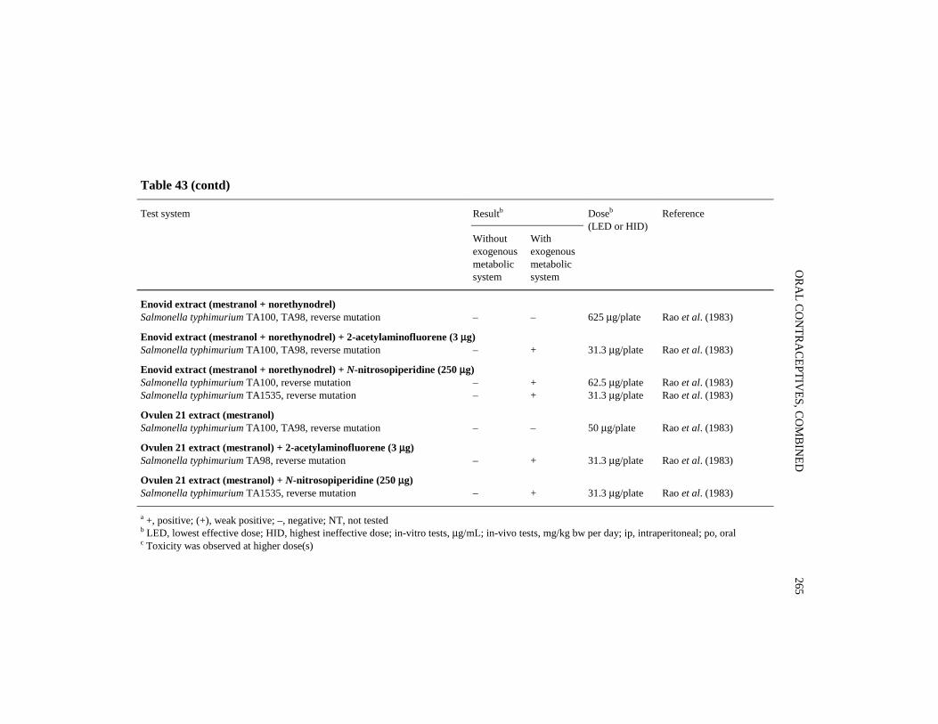
ORA
LCO
NTRA
CEPTIVES, CO
MBIN
ED265
Table 43 (contd)
Test system Resultb
Withoutexogenousmetabolicsystem
Withexogenousmetabolicsystem
Doseb
(LED or HID)Reference
Enovid extract (mestranol + norethynodrel)Salmonella typhimurium TA100, TA98, reverse mutation – – 625 μg/plate Rao et al. (1983)
Enovid extract (mestranol + norethynodrel) + 2-acetylaminofluorene (3 μμg)Salmonella typhimurium TA100, TA98, reverse mutation – + 31.3 μg/plate Rao et al. (1983)
Enovid extract (mestranol + norethynodrel) + N-nitrosopiperidine (250 μμg)Salmonella typhimurium TA100, reverse mutation – + 62.5 μg/plate Rao et al. (1983)Salmonella typhimurium TA1535, reverse mutation – + 31.3 μg/plate Rao et al. (1983)
Ovulen 21 extract (mestranol)Salmonella typhimurium TA100, TA98, reverse mutation – – 50 μg/plate Rao et al. (1983)
Ovulen 21 extract (mestranol) + 2-acetylaminofluorene (3 μμg)Salmonella typhimurium TA98, reverse mutation – + 31.3 μg/plate Rao et al. (1983)
Ovulen 21 extract (mestranol) + N-nitrosopiperidine (250 μμg)Salmonella typhimurium TA1535, reverse mutation – + 31.3 μg/plate Rao et al. (1983)
a +, positive; (+), weak positive; –, negative; NT, not testedb LED, lowest effective dose; HID, highest ineffective dose; in-vitro tests, μg/mL; in-vivo tests, mg/kg bw per day; ip, intraperitoneal; po, oralc Toxicity was observed at higher dose(s)

Enovid enhanced the mutation yield obtained with an ineffective dose of 2-acetyl-aminofluorene (3 μg/plate). The extracts also enhanced the mutation yield obtained withan ineffective dose of N-nitrosopiperidine (250 μg/plate).
In a single study, mestranol induced sister chromatid exchange and chromosomalaberrations in human lymphocytes in vitro and sister chromatid exchange, chromosomalaberrations and micronuclei in bone-marrow cells from mice treated in vivo.
Negative results were obtained with a combination of mestranol and norethisteronein a study of sister chromatid exchange and micronucleus formation in human lym-phocyte cultures. The concentrations of the test material used in this study were in thenanogram per millilitre range, which are those that might be expected during human use.
4.3.4 Cyproterone acetate, metabolites and derivatives (Table 44)Gene mutations were not induced in Salmonella typhimurium by cyproterone acetate
or 6,7-epoxycyproterone acetate. Covalent binding to DNA was observed with cypro-terone acetate in cultured rat liver cells, the binding being greater in cells from femalesthan from males in all cases, according to one study. Cyproterone acetate also bound toDNA in human and porcine hepatocytes in culture, while a metabolite, 3-hydroxy-cyproterone acetate, and a derivative, 3-O-acetylcyproterone acetate, also bound toisolated calf thymus DNA. DNA strand breakage was induced in female rat hepatocytesin vitro in one study. No DNA breakage was observed in male human hepatocytes in vitro.DNA repair, including unscheduled DNA synthesis, of damage induced by cyproteroneacetate appears to be sex specific, since these processes occur in cultured liver cells fromfemale but not male rats. Gene mutations were not induced at the hprt locus of Chinesehamster V79 cells by cyproterone acetate in two studies; in one of the studies, the cellswere co-cultured with rat hepatocytes. The frequency of chromosomal aberrations was notincreased in a single study with cyproterone acetate in Chinese hamster V79 cells co-cultured with rat hepatocytes, whereas a study of the frequency of micronucleus formationin vitro in hepatocytes from female rats gave inconclusive results.
In vivo, covalent binding to DNA has been demonstrated in the livers of male andfemale rats and female mice (weak binding). No binding to male mouse liver DNA wasobserved. Unscheduled DNA synthesis was induced in one study in hepatocytes from ratsexposed to cyproterone acetate. Also in single studies, this compound increased thefrequency of micronucleus formation in hepatocytes from exposed female rats andinduced γ-GT-positive foci in the livers of female rats. In one study, the frequency ofmutation of the LacI transgene was significantly increased in female BigBlue® transgenicFischer 344 rats after exposure to 3-O-acetylcyproterone acetate. DNA adducts werequantified in the same experiment; the mutation frequency started to increase at doses atwhich the number of DNA adducts had already reached a plateau.
4.3.5 Norethisterone alone and in combination with an oestrogen (Table 45)Gene mutation was not induced in S. typhimurium after treatment with nore-
thisterone. Chromosomal aberrations were induced, but the frequency of micronucleus
IARC MONOGRAPHS VOLUME 72266

ORA
LCO
NTRA
CEPTIVES, CO
MBIN
ED267
Table 44. Genetic and related effects of cyproterone acetate and some derivatives
Resulta Doseb
(LED or HID)ReferenceTest system
Withoutexogenousmetabolicsystem
Withexogenousmetabolicsystem
Cyproterone acetateSalmonella typhimurium TA100, TA1535, TA1537, TA1538, TA98, reverse mutation
– – 250 μg/platec Lang & Reimann(1993)
DNA strand breaks, alkaline elution assay, female rat hepatocytes in vitro + NT 20.85 Martelli et al. (1995)DNA repair exclusive of unscheduled DNA synthesis, female rat hepatocytes in vitro
+ NT 0.83 Neumann et al.(1992)
DNA repair exclusive of unscheduled DNA synthesis, female rat hepatocytes in vitro
+ NT 0.83 Topinka et al. (1995)
DNA repair exclusive of unscheduled DNA synthesis, male rat hepatocytes in vitro
– NT 20.9 Topinka et al. (1995)
Unscheduled DNA synthesis, female rat hepatocytes in vitro + NT 1.32 Kasper et al. (1995)Unscheduled DNA synthesis, male rat hepatocytes in vitro – NT 20.85 Martelli et al. (1995)Unscheduled DNA synthesis, female rat hepatocytes in vitro + NT 0.42 Martelli et al. (1995)Unscheduled DNA synthesis, female rat hepatocytes in vitro + NT 0.84 Martelli et al. (1996a)Gene mutation,Chinese hamster V79 cells, hprt locus in vitro – – 80d Lang & Reimann
(1993)Gene mutation, Chinese hamster V79 cells, hprt locus in vitro (co-cultured with hepatocytes)
NT – 41.7 Kasper et al. (1995)
Chromosomal aberrations, Chinese hamster V79 cells in vitro (co-cultured with hepatocytes)
NT – 41.7 Kasper et al. (1995)
Micronucleus formation, female rat hepatocytes in vitro (+) NT 0.42 Kasper et al. (1995)DNA strand breaks, alkaline elution assay, male human hepatocytes in vitro – NT 20.85 (1 sample) Martelli et al. (1995)DNA strand breaks, alkaline elution assay, female human hepatocytes in vitro (+) NT 20.8 (3/4 samples) Martelli et al. (1995)Unscheduled DNA synthesis, male and female human hepatocytes in vitro + NT 0.42 Martelli et al. (1995)Unscheduled DNA synthesis, male and female human hepatocytes in vitro + NT 0.42 Martelli et al. (1996a)

IARC M
ON
OG
RAPH
S VO
LUM
E 72268Table 44 (contd)
Resulta Doseb
(LED or HID)ReferenceTest system
Withoutexogenousmetabolicsystem
Withexogenousmetabolicsystem
Cyproterone acetate (contd)Unscheduled DNA synthesis, female rat hepatocytes in vivo + 100 po × 1 Kasper & Mueller
(1996)Gene mutation, female lacI transgenic rat (BigBlue®) in vivo + 75 po × 1 Krebs et al. (1998)Micronucleus formation, female rat hepatocytes in vivo + 100 po × 1 Martelli et al.
(1996b)γ-Glutamyl transpeptidase-positive foci, female Sprague-Dawley rat liver in vivo
+ 100 po × 6(weekly)
Martelli et al.(1996b)
Binding (covalent) to DNA, female rat hepatocytes in vitro + NT 0.013 Topinka et al. (1993,1995)
Binding (covalent) to DNA, male rat hepatocytes in vitro + NT 0.42 Topinka et al. (1993,1995)
Binding (covalent) to DNA, human (male and female), rat (female) hepatocytes in vitro
+ NT 4.2 Werner et al. (1996)
Binding (covalent) to DNA, pig (male and female), rat (male) hepatocytes in vitro
(+) NT 4.2 Werner et al. (1996)
Binding (covalent) to DNA, female rat liver in vivo + 0.1 po × 1 Topinka et al. (1993)Binding (covalent) to DNA, male rat liver in vivo + 3 po × 1 Topinka et al. (1993)Binding (covalent) to DNA, female rat liver in vivo + 10 po × 1 Werner et al. (1995)Binding (covalent) to DNA, male rat liver in vivo + 100 po × 1 Werner et al. (1995)Binding (covalent) to DNA, male rat liver in vivo + 100 po × 1 Werner et al. (1996)Binding (covalent) to DNA, male C57BL/6 mouse liver in vivo – 35 po × 1 Werner et al. (1996)Binding (covalent) to DNA, female rat liver in vivo + 10 po × 1 Werner et al. (1996)Binding (covalent) to DNA, female C57BL/6 mouse liver in vivo (+) 35 po × 1 Werner et al. (1996)Binding (covalent) to DNA, female rat liver in vivo + 25 po × 1 Krebs et al. (1998)

ORA
LCO
NTRA
CEPTIVES, CO
MBIN
ED269
Table 44 (contd)
Resulta Doseb
(LED or HID)ReferenceTest system
Withoutexogenousmetabolicsystem
Withexogenousmetabolicsystem
6,7-Epoxycyproterone acetateSalmonella typhimurium TA100, TA1535, TA1537, TA1538, TA98, reverse mutation
– – 500 μg/platec Lang & Reimann(1993)
αα-Hydroxycyproterone acetateBinding (covalent) to DNA, calf thymus in vitro + NT 1 Kerdar et al. (1995)
3-O-Acetyl cyproterone acetate (not a metabolite)Gene mutation, female lacI transgenic rat (BigBlue®) in vivo + 75 po × 1 Krebs et al. (1998)Binding (covalent) to DNA, calf thymus in vitro + NT 1 Kerdar et al. (1995)Binding (covalent) to DNA, female rat liver in vivo + 25 po × 1 Krebs et al. (1998)
a +, positive; (+), weak positive; –, negative; NT, not testedb LED, lowest effective dose; HID, highest ineffective dose; in-vitro tests, μg/mL; in-vivo tests, mg/kg bw per day; po, oralc Toxicity was observed at this dose in some or all strainsd Some toxicity was observed

IARC M
ON
OG
RAPH
S VO
LUM
E 72270
Table 45. Genetic and related effects of norethisterone and its ester
Test system Resulta Reference
Withoutexogenousmetabolicsystem
Withexogenousmetabolicsystem
Doseb
(LED or HID)
NorethisteroneSalmonella typhimurium TA100, TA1535, TA1537, TA98, reverse mutation – – 1000 μg/plate Peter et al. (1981)Salmonella typhimurium TA100, TA1535, TA1537, TA1538, TA98, reverse mutation – – 750 μg/plate Lang & Reimann (1993)Unscheduled DNA synthesis, female rat hepatocytes in vitro − NT 150 Blakey & White (1985)Unscheduled DNA synthesis, male rat hepatocytes in vitro (+) NT 15 Blakey & White (1985)Micronucleus formation, female Swiss albino mouse bone-marrow cells in vivo – 30 po × 15 Shyama & Rahiman (1993)Chromosomal aberrations, female Swiss albino mouse bone-marrow cells in vivo – 70 po × 1 Ansari & Adhami (1977)Chromosomal aberrations, female Swiss albino mouse bone-marrow cells in vivo + 3 po × 15 Shyama & Rahiman (1993)
Norethisterone acetateSalmonella typhimurium TA100, TA1535, TA1537, TA1538, TA98, reverse mutation – – 2500 μg/plate Lang & Redmann (1979)Salmonella typhimurium TA100, TA1535, TA1537, TA1538, TA98, reverse mutation – – 1000 μg/plate Dayan et al. (1980)Salmonella typhimurium TA100, TA1535, TA1537, TA1538, TA98, reverse mutation – – 500 μg/platec Lang & Reimann (1993)Salmonella typhimurium TA100, TA1535, TA97a, TA98, reverse mutation – – 1000 μg/platec Dhillon & Dhillon (1996)Sister chromatid exchange, human male lymphocytes in vitro (24-h treatment) + NT 1 Dhillon & Dhillon (1996)Sister chromatid exchange, human male lymphocytes in vitro (90-min treatment) + + 1 Dhillon & Dhillon (1996)Chromosomal aberrations, human lymphocytes in vitro – NT 100 Stenchever et al. (1969)Chromosomal aberrations, human male lymphocytes in vitro (72-h treatment) + NT 1 Dhillon & Dhillon (1996)Chromosomal aberrations, human male lymphocytes in vitro (6-h treatment) – + 10 Dhillon & Dhillon (1996)Host-mediated assay, male Swiss albino mouse (intravenous inoculation), S. typhimurium TA97a, TA98, TA100, TA1535
– 100 ip × 1 Dhillon & Dhillon (1996)
Sister chromatid exchange, Swiss albino mice bone-marrow cells in vivo + 1 ip × 1 Dhillon & Dhillon (1996)Micronucleus formation, male Swiss albino mouse bone-marrow cells in vivo + 1 ip × 1 Dhillon & Dhillon (1996)Dominant lethal mutation induction, C3H and NMRI mice in vivo (+) 1 mg/animal
po daily × 4 wkRohrborn & Hansmann(1974)
Aneuploidy, C3H mice in vivo + 10 mg/animal podaily × 4 wk
Rohrborn & Hansmann(1974)

ORA
LCO
NTRA
CEPTIVES, CO
MBIN
ED271
Table 45 (contd)
ResultaTest system
Withoutexogenousmetabolicsystem
Withexogenousmetabolicsystem
Doseb
(LED or HID)Reference
Norethisterone + mestranolSister chromatid exchange, human lymphocytes in vitro – NT 0.075 + 0.0038 Dutkowski et al. (1983)Micronucleus formation, human lymphocytes in vitro – NT 0.075 + 0.0038 Dutkowski et al. (1983)
Norethisterone acetate + ethinyloestradiolChromosomal aberrations, female Swiss albino mouse bone-marrow cells in vivo + 0.79 + 0.01 po × 15 Shyama et al. (1991)Micronucleus formation, female Swiss albino mouse bone-marrow cells in vivo – 7.9 + 0.1 po × 15 Shyama et al. (1991)
a +, positive; (+), weak positive; –, negative; NT, not testedb LED, lowest effective dose; HID, highest ineffective dose; in-vitro tests, μg/mL; in-vivo tests, mg/kg bw per day; po, oral; ip, intraperitoneal; wk, weekc Precipitation and/or toxicity was observed at higher dose(s).

formation was not increased in bone-marrow cells of female Swiss albino mice treated invivo with norethisterone.
In a single study, the frequencies of sister chromatid exchange and micronuclei werenot increased in cultured human lymphocytes treated with a combination of nore-thisterone and mestranol.
4.3.6 Norethisterone acetate alone and in combination with an oestrogen(Table 45)
Norethisterone acetate did not induce gene mutation in various strains of S. typhi-murium with or without an exogenous metabolic activation system. In a single study, thefrequencies of sister chromatid exchange and chromosomal aberrations were increased inhuman lymphocytes treated in vitro with norethisterone acetate. In the same study, sisterchromatid exchange and micronucleus formation were induced in the bone-marrow cells ofSwiss albino mice treated in vivo with norethisterone acetate, whereas gene mutations werenot induced in S. typhimurium in a mouse host-mediated assay in which the bacteria wererecovered from the livers of animals after treatment with norethisterone acetate.
The combination of norethisterone acetate plus ethinyloestradiol induced chromo-somal aberrations in bone-marrow cells of female Swiss albino mice exposed in vivo,whereas micronuclei were not induced in the bone-marrow cells of these mice.
4.3.7 Chlormadinone acetate (Table 46)After rat liver cells were incubated with chlormadinone acetate in vitro, covalent
DNA binding was observed, particularly in cells from females. Unscheduled DNAsynthesis was reported in female but not male rat hepatocytes in vitro and in male andfemale human hepatocytes in vitro after treatment with chlormadinone acetate. The BdUrdensity shift assay to determine DNA repair in female and male rat hepatocytes exposedto chlormadinone acetate in vitro gave negative results.
No DNA binding was observed in female rat liver in vivo, but micronuclei wereinduced in female rat hepatocytes in vivo.
4.3.8 Gestodene (Table 46)Gene mutations were not induced in S. typhimurium by gestodene or combinations
of gestodene and ethinyloestradiol.
4.3.9 Megestrol acetate (Table 46)Low levels of covalent DNA binding were observed in rat liver cells treated with
megestrol acetate in vitro, particularly in cells from females. Megestrol acetate inducedunscheduled DNA synthesis in rat primary hepatocytes and gave rise to DNA repair infemale and male rat liver cells and in human hepatocytes in vitro.
Weak covalent DNA binding was observed in female rat liver in vivo; however,micronucleus formation was not induced in female rat hepatocytes, and γ-GT-positivefoci were not induced in rat liver in vivo.
IARC MONOGRAPHS VOLUME 72272

ORA
LCO
NTRA
CEPTIVES, CO
MBIN
ED273
Table 46. Genetic and related effects of other progestogens used in combined oral contraceptives
Test system Resulta Doseb
(LED or HID)Reference
Withoutexogenousmetabolicsystem
Withexogenousmetabolicsystem
Chlormadinone acetateSalmonella typhimurium TA100, TA1535, TA1537, TA1538, reverse mutation – – 1000 μg/plate Dayan et al. (1980)DNA repair exclusive of unscheduled DNA synthesis, female and male rat hepatocytes in vitro
– NT 20.3 Topinka et al. (1995)
Unscheduled DNA synthesis, female rat hepatocytes in vitro + NT 0.81 Martelli et al. (1996a)Unscheduled DNA synthesis, male rat hepatocytes in vitro – NT 8.1 Martelli et al. (1996a)Cell transformation, rat liver cells treated in vivo scored in vitro – 100 po × 6 Martelli et al. (1996b)Unscheduled DNA synthesis, male and female human hepatocytes in vitro + NT 0.81 Martelli et al. (1996a)Chromosomal aberrations, human lymphocytes in vitro – NT 100 Stenchever et al. (1969)Micronucleus formation, female rat hepatocytes in vivo + 100 po × 1 Martelli et al. (1996b)Binding (covalent) to DNA, female rat hepatocytes in vitro + NT 1.2 Topinka et al. (1995)Binding (covalent) to DNA, male rat hepatocytes in vitro (+) NT 1.2 Topinka et al. (1995)Binding (covalent) to DNA, female rat liver in vivo – 100 po × 1 Topinka et al. (1995)
GestodeneSalmonella typhimurium TA100, TA1535, TA1537, TA1538, TA98, reverse mutation
– – 5 μg/plate Lang & Reimann (1993)
Gestodene + ethinyloestradiol (2.5 parts + 1 part)Salmonella typhimurium TA100, TA1535, TA1537, TA1538, TA98, reverse mutation
– – 200 μg/platec,d Lang & Reimann (1993)
Megestrol acetateDNA repair exclusive of unscheduled DNA synthesis, female and male rat hepatocytes in vitro
– NT 19.3 Topinka et al. (1995)
Unscheduled DNA synthesis, female rat hepatocytes in vitro + NT 1.93 Martelli et al. (1996a)

IARC M
ON
OG
RAPH
S VO
LUM
E 72274
Table 46 (contd)
Test system Resulta Doseb
(LED or HID)Reference
Withoutexogenousmetabolicsystem
Withexogenousmetabolicsystem
Megestrol acetate (contd)Unscheduled DNA synthesis, male rat hepatocytes in vitro – NT 19.3 Martelli et al. (1996a)γ-Glutamyl transpeptidase-positive foci induction, female Sprague-Dawley rat liver in vivo
– 100 po × 6(weekly)
Martelli et al. (1996b)
Unscheduled DNA synthesis, male and female human hepatocytes in vitro + NT 0.77 Martelli et al. (1996a)Chromosomal aberrations, human lymphocytes in vitro – NT 10 Stenchever et al. (1969)Micronucleus formation, female rat liver in vivo – 100 po × 1 Martelli et al. (1996b)Binding (covalent) to DNA, female rat hepatocytes in vitro + NT 1.2 Topinka et al. (1995)Binding (covalent) to DNA, male rat hepatocytes in vitro (+) NT 1.2 Topinka et al. (1995)Binding (covalent) to DNA, female rat liver in vivo (+) 10 po × 1 Topinka et al. (1995)
NorethynodrelSalmonella typhimurium TA100, TA1535, TA1537, TA1538, TA98, reverse mutation
– – 1000 μg/plate Lang & Redmann (1979)
Salmonella typhimurium TA100, TA1535, TA1537, TA1538, TA98, reverse mutation
– – 250 μg/plate Rao et al. (1983)
Salmonella typhimurium TA100, TA1535, TA1537, TA1538, TA98, reverse mutation
– – 2000 μg/plated Lang & Reimann (1993)
Inhibition of metabolic cooperation, Chinese hamster V79 cells in vitro (+) NT 0.75 Yager (1983)
Norethynodrel + 2-acetylaminofluorene (3 μμg)Salmonella typhimurium TA100, TA1535, TA98, reverse mutation NT – 150 μg/plate Rao et al. (1983)

ORA
LCO
NTRA
CEPTIVES, CO
MBIN
ED275
Table 46 (contd)
Test system Resulta Doseb
(LED or HID)Reference
Withoutexogenousmetabolicsystem
Withexogenousmetabolicsystem
LevonorgestrelSalmonella typhimurium TA100,TA1535, TA1537, TA1538, TA98, reverse mutation
– – 2500 μg/plate Lang & Redmann (1979)
Salmonella typhimurium TA100,TA1535, TA1537, TA1538, TA98, reverse mutation
– – 1000 μg/plate Dayan et al. (1980)
Salmonella typhimurium TA100,TA1535, TA1537, TA1538, TA98, reverse mutation
– – 500 μg/plate Lang & Reimann (1993)
Drosophila melanogaster, sex-linked recessive lethal mutations – 3120 Parádi (1981)Drosophila melanogaster, sex-linked recessive lethal mutations (+) 5000 Aguiar & Tordecilla
(1984)
a +, positive; (+), weak positive; –, negative; NT, not testedb LED, lowest effective dose; HID, highest ineffective dose; in-vitro tests, μg/mL; in-vivo tests, mg/kg bw per day; po, oral; ip, intraperitonealc Total mixtured Toxicity was observed at higher dose(s)
4.3.10 Norethynodrel (Table 46)Gene mutations were not induced in S. typhimurium by norethynodrel, and it did not
enhance the mutagenicity of a sub-threshold mutagenic dose of 2-acetylaminofluorene.
4.3.11 Levonorgestrel (Table 46)Gene mutations were not induced in S. typhimurium by levonorgestrel. Ethanolic
extracts of combinations of levonorgestrel and ethinyloestradiol induced cell transfor-mation in baby rat kidney cells infected with HPV-16 and carrying the Ha-ras-1 onco-gene (see Table 42).
4.4 Reproductive and prenatal effectsThe literature up to 1979 on the developmental effects of sex hormones was reviewed
in Volume 21 of the IARC Monographs (IARC, 1979). It has been shown in both humansand experimental animals that sex hormones can interfere with normal genital deve-lopment. The effects observed with synthetic sex hormones are variable, and oestrogenic,androgenic and progestogenic effects may frequently be observed with one chemical,depending on the target tissues and the background levels of natural hormones acting atspecific times. The effects on embryofetal development also depend on the relative impor-tance of numerous conditioning factors and are not always easy to predict; however,masculinization of female fetuses and feminization of male fetuses are observed. Effectsare found in many organ systems, and genital development, central nervous systemdevelopment and sexual differentiation may be affected. The timing of exposure relativeto embryofetal and postnatal development is critical in determining the type and site of thedefect produced.
The literature on the effects of exposure to sex hormones during pregnancy oninduction of other types of congenital malformation is much more controversial. Earlycase reports and epidemiological studies suggested that a wide variety of defects, affec-ting most organ systems, could be produced. Syndromes such as the VACTERL syndromewere reported, which involves malformations of one or more of the vertebral, anal,cardiac, tracheal, oesophageal, renal and limb systems. Numerous other studies failed tosupport the suggestion that these defects were related to hormonal treatment.
Three categories of exposure in pregnancy were considered. Accidental exposure tooral contraceptives comprised the major group, with the least convincing evidence for aconnection with birth defects. The evidence related to use of hormonal pregnancy testswas a little stronger but still unsubstantiated; the use of such tests was discontinued manyyears ago. The third category is use of hormones to treat women with pregnancy problems,such as intermittent bleeding, repeated or threatened abortion and luteal failure. In thosecases in which the pregnancy is maintained but the fetus has malformations, it is difficultto decide whether the cause was the hormonal treatment or the underlying disease.
Since the last IARC monograph on this subject (IARC, 1979), many papers havebeen published on the topic of exposure to hormones during pregnancy, and some havebeen reviewed. Schardein (1980b) reviewed the literature up to that time on the induction
IARC MONOGRAPHS VOLUME 72276

of genital and non-genital defects. He concluded that the commonest association forgenital defects was masculinization of females, generally seen as clitoral hypertrophy,with exposure at around week 8–10 of pregnancy; the prevalence was low and the riskwas estimated to be about 1% of exposed infants. The evidence for feminization of maleswas reported to be less convincing. Hypospadia has been the commonest defect reported,but recent analysis of more than 2000 cases of hypospadia has shown no association withmaternal use of oral contraceptives (Källén et al., 1991). A meta-analysis of pregnancyoutcomes after exposure to sex hormones during the first trimester showed no excess ofgenital malformations (Raman-Wilms et al., 1995).
The evidence accumulated since 1979 on the involvement of exposure to sexhormones in other non-genital malformations has been largely in favour of no association.In a short paper, Brent (1994) reviewed some of the reasons why false associationsbetween congenital heart malformations and hormones may have been concluded in thepast. These include the grouping of many different types of congenital heart disorder withdifferent causes, inadequate knowledge about the critical times of exposure for specificdefects, failure to differentiate between the actions of oestrogens and progestogens, andinclusion in some studies of syndromes with a known high incidence of heart defects. Thepaper lists 20 reviews on the subject of exposure to hormones and non-genital congenitalmalformations, none of which found a causal association. It can be concluded that mostepidemiological studies do not indicate that progestogens are teratogenic for the cardio-vascular system; secular trend data do not support an association between exposure to sexhormones and cardiovascular disease; a very large number of experimental studies showno relationship between exposure to sex hormones and cardiac malformations; there areno sex hormone receptors in developing cardiac tissue; and no consistent syndrome ofnon-genital defects has been reported. In 1988, as a result of a meeting to review theevidence, the United States Food and Drug Administration removed the warning label onoral contraceptives which had previously stated that exposure during pregnancy couldcause cardiac and limb defects (Brent, 1989).
Useful reviews of the data on congenital malformations have been published (Wilson& Brent, 1981; Polednak, 1985; Simpson, 1985; Bracken, 1990; Simpson & Phillips,1990). Several case–control studies have been conducted that show little or usually noevidence of an association between birth defects and hormonal exposure (Lammer &Cordero, 1986; Hill et al., 1988; Ananijevic-Pandey et al., 1992; Pradat, 1992; Martínez-Frías et al., 1998). In a large cohort study in the United States (Harlap et al., 1985a), noincrease in the incidence of malformations was found in relation to the use of oral orother methods of contraception. In a study of women in Thailand (Pardthaisong et al.,1988), no increase in the incidence of defects of the heart, central nervous system orlimbs was found in the offspring of women using oral or injectable contraceptives, butan increased incidence of polysyndactyly and chromosomal anomalies was observed inwomen who had previously used medroxyprogesterone acetate (Depo Provera). Thesmall numbers of affected children, the long interval between injection of medroxy-progesterone and the conception of the affected offspring and the unrelated nature of the
ORAL CONTRACEPTIVES, COMBINED 277

effects led the authors to conclude that a causal relationship between treatment and effectwas unlikely. Overall, the prevalence of major malformations was significantly lower inthe oral contraceptive users than in the non-users.
A study of oral contraceptive use in 730 mothers of children with Down syndromeand 1035 mothers of children with other malformations (Lejeune & Prieur, 1979) showedthat more of the mothers aged 30–38 years of children with Down syndrome had ceasedcontraceptive use within the six months prior to conception than in the other group. Threeother studies, two of them prospective studies in which data on contraceptive use wasobtained before the outcome of the pregnancy was known (Ericson et al., 1983; Källén,1989), and a smaller case–control study of contraceptive use in the mothers of Downcases and in mothers of normal children (Janerich et al., 1976), found no associationbetween use of oral contraceptives and the subsequent birth of a child with Downsyndrome.
A study of chromosomal abnormalities in 33 551 births and abortions after 20 weekswas reported by Harlap et al. (1985b). No increased risk was found for women who usedoral contraceptives prior to conception or who were still using contraceptives when theybecame pregnant.
4.4.1 Ethinyloestradiol(a) Humans
No data were available to the Working Group.
(b) Experimental systemsA study was carried out with the inbred mouse strain 129SV-S1 C P, which is hetero-
zygous for the S1 gene shown to affect the development of primordial germ cells, andwhich has a 7% spontaneous incidence of testicular teratoma. Pregnant mice wereinjected subcutaneously with 0.02 or 0.2 mg/kg bw ethinyloestradiol in corn oil on days11 and 12 of gestation (the day a vaginal plug was observed was considered to be day 0),which had been shown previously to be the critical period for induction of teratoma. Themale pups were killed at 15 days of age and the testes examined for teratomas. A dose-related increase in the incidence of cryptorchid testes was found, with 4/107 in controls,10/109 at the low dose and 23/115 at the high dose (p < 0.0001 for trend). A smallincrease was observed in the incidence of teratoma which was neither dose-related norsignificant (odds ratio, 2.4; 95% CI, 0.7–9.1) for the pooled data. The authors suggestedthat different mechanisms are involved in the etiology of cryptorchidism and teratoma,although both may be induced by oestrogen stimulation (Walker et al., 1990).
Oral administration of 0, 0.02, 0.2 or 2.0 mg/kg bw ethinyloestradiol in olive oil topregnant Jcl:ICR mice (8–15 litters per group) on days 11–17 of gestation (the presenceof a vaginal plug being considered day 0) resulted in a dose-dependent increase in theincidences of ovotestis and cryptorchidism in males, with persistent Müllerian andWolffian ducts, when fetuses were examined on day 18 of gestation. Leydig-cell proli-feration was also seen at the two higher doses with alterations in cellular morphology
IARC MONOGRAPHS VOLUME 72278

suggestive of preneoplastic changes. Female fetuses showed ovarian hypoplasia, withdecreased numbers of primordial follicles and increased follicular degeneration (Yasudaet al., 1985, 1986).
In a later study (Yasuda et al., 1988), pregnant ICR mice were given a daily dose of0.02 or 0.2 mg/kg bw ethinyloestradiol in olive oil orally on days 11–17 of gestation. Damsat the low dose were allowed to deliver, and their male offspring were reared to maturity(20–22 months). The animals given the high dose had no live offspring. At day 18, malefetuses were recovered from mice at each dose, and the concentrations of testosterone andoestradiol were measured in testes. Ethinyloestradiol treatment significantly (p < 0.001)reduced the concentrations of testosterone in the testes of 18-day-old fetuses, from a meanof 5.21 ± 0.13 pg/testis in the controls (n = 32 testes) to 0.89 ± 0.11 pg/testis (n = 30) at thelow dose and 0.40 ± 0.13 pg/testis (n = 52) at the high dose. Treatment at the high dose alsoreduced the oestradiol levels in the testis from 78.80 ± 0.49 pg/testis in controls to42.25 ± 1.56 pg/testis (p < 0.05). The testosterone:oestradiol ratios were reduced from 1:15in controls, to 1:77 at the low dose and 1:106 at the high dose. At 20–22 months, theoffspring from the low-dose group were killed, and the testes and epididymides wereremoved, examined histologically and analysed for testosterone and oestradiol. There weresignificant (p < 0.05) increases in the frequency of testicular atrophy, Leydig-cellhyperplasia and absence of epididymal sperm in the treated compared with the controlmice. The testosterone concentration was significantly decreased, from 84 to 28 ng/testis(p < 0.01), and the oestradiol level was increased, from 356 to 564 ng/testis (p < 0.05). Theauthors suggested that the dramatic fall in the testosterone:oestradiol ratio in the fetal testisresults from greatly increased conversion of testosterone to oestradiol by the Leydig cells,so that insufficient amounts of testosterone are available for regulation of pro-spermato-genesis in the developing testis and spermatogenesis, eventually resulting in sterility.
4.4.2 Mestranol(a) Humans
No data were available to the Working Group.
(b) Experimental systemsDaily oral administration of 0.05 or 0.2 mg/kg bw mestranol to female NMRI mice
and AFAF1 hybrid mice, on days 4–8 after mating, inhibited implantation and increasedthe number of resorptions. The fetuses of NMRI mice had accessory ribs. Treatment ondays 7–11 with doses of 0.1–0.2 mg/kg bw induced abortions but had no teratogeniceffects (Heinecke & Klaus, 1975).
In rats, subcutaneous injection of 0.002–0.02 mg/kg bw mestranol five days beforeand 30 days after mating prevented implantation in a dose-dependent manner.Subcutaneous injection of 0.02 mg/kg bw or oral administration of 0.1 mg/kg bw on days2–4 of gestation terminated pregnancy (Saunders & Elton, 1967).
Charles River rats received daily oral doses of 0.05–0.2 mg/kg bw Enovid (2.5 mgnorethynodrel, 0.1 mg mestranol) or 0.01–0.1 mg/kg bw mestranol throughout gestation
ORAL CONTRACEPTIVES, COMBINED 279

and for 21 days after parturition. The highest dose of mestranol terminated a significantpercentage of pregnancies. No genital defects were observed in surviving male offspring,but female offspring showed enlarged urethral papillae and prematurely opened vaginas,even at lower doses of mestranol. The fertility of female offspring of rats treated with 0.1mg Enovid was impaired by 55%. Higher doses of Enovid or 0.02 mg mestranol inducedcomplete sterility in female offspring; examination of the ovaries showed no corporalutea and follicles of reduced size (Saunders, 1967).
Sixty female Wistar rats were given a daily dose of 1 mg/kg bw Enidrel (0.075 mgmestranol, 9.2 mg norethynodrel) intragastrically for two months, at which time theywere mated. In 30 animals in which treatment was continued, complete fetal resorptionoccurred rapidly; however, after two weeks without treatment, the fertility rates and littersizes were normal. In the 30 animals in which treatment was discontinued, the fertilityand pre- and post-natal development of the offspring were also normal. No teratogeniceffects were observed (Tuchmann-Duplessis & Mercier-Parot, 1972).
In rabbits, pregnancy was terminated by daily oral doses of > 0.02 mg/kg bw mestranolon days 0–28 or 0.05 mg/kg bw [lower doses not tested] on days 10–28 of gestation and bydaily subcutaneous doses of 0.005 mg/kg bw on days 0–28 or > 0.002 mg/kg bw on days10–28. Doses that did not terminate pregnancy had no effects on litter size or the weightsof the offspring (Saunders & Elton, 1967).
In female Syrian golden hamsters that received a contraceptive steroid containing18.7 μg mestranol and 0.6 mg lynoestrenol [route unspecified] daily for 4.5–8 months,fertility was found to be normal; no effects were seen on sexual behaviour or on thefecundity of the offspring of the following two generations (Cottinet et al., 1974).
When adult beagle bitches received 5 mg/kg bw mestranol orally on day 6 or 21 ofgestation, the embryonic losses, based on corpora lutea counts, were 95.5% with earlytreatment and 67.3% with late mestranol treatment in comparison with 34.5% in controls.The surviving offspring appeared normal (Kennelly, 1969).
4.4.3 Chlormadinone acetate(a) Humans
No increase in the incidence of malformations was reported in 305 infants whosemothers had been exposed to chlormadinone and oestrogens during pregnancy (Goldzieheret al., 1968; Lepage & Gueguen, 1968; Larsson-Cohn, 1970).
(b) Experimental systemsGroups of 8–12 male Sprague-Dawley Crl:CD(SD)Br rats were castrated and injected
immediately thereafter twice daily for 14 days with one of a number of syntheticprogestogens, including chlormadinone acetate, used in the treatment of prostate cancer.Controls received the vehicle, 1% gelatine in 0.9% saline. Dihydrotestosterone was injectedat a dose of 150 μg twice daily for 14 days as a positive control. All animals were killed onthe morning after the last day of treatment, and the ventral prostate and adrenals wereremoved and weighed; furthermore, the prostatic content of ornithine decarboxylase was
IARC MONOGRAPHS VOLUME 72280

measured, as it is considered to be a highly specific, sensitive marker of androgenic activityin the prostate. Dihydrotestosterone increased the ventral prostate weight to 43% abovethat of castrated controls. Chlormadinone acetate was less potent than dihydrotestosteronebut caused significant increases in prostate weight, by about 22% at 3 mg and 36% at 10mg per injection. Whereas dihydrotestosterone caused a 14-fold increase in ornithinedecarboxylase activity in the prostate, chlormadinone acetate caused a 5.3-fold increase at3 mg and an 11.8-fold increase at 10 mg. Chlormadinone acetate thus has weak butsignificant androgenic activity in the rat ventral prostate (Labrie et al., 1987).
Pregnant Wistar rats were given 1, 5 or 10 mg chlormadinone acetate orally once aday for four days on days 17–20 of pregnancy, and the fetuses were removed on day 21.After fixation, histological sections of the pelvic region were examined and the uro-vaginal septum length measured. Masculinization of female fetuses was not observed, inthe absence of change in the development of the urogenital septum (Kawashima et al.,1977).
Chlormadinone acetate given orally at doses of 1–50 mg/kg bw on days 8–15 ofpregnancy to Japanese ddS and CF1 mice caused a significant increase in the incidenceof cleft palate. A dose of 10 mg/kg bw, but not of 1 or 3 mg/kg bw, given orally on days8–20 of gestation to Japanese albino rabbits increased the incidence of cleft palate, abdo-minal wall defects and wrist contractures (Takano et al., 1966).
4.4.4 Cyproterone acetate(a) Humans
Two men treated for prostatic carcinoma with high oral doses of cyproterone acetate(2 × 100 mg per day for seven months) had widespread testicular damage, with disap-pearance of Sertoli cells and spermatogonia and involution of Leydig cells (Re et al.,1979). When cyproterone acetate was given at doses of 5–10 mg per day as a contraceptivein several other studies, decreased sperm concentration and motility and increasedabnormal morphology, with—except in one study—decreased power to penetrate themucus, were observed. Variable effects on plasma gonadotrophins and testosterone levelshave been reported (Føgh et al., 1979; Roy & Chatterjee, 1979; Moltz et al., 1980; Wang& Yeung, 1980). Doses of 50–100 mg cyproterone acetate per day combined with testos-terone induced azoospermia and decreased testis size in each of 10 subjects. All of theeffects were reversible (Meriggiola et al., 1996).
In women, ovulation is inhibited by 2 mg per day cyproterone acetate when given incombination with 35 μg ethinyloestradiol (Spona et al., 1986). No controlled studies ondevelopmental effects are available.
(b) Experimental systemsCyproterone acetate has been reported to have both androgenic and anti-androgenic
activity in experimental animals (see also section 4.2.5).Groups of 8–12 male Sprague-Dawley Crl:CD(SD)Br rats were castrated and then
injected twice daily for 14 days with one of a number of synthetic progestogens,
ORAL CONTRACEPTIVES, COMBINED 281

including cyproterone acetate. Treatment was begun one day after castration. Controlswere injected with the vehicle, 1% gelatine in 0.9% saline. Dihydrotestosterone wasinjected twice daily at a dose of 125 μg for 14 days as a positive control. All animals werekilled on the morning after the last day of treatment, and the ventral prostate and adrenalswere removed and weighed. Dihydrotestosterone increased the ventral prostate weight toapproximately five time that of castrated controls. Cyproterone acetate was less potentthan dihydrotestosterone but caused a significant increase in prostate weight, by 60% ata dose of 5 mg per injection twice daily. Cyproterone acetate thus has weak but signifi-cant androgenic activity in the rat ventral prostate (Poyet & Labrie, 1985).
Anti-androgenic effects have also been reported. Groups of 10 albino Wistar mice weretreated subcutaneously with vehicle alone or with 1 mg per animal per day of cyproteroneacetate for seven days. The animals were killed on the eighth day and the testes removedfor histological and morphometric examination. Treatment caused marked decreases in thevolume, surface area and length of the seminiferous tubules, and it inhibited spermato-genesis (Umapathy & Rai, 1982).
The anti-androgenic activity of cyproterone and cyproterone acetate has been shownin mice (Umapathy & Rai, 1982; Homady et al., 1986), rats (El Etreby et al. 1987),guinea-pigs (Tam et al., 1985), ferrets (Kästner & Apfelbach, 1987), goats (Panda &Jindal, 1982; Kumar & Panda, 1983) and monkeys (Lohiya et al., 1987; Kaur et al.,1990, 1992). The effects observed include decreased sexual behaviour and inter-maleaggression, reduced weights of testis and inhibition of spermatogenesis. Fertility can bereduced by low doses of cyproterone acetate even in the absence of reduced spermato-genesis (Rastogi et al., 1980), which may be due to an effect on epididymal processingof sperm. In addition to reduced secretion of testosterone and luteinizing hormone (Closet al., 1988), there is also evidence that translocation of the testosterone receptor to thenucleus may be affected (Brinkmann et al., 1983).
In rodents, cyproterone acetate has oestrogenic properties, increasing uterine weightand causing vaginal cornification in ovariectomized rats (Arya et al., 1979). When the com-pound was administered to pregnant rats, feminization of male fetuses, including deve-lopment of a vagina, has been reported (Neumann et al., 1966; Forsberg & Jacobsohn,1969).
Treatment of NMRI mice with doses of 5–900 mg/kg bw cyproterone acetate sub-cutaneously on day 2 of gestation (the day a vaginal plug was observed was consideredto be day 0) or with 30 mg/kg bw on single days of pregnancy from day 1 to 12, resultedin a clear dose- and time-related increase in the incidences of cleft palate and of urinarytract and respiratory tract malformations, with up to 64% of fetuses affected after thesingle 900-mg/kg bw dose (Eibs et al., 1982). Administration in late pregnancy or in theneonatal period can produce permanent changes in neuroendocrine and sexual functionof rats. Groups of 15 male and 16 female offspring of rats treated subcutaneously with1 mg cyproterone acetate on days 15–20 of gestation [strain and number of pregnantanimals not specified] were studied when two to three months of age. The weight of thebrain was reduced in animals of each sex and the weight of the testis in males. Cell
IARC MONOGRAPHS VOLUME 72282

density in the ventromedial nucleus of the hypothalamus was increased in males incomparison with females. The prolactin concentration in the pituitary was increased inanimals of each sex (Rossi et al., 1991). Groups of newborn male Swiss CD1 mice wereinjected with cyproterone acetate on days 1–10 (200 μg/day), 11–20 (400 μg), 21–30(800 μg) or 31–40 (1 mg) of age. In all groups, there was an immediate reduction in theweights of the testis, epididymis, vas deferens, preputial gland and seminal vesiclesrelative to body weight. This reduction in the weight of the accessory sex organs waspermanent in animals injected up to day 20 of age but was reversible in animals treatedafter day 20. In mice injected on days 1–10 of age, marked, permanent infertility wasobserved when they became adults, but spermatogenesis, the androgen concentration inplasma and sexual behaviour were not affected. The infertility appeared to be due tofailure of sperm in the epididymis to mature (Jean-Faucher et al., 1985).
4.4.5 Levonorgestrel (see also the monograph on ‘Hormonal contraceptives,progestogens only’, section 4.4.2)(a) Humans
No relevant data were available to the Working Group.
(b) Experimental systemsGroups of six pregnant rats [strain not specified] were ovariectomized on day 8 of
gestation and treated subcutaneously with doses of 0.01–0.3 mg levonorgestrel daily ondays 8–21 of gestation; at the same time they received an injection of 1.0 μg oestrone.The rats were killed on day 22 to measure maintenance of pregnancy; satisfactory main-tenance was achieved with 0.1 and 0.3 mg levonorgestrel. Immature castrated male rats[age not specified] were treated subcutaneously daily for 13 days with doses of 0.1–3 mglevonorgestrel and were killed the day after the last treatment; the weights of the prostateand seminal vesicles were measured. Levonorgestrel showed androgenic activity, asjudged from the increased weight of both tissues (Kuhnz & Beier, 1994).
Groups of 10–12 Prob:WNZ New Zealand white rabbits were mated with Prob:KALCalifornian rabbits, producing hybrid fetuses; the day of mating was called day 1. Theanimals were treated with 0.5 mg/kg bw levonorgestrel in sesame oil by gavage on days5–25 of gestation and were killed on day 21. [The Working Group concluded that theanimals must have been killed after day 25, but the fetal body weights were very low forfull-term offspring.] The fetuses were examined macroscopically, and half of them weresliced for visceral examination and the other half examined for skeletal and cartilagemalformations. No adverse effect of treatment on pregnancy rate, number of implan-tations, number of resorptions or number of live or dead fetuses was observed. Thefemale fetal body weight at term was slightly but significantly reduced (15.2 ± 0.61 gversus 17.4 ± 0.74 g in the sesame oil control group). No malformations were observed(Heinecke & Kohler, 1983).
ORAL CONTRACEPTIVES, COMBINED 283

4.4.6 Lynoestrenol(a) Humans
No data were available to the Working Group.
(b) Experimental systemsGroups of 14–20 inseminated belted Dutch rabbits were given lynoestrenol at a dose
of 0, 0.1, 0.5 or 2.5 mg/kg bw orally on days 6–18 of gestation (the day of inseminationbeing considered day 0). The does were killed on day 29 for examination of the fetuses.Increased post-implantation loss was observed at all doses: 1.3% in controls, 13.2% atthe low dose, 17.1% at the intermediate dose and 90.9% at the high dose. A wide rangeof malformations of the brain and eye was observed in all groups, affecting 31–50% offetuses and only 12% of controls. Cardiovascular malformations were observed in 2, 5and 20% of fetuses, respectively, at the three doses. No masculinization of female fetuseswas observed. In a second experiment with groups of five to six belted Dutch rabbits,lynoestrenol was administered at a dose of 0, 0.1 or 0.5 mg/kg bw on days 6–18 ofgestation. The does were allowed to deliver naturally and raise their pups to four weeksof age. At the higher dose, the litter size was decreased and there was clear evidence ofcentral nervous system abnormalities in surviving pups, with ataxia, disorientation,posterior paralysis and rotation of one or both hindlimbs. Anophthalmia and micro-phthalmia were seen in 3/10 pups at the high dose. Histological examination of thecentral nervous system revealed pathological changes in the ventral horns of the spinalcord, with a marked reduction in the number of neurons (Sannes et al., 1983).
Pregnant Wistar rats were given 1 or 5 mg lynoestrenol orally once a day for fourdays on days 17–20 of gestation, and the fetuses were removed on day 21. After fixation,histological sections of the pelvic region were examined and the urovaginal septumlength measured. Masculinization of female fetuses was seen at 5 mg, as evidenced bydecreased development of the urogenital system (Kawashima et al., 1977).
4.4.7 Megestrol acetate(a) Humans
No data were available to the Working Group.
(b) Experimental systemsMegestrol acetate has some androgenic activity in rats (Poyet & Labrie, 1985; Labrie
et al., 1987). In the study of Labrie et al. (1987), described in section 4.4.3, the positivecontrol dihydrotestosterone increased the ventral prostate weight to 43% above that ofcastrated controls. Megestrol acetate was less potent, but caused significant increases inprostate weight, by about 35% at 3 mg and 59% at 10 mg per injection. Whereas dihydro-testosterone caused a 14-fold increase in the activity of orthinine decarboxylase in theprostate, megestrol acetate caused an 11-fold increase at 3 mg and a 13-fold increase at10 mg. Megestrol acetate thus has weak but significant androgenic activity in the ratventral prostate.
IARC MONOGRAPHS VOLUME 72284
In the study of Kawashima et al. (1977) described in section 4.4.3, megestrol acetateat a dose of 5 mg induced masculinization of female fetuses.
4.4.8 Norethisterone(a) Humans
No data were available to the Working Group.
(b) Experimental systemsTen groups of three timed-mated pregnant rhesus monkeys (Macaca mulatta) were
treated with Norlestrin (norethisterone acetate, 2.5 mg and ethinyloestradiol, 0.05 mg pertablet) orally at a dose of 5, 10, 25 or 50 mg per monkey per day (on the basis of thenorethisterone acetate content). [These doses are equivalent to 0.83, 1.67, 4.17 and8.33 mg/kg bw daily on the basis of the information in the paper that the doses areequivalent to 20, 40, 100 and 200 times the human contraceptive dose.] The animals atthe three lower doses were treated daily during early (days 21–35) or late (days 33–46)organogenesis or throughout (days 21–46) organogenesis and were allowed to deliver atterm (165 days’ gestation). Animals at 50 mg/day were dosed on days 21–35 only anddelivered by caesarean section on day 50 of gestation for serial sectioning and histo-logical examination of the fetuses. Of 26 animals that were allowed to deliver at term,16 delivered morphologically normal infants (nine male, seven female), eight aborted,and two had stillbirths, a rate of 7.4%, which was not different from that of controls. Theoverall pre-natal mortality rate was higher in the treated animals (10/26, 38.5%) than inthe control colony (55/262, 21%). Two of nine animals at each of the 5- and 10-mg dosesaborted, in comparison with 4/9 at 25 mg/day. Among those treated on days 21–35 or21–46 of gestation, six (37.5%) aborted, in comparison with 2/9 (22.2%) of those treatedlater in organogenesis, on days 33–46. Only three aborted embryos were recovered, andall three were much smaller than expected for their gestational age; however, they weretoo severely autolysed for further examination. No morphological or histologicalabnormalities were detected in fetuses recovered on day 50 of gestation from females at50 mg. The infants were followed up for a maximum of 2.5 years, and all three animalsthat died and the five that were sacrificed were necropsied and examined histopatho-logically. No malformations or significant lesions were found. Detailed physical exami-nation of the live infants showed no morphological changes, and the body weights andother measures were no different from those of controls. The serum oestrogen andprogesterone concentrations of females treated with 25 mg on days 21–35 of gestationwere measured daily on days 26–44 by immunoassay. The oestrogen concentrationswere significantly lower (p < 0.05) than those of controls, but the progesterone levelswere similar. As it has been shown in other studies that monkeys can be ovariectomizedon day 23 of gestation without fetal loss, although the plasma oestrogen concentrationfalls almost to zero, the authors suggested that the reduction in oestrogen concentrationwas not the cause of the observed pre-natal deaths. They proposed that Norlestrin has adirect embryolethal effect. The normal progesterone concentrations indicate that
ORAL CONTRACEPTIVES, COMBINED 285

placental synthesis of progesterone is unaffected. The authors also pointed out that as theperiods of treatment in this study did not extend into the early fetal period after day 46,when external genital development occurs in the rhesus monkey, genital malformationswould not be expected to occur (Prahalada & Hendrickx, 1983).
Eight of the offspring in the study described above from females dosed with 5, 10 or25 mg Norlestrin were subjected to limited behavioural examination up to 11 months ofage. No serious deficiencies in the regulation of activity, motor maturity, manual dexte-rity or discrimination learning were observed at three to five months of age. Age-appro-priate sex-differentiated behaviour was seen at five and 11 months of age. The authorsnoted, however, that Norlestrin was not given during the period of sexual differentiationof the brain in rhesus monkeys (Golub et al., 1983).
In the study of Kawashima et al. (1977), described in section 4.4.3, norethisteronecaused masculinization of female fetuses at doses of 5 or 10 mg but not at 1 mg.
4.4.9 Norethynodrel(a) Humans
No data were available to the Working Group.
(b) Experimental systemsMice given 10 mg/kg bw norethynodrel (with 2% mestranol) orally, daily on days
8–15 of gestation, had a very high rate of embryonic death (98.9%), but not when thedose was given on days 14–17 of gestation (Takano et al., 1966).
In mice given 0.2–2.4 mg/kg bw norethynodrel or its 3-hydroxy metabolite as an oralor parenteral dose, either singly or on three consecutive days between days 6 and 16 ofgestation, congenital anomalies were observed in near-term fetuses. A single dose of1.2 mg/kg bw norethynodrel or its metabolite given between days 8 and 16 of gestationproduced congenital abnormalities (retarded development, hydrocephalus, club-foot andminor skeletal anomalies) in 10–30% of offspring (Andrew et al., 1972).
Mice that received a single subcutaneous injection of 0.1 mg/kg bw norethynodrel incombination with 1.5 μg/kg mestranol (Enovid) on day 7, 10, 12, 15 or 17 of gestationhad normal fetuses, with no external or internal genital anomalies; however, treatment onday 10 of gestation led to a significant decrease in aggressive behaviour of male offspringlater in life (Abbatiello & Scudder, 1970).
Oral administration of norethynodrel or its metabolites, 17α-ethynyl-oestr-5(10)-ene-3α,17β-diol and 17α-ethynyl-oestr-5(10)-ene-3β,17β-diol, at a daily dose of 0.15,0.3 or 0.6 mg/kg bw on days 8–10 or 11–13 of gestation resulted in increased numbersof resorptions and intrauterine deaths on days 11–13. The teratogenic effects includedexencephaly after treatment during days 8–10 and hydrocephalus and partial cryptor-chidism after treatment on days 11–13. The most effective agent was 17α-ethynyl-oestr-5(10)-ene-3β,17β-diol (Gidley et al., 1970).
Subcutaneous administration of 0.5 or 1 mg/kg bw norethynodrel to pregnant rats ondays 2–4 of gestation terminated a significant number of pregnancies (Saunders, 1965).
IARC MONOGRAPHS VOLUME 72286

Subcutaneous administration of 0.083–2.5 mg/kg bw per day norethynodrel to rats ondays 10–17 of gestation induced 100% fetal resorptions, whereas a dose of 0.0083 mg/kgbw per day induced 42% resorptions; no virilizing effect was observed in females, but inmales the weight of the testes was significantly lowered and the descent of testes wasdelayed in 35.5% of animals (Roy & Kar, 1967).
In the study of Tuchmann-Duplessis and Mercier-Parot (1972), described in section4.4.2, complete fetal resorption occurred rapidly in 30 animals in which treatment withEnidrel (mestranol/norethynodrel) was continued for the first 15 days of gestation;however, after two weeks without treatment, the fertility rates and litter sizes were normal.In the 30 animals in which treatment was discontinued, the fertility and pre- and post-nataldevelopment of the offspring were normal. No teratogenic effects were observed.
Subcutaneous injection of 1 mg norethynodrel (Enovid) to guinea-pigs daily on days18–60 of gestation prevented pregnancy (Foote et al., 1968).
4.4.10 Norgestimate(a) Humans
No data were available to the Working Group.
(b) Experimental systemsIn the study of Kuhnz and Beier (1994), described in section 4.4.5, groups of rats
were given doses of 0.03–1 mg/day norgestimate. Satisfactory maintenance of pregnancywas achieved with 0.3 and 1 mg. In immature male rats treated with 1–30 mg norges-timate, the weights of the prostate and seminal vesicles were increased, indicating andro-genic activity, but norgestimate was less active than levonorgestrel. The blood concen-trations of levonorgestrel were measured in animals treated with levonorgestrel or nor-gestimate, as levonorgestrel is the major metabolite of norgestimate. Both the pregnancymaintenance and the androgenic activity could be accounted for by the concentrations oflevonorgestrel produced as a metabolite.
4.4.11 Norgestrel(a) Humans
As the active isomer of norgestrel is levonorgestrel, the reader is referred to section4.4.5 of this monograph.
(b) Experimental systemsMature female B3C6F1 mice were superovulated with serum gonadotropin from
pregnant mares and 48 h later by human chorionic gonadotrophin; they were then matedovernight with males known to be fertile. The females were killed, and the fertilized pre-embryos were collected from the fallopian tubes 24 h (one-cell stage) or 48 h (2–4-cellstage) after the injection of human chorionic gonadotrophin. The pre-embryos were thencultured for up to 72 h in the absence or presence of 4 ng/mL (dl)-norgestrel, which isthe peak plasma concentration of levonorgestrel found in women who use norgestrel as
ORAL CONTRACEPTIVES, COMBINED 287

a contraceptive. [It is unclear whether this dose refers to (dl)- or levonorgestrel.] After24, 48 or 72 h of culture, the pre-embryos were examined microscopically, and thenumber of cells counted up to the morula and late blastocyst stages. A similar experimentwas carried out in which one-cell pre-embryos were harvested 24 h after injection ofhuman chorionic gonadotrophin and exposed in culture to norgestrel at a concentrationof 8, 80 or 800 ng/mL for up to 72 h. In neither study was any difference found in thenumber of pre-embryos at various cell stages or in the number of degenerating or ab-normal pre-embryos (Logan et al., 1989).
5. Summary of Data Reported and Evaluation
5.1 Exposure Oral contraceptives have been used since the early 1960s and are now used by about
90 million women worldwide. ‘The pill’ is given as a combination of an oestrogen and aprogestogen or as sequential therapy. Since the 1970s, progestogen-only pills have beenavailable. Continuous development of the formulas and the development of new proges-togens have allowed for lower dosages with fewer acute side-effects, while offeringeffective, convenient contraception.
The oestrogen component of combined oral contraceptives is either ethinyloestradiolor mestranol, and the progestogens used are cyproterone acetate, desogestrel, ethynodiol di-acetate, gestodene, levonorgestrel, lynoestrenol, megestrol, norethisterone, norethisteroneacetate, norethynodrel, norgestimate and norgestrel. Currently, the most commonly usedoestrogen is ethinyloestradiol, and commonly used progestogens are levonorgestrel andnorethisterone.
Large differences exist in the worldwide use of oral contraceptives. These productswere already being used extensively in the 1960s in northern Europe (e.g. theNetherlands, Sweden and the United Kingdom) and the United States. Extensive use oforal contraceptives by adolescents was documented in Sweden and the United Kingdomas early as 1964. Very little use of oral contraceptives is reported in Japan, the countriesof the former Soviet Union and most developing countries. Contraceptive use also differsin relation to religion, ethnicity, educational level, use before or after marriage and usebefore or after first pregnancy.
The type of oral contraceptives prescribed differs between countries, and both thetype of oral contraceptive and the doses of oestrogens and progestogens have changedbetween and within countries over time.
Oral contraceptives may be used for emergency post-coital contraception, and thecomponents of oral contraceptives are used to treat peri- and post-menopausal symptomsand a number of other conditions.
It is important to stress that use of oral contraceptives is a recent human activity, andthe health benefits and adverse effects in women have not yet been followed over acomplete generation, even though they are some of the most widely used drugs in the
IARC MONOGRAPHS VOLUME 72288
world. Women who began using oral contraceptives before the age of 20 in the 1960s areonly now reaching the ages (50–60 years) at which the incidences of most malignanciesbegin to increase.
Oestrogens and progestogens belonging to the same chemical groups may havedifferent oestrogenic, androgenic and progestogenic effects. Little is known about thelong-term health risks and potential protective effects of the individual components. Theeffects become increasingly complex as women grow older, as they may be exposed todifferent types and doses of hormones, starting with oral contraceptives and progressingto post-menopausal hormonal therapy.
5.2 Human carcinogenicity Breast cancer
More than 10 cohort and 50 case–control studies have assessed the relationshipbetween use of combined oral contraceptives and the risk for breast cancer. The studiesincluded over 50 000 women with breast cancer. The weight of the evidence suggests asmall increase in the relative risk for breast cancer among current and recent users, whichis, however, unrelated to duration of use or type or dose of preparation. By 10 years aftercessation of use, the risk of women who used oral contraceptives appears to be similarto that of women who never used them. Important known risk factors do not account forthe association. The possibility that the association seen for current and recent users isdue to detection bias has not been ruled out. Even if the association is causal, the excessrisk for cancer associated with patterns of use that are typical today is very small.
Cervical cancerFive cohort and 16 case–control studies of use of combined oral contraceptives and
invasive cervical cancer have been published; these consistently show a small increasein relative risk associated with long duration of use. These associations were also seen infour studies in which some analyses were restricted to cases and controls who had humanpapillomavirus infections. Biases related to sexual behaviour, screening and other factorscannot be ruled out as possible explanations for the observed associations.
Endometrial cancerThree cohort and 16 case–control studies addressed the relationship between use of
combined oral contraceptives and the risk for endometrial cancer. The results of thesestudies consistently show that the risk for endometrial cancer of women who have takenthese pills is approximately halved. The reduction in risk is generally stronger the longerthe oral contraceptives are used and persists for at least 10 years after cessation of use.Few data are available on the more recent, low-dose formulations.
Use of sequential oral contraceptives which were removed from the consumermarket in the 1970s was associated with an increased risk for endometrial cancer.
ORAL CONTRACEPTIVES, COMBINED 289

Ovarian cancerFour cohort and 21 case–control studies addressed the relationship between ovarian
cancer and use of combined oral contraceptives. Overall, these studies show a consistentreduction in the risk for ovarian cancer with increasing duration of use. The reduction isabout 50% for women who have used the preparations for at least five years, and thereduction seems to persist for at least 10–15 years after use has ceased. Few data areavailable on the more recent, low-dose formulations. A reduction in risk for ovariantumours of borderline malignancy is also observed.
Cancers of the liver and gall-bladderTwo case–control studies of benign hepatocellular tumours showed a strong relation-
ship with duration of use of combined oral contraceptives. Three cohort studies showedno significant association between use of combined oral contraceptives and the incidenceof or mortality from liver cancer, but the expected numbers of cases were very small,resulting in low statistical power. Long-term use of combined oral contraceptives wasassociated with an increase in risk for hepatocellular carcinoma in all nine case–controlstudies conducted in populations with low prevalences of hepatitis B and C viral infec-tion and chronic liver disease, which are major causes of liver cancer, and in analyses inwhich women with these factors were excluded. Few data are available for the morerecent, low-dose formulations. In the two case–control studies conducted in populationswith a high prevalence of infection with hepatitis viruses, there was no increase in riskfor hepatocellular carcinoma associated with use of combined oral contraceptives, butthere was little information on long-term use.
Little information was available on the association between use of combined oralcontraceptives and the risk for cholangiocarcinoma or cancer of the gall-bladder.
Colorectal cancerFour cohort investigations and 10 case–control studies provided information on use
of combined oral contraceptives and risk for colorectal cancer. None showed signifi-cantly elevated risks in women who used these preparations for any length of time.Relative risks lower than 1.0 were found in nine studies, and the risk was significantlyreduced in two.
Cutaneous malignant melanomaFour cohort investigations and 16 case–control studies provided information on use
of combined oral contraceptives and the risk for cutaneous malignant melanoma. Therelative risks were generally close to 1.0 and not related to duration of use.
Thyroid cancerTen case–control studies provided information on use of combined oral contra-
ceptives and the risk for cancer of the thyroid gland. In general, there was no elevationin the risk associated with oral contraceptive use.
IARC MONOGRAPHS VOLUME 72290
5.3 Carcinogenicity in experimental animalsOestrogen–progestogen combinations
Several combinations of oral contraceptives have been tested alone and together withknown carcinogens in mice, rats and monkeys. Consistent tumorigenic effects that areseen with various combinations which are important for classifying the degree of evi-dence for carcinogenicity of this class of compounds are as follows.
The incidences of pituitary adenoma in male and female mice were increased byadministration of mestranol plus chlormadinone acetate, mestranol plus ethynodiol di-acetate, ethinyloestradiol plus ethynodiol diacetate, mestranol plus norethisterone,ethinyloestradiol plus norethisterone (females only) and mestranol plus norethynodrel,which also increased the incidence of pituitary adenomas in female rats.
The incidence of benign mammary tumours was increased in mice by ethinyloestradiolplus chlormadinone acetate (in intact and castrated males) and by mestranol plus nore-thynodrel (only in castrated males). In rats, the incidence of benign mammary tumours wasincreased by administration of ethinyloestradiol plus norethisterone acetate. This combi-nation did not cause tumour formation in any tissue in one study in monkeys.
The incidence of malignant mammary tumours was increased in male and femalemice by ethinyloestradiol plus megestrol acetate and in rats by ethinyloestradiol plusethynodiol diacetate (males and females), mestranol plus norethisterone (females) andmestranol plus norethynodrel (females).
In female mice, the incidence of malignant uterine tumours (non-epithelial) wasincreased by ethinyloestradiol plus ethynodiol diacetate and the incidence of vaginal orcervical tumours by norethynodrel plus mestranol. In mice treated with 3-methylcholan-threne to induce genital tumours, ethinyloestradiol plus lynoestrenol, ethinyloestradiolplus norgestrel and mestranol plus norethynodrel increased the incidence of uterinetumours; however, this occurred only at the highest doses of ethinyloestradiol pluslynoestrenol and ethinyloestradiol plus norgestrel that were tested. Lower doses inhibitedtumorigenesis induced by 3-methylcholanthrene alone.
In rats, the incidence of benign liver tumours (adenomas) was increased by mestranolplus norethisterone (males) and by ethinyloestradiol plus norethisterone acetate (males);the latter combination also increased the incidence of hepatocellular carcinomas infemales. Liver foci, which are putative preneoplastic lesions, were induced in rats bymestranol plus norethynodrel. In rats initiated for hepatocarcinogenesis with N-nitroso-diethylamine, mestranol plus norethynodrel increased the formation of altered hepaticfoci.
OestrogensThe synthetic oestrogens ethinyloestradiol and mestranol have been tested exten-
sively alone and together with known carcinogens in mice, rats, hamsters, dogs andmonkeys.
The incidence of pituitary adenomas was increased by ethinyloestradiol and mestra-nol in male and female mice and by ethinyloestradiol in female rats.
ORAL CONTRACEPTIVES, COMBINED 291

The incidences of malignant mammary tumours in male and female mice and femalerats were increased by ethinyloestradiol and mestranol; however, mestranol did notincrease the incidences of mammary tumours in dogs in a single study.
Ethinyloestradiol increased the incidence of cervical tumours in female mice.In one mouse strain, ethinyloestradiol increased the incidences of hepatocellular
adenomas. In female rats, ethinyloestradiol and mestranol increased the numbers ofaltered hepatic foci. Ethinyloestradiol increased the incidence of adenomas in males andfemales and of hepatocellular carcinomas in females, whereas mestranol increased theincidence of hepatic nodules and carcinomas combined in female rats.
The incidence of microscopic malignant kidney tumours was increased in hamstersexposed to ethinyloestradiol.
In mice initiated for liver carcinogenesis and exposed to unleaded gasoline, ethinyl-oestradiol increased the number of altered hepatic foci; however, when given alone afterthe liver carcinogen, it reduced the number of spontaneous foci.
In female rats initiated for liver carcinogenesis, ethinyloestradiol and mestranolincreased the number of altered hepatic foci and the incidences of adenomas and carci-nomas. Ethinyloestradiol also increased the incidences of kidney adenomas, renal-cell carci-nomas and liver carcinomas in rats initiated with N-nitrosoethyl-N-hydroxyethylamine. Inhamsters initiated with N-nitrosobis(2-oxopropyl)amine, ethinyloestradiol increased theincidence of renal tumours and the multiplicity of dysplasias.
ProgestogensVarious progestogens have been tested alone and together with known carcinogens
in mice, rats and dogs. The incidence of pituitary adenomas was increased by norethisterone in female mice
and by norethynodrel in male and female mice and male rats. The incidence of malignant mammary tumours was increased in female mice by
lynoestrenol, megestrol acetate and norethynodrel. In female rats, lynoestrenol and nore-thisterone slightly increased the incidence of malignant mammary tumours. Norethisteronealso slightly increased the incidence of malignant mammary tumours in male rats, whilenorethynodrel increased the incidence of both benign and malignant mammary tumours inmale rats. In dogs, chlormadinone acetate, lynoestrenol and megestrol acetate increased theincidence of benign and malignant mammary tumours; however, lynoestrenol had a pro-tective effect at a low dose but enhanced tumour incidence at two higher doses. Levo-norgestrel did not increase the incidence of mammary tumours in one study in dogs.
In female mice treated with 3-methylcholanthrene to induce uterine tumours, nore-thynodrel further increased the tumour incidence.
In male mice treated with chlormadinone acetate, ethynodiol diacetate, lynoestrenol,norethisterone or norethisterone acetate, the incidence of liver adenomas was increased.Megestrol acetate increased the incidence of adenomas in female mice. Cyproteroneacetate increased the incidences of liver adenomas and hepatocellular carcinomas in maleand female mice, but at doses exceeding the maximum tolerated dose. In rats, the inci-
IARC MONOGRAPHS VOLUME 72292

dence of liver adenomas was increased by norethisterone acetate (males and females),norethisterone (males), norethynodrel and cyproterone acetate (males and females). Thenumbers of altered hepatic foci in female rats were also increased by norethisteroneacetate and cyproterone acetate. In rats treated with N-nitrosodiethylamine to initiatehepatocarcinogenesis, norethynodrel increased the number of altered hepatic foci.Norethynodrel alone was shown to increase the incidence of hepatocarcinomas in malerats.
Levonorgestrel in combination with N-nitrosobis(2-oxopropyl)amine did notenhance the incidence of renal dysplastic lesions or tumours in hamsters.
5.4 Other relevant data After single or multiple doses, oestrogens and progestogens in combined oral contra-
ceptives are rapidly absorbed and reach maximal serum levels quickly. The proportionof the absorbed hormone that becomes biologically available depends on the extent ofenterohepatic circulation and metabolic transformation of pro-drugs. Interactionsbetween some of these hormones affect their disposition and that of the oestrogen orprogestogen with which they are combined. Several progestogens also exhibit someoestrogenic activity and can thus modify the effects of the oestrogens. In three studies,women taking oestrogen–progestogen combinations had increased epithelial cellproliferation in the breast, and in one of these studies the effect was related to the doseof oestrogen in the presence of progestogen. The constituents of combined oral contra-ceptives may stimulate rat hepatocyte cell proliferation in vitro and in vivo, and thisgrowth potentiation may be selectively effective in preneoplastic hepatocytes. In addi-tion to the major routes of metabolism, a minor proportion of oestrogen may be meta-bolized to catechol intermediates, with significant potential for formation of reactiveintermediates and damage to DNA. Some of the constituents of combined oral contra-ceptives can cause changes in DNA at the nuclear level in some experimental systems.Most, but not all, human studies show effects of this type, which occur at conventionaltherapeutic doses of combined oral contraceptives. When given during pregnancy,combined oral contraceptives can cause developmental abnormalities of the genital tractof offspring. There is evidence for other malformations, but this is controversial and notconsidered proven.
5.5 EvaluationThere is sufficient evidence in humans for the carcinogenicity of combined oral
contraceptives.This classification is based on an increased risk for hepatocellular carcinoma in the
absence of hepatitis viruses observed in studies of predominantly high-dose preparations.There is sufficient evidence in experimental animals for the carcinogenicity of
ethinyloestradiol plus ethynodiol diacetate and mestranol plus norethynodrel.There is limited evidence in experimental animals for the carcinogenicity of ethinyl-
oestradiol plus megestrol acetate, mestranol or ethinyloestradiol plus chlormadinone
ORAL CONTRACEPTIVES, COMBINED 293

acetate, mestranol plus ethynodiol diacetate, mestranol plus lynoestrenol, mestranol orethinyloestradiol plus norethisterone and ethinyloestradiol plus norgestrel.
There is sufficient evidence in experimental animals for the carcinogenicity of ethinyl-oestradiol and mestranol.
There is sufficient evidence in experimental animals for the carcinogenicity of nore-thynodrel and lynoestrenol.
There is limited evidence in experimental animals for the carcinogenicity of chlorma-dinone acetate, cyproterone acetate, ethynodiol diacetate, megestrol acetate, nore-thisterone acetate and norethisterone.
There is inadequate evidence in experimental animals for the carcinogenicity oflevonorgestrel and norgestrel.
Overall evaluationCombined oral contraceptives are carcinogenic to humans (Group 1).There is also conclusive evidence that these agents have a protective effect against
cancers of the ovary and endometrium.
6. References
Abbatiello, E. & Scudder, C.L. (1970) The effect of norethynodrel with mestranol treatment ofpregnant mice on the isolation-induced aggression of their male offspring. Int. J. Fertil., 15,182–189
Abdel-Aziz, M.T. & Williams, K.I.H. (1969) Metabolism of 17α-ethynylestradiol and its 3-methylether by the rabbit; an in vivo D-homoannulation. Steroids, 13, 809–820
Adam, S.A., Sheaves, J.K., Wright, N.H., Mosser, G., Harris, R.W. & Vessey, M.P. (1981) A case–control study of the possible association between oral contraceptives and malignant melanoma.Br. J. Cancer, 44, 45–50
Adami, H.O., Bergstrom, R., Persson, I. & Sparen, P. (1990) The incidence of ovarian cancer inSweden, 1960–1984. Am. J. Epidemiol., 132, 446–452
Aguiar, M.L.J.B. & Tordecilla, J.M.C. (1984) Mutagenic effect of mestranol and norgestrel inDrosophila melanogaster. Actual. biol., 13, 43–47 (in Spanish)
Alton, K.B., Hetyei, N.S., Shaw, C. & Patrick, J.E. (1984) Biotransformation of norgestimate inwomen. Contraception, 29, 19–29
Ananijevic-Pandey, J., Jarebinski, M., Kastratovic, B., Vlajinac, H., Radojkovic, Z. & Brankovic,D. (1992) Case–control study of congenital malformations. Eur. J. Epidemiol., 8, 871–874
Anderson, T.J., Ferguson, D.J.P. & Raab, G.M. (1982) Cell turnover in the ‘resting’ human breast:Influence of parity, contraceptive pill, age and laterality. Br. J. Cancer, 46, 376–382
Anderson, T.J., Battersby, S., King, R.J.B., McPherson, K. & Going, J.J. (1989) Oral contraceptiveuse influences resting breast proliferation. Hum. Pathol., 20, 1139–1144
Andolsek, L., Kovacic, J., Kozuh, M. & Litt, B. (1983) Influence of oral contraceptives on the inci-dence of premalignant and malignant lesions of the cervix. Contraception, 28, 505–519
IARC MONOGRAPHS VOLUME 72294

Andrew, F.D., Williams, T.L., Gidley, J.T. & Wall, M.E. (1972) Teratogenicity of contraceptivesteroids in mice (Abstract). Teratology, 5, 249
Annapurna, V.V., Mukundan, M.A., Sesikeran, B. & Bamji, M.B. (1988) Long-term effects offemale sex steroids on female rat liver in an initiator–promoter model of hepatocarcinogenesis.Indian J. Biochem. Biophys., 25, 708–713
Ansari, S.R. & Adhami, U.M. (1977) Effect of antifertility drugs (norethindrone and mestranol) onthe bone marrow chromosomes of albino rat (Rattus norvegicus). Indian J. Hered., 9, 7–9
Ansari, G.A.S., Walker, R.D., Smart, V.B. & Smith, L.L. (1982) Further investigations of muta-genic cholesterol preparations. Food chem. Toxicol., 20, 35–41
Archer, D.F., Timmer, C.J. & Lammers, P. (1994) Pharmacokinetics of a triphasic oral contra-ceptive containing desogestrel and ethinyl estradiol. Fertil. Steril., 61, 645–651
Armstrong, B.K., Ray, R.M. & Thomas, D.B. (1988) Endometrial cancer and combined oralcontraceptives. The WHO Collaborative Study of Neoplasia and Steroid Contraceptives. Int.J. Epidemiol., 17, 263–269
Arya, M., Gupta, S. & Dixit, V.P. (1979) Effect of cyproterone acetate on the reproductive systemof the female rat. A histological review. Acta anat. (Basel), 103, 259–265
Aten, R.F. & Eisenfeld, A.J. (1982) Estradiol is less potent than ethinyl estradiol for in vivo trans-location of the mammalian liver estrogen receptor to the nucleus. Endocrinology, 111, 1292–1298
Augustsson, A., Stierner, U., Rosdahl, I. & Suurkula, M. (1991) Common and dysplastic naevi asrisk factors for cutaneous malignant melanoma in a Swedish population. Acta dermatol.venereol., 71, 518–524
Bain, C., Hennekens, C.H., Speizer, F.E., Rosner, B., Willett, W. & Belanger, C. (1982) Oralcontraceptive use and malignant melanoma. J. natl Cancer Inst., 68, 537–539
Ball, P., Gelbke, H.P., Haupt, O. & Knuppen, R. (1973) Metabolism of 17α-ethynyl-[4-14C]-oestradiol and [4-14C]mestranol in rat liver slices and interaction between 17α-ethynyl-2-hydroxyoestradiol and adrenalin. Hoppe-Seyler’s Z. physiol. Chem., 354, 1567–1575
Banduhn, N. & Obe, G. (1985) Mutagenicity of methyl 2-benzimidazolecarbamate, diethyl-stilbestrol and estradiol: Structural chromosomal aberrations, sister-chromatid exchanges, C-mitoses, polyploidies and micronuclei. Mutat. Res., 156, 199–218
Banerjee, S.K., Banerjee, S., Li, S.A. & Li, J.J. (1994) Induction of chromosome aberrations inSyrian hamster renal cortical cells by various estrogens. Mutat. Res., 311, 191–197
Barnes, R.B. & Lobo, R.A. (1987) Pharmacology of estrogens. In: Mishell, D.R., Jr, ed., Meno-pause: Physiology and Pharmacology, Chicago, IL, Year Book Medical Publishers, pp. 301–315
Bauer, H.M., Hildesheim, A., Schiffman, M.H., Glass, A.G., Rush, B.B., Scott, D.R., Cadell, D.M.,Kurman, R.J. & Manos, M.M. (1993) Determinants of genital human papillomavirus infectionin low-risk women in Portland, Oregon. Sex. transm. Dis., 20, 274–278
Baumann, A., Kulmann, H., Gorkov, V., Mahler, M. & Kuhnz, W. (1996) Radioimmunologicalanalysis of cyproterone acetate in human serum. Arzneimittel-Forsch./Drug Res., 46, 412–418
ORAL CONTRACEPTIVES, COMBINED 295

Becker, T.M., Wheeler, C.M., McGough, N., Stidley, C.A., Parmenter, C.A., Dorin, M.H. & Jordan,S.W. (1994) Contraceptive and reproductive risks for cervical dysplasia in southwesternHispanic and non-Hispanic white women. Int. J. Epidemiol., 23, 913–922
Beral, V., Ramcharan, S. & Faris, R. (1977) Malignant melanoma and oral contraceptive use amongwomen in California. Br. J. Cancer, 36, 804–809
Beral, V., Evans, S., Shaw, H. & Milton, G. (1984) Oral contraceptive use and malignant melanomain Australia. Br. J. Cancer, 50, 681–685
Beral, V., Hannaford, P. & Kay, C. (1988) Oral contraceptive use and malignancies of the genitaltract. Results of the Royal College of General Practitioners’ Oral Contraception Study. Lancet,ii, 1331–1335
Beral, V., Hermon, C., Kay, C., Hannaford, P., Darby, S. & Reeves, G. (1999) Mortality in relationto oral contraceptive use: 25 year follow-up of women in the Royal College of General Practi-tioners Oral Contraception study. Br. med. J., 318, 96–100
Bergink, E.W., van Meel, F., Turpijn, W. & van der Vies, J. (1983) Binding of progestagens toreceptor proteins in MCF-7 cells. J. Steroid Biochem., 19, 1563–1570
Bernstein, L., Pike, M.C., Krailo, M. & Henderson, B.E. (1990) Update of the Los Angeles study oforal contraceptives and breast cancer: 1981 and 1983. In: Mann, R.D., ed., Oral Contraceptivesand Breast Cancer, Camforth, Publishing Group Limited, pp. 169–181
Blakey, D.C. & White, I.N.H. (1985) Unscheduled DNA synthesis caused by norethindrone andrelated contraceptive steroids in short-term male rat hepatocyte cultures. Carcinogenesis, 6,1201–1205
Blanzat-Reboud, S. & Russfield, A.B. (1969) Effect of parenteral steroids on induction of genitaltumors in mice by 20-methylcholanthrene. Am. J. Obstet. Gynecol., 103, 96–101
Bokkenheuser, V.D., Winter, J., Mosenthal, A.C., Mosbach, E.H., McSherry, C.K., Ayengar,N.K.N., Andrews, A.W., Lebherz, W.B., III, Pienta, R.J. & Wallenstein, S. (1983) Fecal steroid21-dehydroxylase, a potential marker for colorectal cancer. Am. J. Gastroenterol., 78, 469–475
Bolt, H.M. (1979) Metabolism of estrogens—natural and synthetic. Pharmacol. Ther., 4, 155–181
Bolt, H.M. & Bolt, W.H. (1974) Pharmacokinetics of mestranol in man in relation to its oestrogenicactivity. Eur. J. clin. Pharmacol., 7, 295–305
Booth, M., Beral, V. & Smith, P. (1989) Risk factors for ovarian cancer: A case–control study. Br.J. Cancer, 60, 592–598
Booth, M., Beral, V., Maconochie, N., Carpenter, L. & Scott, C. (1992) A case–control study ofbenign ovarian tumours. J. Epidemiol. Community Health, 46, 528–531
Bosch, F.X., Muñoz, N., de Sanjosé, S., Izarzugaza, I., Gili, M., Viladiu, P., Tormo, M.J., Moreo,P., Ascunce, N., Gonzalez, L.C., Tafur, L., Kaldor, J.M., Guerrero, E., Aristizabal, N.,Santamaria, M., de Ruiz, P.A. & Shah, K.V. (1992) Risk factors for cervical cancer inColombia and Spain. Int. J. Epidemiol., 52, 750–758
Bosch, F.X., Muñoz, N., de Sanjosé, S., Eluf Neto, J., Orfila, J., Walboomers, J. & Shah, K. (1995)What is relevant in cervical carcinogenesis other than HPV? In: Monsonego, J., ed.,Challenges of Modern Medicine, Vol. 9, Papillomavirus in Human Pathology, Paris, Ares-Serono Symposia Publications, pp. 173–181
IARC MONOGRAPHS VOLUME 72296

Bostick, R.M., Potter, J.D., Kushi, L.H., Sellers, T.A., Steinmetz, K.A., McKenzie, D.R., Gapstur,S.M. & Folsom, A.R. (1994) Sugar, meat, and fat intake, and non-dietary risk factors for coloncancer incidence in Iowa women (United States). Cancer Causes Control, 5, 38–52
Botella, J., Paris, J. & Lahlou, B. (1987) The cellular mechanism of the antiandrogenic action ofnomegestrol acetate, a new 19-nor progestagen, on the rat prostate. Acta endocrinol., 115,544–550
Botella, J., Duc, I., Delansorne, R., Paris, J. & Lahlou, B. (1990) Structure–activity and structure–affinity relationships of 19-nor-progesterone derivatives in rat uterus. J. endocrinol. Invest.,13, 905–910
Botella, J., Duranti, E., Duc, I., Cognet, A.M., Delansorne, R. & Paris, J.(1994) Inhibition bynomegestrol acetate and other synthetic progestins on proliferation and progesterone receptorcontent of T47-D human breast cancer cells. J. Steroid Biochem. mol. Biol., 50, 41–47
Botella, J., Duranti, E., Viader, V., Duc, I., Delansorne, R. & Paris, J. (1995) Lack of estrogenicpotential of progesterone- or 19-nor-progesterone-derived progestins as opposed to testos-terone or 19-nortestosterone derivatives on endometrial Ishikawa cells. J. Steroid Biochem.mol. Biol., 55, 77–84
Bracken, M.B. (1990) Oral contraception and congenital malformations in offspring: A review andmeta-analysis of the prospective studies. Obstet. Gynecol., 76, 552–557
Breiner, M., Romalo, G. & Schweikert, H.-U. (1986) Inhibition of androgen receptor binding bynatural and synthetic steroids in cultured human genital skin fibroblasts. Klin. Wochenschr.,64, 732–737
Brent, R.L. (1989) Kudos to the Food and Drug Administration: Reversal of the package insertwarning for birth defects for oral contraceptives. Teratology, 39, 93–94
Brent, R.L. (1994) Cardiovascular birth defects and prenatal exposure to female sex hormones:Importance of utilising proper epidemiological methods and teratologic principles. Teratology,49, 159–161
Brewster, D., Jones, R.S. & Symons, A.M. (1977) Effects of neomycin on the biliary excretion andenterohepatic circulation of mestranol and 17β-oestradiol. Biochem. Pharmacol., 26, 943–946
Briggs, M.H. (1975) Contraceptive steroid binding to the human uterine progesterone-receptor.Curr. med. Res. Opin., 3, 95–98
Brinkmann, A.O., Lindh, L.M., Breedveld, D.I., Mulder, E. & Van der Molen, H.J. (1983) Cypro-terone acetate prevents translocation of the androgen receptor in the rat prostate. Mol. cell.Endocrinol., 32, 117–129
Brinton, L.A., Hoover, R., Szklo, M. & Fraumeni, F.J., Jr (1982) Oral contraceptives and breastcancer. Int. J. Epidemiol., 11, 316–322
Brinton, L.A., Huggins, G.R., Lehman, H.F., Mallin, K., Szvitz, D.A., Trapido, E., Rosenthal, J. &Hoover, R. (1986) Long-term use of oral contraceptives and risk of invasive cervical cancer.Int. J. Cancer, 38, 339–344
Brinton, L.A., Tashima, K.T., Lehman, H.F., Levine, R.S., Mallin, K., Savitz, D.A., Stolley, P.D. &Fraumeni, J.F., Jr (1987) Epidemiology of cervical cancer by cell type. Cancer Res., 47,1706–1711
ORAL CONTRACEPTIVES, COMBINED 297

Brinton, L.A., Reeves, W.C., Brenes, M.M., Herrero, R., De Britton, R.C., Gaitan, E., Tenorio, F.,Garcia, M. & Rawls, W.E. (1990) Oral contraceptive use and risk of invasive cervical cancer.Int. J. Epidemiol., 19, 4–11
Brinton, L.A., Daling, J.A., Liff, J.M., Schoenberg, J.B., Malone, K.E., Stanford, J.L., Coates, R.J.,Gammon, M.D., Hanson, L. & Hoover, R.N. (1995) Oral contraceptives and breast cancer riskamong young women. J. natl Cancer Inst., 87, 827–835
British Medical Association (1997) British National Formulary, No. 34, London, Royal Pharma-ceutical Society of Great Britain, pp. 345–349
Brock, K.E., Berry, G., Brinton, L.A., Kerr, C., MacLennan, R. & Mock, P.A. (1989) Sexual repro-ductive and contraceptive risk factors for carcinoma in situ of the uterine cervix in Sydney.Med. J. Austr., 150, 125–130
van der Burg, B., Kalkhoven, E., Isbrücker, L. & de Laat, S.W. (1992) Effects of progestins on theproliferation of estrogen-dependent human breast cancer cells under growth factor-definedconditions. J. Steroid Biochem. mol. Biol., 42, 457–465
Burk, R.D., Kelly, P., Feldman, J., Bromberg, J., Vermund, S.H., Dehovitz, J.A. & Landesman, S.H.(1996) Declining prevalence of cervicovaginal human papillomavirus infection with age isindependent of other risk factors. Sex. transm. Dis., 23, 333–341
Bursch, W., Düsterberg, B. & Schulte-Hermann, R. (1986) Growth, regression and cell death in ratliver as related to tissue levels of the hepatomitogen cyproterone acetate. Arch. Toxicol., 59,221–227
Bursch, W., Oberhammer, F., Jirtle, R.L., Askari, M., Sedivy, R., Grasl-Kraupp, B., Purchio, A.F.& Schulte-Hermann, R. (1993) Transforming growth factor-β1 as a signal for induction of celldeath by apoptosis. Br. J. Cancer, 67, 531–536
Burton, S. & Trachtenberg, J. (1986) Effectiveness of antiandrogens in the rat. J. Urol., 136,932–935
Bustan, M.H., Coker, A.L., Addy, C.L., Macera, C.A., Greene, F. & Sampoerno, D. (1993) Oralcontraceptive use and breast cancer in Indonesia. Contraception, 47, 241–249
Campen, D., Maronpot, R. & Lucier, G. (1990) Dose–response relationships in promotion of rathepatocarcinogenesis by 17β-ethinylestradiol. J. Toxicol. environ. Health, 29, 257–268
Cancer and Steroid Hormone Study of the Centers for Disease Control and the National Institute ofChild Health and Human Development (1986) Oral contraceptive use and the risk of breastcancer. New Engl. J. Med., 315, 405–411
Cancer and Steroid Hormone Study of the Centers for Disease Control and the National Instituteof Child Health and Human Development (1987) The reduction in risk of ovarian cancerassociated with oral contraceptive use. New Engl. J. Med., 316, 650–655
Carol, W., Klinger, G., Jäger, R., Kasch, R. & Brandstädt, A. (1992) Pharmacokinetics of ethinyl-estradiol and levonorgestrel after administration of two oral contraceptive preparations. Exp.clin. Endocrinol., 99, 12–17
Casagrande, J.T., Louie, E.W., Pike, M.C., Roy, S., Ross, R.K. & Henderson, B.E. (1979) Incessantovulation and ovarian cancer. Lancet, ii, 170–173
Catherino, W.H. & Jordan, V.C. (1995) Nomegestrol acetate, a clinically useful 19-norprogesteronederivative which lacks estrogenic activity. J. Steroid Biochem. mol. Biol., 55, 239–246
IARC MONOGRAPHS VOLUME 72298

Catherino, W.H., Jeng, M.H. & Jordan, V.C. (1993) Norgestrel and gestodene stimulate breastcancer cell growth through an oestrogen receptor mediated mechanism. Br. J. Cancer, 67,945–952
Cavalieri, E.L., Stack, D.E., Devanesan, P.D., Todorovic, R., Dwivedy, I., Higginbotham, S.,Johansson, S.L., Patil, K.D., Gross, M.L., Gooden, J.K., Ramanathan, R., Cerny, R.L. &Rogan, E.G. (1997) Molecular origin of cancer: Catechol estrogen-3,4-quinones as endo-genous tumor initiators. Proc. natl Acad. Sci. USA, 94, 10937–10942
Caviezel, M., Lutz, W.K. & Minini, C. (1984) Interaction of estrone and estradiol with DNA andprotein of liver and kidney in rat and hamster in vivo and in vitro. Arch. Toxicol., 55, 97–103
Celentano, D.D., Klassen, A.C., Weisman, C.S. & Rosenshein, N.B. (1987) The role of contra-ceptive use in cervical cancer: The Maryland cervical cancer case–control study. Am. J. Epi-demiol., 126, 592–604
Centers for Disease Control and the National Institute of Child Health and Human Development,Cancer and Steroid Hormone Study (1987) Combination oral contraceptive use and the risk ofendometrial cancer. J. Am. med. Assoc., 257, 796–800
Centers for Disease Control Cancer and Steroid Hormone Study (1983a) Long-term oral contra-ceptive use and the risk of breast cancer. J. Am. med. Assoc., 249, 1591–1595
Centers for Disease Control Cancer and Steroid Hormone Study (1983b) Oral contraceptive useand the risk of ovarian cancer. J. Am. med. Assoc., 249, 1596–1599
Cerbón, M.A., Pasapera, A.M., Gutiérrez-Sagal, R., García, G.A. & Pérez-Palacios, G. (1990)Variable expression of the uteroglobin gene following the administration of norethisterone andits A-ring reduced metabolites. J. Steroid Biochem., 36, 1–6
Chaouki, N., Bosch, F.X., Muñoz, N., Meijer, C.J.L.M., El Gueddari, B., El Ghazi, A., Deacon, J.,Castellsagué, X. & Walboomers, J.M.M. (1998) The viral origin of cervical cancer in Rabat,Morocco. Int. J. Cancer, 75, 546–554
Chhabra, S.K., Kaur, S. & Rao, A.R. (1995) Modulatory influence of the oral contraceptive pill,Ovral, on 3-methylcholanthrene-induced carcinogenesis in the uterus of mouse. Oncology, 52,32–34
Chilvers, C., Mant, D. & Pike, M.C. (1987) Cervical adenocarcinoma and oral contraceptives. Br.med. J., 295, 1446–1447
Chilvers, C.E.D., Smith, S.J. & Members of the UK National Case–Control Study Group (1994)The effect of patterns of oral contraceptive use on breast cancer risk in young women. Br. J.Cancer, 67, 922–923
Chow, W.-H., McLaughlin, J.K., Mandel, J.S., Blot, W.J., Niwa, S. & Fraumeni, J.F., Jr (1995)Reproductive factors and the risk of renal cell cancer among women. Int. J. Cancer, 60,321–324
Chute, C.G., Willett, W.C., Colditz, G.A., Stampfer, M.J., Rosner, B. & Speizer, F.E. (1991) A pros-pective study of reproductive history and exogenous estrogens on the risk of colorectal cancerin women. Epidemiology, 2, 201–207
Clarke, E.A., Hatcher, J., McKeown-Eyssen, G.E. & Lickrish, G.M. (1985) Cervical dysplasia:Association with sexual behavior, smoking, and oral contraceptive use? Am. J. Obstet.Gynecol., 151, 612–616
ORAL CONTRACEPTIVES, COMBINED 299

Clavel, F., Andrieu, N., Gairard, B., Brémand, A., Piana, L., Lansac, J., Bréart, G., Rumeau-Rouquette, C., Flamant, R. & Renaud, R. (1991) Oral contraceptives and breast cancer: AFrench case–control study. Int. J. Epidemiol., 20, 32–38
Clos, V., Esteve, A., Jane, F. & Salva, P. (1988) Microsomal effects of cyproterone acetate andflutamide in rat testis. Gen. Pharmacol., 19, 393–397
Coenen, C.M.H., Thomas, C.M.G., Borm, G.F., Hollanders, J.M.G. & Rolland, R. (1996) Changesin androgens during treatment with four low-dose contraceptives. Contraception, 53, 171–176
Coezy, E., Auzan, C., Lonigro, A., Philippe, M., Menard, J. & Corvol, P. (1987) Effect of mestranolon cell proliferation and angiotensinogen production in HepG2 cells: Relation with the cellcycle and action of tamoxifen. Endocrinology, 120, 133–141
Cohen, C.J. & Deppe, G. (1977) Endometrial carcinoma and oral contraceptive agents. Obstet.Gynecol., 49, 390–392
Coker, A.L., McCann, M.F., Hulka, B.S. & Walton, L.A. (1992) Oral contraceptive use and cervicalintraepithelial neoplasia. J. clin. Epidemiol., 45, 1111–1118
Coldham, N.G. & James, V.H. (1990) A possible mechanism for increased breast cell proliferationby progestins through increased reductive 17β-hydroxysteroid dehydrogenase activity. Int. J.Cancer, 45, 174–178
Colditz, G.A. for the Nurses’ Health Study Research Group (1994) Oral contraceptive use andmortality during 12 years of follow-up: The Nurses’ Health Study. Ann. intern. Med., 120,821–826
Collaborative Group on Hormonal Factors in Breast Cancer (1996a) Breast cancer and hormonalcontraceptives: Further results. Contraception, 54 (Suppl. 3), 1S–106S
Collaborative Group on Hormonal Factors in Breast Cancer (1996b) Breast cancer and hormonalcontraceptives: Collaborative reanalysis of individual data on 53 297 women with breastcancer and 100 239 women without breast cancer from 54 epidemiological studies. Lancet,347, 1713–1727
Collaborative MILTS Project Team (1997) Oral contraceptives and liver cancer. Contraception, 56,275–284
Colletta, A.A., Howell, F.V. & Baum, M. (1989) A novel binding site for a synthetic progestagenin breast cancer cells. J. Steroid Biochem., 33, 1055–1061
Colletta, A.A., Wakefield, L.M., Howell, F.V., Danielpour, D., Baum, M. & Sporn, M.B. (1991)The growth inhibition of human breast cancer cells by a novel synthetic progestin involves theinduction of transforming growth factor beta. J. clin. Invest., 87, 277–283
Committee on Safety of Medicines (1972) Carcinogenicity Tests of Oral Contraceptives, London,Her Majesty’s Stationery Office
Coni, P., Simbula, G., DePrati, A.C., Menegazzi, M., Suzuki, H., Sarma, D.S.R., Ledda-Columbano, G.M. & Columbano, A. (1993) Differences in the steady-state levels of c-fos, c-jun and c-myc messenger RNA during mitogen-induced liver growth and compensatory rege-neration. Hepatology, 17, 1109–1116
Cooke, B.A. & Vallance, D.K. (1965) Metabolism of megestrol acetate and related progesteroneanalogues by liver preparations in vitro. Biochem. J., 97, 672–677
IARC MONOGRAPHS VOLUME 72300

Cooper, J.M. & Kellie, A.E. (1968) The metabolism of megestrol acetate (17-alpha acetoxy-6-methylpregna-4,6-diene-3,20-dione) in women. Steroids, 11, 133–149
Cottinet, D., Czyba, J.C., Dams, R. & Laurent, J.L. (1974) Effects of long-term administration ofanti-ovulatory steroids on the fertility of female golden hamsters and their progeny. C.R. Soc.Biol., 168, 517–520 (in French)
Cramer, D.W., Hutchison, G.B., Welch, W.R., Scully, R.E. & Knapp, R.C. (1982) Factors affectingthe association of oral contraceptives and ovarian cancer. New Engl. J. Med., 307, 1047–1051
Cuzick, J., Singer, A., De Stavola, B.L. & Chomet, J. (1990) Case–control study of risk factors forcervical intraepithelial neoplasia in young women. Eur. J. Cancer, 26, 684–690
Daling, J.R., Madeleine, M.M., McKnight, B., Carter, J.J., Wipf, G.C., Ashley, R., Schwartz, S.M.,Beckmann, A.M., Hagensee, M.E., Mandelson, M.T. & Galloway, D.A. (1996) The relation-ship of human papillomavirus-related cervical tumors to cigarette smoking, oral contraceptiveuse, and prior herpes simplex virus type 2 infection. Cancer Epidemiol. Biomarkers Prev., 5,541–548
Darney, P.D. (1995) The androgenicity of progestins. Am. J. Med., 98 (Suppl. 1A), 104S–110SDayan, J., Crajer, M.C., Bertozzi, S. & Lefrancois, S. (1980) Application of the Salmonella typhi-
murium microsome test to the study of 25 drugs belonging to 5 chemical series. Mutat. Res.,77, 301–306
Deml, E., Schwarz, L.R. & Oesterle, D. (1993) Initiation of enzyme-altered foci by the syntheticsteroid cyproterone acetate in rat liver foci bioassay. Carcinogenesis, 14, 1229–1231
De Vet, H.C.W., Knipschild, P.G. & Sturmans, F. (1993) The role of sexual factors in the aetiologyof cervical dysplasia. Int. J. Epidemiol., 22, 798–803
Dhillon, V.S. & Dhillon, I.K. (1996) Genotoxicity evaluation of norethisterone acetate. Mutat. Res.,367, 1–10
Dhillon, V.S., Singh, J.S., Singh, H. & Kler, R.S. (1994) In vitro and in vivo genotoxicity eva-luation of hormonal drugs. V. Mestranol. Mutat. Res., 322, 173–183
Di Carlo, F., Gallo, E., Conti, G. & Racca, S. (1983) Changes in the binding of oestradiol to ute-rine oestrogen receptors induced by some progesterone and 19-nor-testosterone derivatives. J.Endocrinol., 98, 385–389
Doll, R. (1985) Invasive cervical cancer and combined oral contraceptives (Letter to the Editor).Br. med. J., 290, 1210
Dragan, Y.P., Singh, J. & Pitot, H.C. (1996) Effect of the separate and combined administration ofmestranol and phenobarbital on the development of altered hepatic foci expressing placentalform of glutathione S-transferase in the rat. Carcinogenesis, 17, 2043–2052
Drevon, C., Piccoli, C. & Montesano, R. (1981) Mutagenicity assays of estrogenic hormones inmammalian cells. Mutat. Res., 89, 83–90
Drill, V.A. (1966). Oral Contraceptives, New York, McGraw-Hill, pp. 6–7Duc, I., Botella, J., Bonnet, P., Fraboul, F., Delansorne, R. & Paris, J. (1995) Antiandrogenic
properties of nomegestrol acetate. Arzneimittelforschung, 45, 70–74Dugwekar, Y.G., Narula, R.K. & Laumas, K.R. (1973) Disappearance of 1α-3H-chlormadinone
acetate from the plasma of women. Contraception, 7, 27–45
ORAL CONTRACEPTIVES, COMBINED 301

Duivenvoorden, W.C.M. & Maier, P. (1994) Nongenotoxic carcinogens shift cultured rat hepa-tocytes into G1 cell cycle phase: Influence of tissue oxygen tension on cells with differentploidy. Eur. J. Cell Biol., 64, 368–375
Duivenvoorden, W.C.M., Schäfer, R., Pfeifer, A.M.A., Piquet, D. & Maier, P. (1995) Nuclearmatrix condensation and c-myc and c-fos expression are specifically altered in cultured rathepatocytes after exposure to cyproterone acetate and phenobarbital. Biochem. biophys. Res.Comm., 215, 598–605
Dunkel, V.C., Pienta, R.J., Sivak, A. & Traul, K.A. (1981) Comparative neoplastic transformationresponses of Balb/3T3 cells, Syrian hamster embryo cells, and Rauscher murine leukemiavirus-infected Fischer 344 rat embryo cells to chemical carcinogens. J. natl Cancer Inst., 67,1303–1315
Dunkel, V.C., Zeiger, E., Brusick, D., McCoy, E., McGregor, D., Mortelmans, K., Rosenkranz, H.S.& Simmon, V.F. (1984) Reproducibility of microbial mutagenicity assays: I. Tests withSalmonella typhimurium and Escherichia coli using a standardized protocol. Environ. Mutag.,6 (Suppl. 2), 1–254
Dutkowski, R.T., Kevin, M.J. & Jenkins, E.C. (1983) The effect of oral contraceptives on sisterchromatid exchange, blast transformation, mitotic index and micronuclei formation. Exp. CellBiol., 51, 115–120
Duval, D., Durant, S. & Homo-Delarche, F. (1983) Non-genomic effects of steroids: Interactions ofsteroid molecules with membrane structures and functions. Biochim. biophys. Acta, 737,409–442
Ebeling, K., Nischan, P. & Schindler, C. (1987) Use of oral contraceptives and risk of invasivecervical cancer in previously screened women. Int. J. Cancer, 39, 427–430
Ebeling, K., Ray, R., Nischan, P., Thomas, D.B., Kunde, D. & Stalsberg, H. (1991) Combined oralcontraceptives containing chlormadinone acetate and breast cancer: Results of a case–controlstudy. Br. J. Cancer, 63, 804–808
Edmondson, H.A., Henderson, B. & Benton, B. (1976) Liver-cell adenomas associated with use oforal contraceptives. New Engl. J. Med., 294, 470–472
Edwards, A.M. & Lucas, C.M. (1985) Induction of γ-glutamyl transpeptidase in primary culturesof normal rat hepatocytes by liver tumor promoters and structurally related compounds.Carcinogenesis, 6, 733–739
Eibs, H.G., Spielmann, H. & Hägele, M. (1982) Teratogenic effects of cyproterone acetate andmedroxyprogesterone treatment during the pre- and postimplantation period of mouseembryos.1. Teratology, 25, 27–36
Eisenfeld, A.J., Aten, R.F. & Weinberger, M.J. (1978) Oral contraceptives—possible mediation ofside effects via an estrogen receptor in liver. Biochem. Pharmacol., 27, 2571–2575
El Etreby, M.F., Habenicht, U.-F., Louton, T., Nishino, Y. & Schröder, H.G. (1987) Effect ofcyproterone acetate in comparison to flutamide and megestrol acetate on the ventral prostate,seminal vesicle and adrenal glands of adult male rats. Prostate, 11, 361–375
Ellery, C., MacLennan, R., Berry, G. & Shearman, R.P. (1986) A case–control study of breast cancerin relation to the use of steroid contraceptive agents. Med. J. Aust., 144, 173–176
IARC MONOGRAPHS VOLUME 72302

Eluf-Neto, J., Booth, M., Muñoz, N., Bosch, F.X., Meijer, C.J.L.M. & Walboomers, J.M.M. (1994)Human papillomavirus and invasive cervical cancer in Brazil. Br. J. Cancer, 69, 114–119
Epstein, S.S., Arnold, E., Andrea, J., Bass, W. & Bishop, Y. (1972) Detection of chemical mutagensby the dominant lethal assay in the mouse. Toxicol. appl. Pharmacol., 23, 288–325
Ericson, A., Källén, B. & Lindsten, J. (1983) Lack of correlation between contraceptive pills andDown’s syndrome. Acta obstet. gynecol. scand., 62, 511–514
Ewertz, M. (1992) Oral contraceptives and breast cancer risk in Denmark. Eur. J. Cancer, 28A,1176–1181
Farley, T.M., Meirik, O., Poulter, N.R., Chang, C.L. & Marmot, M.G. (1996) Oral contraceptivesand thrombotic diseases: Impact of new epidemiological studies. Contraception, 54, 193–198
Fathalla, M.F. (1971) Incessant ovulation—a factor in ovarian neoplasia? (Letter to the Editor)Lancet, ii, 163
Fernandez, E., La Vecchia, C., D’Avanzo, B., Franceschi, S., Negri, E. & Parazzini, F. (1996) Oralcontraceptives, hormone replacement therapy and the risk of colorectal cancer. Br. J. Cancer,73, 1431–1435
Fernandez, E., La Vecchia, C., Franceschi, S., Braga, C., Talamini, R., Negri, E. & Parazzini, F.(1998) Oral contraceptive use and risk of colorectal cancer. Epidemiology, 9, 295–300
Fitzgerald, J., DeLaIglesia, F. & Goldenthal, E.I. (1982) Ten-year oral toxicity study withNorlestrin in rhesus monkey. J. Toxicol. environ. Health, 10, 879–896
Føgh, M., Corker, C.S., Hunter, W.M., McLean, H., Philip, J., Schou, G. & Skakkebaek, N.E.(1979) The effects of low doses of cyproterone acetate on some functions of the reproductivesystems in normal men. Acta endocrinol., 91, 545–552
Foote, W.D., Foote, W.C. & Foote, L.H. (1968) Influence of certain natural and synthetic steroidson genital development in guinea pigs. Fertil. Steril., 19, 606–615
Forman, D., Doll, R. & Peto, R. (1983) Trends in mortality from carcinoma of the liver and the useof oral contraceptives. Br. J. Cancer, 48, 349–354
Forman, D., Vincent, T.J. & Doll, R. (1986) Cancer of the liver and the use of oral contraceptives.Br. med. J., 292, 1357–1361
Forsberg, J.-G. (1991) Estrogen effects on chromosome number and sister chromatid exchanges inuterine epithelial cells and kidney cells from neonatal mice. Teratog. Carcinog. Mutag., 11,135–146
Forsberg, J.-G. & Jacobsohn, D. (1969) The reproductive tract of males delivered by rats givencyproterone acetate from days 7 to 21 of pregnancy. J. Endocrinol., 44, 461–462
Fotherby, K. (1995) Levonorgestrel: Clinical pharmacokinetics. Clin. Pharmacokin., 28, 203–215 Francavilla, A., Polimeno, L., DiLeo, A., Barone, M., Ove, P., Coetzee, M., Eagon, P., Makowka,
L., Ambrosino, G., Mazzaferro, V. & Starzl, T.E. (1989) The effect of estrogen and tamoxifenon hepatocyte proliferation in vivo and in vitro. Hepatology, 9, 614–620
Franceschi, S., La Vecchia, C., Helmrich, S.P., Mangioni, C. & Tognoni, G. (1982) Risk factors forepithelial ovarian cancer in Italy. Am. J. Epidemiol., 115, 714–719
Franceschi, S., Fassina, A., Talamini, R., Mazzolini, A., Vianello, S., Bidoli, E., Cizza, G. & LaVecchia, C. (1990) The influence of reproductive and hormonal factors on thyroid cancer inwomen. Rev. Epidemiol. Santé publ., 38, 27–34
ORAL CONTRACEPTIVES, COMBINED 303

Franceschi, S., Parazzini, F., Negri, E., Booth, M., La Vecchia, C., Beral, V., Tzonou, A. &Trichopoulos, D. (1991a) Pooled analysis of 3 European case–control studies of epithelialovarian cancer. III. Oral contraceptive use. Int. J. Cancer, 49, 61–65
Franceschi, S., Bidoli, E., Talamini, R., Barra, S. & La Vecchia, C. (1991b) Colorectal cancer innortheastern Italy: Reproductive, menstrual and female hormone-related factors. Eur. J.Cancer, 27, 604–608
Fuhrmann, U., Bengtson, C., Repenthin, G. & Schillinger, E. (1992) Stable transfection of androgenreceptor and MMTV-CAT into mammalian cells: Inhibition of CAT expression by anti-androgens. J. Steroid Biochem. mol. Biol., 42, 787–793
Fuhrmann, U., Slater, E.P. & Fritzemeier, K.-H. (1995) Characterization of the novel progestingestodene by receptor binding studies and transactivation assays. Contraception, 51, 45–52
Furner, S.E., Davis, F.G., Nelson, R.L. & Haenszel, W. (1989) A case–control study of large bowelcancer and hormone exposure in women. Cancer Res., 49, 4936–4940
Galanti, M.R., Hansson, L., Lund, E., Bergström, R., Grimelius, L., Stalsberg, H., Carlsen, E.,Baron, J.A., Persson, I. & Ekbom, A. (1996) Reproductive history and cigarette smoking asrisk factors for thyroid cancer in women: A population-based case–control study. CancerEpidemiol. Biomarkers Prev., 5, 425–431
Gallagher, R.P., Elwood, J.M., Hill, G.B., Coldman, A.J., Threlfall, W.J. & Spinelli, J.J. (1985)Reproductive factors, oral contraceptives and risk of malignant melanoma: Western Canadamelanoma study. Br. J. Cancer, 52, 901–907
Gefeller, O., Hassan, K. & Wille, L. (1997) A meta-analysis on the relationship between oralcontraceptives and melanoma: Results and methodological aspects. J. Epidemiol. Biostat., 2,225–235
Ghia, M. & Mereto, E. (1989) Induction and promotion of γ-glutamyltranspeptidase-positive fociin the liver of female rats treated with ethinyl estradiol, clomiphene, tamoxifen and theirassociations. Cancer Lett., 46, 195–202
Ghosh, R. & Ghosh, P.K. (1988) Sister chromatid exchange in the lymphocytes of control women,pregnant women, and women taking oral contraceptives: Effects of cell culture temperature.Environ. mol. Mutag., 12, 179–183
Gidley, J.T., Christensen, H.D., Hall, I.H., Palmer, K.H. & Wall, M.E. (1970) Teratogenic and othereffects produced in mice by norethynodrel and its 3-hydroxymetabolites. Teratology, 3,339–344
Giudice, L.C., Milkowski, D.A., Fielder, P.J. & Irwin, J.C. (1991) Characterization of the steroid-dependence of insulin-like growth factor-binding protein-2 synthesis and mRNA expression incultured human endometrial stromal cells. Hum. Reprod., 6, 632–640
Going, J.J., Anderson, T.J., Battersby, S. & Macintyre, C.C.A. (1988) Proliferative and secretoryactivity in human breast during natural and artificial menstrual cycles. Am. J. Pathol., 130,193–204
Goldzieher, J.W. & Brody, S.A. (1990) Pharmacokinetics of ethinyl estradiol and mestranol. Am.J. Obstet. Gynecol., 163, 2114–2119
Goldzieher, J.W., Maas, J.M. & Hines, D.C. (1968) Seven years of clinical experience with asequential oral contraceptive. Int. J. Fertil., 13, 399–404
IARC MONOGRAPHS VOLUME 72304

Golub, M.S., Hayes, L., Prahalada, S. & Hendrickx, A.G. (1983) Behavioral tests in monkeyinfants exposed embryonically to an oral contraceptive. Neurobehav. Toxicol. Teratol., 5, 301–304
Gomes, A.L.R.R., Guimarâes, M.D.C., Gomes, C.C., Chaves, I.G., Goff, H. & Camargos, A.F.(1995) A case–control study of risk factors for breast cancer in Brazil, 1978–87. Int. J. Epi-demiol., 24, 292–299
Gram, I.T., Macaluso, M. & Stalsberg, H. (1992) Oral contraceptive use and the incidence ofcervical intraepithelial neoplasia. Am. J. Obstet. Gynecol., 167, 40–44
Greb, R.R., Heikinheimo, O., Williams, R.F., Hodgem G.D. & Goodman, A.L. (1997) Vascularendothelial growth factor in primate endometrium is regulated by oestrogen-receptor andprogesterone-receptor ligands in vivo. Hum. Reprod., 12, 1280–1292
Green, A. & Bain, C. (1985) Hormonal factors and melanoma in women. Med. J. Austr., 142,446–448
Grill, H.J., Manz, B., Elger, W. & Pollow, K. (1985) 3H-Cyproterone acetate: Binding charac-teristics to human uterine progestagen receptors. J. endocrinol. Invest., 8, 135–141
Gross, T.P. & Schlesselman, J.J. (1994) The estimated effect of oral contraceptive use on the cumu-lative risk of epithelial ovarian cancer. Obstet. Gynecol., 83, 419–424
Gross, T.P., Schlesselman, J.J., Stadel, B.V., Yu, W. & Lee, N.C. (1992) The risk of epithelial ova-rian cancer in short-term users of oral contraceptives. Am. J. Epidemiol., 136, 46–53
Guengerich, F.P. (1988) Oxidation of 17α-ethynylestradiol by human liver cytochrome P-450.Mol. Pharmacol., 33, 500–508
Hackenberg, R., Hofmann, J., Hölzel, F. & Schulz, K.-D. (1988) Stimulatory effects of androgenand antiandrogen on the in vitro proliferation of human mammary carcinoma cells. J. CancerRes. clin. Oncol., 114, 593–601
Hajek, R.A., Van, N.T., Johnston, D.A. & Jones, L.A. (1993) In vivo induction of increased DNAploidy of mouse cervicovaginal epithelium by neonatal estrogen treatment. Biol. Reprod., 49,908–917
Hallquist, A., Hardell, L., Degerman, A. & Boquist, L. (1994) Thyroid cancer: Reproductivefactors, previous diseases, drug intake, family history and diet. A case–control study. Eur. J.Cancer Prev., 3, 481–488
Hallstrom, I.P., Liao, D.-Z., Assefaw-Redda, Y., Ohlson, L.C., Sahlin, L., Eneroth, P., Eriksson,L.C., Gustafsson, J.-A. & Blanck, A. (1996) Role of the pituitary in tumor promotion withethinyl estradiol in rat liver. Hepatology, 24, 849–854
Han, X. & Liehr, J.G. (1994) DNA single-strand breaks in kidneys of Syrian hamsters treated withsteroidal estrogens: Hormone-induced free radical damage preceding renal malignancy.Carcinogenesis, 15, 997–1000
Han, X. & Liehr, J.G. (1995) Microsome-mediated 8-hydroxylation of guanine bases of DNA bysteroid estrogens: Correlation of DNA damage by free radicals with metabolic activation toquinones. Carcinogenesis, 16, 2571–2574
Han, X., Liehr, J.G. & Bosland, M.C. (1995) Induction of a DNA adduct detectable by 32P-post-labelling in the dorsolateral prostate of NBL/Cr rats treated with estradiol-17β and testos-terone. Carcinogenesis, 16, 951–954
ORAL CONTRACEPTIVES, COMBINED 305

Handy, R.W., Palmer, K.H., Wall, M.E. & Piantadosi, C. (1974) The metabolism of antifertility ste-roids. The in vitro metabolism of chlormadinone acetate. Drug Metab. Disposition, 2, 214–220
Hankinson, S.E., Colditz, G.A., Hunter, D.J., Willett, W.C., Stampfer, M.J., Rosner, B., Hennekens,C.H. & Speizer, F.E. (1995) A prospective study of reproductive factors and risk of epithelialovarian cancer. Cancer, 76, 284–290
Hannaford, P.C., Villard-Mackintosh, L., Vessey, M.P. & Kay, C.R. (1991) Oral contraceptives andmalignant melanoma. Br. J. Cancer, 63, 430–433
Hannaford, P.C., Kay, C.R., Vessey, M.P., Painter, R. & Mant, J. (1997) Combined oral contra-ceptives and liver disease. Contraception, 55, 145–151
Harlap, S., Shiono, P.H. & Ramcharan, S. (1985a) Congenital abnormalities in the offspring ofwomen who used oral and other contraceptives around the time of conception. Int. J. Fertil.,30, 39–47
Harlap, S., Shiono, P.H., Ramcharan, S., Golbus, M., Bachman, R., Mann, J. & Lewis, J.P. (1985b)Chromosomal abnormalities in the Kaiser-Permanente birth defects study, with special refe-rence to contraceptive use around the time of conception. Teratology, 31, 381–387
Harlow, B.L., Weiss, N.S., Roth, G.J., Chu, J. & Daling, J.R. (1988) Case–control study of border-line ovarian tumors: Reproductive history and exposure to exogenous female hormones.Cancer Res., 48, 5849–5852
Harris, R.W.C., Brinton, L.A., Cowdell, R.H., Skegg, D.C.G., Smith, P.G., Vessey, M.P. & Doll, R.(1980) Characteristics of women with dysplasia or carcinoma in situ of the cervix uteri. Br. J.Cancer, 42, 359–369
Harris, N.V., Weiss, N.S., Francis, A.M. & Polissar, L.N. (1982) Breast cancer in relation to patternsof oral contraceptive use. Am. J. Epidemiol., 116, 643–651
Harris, R.E., Zang, E.A. & Wynder, E.L. (1990) Oral contraceptives and breast cancer risk: A case–control study. Int. J. Epidemiol., 19, 240–246
Harris, R., Whittemore, A.S., Itnyre, J. & the Collaborative Ovarian Cancer Group (1992) Charac-teristics relating to ovarian cancer risk: Collaborative analysis of 12 US case–control studies.III. Epithelial tumors of low malignant potential in white women. Am. J. Epidemiol., 136,1204–1211
Hartge, P., Schiffman, M.H., Hoover, R., McGowan, L., Lesher, L. & Norris, H.J. (1989a) Acase–control study of epithelial ovarian cancer. Am. J. Obstet. Gynecol., 161,10–16
Hartge, P., Tucker, M.A., Shields, J.A., Augsburger, J., Hoover, R.N. & Fraumeni, J.F., Jr (1989b)Case–control study of female hormones and eye melanoma. Cancer Res., 49, 4622–4625
Hatcher, R.A., Rinckart, W., Blackburn, R. & Geller, J.S. (1997) The Essentials of ContraceptiveTechnology, Baltimore, Population Information Program, Center for Communication Pro-grams, The Johns Hopkins School of Public Health, pp. 5–23
Haukkamaa, M., Niemelä, A. & Tuohimaa, P. (1980) Progesterone-binding properties of themicrosomal fraction from chick oviduct. Mol. cell. Endocrinol., 19, 123–130
Heinecke, H. & Klaus, S. (1975) Effect of mestranol on the gravidity of mice. Pharmazie, 30,53–56 (in German)
Heinecke, H. & Kohler, D. (1983) Prenatal toxic effects of STS557. II. Investigation in rabbits—preliminary results. Exp. clin. Endocrinol., 81, 206–209
IARC MONOGRAPHS VOLUME 72306

Hellberg, D., Valentin, J. & Nilsson, S. (1985) Long-term use of oral contraceptives and cervicalneoplasia: An association confounded by other risk factors? Contraception, 32, 337–346
Helmrich, S.P., Rosenberg, L., Kaufman, D.W., Miller, D.R., Schottenfeld, D., Stolley, P.D. &Shapiro, S. (1984) Lack of an elevated risk of malignant melanoma in relation to oral contra-ceptive use. J. natl Cancer Inst., 72, 617–620
Henderson, B.E., Casagrande, J.T., Pike, M.C., Mack, T., Rosario, I. & Duke, A. (1983a) The epi-demiology of endometrial cancer in young women. Br. J. Cancer, 47, 749–756
Henderson, B., Preston-Martin, S., Edmonson, H.A., Peters, R.L. & Pike, M.C. (1983b) Hepato-cellular carcinoma and oral contraceptives. Br. J. Cancer, 48, 437–440
Hennekens, C.H., Speizer, F.E., Lipnick, R.J., Rosner, B., Bain, C., Belanger, C., Stampfer, M.J.,Willett, W. & Peto, R. (1984) A case–control study of oral contraceptive use and breast cancer.J. natl Cancer Inst., 72, 39–42
Heuner, A., Kuhnz, W., Heger-Mahn, D., Richert, K. & Hümpel, M. (1995) A single-dose and 3-month clinical–pharmacokinetic study with a new combination oral contraceptive. Adv.Contracept., 11, 207–225
Hildesheim, A., Gravitt, P., Schiffman, M.H., Kurman, R.J., Barnes, W., Jones, S., Tchabo, J.-G.,Brinton, L.A., Copeland, C., Epp, J. & Manos, M.M. (1993) Determinants of genital humanpapillomavirus infection in low-income women in Washington, DC. Sex. transm. Dis., 20,279–285
Hildesheim, A., Schiffman, M.H., Gravitt, P.E., Glass, A.G., Greer, C.E., Zhang, T., Scott, D.R.,Rush, B.B., Lawler, P., Sherman, M.E., Kurman, R.J. & Manos, M.M. (1994) Persistence oftype-specific human papillomavirus infection among cytologically normal women. J. infect.Dis., 169, 235–240
Hildreth, N.G., Kelsey, J.L., LiVolsi, V.A., Fischer, D.B., Holford, T.R., Mostow, E.D., Schwartz,P.E. & White, C. (1981) An epidemiologic study of epithelial carcinoma of the ovary. Am. J.Epidemiol., 114, 398–405
Hill, A. & Wolff, S. (1983) Sister chromatid exchanges and cell division delays induced bydiethylstilbestrol, estradiol, and estriol in human lymphocytes. Cancer Res., 43, 4114–4118
Hill, L., Murphy, M., McDowall, M. & Paul, A.H. (1988) Maternal drug histories and congenitalmalformations: Limb reduction defects and oral clefts. J. Epidemiol. Community Health, 42,1–7
Hillbertz-Nilsson, K. & Forsberg, J.-G. (1985) Estrogen effects on sister chromatid exchanges inmouse uterine cervical and kidney cells. J. natl Cancer Inst., 75, 575–580
Ho, G.Y.F., Burk, R.D., Klein, S., Kadish, A.S., Chang, C.J., Palan, P., Basu, J., Tachezy, R., Lewis,R. & Romney, S. (1995) Persistent genital human papillomavirus infection as a risk factor forpersistent cervical dysplasia. J. natl Cancer Inst., 87, 1365–1371
Ho, G.Y.F., Bierman, R., Beardsley, L., Chang, C.J. & Burk, R.D. (1998) Natural history ofcervicovaginal papillomavirus infection in young women. New Engl. J. Med., 338, 423–428
Hoffmann, H., Hillesheim, H.G., Güttner, J., Stade, K., Merbt, E.-M. & Holle, K. (1983) Long-termtoxicological studies on the progestin STS 557. Exp. clin. Endocrinol., 81, 179–196
Holly, E.A., Weiss, N.S. & Liff, J.M. (1983) Cutaneous melanoma in relation to exogenous hor-mones and reproductive factors. J. natl Cancer Inst., 70, 827–831
ORAL CONTRACEPTIVES, COMBINED 307

Holly, E.A., Cress, R.D. & Ahn, D.K. (1994) Cutaneous melanoma in women: Ovulatory life, meno-pause, and use of exogenous oestrogens. Cancer Epidemiol. Biomarkers Prev., 3, 661–668
Holly, E.A., Cress, R.D. & Ahn, D.K. (1995) Cutaneous melanoma in women. III. Reproductivefactors and oral contraceptive use. Am. J. Epidemiol., 141, 943–950
Holmberg, L., Lund, E., Bergström, R., Adami, H.-O. & Meirik, O. (1994) Oral contraceptives andprognosis in breast cancer: Effects of duration, latency, recency, age at first use and relation toparity and body mass index in young women with breast cancer. Eur. J. Cancer, 30A, 351–354
Holtzman, S. (1988) Retinyl acetate inhibits estrogen-induced mammary carcinogenesis in femaleACI rats. Carcinogenesis, 9, 305–307
Homady, M.H., Al-Khayat, T.H.A. & Brain, P.F. (1986) Effects of different doses of cyproteroneacetate (CA) on preputial gland structure and activity in intact male mice. Comp. Biochem.Physiol., 85C, 187–191
Honoré, L.H., Koch, M. & Brown, L.B. (1991) Comparison of oral contraceptive use in womenwith adenocarcinoma and squamous cell carcinoma of the uterine cervix. Gynecol. obstet.Invest., 32, 98–101
Hopkins, M.P. & Morley, G.W. (1991) A comparison of adenocarcinoma and squamous cell carci-noma of the cervix. Obstet. Gynecol., 77, 912–917
Horn-Ross, P.L., Whittemore, A.S., Harris, R., Itnyre, J. & the Collaborative Ovarian Cancer Group(1992) Characteristics relating to ovarian cancer risk: Collaborative analysis of 12 US case–control studies. VI. Nonepithelial cancers among adults. Epidemiology, 3, 490–495
Hsing, A.W., Hoover, R.N., McLaughlin, J.K., Co-Chien, H.T., Wacholder, S., Blot, W.J. &Fraumeni, J.F. (1992) Oral contraceptives and primary liver cancer among young women.Cancer Causes Control, 3, 43–48
Huber, J., Zeillinger, R., Schmidt, J., Täuber, U., Kuhnz, W. & Spona, J. (1988) Pharmacokineticsof cyproterone acetate and its main metabolite 15β-hydroxy-cyproterone acetate in younghealthy women. Int. J. clin. Pharmacol. Ther. Toxicol., 26, 555–561
Hulka, B.S., Chambless, L.E., Kaufman, D.G., Fowler, W.C., Jr & Greenberg, B.G. (1982) Protec-tion against endometrial carcinoma by combination-product oral contraceptives. J. Am. med.Assoc., 247, 475–477
Hümpel, M., Täuber, U., Kuhnz, W., Pfeffer, M., Brill, K., Heithecker, R., Louton, T. & Steinberg,B. (1990) Comparison of serum ethinyl estradiol, sex-hormone-binding globulin, corticoid-binding globulin and cortisol levels in women using two low-dose combined oral contra-ceptives. Hormone Res., 33, 35–39
Hussain, S.P. & Rao, A.R. (1992) Modulatory influence of oral contraceptive pills Ovral and Nora-cycline on 3-methylcholanthrene-induced carcinogenesis in the uterine cervix of mouse. Jpn.J. Cancer Res., 83, 576–583
Hussain, S.P., Chhabra, S.K. & Rao, A.R. (1991) Effects of oral contraceptive pills on drug meta-bolizing enzymes and acid soluble sulfhydryl level in mouse liver. Biochem. int., 25, 973–984
Husum, B., Wulf, H.C. & Niebuhr, E. (1982) Normal sister-chromatid exchanges in oral contra-ceptive users. Mutat. Res., 103, 161–164
Hyder, S.M., Murthy, L. & Stancel, G.M. (1998) Progestin regulation of vascular endothelialgrowth factor in human breast cancer cells. Cancer Res., 58, 392–395
IARC MONOGRAPHS VOLUME 72308

IARC (1974) IARC Monographs on the Evaluation of Carcinogenic Risk of Chemicals to Man,Vol. 6, Sex Hormones, Lyon
IARC (1979) IARC Monographs on the Evaluation of the Carcinogenic Risk of Chemicals toHumans, Vol. 21, Sex Hormones (II), Lyon, pp. 105–106
IARC (1987) IARC Monographs on the Evaluation of the Carcinogenic Risks to Humans, Suppl. 7,Overall Evaluations of Carcinogenicity: An Updating of IARC Monographs Volumes 1–42,pp. 272–310
IARC (1988) IARC Monographs on the Evaluation of Carcinogenic Risks to Humans, Vol. 44,Alcohol Drinking, Lyon
IARC (1994) IARC Monographs on the Evaluation of Carcinogenic Risks to Humans, Vol. 59,Hepatitis Viruses, Lyon
IARC (1995) IARC Monographs on the Evaluation of Carcinogenic Risks to Humans, Vol. 64,Human Papillomaviruses, Lyon
Ingerowski, G.H., Scheutwinkel-Reich, M. & Stan, H.-J. (1981) Mutagenicity studies on veterinaryanabolic drugs with the Salmonella/microsome test. Mutat. Res., 91, 93–98
Iqbal, M.J. & Colletta, A.A. (1987) Characterisation of gestodene binding to the oestrogen receptorin human malignant breast tissue. Anticancer Res., 7, 45–48
Iqbal, M.J. & Valyani, S.H. (1988) Evidence for a novel binding site for the synthetic progestogen,gestodene on oestrogen receptor in human malignant tissue. Anticancer Res., 8, 351–354
Iqbal, M.J., Colletta, A.A., Houmayoun-Valyani, S.D. & Baum, M. (1986) Differences inoestrogen receptors in malignant and normal breast tissue as identified by the binding of a newsynthetic progestogen. Br. J. Cancer, 54, 447–452
Irwin, K.L., Rosero-Bixby, L., Oberle, M.W., Lee, N.C., Whatley, A.S., Fortney, J.A. &Bonhomme, M.G. (1988) Oral contraceptives and cervical cancer risk in Costa Rica. J. Am.med. Assoc., 259, 59–64
Ishidate, M., Jr, ed. (1983) The Data Book of Chromosomal Aberration Tests in Vitro on 587Chemical Substances using a Chinese Hamster Fibroblast Cell Line (CHL Cells), Tokyo, Realize
Ishidate, M., Jr, Hayashi, M., Sawada, M., Matsuoka, A., Yoshikawa, K., Ono, M. & Nakadate, M.(1978) Cytotoxicity test on medical drugs—Chromosome aberration tests with Chinesehamster cells in vitro. Eisei Shikenjo Hokoku, 96, 55–61 (in Japanese)
Jacobs, E.J., White, E. & Weiss, N.S. (1994) Exogenous hormones, reproductive history, and coloncancer (Seattle, Washington, United States). Cancer Causes Control, 5, 359–366
Jacobson, J.S., Neugut, A.I., Garbowski, G.D., Ahsan, H., Waye, J.D., Treat, M.R. & Forde, K.A.(1995) Reproductive risk factors for colorectal adenomatous polyps (New York City, NY,United States). Cancer Causes Control, 6, 513–518
Janerich, D.T., Flink, E.M. & Keogh, M.D. (1976) Down’s syndrome and oral contraceptive usage.Br. J. Obstet. Gynaecol., 83, 617–620
Janerich, D.T., Polednak, A.P., Glebatis, D.M. & Lawrence, C.E. (1983) Breast cancer and oralcontraceptive use: A case–control study. J. chron. Dis., 36, 639–646
Jean-Faucher, C., Berger, M., de Turckheim, M., Veyssiere, G. & Jean, C. (1985) Permanentchanges in the functional development of accessory sex organs and in fertility in male miceafter neonatal exposure to cyproterone acetate. J. Endocrinol., 104, 113–120
ORAL CONTRACEPTIVES, COMBINED 309

Jeng, M.-H. & Jordan, V.C. (1991) Growth stimulation and differential regulation of transforminggrowth factor-β1 (TGF β1), TGF β2, and TGF β3 messenger RNA levels by norethindrone inMCF-7 human breast cancer cells. Mol. Endocrinol., 8, 1120–1128
Jeng, M.-H., Parker, C.J. & Jordan, V.C. (1992) Estrogenic potential of progestins in oral contra-ceptives to stimulate human breast cancer cell proliferation. Cancer Res., 52, 6539–6546
Jiang, S.-Y., Shyu, R.-Y., Yeh, M.-Y. & Jordan, V.C. (1995) Tamoxifen inhibits hepatoma cellgrowth through an estrogen receptor independent mechanism. J. Hepatol., 23, 712–719
Jick, H., Walker, A.M., Watkins, R.N., Dewart, D.C., Hunter, J.R., Danford, A., Madsen, S., Dinan,B.J. & Rothman, K.J. (1980) Oral contraceptives and breast cancer. Am. J. Epidemiol., 112,577–585
Jick, S.S., Walker, A.M., Stergachis, A. & Jick, H. (1989) Oral contraceptives and breast cancer. Br.J. Cancer, 59, 618–621
Jick, S.S., Walker, A.M. & Jick, H. (1993) Oral contraceptives and endometrial cancer. Obstet.Gynecol., 82, 931–935
Jick, H., Jick, S.S., Gurewich, V., Myers, M.W. & Vasilakis, C. (1995) Risk of idiopathic cardio-vascular death and non-fatal venous thromboembolism in women using oral contraceptiveswith differing progestagen components. Lancet, 346, 1589–1593
John, E.M., Whittemore, A.S., Harris, R., Itnyre, J. & Collaborative Ovarian Cancer Group (1993)Characteristics relating to ovarian cancer risk: Collaborative analysis of seven US case–controlstudies. Epithelial ovarian cancer in black women. J. natl Cancer Inst., 85, 142–147
Jones, M.W. & Silverberg, S.G. (1989) Cervical adenocarcinoma in young women: Possiblerelationship to microglandular hyperplasia and use of oral contraceptives. Obstet. Gynecol.,73, 984–989
Jones, C.J., Brinton, L.A., Hamman, R.F., Stolley, P.D., Lehman, H.F., Levine, R.S. & Mallin, K.(1990) Risk factors for in situ cervical cancer: Results from a case–control study. Cancer Res.,50, 3657–3662
Juchem, M. & Pollow, K. (1990) Binding of oral contraceptive progestogens to serum proteins andcytoplasmic receptor. Am. J. Obstet. Gynecol., 163, 2171–2183
Juchem, M., Pollow, K., Elger, W., Hoffmann, G. & Möbus, V. (1993) Receptor binding ofnorgestimate—a new orally active synthetic progestational compound. Contraception, 47,283–294
Kalandidi, A., Tzonon, A., Lipworth, L., Garnatsi, L., Filippa, D. & Trichopoulos, D. (1996) Acase–control study of endometrial cancers in relation to reproductive, somatometric and live-style variables. Oncology, 53, 354–359
Kalkhoven, E., Kwakkenbos-Isbrücker, L., de Laat, S.W., van der Saag, P.T. & van der Burg, B.(1994) Synthetic progestins induce proliferation of breast tumor cell lines via the progesteroneor estrogen receptor. Mol. cell. Endocrinol., 102, 45–52
Kalkhoven, E., Beraldi, E., Panno, M.L., De Winter, J.P., Thijssen, J.H.H. & van der Burg, B.(1996) Growth inhibition by anti-estrogens and progestins in TGF-β-resistant and -sensitivebreast-tumor cells. Int. J. Cancer, 65, 682–687
Källén, B. (1989) Maternal use of contraceptives and Down syndrome. Contraception, 39, 503–505
IARC MONOGRAPHS VOLUME 72310

Källén, B., Mastroiacovo, P., Lancaster, P.A.L., Mutchinick, O., Kringelback, M., Martínez-Frías,M.L., Robert, E. & Castilla, E.E. (1991) Oral contraceptives in the etiology of isolated hypo-spadias. Contraception, 44, 173–182
Kampman, E., Potter, J.D., Slattery, M.L., Caan, B.J. & Edward, S. (1997) Hormone replacementtherapy, reproductive history, and colon cancer: A multicenter, case–control study in theUnited States. Cancer Causes Control, 8, 146–158
Kappus, H., Bolt, H.M. & Remmer, H. (1973) Affinity of ethynyl-estradiol and mestranol for theuterine estrogen receptor and for the microsomal mixed function oxidase of the liver. J. SteroidBiochem., 4, 121–128
Kasid, A. & Laumas, K.R. (1981) Nuclear progestin receptors in human uterus. Endocrinology,109, 553–560
Kasper, P. & Mueller, L. (1996) Time-related induction of DNA repair synthesis in rat hepatocytesfollowing in vivo treatment with cyproterone acetate. Carcinogenesis, 17, 2271–2274
Kasper, P., Tegethoff, K. & Mueller, L. (1995) In vitro mutagenicity studies on cyproterone acetateusing female rat hepatocytes for metabolic activation and as indicator cells. Carcinogenesis,16, 2309–2314
Kästner, D. & Apfelbach, R. (1987) Effects of cyproterone acetate on mating behaviour, testicularmorphology, testosterone level, and body temperature in male ferrets in comparison withnormal and castrated males. Horm. Res., 25, 178–184
Kaufman, R.H., Reeves, K.O. & Dougherty, C.M. (1976) Severe atypical endometrial changes andsequential contraceptive use. J. Am. med. Assoc., 236, 923–926
Kaufman, D.W., Shapiro, S., Slone, D., Rosenberg, L., Miettinen, O.S., Stolley, P.D., Knapp, R.C.,Leavitt, T., Jr, Watring, W.G., Rosenshein, N.B., Lewis, J.L., Jr, Schottenfeld, D. & Engle,R.L., Jr (1980) Decreased risk of endometrial cancer among oral-contraceptive users. NewEngl. J. Med., 303, 1045–1047
Kaur, J., Ramakrishnan, P.R. & Rajalakshmi, M. (1990) Inhibition of spermatazoa maturation inrhesus monkey by cyproterone acetate. Contraception, 42, 349–359
Kaur, J., Ramakrishnan, P.R. & Rajalakshmi, M. (1992) Effect of cyproterone acetate on structureand function of rhesus monkey reproductive organs. Anat. Rec., 234, 62–72
Kawashima, K., Nakaura, S., Nagao, S., Tanaka, S., Kawamura, T. & Omori, Y. (1977) Virilizingactivities of various steroids in female rat fetuses. Endocrinol. Jpn., 24, 77–81
Kay, C.R. (1981) Malignant melanoma and oral contraceptives (Letter to the Editor). Br. J. Cancer,44, 479
Kay, C.R. & Hannaford, P.C. (1988) Breast cancer and the pill—A further report from the RoyalCollege of General Practitioners’ oral contraceptive study. Br. J. Cancer, 58, 675–680
Kelsey, J.L., Holford, T.R., White, C., Mayer, E.S., Kilty, S.E. & Acheson, R.M. (1978) Oralcontraceptives and breast disease. An epidemiological study. Am. J. Epidemiol., 107, 236–244
Kelsey, J.L., LiVolsi, V.A., Holford, T.R., Fischer, D.B., Mostow, E.D., Schwartz, P.E., O’Connor,T. & White, C. (1982) A case–control study of cancer of the endometrium. Am. J. Epidemiol.,116, 333–342
Kennedy, A.R. & Weichselbaum, R.R. (1981) Effects of 17β-estradiol on radiation transformationin vitro; inhibition of effects by protease inhibitors. Carcinogenesis, 2, 67–69
ORAL CONTRACEPTIVES, COMBINED 311

Kennelly, J.J. (1969) The effect of mestranol on canine reproduction. Biol. Reprod., 1, 282–288Kerdar, R.S., Baumann, A., Brudny-Klöppel, M., Biere, H., Blode, H. & Kuhnz, W. (1995) Identi-
fication of 3α-hydroxy-cyproterone acetate as a metabolite of cyproterone acetate in the bileof female rats and the potential of this and other already known or putative metabolites to formDNA adducts in vitro. Carcinogenesis, 16, 1835–1841
Kew, M.C., Song, E., Mohammed, A. & Hodkinson, J. (1990) Contraceptive steroids as a riskfactor for hepatocellular carcinoma: A case–control study in South African black women.Hepatology, 11, 298–302
Kiivet, R.A., Bergman, U., Rootslane, L., Rago, L. & Sjoqvist, F. (1998) Drug use in Estonia in1994–1995: A follow-up from 1989 and comparison with two Nordic countries. Eur. J. clin.Pharmacol., 54, 119–124
Killinger, J., Hahn, D.W., Phillips, A., Hetyei, N.S. & McGuire, J.L. (1985) The affinity of norges-timate for uterine progestogen receptors and its direct action on the uterus. Contraception, 32,311–319
King, R.J.B. (1991) Biology of female sex hormone action in relation to contraceptive agents andneoplasia. Contraception, 43, 527–542
Kjaer, S.K., Engholm, G., Dahl, C., Bock, J.E., Lynge, E. & Jensen, O.M. (1993) Case–controlstudy of risk factors for cervical squamous-cell neoplasia in Denmark. III. Role of oral contra-ceptive use. Cancer Causes Control, 4, 513–519
Kleinman, R.L. (1990) Hormonal Contraception, London, IPPF Medical PublicationsKleinman, R.L. (1996) Directory of Hormonal Contraceptives, London, IPPF Medical PublicationsKloosterboer, H.J., Vonk-Noordegraaf, C.A. & Turpijn, E.W. (1988) Selectivity in progesterone
and androgen receptor binding of progestagens used in oral contraceptives. Contraception, 38,325–332
Kolonel, L.N., Hankin, J.H., Wilkens, L.R., Fukunaga, F.H. & Hinds, M.W. (1990) An epide-miologic study of thyroid cancer in Hawaii. Cancer Causes Control, 1, 223–234
Kontula, K., Paavonen, T., Luukkainen, T. & Andersson, L.C. (1983) Binding of progestins to theglucocorticoid receptor. Correlation to their glucocorticoid-like effects on in vitro functions ofhuman mononuclear leukocytes. Biochem. Pharmacol., 32, 1511–1518
Koper, N.P., Kiemeney, L.A.L.M., Massuger, L.F.A.G., Thomas, C.M.G., Schijf, C.P.T. & Verbeek,A.L.M. (1996) Ovarian cancer incidence (1989–1991) and mortality (1954–1993) in TheNetherlands. Obstet. Gynecol., 88, 387–393
Kopera, H. (1985) Unintended effects of oral contraceptives. II. Progesterone-caused effects, inter-actions with drugs. Wien med. Wochenschr., 17, 415–419 (in German)
van Kordelaar, J.M.G., Vermorken, A.J.M., de Weerd, C.J.M. & van Rossum, J.M. (1975) Inter-action of contraceptive progestins and related compounds with the oestrogen receptor. Part II:Effect on (3H)oestradiol binding to the rat uterine receptor in vitro. Acta endocrinol., 78,165–179
Koumantaki, Y., Tzonou, A., Koumantakis, E., Kaklamani, E., Aravantinos, D. & Trichopoulos,D. (1989) A case–control study of cancer of endometrium in Athens. Int. J. Cancer, 43, 795–799
IARC MONOGRAPHS VOLUME 72312

Koutsky, L.A., Holmes, K.K., Critchlow, C.S., Stevens, C.E., Paavonen, J., Beckmann, A.M.,DeRouen, T.A., Galloway, D.A., Vernon, D. & Kiviat, N.B. (1992) A cohort study of the riskof cervical intraepithelial neoplasia grade 2 or 3 in relation to papillomavirus infection. NewEngl. J. Med., 327, 1272–1278
Krebs, O., Schäfer, B., Wolff, T., Oesterle, D., Deml, E., Sund, M. & Favor, J. (1998) The DNAdamaging drug cyproterone acetate causes gene mutations and induces glutathione-S-trans-ferase P in the liver of female Big Blue™ transgenic F344 rats. Carcinogenesis, 19, 241–245
Kreitmann, B., Bugat, R. & Bayard, F. (1979) Estrogen and progestin regulation of the progesteronereceptor concentration in human endometrium. J. clin. Endocrinol. Metab., 49, 926–929
Kuhl, H. (1990) Pharmacokinetics of oestrogens and progestogens. Maturitas, 12, 171–197Kuhl, H., Jung-Hoffmann, C. & Wiegratz, I. (1995) Gestodene-containing contraceptives. Clin.
Obstet. Gynecol., 38, 829–840 Kuhnz, W. & Beier, S. (1994) Comparative progestational and androgenic activity of norgestimate
and levonorgestrel in the rat. Contraception, 49, 275–289Kuhnz, W. & Löfberg, B. (1995) Urinary excretion of 6β-hydroxycortisol in women during treat-
ment with different oral contraceptive formulations. J. Steroid Biochem. mol. Biol., 55, 129–133 Kuhnz, W., Hümpel, M., Schütt, B., Louton, T., Steinberg, B. & Gansau, C. (1990a) Relative bio-
availability of ethinyl estradiol from two different oral contraceptive formulations after singleoral administration to 18 women in an intraindividual cross-over design. Hormone Res., 33,40–44
Kuhnz, W., Pfeffer, M. & Al-Yacoub, G. (1990b) Protein binding of the contraceptive steroidsgestodene, 3-keto-desogestrel and ethinylestradiol in human serum. J. Steroid Biochem., 35,313–318
Kuhnz, W., Sostarek, D., Gansau, C. & Louton, T. (1991) Single and multiple administration of anew triphasic oral contraceptive to women: Pharmacokinetics of ethinylestradiol and free andtotal testosterone levels in serum. Am. J. Obstet. Gynecol., 165, 596–602
Kuhnz, W., Al-Yacoub, G. & Fuhrmeister, A. (1992) Pharmacokinetics of levonorgestrel andethinylestradiol in 9 women who received a low-dose oral contraceptive over a treatmentperiod of 3 months and, after a wash-out phase, a single oral administration of the same contra-ceptive formulation. Contraception, 46, 455–469
Kuhnz, W., Staks, T. & Jütting, G. (1993a) Pharmacokinetics of cyproterone acetate and ethinyl-estradiol in 15 women who received a combination oral contraceptive during three treatmentcycles. Contraception, 48, 557–575
Kuhnz, W., Baumann, A., Staks, T., Dibbelt, L., Knuppen, R. & Jütting, G. (1993b) Pharmaco-kinetics of gestodene and ethinylestradiol in 14 women during three months of treatment witha new tri-step combination oral contraceptive: Serum protein binding of gestodene andinfluence of treatment on free and total testosterone levels in the serum. Contraception, 48,303–322
Kuhnz, W., Staks, T. & Jütting, G. (1994a) Pharmacokinetics of levonorgestrel and ethinylestradiolin 14 women during three months of treatment with a tri-step combination oral contraceptive:Serum protein binding of levonorgestrel and influence of treatment on free and total testos-terone levels in the serum. Contraception, 50, 563–579
ORAL CONTRACEPTIVES, COMBINED 313

Kuhnz, W., Blode, H. & Mahler, M. (1994b) Systemic availability of levonorgestrel after singleoral administration of a norgestimate-containing combination oral contraceptive to 12 youngwomen. Contraception, 49, 255–263
Kuhnz, W., Fritzemeier, K.H., Hegele-Hartung, C. & Krattenmacher, R. (1995) Comparativeprogestational activity of norgestimate, levonorgestrel-oxime and levonorgestrel in the rat andbinding of these compounds to the progesterone receptor. Contraception, 51, 131–139
Kumar, M.V. & Panda, J.N. (1983) Epididymal histoarchitecture of goats under chronic cypro-terone acetate. Andrologia, 15, 193–195
Kumasaka, T., Itoh, E., Watanabe, H., Hoshino, K., Yoshinaka, A. & Masawa, N. (1994) Effects ofvarious forms of progestin on the endometrium of the estrogen-primed, ovariectomized rat.Endocrine J., 41, 161–169
Kune, G.A., Kune, S. & Watson, L.F. (1990) Oral contraceptive use does not protect against largebowel cancer. Contraception, 41, 19–25
Kwapien, R.P., Giles, R.C., Geil, R.G. & Casey, H.W. (1980) Malignant mammary tumors in beagledogs dosed with investigational oral contraceptive steroids. J. natl Cancer Inst., 65, 137–144
Labrie, C., Cusan, L., Plante, M., Lapointe, S. & Labrie, F. (1987) Analysis of the androgenic acti-vity of synthetic ‘progestins’ currently used for the treatment of prostate cancer. J. Steroid Bio-chem., 28, 379–384
Lachnit-Fixson, U. (1996) The role of triphasic levonorgestrel in oral contraception: A review ofmetabolic and hemostatic effects. Gynecol. Endocrinol., 10, 207–218
Lammer, E.J. & Cordero, J.F. (1986) Exogenous sex hormone exposure and the risk of major mal-formations. J. Am. med. Assoc., 255, 3128–3132
Lande, R.E. (1995) New era for injectables. Popul. Rep., 23, 1–31Landgren, B.M., Dada, O., Aedo, A.R., Johannisson, E. & Diczfalusy, E. (1990) Pituitary, ovarian
and endometrial effects of 300 micrograms norethisterone and 30 micrograms levonorgestreladministered on cycle days 7 to 10. Contraception, 41, 569–581
Lang, R. & Redmann, U. (1979) Non-mutagenicity of some sex hormones in the Ames Salmonella/microsome mutagenicity test. Mutat. Res., 67, 361–365
Lang, R. & Reimann, R. (1993) Studies for a genotoxic potential of some endogenous and exo-genous sex steroids. Environ. mol. Mutag., 21, 272–304
Larsson-Cohn, U. (1970) Contraceptive treatment with low doses of gestagens. Acta endocrinol.,144 (Suppl.), 7–46
La Vecchia, C., Decarli, A., Fasoli, M., Franceschi, S., Gentile, A., Negri, E., Parazzini, F. &Tognoni, G. (1986) Oral contraceptives and cancers of the breast and of the female genital tract.Interim results from a case–control study. Br. J. Cancer, 54, 311–317
La Vecchia, C., Parazzini, F., Negri, E., Boyle, P., Gentile, A., Decarli, A. & Franceschi, S. (1989)Breast cancer and combined oral contraceptives: An Italian case–control study. Eur. J. Cancerclin. Oncol., 25, 1613–1618
La Vecchia, C., Lucchini, F., Negri, E., Boyle, P., Maisonneuve, P. & Levi, F. (1992) Trends ofcancer mortality in Europe, 1955–1989: III. Breast and genital sites. Eur. J. Cancer, 28A,927–998
IARC MONOGRAPHS VOLUME 72314

La Vecchia, C., D’Avanzo, B., Franceschi, S., Negri, E., Parazzini, F. & Decarli, A. (1994)Menstrual and reproductive factors and gastric-cancer risk in women. Int. J. Cancer, 59, 761–764
La Vecchia, C., Negri, E., Franceschi, S., Talamini, R., Amadori, D., Filiberti, R., Conti, E.,Montella, M., Veronesi, A., Parazzini, F., Ferraroni, M. & Decarli, A. (1995) Oral contra-ceptives and breast cancer: A cooperative Italian study. Int. J. Cancer, 60, 163–167
La Vecchia, C., Tavani, A., Franceschi, S. & Parazzini, F. (1996) Oral contraceptives and cancer.Drug Saf., 14, 260–272
La Vecchia, C., Negri, E., Levi, F., Decarli, A. & Boyle, P. (1998) Cancer mortality in Europe:Effects of age, cohort of birth and period of death. Eur. J. Cancer, 34, 118–141
Lax, E.R. (1987) Mechanisms of physiological and pharmacological sex hormone action on themammalian liver. J. Steroid Biochem., 27, 1119–1128
Lax, E.R., Baumann, P. & Schriefers, H. (1984) Changes in the activities of microsomal enzymesinvolved in hepatic steroid metabolism in the rat after administration of androgenic, estro-genic, progestational, anabolic and catatoxic steroids. Biochem. Pharmacol., 33, 1235–1241
Lê, M.G., Cabanes, P.A., Desvignes, V., Chanteau, M.F., Mlika, N. & Avril, M.F. (1992) Oralcontraceptive use and risk of cutaneous malignant melanoma in a case–control study of Frenchwomen. Cancer Causes Control, 3, 199–205
Lee, G.-H., Nomura, K. & Kitagawa, T. (1989) Comparative study of diethylnitrosamine-initiatedtwo-stage hepatocarcinogenesis in C3H, C57BL and BALB mice promoted by various hepato-promoters. Carcinogenesis, 10, 2227–2230
Lees, A.W., Burns, P.E. & Grace, M. (1978) Oral contraceptives and breast disease in premeno-pausal northern Albertan women. Int. J. Cancer, 22, 700–707
Lejeune, J. & Prieur, M. (1979) Oral contraceptives and trisomy 21. A retrospective study of 730cases. Ann. Génét., 22, 61–66 (in French)
Lemus, A.E., Vilchis, F., Damsky, R., Chavez, B.A., Garcia, G.A., Grillasca, I. & Perez-Palacios,G. (1992) Mechanism of action of levonorgestrel: In vitro metabolism and specific interactionswith steroid receptors in target organs. J. Steroid. Biochem. mol. Biol., 41, 881–890
Lepage, F. & Gueguen, J. (1968) Results of a study on the possible teratogenic effects of chlorma-dinone and its possible action on the course of pregnancy. Bull. Soc. fed. Gynecol. Obstet., 20(Suppl.), 313–314 (in French)
Levi, F., Gutzwiller, F., Decarli, A. & La Vecchia, C. (1987) Oral contraceptive use and breast andovarian cancer mortality in Switzerland. J. Epidemiol. Community Health, 41, 267–268
Levi, F., La Vecchia, C., Gulie, C., Negri, E., Monnier, V., Franceschi, S., Delaloye, J.-F. & DeGrandi, P. (1991) Oral contraceptives and the risk of endometrial cancer. Cancer CausesControl, 2, 99–103
Levi, F., Franceschi, S., Gulie, C., Negri, E. & La Vecchia, C. (1993) Female thyroid cancer: Therole of reproductive and hormonal factors in Switzerland. Oncology, 50, 309–315
Levi, F., Lucchini, F., Pasche, C. & La Vecchia, C. (1996) Oral contraceptives, menopausalhormone replacement treatment and breast cancer risk. Eur. J. Cancer Prev., 5, 295–266
Lew, R.A., Sober, A.J., Cook, N., Marvell, R. & Fitzpatrick, T.B. (1983) Sun exposure habits inpatients with cutaneous melanoma: A case–control study. J. Dermatol. surg. Oncol., 9, 981–986
ORAL CONTRACEPTIVES, COMBINED 315

Ley, C., Bauer, H.M., Reingold, A., Schiffman, M.H., Chamber, J.C., Tashiro, C.J. & Manos, M.M.(1991) Determinants of genital human papillomavirus infection in young women. J. natlCancer Inst., 83, 997–1003
Li, J.J. & Li, S.A. (1984) Estrogen-induced tumorigenesis in hamsters: Roles for hormonal andcarcinogenic activity. Arch. Toxicol., 55, 110–118
Li, J.J., Gonzalez, A., Banerjee, S., Banerjee, S.K. & Li, S.A. (1993) Estrogen carcinogenesis inthe hamster kidney: Role of cytotoxicity and cell proliferation. Environ. Health Perspectives,101 (Suppl, 5), 259–264
Liehr, J.G., Avitts, T.A., Randerath, E. & Randerath, K. (1986) Estrogen-induced endogenous DNAadduction: Possible mechanism of hormonal cancer. Proc. natl Acad. Sci. USA, 83, 5301–5305
Liehr, J.G., Purdy, R.H., Baran, J.S., Nutting, E.F., Colton, F., Randerath, E. & Randerath, K.(1987a) Correlation of aromatic hydroxylation of 11β-substituted estrogens with morpho-logical transformation in vitro but not with in vivo tumour induction by these hormones.Cancer Res., 47, 2583–2588
Liehr, J.G., Hall, E.R., Avitts, T.A., Randerath, E. & Randerath, K. (1987b) Localisation ofestrogen-induced DNA adducts and cytochrome P-450 activity at the site of renal carcino-genesis in the hamster kidney. Cancer Res., 47, 2156–2159
Lindblad, P., Mellemgaard, A., Schlehofer, B., Adami, H.-O., McCredie, M., McLaughlin, J.K. &Mandel, J.S. (1995) International renal-cell cancer study. V. Reproductive factors, gynecologicoperations and exogenous hormones. Int. J. Cancer, 61, 192–198
Lipnick, R.J., Buring, J.E., Hennekens, C.H., Rosner, B., Willett, W., Bain, C., Stampfer, M.J.,Colditz, G.A., Peto, R. & Speizer, F.E. (1986) Oral contraceptives and breast cancer. A pros-pective cohort study. J. Am. med. Assoc., 255, 58–61
Lippman, M., Bolan, G. & Huff, K. (1976) The effects of androgens and antiandrogens on hormone-responsive human breast cancer in long-term tissue culture. Cancer Res., 36, 4610–4618
Lipworth, L., Katsouyanni, K., Stuver, S., Samoli, E., Hankinson, S.E. & Trichopolous, D. (1995)Oral contraceptives, menopausal estrogens, and the risk of breast cancer: A case–control studyin Greece. Int. J. Cancer, 62, 548–551
Liu, T., Soong, S., Alvarez, R.D. & Butterworth, C.E., Jr (1995) A longitudinal analysis of humanpapillomavirus 16 infection, nutritional status, and cervical dysplasia progression. Cancer Epi-demiol. Biomarkers Prev., 4, 373–380
Logan, J., Diamond, M.P., Lavy, G. & DeCherney, A.H. (1989) Effect of norgestrel on developmentof mouse pre-embryos. Contraception, 39, 555–561
Lohiya, N.K. & Arya, M. (1981) Oestrogenic activity of cyproterone acetate in female mice. Endo-krinologie, 78, 21–27
Lohiya, N.K., Sharma, O.P., Sharma, R.C. & Sharma, R.S. (1987) Reversible sterility by cypro-terone acetate plus testosterone enanthate in langur monkey with maintenance of libido.Biomed. biochim. Acta, 46, 259–266
Lu, L.W., Liehr, J.G., Sirbasku, D.A., Randerath, E. & Randerath, K. (1988) Hypomethylation ofDNA in estrogen-induced and -dependent hamster kidney tumours. Carcinogenesis, 9, 925–929
IARC MONOGRAPHS VOLUME 72316

Lubahn, D.B., McCarty, K.S., Jr & McCarty, K.S., Sr (1985) Electrophoretic characterization ofpurified bovine, porcine, murine, rat, and human uterine estrogen receptors. J. biol. Chem.,260, 2515–2526
Lumb, G., Mitchell, L. & DeLaIglesia, F.A. (1985) Regression of pathologic changes induced by thelong-term administration of contraceptive steroids to rodents. Toxicol. Pathol., 13, 283–295
Lund, E., Meirik, O., Adami, H.-O., Bergstrøm, R., Christoffersen, T. & Bergsjøss, P. (1989) Oralcontraceptive use and premenopausal breast cancer in Sweden and Norway: Possible effects ofdifferent patterns of use. Int. J. Epidemiol., 18, 527–532
Lundgren, S., Lønning, P.E., Aakvaag, A. & Kvinnsland, S. (1990) Influence of aminoglutethimideon the metabolism of medroxyprogesterone acetate and megestrol acetate in postmenopausalpatients with advanced breast cancer. Cancer Chemother. Pharmacol., 27, 101–105
Luthy, I.A., Begin, D.J. & Labrie, F. (1988) Androgenic activity of synthetic progestins and spiro-nolactone in androgen-sensitive mouse mammary carcinoma (Shionogi) cells in culture. J.Steroid Biochem., 31, 845–852
Lyon, F.A. (1975) The development of adenocarcinoma of the endometrium in young womenreceiving long-term sequential oral contraception. Am. J. Obstet. Gynecol., 123, 299–301
Lyon, F.A. & Frisch, M.J. (1976) Endometrial abnormalities occurring in young women on long-term sequential oral contraceptives. Obstet. Gynecol., 47, 639–643
Madden, S. & Back, D.J. (1991) Metabolism of norgestimate by human gastrointestinal mucosaand liver microsomes in vitro. J. Steroid Biochem. mol. Biol., 38, 497–503
Madden, S., Back, D.J. & Orme, M.L’E. (1990) Metabolism of the contraceptive steroid deso-gestrel by human liver in vitro. J. Steroid Biochem., 35, 281–288
Málková, J., Michalová, K., Pribyl, T. & Schreiber, V. (1977) Chromosomal changes in rat pitui-tary and bone marrow induced by long-term estrogen administration. Neoplasma, 24, 277–284
Malone, K.E. (1991) Oral Contraceptives and Breast Cancer. A Review of the EpidemiologicalEvidence with an Emphasis on Younger Women, Washington DC, National Academy Press,National Academy of Sciences
Malone, K.E., Daling, J.R. & Weiss, N.S. (1993) Oral contraceptives in relation to breast cancer.Epidemiol. Rev., 15, 80–97
Mamber, S.W., Bryson, V. & Katz, S.E. (1983) The Escherichia coli WP2/WP100 rec assay fordetection of potential chemical carcinogens. Mutat. Res., 119, 135–144
Mant, J.W.F. & Vessey, M.P. (1995) Trends in mortality from primary liver cancer in England andWales 1975–92: Influence of oral contraceptives. Br. J. Cancer, 72, 800–803
Markiewicz, L. & Gurpide, E. (1994) Estrogenic and progestagenic activities coexisting in steroi-dal drugs: Quantitative evaluation by in vitro bioassays with human cells. J. Steroid Biochem.mol. Biol., 48, 89–94
Markiewicz, L., Hochberg, R.B. & Gurpide, E. (1992) Intrinsic estrogenicity of some progesta-genic drugs. J. Steroid Biochem. mol. Biol., 41, 53–58
Marr, W., Elder, M.G. & Lim, L. (1980) The effects of oestrogens and progesterone on oestrogenreceptors in female rat liver. Biochem. J., 190, 563–570
ORAL CONTRACEPTIVES, COMBINED 317

Martelli, A., Mattioli, F., Fazio, S., Andrae, U. & Brambilla, G. (1995) DNA repair synthesis andDNA fragmentation in primary cultures of human and rat hepatocytes exposed to cyproteroneacetate. Carcinogenesis, 16, 1265–1269
Martelli, A., Mattioli, F., Ghia, M., Mereto, E. & Brambilla, G. (1996a) Comparative study of DNArepair induced by cyproterone acetate, chlormadinone acetate, and megestrol acetate inprimary cultures of human and rat hepatocytes. Carcinogenesis, 17, 1153–1156
Martelli, A., Campart, G.B., Ghia, M., Allevena, A., Mereto, E. & Brambilla, G. (1996b) Inductionof micronuclei and initiation of enzyme-altered foci in the liver of female rats treated withcyproterone acetate, chlormadinone acetate or megestrol acetate. Carcinogenesis, 17, 551–554
Martinez, M.E., Grodstein, F., Giovannucci, E., Colditz, G.A., Speizer, F.E., Hennekens, C., Rosner,B., Willett, W.C. & Stampfer, M.J. (1997) A prospective study of reproductive factors, oralcontraceptive use, and risk of colorectal cancer. Cancer Epidemiol. Biomarkers Prev., 6, 1–5
Martínez-Frías, M.-L., Rodríguez-Pinilla, E., Bermejo, E. & Prieto, L. (1998) Prenatal exposure tosex hormones: A case–control study. Teratology, 57, 8–12
Maudelonde, T., Lavaud, P., Salazar, G., Laffargue, F. & Rochefort, H. (1991) Progestin treatmentdepresses estrogen receptor but not cathepsin D levels in needle aspirates of benign breastdisease. Breast Cancer Res. Treat., 19, 95–102
Mayberry, R.M. & Stoddard-Wright, C. (1992) Breast cancer risk factors among black and whitewomen: Similarities and differences. Am. J. Epidemiol., 136, 1445–1456
Mayol, X., Perez-Tomas, R., Cullere, X., Romero, A., Estadella, M.D. & Domingo, J. (1991) Cellproliferation and tumour promotion by ethinyl estradiol in rat hepatocarcinogenesis. Carcino-genesis, 12, 1133–1136
McClamrock, H.D. & Adashi, E.Y. (1993) Pharmacokinetics of desogestrel. Am. J. Obstet. Gynecol.,168, 1021–1028
McGuire, J.L., Phillips, A., Hahn, D.W., Tolman, E.L., Flor, S. & Kafrissen, M.E. (1990) Pharma-cologic and pharmacokinetic characteristics of norgestimate and its metabolites. Am. J. Obstet.Gynecol., 163, 2127–2131
McLaughlin, L. (1982) The Pill, John Rock and the Church, Toronto, Little, Brown & Co.,pp. 138–145
McPherson, K., Vessey, M.P., Neil, A., Doll, R., Jones, L. & Roberts, M. (1987) Early oral contra-ceptive use and breast cancer: Results of another case–control study. Br. J. Cancer, 56, 653–660
McTiernan, A.M., Weiss, N.S. & Daling, J.R. (1984) Incidence of thyroid cancer in women inrelation to reproductive and hormonal factors. Am. J. Epidemiol., 120, 423–435
Meirik, O., Lund, E., Adami, H.-O., Bergstrøm, R., Christoffersen, T. & Bergsjö, P. (1986) Oralcontraceptive use and breast cancer in young women. A joint national case–control study inSweden and Norway. Lancet, ii, 650–654
Meirik, O., Farley, T.M.M., Lund, E., Adami, H.-O., Christoffersen, T. & Bergsjö, P. (1989) Breastcancer and oral contraceptives: Patterns of risk among parous and nulliparous women—furtheranalysis of the Swedish–Norwegian material. Contraception, 39, 471–475
IARC MONOGRAPHS VOLUME 72318

Menegazzi, M., Carcereri-De Prati, A., Suzuki, H., Shinozuka, H., Pibiri, M., Piga, R., Columbano,A. & Ledda-Columbano, G.M. (1997) Liver cell proliferation induced by nafenopin and cypro-terone acetate is not associated with increases in activation of transcription factors NF-κB andAP-1 or with expression of tumor necrosis factor α. Hepatology, 25, 585–592
Meriggiola, M.C., Bremner, W.J., Paulsen, C.A., Valdiserri, A., Incorvaia, L., Motta, R., Pavani,A., Capelli, M. & Flamigni, C. (1996) A combined regimen of cyproterone acetate and testos-terone enanthate as a potentially highly effective male contraceptive. J. clin. Endocrinol.Metab., 81, 3018–3023
Miller, D.R., Rosenberg, L., Kaufman, D.W., Schottenfeld, D., Stolley, P.D. & Shapiro, S. (1986)Breast cancer risk in relation to early oral contraceptive use. Obstet. Gynecol., 68, 863–868
Miller, D.R., Rosenberg, L., Kaufman, D.W., Stolley, P., Warshauer, M.E. & Shapiro, S. (1989) Breastcancer before age 45 and oral contraceptive use: New findings. Am. J. Epidemiol., 129, 269–280
Mills, P.K., Beeson, L., Phillips, R.L. & Fraser, G.E. (1989) Prospective study of exogenoushormone use and breast cancer in Seventh-day Adventists. Cancer, 64, 591–597
Misdorp, W. (1991) Progestagens and mammary tumors in dogs and cats. Acta endocrinol., 125,27–31
Mitsumori, K., Furukawa, F., Sato, M., Yoshimura, H., Imazawa, T., Nishikawa, A. & Takahashi,M. (1994) Promoting effects of ethinyl estradiol on development of renal proliferative lesionsinduced by N-nitrosobis(2-oxopropyl)amine in female Syrian golden hamsters. Cancer Res.clin. Oncol., 120, 131–136
Molina, R., Thomas, D.B., Dabancens, A., Lopez, J., Ray, R.M., Martinez, L. & Salas, O. (1988)Oral contraceptives and cervical carcinoma in situ in Chile. Cancer Res., 48, 1011–1015
Moltz, L., Römmler, A., Post, K., Schwartz, U. & Hammerstein, J. (1980) Medium dose cypro-terone acetate (CPA): Effects on hormone secretion and on spermatogenesis in men. Contra-ception, 21, 393–413
Morabia, A., Szklo, M., Stewart, W., Schuman, L. & Thomas, D.B. (1993) Consistent lack of asso-ciation between breast cancer and oral contraceptives using either hospital or neighborhoodcontrols. Prev. Med., 22, 178–186
Mori, T., Cui, L., Satoru, K., Takahashi, S., Imaida, K., Iwasaki, S., Ito, N. & Shirai, T. (1996) Directeffects of testosterone, dihydrotosterone and estrogen on 3,2’-dimethyl-4-aminobiphenol-induced prostate carcinogenesis in castrated F344 rats. Jpn. J. Cancer Res., 7, 570–574
Moser, G.J., Wolf, D.C., Harden, R., Standeven, A.M., Mills, J., Jirtle, R.L. & Goldsworthy, T.L.(1996) Cell proliferation and regulation of negative growth factors in mouse liver foci.Carcinogenesis, 17, 1835–1840
Muechler, E.K. & Kohler, D. (1980) Properties of the estrogen receptor in the human oviduct andits interaction with ethinylestradiol and mestranol in vitro. J. clin. Endocrinol. Metab., 51,962–967
Muñoz, N., Bosch, F.X., de Sanjosé, S., Vergara, A., del Moral, A., Muñoz, M.T., Tafur, L., Gili,M., Izarzugaza, I., Viladiu, P., Navarro, C., de Ruiz, P.A., Aristizabal, N., Santamaria, M.,Orfila, J., Daniel, R.W., Guerrero, E. & Shah, K.V. (1993) Risk factors for cervical intraepi-thelial neoplasia grade III/carcinoma in situ in Spain and Colombia. Cancer Epidemiol. Bio-markers Prev., 2, 423–431
ORAL CONTRACEPTIVES, COMBINED 319

Muñoz, N., Kato, I., Bosch, F.X., Eluf-Neto, J., de Sanjosé, S., Ascunce, N., Gili, M., Izarzugaza,I., Viladiu, P., Tormo, M.-J., Moreo, P., Gonzalez, L.C., Tafur, L., Walboomers, J.M.M. &Shah, K.V. (1996) Risk factors for HPV DNA detection in middle-aged women. Sex. transm.Dis., 23, 504–510
Narod, S.A., Risch, H., Moslehi, R., Dorum, A., Neuhausen, S., Olsson, H., Provencher, D.,Radice, P., Evans, G., Bishop, S., Brunet, J.S. & Ponder, B.A. (1998) Oral contraceptives andthe risk of hereditary ovarian cancer. Hereditary Ovarian Cancer Clinical Study Group. NewEngl. J. Med., 339, 424–428
Negri, E., La Vecchia, C., Parazzini, F., Savoldelli, R., Gentile, A., D’Avanzo, B., Gramenzi, A. &Franceschi, S. (1989) Reproductive and menstrual factors and risk of colorectal cancer. CancerRes., 49, 7158–7161
Negrini, B.P., Schiffman, M.H., Kurman, R.J., Barnes, W., Lannom, L., Malley, K., Brinton, L.A.,Delgado, G., Jones, S., Tchabo, J.-G. & Lancaster, W.D. (1990) Oral contraceptive use, humanpapillomavirus infection, and risk of early cytological abnormalities of the cervix. CancerRes., 50, 4670–4675
Neuberger, J., Forman, D., Doll, R. & Williams, R. (1986) Oral contraceptives and hepatocellularcarcinoma. Br. med. J., 292, 1355–1357
Neumann, F., Elger, W. & Kramer, M. (1966) Development of a vagina in male rats by inhibitingandrogen receptors through an antiandrogen during the critical phase of organogenesis.Endocrinology, 78, 628–632
Neumann, I., Thierau, D., Andrae, U., Greim, H. & Schwartz, L.R. (1992) Cyproterone acetateinduces DNA damage in cultured rat hepatocytes and preferentially stimulates DNA synthesisin γ-glutamyltranspeptidase-positive cells. Carcinogenesis, 13, 373–378
Newcomb, P.A., Longnecker, M.P., Storer, B.E., Mittendorf, R., Baron, J., Clapp, R.W., Trentham-Dietz, A. & Willett, W.C. (1996) Recent oral contraceptive use and risk of breast cancer(United States). Cancer Causes Control, 7, 525–532
New Zealand Contraception and Health Study Group (1994) Risk of cervical dysplasia in users oforal contraceptives, intrauterine devices or depot-medroxyprogesterone acetate. Contra-ception, 50, 431–441
Ngelangel, C., Muñoz, N., Bosch, F.X., Limson, G.M., Festin, M.R., Deacon, J., Jacobs, M.V.,Santamaria, M., Meijer, C.J.L.M. & Walboomers, J.M.M. (1998) Causes of cervical cancer inthe Philippines: A case–control study. J. natl Cancer Inst., 90, 43–49
Ni, N. & Yager, J.D. (1994a) Comitogenic effects of estrogens on DNA synthesis induced byvarious growth factors in cultured female rat hepatocytes. Hepatology, 19, 183–192
Ni, N. & Yager, J.D. (1994b) The co-mitogenic effects of various estrogens for TGF-α-inducedDNA synthesis in cultured female rat hepatocytes. Cancer Lett., 84, 133–140
Nielsen, S.T., Conaty, J.M. & DiPasquale, G. (1987) Progesterone receptor of adult rabbit lung.Pharmacology, 35, 217–226
Nilsson, B. & von Schoultz, B. (1989) Binding of levonorgestrel, norethisterone and desogestrel tohuman sex hormone binding globulin and influence on free testosterone levels. Gynecol.obstet. Invest., 27, 151–154
IARC MONOGRAPHS VOLUME 72320

Nutter, L.M., Wu, Y., Ngo, E.O., Sierra, E.E., Gutierrez, P.L. & Abul-Hajj, Y.J. (1994) An o-quinone form of estrogen produces free radicals in human breast cancer cells: Correlation withDNA damage. Chem. Res. Toxicol., 7, 23–28
Oberhammer, F., Nagy, P., Tiefenbacher, R., Fröschl, G., Bouzahzah, B., Thorgeirsson, S.S. & Carr,B. (1996) The antiandrogen cyproterone acetate induces synthesis of transforming growthfactor β1 in the parenchymal cells of the liver accompanied by an enhanced sensitivity toundergo apoptosis and necrosis without inflammation. Hepatology, 23, 329–337
Ochs, H., Düsterberg, B., Günzel, P. & Schulte-Hermann, R. (1986) Effect of tumor promotingcontraceptive steroids on growth and drug metabolizing enzymes in rat liver. Cancer Res., 46,1224–1232
Odlind, V., Weiner, E., Victor, A. & Johansson, E.D.B. (1980) Effects on sex hormone bindingglobulin of different oral contraceptives containing norethisterone and lynestrenol. Br. J.Obstet. Gynaecol., 87, 416–421
Ogawa, T., Higashi, S., Kawarada, Y. & Mizumoto, R. (1995) Role of reactive oxygen in syntheticestrogen induction of hepatocellular carcinomas in rats and preventive effect of vitamins.Carcinogenesis, 16, 831–836
Olsson, H. (1989) Oral contraceptives and breast cancer. A review. Acta oncol., 6, 849–863Olsson, H., Möller, T.R. & Ranstam, J. (1989) Early oral contraceptive use and breast cancer
among premenopausal women: Final report from a study in southern Sweden. J. natl CancerInst., 81, 1000–1004
Olsson, H., Borg, A., Fermö, M., Möller, T.R. & Ranstam, J. (1991a) Early oral contraceptive useand premenopausal breast cancer—a review of studies performed in southern Sweden. CancerDetect. Prev., 15, 265–271
Olsson, H., Ranstam, J., Baldetorp, B., Ewers, S.-B., Fernö, M., Killander, D. & Sigurdsson, H.(1991b) Proliferation and DNA ploidy in malignant breast tumours in relation to early oralcontraceptive use and early abortions. Cancer, 67, 1285–1290
Olsson, H., Borg, A., Fernö, M., Ranstam, J. & Sigurdsson, H. (1991c) Her-2/neu and INT2 proto-oncogene amplification in malignant breast tumours in relation to reproductive factors andexposure to exogenous hormones. J. natl Cancer Inst., 83, 1483–1487
Olsson, H., Jernström, H., Alm, P., Kreipe, H., Ingvar, C., Jönsson, P.-E. & Rydén, S. (1996)Proliferation of the breast epithelium in relation to menstrual cycle phase, hormonal use andreproductive factors. Breast Cancer Res. Treat., 40, 187–196
Orme, M.L’E., Back, D.J. & Breckenridge, A.M. (1983) Clinical pharmacokinetics of oralcontraceptive steroids. Clin. Pharmacokin., 8, 95–136
Ory, H., Cole, P., MacMahon, B. & Hoover, R. (1976) Oral contraceptives and reduced risk ofbenign breast diseases. New Engl. J. Med., 294, 419–422
Østerlind, A., Tucker, M.A., Stone, B.J. & Jensen, O.M. (1988) The Danish case–control study ofcutaneous malignant melanoma. III. Hormonal and reproductive factors in women. Int. J.Cancer, 42, 821–824
Paffenbarger, R.S., Jr, Fasal, E., Simmons, M.E. & Kampert, J.B. (1977) Cancer risk as related touse of oral contraceptives during fertile years. Cancer, 39, 1887–1891
ORAL CONTRACEPTIVES, COMBINED 321

Palmer, J.R., Rosenberg, L., Kaufman, D.W., Warshauer, M.E., Stolley, P. & Shapiro, S. (1989)Oral contraceptive use and liver cancer. Am. J. Epidemiol., 130, 878–882
Palmer, J.R., Rosenberg, L., Strom, B.L., Harlap, S., Zauber, A.G., Warshauer, E.M. & Shapiro, S.(1992) Oral contraceptive use and risk of cutaneous malignant melanoma. Cancer CausesControl, 3, 547–554
Palmer, J.R., Rosenberg, L., Rao, R.S., Strom, B.L., Warshauer, M.E., Harlap, S., Zauber, A. &Shapiro, S. (1995) Oral contraceptive use and breast cancer risk among African–Americanwomen. Cancer Causes Control, 6, 321–331
Panda, J.N. & Jindal, S.K. (1982) Effect of chronic use of cyproterone acetate on the epididymisof goat. Andrologia, 14, 397–402
Parádi, E. (1981) Mutagenicity of some contraceptive drugs in Drosophila melanogaster. Mutat.Res., 88, 175–178
Parazzini, F., La Vecchia, C., Negri, E., Fasoli, M. & Cecchetti, G. (1988) Risk factors foradenocarcinoma of the cervix: A case–control study. Br. J. Cancer, 57, 201–204
Parazzini, F., La Vecchia, C., Franceschi, S., Negri, E. & Cecchetti, G. (1989) Risk factors forendometrioid, mucinous and serous benign ovarian cysts. Int. J. Cancer, 18, 108–112
Parazzini, F., La Vecchia, C., Negri, E. & Maggi, R. (1990) Oral contraceptive use and invasivecervical cancer. Int. J. Epidemiol., 19, 259–263
Parazzini, F., La Vecchia, C., Negri, E., Bocciolone, L., Fedele, L. & Franceschi, S. (1991a) Oralcontraceptives use and the risk of ovarian cancer: An Italian case–control study. Eur. J. Cancer,27, 594–598
Parazzini, F., Restelli, C., La Vecchia, C., Negri, E., Chiari, S., Maggi, R. & Mangioni, C. (1991b)Risk factors for epithelial ovarian tumours of borderline malignancy. Int. J. Epidemiol., 20,871–877
Parazzini, F., Franceschi, S., La Vecchia, C. & Fasoli, M. (1991c) The epidemiology of ovariancancer. Gynecol. Oncol., 43, 9–23
Parazzini, F., La Vecchia, C., Negri, E., Fedele, L., Franceschi, S. & Gallotta, L. (1992) Risk factorsfor cervical intraepithelial neoplasia. Cancer, 69, 2276–2282
Parazzini, F., La Vecchia, C., Negri, E., Moroni, S. & Villa, A. (1995) Risk factors for benignovarian teratomas. Br. J. Cancer, 71, 644–646
Pardthaisong, T., Gray, R.H., McDaniel, E.B. & Chandacham, A. (1988) Steroid contraceptive useand pregnancy outcome. Teratology, 38, 51–58
Parkin, D.M., Srivatanakul, P., Khlat, M., Chenvidhya, D., Chotiwan, P., Insiripong, S., L’Abbé,K.A. & Wild, C.P. (1991) Liver cancer in Thailand. I. A case–control study of cholangio-carcinoma. Int. J. Cancer, 48, 323–328
Parzefall, W., Monschau, P. & Schulte-Hermann, R. (1989) Induction by cyproterone acetate ofDNA synthesis and mitosis in primary cultures of adult rat hepatocytes in serum free medium.Arch. Toxicol., 63, 456–461
Parzefall, W., Erber, E., Sedivy, R. & Schulte-Hermann, R. (1991) Testing for induction of DNAsynthesis in human hepatocyte primary cultures by rat liver tumor promoters. Cancer Res., 51,1143–1147
IARC MONOGRAPHS VOLUME 72322

Pasapera, A.M., Cerbón, M.A., Castro, I., Gutiérrez, R., Camacho-Arroyo, I., García, G.A. &Pérez-Palacios, G. (1995) Norethisterone metabolites modulate the uteroglobin and proges-terone receptor gene expression in prepubertal rabbits. Biol. Reprod., 52, 426–432
Pater, A., Bayatpour, M. & Pater, M.M. (1990) Oncogenic transformation by human papillomavirus type 16 deoxyribonucleic acid in the presence of progesterone or progestins from oralcontraceptives. Am. J. Obstet. Gynecol., 162, 1099–1103
Paul, C., Skegg, D.C.G., Spears, G.F.S. & Kaldor, J.M. (1986) Oral contraceptives and breast cancer:A national study. Br. med. J., 293, 723–726
Paul, C., Skegg, D.C.G. & Spears, G.F.S. (1990) Oral contraceptives and risk of breast cancer. Int.J. Cancer, 46, 366–373
Paul, C., Skegg, D.C.G. & Spears, G.F.S. (1995) Oral contraceptive use and risk of breast cancerin older women (New Zealand). Cancer Causes Control, 6, 485–491
Peek, M.J., Markham, R. & Fraser, I.S. (1995) The effects of natural and synthetic sex steroids onhuman decidual endothelial cell proliferation. Hum. Reprod., 10, 2238–2243
Peritz, E., Ramcharan, S., Frank, J., Brown, W.L., Huang, S. & Ray, R. (1977) The incidence ofcervical cancer and duration of oral contraceptive use. Am. J. Epidemiol., 106, 462–469
Persson, E., Einhorn, N. & Pettersson, F. (1987) A case–control study of oral contraceptive use inwomen with adenocarcinoma of the uterine cervix. Eur. J. Obstet. Gynecol. reprod. Biol., 26,85–90
Persson, I., Schmidt, M., Adami, H.O., Bergstrom, R., Pettersson, B. & Sparen, P. (1990) Trendsin endometrial cancer incidence and mortality in Sweden, 1960–84. Cancer Causes Control,1, 201–208
Peter, H., Jung, R., Bolt, H.M. & Oesch, F. (1981) Norethisterone-4β,5-oxide and laevonorgestrel-4β,5-oxide: Formation in rat liver microsomal incubations and interference with microsomalepoxide hydrolase and cytoplasmic glutathione S-transferase. J. Steroid Biochem., 14, 83–90
Peters, R.K., Chao, A. Mack, T.M., Thomas, D., Bernstein, L. & Henderson, B.E. (1986a)Increased frequency of adenocarcinoma of the uterine cervix in young women in Los AngelesCounty. J. natl Cancer Inst., 76, 423–428
Peters, R.K., Thomas, D., Hagan, D.G., Mack, T.M. & Henderson, B.E. (1986b) Risk factors forinvasive cervical cancer among Latinas and non-Latinas in Los Angeles County. J. natlCancer Inst., 77, 1063–1077
Peters, R.K., Pike, M.C., Chang, W.W.L. & Mack, T.M. (1990) Reproductive factors and coloncancers. Br. J. Cancer, 61, 741–748
Pettersson, B., Adami, H.O., Bergstrom, R. & Johansson, E.D. (1986) Menstruation span—A time-limited risk factor for endometrial carcinoma. Acta obstet. gynecol. scand., 65, 247–255
Phai, N.V., Knodel, J., Cam, M.V. & Xuyen, H. (1996) Fertility and family planning in Vietnam:Evidence from the 1994 inter-censal demographic survey. Stud. Fam. Plann., 27, 1–17
Phillips, A., Hahn, D.W., Klimek, S. & McGuire, J.L. (1987) A comparison of the potencies andactivities of progestogens used in contraceptives. Contraception, 36, 181–192
Phillips, A., Demarest, K., Hahn, D.W., Wong, F. & McGuire, J.L. (1990) Progestational andandrogenic receptor binding affinities and in vivo activities of norgestimate and other pro-gestins. Contraception, 41, 399–410
ORAL CONTRACEPTIVES, COMBINED 323

Phillips, A., Hahn, D.W. & McGuire, J.L. (1992) Preclinical evaluation of norgestimate, a progestinwith minimal androgenic activity. Am. J. Obstet. Gynecol., 167, 1191–1196
Pike, M.C., Henderson, B.E., Casagrande, J.T., Rosario, I. & Gray, G.E. (1981) Oral contraceptive useand early abortion as risk factors for breast cancer in young women. Br. J. Cancer, 43, 72–76
Pike, M.C., Henderson, B.E., Krailo, M.D., Duke, A. & Roy, S. (1983) Breast cancer in youngwomen and use of oral contraceptives: Possible modifying effect of formulations and age at use.Lancet, ii, 926–929
Pinto, M.R. (1986) Possible effects of hormonal contraceptives on human mitotic chromosomes.Mutat. Res., 169, 149-157
Piper, J.M. & Kennedy, D.L. (1987) Oral contraceptives in the United States: Trends in content andpotency. Int. J. Epidemiol., 16, 215–221
Plante, M., Lapointe, S. & Labrie, F. (1988) Stimulatory effect of synthetic progestins currentlyused for the treatment of prostate cancer on growth of the androgen-sensitive Shionogi tumorin mice. J. Steroid Biochem., 31, 61–64
Polednak, A.P. (1985) Exogenous female sex hormones and birth defects. Public Health Rev., 13,89–114
Pollow, K., Juchem, M., Grill, H.J., Elger, W., Beier, S., Schmidt-Gollwitzer, K. & Manz, B. (1990)Lack of binding of gestodene to estrogen receptor in human breast cancer tissue. Eur. J.Cancer, 26, 608–610
Polychronopoulou, A., Tzonou, A., Hsieh, C., Kaprinis, G., Rebelakos, A., Toupadaki, N. &Trichopoulos, D. (1993) Reproductive variables, tobacco, ethanol, coffee and somatometry asrisk factors for ovarian cancer. Int. J. Cancer, 55, 402–407
Popov, A.A., Visser, A.P. & Ketting, E. (1993) Contraceptive knowledge, attitudes and practice inRussia during the 1980s. Stud. Family Planning, 24, 227–235
Population Council (1994) Syria 1993: Results from the PAPCHILD Survey. Stud. Fam. Plann.,25, 248–252
Population Council (1995) Sudan 1992/93: Results from the PAPCHILD Health Survey. Stud.Fam. Plann., 26, 116–120
Population Council (1996a) Bolivia 1994: Results from the Demographic and Health Survey. Stud.Fam. Plann., 27, 172–176
Population Council (1996b) Morocco 1995: Results from the Demographic and Health Survey.Stud. Fam. Plann., 27, 344–348
Population Council (1997a) Uganda 1995: Results from the Demographic and Health Survey. Stud.Fam. Plann., 28, 156–160
Population Council (1997b) Egypt 1995: Results from the Demographic and Health Survey. Stud.Fam. Plann., 28, 251–255
Population Council (1997c) Guatemala 1995: Results from the Demographic and Health Survey.Stud. Fam. Plann., 28, 151–155
Population Council (1997d) Kazakstan 1995: Results from the Demographic and Health Survey.Stud. Fam. Plann., 28, 256–260
Population Council (1997e) Eritrea 1995: Results from the Demographic and Health Survey. Stud.Fam. Plann., 28, 336–340
IARC MONOGRAPHS VOLUME 72324

Population Council (1997f) Mali 1995–96: Results from the Demographic and Health Survey.Stud. Fam. Plann., 28, 341–345
Population Council (1998a) Benin 1996: Results from the Demographic and Health Survey. Stud.Fam. Plann., 29, 83–87
Population Council (1998b) Brazil 1996: Results from the Demographic and Health Survey. Stud.Fam. Plann., 29, 88–92
Potter, J.D. & McMichael, A.J. (1983) Large bowel cancer in women in relation to reproductiveand hormonal factors: A case–control study. J. natl Cancer Inst., 71, 703–709
Potter, J.D., Bostick, R.M., Grandits, G.A., Fosdick, L., Elmer, P., Wood, J., Grambsch, P. & Louis,T.A. (1996) Hormone replacement therapy is associated with lower risk of adenomatouspolyps of the large bowel: The Minnesota Cancer Prevention Research Unit Case–ControlStudy. Cancer Epidemiol. Biomarkers Prev., 5, 779–784
Poulin, R., Baker, D., Poirier, D. & Labrie, F. (1990) Multiple actions of synthetic ‘progestins’ onthe growth of ZR-75-1 human breast cancer cells: An in vitro model for the simultaneous assayof androgen, progestin, estrogen, and glucocorticoid agonistic and antagonistic activities ofsteroids. Breast Cancer Res. Treat., 17, 197–210
Powell-Jones, W., Thompson, C., Nayfeh, S.N. & Lucier, G.W. (1980) Sex differences in estrogenbinding by cytosolic and nuclear components of rat liver. J. Steroid Biochem., 13, 219–229
Poyet, P. & Labrie, F. (1985) Comparison of the antiandrogenic/androgenic activities of flutamide,cyproterone acetate and megestrol acetate. Mol. cell. Endocrinol., 42, 283–288
Pradat, P. (1992) A case–control study of major congenital heart defects in Sweden—1981–1986.Eur. J. Epidemiol., 8, 789–796
Prahalada, S. & Hendrickx, A.G. (1983) Embryotoxicity of Norlestrin, a combined synthetic oralcontraceptive, in rhesus macaques (Macaca mulatta). Teratology, 27, 215–222
Prentice, R.L. & Thomas, D.B. (1987) On the epidemiology of oral contraceptives and breastdisease. Adv. Cancer Res., 49, 284–401
Preston-Martin, S., Bernstein, L., Pike, M.C., Maldonado, A.A. & Henderson, B.E. (1987) Thyroidcancer among young women related to prior thyroid disease and pregnancy history. Br. J.Cancer, 55, 191–195
Preston-Martin, S., Jin, F., Duda, M.J. & Mack, W.J. (1993) A case–control study of thyroid cancerin women under age 55 in Shanghai (People’s Republic of China). Cancer Causes Control, 4,431–440
Primic-�akelj, M., Evstifeeva, T., Ravnihar, B. & Boyle, P. (1995) Breast cancer risk and oralcontraceptive use in Slovenia women aged 25 to 54. Int. J. Cancer, 62, 414–420
Prost-Avallet, O., Oursin, J. & Adessi, G.L. (1991) In vitro effect of synthetic progestogens onestrone sulfatase activity in human breast carcinoma. J. Steroid Biochem. mol. Biol., 39,967–973
Purdie, D., Green, A., Bain, C., Siskind, V., Ward, B., Hacker, N., Quinn, M., Wright, G., Russell, P.& Susil, B. for the Survey of Women’s Health Group (1995) Reproductive and other factors andrisk of epithelial ovarian cancer: An Australian case–control study. Int. J. Cancer, 62, 678–684
Raman-Wilms, L., Tseng, A.L., Wighardt, S., Einarson, T.R. & Koren, G. (1995) Fetal genital effectsof first-trimester sex hormone exposure: A meta-analysis. Obstet. Gynecol., 85, 141–149
ORAL CONTRACEPTIVES, COMBINED 325

Ramcharan, S., Pellegrin, F.A., Ray, R. & Hsu, J.P. (1981a) The Walnut Creek Contraceptive Study.A Prospective Study of the Side Effects of Oral Contraceptives (NIH Publ No. 81-564), Vol. III,Bethesda, MD, National Institutes of Health
Ramcharan, S., Pellegrin, F.A., Ray, R. & Hsu, J.-P. (1981b) A Prospective Study of the Side Effectsof Oral Contraceptives, Vol. III, An Interim Report. A Comparison of Disease OccurrenceLeading to Hospitalization or Death in Users and Nonusers of Oral Contraceptives (NIHPublication No. 81-564), Bethesda, MD, National Institutes of Health
Ranstam, J. & Olsson, H. (1993) Oral contraceptive use among young women in southern Sweden.J. Epidemiol. Community Health, 47, 32–35
Rao, T.K., Allen, B.E., Cox, J.T. & Epler, J.L. (1983) Enhancement of mutagenic activity in Salmo-nella by contraceptive steroids. Toxicol. appl. Pharmacol., 69, 48–54
Rastogi, R.K., Milone, M. & Chieffi, G. (1980) A long-term study of the epididymal and fertility-suppressing effects of cyproterone acetate in the mouse. Andrologia, 12, 476–481
Ravnihar, B., Siegel, D.G. & Lindtner, J. (1979) An epidemiologic study of breast cancer and benignbreast neoplasia in relation to oral contraceptive and estrogen use. Eur. J. Cancer, 15, 395–403
Ravnihar, B., Zakelj, P., Kosmelj, K. & Stare, J. (1988) A case–control study of breast cancer inrelation to oral contraceptive use in Slovenia. Neoplasma, 35, 109–121
Re, M., Micali, F., Santoro, L., Cuomo, M., Racheli, T., Scapellato, F. & Iannitelli, M. (1979) Histo-logical characteristics of the human testis after long-term treatment with cyproterone acetate.Arch. Androl., 3, 263–268
Reboud, S. & Pageaut, G. (1977) Topographical response and epithelial abnormalities of the mousecervix after parenteral administration of progestational compounds. Contraception, 16, 357–366
Reel, J.R., Humphrey, R.R., Shih, Y.-H., Windsor, B.L., Sakowski, R., Creger, P.L. & Edgren, R.A.(1979) Competitive progesterone antagonists: Receptor binding and biological activity oftestosterone and 19-nortestosterone derivatives. Fertil. Steril., 31, 552–561
Risch, H.A., Weiss, N.S., Lyon, J.L., Daling, J.R. & Liff, J.M. (1983) Events of reproductive lifeand the incidence of epithelial ovarian cancer. Am. J. Epidemiol., 117, 128–139
Risch, H.A., Marrett, L.D. & Howe, G.R. (1994) Parity, contraception, infertility, and the risk ofepithelial ovarian cancer. Am. J. Epidemiol., 140, 585–597
Risch, H.A., Marrett, L.D., Jain, M. & Howe, G.R. (1996) Differences in risk factors for epithelialovarian cancer by histologic type. Results of a case–control study. Am. J. Epidemiol., 144,363–372
Roberts, R.A., Soames, A.R., Gill, J.H., James, N.H. & Wheeldon, E.B. (1995) Non-genotoxichepatocarcinogens stimulate DNA synthesis and their withdrawal induces apoptosis, but indifferent hepatocyte populations. Carcinogenesis, 16, 1693–1698
Rohan, T.E. & McMichael, A.J. (1988) Oral contraceptive agents and breast cancer: A population-based case–control study. Med. J. Aust., 149, 520–524
Röhrborn, G. & Hansmann, I. (1974) Oral contraceptives and chromosome segregation in oocytesof mice. Mutat. Res., 26, 535–544
Romieu, I., Willett, W.C., Colditz, G.A., Stampfer, M.J., Rosner, B., Hennekens, C.H. & Speizer,F.E. (1989) Prospective study of oral contraceptive use and risk of breast cancer in women. J.natl Cancer Inst., 81, 1313–1321
IARC MONOGRAPHS VOLUME 72326

Romieu, I., Berlin, J.A. & Colditz, G. (1990) Oral contraceptives and breast cancer. Review andmeta-analysis. Cancer, 66, 2253–2263
Ron, E., Kleinerman, R.A., Boice, J.D., Jr, LiVolsi, V.A., Flannery, J.T. & Fraumeni, J.F., Jr (1987)A population-based case–control study of thyroid cancer. J. natl Cancer Inst., 79, 1–12
Rooks, J.B., Ory, H.W., Ishak, K.G., Strauss, L.T., Greenspan, J.R., Hill, A.P. & Tyler, C.W. (1979)Epidemiology of hepatocellular adenoma. The role of oral contraceptive use. J. Am. med.Assoc., 242, 644–648
Rookus, M.A., van Leeuwen, F.E., for The Netherlands Oral Contraceptives and Breast CancerStudy Group (1994) Oral contraceptives and risk of breast cancer in women aged 20–54 years.Lancet, 344, 844–851
Rosenberg, L., Shapiro, S., Slone, D., Kaufman, D.W., Helmrich, S.P., Miettinen, P.S., Stolley,P.D., Rosenshein, N.B., Schottenfeld, D. & Engle, R.L., Jr (1982) Epithelial ovarian cancerand combination oral contraceptives. J. Am. med. Assoc., 247, 3210–3212
Rosenberg, L., Miller, D.R., Kaufman, D.W., Helmrich, S.P., Stolley, P.D., Shottenfeld, D. &Shapiro, S. (1984) Breast cancer and oral contraceptive use. Am. J. Epidemiol., 119, 167–176
Rosenberg, L., Palmer, J.P., Clarke, E.A. & Shapiro, S. (1992) A case–control study of the risk ofbreast cancer in relation to oral contraceptive use. Am. J. Epidemiol., 136, 1437–1444
Rosenberg, L., Palmer, J.R., Zauber, A.G., Warshauer, M.E., Lewis, J.L., Jr, Strom, B.L., Harlap,S. & Shapiro, S. (1994) A case–control study of oral contraceptive use and invasive epithelialovarian cancer. Am. J. Epidemiol., 139, 654–661
Rosenberg, L., Palmer, J.R., Rao, R.S., Zauber, A.G., Strom, B.L., Warshauer, M.E., Harlap, S. &Shapiro, S. (1996) Case–control study of oral contraceptive use and risk of breast cancer. Am.J. Epidemiol., 143, 25–37
Rosenblatt, K.A., Thomas, D.B. & the WHO Collaborative Study of Neoplasia and Steroid Contra-ceptives (1991) Hormonal content of combined oral contraceptives in relation to the reducedrisk of endometrial cancer. Int. J. Cancer, 49, 870–874
Rosenblatt, K.A., Thomas, D.B., Noonan, E.A. & the WHO Collaborative Study of Neoplasia andSteroid Contraceptives (1992) High-dose and low-dose combined oral contraceptives: Protec-tion against epithelial ovarian cancer and the length of the protective effect. Eur. J. Cancer,28A, 1872–1876
Rossi, G.L., Bestetti, G.E., Reymond, M.J. & Lemarchand-Béraud, T. (1991) Morphofunctionalstudy of the effects of fetal exposure to cyproterone acetate on the hypothalamo-pituitary-gonadal axis of adult rats. Exp. Brain Res., 83, 349–356
Rossing, M.A., Stanford, J.L., Weiss, N.S. & Habel, L.A. (1996) Oral contraceptive use and riskof breast cancer in middle-aged women. Am. J. Epidemiol., 144, 161–164
Roy, S. & Chatterjee, S. (1979) The role of antiandrogenic action in cyproterone acetate-inducedmorphologic and biochemical changes in human semen. Fertil. Steril., 32, 93–95
Roy, S.K. & Kar, A.B. (1967) Foetal effect of norethynodrel in rats. Indian J. exp. Biol., 5, 14–16Royal College of General Practitioners (1974) Oral Contraceptives and Health, London, Pitman
MedicalRudali, G. & Guggiari, M. (1974) Studies on the carcinogenic effect of two norgestrels on the
mammary gland of the mouse. C.R. Soc. Biol. Méd., 168, 1190–1194 (in French)
ORAL CONTRACEPTIVES, COMBINED 327

Ruokonen, A. & Käär, K. (1985) Effects of desogestrel, levonorgestrel and lynestrenol on serumsex hormone binding globulin, cortisol binding globulin, ceruloplasmin and HDL-cholesterol.Eur. J. Obstet. Gynecol. reprod. Biol., 20, 13–18
Russell-Briefel, R., Ezzati, T. & Perlman, J. (1985) Prevalence and trends in oral contraceptive usein premenopausal females ages 12–54 years, United States, 1971–80. Am. J. public Health, 75,1173–1176
Russo, I.H. & Russo, J. (1991) Progestagens and mammary gland development: Differentiationversus carcinogenesis. Acta endocrinol., 125, 7–12
Russo, I.H., Frederick, J. & Russo, J. (1989) Hormone prevention of mammary carcinogenesis bynorethynodrel–mestranol. Breast Cancer Res. Treat., 14, 43–56
Sannes, E., Lyngset, A. & Nafstad, I. (1983) Teratogenicity and embryotoxicity of orally admi-nistered lynestrenol in rabbits. Arch. Toxicol., 52, 23–33
dos Santos Silva, I. & Swerdlow, A.J. (1995) Recent trends in incidence of and mortality frombreast, ovarian and endometrial cancers in England and Wales and their relation to changingfertility and oral contraceptive use. Br. J. Cancer, 72, 485–492
Sartwell, P.E., Arthes, F.G. & Tonascia, J.A. (1977) Exogenous hormones, reproductive history, andbreast cancer. J. natl Cancer Inst., 59, 1589–1592
Sasaki, M., Sugimura, K., Yoshida, M.A. & Abe, S. (1980) Cytogenetic effects of 60 chemicals oncultured human and Chinese hamster cells. Kromosomo II, 20, 574–584
Saunders, F.J. (1965) Effects on the course of pregnancy of norethynodrel with mestranol (Enovid)administered to rats during early pregnancy. Endocrinology, 77, 873–878
Saunders, F.J. (1967) Effects of norethynodrel combined with mestranol on the offspring whenadministered during pregnancy and lactation in rats. Encocrinology, 80, 447–452
Saunders, F.J. & Elton, R.I. (1967) Effects of ethynodiol diacetate and mestranol in rats and rabbits,on conception, on the outcome of pregnancy and on the offspring. Toxicol. appl. Pharmacol.,11, 229–244
Schardein, J.L. (1980) Studies of the components of an oral contraceptive agent in albino rats. II.Progestogenic component and comparison of effects of the components and the combinedagent. J. Toxicol. environ. Health, 6, 895–906
Schauer, G., Blode, H. & Gunzel, P. (1996) Commentary on the publication: Tucker, M.J., Jones,D.V. Human and Exptl. Toxicol., 1996, 15, 64-66 (Letter to the Editor). Hum. exp. Toxicol., 15,597–598
Schiffman, M.H., Bauer, H.M., Hoover, R.N., Glass, A.G., Cadell, D.M., Rush, B.B., Scott, D.R.,Sherman, M.E., Kurman, R.J., Wacholder, S., Stanton, C.K. & Manos, M.M. (1993) Epi-demiologic evidence showing that human papillomavirus infection causes most cervical intra-epithelial neoplasia. J. natl Cancer Inst., 85, 958–964
Schildkraut, J.M., Hulka, B.S. & Wilkinson, W.E. (1990) Oral contraceptives and breast cancer: Acase–control study with hospital and community controls. Obstet. Gynecol., 76, 395–402
Schlesselman, J.J. (1995) Net effect of oral contraceptive use on the risk of cancer in women in theUnited States. Obstet. Gynecol., 85, 793–801
Schlesselman, J.J., Stadel, B.V., Murray, P. & Lai, S.-L. (1987) Breast cancer risk in relation to typeof estrogen contained in oral contraceptives. Contraception, 3, 595–613
IARC MONOGRAPHS VOLUME 72328

Schlesselman, J.J., Stadel, B.V., Murray, P. & Lai, S. (1988) Breast cancer in relation to early use oforal contraceptives. No evidence of a latent effect. J. Am. med. Assoc., 259, 1828–1833
Schnitzler, R., Foth, J., Degen, G.H. & Metzler, M. (1994) Induction of micronuclei by stilbene-type and steroidal estrogens in Syrian hamster embryo & ovine seminal vesicle cells in vitro.Mutat. Res., 311, 85–93
Schoonen, W.G.E.J., Joosten, J.W.H. & Kloosterboer, H.J. (1995a) Effects of two classes of proges-tagens, pregnane and 19-nortestosterone derivatives, on cell growth of human breast tumorcells: I. MCF-7 cell lines. J. Steroid Biochem. mol. Biol., 55, 423–437
Schoonen, W.G.E.J., Joosten, J.W.H. & Kloosterboer, H.J. (1995b) Effects of two classes ofprogestagens, pregnane and 19-nortestosterone derivatives, on cell growth of human breasttumor cells: II. T47D cell lines. J. Steroid Biochem. mol. Biol., 55, 439–444
Schulte-Hermann, R., Hoffman, V., Parzefall, W., Kallenbach, M., Gerhardt, A. & Schuppler, J.(1980a) Adaptive responses of rat liver to the gestagen and anti-androgen cyproterone acetateand other inducers. II. Induction of growth. Chem.–biol. Interactions, 31, 287–300
Schulte-Hermann, R., Hoffmann, V. & Landgraf, H. (1980b) Adaptive responses of rat liver to thegestagen and anti-androgen cyproterone acetate and other inducers. III. Cytological changes.Chem.–biol. Interactions, 31, 301–311
Schulte-Hermann, R., Ochs, H., Bursch, W. & Parzefall, W. (1988) Quantitative structure–activitystudies on effects of sixteen different steroids on growth and monooxygenases of rat liver.Cancer Res., 48, 2462–2468
Schuppler, J. & Gunzel, P. (1979) Liver tumors and steroid hormones in rats and mice. Arch.Toxicol., Suppl. 2, 181–195
Schuppler, J., Gunzel, P. & El Etreby, M.F. (1977) Effect of progestogens on the liver: Comparativeevaluation in rodents, dogs and monkeys in long-term toxicity studies. J. Toxicol. environ.Health, 3, 370–371
Schuppler, J., Dammé, J. & Schulte-Hermann, R. (1983) Assay of some endogenous and syntheticsex steroids for tumor-initiating activity in rat liver using the Solt-Farber system.Carcinogenesis, 4, 239–241
Schwartz, S.M. & Weiss, N.S. (1986) Increased incidence of adenocarcinoma of the cervix inyoung women in the United States. Am. J. Epidemiol., 124, 1045–1047
Shapiro, S.S., Dyer, R.D. & Colás, A.E. (1978) Synthetic progestins: In vitro potency on humanendometrium and specific binding to cytosol receptor. Am. J. Obstet. Gynecol., 132, 549–554
Shenfield, G.M. & Griffin, J.M. (1991) Clinical pharmacokinetics of contraceptive steroids. Anupdate. Clin. Pharmacokinet., 20, 15–37
Shi, Y.E. & Yager, J.D. (1989) Effects of the liver tumor promoter ethinyl estradiol on epidermalgrowth factor-induced DNA synthesis and epidermal growth factor receptor levels in culturedrat hepatocytes. Cancer Res., 49, 3574–3580
Shimomura, M., Higashi, S. & Mizumoto, R. (1992) 32P-Postlabelling analysis of DNA adducts inrats during estrogen-induced hepatocarcinogenesis and effect of tamoxifen on DNA adductlevel. Jpn. J. Cancer Res., 83, 438–444
ORAL CONTRACEPTIVES, COMBINED 329

Shirai, T., Fukushima, S., Ikawa, E., Tagawa, Y. & Ito, N. (1986) Induction of prostate carcinomain situ at high incidence in F344 rats by a combination of 3,2′-dimethyl-4-aminobiphenyl andethinyl estradiol. Cancer Res., 46, 6423–6426
Shirai, T., Tsuda, H., Ogiso, T., Hirose, M. & Ito, N. (1987) Organ specific modifying potential ofethinyl estradiol on carcinogenesis initiated with different carcinogens. Carcinogenesis, 8,115–119
Shirai, T., Nakamura, A., Fukushima, S., Yamamoto, A., Tada, A. & Ito, N. (1990) Differentcarcinogenic responses in a variety of organs, including the prostate, of five different rat strainsgiven 3,2′-dimethyl-4-aminobiphenyl. Carcinogenesis, 11, 793–797
Shoupe, D. (1994) New progestins—clinical experiences: Gestodene. Am. J. Obstet. Gynecol., 170,1562–1568
Shu, X.O., Brinton, L.A., Gao, Y.T. & Yuan, J.M. (1989) Population-based case–control study ofovarian cancer in Shanghai. Cancer Res., 49, 3670–3674
Shu, X.-O., Brinton, L.A., Zheng, W., Gao, Y.T., Fan, J. & Fraumeni, J.F. (1991) A population-based case–control study of endometrial cancer in Shanghai, China. Int. J. Cancer, 49, 38–43
Shyama, S.K. & Rahiman, M.A. (1993) Progestin (norethisterone)-induced genetic damage inmouse bone marrow. Mutat. Res., 300, 215–221
Shyama, S.K. & Rahiman, M.A. (1996) Genotoxicity of lynoral (ethinylestradiol, an estrogen) inmouse bone marrow cells, in vivo. Mutat. Res., 370, 175–180
Shyama, S.K., Rahiman, M.A. & Vijayalaxmi, K.K. (1991) Genotoxic effect of Anovlar 21, an oralcontraceptive, on mouse bone marrow. Mutat. Res., 260, 47–53
Silverberg, S.G. & Makowski, E.L. (1975) Endometrial carcinoma in young women taking oralcontraceptive agents. Obstet. Gynecol., 46, 503–506
Silverberg, S.G., Makowski, E.L. & Roche, W.D. (1977) Endometrial carcinoma in women under40 years of age: Comparison of cases in oral contraceptive users and non-users. Cancer, 39,592–598
Simon, W.E., Albrecht, M., Hänsel, M., Dietel, M. & Hölzer, F. (1983) Cell lines derived fromhuman ovarian carcinomas: Growth stimulation by gonadotropic and steroid hormones. J. natlCancer Inst., 70, 839–845
Simpson, J.L. (1985) Relationship between congenital anomalies and contraception. Adv. Contra-cept., 1, 3–30
Simpson, J.L. & Phillips, O.P. (1990) Spermicides, hormonal contraception and congenital malfor-mations. Adv. Contracept., 6, 141–167
Sina, J.F., Bean, C.L., Dysart, G.R., Taylor, V.I. & Bradley, M.O. (1983) Evaluation of the alkalineelution/rat hepatocyte assay as a predictor of carcinogenic/mutagenic potential. Mutat. Res.,113, 357–391
Singh, O.S. & Carr, D.H. (1970) A study of the effects of certain hormones on human cells inculture. Can. med. Assoc. J., 103, 349–350
Sisenwine, S.F., Kimmel, H.B., Liu, A.L. & Ruelius, H.W. (1975a) The presence of DL-, D-, andL-norgestrel and their metabolites in the plasma of women. Contraception, 12, 339–353
Sisenwine, S.F., Kimmel, H.B., Liu, A.L. & Ruelius, H.W. (1975b) Excretion and stereoselectivebiotransformations of dl-, d- and l-norgestrel in women. Drug. Metab. Disposition, 3, 180–188
IARC MONOGRAPHS VOLUME 72330

Spona, J., Huber, J. & Schmidt, J.B. (1986) Inhibition of ovulation with 35 micrograms of ethinylestradiol and 2 mg cyproterone acetate (Diane 35). Geburtsh. Frauenheilk., 46, 435–438 (inGerman)
Spritzer, P.M., Barbosa-Coutinho, L.M., Poy, M., Orsi, V., Dahlem, N. & Silva, I.S.B. (1995)Effects of norethisterone acetate and tamoxifen on serum prolactin levels, uterine growth andon the presence of uterine immunoreactive prolactin in estradiol-treated ovariectomized rats.Braz. J. med. biol. Res., 28, 125–130
Stadel, B.V., Rubin, G.L., Webster, L.A., Schlesselman, J.J., Wingo, P.A. for the Cancer and SteroidHormone Group (1985) Oral contraceptives and breast cancer in young women. Lancet, ii,970–973
Stadel, B.V., Lai, S., Schlesselman, S.S. & Murray, P. (1988) Oral contraceptives and premenopausalbreast cancer in nulliparous women. Contraception, 38, 287–299
Stanczyk, F.Z. & Roy, S. (1990) Metabolism of levonorgestrel, norethindrone, and structurallyrelated contraceptive steroids. Contraception, 42, 67–96
Standeven, A.M., Shi, Y.E., Sinclair, J.F., Sinclair, P.R. & Yager, J.D. (1990) Metabolism of theliver tumor promoter ethinyl estradiol by primary cultures of rat hepatocytes. Toxicol. appl.Pharmacol., 102, 486–496
Standeven, A.M., Wolf, D.C. & Goldsworthy, T.L. (1994) Interactive effects of unleaded gasolineand estrogen on liver tumor promotion in female B6C3F1 mice. Cancer Res., 54, 1198–2104
Stanford, J.L., Brinton, L.A. & Hoover, R.N. (1989) Oral contraceptives and breast cancer: Resultsfrom an expanded case–control study. Br. J. Cancer, 60, 375–378
Stanford, J.L., Brinton, L.A., Berman, M.L., Mortel, R., Twiggs, L.B., Barrett, R.J., Wilbanks, G.D.& Hoover, R.N. (1993) Oral contraceptives and endometrial cancer: Do other risk factorsmodify the association? Int. J. Cancer, 54, 243–248
Stenchever, M.A., Jarvis, J.A. & Kreger, N.K. (1969) Effect of selected estrogens and progestinson human chromosomes in vitro. Obstet. Gynecol., 34, 249–252
Stone, S.C. (1995) Desogestrel. Clin. Obstet. Gynecol., 38, 821–828Sutherland, R.L., Hall, R.E., Pang, G.Y.N., Musgrove, E.A. & Clarke, C.L. (1988) Effect of
medroxyprogesterone acetate on proliferation and cell cycle kinetics of human mammarycarcinoma cells. Cancer Res., 48, 5084–5091
Swenberg, J.A. (1981) Utilization of the alkaline elution assay as a short-term test for chemicalcarcinogens. In: Stich, H.F. & San, R.H.C., eds, Short-term Tests for Chemical Carcinogens,New York, Springer-Verlag, pp. 48–58
Takai, K., Kakizoe, T., Tanaka, Y., Tobisu, K.-I. & Aso, Y. (1991) Trial to induce prostatic cancerin ACI/Seg rats treated with a combination of 3,2′-dimethyl-4-aminobiphenyl and ethinylestradiol. Jpn. J. Cancer Res., 82, 286–292
Takano, K., Yamamura, H., Suzuki, M. & Nishimura, H. (1966) Teratogenic effect of chlorma-dinone acetate in mice and rabbits. Proc. Soc. exp. Biol. Med., 121, 455–457
Talamini, K., La Vecchia, C., Franceschi, S., Colombo, F., Decarli, A., Grattoni, E., Grigoletto, E.& Tognoni, G. (1985) Reproductive and hormone factors and breast cancer in a northern Italianpopulation. Int. J. Epidemiol., 14, 70–74
ORAL CONTRACEPTIVES, COMBINED 331

Talamini, R., Franceschi, S., Dal Maso, L., Negri, E., Conti, E., Filiberti, R., Montella, M., Nanni,O. & La Vecchia, C. (1998) The influence of reproductive and hormonal factors on the risk ofcolon and rectal cancer in women. Eur. J. Cancer, 34, 1070–1076
Tam, C.C., Wong, Y.C. & Tang, F. (1985) Further regression of seminal vesicles of castrated guineapig by administration of cyproterone acetate. Acta anat., 124, 65–73
Tamaya, T., Nioka, S., Furuta, N., Shimura, T., Takano, N. & Okada, H. (1977) Contribution offunctional groups of 19-nor-progestogens to binding to progesterone and estradiol-17βreceptors in rabbit uterus. Endocrinology, 100, 1579–1584
Tanooka, H. (1977) Development and applications of Bacillus subtilis test systems for mutagens,involving DNA-repair deficiency and suppressible auxotrophic mutations. Mutat. Res., 42,19–32
Täuber, U., Kuhnz, W. & Hümpel, M. (1990) Pharmacokinetics of gestodene and ethinyl estradiolafter oral administration of a monophasic contraceptive. Am. J. Obstet. Gynecol., 163, 1414–1420
Tavani, A., Negri, E., Franceschi, S., Parazzini, F. & La Vecchia, C. (1993a) Oral contraceptivesand breast cancer in Northern Italy. Final report from a case–control study. Br. J. Cancer, 68,568–571
Tavani, A., Negri, E., Parazzini, F., Franceschi, S. & La Vecchia, C. (1993b) Female hormone utili-sation and risk of hepatocellular carcinoma. Br. J. Cancer, 67, 635–637
Tavassoli, F.A., Casey, H.W. & Norris, H.J. (1988) The morphologic effects of synthetic repro-ductive steroids on the mammary gland of rhesus monkeys. Mestranol, ethynerone, mestranol–ethynerone, chloroethynyl norgestrel–mestranol, and anagestone acetate–mestranol combi-nations. Am. J. Pathol., 131, 213–234
Telang, N.T., Suto, A., Wong, G.Y., Osborne, M.P. & Bradlow, H.L. (1992) Induction by estrogenmetabolite 16α-hydroxyestrone of genotoxic damage and aberrant proliferation in mousemammary epithelial cells. J. natl Cancer Inst., 84, 634–638
Thomas, D.B. (1972) Relationship of oral contraceptives to cervical carcinogenesis. Obstet.Gynecol., 40, 508–518
Thomas, D.B. (1991a) Oral contraceptives and breast cancer: Review of the epidemiologic lite-rature. Contraception, 43, 597–642
Thomas, D.B. (1991b) The WHO Collaborative Study of Neoplasia and Steroid Contraceptives:The influence of combined oral contraceptives on risk of neoplasms in developing and deve-loped countries. Contraception, 43, 695–710
Thomas, D.B. & the WHO Collaborative Study of Neoplasia and Steroid Contraceptives (1991)The influence of combined oral contraceptives on risk of neoplasms in developing and deve-loped countries. Contraception, 43, 695–710
Thomas, D.B., Noonan, E.A. & the WHO Collaborative Study of Neoplasia and Steroid Contra-ceptives (1992) Breast cancer and specific types of combined oral contraceptives. Br. J.Cancer, 65, 108–113
Thomas, E.J., Edridge, W., Weddell, A., McGill, A. & McGarrigle, H.H.G. (1993) The impact of ciga-rette smoking on the plasma concentrations of gonadotrophins, ovarian steroids and androgensand upon the metabolism of oestrogens in the human female. Hum. Reprod., 8, 1187–1193
IARC MONOGRAPHS VOLUME 72332

Thomas, D.B., Noonan, E.A. & the WHO Collaborative Study of Neoplasia and Steroid Contra-ceptives (1994) Risk of breast cancer in relation to use of combined oral contraceptives nearthe age of menopause. Cancer Causes Control, 2, 389–394
Thomas, D.B., Ray, R.M. & WHO Collaborative Study of Neoplasia and Steroid Contraceptives(1996) Oral contraceptives and invasive adenocarcinomas and adenosquamous carcinomas ofthe uterine cervix. Am. J. Epidemiol., 144, 281–289
Thorogood, M. & Villard-Mackintosh, L. (1993) Combined oral contraceptives: Risks andbenefits. Br. med. Bull., 49, 124–139
Tomasson, H. & Tomasson, K. (1996) Oral contraceptives and risk of breast cancer. A historicalprospective case–control study. Acta obstet. gynaecol. scand., 75, 157–161
Topham, J.C. (1980) Do induced sperm-head abnormalities in mice specifically identify mamma-lian mutagens rather than carcinogens? Mutat. Res., 74, 379–387
Topinka, J., Andrae, U., Schwartz, L.R. & Wolff, T. (1993) Cyproterone acetate generates DNAadducts in rat liver and in primary rat hepatocyte cultures. Carcinogenesis, 14, 423–427
Topinka, J., Binkova, B., Zhu, H.K., Andrae, U., Neumann, I., Schwartz, L.R., Werner, S. & Wolff,T. (1995) DNA-damaging activity of the cyproterone acetate analogues chlormadinone acetateand megestrol acetate in rat liver. Carcinogenesis, 16, 1483–1487
Trapido, E.J. (1983) A prospective cohort study of oral contraceptives and cancer of the endo-metrium. Int. J. Epidemiol., 12, 297–300
Tryggvadóttir, L., Tulinius, H. & Gudmundsdóttir, G.B. (1997) Oral contraceptive use at a youngage and the risk of breast cancer: An Icelandic, population-based cohort study of the effect ofbirth year. Br. J. Cancer, 75, 139–143
Tsutsui, T., Suzuki, N., Fukuda, S., Sato, M., Maizumi, H., McLacklan, J.A. & Barrett, J.C. (1987)17β-Estradiol-induced cell transformation and aneuploidy of Syrian hamster embryo cells inculture. Carcinogenesis, 8, 1715–1719
Tsutsui, T., Suzuki, N., Maizumi, H. & Barrett, J.C. (1990) Aneuploidy induction in human fibro-blasts: Comparison with results in Syrian hamster fibroblasts. Mutat. Res., 240, 241–249
Tsutsui, T., Komine, A., Huff, J. & Barrett, J.C. (1995) Effects of testosterone, testosterone pro-pionate, 17β-trenbolone and progesterone on cell transformation and mutagenesis in Syrianhamster embryo cells. Carcinogenesis, 16, 1329–1333
Tsutsui, T., Taguchi, S., Tanaka, Y. & Barrett, C. (1997) 17β-Estradiol, diethylstilbestrol, tamoxifen,toremifene 7 ICI 164,384 induce morphological transformation and aneuploidy in culturedSyrian hamster embryo cells. Int. J. Cancer, 70, 188–193
Tuchmann-Duplessis, H. & Mercier-Parot, L. (1972) Effect of a contraceptive steroid on offspring.J. Gynécol. obstét. Biol. Reprod., 1, 141–159 (in French)
Tucker, M.J. & Jones, D.V. (1996) Effects of cyproterone acetate in C57B1/10J mice. Hum. exp.Toxicol., 15, 64–66
Tucker, M.J., Kalinowski, A.E. & Orton, T.C. (1996) Carcinogenicity of cyproterone acetate in themouse. Carcinogenesis, 17, 1473–1476
Tzonou, A., Day, N.E., Trichopoulos, D., Walker, A., Saliaraki, M., Papapostolou, M. &Polychronopoulou, A. (1984) The epidemiology of ovarian cancer in Greece: A case–controlstudy. Eur. J. Cancer clin. Oncol., 20, 1045–1052
ORAL CONTRACEPTIVES, COMBINED 333

UK National Case–Control Study Group (1989) Oral contraceptive use and breast cancer risk inyoung women. Lancet, i, 973–982
UK National Case–Control Study Group (1990) Oral contraceptive use and breast cancer risk inyoung women: Subgroup analyses. Lancet, 335, 1507–1509
Umapathy, E. & Rai, U.C. (1982) Effect of antiandrogens and medroxyprogesterone acetate ontesticular morphometry in mice. Acta morphol. acad. sci. hung., 30, 99–108
United Nations (1996) Levels and Trends of Contraceptive Use as Assessed in 1994, New York,Department for Economic and Social Information and Policy Analysis, Population Division
United States Census Bureau (1998) Int. Data Base http://www.census.gov/ipc/www/idbacc.htmlUrsin, G., Aragaki, C.C., Paganini-Hill, A., Siemiatycki, J., Thompson, W.D. & Haile, R.W. (1992)
Oral contraceptives and premenopausal bilateral breast cancer: A case–control study. Epi-demiology, 3, 414–419
Ursin, G., Peters, B.E., Henderson, B.E., d’Ablaing, G., III, Monroe, K.R. & Pike, M.C. (1994) Oralcontraceptive use and adenocarcinoma of the cervix. Lancet, 344, 1390–1394
Ursin, G., Hendersen, B.E., Haile, R.W., Pike, M.C., Zhou, N., Diep, A. & Bernstein, L. (1997) Doesoral contraceptive use increase the risk of breast cancer in women with BrCA1/BrCA2 muta-tions more than in other women? Cancer Res., 57, 3678–3681
Vall Mayans, M., Calvet, X., Bruix, J., Bruguera, M., Costa, J., Estève, J., Bosch, F.X., Bru, C. &Rodés, J. (1990) Risk factors for hepatocellular carcinoma in Catalonia, Spain. Int. J. Cancer,46, 378–381
Vessey, M.P. & Painter, R. (1995) Endometrial and ovarian cancer and oral contraceptives—Findings in a large cohort study. Br. J. Cancer, 71, 1340–1342
Vessey, M.P., Doll, R. & Sutton, P.M. (1972) Oral contraceptives and breast neoplasia: A retros-pective study. Br. med. J., iii, 719–724
Vessey, M.P., Doll, R. & Jones, K. (1975) Oral contraceptives and breast cancer. Lancet, i, 941–944Vessey, M., Doll, R., Peto, R., Johnson, B. & Wiggins, P. (1976) A long-term follow-up study of
women using different methods of contraception—An interim report. J. biosoc. Sci., 8, 373–427
Vessey, M.P., McPherson, K., Yeates, D. & Doll, R. (1982) Oral contraceptive use and abortionbefore first term pregnancy in relation to breast cancer risk. Br. J. Cancer, 45, 327–331
Vessey, M., Buron, J., Doll, R., McPherson, K. & Yeates, D. (1983) Oral contraceptives and breastcancer: Final report of an epidemiologic study. Br. J. Cancer, 47, 455–462
Vessey, M.P., McPherson, K., Villard-Mackintosh, L. & Yeates, D. (1989a) Oral contraceptives andbreast cancer: Latest findings in a large cohort study. Br. J. Cancer, 59, 613–617
Vessey, M.P., Villard-Mackintosh, L., McPherson, K. & Yeates, D. (1989b) Mortality among oralcontraceptive users: 20 year follow up of women in a cohort study. Br. med. J., 299, 1487–1491
Vickers, A.E.M. & Lucier, G.W. (1991) Estrogen receptor, epidermal growth factor receptor andcellular ploidy in elutriated subpopulations of hepatocytes during liver tumor promotion by17α-ethinylestradiol in rats. Carcinogenesis, 12, 391–399
Vickers, A.E.M. & Lucier, G.W. (1996) Estrogen receptor levels and occupancy in hepatic sinu-soidal endothelial and Kupffer cells are enhanced by initiation with diethylnitrosamine andpromotion with 17α-ethinylestradiol in rats. Carcinogenesis, 17, 1235–1242
IARC MONOGRAPHS VOLUME 72334

Vickers, A.E.M., Nelson, K., McCoy, Z. & Lucier, G.W. (1989) Changes in estrogen receptor, DNAploidy, and estrogen metabolism in rat hepatocytes during a two-stage model for hepato-carcinogenesis using 17α-ethinylestradiol as the promoting agent. Cancer Res., 49, 6512–6520
Villard-Mackintosh, L., Vessey, M.P. & Jones, L. (1989) The effects of oral contraceptives andparity on ovarian cancer trends in women under 55 years of age. Br. J. Obstet. Gynaecol., 96,783–788
Vizcaino, A.P., Moreno, V., Bosch, F.X., Muñoz, N., Barros-Dios, X.M. & Parkin, D.M. (1998)International trends in the incidence of cervical cancer: I. Adenocarcinoma and adeno-squamous cell carcinomas. Int. J. Cancer, 75, 536–545
Voigt, L.F., Deng, Q. & Weiss, N.S. (1994) Recency, duration, and progestin content of oral contra-ceptives in relation to the incidence of endometrial cancer (Washington, USA). Cancer CausesControl, 5, 227–233
Waetjen, L.E. & Grimes, D.A. (1996) Oral contraceptives and primary liver cancer: Temporaltrends in three countries. Obstet. Gynecol., 88, 945–949
Walker, A.H., Bernstein, L., Warren, D.W., Warner, N.E., Zheng, X. & Henderson, B.E. (1990) Theeffect of in utero ethinyl oestradiol exposure on the risk of cryptorchid testis and testicularteratoma in mice. Br. J. Cancer, 62, 599–602
Walnut Creek Contraceptive Drug Study (1981) A Prospective Study of the Side Effects of OralContraceptives, Vol. 1, Findings in Oral Contraceptive Users and Nonusers at Entry into theStudy (DHEW Publ. No. (NIH) 74-562), Bethesda, MD, National Institutes of Health
Wang, C. & Yeung, K.K. (1980) Use of low-dosage cyproterone acetate as a male contraceptive.Contraception, 21, 245–272
Wanless, I.R. & Medline, A. (1982) Role of estrogens as promoters of hepatic neoplasia. Lab.Invest., 46, 313–320
Warriar, N., Pagé, N., Koutsilieris, M. & Govindan, M.V. (1993) Interaction of antiandrogen-androgen receptor complexes with DNA and transcription activation. J. Steroid Biochem. mol.Biol., 46, 699–711
Weinstein, A.L., Mahoney, M.C., Nasca, P.C., Leske, M.C. & Varma, A.O. (1991) Breast cancerrisk and oral contraceptive use: Results from a large case–control study. Epidemiology, 2,353–358
Weiss, N.S. & Sayvetz, T.A. (1980) Incidence of endometrial cancer in relation to the use of oralcontraceptives. New Engl. J. Med., 302, 551–554
Weiss, N.S., Lyon, J.L., Liff, J.M., Vollmer, W.M. & Daling, J.R. (1981a) Incidence of ovariancancer in relation to the use of oral contraceptives. Int. J. Cancer, 28, 669–671
Weiss, N.S., Daling, J.R. & Chow, W.H. (1981b) Incidence of cancer of the large bowel in womenin relation to reproductive and hormonal factors. J. natl Cancer Inst., 67, 57–60
Werner, S., Topinka, J., Wolff, T. & Schwartz, L.R. (1995) Accumulation and persistence of DNAadducts of the synthetic steroid cyproterone acetate in rat liver. Carcinogenesis, 16, 2369–2372
Werner, S., Topinka, J., Kunz, S., Beckurts, T., Heidecke, C.-D., Schwartz, L.R. & Wolff, T. (1996)Studies on the formation of hepatic DNA adducts by the antiandrogenic and gestagenic drug,cyproterone acetate. Adv. exp. Med. Biol., 387, 253–257
ORAL CONTRACEPTIVES, COMBINED 335

Westerdahl, J., Olsson, H., Måsbäck, A., Ingvar, C. & Jonsson, N. (1996) Risk of malignant mela-noma in relation to drug intake, alcohol, smoking and hormonal factors. Br. J. Cancer, 73,1126–1131
Westhoff, C., Pike, M. & Vessey, M. (1988) Benign ovarian teratomas: A population-based case–control study. Br. J. Cancer, 58, 93–98
Wharton, C. & Blackburn, R. (1988) Lower dose pills. Population Rep., 16, 1–31Wheeler, W.J., Cherry, L.M., Downs, T. & Hsu, T.C. (1986) Mitotic inhibition and aneuploidy
induction by naturally occuring synthetic estrogens in Chinese hamster cells in vitro. Mutat.Res., 171, 31–41
Wheeler, C.M., Parmenter, C.A., Hung, W.C., Becker, T.M., Greer, C.E., Hildesheim, A. & Manos,M.M. (1993) Determinants of genital human papillomavirus infection among cytologicallynormal women attending the University of New Mexico Student Health Center. Sex. transm.Dis., 20, 286–289
White, J.O., Moore, P.A., Marr, W., Elder, M.G. & Lim, L. (1982) Comparative effects of proges-terone, norgestrel, norethisterone and tamoxifen on the abnormal uterus of the anovulatory rat.Biochem. J., 208, 199–204
White, E., Malone, K.E., Weiss, N.S. & Daling, J.R. (1994) Breast cancer among young US womenin relation to oral contraceptive use. J. natl Cancer Inst., 86, 505–514
Whittemore, A.S., Harris, R., Itnyre, J. & the Collaborative Ovarian Cancer Group (1992) Charac-teristics relating to ovarian cancer risk: Collaborative analysis of 12 US case–control studies.II. Invasive epithelial ovarian cancers in white women. Am. J. Epidemiol., 136, 1184–1203
WHO (1992) Oral Contraceptives and Neoplasia. Report of a WHO Scientific Group (WHO Tech-nical Report Series 817), Geneva, pp. 18–21
WHO Collaborative Study of Neoplasia and Steroid Contraceptives (1988) Endometrial cancer andcombined oral contraceptives. Int. J. Epidemiol., 17, 263–269
WHO Collaborative Study of Neoplasia and Steroid Contraceptives (1989a) Epithelial ovariancancer and combined oral contraceptives. Int. J. Epidemiol., 18, 538–545
WHO Collaborative Study of Neoplasia and Steroid Contraceptives (1989b) Combined oral contra-ceptives and liver cancer. Int. J. Cancer, 43, 254–259
WHO Collaborative Study of Neoplasia and Steroid Contraceptives (1989c) Combined oral contra-ceptives and gallbladder cancer. Int. J. Epidemiol., 18, 309–314
WHO Collaborative Study of Neoplasia and Steroid Contraceptives (1990) Breast cancer and com-bined oral contraceptives: Results from multinational study. Br. J. Cancer, 61, 110–119
WHO Collaborative Study of Neoplasia and Steroid Contraceptives (1993) Invasive squamous cellcervical carcinoma and combined oral contraceptives: Results from a multinational study. Int.J. Epidemiol., 55, 228–236
Wilde, M.I. & Balfour, J.A. (1995) Gestodene: A review of its pharmacology, efficacy and tolera-bility in combined contraceptive preparations. Drugs, 50, 365–395
Willett, W.C., Bain, C., Hennekens, C.H., Rosner, B. & Speizer, F.E. (1981) Oral contraceptivesand risk of ovarian cancer. Cancer, 48, 1684–1687
IARC MONOGRAPHS VOLUME 72336
Williams, C.L. & Stancel, G.M. (1996) Estrogens and progestins. In: Wonsiewicz, M.J. & McCurdy,P., eds, Goodman & Gilman’s The Pharmacological Basis of Therapeutics, 9th Ed., New York,McGraw-Hill, pp. 1411–1440
Williams, G., Anderson, E., Howell, A., Watson, R., Coyne, J., Roberts, S.A. & Potten, C.S. (1991)Oral contraceptive (OCP) use increases proliferation and decreases oestrogen receptor contentof epithelial cells in the normal human breast. Int. J. Cancer, 48, 206–210
Wilson, J.G. & Brent, R.L. (1981) Are female sex hormones teratogenic? Am. J. Obstet. Gynecol.,141, 567–580
Wingo, P.A., Lee, N.C., Ory, H.W., Beral, V., Peterson, H.B. & Rhodes, P. (1991) Age-specificdifferences in relationship between oral contraceptive use and breast cancer. Obstet. Gynecol.,78, 161–170
Wingren, G., Hatschek, T. & Axelson, O. (1993) Determinants of papillary cancer of the thyroid.Am. J. Epidemiol., 138, 482–491
Worth, A.J. & Boyes, D.A. (1972) A case–control study into the possible effects of birth controlpills on pre-clinical carcinoma of the cervix. J. Obstet. Gynaecol. Br. Commonw., 79, 673–679
Wu, M.L., Whittemore, A.S., Paffenbarger, R.S., Jr, Sarles, D.L., Kampert, J.B., Grosser, S., Jung,D.L., Ballon, S., Hendrickson, M. & Mohle-Boetani, J. (1988) Personal and environmentalcharacteristics related to epithelial ovarian cancer. I. Reproductive and menstrual events andoral contraceptive use. Am. J. Epidemiol., 128, 1216–1227
Wu-Williams, A.H., Lee, M., Whittemore, A.S., Gallagher, R.P., Jiao, D., Zheng, S., Zhou, L.,Wang, X., Chen, K., Jung, D., Teh, C.-Z., Ling, C., Xu, J.Y. & Paffenbarger, R.S., Jr (1991)Reproductive factors and colorectal cancer risk among Chinese females. Cancer Res., 51,2307–2311
Yager, J.D., Jr (1983) Oral contraceptive steroids as promoters or complete carcinogens for liver infemale Sprague-Dawley rats. Environ. Health Perspectives, 50, 109–112
Yager, J.D., Jr & Fifield, D.S., Jr (1982) Lack of hepatogenotoxicity of oral contraceptive steroids.Carcinogenesis, 3, 625–628
Yager, J.D. & Liehr, J.G. (1996) Molecular mechanisms of estrogen carcinogenesis. Ann. Rev.Pharmacotoxicol., 36, 203–232
Yager, J.D. & Yager, R. (1980) Oral contraceptive steroids as promoters of hepatocarcinogenesisin female Sprague-Dawley rats. Cancer Res., 40, 3680–3685
Yager, J.D., Campbell, H.A., Longnecker, D.S., Roebuck, B.D. & Benoit, M.C. (1984)Enhancement of hepatocarcinogenesis in female rats by ethinyl estradiol and mestranol, butnot estradiol. Cancer Res., 44, 3862–3869
Yager, J.D., Roebuck, B.D., Paluszcyk, T.L. & Memoli, V.A. (1986) Effects of ethinyl estradiol andtamoxifen on liver DNA turnover and new synthesis and appearance of gamma glutamyl trans-peptidase-positive foci in female rats. Carcinogenesis, 7, 2007–2014
Yager, J.D., Zurlo, J. & Ni, N. (1991) Sex hormones and tumor promotion in liver. Proc. Soc. exp.Biol. Med., 198, 667–674
Yager, J.D., Zurlo, J., Sewall, C.H., Lucier, G.E. & He, H. (1994) Growth stimulation followed bygrowth inhibition in livers of female rats treated with ethinyl estradiol. Carcinogenesis, 15,2117–2123
ORAL CONTRACEPTIVES, COMBINED 337

Yamafuji, K., Iiyama, S. & Shinohara, K. (1971) Mode of action of steroid hormones on deoxyribo-nucleic acid. Enzymology, 40, 259–264
Yamagiwa, K., Higashi, S. & Mizumoto, R. (1991) Effect of alcohol ingestion on carcinogenesisby synthetic estrogen and progestin in the rat liver. Jpn. J. Cancer Res., 82, 771–778
Yamagiwa, K., Mizumoto, R., Higashi, S., Kato, H., Tomida, T., Uehara, S., Tanigawa, K., Tanaka,M. & Ishida, N. (1994) Alcohol ingestion enhances hepatocarcinogenesis induced by syntheticestrogen and progestin in the rat. Cancer Detect. Prev., 18, 103–114
Yamamoto, T., Terada, N., Nishizawa, Y. & Petrow, V. (1994) Angiostatic activities of medroxy-progesterone acetate and its analogues. Int. J. Cancer, 56, 393–399
Yasuda, Y., Kihara, T., Tanimura, T. & Nishimura, H. (1985) Gonadal dysgenesis induced by pre-natal exposure to ethinyl estradiol in mice. Teratology, 32, 219–227
Yasuda, Y., Konishi, H. & Tanimura, T. (1986) Leydig cell hyperplasia in fetal mice treated trans-placentally with ethinyl estradiol. Teratology, 33, 281–288
Yasuda, Y., Ohara, I., Konishi, H. & Tanimura, T. (1988) Long-term effects on male reproductiveorgans of prenatal exposure to ethinyl estradiol. Am. J. Obstet. Gynecol., 159, 1246–1250
Ye, Z., Thomas, D.B., Ray, R.M. & WHO Collaborative Study of Neoplasia and Steroid Contra-ceptives (1995) Combined oral contraceptives and risk of cervical cancer in situ. Int. J. Epi-demiol., 24, 19–26
Yen, S., Hsieh, C.-C. & MacMahon, B. (1987) Extrahepatic bile duct cancer and smoking, beverageconsumption, past medical history, and oral-contraceptive use. Cancer, 59, 2112–2116
Yu, M.C., Tong, M.J., Govindarajan, S. & Henderson, B.E. (1991) Nonviral risk factors for hepato-cellular carcinoma in a low-risk population, the non-Asians of Los Angeles County, California.J. natl Cancer Inst., 83, 1820–1826
Yuan, J.-M., Yu, M.C., Ross, R.K., Gao, Y.-T. & Henderson, B.E. (1988) Risk factors for breastcancer in Chinese women in Shanghai. Cancer Res., 48, 1949–1953
Zanetti, R., Franceschi, S., Rosso, S., Bidoli, E. & Colonna, S. (1990) Cutaneous malignant mela-noma in females: The role of hormonal and reproductive factors. Int. J. Epidemiol., 19, 522–526
Zarghami, N., Grass, L. & Diamandis, E.P. (1997) Steroid hormone regulation of prostate-specificantigen gene expression in breast cancer. Br. J. Cancer, 75, 579–588
Zaridze, D., Mukeria, A. & Duffy, S.W. (1992) Risk factors for skin melanoma in Moscow. Int. J.Cancer, 52, 159–161
Zondervan, K.T., Carpenter, L.M., Painter, R. & Vessey, M.P. (1996) Oral contraceptives andcervical cancer—Further findings from the Oxford Family Planning contraceptive study. Br. J.Cancer, 73, 1291–1297
IARC MONOGRAPHS VOLUME 72338