
October 22, 2021 Ms. Donna Jerry Health Policy Analyst ...
227
October 22, 2021 Ms. Donna Jerry Health Policy Analyst Green Mountain Care Board 89 Main Street Montpelier, VT 05620-3101 Re: Application for Certificate of Need for Replacement of Existing MRI Dear Ms. Jerry: Please find enclosed Rutland Regional Medical Center’s (RRMC) Application for a Certificate of Need for the existing 20-year-old magnetic resonance imaging (MRI) scanner. The Application includes the following documents: 1. A request for expedited review; 2. the Application for the Certificate of Need; 3. the Appendix to the Application; and 4. the Verification Under Oath. Please do not hesitate to contact me at [email protected] or Jonathan Reynolds, Vice President, Clinical Operations at [email protected] should you have any questions. Thank you in advance for your consideration of this request Sincerely, /s/ John H. Wallace John H. Wallace General Counsel Enclosures (4)
-
Upload
khangminh22 -
Category
Documents
-
view
1 -
download
0
Transcript of October 22, 2021 Ms. Donna Jerry Health Policy Analyst ...
October 22, 2021 Ms. Donna Jerry Health Policy Analyst Green Mountain Care Board 89 Main Street Montpelier, VT 05620-3101 Re: Application for Certificate of Need for Replacement of Existing MRI Dear Ms. Jerry: Please find enclosed Rutland Regional Medical Center’s (RRMC) Application for a Certificate of Need for the existing 20-year-old magnetic resonance imaging (MRI) scanner. The Application includes the following documents:
1. A request for expedited review;
2. the Application for the Certificate of Need;
3. the Appendix to the Application; and
4. the Verification Under Oath. Please do not hesitate to contact me at [email protected] or Jonathan Reynolds, Vice President, Clinical Operations at [email protected] should you have any questions. Thank you in advance for your consideration of this request Sincerely, /s/ John H. Wallace John H. Wallace General Counsel Enclosures (4)

October 22, 2021 Ms. Donna Jerry Health Policy Analyst Green Mountain Care Board 89 Main Street Montpelier, VT 05620-3101 Re: Request for Expedited Review for Replacement of Existing MRI Dear Ms. Jerry: Rutland Regional Medical Center (RRMC) is seeking an expedited Certificate of Need approval, without a hearing, to replace the current and only magnetic resonance imaging (MRI) scanner, which is over 20 years old and at the end of its useful life. MRI imaging is a core evaluative and diagnostic tool used for clinical care management, and replacement of this equipment is essential to maintain basic medical services within the Rutland region. RRMC's MRI scanner is utilized seven days a week for emergent, inpatient, and outpatient testing services. The Green Mountain Care Board has jurisdiction over the proposed project because the total project capital costs are approximately $3,119,588. A detailed explanation of costs is provided as part of the application. Facility renovation costs are included in the project as the replacement will require renovation to the current MRI suite to accommodate the installation of a new magnet. A detailed explanation of costs is provided as part of the application. Pursuant to 18 V.S.A. § 9440(c)(5)(A), and Rule 4.304, RRMC seeks to obtain expedited review because the project involves a routine replacement of existing medical equipment that involves comparable technology and capability. 18 V.S.A. §9440(c)(5)(D). As required by Rule 4.304, RRMC believes that the application is likely to be uncontested because the project is limited to replacing an existing MRI that will not substantially alter services in that the project will not have a significant impact on (1) on existing services because the change in service will be limited to improvement in image quality and patient experience; (2) the cost of health care; and (3) the RRMC’s financial strength because it will be financed by working capital. In accordance with 18 V.S.A. § 9440(c)(2) and Rule 4, we are providing the following information about the project as further described in the enclosed application:
Project Scope and Expenditures: The application requests a CON approving (1) purchase of a new GE Healthcare SIGNA Artist 1.5T 96 Channel 29.1 MRI scanner for a capital equipment cost of $1,899,230.43; and (2) renovation of existing MRI space located in the existing MRI department

space at the main hospital with construction costs of $869,795 for a total capital expense of approximately $3,119,588. The detailed explanation of costs is included in the application narrative and the financial tables included in the Appendix. Project Rationale: RRMC needs to replace its sole MRI scanner, which is twenty years old and is subject to frequent downtimes. Need to be Addressed: As the only MRI scanner in the Rutland Hospital Service Area, the project will ensure the continued access to existing services. Cost, Access, Quality: The project will maintain existing access while improving the patient experience and enhancing diagnostic image quality. Location: Rutland Regional Medical Center, 160 Allen Street, Rutland, Vermont. Service Area: RRMC is the only provider of MRI services in the Rutland Hospital Service Area. Please do not hesitate to contact me or Jonathan Reynolds, Vice President, Clinical Operations via e-mail at [email protected] or via phone (802-747-1600) should you have further questions. Thank you in advance for your consideration of this request. Sincerely, /s/ Claudio D. Fort Claudio D. Fort President & Chief Executive Officer Enclosures (2)
STATE OF VERMONT GREEN MOUNTAIN CARE BOARD
____________________________________________________________________________________________________________________________________________________________
CERTIFICATE OF NEED APPLICATION
RUTLAND REGIONAL MEDICAL CENTER
RE: Replacement of Existing MRI
October 21, 2021
____________________________________________________________________________________________________________________________________________________________

2 – RRMC MRI
Table of Contents Page Project Summary ………………………………………………………………………………… 3
CON Standard 1.1, 1.4 ……………………………………………………………………….….. 4
CON Standard 1.6 ……………………………………………………………………………….. 5
CON Standard 1.7, 1.8 ….………………………………………………………………………...6
CON Standard 1.9 ………………………………………………………………………………..7
CON Standard 1.10, 1.11, 1.12 ………………………………………………………................. 8
CON Standard 3.4, 3.5 ………………………………………………………………………….. 9
CON Standard 3.7 ……………………………………………………………………………… 10
CON Standard 3.19……. ………………………………………………………………………. 11
CON Standard 3.20, 3.22, 3.23…………………………………………………………………. 12
CON Standard 3.24 …………………………………………………………………………….. 13
CON Standard 1-9 ……………….…………………………………………………………. 13-15

3 – RRMC MRI
I: PROJECT SUMMARY Rutland Regional Medical Center (RRMC) operates an American College of Radiology (ACR) accredited MRI department. RRMC operates a single magnetic resonance imaging (MRI) machine. The MRI machine operates weekdays from 7:00 AM to 11:00 PM, and weekends and holidays from 9:00 AM to 5:30 PM. RRMC is seeking a CON to replace its existing twenty-year-old GE 1.5T HDX Echospeed 8 Channel MRI Signa LX scanner (Appendix 7.a, p.96). Because of the age of the equipment, GE has discontinued equipment upgrades. In addition, the operation of the equipment is subject to an increasing frequency and duration of downtimes, including delays associated with replacement part scarcity (Appendix 7.b, p.97). The MRI experienced 126 downtime hours over the twelve-month period from June 2020 through June 2021, which involved instances where the system was not available to scan. The downtimes resulted in the system being unavailable for an average of ten hours and thirty minutes per month. The replacement MRI machine is a GE Healthcare SIGNA Artist 1.5T 96 Channel 29.1 MR System. The net capital equipment cost for the replacement MRI will be $1,896,210 (Appendix 8.b, p.191). The replacement MRI was selected for its superior image quality and technology features that will improve patient comfort. The equipment will not result in increased volume or include new or different services. The equipment replacement will not result in changes in operating hours, staffing, or charges. The equipment purchases also include accessory items of new MRI magnet safe infusion pumps and patient vitals monitors (Appendix 1.d & 1.e, p. 36-39). As required by Rule 4.304.3(a), the costs associated with the project include the following, and as further detailed in the Financial Tables at Appendix 8, p.190-206: Construction costs Renovation $749,679 Site work $2,800 Construction contingency $74,574 Construction manager fee $42,742 Construction costs subtotal $869,795 Project costs Equipment $1,896,210 Furnishings, fixtures and other $86,980 Architectural/Engineering fee $176,603 Owner Contingency $86,980 Project costs subtotal $2,249,793 Total Project Capital Costs $3,116,567
The construction costs associated with the project are also detailed in (Appendix 8.b p.191). The replacement MRI will be located in the current MRI space in the main hospital building at 160
4 – RRMC MRI
Allen Street. To minimize the cost of the project, construction will be limited to renovation of existing space to accommodate a larger equipment footprint and to comply with updated MRI safety guidelines regarding space configuration. The construction will include upgrading the interior vault and equipment room to accommodate the new MRI and support equipment. The project will also include renovation of approximately 160 square feet to create new ADA compliant patient changing booths and patient interview area. The anticipated project timeline for completion is nineteen weeks, which includes one week for removal of exiting MRI, eleven weeks for renovation, four weeks for installation of new magnet and post installation renovation, and one week for training. During the project, RRMC will use an existing mobile MRI unit adjacent to the building outside the Diagnostic Imaging Department. CON STANDARD 1.1: Applicants shall include published GMCB quality measures for services related to a specific application, for the applicant and other hospitals that report on that quality measure. The applicant shall demonstrate how the project will improve or assist in the improvement of the relevant quality measures, if the applicant's score is not above the national or the Vermont average. The GMCB does not publish quality measures for MRI. As part of the Medicare.gov Hospital Compare program, the U.S. Department of Health and Human Services (HHS) reports data regarding the utilization of MRI for the percentage of outpatients with low-back pain who had an MRI without trying recommended treatments (like physical therapy) first. The percentage for RRMC is 36.8% compared to a national average of 39% and a Vermont average of 28.9%. HHS does not report data on this measure for most Vermont hospitals because the volume is too low to report. The only other independent hospital reported in Vermont is Northwestern Medical Center at 35.1%. The other reported hospitals are Central Vermont Medical Center at 32.8%, and the University of Vermont Medical Center at 21.4%. It is anticipated that the recent implementation of the Medicare Appropriate Use Criteria for Advanced Diagnostic Imaging, which requires ordering practitioners to consult a decision support tool with appropriate use criteria for all tests ordered for Medicare beneficiaries, should contribute to improving this utilization metric as it is intended to influence practitioners’ ordering. CON STANDARD 1.4: If an application proposes services for which a higher volume of such service is positively correlated to better quality, the applicant shall show that it will be able to maintain appropriate volume for the service and that the addition of the service at the facility will not erode volume at any other Vermont facility in such a way that quality at that facility could be compromised. The replacement MRI will provide the same level of service that RRMC currently offers with no new volume planned. MRI testing is a core imaging modality used by primary care practitioners throughout the Rutland Hospital Service Area (HSA) and physician and practitioner specialists including emergency medicine, oncology, orthopaedics, and hospital medicine. The new equipment will continue to offer existing services for hospital patients. RRMC provides MRI imaging that is standard testing for a community hospital environment. MRI imaging is essential for cancer diagnosis and follow up, orthopedic evaluation, triaging emergency department patients for stroke evaluation, and neurology. The graphs below depict MRI utilization over the past four years.

5 – RRMC MRI
CON STANDARD 1.6: Applicants seeking to develop a new health care project shall explain how the applicant will collect and monitor data relating to health care quality and outcomes related to the proposed new health care project. To the extent practicable, such data collection and monitoring shall be aligned with related data collection and monitoring efforts, whether within the applicant's organization, other organizations or the government. Monitoring and optimizing image quality is a prominent focus of RRMC’s MRI process. Each imaging modality has an assigned radiologist or Chief who is responsible for overseeing the modality’s quality and outcomes. The MRI Chief and MRI Lead Technologist evaluate MRI image quality on an ongoing basis to identify opportunities for improvement for either MRI staff or software or hardware performance. The MRI Lead Tech is responsible for providing feedback and training to MRI technologists when necessary to improve image quality. Another indicator of quality is the MRI cancellation rate. A typical cause of MRI cancellations is an inadequate prescreening patient evaluation to identify if a patient is claustrophobic or has a limited tolerance for small spaces. For patients with an identified concern with small spaces, the Diagnostic Imaging nursing service provides medical support to ensure patient comfort and minimize cancellation and the associated delay in care. In the event that an exam cancelled due

6 – RRMC MRI
to a patient’s discomfort, the MRI technologist contacts the ordering practitioner to coordinate a plan to reschedule, which may include the use of supportive medication. CON STANDARD 1.7: Applicants seeking to develop a new health care project shall explain how such project is consistent with evidence-based practice. Such explanation may include a description of how practitioners will be made aware of evidence-based practice guidelines and how such guidelines will be incorporated into ongoing decision making. (2005 State Health Plan, page 48.) Practitioners who order MRIs for Medicare beneficiaries are required to use a decision support tool that includes evidence-based MRI utilization criteria. The use of evidence-based utilization criteria is required by the Protecting Access to Medicare Act (PAMA) at section 1834(q) of the Social Security Act codified at 42 U.S.C. § 1395m and associated regulations at 42 CFR § 419.94. In addition, the MRI Lead Technologist uses evidence-based criteria from the American College of Radiology (ACR) Appropriateness Criteria to reviews MRI orders in advance of performing the test to ensure that the test is appropriate for the diagnosis or reason for the test. In the event that an order for a test is inconsistent with the ACR Appropriateness Criteria, the Lead Technologist and American Registry of Radiologic Technologists (ARRT) certified technologists review the ordered test obtain all other relevant clinical information from the ordering practitioner that supports the test and discuss the appropriateness of the test with a radiologist. CON STANDARD 1.8: Applicants seeking to develop a new health care project shall demonstrate, as appropriate, that the applicant has a comprehensive evidence-based system for controlling infectious disease. The infection prevention practices associated with the Diagnostic Imaging Department and the MRI service are integrated into the RRMC Infection Prevention Program (IP). The IP Program is designed to ensure compliance with The Joint Commission Hospital Accreditation Standard to prevent, control, and investigate infections and communicable diseases. The Program is based on numerous sources of evidence including The Center for Disease Control and Prevention, The Centers for Medicare and Medicaid, Conditions of Participation for Hospitals, and the Association for Professional Infection Prevention and Epidemiology. The IP Program was most recently reviewed by Vermont Division of Licensing & Protection (L&P) on December 15, 2020. The L&P surveyors conducted a focused infection control survey. The surveyors made no regulatory findings and found that the facility was in substantial compliance with the infection control regulatory requirements. The IP Program is integrated into individual departments throughout the Hospital including Diagnostic Imaging. The Infection Preventionists provide surveillance, feedback, and training throughout the Hospital. The Environmental Services Lead Technician provides training regarding the cleaning procedure to all Diagnostic Imaging and Environmental Services staff who work in DI. CON Standard 1.9: Applicants proposing construction projects shall show that the costs and methods of the proposed construction are necessary and reasonable. Applicants shall
7 – RRMC MRI
show that the project is cost-effective and that reasonable energy conservation measures have been taken. In relation to the selection of the replacement MRI, the DI Department assigned a selection team that included the radiologists and the MRI Lead Technologist to identify a cost effective replacement for the current MRI. The selection team evaluated the two top manufacturers based on diagnostic image quality Philips and GE. The selection team visited the Philips’ site to evaluate that system. The team recommended the continued use of a GE system due to superior image quality, enhanced patient friendly coil technology, and better service availability. The new coil technology has a higher number of receivers within them to provide improved image quality. The new coils are made of a pliable material so that they rest close to the body part that is being imaged. The pliable coils are more comfortable and less restrictive than the current technology, which will contribute to an improved patient experience. To ensure that we were receiving a competitive price from GE, an MD Buyline Analysis was completed, and our quote was consistent with the MD Buyline recommended price.
To minimize the cost of the project there is no new construction, expansion or modification to the exterior envelop of the building. However, the project involves an expansive renovation of the existing space to allow for the new equipment’s larger footprint and the associated space requirements. The existing MRI space was constructed twenty years ago. Since that time, the American College of Radiology (ACR) has made numerous changes to its safety recommendations. The existing MRI space is not large enough to comply with the ACR Manual on MR Safety recommendations regarding discrete safety zones around the MR system.1 The project will involve expanding the existing space to allow for the addition of a new Safety Zone II.
The existing MRI suite is 1,070 gross square feet consisting of a 475 square foot imaging vault (Safety Zone IV), a 156 square foot Control Room and 151 Patient Holding area (Safety Zone III), and a 165 square foot equipment room. Patients enter from a public corridor (Safety Zone I) directly into the Patient Holding area (Safety Zone III) with access control and ferromagnetic detection at the corridor door. The current MRI space does not include a distinct Safety Zone II. The purpose of Safety Zone II is to create separation between publicly accessible space and strictly controlled Zones III and IV. The renovation will include converting an existing office into the new Zone II. (Appendix 3.c p.72-77) The new Zone II will serve as space for a waiting room, dressing room, and an area for review of completed safety and screening forms. (Appendix 3.c p. 77). A narrative description of the space renovation is included in a letter from Lavallee/Brensinger Architects (Appendix 3.c, p.72-76). An engineering narrative description of the mechanical and electrical work is included in a letter from LN Consulting. (Appendix 3.b, p.70-71).
1 ACR Committee on MR Safety, ACR Manual on MR Safety, Version 1.0 2020.

8 – RRMC MRI
The renovations will include interior vault upgrades to accommodate the new MRI, modifications to the equipment room to support the MRI equipment, cosmetic renovations to the Control Room and Patient Holding area, and renovation of approximately 160 square feet to create new Safety Zone II with ADA compliant patient changing booths and an interview area with access directly off the public corridor (Safety Zone I). The cost for the proposed construction is $718.84 per sf, which is very competitive for this type of work. To ensure that actual construction costs are competitive and cost effective, RRMC will obtain multiple competitive bids from all trades providing services listed in the Budget Estimate such as masonry, carpentry, plumbing, heating, ventilation, air conditioning, and electrical (Appendix 3.a, p.62-69).
RRMC collaborates with Efficiency Vermont on all projects to ensure that the projects are cost effective and efficient. Efficiency Vermont is involved in this project as well and a letter from Efficiency Vermont is attached following this narrative. (Appendix 3.e, p.83) CON STANDARD 1.10: Applicants proposing new health care projects requiring construction shall show such projects are energy efficient. As appropriate, applicants shall show that Efficiency Vermont, or an organization with similar expertise, has been consulted on the proposal. RRMC collaborates with Efficiency VT on all projects, and Efficiency VT has been involved in this project as well. A letter from Efficiency Vermont is attached following this narrative. In addition, the Architects and Engineers on the project have strong backgrounds in energy efficiency and have worked extensively with Efficiency Vermont. (Appendix 3.e, p.83) CON STANDARD 1.11: Applicants proposing new health care projects requiring new construction shall demonstrate that the new construction is the more appropriate alternative when compared to renovation. There is no new construction proposed in this project. All construction costs are associated with modification of existing space. CON STANADARD 1.12: New Construction of health care projects shall comply with the Guidelines for Construction and Equipment of Hospital and Medical Facilities as issued by the American Institute of Architects (AIA). There is no new construction proposed in this project. The project’s architecture firm, Lavallee/Brensinger has been involved in numerous CON related projects. The Appendix includes a letter from Lavallee/Brensinger, which states that all renovations will comply with the 2018 edition of the Guidelines for Design and Construction of Hospitals, FGI section 2.2-3.4.5 for Magnetic Resonance Imaging Facilities. RRMC will not seek a waiver of any FGI guidelines. See Lavallee/Brensinger Architects letter dated June 29, 2021 (Appendix 3.c, p.72-77). Compliance with FGI Guidelines is further described in Appendix 3.d, p.78-82.

9 – RRMC MRI
CON STANDARD 3.4: Applicants subject to budget review shall demonstrate that a proposed project has been included in hospital budget submissions or explain why inclusion was not feasible. The MRI project has been included in our planning process since 2018. Budget considerations in 2018 and 2019 only supported plans to upgrade the existing MRI machine. In 2020, at the advice and of our radiologists, we reconsidered the plan and decided to replace the entire magnet along with the new hardware and software. Utilizing our old magnet would have been prohibitive to any future technological advances that would provide improved image quality that are clinical industry standards. The project was included in capital plans as follows: Budget 2018 – MRI Full Upgrade $879,589, Included in FY 2019 on 4 Year Capital Plan Budget 2019 – MRI Full Upgrade $799,627, Included in FY 2020 in 4 Year Capital Plan Budget 2020 – MRI Replacement $3,059,885, Included for FY 2020 (delayed due to COVID) Budget 2021 – MRI Replacement $3,218,967, CON, included for FY 2021 CON STANDARD 3.5: Magnetic resonance imaging (MRI) capacity shall not be increased until current capacity is in excess of valid state, regional and/or national benchmarks for medically necessary exams per year and sufficient additional need is demonstrated based on such benchmarks. An applicant proposing a project involving MRI shall provide information on current use, document the effectiveness of the internal program utilized by the applicant to prevent overuse, and verify that the applicant does not have financial incentives in place to encourage MRI utilization. The replacement MRI will not increase capacity or volume. The replacement MRI will allow the studies to be completed in a shorter period of time, which will allow patients to be more comfortable and compliant.
a. MRI utilization management
The RRMC utilization management process ensure that MRI utilization is appropriate. Practitioners who order MRIs for Medicare beneficiaries are required to use a decision support tool that includes evidence-based MRI utilization criteria. The use of evidence-based utilization criteria is required by the Protecting Access to Medicare Act (PAMA) at section 1834(q) of the Social Security Act codified at 42 U.S.C. § 1395m and associated regulations at 42 CFR § 419.94. In addition, the MRI Lead Technologist uses evidence-based criteria from the American College of Radiology (ACR) Appropriateness Criteria to review MRI orders in advance of performing the test to ensure that the test is appropriate for the diagnosis or reason for the test. In the event that an order for a test is inconsistent with the ACR Appropriateness Criteria, the Lead Technologist and American Registry of Radiologic Technologists (ARRT) certified technologists review the ordered test, obtain all other relevant clinical information from the ordering practitioner that supports the test, and discuss the appropriateness of the test with a radiologist. The reviewing technologist also reviews exams to avoid potentially duplicative testing. In addition, the Diagnostic Imaging Department reviews all scheduled outpatient exams in advance of the test to ensure that the test will satisfy medical necessity coverage guidelines for the patient’s health plan and/or the Prior Auth staff verify with the patient’s health plan that the test is prior authorized.

10 – RRMC MRI
b. Financial incentives
RRMC does not use any financial incentives to affect MRI utilization.
Based on calendar year 2019 Vermont Association of Hospitals and Health Systems data, MRI utilization of Rutland County Residents is approximately 51.7 scans/1,000 residents. It is important to note that utilization data of the counties situated along the New Hampshire border (Essex, Caledonia, Orange, and Windsor) are skewed as MRI utilization at Dartmouth Hitchcock Medical Center are not included in this data set. Based on these counties data limitations, we have excluded them in our comparison. Considering the other counties in the State, our utilization compares to the low end of Windom County being 43.9 scans/1,000 and the high end of Franklin County being 62.1 scans per 1,000. Rutland utilization is the State midpoint for the nine counties included in the comparison. We have also included 2020 utilization metrics which have similar comparative results, but lessor utilization due to the impact of COVID. 2019 Utilization 2020 Utilization
Inpatient & Outpatient Discharge data sourced from 14 Vermont hospitals and Brattleboro
Retreat as collected, combined, and prepared by VAHHS-NSO
CON STANDARD 3.7: Applicants proposing to replace diagnostic or therapeutic equipment shall demonstrate that existing equipment is fully depreciated, or the cost of the early replacement, including the cost of the remaining depreciation on existing equipment, is less costly than keeping the existing equipment.

11 – RRMC MRI
The existing MRI total project costs were $2,690,072, including $1,989,571 for the original purchase and $700,501for hardware and software upgrades. The MRI was placed in service in 2001, which was followed by a series of upgrades from 2007 to 2012. A schedule of upgrades is listed below: The MRI had a useful life that ranged from 3 to 10 years depending on the component of the equipment. As of 2019, the equipment and all upgrades were fully depreciated. The costs are outlined below:
RRMC will trade-in the existing MRI to GE Healthcare as part of the purchase. GE Healthcare has agreed to give us a credit of $60,000 against the new Artist 1.5 Tesla MRI as seen in Appendix 2.a, p.40-54. We have received comparable fair market value estimates from eBay and Grand Medical Equipment. The equipment valuations from both reselling companies supports the value that GE has agreed to (Appendix 2.b, p. 55-61).
CON STANDARD 3.19: An applicant seeking to purchase a piece of diagnostic or therapeutic equipment shall include an analysis of whether other health care system costs may be reduced through more effective interventions through the use of the equipment. As appropriate, hospitals shall provide scientific evidence supporting the migration of such equipment and technology outside of tertiary care facilities. As a geographically isolated community hospital that serves of a community of approximately 60,000 individuals who reside in an area of roughly 1,000 square miles, RRMC needs an MRI that has the capacity to assist with high quality care to meet the region’s basic health care needs. The MRI provides critical services that reduce costs and support improved outcomes, such as use for a stroke protocol to determine if a patient can be managed locally or needs to be transferred to a tertiary care center. The MRI is also used in the early detection of illness and for evaluating ongoing cancer treatment. The MRI is also a critical tool for RRMC’s Orthopaedic program to allow practitioners to see soft tissue injuries to guide precise treatment.

12 – RRMC MRI
CON STANDARD 3.20: Applications to purchase diagnostic or therapeutic equipment, or to expand facilities to accommodate major medical equipment purchases, shall address the appropriateness of such distribution as compared to population, the availability of appropriately trained personnel, an evaluation of patient need versus convenience, urgent versus non-urgent use, and appropriate protocol to reduce the risk of repetitive testing(both within the facility purchasing the equipment and within the health care system). If RRMC is unable to replace its existing MRI, patients would need to travel an hour or more. The replacement MRI would be staffed by existing personnel, which includes six licensed MRI technologists. As discussed in response to CON Standard 3.5, RRMC has processes to ensure the appropriate use of MRI. The Radiology Information System identify potential repetitive testing to allow for appropriate cancellation or modification of duplicative testing. CON STANDARD 3.22: For applications involving the purchase of diagnostic or therapeutic equipment, applicants shall establish, through the submission of evidence in the form of peer-reviewed or similar articles, the clinical efficacy of the diagnoses or procedures to be performed. An MRI scanner can be used to take images of any part of the body (e.g., head, joints, abdomen, legs, etc.), in any imaging direction. MRI provides better soft tissue contrast than CT and can differentiate better between fat, water, muscle, and other soft tissue than CT. These images provide information to physicians and can be useful in diagnosing a wide variety of diseases and conditions. U.S. Food & Drug Administration, MRI, Benefits and Risks (Dec. 9, 2017) https://www.fda.gov/radiation-emitting-products/mri-magnetic-resonance-imaging/benefits-and-risks MRI scanners are particularly well suited to image the non-bony parts or soft tissues of the body. They differ from computed tomography (CT), in that they do not use the damaging ionizing radiation of x-rays. The brain, spinal cord and nerves, as well as muscles, ligaments, and tendons are seen much more clearly with MRI than with regular x-rays and CT; for this reason, MRI is often used to image knee and shoulder injuries. In the brain, MRI can differentiate between white matter and grey matter and can also be used to diagnose aneurysms and tumors. Because MRI does not use x-rays or other radiation, it is the imaging modality of choice when frequent imaging is required for diagnosis or therapy, especially in the brain. U.S. Department of Health & Human Services, National Institute of Biomedical Imaging and Bioengineering, Magnetic Resonance Imaging, https://www.nibib.nih.gov/science-education/science-topics/magnetic-resonance-imaging-mri
CON STANDARD 3.23: In addition to proving need, applicants seeking to add or expand diagnostic or therapeutic equipment shall show that the equipment reduces costs and/or improves quality. RRMC's need to replace the MRI scanner relates to maintaining our quality of Care. RRMC's MRI scanner is utilized seven days a week for emergent, inpatient, and outpatient testing services. Parts are likely to become unobtainable and the machine is at end of life. The total
13 – RRMC MRI
MRI downtime where the system was not available to scan for the last 12 months from June 2020 through June 2021 was 126 hours with an average downtime per month of 10.5 hours (Appendix 7.h, p.183). While the new MRI machine will have no new functionality over the current one, it will come with improvements to software and hardware that will provide a better image quality that diagnostically may lead to better patient outcomes. Additionally, many patients of a larger girth must seek their MRI scans elsewhere (often out of state to Dartmouth Hitchcock). The replacement MRI will have a wider 70 centimeter bore that will accommodate larger patients and those that have difficulty in confined spaces. With a larger bore size, these individuals will not be forced to travel for an MRI and the new machine will be more appropriate for the population. The increased access for larger patients will not lead to a material change in volume, but will lessen the number of patients who need to travel outside of the region for an MRI. CON STANDARD 3.24: An applicant shall disclose potential financial conflicts of interest between hospitals and physicians and an equipment purchase. There are no known or perceived conflicts of interest with regard to the purchase of this replacement equipment between RRMC, physicians, and the equipment purchase.
CONSISTENCY WITH 18 V.S.A. § 9437
The proposed Project meets the statutory criteria set forth in Section 9437 of the Vermont Certificate of Need law. §9437 Criteria 1. Proposed project aligns with statewide health care reform goals and principles because the project: A. takes into consideration the health care payment and delivery system reform initiatives. The replacement MRI may result in cost savings because of higher quality images that could lead to improved continuity of care and outcomes associated with early detection and more precise treatment. In addition, the failure to replace the MRI would result in reduced access to imaging service as the performance and reliability of the existing MRI deteriorates. B. addresses current and future community needs in a manner that balances statewide needs, if applicable; and The replacement MRI will be sufficient to address the future community needs. C. is consistent with appropriate allocation of health care resources, including appropriate utilization of services, as identified in the Heath Resource Allocation Plan developed pursuant to section 9405 of this title.
RRMC is the sole provider for MRI service within the Rutland Hospital Service Area. MRI is a core diagnostic modality, which is necessary to manage the health of the region.

14 – RRMC MRI
2. The cost of project is reasonable, because each of the following conditions is met: A. The applicant’s financial condition will sustain any financial burden likely to result from completion of the project;
The capital expenditure will represent 27.2 percent of the FY 2022 capital budget. The project will be funded with working capital and not impose a significant financial burden. The project will not result in increased operational costs. B. The project will not result in an undue increase in the costs of medical care or an undue impact on the affordability of medical care for consumers. In making a finding, the Board shall consider and weigh relevant factors, including: (i) The financial implications of the project on hospitals and other clinical settings, including the impact on their services, expenditures, and charges; and (ii) Whether the impact on services, expenditures, and charges is outweighed by the benefit of the project to the public.
The project involves replacement of existing equipment and will not result in an undue increase in costs or affordability. The project will not affect charges. C. Less expensive alternatives do not exist, would be unsatisfactory, or are not feasible or appropriate.
The selection team of radiologists and MRI Lead Technologist researched the Phillips MRI product and the GE product. The team recommending the continued use of the GE system due to its superior image quality and enhanced patient friendly coil technology. An MD Buyline analysis was completed to ensure GE provides a competitive price, which confirmed that GE’s price quote was consistent with the MD Buyline recommended price.
To minimize the cost of the project, there is no new construction, expansion or modification to the exterior envelop of the building. There are no known alternatives. D. If applicable, the applicant has incorporated appropriate energy efficiency measures. RRMC collaborates with Efficiency VT on all projects, and Efficiency VT has been involved in this project as well. A letter from Efficiency Vermont is attached following this narrative. In addition, the Architects and Engineers on the project have strong backgrounds in energy efficiency and have worked extensively with Efficiency Vermont. 3. There is an identifiable, existing, or reasonably anticipated need for the proposed project that is appropriate for the applicant to provide.
The existing MRI is twenty years old. RRMC needs to replace the existing MRI to ensure the continuation of the service. 4. The project will improve the quality of health care in the State or provide greater access to health care for Vermont’s residents, or both.

15 – RRMC MRI
The replacement MRI will result in improved MRI image quality and patient experience. The project will not affect access. 5. The project will not have an undue adverse impact on any other existing services provided by the applicant.
The replacement MRI will not adversely impact any other services.
6. REPEALED
7. The applicant has adequately considered the availability of affordable, accessible transportation services to the facility, if applicable. The MRI will be located in the main hospital building, which is accessible to car, local bus service, Medicaid Non-Emergency Medical Transportation, and the RRMC operated volunteer transportation program Bridges and Beyond.
8. If the application is for the purchase or lease of new Health Care Information Technology, it conforms with the Health Information Technology Plan established under section 9351 of this title. The Project does not involve the purchase or lease of new Health Care Information Technology. 9. The project will support equal access to appropriate mental health care that meets standards of quality, access, and affordability equivalent to other components of health care as part of an integrated, holistic system of care, as appropriate. The Project has no relationship to mental health care access.

Rutland Regional Medical Center
MRI Replacement – Certificate of Need Appendix
1. EQUIPMENT ..................................................................................................................................... 2-39
a. GE Signa Artist 1.5T MRI Machine and Accessories ................................................................... 2-25 b. GE Rigging Services .................................................................................................................... 26-30 c. GE Closed Circuit TV & Camera Patient Monitoring System ..................................................... 31-35 d. Philips Expression MR400 (MRI-Safe Patient Vitals Monitor) ................................................... 36-38 e. Iradimed 3860+ Pump (Non-Magnetic Infusion Pump) .....................................................................39
2. EQUIPMENT TRADE IN VALUE ................................................................................................ 40-61
a. Trade in of Current MRI Machine ................................................................................................ 40-54 b. Trade in Comparables .................................................................................................................. 55-61
3. FACILITY ........................................................................................................................................ 62-83
a. Construction Quotes (HP Cummings) .......................................................................................... 62-69 b. Engineering Letter (LN Consulting) ............................................................................................ 70-71 c. Architectural Letter and Plans (Lavallee Brensinger) .................................................................. 72-77 d. FGI Standards ............................................................................................................................... 78-82 e. Efficiency VT Letter ..........................................................................................................................83
4. PURCHASED SERVICES ....................................................................................................................84
a. Physicist Survey of new MRI (Cardinal Medical Physics) ................................................................84
5. TRAINING ....................................................................................................................................... 85-90
a. GE Training Costs ........................................................................................................................ 85-90
6. SERVICE CONTRACTS .......................................................................................................... 39, 91-95
a. GE ................................................................................................................................................ 91-93 b. Philips ........................................................................................................................................... 94-95 c. Iradimed ........................................................................................................................... (on quote) 39
7. MISCELLANEOUS ...................................................................................................................... 96-189
a. GE MRI End of Service Notification ................................................................................................96 b. GE Email speaking to difficulty sourcing parts .................................................................................97 c. GE MRI Surface Coils End of Service Notification ..........................................................................98 d. Iradimed End of Service Notification ........................................................................................ 99-100 e. Bayer MR Injection System End of Service Notification ................................................................101 f. GE Signa Artist Technical Specifications ............................................................................... 102-158 g. GE Signa Artist Final Study Drawings .................................................................................... 159-182 h. MRI Downtime Report: past 12 months .................................................................................. 183-189
8. FINANCIAL TABLES ................................................................................................................ 190-206
a. Assumptions ....................................................................................................................................190 b. Table 1: Project Costs ......................................................................................................................191 c. Cost Worksheet ................................................................................................................................192 d. Table 2: Debt Financing & Sources of Funds .................................................................................193 e. Table 3: Income Statement ...................................................................................................... 194-196 f. Table 4: Balance Sheet ............................................................................................................. 197-199 g. Table 6: Payer Revenue Report ............................................................................................... 200-202 h. Table 7: Utilization Projections ............................................................................................... 203-205 i. Table 8: Staffing Report ..................................................................................................................206
Page 1 of 206

October 12, 2021 Quote Number: 2005700941.44 Customer ID: 1-23IGGK Agreement Expiration Date: 1/10/2022
Page 1 of 24 GE Healthcare Confidential and Proprietary
Rutland Regional Medical Center 160 Allen St Rutland, VT 05701-4560
This Agreement (as defined below) is by and between the Customer and the GE Healthcare business (“GE Healthcare”), each as identified below for the sale and purchase of the Products and/or Services identified in this Quotation, together with any applicable schedules referred to herein (“Quotation”). “Agreement” is this Quotation and either: (i) the Governing Agreement identified below; or (ii) if no Governing Agreement is identified, the GE Healthcare Terms and Conditions and Warranties that apply to the Products and/or Services identified in this Quotation. In the event of conflict, the Quotation supersedes. GE Healthcare can withdraw this Quotation at any time before Customer: (i) signs and returns this Quotation or (ii) provides evidence of Quotation acceptance satisfactory to GE Healthcare (“Quotation Acceptance”). On Quotation Acceptance, this Agreement is the complete and final agreement of the parties relating to the Products and/or Services identified in this Quotation. There is no reliance on any terms other than those expressly stated or incorporated by reference in this Agreement and, except as permitted in this Agreement, no attempt to modify will be binding unless agreed to in writing by the parties. Modifications may result in additional fees and cannot be made without GE Healthcare’s prior written consent. Handwritten or electronic modifications on this Agreement (except an indication of the form of payment, Customer purchase order number and signatures on the signature blocks below) are void.
Governing Agreement: Novation Vizient Supply LLC Terms of Delivery FOB Destination Billing Terms 80% delivery / 20% Installation Payment Terms NET 30 Total Quote Net Selling Price $1,754,072.50 Sales and Use Tax Exemption Certificate on File
IMPORTANT CUSTOMER ACTIONS:
Please select your planned source of funds. Source of funds is assumed to be cash unless you choose another option. Once equipment has been shipped, source of funds changes cannot be allowed.
Cash GE HFS Loan GE HFS Lease Other Financing Loan Other Financing Lease Provide Finance Company Name _________________________
The parties have caused this Agreement to be executed by their authorized representative as of the last signature date below.
Rutland Regional Medical Center
Signature: ___________________________________________ Print Name: __________________________________________ Title: ________________________________________________ Date: _______________________________________________ Purchase Order Number, if applicable
GE Precision Healthcare LLC, a GE Healthcare business
Signature: Edwina Ashe
Title: Lead Sales Specialist Imaging
Date: October 12, 2021
Page 2 of 206

October 12, 2021 Quote Number: 2005700941.44 Customer ID: 1-23IGGK Agreement Expiration Date: 1/10/2022
Page 2 of 24 GE Healthcare Confidential and Proprietary
Rutland Regional Medical Center
Addresses:
Bill To: RUTLAND REGIONAL MEDICAL
CENTER
RUTLAND REGIONAL MEDICAL, CENTER 160 ALLEN ST RUTLAND, VT, 05701-4560
Ship To: RUTLAND REGIONAL MEDICAL CENTER
CENTER 160 ALLEN ST RUTLAND, VT, 05701-4560
To Accept This Quotation Please sign the quote and any included attachments (where requested). If requested, please indicate your form of payment. If you include a purchase order, please make sure it references the following information:
The correct Quote number and Version number above The correct Remit To information as indicated in “Payment Instructions” above Your correct SHIP TO and BILL TO site name and address The correct Total Price as indicated above
Upon submission of a purchase order in response to this quotation, GE Healthcare requests the following to evidence agreement to contract terms: Signature page on quote filled out with signature and P.O. number **** OR**** Verbiage on the purchase order must state one of the following:
(i)Per the terms of Quotation # _____, (ii) Per the terms of GPO #_________; (iii) Per the terms of MPA# _____: or (iv) Per the terms of SAA # ______.
Include applicable quote/agreement number with the reference on the purchase order. In addition, Source of Funds (choice of Cash/Third Party Load or GE HFS Lease Loan or Third Party Lease through ______), must be indicated, which may be done on the Quote Signature Page (for signed quotes), or the Purchase Order (where quotes are not signed) or via a separate written source of funds statement (if provided by GE Healthcare).”
To Accept This Quotation
Please sign and return this quotation together with your Purchase Order to:
Name: Edwina Ashe
Email [email protected]
Phone: (351) 209-0771
Fax:
Payment Instructions Please remit payment for invoices associated with this quotation to: GE Precision Healthcare LLC P.O. Box 96483 Chicago, IL 60693 FEIN: 83-0849145
Page 3 of 206
October 12, 2021 Quote Number: 2005700941.44 Customer ID: 1-23IGGK Agreement Expiration Date: 1/10/2022
Page 3 of 24 GE Healthcare Confidential and Proprietary
Catalog Item Details Line Qty. Catalog
(ii) 1 1.00 S7529AH SIGNA™ ARTIST 1.5T 96 CHANNEL 29.1 MR SYSTEM
The SIGNA™ Artist 1.5T 70cm wide-bore magnetic resonance system with SIGNA™Works AIR™ Edition (MR29.1) is designed to enable you to deliver both clinical excellence and operational efficiency while changing the MR experience for your patients and staff. With SIGNA™ Artist, put your patients at ease from start to finish with feet-first or head-first entry, Comfort Tilt head and neck positioning as well as free-breathing, motion forgiving and noise reduced exams. For your staff, simplify and accelerate the scanning process from set-up to acquisition to post-processing with access to an extensive range of clinical imaging and advanced visualization capability. The SIGNA™ Artist system catalog comprises the system RF-architecture electronics, core RF coil suite, gradient electronics, patient table, computing platform, phantoms, and MR29.1 operating/imaging software: • 96ch TDI RF-Receive Technology • XP Gradient and Quiet Acoustic Reduction Technology • Computing Platform and DICOM Conformance • SIGNA™Works AIR™ IQ Edition Workflow with eXpress Detachable Table • SIGNA™Works AIR™ IQ Edition Acceleration, Motion Correct and Tissue Suppression Technology • SIGNA™ Works AIR™ IQ Edition Clinical Applications Toolkits • SIGNA™ Works AIR™ IQ Edition READYView Advanced Visualization To further enhance and extend the performance of SIGNA™ Artist, this offering also comprises: • AIR x™ Auto Graphic Prescription for brain and knee • AIR™ Recon DL and Gen& DL ICN Package • Diffusion Package • HyperWorks Package • DISCO/DISCO Star & LAVA Star free-breathing body imaging TECHNOLOGY FOUNDATION The RF-architecture, gradient and computing technology on SIGNA™ Artist are designed to deliver the signal-to-noise, dynamic range, spatial resolution, temporal resolution and computational power needed to enable demanding clinical applications. Total Digital Imaging (TDI) SIGNA™ Artist features the Total Digital Imaging RF-architecture with an extended 96-channel configuration. The TDI RF-architecture uses a Direct Digital Interface (DDI) to convert the signal from each coil element to a digitized signal (there is no mixing of signal from multiple elements to the same digitizer) to deliver high signal, low noise with extended dynamic range or gray-scale capability. Gradient and Quiet Technology SIGNA™ Artist delivers high spatial and temporal resolution through efficient gradient coil design and a 3-axis gradient amplifier power supply. The gradients are non-resonant and actively shielded to minimize eddy currents and use an innovative digital control architecture designed to deliver high fidelity, accuracy and reproducibility over a large FOV. • Peak amplitude per axis: 44 mT/m • Up to 200 T/m/s instantaneous peak slew rate per axis • Peak current and voltage: 830 Amps, 1650 Volts • Digital PI feedback loop control • Maximum FOV: 55 cm x 55 cm x 50 cm • Duty Cycle: 100% Designed to deliver an enhanced patient experience, SIGNA™ Artist features ART Quiet Acoustic Reduction Technology (ART) that significantly addresses both vibrational noise and airborne sound. ART Quiet acoustic reduction uses 5 levels of isolation, dampening and gradient optimization technology to mitigate vibration and mute sound.
Page 4 of 206

October 12, 2021 Quote Number: 2005700941.44 Customer ID: 1-23IGGK Agreement Expiration Date: 1/10/2022
Page 4 of 24 GE Healthcare Confidential and Proprietary
• Gradient & RF coil isolation – isolates the resonance module from the magnet • Vibro-acoustic isolation –isolates the magnet from the building • Mass-damped acoustic barriers – further mutes sound • Gradient waveform optimization – user selectable Computing Platform and DICOM Conformance SIGNA™ Artist utilizes a parallel, multi-processor design to enable simultaneous scanning, reconstruction, filming, post-processing, archiving and networking. Both the host computer and reconstruction systems use the Scientific Linux operating system. The host computer PC utilizes a single tower configuration and includes an LDC monitor and keyboard assembly with an integrated intercom speaker, microphone, volume controls, and emergency stop switch. Start scan, pause scan, stop scan and table advanced to center “hot” keys are also included. • Host PC Platform: Intel Xeon W-2123 CPU • Memory: 64 GB • Hard Disk Storage: 1024 GB SSD • Media Drives: CD/DVD SIGNA™ Artist enhances data reconstruction with access to the Orchestra platform and Smart AIR™ Recon. The Orchestra computing toolbox enables the integration of advanced reconstruction elements to support demanding, data intense, applications as well as access to the product reconstruction algorithms. AIR™ Recon uses a smart reconstruction algorithm that reduces background noise and artifacts. • Reconstruction Engine: Gen7 Dual Intel Xeon Gold 5118 with Performance Level • Memory: >= 94 GB • Hard Disk Storage: 960 GB SSD • 2D FFT/second (256 x 256 Full FOV): 63,000 2DFFT/second • Orchestra reconstruction toolbox • Smart Algorithm AIR™ Recon SIGNA™ Artist generates MR Image, Secondary Capture, Structured Report, and Gray Scale Softcopy Presentation State DICOM objects. The DICOM networking supports both send and query retrieve as well as send with storage commit to integrate with PACS archive. Please refer to the DICOM Compliance Statement for details. SIGNA™WORKS AIR™ IQ EDITION WORKFLOW WITH eXpress DETACHABLE TABLE The SIGNA™Works AIR™ IQ Edition workflow tools comprise the modality worklist, protocol libraries, workflow manager, auto-functions, inline viewing and inline processing. Together these tools are designed to change the way you work by simplifying and accelerating the scanning process from set-up to acquisition to post-processing. With SIGNA™Works, workflow can begin before the patient enters the magnet room and exams can be completed with a few mouse clicks delivering quality and consistency for all patients and from all technologists. At the same time, SIGNA™Works AIR™ workflow maintains the flexibility needed to rapidly adapt and optimize exams for specific patient situations including the ability to pause/resume a scan. With AIR™ Workflow, scan set-up starts with the Modality Worklist, an automated method to obtain patient, exam and protocol information from a DICOM work-list server. For sites with full DICOM connectivity, once a patient has been selected from the Modality Worklist, the In-Room Operator Console will automatically highlight the relevant exam details. The Modality Worklist enables complete control of the MR protocol prescription, but also reduces work by allowing the MR protocol to be selected and linked to the patient record in advance of the patient’s arrival. Protocol Tools enable exam automation while also giving the user complete control of protocols for prescription, saving, searching, and sharing. Protocols are organized into two libraries: GE Optimized (preloaded protocols) and Site Authored (customized and saved). Protocols can be saved based on patient demographics, anatomy, scan type, or identification number for rapid search and selection, and commonly used protocols can be flagged as favorites for quick selection from the Modality Worklist. In addition to pre-programmed protocols, ProtoCopy enables a complete exam protocol to be shared with the click of a mouse. The GE protocols provided with the system also include Protocol Notes designed to guide the user through the procedure. For special applications, Protocol Notes also include video guides with step-by-step video-based demonstration and instruction. Protocol Notes can be edited by the user to reflect protocol modifications to aid communication among users. In the scan room, the i-ROC In-Room Operator Console guides the technologist throughout patient set-up to the next workflow step and provides real-time feedback via integrated monitor and dual keypads – one on each site of the magnet. The i-ROC also enables the technologist to update patient data, confirm coil connection status and check waveforms without leaving the magnet room.
Page 5 of 206
October 12, 2021 Quote Number: 2005700941.44 Customer ID: 1-23IGGK Agreement Expiration Date: 1/10/2022
Page 5 of 24 GE Healthcare Confidential and Proprietary
• Display of patient name, ID, study description • Display and entry of patient weight • Display and entry of patient orientation and patient position • AIR Touch™ IntelliTouch landmarking • Position the table, stop table movement and return table to home • Display connected coils and coil status • Display of table location and scan time remaining • Control in-bore ventilation and lighting • Cardiac waveform display and ECG/EKG lead confirmation with gating control for trigger selection • Respiratory waveform display • Control multiple level of in-bore ventilation and lighting • Activate screen saver • AutoStart to initiate scanning of the first series of the selected protocol With the patient positioned, IntelliTouch and AIR Touch™ together simplify coil activation to one touch to landmark and one click to coil select. AIR Touch™ automatically determines coil element locations based on the IntelliTouch landmark and intelligently generates the coil configuration with elements activated to optimize image quality for coverage, uniformity and parallel imaging acceleration factor. At the console, WorkFlow Manager implements the selected protocol. The Workflow Manager controls location prescription, acquisition, processing, visualization and networking, and can fully automate these steps, if requested by the user. Once the target anatomy has been prescribed, the Linking feature can be used to translate appropriate parameters to all subsequent series that have been linked, eliminating the need for further action by the user. Auto Functions when selected can automatically initiate the localizer, coil selection, series-to-series scanning, multi-station scanning, prescription of scan plans for brain exams, as well as delivered instructions to the patient. Pause and Resume allows the user to pause a scan in progress (even in automated mode), to respond to a patient need, and then resume mid-scan (without starting over) helping to address rescans. For multi-station exams, such as brain and spine, chest and body, lower leg run-offs, AIR™ Workflow streamlines localization and scanning. Whole Body Localizer automates the acquisition and pasting of multi-station scans for planning, and Whole-Body automated multi-station scanning can be performed with FSE-IR, 3D SPGR and DWI diffusion. Once scanning and processing are complete, Split Exam provides the capability to extract a subset of series from the exam and create/assign a separate exam number for accession numbers in billing and PACS systems. Inline Processing automatically completes post-processing steps for the user after the images have been reconstructed and saved into the database. For certain tasks, such as vascular segmentation, the user must accept the results, or complete additional steps prior to saving the images to the database. These automated processing steps can be saved to the (scan) protocol to ensure consistent output and workflow: • Diffusion weighted series: automatic compute and save • Diffusion tensor series: automatic compute and save • eDWI: automatic compute and save • Image filtering: automatic compute and save • Maximum/Minimum Intensity Projection: automatic compute and save • Pasting: automatic compute and save • Reformat to orthogonal plane: automatic compute and save • T2 map for cartilage: automatic compute and save • 3D Volume Viewer: automatic load • Image Fusion: automatic load • Interactive Vascular Imaging: automatic load • FiberTrak: automatic load • Spectroscopy: automatic load SIGNA™WORKS AIR™ IQ EDITION CLINICAL APPLICATIONS TOOLKITS The SIGNA™Works AIR IQ Edition is designed to change the way you work by simplifying and accelerating the scanning process from set-up to acquisition to post-processing while delivering access to a broad range of clinical imaging capability. The AIR™ IQ Edition of SIGNA™Works comprises the operating software, pulse sequence families, clinical applications and visualization toolkits as well as acceleration, motion correction and tissue suppression technology. The acceleration, motion correction and tissue suppression tools in the SIGNA™Works AIR™ IQ Edition are designed to address overall workflow, rescans and scan time as well as the impact of challenging patients, challenging anatomy and challenging physiology.
Page 6 of 206
October 12, 2021 Quote Number: 2005700941.44 Customer ID: 1-23IGGK Agreement Expiration Date: 1/10/2022
Page 6 of 24 GE Healthcare Confidential and Proprietary
Acceleration Technology Reduce scan set-up and acquisition time with a suite of techniques highlighted by AIR™ Workflow, parallel imaging and partial k-space techniques. Many techniques can be used in combination for additive effects. • AIR Touch™ intelligent activation reduces set-up time by reducing coil selection and optimization to one finger touch and one mouse click. AIR™ Touch then activates coil elements based on the anatomy, FOV and ARC parallel imaging factor. • AIR™ Recon is a smart reconstruction algorithm that reduces background noise and artifacts enabling enhanced image quality without the need for longer scan times. AIR™ Recon is compatible with a broad range of imaging sequences including: the FSE fast spin echo, 3D Cube fast spin echo, SPGR/FSPGR, GRE/FGRE, PROPELLER MB, eDWI, FOCUS DWI, FIESTA, Black Blood, Time Course, MDE, SSMDE and StarMap. • ARC parallel imaging reduces scan time using an auto-calibrating (data-driven) technique. ARC selectively acquires data using an adaptive algorithm. As a result, ARC enables smaller FOV prescription with less sensitivity to motion and prevents coil calibration artifacts. • ASSET parallel imaging reduces scan time using an array spatial sensitivity (image driven) technique. ASSET takes advantage of the data produced by the multiple coil elements to reduce the total data needed. • Flexible No Phase Wrap reduces scan time by reducing the number of increments acquired based on a flexible user-selectable factor. • Fraction NEX reduces scan time by reducing the number of data averages. Motion Correction Technology Enable free-breathing body exams and address the effects of motion with patient-adaptive technologies that proactively detect and correct for motion without hardware dependencies or the need for user intervention. • Auto Body Navigators deliver real-time, respiratory motion compensated imaging for a broad range of sequences, including T1w dynamic contrast-enhanced imaging. Auto Body Navigators use a software-based tracking pulse that is automatically placed for the user and allows on-the-fly adjustment to adapt to challenging patient circumstances, again without the need for hardware or sensors. • PROPELLER MB combines radial acquisition and motion correction post-processing to mitigate the effects of motion without the need to position the patient over a sensor. PROPELLER MB can be used to generate T1, T2, PD, T1 FLAIR, and T2 FLAIR contrasts and is compatible with FatSat, ASPIR, STIR T1 and Auto Body Navigators to enable usage for a broad range of exams. Tissue Suppression Technology Modify the contribution of fat or water signal with multiple tissue suppression techniques. • FatSat uses a frequency selective pulse to target and suppress the signal from fat. • STIR uses an inversion pulse to null either the signal from fat or water based on the timing of the pulse. • SPECIAL essentially combines FatSat and STIR by using a frequency selective inversion pulse that targets and suppresses the signal from fat. • ASPIR enhances fat suppression by using a using a spectrally selective (instead of a single frequency) inversion pulse to null the signal from fat. • IDEAL is a 3-point Dixon technique that separates the signal from fat and water based on phase shift and enables the generation of water-only, fat-only, in-phase and out-of-phase images. • Flex is 2-point Dixon techniques that separates the signal from fat and water based on phase shift and enables the generation of water-only, fat-only, in-phase and out-of-phase images. The SIGNA™Works AIR™ IQ Edition clinical imaging tools are organized and optimized to address six clinical work areas: NeuroWorks, OrthoWorks, BodyWorks, OncoWorks, CVWorks and PaedWorks. NeuroWorks comprises pre-programmed protocols, clinical applications and visualization tools designed for the challenges of brain and brachial plexus imaging. Resulting capability starts with simplified prescription and protocol set-up. Imaging capability extends to sensor-free motion correction, advanced volumetric imaging, enhanced diffusion, susceptibility assessment and selective tissue suppression techniques. Post-processing capability augments the portfolio with 3D multi-planar reformat, volume segmentation/rendering, diffusion assessment and dynamic contrast-enhanced assessment. • READYBrain auto-align for automated brain exam prescription • PROPELLER MB motion robust radial-FSE with T1, PD, T2, T2 FLAIR, T1 FLAIR with STIR and ASPIR • PROPELLER DW Duo FSE-based diffusion with susceptibility reduction • Flex 2-point Dixon fat-water separation for 2D FSE and 3D Cube • 3D Cube 2.0 FSE-based imaging with T1, T2, T1 FLAIR, T2 FLAIR and STIR • 3D Cube Dual Inversion Recovery for gray or while matter nulling
Page 7 of 206

October 12, 2021 Quote Number: 2005700941.44 Customer ID: 1-23IGGK Agreement Expiration Date: 1/10/2022
Page 7 of 24 GE Healthcare Confidential and Proprietary
• 3D COSMIC modified steady state imaging • 2D/3D MERGE T2* multi-echo fast gradient echo imaging • 3D BRAVO IR prepared fast SPGR imaging with concentric k-space filling • 3D MP-RAGE IR prepared fast SPGR imaging with sequential k-space filling • 3D FIESTA and 3D FIESTA-C fast steady state imaging • eDWI enhanced diffusion with Multi-B value and SmartNEX • DTI diffusion tensor imaging • FiberTrak post-processing for diffusion tensor • 3D SWAN 2.0 GRE-based multi-echo susceptibility imaging • PROBE PRESS single voxel spectroscopy • BrainStat GVF and AIF parametric maps • READYView and BrainView post-processing OrthoWorks delivers pre-programmed protocols, clinical applications and visualization tools designed for the challenges of joint, long bone and spine imaging. Resulting capability starts with fast-spin echo techniques as the foundation for articular cartilage, ligaments, menisci and sub-chondral bone imaging. Imaging capability also extends to sensor-free motion correction, advanced volumetric imaging, selective tissue suppression, cartilage assessment and spectral imaging for MR-Conditional implants. Post-processing capability augments the portfolio with 3D multi-planar reformat, volume segmentation/rendering and T2 cartilage mapping. • FSE and frFSE fast spin echo imaging suites with dynamic phase correction • FatSat, STIR, SPECIAL, ASPIR, Spectral Spatial fat-suppression tools • MARS High Bandwidth distortion reduction for FSE • MAVRIC SL FSE-based spectral imaging for MR-Conditional implants with T1, PD, T2 and STIR • PROPELLER MB motion robust radial FSE with T1, PD, T2 and Fat Suppression (STIR and ASPIR) • 3D Cube 2.0 FSE-based imaging with T1, T2, and STIR • Flex 2-point Dixon fat-water separation for 2D FSE and 3D Cube • 3D COSMIC modified steady state imaging • 2D/3D MERGE T2* multi-echo fast gradient echo imaging • CartiGram T2 cartilage mapping • READYView post-processing BodyWorks delivers pre-programmed protocols, clinical applications and visualization tools designed for the challenges of imaging the upper abdomen, liver, male pelvis and female pelvis. Resulting capability starts with sensor-free motion correction and navigators that enable the ability to conduct free-breathing exams with a broad range of contrast weighting capability. Imaging capability further extends to snap-shot imaging, volumetric MRCP imaging, dynamic volumetric imaging, enhanced diffusion, iron deposition and selective tissue suppression techniques. Post-processing capability augments the portfolio with 3D multi-planar reformat and high-definition maximum/minimum intensity pixel projection. • Auto Navigators diaphragm tracker for free-breathing scanning • PROPELLER MB motion robust radial FSE with T1 and Fat Suppression (STIR and ASPIR) • 3D Cube FSE-based imaging with T1, T2, and STIR • eDWI enhanced diffusion with Multi-B value and SmartNEX • 3D Dual Echo gradient echo in/out phase imaging • 3D LAVA and Turbo LAVA with Turbo ARC and SPECIAL for dynamic or single-phase imaging • 3D LAVA Flex GRE 2-point Dixon fat-water separation for dynamic or single-phase imaging • IDEAL FSE 3-point Dixon fat-water separation • Flex GRE 2-point Dixon fat-water separation • 3D MRCP frFSE imaging • 2D Fat Sat FIESTA fast steady state imaging • Enhanced SSFSE Snapshot multi-slice imaging with SmartR • Whole-Body multi-station localizer and pasting • Whole-Body multi-station FSE-IR, 3D SPGR and DWI imaging • StarMap iron assessment for liver and heart (acquisition) • Multiphase DynaPlan • SmartPrep automated bolus detection • Fluoro Trigger real-time bolus monitoring • READYView and BodyView post-processing OncoWorks delivers pre-programmed protocols, multi-station, contrast-timing, clinical applications and visualization tools designed for the challenges of imaging throughout the brain, spine and body. Resulting capability starts with tools that simplify and streamline the steps associated with multi-station acquisition and the timing of contrast delivery. Imaging capability includes sensor-free motion correction and navigators that enable the ability to conduct free-breathing exams with a broad range of contrast weighting capability.
Page 8 of 206

October 12, 2021 Quote Number: 2005700941.44 Customer ID: 1-23IGGK Agreement Expiration Date: 1/10/2022
Page 8 of 24 GE Healthcare Confidential and Proprietary
Capability further extends to snap-shot imaging, dynamic volumetric imaging, enhanced diffusion and selective tissue suppression techniques. Post-processing capability augments the portfolio with 3D multi-planar reformat, volume segmentation/rendering, diffusion assessment and auto-contour. • Auto Navigators diaphragm tracker for free-breathing scanning • PROPELLER MB motion robust radial-FSE with T1, PD, T2, T2 FLAIR, T1 FLAIR with STIR and ASPIR • PROPELLER DW Duo FSE-based diffusion imaging with susceptibility reduction • Flex 2-point Dixon fat-water separation for 2D FSE and Cube • 3D Cube 2.0 FSE-based imaging with T1, T2, T1 FLAIR, T2 FLAIR and STIR • 3D Cube Dual Inversion Recovery for gray or while matter nulling • 3D BRAVO IR prepared fast SPGR imaging with concentric k-space filling • 3D MP-RAGE IR prepared fast SPGR imaging with sequential k-space filling • Enhanced SSFSE Snapshot multi-slice imaging with SmartR • Whole-Body multi-station localizer and pasting • Whole-Body multi-station FSE-IR, 3D SPGR and DWI imaging • eDWI enhanced diffusion with Multi-B value and SmartNEX • 3D LAVA and TurboLAVA with Turbo ARC and SPECIAL • 3D LAVA Flex GRE 2-point Dixon fat-water separation for dynamic or single-phase imaging • Multiphase DynaPlan • SmartPrep automated bolus detection • Fluoro Trigger real-time bolus monitoring • READYView, BrainView and BodyView post-processing CVWorks delivers pre-programmed protocols, multi-station, contrast-timing, clinical applications and visualization tools designed for the challenges of imaging vascular structures and the heart. Resulting capability starts with tools that simplify and streamline the steps associated with multi-station acquisition and the timing of contrast delivery. Imaging capability includes sensor-free navigators that enable the ability to conduct free-breathing exams. For MRA, imaging capability includes 2D and 3D time-of-flight and phase contrast MRA, non-contrast MRA and dynamic MRA techniques. For the heart, imaging capability includes techniques for morphology, function, tissue characterization and iron deposition. Post-processing capability augments the portfolio with interactive vascular imaging for MRA and high-definition maximum/minimum pixel projection. • Auto Navigators diaphragm tracker for free-breathing scanning • iDrive for free breathing cardiac planning • 2D FIESTA Cine gated steady-state, multi-phase imaging • 3D FS FIESTA steady-state imaging with Fat Sat • 2D/3D IR Prep gated fast gradient echo imaging • Black Blood SSFSE single-shot FSE-based imaging • Cine IR fast-gradient echo cardiac cine imaging with IR-prep pulse • 2D/PS MDE phase sensitive tissue characterization • StarMap iron assessment for liver and heart (acquisition) • 2D/3D Time-Of-Flight & 2D Gated Time-of-Flight • 2D/3D Phase Contrast & Phase Contrast Cine • TRICKS dynamic contrast enhanced 3D MRA • Inhance 2.0 non-contrast MRA suite • SmartPrep automated bolus detection • Fluoro Trigger real-time bolus monitoring • 3D QuickStep automated multi-station imaging • READYView post-processing PaedWorks delivers pre-programmed protocols, clinical applications and visualization tools designed for the challenges of imaging pediatric patients. Resulting capability starts with sensor-free motion correction and navigators that enable the ability to conduct free-breathing exams with a broad range of contrast weighting. Imaging capability further extends to advanced volumetric imaging, dynamic volumetric imaging, enhanced diffusion, susceptibility assessment, selective tissue suppression techniques and spectral imaging for MR-Conditional implants. Post-processing capability augments the portfolio with 3D multi-planar reformat, volume segmentation/rendering and diffusion assessment. • PROPELLER MB motion robust radial-FSE with T1, PD, T2, T2 FLAIR, T1 FLAIR with STIR and ASPIR • 3D Cube 2.0 FSE-based imaging with T1, T2, T1 FLAIR, T2 FLAIR and STIR • 3D Cube Dual Inversion Recovery for gray or while matter nulling • 3D COSMIC modified steady state imaging • 2D/3D MERGE T2* multi-echo fast gradient echo imaging • 3D BRAVO IR prepared fast SPGR imaging with concentric k-space filling
Page 9 of 206

October 12, 2021 Quote Number: 2005700941.44 Customer ID: 1-23IGGK Agreement Expiration Date: 1/10/2022
Page 9 of 24 GE Healthcare Confidential and Proprietary
• 3D MP-RAGE IR prepared fast SPGR imaging with sequential k-space filling • 3D FIESTA and 3D FIESTA-C fast steady state imaging • eDWI enhanced diffusion with Multi-B value and SmartNEX • DTI diffusion tensor imaging • FiberTrak post-processing for diffusion tensor • SWAN 2.0 3D GRE-based multi-echo susceptibility imaging • PROBE PRESS single voxel spectroscopy • MAVRIC SL FSE-based spectral imaging for MR-Conditional implants • Auto Navigators diaphragm tracker free-breathing scanning • 3D LAVA and Turbo LAVA with Turbo ARC and SPECIAL for dynamic or single-phase imaging • 3D LAVA Flex GRE 2-point Dixon fat-water separation for dynamic or single-phase imaging • Enhanced SSFSE Snapshot multi-slice imaging with SmartR • Black Blood SSFSE single-shot FSE-based imaging • Cine IR fast-gradient echo cardiac cine imaging with IR-prep pulse • 2D PS/MDE phase sensitive tissue characterization • StarMap iron assessment for liver and heart (acquisition) • BrainStat GVF and AIF parametric maps • READYView and BrainView post-processing Advanced Visualization and Post-Processing READYView is a SIGNA™ Works AIR™ IQ Edition advanced visualization tool designed to simplify the quantitative analyses of multiple data sets. READYView automatically selects the most relevant post-processing protocol for the user and provides guided workflow and general assistance for the processing algorithms. In addition, the user can customize workflows with adjustable layouts, personalized parameter settings and custom review steps. Key capabilities of READYView include the ability to analyze, export and save: • Time series • Diffusion weighted series • Diffusion tensor series • Variable echo series • Blood oxygen level dependent (BOLD) series fMRI processing • Spectroscopy data (single voxel and 2D or 3D CSI) • MR Touch (MR elastography) series AIRx™ AUTO GRAPHIC PRESCRIPTION Change the way you prescribe brain and knee exams. AIR x™ Auto Graphic Prescription uses deep learning algorithms, instead of an atlas-based method, to automatically identify anatomical structures and prescribe slices locations for brain and knee exams. As a result of the deep learning algorithms, AIRx™ automatically adapts slice prescriptions to various patient anatomies and structures to enable consistency and productivity for slice positioning from technologist to technologist, patient to patient and the same patient overtime. AIR™ RECON DL AND DL RECONSTRUCTION ENGINE Level-up your image quality. AIR™ Recon DL is a deep learning-based reconstruction algorithm that utilizes trained neuro networks to remove noise and ringing artifacts from the raw scan data. As a result, AIR™ Recon DL delivers images with enhanced SNR and sharpness while also enabling the reduction in scan time and resulting exam time. AIR™ Recon DL is directly embedded in the reconstruction pipeline to address image quality at the foundation level to produce TrueFidelity images (and therefore is not a traditional filter or a post-processing technique). AIR™ Recon DL is compatible with most 2D applications and select diffusion-weighted EPI sequences. • Intelligent pipeline reconstruction produces TrueFidelity images • Reduces image noise at the foundation level • Reduced Gibbs and truncation artifacts at the foundation level with intelligent ringing suppression • Reduces scan time and resulting exam times • Tailor level based on preference To support the computational intensity of AIR™ Recon DL, this offering package includes the Gen7 DL ICN reconstruction engine with enhanced performance. ADVANCED DIFFUSION PACKAGE
Page 10 of 206

October 12, 2021 Quote Number: 2005700941.44 Customer ID: 1-23IGGK Agreement Expiration Date: 1/10/2022
Page 10 of 24 GE Healthcare Confidential and Proprietary
Extend diffusion capability. The Diffusion Package delivers techniques that reduce distortion, correct for motion and increase spatial resolution and performance for diffusion and diffusion tensor imaging. • PROGRES distortion and motion correction for diffusion • MUSE multi-shot high-resolution diffusion • FOCUS DWI 2D slice-selective high-resolution diffusion • MAGiC DWI diffusion-based synthetic multiple b-value imaging HYPERWORKS ACCELERATION The HyperWorks toolkit comprises a new generation of acceleration tools that employ a variety of optimized approaches to accelerate imaging for a broad range of exams. • HyperSense 2.0 compressed sensing • HyperCube tailored RF • HyperBand simultaneous slice excitation • HyperMAVRIC SL accelerated spectral imaging DISCO/DISCO STAR & LAVA Star DISCO Star enables the of option of free-breathing dynamic abdominal imaging for patients with limited breath-hold capability or patients who are unable to follow breathing instructions. DISCO Star uses an in-plane radial acquisition trajectory to provide active motion compensation, without navigators or bellows, to address both set-up time and rescans due to motion artifacts. The offering also includes LAVA Star, which provides the same motion robust, free-breathing scan for single phase (pre-contrast or delayed) imaging. Line Qty. Catalog
(iii) 1 1.00 M7110HD SIGNA Artist 1.5T Magnet Collector
To improve the patient experience and provide high image quality, no other component of an MRI system has greater impact than the magnet. The SIGNA Artist 1.5T system features a wide bore magnet that delivers a large field of view. The magnet geometry has been optimized to reduce patient anxiety by providing more space in the bore and more exams with the patient's head outside of the magnet. The 55cm field of view (50cm in Z direction) provides uniform image quality and can reduce exam times since fewer acquisitions may be necessary to cover large areas of anatomy. Complemented by GE's active shielding technology, the Artist has very flexible installation specifications to provide easy siting. And with zero-boil-off magnet technology, helium refills are effectively eliminated, thus reducing operating costs and maximizing uptime. Magnet: • Manufactured by GE Healthcare. • Operating field strength 1.5T (63.86 MHz). • Active magnet shielding • Zero boil-off Cryogens. • Magnet length 179cm. • Patient Aperture 76 cm. • Patient Bore Diameter 70cm. • Patient Bore Length 105cm. • Maximum Field of View 55 cm x 55 cm x 50 cm. Magnet Homogeneity: Typical ppm and Guaranteed ppm shown. • 10cm DSV 0.007 and 0.02. • 20cm DSV 0.035 and 0.06. • 30cm DSV 0.10 and 0.15. • 40cm DSV 0.33 and 0.43. • 45cm DSV 0.88 and 1.0. • 48cm DSV 1.75 and 2.0. • 50cm DSV 2.8 and 3.3. DSV = Diameter Spherical Volume.
Page 11 of 206

October 12, 2021 Quote Number: 2005700941.44 Customer ID: 1-23IGGK Agreement Expiration Date: 1/10/2022
Page 11 of 24 GE Healthcare Confidential and Proprietary
Fringe field (axial x radial): • 5 Gauss = 4.0 m x 2.5 m. • 1 Gauss = 5.8 m x 3.2 m. Quiet Technology: GE has implemented Quiet Technology on critical components of the Optima MR system to reduce acoustic noise and improve the patient environment. This technology enables full use of the eXtreme Gradient Platform for excellent image quality, while maintaining a safe environment for the patient. The technology encompasses the gradient coil, RF body coil, and magnet mounting. Line Qty. Catalog
(iv) 1 1.00 S7505EK Preinstallation Collector and Cable Concealment Kit
The Preinstallation Collector delivers to the site in advance of the magnet and main electronic components. This facilitates the later delivery and installation of supporting electronics. The following are the main components in the Preinstallation collector: • Heat exchange cabinet for distribution of chilled water. • Primary Penetration wall panel for support of the penetration cabinet. • Secondary Penetration wall panel for support of gradient filters, helium cables, and chilled air and water. • Helium cryocooler hose kit. The Cable Concealment Kit accommodates a wide-range of scan room ceiling heights and is designed to provide a clean-look installation by concealing the overhead cabling from view. Line Qty. Catalog
(v) 1 1.00 M6001AA Vent Adapter, Standard 8" Straight Up
Vent Adapter, Standard 8" Straight Up Line Qty. Catalog
(vi) 1 1.00 M7006CF SIGNA Artist 1.5T Cable Collector - A
SIGNA Artist 1.5T Cable Collector - A Line Qty. Catalog
(vii) 1 1.00 M7007YS Artist Gradient Cables Config A
Artist Gradient Cables Config A Line Qty. Catalog
(viii) 1 1.00 M7000ZA Main Disconnect Panel
The Main Disconnect Panel safeguards the MR system's critical electrical components, by providing complete power distribution and emergency-off control. Line Qty. Catalog
(ix) 1 1.00 M1000MW Operator Console Table
Page 12 of 206

October 12, 2021 Quote Number: 2005700941.44 Customer ID: 1-23IGGK Agreement Expiration Date: 1/10/2022
Page 12 of 24 GE Healthcare Confidential and Proprietary
The Operator Console Table is designed specifically for the color LCD monitor and keyboard. Line Qty. Catalog
(x) 1 1.00 M3335JZ English Keyboard
Required for our operator console. This keyboard is ergonomically designed to keep your staff comfortable even through the longest shifts. The scan control keyboard assembly has an intercom speaker, microphone, volume controls and emergency stop switch. Line Qty. Catalog
(xi) 1 1.00 R32052AC Standard Service License
The Standard Service License provides access to service tools used to perform basic level service on the Equipment and is included at no charge for the warranty period. Line Qty. Catalog
(xii) 1 1.00 S7529SE LIVER HEALTH FOR 1.5T
The Liver Health package for 1.5T enables non-invasive assessment of the liver via the quantification of liver fat and relative tissue stiffness. IDEAL IQ uses a Dixon technique to separate fat and water, based on the phase shift, to enable quantitative assessment of the triglyceride fat content in the liver. IDEAL IQ delivers 3D whole liver coverage in a single breath-hold to provide whole-organ assessment. MR Touch combines an echo-planar based sequence synchronized with mechanically induced liver vibration to acquire a series of images where the phase contrast is related to the shear stiffness of the soft tissue. Post-processing generates a relative stiffness map, Elastogram, and wave images from the phase contrast images. • IDEAL IQ triglyceride fat quantification • MR Touch relative liver stiffness assessment Line Qty. Catalog
(xiii) 1 1.00 S7529SK BREAST IMAGING WITH 16CH ARRAY FOR 1.5T - NeoCoil
The breast imaging package combines VIBRANT acquisition with the 1.5T 16ch breast array by NeoCoil to enable imaging and MR-guided biopsy of the breast. VIBRANT delivers simultaneous bilateral breast imaging capability with high spatial and high temporal resolution in either the axial or sagittal plane. In addition, VIBRANT combines dual-shim volume ability with the choice of SPECIAL fat suppression or Flex fat-water separation for robust fat suppression. The 16ch breast coil is designed to be used in conjunction with VIBRANT for imaging the breast, axilla and chest wall at 1.5T. The coil is a phased array with 16-channel receive and is designed to accommodate various anatomic shapes and sizes while providing enhanced SNR and parallel imaging performance. The 16ch breast array supports both diagnostic and biopsy imaging. • 3D VIBRANT bilateral axial or sagittal breast imaging • 16-channel breast phased array for 1.5T by NeoCoil Line Qty. Catalog
(xiv) 1 1.00 M7006AC CardioMap
Page 13 of 206

October 12, 2021 Quote Number: 2005700941.44 Customer ID: 1-23IGGK Agreement Expiration Date: 1/10/2022
Page 13 of 24 GE Healthcare Confidential and Proprietary
CardioMap with T1 and T2 cardiac mapping delivers parametric maps with integrated motion correction of the myocardium. CardioMap includes the following sequences: • MOLLI • SmartT1 Maps (GE Exclusive) • T2 Mapping Line Qty. Catalog
(xv) 1 1.00 M7110NA SIGNA™ ARTIST 1.5T 30-channel AIR™ COILS SUITE AND PATIENT TABLE (HNU, PA, AA, T/R Head)
The SIGNA™ Artist coil suite is designed to enhance patient comfort and image quality while simplifying workflow. The suite includes: • Integrated T/R Body Coil • T/R Head Coil • TDI Posterior Array • TDI Head-Neck Unit with Comfort Tilt • AIR™ Anterior Array The TDI Posterior Array is designed to provide optimal element geometry for each targeted anatomy by using different element geometries for the cervical-to-thoracic spine transition, thoracic and lumbar spine, and the body. The PA coil is designed to be used in conjunction with the HNU, Anterior Array and the PV Array (sold separately). The PA coil is embedded in the Express detachable table and is invisible to additional surface coils when they are placed directly on top of the surface. • Elements: 40 • Length: 100 cm; Width: 40cm • S/I coverage: 100cm head-first or feet-first • Parallel imaging in all three scan planes • Head-first or feet-first positioning The TDI Head and Neck Unit comprises the baseplate and three anatomically optimized anterior arrays: the anterior Neuro-vascular array, the anterior cervical spine array, the anterior open-face array. The HNU may be positioned at either end of the Express table to support head-first or feet-first imaging and may remain in place for all body, vascular, spine, and most MSK exams. The HNU baseplate supports the patient's head, and the Comfort Tilt variable-degree ramp can be positioned under the HNU base plate to elevate the coil to match the patient's head and neck position. • Elements: up to 28 combined with PA and AA • Length: 49.5 cm; Width: 38.8 cm • Height with NV Array: 36.8 cm • Height with Cervical Array: 33.6 cm • Height with Open Array: 25.7 cm • S/I coverage: up to 50 cm with PA and AA • Parallel imaging in all three scan planes • Head-first or feet-first positioning The AIR™ Anterior Array is a next generation array that allows flexibility in any direction to conform to the patient’s anatomy. Based on INCA conductor and the E-mode module innovative technologies, the 30ch AIR™ AA provides superb SNR and acceleration performance, while improving the overall patient and user experience. The coil has been designed to adapt to various patient shapes and sizes, with an ultra-lightweight distribution of less than 0.35 grams/cm2. • Elements: 30 • Channels: 45 with PA; 121 with PA, HNU and second AA • Length: 79 cm; Width: 66 cm • Height: 1.2 cm • S/I coverage: up to 63 cm • R/L coverage: up to 60 cm
Page 14 of 206

October 12, 2021 Quote Number: 2005700941.44 Customer ID: 1-23IGGK Agreement Expiration Date: 1/10/2022
Page 14 of 24 GE Healthcare Confidential and Proprietary
• Parallel imaging in all three scan planes • Head-first or feet-first positioning EXPRESS DETACHABLE TABLE SIGNA™ Artist eXpress Patient Table is a crucial part of AIR™ Workflow. The eXpress table is a mobile patient transport device that houses the TDI Posterior RF Array and touch sensitive IntelliTouch land-marking. The fully detachable table is easily docked and undocked by a single operator and moved in and out of the exam room for patient transport and preparation. The eXpress table and embedded PA coil are designed to accommodate head-first or feet-first imaging for all supported exams. • Maximum patient weight for scanning: 500 lbs • Maximum patient weight mobile: 500 lbs • Maximum patient weight for lift: 500 lbs • 205 cm symmetrical scan range • Automated vertical and longitudinal power drive • Fast longitudinal speed: 30 cm/second • Slow longitudinal speed: 0.5 cm/second • Integrated arm boards & non-ferrous IV pole • IntelliTouch & laser land-marking • Laser alignment land-marking Line Qty. Catalog
(xvi) 1 1.00 M7006CE 1.5T 16-Channel T/R Hand-Wrist Coil
The 1.5T 16-Ch T/R Hand Wrist Coil is a transmit and receive MRI RF coil intended for obtaining diagnostic images of patient hand and wrist anatomies. The coil consists of two saddle coils driven in quadrature capable of both transmitting and receiving, along with an array of sixteen surface receive elements. The transmit coil consists of two orthogonal saddles, which is a volume transmit coil for transmitting RF magnetic field into human tissue during transmit phase, and can function as a receive coil for receiving MRI signal from human tissue during receive phase. The device includes two rigid, plastic bases which the coil can be attached to and removed as desired. One positions the coil for horizontal wrist imaging, and one positions the coil for vertical wrist imaging. In the horizontal position, position of the coil can be adjusted along the base to accommodate imaging of either the left or right hand. Foam pads are also provided as accessories to aid in patient immobilization, anatomy positioning, and to enhance patient comfort. Compatible only with MR systems that have 32-channels or more. Not compatible with 16-channel systems. Requires software 26.0 R02 or higher for DV products and 26.2 or higher for Voyager. Line Qty. Catalog
(xvii) 1 1.00 M7100SG 1.5T 16-channel Shoulder Coil by NeoCoil
The 1.5T Shoulder Coil by NeoCoil consists of a soft and light anterior array paired with a formed posterior array that together are designed to aid flexible patient positioning and heightened comfort. The coil is a phased array design with 16-channel receive and parallel imaging compatibility to also deliver enhanced SNR and speed for shoulder imaging at 1.5T. • Elements: 16 • Dimensions: 28.2 cm x 35.3 cm x 18.2 cm • Weight: 3.0 kg • Parallel imaging compatible Line Qty. Catalog
(xviii) 1 1.00 M7001NL 1.5T 16-Channel T/R Knee Array
The 1.5T 16-channel Knee Array is a transmit/receive coil that produces high resolution images of the knee and is optimized for parallel imaging in all three directions to reduce acquisition times.
Page 15 of 206

October 12, 2021 Quote Number: 2005700941.44 Customer ID: 1-23IGGK Agreement Expiration Date: 1/10/2022
Page 15 of 24 GE Healthcare Confidential and Proprietary
Line Qty. Catalog
(xix) 1 1.00 M7006YJ 1.5T AIR™ Multi-Purpose Coil Large & Medium with Positioners
A package includes 1.5T AIR™ Multi-Purpose (MP) Coils, Large and Medium, with a coil positioner kit. The 21-channel 1.5T AIR Multi-purpose (MP) Large and The 20-channel 1.5T AIR MP Medium are the next generation multipurpose coils that allow flexibility in any direction to conform to the patient’s anatomy. Based on the innovative AIR™ Coil technologies, those 1.5T AIR™ MP Coils provide good image quality and acceleration performance, while improving the overall patient and user experience. Those coil have been designed to adapt various patient shapes and sizes, expanding positioning versatility. AIR™ MP Coil Large is recommended to be used for Shoulder, Forearm, Prostate, Hip/bony pelvis, Knee (large patients), Long bone, Foot/ankle. AIR™ MP Coil Medium is recommended to be used for Cardiac, Elbow, Hand/wrist, Knee (small patients), Forefoot. The AIR™ MP Coil positioner kit includes a knee positioner, a foot-ankle positioner, a wedge pad, a u-shaped pad and a strap kit. Those are compatible with both AIR™ MP Coils Large and Medium for positioning. Line Qty. Catalog
(xx) 1 1.00 E8800XA NeoCoil Sentinel G1 Wireless Music System for MRI Systems
The NeoCoil Wireless Audio/Music system provides audio entertainment and facilitates communications between the patient and technologist. Wireless solution eliminates multiple cords and standard 3.5mm audio jack allows any compatible music source. Integrates audio entertainment, the technologist’s voice, and AutoVoice for optimum patient communication MR Conditional wireless audio system for use with high field MRI up to 3.0T Dramatically attenuates gradient noise When the technologist uses the intercom or when the feature AutoVoice is used, the music is interrupted for clear communication Wireless solution operates on 3 batteries Package includes: Wireless 29dB headphones (over-ear)...uses 2 battery packs Wireless airtube/earbud assembly (in-ear)…uses 1 battery pack Disposable 29dB earbud inserts, 125 pair (250/box) Battery charging dock (can wall mount or desk; charges up to 4 batteries in under 6 hours) Audio cable, 3.5mm (3) Individual Li-Po 3.7V Battery Packs (rated for 12 hours continuous use) Transmitter and console interface - wall-mounted transmitter including couplers for penetration panel (2.4 GHz ISM band) Audio Source - Amazon® Fire® tablet, tablet stand, tablet lock, and (2) speakers GE MRI compatibility: Compatible with all MRI systems including Creator/Explorer v25.3 and Pioneer hardware v26.1 Line Qty. Catalog
(xxi) 1 1.00 E8800XH Neocoil Individual battery packNeoCoil Individual Li-Po 3.7V Battery Pack for Sentinel G1
• Removable battery pack for use with NeoCoil wireless system • Rechargeable Li-Po 3.7 V • 1000 mAh • 12 hours of continuous use • Complete system (E8800XA and E8800XK) already includes this item • Expected life of approximately 1 year Line Qty. Catalog
(xxii) 1 1.00 E8822JB Sanitary Covers for Headset - 1000/Box
Page 16 of 206

October 12, 2021 Quote Number: 2005700941.44 Customer ID: 1-23IGGK Agreement Expiration Date: 1/10/2022
Page 16 of 24 GE Healthcare Confidential and Proprietary
Sanitary covers for audio headsets. Packaged 1000 units per box. Line Qty. Catalog
(xxiii) 1 1.00 E8912CA Dimplex MR Heat Exchanger 49kW - Standard Ambient Temp
GE Heat Exchangers - 49kW (20Tons) Cooling for your GE Healthcare MR system has never been so easy. GE Healthcare has partnered with the Glen Dimplex Group, a world leader in cooling systems, to offer heat exchangers designed to meet the needs of your MR System. Now you can look to GE Healthcare for your entire MR purchase and support. This heat exchanger is highly reliable and the only unit verified to perform with the new platform of GE Healthcare MR systems. As part of your integrated GE Healthcare solution, you'll work with a single contact throughout the whole installation. A Project Manager of Installation will help with building layout, room designs, delivery and installation - every step until your system is ready to scan. Our team will work seamlessly with architects, contractors and your internal team to help ensure timely, cost-effective completion. Once your cooling system is running, you'll get fast, highly-skilled service support managed through GE Healthcare - with the same quality and response time you expect from your MR system. FEATURES AND BENEFITS • Designed to provide stable fully dedicated cooling for your MR system's needs • Water/glycol outdoor-air-cooled heat exchangers to support your highest exam volumes and your full range of diagnostic procedures • Redundant fluid pumps with automatic switchover let you keep operating with no loss of cooling even if one pump goes down • Quad compressor, dual tandem refrigeration circuit design saves on energy while your system smoothly transitions through the 10% to 100% heat load capacity cycles of patient scanning and idling • Quiet operation between patient exams and overnight - ideal for facilities in residential areas • Comes with installation support, installation visits, preventative maintenance visit and 1 full year of parts and labor warranty • Installation support includes: support through GE's Project Manager of Install, GE's Design Center, technical support from the Glen Dimplex company, two (2) installation visits • Comprehensive and quality service rapidly delivered through our CARES service solution • 65 gallons of 100% glycol concentrate for complete system filling and diluting • Wall mounted remote display panel provides the ability to monitor the system's operation and indicates possible system errors • Filter kit with flow meter helps to ensure purity of water prior to entry to the MR system • Highly recommended that Vibration Isolation Spring Kit (E8911CJ) be added for systems that will be roof top mounted SPECIFICATIONS • Net Cooling Capacity: 49 kW / 20 Ton • Maximum Coolant Flow: 35 gpm (132 l/m) • Coolant Outlet Temperature: 48 F (8.9 C) • Coolant Temp Stability: E 1.8 F ( E1.0 C) • Max Coolant Pressure : 70 Psi (4.8 Bar) • Refrigerant: R407C • Ambient Temp Range: -20 to 120 F (-30 to 50 C) • Condenser Air Flow (Approx): 18,000 Cfm • Tank Capacity: 100 gal (378 l) • Flow Meter Range: 4-40 gpm • Filters: 50 micron cartridge filters • Supply Voltage: 460v / 3 phase / 60 Hz • Coolant Connections: 2"" NPTF • Overall Size (L x W x H) 44"" x 136"" x 84.5"" COMPATIBILITY: • GE MR450w or MR System NOTE: Item is NON-RETURNABLE and NON-REFUNDABLE Line Qty. Catalog
Page 17 of 206
October 12, 2021 Quote Number: 2005700941.44 Customer ID: 1-23IGGK Agreement Expiration Date: 1/10/2022
Page 17 of 24 GE Healthcare Confidential and Proprietary
(xxiv) 1 1.00 E8911CG Manual Cryogen Compressor Water Bypass
GE MR Heat Exchanger Manual Cryogen Compressor Water Bypass Option Add a level of magnet protection with a Manual Cryogen Compressor Bypass. In case of a power failure, you can cycle municipal or facility water through the cryogen compressor and reduce cryogen loss and reduce the likelihood of quenching. FEATURES AND BENEFITS • Easy to install and simple to use • Helps switch over water supply to your cryogen compressor in the event of loss of power to reduce cryogen loss • Includes fluid supply pressure gauge, temperature gauge and flow rate meter for easy verification of operation • Manual operation reduces unintentional switch-overs and coolant dumping during brown-outs and supply power glitches COMPATIBILITY Must be used with a GE MR Heat Exchanger: • E8911CA • E8911CB • E8911CC • E8911CD • E8912CA • E8912CB • E8912CC • E8912CD NOTE: Item is NON-RETURNABLE and NON-REFUNDABLE Line Qty. Catalog
(xxv) 1 1.00 E8911CJ Vibration Isolation Spring Kit Option
GE MR Heat Exchanger Vibration Isolation Spring Kit Mount your GE Heat Exchanger on spring isolators to reduce noise and lessen vibration transmitted to your building FEATURES AND BENEFITS • Reduces noise and lessens vibration above or near patient care areas or offices SPECIFICATIONS • Custom galvanized dipped for added resistance to corrosive environments elements • 750 lbs rated capacity each • 1.12 rated inches deflection • 670 lb spring constant • 2.1 max G rating • Comes in set of 8 (complete kit for a GE MR Heat Exchanger) COMPATIBILITY • E8911CA • E8911CB • E8911CC • E8911CD • E8912CA • E8912CB • E8912CC • E8912CD Line Qty. Catalog
(xxvi) 1 1.00 E4504FP Eaton Single Phase 700 VA Partial UPS (MR package)
Page 18 of 206

October 12, 2021 Quote Number: 2005700941.44 Customer ID: 1-23IGGK Agreement Expiration Date: 1/10/2022
Page 18 of 24 GE Healthcare Confidential and Proprietary
Using an uninterruptible power supply (UPS) can help improve user productivity and system reliability, as well as reduce service costs and increase system uptime. Combining reliable double-conversion topology, internal static bypass and an easy-to-ready LCD menu display, the Eaton 9SX UPS provides the highly efficient and reliable power you expect from a 9-series UPS in a convenient tower form factor. Applications The Eaton® Single Phase 9SX 700 VA Partial UPS package is designed to support a variety of GE MR imaging systems. When Catalog# E4504FP is used with MR SIGNA™ Voyager, SIGNA Pioneer and SIGNA Premier systems, the configuration requires ordering a specific power cable (catalog# E4504FN). Maintain productivity, improve reliability Reliable power for critical systems The 9SX offers the robust double-conversion, online power protection needed for medical, light industrial, automation and mission critical IT applications. With zero transfer time to battery, continuous filtering of power, and an internal, automatic static bypass, the 9SX ensures performance and compatibility. * Maintains system’s host computer and operator’s workstation power for ~8 minutes after loss of power * Minimizes loss of data * Provides clean constant voltage power * Host computer and operator’s workstation electronics unaffected by under voltage, brownouts, line sags, over voltage, transients, periodic emergency generator testing or automatic transfer switch operation * Host computer and operator’s workstation electronics protected from utility power factor capacitor switching spikes and ring waves * Host computer and operator’s workstation electronics protected from utility re-closer operations common during thunderstorms * Regulates output voltage to meet and exceed system electronics requirements * Allows time for an orderly system shutdown in the event of an extended power outage * Reduces maintenance costs * Helps increase system uptime * Suitable for engine generator applications * Suitable for mobile applications (other optional equipment may be needed) * Installation of the UPS by GE * 1-year warranty on parts and labor Increased battery life * Advanced battery management to extend battery life and provide advanced notice before batteries fail * Batteries are hot-swappable More control * Automate power delivery by utilizing switchable, programmable outlets * Programmable signal input through the RPO port also enables the UPS to change operating modes in reaction to external events Advanced LCD interface * Simplify UPS monitoring with Eaton’s advanced LCD display * Easy access to UPS alarm history, energy logs, unit serial numbers and firmware versions enable first time issue resolution right at the source * Eight user-selectable languages ensure success for global deployments Specifications * Power: 700 VA / 630 W * Input connection: 5-15P, eight feet long * Output receptacles: (5) 5-15R * Dimensions (H x W x D, in. / mm): 9.9 x 6.3 x 13.9 / 252 x 160 x 357 * Weight (lb. / kg): 26.5 / 11.5 General * Topology: Double-conversion, online * Configuration: Tower * Color: Black and silver * Diagnostics: Full system self-test at power up, ABM battery test every 30 days * Warranty: 1 year on electronics and battery * Remote power off: Remote On/Off (ROO) and Remote Power Off (RPO) rear terminal blocks
Page 19 of 206

October 12, 2021 Quote Number: 2005700941.44 Customer ID: 1-23IGGK Agreement Expiration Date: 1/10/2022
Page 19 of 24 GE Healthcare Confidential and Proprietary
* Contents: UPS, Safety guide, Quick Start Guide, Reference Guide, RS-232 serial cable, USB cable Electrical input * Nominal voltage: 120V default (100/110/120/125V) * Input voltage range:Full load: 100-138V, ?25% load: 60-144V * Frequency: 50/60 Hz * Frequency range: 60 Hz: 50-70 Hz, 50 Hz: 40-60 Hz * Input power factor ?.99 * Input current distortion ?8% Electrical output * Power rating: 700VA / 630W * Circuit breaker: None * Nominal voltage: 120V default (100/110/120/125V) * Output voltage regulation, steady state: ±2% nominal mode * Output voltage THD (online): Linear: <3% * Power factor: 0.9 * Efficiency (online mode with resistive load): 87% * Transfer time: 0 ms Communications * User interface: Graphical display. UPS status in a single view. * LEDs: 4 status-indicating LEDs * Communication ports: RS-232 (RJ45) ports; USB port as standard (HID). 6-foot RS-232 and USB cables included Environment & standards * Operating temperature: 0 to 40 °C (32 to 104 °F) in Online mode, with linear derating for altitude * Storage temperature: 0 to 35 °C (32 to 95 °F); without batteries: -25 to 55 °C (-13 to 131 °F) * Relative humidity: 0 to 96% non-condensing * Altitude operating temperature range: UP to 3,000 meters (9,843 ft) above sea level, no derating for 35 °C (95 °F) room temperature * Audible noise: < 50 dBA at 1 meter typical * RoHS compliance: Yes * Safety conformance: UL 1778; IEC 62040-1 * EMC: FCC Part 15 Class B; IEC 62040-2 C1 & C2 * Markings: CE; cULus; NOM * Battery backup time: 5.8 min@ 630 W, 14 min@ 300W Notes: • Customer is responsible for rigging UPS unit • Item is non-returnable and non-refundable • Removal/disposal of the old unit is the customer’s responsibility Line Qty. Catalog
(xxvii) 1 1.00 E88221XE Medrad MRXperion injector on pedestal mount with penetration panel filter kit
The Medrad® MRXperion™ MR Injection System is a smart performer in the MR suite, delivering contrast fluid and data management. Streamlined Injection Workflow • Less time preparing for the injection and more time to focus on the patient and optimize procedure management. Convenience at Point of Care • On-board eGFR and Weight Based Dosing • Calculators, an Injection Pressure Graph, • Independent Test Inject and KVO functions. Real-time Support • Connect to VirtualCare® Remote Support* for • advanced injector system diagnostics, seamless Improved Efficiencies • Snap-on/Twist-off Syringe Design • Auto plunger advance and retract when attaching and detaching syringes • Automatic filling and priming • Injection/post-injection reminders
Page 20 of 206
October 12, 2021 Quote Number: 2005700941.44 Customer ID: 1-23IGGK Agreement Expiration Date: 1/10/2022
Page 20 of 24 GE Healthcare Confidential and Proprietary
• Injection pressure graph Reproducible Quality • Proven track record of design and performance • On-site field service and VirtualCare® Remote Support* for advanced injection system diagnostics and real-time support Personalized Care • Patient-Centric workflow design • Protocol storage/retrieval • On-board eGFR and Weight Based Dosing Calculators • Injection enabled when head is tilted down The MRXperion™ Injector package with penetration panel filter kit includes: • Dual injector head on pedestal with integral double hook IV pole • Scan room unit power supply with 40 ft. (12 m) DC cable • Scan room fiber optic cable – 40 ft. (12 m) • Control room fiber optic cable - 150 ft. (45 m) • Fiber optic quick disconnect panel • Fiber optic penetration panel kit • Control room unit (display and pod) with hand-switch • Display and pod power supplies • CAT5 cable (display to pod) - 1 ft. (0.3m) • CAT5 cable (pod to hospital network) - 25 ft. (7.6m) • Power cords - North America and Japan (3 each), 10 ft. (3 m) • Power cords – International (3 each), 10 ft. (3 m) • Operators manual (English) • Multi-lingual Operators manual CD • Quick guides (English) for injector and hanger • Installation manual (English) • Service manual and schematics manual CDs (English) • Warranty packet • Installation, customer’s operational training at time of installation, and one year full on-site warranty in Bayer service countries • LAN port for VirtualCare Remote Service • Penetration panel filter kit: filter assembly, mounting/centering ring, mounting screws, conductive O-ring (pre-installed on the filter), power supply cable - 10 ft. (3 m), installation instructions The penetration panel filter kit is intended to be used for an alternate installation of the power supply of the MEDRAD® MRXperion™ Injection System outside of a MR scan room. System Specifications System Capabilities • Syringe Capacities: • Syringe A: 65ml • Syringe B: 115ml • Programmable volume range (ml): • Syringe A: 0.5 ml to max syringe volume in 0.1 ml increments from 0.5 ml to 31 ml, 1ml increments above 31 ml • Syringe B: 1 ml to max syringe volume in 1 ml increments • Programmable flow rate range (ml/sec) • 0.01 to 10 ml/s in 0.01 ml/s increments between 0.01 and 3.1 ml/s • 0.1 ml/s increments between 3.1 and 10 ml/s • KVO (Keep Vein Open): 6 factory presets of 0.25 ml every 15, 20, 30, 45, 60 or 75 sec • Test Inject: configurable from 0.5 ml to 20 ml in 0.1 ml increments • Pressure range (psi): 6 factory presets from 100 to 325 PSI (690 to 2240 kPa) • Injection / Post Injection Reminders: up to 5 settings of 1 sec to 20 minutes in 1 sec increments • Injection protocol storage: 60 protocols up to 6 phases each • Injection Hold / Pause: up to 20 minutes in 1 sec increments • eGFR Calculator • For adults: MDRD, Cockroft-Gault, Modified Cockroft-Gault and CKD-EPI methods • For children: Bedside Schwartz method • Weight Based Dosing Calculator: user Configurable • Remote Service Capability: with optional VirtualCare Remote Support
Page 21 of 206

October 12, 2021 Quote Number: 2005700941.44 Customer ID: 1-23IGGK Agreement Expiration Date: 1/10/2022
Page 21 of 24 GE Healthcare Confidential and Proprietary
Dimensions and Weight Control Room Unit • 15.58” (39.58 cm) W • 12.71” (32.28 cm) H • 10.23” (25.98 cm) D • 17.6 lbs (8.0 kg) • Scan Room Unit • 23.30” (59.0 cm) W • 71.40” (181.0 cm) H • 23.30” (59.0 cm) D • 95.7 lbs (43.4 kg) • Power Supply • 7.60” (19.0 cm) W • 3.40” (9.0 cm) H • 15.40” (39.0 cm) D • 5 lbs (2.3 kg) Electrical • Voltage Requirements • 100-240 VAC • 50/60 Hz • 120VA - 210VA Line Qty. Catalog
(xxviii) 1 1.00 E4505SD 200 Amp (166kVA) ProDySC Dynamic Sag Corrector (includes installation)
The ProDySC Dynamic Sag Corrector guards against voltage sags at the system level of your facility without the energy costs and battery care required for other systems. The ProDySC can protect sensitive equipment from the common day-to-day power disturbances (voltage sags) that can cause image degradation, data loss, trips or even equipment damage. Offering includes commissioning, start-up, and installation support (as described below). FEATURES AND BENEFITS • Guards against voltage sags at the system, department for facility level • DySC is optimized for fast sag detection and response time (voltage time of 1.5 ms) • A green power solution...at > 99% energy efficiency, the DySC has no batteries and over time can offer lower cost of maintenance • Smaller footprint and lighter weight than comparable (kVA) UPS systems • Service Contracts offered by GE Healthcare COMMISSIONING, START-UP, AND INSTALLATION SUPPORT - SCOPE OF WORK Customer Responsibility: • Provide space in Imaging Equipment Room for for DySC • Provide access to electrical panels for imaging equipment to disconnect power and install devices • Scheduled downtime on imaging equipment to make connections and test DySC • Acquire Building Permits (if they are required by local codes) • Hire a Rigger (if needed for placing equipment) • Accept delivery of DySC hardware GE Responsibility: • Coordinate DySC and bypass switch delivery with hospital • Coordinate with Rigger for hardware placement (if necessary) • Disconnect and reconnect equipment main power into bypass and DySC and connect DySC output into equipment main disconnect • Start-up and commissioning of the DySC • Testing of the DySC and imaging equipment together Conditions: • Installation will take place at a pre-scheduled time of the customers convenience. Delays outside the control of the installer may result in additional charges • DySC must be installed in Imaging Equipment room within ~ 10 feet of the (A1) disconnect panel
Page 22 of 206

October 12, 2021 Quote Number: 2005700941.44 Customer ID: 1-23IGGK Agreement Expiration Date: 1/10/2022
Page 22 of 24 GE Healthcare Confidential and Proprietary
• Any additional interrupting devices or switches as required by local codes are the responsibility of the hospital • Alterations to room, doors, floor, and structure; adding capacity to electrical system, or devices to comply with local building codes are outside scope of this agreement NOTES: • Item is NON-RETURNABLE and NON-REFUNDABLE
Total Quote Subtotal: $1,754,072.50
Total Quote Net Selling Price: $1,754,072.50
Page 23 of 206

October 12, 2021 Quote Number: 2005700941.44 Customer ID: 1-23IGGK Agreement Expiration Date: 1/10/2022
GE Healthcare Terms & Conditions Page 23 of 24 (Rev. 11.20) GE Healthcare Confidential and Proprietary
GPO Agreement Reference Information Customer: Rutland Regional Medical Center
Contract Number: Novation Vizient Supply LLC
Billing Terms: 80% delivery / 20% Installation
Payment Terms: NET 30
Shipping Terms FOB DESTINATION
Offer subject to the Terms and Conditions of the applicable Group Purchasing Agreements currently in effect between GE Healthcare and Novation Vizient Supply LLC
If applicable, for more information on this devices' operating system, please visit GE Healthcare's product security portal at: https://securityupdate.gehealthcare.com/en/products
This product offering is made per the terms and conditions of Vizient /GE Healthcare GPO Agreements as follows:
Imaging: XR0391-MR, XR0702-Card./Vasc., XR0673-CT, XR0342-Mammo, XR0351-PET-CT, XR0362-Nuc Med, XR0380-R&F/RAD & XR0592-ICAR-EP/HEMO, XR0692-BMD
Ultrasound: XR0431-Ultrasound
Vizient: Please login to the Vizient Marketplace Website. If you require assistance or are experiencing issues, please contact Vizient for support: Email: [email protected] and Phone: 866-600-0618.
Page 24 of 206

Page 24 of 24 GE Healthcare Confidential & Proprietary
Page 25 of 206

October 12, 2021 Quote Number: 2005700941.47 Customer ID: 1-23IGGK Agreement Expiration Date: 1/10/2022
Page 1 of 5 GE Healthcare Confidential and Proprietary
Rutland Regional Medical Center 160 Allen St Rutland, VT 05701-4560
This Agreement (as defined below) is by and between the Customer and the GE Healthcare business (“GE Healthcare”), each as identified below for the sale and purchase of the Products and/or Services identified in this Quotation, together with any applicable schedules referred to herein (“Quotation”). “Agreement” is this Quotation and either: (i) the Governing Agreement identified below; or (ii) if no Governing Agreement is identified, the GE Healthcare Terms and Conditions and Warranties that apply to the Products and/or Services identified in this Quotation. In the event of conflict, the Quotation supersedes. GE Healthcare can withdraw this Quotation at any time before Customer: (i) signs and returns this Quotation or (ii) provides evidence of Quotation acceptance satisfactory to GE Healthcare (“Quotation Acceptance”). On Quotation Acceptance, this Agreement is the complete and final agreement of the parties relating to the Products and/or Services identified in this Quotation. There is no reliance on any terms other than those expressly stated or incorporated by reference in this Agreement and, except as permitted in this Agreement, no attempt to modify will be binding unless agreed to in writing by the parties. Modifications may result in additional fees and cannot be made without GE Healthcare’s prior written consent. Handwritten or electronic modifications on this Agreement (except an indication of the form of payment, Customer purchase order number and signatures on the signature blocks below) are void.
Governing Agreement: Novation Vizient Supply LLC Terms of Delivery FOB Destination Billing Terms 80% delivery / 20% Installation Payment Terms NET 30 Total Quote Net Selling Price $30,000.00 Sales and Use Tax Exemption Certificate on File
IMPORTANT CUSTOMER ACTIONS:
Please select your planned source of funds. Source of funds is assumed to be cash unless you choose another option. Once equipment has been shipped, source of funds changes cannot be allowed.
Cash GE HFS Loan GE HFS Lease Other Financing Loan Other Financing Lease Provide Finance Company Name _________________________
The parties have caused this Agreement to be executed by their authorized representative as of the last signature date below.
Rutland Regional Medical Center
Signature: ___________________________________________ Print Name: __________________________________________ Title: ________________________________________________ Date: _______________________________________________ Purchase Order Number, if applicable
GE Precision Healthcare LLC, a GE Healthcare business
Signature: Edwina Ashe
Title: Lead Sales Specialist Imaging
Date: October 12, 2021
Page 26 of 206

October 12, 2021 Quote Number: 2005700941.47 Customer ID: 1-23IGGK Agreement Expiration Date: 1/10/2022
Page 2 of 5 GE Healthcare Confidential and Proprietary
Rutland Regional Medical Center
Addresses:
Bill To: RUTLAND REGIONAL MEDICAL
CENTER
RUTLAND REGIONAL MEDICAL, CENTER 160 ALLEN ST RUTLAND, VT, 05701-4560
Ship To: RUTLAND REGIONAL MEDICAL CENTER
CENTER 160 ALLEN ST RUTLAND, VT, 05701-4560
To Accept This Quotation Please sign the quote and any included attachments (where requested). If requested, please indicate your form of payment. If you include a purchase order, please make sure it references the following information:
The correct Quote number and Version number above The correct Remit To information as indicated in “Payment Instructions” above Your correct SHIP TO and BILL TO site name and address The correct Total Price as indicated above
Upon submission of a purchase order in response to this quotation, GE Healthcare requests the following to evidence agreement to contract terms: Signature page on quote filled out with signature and P.O. number **** OR**** Verbiage on the purchase order must state one of the following:
(i)Per the terms of Quotation # _____, (ii) Per the terms of GPO #_________; (iii) Per the terms of MPA# _____: or (iv) Per the terms of SAA # ______.
Include applicable quote/agreement number with the reference on the purchase order. In addition, Source of Funds (choice of Cash/Third Party Load or GE HFS Lease Loan or Third Party Lease through ______), must be indicated, which may be done on the Quote Signature Page (for signed quotes), or the Purchase Order (where quotes are not signed) or via a separate written source of funds statement (if provided by GE Healthcare).”
To Accept This Quotation
Please sign and return this quotation together with your Purchase Order to:
Name: Edwina Ashe
Email [email protected]
Phone: (351) 209-0771
Fax:
Name:
Email:
Phone:
Fax:
Payment Instructions Please remit payment for invoices associated with this quotation to: GE Precision Healthcare LLC P.O. Box 96483 Chicago, IL 60693 FEIN: 83-0849145
Page 27 of 206

October 12, 2021 Quote Number: 2005700941.47 Customer ID: 1-23IGGK Agreement Expiration Date: 1/10/2022
Page 3 of 5 GE Healthcare Confidential and Proprietary
Catalog Item Details Line Qty. Catalog
(ii) 1 1.00 NI_MR_INSTALLATION $30,000 is applied to 3rd-Party Rigging Services, as directed by Customer. Rigging (including excess/additional rigging costs) remains the Customer’s responsibility. Unapplied rigging funds will be forfeited without refund or credit.
Total Quote Net Selling Price: $30,000.00
Page 28 of 206
October 12, 2021 Quote Number: 2005700941.47 Customer ID: 1-23IGGK Agreement Expiration Date: 1/10/2022
GE Healthcare Terms & Conditions Page 4 of 5 (Rev. 11.20) GE Healthcare Confidential and Proprietary
GPO Agreement Reference Information Customer: Rutland Regional Medical Center
Contract Number: Novation Vizient Supply LLC
Billing Terms: 80% delivery / 20% Installation
Payment Terms: NET 30
Shipping Terms FOB DESTINATION
Offer subject to the Terms and Conditions of the applicable Group Purchasing Agreements currently in effect between GE Healthcare and Novation Vizient Supply LLC
If applicable, for more information on this devices' operating system, please visit GE Healthcare's product security portal at: https://securityupdate.gehealthcare.com/en/products
This product offering is made per the terms and conditions of Vizient /GE Healthcare GPO Agreements as follows:
Imaging: XR0391-MR, XR0702-Card./Vasc., XR0673-CT, XR0342-Mammo, XR0351-PET-CT, XR0362-Nuc Med, XR0380-R&F/RAD & XR0592-ICAR-EP/HEMO, XR0692-BMD
Ultrasound: XR0431-Ultrasound
Vizient: Please login to the Vizient Marketplace Website. If you require assistance or are experiencing issues, please contact Vizient for support: Email: [email protected] and Phone: 866-600-0618.
Page 29 of 206

Page 5 of 5 GE Healthcare Confidential & Proprietary
Page 30 of 206

October 12, 2021 Quote Number: 2005700941.46 Customer ID: 1-23IGGK Agreement Expiration Date: 1/10/2022
Page 1 of 5 GE Healthcare Confidential and Proprietary
Rutland Regional Medical Center 160 Allen St Rutland, VT 05701-4560
This Agreement (as defined below) is by and between the Customer and the GE Healthcare business (“GE Healthcare”), each as identified below for the sale and purchase of the Products and/or Services identified in this Quotation, together with any applicable schedules referred to herein (“Quotation”). “Agreement” is this Quotation and either: (i) the Governing Agreement identified below; or (ii) if no Governing Agreement is identified, the GE Healthcare Terms and Conditions and Warranties that apply to the Products and/or Services identified in this Quotation. In the event of conflict, the Quotation supersedes. GE Healthcare can withdraw this Quotation at any time before Customer: (i) signs and returns this Quotation or (ii) provides evidence of Quotation acceptance satisfactory to GE Healthcare (“Quotation Acceptance”). On Quotation Acceptance, this Agreement is the complete and final agreement of the parties relating to the Products and/or Services identified in this Quotation. There is no reliance on any terms other than those expressly stated or incorporated by reference in this Agreement and, except as permitted in this Agreement, no attempt to modify will be binding unless agreed to in writing by the parties. Modifications may result in additional fees and cannot be made without GE Healthcare’s prior written consent. Handwritten or electronic modifications on this Agreement (except an indication of the form of payment, Customer purchase order number and signatures on the signature blocks below) are void.
Governing Agreement: Novation Vizient Supply LLC Terms of Delivery FOB Destination Billing Terms 80% delivery / 20% Installation Payment Terms NET 30 Total Quote Net Selling Price $5,000.00 Sales and Use Tax Exemption Certificate on File
IMPORTANT CUSTOMER ACTIONS:
Please select your planned source of funds. Source of funds is assumed to be cash unless you choose another option. Once equipment has been shipped, source of funds changes cannot be allowed.
Cash GE HFS Loan GE HFS Lease Other Financing Loan Other Financing Lease Provide Finance Company Name _________________________
The parties have caused this Agreement to be executed by their authorized representative as of the last signature date below.
Rutland Regional Medical Center
Signature: ___________________________________________ Print Name: __________________________________________ Title: ________________________________________________ Date: _______________________________________________ Purchase Order Number, if applicable
GE Precision Healthcare LLC, a GE Healthcare business
Signature: Edwina Ashe
Title: Lead Sales Specialist Imaging
Date: October 12, 2021
Page 31 of 206

October 12, 2021 Quote Number: 2005700941.46 Customer ID: 1-23IGGK Agreement Expiration Date: 1/10/2022
Page 2 of 5 GE Healthcare Confidential and Proprietary
Rutland Regional Medical Center
Addresses:
Bill To: RUTLAND REGIONAL MEDICAL
CENTER
RUTLAND REGIONAL MEDICAL, CENTER 160 ALLEN ST RUTLAND, VT, 05701-4560
Ship To: RUTLAND REGIONAL MEDICAL CENTER
CENTER 160 ALLEN ST RUTLAND, VT, 05701-4560
To Accept This Quotation Please sign the quote and any included attachments (where requested). If requested, please indicate your form of payment. If you include a purchase order, please make sure it references the following information:
The correct Quote number and Version number above The correct Remit To information as indicated in “Payment Instructions” above Your correct SHIP TO and BILL TO site name and address The correct Total Price as indicated above
Upon submission of a purchase order in response to this quotation, GE Healthcare requests the following to evidence agreement to contract terms: Signature page on quote filled out with signature and P.O. number **** OR**** Verbiage on the purchase order must state one of the following:
(i)Per the terms of Quotation # _____, (ii) Per the terms of GPO #_________; (iii) Per the terms of MPA# _____: or (iv) Per the terms of SAA # ______.
Include applicable quote/agreement number with the reference on the purchase order. In addition, Source of Funds (choice of Cash/Third Party Load or GE HFS Lease Loan or Third Party Lease through ______), must be indicated, which may be done on the Quote Signature Page (for signed quotes), or the Purchase Order (where quotes are not signed) or via a separate written source of funds statement (if provided by GE Healthcare).”
To Accept This Quotation
Please sign and return this quotation together with your Purchase Order to:
Name: Edwina Ashe
Email [email protected]
Phone: (351) 209-0771
Fax:
Name:
Email:
Phone:
Fax:
Payment Instructions Please remit payment for invoices associated with this quotation to: GE Precision Healthcare LLC P.O. Box 96483 Chicago, IL 60693 FEIN: 83-0849145
Page 32 of 206

October 12, 2021 Quote Number: 2005700941.46 Customer ID: 1-23IGGK Agreement Expiration Date: 1/10/2022
Page 3 of 5 GE Healthcare Confidential and Proprietary
Catalog Item Details Line Qty. Catalog
(ii) 1 1.00 E8812CL MR CCTV System (2 camera kit) with 17 inch LCD Monitor
This MRI closed-circuit TV camera package allows for visual monitoring of your patients during their scans. Ceiling- or wall-mounted fixed camera is unobtrusive and transmits high-quality color images in real time to the operator's console where a 17” TFT LCD monitor is located. A dual camera system allows split-screen viewing by the technologists to see both the front and back sides of the magnet simultaneously. Tested and approved by GE on a 3-Tesla magnet (camera and mount). Includes: • 17” TFT LCD color monitor • (2) Compact color cameras with lens • (2) Signal interface box • (2) Exterior-mount power supply • (2) Mounting brackets for camera • (2) sets of cables: 10', 40', 100' • MRI-conditional to 3-Telsa when installed by manufacturers' directions
Total Quote Net Selling Price: $5,000.00
Page 33 of 206

October 12, 2021 Quote Number: 2005700941.46 Customer ID: 1-23IGGK Agreement Expiration Date: 1/10/2022
GE Healthcare Terms & Conditions Page 4 of 5 (Rev. 11.20) GE Healthcare Confidential and Proprietary
GPO Agreement Reference Information Customer: Rutland Regional Medical Center
Contract Number: Novation Vizient Supply LLC
Billing Terms: 80% delivery / 20% Installation
Payment Terms: NET 30
Shipping Terms FOB DESTINATION
Offer subject to the Terms and Conditions of the applicable Group Purchasing Agreements currently in effect between GE Healthcare and Novation Vizient Supply LLC
If applicable, for more information on this devices' operating system, please visit GE Healthcare's product security portal at: https://securityupdate.gehealthcare.com/en/products
This product offering is made per the terms and conditions of Vizient /GE Healthcare GPO Agreements as follows:
Imaging: XR0391-MR, XR0702-Card./Vasc., XR0673-CT, XR0342-Mammo, XR0351-PET-CT, XR0362-Nuc Med, XR0380-R&F/RAD & XR0592-ICAR-EP/HEMO, XR0692-BMD
Ultrasound: XR0431-Ultrasound
Vizient: Please login to the Vizient Marketplace Website. If you require assistance or are experiencing issues, please contact Vizient for support: Email: [email protected] and Phone: 866-600-0618.
Page 34 of 206

Page 5 of 5 GE Healthcare Confidential & Proprietary
Page 35 of 206

Page 1 / 3
Formal QuotationDocument number: 2301213300Date of issue: 10/14/2021
Sold to (94037337):RUTLAND REGIONAL MEDICAL CENTER160 Allen StRUTLAND VT 05701-4595UNITED STATES
Last updated: 10/14/2021 19:05:46
Expiration date: 12/14/2021
Our federal tax ID #: 133429115
Our contact details
Account Manager: Justin Levy
Email: [email protected]
Incoterms: FOB DESTINATION
Payment terms: Within 30 Days Due Net
Via ACH/EFT: Via Check:Payee: Philips Healthcare Philips HealthcareBank: Bank of America P.O. Box 100355Account#: 3750202223 Atlanta, GA 30384-0355ABA#: 1110-0001-2
Item Product and Description Quantity UoM Price/Unit Amount Currency: USD
Terms and conditions of VIZIENT SUPPLY LLC contract XR0610 prevail.
10 866185 Expression Patient Monitor (MR400)
1 PCE
A01 Standard Accessories 1 PCE 0.00/1 PCE 0.00F01 Basic(NBP,ECG,SpO2,CO2,RR) 1 PCE 76,400.00/1 PCE 76,400.00
UPC code: 884838039131Agreement number: GPO00001C0Commodity code (HS/HTS): 9018195500
Gross amount 76,400.00/1 PCE 76,400.00Group Buy Discount (40%) -30,560.00Net amount 45,840.00/1 PCE 45,840.00
20 866428 Expression MR400 Accessories
1 PCE
F11 Adult F01 Access. 1 PCE 1,187.00/1 PCE 1,187.00H01 Power 1 PCE 1,288.00/1 PCE 1,288.00H02 Patient Connections 1 PCE 8,004.00/1 PCE 8,004.00H03 Pat. Connections - ECG CV 1 PCE 1,600.00/1 PCE 1,600.00
UPC code: 884838056312Agreement number: GPO00001C0Commodity code (HS/HTS): 9018191000
Gross amount 12,079.00/1 PCE 12,079.00Group Buy Discount (40%) -4,831.60Net amount 7,247.40/1 PCE 7,247.40
Page 36 of 206
Page 2 / 3
Formal QuotationDocument number: 2301213300Date of issue: 10/14/2021
Via ACH/EFT: Via Check:Payee: Philips Healthcare Philips HealthcareBank: Bank of America P.O. Box 100355Account#: 3750202223 Atlanta, GA 30384-0355ABA#: 1110-0001-2
Item Product and Description Quantity UoM Price/Unit Amount Currency: USD
30 865471 Expression Information Portal (IP5)
1 PCE List Price 12,500.00/1 PCE 12,500.00
A01 Standard Accessories 1 PCE 0.00/1 PCE 0.00A07 Flex Antenna 1 PCE 1,500.00/1 PCE 1,500.00
UPC code: 884838031890Agreement number: GPO00001C0Commodity code (HS/HTS): 9018195500
Group Buy Discount (40%) -5,600.00Net amount 8,400.00/1 PCE 8,400.00
Total net amount 61,487.40
Philips Healthcare is pleased to inform you that financing of itsproducts and services is available to qualified applicants. To obt ain more information contact Philips Medical Capital @ 866-513-4PMC. Contract information for:VIZIENT SUPPLY LLC (T3 PM & MCS) Prices quoted are subject to and reflect applicable discounts per the terms and conditions of the following contract: Contract #XR0610 Expiration:06/30/2023 The discount quoted here-in is a special Vizient group buy discount. The special discount is for Vizient Multi-Modality Group Buy products. The offer is valid from October 1 through December 31, 2021. MD Buyline -- Please be aware that MD Buyline utilizes Philips current list prices as the basis of calculation for discount comparisons. If you are a customer utilizing a GPO contract with fixed pricing, it is likely that the list price on this quotation is based on an older published price list, and may be considerably less than the current listpricing that MD Buyline uses in its analysis. As such, the MD Buyline discount recommendation may be higher than the Philips offering for yourparticular purchase. If you have a question, please ask your Sales Representative for clarification. Should you have concerns or want additional information relative to how discount comparisons are calculated at MD Buyline, please call your analyst at MD Buyline. All work is scheduled within normal working hours; Monday through Friday, 8 a.m. to 5 p.m. excluding Philips holidays. All pricing is based on travel zones 1-3. For travel zones beyond 1-3, consult your Philips sales rep for alternate pricing. It is the customers responsibility to provide Philips with the access necessary to complete the quoted work in a continuous start to finish manner. Excessive delaysand multiple visits will result in additional charges. All prices are based upon 'adequate access' to work areas that are free from obstruction. If it is determined, during the implementation that asbestos removal is required; Philips will suspend performance until theCustomer remediates the asbestos. Philips will work with the customers staff to reduce the downtime during the system transition. Products are for USA end-use only. Taxes, if applicable, are not included unless noted but will be added to the invoice. The Purchase Order must reference the Quote Number and your Purchase Agreement. Please indicate your requested delivery date and your preference, if any, to accept and pay for partial shipments. If this quote includes Value-Added Services, they may be invoiced separately. Additional sold training must be completed within twelve months of delivery/installation. System cabling, if included, is specified at thestandard grade unless noted otherwise. This quote specifically excludes Licensing & Permit Fees, Prevailing Wage Compensation and Union Labor. IMPORTANT NOTICE: Health care providers are reminded that if the transactions herein include or involve a loan or a discount (including arebate or other price reduction), they must fully and accurately report such loan or discount on cost reports or other applicable reports or claims for payment submitted under any federal or state health care program, including but not limited to Medicare and Medicaid, such as maybe required by state or
Page 37 of 206

Page 3 / 3
Formal QuotationDocument number: 2301213300Date of issue: 10/14/2021
Via ACH/EFT: Via Check:Payee: Philips Healthcare Philips HealthcareBank: Bank of America P.O. Box 100355Account#: 3750202223 Atlanta, GA 30384-0355ABA#: 1110-0001-2
federal law, including but not limited to 42 CFR1001.952(h).
This quotation is issued pursuant to, and any PO for the items herein will be accepted subject to the Terms of any current Contract with the customer. If there is no contract in place, this quotation is issued pursuant to, and any PO for the items herein will be accepted subjected to Philips Terms and Conditions of sale posted at http://www.usa.philips.com/healthcare/about/terms-conditions and the terms herein.
This quotation contains confidential and proprietary information of Philips Healthcare and is intended for use only by the customer whose name appears on this quotation.It may not be disclosed to third parties without prior written consent of Philips Healthcare.
Save time and effort on your next order.Try online ordering!
The Philips Healthcare Store has many of the consumables and supplies you order as a healthcare professional. Check out the store today; it’s easy to register!
http://www.patientcare.shop.philips.com/
Please send purchase orders via email, fax or mail to:Email: [email protected]: 1-800-947-3299
Philips HealthcareA division of Philips North America LLC414 Union St, 2nd FloorNashville, TN 37219
Page 38 of 206

155995REVISION DATE
10/12/2021
REGION
BOS
CSR
JLV
QUOTE DATE
10/12/2021
CONTACT PHONE
802-747-1707
CONTACT EMAIL
CONTACT NAME
SHARI PATCH
SALES MANAGER PHONE
(781) 760 0791
SALES MANAGER
SALES MANAGER
Bobby Sanborn
FREIGHT TERMS
Ppd & AddSHIP VIA
UPS GroundFOB
Destination - EXWTERMS
Net 45 DaysCUSTOMER P.O.
QUOTECONFIRMED TO
SHARI PATCH
TAX CODE
Sales Quote
1030073QRUTLAND REGIONAL MEDICAL CTR160 ALLEN STREETRUTLAND, VT 05701-4595US
PHONE: 802-775-7111
1030073QRUTLAND REGIONAL MEDICAL CTR160 ALLEN STREETRUTLAND, VT 05701-4595US
PHONE: 802-775-7111
1030073QRUTLAND REGIONAL MEDICAL CTR160 ALLEN STREETRUTLAND, VT 05701-4595US PHONE: 802-775-7111
S
H
I
P
T
O
B
I
L
L
T
O
S
O
L
D
T
O
LINE PART ID ORDER QTY U/M PRICE
CODEDESCRIPTION EXTENDED
PRICEUNIT PRICE
Modified MRI IV Pump Package
1 1 EA LP 21,450.00 21,450.00MRIDIUM MR INFUSION PUMP W/ SPO23860+
2 1 EA LP 9,975.00 9,975.00MRIDIUM 3861 SIDECAR SECOND CHANNEL3861
3 1 EA LP 7,150.00 7,150.00MRIDIUM 3865 REMOTE MONITOR3865
4 1 EA LP 940.00 940.00ROLL STAND, NON-MAGNETIC1119
5 1 EA LP 2,525.00 2,525.00MRIDIUM 3860 DERS LIBRARY KIT1145
6 1 EA LP 1,310.00 1,310.00MRIDIUM DERS INITIAL LIBRARY ENTRY9992
7 1 EA LP 2,940.00 2,940.00TWO (2) DAY APPLICATIONS TRAINING9999
8 1 EA LP 4,575.00 4,575.00MRIDIUM PREMIUM MAINTENANCE 3 YR9593P
9 1 EA LP 2,910.00 2,910.00SIDECAR PREMIUM MAINTENANCE 3 YR9693P
10 1 EA LP 2,530.00 2,530.00REMOTE PREMIUM MAINTENANCE 3 YR9793P
****************************** QUOTATION VALID FOR A SHIPPABLE PURCHASE ORDER RECEIVED ON OR BEFORE 11/12/21 ******************************
ALL APPLICABLE TAXES WILL BE IN ADDITION TO THE AMOUNT LISTED BELOW UNLESS A TAX EXEMPT FORM IS PROVIDED BY THE HOSPITAL
INSERVICE TRAINING MUST BE COMPLETED WITHIN 90 DAYS OF PRODUCT SHIPMENT
DERS MUST BE COMPLETED WITHIN 90 DAYS OF PRODUCT SHIPMENT
BY PURCHASING FROM THIS QUOTATION, YOU AGREE THAT IRADIMED CORPORATION’S SALES TERMS AND CONDITIONS WILL APPLY TO THIS PURCHASE. A COPY
OF THE TERMS AND CONDITIONS CAN BE ACCESSED AT WWW.IRADIMED.COM/TERMS-AND-CONDITIONS
ORDER TOTAL: $56,305.00
\\ERPPROD\esi\RPT\sorso_NEW_3.rpt Page 1 of 1
Page 39 of 206
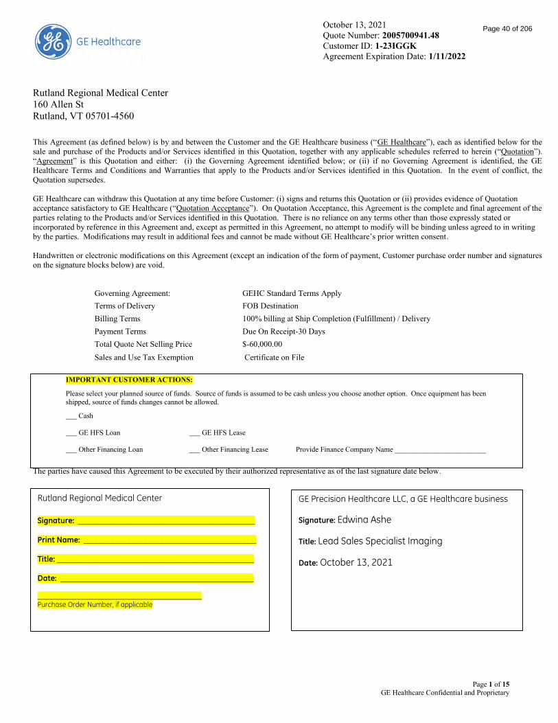
October 13, 2021 Quote Number: 2005700941.48 Customer ID: 1-23IGGK Agreement Expiration Date: 1/11/2022
Page 1 of 15 GE Healthcare Confidential and Proprietary
Rutland Regional Medical Center 160 Allen St Rutland, VT 05701-4560
This Agreement (as defined below) is by and between the Customer and the GE Healthcare business (“GE Healthcare”), each as identified below for the sale and purchase of the Products and/or Services identified in this Quotation, together with any applicable schedules referred to herein (“Quotation”). “Agreement” is this Quotation and either: (i) the Governing Agreement identified below; or (ii) if no Governing Agreement is identified, the GE Healthcare Terms and Conditions and Warranties that apply to the Products and/or Services identified in this Quotation. In the event of conflict, the Quotation supersedes. GE Healthcare can withdraw this Quotation at any time before Customer: (i) signs and returns this Quotation or (ii) provides evidence of Quotation acceptance satisfactory to GE Healthcare (“Quotation Acceptance”). On Quotation Acceptance, this Agreement is the complete and final agreement of the parties relating to the Products and/or Services identified in this Quotation. There is no reliance on any terms other than those expressly stated or incorporated by reference in this Agreement and, except as permitted in this Agreement, no attempt to modify will be binding unless agreed to in writing by the parties. Modifications may result in additional fees and cannot be made without GE Healthcare’s prior written consent. Handwritten or electronic modifications on this Agreement (except an indication of the form of payment, Customer purchase order number and signatures on the signature blocks below) are void.
Governing Agreement: GEHC Standard Terms Apply Terms of Delivery FOB Destination Billing Terms 100% billing at Ship Completion (Fulfillment) / Delivery Payment Terms Due On Receipt-30 Days Total Quote Net Selling Price $-60,000.00 Sales and Use Tax Exemption Certificate on File
IMPORTANT CUSTOMER ACTIONS:
Please select your planned source of funds. Source of funds is assumed to be cash unless you choose another option. Once equipment has been shipped, source of funds changes cannot be allowed.
Cash GE HFS Loan GE HFS Lease Other Financing Loan Other Financing Lease Provide Finance Company Name _________________________
The parties have caused this Agreement to be executed by their authorized representative as of the last signature date below.
Rutland Regional Medical Center
Signature: ___________________________________________ Print Name: __________________________________________ Title: ________________________________________________ Date: _______________________________________________ Purchase Order Number, if applicable
GE Precision Healthcare LLC, a GE Healthcare business
Signature: Edwina Ashe
Title: Lead Sales Specialist Imaging
Date: October 13, 2021
Page 40 of 206

October 13, 2021 Quote Number: 2005700941.48 Customer ID: 1-23IGGK Agreement Expiration Date: 1/11/2022
Page 2 of 15 GE Healthcare Confidential and Proprietary
Rutland Regional Medical Center
Addresses:
Bill To: RUTLAND REGIONAL MEDICAL
CENTER
RUTLAND REGIONAL MEDICAL, CENTER 160 ALLEN ST RUTLAND, VT, 05701-4560
Ship To: RUTLAND REGIONAL MEDICAL CENTER
CENTER 160 ALLEN ST RUTLAND, VT, 05701-4560
To Accept This Quotation Please sign the quote and any included attachments (where requested). If requested, please indicate your form of payment. If you include a purchase order, please make sure it references the following information:
The correct Quote number and Version number above The correct Remit To information as indicated in “Payment Instructions” above Your correct SHIP TO and BILL TO site name and address The correct Total Price as indicated above
Upon submission of a purchase order in response to this quotation, GE Healthcare requests the following to evidence agreement to contract terms: Signature page on quote filled out with signature and P.O. number **** OR**** Verbiage on the purchase order must state one of the following:
(i)Per the terms of Quotation # _____, (ii) Per the terms of GPO #_________; (iii) Per the terms of MPA# _____: or (iv) Per the terms of SAA # ______.
Include applicable quote/agreement number with the reference on the purchase order. In addition, Source of Funds (choice of Cash/Third Party Load or GE HFS Lease Loan or Third Party Lease through ______), must be indicated, which may be done on the Quote Signature Page (for signed quotes), or the Purchase Order (where quotes are not signed) or via a separate written source of funds statement (if provided by GE Healthcare).”
To Accept This Quotation
Please sign and return this quotation together with your Purchase Order to:
Name: Edwina Ashe
Email [email protected]
Phone: (351) 209-0771
Fax:
Payment Instructions Please remit payment for invoices associated with this quotation to: GE Precision Healthcare LLC P.O. Box 96483 Chicago, IL 60693 FEIN: 83-0849145
Page 41 of 206

October 13, 2021 Quote Number: 2005700941.48 Customer ID: 1-23IGGK Agreement Expiration Date: 1/11/2022
Page 3 of 15 GE Healthcare Confidential and Proprietary
Catalog Item Details Total Quote Subtotal: $0.00
Total Quote Net Selling Price: $-60,000.00
If applicable, for more information on this devices' operating system, please visit GE Healthcare's product security portal at: https://securityupdate.gehealthcare.com/en/products
Qty. Credits and Adjustments 1.00 1.5T SIGNA HDx Trade-in $-60,000.00
Page 42 of 206

Trade-in Addendum to GE Healthcare Quotation (Rev. 08.21) Page 4 of 15 GE Healthcare Propriety and Confidential
Trade-in Addendum to GE Healthcare Quotation
This Trade-In Addendum (“Addendum”), effective on October 13, 2021, between the GE Healthcare business identified on the Quotation and Rutland Regional Medical Center (“Customer”), is made a part of Quotation # 2005700941.48 ^ dated October 13, 2021 (“Quotation”) and modifies it as follows:
A. Customer: (i) certifies that it has full legal title to the equipment and/or mobile vehicle (“mobile vehicles” are defined as any systems requiring a vehicle title) listed in Section E (“Trade-In Equipment”), free and clear of all liens and encumbrances; (ii) conveys title and, if applicable, registration and license documents to GE Healthcare effective on the date of removal or receipt of the Trade-In Equipment (mobile vehicles will not be removed from Customer site until GE Healthcare has received a clean title signed over to GE Healthcare); and (iii) affirms that the Trade-In Equipment has never been used on or to provide care to animals. If GE Healthcare removes the Trade-In Equipment, it will do so at its expense at a mutually agreed time. Trade-In Equipment shall be removed no later than thirty days following installation of Customer’s new system, unless explicitly otherwise agreed to by the parties in writing.
Mobile vehicles must include the VIN# on this trade-in addendum: VIN# [insert Vin #]. Mobile vehicles must have a valid DOT sticker and be road worthy at the time GE Healthcare is to take possession of them in order for GE Healthcare to accept a mobile vehicle on trade-in. Any and all logos or hospital affiliation stickers must be removed (outside and inside) by Customer and Customer shall clean the mobile vehicle of all debris and medical supplies prior to removal of the mobile vehicle by GE Healthcare.
B. Customer is responsible for: (i) providing timely, unrestricted access to the Trade-In Equipment in a manner that affords GE Healthcare, or third-party purchaser of the Equipment through GE Healthcare, the ability to complete Equipment inspection and testing, and the ability to complete an operating system back-up prior to de-installation within the timeframe required by GE Healthcare or said third-party purchaser, failure of which to provide may result in termination of this Trade-in Addendum and related credits and/or payments; (ii) ensuring that the Trade-In Equipment and the site where it is located are clean and free of bodily fluids; (iii) informing GE Healthcare of site-related safety risks; (iv) properly managing, transporting and disposing of hazardous materials located on site in accordance with applicable legal requirements; (v) rigging, construction, demolition or facility reconditioning expenses, unless expressly stated otherwise in the Quotation; and (vi) risk of loss and damage to the Trade-In Equipment until safety risks are remediated and the Trade-In Equipment is removed or returned.
C. Prior to removal or return to GE Healthcare, Customer must: (i) remove all Protected Health Information as such term is defined in 45 C.F.R. § 160.103 (“PHI”) from the Trade-In Equipment; and (ii) indemnify GE Healthcare for any loss resulting from PHI not removed. GE Healthcare has no obligation in connection with PHI not properly removed.
D. GE Healthcare may in its sole discretion reduce the trade-in amount or decline to purchase the Trade-In Equipment and adjust the total purchase price of the Quotation accordingly if: (i) the terms of this Addendum are not met; (ii) Customer fails to provide access to the Trade-In Equipment as required herein; or (ii) the Trade-In Equipment is missing components or is inoperable and/or non-functioning when removed or returned – Customer is required to confirm for GE Healthcare the operability of the Trade-In Equipment prior to the deinstallation of the Equipment. All other terms and conditions of the Quotation remain in full force and effect.
E. Trade-In Equipment:
Trade-In Equipment Mfr. Model & Description Quantity System ID* Trade-In Amount ($)
1.5T SIGNA HDx Trade-in 1.00
$ -60,000.00
This Addendum is executed when: (i) signed by the parties below; (ii) Customer receives this Addendum and signs the Quotation that references the Trade-In Equipment; or (iii) Customer receives this Addendum and issues a purchase order identifying either the terms of the Quotation (which includes a reference to the Trade-In Equipment) or the Governing Agreement identified on the Quotation as governing the order (PO# _______________)†. Rutland Regional Medical Center
GE Healthcare
Signature: ______________________________________
Signature: ______________________________________
Print Name: _____________________________________
Print Name: _____________________________________
Title: ___________________________________________
Title: __________________________________________
Date: __________________________________________
Date: __________________________________________
^ A Quotation number must be provided on this document.
Page 43 of 206

Trade-in Addendum to GE Healthcare Quotation (Rev. 08.21) Page 5 of 15 GE Healthcare Propriety and Confidential
* In the event the Trade-In Equipment does not have a System ID, please record the serial number of each component that comprises the Trade-In Equipment. † If you are relying upon the purchase order to reflect acceptance of the terms contained herein, please update this document with the applicable PO number upon receipt of the PO. Failure to do so may result in delays surrounding deinstallation of the System(s).
Page 44 of 206
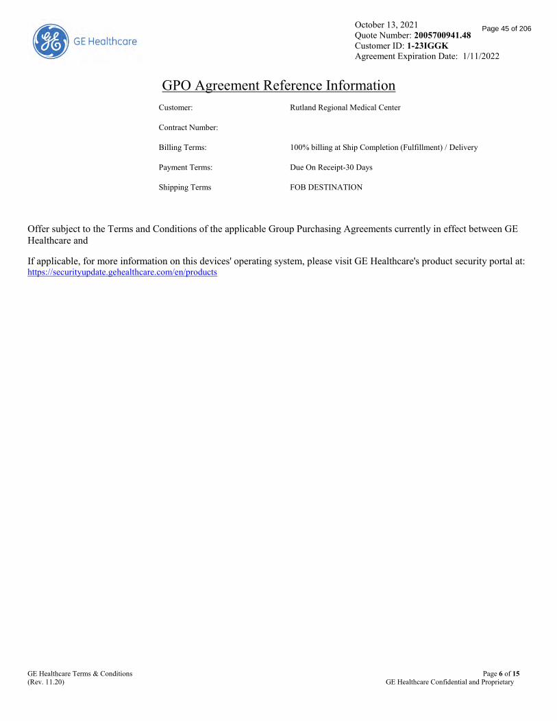
October 13, 2021 Quote Number: 2005700941.48 Customer ID: 1-23IGGK Agreement Expiration Date: 1/11/2022
GE Healthcare Terms & Conditions Page 6 of 15 (Rev. 11.20) GE Healthcare Confidential and Proprietary
GPO Agreement Reference Information Customer: Rutland Regional Medical Center
Contract Number:
Billing Terms: 100% billing at Ship Completion (Fulfillment) / Delivery
Payment Terms: Due On Receipt-30 Days
Shipping Terms FOB DESTINATION
Offer subject to the Terms and Conditions of the applicable Group Purchasing Agreements currently in effect between GE Healthcare and
If applicable, for more information on this devices' operating system, please visit GE Healthcare's product security portal at: https://securityupdate.gehealthcare.com/en/products
Page 45 of 206

GE Healthcare Terms & Conditions Page 7 of 15 (Rev. 11.20) GE Healthcare Confidential and Proprietary
GE Healthcare Terms & Conditions
GE Healthcare Terms & Conditions (Rev 01.30.20)
Definitions. As identified in this Agreement, “Equipment” is hardware and embedded software that is licensed with the purchase of the hardware delivered to Customer in GE Healthcare’s packaging and with its labeling; “Software” is software developed by GE Healthcare and/or delivered to Customer in GE Healthcare’s packaging and with its labeling, and Documentation associated with the software; “Third Party Software” and “Third Party Equipment” are respectively software developed by a third party, and hardware and embedded software that is licensed with the purchase of the hardware, that is delivered to Customer in the third party’s packaging and with its labeling (collectively, “Third Party Product”); “Product” is Equipment, Software and Third Party Product; and “Services” are Product support or professional services; “Subscription” is a limited-term, non-transferable license to access and use a Product (except Healthcare Digital Products), including any associated support Services; “Healthcare Digital Products” are: (i) Software identified in the Quotation as "Centricity"; (ii) Third Party Software licensed for use in connection with Centricity Software; (iii) hardware used to operate Centricity or Third Party Software; (iv) Services provided for implementation, installation or support and maintenance of Centricity or Third Party Software licensed for use in connection with Centricity Software; and/or (v) any Product or Service that is identified in a Healthcare Digital Quotation. “Specifications” are GE Healthcare’s written specifications and manuals as of the date the Equipment shipped. “Documentation” is the online help functions, user instructions and manuals regarding the installation and operation of the Product as made available by GE Healthcare to Customer.
Term and Termination. Software licenses, Services and/or Subscriptions will have individual term lengths identified in the Quotation. If there is a material breach of this Agreement and/or the Quotation that is not cured by the breaching party within 60 days from receipt of written notice, the non-breaching party can terminate the respective Agreement or Quotation. Other than as set forth in this Agreement, neither party can unilaterally terminate this Agreement or a Quotation. Any remaining undisputed, unpaid fees become immediately due and payable on expiration or termination. Expiration or termination of this Agreement will have no effect on Quotations executed prior to the date of expiration or termination.
Software License. Other than as identified in a Quotation, GE Healthcare grants Customer a non-exclusive, non-transferable, non-sublicensable, perpetual license to use the Software for Customer’s internal business purposes only in the United States consistent with the terms of this Agreement. Customer’s independent contractors (except GE Healthcare competitors) may use the Software, but Customer is responsible for their compliance with this license, and additional license fees may apply. Customer cannot modify, reverse engineer, copy or create derivative works of the Software, except for making 1 backup copy, and cannot remove or modify labels or notices of proprietary rights of the Software or Documentation. If GE Healthcare provides Third Party Software, Customer will comply with third party license terms, and licensors are third-party beneficiaries of this Agreement.
Commercial Logistics.
Order Cancellation and Modifications.
Cancellation. If Customer cancels an order prior to shipment without GE Healthcare’s written consent, Customer will be responsible for all third-party expenses incurred by GE Healthcare prior to Customer’s order cancellation and GE Healthcare may charge: (i) a fee of up to 10% of the Product price; and (ii) for site evaluations performed prior to cancellation. GE Healthcare will retain, as a credit, payments received up to the amount of the cancellation charge. Customer must pay applicable progress payments (other than final payment) prior to final calibration, and GE Healthcare may delay calibration until those payments are received. If Customer does not schedule a delivery date within 6 months after order entry, GE Healthcare may cancel on written notice. This Section does not apply to Software or Subscriptions, Third Party Products and/or related professional or installation services; those orders are non-cancellable.
Used Equipment. Equipment identified as pre-owned, refurbished, remanufactured or demonstration Equipment is not new and may have received reconditioning to meet Specifications (“Used Equipment”). Sale of Used Equipment is subject to availability. If it is no longer available, GE Healthcare will attempt to identify other Used Equipment in its inventory that meets Customer's needs, and if substitute Used Equipment is not acceptable, GE Healthcare will cancel the order and refund any deposit Customer paid for the Used Equipment.
Site Preparation. Customer is responsible for network and site preparation, including costs, in compliance with GE Healthcare’s written requirements and applicable laws. GE Healthcare may refuse to deliver or install if the site has not been properly prepared or there are other impediments.
Transportation, Title and Risk of Loss. Unless otherwise identified in the Quotation, shipping terms are FOB Destination. Title and risk of loss to Equipment and Third Party Equipment passes to Customer on delivery to Customer’s designated delivery location.
Delivery, Returns and Installation. Delivery dates are approximate. Products may be delivered in installments. GE Healthcare may invoice multiple installment deliveries on a consolidated basis, but this does not release Customer’s obligation to pay for each installment delivery. Delivery occurs: (i) for Product, on electronic or physical delivery to Customer; and (ii) for Services, on performance.
Products cannot be returned for refund or credit if they match the Quotation.
Delivery and installations will be performed from 8am to 5pm local time, Monday-Friday, excluding GE Healthcare holidays, and outside those hours for an additional fee. Customer will: (i) install cable and assemble products not provided by GE Healthcare; (ii) enable connectivity and interoperability with products not provided by GE Healthcare; (iii) pay for construction and rigging costs; and (iv) obtain all licenses, permits and approvals for installation, use and disposal of Products. For upgrades and revisions to non-Healthcare Digital Products, Customer must return replaced components to GE Healthcare at no charge.
Information Technology Professional Services (“ITPS”). ITPS must be completed within 12 months of the later of the ITPS order date or Product delivery. If not done within this time period, other than because of GE Healthcare's failure to perform, ITPS performance obligations expire without refund. ITPS includes project management, HL7/HIS system integration, database conversion, network design and integration and separately cataloged software installations. This Section does not apply to Healthcare Digital Products.
Acceptance.
Page 46 of 206
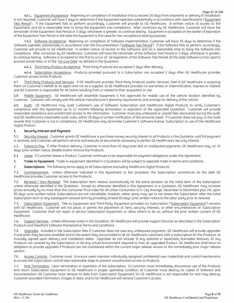
GE Healthcare Terms & Conditions Page 8 of 15 (Rev. 11.20) GE Healthcare Confidential and Proprietary
Equipment Acceptance. Beginning on completion of installation (not to exceed 30 days from shipment) or delivery (if installation is not required), Customer will have 5 days to determine if the Equipment operates substantially in accordance with Specifications (“Equipment Test Period”). If the Equipment fails to perform accordingly, Customer will provide to GE Healthcare: (i) written notice; (ii) access to the Equipment; and (iii) a reasonable time to bring the Equipment into compliance. After correction by GE Healthcare, Customer will have the remainder of the Equipment Test Period or 3 days, whichever is greater, to continue testing. Equipment is accepted on the earlier of expiration of the Equipment Test Period or the date the Equipment is first used for non-acceptance testing purposes.
Software Acceptance. Beginning on completion of Software implementation, Customer will have 30 days to determine if the Software operates substantially in accordance with the Documentation (“Software Test Period”). If the Software fails to perform accordingly, Customer will provide to GE Healthcare: (i) written notice; (ii) access to the Software; and (iii) a reasonable time to bring the Software into compliance. After correction by GE Healthcare, Customer will have the remainder of the Software Test Period or 5 days, whichever is greater, to continue testing. Software is accepted on the first to occur of: (a) expiration of the Software Test Period; (b) the date Software is first used to process actual data; or (c) the “Go-Live Date” as defined in the Quotation.
Third Party Product Acceptance. Third Party Products are accepted 5 days after delivery.
Subscription Acceptance. Products provided pursuant to a Subscription are accepted 5 days after GE Healthcare provides Customer access to the Products.
Third Party Products and Services. If GE Healthcare provides Third Party Products and/or Services, then (i) GE Healthcare is acquiring them on Customer’s behalf as its agent and not as a supplier; (ii) GE Healthcare provides no warranties or indemnification, express or implied; and (iii) Customer is responsible for all claims resulting from or related to their acquisition or use.
Mobile Equipment. GE Healthcare will assemble Equipment it has approved for mobile use at the vehicle location identified by Customer. Customer will comply with the vehicle manufacturer’s planning requirements and arrange for delivery of the vehicle.
Audit. GE Healthcare may audit Customer’s use of Software, Subscription and Healthcare Digital Products to verify Customer’s compliance with this Agreement up to 12 months following termination or expiration of the applicable Quotation. Customer will provide reasonable assistance and unrestricted access to the information. Customer must pay underpaid or unpaid fees discovered during the audit, and GE Healthcare’s reasonable audit costs, within 30 days of written notification of the amounts owed. If Customer does not pay, or the audit reveals that Customer is not in compliance, GE Healthcare may terminate Customer’s Software license, Subscription or use of the Healthcare Digital Product.
Security Interest and Payment.
Security Interest. Customer grants GE Healthcare a purchase money security interest in all Products in the Quotation until full payment is received, and Customer will perform all acts and execute all documents necessary to perfect GE Healthcare’s security interest.
Failure to Pay. If, after Product delivery, Customer is more than 45 days past due on undisputed payments, GE Healthcare may, on 10 days’ prior written notice, disable and/or remove the Products.
Lease. If Customer leases a Product, Customer continues to be responsible for payment obligations under this Agreement.
Trade-In Equipment. Trade-in equipment identified in a Quotation will be subject to separate trade-in terms and conditions.
Subscriptions. The following terms apply to all Subscriptions (excluding Healthcare Digital Products).
Commencement. Unless otherwise indicated in this Agreement or the Quotation, the Subscription commences on the date GE Healthcare provides Customer access to the Products.
Renewal / Non-Renewal. The Subscription term renews automatically for the same duration as the initial term of the Subscription unless otherwise identified in the Quotation. Except as otherwise identified in this Agreement or a Quotation, GE Healthcare may increase prices annually by no more than the Consumer Price Index for All Urban Consumers (U.S. City Average, December to December) plus 2%, upon 90 days’ prior written notice. Subscriptions are not cancellable; however, either party may opt to not renew the Subscription after the initial Subscription term or any subsequent renewal term by providing at least 60 days’ prior written notice to the other party prior to renewal.
Subscription Equipment. Title to Equipment and Third-Party Equipment provided via Subscription (“Subscription Equipment”) remains with GE Healthcare. Customer will not place, or permit the placement of, liens, security interests, or other encumbrances on Subscription Equipment. Customer shall not repair or service Subscription Equipment, or allow others to do so, without the prior written consent of GE Healthcare.
Support Services. Unless otherwise noted in the Quotation, GE Healthcare will provide support Services as described in the Subscription Products and ViewPoint Software Maintenance Terms and Conditions.
Upgrades. Included in the Subscription fees if Customer does not owe any undisputed payments, GE Healthcare will provide upgrades if and when they become available and to the extent they are provided to all GE Healthcare customers with a Subscription for the Products, at mutually agreed upon delivery and installation dates. Upgrades do not include: (i) any optional or separately licensable features; (ii) any Products not covered by the Subscription; or (iii) any virtual environment required to host an upgraded Product. GE Healthcare shall have no obligation to provide upgrades if Products are not maintained within the current major release version or the immediately prior major release version.
Access Controls. Customer must: (i) ensure users maintain individually-assigned confidential user credentials and control mechanisms to access the Subscription; and (ii) take reasonable steps to prevent unauthorized access to Products.
Post-Termination. Upon termination or expiration of the Subscription: (i) Customer must immediately discontinue use of the Products and return Subscription Equipment to GE Healthcare in proper operating condition; (ii) Customer must destroy its copies of Software and Documentation; (iii) Customer must remove its data from Subscription Equipment; (iv) GE Healthcare is not responsible for and may destroy Customer-provided information, images or data; and (v) GE Healthcare will remove Customer’s access.
Page 47 of 206

GE Healthcare Terms & Conditions Page 9 of 15 (Rev. 11.20) GE Healthcare Confidential and Proprietary
Professional Services. For Services not covered under this Agreement or required due to Customer not meeting its responsibilities under the Agreement, applicable additional professional Services and fees will be required: (i) identified in the Quotation; and (ii) subject to GE Healthcare’s then-current pricing.
General Terms.
Confidentiality. Each party will treat this Agreement and the other party's proprietary information as confidential, meaning it will not use or disclose the information to third parties unless permitted in this Agreement or required by law. Customers are not prohibited from discussing patient safety issues in appropriate venues.
Governing Law. The law of the state where the Product is installed, the Service is provided, or the Subscription is accessed will govern this Agreement.
Force Majeure. Performance time for non-monetary obligations will be reasonably extended for delays beyond a party’s control.
Assignment; Use of Subcontractors. Rights and obligations under this Agreement cannot be assigned without the other party’s prior written consent, unless: (i) it is to an entity (except to a GE Healthcare competitor) that (a) is an affiliate or parent of the party or (b) acquires substantially all of the stock or assets of such party’s applicable business, Product line, or Service thereof; and (ii) the assignee agrees in writing to be bound by this Agreement, including payment of outstanding fees. GE Healthcare may hire subcontractors to perform work under this Agreement but will remain responsible for its obligations.
Waiver; Survival. If any provision of this Agreement is not enforced, it is not a waiver of that provision or of a party’s right to later enforce it. Terms in this Agreement related to intellectual property, compliance, data rights and terms that by their nature are intended to survive will survive the Agreement’s expiration or termination.
Intellectual Property. GE Healthcare owns all rights to the intellectual property in GE Healthcare’s Products, Services, Documentation, Specifications, and statements of work related to a Quotation or otherwise. Customer may provide GE Healthcare with feedback related to Products, Services, and related Documentation, and GE Healthcare may use it in an unrestricted manner.
Compliance.
Generally. Each party will comply with applicable laws and regulations. Customer is only purchasing or licensing Products for its own medical, billing and/or non-entertainment use in the United States. GE Healthcare will not deliver, install, service or train if it discovers Products have been or are intended to be used contrary to this Agreement. This Agreement is subject to GE Healthcare’s ongoing credit review and approval. Customer is aware of its legal obligations for cost reporting, including 42 C.F.R. § 1001.952(g) and (h), and will request from GE Healthcare any information beyond the invoice needed to fulfill Customer’s cost reporting obligations. GE Healthcare will provide safety-related updates for Equipment and Software required by applicable laws and regulations at no additional charge.
Security. GE Healthcare is not responsible for: (i) securing Customer’s network; (ii) preventing unauthorized access to Customer’s network or the Product; (iii) backup management; (iv) data integrity; (v) recovery of lost, corrupted or damaged data, images, software or equipment; (vi) third party operating systems, unless specifically provided in the Quotation; or (vii) providing or validating antivirus or related IT safeguards unless sold to Customer by GE Healthcare. NEITHER PARTY WILL BE LIABLE TO THE OTHER PARTY FOR DAMAGES CAUSED BY UNAUTHORIZED ACCESS TO THE NETWORK OR PRODUCTS REGARDLESS OF A PARTY’S COMPLIANT SECURITY MEASURES.
Environmental Health and Safety (“EHS”). GE Healthcare personnel may stop work without penalty due to safety concerns. Customer must: (i) comply with GE Healthcare’s EHS requirements; (ii) provide a safe environment for GE Healthcare personnel; (iii) tell GE Healthcare about chemicals or hazardous materials that might come in contact with Products or GE Healthcare personnel; (iv) perform decommissioning or disposal at Customer facilities; (v) obtain and maintain necessary permits; (vi) thoroughly clean Products before Service; (vii) provide radioactive materials required for testing Products; and (viii) dispose of waste related to Products and installations.
Parts and Tubes. GE Healthcare: (i) recommends the use of parts it has validated for use with the Product; (ii) is not responsible for the quality of parts supplied by third parties to Customer; and (iii) cannot assure Product functionality or performance when non-validated parts are used. Certain Products are designed to recognize GE Healthcare-supplied tubes and report the presence of a non-GE Healthcare tube; GE Healthcare is not responsible for the use of, or effects from, non-GE Healthcare supplied tubes.
Training. GE Healthcare’s training does not guarantee that: (i) Customer trainees are fully trained on Product use, maintenance or operation; or (ii) training will satisfy any licensure or accreditation. Customer must ensure its trainees are fully qualified in the use and operation of the Product. Unless otherwise identified in the training catalog, Customer will complete training within 12 months of: (a) the date of Product delivery for a Product purchase; (b) the respective start date for Services or Subscription for purchase of Service or Subscription; or (c) the date training is ordered for training-only purchases. If not completed within this time period, other than because of GE Healthcare’s fault, training expires without refund.
Medical Diagnosis and Treatment. All clinical and medical treatment, diagnostic and/or billing decisions are Customer’s responsibility.
Connectivity. If a Product has remote access capability: (i) Customer will provide GE Healthcare with, and maintain, a GE Healthcare-validated remote access connection to service the Product; or (ii) GE Healthcare reserves the right to charge Customer for onsite support at GE Healthcare’s then-current billing rate. This remote access and collection of machine data (e.g., temperature, helium level) will continue after the end of this Agreement unless Customer requests in writing that GE Healthcare disable it.
Use of Data.
Protected Health Information. If GE Healthcare creates, receives, maintains, transmits or otherwise has access to Protected Health Information (as defined in 45 C.F.R. § 160.103) (“PHI”), GE Healthcare may use and disclose the PHI only as permitted by law and by the Business Associate Agreement. Before returning any Product to GE Healthcare, Customer must ensure that all PHI stored in it is deleted.
Data Rights. GE Healthcare may collect, prepare derivatives from and otherwise use non-PHI data related to Products and/or Services for such things as training, demonstration, research, development, benchmarking, continuous improvement and facilitating the provision of its products, software and services. GE Healthcare will own all intellectual property and other rights that could result from this collection, preparation and use. The non-PHI data will not be used to identify Customer or sold by GE Healthcare without Customer’s consent.
Page 48 of 206
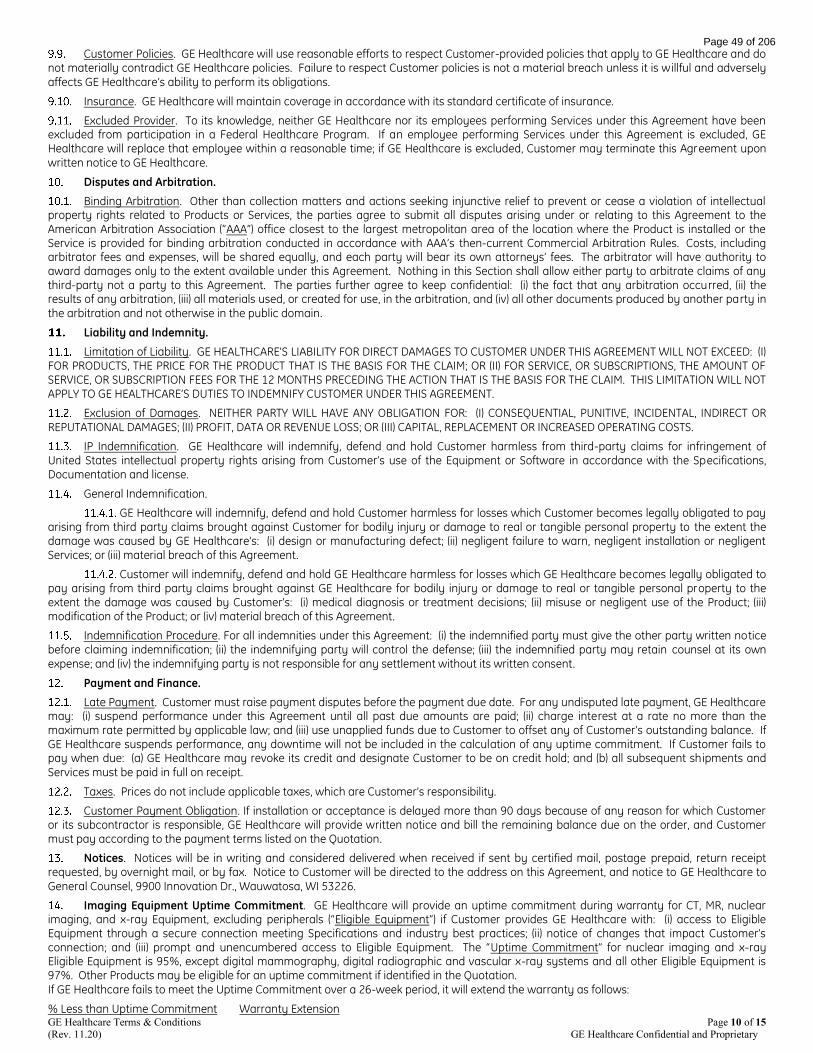
GE Healthcare Terms & Conditions Page 10 of 15 (Rev. 11.20) GE Healthcare Confidential and Proprietary
Customer Policies. GE Healthcare will use reasonable efforts to respect Customer-provided policies that apply to GE Healthcare and do not materially contradict GE Healthcare policies. Failure to respect Customer policies is not a material breach unless it is willful and adversely affects GE Healthcare’s ability to perform its obligations.
Insurance. GE Healthcare will maintain coverage in accordance with its standard certificate of insurance.
Excluded Provider. To its knowledge, neither GE Healthcare nor its employees performing Services under this Agreement have been excluded from participation in a Federal Healthcare Program. If an employee performing Services under this Agreement is excluded, GE Healthcare will replace that employee within a reasonable time; if GE Healthcare is excluded, Customer may terminate this Agreement upon written notice to GE Healthcare.
Disputes and Arbitration.
Binding Arbitration. Other than collection matters and actions seeking injunctive relief to prevent or cease a violation of intellectual property rights related to Products or Services, the parties agree to submit all disputes arising under or relating to this Agreement to the American Arbitration Association (“AAA”) office closest to the largest metropolitan area of the location where the Product is installed or the Service is provided for binding arbitration conducted in accordance with AAA’s then-current Commercial Arbitration Rules. Costs, including arbitrator fees and expenses, will be shared equally, and each party will bear its own attorneys’ fees. The arbitrator will have authority to award damages only to the extent available under this Agreement. Nothing in this Section shall allow either party to arbitrate claims of any third-party not a party to this Agreement. The parties further agree to keep confidential: (i) the fact that any arbitration occurred, (ii) the results of any arbitration, (iii) all materials used, or created for use, in the arbitration, and (iv) all other documents produced by another party in the arbitration and not otherwise in the public domain.
Liability and Indemnity.
Limitation of Liability. GE HEALTHCARE'S LIABILITY FOR DIRECT DAMAGES TO CUSTOMER UNDER THIS AGREEMENT WILL NOT EXCEED: (I) FOR PRODUCTS, THE PRICE FOR THE PRODUCT THAT IS THE BASIS FOR THE CLAIM; OR (II) FOR SERVICE, OR SUBSCRIPTIONS, THE AMOUNT OF SERVICE, OR SUBSCRIPTION FEES FOR THE 12 MONTHS PRECEDING THE ACTION THAT IS THE BASIS FOR THE CLAIM. THIS LIMITATION WILL NOT APPLY TO GE HEALTHCARE’S DUTIES TO INDEMNIFY CUSTOMER UNDER THIS AGREEMENT.
Exclusion of Damages. NEITHER PARTY WILL HAVE ANY OBLIGATION FOR: (I) CONSEQUENTIAL, PUNITIVE, INCIDENTAL, INDIRECT OR REPUTATIONAL DAMAGES; (II) PROFIT, DATA OR REVENUE LOSS; OR (III) CAPITAL, REPLACEMENT OR INCREASED OPERATING COSTS.
IP Indemnification. GE Healthcare will indemnify, defend and hold Customer harmless from third-party claims for infringement of United States intellectual property rights arising from Customer’s use of the Equipment or Software in accordance with the Specifications, Documentation and license.
General Indemnification.
GE Healthcare will indemnify, defend and hold Customer harmless for losses which Customer becomes legally obligated to pay arising from third party claims brought against Customer for bodily injury or damage to real or tangible personal property to the extent the damage was caused by GE Healthcare’s: (i) design or manufacturing defect; (ii) negligent failure to warn, negligent installation or negligent Services; or (iii) material breach of this Agreement.
Customer will indemnify, defend and hold GE Healthcare harmless for losses which GE Healthcare becomes legally obligated to pay arising from third party claims brought against GE Healthcare for bodily injury or damage to real or tangible personal property to the extent the damage was caused by Customer’s: (i) medical diagnosis or treatment decisions; (ii) misuse or negligent use of the Product; (iii) modification of the Product; or (iv) material breach of this Agreement.
Indemnification Procedure. For all indemnities under this Agreement: (i) the indemnified party must give the other party written notice before claiming indemnification; (ii) the indemnifying party will control the defense; (iii) the indemnified party may retain counsel at its own expense; and (iv) the indemnifying party is not responsible for any settlement without its written consent.
Payment and Finance.
Late Payment. Customer must raise payment disputes before the payment due date. For any undisputed late payment, GE Healthcare may: (i) suspend performance under this Agreement until all past due amounts are paid; (ii) charge interest at a rate no more than the maximum rate permitted by applicable law; and (iii) use unapplied funds due to Customer to offset any of Customer’s outstanding balance. If GE Healthcare suspends performance, any downtime will not be included in the calculation of any uptime commitment. If Customer fails to pay when due: (a) GE Healthcare may revoke its credit and designate Customer to be on credit hold; and (b) all subsequent shipments and Services must be paid in full on receipt.
Taxes. Prices do not include applicable taxes, which are Customer’s responsibility.
Customer Payment Obligation. If installation or acceptance is delayed more than 90 days because of any reason for which Customer or its subcontractor is responsible, GE Healthcare will provide written notice and bill the remaining balance due on the order, and Customer must pay according to the payment terms listed on the Quotation.
Notices. Notices will be in writing and considered delivered when received if sent by certified mail, postage prepaid, return receipt requested, by overnight mail, or by fax. Notice to Customer will be directed to the address on this Agreement, and notice to GE Healthcare to General Counsel, 9900 Innovation Dr., Wauwatosa, WI 53226.
Imaging Equipment Uptime Commitment. GE Healthcare will provide an uptime commitment during warranty for CT, MR, nuclear imaging, and x-ray Equipment, excluding peripherals (“Eligible Equipment”) if Customer provides GE Healthcare with: (i) access to Eligible Equipment through a secure connection meeting Specifications and industry best practices; (ii) notice of changes that impact Customer’s connection; and (iii) prompt and unencumbered access to Eligible Equipment. The “Uptime Commitment” for nuclear imaging and x-ray Eligible Equipment is 95%, except digital mammography, digital radiographic and vascular x-ray systems and all other Eligible Equipment is 97%. Other Products may be eligible for an uptime commitment if identified in the Quotation. If GE Healthcare fails to meet the Uptime Commitment over a 26-week period, it will extend the warranty as follows:
% Less than Uptime Commitment Warranty Extension
Page 49 of 206

GE Healthcare Terms & Conditions Page 11 of 15 (Rev. 11.20) GE Healthcare Confidential and Proprietary
0.1 - 3.0 1 week
3.1 - 8.0 2 weeks
8.1 - 13.0 4 weeks
> 13.0 6 weeks
Uptime is calculated as follows:
“Uptime Base” = (“a” hours per day X “b” days per week X 26 weeks) – (Planned Maintenance (“PM”) hours during prior 26 weeks), where “a” hours per day and “b” days per week are determined by the standard warranty for Eligible Equipment. “Downtime” is the number of hours during which Eligible Equipment is subject to a Critical Malfunction. Downtime starts when Customer notifies GE Healthcare that Eligible Equipment is inoperable and unavailable for use due to GE Healthcare’s design, manufacturing, material or performance failure (“Critical Malfunction”). Downtime ends when Eligible Equipment is available for clinical use. To be eligible for the Uptime Commitment, Customer must maintain a performance log that includes data required to calculate Downtime.
DoseWatch Device License. Each connection of a Device (defined below) to the DoseWatch Software requires Customer to purchase a unique Device license referencing a Device ID that allows concurrent use of the DoseWatch Software with that Device at a specified Customer facility on Customer’s secured network. All other terms, duration and warranties applicable to the Software license apply to the Device license. “Device” is specific Customer equipment approved by GE Healthcare to be connected to DoseWatch Software under this Agreement. Additional Device connections may be added to this Agreement, subject to individual Device licenses, and related installation, implementation, configuration and optimization services at GE Healthcare’s then-current rates.
Subscription Products and ViewPoint Software Maintenance Terms and Conditions.
Overview. GE Healthcare will, in accordance with the terms and conditions of this section, maintain, support and update: (i) Products provided via Subscription (excluding Healthcare Digital Products); and (ii) ViewPoint Software licensed by Customer (“ViewPoint Software”) and HIS interface software installed in the United States covered by a Software Maintenance Agreement (“SMA”).
Scope.
Software Support and Maintenance. GE Healthcare will use reasonable efforts to provide Error Correction (defined below) for verifiable and reproducible Errors (defined below) within a reasonable time after: (a) Customer reports the Error to GE Healthcare; or (b) detection by GE Healthcare. Updates (defined below), if released, will be provided at no additional cost as a part of this maintenance commitment. New functionality must be purchased separately, unless otherwise agreed.
Equipment Maintenance. Preventative maintenance service may be required periodically during normal business hours of 8:00 a.m. to 5:00 p.m. (local time) on mutually agreed dates. Customer will make the Equipment available for preventative maintenance upon GE Healthcare request. Additional services to be performed, including specific additional terms thereof, shall be specified in the Quotation or alternate schedules.
Definitions. “Error” means any Software-related problem that: (i) materially interferes with Customer’s use of the Software; and (ii) results from a failure of the Software to materially conform to the Documentation. “Error Correction” means: (a) modification of the Software that corrects an Error by bringing the Software into material conformity with the Documentation; or (b) a procedure that avoids the material adverse effect of the nonconformity. “Update” means a change that provides Error Corrections and/or enhances functionality of the Software version licensed by Customer. An Update does not involve major changes or provide significant, new functionality or applications, or changes to the software architecture or file structure. Updates retain the same license as the original Software.
Hotline Support. GE Healthcare will provide phone and email support during standard business hours, excluding GE Healthcare holidays, for problem solving, Error resolution and general help.
Remote Access Support. GE Healthcare may access Software remotely via Customer’s network and GE Healthcare-supplied secure tunnelling software to monitor Software parameters to help prevent and detect Errors. Customer will reasonably cooperate with GE Healthcare to establish remote connections. Certain modules require remote access in order to obtain support.
Warranty. GE Healthcare warrants that its Services will be performed by trained individuals in a professional, workman-like manner. GE Healthcare will re-perform non-conforming Services as long as Customer provides prompt written notice to GE Healthcare. NO OTHER EXPRESS OR IMPLIED WARRANTIES, INCLUDING IMPLIED WARRANTIES OF NON-INFRINGEMENT, MERCHANTABILITY AND FITNESS FOR A PARTICULAR PURPOSE, WILL APPLY. SERVICE MANUALS AND DOCUMENTATION ARE PROVIDED “AS IS”. GE HEALTHCARE DOES NOT GUARANTEE PRODUCTS WILL OPERATE WITHOUT ERROR OR INTERRUPTION.
Exclusions. GE Healthcare has no obligation to Customer for: (i) use of Products in combination with software, hardware, or services not recommended in writing by GE Healthcare; (ii) use in a manner or environment for which GE Healthcare did not design or license the Products, or in violation of GE Healthcare’s recommendations or instructions; (iii) interface configuration (often referred to as HIS, PACS or EMR interfaces necessary due to changing vendors or versions); (iv) reorganization of Customer data; (v) consulting or software engineering and programming; (vi) support of Products outside the scope of the foregoing maintenance commitments; (vii) failure to use or install, or permit GE Healthcare to use or install, Error Corrections or Updates; (viii) failure to maintain Products within the current major release version or the immediately prior major release version; (ix) defects in products or services not made and provided by GE Healthcare; (x) any cause external to the Products or beyond GE Healthcare’s control; (xi) failure of Customer’s network; (xii) replacement of disposable or consumable items; (xiii) additional equipment or upgrades in connection with Products; and (xiv) migration of Software to different hardware or operating systems.
UptimeBase
DowntimeUptimeBase
Page 50 of 206

GE Healthcare Terms & Conditions Page 12 of 15 (Rev. 11.20) GE Healthcare Confidential and Proprietary
Software Maintenance Agreement Term. The following applies to ViewPoint software and HIS interface software only: The SMA term and start date is identified in the Quotation and its related Schedule A. Either party may terminate the SMA without cause after the first anniversary by providing at least 90 days’ prior written notice to the other party. SMA payments are due within 30 days after receipt of GE Healthcare’s invoice.
17. Magnetic Resonance (“MR”) – Magnetic Maintenance and Cryogens. Customer is responsible for: (i) cryogen loss due to power loss or water chiller failure for the MR’s shield cooler or condenser system during installation; (ii) costs for cryogen replacement plus transfill labor at GE Healthcare’s then-applicable rates; (iii) post-assembly supply and installation of cryogens, unless cryogen loss is caused by a defect in material or workmanship within the scope of GE Healthcare’s warranty. MR magnetic fields attract ferro-magnetic articles and are capable of rapidly accelerating them toward the magnet, creating danger to persons in the vicinity and possible system damage. Magnetic and radio frequency fields may adversely affect the operation of pacemakers, equipment containing magnetic reed switches and aneurysm or surgical clips.
Page 51 of 206

Warranty Statement (Rev 11.20) Page 13 of15 GE Healthcare Confidential & Proprietary
GE Healthcare Warranty Statement
1. Warranty.
1.1 Equipment. For non-customized Equipment purchased from GE Healthcare or its authorized distributors, unless otherwise identified in the Quotation, GE Healthcare warrants that Equipment will be free from defects in title, and, for 1 year from Equipment Acceptance, it will: (i) be free from defects in material and workmanship under normal use and service; and (ii) perform substantially in accordance with the Specifications. The warranty covers parts and labor and only applies to end-users that purchase Equipment from GE Healthcare or its authorized distributors.
1.2 Software. For Software licensed from GE Healthcare, GE Healthcare warrants that: (i) it has the right to license or sublicense
Software to Customer; (ii) it has not inserted Disabling Code into Software; (iii) it will use efforts consistent with industry standards to remove
viruses from Software before delivery; and (iv) unless otherwise identified in the Quotation, for 90 days from Software Acceptance, Software
will perform substantially in accordance with the Documentation. “Disabling Code” is code designed to interfere with the normal operation of
Software, but code that prohibits use outside of the license scope is not Disabling Code.
1.3. Services. GE Healthcare warrants that its Service will be performed by trained individuals in a professional, workman-like manner.
1.4. Used Equipment. Certain Used Equipment is provided with GE Healthcare’s standard warranty for the duration identified in the Quotation, but in no event more than 1 year. If no warranty is identified, the Used Equipment is provided “AS IS” and is not warranted by GE Healthcare.
1.5. Accessories and Supplies. Warranties for accessories and supplies are at www.gehealthcare.com/accessories.
1.6. Third Party Product. Third Party Product is covered by the third party’s warranty and not GE Healthcare’s warranties.
1.7. Subscription Products. Products provided via Subscription (excluding Healthcare Digital Products) are not covered by this Warranty Statement. Instead, the Subscription Products and ViewPoint Software Maintenance Terms and Conditions apply
2. Remedies. If Customer promptly notifies GE Healthcare of its claim during the warranty and makes the Product available, GE Healthcare will: (i) at its option, repair, adjust or replace the non-conforming Equipment or components; (ii) at its option, correct the non-conformity or replace the Software; and/or (iii) re-perform non-conforming Service. Warranty service will be performed from 8am to 5pm local time, Monday-Friday, excluding GE Healthcare holidays, and outside those hours at GE Healthcare’s then-current service rates and subject to personnel availability. GE Healthcare may require warranty repairs to be performed via a secure, remote connection or at an authorized service center. If GE Healthcare replaces Equipment or a component, the original becomes GE Healthcare property and Customer will return the original to GE Healthcare within 5 days after the replacement is provided to Customer. Customer cannot stockpile replacement parts. Prior to returning Equipment to GE Healthcare, Customer will: (a) obtain a return to manufacturer authorization; and (b) back up and remove all information stored on the Equipment (stored data may be removed during repair). Customer is responsible for damage during shipment to GE Healthcare. The warranty for a Product or component provided to correct a warranty failure is the unexpired term of the warranty for the repaired or replaced Product. GE Healthcare may provide a loaner unit during extended periods of Product service. If a loaner unit is provided: (i) it is for Customer’s temporary use at the location identified in the Quotation; (ii) it will be returned to GE Healthcare within 5 days after the Product is returned to Customer, and if it is not, GE Healthcare may repossess it or invoice Customer for its full list price; (iii) it, and all programs and information pertaining to it, remain GE Healthcare property; (iv) risk of loss is with Customer during its possession; (v) Customer will maintain and return it in proper condition, normal wear and tear excepted, in accordance with GE Healthcare’s instructions; (vi) it will not be repaired except by GE Healthcare; (vii) GE Healthcare will be given reasonable access to it; (viii) Customer is not paying for its use, and Customer will ensure charges or claims submitted to a government healthcare program or patient are submitted accordingly; and (ix) prior to returning it to GE Healthcare, Customer will delete all information, including PHI, from it and its accessories, in compliance with industry standards and instructions provided by GE Healthcare. NO OTHER EXPRESS OR IMPLIED WARRANTIES, INCLUDING IMPLIED WARRANTIES OF NON-INFRINGEMENT, MERCHANTABILITY AND FITNESS FOR A PARTICULAR PURPOSE, WILL APPLY. SERVICE MANUALS AND DOCUMENTATION ARE PROVIDED “AS IS”. GE HEALTHCARE DOES NOT GUARANTEE PRODUCTS WILL OPERATE WITHOUT ERROR OR INTERRUPTION.
3. Limitations. GE Healthcare has no obligation to Customer for warranty claims if Customer uses the Product: (a) for non-medical or entertainment use or outside the United States; (b) in combination with software, hardware, or services not recommended in writing by GE Healthcare; and (c) in a manner or environment for which GE Healthcare did not design or license it, or in violation of GE Healthcare’s recommendations or instructions.
In addition, these warranties do not cover: (i) defects or deficiencies from improper storage or handling, maintenance or use that does not conform to Specifications and/or Documentation, inadequate backup or virus protection, cyber-attacks, failure to maintain power quality, grounding, temperature, and humidity within Specifications and/or Documentation; (ii) repairs due to power anomalies or any cause external to the Products or beyond GE Healthcare’s control; (iii) payment or reimbursement of facility costs arising from repair or replacement of the Products or parts; (iv) planned maintenance (unless applicable to Equipment), adjustment, alignment, or calibration; (v) network and antenna installations not performed by GE Healthcare or its subcontractors; (vi) lost or stolen Products; (vii) Products with serial numbers altered, defaced or removed; (viii) modification of Product not approved in writing by GE Healthcare (ix) Products immersed in liquid; and (x) replacement of disposable or consumable items.
4. Exceptions to Standard Warranty. DoseWatch Explore: DOSEWATCH EXPLORE SOFTWARE, SERVICES AND INFORMATION IS PROVIDED "AS IS" WITH NO WARRANTY Partial System Equipment Upgrades for CT, MR, X-Ray, IGS, PET (Scanners, Cyclotrons and Chemistry Labs) and Nuclear systems: 6 months (only applies to the upgraded components unless the parties otherwise agree to modify the coverage of the upgraded and existing components in an existing service agreement. Optima XR240amx partial upgrades are warranted for 1 year
Cyclotron and Radiopharmacy: Warranty starts on the earlier of (i) 3 months after the date GE Healthcare completes mechanical installation,
Page 52 of 206
Warranty Statement (Rev 11.20) Page 14 of15 GE Healthcare Confidential & Proprietary
or (ii) the date Product testing is successfully completed MR Systems: Warranty does not cover: (i) a defect or deficiency from failure of water chillers supplied or serviced by Customer, and (ii) for MR systems with LHe/LN or shield cooler configured superconducting magnets (except for MR Systems with LCC magnets), any cryogen supply, cryogenic service or service to the magnet, cryostat, coldhead, shield cooler compressor or shim coils unless the need for supply or service is caused by a defect in material or workmanship covered by this warranty.
Proteus XR/a, Definium and Precision 500D X-Ray Systems: Warranty does not cover collimator bulbs
Performix 160A (MX160) Tubes: 3 years
X-Ray High Voltage Rectifiers and TV Camera Pick-Up Tubes: 6 months X-Ray Wireless Digital Detectors: In addition to the standard warranty, GE Healthcare will provide coverage for detector damage due to accidental dropping or mishandling. If accidental damage occurs, GE Healthcare will provide Customer with 1 replacement detector during warranty at no additional charge. If subsequent accidental damage occurs during warranty, each additional replacement will be provided for $30,000 per replacement. This additional coverage excludes damage caused by any use that does not conform to original equipment manufacturer (“OEM”) guidelines, use that causes fluid invasion, holes, deep scratches or the detector case to crack, and damage caused by abuse, theft, loss, fire, power failures or surges. If the warranty is voided by these conditions, repair or replacement is Customer’s responsibility. Bone Mineral Densitometry: Alpha Source, Inc. will perform installation, application support and warranty services. Direct warranty claims to Alpha Source, Inc. at 1-800-654-9845. Upgraded computer, printer and monitor components include a 1 month warranty. Customer will not be credited the value of this warranty against pre-existing warranties or service agreements.
OEC New or Exchange Service Parts: 120 days
OEC Tubes and Image Intensifiers: 1 year
OEC Refurbished C-Arms: 1 year after installation
IGS Large Display Monitor: Warranty coverage excludes damage caused by Customer abuse
HealthNet Lan, Advantage Review — Remote Products: 3 months
LOGIQ e, Venue 50, Venue Go, Versana Active and related transducers purchased with them: 5 years
LOGIQ V1, LOGIQ V2, Vivid iq, Vscan and Vscan Extend and related transducers purchased with them: 3 years
Except the following have a 1 year warranty:
Transducers: TEE Probes, Carts: Venue 50 Docking Cart, Venue Go Cart, Venue Go mounting cradle, LOGIQ e Isolation Cart, LOGIQ e Docking Cart, and LOGIQ V1/V2 Cart Other Accessories: Batteries (internal & external), and printers and peripherals, TEE cleaning & storage system, ICECord Connector and printers Warranty covers defective parts and components and includes: (i) repair at GE Healthcare facilities, (ii) a loaner unit or probe replacement shipped for next business day delivery for requests received by 3pm Central Time, (iii) phone support from 7am to 7pm Central Time, Monday-Friday, excluding GE Healthcare holidays. For an additional charge, GE Healthcare may provide field support/service, planned maintenance, and/or coverage for damage due to accidental dropping or mishandling.
LOGIQ P9 R2.5 and newer and, Versana Premier, Versana Balance, Venue and related transducers purchased with them: 5 years Voluson P8 BT18 and newer, LOGIQ F8 2016 and newer, LOGIQ V5, Vivid T8 and Vivid T9 along with related transducers purchased with them: 3 years
Except the following have a 1 year warranty:
Other Accessories: Batteries (internal & external) and printers and peripherals, TEE cleaning & storage system
Transducers: TEE Probes Warranty covers defective parts and components and includes: (i) repair at Product location by a qualified service technician Monday-Friday 8am to 5pm local time, excluding GE Healthcare holidays, and (ii) phone support from 7am to 7pm Central Time, Monday-Friday, excluding GE Healthcare holidays. For an additional charge, GE Healthcare may provide planned maintenance and/or coverage for damage due to accidental dropping or mishandling. Ultrasound Partial System Equipment Upgrades: 3 months (only applies to the upgraded components). Customer will not be credited the value of the warranty against pre-existing warranties or service agreements.
Veterinary Use: Notwithstanding anything herein, any Product validated and sold by GE Healthcare for specific use in the veterinary market shall have a one (1) year warranty.
Batteries: 3 months, except for x-ray nickel cadmium or lead acid batteries and ultrasound batteries, which are warranted for 1 year
CARESCAPE Monitors B450, B650 and B850: 3 years parts, 1 year labor (excluding displays, which are standard)
B40 Monitors: 2 years parts, 1 year labor (excluding displays, which are standard) B105 and B125 Patient Monitors: 3 years parts and labor coverage with: (i) repair services performed at GE Healthcare Repair Operations Center, (ii) phone support from 7am to 5pm Central Time, Monday-Friday, excluding GE Healthcare holidays; and (iii) a loaner Product (subject to availability; shipping charges included). Novii Wireless Patch System- Interface and Pods: 1 year starting 40 days after shipment with: (i) exchange services performed at GE Healthcare Repair Operations Center; and (ii) phone support from 7am to 5pm Central Time, Monday-Friday, excluding GE Healthcare holidays. Customer may elect to purchase coverage for Pod damage due to accidental dropping or mishandling. This coverage excludes patches and cables, which are considered Product accessories, and are warranted pursuant to Section 1.5 above.
MAC 2000 and 3500: 3 years
CARESCAPE V100 and VC150 Vital Signs Monitors: 2 years
CARESCAPE T14 Transmitter: 2 years
SEER 1000: 2 years
Exergen: 4 years
Panda® iRes Warmers, Giraffe® Warmer and Giraffe® Carestation OmniBed: 7 year parts warranty on heater cal rod
Microenvironment and Phototherapy consumable components: 1 month
Page 53 of 206

Warranty Statement (Rev 11.20) Page 15 of15 GE Healthcare Confidential & Proprietary
Corometrics® Fetal Monitoring: Warranty includes: (i) warranty starting on the earlier of (a) if GE Healthcare or Customer installs, 5 days after installation or (b) 40 days after shipment; and (ii) 2 years parts, 1 year labor
Corometrics® Nautilus Transducers: 2 years
Lullaby Phototherapy System: 3 years on lamp assembly
Blood pressure cuffs and related adaptors and air hoses: 1 month
Anesthesia Monitor Mounting Solutions: If purchased directly from GE Healthcare, it will be warranted as a GE Healthcare Product
Tec 850 Vaporizers: 3 years
Tec 6 Plus Vaporizers: 2 years
CARESCAPE Gateway: 1 year
CARESCAPE Bridge: 1 year
Page 54 of 206

Page 55 of 206

Page 56 of 206

Page 57 of 206

Page 58 of 206

Page 59 of 206

Page 60 of 206

Page 61 of 206

RRMC MRI Budget Estimate REV 9 09/27/21
Summary Sheet
Construction Cost estimate $869,795
Owner /soft cost: to include A/E fees, attorneys etc.. allow 20.304% $176,603
FFE, percentage on construction costs allow 10.00% $86,980MRI Cost, removed per owner request. Owner will cover in their cost sheet to CON $0MRI compatible monitor, removed per owner request. Owner will cover in their cost sheet to CON $0Infusion pump, removed per owner request. Owner will cover in their cost sheet to CON $0
Owner Contingency, percentage on construction costs allow 10.00% $86,980
Total Project cost $1,220,357
1:59 PM9/27/2021S:\Proposal\RRMC MRI E1421\Estimates\RRMC MRI Budget Estimate REV 9 09.27.21 1 of 8
Page 62 of 206
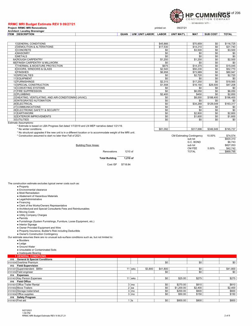
RRMC MRI Budget Estimate REV 9 09/27/21Project: RRMC MRI Renovations printed on 09/27/21Architect: Lavalley Brensinger
ITEM DESCRIPTION QUAN U/M UNIT LABOR LABOR UNIT MAT'L MAT SUB COST TOTAL
1 GENERAL CONDITIONS $45,866 $70,859 $0 $116,7252 DEMOLITION & ALTERATIONS $17,530 $14,210 $0 $31,7403 CONCRETE $0 $3,500 $0 $3,5004 MASONRY $0 $0 $0 $05 METALS $0 $0 $0 $0
6A ROUGH CARPENTRY $1,250 $1,250 $0 $2,5006B FINISH CARPENTRY & MILLWORK $0 $0 $0 $0
7 THERMAL & MOISTURE PROTECTION $675 $14,370 $0 $15,0458 DOORS, WINDOWS & GLASS $2,540 $50,230 $0 $52,7709 FINISHES $6,958 $73,989 $0 $80,947
10 SPECIALTIES $0 $2,720 $0 $2,72011 EQUIPMENT $0 $0 $0 $012 FURNISHINGS $2,315 $17,250 $0 $19,56513 SPECIAL CONSTRUCTION $1,558 $16,150 $29,500 $47,20814 CONVEYING SYSTEMS $0 $0 $0 $021 FIRE SUPPRESSION $0 $6,050 $0 $6,05022 PLUMBING $2,400 $450 $0 $2,85023 HEATING, VENTILATING, AND AIR-CONDITIONING (HVAC) $0 $8,000 $188,400 $196,40025 INTEGRATED AUTOMATION $0 $0 $0 $026 ELECTRICAL $0 $34,268 $129,049 $163,31727 COMMUNICATIONS $0 $0 $0 $028 ELECTRONIC SAFETY & SECURITY $0 $0 $0 $031 EARTHWORK $0 $2,800 $0 $2,80032 EXTERIOR IMPROVEMENTS $0 $1,600 $0 $1,60033 UTILITIES $0 $0 $0 $0
Estimate Assumptions:* Estimate is based on LBA Progress Set dated 1/7/2019 and LN MEP narrative dated 1/21/19.* No winter conditions. $81,092 $317,696 $346,949 $745,737* No structural upgrades if the new unit is in a different location or to accommodate weight of the MRI unit.* Construction assumed to start no later than Fall of 2021. CM Estimating Contingency 10.00% $74,574
sub tot $820,310G.C. BOND $6,743
Building Floor Areas: sub tot $827,053CM FEE 5.00% $42,742
Renovations 1210 sf TOTAL $869,795
Total Building 1,210 sf
Cost /SF $718.84
The construction estimate excludes typical owner costs such as:● Property● Environmental clearance● Mold Remediation● Abatement of Hazardous Materials● Legal/Administrative● Financing● Clerk of the Works/Owners Representative● Architectural and Special Consultants Fees and Reimbursables● Moving Costs● Utility Company Charges● Permits● Furnishings (System Furnishings, Furniture, Loose Equipment, etc.)● Interior Signage ● Owner Provided Equipment and Wire● Property Insurance, Builder's Risk including Deductible● Owner's Construction Contingency
Our estimate assumes there are no unusual sub-surface conditions such as, but not limited to:● Boulders● Ledge● Ground Water● Unsuitable or Contaminated Soils● Inadequate Bearing
1 GENERAL CONDITIONS
010 General & Special Conditions
0101004 Overtime Premium $0 $0 $0012 Field Supervision
0101201 Superintendent $95hr 11 wks $3,800 $41,800 $0 $41,8000101202 Field engineer $0 $0 $0
014 Expenses
0101401 Key Person Expenses 11 wks $0 $25.00 $275 $275016 Field Office
0101601 Office Trailer Rental 3 mo $0 $270.00 $810 $8100101602 2 ea $0 $1,200.00 $2,400 $2,4000101603 Storage trailer/shed 3 mo $0 $200.00 $600 $6000101604 Office supplies 3 mo $0 $50.00 $150 $150
018 Safety Program
0101801 1 ls $0 $800.00 $800 $800
Move Trailers
First aid
9/27/20211:59 PMRRMC MRI Budget Estimate REV 9 09.27.21 2 of 8
Page 63 of 206

RRMC MRI Budget Estimate REV 9 09/27/21Project: RRMC MRI Renovations printed on 09/27/21Architect: Lavalley Brensinger
ITEM DESCRIPTION QUAN U/M UNIT LABOR LABOR UNIT MAT'L MAT SUB COST TOTAL
0101802 1 ls $0 $500.00 $500 $5000101803 Safety Coordinator $75/hr 2hr/wk 11 wks $0 $150.00 $1,650 $1,6500101805 Protective Equipment 1 ls $0 $1,500.00 $1,500 $1,5000101807 Infect Control/Life Safety 11 wks $0 $200.00 $2,200 $2,200
022 Tools
0102202 11 wks $0 $25.00 $275 $2750102203 Equipment to Plant 3 mo $0 $50.00 $150 $1500102204 Rental, Company TBD $0 $0 $00102205 Rental, Non-Company 3 mo $0 $2,000.00 $6,000 $6,0000102206 Pick-Up Trucks 3 mo $0 $600.00 $1,800 $1,8000102207 11 wks $0 $100.00 $1,100 $1,1000102208 1 ls $0 $1,500.00 $1,500 $1,5000102209 Lifts for Trades $0 $0 $0
041 Project Management
0104101 Senior Project Manager $134.4 x 2 hrs p/wk 11 wks $0 $268.80 $2,957 $2,9570104102 11 wks $0 $864.00 $9,504 $9,5040104103 Admin Travel Expense 11 wks $0 $75.00 $825 $8250104104 Executive Management inc. in fee $0 $0 $00104105 Fee wks $0 $0 $00104106 Fee wks $0 $0 $00104107 PM 1 ls $0 $1,000.00 $1,000 $1,000
042 Scheduling
0104201 In House Scheduling inc. in fee $0 $0 $00104202 Scheduling Consultant ls $0 $0 $00104203 Maintain Schedules inc. in fee $0 $0 $0
043 Preconstruction Services
0104301 allow 1 ls $0 $4,500.00 $4,500 $4,5000104302 Reimbursables 1 ls $0 $500.00 $500 $500
045 Insurances/Taxes
0104501 State Sales Tax, EXEMPT 1 ls $0 $0 $00104503 by owner $0 $0 $00104504 Owner Protective Liability by owner $0 $0 $00104505 Bridge B.R. Deduct ls $0 $0 $00104506 Other Insurance ls $0 $0 $0
065 Permits
0106502 1 ls $0 $6,000.00 $6,000 $6,0000106503 State /Fire Safety 1 ls $0 $12,000.00 $12,000 $12,000
330 Survey & Layout
0133003 Engineer Layout 1 ls $0 $0 $0335 Protect/Repair Grounds
0133504 Maintain Egress 1 ls $0 $1,500.00 $1,500 $1,5000133505 1 ls $0 $1,000.00 $1,000 $1,000
510 Temporary Utilities
0151002 by owner $0 $0 $00151003 Temp Lights & Wire by subs $0 $0 $0
520 Winter Conditions
0152001 Temp heat/fuel assume none needed $0 $0 $00152002 Temp heat equipment assume none needed $0 $0 $00152003 Winter weather shelter assume none needed $0 $0 $00152004 Snow removal assume none needed $0 $0 $0
525 Construction Aids
0152507 Temp water 3 mo $0 $75.00 $225 $2250152508 Temp Toilets & Wash 3 mo $0 $125.00 $375 $375
530 Barriers and Enclosures
0153001 Temp Laydown area 1 ls $0 $1,500 $1,500 $1,5000153002 Temp fencing 50 lf $0 $4.00 $200 $2000153003 Temp Barricades 1 ls $0 $500.00 $500 $500
540 Security
0154001 $0 $0 $00154002 Security Systems $0 $0 $0
560 Quality Control/Testing
0156001 Test Soils/Concrete none 1 ls $0 $0 $00156002 Test Steel/Fire none 1 ls $0 $0 $00156003 Assist with Testing none $0 $0 $00156004 IBC Testing none 1 ls $0 $0 $0
565 Temporary Fire Protection
0156501 Temp fire extinguishers 1 ea $0 $200.00 $200 $200580 Project Identification
0158001 $0 $0 $0660 Testing Systems
0166001 Commissioning none $0 $0 $00166002 Operational Testing $0 $0 $0
710 Cleaning
0171001 1 ls $0 $0 $00171002 3 mo $0 $950.00 $2,850 $2,8500171005 Progress Clean $46.2 x 8 hrs p/wk 11 wks $369.60 $4,066 $0 $4,0660171007 Final Clean Building 1210 sf $0 $1.25 $1,513 $1,513
720 Project Documents
0172001 Document Print & Distrib 1 ls $0 $500.00 $500 $5000172002 $0 $0 $00172003 As-Built Drawings 1 ls $0 $500.00 $500 $5000172004 Coordination Drawings $0 $0 $00172007 CAD Record Files $0 $0 $0
Temp Power Consump
Watchman
Project Sign
Recycle PlanDumpsters
Photos
Zoning/Local
Temp Signage
Small Tools
Company GasTool Repair
Owner Safety
Project Manager $108 x 8 hrs p/wk
Accounting Clerical
Fee
Procore Subscription
Builders Risk
9/27/20211:59 PMRRMC MRI Budget Estimate REV 9 09.27.21 3 of 8
Page 64 of 206

RRMC MRI Budget Estimate REV 9 09/27/21Project: RRMC MRI Renovations printed on 09/27/21Architect: Lavalley Brensinger
ITEM DESCRIPTION QUAN U/M UNIT LABOR LABOR UNIT MAT'L MAT SUB COST TOTAL
770 Project Closeout
0177001 $0 $0 $00177002 1 ls $0 $1,000.00 $1,000 $1,0000177003 Closeout Expenses $0 $0 $0
800 LEED Objectives
0180001 $0 $0 $0810 Allowances
0181001 $0 $0 $0820 Alternatives
0182001 $0 $0 $0830 Owner Furnish
0183001 $0 $0 $0840 Contractor Install
0184001 $0 $0 $0850 Project Management
0185001 Environmental Protection $0 $0 $00185002 Indoor Air Quality $0 $0 $00185003 Storm Water Control $0 $0 $00185004 Moisture & Mold Control $0 $0 $0
860 Supplemental GC's
0186001 $0 $2,500.00 $0 $00186002 $0 $0 $0
ITEM TOTAL $45,866 $70,859 $0 $116,725
2 DEMOLITION & ALTERATIONS
024100 Selective Demolition
Create opening to remove magnet 60 hr $62.30 $3,738 $0 $3,738$0 $0 $0
Temp provisions 2 ea $0 $3,000.00 $6,000 $6,000Mechanical/elect demo cut & patch 16 hrs $62.30 $997 $10.00 $160 $1,157Temp partitions 400 sf $10.00 $4,000 $3.00 $1,200 $5,200Rework existing 40 hr $62.30 $2,492 $10.00 $400 $2,892Walk-off mats 8 pads $31.15 $249 $80.00 $640 $889Negative air machines 11 wks $18.00 $198 $75.00 $825 $1,023Remove flooring 1210 sf $1.10 $1,331 $1.00 $1,210 $2,541Remove existing casework 25 lf $14.95 $374 $0 $374Remove existing walls 29 lf $25.00 $725 $0 $725Ceilings remove 675 sf $2.10 $1,418 $1.00 $675 $2,093HEPA filters 1 ls $250.00 $250 $1,000.00 $1,000 $1,250Carts 1 ea $0 $400.00 $400 $400Pressure indicator 1 ea $200.00 $200 $750.00 $750 $950Dumpsters 1 ea $0 $950.00 $950 $950Off hour work 50 hrs $31.15 $1,558 $0 $1,558
$0 $0 $0ITEM TOTAL $17,530 $14,210 $0 $31,740
3 CONCRETE
033000 Cast-In-Place Concrete $0 $0 $0Extend existing exterior chiller pads 1 ls $0 $3,500.00 $3,500 $3,500
$0 $0 $0ITEM TOTAL $0 $3,500 $0 $3,500
4 MASONRY
042000 Unit Masonry $0 $0 $0Subcontract Furnish & Install None 1 Bid $0 $0 $0
Assume existing masonry opening is large enough to allow removal of MRI unit $0 $0 $0$0 $0 $0
ITEM TOTAL $0 $0 $0 $0
5 METALS
051200 Structural Steel none $0 $0 $0054000 Cold-Formed Metal Framing none $0 $0 $0055000 Metal Fabrications none $0 $0 $0
$0 $0 $0ITEM TOTAL $0 $0 $0 $0
6A ROUGH CARPENTRY
061053 Miscellaneous Carpentry $0 $0 $0Exterior blocking none $0 $0 $0Interior blocking general 500 lf $2.50 $1,250 $2.50 $1,250 $2,500
$0 $0 $0ITEM TOTAL $1,250 $1,250 $0 $2,500
6B FINISH CARPENTRY & MILLWORK
062000 Finish Carpentry & Architectural Woodwork SEE 123600 $0 $0 $0$0 $0 $0
ITEM TOTAL $0 $0 $0 $0
General
WarrantiesO&M Manuals
General
General
General
Punch ListTraining
Interior Demo - Renovation/Add.
General
9/27/20211:59 PMRRMC MRI Budget Estimate REV 9 09.27.21 4 of 8
Page 65 of 206

RRMC MRI Budget Estimate REV 9 09/27/21Project: RRMC MRI Renovations printed on 09/27/21Architect: Lavalley Brensinger
ITEM DESCRIPTION QUAN U/M UNIT LABOR LABOR UNIT MAT'L MAT SUB COST TOTAL
7 THERMAL & MOISTURE PROTECTION
071113 Bituminous Damp-proofing not required $0 $0 $0$0 $0 $0
072100 Thermal Insulation at Exterior Wall refer to 072415 $0 $0 $0$0 $0 $0
072110 Acoustic Insulation $0 $0 $0Thermafiber - interior partitions SEE 092500 1700 sf $0 $0 $0R-21 - exterior wall 100 sf $1.25 $125 $2.00 $200 $325
$0 $0 $0072415 EIFS $0 $0 $0
Patch existing 100 sf $0 $50.00 $5,000 $5,000$0 $0 $0
072500 Weather Barriers $0 $0 $0Patch in at existing wall patch 100 sf $0 $10.00 $1,000 $1,000
$0 $0 $0075300 Elastomeric Membrane Roofing $0 $0 $0
Patch at existing 1 ls $0 $5,000.00 $5,000 $5,000$0 $0 $0
076200 Sheet Metal Flashing & Trim $0 $0 $0Aluminum flashing @ roof to wall tie in 88 lf $0 $15.00 $1,320 $1,320Continue drip groove 97 lf $0 $0 $0Drip Edge 97 lf $0 $0 $0
$0 $0 $0077200 Roof Accessories $0 $0 $0Subcontract Furnish & Install none $0 $0 $0
$0 $0 $0078110 Sprayed Fire-Resistive Materials $0 $0 $0
Patch Existing 1 ls $0 $1,200.00 $1,200 $1,200$0 $0 $0
078413 Fire-stopping $0 $0 $0Misc not by trades 1 ls $300.00 $300 $300.00 $300 $600
$0 $0 $0079200 Joint Sealers $0 $0 $0
Interior Joint 1 ls $250.00 $250 $150.00 $150 $400Exterior Joint 50 lf $0 $4.00 $200 $200
$0 $0 $0ITEM TOTAL $675 $14,370 $0 $15,045
8 DOORS, WINDOWS & GLASS
081100 Steel Door Frames $0 $0 $0Door Supplier 1 Bid $0 $0 $0Unload, sort, store & protect ls $0 $0 $0
F1 3'-0" x 7'-0" 3 ea $125.00 $375 $800.00 $2,400 $2,775F2 6'-0" x 7'-0" 1 ea $125.00 $125 $1,200.00 $1,200 $1,325
$0 $0 $0082110 Flush Wood Doors $0 $0 $0
D1 3'-0" x 7'-0" 5 ea $174.00 $870 $1,000.00 $5,000 $5,870$0 $0 $0
083100 Access Doors & Frames $0 $0 $018 x 18" clg access 3 ea $100.00 $300 $200.00 $600 $900
$0 $0 $0084115 Exterior Thermally-Broken Aluminum Windows $0 $0 $0
Replace existing round window 12 sf $0 $650.00 $7,800 $7,800$0 $0 $0
084300 Interior sliding glass partition $0 $0 $0Sliding glass wall 184 sf $0 $100.00 $18,400 $18,400
$0 $0 $0087100 Door Hardware $0 $0 $0
Door Openings (leafs) 5 ea $174.00 $870 $350.00 $1,750 $2,620Access control - rework existing 1 ls $0 $6,000.00 $6,000 $6,000Power swing operators replace existing 2 ea $0 $3,400.00 $6,800 $6,800
$0 $0 $0088000 Glazing $0 $0 $0Glazing Sub $0 $0 $0
Interior door glazing 8 sf $0 $35.00 $280 $280$0 $0 $0
ITEM TOTAL $2,540 $50,230 $0 $52,770
9 FINISHES
090561 Common Work Results For Flooring Preparation $0 $0 $0Floor moisture & Ph testing 1210 sf $0 $0 $0Prep existing floor for new 1210 sf $0 $0 $0Moisture mitigation allow 1210 sf $0 $6.00 $7,260 $7,260
$0 $0 $0092600 Gypsum Board Assemblies $0 $0 $0Gypsum Assemblies Sub $0 $0 $0Exterior Wall System infill existing sf $0 $0 $0
2" Rigid cavity insulation 150 sf $0 $3.00 $450 $4505/8" exterior GWB 150 sf $0 $2.25 $338 $338Framing 150 sf $0 $6.50 $975 $9755 1/2" R-21 150 sf $0 $1.00 $150 $1506 mil poly vapor barrier 150 sf $0 $0.50 $75 $755/8" Gyp wallboard 150 sf $0 $2.25 $338 $338
9/27/20211:59 PMRRMC MRI Budget Estimate REV 9 09.27.21 5 of 8
Page 66 of 206

RRMC MRI Budget Estimate REV 9 09/27/21Project: RRMC MRI Renovations printed on 09/27/21Architect: Lavalley Brensinger
ITEM DESCRIPTION QUAN U/M UNIT LABOR LABOR UNIT MAT'L MAT SUB COST TOTAL
Partition 64 sf $0 $25.00 $1,600 $1,600New closet walls 100 sf $0 $28.00 $2,800 $2,800New Changing area walls 400 sf $0 $25.00 $10,000 $10,000Replace interior exterior wall for magnet removal 150 sf $0 $20.00 $3,000 $3,000Rework soffit for sliders 28 lf $0 $100.00 $2,800 $2,800New soffit for utilities 85 sf $0 $100.00 $8,500 $8,500Patching at existing 1 ls $0 $2,500.00 $2,500 $2,500Remove and replace drywall 200 sf $0 $3.00 $600 $600
$0 $0 $0095100 Acoustical Ceilings $0 $0 $0Ceilings Sub $0 $0 $0
ACT-1 2x2 Armstrong 675 sf $0 $10.00 $6,750 $6,750$0 $0 $0
096500 Resilient Flooring $0 $0 $0Flooring Sub $0 $0 $0
LVT 675 sf $0 $12.00 $8,100 $8,100LVT static guard at magnet room 535 sf $0 $14.00 $7,490 $7,490Protect floors 1210 sf $0 $1.50 $1,815 $1,815Floor prep 1210 sf $0.75 $908 $0.75 $908 $1,816Patch existing floor 1 ls $250.00 $250 $200.00 $200 $450Protect existing flooring to remain 200 sf $1.00 $200 $1.00 $200 $400
$0 $0 $0097100 Access Floor none $0 $0 $0
$0 $0 $0097500 Acoustic Roof Components none $0 $0 $0
$0 $0 $0096800 Carpeting refer to 096500 $0 $0 $0
$0 $0 $0099123 Painting and Coating $0 $0 $0Painting Sub $0 $0 $0
Interior painting (Per Finish Schedule) 1210 sf $0 $4.00 $4,840 $4,840Artist painting on MRI room ceiling, labor 80 hrs $70.00 $5,600 $0 $5,600Artist painting on MRI room ceiling, material 1 ls $0 $1,500.00 $1,500 $1,500Staging for artist 1 ls $0 $800.00 $800 $800
ITEM TOTAL $6,958 $73,989 $0 $80,947
10 SPECIALTIES
101425 Code Required Building Signage by owner $0 $0 $0$0 $0 $0
102123 Cubicles none $0 $0 $0$0 $0 $0
102600 Wall & Corner Protection $0 $0 $0Wall Protection Sub 1 Bid $0 $0 $0
IRPP on Walls 272 sf $0 $10.00 $2,720 $2,720Corner guard 4' 5 ea $0 $0 $0
$0 $0 $0108010 Toilet & Healthcare Accessories none $0 $0 $0
$0 $0 $0104413 Fire Protection Specialties $0 $0 $0
Fire Extinguisher cab & ext re-use existing $0 $0 $0$0 $0 $0
ITEM TOTAL $0 $2,720 $0 $2,720
11 EQUIPMENT
117713 MRI Ferromagnetic Detection System existing to remain $0 $0 $0$0 $0 $0
ITEM TOTAL $0 $0 $0 $0
12 FURNISHINGS
123600 Casework & Counter-tops $0 $0 $0Casework Sub $0 $0 $0
Cabinet/hamper in Changing 1 ea $0 $0 $0Control Room $0 $0 $0
SS Counter-top for GE item #27 9 sf $35.00 $315 $300.00 $2,700 $3,015Equipment room $0 $0 $0
Shelf for GE item #31 1 ea $30.00 $30 $200.00 $200 $230MRI room $0 $0 $0
Base cabinet 12 lf $35.00 $420 $450.00 $5,400 $5,820Wall cabinet 6 lf $35.00 $210 $400.00 $2,400 $2,610SS Counter-top 24 sf $35.00 $840 $250.00 $6,000 $6,840Misc. Millwork 1 ls $500.00 $500 $550.00 $550 $1,050
$0 $0 $0ITEM TOTAL $2,315 $17,250 $0 $19,565
13 SPECIAL CONSTRUCTION
134700 MRI Support $0 $0 $0Install vibroacoustic dampening kit, GE item #1 OFCI 4 ea $249.20 $997 $0 $997Install magnet curtain kit, GE item #2 OFCI 1 ea $373.80 $374 $0 $374Patient table dock anchoring allow 1 ea $186.90 $187 $150 $150 $337
$0 $0 $0134713 Temp MRI Pad $0 $0 $0
Existing Concrete pad to be reused $0 $0 $0
9/27/20211:59 PMRRMC MRI Budget Estimate REV 9 09.27.21 6 of 8
Page 67 of 206

RRMC MRI Budget Estimate REV 9 09/27/21Project: RRMC MRI Renovations printed on 09/27/21Architect: Lavalley Brensinger
ITEM DESCRIPTION QUAN U/M UNIT LABOR LABOR UNIT MAT'L MAT SUB COST TOTAL
Temp awning in place to be reused, allow to alter allow 1 ls $0 $4,000 $4,000 $4,000Remove existing awning when complete allow 1 ls $0 $4,000 $4,000 $4,000
$0 $0 $0134941 RF Shielding $0 $0 $0Shielding Sub Gavin 1 budget $0 $0 $29,500 $29,500
RF enclosure rework $0 $0 $0New filters and testing $0 $0 $0Allow to repair existing 1 ea $0 $8,000 $8,000 $8,000HVAC Wave Guides 0 ea $0 $0 $0Plumbing Wave Guides 0 ea $0 $0 $0RF Electrical Filters 0 ea $0 $0 $0RF Testing Services 0 sf $0 $0 $0Mag shield enclosure plywood 0 sf $0 $0 $0
$0 $0 $0ITEM TOTAL $1,558 $16,150 $29,500 $47,208
14 CONVEYING SYSTEMS
not used none $0 $0 $0$0 $0 $0
ITEM TOTAL $0 $0 $0 $0
21 FIRE SUPPRESSION
210000 Fire Protection $0 $0 $0Fire Protection Sub $0 $0 $0
Rework existing 1210 sf $0 $5.00 $6,050 $6,050Pre-action system Existing to remain 1 ls $0 $0 $0
$0 $0 $0ITEM TOTAL $0 $6,050 $0 $6,050
22 PLUMBING
220000 Plumbing $0 $0 $0Plumbing Sub SEE 230000 1 Bid $0 $0 $0
Rework existing city water back up system 30 hrs $80.00 $2,400 $15 $450 $2,850Med gas outlets, O,V,A,S 0 ea $0 $0 $0Water for equipment room 0 ls $0 $0 $0Water for Chiller 0 ls $0 $0 $0Water to temp MRI 0 lf $0 $0 $0
$0 $0 $0ITEM TOTAL $2,400 $450 $0 $2,850
23 HEATING, VENTILATING, AND AIR-CONDITIONING (HVAC)
230000 HVAC $0 $0 $0Mechanical Sub NEAS 1 budget $0 $0 $188,400 $188,400
New VAV at Changing in above $0 $0 $0New return VAVs in above $0 $0 $0
in above $0 $0 $0
Valves in above $0 $0 $0in above $0 $0 $0
in above $0 $0 $0
Hoist in above $0 $0 $0Rework Quench vent in above $0 $0 $0TAB in above $0 $0 $0HVAC Controls in above $0 $0 $0Temporary provisions in above $0 $0 $0Unforeseen conditions allow 1 ls $0 $8,000 $8,000 $8,000
Exclusions: $0 $0 $0MRI AHU (existing to remain) $0 $0 $0MRI SA VAV's (existing to remain) $0 $0 $0AC Split Units at computer room (existing to remain) $0 $0 $0
$0 $0 $0ITEM TOTAL $0 $8,000 $188,400 $196,400
25 INTEGRATED AUTOMATION
250000 HVAC Instrumentation and Controls refer to 230000 $0 $0 $0$0 $0 $0
ITEM TOTAL $0 $0 $0 $0
26 ELECTRICAL
260000 Electrical $0 $0 $0Electrical Sub Interstate 1 budget $0 $0 $129,049 $129,049
New electrical in above 1210 sf $0 $0 $0Temporary provisions in above 1 ls $0 $0 $0Ceiling scene lighting allow 1 ls $0 $25,000.00 $25,000 $25,000Cable ladder install in above budget 68 lf $0 $26.00 $1,768 $1,768Temporary MRI Trailer Support (lights & power) 1 ls $0 $5,000.00 $5,000 $5,000Unforeseen conditions allow 1 ls $0 $2,500 $2,500 $2,500
$0 $0 $0ITEM TOTAL $0 $34,268 $129,049 $163,317
New 3" Chilled water lines from south mech rm
Remove existing Computer room chiller replace with new provided by ownerRemove existing MRI chiller replace with new provided by owner
9/27/20211:59 PMRRMC MRI Budget Estimate REV 9 09.27.21 7 of 8
Page 68 of 206

RRMC MRI Budget Estimate REV 9 09/27/21Project: RRMC MRI Renovations printed on 09/27/21Architect: Lavalley Brensinger
ITEM DESCRIPTION QUAN U/M UNIT LABOR LABOR UNIT MAT'L MAT SUB COST TOTAL
27 COMMUNICATIONS
270000 Communications $0 $0 $0Subcontract Furnish & Install $0 $0 $0
New data/Phone refer to 260000 $0 $0 $0Nurse call refer to 260000 $0 $0 $0
$0 $0 $0ITEM TOTAL $0 $0 $0 $0
28 ELECTRONIC SAFETY & SECURITY
283100 Fire Detection and Alarm $0 $0 $0Rework existing refer to 260000 1210 sf $0 $0 $0
$0 $0 $0ITEM TOTAL $0 $0 $0 $0
31 EARTHWORK
310000 Earthwork $0 $0 $0Earthwork Sub $0 $0 $0
Extend chiller pad 1 ls $0 $2,800.00 $2,800 $2,800$0 $0 $0
ITEM TOTAL $0 $2,800 $0 $2,800
32 EXTERIOR IMPROVEMENTS
320000 Exterior Improvements $0 $0 $0Landscaping repair 1 ls $0 $1,600.00 $1,600 $1,600
$0 $0 $0ITEM TOTAL $0 $1,600 $0 $1,600
33 UTILITIES
330000 Utilities none $0 $0 $0$0 $0 $0
ITEM TOTAL $0 $0 $0 $0
9/27/20211:59 PMRRMC MRI Budget Estimate REV 9 09.27.21 8 of 8
Page 69 of 206

January 21, 2019 Joe Britton Lavallee Brensinger Architects 155 Dow Street Manchester, NH 03101 Rutland Regional Medical Center – MRI Renovation MEP Narrative L.N. Consulting, Inc. has been retained by Rutland Regional Medical Center to provide a MEP narrative for the renovation of the existing MRI suite at Rutland Regional Medical Center located in Rutland, Vermont. Mechanical: The existing MRI Suite is primarily conditioned via a 4,220 CFM air handling unit located in the ground floor MRI mechanical room. The unit provides air through (3) supply air variable air volume boxes with hot water re-heat coils which provide air to the MRI machine room, holding room, and control room. The existing MRI air handling unit is supported by a 20 ton chiller located outside to the south of the MRI Suite. The existing MRI computer room is conditioned via a dedicated chiller and (2) 6 ton computer room air conditioned units. The existing air handling unit shall be reused. (1) new supply variable air volume boxes with hot water reheat coil shall be provided to support the new changing room. The changing room temperature control zone shall be sized for 250 CFM. (3) New return air VAV’s shall be provided for each temperature control zone. The MRI air handling unit and associated pumps, terminal units, and sensors shall be upgraded to current direct digital control technology. Provide new set of 3” chilled water lines from the existing 6” chilled water mains located in the southern mechanical room. The new chilled water lines shall tie into the existing MRI air handling unit chilled water loop and shall be provided with isolation valves in order to allow for switchover between the MRI air handling unit chiller and the central chilled water plant. The existing MRI computer room chiller shall be demolished and provided with the new GE provided chiller. Expand equipment pad at exterior to accommodate new chiller. Existing 2” chilled water lines shall be reused. The existing city water back-up system shall be reused. Tie the existing 2” chilled water lines into heat exchanger cabinet. Provide direct digital controls interconnection into GE provided chiller.
Page 70 of 206

Electrical: The existing electrical service supporting the MRI is fed from the Normal Power Substation SS103 located in the North Electrical Room. The design documents indicate that the existing MRI main disconnect switch is fed from 200A fuses from Substation SS103 with (3) 4/0 Conductors, (1) #4/0 Neutral, and (1) #4/0 Ground in a 2 ½” Conduit. Conductors and fuses shall be verified in the field. If they are consistent with sizes indicated above, then the existing power feed for the new MRI can be re-used. The 200A main distribution breaker shall feed a new 150A-3P breaker supporting the PDU circuit and a new 50A-3P breaker supporting the HEC circuit. (2) EPO buttons shall be provided to disable MDP circuit. Provide 480V-3P-30A emergency power circuit to Cryrogen Compressor. Provide 120V-20A emergency power circuit to Magnon3. The new MRI chiller shall be fed from a new 80A-3P breaker in Panel H166 located in the MRI mechanical room. Contractor shall provide ¾” conduit with pull string from new chiller location to MRI operators work station. Provide power, conduit, cabling, and cable tray as required per GE MRI equipment drawings. Provide (1) patient pull cord within changing room and (1) nurse call dome light outside of changing room. Provide general receptacle within changing room. Provide fire alarm strobe within changing room. All lighting in renovated areas shall be replaced with new LED light fixtures and controls. Please let us know if you have any questions or comments. Sincerely, L.N. Consulting, Inc. George D. Martin III, P.E.
GE will notallow aseparatecircuit forthiscompressor.
Page 71 of 206
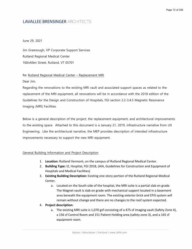
Boston | Manchester | Portland | www.LBPA.com
June 29, 2021
Jim Greenough, VP Corporate Support Services
Rutland Regional Medical Center
160nAllen Street, Rutland, VT 05701
Re: Rutland Regional Medical Center – Replacement MRI
Dear Jim,
Regarding the renovations to the existing MRI vault and associated support spaces as related to the
replacement of the MRI equipment, all renovations will be in accordance with the 2018 edition of the
Guidelines for the Design and Construction of Hospitals, FGI section 2.2-3.4.5 Magnetic Resonance
Imaging (MRI) Facilities.
Below is a general description of the project, the replacement equipment, and architectural improvements
to the existing space. Attached to this document is a January 21, 2019, infrastructure narrative from LN
Engineering. Like the architectural narrative, the MEP provides description of intended infrastructure
improvements necessary to support the new MRI equipment.
General Building Information and Project Description
1. Location: Rutland Vermont, on the campus of Rutland Regional Medical Center.
2. Building Type: I2, Hospital, FGI 2018, (AIA, Guidelines for Construction and Equipment of
Hospitals and Medical Facilities)
3. Existing Building Description: Existing one-story portion of the Rutland Regional Medical
Center.
a. Located on the South side of the hospital, the MRI suite is a partial slab on grade.
The Magnet vault is slab on grade with mechanical support located in a basement
area beneath the equipment room. The existing exterior brick and EFIS system will
remain without change and there are no changes to the roof system expected.
4. Project description:
a. The existing MRI suite is 1,070 gsf consisting of a 475-sf imaging vault (Safety Zone 4),
a 156 sf Control Room and 151 Patient Holding area (safety zone 3), and a 165 sf
equipment room.
Page 72 of 206

June 2021 Page 2 of 3
Lavallee Brensinger Architects
b. Patients enter from a public corridor (safety zone 1) directly into the Patient Holding
area (safety zone 3) with access control and ferromagnetic detection at the corridor
door. There currently is no distinct Patients Safety Zone 2.
c. The project includes the following;
i. Replacing the existing 1.5T MRI with a new 1.5T MRI.
ii. Interior vault upgrades to accommodate the new MRI.
iii. Interior renovations to the equipment room to accommodate the new
support equipment for the MRI.
iv. Cosmetic renovations to the Control room and Patient Holding area.
v. Renovations to approximately 160 sf to create new ADA compliant Patient
Changing booths and interview area (patient safety zone 2) with access
directly off the public corridor (patient safety zone 1).
5. Building Systems, Components and Materials:
a. Building Structure
i. The project does not include any expansion or modification to the existing
building structure.
b. Architecture
i. Exterior envelope;
1. The project does not include any expansion or modification to the
existing exterior envelope.
ii. Interior renovations;
1. Walls in the new Patient Safety Zone 2 will bel S-11, 3 5/8” metal
studs, acoustical insulation full height, 1-layer 5/8” GWB per side, all
walls are full height deck to deck.
2. Doors will be solid core, natural finish hard wood. Frames to be
hollow metal welded. Hardware to be heavy duty mortise lock and
latch sets. Door sizes will be 3’0x7’0 for non-patient access spaces
and 3’6x7’0 for all spaces to meet ADA access.
3. Floor finishes to vinyl sheet, plank or tile products in Zones 1,2,3 and
4 and the equipment room.
4. Wall finishes in general will be paint with some vinyl wall protection
on all walls, up to 4 feet from the top of wall base.
5. Ceilings in finished rooms will be typically 2x2 acoustical ceilings with
15/16 grid. Unfinished spaces such as the Equipment Room may not
have a finished ceiling. The MRI vault ceiling will remain as is with
minor alterations to accommodate venting changes.
6. All millwork will be custom built with plastic laminate finishes and
vinyl edges.
7. Ferromagnetic detection and warning systems will be installed at the
door between Patient Safety Zones 2 and 3, 3 and 4, as well as zones
1 and 3 (the in-patient access).
Page 73 of 206

June 2021 Page 3 of 3
Lavallee Brensinger Architects
Joseph F. Britton RA., Vice President
Lavallee Brensinger Architects
Page 74 of 206
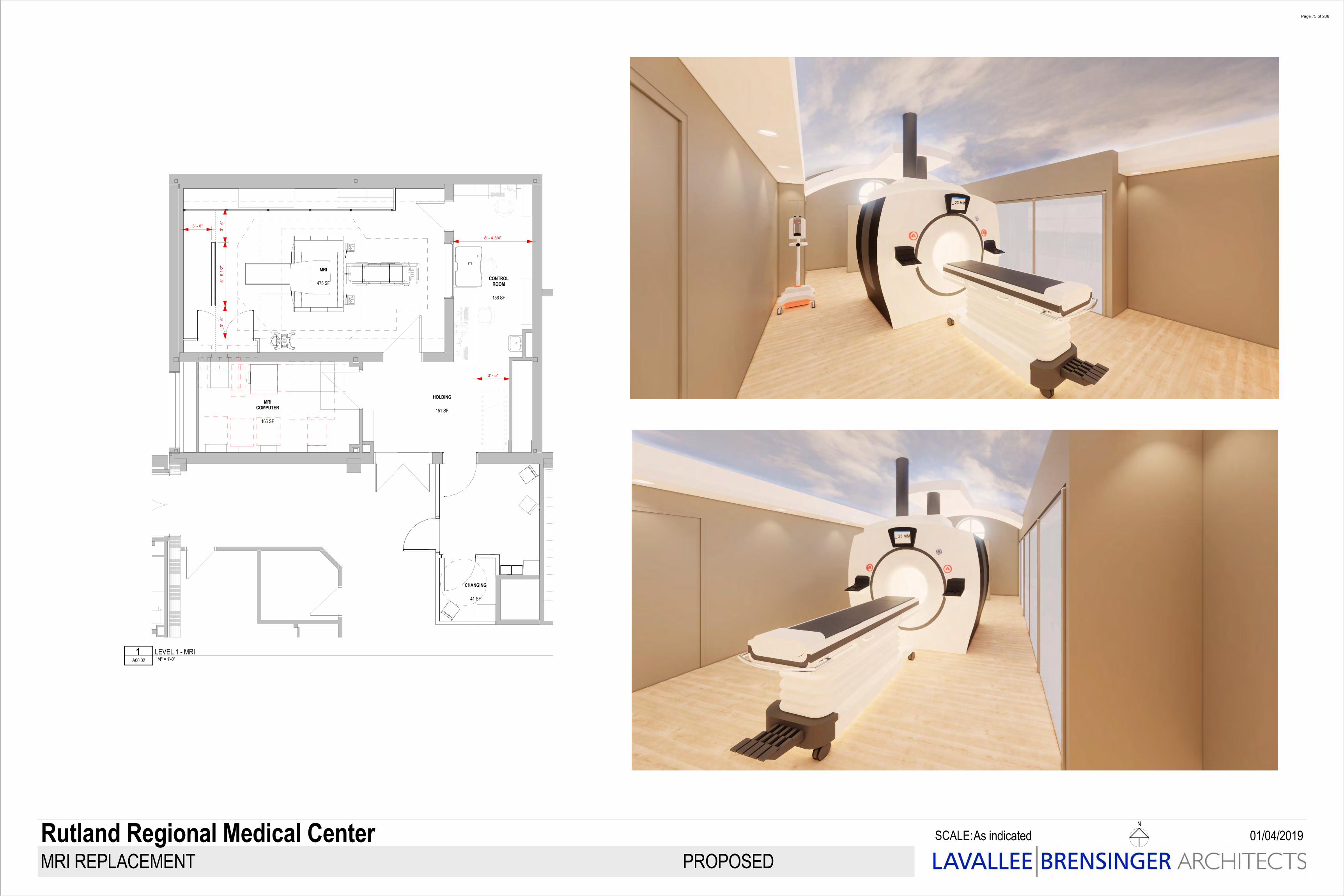
3' - 0"6'
- 9
1/2"
156 SF
CONTROLROOM
165 SF
MRICOMPUTER
151 SF
HOLDING
475 SF
MRI
8' - 4 3/4"
3' - 5"
3' -
6"3'
- 6"
SCALE:
N
As indicated
MRI REPLACEMENT
01/04/2019Rutland Regional Medical CenterPROPOSED
1/4" = 1'-0" A00.02
1 LEVEL 1 - MRI
41 SF
CHANGING
Page 75 of 206

Page 76 of 206

SCALE:
N
As indicated
MRI REPLACEMENT
Rutland Regional Medical CenterPROPOSED
35 SF
CHANGING
41 SF
CHANGING
74 SF
PREP
156 SF
CONTROLROOM
165 SF
MRICOMPUTER
151 SF
HOLDING
475 SF
MRI
Page 77 of 206
joe.britton
Area Measurement
SAFETY ZONE 4
joe.britton
Area Measurement
SAFETY ZONE 3
joe.britton
Area Measurement
SAFETY ZONE 2
joe.britton
Area Measurement
SAFETY ZONE 1
joe.britton
Snapshot
joe.britton
Area Measurement
SAFETY ZONE 4
joe.britton
Area Measurement
SAFETY ZONE 3
joe.britton
Area Measurement
SAFETY ZONE 1
joe.britton
tEXT
EXISTING MRI SAFETY ZONES
joe.britton
Snapshot
joe.britton
tEXT
PROPOSED MRI SAFETY ZONES
joe.britton
Snapshot
joe.britton
tEXT
MRI SAFETY ZONES
joe.britton
tEXT
09/06/2021
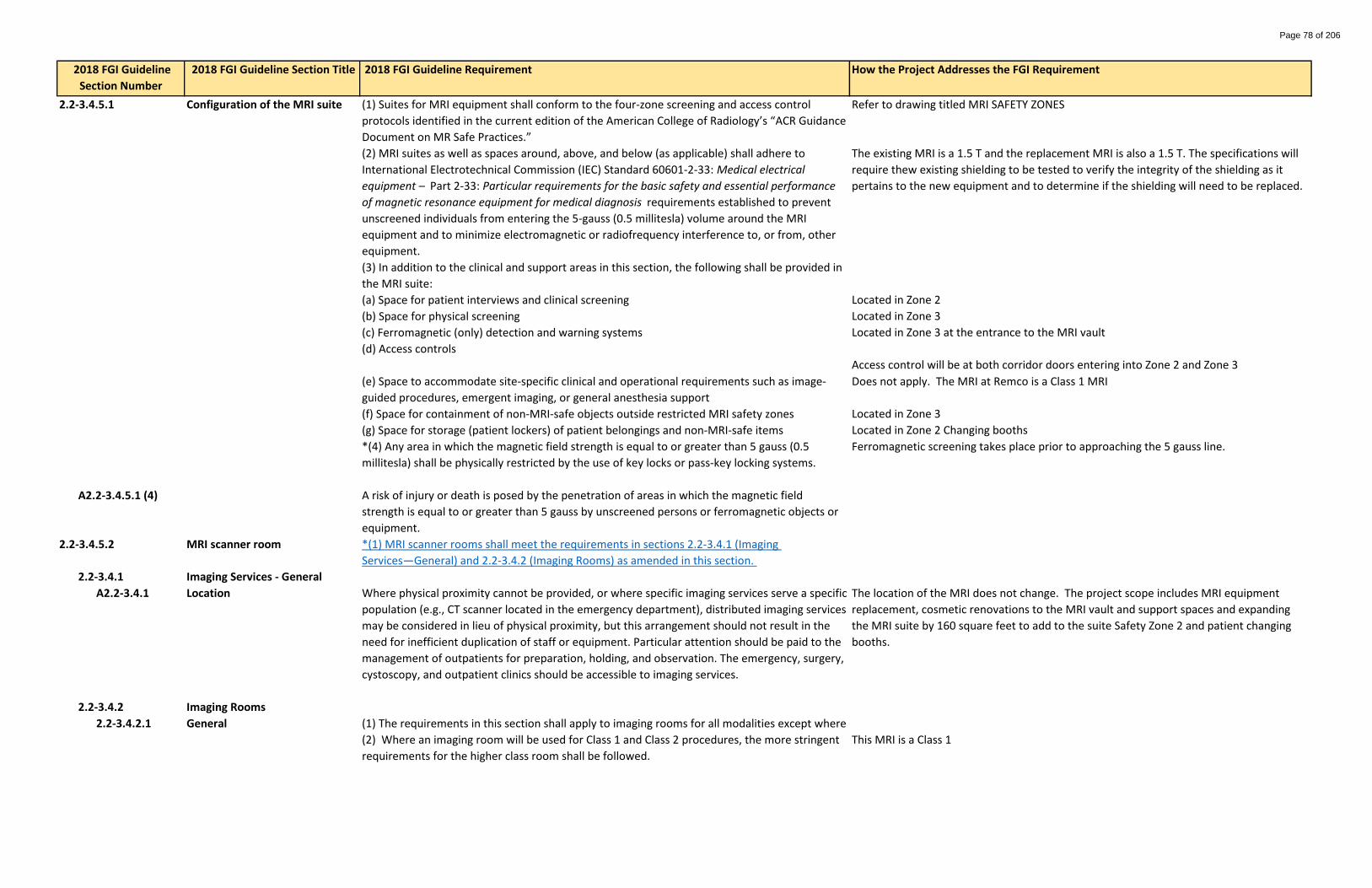
2018 FGI Guideline Section Title How the Project Addresses the FGI Requirement
2.2-3.4.5.1 Configuration of the MRI suite Refer to drawing titled MRI SAFETY ZONES
The existing MRI is a 1.5 T and the replacement MRI is also a 1.5 T. The specifications will
require thew existing shielding to be tested to verify the integrity of the shielding as it
pertains to the new equipment and to determine if the shielding will need to be replaced.
Located in Zone 2
Located in Zone 3
Located in Zone 3 at the entrance to the MRI vault
Access control will be at both corridor doors entering into Zone 2 and Zone 3
Does not apply. The MRI at Remco is a Class 1 MRI
Located in Zone 3
Located in Zone 2 Changing booths
Ferromagnetic screening takes place prior to approaching the 5 gauss line.
A2.2-3.4.5.1 (4)
2.2-3.4.5.2 MRI scanner room
2.2-3.4.1 Imaging Services - General
A2.2-3.4.1 Location The location of the MRI does not change. The project scope includes MRI equipment
replacement, cosmetic renovations to the MRI vault and support spaces and expanding
the MRI suite by 160 square feet to add to the suite Safety Zone 2 and patient changing
booths.
2.2-3.4.2 Imaging Rooms
2.2-3.4.2.1 General
This MRI is a Class 1
Where physical proximity cannot be provided, or where specific imaging services serve a specific
population (e.g., CT scanner located in the emergency department), distributed imaging services
may be considered in lieu of physical proximity, but this arrangement should not result in the
need for inefficient duplication of staff or equipment. Particular attention should be paid to the
management of outpatients for preparation, holding, and observation. The emergency, surgery,
cystoscopy, and outpatient clinics should be accessible to imaging services.
(1) The requirements in this section shall apply to imaging rooms for all modalities except where
(2) Where an imaging room will be used for Class 1 and Class 2 procedures, the more stringent
requirements for the higher class room shall be followed.
(f) Space for containment of non-MRI-safe objects outside restricted MRI safety zones
(g) Space for storage (patient lockers) of patient belongings and non-MRI-safe items
*(4) Any area in which the magnetic field strength is equal to or greater than 5 gauss (0.5
millitesla) shall be physically restricted by the use of key locks or pass-key locking systems.
A risk of injury or death is posed by the penetration of areas in which the magnetic field
strength is equal to or greater than 5 gauss by unscreened persons or ferromagnetic objects or
equipment.
*(1) MRI scanner rooms shall meet the requirements in sections 2.2-3.4.1 (Imaging
Services—General) and 2.2-3.4.2 (Imaging Rooms) as amended in this section.
2018 FGI Guideline
Section Number
(1) Suites for MRI equipment shall conform to the four-zone screening and access control
protocols identified in the current edition of the American College of Radiology’s “ACR Guidance
Document on MR Safe Practices.”
(2) MRI suites as well as spaces around, above, and below (as applicable) shall adhere to
International Electrotechnical Commission (IEC) Standard 60601-2-33: Medical electrical
equipment – Part 2-33: Particular requirements for the basic safety and essential performance
of magnetic resonance equipment for medical diagnosis requirements established to prevent
unscreened individuals from entering the 5-gauss (0.5 millitesla) volume around the MRI
equipment and to minimize electromagnetic or radiofrequency interference to, or from, other
equipment.
(3) In addition to the clinical and support areas in this section, the following shall be provided in
the MRI suite:
(e) Space to accommodate site-specific clinical and operational requirements such as image-
guided procedures, emergent imaging, or general anesthesia support
(a) Space for patient interviews and clinical screening
(b) Space for physical screening
(c) Ferromagnetic (only) detection and warning systems
(d) Access controls
2018 FGI Guideline Requirement
Page 78 of 206

2018 FGI Guideline Section Title How the Project Addresses the FGI Requirement 2018 FGI Guideline
Section Number
2018 FGI Guideline Requirement
This MRI is a Class 1
* 2.2-3.4.2.2 Space requirements Space needs for the MRI vault, and Control space do not change with the replacement
equipment. Modifications to the Equipment Room have been coordinated with the GE the
MRI manufacturer.
A2.2-3.4.2.2 The existing MRI vault will not have to be modified to accommodate the new MRI
equipment.
Existing MRI vault complies with the equipment manufacture's service clearances.
Patient access exceeds the 4 foot min. clearance. Refer to the floor plan.
Clinical access has been provided, refer to the floor plan.
A2.2-3.4.5.2 (1) Anesthesia support is not required.
Handwashing sink is located directly outside of the vault adjacent to the Control space.
2.2-3.4.2.3 Hand-washing station or hand
scrub facilities. MRI sink meets requirements noted in section 2.1-2.8.7
See above for sink location
Does not apply.
Does not apply.
Does not apply.
Does not apply.
(2) Hand-washing station
(a) A hand-washing station that meets the requirements in Section 2.2-3.4.2.3 (Hand-washing
station or hand scrub facilities) shall be provided.
(b) Location of the hand-washing station directly outside the entrance to the MRI scanner room
shall be permitted.
Hand-washing stations and hand scrub facilities shall comply with the requirements in Section
2.1-2.8.7 (Hand-Washing Station) and Section 2.1-2.8.6 (Hand Scrub Facilities).
(3) Hand scrub facilities shall be provided directly outside the entrance to Class 3 imaging
rooms.
If anesthesia support is anticipated, additional space, electrical outlets, and gas lines may be
required.
(1) A hand-washing station shall be provided in Class 1 imaging rooms unless specified otherwise
for a specific imaging modality.
(2) A hand-washing station or hand scrub facilities shall be provided for Class 2 imaging rooms.
(a) Where a hand-washing station is provided, it shall be directly accessible to the Class 2
imaging room.
(b) Where a hand scrub facilities are provided, a hand scrub position shall be directly outside the
entrance to the Class 2 imaging room.
Space layouts should be developed to meet the minimum requirements in the manufacturer’s
technical specifications because area requirements may vary from machine to machine.
However, manufacturers’ recommendations should not be used as the sole determinant for
room size as they may not take into consideration the clinical needs at a particular facility. As
well, because technology changes and siting requirements frequently vary from manufacturer
to manufacturer, rooms should be sized larger than manufacturer’s minimum technical
specifications to allow for upgrading of equipment over time. Consideration should also be
given for the space needs of other equipment (e.g., physiological monitoring or dye injectors).
(1) Imaging rooms shall be sized and configured, at minimum, to comply with the
manufacturer’s recommendations for installation, service, and maintenance.
(2) Imaging rooms shall be sized to provide the following minimum clearances:
(a) 4 feet (122 centimeters) on all circulating sides of the patient table/bed/couch, gantry, or
assembly
(b) Other clearances in accordance with clinical needs (e.g., medical gas service, anesthesia cart,
clinical staff)
(3) Where imaging procedures meeting Class 3 criteria are performed, a room(s) that meets the
requirements for the applicable imaging suite and for an operating room (see Section 2.2-3.3.3,
Operating Rooms) shall be provided. An operating room that meets the requirements in Section
2.2-3.3.4 (Hybrid Operating Room) shall be permitted to meet this requirement.
Space shall be provided to accommodate the equipment and staff needed for planned imaging
services.
Page 79 of 206

2018 FGI Guideline Section Title How the Project Addresses the FGI Requirement 2018 FGI Guideline
Section Number
2018 FGI Guideline Requirement
*2.2-3.4.5.3 Superconducting MRI cryogen
venting.
The cryogen venting system has been coordinated with he GE replacement MRI
equipment and complies with the manufacture's requirements.
A2.2-3.4.5.3 Cryogen venting
Complies
Complies
Complies
Complies
2.2-3.4.5.4 MRI control room. Complies with Section 2.2-3.4.1.3 (1)
Complies
Door is in swinging and does not block the control room window.
2.2-3.4.5.5 Control vestibule The control vestibule is Safety Zone 3
* 2.2-3.4.5.6 Patient treatment/resuscitation
area.
In the event of a patient code, the MRI table is detachable and the patient would be
wheeled out to Zone 3. One staff member would work to wheel the patient out while the
other MRI staff member guards the entrance to Zone 4 to ensure no unscreened staff or
equipment enters the MRI room.
(1) The control vestibule shall be located outside the MRI scanner room so that patients, health
care personnel, and other employees must pass through it before entering the MRI scanner
room.
(2) The control vestibule shall be permitted to be either a part of the MRI control room or
directly visible from the control room.
An area adjacent to the MRI room shall be provided for patient code treatment/resuscitation.
d. Cryogen venting points of discharge should be designed with weather head sufficient to
protect against the ingress of horizontally driven rain.
e. Accessible areas around cryogen vent discharge should be marked to indicate the safety
exclusion zone in accordance with MRI equipment manufacturer standards.
A control room that meets the requirements in Section 2.2-3.4.1.3 (1) (Shielded control alcove
or room) shall be provided as amended in this section.
(1) The operator’s console shall be positioned so the operator has a full view of the principal
approach and entrance to the MRI scanner room.
(2) Where there is an outward-swinging door, in the open position the door shall not obstruct
the view of the entry opening from the operator’s console.
A system for cryogen venting, emergency exhaust, and passive pressure relief shall be provided
in accordance with the equipment manufacturer’s technical specifications.
a. To protect occupants in the event of a cryogen escape, an insulated cryogen quench exhaust
pipe as well as room exhaust and pressure equalization should be provided where
superconducting MRI scanners are installed.
b. Cryogen venting points of discharge should be clearly marked and shielded from staff and
maintenance personnel areas and substantially removed from all public and patient routes of
travel.
c. Cryogen venting points of discharge should have minimum clearances from air intakes,
operable windows, or doors as defined by the MRI system manufacturer.
Page 80 of 206

2018 FGI Guideline Section Title How the Project Addresses the FGI Requirement 2018 FGI Guideline
Section Number
2018 FGI Guideline Requirement
A2.2-3.4.5.6 Yes, resuscitation would occur in Zone 3 within the MRI suite. The MRI suite in general is a
20 second walk to our ED where further treatment could be provided. Additionally, two
code carts are located within close proximity to the MRI suite within the Diagnostic
Imaging department.
2.2-3.4.5.7 System component room. Complies
2.2-3.4.5.8 Equipment installation
requirements
Complies
A2.2-3.4.5.8 (1)
Shielding complies.
Shielding assessment services are provided by Cardinal Medica Physics
A2.2-3.4.5.8 (3) Does not apply
Shielding currently exists and will be assessed and modified if necessary to meet the
requirements of the replacement equipment.
2.2-3.4.5.9 Special design elements for the
MRI scanner room
Complies
A2.2-3.4.5.9 (1)(b) Location of the replacement MRI id the same for the existing.
A2.2-3.4.5.9 (2) Architectural details for the MRI
scanner room
Complies
In addition, individual sites may require magnetic shielding to restrict magnetic interference.
Floor and ceiling assemblies as well as the building structure should accommodate magnetic
shielding. Note: Floor assemblies are required to minimize disturbance to the MRI scanner’s
magnetic field; see Section 2.2-3.4.5.8 (2)(a) (The floor structure…).
(a) Ferromagnetic materials that may become detached or otherwise interfere with the
operation of the MRI scanner shall not be used in MRI scanner rooms.
*(b) The MRI scanner room shall be located and/or shielded to avoid electromagnetic
interference from elevators or other electromagnetic equipment.
The location, quantity, and distance of structural steel should also be considered in locating the
MRI unit.
*(2) Architectural details
a. Radiofrequency (RF) and magnetic shielding. All doors, windows, and penetrations into the RF-
shielded enclosure should be RF-shielded. Therefore, wall, floor, and ceiling assemblies should
accommodate the installation of RF-shielded assemblies.
Magnetic shielding may be required to restrict the magnetic field plot. The area around, above,
and below the MRI suite should be reviewed and evaluated for the following:
—Possible occupancy by person(s) who could have pacemakers or other implants
—Equipment that can be disrupted by a magnetic field. Examples include but are not limited to
computers, CT scanners, and nuclear cameras.
After reviewing and evaluating the surrounding space, appropriate magnetic shielding may be
required based on the type of MRI scanner to be installed.
1) General
*(1) Power conditioning and/or uninterruptible power supplies shall be provided as indicated by
the MRI manufacturer’s power requirements and specific facility conditions.
Power conditioning and voltage regulation equipment as well as direct current (DC) may be
required.
(2) Radiofrequency (RF) shielding shall be provided for clinical MRI installations to attenuate
stray radio frequencies that could interfere with the MRI imaging process.
*(3) At sites where magnetic field hazards or interferences are not adequately controlled
through facility planning (i.e., by physical distance), the need for magnetic shielding shall be
assessed by a certified physicist experienced in magnetic shielding design or equally qualified
expert.
Magnetic shielding can often be avoided in new construction when suite design and planning
are employed to mitigate magnetic field hazards.
The patient treatment/resuscitation area should be located in the MRI suite as specified in the
American College of Radiology’s “ACR Guidance Document on MR Safe Practices.”
A system component room that meets the requirements in Section 2.2-3.4.2.5 (System
component room) shall be provided.
Page 81 of 206

2018 FGI Guideline Section Title How the Project Addresses the FGI Requirement 2018 FGI Guideline
Section Number
2018 FGI Guideline Requirement
Complies
Complies
Complies
Complies
The replacement MRI equipment will use the existing slab on grade.
Complies
Patient room MRI room STC 60 Does not apply
Examination MRI room STC 60 Does not apply
Operating
roomMRI room
STC60
Does not apply
Public space MRI room STC 50 Complies
Facilities may wish to use surfaces or markings to identify the spatial extent of the critical
magnetic field strengths surrounding the MRI scanner, including the 5-gauss exclusion zone or
other magnetic field strength values that may impair the operation of equipment such as
ventilators, pumps, or anesthesia machines.
(a) The floor structure shall be designed to support the weight of MRI scanner equipment,
minimize disturbance to the MRI magnetic field, and mitigate disruptive environmental
vibrations.
(b) MRI rooms shall be marked with a lighted sign with a red light to indicate that the magnet is
always on.
(c) Acoustic control shall be provided to mitigate the noise emitted by the MRI scanner. For
requirements, see Table 1.2-6 (Design Criteria for Minimum Sound Isolation Performance
Between Enclosed Rooms).
Space adequate to accommodate the manufacturer’s shielding requirements and ground
isolation cavities should be allocated between the inside finished face of the MRI scanner room
and the outside face of the scanner room “parent wall."
Penetrations through RF shielding should include a penetration panel and/or wave guides to
assure proper performance of the RF enclosure. Wall, floor, and ceiling construction should
prevent moisture from degrading or compromising the integrity of the RF shield.
b. Access for delivery and removal of scanner. Provision of a knock-out panel or roof hatch is
recommended for delivery and removal of the MRI scanner as MRI scanners are typically too
large to fit through standard door openings.
c. Surfaces, fixtures and equipment. The dangers of magnetic fields make servicing surfaces,
fixtures, and equipment inside the MRI scanner room potentially hazardous. Surfaces, fixtures,
and equipment should be selected to minimize the need for maintenance and servicing.
Page 82 of 206

Memo To: Jim Greenough, VP Corporate Support Services
From: David Adams
Date: October 29, 2020
Re: Rutland Regional Medical Center – MRI Renovation
This memo confirms that Efficiency Vermont is working closely with RRMC on the development and implementation of the MRI Renovation at their Rutland facility.
As part of the project team, Efficiency Vermont has assigned a designated energy consultant, who will provide support services as part of the design process, including:
• Technical assistance & recommendations on energy efficiency opportunities
• Cost/benefit analysis of options
• Collaborate with Architects/Contractors
• Provide “Objective Expertise”
• Financial incentives & assistance
The collaborative goal of these efforts is to achieve the highest levels of efficiency that are appropriate for a project of this nature, and in the process, reduce energy costs, strengthen the economy, and protect our environment.
If you have any questions, don’t hesitate to contact me directly.
Thanks,
David C. Adams, BEP
Senior Account Manager
Efficiency Vermont
P: (802) 540-7628
C: (802) 318-7561
Page 83 of 206

Quotation
32 Main Street, Suite 206Montpelier, VT 05602
Quote produced for:
Shari Patch, Director of Radiology Quote no. 188aRutland Regional Medical Center Quote date 04-Sep-21160 Allen Street Valid until 04-Sep-22Rutland, VT 05701
Description of Service Amount
Physicist survey of new MRI scanner $2,300
TOTAL: $2,300
Comments1. Cardinal Medical Physics Services, LLC will maintain minimum liability insurance of 1 million.2. Cardinal Medical Physics Services, LLC will maintain current calibration certificates on all equipment.3. Arthur J. Savard will maintain MQSA credentials and State registration.
Page 84 of 206

October 12, 2021 Quote Number: 2005700941.45 Customer ID: 1-23IGGK Agreement Expiration Date: 1/10/2022
Page 1 of 6 GE Healthcare Confidential and Proprietary
Rutland Regional Medical Center 160 Allen St Rutland, VT 05701-4560
This Agreement (as defined below) is by and between the Customer and the GE Healthcare business (“GE Healthcare”), each as identified below for the sale and purchase of the Products and/or Services identified in this Quotation, together with any applicable schedules referred to herein (“Quotation”). “Agreement” is this Quotation and either: (i) the Governing Agreement identified below; or (ii) if no Governing Agreement is identified, the GE Healthcare Terms and Conditions and Warranties that apply to the Products and/or Services identified in this Quotation. In the event of conflict, the Quotation supersedes. GE Healthcare can withdraw this Quotation at any time before Customer: (i) signs and returns this Quotation or (ii) provides evidence of Quotation acceptance satisfactory to GE Healthcare (“Quotation Acceptance”). On Quotation Acceptance, this Agreement is the complete and final agreement of the parties relating to the Products and/or Services identified in this Quotation. There is no reliance on any terms other than those expressly stated or incorporated by reference in this Agreement and, except as permitted in this Agreement, no attempt to modify will be binding unless agreed to in writing by the parties. Modifications may result in additional fees and cannot be made without GE Healthcare’s prior written consent. Handwritten or electronic modifications on this Agreement (except an indication of the form of payment, Customer purchase order number and signatures on the signature blocks below) are void.
Governing Agreement: Novation Vizient Supply LLC Terms of Delivery FOB Destination Billing Terms 80% on Delivery / 20% on Acceptance Payment Terms 45 Net Total Quote Net Selling Price $61,917.26 Sales and Use Tax Exemption Certificate on File
IMPORTANT CUSTOMER ACTIONS:
Please select your planned source of funds. Source of funds is assumed to be cash unless you choose another option. Once equipment has been shipped, source of funds changes cannot be allowed.
Cash GE HFS Loan GE HFS Lease Other Financing Loan Other Financing Lease Provide Finance Company Name _________________________
The parties have caused this Agreement to be executed by their authorized representative as of the last signature date below.
Rutland Regional Medical Center
Signature: ___________________________________________ Print Name: __________________________________________ Title: ________________________________________________ Date: _______________________________________________ Purchase Order Number, if applicable
GE Precision Healthcare LLC, a GE Healthcare business
Signature: Edwina Ashe
Title: Lead Sales Specialist Imaging
Date: October 12, 2021
Page 85 of 206

October 12, 2021 Quote Number: 2005700941.45 Customer ID: 1-23IGGK Agreement Expiration Date: 1/10/2022
Page 2 of 6 GE Healthcare Confidential and Proprietary
Rutland Regional Medical Center
Addresses:
Bill To: RUTLAND REGIONAL MEDICAL
CENTER
RUTLAND REGIONAL MEDICAL, CENTER 160 ALLEN ST RUTLAND, VT, 05701-4560
Ship To: RUTLAND REGIONAL MEDICAL CENTER
CENTER 160 ALLEN ST RUTLAND, VT, 05701-4560
To Accept This Quotation Please sign the quote and any included attachments (where requested). If requested, please indicate your form of payment. If you include a purchase order, please make sure it references the following information:
The correct Quote number and Version number above The correct Remit To information as indicated in “Payment Instructions” above Your correct SHIP TO and BILL TO site name and address The correct Total Price as indicated above
Upon submission of a purchase order in response to this quotation, GE Healthcare requests the following to evidence agreement to contract terms: Signature page on quote filled out with signature and P.O. number **** OR**** Verbiage on the purchase order must state one of the following:
(i)Per the terms of Quotation # _____, (ii) Per the terms of GPO #_________; (iii) Per the terms of MPA# _____: or (iv) Per the terms of SAA # ______.
Include applicable quote/agreement number with the reference on the purchase order. In addition, Source of Funds (choice of Cash/Third Party Load or GE HFS Lease Loan or Third Party Lease through ______), must be indicated, which may be done on the Quote Signature Page (for signed quotes), or the Purchase Order (where quotes are not signed) or via a separate written source of funds statement (if provided by GE Healthcare).”
To Accept This Quotation
Please sign and return this quotation together with your Purchase Order to:
Name: Edwina Ashe
Email [email protected]
Phone: (351) 209-0771
Fax:
Name:
Email:
Phone:
Fax:
Payment Instructions Please remit payment for invoices associated with this quotation to: GE Precision Healthcare LLC P.O. Box 96483 Chicago, IL 60693 FEIN: 83-0849145
Page 86 of 206

October 12, 2021 Quote Number: 2005700941.45 Customer ID: 1-23IGGK Agreement Expiration Date: 1/10/2022
Page 3 of 6 GE Healthcare Confidential and Proprietary
Catalog Item Details Line Qty. Catalog
(ii) 1 1.00 W0301MR TIP MR 1.5T Training Program
This training program is designed for customers purchasing a GEHC 1.5T MR system. GEHC will work with the designated Customer contact to agree upon a reasonable training schedule for a pre-defined group of core technologists that will leverage blended content delivery and may include a combination of onsite days and virtual offerings, to include TiP Virtual Assist, the GEHC Answerline and available on-demand courses (“Virtual Inclusions”). This blended curriculum with multiple delivery platforms promotes learner retention and allows for an efficient and effective skill development. This program may contain: • Onsite training (generally 12 days) • Virtual Inclusions may include: • Remote instructor-led training: Instructor leads a remote training session one-on-one or in a group, typically for 1 hour • Answerline Support-Access to GEHC experts for clinical, non-emergency applications assistance via phone or by using the iLinq button on the imaging console • Tip Virtual Assist-Direct interactive access to a GEHC expert for enhanced support. • On Demand courses-On healthcare learning system. Self-paced courses and webinars (CE and non-CE). Training will be delivered at a mutually agreed upon time between the customer and GE Healthcare (excluding GE Healthcare holidays and weekends), are subject to availability and generally will not exceed 15 days. This training program has a term of twelve (12) months commencing on Acceptance, where all onsite training must be scheduled and completed within twelve (12) months of Acceptance and all Virtual Inclusions also expire at the end of such twelve (12) month period. Additional onsite days may be available for purchase separately. All GEHC “Training” terms and conditions apply. Given the unique nature of this program, if this program is purchased as part of a purchase under a Governing Agreement, including any Master Purchase Agreement, Group Purchasing Organization Agreement, or Strategic Alliance Agreement, this program shall take precedence over any conflicting training deliverables set forth therein. Line Qty. Catalog
(iii) 1 1.00 W0305MR JUMPSTART PROTOCOL DEVELOPMENT - MR
This development session is designed for two customer representatives who will integrate with GE Healthcare specialists to promote early confidence in the use of the MR system. The jumpstart program will drive optimization of the top 10 protocols used at the customer site resulting in faster implementation of effective customer workflows. This two-day scanning session should be scheduled prior to the customer’s go-live start date. Courses must be scheduled through appropriate GEHC systems. The course includes tuition, airfare, hotel and meals through the extent of the two-day scanning session for up to two customer representatives. The session is held at GEHC headquarters in Waukesha, WI. This training program has a term of twelve (12) months commencing on Acceptance, where all training must be scheduled and completed within twelve (12) months of Acceptance and will expire at the end of such twelve (12) month period. Line Qty. Catalog
(iv) 1 2.00 W0311ALL TIP SYSTEM TRAINING MILWAUKEE
This training program is designed for an employee of customer to attend a system training course held near Milwaukee, Wisconsin, at the GE Healthcare Institute. Training course must be scheduled through GE Healthcare’s training registration system. Price includes tuition only. Separate packages may be purchased for travel options. Training must be completed within 12 months from purchase. Line Qty. Catalog
(v) 1 2.00 W0320ALL TRAVEL PACKAGE AIRFARE
Page 87 of 206
October 12, 2021 Quote Number: 2005700941.45 Customer ID: 1-23IGGK Agreement Expiration Date: 1/10/2022
Page 4 of 6 GE Healthcare Confidential and Proprietary
The travel airfare package is intended to allow customer the convenience of prepaying its roundtrip airfare expenses when attending a Clinical Education course at the GE Healthcare Institute. If customer stays at a GE Healthcare-designated hotel, transportation to and from the airport will be provided by the hotel. If customer stays at a non-GE Healthcare designated hotel, customer is responsible for its transportation. Customer must make its travel arrangements through a designated GE Healthcare representative. Directions will be provided to customer upon confirmation of training course. The package will expire 12 months from purchase. Line Qty. Catalog
(vi) 1 2.00 W0321ALL TRAVEL PACKAGE MEALS & LODGING
This meals and lodging package is intended to allow customer the convenience of prepaying some of its meals and lodging expenses when attending a Clinical Education course at the GE Healthcare Institute. Price is based on a per day basis. Lodging will be at a GE Healthcare-designated hotel. Breakfast is provided by the hotel and lunch is provided at the GE Healthcare Institute. Dinner and weekend meals are customer’s responsibility. Transportation to and from the airport and to and from the GE Healthcare Institute is provided by the GE Healthcare-designated hotel. Customer must make its travel arrangements through a designated GE Healthcare representative. Directions will be provided to customer upon confirmation of training course. GE Healthcare will not provide reimbursement for meals not included or for students that fail to comply with directions. The package will expire 12 months from purchase.
Total Quote Net Selling Price: $61,917.26
Page 88 of 206

October 12, 2021 Quote Number: 2005700941.45 Customer ID: 1-23IGGK Agreement Expiration Date: 1/10/2022
GE Healthcare Terms & Conditions Page 5 of 6 (Rev. 11.20) GE Healthcare Confidential and Proprietary
GPO Agreement Reference Information Customer: Rutland Regional Medical Center
Contract Number: Novation Vizient Supply LLC
Billing Terms: 80% on Delivery / 20% on Acceptance
Payment Terms: 45 Net
Shipping Terms FOB DESTINATION
Offer subject to the Terms and Conditions of the applicable Group Purchasing Agreements currently in effect between GE Healthcare and Novation Vizient Supply LLC
If applicable, for more information on this devices' operating system, please visit GE Healthcare's product security portal at: https://securityupdate.gehealthcare.com/en/products
This product offering is made per the terms and conditions of Vizient /GE Healthcare GPO Agreements as follows:
Imaging: XR0391-MR, XR0702-Card./Vasc., XR0673-CT, XR0342-Mammo, XR0351-PET-CT, XR0362-Nuc Med, XR0380-R&F/RAD & XR0592-ICAR-EP/HEMO, XR0692-BMD
Ultrasound: XR0431-Ultrasound
Vizient: Please login to the Vizient Marketplace Website. If you require assistance or are experiencing issues, please contact Vizient for support: Email: [email protected] and Phone: 866-600-0618.
Page 89 of 206

Page 6 of 6 GE Healthcare Confidential & Proprietary
Page 90 of 206

205242Name(s):
Email(s):
x Equipment IdentifiersTrans.
TypeEquipment
Effective
DateOffering Options Features
Incremental
Annual
Amount
Comments
System ID: TBD0001Phy Loc Acct: 205242Contract: 0344742 MSA - 2013- 0
ADDPOS
GE MR1.5T SIGNA
ARTIST(M#5SA)
End of Warrantythrough
12/31/2028
AssurePoint Rapid
INCLUDED: • APM Predict: OnWatch • Continuity: Advance - Unlimited base system SW & HW upgrades if required to support such base system SW upgrades (annl amt includes $7,500/yr fee); OS end of support (annl amt includes $500/yr fee) • GE SUPPLIED COILS • ILINQ RESPONSE TIME: 5 MIN. • SPECTROSCOPY • TDI COIL SUITE INCL. PATIENT TABLEEXCLUDED: • CHILLER COVERAGE • MAGNET MAINTENANCE & CRYOGEN • PERIPHERAL DEVICES • Printers • Sentinelle Breast Imaging Table • UNINTERRUPTED POWER SUPPLY • WORKSTATION
• FE Coverage Weekdays: Mon-Fri 8AM-Midnight• FE Coverage Weekend: NO COVERAGE HRS• FE Onsite Response Time: 2-Hours• IAC Physicist Support: Excluded• iCenter• InSite/Tech Phone Support• MR Touch: Excluded• PM Coverage HOURS/DAYS: Mon-Fri 8AM-Midnight**• Repair Parts: Included, Next Day 10:30 AM LST-MR• Softw are and Quality Updates• Special Parts Handling Fee: No charge for Hard Dow n• Third Party Softw are: Excluded• TiP Answ er Line• TiP-Ed Online(TV) Subscription• TVA on Demand• Uptime Commitment: 98%• ViosWorks HW: Excluded
$120,500
System ID: TBD0002Phy Loc Acct: 205242Contract: 0344742 MSA - 2013- 0
ADDPOS
GE MRMR MAGNET
MAINTENANCE AND CRYOGEN
(MSC28Z)
End of Warrantythrough
12/31/2028
Magnet Maintenance and Cryogen
INCLUDED: • APM Predict: OnWatch • MAGNET: 0.5T, 1.0T, 1.5T (NON-TWIN)
• FE Coverage Weekdays: MON-FRI, 8AM-9PM• InSite/Tech Phone Support• Magnet Repair Parts: Included, Next Day 10:30 AM LST-MR• Parts Shipping: Included, Next Day 10:30 AM LST-GENERAL• Third Party Softw are: Excluded
$33,100
System ID: TBD0003Phy Loc Acct: 205242Contract: 0344742 MSA - 2013- 0
ADDPOS
DIMPLEX MV PRDIMPLEX WO2-2-5000 CHILLER 20T
(SDI020)
End of Warrantythrough
12/31/2028
AssurePoint Standard
INCLUDED: • CHILLER AGE: CHILLER AGE <10 YEARSEXCLUDED: • City Water Bypass or Other H/W • R22 Refrigerant
• FE Coverage Weekdays: MON-FRI, 8AM-5PM• FE Onsite Response Time: 24 Hours• PM Coverage HOURS/DAYS: MON-FRI, 8AM-5PM• Repair Parts: Included, Next Day 10:30 AM LST-GENERAL
$5,900
System ID: TBD0004Phy Loc Acct: 205242Contract: 0344742 MSA - 2013- 0
ADDPOS
PRODYSC MV PRProDySC Dynamic Sag Corrector 200
AmP(SPR200)
End of Warrantythrough
12/31/2028
AssurePoint Standard
• FE Coverage Weekdays: MON-FRI, 8AM-5PM• FE Onsite Response Time: 6-Hours• PM Coverage HOURS/DAYS: MON-FRI, 8AM-5PM• Repair Parts: Included, Next Day 10:30 AM LST-GENERAL
$7,520
Support and prices quoted below are valid provided the customer signs and returns this quote to GE Healthcare by 11/12/2021Physical Location Account #:
RUTLAND REGIONAL MEDICAL CENTEROptional: Person(s) to be notif ied w hen this document is processed:
Page 91 of 206

x Equipment IdentifiersTrans.
TypeEquipment
Effective
DateOffering Options Features
Incremental
Annual
Amount
Comments
System ID: TBD0005Phy Loc Acct: 205242Contract: 0344742 MSA - 2013- 0
ADDPOS
MEDRAD MV PRMEDRAD MR
XPERION(SME080)
End of Warrantythrough
12/31/2028
AssurePoint Standard
• FE Coverage Weekdays: MON-FRI, 8AM-5PM• FE Coverage Weekend: NO COVERAGE HRS• FE Onsite Response Time: 24 Hours• PM Coverage HOURS/DAYS: MON-FRI, 8AM-5PM• Repair Parts: Included, Next Day 10:30 AM LST-GENERAL
$6,500
$173,520 NET ADJUSTMENT TO CONTRACT:
Customer: GE Precision Healthcare LLC, a GE Healthcare business:
Approved By: _____________________ Title: _____________________ Approved By: _____________________ Title: _____________________
Signature: _____________________ Date: _____________________ Signature: _____________________ Date: _____________________
The Agreement is hereby amended as follow s. The Product above is added, deleted, or modif ied as indicated. Service for additions or modif ications continues until Agreement expiration. In the event of conflict betw een this Addendum and the Agreement, this Addendum prevails. The Addendum start date is: (a) the above Effective Date if Customer signs and returns this Addendum w ithin 30 calendar days of that date; or (b) the date of signature if Customer does not sign and return this Addendum w ithin 30 calendar days of the above date.
Page 92 of 206

1. Continuity Advance.
1.1. Base-System Upgrades. During the Continuity Term, GE Healthcare will provide (i) base-system software upgrades for the Equipment as identified on the Product Schedule, and (ii) hardware upgrades if such hardware upgrade is required to support the base-system software upgrades, if and when such base-system software and hardware upgrades are commercially available. If any base-system software upgrades require hardware beyond the hardware upgrade that may be provided under Continuity Advance, GE Healthcare has no obligation to provide such base-system software upgrades until Customer has purchased the required hardware (e.g., equipment enclosure, custom cables). All product-related terms for upgrades (e.g., warranty, installation, late payment) are identified in the GE Healthcare Terms and Conditions and the Equipment Warranty Statement to which the Continuity Advance upgrade applies. CONTINUITY ADVANCE DOES NOT PROVIDE ADDITIONAL EXPRESS OR IMPLIED WARRANTIES.
1.2. OS End of Life/Support. If the Equipment's operating system is no longer supported by the operating system's OEM during the Continuity Term, GE Healthcare will provide: (i) commercially available software updates and/or software upgrades that: (a) are required to ensure a supported operating system in the Equipment, and (b) have been validated by GE Healthcare to be compatible with the Equipment; and (ii) enabling hardware, if any, that is required for the Equipment to run the operating system, (collectively, “OS Updates, Upgrades and Enabling Hardware”). Installation of OS Updates, Upgrades and Enabling Hardware will be performed by GE Healthcare during the next scheduled Equipment planned maintenance service (“PM”) if GE Healthcare is performing PM on the Equipment under this Agreement, or at a mutually agreed upon time.
2. Price and Payment. icing for Continuity Advance will be identified on the Product Schedule for the Equipment to which Continuity Advance applies as either a separate line item price or contained in the Options column. No part of the Continuity Advance price is refundable or subject to reduction, and payments are not contingent on GE Healthcare’s delivery, or Customer’s acceptance, of any particular OS Updates, Upgrades and Enabling Hardware, base-system software and/or hardware upgrade, or any other products or service under Continuity Advance.
3. Cost Reporting. Customer is aware of its cost reporting and accounting obligations required by any state or federal reimbursement health care program relating to Continuity Advance and Services provided under this Agreement. Customer will review the Product Schedule to obtain the Continuity Advance price and will request from GE Healthcare any additional information needed to fulfill Customer’s cost reporting obligations..
4. Term and Termination. Continuity Advance will have the start and end dates identified in the Effective Date column of the Product Schedule for the Equipment to which Continuity Advance applies (“Continuity Term”). In no event will the Continuity Term be less than 36 months. The Continuity Term is non-cancelable. If this Agreement terminates or the Equipment to which Continuity Advance applies is removed from this Agreement prior to the end of the Continuity Term, Customer is responsible for any remaining Continuity Advance amounts due to GE Healthcare through the Continuity Term, and Customer will pay all remaining amounts within 30 days after termination or removal. Customer remains responsible for the Continuity Advance price regardless of termination, expiration or any other event relating to this Agreement.
5. Exclusions. Except as identified above, Continuity Advance excludes: (i) Product replacements; (ii) Product upgrades; (iii) accessories, supplies and consumable items; (iv) any Software; (v) advanced operating system applications or features; (vi) physicist testing and calibration; and (vii) training. Products are excluded from coverage under this Agreement and Customer is not entitled to any remedy if GE Healthcare’s failure to perform hereunder is due to: (a) Customer cancellation, rescheduling, or inability of GE Healthcare to access the Product; (b) Customer’s default; (c) improper care of the Product; or (d) any cause beyond GE Healthcare’s control. GE Healthcare is not responsible for providing system database maintenance for Customer, including but not limited to, activities related to backup, new users, user privileges, physician list updates, and archive/data entry.
6. Miscellaneous.
6.1. Customer is responsible for: (i) site preparations, construction and rigging that may be required for Continuity Advance; (ii) ensuring that all data is appropriately backed up prior to installation of OS Updates, Upgrades and Enabling Hardware; and (iii) purchasing any catch-up updates and/or upgrades needed for Continuity Advance. 6.2. OS UPDATES, UPGRADES AND ENABLING HARDWARE PROVIDED UNDER THIS AGREEMENT ARE “AS IS” AND “AS AVAILABLE” WITH NO WARRANTY OF ANY KIND, EXPRESS OR IMPLIED, INCLUDING, BUT NOT LIMITED TO, WARRANTIES OF NON-INFRINGEMENT, MERCHANTABILITY OR FITNESS FOR A PARTICULAR PURPOSE. GE HEALTHCARE MAKES NO REPRESENTATIONS, WARRANTIES OR CONDITIONS THAT SUCH OS UPDATES, UPGRADES AND ENABLING HARDWARE, OR EQUIPMENT WILL BE UNINTERRUPTED, TIMELY, SECURE, ERROR-FREE OR MEET CUSTOMER REQUIREMENTS OR ANY NATIONAL GUIDELINE OR INDUSTRY STANDARD.
6.3. Products, equipment, components, software and/or hardware replaced or removed by GE Healthcare under this Agreement will become GE Healthcare’s property.
6.4. Equipment that is declared end of life/support by GE Healthcare is not eligible for Continuity Advance.
6.5. If Customer assigns this Agreement, all remaining Continuity Advance payments become immediately due and payable by Customer on assignment.
Page 93 of 206

Page 1 / 2
Formal QuotationDocument number: 2301185395Date of issue: 07/02/2021
Sold to (94037337):RUTLAND REGIONAL MEDICAL CENTER160 Allen StRUTLAND VT 05701-4595UNITED STATES
Last updated: 07/02/2021 14:06:30
Expiration date: 09/02/2021
Our federal tax ID #: 133429115
Our contact details
Account Manager: Open Hartford
Incoterms: FOB DESTINATION
Payment terms: Within 30 Days Due Net
Via ACH/EFT: Via Check:Payee: Philips Healthcare Philips HealthcareBank: Bank of America P.O. Box 100355Account#: 3750202223 Atlanta, GA 30384-0355ABA#: 1110-0001-2
Item Product and Description Quantity UoM Price/Unit Amount Currency: USD
Service quote to be used in conjunction with capital quote # 2301185394
10 890416 Patient Care Service Agreement
1 PCE List Price 13,488.00
A11 3 Years of Service 1 PCE 0.00/1 PCE 0.00B04 Comprehensive Onsite 1 PCE 0.00/1 PCE 0.00
Net amount 13,488.00/1 PCE 13,488.00
20 890416 Patient Care Service Agreement
1 PCE List Price 2,253.00
A11 3 Years of Service 1 PCE 0.00/1 PCE 0.00B04 Comprehensive Onsite 1 PCE 0.00/1 PCE 0.00
Net amount 2,253.00/1 PCE 2,253.00
Total net amount 15,741.00
Philips Healthcare is pleased to inform you that financing of itsproducts and services is available to qualified applicants. To obt ain more information contact Philips Medical Capital @ 866-513-4PMC. MD Buyline -- Please be aware that MD Buyline utilizes Philips current list prices as the basis of calculation for discount comparisons. If you are a customer utilizing a GPO contract with fixed pricing, it is likely that the list price on this quotation is based on an older published price list, and may be considerably less than the current listpricing that MD Buyline uses in its analysis. As such, the MD Buyline discount recommendation may be higher than the Philips offering for yourparticular purchase. If you have a question, please ask your Sales Representative for clarification. Should you have concerns or want additional information relative to how discount comparisons are calculated at MD Buyline, please call your analyst at MD Buyline.
Page 94 of 206

Page 2 / 2
Formal QuotationDocument number: 2301185395Date of issue: 07/02/2021
Via ACH/EFT: Via Check:Payee: Philips Healthcare Philips HealthcareBank: Bank of America P.O. Box 100355Account#: 3750202223 Atlanta, GA 30384-0355ABA#: 1110-0001-2
All work is scheduled within normal working hours; Monday through Friday, 8 a.m. to 5 p.m. excluding Philips holidays. All pricing is based on travel zones 1-3. For travel zones beyond 1-3, consult your Philips sales rep for alternate pricing. It is the customers responsibility to provide Philips with the access necessary to complete the quoted work in a continuous start to finish manner. Excessive delaysand multiple visits will result in additional charges. All prices are based upon 'adequate access' to work areas that are free from obstruction. If it is determined, during the implementation that asbestos removal is required; Philips will suspend performance until theCustomer remediates the asbestos. Philips will work with the customers staff to reduce the downtime during the system transition. Products are for USA end-use only. Taxes, if applicable, are not included unless noted but will be added to the invoice. The Purchase Order must reference the Quote Number and your Purchase Agreement. Please indicate your requested delivery date and your preference, if any, to accept and pay for partial shipments. If this quote includes Value-Added Services, they may be invoiced separately. Additional sold training must be completed within twelve months of delivery/installation. System cabling, if included, is specified at thestandard grade unless noted otherwise. This quote specifically excludes Licensing & Permit Fees, Prevailing Wage Compensation and Union Labor. IMPORTANT NOTICE: Health care providers are reminded that if the transactions herein include or involve a loan or a discount (including arebate or other price reduction), they must fully and accurately report such loan or discount on cost reports or other applicable reports or claims for payment submitted under any federal or state health care program, including but not limited to Medicare and Medicaid, such as maybe required by state or federal law, including but not limited to 42 CFR1001.952(h).
This quotation is issued pursuant to, and any PO for the items herein will be accepted subject to the Terms of any current Contract with the customer. If there is no contract in place, this quotation is issued pursuant to, and any PO for the items herein will be accepted subjected to Philips Terms and Conditions of sale posted at http://www.usa.philips.com/healthcare/about/terms-conditions and the terms herein.
This quotation contains confidential and proprietary information of Philips Healthcare and is intended for use only by the customer whose name appears on this quotation.It may not be disclosed to third parties without prior written consent of Philips Healthcare.
Save time and effort on your next order.Try online ordering!
The Philips Healthcare Store has many of the consumables and supplies you order as a healthcare professional. Check out the store today; it’s easy to register!
http://www.patientcare.shop.philips.com/
Please send purchase orders via email, fax or mail to:Email: [email protected]: 1-800-947-3299
Philips HealthcareA division of Philips North America LLC414 Union St, 2nd FloorNashville, TN 37219
Page 95 of 206

Page 96 of 206

Page 97 of 206

Page 98 of 206

Page 99 of 206

Page 100 of 206

Page 101 of 206

0 J/ w •'
• SIG A™ Artist AIR™ IQ Edition data sheet
gehealthcare.com/mr
Page 102 of 206

Magnet . . . . . . . . . . . . . . . . .... . . . . .. . . . . . .. . . . .. . . . . . . . . . . . . . . . . . ...... 3
Gradient .................. . .... . . . .... . . . .. . . . .................. . . . . 4
RF. ............... . ......... . .... . . . .. . . . . .. . . . . ...... . ..... . ... . . . . . . 5
Volume Reconstruction Engine & Host Computer . . . . . . . . . . . . . . . .... . 6
Computing Platform ... . . .. . . .... . . . . .. . . . . .. . . . . . . . . . . . . . . . . . . ...... 7
Scan Parameters .. .. . . . . . . . . ......... . . .... . . . . . . . . ..... . ...... . . ... 8
SIGNA™Works .... . . . . . . . . . .. . . . . . .. . . . . .. ........ . . . . . . . . . . . . . . . . . .. 9
NeuroWorks .. . . . . . . . .. . . . . .. . . . . . . .. . . . .. . . . . . ................. 10
BodyWorks . ... . ... . . . . . . .. . . . . .. . . . . . .. . . . . . . . ..... . ..... .. . . . . 11
CVWorks .. . . . . . . . . . . .... . . . . . .. . . . . .. . . . .. . . . . . . . . . . . . . . . . . . . .. 12
OrthoWorks . . . . . . .. . . . . . .... . . . . . .. . . . .. . . . . . . . . . . . . . . . . . .... . . 13
OncoWorks . . ....... . . . . .. . . . . .. . . . . . .. . . . .... . . . . . . . . ... . . . . . .. 14
PaedWorks .............. . ........... . .... . . . . . ................ . 15
SIGNNMWorks Features .. . . . . . .. . . . . . .. . . . .. . . . . . . . .......... . . . . . . . 16
Image Acquisition .. . . . ... . . . . . . . . . .. . . . . .. . . . . . .. . . . . . . . . . . . . . . . . . .. 22
RF Coils Suite . . . . . . . . . ... . .. . . . . .... . . .. . . . . . .. . . . . . . . . . . . . . . . . . .. . . 29
SIGNA™ Flow .. . . . . .. .. . . . . . . .... . . . . ....... . . . . . . . . . .. .... .. ....... 40
Air X'M ........ . ... . . . . . . ...... . . . . . . .. . . . .. . . . . .... . . . . . . . . . . . . . . . . . 41
Air™ Recon DL . . . . . . . . . . . . . .. . . . ......... . . . . . .. . . . . . . . . . . . . . . . . . .. . 42
oZTEo MR Bone Imaging . . . . . . .. . . . . . .. . . . . . .. . ......... . . . ...... . . . 43
Patient Setup ............ . . . .... . . . . ..... .. . . . . .... . . . . . ............ 44
In-line Processing & In-line Viewing . . . . . .. . . .... . . . . . . . . . . . . . . . . .. . . 46
Scanning .......... . .. . . . . . .... . . . ...... . ....................... . ... 47
Visualization .. . . . . . . . .......... . . . .... . . . .. . . . ...... . . . . . . . . . . . . . ... 48
READYView . . . . . . . . . . . . . . .... . . . . . ....... . . . . . .. . . . . . . . . . . . . . . . . . ... 49
Siting ....... .. .... . ...... . . . . . .. . . . . . . . .. . . . .. . . . ..... . . . ...... . . . . . 52
Miscellaneous . . . . . . . . . . . . . . . .. . . . . . . .. . . . .. . . . . . .. . ... . . . . . . . . . .... 53
•
Page 103 of 206

Magnet The foundation for quality and flexibility
When it comes to image quality and applications flexibility, no other component of an MRI system has greater impact than the magnet architecture.
The SIGNNM Artist system features a platform wide bore magnet that delivers a large field of view. The magnet geometry has been optimized to reduce patient anxiety by providing more space in the bore and more exams with the patient's head out of the magnet. The 55 em* field of view provides uniform image quality and may reduce exam times since fewer acquisitions may be necessary to cover large anatomy.
Magnet Specifications
Magnet Length
Operating field strength
Magnet shielding
Magnet Shimming
EMI Shielding Factor
Magnet weight with cryogens
Magnet cooling
'Under normal ope-rating
179cm
1.5T (63.86 MHz)
Active
Active and Passive
99%
7,275 lbs (3,300 kg)
Cryogenic (liquid helium)
*FOV SOcm in Z direction ... FOV 50cm in Z directionconditions
Diameter Volume . ( ) Typtcal ppm Guaranteed ppm x,y,z
10cm DSV 0.007 0.02
20cm DSV 0.035 0.06
30 em DSV 0.10 0.15
40cm DSV 0.33 0.43
45 em DSV 0.88 1.0
48 em DSV 1.75 2.0
50 em DSV 2.8 3.3
Volume Root-Mean-Square (V-RMS) values are computed from 24 measurements on each of 32 planes with linear terms set to zero
Patient bore
Patient Bore (LxW x H)
Patient Aperture
Patient comfort module
105 em x 70 em x 70 em
76cm
Head or feet first entry
Dual-flared patient bore
2 way in-bore intercom system
Adjustable in-bore lighting system
Adjustable in-bore patient ventilation system
3
Page 104 of 206

Gradient Premium clinical performance is enhanced with the
SIGNNM Artist gradient system. Gradient speed, accuracy
and reproducibility are critical for all acquisitions, but the performance is especially important in challenging acquistions,
such as fMRI , diffusion, and PROPELLER.
ART {Acoustic Reduction Technology) State-of-the-art clinical imaging demands the routine use of
ultra-fast imaging techniques. At LST, the strong gradients interact with the magnetic field to create mechanical forces
resulting in acoustic noise. GE has implemented Quiet Technology on many components of the system to reduce
acoustic noise and improve the patient environment.
Gradient Coil Isolation and Acoustic Damping The full performance of the Extreme Gradient Driver is used
while helping to maintain a safe environment for the patient.
Clear separation between the gradient coil, RF body coil , and patient support structures ensures minimal component
interactions. In addition, mass-damped acoustic barriers are
used under the system enclosures to further reduce acoustic
noise for the patient.
RF Coil Isolation During gradient pulses, the RF body coil acts as a secondary
source of noise. To further reduce the noise heard by the patient,
the RF body coil mounting has been optimally designed with features to reduce acoustic noise.
Vibro-Acoustic Isolation To isolate the magnet from the building and reduce the transmission of acoustic noise in the structure, GE has designed
a vibro-acoustic dampening pad that sits under the feet of the
magnet. The dampening characteristics of the pad are optimized based on the magnet geometry and weight.
Gradient Waveform Optimization User selectable mode to further reduce acoustic noise.
Gradient Performance
Peak amplitude
Slew-rate
Maximum FOV*
Duty Cycle
44mT/m
200T/m/s
55 em X 55 em X 50 em
100%
Gradient amplifier (water-cooled)
Gradient amplifier
Current and Voltage
Control
830 Amps/1650 Volts Peak
Frequency dependent feedforward model
Digital PI feedback control loop
Peak gradient specifica tions determined through maximum measured gradient amplifier output and graditmt coil efficiency.
Typical grad ient fit expressed in terms of the absolute integrated errors in mic ro-Amperes-second ( ~ As). Gradient in tegra l precision is the ma:<imum integrated current error over a full -sca le, echo-planar gradient waveform. Shot- to-shot repeatability is the largest difference between integrated errors across vo~a.ve forms. Svmmetry is the largest difference in in tegrated current error when comparing positive and negative gradient wav@forms. 4
Page 105 of 206

RF Architecture Total Digital Imaging The RF acquisition technology of the SIGNNM Artist 1.5T enables greater clinical performance and higher image quality especially for data-intensive applications and provides an improvement in SNR versus previous generation based on GE's Total Digital Imaging (TDI) RF architecture.
Direct Digital Interface (DDI) employs an independent analog-to-digital converter to digitize inputs from each of up to 128 RF channels, eleminating unnecessary noise enhancement. In other words, every element translates to a digitized signal. The result? Not only does DDI technology improve SNR of our images but it also works with legacy GE coils for unmatched flexibility.
RF Architecture
Receiver sampling per channel
Quadrature demodulation
80 Mhz
Digital
Receiver dynamic range at 1 HzBW
> 165 dB
Receiver resolution Up to 32 bits
TOI Receive RF Architecture for 128 channels system
Maximum number of channels per Field of View without table movement each generating an independent partial image
TDI Receive RF Architecture for 96 channels system
Maximum number of channels per Field of View
128
without table movement each generating an 96 independent partial image
TOI Receive RF Architecture for 64 channels system
Maximum nuber of channels per Field of View without table movement each generating an 64 independent partial image
Standard RF transmit architecture
RF Amplifier
Maximum output power
Maximum RF field with integrated body coil
Transmit gain
RF exciter frequency range
Frequency resolution
Frequency stability
Phase resolution
Amplitude control
Amplitude stability
Digital RF pulse control
Water cooled, small footprint
16 kW Body
2 kW Head
>20 uT
>100 dB (40 dB coarse/
>84 dB instantaneous)
63.86 ± 0.650 MHz
< 0.6 Hz/step
14 parts per billion (0 to 50 C)
0.005 degree/step
16 bit with 12.5 ns resolution
<0.1 dB over one min. at rated power
2 amplitude modulators,
2 frequency/phase modulators
5
Page 106 of 206

Volume Reconstruction Engine & Host Computer Reconstruction performance today is challenged by explosive growth in data, and increased computational complexity. The amount of data to be stored and processed continues to increase with the advances in MR system technology. The SIGNNM Artist meets that challenge head-on with innovations in reconstruction to take full advantage of computing power and by leveraging both hardware and software technology.
Reconstruction System Gen7
Operating system
Processor
Clock rate
Memory
Network
Hard disk storage
20 FFT/second (256 x 256 full FOV)
GPU
Host Computer
Operating system
Processor
PERFORMANCE
Scientific Linux
Dual Intel Xeon Gold 5118
2.3 GHz
>=128GB
1 GbE
960GB SSD
63,000 20 FFTs/second
NA
Scientific Linux (RT)
Intel Xeon W-2123 CPU
ADVANCED* PERFORMANCE-DL
Scientific Linux Scientific Linux
Dual Intel Xeon Gold 6130 Dual Intel Xeon Silver 4214 processor
2.1 GHz 2.2 GHz
>=192GB >=128GB
lOGbE 1 GbE
1440GB SSD >=960GB
81,000 20 FFTs/second 63000 20 FFTs/second
NA Nvidia T4
AIR'"' Recon'"'
Reconstruction is at the heart of every scan, and reducing noise during reconstruction is critical to achieving clear images.
Clock rate
Memory
Network
Hard disk storage
Graphics subsytem
3.6 GHz
64GB
Gigabit (10/100/1000) Ethernet
1024GBSSD
With AIR'H ReconTH, GE's smart reconstruction algorithm available on several key applications like PROPELLER, Cube, FSE and Flex, you can reduce background noise and out-of-FOV artifacts while improving SNR. The result is cleaner, crisper images without having to overcompensate in your scanning protocol.
Media drives
Cabinets
NVIDIA Quadro with minimum of 1 TFLOPS performance
CD/DVD drive
Single, tower configuration
Orchestra Reconstruction Platform
Orchestra is a high performance computing software library toolbox that enables new possibilities for integration of advanced reconstruction elements. Delivering enhanced productivity gains by increased image reconstruction speed and minimizing workflow disruptions. A powerful platform not only built to support the most demanding application such as HyperSense, but also to provide our collaborators with easy access to the product reconstruction algorithms.
"'Optional
AIR'"' Recon DL**
Deep Learning based reconstruction to reduce noise, blurring and ringing artifacts for MR images. AIR'., Recon DL, aGE-first deeplearning application for MR image reconstruction, is designed to improve signal-to-noise and image sharpness, enabling shorter scan times. It uses trained neural networks to remove noise and ringing from the reconstructed image.
"'"'A IR Recon DL is not vet CE marked. Cannot be placed on the market or put into service until it has be<"n made to comply with CE marking. Not available for sa le in all regions. 6
Page 107 of 206

Computing Platform Operator Console
The SIGNATM Artist system comes equipped with a scan control keyboard assembly that contains intercom speaker, microphone and volume controls, and an emergency stop switch. Start-scan, pause-scan, stop-scan, and table advance to isocenter hot keys are also included.
Display and DICOM Data
The SIGNATM Artist 1.5T system generates MR Image, Secondary Capture and Grayscale Softcopy Presentation State (GSPS) DICOM objects. The DICOM networking supports both send and query retrieve as well as send with storage commit to integrate with the site's PACS archive. DICOM filming support includes both Basic Grayscale and Basic Color Print Service Classes. Additionally, the SIGNNM Artist system supports the CT and PET image objects for display allowing the user to refer to cross-modality studies.
Display
Auto View
Window/Level (W/L)
Image display
Dedicated image review window
6 user-programmable keys on scan control keyboard plus one key for returning to prior setting
6 user-programmable buttons in image viewer
Arrow keys on scan control keyboard
On-image through middle mouse button
Save State stores user-selected image orientation, user annotation and window level
Zoom/Roam/Flip/Rotate/Scroll Explicit Magnify and Magnifying Glass
Image Measurement Tools Grid On/Off
Cross Reference/User Annotation Exam/ Series Page
Hide Graphics/Erase Annotation/Screen Save
Accelerator Command Bar
Compare Mode/Reference Image
Minified Reference Scoutview
Cine Paging (up to 4 windows and 128 images/window)
Add/Subtract/Edit Patient Data
Split Exam
Image display performance
Image annotation
Filming
Filming
Provides the capability to extract a subset of series from an exam and create a separate exam
Performed on the locally-accessible image database
256 Image buffer (256 x 256) at 30 fps
Shadowed to permit ease in reading
Two graphic/text planes overlay the entire screen
Grid placement with anatomical reference on an image
Drawing and annotation may be added to and removed from ima_~~
Drag and Drop filming
One-button Print Series
One-button Print Page
Multi-image formats- from 1 to 24 images displayed simultaneously in various layouts
DICOM Basic Grayscale Print Service Class
DICOM Basic Color Print Service Class
Wide-screen display monitor
Display monitor
24" Widescreen LCD Flat Panel
1920 x 1200 dot resolution
Ability to display DICOM images in 2048x2048 matrix
7
Page 108 of 206

Scan Parameters Sequences Parameters Matrix 64 Matrix 128 Matrix 256 Matrix 512
2D Spin Echo Min. TR(ms) N/A 3.0ms 4.0ms 3.04 ms
Min. TE (ms) N/A 1.576 ms 1.928 ms 2.784 ms
Min. TR(ms) N/A 3.0ms 4.0ms 6.0ms
Min. TE(ms) N/A 1.608 ms 1.896 ms 2.784ms
2D Fast Spin Echo Min. slice thickness 0.2mm
Min. ESP (ms) N/A 1.608 ms 1.896 ms 2.784 ms
Max. ETL N/A 480
Min. TR (ms) N/A 45ms 53 ms 74ms
Min. TE (ms) N/A 4.0ms 5.0ms ?.Oms
3D Fast Spin Echo Min. slice thickness 0.3mm
Min. ESP (ms) N/A 1.656 ms 2.272 ms 3.712 ms
Max. ETL N/A 400 400 400
Min. TR(ms) 0.554 ms 0.682 ms 0.906 ms 1.308 ms 2D Fast Gradient Echo
Min. TE (ms) 0.184ms 0.184ms 0.188 ms 0.192 ms
Min. TR(ms} 0.54 ms 0.668 ms 0.89 ms 1.25 ms
3D Fast Gradient Echo Min. TE (ms) 0.184 ms 0.184 ms 0.18 ms 0.18 ms
Min. slice thickness 0.1mm
Min. TR(ms} N/A 56.8 ms 57.0 ms 59.0 ms
Inversion Recovery Min. TE (ms} N/A 1.608 ms 1.928 ms 2.784 ms
Min. Tl (ms) N/A 50.0 ms 50.0 ms 50.0 ms
Min. TR(ms) 0.91 ms 3D FIESTA
1.23 ms 1.89 ms 3.04 ms
Min. TE (ms) 0.24ms 0.316 ms 0.432 ms 0.628 ms
Min. TR(ms) 4.0ms S.Oms 5.0ms N/A
Min. TE (ms) 1.1 ms 1.2 ms 1.6ms N/A
Min. slice thickness 0.6mm
Min. FOVcm 4cm
ESP at 25 em 0.452 ms 0.656 ms 1.052 ms N/A Echo Planar Imaging
ESP at 48 em 0.324 ms 0.452 ms 0.656 ms N/A
ESPat99cm 0.220 ms 0.308 ms 0.564 ms N/A
Images per second 163 163 163 N/A - ·---b value Maximum (s/mm'): 10.000 Max# for ADC: 40
Diffusion Tensor directions Max: 300
SLICE THICKNESS and FOV
Minimum slice thickness in 2D 0.1mm Minimum slice thickness in 3D 0.1mm Min/Max FOV 10 mm/550 mm* Min/Max Matrix 32-1024
*FOV SOcm in Z direction 8
Page 109 of 206

SIGNA™Works The latest software platform provided by GE, it includes
the base pulse sequences, workflow enhancements and
visualization tools to enable high productivity with exceptional
quality and outcomes. SIGNNMWorks, starting with the
acquisition, provides the tools needed to enable superb results
in the various clinical fields. With 6 optimized Works categories,
GE delivers preset protocols for the most demanding Neuro,
Muskuloskeletal, CardioVascular, Body, Oncology and Paediatric
areas. In addition to enabling the routine imaging, SIGNATMWorks
provides the user with a streamlined and efficient operating
environment with in-line processing through single-click
outcomes for even the most demanding processes.
SIGNA™Works provides:
Software platform with a wider range of assets for image acquisition, display and post processing.
Strategically packaged to deliver speed, high quality diagnostic images and reliable post processing to each clinical area.
An intelligent combination of MR pulse sequences and advanced techniques, designed to bring solutions for enhanced care and productivity.
• From SE, FSE, frFSE, Inversion Recovery, SSFSE, SSFSE-IR, GRE, FGRE, SPGR, FSPGR to Volumetric imaging, Motion Correction, Diffusion Weighted, Vascular imaging and beyond.
9
Page 110 of 206

NeuroWorks NeuroWorks includes the basic imaging acquisitions and processing to the latest in motion correction, functional and volumetrics. Supporting both simple reconstruction to real-time perfusion results with BrainSTAT Arterial input Function (AI F).
Volumetric Imaging
3D Cube
3D Cube DIR
BRAVOTl
Visualization
PD, Tl, T2, T1 FLAIR, T2 FLAIR, STIR, MSDE
Three-dimensional FSE (3D FSE), with flip angle modulation
Isotropic high resolution volumetric
One sequence, reformat in all planes
DIR, typically but not limited to CSF and white matter suppression
< 1 mm isotropic, MP-RAGE optional sequence of choice for functional data overlay
3D reformat MPR
Volume segmentation
Volume rendering
Auto-contour
Motion Correction
PROPELLER MB
Visualization
Multiple contrasts- Tl, PD, T2, T1 FLAIR, T2 FLAIR and DWI
Motion reduction
Ma_gnetic susceptibility effects reduction
Registration
Motion correction
Enhanced Diffusion Weighted
eDWI
Visualization
Multi b-value
3:1, Tetrahedral
Smart NEX
Inversion recovery for robust FatS at
RTFA: Increases SNR by 50% and distortion reduction for accurate post processing when compared to dual spin echo
ADCand eADC
One Touch Protocol
READYBrain
Automated multi-series, multi-plane prescription
Combine with Auto Scan for one touch protocol
In-line for Auto Post processing
Dynamic Brain Function
BrainSTAT Perfusion and Analysis
BrainSTAT Arterial Input Function (AI F)
Visualization
Spectroscopy
PROBE PRESS
Visualization
Spine Imaging
2D/3D MERGE
3D COSMIC
Visualization
EPI-GE/SE T2* pulse sequence for DSC (Dynamic Susceptibility Contrast) Brain Perfusion
Blood flow
Blood volume
Mean transit time
Time to peak parametric
Fusion
Manage tracer arrival differences due to patient flow dynamics
Automatically or manually specify the AIF to normalize maps
Brain STAT
Concentrations of in-vivo metabolites evaluation
Acquisition and display
Reduced flip angles for lower min TE values
Up to twice the SNR when compared to PROBE STEAM
Brain Spectroscopy
High SNR T2* contrast
Gray/white matter differentiation
Foramina! detail
SSFP to emphasize T2 signal for improved contrast
Nerve root and disc detail
3D reformat MPR
Volume segmentation
Volume rendering
10
Page 111 of 206

BodyWorks The latest in torso imaging is delivered with volumetric imaging supporting advanced parallel imaging standard. Including, Snapshot imaging with optimized Single Shot FSE, 3D isotropic imaging for MRCP, Dynamic Imaging and Routine Volumetric imaging enabled with Motion Free navigation for post-contrast uses with high temporal resolution results. Motion correction is further enhanced with both the PB navigators as well as PROPELLER including Tl-weighted results. Turbo class of acquisitions, streamlines the speed and enables higher quality results. Advanced processing is made one-touch with the new READYView on Console capabilities.
Volumetric Imaging
3D Cube
3D Dual Echo
Visualization
Isotropic high resolution volumetric
Three-dimensional FSE (3D FSE), with flip angle modulation
One sequence, reformat in all planes
In- and out-of-phase
Used to help identifying fatty infiltration, focal fatty sparing, liver lesions, and other conditions
Hi~h spatial resolution
3D reformat MPR
Volume segmentation
Volume rendering
Auto-contour
Motion Correction
PROPELLER MB
Auto Navigator
Respiratory Trigger
Visualization
Motion reduction
Free-breathing tracker
Free breathing bellows
Registration
Motion correction
Enhanced Diffusion Imaging
eDWI
Multi b-value,
3:1, Tetrahedral
Smart NEX
Inversion recovery for robust FatSat
RTFA: Increases SNR by 50% and distortion reduction for accurate post processing when compared to dual spin echo
Enhanced Diffusion Imaging
ADC and eADC Visualization
Fusion
Dynamic Body Imaging
LAVA
LAVA Turbo
Multi Phase Dynaplan
Visualization
SPGR Fast Liver Acquisition
SPECIAL for robust fat suppression
ARC acceleration for full organ coverage
Shorter breath-holds
Customizable phase delay for dynamic studies
Series per phase
Auto subtraction
Pause after mask
MR standard
SER
Non-Invasive Non Contrast Biliary System- MRCP
3D frFSE MRCP T2 Prep for background suppression
Breath-hold and PB navigator
T2-weighted, with sub second single slice acquisition
High signal from fluids
Good suppression of other tissues
2D SSFSE Snapshot acquisition, motion artifacts virtually eliminated
Thin slices and thick slab protocols
Single breath-hold acquisition
MIP post processing
Excellent contrast between ducts and 2D FatSat FIESTA gallbladder with surrounding anatomy
2DfrFSE
FatSat for increased conspicuity
T2-weighted
High resolution
Supplementary information for assessment of extra ductal masses
3D Reformat MPR Visualization
MIP&HDMIP
11
Page 112 of 206

CVWorks CVWorks provides GE's extensive coverage for the latest techniques enabling high performance CardioVascular imaging outcomes. Single breath-hold imaging for whole heart coverage are available from Morphology to Delayed enhancement. Enabling simplified generation of superb results including head-to-toe MRA support to single acquisition TOF and additional non-contrast imaging for flow.
Myocardium Delayed Enhancement
MOE PLUS
Single-Shot Myocardial Delayed Enhancement (SSH MOE)
Shorten breath-holds or free breathing for better patient tolerance
Potential for reduced scan time
Imaging arrhythmic patients
Snapshot imaging for motion reduction
Robust Myocardial Suppression
Fat Suppression
Adiabatic IR Pulse Adiabatic fat suppression pulse
MDE Plus: Phase Sensitive MOE (PSMDE)
Improved characterization of enhancing tissue
Inversion Recovery FGRE sequence
Phase-sensitive image reconstruction
Consistent myocardial suppression, even with sub-optimal Tl
Improved contrast for myocardial
Potential to shorten overall exam time
Single Breath Hold Whole Heart
Black Blood SSFSE
Viability Imaging
CINEIR
Function -- ---
FIESTA
""'Optional
Difficult patients with irregular heartbeats or limited breath-hold capacity
Potential to shorten exam times
Shorten breath-holds for better patient tolerance
Whole chest survey
Multi phase FGRE Cine acquisition ... quick assessment of optimal Tl time for MDE
Captures image contrast evolution at different Tl times
Adiabatic Inversion Recovery for uniform myocardial suppression
Support both 1 RR and 2 RR mode
Fast Cine with retrospective gating
Fast Card with prospective gating
T2* Mapping
StarMap
READYView
T2* mapping compatible with gating for cardiac evaluation
Non-invasive evaluation of the entire organ
R2 Star
Navigator Free-breathingAcquisition
Auto Navigator
Flow Imaging
Flow Analysis*
Used with 3D IR Prepared FGRE or 3D FatSat FIESTA
Free-breathing navigator diaphragm tracking
Flow velocity and volume flow quantification
Peak and average flow charts and graphics
Automated contour detection
Brain, chest and abdominal clinical applications
Contrast Enhancement Tracking
SmartPrep
Fluoro triggered
Visualization
Automated bolus tracking
Real Time bolus tracking -·----
MIP&HDMIP
Peripheral Vascular Runoff
QuickStep
Multi-station, multi phase acquisition
Automatically prescribes, acquires, and combines images from multiple stations
Entire exam complete with no user intervention in as little as 7 minutes
Auto subtraction
Non-contrast Vascular Imaging
20 Time of Flight (TOF)
3DTOF
3D TOF Multi Slab
2D Phase Contrast
3D Phase Contrast
Visualization
lnline Self Calibrating Phase Contrast
Carotid bifurcation, venous anatomy, aortic arch, peripheral vessels
Circle of willis, intracranial vasculature, abdominal vasculature
Intracranial vasculature, carotid bifurcation, aortic arch, peripheral vessels, venous anatomy
Localizer, flow direction and velocity for intracranial and extracranial vasculature, portal or hepatic vein, quantitative measurement of flow velocity
Intracranial vasculature, renal arteries
MIP&HD MIP
The feature provides an in line postprocessing task that automatically corrects phase-contrast images from background phase error for MR flow imaging by using areas in the image that are known to have zero velocity.
12
Page 113 of 206

-OrthoWorks OrthoWorks delivers routine imaging that is not always a given. From motion correction to advanced volumetric imaging, GE's latest MSK techniques provide you with the contrasts you need for the basic imaging to enhanced cartilage imaging. And with multiple tissue suppression methods available, OrthoWorks enables the best of what can be achieved in a standard configuration.
High Resolution Imaging
FSE & frFSE
Intermediate PD, Tl, T2-weighted imaging
Compatible with FatSat, ASPIR, STIR and SPECIAL
Gold standard for articular cartilage, cartilage ligaments, menisci and subcondral bone
Volumetric Imaging
3D Cube
Visualization
Motion Correction
PROPELLER MB
Visualization
PD, Tl, T2, STIR
Isotropic high resolution volumetric
Three-dimensional FSE (3D FSE), with flip angle modulation
One sequence, reformat in all planes
3D reformat MPR
Volume segmentation
Volume rendering
Multiple contrasts- Tl, PO, T2, STIR
Motion reduction
Registration
Motion correction
T2*-weighted Imaging
30 MERGE
3D COSMIC
Visualization
High SNR T2* contrast
Visualization of ligaments while adding soft tissue contrast
Reduced chemical shift
Fast, high resolution volumetric imaging
SSFP to emphasize T2 signal for improved contrast
30 reformat MPR
Volume segmentation
Volume rendering
Artifact Reduction Standard Sequence
MARS
Fat Supression
Chemical FatSat
STIR
ASPIR
SPECIAL
Spectral Spatial
FSE High bandwidth protocols
High resolution, small FOV imaging --- - - ·- ·-·-
Frequency selective fat saturation
Inversion recovery fat null point method
Solution for poor fat suppression due to 81
inhomogeneity
Hybrid method between chemical FatSat and STIR
Water excitation only
13
Page 114 of 206

OncoWorks OncoWorks delivers a complete platform for your needs in prostate, breast and radiation therapy planning. From the basic routine acquisitions to whole body imaging including volumetric and enhanced diffusion capabilities, GE enables superb linearity from the gradient platform and hardware performance. GE provides the necessary preset protocols to supply you with optimal imaging for your oncology needs that is further enhanced visualization capabilities so that your results can be a single click away.
Volumetric Imaging
3D Cube
3D Cube DIR
BRAVOTl
Visualization
PO, Tl, T2, T1 FLAIR, T2 FLAIR and STIR
Isotropic high resolution volumetric
Three-dimensional FSE (3D FSE), with flip angle modulation
One sequence, reformat in all planes
DIR, typically but not limited to CSF and white matter suppression
< 1 mm isotropic, MP-RAGE optional sequence of choice for functional data overlay
30 reformat MPR
Volume segmentation
Volume rendering
Auto-contour
Enhanced Diffusion Weighted
eDWI
Multi b-value
3:1, Tetrahedral
Smart NEX
Inversion recovery for robust FatS at
RTFA: Increases SNR by 50% and distortion reduction for accurate post processing when compared to dual spin echo
Visualization ADC and eADC
Dynamic Imaging
SPGR dynamic fast acquisition Multi-phase SPGR
SPECIAL for robust fat suppression
MR standard Visualization
SER
Whole Body Scanning
FSE-IR/30 SPGR/ OWl
Multi-station localizer
Whole body imaging
Multiple stations with large FOV
Metastasis screening
Consistent set-up
Auto-table movement
Auto-pasting
Efficient work-flow
14
Page 115 of 206

PaedWorks PaedWorks is the GE solution to address your specific needs in paediatric imaging, from standard sequences supported with the latest in motion control for brain to toes. GE delivers standard acoustic reduction technologies and further addresses clinical needs for volumetric imaging, whole body imaging and enhanced diffusion results. The streamlined processing enables simplified one-click processing and visualization of complex results. PaedWorks covers your needs for all anatomies and provides optimized protocols and preset procedures.
Volumetric Imaging
3D Cube
3D Cube DIR
BRAVOTl
3D Dual Echo
Visualization
PD, Tl, T2, T1 FLAIR, T2 FLAIR and STIR
Three-dimensional FSE (3D FSE), with flip angle modulation
Isotropic high resolution volumetric
One sequence, reformat in all planes
DIR, typically but not limited to CSF and white matter suppression
< 1 mm isotropic, MP-RAGE optional sequence of choice for functional data overlay
In- and out-of-phase used to help identifying fatty infiltration, focal fatty sparing, liver lesions, and other conditions
High spatial resolution
3D reformat MPR
Volume segmentation
Volume rendering
Motion Correction
PROPELLER MB
Auto Navigator
Respiratory Trigger
Visualization
Motion reduction
Free-breathing tracker
Free breathing bellows
Registration
Motion correction
One Touch Protocol
READYBrain
(Not recommended
for under 1 year of
age)
Automated multi series, multi plane prescription
Combine with auto scan for one touch protocol
In-line for auto post processing
Dynamic Brain Function
BrainSTAT Perfusion and Analysis
BrainSTAT Arterial Input Function (AI F)
Visualization
Spectroscopy
PROBE PRESS
Visualization
Spine Imaging
2D/3D MERGE
3D COSMIC
Visualization
Blood flow
Blood volume
Mean transit time
Time to peak parametric
Fusion
Manage tracer arrival differences due to patient flow dynamics
Automatically or manually specify the AIF to normalize maps
BrainSTAT
Concentrations of in-vivo metabolites evaluation
Acquisition and display
Reduced flip angles for lower min TE values
Up to Twice the SNR when compared to PROBE STEAM
Brain spectroscopy
High SNR T2* contrast
Gray/white matter differentiation
Foramina! detail
SSFP to emphasize T2 signal for improved contrast
Nerve root and disc detail
3D reformat MPR
Volume segmentation
Volume rendering
15
Page 116 of 206

SIGNA™Works Features HyperSense*
Going further than common sense
HyperSense is is a Compressed Sensing acceleration technique based on sparse data sampling enabling faster imaging without the penalties commonly found with conventional parallel imaging.
HyperSense is intended to be used with volumetric acquisitions, it is combined with (ARC) parallel imaging delivering optimal signal to noise ratio with shorter acquisition times, extending the capabilities to additional sequences.
HyperBand for EPI*
Quality and Speed Synchronized
HyperBand provides a reduction in scan time by simultaneously exciting multiple slices at multiple locations. It can lead to higher acceleration reduction factors when combined to other methods of parallel imaging. The benefits of HyperBand acceleration include enhancements on productivity and patient experience, increased anatomy coverage and higher resolution image acquisition.
~f)ptional
Benefits
• Increase productivity by reduced
scan times • Combined with ARC for higher
acceleration factors
• Reduce breath hold time for dynamic imaging
• Drives higher spatial resolution for 3D imaging
Benefits
• Simultaneous excitation: multiple slices at multiple locations
• Acquisition time reduction without
compromising post processing metrics
• More diffusion directions, number of slices or higher temporal resolution without extra scan time
• Shorter breath-holds • Combine with ARC for higher
acceleration factor
• Used for DWI, DTI, Gradient Echo EPI & fMRI imaging
.... HyperSense expansions is not yet CE marked. Cannot be placed on the market or put into service until it has been made to comply with CE marking. Not available for sale in all regions.
16
Page 117 of 206

SIGNA™Works Features (continued) HyperCube*
Tailored 3D imaging that fits to perfection
Delivers small field of view organ specific volumetric imaging acquisition that can reduce artifacts originating from outside ofthe prescribed FOV. HyperCube can be applied with or without fat suppression using Flex or chemical saturation methods. Provides significant savings of imaging time without sacrificing contrast quality and it can be used across the entire body.
;.Optional
Benefits
• Significant scan time reduction while
maintaining SNR efficiency • High resolution small FOV
isotropic volumetric imaging • FLEX for large FOV robust fat
suppression
17
Page 118 of 206

SIGNA™Works Features (continued) Flex for Cube and FSE*
Unlimited solutions, consistent results
Flex uses a dual echo fat-water separation technology to provide robust and homogeneous fat suppressed images. Flex is compatible with ARC acceleration and can be used with a fast triple echo selection for significant scan time reduction. Enhanced uniformity and control of fat water swaps allow large field of view and off-center imaging where uniformity is a challenge. Delivering fast 2D and 3D acquisitions with reconstructed in-phase, out-of-phase, water and fat images, Flex represents productivity gains in all clinical areas.
MAGiC* MAGiC {MAGnetic resonance image Compilation), enables one and done imaging capability by delivering multiple contrasts in a single scan. MAGiC utilizes a multidelay, multi-echo acquisition. The data acquired is processed using a technique to generate Tl, T2, PD and Inversion Recovery {IR) weighted images (including: T1 FLAIR, T2 FLAIR, STIR, DuaiiR and PSIR weighted images), all at once, reducing scan time by up to 50% compared to acquiring all contrasts separately.** MAGiC generates all the different contrasts from the same acquisition, leading to enhanced image slice registration, owing to the absence of inter-acquisition patient movement. Because of the efficiency of MAGiC, the user has the flexibility to explore more advanced imaging, such as Spectroscopy***, Susceptibility Weighted Imaging*** etc., in the same time required to perform the routine exam without MAGiC. MAGiC
""'Optional
provides the user the ability to change the contrast of the images after acquisition. This is performed by adjusting the TR, TE, and/or Tl parameters post-acquisition, to generate the specific contrast desired. MAGiC also enables users to generate parametric Tl, T2, Rl, R2, PD maps for further analysis ofMRI acquisition data.
One MAGiC scan defivers six contrasts
**Based on MAGiC clinical study of 109 pitients from 6 separate institutions. "**Optional package (t-1AGiC in itself does not deliver advanced imaging) It is recommended to acquire conventional TZ FLAIR images in addition to t-1AGiC
Benefits
• 2D and 3D dual echo
fat-water separation technique
• Uniform fat suppression for large
FOV challenging offcenter anatomies
• Dixon-based, less sensitive to B0
inhomogeneity
• Choice of single pass acquisition for
significant scan time reduction
• Water, Fat, in-phase and out-of-phase
Benefits
• Multiple contrasts a single scan
• Up to 50% faster than acquiring all
contrasts separately*
• Ability to change the contrast after
acquisition by modifying TR, TE and/
orTI
• Enhanced image slice registration
owing to the absence of
inter-acquisition patient motion
• Parametric Maps: Tl, T2, Rl, R2, PD
• User Mask: manually mark regions of
interest
• Auto ROI: after user selects a
pixel, an ROI will be created from
neighboring pixels with similar Rl, R2
and PD
• Multiple layouts can be saved
18
Page 119 of 206

SIGNA™Works Features (continued) MAGiC DWI* MAGiC DWI generates multiple synthetic b-values from a single DWI scanned series allowing the user to view diffusion contrasts changes in real time after the acquisition. It delivers high b-values without stressing protocol parameters and resulting in shorter scan times without sacrificing contrast or anatomy coverage. Synthetic Diffusion is not limited to diffusion directionality or coil type.
ViosWorks*
Confident Functional Accuracy
ViosWorks is a 30 cine-based acquisition that can be planned in any dimension and allows for velocity encoding in all directions to assess vascular flow. The acquisition delivers fast imaging with the use of Hyperkat acceleration including both, single and view sharing frames for higher temporal results. Provides high spatial resolution to enable visualization of flow through complex structures.
""Optional
Benefits
• Multiple synthetic b-values from a
single DWI scan
• High b-values in shorter scan times
• Compatible with FOCUS Diffusion
Benefits
• 3D cine acquisition in any dimension
• Free breathing whole chest coverage • Allows velocity encoding in all
directions
• Single and view sharing frames for higher temporal resolution
• Effortless workflow
19
Page 120 of 206

SIGNA™Works Features (continued) CardioMaps*
Achieving measurable benefits
CardioMaps is a powerful diagnostic technique that supports detection of cardiac pathologies by quantitative measurement ofTl and T2 relaxation times. The T1 Mapping acquisition includes automatic motion correction that compensates for cardiac and/ or respiratory motion, providing reliable results. T1 Mapping offers two methods of acquisition: Inversion-recovery Look-Locker with FIESTA readout (MOLLI) for apparent T1 (Tl*) measurements or saturation-recovery SMART1Map for true Tl measurements.
""Optional
Benefits
• Quantitative measurement ofTl and
T2 relaxation times
• Automatic motion correction for T1 Mapping
• Two methods of acquisition for Tl* or
true T1 measurements
• R2 T1 mapping: R-squared to visualize
a good fitting ofthe T1 mapping curve
20
Page 121 of 206
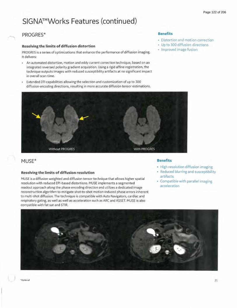
SIGNA™Works Features (continued)
PROGRES*
Resolving the limits of diffusion distortion
PROGRES is a series of optimizations that enhance the performance of diffusion imaging. It delivers:
An automated distortion, motion and eddy current correction technique, based on an integrated reversed polarity gradient acquisition. Using a rigid affine registration, the technique outputs images with reduced susceptibility artifacts at no significant impact in overall scan time.
Extended DTI capabilities allowing the selection and customization of up to 300 diffusion-encoding directions, resulting in more accurate diffusion tensor estimations.
MUSE*
Resolving the limits of diffusion resolution
MUSE is a diffusion weighted and diffusion tensor technique that allows higher spatial resolution with reduced EPI-based distortions. MUSE implements a segmented readout approach along the phase encoding direction and utilizes a dedicated image reconstruction algorithm to mitigate shot-to-shot motion-induced phase errors inherent to multi-shot diffusion. The technique is compatible with Auto Navigators, cardiac and respiratory gating, as well as well as acceleration such as ARC and ASSET. MUSE is also compatible with fat sat and STIR.
<~- o p tiona l
Benefits
• Distortion and motion correction
• Up to 300 diffusion directions
• Improved image fusion
Benefits
• High resolution diffusion imaging
• Reduced blurring and susceptibility
artifacts
• Compatible with parallel imaging
acceleration
21
Page 122 of 206

Image Acquisition ~ Pulse Sequences
SPIN Echo
SE FSE frFSE
IR FSE-IR
3D FSE 3D frFSE
T1 FLAIR T2 FLAIR
Double IR/Triple IR (Black Blood}
Double IR/Triple IR Single Shot
SSFSE SSFSE-IR
SSFSE Snapshot
3D MRCP
T2 MAP'
Cube FLAIR
Cube DIR
Cube PROMO'
2D IDEAL'
MAVRIC SL* HyperMAVRIC SL'
3DASL'
'*Optional
Standard pulse sequences that are used to generate Tl, Proton Density and T2 contrasts. The FSE technique enables long TR and long TE choices in reduced scan times. frFSE produces images with more T2 contribution allowing shorterTR values and resulting in shorter scan times when compared to FSE.
IR techniques provide uniform suppression of tissues by applying an inversion pulse to null signal. FSE-IR reduces scan time while still achieving efficient tissue suppression
FSE-IR with Water SAT pulse and manual adjustment of Center Frequency location to suppress silicon signal in breast imaging.
Three-dimensional imaging acquisitions mostly used for T2-weighted contrast.
T1 and TZ Fluid Attenuated Inversion Recovery (FLAIR} pulse sequences allow the suppression of signal from cerebrospinal fluid (CSF}. This sequence provides contrast to differentiate white and gray matter to Tl- and T2-weighted brain and spine imaging.
These pulse sequences are included to allow Black Blood imaging for studies of cardiac morphology (Tl, TZ, and PO). Triple IR adds fat suppression to Black Blood imaging. It also can be combined with Single Shot.
Single Shot Black Blood acquisitions allow larger volume acquisitions in fewer breath-holds.
Single Shot Fast Spin Echo is a technique that permits single slice data acquisition in less than one second. It is frequently used for MRCP studies in a single breath-hold and myelograms.
The imaging efficiency of navigated/respiratory triggered SSFSE can be improved by imaging multiple slice locations per trigger event with SSFSE Snapshot.
30 frFSE sequence that combined with the T2 Prep option provides improved background tissue suppression for MRCP exams.
T2 MAP is a multiple acquisition; multiple echoes FSE based method to obtain images that represent different T2 weighting values. The acquired data is processed to produce TZ color maps that are used for cartilage evaluation.
Three-dimensional FSE (3D FSE}, with flip angle modulation. You can easily reformat sub-millimeter isotropic volume data from a single Cube acquisition into any plane- without gaps, and with the same resolution as the native plane. T1 CUBE for blood saturation.
30 FSE technique that applies modified refocusing pulses for increased SNR. It is used to acquire isotropic data that can be reformatted in any plane.
Cube OIR, double inversion recovery, is designed to achieve signal suppression from either gray or white matter and CSF.
Prospective Motion correction is a real time 30 navigator based motion correction technique compatible with Cube TZ, Cube DIR and Cube T2 FLAIR.
20 FSE 3-point Dixon Water Fat Separation method that acquires 4 contrasts in one acquisition: Water, Fat, in-phase and out-of phase.
Multi-Spectral imaging technique is designed to reduce metal artifact near MR conditional implants. Improvements have been made with HyperMAVRIC SL feature to reduce scan time through a patient-specific metal analysis scan and allow functionalities, such as Variable flip angles, flow compensation, and No Phase Wrap. In addition to the Tl, PO, and STIR contrasts, the sequence now also provides T2 weighting, and a Bl-optimized STIR pulse.
30 FSE based technique that uses a "labeling" pulse to quantify cerebral blood flow.
22
Page 123 of 206
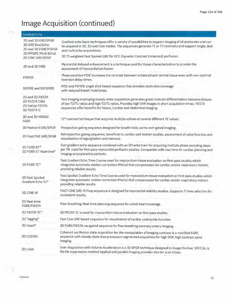
Image Acquisition (continued) Gradient Echo
20 and 3D GRE/SPGR 3D GRE Dual Echo 20 and 3D FGRE/FSPGR 20 MFGRE (Multi Echo) 20 CINE GRE/SPGR
2Dand 3D MOE
PSMDE
SSM DE and SSPSMDE
20 and 3D FIESTA 20 FIESTA CINE 20 FatSat FIESTA 3D FIESTA-C
20 and 3D MERGE FGRE
20 Fastcard GRE/SPGR
20 FastCINE GRE/SPGR
20 FGRE-ET* 20 FGRE-ET Real-time*
20 FGRETC*
20 Fast Spoiled Gradient Echo TC*
20 CINE-IR
20 Real-time FGRE/FIESTA
20 FIESTA TC*
20 Tagging*
3D Heart*
30 COSMIC
3D LAVA
"Optional
Gradient echo basic techniques offer a variety of possibilities to support imaging of all anatomies and can be acquired in 20, 3D and Cine modes. The sequences generate T1 or T2 contrasts and support single, dual and multi echo acquisitions.
3D T1 weighted Fast Spoiled GRE for DCE (Dynamic Contrast Enhanced) perfusion.
Myocardial delayed enhancement is a technique used for tissue characterization to provide the assessment of myocardial perfusion.
Phase sensitive MOE increases the contrast between enhanced and normal tissue even with non-optimal inversion delay times.
MOE and PSMDE single shot based sequence that provides multi slice coverage with reduced breath-hold times.
Fast imaging employing steady-state acquisition generates great contrast differentiation between tissues of low T2/Tl ratios and high T2/Tl ratios. Provides high SNR images in short acquisition times. FIESTA sequences offer benefits for Neuro, Cardiac and Abdominal imaging.
T2* contrast technique that acquires multiple echoes at several different TE values.
Prospective gating sequence designed for breath-hold, aortic arch gated imaging.
Retrospective gating sequence, beneficial to cardiac wall motion studies, assessment of valve function and visualization of regurgitation and stenosis.
Fast gradient echo sequence combined with an EPI echo train for acquiring multiple phase encoding steps per TR. Used for first pass myocardial perfusion studies. Compatible with real time for cardiac planning and imaging uncooperative patients.
Fast Gradient Echo Time Course used for myocardium tissue evaluation on first pass studies which integrates automatic motion correction (MoCo) that compensates for cardiac and/or respiratory motion, providing reliable results.
Fast Spoiled Gradient Echo Time Course used for myocardium tissue evaluation on first pass studies which integrates automatic motion correction (MoCo) that compensates for cardiac and/or respiratory motion, providing reliable results.
FAST-CINE GRE IR Prep sequence is designed for myocardial viability studies. Supports Tl time selection for consistent results.
Free-breathing, Real-time planning sequence for whole heart coverage.
20 FIESTA TC is used for myocardium tissue evaluation on first pass studies.
Fast Cine GRE based sequence for visualization of cardiac contractile function.
3D FGRE/FIESTA navigated sequence for free breathing coronary artery imaging.
Coherent oscillatory state acquisition for the manipulation of imaging contrast is a modified FGRE sequence with steady-state free precession segmented acquisition for high SNR, high contrast spine imaging.
Liver Acquisition with Volume Acceleration is a 3D SPGR technique designed to image the liver. SPECIAL is the fat suppression method applied and parallel imaging provides shorter scan times.
23
Page 124 of 206
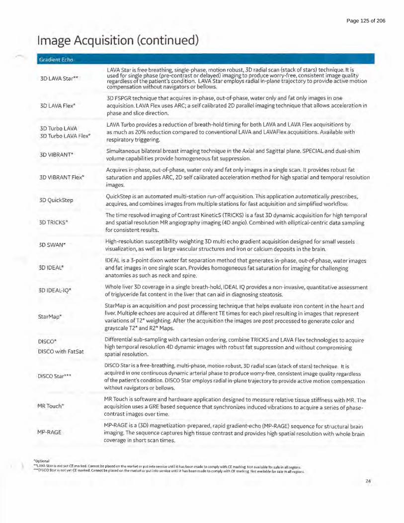
Image Acquisition (continued) Gradient Echo
3D LAVA Star**
3D LAVA Flex*
3D Turbo LAVA 3D Turbo LAVA Flex*
3D VIBRANT*
3D VIBRANT Flex*
3D QuickStep
3D TRICKS*
3D SWAN*
3D IDEAL*
3D IDEAL-IQ*
StarMap*
DISCO*
DISCO with FatSat
DISCO Star***
MRTouch*
MP-RAGE
"'Optional
LAVA Star is free breathing, single-phase, motion robust, 3D radial scan (stack of stars) technique. It is used for single phase (pre-contrast or delayed) imaging to produce worry-free, consistent image quality regardless of the patient's condition. LAVA Star employs radial in-plane trajectory to provide active motion compensation without navigators or bellows.
3D FSPGR technique that acquires in-phase, out-of-phase, water only and fat only images in one acquisition. LAVA Flex uses ARC; a self calibrated 2D parallel imaging technique that allows acceleration in phase and slice direction.
LAVA Turbo provides a reduction of breath-hold timing for both LAVA and LAVA Flex acquisitions by as much as 20% reduction compared to conventional LAVA and LAVAFiex acquisitions. Available with respiratory triggering.
Simultaneous bilateral breast imaging technique in the Axial and Sagittal plane. SPECIAL and dual-shim volume capabilities provide homogeneous fat suppression.
Acquires in-phase, out-of-phase, water only and fat only images in a single scan. It provides robust fat saturation and applies ARC, 2D self calibrated acceleration method for high spatial and temporal resolution images.
QuickStep is an automated multi-station run-off acquisition. This application automatically prescribes, acquires, and combines images from multiple stations for fast acquisition and simplified workflow.
The time resolved imaging of Contrast KineticS (TRICKS) is a fast 3D dynamic acquisition for high temporal and spatial resolution MR angiography imaging (4D angio). Combined with elliptical-centric data sampling for consistent results.
High-resolution susceptibility weighting 3D multi echo gradient acquisition designed for small vessels visualization, as well as large vascular structures and iron or calcium deposits in the brain.
IDEAL is a 3-point dixon water fat separation method that generates in-phase, out-of-phase, water images and fat images in one single scan. Provides homogeneous fat saturation for imaging for challenging anatomies as such as neck and spine.
Whole liver 3D coverage in a single breath-hold, IDEAL IQ provides a non-invasive, quantitative assessment of triglyceride fat content in the liver that can aid in diagnosing steatosis.
StarMap is an acquisition and post processing technique that helps evaluate iron content in the heart and liver. Multiple echoes are acquired at different TE times for each pixel resulting in images that represent variations ofT2* weighting. After the acquisition the images are post processed to generate color and grayscale T2* and R2* Maps.
Differential sub-sampling with cartesian ordering, combine TRICKS and LAVA Flex technologies to acquire high temporal resolution 4D dynamic images with robust fat suppression and without compromising spatial resolution.
DISCO Star is a free-breathing, multi-phase, motion robust, 3D radial scan (stack of stars) technique. It is acquired in one continuous dynamic arterial phase to produce worry-free, consistent image quality regardless oft he patient's condition. DISCO Star employs radial in-plane trajectory to provide active motion compensation without navigators or bellows.
MR Touch is software and hardware application designed to measure relative tissue stiffness with MR. The acquisition uses a GRE based sequence that synchronizes induced vibrations to acquire a series of phasecontrast images over time.
MP-RAGE is a (3D) magnetization-prepared, rapid gradient-echo (MP-RAGE) sequence for structural brain imaging. The sequence captures high tissue contrast and provides high spatial resolution with whole brain coverage in short scan times.
""'LAVA Star is not yet CE marked. Cannot be placed on the market or put into service until it has been made to comply with CE marking. Not available for sale in all regions. """DISCO Star is not yet CE marked. Cannot be placed on the marhet or put into service until it has been made to comply with CE marking. Not available for sale in all regions.
24
Page 125 of 206

Image Acquisition (continued) Vascular
lnhance Inflow IR*
lnhance 3D Velocity*
lnhance 2D Inflow*
lnhance 3D Delta Flow*
2D TOF / 20 Gated TOF 20 Fast TOF FGRE/ SPGR 3DTOF 30 Fast TOF FGRE/ SPGR
20 CINE Phase Contrast 2D FastCINE Phase Contrast
20 Phase Contrast 3D Phase Contrast
fMRI- BrainWave*
GRE-EPI SE-EPI FLAIR-EPI OW-EPI
DTI*
eO WI
RTFA
'"Optional
30 FIESTA based non-contrast-enhanced MR angiography technique that provides static background tissue and venous flow suppression for imaging arteries. It uses SPECIAL for uniform fat suppression and respiratory gating compatibility reduces respiratory motion artifacts during free-breathing renal exams.
30 Phase Contrast based technique designed to acquire angiographic images in brain and renal arteries with robust background suppression in a short scan time. Respiratory triggering compatibility enabling
abdominal angiography.
Designed for imaging arteries that follow almost a straight path (i.e. femoral, popliteal, and carotid arteries) lnhance 20 Inflow acquires data during the systolic phase only. Compatible with Peripheral or Cardiac Gating and ASSET.
30 FSE cardiac gated based non-contrast-enhanced MRA application designed for peripheral arterial imaging. This technique uses the differences between systolic and diastolic flow to help generate arterial signal contrast with robust background and venous suppression. ASSET compatibility provides shorter scan times.
20 TOF Imaging, 20 Gated TOF Imaging, 30 TOF Imaging and Enhanced 30 TOF Imaging are used for MR angiography imaging. Based on conventional gradient echo scanning, TOF imaging techniques rely primarily on flow-related enhancements to distinguish moving from stationary spins.
This pulse sequence is included specifically for studies of cardiac function. Through the use of retrospective gating, it allows full R-R coverage.
These techniques demonstrate flow velocities and directional properties in vessels and other moving fluids such as CSF and aortic flow.
Real time acquisition, processing and display offunctional imaging.
Standard on all systems are gradient echo, spin echo, FLAIR, and diffusion weighted echo planar imaging. The EPI sequence supports single and multishot imaging, multi-phase imaging, as well as cardiac gating. Diffusion EPI produces images that can detect acute and hyper-acute stroke with b-value up to 10,000 s/mm2, multi-NEX compatibility and the ability to generate AOC and T2-weighted TRACE images. The FLAIR option suppresses the CSF signal.
OTI (Diffusion Tensor Imaging} is an EPI technique that acquires diffusion information in up to 300 different directions. The image contrast is based on the degree of diffusion anisotropy in the tissues. Post processing include Fractional Anisotropy (FA}, Apparent Diffusion Coefficient (AOC}, 20 directional maps and 30 fiber track models. Multi-shell OTI is available in clinical mode.
Enhanced OWl (eOWI) provides high SNR diffusion images with short acquisition times. Supports Multi b-values with SMART NEX for variable NEX selection per B-value, "3 in 1" diffusion weighting to all three gradients simultaneously, tetrahedral selection with four different diffusion weighting combinations for shorter TE values and Inversion recovery for fat signal reduction.
The RTFA algorithm leads to a reduction in distortion of the diffusion image per diffusion axis. RTFA is designed to reduce image blurring and distortions typically associated with diffusion imaging throughout the body. RTFA also allows for increased utilization of single spin echo OWl which results in an increase in SNR by up to 50% compared to dual spin echo and, when combined with the improved resolution leads to an increase in image quality that can be utilized for image presentation, fusion and AOC map outputs.
25
Page 126 of 206

Image Acquisition (continued)
RTCF
FOCUS OWl*
Spectroscopy
PROBE-PRESS PROBE-STEAM*
PROBE-PRESS CSI (2D &3D*)
BREASE*
TEA-PRESS*
PROPELLER MB
Silent Tl, PO, T2, DWI, T1 FLAIR and T2 FLAIR PROPELLER MB*
Tl, PDand T2 PROPELLER MB
T2 FLAIR PROPELLER MB
T1 FLAIR PROPELLER MB
DWI PROPELLER MB
PROPELLER DUO
Silenz*
Silenz T1
Silenz PD
Fat Supp1ession Technology
Fat Sat
'"'Optional
Real-Time Center Frequency (RTCF) option can be applied to OWl & DTI to enable using the optimal center
frequency for each slice. This is intended to help improve fat suppression and signal drop off at areas of high B0 inhomogeneity (off-isocenter, or area with high tissue susceptibility). It is also intended to reduce station
to-station misalignment in whole body diffusion imaging.
FOV Optimized & Constrained Undistorted Single-shot (FOCUS) DWI utilizes 2D selective excitation pulses to limit the prescribed phase encode FOV eliminating artifacts from motion, imaging back folding or
unsuppressed tissue.
PROBE Single-Voxel spectroscopy allows non-invasive evaluation of the relative concentrations of in-vivo
metabolites. The sequence provides acquisition and displayofvolume localized, water-suppressed Hl spectra in single-voxel mode. The sequence consists ofthree slice selective RF pulses with crusher gradients. PRESS provides up to twice the SNR over STEAM.
PROBE 20 and 3D CSI enable simultaneous multi-voxel spectmscopic acquisitions in the brain. It is available with PRESS excitation to maximize SNR. Post processing includes automatically generated metabolic maps.
ATE-averaged PRESS (Point RESolved Spectroscopy) acquisition that provides the necessary biochemical information to help characterize breast tissue by assessing the presence of choline.
TEA PRESS is aTE-Averaged variant of the PRESS CSI pulse sequence. It collects spectra across a range of TE values and averages the results together to reduce the appearance of signals whose intensity varies as a function ofTE. This allows signals whose intensity does not vary with TE to be accentuated in comparison. This is the underlying pulse sequence behind the BREASE application.
PROPELLER MB is a multi-shot per blade sequence that uses a radial k-space filling pattern acquisition and a post processing correction algorithm to significantly reduce the effects of motion artifacts. PROPELLER MB is compatible with spatial and chemical Sat, ASPIR, STIR Tl, PD and T2 Auto TI/TR and Navigator.
PROPELLER DUO is a FSE based technique that is less prone to distortions caused by field inhomogeneities. PROPELLER DUO has a comparable scan time when compared to conventional PROPELLER DWI, and has spatial sat and shim volume capability to further reduce distortions and reduce artifacts and improve image quality.
Silenz is a 30 Zero-TE sequence comprising high bandwidth excitation and reduced gradient-switching radial acquisition that results in sound levels near ambient. Silenz has added flexibility in sequence prescription for anisotropic resolution enabling faster scan times and includes axial as well as oblique geometries.
Applies a frequency selective saturation pulse at the frequency offat before the imaging excitation pulse with the result being a signal measurement primarily from water.
26
Page 127 of 206

Image Acquisition (continued) Fat Suppression Technology
STIR
SPECIAL
Spectral Spatial
ASPIR
IDEAL*
Flex*
STIR is an inversion recovery method that takes advantage of the T1 difference between water and fat to allow selection oft he signal to suppress. In order to eliminate the signal from tissues, the Tl time must match exactly the null point of the tissue that needs to be suppressed.
Hybrid fat suppression technique that incorporates features from both the frequency selective FatSat and the STIR techniques by using a spectrally selective inversion pulse that inverts only the fat magnetization and leaves the only the water peak available for excitation.
Method that applies selective pulses for water excitation only, while fat is left untouched, thereby producing
no signal.
ASPIR method is a solution for poor fat suppression due to B1 inhomogeneity. It is based on the frequency and the relaxation fat behaviors. Applies a spectrally selective adiabatic inversion pulse to excite the fat spins, imaging pulses are then applied after Tl null time when longitudinal magnetization of fat crosses zero. The disadvantages include sensitivity to Bo and longer scan times.
IDEAL is a 3-point Dixon technique that acquires three images at slightly different echo times to generate phase shifts between water and fat. The water/fat separation method is very efficient at providing homogeneous image quality. One acquisition provides four contrasts: water, fat, in-phase and out-of-phase images.
Flex is a 2-point dixon technique delivering faster scan times compared to IDEAL 3-point dixon. It is based on the difference between fat and water resonance frequencies using two flexible echo times for further scan time reduction . One acquisition provides four contrasts: Water, Fat, in-phase and out-of-phase images.
Motion Correction Technology
PROPELLER MB
PROMO*
PB Navigators
Respiratory Trigger
VCG
PG
Acceleration Technology
Fractional Nex
Fractional No Phase Wrap
ASSET
ARC
.. Optional
PROPELLER MB is a multi-shot per blade sequence that uses a radial k-space filling pattern acquisition and a post processing correction algorithm to significantly reduce the effects of motion artifacts. It is compatible with spatial and chemical Sat, ASPIR, STIR Auto TI/TR and navigator.
Prospective motion correction is a real time 3D navigator based motion correction technique compatible with Cube T2, Cube DIR, Cube T2 FLAIR, BRAVO and MPRAGE.
Pencil beam navigators allow free breathing body and cardiac imaging by tracking the motion of the diaphragm. There are two navigator modes: navigator gating, uses a predefined signal acceptable range during the expiration and navigator triggering, uses signal to trigger data collection during the expiration.
Reduces breathing motion artifacts by synchronizing the acquisition with the respiratory cycle.
Vector cardiac gating reduces motion artifacts by synchronizing the acquisition with the cardiac cycle.
Peripheral gating reduces motion artifacts caused by pulsating blood.
Technique in which only partial k-space data is collected and the remaining data is estimated. It uses the phase conjugate symmetry reconstruction method, which only half of the phase encode steps are acquired for scan time reduction.
Selectable on the user interface, Fractional No Phase Wrap allows you to adjust the phase FOV based upon the patient size and shape. Benefits include a physical viewofNPW placement on the user interface, flexibility to manage SNR and Scan Time, and the power to scan only the area of interest within the determined FOV.
Array spatial sensitivity encoding technique acquires under sampled multicoil data generatingaliased images. These are post processed with coil sensitivity maps from the calibration scan to unfold the images.
Auto-calibrating reconstruction for cartesian imaging is a highly accelerated parallel imaging auto-calibrating method that doesn't require coil sensitivity maps. It enables smaller FOV prescriptions, less sensitivity to motion and prevents artifacts caused by coil calibration inaccuracies .
27
Page 128 of 206

Image Acquisition (continued) Acceleration Technology
HyperBand*
HyperSense**
Hyperkat*
HyperCube*
HyperBand enables scan time reduction by simultaneously exciting multiple slices at multiple locations. Reconstruction algorithms are then applied in order to separate the images acquired.
HyperSense has been expanded to include T1 acquisitions including MP-RAGE & BRAVO for neuro imaging and LAVA, LAVA-Flex, DISCO and DISCO-Flex for body applications, and Vibrant for breast applications. In addition, HyperSense is now compatible with other 3D gradient echo sequences, such as MERGE, FIESTA
and COSMIC.
Hyper Kat san advanced k-t acceleration method that employs time-shifted sampling in data acquisition and exploits both spatial and temporal correlation with motion-adaptive time window selection in image
reconstruction.
Small FOV organ specific volumetric imaging acquisition method that enables outside phase FOV HyperCube signal suppression. The technique can help to reduce artifacts originated outside of the prescribed field of
view.
Uniformity Correction Technology
SCENIC
PURE
deFINE
reFINE
Noise Reduction Technology
ART
Silenz*
Silent PROPELLER*
SCENIC (Surface Coil ENhancement for Imaging Clarity) is an advanced image uniformity correction that
further improves upon the previous reFINE algorithm.
By using the biased field, SCENIC utilizes B-Splines to iteratively determine the best sharpening algorithm.
This results in improved contrast, reduced shading, and consistent sharpening when compared to
conventional imaging filtering techniques
PURE corrects the field inhomogeneity by collecting a calibration scan from the (uniform) body coil and the (non-uniform) surface coil and calculating maps that relate the intensity correction values to the images.
deFINE is an integrated in-line imaging processing method that provides edge enhancement and smoothing algorithms allowing the user to customize the image appearance.
reFINE is an advanced image uniformity correction algorithm that addresses non-uniformity due to coil sensitivity profiles and dielectric shading effects. It reduces organ-motion induced misregistration artifacts, effects of low signal in dark regions and edge effects at tissue interfaces and borders. reFINE optimizes parameter settings for each application, coil, and body anatomy maximizing image uniformity results.
Acoustic Noise Reduction Technology optimizes the gradient waveform to reduce the gradient noise without compromising performance.
Silenz is a 3D Zero-TE sequence comprising high bandwidth excitation and reduced gradientswitching radial acquisition that results in sound levels near ambient. Silenz has added flexibility in sequence prescription for anisotropic resolution enabling faster scan times and includes axial as well as oblique geometries.
Silent PROPELLER gradient waveform approach reduces the acoustic noise level to less than lldB above the ambient room noise.
Un1formity Correction Technology
SCENIC
*Optional
SCENIC (Surface Coil ENhancement for Imaging Clarity) is an advanced image uniformity correction that further improves upon the previous reFINE algorithm.
By using the biased field, SCENIC utilizes B-Splines to iteratively determine the best sharpening algorithm.
This results in improved contrast, reduced shading, and consistent sharpening when compared to conventional imaging filtering techniques.
"11
HyperSense expansions is not y(>t CE markt>d. Cannot be placed on the market or put into service until it has been made to comply with CE marking. Not available for sale in all regions.
28
Page 129 of 206
Image Acquisition (continued) Uniformity Correction Technology
PURE
deFINE
reFINE
Noise Reduction Technology
ART
Silenz*
Silent PROPELLER*
PURE corrects the field inhomogeneity by collecting a calibration scan from the (uniform) body coil and the (non-uniform) surface coil and calculating maps that relate the intensity correction values to the images.
deFINE is an integrated in-line imaging processing method that provides edge enhancement and smoothing algorithms allowing the user to customize the image appearance.
reFINE is an advanced image uniformity correction that consists of SCENIC and PURE that a dresses nonuniformity due to coil sensitivity profiles and dielectric shading effects. It reduces organ-motion induced misregistration artifacts, effects of low signal in dark regions and edge effects at tissue interfaces and borders. Refine optimizes parameter settings for each application, coil, and body anatomy maximizing image uniformity results.
Acoustic Noise Reduction Technology optimizes the gradient waveform to reduce the gradient noise without compromising performance.
Silenz is a 3D Zero-TE sequence comprising high bandwidth excitation and reduced gradientswitching radial acquisition that results in sound levels near ambient. Silenz has added flexibility in sequence prescription for anisotropic resolution enabling faster scan times and includes axial as well as oblique geometries.
Silent PROPELLER gradient waveform approach reduces the acoustic noise level to less than lldB above the ambient room noise.
Magnetization Transfer Contrast
Used to improve the Magnetization Transfer Imaging Option to improve contrast between blood flow and surrounding tissue in 3D TOF images, to augment post-contrast Tl-weighted brain images, and to increase myelographic effect for improved disc and cord lesion visualization
Blood Saturation
Use Blood Suppression to obtain "black blood" cardiac images and reduce flow-related ghosting.
"'Optional 29
Page 130 of 206

RF Coils Suite eXpress Table & Posterior Array
Detachable table with embedded posterior array
100 em S/1 Coverage
40 Elements with dedicated spine configurations
• Head-first or feet-first
• Automatic coil mode selection
Acceleration in all directions
Patient-centric comfort pads
Petite Female
Comfort Pads
Very Large Male
30
Page 131 of 206

RF Coils Suite (continued)
AIR™ Coils l.ST 30ch AIR™ AA Coil*
The 30-channel AIR Anterior Array (AA) is the next generation anterior array coil that allows flexibility in any direction to conform to the patient's anatomy. Based on the innovative technologies behind the INCA conductor and the E-mode module, the 1.5T 30ch AIR AA provides superb SNR and acceleration performance, while improving the overall patient and user experience. The coil has been designed to adapt various patient shapes and sizes, with an ultra lightweight distribution of less than 0.35 grams/cm2
• The 1.5T 30ch AIR AA is a receive-only RF coil designed for use with GE 1.5T MRI systems to produce diagnostic images of general human anatamy, including extremities.
l.ST 30ch AIR AA
Elements
Maximum number of channels in the maximum FOV
Maximum number of channels in head-to-thighs imaging (S/1145cm)
Weight
R/L Coverage
S/1 Coverage
Dimensions (W x L x H)
Patient orientation
Coil combinations
30
45, when combined with the Posterior Array
121, when combined with the Head-Neck Unit, Posterior Array and 2"d AIR Anterior Array
1.8 kg (41bs) resting on patient, 2.7 kg (61bs) with the cable
60cm
63cm
66cm x 79 em x 1.2 em
Head-first or feet-first
Can be combined with the following coils:
Head-Neck Unit
Posterior Array
• AIR MP coils
• 2"d AIR Anterior Array
Peripheral Vascular Peripheral Vascular
*The 1.5T 30ch AIR'M AA coil is not yet CE marked. Cannot be placed on the market or put into service until it has been made to comply with the Medical Device Directive. 31
Page 132 of 206
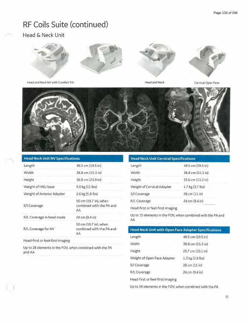
RF Coils Suite (continued) Head & Neck Unit
Head and Neck NV with Comfort Tilt
Head Neck Unit NV Specifications
Length
Width
Height
WeightofHNU base
Weight of Anterior Adapter
S/1 Coverage
R/L Coverage in head mode
R/L Coverage for NV
Head-first or feet-first imaging
49.5 em (19.5 in)
38.8 em (15.3 in)
36.8 em (23.9 in)
5.0 kg (lllbs)
2.6 kg (5.8 lbs)
50 em (19.7 in), when
combined with the PA and
AA
24 em (9.4 in)
50 em (19.7 in), when
combined with the PA and
AA
Up to 28 elements in the FOV, when combined with the PA andAA
Head and Neck Cervical Open Face
Head Neck Unit Cervical Specifications
Length
Width
Height
Weight of Cervical Adapter
S/1 Coverage
R/L Coverage
Head-first or feet-first imaging
49.5 em (19.5 in)
38.8 em (15.3 in)
33.6 em (13.2 in)
1.7 kg (3.7 lbs)
28cm (11 in)
24 em (9.4 in)
Up to 15 elements in the FOV, when combined with the PA and AA
Head Neck Unit with Open Face Adapter Specifications
Length
Width
Height
Weight of Open Face Adapter
S/1 Coverage
R/L Coverage
Head-first or feet-first imaging
49.5 em (19.5 in)
38.8 em (15.3 in)
25.7 em (10.1 in)
1.3 kg (2.8 lbs)
28 em (11 in)
24 em (9.4 in)
Up to 19 elements in the FOV, when combined with the PA
32
Page 133 of 206

RF Coils Suite (continued) Anterior Array
Anterior Array Specifications
Length
Width
Height
Weight
S/1 Coverage
R/L Coverage
Head-first or feet-first imaging
Up to 36 elements in the FOV, when combined with the PA
55.6 em (21.9 in)
67.4 em (26.5 in)
3.3 em (1.3 in)
2.8 kg (6.161bs) resting on patient 4.1 kg (9.0 lbs) with cable
54 em (21.3 in)
Full 50 em (19.7 in) FOV of the system
33
Page 134 of 206

RF Coils Suite (continued) Peripheral Vascular Array*
Optional Peripheral Vascular/Lower Extremity Array
Length
Width
Height
Weight
S/1 Coverage
R/L Coverage
Head-first or feet-first imaging
105 em (41.3 in)
2nd station: 51.6 em (20.3 in) 3'd station: 64.2 em (25.3 in)
24.8 em (9.8 in)
8.4 kg (18.61bs)
104 em (49.9 in) overall 2"d station: 52 em (20.5 in) 3'd station: 52 em (20.5 in)
Full 50 em (19.7 in) FOV
of the system
Up to 35 elements in the FOV, when combined with the PA
*Optional 34
Page 135 of 206

RF Coils Suite (continued) 16-channel Flex Coils*
Knee and foot ankle positioner
·'
(y-,~- t
\
~ ~; ~ , . ' ... _ .. I
• ... I
' \·
\~ ~~ : :' £'Ji~ , :
\" ;
~ . ' 'f .. I
' I.' . ... ·,
GEM Flex Specifications
Coil Dimensions (W x L) Wrap Diameter Elements Weight
GEM Flex Large 23 em x 70 em 15.5 em- 21.5 em 16 1.0 kg
GEM Flex Medium 23 em x48 em 11.5 em- 15.5 em 16 0.8 kg
GEM Flex Small 23 em x 38 em 9 em -12.5 em 16 0.8 kg
32-channel Pediatric Coil Solution*
The 32-channel pediatric coil solution consists of a pediatric stabilizer positioner and interface that accommodates the Large Flex
coil and the Medium Flex coil. Compatible with the Silent Suite.
GEM Flex Specifications
Component Coverage (W x L)
GEM Flex Coil, Large 23 em x 70 em
GEM Flex Coil, Medium 23 em x 48 em
~optional
Wrap Diameter
15.5 em - 21.5 em
11.5 em- 15.5 em
Elements
16
16
Weight
1.0 kg
0.8 kg
35
Page 136 of 206

RF Coils Suite (continued) 16-channel Shoulder Coil*
The Phased Array 16-channel Shoulder Coil consists of a baseplate that supports a posterior hard shell connected to an adjustable anterior plate, designed to better accommodate the patient anatomy. The baseplate and customized pad provide easy
right- left adjustment for off-center positioning.
Specifications
Coil
16ch Shoulder
Approximate dimensions (W x L x H)
28 em x 28 em x 31.1 em
16-channel T/R Knee Coil*
The 16-channel Transmit Receive Phased Array Knee Coil is designed
to acquire high SNR images of the knee. It is generously sized to effortlessly accommodate a wide range of the patient population . The two-part design provides a quick and efficient workflow. Offset imaging is fully supported with adjustable left-right coil positioning.
Specifications
Elements
16
Coil Approximate dimensions (W x L x H) Approximate Diameter
16ch T/R Knee 49.2 em x 50.0 em x 28.4 em 15.5 em
""Optional
Benefits
• 16-channel phased array design • Adjustable anterior plate for ease of
positioning • Parallel imaging compatible for speed
Approximate Weight
3.9 kg
Benefits
• Transmit Receive 16-channel array
design • Large diameter to better
ccommodate anatomy • High SNR for unique performance • Parallel imaging compatible for speed
Elements Approximate Weight
16 7.5 kg
36
Page 137 of 206

RF Coils Suite (continued) 8-channel Foot/Ankle Coil*
The Phased Array 8-channel Foot/Ankle Coil consists of a baseplate and a detachable hard shell coil that is designed for fast and easy positioning, comfortably
accommodating the anatomy while providing proper immobilization.
Specifications
Coil
8ch Foot/Ankle
Baseplate
Approximate dimensions (W x L x H)
18 em x 33.7 em x 31.4 em
35.8 em x 51.5 em x 33.6 em
Elements
8
16-channel Shoulder Coil** (by Neocoil)
Specifications
Coil
16eh Shoulder
Table Pad
"'Optional
The Phased Array 16 channel Shoulder coil consists of a soft and light design anterior part and a hard shell posterior array to achieve higher SNR and better patient comfort. The simple coil design is allowing more flexible
positioning.
Approximate dimensions (W x L x H)
28.2 em x 35.3 em x 18.2 em
52.0 em x 52.0 em x 5.7 em
Elements
16
Benefits
• 8-channel dedicated foot and ankle phased array coil
• Optimized design to accommodate
foot and ankle anatomy
• Slide and lock mechanism for easy positioning
Benefits
Approximate Weight
3.1 kg
3.8 kg
• 16 channel phased array design
• Flexible anterior part for ease of positioning as well as patient comfort.
• Parallel imaging compatible for speed.
Approximate Weight
3.0 kg
0.6 kg
..,..~ptional, 16ch Shoulder Coil (bv NeoCoil) is not yet CE marked. Cannot be placed on the market or put into service until it has been made to comply with CE marking. Not available for sale in all reg1ons. 37
Page 138 of 206

RF Coils Suite (continued) ~ RF Coils and Arrays*
There are many optional receiver coils available to configure a SIGNNM Artist 1.5T to meet specific applications requirements . The
coils listed below are commercially available at the time of printing and are optional with the system. Please contact your local GE
sales representative for the most current list.
Shoulder Phased Array*
3-channel phased-array coil
Sleeve design
• Comprehensive shoulder imaging
Homogeneous penetration of the humeral head and neck, rotator cuff, glenoid labrum, acromium process, and glenohumeral articular surfaces
16-channel T/R Hand Wrist Coil*
16-channel phased array coil, local transmit coil
Prone or Supine positioning
Optimized design for Fingers through wrist
High SNR to enable high resolution images
Parallel imaging compatible for speed
• Coil dimensions: 46 x 14 x 20 em
""O pt ione~ l
Small Anterior Array*
Up to 33 elements in the FOV when combined with PA for cardiac and body imaging
Head first or feet first
Optimized for parallel imaging
Anterior coil dimensions (L x W x H) 45 em x 40.5 em x 4.5 em (17.7 in x 15.9 in x 1.8 in)
Anterior coil weight: 2.95 kg (6.5 lbs)
l.ST Endorectal coil by Rapid
Channel Count: 1 Ch Coil
Dimensions: 9.7 x 2.5 x 1.7 em
Weight; 1 Kg (w/o cable)
Blrms: 2.5uT
• Scan range: S/1: 8 em, R/L: 1.6 em
Port Campatibilitty: Any
Head first or Feet first
Coil Neck: 7.5 x 1.2 em (Lengh x Diameter)
• House Dimension: 36 x 4.4 x 3.9 em (Total)
HD Breast Array*
8-channel8-element phased-array design
Optimized for uniformity, parallel imaging and VIBRANT
Bilateral and unilateral breast imaging Biopsy plates available
Coil dimensions: 50 em x 54 em x 25 em (20 in x 21 in x 10 in)
38
Page 139 of 206
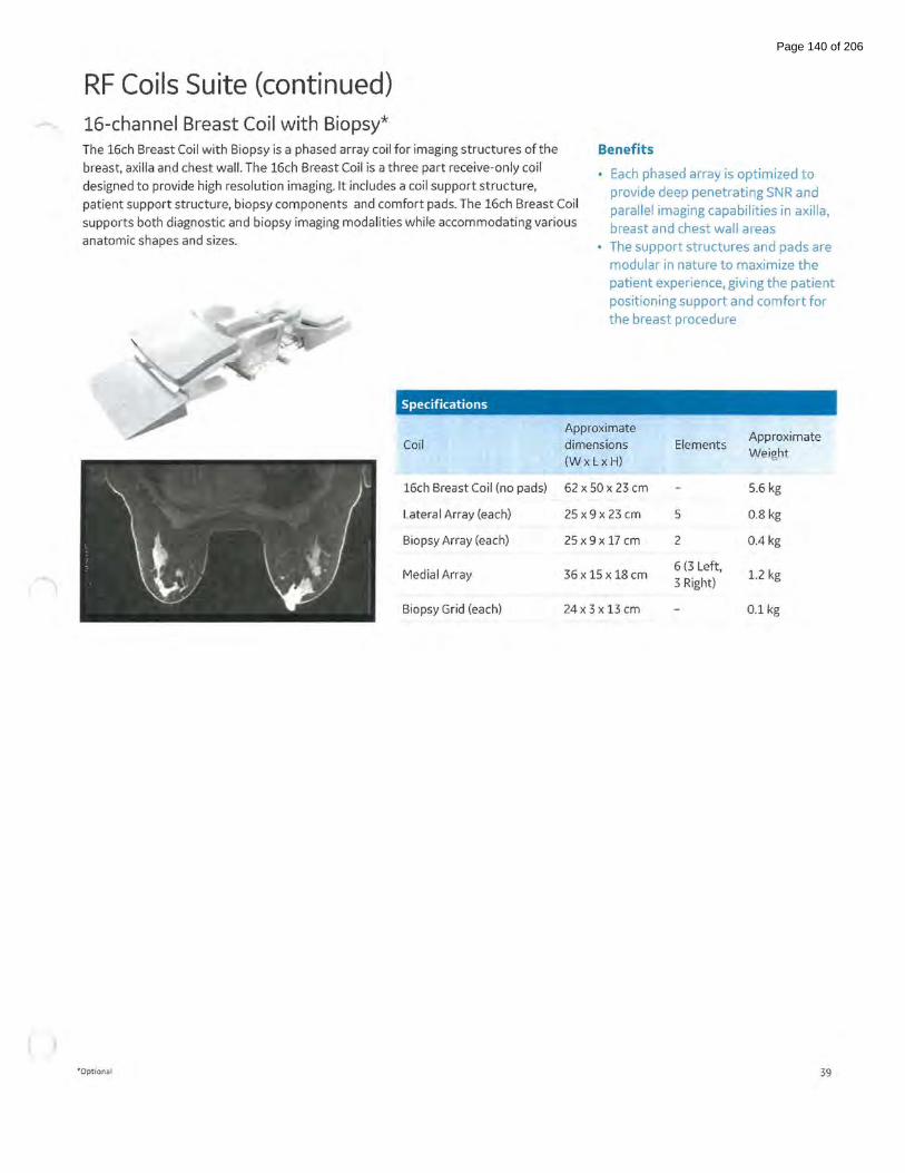
RF Coils Suite (continued) 16-channel Breast Coil with Biopsy* The 16ch Breast Coil with Biopsy is a phased array coil for imaging structures of the
breast, axilla and chest wall. The 16ch Breast Coil is a three part receive-only coil
designed to provide high resolution imaging. It includes a coil support structure,
patient support structure, biopsy components and comfort pads. The 16ch Breast Coil
supports both diagnostic and biopsy imaging modalities while accommodating various
anatomic shapes and sizes.
Benefits
• Each phased array is optimized to
provide deep penetrating SNR and
parallel imaging capabilities in axilla,
breast and chest wall areas
• The support structures and pads are modular in nature to maximize the
patient experience, giving the patient
positioning support and comfort for
the breast procedure
Specifications
Approximate Approximate
Coil dimensions Elements Weight
(WxLxH)
16ch Breast Coil (no pads) 62 x 50 x 23 em 5.6 kg
Lateral Array (each) 25 x 9 x 23 em 5 0.8 kg
Biopsy Array (each) 25x9x17em 2 0.4 kg
Medial Array 36 x 15 x 18 em 6 (3 Left,
1.2 kg 3 Right)
Biopsy Grid (each) 24x 3 x 13 em 0.1 kg
"'Optional 39
Page 140 of 206
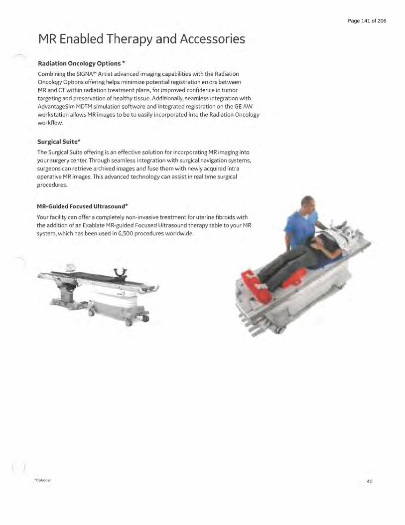
MR Enabled Therapy and Accessories
Radiation Oncology Options *
Combining the SIGNNM Artist advanced imaging capabilities with the Radiation
Oncology Options offering helps minimize potential registration errors between
MR and CT within radiation treatment plans, for improved confidence in tumor
targeting and preservation of healthy tissue. Additionally, seamless integration with
AdvantageSim MDTM simulation software and integrated registration on the GE AW
workstation allows MR images to be to easily incorporated into the Radiation Oncology
workflow.
Surgical Suite*
The Surgical Suite offering is an effective solution for incorporating MR imaging into
your surgery center. Through seamless integration with surgical navigation systems,
surgeons can retrieve archived images and fuse them with newly acquired intra
operative MR images. This advanced technology can assist in real time surgical
procedures.
MR-Guided Focused Ultrasound*
Your facility can offer a completely non-invasive treatment for uterine fibroids with
the addition of an Exablate MR-guided Focused Ultrasound therapy table to your MR
system, which has been used in 6,500 procedures worldwide.
'""Optional 40
Page 141 of 206

SIGNA™ Flow SIGNA™ Flow is designed to standardize and accelerate
workflow for patient setup, exam prescription, scanning and
post processing. eXpress Workflow can begin before the patient
enters the magnet room and exams can be completed within
a few mouse clicks- delivering quality and consistency for all
patients and from all technologists. At the same time, eXpress
Workflow maintains the flexibility needed to rapidly adapt and
optimize exams for patient specific situations.
Exam Setup
Modality Worklist
Automated and standardized rapid set up
Allows the MR protocol to be selected and linked to the patient record in advance of the patient's arrival
For sites with full DICOM connectivity, select the patient from the Modality Worklist, start a new session and view the relevant exam details on the in-room operator console
• Add critical patient information such as allergies, premedication, pregnancy status and history
Protocol Tools
Search, select and one click to share
Protocol Libraries: GE Optimized (preloaded protocols) and Site Authored (customized and saved)
• Protocols can be saved based on patient demographics, anatomy, scan type, or identification number for rapid search
Commonly used protocols can be flagged for quick selection from the modality worklist
One-click to share protoCopy- enables a complete exam protocol to be shared with the click of a mouse and provides a process for managing protocols across multiple systems as well as saving protocols for back up
41
Page 142 of 206

AIRx™* AIR xrM (auto graphic Rx)- contains deep
learning algorithms that automatically identify anatomical structures to prescribe slices
for challenging setup planes for brain and knee. This workflow tool enables consistency and productivity improvements for routine and follow-up examinations and extends
research/clinical capabilities for longitudinal quantification studies.
• Increases productivity by simplifying workflow steps, thus reducing prescription times
Improves consistency and reduces slice positioning variation amongst different technologists
• Automatically adapts slice prescriptions to various patient anatomies and structures.
"'Optional 42
Page 143 of 206

AIR™ Recon DL**
Deep Learning based reconstruction to reduce noise, blurring and ringing artifacts for MR images. AIR'M Recon DL, aGE first
deep-learning application for MR image reconstruction, is designed to improve signal-to-noise and image sharpness, enabling shorter scan times. It uses trained neural networks to remove noise and ringing from the reconstructed image. Compatible with most 2D applications, including diffusion weighted EPI.
AI RIM Recon DL is a pioneering, deep-learning based reconstruction software that will change the way you think about MR imaging. Part of GE Healthcare's AIR'M family of products, which includes lightweight coil design and intelligent workflow applications, this software challenges the inherent trade-off between SNR, scan time and image resolution. AIR'M Recon DL is not a filter or a post-processing technique. It improves image quality at the foundational level because it's embedded directly in
the reconstruction pipeline and is applied to raw data to remove noise and ringing artifacts.
AIR™ RECON DL AT A GLANCE
• Increases productivity by enabling shorter scan times
• Removes image noise and ringing by leveraging raw image data
• Delivers sharper and clearer TrueFidelityTM MR images Enables you to set your preferred SNR improvement level
'"optional, AIR Recon DL is not yet CE marked. Cannot be placed on the market or put into service until it has been made to comply with CE marking. Not available for sale in all regions. 43
Page 144 of 206

oZTEo* MR bone imaging
GE's unique MR bone imaging application, oZTEo, is based on the zero echo time (ZTE) acquisition that is also used in the Silent Suite (Silenz) application. oZTEo complements the conventional soft tissue exam by providing cortical bone surface information. Automated grayscale inversion provides positive bone contrast that is more familiar to visualize for surgeons and clinicians. The ZTE sequence can be used for 30 isotropic resolution and adapts to the patient by providing a inherent motion insensitivity from a radial acquisition. oZTEo can be used with any surface coil that is compatible with SCENIC and includes protocols for common joints such as hip, shoulder, wrist, ankle and knee.
CTSCAN ZTEMRI
*optional, oZTEo is not yet CE marked. Cannot be placed on the market or put into service until it has been made to comply with CE marking. Not available for sale 1n all regions. 44
Page 145 of 206

Patient Setup eXpress Patient Table
Safety, Comfort and Efficiency
Reduce patient transfers- transfer outside the magnet room directly to the eXpress table
Accelerate emergency egress- can be undocked and removed by one user in under 30 seconds typically
Automatic coil disconnect- in time sensitive situations the system coils are automatically disconnected
Patient choice- feet-first or head-first positioning for all supported exams
Reduce in-room patient setup and address privacy by fully preparing the patient and coils for an exam outside of the magnet room
Integrate arm-boards and IV pole to support patient for transport
Embedded posterior array and multiple high density surface coil connectors
lntelliTouch land marking sensors
Compatible second table, prepare the next patient outside the magnet room while scanning the current patient
Express Patient Table
Configuration
Minimum & Maximum Height ---- · -·
Table Drive
Longitudinal Speed - - ·-·--· Total Cradle Length
Total Scanable Range
Maximum Patient Weight for Scanning
Maximum Patient Weight Detached and Mobile
Maximum Lift Capacity
Patient Transport Accessories
Landmarking
Coil Connection Ports
rMinimum height (from floor) to inste~ll Head Coils at both ends oft he table: 64.4cm
Detachable and mobile
70 em to 93 em continuous*
Automated power-driven vertical Automated power-driven longitudinal ---~···--
30 em/sec (fast) and 0.5 em/sec (slow)
210.8cm
205cm
227 kgs (500 lbs)
227 kgs (500 lbs)
227 kgs (500 lbs)
Self-storing non-ferrous IV pole
Positioning Pads
Immobilization Straps
Laser alignment with S/1 and R/L alignment
lntelliTouch touch sensors
2 high density, auto-sensing ports
45
Page 146 of 206

Patient Setup (continued) AIR Touch™*
Intelligent coil localization and selection
Accelerate your scanning process the minute the patient gets on the table with AIR Touch'M, a new workflow application that automates coil selection and land marking. With AIR TouchTM, you simply use lntelliTouch, GE's 1-touch landmarking tool, to activate an optimized set of coils that is selected based on the patient's anatomy. This advanced technology selects from numerous coil combinations such as the posterior array (PA) and flexible coils, to efficiently set up patients. With the anatomical-based protocol optimization, AIR Touch'M optimizes for the anatomy and the protocol parameters with a single touch, delivering a significant productivity gain from plan to scan. AIR Touch'M automatically integrates all calibration scans, providing uninterrupted workflow for the technologist. Further scan times savings are realized with Flexible No Phase Wrap {NPW) to scan only what you need while allowing you to focus on your patient, not the scanner.
Dynamically generated coil configurations with elements activated to optimize image quality (coverage, uniformity and parallel imaging acceleration) for every scan
Coil locations determined automatically
Calibration scans seamlessly acquired without interrupting workflow
Dramatically simplified coil selection Ul;no need to touch it for most exams
0 mm 00:00 , .... "' .. _. -,_
, .... nt 10 In .S-41 ..
In-Room Operator Console and Control
Full Control from table side
From the in-room operator console and controls, the user can:
Position the table
Return the table to home
• Stop the table movement
• Control multiple levels of in-bore ventilation and lighting
• Display of patient name, ID, study description
Display patient weight
Intel liT ouch
Touch to Landmark
lntelliTouch sensors for simplified non-laser patient land marking
With lntelliTouch technology, the user can touch to complete
• Patient landmarking
Localizing to the surface coil for auto-coil selection
Move patient to scan
Start scanning (with AutoStart activated)
Acquire, process and network images
iROC
" -(
0 mm 00:00
Fan Light Control
Display and entry of patient orientation and patient position
Cardiac waveform display and ECG/EKG lead confirmation
Gating control for trigger select, invert and reset
• Respiratory waveform display
lntelliTouch technology land marking
Auto Start to initiate scanning of the selected protocol
Display connected coils and coil status
Display oftable location and scan time remaining
Activate Screen Saver
The in-room display also allows for the integration of third-party visualization tools.
46
Page 147 of 206

In-line Processing & In-line Viewing In-line Processing
Automated post processing
• Automated post processing of specific applications
Automatic opening and loading to advanced visualization tools when appropriate
• Automated in-line processing can be stored within the protocol
Automatic Pasting and Saving
MR Pasting: Combine images from separate acquisitions into a single series with MR Pasting. MR Pasting is an image analysis software packagethatfacilitates the display and filming of multiple station MR data sets in the body applications (total spine, total body) as well as peripheral MR angiography data. MR Pasting will automatically register and combine multiple acquisition stations into a single image of
covered anatomy
• AutPasting: automatic compute and save
3D ASL series*
Diffusion Weighted series
Diffusion tensor series*
eDWI series
Image filtering: A-E, deFINE
Maximum/Minimum Intensity Projection
Reformat to orthogonal plane
T2 map for cartilage evaluation*
3D Volume Viewer
BrainStat
FiberTrak*
Image Fusion
Interactive Vascular Imaging
Pasting
In-line Viewing Enhanced Visualization
Automatic compute and save
Automatic compute and save
Automatic compute and save
Automatic compute and save
Automatic compute and save
Automatic compute and save
Automatic compute and save
Automatic compute and save
Automatic load
Automatic load
Automatic load
Automatic load
Automatic load
Automatic load
In-line viewing allows the user to seamlessly and conveniently view, compare, and analyze images (during scan progress). The user simply selects the series, or multiple series, to view from the workflow manager, and the images are displayed along with the image display tools.
""Optional 47
Page 148 of 206

(
Scanning
Workflow Manager
Linking and Auto Functions
Auto Start
AutoCoil
AutoScan
Auto-calibration
Auto Voice
PB Navigators
READYBrain
QuickSTEP
eXpress Prescan 2.0
Pause and Resume
Automatically initiates scanning oft he selected protocol upon closure of the scan room door.
Automatically determines the optimum coil elements to activate for scanning. If the prescribed field-of-view changes, AutoCoil automatically adjust the selection. The user has the option to review and edit the selection.
Automatically scans the prescribed series without user interaction. For series requiring a contrast injection, the Workflow Manager will pause and await user interaction.
For acquisitions that utilize ASSET parallel imaging or PURE surface coil intensity correction, Auto-Cal will prescribe and acquire a calibration scan based on the prescribed imaging volume.
Delivers user selected, pre-recorded instructions to the patient at defined points in the acquisition to help ensure exam consistency. AutoVoice includes instructions in 141anguages and also allows the user to create and save unique instructions for specific local needs.
Enable free-breathing body imaging for patients unable to breath-hold. The diaphragm tracker pulse automatically places and updates to streamline workflow and eliminate the setup time associated with respiratory triggering. Auto Navigators can be used with a broad range of imaging techniques including dynamic contrast enhanced Tl-weighted imaging.
Automates localizer acquisition, scan plane prescription, scanning, and post processing for brain exams. READYBrain automatically calculates the mid-sagittal plane and determines the AC-PC line/OM line for 2D/3D prescription as well as corrects for extreme (>45 degree) rotation.
Automatically prescribes, acquires, and combines images from multiple stations. QuickSTEP acquires mask data sets and then secondary datasets from multiple stations (same locations), and automatically subtracts the mask data sets from the secondary data sets to create one subtracted series.
Reduces pre-scan time for FSE-based techniques by up to 40% with a new calibration algorithm that reduces pre-scan time and consequently overall exam time.
Allows the user to pause a scan in progress, to respond to a patient need, and then resume mid-scan (without repeating scan).
48
Page 149 of 206

Visualization READYView on MR Operator Console
Integrated Post Processing & Advanced Visualization
READYView is an image analysis software that allows the user to process dynamic
or functional volumetric data and to generate maps that display changes in image
intensity over time, echo time, b-value (diffusion imaging), frequency (spectroscopy).
The combination of acquired images, reconstructed images, calculated parametric
images, tissue segmentation, annotations and measurement performed by the
clinician allows multiparametric analysis and may provide clinically relevant
information for diagnosis.
Automatically selects the most relevant post processing protocol*
Provides guided workflow and general assistance for the processing algorithms
Multipara metric protocols selection for Brain, Breast, Liver, Knee and Pelvis studies when two or more functional series are present
MR general review enables efficient reading of multi-contrast exams based on Smart Layout Technology
One-click- to select and process functional data
One-click- to save all generated parametric images
One-click- to save and restore the state of processed images at any stage
One ROI- display all multi-parametric images and get all related functional values from a single ROI
Export- display and export ROI statistics from the summary table
Export graph values as csv files
Customize workflows with adjustable layouts, personalized parameter settings, and custom review steps
*When only one protocol is compatible with the selected data, the access is made through the One-Touch mode. tfmore than
one protocol is comp<ltible, the Protocol page opens for user selection.
Benefits
• 3D ROI
• 3D Reformat MPR
• Auto-contour
• Distortion Correction
• Fusion & Registration
• MIP&HD MIP
• Motion Correction
• Multiparametric protocols
• Multiple graphics display
• Ratio AB/CD
• Reformat & Graphview
• Subtraction
• Volume Rendering
• Volume segmentation ROI
49
Page 150 of 206
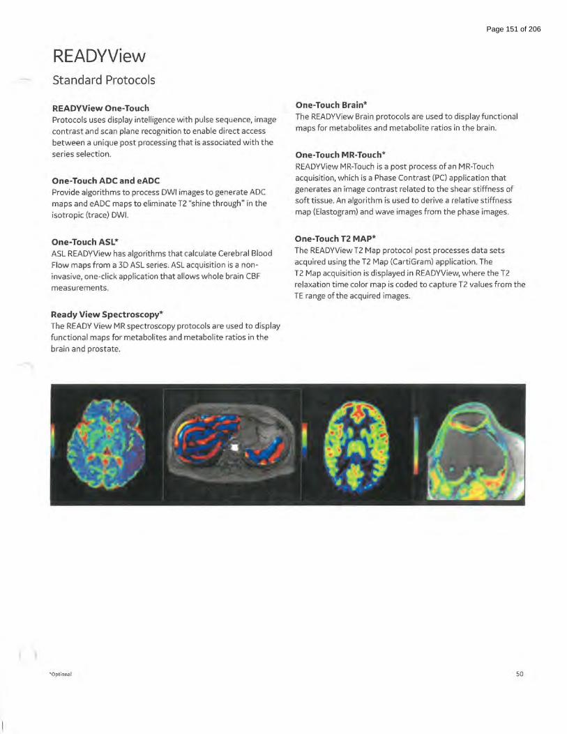
READYView Standard Protocols
READVView One-Touch Protocols uses display intelligence with pulse sequence, image
contrast and scan plane recognition to enable direct access
between a unique post processing that is associated with the
series selection.
One-Touch ADC and eADC Provide algorithms to process DWI images to generate ADC
maps and eADC maps to eliminate T2 "shine through" in the
isotropic (trace) DWI.
One-Touch ASL* ASL READYView has algorithms that calculate Cerebral Blood
Flow maps from a 3D ASL series. ASL acquisition is a non
invasive, one-click application that allows whole brain CBF
measurements.
Ready View Spectroscopy* The READY View MR spectroscopy protocols are used to display
functional maps for metabolites and metabolite ratios in the
brain and prostate.
"Optional
One-Touch Brain* The READYView Brain protocols are used to display functional
maps for metabolites and metabolite ratios in the brain.
One-Touch MR-Touch* READYView MR-Touch is a post process of an MR-Touch
acquisition, which is a Phase Contrast (PC) application that
generates an image contrast related to the shear stiffness of soft tissue. An algorithm is used to derive a relative stiffness
map (Eiastogram) and wave images from the phase images.
One-Touch T2 MAP* The READYView T2 Map protocol post processes data sets
acquired using the T2 Map (CartiGram) application. The
T2 Map acquisition is displayed in READYView, where the T2
relaxation time color map is coded to capture T2 values from the
TE range of the acquired images.
50
Page 151 of 206

READYView (continued) Integrated Registration provides you with the capability to align and fuse two volumetric acquisitions from either the same or
different acquisition modalities. Multiple 2D and 3D fusion capabilities.
The Integrated Registration application automatically detects the series that are the best candidates for registration based on
the data set attributes and the use case. After the Reference (i.e., fixed) and Registered data sets are identified, the applicable
registration methods will be automatically detected.
After the automatic registration is done, you can either directly accept automatic setup or validate it visually.
If you are still not satisfied with the result of the registration, it can be adjusted manually by translation or rotation, placing common
anatomical landmarks, or a Region Of Interest (ROI) on the Registered dataset, where the registration should be performed, can be
defined; the regions outside the ROI are ignored by the registration process.
BrainStat BrainStat is an MR Time Course imaging READYView protocol
that provides accurate spatial resolution for brain tissue viability given by hemodynamic parameters: BV, BF, TTP, MTT
(SVD), BAT, Tmax. These hemodynamic parameters can provide
unique information on tissue changes and improve delineation
of vascular-deficient or vascular-rich regions in normal and
abnormal anatomy.
MRStandard MR Standard is a time course protocol. The READYView MR
Standard is a time course protocol that can be used to create the following maps: enhancement integral (negative and
positive), time to peak, mean time to enhance, maximum slope
of increase, maximum slope of decrease.
SER SER is a time course protocol for analyzing Tl-contrast
changes. The READYView SER protocol can be used to create
the following maps: Positive enhancement integral, signal
enhancement ratio and maximum slope of increase.
"'Optional
FiberTrak* FiberTrak is designed for the advanced analysis of MR images
acquired with a DTI technique. It allows for processing of
isotropic, ADC and FA maps among other options. The FiberTrak
option augments this functionality to allow DTI processing to
create: 2D color orientation maps, 2D color eigenvector maps
and 3D tractography maps.
fMRI* Functional imaging or BOLD provides fMRI analysis using the
correlation coefficient algorithm to analyze an image set.
Neuronal activity of either motor or cognitive functions can be
mapped by fMRI through changes in signal intensity.
The resulting functional maps can be used for mapping the
motor cortex and higher cognitive regions of the brain.
R2 Star* The R2 Star feature uses water proton transverse relaxation
rates (R2) technique. It provides parametric maps for R2* (Hz)
and T2* (ms). The R2* values vary with tissue characteristics
such as iron concentration.
51
Page 152 of 206

READYView (continued) Multiparametric Protocols: Visualization at a Glance
READYView multi parametric protocols provide a guided
workflow to streamline post processing and analysis of
multiparametric studies. All measurements can be obtained
*Optional and requires two or more of the functional series selected.
with one ROI and the user customizable workflow has the ability
to display all processed maps in one screen.
52
Page 153 of 206
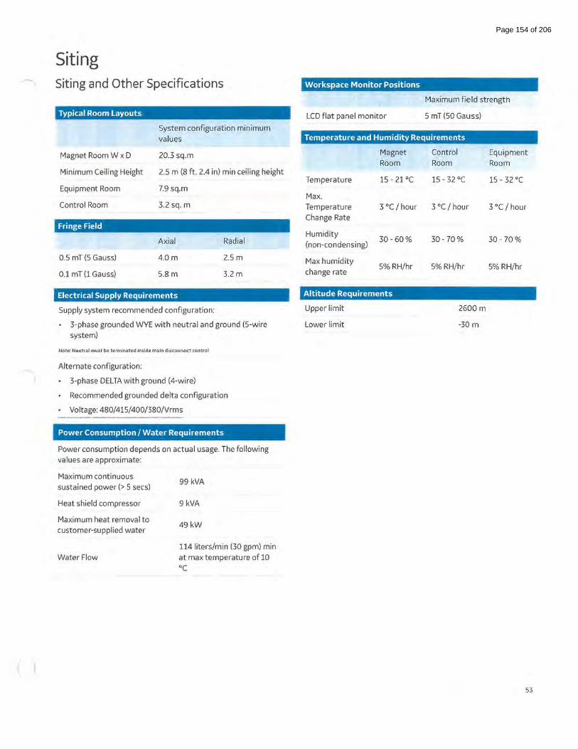
Siting "' Siting and Other Specifications
Typical Room Layouts
Magnet Room W x D
Minimum Ceiling Height
Equipment Room
Control Room
Fringe Field
0.5 mT (5 Gauss)
0.1 mT (1 Gauss)
System configuration minimum values
20.3 sq.m
2.5 m (8ft. 2.4 in) min ceiling height
7.9 sq.m
3.2 sq. m
Axial
4.0m
5.8 m
Radial
2.5 m
3.2m
Electrical Supply Requirements
Supply system recommended configuration:
3-phase grounded WYE with neutral and ground (S-wire system)
Note: Neutral must be terminated inside main disconnect control
Alternate configuration:
3-phase DELTA with ground (4-wire)
• Recommended grounded delta configuration
Voltage: 480/415/400/380/Vrms
Power Consumption I Water Requirements
Power consumption depends on actual usage. The following values are approximate:
Maximum continuous sustained power(> 5 sees)
Heat shield compressor
Maximum heat removal to customer-supplied water
WaterFlow
99 kVA
9 kVA
49kW
114 liters/min (30 gpm) min at max temperature of 10 oc
Workspace Monitor Positions
LCD flat panel monitor
Maximum field strength
5 mT (50 Gauss)
Temperature and Humidity Requirements
Magnet Room
Temperature 15- 21 oc
Max. Temperature 3 oc I hour Change Rate
Humidity 30-60%
(non-condensing)
Max humidity 5% RH/hr
change rate
Altitude Requirements
Upper limit
Lower limit
Control Equipment Room Room
15- 32 oc 15- 32 oc
3 oc I hour 3 °C I hour
30-70% 30-70%
5% RH/hr 5% RH/hr
2600m
-30m
53
Page 154 of 206

Miscellaneous Alternative environments
Modular buildings may also be available (including air
conditioning, heating, chiller, RF shielding, additional magnetic
shielding in walls). Contact your local GE representative forGE
certified designs and vendors.
Please ask your local GE project manager for a comprehensive
installation and siting manual.
Filming considerations
Filming requires the SIGNA'M Artist analog or digital filming.
Interface (purchased separately) unless DICOM Print will be
used exclusively for software filming to DICOM Print peripheral
devices. An Analog/VDB or Digitai/LCAM Camera Interface is
typically required for most installations.
Accessory Package
SPT phantom set with storage cart
Customer diagnostic software
Operator manuals
Patient log books
Emergency stop
Disconnects electrical power from RF and gradient components
in the magnet room (duplicate control at the magnet).
Warranty
The published GE warranty in effect on the date of shipment
shall apply. GE reserves the right to make changes.
lnSite* Remote Diagnostics
GE's unique remote service and applications support including
magnet monitoring. Also allows downloading of applications
software such as eFiexTrials program.
Accessories package
A comprehensive suite of MR compatible accessories is available
on the SIGNA'M Artist. Please contact your
GE representative for details.
GE regulatory compliance
The SIGNA'M Artist complies with all applicable safety
standards including but not limited to IEC60601-1, IEC60601-1-2
(Electromagnetic Compatibility), and IEC 60601-2-33 (MR).
5537470
LASER RADIATION 0 NOT STARE INTO THE BEAM
OR VIEW DIRECTLY WITH OPTICAL INSTRUMENTS
CLASS 2M LASER PRODUCT 1mW @655nm
IEC 60825-1 :2007
54
Page 155 of 206
Imagination at work
(<,;) 2020 General Electric Company- All rights reserved.
GE Healthcare reserves the right to make changes in specific~tions and features shown herein, or discontinue the product described at any time without notice or obligation. Contact your GE Healthcare representative for the most current information. SIGNA. GE and the GE Monogram, are trademarks ofGener<~l Electric Company. GE Healthcare, a division of General Electric Company. GE Medical Systems, Inc., doing business as GE Healthcare.
Reproduction in any form is forbidden without prior written permission from GE. Nothing in this material should be used to diagnose or treat any disease or condition. Readers must consult a healthcare professional.
Intel and X eon are trademarks of Intel Corporation or its subsidiaries in the U.S. and/or other countries.
NVidial!; and Quadro® are trademarks ofNVIDIA Corporation.
DOC2452375
Page 156 of 206

1.5T AIR™ Multi-Purpose Coils The 21-channel AIR™ Multi-Purpose (MP) Coil Large and the 20-channel AIR'M Multi-Purpose (MP) Coil Medium are next generation multipurpose coil that allow flexibility in any direction to conform to the patient's anatomy. Based on the innovative AIR'M Coil technologies, the AIR'M MP Coils provide good image quality and acceleration performance, while improving the overall patient and user experience. These coils have been designed to adapt various patient shapes and sizes, expanding positioning versatility. AIR'M MP Coil Large is recommended to be used for Shoulder, Knee, Foot, Ankle, Hips, Prostate. AIR'M MP Coil Medium is recommended to be used for Wrist, Elbow, Cardiac.
The AIR'M MP Coil positioner kit includes a foot-ankle positioner, a knee positioner, a wedge pad, au-shaped pad and a strap kit. Those are compatible with both AIR'M MP Coils Large and Medium for positioning.
AIR'M MP Coils Specifications
Coil
Elements
Weight
Dimensions (W x L x H)
Coverage (W x L)
Patient orientation
l.ST AIR"' Multi-Purpose Coil Large
(l.ST AIR'"' MP L)
21
1.6 kg (3.5 lbs) resting on patient
2.4kg (5.21bs) with the cable
40 em x 74 em x 1.2 em
30cm x68 em
Head first I feet first
l.ST AIR'"' Multi-Purpose Coil Medium
(l.ST AIR'"' MP M)
20
1.2 kg (2.6 lbs) resting on patient
2.0kg (4.3 lbs) with the cable
40 em x 54.5 em x 1.2 em
30 em x 48.5 em
Head first I feet first
•
Page 157 of 206

l.ST AIR™ Multi-Purpose Coils (Continued) Coil compatibility
SIGNNM Artist*
Coil combinations
SIGNATM Voyagert
Coil combinations
*Minimum DV27.1 software **Minimum DV29.1 software ***Minimum DV27.1 R02 software
AIR'M MP Coil Large can be combined with the following coils:
GEM Head Neck Unit
GEM Posterior Array
30ch AIR'M Anterior Array
TDI Anterior Array**
2nd AIR'M MP Coil Large
AIR'M MP Coil Medium
Rapid Endorectal Coil***
AIR'M MP Coil Large can be combined with the following coils:
TDI Posterior Array
TDI Anterior Arrayt
16ch AIR'M Anterior Array'!'
Rapid Endorectal Coil
tMinimum VX29.1 software and 49ch system configuration *Requires 65ch system configuration
IS> 2021 General Electric Company- All rights reserved .
GE Healthcare reserves the right to make changes in specificat io•ls and features shown herein, or discontinue the product described
at any time without notice or obligation. Contact your GE Htoalthcare representative for the most current information. GE and the GE
AIRrM MP Coil Medium can be combined with the following coils:
GEM Head Neck Unit
GEM Posterior Array
30ch AIR'M Anterior Array
TDI Anterior Array**
AIR'M MP Coil Large
2nd AIR'M MP Coil Medium
Rapid Endorectal Coil***
AIR'M MP Coil Medium can be combined with the following coils:
TDI Posterior Array
TDI Anterior Arrayt
16ch AIRrM Anterior Array'!'
Rapid Endorectal Coil
Monogram, are trademarks of General Electric Company. GE Healthcare, a division of General Electric Company AIR' .. and SIGNA'"' are trademarks ofGE Healthcare. GE Medical Systems, Inc., doing business as GE Healthcare. • 00(2522821
Page 158 of 206

Page 159 of 206

Page 160 of 206

Page 161 of 206

Page 162 of 206

Page 163 of 206

Page 164 of 206

Page 165 of 206

Page 166 of 206

Page 167 of 206

Page 168 of 206

Page 169 of 206

Page 170 of 206

Page 171 of 206

Page 172 of 206

Page 173 of 206

Page 174 of 206

Page 175 of 206

Page 176 of 206

Page 177 of 206

Page 178 of 206

Page 179 of 206
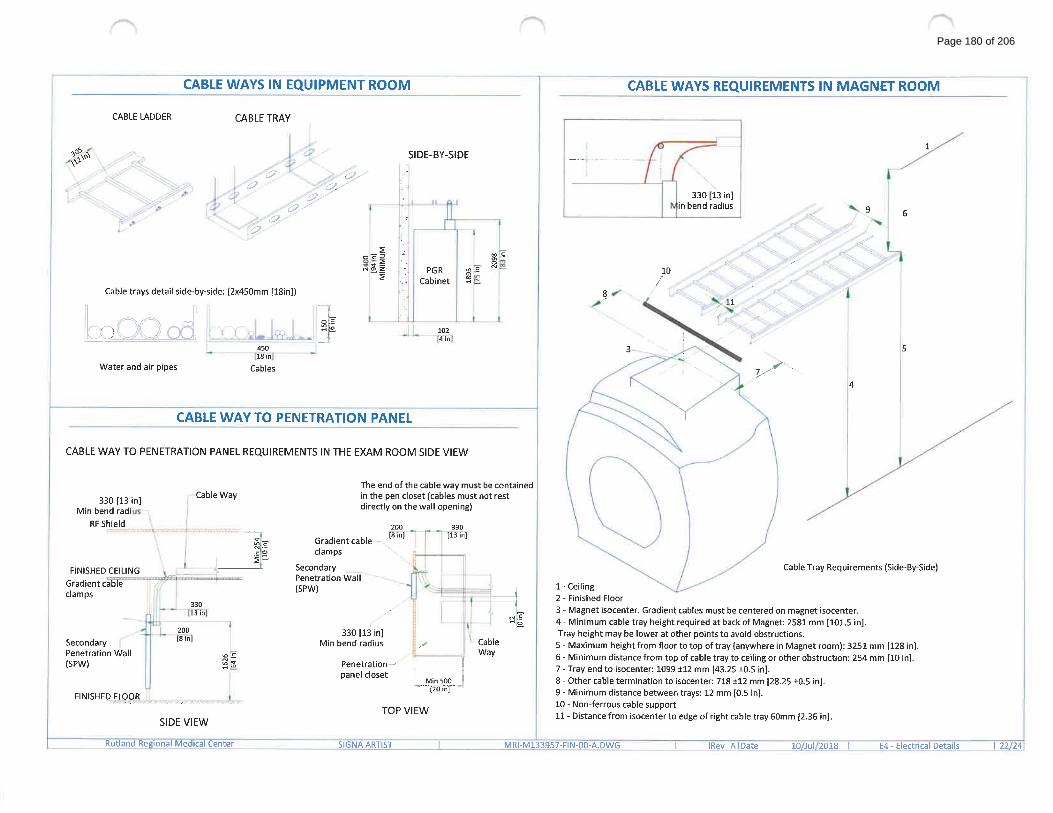
Page 180 of 206

Page 181 of 206

Page 182 of 206
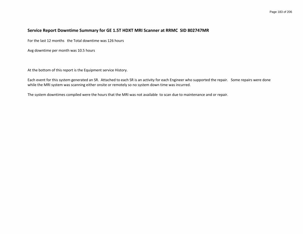
Service Report Downtime Summary for GE 1.5T HDXT MRI Scanner at RRMC SID 802747MR For the last 12 months the Total downtime was 126 hours Avg downtime per month was 10.5 hours At the bottom of this report is the Equipment service History. Each event for this system generated an SR. Attached to each SR is an activity for each Engineer who supported the repair. Some repairs were done while the MRI system was scanning either onsite or remotely so no system down time was incurred. The system downtimes compiled were the hours that the MRI was not available to scan due to maintenance and or repair.
Page 183 of 206

Service Reports for SID 802747MR CR 25 Jun 2021 System Downtime for this event: 1.0
Status: Open Owner: Harold Flad Problem: 8Ch NeuroVascular coil “Invivo” GE Part # 2317115-2 Assembly # 103045 shows low signal on one or more channels. Passes MCQA
CR 21 Jun 2021 System Downtime for this event: 2.0
Status: Closed Owner: Charles Nutt Problem:
We iLinqed today about pre scan failure, they said it might be the A port, just tried Abdomen scan and got the same pre scan failure. Brain coil is working fine, it seems anything with A port gets the pre scan failure. Image:NONE-NONE-NONE
CR 21 Jun 2021 System Downtime for this event: 3.0 Status: Closed Owner: Harold Flad Problem: Driver Module fault Image:NONE-NONE-NONE
Page 184 of 206

CR 18 Jun 2021 System Downtime for this event: 69.0
Status: Pending Approval Owner: Robert Ochal Problem:
Getting error, PDCU is in stop mode, Emergency stop button was pressed or PDU 24 volt supply has failed. We did not hit the emergency stop. Image:NONE-NONE-NONE
PM System Downtime for this event: 0.0 17 Jun 2021
Status: Open Owner: Harold Flad Problem: PM Jul 2021
CR System Downtime for this event: 0.0 15 Jun 2021
Status: Open Owner: Harold Flad Problem:
Analogic RF Amplifier Peak Power Fault in error log CD 06/16/2021 08:00:00 (GMT-05:00) Eastern Time (US & Canada) Customer has delayed service, please refer to the Planned Date to get this information and page the FE when needed. / Customer Symptom: Axial t2 and PD sequences on Lumbar scans giving RF failure error. Tried TPS reset, did not fix issue. Weekend techs said the same error happened on lumbar scan this last weekend. Unable to run both axial t2 and PD, all sag sequences ran without issue. Image:Lumbar wo-1446,1447-NONE | <S>
CR 14 Jun 2021 System Downtime for this event: 0.0
Status: Closed Owner: David Baxter Problem: NVA image quality complaint
CR System Downtime for this event: 0.0 14 Jun 2021
Status: Closed Owner: Harold Flad Problem: low signal on one channel on Invivo 8 Ch brain coil
CR System Downtime for this event: 1.0 07 Jun 2021
Status: Closed Owner: Harold Flad Problem: Knee coil will not pass MCQA
CR System Downtime for this event: 0.0 03 Jun 2021
Status: Closed Owner: David Baxter Problem: ongoing white pixels
CR System Downtime for this event: 6.5 02 Jun 2021
Page 185 of 206

Status: Closed Owner: Douglas Cyr Problem: EPIWP analysis for FE.
CR System Downtime for this event: 1.0 18 May 2021
Status: Closed Owner: Alejandro Jasso Problem: Customer states that the unit is down.
CR System Downtime for this event: 27.0 25 Apr 2021
Status: Pending Approval Owner: Douglas Cyr Problem: Customer states that the computed on the machine crashed
and won't reboot.
CR System Downtime for this event: 2.0 19 Mar 2021
Status: Assigned Owner: Harold Flad Problem: White Pixel noise causing Intermittent corduroy artifact on some coils
PM System Downtime for this event: 0.0 16 Mar 2021 Status: Cancelled Owner: Harold Flad Problem: PM Apr 2021. Exempted - Revised PM DUE Date Jun 2021.
CR System Downtime for this event: 0.0 02 Mar 2021
Status: Closed Owner: Alejandro Jasso Problem: Customer needs service reports.
CR System Downtime for this event: 1.5 27 Feb 2021
Status: Closed Owner: Robert Ochal Problem:
CD 3/1/2021 08:00 AM COULD YOU PLEASE NOTIFY OUR FE THAT WE ARE HAVING TROUBLE WITH THE NUMBER 3 COIL IN OUR 16CH NEURO ARRAY COIL. PLEASE HAVE HIM GIVE US A CALL ON MONDAY. THANKS Image:NONE-NONE-NONE
CR System Downtime for this event: 1.5 26 Jan 2021
Page 186 of 206

Status: Closed Owner: Harold Flad Problem: artifact on DWI, cube and fiesta sequences. Noticed a few days ago, continuing today. Image:NONE-NONE-NONE
CR System Downtime for this event: 1.0 25 Dec 2020
Status: Closed Owner: Kyle Tempesta Problem: Breaker to PDU tripped. System reset but TPS system not responding after multiple resets. Image:NONE-NONE-NONE
CR System Downtime for this event: 0.0 22 Dec 2020
Status: Closed Owner: Gary Henning Problem:
I SEE THAT SOMEONE IS IN MY SYSTEM. COULD THEY PLEASE LOOK TO SEE WHY IM GETTING MULTI BIAS-FAULT ERRORS. Image:NONE-NONE-NONE
PM System Downtime for this event: 3.0 16 Dec 2020
Status: Closed Owner: Harold Flad Problem: PM Jan 2021
CR System Downtime for this event: 0.0 13 Dec 2020
Status: Closed Owner: Avery Thomas Problem:
Equipment room overheated. Maintenance working on AC. Chiller off most of the night. back on around 0930 this morning. Able to scan for short while but now getting Gradient problem messages. Please call 802-747-1707. Image:NONE-NONE-NONE
CR System Downtime for this event: 0.0 13 Dec 2020
Status: Closed Owner: Charles Nutt Problem: MRM80002. P1 - Compressor Off (Magnet)
Page 187 of 206

CR System Downtime for this event: 0.0 07 Dec 2020
Status: Open Owner: Harold Flad Problem:
Default Password Update – Insite One – During your next service or PM, please change the passwords identified in the Service Note for this modality. The new password will be the “Row-ID” identifier for this system ID and can be found in Service Request screen (Not the Activity Screen) under the heading System Info in FX. In your debrief “Action” comments, confirm “Password changed to “XXXXXX” to document the actual password used. Consult your service documentation or contact Technical Support if you have questions. Reminder: passwords are case sensitive, and mistakes will likely require reset via LFC!
CR System Downtime for this event: 0.0 16 Nov 2020 Status: Closed Owner: William Nichol Problem: MRM80002. P1 - Compressor Off (Magnet)
CR System Downtime for this event: 0.5 08 Nov 2020
Status: Closed Owner: Gary Henning Problem:
We are starting to see gradient amplifier not ready messages when trying to do Axial T2 FSE sequences. Has happened a couple of times this week. Most recently about 5 mins ago. Image:NONE-NONE-NONE
PM System Downtime for this event: 1.0 16 Sep 2020
Status: Closed Owner: Harold Flad Problem: PM Oct 2020
CR System Downtime for this event: 1.0 31 Aug 2020 Status: Closed Owner: Harold Flad Problem: 12 channel anterior coil cabling has a crack in the cover.
CR System Downtime for this event: 1.0 25 Jun 2020
Page 188 of 206

Status: Closed Owner: Harold Flad Problem:
There is a problem with the alignment on the table. It is rubbing as it comes out and causing a horrible grinding noise. Could you please let my FE know he needs to come and adjust it. Image:NONE-NONE-NONE
PM System Downtime for this event: 3.0 16 Jun 2020
Status: Closed Owner: Harold Flad Problem: PM Jul 2020
Page 189 of 206

PLEASE PROVIDE ASSUMPTIONSRutland Regional Medical Center
MRI Replacement
Table 11. The trade in amount for the old MRI has been added to the capital cost of the new, as it is assumed that the trade in value is equivalent to the Fair Market Value.
Table 3A2. Net Patient Care Revenue, including Fixed Prospective Payments, is assumed to increase 3.5% each year FY 2023-FY 20253. Operating Margin is assumed to be 2.5% each year FY 2023-FY2025.4. Bad Debt and Free Care as a percent of gross revenue are assumed to remain consistent with the 2022 budget.5. Medicaid DPS for FY 2023-FY2025 is anticipated to be consistent with budget 2022 Medicaid DPS as a percent of Net Patient Care Revenue.
Table 3B6. Project-related expenses include depreciation, training, and service agreements.7. Other Operating Revenue represents the gain on trade in of the current MRI.
Table 6A8. Payer mix and contractual allowances are consistent with budget 2022.
Table 79. Utilization numbers for FY 2023-2025 are consistent with the 2022 budget.
Table 810. FTE numbers for FY 2023-2025 are consistent with the 2022 budget.
10/12/2021 GMCB Assumptions
Page 190 of 206
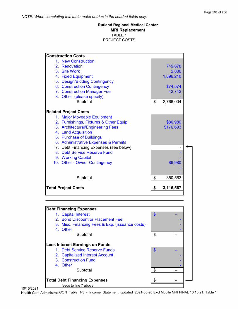
NOTE: When completing this table make entries in the shaded fields only.
Construction Costs1. New Construction2. Renovation 749,678 3. Site Work 2,800 4. Fixed Equipment 1,896,210 5. Design/Bidding Contingency6. Construction Contingency $74,5747. Construction Manager Fee 42,742 8. Other (please specify) -
Subtotal 2,766,004$
Related Project Costs1. Major Moveable Equipment2. Furnishings, Fixtures & Other Equip. $86,9803. Architectural/Engineering Fees $176,6034. Land Acquisition5. Purchase of Buildings6. Administrative Expenses & Permits7. Debt Financing Expenses (see below) - 8. Debt Service Reserve Fund - 9. Working Capital -
10. Other - Owner Contingency 86,980 - -
Subtotal 350,563$
Total Project Costs 3,116,567$
Debt Financing Expenses1. Capital Interest -$ 2. Bond Discount or Placement Fee - 3. Misc. Financing Fees & Exp. (issuance costs) - 4. Other -
Subtotal -$
Less Interest Earnings on Funds1. Debt Service Reserve Funds -$ 2. Capitalized Interest Account - 3. Construction Fund - 4. Other -
Subtotal -$
Total Debt Financing Expenses -$ feeds to line 7 above
TABLE 1PROJECT COSTS
Rutland Regional Medical CenterMRI Replacement
10/15/2021Health Care AdministrationCON_Table_1-3_-_Income_Statement_updated_2021-05-20 Excl Mobile MRI FINAL 10.15.21, Table 1
Page 191 of 206
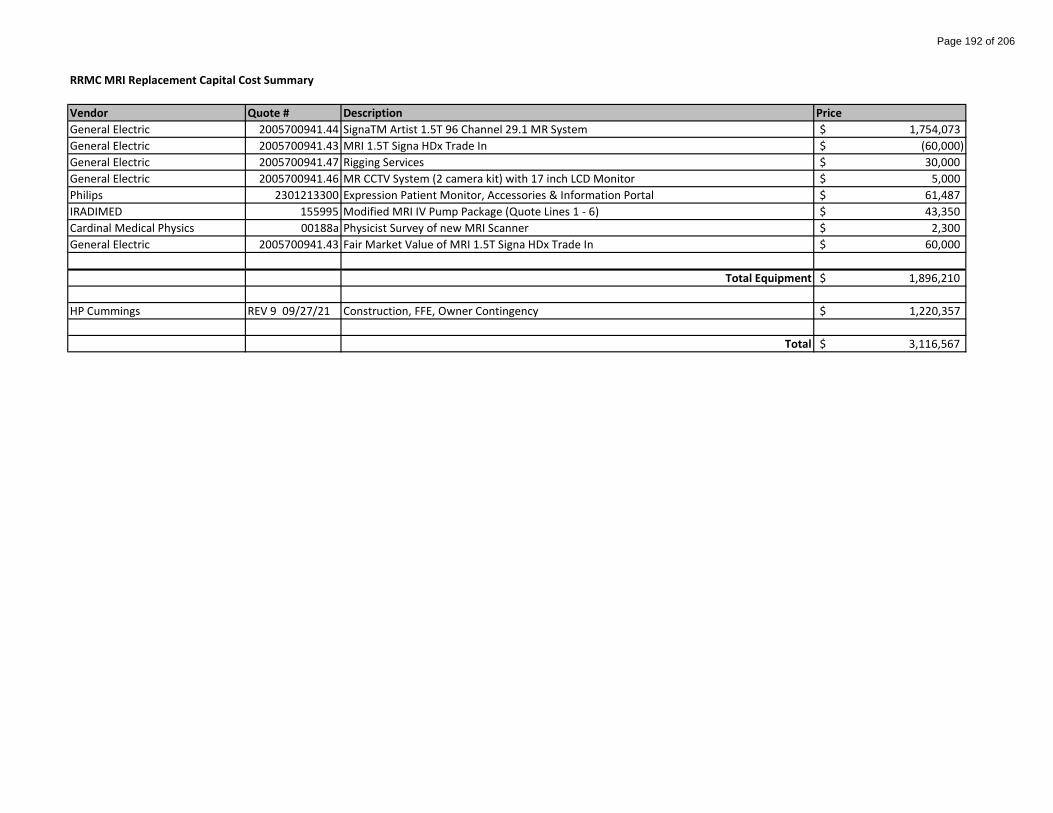
RRMC MRI Replacement Capital Cost Summary
Vendor Quote # Description PriceGeneral Electric 2005700941.44 SignaTM Artist 1.5T 96 Channel 29.1 MR System 1,754,073$ General Electric 2005700941.43 MRI 1.5T Signa HDx Trade In (60,000)$ General Electric 2005700941.47 Rigging Services 30,000$ General Electric 2005700941.46 MR CCTV System (2 camera kit) with 17 inch LCD Monitor 5,000$ Philips 2301213300 Expression Patient Monitor, Accessories & Information Portal 61,487$ IRADIMED 155995 Modified MRI IV Pump Package (Quote Lines 1 - 6) 43,350$ Cardinal Medical Physics 00188a Physicist Survey of new MRI Scanner 2,300$ General Electric 2005700941.43 Fair Market Value of MRI 1.5T Signa HDx Trade In 60,000$
Total Equipment 1,896,210$
HP Cummings REV 9 09/27/21 Construction, FFE, Owner Contingency 1,220,357$
Total 3,116,567$
Page 192 of 206

NOTE: When completing this table make entries in the shaded fields only.
Sources of Funds
1. Financing Instrument Bond
a. Interest Rate 0.0%
b. Loan Period To:
c. Amount Financed -$
2. Equity Contribution 3,116,567
3. Other Sources
a. Working Capital -
b. Fundraising -
c. Grants -
d. Other -
Total Required Funds 3,116,567$
Uses of Funds
Project Costs (feeds from Table 1)
1. New Construction -$
2. Renovation 749,678
3. Site Work 2,800
4. Fixed Equipment 1,896,210
5. Design/Bidding Contingency -
6. Construction Contingency 74,574
7. Construction Manager Fee 42,742
8. Major Moveable Equipment -
9. Furnishings, Fixtures & Other Equip. 86,980
10. Architectural/Engineering Fees 176,603
11. Land Acquisition -
12. Purchase of Buildings -
13. Administrative Expenses & Permits -
14. Debt Financing Expenses -
15. Debt Service Reserve Fund -
16. Working Capital -
17. Other (please specify) 86,980
Total Uses of Funds 3,116,567$
Total sources should equal total uses of funds.
Rutland Regional Medical CenterMRI Replacement
TABLE 2DEBT FINANCING ARRANGEMENT, SOURCES & USES OF FUNDS
10/15/2021Health Care AdministrationCON_Table_1-3_-_Income_Statement_updated_2021-05-20 Excl Mobile MRI FINAL 10.15.21, Table 2
Page 193 of 206

Proposed Yr 1 Proposed Yr 2 Proposed Yr 3
2020 2021 2021 2022 2023 2024 2025
Actual Budget % change Projection % change Budget % change % change % change%
change
REVENUES INPATIENT CARE REVENUE 183,480,641 194,725,235 6.1% 196,085,443 0.7% 209,653,759 6.9% 225,555,404$ 7.6% 243,261,915$ 7.9% 263,026,797$ 8.1% OUTPATIENT CARE REVENUE 301,057,099 325,575,607 8.1% 359,113,980 10.3% 367,569,301 2.4% 395,448,394$ 7.6% 426,491,815$ 7.9% 461,144,014$ 8.1% OUTPATIENT CARE REVENUE - PHYSICIAN 54,903,250 57,988,255 5.6% 60,948,483 5.1% 62,615,572 2.7% 67,364,786$ 7.6% 72,653,045$ 7.9% 78,556,060$ 8.1% CHRONIC/SNF PT CARE REVENUE - - #DIV/0! - #DIV/0! - #DIV/0! 0 #DIV/0! -$ #DIV/0! -$ #DIV/0! SWING BEDS PT CARE REVENUE - - #DIV/0! - #DIV/0! - #DIV/0! 0 #DIV/0! -$ #DIV/0! -$ #DIV/0!
GROSS PATIENT CARE REVENUE 539,440,990 578,289,097 7.2% 616,147,906 6.5% 639,838,632 3.8% 688,368,584 7.6% 742,406,775 7.9% 802,726,872 8.1%
DISPROPORTIONATE SHARE PAYMENTS 3,242,526 3,370,325 3.9% 3,383,358 0.4% 3,435,517 1.5% 3,555,760 3.5% 3,680,212 3.5% 3,809,019 3.5% BAD DEBT FREE CARE (14,709,372) (15,283,355) 3.9% (9,123,515) -40.3% (9,876,429) 8.3% (10,625,528) 7.6% (11,459,652) 7.9% (12,390,741) 8.1% DEDUCTIONS FROM REVENUE (287,746,728) (318,089,163) 10.5% (342,451,314) 7.7% (361,822,542) 5.7% (400,218,507) 10.6% (443,709,215) 10.9% (493,044,895) 11.1%NET PATIENT CARE REVENUE 240,227,416 248,286,904 3.4% 267,956,435 7.9% 271,575,178 1.4% 281,080,309 3.5% 290,918,120 3.5% 301,100,255 3.5% FIXED PROSPECTIVE PAYMENTS AND RESERVES (972,075) (799,220) -17.8% (1,087,622) 36.1% (1,213,808) 11.6% (1,256,291) 3.5% (1,300,261) 3.5% (1,345,771) 3.5%NET PATIENT CARE REV & FIXED PAYMENTS & RESERVES 239,255,341 247,487,684 3.4% 266,868,813 7.8% 270,361,370 1.3% 279,824,018 3.5% 289,617,859 3.5% 299,754,484 3.5%
OTHER OPERATING REVENUE 42,639,646 20,983,965 -50.8% 32,894,263 56.8% 20,745,734 -36.9% 21,245,734 2.4% 20,745,734 -2.4% 20,745,734 0.0%
TOTAL OPERATING REVENUE 281,894,987 268,471,649 -4.8% 299,763,076 11.7% 291,107,104 -2.9% 301,069,752 3.4% 310,363,593 3.1% 320,500,218 3.3%
OPERATING EXPENSE SALARIES NON MD 94,100,223 89,765,463 -4.6% 95,663,626 6.6% 95,658,732 0.0% 98,528,494 3.0% 101,484,349 3.0% 104,528,879 3.0% FRINGE BENEFITS NON MD 29,782,342 26,572,590 -10.8% 28,398,412 6.9% 30,635,972 7.9% 31,555,051 3.0% 32,501,703 3.0% 33,476,754 3.0% PHYSICIAN FEES & SALARIES 33,998,006 32,633,878 -4.0% 34,765,222 6.5% 34,314,275 -1.3% 34,314,275 0.0% 35,064,275 2.2% 35,814,275 2.1% FRINGE BENEFITS MD 1,651,399 1,472,862 -10.8% 1,607,147 9.1% 1,691,419 5.2% 1,691,419 0.0% 1,734,170 2.5% 1,776,921 2.5% HEALTH CARE PROVIDER TAX 15,348,072 14,419,633 -6.0% 15,416,978 6.9% 16,064,517 4.2% 16,626,775 3.5% 17,208,712 3.5% 17,811,017 3.5% DEPRECIATION AMORTIZATION 12,592,768 12,668,835 0.6% 12,726,667 0.5% 12,712,258 -0.1% 12,926,032 1.7% 12,998,135 0.6% 13,056,887 0.5% INTEREST - LONG/SHORT TERM 1,302,013 1,434,557 10.2% 1,216,636 -15.2% 1,562,040 28.4% 1,589,309 1.7% 1,537,187 -3.3% 1,483,964 -3.5% OTHER OPERATING EXPENSE 92,574,674 87,803,081 -5.2% 103,940,860 18.4% 98,346,528 -5.4% 96,446,528 -1.9% 100,111,496 3.8% 104,416,290 4.3%
TOTAL OPERATING EXPENSE 281,349,496 266,770,899 -5.2% 293,735,548 10.1% 290,985,741 -0.9% 293,677,883 0.9% 302,640,027 3.1% 312,364,988 3.2%278,273,483
NET OPERATING INCOME (LOSS) 545,491 1,700,750 211.8% 6,027,528 254.4% 121,363 -98.0% 7,391,869 5990.7% 7,723,566 4.5% 8,135,231 5.3%
NON-OPERATING REVENUE 14,875,238 8,380,504 -43.7% 17,168,103 104.9% 6,373,864 -62.9% 5,822,035 -8.7% 6,010,708 3.2% 6,205,580 3.2%
EXCESS (DEFICIT) OF REVENUE OVER EXPENSE 15,420,729 10,081,254 -34.6% 23,195,631 130.1% 6,495,227 -72.0% 13,213,904 103.4% 13,734,274 3.9% 14,340,811 4.4%
Operating Margin % 0.2% 0.6% 2.0% 0.0% 2.5% 2.5% 2.5%Bad Debt & Free Care% 2.7% 2.6% 1.5% 1.5% 1.5% 1.5% 1.5%Compensation Ratio 56.7% 56.4% 54.6% 55.8% 56.6% 56.4% 56.2%Capital Cost % of Total Expenses 4.9% 5.3% 4.7% 4.9% 4.9% 4.8% 4.7%
RUTLAND REGIONAL MEDICAL CENTER
INCOME STATEMENT
MRI Replacement
WITHOUT PROJECTTable 3A
10/15/2021, Pg 1 GMCB CON_Table_1-3_-_Income_Statement_updated_2021-05-20 Excl Mobile MRI FINAL 10.15.21
Page 194 of 206
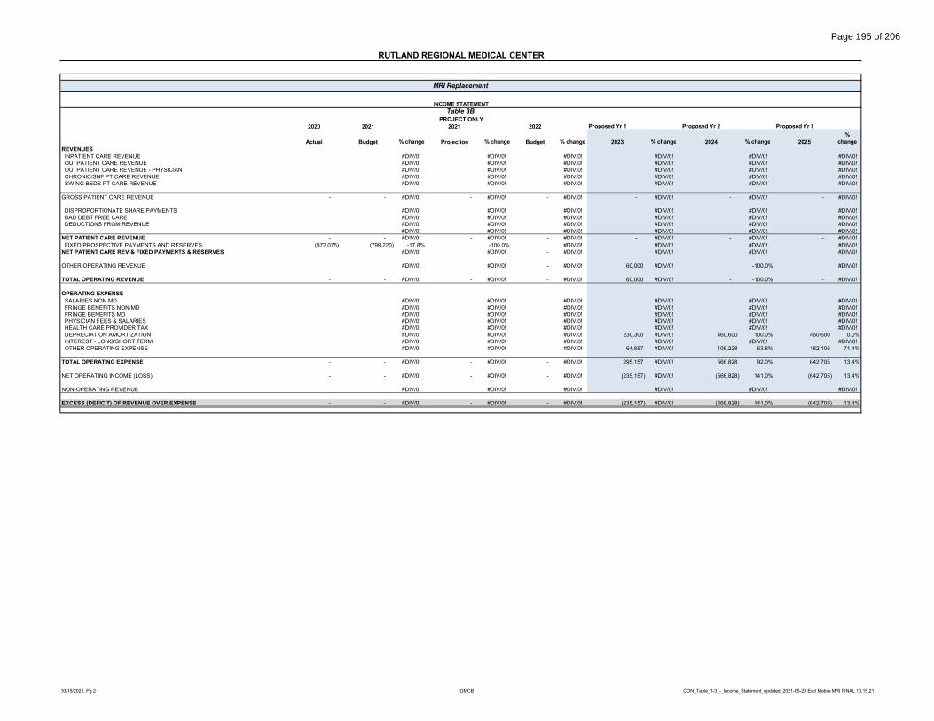
RUTLAND REGIONAL MEDICAL CENTER
2020 2021 2021 2022 Proposed Yr 1 Proposed Yr 2 Proposed Yr 3
Actual Budget % change Projection % change Budget % change 2023 % change 2024 % change 2025%
changeREVENUES INPATIENT CARE REVENUE #DIV/0! #DIV/0! #DIV/0! #DIV/0! #DIV/0! #DIV/0! OUTPATIENT CARE REVENUE #DIV/0! #DIV/0! #DIV/0! #DIV/0! #DIV/0! #DIV/0! OUTPATIENT CARE REVENUE - PHYSICIAN #DIV/0! #DIV/0! #DIV/0! #DIV/0! #DIV/0! #DIV/0! CHRONIC/SNF PT CARE REVENUE #DIV/0! #DIV/0! #DIV/0! #DIV/0! #DIV/0! #DIV/0! SWING BEDS PT CARE REVENUE #DIV/0! #DIV/0! #DIV/0! #DIV/0! #DIV/0! #DIV/0!
GROSS PATIENT CARE REVENUE - - #DIV/0! - #DIV/0! - #DIV/0! - #DIV/0! - #DIV/0! - #DIV/0!
DISPROPORTIONATE SHARE PAYMENTS #DIV/0! #DIV/0! #DIV/0! #DIV/0! #DIV/0! #DIV/0! BAD DEBT FREE CARE #DIV/0! #DIV/0! #DIV/0! #DIV/0! #DIV/0! #DIV/0! DEDUCTIONS FROM REVENUE #DIV/0! #DIV/0! #DIV/0! #DIV/0! #DIV/0! #DIV/0!
#DIV/0! #DIV/0! #DIV/0! #DIV/0! #DIV/0! #DIV/0!NET PATIENT CARE REVENUE - - #DIV/0! - #DIV/0! - #DIV/0! - #DIV/0! - #DIV/0! - #DIV/0! FIXED PROSPECTIVE PAYMENTS AND RESERVES (972,075) (799,220) -17.8% -100.0% #DIV/0! #DIV/0! #DIV/0! #DIV/0!NET PATIENT CARE REV & FIXED PAYMENTS & RESERVES #DIV/0! #DIV/0! - #DIV/0! #DIV/0! #DIV/0! #DIV/0!
OTHER OPERATING REVENUE #DIV/0! #DIV/0! - #DIV/0! 60,000 #DIV/0! -100.0% #DIV/0!
TOTAL OPERATING REVENUE - - #DIV/0! - #DIV/0! - #DIV/0! 60,000 #DIV/0! - -100.0% - #DIV/0!
OPERATING EXPENSE SALARIES NON MD #DIV/0! #DIV/0! #DIV/0! #DIV/0! #DIV/0! #DIV/0! FRINGE BENEFITS NON MD #DIV/0! #DIV/0! #DIV/0! #DIV/0! #DIV/0! #DIV/0! FRINGE BENEFITS MD #DIV/0! #DIV/0! #DIV/0! #DIV/0! #DIV/0! #DIV/0! PHYSICIAN FEES & SALARIES #DIV/0! #DIV/0! #DIV/0! #DIV/0! #DIV/0! #DIV/0! HEALTH CARE PROVIDER TAX #DIV/0! #DIV/0! #DIV/0! #DIV/0! #DIV/0! #DIV/0! DEPRECIATION AMORTIZATION #DIV/0! #DIV/0! #DIV/0! 230,300 #DIV/0! 460,600 100.0% 460,600 0.0% INTEREST - LONG/SHORT TERM #DIV/0! #DIV/0! #DIV/0! #DIV/0! #DIV/0! #DIV/0! OTHER OPERATING EXPENSE #DIV/0! #DIV/0! #DIV/0! 64,857 #DIV/0! 106,228 63.8% 182,105 71.4%
TOTAL OPERATING EXPENSE - - #DIV/0! - #DIV/0! - #DIV/0! 295,157 #DIV/0! 566,828 92.0% 642,705 13.4%
NET OPERATING INCOME (LOSS) - - #DIV/0! - #DIV/0! - #DIV/0! (235,157) #DIV/0! (566,828) 141.0% (642,705) 13.4%
NON-OPERATING REVENUE #DIV/0! #DIV/0! #DIV/0! #DIV/0! #DIV/0! #DIV/0!
EXCESS (DEFICIT) OF REVENUE OVER EXPENSE - - #DIV/0! - #DIV/0! - #DIV/0! (235,157) #DIV/0! (566,828) 141.0% (642,705) 13.4%
MRI Replacement
INCOME STATEMENT
Table 3BPROJECT ONLY
10/15/2021, Pg 2 GMCB CON_Table_1-3_-_Income_Statement_updated_2021-05-20 Excl Mobile MRI FINAL 10.15.21
Page 195 of 206

RUTLAND REGIONAL MEDICAL CENTER
2020 2021 2021 2022 Proposed Yr 1 Proposed Yr 2 Proposed Yr 3
Actual Budget % change Projection % change Budget %
change 2023 % change 2024 % change 2025 %
change REVENUES INPATIENT CARE REVENUE 183,480,641 194,725,235 6.1% 196,085,443 0.7% 209,653,759 6.9% 225,555,404 7.6% 243,261,915 7.9% 263,026,797 8.1% OUTPATIENT CARE REVENUE 301,057,099 325,575,607 8.1% 359,113,980 10.3% 367,569,301 2.4% 395,448,394 7.6% 426,491,815 7.9% 461,144,014 8.1% OUTPATIENT CARE REVENUE - PHYSICIAN 54,903,250 57,988,255 5.6% 60,948,483 5.1% 62,615,572 2.7% 67,364,786 7.6% 72,653,045 7.9% 78,556,060 8.1% CHRONIC/SNF PT CARE REVENUE - - #DIV/0! - #DIV/0! - #DIV/0! - #DIV/0! - #DIV/0! - #DIV/0! SWING BEDS PT CARE REVENUE - - #DIV/0! - #DIV/0! - #DIV/0! - #DIV/0! - #DIV/0! - #DIV/0!
GROSS PATIENT CARE REVENUE 539,440,990 578,289,097 7.2% 616,147,906 6.5% 639,838,632 3.8% 688,368,584 7.6% 742,406,775 7.9% 802,726,872 8.1%
DISPROPORTIONATE SHARE PAYMENTS 3,242,526 3,370,325 3.9% 3,383,358 0.4% 3,435,517 1.5% 3,555,760 3.5% 3,680,212 3.5% 3,809,019 3.5% BAD DEBT FREE CARE (14,709,372) (15,283,355) 3.9% (9,123,515) -40.3% (9,876,429) 8.3% (10,625,528) 7.6% (11,459,652) 7.9% (12,390,741) 8.1% DEDUCTIONS FROM REVENUE (287,746,728) (318,089,163) 10.5% (342,451,314) 7.7% (361,822,542) 5.7% (400,218,507) 10.6% (443,709,215) 10.9% (493,044,895) 11.1%
- - #DIV/0! - #DIV/0! - #DIV/0! - #DIV/0! - #DIV/0! - #DIV/0!NET PATIENT CARE REVENUE 240,227,416 248,286,904 3.4% 267,956,435 7.9% 271,575,178 1.4% 281,080,309 3.5% 290,918,120 3.5% 301,100,255 3.5% FIXED PROSPECTIVE PAYMENTS AND RESERVES (1,944,150) (1,598,440) -17.8% (1,087,622) -32.0% (1,213,808) 11.6% (1,256,291) 3.5% (1,300,261) 3.5% (1,345,771) 3.5%NET PATIENT CARE REV & FIXED PAYMENTS & RESERVES 239,255,341 247,487,684 3.4% 266,868,813 7.8% 270,361,370 1.3% 279,824,018 3.5% 289,617,859 3.5% 299,754,484 3.5%
OTHER OPERATING REVENUE 42,639,646 20,983,965 -50.8% 32,894,263 56.8% 20,745,734 -36.9% 21,305,734 2.7% 20,745,734 -2.6% 20,745,734 0.0%
TOTAL OPERATING REVENUE 281,894,987 268,471,649 -4.8% 299,763,076 11.7% 291,107,104 -2.9% 301,129,752 3.4% 310,363,593 3.1% 320,500,218 3.3%
OPERATING EXPENSE SALARIES NON MD 94,100,223 89,765,463 -4.6% 95,663,626 6.6% 95,658,732 0.0% 98,528,494 3.0% 101,484,349 3.0% 104,528,879 3.0% FRINGE BENEFITS NON MD 29,782,342 26,572,590 -10.8% 28,398,412 6.9% 30,635,972 7.9% 31,555,051 3.0% 32,501,703 3.0% 33,476,754 3.0% FRINGE BENEFITS MD 33,998,006 32,633,878 -4.0% 34,765,222 6.5% 34,314,275 -1.3% 34,314,275 0.0% 35,064,275 2.2% 35,814,275 2.1% PHYSICIAN FEES & SALARIES 1,651,399 1,472,862 -10.8% 1,607,147 9.1% 1,691,419 5.2% 1,691,419 0.0% 1,734,170 2.5% 1,776,921 2.5% HEALTH CARE PROVIDER TAX 15,348,072 14,419,633 -6.0% 15,416,978 6.9% 16,064,517 4.2% 16,626,775 3.5% 17,208,712 3.5% 17,811,017 3.5% DEPRECIATION AMORTIZATION 12,592,768 12,668,835 0.6% 12,726,667 0.5% 12,712,258 -0.1% 13,156,332 3.5% 13,458,735 2.3% 13,517,487 0.4% INTEREST - LONG/SHORT TERM 1,302,013 1,434,557 10.2% 1,216,636 -15.2% 1,562,040 28.4% 1,589,309 1.7% 1,537,187 -3.3% 1,483,964 -3.5% OTHER OPERATING EXPENSE 92,574,674 87,803,081 -5.2% 103,940,860 18.4% 98,346,528 -5.4% 96,511,385 -1.9% 100,217,724 3.8% 104,598,396 4.4%
TOTAL OPERATING EXPENSE 281,349,496 266,770,899 -5.2% 293,735,548 10.1% 290,985,741 -0.9% 293,973,040 1.0% 303,206,855 3.1% 313,007,693 3.2%
NET OPERATING INCOME (LOSS) 545,491 1,700,750 211.8% 6,027,528 254.4% 121,363 -98.0% 7,156,712 5796.9% 7,156,738 0.0% 7,492,525 4.7%
NON-OPERATING REVENUE 14,875,238 8,380,504 -43.7% 17,168,103 104.9% 6,373,864 -62.9% 5,822,035 -8.7% 6,010,708 3.2% 6,205,580 3.2%
EXCESS (DEFICIT) OF REVENUE OVER EXPENSE 15,420,729 10,081,254 -34.6% 23,195,631 130.1% 6,495,227 -72.0% 12,978,747 99.8% 13,167,446 1.5% 13,698,105 4.0%
Operating Margin % 0.2% 0.6% 2.0% 0.0% 2.4% 2.3% 2.3%Bad Debt & Free Care% 2.7% 2.6% 1.5% 1.5% 1.5% 1.5% 1.5%Compensation Ratio 56.7% 56.4% 54.6% 55.8% 56.5% 56.3% 56.1%Capital Cost % of Total Expenses 4.9% 5.3% 4.7% 4.9% 5.0% 4.9% 4.8%
WITH PROJECT
MRI Replacement
Note: This table requires no "fill-in" as it is populated automaticallyINCOME STATEMENT
Table 3C
10/15/2021, Pg 3 GMCB CON_Table_1-3_-_Income_Statement_updated_2021-05-20 Excl Mobile MRI FINAL 10.15.21
Page 196 of 206

2020 2021 2021 2022 2023 2024 2025
Actual Budget %
change Projection %
change Budget %
change Proposed Year
1 %
change Proposed Year
2 %
change Proposed Year
3%
change
ASSETS
CURRENT ASSETS CASH & INVESTMENTS 54,980,027 6,095,566 -88.9% 50,451,836 727.7% 23,526,410 -53.4% 26,698,808 13.5% 31,161,834 16.7% 36,899,607 18.4% PATIENT ACCOUNTS RECEIVABLE, GROSS 67,899,911 79,128,589 16.5% 70,899,912 -10.4% 73,081,967 3.1% 81,629,493 11.7% 91,057,476 11.5% 101,225,556 11.2% LESS: ALLOWANCE FOR UNCOLLECTIBLE ACCTS (45,127,676) (47,164,914) 4.5% (48,250,174) 2.3% (48,250,174) 0.0% (53,891,541) 11.7% (60,114,010) 11.5% (66,824,943) 11.2% DUE FROM THIRD PARTIES - - #DIV/0! - #DIV/0! - #DIV/0! - #DIV/0! - #DIV/0! - #DIV/0! ACO RISK RESERVE/SETTLEMENT RECEIVABLE - - #DIV/0! - #DIV/0! - #DIV/0! - #DIV/0! - #DIV/0! - #DIV/0! OTHER CURRENT ASSETS 17,185,190 11,887,671 -30.8% 12,779,227 7.5% 12,864,055 0.7% 12,864,055 0.0% 13,114,055 1.9% 13,114,055 0.0%
TOTAL CURRENT ASSETS 94,937,452 49,946,912 -47.4% 85,880,801 71.9% 61,222,258 -28.7% 67,300,815 9.9% 75,219,355 11.8% 84,414,275 12.2%
BOARD DESIGNATED ASSETS #DIV/0! #DIV/0! - #DIV/0! - #DIV/0! - #DIV/0! FUNDED DEPRECIATION 147,161,177 145,473,017 -1.1% 164,410,983 13.0% 170,870,816 3.9% 170,970,816 0.1% 171,070,816 0.1% 171,320,816 0.1% ESCROWED BOND FUNDS 1,347,409 1,143,729 -15.1% 2,047,409 79.0% 2,047,409 0.0% 2,047,409 0.0% 2,047,409 0.0% 2,047,409 0.0% OTHER - - #DIV/0! - #DIV/0! - #DIV/0! - #DIV/0! - #DIV/0! - #DIV/0!
TOTAL BOARD DESIGNATED ASSETS 148,508,586 146,616,746 -1.3% 166,458,392 13.5% 172,918,225 3.9% 173,018,225 0.1% 173,118,225 0.1% 173,368,225 0.1%
PROPERTY, PLANT, AND EQUIPMENT LAND, BUILDINGS & IMPROVEMENTS 134,021,375 117,886,572 -12.0% 150,021,375 27.3% 162,733,633 8.5% 170,782,624 4.9% 178,831,614 4.7% 186,880,605 4.5% CONSTRUCTION IN PROGRESS 4,453,071 23,700,000 432.2% 4,453,071 -81.2% 4,453,071 0.0% 4,673,325 4.9% 4,893,579 4.7% 5,113,833 4.5% MAJOR MOVABLE EQUIPMENT 111,052,566 118,652,117 6.8% 111,052,566 -6.4% 111,052,566 0.0% 116,545,352 4.9% 122,038,139 4.7% 127,530,925 4.5% FIXED EQUIPMENT 31,094,495 30,416,063 -2.2% 31,094,495 2.2% 31,094,495 0.0% 32,632,464 4.9% 34,170,433 4.7% 35,708,402 4.5%
TOTAL PROPERTY, PLANT AND EQUIPMENT 280,621,507 290,654,752 3.6% 296,621,507 2.1% 309,333,765 4.3% 324,633,765 4.9% 339,933,765 4.7% 355,233,765 4.5%
LESS: ACCUMULATED DEPRECIATION LAND, BUILDINGS & IMPROVEMENTS (71,089,199) (65,986,034) -7.2% (71,089,199) 7.7% (71,089,199) 0.0% (75,545,040) 6.3% (80,025,736) 5.9% (84,526,685) 5.6% EQUIPMENT - FIXED (18,892,474) (17,630,288) -6.7% (18,892,474) 7.2% (18,892,474) 0.0% (20,076,646) 6.3% (21,267,424) 5.9% (22,463,584) 5.6% EQUIPMENT - MAJOR MOVEABLE (90,803,385) (111,082,494) 22.3% (103,530,052) -6.8% (116,242,310) 12.3% (123,528,329) 6.3% (130,854,990) 5.9% (138,214,768) 5.6%
TOTAL ACCUMULATED DEPRECIATION (180,785,058) (194,698,816) 7.7% (193,511,725) -0.6% (206,223,983) 6.6% (219,150,015) 6.3% (232,148,150) 5.9% (245,205,037) 5.6%
TOTAL PROPERTY, PLANT AND EQUIPMENT, NET 99,836,449 95,955,936 -3.9% 103,109,782 7.5% 103,109,782 0.0% 105,483,750 2.3% 107,785,615 2.2% 110,028,728 2.1%
OTHER LONG-TERM ASSETS 10,849,971 10,610,916 -2.2% 15,929,289 50.1% 19,396,997 21.8% 20,346,997 4.9% 21,296,997 4.7% 22,246,997 4.5%
TOTAL ASSETS 354,132,458 303,130,510 -14.4% 371,378,264 22.5% 356,647,262 -4.0% 366,149,787 2.7% 377,420,192 3.1% 390,058,225 3.3%
LIABILITIES AND FUND BALANCE
CURRENT LIABILITIES ACCOUNTS PAYABLE 8,182,957 4,748,731 -42.0% 4,382,956 -7.7% 4,507,541 2.8% 4,545,637 3.7% 4,607,288 1.4% 4,680,869 1.6% CURRENT LIABILITIES COVID-19 13,547,104 - -100.0% - #DIV/0! - #DIV/0! - #DIV/0! - #DIV/0! - #DIV/0! SALARIES, WAGES AND PAYROLL TAXES PAYABLE 15,480,232 4,548,154 -70.6% 12,586,068 176.7% 12,586,068 0.0% 12,586,068 0.0% 11,836,068 -6.0% 11,836,068 0.0% ESTIMATED THIRD-PARTY SETTLEMENTS 12,297,216 7,836,985 -36.3% 3,826,754 -51.2% 1,504,312 -60.7% 1,504,312 -60.7% 1,504,312 0.0% 1,504,312 0.0% OTHER CURRENT LIABILITIES 10,272,198 6,717,991 -34.6% 8,681,148 29.2% 8,811,923 1.5% 8,811,923 1.5% 8,611,923 -2.3% 8,411,923 -2.3% CURRENT PORTION OF LONG-TERM DEBT 2,036,262 1,993,580 -2.1% 3,268,209 63.9% 3,354,083 2.6% 1,609,861 -50.7% 1,610,699 0.1% 1,644,540 2.1%
TOTAL CURRENT LIABILITIES 61,815,969 25,845,441 -58.2% 32,745,135 26.7% 30,763,927 -6.1% 29,057,801 -11.3% 28,170,290 -3.1% 28,077,712 -0.3%
LONG-TERM DEBT LONG TERM LIABILITIES COVID-19 - - #DIV/0! - #DIV/0! - #DIV/0! - #DIV/0! - #DIV/0! - #DIV/0! BONDS & MORTGAGES PAYABLE 42,780,592 50,799,608 18.7% 47,706,018 -6.1% 44,338,383 -7.1% 44,016,927 -7.7% 42,440,569 -3.6% 40,830,369 -3.8% CAPITAL LEASE OBLIGATIONS - - #DIV/0! - #DIV/0! - #DIV/0! - #DIV/0! - #DIV/0! - #DIV/0! OTHER LONG-TERM DEBT - - #DIV/0! - #DIV/0! - #DIV/0! - #DIV/0! - #DIV/0! - #DIV/0!
TOTAL LONG-TERM DEBT 42,780,592 50,799,608 18.7% 47,706,018 -6.1% 44,338,383 -7.1% 44,016,927 -7.7% 42,440,569 -3.6% 40,830,369 -3.8%
OTHER NONCURRENT LIABILITIES 28,706,646 4,543,711 -84.2% 22,728,806 400.2% 6,782,014 -70.2% 5,098,217 -77.6% 5,098,217 0.0% 5,098,217 0.0%
TOTAL LIABILITIES 133,303,207 81,188,760 -39.1% 103,179,959 27.1% 81,884,324 -20.6% 78,172,945 -24.2% 75,709,076 -3.2% 74,006,298 -2.2%
FUND BALANCE 220,829,250 221,941,750 0.5% 268,198,304 20.8% 274,762,937 2.4% 287,976,842 7.4% 301,711,116 4.8% 316,051,927 4.8%
TOTAL LIABILITIES AND FUND BALANCE 354,132,457 303,130,510 -14.4% 371,378,263 22.5% 356,647,261 -4.0% 366,149,787 -1.4% 377,420,192 3.1% 390,058,225 3.3%
MRI Replacement
RUTLAND REGIONAL MEDICAL CENTER
Balance SheetWITHOUT PROJECT
10/15/2021, Pg 1 CON_Table_4_Balance_Sheet_updated_2021-05-20 Exclude Mobile MRI FINAL 10.15.21
Page 197 of 206

RUTLAND REGIONAL MEDICAL CENTER
2020 2021 2021 2022 2023 2024 2025
Actual Budget % change Projection % change Budget %
change Proposed Year
1 %
change Proposed Year
2 %
change Proposed Year
3%
changeASSETS
CURRENT ASSETS CASH & INVESTMENTS #DIV/0! #DIV/0! #DIV/0! (3,121,424) #DIV/0! (3,227,651) 3.4% (3,409,755) 5.6% PATIENT ACCOUNTS RECEIVABLE, GROSS #DIV/0! #DIV/0! #DIV/0! #DIV/0! #DIV/0! #DIV/0! LESS: ALLOWANCE FOR UNCOLLECTIBLE ACCTS #DIV/0! #DIV/0! #DIV/0! #DIV/0! #DIV/0! #DIV/0! DUE FROM THIRD PARTIES #DIV/0! #DIV/0! #DIV/0! #DIV/0! #DIV/0! #DIV/0! ACO RISK RESERVE/SETTLEMENT RECEIVABLE #DIV/0! #DIV/0! #DIV/0! #DIV/0! #DIV/0! #DIV/0! OTHER CURRENT ASSETS #DIV/0! #DIV/0! #DIV/0! #DIV/0! #DIV/0! #DIV/0!
TOTAL CURRENT ASSETS - - #DIV/0! - #DIV/0! - #DIV/0! (3,121,424) #DIV/0! (3,227,651) 3.4% (3,409,755) 5.6%
BOARD DESIGNATED ASSETS #DIV/0! #DIV/0! #DIV/0! FUNDED DEPRECIATION #DIV/0! #DIV/0! #DIV/0! #DIV/0! #DIV/0! #DIV/0! ESCROWED BOND FUNDS #DIV/0! #DIV/0! #DIV/0! #DIV/0! #DIV/0! #DIV/0! OTHER #DIV/0! #DIV/0! #DIV/0! #DIV/0! #DIV/0! #DIV/0!
TOTAL BOARD DESIGNATED ASSETS - - #DIV/0! - #DIV/0! - #DIV/0! - #DIV/0! - #DIV/0! - #DIV/0!
PROPERTY, PLANT, AND EQUIPMENT LAND, BUILDINGS & IMPROVEMENTS #DIV/0! #DIV/0! #DIV/0! 1,133,378 #DIV/0! 1,133,378 0.0% 1,133,378 0.0% CONSTRUCTION IN PROGRESS #DIV/0! #DIV/0! #DIV/0! #DIV/0! #DIV/0! #DIV/0! MAJOR MOVABLE EQUIPMENT #DIV/0! #DIV/0! #DIV/0! 86,980 #DIV/0! 86,980 0.0% 86,980 0.0% FIXED EQUIPMENT #DIV/0! #DIV/0! #DIV/0! 1,896,209 #DIV/0! 1,896,209 0.0% 1,896,209 0.0%
TOTAL PROPERTY, PLANT AND EQUIPMENT - - #DIV/0! - #DIV/0! - #DIV/0! 3,116,567 #DIV/0! 3,116,567 0.0% 3,116,567 0.0%
LESS: ACCUMULATED DEPRECIATION LAND, BUILDINGS & IMPROVEMENTS #DIV/0! #DIV/0! #DIV/0! (37,779) #DIV/0! (113,338) 200.0% (188,897) 66.7% EQUIPMENT - FIXED #DIV/0! #DIV/0! #DIV/0! (2,899) #DIV/0! (8,698) 200.0% (14,497) 66.7% EQUIPMENT - MAJOR MOVEABLE #DIV/0! #DIV/0! #DIV/0! (189,622) #DIV/0! (568,864) 200.0% (948,106) 66.7%
TOTAL ACCUMULATED DEPRECIATION - - #DIV/0! - #DIV/0! - #DIV/0! (230,300) #DIV/0! (690,900) 200.0% (1,151,500) 66.7%
TOTAL PROPERTY, PLANT AND EQUIPMENT, NET - - #DIV/0! - #DIV/0! - #DIV/0! 2,886,267 #DIV/0! 2,425,667 -16.0% 1,965,067 -19.0%
OTHER LONG-TERM ASSETS #DIV/0! #DIV/0! #DIV/0! #DIV/0! #DIV/0! #DIV/0!
TOTAL ASSETS - - #DIV/0! - #DIV/0! - #DIV/0! (235,157) #DIV/0! (801,984) 241.0% (1,444,688) 80.1%
LIABILITIES AND FUND BALANCE
CURRENT LIABILITIES ACCOUNTS PAYABLE #DIV/0! #DIV/0! #DIV/0! #DIV/0! #DIV/0! #DIV/0! CURRENT LIABILITIES COVID-19 #DIV/0! #DIV/0! #DIV/0! #DIV/0! #DIV/0! #DIV/0! SALARIES, WAGES AND PAYROLL TAXES PAYABLE #DIV/0! #DIV/0! #DIV/0! #DIV/0! #DIV/0! #DIV/0! ESTIMATED THIRD-PARTY SETTLEMENTS #DIV/0! #DIV/0! #DIV/0! #DIV/0! #DIV/0! #DIV/0! OTHER CURRENT LIABILITIES #DIV/0! #DIV/0! #DIV/0! #DIV/0! #DIV/0! #DIV/0! CURRENT PORTION OF LONG-TERM DEBT #DIV/0! #DIV/0! #DIV/0! #DIV/0! #DIV/0! #DIV/0!
TOTAL CURRENT LIABILITIES - - #DIV/0! - #DIV/0! - #DIV/0! - #DIV/0! - #DIV/0! - #DIV/0!
LONG-TERM DEBT LONG TERM LIABILITIES COVID-19 #DIV/0! #DIV/0! #DIV/0! #DIV/0! #DIV/0! #DIV/0! BONDS & MORTGAGES PAYABLE #DIV/0! #DIV/0! #DIV/0! #DIV/0! #DIV/0! #DIV/0! CAPITAL LEASE OBLIGATIONS #DIV/0! #DIV/0! #DIV/0! #DIV/0! #DIV/0! #DIV/0! OTHER LONG-TERM DEBT #DIV/0! #DIV/0! #DIV/0! #DIV/0! #DIV/0! #DIV/0!
TOTAL LONG-TERM DEBT - - #DIV/0! - #DIV/0! - #DIV/0! - #DIV/0! - #DIV/0! - #DIV/0!
OTHER NONCURRENT LIABILITIES #DIV/0! #DIV/0! #DIV/0! #DIV/0! #DIV/0! #DIV/0!
TOTAL LIABILITIES - - #DIV/0! - #DIV/0! - #DIV/0! - #DIV/0! - #DIV/0! - #DIV/0!
FUND BALANCE #DIV/0! #DIV/0! #DIV/0! (235,157) #DIV/0! (801,984) 241.0% (1,444,688) 80.1%
TOTAL LIABILITIES AND FUND BALANCE - - #DIV/0! - #DIV/0! - #DIV/0! (235,157) #DIV/0! (801,984) 241.0% (1,444,688) 80.1%
MRI Replacement
Balance SheetPROJECT ONLY
10/15/2021, Pg 2 CON_Table_4_Balance_Sheet_updated_2021-05-20 Exclude Mobile MRI FINAL 10.15.21
Page 198 of 206

RUTLAND REGIONAL MEDICAL CENTER
2020 2021 2021 2022 2023 2024 2025
Actual Budget %
change Projection %
change Budget %
change Proposed Year
1 %
change Proposed Year
2 %
change Proposed Year
3 %
change ASSETS
CURRENT ASSETS CASH & INVESTMENTS 54,980,027 6,095,566 -88.9% 50,451,836 727.7% 23,526,410 -53.4% 23,577,384 0.2% 27,934,183 18.5% 33,489,852 19.9% PATIENT ACCOUNTS RECEIVABLE, GROSS 67,899,911 79,128,589 16.5% 70,899,912 -10.4% 73,081,967 3.1% 81,629,493 11.7% 91,057,476 11.5% 101,225,556 11.2% LESS: ALLOWANCE FOR UNCOLLECTIBLE ACCTS (45,127,676) (47,164,914) 4.5% (48,250,174) 2.3% (48,250,174) 0.0% (53,891,541) 11.7% (60,114,010) 11.5% (66,824,943) 11.2% DUE FROM THIRD PARTIES - - #DIV/0! - #DIV/0! - #DIV/0! - #DIV/0! - #DIV/0! - #DIV/0! ACO RISK RESERVE/SETTLEMENT RECEIVABLE - - #DIV/0! - #DIV/0! - #DIV/0! - #DIV/0! - #DIV/0! - #DIV/0! OTHER CURRENT ASSETS 17,185,190 11,887,671 -30.8% 12,779,227 7.5% 12,864,055 0.7% 12,864,055 0.0% 13,114,055 1.9% 13,114,055 0.0%
TOTAL CURRENT ASSETS 94,937,452 49,946,912 -47.4% 85,880,801 71.9% 61,222,258 -28.7% 64,179,391 4.8% 71,991,704 12.2% 81,004,520 12.5%
BOARD DESIGNATED ASSETS #DIV/0! #DIV/0! #DIV/0! FUNDED DEPRECIATION 147,161,177 145,473,017 -1.1% 164,410,983 13.0% 170,870,816 3.9% 170,970,816 0.1% 171,070,816 0.1% 171,320,816 0.1% ESCROWED BOND FUNDS 1,347,409 1,143,729 -15.1% 2,047,409 79.0% 2,047,409 0.0% 2,047,409 0.0% 2,047,409 0.0% 2,047,409 0.0% OTHER - - #DIV/0! - #DIV/0! - #DIV/0! - #DIV/0! - #DIV/0! - #DIV/0!
TOTAL BOARD DESIGNATED ASSETS 148,508,586 146,616,746 -1.3% 166,458,392 13.5% 172,918,225 3.9% 173,018,225 0.1% 173,118,225 0.1% 173,368,225 0.1%
PROPERTY, PLANT, AND EQUIPMENT LAND, BUILDINGS & IMPROVEMENTS 134,021,375 117,886,572 -12.0% 150,021,375 27.3% 162,733,633 8.5% 171,916,002 5.6% 179,964,992 4.7% 188,013,983 4.5% CONSTRUCTION IN PROGRESS 4,453,071 23,700,000 432.2% 4,453,071 -81.2% 4,453,071 0.0% 4,673,325 4.9% 4,893,579 4.7% 5,113,833 4.5% MAJOR MOVABLE EQUIPMENT 111,052,566 118,652,117 6.8% 111,052,566 -6.4% 111,052,566 0.0% 116,632,332 5.0% 122,125,119 4.7% 127,617,905 4.5% FIXED EQUIPMENT 31,094,495 30,416,063 -2.2% 31,094,495 2.2% 31,094,495 0.0% 34,528,673 11.0% 36,066,642 4.5% 37,604,611 4.3%
TOTAL PROPERTY, PLANT AND EQUIPMENT 280,621,507 290,654,752 3.6% 296,621,507 2.1% 309,333,765 4.3% 327,750,332 6.0% 343,050,332 4.7% 358,350,332 4.5%
LESS: ACCUMULATED DEPRECIATION LAND, BUILDINGS & IMPROVEMENTS (71,089,199) (65,986,034) -7.2% (71,089,199) 7.7% (71,089,199) 0.0% (75,582,819) 6.3% (80,139,074) 6.0% (84,715,582) 5.7% EQUIPMENT - FIXED (18,892,474) (17,630,288) -6.7% (18,892,474) 7.2% (18,892,474) 0.0% (20,079,545) 6.3% (21,276,122) 6.0% (22,478,081) 5.6% EQUIPMENT - MAJOR MOVEABLE (90,803,385) (111,082,494) 22.3% (103,530,052) -6.8% (116,242,310) 12.3% (123,717,951) 6.4% (131,423,854) 6.2% (139,162,874) 5.9%
TOTAL ACCUMULATED DEPRECIATION (180,785,058) (194,698,816) 7.7% (193,511,725) -0.6% (206,223,983) 6.6% (219,380,315) 6.4% (232,839,050) 6.1% (246,356,537) 5.8%
TOTAL PROPERTY, PLANT AND EQUIPMENT, NET 99,836,449 95,955,936 -3.9% 103,109,782 7.5% 103,109,782 0.0% 108,370,017 5.1% 110,211,282 1.7% 111,993,795 1.6%
OTHER LONG-TERM ASSETS 10,849,971 10,610,916 -2.2% 15,929,289 50.1% 19,396,997 21.8% 20,346,997 4.9% 21,296,997 4.7% 22,246,997 4.5%
TOTAL ASSETS 354,132,458 303,130,510 -14.4% 371,378,264 22.5% 356,647,262 -4.0% 365,914,630 2.6% 376,618,208 2.9% 388,613,537 3.2%
LIABILITIES AND FUND BALANCE
CURRENT LIABILITIES ACCOUNTS PAYABLE 8,182,957 4,748,731 -42.0% 4,382,956 -7.7% 4,507,541 2.8% 4,545,637 3.7% 4,607,288 1.4% 4,680,869 1.6% CURRENT LIABILITIES COVID-19 13,547,104 - -100.0% - #DIV/0! - #DIV/0! - #DIV/0! - #DIV/0! - #DIV/0! SALARIES, WAGES AND PAYROLL TAXES PAYABLE 15,480,232 4,548,154 -70.6% 12,586,068 176.7% 12,586,068 0.0% 12,586,068 0.0% 11,836,068 -6.0% 11,836,068 0.0% ESTIMATED THIRD-PARTY SETTLEMENTS 12,297,216 7,836,985 -36.3% 3,826,754 -51.2% 1,504,312 -60.7% 1,504,312 -60.7% 1,504,312 0.0% 1,504,312 0.0% OTHER CURRENT LIABILITIES 10,272,198 6,717,991 -34.6% 8,681,148 29.2% 8,811,923 1.5% 8,811,923 1.5% 8,611,923 -2.3% 8,411,923 -2.3% CURRENT PORTION OF LONG-TERM DEBT 2,036,262 1,993,580 -2.1% 3,268,209 63.9% 3,354,083 2.6% 1,609,861 -50.7% 1,610,699 0.1% 1,644,540 2.1%
TOTAL CURRENT LIABILITIES 61,815,969 25,845,441 -58.2% 32,745,135 26.7% 30,763,927 -6.1% 29,057,801 -11.3% 28,170,290 -3.1% 28,077,712 -0.3%
LONG-TERM DEBT LONG TERM LIABILITIES COVID-19 - - #DIV/0! - #DIV/0! - #DIV/0! - #DIV/0! - #DIV/0! - #DIV/0! BONDS & MORTGAGES PAYABLE 42,780,592 50,799,608 18.7% 47,706,018 -6.1% 44,338,383 -7.1% 44,016,927 -7.7% 42,440,569 -3.6% 40,830,369 -3.8% CAPITAL LEASE OBLIGATIONS - - #DIV/0! - #DIV/0! - #DIV/0! - #DIV/0! - #DIV/0! - #DIV/0! OTHER LONG-TERM DEBT - - #DIV/0! - #DIV/0! - #DIV/0! - #DIV/0! - #DIV/0! - #DIV/0!
TOTAL LONG-TERM DEBT 42,780,592 50,799,608 18.7% 47,706,018 -6.1% 44,338,383 -7.1% 44,016,927 -7.7% 42,440,569 -3.6% 40,830,369 -3.8%
OTHER NONCURRENT LIABILITIES 28,706,646 4,543,711 -84.2% 22,728,806 400.2% 6,782,014 -70.2% 5,098,217 -77.6% 5,098,217 0.0% 5,098,217 0.0%
TOTAL LIABILITIES 133,303,207 81,188,760 -39.1% 103,179,959 27.1% 81,884,324 -20.6% 78,172,945 -24.2% 75,709,076 -3.2% 74,006,298 -2.2%
FUND BALANCE 220,829,250 221,941,750 0.5% 268,198,304 20.8% 274,762,937 2.4% 287,741,685 7.3% 300,909,132 4.6% 314,607,239 4.6%
TOTAL LIABILITIES AND FUND BALANCE 354,132,457 303,130,510 -14.4% 371,378,263 22.5% 356,647,261 -4.0% 365,914,630 -1.5% 376,618,208 2.9% 388,613,537 3.2%
Note: This table requires no "fill-in" as it is populated automatically
Balance SheetWITH PROJECT
MRI Replacement
10/15/2021, Pg 3 CON_Table_4_Balance_Sheet_updated_2021-05-20 Exclude Mobile MRI FINAL 10.15.21
Page 199 of 206

2020 2021 2021 2022 2023 2024 2025Actual Budget % change Projection % change Budget % change Proposed Year 1 % change Proposed Year 2 % change Proposed Year 3 % change
Commercial Hospital 148,001,292 160,517,591 8.5% 158,000,258 -1.6% 163,132,507 3.2% 175,505,647 7.6% 189,283,160 7.9% 204,662,302 8.1%Physician 21,404,997 20,943,806 -2.2% 20,095,533 -4.1% 20,702,352 3.0% 22,272,567 7.6% 24,021,004 7.9% 25,972,696 8.1%Total Revenue 169,406,289 181,461,397 7.1% 178,095,791 -1.9% 183,834,859 3.2% 197,778,214 7.6% 213,304,164 7.9% 230,634,998 8.1%
Allowances - Hospital -29,668,724 -25,480,500 -14.1% -41,320,461 62.2% -41,642,768 0.8% (46,061,825) 10.6% (51,067,244) 10.9% (56,745,370) 11.1%Allowances - Physicians -7,604,177 -12,957,361 70.4% -6,367,604 -50.9% -6,464,189 1.5% (7,150,157) 10.6% (7,927,146) 10.9% (8,808,559) 11.1%Free Care -5,531,925 -6,819,470 23.3% -4,556,395 -33.2% -4,016,432 -11.9% (4,321,067) 7.6% (4,660,279) 7.9% (5,038,923) 8.1%Bad Debt -9,177,446 -8,463,885 -7.8% -4,567,120 -46.0% -5,859,997 28.3% (6,304,461) 7.6% (6,799,373) 7.9% (7,351,818) 8.1%Net Payer Revenue 117,424,017 127,740,181 8.8% 121,284,211 -5.1% 125,851,473 3.8% 133,940,704 6.4% 142,850,123 6.7% 152,690,327 6.9%Fixed Prospective Payment & Reserves 0 0 #DIV/0! 0 #DIV/0! 0 #DIV/0! - #DIV/0! - #DIV/0! - #DIV/0!Total Net Payer Revenue & Fixed Prospective Payment 117,424,017 127,740,181 8.8% 121,284,211 -5.1% 125,851,473 3.8% 133,940,704 6.4% 142,850,123 6.7% 152,690,327 6.9%Reimbursement Rate - Commercial 69% 70% 68% 68% 68% 67% 66%Payer Mix - Commercial 49% 52% 45% 47% 48% 49% 51%
MedicaidHospital 81,589,573 89,587,292 9.8% 94,458,223 5.4% 98,635,950 4.4% 106,117,208 7.6% 114,447,603 7.9% 123,746,401 8.1%Physician 9,051,047 10,836,026 19.7% 11,743,450 8.4% 12,068,396 2.8% 12,983,750 7.6% 14,002,998 7.9% 15,140,733 8.1%Total Revenue 90,640,620 100,423,318 10.8% 106,201,673 5.8% 110,704,346 4.2% 119,100,958 7.6% 128,450,600 7.9% 138,887,133 8.1%
Allowances - Hospital -61,219,412 -68,214,560 11.4% -64,170,001 -5.9% -70,489,078 9.8% (77,969,254) 10.6% (86,441,970) 10.9% (96,053,385) 11.1%Allowances - Physicians -6,594,162 -7,444,464 12.9% -8,324,101 11.8% -8,525,951 2.4% (9,430,710) 10.6% (10,455,521) 10.9% (11,618,062) 11.1%Free Care 0 0 #DIV/0! 0 #DIV/0! 0 #DIV/0! - #DIV/0! - #DIV/0! - #DIV/0!Bad Debt 0 0 #DIV/0! 0 #DIV/0! 0 #DIV/0! - #DIV/0! - #DIV/0! - #DIV/0!Graduate Medical Education Payments-Phys. 0 0 #DIV/0! 0 #DIV/0! 0 #DIV/0! - #DIV/0! - #DIV/0! - #DIV/0!Graduate Medical Education Payments-Hosp 0 0 #DIV/0! 0 #DIV/0! 0 #DIV/0! - #DIV/0! - #DIV/0! - #DIV/0!Net Payer Revenue 22,827,046 24,764,294 8.5% 33,707,571 36.1% 31,689,317 -6.0% 31,700,994 0.0% 31,553,110 -0.5% 31,215,686 -1.1%Fixed Prospective Payment & Reserves -972,075 -799,220 -17.8% -1,087,622 36.1% -1,213,808 11.6% (1,256,291) 3.5% (1,300,261) 3.5% (1,345,771) 3.5%Total Net Payer Revenue & Fixed Prospective Payment 21,854,971 23,965,074 9.7% 32,619,949 36.1% 30,475,509 -6.6% 30,444,703 -0.1% 30,252,849 -0.6% 29,869,915 -1.3%Reimbursement Rate - Medicaid 24% 24% 31% 28% 25.6% 23.6% 21.5%Payer Mix - Medicaid 9% 10% 12% 11% 11% 10% 10%
MedicareHospital 254,946,875 270,195,959 6.0% 302,740,942 12.0% 315,454,603 4.2% 339,380,943 7.6% 366,022,967 7.9% 395,762,110 8.1%Physician 24,447,206 26,208,423 7.2% 29,109,500 11.1% 29,844,824 2.5% 32,108,469 7.6% 34,629,043 7.9% 37,442,631 8.1%Total Revenue 279,394,081 296,404,382 6.1% 331,850,442 12.0% 345,299,427 4.1% 371,489,413 7.6% 400,652,010 7.9% 433,204,741 8.1%
#DIV/0! #DIV/0!Allowances - Hospital -169,521,963 -186,115,647 9.8% -202,630,695 8.9% -214,646,705 5.9% (237,424,632) 10.6% (263,224,951) 10.9% (292,492,727) 11.1%Allowances - Physicians -13,138,290 -17,876,631 36.1% -19,638,452 9.9% -20,053,851 2.1% (22,181,930) 10.6% (24,592,383) 10.9% (27,326,791) 11.1%Free Care 0 0 #DIV/0! 0 #DIV/0! 0 #DIV/0! - #DIV/0! - #DIV/0! - #DIV/0!Bad Debt 0 0 #DIV/0! 0 #DIV/0! 0 #DIV/0! - #DIV/0! - #DIV/0! - #DIV/0!Net Payer Revenue 96,733,828 92,412,104 -4.5% 109,581,295 18.6% 110,598,871 0.9% 111,882,851 1.2% 112,834,676 0.9% 113,385,223 0.5%Fixed Prospective Payment & Reserves 0 0 #DIV/0! 0 #DIV/0! 0 #DIV/0! - #DIV/0! - #DIV/0! - Total Net Payer Revenue & Fixed Prospective Payment 96,733,828 92,412,104 -4.5% 109,581,295 18.6% 110,598,871 0.9% 111,882,851 1.2% 112,834,676 0.9% 113,385,223Reimbursement Rate - Medicare 35% 31% 33% 32% 30.12% 28.16% 26.17%Payer Mix - Medicare 40% 37% 41% 41% 40% 39% 38%
Disproportionate Share Payments 3,242,526 3,370,325 3.9% 3,383,358 0.4% 3,435,517 1.5% 3,555,760 3.5% 3,680,212 3.5% 3,809,019 3.5%
Total Payer RevenueHospital 484,537,740 520,300,842 7.4% 555,199,423 6.7% 577,223,060 4.0% 621,003,798 7.6% 669,753,730 7.9% 724,170,812 8.1%Physician 54,903,250 57,988,255 5.6% 60,948,483 5.1% 62,615,572 2.7% 67,364,786 7.6% 72,653,045 7.9% 78,556,060 8.1%Total Revenue 539,440,990 578,289,097 7.2% 616,147,906 6.5% 639,838,632 3.8% 688,368,584 7.6% 742,406,775 7.9% 802,726,872 8.1%
Allowances - Hospital -260,410,099 -279,810,707 7.5% -308,121,157 10.1% -326,778,551 6.1% (361,455,710) 10.6% (400,734,165) 10.9% (445,291,483) 11.1%Allowances - Physicians -27,336,629 -38,278,456 40.0% -34,330,157 -10.3% -35,043,991 2.1% (38,762,797) 10.6% (42,975,050) 10.9% (47,753,412) 11.1%Free Care -5,531,925 -6,819,470 23.3% -4,556,395 -33.2% -4,016,432 -11.9% (4,321,067) 7.6% (4,660,279) 7.9% (5,038,923) 8.1%Bad Debt -9,177,446 -8,463,885 -7.8% -4,567,120 -46.0% -5,859,997 28.3% (6,304,461) 7.6% (6,799,373) 7.9% (7,351,818) 8.1%Disproportionate Share Payments 3,242,526 3,370,325 3.9% 3,383,358 0.4% 3,435,517 1.5% 3,555,760 3.5% 3,680,212 3.5% 3,809,019 3.5%Graduate Medical Education Payments_Phys. 0 0 #DIV/0! 0 #DIV/0! 0 #DIV/0! - #DIV/0! - #DIV/0! - #DIV/0!Graduate Medical Education Payments-Hosp 0 0 #DIV/0! 0 #DIV/0! 0 #DIV/0! - #DIV/0! - #DIV/0! - #DIV/0!Net Payer Revenue 240,227,417 248,286,904 3.4% 267,956,435 7.9% 271,575,178 1.4% 281,080,309 3.5% 290,918,120 3.5% 301,100,255 3.5%Fixed Prospective Payment & Reserves -972,075 -799,220 -1,087,622 -1,213,808 (1,256,291) (1,300,261) (1,345,771) Total Net Payer Revenue & Fixed Prospective Payment 239,255,342 247,487,684 266,868,813 270,361,370 279,824,018 289,617,859 299,754,484 Reimbursement Rate - All Payers 44% 43% 43% 42% 41% 39% 37%
RUTLAND REGIONAL MEDICAL CENTERMRI Replacement
PAYER REVENUE REPORT
WITHOUT PROJECT
10/8/2021, Pg 1 CON_Table_6_Payer_Gross_to_NPR_updated_2021-05-20 Excl Mobile MRI FINAL
Page 200 of 206
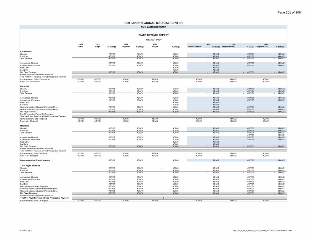
RUTLAND REGIONAL MEDICAL CENTERMRI Replacement
2020 2021 2021 2022 2,023 2024 2025Actual Budget % change Projection % change Budget % change Proposed Year 1 % change Proposed Year 2 % change Proposed Year 3 % change
Commercial Hospital #DIV/0! #DIV/0! #DIV/0! #DIV/0! #DIV/0! #DIV/0!Physician #DIV/0! #DIV/0! #DIV/0! #DIV/0! #DIV/0! #DIV/0!Total Revenue #DIV/0! #DIV/0! #DIV/0! - #DIV/0! - #DIV/0! - #DIV/0!
Allowances - Hospital #DIV/0! #DIV/0! #DIV/0! #DIV/0! #DIV/0! #DIV/0!Allowances - Physicians #DIV/0! #DIV/0! #DIV/0! #DIV/0! #DIV/0! #DIV/0!Free Care #DIV/0! #DIV/0!Bad Debt #DIV/0! #DIV/0!Net Payer Revenue #DIV/0! #DIV/0! #DIV/0! - #DIV/0! - #DIV/0! - #DIV/0!Fixed Prospective Payment & ReservesTotal Net Payer Revenue & Fixed Prospective PaymentReimbursement Rate - Commercial #DIV/0! #DIV/0! #DIV/0! #DIV/0! #DIV/0! #DIV/0! #DIV/0!Payer Mix - Commercial #DIV/0! #DIV/0! #DIV/0! #DIV/0! #DIV/0! #DIV/0! #DIV/0!
MedicaidHospital #DIV/0! #DIV/0! #DIV/0! #DIV/0! #DIV/0! #DIV/0!Physician #DIV/0! #DIV/0! #DIV/0! #DIV/0! #DIV/0! #DIV/0!Total Revenue #DIV/0! #DIV/0! #DIV/0! - #DIV/0! - #DIV/0! - #DIV/0!
Allowances - Hospital #DIV/0! #DIV/0! #DIV/0! #DIV/0! #DIV/0! #DIV/0!Allowances - Physicians #DIV/0! #DIV/0! #DIV/0! #DIV/0! #DIV/0! #DIV/0!Free Care #DIV/0! #DIV/0!Bad Debt #DIV/0! #DIV/0!Graduate Medical Education Payments-Phys. #DIV/0! #DIV/0! #DIV/0! #DIV/0! #DIV/0! #DIV/0!Graduate Medical Education Payments-Hosp #DIV/0! #DIV/0! #DIV/0! #DIV/0! #DIV/0! #DIV/0!Net Payer Revenue #DIV/0! #DIV/0! #DIV/0! - #DIV/0! - #DIV/0! - #DIV/0!Fixed Prospective Payment & ReservesTotal Net Payer Revenue & Fixed Prospective PaymentReimbursement Rate - Medicaid #DIV/0! #DIV/0! #DIV/0! #DIV/0! #DIV/0! #DIV/0! #DIV/0!Payer Mix - Medicaid #DIV/0! #DIV/0! #DIV/0! #DIV/0! #DIV/0! #DIV/0! #DIV/0!
MedicareHospital #DIV/0! #DIV/0! #DIV/0! #DIV/0! #DIV/0! #DIV/0!Physician #DIV/0! #DIV/0! #DIV/0! #DIV/0! #DIV/0! #DIV/0!Total Revenue #DIV/0! #DIV/0! #DIV/0! - #DIV/0! - #DIV/0! - #DIV/0!
#DIV/0! #DIV/0!Allowances - Hospital #DIV/0! #DIV/0! #DIV/0! #DIV/0! #DIV/0! #DIV/0!Allowances - Physicians #DIV/0! #DIV/0! #DIV/0! #DIV/0! #DIV/0! #DIV/0!Free Care #DIV/0! #DIV/0!Bad Debt #DIV/0! #DIV/0!Net Payer Revenue #DIV/0! #DIV/0! #DIV/0! - #DIV/0! - #DIV/0! - #DIV/0!Fixed Prospective Payment & ReservesTotal Net Payer Revenue & Fixed Prospective PaymentReimbursement Rate - Medicare #DIV/0! #DIV/0! #DIV/0! #DIV/0! #DIV/0! #DIV/0! #DIV/0!Payer Mix - Medicare #DIV/0! #DIV/0! #DIV/0! #DIV/0! #DIV/0! #DIV/0! #DIV/0!
Disproportionate Share Payments #DIV/0! #DIV/0! #DIV/0! #DIV/0! #DIV/0! #DIV/0!
Total Payer RevenueHospital #DIV/0! #DIV/0! - #DIV/0! #DIV/0! #DIV/0! #DIV/0!Physician #DIV/0! #DIV/0! #DIV/0! #DIV/0! #DIV/0! #DIV/0!Total Revenue #DIV/0! #DIV/0! #DIV/0! - #DIV/0! - #DIV/0! - #DIV/0!
Allowances - Hospital #DIV/0! #DIV/0! - #DIV/0! #DIV/0! #DIV/0! #DIV/0!Allowances - Physicians #DIV/0! #DIV/0! #DIV/0! #DIV/0! #DIV/0! #DIV/0!Free Care #DIV/0! #DIV/0! #DIV/0! #DIV/0! #DIV/0! #DIV/0!Bad Debt #DIV/0! #DIV/0! #DIV/0! #DIV/0! #DIV/0! #DIV/0!Disproportionate Share Payments #DIV/0! #DIV/0! #DIV/0! #DIV/0! #DIV/0! #DIV/0!Graduate Medical Education Payments-Phys. #DIV/0! #DIV/0! #DIV/0! #DIV/0! #DIV/0! #DIV/0!Graduate Medical Education Payments-Hosp #DIV/0! #DIV/0! #DIV/0! #DIV/0! #DIV/0! #DIV/0!Net Payer Revenue #DIV/0! #DIV/0! - #DIV/0! - #DIV/0! - #DIV/0! - #DIV/0!Fixed Prospective Payment & ReservesTotal Net Payer Revenue & Fixed Prospective Payment 0Reimbursement Rate - All Payers #DIV/0! #DIV/0! #DIV/0! #DIV/0! #DIV/0! #DIV/0! #DIV/0!
PAYER REVENUE REPORT
PROJECT ONLY
10/8/2021, Pg 2 CON_Table_6_Payer_Gross_to_NPR_updated_2021-05-20 Excl Mobile MRI FINAL
Page 201 of 206

RUTLAND REGIONAL MEDICAL CENTERMRI Replacement
2020 2021 2021 2022 2,023 2024 2025Actual Budget % change Projection % change Budget % change Proposed Year 1 % change Proposed Year 2 % change Proposed Year 3 % change
Commercial Hospital 148,001,292 160,517,591 8.5% 158,000,258 -1.6% 163,132,507 3.2% 175,505,647 7.6% 189,283,160 7.9% 204,662,302 8.1%Physician 21,404,997 20,943,806 -2.2% 20,095,533 -4.1% 20,702,352 3.0% 22,272,567 7.6% 24,021,004 7.9% 25,972,696 8.1%Total Revenue 169,406,289 181,461,397 7.1% 178,095,791 -1.9% 183,834,859 3.2% 197,778,214 7.6% 213,304,164 7.9% 230,634,998 8.1%
Allowances - Hospital -29,668,724 -25,480,500 -14.1% -41,320,461 62.2% -41,642,768 0.8% (46,061,825) 10.6% -51,067,244 10.9% -56,745,370 11.1%Allowances - Physicians -7,604,177 -12,957,361 70.4% -6,367,604 -50.9% -6,464,189 1.5% (7,150,157) 10.6% -7,927,146 10.9% -8,808,559 11.1%Free Care -5,531,925 -6,819,470 23.3% -4,556,395 -33.2% -4,016,432 -11.9% (4,321,067) 7.6% -4,660,279 7.9% -5,038,923 8.1%Bad Debt -9,177,446 -8,463,885 -7.8% -4,567,120 -46.0% -5,859,997 28.3% (6,304,461) 7.6% -6,799,373 7.9% -7,351,818 8.1%Net Payer Revenue 117,424,017 127,740,181 8.8% 121,284,211 -5.1% 125,851,473 3.8% 133,940,704 6.4% 142,850,123 6.7% 152,690,327 6.9%Fixed Prospective Payment & Reserves 0 0 #DIV/0! 0 #DIV/0! 0 #DIV/0! - #DIV/0! 0 #DIV/0! 0 #DIV/0!Total Net Payer Revenue & Fixed Prospective Payment 117,424,017 127,740,181 8.8% 121,284,211 -5.1% 125,851,473 3.8% 133,940,704 6.4% 142,850,123 6.7% 152,690,327 0Reimbursement Rate - Commercial 69% 70% 68% 68% 1 67% 66%Payer Mix - Commercial 49% 52% 45% 47% 0 49% 51%
MedicaidHospital 81,589,573 89,587,292 9.8% 94,458,223 5.4% 98,635,950 4.4% 106,117,208 7.6% 114,447,603 7.9% 123,746,401 8.1%Physician 9,051,047 10,836,026 19.7% 11,743,450 8.4% 12,068,396 2.8% 12,983,750 7.6% 14,002,998 7.9% 15,140,733 8.1%Total Revenue 90,640,620 100,423,318 10.8% 106,201,673 5.8% 110,704,346 4.2% 119,100,958 7.6% 128,450,600 7.9% 138,887,133 8.1%
Allowances - Hospital -61,219,412 -68,214,560 11.4% -64,170,001 -5.9% -70,489,078 9.8% (77,969,254) 10.6% -86,441,970 10.9% -96,053,385 11.1%Allowances - Physicians -6,594,162 -7,444,464 12.9% -8,324,101 11.8% -8,525,951 2.4% (9,430,710) 10.6% -10,455,521 10.9% -11,618,062 11.1%Free Care 0 0 #DIV/0! 0 #DIV/0! 0 #DIV/0! - #DIV/0! 0 #DIV/0! 0 #DIV/0!Bad Debt 0 0 #DIV/0! 0 #DIV/0! 0 #DIV/0! - #DIV/0! 0 #DIV/0! 0 #DIV/0!Graduate Medical Education Payments-Phys. 0 0 #DIV/0! 0 #DIV/0! 0 #DIV/0! - #DIV/0! 0 #DIV/0! 0 #DIV/0!Graduate Medical Education Payments-Hosp 0 0 #DIV/0! 0 #DIV/0! 0 #DIV/0! - #DIV/0! 0 #DIV/0! 0 #DIV/0!Net Payer Revenue 22,827,046 24,764,294 8.5% 33,707,571 36.1% 31,689,317 -6.0% 31,700,994 0.0% 31,553,110 -0.5% 31,215,686 -1.1%Fixed Prospective Payment & Reserves -972,075 -799,220 -1,087,622 -1,213,808 (1,256,291) -1,300,261 -1,345,771Total Net Payer Revenue & Fixed Prospective Payment 21,854,971 23,965,074 32,619,949 30,475,509 30,444,703 30,252,849 29,869,915Reimbursement Rate - Medicaid 24% 24% 31% 28% 0 24% 22%Payer Mix - Medicaid 9% 10% 12% 11% 0 10% 10%
MedicareHospital 254,946,875 270,195,959 6.0% 302,740,942 12.0% 315,454,603 4.2% 339,380,943 7.6% 366,022,967 7.9% 395,762,110 8.1%Physician 24,447,206 26,208,423 7.2% 29,109,500 11.1% 29,844,824 2.5% 32,108,469 7.6% 34,629,043 7.9% 37,442,631 8.1%Total Revenue 279,394,081 296,404,382 6.1% 331,850,442 12.0% 345,299,427 4.1% 371,489,413 7.6% 400,652,010 7.9% 433,204,741 8.1%
Allowances - Hospital -169,521,963 -186,115,647 9.8% -202,630,695 8.9% -214,646,705 5.9% (237,424,632) 10.6% -263,224,951 10.9% -292,492,727 11.1%Allowances - Physicians -13,138,290 -17,876,631 36.1% -19,638,452 9.9% -20,053,851 2.1% (22,181,930) 10.6% -24,592,383 10.9% -27,326,791 11.1%Free Care 0 0 #DIV/0! 0 #DIV/0! 0 #DIV/0! - #DIV/0! 0 #DIV/0! 0 #DIV/0!Bad Debt 0 0 #DIV/0! 0 #DIV/0! 0 #DIV/0! - #DIV/0! 0 #DIV/0! 0 #DIV/0!Net Payer Revenue 96,733,828 92,412,104 -4.5% 109,581,295 18.6% 110,598,871 0.9% 111,882,851 1.2% 112,834,676 0.9% 113,385,223 0.5%Fixed Prospective Payment & Reserves 0 0 0 0 - 0 0Total Net Payer Revenue & Fixed Prospective Payment 96,733,828 92,412,104 109,581,295 110,598,871 111,882,851 112,834,676 113,385,223Reimbursement Rate - Medicare 35% 31% 33% 32% 0 28% 26%Payer Mix - Medicare 40% 37% 41% 41% 0 39% 38%
Disproportionate Share Payments 3,242,526 3,370,325 3.9% 3,383,358 0.4% 3,435,517 1.5% 3,555,760 3.5% 3,680,212 3.5% 3,809,019 3.5%
Total Payer RevenueHospital 484,537,740 520,300,842 7.4% 555,199,423 6.7% 577,223,060 4.0% 621,003,798 7.6% 669,753,730 7.9% 724,170,812 8.1%Physician 54,903,250 57,988,255 5.6% 60,948,483 5.1% 62,615,572 2.7% 67,364,786 7.6% 72,653,045 7.9% 78,556,060 8.1%Total Revenue 539,440,990 578,289,097 7.2% 616,147,906 6.5% 639,838,632 3.8% 688,368,584 7.6% 742,406,775 7.9% 802,726,872 8.1%
Allowances - Hospital -260,410,099 -279,810,707 7.5% -308,121,157 10.1% -326,778,551 6.1% (361,455,710) 10.6% -400,734,165 10.9% -445,291,483 11.1%Allowances - Physicians -27,336,629 -38,278,456 40.0% -34,330,157 -10.3% -35,043,991 2.1% (38,762,797) 10.6% -42,975,050 10.9% -47,753,412 11.1%Free Care -5,531,925 -6,819,470 23.3% -4,556,395 -33.2% -4,016,432 -11.9% (4,321,067) 7.6% -4,660,279 7.9% -5,038,923 8.1%Bad Debt -9,177,446 -8,463,885 -7.8% -4,567,120 -46.0% -5,859,997 28.3% (6,304,461) 7.6% -6,799,373 7.9% -7,351,818 8.1%Disproportionate Share Payments 3,242,526 3,370,325 3.9% 3,383,358 0.4% 3,435,517 1.5% 3,555,760 3.5% 3,680,212 3.5% 3,809,019 3.5%Graduate Medical Education Payments-Phys. 0 0 #DIV/0! 0 #DIV/0! 0 #DIV/0! - #DIV/0! 0 #DIV/0! 0 #DIV/0!Graduate Medical Education Payments-Hosp 0 0 #DIV/0! 0 #DIV/0! 0 #DIV/0! - #DIV/0! 0 #DIV/0! 0 #DIV/0!Net Payer Revenue 240,227,417 248,286,904 3.4% 267,956,435 7.9% 271,575,178 1.4% 281,080,309 3.5% 290,918,120 3.5% 301,100,255 3.5%Fixed Prospective Payment & Reserves -972,075 -799,220 -1,087,622 -1,213,808 (1,256,291) -1,300,261 -1,345,771Total Net Payer Revenue & Fixed Prospective Payment 239,255,342 247,487,684 266,868,813 270,361,370 279,824,018 289,617,859 299,754,484Reimbursement Rate - All Payers 45% 43% 43% 42% 0 39% 38%
PAYER REVENUE REPORT
WITH PROJECT
Note: This table requires no "fill-in" as it is populated automatically
10/8/2021, Pg 3 CON_Table_6_Payer_Gross_to_NPR_updated_2021-05-20 Excl Mobile MRI FINAL
Page 202 of 206

2020 2021 2021 2022 Proposed Yr 1 Proposed Yr 2 Proposed Yr 3Actual Budget % change Projection % change Budget % change 2023 % change 2024 % change 2025 % change
Inpatient Utilization Acute Beds (Staffed) 109 - -100.0% 109 #DIV/0! 109 0.0% 109 0.0% 109 0.0% 109 0.0% Acute Admissions 5,944 - -100.0% 5,984 #DIV/0! 6,537 9.2% 6,537 0.0% 6,537 0.0% 6,537 0.0% Acute Patient Days 28,987 - -100.0% 29,444 #DIV/0! 31,744 7.8% 31,744 0.0% 31,744 0.0% 31,744 0.0% Acute Average Length Of Stay 4.88 - -100.0% 4.92 #DIV/0! 4.86 -1.3% 4.86 0.0% 4.86 0.0% 4.86 0.0% Outpatient All Outpatient Visits 240,617 - -100.0% 296,102 #DIV/0! 299,194 1.0% 299,194 0.0% 299,194 0.0% 299,194 0.0% Physician Office Visits - - #DIV/0! - #DIV/0! - #DIV/0! - #DIV/0! - #DIV/0! - #DIV/0! Ancillary All Operating Room Procedure 4,426 - -100.0% 5,135 #DIV/0! 5,135 0.0% 5,135 0.0% 5,135 0.0% 5,135 0.0% All Operating Room Cases - - #DIV/0! - #DIV/0! - #DIV/0! - #DIV/0! - #DIV/0! - #DIV/0! Emergency Room Visits 27,196 - -100.0% 25,885 #DIV/0! 25,207 -2.6% 25,207 0.0% 25,207 0.0% 25,207 0.0% Cat Scan Procedures 14,697 - -100.0% 13,955 #DIV/0! 13,633 -2.3% 13,633 0.0% 13,633 0.0% 13,633 0.0% Magnetic Resonance Image Exams 4,810 - -100.0% 6,041 #DIV/0! 5,197 -14.0% 5,197 0.0% 5,197 0.0% 5,197 0.0% Nuclear Medicine Procedures 668 - -100.0% 723 #DIV/0! 723 0.0% 723 0.0% 723 0.0% 723 0.0% Radiology - Diagnostic Procedures 40,008 - -100.0% 40,492 #DIV/0! 39,630 -2.1% 39,630 0.0% 39,630 0.0% 39,630 0.0% Laboratory Tests 561,135 - -100.0% 654,552 #DIV/0! 650,923 -0.6% 650,923 0.0% 650,923 0.0% 650,923 0.0%
#DIV/0! #DIV/0! #DIV/0! - #DIV/0! - #DIV/0! - #DIV/0!#DIV/0! #DIV/0! #DIV/0! - #DIV/0! - #DIV/0! - #DIV/0!
Adjusted Statistics Adjusted Admissions 17,476 - -100.0% 18,803 #DIV/0! 19,950 6.1% 19,950 0.0% 19,950 0.0% 19,950 0.0% Adjusted Days 85,223 - -100.0% 92,520 #DIV/0! 96,879 4.7% 96,879 0.0% 96,879 0.0% 96,879 0.0%
Rutland Regional Medical CenterMRI Replacement
UTILIZATION PROJECTIONS--TABLE 7
WITHOUT PROJECT
1 GMCB CON_Table_7-Utilization_Statistics_updated_2021-05-20 Excl Mobile MRI FINAL
Page 203 of 206

Rutland Regional Medical CenterMRI Replacement
UTILIZATION PROJECTIONS--TABLE 7
2020 2021 2021 2022 Proposed Yr 1 Proposed Yr 2 Proposed Yr 3Actual Budget % change Projection % change Budget % change 2023 % change 2024 % change 2025 % change
Inpatient Utilization Acute Beds (Staffed) #DIV/0! #DIV/0! #DIV/0! #DIV/0! #DIV/0! #DIV/0! Acute Admissions #DIV/0! #DIV/0! #DIV/0! #DIV/0! #DIV/0! #DIV/0! Acute Patient Days #DIV/0! #DIV/0! #DIV/0! #DIV/0! #DIV/0! #DIV/0! Acute Average Length Of Stay #DIV/0! #DIV/0! #DIV/0! #DIV/0! #DIV/0! #DIV/0! Outpatient All Outpatient Visits #DIV/0! #DIV/0! #DIV/0! #DIV/0! #DIV/0! #DIV/0! Physician Office Visits #DIV/0! #DIV/0! #DIV/0! #DIV/0! #DIV/0! #DIV/0! Ancillary All Operating Room Procedure #DIV/0! #DIV/0! #DIV/0! #DIV/0! #DIV/0! #DIV/0! All Operating Room Cases #DIV/0! #DIV/0! #DIV/0! #DIV/0! #DIV/0! #DIV/0! Emergency Room Visits #DIV/0! #DIV/0! #DIV/0! #DIV/0! #DIV/0! #DIV/0! Cat Scan Procedures #DIV/0! #DIV/0! #DIV/0! #DIV/0! #DIV/0! #DIV/0! Magnetic Resonance Image Exams #DIV/0! #DIV/0! #DIV/0! #DIV/0! #DIV/0! #DIV/0! Nuclear Medicine Procedures #DIV/0! #DIV/0! #DIV/0! #DIV/0! #DIV/0! #DIV/0! Radiology - Diagnostic Procedures #DIV/0! #DIV/0! #DIV/0! #DIV/0! #DIV/0! #DIV/0! Laboratory Tests #DIV/0! #DIV/0! #DIV/0! #DIV/0! #DIV/0! #DIV/0!
#DIV/0! #DIV/0! #DIV/0! #DIV/0! #DIV/0! #DIV/0!#DIV/0! #DIV/0! #DIV/0! #DIV/0! #DIV/0! #DIV/0!
Adjusted Statistics Adjusted Admissions #DIV/0! #DIV/0! #DIV/0! #DIV/0! #DIV/0! #DIV/0! Adjusted Days #DIV/0! #DIV/0! #DIV/0! #DIV/0! #DIV/0! #DIV/0!
PROJECT ONLY
2 GMCB CON_Table_7-Utilization_Statistics_updated_2021-05-20 Excl Mobile MRI FINAL
Page 204 of 206
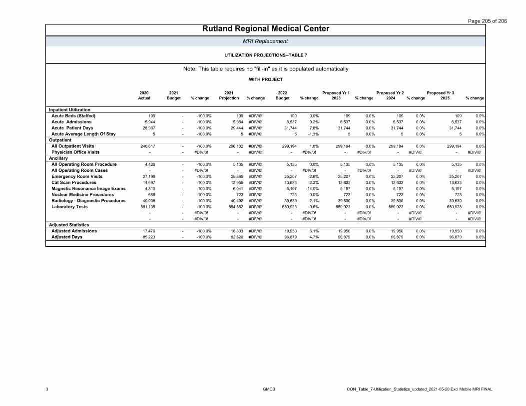
Rutland Regional Medical CenterMRI Replacement
UTILIZATION PROJECTIONS--TABLE 7
2020 2021 2021 2022 Proposed Yr 1 Proposed Yr 2 Proposed Yr 3Actual Budget % change Projection % change Budget % change 2023 % change 2024 % change 2025 % change
Inpatient Utilization Acute Beds (Staffed) 109 - -100.0% 109 #DIV/0! 109 0.0% 109 0.0% 109 0.0% 109 0.0% Acute Admissions 5,944 - -100.0% 5,984 #DIV/0! 6,537 9.2% 6,537 0.0% 6,537 0.0% 6,537 0.0% Acute Patient Days 28,987 - -100.0% 29,444 #DIV/0! 31,744 7.8% 31,744 0.0% 31,744 0.0% 31,744 0.0% Acute Average Length Of Stay 5 - -100.0% 5 #DIV/0! 5 -1.3% 5 0.0% 5 0.0% 5 0.0% Outpatient All Outpatient Visits 240,617 - -100.0% 296,102 #DIV/0! 299,194 1.0% 299,194 0.0% 299,194 0.0% 299,194 0.0% Physician Office Visits - - #DIV/0! - #DIV/0! - #DIV/0! - #DIV/0! - #DIV/0! - #DIV/0! Ancillary All Operating Room Procedure 4,426 - -100.0% 5,135 #DIV/0! 5,135 0.0% 5,135 0.0% 5,135 0.0% 5,135 0.0% All Operating Room Cases - - #DIV/0! - #DIV/0! - #DIV/0! - #DIV/0! - #DIV/0! - #DIV/0! Emergency Room Visits 27,196 - -100.0% 25,885 #DIV/0! 25,207 -2.6% 25,207 0.0% 25,207 0.0% 25,207 0.0% Cat Scan Procedures 14,697 - -100.0% 13,955 #DIV/0! 13,633 -2.3% 13,633 0.0% 13,633 0.0% 13,633 0.0% Magnetic Resonance Image Exams 4,810 - -100.0% 6,041 #DIV/0! 5,197 -14.0% 5,197 0.0% 5,197 0.0% 5,197 0.0% Nuclear Medicine Procedures 668 - -100.0% 723 #DIV/0! 723 0.0% 723 0.0% 723 0.0% 723 0.0% Radiology - Diagnostic Procedures 40,008 - -100.0% 40,492 #DIV/0! 39,630 -2.1% 39,630 0.0% 39,630 0.0% 39,630 0.0% Laboratory Tests 561,135 - -100.0% 654,552 #DIV/0! 650,923 -0.6% 650,923 0.0% 650,923 0.0% 650,923 0.0%
- - #DIV/0! - #DIV/0! - #DIV/0! - #DIV/0! - #DIV/0! - #DIV/0!- - #DIV/0! - #DIV/0! - #DIV/0! - #DIV/0! - #DIV/0! - #DIV/0!
Adjusted Statistics Adjusted Admissions 17,476 - -100.0% 18,803 #DIV/0! 19,950 6.1% 19,950 0.0% 19,950 0.0% 19,950 0.0% Adjusted Days 85,223 - -100.0% 92,520 #DIV/0! 96,879 4.7% 96,879 0.0% 96,879 0.0% 96,879 0.0%
Note: This table requires no "fill-in" as it is populated automatically
WITH PROJECT
3 GMCB CON_Table_7-Utilization_Statistics_updated_2021-05-20 Excl Mobile MRI FINAL
Page 205 of 206

2020 2021 2021 2022Proposed Year 1
Proposed Year 2
Proposed Year 3
Actual Budget % change Projection % change Budget % change 2023 % change 2024 % change 2025 % change
PHYSICIAN FTEs 70.5 - -100.0% 72.3 #DIV/0! 72.5 0.2% 72.5 0.0% 72.5 0.0% 72.5 0.0%
TRAVELERS 93.1 - -100.0% - #DIV/0! 25.0 #DIV/0! 25.0 0.0% 25.0 0.0% 25.0 0.0%
Residents & Fellows - - #DIV/0! - #DIV/0! - #DIV/0! #DIV/0! #DIV/0! #DIV/0!MLPs - - #DIV/0! - #DIV/0! - #DIV/0! #DIV/0! #DIV/0! #DIV/0!Non-MD FTEs 1,271.4 - -100.0% 1,277.9 #DIV/0! 1,287.8 0.8% 1,287.8 0.0% 1,287.8 0.0% 1,287.8 0.0%TOTAL NON-MD FTEs 1,271.4 - -100.0% 1,277.9 #DIV/0! 1,287.8 0.8% 1,287.8 0.0% 1,287.8 0.0% 1,287.8 0.0%
Note: Mid-Level Providers and Residents are now included in Non-MD Employees, prior to 2013 Actual they were included in Physician FTEs
2020 2021 2021 2022Proposed Year 1
Proposed Year 2
Proposed Year 3
Actual Budget % change Projection % change Budget % change 2023 % change 2024 % change 2025 % change
PHYSICIAN FTEs #DIV/0! #DIV/0! #DIV/0! #DIV/0! #DIV/0! #DIV/0!
TRAVELERS #DIV/0! #DIV/0! #DIV/0! #DIV/0! #DIV/0! #DIV/0!
Residents & Fellows #DIV/0! #DIV/0! #DIV/0! #DIV/0! #DIV/0! #DIV/0!MLPs #DIV/0! #DIV/0! #DIV/0! #DIV/0! #DIV/0! #DIV/0!Non-MD FTEs #DIV/0! #DIV/0! #DIV/0! #DIV/0! #DIV/0! #DIV/0!TOTAL NON-MD FTEs - - #DIV/0! - #DIV/0! - #DIV/0! - #DIV/0! - #DIV/0! - #DIV/0!
Note: Mid-Level Providers and Residents are now included in Non-MD Employees, prior to 2013 Actual they were included in Physician FTEs
2020 2021 2021 2022Proposed Year 1
Proposed Year 2
Proposed Year 3
Actual Budget % change Projection % change Budget % change 2023 % change 2024 % change 2025 % change
PHYSICIAN FTEs 70.5 - -100.0% 72.3 #DIV/0! 72.5 0.2% 72.5 0.0% 72.5 0.0% 72.5 0.0%
TRAVELERS 93.1 - -100.0% - #DIV/0! 25.0 #DIV/0! 25.0 0.0% 25.0 0.0% 25.0 0.0%
Residents & Fellows - - #DIV/0! - #DIV/0! - #DIV/0! - #DIV/0! - #DIV/0! - #DIV/0!MLPs - - #DIV/0! - #DIV/0! - #DIV/0! - #DIV/0! - #DIV/0! - #DIV/0!Non-MD FTEs 1,271.4 - -100.0% 1,277.9 #DIV/0! 1,287.8 0.8% 1,287.8 0.0% 1,287.8 0.0% 1,287.8 0.0%TOTAL NON-MD FTEs 1,271.4 - -100.0% 1,277.9 #DIV/0! 1,287.8 0.8% 1,287.8 0.0% 1,287.8 0.0% 1,287.8 0.0%
Note: Mid-Level Providers and Residents are now included in Non-MD Employees, prior to 2013 Actual they were included in Physician FTEs
PROJECT ONLY
Note: This table requires no "fill-in" as it is populated automatically
STAFFING REPORT - TABLE 8
WITH PROJECT
RUTLAND REGIONAL MEDICAL CENTER
MRI Replacement
STAFFING REPORT - TABLE 8
WITHOUT PROJECT
STAFFING REPORT - TABLE 8
10/8/2021 CON_Table_8-Staffing_Report_updated_2021-05-20 Excl Mobile MRI FINAL
Page 206 of 206