
Observing plant roots in their environment: current imaging options and specific contribution of...
10
Observing plant roots in their environment: current imaging options and specific contribution of two-dimensional approaches Alain Pierret, Claude Doussan, Emmanuelle Garrigues, John Mc Kirby To cite this version: Alain Pierret, Claude Doussan, Emmanuelle Garrigues, John Mc Kirby. Observing plant roots in their environment: current imaging options and specific contribution of two-dimensional approaches. Agronomie, EDP Sciences, 2003, 23 (5-6), pp.471-479. <10.1051/agro:2003019>. <hal-00886199> HAL Id: hal-00886199 https://hal.archives-ouvertes.fr/hal-00886199 Submitted on 1 Jan 2003 HAL is a multi-disciplinary open access archive for the deposit and dissemination of sci- entific research documents, whether they are pub- lished or not. The documents may come from teaching and research institutions in France or abroad, or from public or private research centers. L’archive ouverte pluridisciplinaire HAL, est destin´ ee au d´ epˆ ot et ` a la diffusion de documents scientifiques de niveau recherche, publi´ es ou non, ´ emanant des ´ etablissements d’enseignement et de recherche fran¸cais ou ´ etrangers, des laboratoires publics ou priv´ es.
-
Upload
independent -
Category
Documents
-
view
2 -
download
0
Transcript of Observing plant roots in their environment: current imaging options and specific contribution of...

Observing plant roots in their environment: current
imaging options and specific contribution of
two-dimensional approaches
Alain Pierret, Claude Doussan, Emmanuelle Garrigues, John Mc Kirby
To cite this version:
Alain Pierret, Claude Doussan, Emmanuelle Garrigues, John Mc Kirby. Observing plant rootsin their environment: current imaging options and specific contribution of two-dimensionalapproaches. Agronomie, EDP Sciences, 2003, 23 (5-6), pp.471-479. <10.1051/agro:2003019>.<hal-00886199>
HAL Id: hal-00886199
https://hal.archives-ouvertes.fr/hal-00886199
Submitted on 1 Jan 2003
HAL is a multi-disciplinary open accessarchive for the deposit and dissemination of sci-entific research documents, whether they are pub-lished or not. The documents may come fromteaching and research institutions in France orabroad, or from public or private research centers.
L’archive ouverte pluridisciplinaire HAL, estdestinee au depot et a la diffusion de documentsscientifiques de niveau recherche, publies ou non,emanant des etablissements d’enseignement et derecherche francais ou etrangers, des laboratoirespublics ou prives.

471Agronomie 23 (2003) 471–479© INRA, EDP Sciences, 2003DOI: 10.1051/agro:2003019
Original article
Observing plant roots in their environment: current imaging options and specific contribution of two-dimensional approaches
Alain PIERRETa, Claude DOUSSANb*, Emmanuelle GARRIGUESb, John Mc KIRBYa
a CSIRO Land & Water, GPO Box 1666, Canberra ACT, Australiab INRA-CSE, Domaine Saint-Paul, Site Agroparc, 84914 Avignon Cedex 9, France
(Received 19 June 2002; accepted 28 January 2003)
Abstract – This paper provides an overview of current options for imaging roots in their environment. Our primary aim is to describe thepotential and limitations of these techniques, rather than their physical principles. We primarily discuss two-dimensional imaging techniqueswhich, while inherently limited by their 2-dimensional nature are technically simple, cheaper, more readily accessible, and offer larger field-of-view or better resolution for a similar field-of-view than three-dimensional imaging techniques. We show that techniques involvingmicroscopic or high-resolution X-ray imaging of roots in undisturbed soil samples have a considerable value because they can provide, at ascale which is not accessible by any other means, essential information on complex interactions between roots and soil structure. We alsoillustrate how 2D imaging techniques, based on either visible light or X-ray attenuation through thin growing containers (rhizotrons), can beused to study whole root systems. We conclude that, while fully appropriate 3D imaging tools will ultimately become available, in themeantime, two-dimensional techniques should be considered as relevant options for observing roots in their environment.
2D and 3D imaging / root system / root-soil interactions / water uptake
Résumé – Observation des racines dans leur environnement : techniques d’imagerie actuelles et apports spécifiques des approches 2D.Cet article offre un inventaire des techniques d’imagerie actuellement disponibles pour l’observation des racines au sein de leur environnement.Le propos est avant tout d’informer le lecteur sur le potentiel et les limites des techniques et non sur leurs principes physiques. D’autre part,l’accent est mis sur l’apport spécifique des méthodes 2D : en dépit des limitations et biais qui leur sont inhérents, ces méthodes offrent en effet,par rapport aux outils 3D actuellement disponibles, des avantages tels qu’un grand champ d’observation ou une résolution supérieure à champd’observation équivalent, ou encore, une mise en œuvre simplifiée et un coût réduit. Ainsi, la microscopie et la radiographie X à haute résolutionsont très attractives car elles permettent d’étudier, à une échelle qui demeure hors de la portée des méthodes 3D, les interactions complexesentre racines et structure du sol. On montre par ailleurs que les méthodes 2D basées sur l’atténuation de lumière visible ou de rayons X s’avèrenttrès utiles pour étudier le fonctionnement de systèmes racinaires entiers en rhizotrons. S’il ne fait guère de doute qu’à relativement court terme,les progrès accomplis dans le domaine de l’imagerie 3D aboutiront à la mise au point d’outils parfaitement adaptés à l’observation nondestructive des racines dans leur environnement, il n’en reste pas moins que, dans l’attente de tels outils, les techniques 2D continueront de faireprogresser notre connaissance des racines et de leur fonctionnement.
imagerie 2D et 3D / système racinaire / interactions sol-racines / prélèvement d’eau
1. INTRODUCTION
The above ground parts of plants have been well studied butroots have received much less attention. A simple reason forthis apparently limited interest in roots is purely technical:roots being tightly enmeshed in the opaque soil matrix, it isextremely difficult to observe them in situ [10]. Under fieldconditions, roots are exposed to a very heterogeneousenvironment: the physical, chemical and biological properties
of soil can vary greatly within a few millimeters, e.g. [39, 46],and affect root growth, either beneficially or detrimentally, ata very local scale. Interactions between roots and soil structureand the resulting effects on root physiology remain poorlyunderstood. Such knowledge is however essential as it is likelythat plant performance is linked to the precise location of rootswith respect to soil structure features [35, 36] as well as themicro-environment surrounding the roots [32]. For example,observations based on physical separation of roots from soil,
* Correspondence and [email protected]
Communicated by Philippe Hinsinger (Montpellier, France)

472 A. Pierret et al.
suggest that although roots growing down macropores may beable to access more water and nutrient from depth, thisapparent advantage is most likely offset by poor soil-contactalong much of the root and impeded lateral growth [44].
The simplest and most commonly used method of measur-ing roots is by washing auger sampled cores. This method canbe used to determine root length density, a key parameter inmany models of root water extraction. However, it is well doc-umented that many fine roots are broken off and washed awaywith the soil, leading to biased estimates, e.g. [27]. Other sim-ple invasive methods such as trench profile methods are suita-ble for observing the spatial distribution of roots and, to someextent, the root environment. However, such trench methodsdo not allow direct assessment of root growth and poor visiblecontrast between roots and the soil matrix may affect theresults of root mapping [21]. Alternatively the mini-rhizotronmethod is a relatively standard option for assessing rootgrowth, but there are concerns about roots growing preferen-tially around mini-rhizotron tubes [13].
In this paper we examine the current options for imagingroots and their environment. We deliberately give little descrip-tion of the principles behind these techniques and concentrateon their suitability and limitations, in the specific context ofroot-soil studies. Three-dimensional imaging techniques areonly briefly dealt with in a general discussion since they havereceived reasonable coverage in recent literature, e.g. [2]. Pri-ority is given to the discussion of two-dimensional imaging andwe consider and discuss in greater detail techniques we haverecently experimented with, namely: (i) high-resolution X-rayimaging of roots in undisturbed, resin impregnated soilsamples; (ii) X-ray imaging of root growth and response to soilconstraints in thin containers and (iii) light transmission imag-ing of root water uptake in thin containers. We show that thesetechnically simple alternatives to 3-dimensional imaging arecheaper, more readily accessible and generally offer a largerfield-of-view or better resolution for a similar field-of-view.
2. TWO-DIMENSIONAL DESTRUCTIVE/INVASIVE TECHNIQUES
2.1. Microscopy of roots in undisturbed soil samples
As far back as 1954, Barley [5] used optical microscopy toderive soil density profiles around roots grown in sandy loams.During the last decade, substantial progress in observing/measuring the spatial relations between roots and soil wasachieved via optical microscopy. Using thin sections ofundisturbed soil samples van Noordwijk et al. [47] andKooistra et al. [24] demonstrated that it is possible toqualitatively investigate the degree of contact between rootsand soil, an important parameter influencing oxygen, waterand nutrient uptake [12]. Similar studies on polished sectionsof undisturbed soil demonstrated, at a slightly coarser scale,non-random relationships between roots and soil structuralfeatures such as macropores [25, 42, 43].
At a finer scale, electron microscopy of thin sections hasbeen used to study physical interactions between roots andsoil. For example, using backscattered electron images,
Bruand et al. [8] showed that by rearranging the packing ofsoil skeleton grains, root radial growth can cause local changesin bulk density of up to 0.26 Mg·m–3.
Examination of fresh, undisturbed samples frozen in thefield, by cryo-Scanning Electron Microscopy (cryo-SEM) cou-pled with X-ray microanalysis is leading to advances in thestudy of the water relations at the soil-root interface, as well asfeed-forward/feedback effects between roots and their rhizo-spheres [32]. One example which clearly demonstrates thepotential of this technique was the finding of so-calledrhizosheaths [48], zones of enhanced and stabilized soil aggre-gation around roots of many grass species including cereals.From a functional point of view, rhizosheaths have been shownto considerably increase the contact between roots and soil.Cryo-SEM studies demonstrated that although rhizosheathshave an inherent porosity, parts of root hairs constantly stay incontact with rhizosheath soil via surrounding mucilageexpanded by exuded water [33].
The main hindrance to a wider adoption of these micro-scopic techniques results either from the cost of specializedlaboratory equipment and labour required for sample prepara-tion (thin sections), or from the cost of analytical equipment(cryo-SEM). However, these methods can provide essentialinformation on how plant roots grow and function under fieldconditions, at a scale which is not accessible by any othermeans.
2.2. X-ray imaging of roots in undisturbed, resin impregnated soil samples
This method was first introduced by Moran et al. [34]. Tosome extent, it can be seen as an X-ray equivalent of the thinsection method described in the previous section, with theadvantage of providing a quantified description of the soilstructure as a density/porosity field. X-rays emitted by amicrofocus source (i.e. a source with a small focal spot ofabout 10–20 �m) are attenuated differently by sample featuressuch as soil structural elements with different densities. Inaddition to this differential attenuation, materials with a phasecontrast such as the interface between soil and roots cause aslight refraction of the X-rays [49]. This enhances theattenuation contrast between soil and roots, and thus leads toimproved image clarity. The resulting images permit readydiscrimination of the roots. Samples observed are typically1 mm thick sections of resin impregnated soil collected in thefield. Due to the cone-shaped X-ray beam, projected imagescan be directly magnified on the image detector. Possibleimaging configurations are many: the most commonly usedfor this application yields images of a 66 mm square field-of-view with a 17.5 �m pixel size. With high-flux sources nowavailable, exposure times are of the order of a few seconds.Different types of image detectors can be used, such asphotographic plates, charge-coupled device (CCD) camerascoupled with phosphor screens or phosphor image plates.Once acquired, the images can be transformed into soil densityfields according to the calibration procedure of Bresson andMoran [7]. The images are subsequently processed andvarious attributes such as the root length density, root diameteror estimates of the spatial association of roots with the leastdense areas of soil determined.

Imaging of roots in soil 473
To assess the potential of this method, undisturbed sampleswere collected from a Natric Palexeralf [40] under a wheatcrop. After transportation to the laboratory, samples wereimpregnated with polyester resin and sectioned both verticallyand horizontally. It could be shown that, under this soil/cropcombination, roots grew preferentially in looser soil but not
necessarily in the macropores (Figs. 1a, b). This confirmedprevious results [42], obtained from observation of the surfaceof the same soil sections using optical microscopy. Estimatesof root length density obtained with this method were veryhigh, about an order of magnitude greater than thosecommonly obtained by core washing. They were however ingood agreement with values reported by Stewart [42] or Krebset al. [25], based on microscopic examination of polishedsections, or by Heeraman et al. [22] based on X-raytomography. Such results indicate that fine roots ( 0.1 mm inradius), often lost during core washing, may account for up to50–60% of the total root length. Finally, the method can beused to get some insight into root system geometry, throughquantification of root orientation and branching intensity [34].
Although restricted to 2-dimensions, this method offersnew opportunities for the quantification of root systemgeometry and the interaction of roots with soil structure. As inthe case of optical and electronic microscopy, the cost ofequipment and labour associated with this method is certainlynot negligible. However, this is counterbalanced by the factthat it enables unique characterization of roots and root-soilinterplay under field conditions. In addition, this method isquite versatile and with modification can also be used to studyroot growth dynamics and activity in controlled laboratoryexperiments (see below Sect. 4.1).
3. TWO-DIMENSIONAL NON-DESTRUCTIVE/INVASIVE IMAGING TECHNIQUES
The idea of observing and measuring living roots throughthe walls of thin transparent containers (rhizotrons) is quite oldand has been implemented in a variety of forms for decades.Coupled with time-lapse photography, it was successfullyused by many authors to study root dynamics and how differ-ent root zone conditions affect root growth, e.g. [28]. Com-bined with the use of dye indicators, such an approach provedpowerful for visualizing pH changes along the root systemunder a range of nutritional conditions [31]. A recent improve-ment of this kind of techniques is the so-called videodensitom-etry method which permits the mapping at high spatialresolution of the dynamics of pH changes occurring alongroots [23].
Rhizotrons have also been used in combination withradioisotopes to monitor root induced changes in therhizosphere. For example, Barber and Ozanne [4] grew plantsin 45Ca labeled soil to demonstrate autoradiographically theaccumulation/depletion of this element along roots. Finally,although more loosely related to the classical rhizotronconcept, we mention two recent applications. The first is alight transmission technique in which projection images ofroots in translucent gels are used to obtain unbiased estimatesof root lengths, branching patterns and diameter distribution[50]. The second is a high-resolution X-radiographic methodused to monitor root tissue differentiation and primordiumdevelopment in roots grown on synthetic mesh withcirculating nutrient solution [6]. In the subsequent sections, wediscuss in further detail two new rhizotron based methods wehave recently experimented with.
�
Figure 1. (a) Image of roots growing in structured subsoil (verticalsection), showing the soil density and root diameter. (b) Relationshipbetween bulk density of soil in contact with roots and root radius.This indicates that, for the case study discussed under Section 2.3, inboth the topsoil and subsoil, roots grew preferentially in soil lessdense than the average bulk densities of the subsoil and the topsoil(represented by the thick and thin dotted lines, respectively).(Redrawn from [34]). Figure 1(a) is available in colour at:www.edpsciences.org/agro/.

474 A. Pierret et al.
3.1. X-ray imaging of live root systems in rhizotrons
Radiographic techniques, using either, X-ray [51] orneutron sources, e.g. [9], have been successfully used to imageand measure live plant roots in soil. Here, we present anddiscuss the efficacy of X-radiography as a tool for monitoringroot growth and response to soil constraints in simplifiedsystems (rhizotrons). The imaging equipment and setup weused to acquire images is basically the same as that describedunder 2.3 for the imaging of roots in thin sections ofimpregnated soil. However, to obtain the largest possiblefield-of-view (20 × 25 cm in our experiments), samples wereplaced in near to close contact with the image detectorresulting in slightly more than ×1 magnification. Exposuretimes were of the order of 1 minute, with the X-ray source setat 60 kVp and 0.33 mA.
To illustrate this point, Figure 2 shows a sequence of imagesof the root system of a narrow leafed lupin. The plant was grownin a 1 cm thick, 50 × 25 cm rhizotron, under controlledenvironmental conditions (12 hours photoperiod with400 �mole·m–2·s photosynthetically active radiations, aver-age relative humidity 30–40%, air temperature variabledepending on experiment). The rhizotron was filled with asandy loam soil homogeneously packed at a medium bulk den-sity of 1.4 Mg·m–3. The technique proved very effective atdetecting roots and monitoring root growth. Figure 2 illustratesthe typical image resolution that can be achieved with such aradiographic system: second-order lateral roots ~0.5 mm indiameter could be imaged. Figure 3a demonstrates how thistechnique can also be used to observe a root’s response to soilconstraint: upon hitting a dense layer, a lupin plant’s taprootintermittently grew upward until its tip found a weaker zonefrom which it could resume its normal gravitropic course (asindicated by the arrow in Fig. 3a). Along with this perturbedgrowth pattern, by observing grey level values (light = high),we can also see (Fig. 3b) that the soil around the root was com-pacted. Although a similar compaction effect was measurable
in loose soil, it was not as obvious as in dense soil (data notshown).
Tidwell and Glass [45] showed how X-ray transmissioncould be used to measure two-dimensional water saturationfields in thin containers. If some degree of approximation canbe tolerated, i.e., if it can be assumed that soil particles remainimmobile as the soil moisture content diminishes, then someinformation about local changes in water content can be
Figure 2. X-ray imaging of live root systems in rhizotrons: series ofimages taken at weekly interval demonstrating the techniquepotential for monitoring root growth. The enlarged section of thecentral image shows the typical spatial resolution: second-orderlateral roots ~0.5 mm in diameter are clearly visible.
Figure 3. X-ray imaging of live root systems in rhizotrons. (a) Detailof an X-ray image showing the tap root’s morphology at the interfacebetween the loose upper layer and the dense lower soil. (b) Plot ofgrey level values at increasing distances from this impeded taproot:higher (lighter) values indicate root-induced soil compaction (thethin and thick dotted lines are the average grey levels of the soilwithin 20 mm and at further distances from the root, respectively).

Imaging of roots in soil 475
derived. By applying such an approximation to images of lupinroots growing in homogeneously repacked loamy soil, soilwater content was seen to drop more quickly around the lowerpart of the root system, and that the most intense drying wasoccurring along a zone ~10 cm long behind the root tips(Fig. 4a). In a rhizotron with distributed dense aggregates, itwas observed (Fig. 4b) that water content changed more rapidlyin the loose soil matrix than in the aggregates. This tends to indi-cate that water was preferentially taken up from the loosematrix, and is consistent with the fact that (i) roots preferen-tially colonized the matrix and (ii) soil hydraulic conductivitywas lower in the aggregates.
Overall, this technique gives access to spatial informationon how a root system develops in the soil with time, dependingon local environmental conditions such as, e.g. variable soildensity and associated water availability. One of the strengthsof this method is that it permits one to assess the efficiency ofdifferent parts of the root system with respect to waterextraction from soil. This method’s major limitations arerelated to the rhizotrons geometry. By constraining rootgrowth within a thin slab of soil, it is likely that root lengthdensity is higher than what it would be in a real 3-dimensionalsystem. This in turn may lead to amplified water uptake rates.
3.2. Light transmission imaging of root water uptake in rhizotrons
Visible light transmission can be used to quantitativelydetermine the moisture content within a thin, sand filled slabchamber [11, 45]. In such a 3 phase system made of sand,water and air, the higher the soil moisture content, the betterthe light transmission. Physically, this is due to the higherrefractive index of water than that of air: image contrast isgoverned by differences in the refractive indices of the air-sand and fluid–sand interfaces. One important requirement/limitation of this technique is that the porous media betranslucent and thus the thickness of experimental chamber isgenerally limited to less than 10 mm.
This principle has been successfully applied to detect thechanges in moisture content that develop around roots as theyextract water from the sand mixture they grow in [14, 15].Rhizotrons 50 � 100 cm and 0.4 cm thick were used to growplants. These rhizotrons were made of 7 individual compart-ments separated by Poly Vynil Chloride (PVC) spacers. Plantswere grown for 4–6 weeks under controlled environmentalconditions (24 °C/day 20 °C/night, 13 hours photoperiod,450 �ml·m–2·s photosynthetically active radiation, and 60%relative humidity). A specifically devised mixture of 1.5%clay and 98.5% quartz sand (average particle size 175 �m) waschosen for its moisture retention and optical properties. Thelight source was composed of 19, regularly spaced, 18 W flu-orescent tubes, in front of which was placed an Altuglas®
sheet used as a diffuser. Images were captured with a blackand white CCD video camera and subsequently digitized into8-bit format (pixel size ~1.5 mm). A calibration chamber wasused to derive a calibration curve for point wise estimation ofwater contents in the images. This calibration chamber waspartitioned into 7 separate sections filled with the same sandmixture as the growth container, each at known, different,
water content. An image of this chamber was used to derivethe function ��= f(GL), where � is the gravimetric moisturecontent and GL the image grey level. To account for randomvariations in the light source field and video signal noise, anormalization reference with 8 shades of grey was included ineach image’s field-of-view.
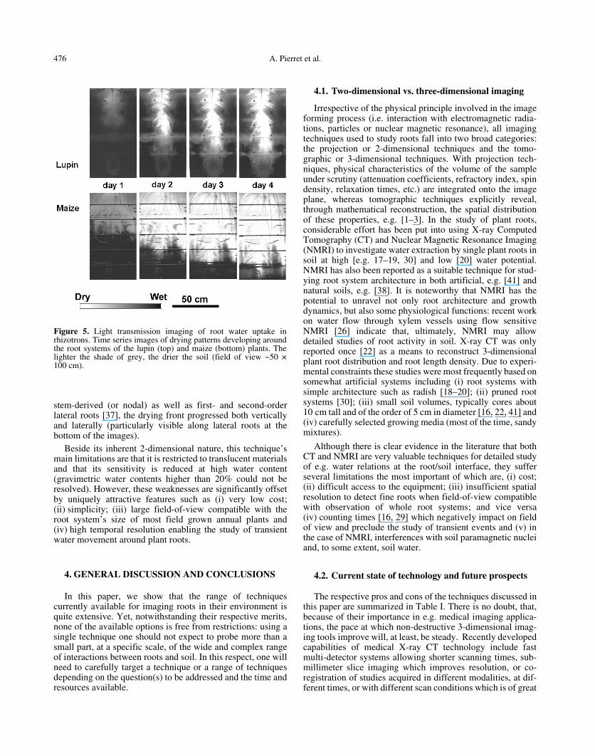
To illustrate the potential of this technique, Figure 5presents time series images of drying patterns developingaround the root systems of 1 month old lupin and maize plants.A major difference in the water uptake patterns of maize andlupin is due to the different root system architecture of thesetwo species: for lupin, which has a taprooted root system, thedrying front moved essentially downward with time. Incontrast, with maize, which has a more complex and laterallyspread out root system, including seed derived (or axile),
Figure 4. X-ray imaging of live lupin root systems in rhizotrons.(a) Water uptake pattern 4 days after stopping irrigation in loosesandy-loam soil at depths 35–50 cm (Field of view ~20 × 15 cm):dryer zones (indicated by lighter shades of grey) developed aroundthe lower part of the root system, and the most intense drying wasoccurring along a zone ~10 cm long behind the root tips. (b) Wateruptake pattern 2 days after stopping irrigation in sandy-loam soil withdistributed disks at depths 2–17 cm (Field of view ~20 × 15 cm): theloose matrix dried up more rapidly (as indicated by lighter shades ofgrey) than the dense disks. This tends to indicate that water waspreferentially taken up from the loose matrix, and is consistent withthe fact that (i) roots preferentially colonized the matrix and (ii) soilhydraulic conductivity was lower in the disks.
476 A. Pierret et al.
stem-derived (or nodal) as well as first- and second-orderlateral roots [37], the drying front progressed both verticallyand laterally (particularly visible along lateral roots at thebottom of the images).
Beside its inherent 2-dimensional nature, this technique’smain limitations are that it is restricted to translucent materialsand that its sensitivity is reduced at high water content(gravimetric water contents higher than 20% could not beresolved). However, these weaknesses are significantly offsetby uniquely attractive features such as (i) very low cost;(ii) simplicity; (iii) large field-of-view compatible with theroot system’s size of most field grown annual plants and(iv) high temporal resolution enabling the study of transientwater movement around plant roots.
4. GENERAL DISCUSSION AND CONCLUSIONS
In this paper, we show that the range of techniquescurrently available for imaging roots in their environment isquite extensive. Yet, notwithstanding their respective merits,none of the available options is free from restrictions: using asingle technique one should not expect to probe more than asmall part, at a specific scale, of the wide and complex rangeof interactions between roots and soil. In this respect, one willneed to carefully target a technique or a range of techniquesdepending on the question(s) to be addressed and the time andresources available.
4.1. Two-dimensional vs. three-dimensional imaging
Irrespective of the physical principle involved in the imageforming process (i.e. interaction with electromagnetic radia-tions, particles or nuclear magnetic resonance), all imagingtechniques used to study roots fall into two broad categories:the projection or 2-dimensional techniques and the tomo-graphic or 3-dimensional techniques. With projection tech-niques, physical characteristics of the volume of the sampleunder scrutiny (attenuation coefficients, refractory index, spindensity, relaxation times, etc.) are integrated onto the imageplane, whereas tomographic techniques explicitly reveal,through mathematical reconstruction, the spatial distributionof these properties, e.g. [1–3]. In the study of plant roots,considerable effort has been put into using X-ray ComputedTomography (CT) and Nuclear Magnetic Resonance Imaging(NMRI) to investigate water extraction by single plant roots insoil at high [e.g. 17–19, 30] and low [20] water potential.NMRI has also been reported as a suitable technique for stud-ying root system architecture in both artificial, e.g. [41] andnatural soils, e.g. [38]. It is noteworthy that NMRI has thepotential to unravel not only root architecture and growthdynamics, but also some physiological functions: recent workon water flow through xylem vessels using flow sensitiveNMRI [26] indicate that, ultimately, NMRI may allowdetailed studies of root activity in soil. X-ray CT was onlyreported once [22] as a means to reconstruct 3-dimensionalplant root distribution and root length density. Due to experi-mental constraints these studies were most frequently based onsomewhat artificial systems including (i) root systems withsimple architecture such as radish [18–20]; (ii) pruned rootsystems [30]; (iii) small soil volumes, typically cores about10 cm tall and of the order of 5 cm in diameter [16, 22, 41] and(iv) carefully selected growing media (most of the time, sandymixtures).
Although there is clear evidence in the literature that bothCT and NMRI are very valuable techniques for detailed studyof e.g. water relations at the root/soil interface, they sufferseveral limitations the most important of which are, (i) cost;(ii) difficult access to the equipment; (iii) insufficient spatialresolution to detect fine roots when field-of-view compatiblewith observation of whole root systems; and vice versa(iv) counting times [16, 29] which negatively impact on fieldof view and preclude the study of transient events and (v) inthe case of NMRI, interferences with soil paramagnetic nucleiand, to some extent, soil water.
4.2. Current state of technology and future prospects
The respective pros and cons of the techniques discussed inthis paper are summarized in Table I. There is no doubt, that,because of their importance in e.g. medical imaging applica-tions, the pace at which non-destructive 3-dimensional imag-ing tools improve will, at least, be steady. Recently developedcapabilities of medical X-ray CT technology include fastmulti-detector systems allowing shorter scanning times, sub-millimeter slice imaging which improves resolution, or co-registration of studies acquired in different modalities, at dif-ferent times, or with different scan conditions which is of great
Figure 5. Light transmission imaging of root water uptake inrhizotrons. Time series images of drying patterns developing aroundthe root systems of the lupin (top) and maize (bottom) plants. Thelighter the shade of grey, the drier the soil (field of view ~50 ×100 cm).

Imaging of roots in soil 477
interest for the study of dynamics processes. As an illustrationof what such technological advances have to offer to the studyof whole root systems, we present here (Fig. 6), the image ofthe root system of a 6 weeks old chickpea plant obtained byA. McNeill (University of Adelaide, Australia), using a state-of-the-art Toshiba Aquilion™ helical CT scanner, at a resolu-tion of 0.5 mm.
Despite these recent advances, root scientists interested innon-destructive/non-invasive observation of plant roots andtheir environment still face a trade-off between spatial resolu-tion, field-of-view and 3-dimensionality: with the current stateof the technology it is possible to have any two. This is a majorlimitation, considering that unconstrained root systems are 3-dimensional entities which include many fine roots, and which
µ µ µ µ µ
× × × × ×
Table I. Respective pros and cons of the techniques discussed in the paper. The disk size is proportional to the suitability of a given techniquefor a specific purpose.

478 A. Pierret et al.
explore large volumes of soil. We conclude that, while fullyappropriate 3D imaging tools will ultimately become availa-ble, in the meantime, simpler two-dimensional techniquesshould be considered for observing roots in their environment.In particular, we show that 2D imaging techniques, based oneither visible light or X-ray attenuation through thin growingcontainers (rhizotrons), can be quite useful to study the func-tioning of whole root systems.
Additionally, one must choose between non-destructive/non-invasive and destructive/invasive techniques. Obviously,the former enable the study of live plants, which is mostvaluable when dealing with dynamic processes such as wateruptake. On the other hand, although destructive investigationsonly provide a static, ‘snapshot-like’ view of a sample, theycertainly represent a valuable option for small-scale study ofthe interplay between roots and soil, in particular under fieldconditions. We conclude that, microscopic or high-resolutionX-ray imaging of roots in undisturbed soil samples have aconsiderable value because they can provide, at a scale whichis not accessible by any other means, essential information oncomplex interactions between roots and soil structure.
Acknowledgements: Part of the experimental work described in thispaper was financially supported by the Grains Research and DevelopmentCorporation, Australia. Colin McLachlan of CSIRO Land and Water,Canberra helped with fieldwork and in the preparation of the thin section.Annagret Fier of Osnabrueck University, Germany, helped with X-rayimaging of live roots. Drs. Ann McNeil, University of Adelaide, Australia,and Yves Coquet, Institut National Agronomique, Grignon, France, aregratefully acknowledged for helpful comments and discussions.
REFERENCES
[1] Anderson S.H., Hopmans J.W., Tomography of soil-water-rootprocesses. Proceedings of a symposium, Minneapolis, Minnesota,USA, 4 Nov. 1992, Earthscan Publications Ltd, London, USA,1994, 148 p.
[2] Asseng S., Aylmore L.A.G., McFall J.S., Hopmans J.W., GregoryP.J., Computer-assisted tomography and magnetic resonanceimaging, in: Smit A.L., Bengough A.G., Engels C., VanNoordwijk M., Pellerin S., Van de Geijn S.C. (Eds.), RootsMethods, Springer-Verlag, Berlin Heidelberg, 2000, pp. 343–363.
[3] Aylmore L.A.G., Use of computer-assisted tomography instudying water movement around plant roots, Adv. Agron. 49(1993) 2-54.
[4] Barber S.A., Ozanne P.G., Autoradiographic evidence for thedifferential effect of four plant species in altering the calciumcontent of the rhizosphere soil, Soil Sci. Soc. Am. Proc. 34 (1970)635–637.
[5] Barley K.P., The effects of mechanical stress on the growth ofroots, J. Exp. Bot. 13 (1954) 95–110.
[6] Bidel L.P.R., Mannino M.R., Rivière L.M., Pagès L., Tracing rootdevelopment using the soft X-ray radiographic method, as appliedto young cuttings of western red cedar (Thuja plicata), Can. J. Bot.77 (1999) 348–60.
[7] Bresson L.M., Moran C.J., High-resolution bulk density images,using calibrated X-ray radiography of impregnated soil slices, SoilSci. Soc. Am. J. 62 (1998) 299–305.
[8] Bruand A., Cousin I., Nicoullaud B., Duval O., Bégon J.C.,Backscattered electron scanning images of soil porosity foranalyzing soil compaction around roots, Soil Sci. Soc. Am. J. 60(1996) 895–901.
[9] Bushamuka V.N., Zobel R.W., Differential genotypic and roottype penetration of compacted soil layers, Crop Sci. 38 (1998)776–781.
[10] Clothier B.E., Green S.R., Roots: the big movers of water andchemicals in soil, Soil Sci. 162 (1997) 534–543.
[11] Darnault C.J.G., Throop J.A., DiCarlo D.A., Rimmer A.,Steenhuis T.S., Parlange J.Y., Visualization by light transmissionof oil and water contents in transient two-phase flow fields, J.Contam. Hydrol. 31 (1998) 337–348.
[12] De Willigen P., Van Noordwijk M., Roots, plant production andnutrient use efficiency, Ph.D. Thesis, Agricultural University ofWageningen, The Netherlands, 1987, 282 p.
[13] Devereux-Joslin J., Wolfe M.H., Disturbances during minirhizo-tron installation can affect root observation data, Soil Sci. Soc.Am. J. 63 (1999) 218–221.
[14] Garrigues E., Doussan C., Debroux M., Plant water uptake andtransfer- from the single root to the root system scale: experimentalstudy, Geophysical Research Abstract, Vol. 2, Congrès EGS, 25e
assemblée, Nice, Avril 2000, CD-ROM édition, ISSN: 1029–7006, 2000.
[15] Garrigues E., Doussan C., A comparison of root water uptakebetween maize and Lupin: Experimental study, 26e colloque del’EGS Nice, Mars 2001, Geophysical Research Abstracts, Vol. 3,CD-ROM édition, ISSN: 1029–7006, 2001.
[16] Gregory P.J., Hinsinger P., New approaches to studying chemicaland physical changes in the rhizosphere: an overview, Plant andSoil 211 (1999) 1–9.
[17] Hainsworth J.M., Aylmore L.A.G., The use of computer assistedtomography to determine spatial distribution of soil water content,Aust. J. Soil Res. 21 (1983) 435–443.
[18] Hainsworth J.M., Aylmore L.A.G., Water extraction by singleplant roots, Soil Sci. Soc. Am. J. 50 (1986) 841–848.
[19] Hamza M.A., Aylmore L.A.G., Soil solute concentration andwater uptake by single lupin and radish plant roots. I. Water extrac-tion and solute accumulation, Plant and Soil 145 (1992) 187–196.
[20] Hamza M.A., Anderson S.H., Aylmore L.A.G., Studies of soilwater drawdowns by single radish roots at deacreasing soil watercontent using computer-assisted tomography, Aust. J. Soil Res. 39(2001) 1387–1396.
[21] Heeraman D.A., Juma N.G., A comparison of minirhizotron, coreand monolith methods for quantifying barley (Hordeum vulgare
Figure 6. Three-dimensional rendering of the root system of a6 weeks old chickpea plant obtained by Dr. A. McNeill (Universityof Adelaide, Australia), using a Toshiba Aquilion™ Real Timehelical CT scanner, at a resolution of 0.5 mm.

Imaging of roots in soil 479
L.) and fababean (Vicia faba L.) root distribution, Plant and Soil148 (1993) 29–41.
[22] Heeraman D.A., Hopmans J.W., Clausnitzer V., Three dimen-sional imaging of plant roots in situ with X-ray computed tomog-raphy, Plant and Soil 189 (1997) 167–179.
[23] Jaillard B., Ruiz L., Arvieu J.C., pH mapping in transparent gelusing color indicator videodensitometry, Plant and Soil 183 (1996)85–95.
[24] Kooistra M.J., Schoonderbeek D., Boone F.R., Veen B.W., VanNoordwijk M., Root-soil contact of maize, as measured by a thin-section technique. II. Effects of soil compaction, Plant and Soil139 (1992) 119–129.
[25] Krebs M., Kretzschmar A., Babel U., Chadoeuf J., Goulard M.,Investigations on distribution patterns in soil: basic and relativedistributions of roots, channels and cracks, Dev. Soil Sci. 22(1994) 437–449.
[26] Kuchenbrod E., Landek M., Thurmer F., Haase A., ZimmermannU., Measurement of water flow in xylem vessels of intact maizeplants using flow-sensitive NMR imaging, Bot. Acta 109 (1996)184–186.
[27] Livesley S.J., Stacey C.L., Gregory P.J., Buresh R.J., Sieve sizeeffects on root length and biomass measurements of maize (Zeamays) and Grevillea robusta, Plant and Soil 207 (1998) 183–193.
[28] Lonkerd W.E., Ritchie J.T., Split root observation system for rootdynamics studies, Agron. J. 71 (1979) 519–522.
[29] Macedo A., Crestana S., Vaz C.M.P., X-ray microtomography toinvestigate thin layers of soil clod, Soil Tillage Res. 49 (1998)249–253.
[30] MacFall J.S., Johnson G.A., Use of magnetic resonance imaging inthe study of plants and soils, in: Anderson S.H., Hopmans J.W.(Eds.), Tomography of soil-water-root processes, Proceedings of asymposium, Minneapolis, Minnesota, USA, 4 Nov. 1992, Earths-can Publications Ltd, London, USA, 1994, pp. 99–114.
[31] Marshner H., Römheld V., Horst W.J., Martin P., Root-inducedchanges in the rhizosphere: Importance for the mineral nutrition ofplants, Z. Pflanzenernähr. Bodenk. 149 (1986) 441–456.
[32] McCully M.E., Roots in soil: unearthing the complexities of rootsand their rhizospheres, Annu. Rev. Plant Physiol. Plant Mol. Biol.50 (1999) 695–718.
[33] McCully M.E., Water efflux from the surface of field-grown roots.Observations by cryoscanning electron microscopy, Physiol.Plant. 95 (1995) 217–224.
[34] Moran C.J., Pierret A., Stevenson A.W., X-ray absorption andphase contrast imaging to study the interplay between plant rootsand soil structure, Plant and Soil 23 (2000) 99–115.
[35] Passioura J.B., Roots and water economy of wheat, in: Day W.,Atkin R. (Eds.), Wheat growth and modelling, Plenum Press, NewYork, 1985, pp. 185–198.
[36] Passioura J.B., Soil structure and plant growth, Aust. J. Soil Res.29 (1991) 717–728.
[37] Pellerin S., Pagès L., Evaluation in field conditions of a three-dimensional architectural model of the maize root system: compar-ison of simulated and observed horizontal root maps, Plant andSoil 178 (1996) 101–112.
[38] Rogers H.H., Bottomley P.A., In situ nuclear magnetic resonanceimaging of roots: influence of soil type, ferromagnetic particlecontent, and soil water, Agron. J. 79 (1987) 957–965.
[39] Santos D., Murphy S.L.S., Taubner H., Smucker A.J.M., Horn R.Uniform separation of concentric surface layers from soil aggre-gates, Soil Sci. Soc. Am. J. 61 (1997) 720–724.
[40] Soil Survey Staff, Keys to soil taxonomy, U.S.D.A., NaturalResources Conservation Service, 1998.
[41] Southon T.E., Jones R.A., NMR imaging of roots: methods forreducing the soil signal and for obtaining a 3-dimensional descrip-tion of the roots, Physiol. Plant. 86 (1992) 322–328.
[42] Stewart J.B., The spatial distribution of plant roots and their inter-action with soil structure, Ph.D. Thesis, The University of Sydney,1997.
[43] Stewart J.B., Moran C.J., Wood J.T., Macropore sheath: quantifi-cation of plant root and soil macropore association, Plant and Soil211 (1999) 59–67.
[44] Stirzaker R.J., Passioura J.B., Wilms T., Soil structure and plantgrowth – impact of bulk density and biopores, Plant and Soil 185(1996) 151–162.
[45] Tidwell V.C., Glass R.J., X-ray and visible light transmission forlaboratory measurement of two-dimensional saturation fields inthin-slab systems, Water Resour. Res. 30 (1994) 2873–2882.
[46] Tiunov A.V., Scheu S., Microbial respiration, biomass, biovolumeand nutrient status in burrow walls of Lumbricus terrestris L.(Lumbricidae), Soil Biol. Biochem. 31 (1999) 2039–2048.
[47] Van Noordwijk M., Schoonderbeek D., Kooistra M.J., Root-soilcontact of field-grown winter wheat, Geoderma 56 (1993) 277–286.
[48] Watt M., McCully M.E., Canny M.J., Formation and stabilizationof rhizosheaths in Zea mays L. Effect of soil water content, PlantPhysiol. 106 (1994) 179–186.
[49] Wilkins S.V., Gureyev T.E., Gao D., Pogany A., Stevenson A.W.,Phase-contrast imaging using polychromatic hard X-rays, Nature384 (1996) 335–338.
[50] Wulfsohn D., Nyengaard J.R., Gundersen H.J.G., Cutler A.J.,Squires T.M., Non-destructive, stereological estimation of plantroot lengths, branching pattern and diameter distribution, Plant andSoil 214 (1999) 15–26.
[51] Zwiggelaar R., Bull C.R., Mooney M.J., Czarnes S., The detectionof “soft” materials by selective energy X-ray transmission imagingand computer tomography, J. Agric. Eng. Res. 66 (1997) 203–212.