Nutrition and Cancer - MDPI
216
Nutrition and Cancer Vera C. Mazurak www.mdpi.com/journal/nutrients Edited by Printed Edition of the Special Issue Published in Nutrients nutrients
-
Upload
khangminh22 -
Category
Documents
-
view
4 -
download
0
Transcript of Nutrition and Cancer - MDPI

Nutrition and Cancer
Vera C. Mazurak
www.mdpi.com/journal/nutrients
Edited by
Printed Edition of the Special Issue Published in Nutrients
nutrients

Nutrition and Cancer
Special Issue Editor Vera C. Mazurak
MDPI • Basel • Beijing • Wuhan • Barcelona • Belgrade

Special Issue EditorVera C. Mazurak
University of Alberta
Canada
Editorial Office
MDPI St. Alban-Anlage 66
Basel, Switzerland
This edition is a reprint of the Special Issue published online in the open access journal Nutrients (ISSN 2072-6643) from 2014–2015 (available at: http://www.mdpi.com/journal/nutrients/special issues/
nutrition-cancer).
For citation purposes, cite each article independently as indicated on the article page online and as indicated below:
Lastname, F.M.; Lastname, F.M. Article title. Journal Name Year, Article number, page range.
First Editon 2018
ISBN 978-3-03842-891-6 (Pbk)
ISBN 978-3-03842-892-3 (PDF)
Articles in this volume are Open Access and distributed under the Creative Commons Attribution
(CC BY) license, which allows users to download, copy and build upon published articles even
for commercial purposes, as long as the author and publisher are properly credited, which ensures
maximum dissemination and a wider impact of our publications. The book taken as a whole isc© 2018 MDPI, Basel, Switzerland, distributed under the terms and conditions of the Creative Commons
license CC BY-NC-ND (http://creativecommons.org/licenses/by-nc-nd/4.0/).

Table of Contents
About the Special Issue Editor . . . . . . . . . . . . . . . . . . . . . . . . . . . . . . . . . . . . . . v
Preface to ”Nutrition and Cancer” . . . . . . . . . . . . . . . . . . . . . . . . . . . . . . . . . . . . vii
Lone S. Sorensen, Ole Thorlacius-Ussing, Henrik H. Rasmussen, Søren Lundbye-Christensen,
Philip. C. Calder, Karen Lindorff-Larsen and Erik B. Schmidt
Effects of Perioperative Supplementation with Omega-3 Fatty Acids on Leukotriene B4 andLeukotriene B5 Production by Stimulated Neutrophils in Patients with Colorectal Cancer:A Randomized, Placebo-Controlled Intervention Trialdoi: 10.3390/nu6104043 . . . . . . . . . . . . . . . . . . . . . . . . . . . . . . . . . . . . . . . . 1
Andrea Dueregger, Isabel Heidegger, Philipp Ofer, Bernhard Perktold, Reinhold Ramoner,
Helmut Klocker and Iris E. Eder
The Use of Dietary Supplements to Alleviate Androgen Deprivation Therapy Side Effectsduring Prostate Cancer Treatmentdoi: 10.3390/nu6104491 . . . . . . . . . . . . . . . . . . . . . . . . . . . . . . . . . . . . . . . . 14
Jennifer M. Monk, Harmony F. Turk, Danyelle M. Liddle, Anna A. De Boer, Krista A. Power,
David W.L. Ma and Lindsay E. Robinson
n-3 Polyunsaturated Fatty Acids and Mechanisms to Mitigate Inflammatory ParacrineSignaling in Obesity-Associated Breast Cancerdoi: 10.3390/nu6114760 . . . . . . . . . . . . . . . . . . . . . . . . . . . . . . . . . . . . . . . . 37
Mark J. McCann, Ian R. Rowland and Nicole C. Roy
The Anti-Proliferative Effects of Enterolactone in Prostate Cancer Cells: Evidence for the Roleof DNA Licencing Genes, mi-R106b Cluster Expression, and PTEN Dosagedoi: 10.3390/nu6114839 . . . . . . . . . . . . . . . . . . . . . . . . . . . . . . . . . . . . . . . . 64
Jiajie Liu and David W. L. Ma
The Role of n-3 Polyunsaturated Fatty Acids in the Prevention and Treatment of Breast Cancerdoi: 10.3390/nu6115184 . . . . . . . . . . . . . . . . . . . . . . . . . . . . . . . . . . . . . . . . 79
Maryam Ebadi and Vera C. Mazurak
Evidence and Mechanisms of Fat Depletion in Cancerdoi: 10.3390/nu6115280 . . . . . . . . . . . . . . . . . . . . . . . . . . . . . . . . . . . . . . . . 108
Katie M. Di Sebastiano and Marina Mourtzakis
The Role of Dietary Fat throughout the Prostate Cancer Trajectorydoi: 10.3390/nu6126095 . . . . . . . . . . . . . . . . . . . . . . . . . . . . . . . . . . . . . . . . 123
Karen S. Bishop, Sharon Erdrich, Nishi Karunasinghe, Dug Yeo Han, Shuotun Zhu,
Amalini Jesuthasan and Lynnette R. Ferguson
An Investigation into the Association between DNA Damage and Dietary Fatty Acid in Menwith Prostate Cancerdoi: 10.3390/nu7010405 . . . . . . . . . . . . . . . . . . . . . . . . . . . . . . . . . . . . . . . . 135
Reema F. Tayyem, Hiba A. Bawadi, Ihab N. Shehadah, Suhad S. Abu-Mweis, Lana M. Agraib,
Kamal E. Bani-Hani, Tareq Al-Jaberi, Majed Al-Nusairr and Dennis D. Heath
Macro- and Micronutrients Consumption and the Risk for Colorectal Cancer among Jordaniansdoi: 10.3390/nu7031769 . . . . . . . . . . . . . . . . . . . . . . . . . . . . . . . . . . . . . . . . 151
iii

Rita Ostan, Catia Lanzarini, Elisa Pini, Maria Scurti, Dario Vianello, Claudia Bertarelli,
Cristina Fabbri, Massimo Izzi, Giustina Palmas, Fiammetta Biondi, Morena Martucci,
Elena Bellavista, Stefano Salvioli, Miriam Capri, Claudio Franceschi and Aurelia Santoro
Inflammaging and Cancer: A Challenge for the Mediterranean Dietdoi: 10.3390/nu7042589 . . . . . . . . . . . . . . . . . . . . . . . . . . . . . . . . . . . . . . . . 165
Rodolfo Gonzalez Camargo, Daniela Mendes dos Reis Riccardi, Henrique Quintas Teixeira Ribeiro,
Luiz Carlos Carnevali Jr., Emidio Marques de Matos-Neto, Lucas Enjiu, Rodrigo Xavier Neves,
Joanna Darck Carola Correia Lima, Raquel Galvao Figueredo, Paulo Sergio Martins de Alcantara,
Linda Maximiano, Jose Otoch, Miguel Luiz Batista Jr., Gerhard Puschel and Marilia Seelaender
NF-κBp65 and Expression of Its Pro-Inflammatory Target Genes Are Upregulated in theSubcutaneous Adipose Tissue of Cachectic Cancer Patientsdoi: 10.3390/nu7064465 . . . . . . . . . . . . . . . . . . . . . . . . . . . . . . . . . . . . . . . . 192
iv

About the Special Issue Editor
Vera C. Mazurak’s research focuses on alterations in the metabolism that occur during critical illness with a focus on lipid metabolism and immunity. Following her PhD, she was employed as a research associate at the Cross Cancer Institute in Edmonton before she commenced her first faculty position at the University of Alberta in 2004, where she is currently a Professor of Human Nutrition. Her current research focuses on how changes in metabolism impact on the nutritional requirements in diseases characterized by inflammation, with a primary focus on cancer and its treatment. She is particularly interested in how providing essential dietary nutrients can improve care for patients. Her translational research program spans from experimental models of disease to human clinical trials. Dr. Mazurak developed and currently teaches a “Nutrition and Cancer” course and supervises undergraduate, graduate student (PhD and MSc) and postdoctoral fellow research programs in nutrition, as well as at the Faculty of Medicine and Dentistry as part of her research program.
v


Preface to ”Nutrition and Cancer”
Defining better nutritional interventions for those at risk of malnutrition has been a passion of mine
ever since I started research, which is why I am excited to bring forward this Special Issue focused
on nutrition and cancer. The development and treatment of cancer presents a complex interaction
between tumor and host. The provision of nutrients not only enables the maintenance of nutritional
status, but also provides substrates and signals for immunity, tumor metabolism and the protection of
the host from treatment toxicities. Unique elements in the diet can help to tip the metabolic balance in
favor of the host by acting on multiple pathways concurrently, both enhancing the host’s ability to
fight the tumor and endure cancer treatment while simultaneously attenuating tumor growth. This
Special Issue includes articles that highlight the role of certain dietary components in the prevention of
cancer and host response during cancer treatment.
Fat is one dietary element that has been explored for its role in cancer development. While the bulk
of these studies have been observational or experimental, the evidence assembled suggests that dietary
lipids behave uniquely to prevent or promote cancers. Of particular interest are the n-3 fatty acids,
consisting of linolenic acid, eicosapentaenoic acid and docosahexaenoic acid. These fatty acids are
most widely recognized for their anti-inflammatory properties that attenuate cancer progression while
also enhancing host immune responses. Several articles in this Special Issue focus on the n-3 fatty acids
and the mechanisms involved in cancer prevention, providing a harmonious accompaniment to the
reviews of the epidemiological evidence.
An additional aspect of cancer development is the role of adipose tissue, which has an expanded
role beyond energy storage, contributing to the whole body metabolism. Adipose tissue is a source
of and responds to inflammatory signals that may be involved in tumor development. This Special
Issue also provides reviews on various aspects of adipose tissue in relation to cancer development and
progression.
The contributors to this Special Issue are well recognized leaders in the field of cancer and have
unique areas of focus including metabolism, immunology, biochemistry, epidemiology and nutrition.
Each contribution highlights the latest research in these areas and what is known about fat and cancer.
Vera Mazurak Special Issue Editor
vii


nutrients
Article
Effects of Perioperative Supplementation withOmega-3 Fatty Acids on Leukotriene B4 andLeukotriene B5 Production by Stimulated Neutrophilsin Patients with Colorectal Cancer: A Randomized,Placebo-Controlled Intervention Trial
Lone S. Sorensen 1,*, Ole Thorlacius-Ussing 1,2, Henrik H. Rasmussen 3,
Søren Lundbye-Christensen 4, Philip. C. Calder 5, Karen Lindorff-Larsen 6 and Erik B. Schmidt 4
1 Department of Surgical Gastroenterology, Aalborg University Hospital, 9000 Aalborg, Denmark; [email protected] Institute of Clinical Medicine, Aarhus University Hospital, Aarhus 8000, Denmark3 Center for Nutrition and Bowel Disease, Aalborg University Hospital, 9000 Aalborg, Denmark; [email protected] Department of Cardiology, Center for Cardiovascular Research, Aalborg University Hospital, 9000 Aalborg,
Denmark; [email protected] (S.L.); [email protected] (E.B.S.)5 National Institute for Health Research Southampton Biomedical Research Center, University Hospital
Southampton NHS Foundation Trust and University of Southampton, Southampton SO16 6YD, UK;[email protected]
6 NordSim, Center for Simulation, Skills Training, Science and Innovation, Aalborg University Hospital, 9000Aalborg, Denmark; [email protected]
* Author to whom correspondence should be addressed; [email protected]; Tel.: +45-9766-1188; Fax: +45-9932-2560.
Received: 30 July 2014; in revised form: 18 September 2014; Accepted: 19 September 2014;Published: 29 September 2014
Abstract: Omega-3 fatty acids (n-3 FA) may have beneficial clinical and immune-modulating effectsin surgical patients. In a randomized, double-blind, prospective, placebo-controlled trial, 148 patientsreferred for elective colorectal cancer surgery received an n-3 FA-enriched oral nutritional supplement(ONS) providing 2.0 g of eicosapentaenoic acid (EPA) and 1.0 g of docosahexaenoic acid (DHA) perday or a standard ONS for seven days before surgery. On the day of operation, there was a significantincrease in the production of leukotriene B5 (LTB5) (p < 0.01) and 5-hydroxyeicosapentaenoic acid(5-HEPE) (p < 0.01), a significant decrease in the production of leukotriene B4 (LTB4) (p < 0.01) anda trend for a decrease in the production of 5-hydroxyeicosatetraenoic acid (5-HETE) (p < 0.1) fromstimulated neutrophils in the active group compared with controls. There was no association betweenLTB4 values and postoperative complications. In conclusion, oral n-3 FA exerts anti-inflammatoryeffects in surgical patients, without reducing the risk of postoperative complications.
Keywords: colorectal cancer; omega-3 fatty acids; immunomodulation; fish oil; leukotrienes
1. Introduction
Patients undergoing surgery are at risk of developing complications in the postoperative period [1–3]. This is believed to be partly caused by changes in the immune response following surgery [4]. Thus,initially, a hyper-inflammatory response followed by a phase of relative immune incompetence occursin relation to major surgery [5].
The pathophysiological changes are complex, but may be driven by excessive production ofvarious lipid mediators, including the very potent pro-inflammatory leukotriene B4 (LTB4) producedfrom the omega-6 fatty acid (n-6 FA) arachidonic acid (AA) present in cell membranes.
Nutrients 2014, 6, 4043–4057 1 www.mdpi.com/journal/nutrients

Nutrients 2014, 6, 4043–4057
Among factors known to influence the clinical course of patients after surgery are nutritional statusand specific biologically active nutrients [2,6–10] that might include the marine omega-3 fatty acids (n-3FA) with the main biologically active n-3 FAs being eicosapentaenoic acid (EPA) and docosahexaenoicacid (DHA). Consumption of fish and fish oils oil increases the concentration of EPA and DHA inblood, cells and tissues [11,12] and alters the physical properties of cell membranes and the functionof membrane proteins, including receptors, transporters and signalling proteins [13,14]. n-3 FA areincorporated into cell membranes in competition with the more abundant n-6 FA, AA, at the expenseof the latter. AA may be liberated by phospholipases from cell membranes and induces leucocytes toproduce the pro-inflammatory LTB4 and the side product, 5-hydroxyeicosatetraenoic acid (5-HETE).In contrast, leukotriene B5 (LTB5) and the side product, 5-hydroxyeicosapentaenoic acid (5-HEPE),derived from EPA [15], have considerably less potent biological activities in comparison to LTB4 [15,16].Replacement of n-6 FA with n-3 FA in membranes of immune active cells may therefore lead to reducedformation of pro-inflammatory compounds, and by this, and other [5,12] mechanisms, n-3 FA maydecrease infectious complications after surgery [7,17–20]. The influence of enteral feeds, includingn-3 FA on AA-derived eicosanoids (e.g., LTB4), has been the subject of much attention [5,21–23].Several studies have indicated that n-3 FAs modulate the generation of inflammatory eicosanoids ingastrointestinal surgical patients [24–26] and may help to counteract the surgery-induced decline inantigen-presenting cell activity [20] and T-cell cytokine production [27].
The aim of the present study was to evaluate the production of LTB4, 5-HETE, LTB5 and 5-HEPEfrom stimulated neutrophils after seven days of preoperative treatment with an n-3 FA-enrichedoral nutritional supplement (ONS) in patients undergoing colorectal cancer surgery and to studythe possible impact on clinical outcome. Furthermore, the correlation between LTB4 values andpostoperative complications was investigated.
2. Materials and Methods
2.1. Study Design
This was a sub-study of a randomized, double-blind, prospective, placebo-controlled single-centreinterventional trial involving 148 participants (Figure 1) awaiting colorectal cancer surgery [28].Participants were recruited consecutively from the outpatient clinic of the Department of SurgicalGastroenterology, Aalborg University Hospital. All eligible participants were asked to participate.
Exclusion criteria were diabetes mellitus, consumption of >5 alcoholic drinks per day, emergencysurgery, inability to understand the spoken and written information in Danish, untreated psychiatricconditions, pregnancy or breast-feeding, reduced kidney function (plasma creatinine > 130 μmol/L),use of n-3 FA supplements, anticipated poor compliance, immunosuppressive diseases andparticipation in another clinical trial.
After providing oral and written informed consent, participants were randomly assigned totreatment with n-3 FA (active treatment) or control (a standard ONS without marine n-3 FA), 200 mLtwice per day (morning and afternoon) for 7 days before surgery. Randomization was performed usingsealed non-transparent envelopes containing the randomization number and kept at the investigationsite according to CONSORT (Consolidated Standards of Reporting Trials) guidelines [29]. The activeand the control ONS cartons looked identical and had identical taste and scent (coffee). The maininvestigator and a study nurse enrolled the participants at the outpatient clinic and randomly assignedthem to active treatment or control. The participants, carers, investigators and other researchers wereblinded to treatment allocation throughout the study. The investigators had no access to the code untilafter completion of the study. Statistical analyses were completed before the code was broken.
Information collected included demographic data, tumour location and American Society ofAnaesthesiologists (ASA) risk score [30]. All patients underwent standard Nutritional Risk Screening(NRS 2002) [31]. A food questionnaire focusing on consumption of seafood on a monthly basis wascompleted at baseline.
2

Nutrients 2014, 6, 4043–4057
The study was approved by the regional ethics committee (N-VN-20050035) and conductedaccording to the Hong Kong amendment to the Declaration of Helsinki. The trial was registered atClinicalTrials.gov: ID NCT00488904 [32].
2.2. Intervention
Participants in the active or control group received the ONS as a sip feed (200 mL twice a daymorning and afternoon) for 7 days before surgery. The feeds (Supportan®) were isocaloric (1.5 kcal/mL)and isonitrogenous (Table 1) and were provided by Fresenius Kabi (Bad Homburg, Germany). Bothfeeds contained the same amounts of carbohydrate, protein, total fat and n-6 FA (Table 1).
Figure 1. Patient flow through the study.
3

Nutrients 2014, 6, 4043–4057
Table 1. Daily intake of energy and nutrients from the n-3 FA-enriched and the control oralnutritional supplement.
Daily dose Control n-3 FA (Active)
Energy (kcal) 600 600Protein (g) 40 40
Carbohydrate (g) 49.6 49.6Fat (g) 26.8 26.8
EPA (g) 0 2DHA (g) 0 1
Total n-6 FA (g) 3.3 3.3
Fat content of the supplement was comprised of medium chain triglycerides, sunflower oil andsafflower oil. The active supplement also contained additional fish oil at a level to achieve 2 g EPA and1 g DHA per day (Table 1). Participants were provided with the sip feeds at inclusion, to consume twicea day for 7 days at home before hospitalization. A questionnaire regarding compliance preoperativelywas completed, and good compliance was defined as self-reported consumption of at least 12 of the14 ONS cartons before surgery.
2.3. Isolation of Blood Neutrophils
Blood was drawn in the fasting state on the day of the surgery. Neutrophils were separated fromanticoagulated (K-EDTA 1.6 mg/mL) blood layered on top of PolymorphprepTM (AXIS-SHIELD PoCAS, Rodeloekka, Norway) and separated by a one-step centrifugation technique at 450 g for 40 min.Neutrophils were harvested and washed twice in tissue culture medium (RPMI 1640, Sigma-Aldrich,Ayrshire, UK), at ambient temperature and centrifuged for 10 min at 520× g. Subsequently, neutrophilswere counted and red cells eliminated by the addition of ice-cold 0.2% saline for 35 s. Next,1.6% ice-cold saline was added in order to obtain an isotonic 0.9% concentration, followed bycentrifugation at 300× g at 5 ◦C for 5 min, which was repeated once. Neutrophils were then washed ina phosphate buffer containing glucose and human albumin (PBS) and resuspended in PBS adjusting theconcentration to 1 × 10⁷ neutrophils/mL PBS. Isolated neutrophils were stored at −80 ◦C until analysis.
2.4. Analysis of Leukotrienes, 5-HEPE and 5-HETE
The neutrophil suspension (0.9 mL of 1 × 10⁷ granulocytes/mL PBS) was prewarmed to 37 ◦C,and CaCl2, MgCl2 and calcium ionophore (A23187) at a final concentration of 10 μM were addedto initiate stimulation. After 10 min, the reaction was terminated by the addition of 100% ice-coldethanol, and the mixtures were centrifuged at 4 ◦C at 700× g. The supernatant was stored at −80◦C for later analysis. C18 cartridges (Sep-Pak VAC RC, Waters Co., Milford, MA, USA) were usedfor the extraction of leukotrienes (LT), 5-HEPE and 5-HETE. The ethanol mixture was thawed andcentrifuged, and international standard prostaglandin B2 (PGB2) and trifluoroacetic acid were added.The cartridges were conditioned and equilibrated using methyl formate, 100% ethanol and water.The acidified sample was loaded onto the cartridge, washed with 15% ethanol, water and hexaneand eluted with methyl formate. The solvent was evaporated to dryness under nitrogen, and thesediment was dissolved in the mobile phase (31% H2O, 27% methanol, 42% acetonitrile and 0.025%trifluoroacetic acid).
Analysis was performed by high pressure liquid chromatography (Dionex Ultimate LPG-3400A)on an Acclaim RSLC 2.1 mm × 100 mm C18 column (Dionex Corporation, Sunnyvale, CA, USA).Concentrations were calculated using the internal standard and response factors. The response factorswere calculated by analysis of a non-stimulated neutrophil suspension after the addition of knownamounts of standards of LTB4, LTB5, 5-HETE and 5-HEPE, as well as an internal standard (PGB2).Samples were extracted and analysed using high performance liquid chromatography, and eventually,the recovery factors were calculated.
4

Nutrients 2014, 6, 4043–4057
2.5. Granulocyte Fatty Acid Analysis
Blood was drawn in the fasting state on the day of surgery. Granulocytes were prepared asdescribed previously [33]. FA profiles were determined by gas chromatography using a Varian3900 gas chromatograph, CP-8400 autosampler and CP 8414 autoinjector (Varian, Middelburg, TheNetherlands), as well as a flame ionization detector. In split injection mode, a CP-sil 88.60-m × 0.25-mmcapillary column (Varian, Middelburg, The Netherlands), temperature programming from 90 to 205◦C, a constant flow rate of 1.0 mL/min and helium carrier gas were used. Results for individual FAsare expressed as a percentage of the total FA content.
2.6. Statistical Analysis
The basic characteristics of the trial population were analysed with Fisher’s exact test forcategorical variables and unpaired t-tests for continuous variables. Differences between treatmentgroups were analysed using unpaired t-tests. If variances differed between groups, Welch’sapproximation was used. The distribution of continuous data was analysed for normality. As thevalues for 5-HEPE, 5-HETE and LTs were right skewed distributed, log transformed observationswere analysed, and these were normally distributed. Distributions of 5-HEPE, 5-HETE and LT weredescribed with median and inter-quartile range (IQR). Relative differences between groups in medianswere calculated by exponentiation of the differences between log-transformed means. Associationsbetween LTB4 values and postoperative complications were analysed using logistic regression. Theassociations between log-transformed LT values 5-HEPE and AA, EPA and AA/EPA were analysedusing linear regression. Analyses were performed blinded to treatment groups. The active and controlgroups were not identified until after the statistical analyses had been conducted. All p-values weretwo-tailed, and differences were reported with 95% confidence intervals (CI). p-values below 0.05 wereconsidered significant. All analyses were performed using Stata version 11.2 (StataCorp, 2009; TexasCity, TX, USA).
3. Results
3.1. Participants Characteristics
All eligible participants (n = 610) were asked to participate, but 230 participants were notincluded. This was due to a change in clinical practice during the study, such that many patientswere offered surgery within a five-day period, which did not allow for participants to complete theseven-day intervention. Furthermore, some participants did not meet the inclusion criteria (201), and31 participants declined to participate. Baseline characteristics of the included vs. the non-includedparticipants did not differ.
A total of 148 consecutive patients (68 females, 80 males; mean age 71 (range 41–89) years) wereincluded in the study. The majority of participants had open surgery; laparoscopic resection was onlyperformed in nine patients in the control group and nine in the n-3 FA group. Participant characteristicsdid not differ between treatment groups (Table 2).
5

Nutrients 2014, 6, 4043–4057
Table 2. Characteristics of patients in the control and active groups.
Variable Control (n =74)
Active (n = 74) p
Demographic data
Age, years, mean (SD) 71 (10) 69 (11) 0.164Sex (male/female) 36/38 44/30 0.248Body weight, kg, mean (SD) 76 (19) 77(17) 0.570Height, cm, mean (SD) 169 (11) 171 (9) 0.301BMI, kg/m2, mean (SD) 26 (5) 26 (5) 0.651Weight loss * (n) 19 11 0.068Clinical characteristicsSmoking/non-smoking (n) 11/60 17/54 0.292Unknown smoking status 3 3Cancer locationColon/rectum (n) 40/34 38/36 0.869Surgical procedure 0.977Right hemicolectomy + transverse colon (n) 16 17Left hemicolectomy + sigmoid colon (n) 10 12Laparoscopic resection of sigmoid colon (n) 9 9Low anterior resection of rectum orabdominoperineal resection
28 30
Colectomy (n) 4 3Other rectum resection (n) 7 3Nutritional status ** 0.089No risk (NRS score <3) (n) 23 34At risk (NRS score ≥3) (n) 50 39Unknown (n) 1 1
Notes: There were no significant differences between groups (p > 0.068); BMI, body mass index; * defined as loss ofmore than 5% of body weight; ** defined according to NRS 2002 [31].
3.2. Fatty Acid Composition of Neutrophils
Neutrophil EPA and DHA were significantly higher, and AA and linoleic acid was significantlylower in the group receiving n-3 FA than in the control group (Table 3) [28].
The food questionnaire indicated an average dietary intake of n-3 FA of 0.6 g per day, withno difference in preoperative intake between groups (p = 0.770). None of the included participantsreceived more than 150 mg of anti-inflammatory drugs daily. Both supplements were well toleratedwith no adverse effects reported. Nine participants randomized to active treatment and 10 participantsin the control group did not receive the allocated intervention for reasons listed in Figure 1.
Preoperatively, 63 of 65 participants in the active group were compliant compared with 56 of 64participants in the control group (p = 0.266). Two participants died in each group. In the active group,death was caused by pneumonia and a myocardial infarction, whereas the participants in the controlgroup died from septicaemia and sudden cardiac death.
6

Nutrients 2014, 6, 4043–4057
Table 3. Granulocyte fatty acids on day of operation in the control and active groups.
Weight% of Total FA Content
Control Active
EPA 0.54 (0.42–0.74) 2.10 (1.83–2.55) *DPA 0.54 (0.42–0.74) 2.11 (1.83–2.55) *DHA 1.31 (1.10–1.57) 1.61 (1.34–1.84) *
Total n-3 FA 2.44 (1.98–2.97) 5.95 (5.20–6.75) *Arachidonic acid 12.51 (11.65–13.19) 11.61 (10.67–12.48) *
Linoleic acid 8.94 (8.24–9.49) 9.42 (8.72–10.19) **
Notes: Values are the median (IQR); FA, fatty acid; ONS, oral nutritional supplement; EPA, eicosapentaenoic acid;DPA, docosapentaenoic acid; DHA, docosahexaenoic acid; * p < 0.001; ** p < 0.05 versus the control group.
3.3. Production of Mediators from Neutrophils
Furthermore, compared to neutrophils from controls, those from participants in the n-3 FA groupshowed a significantly higher (by 176% and 306%, respectively) production of LTB5 (p < 0.001) and5-HEPE (p < 0.001) (Table 4).
Table 4. Formation of leukotrienes (LT) and side products (5-HEPE; 5-HETE) from activated neutrophilsaccording to treatment group.
Eicosanoids Control Active % Difference
LTB5 5.8 (4.9–7.6) 17.5 (13.5–22.8) 176 *** (143–215)LTB4 186.8 (156.8–230.7) 163.5 (136.6–199.9) −12 *** (−21–−3)
5-HETE 293.1 (246.5–357.8) 273.7 (221.8–320.7) −7 * (−14–−0)5-HEPE 34.2 (25.7–55.3) 154.7 (122.4–190.4) 306 *** (255–364)
LTB4/LTB5 31.8 (25.0–40.1) 9.6 (7.8–11.4) −68*** (−72–−65)
Notes: Data are the median (IQR) ng/107 neutrophils and the percentage of difference between estimated medians;*** indicates p < 0.01; * indicates p < 0.1; ng/107 = nanogram/107 neutrophils.
Conversely, in the active group, neutrophils showed a significantly lower (by 12%) productionof LTB4 (p < 0.001) and a trend towards lower (by 7%) production of 5-HETE (p = 0.059). LTB4/LTB5
was significantly different between groups (by 68%) (p < 0.001) (Table 4). There was no statisticallysignificant difference in clinical outcomes (total number of complications, infectious complications,non-infectious complications, intensive care unit stay, mortality, readmissions and hospital stay)between groups, as reported previously [28].
There was no statistically significant association between the values of the proinflammatory LTB4
production and any clinical outcome, including total number of complications (p = 0.524), infectiouscomplications (p = 0.660) and non-infectious complications (p = 0.307) (Table 5). The ratio LTB4/LTB5
did not have a statistically significant association with the total number of complications (p = 0.707),infectious complications (p = 0.711) and non-infectious complications (p = 0.143) (Table 4).
However, There were strong associations between the content of AA and EPA in neutrophils andproduction of LTB4 and LTB5 (Figure 2) (all p < 0.01).
These graphs illustrate that the higher the content of EPA in the cell membranes, the higher theproduction of LTB5. Furthermore, it can be seen that the higher the content of AA in cell membranes,the lower the production of LTB5. Furthermore, there were strong associations between AA/EPA inneutrophils and LTB4 and LTB5 production (both p < 0.01) (results not shown) and between AA, EPA,AA/EPA and 5-HEPE production (all p < 0.001) (results not shown).
7

Nutrients 2014, 6, 4043–4057
Figure 2. Associations between log-transformed AAg (AA content in the cell membranes of thegranulocytes) and EPAg (EPA content in the cell membranes of the granulocytes) in the neutrophils, aswell as the formation of LTs (LTB4 and LTB5 (ng/107)) by neutrophils, illustrated using scatter plotswith regression lines and confidence bands added. Control group, red dots; active group, blue dots.
Table 5. Associations between LTs production by neutrophils and clinical outcome described by oddsratio (OR), CI and p-values for a unit change in LTB4 and LTB4/LTB5, respectively.
LTB4 LTB4/LTB5
OR CI p OR CI p
Infectious complications 1.00 1.00–1.01 0.660 1.00 0.97–1.03 0.711Non-infectious complications 1.00 0.99–1.00 0.307 0.98 0.94–1.00 0.143Total number of complications 1.00 0.99–1.00 0.524 1.00 0.98–1.03 0.707
4. Discussion
In this prospective randomized, double-blind, single-centre, placebo controlled study, it wasdemonstrated that seven days of enteral supplementation with 3 g of EPA + DHA daily resulted in ahigher neutrophil production of LTB5 and a lower production of LTB4 compared to the control group. Inaddition, there was a higher neutrophil 5-HEPE production and a trend to lower production of 5-HETEin the active group compared to the control. While the differences in LTB4 and LTB5 production indicatean anti-inflammatory action of the supplement, it is unknown whether the difference in formation of5-HETE and 5-HEPE between groups is of clinical relevance. However, 5-HETE enhances lymphocyte
8

Nutrients 2014, 6, 4043–4057
proliferation, whereas 5-HEPE only has one-tenth the potency of 5-HETE regarding this [34]. Thus, thecurrent study demonstrates that preoperative supplementation with n-3 FA for one week can modulateimmune function, assessed as the production of lipoxygenase mediators, in participants admitted forelective colorectal cancer surgery. However, this was not associated with a decrease in postoperativecomplication rates [28].
The strengths of the present study are that it was a randomized, prospective, relatively largeclinical study, was double-blind, with an identical appearance for the sip feed cartons, the taste of n-3FA was undetectable and that compliance was acceptable. Furthermore, the study population wasrelatively homogenous.
One limitation of the present study is that we only had data on eicosanoid formation from theday of surgery, whereas postoperative changes would also have been of interest. The short durationof the intervention (seven days) may have limited the incorporation of n-3 FA and the decreaseof AA in neutrophils and, thereby, limited the impact on LTB4 production, but a longer period ofsupplementation was not possible, as participants were operated on soon after cancer diagnosis.However, we showed in earlier publications that 3 g of n-3 FA for seven days before surgery wassufficient to assure significant incorporation of n-3 FA into neutrophils and into colonic tissue [28,35].The required sample size was based on a reduction in postoperative infection rates from 30% to 10%and was calculated to be 148 participants in all, but we were only able to analyse data from 129 of these.A final limitation is the discrepancy between the number of eligible (610) and analysed participants(129) due to a change in clinical practice during the study, such that many patients were offered surgerywithin a five-day period, which did not allow for participants to complete the seven-day intervention.
Our findings are consistent with the results from three recent studies in humans [24,36,37].In a prospective double-blind study, Wang et al. [37] randomized 64 participants with a need forpostoperative parenteral nutrition after surgery into two groups. The study population was a mixof surgical patients (22 gastric cancers; 29 colonic cancers; 13 with other digestive diseases). Theyreceived either fish oil containing lipid emulsion (a mixture of soybean oil, MCT and fish oil) as partof the intravenous regimen, or a mix of soybean oil and MCT for 5 days after surgery. There was asignificant increase in the neutrophil LTB5/LTB4 ratio but no effect on clinical outcome, infectiouscomplications and bleeding events. Grimm et al. [36] randomized 33 participants undergoing majorabdominal surgery into two groups in a prospective double-blind study to receive parenteral nutritionproviding either a fish oil containing lipid emulsion (a mixture of soybean oil, MCT, olive oil and fishoil) or soybean oil for five days after surgery. The study population was again a mix of surgical patients.The initial production of LTB4 and LTB5 by neutrophils was similar in both groups. The production ofLTB5 from neutrophils was significantly increased, and the release of LTB4 was decreased, though notsignificantly, in the participants receiving fish oil. The length of hospital stay was significantly shorterin the intervention group. Finally, Köller et al. [24] conducted a prospective double-blind randomizedstudy with 30 participants undergoing colorectal surgery. Participants received parenteral nutrition,providing either a fish oil containing lipid emulsion (a mixture of soybean oil, MCT and fish oil) orsoybean oil for five days post-surgery. This study also found a significant increase in LTB5 productionby leukocytes in the fish oil group, but without a concomitant decrease in LTB4 production. Thesethree studies all made use of intravenous (IV) nutrition given postoperatively. The present study,which used enteral nutrition given preoperatively, agrees with these earlier findings of increased LTB5
production and decreased LTB4 production after fish oil provision, but with limited clinical impact.Some earlier studies have reported beneficial effects of oral n-3 FA supplementation in
gastrointestinal surgery patients. Wachtler et al. [26] analysed leukocyte function in 40 participantsundergoing major upper gastrointestinal surgery in a placebo-controlled double-blind study. Onegroup received an n-3 FA-enriched (0.33 g/100 mL) oral supplement, also containing arginineand ribonucleic acid, and the other group received a standard control supplement for five dayspreoperatively. There was a significantly higher production of LTB5 from neutrophils in the interventiongroup when compared to controls. However, no changes in LTB4 were evident in the intervention
9

Nutrients 2014, 6, 4043–4057
group. The authors reported a low number of postoperative complications. In another study, Shimizu etal. [38] gave 12 children with ulcerative colitis 1.8 g EPA orally per day for two months. LTB4 productionby leucocytes and colonic mucosa were assessed before and after the intervention. Biopsies were takenfrom the rectal mucosa during sigmoidoscopy before and after initiation of EPA supplementation.After two months of supplementation, there was a decrease in LTB4 production by leucocytes andcolonic mucosa, while no information regarding LTB5 production was given.
IV administration of n-3 FA ensures a quicker incorporation of the presumed active substances(n-3 FA) into the membranes of immune cells [39,40]. However, this format is not feasible in thepre-operative setting. ONSs are less expensive and enable the use of the gut prior to surgery. Twostudies providing IV n-3 FA for five days post-operatively did not show any decrease in the productionof LTB4 [24,36]. Importantly many of these studies did not provide explicit information about theamount of n-3 FA given.
In the present study, seven days of oral supplementation with 3 g of n-3 FA daily ensuredsignificant incorporation of EPA into neutrophils and a significant decrease in the formation of LTB4.However, to achieve an anti-inflammatory response meditated by n-3 FA, it is probably more importantthat the formation of the AA-derived LTB4 is suppressed than an increase in LTB5. Despite this, therewas no effect on clinical outcome in the current study. One explanation for this may be that n-3 FAincorporation was not sufficiently high. A higher n-3 FA dose or a longer duration of interventioncould have had an impact on clinical outcome. The ratio LTB4/LTB5 was 68% lower in the activegroup, which is a considerable decrease, but still did not have any effect on clinical outcome.
One other important factor may be that the nutritional status of the patients, evaluated by NRS2002, was generally good for most participants. A weight loss of more than 5% of body weight wasonly detected in 23% of participants entering the study. This could account for the lack of clinicalimprovement with the n-3 FA-enriched ONS, since it is likely that malnourished participants mightbenefit the most from ONS and from oral n-3 FA.
5. Conclusions
In summary, the current study shows that an ONS providing 3 g of n-3 FA daily for seven daysbefore surgery was able to induce a significant decrease in the formation of the pro-inflammatory LTB4
from neutrophils with a simultaneous increased production of LTB5. A decrease in the formation of5-HETE, though not significant, and a significant rise in 5-HEPE was also seen. However, the clinicalconsequences of these changes are unknown. Associations between values of LTB4 or LTB4/LTB5 andpostoperative complication rates were not seen. This indicates either that the changes observed weretoo small or that the formation of LTs from activated neutrophils is not an important determinant ofsurgical complications. Whether a longer period (months) of n-3 FA intake could be of a benefit forpatients operated on for colorectal cancer regarding shorter stay in hospital or longer survival needs tobe investigated in larger trials.
Abbreviations
AA arachidonic acidASA American Society of AnaesthesiologistsBMI body mass indexCI confidence intervalsDHA docosahexaenoic acidEPA eicosapentaenoic acid5-HEPE 5-hydroxyeicosapentaenoic acid5-HETE 5-hydroxyeicosatetraenoic acidIV intravenous;IQR inter-quartile rangeLT leukotrienesLTB4 leukotriene B4LTB5 leukotriene B5
10

Nutrients 2014, 6, 4043–4057
n-3 FA n-3 fatty acidsn-6 FA n-6 fatty acidsMCT medium-chain triglyceridesng nanogramsNRS 2002 Nutritional Risk ScreeningONS oral nutritional supplementOR odds ratioPBS phosphate-buffered salinePGB2 prostaglandin B2
Acknowledgments: This work was kindly supported by Fresenius Kabi (Bad Homburg, Germany), The ObelskeFamily Foundation, the Hoejmosegaard Grant, the Aase and Ejnar Danielsens Foundation and the North JutlandMedical Association.
We thank study nurse Anne Madsen and the staff at the Lipid Research Clinic at Aalborg UniversityHospital for invaluable assistance in connection with completion of the study. We also thank Hanne Madsenfor proofreading.
Author Contributions: Lone Schmidt Sorensen: study concept and design; acquisitions of data; analysis andinterpretations of data; drafting of the manuscript; statistical analysis; obtained funding; study supervision;approval of the final version of the manuscript.
Henrik Hojgaard Rasmussen: study concept and design; critical revision of the manuscript; studysupervision; approval of the final version of the manuscript.
Søren Lundbye-Christensen: statistical support; critical revision of the manuscript; approval of the finalversion of the manuscript.
Ole Thorlacius-Ussing: study concept and design; critical revision of the manuscript; study supervision;approval of the final version of the manuscript.
Karen Lindorff-Larsen: study concept and design; acquisitions of data; critical revision of the manuscript;study supervision; approval of the final version of the manuscript.
Erik Berg Schmidt: study concept and design; critical revision of the manuscript; study supervision; approvalof the final version of the manuscript.
Philip C. Calder: study concept and design; critical revision of the manuscript; approval of the final versionof the manuscript.
Conflicts of Interest: The authors declare no conflict of interest.
References
1. Platt, J.J.; Ramanathan, M.L.; Crosbie, R.A.; Anderson, J.H.; McKee, R.F.; Horgan, P.G.; McMillan, D.C.C-reactive protein as a predictor of postoperative infective complications after curative resection in patientswith colorectal cancer. Ann. Surg. Oncol. 2012, 19, 4168–4177. [CrossRef]
2. Zhu, M.W.; Tang, D.N.; Hou, J.; Wei, J.M.; Hua, B.; Sun, J.H.; Cui, H.Y. Impact of fish oil enriched totalparenteral nutrition on elderly patients after colorectal cancer surgery. Chin. Med. J. (Engl.) 2012, 125,178–181.
3. The Danish Colorectal Cancer Database, The Danish Colorectal Cancer Group. Annual review.Available online: http://www.dccg.dk/03_Publikation/02_arsraport_pdf/aarsrapport_2011.png(accessed on 3 April 2013).
4. Kehlet, H.; Dahl, J.B. Anaesthesia, surgery, and challenges in postoperative recovery. Lancet 2003, 362,1921–1928. [CrossRef]
5. Calder, P.C. n-3 fatty acids, inflammation, and immunity—Relevance to postsurgical and critically ill patients.Lipids 2004, 39, 1147–1161. [CrossRef]
6. Bozzetti, F.; Braga, M.; Gianotti, L.; Gavazzi, C.; Mariani, L. Postoperative enteral versus parenteral nutritionin malnourished patients with gastrointestinal cancer: A randomised multicentre trial. Lancet 2001, 358,1487–1492. [CrossRef]
7. Braga, M.; Gianotti, L.; Vignali, A.; Carlo, V.D. Preoperative oral arginine and n-3 fatty acid supplementationimproves the immunometabolic host response and outcome after colorectal resection for cancer. Surgery2002, 132, 805–814. [CrossRef]
8. Gustafsson, U.O.; Ljungqvist, O. Perioperative nutritional management in digestive tract surgery. Curr. Opin.Clin. Nutr. Metab. Care 2011, 14, 504–509. [CrossRef]
9. Jie, B.; Jiang, Z.M.; Nolan, M.T.; Zhu, S.N.; Yu, K.; Kondrup, J. Impact of preoperative nutritional support onclinical outcome in abdominal surgical patients at nutritional risk. Nutrition 2012, 28, 1022–1027. [CrossRef]
11

Nutrients 2014, 6, 4043–4057
10. Ryan, A.M.; Reynolds, J.V.; Healy, L.; Byrne, M.; Moore, J.; Brannelly, N.; McHugh, A.; McCormack, D.;Flood, P. Enteral nutrition enriched with eicosapentaenoic acid (EPA) preserves lean body mass followingesophageal cancer surgery: Results of a double-blinded randomized controlled trial. Ann. Surg. 2009, 249,355–363. [CrossRef]
11. Senkal, M.; Haaker, R.; Linseisen, J.; Wolfram, G.; Homann, H.H.; Stehle, P. Preoperative oral supplementationwith long-chain Omega-3 fatty acids beneficially alters phospholipid fatty acid patterns in liver, gut mucosa,and tumor tissue. J. Parenter. Enter. Nutr. 2005, 29, 236–240. [CrossRef]
12. Senkal, M.; Geier, B.; Hannemann, M.; Deska, T.; Linseisen, J.; Wolfram, G.; Adolph, M. Supplementationof omega-3 fatty acids in parenteral nutrition beneficially alters phospholipid fatty acid pattern. J. Parenter.Enter. Nutr. 2007, 31, 12–17. [CrossRef]
13. Calder, P.C. Mechanisms of action of (n-3) fatty acids. J. Nutr. 2012, 142, 592S–599S. [CrossRef]14. Stubbs, C.D.; Smith, A.D. The modification of mammalian membrane polyunsaturated fatty acid composition
in relation to membrane fluidity and function. Biochim. Biophys. Acta 1984, 779, 89–137. [CrossRef]15. Calder, P.C. The relationship between the fatty acid composition of immune cells and their function.
Prostaglandins Leukot. Essent. Fatty Acids 2008, 79, 101–108. [CrossRef]16. Terano, T.; Salmon, J.A.; Moncada, S. Biosynthesis and biological activity of leukotriene B5. Prostaglandins
1984, 27, 217–232. [CrossRef]17. Jiang, Z.M.; Wilmore, D.W.; Wang, X.R.; Wei, J.M.; Zhang, Z.T.; Gu, Z.Y.; Wang, S.; Han, S.M.; Jiang, H.; Yu, K.
Randomized clinical trial of intravenous soybean oil alone versus soybean oil plus fish oil emulsion aftergastrointestinal cancer surgery. Br. J. Surg. 2010, 97, 804–809. [CrossRef]
18. Senkal, M.; Mumme, A.; Eickhoff, U.; Geier, B.; Spath, G.; Wulfert, D.; Joosten, U.; Frei, A.; Kemen, M. Earlypostoperative enteral immunonutrition: Clinical outcome and cost-comparison analysis in surgical patients.Crit. Care Med. 1997, 25, 1489–1496. [CrossRef]
19. Tsekos, E.; Reuter, C.; Stehle, P.; Boeden, G. Perioperative administration of parenteral fish oil supplementsin a routine clinical setting improves patient outcome after major abdominal surgery. Clin. Nutr. 2004, 23,325–330.
20. Weiss, G.; Meyer, F.; Matthies, B.; Pross, M.; Koenig, W.; Lippert, H. Immunomodulation by perioperativeadministration of n-3 fatty acids. Br. J. Nutr. 2002, 87 (Suppl. 1), S89–S94. [CrossRef]
21. Furst, P.; Kuhn, K.S. Fish oil emulsions: What benefits can they bring? Clin. Nutr. 2000, 19, 7–14.22. Grimm, H.; Mayer, K.; Mayser, P.; Eigenbrodt, E. Regulatory potential of n-3 fatty acids in immunological
and inflammatory processes. Br. J. Nutr. 2002, 87 (Suppl. 1), S59–S67. [CrossRef]23. Mayer, K.; Grimm, H.; Grimminger, F.; Seeger, W. Parenteral nutrition with n-3 lipids in sepsis. Br. J. Nutr.
2002, 87 (Suppl. 1), S69–S75. [CrossRef]24. Koller, M.; Senkal, M.; Kemen, M.; Konig, W.; Zumtobel, V.; Muhr, G. Impact of omega-3 fatty acid enriched
TPN on leukotriene synthesis by leukocytes after major surgery. Clin. Nutr. 2003, 22, 59–64. [CrossRef]25. Morlion, B.J.; Torwesten, E.; Lessire, H.; Sturm, G.; Peskar, B.M.; Furst, P.; Puchstein, C. The effect of
parenteral fish oil on leukocyte membrane fatty acid composition and leukotriene-synthesizing capacity inpatients with postoperative trauma. Metabolism 1996, 45, 1208–1213. [CrossRef]
26. Wachtler, P.; Axel, H.R.; Konig, W.; Bauer, K.H.; Kemen, M.; Koller, M. Influence of a pre-operative enteralsupplement on functional activities of peripheral leukocytes from patients with major surgery. Clin. Nutr.1995, 14, 275–282. [CrossRef]
27. Schauder, P.; Rohn, U.; Schafer, G.; Korff, G.; Schenk, H.D. Impact of fish oil enriched total parenteralnutrition on DNA synthesis, cytokine release and receptor expression by lymphocytes in the postoperativeperiod. Br. J. Nutr. 2002, 87 (Suppl. 1), S103–S110. [CrossRef]
28. Sorensen, L.S.; Thorlacius-Ussing, O.; Schmidt, E.B.; Rasmussen, H.H.; Lundbye-Christensen, S.; Calder, P.C.;Lindorff-Larsen, K. Randomized clinical trial of perioperative omega-3 fatty acid supplements in electivecolorectal cancer surgery. Br. J. Surg. 2014, 101, 33–42. [CrossRef]
29. Moher, D.; Hopewell, S.; Schulz, K.F.; Montori, V.; Gotzsche, P.C.; Devereaux, P.J.; Elbourne, D.; Egger, M.;Altman, D.G. CONSORT 2010 Explanation and Elaboration: Updated guidelines for reporting parallel grouprandomised trials. J. Clin. Epidemiol. 2010, 63, e1–e37. [CrossRef]
30. Wolters, U.; Wolf, T.; Stutzer, H.; Schroder, T. ASA classification and perioperative variables as predictors ofpostoperative outcome. Br. J. Anaesth. 1996, 77, 217–222. [CrossRef]
12

Nutrients 2014, 6, 4043–4057
31. Kondrup, J.; Rasmussen, H.H.; Hamberg, O.; Stanga, Z. Nutritional risk screening (NRS 2002): A newmethod based on an analysis of controlled clinical trials. Clin. Nutr. 2003, 22, 321–336. [CrossRef]
32. A Service of the U.S. National Institutes of Health. Available online: http://clinicaltrials.gov/ct2/show/NCT00488904?term=NCT00488904&rank=1 (accessed on 4 September 2010).
33. Nielsen, M.S.; Gammelmark, A.; Madsen, T.; Obel, T.; Aardestrup, I.; Schmidt, E.B. The effect of low-dosemarine n-3 fatty acids on the biosynthesis of pro-inflammatory 5-lipoxygenase pathway metabolites inoverweight subjects: A randomized controlled trial. Prostaglandins Leukot. Essent. Fatty Acids 2012, 87, 43–48.[CrossRef]
34. Powell, W.S.; Gravel, S.; Gravelle, F. Formation of a 5-oxo metabolite of 5,8,11,14,17-eicosapentaenoic acidand its effects on human neutrophils and eosinophils. J. Lipid Res. 1995, 36, 2590–2598.
35. Sorensen, L.S.; Rasmussen, H.H.; Aardestrup, I.V.; Thorlacius-Ussing, O.; Lindorff-Larsen, K.; Schmidt, E.B.;Calder, P.C. Rapid Incorporation of omega-3 Fatty Acids Into Colonic Tissue After Oral Supplementation inPatients With Colorectal Cancer: A Randomized, Placebo-Controlled Intervention Trial. J. Parenter. Enter.Nutr. 2013, 38, 617–624. [CrossRef]
36. Grimm, H.; Mertes, N.; Goeters, C.; Schlotzer, E.; Mayer, K.; Grimminger, F.; Furst, P. Improved fatty acidand leukotriene pattern with a novel lipid emulsion in surgical patients. Eur. J. Nutr. 2006, 45, 55–60.
37. Wang, J.; Yu, J.C.; Kang, W.M.; Ma, Z.Q. Superiority of a fish oil-enriched emulsion to medium-chaintriacylglycerols/long-chain triacylglycerols in gastrointestinal surgery patients: A randomized clinical trial.Nutrition 2012, 28, 623–629. [CrossRef]
38. Shimizu, T.; Fujii, T.; Suzuki, R.; Igarashi, J.; Ohtsuka, Y.; Nagata, S.; Yamashiro, Y. Effects of highly purifiedeicosapentaenoic acid on erythrocyte fatty acid composition and leukocyte and colonic mucosa leukotrieneB4 production in children with ulcerative colitis. J. Pediatr. Gastroenterol. Nutr. 2003, 37, 581–585. [CrossRef]
39. Carpentier, Y.A.; Simoens, C.; Siderova, V.; Vanweyenberg, V.; Eggerickx, D.; Deckelbaum, R.J. Recentdevelopments in lipid emulsions: Relevance to intensive care. Nutrition 1997, 13, 73S–78S. [CrossRef]
40. Barros, K.V.; Carvalho, P.O.; Cassulino, A.P.; Andrade, I.; West, A.L.; Miles, E.A.; Calder, P.C.; Silveira, V.L.Fatty acids in plasma, white and red blood cells, and tissues after oral or intravenous administration of fishoil in rats. Clin. Nutr. 2013, 32, 993–998. [CrossRef]
© 2014 by the authors. Licensee MDPI, Basel, Switzerland. This article is an open accessarticle distributed under the terms and conditions of the Creative Commons Attribution(CC BY) license (http://creativecommons.org/licenses/by/4.0/).
13

nutrients
Review
The Use of Dietary Supplements to AlleviateAndrogen Deprivation Therapy Side Effects duringProstate Cancer Treatment
Andrea Dueregger 1, Isabel Heidegger 1, Philipp Ofer 1, Bernhard Perktold 2,
Reinhold Ramoner 2, Helmut Klocker 1 and Iris E. Eder 1,*
1 Division of Experimental Urology, Department of Urology, Innsbruck Medical University, Innsbruck,A-6020 Austria; [email protected] (A.D.); [email protected] (I.H.);[email protected] (P.O.); [email protected] (H.K.)
2 Department of Dietetics, University of Applied Sciences Tyrol, Innsbruck A-6020, Austria;[email protected] (B.P.); [email protected] (R.R.)
* Author to whom correspondence should be addressed;[email protected]; Tel.: +43-50-504-24819; Fax: +43-50-504-67-24818.
Received: 4 August 2014; in revised form: 7 September 2014; Accepted: 19 September 2014;Published: 21 October 2014
Abstract: Prostate cancer (PCa), the most commonly diagnosed cancer and second leading causeof male cancer death in Western societies, is typically androgen-dependent, a characteristic thatunderlies the rationale of androgen deprivation therapy (ADT). Approximately 90% of patientsinitially respond to ADT strategies, however many experience side effects including hot flashes,cardiotoxicity, metabolic and musculoskeletal alterations. This review summarizes pre-clinical andclinical studies investigating the ability of dietary supplements to alleviate adverse effects arisingfrom ADT. In particular, we focus on herbal compounds, phytoestrogens, selenium (Se), fatty acids(FA), calcium, and Vitamins D and E. Indeed, there is some evidence that calcium and Vitamin Dcan prevent the development of osteoporosis during ADT. On the other hand, caution should betaken with the antioxidants Se and Vitamin E until the basis underlying their respective associationwith type 2 diabetes mellitus and PCa tumor development has been clarified. However, many otherpromising supplements have not yet been subjected large-scale clinical trials making it difficult toassess their efficacy. Given the demographic trend of increased PCa diagnoses and dependenceon ADT as a major therapeutic strategy, further studies are required to objectively evaluate thesesupplements as adjuvant for PCa patients receiving ADT.
Keywords: prostate cancer; androgen deprivation therapy; adverse effects; dietary supplements;alternative therapies
1. Introduction
Prostate Cancer (PCa) is the most commonly diagnosed male cancer and the second leading causeof cancer death among men in Western societies [1,2]. Radical prostatectomy or primary radiationtherapy are the preferred treatment modalities in men with locally confined PCa. For advancedtumors or tumor recurring after primary surgery or radiation therapy the androgen receptor (AR)and its signaling network are the prime targets of therapy. The androgen receptor orchestratescrucial oncogenic factors in PCa etiology since androgens drive proliferation, differentiation, andsurvival of benign and malignant prostate cells [3]. Hence, upon initial diagnosis, 80%–90% of PCaare androgen-dependent [4], an observation that underlies the rationale of androgen deprivationtherapy (ADT), the current mainstay systemic treatment for advanced PCa [5]. Although highly
Nutrients 2014, 6, 4491–4519 14 www.mdpi.com/journal/nutrients

Nutrients 2014, 6, 4491–4519
effective, ADT is associated with considerable side effects that negatively affect the patient’s quality oflife [6,7]. These adverse events include hot flashes [8], metabolic effects such as an induced metabolicsyndrome (MetS) [9–12] including insulin resistance [13,14], cardiovascular (CV) diseases [15,16],musculoskeletal side effects characterized by reduced lean body mass and muscle strength, andosteoporosis [17–20] as well as depression and sexual dysfunction. Although several medical regimenshave been developed [21], their impact on minimizing ADT side effects and improving quality oflife is still under discussion. Recent statistics revealed an increasing use of complementary andalternative substances by PCa patients [22]. Indeed, approximately one in four patients with PCa usesat least one complementary or alternative method with the primary aim of ameliorating ADT-inducedadverse effects [23,24]. In particular, herbal and dietary supplements appeal to patients because theyare perceived as being “natural” with fewer side effects than prescription medicines. Despite thewidespread use of alternatives to medical treatment options, little is known about their safety, efficacyand mechanism of action. The limitation of clinical studies investigating this issue leads to a lack ofinformation concerning the use of different types of alternative interventions. This article focuses onthe metabolic and musculoskeletal side effects of ADT, which are not alleviated by current treatmentstrategies. In particular, we discuss several adjuvant dietary options including herbs, phytoestrogens,selenium (Se), fatty acids (FA), calcium, and Vitamins D and E, whose use in the treatment of ADTside effects is supported by scientific evidence derived either from cell-based models, animal modelsor clinical trials.
2. ADT in the Treatment of PCa
The androgenic hormones testosterone (T) and 5α-dihydrotestosterone (DHT), which mediatetheir action through the AR, are essential for normal prostate development but also contribute toprostate tumor growth by regulating cell proliferation and differentiation. The concept of hormonalmanipulation using ADT to restrict PCa growth was first described in 1941 by Huggins and Hodges [25]and is based on the observation that 80%–90% of PCa are androgen dependent. Since then, multiplestrategies have been established to reduce serum androgen levels or to interfere with their functionby inhibiting the AR. Current strategies for hormonal blockade used in the treatment of PCa havebeen reviewed recently [26] and include bilateral orchiectomy, luteinizing hormone-releasing hormone(LHRH) agonists or antagonists and anti-androgens (Figure 1 and Table 1).
15

Nutrients 2014, 6, 4491–4519
Figure 1. Mechanisms of action of androgen deprivation therapy (ADT) for blockage of thehypothalamus-pituary axis.
Current ADT strategies used in the treatment of PCa include bilateral orchiectomy (surgicalcastration), LHRH agonists or antagonists and anti-androgens (medical castration). (1) Bilateralorchiectomy is the surgical removal of both testicles inhibiting the production of testicular testosterone(T) and estradiol; (2) LHRH analogs reversibly decrease T production by the testis resulting inthe down-regulation of LHRH receptors and thus reduced levels of luteinizing hormone (LH),follicle stimulating hormone (FSH) and T; (3) Anti-androgens (including flutamide, nilutamide, andbicalutamide) may bind competitively to the AR in cells thereby inhibiting its activation, or (abiraterone)block de novo androgen synthesis, both leading to apoptosis and reduced prostate tumor growth.
16

Nutrients 2014, 6, 4491–4519
Table 1. Treatment options for hormone reduction.
Modality Drug Mechanism Side Effects
Surgical Castration
Bilateral Orchiectomy - Surgical removal oftesticles, ↓T
Hot flashes, reducedmuscle mass and energy,anemia and osteoporosis
Medical Castration
LHRH Interference
LHRH agonistsLeuproreline acetate
(Trenantone®, Eligard®)Goserelin (Zoladex®)Triptorelin (Trelstar®)
Histrelin (Vantas®)
LHRH receptordownregulation after
initial flare, ↓LH, ↓FSH,↓T
Hot flashes, reducedmuscle mass and energy,anemia and osteoporosis,
flare phenomenon, CVevents, cardiotoxicity
LHRH antagonistsDegarelix (Firmagon®)
Blockade of LHRHreceptor, ↓LH, ↓FSH, ↓T
Hot flashes, reducedmuscle mass and energy,anemia and osteoporosis,
CV events, histaminerelease
Antiandrogens
Non-SteroidalFlutamide (Eulexin®)
Bicalutamide (Casodex®)Nilutamide (Nilandron®)Enzalutamide (Xtandi®)
Antagonizes AR in targettissues, ↑T
Gynecomastia,hepatotoxicity
(flutamide), visual andrespiratory disturbanceand alcohol intolerance
(nilutamide), GIproblems ,fatigue and
hot flushes(enzalutamide)
SteroidalCyproterone acetate
(Androcur®, Cyprostat®)
Antagonizes AR in targettissues, suppress LHRH
secretion, ↓LH, ↓T
Gynecomastia,cardiovascular events,
fluid retention, GIproblems
Abiraterone acetate(Zytiga®)
Inhibition of Cyp17A1enzyme, suppresses T,
estrogen andglucocorticoidbiosynthesis
Vomiting, GI problems,swellings, weakness,
cough, high bloodpressure
LHRH, luteinizing hormone-releasing hormone; LH, luteinizing hormone; T, testosterone; AR, androgen receptor;GI, gastrointestinal; CV, cardiovascular.
Although accompanied by fewer side effects than “medical castration”, the use of surgical bilateralorchiectomy is currently limited in developed countries. Rather, medical-based approaches to achievecastration levels of circulating androgens are preferred. In contrast to surgical castration, medicalcastration using LHRH analogs reversibly decrease T production by the testis and are therefore thepreferred treatment modality. There are two different classes of LHRH analogs: LHRH agonistsand antagonists. LHRH agonists stimulate the LHRH receptors in the pituitary gland resulting ina temporary increase in LH and FSH secretion, which in turn causes an initial rise in T production(the so-called “flare phenomenon [27]”). However, constant LHRH stimulation leads to a negativefeedback loop resulting in the down-regulation of LHRH receptors and thus reduced levels of LH,FSH and T. In contrast to LHRH agonists, LHRH antagonists competitively bind to their receptors inthe pituitary gland thereby blocking their activation by the natural ligand, inducing a rapid decreasein LH, FSH, and T levels without any flare.
Anti-androgens differ mechanistically from the above-described castration therapies as they donot alter androgen induction by direct modification of the hypothalamic-pituitary-gonadal axis inthe brain. Rather, most anti-androgens (including flutamide, nilutamide, and bicalutamide) bind
17

Nutrients 2014, 6, 4491–4519
competitively to the AR in cells inhibiting its activation, leading to apoptosis and reduced prostatetumor growth. By contrast, abiraterone blocks de novo androgen synthesis via irreversible inhibition ofCYP17-A1, a rate-limiting enzyme that catalyzes the conversion of cholesterol to androgen and estrogenprecursors. The new-generation drugs abiraterone and enzalutamide have been developed during thepast 5 years [28] and thus knowledge concerning their side effects is still limited. Since serum T andestrogen levels are maintained for all anti-androgen drugs that target the AR, hypogonadal side effectsare generally less pronounced [29]. However, anti-androgens are often used in combination withLHRH analogs, thus this article will not discuss the side effect profile of anti-androgen monotherapy.
3. Adverse Effects of ADT
Frequent side effects of ADT that result in poor quality of life include hot flashes, metabolic effectssuch as gynacomastia as well as an increased body mass index, insulin resistance, metabolic syndrome(MetS), cardiovascular (CV) diseases, and musculoskeletal effects including reduced muscle mass,osteoporosis, and also sexual dysfunction (summarized Table 2) [1,7,30–35] Of these adverse events,metabolic and musculoskeletal effects are the most prevalent and distressing side effects reported bypatients [21].
ADT targets gonadal function. Consequently, hypogonadism is prevalent in PCa patientsundergoing ADT compared to those that undergo surgery and/or radiation therapy or compared toage-matched controls [7]. Thus, ADT induces a profound hypogonadism, which in turn is responsiblefor increased body mass index, increased fat mass, reduced lean body mass and muscle strength, andosteoporosis. Besides the desired physiological consequences of ADT in reducing serum androgens,hormonal castration is also associated with a decrease in circulating estrogens that are synthesized fromandrogens by peripheral aromatization (Figure 2). Despite having normal to elevated serum T levels,men with congenital aromatase deficiency (and thus, non-detectable serum estrogen levels) have ahigh prevalence of osteoporosis, insulin resistance and metabolic syndrome (MetS) [36], an observationthat underscores the importance of estrogens in men. Moreover, it is decreased estrogen rather thanT levels that are responsible for decreased bone density, accelerated rate of bone loss, and increasedfracture incidence [37]. Thus, side effects induced by ADT leading to hot flashes, osteoporosis, MetSand higher CV events are related to androgen as well as estrogen deficiencies [35,38]. However, therelative contribution of T and estrogen to these adverse effects remains unclear.
ADT is associated with multiple adverse effects, many of which are related to androgen as well asestrogen deficiency that occur as a result of treatment [30,35].
18

Nutrients 2014, 6, 4491–4519
Table 2. Summary of dietary supplements for the management of androgen deprivation therapy (ADT)side effects.
Side Effect Postulated Management Efficacy Reference
Hot Flushes
Herbs
Black cohosh Minimal, ↓sweating symptoms,may ↓hot flashes in women. [39,40]
Dong quai No benefit in women or men. [41]Ginseng Minimal effects in women. [42]
Phyto-Estrogens
Soy-IsoflavonsNo effect with supplements in
men; possible impact with dietarysource seen in women.
[43–49]
FlaxseedUnknown, initial studies showedimpact on hormonal levels and
serum lipids.[50]
Vitamin E
Previously recommended, butincreased risk for diabetes. Might
also increase the risk for PCa(SELECT trial).
[43,51–58]
Osteoporosis
Herbs
Black cohoshPotentially effective in preclinical
studies (only studied in femaleanimals).
[40,59]
Phyto-estrogens
Soy-Isoflavones Potentially effective in preclinicalstudies (castrated male animals). [60,61]
Fatty acids
Omega-3 FA(CLA) Potentially effective in preclinicalstudies (male and female animals). [62–66]
Calcium andVitamin D
Effective in men andpostmenopausal women. [67–72]
Cardiovascular Events
Herbs
Ginseng No benefit. [42]Garlic Showed to reduce blood pressure. [73,74]
Phyto-Estrogens
Soy-Isoflavones No effect. Might reduce↓LDL-cholesterol. [46,75,76]
FlaxseedPotentially effective, reduced
↓LDL-cholesterol inpostmenopausal women.
[77]
Fatty acids
Omega-3 FA No benefit. Postulatedmechanism: ↓TG [78–82]
SeleniumNo effect. Might have adverse
effects on diabetes. Increased PCarisk (SELECT trial).
[24,56,58,84–90]
Vitamin ENegative association with CV
health. Might increase PCa risk.No benefit in women.
[43,51,55,58,86,92]
CV, cardiovascular; CLA, conjugated linoleic acid; LDL, low density lipoprotein; TG, triglyceride.
19

Nutrients 2014, 6, 4491–4519
Figure 2. Side effects associated with androgen deprivation therapy (ADT).
3.1. Metabolic and Cardiovascular Side Effects
A number of prospective studies have shown that ADT increases abdominal fat and serumtriglycerides (TG) and decreases insulin sensitivity [93]. Additionally, cross-sectional studies haveshown that men receiving ADT are more likely to meet the diagnostic criteria of MetS [94,95]. Moreover,long term ADT therapy caused significantly higher levels of fasting insulin and glucose compared tomen with PCa not on ADT as well as to age-matched controls [96]. A retrospective study involving 44PCa patients who received ADT showed an increase in fasting blood glucose, total cholesterol, LDLcholesterol, and TG, but a decrease in HDL cholesterol in these patients [34]. In addition, longitudinalstudies indicate that lower T levels in men independently predict MetS [97,98] and type 2 diabetesmellitus (T2DM) [99]. Because MetS is independently associated with CV mortality [100], it is plausiblethat the positive association between lower serum T levels and MetS may at least in part explain thehigher CV mortality in men with PCa receiving ADT (with therapy-induced hypogonadism as thelikely trigger of these events) [101]. The protective role of T on the CV system and its relationship toMetS, CV morbidity and mortality has been recently reviewed [98]. Since CV disease has become themost common cause of non-PCa-related death among this patient group [102], the potential risks ofADT with regards to CV events should be carefully weighed against the expected benefits. Moreover,since metabolic complications predominantly occur within 3–6 months after starting ADT therapy,close observation of patients especially during the first year of ADT is highly recommended [93].
3.2. Musculoskeletal Side Effects
Given that 25–40% of patients already have osteoporosis at PCa diagnosis, it must be consideredthat the symptoms will worsen upon administration of ADT. In general, two different forms ofosteoporosis are known [103]. Whereas primary osteoporosis is caused by malnutrition and genetic
20
Nutrients 2014, 6, 4491–4519
predisposition, secondary osteoporosis predominantly arises in men due to hypogonadism, includingthat experienced by PCa patients receiving ADT [104]. Longitudinal studies report that approximately35% of men on LHRH agonists experience at least one skeletal fracture and approximately 20% willbe diagnosed as osteoporotic or osteopenic within 7 years of starting therapy [105]. The absoluteexcess risk of fractures ranges from 5%–7% over an average of 6 years follow-up compared to men notreceiving ADT [31,106]. In particular, treatment with either LHRH analogs or orchiectomy is associatedwith a significant decline in bone mineral density (BMD) and increased risk of bone fractures [31,106].In this context, it should be noted that bone fractures in men with PCa have been associated withhigher mortality rates [107]. Cross-sectional studies have shown that BMD at the hip and lumbar spinedecreases with the duration of continuous ADT [108]. However, BMD appears to initially decline at afast rate. For example, greater declines of BMD were observed among recent versus chronic ADT userswith stronger declines in both groups in contrast to ADT naïve men [109]. BMD declined at multiplesites including the lumbar spine and hip in the first year after starting ADT, however subsequentchanges in the second year were much smaller and no longer statistically significant [110]. A largerstudy followed 618 men on ADT for up to 7 years with annual dual-energy X-ray absorptiometry(DXA) scans [111]. Whilst steady annual declines in BMD among men with normal BMD at baselinewere noted, only 38 men with normal BMD at baseline still remained in the study by 3 years [111].Thus, due to the lack of longitudinal studies, it is currently not possible to determine whether BMDloss beyond the first year of ADT is attenuated.
4. Dietary Supplements
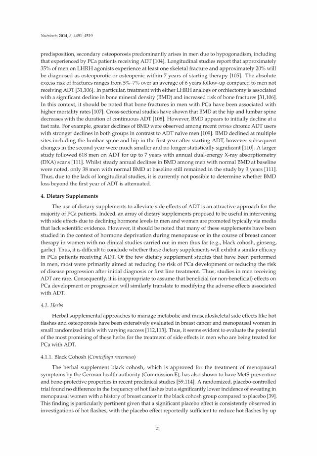
The use of dietary supplements to alleviate side effects of ADT is an attractive approach for themajority of PCa patients. Indeed, an array of dietary supplements proposed to be useful in interveningwith side effects due to declining hormone levels in men and women are promoted typically via mediathat lack scientific evidence. However, it should be noted that many of these supplements have beenstudied in the context of hormone deprivation during menopause or in the course of breast cancertherapy in women with no clinical studies carried out in men thus far (e.g., black cohosh, ginseng,garlic). Thus, it is difficult to conclude whether these dietary supplements will exhibit a similar efficacyin PCa patients receiving ADT. Of the few dietary supplement studies that have been performedin men, most were primarily aimed at reducing the risk of PCa development or reducing the riskof disease progression after initial diagnosis or first line treatment. Thus, studies in men receivingADT are rare. Consequently, it is inappropriate to assume that beneficial (or non-beneficial) effects onPCa development or progression will similarly translate to modifying the adverse effects associatedwith ADT.
4.1. Herbs
Herbal supplemental approaches to manage metabolic and musculoskeletal side effects like hotflashes and osteoporosis have been extensively evaluated in breast cancer and menopausal women insmall randomized trials with varying success [112,113]. Thus, it seems evident to evaluate the potentialof the most promising of these herbs for the treatment of side effects in men who are being treated forPCa with ADT.
4.1.1. Black Cohosh (Cimicifuga racemosa)
The herbal supplement black cohosh, which is approved for the treatment of menopausalsymptoms by the German health authority (Commission E), has also shown to have MetS-preventiveand bone-protective properties in recent preclinical studies [59,114]. A randomized, placebo-controlledtrial found no difference in the frequency of hot flashes but a significantly lower incidence of sweating inmenopausal women with a history of breast cancer in the black cohosh group compared to placebo [39].This finding is particularly pertinent given that a significant placebo effect is consistently observed ininvestigations of hot flashes, with the placebo effect reportedly sufficient to reduce hot flashes by up
21

Nutrients 2014, 6, 4491–4519
to 75% [115]. On the other hand, significant evidence of potentially beneficial effects on bone tissuewas shown, although it should be noted that all clinical studies were conducted in females [59,116].Of interest is one study that was carried out in orchidectomized male rats and showed to preventosteoporosis in vivo [117].
The mechanism underlying the activity of black cohosh remains poorly understood. There is alack of scientific evidence regarding its proposed estrogenic activity. However, it was recently shown toattenuate nucleoside uptake into cells and thus may have an impact on tumor treatment by nucleosideanalogs [118]. One may speculate that its modulation of adenosine signaling may be beneficial for CVside effects of ADT [119], but this has yet to be investigated. Despite the lack of knowledge regardingthe mechanism of action of black cohosh, its long history of use reveals that black cohosh is welltolerated. The use of black cohosh in PCa patients undergoing ADT may be warranted for hot flushes,CV and osteoporotic side effects. Importantly, further systematic studies assessing the safety andefficacy of black cohosh in alleviating ADT-induced side effects may be worth pursuing.
4.1.2. Dong Quai (Angelica sinensis)
Dong quai is a traditional Chinese herbal remedy most commonly used in the treatment of femalereproductive problems. According to our knowledge, there are no pre-clinical studies addressing itseffects in PCa. However, a small randomized clinical trial was conducted in men receiving ADT wheredong quai was shown to be ineffective in reducing hot flashes [41]. Similarly, randomized trials inwomen also found no effect of dong quai on hot flashes beyond a placebo, irrespective of whether theherb was used alone or as part of a complex multi-ingredient intervention [120]. Taken together, thecurrent evidence does not support the use of dong quai in patients undergoing ADT.
4.1.3. Ginseng (Panax ginseng)
Ginseng extract is widely used in traditional Chinese medicine and was reported to reduce fatigue,insomnia and depression in post-menopausal women, although there was no significant benefit on hotflashes [121]. However, a recent review of studies examining the efficacy of ginseng on menopausalsymptoms highlighted the poor quality and bias of many randomized clinical trials conductedto date, raising doubt as to the usefulness of this herb in managing menopause symptoms [42].Nonetheless, several pre-clinical and clinical studies have shown that ginseng may possess anti-cancerand hypoglycemic properties, the latter of potential benefit in ameliorating MetS in men receivingADT [122,123].
4.1.4. Garlic (Allium sativum)
Garlic is frequently used as a dietary supplement for the treatment of hyperlipidemia, heartdisease, and hypertension [124]. In addition, there is evidence that garlic is associated with bloodpressure reduction in patients with elevated systolic blood pressure (10–12 mmHg systolic, 6–9 mmHgdiastolic) but not in normotensive patients [73,74]. In this respect, it is conceivable that garlic mayalso reduce CV effects in PCa patients undergoing ADT. However, there is currently insufficientdata to support this hypothesis and further studies that specifically address this question would berequired [125].
4.2. Phytoestrogens
In the past, suppression of T was achieved using high doses of estrogens (estradiol) or selectiveestrogen receptor modulators [126–129]. However, these treatments were prone to severe andeven fatal CV side effects [130,131]. Consequently, these treatments have been replaced by othertherapeutics such as LHRH analogs. Phytoestrogens (plant estrogens) are non-steroidal naturallyoccurring phenolic compounds with known estrogenic effects and estrogen receptor (ER-β and/orERα) binding properties [132–134]. Thus, these “mild” estrogens could possibly serve as naturalalternatives with potentially fewer side effects. Indeed, phytoestrogens have been shown to improve
22

Nutrients 2014, 6, 4491–4519
metabolic health, reduce CV risk, and improve BMD and brain function [60,75,135]. There are threeclasses of phyto-estrogens, which are categorized according to their chemical structure as isoflavones,lignans or coumestans. Isoflavones are the largest and also the most extensively studied group ofphytoestrogens, which includes genistein, daidzein and glycitein. Isoflavones are found in highestamounts in soybeans, flaxseed and legumes. Soy is stably integrated into Asian diets where dailyintake is at least 40 times higher than among Western populations [136]. This is considered to beone of the factors contributing to a much lower incidence of prostate and breast cancer in Asiancountries [137]. In addition, increased consumption of isoflavones has been associated with decreasedincidence of diabetes and heart disease in South and East Asia [44,138]. In clinical trials, isoflavoneshave shown an improvement in hot flash severity, glycemic control, MetS and inflammatory profile inboth men and postmenopausal women [45,76,139]. However, in a recent double-blinded, randomized,placebo-controlled pilot study, phytoestrogens failed to improve metabolic or inflammatory parametersof men with PCa during ADT [47]. This is consistent with another pilot study conducted in 33 menwhere high dose isoflavones showed no significant improvement in cognition, vasomotor symptomsor any other aspect of quality of life measures compared to placebo in androgen deprived men [140].Thus, there is currently no clinical evidence for the proposed improvement of ADT-induced sideeffects by phytoestrogens, although limitations of these studies including small cohort sizes and shorttreatment durations should be taken into account.
4.3. Selenium (Se)
Se is an essential trace element, which is incorporated as selenocysteine into selenoproteins, manyof which are reactive oxygen species (ROS) scavenging enzymes, such as glutathione peroxidaseand thioredoxin reductase [141,142]. Thus, Se may be useful in decreasing oxidative stress andlow-grade inflammation. Notably, plasma biomarkers of oxidative stress and low-grade inflammationare associated with MetS, obesity and insulin resistance, conditions that are common side effects ofADT [143]. Se was shown to have strong anti-proliferative and pro-apoptotic effects on human PCa cellsand to improve severe metabolic side effects in patients [83,144]. Moreover, cancer prevention studiesindicated that Se decreases ROS and is associated with decreased incidence of PCa [145,146]. Takentogether, these observations provided a strong mechanistic rationale to combine ADT and Se for thetreatment of PCa [5] and led to a number of epidemiological studies and clinical trials [51,83]. However,epidemiological studies have suggested that supranutritional Se intake and high plasma Se levelsare not necessarily preventive against cancer, and may even be a possible risk factor for developingT2DM [83]. For example, supplementation with Se and/or Vitamin E in the large-scale Seleniumand Vitamin E Cancer Prevention Trial (SELECT) did not prevent the development of PCa, rather,the incidence of newly diagnosed T2DM increased among the Se-supplemented participants [51,83].Whilst the Nutritional Prevention of Cancer (NPC) study reported a decreased risk of PCa among Sesupplemented men, an increased risk of T2DM was also observed in participants with baseline plasmaSe levels in the top tertile [147]. Since then, several longitudinal studies have failed to support a causalrole of Se in T2DM, although cross-sectional studies continued to find significant associations betweencirculating Se levels and T2DM [148]. For example, serum Se was observed to be associated withadipocytokines, such as TNF-α, VCAM-1, leptin, FABP-4, and MCP-1 [149,150] and adiponectin [151].Although on the other hand, an analysis across randomized groups showed that Se supplementationhad no effect on adiponectin levels after six months of treatment [84]. Thus, it appears likely that thereported link between Se and T2DM is due to indirect effects of Se-containing ROS scavenging enzymes,affecting the hydrogen peroxide level that in turn modulates both glucose-induced insulin secretionand insulin-induced signaling [152,153]. Moreover, Se homeostasis is modulated by factors relatedto carbohydrate metabolism, suggesting that low serum Se levels may themselves be a consequenceof dysregulated energy metabolism in T2DM [153]. In addition, there appears to be a significantinteraction between dietary intake of phytoestrogens and Se with important implications for heartdisease, cancer, diabetes, and other conditions related to body weight [154].
23

Nutrients 2014, 6, 4491–4519
From the current prospective—especially since to date there has been no study specificallyevaluating the supplementation of Se in PCa patients receiving ADT—a combination of ADT with Secannot be recommended given that many patients develop pre-diabetes.
4.4. Fatty Acids (FA)
The relative amounts and different types of dietary FA are thought to play critical roles in PCaassociated MetS. Total FA intake and the ratio of omega-3 (ω-3) to omega-6 (ω-6) polyunsaturated FA(PUFAs) in the Western diet have increased significantly since the Industrial Revolution [155]. Theeffects of ω-3 PUFAs on CV disease, cancer as well as MetS have been investigated extensively [78,156–161]. Consequently, the United States Food and Drug Administration (FDA) has approvedω-3 PUFAs for the prevention of CV adverse outcomes by the postulated mechanism of loweringserum triglyceride levels [162,163]. Additionally, three common dietary ω-3 FA—alpha-linolenicacid (ALA), eicosa-pentaenoic acid (EPA), and docosahexaenoic acid (DHA)—were proposed toexhibit anti-inflammatory properties [50,79,164]. Since inflammation is a major risk factor for thedevelopment of CV disease, these ω-3 FA may reduce the risk of CV disease [50,79,164]. Variouspre-clinical, epidemiological and clinical studies have investigated the influence of ω-3 and ω-6 FAon the development and progression of PCa [80,155,158,161,165–173]. However, these studies haveyielded contradictory results. In particular, dietary intake of long-chain ω-3 PUFA or its individualcomponents (EPA, DHA, docosapentaenoic acid (DPA) and ALA) have been associated with PCarisk and progression [80,155,158,161,165–173]. On the other hand, encouraging results were obtainedfrom clinical trials showing potential anti-inflammatory effects of ω-3 FA [77,174–177]. Other trialsinvestigating higher doses of ω-3 FA (>1 g/day EPA and/or DHA) in populations at high risk for CVdisease reported improvements (i.e., reduced concentrations) of selected inflammatory markers [81,178,179]. However, it should be noted that in these studies the same dosage of ω-3 FA showed mixedresponse rates in healthy adults, and no beneficial effect in patients that received lower doses (<1g/day) [81,174,175,178–185]. A single study has reported that very high intakes (6.6 g/day), which arewell beyond the current recommendations (500 mg/day–1 g/day), may raise blood concentrations ofsome inflammation markers such as soluble tumor necrosis factor receptors 1 and 2 (sTNF-Rs 1 and2) [79,185]. It is likely that differences in dosage, study population characteristics, the source of ω-3 FA,study duration, and background diet may explain the inconsistencies in these findings. In this respect, itmay be noted that the majority of clinical trials have used marine sources of ω-3 FA (EPA and DHA fromfish), whereas few have examined FA from plants (ALA from flax oil) and only two trials comparedboth sources [82,175]. The heterogeneous results from these studies might also be due to variations inmeasuring FA consumption of individuals and different techniques assessing their diet [186–190]. Themain mechanisms underlying the purported anticancer effects of modulating dietary fat appear tobe through reduced insulin-like growth factor signaling and alterations in membrane ω-6 to ω-3 FAratios leading to suppressed COX-2-dependent PGE-2 production and reduction of inflammation viamodification of the eicosanoid pathway [191–195]. Moreover, decreased PGE-2 levels are expected todecrease estrogen production and further also modify androgen production [190].
In summary, although ω-3 FA exhibits some anti-inflammatory potential, there is still a lack ofconsensus regarding their optimal use and dosage. Moreover, results from the aforementioned studieshave to be interpreted with caution, since the metabolic conditions during ADT are different. Furtherresearch is warranted to better elucidate the mechanism of action and ideal consumption of ω-3 FA forpotential CV health benefits during ADT.
4.5. Calcium and Vitamin D
Osteoporosis is a common and one of the most debilitating side effects of ADT. The mostimportant nutritional factors contributing to osteoporosis include deficiencies in Vitamin D andcalcium. Measurement of Vitamin D levels in osteoporotic males by a large multi-center study inthe US (MrOs) revealed a deficiency in 26% and an insufficiency in 72% of subjects [54]. Because
24

Nutrients 2014, 6, 4491–4519
deterioration of BMD occurs soon after initiation of ADT therapy [67], the European Association ofUrology does not specify recommendations but states that calcium supplementation is protective [196].The National Comprehensive Cancer Network cite the National Osteoporosis foundation guidelinesthat recommend calcium (1000 mg daily) and Vitamin D (800–1000 IU daily) supplementation for allmen aged 50 or above [197]. Notably however, data supporting this recommendation are lacking asshown in a recent systematic review [196]. One cross-sectional study suggested an association betweenlow calcium intake and a greater likelihood of osteoporosis in men with PCa of whom 71 % wereundergoing ADT. Unfortunately, Vitamin D use was not examined in this study [198]. Another trialreported a positive association of calcium and Vitamin D use (examined together) on hip and lumbarspine BMD in men on ADT [199]. Importantly, results from 12 different clinical trials revealed that thecommonly recommended doses, of 500–1000 mg calcium and 200–500 IU Vitamin D per day still resultin BMD loss in men receiving ADT [196]. Alibhai et al. examined long-term effects of calcium andVitamin D in a prospective 3-year matched cohort study comparing PCa patients with and withoutADT. This study found that Vitamin D but not calcium may be protective particularly in the first yearof ADT [32]. In a multivariate analysis, it was further shown that the mean daily calcium intake inmen receiving ADT was significantly lower in men who suffered from osteoporosis compared to thosewithout osteoporosis [198].
In summary, calcium and Vitamin D supplementation is a recommended complementary therapynot only in elderly men with osteoporosis but also in men undergoing ADT even though the long-termimpact of ADT on BMD and the value of calcium and Vitamin D in ameliorating negative effectsremains to be elucidated more precisely.
4.6. Vitamin E
Vitamin E is a potent antioxidant, which is of interest to ameliorate hot flushes and CV associatedside effects of ADT. Vitamin E has a long history in the treatment of pre-eclampsia (characterized byhigh blood pressure in pregnant women), premenstrual syndrome, painful periods, menopausalsyndrome, hot flashes associated with breast cancer, and breast cysts even though randomizedcontrolled clinical studies did not reveal any benefit [52,53,86,200]. Moreover, the Physicians HealthStudy II concluded that Vitamin E does not reduce the risk of major CV events (non-fatal myocardialinfarction, non-fatal stroke, or CV disease death) [57]. Similarly, the Women’s Health Study, whichcomprised approximately 40,000 healthy women, found that Vitamin E did not reduce the risk of deathor major CV events. Interestingly however, there was a significant reduction in the secondary endpointof CV deaths and in major CV events among a subgroup of women aged 65 or over [55]. The Women’sAntioxidant Cardiovascular Study found that there were no overall effects of Vitamin E on CV eventsamong women at high risk of CV disease [91]. Moreover, the intake of Vitamin E has been shown toincrease all-cause mortality and may even have negative effects on CV health [92].
There have been a number of studies in men, which have purported a positive effect of Vitamin Eon hot flushes and high blood pressure [201]. However, most of these studies have yielded inconclusiveor conflicting findings or a lack of benefit for its use, illustrating the need for studies of higher qualityin this area. Thus, clinical trials have failed to recapitulate the promising findings of in vitro and manyobservational studies. Possible reasons for this discrepancy may be that clinical trials are too short induration to reverse the results of decades of oxidative stress contributing to atherosclerosis or that theantioxidants selected for study were chosen for their ease of availability rather than proven efficacy(Vitamin E) [202]. Recent evidence from the SELECT trial revealed an increased risk for PCa in theVitamin E supplemented group. Taken together, current evidence does not support a beneficial effectfor Vitamin E, and its use as a supplementary treatment is therefore not recommended [56,88].
5. Discussion
The administration of ADT is associated with a diverse set of known side effects that havea significant impact on patient quality of life, overall health, and mortality. Some of the dietary
25

Nutrients 2014, 6, 4491–4519
supplements discussed in this review may be beneficial for patients undergoing ADT. The value ofcalcium and Vitamin D supplementation remains to be elucidated more precisely; however, becauseof their long and safe history of usage they may be recommended in the prevention of osteoporosisduring ADT. Phytoestrogens were shown to prevent hot flashes and other climacteric complaints andexert anti-osteoporotic effects in women. However, positive effects on CV health are still questionableand require further elucidation, especially with respect to their effects in PCa patients receiving ADT. Inaddition, further clinical trials evaluating the efficacy of isoflavones must be conducted before their usefor relieving ADT-induced side effects can be advocated. We can conclude that dietary interventionswith herbal substances may in fact be helpful in the treatment of adverse effects arising from ADT [43];however, more clinical randomized studies in PCa patients on ADT are highly warranted to supportthese findings. The long history of use and lack of adverse effects of black cohosh in the treatmentof climacteric complaints in women is particularly encouraging. Further evaluation of its proposedosteo-protective and anti-metabolic effects in conjunction with ADT in randomized and controlledclinical studies is also warranted. Recent data obtained in the large SELECT trial suggest that combinedsupplementation of Vitamin E and supranutritional Se may increase the risk of PCa, making this anon-recommended treatment for men receiving ADT. From the current perspective, a combination ofADT with Se, which has been associated with an increased risk for the development of T2DM, cannotbe recommended given that many patients develop pre-diabetes.
6. Conclusions
In summary, dietary supplements are active compounds with as yet mostly poorly defined effects.As such, the unregulated self-prescription of active compounds such as soy, Se or Vitamin E may insome cases even prove to be harmful or negatively interfere with cancer treatment. Given the prevalentuse of alternative dietary supplements in PCa patients, there is an urgent need to (1) perform rigorousresearch to determine the precise physiological effects of these different supplements with respect torelieving side effects of ADT; (2) conduct clinical trials of these supplements in men undergoing ADTand (3) establish more open lines of communication between patients and physicians regarding theuse of dietary supplements and their integration into conventional treatment strategies.
Acknowledgments: We thank Natalie Sampson for reading the manuscript and performing English corrections.This work was supported by the Competence Center Oncotyrol (project 2.3.3) within the scope of the AustrianCompetence Center for Excellent Technologies (COMET)—program conducted by the Austrian ResearchPromotion Agency (FFG) and a Category B research funding from ÖKH-KG/Tirol given to Andrea Dueregger.Andrea Dueregger was partly funded by autonomous province Bozen (Grant Number: 37/40.3) given toPetra Massoner.
Author Contributions: Andrea Dueregger and Iris E. Eder designed and wrote the review. Isabel Heideggerwrote parts of the manuscript. Bernhard Perktold, Reinhold Ramoner and Helmut Klocker were significantlyinvolved in designing the review regarding its content and also in editing the manuscript.
Conflicts of Interest: The authors declare no conflict of interest.
References and Notes
1. Siegel, R.; Naishadham, D.; Jemal, A. Cancer statistics, 2012. Cancer J. Clin. 2012, 62, 10–29. [CrossRef]2. Siegel, R.; Ma, J.; Zou, Z.; Jemal, A. Cancer statistics, 2014. Cancer J. Clin. 2014, 64, 9–29. [CrossRef]3. Heinlein, C.A.; Chang, C. Androgen receptor in prostate cancer. Endocr. Rev. 2004, 25, 276–308. [CrossRef]
[PubMed]4. Denis, L.J. The role of active treatment in early prostate cancer. Radiother. Oncol. 2000, 57, 251–258. [CrossRef]
[PubMed]5. Lieberman, R. Androgen deprivation therapy for prostate cancer chemoprevention: Current status and
future directions for agent development. Urology 2001, 58, 83–90. [CrossRef] [PubMed]6. Molina, A.; Belldegrun, A. Novel therapeutic strategies for castration resistant prostate cancer: Inhibition of
persistent androgen production and androgen receptor mediated signaling. J. Urol. 2011, 185, 787–794.[CrossRef] [PubMed]
26

Nutrients 2014, 6, 4491–4519
7. Basaria, S.; Davda, M.N.; Travison, T.G.; Ulloor, J.; Singh, R.; Bhasin, S. Risk factors associated withcardiovascular events during testosterone administration in older men with mobility limitation. J. Gerontol.Series A Biol. Sci. Med. Sci. 2013, 68, 153–160. [CrossRef]
8. Hot flashes are a sudden wave of mild or intense body heat caused by rushes of hormonal changes resultingfrom decreased levels of estrogen. They can occur at any time and may last from a few seconds to half anhour. They are due to blood vessel dilation and constriction. Their frequency is increased in menopausalwomen and men receiving ADT.
9. The “metabolic” or “insulin-resistance” syndrome (MetS) is a cluster of cardio-vascular (CV) risks related toinsulin resistance [10,11]. Diagnosis of the syndrome is defined by the adult treatment panel III (ATP III)and the World health organization (WHO) as a combination of low HDL, increased waist circumference,increased triglycerides, increased fasting glucose and hypertension [11]. The syndrome itself is furtherassociated with various metabolic abnormalities, including insulin resistance as well as other factors such asmale hypogonadism [12].
10. Ferrannini, E. Metabolic syndrome: A solution in search of a problem. J. Clin. Endocrinol. Metab. 2007, 92,396–398. [CrossRef] [PubMed]
11. Kahn, R.; Buse, J.; Ferrannini, E.; Stern, M. The metabolic syndrome: Time for a critical appraisal: Jointstatement from the American Diabetes Association and the European Association for the Study of Diabetes.Diabetes Care 2005, 28, 2289–2304. [CrossRef] [PubMed]
12. Cho, L.W. Metabolic syndrome. Singap. Med. J. 2011, 52, 779–785.13. Insulin resistance is defined clinically as the inability of a known quantity of exogenous or endogenous insulin
to increase glucose uptake and utilization in an individual as much as it does in a normal population [14].14. Lebovitz, H.E. Insulin resistance: Definition and consequences. Exp. Clin. Endocrinol. Diabetes 2001, 109,
135–148. [CrossRef] [PubMed]15. Cardiovascular events refer to any incidents that may cause damage to the heart muscle involving the
heart and/or blood vessels which include: arterial embolic or thrombotic events, hemorrhagic or ischemiccerebrovascular conditions, myocardial infarction, and other ischemic heart disease. Severe CV eventsinclude conditions such as myocardial infarction or congestive heart failure which may be fatal [16].
16. WHO Fact Sheet—Cardiovascular diseases (CVDs). Available online: Http://www.who.int/mediacentre/factsheets/fs317/en/ (accessed on 4 September 2014).
17. Osteoporosis is a skeletal disorder characterized by compromised bone strength predisposing to an increasedrisk for bone fractures. Bone strength reflects the integration of two main features: bone mineral density(BMD) and bone quality [18]. Osteoporosis is defined by the WHO as a BMD of 2.5 standard deviations ormore below the mean peak bone mass (average of young, healthy adults) as measured by dual-energy X-rayabsorptiometry (DXA). A BMD value allows fracture risk to be calculated using FRAX or CAROC assessmentalgorithms, which incorporate a group of clinical risk factors in addition to femoral neck BMD [19,20].
18. Kilbanski, A.; Adams-Campbell, L.; Bassford, T.; Blair, S.N.; Boden, S.D.; Dickersin, K.; Gifford, D.R.;Glasse, L.; Goldring, S.R.; Hruska, K.; et al. Osteoporosis prevention, diagnosis, and therapy. JAMA J.American Medical Association 2001, 285, 785–795. [CrossRef]
19. Leslie, W.D.; Lix, L.M.; Johansson, H.; Oden, A.; McCloskey, E.; Kanis, J.A. Independent clinical validation ofa Canadian FRAX tool: Fracture prediction and model calibration. J. Bone Miner. Res. 2010, 25, 2350–2358.[CrossRef] [PubMed]
20. Leslie, W.D.; Tsang, J.F.; Lix, L.M. Simplified system for absolute fracture risk assessment: Clinical validationin Canadian women. J. Bone Miner. Res. 2009, 24, 353–360. [CrossRef] [PubMed]
21. Grossmann, M.; Zajac, J.D. Androgen deprivation therapy in men with prostate cancer: How should the sideeffects be monitored and treated? Clin. Endocrinol. 2011, 74, 289–293.
22. Truant, T.L.; Porcino, A.J.; Ross, B.C.; Wong, M.E.; Hilario, C.T. Complementary and alternative medicine(CAM) use in advanced cancer: A systematic review. J. Support. Oncol. 2013, 11, 105–113. [CrossRef][PubMed]
23. Philippou, Y.; Hadjipavlou, M.; Khan, S.; Rane, A. Complementary and alternative medicine (CAM) inprostate and bladder cancer. BJU Int. 2013, 112, 1073–1079. [CrossRef] [PubMed]
24. Klempner, S.J.; Bubley, G. Complementary and alternative medicines in prostate cancer: From bench tobedside? The oncologist 2012, 17, 830–837. [CrossRef]
27
Nutrients 2014, 6, 4491–4519
25. Huggins, C.; Hodges, C.V. Studies on prostatic cancer: I. The effect of castration, of estrogen and of androgeninjection on serum phosphatases in metastatic carcinoma of the prostate. J. Urol. 2002, 168, 9–12. [CrossRef][PubMed]
26. Schroder, F.; Crawford, E.D.; Axcrona, K.; Payne, H.; Keane, T.E. Androgen deprivation therapy: Past,present and future. BJU Int. 2012, 109, 1–12. [CrossRef] [PubMed]
27. The initial administration of an LHRH agonist first causes an increase of LH with a corresponding rise inserum T over 1–2 weeks. This increase in T may initially stimulate and worsen the disease and relatedsymptoms. To prevent this phenomenon, administration of an anti-androgen or estrogen for one week beforeand during the first few weeks of LHRH agonist therapy is often employed.
28. Rodrigues, D.N.; Butler, L.M.; Estelles, D.L.; de Bono, J.S. Molecular pathology and prostate cancertherapeutics: From biology to bedside. J. Pathol. 2014, 232, 178–184. [CrossRef] [PubMed]
29. Kolvenbag, G.J.; Iversen, P.; Newling, D.W. Antiandrogen monotherapy: A new form of treatment forpatients with prostate cancer. Urology 2001, 58, 16–23. [CrossRef] [PubMed]
30. Grossmann, M.; Zajac, J.D. Management of side effects of androgen deprivation therapy. Endocrinol. Metab.Clin. N. Am. 2011, 40, 655–671. [CrossRef]
31. Alibhai, S.M.; Duong-Hua, M.; Sutradhar, R.; Fleshner, N.E.; Warde, P.; Cheung, A.M.; Paszat, L.F. Impact ofandrogen deprivation therapy on cardiovascular disease and diabetes. J. Clin. Oncol. 2009, 27, 3452–3458.[CrossRef] [PubMed]
32. Alibhai, S.M.; Mohamedali, H.Z.; Gulamhusein, H.; Panju, A.H.; Breunis, H.; Timilshina, N.; Fleshner, N.;Krahn, M.D.; Naglie, G.; Tannock, I.F.; et al. Changes in bone mineral density in men starting androgendeprivation therapy and the protective role of vitamin D. Osteoporos. Int. 2013, 24, 2571–2579. [CrossRef][PubMed]
33. Yuan, J.Q.; Xu, T.; Zhang, X.W.; Yu, L.P.; Li, Q.; Liu, S.J.; Huang, X.B.; Wang, X.F. Metabolic syndrome andandrogen deprivation therapy in metabolic complications of prostate cancer patients. Chin. Med. J. 2012, 125,3725–3729. [PubMed]
34. Saglam, H.S.; Kose, O.; Kumsar, S.; Budak, S.; Adsan, O. Fasting blood glucose and lipid profile alterationsfollowing twelve-month androgen deprivation therapy in men with prostate cancer. Sci. World J. 2012, 2012.[CrossRef]
35. Freedland, S.J.; Eastham, J.; Shore, N. Androgen deprivation therapy and estrogen deficiency inducedadverse effects in the treatment of prostate cancer. Prostate Cancer Prostatic Dis. 2009, 12, 333–338. [CrossRef][PubMed]
36. Carani, C.; Qin, K.; Simoni, M.; Faustini-Fustini, M.; Serpente, S.; Boyd, J.; Korach, K.S.; Simpson, E.R. Effectof testosterone and estradiol in a man with aromatase deficiency. N Engl. J. Med. 1997, 337, 91–95. [CrossRef][PubMed]
37. Khosla, S. Update in male osteoporosis. J. Clin. Endocrinol. Metab. 2010, 95, 3–10. [CrossRef] [PubMed]38. Lee, R.J.; Saylor, P.J.; Smith, M.R. Contemporary therapeutic approaches targeting bone complications in
prostate cancer. Clin. Genitourin. Cancer 2010, 8, 29–36. [CrossRef] [PubMed]39. Jacobson, J.S.; Troxel, A.B.; Evans, J.; Klaus, L.; Vahdat, L.; Kinne, D.; Lo, K.M.; Moore, A.; Rosenman, P.J.;
Kaufman, E.L.; et al. Randomized trial of black cohosh for the treatment of hot flashes among women with ahistory of breast cancer. J. Clin. Oncol. 2001, 19, 2739–2745. [PubMed]
40. Fritz, H.; Seely, D.; McGowan, J.; Skidmore, B.; Fernandes, R.; Kennedy, D.A.; Cooley, K.; Wong, R.; Sagar, S.;Balneaves, L.G.; et al. Black cohosh and breast cancer: A systematic review. Integr. Cancer Ther. 2014, 13,12–29. [CrossRef] [PubMed]
41. Al-Bareeq, R.J.; Ray, A.A.; Nott, L.; Pautler, S.E.; Razvi, H. Dong Quai (Angelica sinensis) in the treatmentof hot flashes for men on androgen deprivation therapy: Results of a randomized double-blind placebocontrolled trial. Can. Urol. Assoc. J. 2010, 4, 49–53. [PubMed]
42. Kim, M.S.; Lim, H.J.; Yang, H.J.; Lee, M.S.; Shin, B.C.; Ernst, E. Ginseng for managing menopause symptoms:A systematic review of randomized clinical trials. J. Ginseng Res. 2013, 37, 30–36. [CrossRef] [PubMed]
43. Moyad, M.A. Complementary/alternative therapies for reducing hot flashes in prostate cancer patients:Reevaluating the existing indirect data from studies of breast cancer and postmenopausal women. Urology2002, 59, 20–33. [CrossRef]
44. Zhang, X.; Shu, X.O.; Gao, Y.T.; Yang, G.; Li, Q.; Li, H.; Jin, F.; Zheng, W. Soy food consumption is associatedwith lower risk of coronary heart disease in Chinese women. J. Nutr. 2003, 133, 2874–2878. [PubMed]
28

Nutrients 2014, 6, 4491–4519
45. Azadbakht, L.; Kimiagar, M.; Mehrabi, Y.; Esmaillzadeh, A.; Padyab, M.; Hu, F.B.; Willett, W.C. Soy inclusionin the diet improves features of the metabolic syndrome: A randomized crossover study in postmenopausalwomen. Am. J. Clin. Nutr. 2007, 85, 735–741. [PubMed]
46. Thelen, P.; Burfeind, P.; Schweyer, S.; Scharf, J.G.; Wuttke, W.; Ringert, R.H. Molecular principles of alternativetreatment approaches for hormone-refractory prostate cancer. Der. Urol. Ausg. A 2007, 46, 1271–1274.[CrossRef]
47. Napora, J.K.; Short, R.G.; Muller, D.C.; Carlson, O.D.; Odetunde, J.O.; Xu, X.; Carducci, M.; Travison, T.G.;Maggio, M.; Egan, J.M.; et al. High-dose isoflavones do not improve metabolic and inflammatory parametersin androgen-deprived men with prostate cancer. J. Androl. 2011, 32, 40–48. [CrossRef] [PubMed]
48. Albertazzi, P.; Pansini, F.; Bonaccorsi, G.; Zanotti, L.; Forini, E.; de Aloysio, D. The effect of dietary soysupplementation on hot flushes. Obstetrics Gynecol. 1998, 91, 6–11. [CrossRef]
49. Charles, C.; Yuskavage, J.; Carlson, O.; John, M.; Tagalicud, A.S.; Maggio, M.; Muller, D.C.; Egan, J.; Basaria, S.Effects of high-dose isoflavones on metabolic and inflammatory markers in healthy postmenopausal women.Menopause 2009, 16, 395–400. [CrossRef] [PubMed]
50. James, M.J.; Gibson, R.A.; Cleland, L.G. Dietary polyunsaturated fatty acids and inflammatory mediatorproduction. Am. J. Clin. Nutr. 2000, 71, 343–348.
51. Lippman, S.M.; Klein, E.A.; Goodman, P.J.; Lucia, M.S.; Thompson, I.M.; Ford, L.G.; Parnes, H.L.;Minasian, L.M.; Gaziano, J.M.; Hartline, J.A.; et al. Effect of selenium and vitamin E on risk of prostate cancerand other cancers: The Selenium and Vitamin E Cancer Prevention Trial (SELECT). Jama 2009, 301, 39–51.[CrossRef] [PubMed]
52. Rubenstein, B.B. Vitamin E diminishes the vasomotor symptoms of menopause. Fed. Proc. 1948, 7, 106.[PubMed]
53. Raffy, A. Menopause and vitamin E. Ther. Hung. 1955, 4, 12–14. [PubMed]54. Orwoll, E.; Nielson, C.M.; Marshall, L.M.; Lambert, L.; Holton, K.F.; Hoffman, A.R.; Barrett-Connor, E.;
Shikany, J.M.; Dam, T.; Cauley, J.A. Vitamin D deficiency in older men. J. Clin. Endocrinol. Metab. 2009, 94,1214–1222. [CrossRef] [PubMed]
55. Liu, S.; Lee, I.M.; Song, Y.; Van Denburgh, M.; Cook, N.R.; Manson, J.E.; Buring, J.E. Vitamin E and riskof type 2 diabetes in the women’s health study randomized controlled trial. Diabetes 2006, 55, 2856–2862.[CrossRef] [PubMed]
56. Klein, E.A.; Thompson, I.M., Jr.; Tangen, C.M.; Crowley, J.J.; Lucia, M.S.; Goodman, P.J.; Minasian, L.M.;Ford, L.G.; Parnes, H.L.; Gaziano, J.M.; et al. Vitamin E and the risk of prostate cancer: The Selenium andVitamin E Cancer Prevention Trial (SELECT). JAMA 2011, 306, 1549–1556. [CrossRef] [PubMed]
57. Sesso, H.D.; Buring, J.E.; Christen, W.G.; Kurth, T.; Belanger, C.; MacFadyen, J.; Bubes, V.; Manson, J.E.;Glynn, R.J.; Gaziano, J.M. Vitamins E and C in the prevention of cardiovascular disease in men:The Physicians’ Health Study II randomized controlled trial. JAMA 2008, 300, 2123–2133. [CrossRef][PubMed]
58. Peters, U.; Littman, A.J.; Kristal, A.R.; Patterson, R.E.; Potter, J.D.; White, E. Vitamin E and seleniumsupplementation and risk of prostate cancer in the Vitamins and lifestyle (VITAL) study cohort.Cancer Causes Control 2008, 19, 75–87. [CrossRef] [PubMed]
59. Seidlova-Wuttke, D.; Stecher, G.; Kammann, M.; Haunschild, J.; Eder, N.; Stahnke, V.; Wessels, J.; Wuttke, W.Osteoprotective effects of Cimicifuga racemosa and its triterpene-saponins are responsible for reduction ofbone marrow fat. Phytomedicine 2012, 19, 855–860. [CrossRef] [PubMed]
60. Kuhnle, G.G.; Ward, H.A.; Vogiatzoglou, A.; Luben, R.N.; Mulligan, A.; Wareham, N.J.; Forouhi, N.G.;Khaw, K.T. Association between dietary phyto-oestrogens and bone density in men and postmenopausalwomen. Br. J. Nutr. 2011, 106, 1063–1069. [CrossRef] [PubMed]
61. Soung, D.Y.; Devareddy, L.; Khalil, D.A.; Hooshmand, S.; Patade, A.; Lucas, E.A.; Arjmandi, B.H. Soy affectstrabecular microarchitecture and favorably alters select bone-specific gene expressions in a male rat modelof osteoporosis. Calcif. Tissue Int. 2006, 78, 385–391. [CrossRef] [PubMed]
62. Hogstrom, M.; Nordstrom, P.; Nordstrom, A. n-3 Fatty acids are positively associated with peak bone mineraldensity and bone accrual in healthy men: The NO2 Study. Am. J. Clin. Nutr. 2007, 85, 803–807. [PubMed]
63. Watkins, B.A.; Li, Y.; Lippman, H.E.; Seifert, M.F. Omega-3 polyunsaturated fatty acids and skeletal health.Exp. Biol Med. (Maywood) 2001, 226, 485–497.
29

Nutrients 2014, 6, 4491–4519
64. Fernandes, G.; Bhattacharya, A.; Rahman, M.; Zaman, K.; Banu, J. Effects of n-3 fatty acids on autoimmunityand osteoporosis. Front. Biosci. 2008, 13, 4015–4020. [CrossRef] [PubMed]
65. Shen, C.L.; Yeh, J.K.; Rasty, J.; Chyu, M.C.; Dunn, D.M.; Li, Y.; Watkins, B.A. Improvement of bone quality ingonad-intact middle-aged male rats by long-chain n-3 polyunsaturated fatty acid. Calcif. Tissue Int. 2007, 80,286–293. [CrossRef] [PubMed]
66. Rahman, M.M.; Bhattacharya, A.; Banu, J.; Fernandes, G. Conjugated linoleic acid protects againstage-associated bone loss in C57BL/6 female mice. J. Nutr. Biochem. 2007, 18, 467–474. [CrossRef] [PubMed]
67. Tang, B.M.; Eslick, G.D.; Nowson, C.; Smith, C.; Bensoussan, A. Use of calcium or calcium in combinationwith vitamin D supplementation to prevent fractures and bone loss in people aged 50 years and older:A meta-analysis. Lancet 2007, 370, 657–666. [CrossRef] [PubMed]
68. Gates, B.J.; Das, S. Management of osteoporosis in elderly men. Maturitas 2011, 69, 113–119. [CrossRef][PubMed]
69. Gielen, E.; Boonen, S.; Vanderschueren, D.; Sinnesael, M.; Verstuyf, A.; Claessens, F.; Milisen, K.;Verschueren, S. Calcium and vitamin D supplementation in men. J. Osteoporos. 2011, 2011. [CrossRef]
70. Verbrugge, F.H.; Gielen, E.; Milisen, K.; Boonen, S. Who should receive calcium and vitamin Dsupplementation? Age Ageing 2012, 41, 576–580. [CrossRef]
71. Cranney, A.; Horsley, T.; O’Donnell, S.; Weiler, H.; Puil, L.; Ooi, D.; Atkinson, S.; Ward, L.; Moher, D.;Hanley, D.; et al. Effectiveness and safety of vitamin D in relation to bone health. Evid. Rep. Technol. Assess.2007, 1–235.
72. Moyad, M.A. Complementary therapies for reducing the risk of osteoporosis in patients receiving luteinizinghormone-releasing hormone treatment/orchiectomy for prostate cancer: A review and assessment of theneed for more research. Urology 2002, 59, 34–40. [CrossRef] [PubMed]
73. Stabler, S.N.; Tejani, A.M.; Huynh, F.; Fowkes, C. Garlic for the prevention of cardiovascular morbidity andmortality in hypertensive patients. Cochrane Database Syst. Rev. 2012, 8. [CrossRef]
74. Shouk, R.; Abdou, A.; Shetty, K.; Sarkar, D.; Eid, A.H. Mechanisms underlying the antihypertensive effectsof garlic bioactives. Nutr Res. 2014, 34, 106–115. [CrossRef] [PubMed]
75. Clarkson, T.B. Soy, soy phytoestrogens and cardiovascular disease. J. Nutr. 2002, 132, 566–569.76. Atteritano, M.; Marini, H.; Minutoli, L.; Polito, F.; Bitto, A.; Altavilla, D.; Mazzaferro, S.; D’Anna, R.;
Cannata, M.L.; Gaudio, A.; et al. Effects of the phytoestrogen genistein on some predictors of cardiovascularrisk in osteopenic, postmenopausal women: A two-year randomized, double-blind, placebo-controlled study.J. Clin. Endocrinol. Metab. 2007, 92, 3068–3075. [CrossRef] [PubMed]
77. Bloedon, L.T.; Balikai, S.; Chittams, J.; Cunnane, S.C.; Berlin, J.A.; Rader, D.J.; Szapary, P.O. Flaxseed andcardiovascular risk factors: Results from a double blind, randomized, controlled clinical trial. J. Am. Coll.Nutr. 2008, 27, 65–74. [CrossRef] [PubMed]
78. Jacobson, T.A. Beyond lipids: The role of omega-3 fatty acids from fish oil in the prevention of coronaryheart disease. Curr. Atheroscler. Rep. 2007, 9, 145–153. [CrossRef] [PubMed]
79. Kris-Etherton, P.M.; Harris, W.S.; Appel, L.J. Fish consumption, fish oil, omega-3 fatty acids,and cardiovascular disease. Circulation 2002, 106, 2747–2757. [CrossRef] [PubMed]
80. Simopoulos, A.P. The importance of the ratio of omega-6/omega-3 essential fatty acids. Biomed. Pharmacother.2002, 56, 365–379. [CrossRef] [PubMed]
81. Hjerkinn, E.M.; Seljeflot, I.; Ellingsen, I.; Berstad, P.; Hjermann, I.; Sandvik, L.; Arnesen, H. Influence oflong-term intervention with dietary counseling, long-chain n-3 fatty acid supplements, or both on circulatingmarkers of endothelial activation in men with long-standing hyperlipidemia. Am. J. Clin. Nutr. 2005, 81,583–589. [PubMed]
82. Dewell, A.; Marvasti, F.F.; Harris, W.S.; Tsao, P.; Gardner, C.D. Low- and high-dose plant and marine (n-3)fatty acids do not affect plasma inflammatory markers in adults with metabolic syndrome. J. Nutr. 2011, 141,2166–2171. [CrossRef] [PubMed]
83. Koyama, H.; Mutakin; Abdulah, R.; Yamazaki, C.; Kameo, S. Selenium supplementation trials for cancerprevention and the subsequent risk of type 2 diabetes mellitus. Nihon Eiseigaku Zasshi 2013, 68, 1–10.[CrossRef] [PubMed]
84. Rayman, M.P.; Blundell-Pound, G.; Pastor-Barriuso, R.; Guallar, E.; Steinbrenner, H.; Stranges, S.A randomized trial of selenium supplementation and risk of type-2 diabetes, as assessed by plasmaadiponectin. PLoS One 2012, 7, 45269. [CrossRef]
30

Nutrients 2014, 6, 4491–4519
85. Faghihi, T.; Radfar, M.; Barmal, M.; Amini, P.; Qorbani, M.; Abdollahi, M.; Larijani, B. A randomized,placebo-controlled trial of selenium supplementation in patients with type 2 diabetes: Effects on glucosehomeostasis, oxidative stress, and lipid profile. Am. J. Ther. 2013. [CrossRef]
86. Dennehy, C.; Tsourounis, C. A review of select vitamins and minerals used by postmenopausal women.Maturitas 2010, 66, 370–380. [CrossRef] [PubMed]
87. Algotar, A.M.; Stratton, M.S.; Stratton, S.P.; Hsu, C.H.; Ahmann, F.R. No effect of selenium supplementationon serum glucose levels in men with prostate cancer. Am. J. Med. 2010, 123, 765–768. [CrossRef] [PubMed]
88. Kristal, A.R.; Darke, A.K.; Morris, J.S.; Tangen, C.M.; Goodman, P.J.; Thompson, I.M.; Meyskens, F.L., Jr.;Goodman, G.E.; Minasian, L.M.; Parnes, H.L.; et al. Baseline selenium status and effects of selenium andvitamin E supplementation on prostate cancer risk. J. Nat. Cancer Inst. 2014, 106. [CrossRef] [PubMed]
89. Algotar, A.M.; Stratton, M.S.; Ahmann, F.R.; Ranger-Moore, J.; Nagle, R.B.; Thompson, P.A.; Slate, E.;Hsu, C.H.; Dalkin, B.L.; Sindhwani, P.; et al. Phase 3 clinical trial investigating the effect of seleniumsupplementation in men at high-risk for prostate cancer. Prostate 2013, 73, 328–335. [CrossRef] [PubMed]
90. b>Algotar, A.M.; Hsu, C.H.; Singh, P.; Stratton, S.P. Selenium supplementation has no effect on serum glucoselevels in men at high risk for Prostate Cancer. J. Diabetes 2013, 5, 465–470. [CrossRef] [PubMed]
91. Cook, N.R.; Albert, C.M.; Gaziano, J.M.; Zaharris, E.; MacFadyen, J.; Danielson, E.; Buring, J.E.;Manson, J.E. A randomized factorial trial of vitamins C and E and beta carotene in the secondaryprevention of cardiovascular events in women: Results from the Women’s Antioxidant Cardiovascular Study.Arch. Internal Med. 2007, 167, 1610–1618. [CrossRef]
92. Saremi, A.; Arora, R. Vitamin E and cardiovascular disease. Am. J. Ther. 2010, 17, 56–65. [CrossRef]93. Shahani, S.; Braga-Basaria, M.; Basaria, S. Androgen deprivation therapy in prostate cancer and metabolic
risk for atherosclerosis. J. Clin. Endocrinol. Metab. 2008, 93, 2042–2049. [CrossRef] [PubMed]94. Smith, M.R.; Lee, H.; McGovern, F.; Fallon, M.A.; Goode, M.; Zietman, A.L.; Finkelstein, J.S. Metabolic
changes during gonadotropin-releasing hormone agonist therapy for prostate cancer: Differences from theclassic metabolic syndrome. Cancer 2008, 112, 2188–2194. [CrossRef] [PubMed]
95. Braga-Basaria, M.; Dobs, A.S.; Muller, D.C.; Carducci, M.A.; John, M.; Egan, J.; Basaria, S. Metabolic syndromein men with prostate cancer undergoing long-term androgen-deprivation therapy. J. Clin. Oncol. 2006, 24,3979–3983. [CrossRef] [PubMed]
96. Basaria, S.; Muller, D.C.; Carducci, M.A.; Egan, J.; Dobs, A.S. Hyperglycemia and insulin resistance in menwith prostate carcinoma who receive androgen-deprivation therapy. Cancer 2006, 106, 581–588. [CrossRef][PubMed]
97. Laaksonen, D.E.; Niskanen, L.; Punnonen, K.; Nyyssonen, K.; Tuomainen, T.P.; Valkonen, V.P.; Salonen, R.;Salonen, J.T. Testosterone and sex hormone-binding globulin predict the metabolic syndrome and diabetesin middle-aged men. Diabetes Care 2004, 27, 1036–1041. [CrossRef] [PubMed]
98. Tirabassi, G.; Gioia, A.; Giovannini, L.; Boscaro, M.; Corona, G.; Carpi, A.; Maggi, M.; Balercia, G. Testosteroneand cardiovascular risk. Intern. Emerg. Med. 2013, 8, 65–69. [CrossRef] [PubMed]
99. Stellato, R.K.; Feldman, H.A.; Hamdy, O.; Horton, E.S.; McKinlay, J.B. Testosterone, sex hormone-bindingglobulin, and the development of type 2 diabetes in middle-aged men: Prospective results from theMassachusetts male aging study. Diabetes Care 2000, 23, 490–494. [CrossRef] [PubMed]
100. Eckel, R.H.; Alberti, K.G.; Grundy, S.M.; Zimmet, P.Z. The metabolic syndrome. Lancet 2010, 375, 181–183.[CrossRef] [PubMed]
101. Fink, R.I.; Kolterman, O.G.; Griffin, J.; Olefsky, J.M. Mechanisms of insulin resistance in aging. J. Clin. Investig.1983, 71, 1523–1535. [CrossRef] [PubMed]
102. Lu-Yao, G.; Stukel, T.A.; Yao, S.L. Changing patterns in competing causes of death in men with prostatecancer: A population based study. J. Urol. 2004, 171, 2285–2290. [CrossRef] [PubMed]
103. WHO Assessment of fracture risk and its application to screening for postmenopausal osteoporosis. Report ofa WHO Study Group. World Health Organ. Technical Rep. Ser. 1994, 843, 1–129.
104. Lee, R.J.; Saylor, P.J.; Smith, M.R. Treatment and prevention of bone complications from prostate cancer. Bone2011, 48, 88–95. [CrossRef] [PubMed]
105. Krupski, T.L.; Smith, M.R.; Lee, W.C.; Pashos, C.L.; Brandman, J.; Wang, Q.; Botteman, M.; Litwin, M.S.Natural history of bone complications in men with prostate carcinoma initiating androgen deprivationtherapy. Cancer 2004, 101, 541–549. [CrossRef] [PubMed]
31

Nutrients 2014, 6, 4491–4519
106. Shahinian, V.B.; Kuo, Y.F.; Freeman, J.L.; Goodwin, J.S. Risk of fracture after androgen deprivation forprostate cancer. N. Engl. J. Med. 2005, 352, 154–164. [CrossRef] [PubMed]
107. Oefelein, M.G.; Ricchiuti, V.; Conrad, W.; Resnick, M.I. Skeletal fractures negatively correlate with overallsurvival in men with prostate cancer. J. Urol. 2002, 168, 1005–1007. [CrossRef] [PubMed]
108. Kiratli, B.J.; Srinivas, S.; Perkash, I.; Terris, M.K. Progressive decrease in bone density over 10 years ofandrogen deprivation therapy in patients with prostate cancer. Urology 2001, 57, 127–132. [CrossRef][PubMed]
109. Greenspan, S.L.; Coates, P.; Sereika, S.M.; Nelson, J.B.; Trump, D.L.; Resnick, N.M. Bone loss after initiation ofandrogen deprivation therapy in patients with prostate cancer. J. Clin. Endocrinol. Metab. 2005, 90, 6410–6417.[CrossRef] [PubMed]
110. Morote, J.; Orsola, A.; Abascal, J.M.; Planas, J.; Trilla, E.; Raventos, C.X.; Cecchini, L.; Encabo, G.; Reventos, J.Bone mineral density changes in patients with prostate cancer during the first 2 years of androgensuppression. J. Urol. 2006, 175, 1679–1683. [CrossRef] [PubMed]
111. Wadhwa, V.K.; Weston, R.; Mistry, R.; Parr, N.J. Long-term changes in bone mineral density and predictedfracture risk in patients receiving androgen-deprivation therapy for prostate cancer, with stratification oftreatment based on presenting values. BJU Int. 2009, 104, 800–805. [CrossRef] [PubMed]
112. Lloyd, K.B.; Hornsby, L.B. Complementary and alternative medications for women’s health issues.Nutr. Clin. Prac. 2009, 24, 589–608. [CrossRef]
113. Dennehy, C.E. The use of herbs and dietary supplements in gynecology: An evidence-based review. J.Midwifery Women’s Health 2006, 51, 402–409. [CrossRef]
114. Seidlova-Wuttke, D.; Eder, N.; Stahnke, V.; Kammann, M.; Stecher, G.; Haunschild, J.; Wessels, J.T.; Wuttke, W.Cimicifuga racemosa and its triterpene-saponins prevent the Metabolic Syndrome and deterioration ofcartilage in the knee joint of ovariectomized rats by similar mechanisms. Phytomedicine 2012, 19, 846–853.[CrossRef]
115. Mahon, S.M.; Kaplan, M. Placebo effect in hot flush research. Lancet Oncol. 2012, 13, 188. [CrossRef]116. Wuttke, W.; Gorkow, C.; Seidlova-Wuttke, D. Effects of black cohosh (Cimicifuga racemosa) on bone
turnover, vaginal mucosa, and various blood parameters in postmenopausal women: A double-blind,placebo-controlled, and conjugated estrogens-controlled study. Menopause 2006, 13, 185–196. [CrossRef]
117. Seidlova-Wuttke, D.; Jarry, H.; Pitzel, L.; Wuttke, W. Effects of estradiol-17beta, testosterone and a blackcohosh preparation on bone and prostate in orchidectomized rats. Maturitas 2005, 51, 177–186. [CrossRef]
118. Dueregger, A.; Guggenberger, F.; Barthelmes, J.; Stecher, G.; Schuh, M.; Intelmann, D.; Abel, G.; Haunschild, J.;Klocker, H.; Ramoner, R.; et al. Attenuation of nucleoside and anti-cancer nucleoside analog drug uptake inprostate cancer cells by Cimicifuga racemosa extract BNO-1055. Phytomedicine 2013, 20, 1306–1314. [CrossRef]
119. Molina-Arcas, M.; Casado, F.J.; Pastor-Anglada, M. Nucleoside transporter proteins. Curr. Vasc. Pharmacol.2009, 7, 426–434. [CrossRef]
120. Cheema, D.; Coomarasamy, A.; El-Toukhy, T. Non-hormonal therapy of post-menopausal vasomotorsymptoms: A structured evidence-based review. Arch. Gynecol. Obstet. 2007, 276, 463–469. [CrossRef]
121. Wiklund, I.K.; Mattsson, L.A.; Lindgren, R.; Limoni, C. Effects of a standardized ginseng extract onquality of life and physiological parameters in symptomatic postmenopausal women: A double-blind,placebo-controlled trial. Swedish Alternative Medicine Group. Int. J. Clin. Pharmacol. Res. 1999, 19, 89–99.
122. Nag, S.A.; Qin, J.J.; Wang, W.; Wang, M.H.; Wang, H.; Zhang, R. Ginsenosides as Anticancer Agents:In vitro and in vivo Activities, Structure-Activity Relationships, and Molecular Mechanisms of Action. Front.Pharmacol. 2012, 3, 25. [CrossRef]
123. Wang, Z.; Wang, J.; Chan, P. Treating type 2 diabetes mellitus with traditional chinese and Indian medicinalherbs. Evid. Based Complement. Altern. Med. 2013, 2013. [CrossRef]
124. Rahman, K.; Lowe, G.M. Garlic and cardiovascular disease: A critical review. J. Nutr. 2006, 136, 736–740.125. Rabito, M.J.; Kaye, A.D. Complementary and alternative medicine and cardiovascular disease:
An evidence-based review. Evid. Based Complement. Altern. Med. 2013, 2013. [CrossRef]126. Smith, M.R.; Malkowicz, S.B.; Chu, F.; Forrest, J.; Sieber, P.; Barnette, K.G.; Rodriquez, D.; Steiner, M.S.
Toremifene improves lipid profiles in men receiving androgen-deprivation therapy for prostate cancer:Interim analysis of a multicenter phase III study. J. Clin. Oncol. 2008, 26, 1824–1829. [CrossRef]
32

Nutrients 2014, 6, 4491–4519
127. Smith, M.R.; Morton, R.A.; Barnette, K.G.; Sieber, P.R.; Malkowicz, S.B.; Rodriguez, D.; Hancock, M.L.;Steiner, M.S. Toremifene to reduce fracture risk in men receiving androgen deprivation therapy for prostatecancer. J. Urol. 2013, 189, 45–50. [CrossRef]
128. Kearns, A.E.; Northfelt, D.W.; Dueck, A.C.; Atherton, P.J.; Dakhil, S.R.; Rowland, K.M., Jr.; Fuloria, J.;Flynn, P.J.; Dentchev, T.; Loprinzi, C.L. Osteoporosis prevention in prostate cancer patients receivingandrogen ablation therapy: Placebo-controlled double-blind study of estradiol and risedronate: N01C8.Support. Care Cancer 2010, 18, 321–328. [CrossRef]
129. Wibowo, E.; Schellhammer, P.; Wassersug, R.J. Role of estrogen in normal male function: Clinical implicationsfor patients with prostate cancer on androgen deprivation therapy. J. Urol. 2011, 185, 17–23. [CrossRef]
130. Treatment and survival of patients with cancer of the prostate. The Veterans Administration Co-operativeUrological Research Group. Surg. Gynecol. Obstet. 1967, 124, 1011–1017.
131. Byar, D.P.; Corle, D.K. Hormone therapy for prostate cancer: Results of the Veterans AdministrationCooperative Urological Research Group studies. NCI Monographs 1987, 7, 165–170.
132. Murkies, A.L.; Wilcox, G.; Davis, S.R. Clinical review 92: Phytoestrogens. J. Clin. Endocrinol. Metab. 1998, 83,297–303.
133. Turner, J.V.; Agatonovic-Kustrin, S.; Glass, B.D. Molecular aspects of phytoestrogen selective binding atestrogen receptors. J. Pharm. Sci. 2007, 96, 1879–1885. [CrossRef]
134. Dahlman-Wright, K.; Cavailles, V.; Fuqua, S.A.; Jordan, V.C.; Katzenellenbogen, J.A.; Korach, K.S.; Maggi, A.;Muramatsu, M.; Parker, M.G.; Gustafsson, J.A. International Union of Pharmacology. LXIV. Estrogenreceptors. Pharmacol. Rev. 2006, 58, 773–781. [CrossRef]
135. Khosla, S.; Melton, L.J.; Riggs, B.L. The unitary model for estrogen deficiency and the pathogenesis ofosteoporosis: Is a revision needed? J. Bone Miner. Res. 2011, 26, 441–451. [CrossRef]
136. De Kleijn, M.J.; van der Schouw, Y.T.; Wilson, P.W.; Adlercreutz, H.; Mazur, W.; Grobbee, D.E.; Jacques, P.F.Intake of dietary phytoestrogens is low in postmenopausal women in the United States: The Framinghamstudy(1–4). J. Nutr. 2001, 131, 1826–1832.
137. Jemal, A.; Siegel, R.; Ward, E.; Hao, Y.; Xu, J.; Murray, T.; Thun, M.J. Cancer statistics, 2008. Cancer J. Clin.2008, 58, 71–96. [CrossRef]
138. Villegas, R.; Gao, Y.T.; Yang, G.; Li, H.L.; Elasy, T.A.; Zheng, W.; Shu, X.O. Legume and soy food intake andthe incidence of type 2 diabetes in the Shanghai Women’s Health Study. Am. J. Clin. Nutr. 2008, 87, 162–167.
139. Huang, Y.; Cao, S.; Nagamani, M.; Anderson, K.E.; Grady, J.J.; Lu, L.J. Decreased circulating levels of tumornecrosis factor-alpha in postmenopausal women during consumption of soy-containing isoflavones. J. Clin.Endocrinol. Metab. 2005, 90, 3956–3962. [CrossRef]
140. Sharma, P.; Wisniewski, A.; Braga-Basaria, M.; Xu, X.; Yep, M.; Denmeade, S.; Dobs, A.S.; DeWeese, T.;Carducci, M.; Basaria, S. Lack of an effect of high dose isoflavones in men with prostate cancer undergoingandrogen deprivation therapy. J. Urol. 2009, 182, 2265–2272. [CrossRef]
141. Fairweather-Tait, S.J.; Bao, Y.; Broadley, M.R.; Collings, R.; Ford, D.; Hesketh, J.E.; Hurst, R. Selenium inhuman health and disease. Antioxid. Redox signal. 2011, 14, 1337–1383. [CrossRef]
142. Steinbrenner, H.; Sies, H. Protection against reactive oxygen species by selenoproteins. Biochim. Biophys. Acta2009, 1790, 1478–1485. [CrossRef]
143. Holvoet, P. Relations between metabolic syndrome, oxidative stress and inflammation and cardiovasculardisease. Verhandelingen 2008, 70, 193–219.
144. Yang, M.; Sytkowski, A.J. Differential expression and androgen regulation of the human selenium-bindingprotein gene hSP56 in prostate cancer cells. Cancer Res. 1998, 58, 3150–3153.
145. Clark, L.C.; Dalkin, B.; Krongrad, A.; Combs G.F., Jr.; Turnbull, B.W.; Slate, E.H.; Witherington, R.;Herlong, J.H.; Janosko, E.; Carpenter, D.; et al. Decreased incidence of prostate cancer with seleniumsupplementation: Results of a double-blind cancer prevention trial. Br. J. Urol. 1998, 81, 730–734. [CrossRef]
146. Yoshizawa, K.; Willett, W.C.; Morris, S.J.; Stampfer, M.J.; Spiegelman, D.; Rimm, E.B.; Giovannucci, E. Studyof prediagnostic selenium level in toenails and the risk of advanced prostate cancer. J. Nat. Cancer Inst. 1998,90, 1219–1224. [CrossRef]
147. Duffield-Lillico, A.J.; Dalkin, B.L.; Reid, M.E.; Turnbull, B.W.; Slate, E.H.; Jacobs, E.T.; Marshall, J.R.;Clark, L.C. Selenium supplementation, baseline plasma selenium status and incidence of prostate cancer:An analysis of the complete treatment period of the Nutritional Prevention of Cancer Trial. BJU Int. 2003, 91,608–612. [CrossRef]
33

Nutrients 2014, 6, 4491–4519
148. Rayman, M.P.; Stranges, S. Epidemiology of selenium and type 2 diabetes: Can we make sense of it? FreeRadic. Biol. Med. 2013, 65, 1557–1564.
149. Savini, I.; Catani, M.V.; Evangelista, D.; Gasperi, V.; Avigliano, L. Obesity-associated oxidative stress:Strategies finalized to improve redox state. Int J. Mol. Sci. 2013, 14, 10497–10538. [CrossRef]
150. Hassanzadeh, M.; Faridhosseini, R.; Mahini, M.; Faridhosseini, F.; Ranjbar, A. Serum Levels of TNF-, IL-6,and Selenium in Patients with Acute and Chronic Coronary Artery Disease. Iran. J. Immunol. 2006, 3,142–145.
151. Zhang, Y.; Chen, X. Reducing selenoprotein P expression suppresses adipocyte differentiation as a result ofincreased preadipocyte inflammation. Am. J. Physiol. Endocrinol. Metab. 2011, 300, 77–85. [CrossRef]
152. Steinbrenner, H.; Speckmann, B.; Pinto, A.; Sies, H. High selenium intake and increased diabetes risk:Experimental evidence for interplay between selenium and carbohydrate metabolism. J. Clin. Biochem. Nutr.2011, 48, 40–45. [CrossRef]
153. Steinbrenner, H. Interference of selenium and selenoproteins with the insulin-regulated carbohydrate andlipid metabolism. Free Radic. Biol. Med. 2013, 65, 1538–1547. [CrossRef]
154. Quiner, T.E.; Nakken, H.L.; Mason, B.A.; Lephart, E.D.; Hancock, C.R.; Christensen, M.J. Soy content of basaldiets determines the effects of supplemental selenium in male mice. J. Nutr. 2011, 141, 2159–2165. [CrossRef]
155. Simopoulos, A.P. Essential fatty acids in health and chronic disease. Am. J. Clin. Nutr. 1999, 70, 560–569.156. Nettleton, J.A. Omega-3 Fatty Acids and Health; Springer US: New York, NY, USA, 1995; pp. 64–76.157. Heinze, V.M.; Actis, A.B. Dietary conjugated linoleic acid and long-chain n-3 fatty acids in mammary and
prostate cancer protection: A review. Int. J. Food Sci. Nutr. 2012, 63, 66–78. [CrossRef]158. Tziomalos, K.; Athyros, V.G.; Mikhailidis, D.P. Fish oils and vascular disease prevention: An update.
Curr. Med. Chem. 2007, 14, 2622–2628. [CrossRef]159. Carpentier, Y.A.; Portois, L.; Malaisse, W.J. n-3 fatty acids and the metabolic syndrome. Am. J. Clin. Nutr.
2006, 83, 1499–1504.160. Barre, D.E. The role of consumption of alpha-linolenic, eicosapentaenoic and docosahexaenoic acids in
human metabolic syndrome and type 2 diabetes–a mini-review. J. Oleo Sci. 2007, 56, 319–325. [CrossRef]161. Chen, Y.Q.; Edwards, I.J.; Kridel, S.J.; Thornburg, T.; Berquin, I.M. Dietary fat-gene interactions in cancer.
Cancer Metastasis Rev. 2007, 26, 535–551. [CrossRef]162. Ballantyne, C.M.; Bays, H.E.; Kastelein, J.J.; Stein, E.; Isaacsohn, J.L.; Braeckman, R.A.; Soni, P.N. Efficacy and
safety of eicosapentaenoic acid ethyl ester (AMR101) therapy in statin-treated patients with persistent hightriglycerides (from the ANCHOR study). Am. J. Cardiol. 2012, 110, 984–992. [CrossRef]
163. Bays, H.E.; Ballantyne, C.M.; Kastelein, J.J.; Isaacsohn, J.L.; Braeckman, R.A.; Soni, P.N. Eicosapentaenoicacid ethyl ester (AMR101) therapy in patients with very high triglyceride levels (from the Multi-center,plAcebo-controlled, Randomized, double-blINd, 12-week study with an open-label Extension [MARINE]trial). Am. J. Cardiol. 2011, 108, 682–690. [CrossRef]
164. Connor, W.E. Importance of n-3 fatty acids in health and disease. Am. J. Clin. Nutr. 2000, 71, 171–175.165. Wang, S.; Wu, J.; Suburu, J.; Gu, Z.; Cai, J.; Axanova, L.S.; Cramer, S.D.; Thomas, M.J.; Perry, D.L.;
Edwards, I.J.; et al. Effect of dietary polyunsaturated fatty acids on castration-resistant Pten-null prostatecancer. Carcinogenesis 2012, 33, 404–412. [CrossRef]
166. Friedrichs, W.; Ruparel, S.B.; Marciniak, R.A.; deGraffenried, L. Omega-3 fatty acid inhibition of prostatecancer progression to hormone independence is associated with suppression of mTOR signaling andandrogen receptor expression. Nutr. Cancer 2011, 63, 771–777. [CrossRef]
167. Cavazos, D.A.; Price, R.S.; Apte, S.S.; deGraffenried, L.A. Docosahexaenoic acid selectively induces humanprostate cancer cell sensitivity to oxidative stress through modulation of NF-kappaB. Prostate 2011, 71,1420–1428. [CrossRef]
168. McEntee, M.F.; Ziegler, C.; Reel, D.; Tomer, K.; Shoieb, A.; Ray, M.; Li, X.; Neilsen, N.; Lih, F.B.; O’Rourke, D.;Whelan, J. Dietary n-3 polyunsaturated fatty acids enhance hormone ablation therapy in androgen-dependentprostate cancer. Am. J. Pathol. 2008, 173, 229–241. [CrossRef]
169. Epstein, M.M.; Kasperzyk, J.L.; Mucci, L.A.; Giovannucci, E.; Price, A.; Wolk, A.; Hakansson, N.; Fall, K.;Andersson, S.O.; Andren, O. Dietary fatty acid intake and prostate cancer survival in Orebro County, Sweden.Am. J. Epidemiol. 2012, 176, 240–252. [CrossRef]
34

Nutrients 2014, 6, 4491–4519
170. Aronson, W.J.; Kobayashi, N.; Barnard, R.J.; Henning, S.; Huang, M.; Jardack, P.M.; Liu, B.; Gray, A.; Wan, J.;Konijeti, R.; et al. Phase II prospective randomized trial of a low-fat diet with fish oil supplementation inmen undergoing radical prostatectomy. Cancer Prev Res. (Phila) 2011, 4, 2062–2071. [CrossRef]
171. Fradet, V.; Cheng, I.; Casey, G.; Witte, J.S. Dietary omega-3 fatty acids, cyclooxygenase-2 genetic variation,and aggressive prostate cancer risk. Clin. Cancer Res. 2009, 15, 2559–2566. [CrossRef]
172. Berquin, I.M.; Edwards, I.J.; Chen, Y.Q. Multi-targeted therapy of cancer by omega-3 fatty acids. Cancer Lett.2008, 269, 363–377. [CrossRef]
173. Szymanski, K.M.; Wheeler, D.C.; Mucci, L.A. Fish consumption and prostate cancer risk: A review andmeta-analysis. Am. J. Clin. Nutr. 2010, 92, 1223–1233. [CrossRef]
174. Nelson, T.L.; Stevens, J.R.; Hickey, M.S. Inflammatory markers are not altered by an eight week dietaryalpha-linolenic acid intervention in healthy abdominally obese adult males and females. Cytokine 2007, 38,101–106. [CrossRef]
175. Thies, F.; Miles, E.A.; Nebe-von-Caron, G.; Powell, J.R.; Hurst, T.L.; Newsholme, E.A.; Calder, P.C. Influenceof dietary supplementation with long-chain n-3 or n-6 polyunsaturated fatty acids on blood inflammatorycell populations and functions and on plasma soluble adhesion molecules in healthy adults. Lipids 2001, 36,1183–1193. [CrossRef]
176. Zhao, G.; Etherton, T.D.; Martin, K.R.; West, S.G.; Gillies, P.J.; Kris-Etherton, P.M. Dietary alpha-linolenicacid reduces inflammatory and lipid cardiovascular risk factors in hypercholesterolemic men and women.J. Nutr. 2004, 134, 2991–2997.
177. Pot, G.K.; Geelen, A.; Majsak-Newman, G.; Harvey, L.J.; Nagengast, F.M.; Witteman, B.J.; van deMeeberg, P.C.; Hart, A.R.; Schaafsma, G.; Lund, E.K.; et al. Increased consumption of fatty and leanfish reduces serum C-reactive protein concentrations but not inflammation markers in feces and in colonicbiopsies. J. Nutr. 2010, 140, 371–376. [CrossRef]
178. Micallef, M.A.; Garg, M.L. Anti-inflammatory and cardioprotective effects of n-3 polyunsaturated fatty acidsand plant sterols in hyperlipidemic individuals. Atherosclerosis 2009, 204, 476–482. [CrossRef]
179. Kelley, D.S.; Siegel, D.; Fedor, D.M.; Adkins, Y.; Mackey, B.E. DHA supplementation decreases serumC-reactive protein and other markers of inflammation in hypertriglyceridemic men. J. Nutr. 2009, 139,495–501. [CrossRef]
180. Geelen, A.; Brouwer, I.A.; Schouten, E.G.; Kluft, C.; Katan, M.B.; Zock, P.L. Intake of n-3 fatty acids from fishdoes not lower serum concentrations of C-reactive protein in healthy subjects. Eur. J. Clin. Nutr. 2004, 58,1440–1442. [CrossRef]
181. Vega-Lopez, S.; Kaul, N.; Devaraj, S.; Cai, R.Y.; German, B.; Jialal, I. Supplementation with omega3polyunsaturated fatty acids and all-rac alpha-tocopherol alone and in combination failed to exert ananti-inflammatory effect in human volunteers. Metabolism 2004, 53, 236–240. [CrossRef]
182. Damsgaard, C.T.; Frokiaer, H.; Andersen, A.D.; Lauritzen, L. Fish oil in combination with high or low intakesof linoleic acid lowers plasma triacylglycerols but does not affect other cardiovascular risk markers in healthymen. J. Nutr. 2008, 138, 1061–1066.
183. Yusof, H.M.; Miles, E.A.; Calder, P. Influence of very long-chain n-3 fatty acids on plasma markers ofinflammation in middle-aged men. Prostaglandins Leukot Essent Fatty Acids 2008, 78, 219–228. [CrossRef]
184. Pot, G.K.; Brouwer, I.A.; Enneman, A.; Rijkers, G.T.; Kampman, E.; Geelen, A. No effect of fish oilsupplementation on serum inflammatory markers and their interrelationships: A randomized controlledtrial in healthy, middle-aged individuals. Eur. J. Clin. Nutr. 2009, 63, 1353–1359. [CrossRef]
185. Fujioka, S.; Hamazaki, K.; Itomura, M.; Huan, M.; Nishizawa, H.; Sawazaki, S.; Kitajima, I.; Hamazaki, T. Theeffects of eicosapentaenoic acid-fortified food on inflammatory markers in healthy subjects—A randomized,placebo-controlled, double-blind study. J. Nutr. Sci. Vitaminol. 2006, 52, 261–265. [CrossRef]
186. Carayol, M.; Grosclaude, P.; Delpierre, C. Prospective studies of dietary alpha-linolenic acid intake andprostate cancer risk: A meta-analysis. Cancer Causes Control 2010, 21, 347–355. [CrossRef]
187. Ma, R.W.; Chapman, K. A systematic review of the effect of diet in prostate cancer prevention and treatment.J. Hum. Nutr Diet. 2009, 22, 187–199. [CrossRef]
188. Chua, M.E.; Sio, M.C.; Sorongon, M.C.; Dy, J.S. Relationship of dietary intake of omega-3 and omega-6Fatty acids with risk of prostate cancer development: A meta-analysis of prospective studies and review ofliterature. Prostate Cancer 2012, 2012. [CrossRef]
35

Nutrients 2014, 6, 4491–4519
189. Chua, M.E.; Sio, M.C.; Sorongon, M.C.; Morales, M.L., Jr. The relevance of serum levels of long chain omega-3polyunsaturated fatty acids and prostate cancer risk: A meta-analysis. Can.Urol. Assoc. J. 2013, 7, 333–343.
190. Larsson, S.C.; Kumlin, M.; Ingelman-Sundberg, M.; Wolk, A. Dietary long-chain n-3 fatty acids for theprevention of cancer: A review of potential mechanisms. Am. J. Clin. Nutr. 2004, 79, 935–945.
191. Ngo, T.H.; Barnard, R.J.; Cohen, P.; Freedland, S.; Tran, C.; deGregorio, F.; Elshimali, Y.I.; Heber, D.;Aronson, W.J. Effect of isocaloric low-fat diet on human LAPC-4 prostate cancer xenografts in severecombined immunodeficient mice and the insulin-like growth factor axis. Clin. Cancer Res. 2003, 9, 2734–2743.
192. Kobayashi, N.; Barnard, R.J.; Said, J.; Hong-Gonzalez, J.; Corman, D.M.; Ku, M.; Doan, N.B.; Gui, D.;Elashoff, D.; Cohen, P.; et al. Effect of low-fat diet on development of prostate cancer and Akt phosphorylationin the Hi-Myc transgenic mouse model. Cancer Res. 2008, 68, 3066–3073. [CrossRef]
193. Berquin, I.M.; Min, Y.; Wu, R.; Wu, J.; Perry, D.; Cline, J.M.; Thomas, M.J.; Thornburg, T.; Kulik, G.; Smith, A.;et al. Modulation of prostate cancer genetic risk by omega-3 and omega-6 fatty acids. J. Clin. Investig. 2007,117, 1866–1875. [CrossRef]
194. De Marzo, A.M.; Platz, E.A.; Sutcliffe, S.; Xu, J.; Gronberg, H.; Drake, C.G.; Nakai, Y.; Isaacs, W.B.;Nelson, W.G. Inflammation in prostate carcinogenesis. Nat. Rev. Cancer 2007, 7, 256–269. [CrossRef]
195. Chan, J.M.; Gann, P.H.; Giovannucci, E.L. Role of diet in prostate cancer development and progression.J. Clin. Oncol. 2005, 23, 8152–8160. [CrossRef]
196. Datta, M.; Schwartz, G.G. Calcium and vitamin D supplementation during androgen deprivation therapyfor prostate cancer: A critical review. Oncologist 2012, 17, 1171–1179. [CrossRef]
197. Cosman, F.; de Beur, S.J.; LeBoff, M.S.; Lewiecki, E.M.; Tanner, B.; Randall, S.; Lindsay, R. Clinician’s Guideto Prevention and Treatment of Osteoporosis. Osteoporos. Int. 2014, 25, 2359–2381. [CrossRef]
198. Planas, J.; Morote, J.; Orsola, A.; Salvador, C.; Trilla, E.; Cecchini, L.; Raventos, C.X. The relationship betweendaily calcium intake and bone mineral density in men with prostate cancer. BJU Int. 2007, 99, 812–816.[CrossRef]
199. Ryan, C.W.; Huo, D.; Stallings, J.W.; Davis, R.L.; Beer, T.M.; McWhorter, L.T. Lifestyle factors and durationof androgen deprivation affect bone mineral density of patients with prostate cancer during first year oftherapy. Urology 2007, 70, 122–126. [CrossRef]
200. Mata-Granados, J.M.; Cuenca-Acebedo, R.; Luque de Castro, M.D.; Quesada Gomez, J.M. Lower vitaminE serum levels are associated with osteoporosis in early postmenopausal women: A cross-sectional study.J. Bone Miner. Metab. 2013, 31, 455–460. [CrossRef]
201. Nieman, L.K. Management of surgically hypogonadal patients unable to take sex hormone replacementtherapy. Endocrinol. Metab. Clin. N. Am. 2003, 32, 325–336. [CrossRef]
202. Steinhubl, S.R. Why have antioxidants failed in clinical trials? Am. J. Cardiol. 2008, 101, 14–19. [CrossRef]
© 2014 by the authors. Licensee MDPI, Basel, Switzerland. This article is an open accessarticle distributed under the terms and conditions of the Creative Commons Attribution(CC BY) license (http://creativecommons.org/licenses/by/4.0/).
36

nutrients
Review
n-3 Polyunsaturated Fatty Acids and Mechanisms toMitigate Inflammatory Paracrine Signaling inObesity-Associated Breast Cancer
Jennifer M. Monk 1,2, Harmony F. Turk 3, Danyelle M. Liddle 1, Anna A. De Boer 1,
Krista A. Power 1,2, David W.L. Ma 1 and Lindsay E. Robinson 1,*
1 Department of Human Health and Nutritional Sciences, University of Guelph, Guelph, ON N1G 2W1,Canada; [email protected] (J.M.M.); [email protected] (D.M.L.); [email protected] (A.A.D.B.);[email protected] (K.A.P.); [email protected] (D.W.L.M.)
2 Guelph Food Research Centre, Agriculture and Agri-Food Canada, Guelph, ON N1G 5C9, Canada3 Institut Curie, Paris 75248, France; [email protected]* Author to whom correspondence should be addressed; [email protected];
Tel.: +1-519-824-4120 (ext. 52297); Fax: +1-519-763-5902.
Received: 2 September 2014; in revised form: 8 October 2014; Accepted: 10 October 2014;Published: 30 October 2014
Abstract: Globally, the prevalence of obesity is increasing which subsequently increases the riskof the development of obesity-related chronic diseases. Low-grade chronic inflammation anddysregulated adipose tissue inflammatory mediator/adipokine secretion are well-established inobesity, and these factors increase the risk of developing inflammation-associated cancer. Breastcancer is of particular interest given that increased inflammation within the subcutaneous mammaryadipose tissue depot can alter the local tissue inflammatory microenvironment such that it resemblesthat of obese visceral adipose tissue. Therefore, in obese women with breast cancer, increasedinflammatory mediators both locally and systemically can perpetuate inflammation-associatedpro-carcinogenic signaling pathways, thereby increasing disease severity. Herein, we discuss some ofthese inflammation-associated pro-carcinogenic mechanisms of the combined obese breast cancerphenotype and offer evidence that dietary long chain n-3 polyunsaturated fatty acids (PUFA) mayhave utility in mitigating the severity of obesity-associated inflammation and breast cancer.
Keywords: breast cancer; inflammation; obesity; adipokines; n-3 polyunsaturated fatty acids; leptin;adiponectin; aromatase; lipid rafts; eicosanoids
1. Introduction
Based on body mass index (BMI), globally 1.5 billion people are overweight (BMI ≥ 25.0 kg/m2),and 500 million of these individuals are classified as obese (BMI ≥ 30.0 kg/m2) [1]. The clinicalconsequence of obesity is that it acts as an independent risk factor for several other pathologies,including cancer [1,2]. In this context, obesity is associated with increased mortality in several types ofcancer, including breast cancer (BC) [3]. It is estimated that obesity contributes to 50% of all BC cases inolder women [4]. Furthermore, considering the prevalence of obesity in younger populations and theprojected expansion of the obese population [1], the impact on BC incidence is likely to be exacerbatedin the future. Obesity increases the risk of developing the most common BC subtype, estrogenreceptor (ER)-positive and progesterone receptor (PR)-positive BC (i.e., hormone-sensitive form of thedisease) [4–7]. Paradoxically the incidence of hormone sensitive BC increases with age, which coincideswith increasing adiposity and decreasing circulating estrogen levels [8]. In fact, in postmenopausalwomen the majority of BC cases associated with obesity are ER-positive with a phenotype exhibiting
Nutrients 2014, 6, 4760–4793 37 www.mdpi.com/journal/nutrients

Nutrients 2014, 6, 4760–4793
larger and faster growing tumors that metastasize to axillary lymph nodes [4,5,9–11]. The positiveassociation between BC development and obesity in postmenopausal women is well-established usingmultiple anthropometric indices of obesity including BMI, adiposity and waist:hip circumferenceratio [4,5,9–12]. Interestingly, postmenopausal BC risk is increased by the degree of weight gainduring adult life prior to menopause [13,14], thereby indicating that the effects of obesity during thepremenopausal phase impact BC risk later in life. Conversely, in premenopausal women the linkbetween BC risk and BMI as a measure of obesity status is more controversial [15–17]. Studies findingno association commonly normalize data to BMI alone, which is a poor discriminator of body fat andlean mass, and fails to account for visceral adiposity which is believed to be a more deleterious adiposedepot compared to subcutaneous [18,19]. Premenopausal women with high BMI have been shown todevelop significantly larger tumors and worse histopathological features including increased tumorvascularization and metastasis to axillary lymph nodes compared to healthy BMI BC patients [9].Additionally, premenopausal obesity has been shown to increase the risk of developing triple negativeBC (ER, PR and HER negative) [9,20] and hormone receptor-negative BC (ER and PR-negative) [21,22].Collectively, these data indicate that independent of hormonal status, obesity increases overall BC risk,an effect that is, at least in part, attributed to inflammatory mechanisms and paracrine interactions (i.e.,cross-talk) between cell types within the mammary tissue that promote tumorigenesis [4,23,24].
The connection between obesity and ER-positive BC in post-menopausal women is likelyattributable to two main interrelated factors that will be a key focus of this review, including: (i)increased adipose tissue (AT) mass and the associated increase in inflammatory mediator production(both locally and systemically); and (ii) elevated AT aromatase activation, which is up-regulated byinflammatory mediators and drives aberrant estrogen production within the AT, thereby promotingBC tumorigenesis. The obesity-associated inflammatory mammary tumor microenvironment iscomplex and the resultant phenotype is underscored by autocrine and paracrine interactionsbetween adipocytes, tumor infiltrating macrophages (TAM) and epithelial cells, which produceAT-derived inflammatory mediators, collectively referred to as adipokines, which will be discussed inmore detail herein. The inflammatory mammary tumor microenvironment should not be confusedwith “inflammatory breast cancer” (IBC), a rare (1%–6% of all breast malignancies) aggressive BCsubtype with higher grade metastatic hormone receptor negative tumors that has been reviewedelsewhere [25,26]. Moreover, we provide evidence that dietary long-chain (LC) n-3 polyunsaturatedfatty acids (PUFA), particularly fish oil (marine)-derived eicosapentaenoic acid (EPA, 20:5n-3) anddocosahexaenoic acid (DHA, 22:6n3), which have well established anti-inflammatory effects inobesity [27–41] and anti-carcinogenic effects in BC [42–56], may represent an effective complementaryapproach in the prevention and/or treatment of obesity-associated BC by attenuating inflammatoryadipokine-mediated paracrine interactions within the mammary tumor microenvironment.
2. Obese Inflammatory Phenotype
AT is an endocrine organ that secretes greater than 50 recognized proteins including severalcytokines and chemokines, both of which are included in the term adipokine (i.e., of AT origin) [57].In the classic obese phenotype (reviewed elsewhere [2,58,59]), the tissue stress and remodeling thatoccurs in expanding visceral AT is associated with dysregulated adipokine secretion and a subsequentstate of chronic, sub-clinical, low-grade, systemic inflammation [2,60]. The cellular source of theseinflammatory mediators includes adipocytes and cells of the stromal vascular fraction (SVF) includingendothelial cells, fibroblasts, macrophages and T cells [60]. These adipokines can influence whole-bodymetabolism, insulin sensitivity and inflammation through autocrine, paracrine and endocrine signaling.Most notably, in obesity both the local AT and circulating levels of inflammatory mediators, such asTNFα, IL-6, IL-1β, MCP-1, leptin and many others (reviewed by [58,59]), are elevated, while levelsof adiponectin, an anti-inflammatory adipokine, are decreased [61]. Many of these same adipokinesare up-regulated in obese BC and activate signaling pathways that drive inflammation-associated
38

Nutrients 2014, 6, 4760–4793
malignant transformation, and therefore, when present in the mammary tissue result in a more severeBC phenotype, as discussed in detail below.
3. n-3 Polyunsaturated Fatty Acids and Obesity
In obesity, marine source LC n-3 PUFA have been shown to modulate and improve severalcritical aspects of the obese phenotype, collectively reducing AT inflammation. Specifically, n-3 PUFAmodulate the production of AT-derived adipokines by increasing anti-inflammatory adiponectinlevels [27–35], while decreasing production of inflammatory mediators such as leptin [36–39] andcytokines including TNFα, IL-6 and MCP-1 [29,35,40,41]. Moreover, dietary n-3 PUFA have beenfound to reverse and/or improved obesity-associated hepatic steatosis and impairments in glucosemetabolism and insulin sensitivity [27–29,35,62–64]. Collectively, these anti-inflammatory effectsof n-3 PUFA alter the obesity-associated inflammatory microenvironment and improve the overallobese phenotype. One well-documented effect of n-3 PUFA is the suppression of inflammation byinterfering with pro-inflammatory signaling cascades via peroxisome proliferator-activated receptor(PPAR)γ-dependent and independent mechanisms that involve up-regulation of adiponectin, inmurine [65] and human adipocytes [66]. Additionally, PPARγ is involved in trans-repression ofnuclear factor kappa-light-chain-enhancer of activated B cells (NFκB) transcriptional activity leadingto decreased expression of NFκB responsive genes including several inflammatory cytokines (TNFα,IL-1β, IL-6 and MCP-1) [67]. In this connection, n-3 PUFA functioning as PPAR-receptor ligands alsointerfere with other transcription factors involved in inflammatory signal transduction pathwaysincluding AP-1, STAT-1 and NFAT [68].
n-3 PUFA can also perturb inflammatory signaling in obesity through PPARγ independentsignaling mechanisms, most notably by acting as ligands for the G-protein coupled receptor 120(GPR120) [69]. GPR120 has been shown to be partly responsible for the anti-inflammatory effectsof DHA by using the adaptor β-arrestin2 to interfere with inflammatory mediator-stimulated NFκBactivation in macrophages [69]. Additionally, EPA and DHA exert anti-inflammatory effects followingtheir selective incorporation into the phospholipid fraction of cell membranes where they can act todecrease the signaling efficiency of protein complexes in lipid rafts [70], or serve as substrates for thesynthesis of anti-inflammatory bioactive lipid mediators (i.e., eicosanoids) [71,72]. Taken together,n-3 PUFA may beneficially modulate obesity-associated pro-inflammatory paracrine interactionsbetween the different cell types within AT. Overall, n-3 PUFA utilize multiple mechanisms to suppressinflammatory signaling, thereby modulating the obesity-associated inflammatory phenotype.
4. n-3 Polyunsaturated Fatty Acids and Breast Cancer
Marine-derived n-3 PUFA have well-established anti-tumorigenic effects in chemically induced,transgenic and xenograft rodent models of BC [73]. As a point of reference, amongst high LC n-3PUFA consuming populations, the typical Japanese diet contains 1%–2% of daily energy as LC n-3PUFA [74,75], whereas intake levels are higher amongst the Greenland Inuit who typically consume2.4%–6.3% of daily energy as LC n-3 PUFA [76,77]. Although higher levels of n-3 PUFA intake can beachieved through supplementation, these physiologically relevant intake levels have been recapitulatedin BC rodent dietary intervention studies which demonstrate a beneficial effect of n-3 PUFA on theBC phenotype [42–44,46,48,49,55,78]. In this connection, n-3 PUFA are recognized for their potentialapplication in reducing obesity-associated inflammation and consequent tumorigenic risk [45]. Inbrief, LC n-3 PUFA are incorporated into mammary AT and tumor tissue [46,47], thereby increasingthe levels of n-3 PUFA-derived lipid mediators at the expense of those derived from n-6 PUFA (i.e.,arachidonic acid (AA, C20:4n-6)-derived eicosanoids) [42,56,78], altering adipokine secretion [54]and interrupting tumorigenic signaling pathways [79]. These chemoprotective effects of n-3 PUFAresult in decreased cell proliferation and increased apoptosis, ultimately resulting in reduced BCtumor incidence, growth, multiplicity, and metastasis in rodent models of BC [43,44,46,48–53,55,79].Further, in a model of obese postmenopausal BC, n-3 PUFA supplementation reduced mammary AT
39

Nutrients 2014, 6, 4760–4793
inflammation and markers of inflammatory M1 macrophage infiltration [80] which was associated withreduced tumor burden, indicating that the inflammatory microenvironment promotes tumorigenesisand that n-3 PUFA directly antagonize this process. Similar n-3 PUFA-mediated anti-tumorigeniceffects have been reported in overweight humans wherein n-3 PUFA supplementation up-regulatedthe expression of several genes involved in cell cycle regulation [81]. These studies clearly demonstratethat n-3 PUFA can independently modulate responsiveness to cell proliferative and/or apoptoticsignaling. This is further highlighted in Figure 1, which outlines the effects of n-3 PUFA on criticaladipokine/inflammatory mediator levels that underlie the paracrine interactions within the obesemammary tumor microenvironment that ultimately impact proliferative and apoptotic signaling andwill be discussed in this review.
Figure 1. Summary of inflammatory mediator paracrine interactions produced within the obesemammary tissue tumor microenvironment highlighting the complex interactions mediated byadipocytes, macrophages and epithelial cells (main cellular sources of inflammatory mediators). Solidarrows denote stimulatory effects and dotted arrows denote inhibitory effects between inflammatorymediators. Red arrows indicate the effects of n-3 PUFA to increase or decrease inflammatorymediator levels, thereby subsequently up-regulating (adiponectin, n-3 PUFA-derived eicosanoids) ordown-regulating (leptin, n-6 PUFA-derived eicosanoids, cytokines (TNFα, IL-1β, IL-6 and MCP-1)and macrophage tissue infiltration). TAM, tumor associated-macrophage; CLS, crown-like structure;HIF-1α, hypoxia induced factor-1α; COX-2, cyclooxygenase-2; PGE2, prostaglandin E2; TNFα, tumornecrosis factor-α; IL, interleukin; MCP-1, monocyte chemoattractant protein-1.
A recent meta-analysis that included 21 independent prospective cohort studies determinedthat marine source n-3 PUFA intake was associated with a 14% reduction in BC risk (RR = 0.86 forhighest versus lowest category of intake (95% confidence interval 0.78–0.94)) [82]. The results were alsosustained if the data was analyzed based on either reported dietary intake levels or tissue biomarkerlevels of n-3 PUFA, thereby removing concerns regarding intake compliance or accuracy in dietaryrecall data. In obese women, a decreased risk of BC was found to be significantly associated with both
40

Nutrients 2014, 6, 4760–4793
increased intake of n-3 PUFA and altered dietary fatty acid composition by increasing the ratio of n-3:n-6PUFA intake, although this association was not significant in overweight or normal weight women [83].These data suggest that obesity status may affect the association between n-3 PUFA intake and BCrisk. To the best of our knowledge, this is the only case-control study that has specifically investigatedthe relationship between obesity, n-3 PUFA intake and BC risk. Interestingly, the chemoprotectiveeffect of an increased ratio of n-3:n-6 PUFA intake has been reported elsewhere [84], and severalcase-control studies have reported an inverse association between BC risk and increased n-3 PUFAintake and/or increasing the n-3:n-6 PUFA ratio in breast AT [85–88]. Although obesity status wasnot independently assessed, some of these studies report a trend with more normal weight women inthe control group and more overweight and obese women with BC [85,88]. In contrast, others havereported no association between AT n-3 PUFA levels and the development of BC; however, BMI waslower among these BC cases [89]. Taken together, while higher n-3 PUFA intake and tissue content ismore often than not associated with a decreased risk of developing BC, controversy exists surroundingthe associations between BC incidence, obesity status and n-3 PUFA intake, warranting case-controlstudies that investigate a relationship between all endpoints.
5. n-3 PUFA, Lipid Rafts and Breast Cancer
Extensive research has indicated that n-3 PUFA have a unique ability to broadly affect cellsignaling. A ubiquitous mechanism by which n-3 PUFA can alter signal transduction is by modifyinglipid rafts, which are heterogeneous, highly ordered membrane microdomains that facilitate severalsignaling events [90,91]. Lipid rafts laterally isolate their components from the bulk membrane andthen are able to coalesce in response to stimuli to form signaling platforms [92,93], thereby playing anintegral role in the propagation of multiple signaling events that are involved in tumor-promotingactivities, including cell proliferation, survival, migration, and invasion [94]. The physical propertiesthat facilitate the segregation of lipid raft domains from the bulk membrane is imparted by theenrichment of cholesterol, sphingolipids, and other phospholipids with saturated, long hydrocarbonchains within lipid rafts [95,96]. n-3 PUFA display low affinity for cholesterol due to their highdegree of unsaturation [97,98]; therefore, enrichment of the membrane with n-3 PUFA can alter thecomposition and organization of raft domains. Altering the properties of lipid rafts can then havemajor effects on signaling events that are initiated or propagated by these integral domains.
An important property of lipid rafts is their size and number. Changes to the size of rafts canimpose substantial changes on their function. It has been shown that rafts must be small and mobilefor optimal activity [99]. Studies in BC cells, in addition to many other cell types, have demonstratedthe ability of n-3 PUFA to alter the size of lipid rafts. Specifically, DHA was found to alter the sizeof lipid rafts in BC cells, resulting in lipid rafts of varying height [100]. The same study illustratedthat DHA decreased the total amount of lipid rafts on the order of 20%–30%. This is particularlyinteresting because the levels of lipid rafts are elevated in some forms of cancer, including BC [101],and perturbing these domains can sensitize cells to apoptosis [102]. In addition to altering the size andnumber of lipid rafts, DHA was found to reduce cell surface levels of lipid rafts by enhancing theirinternalization [102]. These data implicate n-3 PUFA-induced changes in the physical properties oflipid rafts as a mechanism by which these fatty acids exert a chemoprotective effect.
The composition of raft domains is also central to their function as signaling platforms, and n-3PUFA can substantially alter the contents of lipid rafts. The lipid composition of rafts endows theproperties necessary for ordering and segregation. BC cells treated with a combination of EPA andDHA were demonstrated to have significantly reduced cholesterol, sphingomyelin, and diacylglycerollipid raft content [103]. Another study demonstrated differential effects of EPA and DHA on thelipid composition of rafts [100]. EPA was shown to displace AA from raft domains, whereas DHAreduced cholesterol and sphingomyelin content. In addition to lipids, many proteins reside in lipidrafts and require localization to lipid rafts for signal transduction. Importantly, many of these proteinsare established mediators of oncogenesis, and displacing these proteins can markedly reduce their
41

Nutrients 2014, 6, 4760–4793
signaling capacity. In MDA-MB-231 BC cells, several raft-associated proteins, including EGFR, Hsp90,Akt, and Src, are redistributed out of raft domains in response to DHA treatment [102], which inducedincreased BC cell apoptosis. Additionally, DHA was demonstrated to disrupt lipid rafts and reduceHER-2 signaling in mammary epithelial cells overexpressing HER-2 [104]. All of these proteins areinvolved in the regulation of cell survival and proliferation, and many are targets for cancer therapy.Another well-known therapeutic target for BC metastasis is the chemokine receptor, CXCR4. Treatmentof BC cells with DHA or EPA caused redistribution of CXCR4 from lipid rafts to the cell surface [105],resulting in an overall reduction in cell migration. In addition to shifting proteins out of lipid rafts,n-3 PUFA can prompt the localization of some proteins into these domains. For example, CD95(APO-1/FAS) is the transmembrane death receptor which activates the extrinsic apoptosis pathwayand activation results in CD95 aggregation in the plasma membrane, followed closely by recruitment ofFas-associated death domain-containing protein (FADD) and caspase-8 to the CD95 receptor, formingthe death-inducing signaling complex (DISC) [106]. EPA and DHA have been shown to inducetranslocation of CD95 into lipid rafts in MDA-MB-231 BC cells and this effect was accompaniedby reduced cell growth [107]. Moreover, when DHA was used as a co-treatment it enhanced thechemotherapeutic effects of doxorubicin [107], an anti-neoplastic drug therapy, which has been shownto induce apoptosis and the movement of DISC to membrane rafts [108,109]. All of these data supportthe regulation of lipid rafts by n-3 PUFA as a mechanism by which they exert protective effects in BCand may also have utility as a complementary therapy in combination with pharmaceuticals, althoughfurther study is required.
6. Paracrine Interactions, Inflammatory Mediator Signaling and Breast Cancer
The majority of breast tissue is comprised of adipocytes, whereas epithelial cells account for only10% of total breast cellular volume [110]. Mammary epithelial cells are embedded within the AT, whichfacilitates direct contact between epithelial cells and adjacent adipocytes and allows for direct functionalinteractions between AT and mammary tumor cells in a paracrine manner. Within the BC tumormicroenvironment, these cellular interactions are further influenced by exposure to circulating ATadipokines [111,112]. Inflammation plays a role in the carcinogenic process and approximately 20% ofall cancers originate in association with inflammation [113]. Given the chronic low grade-inflammatorystate perpetuated in obesity [114,115], it is likely that obese AT-derived inflammatory mediatorproduction could exacerbate inflammation-associated tumorigenic effects. Specifically, these mediatorsinclude n-6 PUFA-derived eicosanoids, and adipokines such as leptin and inflammatory cytokines(TNFα, IL-1β and IL-6) with a concomitant reduction in the anti-inflammatory adipokine, adiponectin(discussed below), which are produced in both visceral AT depots and surprisingly also withinmammary AT depots, and collectively contribute to the development of a more severe BC phenotypevia stimulating BC growth, invasion and metastasis. Typically in obesity-associated BC, inflammatoryprocesses precede tumorigenesis. However, once developed, mammary tumor cells may also serve asa cellular source of inflammatory mediators and support the on-going inflammatory milieu within themammary tumor microenvironment, thereby potentiating a feed-forward pro-tumorigenic mechanismfacilitated by local inflammatory autocrine and paracrine interactions. In summary, autocrine andparacrine signaling between cell types within the mammary AT and tumor tissue are thought to play acentral role in breast tumorigenesis [110,116], indicating that dysregulation of adipokines may underliethe association between obesity and BC.
One of the main cellular sources of these inflammatory mediators, apart from adipocytes,is macrophages that infiltrate obese AT and form crown-like structures (CLS) [117,118]. CLSare inflammatory lesions defined as adipocytes surrounded by an aggregation of macrophagesthat undergo necrosis and fuse to form a syncytium of lipid-containing giant multinucleatedcells [119]. Obese mice have increased CLS formation in both visceral AT and mammary AT,which is associated with increased local inflammatory cytokine production (TNFα, IL-1β and IL-6),COX-2 induction and eicosanoid (PGE2) production, as well as increased aromatase gene expression,
42

Nutrients 2014, 6, 4760–4793
enzyme activity and subsequent estrogen synthesis [119–122]. Therefore, CLS formation represents atissue localization wherein largely adipocyte-macrophage-mediated paracrine interactions promoteboth the development and persistence of an inflammatory AT microenvironment, which throughfurther paracrine interactions signal to the mammary epithelial cells to promote BC growth andinvasion [120,121]. Transformed mammary epithelial cells and/or the BC tumor itself can further serveas a cellular source for inflammatory mediator production and amplify the on-going local productionof inflammatory mediators, and spread these signals to the surrounding non-involved mammarytissue. Moreover, in obesity, mammary AT, which is a subcutaneous AT depot, is transformed tomimic the inflammatory milieu that characterizes the obese visceral AT phenotype [120,121]. Theobesity-associated mammary AT phenotypic switch is significant because evidence suggests thatsubcutaneous AT tends to be less inflammatory compared to visceral sources [118,123] and therefore,in obese mammary AT, a typically less inflammatory depot exhibits a more pronounced inflammatoryphenotype that can drive tumorigenesis. In obesity, dietary n-3 PUFA supplementation has beenshown to reduce AT CLS formation, reduce macrophage AT infiltration by reducing MCP-1 tissueexpression and improve the inflammatory secretory profile, in part, by increasing adiponectin [27,28].
The mammary AT tumor microenvironment is complex. Increased local production ofinflammatory adipokines underpin the paracrine signaling that regulates the cellular interactionsbetween adipocytes, stromal epithelial cells and infiltrating macrophages, which ultimately driveand define mammary tumor development and the end-stage phenotype (tumor size, type andinflammatory status). Up-regulated paracrine interactions (i.e., cross-talk) in obesity-associatedBC perpetuate the carcinogenic process by stimulating multiple, overlapping signaling pathways.These pathways converge to stimulate aromatase expression/activation that aberrantly produceslocal estrogen, promote cell proliferation and/or inhibit apoptosis and stimulate the productionof additional inflammatory mediators within mammary tissue, all of which ultimately supporttumorigenesis. The critical inflammatory mediators that are up-regulated in obesity-associatedBC perpetuate the carcinogenic process and exhibit redundant effects by stimulating multipleand overlapping signaling pathways that converge to stimulate aromatase expression/activation,resulting in aberrant local estrogen production, which promotes cell proliferation and/or inhibitsapoptosis, and stimulate the production of additional inflammatory mediators within mammarytissue, ultimately resulting in tumorigenesis. Since a large and diverse list of hormones, adipokinesand lipid mediators are implicated in promoting obesity-associated mammary tumorigenesis, ourreview will focus on a critical subset that work in concert to promote estrogen production (viaaromatase activation) and tumorigenesis, specifically eicosanoids, inflammatory cytokines, leptin andadiponectin; however, we recognize that other mediators play a role in this process such as insulin,insulin-like growth factors, resistin nampt/visfatin and cholesterol as reviewed elsewhere [124–126].The specific mechanisms/pathways through which obesity-associated inflammatory adipokines exertpro-tumorigenic effects are discussed in detail below, and the complexity of these paracrine interactionsare shown in Figure 1.
7. The Role of Estrogen and Aromatase Activation in BC
Circulating estrogen levels are higher in obese women compared to lean women, and increasedcirculating estrogen is associated with approximately a two-fold increased risk of postmenopausalBC [5,6,127–129]. Additionally, obesity, particularly abdominal adiposity, increases estradiol productionand bioavailability due to a reduction in hepatic synthesis of sex hormone-binding globulin (SHBG)in postmenopausal women [6,12,127,129,130]. These hormonal changes are widely believed to playan underlying role in the increased risk of BC in obese postmenopausal women [6,12,127,131]. Aftermenopause, the primary source of estrogen are extra-ovarian sites, primarily in the AT, and aberrantAT estrogen production is attributable to increased aromatase activity, which is present at higher levelsin mammary tumors compared to normal mammary tissue [132,133]. Aromatase is the rate-limitingenzyme in the estrogen biosynthesis pathway [131] which catalyzes the peripheral conversion of
43
Nutrients 2014, 6, 4760–4793
androstenedione and testosterone to estrone and estradiol, respectively [110]. Downstream conversionof estrone to the biologically potent estradiol is catalyzed by 17β-hydroxysteroid dehydrogenase,which is also expressed within AT [131]. In obese individuals, aromatase expression is reported tobe increased by two-fold compared to normal weight individuals [125]. Additionally, aromataseexpression is four to five-fold higher within breast tumor tissue compared to non-involved tissueswithin the same breast [125]. Consequently, mammary tissue estrogen levels are reported to be 10–50times higher compared to blood levels in postmenopausal healthy women, which has been shown toplay a critical role in BC cell growth [134–138].
Typically, mammary tumors are located in regions of the breast with the highest aromataseexpression and activity [139,140]. Furthermore, breast tissue aromatase expression is highest inthe quadrant of the breast that contains the greatest proportion of adipose stromal cells, as thereis little aromatase activity in mature adipocytes [141], and accordingly, aromatase expression istypically highest in the adipose stromal cells adjacent to the tumor mass [139,140,142]. Therefore,the ratio of stromal cells to adipocytes within mammary tissue may have a predictive value inpotential tumor development. Moreover, mammary tumors are typically surrounded by a layer ofproliferating cancer-associated fibroblasts (CAF) which have also been shown to express aromatase,thereby indicating that factors produced by the tumor may also stimulate aromatase expression in thesurrounding CAF [143].
Aromatase expression and activity is strongly influenced by local inflammatory paracrinesignaling within mammary tissue. For instance, malignant epithelial cells along with AT macrophagesproduce pro-inflammatory mediators, including the eicosanoid PGE2, which induce aromatase activityand stimulate estrogen production in pre-adipocytes [131,143,144]. The resultant inflammatorymammary tissue microenvironment is further propagated in the obese state, thereby creating afavorable tissue microenvironment to promote the progression of BC growth [120,121]. Further, inobese human BC tissue, aromatase expression is associated with increased tissue levels of COX-2 andPGE2 [121]. In BC tissue, COX-2 expression is induced by pro-inflammatory cytokines, notably TNFα,and the resultant increased PGE2 levels are associated with large tumor size and high proliferationrates [131,145], due to, in part, the induction of aromatase expression via activation of cAMP-PKA andPKC-mediated signaling cascades [141,146,147]. Conversely, PGE3, an n-3 PUFA-derived eicosanoiddoes not induce aromatase expression [148]. Aromatase expression is negatively regulated, in part, byAMP-activated protein kinase (AMPK), which also functions as a negative regulator of the Akt/mTORsignaling pathway that is frequently activated in BC [125]. Additionally, liver kinase B1 (LKB1) canfunction as a tumor suppressor and can regulate aromatase expression via directly phosphorylatingand activating AMPK [149]. Therefore, LKB1 and AMPK both function as negative regulators ofaromatase expression in BC. As many inflammatory and metabolic factors alter aromatase expressionvia effects on LKB1 and/or AMPK, this may provide a critical link between obesity, inflammation andaromatase expression in BC [125]. Leptin increases aromatase expression by decreasing LKB1 proteinexpression and phosphorylation, whereas adiponectin exerts the opposite effect by stimulating LKB1and its activity, leading to decreased aromatase expression [150]. Additionally, PGE2 down-regulatesthe phosphorylation of AMPK and LKB1, thereby promoting aromatase expression [150]. Inflammatorycytokines such as IL-6, IL-1β and TNFα have also been shown to stimulate aromatase activity [151–154].TNFα induces aromatase expression through two mechanisms, (i) stimulating the binding of c-fosand c-jun transcription factors to activating protein-1 (AP-1) binding site; and (ii) activation of NFκBand MAPK signaling pathways [152,153]. Obese ovariectomized rodents exhibit increased NFκBactivation and inflammatory mediator production (TNF-α, IL-1β, COX-2), which is accompanied byelevated levels of aromatase expression and activity in both the mammary gland and visceral fat [120].Collectively, these data demonstrate that inflammatory mediator signaling in the mammary tissuemicroenvironment is driven by autocrine/paracrine interactions that regulate critical aspects of themammary tissue tumor phenotype including aromatase activation and local estrogen production.
44

Nutrients 2014, 6, 4760–4793
These inflammatory mediators can establish a positive feedback mechanism that stimulates cellproliferation and mammary tumor development.
8. Inflammatory and Chemopromotive Fatty Acid Derived Lipid Mediators: Differential Effectsof n-6 versus n-3 PUFA
LC n-3 and n-6 PUFA are metabolized by the cyclooxygenase (COX), lipoxygenase (LOX)and cytochrome P450 pathways to produce eicosanoids [72]. Specifically, n-6 PUFA (such as AA)serves as a substrate for COX enzymes (producing two-series prostanoids such as prostaglandins(PG) and thromboxanes (TX)), LOX enzymes (producing four-series leukotrienes (LT) andhydroxyeicosatetraenoic acids (HETE), principally 5-, 12- and 15-HETE), or cytochrome P450 enzymes(producing primarily 20-HETE) [72,148,155]. The same enzymes metabolize n-3 PUFA to structurallydifferent three-series prostanoids, five-series LT, and primarily 19- and 20-hydroxyeicosapentaenoicacids (HEPE) and 21- and 22-hydroxydocosahexaenoic acids (HDHE), as well as the unique lipidmediators E- and D-series resolvins and protectins [72,148,156]. Although both resolvins and protectinsexert anti-inflammatory effects, resolvins can stimulate the resolution phase of inflammation tobegin at an earlier point, thereby limiting the tissue exposure to inflammatory signaling [157],whereas protectins reduce inflammatory cytokine production [158]. Generally, n-6 PUFA-derivedeicosanoids are pro-inflammatory and pro-carcinogenic, whereas n-3 PUFA-derived lipid mediatorsare less biologically active and functionally oppose the synthesis and activity n-6 PUFA-derivedeicosanoids [72,148,159]. Excessive dietary intake of n-6 PUFA versus n-3 PUFA (five- to 20-fold greateramounts) results in a significantly greater proportion of eicosanoids generated from n-6 PUFA [72,160].Importantly, the fatty acid profile of adipocyte, immune and tumor cell membrane phospholipids canbe modified by increased intake of n-3 PUFA, thereby suppressing the biosynthesis of AA-derivedeicosanoids in favor of EPA and DHA-derived lipid mediators [72,148,159,161].
AA has been shown to be preferentially taken up by MDA-MB-231 BC cells in comparisonto EPA, especially in a pro-inflammatory microenvironment [162]. Also, COX-2 and 12-LOXenzymes are overexpressed in BC tumor tissue [131], thereby increasing production of AA-derivedinflammatory eicosanoids, which have established pro-tumorigenic effects, and dominate the BCphenotype [131,163–166]. Specifically, PGE2, LTB4 and 5-, 12- and 15-HETE, have been shown toincrease cell proliferation, down-regulate apoptotic pathways, and induce rapid growth and tumormetastasis, and these effects have been shown to be counteracted by n-3 PUFA [50,51,159,165–169].Furthermore, n-6 PUFA-derived eicosanoid levels are elevated during obesity and have been shownto stimulate breast tumor growth, invasion, and metastasis [131], indicating that obesity perpetuateslocal inflammatory eicosanoid production.
EPA is the preferential substrate for LOX enzymes, and therefore, when present in comparableproportions, n-3 PUFA-derived eicosanoids will be produced at the expense of n-6 PUFA-derivedeicosanoids [148]. Moreover, n-3 PUFA suppress COX-2 expression, which is associated with decreasedmammary epithelial cell proliferation in MMTV-HER-2/neu transgenic mice [78]. Mammary tumorPGE2 and 12- and 15-HETE concentrations are dose-dependently reduced by increased dietary n-3PUFA intake in female nude mice injected with MDA-MB-231 cells [42,170], which is associatedwith increased apoptotic activity and decreased breast tumor cell proliferation, growth and lungmetastasis. Similar n-3 PUFA-mediated anti-tumorigenic effects have been associated with suppressedcell proliferation and decreased expression of Bcl-2 and other carcinogenic proteins including Ki-67,Her-2/neu and c-Myc [50,51,159,167,168,171]. Taken together, n-6 PUFA-derived eicosanoids provide alink between obesity-associated chronic inflammation and the development of BC, and are a potentialtarget for dietary LC n-3 PUFA intervention to mitigate the pro-carcinogenic effects of inflammatoryn-6 PUFA-derived eicosanoids within the mammary tumor microenvironment.
45

Nutrients 2014, 6, 4760–4793
9. Role of Inflammatory Cytokines
Inflammatory cytokines (TNFα, IL-1β, IL-6 and MCP-1) also contribute to the local mammarytissue milieu through paracrine signaling and through an autocrine positive-feedback mechanismto further their own on-going production, in part, by activating the transcription factor NFκB.Interestingly, NFκB activation underlies many aspects of BC cell proliferation, invasion andmetastasis [126,172,173]. Moreover, aberrant NFκB signaling is proposed to be one of the mechanismsthrough which chronic inflammation leads to cancer, as NFκB activation promotes tumorigenesisby inhibiting apoptosis (via activation of Bcl2, Bcl-xL, cFLIP and other genes) and increasingcell proliferation by regulating expression of cyclinD1, cyclinE, CDK2, and c-Myc [174]. Severalinflammatory mediators are up-regulated by NFκB activation; specifically, inflammatory n-6PUFA-derived eicosanoid production is stimulated by NFκB activation of COX-2 [175]. Additionally,within chronically inflamed rodent mammary tissue, NFκB increases production of TNFα andIL-1β [120] and similar findings are reported in the mammary tissue of both pre- and postmenopausalobese women [121]. IL-1β levels are increased in patients with invasive ductal carcinoma and ductalcarcinomas in situ compared to benign mammary tissue levels [176,177], and are overexpressed inbreast carcinomas, but undetectable in normal breast tissue [178]. IL-1β levels are positively correlatedwith the expression of angiogenic factor expression, tumor grade and the expression of AP-1 [176–178].
IL-1β and IL-6 have been shown to stimulate BC cell proliferation in an additive mannerwith estrogen [154], indicative of synergy between inflammatory mediators and hormones withinthe mammary tumor microenvironment. TNFα, another potent inflammatory cytokine, alsopromotes mammary tumor development [179] and has been shown to contribute to BC cellepithelial-mesenchymal transition (EMT) by increasing matrix metalloproteinase (MMP)-9 expression,thereby enhancing migration and invasive capacity [180–182]. Additionally, the high levels of IL-6 andTNFα found in obese rodent adipocyte-conditioned media and serum have been shown to promotecancer cell EMT [183]. Interestingly, the two main cellular sources of TNFα are tumor-associatedmacrophages (TAM) and the BC cells themselves [131], highlighting the role of inflammatorymacrophages in BC.
Macrophage infiltration into mammary tumor sites (and subsequent development of CLS) isdriven, in part, by chemotactic signaling. MCP-1, also referred to as CCL2, signals to increasemacrophage infiltration into the inflamed mammary tissue, thereby increasing the number ofdeleterious TAM that accumulate in mammary tumor tissue [184,185]. The cellular sources ofMCP-1 in primary breast tumor sites are tumor cells and the TAM themselves, which indicatesa feed-forward mechanism wherein macrophage accumulation/tumor recruitment is perpetuatedthroughout the stages of tumor growth [184,185]. Overall, TAM form CLS within the mammary ATand act as the cellular source of several inflammatory mediators/cytokines that perpetuate the localinflammatory tissue microenvironment, which, through autocrine and paracrine interactions, furtherpromotes BC development [119–122,131]. Collectively, this highlights the critical role that macrophagesplay in driving tumor-associated inflammatory paracrine interactions. MCP-1 tumor expression isassociated with a more advanced course of tumor progression, wherein MCP-1 promotes angiogenesisby stimulating the production of angiogenic factors (such as IL-8 and VEGF) [184,185]. In obesity,circulating and AT levels of MCP-1 are increased. This chemokine provides the main chemoattractantsignal that drives visceral AT macrophage infiltration and CLS formation, up-regulating localAT inflammatory mediator production and subsequently impairing glucose metabolism [58,59].Macrophage recruitment is also stimulated by AT hypoxia. In obesity, adipocyte hypertrophy results indecreased oxygen diffusion, leading to localized tissue AT hypoxia, as evidenced by upregulation of thehypoxia master regulator, hypoxia induced factor (HIF-1α), which stimulates MCP-1 and subsequentmacrophage chemotaxis [143,186,187].
Countering these effects in obesity, n-3 PUFA have been shown to improve the hypoxic ATmicroenvironment by reducing adipocyte size [27] and to decrease obesity-associated expression ofHIF-1α [81,188]. Furthermore, n-3 PUFA reduce visceral AT MCP-1 levels [27,28,35,189,190], thereby
46

Nutrients 2014, 6, 4760–4793
reducing obesity-associated AT macrophage accumulation, CLS formation and AT inflammation [27,28].We and others have shown that n-3 PUFA reduce inflammatory paracrine signaling between adipocytesand macrophages by decreasing NFκB activation and subsequent secretion of TNFα, MCP-1 andIL-6 [191,192]. Moreover, independent of cellular source, n-3 PUFA have been shown to inhibit NFκBactivation by decreasing IκB phosphorylation and activation, thereby reducing production TNFα,IL-1β and IL-6 [29,35,40,41,67,191,193,194].
10. Role of Leptin
Leptin exerts pleiotropic effects apart from regulation of energy expenditure and food intake,including effects on immunity, inflammation, cell differentiation and proliferation [124,195], allof which have direct relevance to cancer. Classically, circulating leptin levels are proportional tothe amount of body fat [196] and increased leptin levels are associated with increased risk of BCdevelopment and progression [197,198]. Leptin gene expression is detected in normal healthy breastepithelial tissue [110], consistent with its endogenous role in normal mammary gland developmentand lactation [199]; however, it is also capable of contributing to mammary tumorigenesis [200] andis expressed in the healthy tissue that surrounds malignant ductal lesions [201]. In primary tumorsand various BC cell lines, both leptin and various isoforms of the leptin receptor (ObR) includingthe long signaling form ObR1 are overexpressed [197,202–207]. Recently, three single nucleotidepolymorphisms in the leptin receptor gene (K109R, K656N and Q223R) were identified to be associatedwith increased BC risk, suggesting that tumor leptin receptor signaling can directly influence tumorgrowth and progression [208]. Interestingly, in BC patients high intra-tumor ObR gene expression wasstrongly correlated with decreased relapse-free survival [209], indicating that susceptibility to leptinsignaling is strongly associated with BC disease prognosis.
Leptin signaling has been shown to exert autocrine and paracrine effects, ultimately promotingcell growth and proliferation via the activation of critical signaling pathways including those mediatedby PKC, c-Jun N-terminal kinase (JNK), p38 MAPK, Janus Kinase2/Signal Transducer and Activator ofTranscription 3 (JAK2/STAT3), PI3K/Akt/mTOR, Akt/GSK3 and MAPK/extracellular signal-relatedkinase 1/2 (ERK1/2) pathways [116,181,195,203,210–213]. Further, leptin increases the levels of cellcycle regulators in human MCF-7 BC cells by up-regulating the expression of cyclin dependent kinase2 (cdk2) and cyclin D1, which advances cells from the G1 to S phase of the cell cycle [214], andinduces cell proliferation in ZR-75-1 BC cells via up-regulation of cyclin D1 and c-Myc [215]. Therefore,overexpression and activation of leptin within the mammary tissue microenvironment can ultimatelypromote tumorigenesis. Similar to obese women, rodents with high leptin levels are more likelyto develop mammary tumors [216,217]. Furthermore, in animal BC models leptin antagonism hasproduced successful outcomes. Specifically MMTV-TGF-α mice crossed with leptin deficient mice failto develop tumors [218,219] and treatment with a leptin antagonist decreases the growth of murinetriple-negative breast tumors [220] and 4T1 mammary cancer cell growth via reducing levels of VEGF,pSTAT3 and cyclin D1 [221].
An additional role of leptin in breast carcinogenesis is to potentiate estrogen signaling, as leptinhas been shown to induce aromatase expression/activity and subsequent estrogen synthesis, therebyenhancing ERα activity in BC [210,222,223]. Leptin levels positively correlate with ER expressionand BC tumor size [203,205]. Further, leptin can transactivate ERα via ERK1/2 signaling [210] andenhance ERα-dependent transcription by reducing ERα ubiquitination and degradation, even in thepresence of an estrogen inhibitor [223], thereby potentiating the effects of estrogen on cell proliferation.Bi-directional influences of estrogen on leptin also exist, wherein estradiol induces leptin and ObRexpression, in both AT and BC cell lines [203,224–226].
Another obesity-associated factor that can contribute to the increased local production ofleptin and subsequent pro-tumorigenic effects is AT hypoxia. Leptin receptor expression canalso be stimulated by tissue hypoxia [226] and, therefore, local tissue leptin expression isup-regulated by HIF-1α [227,228]. Conversely, n-3 PUFA have been shown to improve the hypoxic AT
47

Nutrients 2014, 6, 4760–4793
microenvironment by reducing adipocyte size [27] and to decrease obesity-associated expression ofHIF-1α [81,188], thereby providing a mechanism through which local leptin signaling responsivenesscould be attenuated.
Dietary n-3 PUFA have been shown to reduce leptin AT gene expression and/or circulatinglevels in obese rodents [36,37] and humans [38,39], an effect that was most prominent when combinedwith weight loss [39]. Decreased leptin signaling represents an additional mechanism through whichn-3 PUFA attenuate the effects of leptin. In a rodent obesity model, n-3 PUFA supplementation wasfound to decrease leptin receptor gene expression [229], thereby decreasing leptin signaling. In thisconnection, leptin receptors have been shown to localize to lipid rafts and downstream proliferativeeffects of leptin mediated through p38 MAPK signaling is lipid raft dependent [230]. Thus, n-3 PUFAantagonism of lipid raft size and composition in BC [100,103], as already discussed herein, may alsoantagonize leptin-mediated proliferative signaling within the mammary tumor microenvironment.Therefore, these findings add to the complex interplay of autocrine and paracrine interactions thatunderlie the obesity-associated BC inflammatory phenotype, and suggest dietary n-3 PUFA as anintervention that may have utility in mitigating local mammary tissue leptin production and signalingto inhibit its pro-tumorigenic effects.
11. Role of Adiponectin
Adipocytes are the primary cellular source of adiponectin, which is secreted as a monomericprotein that can be oligomerized to form both low-molecular weight and high molecular weightcomplexes [231]. Additionally, cleavage reactions via the action of elastase, can generate globularoligomeric complexes [232] that bind with greater affinity to the adiponectin receptor 1 (AdipoR1),whereas the AdipoR2 preferentially binds full-length and multimeric adiponectin [233].
Within the context of the tumor microenvironment, adiponectin and leptin counter-regulate eachother and exert opposing effects [126]. Decreased levels of adiponectin may explain, in part, theincreased risk of BC in obesity. Circulating adiponectin levels are generally inversely correlated withBMI, adiposity and visceral fat mass [234,235] and the decreased adiponectin levels in obesity [236]correlate with increased BC risk [111,237,238]. Moreover, three recent independent meta-analysesof BC observational studies confirmed the correlation of higher circulating adiponectin levels withlower BC risk in postmenopausal women [239–241]. More specifically, an increase of 3 μg/mL ofcirculating adiponectin corresponded to a 5% reduction in BC risk [239]. In obese postmenopausalwomen, hypoadiponectinemia is associated with increased BC risk, and the disease has been shownto manifest with an aggressive metastatic phenotype [235,242]. In premenopausal women, however,adiponectin levels are not associated with BC risk (95% CI −0.164 to 0.204, p = 0.829) [239–241]. Thelocal breast tumor tissue mRNA and protein expression of adiponectin is low, although its receptorsare still expressed, indicating that adiponectin-mediated anti-tumorigenic signaling is possible inBC [111,243,244]. In animal studies, reduced production of adiponectin is associated with earlier tumoronset and accelerated tumor growth [245], and overexpression of adiponectin results in mice withreduced mammary tumor size and weight [246]. Studies using various BC cell lines demonstrate thatthe anti-proliferative effect of adiponectin is mediated through AdipoR1 and AdipoR2 signaling [246–248]. There is a negative correlation between AdipoR1 expression and tumor size, which suggeststhat the loss of AdipoR1 signaling favours tumor growth [249]. These data are indicative of a weakautocrine/paracrine activity of this hormone within the tumor microenvironment and a loss of thebeneficial anti-tumor effects of adiponectin. Despite reduced tissue levels and blunted adiponectinsignaling in obesity-associated BC, treatment strategies designed to stimulate adiponectin signalingmight represent a novel therapeutic approach.
Adiponectin exerts an anti-proliferative effect in BC [246,247,250–253] by impacting severalsignaling pathways. Specifically, adiponectin has been shown to impact the glycogen synthasekinase-3β (GSK-3β)/β-catenin signaling pathway via inhibition of phosphorylation of Akt and GSK-3βand subsequent suppression of intracellular accumulation of β-catenin and its transcriptional activities,
48

Nutrients 2014, 6, 4760–4793
resulting in reduced cyclin-D1 expression [246,247,251]. Additionally, adiponectin has been shownto reduce BC cell proliferation by regulating the PTEN/PI3K/mTOR and MAPK pathways [212],specifically inactivating ERK1/2, stimulating AMPK activity and decreasing Akt phosphorylation,leading to reduced mTOR activity [4,251–253]. In MCF-7 BC cells, microarray analysis demonstratedthat adiponectin represses expression of multiple important genes that regulate cell cycle (MAPK3 andATM) and apoptosis (BAG1, BAG3, and TP53), as well as potential diagnostic/prognostic markers(ACADS, CYP19A1, DEGS1, and EVL) [250]. Adiponectin has also been shown to induce BC cellapoptosis [251,253] by down-regulating Bcl2 and up-regulating p53, Bax and p21 expression [4,248,251]; however, this outcome is dependent on the BC cell line utilized and duration of adiponectinincubation (reviewed [212]). Adiponectin also exerts anti-inflammatory effects by inhibiting the effectsof leptin [126] and inhibiting TNFα production by macrophages and adipocytes [254]. Generally,independent of cell type, adiponectin signaling down-regulates the activation of NFκB and productionof inflammatory cytokines (TNFα, IL-1β, IL-6 and MCP-1) [255]. Moreover, adiponectin reducesmacrophage-mediated inflammation within the tumor microenvironment by suppressing IL-6 geneexpression and antagonizing NFκB, JNK and p38 MAPK mediated signaling [255,256].
Increasing the production of adiponectin in obesity may be a beneficial strategy to mitigateinflammation. n-3 PUFA have been shown to up-regulate adiponectin secretion in both murine [65] andhuman adipocytes [66]. Furthermore, dietary n-3 PUFA improve the obesity-associated inflammatorysecretory profile, in part, by increasing adiponectin levels [27–31,35,63]. In human clinical trials, dietaryn-3 PUFA have been shown to increase adiponectin levels [32–34] in obese and overweight subjects,thereby demonstrating the potential utility of n-3 PUFA to stimulate the effects of this anti-inflammatoryadipokine in obesity. Considering the anti-tumorigenic effects of adiponectin and the ability of n-3PUFA to restore adiponectin function in obesity [27–35,63], further research initiatives should beundertaken to determine the utility of n-3 PUFA in mitigating obesity-associated BC inflammation andtumor production.
12. Conclusions
In obese women with BC, increased inflammatory adipokine production, both locally in themammary AT depot and systemically, perpetuates inflammation-associated pro-tumorigenic signalingpathways, thereby increasing disease severity. A spectrum of inflammatory mediators/adipokinesare produced by adipocytes, TAM, mammary epithelial cells and tumor cells, which collectivelystimulate diverse and overlapping signaling pathways that converge to stimulate aromatase activitythat aberrantly increases local estrogen production, up-regulates cell proliferation and down-regulatesapoptosis. The complex nature of the obesity-associated BC inflammatory pathophysiology is not likelyto be attenuated or prevented by targeting any individual inflammatory mediator and/or signalingpathway, which may explain why most drug therapies, in this context, are ineffective. Instead,a pan-anti-inflammatory approach is more likely to have success in mitigating obesity-associatedmammary tissue inflammatory paracrine interactions and subsequent tumorigenesis, and in thiscontext, n-3 PUFA may have utility. n-3 PUFA have been shown to concurrently target multiple aspectsof the obese BC phenotype including reduction of macrophage AT infiltration and CLS formationand down-regulation of critical adipokine production. Collectively, increased n-3 PUFA intake couldattenuate obesity-associated BC. Considering the current state of obesity world-wide, further studiesin human at risk populations should be made a priority.
Acknowledgments: This work was supported by the Natural Sciences and Engineering Research Council ofCanada (NSERC). Danyelle M. Liddle was supported by an Ontario Graduate Scholarship. Anna A. De Boer wassupported by a NSERC graduate scholarship.
Author Contributions: All authors contributed to the writing and editing of this review. The final manuscriptwas approved by all authors.
Conflicts of Interest: The authors declare no conflict of interest.
49

Nutrients 2014, 6, 4760–4793
References
1. Wang, Y.C.; McPherson, K.; Marsh, T.; Gortmaker, S.L.; Brown, M. Health and economic burden of theprojected obesity trends in the USA and the UK. Lancet 2011, 378, 815–825. [CrossRef] [PubMed]
2. Lumeng, C.N.; Saltiel, A.R. Inflammatory links between obesity and metabolic disease. J. Clin. Investig. 2011,121, 2111–2117. [CrossRef] [PubMed]
3. Calle, E.E.; Rodriguez, C.; Walker-Thurmond, K.; Thun, M.J. Overweight, obesity, and mortality from cancerin a prospectively studied cohort of U.S. adults. N. Engl. J. Med. 2003, 348, 1625–1638. [CrossRef] [PubMed]
4. Van Kruijsdijk, R.C.; van der Wall, E.; Visseren, F.L. Obesity and cancer: The role of dysfunctional adiposetissue. Cancer Epidemiol. Biomark. Prev. 2009, 18, 2569–2578. [CrossRef]
5. Cleary, M.P.; Grossmann, M.E. Minireview: Obesity and breast cancer: The estrogen connection.Endocrinology 2009, 150, 2537–2542. [CrossRef] [PubMed]
6. Key, T.J.; Appleby, P.N.; Reeves, G.K.; Roddam, A.; Dorgan, J.F.; Longcope, C.; Stanczyk, F.Z.;Stephenson, H.E., Jr.; Falk, R.T.; Miller, R.; et al. Body mass index, serum sex hormones, and breast cancerrisk in postmenopausal women. J. Natl. Cancer Inst. 2003, 95, 1218–1226. [CrossRef] [PubMed]
7. van den Brandt, P.A.; Spiegelman, D.; Yaun, S.S.; Adami, H.O.; Beeson, L.; Folsom, A.R.; Fraser, G.;Goldbohm, R.A.; Graham, S.; Kushi, L.; et al. Pooled analysis of prospective cohort studies on height,weight, and breast cancer risk. Am. J. Epidemiol. 2000, 152, 514–527.
8. Li, C.I.; Daling, J.R.; Malone, K.E. Incidence of invasive breast cancer by hormone receptor status from 1992to 1998. J. Clin. Oncol. 2003, 21, 28–34. [CrossRef] [PubMed]
9. Biglia, N.; Peano, E.; Sgandurra, P.; Moggio, G.; Pecchio, S.; Maggiorotto, F.; Sismondi, P. Body mass index(bmi) and breast cancer: Impact on tumor histopathologic features, cancer subtypes and recurrence rate inpre and postmenopausal women. Gynecol. Endocrinol. 2013, 29, 263–267. [CrossRef] [PubMed]
10. Garrisi, V.M.; Tufaro, A.; Trerotoli, P.; Bongarzone, I.; Quaranta, M.; Ventrella, V.; Tommasi, S.; Giannelli, G.;Paradiso, A. Body mass index and serum proteomic profile in breast cancer and healthy women:A prospective study. PLoS One 2012, 7, e49631. [CrossRef] [PubMed]
11. Kamineni, A.; Anderson, M.L.; White, E.; Taplin, S.H.; Porter, P.; Ballard-Barbash, R.; Malone, K.; Buist, D.S.Body mass index, tumor characteristics, and prognosis following diagnosis of early-stage breast cancer in amammographically screened population. Cancer Causes Control 2013, 24, 305–312. [CrossRef] [PubMed]
12. Rinaldi, S.; Key, T.J.; Peeters, P.H.; Lahmann, P.H.; Lukanova, A.; Dossus, L.; Biessy, C.; Vineis, P.;Sacerdote, C.; Berrino, F.; et al. Anthropometric measures, endogenous sex steroids and breast cancerrisk in postmenopausal women: A study within the epic cohort. Int. J. Cancer 2006, 118, 2832–2839.[CrossRef] [PubMed]
13. John, E.M.; Phipps, A.I.; Sangaramoorthy, M. Body size, modifying factors, and postmenopausal breastcancer risk in a multiethnic population: The San Francisco bay area breast cancer study. SpringerPlus 2013, 2,239. [CrossRef] [PubMed]
14. Krishnan, K.; Bassett, J.K.; MacInnis, R.J.; English, D.R.; Hopper, J.L.; McLean, C.; Giles, G.G.; Baglietto, L.Associations between weight in early adulthood, change in weight, and breast cancer risk in postmenopausalwomen. Cancer Epidemiol. Biomark. Prev. 2013, 22, 1409–1416. [CrossRef]
15. Cheraghi, Z.; Poorolajal, J.; Hashem, T.; Esmailnasab, N.; Doosti Irani, A. Effect of body mass index on breastcancer during premenopausal and postmenopausal periods: A meta-analysis. PLoS One 2012, 7, e51446.[CrossRef] [PubMed]
16. Pierobon, M.; Frankenfeld, C.L. Obesity as a risk factor for triple-negative breast cancers: A systematicreview and meta-analysis. Breast Cancer Res. Treat. 2013, 137, 307–314. [CrossRef] [PubMed]
17. Amadou, A.; Ferrari, P.; Muwonge, R.; Moskal, A.; Biessy, C.; Romieu, I.; Hainaut, P. Overweight, obesityand risk of premenopausal breast cancer according to ethnicity: A systematic review and dose-responsemeta-analysis. Obes. Rev. 2013, 14, 665–678. [CrossRef] [PubMed]
18. Brown, K.A. Impact of obesity on mammary gland inflammation and local estrogen production. J. MammaryGland Biol. Neoplasia 2014, 19, 183–189. [CrossRef] [PubMed]
19. Coutinho, T.; Goel, K.; Correa de Sa, D.; Carter, R.E.; Hodge, D.O.; Kragelund, C.; Kanaya, A.M.; Zeller, M.;Park, J.S.; Kober, L.; et al. Combining body mass index with measures of central obesity in the assessment ofmortality in subjects with coronary disease: Role of “Normal weight central obesity”. J. Am. Coll. Cardiol.2013, 61, 553–560. [CrossRef] [PubMed]
50

Nutrients 2014, 6, 4760–4793
20. Kimura, K.; Tanaka, S.; Iwamoto, M.; Fujioka, H.; Takahashi, Y.; Satou, N.; Uchiyama, K. Association betweenbody mass index and breast cancer intrinsic subtypes in Japanese women. Exp. Ther. Med. 2012, 4, 391–396.[PubMed]
21. Daling, J.R.; Malone, K.E.; Doody, D.R.; Johnson, L.G.; Gralow, J.R.; Porter, P.L. Relation of body mass indexto tumor markers and survival among young women with invasive ductal breast carcinoma. Cancer 2001, 92,720–729. [CrossRef] [PubMed]
22. Fagherazzi, G.; Chabbert-Buffet, N.; Fabre, A.; Guillas, G.; Boutron-Ruault, M.C.; Mesrine, S.;Clavel-Chapelon, F. Hip circumference is associated with the risk of premenopausal er-/pr- breast cancer.Int. J. Obes. 2012, 36, 431–439. [CrossRef]
23. Olefsky, J.M.; Glass, C.K. Macrophages, inflammation, and insulin resistance. Annu. Rev. Physiol. 2010, 72,219–246. [CrossRef]
24. Pierce, B.L.; Ballard-Barbash, R.; Bernstein, L.; Baumgartner, R.N.; Neuhouser, M.L.; Wener, M.H.;Baumgartner, K.B.; Gilliland, F.D.; Sorensen, B.E.; McTiernan, A.; et al. Elevated biomarkers of inflammationare associated with reduced survival among breast cancer patients. J. Clin. Oncol. 2009, 27, 3437–3444.[CrossRef] [PubMed]
25. Woodward, W.A.; Cristofanilli, M. Inflammatory breast cancer. Semin. Radiat. Oncol. 2009, 19, 256–265.[CrossRef] [PubMed]
26. Cariati, M.; Bennett-Britton, T.M.; Pinder, S.E.; Purushotham, A.D. “Inflammatory” Breast cancer. Surg.Oncol. 2005, 14, 133–143. [CrossRef] [PubMed]
27. Rossmeisl, M.; Jilkova, Z.M.; Kuda, O.; Jelenik, T.; Medrikova, D.; Stankova, B.; Kristinsson, B.;Haraldsson, G.G.; Svensen, H.; Stoknes, I.; et al. Metabolic effects of n-3 pufa as phospholipids are superiorto triglycerides in mice fed a high-fat diet: Possible role of endocannabinoids. PLoS One 2012, 7, e38834.[CrossRef] [PubMed]
28. Rossmeisl, M.; Medrikova, D.; van Schothorst, E.M.; Pavlisova, J.; Kuda, O.; Hensler, M.; Bardova, K.;Flachs, P.; Stankova, B.; Vecka, M.; et al. Omega-3 phospholipids from fish suppress hepatic steatosis byintegrated inhibition of biosynthetic pathways in dietary obese mice. Biochim. Biophys. Acta 2014, 1841,267–278. [CrossRef] [PubMed]
29. Kalupahana, N.S.; Claycombe, K.; Newman, S.J.; Stewart, T.; Siriwardhana, N.; Matthan, N.;Lichtenstein, A.H.; Moustaid-Moussa, N. Eicosapentaenoic acid prevents and reverses insulin resistancein high-fat diet-induced obese mice via modulation of adipose tissue inflammation. J. Nutr. 2010, 140,1915–1922. [CrossRef] [PubMed]
30. Flachs, P.; Mohamed-Ali, V.; Horakova, O.; Rossmeisl, M.; Hosseinzadeh-Attar, M.J.; Hensler, M.;Ruzickova, J.; Kopecky, J. Polyunsaturated fatty acids of marine origin induce adiponectin in mice feda high-fat diet. Diabetologia 2006, 49, 394–397. [CrossRef] [PubMed]
31. Neschen, S.; Morino, K.; Rossbacher, J.C.; Pongratz, R.L.; Cline, G.W.; Sono, S.; Gillum, M.; Shulman, G.I.Fish oil regulates adiponectin secretion by a peroxisome proliferator-activated receptor-gamma-dependentmechanism in mice. Diabetes 2006, 55, 924–928. [CrossRef] [PubMed]
32. Itoh, M.; Suganami, T.; Satoh, N.; Tanimoto-Koyama, K.; Yuan, X.; Tanaka, M.; Kawano, H.; Yano, T.; Aoe, S.;Takeya, M.; et al. Increased adiponectin secretion by highly purified eicosapentaenoic acid in rodent modelsof obesity and human obese subjects. Arterioscler. Thromb. Vasc. Biol. 2007, 27, 1918–1925. [CrossRef][PubMed]
33. Nomura, S.; Shouzu, A.; Omoto, S.; Inami, N.; Ueba, T.; Urase, F.; Maeda, Y. Effects of eicosapentaenoic acidon endothelial cell-derived microparticles, angiopoietins and adiponectin in patients with type 2 diabetes.J. Atheroscler. Thromb. 2009, 16, 83–90. [CrossRef] [PubMed]
34. Gammelmark, A.; Madsen, T.; Varming, K.; Lundbye-Christensen, S.; Schmidt, E.B. Low-dose fish oilsupplementation increases serum adiponectin without affecting inflammatory markers in overweightsubjects. Nutr. Res. 2012, 32, 15–23. [CrossRef]
35. Todoric, J.; Loffler, M.; Huber, J.; Bilban, M.; Reimers, M.; Kadl, A.; Zeyda, M.; Waldhausl, W.; Stulnig, T.M.Adipose tissue inflammation induced by high-fat diet in obese diabetic mice is prevented by n-3polyunsaturated fatty acids. Diabetologia 2006, 49, 2109–2119. [CrossRef] [PubMed]
36. Takahashi, Y.; Ide, T. Dietary n-3 fatty acids affect mrna level of brown adipose tissue uncoupling protein 1,and white adipose tissue leptin and glucose transporter 4 in the rat. Br. J. Nutr. 2000, 84, 175–184. [PubMed]
51

Nutrients 2014, 6, 4760–4793
37. Ruzickova, J.; Rossmeisl, M.; Prazak, T.; Flachs, P.; Sponarova, J.; Veck, M.; Tvrzicka, E.; Bryhn, M.; Kopecky, J.Omega-3 pufa of marine origin limit diet-induced obesity in mice by reducing cellularity of adipose tissue.Lipids 2004, 39, 1177–1185. [CrossRef] [PubMed]
38. Winnicki, M.; Somers, V.K.; Accurso, V.; Phillips, B.G.; Puato, M.; Palatini, P.; Pauletto, P. Fish-rich diet, leptin,and body mass. Circulation 2002, 106, 289–291. [CrossRef] [PubMed]
39. Mori, T.A.; Burke, V.; Puddey, I.B.; Shaw, J.E.; Beilin, L.J. Effect of fish diets and weight loss on serum leptinconcentration in overweight, treated-hypertensive subjects. J. Hypertens. 2004, 22, 1983–1990. [CrossRef][PubMed]
40. Perez-Echarri, N.; Perez-Matute, P.; Marcos-Gomez, B.; Baena, M.J.; Marti, A.; Martinez, J.A.;Moreno-Aliaga, M.J. Differential inflammatory status in rats susceptible or resistant to diet-induced obesity:Effects of epa ethyl ester treatment. Eur. J. Nutr. 2008, 47, 380–386. [CrossRef] [PubMed]
41. Puglisi, M.J.; Hasty, A.H.; Saraswathi, V. The role of adipose tissue in mediating the beneficial effects ofdietary fish oil. J. Nutr. Biochem. 2011, 22, 101–108. [CrossRef] [PubMed]
42. Connolly, J.M.; Gilhooly, E.M.; Rose, D.P. Effects of reduced dietary linoleic acid intake, alone or combinedwith an algal source of docosahexaenoic acid, on mda-mb-231 breast cancer cell growth and apoptosis innude mice. Nutr. Cancer 1999, 35, 44–49. [CrossRef] [PubMed]
43. Hardman, W.E.; Munoz, J., Jr.; Cameron, I.L. Role of lipid peroxidation and antioxidant enzymes in omega3 fatty acids induced suppression of breast cancer xenograft growth in mice. Cancer Cell Int. 2002, 2, 10.[CrossRef] [PubMed]
44. Hardman, W.E.; Sun, L.; Short, N.; Cameron, I.L. Dietary omega-3 fatty acids and ionizing irradiation onhuman breast cancer xenograft growth and angiogenesis. Cancer Cell Int. 2005, 5, 12. [CrossRef] [PubMed]
45. Howe, L.R.; Subbaramaiah, K.; Hudis, C.A.; Dannenberg, A.J. Molecular pathways: Adipose inflammationas a mediator of obesity-associated cancer. Clin. Cancer Res. 2013, 19, 6074–6083. [CrossRef] [PubMed]
46. Leslie, M.A.; Abdelmagid, S.A.; Perez, K.; Muller, W.J.; Ma, D.W. Mammary tumour development isdose-dependently inhibited by n-3 polyunsaturated fatty acids in the mmtv-neu(ndl)-yd5 transgenic mousemodel. Lipids Health Dis. 2014, 13, 96. [CrossRef] [PubMed]
47. Ma, D.W.; Ngo, V.; Huot, P.S.; Kang, J.X. n-3 polyunsaturated fatty acids endogenously synthesized in fat-1mice are enriched in the mammary gland. Lipids 2006, 41, 35–39. [CrossRef] [PubMed]
48. MacLennan, M.B.; Clarke, S.E.; Perez, K.; Wood, G.A.; Muller, W.J.; Kang, J.X.; Ma, D.W. Mammary tumordevelopment is directly inhibited by lifelong n-3 polyunsaturated fatty acids. J. Nutr. Biochem. 2013, 24,388–395. [CrossRef] [PubMed]
49. Mandal, C.C.; Ghosh-Choudhury, T.; Yoneda, T.; Choudhury, G.G.; Ghosh-Choudhury, N. Fish oil preventsbreast cancer cell metastasis to bone. Biochem. Biophys. Res. Commun. 2010, 402, 602–607. [CrossRef][PubMed]
50. Manna, S.; Chakraborty, T.; Ghosh, B.; Chatterjee, M.; Panda, A.; Srivastava, S.; Rana, A.; Chatterjee, M.Dietary fish oil associated with increased apoptosis and modulated expression of bax and bcl-2 during7,12-dimethylbenz(alpha)anthracene-induced mammary carcinogenesis in rats. Prostaglandins Leukot. Essent.Fatty Acids 2008, 79, 5–14. [CrossRef] [PubMed]
51. Manna, S.; Janarthan, M.; Ghosh, B.; Rana, B.; Rana, A.; Chatterjee, M. Fish oil regulates cell proliferation,protect DNA damages and decrease her-2/neu and c-myc protein expression in rat mammary carcinogenesis.Clin. Nutr. 2010, 29, 531–537. [CrossRef] [PubMed]
52. Menendez, J.A.; Lupu, R.; Colomer, R. Exogenous supplementation with omega-3 polyunsaturated fattyacid docosahexaenoic acid (dha; 22:6n-3) synergistically enhances taxane cytotoxicity and downregulatesher-2/neu (c-erbb-2) oncogene expression in human breast cancer cells. Eur. J. Cancer Prev. 2005, 14, 263–270.[CrossRef] [PubMed]
53. Menendez, J.A.; Vazquez-Martin, A.; Ropero, S.; Colomer, R.; Lupu, R. Her2 (erbb-2)-targeted effects ofthe omega-3 polyunsaturated fatty acid, alpha-linolenic acid (ala; 18:3n-3), in breast cancer cells: The “Fatfeatures” Of the “Mediterranean diet” As an “Anti-her2 cocktail”. Clin. Transl. Oncol. 2006, 8, 812–820.[CrossRef] [PubMed]
54. Mizuno, N.K.; Rogozina, O.P.; Seppanen, C.M.; Liao, D.J.; Cleary, M.P.; Grossmann, M.E. Combination ofintermittent calorie restriction and eicosapentaenoic acid for inhibition of mammary tumors. Cancer Prev. Res.2013, 6, 540–547. [CrossRef]
52

Nutrients 2014, 6, 4760–4793
55. Yee, L.D.; Young, D.C.; Rosol, T.J.; Vanbuskirk, A.M.; Clinton, S.K. Dietary (n-3) polyunsaturated fatty acidsinhibit her-2/neu-induced breast cancer in mice independently of the ppargamma ligand rosiglitazone.J. Nutr. 2005, 135, 983–988. [PubMed]
56. Zou, Z.; Bellenger, S.; Massey, K.A.; Nicolaou, A.; Geissler, A.; Bidu, C.; Bonnotte, B.; Pierre, A.S.;Minville-Walz, M.; Rialland, M.; et al. Inhibition of the her2 pathway by n-3 polyunsaturated fatty acidsprevents breast cancer in fat-1 transgenic mice. J. Lipid Res. 2013, 54, 3453–3463. [CrossRef] [PubMed]
57. Kershaw, E.E.; Flier, J.S. Adipose tissue as an endocrine organ. J. Clin. Endocrinol. Metab. 2004, 89, 2548–2556.[CrossRef] [PubMed]
58. Trayhurn, P.; Wood, I.S. Adipokines: Inflammation and the pleiotropic role of white adipose tissue. Br. J. Nutr.2004, 92, 347–355. [CrossRef] [PubMed]
59. Balistreri, C.R.; Caruso, C.; Candore, G. The role of adipose tissue and adipokines in obesity-relatedinflammatory diseases. Mediat. Inflamm. 2010, 2010, 802078. [CrossRef]
60. Surmi, B.K.; Hasty, A.H. Macrophage infiltration into adipose tissue: Initiation, propagation and remodeling.Future Lipidol. 2008, 3, 545–556. [CrossRef] [PubMed]
61. Yu, J.G.; Javorschi, S.; Hevener, A.L.; Kruszynska, Y.T.; Norman, R.A.; Sinha, M.; Olefsky, J.M. The effect ofthiazolidinediones on plasma adiponectin levels in normal, obese, and type 2 diabetic subjects. Diabetes 2002,51, 2968–2974. [CrossRef] [PubMed]
62. Peyron-Caso, E.; Fluteau-Nadler, S.; Kabir, M.; Guerre-Millo, M.; Quignard-Boulange, A.; Slama, G.;Rizkalla, S.W. Regulation of glucose transport and transporter 4 (glut-4) in muscle and adipocytes ofsucrose-fed rats: Effects of n-3 poly- and monounsaturated fatty acids. Horm. Metab. Res. 2002, 34, 360–366.[CrossRef] [PubMed]
63. Tishinsky, J.M.; de Boer, A.A.; Dyck, D.J.; Robinson, L.E. Modulation of visceral fat adipokine secretion bydietary fatty acids and ensuing changes in skeletal muscle inflammation. Appl. Physiol. Nutr. Metab. 2014, 39,28–37. [CrossRef] [PubMed]
64. Gonzalez-Periz, A.; Horrillo, R.; Ferre, N.; Gronert, K.; Dong, B.; Moran-Salvador, E.; Titos, E.;Martinez-Clemente, M.; Lopez-Parra, M.; Arroyo, V.; et al. Obesity-induced insulin resistance and hepaticsteatosis are alleviated by omega-3 fatty acids: A role for resolvins and protectins. FASEB J. 2009, 23,1946–1957. [CrossRef] [PubMed]
65. Oster, R.T.; Tishinsky, J.M.; Yuan, Z.; Robinson, L.E. Docosahexaenoic acid increases cellular adiponectinmRNA and secreted adiponectin protein, as well as ppargamma mrna, in 3t3-l1 adipocytes. Appl. Physiol.Nutr. Metab. 2010, 35, 783–789. [CrossRef] [PubMed]
66. Tishinsky, J.M.; Ma, D.W.; Robinson, L.E. Eicosapentaenoic acid and rosiglitazone increase adiponectin in anadditive and ppargamma-dependent manner in human adipocytes. Obesity 2011, 19, 262–268. [CrossRef][PubMed]
67. Glass, C.K.; Saijo, K. Nuclear receptor transrepression pathways that regulate inflammation in macrophagesand t cells. Nat. Rev. Immunol. 2010, 10, 365–376. [CrossRef] [PubMed]
68. Szanto, A.; Nagy, L. The many faces of ppargamma: Anti-inflammatory by any means? Immunobiology 2008,213, 789–803. [CrossRef]
69. Oh, D.Y.; Talukdar, S.; Bae, E.J.; Imamura, T.; Morinaga, H.; Fan, W.; Li, P.; Lu, W.J.; Watkins, S.M.; Olefsky, J.M.Gpr120 is an omega-3 fatty acid receptor mediating potent anti-inflammatory and insulin-sensitizing effects.Cell 2010, 142, 687–698. [CrossRef] [PubMed]
70. Stulnig, T.M.; Huber, J.; Leitinger, N.; Imre, E.M.; Angelisova, P.; Nowotny, P.; Waldhausl, W. Polyunsaturatedeicosapentaenoic acid displaces proteins from membrane rafts by altering raft lipid composition. J. Biol. Chem.2001, 276, 37335–37340. [CrossRef] [PubMed]
71. Calder, P.C. Feeding the immune system. Proc. Nutr. Soc. 2013, 72, 299–309. [CrossRef] [PubMed]72. Calder, P.C. n-3 fatty acids, inflammation and immunity: New mechanisms to explain old actions.
Proc. Nutr. Soc. 2013, 72, 326–336. [CrossRef] [PubMed]73. Liu, J.; Ma, D.W.L. The role of n-3 polyunsaturated fatty acids in the prevention and treatment of breast
cancer. Nutrients 2014, in press.74. Conquer, J.A.; Holub, B.J. Effect of supplementation with different doses of dha on the levels of circulating
dha as non-esterified fatty acid in subjects of asian indian background. J. Lipid Res. 1998, 39, 286–292.[PubMed]
53

Nutrients 2014, 6, 4760–4793
75. Kris-Etherton, P.M.; Taylor, D.S.; Yu-Poth, S.; Huth, P.; Moriarty, K.; Fishell, V.; Hargrove, R.L.; Zhao, G.;Etherton, T.D. Polyunsaturated fatty acids in the food chain in the United States. Am. J. Clin. Nutr. 2000, 71,179S–188S. [PubMed]
76. Damsgaard, C.T.; Frokiaer, H.; Lauritzen, L. The effects of fish oil and high or low linoleic acid intake on fattyacid composition of human peripheral blood mononuclear cells. Br. J. Nutr. 2008, 99, 147–154. [CrossRef][PubMed]
77. Feskens, E.J.; Kromhout, D. Epidemiologic studies on eskimos and fish intake. Ann. N. Y. Acad. Sci. 1993,683, 9–15. [CrossRef] [PubMed]
78. Yee, L.D.; Agarwal, D.; Rosol, T.J.; Lehman, A.; Tian, M.; Hatton, J.; Heestand, J.; Belury, M.A.; Clinton, S.K.The inhibition of early stages of her-2/neu-mediated mammary carcinogenesis by dietary n-3 pufas. Mol.Nutr. Food Res. 2013, 57, 320–327. [CrossRef] [PubMed]
79. Chen, Z.; Zhang, Y.; Jia, C.; Wang, Y.; Lai, P.; Zhou, X.; Wang, Y.; Song, Q.; Lin, J.; Ren, Z.; et al. Mtorc1/2targeted by n-3 polyunsaturated fatty acids in the prevention of mammary tumorigenesis and tumorprogression. Oncogene 2014, 33, 4548–4557. [CrossRef] [PubMed]
80. Chung, H.; Lee, Y.S.; Mayoral, R.; Oh, D.Y.; Siu, J.T.; Webster, N.J.; Sears, D.D.; Olefsky, J.M.; Ellies, L.G.Omega-3 fatty acids reduce obesity-induced tumor progression independent of gpr120 in a mouse model ofpostmenopausal breast cancer. Oncogene 2014. [CrossRef]
81. Bouwens, M.; van de Rest, O.; Dellschaft, N.; Bromhaar, M.G.; de Groot, L.C.; Geleijnse, J.M.; Muller, M.;Afman, L.A. Fish-oil supplementation induces antiinflammatory gene expression profiles in human bloodmononuclear cells. Am. J. Clin. Nutr. 2009, 90, 415–424. [CrossRef] [PubMed]
82. Zheng, J.S.; Hu, X.J.; Zhao, Y.M.; Yang, J.; Li, D. Intake of fish and marine n-3 polyunsaturated fatty acidsand risk of breast cancer: Meta-analysis of data from 21 independent prospective cohort studies. BMJ 2013,346, f3706. [CrossRef] [PubMed]
83. Chajes, V.; Torres-Mejia, G.; Biessy, C.; Ortega-Olvera, C.; Angeles-Llerenas, A.; Ferrari, P.; Lazcano-Ponce, E.;Romieu, I. Omega-3 and omega-6 polyunsaturated fatty acid intakes and the risk of breast cancer in mexicanwomen: Impact of obesity status. Cancer Epidemiol. Biomark. Prev. 2012, 21, 319–326. [CrossRef]
84. Goodstine, S.L.; Zheng, T.; Holford, T.R.; Ward, B.A.; Carter, D.; Owens, P.H.; Mayne, S.T. Dietary (n-3)/(n-6)fatty acid ratio: Possible relationship to premenopausal but not postmenopausal breast cancer risk in U.S.Women. J. Nutr. 2003, 133, 1409–1414. [PubMed]
85. Maillard, V.; Bougnoux, P.; Ferrari, P.; Jourdan, M.L.; Pinault, M.; Lavillonniere, F.; Body, G.; le Floch, O.;Chajes, V. n-3 and n-6 fatty acids in breast adipose tissue and relative risk of breast cancer in a case-controlstudy in tours, France. Int. J. Cancer 2002, 98, 78–83. [CrossRef] [PubMed]
86. Bagga, D.; Anders, K.H.; Wang, H.J.; Glaspy, J.A. Long-chain n-3-to-n-6 polyunsaturated fatty acid ratios inbreast adipose tissue from women with and without breast cancer. Nutr. Cancer 2002, 42, 180–185. [CrossRef][PubMed]
87. Klein, V.; Chajes, V.; Germain, E.; Schulgen, G.; Pinault, M.; Malvy, D.; Lefrancq, T.; Fignon, A.; le Floch, O.;Lhuillery, C.; et al. Low alpha-linolenic acid content of adipose breast tissue is associated with an increasedrisk of breast cancer. Eur. J. Cancer 2000, 36, 335–340. [CrossRef] [PubMed]
88. Kim, J.; Lim, S.Y.; Shin, A.; Sung, M.K.; Ro, J.; Kang, H.S.; Lee, K.S.; Kim, S.W.; Lee, E.S. Fatty fish and fishomega-3 fatty acid intakes decrease the breast cancer risk: A case-control study. BMC Cancer 2009, 9, 216.[CrossRef] [PubMed]
89. Witt, P.M.; Christensen, J.H.; Schmidt, E.B.; Dethlefsen, C.; Tjonneland, A.; Overvad, K.; Ewertz, M. Marinen-3 polyunsaturated fatty acids in adipose tissue and breast cancer risk: A case-cohort study from Denmark.Cancer Causes Control 2009, 20, 1715–1721. [CrossRef] [PubMed]
90. Pike, L.J. Lipid rafts: Heterogeneity on the high seas. Biochem. J. 2004, 378, 281–292. [CrossRef] [PubMed]91. Staubach, S.; Hanisch, F.G. Lipid rafts: Signaling and sorting platforms of cells and their roles in cancer.
Expert Rev. Proteomics 2011, 8, 263–277. [CrossRef] [PubMed]92. Hofman, E.G.; Ruonala, M.O.; Bader, A.N.; van den Heuvel, D.; Voortman, J.; Roovers, R.C.; Verkleij, A.J.;
Gerritsen, H.C.; van Bergen En Henegouwen, P.M. Egf induces coalescence of different lipid rafts. J. Cell Sci.2008, 121, 2519–2528. [CrossRef] [PubMed]
93. Lingwood, D.; Simons, K. Lipid rafts as a membrane-organizing principle. Science 2010, 327, 46–50. [CrossRef][PubMed]
54

Nutrients 2014, 6, 4760–4793
94. Patra, S.K. Dissecting lipid raft facilitated cell signaling pathways in cancer. Biochim. Biophys. Acta 2008,1785, 182–206. [PubMed]
95. Pike, L.J.; Han, X.; Chung, K.N.; Gross, R.W. Lipid rafts are enriched in arachidonic acid andplasmenylethanolamine and their composition is independent of caveolin-1 expression: A quantitativeelectrospray ionization/mass spectrometric analysis. Biochemistry 2002, 41, 2075–2088. [CrossRef] [PubMed]
96. Simons, K.; Sampaio, J.L. Membrane organization and lipid rafts. Cold Spring Harb. Perspect. Biol. 2011, 3,a004697. [CrossRef] [PubMed]
97. Brzustowicz, M.R.; Cherezov, V.; Caffrey, M.; Stillwell, W.; Wassall, S.R. Molecular organization of cholesterolin polyunsaturated membranes: Microdomain formation. Biophys. J. 2002, 82, 285–298. [CrossRef] [PubMed]
98. Wassall, S.R.; Brzustowicz, M.R.; Shaikh, S.R.; Cherezov, V.; Caffrey, M.; Stillwell, W. Order from disorder,corralling cholesterol with chaotic lipids. The role of polyunsaturated lipids in membrane raft formation.Chem. Phys. Lipids 2004, 132, 79–88. [PubMed]
99. Nicolau, D.V., Jr.; Burrage, K.; Parton, R.G.; Hancock, J.F. Identifying optimal lipid raft characteristicsrequired to promote nanoscale protein-protein interactions on the plasma membrane. Mol. Cell. Biol. 2006,26, 313–323. [CrossRef] [PubMed]
100. Corsetto, P.A.; Cremona, A.; Montorfano, G.; Jovenitti, I.E.; Orsini, F.; Arosio, P.; Rizzo, A.M.Chemical-physical changes in cell membrane microdomains of breast cancer cells after omega-3 pufaincorporation. Cell Biochem. Biophys. 2012, 64, 45–59. [CrossRef] [PubMed]
101. Li, H.; Sugimura, K.; Kaji, Y.; Kitamura, Y.; Fujii, M.; Hara, I.; Tachibana, M. Conventional MRI capabilities inthe diagnosis of prostate cancer in the transition zone. AJR Am. J. Roentgenol. 2006, 186, 729–742. [CrossRef][PubMed]
102. Lee, E.J.; Yun, U.J.; Koo, K.H.; Sung, J.Y.; Shim, J.; Ye, S.K.; Hong, K.M.; Kim, Y.N. Down-regulation oflipid raft-associated onco-proteins via cholesterol-dependent lipid raft internalization in docosahexaenoicacid-induced apoptosis. Biochim. Biophys. Acta 2014, 1841, 190–203. [CrossRef] [PubMed]
103. Schley, P.D.; Brindley, D.N.; Field, C.J. (n-3) pufa alter raft lipid composition and decrease epidermal growthfactor receptor levels in lipid rafts of human breast cancer cells. J. Nutr. 2007, 137, 548–553. [PubMed]
104. Ravacci, G.R.; Brentani, M.M.; Tortelli, T., Jr.; Torrinhas, R.S.; Saldanha, T.; Torres, E.A.; Waitzberg, D.L.Lipid raft disruption by docosahexaenoic acid induces apoptosis in transformed human mammary luminalepithelial cells harboring her-2 overexpression. J. Nutr. Biochem. 2013, 24, 505–515. [CrossRef] [PubMed]
105. Altenburg, J.D.; Siddiqui, R.A. Omega-3 polyunsaturated fatty acids down-modulate cxcr4 expression andfunction in mda-mb-231 breast cancer cells. Mol. Cancer Res. 2009, 7, 1013–1020. [CrossRef] [PubMed]
106. Fulda, S.; Strauss, G.; Meyer, E.; Debatin, K.M. Functional cd95 ligand and cd95 death-inducing signalingcomplex in activation-induced cell death and doxorubicin-induced apoptosis in leukemic T cells. Blood 2000,95, 301–308. [PubMed]
107. Ewaschuk, J.B.; Newell, M.; Field, C.J. Docosahexanoic acid improves chemotherapy efficacy by inducingcd95 translocation to lipid rafts in er(-) breast cancer cells. Lipids 2012, 47, 1019–1030. [CrossRef] [PubMed]
108. Bollinger, C.R.; Teichgraber, V.; Gulbins, E. Ceramide-enriched membrane domains. Biochim. Biophys. Acta2005, 1746, 284–294. [CrossRef] [PubMed]
109. Kim, H.S.; Lee, Y.S.; Kim, D.K. Doxorubicin exerts cytotoxic effects through cell cycle arrest and fas-mediatedcell death. Pharmacology 2009, 84, 300–309. [CrossRef] [PubMed]
110. Lorincz, A.M.; Sukumar, S. Molecular links between obesity and breast cancer. Endocr. Relat. Cancer 2006, 13,279–292. [CrossRef] [PubMed]
111. Korner, A.; Pazaitou-Panayiotou, K.; Kelesidis, T.; Kelesidis, I.; Williams, C.J.; Kaprara, A.; Bullen, J.;Neuwirth, A.; Tseleni, S.; Mitsiades, N.; et al. Total and high-molecular-weight adiponectin in breast cancer:In vitro and in vivo studies. J. Clin. Endocrinol. Metab. 2007, 92, 1041–1048. [CrossRef] [PubMed]
112. Iyengar, P.; Combs, T.P.; Shah, S.J.; Gouon-Evans, V.; Pollard, J.W.; Albanese, C.; Flanagan, L.;Tenniswood, M.P.; Guha, C.; Lisanti, M.P.; et al. Adipocyte-secreted factors synergistically promote mammarytumorigenesis through induction of anti-apoptotic transcriptional programs and proto-oncogene stabilization.Oncogene 2003, 22, 6408–6423. [CrossRef] [PubMed]
113. Grivennikov, S.I.; Karin, M. Inflammatory cytokines in cancer: Tumour necrosis factor and interleukin 6 takethe stage. Ann. Rheum. Dis. 2011, 70 (Suppl. 1), i104–i108. [CrossRef] [PubMed]
114. Horng, T.; Hotamisligil, G.S. Linking the inflammasome to obesity-related disease. Nat. Med. 2011, 17,164–165. [CrossRef] [PubMed]
55

Nutrients 2014, 6, 4760–4793
115. Hotamisligil, G.S. Inflammation and metabolic disorders. Nature 2006, 444, 860–867. [CrossRef] [PubMed]116. Dalamaga, M.; Diakopoulos, K.N.; Mantzoros, C.S. The role of adiponectin in cancer: A review of current
evidence. Endocr. Rev. 2012, 33, 547–594. [CrossRef] [PubMed]117. Fain, J.N. Release of inflammatory mediators by human adipose tissue is enhanced in obesity and primarily
by the nonfat cells: A review. Mediat. Inflamm. 2010, 2010, 513948. [CrossRef]118. Fain, J.N.; Madan, A.K.; Hiler, M.L.; Cheema, P.; Bahouth, S.W. Comparison of the release of adipokines by
adipose tissue, adipose tissue matrix, and adipocytes from visceral and subcutaneous abdominal adiposetissues of obese humans. Endocrinology 2004, 145, 2273–2282. [CrossRef] [PubMed]
119. Cinti, S.; Mitchell, G.; Barbatelli, G.; Murano, I.; Ceresi, E.; Faloia, E.; Wang, S.; Fortier, M.; Greenberg, A.S.;Obin, M.S. Adipocyte death defines macrophage localization and function in adipose tissue of obese miceand humans. J. Lipid Res. 2005, 46, 2347–2355. [CrossRef] [PubMed]
120. Subbaramaiah, K.; Howe, L.R.; Bhardwaj, P.; Du, B.; Gravaghi, C.; Yantiss, R.K.; Zhou, X.K.; Blaho, V.A.;Hla, T.; Yang, P.; et al. Obesity is associated with inflammation and elevated aromatase expression in themouse mammary gland. Cancer Prev. Res. 2011, 4, 329–346. [CrossRef]
121. Morris, P.G.; Hudis, C.A.; Giri, D.; Morrow, M.; Falcone, D.J.; Zhou, X.K.; Du, B.; Brogi, E.; Crawford, C.B.;Kopelovich, L.; et al. Inflammation and increased aromatase expression occur in the breast tissue of obesewomen with breast cancer. Cancer Prev. Res. 2011, 4, 1021–1029. [CrossRef]
122. Bhardwaj, P.; Du, B.; Zhou, X.K.; Sue, E.; Harbus, M.D.; Falcone, D.J.; Giri, D.; Hudis, C.A.; Kopelovich, L.;Subbaramaiah, K.; et al. Caloric restriction reverses obesity-induced mammary gland inflammation in mice.Cancer Prev. Res. 2013, 6, 282–289. [CrossRef]
123. Ibrahim, M.M. Subcutaneous and visceral adipose tissue: Structural and functional differences. Obes. Rev.2010, 11, 11–18. [CrossRef] [PubMed]
124. Dalamaga, M. Obesity, insulin resistance, adipocytokines and breast cancer: New biomarkers and attractivetherapeutic targets. World J. Exp. Med. 2013, 3, 34–42. [PubMed]
125. Ferguson, R.D.; Gallagher, E.J.; Scheinman, E.J.; Damouni, R.; LeRoith, D. The epidemiology and molecularmechanisms linking obesity, diabetes, and cancer. Vitam. Horm. 2013, 93, 51–98. [PubMed]
126. Rose, D.P.; Vona-Davis, L. Biochemical and molecular mechanisms for the association between obesity,chronic inflammation, and breast cancer. BioFactors 2014, 40, 1–12. [CrossRef] [PubMed]
127. Baglietto, L.; English, D.R.; Hopper, J.L.; MacInnis, R.J.; Morris, H.A.; Tilley, W.D.; Krishnan, K.; Giles, G.G.Circulating steroid hormone concentrations in postmenopausal women in relation to body size andcomposition. Breast Cancer Res. Treat. 2009, 115, 171–179. [CrossRef] [PubMed]
128. Yager, J.D.; Davidson, N.E. Estrogen carcinogenesis in breast cancer. N. Engl. J. Med. 2006, 354, 270–282.[CrossRef] [PubMed]
129. Key, T.; Appleby, P.; Barnes, I.; Reeves, G.; Endogenous, H.; Breast Cancer Collaborative, G. Endogenoussex hormones and breast cancer in postmenopausal women: Reanalysis of nine prospective studies. J. Natl.Cancer Inst. 2002, 94, 606–616. [CrossRef] [PubMed]
130. Haffner, S.M.; Katz, M.S.; Dunn, J.F. Increased upper body and overall adiposity is associated with decreasedsex hormone binding globulin in postmenopausal women. Int. J. Obes. 1991, 15, 471–478. [PubMed]
131. Vona-Davis, L.; Rose, D.P. The obesity-inflammation-eicosanoid axis in breast cancer. J. Mammary Gland Biol.Neoplasia 2013, 18, 291–307. [CrossRef] [PubMed]
132. Suzuki, T.; Miki, Y.; Ohuchi, N.; Sasano, H. Intratumoral estrogen production in breast carcinoma:Significance of aromatase. Breast Cancer 2008, 15, 270–277. [CrossRef] [PubMed]
133. Irahara, N.; Miyoshi, Y.; Taguchi, T.; Tamaki, Y.; Noguchi, S. Quantitative analysis of aromatase mRNAexpression derived from various promoters (i.4, i.3, pii and i.7) and its association with expression ofTNF-alpha, il-6 and cox-2 mRNAs in human breast cancer. Int. J. Cancer 2006, 118, 1915–1921. [CrossRef][PubMed]
134. Brodie, A.; Lu, Q.; Nakamura, J. Aromatase in the normal breast and breast cancer. J. Steroid Biochem. Mol.Biol. 1997, 61, 281–286. [CrossRef] [PubMed]
135. Macaulay, V.M.; Nicholls, J.E.; Gledhill, J.; Rowlands, M.G.; Dowsett, M.; Ashworth, A. Biological effects ofstable overexpression of aromatase in human hormone-dependent breast cancer cells. Br. J. Cancer 1994, 69,77–83. [CrossRef] [PubMed]
136. Simpson, E.R.; Mahendroo, M.S.; Nichols, J.E.; Bulun, S.E. Aromatase gene expression in adipose tissue:Relationship to breast cancer. Int. J. Fertil. Menopausal Stud. 1994, 39 (Suppl. 2), S75–S83.
56

Nutrients 2014, 6, 4760–4793
137. Pasqualini, J.R.; Chetrite, G.S. Recent insight on the control of enzymes involved in estrogen formation andtransformation in human breast cancer. J. Steroid Biochem. Mol. Biol. 2005, 93, 221–236. [CrossRef] [PubMed]
138. Simpson, E.R.; Ackerman, G.E.; Smith, M.E.; Mendelson, C.R. Estrogen formation in stromal cells of adiposetissue of women: Induction by glucocorticosteroids. Proc. Natl. Acad. Sci. USA 1981, 78, 5690–5694.[CrossRef] [PubMed]
139. O’Neill, J.S.; Elton, R.A.; Miller, W.R. Aromatase activity in adipose tissue from breast quadrants: A linkwith tumour site. Br. Med. J. 1988, 296, 741–743. [CrossRef]
140. Bulun, S.E.; Price, T.M.; Aitken, J.; Mahendroo, M.S.; Simpson, E.R. A link between breast cancer and localestrogen biosynthesis suggested by quantification of breast adipose tissue aromatase cytochrome p450transcripts using competitive polymerase chain reaction after reverse transcription. J. Clin. Endocrinol. Metab.1993, 77, 1622–1628. [PubMed]
141. Bulun, S.E.; Chen, D.; Moy, I.; Brooks, D.C.; Zhao, H. Aromatase, breast cancer and obesity: A complexinteraction. Trends Endocrinol. Metab. 2012, 23, 83–89. [CrossRef] [PubMed]
142. Bulun, S.E.; Simpson, E.R. Regulation of aromatase expression in human tissues. Breast Cancer Res. Treat.1994, 30, 19–29. [CrossRef] [PubMed]
143. Simpson, E.R.; Brown, K.A. Obesity and breast cancer: Role of inflammation and aromatase. J. Mol. Endocrinol.2013, 51, T51–T59. [CrossRef] [PubMed]
144. Subbaramaiah, K.; Morris, P.G.; Zhou, X.K.; Morrow, M.; Du, B.; Giri, D.; Kopelovich, L.; Hudis, C.A.;Dannenberg, A.J. Increased levels of cox-2 and prostaglandin e2 contribute to elevated aromatase expressionin inflamed breast tissue of obese women. Cancer Discov. 2012, 2, 356–365. [CrossRef] [PubMed]
145. Pender-Cudlip, M.C.; Krag, K.J.; Martini, D.; Yu, J.; Guidi, A.; Skinner, S.S.; Zhang, Y.; Qu, X.; He, C.; Xu, Y.;et al. Delta-6-desaturase activity and arachidonic acid synthesis are increased in human breast cancer tissue.Cancer Sci. 2013, 104, 760–764. [CrossRef] [PubMed]
146. Chen, D.; Reierstad, S.; Lin, Z.; Lu, M.; Brooks, C.; Li, N.; Innes, J.; Bulun, S.E. Prostaglandin e(2) inducesbreast cancer related aromatase promoters via activation of p38 and c-jun nh(2)-terminal kinase in adiposefibroblasts. Cancer Res. 2007, 67, 8914–8922. [CrossRef] [PubMed]
147. Zhao, Y.; Agarwal, V.R.; Mendelson, C.R.; Simpson, E.R. Estrogen biosynthesis proximal to a breast tumoris stimulated by pge2 via cyclic amp, leading to activation of promoter II of the cyp19 (aromatase) gene.Endocrinology 1996, 137, 5739–5742. [PubMed]
148. Larsson, S.C.; Kumlin, M.; Ingelman-Sundberg, M.; Wolk, A. Dietary long-chain n-3 fatty acids for theprevention of cancer: A review of potential mechanisms. Am. J. Clin. Nutr. 2004, 79, 935–945. [PubMed]
149. Shackelford, D.B.; Shaw, R.J. The lkb1-ampk pathway: Metabolism and growth control in tumour suppression.Nat. Rev. Cancer 2009, 9, 563–575. [CrossRef] [PubMed]
150. Brown, K.A.; McInnes, K.J.; Hunger, N.I.; Oakhill, J.S.; Steinberg, G.R.; Simpson, E.R. Subcellular localizationof cyclic amp-responsive element binding protein-regulated transcription coactivator 2 provides a linkbetween obesity and breast cancer in postmenopausal women. Cancer Res. 2009, 69, 5392–5399. [CrossRef][PubMed]
151. Purohit, A.; Newman, S.P.; Reed, M.J. The role of cytokines in regulating estrogen synthesis: Implications forthe etiology of breast cancer. Breast Cancer Res. 2002, 4, 65–69. [CrossRef] [PubMed]
152. Zhao, Y.; Nichols, J.E.; Valdez, R.; Mendelson, C.R.; Simpson, E.R. Tumor necrosis factor-alpha stimulatesaromatase gene expression in human adipose stromal cells through use of an activating protein-1 bindingsite upstream of promoter 1.4. Mol. Endocrinol. 1996, 10, 1350–1357. [PubMed]
153. To, S.Q.; Knower, K.C.; Clyne, C.D. Nfkappab and mapk signalling pathways mediate tnfalpha-inducedearly growth response gene transcription leading to aromatase expression. Biochem. Biophys. Res. Commun.2013, 433, 96–101. [CrossRef] [PubMed]
154. Honma, S.; Shimodaira, K.; Shimizu, Y.; Tsuchiya, N.; Saito, H.; Yanaihara, T.; Okai, T. The influence ofinflammatory cytokines on estrogen production and cell proliferation in human breast cancer cells. Endocr. J.2002, 49, 371–377. [CrossRef] [PubMed]
155. Panigrahy, D.; Kaipainen, A.; Greene, E.R.; Huang, S. Cytochrome p450-derived eicosanoids: The neglectedpathway in cancer. Cancer Metastasis Rev. 2010, 29, 723–735. [CrossRef] [PubMed]
156. Arnold, C.; Konkel, A.; Fischer, R.; Schunck, W.H. Cytochrome p450-dependent metabolism of omega-6 andomega-3 long-chain polyunsaturated fatty acids. Pharmacol. Rep. 2010, 62, 536–547. [CrossRef] [PubMed]
57

Nutrients 2014, 6, 4760–4793
157. Bannenberg, G.; Arita, M.; Serhan, C.N. Endogenous receptor agonists: Resolving inflammation. Sci. World J.2007, 7, 1440–1462. [CrossRef]
158. Bannenberg, G.L.; Chiang, N.; Ariel, A.; Arita, M.; Tjonahen, E.; Gotlinger, K.H.; Hong, S.; Serhan, C.N.Molecular circuits of resolution: Formation and actions of resolvins and protectins. J. Immunol. 2005, 174,4345–4355. [CrossRef] [PubMed]
159. Rose, D.P.; Connolly, J.M. Omega-3 fatty acids as cancer chemopreventive agents. Pharmacol. Ther. 1999, 83,217–244. [CrossRef] [PubMed]
160. Calder, P.C. The role of marine omega-3 (n-3) fatty acids in inflammatory processes, atherosclerosis andplaque stability. Mol. Nutr. Food Res. 2012, 56, 1073–1080. [CrossRef] [PubMed]
161. Wei, N.; Wang, B.; Zhang, Q.Y.; Mi, M.T.; Zhu, J.D.; Yu, X.P.; Yuan, J.L.; Chen, K.; Wang, J.; Chang, H. Effectsof different dietary fatty acids on the fatty acid compositions and the expression of lipid metabolic-relatedgenes in mammary tumor tissues of rats. Nutr. Cancer 2008, 60, 810–825. [CrossRef] [PubMed]
162. Kaur, B.; Jorgensen, A.; Duttaroy, A.K. Fatty acid uptake by breast cancer cells (mda-mb-231): Effects ofinsulin, leptin, adiponectin, and tnfalpha. Prostaglandins Leukot. Essent. Fatty Acids 2009, 80, 93–99. [CrossRef][PubMed]
163. Liu, X.H.; Connolly, J.M.; Rose, D.P. The 12-lipoxygenase gene-transfected mcf-7 human breast cancer cellline exhibits estrogen-independent, but estrogen and omega-6 fatty acid-stimulated proliferation in vitro,and enhanced growth in athymic nude mice. Cancer Lett. 1996, 109, 223–230. [CrossRef] [PubMed]
164. Connolly, J.M.; Rose, D.P. Enhanced angiogenesis and growth of 12-lipoxygenase gene-transfected mcf-7human breast cancer cells in athymic nude mice. Cancer Lett. 1998, 132, 107–112. [CrossRef] [PubMed]
165. Avis, I.; Hong, S.H.; Martinez, A.; Moody, T.; Choi, Y.H.; Trepel, J.; Das, R.; Jett, M.; Mulshine, J.L.Five-lipoxygenase inhibitors can mediate apoptosis in human breast cancer cell lines through complexeicosanoid interactions. FASEB J. 2001, 15, 2007–2009. [PubMed]
166. Tong, W.G.; Ding, X.Z.; Adrian, T.E. The mechanisms of lipoxygenase inhibitor-induced apoptosis in humanbreast cancer cells. Biochem. Biophys. Res. Commun. 2002, 296, 942–948. [CrossRef] [PubMed]
167. Rose, D.P.; Connolly, J.M. Effects of fatty acids and inhibitors of eicosanoid synthesis on the growth of ahuman breast cancer cell line in culture. Cancer Res. 1990, 50, 7139–7144. [PubMed]
168. Navarro-Tito, N.; Soto-Guzman, A.; Castro-Sanchez, L.; Martinez-Orozco, R.; Salazar, E.P. Oleic acid promotesmigration on mda-mb-231 breast cancer cells through an arachidonic acid-dependent pathway. Int. J. Biochem.Cell Biol. 2010, 42, 306–317. [CrossRef] [PubMed]
169. Hu, N.; Li, Y.; Zhao, Y.; Wang, Q.; You, J.C.; Zhang, X.D.; Ye, L.H. A novel positive feedback loop involvingfasn/p-erk1/2/5-lox/ltb4/fasn sustains high growth of breast cancer cells. Acta Pharmacol. Sin. 2011, 32,921–929. [CrossRef] [PubMed]
170. Rose, D.P.; Connolly, J.M.; Rayburn, J.; Coleman, M. Influence of diets containing eicosapentaenoic ordocosahexaenoic acid on growth and metastasis of breast cancer cells in nude mice. J. Natl. Cancer Inst. 1995,87, 587–592. [CrossRef] [PubMed]
171. Erickson, K.L.; Hubbard, N.E. Fatty acids and breast cancer: The role of stem cells. Prostaglandins Leukot.Essent. Fatty Acids 2010, 82, 237–241. [CrossRef] [PubMed]
172. Sethi, S.; Sarkar, F.H.; Ahmed, Q.; Bandyopadhyay, S.; Nahleh, Z.A.; Semaan, A.; Sakr, W.; Munkarah, A.;Ali-Fehmi, R. Molecular markers of epithelial-to-mesenchymal transition are associated with tumoraggressiveness in breast carcinoma. Transl. Oncol. 2011, 4, 222–226. [CrossRef] [PubMed]
173. Zhou, Y.; Yau, C.; Gray, J.W.; Chew, K.; Dairkee, S.H.; Moore, D.H.; Eppenberger, U.; Eppenberger-Castori, S.;Benz, C.C. Enhanced NF kappa b and ap-1 transcriptional activity associated with antiestrogen resistantbreast cancer. BMC Cancer 2007, 7, 59. [CrossRef] [PubMed]
174. Naugler, W.E.; Karin, M. NF-kappab and cancer-identifying targets and mechanisms. Curr. Opin. Genet. Dev.2008, 18, 19–26. [CrossRef] [PubMed]
175. Calder, P.C. Lipids for intravenous nutrition in hospitalised adult patients: A multiple choice of options.Proc. Nutr. Soc. 2013, 72, 263–276. [CrossRef] [PubMed]
176. Kurtzman, S.H.; Anderson, K.H.; Wang, Y.; Miller, L.J.; Renna, M.; Stankus, M.; Lindquist, R.R.; Barrows, G.;Kreutzer, D.L. Cytokines in human breast cancer: Il-1alpha and il-1beta expression. Oncol. Rep. 1999, 6,65–70. [PubMed]
58

Nutrients 2014, 6, 4760–4793
177. Pantschenko, A.G.; Pushkar, I.; Anderson, K.H.; Wang, Y.; Miller, L.J.; Kurtzman, S.H.; Barrows, G.;Kreutzer, D.L. The interleukin-1 family of cytokines and receptors in human breast cancer: Implications fortumor progression. Int. J. Oncol. 2003, 23, 269–284. [PubMed]
178. Chavey, C.; Bibeau, F.; Gourgou-Bourgade, S.; Burlinchon, S.; Boissiere, F.; Laune, D.; Roques, S.; Lazennec, G.Oestrogen receptor negative breast cancers exhibit high cytokine content. Breast Cancer Res. 2007, 9, R15.[CrossRef] [PubMed]
179. Candido, J.; Hagemann, T. Cancer-related inflammation. J. Clin. Immunol. 2013, 33 (Suppl. 1), S79–S84.[CrossRef]
180. Kim, S.; Choi, J.H.; Kim, J.B.; Nam, S.J.; Yang, J.H.; Kim, J.H.; Lee, J.E. Berberine suppressesTNF-alpha-induced mmp-9 and cell invasion through inhibition of ap-1 activity in mda-mb-231 humanbreast cancer cells. Molecules 2008, 13, 2975–2985. [CrossRef] [PubMed]
181. Wu, Y.; Deng, J.; Rychahou, P.G.; Qiu, S.; Evers, B.M.; Zhou, B.P. Stabilization of snail by NF-kappab isrequired for inflammation-induced cell migration and invasion. Cancer Cell 2009, 15, 416–428. [CrossRef][PubMed]
182. Cho, S.G.; Li, D.; Stafford, L.J.; Luo, J.; Rodriguez-Villanueva, M.; Wang, Y.; Liu, M. Kiss1 suppressestnfalpha-induced breast cancer cell invasion via an inhibition of rhoa-mediated NF-kappab activation.J. Cell Biochem. 2009, 107, 1139–1149. [CrossRef] [PubMed]
183. Kushiro, K.; Nunez, N.P. Ob/ob serum promotes a mesenchymal cell phenotype in b16bl6 melanoma cells.Clin. Exp. Metastasis 2011, 28, 877–886. [CrossRef] [PubMed]
184. Soria, G.; Ben-Baruch, A. The inflammatory chemokines ccl2 and ccl5 in breast cancer. Cancer Lett. 2008, 267,271–285. [CrossRef] [PubMed]
185. Ueno, T.; Toi, M.; Saji, H.; Muta, M.; Bando, H.; Kuroi, K.; Koike, M.; Inadera, H.; Matsushima, K. Significanceof macrophage chemoattractant protein-1 in macrophage recruitment, angiogenesis, and survival in humanbreast cancer. Clin. Cancer Res. 2000, 6, 3282–3289. [PubMed]
186. Ye, J.; Gao, Z.; Yin, J.; He, Q. Hypoxia is a potential risk factor for chronic inflammation and adiponectinreduction in adipose tissue of ob/ob and dietary obese mice. Am. J. Physiol. Endocrinol. Metab. 2007, 293,E1118–E1128. [CrossRef] [PubMed]
187. Cancello, R.; Henegar, C.; Viguerie, N.; Taleb, S.; Poitou, C.; Rouault, C.; Coupaye, M.; Pelloux, V.; Hugol, D.;Bouillot, J.L.; et al. Reduction of macrophage infiltration and chemoattractant gene expression changesin white adipose tissue of morbidly obese subjects after surgery-induced weight loss. Diabetes 2005, 54,2277–2286. [CrossRef] [PubMed]
188. Itariu, B.K.; Zeyda, M.; Hochbrugger, E.E.; Neuhofer, A.; Prager, G.; Schindler, K.; Bohdjalian, A.; Mascher, D.;Vangala, S.; Schranz, M.; et al. Long-chain n-3 pufas reduce adipose tissue and systemic inflammation inseverely obese nondiabetic patients: A randomized controlled trial. Am. J. Clin. Nutr. 2012, 96, 1137–1149.[CrossRef] [PubMed]
189. Awada, M.; Meynier, A.; Soulage, C.O.; Hadji, L.; Geloen, A.; Viau, M.; Ribourg, L.; Benoit, B.; Debard, C.;Guichardant, M.; et al. n-3 pufa added to high-fat diets affect differently adiposity and inflammation whencarried by phospholipids or triacylglycerols in mice. Nutr. Metab. 2013, 10, 23. [CrossRef]
190. Titos, E.; Rius, B.; Gonzalez-Periz, A.; Lopez-Vicario, C.; Moran-Salvador, E.; Martinez-Clemente, M.;Arroyo, V.; Claria, J. Resolvin d1 and its precursor docosahexaenoic acid promote resolution of adiposetissue inflammation by eliciting macrophage polarization toward an m2-like phenotype. J. Immunol. 2011,187, 5408–5418. [CrossRef] [PubMed]
191. Oliver, E.; McGillicuddy, F.C.; Harford, K.A.; Reynolds, C.M.; Phillips, C.M.; Ferguson, J.F.; Roche, H.M.Docosahexaenoic acid attenuates macrophage-induced inflammation and improves insulin sensitivity inadipocytes-specific differential effects between lc n-3 pufa. J. Nutr. Biochem. 2012, 23, 1192–1200. [CrossRef][PubMed]
192. De Boer, A.A.; Monk, J.M.; Robinson, L.E. Docosahexaenoic acid decreases pro-inflammatory mediators inan in vitro murine adipocyte macrophage co-culture model. PLoS One 2014, 9, e85037. [CrossRef] [PubMed]
193. Novak, T.E.; Babcock, T.A.; Jho, D.H.; Helton, W.S.; Espat, N.J. Nf-kappa b inhibition by omega-3 fatty acidsmodulates lps-stimulated macrophage tnf-alpha transcription. Am. J. Physiol. Lung Cell. Mol. Physiol. 2003,284, L84–L89. [PubMed]
59

Nutrients 2014, 6, 4760–4793
194. Weldon, S.M.; Mullen, A.C.; Loscher, C.E.; Hurley, L.A.; Roche, H.M. Docosahexaenoic acid induces ananti-inflammatory profile in lipopolysaccharide-stimulated human thp-1 macrophages more effectively thaneicosapentaenoic acid. J. Nutr. Biochem. 2007, 18, 250–258. [CrossRef] [PubMed]
195. Moon, H.S.; Dalamaga, M.; Kim, S.Y.; Polyzos, S.A.; Hamnvik, O.P.; Magkos, F.; Paruthi, J.; Mantzoros, C.S.Leptin’s role in lipodystrophic and nonlipodystrophic insulin-resistant and diabetic individuals. Endocr. Rev.2013, 34, 377–412. [CrossRef] [PubMed]
196. Widjaja, A.; Stratton, I.M.; Horn, R.; Holman, R.R.; Turner, R.; Brabant, G. Ukpds 20: Plasma leptin, obesity,and plasma insulin in type 2 diabetic subjects. J. Clin. Endocrinol. Metab. 1997, 82, 654–657. [CrossRef][PubMed]
197. Jarde, T.; Caldefie-Chezet, F.; Damez, M.; Mishellany, F.; Penault-Llorca, F.; Guillot, J.; Vasson, M.P. Leptinand leptin receptor involvement in cancer development: A study on human primary breast carcinoma.Oncol. Rep. 2008, 19, 905–911. [PubMed]
198. Revillion, F.; Charlier, M.; Lhotellier, V.; Hornez, L.; Giard, S.; Baranzelli, M.C.; Djiane, J.; Peyrat, J.P.Messenger RNA expression of leptin and leptin receptors and their prognostic value in 322 human primarybreast cancers. Clin. Cancer Res. 2006, 12, 2088–2094. [CrossRef] [PubMed]
199. Neville, M.C.; McFadden, T.B.; Forsyth, I. Hormonal regulation of mammary differentiation and milksecretion. J. Mammary Gland Biol. Neoplasia 2002, 7, 49–66. [CrossRef] [PubMed]
200. Garofalo, C.; Surmacz, E. Leptin and cancer. J. Cell. Physiol. 2006, 207, 12–22. [CrossRef] [PubMed]201. Caldefie-Chezet, F.; Damez, M.; de Latour, M.; Konska, G.; Mishellani, F.; Fusillier, C.; Guerry, M.;
Penault-Llorca, F.; Guillot, J.; Vasson, M.P. Leptin: A proliferative factor for breast cancer? Study onhuman ductal carcinoma. Biochem. Biophys. Res. Commun. 2005, 334, 737–741. [CrossRef] [PubMed]
202. Dieudonne, M.N.; Machinal-Quelin, F.; Serazin-Leroy, V.; Leneveu, M.C.; Pecquery, R.; Giudicelli, Y. Leptinmediates a proliferative response in human mcf7 breast cancer cells. Biochem. Biophys. Res. Commun. 2002,293, 622–628. [CrossRef] [PubMed]
203. Garofalo, C.; Koda, M.; Cascio, S.; Sulkowska, M.; Kanczuga-Koda, L.; Golaszewska, J.; Russo, A.;Sulkowski, S.; Surmacz, E. Increased expression of leptin and the leptin receptor as a marker of breastcancer progression: Possible role of obesity-related stimuli. Clin. Cancer Res. 2006, 12, 1447–1453. [CrossRef]
204. Hu, X.; Juneja, S.C.; Maihle, N.J.; Cleary, M.P. Leptin—A growth factor in normal and malignant breast cellsand for normal mammary gland development. J. Natl. Cancer Inst. 2002, 94, 1704–1711. [CrossRef] [PubMed]
205. Ishikawa, M.; Kitayama, J.; Nagawa, H. Enhanced expression of leptin and leptin receptor (ob-r) in humanbreast cancer. Clin. Cancer Res. 2004, 10, 4325–4331. [CrossRef] [PubMed]
206. Laud, K.; Gourdou, I.; Pessemesse, L.; Peyrat, J.P.; Djiane, J. Identification of leptin receptors in human breastcancer: Functional activity in the t47-d breast cancer cell line. Mol. Cell. Endocrinol. 2002, 188, 219–226.[CrossRef] [PubMed]
207. Yin, N.; Wang, D.; Zhang, H.; Yi, X.; Sun, X.; Shi, B.; Wu, H.; Wu, G.; Wang, X.; Shang, Y. Molecularmechanisms involved in the growth stimulation of breast cancer cells by leptin. Cancer Res. 2004, 64,5870–5875. [CrossRef] [PubMed]
208. Nyante, S.J.; Gammon, M.D.; Kaufman, J.S.; Bensen, J.T.; Lin, D.Y.; Barnholtz-Sloan, J.S.; Hu, Y.; He, Q.;Luo, J.; Millikan, R.C. Common genetic variation in adiponectin, leptin, and leptin receptor and associationwith breast cancer subtypes. Breast Cancer Res. Treat. 2011, 129, 593–606. [CrossRef] [PubMed]
209. Miyoshi, Y.; Funahashi, T.; Tanaka, S.; Taguchi, T.; Tamaki, Y.; Shimomura, I.; Noguchi, S. High expression ofleptin receptor mRNA in breast cancer tissue predicts poor prognosis for patients with high, but not low,serum leptin levels. Int. J. Cancer 2006, 118, 1414–1419. [CrossRef] [PubMed]
210. Catalano, S.; Mauro, L.; Marsico, S.; Giordano, C.; Rizza, P.; Rago, V.; Montanaro, D.; Maggiolini, M.;Panno, M.L.; Ando, S. Leptin induces, via erk1/erk2 signal, functional activation of estrogen receptor alphain mcf-7 cells. J. Biol. Chem. 2004, 279, 19908–19915. [CrossRef] [PubMed]
211. Grossmann, M.E.; Cleary, M.P. The balance between leptin and adiponectin in the control ofcarcinogenesis—Focus on mammary tumorigenesis. Biochimie 2012, 94, 2164–2171. [CrossRef] [PubMed]
212. Jarde, T.; Perrier, S.; Vasson, M.P.; Caldefie-Chezet, F. Molecular mechanisms of leptin and adiponectin inbreast cancer. Eur. J. Cancer 2011, 47, 33–43. [CrossRef] [PubMed]
213. Ray, A. Adipokine leptin in obesity-related pathology of breast cancer. J. Biosci. 2012, 37, 289–294. [CrossRef][PubMed]
60

Nutrients 2014, 6, 4760–4793
214. Okumura, M.; Yamamoto, M.; Sakuma, H.; Kojima, T.; Maruyama, T.; Jamali, M.; Cooper, D.R.; Yasuda, K.Leptin and high glucose stimulate cell proliferation in mcf-7 human breast cancer cells: Reciprocalinvolvement of pkc-alpha and ppar expression. Biochim. Biophys. Acta 2002, 1592, 107–116. [PubMed]
215. Chen, C.; Chang, Y.C.; Liu, C.L.; Chang, K.J.; Guo, I.C. Leptin-induced growth of human zr-75-1 breast cancercells is associated with up-regulation of cyclin d1 and c-myc and down-regulation of tumor suppressor p53and p21waf1/cip1. Breast Cancer Res. Treat. 2006, 98, 121–132. [CrossRef] [PubMed]
216. Seilkop, S.K. The effect of body weight on tumor incidence and carcinogenicity testing in b6c3f1 mice andf344 rats. Fundam. Appl. Toxicol. 1995, 24, 247–259. [CrossRef] [PubMed]
217. Klurfeld, D.M.; Lloyd, L.M.; Welch, C.B.; Davis, M.J.; Tulp, O.L.; Kritchevsky, D. Reduction of enhancedmammary carcinogenesis in la/n-cp (corpulent) rats by energy restriction. Proc. Soc. Exp. Biol. Med. 1991,196, 381–384. [CrossRef] [PubMed]
218. Cleary, M.P.; Juneja, S.C.; Phillips, F.C.; Hu, X.; Grande, J.P.; Maihle, N.J. Leptin receptor-deficientmmtv-tgf-alpha/lepr(db)lepr(db) female mice do not develop oncogene-induced mammary tumors. Exp.Biol. Med. 2004, 229, 182–193.
219. Cleary, M.P.; Phillips, F.C.; Getzin, S.C.; Jacobson, T.L.; Jacobson, M.K.; Christensen, T.A.; Juneja, S.C.;Grande, J.P.; Maihle, N.J. Genetically obese mmtv-tgf-alpha/lep(ob)lep(ob) female mice do not developmammary tumors. Breast Cancer Res. Treat. 2003, 77, 205–215. [CrossRef] [PubMed]
220. Otvos, L., Jr.; Kovalszky, I.; Riolfi, M.; Ferla, R.; Olah, J.; Sztodola, A.; Nama, K.; Molino, A.; Piubello, Q.;Wade, J.D.; et al. Efficacy of a leptin receptor antagonist peptide in a mouse model of triple-negative breastcancer. Eur. J. Cancer 2011, 47, 1578–1584. [CrossRef] [PubMed]
221. Gonzalez, R.R.; Cherfils, S.; Escobar, M.; Yoo, J.H.; Carino, C.; Styer, A.K.; Sullivan, B.T.; Sakamoto, H.;Olawaiye, A.; Serikawa, T.; et al. Leptin signaling promotes the growth of mammary tumors and increasesthe expression of vascular endothelial growth factor (vegf) and its receptor type two (vegf-r2). J. Biol. Chem.2006, 281, 26320–26328. [CrossRef] [PubMed]
222. Catalano, S.; Marsico, S.; Giordano, C.; Mauro, L.; Rizza, P.; Panno, M.L.; Ando, S. Leptin enhances, via ap-1,expression of aromatase in the mcf-7 cell line. J. Biol. Chem. 2003, 278, 28668–28676. [CrossRef] [PubMed]
223. Garofalo, C.; Sisci, D.; Surmacz, E. Leptin interferes with the effects of the antiestrogen ici 182,780 in mcf-7breast cancer cells. Clin. Cancer Res. 2004, 10, 6466–6475. [CrossRef] [PubMed]
224. Machinal-Quelin, F.; Dieudonne, M.N.; Pecquery, R.; Leneveu, M.C.; Giudicelli, Y. Direct in vitro effects ofandrogens and estrogens on ob gene expression and leptin secretion in human adipose tissue. Endocrine2002, 18, 179–184. [CrossRef] [PubMed]
225. O’Neil, J.S.; Burow, M.E.; Green, A.E.; McLachlan, J.A.; Henson, M.C. Effects of estrogen on leptin genepromoter activation in mcf-7 breast cancer and jeg-3 choriocarcinoma cells: Selective regulation via estrogenreceptors alpha and beta. Mol. Cell. Endocrinol. 2001, 176, 67–75. [CrossRef] [PubMed]
226. Liu, X.; Wu, Y.M.; Xu, L.; Tang, C.; Zhong, Y.B. [Influence of hypoxia on leptin and leptin receptor geneexpression of c57bl/6j mice]. Zhonghua Jie He He Hu Xi Za Zhi 2005, 28, 173–175. [PubMed]
227. Ambrosini, G.; Nath, A.K.; Sierra-Honigmann, M.R.; Flores-Riveros, J. Transcriptional activation of thehuman leptin gene in response to hypoxia. Involvement of hypoxia-inducible factor 1. J. Biol. Chem. 2002,277, 34601–34609. [CrossRef] [PubMed]
228. Grosfeld, A.; Andre, J.; Hauguel-De Mouzon, S.; Berra, E.; Pouyssegur, J.; Guerre-Millo, M. Hypoxia-induciblefactor 1 transactivates the human leptin gene promoter. J. Biol. Chem. 2002, 277, 42953–42957. [CrossRef][PubMed]
229. Fan, C.; Liu, X.; Shen, W.; Deckelbaum, R.J.; Qi, K. The regulation of leptin, leptin receptor andpro-opiomelanocortin expression by n-3 pufas in diet-induced obese mice is not related to the methylation oftheir promoters. Nutr. Metab. 2011, 8, 31. [CrossRef]
230. Zeidan, A.; Javadov, S.; Chakrabarti, S.; Karmazyn, M. Leptin-induced cardiomyocyte hypertrophy involvesselective caveolae and rhoa/rock-dependent p38 mapk translocation to nuclei. Cardiovasc. Res. 2008, 77,64–72. [CrossRef] [PubMed]
231. Kadowaki, T.; Yamauchi, T. Adiponectin and adiponectin receptors. Endocr. Rev. 2005, 26, 439–451. [CrossRef][PubMed]
232. Waki, H.; Yamauchi, T.; Kamon, J.; Kita, S.; Ito, Y.; Hada, Y.; Uchida, S.; Tsuchida, A.; Takekawa, S.;Kadowaki, T. Generation of globular fragment of adiponectin by leukocyte elastase secreted by monocyticcell line thp-1. Endocrinology 2005, 146, 790–796. [CrossRef] [PubMed]
61

Nutrients 2014, 6, 4760–4793
233. Yamauchi, T.; Kamon, J.; Ito, Y.; Tsuchida, A.; Yokomizo, T.; Kita, S.; Sugiyama, T.; Miyagishi, M.; Hara, K.;Tsunoda, M.; et al. Cloning of adiponectin receptors that mediate antidiabetic metabolic effects. Nature 2003,423, 762–769. [CrossRef] [PubMed]
234. Ryan, A.S.; Berman, D.M.; Nicklas, B.J.; Sinha, M.; Gingerich, R.L.; Meneilly, G.S.; Egan, J.M.; Elahi, D. Plasmaadiponectin and leptin levels, body composition, and glucose utilization in adult women with wide rangesof age and obesity. Diabetes Care 2003, 26, 2383–2388. [CrossRef] [PubMed]
235. Vona-Davis, L.; Rose, D.P. Adipokines as endocrine, paracrine, and autocrine factors in breast cancer riskand progression. Endocr. Relat. Cancer 2007, 14, 189–206. [CrossRef] [PubMed]
236. Kern, P.A.; di Gregorio, G.B.; Lu, T.; Rassouli, N.; Ranganathan, G. Adiponectin expression from humanadipose tissue: Relation to obesity, insulin resistance, and tumor necrosis factor-alpha expression. Diabetes2003, 52, 1779–1785. [CrossRef] [PubMed]
237. Miyoshi, Y.; Funahashi, T.; Kihara, S.; Taguchi, T.; Tamaki, Y.; Matsuzawa, Y.; Noguchi, S. Association ofserum adiponectin levels with breast cancer risk. Clin. Cancer Res. 2003, 9, 5699–5704. [PubMed]
238. Mantzoros, C.; Petridou, E.; Dessypris, N.; Chavelas, C.; Dalamaga, M.; Alexe, D.M.; Papadiamantis, Y.;Markopoulos, C.; Spanos, E.; Chrousos, G.; et al. Adiponectin and breast cancer risk. J. Clin. Endocrinol.Metab. 2004, 89, 1102–1107. [CrossRef] [PubMed]
239. Macis, D.; Guerrieri-Gonzaga, A.; Gandini, S. Circulating adiponectin and breast cancer risk: A systematicreview and meta-analysis. Int. J. Epidemiol. 2014, 43, 1226–1236. [CrossRef] [PubMed]
240. Ye, J.; Jia, J.; Dong, S.; Zhang, C.; Yu, S.; Li, L.; Mao, C.; Wang, D.; Chen, J.; Yuan, G. Circulating adiponectinlevels and the risk of breast cancer: A meta-analysis. Eur. J. Cancer Prev. 2014, 23, 158–165. [CrossRef][PubMed]
241. Liu, L.Y.; Wang, M.; Ma, Z.B.; Yu, L.X.; Zhang, Q.; Gao, D.Z.; Wang, F.; Yu, Z.G. The role of adiponectin inbreast cancer: A meta-analysis. PLoS One 2013, 8, e73183. [CrossRef] [PubMed]
242. Grossmann, M.E.; Ray, A.; Nkhata, K.J.; Malakhov, D.A.; Rogozina, O.P.; Dogan, S.; Cleary, M.P. Obesity andbreast cancer: Status of leptin and adiponectin in pathological processes. Cancer Metastasis Rev. 2010, 29,641–653. [CrossRef] [PubMed]
243. Takahata, C.; Miyoshi, Y.; Irahara, N.; Taguchi, T.; Tamaki, Y.; Noguchi, S. Demonstration of adiponectinreceptors 1 and 2 mRNA expression in human breast cancer cells. Cancer Lett. 2007, 250, 229–236. [CrossRef][PubMed]
244. Jarde, T.; Caldefie-Chezet, F.; Damez, M.; Mishellany, F.; Perrone, D.; Penault-Llorca, F.; Guillot, J.; Vasson, M.P.Adiponectin and leptin expression in primary ductal breast cancer and in adjacent healthy epithelial andmyoepithelial tissue. Histopathology 2008, 53, 484–487. [CrossRef] [PubMed]
245. Lam, J.B.; Chow, K.H.; Xu, A.; Lam, K.S.; Liu, J.; Wong, N.S.; Moon, R.T.; Shepherd, P.R.; Cooper, G.J.; Wang, Y.Adiponectin haploinsufficiency promotes mammary tumor development in mmtv-pyvt mice by modulationof phosphatase and tensin homolog activities. PLoS One 2009, 4, e4968. [CrossRef] [PubMed]
246. Wang, Y.; Lam, J.B.; Lam, K.S.; Liu, J.; Lam, M.C.; Hoo, R.L.; Wu, D.; Cooper, G.J.; Xu, A. Adiponectinmodulates the glycogen synthase kinase-3beta/beta-catenin signaling pathway and attenuates mammarytumorigenesis of mda-mb-231 cells in nude mice. Cancer Res. 2006, 66, 11462–11470. [CrossRef] [PubMed]
247. Nakayama, S.; Miyoshi, Y.; Ishihara, H.; Noguchi, S. Growth-inhibitory effect of adiponectin via adiponectinreceptor 1 on human breast cancer cells through inhibition of s-phase entry without inducing apoptosis.Breast Cancer Res. Treat. 2008, 112, 405–410. [CrossRef] [PubMed]
248. Dos Santos, E.; Benaitreau, D.; Dieudonne, M.N.; Leneveu, M.C.; Serazin, V.; Giudicelli, Y.; Pecquery, R.Adiponectin mediates an antiproliferative response in human mda-mb 231 breast cancer cells. Oncol. Rep.2008, 20, 971–977. [PubMed]
249. Pfeiler, G.; Hudelist, G.; Wulfing, P.; Mattsson, B.; Konigsberg, R.; Kubista, E.; Singer, C.F. Impact of adipor1expression on breast cancer development. Gynecol. Oncol. 2010, 117, 134–138. [CrossRef] [PubMed]
250. Jarde, T.; Caldefie-Chezet, F.; Goncalves-Mendes, N.; Mishellany, F.; Buechler, C.; Penault-Llorca, F.;Vasson, M.P. Involvement of adiponectin and leptin in breast cancer: Clinical and in vitro studies. Endocr.Relat. Cancer 2009, 16, 1197–1210. [CrossRef] [PubMed]
251. Dieudonne, M.N.; Bussiere, M.; dos Santos, E.; Leneveu, M.C.; Giudicelli, Y.; Pecquery, R. Adiponectinmediates antiproliferative and apoptotic responses in human mcf7 breast cancer cells. Biochem. Biophys.Res. Commun. 2006, 345, 271–279. [CrossRef] [PubMed]
62

Nutrients 2014, 6, 4760–4793
252. Taliaferro-Smith, L.; Nagalingam, A.; Zhong, D.; Zhou, W.; Saxena, N.K.; Sharma, D. Lkb1 is required foradiponectin-mediated modulation of ampk-s6k axis and inhibition of migration and invasion of breastcancer cells. Oncogene 2009, 28, 2621–2633. [CrossRef] [PubMed]
253. Nkhata, K.J.; Ray, A.; Schuster, T.F.; Grossmann, M.E.; Cleary, M.P. Effects of adiponectin and leptinco-treatment on human breast cancer cell growth. Oncol. Rep. 2009, 21, 1611–1619. [PubMed]
254. Villarreal-Molina, M.T.; Antuna-Puente, B. Adiponectin: Anti-inflammatory and cardioprotective effects.Biochimie 2012, 94, 2143–2149. [CrossRef] [PubMed]
255. Ouchi, N.; Walsh, K. Adiponectin as an anti-inflammatory factor. Clin. Chim. Acta 2007, 380, 24–30. [CrossRef][PubMed]
256. Folco, E.J.; Rocha, V.Z.; Lopez-Ilasaca, M.; Libby, P. Adiponectin inhibits pro-inflammatory signaling inhuman macrophages independent of interleukin-10. J. Biol. Chem. 2009, 284, 25569–25575. [CrossRef][PubMed]
© 2014 by the authors. Licensee MDPI, Basel, Switzerland. This article is an open accessarticle distributed under the terms and conditions of the Creative Commons Attribution(CC BY) license (http://creativecommons.org/licenses/by/4.0/).
63

nutrients
Article
The Anti-Proliferative Effects of Enterolactone inProstate Cancer Cells: Evidence for the Role of DNALicencing Genes, mi-R106b Cluster Expression, andPTEN Dosage
Mark J. McCann 1,2,*, Ian R. Rowland 3 and Nicole C. Roy 1,2,4
1 Food Nutrition & Health, Food and Bio-based Products, AgResearch Grasslands Research Centre,Palmerston North 4442, New Zealand; [email protected]
2 Gravida: National Centre for Growth and Development, The University of Auckland,Auckland 1142, New Zealand
3 Department of Food and Nutritional Sciences, P.O. Box 226, Whiteknights, Reading RG6 6AP, UK;[email protected]
4 The Riddet Institute, Massey University, Palmerston North 4442, New Zealand* Author to whom correspondence should be addressed; [email protected];
Tel.: +64-6-351-8231; Fax: +64-6-351-8032.
Received: 1 September 2014; in revised form: 10 October 2014; Accepted: 20 October 2014;Published: 3 November 2014
Abstract: The mammalian lignan, enterolactone, has been shown to reduce the proliferation of theearlier stages of prostate cancer at physiological concentrations in vitro. However, efficacy in thelater stages of the disease occurs at concentrations difficult to achieve through dietary modification.We have therefore investigated what concentration(s) of enterolactone can restrict proliferation inmultiple stages of prostate cancer using an in vitro model system of prostate disease. We determinedthat enterolactone at 20 μM significantly restricted the proliferation of mid and late stage modelsof prostate disease. These effects were strongly associated with changes in the expression of theDNA licencing genes (GMNN, CDT1, MCM2 and 7), in reduced expression of the miR-106b cluster(miR-106b, miR-93, and miR-25), and in increased expression of the PTEN tumour suppressor gene.We have shown anti-proliferative effects of enterolactone in earlier stages of prostate disease thanpreviously reported and that these effects are mediated, in part, by microRNA-mediated regulation.
Keywords: enterolactone; lignan; prostate; proliferation; PTEN; miR-106b cluster
1. Introduction
Prostate cancer is the second most common cancer in men worldwide with seventy percent ofannual diagnoses occurring in Westernised societies [1]. The incidence of the disease is considerablyhigher in the EU, North America and New Zealand than in China (14, 22 and 25-fold, respectively) [2–5]Whilst risk factors for prostate cancer such as age, ethnic origin and heredity are important,geographical and economic differences in diet and lifestyle appear to influence prostate diseaserisk to a greater extent [6–8]. A clear link between diet and prostate cancer is yet to be shown, duein part to a lack of understanding of the effects, or absence of effect of dietary components on themechanisms of prostate tumourigenesis.
Enterolactone (ENL) is a weakly-oestrogenic (100 to 1000-fold less compared to natural oestradiol)mammalian metabolite that is produced by the metabolism of plant lignans by intestinal bacteria, butmay also be present in low amounts in dairy foods and meat as a consequence of ruminant intestinalmetabolism (reviewed [9–11]), [12–16]. ENL has been reported to have anti-cancer, anti-oxidant,
Nutrients 2014, 6, 4839–4855 64 www.mdpi.com/journal/nutrients

Nutrients 2014, 6, 4839–4855
anti-inflammatory and anti-angiogenic properties [10,11,17–21], but ecological studies examining ENLexposure and disease risk, especially with regard to prostate cancer, have been inconclusive [9–11].This is due, in part, to a lack of understanding of how the inter-individual response to ENL may beaffected by diet and lifestyle, genetic and/or epigenetic factors and intestinal microbiota composition.
Serum or urinary ENL levels, a biomarker of exposure, vary considerably by population anddietary preference, and typically ranges from 0.1 to 10 μM [9–11]. There is, however, some evidencethat ENL can accumulate to higher levels (up to 25-fold higher) in prostate tissue and fluid, suggestinga biological function for ENL in the prostate [22]. Although there are human, animal and in vitrostudies showing that purified ENL, or foods rich in ENL, can inhibit the development and progressionof prostate cancer for example by reducing proliferation [18–21] or affecting steroid metabolism andactivity [23], it is not yet clear if these effects occur at concentrations achievable through dietary intakealone [9–11]. There is a distinct lack of data available on the concentration of ENL in prostate tissue preand post-intervention with ENL precursors, which restricts our understanding of how bio-availableENL is in the prostate. We have recently shown that physiologically-relevant concentrations of ENLcan reduce the proliferation of early-stage prostate disease in vitro and that these effects are associatedwith alterations in the expression of DNA replication licencing genes [19].
The correct initiation of DNA replication requires the licencing of origin of replications by theminichromosome maintenance complex (MCM) [24]. The loading of this MCM complex is facilitated,in part, by chromatin licensing and DNA replication factor 1 (CDT1), which is itself negativelyregulated by geminin (GMMN). Abnormal expression of GMNN, CDT1, and MCM2 and 7 have beenlinked with the malignant progression of prostate cancer [25–31]. Another key signalling pathwaydisrupted in prostate cancer is the phosphoinositide-3-kinase (PI3K)-AKT signalling pathway. Thephosphatase and tensin homolog (PTEN) tumour suppressor gene negatively regulates the PI3K/AKTpathway and PTEN is one of the most common tumour suppressor genes whose appropriate functionis compromised in prostate cancer (~70% of cases) [32–34], which leads to abnormal proliferation andcell death. Initiation of DNA replication and PTEN tumour suppression are transcriptionally-linked asone of the MCMs (MCM7) has a microRNA cluster (miR-106b, -93, and 25) in one of its introns thatsuppresses PTEN translation and dysregulation of the cluster is also linked to cancer [35].
Cancer is not composed of abnormal cells at the same stage of disease; rather it is a series ofabnormal cells at differing stages of disease that collectively compromise the appropriate function ofthe tissue. Previous research has shown anti-proliferative effects of ENL in one or in limited stages ofdisease, rather than efficacy in a range of disease states [18–21]. Therefore, we hypothesised that ENLcould restrict the proliferation of more than just the later stages of prostate disease. To investigate ourhypothesis we used an in vitro model system of six prostate cell lines representing the early (RWPE-1and WPE1-NA22), mid (WPE1-NB14 and WPE1-NB11) and later (WPE1-NB26 and LNCaP) stagesof prostate tumourigenesis [36,37]. The LNCaP cell line is a model of the switch between androgensensitivity and insensitivity during prostate disease that occurs in the later stages of carcinogenesis [37].This in vitro model system was used to assess how the metabolic activity, growth rate, cell cycleprogression changes with ENL exposure over 24 and 48 h. Based on these data we explored potentialmechanisms for the anti-proliferative activity by measuring the expression of the GMMN, CDT1,MCM2 and MCM7, miR-106b cluster, and PTEN genes.
2. Experimental Section
2.1. Cell Culture and Enterolactone Preparation
Authenticated RWPE-1 (P52), WPE1-NA22 (P20), WPE1-NB14 (P16), WPE1-NB11 (P24),WPE1-NB26 (P15), and LNCaP (P22) cell lines were purchased from the American Type CultureCollection (Manassas, VA, USA) to test the effects of ENL. All cell culture reagents were obtained fromLife Technologies (Auckland, New Zealand) unless otherwise stated. The cell lines were cultured and
65

Nutrients 2014, 6, 4839–4855
maintained as described previously [19], with all experiments were completed within ten sub-culturesfrom the original ATCC stock.
A stock solution of ENL (45199, Sigma-Aldrich, Auckland, New Zealand) at 16.76 mM (100%DMSO) was prepared and used to prepare test concentrations of ENL in cell-line specific medium.Etoposide (Sigma-Aldrich, Auckland New Zealand) was used as a positive control for proliferation asit blocks DNA synthesis resulting in apoptosis. The negative control for all experiments was cell-linespecific medium adjusted to contain 0.36% v/v of DMSO.
2.2. Cell Viability—Mitochondrial Activity Assay
The effect of 10 to 100 μM ENL on the metabolic activity of the six cell lines over 48 h was measuredusing the water soluble tetrazolium cytotoxicity assay (WST-1, Clontech, Mountain View, CA, USA)as described previously [19]. The negative control and each concentration of ENL were tested ontwenty-four biological replicates for both time points. The absorbance of the formazan dye producedwas measured at 450 and 650 nm using a SpectraMax 250 spectrophotometer (Molecular Devices,Sunnyvale, CA, USA). For all measurements (including the blank), the background absorbance (650nm) was subtracted from the detection wavelength (450 nm) and these corrected values were usedfor analysis.
2.3. Cell Viability—Growth Kinetics Assay
The effect of 10 to 60 μM ENL on the viability of each cell line over 48 h was measured usingtrypan blue staining and the number of non-blue (viable) cells counted as described previously [19].For each of three separate assays for each time point, each concentration was tested on two technicalreplicates with an initial seeding density of 5 × 10⁵ cells. From these data the effect of ENL on thedoubling time of the cell lines was calculated.
2.4. Cell Viability—Cell Cycle Profile Assay
The effect of 20 μM ENL on the cell cycle profile of six cell lines over 48 h was measured with theDead Cell Apoptosis Kit with Annexin V Alexa Fluor® 488 & Propidium Iodide from Life Technologies(Auckland, New Zealand). This kit is a flow cytometry kit used to measure early apoptosis by detectingphosphatidyl serine expression and membrane permeability [18,38].
Each assay was completed according to the manufacturer’s instructions. For each of three separateassays for each time point, each concentration was tested on three technical replicates with an initialseeding density of 5 × 10⁵ cells. The fluorescence intensities of Alexa Fluor 488 and Propidium Iodidein each sample, at 585 nm, were measured using a FACSCalibur flow cytometer with CELLQuestPro Software (BD Biosciences, Auckland, New Zealand), and analysed using FlowJo V7.6.3 (TreeStar,Ashland, OR, USA).
2.5. Quantification of Gene Expression—mRNA and miRNA Genes
The expression of GMNN, CDT1, MCMs 2 and 7, PTEN, hsa-miR-106b, hsa-miR-93, andhsa-miR-25 by the six cell lines treated with 20 μM ENL over 48 h was quantified using probe-basedreal-time PCR. All reagents were obtained from Life Technologies (Auckland, New Zealand) unlessotherwise stated. For each gene and time point, two biological replicates (with triplicate qPCRmeasurements) of 5 × 10⁵ cells were used.
The NucleoSpin® miRNA kit (Macherey-Nagel, Düren, Germany) was used to extract large andsmall RNA in separate fractions from each of the samples according to the manufacturer’s instructions.RNA quantity and integrity was determined based on A260:280 and A260:230 nm ratios using aNanoDrop 1000 spectrophotometer (Thermo Fisher Scientific, Melbourne, Australia) and a Agilent2100 bioanalyser (Agilent, Santa Clara, CA, USA). Only RNA with both absorbance ratios of 1.8 to 2.1and with a RIN value of 9 or greater were considered to be of sufficient quality and integrity.
66
Nutrients 2014, 6, 4839–4855
For the large RNA fractions, 500 ng was reverse transcribed into cDNA using a highcapacity RNA-to-cDNA kit according to the manufacturer’s instructions. The expression levelsof the mRNA transcripts of the GMNN, CDT1, MCMs 2 and 7, and PTEN genes werequantified using pre-validated PrimeTime Nuclease assays (Hs.PT.51.14706721.g, Hs.PT.53.27448129.gs,Hs.PT.53.25820936, Hs.PT.53.23112694.g, and Hs.PT.51.14706721.g) (Integrated DNA Technologies,Singapore, Singapore). The HPRT1 (Hs.PT.39a.22214821) reference gene was used to normalise forRNA content.
For the small RNA fractions, 10 ng was reverse transcribed into cDNA with gene specific primersusing the TaqMan microRNA RT kit according to the manufacturer’s instructions. The expression levelsof the mature hsa-miR-106, hsa-miR-93, and hsa-miR-25 genes were quantified using pre-validatedTaqMan assays (000442, 002139, and 002442). The RNU6B reference gene (001093) was used tonormalise for RNA content.
All real-time PCR assays were prepared as triplicate 10 μL reactions comprising a 9.0 μL aliquotof master mix (5.0 μL of 2x Kapa Fast Probe mix (Kapa Biosystems, Wilmington, DE, USA), 0.5μL of 20x mRNA or miRNA gene assay, 3.5 μL of nuclease-free water, and 1 μL of cDNA (10-folddilution in nuclease-free water). The thermal profile used was: 95 ◦C for 20 s, followed by 40 cyclesof 95 ◦C for 3 s and 60 ◦C for 30 s. The experiment was completed using a RotorGene 6000 qPCRinstrument (Qiagen, Hilden, Germany). Data were normalised to the appropriate reference geneand analysed for expression level changes (ratio compared to untreated) using the ΔCq method withefficiency correction. The efficiencies for all PCRs ranged between 1.91 and 2.03, where 2.0 represents100% efficiency.
2.6. Statistical Analyses
All data were analysed for statistical significance using a one-way ANOVA with SigmaStat 12.3(Systat Software Inc., San Jose, CA, USA). The normality of the data was tested using the Shapiro-Wilkmethod and the equality of variance using the Leven Median test. Non-normally distributed data wasranked and analysed using the Kruskal-Wallis ANOVA method. Following ANOVA, significantlydifferent means were identified using the Dunnett’s post-hoc test. A probability (p) value of less than0.05 was considered to show a significant difference.
3. Results
3.1. ENL Reduces the Viability of Mid to Later Stage Prostate Disease Cell Lines
ENL exerted differential effects on the mitochondrial metabolic activity and growth kinetics ofthe prostate cell lines at 24 and 48 h of exposure (Figures 1 and 2).
At 24 h, 20 μM ENL or greater significantly reduced the metabolic activity of the WPE1-NB14,WPE1-NB11, WPE1-NB26, and LNCaP cell lines. At 48 h, 40 μM ENL or greater reduced the activity ofall cell lines. However, the activity of the WPE1-NA22 (10 and 20 μM), WPE1-NB14 (10 and 20 μM),WPE1-NB11 (10 and 20 μM), WPE1-NB26 (10 and 20 μM), and LNCaP (20 μM) cell lines was reducedat this time point. The RWPE-1 cell line tolerated up 40 μM ENL without significant alterations inmetabolic activity. The metabolic activity of the WPE1-NB44, WPE1-NB11, WPE1-NB26, and LNCaPcell lines were significantly reduced in a dose dependent manner at concentrations of 20 μM of greaterat both time points. The WPE1-NB14 and WPE1-NB11 cells were particularly sensitive to ENL at 24and 48 h.
These data indicate that the lowest concentration of ENL that affects the metabolic activity inthe “diseased” cell lines, but does not affect the “normal” cell line is 20 μM. As 40 to 100 μM ENLclearly affected all cell lines, these concentrations were excluded from further analysis. As changes inmetabolic activity may result in altered growth rates, i.e., a change in the time taken for a population ofcells to double in number, we measured the doubling times of the cell lines in response to 10 and 20μM ENL over 48 h.
67

Nutrients 2014, 6, 4839–4855
Figure 1. The effect of ENL on the metabolic activity of prostate cell lines over 48 h. The data areexpressed as the mean absorbance ±SEM (n = 24). A statistical difference between untreated andtreated samples is indicated by * (p < 0.05), ** (p < 0.01), or *** (p < 0.002).
The growth kinetics, based on the time for the population to double in number, of the RWPE-1and WPE1-NA22 cell lines were unaltered by ENL. The positive control, 20 μM etoposide, significantly(p < 0.018) decreased the metabolic activity and increased in the doubling time of the cell lines over 48 h.The WPE1-NB14, WPE1-NB11, and WPE1-NB26 cell lines were the most sensitive to the ENL-induced
68

Nutrients 2014, 6, 4839–4855
increased doubling time (i.e., slower growth) of these cell lines. The doubling time of the LNCaP cellline was only affected by 20 μM ENL.
As 20 μM ENL was the lowest concentration that affected both the metabolic activity and doublingtimes of the WPE1 and LNCaP cell lines, but not the RWPE-1 cell line (the least diseased cell line in ourmodel and an approximation of a “normal” cell line) this concentration was selected for further study.
Figure 2. The effect of ENL on the doubling times of prostate cell lines over 48 h. The data are expressedas the mean doubling time ±SEM (n = 3). A statistical difference between untreated (UNT) and treatedsamples is indicated by * (p < 0.05), or ** (p < 0.01).
3.2. ENL Restricts the Cell Cycle of and Induces Apoptosis in Mid to Later Stage Prostate Disease Cell Lines
The restriction of cell cycle progression in the cell lines with 20 μM ENL over 48 h is shown inFigure 3 to Figure 4. The positive control, 20 μM etoposide, significantly increased the S-phase andlevel of apoptosis of the cell lines over 48 h (p < 0.014).
At 24 h, there was an increase in the percentage of cells in the G0/G1 phase of the cell cycle forthe RWPE-1, WPE1-NB14, WPE1-NB11, WPE1-NB26, and LNCaP cell lines in response to 20 μM ENL.For the LNCaP cell line there was also decrease in the percentage of cells in the G2/M phase. At 48 h,the cell cycle of the WPE1-NB14 and WPE1-NB11 cell lines remained altered (NB14: increased G0/G1,decreased S, and NB11: decreased G0/G1, increased S, decreased G2/M) by ENL. The G0/G1 andG2/M phases of the LNCaP cell line were also restricted, both reduced, after 48 h.
The data in Figures 3 and 4 also show that 20 μM ENL induces apoptosis in the WPE1-NB14,WPE1-NB11, and WPE1-NB26 after 24 and 48 h. At 48 h, the WPE1-NA22 and LNCaP cell lines alsohad increased levels of apoptosis in response to ENL.
These data indicate that the disrupted viability (metabolic activity and doubling times), shownin Figures 1 and 2, of the cell lines is due, in part, to alterations in cell cycling and cell death. Giventhe alterations shown Figures 3 and 4, the effect of 20 μM ENL on the expression of genes involved intwo key pathways during abnormal growth and carcinogenesis was quantified to explore potentialmechanisms of action.
69

Nutrients 2014, 6, 4839–4855
3.3. ENL Alters the Expression of DNA Licencing Genes in Mid to Later Stage Prostate Disease Cell Lines
The expression of the DNA licencing genes in response to 20 μM ENL by the six cell lines areshown in Figure 5. The co-efficient of variation for the HPRT1 reference gene amongst the untreatedcell lines was 9% and 5%, at 24 and 48 h respectively. These data show that the expression of GMNN(CDT1 inhibitor) is increased approximately 2 to 3 fold in the WPE1-NB14 and WPE1-NB11 cell linesafter 24 and 48 h. The expression of CDT1 was reduced in these cell lines by approximately 2 fold. Theexpression of the MCM2 and 7 genes was reduced in the majority of cell lines at 24 h, but only in theWPE1-NA2, WPE1-NB14, and WPE1-NB11 cell lines at 48 h. These changes in expression imply thatthe licencing of DNA for replication is reduced and would results in cell cycle restrictions (particularlyin the G0/G1 and S phases), reduced proliferation, and/or increased cell death.
The reduced expression of MCM7 suggests that the miR-106b cluster (located in one of the intronsof MCM7) may also be influenced by ENL and if so this may affect the expression of the PTEN gene.
3.4. ENL Alters the Expression of the miR-106b Cluster Leading to Increased PTEN Expression
The expression of the miR-106b cluster and PTEN genes in response to 20 μM ENL by the sixcell lines are shown in Figure 6. The co-efficient of variation for the rnu6b reference gene in theuntreated cell lines was 2.3% and 2%, at 24 and 48 h respectively. These data show that the expressionof miR-106b, miR-93, and miR-25 are decreased in the WPE1-NB14, WPE1-NB11, WPE1-NB26, andLNCaP cell lines after 24 and 48 h. The expression of PTEN is substantially increased in the WPE1 andLNCaP cell lines.
These data suggest that the repression of the miR-106b cluster leads, in part, to increased PTENexpression. However, the expression of PTEN was increased by ENL in the WPE1-NA22 cell linedespite no substantial change in the expression of the miR-106b cluster.
70

Nutrients 2014, 6, 4839–4855
Figure 3. The effect of 20 μM ENL on the cell cycle profile of prostate cell lines after 24 h. The dataare expressed as the mean percentage of events in each phase ±SEM (n = 3). A statistical differencebetween untreated and treated samples is indicated by * (p < 0.05), ** (p < 0.01), or *** (p < 0.002).
71

Nutrients 2014, 6, 4839–4855
Figure 4. The effect of 20 μM ENL on the cell cycle profile of prostate cell lines after 48 h. The dataare expressed as the mean percentage of events in each phase ±SEM (n = 3). A statistical differencebetween untreated and treated samples is indicated by * (p < 0.05), ** (p < 0.01), or *** (p < 0.002).
72

Nutrients 2014, 6, 4839–4855
Figure 5. The effect of 20 μM ENL on the expression of DNA licencing genes by prostate cell lines over48 h. The data are expressed as the mean expression level ±SEM (n = 3). For each cell line, a differencebetween untreated and treated samples is indicated by * (p < 0.05), ** (p < 0.01), or *** (p < 0.002).
73

Nutrients 2014, 6, 4839–4855
Figure 6. The effect of ENL on the expression of the PTEN gene and miR-106b cluster genes by prostatecell lines over 48 h. The data are expressed as the mean expression level ±SEM (n = 3). For each cellline, a difference between untreated and treated samples is indicated by * (p < 0.05), or *** (p < 0.002).
4. Discussion
The present study provides further evidence that a pure mammalian lignan inhibits the in vitroproliferation of prostate cell lines. To our knowledge, this is the first study to examine the correlation
74

Nutrients 2014, 6, 4839–4855
between the expression of genes associated with DNA licencing and miR-106b cluster mediated PTENand biological end-points of proliferation in vitro.
Previous in vitro studies examining how the prostate cell lines respond to ENL have reportedanti-proliferative effects for concentrations ranging from 25 to 100 μM and our data are in agreementwith these findings [18,20]. In contrast to the previous studies we have studied the effects of ENL onthe proliferation of a range of prostate cell models, rather than the late-stage models generally used,and explored potential mechanisms of action. We have shown that ENL at 20 μM over 48 h is sufficientto restrict the proliferation of primarily mid to later stage prostate cancer cells without any effects onthe approximately “normal” RWPE-1 cell line. However, this cell line is immortalised and is not trulynormal. Additionally we have shown that the anti-proliferative effects of ENL are strongly associatedwith: (1) improved negative regulation of abnormal DNA licencing (increased GMNN expression anddecreased CDT1 expression); and (2) inhibition of miR-106b cluster expression leading to increasedexpression of the tumour suppressive gene PTEN.
The ENL-induced changes in genes required for DNA replication initiation may explain the effectsof ENL on cell cycle control and consequently proliferation in the prostate cell lines. However, theeffect of altering the GMNN/CDT1 balance in tumourigenic cells (which express higher levels of thesegenes compared to normal cells [31]) is unclear as there is debate about how the GMNN/CDT1 balanceinfluences the development and progression of cancer [26–28,39,40]. Additionally we have also shownthat the expression of MCM2 and MCM7 is reduced by ENL in the prostate cell lines used, and this maybe linked altered CDT1 expression as CDT1 is required for the loading of the MCM complex duringthe initiation of DNA replication [24]. MCM7, in particular, is known to be oncogenic [25,29,41] notonly for its role in DNA licencing, but also due to other interactions such as: (1) MCM7 overexpressioncan inhibit the retinoblastoma-controlled cell G1/S cell cycle block [42,43]; (2) MCM7 interacts withthe androgen receptor [44], appropriate androgen signalling and the consequences of androgeninsensitivity are key factors in prostate carcinogenesis and relapse [45,46]; and (3) one of the introns ofMCM7 contains the mir-106b cluster [35] which targets two key tumour suppressor genes implicatedin the prostate cancer, PTEN and CDKN1A (p21) [47–49].
Reduced PTEN expression can contribute to cancer development due to decreased negativeregulation of the PI3K/AKT pathway (known as quasi-sufficiency) [50]. Unlike the classic “two-hit”model of tumour suppression, it is the amount of functional PTEN (which can be affected by severalfactors not just transcription) that determines its tumour suppressive capacity. We have shown thatENL can increase PTEN expression, which may restore the appropriate regulation by PTEN of thePI3K/AKT pathway. However, the LNCaP cell line has a mutated and non-functional PTEN gene [32]and therefore the consequence of its increased expression by ENL is unclear.
The concentrations used in this study have yet to be shown to be achievable in the prostatein vivo either through dietary or pharmacological intervention. We also have only demonstrated a linkbetween gene expression and proliferation markers—further work is needed to establish whether theexpression changes result in functional changes at the post-transcriptional level. We have shown thatthere appears to be a relationship between ENL-mediated expression of the PTEN gene, perhaps viasuppression of the miR-106b cluster, and proliferation in prostate cancer cell lines. If these are confirmedat the proteomic and functional level, it may represent a novel mechanism for the anti-proliferativeactivity of ENL.
5. Conclusions
In conclusion we have provided evidence for the anti-proliferative effects of ENL in mid andlate prostate cell lines, and have shown that changes in the transcription of DNA licencing, miR-106bcluster, and PTEN genes may be involved in these effects. This is important as we have shown that ENLis effective in earlier stages of prostate cancer than previously reported and two important pathwaysin prostate tumourigenesis are linked through miRNA effects.
75

Nutrients 2014, 6, 4839–4855
Acknowledgments: This work was supported by the Marsden Fund Council from NZ Government funding(administered by the Royal Society of New Zealand) and the Palmerston North Medical Research Foundation.
Author Contributions: Mark J. McCann wrote the manuscript and completed the experimental work. All of theauthors reviewed the manuscript and contributed to the scientific content of this paper.
Conflicts of Interest: The authors declare no conflict of interest.
References
1. Jemal, A.; Bray, F.; Center, M.M.; Ferlay, J.; Ward, E.; Forman, D. Global cancer statistics. CA Cancer J. Clin.2011, 61, 69–90. [CrossRef]
2. Center, M.M.; Jemal, A.; Lortet-Tieulent, J.; Ward, E.; Ferlay, J.; Brawley, O.; Bray, F. International variation inprostate cancer incidence and mortality rates. Eur. Urol. 2012, 61, 1079–1092. [CrossRef] [PubMed]
3. Ferlay, J.; Shin, H.R.; Bray, F.; Forman, D.; Mathers, C.; Parkin, D.M. Estimates of worldwide burden of cancerin 2008: GLOBOCAN 2008. Int. J. Cancer 2010, 127, 2893–2917. [CrossRef] [PubMed]
4. Baade, P.D.; Youlden, D.R.; Krnjacki, L.J. International epidemiology of prostate cancer: Geographicaldistribution and secular trends. Mol. Nutr. Food Res. 2009, 53, 171–184. [CrossRef] [PubMed]
5. Cullen, J.; Elsamanoudi, S.; Brassell, S.A.; Chen, Y.; Colombo, M.; Srivastava, A.; McLeod, D.G. The burdenof prostate cancer in Asian nations. J. Carcinog. 2012, 11, 7. [CrossRef] [PubMed]
6. Venkateswaran, V.; Klotz, L.H. Diet and prostate cancer: Mechanisms of action and implications forchemoprevention. Nat. Rev. Urol. 2010, 7, 442–453. [CrossRef] [PubMed]
7. Khan, N.; Afaq, F.; Mukhtar, H. Lifestyle as risk factor for cancer: Evidence from human studies. Cancer Lett.2010, 293, 133–143. [CrossRef] [PubMed]
8. Muller, D.C.; Severi, G.; Baglietto, L.; Krishnan, K.; English, D.R.; Hopper, J.L.; Giles, G.G. Dietary patternsand prostate cancer risk. Cancer Epidemiol. Biomark. Prev. 2009, 18, 3126–3129. [CrossRef]
9. Saarinen, N.M.; Tuominen, J.; Pylkkanen, L.; Santti, R. Assessment of information to substantiate a healthclaim on the prevention of prostate cancer by lignans. Nutrients 2010, 2, 99–115. [CrossRef] [PubMed]
10. Adlercreutz, H. Lignans and human health. Crit. Rev. Clin. Lab. Sci. 2007, 44, 483–525. [CrossRef] [PubMed]11. McCann, M.J.; Gill, C.I.; McGlynn, H.; Rowland, I.R. Role of mammalian lignans in the prevention and
treatment of prostate cancer. Nutr. Cancer 2005, 52, 1–14. [CrossRef] [PubMed]12. Kuhnle, G.G.; Dell’Aquila, C.; Aspinall, S.M.; Runswick, S.A.; Mulligan, A.A.; Bingham, S.A. Phytoestrogen
content of foods of animal origin: Dairy products, eggs, meat, fish, and seafood. J. Agric. Food Chem. 2008, 56,10099–10104. [CrossRef] [PubMed]
13. Setchell, K.D.; Lawson, A.M.; Mitchell, F.L.; Adlercreutz, H.; Kirk, D.N.; Axelson, M. Lignans in man and inanimal species. Nature 1980, 287, 740–742. [CrossRef] [PubMed]
14. Woting, A.; Clavel, T.; Loh, G.; Blaut, M. Bacterial transformation of dietary lignans in gnotobiotic rats.FEMS Microbiol. Ecol. 2010, 72, 507–514. [CrossRef] [PubMed]
15. Clavel, T.; Borrmann, D.; Braune, A.; Dore, J.; Blaut, M. Occurrence and activity of human intestinal bacteriainvolved in the conversion of dietary lignans. Anaerobe 2006, 12, 140–147. [CrossRef] [PubMed]
16. Clavel, T.; Henderson, G.; Engst, W.; Dore, J.; Blaut, M. Phylogeny of human intestinal bacteria that activatethe dietary lignan secoisolariciresinol diglucoside. FEMS Microbiol. Ecol. 2006, 55, 471–478. [CrossRef][PubMed]
17. Saarinen, N.M.; Thompson, L.U. Prolonged administration of secoisolariciresinol diglycoside increaseslignan excretion and alters lignan tissue distribution in adult male and female rats. Br. J. Nutr. 2010, 104,833–841. [CrossRef] [PubMed]
18. McCann, M.J.; Gill, C.I.; Linton, T.; Berrar, D.; McGlynn, H.; Rowland, I.R. Enterolactone restricts theproliferation of the LNCaP human prostate cancer cell line in vitro. Mol. Nutr. Food Res. 2008, 52, 567–580.[CrossRef] [PubMed]
19. McCann, M.J.; Rowland, I.R.; Roy, N.C. Anti-proliferative effects of physiological concentrations ofenterolactone in models of prostate tumourigenesis. Mol. Nutr. Food Res. 2013, 57, 212–224. [CrossRef][PubMed]
20. Chen, L.H.; Fang, J.; Li, H.; Demark-Wahnefried, W.; Lin, X. Enterolactone induces apoptosis in humanprostate carcinoma LNCaP cells via a mitochondrial-mediated, caspase-dependent pathway. Mol. Cancer Ther.2007, 6, 2581–2590. [CrossRef] [PubMed]
76

Nutrients 2014, 6, 4839–4855
21. Lin, X.; Switzer, B.R.; Demark-Wahnefried, W. Effect of mammalian lignans on the growth of prostate cancercell lines. Anticancer Res. 2001, 21, 3995–3999. [PubMed]
22. Morton, M.S.; Chan, P.S.; Cheng, C.; Blacklock, N.; Matos-Ferreira, A.; Abranches-Monteiro, L.; Correia, R.;Lloyd, S.; Griffiths, K. Lignans and isoflavonoids in plasma and prostatic fluid in men: Samples fromPortugal, Hong Kong, and the United Kingdom. Prostate 1997, 32, 122–128. [CrossRef] [PubMed]
23. Adlercreutz, H.; Bannwart, C.; Wahala, K.; Makela, T.; Brunow, G.; Hase, T.; Arosemena, P.J.; Kellis, J.T., Jr.;Vickery, L.E. Inhibition of human aromatase by mammalian lignans and isoflavonoid phytoestrogens.J. Steroid Biochem. Mol. Biol. 1993, 44, 147–153. [CrossRef] [PubMed]
24. Masai, H.; Matsumoto, S.; You, Z.; Yoshizawa-Sugata, N.; Oda, M. Eukaryotic chromosome DNA replication:Where, when, and how? Annu. Rev. Biochem. 2010, 79, 89–130. [CrossRef]
25. Luo, J.H. Oncogenic activity of MCM7 transforming cluster. World J. Clin. Oncol. 2011, 2, 120–124. [CrossRef][PubMed]
26. Hook, S.S.; Lin, J.J.; Dutta, A. Mechanisms to control rereplication and implications for cancer. Curr. Opin.Cell Biol. 2007, 19, 663–671. [CrossRef] [PubMed]
27. Lau, E.; Tsuji, T.; Guo, L.; Lu, S.H.; Jiang, W. The role of pre-replicative complex (pre-RC) components inoncogenesis. FASEB J. 2007, 21, 3786–3794. [CrossRef] [PubMed]
28. Montanari, M.; Macaluso, M.; Cittadini, A.; Giordano, A. Role of geminin: from normal control of DNAreplication to cancer formation and progression? Cell Death Differ. 2006, 13, 1052–1056. [CrossRef] [PubMed]
29. Ren, B.; Yu, G.; Tseng, G.C.; Cieply, K.; Gavel, T.; Nelson, J.; Michalopoulos, G.; Yu, Y.P.; Luo, J.H. MCM7amplification and overexpression are associated with prostate cancer progression. Oncogene 2006, 25,1090–1098. [CrossRef] [PubMed]
30. Honeycutt, K.A.; Chen, Z.; Koster, M.I.; Miers, M.; Nuchtern, J.; Hicks, J.; Roop, D.R.; Shohet, J.M. Deregulatedminichromosomal maintenance protein MCM7 contributes to oncogene driven tumorigenesis. Oncogene2006, 25, 4027–4032. [CrossRef] [PubMed]
31. Xouri, G.; Lygerou, Z.; Nishitani, H.; Pachnis, V.; Nurse, P.; Taraviras, S. Cdt1 and geminin are down-regulatedupon cell cycle exit and are over-expressed in cancer-derived cell lines. Eur. J. Biochem. 2004, 271, 3368–3378.[CrossRef] [PubMed]
32. Chen, Z.; Trotman, L.C.; Shaffer, D.; Lin, H.K.; Dotan, Z.A.; Niki, M.; Koutcher, J.A.; Scher, H.I.; Ludwig, T.;Gerald, W.; et al. Crucial role of p53-dependent cellular senescence in suppression of Pten-deficienttumorigenesis. Nature 2005, 436, 725–730. [CrossRef] [PubMed]
33. Pourmand, G.; Ziaee, A.A.; Abedi, A.R.; Mehrsai, A.; Alavi, H.A.; Ahmadi, A.; Saadati, H.R. Role of PTENgene in progression of prostate cancer. Urol. J. 2007, 4, 95–100. [PubMed]
34. Vlietstra, R.J.; van Alewijk, D.C.; Hermans, K.G.; van Steenbrugge, G.J.; Trapman, J. Frequent inactivation ofPTEN in prostate cancer cell lines and xenografts. Cancer Res. 1998, 58, 2720–2723. [PubMed]
35. Poliseno, L.; Salmena, L.; Riccardi, L.; Fornari, A.; Song, M.S.; Hobbs, R.M.; Sportoletti, P.; Varmeh, S.; Egia, A.;Fedele, G.; et al. Identification of the miR-106b~25 microRNA cluster as a proto-oncogenic PTEN-targetingintron that cooperates with its host gene MCM7 in transformation. Sci. Signal. 2010, 3, ra29. [CrossRef][PubMed]
36. Webber, M.M.; Quader, S.T.; Kleinman, H.K.; Bello-DeOcampo, D.; Storto, P.D.; Bice, G.;DeMendonca-Calaca, W.; Williams, D.E. Human cell lines as an in vitro/in vivo model for prostatecarcinogenesis and progression. Prostate 2001, 47, 1–13. [CrossRef] [PubMed]
37. Horoszewicz, J.S.; Leong, S.S.; Chu, T.M.; Wajsman, Z.L.; Friedman, M.; Papsidero, L.; Kim, U.; Chai, L.S.;Kakati, S.; Arya, S.K.; et al. The LNCaP cell line—A new model for studies on human prostatic carcinoma.Prog. Clin. Biol. Res. 1980, 37, 115–132. [PubMed]
38. Van Engeland, M.; Nieland, L.J.; Ramaekers, F.C.; Schutte, B.; Reutelingsperger, C.P. Annexin V-affinity assay:A review on an apoptosis detection system based on phosphatidylserine exposure. Cytometry 1998, 31, 1–9.[CrossRef] [PubMed]
39. Arentson, E.; Faloon, P.; Seo, J.; Moon, E.; Studts, J.M.; Fremont, D.H.; Choi, K. Oncogenic potential of theDNA replication licensing protein CDT1. Oncogene 2002, 21, 1150–1158. [CrossRef] [PubMed]
40. Blow, J.J.; Gillespie, P.J. Replication licensing and cancer—A fatal entanglement? Nat. Rev. Cancer 2008, 8,799–806. [CrossRef]
41. Lei, M. The MCM complex: Its role in DNA replication and implications for cancer therapy. Curr. CancerDrug Targets 2005, 5, 365–380. [CrossRef] [PubMed]
77

Nutrients 2014, 6, 4839–4855
42. Sterner, J.M.; Dew-Knight, S.; Musahl, C.; Kornbluth, S.; Horowitz, J.M. Negative regulation of DNAreplication by the retinoblastoma protein is mediated by its association with MCM7. Mol. Cell. Biol. 1998, 18,2748–2757. [PubMed]
43. Mukherjee, P.; Winter, S.L.; Alexandrow, M.G. Cell cycle arrest by transforming growth factor beta1 nearG1/S is mediated by acute abrogation of prereplication complex activation involving an Rb-MCM interaction.Mol. Cell. Biol. 2010, 30, 845–856. [CrossRef] [PubMed]
44. Shi, Y.K.; Yu, Y.P.; Zhu, Z.H.; Han, Y.C.; Ren, B.; Nelson, J.B.; Luo, J.H. MCM7 interacts with androgenreceptor. Am. J. Pathol. 2008, 173, 1758–1767. [CrossRef] [PubMed]
45. Evans, C.P.; Lara, P.N., Jr. Prostate cancer: Predicting response to androgen receptor signalling inhibition.Nat. Rev. Urol. 2014, 11, 433–435. [CrossRef] [PubMed]
46. Balk, S.P. Androgen receptor functions in prostate cancer development and progression. Asian J. Androl.2014, 16, 561–564. [CrossRef] [PubMed]
47. Song, M.S.; Salmena, L.; Pandolfi, P.P. The functions and regulation of the PTEN tumour suppressor. Nat.Rev. Mol. Cell Biol. 2012, 13, 283–296. [PubMed]
48. Sarker, D.; Reid, A.H.; Yap, T.A.; de Bono, J.S. Targeting the PI3K/AKT pathway for the treatment of prostatecancer. Clin. Cancer Res. 2009, 15, 4799–4805. [CrossRef] [PubMed]
49. Abbas, T.; Dutta, A. p21 in cancer: Intricate networks and multiple activities. Nat. Rev. Cancer 2009, 9,400–414. [CrossRef] [PubMed]
50. Berger, A.H.; Knudson, A.G.; Pandolfi, P.P. A continuum model for tumour suppression. Nature 2011, 476,163–169. [CrossRef] [PubMed]
© 2014 by the authors. Licensee MDPI, Basel, Switzerland. This article is an open accessarticle distributed under the terms and conditions of the Creative Commons Attribution(CC BY) license (http://creativecommons.org/licenses/by/4.0/).
78

nutrients
Review
The Role of n-3 Polyunsaturated Fatty Acids in thePrevention and Treatment of Breast Cancer
Jiajie Liu and David W. L. Ma *
Department of Human Health & Nutritional Sciences, College of Biological Sciences, University of Guelph,Guelph, ON N1G 2W1, Canada; [email protected]* Author to whom correspondence should be addressed; [email protected];
Tel.: +1-519-824-4120 (ext. 52272); Fax: +1-519-763-5902.
Received: 27 August 2014; in revised form: 28 October 2014; Accepted: 4 November 2014;Published: 18 November 2014
Abstract: Breast cancer (BC) is the most common cancer among women worldwide. Dietary fattyacids, especially n-3 polyunsaturated fatty acids (PUFA), are believed to play a role in reducingBC risk. Evidence has shown that fish consumption or intake of long-chain n-3 PUFA, such aseicosapentaenoic acid (EPA) and docosahexaenoic acid (DHA), are beneficial for inhibiting mammarycarcinogenesis. The evidence regarding α-linolenic acid (ALA), however, remains equivocal. It isessential to clarify the relation between ALA and cancer since ALA is the principal source of n-3PUFA in the Western diet and the conversion of ALA to EPA and DHA is not efficient in humans. Inaddition, the specific anticancer roles of individual n-3 PUFA, alone, have not yet been identified.Therefore, the present review evaluates ALA, EPA and DHA consumed individually as well as inn-3 PUFA mixtures. Also, their role in the prevention of BC and potential anticancer mechanismsof action are examined. Overall, this review suggests that each n-3 PUFA has promising anticancereffects and warrants further research.
Keywords: n-3 polyunsaturated fatty acids (PUFA); eicosapentaenoic acid (EPA); docosahexaenoicacid (DHA); α-linolenic acid (ALA); mammary gland; breast cancer (BC)
1. Introduction
Breast cancer (BC) is a major health problem among women worldwide, and is the second leadingcause of death for women in Canada and the United States [1,2]. On average, 65 Canadian women willbe diagnosed with BC per day, with 1 in 9 females expected to develop BC in their lifetime [1,2]. Bothgenetic and environmental factors are believed to play a role in a woman’s risk of developing BC [3,4].Most anticancer drugs, developed to date, aim to kill cancer cells and decrease tumor burden but arerelatively ineffective against some phases of tumorigenesis [5,6]. Thus, alternate strategies to preventtumorigenesis are urgently required. In the past few decades, epidemiological studies have suggestedthat a healthy diet and lifestyle are critical for the prevention of BC. Dietary fatty acids are one of themost intensively studied dietary factors [7–9].
Saturated fatty acids (SFA), monounsaturated fatty acids (MUFA) and trans fatty acids (TFA) havebeen found to increase cancer risk; while specific polyunsaturated fatty acids (PUFA) are indicatedto have anticancer effects [9,10]. There are two major classes of PUFA: n-6 PUFA and n-3 PUFA. Inmammals, n-6 and n-3 PUFA are both essential fatty acids for health and must be consumed as partof the diet because they cannot be endogenously synthesized [11]. Linoleic acid (LA, 18:2n-6) andarachidonic acid (AA, 20:4n-6) are the two most common n-6 PUFA in typical Western diets; LA can befound in some plant oils such as corn and safflower oils, and AA usually comes from dietary animalsources or can be synthesized from LA [12,13]. α-linolenic acid (ALA, 18:3n-3) is the precursor of then-3 PUFA family which can be further elongated and desaturated to two important long chain n-3
Nutrients 2014, 6, 5184–5223 79 www.mdpi.com/journal/nutrients

Nutrients 2014, 6, 5184–5223
PUFA, eicosapentaenoic acid (EPA, 20:5n-3) and docosahexaenoic acid (DHA, 22:6n-3) [11]. ALA is aplant-derived n-3 PUFA, which is present in flaxseed, canola and soybean oils [14]. The longer chainn-3 PUFA, EPA and DHA can be obtained directly from marine sources such as seafood and fish oils,and are widely known for their cardioprotective benefit [13]. ALA is the major n-3 PUFA consumed inthe Western diet, whereas intakes of EPA and DHA are typically low. It is estimated that the typicalNorth American diet provides approximately 1.4 g of ALA and 0.1–0.2 g of EPA plus DHA per day [15].With regard to dietary reference intake of n-3 PUFA, the Institute of Medicine (IOM) recommends since2005 a daily intake 1.1 g ALA for women and 1.6 g ALA for men to prevent some chronic diseases, andup to ten percent of this can be consumed as EPA and/or DHA [16]. While current intakes meet IOMrecommendations, in a 2014 report by the Academy of Nutrition and Dietetics, 500 mg EPA plus DHAper day is required for the general healthy adult population [17].
Results from both in vivo and in vitro studies suggest that n-6 PUFA accelerate tumorigenesis, incontrast, n-3 PUFA may have anticancer effects [18–22]. Western diets are typically deficient in n-3PUFA and high in n-6 PUFA compared with traditional Asian diets [8,23,24]. Migration studies haveshown that Asian women, who typically have a lower rate of BC and higher fish consumption exhibitedan increased incidence of BC within one generation after migration to the Western countries [24,25].Historically, the intake of n-6 and n-3 PUFA has been estimated to be approximately equal. However,in recent years, the content of Western diets has significantly increased in n-6 PUFA resulting in anincrease in the n-6/n-3 PUFA ratio [8]. Excessive amounts of n-6 PUFA and a very high n-6/n-3ratio (16:1 or higher), as is currently found in Western diets, have been suggested to promote thepathogenesis of many diseases such as cardiovascular disease, autoimmune diseases and some typesof cancer; whereas increased levels of n-3 PUFA (a low n-6/n-3 ratio) have been shown to exertsuppressive effects [8]. However, it remains to be resolved whether it is simply the reduced n-3 PUFAor the changing n-6/n-3 ratio that is relevant to these outcomes.
Substantial evidence from cell culture and rodent studies indicate that increased fish consumptionor intake of n-3 PUFA inhibits BC cell proliferation and reduces BC risk relative to n-6 PUFA [26–30].Nevertheless, there has been longstanding controversy in epidemiological and observational studiesregarding the potential anticancer effects of n-3 PUFA due to the inability to show causality. Previouspublished reviews have focused on the role of fish and marine n-3 fatty acids in BC prevention, whereasthe evidence for ALA and individual effects of long chain n-3 PUFA is lacking [3,8,11,23,31–34]. Sincea typical North American diet is mainly comprised of ALA as the source of n-3 PUFA, it is necessary toelucidate the specific effects of ALA and cancer risk. Therefore, the purpose of the present review is toevaluate the preventative role of ALA, EPA and DHA in BC development when consumed individually,as well as in n-3 PUFA mixtures through dietary and supplemental forms. In addition, the potentialmechanisms by which they exert anticancer effects will also be discussed.
2. The Effects of n-3 PUFA in Human BC Studies
Dietary n-3 PUFA may influence breast cancer (BC) progression and prognosis. In the past tenyears, six prospective cohort studies (Table 1) and nine case-control studies (Table 2) have examinedthe association between the consumption of either fish or fish oil supplements and BC risk, showing aprotective effect of n-3 PUFA. These studies were conducted in many different geographic areas withmixed findings. In general, Asian populations with a low total fat intake and high fish consumption,associated n-3 PUFA intake with a reduced risk of BC [35–39]. There was also a weak associationwith reduced BC risk in US studies involving women whose diets had higher n-6 PUFA contentcombined with fish oil supplements [18,40,41]. European studies were less consistent and somewhatcontradictory [42–45].
In the Japan Collaborative Cohort (JACC) study, a significant decrease in the risk of BC wasdetected in women with the highest dietary intake of fish fat and the long-chain n-3 PUFA [35]. Similarresults were observed in a large prospective study of 35,298 Singapore women, indicating an inverseassociation between dietary n-3 PUFA from marine sources and BC risk [36]. Relative to the lowest
80

Nutrients 2014, 6, 5184–5223
quartile of n-3 PUFA intake, individuals in the top three quartiles exhibited a 26% reduction in BCrisk (relative risk = 0.74; 95% confidence interval = 0.58–0.94). Additionally, a recent analysis fromthe VITamins And Lifestyle (VITAL) cohort carried out in US indicated that the current use of fishoil supplements was associated with a decreased risk of localized invasive ductal carcinomas inpostmenopausal women [40].
Table 1. n-3 PUFA and breast cancer risk: Prospective cohort studies.
Year Country SubjectsMethod of
Assessmentn-3/n-6 PUFA Source BC Risk Reference
2005 Japan26,291 women
40–79 years 129BC cases
FFQ 1Animal and fish fat,
vegetable oil, SFA, MUFAand PUFA
↑ fish fat, EPA +DHA ↓ BC risk [35]
2003 Singapore35,298 women
45–74 years 342BC cases
FFQFish/shellfish, saturated,
monounsaturated andpolyunsaturated fat
↑ n-3 PUFA fromfish/shellfish ↓ BCrisk ↑ n-6 PUFA (low marine n-3) ↑
BC risk
[36]
2010 US
35,016postmenopausal50–76 years 880
BC cases
FFQ Dietary fish oil supplement↑ fish oil ↓ risk ofinvasive ductal
carcinomas[40]
2009 France56,007 women
40–65 years 1650BC case
FFQ
ALA and n-6 PUFA fromfruit, nuts and vegetable
oils; Long chain n-3 PUFAfrom meals
no associationbetween total n-3
and BC risk ↑ALA ↓BC risk ↑long chain n-3
PUFA ↓ BC risk(at highest quintile
of n-6 PUFA)
[42]
2003 Denmark
23,693postmenopausal50–64 years 424
BC cases
FFQ Fish ↑ intake of fish ↑ER + BC incidence [43]
2011 China72,571 women
40–70 years 712BC cases
FFQ Fish, marine-derived n-3PUFA red meat
↑ n-6/n-3 PUFAratio ↑ BC risk [37]
1 FFQ: food frequency questionnaire; ↑: increase; ↓: decrease.
81

Nutrients 2014, 6, 5184–5223
Table 2. n-3 PUFA and breast cancer risk: Case-control studies.
Year CountrySubjects
CharacteristicsMethod of
Assessmentn-3/n-6 PUFA Source BC Risk Reference
2007 Japan 103 incident BCcases 309 controls
erythrocytemembrane
FFQ
dietary food intakeincluding soy and meatproducts, fish and other
seafood, vegetables
↑ dietary intake ofn-3 fatty acids ↓BC risk ↑ long
chain n-3 PUFA inerythrocyte ↓ BCrisk ↑ saturatedfatty ↑ BC risk
[46]
2007 China 322 incident BCcases 1030 controls
erythrocytemembrane
↑ total n-3 fattyacids and EPA ↓
BC risk[47]
2009 China155 NPFC 1 185PFC 2 241 BC,1030 controls
erythrocytemembrane
FFQdietary food intake
↑ EPA ↓ risk ofNPFC ↓
progression ofPFC to BC ↑
γ-linolenic acid ↑risk of NPFC, PFC
and BC
[38]
2002 US 73 BC patients 74controls
breastadiposetissue
↑ EPA and DHA ↓n-6/n-3 PUFA
ratio ↓ BC risk ↑n-6 PUFA ↑ BC
risk
[18]
2003 US 565 incident BC554 controls FFQ daily fat intake
↓ n-6/n-3 PUFAratio ↓ BC risk
(premenopausal)↑EPA, DHA ↓ BCrisk (21% and 18%,
respectively)
[41]
2009 Denmark 463 BC cases 1098controls
Glutealadipose
tissue biopsydietary food intake
No associationbetween total or
individual marinen-3 PUFA in
adipose tissue andrisk of BC
[44]
2002 France
241 invasive BCcases 88
controls-benignbreast disease
breastadiposetissue
↑ ALA ↑ DHA ↓n-6/n-3 PUFAratio ↓ BC risk
[45]
2012 Mexican 1000 incident BCcases 1074 controls
Interviewand FFQ dietary food intake
↑ n-3 PUFA ↓ BCrisk (obese
women) ↑ n-6PUFA ↑ BC risk
(premenopausal)
[19]
2009 South Korea358 incident BC
patients 360controls
FFQ fatty and lean fish
↑ fatty fishconsumption ↓ BC
risk ↑EPA andDHA derived
from fish ↓ BC risk
[39]
1 Benign proliferative fibrocystic conditions (PFC); 2 non-proliferative fibrocystic conditions (NPFC); ↑: increase;↓: decrease.
The association between dietary intakes of n-3 PUFA or fish oil supplements with overall survivalwas also examined. Patterson et al. demonstrated that women with higher intakes of EPA andDHA from food, but not from fish oil supplements, had a dose-dependent reduction in all-causemortality [48]. They also showed a reduced risk of additional BC events of approximately 25% whencompared with the lowest tertile of intake (tertile 3:hazard ratio = 0.72; 95% confidence interval =0.57–0.90) [48]. In support of these self-reported intake studies, Zheng et al. performed a comprehensiveanalysis of 21 independent prospective cohort studies and found that marine n-3 PUFA were associatedwith a 14% risk reduction of BC, and the relative risk remained similar whether marine n-3 PUFA wasmeasured as dietary intake or as tissue biomarkers [7]. Further, a dose-response analysis indicated a 5%lower risk of BC per 0.1 g/day (0.95, 0.90 to 1.00, I2 = 52%) increment of dietary marine n-3 PUFA [7].
82

Nutrients 2014, 6, 5184–5223
Conversely, a French study comprising over 56,000 women found no association between total n-3or n-6 PUFA intake and BC risk [42]. Also, a large study of postmenopausal women in Denmarkconcluded that increased fish consumption was associated with elevated incidence rates of BC, but thisassociation was present only for development of estrogen positive BC [43]. These null studies weremostly conducted in European populations with relatively low per capita intake of n-3 PUFA [32,44].
In order to examine the relationship between n-3 PUFA exposure and BC risk, several case-controlstudies have been conducted using different biomarkers (Table 2). Kuriki et al. investigated the fattyacid compositions of erythrocyte membranes as a biomarker and demonstrated that BC risk exhibiteda significant inverse association with dietary intake of n-3 PUFA derived from fish and high levels oflong-chain n-3 PUFA in erythrocyte membranes [46]. Another assessment of erythrocyte fatty acidcomposition found the inverse association significant only for EPA and total n-3 PUFA content [47].Furthermore, Shannon et al. evaluated the role of n-3 and n-6 PUFA in the development of benignproliferative fibrocystic conditions (PFC) and non-proliferative fibrocystic conditions (NPFC) in thebreast [38]. They showed that women in the highest quartile of erythrocyte EPA concentrations were67% less likely to have NPFC alone or with BC, and EPA significantly lowered the risk of progressingfrom PFC to BC by 43% [38]. However, γ-linolenic acid (n-6 PUFA) was found to be positivelyassociated with nearly all conditions [38]. These results were consistent with an earlier meta-analysisshowing that total and individual n-3 PUFA, especially EPA and DHA, play a protective effect againstBC, while total SFA, MUFA, palmitic and oleic acids were associated with increased BC risk [9].
In a Korean case control study, 358 patients with BC and 360 healthy controls underwent dietaryassessment by questionnaire and interview to determine their dietary consumption of fish and n-3PUFA derived from fish. Both pre- and postmenopausal women in the highest quartile of fatty-fishintake had a lower incidence of BC (odds ratio OR = 0.23, 95% CI = 0.13–0.42; p < 0.001), but theprotective effect of EPA and/or DHA intake was only observed for postmenopausal women [39].These findings were similarly observed in a study of Mexican women where BC risk was lower inobese women (BMI ≥ 30) with high n-3 PUFA intake, not in women of normal weight [19]. In contrast,a case-cohort study of Danish women did not find any association between either total or individualmarine n-3 PUFA intake and BC risk [44]. As the total levels of marine n-3 PUFA intake were low inEurope, this may account for observed discrepancies relative to populations consuming marine-richdiets [32,44].
Other population studies have investigated interactions between n-3 and n-6 PUFA. Quantifyingconsumption of both types of fatty acids is an essential step in isolating n-3 PUFA specific effects.The study of Bagga et al. showed that excessive intake of n-6 PUFA contributed to the high risk ofBC in US, while a decreased risk of BC development was accompanied with higher EPA and DHAconsumption [18]. A similar inverse relationship was also observed in regard to the n-6/n-3 PUFA ratio.In another US study, when the analysis was restricted to pre-menopausal women, the consumptionof the lowest ratio of n-6 to n-3 was associated with a 41% reduction of BC risk, although it wasnot significant [41]. This observation was also observed in studies conducted in China and France,although there was no association between n-3 PUFA intake and BC risk, low n-3 PUFA intake bywomen who had the highest n-6 PUFA was correlated with elevated BC risk [37,45]. However, basedon the ratio, it is not possible to determine whether it is increased n-6 or decreased n-3 PUFA that is thecausal driver of BC risk. Nevertheless, these studies indicate the necessity of higher n-3 PUFA intakesgiven that n-6 intakes are adequate in all populations, and thus heightening the potential value of n-3PUFA as effective agents against BC.
Diet intervention by n-3 PUFA supplements as a mean of decreasing BC risk in women still needsto be tested clinically. To date, few human intervention studies have assessed the effectiveness of n-3PUFA in BC prevention and treatment. One randomized clinical trial tested the combined effects ofn-3 PUFA (EPA + DHA = 3.36 g/day, 2 year) and Raloxifene (anti-estrogen) in reducing risk of BCin postmenopausal women [49]. Although the plasma n-6/n-3 ratio significantly decreased amongsubjects after n-3 PUFA intervention compared with the subjects without intervention, n-3 PUFA
83

Nutrients 2014, 6, 5184–5223
administration did not affect any selected biomarkers that associated with BC risk [49]. While inanother human intervention study, Thompson et al. demonstrated that daily intake of 25 g flaxseed(ALA = 57% of total fatty acids) can significantly reduce cell proliferation and increase cell apoptosis intumors of postmenopausal BC patients. These limited results provide encouragement for future studyof n-3 PUFA as an adjuvant therapy in BC [50].
3. PUFA—Potential Mechanisms of Action
For more than 30 years, numerous studies have attempted to establish whether there is a causalrelationship between n-3 PUFA ingestion and a reduction in mammary carcinogenesis. Mountingevidence shows that dietary n-3 PUFA may exert an anti-carcinogenic action by altering the compositionof cell membrane phospholipids, inhibiting AA metabolism and decreasing AA derived eicosanoids, aswell as modulating the expression and function of numerous receptors, transcription factors and lipidderived signaling molecules. However, studies of the effects of dietary n-3 PUFA on BC progressionand prognosis are limited in humans. Epidemiological studies do not allow for the analysis ofimportant cellular interactions and the specific molecular pathways which are activated during thecourse of tumor initiation and cancer progression in the mammary gland. Thus, the use of animalmodels (in vivo) and BC cell lines (in vitro) provide crucial avenues for improving our understandingof the underlying biological pathways involved in trigger BC development and potential therapeuticapproaches for BC treatment and prevention. Here we first introduce some potential mechanisms andimportant downstream mediators that involved in the anticancer action of n-3 PUFA.
3.1. Influence on Cell Plasma Membrane Composition
Fatty acids play an important role in membrane biogenesis in the form of glycerophospholipids, amajor class of lipids found all cell membranes [51]. Dietary PUFA integrate into plasma membraneglycerophosholipids and influence the fatty acid composition [52]. The sn-1 position on the glycerolbackbone of glycerophospholipids is usually linked to saturated fatty acids, and the sn-2 position islinked to an n-6 PUFA such as AA. Increased intake of dietary n-3 PUFA may replace n-6 withn-3 fatty acids at the sn-2 position of glycerophospholipids [52]. Since n-3 PUFA has a greaterdensity compared to n-6 PUFA, the aggregation of n-3 fatty acids tend to be closer to the lipid-waterinterface of the membrane. This characteristic can significantly affect plasma membrane fluidity andpermeability [53]. In addition, due to the high level unsaturation of long chain n-3 PUFA, they havevery poor affinity for cholesterol [52,54]. Membrane cholesterol serves as a spacer for the hydrocarbonchains of sphingolipids and maintains the assembled microdomains of lipid rafts [55]. Thus, cholesteroldepletion leads to the disorganization of lipid raft structure [54]. Lipid rafts are important membranedomains for cell signaling since it is enriched with many regulatory proteins and some growth factorreceptors. [54,55] As a result, the incorporation of n-3 PUFA, especially EPA and DHA, can disturbformation of lipid rafts and suppress raft-associated cell signal transduction [22,56,57].
3.2. Inhibition of Arachidonic Acid (AA) Derived Eicosanoid Biosynthesis
One of the key cellular functions of PUFA is related to their enzymatic conversion into eicosanoids.Eicosanoids are short-lived, hormone-like lipids, typically comprised of 20 carbon atoms, which playa critical role in platelet aggregation, cellular growth and cell differentiation [31]. The most salientmechanism by which n-3 PUFA reduce tumor development is through inhibiting the synthesis ofinflammatory eicosanoids derived from AA [31]. As indicated in Figure 1, firstly, both ALA and LAare initially converted to their long-chain metabolites (EPA and AA, respectively) through the samedesaturation/elongation pathway; therefore, there exists potential competition between these twofamilies of fatty acids for desaturases and elongases. The initial conversion of ALA to stearidonicacid (18:4n-3) is the rate limiting reaction of the pathway. The affinity of delta 6-desaturase for ALAis greater than for LA [58]. As a result, a higher intake of ALA reduces the synthesis of AA fromLA and thus, less AA is available for synthesis of inflammatory eicosanoids [59]. Secondly, increase
84

Nutrients 2014, 6, 5184–5223
consumption of n-3 PUFA results in their incorporation into membrane phospholipids, where theypartially replace AA, therefore reducing the substrate for AA derived eicosanoids. Healy et al. showedthat dietary supplements with four different concentrations of fish oil resulted in the incorporationof EPA and DHA into human inflammatory cells occurs in a dose-response fashion at the expense ofAA [60]. Thirdly, both AA and EPA are substrate for eicosanoid synthesis such as prostaglandins (PG)and leukotrienes (LT) [31]. AA can be metabolized by two major pathways including the cyclooxyenase(COX) and lipoxygenase (LOX) pathways. COX-2 catalyzes the rate limiting step in the formation of2-series PGs. 5-LOX catalyzes the first step in oxygenation of AA to produce hydroxyl derivativesand 4-series LTs. The overexpression of COX-2 has been detected in many types of cancers andPGE2 has been shown to promote cell proliferation in mammary tumor tissues [61]. PGE2 stimulatesthe expression and activation of aromatase, the enzyme that converts androgens to estrogens [62].Therefore, it is hypothesized that estrogen levels can be lowered by decreasing n-6 PUFA intake. Incontrast, EPA metabolism results in 3-series PGs and 5-series LTs, with a slightly different structureand anti-tumorigenic properties [58]. n-3 PUFA supplementation has been shown to lower productionof PGE2 (by 60%) and LTB4 (by 75%) in human peripheral blood mononuclear cells [63]. Furthermore,n-3 PUFA has been suggested to suppress the expression of COX-2 and 5-LOX. Feeding mice withhigh n-3 PUFA diets influences mammary tumor development by down-regulating COX-2 and 5-LOXexpression [20,64]. In addition to the inhibitory effects on the generation of inflammatory eicosanoids,recent studies have identified a novel group of lipid mediators, termed resolvins, which are formedfrom EPA and DHA [65,66]. These mediators appear to exert potent anti-inflammatory actions andare considered as potential therapeutic interventions for some chronic inflammatory diseases andcancer [67,68].
Figure 1. Synthetic pathways of long-chain PUFA and eicosanoids. α-linolenic acid (ALA; 18:3n-3) andlinoleic acid (LA; 18:2n-6) are essential PUFA obtained from the diet, and involve in similar sequentialdesaturation and elongation steps, give rise to long chain, more unsaturated PUFA eicosapentaenoicacid (EPA; 20:5n-3), docosahexaenoic acid (DHA; 22:6n-3), and arachidonic acid (AA; 20:4n-6). Relevantintermediates in these pathways include SDA (stearidonic acid), ETA (eicosatetraenoic acid), DPA(docosapentaenoic acid), GLA (γ-linolenic acid), DGLA (dihomo-γ-linolenic acid) and AdA (adrenicacid). Both AA and EPA are substrates for the synthesis of eicosanoid products such as prostaglandins(PG) and leukotrienes (LT). The products of n-6 PUFA tend to promote cell proliferation while theproducts of n-3 PUFA have anti-tumorigenic properties. n-3 PUFA may lower the risk of BC bydisrupting the biosynthesis of AA-derived inflammatory eicosanoids.
85

Nutrients 2014, 6, 5184–5223
In general, n-3 PUFA can not only block AA metabolism, but can also compete with AAfor eicosanoid synthesis. This anti-inflammatory effect of n-3 PUFA is of interest since chronicinflammation has been linked to cancer initiation and progression [69].
3.3. Influence on Gene Expression, Transcription Factor Activity and Signal Transduction
BC development is a multi-step process that requires the accumulation of several geneticalterations in a single cell. Many studies have now demonstrated that the regulation of intracellularsignaling is a critical aspect of controlling cell functions in various types of cancers. Neoplasticgrowth and progression are generally regarded as dependent on a high rate of cell proliferation anda low rate of apoptosis. Dietary PUFA and their metabolites may exert some of their anti-cancer ortumor-promoting effects by affecting gene expression or activating signal transduction moleculesinvolved in the control of cell proliferation, differentiation apoptosis and metastasis (Figure 2).
Figure 2. Hypothetical scheme showing how n-3 PUFA modulates cell functions via intracellularsignaling molecules. Cell proliferation and cell apoptosis are the two important fundamental processesintegral to carcinogenesis. n-3 PUFA exerts anti-cancer effects by reducing the expression of somegrowth factors including human epidermal growth factor receptor-2 (HER-2), epidermal growthfactor receptor (EGFR) and insulin-like growth factor 1(IGF-1R); inhibiting cell proliferation by eitheractivating PPARγ or decreasing levels of fatty acid synthase (FAS) protein; and promoting cell apoptosisvia blocking PI3K/Akt pathways, downregulating phosphorylated Akt, inhibiting NF-κB activity andlowering Bcl-2/Bax ratio.
3.3.1. EGFR and HER-2
Among numerous factors, carcinogenesis involves the activation of oncogenes such as theepidermal growth factor receptor (EGFR) and human epidermal growth factor receptor-2 (HER-2).EGFR is a receptor tyrosine kinase, which plays essential roles in regulating a number of cellularprocesses including cell proliferation, survival and migration [70]. EGFR is usually activatedin response to extracellular ligands (epidermal growth factor [EGF)) by its phosphorylation [71].
86

Nutrients 2014, 6, 5184–5223
Dysregulated EGFR activation is often associated with overexpression of EGFR, which has beenobserved in several cancer types including breast carcinomas [72]. Marine n-3 PUFA were able toinhibit EGFR activity, in particular, DHA was found to induce apoptosis in BC cells by down-regulatingEGFR expression [71]. Similar to EGFR, HER-2 (HER-2/neu or erbB-2) is a 185-kD transmembranereceptor tyrosine kinase that is involved in human mammary oncogenesis [73]. The overexpressionof HER-2 occurs in 25%–30% of human invasive BCs, and is associated with a more aggressivephenotype and poor patient prognosis [12,74]. HER-2 functions as a co-receptor and forms homodimersor heterodimers with EGFR or insulin-like growth factor 1(IGF-1R) to activate downstream targetsignaling cascades that involve cell survival and proliferation, such as phosphatidylinositol 3-kinase(PI3K)/Akt, mitogen-activated protein kinase (MAPK) and inhibition of apoptotic pathways suchas Bcl-2-associated death promoter protein [75,76]. It should be noted that the HER-2 receptors alsoactivate lipogenic pathways mediated by the fatty acid synthase (FAS) protein [6], a key lipogenicenzyme catalyzing the terminal steps in the de novo biogenesis of fatty acids in cancer pathogenesis [51].Dietary n-3 PUFA were demonstrated to inhibit the early stages of HER-2/neu-mediated mammarycarcinogenesis in rats [77]. Notably, ALA alone was able to reduce HER-2 protein expression by 79%in MCF-7 cell lines [78]. Both EGFR and HER-2 are regarded as important therapeutic targets againstBC, and n-3 PUFA may be a dietary treatment for controlling the growth factor-mediated oncogenesis.
3.3.2. Peroxisome Proliferator-Activated Receptor Gamma (PPARγ)
Peroxisome proliferator-activated receptors (PPARs) are members of the nuclear receptorsuperfamily and function as ligand-activated transcription factors [79]. PPARγ is a subset of thePPAR family, it is mainly expressed in adipose tissue, mammary gland, colon and the immunesystem [80,81]. PPARγ regulates the expression of target genes by binding to DNA sequence elements,termed PPAR response elements (PPREs). PPREs have been identified in the regulatory regions of avariety of genes that are involved in lipid metabolism and homeostasis, but recently have appearedto be involved in cell proliferation, cell differentiation, and inflammatory responses [79,82]. PPARγligands include naturally occurring compounds such as PUFA and eicosanoids, as well as syntheticactivators, such as the hypolipidemic drugs [83]. Clay et al. indicated that induction of apoptosisis a biological response resulting from PPARγ activation in some BC cells [84]. n-3 PUFA are directagonists for PPARγ, which have been shown to exert anti-tumorigenic effects via the activation ofPPARγ [82,85]. For instance, DHA was found to attenuate MCF-7 cell proliferation by activation ofPPARγ [86]. In addition, dietary supplementation with a low ratio of n-6/n-3 PUFA (1:14.6) was shownto increase PPARγ protein content, which was paralleled with a reduction of tumor burden in ratswith induced mammary carcinogenesis [87]. As a result, PPARγ activation is beneficial for controllingBC, which suggests a potential role for PPARγ ligands in the treatment of BC.
3.3.3. Bax/Bcl-2
Apoptosis is a form of cell death triggered during a variety of physiological conditions and istightly regulated by a number of gene products that promote or block cell death at different stages [88].Bcl-2 is well-known as an important apoptosis-regulator protein [89], normally blocking apoptosis andits overexpression contributes to BC by prolonging cell survival [90]. Bax is a pro-apoptotic memberof the Bcl-2 family of proteins. It is likely to have pore-forming activity to increase mitochondrialmembrane permeability, and can also form a homodimer with Bcl-2 to enhance the effects of apoptoticstimuli [90,91]. Raisova et al. showed that the Bax/Bcl-2 ratio determines the susceptibility of cellsto apoptosis [92]. Thus, a low Bax/Bcl-2 ratio is associated with enhanced survival of BC cells andresistance to apoptosis, and vice versa. It has been proposed that diets rich in n-3 PUFA, such as fishand canola oil, reduces the abundance of Bcl-2 and up-regulates Bax expression to induce apoptosis,thereby reducing BC risk [27,93].
87

Nutrients 2014, 6, 5184–5223
3.3.4. PI3K/Akt, NF-κB
Besides Bcl-2, the PI3K/Akt pathway also plays an important role in cell apoptosis.Phosphatidylinositol 3-kinase (PI3K) is a heterodimeric lipid kinase that is composed of a regulatoryand catalytic subunit that are encoded by different genes [93]. The primary consequence of PI3Kactivation is the generation of the second messenger PtdIns (3,4,5) P3 (PIP3) in the membrane, whichin turn recruits and activates Akt, a downstream serine/threonine kinase [94]. Akt activation is a dualregulatory mechanism that requires translocation to the plasma membrane and phosphorylation atThr308 and Ser473 [52,94,95]. Due to this mechanism, Akt functions as an anti-apoptotic signalingmolecule. Thus, upregulation of phosphorylated Akt is relevant to tumor cell growth and resistanceto cell apoptosis [6,87,96]. HER-2 overexpression constitutively activates survival and proliferationpathways by increasing activation of Akt, however, n-3 PUFA was found to either modulate total Aktexpression or interact with Akt to down-regulate its phosphorylation [6,97]. Since Akt requirestranslocation to the plasma membrane for activation, it is possible that tumor cell membraneenrichment of n-3 PUFA might affect the phosphorylation of Akt that are recruited to the membranefor activation [96]. In addition, recent studies have demonstrated that PI3K/Akt promoted cellsurvival is mediated, in part, through the activation of the nuclear factor kappa-B (NF-κB) transcriptionfactor [95,98,99]. NF-κB is a key regulator of genes involved in cell proliferation, migration, andangiogenesis [100,101]. In tumor cells, impaired regulation of NF-κB activation will lead to deregulatedexpression of the anti-apoptotic genes under the control of NF-κB [100]. For instance, NF-κB has beenshown to inhibit the activity of p53, a tumor suppressor known to trigger apoptosis in cells withdamaged DNA [102]. As a result, constitutive NF-κB expression may contribute to the developmentand progression of BC.
3.3.5. Cell Proliferation Marker: Ki-67 and PCNA
Cell proliferation is another fundamental process integral to carcinogenesis. Ki-67 is a nuclearprotein, and being widely used as a prognostic or predictive marker in BC and other malignantdisease [103]. The human Ki-67 protein is present during all active phases of the cell cycle G(1), S,G(2), and mitosis, but is absent from resting cells G(0), which makes Ki-67 an excellent marker fordetermining the growth fraction of a given cell population [104]. Ki-67 immunohistochemical staininghas been used as an index of tumor growth in numerous of cancer studies, especially prostate andbreast carcinomas [26,87]. Treatment with ALA-rich flaxseed oil markedly lowered tumor burden inrats accompanied by reduced Ki-67 level [78,105].
Similarly to Ki-67, Proliferating Cell Nuclear Antigen (PCNA) is also considered a potentialprognostic marker in BC [106]. PCNA is a ring-like nuclear protein which functions as the slidingclamp of DNA polymerases [107,108]. Thus, it is involved in DNA replication and repair machinery ofthe cell [109]. Expression of PCNA is a valid cell proliferation marker since the distribution of PCNAwas found to occur during G1, S and G2 phase, but reaches low immunohistochemically detectablelevels in M-phase of the cell cycle [106,108,109]. It has been shown that supplementation with n-3PUFA reduces the percentage of proliferating tumor cells by decreasing the expression of PCNA [110].
The overall effect of high n-3 PUFA intake on cellular signaling process either inhibits cellproliferation or promotes cell apoptosis (Figure 2). The changes in gene expression, transcription factoractivity and signaling transduction will be highlighted in the following sections.
4. The Effect of n-3 PUFA Mixtures on BC Development
4.1. Animal Studies
The inhibitory effects of n-3 PUFA on tumor growth have been well documented in rodent modelsof BC. Xenograft, transgenic and chemically induced methods are the three main approaches employedin rodent models.
88

Nutrients 2014, 6, 5184–5223
4.1.1. Breast Cancer Studies in Xenograft Rodent Models
Xenograft rodent models involve the transplantation of human BC cells into immunocompromisedmice [78,111]. The type and concentration of dietary PUFA have profound influences on the growthrate of transplantable human BC in rodents (Table 3). In one study, MDA-MB-435 BC cells xenograftedinto athymic nude mice in order to compare intake of LA alone with diets containing LA and variousproportions of EPA/DHA [20]. The diet rich in n-6 PUFA stimulated the growth and migrationof human BC cells in mice, whereas diets supplemented with EPA or DHA exerted suppressiveeffects [20]. Similarly, Karmail et al. also found an inhibition of R3230AC mammary tumor growthin rat fed with Maxepa, a menhaden oil supplement that contains approximately 18% EPA and 12%DHA [112]. These observations were largely attributed to the high incorporation of n-3 PUFA into thetumor phospholipids, which further reduced pro-inflammatory eicosanoid synthesis from AA [20].More recently, a diet supplemented with 3% w/w fish oil concentrate was shown to stimulate lipidperoxidation in the tumor cells, and thereby slowed down the tumor growth rate in athymic nudemice implanted with MDA-MB-231 [30]. Similar effects were observed in MCF-7 human breast cancerxenografts, although a higher percentage of fish oil (19% w/w) was required to decrease tumor volumein the nude mice [113]. These findings supported an earlier study showed that n-6/n-3 PUFA consumedin a low ratio resulted in prolonged tumor latency and reduced tumor growth rate in BALB/cAnNmice [114]. Therefore, diet enriched with n-3 PUFA can alter murine mammary tumorigenesis.
4.1.2. Breast Cancer Studies in Transgenic Rodent Models
In transgenic mouse models, tumor growth can be initiated in two different ways: gain of functioninvolving oncogenes responsible for cell proliferation or loss of function involving cell apoptoticpathways [115,116]. For example, the mouse mammary tumor virus (MMTV) promoter allows thecancer-causing virus to be activated and expressed in mammary tissue, leading to the developmentof mammary tumors [117]. Several studies have employed MMTV-neu related models and providedstrong evidence for protective effects of n-3 PUFA towards HER-2 positive BC (Table 4).
One study examined mammary tumor development in MMTV-Her-2/neu mice fed menhaden fishoil from 7 weeks of age onwards. The tumor incidence was dramatically reduced as well as prolongedtumor latency [115]. Further, Yee et al. demonstrated that dietary n-3 PUFA downregulated COX-2and Ki-67 to inhibit cell proliferation, further reducing atypical hyperplasia to prevent HER-2/neumammary carcinogenesis at early stages [77]. Moreover, we showed that lifelong n-3 PUFA exposurecan mitigate tumor development in mice expressing MMTV-neu(ndl)-YD5, a more aggressive HER-2-positive BC model [116]. In this study, MMTV-neu(ndl)-YD5 mice were crossed with fat-1 mice, yieldingmice that were capable of endogenous synthesis of n-3 from n-6 PUFA and had the susceptibility tomammary tumor growth [116]. Thus, this study provided a direct evidence for a protective effect ofn-3 PUFA via both complementary genetic and conventional dietary approaches [116]. In order tounderstand the dose-dependent effect of n-3 PUFA on mammary gland tumor development, Leslieet al. further demonstrated a dose-response relationship of 0%, 3% and 9% (w/w) menhaden oil ontumor burden reduction in MMTV-neu-YD5 mice [21]. In line with the previous study, the observeddose-dependent effects of n-3 PUFA were associated with a dose-dependent change in the fatty acidprofile, reflecting a decreased n-6/n-3 ratio in the mammary glands and an increased of EPA andDHA in tumor phospholipid classes [21]. This is consistent with an earlier human clinical trial whichshowed dietary DHA and EPA supplementation caused a dose-dependent increase of n-3 PUFA inserum and breast adipose tissues of BC patients [118].
89

Nutrients 2014, 6, 5184–5223
Table 3. n-3 PUFA and breast cancer risk: Xenograft rodent models.
Animal Model n-3 PUFA Source Feeding Period Main Findings Mechanism Reference
Athymic nu/nu miceMDA-MB 231
3% w/w fish oilconcentrate (10.2 g/kg
EPA, 7.2 g/kg DHA, 3.0g/kg ALA)
7-week (fed after tumorestablished)
↓ tumor growth rate ↑effectiveness of
doxorubucin
↑ EPA incorporation intotumor ↑ lipid
peroxidation in tumor[30]
Athymic nu/nu mice(NCr-nu/nu)MDA-MB 435
40 or 80 g/kg EPA, DHA 13-week (fed beforetransplantation)
↓ tumor growth, size ↓tumor weight
↑ EPA, DHA in tumorphospholipids ↓ LA, AAin tumor phospholipids
↓ AA-derivedeicosanoids
[20]
Inbred F44 ratsR3230AC
5% marine oilsupplementation (18%
EPA, 12% DHA)
4-week (fed beforetransplantation) ↓ tumor weight, volume
↑ EPA, DHA, AAincorporation into tumor↓ Prostaglandins 2 series
[112]
BALB/cAnN miceMouse BC cell
10% or 20% w/wmenhaden fish oil
7-week (fed beforetransplantation)
↑ tumor latency ↓ tumorgrowth rate NA [114]
Athymic nude miceMCF-7
19% w/w menhaden oil(1.9 g/kg ALA, 19.4g/kg EPA, 24.3 g/kg
DHA)
6 or 8-week (fed aftertumor established) ↓ tumor volume ↑ lipid peroxidation in
tumor [113]
↑: increase; ↓: decrease; NA: not available.
Table 4. n-3 PUFA and breast cancer risk: transgenic rodent models.
Animal Model n-3 PUFA Source Feeding Period Main Findings Mechanism Reference
MMTV-HER-2/neu22.50 kcal% menhadenoil (15 g/kg EPA, 10.8
g/kg DHA)
28-week (fed beforetumor development)
↓ atypical ductalhyperplasia ↓ cell
proliferation preventedHER-2/neu at early
stages
↓ Ki-67 expression ↓COX-2 expression [77]
MMTV-HER-2/neu22.50 kcal% menhadenoil (15 g/kg EPA, 10.8
g/kg DHA)
52-week (fed beforetumor development)
↓ tumor incidence andmultiplicity ↑ tumorlatency ↓ mammary
gland dysplasia
NA [115]
MMTV-neu(ndl)-YD5 × fat1
3% w/w menhaden oil(0.5 g/kg ALA, 4.1 g/kg
EPA, 3 g/kg DHA)
20-week (lifelongtreatment, fed beforetumor development)
↓ tumor volume andmultiplicity
↑ EPA, DHA and overalln-3 in mammary tissues↓ n-6/n-3 ratio in tumor
phospholipids
[116]
MMTV-neu(ndl)-YD5
3% w/w menhaden oil(0.5 g/kg ALA, 4.1 g/kgEPA, 3 g/kg DHA) 9%
w/w menhaden oil (1.3g/kg ALA, 12.4 g/kg
EPA, 9 g/kg DHA)
20-week (lifelongtreatment, fed beforetumor development)
↓ tumor volume andmultiplicity ↑ tumor
latency (all in adose-dependent
manner)
↑ EPA, DPA inmammary tissues ↑ EPA,
DHA in tumorphospholipids ↓ LA, AA,
n-6/n-3 PUFA ratio inboth mammary andtumor tissues in a
dose-dependent manner
[21]
↑: increase; ↓: decrease; NA: not available.
Supplementing mice with 24% (w/w) menhaden oil delayed mammary tumor development by 15weeks relative to mice fed the same amount of corn oil [119]. This result heightened the potential valueof n-3 as an effective agent against BC, while the timing of exposure to n-3 PUFA will also influencefuture cancer risk [34]. Mammary gland research has identified critical periods of development,including early windows such as in utero, lactation and pubescence. The mammary gland undergoesrapid growth during these periods, and exposure to environmental agents may influence the long-termhealth of mammary tissue. Thus, supplement with n-3 PUFA in early life stages may protect againstlater BC development. In support of this, Su et al. conducted a study exposing rats to maternal high n-6PUFA diet with or without fish oil supplementation during the perinatal period via maternal intakeor during puberty or adulthood [29]. They found that fish oil intake during the perinatal period hada greater effect in preventing mammary tumors than fish oil supplementation in later life [29]. Thedecreased maternal serum estradiol levels in pregnant rats with fish oil supplementation was thoughtto play a role in reducing susceptibility to later BC development in the female offspring [29].
4.1.3. Breast Cancer Studies in Chemically-Induced Rodent Models
Evidence from chemical-induced BC studies consistently supports the anti-cancer effect of n-3PUFA. Induced mammary carcinoma in rats by the injection of carcinogenic chemicals such as 7,
90

Nutrients 2014, 6, 5184–5223
12-dimethylbenz (α) anthracene (DMBA) and N-methyl-N-nitrosourea (MNU) have been widely usedin various BC chemopreventive studies. When provided in the diet through menhaden or fish oilconcentrates, the incorporated n-3 PUFA, EPA and DHA in particular, lower tumor incidence aswell as retard tumor growth and metastasis of chemically-induced and transplantable mammarytumors (Table 5). Olivo et al. compared the effects of low- or high-, n-3 or n-6 PUFA exposureon mammary tumorigenesis in rats [110]. Feeding rats a low-fat n-3 diet significantly lowered theincidence of DMBA-induced mammary tumors compared to n-6 fed rats. This was accompanied byreduced cell proliferation and elevated lipid peroxidation. However, the high-fat n-3 diet resulted in asignificant increase risk of mammary tumorigenesis relative to rats fed with n-6 diets [110]. Further, anin vivo study demonstrated that low-fat n-3 PUFA diets inhibited cell proliferation via activation ofPPARγ and/or down-regulation of COX-2 and PCNA expression; whereas high fat n-3 PUFA dietsstimulated cell proliferation and were positively associated with levels of phosphorylated Akt [110].Similar suppressive effects of n-3 PUFA were observed in another DMBA-induced rat model [27].Supplementation of EPA and DHA (from Maxepa) effectively suppressed cell proliferation, which wasaccompanied by down-regulation of Ki-67 and HER-2/neu positive expressions. Meanwhile, EPA andDHA induced cell apoptosis through modulating the expression of Bcl-2 and Bax in mammary tissueof Sprague-Sawley rats [26–28]. These findings suggest that gene-nutrient interactions are of a criticalimportance in the development of BC.
Table 5. n-3 PUFA and breast cancer risk: Chemically-induced rodent models.
Carcinogen n-3 PUFA Source Feeding Period Main Findings Mechanism Reference
MNU Fish oil 2%–10% w/wn-3 PUFA in diet
18-week (at the sametime as MNU
administration)
Absolute n-3 diet: ↓body weight, no tumoroccurrence (10% w/wn-3 PUFA) 1:1n-6/n-3diet ↓ tumor incidenceand multiplicity (5%
w/w n-3 PUFA)
↑ EPA, DHA inmammary ↓ FAS, COX-2,
5-LOX[64]
MNUFish oil concentrate Low
n-6/n-3 = 1:14.6 Highn-6/n-3 = 1:0.7
2-week (at the same timeas MNU administration)
Low vs. high ration-6/n-3 PUFA diet: ↓
tumor incidence (21%), ↓tumor multiplicity (30%),
tumor burden (80%) ↑apoptotic index (129%)
↓ Ki-67 ↑ Bax, Bax/Bcl2,PPARγ ↓ NF-κB p65,
pAkt, IGF-IR[87]
MNU
EPA/DHA alone: 95g/kg EPA/DHA EPA +DHA: 47.5 g/kg EPA +
47.5 g/kg DHA
20-week (at the sametime as MNU
administration)
DHA alone vs EPA +DHA vs EPA alone: ↓tumor incidence: 23%,
73%, 65% ↓ tumormultiplicity: 0.23, 1.67,
1.59 DHA is moreeffectively than EPA
NA [120]
DMBAMaxepa (fish oil
concentrate): 90 mg EPA+ 60 mg DHA per day
24-week study 35-weekstudy (before DMBA
injection)
↓ DNA single-strandbreaks ↓ cellproliferation
↓ Ki-67, Her-2/neu [26]
DMBA Maxepa: 90 mg EPA + 60mg DHA per day
24-week study 35-weekstudy (before DMBA
injection)
↓ tumor incidence (23%),tumor multiplicity (42%)↑ cell apoptosis ↓cell
proliferation
↓ Bcl-2 ↑Bax ↑ p53 [27,28]
DMBAFish oil (0.5%ALA, 16%
EPA, 1.2% DPA, 8%DHA in fish oil)
NA
↓ tumor incidence withfish oil consumption:
adulthood < in utero <puberty < perinatal ↓
tumor multiplicity withfish oil consumption:
adulthood > puberty >perinatal > in utero
↓ maternal serumestradiol [29]
DMBA
Menhaden oil Low-fatn-3 PUFA diet: 4.6 g/kg
EPA + 3.2 g/kg DHAHigh fat n-3 PUFA diet:
9.1 g/kg EPA + 6.3 g/kgDHA
20-day (before DMBAinjection)
Low n-3 diet: ↓ tumorincidence ↓ TEBs ↓ cell
proliferation ↑ cellapoptosis; High n-3 diets
exert opposite effects
Low n-3 diet: ↓ COX-2,PCNA ↑ PPARγ ↑ lipidperoxidation High n-3
diet: ↑ pAkt ↑ lipidperoxidation
[110]
↑: increase; ↓: decrease; NA: not available.
91

Nutrients 2014, 6, 5184–5223
It has been shown that the different dietary fatty acid composition and n-6/n-3 PUFA ratios candiversely influence the occurrence and progression of BC [8,32,64]. Wei et al. fed MNU-inducedrats with various n-6/n-3 PUFA ratios and demonstrated the 1:1 ratio of n-6/n-3 PUFA in dietwas more effective in the prevention of mammary tumor development when compared with dietshigher in n-6 PUFA [64]. Replacement of n-6 by n-3 PUFA in diets, reflected an increase in EPAand DHA in mammary tumor tissue, which subsequently downregulated the expression of lipidmetabolic-related genes and inhibited cell proliferation [64]. In agreement with this study, Jiang etal. reported that low n-6/n-3 PUFA ratio (1:14.6) caused 80% reduction in tumor burden and 30%decrease in tumor multiplicity in the same rodent model compared to a high n-6/n-3 PUFA ratio (1:0.7).These observations were mainly mediated by downregulating NF-κB, pAkt and IGF-IR, and increasedactivity of PPARγ [87].
4.2. Cell Culture Studies
It has been well established that n-3 PUFA can suppress the development of cancers by inhibitingcellular proliferation and inducing apoptosis. Cell culture studies investigating the effects of n-3PUFA on murine and human BC cells provide important insights into the mechanisms underlying thisinhibitory effect. Although there are few studies describing the individual effects of ALA, EPA andDHA in vitro, the available data consistently show that n-3 PUFA have direct growth inhibitory effectson several BC cells lines (Table 6).
MDA-MB-231 human BC cell line was used to investigate the effect of various classes of fattyacids (n-3, n-6 and n-9 PUFA) [121]. EPA and DHA exhibited a dose-dependent inhibition of cellgrowth, whereas LA and oleic acid (OA) stimulated cell growth at very low concentration [121]. Somein vitro studies have not included the essential n-6 PUFA (i.e., LA) in the growth medium, which wouldbe present in vivo and is required for mammary tumorgenesis in animals. These studies are not ableto explain the tumor growth inhibition by n-3 PUFA in the presence of abundant LA. Schley et al.examined the inhibitory effects of n-3 PUFA on BC in vitro in the presence or absence of LA [22,96].It was demonstrated that EPA and DHA induced apoptosis and increased DNA fragmentation inMDA-MB-231 cells when provided in combination with LA. Similar effects were observed in the MCF-7BC cell line [71,86,105,122]. Barascu et al. revealed that EPA and DHA decreased MCF-7 cell growthand increased the fraction of apoptotic cells in a concentration-dependent manner, and particularly,a higher efficiency noted for DHA [86]. Specifically, they demonstrated that n-3 PUFA inhibited cellproliferation by lengthening the cell cycle between the G2/M transition [86]. In accordance with thisfinding, a previous study demonstrated that DHA induced marked G2-M and G1-S arrest of the MCF-7cells [123]. Furthermore, treatment with EPA and DHA was also shown to induce cell differentiationand increase lipid peroxidation product levels in MCF-7 cells [105,124].
92

Nutrients 2014, 6, 5184–5223
Table 6. n-3 PUFA and breast cancer risk: cell culture studies.
Cell Type n-3 PUFA Source Main Finding Mechanism Reference
MDA-MB-231
EPA/DHA alone: 75 μMor 100 μM EPA + DHA
combination: 45 μM EPA+ 30 μM DHA or 60 μMEPA + 40 μM DHA (in
presence/absence of LA)
↓ cell viability, cellproliferation ↑ DNAfragmentation, cell
apoptosis DHA was morepotent than EPA
↓ pAkt ↓ NF-κB and DNAbinding activity [96]
MDA-MB-2310.5–2.5 μg/mL of EPA,DHA (1.7–8.2 μM EPA,
1.5–7.6 μMDHA)
↓ tumor cells growth (DHA> EPA, dose-dependent)
↓ LA composition in celllipids ↓ AA-derivedeicosanoid synthesis
[121]
MDA-MB-231
EPA/DHA alone: 75 μMor 100 μM EPA + DHA
combination: 45 μM EPA+ 30 μM DHA or 60 μMEPA + 40 μM DHA (in
presence/absence of LA)
↓cell growth (48%–62%)↑ EPA, DHA, DPA and total
n-3 in lipid rafts ↓ EGFRlevels ↑ pEGFR
[22]
MDA-MB-231MCF-7
EPA (230 μM), DHA (200μM)
↓ cell viability ↑ cellapoptosis
↓ Bcl-2 ↑pro-caspase-8 ↓pEGFR ↓ EGFR (only DHA)↓ AA ↑ EPA, DPA, DHA in
total cell lipids
[71]
MDA-MB-231MCF-7 3–100 μM of EPA, DHA
At 50 μM EPA, 30 μM DHA↑ cell apoptosis ↓ cell
growth At 50 μM EPA, DHA↑ G2/M duration DHA was
more potent than EPA
↓ phosphorylation of cyclinB1 ↓ activity of CDK1-cyclin
B1[86]
MCF-7 100 μM of EPA, DHA
↓ cell growth (30% by EPA,54% by DHA) ↑ cell
differentiation (30% by EPA,65% by DHA) No significanteffects on cell apoptosis and
cell cycle DHA was morepotent than EPA
↑ PPARγ (DHA only) [125]
MCF-7 MCF-10A 6–30 μM of ALA, EPA,DHA
All n-3 PUFA ↓ MCF-7 cellgrowth (EPA, DHA > ALA,
dose-dependent) AA ↓MCF-7 cell growth (similar
as ALA)
NA [122]
ER+ and ER− cells20 μg/mL of ALA, EPA,DHA (72 μM ALA, 66μM EPA, 61 μM DHA)
EPA, DHA ↓ cellproliferation (all cell lines)
ALA ↓ estrogen independentBC cell proliferation
↑ lipid peroxidation [124]
↑: increase; ↓: decrease; NA: not available
Chajes et al. examined the effect of ALA, EPA, and DHA on the proliferation of humanestrogen-positive (ER+) and estrogen-negative (ER−) BC cell lines [124]. EPA and DHA displayed asignificant inhibitory effect on the proliferation of all types of tumor cells, whereas ALA significantlyinhibited cell growth in (ER−) MDA-MB-231 and HBL-100 human breast tumor cells but not in (ER+)MCF-7 cells [124]. The efficiency of this inhibitory effect was found to be correlated with the generationof lipid peroxidation products [124]. Consistently, another in vitro study identified a dose-dependentinhibitory effect of ALA, EPA, and DHA on MCF-7 cells; however, the cells were dramatically inhibitedby EPA and DHA, and moderately inhibited by ALA [122].
Rapid tumor cell proliferation is a critical feature for tumor aggressiveness. Treatment with LA andOA increased MDA-MB-231 cell proliferation through production of pro-inflammatory prostaglandinsand leukotrienes [121]. EPA and DHA could mimic the effect of indomethacin, an inhibitor of both COXand LOX, which in turn attenuated BC cell proliferation by inhibiting n-6 PUFA related eicosanoidsynthesis [126]. In addition, treatment with EPA and DHA inhibited EGFR phosphorylation anddiminished EFGR levels in lipid rafts [22,71]. These observations were due to the incorporation of n-3PUFA into BC cell membrane, which subsequently altered membrane structure, signal transductionand function of BC cells [71]. Moreover, n-3 PUFA can exert anti-proliferative effects by limiting cellcycle progression, attributed to an inhibition of CDK1-cyclin B1 complex, a master regulator required
93

Nutrients 2014, 6, 5184–5223
for the initiation of mitosis [86]. Furthermore, increasing evidence demonstrated that n-3 PUFA caninhibit cell proliferation and increase cell differentiation by activating PPARγ, although this effect wasonly observed with DHA treatment [105].
Dysregulation of apoptosis is also a hallmark of cancer cells, and thus agents that activateapoptosis are highly desired. Treatment with EPA and DHA inhibited phosphorylation of Akt aswell as the NF-κB DNA binding activity [96]. This represents a novel mechanism by which n-3 PUFAinduce apoptosis in MDA-MB-231 cells [96]. Additionally, it has been shown that n-3 PUFA decreasedexpression of Bcl-2 and increased activity of pro-caspase-8, an apoptosis effector enzyme [71]. As aresult, n-3 PUFA can inhibit BC development in vitro by both suppressing tumor cell proliferation andinducing tumor cell death.
5. The Effect of Individual n-3 PUFA on BC Development
5.1. ALA and BC
5.1.1. Inefficient Conversion from ALA to EPA and DHA
α-Linolenic acid (18:3n-3; ALA) is the major n-3 PUFA in the Western diet. Typical consumptionof ALA in Europe, Australia and North America ranges between 0.6 and 1.7 g per day in men and0.5–1.4 g per day in women [58]. This is about 10-fold lower than the consumption of n-6 PUFA. ALA,regarded as the precursor for long-chain PUFA, can be converted to EPA (20:5n-3), DPA (22:5n-3) andDHA (22:6n-3) by the pathway shown in Figure 1. Whether the essentiality of ALA in the diet primarilyreflects the activity of ALA itself or of long-chain PUFA synthesized from ALA is a matter of debate.The concentration of ALA in plasma phospholipids, cells and tissues is found to less than 0.5% oftotal fatty acids [127]. Although the dietary intake of EPA and DHA are approximately 10-fold lowerthan those of ALA in North America, the concentrations of these long-chain PUFA in plasma, cell andtissue phospholipids are greater than those of ALA [127]. This apparent mismatch between dietaryintakes and levels of incorporation further suggests that the primary biological role of ALA is for EPAand DHA synthesis. However, it is also possible that the low concentration of ALA may be due tonegative selection in the incorporation of ALA into blood and cell membrane lipid pools [58]. Sinceconsumption of EPA and DHA show a strong inverse association with the risk of BC, this raises thequestion of whether conversion of ALA to EPA and DHA in humans is a viable alternative to dietarysources of these long-chain PUFA. The majority of human studies estimate that ALA supplementationin human adults generally lead to an increase in EPA and DPA, but have little or no effect on DHAcontent [128–132]. Conservatively, Pawlosky et al. estimated the overall efficiency of conversion fromALA was 0.2% to EPA, 0.13% to DPA and 0.05% to DHA [129]. This inefficient conversion is due to thefirst rate limiting reaction catalyzed by Δ6-desaturase [58]. As a result, the extent of ALA conversion toEPA and DHA is inefficient and limited. Thus, ALA may not be considered as an effective alternativesource to fish for providing EPA and DHA.
5.1.2. Individual Effect of ALA on Breast Cancer
Data derived from epidemiological and observational studies suggest that ALA present in theWestern diet has protective effects in BC (Tables 7 and 8). Two case control studies compared thefatty acid composition in the adipose breast tissue from women with invasive non-metastatic breastcarcinoma and women with benign breast disease [45,133]. Low ALA content in adipose tissue wasfound to be associated with an increased risk of BC. This observation was consistent with a previouscohort study on 121 BC patients, which demonstrated a link between a low level of ALA in adiposebreast tissue and increased risk of metastatic development [134]. In particular, the ratio of n-6/n-3PUFA was also shown to be positively correlated with BC in these patients, highlighting the role of n-3and n-6 PUFA balance in BC.
94

Nutrients 2014, 6, 5184–5223
Dietary supplementation of ALA reduced the growth of established mammary tumors inchemically-induced rats and xenograft rodent models. In OVX athymic mice with high circulatingestrogen level, flaxseed oil and its high ALA content, attenuated (ER+) MCF-7 breast tumor growth byreducing cell proliferation and increasing apoptotic index [78]. This effect was probably due to thedownregulation of tyrosine kinase receptors such as EGFR and HER2, with a subsequent reductionin pAkt. Compared to corn oil, consumption of ALA-rich flaxseed oil tends to modify the n-6/n-3PUFA ratio by increasing serum ALA, EPA and DHA concentrations. This provides evidence that ALAabsorption and conversion may contribute to the observed tumor reducing effect in the flaxseed oildiet [78]. Further, an in vitro study showed that pure ALA inhibited MCF-7 cell proliferation by 33%,which was in accordance with the in vivo reduction in palpable tumor growth (33%) [78]. This suggeststhat ALA itself, rather than the generated EPA and DHA, exerts this anti-tumorigenic effect [78].Another in vivo study was designed to elucidate which component(s) of flaxseed (lignan or ALA)was responsible for enhancing tamoxifens effect on reducing the growth of established MCF-7 breasttumors at low circulating estrogen levels [105]. ALA-rich flaxseed oil had a stronger effect in reducingthe palpable tumor size of tamoxifen-treated tumors compared with lignan-treated mice [105]. Moreimportantly, ALA was found to downregulate HER2 expression, and subsequently modulate growthfactor-mediated signaling pathways by repressing IGF-1R and Bcl-2 [105].
Table 7. Individual role of ALA, EPA and DHA on BC.
n-3 PUFA Amount of Fatty Acid Effect Mechanism Reference
ALA
NA Moderate decrease BC risk NA [45]~22.8 g of ALA per kg
diet Reduced tumor cell proliferation Inhibited HER2, EGFRexpression [78]
~22.8 g of ALA per kgdiet Inhibited MCF-7 cell proliferation [78]
~11 g ALA per kg diet Reduced tumor incidence and burden Increased BAX/Bcl-2ratio [93]
10.6 g ALA per kg diet Decreased tumor growth rate Inhibited HER2expression [105]
72 μM ALA Moderate inhibited ER-negative cell proliferation,not affect MCF-7 NA [124]
30 μM of ALA Slightly inhibited MCF-7 NA [122]NA Inversely associated with BC risk NA [133]NA Inversely correlated with metastasis development NA [134]
55.9 g ALA per kg diet Reduced tumor growth and metastasis NA [135]8 g ALA per kg diet Decreased tumor growth rate NA [136]
10 g ALA per kg diet Reduced tumor burden and increased survival rate NA [137]
2.5-40 μM of ALA enhanced cytotoxic effects of Trastuzumab (at 10 μMof ALA)
Down-regulated HER2(at 20 μM of ALA) [138]
10 μM of ALA Diminished proteolytic cleavage of the extracellulardomain of HER2
Inhibited HER-2activity [139]
~21.2 g of ALA per kgdiet
Minimal inhibited tumor growth w/woTrastuzumab NA [140]
52.8 g of ALA per kg diet Inhibited mammary tumor development NA [141]
EPA
40–80 g of EPA per kgdiet Slowed down tumor growth, reduced tumor burden Decreased AA
derived-eicosanoid [20]
3–100 μM of EPA Induced BC cell apoptosis (at 50 μM of EPA) NA [86]
40–200 μM of EPA Restored the growth inhibitory effect of Tamoxifen(at 40 μM of EPA)
Decreased pAkt (at 20μM of EPA) [97]
20–80 g of EPA per kgdiet Inhibited the development of lung metastasis NA [126]
100 μM of EPA Inhibited MCF-7 cell growth NA [125]
40 μM of EPA Induced apoptosis, inhibited cell proliferation,arrested cell cycle at G0/G1
down-regulated Bcl-2expression [142]
95 g of EPA per kg diet Reduced KPL-1 cell proliferation rate and metastasis NA [143]
42 g of EPA per kg diet Suppressed cell proliferation in MCF-7 xenografts inrats NA [144]
50 μM of EPA Increased PPARγ at mRNA level NA [145]0–200 μM of EPA Inhibited MCF-7 cell growth (at 60 μM of EPA) NA [146]
95

Nutrients 2014, 6, 5184–5223
Table 7. Cont.
n-3 PUFA Amount of Fatty Acid Effect Mechanism Reference
DHA
120 μM of DHA Decreased cancer cell viability, enhanced thecytotoxic activity of taxanes
Decreased theexpression of
Her-2/neu[5]
100 μM of DHA Disrupted lipid rafts, induced apoptosis in HER-2overexpressing cells
Decreased Akt activityand FAN [6]
100 μM of DHA Decreased MDA-MB-231 cell proliferation, enhancedEGFR inhibitors
Altered EGFRphosphorylation and
localization[56]
0–200 μM of DHA Reduced MCF-7 cell viability and DNA synthesis (at25 μM of DHA)
Increased lipidperoxidation, capase 8
activation[146]
20 or 100 μM of DHA Inhibited MDA-MB-231 cell proliferation, promotednuclear condensation
Increased caspase-3activity (at 100 μM of
DHA)[147]
10–160 μM of DHA Inhibited MCF-7 cell growth and induced apoptosis(at 40 μM of DHA)
Downregulated Bcl-2,increased Bax/Bcl-2
ratio[148]
270 μM of DHA 50% inhibitory KPL-1 cell growth after 72 htreatment
Downregulated Bcl-2,increased Bax/Bcl-2
ratio[149]
40 g of DHA per kg diet Decreased tumor growth rate and final tumorweight, increased apoptosis
Reduced tumor PGE2,decreased Ki-67 [150]
32 g of DHA per kg diet Reduced tumor incidence Increased BRCA1 atprotein level [151]
30 μM of DHA 50% inhibitory MCF7 cell growth after 96 h treatment Increased BRCA1/2 attranscriptional level [152]
NA Increased response of the tumor to chemotherapies,increased survival rate [153]
NA: not available.
Table 8. Individual effect of ALA, EPA and DHA on different types of BC.
BC Cell Type ALA EPA DHA
MDA-MB-231 (ER−)MDA-MB 435 (ER−) NA
MCF-10A (ER−) —HBL-100 (ER−)
MCF-7 (ER+) —ZR-75 (ER+) —T-47-D (ER+) —
SK-Br3 and BT-474(HER-2/neu positive) NA
have significant inhibitory effect on cell proliferation; — slightly inhibit the cell growth; NA: not available.
Remarkably, long-term changes in dietary ALA and LA ratio significantly affect mammary tumoroutcomes. Feeding BALB/c mice with ALA-rich linseed oil inhibited the development of mammarytumors compared to mice on corn oil diets [135]. In a recent study, use of canola oil (10% ALA)instead of corn oil (1% ALA) in the diet of MDA-MB-231 implanted mice reduced tumor growthrate [136]. Progression to apoptosis is associated with a balance between pro-apoptotic Bax andanti-apoptotic Bcl-2. Mice exposed to canola oil had an increased ratio of Bax/Bcl-2, which wasresponsible for the apoptosis of defective epithelial cells [93]. Furthermore, the maternal diets havea life-long influence on development of BC in the daughter. Substitution of corn oil with canola oilin the maternal diet increased n-3 PUFA incorporation into mammary glands, which in turn delayedoccurrence of mammary tumors and increased tumor cell apoptosis in offspring [93]. Elsewhere,maternal replacement of dietary soybean oil with canola oil significantly lowered the burden ofMNU-induced mammary tumors, along with increased survival rate in the offspring [137]. As a result,maternal ALA supplementation brings about a stable epigenetic imprint of genes that are involved inthe development and differentiation of the mammary gland, which are passed on to female offspringwhere they exert a protective effect against BC.
96

Nutrients 2014, 6, 5184–5223
Notably, the protective role of ALA in BC was inconclusive in in vitro studies. In BT-474 andSkBr-3 cancer cells that naturally amplify the HER-2 oncogene, exogenous supplementation with ALAsignificantly suppressed HER-2 mRNA expression, thereby reducing the probability of activation thatleads to tumor growth [138]. Moreover, ALA co-exposure was reported to synergistically enhancetrastuzumab (an anti-cancer therapy) efficacy in HER2-overexpression BC cells [139]. However, in aseparate study, ALA exerted minimal tumor-reducing effects in the presence of trastuzumab [140].In addition, some studies had difficulties in characterizing the role of ALA in estrogen dependentand independent BC cell lines [122,124]. This suggests a variable effect of ALA on cell proliferationdepending on the cell line assessed.
Overall, these findings suggest that diets rich in ALA can inhibit mammary tumor developmentin animals and in vitro, however, it cannot be ruled out that some of the effects are due in part toconversion of ALA to EPA and DHA, albeit limited.
5.2. Individual Effect of EPA on Breast Cancer
Fish oil is a mixture of EPA and DHA, which have been previously shown to have protectiveeffects against BC. However, whether EPA and DHA differentially or similarly affect BC has notyet been determined. In support of the independent effect of EPA on BC, several in vitro studiesconsistently demonstrated the ability of EPA to induce apoptosis in human BC cells (Tables 7 and 8).Chiu et al. demonstrated that 40 μM EPA induced cell apoptosis through inhibition of anti-apoptoticregulator proteins, such as Bcl-2 [142]. Moreover, Akt was also shown to be susceptible to EPA [97].Treatment with EPA lowered both total and phosphorylated Akt content in transfected MCF-7 cellsthat overexpressing constitutively active Akt [97]. Additionally, co-treatment with EPA enhanced thegrowth inhibitory response to tamoxifen in MCF-7 cells, thus suggesting that EPA may be useful as anutritional adjuvant in the treatment of BC [97].
Diets rich in EPA have also been shown to inhibit the growth of spontaneous or transplantedmammary carcinomas in animal models and human clinical trials. EPA was observed to slow tumorgrowth and reduced metastasis in mice implanted with KPL-1 human BC cells [143]. In a separatestudy, when compared with LA diet, intake of EPA significantly inhibited tumor cell proliferation anddevelopment of lung metastasis in mice with induced mammary tumorigenesis [20,126]. Theseinhibitory effects were attributed to high incorporation of EPA into tumor phospholipids, andsubsequently, disrupting inflammatory eicosanoid biosynthesis from AA [20]. More recently, EPAwas found to regulate cell proliferation in MCF-7 xenografts via an inhibitory G protein-coupledreceptor-mediated signal transduction pathway [144]. Furthermore, a human clinical studydemonstrated a strong positive correlation between plasma EPA concentrations and PPARγ mRNAlevels in adipose tissue of obese subjects [145]. Since activators of PPARγ are known to inhibit cellproliferation and tumorigensis, up-regulation of PPARγ gene expression by EPA might be a potentialmechanism of action. These findings suggest that EPA can independently act to inhibit the developmentand progression of human BC.
5.3. Individual Effect of DHA on Breast Cancer
Several independent reports have shown that DHA can inhibit mammary carcinoma developmentand progression (Tables 7 and 8). However, the specific mechanisms underlying these protective effectsof DHA remain to be determined.
HER-2 signaling is central to many processes involved in cellular proliferation and survival [6].Treatment with DHA alone in vitro was shown to be effective in disrupting lipid rafts in HER-2overexpressing cells, inhibiting HER-2 activity and its downstream signaling molecules (Akt andFAS, etc.), and consequently led to cell death [6]. Menendez et al. demonstrated that exogenoussupplementation with DHA was able to downregulate HER-2/neu oncogene expression in SK-Br3 andBT-474 human BC cells [5]. This supports the therapeutic potential of DHA supplementation in thetreatment of HER-2 positive BC. On the other hand, recent evidence suggests that DHA itself is capable
97

Nutrients 2014, 6, 5184–5223
of decreasing EGFR localization in the lipid rafts of the MDA-MB-231 BC cell line [56]. Moreover,DHA supplementation was reported to significantly enhance the efficacy of EGFR inhibitors, whichprovides strong evidence for the potential development of combination therapies targeting EGFR [56].The pre-exposure with DHA has been shown to synergistically enhance the cytotoxicity of antimitoticdrugs including Taxane and Taxol against highly metastatic BC cells [5]. This was attributed to theincorporation of DHA into cellular lipids, and subsequently altered membrane fluidity and function,thereby increasing drug intake [5]. In agreement with this, a recent human clinical trial demonstratedthat addition of DHA into chemotherapy increased survival in metastatic BC patients [154].
A number of earlier studies have suggested that the anti-cancer property of DHA is attributableto its ability to inhibit cell growth and induce apoptosis. Kang et al. demonstrated that DHA inducedapoptosis in MCF-7 cells through a combination of pathways [146]. Mechanistically, it was largely dueto increased lipid peroxidation, followed by accumulation of reactive oxygen species in cancer cells andhigher oxidative stress, ultimately resulting in cell apoptosis [146]. It was also possible that the elevatedintracellular levels of DHA stimulated activation of apoptosis effector enzyme, such as caspase-8 andcaspase-3, and thus inducing apoptotic cell death [146,147]. Furthermore, Chiu et al. found that pureDHA inhibited growth of MCF-7 cells [148]. Although DHA did not affect pro-apoptotic Bax protein,it induced the downregulation of anti-apoptotic Bcl-2 gene expression time-dependently, and thusincreasing the Bax/Bcl-2 ratio [148]. DHA has also shown to suppress KPL-1 cell growth in vitro,accompanied by downregulation of Bcl-2 [149]. Since the ratio of Bax/Bcl-2 is positively associatedwith apoptotic activity, the regulation of Bax and Bcl-2 can be considered an important step in theapoptotic actions of DHA.
Animal studies also support the anti-cancer role of DHA in BC. Dietary supplementation ofpure DHA was showed to suppress tumor development in both chemically-induced carcinoma andxenograft rodent models [150,151]. Low level DHA administration markedly reduced tumor growthrates and tumor weight compared with rats fed LA [150]. These observed suppressive effects of DHAresulted from diminished tumor eicosanoid concentrations and decreased cell proliferation [150]. DHAhas also been shown to decrease mammary tumor incidence coinciding with a 60% increase in BRCA1protein, a major tumor suppressor [151]. In complement, DHA supplementation was shown to increaseBRCA1 at the transcriptional level [152]. The correlation between the in vitro and in vivo observationsadds weight to DHA’s potential mechanism and supports a beneficial role for DHA against BC.
Altogether, ALA, EPA and DHA have various effects against mammary tumor developmentin vivo. In terms of in vitro studies, EPA and DHA individually exert an inhibitory effect on theproliferation of almost all types of tumor cells in vitro; whereas ALA only has effects on ER− andHER-2 positive BC cells (Table 8). As little as 50 μM of EPA or 30 μM of DHA can dramatically reducetumor cell viability, while 72 μM of ALA only cause moderate inhibition on ER− cell proliferation.However, it is not appropriate to compare the effective dosage of individual n-3 PUFA from differenttype of studies, since the amount of tumor cells and the duration of treatment are different. Additionalexperimental studies are needed to compare the efficacy of each individual n-3 PUFA under thesame conditions.
6. Plant-Derived n-3 (ALA) vs. Marine-Based n-3 (EPA, DHA)
The potential health benefits of n-3 PUFA have been examined in various types of studies.However, the relative potency of plant-based n-3 versus marine n-3 PUFA, as well as EPA versus DHAin inhibiting tumor growth remains unclear. There are only two studies that have compared the efficacybetween EPA and DHA [120,155]. The first study compared the ability of dietary EPA or DHA tosuppress MNU-induced mammary carcinogenesis in a rat model [120]. Although treatment with DHAor EPA alone was not as effective in combination, DHA was more potent in delaying tumor onset andreducing tumor multiplicity relative to EPA [120]. In accordance, several BC cell line studies indicatedthat DHA was more effective in inhibiting MDA-MB-231 and MCF-7 cell proliferation and invasionthan EPA at the same concentration [86,96,121,125,155].
98

Nutrients 2014, 6, 5184–5223
In terms of the efficacy of plant-based n-3 PUFA, there has not yet been a human clinical studydirectly comparing ALA versus EPA or DHA in BC. To the best of our knowledge, only two cell culturestudies have examined the effect of different types of n-3 PUFA. ALA was less effective compared withEPA or DHA, but ALA had moderate inhibitory effects in some BC cell lines [122,124]. This may bedue to the conversion of ALA to EPA and DHA which would lower the amount of ALA incubatedwith cancer cells. In addition, the lower incorporation of ALA into the cellular lipid pool may also be aconfounding factor.
7. Conclusions
In summary, the present review has assessed the anticancer effects of n-3 PUFA when consumedor treated individually, as well as in n-3 PUFA mixtures. ALA, EPA and DHA can differentially inhibitmammary tumor development by changing the cell membrane fatty acid composition, suppressingAA-derived eicosanoid biosynthesis and influencing signaling transcriptional pathways to inhibitcell proliferation and induce apoptosis. This review also provided evidence for using n-3 PUFA as anutritional intervention in the treatment of BC to enhance conventional therapeutics, or potentiallylowering effective doses.
Overall, in order to provide definitive recommendations, additional human studies are required.Long term studies tracking fish or n-3 PUFA intake are needed to demonstrate a role for n-3 PUFA inprevention. Also, additional clinical trials are needed to evaluate the effect n-3 PUFA on BC outcomes.Nevertheless, evidence does not indicate harm and all forms of n-3 PUFA may be included in ahealthy diet.
Acknowledgments: Funding from a Canadian Institutes of Health Research operating grant (89971) is providedto D.W.L. Ma. Jiajie Liu is supported by Ontario Graduate Scholarship.
Conflicts of Interest: The authors declare no conflict of interest.
References
1. DeSantis, C.; Ma, J.; Bryan, L.; Jemal, A. Breast cancer statistics, 2013. CA Cancer J. Clin. 2014, 64, 52–62.[CrossRef] [PubMed]
2. Canadian Cancer Statistics. Canadian Cancer Society’s Advisory; Canadian Cancer statistics:Toronto, ON, Canada, 2013.
3. Gerber, M. Omega-3 fatty acids and cancers: A systematic update review of epidemiological studies.Br. J. Nutr. 2012, 107 (Suppl. 2), S228–S239. [CrossRef] [PubMed]
4. Iwasaki, M.; Tsugane, S. Risk factors for breast cancer: Epidemiological evidence from Japanese studies.Cancer Sci. 2011, 102, 1607–1614. [CrossRef] [PubMed]
5. Menendez, J.A.; Lupu, R.; Colomer, R. Exogenous supplementation with omega-3 polyunsaturated fattyacid docosahexaenoic acid (DHA; 22:6n-3) synergistically enhances taxane cytotoxicity and downregulatesHer-2/neu (c-erbB-2) oncogene expression in human breast cancer cells. Eur. J. Cancer Prev. 2005, 14, 263–270.[CrossRef] [PubMed]
6. Ravacci, G.R.; Brentani, M.M.; Tortelli, T., Jr.; Torrinhas, R.S.; Saldanha, T.; Torres, E.A.; Waitzberg, D.L.Lipid raft disruption by docosahexaenoic acid induces apoptosis in transformed human mammary luminalepithelial cells harboring HER-2 overexpression. J. Nutr. Biochem. 2013, 24, 505–515. [CrossRef] [PubMed]
7. Zheng, J.S.; Hu, X.J.; Zhao, Y.M.; Yang, J.; Li, D. Intake of fish and marine n-3 polyunsaturated fatty acidsand risk of breast cancer: Meta-analysis of data from 21 independent prospective cohort studies. BMJ 2013,346. [CrossRef] [PubMed]
8. Simopoulos, A.P. The importance of the ratio of omega-6/omega-3 essential fatty acids. Biomed. Pharmacother.2002, 56, 365–379. [CrossRef] [PubMed]
9. Saadatian-Elahi, M.; Norat, T.; Goudable, J.; Riboli, E. Biomarkers of dietary fatty acid intake and the risk ofbreast cancer: A meta-analysis. Int. J. Cancer 2004, 111, 584–591. [CrossRef] [PubMed]
10. Boyd, N.F.; Stone, J.; Vogt, K.N.; Connelly, B.S.; Martin, L.J.; Minkin, S. Dietary fat and breast cancer riskrevisited: A meta-analysis of the published literature. Br. J. Cancer 2003, 89, 1672–1685. [CrossRef] [PubMed]
99

Nutrients 2014, 6, 5184–5223
11. Anderson, B.M.; Ma, D.W. Are all n-3 polyunsaturated fatty acids created equal? Lipids Health Dis. 2009, 8,33. [CrossRef] [PubMed]
12. Zou, Z.; Bidu, C.; Bellenger, S.; Narce, M.; Bellenger, J. n-3 polyunsaturated fatty acids and HER2-positivebreast cancer: Interest of the fat-1 transgenic mouse model over conventional dietary supplementation.Biochimie 2014, 96, 22–27. [CrossRef] [PubMed]
13. Murray, M. ω-3 Polyunsaturated fatty acids and their metabolites as inhibitors of mammalian tumorigenesis.Phytochem. Rev. 2014, 13, 139–156. [CrossRef]
14. Akhtar, S.; Ismail, T.; Riaz, M. Flaxseed—A miraculous defense against some critical maladies. Pak. J. Pharm.Sci. 2013, 26, 199–208. [PubMed]
15. Kris-Etherton, P.M.; Harris, W.S.; Appel, L.J. Fish consumption, fish oil, omega-3 fatty acids, andcardiovascular disease. Circulation 2002, 106, 2747–2757. [CrossRef] [PubMed]
16. Institute of Medicine of the National Academies. Dietary Reference Intakes for Energy, Carbohydrate, Fiber, Fat,Fatty Acids, Cholesterol, Protein, and Amino Acids; The National Academies Press: Washington, DC, USA, 2005.
17. Vannice, G.; Rasmussen, H. Position of the academy of nutrition and dietetics: Dietary fatty acids for healthyadults. J. Acad. Nutr. Diet. 2014, 114, 136–153. [CrossRef] [PubMed]
18. Bagga, D.; Anders, K.H.; Wang, H.J.; Glaspy, J.A. Long-chain n-3-to-n-6 polyunsaturated fatty acid ratios inbreast adipose tissue from women with and without breast cancer. Nutr. Cancer 2002, 42, 180–185. [CrossRef][PubMed]
19. Chajes, V.; Torres-Mejia, G.; Biessy, C.; Ortega-Olvera, C.; ngeles-Llerenas, A.; Ferrari, P.; Lazcano-Ponce, E.;Romieu, I. omega-3 and omega-6 Polyunsaturated fatty acid intakes and the risk of breast cancer in Mexicanwomen: Impact of obesity status. Cancer Epidemiol. Biomark. Prev. 2012, 21, 319–326. [CrossRef]
20. Rose, D.P.; Connolly, J.M.; Rayburn, J.; Coleman, M. Influence of diets containing eicosapentaenoic ordocosahexaenoic acid on growth and metastasis of breast cancer cells in nude mice. J. Natl. Cancer Inst. 1995,87, 587–592. [CrossRef] [PubMed]
21. Leslie, M.A.; Abdelmagid, S.A.; Perez, K.; Muller, W.J.; Ma, D.W. Mammary tumour development isdose-dependently inhibited by n-3 polyunsaturated fatty acids in the MMTV-neu(ndl)-YD5 transgenicmouse model. Lipids Health Dis. 2014, 13, 96. [CrossRef] [PubMed]
22. Schley, P.D.; Brindley, D.N.; Field, C.J. (n-3) PUFA alter raft lipid composition and decrease epidermal growthfactor receptor levels in lipid rafts of human breast cancer cells. J. Nutr. 2007, 137, 548–553. [PubMed]
23. Silva, V.; Barazzoni, R.; Singer, P. Biomarkers of fish oil omega-3 polyunsaturated fatty acids intake inhumans. Nutr. Clin. Pract. 2014, 29, 63–72. [CrossRef] [PubMed]
24. Ziegler, R.G.; Hoover, R.N.; Pike, M.C.; Hildesheim, A.; Nomura, A.M.; West, D.W.; Wu-Williams, A.H.;Kolonel, L.N.; Horn-Ross, P.L.; Rosenthal, J.F.; et al. Migration patterns and breast cancer risk inAsian-American women. J. Natl. Cancer Inst. 1993, 85, 1819–1827. [CrossRef]
25. Singh, G.K.; Hiatt, R.A. Trends and disparities in socioeconomic and behavioural characteristics, lifeexpectancy, and cause-specific mortality of native-born and foreign-born populations in the United States,1979–2003. Int. J. Epidemiol. 2006, 35, 903–919. [CrossRef] [PubMed]
26. Manna, S.; Janarthan, M.; Ghosh, B.; Rana, B.; Rana, A.; Chatterjee, M. Fish oil regulates cell proliferation,protect DNA damages and decrease HER-2/neu and c-Myc protein expression in rat mammarycarcinogenesis. Clin. Nutr. 2010, 29, 531–537. [CrossRef] [PubMed]
27. Manna, S.; Chakraborty, T.; Ghosh, B.; Chatterjee, M.; Panda, A.; Srivastava, S.; Rana, A.; Chatterjee, M.Dietary fish oil associated with increased apoptosis and modulated expression of Bax and Bcl-2 during7,12-dimethylbenz(α)anthracene-induced mammary carcinogenesis in rats. Prostaglandins Leukot. Essent.Fatty Acids 2008, 79, 5–14. [CrossRef] [PubMed]
28. Manna, S.; Chakraborty, T.; Damodaran, S.; Samanta, K.; Rana, B.; Chatterjee, M. Protective role of fish oil(Maxepa) on early events of rat mammary carcinogenesis by modulation of DNA-protein crosslinks, cellproliferation and p53 expression. Cancer Cell Int. 2007, 7, 6. [CrossRef] [PubMed]
29. Su, H.M.; Hsieh, P.H.; Chen, H.F. A maternal high n-6 fat diet with fish oil supplementation during pregnancyand lactation in rats decreases breast cancer risk in the female offspring. J. Nutr. Biochem. 2010, 21, 1033–1037.[CrossRef] [PubMed]
30. Hardman, W.E.; Avula, C.P.; Fernandes, G.; Cameron, I.L. Three percent dietary fish oil concentrate increasedefficacy of doxorubicin against MDA-MB 231 breast cancer xenografts. Clin. Cancer Res. 2001, 7, 2041–2049.[PubMed]
100

Nutrients 2014, 6, 5184–5223
31. Larsson, S.C.; Kumlin, M.; Ingelman-Sundberg, M.; Wolk, A. Dietary long-chain n-3 fatty acids for theprevention of cancer: A review of potential mechanisms. Am. J. Clin. Nutr. 2004, 79, 935–945. [PubMed]
32. Terry, P.D.; Rohan, T.E.; Wolk, A. Intakes of fish and marine fatty acids and the risks of cancers of the breastand prostate and of other hormone-related cancers: A review of the epidemiologic evidence. Am. J. Clin. Nutr.2003, 77, 532–543. [PubMed]
33. Stoll, B.A. n-3 fatty acids and lipid peroxidation in breast cancer inhibition. Br. J. Nutr. 2002, 87, 193–198.[CrossRef] [PubMed]
34. MacLennan, M.; Ma, D.W. Role of dietary fatty acids in mammary gland development and breast cancer.Breast Cancer Res. 2010, 12, 211. [CrossRef] [PubMed]
35. Wakai, K.; Tamakoshi, K.; Date, C.; Fukui, M.; Suzuki, S.; Lin, Y.; Niwa, Y.; Nishio, K.; Yatsuya, H.;Kondo, T.; et al. Dietary intakes of fat and fatty acids and risk of breast cancer: A prospective studyin Japan. Cancer Sci. 2005, 96, 590–599. [CrossRef] [PubMed]
36. Gago-Dominguez, M.; Yuan, J.M.; Sun, C.L.; Lee, H.P.; Yu, M.C. Opposing effects of dietary n-3 and n-6 fattyacids on mammary carcinogenesis: The Singapore Chinese Health Study. Br. J. Cancer 2003, 89, 1686–1692.[CrossRef] [PubMed]
37. Murff, H.J.; Shu, X.O.; Li, H.; Yang, G.; Wu, X.; Cai, H.; Wen, W.; Gao, Y.T.; Zheng, W. Dietary polyunsaturatedfatty acids and breast cancer risk in Chinese women: A prospective cohort study. Int. J. Cancer 2011, 128,1434–1441. [CrossRef] [PubMed]
38. Shannon, J.; King, I.B.; Lampe, J.W.; Gao, D.L.; Ray, R.M.; Lin, M.G.; Stalsberg, H.; Thomas, D.B. Erythrocytefatty acids and risk of proliferative and nonproliferative fibrocystic disease in women in Shanghai, China.Am. J. Clin. Nutr. 2009, 89, 265–276. [CrossRef]
39. Kim, J.; Lim, S.Y.; Shin, A.; Sung, M.K.; Ro, J.; Kang, H.S.; Lee, K.S.; Kim, S.W.; Lee, E.S. Fatty fish and fishomega-3 fatty acid intakes decrease the breast cancer risk: A case-control study. BMC Cancer 2009, 9, 216.[CrossRef] [PubMed]
40. Brasky, T.M.; Lampe, J.W.; Potter, J.D.; Patterson, R.E.; White, E. Specialty supplements and breast cancerrisk in the VITamins And Lifestyle (VITAL) Cohort. Cancer Epidemiol. Biomark. Prev. 2010, 19, 1696–1708.[CrossRef]
41. Goodstine, S.L.; Zheng, T.; Holford, T.R.; Ward, B.A.; Carter, D.; Owens, P.H.; Mayne, S.T. Dietary (n-3)/(n-6)fatty acid ratio: Possible relationship to premenopausal but not postmenopausal breast cancer risk in U.S.women. J. Nutr. 2003, 133, 1409–1414. [PubMed]
42. Thiebaut, A.C.; Chajes, V.; Gerber, M.; Boutron-Ruault, M.C.; Joulin, V.; Lenoir, G.; Berrino, F.; Riboli, E.;Benichou, J.; Clavel-Chapelon, F. Dietary intakes of omega-6 and omega-3 polyunsaturated fatty acids andthe risk of breast cancer. Int. J. Cancer 2009, 124, 924–931. [CrossRef] [PubMed]
43. Stripp, C.; Overvad, K.; Christensen, J.; Thomsen, B.L.; Olsen, A.; Moller, S.; Tjonneland, A. Fish intake ispositively associated with breast cancer incidence rate. J. Nutr. 2003, 133, 3664–3669. [PubMed]
44. Witt, P.M.; Christensen, J.H.; Schmidt, E.B.; Dethlefsen, C.; Tjonneland, A.; Overvad, K.; Ewertz, M. Marinen-3 polyunsaturated fatty acids in adipose tissue and breast cancer risk: A case-cohort study from Denmark.Cancer Causes Control 2009, 20, 1715–1721. [CrossRef] [PubMed]
45. Maillard, V.; Bougnoux, P.; Ferrari, P.; Jourdan, M.L.; Pinault, M.; Lavillonniere, F.; Body, G.; Le, F.O.; Chajes, V.n-3 and n-6 fatty acids in breast adipose tissue and relative risk of breast cancer in a case-control study inTours, France. Int. J. Cancer 2002, 98, 78–83. [CrossRef] [PubMed]
46. Kuriki, K.; Hirose, K.; Wakai, K.; Matsuo, K.; Ito, H.; Suzuki, T.; Hiraki, A.; Saito, T.; Iwata, H.; Tatematsu, M.;et al. Breast cancer risk and erythrocyte compositions of n-3 highly unsaturated fatty acids in Japanese.Int. J. Cancer 2007, 121, 377–385. [CrossRef] [PubMed]
47. Shannon, J.; King, I.B.; Moshofsky, R.; Lampe, J.W.; Gao, D.L.; Ray, R.M.; Thomas, D.B. Erythrocyte fattyacids and breast cancer risk: A case-control study in Shanghai, China. Am. J. Clin. Nutr. 2007, 85, 1090–1097.[PubMed]
48. Patterson, R.E.; Flatt, S.W.; Newman, V.A.; Natarajan, L.; Rock, C.L.; Thomson, C.A.; Caan, B.J.; Parker, B.A.;Pierce, J.P. Marine fatty acid intake is associated with breast cancer prognosis. J. Nutr. 2011, 141, 201–206.[CrossRef] [PubMed]
49. Signori, C.; DuBrock, C.; Richie, J.P.; Prokopczyk, B.; Demers, L.M.; Hamilton, C.; Hartman, T.J.; Liao, J.;El-Bayoumy, K.; Manni, A. Administration of omega-3 fatty acids and Raloxifene to women at high risk of
101

Nutrients 2014, 6, 5184–5223
breast cancer: Interim feasibility and biomarkers analysis from a clinical trial. Eur. J. Clin. Nutr. 2012, 66,878–884. [CrossRef] [PubMed]
50. Thompson, L.U.; Chen, J.M.; Li, T.; Strasser-Weippl, K.; Goss, P.E. Dietary flaxseed alters tumor biologicalmarkers in postmenopausal breast cancer. Clin. Cancer Res. 2005, 11, 3828–3835. [CrossRef] [PubMed]
51. Menendez, J.A.; Lupu, R. Fatty acid synthase and the lipogenic phenotype in cancer pathogenesis.Nat. Rev. Cancer 2007, 7, 763–777. [CrossRef] [PubMed]
52. Gu, Z.; Suburu, J.; Chen, H.; Chen, Y.Q. Mechanisms of omega-3 polyunsaturated fatty acids in prostatecancer prevention. Biomed. Res. Int. 2013, 2013. [CrossRef] [PubMed]
53. Wassall, S.R.; Stillwell, W. Docosahexaenoic acid domains: The ultimate non-raft membrane domain. Chem.Phys. Lipids 2008, 153, 57–63. [CrossRef] [PubMed]
54. Chenais, B.; Blanckaert, V. The Janus face of lipids in human breast cancer: How polyunsaturated Fatty acidsaffect tumor cell hallmarks. Int. J. Breast Cancer 2012, 2012. [CrossRef]
55. Murai, T. The role of lipid rafts in cancer cell adhesion and migration. Int. J. Cell Biol. 2012, 2012. [CrossRef]56. Rogers, K.R.; Kikawa, K.D.; Mouradian, M.; Hernandez, K.; McKinnon, K.M.; Ahwah, S.M.; Pardini, R.S.
Docosahexaenoic acid alters epidermal growth factor receptor-related signaling by disrupting its lipid raftassociation. Carcinogenesis 2010, 31, 1523–1530. [CrossRef] [PubMed]
57. Ma, D.W.; Seo, J.; Davidson, L.A.; Callaway, E.S.; Fan, Y.Y.; Lupton, J.R.; Chapkin, R.S. n-3 PUFA altercaveolae lipid composition and resident protein localization in mouse colon. FASEB J. 2004, 18, 1040–1042.[CrossRef] [PubMed]
58. Burdge, G.C.; Calder, P.C. Conversion of α-linolenic acid to longer-chain polyunsaturated fatty acids inhuman adults. Reprod. Nutr. Dev. 2005, 45, 581–597. [CrossRef] [PubMed]
59. Young, L.R.; Kurzer, M.S.; Thomas, W.; Redmon, J.B.; Raatz, S.K. Effect of dietary fat and omega-3 fattyacids on urinary eicosanoids and sex hormone concentrations in postmenopausal women: A randomizedcontrolled feeding trial. Nutr. Cancer 2011, 63, 930–939. [CrossRef] [PubMed]
60. Healy, D.A.; Wallace, F.A.; Miles, E.A.; Calder, P.C.; Newsholm, P. Effect of low-to-moderate amounts ofdietary fish oil on neutrophil lipid composition and function. Lipids 2000, 35, 763–768. [CrossRef] [PubMed]
61. Benoit, V.; Relic, B.; Leval, X.X.; Chariot, A.; Merville, M.P.; Bours, V. Regulation of HER-2 oncogeneexpression by cyclooxygenase-2 and prostaglandin E2. Oncogene 2004, 23, 1631–1635. [CrossRef] [PubMed]
62. Richards, J.A.; Brueggemeier, R.W. Prostaglandin E2 regulates aromatase activity and expression in humanadipose stromal cells via two distinct receptor subtypes. J. Clin. Endocrinol. Metab. 2003, 88, 2810–2816.[CrossRef] [PubMed]
63. Kelley, D.S.; Taylor, P.C.; Nelson, G.J.; Schmidt, P.C.; Ferretti, A.; Erickson, K.L.; Yu, R.; Chandra, R.K.;Mackey, B.E. Docosahexaenoic acid ingestion inhibits natural killer cell activity and production ofinflammatory mediators in young healthy men. Lipids 1999, 34, 317–324. [CrossRef] [PubMed]
64. Wei, N.; Wang, B.; Zhang, Q.Y.; Mi, M.T.; Zhu, J.D.; Yu, X.P.; Yuan, J.L.; Chen, K.; Wang, J.; Chang, H. Effectsof different dietary fatty acids on the fatty acid compositions and the expression of lipid metabolic-relatedgenes in mammary tumor tissues of rats. Nutr. Cancer 2008, 60, 810–825. [CrossRef] [PubMed]
65. Serhan, C.N. Novel eicosanoid and docosanoid mediators: Resolvins, docosatrienes, and neuroprotectins.Curr. Opin. Clin. Nutr. Metab. Care 2005, 8, 115–121. [CrossRef] [PubMed]
66. Serhan, C.N.; Arita, M.; Hong, S.; Gotlinger, K. Resolvins, docosatrienes, and neuroprotectins, novelomega-3-derived mediators, and their endogenous aspirin-triggered epimers. Lipids 2004, 39, 1125–1132.[CrossRef] [PubMed]
67. Weylandt, K.H.; Kang, J.X.; Wiedenmann, B.; Baumgart, D.C. Lipoxins and resolvins in inflammatory boweldisease. Inflamm. Bowel. Dis. 2007, 13, 797–799. [CrossRef] [PubMed]
68. Kang, J.X.; Weylandt, K.H. Modulation of inflammatory cytokines by omega-3 fatty acids. Subcell. Biochem.2008, 49, 133–143. [PubMed]
69. Calder, P.C. n-3 polyunsaturated fatty acids, inflammation, and inflammatory diseases. Am. J. Clin. Nutr.2006, 83, 1505S–1519S. [PubMed]
70. Zandi, R.; Larsen, A.B.; Andersen, P.; Stockhausen, M.T.; Poulsen, H.S. Mechanisms for oncogenic activationof the epidermal growth factor receptor. Cell Signal. 2007, 19, 2013–2023. [CrossRef] [PubMed]
71. Corsetto, P.A.; Montorfano, G.; Zava, S.; Jovenitti, I.E.; Cremona, A.; Berra, B.; Rizzo, A.M. Effects of n-3PUFAs on breast cancer cells through their incorporation in plasma membrane. Lipids Health Dis. 2011, 10,73. [CrossRef] [PubMed]
102

Nutrients 2014, 6, 5184–5223
72. Bhargava, R.; Gerald, W.L.; Li, A.R.; Pan, Q.; Lal, P.; Ladanyi, M.; Chen, B. EGFR gene amplification in breastcancer: Correlation with epidermal growth factor receptor mRNA and protein expression and HER-2 statusand absence of EGFR-activating mutations. Mod. Pathol. 2005, 18, 1027–1033. [CrossRef] [PubMed]
73. Hynes, N.E.; Stern, D.F. The biology of erbB-2/neu/HER-2 and its role in cancer. Biochim. Biophys. Acta 1994,1198, 165–184. [PubMed]
74. Tovey, S.M.; Brown, S.; Doughty, J.C.; Mallon, E.A.; Cooke, T.G.; Edwards, J. Poor survival outcomes inHER2-positive breast cancer patients with low-grade, node-negative tumours. Br. J. Cancer 2009, 100, 680–683.[CrossRef] [PubMed]
75. Moasser, M.M. The oncogene HER2: Its signaling and transforming functions and its role in human cancerpathogenesis. Oncogene 2007, 26, 6469–6487. [CrossRef] [PubMed]
76. Bollig-Fischer, A.; Dziubinski, M.; Boyer, A.; Haddad, R.; Giroux, C.N.; Ethier, S.P. HER-2 signaling,acquisition of growth factor independence, and regulation of biological networks associated with celltransformation. Cancer Res. 2010, 70, 7862–7873. [CrossRef] [PubMed]
77. Yee, L.D.; Agarwal, D.; Rosol, T.J.; Lehman, A.; Tian, M.; Hatton, J.; Heestand, J.; Belury, M.A.; Clinton, S.K.The inhibition of early stages of HER-2/neu-mediated mammary carcinogenesis by dietary n-3 PUFAs.Mol. Nutr. Food Res. 2013, 57, 320–327. [CrossRef] [PubMed]
78. Truan, J.S.; Chen, J.M.; Thompson, L.U. Flaxseed oil reduces the growth of human breast tumors (MCF-7) athigh levels of circulating estrogen. Mol. Nutr. Food Res. 2010, 54, 1414–1421. [CrossRef] [PubMed]
79. Desvergne, B.; Wahli, W. Peroxisome proliferator-activated receptors: Nuclear control of metabolism.Endocr. Rev. 1999, 20, 649–688. [PubMed]
80. Georgiadi, A.; Kersten, S. Mechanisms of gene regulation by fatty acids. Adv. Nutr. 2012, 3, 127–134.[CrossRef] [PubMed]
81. Gurnell, M.; Savage, D.B.; Chatterjee, V.K.; O’Rahilly, S. The metabolic syndrome: Peroxisomeproliferator-activated receptor gamma and its therapeutic modulation. J. Clin. Endocrinol. Metab. 2003, 88,2412–2421. [CrossRef] [PubMed]
82. Flachs, P.; Rossmeisl, M.; Bryhn, M.; Kopecky, J. Cellular and molecular effects of n-3 polyunsaturated fattyacids on adipose tissue biology and metabolism. Clin. Sci. 2009, 116, 1–16. [CrossRef] [PubMed]
83. Kliewer, S.A.; Sundseth, S.S.; Jones, S.A.; Brown, P.J.; Wisely, G.B.; Koble, C.S.; Devchand, P.; Wahli, W.;Willson, T.M.; Lenhard, J.M.; et al. Fatty acids and eicosanoids regulate gene expression through directinteractions with peroxisome proliferator-activated receptors alpha and gamma. Proc. Natl. Acad. Sci. USA1997, 94, 4318–4323. [CrossRef] [PubMed]
84. Clay, C.E.; Namen, A.M.; Atsumi, G.; Willingham, M.C.; High, K.P.; Kute, T.E.; Trimboli, A.J.; Fonteh, A.N.;Dawson, P.A.; Chilton, F.H. Influence of J series prostaglandins on apoptosis and tumorigenesis of breastcancer cells. Carcinogenesis 1999, 20, 1905–1911. [CrossRef] [PubMed]
85. Mueller, E.; Sarraf, P.; Tontonoz, P.; Evans, R.M.; Martin, K.J.; Zhang, M.; Fletcher, C.; Singer, S.;Spiegelman, B.M. Terminal differentiation of human breast cancer through PPAR gamma. Mol. Cell 1998, 1,465–470. [CrossRef] [PubMed]
86. Barascu, A.; Besson, P.; Le, F.O.; Bougnoux, P.; Jourdan, M.L. CDK1-cyclin B1 mediates the inhibition ofproliferation induced by omega-3 fatty acids in MDA-MB-231 breast cancer cells. Int. J. Biochem. Cell Biol.2006, 38, 196–208. [CrossRef] [PubMed]
87. Jiang, W.; Zhu, Z.; McGinley, J.N.; El, B.K.; Manni, A.; Thompson, H.J. Identification of a molecular signatureunderlying inhibition of mammary carcinoma growth by dietary n-3 fatty acids. Cancer Res. 2012, 72,3795–3806. [CrossRef] [PubMed]
88. Elmore, S. Apoptosis: A review of programmed cell death. Toxicol. Pathol. 2007, 35, 495–516. [CrossRef][PubMed]
89. Jacobson, M.D.; Burne, J.F.; King, M.P.; Miyashita, T.; Reed, J.C.; Raff, M.C. Bcl-2 blocks apoptosis in cellslacking mitochondrial DNA. Nature 1993, 361, 365–369. [CrossRef] [PubMed]
90. Van Slooten, H.J.; Clahsen, P.C.; van Dierendonck, J.H.; Duval, C.; Pallud, C.; Mandard, A.M.;obelle-Deroide, A.; van de Velde, C.J.; van de Vijver, M.J. Expression of Bcl-2 in node-negative breast canceris associated with various prognostic factors, but does not predict response to one course of perioperativechemotherapy. Br. J. Cancer 1996, 74, 78–85. [CrossRef] [PubMed]
91. Oltvai, Z.N.; Milliman, C.L.; Korsmeyer, S.J. Bcl-2 heterodimerizes in vivo with a conserved homolog, Bax,that accelerates programmed cell death. Cell 1993, 74, 609–619. [CrossRef] [PubMed]
103

Nutrients 2014, 6, 5184–5223
92. Raisova, M.; Hossini, A.M.; Eberle, J.; Riebeling, C.; Wieder, T.; Sturm, I.; Daniel, P.T.; Orfanos, C.E.;Geilen, C.C. The Bax/Bcl-2 ratio determines the susceptibility of human melanoma cells toCD95/Fas-mediated apoptosis. J. Investig. Dermatol. 2001, 117, 333–340. [CrossRef] [PubMed]
93. Ion, G.; Akinsete, J.A.; Hardman, W.E. Maternal consumption of canola oil suppressed mammary glandtumorigenesis in C3(1) TAg mice offspring. BMC Cancer 2010, 10, 81. [CrossRef] [PubMed]
94. Vivanco, I.; Sawyers, C.L. The phosphatidylinositol 3-Kinase AKT pathway in human cancer. Nat. Rev. Cancer2002, 2, 489–501. [CrossRef] [PubMed]
95. Bai, D.; Ueno, L.; Vogt, P.K. Akt-mediated regulation of NFκB and the essentialness of NFκB for theoncogenicity of PI3K and Akt. Int. J. Cancer 2009, 125, 2863–2870. [CrossRef] [PubMed]
96. Schley, P.D.; Jijon, H.B.; Robinson, L.E.; Field, C.J. Mechanisms of omega-3 fatty acid-induced growthinhibition in MDA-MB-231 human breast cancer cells. Breast Cancer Res. Treat. 2005, 92, 187–195. [CrossRef][PubMed]
97. DeGraffenried, L.A.; Friedrichs, W.E.; Fulcher, L.; Fernandes, G.; Silva, J.M.; Peralba, J.M.; Hidalgo, M.Eicosapentaenoic acid restores tamoxifen sensitivity in breast cancer cells with high Akt activity. Ann. Oncol.2003, 14, 1051–1056. [CrossRef] [PubMed]
98. Nicholson, K.M.; Anderson, N.G. The protein kinase B/Akt signalling pathway in human malignancy. CellSignal. 2002, 14, 381–395. [CrossRef] [PubMed]
99. Hussain, A.R.; Ahmed, S.O.; Ahmed, M.; Khan, O.S.; Al, A.S.; Platanias, L.C.; Al-Kuraya, K.S.; Uddin, S.Cross-talk between NFκB and the PI3-kinase/AKT pathway can be targeted in primary effusion lymphoma(PEL) cell lines for efficient apoptosis. PLoS One 2012, 7. [CrossRef] [PubMed]
100. Dolcet, X.; Llobet, D.; Pallares, J.; Matias-Guiu, X. NF-κB in development and progression of human cancer.Virchows Arch. 2005, 446, 475–482. [CrossRef] [PubMed]
101. Wu, J.T.; Kral, J.G. The NF-κB/IκB signaling system: A molecular target in breast cancer therapy. J. Surg. Res.2005, 123, 158–169. [CrossRef] [PubMed]
102. Mayo, M.W.; Wang, C.Y.; Cogswell, P.C.; Rogers-Graham, K.S.; Lowe, S.W.; Der, C.J.; Baldwin, A.S., Jr.Requirement of NF-κB activation to suppress p53-independent apoptosis induced by oncogenic Ras. Science1997, 278, 1812–1815. [CrossRef] [PubMed]
103. Yerushalmi, R.; Woods, R.; Ravdin, P.M.; Hayes, M.M.; Gelmon, K.A. Ki67 in breast cancer: Prognostic andpredictive potential. Lancet Oncol. 2010, 11, 174–183. [CrossRef] [PubMed]
104. Scholzen, T.; Gerdes, J. The Ki-67 protein: From the known and the unknown. J. Cell. Physiol. 2000, 182,311–322. [CrossRef] [PubMed]
105. Saggar, J.K.; Chen, J.; Corey, P.; Thompson, L.U. Dietary flaxseed lignan or oil combined with tamoxifentreatment affects MCF-7 tumor growth through estrogen receptor- and growth factor-signaling pathways.Mol. Nutr. Food Res. 2010, 54, 415–425. [CrossRef] [PubMed]
106. Taftachi, R.; Ayhan, A.; Ekici, S.; Ergen, A.; Ozen, H. Proliferating-cell nuclear antigen (PCNA) as anindependent prognostic marker in patients after prostatectomy: A comparison of PCNA and Ki-67. BJU Int.2005, 95, 650–654. [CrossRef] [PubMed]
107. Paunesku, T.; Mittal, S.; Protic, M.; Oryhon, J.; Korolev, S.V.; Joachimiak, A.; Woloschak, G.E. Proliferatingcell nuclear antigen (PCNA): Ringmaster of the genome. Int. J. Radiat. Biol. 2001, 77, 1007–1021. [CrossRef][PubMed]
108. Malkas, L.H.; Herbert, B.S.; bdel-Aziz, W.; Dobrolecki, L.E.; Liu, Y.; Agarwal, B.; Hoelz, D.; Badve, S.;Schnaper, L.; Arnold, R.J.; et al. A cancer-associated PCNA expressed in breast cancer has implications as apotential biomarker. Proc. Natl. Acad. Sci. USA 2006, 103, 19472–19477. [CrossRef] [PubMed]
109. Strzalka, W.; Ziemienowicz, A. Proliferating cell nuclear antigen (PCNA): A key factor in DNA replicationand cell cycle regulation. Ann. Bot. 2011, 107, 1127–1140. [CrossRef] [PubMed]
110. Olivo, S.E.; Hilakivi-Clarke, L. Opposing effects of prepubertal low- and high-fat n-3 polyunsaturated fattyacid diets on rat mammary tumorigenesis. Carcinogenesis 2005, 26, 1563–1572. [CrossRef] [PubMed]
111. Kim, I.S.; Baek, S.H. Mouse models for breast cancer metastasis. Biochem. Biophys. Res. Commun. 2010, 394,443–447. [CrossRef] [PubMed]
112. Karmali, R.A.; Marsh, J.; Fuchs, C. Effect of omega-3 fatty acids on growth of a rat mammary tumor. J. Natl.Cancer Inst. 1984, 73, 457–461. [PubMed]
113. Gonzalez, M.J.; Schemmel, R.A.; Gray, J.I.; Dugan, L., Jr.; Sheffield, L.G.; Welsch, C.W. Effect of dietary fat ongrowth of MCF-7 and MDA-MB231 human breast carcinomas in athymic nude mice: Relationship between
104

Nutrients 2014, 6, 5184–5223
carcinoma growth and lipid peroxidation product levels. Carcinogenesis 1991, 12, 1231–1235. [CrossRef][PubMed]
114. Hubbard, N.E.; Lim, D.; Erickson, K.L. Alteration of murine mammary tumorigenesis by dietary enrichmentwith n-3 fatty acids in fish oil. Cancer Lett. 1998, 124, 1–7. [CrossRef] [PubMed]
115. Yee, L.D.; Young, D.C.; Rosol, T.J.; Vanbuskirk, A.M.; Clinton, S.K. Dietary (n-3) polyunsaturated fatty acidsinhibit HER-2/neu-induced breast cancer in mice independently of the PPARgamma ligand rosiglitazone.J. Nutr. 2005, 135, 983–988. [PubMed]
116. MacLennan, M.B.; Clarke, S.E.; Perez, K.; Wood, G.A.; Muller, W.J.; Kang, J.X.; Ma, D.W. Mammary tumordevelopment is directly inhibited by lifelong n-3 polyunsaturated fatty acids. J. Nutr. Biochem. 2013, 24,388–395. [CrossRef] [PubMed]
117. Dankort, D.; Maslikowski, B.; Warner, N.; Kanno, N.; Kim, H.; Wang, Z.; Moran, M.F.; Oshima, R.G.;Cardiff, R.D.; Muller, W.J. Grb2 and Shc adapter proteins play distinct roles in Neu (ErbB-2)-inducedmammary tumorigenesis: Implications for human breast cancer. Mol. Cell Biol. 2001, 21, 1540–1551.[CrossRef] [PubMed]
118. Yee, L.D.; Lester, J.L.; Cole, R.M.; Richardson, J.R.; Hsu, J.C.; Li, Y.; Lehman, A.; Belury, M.A.; Clinton, S.K.Omega-3 fatty acid supplements in women at high risk of breast cancer have dose-dependent effects onbreast adipose tissue fatty acid composition. Am. J. Clin. Nutr. 2010, 91, 1185–1194. [CrossRef] [PubMed]
119. Luijten, M.; Verhoef, A.; Dormans, J.A.; Beems, R.B.; Cremers, H.W.; Nagelkerke, N.J.; Adlercreutz, H.;Penalvo, J.L.; Piersma, A.H. Modulation of mammary tumor development in Tg.NK (MMTV/c-neu) miceby dietary fatty acids and life stage-specific exposure to phytoestrogens. Reprod. Toxicol. 2007, 23, 407–413.[CrossRef] [PubMed]
120. Yuri, T.; Danbara, N.; Tsujita-Kyutoku, M.; Fukunaga, K.; Takada, H.; Inoue, Y.; Hada, T.; Tsubura, A. Dietarydocosahexaenoic acid suppresses N-methyl-N-nitrosourea-induced mammary carcinogenesis in rats moreeffectively than eicosapentaenoic acid. Nutr. Cancer 2003, 45, 211–217. [CrossRef] [PubMed]
121. Rose, D.P.; Connolly, J.M. Effects of fatty acids and inhibitors of eicosanoid synthesis on the growth of ahuman breast cancer cell line in culture. Cancer Res. 1990, 50, 7139–7144. [PubMed]
122. Grammatikos, S.I.; Subbaiah, P.V.; Victor, T.A.; Miller, W.M. n-3 and n-6 fatty acid processing and growtheffects in neoplastic and non-cancerous human mammary epithelial cell lines. Br. J. Cancer 1994, 70, 219–227.[CrossRef] [PubMed]
123. Kachhap, S.K.; Dange, P.P.; Santani, R.H.; Sawant, S.S.; Ghosh, S.N. Effect of omega-3 fatty acid(docosahexanoic acid) on BRCA1 gene expression and growth in MCF-7 cell line. Cancer Biother. Radiopharm.2001, 16, 257–263. [CrossRef] [PubMed]
124. Chajes, V.; Sattler, W.; Stranzl, A.; Kostner, G.M. Influence of n-3 fatty acids on the growth of human breastcancer cells in vitro: Relationship to peroxides and vitamin-E. Breast Cancer Res. Treat. 1995, 34, 199–212.[CrossRef] [PubMed]
125. Chamras, H.; Ardashian, A.; Heber, D.; Glaspy, J.A. Fatty acid modulation of MCF-7 human breast cancercell proliferation, apoptosis and differentiation. J. Nutr. Biochem. 2002, 13, 711–716. [CrossRef] [PubMed]
126. Rose, D.P.; Connolly, J.M.; Coleman, M. Effect of ω-3 fatty acids on the progression of metastases after thesurgical excision of human breast cancer cell solid tumors growing in nude mice. Clin. Cancer Res. 1996, 2,1751–1756. [PubMed]
127. Arterburn, L.M.; Hall, E.B.; Oken, H. Distribution, interconversion, and dose response of n-3 fatty acids inhumans. Am. J. Clin. Nutr. 2006, 83, 1467S–1476S. [PubMed]
128. Harper, C.R.; Edwards, M.J.; DeFilippis, A.P.; Jacobson, T.A. Flaxseed oil increases the plasma concentrationsof cardioprotective (n-3) fatty acids in humans. J. Nutr. 2006, 136, 83–87. [PubMed]
129. Pawlosky, R.J.; Hibbeln, J.R.; Novotny, J.A.; Salem, N., Jr. Physiological compartmental analysis of α-linolenicacid metabolism in adult humans. J. Lipid Res. 2001, 42, 1257–1265. [PubMed]
130. Brenna, J.T. Efficiency of conversion of α-linolenic acid to long chain n-3 fatty acids in man. Curr. Opin. Clin.Nutr. Metab Care 2002, 5, 127–132. [CrossRef] [PubMed]
131. Ezaki, O.; Takahashi, M.; Shigematsu, T.; Shimamura, K.; Kimura, J.; Ezaki, H.; Gotoh, T. Long-term effects ofdietary α-linolenic acid from perilla oil on serum fatty acids composition and on the risk factors of coronaryheart disease in Japanese elderly subjects. J. Nutr. Sci. Vitaminol. 1999, 45, 759–772. [CrossRef] [PubMed]
132. Ghafoorunissa; Vani, A.; Laxmi, R.; Sesikeran, B. Effects of dietary α-linolenic acid from blended oils onbiochemical indices of coronary heart disease in Indians. Lipids 2002, 37, 1077–1086. [CrossRef] [PubMed]
105

Nutrients 2014, 6, 5184–5223
133. Klein, V.; Chajes, V.; Germain, E.; Schulgen, G.; Pinault, M.; Malvy, D.; Lefrancq, T.; Fignon, A.; Le, F.O.;Lhuillery, C.; Bougnoux, P. Low α-linolenic acid content of adipose breast tissue is associated with anincreased risk of breast cancer. Eur. J. Cancer 2000, 36, 335–340. [CrossRef] [PubMed]
134. Bougnoux, P.; Koscielny, S.; Chajes, V.; Descamps, P.; Couet, C.; Calais, G. α-Linolenic acid content of adiposebreast tissue: A host determinant of the risk of early metastasis in breast cancer. Br. J. Cancer 1994, 70,330–334. [CrossRef] [PubMed]
135. Fritsche, K.L.; Johnston, P.V. Effect of dietary α-linolenic acid on growth, metastasis, fatty acid profileand prostaglandin production of two murine mammary adenocarcinomas. J. Nutr. 1990, 120, 1601–1609.[PubMed]
136. Hardman, W.E. Dietary canola oil suppressed growth of implanted MDA-MB 231 human breast tumors innude mice. Nutr. Cancer 2007, 57, 177–183. [CrossRef]
137. Mabasa, L.; Cho, K.; Walters, M.W.; Bae, S.; Park, C.S. Maternal dietary canola oil suppresses growth ofmammary carcinogenesis in female rat offspring. Nutr. Cancer 2013, 65, 695–701. [CrossRef] [PubMed]
138. Menendez, J.A.; Vazquez-Martin, A.; Ropero, S.; Colomer, R.; Lupu, R. HER2 (erbB-2)-targeted effects of theomega-3 polyunsaturated fatty acid, α-linolenic acid (ALA; 18:3n-3), in breast cancer cells: The “fat features”of the “Mediterranean diet” as an “anti-HER2 cocktail”. Clin. Transl. Oncol. 2006, 8, 812–820. [CrossRef][PubMed]
139. Menendez, J.A.; Ropero, S.; Lupu, R.; Colomer, R. Dietary fatty acids regulate the activation status ofHer-2/neu (c-erbB-2) oncogene in breast cancer cells. Ann. Oncol. 2004, 15, 1719–1721. [CrossRef] [PubMed]
140. Mason, J.K.; Fu, M.H.; Chen, J.; Yu, Z.; Thompson, L.U. Dietary flaxseed-trastuzumab interactive effectson the growth of HER2-overexpressing human breast tumors (BT-474). Nutr. Cancer 2013, 65, 451–459.[CrossRef] [PubMed]
141. Hirose, M.; Masuda, A.; Ito, N.; Kamano, K.; Okuyama, H. Effects of dietary perilla oil, soybean oil andsafflower oil on 7,12-dimethylbenz (a) anthracene (DMBA) and 1,2-dimethyl-hydrazine (DMH)-inducedmammary gland and colon carcinogenesis in female SD rats. Carcinogenesis 1990, 11, 731–735. [CrossRef][PubMed]
142. Chiu, L.C.; Wan, J.M. Induction of apoptosis in HL-60 cells by eicosapentaenoic acid (EPA) is associated withdownregulation of bcl-2 expression. Cancer Lett. 1999, 145, 17–27. [CrossRef] [PubMed]
143. Senzaki, H.; Iwamoto, S.; Ogura, E.; Kiyozuka, Y.; Arita, S.; Kurebayashi, J.; Takada, H.; Hioki, K.; Tsubura, A.Dietary effects of fatty acids on growth and metastasis of KPL-1 human breast cancer cells in vivo andin vitro. Anticancer Res. 1998, 18, 1621–1627. [PubMed]
144. Sauer, L.A.; Dauchy, R.T.; Blask, D.E.; Krause, J.A.; Davidson, L.K.; Dauchy, E.M. Eicosapentaenoicacid suppresses cell proliferation in MCF-7 human breast cancer xenografts in nude rats via a pertussistoxin-sensitive signal transduction pathway. J. Nutr. 2005, 135, 2124–2129. [PubMed]
145. Chambrier, C.; Bastard, J.P.; Rieusset, J.; Chevillotte, E.; Bonnefont-Rousselot, D.; Therond, P.; Hainque, B.;Riou, J.P.; Laville, M.; Vidal, H. Eicosapentaenoic acid induces mRNA expression of peroxisomeproliferator-activated receptor gamma. Obes. Res. 2002, 10, 518–525. [CrossRef] [PubMed]
146. Kang, K.S.; Wang, P.; Yamabe, N.; Fukui, M.; Jay, T.; Zhu, B.T. Docosahexaenoic acid induces apoptosis inMCF-7 cells in vitro and in vivo via reactive oxygen species formation and caspase 8 activation. PLoS One2010, 5. [CrossRef] [PubMed]
147. Blanckaert, V.; Ulmann, L.; Mimouni, V.; Antol, J.; Brancquart, L.; Chenais, B. Docosahexaenoic acid intakedecreases proliferation, increases apoptosis and decreases the invasive potential of the human breastcarcinoma cell line MDA-MB-231. Int. J. Oncol. 2010, 36, 737–742. [CrossRef] [PubMed]
148. Chiu, L.C.; Wong, E.Y.; Ooi, V.E. Docosahexaenoic acid from a cultured microalga inhibits cell growth andinduces apoptosis by upregulating Bax/Bcl-2 ratio in human breast carcinoma MCF-7 cells. Ann. N. Y. Acad.Sci. 2004, 1030, 361–368. [CrossRef] [PubMed]
149. Tsujita-Kyutoku, M.; Yuri, T.; Danbara, N.; Senzaki, H.; Kiyozuka, Y.; Uehara, N.; Takada, H.; Hada, T.;Miyazawa, T.; Ogawa, Y.; et al. Conjugated docosahexaenoic acid suppresses KPL-1 human breast cancer cellgrowth in vitro and in vivo: Potential mechanisms of action. Breast Cancer Res. 2004, 6, R291–R299. [CrossRef][PubMed]
150. Connolly, J.M.; Gilhooly, E.M.; Rose, D.P. Effects of reduced dietary linoleic acid intake, alone or combinedwith an algal source of docosahexaenoic acid, on MDA-MB-231 breast cancer cell growth and apoptosis innude mice. Nutr. Cancer 1999, 35, 44–49. [CrossRef] [PubMed]
106

Nutrients 2014, 6, 5184–5223
151. Jourdan, M.L.; Maheo, K.; Barascu, A.; Goupille, C.; de Latour, M.P.; Bougnoux, P.; Rio, P.G. IncreasedBRCA1 protein in mammary tumours of rats fed marine omega-3 fatty acids. Oncol. Rep. 2007, 17, 713–719.[PubMed]
152. Bernard-Gallon, D.J.; Vissac-Sabatier, C.; ntoine-Vincent, D.; Rio, P.G.; Maurizis, J.C.; Fustier, P.; Bignon, Y.J.Differential effects of n-3 and n-6 polyunsaturated fatty acids on BRCA1 and BRCA2 gene expression inbreast cell lines. Br. J. Nutr. 2002, 87, 281–289. [CrossRef] [PubMed]
153. Bougnoux, P.; Chajes, V.; Germain, E.; Hubert, B.; Lhuillery, C.; Le, F.O.; Body, G.; Calais, G. Cytotoxic drugefficacy correlates with adipose tissue docosahexaenoic acid level in locally advanced breast carcinoma.Lipids 1999, 34, S109. [CrossRef] [PubMed]
154. Bougnoux, P.; Hajjaji, N.; Ferrasson, M.N.; Giraudeau, B.; Couet, C.; Le, F.O. Improving outcome ofchemotherapy of metastatic breast cancer by docosahexaenoic acid: A phase II trial. Br. J. Cancer 2009, 101,1978–1985. [CrossRef] [PubMed]
155. Rahman, M.M.; Veigas, J.M.; Williams, P.J.; Fernandes, G. DHA is a more potent inhibitor of breast cancermetastasis to bone and related osteolysis than EPA. Breast Cancer Res. Treat. 2013, 141, 341–352. [CrossRef][PubMed]
© 2014 by the authors. Licensee MDPI, Basel, Switzerland. This article is an open accessarticle distributed under the terms and conditions of the Creative Commons Attribution(CC BY) license (http://creativecommons.org/licenses/by/4.0/).
107

nutrients
Review
Evidence and Mechanisms of Fat Depletion in Cancer
Maryam Ebadi and Vera C. Mazurak *
Division of Human Nutrition, Department of Agricultural, Food and Nutritional Science, University of Alberta,4-002 Li Ka Shing Centre for Health Research Innovation, Edmonton, AB T6G 2E1, Canada; [email protected]* Author to whom correspondence should be addressed; [email protected]; Tel.: +1-780-492-8048.
Received: 5 August 2014; in revised form: 19 September 2014; Accepted: 10 October 2014;Published: 19 November 2014
Abstract: The majority of cancer patients experience wasting characterized by muscle loss withor without fat loss. In human and animal models of cancer, body composition assessment andmorphological analysis reveals adipose atrophy and presence of smaller adipocytes. Fat loss isassociated with reduced quality of life in cancer patients and shorter survival independent of bodymass index. Fat loss occurs in both visceral and subcutaneous depots; however, the pattern of losshas been incompletely characterized. Increased lipolysis and fat oxidation, decreased lipogenesis,impaired lipid depositionand adipogenesis, as well as browning of white adipose tissue may underlieadipose atrophy in cancer. Inflammatory cytokines such as interleukin-6 (IL-6), tumor necrosis factoralpha (TNF-α), and interleukin-1 beta (IL-1β) produced by the tumor or adipose tissue may alsocontribute to adipose depletion. Identifying the mechanisms and time course of fat mass changesin cancer may help identify individuals at risk of adipose depletion and define interventions tocircumvent wasting. This review outlines current knowledge of fat mass in cancer and illustratesthe need for further studies to assess alterations in visceral and subcutaneous adipose depots andpossible mechanisms for loss of fat during cancer progression.
Keywords: adipose tissue; cancer; computed tomography; fat mobilization
1. Introduction
Adipose tissue (AT) is an active secretory organ that regulates energy balance, homeostasis,appetite, inflammation, insulin sensitivity, angiogenesis, and fat metabolism [1]. Adipose tissuemetabolism and whole body fat mass are regulated through two major pathways: lipolysis (fatbreakdown) and lipogenesis (fat synthesis) [2]. Adipose tissue dysfunction, fat mass changes andconcurrent alterations in the production of adipokines, inflammatory cytokines, and lipid metabolitesare common in metabolic disorders, such as insulin resistance, type 2 diabetes, cardiovascular disease,and obesity-related cancers such as colorectal and breast cancer [3,4].
The link between obesity and increased cancer incidence is well established [5,6], but therelationship between fat mass and cancer progression is much less clear. Studies indicate that themajority of cancer patients experience some degree of cancer-related wasting of both muscle and/orfat during the disease trajectory [7]. However, little is known about the importance of fat loss in cancerbecause the majority of studies of cancer-associated wasting typically focus on muscle. Potentiallinks between fat loss and poor outcomes have been identified that indicate fat loss to be a poorprognostic factor in advanced cancer regardless of a patient’ body weight [8,9]. This article reviewscurrent knowledge of adipose tissue depletion in cancer, focusing on both assessment of fat tissueand morphological determination of adipose tissue in cancer populations. Possible mechanisms offat loss are also discussed. Biological alterations in adipose tissue metabolism precede the physicalmanifestation of adipose tissue loss. Thus, understanding mechanisms and potential markers of fat
Nutrients 2014, 6, 5280–5297 108 www.mdpi.com/journal/nutrients

Nutrients 2014, 6, 5280–5297
loss in cancer are important for early detection which facilitates prevention of further loss to preservefat and improve survival in cancer patients.
2. Adipose Atrophy in Cancer
Fat loss has been reported to be associated with shorter survival time [8,9]. Analysis of adiposetissue morphology and body composition has revealed body fat depletion in human and animal modelsof cancer cachexia. In the majority of human studies discussed in this review, cachexia is defined as≥5% weight loss (WL) over 3 months or ≥10% within the previous 6 months. Weight loss does notnecessarily reflect the severity of cachexia and fat loss but is the first outcome measurement typicallyused in studies of cancer. Validated data for classification of cachexia based on recent consensus areemerging [10].
The murine adenocarcinoma (MAC16) causes diminished adipocyte size with increasedmitochondrial density, and elevated adipose tissue fibrosis in cachectic mice, compared to pair-fedand control animals [11]. The Walker 256 carcinoma, a well-established cancer cachexia model, affectsadipose tissue in a time and depot-dependent manner [12–14]. Seven days after Walker 256 tumorinjection, no significant changes were observed in adipocyte size. However, after 14 days, adipocytesize of retroperitoneal, and epididymal adipose tissue was decreased. On the other hand, mesentericadipose tissue was not lost and size of mesenteric adipocytes increased after 14 days [13,14]. Insupport of these experimental studies, reduction in fat cell volume has been reported in weight-losinggastrointestinal (GI) cancer patients [15–17]. Cachectic patients exhibited smaller adipocytes comparedto weight-stable controls [15–18] and non-cancer patients [16] but total body fat cell number was notaltered [16,17]. Collectively, these studies suggest altered adipocyte size and reduced lipid storagecapacity in the presence of a tumor.
3. Assessment of Fat Tissue over the Cancer Trajectory
The body mass index (BMI) has been used frequently as a clinically accessible measure ofhuman body composition. However, the BMI does not differentiate between fat and fat-free massor fat depots. Accumulation or loss of specific fat tissues are differentially associated with healthoutcomes. For example, there is a relationship between visceral adipose tissue (VAT) accumulationand insulin resistance [19]. Insulin resistant adipocytes that reside in VAT are more sensitive tocatecholamine-induced lipolysis than subcutaneous adipose tissue (SAT) [1,20]. Lipolysis of fat fromVAT enables direct delivery of free fatty acids to liver, which may lead to elevated hepatic triglyceride(TG) production, increased very low density lipoprotein secretion, and higher plasma TGs whichexacerbates an already dysregulated metabolic state [20]. Therefore, an understanding of the intensityof loss and the type of fat being lost (VAT vs. SAT) is required. Potential differences in fat loss betweendepots has not been consistently demonstrated, partly due to the use of analytical techniques withlimited applicability, as well as the variability among studies with regards to tumor type, stage and thetime-point in the cancer trajectory that patients are studied. Discrepant methods of assessing fat andreporting values as cross sectional area (cm2) or volume (cm3), total fat mass (kg or %), change in areaor the rate of changes also limit the ability to interpret and compare studies.
Body composition is assessed in cancer patients using a variety of methods including bioelectricalimpedance analysis (BIA), dual-energy X-ray absorptiometry (DEXA), magnetic resonance imaging(MRI) and computed tomography (CT) scan analysis [21]. Body composition analysis using BIAhas demonstrated lower body fat (% or kg) in cachectic patients compared to weight-stable cancercontrols [15–17,22], healthy controls [23], or non-malignant controls [16,22]. When DEXA was appliedto malnourished palliative cancer patients, no differences were observed in absolute fat mass (kg)during follow-up (4–62 months) [9]. However, the relative change (percentage of change frominitial values) revealed a loss of fat concurrent with a marginal increase in lean mass during cancerprogression [9]. As DEXA quantifies regional lean body mass, this study raised the possibility thatpatients may not have been gaining skeletal muscle per se but rather lean mass in internal organs
109

Nutrients 2014, 6, 5280–5297
such as the liver and spleen which has been reported as patients approached death in a subsequentstudy [24].
In an oncologic population, CT images are a routine part of treatment and are available frompatient records as a chart review. CT image analysis has emerged as the gold standard for bodycomposition assessment in cancer patients due to its ability to discriminate and quantify muscle,adipose tissue and organs. Shen et al. established that single slice tissue areas can be used to estimatewhole body muscle and adipose tissue volumes [25]. Cachectic cancer patients exhibit lower adiposetissue mass compared to weight stable and/or controls [15–17,26]. Volumes of total adipose tissue, VATand SAT were calculated in newly diagnosed GI cancer patients receiving no anticancer treatment [26].Cachectic groups were separated into two groups, those with and without gastrointestinal obstructionthat interfered with their food intake. Cachectic groups were compared to weight-stable cancer patients.Deterioration in nutritional status was confirmed by a higher Patient-Generated Subjective GlobalAssessment (PG-SGA) score in the cachectic patients with GI obstruction. Both BIA and CT analysisindicated that total fat mass (kg), and visceral and abdominal subcutaneous volumes were lower incachectic patients compared to the weight-stable group. The cachectic patients with GI obstructionlost approximately two times more weight but VAT volume was greater compared to cachectic groupwithout GI obstruction [26]. This study applied CT scans taken at one point in time; therefore, intensityof loss over time can not be determined. A lower amount of VAT, not the loss per se, was observed incachectic group who did not have altered food intake.
Approaching death, the intensity of tissue loss increases and patients experience the greatest andmost accelerated rate of loss [8,9,24,27]. Analysis of sequential CT images in 34 advanced colorectalcancer patients revealed that the greatest changes in body composition occur starting at 4.2 monthsfrom death [24]. One month from death, liver and spleen mass increase, whereas skeletal muscleand fat mass decrease [24]. A study by Murphy et al. quantified fat mass in 108 colorectal and lungcancer patients with at least two abdominal CT images in the last 500 days of life. Beginning sevenmonths prior to death, both VAT and SAT mass decreased in cancer patients, reaching intensities of10 kg of fat loss/100 days [8]. A recent study in pancreatic cancer patients suggested that the rateof visceral adipose tissue loss, rather than the absolute amount, may be an important indicator ofsurvival [28]. Patients with at least two abdominal CT scans between diagnosis and death, receivingsurgery (62%) or chemotherapy (88%) during cancer progression, were selected for this study. Therate of change (% change/100 days) for SAT was similar to VAT but a change in VAT was significantlycorrelated with survival in cancer patients. The presence of co-morbidities such as diabetes and anemiamay have accelerated loss of VAT [28]. In another study, cachectic gastrointestinal cancer patientshad significantly lower VAT and SAT three months prior to death compared to the benign controls.However, there was a tendency for cachectic patients to have smaller visceral and subcutaneous areacompared to cancer patients without cachexia [27]. At present, no other studies exist regarding thepattern of fat loss in cancer and further studies are needed to establish the timeline and pattern of fatmass alterations in different adipose tissue depots during cancer progression. Further, the majorityof studies assessing fat mass focus on gastrointestinal cancer patients so there is a gap in knowledgerelated to other malignant tumors.
While the majority of human studies focus on cachectic vs. non-cachectic patients, less is knownabout the effect of cancer treatments, which may also induce alterations in fat mass. For example,cancer surgery contributes to weight loss. Six months after surgery, weight is reduced from the baselinedue to the catabolic response to the operation [29–31] and stabilizes after 12 months [31]. Adamsreported that weight loss occurs rapidly in the three months following surgery [29]. Body compositionassessment before, and 6 and 12 months after gastrectomy, measured from total body potassium andwater, indicated 40% of fat mass was lost during the six months after surgery [30]. In a study by ourgroup, two to six months after surgery, patients with colorectal liver metastasis were losing VAT ata greater rate than SAT, measured using consecutive CT scans (Figure 1). This supports the work ofothers in pancreatic cancer patients during early stages of disease progression. Intra-abdominal and
110

Nutrients 2014, 6, 5280–5297
subcutaneous adipose tissue mass were assessed before and after surgery using CT scans. Fat loss fromintra-abdominal depot was greater than abdominal subcutaneous following surgery [32]. Therefore,surgical procedures may contribute to weight and fat loss due to the catabolic and inflammatoryresponse to the surgery.
Figure 1. Mean rate of visceral adipose tissue (VAT) and subcutaneous adipose tissue (SAT) changein fat losing cancer patients assessed by consecutive computed tomography (CT) scans. Data arerepresented as Mean ± SD, n = 5 (Fat Losing) and n = 2 (Fat Stable), p < 0.05 VAT, visceral adiposetissue; SAT, subcutaneous adipose tissue.
Fat loss or gain after chemotherapy will depend on the tumor type, drug type, dose andoverall response to chemotherapy. Following at least one cycle of chemotherapy treatment (cisplatin,5-fluorouracil and/or epirubicin), patients with locally advanced oesophagogastric cancers lost anaverage of 1.3 ± 3.2 kg (6%) fat mass [33]. In advanced pancreatic cancer patients, a multivariatesurvival analysis revealed that VAT loss (determined from CT pre and post chemoradiation) but notmuscle loss was significantly related to shorter survival [34].
Three months after chemotherapy initiation, testicular cancer patients who received 3 or 4 cyclesof cisplatin-based chemotherapy had significantly higher VAT volume without changes in SAT [35].However, nine months later, both VAT and SAT increased significantly, suggesting a capacity to rebuildlost adipose tissue [35]. A recent study using CT imaging to understand the loss and gain of muscleand adipose tissue during the year preceding death revealed that anabolic potential does exist, as somepatients gained muscle and adipose tissue, but were only capable of doing this >3 months prior todeath [36]. These results will initiate further research aiming to define the appropriate time to initiatenutritional intervention to preserve both muscle and fat tissue.
Fat loss may precede the loss of lean tissues (Table 1). The only patient group in which thisquestion has been addressed is patients with newly diagnosed GI cancers. However, in all studies thathave addressed this question to date, changes in adipose tissue were observed in absence of changesin lean tissues. The majority of these studies use BIA and DEXA for body composition assessmentswhich are limited in ability to provide a direct estimate of muscle mass; further studies are needed toconfirm that these findings are attributable to muscle loss or other lean body mass loss. Only one studyused CT scans to assess body composition in GI cancer patients and that study showed no differencein abdominal muscle volume between cachectic and weight-stable cancer patients. However, thatstudy assessed CT images at only one time point [26]. Adipose depletion may occur more rapidly thanmuscle during disease progress. Advanced pancreatic cancer patients lost both VAT and SAT overtime, and the rate of change (%change/100 days) in total adipose tissue (−40.4 ± 25.4%/100 days)
111

Nutrients 2014, 6, 5280–5297
was much greater than muscle tissue (−3.1 ± 12.0%/100 days). No significant differences in adiposetissue mass were observed between patients who were or were not receiving chemotherapy [37]. Theseobservations are supported by an experimental study in which lung carcinoma or melanoma cellswere injected subcutaneously to induce cachexia in mice. Fat loss occurred prior to muscle loss, atearly stages of tumor growth, at an intensity that was greater than muscle loss [38]. White adiposetissue browning, which contributes to fat loss in cancer, occurred before skeletal muscle wasting inmouse models of cancer cachexia [39].
Table 1. Articles reporting fat and lean tissue loss in newly diagnosed cancer patients.
Authors Subjects 1 Cancer TypeBody Composition
AssessmentGeneral Comments
Fouladiun et al., [9]
Malnourished patients (n =132; 66 ± 3 years) advanced
cancer with malnutrition(T4N1M1)
GI (n = 123)Breast (n = 1)
Melanomas (n = 2)Other (n = 6), followed for
6–42 months
DEXA
Whole body fat loss was related toshorter survival
Body fat loss more intense andpronounced compared to
lean tissue
Agusstson et al., [15]
Weight stable cancer patients(n = 11), Weight-losing
cachectic cancer patients with(n = 8) and without (n = 7)
malnutrition
GI cancer with no treatmentbefore surgery BIA
No differences in lean body massbetween groups
Increased lipolysis in cancercachectic patients
Dahlman et al., [17] Cachectic patients (n = 13)Weight-stable cancer (n = 14)
GI cancer with no treatmentbefore surgery BIA
Decreased body fat mass butsimilar lean body mass between
cachectic and control patients
Ryden et al., [16]
Cachectic patients (n = 13)Weight stable cancer patients
(n = 10),Without cancer (n = 5)
GI cancer with no treatmentbefore surgery BIA
No difference in lean body massbetween groups
Elevated lipolysis with nochanges in lipogenesisNo local inflammation
Agustsson et al., [26]
Cancer cachectic without (n =13) and with gastrointestinalobstruction (n = 10), Weight
losing-cancer (n = 17)
GI cancer with no treatmentbefore surgery BIA, CT
No changes were observed inlean mass
Visceral fat volume was lower incachectic group compared to
weight stable1 No patients received chemotherapy or radiotherapy.
4. Mechanisms for Adipose Depletion in Cancer
Elevated energy expenditure, decreased food intake and alterations in circulating levels ofhormones including insulin, leptin, catecholamines, as well as elevated catabolism due to the tumorpresence (high energy demands of tumor, inflammatory mediators produced by tumor) and tumor-hostinteractions are factors contributing to wasting in cancer [40]. These factors can cause abnormalities inlipid metabolism which may also lead to fat loss. Increased lipolytic activity, evidenced by elevatedfasting plasma glycerol and free fatty acids is a driver of fat loss in advanced cancer patients [15,23,41]but the underlying causes of elevated lipolysis are not known. Other mechanisms including decreasedlipogenesis [42,43], impairment in adipogenesis [11,14], elevated fat oxidation [17,23,44], and decreasedlipid deposition [45–49] have also been attributed to fat loss in cancer (Figure 2).
112

Nutrients 2014, 6, 5280–5297
Figure 2. Summary of mechanisms and specific genes involved in adipose atrophy in cancer.WAT, white adipose tissue; FFAs, free fatty acids; ATGL, adipose triglyceride lipase, HSL,hormone sensitive lipase; PGC-1α, peroxisome proliferator-activated receptor-gamma coactivator1 alpha; UCPs, un-coupling proteins; CIDEA, Cell death-inducing DFFA (DNA fragmentationfactor-alpha)-like effector A; CPT1α, carnitine palmitoyltransferase 1 alpha; PPAR-γ, Peroxisomeproliferator-activatedreceptor gamma; C/EBPα, CCAAT-enhancer-binding proteinα; LPL, lipoproteinlipase; FAS, fatty acid synthase; ACC, Acetyl-CoA carboxylase; Scd1, Stearoyl-CoA desaturase;SREBP1c, sterol regulatory element binding protein-1c.
Human and experimental models have been used to study the mechanisms of fat loss in cancer.Animal models are necessary to elevate our understanding of cancer associated weight-loss. However,each model may represent only some aspects of human cancer cachexia and choice of animal modelis based on research objectives. For example, the MAC16 adenocarcinoma induces cachexia in theabsence of anorexia and is suitable to study wasting related to the tumor produced factors ratherthan food intake. Yoshida ascites hepatoma AH130 (YAH-130), on the other hand, induces cachexiaand anorexia accompanied by inflammation [50]. Therefore, the result of studies investigating themechanisms underlying fat loss in cancer should be interpreted with caution as each specific tumortype, in various stages of growth, can affect various adipose tissue depots in a different manner.
4.1. Decreased Food Intake and Hypermetabolism
Anorexia alone does not explain reduced body weight or/and fat mass in cancer patientsand cachexia-associated wasting can not be completely reversed by elevated nutritional intake [10].Compared to pair-fed controls, the MAC-16 tumor leads to greater fat loss in mice indicating thattumors with high energy demands, rather than calorie restriction, may be responsible for adiposedepletion [11]. Likewise, human studies have shown that in the absence of changes in food intake,hyper metabolism, characterized by elevated resting energy expenditure (REE), may be a contributingfactor to the weight loss in cancer. Weight-losing and weight-stable cancer patients with various solidtumors had similar dietary intakes but weight losing patients had a higher REE determined by indirectcalorimetry [51]. REE was also higher in weight-stable cancer patients compared to non-cancer controlswhich indicate that the tumor contributes to an elevated REE [52]. Johnson et al. reported no differencein measured REE between weight-losing and stable cancer patients but rather attributed higher REE in
113

Nutrients 2014, 6, 5280–5297
weight-losing cancer patients to elevated C-reactive protein (CRP) [53]. Although REE can contributeto weight loss in cancer, factors like tumor type, stage and duration of the disease also affect the REEin cancer [52,54].
In palliative cancer patients, during 4–62 months follow-up, body weight and fat mass (% changefrom baseline) decreased in the absence of changes in REE. Despite providing nutritional support topatients who had baseline calorie intake less than 90% of their energy requirement, body weight andfat mass did not increase [9]. Therefore, factors other than nutrient intake and hypermetabolism maycontribute to fat loss in cancer.
4.2. Lipolysis and Elevated Fat Oxidation
It is well accepted that elevated lipolysis is the main cause of fat loss in cancer [15–17,22,23,55,56],however the specific mechanisms contributing to lipolysis have not been clearly defined. Hormonesensitive lipase (HSL) and adipose triglyceride lipase (ATGL) are major enzymes that contributeto TG breakdown in adipose tissue. Adipose triglyceride lipase catalyzes the first step in TGhydrolysis. During adipose tissue lipolysis, free fatty acids (FFA) and glycerol molecules are producedby the hydrolysis of triglyceride. HSL activity is regulated by hormones, i.e., catecholamines,insulin and glucagon, through a cAMP-mediated process [57,58]. Catecholamines stimulate lipolysis,whereas insulin has anti-lipolytic functions [59]. Binding of hormones to G-protein-coupled receptorsresults in up-regulation of adenylate cyclase which leads to an increase in intracellular cyclicadenosine monophosphate (cAMP) concentrations. cAMP stimulates a protein kinase which inturn phosphorylates and activates HSL [57,58]. Phosphorylated HSL translocates from the cytosol tothe surface of lipid droplets to induce lipolysis.
Elevated expression of HSL mRNA [15,22,60] and protein [15,22] has been reported in cancercachectic patients compared to weight-stable cancer. Higher mRNA expression of HSL in SAT wasassociated with higher serum FFAs in cancer patients, however, no significant differences wereobserved in mRNA expression of lipoprotein lipase (LPL), fatty acid synthase (FAS), insulin andtumor necrosis factor alpha (TNF-α) in adipose tissue of cancer patents compared to controls [60].These results are supported by a study that reported HSL mRNA and protein over-expression, as wellas increased hormone-stimulated lipolysis in cachectic cancer patients compared to malnourishedweight-losing and weight stable cancer patients, explained the elevated adipose atrophy in cancercachexia [15]. The ratio of plasma glycerol/body fat (index of in vivo lipolysis) was two times higherin cachectic patients. Ex vivo culture of adipocytes from the same patients revealed no difference inbasal lipolysis (glycerol release to the media) between groups. However, incubation of adipocyteswith catecholamines and natriuretic peptides elevated glycerol release to the media in the cachecticgroup suggesting that the adipocytes were more sensitive to the same amount of stimuli, and thereforemore catabolic. There was no significant difference in plasma levels of catecholamines and natriureticpeptides between groups [15]. An explanation of the lack of difference in plasma hormone levelscould be that lipolytic effects of hormones are elevated at the receptor level, evidenced by elevatedβ1-adrenoceptor (ADRB1) expression on adipocyte membranes in cachectic GI cancer patients [22].Consequently, higher HSL expression and activity, which positively correlated with ADRB1 expression,were associated with higher plasma glycerol/fat mass and FFA/fat mass [22]. Therefore, lipolysiscan be elevated in cancer cachectic patients due to increased expression of receptors on adipocytesmembrane and their response to lipolytic effects of hormones, rather than elevated levels of mediators.
In study by Agustsson et al. [15], elevated HSL mRNA and protein expression contributed to theincreased lipolysis. No significant difference in mRNA expression of ATGL was observed betweencachectic cancer patients and controls; however, protein expression was not measured in this study [15].Das and Hoefler [61] reported that ATGL mRNA expression may not translate to enzyme activity as itsfunction is regulated via post-translational modifications. In another study, Das et al. reported higherATGL and HSL activity in VAT of cachectic patients compared to non-cancer and cancer patientswithout cachexia, which has been previously reported [38]. Animal studies suggest that ATGL plays a
114

Nutrients 2014, 6, 5280–5297
more important role in adipose tissue lipolysis than HSL [38,62]. In mice bearing cachexia-inducinglung carcinoma or melanoma cells, lower body weight, decreased fat and muscle mass and elevatedlipolysis were observed in tumor group compared to the controls. In HSL deficient mice, the tumorcould reduce body weight and fat mass due to elevated ATGL activity. However, in ATGL deficientmice, the tumor did not induce elevated lipolysis and there was no significant difference in weightand fat mass between control and tumor group. Fat preservation in ATGL deficient mice, preventedmuscle loss in tumor bearing animals [38]. Consistent with these findings, a recent study in micebearing cachectic Colon-26 carcinoma revealed lower fat mass and increased lipolysis in cachecticmice compared to control mice. Increased lipolysis was induced by ATGL rather than the PKA/HSLpathway during late stages of cancer cachexia. ATGL protein levels increased in cachectic mice,however, no changes were observed in ATGL mRNA expression [62]. Therefore, not only mRNAexpression but also protein expression and/or activity of these enzymes need to be determined inadipose tissue to investigate mechanisms that underlie elevated lipolysis.
The majority of studies indicated elevated lipolysis as a reason for fat loss in cancer; andconsequently, increasing fatty acid oxidation could be a tentative approach to utilize surplus fattyacids (FAs). By increasing fatty acid oxidation within adipose tissue, liberated FAs are oxidizedand can not be re-esterified into TG. Zuijdgeest-van Leeuwen et al. [23] reported higher lipolysisand reduced food intake in weight-losing cancer patients compared to healthy weight-stable adults.Whole body lipolysis and fatty acid oxidation were higher in cancer patients compared to healthysubjects, even after adjusting for food intake. However, this heterogeneous population of cancerpatients had varying degrees of weight loss (5.3%–25%/6 months) and were at different stagesin the disease trajectory (1–180 months since diagnosis) [23]. Up-regulation of genes involved inmitochondrial fat oxidation such as peroxisome proliferator-activated receptor-gamma coactivator1 alpha (PGC-1α), and uncoupling protein 2 (UCP-2) have been demonstrated in animal models ofcancer cachexia [11]. Enhanced fat oxidation reflected by a decreased respiratory quotient (RQ) [17,44],higher expression of genes related to energy and FA metabolism pathways such as Krebs TCA cycle,oxidative phosphorylation, and FA degradation have been reported in patients with cachexia [17].No differences were observed in expression of genes involved in fatty acid oxidation includingPPARα, PGC-1α, carnitine palmitoyltransferase 1 alpha (CPT1α) in mice bearing Colon-26 carcinomacompared to controls. However, mRNA expression of peroxisomal bifunctional enzyme (Pbe), specificfor peroxisomal fatty acid oxidation, was higher in cachectic mice [62]. Another study found thatmRNA levels of Cell death-inducing DFFA (DNA fragmentation factor-alpha)-like effector A (Cidea),which mediates oxidation of excess FAs rather than glucose, is increased in SAT of cachectic patients.In addition, cachectic patients had lower plasma TGs, increased FAs and glycerol, and lower RQindicating elevated FA oxidation [44]. In pancreatic cancer patients, Cidea expression was higher inintra-abdominal adipose tissue compared to subcutaneous in early stages of tumor progression. CTimage analysis of these same patients revealed that patients were losing intra-abdominal fat more thansubcutaneous fat [32].
Excess fatty acids from enhanced lipolysis are oxidized by mitochondria to produce energy.However, the appearance of brown adipocytes within the white adipose tissue can dissipate energyof substrate oxidation as heat through uncoupling fatty acid oxidation from ATP production byuncoupling protein-1 (UCP1) [63]. Recently, studies found that white adipose tissue browningcan contribute to adipose atrophy in cancer by enhancing white fat thermogenesis [39,64]. Smalladipocytes with large nuclei were observed during early stages of cachexia in SAT of mouse models oflung and pancreatic cancer. Multi-locular cells interspersed in the white adipose tissue, resemblingbrown adipocytes, positively stained for UCP-1 [39]. White adipose tissue browning was associatedwith increased expression of brown fat markers including UCP-1, PGC-1α, PPARγ and Cidea incachectic mice compared to the controls [39,64]. β-adrenergic signaling, inflammatory cytokines likeinterleukin-6 (IL-6) [39] and tumor-derived parathyroid hormone-related protein (PTHrP) [64] canmediate white adipose browning by inducing expression of thermogenic genes. Blocking of these
115

Nutrients 2014, 6, 5280–5297
mediators might help to prevent adipose atrophy in cancer cachexia [39,64]. Collectively, lipolysis,increase fatty acid oxidation and elevated white adipose tissue thermogenesis play an important rolein AT depletion in cancer.
4.3. Lipogenesis and Lipid Deposition
Despite the importance of lipolysis in fat loss in cancer, fat depletion can also occur whenlipogenesis is limited in white adipose tissue. In murine cachectic models (Yoshida AH-130 asciteshepatoma), decreased AT lipogenesis was accompanied by an increase in liver lipogenesis andhypertriglyceridemia [42]. Decreased lipogenesis was accompanied by lower activities of FAS, citratecleavage enzyme, and malic enzyme in rats bearing a mammary adenocarcinoma during late phasesof tumor progression [43]. Deterioration in lipid synthesis capacity of epidydimal adipose tissuewas observed in MAC16 bearing rats, evidenced by decreased mRNA levels of important lipogenicenzymes such as acetyl-CoA carboxylase, FAS, stearoyl-CoA desaturase-1 and glycerol-3-phosphateacyltransferase [11].
Increased lipolysis and decreased lipogenesis has been reported in male Japanese white rabbitsbearing the VX2 tumor cells compared to food-restricted animals. Body weight reduction and fatloss occurred before any decrease in food intake [65]. Adipocyte apoptosis (20–30 days after tumorimplantation) was also observed in tumor groups, however no changes in total body fat cell numbershas been reported in previous human studies [15–17]. Discrepancies may be caused by the factthat patients in previous studies were at early stages of disease and also the fat cell numbers wereextrapolated based on the total body fat and mean fat cell volume. In contrast to those human studies,animals were followed during cancer progression and also biological differences and limitations ofextrapolating results between different species may contribute to the discrepancies.
LPL mediate FAs uptake in adipose tissue by hydrolysis of very-low-density lipoproteinsand chylomicrons. Numerous animal studies suggest reduced LPL activity in cancer [42,43,45,46].Reduction in AT LPL activity in tumor bearing mice to the levels of starved animals was associatedwith impaired lipid deposition, fat loss, reduced breakdown of plasma lipoproteins and increasedcirculating lipid concentrations [47]. Decreased adipose tissue LPL activity was associated withhypertriglyceridemia during early stages of tumor growth in Lewis rats bearing a mammaryadenocarcinoma [43]. Decreased fat content and LPL activity in WAT was accompanied by increasingcirculating triglycerides, and body weight loss induced by the Yoshida AH-130 ascites hepatoma inrats [42,45,46]. In mice bearing MAC16, plasma TGs decreased during cancer progression, regardlessof the amount of weight loss. At early stages, plasma FFA decreased and LPL activity increased;however, at advanced stages of tumor, LPL activity decreased [49].
While the majority of studies have utilized animal models to investigate lipogenesis and LPLactivity during cancer progression, the human studies have reported decreased mRNA expression andactivity of LPL and FAS in VAT in proximity to a tumor compared to distal adipose tissue in colorectalcancer patients [48]. Decreased FAS activity in adipose tissue and elevated activity in tumor cellsmight be important for tumor cell growth [48]. No changes were observed in lipogenesis in adipocytesisolated from cancer patients’ SAT, compared to controls [16]. Lower plasma TG and higher glyceroland FFAs have been observed in cachectic patients [16,17,27] but the activity or expression of LPL wasnot determined in these studies. Further studies are required to determine the lipogenesis capacityand fatty acid uptake by adipose tissue in various groups of cancer patients at different stages duringdisease trajectory.
4.4. Adipogenesis
Fat loss may arise from impairment in the adipose tissue development and ability for fatsynthesis and storage capacity. Adipogenesis is a highly regulated process which encompassespreadipocyte proliferation and differentiation into mature adipocytes. Adipogenesis is then followedby lipogenesis to store lipid in fat cells. TNF-α, a proinflammatory cytokine produced by both tumor
116

Nutrients 2014, 6, 5280–5297
and adipose tissue regulates adipocyte differentiation [66]. Therefore, higher production of TNF-α incancer-associated cachexia may lead to the altered differentiation status of adipocytes. A reductionin mRNA levels of adipogenic transcription factors including CCAAT-enhancer-binding proteins(c/EBPβ), PPARγ, c/EBPα, sterol regulatory element binding protein-1c (SREBP-1c) in epididymaladipose tissue of mice bearing MAC16 tumor was associated with diminished adipocytes size [11].Expression of adipogenic factors including C/EBPα, SREBP1C and PPARγ decreased in rats bearingWalker 256 during early stages of cachexia. Morphological changes evident by smaller adipocytesoccurred during late stages of cachexia which supports a reduction in expression of adipogenicgenes [14]. Lower expression of adipogenic genes such as C/EBPα, Reverba, Per2 and PPARγhas been reported in cachectic mice bearing the Colon-26 carcinoma [62]. More research in bothanimal and human models is required to demonstrate the possible alterations in adipogenesis duringcancer progression.
5. Local Adipose Tissue Inflammation
Proinflammatory cytokines, i.e., IL-1β, TNF-α, IL-6 produced by tumor or host tissue due totumor presence leads to both systemic and local inflammation in cancer [67,68]. Visceral adipose tissueis a more active producer of inflammatory cytokines IL-6, TNF-α [69,70]. However, data on localadipose tissue inflammation in cancer are inconsistent, being reported as either increased [14,62,71]or unchanged [11,16,17,31,32]. Walker 256 carcinoma caused elevated expression of macrophagemarkers (f4/80, CD68) especially during late stages of tumor progression [14]. Mesenteric andepididymal adipose tissue were the most and least commonly affected fat depots by macrophages,respectively [14]. In mice bearing Colon-26 carcinoma, depleted fat mass was associated withenhanced inflammatory IL-6/STAT3 cytokine signaling pathway [62]. IL-6 induces signal transducerand activator of transcription-3 (STAT3) activation through phosphorylation. Elevated levels ofphosphorylated STAT3 in cachectic mice compared to the control group were observed [62]. HighermRNA expression of TNF-α in SAT, not VAT, of cachectic GI cancer patients compared to weight-stablecancer patients was reported in newly diagnosed cancer patients. [71]. Contrary to those studies,no change in mRNA expression of inflammatory markers including IL-6 and TNF-α were observedin SAT from cancer patients [16,17] or in an animal model [11]. This paralleled the observation thatmacrophages or lymphocytes did not infiltrate SAT, as there were no changes in mRNA levels ofCD68 (macrophages infiltration marker), CD3 (T-lymphocytes marker) [16] in humans, and MAC1 andF4/80 (macrophage markers) expression in animals [11]. Monocyte chemoattractant protein-1 (MCP-1)and TNF-α mRNA levels in both intra-abdominal and subcutaneous depots did not differ betweenpancreatic cancer patient and non-cancer controls. However, mRNA expression of MCP-1 and TNF-αin intra-abdominal adipose tissue negatively correlated with post-operative change in intra-abdominalmass assessed by CT scans [32]. This paradox may be due to differences in tumor stages betweenstudies, involvement of other cytokines such as transforming growth factor-b (TGF-b), IL-1 or interferongamma in cancer associated cachexia or the balance between anti- and pro-inflammatory cytokinesof might be important for cachexia-associated inflammation [61,72]. Another explanation is that acytokine like TNF-α is involved in early stages of cachexia but is transient in nature. Therefore, due toits short half-life and different assay sensitivities, results should be interpreted with caution (reviewedby Das and Hoefler) [61]. Alternate markers such as TNF-R1 and TNF-R2 (Soluble TNF-α membranereceptors) may be more accurate markers than TNF-α due to their longer half-life and stability [73].Overall, a major gap remains related to comparison of local inflammatory markers in both visceral andsubcutaneous depots. Inflammatory cytokines can mediate fat loss in cancer (reviewed by Bing [74]),therefore, assessing whether depot-specific differences in inflammatory cytokines transcription maycontribute to inflammatory factors production and subsequent alterations in fat mass would be ofgreat value.
117

Nutrients 2014, 6, 5280–5297
6. Conclusions
Alterations in adipose tissue fat metabolism including changes in expression of genes involvedin fat synthesis, storage, mobilization or oxidation, browning of white adipose tissue, adipocytesdevelopment, and elevated inflammatory signaling may have a role in fat loss in cancer patients. Fataccumulation at the time of diagnosis may contribute to cancer progression but the accelerated rateof adipose tissue loss would be expected to be associated with shorter survival time during cancerprogression. Alterations in fat mass and composition between visceral and subcutaneous depotsare equivocal in cancer trajectory and little is known regarding these alterations. The prognosticsignificance of these depots needs to be investigated in large populations throughout the cancerprogression. Due to various roles of adipose tissue in controlling human metabolism, furtheridentification of mechanisms and mediators of fat loss in cancer would help in the identifying fat-losingcancer patients that would benefit from early therapeutic interventions which could improve survivaland prevent muscle atrophy in these patients. Finally, results need to be interpreted carefully as factorsincluding tumor type, cancer stage, response to treatment and metabolic capacity of patients mayinfluence findings.
Author Contributions: Maryam Ebadi and Vera C. Mazurak contributed to the conception and intellectual contentof the paper.
Conflicts of Interest: The authors declare no conflict of interest.
References
1. Ibrahim, M.M. Subcutaneous and visceral adipose tissue: Structural and functional differences. Obes. Rev.2010, 11, 11–18.
2. Ntambi, J.M.; Young-Cheul, K. Adipocyte differentiation and gene expression. J. Nutr. 2000, 130, 3122–3126.3. Park, J.; Euhus, D.M.; Scherer, P.E. Paracrine and endocrine effects of adipose tissue on cancer development
and progression. Endocr. Rev. 2011, 32, 550–570. [PubMed]4. Guilherme, A.; Virbasius, J.V.; Puri, V.; Czech, M.P. Adipocyte dysfunctions linking obesity to insulin
resistance and type 2 diabetes. Nat. Rev. Mol. Cell Biol. 2008, 9, 367–377. [CrossRef] [PubMed]5. Calle, E.E.; Kaaks, R. Overweight, obesity and cancer: Epidemiological evidence and proposed mechanisms.
Nat. Rev. Cancer 2004, 4, 579–591. [CrossRef] [PubMed]6. Prieto-Hontoria, P.L.; Perez-Matute, P.; Fernandez-Galilea, M.; Bustos, M.; Martinez, J.A.; Moreno-Aliaga, M.J.
Role of obesity-associated dysfunctional adipose tissue in cancer: A molecular nutrition approach.Biochim. Biophys. Acta 2011, 1807, 664–678. [CrossRef] [PubMed]
7. Fearon, K.C. Cancer cachexia: Developing multimodal therapy for a multidimensional problem. Eur. J. Cancer2008, 44, 1124–1132. [CrossRef] [PubMed]
8. Murphy, R.A.; Wilke, M.S.; Perrine, M.; Pawlowicz, M.; Mourtzakis, M.; Lieffers, J.R.; Maneshgar, M.;Bruera, E.; Clandinin, M.T.; Baracos, V.E.; et al. Loss of adipose tissue and plasma phospholipids: Relationshipto survival in advanced cancer patients. Clin. Nutr. 2010, 29, 482–487. [CrossRef] [PubMed]
9. Fouladiun, M.; Korner, U.; Bosaeus, I.; Daneryd, P.; Hyltander, A.; Lundholm, K.G. Body compositionand time course changes in regional distribution of fat and lean tissue in unselected cancer patients onpalliative care—Correlations with food intake, metabolism, exercise capacity, and hormones. Cancer 2005,103, 2189–2198. [CrossRef] [PubMed]
10. Fearon, K.; Strasser, F.; Anker, S.D.; Bosaeus, I.; Bruera, E.; Fainsinger, R.L.; Jatoi, A.; Loprinzi, C.;MacDonald, N.; Mantovani, G.; et al. Definition and classification of cancer cachexia: An internationalconsensus. Lancet Oncol. 2011, 12, 489–495. [CrossRef] [PubMed]
11. Bing, C.; Russell, S.; Becket, E.; Pope, M.; Tisdale, M.J.; Trayhurn, P.; Jenkins, J.R. Adipose atrophy in cancercachexia: Morphologic and molecular analysis of adipose tissue in tumour-bearing mice. Br. J. Cancer 2006,95, 1028–1037. [CrossRef]
12. Machado, A.P.; Costa Rosa, L.F.; Seelaender, M.C. Adipose tissue in walker 256 tumour-induced cachexia:Possible association between decreased leptin concentration and mononuclear cell infiltration. Cell Tissue Res.2004, 318, 503–514. [CrossRef] [PubMed]
118

Nutrients 2014, 6, 5280–5297
13. Bertevello, P.S.; Seelaender, M.C. Heterogeneous response of adipose tissue to cancer cachexia. Braz. J. Med.Biol. Res. 2001, 34, 1161–1167. [CrossRef] [PubMed]
14. Batista, M.L., Jr.; Neves, R.X.; Peres, S.B.; Yamashita, A.S.; Shida, C.S.; Farmer, S.R.; Seelaender, M.Heterogeneous time-dependent response of adipose tissue during the development of cancer cachexia.J. Endocrinol. 2012, 215, 363–373. [CrossRef] [PubMed]
15. Agustsson, T.; Ryden, M.; Hoffstedt, J.; van Harmelen, V.; Dicker, A.; Laurencikiene, J.; Isaksson, B.;Permert, J.; Arner, P. Mechanism of increased lipolysis in cancer cachexia. Cancer Res. 2007, 67, 5531–5537.[CrossRef] [PubMed]
16. Ryden, M.; Agustsson, T.; Laurencikiene, J.; Britton, T.; Sjolin, E.; Isaksson, B.; Permert, J.; Arner, P.Lipolysis—Not inflammation, cell death, or lipogenesis—Is involved in adipose tissue loss in cancer cachexia.Cancer 2008, 113, 1695–1704. [CrossRef] [PubMed]
17. Dahlman, I.; Mejhert, N.; Linder, K.; Agustsson, T.; Mutch, D.M.; Kulyte, A.; Isaksson, B.; Permert, J.;Petrovic, N.; Nedergaard, J.; et al. Adipose tissue pathways involved in weight loss of cancer cachexia.Br. J. Cancer 2010, 102, 1541–1548. [CrossRef] [PubMed]
18. Mracek, T.; Stephens, N.A.; Gao, D.; Bao, Y.; Ross, J.A.; Ryden, M.; Arner, P.; Trayhurn, P.; Fearon, K.C.;Bing, C.; et al. Enhanced ZAG production by subcutaneous adipose tissue is linked to weight loss ingastrointestinal cancer patients. Br. J. Cancer 2011, 104, 441–447. [CrossRef] [PubMed]
19. Otake, S.; Takeda, H.; Suzuki, Y.; Fukui, T.; Watanabe, S.; Ishihama, K.; Saito, T.; Togashi, H.; Nakamura, T.;Matsuzawa, Y.; et al. Association of visceral fat accumulation and plasma adiponectin with colorectaladenoma: Evidence for participation of insulin resistance. Clin. Cancer Res. 2005, 11, 3642–3646. [CrossRef][PubMed]
20. Hellmer, J.; Marcus, C.; Sonnenfeld, T.; Arner, P. Mechanisms for differences in lipolysis between humansubcutaneous and omental fat cells. J. Clin. Endocrinol. Metab. 1992, 75, 15–20. [PubMed]
21. Fabbro, E.D.; Bruera, E.; Demark-Wahnefried, W.; Bowling, T.; Hopkinson, J.B.; Baracos, V.E. Nutrition andthe Cancer Patient; Oxford University Press: New York, NY, USA, 2010; pp. 24–28.
22. Cao, D.X.; Wu, G.H.; Yang, Z.A.; Zhang, B.; Jiang, Y.; Han, Y.S.; He, G.D.; Zhuang, Q.L.; Wang, Y.F.;Huang, Z.L.; et al. Role of beta1-adrenoceptor in increased lipolysis in cancer cachexia. Cancer Sci. 2010, 101,1639–1645. [CrossRef] [PubMed]
23. Zuijdgeest-van Leeuwen, S.D.; van den Berg, J.W.; Wattimena, J.L.; van der Gaast, A.; Swart, G.R.; Wilson, J.H.;Dagnelie, P.C. Lipolysis and lipid oxidation in weight-losing cancer patients and healthy subjects. Metabolism2000, 49, 931–936. [CrossRef] [PubMed]
24. Lieffers, J.R.; Mourtzakis, M.; Hall, K.D.; McCargar, L.J.; Prado, C.M.; Baracos, V.E. A viscerally drivencachexia syndrome in patients with advanced colorectal cancer: Contributions of organ and tumor mass towhole-body energy demands. Am. J. Clin. Nutr. 2009, 89, 1173–1179. [CrossRef] [PubMed]
25. Shen, W.; Punyanitya, M.; Wang, Z.; Gallagher, D.; St-Onge, M.P.; Albu, J.; Heymsfield, S.B.; Heshka, S. Totalbody skeletal muscle and adipose tissue volumes: Estimation from a single abdominal cross-sectional image.J. Appl. Physiol. 2004, 97, 2333–2338. [CrossRef] [PubMed]
26. Agustsson, T.; Wikrantz, P.; Ryden, M.; Brismar, T.; Isaksson, B. Adipose tissue volume is decreased inrecently diagnosed cancer patients with cachexia. Nutrition 2012, 28, 851–855. [CrossRef] [PubMed]
27. Ogiwara, H.; Takahashi, S.; Kato, Y.; Uyama, I.; Takahara, T.; Kikuchi, K.; Iida, S. Diminished visceral adiposetissue in cancer cachexia. J. Surg. Oncol. 1994, 57, 129–133. [CrossRef] [PubMed]
28. Di Sebastiano, K.M.; Yang, L.; Zbuk, K.; Wong, R.K.; Chow, T.; Koff, D.; Moran, G.R.; Mourtzakis, M.Accelerated muscle and adipose tissue loss may predict survival in pancreatic cancer patients:The relationship with diabetes and anaemia. Br. J. Nutr. 2013, 109, 302–312. [CrossRef] [PubMed]
29. Adams, J.F. The clinical and metabolic consequences of total gastrectomy. I. Morbidity, weight, and nutrition.Scand. J. Gastroenterol. 1967, 2, 137–149. [CrossRef] [PubMed]
30. Liedman, B.; Andersson, H.; Bosaeus, I.; Hugosson, I.; Lundell, L. Changes in body composition aftergastrectomy: Results of a controlled, prospective clinical trial. World J. Surg. 1997, 21, 416–421. [CrossRef][PubMed]
31. Bachmann, J.; Heiligensetzer, M.; Krakowski-Roosen, H.; Buchler, M.W.; Friess, H.; Martignoni, M.E.Cachexia worsens prognosis in patients with resectable pancreatic cancer. J. Gastrointest. Surg. 2008,12, 1193–1201. [CrossRef] [PubMed]
119

Nutrients 2014, 6, 5280–5297
32. Haugen, F.; Labori, K.J.; Noreng, H.J.; Buanes, T.; Iversen, P.O.; Drevon, C.A. Altered expression of genes inadipose tissues associated with reduced fat mass in patients with pancreatic cancer. Arch. Physiol. Biochem.2011, 117, 78–87. [CrossRef] [PubMed]
33. Awad, S.; Tan, B.H.; Cui, H.; Bhalla, A.; Fearon, K.C.; Parsons, S.L.; Catton, J.A.; Lobo, D.N. Marked changesin body composition following neoadjuvant chemotherapy for oesophagogastric cancer. Clin. Nutr. 2012, 31,74–77. [CrossRef] [PubMed]
34. Dalal, S.; Hui, D.; Bidaut, L.; Lem, K.; del Fabbro, E.; Crane, C.; Reyes-Gibby, C.C.; Bedi, D.; Bruera, E.Relationships among body mass index, longitudinal body composition alterations, and survival in patientswith locally advanced pancreatic cancer receiving chemoradiation: A pilot study. J. Pain Symptom Manag.2012, 44, 181–191. [CrossRef]
35. Willemse, P.P.; van der Meer, R.W.; Burggraaf, J.; van Elderen, S.G.; de Kam, M.L.; de Roos, A.; Lamb, H.J.;Osanto, S. Abdominal visceral and subcutaneous fat increase, insulin resistance and hyperlipidemiain testicular cancer patients treated with cisplatin-based chemotherapy. Acta Oncol. 2014, 53, 351–360.[CrossRef]
36. Prado, C.M.; Sawyer, M.B.; Ghosh, S.; Lieffers, J.R.; Esfandiari, N.; Antoun, S.; Baracos, V.E. Central tenetof cancer cachexia therapy: Do patients with advanced cancer have exploitable anabolic potential? Am. J.Clin. Nutr. 2013, 98, 1012–1019. [CrossRef]
37. Tan, B.H.; Birdsell, L.A.; Martin, L.; Baracos, V.E.; Fearon, K.C. Sarcopenia in an overweight or obese patientis an adverse prognostic factor in pancreatic cancer. Clin. Cancer Res. 2009, 15, 6973–6979. [CrossRef][PubMed]
38. Das, S.K.; Eder, S.; Schauer, S.; Diwoky, C.; Temmel, H.; Guertl, B.; Gorkiewicz, G.; Tamilarasan, K.P.;Kumari, P.; Trauner, M.; et al. Adipose triglyceride lipase contributes to cancer-associated cachexia. Science2011, 333, 233–238. [CrossRef] [PubMed]
39. Petruzzelli, M.; Schweiger, M.; Schreiber, R.; Campos-Olivas, R.; Tsoli, M.; Allen, J.; Swarbrick, M.;Rose-John, S.; Rincon, M.; Robertson, G.; et al. A switch from white to brown fat increases energy expenditurein cancer-associated cachexia. Cell Metab. 2014, 20, 433–447. [CrossRef] [PubMed]
40. Tisdale, M.J. Cachexia in cancer patients. Nat. Rev. Cancer 2002, 2, 862–871. [CrossRef] [PubMed]41. Ryden, M.; Arner, P. Fat loss in cachexia—Is there a role for adipocyte lipolysis? Clin. Nutr. 2007, 26, 1–6.
[CrossRef] [PubMed]42. Lopez-Soriano, J.; Argiles, J.M.; Lopez-Soriano, F.J. Lipid metabolism in rats bearing the Yoshida AH-130
ascites hepatoma. Mol. Cell. Biochem. 1996, 165, 17–23. [CrossRef] [PubMed]43. Lanza-Jacoby, S.; Lansey, S.C.; Miller, E.E.; Cleary, M.P. Sequential changes in the activities of lipoprotein
lipase and lipogenic enzymes during tumor growth in rats. Cancer Res. 1984, 44, 5062–5067. [PubMed]44. Laurencikiene, J.; Stenson, B.M.; Arvidsson Nordstrom, E.; Agustsson, T.; Langin, D.; Isaksson, B.; Permert, J.;
Ryden, M.; Arner, P. Evidence for an important role of CIDEA in human cancer cachexia. Cancer Res. 2008,68, 9247–9254. [CrossRef] [PubMed]
45. Lopez-Soriano, J.; Argiles, J.M.; Lopez-Soriano, F.J. Sequential changes in lipoprotein lipase activity andlipaemia induced by the Yoshida AH-130 ascites hepatoma in rats. Cancer Lett. 1997, 116, 159–165. [CrossRef][PubMed]
46. Lopez-Soriano, J.; Argiles, J.M.; Lopez-Soriano, F.J. Marked hyperlipidaemia in rats bearing the YoshidaAH-130 ascites hepatoma. Biochem. Soc. Trans. 1995, 23, 492.
47. Thompson, M.P.; Koons, J.E.; Tan, E.T.; Grigor, M.R. Modified lipoprotein lipase activities, rates of lipogenesis,and lipolysis as factors leading to lipid depletion in C57BL mice bearing the preputial gland tumor, ESR-586.Cancer Res. 1981, 41, 3228–3232. [PubMed]
48. Notarnicola, M.; Miccolis, A.; Tutino, V.; Lorusso, D.; Caruso, M.G. Low levels of lipogenic enzymes inperitumoral adipose tissue of colorectal cancer patients. Lipids 2012, 47, 59–63. [CrossRef] [PubMed]
49. Briddon, S.; Beck, S.A.; Tisdale, M.J. Changes in activity of lipoprotein lipase, plasma free fatty acids andtriglycerides with weight loss in a cachexia model. Cancer Lett. 1991, 57, 49–53. [CrossRef] [PubMed]
50. Bennani-Baiti, N.; Walsh, D. Animal models of the cancer anorexia-cachexia syndrome. Support. Care Cancer2011, 19, 1451–1463. [CrossRef] [PubMed]
51. Bosaeus, I.; Daneryd, P.; Lundholm, K. Dietary intake, resting energy expenditure, weight loss and survivalin cancer patients. J. Nutr. 2002, 132, 3465–3466.
120

Nutrients 2014, 6, 5280–5297
52. Cao, D.X.; Wu, G.H.; Zhang, B.; Quan, Y.J.; Wei, J.; Jin, H.; Jiang, Y.; Yang, Z.A. Resting energy expenditureand body composition in patients with newly detected cancer. Clin. Nutr. 2010, 29, 72–77. [CrossRef][PubMed]
53. Johnson, G.; Salle, A.; Lorimier, G.; Laccourreye, L.; Enon, B.; Blin, V.; Jousset, Y.; Arnaud, J.P.; Malthiery, Y.;Simard, G.; et al. Cancer cachexia: Measured and predicted resting energy expenditures for nutritional needsevaluation. Nutrition 2008, 24, 443–450. [CrossRef] [PubMed]
54. Fredrix, E.W.; Soeters, P.B.; Wouters, E.F.; Deerenberg, I.M.; von Meyenfeldt, M.F.; Saris, W.H. Effect ofdifferent tumor types on resting energy expenditure. Cancer Res. 1991, 51, 6138–6141. [PubMed]
55. Klein, S.; Wolfe, R.R. Whole-body lipolysis and triglyceride-fatty acid cycling in cachectic patients withesophageal cancer. J. Clin. Investig. 1990, 86, 1403–1408. [CrossRef]
56. Jeevanandam, M.; Horowitz, G.D.; Lowry, S.F.; Brennan, M.F. Cancer cachexia and the rate of whole bodylipolysis in man. Metabolism 1986, 35, 304–310. [CrossRef] [PubMed]
57. Jaworski, K.; Sarkadi-Nagy, E.; Duncan, R.E.; Ahmadian, M.; Sul, H.S. Regulation of triglyceride metabolism.IV. Hormonal regulation of lipolysis in adipose tissue. Am. J. Physiol. Gastrointest. Liver Physiol. 2007, 293,1–4. [CrossRef]
58. Jocken, J.W.; Blaak, E.E. Catecholamine-induced lipolysis in adipose tissue and skeletal muscle in obesity.Physiol. Behav. 2008, 94, 219–230. [CrossRef] [PubMed]
59. Holm, C. Molecular mechanisms regulating hormone-sensitive lipase and lipolysis. Biochem. Soc. Trans.2003, 31, 1120–1124. [CrossRef] [PubMed]
60. Thompson, M.P.; Cooper, S.T.; Parry, B.R.; Tuckey, J.A. Increased expression of the mRNA forhormone-sensitive lipase in adipose tissue of cancer patients. Biochim. Biophys. Acta 1993, 1180, 236–242.[CrossRef] [PubMed]
61. Das, S.K.; Hoefler, G. The role of triglyceride lipases in cancer associated cachexia. Trends Mol. Med. 2013, 19,292–301. [CrossRef] [PubMed]
62. Tsoli, M.; Schweiger, M.; Vanniasinghe, A.S.; Painter, A.; Zechner, R.; Clarke, S.; Robertson, G. Depletion ofwhite adipose tissue in cancer cachexia syndrome is associated with inflammatory signaling and disruptedcircadian regulation. PLoS One 2014, 9. [CrossRef] [PubMed]
63. Harms, M.; Seale, P. Brown and beige fat: Development, function and therapeutic potential. Nat. Med. 2013,19, 1252–1263. [CrossRef] [PubMed]
64. Kir, S.; White, J.P.; Kleiner, S.; Kazak, L.; Cohen, P.; Baracos, V.E.; Spiegelman, B.M. Tumour-derivedPTH-related protein triggers adipose tissue browning and cancer cachexia. Nature 2014, 513, 100–104.[CrossRef] [PubMed]
65. Ishiko, O.; Nishimura, S.; Yasui, T.; Sumi, T.; Hirai, K.; Honda, K.; Ogita, S. Metabolic and morphologiccharacteristics of adipose tissue associated with the growth of malignant tumors. Jpn. J. Cancer Res. 1999, 90,655–659. [CrossRef] [PubMed]
66. Cawthorn, W.P.; Heyd, F.; Hegyi, K.; Sethi, J.K. Tumour necrosis factor-alpha inhibits adipogenesis viaa beta-catenin/TCF4 (TCF7L2)-dependent pathway. Cell Death Differ. 2007, 14, 1361–1373. [CrossRef][PubMed]
67. Bing, C.; Trayhurn, P. New insights into adipose tissue atrophy in cancer cachexia. Proc. Nutr. Soc. 2009, 68,385–392. [CrossRef] [PubMed]
68. Lin, W.W.; Karin, M. A cytokine-mediated link between innate immunity, inflammation, and cancer.J. Clin. Investig. 2007, 117, 1175–1183. [CrossRef] [PubMed]
69. Harman-Boehm, I.; Bluher, M.; Redel, H.; Sion-Vardy, N.; Ovadia, S.; Avinoach, E.; Shai, I.; Kloting, N.;Stumvoll, M.; Bashan, N.; et al. Macrophage infiltration into omental versus subcutaneous fat across differentpopulations: Effect of regional adiposity and the comorbidities of obesity. J. Clin. Endocrinol. Metab. 2007, 92,2240–2247. [CrossRef] [PubMed]
70. Fain, J.N.; Madan, A.K.; Hiler, M.L.; Cheema, P.; Bahouth, S.W. Comparison of the release of adipokines byadipose tissue, adipose tissue matrix, and adipocytes from visceral and subcutaneous abdominal adiposetissues of obese humans. Endocrinology 2004, 145, 2273–2282. [CrossRef] [PubMed]
71. Batista, M.L., Jr.; Olivan, M.; Alcantara, P.S.; Sandoval, R.; Peres, S.B.; Neves, R.X.; Silverio, R.;Maximiano, L.F.; Otoch, J.P.; Seelaender, M.; et al. Adipose tissue-derived factors as potential biomarkers incachectic cancer patients. Cytokine 2013, 61, 532–539. [CrossRef] [PubMed]
121

Nutrients 2014, 6, 5280–5297
72. Argiles, J.M.; Alvarez, B.; Lopez-Soriano, F.J. The metabolic basis of cancer cachexia. Med. Res. Rev. 1997, 17,477–498. [CrossRef] [PubMed]
73. Hosono, K.; Yamada, E.; Endo, H.; Takahashi, H.; Inamori, M.; Hippo, Y.; Nakagama, H.; Nakajima, A.Increased tumor necrosis factor receptor 1 expression in human colorectal adenomas. World J. Gastroenterol.2012, 18, 5360–5368. [CrossRef] [PubMed]
74. Bing, C. Lipid mobilization in cachexia: Mechanisms and mediators. Curr. Opin. Support. Palliat. Care 2011,5, 356–360. [CrossRef] [PubMed]
© 2014 by the authors. Licensee MDPI, Basel, Switzerland. This article is an open accessarticle distributed under the terms and conditions of the Creative Commons Attribution(CC BY) license (http://creativecommons.org/licenses/by/4.0/).
122

nutrients
Review
The Role of Dietary Fat throughout the ProstateCancer Trajectory
Katie M. Di Sebastiano and Marina Mourtzakis *
Department of Kinesiology, University of Waterloo, 200 University Avenue W., Waterloo, ON N2L 3G1, Canada;[email protected]* Author to whom correspondence should be addressed; [email protected];
Tel.: +1-519-888-4567 (ext. 38459); Fax: +1-519-885-0470.
Received: 16 October 2014; in revised form: 5 December 2014; Accepted: 11 December 2014;Published: 22 December 2014
Abstract: Prostate cancer is the second most common cancer diagnosed world-wide; however,patients demonstrate exceptionally high survival rates. Many lifestyle factors, including obesity anddiet, are considered risk factors for advanced prostate cancer. Dietary fat is a fundamental contributorto obesity and may be specifically important for prostate cancer patients. Prostate cancer treatmentcan result in changes in body composition, affecting quality of life for survivors by increasingthe risk of co-morbidities, like cardiovascular disease and diabetes. We aim to examine dietaryfat throughout the prostate cancer treatment trajectory, including risk, cancer development andsurvivorship. Focusing on one specific nutrient throughout the prostate cancer trajectory provides aunique perspective of dietary fat in prostate cancer and the mechanisms that may exacerbate prostatecancer risk, progression and recurrence. Through this approach, we noted that high intake of dietaryfat, especially, high intake of animal and saturated fats, may be associated with increased prostatecancer risk. In contrast, a low-fat diet, specifically low in saturated fat, may be beneficial for prostatecancer survivors by reducing tumor angiogenesis and cancer recurrence. The insulin-like growthfactor (IGF)/Akt signaling pathway appears to be the key pathway moderating dietary fat intakeand prostate cancer development and progression.
Keywords: risk; progression; survivorship; IGF signaling; saturated fatty acids; monounsaturatedfatty acids; polyunsaturated fatty acids; trans fatty acids
1. Introduction
Prostate cancer is the second most commonly diagnosed malignancy in men worldwide. Prostatecancer diagnoses accounts for 15% of all male cancer diagnoses, second to only lung cancer [1,2]. In2012, more than 1.1 million prostate cancer cases were diagnosed worldwide [1,2]; however, prostatecancer mortality rates are exceptionally low, with only 307,000 prostate cancer deaths estimated in2012, accounting for only 6.6% of male deaths. [1]. This remarkably low mortality rate is attributed tothe wide-spread use of prostate-specific antigen (PSA) screening in most developed countries wherethe incidence of prostate cancer is higher. PSA screening allows the detection of smaller and earlierstage tumors that may or may not progress to more advanced cancer. Because of the growing incidenceof prostate cancer and the low death rates, prevention of prostate cancer and, specifically, aggressiveand advanced prostate cancer is of the utmost importance. Many lifestyle factors, such as obesity, diet,physical activity levels and smoking, are considered risk factors in the development of prostate cancer.Consequently, a large body of literature endeavours to elucidate the role of lifestyle factors, includingobesity, in prostate cancer risk, development of the tumour and successful survivorship.
Obesity and prostate cancer endure a complex relationship. Obesity is associated with increasedincidence of high-risk or aggressive prostate cancer [3]. Additionally, it is associated with increased
Nutrients 2014, 6, 6095–6109 123 www.mdpi.com/journal/nutrients

Nutrients 2014, 6, 6095–6109
incidence of prostate cancer recurrence [4], which may be attributed to increased adiposity and reducedmuscularity in prostate cancer patients who undergo androgen deprivation therapy (ADT) [5]. One ofthe potential factors thought to link obesity and prostate cancer is dietary fat intake. Dietary fat is afundamental contributor to obesity [6] and may help explain the complicated relationship betweenobesity and prostate cancer. For prostate cancer patients, obesity is not only a potential risk factor, butchanges in body composition may affect the quality of life of prostate cancer survivors by increasingthe risk of co-morbidities, like cardiovascular disease [7], and by decreasing functional outcomes [8].Changes in dietary fat intake may help ameliorate some of the negative outcomes associated withchanges in body composition in the prostate cancer survivor. Thus, many studies have looked at dietand exercise manipulation to counter the expected increase in adiposity and the loss of muscle. Furtherconsideration into the role of obesity, dietary fat and the prostate cancer trajectory is warranted to helpclarify this complex relationship.
The aim of this manuscript is to review the literature examining dietary fat throughout the prostatecancer trajectory, including risk, development and survivorship. The majority of reviews that examinethe role of dietary fat in prostate cancer consider many nutrients at one particular time point duringthe prostate cancer trajectory (i.e., the role of diet in prostate cancer risk). This narrative review willexamine and summarize the role of dietary fat in prostate cancer throughout the trajectory to providea unique and comparative perspective across the time-course of the disease. We also aim to investigatethe mechanisms that exacerbate prostate cancer risk, progression and recurrence and how they may berelated. Saturated fat, n-3 fatty acids and trans-fatty acids will be given special considerations, as thesetypes of dietary fat may be of particular importance for the prostate cancer patient. The perspectivesdescribed in this review will facilitate the identification of gaps in the literature and will aid futurestudies to advance this discipline.
2. Dietary Fat and Prostate Cancer Risk
There are numerous nutritional factors associated with obesity and prostate cancer risk, includingpositive energy balance [9], red meat and dairy intake [10], saturated fat [11], trans fatty acid intake [12]and total dietary fat intake [11]. Conversely, n-3 fatty acids have been identified as having a potentiallyprotective effect against prostate cancer [11]. The earliest identification of dietary fat as a potentialrisk factor for prostate cancer stems from correlations and case-controls studies; however, the resultswere mixed, with studies demonstrating positive, negative and no associations between dietary fatand the risk of prostate cancer. These studies are based on the principle that participants moving fromcountries where the risk of prostate cancer is low, such as Japan, had a significant increase in the riskof developing prostate cancer upon moving to North America [13,14]. These studies hypothesizedthat increased dietary fat consumption may be driving this relationship [15]; however, they did notquantify dietary fat, nor did they control for confounders, which may affect the association withprostate cancer. Early case-control studies examining dietary fat as a risk factor for prostate canceralso demonstrated mixed results [16–23]. Some of these case-control studies demonstrated positiveassociations between both dietary and saturated fat intake [16–19], while others found no associationbetween dietary fat intake and prostate cancer diagnosis [20–23]. Early prospective cohort studies alsodemonstrated mixed results, with some demonstrating no association between high meat and dairyproducts [24–26] and prostate cancer risk, while animal product intake was positively associated withthe risk of prostate cancer, though these associations were weak (relative risk: 1.3–1.5; p > 0.1 [27];relative risk: 1.38, 95% CI: 0.89–2.16) [27,28]. These studies were also limited by the methods used toassess dietary intake. It was not until Giovannucci and colleagues [29] published their prospectiveanalysis of dietary fat and prostate cancer risk using the Health Professionals follow-up study thatthe relationship between dietary fat and prostate cancer risk began to become clearer. Giovannucciet al. [29] used a semi-quantitative food frequency questionnaire to assess dietary fat in these men.They concluded that total fat consumption was related to the risk of advanced cancer and that thisrelationship was primarily related to animal fat consumption (relative risk: 1.79; 95% CI: 1.04–3.07).
124

Nutrients 2014, 6, 6095–6109
Fat from dairy products, with the exception of butter, appeared to be unrelated to advanced prostatecancer risk.
These early studies formed the basis for much of the current work that has evaluated dietary fatintake as a potential risk factor for prostate cancer. A systematic review by Ma and Chapman [30]indicated that based on the Oxford Centre for Evidenced-Based Medicine Levels of Evidence, there isLevel 2b evidence to suggest that dietary fat, and in particular, high intake of animal and saturated fats(~40% total fat intake; [31]), is associated with the increased risk of prostate cancer. Most of the studiesevaluated in the systematic review were primarily Level 4 studies (Case series), but also includingsome Level 2 (prospective comparative) and 3 (retrospective cohort and case-control) studies. Ma andChapman [30] examined numerous types of fat and identified that there is suggestive evidence tosupport total fat intake, animal and saturated fat intake, monounsaturated fatty acids and α-linoleicacid as being associated with the increased risk of prostate cancer, while there was not enough evidenceto draw conclusions about polyunsaturated fatty acid intake, as well as eicosapentaenoic acid (EPA).
Gathirua-Mwangi and Zhang [32] evaluated the relationship between dietary fat intake and therisk of advanced cancer. Despite the limited number of studies, their conclusions were similar toMa and Chapman [30], indicating that total fat intake (odds ratios: 1.25–1.80; 95% CI: 0.75–2.91) and,specifically, saturated fat intake are significantly associated with the increased risk of advanced prostatecancer (odds ratios: 1.44–1.80; 95% CI: 0.82–5.20) [32]. When they evaluated the classification of fattyacids, they concluded that monounsaturated and polyunsaturated fatty acids were not associatedwith the risk of advanced prostate cancer, nor were linoleic and linolenic acids. They also noted thathigher animal fat intake was also associated with advanced prostate cancer, and a borderline inverserelationship was noted for total vegetable fat intake and advanced prostate cancer risk. The mostrecent evidence for this relationship comes from Pelser and colleagues [11]. They suggested that adichotomous relationship between dietary fat consumption and prostate cancer risk exists. Usingdata from the NIH-AARP Diet and Health Study, they demonstrated that total fat intake and mono-and poly-unsaturated fats were not associated with the incidence of prostate cancer, but saturated fatincreased the risk of advanced prostate cancer (hazards ratio: 1.21; 95% CI: 1.00–1.46) and prostatecancer death (hazards ratio: 1.47; 95% CI: 1.01–2.15). They also noted that EPA was associated withdecreased risk of fatal prostate cancer (hazards ratio: 0.82; 95% CI: 0.64–1.04).
Despite the limited number of studies, the systematic reviews cited here indicated some evidencefor the increased risk of prostate cancer and, specifically, advanced or fatal prostate cancer withincreased dietary fat intake. When evaluating specific categories of fats, saturated and animal fatsappear to pose the greatest risk for prostate cancer development, while EPA may have a protectiveeffect. However, more evidence is needed to understand the potential mechanisms that drive therelationship between fat intake and prostate cancer development.
3. Effects of Dietary Fat on Prostate Cancer Development
A number of potential mechanisms have been identified that may mediate the effects of dietaryfat on prostate cancer development and progression. These include changes in the insulin-like growthfactor (IGF)-Akt pathway, androgen signaling and alterations in cell proliferation and angiogenesis.
3.1. The IGF Signaling Pathway
The IGF signaling pathway is one of the main regulating pathways in which dietary fat canpromote prostate cancer development and progression. Dietary fat intake is positively correlatedwith circulating levels of IGF-1, which may ultimately result in increased signaling through the IGFsignaling pathway [33,34]. Fat intake is also negatively correlated with insulin-like growth factorbinding protein-3 (IGFBP-3), the major binding protein of IGF-1 in plasma [33,34], which is alsoindependently associated with the risk of prostate cancer [35]. Thus, increases in circulating levelsof IGF-1 and decreases in IFGBP-3 can increase the stimulation of the IGF signaling cascade, whichbecomes disrupted in malignant cells. Given that perturbations in the IGF system play a critical role
125

Nutrients 2014, 6, 6095–6109
in cell proliferation, differentiation, apoptosis and transformation, understanding the function ofIGF signaling is key to determining the mechanisms of dietary fat in prostate cancer developmentand proliferation.
In non-malignant cells, IGF-1 binds to one of two receptors, IGF-1R and IGF-2R, with a preferencefor IGF-1R [36]. IGF-1R is a type 2 tyrosine kinase receptor that, under normal conditions, is involvedin proliferation and differentiation; however, in transformed malignant cells, IGF-1R plays a key role inthe establishment and progression of cancerous cells [36]. Cells lines without IGF-1R have an impairedability to transform into malignant cells, though the exact mechanism is unknown [37]. In contrast,the presence of IGF-1R may also contribute to the ability of malignant cells to metastasize [36]. Theprimary role of IGF-1R cell growth is mediated through the IGF-1R/insulin receptor substrate (IRS)-1axis. Numerous reviews on the role of the IGF-1R/IRS-1 axis and its role in cancer are available [36–41].Briefly, when a ligand binds to IGF-1R, it activates the tyrosine kinase of the cytoplasmic domain of thereceptor [36]. This results in the phosphorylation of the numerous IGF-1R substrates—IRS-1 and -2 Src-and collagen-homology (SHC) and growth factor receptor protein 2 (Grb2) [38–41]. The phosphorylatedIRS and SHC in combination with Grb2 activate the mitogen-activated protein kinase (MAPK) cascade,resulting in cell growth and proliferation [38–41]. IRS-1 also phosphorylates phosphatidylinositol3′-kinase (PI3K) and the Akt complex, which blocks Bad and Caspase 9, which are key pro-apoptoticproteins [38–41]. These pathways help regulate the metabolic and anti-apoptotic signal of IGF-1 [38–41]. Akt can also activate the transcription of nuclear factor-κB (NFκB) and mammalian target ofrapamycin (mTOR). When NFκB is active in healthy cells, it functions as a regulator for many genesthat control proliferation and cell survival; in cancerous cells, it is dysregulated, resulting in decreasedcell death [38–41]. Akt activation promotes signaling in the IGF-1R/IRS-1 axis, which contributes tothe dysregulation of NFκB and, ultimately, cancer cell growth. The IGF-1R/IRS-1 axis also indirectlyincreases mTOR activity [38–41]. mTOR then promotes cell growth through the S6kinase, ProteinKinase C (PKC), p21 and Glycogen Synthase Kinase 3β (GSK3β) activation [38]. These pathwayschange the cell cycle, ultimately promoting cell growth. Thus, increased dietary fat intake maypotentially promote malignant cell growth through increased IGF-1 and decreased IGFBP-3, resultingin increased IGF-1 signaling through the IGF-1R, a receptor implicated in the transformation of healthycells to cancerous cells and a mediator of cell growth through the IGF-1R/IRS-1 axis.
3.2. Increases in Androgen Signaling Due to Dietary Fat Intake
Along with IGF-1 signaling, androgen signaling is another pathway in which dietary fat intakecan influence prostate cancer development. Some have demonstrated that decreased dietary fat intakeis associated with decreased androgen [42,43] and testosterone levels [44–46], which subsequentlyimproves signaling mediated through the IGF-1 signaling pathway. Androgens play a key role inthe development of normal healthy prostate tissue; however, androgen signaling and, specifically,the androgen receptor, also known as nuclear receptor subfamily 3, group C, member 4, (NR3C4),is the principle stimulant of prostate cancer progression. In the early stages of development,malignant prostate cancer cells require androgen stimulation for growth [47]. However, increasedandrogen receptor growth is associated with the progression or switch of hormone sensitive cancers tohormone-resistant cancers, the more aggressive form of prostate cancer [47,48]. Androgens stimulateprostate cancer cell growth via the Erk-2 pathway, where Erk-2 activation increases the androgenreceptor complex content in the prostate cells [49]. Androgens also increase IGF-1R expression [50],which is associated with prostate cancer development, as previously described, but IGF-1 can alsohave a more direct effect on the androgen receptor. Stimulation of the IGF-1 signaling cascade activatesMAPK, which decreases the acetylation of heat shock protein (HSP) 90. HSP90 is a chaperone proteinof the androgen receptor [51]. This decreased acetylation increases the association of the HSP90with the androgen receptor, which further stimulates the signaling through the androgen receptorpathway. Ultimately, stimulation of this pathway results in upregulation of the androgen receptors,their associated proteins and androgen receptor regulated genes. High-fat diets have been shown
126

Nutrients 2014, 6, 6095–6109
to increase stimulation through the IGF-1 axis [33,34], as well as being associated with increasedandrogen and testosterone levels [42–46]. Diets low in total and saturated fat and high in n-3 fattyacids counter this pathway by inhibiting IGF-1 binding and decreasing HSP90 association with theandrogen receptor [52]. Consequently, there is increased acetylation of androgen receptors resulting intheir degradation, ultimately reducing androgen receptor proteins, as well as the number of androgenreceptor-regulated genes.
3.3. Dietary Fat Mediation of Cell Proliferation and Angiogenesis
While total dietary fat and saturated fat intake have not been shown to have a direct effect in thecell cycle and angiogenesis, n-3 fatty acid intake has been shown to inhibit malignant cell proliferationand angiogenesis [53]. n-3 fatty acids work both intrinsically (mitochondrial pathway) and extrinsically(death receptor pathway) to induce apoptosis. Specifically, they can inhibit PI3K activity, whichphosphorylates the Akt complex. Phosphorylated Akt regulates a number of downstream factors thatcan directly affect apoptosis and the cell cycle. Briefly, phosphorylated Akt inhibits caspase 9 andpro-apoptotic proteins Bad and BAK, leading to decreased apoptosis; inhabitation of these pathwaysvia n-3 fatty acids ultimately results in increased cell death. Phosphorylated Akt also increases p27and inactivates NFκB signaling, which can independently and directly halt the cell cycle. Thus, n-3fatty acids may have an important role in inhibiting malignant cell proliferation and angiogenesis.
Total dietary fat and saturated fat may work indirectly to enhance cell proliferation andangiogenesis through the creation of reactive oxygen species (ROS) [54]. ROS generated endogenouslyand externally are associated with cancer progression by inducing a number of neoplastictransformations. ROS alter the conformational structure of the p53 protein, resulting in changesin protein behaviour and causing a mutated phenotype. These types of p53 mutations are specificallyimportant in prostate cancer progression. Total dietary fat, especially n-6 fatty acids, as well asandrogens can all serve as oxidants directly increasing oxidative stress and altering a number oftranscription factors. Oxidative stress has been shown to be higher in the benign epithelium ofmen with prostate cancer when compared to men without prostate cancer [54], while Lee et al. [55]demonstrated that inactivation of glutathione-s-transferase pi, a pro-oxidant scavenging enzyme, iscritical in the development of prostate cancer carcinogenesis. Specifically, dietary fat consumptionmay contribute to the carcinogenesis of prostate tissue via lipid peroxidation [56], thus resulting inincreased oxidative stress.
The relationship between dietary fat, cell proliferation and angiogenesis is less direct than someof the other relationships previously described in this review. There is little evidence to suggest thattotal and saturated fat intake play any part in malignant cell proliferation and angiogenesis. Dietaryfat may be working though secondary pathways, such as ROS generation, to stimulate proliferationand angiogenesis. More research in these areas is needed to elucidate this complex relationship.
4. Fatty Acid Type and Its Relationship with Prostate Cancer
While there are many proposed mechanisms through which dietary fat may affect prostate cancerdevelopment and progression, much of the literature examining prostate cancer and dietary fat intakelacks a definite conclusion as to the negative impact of dietary fat in prostate cancer. This discrepancymay be, in part, attributed to the diverse physiological effects of different types and distributions ofdietary fats; thus, an evaluation of total dietary fat intake may miss important relationships betweenspecific types of dietary fat intake and prostate cancer development. The direct and indirect roles, aswell as the interrelationships between saturated fatty acids (SFA), monounsaturated fatty acids (MUFA),polyunsaturated fatty acids (PUFA) and trans fatty acids (TFA) on prostate cancer development andprogression need to be elucidated in future work.
There is diverse epidemiological evidence suggesting that SFA intake is a risk factor for prostatecancer. Some studies show increased risk of prostate cancer risk and progression, while others areinconclusive. Animal models suggest that the quality of dietary fat, and, specifically, the PUFA content
127

Nutrients 2014, 6, 6095–6109
of dietary fat intake, may be an important prognosticator [57,58]. Long-chain SFA may negativelyaffect prostate cancer, while short-chain fatty acids may be beneficial. Escobar et al. [59] fed rats twoisocaloric low-fat diets, in which only 7% of total calorie content was derived from fat. In one diet, fatwas derived from lard and the other from linseed oil, which Vereshagin and Novitskaya [60] identifiedas: 52%–55% α-linolenic acid, 18:3n-3; ~7% palmitic acid, 16:0; ~4% stearic acid, 18:0; ~18%–23% oleicacid, 18:1n-9; and ~14%–17% linoleic acid, 18:2n-6. The lard-derived diet, which was high in palmiticacid and oleic acid, increased prostate weight, testosterone, cell proliferation and androgen receptorexpression, compared to the diet rich in α-linolenic acid. These data support the notion that long-chainSFA may have negative effects on various physiological factors in prostate cancer; however, this hasyet to be investigated in humans.
The role of MUFA is less clear than the role of SFA in prostate cancer development and progression.The Mediterranean diet, which is rich in oleic acid (18:1n-9), was originally thought to reduce the risk ofprostate cancer [61]; however, this early evidence remains inconclusive, as studies have demonstratedprotective [62], no association [63,64] and negative effects of MUFA on prostate cancer [65,66]. As theMediterranean diet contains a variety of potential protective agents, such as lycopene-rich tomatoes,fish that are high in n-3 fatty acids and low quantities of red meat, it is challenging to pinpoint the roleof MUFA.
There is extensive research examining the role of PUFA in prostate cancer. It is reported that n-6fatty acids increase prostate cancer risk, while n-3 fatty acids decrease prostate cancer risk [57,58].Specifically, the anti-cancer benefits of a diet with low n-6-to-n-3 fatty acid ratios are supported in theliterature [67]. This type of diet is in specific contrast to the Western style diet with a 30:1 ratio of n-6to n-3 fatty acids [68–71]. The main mechanisms of action seem to converge on the IGF-1 signalingpathway, leading to prevention or inhibition of malignant growth in prostate cancer cells.
Conversely, a limited number of studies have examined the role of TFA in prostate cancer riskand development. Overall, they appear to suggest an increased risk of prostate cancer with increasedserum TFA. Previously, Smith et al. [72] reviewed the role of TFA in a number of cancers and identifiedsix studies that examined prostate cancer. Bakker et al. [73] conducted an ecological study examiningthe fatty acid component of adipose tissue in 690 participants across eight European countries andIsrael and found no significant relationship between TFA levels and risk of prostate cancer. King etal. [74] and Chavarro et al. [75] used case-control and nested case-control methodologies, respectively,and demonstrated increased risk in prostate cancer with increased levels of the number of differentTFA in both serum phospholipids [74] and whole blood [75]. Food frequency questionnaires have alsobeen used to examine TFA intake and prostate cancer risk and have demonstrated the increased risk ofadvanced cancer [76] and no relationship [77,78]. More recently, Brasky et al. [79] demonstrated aninverse risk between serum TFA and high risk prostate cancer and suggested that the relationshipbetween TFA and prostate cancer risk may be more complicated than earlier hypotheses. Interestingly,Laake et al. [80] demonstrated that the source of the TFAs may play a significant role in determiningrisk, as they demonstrated no association between dietary intake levels of TFA from vegetable sources,but increased risk of prostate cancer when the TFA source is a fish source. This evidence is in thevery early stages, where data is associative; thus, more research is warranted to identify potentialmechanisms in which prostate cancer is affected by TFA intake, as this relationship appears to be morecomplex than previously thought.
5. Dietary Fat in the Prostate Cancer Survivor
As it is the second most common malignancy diagnosed in men worldwide and because thesurvival rates are so high [1,2], the number of prostate cancer survivors is constantly increasing. Thus,understanding how manipulating dietary fat may positively influence quality of life in prostate cancersurvivorship is important. Another important consideration for prostate cancer survivors is the use ofaADT, a common treatment for aggressive prostate cancer, which causes significant loss of skeletalmuscle and increases in adipose tissue and has been related to increased risk of cardiovascular disease
128

Nutrients 2014, 6, 6095–6109
and diabetes in prostate cancer survivors [7]. However, there is limited data examining dietary fatthroughout this stage of the prostate cancer trajectory.
Early reports demonstrate that fat intake, specifically saturated fat intake, may decreasedisease-specific survival. Fradet and colleagues [81] followed a group of men diagnosed with prostatecancer for an average of 5.2 years. After controlling for cancer grade, clinical stage, treatment age andtotal energy intake, men in the lowest tertile of saturated fat intake had a decreased risk of dying fromprostate cancer as compared to those in the highest tertile of saturated fat intake (hazards ratio: 3.13;95% CI: 1.28–7.67). These findings align with the idea that fat intake, specifically saturated fat intake,promotes an environment conducive to prostate cancer growth. While this observational evidencesupports the hypothesis that high levels of dietary fat have negative effects on prostate cancer survival,well-designed intervention studies are needed to identify if manipulating dietary fat can have positiveeffects on survivorship. These interventions should also investigate different sources of dietary fat andtheir ability to improve quality of life for the prostate cancer survivor by mitigating cancer recurrence,as some literature suggests that plant-based fats may be less harmful than animal-based fats [31,32,59].
Davies and colleagues [82] reviewed the evidence of the effects of low-fat diets on prostate cancerprogression and identified five studies that manipulated dietary fat intake in some way to examinethe potentially protective effects of these interventions. Ornish et al. [83] used a randomized controltrial (RCT) design to examine the effect of an entire lifestyle intervention, including low-fat vegan dietsupplemented with fish oil and a number of other vitamins and minerals combined with physicalactivity and stress management techniques, in a group of prostate cancer survivors and examined theeffects on the prostate specific antigen (PSA) levels and LNCaP (human prostatic adenocarcinoma) cellgrowth in vitro. This comprehensive lifestyle intervention demonstrated significant improvements forprostate cancer patients versus the control group; however, the combined nature of this interventionmakes it difficult to discern the individual effects of the lifestyle components (i.e., exercise vs. low-fatdiet vs. stress management techniques). Perhaps the synergistic interactions between these componentsimprove patient outcomes. Two studies have examined the effects of low-fat diets supplementedwith flaxseed on prostate cancer outcomes. Demark-Wahnefried et al. [84] demonstrated decreasedproliferation rates in the men supplemented with flaxseed and that the low-fat diet group hadsignificantly reduced serum cholesterol levels following ~30 days of supplementation. Heymachet al. [85] demonstrated that, as compared to the control arm, a low-fat diet, a flaxseed-supplementeddiet and a low-fat diet with a flaxseed supplementation for 30 days had each decreased a number ofangiogenic factors, though the results were greatest in the low-fat diet alone group. They speculatethat the NFκB pathway may be regulating this response. A review by Hori et al. [86] supports thehypothesis suggested by the flax-supplemented diet that n-3 fatty acid may be beneficial for prostatecancer patients. Like Ornish et al. [83], Aronson et al. [87] used an RCT design to look at the effects of afour-week low-fat diet intervention as compared to a traditional Western diet on the effects of LNCaPcell growth. Serum for the low-fat diet group decreased the growth of the LNCaP cells as compared tothe serum from the men on the Westernized diet. While there appears to be some evidence as to theprotective effect of a low-fat diet for prostate cancer survivors, more research is warranted to betterelucidate the specific components of these lifestyle interventions that will be most effective for prostatecancer patients.
Androgen Deprivation Therapy and Dietary Fat
ADT is a common treatment for prostate cancer patients; however, its use can have negativeconsequences for prostate cancer survivors. Specifically, patients undergoing ADT lose skeletal musclemass and gain fat mass [88]. These changes are associated with increased risk of cardiovascular diseaseand diabetes [7]. Because of the associated changes in body composition and risk of comorbidities insurvivorship, dietary intervention may be useful for prostate cancer patients receiving ADT. However,these considerations are beyond the scope of this review. Saylor and Smith [89] suggest lifestyleintervention, including low-fat diet and increased physical activity and weight control, may be
129

Nutrients 2014, 6, 6095–6109
beneficial in the prevention of these comorbidities in men receiving ADT and that future investigationsare justified.
6. Conclusions
The aim of this paper was to examine dietary fat throughout the prostate cancer trajectory,including risk, development and survivorship. In most cases, there is limited evidence linking dietaryfat and prostate cancer; however, some trends do emerge. Dietary fat, and, in particular, high intakeof animal and saturated fats, may be associated with prostate cancer risk. The IGF/Akt signalingpathway appears to be the key signaling pathway moderating malignant cell growth and changesin androgen receptor signaling. The type of fat consumed may mediate the relationship betweendietary fat and prostate cancer. Saturated fat and TFA have been negatively associated with prostatecancer development, while PUFA may have a protective effect, though these relationships remaintenuous. For prostate cancer survivors, a diet low in fat and particularly low in saturated fat may bebeneficial, as it may reduce tumor angiogenesis and cancer recurrence. Integrating research throughoutthe prostate cancer trajectory may provide new insights into the relationship between dietary fatintake and prostate cancer, as many of the same pathways are implicated throughout the trajectory. Inconclusion, preliminary evidence suggests diets low in fat may be beneficial at any point in the prostatecancer trajectory; however, much more research is needed to elucidate the complex relationships thatexist between dietary fat and prostate cancer biology.
Acknowledgments: Katie M. Di Sebastiano is supported by the Constantine Karayannopoulos GraduateStudentship from Prostate Cancer Canada, Grant No. GS2014-04. Support for this review was also providedthrough a Seed Grant from the Motorcycle Ride for Dad Foundation awarded to Marina Mourtzakis.
Author Contributions: Katie M. Di Sebastiano and Marina Mourtzakis were responsible for the conception,design, information collection, writing and revisions of the manuscript.
Conflicts of Interest: The authors declare no conflict of interest.
References
1. Ferlay, J.; Soerjomataram, I.; Ervik, M.; Dikshit, R.; Eser, S.; Mathers, C.; Rebelo, M.; Parkin, D.M.; Forman, D.;Bray, F. GLOBOVAN 2012 v1.0, Cancer Incidence and Mortality Worldwide: IARC CancerBase No. 11.Available online: http://globocan.iarc.fr/Pages/fact_sheets_cancer.aspx (accessed on 29 September 2014).
2. Bray, F.; Ren, J.S.; Masuyer, E.; Ferlay, J. Estimates of global cancer prevalence for 27 sites in the adultpopulation in 2008. Int. J. Cancer 2013, 132, 1133–1145. [CrossRef] [PubMed]
3. Discacciati, A.; Orsini, N.; Wolk, A. Body mass index and incidence of localized and advanced prostatecancer—A dose-response meta-analysis of prospective studies. Ann. Oncol. 2012, 23, 1665–1671. [CrossRef][PubMed]
4. Cao, Y.; Ma, J. Body mass index, prostate-cancer specific mortality, and biochemical recurrence: A systematicreview and meta-analysis. Cancer Prev. Res. (Phila) 2011, 4, 486–501. [CrossRef]
5. Nguyen, P.L.; Alibhai, S.M.; Basaria, S.; D’Amico, A.V.; Kantoff, P.W.; Keating, N.L.; Penson, D.F.; Rosario, D.J.;Tombal, B.; Smith, M.R. Adverse effects of androgen deprivation therapy and strategies to mitigate them.Eur. Urol. 2014, in press.
6. Bray, G.A.; Popkin, B.M. Dietary fat intake does affect obesity! Am. J. Clin. Nutr. 1998, 68, 1157–1173.[PubMed]
7. Keating, N.L.; O’Malley, A.J.; Freedland, S.J.; Smith, M.R. Diabetes and cardiovascular disease duringandrogen deprivation therapy: Observational study of veterans with prostate cancer. J. Natl. Cancer Inst.2010, 102, 39–46. [CrossRef] [PubMed]
8. Resnick, M.J.; Koyama, T.; Fan, K.H.; Albertsen, P.C.; Goodman, M.; Hamilton, A.S.; Hoffman, R.M.;Potosky, A.L.; Stanford, J.L.; Stroup, A.M.; et al. Long-term functional outcomes after treatment for localizedprostate cancer. N. Engl. J. Med. 2013, 368, 436–445. [CrossRef] [PubMed]
130

Nutrients 2014, 6, 6095–6109
9. Andersson, S.O.; Wolk, A.; Bergstrom, R.; Adami, H.O.; Englholm, G.; Englund, A.; Nyrén, O. Body size andprostate cancer: A 20 year follow-up study among 135006 Swedish construction worker. J. Natl. Cancer Inst.1997, 5, 385–389. [CrossRef]
10. Mandair, D.; Rossi, R.E.; Pericleous, M.; Whyand, T.; Caplin, M.E. Prostate cancer and the influence of dietaryfactors and supplements: A systematic review. Nutr. Metab. (Lond.) 2014, 11. [CrossRef]
11. Pelser, C.; Mondul, A.M.; Hollenbeck, A.R.; Park, Y. Dietary fatty acids and risk of prostate cancer in theNIH-AARP diet and health study. Cancer Epidemiol. Biomark. Prev. 2013, 22, 697–707. [CrossRef]
12. Hu, J.; la Vecchia, C.; de Groh, M.; Negri, E.; Morrison, H.; Mery, L. Dietary transfatty acids and cancer risk.Eur. J. Cancer Prev. 2011, 20, 530–538. [CrossRef] [PubMed]
13. Haenszel, W.; Kurihara, M. Studies of Japanese migrants. I. Mortality from cancer and other disease amongJapanese in the United States. J. Natl. Cancer Inst. 1968, 40, 43–68. [PubMed]
14. Staszewski, W.; Haenszel, W. Cancer mortality among the Polish-born in the United States. J. Natl. CancerInst. 1965, 35, 291–297. [PubMed]
15. Armstrong, B.; Doll, R. Environmental factors and cancer incidence and mortality in different countries,with special reference to dietary practice. Int. J. Cancer 1975, 15, 617–631. [CrossRef] [PubMed]
16. Graham, S.; Haughey, B.; Marshall, J.; Priore, R.; Byers, T.; Rzepka, T.; Mettlin, C.; Pontes, J.E. Diet in theepidemiology of the carcinoma of the prostate gland. J. Natl. Cancer Inst. 1983, 70, 687–692. [PubMed]
17. Heshmat, M.Y.; Kaul, L.; Kovi, J.; Jackson, M.A.; Jackson, A.G.; Jones, G.W.; Edson, M.; Enterline, J.P.;Worrell, R.G.; Perry, S.L. Nutrition and prostate cancer: A case-control study. Prostate 1985, 6, 7–17. [CrossRef][PubMed]
18. Kolonel, L.N.; Yoshizawa, C.N.; Hankin, J.H. Diet and prostatic cancer: A case-control study in Hawaii.Am. J. Epidemiol. 1988, 127, 999–1012. [PubMed]
19. West, D.W.; Slattery, M.L.; Robinson, L.M.; French, T.K.; Mahoney, A.W. Adult dietary intake and prostatecancer risk in Utah: A case-control study with special emphasis on aggressive tumors. Cancer Causes Control1991, 2, 85–94. [CrossRef]
20. Ross, R.K.; Paganini-Hill, A.; Henderson, B.E. The etiology of prostate cancer: What does the epidemiologysuggest? Prostate 1983, 4, 333–344. [CrossRef] [PubMed]
21. Kaul, L.; Heshmat, M.Y.; Kovi, J.; Jackson, M.A.; Jackson, A.G.; Jones, G.W.; Edson, M.; Enterline, J.P.;Worrell, R.G.; Perry, S.L. The role of diet in prostate cancer. Nutr. Cancer 1987, 9, 123–128. [CrossRef][PubMed]
22. Mettlin, C.; Selenskas, S.; Natarajan, N.; Huben, R. Beta-carotene and animal fats an their relationship toprostate cancer risk. A case-control study. Cancer 1989, 64, 605–612. [CrossRef] [PubMed]
23. Ohno, Y.; Yoshida, O.; Oishi, K.; Okada, K.; Yamabe, H.; Schroeder, F.H. Dietary beta-carotene and cancer ofthe prostate: A case-control study is Kyoto, Japan. Cancer Res. 1988, 48, 1331–1336. [PubMed]
24. Hirayama, T. Epidemiology or prostate cancer with special reference to the role of diet. Natl. Cancer Inst.Monogr. 1979, 53, 149–155. [PubMed]
25. Hsing, A.W.; McLaughlin, J.K.; Schuman, L.M.; Bjelke, E.; Gridley, G.; Wacholder, S.; Chien, H.T.; Blot, W.J.Diet, tobacco use, and fatal prostate cancer: Results from the Lutheran Brotherhood Cohort Study. CancerRes. 1990, 50, 6836–6840. [PubMed]
26. Severson, R.K.; Nomura, A.M.; Grove, J.S.; Stemmermann, G.N. A prospective study of demographics, diet,and prostate cancer among men of Japanese ancestry in Hawaii. Cancer Res. 1989, 49, 1857–1860. [PubMed]
27. Mills, P.K.; Beeson, W.L.; Phillips, R.L.; Fraser, G.E. Cohort study of diet, lifestyle, and prostate cancer inAdventist men. Cancer 1989, 64, 598–604. [CrossRef] [PubMed]
28. Snowdon, D.A.; Phillips, R.L.; Choi, W. Diet, obesity, and risk of fatal prostate cancer. Am. J. Epidemiol. 1984,120, 244–250. [PubMed]
29. Giovannucci, E.; Rimm, E.B.; Colditz, G.A.; Stampfer, M.J.; Ascherio, A.; Chute, C.G.; Willett, W.C. Aprospective study of dietary fat and risk of prostate cancer. J. Natl. Cancer Inst. 1993, 85, 1571–1579.[CrossRef] [PubMed]
30. Ma, R.W.L.; Chapman, K. A systematic review of the effect of diet in prostate cancer prevention and treatment.J. Hum. Nutr. Diet 2009, 22, 187–199. [CrossRef] [PubMed]
31. Bairati, I.; Meyer, F.; Fradet, Y.; Moore, L. Dietary fat and advanced prostate cancer. J. Urol. 1998, 159,1271–1275. [CrossRef] [PubMed]
131

Nutrients 2014, 6, 6095–6109
32. Gathirua-Mwangi, W.G.; Zhang, J. Dietary factors and risk for advanced prostate cancer. Eur. J. Cancer Prev.2014, 23, 96–109. [CrossRef] [PubMed]
33. Kaklamani, V.G.; Linos, A.; Kaklamani, E.; Markaki, I.; Koumantaki, Y.; Mantzoros, C.S. Dietary fat andcarbohydrates are independently associated with circulating insulin-like growth factor 1 and insulin-likegrowth factor-binging protein 3 concentrations in healthy adults. J. Clin. Oncol. 1999, 17, 3291–3298.[PubMed]
34. Holmes, M.D.; Pollack, M.N.; Willett, W.C.; Hankinson, S.E. Dietary correlates of plasma insulin-like growthfactor 1 and insulin-like growth factor binding protein 3 concentrations. Cancer Epidemiol. Biomark. Prev.2002, 11, 852–861.
35. Rowlands, M.A.; Gunnell, D.; Harris, R.; Vatten, L.J.; Holly, J.M.; Martin, R.M. Circulating insulin-like growthfactor peptides and prostate cancer risk: A systematic review and meta-analysis. Int. J. Cancer 2009, 124,2416–2429. [CrossRef] [PubMed]
36. Singh, P.; Alex, J.M.; Bast, F. Insulin receptor (IR) and insulin-like-growth factor receptor 1 (IGF-1R) signalingsystems: Novel treatment strategies for cancer. Med. Oncol. 2014, 31, 1–14. [CrossRef]
37. Baserga, R.; Peruzzi, F.; Reiss, K. The IGF-1 receptor in cancer biology. Int. J. Cancer 2003, 107, 873–877.[CrossRef] [PubMed]
38. Vankateswaran, V.; Klotz, L.H. Diet and prostate cancer: Mechanisms of action and implications forchemoprevention. Nat. Rev. Urol. 2010, 7, 442–453. [CrossRef] [PubMed]
39. LeRoith, D.; Roberts, C.T., Jr. The insulin-like growth factor system and cancer. Cancer Lett. 2003, 195,127–137. [CrossRef] [PubMed]
40. Rubin, R.; Baserga, R. Insulin-like growth factor-1 receptor. Its role in cell proliferation, apoptosis, andtumorigenicity. Lab. Investig. 1995, 73, 311–331. [PubMed]
41. Pollack, M. Insulin and insulin-like growth factor signaling in neoplasia. Nat. Rev. Cancer 2008, 8, 915–928.[CrossRef] [PubMed]
42. Hill, P.; Wynder, E.L.; Garbaczewski, L.; Garnes, H.; Walker, A.R. Diet and urinary steroids in black andwhite North American men and black South African men. Cancer Res. 1979, 39, 5101–5105. [PubMed]
43. Fleshner, N.; Zlotta, A.R. Prostate cancer prevention: Past present and future. Cancer 2007, 110, 1889–1899.[CrossRef] [PubMed]
44. Hamalainen, E.; Adlercreutz, H.; Puska, P.; Pietinen, P. Diet and serum sex hormones in healthy men.J. Steroid Biochem. 1984, 20, 459–464. [CrossRef] [PubMed]
45. Hamalainen, E.; Adlercreutz, H.; Puska, P.; Pietinen, P. Decrease of serum total and free testosterone duringa low-fat high-fibre diet. J. Steroid Biochem. 1983, 18, 369–370. [CrossRef] [PubMed]
46. Rosenthal, M.B.; Barnard, R.J.; Rose, D.P.; Inkeles, S.; Hall, J.; Pritikin, N. Effects of ahigh-complex-carbohydrate, low-fat, low-cholesterol diet on levels of serum lipids and estradiol. Am. J. Med.1985, 78, 23–27. [CrossRef] [PubMed]
47. Grossmann, M.; Cheung, A.S.; Zajac, J.D. Androgens and prostate cancer pathogenesis and deprivationtherapy. Best Pract. Res. Clin. Endocrinol. Metab. 2013, 27, 603–616. [CrossRef] [PubMed]
48. Knudsen, K.E.; Scher, H.I. Starving the addiction: New opportunities for durable suppression of AR signalingin prostate cancer. Clin. Cancer Res. 2009, 15, 4792–4798. [CrossRef] [PubMed]
49. Chatterjee, B. The role of the androgen receptor in the development of prostatic hyperplasia and prostatecancer. Mol. Cell. Biochem. 2003, 253, 89–101. [CrossRef] [PubMed]
50. Wu, J.D.; Haugk, K.; Woodke, L.; Nelson, P.; Coleman, I.; Plymate, S.R. Interaction of IGF signaling and theandrogen receptor in prostate cancer progression. J. Cell. Biochem. 2006, 99, 392–401. [CrossRef] [PubMed]
51. Chen, Y.; Sawyers, C.L.; Scher, H.I. Targeting the androgen receptor pathway in prostate cancer.Curr. Opin. Pharmacol. 2008, 8, 440–448. [CrossRef] [PubMed]
52. McCarty, M.F.; Hejazi, J.; Rastmanesh, R. Beyond androgen deprivation: Ancillary integrative strategiesfor targeting the androgen receptor addiction of prostate cancer. Integr. Cancer Ther. 2014, 13, 389–395.[CrossRef]
53. Spencer, L.; Mann, C.; Metcalfe, M.; Webb, M.; Pollard, C.; Spencer, D.; Berry, D.; Steward, W.; Dennison, A.The effects of omega-3 FAs on tumour angiogenesis and their therapeutic potential. Eur. J. Cancer 2009, 45,2077–2086. [CrossRef] [PubMed]
54. Fleshner, N.E.; Klotz, L.H. Diet, androgens, oxidative stress and prostate cancer susceptibility.Cancer Metastasis Rev. 1999, 17, 325–330. [CrossRef]
132

Nutrients 2014, 6, 6095–6109
55. Lee, W.H.; Morton, R.A.; Epstein, J.I.; Brooks, J.D.; Campbell, P.A.; Bova, G.S.; Hsieh, W.S.; Isaacs, W.B.;Nelson, W.G. Cytidine methylation of regulatory sequences near the pi-class glutathione S-transferase geneaccompanies human prostatic carcinogenesis. Proc. Natl. Acad. Sci. USA 1994, 91, 11733–11737. [CrossRef][PubMed]
56. Vaco, C.E.; Wilhelm, J.; Harms-Rigdahl, M. Interaction of lipid peroxidation products with DNA. A review.Mutat. Res. 1988, 195, 137–149. [CrossRef] [PubMed]
57. Berquin, I.M.; Edwards, I.J.; Kridel, S.J.; Chen, Y.Q. Polyunsaturated fatty acid metabolism in prostate cancer.Cancer Metastasis Rev. 2011, 30, 295–309. [CrossRef] [PubMed]
58. Chen, Y.Q.; Edwards, I.J.; Kridel, S.J.; Thornburg, T.; Berquin, I.M. Dietary fat-gene interactions in cancer.Cancer Metastasis Rev. 2007, 26, 535–551. [CrossRef] [PubMed]
59. Escobar, E.L.; Gomes-Marcondes, M.C.; Carvalho, H.F. Dietary fatty acid quality affects AR and PPARgammalevels and prostate growth. Prostate 2009, 69, 548–558. [CrossRef] [PubMed]
60. Vereshagin, A.G.; Novitskaya, G.V. The triglyceride composition of linseed oil. J. Am. Oil Chem. Soc. 1965, 42,970–974. [CrossRef] [PubMed]
61. Trichopoulou, A.; Lagiou, P.; Kuper, H.; Trichopoulos, D. Cancer and Mediterranean dietary traditions.Cancer Epidemiol. Biomark. Prev. 2000, 9, 869–873.
62. Lopez-Miranda, J.; Perez-Jimenez, F.; Ros, E.; de Caterina, R.; Badimon, L.; Covas, M.I.; Escrich, E.;Ordovás, J.M.; Soriguer, F.; Abiá, R.; et al. Olive oil and health: Summary of the II international conferenceon olive oil and health consensus report, Jaen and Cordoba (Spain) 2008. Nutr. Metab. Cardiovasc. Dis. 2010,20, 284–294. [CrossRef] [PubMed]
63. Park, S.Y.; Wilkens, L.R.; Henning, S.M.; le Marchand, L.; Gao, K.; Goodman, M.T.; Murphy, S.P.;Henderson, B.E.; Kolonel, L.N. Circulating fatty acids and prostate cancer risk in a nested case-controlstudy: The Multiethnic Cohort. Cancer Causes Control 2009, 20, 211–223. [CrossRef] [PubMed]
64. Crowe, F.L.; Key, T.J.; Appleby, P.N.; Travis, R.C.; Overvad, K.; Jakobsen, M.U.; Johnsen, N.F.; Tjønneland, A.;Linseisen, J.; Rohrmann, S.; et al. Dietary fat intake and risk of prostate cancer in the European ProspectiveInvestigation into Cancer and Nutrition. Am. J. Clin. Nutr. 2008, 87, 1405–1413. [PubMed]
65. Narita, S.; Tsuchiya, N.; Saito, M.; Inoue, T.; Kumazawa, T.; Yuasa, T.; Nakamura, A.; Habuchi, T. Candidategenes involved in enhanced growth of human prostate cancer under high fat feeding identified by microarrayanalysis. Prostate 2008, 68, 321–335. [CrossRef] [PubMed]
66. Bidoli, E.; Talamini, R.; Bosetti, C.; Negri, E.; Maruzzi, D.; Montella, M.; Franceschi, S.; la Vecchia, C.Macronutrients, fatty acids, cholesterol and prostate cancer risk. Ann. Oncol. 2005, 16, 152–157. [CrossRef][PubMed]
67. Williams, C.D.; Whitley, B.M.; Hoyo, C.; Grant, D.J.; Iraggi, J.D.; Newman, K.A.; Gerber, L.; Taylor, L.A.;McKeever, M.G.; Freedland, S.J. A high ratio of dietary n-6/n-3 polyunsaturated fatty acids is associatedwith increased risk of prostate cancer. Nutr. Res. 2011, 31, 1–8. [CrossRef] [PubMed]
68. Kobayashi, N.; Barnard, R.J.; Said, J.; Hong-Gonzalez, J.; Corman, D.M.; Ku, M.; Doan, N.B.; Gui, D.;Elashoff, D.; Cohen, P.; et al. Effect of low-fat diet on development of prostate cancer and Akt phosphorylationin the Hi-Myc transgenic mouse model. Cancer Res 2008, 68, 3066–3073. [CrossRef] [PubMed]
69. Berquin, I.M.; Min, Y.; Wu, R.; Wu, J.; Perry, D.; Cline, J.M.; Thomas, M.J.; Thornburg, T.; Kulik, G.; Smith, A.;et al. Modulation of prostate cancer genetic risk by omega-3 and omega-6 fatty acids. J. Clin. Investig. 2007,117, 1866–1875. [CrossRef] [PubMed]
70. Kobayashi, N.; Barnard, R.J.; Henning, S.M.; Elashoff, D.; Reddy, S.T.; Cohen, P.; Leung, P.; Hong-Gonzalez, J.;Freedland, S.J.; Said, J.; et al. Effect of altering dietary omega-6/omega-3 fatty acid ratios on prostate cancermembrane composition, cyclooxygenase-2, and prostaglandin E2. Clin. Cancer Res. 2006, 12, 4662–4670.[CrossRef] [PubMed]
71. Akinsete, J.A.; Ion, G.; Witte, T.R.; Hardman, W.E. Consumption of high omega-3 fatty acid diet suppressedprostate tumorigenesis in C3(1) Tag mice. Carcinogenesis 2012, 33, 140–148. [CrossRef] [PubMed]
72. Smith, B.K.; Robinson, L.E.; Nam, R.; Ma, D.W.L. Trans-fatty acids and cancer: A mini-review. Br. J. Nutr.2009, 102, 1254–1266. [CrossRef] [PubMed]
73. Bakker, N.; Van’t, V.P.; Zock, P.L. Adipose fatty acids and cancers of the breast, prostate and colon:An ecological study. Int. J. Cancer 1997, 72, 587–591. [CrossRef] [PubMed]
133

Nutrients 2014, 6, 6095–6109
74. King, I.B.; Kristal, A.R.; Schaffer, S.; Thronquist, M.; Goodman, G.E. Serum trans-fatty acids are associatedwith risk of prostate cancer in b-Carotene and Retinol Efficacy Trial. Cancer Epidemiol. Biomark. Prev. 2005, 14,988–992. [CrossRef]
75. Chavarro, J.E.; Stampfer, M.J.; Campos, H.; Kurth, T.; Willett, W.C.; Ma, J. A prospective study of trans-fattyacid levels in blood and risk of prostate cancer. Cancer Epidemiol. Biomark. Prev. 2008, 17, 95–101. [CrossRef]
76. Liu, X.; Schumacher, F.R.; Plummer, S.J.; Jorgenson, E.; Casey, G.; Witte, J.S. Trans-fatty acid intake andincreased risk of advanced prostate cancer: Modification by RNASEL R462Q variant. Carcinogenesis 2007, 28,1232–1236. [CrossRef] [PubMed]
77. Schuurman, A.G.; van den Brandt, P.A.; Dorant, E.; Brants, H.A.; Goldbohm, R.A. Association of energyand fat intake with prostate carcinoma risk: Results from The Netherlands Cohort Study. Cancer 1999, 86,1019–1027. [CrossRef] [PubMed]
78. Hodge, A.M.; English, D.R.; McCredie, M.R.; Severi, G.; Boyle, P.; Hopper, J.L.; Giles, G.G. Foods, nutrientsand prostate cancer. Cancer Causes Control 2004, 15, 11–20. [CrossRef] [PubMed]
79. Brasky, T.M.; Till, C.; White, E.; Neuhouser, M.L.; Song, X.; Goodman, P.; Thompson, I.M.; King, I.B.;Albanes, D.; Kristal, A.R. Serum phospholipid fatty acids and prostate cancer risk: Results from the prostatecancer prevention trial. Am. J. Epidemiol. 2011, 173, 1429–1439.
80. Laake, I.; Carlsen, M.H.; Pedersen, J.I.; Weiderpass, E.; Selmer, R.; Kirkhus, B.; Thune, I.; Veierød, M.B. Intakeof trans fatty acids from partially hydrogenated vegetable and fish oil and ruminant fat in relation to cancerrisk. Int. J. Cancer 2013, 132, 1389–1403. [CrossRef] [PubMed]
81. Fradet, Y.; Meyer, F.; Bairati, I.; Shadmani, R.; Moore, L. Dietary fat and prostate cancer progression andsurvival. Eur. Urol. 1999, 35, 388–391. [CrossRef] [PubMed]
82. Davies, N.J.; Batehup, L.; Thomas, R. The role of diet and physical Activity in breast, colorectal, and prostatecancer survivorship: A review of the literature. Br. J. Cancer 2011, 105, 52–73. [CrossRef]
83. Ornish, D.; Weidner, G.; Fair, W.R.; Marlin, R.; Pettengill, E.B.; Raisin, C.J.; Dunn-Emke, S.; Crutchfield, L.;Jacobs, F.N.; Barnard, R.J.; et al. Intensive lifestyle changes may affect the progression of prostate cancer.J. Urol. 2005, 174, 1065–1070.
84. Demark-Wahnefried, W.; Polascik, T.J.; George, S.L.; Switzer, B.R.; Madden, J.F.; Ruffin, M.T.; Snyder, D.C.;Owzar, K.; Hars, V.; Albala, D.M.; et al. Flaxseed supplementation (not dietary fat restriction) reducesprostate cancer proliferation rates in men pre surgery. Cancer Epidemiol. Biomarkers Prev. 2008, 17, 3577–3587.[CrossRef] [PubMed]
85. Heymach, J.V.; Schackleford, T.J.; Tran, H.T.; Yoo, S.Y.; Do, K.A.; Wergin, M.; Saintigny, P.; Vollmer, R.T.;Polascik, T.J.; Snyder, D.; et al. Effects of low-fat diets on plasma levels of NF-kB-regulated inflammatorycytokine and angiogenic factors in men with prostate cancer. Cancer Prev. Res. (Phila) 2011, 4, 1590–1598.[CrossRef]
86. Hori, S.; Butler, E.; McLoughlin, J. Prostate cancer and diet: Food for thought? BJUI Int. 2011, 107, 1348–1359.[CrossRef]
87. Aronson, W.J.; Barnard, R.J.; Freedland, S.J.; Henning, S.; Elashoff, D.; Jardack, P.M.; Cohen, P.; Heber, D.;Kobayashi, N. Growth inhibitory effect of low fat diet on prostate cancer cells: Results of a prospective,randomized dietary intervention trial in men with prostate cancer. J. Urol. 2010, 183, 345–350. [CrossRef][PubMed]
88. Haseen, F.; Murray, L.J.; Cardwell, C.R.; O’Sullivan, J.M.; Cantwell, M.M. The effects of androgendeprivation therapy on body composition in men with prostate cancer: Systematic review and meta-analysis.J. Cancer Surviv. 2010, 4, 128–139. [CrossRef] [PubMed]
89. Saylor, P.J.; Smith, M.R. Prostate cancer: How can we improve the health of men who receive ADT?Nat. Rev. Urol. 2009, 6, 529–531. [CrossRef] [PubMed]
© 2014 by the authors. Licensee MDPI, Basel, Switzerland. This article is an open accessarticle distributed under the terms and conditions of the Creative Commons Attribution(CC BY) license (http://creativecommons.org/licenses/by/4.0/).
134

nutrients
Article
An Investigation into the Association between DNADamage and Dietary Fatty Acid in Men withProstate Cancer
Karen S. Bishop 1,*, Sharon Erdrich 2, Nishi Karunasinghe 1, Dug Yeo Han 3, Shuotun Zhu 3,
Amalini Jesuthasan 3 and Lynnette R. Ferguson 1,2,3
1 Auckland Cancer Society Research Centre, FM & HS, University of Auckland, Private Bag 92019, Auckland1142, New Zealand; [email protected] (N.K.); [email protected] (L.R.F.)
2 Discipline of Nutrition, FM & HS, University of Auckland, Private Bag 92019, Auckland 1142, New Zealand;[email protected]
3 Nutrigenomics New Zealand, University of Auckland, Private Bag 92019, Auckland 1142, New Zealand;[email protected] (D.Y.H.); [email protected] (S.Z.); [email protected] (A.J.)
* Author to whom correspondence should be addressed; [email protected];Tel.: +64-9-923-4471; Fax: +64-9-303-5962.
Received: 26 August 2014; Accepted: 19 December 2014; Published: 8 January 2015
Abstract: Prostate cancer is a growing problem in New Zealand and worldwide, as populationsadopt a Western style dietary pattern. In particular, dietary fat is believed to be associated withoxidative stress, which in turn may be associated with cancer risk and development. In addition,DNA damage is associated with the risk of various cancers, and is regarded as an ideal biomarkerfor the assessment of the influence of foods on cancer. In the study presented here, 20 men withprostate cancer adhered to a modified Mediterranean style diet for three months. Dietary records,blood fatty acid levels, prostate specific antigen, C-reactive protein and DNA damage were assessedpre- and post-intervention. DNA damage was inversely correlated with dietary adherence (p = 0.013)and whole blood monounsaturated fatty acids (p = 0.009) and oleic acid (p = 0.020). DNA damagewas positively correlated with the intake of dairy products (p = 0.043), red meat (p = 0.007) andwhole blood omega-6 polyunsaturated fatty acids (p = 0.015). Both the source and type of dietaryfat changed significantly over the course of the dietary intervention. Levels of DNA damage werecorrelated with various dietary fat sources and types of dietary fat.
Keywords: DNA damage; Mediterranean style diet; fatty acids; prostate cancer
1. Introduction
Prostate cancer in New Zealand and worldwide is an increasing problem with respect toprevalence and receipt of appropriate, and in some countries, timely treatment. Prostate canceris the most common cancer amongst men in New Zealand, accounting for 27% of all new male cancercases [1]. In addition to older age, ethnicity and family history being risk factors for prostate cancer,lifestyle is also believed to play a role [2]. This belief is supported by evidence obtained from migrantswho adopted the lifestyle of their new country to varying degrees [3]. Such migrants also adopted therisk levels associated with that country, rather than their country of origin, depending on the extent towhich they changed their lifestyle [3]. It is widely accepted that diet plays an important role in thedevelopment of cancers and that a Mediterranean style diet, as opposed to a Western style diet, mayameliorate the risk and progression of prostate cancer due to the effect of various Mediterranean styledietary components on inflammation and oxidative stress, amongst other factors [4]. The source andcomponents of dietary fat vary enormously between Mediterranean and Western dietary patterns. The
Nutrients 2015, 7, 405–422 135 www.mdpi.com/journal/nutrients

Nutrients 2015, 7, 405–422
former is higher in monounsaturated fatty acid (MUFA) rich-plant foods including oleic acid-rich oliveoils, as well as the long chain omega 3 polyunsaturated fatty acids (PUFA) that are largely sourcedfrom oily fish (which are high in the omega 3 fatty acids (n3PUFA), eicosapentanoiec acid (EPA) anddocosahexaneoic acid (DHA)) [5]. A Western style dietary pattern on the other hand is higher in omega6 fatty acids (n6PUFA) sourced largely from seed oils and animal fats [5].
Exogenous and endogenous factors can influence oxidative stress [6], which is caused by animbalance between antioxidants and reactive oxygen species. Lifestyle and diet can be a source ofantioxidants and can also promote oxidative stress. Examples of foods that promote oxidative stressinclude meat cooked at high temperature, as well as some processed and smoked meats [7,8]. Meatcooked at high heat can generate heterocyclic amines (HCA) and polycyclic aromatic hydrocarbonsand these can induce DNA instability [7–10]. The susceptibility to prostate cancer risk as a result ofconsumption of such compounds may be modified by genotype [11]. The consumption of processedmeats may also promote the formation of cancers as they contain potentially harmful nitrates andnitrites [9]. Other dietary sources of fat, such as dairy, contain calcium and angiotensin-convertingenzyme inhibitors that may decrease oxidative stress, at least in people who are obese [12]. Despitesuch evidence, dairy intake has received mixed reviews with respect to association with prostate cancerrisk [13–15].
There is some controversy regarding dietary fat intake and prostate cancer prevalence andprogression [16–18]. Total and saturated fat intake has been positively associated with prostate specificantigen (PSA) levels [19], increased risk of prostate cancer, and aggressive prostate cancer [16,18],whilst saturated fat intake has been associated with fatal prostate cancer [18].
The dietary fatty acids that are discussed herein are shown in relation to one another in Figure 1.Both animal and plants consist of different types of fats in varying proportions. Animal fats consistpredominantly of saturated fats (single carbon bonds in the hydrocarbon chains), and plant fatsconsist predominantly of unsaturated fats (with a varying number of double bonds). There are someexceptions, for example coconut oil contains predominantly saturated fat, and fish consists primarilyof PUFA. Unsaturated trans fats are only found in trace amounts in meat and dairy, but they are oftenproduced during the hydrogenation of vegetable oils to produce saturated fats, and therefore arecommon in processed foods [20].
Figure 1. The biosynthesis pathways of the omega 3, 6 and 9 family of poly- and mono-unsaturatedfatty acids (adapted from [21–23]).The main dietary sources are shown in blue text, and the enzymes inred text.
136

Nutrients 2015, 7, 405–422
Linoleic and α-linolenic acid are essential fatty acids, whilst other fatty acids, to some degree,can be synthesised from precursors [22] (Figure 1). In the n3 PUFA and n6PUFA pathways there iscompetition for the Δ6-desaturase and Δ5-desaturase enzymes, although both enzymes preferentiallycatalyse the n3PUFA pathway [21]. In a Review by Plourde and Cunnane [24] the authors discuss theacceptance of the view that there is an “extremely limited efficiency” of the desaturase conversion ofALA to DHA. The controversy of the conversion of LA and ALA to the long chain PUFAs arose inpart due to the early use of rat models and also due to using animals that were deficient in essentialfatty acids [25]. These two approaches were misleading as rats have a more efficient conversion ofLA and ALA to longer chain PUFA, and fatty acid deficiency stimulates the conversion of LA andALA to longer chain PUFA [25]. Although EPA and DHA biosynthesis is generally regarded as beinginefficient [22], the extent of this inefficiency is controversial as measurements of longer chain PUFAmay be quite different in plasma versus levels measured in other tissues, and it is plasma levels that aremore commonly measured and reported [24,26]. However, it seems that the most predictable means ofachieving adequate levels of EPA and DHA in plasma and tissues is through consuming long chainPUFA from dietary sources.
The intake of animal, saturated and trans-unsaturated fats is associated with all-causemortality [27] and death due to prostate cancer [18,27]. The consumption of MUFA, PUFA, andvegetable fats on the other hand are associated with a decreased risk of developing prostate cancer ordeath from prostate cancer [18,27].
Unrepaired DNA damage can result in mutations and some mutations can lead to the developmentof cancerous tumours. Polymorphisms, such as the single nucleotide polymorphism rs2853826 foundin the mitochondrial gene NADH dehydrogenase subunit, can influence oxidative stress in womencarrying the G allele (G10398), and who also consume alcohol [28]. In such an instance, genotypeand alcohol consumption may therefore have an impact on the risk of breast cancer development.Unsurprisingly, cancers such as prostate cancer have been found to be associated with raised levelsof DNA damage [29], and raised antioxidant levels can help activate the expression of the glutathioneS-transferase gene and thereby help protect against this damage [30]. The measurement of DNA damageis regarded as an ideal biomarker for the assessment of the influence of foods or food components oncancer, and the alkaline comet assay (single cell gel electrophoresis) is regarded as a suitable techniquefor such an assessment [31,32].
The aim of this study was to determine the association between fat and oil intake, as part of amodified Mediterranean style dietary intervention study, and whole blood fatty acid profiles andtheir association with markers of inflammation and DNA damage in men with prostate cancer. Itwas hypothesised that the proposed diet would be associated with improvements in PSA, CRP, DNAdamage and whole blood fatty acid levels. Evidence obtained could be used to support the prescribeddiet as the basis for dietary guidelines that may benefit men with prostate cancer in the future.
2. Experimental Section
Ethical approval was obtained from the Northern B Health and Disability Ethics Committee,Auckland, New Zealand (Ethics number NTY/11/11/109) to perform this study. Study volunteerswere selected from an existing cohort of men with prostate cancer based on their Gleason scores, suchthat those with a Gleason score of 6 (3 + 3) and 7 (3 + 4) were invited to participate in this dietaryintervention. Neither a control group free from prostate cancer, nor a control group with prostatecancer and following a standard diet were included. The dietary intervention was explained in detailand a hardcopy of the guidelines and a lengthy compilation of recipes were provided [33]. From thepoint of view of fat intake, volunteers were asked to adhere to the following guidelines: to include30–50 g of mixed, unsalted seeds and nuts daily; to include 15 mL or more of extra virgin olive oiland to avoid exposure of the oil to medium and high heat; to reduce dairy intake to one portion daily(information on alternative sources of dietary calcium was provided); to substitute butter and/ormargarine with an olive oil based spread; to limit intake of red meat to less than 400 g a week and
137

Nutrients 2015, 7, 405–422
to substitute with oily fish and white meat; to avoid high temperature cooking of protein; to avoidprocessed meats; and to include oily fish in the diet at least once a week. The intention was not tochange calorie intake, although there was a concern that this may increase due to nut and olive oilintake. Exercise was monitored at baseline and study end through the use of activity diaries. Light tomoderate exercise was encouraged during the enrollment interview to encourage general well-being,but no support or resources were provided in this regard. Volunteers were provided with food samplesdue to the expense and novelty of some of the items, and blood samples were collected at baselineand at three months from volunteers in a non-fasting state. The blood samples were collected intovaccutainers and either kept on ice or at room temperature (plain, EDTA, Heparin and SST II Advancetubes were used). All blood tubes were processed within two hours of blood draw. The food samplessupplied included 200 g of frozen vacuum packed salmon per week (Aoraki Smokehouse Salmon,Twizel, New Zealand) and 1 L of extra virgin olive oil (oleic acid content of 78.3%) per month (Seed OilExtraction Ltd., Ashburton, New Zealand). Adherence to various aspects of the dietary interventionwas assessed using a modified, validated questionnaire [34].
The fatty acid profiles were determined using the Holman Bloodspot fatty acid profile test (LipidTechnologies LLC (Austin, MN, USA) via Functional and Integrative Medicine Ltd. (Napier, NewZealand)). Frozen whole blood was thawed and approximately 75 μL was spotted onto the suppliedfilter cards. The composition of the fatty acids in the samples was derivatised to form fatty acid methylesters and thereafter assessed using gas chromatography (Lipid Technologies LLC).
The comet assay can be used to detect lesions in DNA strands [35], and was used herein to assesschange in DNA damage over time. Results were also obtained by additionally challenging DNAwith hydrogen peroxide (H2O2) as described by Olive & Banáth [36]. This involved treating 20 μL ofwhole blood with 1 mL of a 200 μM solution of H2O2 in phosphate buffered saline solution, placingon ice for 30 min and discarding the supernatant after centrifugation. Thereafter the comet assaywas performed on heparinised blood as outlined in Karunasinghe et al. [37,38]. DNA damage wasquantitated using the Komet® version 6.0 digital imaging system (Andor Technology, Belfast, UK). Thefirst 50 leucocytes suitable for capturing were scored. Leucocytes were visualised using an Axioskop 2fluorescent microscope (Zeiss, Goettingen, Germany) and a CCD camera (Evolution VF, QI Imaging,Media Cybernetics, Warrendale, PA, USA). In this way DNA damage was induced wherever significantweakness was present in the DNA strands and hence H2O2-induced DNA damage was consideredas an indicator of “DNA fragility”. Data for percentage tail DNA were log-transformed as they werenot normally distributed. The back-transformed mean of the log-transformed values was used for thestatistical analysis.
Statistical analysis was carried out using SAS (V9.2 SAS Institute, Cary, NC, USA) as follows: theStudents paired t-test was used for the comparison of variables at the baseline and three month timepoints and Spearman bivariate correlations were used to measure relationships between variables.
3. Results
The characteristics of the study participants are presented in Table 1 and summarised as follows:participants were aged between 52 and 74 years; 80% had a body mass index (BMI) of ≥25 kg/m2 andover the course of the study mean body weight reduced by 2.3 kg (p = 0.0007); 60% had undergoneprostatectomy, whilst 30% of participants were on watchful wait or active surveillance. All participantshad a Gleason score of 6 (3 + 3) or 7 (3 + 4) at the time of prostatectomy or most recent biopsy.
138

Nutrients 2015, 7, 405–422
Table 1. Baseline characteristics of the study participants.
Baseline Characteristics n
Age (years) (range 52–74 years)50–59 360–69 12≥70 5
BMI (kg/m2) (range 23–33 kg/m2)≤19.9 0
20–24.9 425–29.9 12≥30 4
Gleason score *3 + 3 143 + 4 6
Smoking statusNever 7Past 13
Present 0
Supplements Omega 3 (from fish oil) 3Vitamins 4
Treatment type
None 6Prostatectomy 10
Prostatectomy + ADT +DxR 1
Prostatectomy + DxR 1ADT + DxR 1
Brachytherapy 1
BMI: Body mass index; ADT: Androgen deprivation therapy; DxR: Radiotherapy (other than Brachytherapy); * TheGleason score is based on tissue obtained from the prostatectomy. Where a prostatectomy was not performed, theGleason score was based on a biopsy sample.
A modified Mediterranean adherence score was used to assess adherence to the study diet atbaseline and at three months. The intake of olive oil, nuts, dairy, fish and red meat changed significantlyover the course of the study (Table 2). Saturated fat intake, as a percentage of total fat intake at baselineand three months, decreased significantly (p < 0.0001). As expected, the source of dietary fat changedin response to the recommended dietary intervention. Figure 2 shows intake of MUFA increased andSFA and total fatty acid decreased significantly over the study period. However, the intake of total fatand PUFA, when measured in grams per day, did not change (Figure 2).
The source, type and amount of fatty acid intake influenced various physiological characteristics,as well as blood levels and ratios. At study end BMI was inversely and significantly correlated toblood n3PUFA (r = −0.451; p = 0.046). Decreases in BMI were associated with increased measurementsof PUFA (r = −0.484; p = 0.031) and LA (r = −0.463; p = 0.040). In addition, increased whole bloodarachidonic acid (AA) (r = −0.455; p = 0.044) levels were associated with weight loss but not asignificant decrease in BMI.
139

Nutrients 2015, 7, 405–422
Table 2. Changes in the sources of dietary fat from baseline to three months.
Dietary Component (Unitof Measure)
Mean (SE) Mean Difference(95% CI)
pBaseline Three Months
Olive oil (mL/day) 14.5 (3.8) 28.8 (4.7) 14.2 (6.8–16.0) 0.0008Nuts (Servings/week) 2.3 (0.5) 5.1 (0.6) 2.9 (1.5–4.2) 0.0003Butter/cream/margarine(Servings/day) 2.1 (0.3) 1.0 (0.3) −1.1 (−0.6–−1.6) 0.0002
Dairy products(Servings/week) 7.4 (0.9) 4.4 (0.7) −2.9 (−1.2–−4.7) 0.0025
Fish (Servings/week) 1.7 (0.2) 3.5 (0.5) 1.8 (0.9–2.7) 0.0005Red and processed meat(Servings/week) 3.9 (0.5) 1.9 (0.4) −2.0 (−2.6–−1.3) 0.0005
SE: standard error; CI: confidence interval.
Figure 2. Changes in types of dietary fat intake from baseline to three months.* Statistically significantp values; PUFA: polyunsaturated fatty acids; MUFA: monounsaturated fatty acids; SFA: saturated fattyacids; TFA: trans fatty acids.
Total SFA significantly decreased at study end, partly due to a significant decrease in stearic acidintake (Table 3). Total MUFA, PUFA or any individual fatty acid within those synthesis pathways,showed no change, with the exception of DHA and DHA + EPA which showed a statistically significantincrease in blood levels (Table 3). In addition, the ratios of n6PUFA:n3PUFA and AA:EPA had bothdecreased by study end (Table 3).
No significant correlations were noted between fatty acid measurements obtained from the bloodfatty acid profile and food intake assessed via FoodWorks®7 (Xyris software Pty Ltd. 2012, KenmoreHills, Australia). However, some statistically significant correlations were evident between variousfatty acids reported from the blood fatty acid profile and food items as assessed in an adherencequestionnaire (Table 4). Dairy intake in particular was inversely correlated with total n3PUFA, EPAand EPA + DHA, and positively correlated with the ratio of AA to EPA (Table 4).
C-reactive protein, PSA and DNA damage were measured at baseline and at three months. NeitherC-reactive protein nor PSA changed significantly over the course of the study period. However, asignificant, inverse relationship between adherence to the modified Mediterranean diet and basalDNA damage emerged. Spearman correlation was used to identify relationships between intake ofindividual food items that were recommended as part of the dietary intervention and DNA damageat three months. Foods high in animal fat were significantly positively associated with basal DNAdamage (Table 5). In addition, association of DNA fragility with various fat related dietary components
140

Nutrients 2015, 7, 405–422
was assessed and the DNA fragility was inversely correlated with fish intake (r = −0.452; p = 0.045)whilst dairy intake was found to be positively associated with DNA fragility (r = 0.571; p = 0.008).
Significant correlations were observed between basal DNA damage and dietary fat sources, asmeasured by an adherence questionnaire, as well as various fatty acids reported from the blood fattyacids profile at three months (Table 5). No associations were evident when analysing fatty acid intake,as measured by the diet diaries and analysed via FoodWorks®7 (Xyris software Pty Ltd. 2012), andbasal DNA damage. A representative example of various levels of DNA damage is evident in Figure 3.
Results show that total MUFA and n9MUFA (particularly oleic acid), were inversely associatedwith DNA damage while total n6PUFA, and a higher ratio of n6PUFA to n3PUFA, were associatedwith increased DNA damage.
Table 3. Whole blood fatty acid profile expressed as mean percent, at baseline and three months.
Blood Fatty AcidsMean (SE) Mean Difference
(95% CI)p
Baseline Three Months
Total SFA 34.7 (0.3) 33.7 (0.4) −1.0 (0.4–1.5) 0.00216:0 Palmitic acid 22.6 (0.3) 22.3 (0.4) −0.3 (−0.1–0.7) 0.16118:0 Stearic acid 10.5 (0.2) 10.0 (0.2) −0.5 (0.2–0.9) 0.002
Total MUFA 23.4 (0.4) 23.7 (0.4) 0.3 (0.4–1.0) 0.366Total n9MUFA 23.1 (0.4) 23.4 (0.4) 0.3 (−0.4–1.0) 0.380
18:1ω9 Oleic acid 22.7 (0.4) 23.2 (0.4) 0.5 (−0.2–1.1) 0.162Total PUFA 39.5 (0.5) 40.3 (0.5) 0.9 (−0.1–1.8) 0.079
Total n6PUFA 32.8 (0.4) 33.0 (0.5) 0.2 (−0.7–1.2) 0.63618:2ω6 LA 19.6 (0.7) 19.4 (0.9) −0.2 (−1.7–1.4) 0.83220:4ω6 AA 9.1 (0.3) 8.9 (0.3) −0.2 (−0.7–0.3) 0.379
Total n3PUFA 6.6 (0.4) 7.3 (0.3) 0.6 (−0.0–1.3) 0.05718:3ω3 LNA 0.5 (0.0) 0.6 (0.1) 0.0 (−0.1–0.2) 0.68920:5ω3 EPA 1.4 (0.9) 1.5 (0.7) 0.1 (−0.2–0.5) 0.46322:6ω3 DHA 3.0 (0.9) 3.5 (0.1) 0.5 (0.2–0.8) 0.001EPA + DHA 4.4 (0.4) 5.0 (0.2) 0.6 (0.3–1.2) 0.042
Modified WBS n3Index 6.1 (0.5) 7.0 (0.3) 0.9 (0.0–1.7) 0.043
n6PUFA:n3PUFA 5.2 (0.3) 4.7 (0.2) −0.6 (−1.0–−0.1) 0.019AA:EPA 8.58 (0.9) 6.9 (0.6) −1.6 (−3.1–−0.2) 0.030
Abbreviations: AA: Arachidonic acid; CI: confidence interval; DHA: docosahexaneoic acid; DPA: docosapentaenoicacid; EPA: eicosapentaenoic acid; LA: linoleic acid; LNA: linolenic acid; MUFA: monounsaturated fatty acids;n9MUFA: omega 9 monounsaturated fatty acids; n3PUFA: omega 3 polyunsaturated fatty acids; n6PUFA: omega6 polyunsaturated fatty acids; p: probability value; PUFA: polyunsaturated fatty acids; SE: standard error; SFA:saturated fatty acids; WBS: whole blood spot.
141

Nutrients 2015, 7, 405–422
Table 4. Correlation between various whole blood fatty acid levels and intake of selected food items atthree months.
Blood Fatty Acids Dietary Fat Source Correlation p
Total n3PUFA
Fish intake 0.210 0.374
Nut intake 0.341 0.141Dairy intake −0.433 0.057
Red meat intake 0.082 0.732
EPA
Fish intake 0.172 0.468Nut intake 0.147 0.535
Dairy intake −0.580 0.007Red meat intake −0.475 0.034
EPA + DHA
Fish intake 0.123 0.605Nut intake 0.222 0.347
Dairy intake −0.609 0.004Red meat intake 0.055 0.817
n6PUFA:n3PUFA
Fish intake 0.192 0.418Nut intake −0.349 0.132
Dairy intake −0.147 0.537Red meat intake 0.486 0.029
AA:EPA
Fish intake 0.233 0.323Nut intake 0.084 0.725
Dairy intake 0.409 0.073Red meat intake −0.029 0.904
Abbreviations: EPA: eicosapentanoiec acid; DHA: docosahexaneoic; n3PUFA: omega 3 polyunsaturated fatty acid,n6PUFA: omega 6 polyunsaturated fatty acid; AA: arachidonic acid.
Table 5. Correlation between DNA damage and dietary fatty acid intake and blood fatty acids.
Outcome of Interest Dietary Fat SourcesBaseline Three Months
Correlation p Correlation p
DNA damage
Olive oil 0.002 0.995 −0.370 0.109
Servings of butter,cream, margarine 0.278 0.235 0.456 0.043
Servings of fish 0.202 0.393 0.510 0.829Servings of red meat 0.066 0.783 0.576 0.007
Blood Fatty Acid
Total MUFA 0.200 0.3988 −0.565 0.009Total n9MUFA 0.211 0.371 −0.561 0.010
Oleic acid 0.220 0.352 −0.514 0.020Total n6PUFA −0.116 0.627 0.536 0.015Total n3PUFA −0.314 0.178 −0.224 0.342
n6PUFA:n3PUFAratio 0.330 0.155 0.507 0.023
Abbreviations: MUFA: mono-unsaturated fatty acids; n9MUFA: omega 9 polyunsaturated fatty acids; n6PUFA:omega 6 polyunsaturated fatty acids; n3PUFA: omega 3 polyunsaturated fatty acids.
142

Nutrients 2015, 7, 405–422
Figure 3. Representative images of different levels of DNA damage as measured by the Comet assay.A: extensive damage; B: moderate damage; C: minor damage.
4. Discussion
Dietary fat intake was measured by assessing whole blood fatty acid levels, as well as byusing four-day food diaries and assessing intake via FoodWorks®7 software (Xyris software PtyLtd. 2012). A modified Mediterranean diet adherence questionnaire was used to evaluate conformityto a Mediterranean style dietary pattern, which is generally high in both n3 and n6PUFA, and tomeasure intake and change in intake of specific high fat foods in response to the dietary intervention.
The Holman Bloodspot fatty acid profile test (Lipid Technologies LLC), requiring whole bloodsamples, was used to assess fatty acid profiles at baseline and study end. Although other fatty acidprofile tests can be used to measure fatty acid levels from other components of blood samples, wholeblood was regarded as preferable as it can be used to assess fatty acid intake over the previous twomonths. Both Rise et al. [39] and Sun et al. [40] state that erythrocyte fatty acid profiles provide a betterreflection of long term PUFA intake than plasma fatty acid profiles, and this view is supported bywork carried out by Katan et al. [41]. In their study, Katan et al. concluded that erythrocyte fatty acidprofiles reflected intake over the past one to two months [41]. It is clear that plasma and serum fattyacid profiles reflect more recent fatty acid intake than erythrocyte, whole blood or adipose tissue fattyacid profiles [39,40,42]. Based on this evidence it is likely that the Holman Bloodspot test captures fattyacid intake over the two months prior to blood collection.
A number of statistically significant changes in fatty acid profiles (Table 3) were noted such as theincrease in DHA whole blood levels from 3.0% to 3.5% (p = 0.001). However, although the changes inEPA levels were not statistically significant, they did increase from 1.36% to 1.5% over the three monthstudy period. Together, these changes contributed to a statistically significant increase in the modifiedWBS n3 index (p = 0.043) (Table 3).
Fatty acid profiles were measured from whole blood spots (WBS) and this presents challenges withregards to calculating a red blood cell (RBC) n3 index. A RBC n3 index is typically calculated from thesum of EPA and DHA (from RBC membranes) as a percentage of total RBC fatty acids [43]. Bailey-Hallet al. compared DHA and EPA levels from whole blood obtained from a finger prick with valuesobtained from RBCs (venipuncture) [44]. Although the mean percentage for DHA was approximately150% lower from capillary whole blood than in RBCs, the DHA and EPA values from the two sampletypes were highly correlated [44]. As mentioned in the “Experimental” section, the fatty acid profileswere determined by Lipid Technologies LLC (Austin Minnesota) using Holman Bloodspot fatty acidprofile tests. Results were presented as a percentage of total lipid content from WBS. An RBC n3 indexwas reported and as this percentage was calculated from WBS, it was therefore necessary to apply aconversion factor wherein the relationship between DHA from whole blood versus DHA from red bloodcells, was taken into account [44]. The actual algorithm used is proprietary information [45]. Ratherthan refer to this value as the RBC n3 index, which could be viewed as misleading, the authors haveused the term “modified WBS n3 index” (Table 3). Due to the relationship between blood fatty acids inwhole blood vs. RBC from venipuncture established by Bailey-Hall [44] upon which the algorithmdeveloped by Lipid Technologies is based [45], the authors have considered the modified WBS n3
143

Nutrients 2015, 7, 405–422
index as equivalent to the widely used RBC n3 index. The RBC n3 index is negatively associated withdeath, particularly sudden death, from coronary heart disease [43]. Although the most desirable levelsmight be influenced by cultural background, maximal cardioprotection and slowest rate of telomereloss takes place at an RBC n3 index ≥8% and 8.7% respectively [43]. The increase in the modifiedWBS n3 index found in this study (from 6.10% to 6.98%) was significant, yet it remained below thetarget value for the reduction of coronary heart disease risk. However, it is believed that any increasein the modified WBS n3 index would be beneficial as n3 fatty acids can alter membrane biophysicalproperties and in addition to lipid metabolism, this may impact on inflammatory responses [46]. Inaddition, the dietary intervention continued for only three months and it is possible that the modifiedWBS n3 index may have continued to increase until target levels were reached.
Fatty acid profiles have predominantly been analysed from either plasma [47–49] or serum [50]and therefore results from these studies are not comparable with our own due to the different substratesused. However, in a recent Australian study data were collected on fasting whole blood fatty acids,but only intake in grams of SFA, MUFA and PUFA were shown, as well as a limited number of bloodfatty acid ratios [51]. Total fat, as well as SFA, MUFA and PUFA intake (all measured in grams per day)were all comparatively higher in our study, relative to the study carried out by Alhazmi et al. [51].
An association between a change in dietary pattern over a three month time period, andwhole blood fatty acids was investigated. Importantly, overall fat intake did not change despitea Mediterranean dietary pattern being traditionally high in fat. This lack of change is due to asubstitution in source of fats, such that meats high in saturated fat were replaced by oily fish, andalthough dairy intake decreased significantly, olive oil and nut intake also increased significantly(Table 2). These changes are consistent with the adoption of a Mediterranean style dietary pattern.The change in fat source is supported by the statistically significant decrease in total SFA (Figure 2),particularly stearic acid (Table 3). Changes in blood fatty acid profiles, although physiologically small,were statistically significant (Table 3). This is largely due to the fact that the percentage values aresmall and therefore a large physiological change is unlikely.
In this dietary intervention study the intake of olive oil, oily fish, seeds and nuts was promoted,and therefore the dietary intake of MUFA and PUFA increased (Table 2). This increase is partly reflectedin the change in blood fatty acid levels (Table 3). The intake of dietary sources of MUFA increasedsignificantly (p = 0.0243) (Figure 2), as did the whole blood levels of n3PUFAs DHA (p = 0.001) andEPA + DHA (p = 0.042) (Table 3).
The increase in the modified WBS n3 index was consistent with the reported intake of dietaryitems containing n3PUFAs. Increased intake of n3PUFA is often associated with a reduction in n6PUFAblood levels partially due to competitive inhibition of rate limiting desaturase enzymes [23] (Figure 1),although there is some debate regarding this perhaps overly simplistic view [24,26]. However, therewere no correlating significant changes in percentage n6PUFA in our study. While we expectedthat intake of some sources of n6PUFA, such as the cheaper vegetable oils that are often found inprocessed foods (e.g., soybean, sunflower, rice-bran, cottonseed and corn oils) would decrease due tosubstitution with olive oil, which is much lower in n6PUFA, there is no evidence that this occurred.Although we assessed for olive oil intake, we did not question the intake of other oils. An alternativeexplanation could lie in the fatty acid composition of nuts. We recommended and observed anincreased consumption of nuts. Nut consumption increased from a mean of 2.2 to 5.2 servings perweek. Many nuts are high in n6PUFA, thus off-setting the decrease of n6PUFA from other sources.In spite of this, the increase in n3PUFA contributed to a statistically significant decrease in the n6:n3ratio, indicating a shift towards a less inflammatory profile. Correlations between intake of dietaryfatty acids and blood fatty acids were not evident (values not reported). The levels of blood fattyacids are not only affected by intake [48,49], but also by the rate at which fatty acids are transformed(Figure 1). This transformation is often inefficient and influenced by rate limiting enzymes such as thedelta-6-desaturase enzymes [52].
144

Nutrients 2015, 7, 405–422
It is also important to consider whether n3PUFAs from plant sources decreased as this mightcounter-balance the increase in fish intake. However, one would expect this to be evident from theblood fatty acid profiles, as sources of EPA and DHA would largely be from oily fish and the limitedconversion of alpha linolenic acid via elongation and desaturation reactions to stearidonic acid, EPAand finally DHA [23]. EPA and DHA can also be obtained from certain algal species [23], but onlyone of the study participants took algae-based supplements. The output from FoodWorks®7 software(Xyris software Pty Ltd. 2012) is in the form of food components and therefore we are comparingmeasurements of whole foods, such as fish, from the adherence tool, with measurement of foodcomponents such as n3PUFA, which can be sourced from a number of different foods including fish,refined vegetable oils and nuts for example.
What is of particular interest and relevance is that EPA intake was inversely associated withintake of dairy products (p = 0.007) and red meat (p = 0.034); and blood percentage EPA + DHA wassignificantly inversely associated with dairy intake. Due to the study design, the effect of dietaryintake on prostate cancer risk could not be assessed. However, it is interesting to note that the abovementioned association between increased EPA and DHA intake with decreased dairy was also reportedin a study where the influence of various dietary components on prostate cancer risk was assessed [53].These results support evidence obtained from the adherence questionnaires that fish, as the primarydietary source of EPA, partially replaced the intake of meat, and some dairy products.
The Comet assay is a standard method for measuring DNA damage in eukaryotic cells, regardlessof how that damage has been caused [54]. Leucocytes, as in this study, are usually used for theanalysis of comets, but one of the drawbacks is that these cells are not usually a target tissue forcancer [54]. However, DNA damage in leucocytes, as measured in a Comet assay, may still present asa reliable marker for increased cancer risk as genomic instability is a common and widely acceptedcharacteristic amongst cancers. A number of studies have been reported wherein DNA damage hasbeen used to assess response to genotoxic stress in terms of cancer risk or effect on cancer relatedpathways [55–57]. Machowetz et al. and Colomer et al. both reported a reduction in DNA damage inresponse to olive oil consumption [58,59]. For this reason it was anticipated that a similar reduction inDNA damage would be observed in our own study participants as their consumption of extra virginolive oil had increased significantly from 14.83 mL/day to 28.75 mL/day (Table 2). While an inverseassociation was seen between olive oil consumption and DNA damage, this was not significant (p= 0.109) (Table 5). However, the percentage of oleic acid in the blood (along with total MUFA andtotal omega 9), was inversely associated with basal DNA damage at the end of the study, which isconsistent with published results [60]. As this association occurred in spite of only a minor increase inthe blood oleic acid ratio, the relationship may serve as a marker for an unmeasured, associated factor,such as olive oil polyphenols.
When investigating food sources of fatty acids, it was clear that DNA damage was associatedwith a higher intake of dairy products and red meat (Table 5). Increased MUFA intake (Figure 2),supported by statistically significant MUFA blood levels (Table 3) were inversely correlated with basalDNA damage at three months (Table 5). Total n6PUFA and n6PUFA:n3PUFA on the other hand werepositively correlated with DNA damage (Table 5) and this was not unexpected as n6PUFA is believedto be pro-inflammatory and low n6PUFA:n3PUFA ratios are believed to be anti-inflammatory.
While we did not question participants to obtain detailed information about culinary fats atbaseline, we predicted that most participants would have been consuming olive oil with a lowerlevel of polyphenols than that provided by the extra virgin olive oil supplied for this study (OilSeed Extractions Ltd., Ashburton, New Zealand). Furthermore, our requirement of just one or moretablespoons of olive oil daily was perhaps too low to boost oleic acid levels sufficiently. In a study byMitjavila et al. [61] olive oil was supplemented at a rate of a litre per week (equivalent to just over 140mL/day), the usual Mediterranean diet includes 60 mL/day of extra virgin olive oil [34]. However,this was thought to be too high an expectation for a New Zealand population that does not have atradition of olive oil consumption.
145

Nutrients 2015, 7, 405–422
The inclusion of oily fish was an important component of the modified Mediterranean diet. Thediet was modified to promote the inclusion of oily fish due to the n3PUFA content in fish being a goodsource of the anti-inflammatory fatty acids, EPA and DHA. From the adherence questionnaire thereported intake of fish doubled (Table 2), and this increase was statistically significant (p = 0.0005).No significant correlation was seen between any of the blood fatty acids and fish intake (correlationsranged from r = 0.017 to 0.21 (Table 4)). These results are not entirely inconsistent with those reportedby Norrish et al., in which fish intake was “moderately correlated” to EPA and DHA when measuredfrom red blood cells obtained from New Zealand men (r = 0.26 and 0.32 respectively, the p valueswere not reported) [62]. The whole blood fatty acid profile is a reflection of oily fish intake over thepreceding two months, whilst the diet diaries are a measure of intake the week prior to the blooddraw. Some of the volunteers indicated that they had consumed all their salmon donations by thisstage and may have been unwilling to purchase additional oily fish. This highlights the advantage ofblood biomarkers that reflect both short and longer-term intake over diet diaries or food frequencyquestionnaires to assess dietary intake.
In addition to changes in the consumption of fish, it can be seen that intake from other sources ofdietary fat also changed. Statistically significant changes were seen in the consumption of olive oil andnuts, where consumption increased, and dairy products, where consumption decreased (Table 2). Thiscould result in the increased intake of n3PUFA and the decreased intake of n6PUFA, depending on thetype and quantity of nuts consumed. The type of nuts consumed was not recorded.
Although the authors cannot speculate as to whether the modified Mediterranean diet detailedherein would increase longevity, it is clear that indicators of general health were enhanced. Thisview is supported by the fact that many of the men who were carrying excess weight, decreasedtheir body weight during the study period; that whole blood fatty acid profiles improved, specificallyDHA levels and the modified WBS n3 index (a marker of heart health); and that DNA damage levelsdecreased. In addition, anecdotal reports show that one of the study volunteers reported improvedsleep patterns, thought to be due to decreased nocturia (nocturia being a common side-effect ofprostate cancer treatment and prostatic disease); one volunteer experienced reduced arthritic pain;another experienced a reduced need for anti-inflammatory medication, whilst a number of volunteerscommented on an improved feeling of well-being.
5. Conclusions
Dietary change to promote the intake of oily fish and olive oil as part of a Mediterranean style dietcan be achieved in men with prostate cancer. Both the source and type of dietary fat intake changedsignificantly over the course of the dietary intervention. The intake of olive oil, nuts and fish increasedsignificantly, whilst the intake of dairy and red meat decreased significantly from baseline to threemonths. The whole blood levels of the SFA, stearic acid decreased significantly, whilst the levelsof DHA increased significantly. Although the whole blood levels of total n6PUFAs did not changesignificantly over the course of the intervention, care should be taken to provide advice regarding theincreased intake of nuts to ensure that the type and quantity of nuts consumed maintains n6PUFAwithin levels associated with reduced health risks. Whilst dietary fat intake significantly changed overthe course of the study, this change was not statistically associated with the significant changes inblood fatty acid profiles. However, total MUFA and oleic acid levels in the volunteers adhering to thisdietary intervention were associated with a significant reduction in DNA damage. DNA damage waspositively correlated with the ratio of n6PUFA to n3PUFA, as well as to the intake of red and processedmeats, and dairy products.
Acknowledgments: Fatty acid tests were sponsored by Lipid Technologies LLC via Functional and IntegrativeMedicine Ltd.; Salmon was donated by Aoraki Smokehouse Salmon, Twizel, New Zealand; Olive oil was donatedby Oil Seed Extraction Ltd., Ashburton, New Zealand.
The Volunteers and their partners are acknowledged for their commitment to this study.
146

Nutrients 2015, 7, 405–422
Author Contributions: K.S.B. planned and initiated the study; planned and wrote the manuscript; S.E. andK.S.B. carried out the study; N.K. and S.E. carried out the Comet assays and interpreted the results; planned andedited drafts of the manuscript; D.Y.H. carried out the statistical analysis and edited drafts of the manuscript; A.J.processed and stored the samples; S.Z. performed the phlebotomy; L.R.F. helped plan the study and edited draftsof the manuscript.
Conflicts of Interest: The authors declare no conflict of interest. Companies who donated food items and providedfree testing were not involved in the design of the study or writing, approving or paying for the publication ofthe manuscript.
References
1. Cancer: New Registrations and Deaths 2010. Available online: http://www.health.govt.nz/publication/cancer-new-registrations-and-deaths-2010 (accessed on 22 August 2014).
2. Bishop, K.S.; Chi, H.-J.K.; Han, D.Y.; Ferguson, L.R. Prostate cancer prevention in the developingworld—What are we waiting for? Curr. Pharmacogenomics Pers. Med. 2012, 10, 70–86. [CrossRef]
3. Itsiopoulos, C.; Hodge, A.; Kaimakamis, M. Can the Mediterranean diet prevent prostate cancer? Mol. Nutr.Food Res. 2009, 53, 227–239. [CrossRef] [PubMed]
4. Melnik, B.C.; John, S.M.; Schmitz, G. Over-stimulation of insulin/IGF-1 signaling by western diet maypromote diseases of civilization: Lessons learnt from Laron syndrome. Nutr. Metab. 2011, 8, 41. [CrossRef]
5. Serra-Majem, L.; de la Cruz, J.N.; Ribas, L.; Salleras, L. Mediterranean diet and health: Is all the secret inolive oil? Pathophysiol. Haemost. Thromb. 2003, 33, 461–465. [CrossRef] [PubMed]
6. Breen, A.P.; Murphy, J.A. Reactions of oxyl radicals with DNA. Free Radic. Biol. Med. 1995, 18, 1033–1077.[CrossRef] [PubMed]
7. Sugimura, T.; Wakabayashi, K.; Nakagama, H.; Nagao, M. Heterocyclic amines: Mutagens/carcinogensproduced during cooking of meat and fish. Cancer Sci. 2004, 95, 290–299. [CrossRef] [PubMed]
8. Norrish, A.E.; Ferguson, L.R.; Knize, M.G.; Felton, J.S.; Sharpe, S.J.; Jackson, R.T. Heterocyclic amine contentof cooked meat and risk of prostate cancer. J. Natl. Cancer Inst. 1999, 91, 2038–2044. [CrossRef] [PubMed]
9. John, E.M.; Stern, M.C.; Sinha, R.; Koo, J. Meat consumption, cooking practices, meat mutagens, and risk ofprostate cancer. Nutr. Cancer 2011, 63, 525–537. [CrossRef] [PubMed]
10. Joshi, A.D.; Corral, R.; Catsburg, C.; Lewinger, J.-P.; Koo, J.; John, E.M.; Ingles, S.; Stern, M.C. Red meat andpoultry, cooking practices, genetic susceptibility and risk of prostate cancer: Results from the CaliforniaCollaborative Prostate Cancer Study. Carcinogenesis 2012, 33, 2108–2118. [CrossRef]
11. Van Hemelrijck, M.; Rohrmann, S.; Steinbrecher, A.; Kaaks, R.; Teucher, B.; Linseisen, J. Heterocyclic aromaticamine (HCA) intake and prostate cancer risk: Effect modification by genetic variants. Nutr. Cancer 2012, 64,704–713.
12. Zemel, M.B.; Sun, X. Dietary calcium and dairy products modulate oxidative and inflammatory stress inmice and humans. J. Nutr. 2008, 138, 1047–1052. [PubMed]
13. Ganmaa, D.; Li, X.M.; Wang, J.; Qin, L.Q.; Wang, P.Y.; Sato, A. Incidence and mortality of testicular andprostatic cancers in relation to world dietary practices. Int. J. Cancer 2002, 98, 262–267. [CrossRef] [PubMed]
14. Torfadottir, J.E.; Steingrimsdottir, L.; Mucci, L.; Aspelund, T.; Kasperzyk, J.L.; Olafsson, O.; Fall, K.;Tryggvadottir, L.; Harris, T.B.; Launer, L.; et al. Milk intake in early life and risk of advanced prostatecancer. Am. J. Epidemiol. 2012, 175, 144–153. [CrossRef] [PubMed]
15. Pettersson, A.; Kasperzyk, J.L.; Kenfield, S.A.; Richman, E.L.; Chan, J.M.; Willett, W.C.; Stampfer, M.J.;Mucci, L.A.; Giovannucci, E.L. Milk and dairy consumption among men with prostate cancer and risk ofmetastases and prostate cancer death. Cancer Epidemiol. Biomark. Prev. 2012, 21, 428–436. [CrossRef]
16. Giovannucci, E.; Rimm, E.B.; Colditz, G.A.; Stampfer, M.J.; Ascherio, A.; Chute, C.G.; Willett, W.C.A prospective study of dietary fat and risk of prostate cancer. J. Natl. Cancer Inst. 1993, 85, 1571–1579.[CrossRef] [PubMed]
17. Salem, S.; Salahi, M.; Mohseni, M.; Ahmadi, H.; Mehrsai, A.; Jahani, Y.; Pourmand, G. Major dietaryfactors and prostate cancer risk: A prospective multicenter case-control study. Nutr. Cancer 2011, 63, 21–27.[PubMed]
18. Pelser, C.; Mondul, A.M.; Hollenbeck, A.R.; Park, Y. Dietary fat, fatty acids, and risk of prostate cancer in theNIH-AARP diet and health study. Cancer Epidemiol. Biomark. Prev. 2013, 22, 697–707. [CrossRef]
147

Nutrients 2015, 7, 405–422
19. Ohwaki, K.; Endo, F.; Kachi, Y.; Hattori, K.; Muraishi, O.; Nishikitani, M.; Yano, E. Relationship betweendietary factors and prostate-specific antigen in healthy men. Urol. Int. 2012, 89, 270–274. [CrossRef][PubMed]
20. Erkkilä, A.; de Mello, V.D.F.; Risérus, U.; Laaksonen, D.E. Dietary fatty acids and cardiovascular disease:An epidemiological approach. Prog. Lipid Res. 2008, 47, 172–187. [CrossRef] [PubMed]
21. White, H.M.; Richert, B.T.; Latour, M.A. Impacts of Nutrition and Environmental Stressors on LipidMetabolism. In Lipid Metabolism; Baeze, R.V., Ed.; InTech: Rijeka, Croatia, 2013; Chapter 10. [CrossRef]
22. Fatty Acids: Methylene-Interrupted Double Bonds. Available online: http://lipidlibrary.aocs.org/Lipids/fa_poly/index.htm (accessed on 22 August 2014).
23. Lenihan-Geels, G.; Bishop, K.S.; Ferguson, L.R. Alternative sources of omega-3 fats: Can we find a sustainablesubstitute for fish? Nutrients 2013, 5, 1301–1315. [CrossRef] [PubMed]
24. Plourde, M.; Cunnane, S.C. Extremely limited synthesis of long chain polyunsaturates in adults: Implicationsfor their dietary essentiality and use as supplements. Appl. Physiol. Nutr. Metab. 2007, 32, 619–634. [CrossRef][PubMed]
25. Plourde, M.; Fortier, M.; Vandal, M.; Tremblay-Mercier, J.; Freemantle, E.; Begin, M.; Pifferi, F.; Cunnane, S.C.Unresolved issues in the link between docosahexaenoic acid and Alzheimer’s disease. Prostaglandins Leukot.Essent. Fat. Acids 2007, 77, 301–308. [CrossRef]
26. Barcelo-Coblijn, G.; Murphy, E.J. Alpha-linolenic acid and its conversion to longer chain n-3 fatty acids:Benefits for human health and a role in maintaining tissue n-3 fatty acid levels. Prog. Lipid Res. 2009, 48,355–374. [CrossRef] [PubMed]
27. Richman, E.; Kenfield, S.A.; Chavarro, J.E.; Stampfer, M.J.; Giovannucci, E.L.; Willett, W.C.; Chan, J.M.Fat intake after diagnosis and risk of lethal prostate cancer and all-cause mortality. JAMA 2013, 173, 1–8.
28. Blein, S.; Berndt, S.; Joshi, A.D.; Campa, D.; Ziegler, R.G.; Riboli, E.; Cox, D.G.; Gaudet, M.M.; Stevens, V.L.;Diver, W.R.; et al. Factors associated with oxidative stress and cancer risk in the breast and prostate cancercohort consortium. Free Radic. Res. 2014, 48, 380–386. [CrossRef]
29. Lockett, K.L.; Hall, M.C.; Clark, P.E.; Chuang, S.C.; Robinson, B.; Lin, H.Y.; Su, L.J.; Hu, J.J. DNA damagelevels in prostate cancer cases and controls. Carcinogenesis 2006, 27, 1187–1193. [CrossRef] [PubMed]
30. Kanwal, R.; Pandey, M.; Bhaskaran, N.; Maclennan, G.T.; Fu, P.; Ponsky, L.E.; Gupta, S. Protection againstoxidative DNA damage and stress in human prostate by glutathione S-transferase P1. Mol. Carcinog. 2014,53, 8–18. [CrossRef] [PubMed]
31. Wasson, G.R.; McKelvey-Martin, V.J.; Downes, C.S. The use of the comet assay in the study of humannutrition and cancer. Mutagenesis 2008, 23, 153–162. [CrossRef] [PubMed]
32. Liao, W.; McNutt, M.A.; Zhu, W.G. The comet assay: A sensitive method for detecting DNA damage inindividual cells. Methods 2009, 48, 46–53. [CrossRef] [PubMed]
33. Erdrich, S.; Bishop, K. The Modified Mediterranean Diet for Men: Recipe Collection. Available online:https://cdn.auckland.ac.nz/assets/fmhs/sms/nutrition/pcd/docs/recipes.png (accessed on 16 June 2014).
34. Martinez-Gonzalez, M.A.; Garcia-Arellano, A.; Toledo, E.; Salas-Salvado, J.; Buil-Cosiales, P.; Corella, D.;Covas, M.I.; Schroder, H.; Aros, F.; Gomez-Gracia, E.; et al. A 14-item Mediterranean diet assessment tooland obesity indexes among high-risk subjects: The PREDIMED trial. PLoS One 2012. [CrossRef]
35. Dhillon, V.S.; Thomas, P.; Fenech, M. Comparison of DNA damage and repair following radiation challengein buccal cells and lymphocytes using single-cell gel electrophoresis. Int. J. Radiat. Biol. 2004, 80, 517–528.[CrossRef] [PubMed]
36. Olive, P.L.; Banath, J.P. The comet assay: A method to measure DNA damage in individual cells. Nat. Protoc.2006, 1, 23–29. [CrossRef] [PubMed]
37. Karunasinghe, N.; Ryan, J.; Tuckey, J.; Masters, J.; Jamieson, M.; Clarke, L.C.; Marshall, J.R.; Ferguson, L.R.DNA stability and serum selenium levels in a high-risk group for prostate cancer. Cancer Epidemiol.Biomark. Prev. 2004, 13, 391–397.
38. Ferguson, L.R.; Han, D.Y.; Fraser, A.G.; Huebner, C.; Lam, W.J.; Morgan, A.R.; Duan, H.; Karunasinghe, N.Genetic factors in chronic inflammation: Single nucleotide polymorphisms in the STAT-JAK pathway,susceptibility to DNA damage and Crohn’s disease in a New Zealand population. Mutat. Res. 2010, 690,108–115. [CrossRef]
148

Nutrients 2015, 7, 405–422
39. Rise, P.; Eligini, S.; Ghezzi, S.; Colli, S.; Galli, C. Fatty acid composition of plasma, blood cells and wholeblood: Relevance for the assessment of the fatty acid status in humans. Prostaglandins Leukot. Essent.Fat. Acids 2007, 76, 363–369. [CrossRef]
40. Sun, Q.; Ma, J.; Campos, H.; Hankinson, S.E.; Hu, F.B. Comparison between plasma and erythrocyte fattyacid content as biomarkers of fatty acid intake in US women. Am. J. Clin. Nutr. 2007, 86, 74–81. [PubMed]
41. Katan, M.B.; Deslypere, J.P.; van Birgelen, A.P.; Penders, M.; Zegwaard, M. Kinetics of the incorporation ofdietary fatty acids into serum cholesteryl esters, erythrocyte membranes, and adipose tissue: An 18-monthcontrolled study. J. Lipid Res. 1997, 38, 2012–2022. [PubMed]
42. Katan, M.B.; Grundy, S.M.; Willett, W.C. Should a low-fat, high-carbohydrate diet be recommended foreveryone? Beyond low-fat diets. N. Engl. J. Med. 1997, 337, 563–566; discussion 566–567. [PubMed]
43. Harris, W.S. The omega-3 index: Clinical utility for therapeutic intervention. Curr. Cardiol. Rep. 2010, 12,503–508. [CrossRef] [PubMed]
44. Bailey-Hall, E.; Nelson, E.B.; Ryan, A.S. Validation of a rapid measure of blood PUFA levels in humans.Lipids 2008, 43, 181–186. [CrossRef] [PubMed]
45. Bibus, D.M.; Lipid Technologies, LLC, Austin, MN, USA. Personal communication, 2014.46. Harris, W.S. The omega-3 index as a risk factor for coronary heart disease. Am. J. Clin. Nutr. 2008, 87,
1997S–2002S. [PubMed]47. Gann, P.H.; Hennekens, C.H.; Sacks, F.M.; Grodstein, F.; Giovannucci, E.L.; Stampfer, M.J. Prospective study
of plasma fatty acids and risk of prostate cancer. J. Natl. Cancer Inst. 1994, 86, 281–286. [CrossRef] [PubMed]48. Ma, J.; Folsom, A.R.; Shahar, E.; Eckfeldt, J.H. Plasma fatty acid composition as an indicator of habitual dietary
fat intake in middle-aged adults. The Atherosclerosis Risk in Communities (ARIC) Study Investigators.Am. J. Clin. Nutr. 1995, 62, 564–571. [PubMed]
49. Saadatian-Elahi, M.; Slimani, N.; Chajes, V.; Jenab, M.; Goudable, J.; Biessy, C.; Ferrari, P.; Byrnes, G.; Autier, P.;Peeters, P.H.; et al. Plasma phospholipid fatty acid profiles and their association with food intakes: Resultsfrom a cross-sectional study within the European Prospective Investigation into Cancer and Nutrition. Am.J. Clin. Nutr. 2009, 89, 331–346. [CrossRef] [PubMed]
50. Holub, B.J.; Wlodek, M.; Rowe, W.; Piekarski, J. Correlation of omega-3 levels in serum phospholipid from2053 human blood samples with key fatty acid ratios. Nutr. J. 2009, 8, 58. [CrossRef] [PubMed]
51. Alhazmi, A.; Stojanovski, E.; Garg, M.L.; McEvoy, M. Fasting whole blood fatty acid profile and risk of type2 diabetes in adults: A nested case control study. PLoS One 2014. [CrossRef]
52. Surette, M.E. Dietary omega-3 PUFA and health: Stearidonic acid-containing seed oils as effective andsustainable alternatives to traditional marine oils. Mol. Nutr. Food Res. 2013, 57, 748–759. [CrossRef][PubMed]
53. Leitzmann, M.F.; Stampfer, M.J.; Michaud, D.S.; Augustsson, K.; Colditz, G.C.; Willett, W.C.; Giovannucci, E.L.Dietary intake of n-3 and n-6 fatty acids and the risk of prostate cancer. Am. J. Clin. Nutr. 2004, 80, 204–216.[PubMed]
54. Collins, A. The comet assay for DNA damage and repair. Mol. Biotechnol. 2004, 26, 249–261. [CrossRef][PubMed]
55. Waters, D.J.; Shen, S.; Glickman, L.T.; Cooley, D.M.; Bostwick, D.G.; Qian, J.; Combs, G.F.; Morris, J.S. Prostatecancer risk and DNA damage: Translational significance of selenium supplementation in a canine model.Carcinogenesis 2005, 26, 1256–1262. [CrossRef] [PubMed]
56. Mroz, R.M.; Schins, R.P.F.; Li, H.; Jimenez, L.A.; Drost, E.M.; Holownia, A.; MacNee, W.; Donaldson, K.Nanoparticle-driven DNA damage mimics irradiation-related carcinogenesis pathways. Eur. Respir. J. 2008,31, 241–251. [CrossRef] [PubMed]
57. Karanika, S.; Karantanos, T.; Li, L.; Corn, P.G.; Thompson, T.C. DNA damage response and prostate cancer:Defects, regulation and therapeutic implications. Oncogene 2014. [CrossRef]
58. Machowetz, A.; Poulsen, H.E.; Gruendel, S.; Weimann, A.; Fito, M.; Marrugat, J.; de la Torre, R.; Salonen, J.T.;Nyyssonen, K.; Mursu, J.; et al. Effect of olive oils on biomarkers of oxidative DNA stress in Northern andSouthern Europeans. FASEB J. 2007, 21, 45–52. [CrossRef] [PubMed]
59. Colomer, R.; Menendez, J.A. Mediterranean diet, olive oil and cancer. Clin. Transl. Oncol. 2006, 8, 15–21.[CrossRef] [PubMed]
149

Nutrients 2015, 7, 405–422
60. Weinbrenner, T.; Fito, M.; de la Torre, R.; Saez, G.T.; Rijken, P.; Tormos, C.; Coolen, S.; Albaladejo, M.F.;Abanades, S.; Schroder, H.; et al. Olive oils high in phenolic compounds modulate oxidative/antioxidativestatus in men. J. Nutr. 2004, 134, 2314–2321. [PubMed]
61. Mitjavila, M.T.; Fandos, M.; Salas-Salvado, J.; Covas, M.I.; Borrego, S.; Estruch, R.; Lamuela-Raventos, R.;Corella, D.; Martinez-Gonzalez, M.A.; Sanchez, J.M.; et al. The Mediterranean diet improves the systemiclipid and DNA oxidative damage in metabolic syndrome individuals. A randomized, controlled, trial.Clin. Nutr. 2013, 32, 172–178. [CrossRef] [PubMed]
62. Norrish, A.E.; Skeaff, C.M.; Arribas, G.L.; Sharpe, S.J.; Jackson, R.T. Prostate cancer risk and consumptionof fish oils: A dietary biomarker-based case-control study. Br. J. Cancer 1999, 81, 1238–1242. [CrossRef][PubMed]
© 2015 by the authors. Licensee MDPI, Basel, Switzerland. This article is an open accessarticle distributed under the terms and conditions of the Creative Commons Attribution(CC BY) license (http://creativecommons.org/licenses/by/4.0/).
150

nutrients
Article
Macro- and Micronutrients Consumption and theRisk for Colorectal Cancer among Jordanians
Reema F. Tayyem 1,*, Hiba A. Bawadi 2, Ihab N. Shehadah 3, Suhad S. Abu-Mweis 1,
Lana M. Agraib 1, Kamal E. Bani-Hani 4, Tareq Al-Jaberi 5, Majed Al-Nusairr 6 and
Dennis D. Heath 7
1 Department of Clinical Nutrition & Dietetic, The Hashemite University, P.O. Box 150459, Zarqa 13115,Jordan; [email protected] (S.S.A.-M.); [email protected] (L.M.A.)
2 Department of Health Sciences, College of Arts and Sciences, Qatar University, P.O. Box 2713, Doha, Qatar;[email protected]
3 Chief Gastroenterology Division, King Hussein Cancer Center, P.O. Box 35102, Amman 11180, Jordan;[email protected]
4 Faculty of Medicine, The Hashemite University, P.O. Box 150459, Zarqa 13115, Jordan; [email protected] Department of General and Pediatric Surgery, Jordan University of Science and Technology, P.O. Box 3030,
Irbid 22110, Jordan; [email protected] Chief Gastroenterology Division, Prince Hamza Hospital, P.O. Box 86, Amman 11118, Jordan;
[email protected] Cancer Prevention and Control Program, Moores Cancer Center, University of California, San Diego, La
Jolla, CA 92093, USA; [email protected]* Author to whom correspondence should be addressed; [email protected];
Tel.: +962-797902535; Fax: +962-65330110.
Received: 15 October 2014; Accepted: 16 February 2015; Published: 10 March 2015
Abstract: Objective: Diet and lifestyle have been reported to be important risk factors for thedevelopment of colorectal cancer (CRC). However, the association between total energy and nutrientintake and the risk of developing CRC has not been clearly explained. The aim of our study is toexamine the relationship between total energy intake and other nutrients and the development ofCRC in the Jordanian population. Research Methods and Procedures: Dietary data was collectedfrom 169 subjects who were previously diagnosed with CRC, and 248 control subjects (matched byage, gender, occupation and marital status). These control subjects were healthy and disease free.Data was collected between January 2010 and December 2012, using interview-based questionnaires.Logistic regression was used to evaluate the association between quartiles of total energy, macro- andmicronutrient intakes with the risk of developing CRC in our study population. Results: Total energyintake was associated with a higher risk of developing CRC (OR = 2.60 for the highest versus lowestquartile of intake; 95% CI: 1.21–5.56, p-trend = 0.03). Intakes of protein (OR = 3.62, 95% CI: 1.63–8.05,p-trend = 0.002), carbohydrates (OR = 1.41, 95% CI: 0.67–2.99, p-trend = 0.043), and percentage ofenergy from fat (OR = 2.10, 95% CI: 0.38–11.70, p-trend = 0.009) significantly increased the risk forthe development of CRC. Saturated fat, dietary cholesterol and sodium intake showed a significantassociation with the risk of developing CRC (OR = 5.23, 95% CI: 2.33–11.76; OR = 2.48, 95% CI:1.18–5.21; and OR = 3.42, 95% CI: 1.59–7.38, respectively), while vitamin E and caffeine intake wereindicative of a protective effect against the development of CRC, OR = 0.002 (95% CI: 0.0003–0.011)and 0.023 (95%CI: 0.008–0.067), respectively. Conclusion: Our results suggest an increased risk for thedevelopment of CRC in subjects with high dietary intake of energy, protein, saturated fat, cholesterol,and sodium, and diets high in vitamin E and caffeine were suggestive of a protective effect againstthe risk of developing CRC. Impact: This is the first study in Jordan to suggest that it may be possibleto reduce CRC risk by adjusting the intake of some macro-and micronutrients.
Keywords: colorectal cancer; total energy; macronutrient; micronutrients
Nutrients 2015, 7, 1769–1786 151 www.mdpi.com/journal/nutrients

Nutrients 2015, 7, 1769–1786
1. Introduction
Published report suggests that colorectal cancer (CRC) is one of the three most common formsof cancer with nearly 1.4 million new cases diagnosed in the year 2012 [1]. The report noted thatthe Republic of Korea, followed by Slovakia and Hungary had the highest incidence of diagnosedCRC, while the lowest incidence of diagnosed CRC was in Africa and Asia [1]. The National CancerRegistry of Jordan reported 554 diagnosed cases of CRC in the year 2009. This total number of caseswas calculated to be 11.9% of all newly diagnosed cancer cases in the kingdom of Jordan [2]. Canceris described in part by an abnormal cell growth that is believed to be initiated either by internal orexternal (environmental) factors [3]. Diet and physical activity are external factors that may play a rolein the development of CRC disease [3].
Dietary intakes of energy, macro- and micro-nutrients have been implicated in the etiology ofCRC [4]. Several studies have shown that high dietary intakes of energy and energy-supplyingmacronutrients (fat, protein and carbohydrate) may have a positive association with the riskof developing CRC [4,5]. Additionally, fruits and vegetables as sources of dietary fiber, folate,phytoestrogens, vitamin C, selenium, carotenoids, phenols, and flavonoids could protect againstthe development of CRC [6,7]. Antioxidants are reported to function by trapping free radicals andreactive oxygen molecules at the cellular level, thus acting as a protective mechanism against oxidativedamage [6,7]. Free radicals in the body are generally produced during metabolic processes, suchas those involving digestion. For example, iron found in red meat is reported to be a source of freeradicals present in the body [8]. However, evidence suggesting red meat as a possible cause of coloncancer has been questioned by Santorelli et al. [9] in the CRC debate. Generally, in many households,meals high in fat are usually low in fruits, vegetables and fiber. Therefore, it is unclear if this increaserisk in developing CRC is attributable to the high fat intake or the low fruit, vegetable and fiber intake.Free radical-induced lipid peroxidation has been implicated in malignant transformation [10]. Theformation of lipid peroxidation products is normally prevented or scavenged by host antioxidants.Low levels of antioxidant nutrients in circulation have been associated with an increased risk ofcancer [10].
One local Jordanian study published by Arafa et al. [11], showed in descriptive terms thattraditional Jordanian foods are cooked with a high quantity of saturated fats and oils, and moreimportantly, anecdotal evidence exists that the fruits and vegetables component of the local diet is verylow, while red meat and saturated fat components are quite high. More so, in the study by Arafa etal. [11], difference in the dietary macro- and micronutrient intake in the traditional diets were reported.The study results were descriptive in nature and did not investigate any association between diet andthe risk of developing CRC. In addition, it is important to identify risk factors that could be modifiedto decrease CRC incidence among Jordanians. Based on current knowledge, the risk of developingCRC in the Jordanian population should be investigated in more detail. Accordingly, the aim of thepresent study is to investigate the association between macro-and micronutrient intake and colorectalcancer risk using data from a case-control study conducted in Jordan.
2. Materials and Methods
2.1. Study Population and Methods
The study sample consisted of 503 participants; with 232 diagnosed CRC cases and 271 controls(262 males and 241 females). Participants were enrolled in the study from January 2010 to December2012. Participating subjects were patients diagnosed with CRC (cases) who were recruited fromfive Jordanian hospitals specializing in oncology diagnosis and treatment. The hospitals includedKing Hussein Cancer Center (KHCC), King Abdullah University Hospital, Prince Hamzeh Hospital,Jordan University Hospital, and Al-Basheer Hospital. The control group was recruited from hospitalpersonnel, outpatients, visitors and was matched as closely as possible for age, gender, occupation andmarital status. Control subjects were excluded if any first- or second degree relatives were diagnosed
152

Nutrients 2015, 7, 1769–1786
with CRC. The study protocol was approved by KHCC Institutional Review Board (IRB) Committee (09KHCC 10; May 2009) and other hospitals gave their approval accordingly. Written informed consentwas obtained from all subjects before their interview. The following inclusion criteria for controlswere used: Jordanian nationals aged 18 years or older, ability to communicate clearly and verbally,free of any type of diagnosed cancer, diabetes mellitus, liver disease and rheumatoid arthritis. Forinclusion in the diagnosed CRC cancer group, subjects must have received their diagnosis less than 1year prior to the time of the first interview. The exclusion criteria for this group included those whowere considered “critically ill”, such as an in-patient at any facility and those who were unable tocommunicate verbally and clearly.
2.2. Data Collection
Socio-demographic, health and dietary data were collected by trained research assistants usinginterview-based questionnaires. The socio-demographic data included age, marital status, householdincome, education (illiterate, primary and secondary, diploma and B.Sc., and postgraduate degrees),occupation and tobacco usage (current and previous smokers were categorized as smokers and thosewho never smoked were set as non-smokers). The comprehensive health data included the participant’sfull medical history to confirm that only CRC diagnosed subjects and healthy disease free subjects wereincluded. A validated Arabic quantitative Food Frequency Questionnaire (FFQ), adapted from the DietHistory Questionnaire (DHQ I) of the National Cancer Institute of the United States of America [12],was used for dietary assessment. The FFQ questions sought to obtain information on the dietary historyof study participants prior to CRC diagnosis, and to confirm the dietary habits of control participants.We selected a period of one year prior to the study inception date, to account for seasonal variationin food types. We noted a fixed dietary pattern for the period, with some participants suggestingthis pattern existed for at least five years. A qualified dietitian asked participants, during face-to-faceinterviews, how frequently, on average, during the past year they had consumed one standard servingof specific food items in nine categories (<1/month, 2–3/month, 1–2/week, 3–4/week, 5–6/week,1/day, 2–3/day, 4–5/day, or 6/day). Food lists in the modified FFQ questions were classified based ontypes of foods: 21 items of vegetables; 16 items of meat such as red meat (lamb and beef), chicken, fish,cold meat, and others; 21 items of fruits and juices; nine items of milk and dairy products; eight itemsof cereals; four items of beans; four items of soups and sauces; five items of drinks; nine items of snacksand sweets; and 14 items of herbs and spices [12]. Food models and standard measuring tools wereused to help participants estimate portion size. Responses on frequency of consumption and servingsize for each food item were converted into average daily intake. Data was collected from a total of503 participants. However, the data from 86 participants was excluded due to incomplete response torequired questions (n = 58); over-estimation of calorie intake (>5000 kcal for male and >4000 kcal forfemale) (n = 12); and under-estimation of calorie intake (<500 kcal for females and <800 kcal for males)(n = 16) [13]. Dietary intakes were analyzed using dietary analysis software (ESHA Food ProcessorSQL version 10.1.1; ESHA, Salem, OR, USA) with additional data on foods consumed in Jordan [14].
The 7-day Physical Activity Recall (PAR), developed by Sallis et al. [15] was used to measurephysical activity level. 7-Day PAR is a structured interview that depends on participant’s recall oftime spent engaging in physical activity over a seven day period. Our participants were asked specificand probing questions in order to obtain a complete history of their physical activities. They wereasked to recall their physical activities for the previous year before their enrollment into the study.PAR covers different levels of physical activity and intensity such as aerobic exercise, work-relatedactivities, gardening, walking, recreation, and leisure-time activities. The PAR interview focuses oncollecting data on intensity, time or duration, and type of activity. The number of hours spent indifferent activity levels were obtained and converted into Metabolic Equivalents (METs). AverageMETs for walking = 3.3 METs, for moderate activity = 4.0 METs, for vigorous activity = 8.0 METs. Thescore expressed as MET-min/week was calculated as: (MET level × minutes of activity/day × daysper week). Total Physical Activity MET-min/week is obtained by METs summation and categorized
153

Nutrients 2015, 7, 1769–1786
as inactive (below 600 MET-min/week), minimally active and Health Enhancing Physical Activity(HEPA) active. Minimally active category included subjects who reported a minimum of at least 600MET-min/week. The category HEPA active included any subject who performed vigorous-intensityactivity on at least 3 days a week and accumulated at least 1500 MET-min/week or who performedany combination of walking, moderate-intensity or vigorous intensity activities on 5 or more daysachieving a minimum of at least 3000 MET-min/week [15].
Body weight was measured to the nearest 0.1 kg, with minimal clothing and without shoes, usinga calibrated portable scale. Height was measured to the nearest 0.5 cm with participants in the fullstanding position without shoes using a calibrated portable measuring rod. Body mass index (BMI)was calculated as the ratio of weight in kilograms to the square of height in meters [16].
2.3. Statistical Analyses
Statistical analysis was performed with SPSS IBM-20 software. The significance level was set at p= 0.05. For descriptive statistics, mean ± standard deviation (SD) and percentages were used. T-testsevaluated the differences between cases and controls in continuous variables, and Chi-square wasused to detect differences among categorical variables.
Because all nutrients were correlated with energy intake, variation due to energy intake andits associated measurement error was minimized by energy adjustment of the nutrients using theregression method [17]. This method of energy adjustment is computed from the residuals of theregression model with total energy intake as the independent variable and the nutrient as the dependentvariable. Regression equation was used to calculate the expected mean of nutrient intake of the studypopulation. Next, for each participant, the energy-adjusted intake was calculated by adding theexpected mean nutrient intake of the study population to the residual derived from the regressionanalysis. Shapiro-Wilk test was used to assess the normality of the distributions of dietary intakevariables. Non-normally distributed variables were log transformed [17].
Nutrient intakes were modeled using quartiles of distribution in the study population withquartile 1 being the lowest intake and quartile 4 the highest. Odds ratios (ORs) and 95% CIs (95% CIs)for CRC were calculated by using logistic regression models for quartiles of nutrient intakes, with thelowest quartile as the reference category. Confounders were selected based on known risk factors forCRC reported in the literature. Potential confounders were chosen based on previous studies [4,18]including the Cancer Prevention Study II [18]. Confounders included in data analysis included age,gender, BMI, physical activity (MET-min/week), family history (beyond the second degree) of CRC,household income, educational level, marital status and smoking. Trend tests were calculated usinglinear regression with nutrient intakes as continuous data.
3. Results
Table 1 shows participants’ age, anthropometric measurements, socio-demographic and healthcharacteristics, stratified by gender. Average age for controls was 51.4 ± 10.9 years and 53.8 ± 12.2years for cases. Significant differences were found between cases and controls in male height, andin female BMI. No significant differences were detected in employment, marital status, monthlyincome, smoking and physical activity levels between the CRC diagnosed and control participants.However, family history (beyond the second degree) of CRC and having other health problems infemale participants was significantly higher in the CRC group compared to the controls.
The mean daily intakes of total energy, macronutrients, and micronutrients appear in Table 2.The CRC group reported significantly higher intakes of total energy, protein, fat, saturated fat andcholesterol (p < 0.05) compared to the control group. In addition, the CRC group had significantlyhigher intakes of folate, Iron, selenium as well as omega-3 (p < 0.05) when compared to the controlgroup, and the control group had a higher percentage of calories from carbohydrate when comparedto the CRC group (p < 0.05).
154

Nutrients 2015, 7, 1769–1786
Table 1. Age, anthropometrics measurements and selected characteristics of the study participants.
CharacteristicsMales (n = 193) Females (n = 224)
Control (n = 113) Case (n = 80) p-value Control (n = 135) Case (n = 89) p-value
Age years(mean ± SD) 55.2 ± 11.6 57.9 ± 12.1 0.110 48.1 ± 8.9 50.0 ± 11.0 0.161
Height cm(mean ± SD) 164.3 ± 9.6 169.5 ± 9.2 0.001 170.1 ± 40.5 166.8 ± 10.6 0.458
Weight kg(mean ± SD) 81.3 ± 16 81.1 ± 14.6 0.092 79.9 ± 14.9 77.5 ± 16.5 0.271
BMI kg/m2
(mean ± SD) 27.3 ± 4.8 27.8 ± 5.4 0.558 30.2 ± 6.0 27.6 ± 7.4 0.004
Age Category n (%)
<40 years 9 (8) 10 (12.7)
0.397
22 (16.9) 14 (15.9)
0.09540–49
years 25 (22.1) 12 (15.2) 57 (43.8) 32 (36.4)50–59
years 36 (31.9) 15 (19.0) 35 (26.9) 20 (22.7)≥60 years 43 (38.1) 42 (53.2) 16 (12.3) 22 (25.0)Total 113 (100.0) 79 (100.0) 130 (100.0) 88 (100.0)
Employed (%)
Yes 63 (55.8) 30 (37.5)0.081
26 (19.3) 21 (23.6)0.510No 50 (44.2) 50 (62.5) 109 (80.7) 68 (76.4)
Total 113 (100.0) 80 (100.0) 133 (100.0) 89 (100.0)
Marital status n (%)
Married 106 (93.8) 76 (95)
0.695
113 (83.7) 74 (83.2)
0.364Single 4 (3.5) 2 (2.6) 10 (7.4) 2 (2.2)Divorced - 1 (1.3) 1 (0.7) 1 (1.1)Widowed 3 (2.7) 1 (1.3) 11 (8.1) 12 (13.5)Total 113 (100.0) 80 (100.0) 135 (100.0) 89 (100.0)
Family history (beyond the second degree) of CRC n (%)
Yes 45 (40.2) 27 (33.8)0.475
45 (34.1) 42 (47.7)0.016No 67 (59.8) 53 (66.3) 87 (65.9) 46 (52.3)
Total 112 (100.0) 80 (100.0) 132 (100.0) 88 (100.0)
Other health problem n (%) (excluding diabetes, liver disease, rheumatoid arthritis)
Yes 45 (40.2) 45 (57.0)0.367
47 (34.8) 40 (44.9)0.043No 67 (59.8) 34 (43.0) 88 (65.2) 48 (53.9)
Total 112 (100.0) 79 (100.0) 135 (100.0) 88 (100.0)
Tobacco use n (%)
Yes 36 (34.0) 17 (23.0)0.113
4 (3.0) 7 (8.0)0.093No 70 (66.0) 57 (77.0) 131 (97.0) 81 (92.0)
Total 106 (100.0) 74 (100.0) 135 (100.0) 88 (100.0)
Household income n (%)
<300 USA$ 10 (8.8) 6 (7.4)
0.663
15 (11.0) 5 (5.6)
0.562300–750
USA $ 27 (23.9) 19 (23.8) 41 (30.4) 24 (27.0)>750 USA
$ 65 (57.6) 36 (45.0) 44 (32.6) 24 (27.0)Unknown 11 (9.7) 19 (23.8) 35 (26.0) 36 (40.4)Total 113 (100.0) 80 (100.0) 135 (100.0) 89 (100.0)
Physical activity levels n (%)
Inactive * 30 (27.0) 27 (33.8)
0.469
10 (9.1) 16 (18.0)
0.183MinimallyActive † 40 (36.0) 25 (31.3) 33 (30.0) 23 (25.8)
HEPAactive ‡‡ 41 (36.9) 28 (35.0) 67 (60.9) 50 (56.2)
Total 111 (100.0) 80 (100.0) 110 (100.0) 89 (100.0)
BMI: Body Mass Index; * Inactive: not fitting in “Minimally Active” or “HEPA active”; Significance is at p ≤ 0.05. †
Minimally Active: at least 600 MET per week; ‡‡ Health Enhancing Physical Activity: HEPA active: more than 3000MET per week.
155

Nutrients 2015, 7, 1769–1786
Table 2. Mean intake per day ± SD of nutrients for the study participants.
Nutrient Control Case Difference (Case − Control) p-value *
Energy (kcal) 3476.0 ± 1172.9 3719.4 ± 1018.1 243.4 0.029Protein (g) 109.6 ± 42.5 120.9 ± 52.4 11.3 0.016Protein % 12.5 ± 2.3 13 ± 3.7 0.4 NS
Carbohydrate (g) 593.9 ± 203.1 608.1 ± 164.6 14.2 NSCarbohydrate % 68.9 ± 8.6 66.1 ± 8.4 −2.8 0.001
Fiber (g) 48.4 ± 23.3 48.1 ± 21.3 −0.3 NSSoluble Fiber (g) 6.0 ± 4.5 5.4 ± 3.5 −0.7 NS
Insoluble Fiber (g) 14.7 ± 10.8 13.1 ± 9.1 −1.5 NSFat (g) 80.2 ± 39.1 93.8 ± 41.3 13.6 0.001Fat % 20.4 ± 6.6 22.1 ± 6.5 1.7 0.009
Saturated Fat (g) 29.8 ±16.3 35.4 ± 17.1 5.6 0.001Saturated Fat % 7.6 ± 3.2 8.4 ± 3.2 0.8 0.009Cholesterol (mg) 340.2 ± 229.0 409.3 ± 273.7 69.1 0.005Vitamin A (RE) 1116.5 ± 850.5 1199.8 ± 871.6 83.3 NS
Beta-carotene (μg) 6478.7 ± 5259.1 6822.6 ± 556.3 343.9 NSVitamin B12 (μg) 3.9 ± 3.5 4.5 ± 4.3 0.6 NSVitamin C (mg) 239.2 ± 173.1 259.7 ± 207.4 20.5 NSVitamin D (mg) 0.8 ± 0.7 0.9 ± 0.7 0.1 NS
Vitamin E (α-Tocopherol) (mg) 6.4 ± 3.9 6.7 ± 4.1 0.2 NSFolate (mcg) 461.3 ± 217.2 506.3 ± 186.6 44.9 0.029
Vitamin K (μg) 193.3 ± 203.8 197.5 ± 174.3 4.1 NSCalcium (mg) 1171.1 ± 526.9 1230.5 ± 459.2 59.4 NS
Iron (mg) 25.2 ± 9.9 27.4 ± 10.1 2.3 0.022Sodium (mg) 4796.2 ± 2837.5 5112.0 ± 2218.6 315.8 NSSelenium (μg) 109.3 ± 53.3 120.1 ± 48.9 10.9 0.033
Phosphate (mg) 1334.4 ± 561.7 1416.6 ± 619.5 82.2 NSOmega-3 (mg) 0.6 ± 0.4 0.7 ± 0.5 0.1 0.014Caffeine (mg) 3036.6 ± 2829.8 2980.7 ± 3041.8 −55.9 NS
* Significance is at p < 0.05.
Table 3 shows the ORs and corresponding 95% CI of the CRC group by intake quartile of associatedmacronutrients. After adjusting for potential confounders, increasing intakes (in the highest versusthe lowest quartile of intake) of total energy (OR = 2.60, 95% CI: 1.22–5.56, p-trend = 0.030), andprotein (OR = 3.62, 95% CI: 1.63–8.04, p-trend = 0.002) were significantly associated with CRC. Asignificant positive trend in risk was found for carbohydrate (p = 0.043), but none of the quartiles aredifferent from the reference category. The odds ratios for quartiles of fat intake as g/day were notcalculated due to distribution issues (the bottom quartile had only 1 participant and the top quartileonly 2 control participants). As noted in Table 3, saturated fat and cholesterol intakes show significantdirect associations with CRC risk (OR = 5.23, 95% CI: 2.33–11.76 and OR = 2.48, 95% CI: 1.18–5.21,respectively) in the highest versus the lowest quartile of intake, and the trend tests were also significant.No association for intake of total fiber with CRC was detected, (p-trend = 0.979, Table 3). However, theupper quartile of insoluble fiber was found to be protective against CRC (OR = 0.42, 95% CI: 0.19–0.91)but the trend test was not significant (p-trend = 0.162).
156

Nutrients 2015, 7, 1769–1786
Table 3. Adjusted ORs a and CIs of CRC risk by macronutrient intake quartiles.
NutrientAdjusted
Q1 Q2 Q3 Q4 p-Trend
Energy (Kcal)
0.030No. of Cases(169)/Controls (248) 32/72 45/60 44/60 48/56
OR 1 1.51 1.83 2.60 *95% CI - 0.70–3.27 0.85–3.93 1.22–5.56
Protein (g)
0.002No. of Cases
(169)/Controls (248) 30/74 37/67 46/58 56/48
OR 1 1.66 1.74 3.62 *95% CI - 0.74–3.69 0.78–3.90 1.63–8.04
Carbohydrate (g)
0.043No. of Cases
(169)/Controls (248) 36/68 37/67 43/62 53/51
OR 1 0.77 1.24 1.41 *95% CI - 0.36–1.64 0.58–2.64 0.68–2.99
Fiber (g)
0.979No. of Cases
(169)/Controls (248) 46/58 49/56 33/71 41/63
OR 1 1.29 0.48 0.5795% CI - 0.62–2.69 0.22–1.04 0.27–1.21
Soluble Fiber (g)
0.551No. of Cases
(169)/Controls (248) 43/61 53/52 39/65 34/70
OR 1 1.86 0.85 0.5895% CI - 0.84–4.15 0.39–1.84 0.26–1.27
Insoluble Fiber (g)
0.162No. of Cases
(169)/Controls (248) 46/58 51/54 40/64 32/72
OR 1 1.04 0.66 0.4295% CI 0.49–2.22 0.30–1.41 0.19–0.91
Fat b (g)No. of Cases
(169)/Controls (248) 0/104 4/100 63/42 102/2
OR - - - -95% CI - - - -
Saturated Fat (g)
0.009No. of Cases
(169)/Controls (248) 28/76 36/69 52/52 53/51
OR 1 2.23 3.61 5.23 *95% CI - 1.00–4.98 1.68–7.77 2.33–11.76
Cholesterol (mg)
0.027No. of Cases
(169)/Controls (248) 33/71 32/72 49/56 55/49
OR 1 0.94 1.84 2.48 *95% CI - 0.43–2.05 0.87–3.91 1.18–5.21
a Adjusted for total energy intake normality of the distributions of dietary intake variables was assessed by theShapiro-Wilk test. Non-normally distributed variables were log transformed. Other potential confounders includedage, gender, BMI, physical activity (METs/week), family history (beyond the second degree) of CRC, educationattainment, household income, marital status and tobacco use; b Odds ratios were also calculated for percentage ofenergy from fat using the following categories: 1 (≤20% of energy), 2 (20%–35% of energy), 3 (≥35% of energy). TheORs for category 2 and category 3 relative to category 1 were 1.80 (95% CI: 1.07–3.04), and 2.10 (95% CI: 0.38–11.70),respectively, with p-trend = 0.009. * Significant different from reference category, p ≤ 0.05.
Vitamin E intake showed significant protective effect against CRC with OR = 0.02 and 95% CI:0.0003–0.011, (Table 4). Neither quartile analysis nor the trend test was significant for vitamins A, C,B12, D, K, and folate, beta-carotene, phosphate, and omega-3. Calcium showed a significant risk in thetop two quartiles.
157

Nutrients 2015, 7, 1769–1786
Table 4. Adjusted ORs a and CIs of CRC risk by micronutrient intake quartiles.
NutrientAdjusted
Q1 Q2 Q3 Q4 p-Trend
Vitamin A (RAE)
0.769No. of Cases(169)/Controls (248) 42/62 38/67 43/61 46/58
OR 1 0.90 0.87 0.7795% CI - 0.43–1.89 0.43–1.77 0.37–1.58
Beta-carotene (μg)
0.575No. of Cases
(169)/Controls (248) 42/62 39/66 63/41 47/57
OR 1 0.78 0.78 0.7195% CI - 0.38–1.59 0.38–1.63 0.33–1.55
Vitamin B12 (μg)
0.493No. of Cases
(169)/Controls (248) 45/59 31/73 40/65 53/51
OR 1 0.38 0.66 1.0795% CI - 0.17–0.83 0.32–1.35 0.52–2.222
Vitamin C (mg)
0.359No. of Cases
(169)/Controls (248) 41/63 40/65 40/64 48/56
OR 1 0.81 0.63 0.8995% CI - 0.40–1.62 0.29–1.34 0.42–1.89
Vitamin D (mg)
0.163No. of Cases
(169)/Controls (248) 39/68 43/59 40/64 47/57
OR 1 1.07 1.33 1.4795% CI - 0.50–2.31 0.64–2.76 0.70–3.08
Vitamin E (α-Tocopherol)(mg)
0.001No. of Cases(169)/Controls (248) 95/9 62/43 12/92 -/104
OR 1 0.05 0.02 -95% CI - 0.01–0.23 0.0003–0.011 -
Folate (μg)
0.057No. of Cases
(169)/Controls (248) 33/71 41/64 46/58 49/55
OR a 1 1.32 1.24 1.1495% CI - 0.62–2.84 0.58–2.66 0.54–2.42
Vitamin K (μg)
0.612No. of Cases
(169)/Controls (248) 37/67 41/63 48/57 43/61
OR 1 0.95 1.12 0.9595% CI - 0.68–2.13 0.88–2.70 0.74–2.29
Calcium (mg)
0.146No. of Cases
(169)/Controls (248) 29/75 40/65 55/49 45/59
OR 1 1.80 3.92 2.3995% CI - 0.82–3.96 1.81–8.50 1.04–5.52
a Adjusted for total energy intake normality of the distributions of dietary intake variables was assessed by theShapiro-Wilk test. Non-normally distributed variables were log transformed. Other potential confounders includedage, gender, BMI, physical activity (METs/week), family history (beyond the second degree) of CRC, educationattainment, household income, marital status and tobacco use.
4. Discussion
The results from of the present study provide further evidence for an association between CRCrisk and diet. Generally, the results of this case-control study on CRC risk illustrate a relationshipbetween macro- and micronutrients intake and this type of cancer among Jordanians.
As BMI was obtained at the time of interview for both patients and controls, the associationbetween obesity and CRC in this study could not be evaluated. The lower BMI in cases may reflectthe effect of chemotherapy and other therapies which cancer patients were exposed to before theinterview time.
Our study revealed a direct association between total energy intake and the risk of developingCRC, as supported by several other studies [4,19]. Caloric restriction was found to reduce cancer
158

Nutrients 2015, 7, 1769–1786
incidence in rodents and colorectal cell proliferation in humans [20]. The potential mechanism couldbe through insulin growth factor-1 (IGF-1), where increasing energy could be responsible for glycemicoverload and a compensatory increase of serum insulin and related IGF-1, a promoter of tumor cellgrowth in vitro [21,22]. Elevated circulating insulin and IGF level may increase CRC risk, possibly bydecreasing IGF-binding proteins (IGFBP-1) and increasing the bioactivity of IGF-I [23,24]. Insulin mayincrease the circulating IGF-1/IGFBP-3 ratio by increasing hepatic growth hormone sensitivity whichcould be implicated in increasing the risk for CRC [23,24].
High carbohydrate intake may increase glycemic load, insulin levels, and IGF-1 [20,21]. Asignificant trend for higher intake of carbohydrate was detected among cases compared to controls.This observation is consistent with some studies [25–27] but not all [4,28]. Borugian et al. [26] reporteda significant positive association between carbohydrate intake and risk of CRC in both men (OR =1.7; 95% CI: 1.1–2.7) and women (OR = 2.7; 95% CI: 1.5–4.8) among Chinese in North America. While,Franceschi et al. [27] found a direct association between dietary glycemic load and CRC risk, with ORof 1.7 (95% CI: 1.5–2.2).
The effect of fiber on CRC incidence is inconsistent; some studies report a significant inverseassociation between total fiber intake and CRC risk [4,29–31], whereas other studies found noassociation between fiber intake and CRC incidence [27,32–34]. Although our results showed noassociation for the intake of total fiber with CRC, a significant protective effect of insoluble fiber onthe risk of CRC development at the highest quartile has been detected [32]. A prospective cohortstudy of women in the United States, found that total fiber was not associated with CRC risk, withrelative risk (RR) for the highest relative to lowest quintile of 0.75 (95% CI: 0.48–1.17, p-trend = 0.12). Inthe other two mentioned studies, significant associations in age-adjusted models disappeared afteradjustment for other risk factors. In the Pooling Project analysis including data from 13 cohort studiesthe report showed statistically significant inverse associations for colorectal cancer in the age adjustedmodels (Quintile 5 vs. Quintile 1, RR 0.84, 95% CI: 0.77–0.92), but not after multivariable adjustment(Quintile 5 vs. Quintile 1, RR 0.94, 95% CI: 0.86–1.03) [33]. Similarly, in an NIH-AARP analysis thestatistically significant inverse association in the age adjusted model (Quintile 5 vs. Quintile 1, HR 0.73,95% CI: 0.65–0.82) disappeared after multivariable adjustment (Quintile 5 vs. Quintile 1, RR 0.99, 95%CI: 0.85–1.15) [34].
Similar to other studies [4,12,26,35], our study results show that total fat, saturated fat andcholesterol have a significant direct effect on CRC risk. The total consumption of fat was much higherin our CRC participants than controls, with only one control in the first quartile. Our observation onfat, saturated fat and cholesterol is in agreement with the report of Arafa et al. [11] who reported thedaily intake from saturated, mono and polyunsaturated fats and cholesterol is significantly higheramong CRC diagnosed subjects as compared to controls, (p < 0.05). The proposed mechanism for fatinvolvement in colorectal carcinogenesis appears to be complex. However, Endo et al. [35], showedthat the molecular mechanisms underlying the promotion of colorectal carcinogenesis by a high-fatdiet (HFD) is through its effect on the role of the insulin-signal pathway and the c-Jun N-terminalkinase (JNK) pathway, which was reported to play a crucial role in insulin resistance during colorectalcarcinogenesis in the presence of hyperinsulinaemia induced by a HFD. They found that colonic cellproliferation was promoted via the JNK pathway in the presence of a HFD providing an explanationof the effect of dietary fat intake on colon carcinogenesis through the JNK pathway [35].
In our study, protein intake was found to have a significant direct association with CRC. Arafaet al. [11] indicated that the consumption of protein among CRC diagnosed patients was higher thanintake in a control group, and they speculated that this may be associated with a higher risk fordevelopment of CRC. In contrast, one other study by Sun et al. [5], reported an inverse associationfor intake of protein (OR: 0. 85, 95% CI: 0.69–1.00, p-trend = 0.002, 4th versus 1st quartile). However,Egeberg et al. [36] reported a significant association between specific red meat subtypes intake and therisk of developing colon and rectal cancers. They found that consuming lamb meat was significantlyrelated to risk of developing colon cancer, while consuming pork meat was significantly related to
159

Nutrients 2015, 7, 1769–1786
the risk of developing rectal cancer [36]. No associations were found between intake of red meat,processed meat, fish, or poultry and risk for colon cancer or rectal cancer [36]. Fifty percent ofparticipants consumed red meat more than 1–2 times per week (results not shown) in serving sizeranges from 90–120 gm. However, the majority (80%) consumed poultry more than 3–4 times weekly.This may partially explain why protein intake in this study was a CRC risk factor rather than protective.In a previous report, Tayyem et al [37], we reported that the Jordanian population consumes moreanimal proteins than plant proteins [37]. In fact, meat intake increased from 7.68 kg/year per-capita in1961 to 35.85 kg/year per-capita in 2005 [37].
In our study, vitamin E was found to have a significant inverse association with the risk of CRCdevelopment. The remarkable effect of vitamin E consumption on protecting against CRC could beattributed to the comparative ratio of 95 case/9 control participants at the lowest quartile comparedto 12 case/92 control participants at the 3rd quartile of vitamin E consumption. These results are inagreement with other studies [31,38–41]. A study of Satia-Abouta et al. [29], that had been conductedon African Americans, revealed that vitamin E intake was strongly and inversely associated witha 70% reduced risk for colon cancer (OR 0.3; 95% CI (0.1–0.6)). This trend of association was notdemonstrated in the same study in Whites (OR 1.0; 95% CI (0.6–1.6)). This could be attributed to ethnicdifferences [29]. Perhaps, this may be due to genetic makeup or the influence of genes in the metabolicprocesses. This effect may arise from vitamin E activity as an antioxidant against free radicals andreactive oxygen molecules [39].
The association between folate intake and CRC disease state is being debated and no significantassociation between folate and CRC risk has been reported [26,42–45], similar to results obtained inour study. However, recent research indicates that folate may have a role in the metabolism of coloncarcinogenesis, perhaps by increasing 5, 10-methylenetetrahydrofolate levels for DNA synthesis [44,45].
Iron (Fe) intake was found to have a positive weak association with CRC risk [24]. Our results areconsistent with those from other studies [46–48]. In Larsson et al. [46] study, the RR of colon cancer,comparing extreme categories of heme iron intake, was 2.29 (95% CI: 1.25–4.21) among women whoconsumed at least 20 g/week of alcohol, and 1.05 (95% CI: 0.74–1.48) among women who consumedless than 20 g/week of alcohol. The molecular mechanisms of iron carcinogenesis may be explained bythe actions of auto-oxidation of iron involving only Fe2+ + O2 in oxidant formation in biological systemsand its pH dependency, activation of oxidative responsive transcription factors and pro-inflammatorycytokines, and iron-induced hypoxia signaling [47,48].
In our study, the intake of selenium was found to have a significant trend for direct associationwith CRC risk. Other studies reported the presence of an inverse association between seleniumintake and plasma levels with CRC risk [49–51]. Our results may be explained by the finding ofWhanger [51] who conducted a study on the form of selenium compounds with CRC risk. Hisresults suggested that Selenomethionine (Semet), the major seleno-compound in cereal grains andenriched yeast, may be the effective form of selenium against CRC. However, Whanger [51] found thatSe-methylselenocysteine (SeMCYS), the major seleno-compound in Se-accumulator plants and someplants of economic importance such as garlic and broccoli, may be ineffective against CRC and onlyprotect against mammary tumors. Our results suggest that the effective form of selenium that couldprotect against CRC disease may have been limited in our participants’ diets.
Some studies have shown an association between high sodium intake and CRC development [52–54], and Zhivotovskiy et al. [54] have reported that the risk of CRC increased almost 3.5-fold as thedietary intake of salt increased with p-trend of 0.008.The results of our study is in agreement with thesereports. One potential explanation for this is the presence of chemical carcinogens such as N-nitrosocompounds in salted foods such as processed meats, dairy products and canned foods, which canbe formed by the reaction of sodium nitrate or sodium nitrite in the curing process or in the body, orheterocyclic amines, which have been detected in fish or meat cooked in high temperatures, such asgrilling, which is commonly used for grilled meat [54].
160

Nutrients 2015, 7, 1769–1786
Regarding caffeine intake, our results are in agreement with other studies. Caffeine has beenshown to have a negative association with CRC risk [55,56]. It inhibits colon cancer cell growth, byacting as antioxidant and effectively scavenging hydroxyl radicals (·OH) [55]. Additionally, caffeinecan decrease insulin sensitivity, possibly as a result of elevated plasma epinephrine levels [56].
5. Study Limitation
In a study of this type we rely greatly on the ability and memory recall of participants to accuratelyand carefully provide information from a period when it was not necessarily important to rememberthe details of long digested meals, or physical activities that were undertaken. It is understandablethat some individuals may have a greater recall than others, and that biases may exist in the minds ofthose being interviewed, and indeed, by the interviewer. Traumatic events such as the diagnosis of alife threatening disease condition may ultimately have a very significant role in the memory recall ofsome participants. Because of the obvious limitations placed on the recall of memory, we are usingthe only means currently available to us, the FFQ, which although prone to errors, is nevertheless anaccepted and validated form used in many research studies.
We did not take into account the possible effects of cooking on the bioavailability of the variousnutrients, and although we attempted to control for a range of potential confounders, we did notmeasure alcohol use (culturally discouraged). Nor did we consider the use of food dietary supplements;however, we are aware that the use of dietary supplements is not common or widespread. Finally, theone year dietary recall time frame may not be sufficient to determine an association with a disease statethat may take years to develop; nevertheless, we see this study as a pointer to the need for further longterm studies involving journal and diary entries of nutritional intakes along with physical activities fordesignated period of time, whether it be five to fifteen years.
A major strength of our study is the validated and detailed FFQ used to collect dietary datafrom our study population. Even though dietary data were collected at only one time, the FFQ hasbeen reported to be an adequate instrument for measuring macro- and micronutrient intake [12].Confirmative studies should verify and extend the presented data on Jordanian dietary habits in orderto establish recommendations for people in Jordan to decrease colorectal cancer incidence.
6. Conclusions
This study, conducted in a Jordanian population group provides additional evidence that dietscontaining high energy, protein, total fat, saturated fat, cholesterol, and sodium intakes may increasethe risk of CRC development, whereas high intakes of insoluble fiber, vitamin E, and caffeine maydecrease the risk of these diseases. These results suggest that dietary changes could help to reduce theincidences of CRC in the Jordanian population.
Acknowledgments: The authors would like to thank the Higher Council of Science and Technology for sponsoringthe research projects. We also would like to thank Shirley W. Flatt at University of California, San Diego forreviewing the statistical analysis of the research and editing the manuscript.
Author Contributions: Conception and design: Reema F. Tayyem. Development of methodology: Reema F.Tayyem, Hiba A. Bawadi. Acquisition of data: Ihab N. Shehadah, Tareq Al-Jaberi, Majed Al-Nusairr, Lana M.Agraib. Analysis and interpretation of data: Reema F. Tayyem, Suhad S. Abu-Mweis, Dennis D. Heath. Writing,review and/or revision of the manuscript: Reema F. Tayyem, Hiba A. Bawadi, Ihab N. Shehadah, Suhad S.Abu-Mweis, Dennis D. Heath, Tareq Al-Jaberi, Majed Al-Nusairr, Kamal E. Bani-Hani. Administrative, technical,or material support: Reema F. Tayyem, Kamal E. Bani-Hani. Study supervision: Reema F. Tayyem, Lana M.Agraib, Kamal E. Bani-Hani.
Abbreviation
CRC Colorectal cancerFFQ Food Frequency QuestionnaireDHQI
Diet History Questionnaire IPAR 7-day Physical Activity RecallHEPA Health Enhancing Physical Activity
161

Nutrients 2015, 7, 1769–1786
METs Metabolic EquivalentsBMI Body mass index
Conflicts of Interest: The authors declare no conflict of interests.
References
1. World Cancer Research Fund and American Institute for Cancer Research. Food, Nutrition, PhysicalActivity, and the Prevention of Cancer: A Global Perspective; American Institute for Cancer Research:Washington, DC, USA, 2007.
2. Cancer Prevention Directorate: National Cancer Registry. Incidence of Cancer in Jordan; Cancer PreventionDirectorate: National Cancer Registry: Amman, Jordan, 2009.
3. Georgakilas, A. Cancer Prevention—From Mechanisms to Translational Benefits. Available online:http://www.intechopen.com/books/cancer-prevention-from-mechanisms-to-translational-benefits(Accessed on 22 December 2014).
4. Harshman, M.R.; Aldoori, W. Diet and colorectal cancer: Review of the evidence. Can. Fam. Physician 2007,53, 1913–1920. [PubMed]
5. Sun, Z.; Liu, L.; Wang, P.P.; Roebothan, B.; Zhao, J.; Dicks, E.; Cotterchio, M.; Buehler, S.; Campbell, P.T.;McLaughlin, J.R.; et al. Association of total energy intake and macronutrient consumption with colorectalcancer risk: Results from a large population-based case-control study in Newfoundland and Labrador andOntario, Canada. Nutr. J. 2012, 11, 18. [CrossRef] [PubMed]
6. Millen, A.E.; Subar, A.F.; Graubard, B.I.; Peters, U.; Hayes, R.B.; Weissfeld, J.L.; Yokochi, L.A.; Ziegler, R.G.;PLCO Cancer Screening Trial Project Team. Fruit and vegetable intake and prevalence of colorectal adenomain a cancer screening trial. Am. J. Clin. Nutr. 2007, 86, 1754–1764.
7. Sato, Y.; Tsubono, Y.; Nakaya, N.; Ogawa, K.; Kurashima, K.; Kuriyama, S.; Hozawa, A.; Nishino, Y.;Shibuya, D.; Tsuji, I. Fruit and vegetable consumption and risk of colorectal cancer in Japan: The MiyagiCohort Study. Public Health Nutr. 2005, 8, 309–314. [CrossRef] [PubMed]
8. Kabat, G.C.; Miller, A.B.; Jain, M.; Rohan, T.E. A cohort study of dietary iron and heme iron intake and riskof colorectal cancer in women. Br. J. Cancer 2007, 97, 118–122. [CrossRef] [PubMed]
9. Santarelli, R.L.; Vendeuvre, J.L.; Naud, N.; Taché, S.; Guéraud, F.; Viau, M.; Genot, C.; Corpet, D.E.; Pierre, F.H.Meat processing and colon carcinogenesis: Cooked, nitrite-treated, and oxidized high-heme cured meatpromotes mucin-depleted foci in rats. Cancer Prev. Res. (Phila) 2010, 3, 852–864. [CrossRef]
10. Gonzalez, R.A. Free radicals, oxidative stress and DNA metabolism in human cancer. Cancer Investig. 1999,17, 376–377. [CrossRef]
11. Arafa, M.A.; Waly, M.I.; Jriesat, S.; Al Khafajei, A.; Sallam, S. Dietary and lifestyle characteristics of colorectalcancer in Jordan: A case-control study. Asian Pac. J. Cancer Prev. 2011, 12, 1931–1936. [PubMed]
12. Tayyem, R.F.; Abu-Mweis, S.S.; Bawadi, H.A.; Agraib, L.; Bani-Hani, K. Validation of a Food FrequencyQuestionnaire to assess macronutrient and micronutrient intake among Jordanians. J. Acad. Nutr. Diet. 2014,114, 1046–1052. [CrossRef] [PubMed]
13. Willett, W. Nutritional Epidemiology, 2nd ed.; Oxford University Press: New York, NY, USA, 1998.14. Pellet, P.; Shadarevian, S. The Food Composition Tables for Use in the Middle East, 2nd ed.; American University
of Beirut: Beirut, Lebanon, 1970.15. Sallis, J.F.; Haskell, W.L.; Wood, P.D. Physical activity assessment methodology in the Five-City Project.
Am. J. Epidemiol. 1985, 121, 91–106. [PubMed]16. Lee, R.D.; Nieman, D.C. Nutritional Assessment, 6th ed.; McGraw Hill: New York, NY, USA, 2012.17. Willett, W.; Stampfer, M.J. Total energy intake: Implications for epidemiologic analyses. Am. J. Epidemiol.
1986, 124, 17–27. [PubMed]18. Chao, A.; Thun, M.J.; Jacobs, E.J.; Henley, S.J.; Rodriguez, C.; Calle, E.E. Cigarette smoking and colorectal
cancer mortality in the cancer prevention study II. J. Natl. Cancer Inst. 2000, 92, 1888–1896. [CrossRef][PubMed]
19. Jarvandi, S.; Davidson, N.O.; Schootman, M. Increased risk of colorectal cancer in type 2 diabetes isindependent of diet quality. PLoS One 2013, 8, e74616. [CrossRef] [PubMed]
20. Longo, V.D.; Fontana, L. Calorie restriction and cancer prevention: Metabolic and molecular mechanisms.Trends Pharmacol. Sci. 2010, 31, 89–98. [CrossRef] [PubMed]
162

Nutrients 2015, 7, 1769–1786
21. Meinhold, C.L.; Dodd, K.W.; Jiao, L.; Flood, A.; Shikany, J.M.; Genkinger, J.M.; Hayes, R.B.;Stolzenberg-Solomon, R.Z. Available carbohydrates, glycemic load, and pancreatic cancer: Is there a link?Am. J. Epidemiol. 2010, 171, 1174–1182. [CrossRef] [PubMed]
22. Lin, J.K.; Shen, M.Y.; Lin, T.C.; Lan, Y.T.; Wang, H.S.; Yang, S.H.; Li, A.F.; Chang, S.C. Distribution of a singlenucleotide polymorphism of insulin-like growth factor-1 in colorectal cancer patients and its associationwith mucinous adenocarcinoma. Int. J. Biol. Markers 2010, 25, 195–199. [PubMed]
23. Peters, G.; Gongoll, S.; Langner, C.; Mengel, M.; Piso, P.; Klempnauer, J.; Rüschoff, J.; Kreipe, H.;von Wasielewski, R. IGF-1R, IGF-1 and IGF-2 expression as potential prognostic and predictive markers incolorectal-cancer. Virchows Arch. 2003, 443, 139–145. [CrossRef]
24. Kaaks, R.; Toniolo, P.; Akhmedkhanov, A.; Lukanova, A.; Biessy, C.; Dechaud, H.; Rinaldi, S.;Zeleniuch-Jacquotte, A.; Shore, R.E.; Riboli, E. Serum C-peptide, insulin-like growth factor (IGF)-I,IGF-binding proteins, and colorectal cancer risk in women. J. Natl. Cancer Inst. 2000, 92, 1592–1600.[CrossRef] [PubMed]
25. McCarl, M.; Harnack, L.; Limburg, P.J.; Anderson, K.E.; Folsom, A.R. Incidence of colorectal cancer inrelation to glycemic index and load in a cohort of women. Cancer Epidemiol. Biomark. Prev. 2006, 15, 892–896.[CrossRef]
26. Borugian, M.J.; Sheps, S.B.; Whittemore, A.S.; Wu, A.H.; Potter, J.D.; Gallagher, R.P. Carbohydrates andcolorectal cancer risk among Chinese in North America. Cancer Epidemiol. Biomark. 2002, 11, 187–193.
27. Franceschi, S.; Dal Maso, L.; Augustin, L.; Negri, E.; Parpinel, M.; Boyle, P.; Jenkins, D.J.; la Vecchia, C.Dietary glycemic load and colorectal cancer risk. Ann. Oncol. 2001, 12, 173–178. [CrossRef] [PubMed]
28. Dray, X.; Boutron-Ruault, M.C.; Bertrais, S.; Sapinho, D.; Benhamiche-Bouvier, A.M.; Faivre, J. Influence ofdietary factors on colorectal cancer survival. Gut 2003, 52, 868–873. [CrossRef] [PubMed]
29. Satia-Abouta, J.; Galanko, J.A.; Potter, J.D.; Ammerman, A.; Martin, C.F.; Sandler, R.S. Associations of totalenergy and macronutrients with colon cancer risk in African Americans and Whites: Results from the NorthCarolina colon cancer study. Am. J. Epidemiol. 2003, 158, 951–962. [CrossRef] [PubMed]
30. Nomura, A.M.Y.; Hankin, J.H.; Henderson, B.E.; Wilkens, L.R.; Murphy, S.P.; Pike, M.C.; le Marchand, L.;Stram, D.O.; Monroe, K.R.; Kolonel, L.N. Dietary fiber and colorectal cancer risk: The multiethnic cohortstudy. Cancer Causes Control 2007, 18, 753–764. [CrossRef] [PubMed]
31. Tantamango, Y.M.; Knutsen, S.F.; Beeson, L.; Fraser, G.; Sabate, J. Association between dietary fiber andincident cases of colon polyps: The adventist health study. Gastrointest. Cancer Res. 2011, 4, 161–167.[PubMed]
32. Lin, J.; Zhang, S.M.; Cook, N.R.; Rexrode, K.M.; Liu, S.; Manson, J.E.; Lee, I.M.; Buring, J.E. Dietary intakes offruit, vegetables, and fiber, and risk of colorectal cancer in a prospective cohort of women (United States).Cancer Causes Control 2005, 16, 225–233. [CrossRef] [PubMed]
33. Park, Y.; Hunter, D.J.; Spiegelman, D.; Bergkvist, L.; Berrino, F.; van den Brandt, P.A.; Buring, J.E.;Colditz, G.A.; Freudenheim, J.L.; Fuchs, C.S.; et al. Dietary fiber intake and risk of colorectal cancer:A pooled analysis of prospective cohort studies. JAMA 2005, 294, 2849–2857. [CrossRef] [PubMed]
34. Schatzkin, A.; Mouw, T.; Park, Y.; Subar, A.F.; Kipnis, V.; Hollenbeck, A.; Leitzmann, M.F.; Thompson, F.E.Dietary fiber and whole-grain consumption in relation to colorectal cancer in the NIH-AARP Diet and HealthStudy. Am. J. Clin. Nutr. 2007, 85, 1353–1360. [PubMed]
35. Endo, H.; Hosono, K.; Fujisawa, T.; Takahashi, H.; Sugiyama, M.; Yoneda, K.; Nozaki, Y.; Fujita, K.;Yoneda, M.; Inamori, M.; et al. Involvement of JNK pathway in the promotion of the early stage of colorectalcarcinogenesis under high-fat dietary conditions. Gut 2009, 58, 1637–1643. [CrossRef] [PubMed]
36. Egeberg, R.; Olsen, A.; Christensen, J.; Halkjær, J.; Jakobsen, M.U.; Overvad, K.; Tjønneland, A. Associationsbetween red meat and risks for colon and rectal cancer depend on the type of red meat consumed. J. Nutr.2013, 143, 464–472. [CrossRef] [PubMed]
37. Tayyem, R.F.; Bawadi, H.; Shawawreh, A.; Jad-Allah, H.; Abu-Oleim, S.; Khader, Y. Changes in eating patternamong Jordanians. Dirasat Agric. Sci. 2010, 37, 46–55.
38. Bostick, R.M.; Potter, J.D.; McKenzie, D.R.; Sellers, T.A.; Kushi, L.H.; Steinmetz, K.A.; Folsom, A.R. Reducedrisk of colon cancer with high intake of vitamin E: The Iowa Women’s Health Study. Cancer Res. 1993, 53,4230–4237. [PubMed]
39. Ferraroni’, M.; la Vecchia, C.; D’Avanzo, B.; Negri, E.; Francesc, S.; Decarli, A. Selected micronutrient intakeand the risk of colorectal cancer. Br. J. Cancer 1994, 70, 1150–1155.
163

Nutrients 2015, 7, 1769–1786
40. Tseng, M.; Murray, S.C.; Kupper, L.L.; Sandier, R.S. Micronutrients and the risk of colorectal adenomas.Am. J. Epidemiol. 1996, 144, 1005–1014. [CrossRef] [PubMed]
41. Williams, C.D.; Satia, J.A.; Adair, L.S.; Stevens, J.; Galanko, J.; Keku, T.O.; Sandler, R.S. Antioxidant and DNAmethylation-related nutrients and risk of distal colorectal cancer. Cancer Causes Control 2010, 21, 1171–1181.[CrossRef] [PubMed]
42. De Vogel, S.; Dindore, V.; van Engeland, M.; Goldbohm, R.A.; van den Brandt, P.A.; Weijenberg, M.P. Dietaryfolate, methionine, riboflavin, and vitamin B-6 and risk of sporadic colorectal cancer. J. Nutr. 2008, 138,2372–2378. [CrossRef] [PubMed]
43. Harnack, L.; Jacobs, D.R., Jr.; Nicodemus, K.; Lazovich, D.; Anderson, K.; Folsom, A.R. Relationship of folate,vitamin B-6, vitamin B-12, and methionine intake to incidence of colorectal cancers. Nutr. Cancer 2002, 43,152–158. [CrossRef] [PubMed]
44. Ma, J.; Stampfer, M.J.; Giovannucci, E.; Artigas, C.; Hunter, D.J.; Fuchs, C.; Willett, W.C.; Selhub, J.;Hennekens, C.H.; Rozen, R. Methylenetetrahydrofolate reductase polymorphism, dietary interactions,and risk of colorectal cancer. Cancer Res. 1997, 57, 1098–1102. [PubMed]
45. Kennedy, D.A.; Stern, S.J.; Moretti, M.; Matok, I.; Sarkar, M.; Nickel, C.; Koren, G. Folate intake and the riskof colorectal cancer: A systematic review and meta-analysis. Cancer Epidemiol. 2011, 35, 2–10. [CrossRef][PubMed]
46. Larsson, S.C.; Adami, H.O.; Giovannucci, E.; Wolk, A. Correspondence. Re: Heme iron, zinc, alcoholconsumption, and risk of colon cancer. J. Natl. Cancer Inst. 2005, 97, 232–234. [CrossRef] [PubMed]
47. Sesink, A.; Termont, D.; Kleibeuker, J.H.; Meer, R.V. Red meat and colon cancer: Dietary haem, but not fat,has cytotoxic and hyperproliferative effects on rat colonic epithelium. Carcinogenesis 2000, 21, 1909–1915.[CrossRef] [PubMed]
48. Huang, X. Iron overload and its association with cancer risk in humans: Evidence for iron as a carcinogenicmetal. Mutat. Res. 2003, 533, 153–171. [CrossRef] [PubMed]
49. Jacobs, E.T.; Jiang, R.; Alberts, D.S.; Greenberg, E.R.; Gunter, E.W.; Karagas, M.R.; Lanza, E.; Ratnasinghe, L.;Reid, M.E.; Schatzkin, A.; et al. Selenium and colorectal adenoma: Results of a pooled analysis. J. Natl.Cancer Inst. 2004, 96, 1669–1675. [CrossRef] [PubMed]
50. Me´plan, C.; Hesketh, J. The influence of selenium and selenoprotein gene variants on colorectal cancer risk.Mutagenesis 2012, 27, 177–186. [CrossRef] [PubMed]
51. Whanger, P.D. Selenium and its relationship to cancer: An update. Br. J. Nutr. 2004, 91, 11–28. [CrossRef][PubMed]
52. Murata, A.; Fujino, Y.; Pham, T.M.; Kubo, T.; Mizoue, T.; Tokui, N.; Matsuda, S.; Yoshimura, T. Prospectivecohort study evaluating the relationship between salted food intake and gastrointestinal tract cancer mortalityin Japan. Asia Pac. J. Clin. Nutr. 2010, 19, 564–571. [PubMed]
53. Tuyns, A.J. Salt and gastrointestinal cancer. Nutr. Cancer 1988, 11, 229–232. [CrossRef] [PubMed]54. Zhivotovskiy, A.S.; Kutikhin, A.G.; Azanov, A.Z.; Yuzhalin, A.E.; Magarill, Y.A.; Brusina, E.B. Colorectal
cancer risk factors among the population of South-East Siberia: A case-control study. Asian Pac. J. Cancer Prev.2012, 13, 5183–5188. [CrossRef] [PubMed]
55. Sinha, R.; Cross, A.J.; Daniel, C.R.; Graubard, B.I.; Wu, J.W.; Hollenbeck, A.R.; Gunter, M.J.; Park, Y.;Freedman, N.D. Caffeinated and decaffeinated coffee and tea intakes and risk of colorectal cancer in a largeprospective study. Am J Clin Nutr. 2012, 96, 374–381. [CrossRef] [PubMed]
56. Shi, X.; Dalal, N.S.; Jain, A.C. Antioxidant behaviour of caffeine: Efficient scavenging of hydroxyl radicals.Food Chem. Toxicol. 1991, 29, 1–6. [CrossRef] [PubMed]
© 2015 by the authors. Licensee MDPI, Basel, Switzerland. This article is an open accessarticle distributed under the terms and conditions of the Creative Commons Attribution(CC BY) license (http://creativecommons.org/licenses/by/4.0/).
164

nutrients
Review
Inflammaging and Cancer: A Challenge for theMediterranean Diet
Rita Ostan 1, Catia Lanzarini 1,2, Elisa Pini 1, Maria Scurti 1, Dario Vianello 1, Claudia Bertarelli 1,
Cristina Fabbri 1, Massimo Izzi 2, Giustina Palmas 2, Fiammetta Biondi 2, Morena Martucci 1,
Elena Bellavista 1,2, Stefano Salvioli 1,2, Miriam Capri 1,2, Claudio Franceschi 1,3,4 and
Aurelia Santoro 1,*
1 Department of Experimental, Diagnostic and Specialty Medicine (DIMES), University of Bologna, Via SanGiacomo 12, 40126 Bologna, Italy; [email protected] (R.O.); [email protected] (C.L.);[email protected] (E.P.); [email protected] (M.S.); [email protected] (D.V.);[email protected] (C.B.); [email protected] (C.F.); [email protected] (M.M.);[email protected] (E.B.); [email protected] (S.S.); [email protected] (M.C.);[email protected] (C.F.)
2 Interdepartmental Centre “L. Galvani” (CIG) University of Bologna, Via San Giacomo 12, 40126 Bologna,Italy; [email protected] (M.I.); [email protected] (G.P.); [email protected] (F.B.)
3 IRCCS, Institute of Neurological Sciences, Via Altura 3, 40139 Bologna, Italy4 National Research Council of Italy, CNR, Institute for Organic Synthesis and Photoreactivity (ISOF), Via P.
Gobetti 101, 40129 Bologna, Italy* Author to whom correspondence should be addressed; [email protected];
Tel.: +39-051-2094-758; Fax: +39-051-2094-747.
Received: 7 November 2014; Accepted: 24 March 2015; Published: 9 April 2015
Abstract: Aging is considered the major risk factor for cancer, one of the most important mortalitycauses in the western world. Inflammaging, a state of chronic, low-level systemic inflammation,is a pervasive feature of human aging. Chronic inflammation increases cancer risk and affects allcancer stages, triggering the initial genetic mutation or epigenetic mechanism, promoting cancerinitiation, progression and metastatic diffusion. Thus, inflammaging is a strong candidate to connectage and cancer. A corollary of this hypothesis is that interventions aiming to decrease inflammagingshould protect against cancer, as well as most/all age-related diseases. Epidemiological data areconcordant in suggesting that the Mediterranean Diet (MD) decreases the risk of a variety of cancersbut the underpinning mechanism(s) is (are) still unclear. Here we review data indicating that the MD(as a whole diet or single bioactive nutrients typical of the MD) modulates multiple interconnectedprocesses involved in carcinogenesis and inflammatory response such as free radical production,NF-κB activation and expression of inflammatory mediators, and the eicosanoids pathway. Particularattention is devoted to the capability of MD to affect the balance between pro- and anti-inflammagingas well as to emerging topics such as maintenance of gut microbiota (GM) homeostasis and epigeneticmodulation of oncogenesis through specific microRNAs.
Keywords: aging; inflammation; inflammaging; cancer; mediterranean diet; nutrients; microRNAs;NU-AGE project
1. Inflammaging as a Major Component of Aging, Age-Related Diseases and Cancer
Human aging is a complex, extremely heterogeneous and dynamic trait determined by a numberof environmental, genetic, epigenetic, and stochastic factors [1]. A pervasive feature of human agingand probably one of its major causes, is represented by the chronic, low-level state of systemicand sterile (in the absence of overt infection) inflammation called “inflammaging” [2,3]. Indeed,
Nutrients 2015, 7, 2589–2621 165 www.mdpi.com/journal/nutrients

Nutrients 2015, 7, 2589–2621
inflammation has been recently included among the seven pillars of aging [4]. It can be beneficialas an acute, transient immune response to harmful conditions, facilitating the repair, turnover andadaptation of many tissues. However, during aging, inflammatory response tends to become chronicand of low grade, leading to tissue degeneration.
Indeed, inflammaging is characterized by a general increase in plasma levels and cell capabilityto produce pro-inflammatory cytokines such as Interleukin-6 (IL-6), Interleukin-1 (IL-1) and TumourNecrosis Factor-α (TNF-α) and by a subsequent increase of the main inflammatory markers, such asC-reactive protein (CRP) and serum amyloid A (A-SAA) [2,5]. This generalized pro-inflammatorystatus, interacting with the genetic background and environmental factors, potentially triggers theonset of the most important age-related diseases, such as cardiovascular diseases, atherosclerosis,metabolic syndrome, type 2 diabetes, obesity, neurodegeneration, arthrosis and arthritis, osteoporosisand osteoarthritis, sarcopenia, major depression, frailty and cancer [6,7].
The hypothesis of a possible correlation between cancer and inflammation was firstly formulatedby the Greek physician Galenus [8,9]. In 1863 Rudolph Virchow, a pioneer of cellular pathology, notedinflammatory cells within tumor mass and that tumors arise at sites of chronic inflammation [10,11].A functional framework developed by Hanahan and Weinberg (2000) characterizes cancer by sixbiological hallmarks, able to regulate the conversion of normal cells in cancer cells: self-sufficiencyin growth signals, insensitivity to growth inhibitory signals, limitless replicative potential, the abilityto evade programmed cell-death (apoptosis), the ability to sustain angiogenesis, the ability to invadetissues and metastasize [12]. Studies have also supported the important role of inflammatory cellsand cytokines in the tumor microenvironment [13–15]. In 2011, Weinberg and Hanahan proposedfour additional new cancer hallmarks: ability to evade the immune system, presence of inflammation,tendency towards genomic instability and dysregulated metabolism [16]. The correlation betweenchronic inflammation and cancer has been supported by epidemiological and experimental studieson humans and animal models [13,15,17] along with the observation that preventive treatments withanti-inflammatory drugs such as aspirin or cyclooxygenase-2 (COX-2) inhibitors reduce the risk ofdeveloping colorectal and breast cancer and even mortality [15,18,19]. Chronic inflammation affectsall cancer stages, increasing the onset risk, supporting the initial genetic mutation or epigeneticmechanism leading to cancer initiation [20–22], promoting tumor progression, and supportingmetastatic diffusion [9,22–25].
We recently hypothesised that it is important to distinguish between systemic inflammaging andlocal inflammaging. While the pathological and pathogenetic role of circulating pro-inflammatorycompounds and cytokines is unclear and possibly negligible (a marker of inflammation rather than anactive player), there are a number of observations and papers suggesting that the local production ofinflammatory cytokines can have strong deleterious effects, as we recently suggested in the case ofbreast cancer niche as a paradigmatic example [26,27]. Therefore, it is tempting to speculate that theimportant aspect to be considered in inflammaging and cancer is not the mere increase in inflammatorymediators but rather the source, and therefore the local targets, of these mediators. The tangledinterplay among local immune responses and systemic inflammation and their influence on clinicaloutcomes in cancer has been recently reviewed [28].
Different types of tissues (muscle, adipose tissue), organs (brain and liver), systems (immunesystem) and ecosystems (gut microbiota, GM) may contribute to the systemic inflammatory state,through altered production of pro-inflammatory and/or anti-inflammatory mediators [5,7,26,29,30].
Inflammaging can be influenced by many other factors, such as microRNAs (miRs) andagalactosylated N-glycans, together with the products and metabolites of the intestinal microbiota.
Additionally, some mitochondrial components, including mtDNA and other “cellular debris”released outside of the cells, as a consequence of natural cell turnover/damage, could trigger andsustain a sort of “physiological inflammatory tone” that increases with age [31]. This conceptualizationcan be extended to nutrients, whereby an excess of nutrients could be therefore capable of triggering
166

Nutrients 2015, 7, 2589–2621
an inflammatory response [32–34] that has been dubbed “metaflammation” [35], contributing to theabove-mentioned physiological inflammatory tone.
Inflammatory Sources for Cancer Development
Apart from inflammaging, viruses, bacteria and parasite infections as well as the exposure tochemical or physical agents can support chronic inflammation and have been linked to several cancertypes [36–38]. Similarly, unresolved inflammation unrelated to infections can also contribute tocarcinogenesis as observed in Barrett’s metaplasia, chronic pancreatitis or esophagitis [21,38–43] or inautoimmune diseases [21].
Obesity plays a central role in carcinogenesis since adipose tissue has been recognized as anendocrine source of mediators (hormones, acute-phase proteins, cytokines, adipokines and growthfactors, [44]) able to sustain a chronic low-grade inflammation. During the last fifteen years, obesityhas been associated with several types of tumors such as breast, endometrium, prostate, kidney,esophagus, stomach, colon, pancreas, gallbladder, and liver [45–49] and also with an increased canceraggressiveness, risk of relapse and mortality [49].
A large amount of data indicates that inflammation is closely connected to oxidative stress.Reactive oxygen species (ROS) are continuously produced by our cells as a by-product of oxidativemetabolism and are essential for several physiological functions and signalling pathways. However,an excessive accumulation of ROS may cause cellular oxidative damage to nucleic acids and proteinsin cells of several systems including the endocrine and the immune systems [26,50]. We have recentlyhypothesised that most of the deleterious effects of excessive oxidative stress in tissues and organs canbe mediated by the induction of unwanted inflammatory reactions [50].
Indeed, an important characteristic of tumor promoters is their ability to recruit inflammatorycells and to stimulate them to generate ROS [51]. Mast cells and leukocytes recruited to the site ofdamage lead to a “respiratory burst” due to an increased uptake of oxygen and, thus, an increasedrelease and accumulation of ROS at the site of damage [20]. On the other hand, inflammatory cells alsoproduce soluble mediators, such as metabolites of arachidonic acid, CRP, cytokines (IL-1, IL-6), andchemokines, which act by further recruiting inflammatory cells to the site of damage and producingmore reactive species. This sustained inflammatory/oxidative environment leads to a vicious cycle,which can affect healthy neighboring epithelial and stromal cells, by inducing DNA damage andactivating epigenetic mechanisms, and over a long period of time may lead to carcinogenesis.
During tumor progression, immune and inflammatory cells produce cytokines and chemokines,which facilitate cancer cell survival and proliferation, and promote the angiogenic switch enhancingtumor growth [52]. Cytokines and chemokines also induce further recruitment and differentiation ofimmune cells in the tumor microenvironment [53]. The key mediators can activate signal transductioncascades and induce changes in transcription factors, such as nuclear factor-κB (NF-κB), signaltransducer and activator of transcription 3 (STAT3), PPAR-γ, β-catenin, p53, hypoxia-induciblefactor-1α (HIF-1α), activator protein-1 (AP-1), nuclear factor of activated T cells (NFAT), and NF-E2related factor-2 (Nrf2), which mediates immediate cellular stress responses [54]. All these molecules areregulated by the transcription factor NF-κB [55], which could be considered as a “hub” in tumorigenesislinking cellular senescence, inflammaging and cancer [23]. As summarized in Figure 1, almost all geneproducts involved in inflammation are indeed regulated by the activation of NF-κB (e.g., TNF-α, IL-1,IL-6, chemokines, COX-2, 5LOX, CRP) [56] and NF-κB is activated in response to several well knowncancer risk factors such as smoke, stress, dietary agents, obesity, infectious agents and irradiation.Moreover, NF-κB has been associated with transformation of cells [57] and is constitutively activein most tumor cells. Cellular senescence, a tumour suppressive stress response, is associated with asecretory phenotype that might be an important additional contributor to chronic inflammation [58].Senescent cells are in fact characterized by the capability to produce high amounts of pro-inflammatoryproteins [59,60]. The senescent phenotype is also accompanied by an upregulation of the DNAdamage-response system and recently it has been proposed that the accrual of DNA damage with age
167

Nutrients 2015, 7, 2589–2621
can contribute significantly to inflammaging via the production of IL-6 [27]. In the cancer field thisphenomenon, related to the propagation to bystander cells of DNA damage, DNA damage responseand inflammation, has been conceptualized as “para-flammation” [61].
All these exogenous and endogenous danger signals (viruses, bacteria, including the GM and itsproducts, damaged and senescent cells, cell debris, altered/modified proteins, N-glycans, mtDNA,ROS, etc.) are overall conceptualized by our group as “garbage”, able to trigger inflammagingand inflammation. Indeed, all these “dysfunctional” molecules can be sensed by receptors of theinnate immune response and thus are potential stimulants of pro-inflammatory responses. This“garbage” is an inevitable byproduct of the normal metabolism, but its accumulation becomesevident with advancing age and/or in pathological conditions [3], due to the lifelong exposure toexogenous/endogenous insults on one side, and to the decreased capacity of the ubiquitin-proteasomesystem [62,63] and autophagy [64] to cope with these products on the other side.
Figure 1. Among the main causes of inflammaging, we found the accumulation of pro-inflammatoryfactors, viruses and bacteria, age, reactive oxygen species (ROS) and cellular senescence. Inflammagingcan also be influenced by many other factors, including non-immunological ones, and those notdirectly related to inflammation, such as microRNAs (miRs), circulating mitochondrial DNA (cmtDNA)and agalactosylated N-glycans, together with the products and related metabolites of the intestinalmicrobiota. Several pathways and molecules are triggered by these factors, which then are able toactivate the nuclear transcription factor NF-κB which could be considered as a hub in carcinogenesis,linking inflammaging, cellular senescence and cancer.
2. The Mediterranean Diet
2.1. The Mediterranean Diet: Definitions and Characteristics
In literature, there are various definitions of the Mediterranean Diet (MD) but they generallyshare the main components: a high consumption of vegetables, fruits, whole grains, legumes, olive oiland fish (especially marine species), a low intake of saturated fats such as butter and other animal fats,red meat, poultry, dairy products and a regular but moderate consume of ethanol mainly consisting ofred wine during meals. Some of these features overlap with other healthy dietary patterns, whereasother aspects are unique to the MD.
The MD is the typical dietary pattern of the populations bordering the Mediterranean area. Thetraditional MD has been accepted and acknowledged by the scientific community following thepublication by Ancel Keys and colleagues showing results from the Seven Countries Study. The
168

Nutrients 2015, 7, 2589–2621
purpose of this longitudinal epidemiological study, started in the late 1950s, was to examine therelationships between lifestyle and dietary factors and cardiovascular diseases in populations fromdifferent regions of the world (the USA, Northern Europe, Southern Europe and Japan). Resultingdata indicated that the mortality rate for coronary heart disease was higher in the USA and NorthernEurope in comparison to Southern Europe. In particular, subjects from Greece and Italy showed thelowest mortality for cardiovascular diseases [65]. In 2003, the PREDIMED study found that the MDsupplemented with extra virgin olive oil or tree nuts was able to prevent cardiovascular diseases incomparison to a low-fat diet [66].
Between 2005 and 2010, the Moli-sani study showed that higher adherence to the MD wasassociated with a reduction of leukocytes and platelets suggesting that the set of foods composingthe MD could have an anti-inflammatory action and a protective effect on many diseases (primarilyatherosclerosis) with an inflammatory pathogenesis [67]. Overall, these and other studies indicated theexistence of inverse associations between MD and total mortality [68], the incidence of coronary heartdisease [69,70], thrombotic stroke [71], and with the development of various forms of cancer [72,73].
The international scientific community has accepted the role of the MD in increasing lifeexpectancy and improving general health contributing to the spread of the MD pattern as a centralpillar of programs and public health policy in many countries, from the USA to Europe. The MD is notonly a diet but represents a lifestyle. The “Mediterranean Diet Foundation” developed a chart of thefood pyramid, which includes information closely related to the Mediterranean lifestyle, cultural andsocial order as well as the importance of exercise and conviviality. Figure 2 highlights the importanceof the Mediterranean lifestyle, including factors not associated with the use of particular foods.
Figure 2. Pyramid of Mediterranean lifestyle (inspired by the “Mediterranean Diet Foundation”Barcelona Spain). The size of different sectors of the pyramid is directly proportional to the frequencyof use of that particular food or food group. At the base of the pyramid there are healthy habits andgroups of foods to be eaten daily and in large quantities (i.e., fruit, vegetables, etc.). The upper levelsshow the foods to be eaten moderately (i.e., sweets, red meat, etc.).
169

Nutrients 2015, 7, 2589–2621
The carbohydrate composition of the MD deserves special attention. Consumption of unrefinedwhole grain carbohydrates as a preferred choice has a double action: it limits the elevation ofpostprandial blood glucose and ensures a good supply of fiber. In fact, whole grain cereals havea lower glycemic index (GI) than refined products made with white flour, white rice and sugar.The consumption of low-GI foods avoids sudden increases in blood glucose, limits the secretion ofinsulin, and, therefore, inflammation [74]. Moreover, low-GI products have an anti-atherogenic action,decreasing the production of atherogenic lipoproteins, oxidized LDL and inflammatory markers [75].The consumption of whole grains, legumes and other plant foods recommended by the MD brings ahigh amount of fiber (β-glucans, arabinoxylans, galactomannans, pectins) that increases satiety andhelps to control weight. Numerous scientific results showed that dietary fiber promotes gut healthand prevents cardiovascular disease, cancer, obesity and diabetes [76]. In the gut, prebiotic fiber(inulin, lactulose and galactooligosaccharides) can be selectively fermented by Bifidobacteria and/orLactobacilli. The growth of these microorganisms maintains homeostasis and functionality of theintestinal microbiota and reduces the risk of dysbiosis [77]. Moreover, fiber is an effective “carrier” ofbioactive antioxidants (vitamins C and E, carotenoids, and polyphenols).
The MD is characterized by a high content of “good fats”, monounsaturated (MUFA) andpolyunsaturated (PUFA) fatty acids, present in marine fish, vegetable oils (especially olive oil), in nutsand seeds, and by a low intake of saturated fatty acids and hydrogenated oils (trans fats). In particular,the MD provides an optimal dietary fat profile characterized by a low intake of saturated and ω-6 fattyacids and a moderate intake of ω-3 fatty acids [78]. The ratio between ω-6 and ω-3 PUFAs plays animportant role in the modulation of inflammation and blood coagulation [79] and is one of the mostpowerful anti-inflammatory features of this diet.
In terms of micronutrients, the MD is rich in B vitamins (B1, B2, niacin, B6, folate or B12),antioxidant vitamins (vitamins E and C) and minerals, especially iron, selenium, phosphorusand potassium.
Plant foods constitute the core of MD and are characterized by a high content of “non-nutritive”components (phytochemicals), including polyphenols, phytosterols and carotenoids. Phytochemicalsare bioactive substances known to combat cellular inflammation due to their powerful antioxidantaction. Data from PREDIMED and other studies suggested that the efficiency of the dietary antioxidantsin modulating the plasma antioxidant capacity depends on the health status of individuals. In fact, thebest results are obtained in people with some risk factors (for example, smokers) or cardiovasculardisease or in subjects with a low initial plasma antioxidant capacity. The healthy subjects withlow levels of oxidative stress showed a reduced responsiveness to antioxidants. In addition, somestudies indicate a negative effect of antioxidant supplements in overall mortality and mortalitycaused by cardiovascular disease, diabetes and some types of cancer. This is certainly due to thecomplexity of the interactions between endogenous and exogenous antioxidants and to the existenceof homestatic mechanisms of control intended to prevent an overload of reducing agents maintainingthe physiological state of homeostasis [80,81]. Therefore, although an adequate intake of antioxidantsis needed to counteract oxidative stress, these compounds should be introduced through plant foodssuch as fruits, vegetables, whole grains, nuts and seeds naturally present in a healthy and completenutritional model such as the MD.
Diet scores are increasingly being employed to define MD adherence in epidemiologicalstudies [82]. Trichopoulou and colleagues proposed in 1995, and subsequently updated, theMediterranean Diet Score (MDS) [83]. This simple score was constructed by assigning a value of0 or 1 for each of the nine components. Therefore, the total MDS ranges from 0 (minimal adherence tothe traditional Mediterranean diet) to 9 (maximum adherence) [70]. The NIH-AARP Diet, the HealthStudy, the European Prospective Investigation into Cancer and Nutrition (EPIC) and other studiesused the MDS and other scores later derived from it to confirm the correlation between adherenceto MD and mortality reduction, demonstrating the MD protective role in regard to the prevention ofcancer, cardiovascular and other chronic diseases [72,73,84,85].
170

Nutrients 2015, 7, 2589–2621
2.2. The Preventive Role of the Mediterranean Diet on Cancer
2.2.1. Epidemiological Studies
Nutrition represents an easily modifiable factor able to contrast inflammation and oxidative stress.Growing evidence indicates the beneficial and preventive role of the Mediterranean Diet (MD) in theonset of cancer and other diseases associated with increased level of inflammation, oxidative damageand angiogenesis. A recent metanalysis of all the observational studies regarding the adherence toMD in relation to cancer risk [86] showed that MD is associated with a significant reduction of overallrisk of cancer incidence and mortality by 10%. In particular, increased adherence to the MD reducesthe likelihood of having colorectal cancer CRC, even among obese and diabetic patients suggestingpotential benefits of this dietary model on CRC risk factors [87–89]. Contrasting data are reportedon other forms of neoplasms. While the metanalysis by Schwingshackl and Hoffmann indicates areduction of the risk of prostate cancer by 4%, a recent paper reported that a higher MediterraneanDiet Score (MDS, see Section 2.1 for details) was not associated with risk of advanced prostate canceror disease progression in a cohort of 47867 men prospectively followed for 24 years. However, in thesame subjects, the adherence to the MD was associated with lower overall mortality after diagnosisof nonmetastatic prostate cancer [90]. Some studies investigating the role of the MD on oral andpharyngeal cancer reported an inverse association between the risk of this neoplasm and adherence tothe MD, as measured by various indexes, and indicated a stronger effect in younger subjects [91–93].A meta-analysis by Schwingshackl and Hoffmann did not seem to confirm an effect of the MD onthe risk of breast cancer, even if a subgroup of case-control study showed that the risk of this cancercould be reduced by 18% in women adhering to the MD. In particular, a recent study on 500 Greekmiddle-aged women showed that one unit increase in MDS was associated with 9% lower risk ofbreast cancer. It is worth noting that the protective effect of the MD against breast cancer seemed todepend on individual’s characteristics and potential risk factors, i.e. obesity, physical activity, smoking,age at the menarche, menopausal status. In the above described cohort, the beneficial effect of MD onbreast cancer risk is observed only in normal weight, non smoking women and in women who did notpresent an early menarche (<12 year old) [94]. The studies describing the MD preventive action againstvarious types of cancer suggested that this healthful dietary pattern acts through several mechanisms,decreasing the dysregulated free radical production and inflammation [80,95,96].
2.2.2. Chemoprotective Effects of Polyphenols on Inflammation and Cancer
The abundant consumption of fruit, vegetables, grains, legumes, olive oil and the moderateintake of red wine introduces, in the organism, high levels of different polyphenols and plant bioactivecompounds that initially were known as antioxidants but later were studied for their anti-inflammatory,anti-tumor, anti-atherogenic abilities that could not be explained solely on the basis of their antioxidantproperties. In fact, a series of investigations into the mechanism of action of these molecules haveshed light on the fact that polyphenols do not merely exert their effects only as free radical scavengers,but may also modulate cellular signaling processes involved in inflammatory response or maythemselves serve as signaling agents [97]. In particular, dietary polyphenols from olive oil (oleuropein,hydroxyltyrosol) and from red wine (resveratrol) were shown to modulate the eicosanoids pathwaythrough the inhibition of cellular enzymes such as phospholipase A2 (PLA2), cyclooxygenase (COX-1and COX-2) and lipoxigenase (LOX). This action reduces the cellular production of arachidonic acidand inflammatory prostaglandins and leukotrienes [98]. Other studies showed that also quercitin, themost abundant and widespread natural flavonoid present in a variety of fruit and vegetables, inhibitedCOX and LOX in different cellular animal models exerting an anti-inflammatory action [99–101];quercitin is also able to rejuvenate senescent fibroblasts by activating proteasome function [102].Olive oil and red wine polyphenols reduce inflammatory angiogenesis, a key pathogenic processin cancer and atherosclerosis, in human cultured endothelial cell through the inibithion of COX-2protein expression, prostaglandin production and MMP-9 release. This effect is accompanied by a
171

Nutrients 2015, 7, 2589–2621
substantial reduction of ROS levels and NF-κB activation [103]. A variety of polyphenols (quercitin,apigenin, luteolin, kaempferol, myricetin) are able to modulate the inflammatory process throughthe inhibition of nitric oxide (NO) production by supressing nitric oxide synthase (NOS) enzymeexpression and/or activity [104–106]. A plethora of studies on human and animal cellular models haveshown that different flavonoids such as quercitin and phenolic compounds from extra virgin olive oilinterfere with the expression, production and/or function of cytokines/chemokines such as TNF-α,IL-1β, IL-6, IL-8, MCP-1, IFN-γ and IL-10, contributing to the control of the balance between pro- andanti-inflammatory mediators and exerting a potent anti-inflammatory activity. These compounds, asnatural antioxidants are able to efficiently modulate the redox status of cells and strictly regulate theinducible gene expression of inflammatory mediators [107–112]. Moreover, polyphenols are involvedin multiple steps of the NF-κB activation process, which represent an important and very promisingpathway for the treatment and prevention of inflammatory diseases and cancer [113]. A recent reviewdescribed the action played by dietary polyphenols in the inhibition of cancer cell growth due to theirability to modulate the activity of multiple targets involved in carcinogenesis through simultaneousdirect interaction or modulation of gene expression. In particular, polyphenols are able to reduce andprevent the cross-talk between ErbB receptors, NF-κB and the Hedgehog (HH)/glioma-associatedoncogene (GLI) pathways representing three of the main signal transduction pathways for neoplastictransformation [114].
Phenolic compounds are able to modulate the pathways of mitogen-activated protein kinases(MAPKs). These specific transcription factors play a central role in cell growth, proliferation, death anddifferentiation by modulating gene transcription in response to changes in the cellular environment.MAPKs regulate the transcription and traslocation of inflammatory mediators and represent potentialtargets for new anti-inflammatory molecules. Kaempferol, chrysin, apigenin and luteolin inhibit theactivity of the three mitogen activated protein kinases, ERK, JNK and p38, blocking TNF-α stimulatedICAM-1 expression in respiratory epithelial cells [115]. Even quercitin inhibits a wide range ofpro-inflammatory genes through the regulation of the MAPK pathway. In particular, this compoundinhibits ERK, JNK and their phosphorylated forms, suppressing the transcription and the productionof TNF-α in human monocytes [116].
In this context, quercitin and other dietary polyphenols typical in the MD, are able to reduceinflammation through a series of different but interconnected mechanisms, and may represent veryattractive anti-inflammatory agents and safe non-pharmacological tools for the prevention andtreatment of cancer (Figure 3). Moreover, polyphenols exerted their anti-cancer and chemopreventiveaction through the regulation of mTOR (mammalian target of rapamycin) and the sirtuins pathways bymechanisms that mimic caloric restriction [117]. In particular, quercitin is able to inhibit mTOR activityby multiple pathways. The signalling pathway of mTOR stimulates cell growth and proliferationinducing protein synthesis and inhibiting autophagy in case of food wealth. When essential cellularfunctions are endangered by insufficient nutrient supply, as in the case of caloric restriction, mTORactivity is blocked and cytosolic compounds are recruited for degradation and recycled by autophagy.On the contrary, the mTOR complex is often hyperactivated in cancer [118] and therefore is consideredto be an interesting and attractive therapeutic target for anti-cancer therapy. Sirtuins role in cancerdevelopment is very complex and contradictory since different members of the sirtuin family areimplicated in various cancer types. Several studies corroborate the possibility of the inhibitory effectof sirtuins on inflammation [119,120] by influencing mainly the NF-κB pathway [121,122] or TNF-αand IL-6 expression (SIRT6) [123,124]. A series of polyphenols have been shown to induce SIRTt1,defined as a guardian against oxidative stress and DNA damage [117], acting as tumor suppressor andattenuating cellular proliferation, but also speeding up tumorigenesis activating oncoproteins. Suchdual functions of SIRT1 may be determined, at least in part, by its subcellular localization [125,126].Data from mice indicated that a diet rich in olive oil polyphenols reduced oxidative stress, inducingNRF2 and the expression of its target genes coding for antioxidant enzymes, and increasing SIRT1 geneexpression [127]. Systematic molecular analysis of olive oil phenolic extracts identified secoiridoids as
172

Nutrients 2015, 7, 2589–2621
a family of compounds with a strong anti-cancer activity related to the activation of anti-aging/cellularstress-like gene signatures, including endoplasmic reticulum (ER) stress and the unfolded proteinresponse as well as SIRT1 and NRF2 signaling [128]. Several studies demonstrated that resveratrol isable to induce Sirt1 and clinical investigations indicated a role of this compound in the modulation ofenzyme systems involved in carcinogen activation and detoxification, suggesting a possible mechanismby which resveratrol inhibits carcinogenesis. Unfortunately, phase II studies have failed to confirm thesafety and efficacy of resveratrol in patients with relapsed/refractory multiple myeloma [129].
While many epidemiological studies have associated the consumption of polyphenols with adecreased risk of developing several diseases such as cancer, intervention studies have not alwaysconfirmed these effects. This discrepancy may in part depend on potential differences in doses,interactions with the food matrix, and differences in polyphenol bioavailability that limit their overallbiological effectiveness. In addition to endogenous factors such as microbiota and digestive enzymes,the food matrix considerably affects bioaccessibility, uptake, and further metabolism of polyphenols.In particular, dietary fiber (such as hemicellulose), divalent minerals, and viscous and protein-richmeals are likely to cause detrimental effects on polyphenol bioaccessibility. In addition, certainfood preparation techniques may alter nutrient composition and structure reducing polyphenolbioavailability [130].
Figure 3. Polyphenols control and reduce inflammation through a series of pathways preventing cancerand other age-related diseases with an inflammatory pathogenesis. Moreover, resveratrol, quercitinand other polyphenols exerted their anti-cancer and chemopreventive action through mechanism thatmimic caloric restriction (sirtuin and mTOR pathways).
2.2.3. Other Mediterranean Diet Components and Their Effects on Inflammation and Cancer
Several epidemiological studies have linked the increased consumption of lycopene, the red andlipophilic carotenoid representing the most relevant functional component of tomatoes, with decreasedprostate cancer risk. In vitro, several experiments showed that lycopene enhances the antioxidantresponse of prostate cells, inhibits proliferation, induces apoptosis and decreases the metastatic capacityof prostate cancer cells. However, the in vivo effectiveness of lycopene as a chemoprotective has still tobe proven [131]. Several clinical trials have shown that carotenoids, vitamin A, and vitamin E basedantioxidant supplements do not possess preventive effects on cancer and may be harmful and increase
173

Nutrients 2015, 7, 2589–2621
mortality especially in well-nourished populations. Therefore, the optimal source of antioxidantsseems to come from diet, not from antioxidant supplements in pills or tablets [132].
The high presence of fiber typical of the MD is probably at the basis of the reduced incidenceof colorectal cancer. Dietary fibers possess a proven anti-inflammatory action decreasing systemicinflammation-associated biomarkers such as CRP, IL-6, and TNF-α [133–135], as well as inhibitingCOX-2 and iNOS activities and gene expression [136].
Fermentable dietary fibers shift the gut microbial populations by providing substrates for bacterialfermentation. In particular, fructooligosaccharides and galactooligosaccharides increase the fecalpopulations of Bifidobacteria and Lactobacillus [137] and these changes in gut flora population canresult in a modulation of inflammatory processes [138]. Moreover, the consumption of fiber-richfoods avoids glycemic “spikes” and rapid insulin release that may adversely regulate multiplemechanisms, including (acutely and/or chronically) oxidative stress, inflammation, low-densitylipoprotein oxidation, protein glycation, and blood coagulation [74]. Finally, dietary fibers favouran enlargement of the bulk of stool fasting intestinal transit and reducing the contact of potentiallycarcinogenic and toxic coumpounds with gastrointestinal epithelium [139].
Data from PREDIMED and other studies indicated an association between walnut consumptionand reduced risk of cancer and mortality particularly in the context of the MD. Numerous componentsof walnuts, including α-linolenic acid (ALA), ellagitannins, γ-tocopherol, melatonin, β-sisterol andfiber may counter inflammation-related cancer mechanisms [140]. In particular, one-year of the MDsupplemented with either extra virgin olive oil or mixed nuts (walnuts, almonds, and hazelnuts) versusa low fat diet decreased intercellular adhesion molecule-1 (ICAM-1), IL-6, TNFR60, and TNFR80 levelsin adults [141].
The frequent consumption of marine fish typical in the MD provides a high quantity of ω-3polyunsatured fatty acids. A high ω-3 to ω-6 fatty acids ratio has been associated with a reducedrisk of cancer, especially breast cancer, and with improved prognosis [78,142]. ω-3 Fatty acids exertanti-angiogenic effects and have anti-inflammatory and immunosuppressive properties reducinginflammation through different mechanisms. In particular, EPA (eicosapentaenoic) and DHA(docosahexanoic) ω-3 fatty acids partially replace arachidonic acid as eicosanoid substrate in allcell membranes but especially in erythrocytes, neutrophils, monocytes and liver cells, thus suppressingthe production of ω-6 pro-inflammatory eicosanoids. In addition, EPA and DHA suppress the NF-κBpathway and modulate plasma membrane micro-organization (lipid rafts), in particular relatively tothe function of Toll-like receptors (TLRs), and T-lymphocyte signaling molecule recruitment to theimmunological synapse [143].
The traditional MD is characterized by a low consumption of red and processed meat, whichis often associated with an increased risk of colorectal cancer. Processed meat intake is linked tocancer risk through different mechanisms including the production of carcinogenic heterocyclicamines, polycyclic aromatic hydrocarbons and N-nitroso compounds as well as the high content ofsaturated fatty acids that enhances the prostaglandin system feeding the arachidonic acid and PGE2
pro-inflammatory pathways [144].In contrast to the MD, the Western diet, characterized by a low intake of nutrient-rich food (fruit,
vegetables, whole grain cereals, legumes and fish) and by an over consumption of salt, refined sugars,saturated fatty acids and a low ω-6:ω-3 fatty acids ratio, damages the immune system leading to anincreased level of inflammation and increased onset of cancer [145].
2.3. The Mediterranean Diet Epigenetic Regulation: the Role of microRNAs
MiRs are small non-coding RNAs involved in the post-transcriptional regulation of geneexpression and are recently recognized as diagnostic and prognostic biomarkers for many age-relateddiseases and aging [59,146].
MiRs play a critical role in basic biological processes such as cellular differentiation, apoptosis,cell proliferation, metabolism, inflammation, stem cells development, immune modulation and
174

Nutrients 2015, 7, 2589–2621
carcinogenesis [147,148]. It has also been shown that miR expression is tissue specific, altered withage, and can define the physiological context of the cell, including disease [149]. Alterations inthe expression of specific miRs have also been reported to play a role in oxidative stress-inducedinflammation [20,51–54,150]. Recently, our team identified three miRs we named “inflamma-miRs”:miR-21, -126 and -146a, which target mRNAs belonging to the NF-κB pathway [151].
Several patterns of expression of miRs were exclusive of certain tumors and reflect thedifferentiation state of tumor development [152]. Especially in the last 10 years, many studies haveshown that dysregulation of miR expression underlies many human cancers, both as oncogenes(oncomirs) or tumor suppressors [153–155]. Therefore, the possibility of using miRs to blockaccumulation of senescent cells to inhibit the establishment of a microenvironment favoring cancerdevelopment and progression could be a potential new approach to cancer prevention.
In this regard, it is extremely important to know which type of miRs can be modulated by nutrientstypical of MD, and to evaluate the possible role of nutrient-induced miRs as chemoprevention therapy.
MiRs are also present in all the biological fluids of our body. The possibility to monitor thechanges of metabolic miR profiling in the blood stream after prolonged diet intervention in humanscould be a relevant achievement. Currently, this topic is in its infancy; in cancer, the study of theassociation between miRs expression and diet has been carried out mainly using tumor cell lines oranimal models. The absorption and metabolism of nutrients at the molecular level have been studiedwith high-throughput “omics” technologies. The results obtained led to the recognition of certainnutrients able to regulate gene expression, at the base of nutrigenomics [156]. It is expected that miRsexpression may also change in response to certain dietary bioactive agents, such as PUFAs, vitaminsand phytochemicals and some important data are reviewed as follows:
2.3.1. Fatty Acids
The development of tumors such as colon cancer [157–160], breast cancer [161], andglioblastoma [162] is inversely related to the intake of ω-3 PUFA. In contrast, diets rich in ω-6PUFA (linoleic acid, arachidonic acid and LA, AA) favor both the initiation and promotion ofcolon cancer [163,164]. In mice, Davidson and colleagues studied the effect of a diet based on cornoil-cellulose compared with a diet based on fish oil (EPA and DHA) and pectin in the presenceof carcinogens: their results demonstrated an increased expression of miR-16, miR-19b, miR-21,miR-26b, miR-27b, miR -93, 200c, and miR-203 and the decreased expression of some of their directtargets, such as, PTK2B, TCF4, PDE4B, HDAC4, and IGF1 [158], thus suggesting some differentmolecular mechanisms involving the fish oil diet. Vinciguerra et al. observed that unsaturatedfatty acids inhibit PTEN expression in human hepatocytes by up-regulating miR-21 synthesis viamTOR/NF-κB-dependent signaling, exemplifying a regulatory mechanism by which fatty acids affectPTEN expression and trigger liver disorders [165].
Butyrate has chemoprotective properties acting as an inhibitor of histone deacetylase, decreasingproliferation and increasing apoptosis in tumor cells of the colon-rectum [166–169]. Human coloncancer cells (HCT116) treated with butyrate reduce the expression of different miRs belonging to themiR-17-92-18b, miR-106a and miR-106b-25 clusters [170] mediated by p21, which is a direct target ofmiR-106b. Experiments in rats showed that butyrate, generated by fermentable fiber and fish oil (EPAand DHA) have a synergistic protective action towards colon tumorigenesis. The increased expressionof miR-19b, -26b, -27b, -200c, -203 and the concomitant decrease of their targets expression mediate thetumor suppression mechanisms [171].
2.3.2. Vitamins
All-trans-retinoic acid, the most biologically active metabolite of vitamin A, acts as a tumorsuppressor factor in lung, liver, bladder, prostate, breast, and pancreatic cancer models [172]. In breastcancer cells (MCF-7), exposure to retinoic acid inhibited cell proliferation by inducing miR-21 [173].
175

Nutrients 2015, 7, 2589–2621
Recent studies show that vitamin D may exert its protective effects by modulating the expressionof miRs and their targets. Vitamin D3 down-regulated miR-181a and miR-181b in human myeloidleukemia cells, resulting in an up-regulation of p27Kip1 and p21Cip1 and cell cycle arrest [174].
2.3.3. Phytochemicals
As described in previous paragraphs, many studies showed that the consumption of foods rich inpolyphenols was associated with the prevention of chronic diseases [175–178]. In particular, quercetin,hesperidin, narangin, anthocyanins, catechins, proanthocyanin, caffeic acid, ferulic acid, and curcumin,act through a common mechanism envisaging the modulation of five miRs, i.e., miR-30c, miR-291b-5p,miR-296-5p, miR -373, and miR-467b [179].
The treatment of human pancreatic cancer cells with curcumin led to a significant up-regulation ofeleven miRs and down-regulation of eighteen miRs [180]. Among all, miR-22 was the most significantlyup-regulated and was associated with the suppression of Sp1 and estrogen receptor 1, while miR-199a*was the most significantly down-regulated miR. Curcumin and its synthetic analogue, curcumindiflourinated (CDF), alone or in combination, down-regulate miR-200 and miR-21 expression, inducingthe up-regulation of its target PTEN in pancreatic cancer cells [181].
Hereafter, changes in blood levels of miRs after the consumption of specific nutrients could beused as biomarkers to monitor the metabolic effects of dietary intervention over time and thus identifydietary interventions that may protect our body from the development of cancer.
3. Gut Microbiota, Inflammation and Cancer
Inflammaging and immunosenescence are the main culprits of the changes in microbiotacomposition in older people [182,183]. It is well known that the human digestive tract is colonizedby over 100 trillions of bacteria, which constitute the so-called “gut microbiota” (GM) [184]. Thesemicroorganisms, responsible for the degradation of certain complex substances ingested by diet,allowing their digestion and the absorption of certain micronutrients, contribute substantially to ourmetabolism, and are also essential for the normal development of the immune system. Longitudinalstudies have shown that the intestinal microbiota is extremely malleable and could be altered inresponse to changes in the environment, geography, genetics, metabolism, age, antibiotic treatments,stress, and diet. This plasticity allows the human body to optimize performance while preservingmetabolic and immune homestasis as well as health [185].
The impact of the habitual diet on the GM of the elderly [186,187] has been recently highlightedin a study demonstrating the correlation of diet with the inflammatory status, residence and a differentrate of health decline upon aging [188]. The balance of carbohydrates, proteins and fats has a profoundinfluence on the maintenance of GM homeostasis [189–191]. The adoption of a specific diet, of animalorigin, rather than a predominantly vegetable one, determines a different composition of the GM atthe expense of interindividual differences in microbial gene expression [192]. Moreover, a diet richin animal fats induces the development of inflammation and intestinal diseases by modification ofthe GM.
Our group has shown that centenarians have a different composition of the GM in respect toyoung subjects and that this is associated with an increase of the “inflammatory state” represented byhigh levels of pro-inflammatory cytokines (IL-6 and IL-8) [193].
The dysbiosis condition of GM in the elderly can trigger the development of carcinogenic processesin the intestinal mucosa. The number of cases of colorectal cancer (CRC), in fact, increases in the elderlypopulation. The greatest number of CRC occurs in the elderly, with nearly 70% of cases diagnosed inthose older than age 65 and 40% diagnosed in those over 75 years of age [194].
It has been demonstrated that mice with a compromised GM homeostasis are prone todeveloping an inflammatory state in the intestinal mucosa, which, in turn, predisposes them tocancer development [195].
176

Nutrients 2015, 7, 2589–2621
The next generation sequencing techniques allowed, with extreme accuracy, the identificationof the composition of GM associated with CRC. The feces of CRC patients compared with those ofhealthy subjects are particularly rich in pro-inflammatory opportunistic pathogens and microorganismsresponsible for metabolic disorders. The “good” GM, with protective function on the mucousmembrane, is rather very poor. It is therefore possible to assume that there are “families” ofmicroorganisms able to perform a pro-carcinogenic function (Fusobacterium, Prevotella, Coprobacillus),as well as other families able to perform a protective function of the intestinal mucosa such asBifidobacterium and Faecalibacterium [196].
The mechanisms by which microorganisms induce the development of the CRC are varied andinclude the development of a chronic inflammatory process, the production of toxic metabolites, thedevelopment of genotoxins that acts directly on the cell cycle, on the development of DNA and theactivation of food heterocyclic amine which are pro-carcinogenic compounds [197].
Among the various metabolites produced by the intestinal microbiota, pro-carcinogenic moleculesand molecules with important protective functions have been identified. Secondary bile acids,ammonia, certain amines, phenols and hydrogen sulfide are toxic metabolites, while butyrate plays ananti-proliferative action, energizes the intestinal cells and has shown an apoptotic action against CRCcells in vitro [198].
Thus, due to the strong influence of nutrition on GM composition and metabolism [187], theadoption of an anti-inflammatory dietary pattern such as the Mediterranean Diet will contribute to themaintenance of a “good” GM with healthy outcomes.
4. A Systems Biology Approach to Diet, Inflammation and Cancer
Systems biology aims to understand how a biological system as a whole responds to internaland external stimuli. Metabolomics [199], instead, aims to measure the global dynamic metabolicresponse of a living system to biological stimuli or genetic manipulation as a whole, and representsthe cutting-edge methodology to fully understand the system-wide effects that diet has on any livingorganism. The latter approach neatly superimposes to the first: the final result is in both casesa “top-down” view of all the biochemistry processes involved in a complex organism, even if atdistinct levels.
Important efforts were made to allow researchers to readily process metabolomics data. TheHuman Metabolome Project [200] seeks to reproduce what the Human Genome Project did in thegenome field, hosting a rapidly growing database of thousands of human metabolites, along with theirspectroscopic data. Similarly, the LIPID Metabolites and Pathway Strategy [201] is progressivelycharacterizing human lipids. Metabolomics represents a first-class tool to understand diseaseswhere other approaches are falling short [202]. Metabolome-wide association (MWA) studies, deeplyintertwining genome-derived concepts to the “metabolome” world, will in the near future represent astandard approach [203–206]. A recent study by Watson et al. [207] successfully identified metabolitesthat affect Caenorhabditis elegans gene expression and physiology, exploiting an interspecies systemsbiology approach. Influence of a herring-based diet on sterol metabolism and protein turnover in micewas recently identified, with clear implications also on disease development [208].
The common thread connecting cancer, inflammation and diet is now the focus of metabolomicsstudies, where the goal is to identify individual metabolites representing end-points of perturbedmolecular pathways. These altered molecular pathways may then be further investigated in depth,exploiting other “-omics” techniques [209–211]. In a work by Sreekumar et al. (2009), the authorswere able to successfully identify 87 metabolites distinguishing prostate cancer from benign prostatetissue [212]. More recently, de Boer et al. designed a protocol to easily screen the population to detectCRC [213] (or its precursor, advanced adenoma) relying only on volatile organic compound (VOC)analysis. This finding, if confirmed, will pave the way to new, effective and cost-saving methods toperform preventative, large-scale screening, of the population.
177

Nutrients 2015, 7, 2589–2621
To strengthen the proofs in favour of this inter-systems link, focused cohort-based studies areneeded. The European Prospective Investigation into Cancer and Nutrition (EPIC) did exactly that:starting from a cohort of 2380 subjects they analysed a panel of 127 serum metabolites [214], lookingfor correlations among metabolite networks and different conditions, i.e., physical activity energyexpenditure, obesity, and waist circumference, shedding light on the possible adoption of some of themetabolites under analysis as markers to evaluate subjects health. Additionally, an independent cohortstudy investigating the effect of a diet based on healthy Nordic foods [208], found a lower incidenceof colorectal cancer in women following such a diet. The same study also suggests that the Nordicpopulation could better improve their health following diets centered on Nordic food, breaking thedogma that a “Mediterranean Diet” represents the best possible nutritional intervention in all cases,independently from anthropological considerations.
In our bird’s eye voyage through the three different worlds of diet, inflammation and cancer, weshowed how these worlds are indeed deeply intertwined. The modulatory effect that unsaturatedfatty acids has on the immune systems, the inflammatory response of which can then fuel themicro-environment propelling tumour progression, is just one of the examples of these extensiveand branched connections. Any effort to understand the functioning of one of these phenomena musttake into consideration all of them, or faces a highly probable failure. In some of the just cited articles,authors fruitfully applied systems biology approaches to unravel the mechanistic reasons underliningthe effects they observed. Most of these efforts led to results indicating important advances in the field.In the future, widespread adoption of these analysis techniques will permit breakthrough discoveriesthat may eventually have a deep impact on public health.
5. The Mediterranean Diet and Healthy Aging: the European Project NU-AGE Targeted onInflammaging to Prevent Age-Related Diseases as a Whole
EU member states are experiencing an extraordinary increase in the life expectancy, which ispredicted to reach 84.6 years for women and 82.5 years for men in 2060 [215]. As a direct consequenceof the improvement of socio-economic-environmental conditions, the increasing longevity of Europeancitizens will also imply a wide range of societal responsibilities, such as the increased incidence ofage-related diseases and the resulting impact on health care cost. Consequently, there is an urgent needto provide policies for preventing aging and related disorders, such as cancer and neurodegenerativedisorders among others, allowing the maintenance of reasonably good health as long as possible.
One of the most fascinating anti-aging strategies seems to be the possibility of reducinginflammaging without compromising the physiological role of inflammation, which is essential forsurvival [5]. At present, nutrition represents the most powerful and flexible tool that we have to reacha chronic and systemic modulation of the aging process in order to improve the health status of theelderly population.
Several studies analized the effect of an individual food or nutrient on a particular form of cancer.However, the overall dietary pattern is more than the sum of the single foods or nutrients eatenand the effect of a dietary intervention on any health outcome is strongly influenced by genetic andenvironmental factors. Studies regarding the role of the MD on cancer prevention are often conductedanalyzing the association between MD adherence and the onset of a specific neoplasia. Studiesconsidering in a comprehensive and integrated way the effect of a balanced whole MD, followed for aconsistent period of time, on the chronic and systemic inflammation typical of aging are still scant inthe scientific literature.
To rectify this gap in knowledge, the European Project NU-AGE (ClinicalTrials.gov Identifier,NCT01754012) [216,217] will study in a comprehensive and integrated way, the effect of a whole MDnewly designed according to the nutritional needs of people over 65 years of age called “NU-AGEdiet” [218] on the health status of elderly people in the EU.
The specific rationale of NU-AGE is to enroll in the study free-living, apparently healthy,elderly people including pre-frail subjects (a large segment of the elderly population having the
178

Nutrients 2015, 7, 2589–2621
potential to benefit from diet change). All the volunteers will be characterized before and afterdietary intervention by measuring a number of robust parameters capable of providing reliabledata about different domains/subsystems (health and nutritional status, physical and cognitivefunctions, immunological, biochemical and metabolic parameters). A sub-group of subjects will befurther characterized by advanced techniques (genetics, epigenetics) and highthroughput “omics”(transcriptomics, metagenomics, pyrosequencing, HITChip array) in order to identify cellular andmolecular targets and mechanisms responsible for the effects of the whole diet intervention.
The massive amount of data collected from the NU-AGE nutritional intervention will be stored inan ad-hoc built database that, based on an integrated statistical analysis and a system biology approach,will allow the identification of nutritional risk factors in the elderly associated with inflammaging, andto identify the pathways and networks responsive to the NU-AGE diet [219].
This approach will allow an evaluation of the whole-organism response considering several tissuesand organs/systems as a functional network instead of assessing the single tissue and organ responsesseparately, as in previously funded projects, which thereby missed the fundamental cross-talk betweentissues and organs/systems. For a detailed description of the entire project it is suggested to read therecently published NU-AGE project dedicated special issue [220].
6. Conclusions
Chronic inflammation plays a pivotal role in each stage of carcinogenesis from the initial genetic orepigenetic changes to tumour progression and metastatic diffusion [23,25]. Thus, the chronic, low-levelinflammatory state typical in the elderly, that we have named “inflammaging”, likely represents one ofthe links between aging and cancer.
Inflammaging is both local and systemic, and a variety of organs and systems provideinflammatory stimuli that accumulate lifelong [3]. The key mediators of the inflammatory responseactivate, in turn, the NF-κB pathway that can be considered the link among cellular senescence,inflammaging and cancer.
A suitable intervention to combat inflammation is a modification of dietary habits. Actually,several studies reported that a healthy lifestyle and a balanced diet might provide benefits to health notonly by preventing the risk of diseases but also through facilitating recovery and improving survival.The MD represents one of the best examples of a healthy diet, considered a heritage of humanity byUNESCO, and is considered pivotal in many public health programs in Europe. The acceptance of aMediterranean lifestyle indeed has a beneficial and preventive role in the onset of cancer and otherdiseases associated with increased level of inflammation, oxidative damage and angiogenesis.
However, even if the outcomes of the MD on health are well known, knowledge of the reasons ofthese outcomes is still rather poor. While recent nutrition research focused on the effects of specificdietary constituents, it is still unclear which are the molecular and cellular pathways triggered bythe MD as a whole. To this aim, the NU-AGE project is studying in depth the effects of a one-yearwhole MD in a representative sample of four geographic and cultural areas of Europe (Northern,Eastern, Central and Southern) by measuring an unprecedented number of parameters, includingthose obtained from “omic” high-throughput analyses, and considering them in a systems biologyperspective in order to get a comprehensive view of this dietary intervention on inflammation in oldage. This strategy could also hopefully provide insight on diet as cancer prevention; NU-AGE and itsstudy design were noted, in a special issue of the journal Nature, devoted to aging studies, as “the kindof large, longitudinal study that scientists the world over are clamouring for” [221].
Acknowledgments: This study was supported by the European Union’s Seventh Framework Program underthe grant agreement No. 266486 (“NU-AGE: New dietary strategies addressing the specific needs of the elderlypopulation for healthy aging in Europe”) and No. 259679 (IDEAL) to CF, by Progetto Made in Italy AlimentazioneOver 50 (MIAO50) to CF and by the Roberto and Cornelia Pallotti Legacy for cancer research to SS and MC.
Author Contributions: R.O., C.L.: contributed to the review of the literature on nutrition, the Mediterranean Dietand cancer and wrote the paper; E.P., M.S., M.I., G.P.: wrote the paragraph on inflammation and cancer; D.V.:
179

Nutrients 2015, 7, 2589–2621
wrote on systems biology; C.B., C.Fa., F.B., M.M.: reviewed the literature with particular attention to inflammationand cancer; EB: reviewed the literature on aging and inflammaging; S.S. and M.C.: critically revised the wholemanuscript; C.Fr. and A.S.: designed the structure of the paper and wrote on aging and inflammaging.
Conflicts of Interest: The authors declare no conflict of interest.
References
1. López-Otín, C.; Blasco, M.A.; Partridge, L.; Serrano, M.; Kroemer, G. The hallmarks of aging. Cell 2013, 153,1194–1217. [CrossRef] [PubMed]
2. Franceschi, C.; Bonafè, M.; Valensin, S.; Olivieri, F.; de Luca, M.; Ottaviani, E.; de Benedictis, G. Inflamm-aging.An evolutionary perspective on immunosenescence. Ann. N.Y. Acad. Sci. 2000, 908, 244–254. [CrossRef][PubMed]
3. Franceschi, C.; Campisi, J. Chronic inflammation (inflammaging) and its potential contribution toage-associated diseases. J. Gerontol. A Biol. Sci. Med. Sci. 2014, 69, 4–9. [CrossRef]
4. Kennedy, B.K.; Berger, S.L.; Brunet, A.; Campisi, J.; Cuervo, A.M.; Epel, E.S.; Franceschi, C.; Lithgow, G.J.;Morimoto, R.I.; Pessin, J.E.; et al. Geroscience: Linking aging to chronic disease. Cell 2014, 159, 709–713.[CrossRef]
5. Franceschi, C. Inflammaging as a major characteristic of old people: Can it be prevented or cured? Nutr. Rev.2007, 65, 173–176. [CrossRef] [PubMed]
6. Franceschi, C.; Capri, M.; Monti, D.; Giunta, S.; Olivieri, F.; Sevini, F.; Panourgia, M.P.; Invidia, L.; Celani, L.;Scurti, M.; et al. Inflammaging and anti-inflammaging: A systemic perspective on aging and longevityemerged from studies in humans. Mech. Ageing. Dev. 2007, 128, 92–105. [CrossRef] [PubMed]
7. Cevenini, E.; Monti, D.; Franceschi, C. Inflammaging. Curr. Opin. Clin. Nutr. Metab. Care 2013, 16, 14–20.[CrossRef] [PubMed]
8. Reedy, J. Galen on cancer and related diseases. Clio Med. 1975, 10, 227–238. [PubMed]9. Trinchieri, G. Cancer and inflammation: An old intuition with rapidly evolving new concepts. Annu. Rev.
Immunol. 2012, 30, 677–706. [CrossRef] [PubMed]10. Virchow, R. Cellular Pathology as Based upon Physiological and Pathological Histology; J.B. Lippincott:
Philadelphia, PA, USA, 1863.11. Balkwill, F.; Mantovani, A. Inflammation and cancer: Back to Virchow? Lancet 2001, 357, 539–545. [CrossRef]
[PubMed]12. Hanahan, D.; Weinberg, R.A. The Hallmarks of Cancer. Cell 2000, 100, 57–70. [CrossRef] [PubMed]13. Coussens, L.M.; Werb, Z. Inflammation and cancer. Nature 2002, 420, 860–867. [CrossRef]14. Luo, J.L.; Tan, W.; Ricono, J.M.; Korchynskyi, O.; Zhang, M.; Gonias, S.L.; Cheresh, D.A.; Karin, M. Nuclear
cytokine-activated IKKalpha controls prostate cancer metastasis by repressing Maspin. Nature 2007, 446,690–694. [CrossRef] [PubMed]
15. Mantovani, A.; Allavena, P.; Sica, A.; Balkwill, F. Cancer-related inflammation. Nature 2008, 454, 436–444.[CrossRef] [PubMed]
16. Hanahan, D.; Weinberg, R.A. Hallmarks of cancer: The next generation. Cell 2011, 144, 646–674. [CrossRef][PubMed]
17. Karin, M. Nuclear factor-κB in cancer development and progression. Nature 2006, 441, 431–436. [CrossRef][PubMed]
18. Chia, W.K.; Ali, R.; Toh, H.C. Aspirin as adjuvant therapy for colorectal cancer—Reinterpreting paradigms.Nat. Rev. Clin. Oncol. 2012, 9, 561–570. [CrossRef] [PubMed]
19. Giraldo, N.A.; Becht, E.; Remark, R.; Damotte, D.; Sautès-Fridman, C.; Fridman, W.H. The immune contextureof primary and metastatic human tumours. Curr. Opin. Immunol. 2014, 27, 8–15. [CrossRef]
20. Hussain, S.P.; Hofseth, L.J.; Harris, C.C. Radical causes of cancer. Nat. Rev. Cancer 2003, 3, 276–285. [CrossRef][PubMed]
21. Colotta, F.; Allavena, P.; Sica, A.; Garlanda, C.; Mantovani, A. Cancer-related inflammation, the seventhhallmark of cancer: Links to genetic instability. Carcinogenesis 2009, 30, 1073–1081. [CrossRef] [PubMed]
22. Grivennikov, S.I.; Greten, F.R.; Karin, M. Immunity, inflammation, and cancer. Cell 2010, 140, 883–899.[CrossRef] [PubMed]
180

Nutrients 2015, 7, 2589–2621
23. Aggarwal, B.B.; Gehlot, P. Inflammation and cancer: How friendly is the relationship for cancer patients?Curr. Opin. Pharmacol. 2009, 9, 351–369. [CrossRef] [PubMed]
24. Multhoff, G.; Molls, M.; Radons, J. Chronic inflammation in cancer development. Front. Immunol. 2012, 2, 98.[CrossRef] [PubMed]
25. De Lerma Barbaro, A.; Perletti, G.; Bonapace, I.M.; Monti, E. Inflammatory cues acting on the adult intestinalstem cells and the early onset of cancer (review). Int. J. Oncol. 2014, 45, 959–968. [PubMed]
26. Salvioli, S.; Monti, D.; Lanzarini, C.; Conte, M.; Pirazzini, C.; Bacalini, M.G.; Garagnani, P.; Giuliani, C.;Fontanesi, E.; Ostan, R.; et al. Immune system, cell senescence, aging and longevity—inflamm-agingreappraised. Curr. Pharm. Des. 2013, 19, 1675–1679. [PubMed]
27. Bonafè, M.; Storci, G.; Franceschi, C. Inflamm-aging of the stem cell niche: Breast cancer as a paradigmaticexample: Breakdown of the multi-shell cytokine network fuels cancer in aged people. Bioessays 2012, 34,40–49. [CrossRef] [PubMed]
28. Diakos, C.I.; Charles, K.A.; McMillan, D.C.; Clarke, S.J. Cancer-related inflammation and treatmenteffectiveness. Lancet Oncol. 2014, 15, 493–503. [CrossRef]
29. Cevenini, E.; Caruso, C.; Candore, G.; Capri, M.; Nuzzo, D.; Duro, G.; Rizzo, C.; Colonna-Romano, G.; Lio, D.;di Carlo, D.; et al. Age-related inflammation: The contribution of different organs, tissues and systems. Howto face it for therapeutic approaches. Curr. Pharm. Des. 2010, 16, 609–618. [CrossRef] [PubMed]
30. Ostan, R.; Bucci, L.; Capri, M.; Salvioli, S.; Scurti, M.; Pini, E.; Monti, D.; Franceschi, C. Immunosenescenceand immunogenetics of human longevity. Neuroimmunomodulation 2008, 15, 224–240. [CrossRef] [PubMed]
31. Grignolio, A.; Franceschi, C. History of Research into Aging/Senescence; John Wiley & Sons: Chichester, UK, 2012.32. Wellen, K.E.; Fucho, R.; Gregor, M.F.; Furuhashi, M.; Morgan, C.; Lindstad, T.; Vaillancourt, E.; Gorgun, C.Z.;
Saatcioglu, F.; Hotamisligil, G.S. Coordinated regulation of nutrient and inflammatory responses by STAMP2is essential for metabolic homeostasis. Cell 2007, 129, 537–548. [CrossRef] [PubMed]
33. Erridge, C.; Attina, T.; Spickett, C.M.; Webb, D.J. A high-fat meal induces low-grade endotoxemia: Evidenceof a novel mechanism of postprandial inflammation. Am. J. Clin. Nutr. 2007, 86, 1286–1292. [PubMed]
34. Cani, P.D.; Amar, J.; Iglesias, M.A.; Poggi, M.; Knauf, C.; Bastelica, D.; Neyrinck, A.M.; Fava, F.; Tuohy, K.M.;Chabo, C.; et al. Metabolic endotoxemia initiates obesity and insulin resistance. Diabetes 2007, 56, 1761–1772.[CrossRef] [PubMed]
35. Gregor, M.F.; Hotamisligil, G.S. Inflammatory mechanisms in obesity. Annu. Rev. Immunol. 2011, 29, 415–445.[CrossRef] [PubMed]
36. De Martel, C.; Franceschi, S. Infections and cancer: Established associations and new hypotheses. Crit. Rev.Oncol. Hematol. 2009, 70, 183–194. [CrossRef] [PubMed]
37. De Visser, K.E.; Eichten, A.; Coussens, L.M. Paradoxical roles of the immune system during cancerdevelopment. Nat. Rev. Cancer 2006, 6, 24–37. [CrossRef] [PubMed]
38. Lu, H.; Ouyang, W.; Huang, C. Inflammation, a key event in cancer development. Mol. Cancer Res. 2006, 4,221–233. [CrossRef] [PubMed]
39. Macarthur, M.; Hold, G.L.; El-Omar, E.M. Inflammation and Cancer II. Role of chronic inflammation andcytokine polymorphisms in the pathogenesis of gastrointestinal malignancy. Am. J. Physiol. Gastrointest.Liver Physiol. 2004, 286, G515–G520. [CrossRef] [PubMed]
40. Whitcomb, D.C. Inflammation and Cancer V. chronic pancreatitis and pancreatic cancer. Am. J. Physiol.Gastrointest. Liver Physiol. 2004, 287, 315–319. [CrossRef]
41. Baan, R.; Grosse, Y.; Straif, K.; Secretan, B.; El Ghissassi, F.; Bouvard, V. A review of human carcinogens PartF. Chemical agents and related occupations. Lancet Oncol. 2009, 10, 1143–1144. [CrossRef] [PubMed]
42. Houghton, A.M. Mechanistic links between COPD and lung cancer. Nat. Rev. Cancer 2013, 13, 233–245.[CrossRef] [PubMed]
43. Askling, J.; Grunewald, J.; Eklund, A.; Hillerdal, G.; Ekbom, A. Increased risk for cancer following sarcoidosis.Am. J. Respir. Crit. Care Med. 1999, 160, 1668–1672. [CrossRef] [PubMed]
44. Calder, P.C.; Ahluwalia, N.; Brouns, F.; Buetler, T.; Clement, K.; Cunningham, K.; Esposito, K.; Jönsson, L.S.;Kolb, H.; Lansink, M.; et al. Dietary factors and low-grade inflammation in relation to overweight andobesity. Br. J. Nutr. 2011, 106, S5–S78. [CrossRef] [PubMed]
45. Wolk, A.; Gridley, G.; Svensson, M.; Nyrén, O.; McLaughlin, J.K.; Fraumeni, J.F.; Adam, H.O. A prospectivestudy of obesity and cancer risk (Sweden). Cancer Causes Control 2001, 12, 13–21. [CrossRef] [PubMed]
181

Nutrients 2015, 7, 2589–2621
46. Calle, E.E.; Rodriguez, C.; Walker-Thurmond, K.; Thun, M.J. Overweight, obesity, and mortality from cancerin a prospectively studied cohort of U.S. adults. N. Engl. J. Med. 2003, 348, 1625–1638. [CrossRef] [PubMed]
47. Renehan, A.G.; Tyson, M.; Egger, M.; Heller, R.F.; Zwahlen, M. Body-mass index and incidence of cancer:A systematic review and meta-analysis of prospective observational studies. Lancet 2008, 371, 569–578.[CrossRef] [PubMed]
48. Basen-Engquist, K.; Chang, M. Obesity and cancer risk: Recent review and evidence. Curr. Oncol. Rep. 2011,13, 71–76. [CrossRef] [PubMed]
49. Karagozian, R.; Derdák, Z.; Baffy, G. Obesity-associated mechanisms of hepatocarcinogenesis. Metabolism2014, 63, 607–617. [CrossRef] [PubMed]
50. Vitale, G.; Salvioli, S.; Franceschi, C. Oxidative stress and the ageing endocrine system. Nat. Rev. Endocrinol.2013, 9, 228–240. [CrossRef] [PubMed]
51. Reuter, S.; Gupta, S.C.; Chaturvedi, M.M.; Aggarwal, B.B. Oxidative stress, inflammation, and cancer:How are they linked? Free Radic. Biol. Med. 2010, 49, 1603–1616. [CrossRef] [PubMed]
52. Mantovani, A. Cancer: inflammation by remote control. Nature 2005, 435, 752–753.53. Lin, W.W.; Karin, M.A. A cytokine-mediated link between innate immunity, inflammation, and cancer.
J. Clin. Investig. 2007, 117, 1175–1183. [CrossRef] [PubMed]54. Hoesel, B.; Schmid, J.A. The complexity of NF-κB signaling in inflammation and cancer. Mol. Cancer 2013, 2,
12–86.55. Tieri, P.; Termanini, A.; Bellavista, E.; Salvioli, S.; Capri, M.; Franceschi, C. Charting the NF-κB pathway
interactome map. PLoS ONE 2012, 7, e32678. [CrossRef] [PubMed]56. Aggarwal, B.B.; Sung, B. NF-κB in cancer: A matter of life and death. Cancer Discov. 2011, 1, 469–471.
[CrossRef] [PubMed]57. Fan, Y.; Mao, R.; Yang, J. NF-κB and STAT3 signaling pathways collaboratively link inflammation to cancer.
Protein Cell 2013, 4, 176–185. [CrossRef] [PubMed]58. Freund, A.; Orjalo, A.V.; Desprez, P.Y.; Campisi, J. Inflammatory networks during cellular senescence: causes
and consequences. Trends Mol. Med. 2010, 16, 238–246. [CrossRef] [PubMed]59. Olivieri, F.; Lazzarini, R.; Recchioni, R.; Marcheselli, F.; Rippo, M.R.; di Nuzzo, S.; Albertini, M.C.; Graciotti, L.;
Babini, L.; Mariotti, S.; et al. MiR-146a as marker of senescence-associated pro-inflammatory status in cellsinvolved in vascular remodelling. Age (Dordr) 2012, 35, 1157–1172. [CrossRef]
60. Rodier, F.; Campisi, J. Four faces of cellular senescence. J. Cell Biol. 2011, 192, 547–556. [CrossRef] [PubMed]61. Martin, O.A.; Redon, C.E.; Dickey, J.S.; Nakamura, A.J.; Bonner, W.M. Para-inflammation mediates systemic
DNA damage in response to tumor growth. Commun. Integr. Biol. 2011, 4, 78–81. [CrossRef] [PubMed]62. Bellavista, E.; Andreoli, F.; Parenti, M.D.; Martucci, M.; Santoro, A.; Salvioli, S.; Capri, M.; Baruzzi, A.;
Del Rio, A.; Franceschi, C.; et al. Immunoproteasome in cancer and neuropathologies: a new therapeutictarget? Curr. Pharm. Des. 2013, 19, 702–718. [CrossRef] [PubMed]
63. Bellavista, E.; Martucci, M.; Vasuri, F.; Santoro, A.; Mishto, M.; Kloss, A.; Capizzi, E.; Degiovanni, A.;Lanzarini, C.; Remondini, D.; et al. Lifelong maintenance of composition, function andcellular/subcellulardistribution of proteasomes in human liver. Mech. Ageing Dev. 2014, 141–142, 26–34.[CrossRef] [PubMed]
64. Morimoto, R.I.; Cuervo, A.M. Proteostasis and the aging proteome in health and disease. J. Gerontol. A Biol.Sci. Med. Sci. 2014, 69, 33–38. [CrossRef]
65. Keys, A.; Arvanis, C.; Blackburn, H. Seven Countries: A Multivariate Analysis of Death and Coronary HeartDisease; Harvard University Press: Cambridge, MA, USA, 1980.
66. Estruch, R.; Ros, E.; Salas-Salvadó, J.; Covas, M.I.; Corella, D.; Arós, F.; Gómez-Gracia, E.; Ruiz-Gutiérrez, V.;Fiol, M.; Lapetra, J.; et al. PREDIMED Study Investigators. Primary prevention of cardiovascular diseasewith a Mediterranean diet. N. Engl. J. Med. 2013, 368, 1279–1290. [CrossRef] [PubMed]
67. Bonaccio, M.; di Castelnuovo, A.; de Curtis, A.; Costanzo, S.; Persichillo, M.; Donati, M.B.; Cerletti, C.;Iacoviello, L.; de Gaetano, G. Moli-sani Project Investigators. Adherence to the Mediterranean diet isassociated with lower platelet and leukocyte counts: Results from the Moli-sani study. Blood 2014, 123,3037–3044. [CrossRef] [PubMed]
68. Trichopoulou, A.; Orfanos, P.; Norat, T.; Bueno-de-Mesquita, B.; Ocké, M.C.; Peeters, P.H.;van der Schouw, Y.T.; Boeing, H.; Hoffmann, K.; Boffetta, P.; et al. Modified Mediterranean diet andsurvival: EPIC-elderly prospective cohort study. BMJ 2005, 330, 991. [CrossRef] [PubMed]
182

Nutrients 2015, 7, 2589–2621
69. Mente, A.; de Koning, L.; Shannon, H.S.; Anand, S.S. A systematic review of the evidence supporting a causallink between dietary factors and coronary heart disease. Arch. Intern. Med. 2009, 169, 659–669. [CrossRef][PubMed]
70. Trichopoulou, A.; Costacou, T.; Bamia, C.; Trichopoulos, D. Adherence to a Mediterranean diet and survivalin a Greek population. N. Engl. J. Med. 2003, 348, 2599–2608. [CrossRef] [PubMed]
71. Misirli, G.; Benetou, V.; Lagiou, P.; Bamia, C.; Trichopoulos, D.; Trichopoulou, A. Relation of the traditionalMediterranean diet to cerebrovascular disease in a Mediterranean population. Am. J. Epidemiol. 2012, 176,1185–1192. [CrossRef] [PubMed]
72. Benetou, V.; Trichopoulou, A.; Orfanos, P.; Naska, A.; Lagiou, P.; Boffetta, P.; Trichopoulos, D.; Greek, E.Conformity to traditional Mediterranean diet and cancer incidence: The Greek EPIC cohort. Br. J. Cancer2008, 99, 191–195. [CrossRef] [PubMed]
73. Couto, E.; Boffetta, P.; Lagiou, P.; Ferrari, P.; Buckland, G.; Overvad, K.; Dahm, C.C.; Tjonneland, A.; Olsen, A.;Clavel-Chapelon, F.; et al. Mediterranean dietary pattern and cancer risk in the EPIC cohort. Br. J. Cancer2011, 104, 1493–1499. [CrossRef] [PubMed]
74. Brand-Miller, J.; Dickinson, S.; Barclay, A.; Celermajer, D. The glycemic index and cardiovascular diseaserisk. Curr. Atheroscler. Rep. 2007, 9, 479–485. [CrossRef] [PubMed]
75. Mirrahimi, A.; Chiavaroli, L.; Srichaikul, K.; Augustin, L.S.; Sievenpiper, J.L.; Kendall, C.W.; Jenkins, D.J.The role of glycemic index and glycemic load in cardiovascular disease and its risk factors: A review of therecent literature. Curr. Atheroscler. Rep. 2014, 16, 381. [CrossRef] [PubMed]
76. Sofi, F.; Macchi, C.; Abbate, R.; Gensini, G.F.; Casini, A. Mediterranean diet and health. Biofactors 2013, 39,335–342. [CrossRef] [PubMed]
77. De Vrese, M.; Schrezenmeir, J. Probiotics, prebiotics, and synbiotics. Adv. Biochem. Eng. Biotechnol. 2008, 111,1–66.
78. De Lorgeril, M.; Salen, P. New insights into the health effects of dietary saturated and omega-6 and omega-3polyunsaturated fatty acids. BMC Med. 2012, 10, 50. [CrossRef] [PubMed]
79. Kalogeropoulos, N.; Chiou, A.; Ioannou, M.S.; Karathanos, V.T. Nutritional evaluation and health promotingactivities of nuts and seeds cultivated in Greece. Int. J. Food Sci. Nutr. 2013, 64, 757–767. [CrossRef] [PubMed]
80. Zamora-Ros, R.; Serafini, M.; Estruch, R.; Lamuela-Raventós, R.M.; Martínez-González, M.A.;Salas-Salvadó, J.; Fiol, M.; Lapetra, J.; Arós, F.; Covas, M.I.; et al. PREDIMED Study Investigators.Mediterranean diet and non enzymatic antioxidant capacity in the PREDIMED study: Evidence for amechanism of antioxidant tuning. Nutr. Metab. Cardiovasc. Dis. 2013, 23, 1167–1174. [CrossRef] [PubMed]
81. Kolomvotsou, A.I.; Rallidis, L.S.; Mountzouris, K.C.; Lekakis, J.; Koutelidakis, A.; Efstathiou, S.;Nana-Anastasiou, M.; Zampelas, A. Adherence to Mediterranean diet and close dietetic supervisionincrease total dietary antioxidant intake and plasma antioxidant capacity in subjects with abdominal obesity.Eur. J. Nutr. 2013, 52, 37–48. [CrossRef] [PubMed]
82. Trichopoulou, A.; Martínez-González, M.A.; Tong, T.Y.; Forouhi, N.G.; Khandelwal, S.; Prabhakaran, D.;Mozaffarian, D.; de Lorgeril, M. Definitions and potential health benefits of the Mediterranean diet: viewsfrom experts around the world. BMC Med. 2014, 12, 112. [CrossRef] [PubMed]
83. Trichopoulou, A.; Kouris-Blazos, A.; Wahlqvist, M.L.; Gnardellis, C.; Lagiou, P.; Polychronopoulos, E.;Vassilakou, T.; Lipworth, L.; Trichopoulos, D. Diet and overall survival in elderly people. BMJ 1995, 311,1457–1460. [CrossRef] [PubMed]
84. Bosire, C.; Stampfer, M.J.; Subar, A.F.; Park, Y.; Kirkpatrick, S.I.; Chiuve, S.E.; Hollenbeck, A.R.; Reedy, J.Index-based dietary patterns and the risk of prostate cancer in the NIH-AARP diet and health study.Am. J. Epidemiol. 2013, 177, 504–513. [CrossRef] [PubMed]
85. Mitrou, P.N.; Kipnis, V.; Thiébaut, A.C.; Reedy, J.; Subar, A.F.; Wirfält, E.; Flood, A.; Mouw, T.;Hollenbeck, A.R.; Leitzmann, M.F.; et al. Mediterranean dietary pattern and prediction of all-cause mortalityin a US population: results from the NIH-AARP Diet and Health Study. Arch. Intern. Med. 2007, 167,2461–2468. [CrossRef] [PubMed]
86. Schwingshackl, L.; Hoffmann, G. Adherence to Mediterranean diet and risk of cancer: A systematic reviewand meta-analysis of observational studies. Int. J. Cancer 2014, 135, 1884–1897. [CrossRef] [PubMed]
87. Grosso, G.; Biondi, A.; Galvano, F.; Mistretta, A.; Marventano, S.; Buscemi, S.; Drago, F.; Basile, F. Factorsassociated with colorectal cancer in the context of the Mediterranean diet: A case-control study. Nutr. Cancer2014, 66, 558–565. [CrossRef] [PubMed]
183

Nutrients 2015, 7, 2589–2621
88. Agnoli, C.; Grioni, S.; Sieri, S.; Palli, D.; Masala, G.; Sacerdote, C.; Vineis, P.; Tumino, R.; Giurdanella, M.C.;Pala, V.; et al. Italian Mediterranean Index and risk of colorectal cancer in the Italian section of the EPICcohort. Int. J. Cancer 2013, 132, 1404–1411. [CrossRef] [PubMed]
89. Reedy, J.; Mitrou, P.N.; Krebs-Smith, S.M.; Wirfält, E.; Flood, A.; Kipnis, V.; Leitzmann, M.; Mouw, T.;Hollenbeck, A.; Schatzkin, A.; et al. Index-based dietary patterns and risk of colorectal cancer: the NIH-AARPDiet and Health Study. Am. J. Epidemiol. 2008, 168, 38–48. [CrossRef] [PubMed]
90. Kenfield, S.A.; DuPre, N.; Richman, E.L.; Stampfer, M.J.; Chan, J.M.; Giovannucci, E.L. Mediterranean dietand prostate cancer risk and mortality in the health professionals follow-up study. Eur. Urol. 2014, 65,887–894. [CrossRef] [PubMed]
91. Filomeno, M.; Bosetti, C.; Garavello, W.; Levi, F.; Galeone, C.; Negri, E.; la Vecchia, C. The role of aMediterranean diet on the risk of oral and pharyngeal cancer. Br. J. Cancer 2014, 111, 981–986. [CrossRef][PubMed]
92. Bosetti, C.; Gallus, S.; Trichopoulou, A.; Talamini, R.; Franceschi, S.; Negri, E.; la Vecchia, C. Influence of theMediterranean diet on the risk of cancers of the upper aerodigestive tract. Cancer Epidemiol. Biomarkers Prev.2003, 12, 1091–1094. [PubMed]
93. Samoli, E.; Lagiou, A.; Nikolopoulos, E.; Lagogiannis, G.; Barbouni, A.; Lefantzis, D.; Trichopoulos, D.;Brennan, P.; Lagiou, P. Mediterranean diet and upper aerodigestive tract cancer: the Greek segment ofthe Alcohol-Related Cancers and Genetic Susceptibility in Europe study. Br. J. Nutr. 2010, 104, 1369–1374.[CrossRef] [PubMed]
94. Mourouti, N.; Papavagelis, C.; Plytzanopoulou, P.; Kontogianni, M.; Vassilakou, T.; Malamos, N.; Linos, A.;Panagiotakos, D. Dietary patterns and breast cancer: A case-control study in women. Eur. J. Nutr. 2014, inpress.
95. De la Torre-Carbot, K.; Chávez-Servín, J.L.; Jaúregui, O.; Castellote, A.I.; Lamuela-Raventós, R.M.; Nurmi, T.;Poulsen, H.E.; Gaddi, A.V.; Kaikkonen, J.; Zunft, H.F.; et al. Elevated circulating LDL phenol levels in menwho consumed virgin rather than refined olive oil are associated with less oxidation of plasma LDL. J. Nutr.2010, 140, 501–508. [CrossRef] [PubMed]
96. Camargo, A.; Delgado-Lista, J.; Garcia-Rios, A.; Cruz-Teno, C.; Yubero-Serrano, E.M.; Perez-Martinez, P.;Gutierrez-Mariscal, F.M.; Lora-Aguilar, P.; Rodriguez-Cantalejo, F.; Fuentes-Jimenez, F.; et al. Expression ofproinflammatory, proatherogenic genes is reduced by the Mediterranean diet in elderly people. Br. J. Nutr.2012, 108, 500–508. [CrossRef] [PubMed]
97. Aggarwal, B.B.; Shisodia, S. Suppression of the nuclear factor-kappaB activation pathway by spice-derivedphytochemicals: reasoning for seasoning. Ann. N.Y. Acad. Sci. 2004, 1030, 434–441. [CrossRef] [PubMed]
98. Santangelo, C.; Varì, R.; Scazzocchio, B.; Di Benedetto, R.; Filesi, C.; Masella, R. Polyphenols, intracellularsignalling and inflammation. Ann. Ist. Super Sanita 2007, 43, 394–405. [PubMed]
99. Laughton, M.J.; Evans, P.J.; Moroney, M.A.; Hoult, J.R.; Halliwell, B. Inhibition of mammalian 5-lipoxygenaseand cyclo-oxygenase by flavonoids and phenolic dietary additives. Relationship to antioxidant activity andto iron ion-reducing ability. Biochem. Pharmacol. 1991, 42, 1673–1681. [CrossRef] [PubMed]
100. Ferrándiz, M.L.; Alcaraz, M.J. Anti-inflammatory activity and inhibition of arachidonic acid metabolism byflavonoids. Agents Actions 1991, 32, 283–288. [CrossRef] [PubMed]
101. Kim, H.P.; Mani, I.; Iversen, L.; Ziboh, V.A. Effects of naturally-occurring flavonoids and biflavonoids onepidermal cyclooxygenase and lipoxygenase from guinea-pigs. Prostaglandins Leukot. Essent. Fatty Acids1998, 58, 17–24. [CrossRef] [PubMed]
102. Chondrogianni, N.; Kapeta, S.; Chinou, I.; Vassilatou, K.; Papassideri, I.; Gonos, E.S. Anti-ageing andrejuvenating effects of quercetin. Exp. Gerontol. 2010, 45, 763–771. [CrossRef] [PubMed]
103. Scoditti, E.; Calabriso, N.; Massaro, M.; Pellegrino, M.; Storelli, C.; Martines, G.; de Caterina, R.;Carluccio, M.A. Mediterranean diet polyphenols reduce inflammatory angiogenesis through MMP-9 andCOX-2 inhibition in human vascular endothelial cells: A potentially protective mechanism in atheroscleroticvascular disease and cancer. Arch. Biochem. Biophys. 2012, 527, 81–89. [CrossRef] [PubMed]
104. Liang, Y.C.; Huang, Y.T.; Tsai, S.H.; Lin-Shiau, S.Y.; Chen, C.F.; Lin, J.K. Suppression of induciblecyclooxygenase and inducible nitric oxide synthase by apigenin and related flavonoids in mousemacrophages. Carcinogenesis 1999, 20, 1945–1952. [CrossRef] [PubMed]
184

Nutrients 2015, 7, 2589–2621
105. Kim, H.K.; Cheon, B.S.; Kim, Y.H.; Kim, S.Y.; Kim, H.P. Effects of naturally occurring flavonoids on nitricoxide production in the macrophage cell line RAW 264.7 and their structure-activity relationships. Biochem.Pharmacol. 1999, 58, 759–765. [CrossRef] [PubMed]
106. Chen, Y.C.; Shen, S.C.; Lee, W.R.; Hou, W.C.; Yang, L.L.; Lee, T.J. Inhibition of nitric oxide synthase inhibitorsand lipopolysaccharide induced inducible NOS and cyclooxygenase-2 gene expressions by rutin, quercetin,and quercetin pentaacetate in RAW 264.7 macrophages. J. Cell Biochem. 2001, 82, 537–548. [CrossRef][PubMed]
107. Miles, E.A.; Zoubouli, P.; Calder, P.C. Differential anti-inflammatory effects of phenolic compounds fromextra virgin olive oil identified in human whole blood cultures. Nutrition 2005, 21, 389–394. [CrossRef][PubMed]
108. Calixto, J.B.; Campos, M.M.; Otuki, M.F.; Santos, A.R. Anti-inflammatory compounds of plant origin. PartII. modulation of pro-inflammatory cytokines, chemokines and adhesion molecules. Plant. Med. 2004, 70,93–103. [CrossRef]
109. Comalada, M.; Ballester, I.; Bailón, E.; Sierra, S.; Xaus, J.; Gálvez, J.; de Medina, F.S.; Zarzuelo, A. Inhibitionof pro-inflammatory markers in primary bone marrow-derived mouse macrophages by naturally occurringflavonoids: Analysis of the structure-activity relationship. Biochem. Pharmacol. 2006, 72, 1010–1021.[CrossRef] [PubMed]
110. Blonska, M.; Czuba, Z.P.; Krol, W. Effect of flavone derivatives on interleukin-1beta (IL-1beta) mRNAexpression and IL-1beta protein synthesis in stimulated RAW 264.7 macrophages. Scand. J. Immunol. 2003,57, 162–166. [CrossRef] [PubMed]
111. Sharma, V.; Mishra, M.; Ghosh, S.; Tewari, R.; Basu, A.; Seth, P.; Sen, E. Modulation of interleukin-1betamediated inflammatory response in human astrocytes by flavonoids: Implications in neuroprotection. BrainRes. Bull. 2007, 73, 55–63. [CrossRef] [PubMed]
112. Min, Y.D.; Choi, C.H.; Bark, H.; Son, H.Y.; Park, H.H.; Lee, S.; Park, J.W.; Park, E.K.; Shin, H.I.; Kim, S.H.Quercetin inhibits expression of inflammatory cytokines through attenuation of NF-kappaB and p38 MAPKin HMC-1 human mast cell line. Inflamm. Res. 2007, 5, 210–215. [CrossRef]
113. Rahman, I.; Biswas, S.K.; Kirkham, P.A. Regulation of inflammation and redox signaling by dietarypolyphenols. Biochem. Pharmacol. 2006, 72, 1439–1452. [CrossRef] [PubMed]
114. Benvenuto, M.; Fantini, M.; Masuelli, L.; de Smaele, E.; Zazzeroni, F.; Tresoldi, I.; Calabrese, G.; Galvano, F.;Modesti, A.; Bei, R. Inhibition of ErbB receptors, Hedgehog and NF-kappaB signaling by polyphenols incancer. Front. Biosci. (Landmark Ed.) 2013, 18, 1290–1310. [CrossRef]
115. Chen, C.C.; Chow, M.P.; Huang, W.C.; Lin, Y.C.; Chang, Y.J. Flavonoids inhibit tumor necrosisfactor-alpha-induced up-regulation of intercellular adhesion molecule-1 (ICAM-1) in respiratory epithelialcells through activator protein-1 and nuclear factor-kappaB: Structure-activity relationships. Mol. Pharmacol.2004, 66, 683–693. [CrossRef] [PubMed]
116. Huang, S.M.; Wu, C.H.; Yen, G.C. Effects of flavonoids on the expression of the pro-inflammatory responsein human monocytes induced by ligation of the receptor for AGEs. Mol. Nutr. Food. Res. 2006, 50, 1129–1139.[CrossRef] [PubMed]
117. Pallauf, K.; Giller, K.; Huebbe, P.; Rimbach, G. Nutrition and healthy ageing: Calorie restriction orpolyphenol-rich “MediterrAsian” diet? Oxid. Med. Cell. Longev. 2013, 2013, 707421. [PubMed]
118. Bruning, A. Inhibition of mTOR signaling by quercetin in cancer treatment and prevention. Anticancer AgentsMed. Chem. 2013, 13, 1025–1031. [CrossRef] [PubMed]
119. Galli, M.; van Gool, F.; Leo, O. Sirtuins and inflammation: friends or foes? Biochem. Pharmacol. 2011, 81,569–576. [CrossRef] [PubMed]
120. Horio, Y.; Hayashi, T.; Kuno, A.; Kunimoto, R. Cellular and molecular effects of sirtuins in health and disease.Clin. Sci. (Lond) 2011, 121, 191–203. [CrossRef]
121. Yeung, F.; Hoberg, J.E.; Ramsey, C.S.; Keller, M.D.; Jones, D.R.; Frye, R.A.; Mayo, M.W. Modulation ofNF-kappaB-dependent transcription and cell survival by the SIRT1 deacetylase. EMBO J. 2004, 23, 2369–2380.[CrossRef] [PubMed]
122. Kawahara, T.L.; Michishita, E.; Adler, A.S.; Damian, M.; Berber, E.; Lin, M.; McCord, R.A.; Ongaigui, K.C.;Boxer, L.D.; Chang, H.Y.; et al. SIRT6 links histone H3 lysine 9 deacetylation to NF-kappaB-dependent geneexpression and organismal life span. Cell 2009, 136, 62–74. [CrossRef] [PubMed]
185

Nutrients 2015, 7, 2589–2621
123. Alhazzazi, T.Y.; Kamarajan, P.; Verdin, E.; Kapila, Y.L. Sirtuin-3 (SIRT3) and the Hallmarks of Cancer.Genes Cancer 2013, 4, 164–171. [CrossRef] [PubMed]
124. Van Gool, F.; Galli, M.; Gueydan, C.; Kruys, V.; Prevot, P.P.; Bedalov, A.; Mostoslavsky, R.; Alt, F.W.;de Smedt, T.; Leo, O. Intracellular NAD levels regulate tumor necrosis factor protein synthesis in asirtuin-dependent manner. Nat. Med. 2009, 15, 206–210. [CrossRef] [PubMed]
125. Parbin, S.; Kar, S.; Shilpi, A.; Sengupta, D.; Deb, M.; Rath, S.K.; Patra, S.K. Histone deacetylases: A saga ofperturbed acetylation homeostasis in cancer. J. Histochem. Cytochem. 2014, 62, 11–33. [CrossRef] [PubMed]
126. Song, N.Y.; Surh, Y.J. Janus-faced role of SIRT1 in tumorigenesis. Ann. N. Y. Acad. Sci. 2012, 1271, 10–19.[CrossRef] [PubMed]
127. Bayram, B.; Ozcelik, B.; Grimm, S.; Roeder, T.; Schrader, C.; Ernst, I.M.; Wagner, A.E.; Grune, T.; Frank, J.;Rimbach, G. A diet rich in olive oil phenolics in the heart of SAMP8 mice by induction of Nrf2-dependentgene expression. Rejuvenation Res. 2012, 15, 71–81. [CrossRef] [PubMed]
128. Menendez, J.A.; Joven, J.; Aragonès, G.; Barrajón-Catalán, E.; Beltrán-Debón, R.; Borrás-Linares, I.; Camps, J.;Corominas-Faja, B.; Cufí, S.; Fernández-Arroyo, S.; et al. Xenohormetic and anti-aging activity of secoiridoidpolyphenols present in extra virgin olive oil: A new family of gerosuppressant agents. Cell Cycle 2013, 12,555–578. [CrossRef] [PubMed]
129. Popat, R.; Plesner, T.; Davies, F.; Cook, G.; Cook, M.; Elliott, P.; Jacobson, E.; Gumbleton, T.; Oakervee, H.;Cavenagh, J. A phase 2 study of SRT501 (resveratrol) with bortezomib for patients with relapsed and orrefractory multiple myeloma. Br. J. Haematol. 2013, 160, 714–717. [CrossRef] [PubMed]
130. Bohn, T. Dietary factors affecting polyphenol bioavailability. Nutr. Rev. 2014, 72, 429–452. [CrossRef][PubMed]
131. Holzapfel, N.P.; Holzapfel, B.M.; Champ, S.; Feldthusen, J.; Clements, J.; Hutmacher, D.W. The potentialrole of lycopene for the prevention and therapy of prostate cancer: from molecular mechanisms to clinicalevidence. Int. J. Mol. Sci. 2013, 14, 14620–14646. [CrossRef] [PubMed]
132. Bjelakovic, G.; Nikolova, D.; Gluud, C. Antioxidant supplements and mortality. Curr. Opin. Clin. Nutr.Metab. Care 2014, 17, 40–44. [PubMed]
133. Ma, Y.; Hébert, J.R.; Li, W.; Bertone-Johnson, E.R.; Olendzki, B.; Pagoto, S.L.; Tinker, L.; Rosal, M.C.;Ockene, I.S.; Ockene, J.K.; et al. Association between dietary fiber and markers of systemic inflammation inthe Women’s Health Initiative Observational Study. Nutrition 2008, 24, 941–949. [CrossRef] [PubMed]
134. Ma, Y.; Griffith, J.A.; Chasan-Taber, L.; Olendzki, B.C.; Jackson, E.; Stanek, E.J., 3rd; Li, W.; Pagoto, S.L.;Hafner, A.R.; Ockene, I.S. Association between dietary fiber and serum C-reactive protein. Am. J. Clin. Nutr.2006, 83, 760–766. [PubMed]
135. Chuang, S.C.; Vermeulen, R.; Sharabiani, M.T.; Sacerdote, C.; Fatemeh, S.H.; Berrino, F.; Krogh, V.; Palli, D.;Panico, S.; Tumino, R.; et al. The intake of grain fibers modulates cytokine levels in blood. Biomarkers 2011,16, 504–510. [CrossRef] [PubMed]
136. Reddy, B.S.; Hirose, Y.; Cohen, L.A.; Simi, B.; Cooma, I.; Rao, C.V. Preventive potential of wheat bran fractionsagainst experimental colon carcinogenesis: implications for human colon cancer prevention. Cancer Res.2000, 60, 4792–4797. [PubMed]
137. Guigoz, Y.; Rochat, F.; Perruisseau-Carrier, G.; Rochat, I.; Schiffrin, E.J. Effects of oligosaccharide on thefaecal flora and nonspecific immune system in elderly people. Nutr. Res. 2002, 22, 13–25. [CrossRef]
138. Kaczmarczyk, M.M.; Miller, M.J.; Freund, G.G. The health benefits of dietary fiber: Beyond the usualsuspects of type 2 diabetes mellitus, cardiovascular disease and colon cancer. Metabolism 2012, 61, 1058–1066.[CrossRef] [PubMed]
139. Durko, L.; Malecka-Panas, E. Lifestyle Modifications and Colorectal Cancer. Curr. Colorectal. Cancer Rep.2014, 10, 45–54. [CrossRef] [PubMed]
140. Toner, C.D. Communicating clinical research to reduce cancer risk through diet: Walnuts as a case example.Nutr. Res. Pract. 2014, 8, 347–351. [CrossRef] [PubMed]
141. Urpi-Sarda, M.; Casas, R.; Chiva-Blanch, G.; Romero-Mamani, E.S.; Valderas-Martínez, P.; Salas-Salvadó, J.;Covas, M.I.; Toledo, E.; Andres-Lacueva, C.; Llorach, R.; et al. The Mediterranean diet pattern and its maincomponents are associated with lower plasma concentrations of tumor necrosis factor receptor 60 in patientsat high risk for cardiovascular disease. J. Nutr. 2012, 142, 1019–1025. [CrossRef] [PubMed]
186

Nutrients 2015, 7, 2589–2621
142. Patterson, R.E.; Flatt, S.W.; Newman, V.A.; Natarajan, L.; Rock, C.L.; Thomson, C.A.; Caan, B.J.; Parker, B.A.;Pierce, J.P. Marine fatty acid intake is associated with breast cancer prognosis. J. Nutr. 2011, 141, 201–206.[CrossRef] [PubMed]
143. Chapkin, R.S.; Kim, W.; Lupton, J.R.; McMurray, D.N. Dietary docosahexaenoic and eicosapentaenoic acid:emerging mediators of inflammation. Prostaglandins Leukot. Essent. Fatty Acids 2009, 81, 187–191. [CrossRef][PubMed]
144. Santarelli, R.L.; Pierre, F.; Corpet, D.E. Processed meat and colorectal cancer: A review of epidemiologic andexperimental evidence. Nutr. Cancer 2008, 60, 131–144. [CrossRef] [PubMed]
145. Myles, I.A. Fast food fever: reviewing the impacts of the Western diet on immunity. Nutr. J. 2014, 13, 61.[CrossRef] [PubMed]
146. Olivieri, F.; Spazzafumo, L.; Santini, G.; Lazzarini, R.; Albertini, M.C.; Rippo, M.R.; Galeazzi, R.;Abbatecola, A.M.; Marcheselli, F.; Monti, D.; et al. Age-related differences in the expression of circulatingmicroRNAs: miR-21 as a new circulating marker of inflammaging. Mech. Ageing Dev. 2012, 133, 675–685.[CrossRef] [PubMed]
147. Esquela-Kerscher, A.; Slack, F.J. Oncomirs—microRNAs with a role in cancer. Nat. Rev. Cancer 2006, 6,259–269. [CrossRef] [PubMed]
148. Winter, J.; Jung, S.; Keller, S.; Gregory, R.I.; Diederichs, S. Many roads to maturity: microRNA biogenesispathways and their regulation. Nat. Cell Biol. 2009, 11, 228–234. [CrossRef] [PubMed]
149. Tzur, G.; Israel, A.; Levy, A.; Benjamin, H.; Meiri, E.; Shufaro, Y.; Meir, K.; Khvalevsky, E.; Spector, Y.;Rojansky, N.; et al. Comprehensive gene and microRNA expression profiling reveals a role for microRNAs inhuman liver development. PLoS ONE 2009, 4, e7511. [CrossRef] [PubMed]
150. Hussain, S.P.; Harris, C.C. Inflammation and cancer: an ancient link with novel potentials. Int. J. Cancer 2007,121, 2373–2380. [CrossRef] [PubMed]
151. Olivieri, F.; Rippo, M.R.; Monsurrò, V.; Salvioli, S.; Capri, M.; Procopio, A.D.; Franceschi, C. MicroRNAslinking inflamm-aging, cellular senescence and cancer. Ageing Res. Rev. 2013, 12, 1056–1068. [CrossRef][PubMed]
152. Lu, J.; Getz, G.; Miska, E.A.; Alvarez-Saavedra, E.; Lamb, J.; Peck, D.; Sweet-Cordero, A.; Ebert, B.L.;Mak, R.H.; Ferrando, A.A.; et al. MicroRNA expression profiles classify human cancers. Nature 2005, 435,834–838. [CrossRef] [PubMed]
153. Piepoli, A.; Tavano, F.; Copetti, M.; Mazza, T.; Palumbo, O.; Panza, A.; di Mola, F.F.; Pazienza, V.;Mazzoccoli, G.; Biscaglia, G.; et al. miRNA expression profiles identify drivers in colorectal and pancreaticcancers. PLoS ONE 2012, 7, e33663. [CrossRef] [PubMed]
154. Lu, Y.; Govindan, R.; Wang, L.; Liu, P.Y.; Goodgame, B.; Wen, W.; Sezhiyan, A.; Pfeifer, J.; Li, Y.F.; Hua, X.; et al.MicroRNA profiling and prediction of recurrence/relapse-free survival in stage I lung cancer. Carcinogenesis2012, 33, 1046–1054. [CrossRef] [PubMed]
155. Zhang, Y.; Li, M.; Wang, H.; Fisher, W.E.; Lin, P.H.; Yao, Q.; Chen, C. Profiling of 95 microRNAs in pancreaticcancer cell lines and surgical specimens by real-time PCR analysis. World. J. Surg. 2009, 33, 698–709.[CrossRef] [PubMed]
156. Fenech, M.; El-Sohemy, A.; Cahill, L.; Ferguson, L.R.; French, T.A.; Tai, E.S.; Milner, J.; Koh, W.P.; Xie, L.;Zucker, M.; et al. Nutrigenetics and nutrigenomics: Viewpoints on the current status and applications innutrition research and practice. J. Nutrigenet. Nutrigenomics 2011, 4, 69–89. [CrossRef] [PubMed]
157. Chang, W.L.; Chapkin, R.S.; Lupton, J.R. Fish oil blocks azoxymethane-induced rat colon tumorigenesisby increasing cell differentiation and apoptosis rather than decreasing cell proliferation. J. Nutr. 1998, 128,491–497. [PubMed]
158. Davidson, L.A.; Wang, N.; Ivanov, I.; Goldsby, J.; Lupton, J.R.; Chapkin, R.S. Identification of activelytranslated mRNA transcripts in a rat model of early-stage colon carcinogenesis. Cancer Prev. Res. 2009, 2,984–994. [CrossRef]
159. Kachroo, P.; Ivanov, I.; Davidson, L.A.; Chowdhary, B.P.; Lupton, J.R.; Chapkin, R.S. Classification ofdiet-modulated gene signatures at the colon cancer initiation and progression stages. Dig. Dis. Sci. 2011, 5,2595–2604. [CrossRef]
160. Turk, H.F.; Barhoumi, R.; Chapkin, R.S. Alteration of EGFR spatiotemporal dynamics suppresses signaltransduction. PLoS ONE 2012, 7, e39682. [CrossRef] [PubMed]
187

Nutrients 2015, 7, 2589–2621
161. Dimri, M.; Bommi, P.V.; Sahasrabuddhe, A.A.; Khandekar, J.D.; Dimri, G.P. Dietary omega-3 polyunsaturatedfatty acids suppress expression of EZH2 in breast cancer cells. Carcinogenesis 2010, 31, 489–495. [CrossRef][PubMed]
162. Leaver, H.A.; Wharton, S.B.; Bell, H.S.; Leaver-Yap, I.M.; Whittle, I.R. Highly unsaturated fatty acidinduced tumour regression in glioma pharmacodynamics and bioavailability of gamma linolenic acidin an implantation glioma model: effects on tumour biomass, apoptosis and neuronal tissue histology.Prostaglandins Leukot. Essent. Fatty Acids 2002, 67, 283–292. [CrossRef] [PubMed]
163. Reddy, B.S.; Burill, C.; Rigotty, J. Effect of diets high in omega-3 and omega-6 fatty acids on initiation andpostinitiation stages of colon carcinogenesis. Cancer Res 1991, 51, 487–491. [PubMed]
164. Whelan, J.; McEntee, M.F. Dietary (n-6) PUFA and intestinal tumorigenesis. J Nutr. 2004, 134, 3421–3426.165. Vinciguerra, M.; Sgroi, A.; Veyrat-Durebex, C.; Rubbia-Brandt, L.; Buhler, L.H.; Foti, M. Unsaturated fatty
acids inhibit the expression of tumor suppressor phosphatase and tensin homolog (PTEN) via microRNA-21up-regulation in hepatocytes. Hepatology 2009, 49, 1176–1184. [CrossRef] [PubMed]
166. Hodin, R.A.; Meng, S.; Archer, S.; Tang, R. Cellular growth state differentially regulates enterocyte geneexpression in butyrate-treated HT-29 cells. Cell Growth Differ. 1996, 7, 647–653. [PubMed]
167. Hinnebusch, B.F.; Meng, S.; Wu, J.T.; Archer, S.Y.; Hodin, R.A. The effects of short-chain fatty acids on humancolon cancer cell phenotype are associated with histone hyperacetylation. J. Nutr. 2002, 132, 1012–1017.[PubMed]
168. Chirakkal, H.; Leech, S.H.; Brookes, K.E.; Prais, A.L.; Waby, J.S.; Corfe, B.M. Upregulation of BAK by butyratein the colon is associated with increased Sp3 binding. Oncogene 2006, 25, 7192–7200. [CrossRef] [PubMed]
169. Comalada, M.; Bailón, E.; de Haro, O.; Lara-Villoslada, F.; Xaus, J.; Zarzuelo, A.; Gálvez, J. The effects ofshort-chain fatty acids on colon epithelial proliferation and survival depend on the cellular phenotype.J. Cancer Res. Clin. Oncol. 2006, 132, 487–497. [CrossRef] [PubMed]
170. Hu, S.; Dong, T.S.; Dalal, S.R.; Wu, F.; Bissonnette, M.; Kwon, J.H.; Chang, E.B. The microbe-derived shortchain fatty acid butyrate targets miRNA-dependent p21 gene expression in human colon cancer. PLoS ONE2011, 6, e16221. [CrossRef] [PubMed]
171. Shah, M.S.; Schwartz, S.L.; Zhao, C.; Davidson, L.A.; Zhou, B.; Lupton, J.R.; Ivanov, I.; Chapkin, R.S.Integrated microRNA and mRNA expression profiling in a rat colon carcinogenesis model: Effect of achemo-protective diet. Physiol. Genomics 2011, 43, 640–654. [CrossRef] [PubMed]
172. Sun, Y.; Zuo, L.; Xu, C.; Shen, T.; Pan, H.; Zhang, Z. Apoptosis and differentiation induced by sodium selenitecombined with all-trans retinoic acid (ATRA) in NB4 cells. Zhonghua Xue Ye Xue Za Zhi 2002, 23, 628–630.[PubMed]
173. Terao, M.; Fratelli, M.; Kurosaki, M.; Zanetti, A.; Guarnaccia, V.; Paroni, G.; Tsykin, A.; Lupi, M.; Gianni, M.;Goodall, G.J.; et al. Induction of miR-21 by retinoic acid in estrogen receptor-positive breast carcinoma cells:Biological correlates and molecular targets. J. Biol. Chem. 2011, 286, 4027–4042. [CrossRef] [PubMed]
174. Wang, X.; Gocek, E.; Liu, C.G.; Studzinski, G.P. MicroRNAs181 regulate the expression of p27Kip1 in humanmyeloid leukemia cells induced to differentiate by 1,25-dihydroxyvitamin D3. Cell Cycle 2009, 8, 736–741.[CrossRef] [PubMed]
175. Arts, I.C.; Hollman, P.C. Polyphenols and disease risk in epidemiologic studies. Am. J. Clin. Nutr. 2005, 81,317–325.
176. Scalbert, A.; Manach, C.; Morand, C.; Rémésy, C.; Jiménez, L. Dietary polyphenols and the prevention ofdiseases. Crit. Rev. Food Sci. Nutr. 2005, 45, 287–306. [CrossRef] [PubMed]
177. Schroeter, H.; Heiss, C.; Balzer, J.; Kleinbongard, P.; Keen, C.L.; Hollenberg, N.K.; Sies, H.; Kwik-Uribe, C.;Schmitz, H.H.; Kelm, M. Epicatechin mediates beneficial effects of flavanol-rich cocoa on vascular functionin humans. Proc. Natl. Acad. Sci. USA 2006, 103, 1024–1029. [CrossRef] [PubMed]
178. Spencer, J.P.; Abd El Mohsen, M.M.; Minihane, A.M.; Mathers, J.C. Biomarkers of the intake of dietarypolyphenols: strengths, limitations and application in nutrition research. Br. J. Nutr. 2008, 99, 12–22.[PubMed]
179. Joven, J.; Espinel, E.; Rull, A.; Aragonès, G.; Rodríguez-Gallego, E.; Camps, J.; Micol, V.; Herranz-López, M.;Menéndez, J.A.; Borrás, I.; et al. Plant-derived polyphenols regulate expression of miRNA paralogsmiR-103/107 and miR-122 and prevent diet-induced fatty liver disease in hyperlipidemic mice. Biochim.Biophys. Acta 2012, 1820, 894–899. [CrossRef] [PubMed]
188

Nutrients 2015, 7, 2589–2621
180. Sun, M.; Estrov, Z.; Ji, Y.; Coombes, K.R.; Harris, D.H.; Kurzrock, R. Curcumin (diferuloylmethane) altersthe expression profiles of microRNAs in human pancreatic cancer cells. Mol. Cancer Ther. 2008, 7, 464–473.[CrossRef] [PubMed]
181. Bao, B.; Ali, S.; Kong, D.; Sarkar, S.H.; Wang, Z.; Banerjee, S.; Aboukameel, A.; Padhye, S.; Philip, P.A.;Sarkar, F.H. Anti-tumor activity of a novel compound-CDF is mediated by regulating miR-21, miR-200, andPTEN in pancreatic cancer. PLoS ONE 2011, 6, e17850. [CrossRef] [PubMed]
182. Biagi, E.; Candela, M.; Fairweather-Tait, S.; Franceschi, C.; Brigidi, P. Aging of the human metaorganism:the microbial counterpart. Age (Dordr) 2012, 34, 247–267. [CrossRef]
183. Claesson, M.J.; Cusack, S.; O’Sullivan, O.; Greene-Diniz, R.; de Weerd, H.; Flannery, E.; Marchesi, J.R.;Falush, D.; Dinan, T.; Fitzgerald, G.; et al. Composition, variability, and temporal stability of the intestinalmicrobiota of the elderly. Proc. Natl. Acad. Sci. USA 2011, 108, 4586–4591. [CrossRef] [PubMed]
184. Eckburg, P.B.; Bik, E.M.; Bernstein, C.N.; Purdom, E.; Dethlefsen, L.; Sargent, M.; Gill, S.R.; Nelson, K.E.;Relman, D.A. Diversity of the human intestinal microbial flora. Science 2005, 308, 1635–1638. [CrossRef][PubMed]
185. Candela, M.; Biagi, E.; Brigidi, P.; O’Toole, P.W.; de Vos, W.M. Maintenance of a healthy trajectory of theintestinal microbiome during aging: A dietary approach. Mech. Ageing Dev. 2014, 136–137, 70–75. [CrossRef][PubMed]
186. Yatsunenko, T.; Rey, F.E.; Manary, M.J.; Trehan, I.; Dominguez-Bello, M.G.; Contreras, M.; Magris, M.;Hidalgo, G.; Baldassano, R.N.; Anokhin, A.P.; et al. Human gut microbiome viewed across age and geography.Nature 2012, 486, 222–227. [PubMed]
187. Jeffery, I.B.; O’Toole, P.W. Diet-microbiota interactions and their implications for healthy living. Nutrients2013, 5, 234–252. [CrossRef] [PubMed]
188. Claesson, M.J.; Jeffery, I.B.; Conde, S.; Power, S.E.; O’Connor, E.M.; Cusack, S.; Harris, H.M.; Coakley, M.;Lakshminarayanan, B.; O’Sullivan, O.; et al. Gut microbiota composition correlates with diet and health inthe elderly. Nature 2012, 488, 178–284. [CrossRef] [PubMed]
189. Turnbaugh, P.J.; Ridaura, V.K.; Faith, J.J.; Rey, F.E.; Knight, R.; Gordon, J.I. The effect of diet on the humangut microbiome: A metagenomic analysis in humanized gnotobiotic mice. Sci. Transl. Med. 2009, 1, 6ra14.[CrossRef] [PubMed]
190. Bäckhed, F.; Ley, R.E.; Sonnenburg, J.L.; Peterson, D.A.; Gordon, J.I. Host-bacterial mutualism in the humanintestine. Science 2005, 307, 1915–1920. [CrossRef] [PubMed]
191. Duncan, S.H.; Belenguer, A.; Holtrop, G.; Johnstone, A.M.; Flint, H.J.; Lobley, G.E. Reduced dietary intake ofcarbohydrates by obese subjects results in decreased concentrations of butyrate and butyrate-producingbacteria in feces. Appl. Environ. Microbiol. 2007, 73, 1073–1078. [CrossRef] [PubMed]
192. David, L.A.; Maurice, C.F.; Carmody, R.N.; Gootenberg, D.B.; Button, J.E.; Wolfe, B.E.; Ling, A.V.; Devlin, A.S.;Varma, Y.; Fischbach, M.A.; et al. Diet rapidly and reproducibly alters the human gut microbiome. Nature2014, 505, 559–563. [CrossRef] [PubMed]
193. Biagi, E.; Nylund, L.; Candela, M.; Ostan, R.; Bucci, L.; Pini, E.; Nikkïla, J.M.; Monti, D.; Satokari, R.;Franceschi, C.; et al. Through aging, and beyond: gut microbiota and inflammatory status in seniors andcentenarians. PLoS ONE 2010, 5, e10667. [CrossRef] [PubMed]
194. Boyle, P.; Ferlay, J. Mortality and survival in breast and colorectal cancer. Nat. Clin. Pract. Oncol. 2005, 2,424–425. [CrossRef] [PubMed]
195. Mladenova, D.; Kohonen-Corish, M.R. Review: Mouse models of inflammatory bowel disease: insights intothe mechanisms of inflammation-associated colorectal cancer. In Vivo 2012, 26, 627–646. [PubMed]
196. Nikolaki, S.; Tsiamis, G. Microbial diversity in the era of omic technologies. Biomed. Res. Int. 2013, 2013,958719. [CrossRef] [PubMed]
197. Tjalsma, H.; Boleij, A.; Marchesi, J.R.; Dutilh, B.E. A bacterial driver-passenger model for colorectal cancer:beyond the usual suspects. Nat. Rev. Microbiol. 2012, 10, 575–582. [CrossRef] [PubMed]
198. Schwabe, R.F.; Wang, T.C. Cancer. Bacteria deliver a genotoxic hit. Science 2012, 338, 52–53. [CrossRef][PubMed]
199. Nicholson, J.K.; Lindon, J.C. Systems biology: Metabonomics. Nature 2008, 455, 1054–1056. [CrossRef][PubMed]
189

Nutrients 2015, 7, 2589–2621
200. Wishart, D.S.; Knox, C.; Guo, A.C.; Eisner, R.; Young, N.; Gautam, B.; Hau, D.D.; Psychogios, N.; Dong, E.;Bouatra, S.; et al. HMDB: A knowledgebase for the human metabolome. Nucl. Acids Res. 2009, 37, 603–610.[CrossRef]
201. Fahy, E.; Subramaniam, S.; Murphy, R.C.; Nishijima, M.; Raetz, C.R.; Shimizu, T.; Spener, F.; van Meer, G.;Wakelam, M.J.; Dennis, E.A. Update of the LIPID MAPS comprehensive classification system for lipids.J. Lipid Res. 2009, 50, 9–14. [CrossRef]
202. Vinayavekhin, N.; Homan, E.; Saghatelian, A. Exploring disease through metabolomics. ACS Chem. Biol.2010, 5, 91–103. [CrossRef] [PubMed]
203. Gieger, C.; Geistlinger, L.; Altmaier, E.; Hrabé de Angelis, M.; Kronenberg, F.; Meitinger, T.; Mewes, H.W.;Wichmann, H.-E.; Weinberger, K.M.; Adamski, J.; et al. Genetics meets metabolomics: A genome-wideassociation study of metabolite profiles in human serum. PLoS Genet 2008, 4, e1000282. [CrossRef] [PubMed]
204. Chadeau-Hyam, M.; Ebbels, T.M.; Brown, I.J.; Chan, Q.; Stamler, J.; Huang, C.C.; Daviglus, M.L.; Ueshima, H.;Zhao, L.; Holmes, E.; et al. Metabolic profiling and the metabolome-wide association study: significancelevel for biomarker identification. J. Proteome Res. 2010, 9, 4620–4627. [CrossRef] [PubMed]
205. Nicholson, J.K.; Holmes, E.; Elliott, P. The metabolome-wide association study: A new look at human diseaserisk factors. J Proteome Res 2008, 7, 3637–3638. [CrossRef] [PubMed]
206. Bictash, M.; Ebbels, T.M.; Chan, Q.; Loo, R.L.; Yap, I.K.; Brown, I.J.; de Iorio, M.; Daviglus, M.L.; Holmes, E.;Stamler, J.; et al. Opening up the “Black Box”: Metabolic phenotyping and metabolome-wide associationstudies in epidemiology. J. Clin. Epidemiol. 2010, 63, 970–979. [CrossRef] [PubMed]
207. Watson, E.; MacNeil, L.T.; Ritter, A.D.; Yilmaz, L.S.; Rosebrock, A.P.; Caudy, A.A.; Walhout, A.J. Interspeciessystems biology uncovers metabolites affecting C. elegans gene expression and life history traits. Cell 2014,156, 759–770. [CrossRef] [PubMed]
208. Nookaew, I.; Gabrielsson, B.G.; Holmäng, A.; Sandberg, A.S.; Nielsen, J. Identifying molecular effects ofdiet through systems biology: Influence of herring diet on sterol metabolism and protein turnover in mice.PLoS ONE 2010, 5, e12361. [CrossRef] [PubMed]
209. Griffin, J.L.; Shockcor, J.P. Metabolic profiles of cancer cells. Nat. Rev. Cancer 2004, 4, 551–561. [CrossRef][PubMed]
210. Spratlin, J.L.; Serkova, N.J.; Eckhardt, S.G. Clinical applications of metabolomics in oncology: A review. Clin.Cancer Res. 2009, 15, 431–440. [CrossRef] [PubMed]
211. Tieri, P.; de la Fuente, A.; Termanini, A.; Franceschi, C. Integrating Omics data for signaling pathways,interactome reconstruction, and functional analysis. Methods Mol. Biol. 2011, 719, 415–433. [PubMed]
212. Sreekumar, A.; Poisson, L.M.; Rajendiran, T.M.; Khan, A.P.; Cao, Q.; Yu, J.; Laxman, B.; Mehra, R.; Lonigro, R.J.;Li, Y.; et al. Metabolomic profiles delineate potential role for sarcosine in prostate cancer progression. Nature2009, 457, 910–914. [CrossRef] [PubMed]
213. De Boer, N.K.H.; de Meij, T.G.; Oort, F.A.; Ben Larbi, I.; Mulder, C.J.; van Bodegraven, A.A.; vander Schee, M.P. The Scent of Colorectal Cancer: Detection by Volatile Organic Compound Analysis.Clin. Gastroenterol. Hepatol. 2014, 12, 1085–1089. [CrossRef] [PubMed]
214. Floegel, A.; Wientzek, A.; Bachlechner, U.; Jacobs, S.; Drogan, D.; Prehn, C.; Adamski, J.; Krumsiek, J.;Schulze, M.B.; Pischon, T.; et al. Linking diet, physical activity, cardiorespiratory fitness and obesity toserum metabolite networks: findings from a population-based study. Int. J. Obes. (Lond) 2014, 38, 1388–1396.[CrossRef]
215. Eurostat. Available online: http://ec.europa.eu/eurostat/help/new-eurostat-website (accessed on7 April 2015).
216. The European Project NU-AGE (FP7,n◦ 266486; 2011–2016). Available online: http://www.nu-age.eu(accessed on 7 April 2015).
217. Santoro, A.; Pini, E.; Scurti, M.; Palmas, G.; Berendsen, A.; Brzozowska, A.; Pietruszka, B.; Szczecinska, A.;Cano, N.; Meunier, N.; et al. The NU-AGE Consortium. Combating inflammaging through a Mediterraneanwhole diet approach: the NU-AGE project’s conceptual framework and design. Mech. Ageing Dev. 2014,136–137, 3–13. [CrossRef] [PubMed]
218. Berendsen, A.; Santoro, A.; Pini, E.; Cevenini Ostan, R.; Pietruszka, B.; Rolf, K.; Cano, N.; Caille, A.;Lyon-Belgy, N.; Fairweather-Tait, S.; et al. A randomized trial on the effect of a full dietary intervention onageing in European elderly people: Design of the NU-AGE dietary intervention study. Mech. Ageing Dev.2013, 134, 523–530. [CrossRef] [PubMed]
190

Nutrients 2015, 7, 2589–2621
219. Calçada, D.; Vianello, D.; Giampieri, E.; Sala, C.; Castellani, G.; de Graaf, A.; Kremer, B.; van Ommen, B.;Feskens, E.; Santoro, A.; et al. The role of low-grade inflammation and metabolic flexibility in aging andnutritional modulation thereof: a systems biology approach. Mech. Ageing Dev. 2014, 136–137, 138–147.[CrossRef] [PubMed]
220. Mediterranean diet and inflammaging in the elderly: The European project NU-AGE. Mech. Ageing Dev.2014, 136–137. [CrossRef] [PubMed]
221. Hughes, V. Microbiome: Cultural differences. Nature 2012, 492, S14–S15. [CrossRef] [PubMed]
© 2015 by the authors. Licensee MDPI, Basel, Switzerland. This article is an open accessarticle distributed under the terms and conditions of the Creative Commons Attribution(CC BY) license (http://creativecommons.org/licenses/by/4.0/).
191

nutrients
Article
NF-κBp65 and Expression of Its Pro-InflammatoryTarget Genes Are Upregulated in the SubcutaneousAdipose Tissue of Cachectic Cancer Patients
Rodolfo Gonzalez Camargo 1,†,*, Daniela Mendes dos Reis Riccardi 1,†,
Henrique Quintas Teixeira Ribeiro 1, Luiz Carlos Carnevali Jr. 1,
Emidio Marques de Matos-Neto 1, Lucas Enjiu 1, Rodrigo Xavier Neves 1,
Joanna Darck Carola Correia Lima 1, Raquel Galvão Figuerêdo 1,
Paulo Sérgio Martins de Alcântara 2, Linda Maximiano 2, José Otoch 2, Miguel Luiz Batista Jr. 3,
Gerhard Püschel 4 and Marilia Seelaender 1
1 Cancer Metabolism Research Group, Institute of Biomedical Sciences, University of Sao Paulo, Av. Prof.Lineu Prestes, 1524—Cidade Universitária, Sao Paulo, 05508-000, Brazil; [email protected] (D.M.R.R.);[email protected] (H.Q.T.R.); [email protected] (L.C.C.);[email protected] (E.M.M.-N.); [email protected] (L.E.); [email protected] (R.X.N.);[email protected] (J.D.C.C.L.); [email protected] (R.G.F.); [email protected] (M.S.)
2 Department of Clinical Surgery, University of Sao Paulo, Av. Prof. Lineu Prestes, 2565—Cidade Universitária,São Paulo, 05508-000, Brazil; [email protected] (P.S.M.A.); [email protected] (L.M.); [email protected] (J.O.)
3 Biotechnology Group, Laboratory of Adipose Tissue Biology, University of Mogi das Cruzes, Mogi dasCruzes, Sao Paulo, 05508-100, Brazil; [email protected]
4 Department of Nutritional Biochemistry, University of Potsdam, Potsdam, 14558, Germany,[email protected]
* Author to whom correspondence should be addressed; [email protected];Tel./Fax: +55-11-3091-7225.
† These authors contributed equally to the work.
Received: 9 April 2015; Accepted: 25 May 2015; Published: 4 June 2015
Abstract: Cancer cachexia, of which the most notable symptom is severe and rapid weight loss, ispresent in the majority of patients with advanced cancer. Inflammatory mediators play an importantrole in the development of cachexia, envisaged as a chronic inflammatory syndrome. The whiteadipose tissue (WAT) is one of the first compartments affected in cancer cachexia and suffers a highrate of lipolysis. It secretes several cytokines capable of directly regulating intermediate metabolism.A common pathway in the regulation of the expression of pro-inflammatory cytokines in WATis the activation of the nuclear transcription factor kappa-B (NF-κB). We have examined the geneexpression of the subunits NF-κBp65 and NF-κBp50, as well as NF-κBp65 and NF-κBp50 binding,the gene expression of pro-inflammatory mediators under NF-κB control (IL-1β, IL-6, INF-γ, TNF-α,MCP-1), and its inhibitory protein, nuclear factor of kappa light polypeptide gene enhancer in B-cellsinhibitor, alpha (IκB-α). The observational study involved 35 patients (control group, n = 12 andcancer group, n = 23, further divided into cachectic and non-cachectic). NF-κBp65 and its targetgenes expression (TNF-α, IL-1β, MCP-1 and IκB-α) were significantly higher in cachectic cancerpatients. Moreover, NF-κBp65 gene expression correlated positively with the expression of its targetgenes. The results strongly suggest that the NF-κB pathway plays a role in the promotion of WATinflammation during cachexia.
Keywords: cancer cachexia; inflammation; white adipose tissue; NF-κB; IκB
Nutrients 2015, 7, 4465–4479 192 www.mdpi.com/journal/nutrients

Nutrients 2015, 7, 4465–4479
1. Introduction
Cancer cachexia is mainly characterized by involuntary weight loss. This syndrome is presentin around fifty percent of all cancer patients and may be found in more than two thirds of thosein the advanced stage of the disease [1]. It represents the direct cause of at least twenty to fortypercent of all deaths associated with cancer [2]. The etiology of cachexia is extremely complex andthe syndrome compromises survival and quality of life [3]. It is currently accepted that systemicinflammation plays a major role in the plethora of alterations that characterize cachexia [4]. Indeed,high concentration of inflammatory cytokines is reported in the plasma and tissues of both animalmodels and patients [5]. Yet, one question remains unclear: What elements trigger and maintaincachexia-related systemic inflammation? We have previously provided evidence that the whiteadipose tissue is a potential contributor to the maintenance of systemic inflammation in cachexia,as it secretes several inflammatory cytokines and adipokines [4,6,7]. These factors directly regulateseveral functions related with metabolism, body composition, activity of the complement systemand vascular homeostasis [8]. Among these adipose-derived factors, several pro-inflammatory andanti-inflammatory mediators are described, including tumor necrosis factor alpha (TNF-α), interleukin1 beta (IL-1β), interleukin-6 (IL-6) and monocyte chemoattractant protein 1 (MCP-1) [9]. Therefore,besides being profoundly affected by cachexia [10], the adipose tissue may play an important role inits etiology.
A central step in the control of the cellular expression of pro-inflammatory cytokines is theactivation in cells of the nuclear transcription factor kappa B (NF-κB), which induces the transcriptionof most genes related with inflammation, including the so called ‘classic cachectic cytokines’ TNF-α,IL-6, IL-1β, interferon gamma (INF-γ) and of chemokines such as MCP-1. This pathway also inducesnitric oxide synthase (iNOS), as well as the expression of adhesion molecules [11]. A large bodyof evidence indicates a link between inflammation promoted by the activation of this transcriptionfactor and cancer (with regard to tumor progression) and it has been shown that inhibition of NF-κBactivation markedly affects cachexia [12]. Thus, NF-κB is considered a target for cancer treatment [13].Furthermore, extensive lipolysis in white adipose tissue seems to be related with TNF-α action throughthe activation of the NF-κB signaling pathway, as demonstrated in cultured adipocytes [14].
The functional form of the molecule of NF-κB consists of dimers (homo-or heterodimers) [14],Different combinations of NF-κB subunits present different functions in the regulation of the immuneresponse. The transcription of pro-inflammatory genes in the NF-κB classical signaling pathway isregulated by the heterodimer NF-κBp65-p50, while the homodimer NF-κBp50-p50 has been describedas anti-inflammatory, repressing the expression of several pro-inflammatory molecules due to theabsence of its COOH-terminal transactivation domain [15]. The most studied heterodimers areNF-κBp65/NF-κBp50 (NF-κBp65-p50), and NF-κBp52/RelB (NF-κBp52-RelB) [12]. The vast majorityof the studies on inflammation focus on the heterodimer NF-κBp65-p50, due to its unequivocalinflammatory function. Given the potential role of the white adipose tissue to maintain systemicinflammation in cancer cachexia, and the high circulating levels of cytokines that are characteristicof the syndrome, we performed an observational study in which we examined, for the first time, thecorrelation of the dimer NF-κBp65-p50 with the induction of gene expression of pro-inflammatorycytokines and chemokines in the subcutaneous adipose tissue of cachectic cancer patients, as comparedwith non-cachectic.
2. Experimental Section
2.1. Patient Recruitment
Patients (n = 35) were recruited between July 2011 and January 2013 at the University Hospital ofthe University of São Paulo. The recruitment was conducted by the hospital personnel and consistedin selecting patients engaged in the treatment of hernia (control group (N), n = 12) and cancer, furtherdivided in non-cachectic (T), n = 11 and cachectic [16], n = 12. The project was approved by the
193

Nutrients 2015, 7, 4465–4479
University of São Paulo Biomedical Sciences Institute Ethics Committee (1004/CEP), and by theUniversity Hospital Ethics Committee (CEP-HU/USP: 752/07). The inclusion criteria were: not havingreceived prior anticancer or anti-inflammatory treatment, and willingness to participate. The exclusioncriteria were: liver failure, renal failure, AIDS, inflammatory diseases of the bowel and autoimmunedisorders. After the selection, anthropometric measurements were obtained (height, weight) andthe patients were interviewed with a quality of life questionnaire validated for Portuguese (EORTCQLQ-C30) [17,18], which addresses three clusters that compose quality of life: Functionality (physical,cognitive, emotional and social), Symptomatic (fatigue, pain, nausea and vomiting) and Global health.The cancer patients groups division was based on “Cachexia a new definition” [16], in which cachexiais diagnosed in patients with involuntary weight loss of at least 5% in the past 12 months or BMI <20kg/m2, plus at least three of the five following criteria: decreased muscle strength, fatigue, anorexia,low fat-free mass index and abnormal biochemistry (increased circulated inflammatory markers as IL-6>4.0 pg/mL or C-Reactive Protein (CRP) >5.0 mg/L, anemia (Hb < 12 g/dL) or low serum albumin(<3.2 g/dL). The non-cachectic cancer group was composed of patients under cancer treatment thatdid not fulfill the mentioned criteria. A full written consent form was obtained from each patient.
2.2. Clinical and Biochemical Parameters Assessment
Height and weight were determined and approximately 10mL of blood collected on the interviewday previous to surgery. The samples were then centrifuged and serum was collected and frozenat −80 ◦C for further analysis. The serum measurements (CRP, Albumin) were performed with thecommercial kit (Turbiquest plus (Cat# 331) ultrasensitive CRP and Albumin (Cat#19)) from Labstest,Lagoa Santa, MG, Brazil. Haemoglobin measurements were performed by the University Hospitallaboratory (Cidade universitária, São Paulo, Brazil).
2.3. Adipose Tissue Biopsies
Approximately one gram of subcutaneous white adipose tissue was collected during surgery.Tissue samples were rapidly divided in two tubes: The first with 1 mL of Trizol® for subsequenttotal RNA extraction and Quantitative real-time PCR (qPCR) experiments, and the second with 20mL of PBS 1 X with 5% of phosphatase inhibitor for subsequent ELISA binding assay experiments.This procedure presented a minimal degree of risk, and did not interfere with the standard surgeryprocedure or with anesthesia.
2.4. Gene Expression
Total RNA was isolated using the Trizol® Reagent according to the manufacturer’s instructions.Total RNA concentrations were quantified using the Biomate 3 spectrophotometer (Thermo FisherScientific Inc., Waltham, MA, USA). Complementary DNA synthesis was carried out using the highcapacity cDNA reverse transcription kit (Life Technologies, Grand Island, NY, USA), which consistedof an assay mix containing 1 μg total RNA, 2 μL 10× RT Buffer, 0.8 μL 25× dNTP mix (100 mM), 2μL 10× Random primers, 1 μL MultiScribe™ Reverse Transcriptase and 4.2 μL of nuclease-free waterin a final volume of 20 μL. The thermal cycler conditions were: 25 ◦C for 10 min, then 37 ◦C for 120min followed by 85 ◦C for 5 min. Then, 20 ng of cDNA were mixed with 2× SYBR Green fast PCRmaster mix—and primers (Table 1) (Life Technologies, Grand Island, NY, USA)—in a final volumeof 10 μL for qPCR, performed in the Quantstudio 12K Real Time Systems (Life Technologies, GrandIsland, NY, USA).The mRNA levels were determined by the comparative Ct method. For each sample,a ΔCt value was obtained by subtracting RPL-27 values from those of the gene of interest. The averageΔCt value of the control group was then, subtracted from the sample to derive a −ΔΔCt value. Theexpression of each gene was evaluated by 2−ΔΔCt, according to Livak et al. 2001 [19].
194

Nutrients 2015, 7, 4465–4479
Table 1. Primer sequences used in the qPCR experiments.
Gene Sense (5′–3′) Antisense (5′–3′)RPL-27 (NM_000988.3) CCGAAATGGGCAAGTTCAT CCATCATCAATGTTCTTCACGA
NF-κBp65 (NM_021975.3) CCTGGAGCAGGCTATCAGTC ATGGGATGAGAAAGGACAGGNF-κBp50 (NM_003998.3) CATCCCATGGTGGACTACCT TGGGTCCAGCAGTTACAGTG
IκB-α (NM_020529.2) CTCCGAGACTTTCGAGGAAATAC GCCATTGTAGTTGGTAGCCTTCAIL-1β (NM_000576.2) AGCCAATCTTCATTGCTCAAGT AGTCATCCTCATTGCCACTGTIL-6 (NM_000600.3) CAGCCCTGAGAAAGGAGACAT AGCCATCTTTGGAAGGTTCA
TNF-α (NM_000594.3) CTCTCTCCCCTGGAAAGGAC ATCACTCCAAAGTGCAGCAGINF-γ (NM_000619.2) TGGAAAGAGGAGAGTGACAGAA TGGAAAGAGGAGAGTGACAGAAMCP-1 (NM_002982.3) TCAGCCAGATGCAATCAATG ACACTTGCTGCTGGTGATTCT
2.5. NF-κB Binding Assay
Subcutaneous adipose tissue protein extraction was carried out employing the Active Motif®
Nuclear extract kit (Active Motif®, Carlsbad, CA, USA) according to the manufacturer’s protocol. Totalprotein was assessed with the commercial Pierce BCA protein assay kit (Life Technologies, GrandIsland, NY, USA), according to the manufacturer’s protocol. Western blot was performed to verify theefficacy of nuclear protein extraction as described below:
Samples were boiled at 95 ◦C for 5 min in SDS-mercaptoethanol sample buffer. Then, werecentrifuged for 5 min at 12,000× g. Equal amounts of protein (20 μg per sample) were separated in theNuPAGE® Novex® 4%–12% Bis-Tris protein gel (Catalog # NP00336BOX) (Life Technologies, GrandIsland, NY, USA) and then transferred to a PVDF membrane. After blocking with 5% non-fat milk inTris buffered saline Tween 20 (TBS-Tween 0.1%) for 1 h at room temperature, membranes were washedthree times with TBS-Tween 0.1% for 10 min and then incubated overnight with primary antibodies at4 ◦C. The Primary antibodies against Lamin A 1:500 (Catalog # sc-20680; Lamin A antibody H-102)and β-Tubulin 1:1000 (Catalog # sc-9104 β-Tubulin antibody H-235) were obtained from Santa CruzBiotechnology Inc. (Santa Cruz, CA, USA). After three washes on the next day, the membranes wereincubated with anti-rabbit IgG secondary antibody (1:5000) for two hours. The membranes were thenincubated with ECL-Plus chemiluminescent detection HRP reagents (Bio-rad, Hercules, California,USA). Immunoreactive bands were visualized using the ImageQuant LAS 4000 (GE, Fairfield, CT,USA).
The binding assay was performed employing the NF-κB Family Transam transcription factorassay kit® (Active Motif®, Carlsbad, CA, USA), according to the manufacturer’s protocol whichconsisted of a NF-κB binding sequence (5′-GGGACTTTCC-3′) immobilized in each of the 96-wellplate used in the assay. The protein extract (20 μg) was then pipetted in each well and the bindingprocess occurred. After binding, antibodies against NF-κBp65 and NF-κBp50 were pipetted, followedby the secondary antibodies and a developing solution. Absorbance was then measured and comparedbetween the groups.
2.6. Statistical Analysis
General characteristics (Table 2), Biochemical parameters results are expressed as means ± SD(Figure 1), Quality of life Score (Figure 2) and gene expression (Table 3 and Figure 3) data are expressedas means ± SE.. Binding assay results are expressed as means ± SE of percentage of the controlgroup value (Figure A2). Statistical significance was determined either by ANOVA non-parametricanalysis (Kruskal-Wallis test with Dunn’s post-test), for those parameters that did not present equalvariances, or ANOVA one-way with Tukey’s post-test, for the parameters that showed equal variance,as assessed by the Bartlett’s test. p < 0.05 was considered statistically significant. Spearman’s correlationanalysis was then performed between paired samples. All statistics analyses were performed with theGraphpad Prism software (version 5.0).
195

Nutrients 2015, 7, 4465–4479
Table 2. General characteristics of patients in each group.
N T TC p
n 12 11 12Male/Female (n) 9/3 7/4 6/6
Age (years) 62.00 ± 2.51 58.64 ± 4.04 60.42 ± 2.93 0.7609Height (m) 1.65 ± 0.03 1.64 ± 0.02 1.64 ± 0.02 0.9909
Previous bodymass (Kg)
75.48 ± 4.86 75.64 ± 4.532 74.44 ± 2.665 0.9728
Current bodymass (Kg)
75.48 ± 4.86 67.83 ± 3.87 64.45 ± 2.98 0.1432
Δ Body mass (%) 0.00 ± 0.00 9.36 ± 3.27 * 13.58 ± 1.75 * 0.0005
BMI (kg/m2) 27.76 ± 1.40 25.31 ± 1.58 23.89 ± 1.16 0.1573Tumor stage
I - 18.2% 0% -IIA/IIB/IIC - 27.3% 25% -
IIIA/IIIB/IIIC - 45.4% 33.3% -IVA/IVB - 9.1% 41.7% -
Primary tumoursite
Colon and rectum - 72.7% 58.3% -Stomach - 18.2% 41.7% -
Other - 9.1% 0% -
Data expressed as mean ± standard error. Δ: Difference between self-declared previous body mass and currentbody mass. *: Significant difference versus N.
Table 3. Subcutaneous adipose tissue NF-κB signaling pathway proteins and pro-inflammatorymediators under NF-κB control gene expression.
Gene Expression Statistical Analysis Significance
(A) NF-κBp65 p = 0.0147 TC vs. T; TC vs. N(B) NF-κBp50 p = 0.1719 —(C) IL-6 p = 0.1458 —(D) IL-1β p = 0.0049 TC vs. T(E) TNF-α p = 0.0201 TC vs. N(F) INF-γ p = 0.2255 —(G) MCP-1 p = 0.0033 TC vs. T; TC vs. N(H) IkB-α p = 0.0019 TC vs. T; TC vs. N
(A) Gene expression analysis of NF-κBp65 showed higher values (p = 0.0147) in cachectic cancer patients comparedto controls; (B) Gene expression of NF-κBp50 protein showed no differences among the patients (p = 0.1719); (C)IL-6 gene expression showed no differences among the patients (p = 0.1458); (D) IL-1β gene expression was higherin cachectic cancer patients (p = 0.049) compared to non-cachectic patients; (E) TNF-α gene expression was higherin cachectic cancer patients (p = 0.0201) compared to the control group; (F) INF-γ gene expression showed nodifferences among the groups (p = 0.2255); (G) MCP-1 gene expression was higher in cachectic cancer patients (p= 0.0033), compared to controls; (H) The inhibitory protein IκB-α gene expression was higher in cachectic cancerpatients (p = 0.0019), compared to controls.
Figure 1. Serum Hemoglobin (A) C-Reactive Protein (B) and Albumin (C) concentration. Dataexpressed as mean ± standard error; *: p < 0.05; ***: p < 0.001.
196

Nutrients 2015, 7, 4465–4479
Figure 2. Quality of life Score. Data expressed as mean ± standard error; **: p < 0.01; ***: p < 0.001.
Figure 3. Subcutaneous adipose tissue NF-κBp65 and NF-κBp50 gene expression.(A) NF-κBp65/RPL-27 gene expression; (B) NF-κBp50/RPL-27 gene expression. Data expressed asmean ± standard error; *: p < 0.05.
3. Results
3.1. Clinical Findings
Baseline characteristics of the patients are shown in Table 2. The subjects in the three groups wereof similar height, weight and body mass index (BMI). Non-cachectic and cachectic cancer patientsshowed a significant difference in serum hemoglobin (p < 0.001), compared with the control group.TCC also presented significantly higher CRP serum levels (p < 0.001) compared with the other groups.We did not evaluate lean body mass among the groups, although groups were matched by BMI.
3.2. Quality of Life Analysis
The three parameters that compose the criteria for global quality of life analysis showed cachexiato negatively influence these parameters (p < 0.001). Non-cachectic cancer patients also demonstrateda reduction in quality of life compared with the control group (p < 0.001), as shown in Figure 2.
197

Nutrients 2015, 7, 4465–4479
Figure 4. Subcutaneous adipose tissue gene expression and Spearman’s correlation with NF-κBp65.(A) IL-6; (B) IL1-β; (C) TNF-α; (D) INF-γ; (E) MCP-1; (F) IκB-α. Data expressed as mean ± standarderror. *: p < 0.05; **: p < 0.01.
3.3. Gene Expression
Groups: Control (N), non-cachectic cancer patients (T) and cachectic cancer patients [16].
4. Discussion
Systemic inflammation is a central feature of cancer cachexia [16,20–22]. Circulatingpro-inflammatory mediators such as Il-6, TNF-α and acute phase proteins (CRP) are upregulatedin cachectic patients and correlate positively with weight loss and poor prognosis. In this study,
198

Nutrients 2015, 7, 4465–4479
CRP concentration was significantly higher in cachectic cancer patients. This acute-phase proteinhas been described as a marker of systemic inflammation and is also considered as part of cachexiadiagnostic criteria [16]. Our results reinforce the importance of circulatory pro-inflammatory mediatorsas cachectic markers and corroborate previous studies with cancer patients [23]. The search fornon-invasive cachexia markers is mandatory, and would warrant earlier intervention, thus preventingthe onset of the symptoms and adverse prognosis. In this study, quality of life was evaluated bythe application of the QLQ-C30 questionnaire. Patients reported diminished quality of life, whichcompromises the treatment adherence and survival. The three clusters analyzed in this questionnaire:Functionality (physical, cognitive, emotional and social), Symptomatic (fatigue, pain, nausea andvomiting) and Global health were all impaired in the cachectic cancer patients. Amongst thesymptomatic cluster, fatigue is the main declared symptom and is usually present in more than 75% ofcachectic patients [24]. It is accepted that systemic inflammation markedly contributes to the worseningof cachexia prognosis. Several peripheral organs suffer the consequences of inflammation triggered bythe high circulatory level of pro-inflammatory mediators, that, in turn, induce in peripheral and centralorgans the activation of the inflammatory signaling pathways, such as the NF-κB pathway [25]. TheNF-κB/Rel family of proteins, described first by Sen and Baltimore in 1986 [26] consists of transcriptionfactors intensely studied due to the major implication as key mediators of a wide variety of cellularresponses associated mainly with inflammation, infection and apoptosis. These include the stimulationof the expression of pro-inflammatory mediators such as TNF-α, IL-6, INF-δ, IL-1β, of chemokines suchas MCP-1, and of reactive oxygen species (ROS) [12,13,27]. The relationship of the NF-κB signalingpathway with poor prognosis of cancer cachexia has already been extensively studied in the muscle,where increased NF-κB signaling has been described in patients [28]. Similarly, in patients undertreatment for lung cancer, circulating pro-inflammatory mediators were associated with the activationof the NF-κB signaling pathway in the muscle [29]. Moreover, the importance of this transcriptionfactor was confirmed by pharmacological inhibition, which was demonstrated to be an effective toolto reduce muscle proteolysis and consequently, atrophy [30,31]. Besides the muscle, other organssuch as the liver, the brain, the gut and the adipose tissue are affected by cachexia. Adipose tissuemetabolism is impaired and extensive lipolysis is observed [32]. WAT actively contributes to theinflammatory state in cachexia by actively secreting pro-inflammatory mediators [4,6,33]. Despitebeing recognized as an active player in cachexia, no information is available in the literature aboutthe role of NF-κB in the development and maintenance of local inflammation of the adipose tissue.In the present study we demonstrate for the first time, that gene expression of NF-κBp65, whichis a subunit of the one of the most important transcription factors that induce pro-inflammatorymediator gene expression, is upregulated in the subcutaneous adipose tissue of cachectic cancerpatients, compared with controls. This is a strong indication of the role of NF-κBp65 in the promotionof inflammation in the subcutaneous adipose tissue in cachectic cancer patients. To confirm suchparticipation of NF-κBp65 in the regulation of the adipose tissue inflammation, an assay was performedto evaluate NF-κBp65 and NF-κBp50 binding capacity to its promoter region of the DNA. This assaymay be envisaged as a ‘snapshot’ of the cellular nucleus subunits NF-κBp65 and NF-κBp50 capacityof action, at the moment of tissue collection. Previous experiments of our group showed that theNF-κBp65 binding to its promoter region is higher, but not statistically significant among groups,while NF-κBp50 did not differ among groups (Figure A1). This assay, nevertheless, does not provideoptimal evidence of total binding rate, as NF-κB dimer activity presents a fast up-regulation in thenucleus and then decays rapidly, migrating back to the cytoplasm, where it is again sequestered byits inhibitory protein, IκB-α. This is described as a rapid and dose-dependent response, that involvesphosphorylation and subsequently proteasome degradation of the NF-κB inhibitory protein, IκB-α,which leaves the transactivation domain of the NF-κB dimer free to translocate to the nucleus and exertits functions as a transcription factor [34]. This rapid and transient stimulus, however, is sufficient toalter gene expression and cause prolonged changes in the NF-κB target proteins mRNA levels [35].This led us to hypothesize that actually the best experiment to examine NF-κBp65 activity would
199

Nutrients 2015, 7, 4465–4479
rather consist of sequential evaluation of binding at different collection times; what is, unfortunatelynot possible in a human study due to ethical limitations. An alternative, however, would be theanalysis of the expression of its target genes. Thus, we proceeded with the gene expression analysis ofthe NF-κBp65 pro-inflammatory target genes by qPCR, having found that IL-1β, TNF-α and MCP-1expression were upregulated in cachectic cancer patients compared with controls. This is a strongindication that the local subcutaneous adipose tissue inflammation described in cachectic cancerpatients may be mediated by the increased expression and activity of NF-κBp65. Considering thatseveral inflammatory signaling pathways work in concert in promoting inflammation and, in orderto verify the specific relationship between NF-κBp65 and the induction of pro-inflammatory genesexpression, we performed Spearman’s correlation tests. The results show that all genes describedas NF-κBp65 targets present a positive correlation with the expression of NF-κBp65 in the patients’subcutaneous adipose tissue, including its inhibitor IκB-α, which is significantly more expressed incachectic cancer patients, as compared with controls (Figure 4). These data strongly corroborate theNF-κB target gene expression results obtained in the study. Furthermore, NF-κBp65 is the most activeprotein able to induce IκB-α expression [36] and the accumulation of newly synthesized IκB-α isdescribed as a pivotal factor in the termination of NF-κB activity and shuttling NF-κB complexes fromthe nucleus back to the cytoplasm [36]. Therefore, parallelism between NF-κBp65 and IκB-α, in afeedback mechanism was expected. This result was a confirmation of previous studies that pointed outa role of the NF-κB signaling pathway in the promotion of an inflammatory state related with cachexia.Despite these very encouraging results, other signaling pathways such as the signal transducer andactivator of transcription 3 (STAT3), the c-Jun N-terminal kinase (JNK), p38 and AP-1 are also of interestin WAT inflammation. While in this study we focused solely on the NF-κB signaling pathway, theseother pro-inflammatory pathways may not be disregarded, as they are known to participate activelyas co-players of the worsened prognosis in cachexia [37–39]. The limitations of the study should beacknowledged. There was no measurement of the patient’s lean body mass, although the patients andgroups presented similar BMI. Owing to human tissue sample implicit variation, some of the analyseswere not performed with the total number of patients formerly enrolled as some samples fell out ofthe detection range of assays.
5. Conclusions
NF-κB classical signaling pathway protein NF-κBp65 gene expression is increased in thesubcutaneous white adipose tissue of cachectic cancer patients. Its target genes IL-1β, TNF-α, MCP-1and IκB-α are also up-regulated. NF-κBp65 gene expression was positively correlated with all thetargets genes analysed in this study. We strongly suggest a role for the NF-κB classical pathway in theinflammation of WAT in cachectic cancer patients.
Acknowledgments: We would like to thank Emilia Ribeiro for excellent technical support and FAPESP(12/50079-0) for the financial support.
Author Contributions: Rodolfo Gonzalez Camargo together with Marilia Seelaender and Miguel Batistaconceptualized the study and drafted the initial manuscript; Daniela Mendes dos Reis Riccardi, HenriqueRibeiro and Luiz Carnevali together with Rodolfo Gonzalez Camargo carried out the quantitative real-time PCRexperiments and data analyses; Lucas Enjiu, Emidio Marques de Matos-Neto and Rodrigo Xavier da Nevestogether with Rodolfo Gonzalez Camargo carried out the NF-κB binding assay and also data analysis; PauloSérgio Martins de Alcântara, Linda Maximiano and José Otoch, Raquel Galvão Figuerêdo, Joanna Darck CarolaCorreia Lima and Daniela Mendes dos Reis Riccardi together with Rodolfo Gonzalez Camargo carried out thepatients recruitment, interview, tissue collection, serum and data analysis. Gerhard Püschel participated in thesupervision of the project.
Conflicts of Interest: The authors declare no conflicts of interest.
200

Nutrients 2015, 7, 4465–4479
Appendix A
NF-κB Binding Assay
Groups: Control (N), non-cachectic cancer patients (T) and cachectic cancer patients [16].In order to assure that only nuclear proteins were used in the assay, first a Western blot assay
with antibodies against a specific nuclear protein (Lamin A) cytoplasmic protein (β-Tubulin) in twodifferent samples (sample 1 and 2) was performed. The cytoplasmic fraction was contaminated withnuclear proteins, but the nuclear fraction, which was employed in the assay, was free from cytoplasmicprotein. The binding assay revealed no difference between the groups in the two NF-κB subunits:NF-κBp65 (p = 0.2874) and NF-κBp50 (p = 0.1469).
Figure A1. Western blot for nuclear and cytoplasmic protein extraction purity confirmation:β-Tubulin—51kd (A) and Lamin A—69 kd (B).
Figure A2. NF-κBp65 binding to the DNA NF-κB promoter region (A) and NF-κBp50 binding to theDNA NF-κB promoter region (B).
References
1. Fearon, K.C.; Moses, A.G. Cancer cachexia. Int. J. Cardiol. 2002, 85, 73–81. [CrossRef]2. Clay, W.D. Balancing the scales: A common-sense look at global nutrition problems and what can be done
about them. World Rev. Nutr. Diet. 2008, 98, 179–197. [PubMed]3. Barber, M.D.; Ross, J.A.; Fearon, K.C. Cancer cachexia. Surg. Oncol. 1999, 8, 133–141. [CrossRef]4. Lira, F.S.; Rosa, J.C.; Zanchi, N.E.; Yamashita, A.S.; Lopes, R.D.; Lopes, A.C.; Batista, M.L., Jr.; Seelaender, M.
Regulation of inflammation in the adipose tissue in cancer cachexia: Effect of exercise. Cell Biochem. Funct.2009, 27, 71–75. [CrossRef] [PubMed]
5. Pajak, B.; Orzechowska, S.; Pijet, B.; Pijet, M.; Pogorzelska, A.; Gajkowska, B.; Orzechowski, A. Crossroads ofcytokine signaling—The chase to stop muscle cachexia. J. Physiol. Pharmacol. 2008, 59 (Suppl. 9), 251–264.[PubMed]
6. Machado, A.P.; Costa Rosa, L.F.; Seelaender, M.C. Adipose tissue in Walker 256 tumour-induced cachexia:Possible association between decreased leptin concentration and mononuclear cell infiltration. Cell Tissue Res.2004, 318, 503–514. [CrossRef] [PubMed]
7. Batista, M.L., Jr.; Peres, S.B.; McDonald, M.E.; Alcantara, P.S.; Olivan, M.; Otoch, J.P.; Farmer, S.R.;Seelaender, M. Adipose tissue inflammation and cancer cachexia: Possible role of nuclear transcriptionfactors. Cytokine 2012, 57, 9–16. [CrossRef] [PubMed]
201

Nutrients 2015, 7, 4465–4479
8. Booth, A.; Magnuson, A.; Fouts, J.; Foster, M. Adipose tissue, obesity and adipokines: Role in cancerpromotion. Horm. Mol. Biol. Clin. Investig. 2015, 21, 57–74. [CrossRef] [PubMed]
9. Trayhurn, P.; Wood, I.S. Adipokines: Inflammation and the pleiotropic role of white adipose tissue. Br. J. Nutr.2004, 92, 347–355. [CrossRef] [PubMed]
10. Arner, P. Medicine. Lipases in cachexia. Science 2011, 333, 163–164. [CrossRef] [PubMed]11. Barnes, P.J.; Karin, M. Nuclear factor-kappaB: A pivotal transcription factor in chronic inflammatory diseases.
N. Engl. J. Med. 1997, 336, 1066–1071. [PubMed]12. Fraser, C.C. Exploring the positive and negative consequences of NF-kappaB inhibition for the treatment of
human disease. Cell Cycle 2006, 5, 1160–1163. [CrossRef] [PubMed]13. Garg, A.; Aggarwal, B.B. Nuclear transcription factor-kappaB as a target for cancer drug development.
Leukemia 2002, 16, 1053–1068. [CrossRef] [PubMed]14. Laurencikiene, J.; van Harmelen, V.; Arvidsson Nordstrom, E.; Dicker, A.; Blomqvist, L.; Naslund, E.;
Langin, D.; Arner, P.; Ryden, M. NF-kappaB is important for TNF-alpha-induced lipolysis in humanadipocytes. J. Lipid Res. 2007, 48, 1069–1077. [CrossRef] [PubMed]
15. Saccani, A.; Schioppa, T.; Porta, C.; Biswas, S.K.; Nebuloni, M.; Vago, L.; Bottazzi, B.; Sica, A.; Colombo, M.P.;Mantovani, A. p50 nuclear factor-kappaB overexpression in tumor-associated macrophages inhibits M1inflammatory responses and antitumor resistance. Cancer Res. 2006, 66, 11432–11440. [CrossRef] [PubMed]
16. Evans, W.J.; Morley, J.E.; Argiles, J.; Bales, C.; Baracos, V.; Guttridge, D.; Jatoi, A.; Lochs, H.;Kalantar-Zadeh, K.; Mantovani, G.; et al. Cachexia: A new definition. Clin. Nutr. 2008, 27, 793–799.[CrossRef] [PubMed]
17. Aaronson, N.K.; Ahmedzai, S.; Bergman, B.; Bullinger, M.; Cull, A.; Duez, N.J.; Filiberti, A.; Flechtner, H.;Fleishman, S.B.; Haes, J.C.J.M.; et al. The European Organization for Research and Treatment of CancerQLQ-C30: A Quality-of-Life Instrument for Use in International Clinical Trials in Oncology. J. Natl. Inst.1986, 85, 365–376. [CrossRef]
18. Pais-Ribeiro, J.; Pinto, C.; Santos, C. Validation studyof the portuguese version of the QLC-C30-V.3. Psicol.Saúde Doenças 2008, 9, 89–102.
19. Livak, K.J.; Schmittgen, T.D. Analysis of relative gene expression data using real-time quantitative PCR andthe 2(-Delta Delta C(T)) Method. Methods 2001, 25, 402–408. [CrossRef] [PubMed]
20. Richards, C.H.; Roxburgh, C.S.D.; McMillan, M.T.; Isswiasi, S.; Robertson, E.G.; Guthrie, G.K.; Horgan, P.G.;McMillan, D.C. The Relationships between Body Composition and the Systemic Inflammatory Response inPatients with Primary Operable Colorectal Cancer. PLoS ONE 2012, 7, e41883. [CrossRef] [PubMed]
21. Fearon, K.; Strasser, F.; Anker, S.D.; Bosaeus, I.; Bruera, E.; Fainsinger, R.L.; Jatoi, A.; Loprinzi, C.;MacDonald, N.; Mantovani, G.; et al. Definition and classification of cancer cachexia: An internationalconsensus. Lancet Oncol. 2011, 12, 489–495. [CrossRef]
22. Argiles, J.M.; Moore-Carrasco, R.; Fuster, G.; Busquets, S.; Lopez-Soriano, F.J. Cancer cachexia: The molecularmechanisms. Int. J. Biochem. Cell Biol. 2003, 35, 405–409. [CrossRef]
23. McMillan, D.C. Systemic inflammation, nutritional status and survival in patients with cancer. Curr. Opin.Clin. Nutr. Metab.Care 2009, 12, 223–226. [CrossRef] [PubMed]
24. Gupta, S.C.; Kim, J.H.; Kannappan, R.; Reuter, S.; Dougherty, P.M.; Aggarwal, B.B. Role of nuclear factorkappaB-mediated inflammatory pathways in cancer-related symptoms and their regulation by nutritionalagents. Exp. Biol. Med. 2011, 236, 658–671. [CrossRef] [PubMed]
25. Deans, C.; Wigmore, S.J. Systemic inflammation, cachexia and prognosis in patients with cancer. Curr. Opin.Clin. Nutr. Metab.Care 2005, 8, 265–269. [CrossRef] [PubMed]
26. Sen, R.; Baltimore, D. Inducibility of kappa immunoglobulin enhancer-binding protein Nf-kappa B by aposttranslational mechanism. Cell 1986, 47, 921–928. [CrossRef]
27. Schwartz, S.A.; Hernandez, A.; Mark Evers, B. The role of NF-kappaB/IkappaB proteins in cancer:Implications for novel treatment strategies. Surg. Oncol. 1999, 8, 143–153. [CrossRef]
28. Rhoads, M.G.; Kandarian, S.C.; Pacelli, F.; Doglietto, G.B.; Bossola, M. Expression of NF-kappaB and IkappaBproteins in skeletal muscle of gastric cancer patients. Eur. J. Cancer 2010, 46, 191–197. [CrossRef] [PubMed]
29. Op den Kamp, C.M.; Langen, R.C.; Snepvangers, F.J.; de Theije, C.C.; Schellekens, J.M.; Laugs, F.;Dingemans, A.M.; Schols, A.M. Nuclear transcription factor kappa B activation and protein turnoveradaptations in skeletal muscle of patients with progressive stages of lung cancer cachexia. Am. J. Clin. Nutr.2013, 98, 738–748. [CrossRef] [PubMed]
202

Nutrients 2015, 7, 4465–4479
30. Cai, D.; Frantz, J.D.; Tawa, N.E., Jr.; Melendez, P.A.; Oh, B.C.; Lidov, H.G.; Hasselgren, P.O.; Frontera, W.R.;Lee, J.; Glass, D.J.; et al. IKKbeta/NF-kappaB activation causes severe muscle wasting in mice. Cell 2004, 119,285–298. [CrossRef] [PubMed]
31. Zhou, W.; Jiang, Z.W.; Tian, J.; Jiang, J.; Li, N.; Li, J.S. Role of NF-κB and cytokine in experimental cancercachexia. World J. Gastroenterol. 2003, 9, 1567–1570. [PubMed]
32. Arner, P.; Langin, D. Lipolysis in lipid turnover, cancer cachexia, and obesity-induced insulin resistance.Trends Endocrinol. Metab. 2014, 25, 255–262. [CrossRef] [PubMed]
33. Batista, M.L., Jr.; Olivan, M.; Alcantara, P.S.; Sandoval, R.; Peres, S.B.; Neves, R.X.; Silverio, R.;Maximiano, L.F.; Otoch, J.P.; Seelaender, M. Adipose tissue-derived factors as potential biomarkers inCachectic cancer patients. Cytokine 2013, 61, 532–539. [CrossRef] [PubMed]
34. Han, Y.; Meng, T.; Murray, N.R.; Fields, A.P.; Brasier, A.R. Interleukin-1-induced nuclearfactor-kappaB-IkappaBalpha autoregulatory feedback loop in hepatocytes. A role for protein kinase calphain post-transcriptional regulation of ikappabalpha resynthesis. J. Biol. Chem. 1999, 274, 939–947. [CrossRef][PubMed]
35. Argiles, J.M.; Busquets, S.; Lopez-Soriano, F.J. Anti-inflammatory therapies in cancer cachexia.Eur. J. Pharmacol. 2011, 668 (Suppl. 1), S81–S86. [CrossRef] [PubMed]
36. Le Bail, O.; Schmidt-Ullrich, R.; Israel, A. Promoter analysis of the gene encoding the I kappa B-alpha/MAD3inhibitor of NF-kappa B: Positive regulation by members of the rel/NF-kappa B family. EMBO J. 1993, 12,5043–5049. [PubMed]
37. Narsale, A.A.; Enos, R.T.; Puppa, M.J.; Chatterjee, S.; Murphy, E.A.; Fayad, R.; Pena, M.O.; Durstine, J.L.;Carson, J.A. Liver Inflammation and Metabolic Signaling in ApcMin/+ Mice: The Role of CachexiaProgression. PLoS ONE 2015, 10, e0119888. [CrossRef] [PubMed]
38. McClung, J.M.; Judge, A.R.; Powers, S.K.; Yan, Z. p38 MAPK links oxidative stress to autophagy-relatedgene expression in Cachectic muscle wasting. Am. J. Physiol. Cell Physiol. 2010, 298, C542–C549. [CrossRef][PubMed]
39. Moore-Carrasco, R.; Garcia-Martinez, C.; Busquets, S.; Ametller, E.; Barreiro, E.; Lopez-Soriano, F.J.;Argiles, J.M. The AP-1/CJUN signaling cascade is involved in muscle differentiation: Implications inmuscle wasting during cancer cachexia. FEBS Lett. 2006, 580, 691–696. [CrossRef] [PubMed]
© 2015 by the authors. Licensee MDPI, Basel, Switzerland. This article is an open accessarticle distributed under the terms and conditions of the Creative Commons Attribution(CC BY) license (http://creativecommons.org/licenses/by/4.0/).
203


MDPI
St. Alban‐Anlage 66
4052 Basel, Switzerland
Tel. +41 61 683 77 34
Fax +41 61 302 89 18
http://www.mdpi.com
Nutrients Editorial Office
E‐mail: [email protected]
http://www.mdpi.com/journal/nutrients


MDPI St. Alban-Anlage 66 4052 Basel Switzerland
Tel: +41 61 683 77 34 Fax: +41 61 302 89 18
www.mdpi.com ISBN 978-3-03842-892-3