Novel DOTA-Neurotensin Analogues for 111In Scintigraphy and 68Ga PET Imaging of Neurotensin...
12
Published: June 12, 2011 r2011 American Chemical Society 1374 dx.doi.org/10.1021/bc200078p | Bioconjugate Chem. 2011, 22, 1374–1385 ARTICLE pubs.acs.org/bc Novel DOTA-Neurotensin Analogues for 111 In Scintigraphy and 68 Ga PET Imaging of Neurotensin Receptor-Positive Tumors Faisal Alshoukr, †,‡,§,Δ Aur elie Prignon, ||,Δ Luc Brans, ^ Abdelhak Jallane, †,‡,§ Sandra Mendes, †,‡,§ Jean-No € el Talbot, || Dirk Tourw e, ^ Jacques Barbet, #,§ and Anne Gruaz-Guyon* ,†,‡,§ † Inserm, U773, Paris, F-75018, France ‡ Universit e Denis Diderot-Paris 7, UMR S773 Paris, F-75018, France § CNRS, GDR 3260 Antibodies and therapeutic targeting, Tours, F-37032, France ) Plateforme LIMP, IFR 65, Universit e Pierre et Marie Curie, Paris, F-75020, France ^ Department of Organic Chemistry, Vrije Universiteit Brussel, Pleinlaan 2, B-1050 Brussels, Belgium # Centre de Recherche en Canc erologie de Nantes-Angers, Inserm, Universit e de Nantes, U892, Nantes, F-44000, France ’ INTRODUCTION During the past few years, several reports have pointed to the role of neurotensin and the high affinity neurotensin receptor 1 (NTSR1) in the progression of a variety of human cancers. 1 Neurotensin (NT) is a tridecapeptide which acts, in the central nervous system, as a neuromodulator involved in dopamine transmission, inhibition of food intake, hypothermia, and analge- sia. In the periphery, neurotensin effects involve hypotension, decrease in gastric acid secretion, lipid digestion, gut motility, proinflammatory response, and also cell proliferation of a variety of normal or cancer cells such as pancreatic adenocarcinoma, colon, prostate, breast, and lung cancer cells. 14 Neurotensin exerts its trophic effects, in an endocrine, paracrine, or autocrine fashion, predominantly through NTSR1, but NTSR2 and parti- cularly NTSR3 may also contribute to growth stimulation of normal and neoplastic tissues. 3 Overexpression of NTSR1 has been demonstrated in several human cancers such as pancre- atic adenocarcinoma (7588%), 5,6 invasive ductal breast cancer (91%), 7 non-small cell lung carcinoma (60%), 8 malignant mesothe- lioma (90%), 9 colon adenocarcinoma, 10 head and neck squamous cell carcinoma, 11 prostate cancer, 12 small cell lung carcinoma, Ewing’ s sarcoma, and meningioma. 13 Recent studies have suggested that increased NTSR1 expression contributes to the progression and aggressiveness of several tumors. 7,8,10,11 In addition, NTSR1 over- expression has been proposed as a new marker for human ductal pancreatic carcinoma 6 and as an independent factor for poor prognosis for ductal breast cancer, 14 head and neck squamous cell carcinoma, 11 and non-small cell lung cancer. 8 Received: February 8, 2011 Revised: May 18, 2011 ABSTRACT: Overexpression of the high affinity neurotensin receptor 1 (NTSR1), demonstrated in several human cancers, has been proposed as a new marker for human ductal pancreatic carcinoma and as an independent factor for poor prognosis for ductal breast cancer, head and neck squamous cell carcinoma, and non-small cell lung cancer. The aim of the present study was to develop new DOTA-neurotensin analogues for positron emission tomography (PET) imaging with 68 Ga and for targeted radiotherapy with 90 Y or 177 Lu. We synthesized a DOTA-neurotensin analogue series. Two of these peptides bear two sequence modifications for metabolic stability: DOTA-NT-20.3 shares the same peptide sequence as the previously described DTPA-NT-20.3. In the sequence of DOTA-NT-20.4, the Arg 8 -Arg 9 bond was N-methylated instead of the Pro 7 -Arg 8 bond in DOTA-NT-20.3. An additional sequence modification was introduced in DOTA- LB119 to increase stability. A spacer was added between DOTA and the peptide sequence to increase affinity. Binding to HT29 cells, which express NTSR1, in vivo stability, and biodistribution of the various analogues were compared, and the best candidate was used to image tumors of various sizes with the microPET in mice. 111 In-DOTA-NT-20.3, in spite of a relatively high uptake in kidneys, showed specific tumor uptake and elevated tumor to other organ uptake ratios. High contrast images were obtained at early time points after injection that allowed tumor detection at a time interval postinjection appropriate for imaging with the short-lived radionuclide 68 Ga. 111 In-DOTA-NT-20.4 displayed inferior binding to HT29 cells and reduced tumor uptake. 111 In-DOTA-LB119 displayed at early time points a significantly lower renal uptake but also a lower tumor uptake than 111 In-DOTA-NT-20.3, although binding to HT29 cells was similar. 68 Ga-DOTA-NT-20.3 displayed higher tumor uptake than 68 Ga-DOTA-LB119 and allowed the detection of very small tumors by PET. In conclusion, DOTA-NT-20.3 is a promising candidate for 68 Ga-PET imaging of neurotensin receptor-positive tumors. DOTA-NT-20.3 may also be considered for therapy, as the yttrium-labeled peptide has higher affinity than that of the indium-labeled one. A prerequisite for therapeutic application of this neurotensin analogue would be to lower kidney uptake, for example, by infusion of basic amino acids, gelofusin, or albumin fragments, to prevent nephrotoxicity, as with radiolabeled somatostatin analogues.
-
Upload
independent -
Category
Documents
-
view
1 -
download
0
Transcript of Novel DOTA-Neurotensin Analogues for 111In Scintigraphy and 68Ga PET Imaging of Neurotensin...

Published: June 12, 2011
r 2011 American Chemical Society 1374 dx.doi.org/10.1021/bc200078p | Bioconjugate Chem. 2011, 22, 1374–1385
ARTICLE
pubs.acs.org/bc
Novel DOTA-Neurotensin Analogues for 111In Scintigraphy and68Ga PET Imaging of Neurotensin Receptor-Positive TumorsFaisal Alshoukr,†,‡,§,Δ Aur�elie Prignon,||,Δ Luc Brans,^ Abdelhak Jallane,†,‡,§ Sandra Mendes,†,‡,§
Jean-No€el Talbot,|| Dirk Tourw�e,^ Jacques Barbet,#,§ and Anne Gruaz-Guyon*,†,‡,§
†Inserm, U773, Paris, F-75018, France‡Universit�e Denis Diderot-Paris 7, UMR S773 Paris, F-75018, France§CNRS, GDR 3260 Antibodies and therapeutic targeting, Tours, F-37032, France
)Plateforme LIMP, IFR 65, Universit�e Pierre et Marie Curie, Paris, F-75020, France^Department of Organic Chemistry, Vrije Universiteit Brussel, Pleinlaan 2, B-1050 Brussels, Belgium#Centre de Recherche en Canc�erologie de Nantes-Angers, Inserm, Universit�e de Nantes, U892, Nantes, F-44000, France
’ INTRODUCTION
During the past few years, several reports have pointed to therole of neurotensin and the high affinity neurotensin receptor 1(NTSR1) in the progression of a variety of human cancers.1
Neurotensin (NT) is a tridecapeptide which acts, in the centralnervous system, as a neuromodulator involved in dopaminetransmission, inhibition of food intake, hypothermia, and analge-sia. In the periphery, neurotensin effects involve hypotension,decrease in gastric acid secretion, lipid digestion, gut motility,proinflammatory response, and also cell proliferation of a varietyof normal or cancer cells such as pancreatic adenocarcinoma,colon, prostate, breast, and lung cancer cells.1�4 Neurotensinexerts its trophic effects, in an endocrine, paracrine, or autocrinefashion, predominantly through NTSR1, but NTSR2 and parti-cularly NTSR3 may also contribute to growth stimulation ofnormal and neoplastic tissues.3 Overexpression of NTSR1 has
been demonstrated in several human cancers such as pancre-atic adenocarcinoma (75�88%),5,6 invasive ductal breast cancer(91%),7 non-small cell lung carcinoma (60%),8 malignant mesothe-lioma (90%),9 colon adenocarcinoma,10 head and neck squamouscell carcinoma,11 prostate cancer,12 small cell lung carcinoma,Ewing’s sarcoma, and meningioma.13 Recent studies have suggestedthat increasedNTSR1 expression contributes to the progression andaggressiveness of several tumors.7,8,10,11 In addition, NTSR1 over-expression has been proposed as a new marker for human ductalpancreatic carcinoma 6 and as an independent factor for poorprognosis for ductal breast cancer,14 head and neck squamous cellcarcinoma,11 and non-small cell lung cancer.8
Received: February 8, 2011Revised: May 18, 2011
ABSTRACT:Overexpression of the high affinity neurotensin receptor 1 (NTSR1), demonstratedin several human cancers, has been proposed as a new marker for human ductal pancreaticcarcinoma and as an independent factor for poor prognosis for ductal breast cancer, head and necksquamous cell carcinoma, and non-small cell lung cancer. The aim of the present study was todevelop newDOTA-neurotensin analogues for positron emission tomography (PET) imaging with68Ga and for targeted radiotherapy with 90Y or 177Lu. We synthesized a DOTA-neurotensinanalogue series. Two of these peptides bear two sequence modifications for metabolic stability: DOTA-NT-20.3 shares the samepeptide sequence as the previously described DTPA-NT-20.3. In the sequence of DOTA-NT-20.4, the Arg8-Arg9 bond wasN-methylated instead of the Pro7-Arg8 bond in DOTA-NT-20.3. An additional sequence modification was introduced in DOTA-LB119 to increase stability. A spacer was added betweenDOTA and the peptide sequence to increase affinity. Binding toHT29 cells,which express NTSR1, in vivo stability, and biodistribution of the various analogues were compared, and the best candidate was usedto image tumors of various sizes with the microPET in mice. 111In-DOTA-NT-20.3, in spite of a relatively high uptake in kidneys,showed specific tumor uptake and elevated tumor to other organ uptake ratios. High contrast images were obtained at early timepoints after injection that allowed tumor detection at a time interval postinjection appropriate for imaging with the short-livedradionuclide 68Ga. 111In-DOTA-NT-20.4 displayed inferior binding to HT29 cells and reduced tumor uptake. 111In-DOTA-LB119displayed at early time points a significantly lower renal uptake but also a lower tumor uptake than 111In-DOTA-NT-20.3, althoughbinding to HT29 cells was similar. 68Ga-DOTA-NT-20.3 displayed higher tumor uptake than 68Ga-DOTA-LB119 and allowed thedetection of very small tumors by PET. In conclusion, DOTA-NT-20.3 is a promising candidate for 68Ga-PET imaging ofneurotensin receptor-positive tumors. DOTA-NT-20.3 may also be considered for therapy, as the yttrium-labeled peptide hashigher affinity than that of the indium-labeled one. A prerequisite for therapeutic application of this neurotensin analogue would beto lower kidney uptake, for example, by infusion of basic amino acids, gelofusin, or albumin fragments, to prevent nephrotoxicity, aswith radiolabeled somatostatin analogues.

1375 dx.doi.org/10.1021/bc200078p |Bioconjugate Chem. 2011, 22, 1374–1385
Bioconjugate Chemistry ARTICLE
Radiolabeled neurotensin analogues could be used with scinti-graphy or positron emission tomography (PET) for stagingand/or prognostication, treatment follow-up, and further for inter-nal radiotherapy of tumors overexpressing neurotensin receptors.The potential of radiolabeled peptide receptor ligands has beendemonstrated by the role of somatostatin receptor 111In-scinti-graphy, which is nowadays a routine imaging modality in thediagnosis and staging of gastroenteropancreatic neuroendocrinetumors (GEPNETs), and by the therapy results obtained with90Y- and 177Lu-labeled somatostatin analogues.15,16
We have previously developed DTPA-conjugated analoguesof NT(8�13), the minimal sequence that mimics the effects offull-length NT,17 and of NT(6�13).18,19 Since neurotensin israpidly degraded in vivo, sequencemodifications were introducedto stabilize these molecules. An NT(6�13) analogue, DTPA-(111In)-NT-20.3, may be considered a promising candidate for111In imaging of neurotensin receptor-positive tumors. Thistracer showed specific tumor uptake in vivo and yielded highcontrast on planar and SPECT tumor imaging in nude mice. Inspite of a relatively high uptake in kidneys, uptake ratios betweentumors and other normal organs including stomach, small intes-tine, and colon were high. Tumor was detected at early time pointspostinjection (30�60min) on images obtained with 111In-DTPA-NT-20.3. Such a time interval is appropriate for imaging with theshort-lived radionuclide 68Ga (half-life of 68 min). Indeed,68Ge/68Ga generators, with a relatively long half-life that permitsuse over more than one year, have been made available. Theyprovide 68Ga independently of an on-site cyclotron. The gen-erators make the labeling of peptides with this positron emittingradionuclide relatively easy by simple chelation.20 They promptedthe development of several 68Ga-labeled radiopharmaceuticals,particularly peptide receptor ligands such as somatostatin,R-MSH, or bombesin analogues.15,21�23 The superiority of PETimaging of somatostatin receptors with 68Ga-labeled somatostatinderivatives over SPECT with 111In-labeled pentetreotide has beenreported in a number of publications.24�27
DTPA provides easy and stable peptide labeling with indium-111. However, labeling stability has been shown to be lesssatisfactory with other radioactive metals, particularly for yt-trium-90 for therapy. The macrocyclic chelator 1,4,7,10-tetra-azacyclododecane-N,N0,N00,N000-tetraacetic acid (DOTA) issuitable for labeling with numerous radionuclide and is muchmore efficient in preventing leakage and subsequent bone marrowtoxicity in targeted radionuclide therapy with yttrium-90.28
DOTA-substituted peptides have also been used successfully forlabeling with lutetium-177 and gallium-68. The aim of the presentstudy was thus to develop a new DOTA conjugated neurotensinanalogue series that would allow PET imaging with 68Ga ofneurotensin receptor-positive tumors, using DTPA-NT-20.3 as
lead molecule. These DOTA-neurotensin analogues should alsobe suitable for labeling with 90Y or 177Lu in the aim of internalradiotherapy. Binding toHT29 cells, which expressNTSR1, in vivostability, and biodistribution of the various analogues were com-pared, and the best candidate was used to image tumors of varioussizes with the microPET in mice.
’MATERIALS AND METHODS
Cells. Experiments were performed with the HT29 humancolorectal carcinoma cell line (ATCC, Rockville, USA). Cellswere grown in DMEM (Gibco, France) supplemented with 10%fetal calf serum, 2 mM glutamine, and 50 μg/mL gentamycin at37 �C in 5% CO2.Synthesis of the DOTA-NT Analogues. All reagents used
for the synthesis were obtained from Sigma-Aldrich (SaintQuentin Fallavier, France, or Bornem, Belgium), Macrocyclics(Dallas, USA), Novabiochem (L€aufelfingen, Switzerland),Bachem (Bubendorf, Switzerland), and RSP (Shirley, USA).Sodium acetate 2 M was obtained from Hospira (Lake Forest,
USA), absolute ethanol from Prolabo (Briare, France), trifluor-oacetic acid from Supelco (Bellefont, USA). Water purified onChelex resin from Biorad (St. Louis, USA) was used in allreactions.The purity of the compounds was checked by HPLC on a
Nucleosil C18 (5 μm, 100 Å, Shandon, France) reverse-phasecolumn or on a Discovery BIO SUPELCO Wide Pore (5 μm,300 Å, Sigma-Aldrich) column with a gradient of A, water (0.05%TFA), and B, CH3CN (0.05%TFA), at a flow rate of 1.5mL/minon a Waters apparatus.The acetylated NT(6�13) analogues NT-20.3 (Ac-Lys-Pro-
Me-Arg-Arg-Pro-Tyr-Tle-Leu-OH) and NT-20.4 (Ac-Lys-Pro-Arg-Me-Arg-Pro-Tyr-Tle-Leu-OH)were synthesized byNeoMPS(Strasbourg, France). 1,4,7,10-Tetraazacyclododecane-1,4,7-tris-(acetic acid)-10-acetic acid mono(N-hydroxysuccinimidyl ester)(DOTA-NHS ester) (Macrocyclics, Dallas, TX, USA) (5 equiv)was coupled to the lysine ε-NH2 of NT-20.3 or of NT-20.4(1 equiv) as described.29 These DOTA-NT-20.3 and DOTA-NT-20.4 were purified by C18 reverse phase chromatography(5 μm, 100 Å,Nucleosil, Shandon, France) using a linear 150mingradient (flow rate, 2 mL/min; A, H2O/TFA(0.05%); B, acet-onitrile/TFA(0.05%)) from 0% to 37% B. Coupling yields wereapproximately 85% for DOTA-NT-20.3 and 64% for DOTA-NT-20.4.DOTA-LB119 was obtained starting from Ac-Lys(Dde)-Pro-
Me-Arg(Pbf)-Arg(Pbf)-Pro-Dmt(Trt)-Tle-Leu-OWang resin.After deprotection of the Dde protection using NH2OH.HCl/imidazole,30 Fmoc-Ahx was coupled to the free ε-NH2 group ofLys (DIC/HOBt) followed by Fmoc deprotection and coupling
Table 1. Peptide Sequence and Analytical Data
peptide sequence % purity M+H+ MALDI-TOF M+H+ calculated
NT pGlu-Leu-Tyr-Glu-Asn-Lys-Pro-Arg-Arg-Pro-Tyr-Ile-Leu-OH
NT(8�13) H-Arg-Arg-Pro-Tyr-Ile-Leu-OH
NT-20.3 Ac-Lys-Pro-Me-Arg-Arg-Pro-Tyr-Tle-Leu-OH
DTPA-NT-20.3a Ac-Lys(DTPA)-Pro-Me-Arg-Arg-Pro-Tyr-Tle-Leu-OH >99 1473.83 1473.80
DOTA-NT-20.3 Ac-Lys(DOTA)-Pro-Me-Arg-Arg-Pro-Tyr-Tle-Leu-OH 98 1484. 83 1484.85
DOTA-NT-20.4 Ac-Lys(DOTA)-Pro-Arg-Me-Arg-Pro-Tyr-Tle-Leu-OH >97 1484. 84 1484.85
DOTA-LB119b Ac-Lys(Ahx-DOTA)-Pro-Me-Arg-Arg-Pro-Dmt-Tle-Leu-OH >95 1627.08 1626.98a Previously published.19 bAhx: aminohexanoic acid; Dmt: 20,60-dimethyltyrosine.

1376 dx.doi.org/10.1021/bc200078p |Bioconjugate Chem. 2011, 22, 1374–1385
Bioconjugate Chemistry ARTICLE
of DOTA(OtBu)3 using HATU. The peptide was cleaved fromthe resin using TFA/H2O/thioanisole/phenol/ethanedithiol(82.5:5:5:5:2.5) and purified by HPLC.All DOTA-peptides were purified to at least 95% purity and
identified by mass spectrometry (Table 1).Radiolabeling. 111In Labeling. DTPA-NT-20.3 was labeled
with 111In as already described.19 The DOTA-NT analogues(1 nmol) were labeled with indium-111 (111InCl3, 10�20 MBq,Covidien imaging, France) in 270 mM acetate, 27 mM citrate,buffer pH 4.5 during 25 min at 95 �C. Excess free indium wasremoved on a Sep-Pak cartridge (Waters Milford, USA). Radio-chemical purity was confirmed by reverse-phase HPLC.
68Ga Labeling. A fully automatic, PC-controlled, radiophar-maceutical synthesis device (SynChrom R&D, Raytest, Germany)was used for all labeling steps. 68Ga (t1/2 68min) was eluted from a68Ge/68Ga-generator-system (IGG100, Eckert Ziegler, Berlin) inwhich 68Ge (t1/2 270.8 d) was attached to a borosilicate glasscolumn containing a titanium dioxide bed. The 68Ga was elutedwith 5 mL of 0.1 M hydrochloric acid. DOTA-peptide (25 nmol)in 290μL 0.8M sodium acetate was added to 300MBq of 68GaCl3in 2 mL 0.1 MHCl. The reaction mixture (pH 3.5) was incubatedat 95 �C for 8 min. Excess free 68Ga was removed on a Sep-Pakcartridge (Waters Milford, USA). Radiochemical purity was con-firmed by reverse-phase HPLC.Determination of the IC50 of the DOTA-NT Analogues for
Binding to NTSR1 in Living HT29 Cells and Kinetics ofActivity Associated to or Internalized into Cells. The chelateformed by the nonradioactive metal (Me) and the DOTA-peptides (Me-DOTA-peptides) was obtained by incubation(25 min at 95 �C) of the DOTA-peptide (150 nmol in 150 μLwater) with solutions of InCl3, YCl3, or GaCl3 (1.5 μmol in150 μL acetate 100 mM, citrate 10 mM, buffer, pH 5). IC50 forthe binding to living HT29 cells was determined by competi-tion between 125I-labeled neurotensin (Perkin-Elmer, France)and the Me-DOTA-peptide chelate. HT29 cells (1.5� 106 cells)were rinsed with 500 μL DMEM 0.2% BSA and incubated for60 min at 37 �C with 125I-labeled neurotensin (40 pM, 300 μLDMEM, 0.2% BSA, 0.8 mM 1,10-phenanthroline) in the pre-sence of increasing concentrations of nonradioactiveMe-DOTA-NT analogue. After washing the wells twice with ice-cold DMEM0.2% BSA, cells were lysed in 500 μL 0.1 N NaOH and radio-activity was counted. Nonspecific binding was evaluated in thepresence of 10�6 M neurotensin. Competition curves wereanalyzed with the Equilibrium Expert software.31 All experimentswere performed three times in triplicate.Kinetic studies were performed with 0.5 � 10�9 M 111In-
DOTA-NT-20.3 or 111In-DOTA-LB119 or 5 � 10�9 M 111In-DOTA-NT-20.4 as above, except for the use of twelve-well plates(600 μL, 1.5 � 106 cells). At selected times, total radioactivityassociated to the cells was evaluated as above. To determine theamount of internalized radioactivity, wells were incubated inDMEM/0.2% BSA, pH 2.0, for 15 min at 4 �C, to dissociate thesurface-bound ligand. Internalized activity was then counted afterwashing and cell lysis. Nonspecific binding and internalizationwas evaluated in the presence of 10�6 M neurotensin. Results areexpressed as the percentage of acid washed resistant activity,corresponding to specific binding, related to the activity specifi-cally associated with the cells (I/B, mean ( sem).In VivoMetabolic Stability. Female BALB/c mice (n = 3�4)
were injected in the tail vein with 111In-labeled DOTA-NTanalogues (50 pmol). Mice were sacrificed 15 min after injection.Plasma and urine samples (50 μL) were added to 200 μL
methanol and filtered. Then, methanol was evaporated undervacuum and the sample was analyzed by C18 RP-HPLC. Detec-tion was performedwith a radioactivity detector (HERMLB 500,Berthold, France). Elution was performed using, after 5 min at0% B, a linear 15 min gradient from 0% to 35% B and a linear25 min gradient from 35% to 50%; flow rate: 1.5 mL/min. Thesample was also coinjected with fresh radioactive peptide toidentify the peak corresponding to intact peptide. Under theseconditions, the retention times of the peptides were 34.0 min for111In-DOTA-NT-20.3 and 111In-DOTA-LB119 and 33.1 min for111In-DOTA-NT-20.4.Biodistribution and Imaging Studies. All in vivo experi-
ments were performed in compliance with the French guidelinesfor experimental animal studies and fulfill the UKCCCR guide-lines for the welfare of animals in experimental neoplasia.HT29 cells (6.7 � 105 cells) were inoculated subcutaneously
in the flank of 6�8 week old athymic nu/nu male mice (Harlan,France). Biodistribution and imaging studies were performedtwo weeks later except as mentioned otherwise.Biodistribution and Imaging Studies of 111In-Labeled
DOTA-NT Analogues. Mice were injected in the tail vein with111In-labeled DTPA-NT-20.3 (25�45 pmol) or 111In-labeledDOTA-NT analogues (40�65 pmol, 0.5�0.7 MBq in 100 μLPBS 0.01%mouse serum albumin, except for mice dissected 49 hpostinjection which received 500�900 pmol, 7�12 MBq) andsacrificed at different times. Blood, organs, and tumors werecollected and weighed and radioactivity was counted. Injectedactivity was corrected for losses by subtraction of noninjectedand subcutaneously injected material remaining in the animaltail. In blocked control experiments, each mouse received acoinjection of the labeled peptide and of its unlabeled counter-part (180 nmol NT-20.3 for DOTA-NT-20.3, Ac-Lys-Pro-Arg-Me-Arg-Pro-Tyr-Tle-Leu-OH for DOTA-NT-20.4, and Ac-Lys(Ahx)-Pro-Me-Arg-Arg-Pro-Dmt-Tle-Leu-OH for DOTA-LB119).To estimate the areas under the time concentration curves
(AUC) of the various peptides, biodistribution data collected atvarious time after injection were fitted using WinSAAM32 to apharmacokinetic model with two compartments to describe thedistribution and elimination kinetics. Specific uptake in tumorsand kidneys was modeled using one additional compartment foreach tissue. Uptake data in tumor and kidneys were then fitted asthe sum of the content of these additional compartments plus afraction of the content of the central compartment of the model.To reduce the number of adjustable parameters, the volume ofthe central compartment was set to that of blood calculated as9.5% of mean mouse body weight,33 and the fractions of rapidlyexchangeable fluid (blood + interstitial fluid) were set to 0.25 andto 0.39 for tumor and kidneys, respectively, as determined bySung and co-workers34 and Covell and co-workers.35 The samemodel was used for all peptides. Addition of more compartmentsor adjusting the central compartment volume or the fractions ofrapidly exchangeable fluid did not significantly improve datafitting. AUC were then calculated for blood, tumor, and kidneysby adding an accumulation compartment and extrapolating to12 000 min.
111In scintigraphic imaging was performed at the imagingplatform of CEFI (Institut Claude Bernard, IFR 2, Paris). Miceunder pentobarbital anesthesia were i.v. injected with 111In-DOTA-NT analogues (500�900 pmol, 7�12 MBq) using adedicated small animal Gamma Imager-S/CT system (BiospaceMesures) equipped with a parallel collimator (matrix 128� 128,
1377 dx.doi.org/10.1021/bc200078p |Bioconjugate Chem. 2011, 22, 1374–1385
Bioconjugate Chemistry ARTICLE
with 15% energy windows centered on both indium-111 peaks at171 and 245 keV). Planar anterior acquisitions were performedfrom 0 to 1 h, 1 to 1.5 h, 4.5 to 5.5 h, 24 to 25 h, and 48 to 49 hpostinjection. Tumor to kidney activity ratio was evaluated usingROI surrounding the tumor and the right kidney. Radioactivityexcretion in urine was determined from activity collected in thebladder 1.5 h postinjection.Biodistribution and Imaging Studies with 68Ga-Labeled
DOTA-NT Analogues.Animals were injected in the retro-orbitalsinus, under general anesthesia by isoflurane inhalation, with68Ga-DOTA-NT-20.3 (420 ( 30 pmol, 0.96 ( 0.08 MBq) or68Ga-DOTA-LB119 (450( 50 pmol, 1.2( 0.1 MBq) in 0.1 mLsaline, 8 and/or 14 days after graft PET acquisitions wereperformed at the LIMP imaging platform (Hopital Tenon, IFR65 of Universit�e Pierre et Marie Curie, Paris) with the Mosaicanimal PET machine (Philips Medical systems, Cleveland, OH,USA). Static acquisitions were performed 45 min later with anexposure time of 10 min except otherwise mentioned.Data were standardized with SUV units (standardized uptake
value, g/mL). It is a widely used, simple PET quantifier, calculatedas the ratio of radioactive concentration in a ROI surroundingthe organ (MBq/mL) to the injected activity per animal bodyweight corrected from decay (MBq/g). Data were analyzed usingPETView and Syntegra�Philips software (PETView; PhilipsMedical Systems, Bothell, WA).Mice were sacrificed by cervical dislocation while under an-
esthesia one hour after injection. Blood, organs, and tumors werecollected and weighed and radioactivity was counted. Tissueactivity was decay-corrected.For comparison, 9 days after inoculation of tumor cells, after
a fasting period of 12 h, mice were injected intravenously with6 to 10 MBq fluorodeoxyglucose (18F) (FDG). Images wererecorded one hour later with a 10 min acquisition time.Statistical Analysis. Statistical analysis of differences in tissue
uptake values was performed using unpaired t test for compar-ison between two groups or ANOVA analysis followed byNewman-Keuls’ test for multiple comparisons. Differences ofp < 0.05 were considered significant.
’RESULTS
Synthesis of DOTA-NT Analogues. A series of NT (6�13)analogues that bear DOTA on the Lys6 lateral chain weresynthesized. The N-terminal end was acetylated to protectagainst amino-peptidases and to neutralize the positive chargeof the R-NH2 that favors renal accumulation.36,37 Changes wereintroduced in the peptide sequence to protect the bonds betweenArg8 and Arg9, Pro10 and Tyr11, or Tyr11 and Ile12 againstenzymatic degradation (Table 1). DOTA-NT-20.3 is the DOTAanalogue of DTPA-NT-20.3, which provided, in a previous study,very encouraging 111In targeting to neurotensin receptor-positivetumors.19 This peptide was doubly stabilized by N-methylationof the Pro7-Arg8 bond and a Tle12 substitution. In DOTA-NT-20.4, the N-methylation was introduced at the Arg8-Arg9 bondinstead of the Pro7-Arg8 bond in DOTA-NT-20.3.We previously demonstrated that coupling of polyaminopo-
lycarboxylate chelators such as DTPA dramatically decreases theaffinity, unless the distance between the chelating agent and theNT(8�13) sequence that binds to the NTSR1 receptor isincreased.19 Therefore, we introduced an aminohexanoic acidspacer between DOTA and the peptide sequence in DOTA-LB119, an analogue of DOTA-NT-20.3 in which Tyr11 was
replaced by 20,60-dimethyltyrosine (Dmt) to further stabilize themolecule.All DOTA-peptides were purified to at least 95% purity and
identified by mass spectrometry (Table 1).Radiolabeling. 111In-labeling yields of the DOTA-NT analo-
gues were 82 ( 3% for DOTA(111In)-NT-20.3, 79 ( 6% forDOTA(111In)-NT-20.4, and 87( 2% for DOTA(111In)-LB119.Very similar specific activities of about 11 MBq/nmol wereobtained for the three peptides.The decay-corrected labeling yield of 68Ga obtained was 67(
9%with a specific activity of 2.5�4.5MBq/nmol at the end of thelabeling. The overall preparation time was 30 min.After purification of the labeled compound on a reverse phase
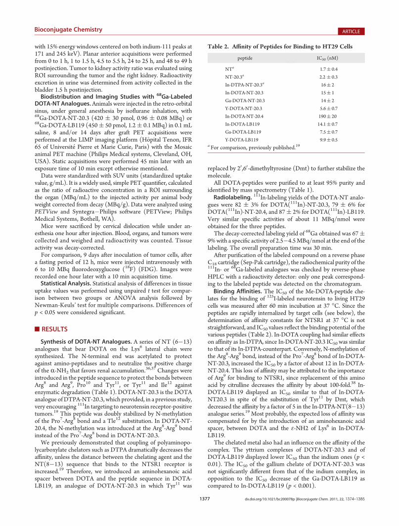
C18 cartridge (Sep-Pak cartridge), the radiochemical purity of the111In- or 68Ga-labeled analogues was checked by reverse-phaseHPLC with a radioactivity detector: only one peak correspond-ing to the labeled peptide was detected on the chromatogram.Binding Affinities. The IC50 of the Me-DOTA-peptide che-
lates for the binding of 125I-labeled neurotensin to living HT29cells was measured after 60 min incubation at 37 �C. Since thepeptides are rapidly internalized by target cells (see below), thedetermination of affinity constants for NTSR1 at 37 �C is notstraightforward, and IC50 values reflect the binding potential of thevarious peptides (Table 2). In-DOTA coupling had similar effectson affinity as In-DTPA, since In-DOTA-NT-20.3 IC50 was similarto that of its In-DTPA-counterpart. Conversely, N-methylation ofthe Arg8-Arg9 bond, instead of the Pro7-Arg8 bond of In-DOTA-NT-20.3, increased the IC50 by a factor of about 12 in In-DOTA-NT-20.4. This loss of affinity may be attributed to the importanceof Arg9 for binding to NTSR1, since replacement of this aminoacid by citrulline decreases the affinity by about 100-fold.38 In-DOTA-LB119 displayed an IC50 similar to that of In-DOTA-NT20.3 in spite of the substitution of Tyr11 by Dmt, whichdecreased the affinity by a factor of 5 in the In-DTPA-NT(8�13)analogue series.19 Most probably, the expected loss of affinity wascompensated for by the introduction of an aminohexanoic acidspacer, between DOTA and the ε-NH2 of Lys6 in In-DOTA-LB119.The chelated metal also had an influence on the affinity of the
complex. The yttrium complexes of DOTA-NT-20.3 and ofDOTA-LB119 displayed lower IC50 than the indium ones (p <0.01). The IC50 of the gallium chelate of DOTA-NT-20.3 wasnot significantly different from that of the indium complex, inopposition to the IC50 decrease of the Ga-DOTA-LB119 ascompared to In-DOTA-LB119 (p < 0.001).
Table 2. Affinity of Peptides for Binding to HT29 Cells
peptide IC50 (nM)
NTa 1.7( 0.4
NT-20.3a 2.2 ( 0.3
In-DTPA-NT-20.3a 16( 2
In-DOTA-NT-20.3 15( 1
Ga-DOTA-NT-20.3 14( 2
Y-DOTA-NT-20.3 5.6( 0.7
In-DOTA-NT-20.4 190( 20
In-DOTA-LB119 14.1( 0.7
Ga-DOTA-LB119 7.5( 0.7
Y-DOTA-LB119 9.9( 0.5a For comparison, previously published.19

1378 dx.doi.org/10.1021/bc200078p |Bioconjugate Chem. 2011, 22, 1374–1385
Bioconjugate Chemistry ARTICLE
Kinetics of in Vitro Radioactivity Binding and Internaliza-tion intoHT29 Cells.As expected from their IC50, the binding of111In-DOTA-NT-20.3 and of 111In-DOTA-LB119 was not sig-nificantly different at every time point, and a significantly lowerbinding was observed for 111In-DOTA-NT-20.4 after 15 min ofincubation (Figure 1). The amount of 111In-DOTA-peptideassociated with cells that was internalized into cells increasedrapidly with time reaching a 85 ( 2% plateau for 111In-DOTA-NT-20.3, 91 ( 2% for 111In-DOTA-NT-20.4, and 93 ( 2% for111In-DOTA-LB119 after less than 60 min.In Vivo Peptide Catabolism. The stability to enzymatic degra-
dation was evaluated in vivo. The fraction of radioactivity associatedto the intact 111In-DOTA-peptides in plasmawas determined 15minafter iv injection to BALB/cmice (Figure 2). Samples were analyzedby C18 RP-HPLC chromatography. Metabolites eluted at shorterretention times than the full-length radioactive peptide.
There was no significant difference between the amount ofintact peptide recovered in the plasma of mice injected with111In-DOTA-NT-20.3 (22 ( 1%) and 111In-DOTA-LB119(26( 3%), but 111In-DOTA-NT-20.4 (16( 2%) was less stablethan 111In-DOTA-LB119 (p < 0.05). The fraction of activity (%ID/g) remaining in blood 15 min postinjection amounted to 6.1( 0.4%, 5.2 ( 0.2%, and 3.9 ( 0.8% for 111In-DOTA-NT-20.3,111In-DOTA-NT-20.4, and 111In-DOTA-LB119, respectively.Biodistribution and Imaging Studies of the DOTA(111In)-
NT Peptides. Biodistribution studies of the neurotensin analo-gues 111In-DTPA-NT-20.3 (Table 3), 111In-DOTA-NT-20.3(Table 4), 111In-DOTA-NT-20.4 (Table 5), and 111In-DOTA-LB119 (Table 6) were performed at various time points post-injection in male nude mice. Tumor accretion expressed as thepercentage of injected dose per gram of tumor (%ID/g) showedno significant difference between 111In-DTPA-NT-20.3 and111In-DOTA-NT-20.3 at any time postinjection, indicating simi-lar tumor targeting efficacy of these two peptides. A slow tumorwashout of both peptides was observed between 3 and 6 h, asalready described for 111In-DTPA-NT-20.3 in female nude mice.The renal uptake of these two peptides was not significantlydifferent, except at 6 h postinjection (P < 0.05).At early time points, 111In-DOTA-NT-20.3 displayed a higher
tumor uptake than 111In-DOTA-LB119 (1 h P < 0.001 and 3 hP < 0.05), but 111In-DOTA-LB119 tumor uptake decreasedslowly with time and, from 6 to 24 h, no significant differencewas observed between these two peptides. Renal accumulation ofradioactivity was lower for 111In-DOTA-LB119 than for 111In-DOTA-NT-20.3 at early times postinjection (P < 0.001 at 1 h andP < 0.05 from 3 to 6 h). As a consequence, despite its lower tumoruptake, 111In-DOTA-LB119 tumor to kidney uptake ratios werehigher at 6 h (P < 0.001) postinjection than that of 111In-DOTA-NT-20.3. 111In-DOTA-NT-20.4 displayed a lower tumor up-take than 111In-DOTA-NT-20.3 at every time point and than111In-DOTA-LB119 from 3 to 24 h, and a higher kidney up-take than these two peptides. As a consequence, the tumor to
Figure 1. In vitro binding and internalization kinetics of 111In-DOTA-peptides in HT29 cells. Panel A: Kinetics of specific radioactivityaccretion to 1 � 106 HT29 cells at 37 �C (bound, fmol, mean ( sem)in the presence of 5� 10�10M labeled peptide. Each point is the averageof four experiments performed in triplicate. Open squares: 111In-DOTA-NT-20.3. Black squares: 111In-DOTA-LB119. Triangles: 111In-DOTA-NT-20.4. Panels B (111In-DOTA-NT-20.3), C (111In-DOTA-NT-20.4),and D (111In-DOTA-LB119): Internalization kinetics. Results areexpressed as the percentage of acid washed resistant activity, corre-sponding to specific binding, related to the activity associated with thecells (internalized/bound, mean ( SEM). Radioactivity, associated tocells or acid wash resistant, corresponding to nonspecific binding wasevaluated in the presence of 10�6Mneurotensin. Solid line: results fittedwith amonoexponential curve. Three experiments performed in triplicate.
Figure 2. In vivo serum stability of 111In-DOTA-peptides: representa-tive C18 HPLC chromatograms of plasma samples collected 15 minpostinjection to mice. (A) 111In-DOTA-NT-20.3. (B) 111In-DOTA-NT-20.4. (C) 111In-DOTA-LB119. Solid line: plasma sample. Dottedline: coinjection of the sample with the radioactive control. Chromato-grams were normalized to the highest metabolite peak.

1379 dx.doi.org/10.1021/bc200078p |Bioconjugate Chem. 2011, 22, 1374–1385
Bioconjugate Chemistry ARTICLE
kidney uptake ratio of this peptide was very low at every timepostinjection.Tumor uptake was receptormediated, as shown by the dramatic
decrease in tumor uptake when the radiolabeled 111In-DOTA-peptides were coinjected with their unlabeled counterpart: 2.5 (0.2% vs 0.14 ( 0.02% ID/g for 111In-DOTA-NT-20.3, 1.41 (0.05% vs 0.12( 0.03% for 111In-DOTA-LB119, and 0.52( 0.07%vs 0.11 ( 0.01% for 111In-DOTA-NT-20.4 (p < 0.001, 79�94%reduction, 3 h postinjection).
Radioactivity excretion in urine was fast, andmore than 60% ofthe injected activity was recovered in the bladder 1.5 h afterinjection of 111In-DOTA-NT-20.3 or 111In-DOTA-LB119.Blood activity decreased rapidly for the three 111In-DOTA-peptides, and low radioactivity uptake was observed in mostnontumor organs, except in kidneys and, to some extent, in thegastrointestinal tract, particularly in small intestine and in colon.With the exception of kidneys, tumor to normal organ uptakeratios were high for 111In-DOTA-NT-20.3 and, to a lesser extent,
Table 3. Tissue Distributions of 111In-DTPA-NT-20.3 in Male Nude Mice Grafted with HT29 Cells
111In-DTPA-NT-20.3
uptake (%ID/g)a 1 h n = 4 3 h n = 4 6 h n = 6 24 h n = 4 48 h n = 3
blood 0.13( 0.03 0.026( 0.004 0.023( 0.004 0.0076( 0.0008 0.0028( 0.0005
lungs 0.6( 0.3 0.11( 0.02 0.13( 0.04 0.044( 0.003 0.035 ( 0.003
liver 0.12( 0.01 0.093( 0.008 0.075( 0.009 0.049( 0.003 0.041( 0.002
spleen 0.13( 0.01 0.13 ( 0.02 0.10( 0.01 0.074( 0.005 0.064( 0.004
stomachb 0.14( 0.03 0.08 ( 0.02 0.15( 0.05 0.27( 0.08 0.033( 0.008
small intestineb 1.1( 0.4 0.5( 0.1 0.61( 0.09 0.38( 0.05 0.192( 0.005
large intestineb 0.5 ( 0.2 0.23( 0.02 1.4( 0.2 0.82( 0.13 0.15( 0.03
muscle 0.5( 0.4 0.14( 0.09 0.024( 0.005 0.023 ( 0.007 0.016( 0.004
bone 0.20( 0.06 0.10( 0.02 0.08( 0.01 0.044( 0.003 0.027( 0.004
kidney 7.8( 0.1 7( 2 2.8( 0.3 1.9( 0.3 1.5( 0.3
tumor 3.1( 0.4 2.0( 0.4 2.0( 0.2 0.86( 0.07 0.9( 0.1
tumor(T)/organ
T/blood 28( 7 75( 6 100( 20 110( 10 360( 90
T/kidney 0.40( 0.03 0.32( 0.03 0.72( 0.09 0.5( 0.1 0.64 ( 0.06
T/liver 26( 2 21( 3 27( 3 17.7( 0.5 23( 3
T/muscle 25( 9 40( 30 100( 10 50( 10 70( 10aUptake is expressed as the percentage of injected dose per gram of tissue (%ID/g). bOrgans with their content.
Table 4. Tissue Distributions of 111In-DOTA-NT-20.3 in Male Nude Mice Grafted with HT29 Cells111In-DOTA-NT-20.3
uptake (%ID/g)a 1 h n = 7 3 h n = 11 6 h n = 7 24 h n = 4 49 h n = 3
blood 0.36( 0.06 0.014( 0.002 0.006 ( 0.002 0.0028( 0.0003 0.0028 ( 0.0004
lungs 0.47( 0.04 0.14( 0.02 0.10( 0.01 0.062( 0.004 0.07( 0.01
liver 0.21( 0.02 0.13 ( 0.02 0.123( 0.008 0.085( 0.002 0.07( 0.01
spleen 0.19( 0.01 0.11( 0.01 0.113( 0.009 0.10( 0.01 0.16 ( 0.01
stomachb 0.13( 0.03 0.2( 0.1 0.09( 0.04 0.06( 0.01 0.020( 0.005
small intestineb 0.9( 0.1 0.52 ( 0.09 0.34( 0.06 0.32( 0.02 0.070( 0.003
large intestineb 0.39( 0.05 1.1( 0.3 1.5( 0.5 0.19 ( 0.02 0.058( 0.007
muscle 0.10( 0.02 0.027( 0.009 0.04( 0.01 0.0116( 0.0008 0.008( 0.001
bone 0.15( 0.02 0.10( 0.03 0.099 ( 0.007 0.030( 0.005 0.053 ( 0.002
kidney 7.6( 0.9 4.9( 0.4 5.2( 0.5 2.5( 0.1 0.86( 0.08
pancreas 0.095( 0.009 0.03 ( 0.01 0.030( 0.002 ND ND
tumor 4.7( 0.8 2.5( 0.2 1.9( 0.2 1.3( 0.2 0.68( 0.09
tumor(T)/organ
T/blood 17 ( 5 170( 30 420( 90 500( 100 250( 30
T/kidney 0.63( 0.07 0.53 ( 0.05 0.35( 0.02 0.50( 0.04 0.78( 0.03
T/liver 22( 3 21( 2 17( 3 15( 1 11( 4
T/muscle 60( 20 130( 20 80( 20 110( 20 80( 20
T/pancreas 47 ( 8 90( 10 70( 10 ND NDaUptake is expressed as the percentage of injected dose per gram of tissue (%ID/g). bOrgans with their content.

1380 dx.doi.org/10.1021/bc200078p |Bioconjugate Chem. 2011, 22, 1374–1385
Bioconjugate Chemistry ARTICLE
for 111In-DOTA-LB119. For example, tumor to blood amountedto 170( 30% and 63( 7%, respectively, tumor to muscle 130(20% and 80( 20%, tumor to pancreas 90( 10% and 65( 2%,and tumor to liver 21 ( 2% and 9.4 ( 0.4% at three hourspostinjection (Tables 4 and 6). Some excretion by the digestiveroute was also observed: 55 ( 8% and 60 ( 10% of the
radioactivity in the stomach and 76 ( 8% and 79 ( 2% in thecolon were associated to the organ content for 111In-DOTA-NT-20.3 and 111In-DOTA-LB119, respectively, at 3 h postinjection.Nevertheless, tumor to stomach (32( 8% at 3 h postinjection),to small intestine (7 ( 1%), and to colon (4 ( 1%) ratios werequite high for 111In-DOTA-NT-20.3. They were somewhat
Table 5. Tissue Distributions of 111In-DOTA-NT-20.4 in Male Nude Mice Grafted with HT29 Cells
111In-DOTA-NT-20.4
uptake (%ID/g)a 1 h n = 4 3 h n = 4 6 h n = 4 24 h n = 4 49 h n = 3
blood 0.27( 0.03 0.040( 0.003 0.037( 0.005 0.016( 0.005 0.0019( 0.0006
lungs 0.27( 0.02 0.10( 0.03 0.07( 0.01 0.06( 0.01 0.037 ( 0.001
liver 0.107( 0.007 0.076( 0.008 0.09( 0.01 0.071( 0.007 0.074( 0.007
spleen 0.097( 0.006 0.082( 0.006 0.070( 0.006 0.075( 0.009 0.08( 0.02
stomachb 0.18( 0.09 0.16( 0.09 0.062( 0.005 0.05( 0.02 0.0086( 0.0009
small intestineb 0.5( 0.3 0.12( 0.03 0.15 ( 0.05 0.090( 0.008 0.023( 0.003
large intestineb 0.20( 0.04 0.5( 0.3 0.36( 0.05 0.12( 0.02 0.033( 0.007
muscle 0.10( 0.02 0.04( 0.01 0.040 ( 0.008 0.023( 0.008 0.010 ( 0.002
bone 0.16( 0.05 0.09( 0.01 0.06( 0.01 0.035( 0.005 0.037( 0.008
kidney 9( 2 8( 1 9( 2 4.2( 0.8 1.7( 0.7
pancreas 0.044 ( 0.004 0.027( 0.002 0.022 ( 0.003 0.023( 0.003 ND
tumor 0.8( 0.1 0.52( 0.07 0.5( 0.1 0.33 ( 0.05 0.21( 0.08
tumor(T)/organ
T/blood 3.1 ( 0.8 13( 2 13( 3 40( 10 130( 80
T/kidney 0.095( 0.008 0.064 ( 0.005 0.053( 0.002 0.080 ( 0.007 0.2( 0.1
T/liver 7.2( 0.7 6.8( 0.7 5.4( 0.6 7( 2 2.8( 0.8
T/muscle 9( 1 14( 3 12( 3 26( 7 30( 10
T/pancreas 18( 4 20( 4 23( 5 21( 5 NDaUptake is expressed as the percentage of injected dose per gram of tissue (%ID/g). bOrgans with their content.
Table 6. Tissue Distributions of 111In-DOTA-LB119 in Male Nude Mice Grafted with HT29 Cells
111In-DOTA-LB119
uptake (%ID/g)a 1 h n = 7 3 h n = 4 6 h n = 4 24 h n = 3 49 h n = 3
blood 0.38( 0.05 0.023( 0.002 0.0045( 0.0002 0.007( 0.002 0.0016( 0.0003
lungs 0.36( 0.03 0.106( 0.007 0.09( 0.01 0.06( 0.01 0.04 ( 0.01
liver 0.20( 0.01 0.151( 0.007 0.14( 0.02 0.080( 0.005 0.09( 0.03
spleen 0.153( 0.007 0.09( 0.01 0.076( 0.009 0.064 ( 0.003 0.10( 0.03
stomachb 0.28( 0.08 0.16( 0.04 0.5( 0.5 0.08( 0.01 0.024( 0.004
small intestineb 1.1 ( 0.1 0.67( 0.08 0.7( 0.1 0.35( 0.05 0.084( 0.005
large intestineb 0.4( 0.1 1.5( 0.5 1.2 ( 0.8 0.16( 0.03 0.101( 0.006
muscle 0.09( 0.01 0.021( 0.005 0.05( 0.02 0.010( 0.003 0.015( 0.007
bone 0.15( 0.04 0.05( 0.01 0.07( 0.02 0.05( 0.01 0.05( 0.02
kidney 3.4( 0.2 2.4( 0.2 2.2( 0.2 1.04( 0.07 0.6( 0.1
pancreas 0.081( 0.008 0.0217( 0.0008 0.018( 0.001 0.0180( 0.0006 0.013( 0.002
tumor 1.8( 0.1 1.41( 0.05 1.4( 0.2 1.0( 0.3 0.46( 0.06
tumor(T)/organ
T/blood 6( 1 63 ( 7 300( 40 160( 60 330( 90
T/kidney 0.55( 0.05 0.60( 0.05 0.63 ( 0.06 0.9( 0.2 0.74( 0.07
T/liver 9.0( 0.7 9.4( 0.4 10( 2 12( 3 6( 1
T/muscle 24( 7 80( 10 80( 40 130( 70 40( 10
T/pancreas 25( 4 65 ( 2 75( 7 50( 10 37( 3aUptake is expressed as the percentage of injected dose per gram of tissue (%ID/g). bOrgans with their content.

1381 dx.doi.org/10.1021/bc200078p |Bioconjugate Chem. 2011, 22, 1374–1385
Bioconjugate Chemistry ARTICLE
lower for 111In-DOTA-LB119: 10 ( 2%, 2.2( 0.3%, and 1.2 (0.3%, respectively. Some of the radioactivity accretion in theintestines was receptor-mediated, since after removal of theircontent, the radioactivity uptake was significantly reduced insmall intestine (0.171 ( 0.008% vs 0.082 ( 0.007% ID/g for111In-DOTA-20.3 and 0.68 ( 0.01% vs 0.17 ( 0.04% ID/g forLB119, p < 0.001) and in colon (0.4 ( 0.1% vs 0.09 ( 0.02%,p < 0.05, and 0.7 ( 0.1% vs 0.16 ( 0.06% ID/g, p < 0.01,respectively), when the tracer was coinjected with an excess ofunlabeled peptide.Comparison between organ uptakes of different peptides at
selected times postinjection hardly reflects irradiation dosesdelivered to these organs over time for a therapeutic injectionof radiolabeled peptides. Then, for a preliminary evaluation of thepotential of these peptides for targeted radiotherapy, areas underthe time�activity curves (AUC expressed as %ID/g.min) werecalculated after simultaneously fitting the activity biodistributiondata for blood, tumor, and kidney to a multicompartmentalmodel. The same, relatively simple model was used for allpeptides with two compartments to describe the blood pharma-cokinetic and one additional compartment to describe tumor and
kidney uptake kinetics. The addition of a fraction of the centralcompartment, calculated on the basis of published data for thefractions of blood and interstitial fluid contained in tumors andnormal organs,34,35 to these tissues considerably improved datafitting. A total of seven parameters were thus adjusted tosimultaneously fit the biodistribution data in blood, tumor, andkidneys. Good fitting was obtained in all cases (Figure 3,Table 7); however, it should be kept in mind that the kineticsreflect the total activity present in the tissues, responsible fortissue irradiation, and not the intact radiolabeled peptide, whichis quickly catabolized. The estimated values for these parameterswere rather similar for all peptides with subtle changes explainingthe differences in pharmacokinetics.Blood clearances were pretty close for all peptides; however, as
judged by the AUC, tumor uptake was similar for 111In-DTPA-NT-20.3 and 111In-DOTA-NT-20.3, lower for 111In-DOTA-LB119, and still lower for 111In-DOTA-NT-20.4. Kidney uptakewas lower for 111In-DOTA-LB119, higher for 111In-DTPA-NT-20.3 and 111In-DOTA-NT-20.3, and even higher for 111In-DOTA-NT-20.4. These resulted in similar tumor/kidney AUCratios (0.6 to 0.7) for 111In-DTPA-NT-20.3, 111In-DOTA-NT-20.3, and 111In-DOTA-LB119 and in a much lower value for111In-DOTA-NT-20.4 (0.1). The IC50 of the yttrium complex ofDOTA-NT-20.3 is 2-fold lower than that of the indium one (p <0.01).Then, one can expect that tumor AUC and so tumor tokidney AUC ratio may be higher with 90Y- than 111In-labeledDOTA-NT-20.3.Planar images of mice grafted with HT29 cells were recorded
from 1 to 48 h, after injection of 111In-DOTA-NT-20.3 or 111In-DOTA-LB 119 (Figure 4). There was a clear visualization of thetumor at early time points in mice injected with 111In-DOTA-NT-20.3. At late time points, the tumor could be clearly detectedwith the two peptides. These results were consistent withbiodistribution data and tumor to organ uptake ratios. Kidneysand bladder were the only other sites of visible activity accumula-tion. The activity ratio between tumor and kidneys was 0.47 (0.03% at 1 h and 0.69( 0.07% at 24 h for 111In-DOTA-NT-20.3
Figure 3. Biodistribution kinetics of 111In-DOTA-peptides in tumor-bearing nude mice. Mice were injected with 111In-DOTA-NT-20.3(panel A), 111In-DOTA-NT-20.4 (panel B), or 111In-DOTA-LB119(panel C) and sacrificed at selected time intervals. Blood sample(circles), tumors (squares), and right kidneys (triangles) were weighedand counted, and the activity was expressed as %ID/g. The three kineticswere fitted simultaneously using the same pharmacokinetic model asdescribed in Materials and Methods, and the fitted curves for blood(dotted line), tumor (dashed line), and right kidney (solid line) areshown in the graphs as semilogarithmic plots.
Table 7. Area under Curves for Tumor, Kidneys, and Blood
peptide tumor AUCa kidney AUCa blood AUCa
111In-DTPA-NT-20.3 6743 10767 464111In-DOTA-NT-20.3 5463 9210 500111In-DOTA-LB-119 3687 5163 514111In-DOTA-NT-20.4 1618 13774 492
aArea under curve (AUC) are expressed as %ID/g.min.
Figure 4. 111In-DOTA-NT-20.3 and 111In-DOTA-LB119 planarimages of male nude mice grafted with HT29 cells. Planar anterioracquisitions were performed from 0 to 1 h, 1 to 1.5 h, 4.5 to 5.5 h, 24 to25 h, and 48 to 49 h postinjection under anesthesia. Bl: bladder. K:kidney. T: tumor. Tumor weight: 167 mg for 111In-DOTA-NT-20.3 and196 mg for 111In-DOTA-LB119.

1382 dx.doi.org/10.1021/bc200078p |Bioconjugate Chem. 2011, 22, 1374–1385
Bioconjugate Chemistry ARTICLE
(tumor weight: 0.15( 0.04% g), 0.45( 0.05% and 0.50( 0.01%for 111In-DOTA-LB119 (tumor weight: 0.20 ( 0.05% g).Biodistribution and PET Imaging with 68Ga-DOTA-NT-
20.3 and 68Ga-DOTA-LB119. Biodistribution studies were
performed 1 h after injection of 68Ga-DOTA-NT20.3 (420 ( 30pmol, 0.96 ( 0.08 MBq) or 68Ga-DOTA-LB119 (450 ( 50pmol, 1.2 ( 0.1 MBq). No significant difference was observedbetween tumor uptake of 68Ga-labeled and 111In-labeled DOTA-NT-20.3, but, probably due to its higher affinity, 68Ga-DOTA-LB119 tumor uptake was significantly increased as compared to111In-DOTA-LB119 (50% increase, P < 0.05). Nevertheless, thetumor uptake of 68Ga-DOTA-NT20.3 was higher than that of68Ga-DOTA-LB119 (P < 0.05).As observed for the 111In complexes the renal accretion of
68Ga-DOTA-LB119 was lower than that of 68Ga-DOTA-NT20.3(P < 0.05), and for both 68Ga-labeled peptides, the renal uptakewas similar to that of the 111In-labeled ones. Tumor to normaltissue uptake ratios were similar or higher for 68Ga-DOTA-NT20.3 as compared to 68Ga-DOTA-LB119 except for kidneys(0.46 ( 0.06% vs 0.79 ( 0.07%) (Figure 5).High contrast images were obtained with 68Ga-DOTA-
NT20.3, which allowed the visualization of very small tumors(Figure 6). As shown in Figure 6, 22 and 40 mm3 tumor graftscould be easily detected as soon as 45 min after 68Ga-DOTA-NT20.3 injection (10 min acquisition). The tumor SUVmax 45min after 68Ga-DOTA-NT-20.3 injection amounted to 0.9( 0.1and the tumor to kidney SUVmax ratio was 0.6( 0.1. Tumor wasnot detectable on FDG PET images (not shown).
’DISCUSSION
Since radiolabeled neurotensin analogues may be valuabletools for both imaging and therapy of neurotensin receptor-positive tumors, a number of radiolabeled neurotensin analogues,stabilized against enzymatic degradation in vivo by changes in thepeptide sequence, has been described in the literature. In a seriesof peptides bearing (NRHis)Ac, a chelator of 99mTc and 188Re,NT-XIX, an NT(8�13) analogue, displayed the most promisingproperties to target these radionuclides in vivo, with low kidneyuptake, and reduction of tumor mass was observed after treat-ment with the rhenium-188-labeled peptide.39,40 Nevertheless,some intestine uptakemay be a source of background for imagingand of toxicity for therapeutic applications. Polyaminopolycar-boxylate chelators, such as DTPA or DOTA, coupled to soma-tostatin analogues have been used with success to deliver variousradiometals, including 111In, 68Ga, 90Y, or 177Lu, to somatostatinreceptor-positive tumors, such as gastroenteropancreatic neu-roendocrine tumors (GEPNETs). PET imaging with [68Ga-DOTA0,Tyr3]octreotate or [68Ga-DOTA0,Tyr3]octreotide hasbeen reported to achieve higher diagnostic efficacy than SPECTwith 111In-labeled analogues.24�27 In addition, therapy with [90Y-DOTA0,Tyr3]octreotide and [177Lu-DOTA0,Tyr3]octreotateprovided symptomatic improvement, tumor regression, im-proved quality of life, and a benefit in overall survival.15,16 SeveralDTPA and DOTA neurotensin analogues have been developedto target these radionuclides to tumors overexpressing neuro-tensin receptors.18,41,42 However, these radiolabeled peptidesshowed moderate tumor uptake and comparably high kidneyuptake. We recently reported the encouraging results obtainedwith a new DTPA-neurotensin(6�13) analogue, DTPA-NT-20.3, which displayed improved uptake ratio between tumorand kidneys, superior to previously published DTPA and DOTAneurotensin analogues.19 High-quality planar scintigraphy andSPECT images were obtained after a short delay, compatiblewith the use of the short-lived radionuclide 68Ga for PETimaging.
Figure 5. Tumor to normal organ uptake ratios obtained 1 h afterinjection of 68Ga-DOTA-NT-20.3 (420 ( 30 pmol, 0.96 ( 0.08 MBq)or 68Ga-DOTA-LB119 (450 ( 50 pmol, 1.2 ( 0.1 MBq) in male nudemice, grafted with HT29 cells. Bl: blood. Lu: lung. Li: liver. Sp: spleen.St: stomach. Si: small intestine. Co: colon. Mu: muscle. Pa: pancreas. Ki:kidney.
Figure 6. TEP imaging of male nude mice, grafted with HT29 cells inthe right flank, injected with 68Ga-DOTA-NT-20.3: (A) images wererecorded at different time points after injection of 68Ga-DOTANT-20.3;in the samemouse (tumor volume 40mm3), no tumor was detectable on18F-FDG PET images performed 24 h later; (B) and (C) imagesrecorded 45 min after 68Ga-DOTA-NT-20.3 injection (tumor volumes:22 and 288 mm3). Imaging was performed seven (A and B) or twelvedays (C) after cell graft. The acquisition time was 10 min a = MaximumIntensity Projection (MIP), frames: b = axial, c = sagittal, and d =coronal. Bl: Bladder. K: Kidney. T: Tumor. ant: anterior. pos: posterior.

1383 dx.doi.org/10.1021/bc200078p |Bioconjugate Chem. 2011, 22, 1374–1385
Bioconjugate Chemistry ARTICLE
A macrocyclic chelator, such as DOTA, is necessary fortargeted radionuclide therapy with 90Y, since in vivo leakage ofthis radionuclide from DTPA complexes leads to bone marrowtoxicity.28 For PET imaging, the in vivo stability of 68Ga-DTPAchelates remains a matter of controversy. Then, the aim of thepresent study was to design new DOTA-substituted neurotensinanalogues suitable for 68Ga PET imaging and possibly for 90Y or177Lu targeted radiotherapy of neurotensin-receptor-positivetumors.
In the three synthesized DOTA-peptides, a Tle12 substitutionwas introduced to protect the bond between Tyr11 and Ile12.Since positive charges increase renal uptake,36,37 the R-NH2 wasneutralized by acetylation, which also protected the peptidesagainst aminopeptidases. In DOTA-NT-20.3, which shares thesame peptide sequence with DTPA-NT-20.3, an N-methylationof the Pro7-Arg8 bond was introduced for stability. In the triple-stabilized DOTA-LB119, Tyr11 was substituted with Dmt and aspacer was introduced between the peptide and the chelator inorder to reduce the affinity loss induced by polyaminopolycarbox-ylate coupling, which we previously observed.19 In DOTA-NT-20.4, the Arg8-Arg9 bond was protected by an N-methylation.
The properties of the 111In labeled DOTA-peptides wereevaluated and compared to those of 111In-DTPA-NT-20.3. Theaffinity decrease induced by DOTA coupling is similar to thatobserved with DTPA, as indicated by the similar IC50 of thesetwo peptides. As already observed for 111In-DTPA-NT-20.3, theDOTA analogue rapidly internalizes in vitro. Biodistribution ofboth peptides was very similar in male nude mice grafted withHT29 cells. Circulating activity decreased rapidly, and high tumorto blood uptake ratios were obtained at early times postinjection.Tumor uptakes of both peptides were not significantly different atany time point and decreased quite slowly with time. Uptake innormal organs was low, leading to high tumor to organs ratioexcept in kidneys.
The three 111In-DOTA-peptides exhibited receptor mediatedtumor targeting, abolished by coinjection of their unlabeledcounterpart. N-Methylation of the Pro7-Arg8 or of the Arg8-Arg9 bonds was similarly effective for in vivo stabilization, asshown by the stability results obtained with 111In-DOTA-NT-20.3 and 111In-DOTA-NT-20.4. Probably due to its loweraffinity, tumor uptake of 111In-DOTA-NT-20.4 was lower thanthe two other 111In-DOTA-analogues. Unexpectedly, renal ac-cumulation of this peptide was also higher. Although 111In-DOTA-NT-20.3 and 111In-DOTA-LB119 displayed similar sta-bility and IC50, the tumor uptake of this last peptide wassignificantly lower at early time points. Renal uptake was alsolower at early time points for 111In-DOTA-LB119, as alreadyobserved forDmt11 substituted (NRHis)Ac-NT(8�13) analogues.39
In agreement with the biodistribution studies, high contrast imageswere obtained with 111In-DOTA-NT-20.3 with a clear detection oftumors at early time points.
Since tumor grafts were detected at early time points post-injection with 111In-DOTA-LB119 and particularly 111In-DOTA-NT-20.3, these peptides were further evaluated for68Ga targeting. Radiolabeling was obtained in good yields with68Ga, and no free 68Ga was detected after purification. Thespecific activities obtained allowed the injection of less than 500pmol per mouse for PET imaging, in order to minimize thesaturation of tumor neurotensin receptors. For both peptides,uptake in normal organs, 1 h postinjection, was low except inkidneys. 68Ga-DOTA-NT-20.3 provided reasonably high tumoruptake, higher than that of 68Ga-DOTA-LB119. Very small
tumor masses (20�40 mm3) could be detected with 68Ga-DOTA-NT-20.3 at 45 min after injection.
Metal complexation had an influence on the IC50 of theDOTA-peptides. Effects observed with gallium differed accord-ing to the peptide. No significant difference was observed withDOTA-NT-20.3, but it enhanced DOTA-LB119 affinity ascompared to indium, and the tumor uptake of 68Ga-DOTA-LB119 was significantly increased (by 50%). Yttrium significantlydecreased the IC50 of both peptides. Such an affinity differenceinduced by the incorporated metal has already been described inthe literature for DOTATOC43 and bombesin analogues.44
’CONCLUSION
In summary, in spite of a relatively high uptake in kidneys,111In-DOTA-NT-20.3 showed specific tumor uptake, providedelevated tumor to other organ uptake ratios, particularly tumor tointestine, and high contrast images at early time points afterinjection. A very low background in normal tissues, exceptkidneys, was obtained by PET imaging with 68Ga-DOTA-NT-20.3 which allowed the detection of very small tumors. DOTA-NT-20.3 may be considered a promising candidate for 68Ga-PETimaging of neurotensin receptor-positive tumors, such as pan-creatic adenocarcinoma, invasive ductal breast cancer, and non-small cell lung carcinoma. The high affinity observed for theyttrium complex of DOTA-NT-20.3, higher than that of theindium one, suggests that targeting 90Y with this peptide mayensure a higher tumor uptake than with 111In. A prerequisite fortherapeutic application of this neurotensin analogue would be tolower kidney uptake, for example, by infusion of basic aminoacids, gelofusin, or albumin fragments, to prevent nephrotoxicity,as with radiolabeled somatostatin analogues.45,46
’AUTHOR INFORMATION
Corresponding Author*Anne Gruaz-Guyon, Inserm, U773, Facult�e de M�edecine XavierBichat, 16 rue Henri Huchard, BP 416, F-75018, France. E-mail:[email protected], Phone: (33) 1 57 27 75 55, Fax(33) 1 57 27 75 31.
NotesΔFaisal Alshoukr and Aur�elie Prignon are to be considered asequal coauthors.
’ACKNOWLEDGMENT
This work was supported by Inserm, by the Association pour laRecherche sur le Cancer (ARC), and by the Canc�eropole Ile deFrance. We thank the Syrian government for awarding a researchfellowship to F.A. We are grateful to Edouard Treca for IC50
determination of some peptides.
’REFERENCES
(1) Myers, R.M., Shearman, J.W., Kitching,M.O., Ramos-Montoya,A., Neal, D. E., and Ley, S. V. (2009) Cancer, chemistry, and the cell:molecules that interact with the neurotensin receptors. ACS Chem. Biol.4, 503–525.
(2) Vincent, J. P., Mazella, J., and Kitabgi, P. (1999)Neurotensin andneurotensin receptors. Trends Pharmacol. Sci. 20, 302–309.
(3) Evers, B. M. (2006) Neurotensin and growth of normal andneoplastic tissues. Peptides 27, 2424–2433.

1384 dx.doi.org/10.1021/bc200078p |Bioconjugate Chem. 2011, 22, 1374–1385
Bioconjugate Chemistry ARTICLE
(4) Thomas, R. P., Hellmich, M. R., Townsend, C. M., Jr., and Evers,B. M. (2003) Role of gastrointestinal hormones in the proliferation ofnormal and neoplastic tissues. Endocr. Rev. 24, 571–599.(5) Ehlers, R. A., Kim, S., Zhang, Y., Ethridge, R. T., Murrilo, C.,
Hellmich, M. R., Evans, D. B., Townsend, C. M., Jr., and Mark Evers, B.(2000) Gut peptide receptor expression in human pancreatic cancers.Ann. Surg. 231, 838–848.(6) Reubi, J. C., Waser, B., Friess, H., Buchler, M., and Laissue, J.
(1998) Neurotensin receptors: a new marker for human ductal pan-creatic adenocarcinoma. Gut 42, 546–550.(7) Souaze, F., Dupouy, S., Viardot-Foucault, V., Bruyneel, E.,
Attoub, S., Gespach, C., Gompel, A., and Forgez, P. (2006) Expressionof neurotensin and NT1 receptor in human breast cancer: a potentialrole in tumor progression. Cancer Res. 66, 6243–6249.(8) Alifano, M., Souaze, F., Dupouy, S., Camilleri-Broet, S., Younes,
M., Ahmed-Zaid, S. M., Takahashi, T., Cancellieri, A., Damiani, S.,Boaron, M., Broet, P., Miller, L. D., Gespach, C., Regnard, J. F., andForgez, P. (2010) Neurotensin receptor 1 determines the outcome ofnon-small cell lung cancer. Clin. Cancer Res. 16, 4401–4410.(9) Alifano, M., Loi, M., Camilleri-Broet, S., Dupouy, S., Regnard,
J. F., and Forgez, P. (2010) Neurotensin expression and outcome ofmalignant pleural mesothelioma. Biochimie 92, 164–170.(10) Gui, X., Guzman, G., Dobner, P. R., and Kadkol, S. S. (2008)
Increased neurotensin receptor-1 expression during progression ofcolonic adenocarcinoma. Peptides 29, 1609–1615.(11) Shimizu, S., Tsukada, J., Sugimoto, T., Kikkawa, N., Sasaki, K.,
Chazono, H., Hanazawa, T., Okamoto, Y., and Seki, N. (2008) Identi-fication of a novel therapeutic target for head and neck squamous cellcarcinomas: a role for the neurotensin-neurotensin receptor 1 oncogenicsignaling pathway. Int. J. Cancer 123, 1816–1823.(12) Swift, S. L., Burns, J. E., and Maitland, N. J. (2010) Altered
expression of neurotensin receptors is associated with the differentiationstate of prostate cancer. Cancer Res. 70, 347–356.(13) Reubi, J. C., Waser, B., Schaer, J. C., and Laissue, J. A. (1999)
Neurotensin receptors in human neoplasms: high incidence in Ewing’ssarcomas. Int. J. Cancer 82, 213–218.(14) Dupouy, S., Viardot-Foucault, V., Alifano, M., Souaze, F., Plu-
Bureau, G., Chaouat, M., Lavaur, A., Hugol, D., Gespach, C., Gompel, A.,and Forgez, P. (2009) The neurotensin receptor-1 pathway contributesto human ductal breast cancer progression. PLoS One 4, e4223.(15) Kwekkeboom, D. J., Kam, B. L., van Essen, M., Teunissen, J. J.,
van Eijck, C. H., Valkema, R., de Jong, M., de Herder, W. W., andKrenning, E. P. (2010) Somatostatin-receptor-based imaging and ther-apy of gastroenteropancreatic neuroendocrine tumors. Endocr. Relat.Cancer 17, R53–73.(16) Krenning, E. P., Teunissen, J. J., Valkema, R., deHerder, W. W.,
deJong,M., and Kwekkeboom, D. J. (2005)Molecular radiotherapy withsomatostatin analogs for (neuro-)endocrine tumors. J. Endocrinol. Invest.28, 146–150.(17) Granier, C., van Rietschoten, J., Kitabgi, P., Poustis, C., and
Freychet, P. (1982) Synthesis and characterization of neurotensin analo-gues for structure/activity relationship studies. Acetyl-neurotensin-(8�13)is the shortest analogue with full binding and pharmacological activities.Eur. J. Biochem. 124, 117–124.(18) Hillairet De Boisferon, M., Raguin, O., Thiercelin, C., Dussaillant,
M., Rostene, W., Barbet, J., Pelegrin, A., and Gruaz-Guyon, A. (2002)Improved tumor selectivity of radiolabeled peptides by receptor andantigen dual targeting in the neurotensin receptor model. BioconjugateChem. 13, 654–662.(19) Alshoukr, F., Rosant, C., Maes, V., Abdelhak, J., Raguin, O.,
Burg, S., Sarda, L., Barbet, J., Tourwe, D., Pelaprat, D., andGruaz-Guyon,A. (2009) Novel neurotensin analogues for radioisotope targeting toneurotensin receptor-positive tumors. Bioconjugate Chem. 20, 1602–1610.(20) Maecke, H. R., Hofmann, M., and Haberkorn, U. (2005)
(68)Ga-labeled peptides in tumor imaging. J. Nucl. Med. 46 (Suppl 1),172S–178S.(21) Wei, L., Zhang, X., Gallazzi, F., Miao, Y., Jin, X., Brechbiel,
M. W., Xu, H., Clifford, T., Welch, M. J., Lewis, J. S., and Quinn, T. P.
(2009) Melanoma imaging using (111)In-, (86)Y- and (68)Ga-labeledCHX-A00-Re(Arg11)CCMSH. Nucl. Med. Biol. 36, 345–354.
(22) Mansi, R., Wang, X., Forrer, F., Kneifel, S., Tamma, M. L.,Waser, B., Cescato, R., Reubi, J. C., andMaecke, H. R. (2009) Evaluationof a 1,4,7,10-tetraazacyclododecane-1,4,7,10-tetraacetic acid-conjugatedbombesin-based radioantagonist for the labeling with single-photonemission computed tomography, positron emission tomography, andtherapeutic radionuclides. Clin. Cancer Res. 15, 5240–5249.
(23) Pagou,M., Zerizer, I., and Al-Nahhas, A. (2009) Can gallium-68compounds partly replace (18)F-FDG in PET molecular imaging? Hell.J. Nucl. Med. 12, 102–105.
(24) Buchmann, I., Henze, M., Engelbrecht, S., Eisenhut, M., Runz,A., Schafer, M., Schilling, T., Haufe, S., Herrmann, T., andHaberkorn, U.(2007) Comparison of 68Ga-DOTATOC PET and 111In-DTPAOC(Octreoscan) SPECT in patients with neuroendocrine tumours. Eur. J.Nucl. Med. Mol. Imaging 34, 1617–1626.
(25) Hofmann, M., Maecke, H., Borner, R., Weckesser, E., Schoffski,P., Oei, L., Schumacher, J., Henze, M., Heppeler, A., Meyer, J., andKnapp, H. (2001) Biokinetics and imaging with the somatostatinreceptor PET radioligand (68)Ga-DOTATOC: preliminary data. Eur.J. Nucl. Med. 28, 1751–1757.
(26) Gabriel, M., Decristoforo, C., Kendler, D., Dobrozemsky, G.,Heute, D., Uprimny, C., Kovacs, P., Von Guggenberg, E., Bale, R., andVirgolini, I. J. (2007) 68Ga-DOTA-Tyr3-octreotide PET in neuroendo-crine tumors: comparison with somatostatin receptor scintigraphy andCT. J. Nucl. Med. 48, 508–518.
(27) Kowalski, J., Henze,M., Schuhmacher, J., Macke, H. R., Hofmann,M., and Haberkorn, U. (2003) Evaluation of positron emission tomogra-phy imaging using [68Ga]-DOTA-D Phe(1)-Tyr(3)-Octreotide in com-parison to [111In]-DTPAOC SPECT. First results in patients withneuroendocrine tumors. Mol. Imaging Biol. 5, 42–48.
(28) Liu, S. (2008) Bifunctional coupling agents for radiolabeling ofbiomolecules and target-specific delivery of metallic radionuclides. Adv.Drug Delivery Rev. 60, 1347–1370.
(29) Wang, D., Miller, S. C., Sima, M., Parker, D., Buswell, H.,Goodrich, K. C., Kopeckova, P., and Kopecek, J. (2004) The arthro-tropism of macromolecules in adjuvant-induced arthritis rat model: apreliminary study. Pharm. Res. 21, 1741–1749.
(30) Brans, L.,Maes, V., Garcia-Garayoa, E., Schweinsberg, C., Daepp,S., Blauenstein, P., Schubiger, P. A., Schibli, R., and Tourwe, D. A. (2008)Glycation methods for bombesin analogs containing the (NalphaHis)Acchelator for 99mTc(CO)3 radiolabeling. Chem. Biol. Drug Des. 72, 496–506.
(31) Raguin, O., Gruaz-Guyon, A., and Barbet, J. (2002) Equilibriumexpert: an add-in to Microsoft Excel for multiple binding equilibriumsimulations and parameter estimations. Anal. Biochem. 310, 1–14.
(32) Stefanovski, D., Moate, P. J., and Boston, R. C. (2003)WinSAAM: a windows-based compartmental modeling system. Meta-bolism 52, 1153–1166.
(33) Riches, A. C., Sharp, J. G., Thomas, D. B., and Smith, S. V. (1973)Blood volume determination in the mouse. J. Physiol. 228, 279–284.
(34) Sung, C., Youle, R. J., and Dedrick, R. L. (1990) Pharmacoki-netic analysis of immunotoxin uptake in solid tumors: role of plasmakinetics, capillary permeability, and binding. Cancer Res. 50, 7382–7392.
(35) Covell, D. G., Barbet, J., Holton, O. D., Black, C. D., Parker,R. J., and Weinstein, J. N. (1986) Pharmacokinetics of monoclonalimmunoglobulin G1, F(ab0)2, and Fab0 in mice. Cancer Res. 46, 3969–3978.
(36) Akizawa, H., Arano, Y., Mifune, M., Iwado, A., Saito, Y., Mukai,T., Uehara, T., Ono, M., Fujioka, Y., Ogawa, K., Kiso, Y., and Saji, H.(2001) Effect of molecular charges on renal uptake of 111In-DTPA-conjugated peptides. Nucl. Med. Biol. 28, 761–768.
(37) Froidevaux, S., Calame-Christe, M., Tanner, H., and Eberle,A. N. (2005) Melanoma targeting with DOTA-alpha-melanocyte-sti-mulating hormone analogs: structural parameters affecting tumor uptakeand kidney uptake. J. Nucl. Med. 46, 887–895.
(38) Barroso, S., Richard, F., Nicolas-Etheve, D., Reversat, J. L.,Bernassau, J. M., Kitabgi, P., and Labbe-Jullie, C. (2000) Identification of

1385 dx.doi.org/10.1021/bc200078p |Bioconjugate Chem. 2011, 22, 1374–1385
Bioconjugate Chemistry ARTICLE
residues involved in neurotensin binding and modeling of the agonistbinding site in neurotensin receptor 1. J. Biol. Chem. 275, 328–336.(39) Garcia-Garayoa, E., Blauenstein, P., Blanc, A., Maes, V., Tourwe,
D., and Schubiger, P. A. (2009) A stable neurotensin-based radiopharma-ceutical for targeted imaging and therapy of neurotensin receptor-positivetumours. Eur. J. Nucl. Med. Mol. Imaging 36, 37–47.(40) Maes, V., Garcia-Garayoa, E., Blauenstein, P., and Tourwe, D.
(2006) Novel 99mTc-labeled neurotensin analogues with optimizedbiodistribution properties. J. Med. Chem. 49, 1833–1836.(41) Achilefu, S., Srinivasan, A., Schmidt, M. A., Jimenez, H. N.,
Bugaj, J. E., and Erion, J. L. (2003) Novel bioactive and stableneurotensin peptide analogues capable of delivering radiopharmaceu-ticals and molecular beacons to tumors. J. Med. Chem. 46, 3403–3411.(42) de Visser, M., Janssen, P. J., Srinivasan, A., Reubi, J. C., Waser,
B., Erion, J. L., Schmidt, M. A., Krenning, E. P., and de Jong, M. (2003)Stabilised 111In-labelled DTPA- and DOTA-conjugated neurotensinanalogues for imaging and therapy of exocrine pancreatic cancer. Eur. J.Nucl. Med. Mol. Imaging 30, 1134–1139.(43) Froidevaux, S., Eberle, A. N., Christe, M., Sumanovski, L.,
Heppeler, A., Schmitt, J. S., Eisenwiener, K., Beglinger, C., and Macke,H. R. (2002) Neuroendocrine tumor targeting: study of novel gallium-labeled somatostatin radiopeptides in a rat pancreatic tumor model. Int.J. Cancer 98, 930–937.(44) Koumarianou, E.,Mikolajczak, R., Pawlak,D., Zikos, X., Bouziotis,
P., Garnuszek, P., Karczmarczyk, U., Maurin, M., and Archimandritis, S. C.(2009)Comparative study onDOTA-derivatized bombesin analog labeledwith 90Y and 177Lu: in vitro and in vivo evaluation. Nucl. Med. Biol.36, 591–603.(45) de Jong, M., Rolleman, E. J., Bernard, B. F., Visser, T. J., Bakker,
W. H., Breeman, W. A., and Krenning, E. P. (1996) Inhibition of renaluptake of indium-111-DTPA-octreotide in vivo. J. Nucl. Med. 37, 1388–1392.(46) Vegt, E., van Eerd, J. E., Eek, A., Oyen, W. J., Wetzels, J. F., de
Jong, M., Russel, F. G., Masereeuw, R., Gotthardt, M., and Boerman,O. C. (2008) Reducing renal uptake of radiolabeled peptides usingalbumin fragments. J. Nucl. Med. 49, 1506–1511.
![Somatostatin receptor scintigraphy with [111In-DTPA-d-Phe1]- and [123I-Tyr3]-octreotide: the Rotterdam experience with more than 1000 patients](https://static.fdokumen.com/doc/165x107/63360adfb5f91cb18a0ba76f/somatostatin-receptor-scintigraphy-with-111in-dtpa-d-phe1-and-123i-tyr3-octreotide.jpg)