
Erythropoietin: Blood, Brain and Beyond - Gunadarma University

Novel antibodies directed against the human erythropoietinreceptor: creating a basis for clinical implementation
Perry Maxwell,1,2 Florinda Melendez-
Rodr�ıguez,3 Kyle B. Matchett,2 Julian
Aragones,3 Nathalie Ben-Califa,4
Heidelinde Jaekel,5 Ludger Hengst,5,6
Herbert Lindner,6 Andre Bernardini,7
Ulf Brockmeier,7 Joachim Fandrey,7
Fritz Grunert,8 Howard S. Oster,9 Moshe
Mittelman,9 Mohamed El-Tanani,2
Markus Thiersch,10 Edith M. Schneider
Gasser,10 Max Gassmann,10,11 David
Dangoor,12 Robert J. Cuthbert,13
Alexandra Irvine,2 Anne Jordan,2
Terence Lappin,2 John Thompson8 and
Drorit Neumann4,*1Northern Ireland Molecular Pathology Labora-
tory, Belfast Health & Social Care Trust,
Queen’s University Belfast, 2Centre for Cancer
Research and Cell Biology, Queen’s University
Belfast, Belfast, UK, 3Hospital Universitario
Santa Cristina, Autonomous University of
Madrid, Madrid, Spain, 4Department of Cell
and Developmental Biology, Sackler Faculty of
Medicine, Tel Aviv University, Tel-Aviv, Israel,5Division of Medical Biochemistry, Biocentre,
Innsbruck Medical University, 6Division of
Clinical Biochemistry, Biocentre, Innsbruck
Medical University, Innsbruck, Austria, 7Insitute
of Physiology, University Duisburg Essen, Essen,8Aldevron Freiburg GmbH, Freiburg, Germany,9Department of Internal Medicine A, Tel Aviv
Sourasky Medical Centre, Tel Aviv University,
Tel Aviv, Israel, 10Institute of Veterinary
Physiology, Zurich Centre for Integrative Human
Physiology (ZIHP), University of Zurich, Zurich,
Switzerland, 11Universidad Peruana Cayetano
Heredia (UPCH), Lima, Peru, 12OSM-DAN Ltd,
Rehovot, Israel and 13Belfast Health & Social
Care Trust, Belfast, UK
Received 25 July 2014; accepted for publication
2 August 2014Correspondence: Drorit Neumann, Department of
Cell and Developmental Biology, Sackler Faculty of
Medicine, Tel Aviv University, Ramat-Aviv 69978,
Tel-Aviv, Israel.
E-mail: [email protected]
*The work represents the efforts of the Euro-
pean consortium called EpoCan that is
financed until September 2014 by the FP7 call.
Summary
Recombinant human erythropoietin (rHuEPO) is an effective treatment for
anaemia but concerns that it causes disease progression in cancer patients
by activation of EPO receptors (EPOR) in tumour tissue have been contro-
versial and have restricted its clinical use. Initial clinical studies were flawed
because they used polyclonal antibodies, later shown to lack specificity for
EPOR. Moreover, multiple isoforms of EPOR caused by differential splicing
have been reported in cancer cell lines at the mRNA level but investigations
of these variants and their potential impact on tumour progression, have
been hampered by lack of suitable antibodies. The EpoCan consortium
seeks to promote improved pathological testing of EPOR, leading to safer
clinical use of rHuEPO, by producing well characterized EPOR antibodies.
Using novel genetic and traditional peptide immunization protocols, we
have produced mouse and rat monoclonal antibodies, and show that sev-
eral of these specifically recognize EPOR by Western blot, immunoprecipi-
tation, immunofluorescence, flow cytometry and immunohistochemistry in
cell lines and clinical material. Widespread availability of these antibodies
should enable the research community to gain a better understanding of
the role of EPOR in cancer, and eventually to distinguish patients who can
be treated safely by rHuEPO from those at increased risk from treatment.
Keywords: cancer anaemia, recombinant erythropoietin, erythropoietin
receptor, antibody, risk assessment.
research paper
ª 2014 John Wiley & Sons Ltd, British Journal of Haematology doi:10.1111/bjh.13133
Anaemia is an independent prognostic factor for poor sur-
vival in cancer patients (Caro et al, 2001), but the use of
recombinant human erythropoietin (rHuEPO) to treat these
patients is controversial due to concerns about patient safety
arising from Phase III clinical trials showing more rapid can-
cer progression and reduced survival in subjects randomized
to rHuEPO (Henke et al, 2003; Leyland-Jones et al, 2005;
Miller et al, 2009). Consequently there has been a marked
decline in the use of rHuEPO since 2007 (Hill et al, 2012).
Clearly, the benefits of EPO treatment must be carefully bal-
anced against the risk of enhanced cancer progression for
each patient.
Erythropoietin functions by binding to its receptor (EPOR)
on the surface membrane of erythroid progenitors and activat-
ing JAK2/STAT5 signalling pathways (Jelkmann, 2010; Wenger
& Kurtz, 2011). There is now evidence that growth factor
receptor-mediated cell signalling pathways can overlap in can-
cer cells. In a landmark study, Liang et al (2010) found that
EPOR is co-expressed with human epidermal growth factor
receptor-2 (HER2, also known as ERBB2) in breast cancer cell
lines and tumour specimens. rHuEPO administered to patients
antagonized trastuzumab treatment and resulted in shorter
progression-free and overall survival in patients with HER2-
positive metastatic breast cancer (Liang et al, 2010).
Erythropoietin has pleiotropic actions and EPOR is
expressed outside the haematopoietic compartment (Lappin
et al, 2002; Ghezzi et al, 2010; Vogel & Gassmann, 2011).
EPOR is functionally active in endothelial cells (Anagnostou
et al, 1994) and endothelial progenitor cells promoting vas-
cular repair and endothelial regeneration (Trincavelli et al,
2013). Moreover, EPO stimulates angiogenesis both in vitro
and in vivo (Trincavelli et al, 2013). Positive effects of EPO
treatment on the immune system have been documented
(Mittelman et al, 2004; Prutchi-Sagiv et al, 2006, 2008; Katz
et al, 2007; Lifshitz et al, 2010; Mausberg et al, 2011; Nairz
et al, 2012; Oster et al, 2013). EPO has thrombotic effects, at
least in vitro, as it increases plasminogen activator inhibitor-1
release in HUVEC culture (Stasko et al, 2002). Patients with
end-stage renal disease on high doses of rHuEPO have high
haemoglobin levels, are associated with increased risk of car-
diovascular and thrombotic events, and have reduced sur-
vival (Smith et al, 2003; Provatopoulou & Ziroyiannis, 2011).
These observations support the hypothesis that supraphysio-
logical levels of circulating EPO could lead to enhanced
tumour growth, neovascularization and thrombosis in some
cancer patients. Recommendations for the use of erythropoi-
esis-stimulating agents (ESAs) in patients with cancer have
been published (Rizzo et al, 2010).
To understand the increased risk of administering
rHuEPO to patients with cancer, it is imperative to examine
its effects in tumour tissue. Angiogenic factors, such as vas-
cular endothelial growth factor A (VEGFA), platelet-derived
growth factors (PDGFs), fibroblast growth factors (FGFs)
and angiopoietins are often elevated in the tumour environ-
ment. PDGFBB targets perivascular cells to nascent vascular
networks (Abramsson et al, 2003) and modulates tumour
angiogenesis by increasing EPO production in stromal cells,
leading to the induction of endothelial cell proliferation,
migration, sprouting and tube formation (Xue et al, 2012).
Contentious inferences were drawn from clinical studies
that were based on immunohistochemistry (IHC) using anti-
bodies, but later shown to lack specificity for EPOR (Elliott
et al, 2006; Brown et al, 2007). Thus, investigation of EPOR
expression and function relies heavily on the availability of
specific anti-EPOR antibodies.
The EU-based EpoCan project addresses safety concerns
related to EPO treatment and is investigating the risks of
EPO treatment using mouse models, human clinical samples
and patient databases. The studies focus on the long-term
effects of EPO treatment on tumour growth and incidence,
the role of EPO in angiogenesis and its association with
thromboembolic events in cancer and cardiovascular disease.
An important initial objective was to prepare a range of
highly specific monoclonal antibodies against human EPOR.
To this end, a cohort of 15 EPOR mouse and rat monoclonal
antibodies were produced and evaluated for different applica-
tions. Here we report on the characterization of four of these
antibodies, which have proved suitable for a range of appli-
cations.
Materials and methods
Cell lines
The human cell lines used were: megakaryoblastic leukaemia
cells, UT-7; acute lymphocytic leukaemia cells, REH; pre-B
cells, NALM6; breast cancer cells, MDA-MB-231; human
embryonic kidney cells HEK293T, and its derivative,
BOSC23; and lung cancer cells, A549.
Tissue sections
Formalin-Fixed Paraffin wax-Embedded (FFPE) tissue sec-
tions and bone marrow aspirates were obtained under agree-
ment with the Northern Ireland Biobank (Ethical approval
reference NIB12-0044).
Monoclonal antibody generation by geneticimmunization
Monoclonal antibodies were generated using cDNA con-
structs encoding the extracellular domain (ECD) of hEPOR
cloned into proprietary immunization and screening vectors.
Anti-tag antibodies were used to confirm expression after
transient transfection of the cDNA constructs into mamma-
lian cells, in vitro. The immunization constructs were
adsorbed onto the surface of gold particles and introduced
intradermally into mice and rats using a BioRad gene gun
(Bio-Rad GmbH Munchen, Germany), with several cDNA
boosts, following proprietary protocols. This cDNA was
P. Maxwell et al
2 ª 2014 John Wiley & Sons Ltd, British Journal of Haematology
taken up and translated by skin cells, whereby the protein
was brought to the cell surface and finally secreted to allow
an optimal immune response against the EPOR ECD. The
sera were tested against both the ECD and full-length EPOR
construct, the cDNA of which had been transiently transfect-
ed into mammalian cells.
Monoclonal antibody generation using synthetic peptides
The amino acid sequence of hEPOR was analysed using
hydrophilicity/hydrophobicity software to identify peptide
regions representing potential epitopes. In total, six peptides
(h1–h6) were chosen for peptide synthesis of which five were
located in the cytoplasmic domain (h2–h6) as shown for
hEPOR (Table I). The peptides were conjugated with keyhole
limpet haemocyanin for immunization and with ovalbumin
for screening in peptide enzyme-linked immunosorbent
assays (ELISA). Immunization was carried out intraperitone-
ally with one group of six peptides (h1–h6) into two cohorts
of three rats. The peptides were mixed with complete
Freund’s adjuvant for the initial immunization, followed by
several boosts with incomplete Freund’s adjuvant. Following
immunization of three animals per cohort, sera were tested
for positivity in a peptide ELISA against ovalbumin-peptide
conjugate mixes, corresponding to their immunogen mix-
tures. After lymphocyte fusion, the resulting hybridoma
supernatants were screened against each individual ovalbu-
min-peptide conjugate in the same ELISA assay to identify
antibodies against specific peptides.
Flow cytometry
Flow cytometry was performed essentially as described (Lif-
shitz et al, 2010). Cell suspensions were analysed on a FAC-
Sort flow cytometer (Becton Dickinson, Oxford, UK) and the
results were analysed using WinMDI software (J.Trotter free
download, http://winmdi.software.informer.com/2.8/). Anti-
HA antibody (MMS-101R) was from Covance (Princeton,
NJ, USA). Fluorescein isothiocyanate (FITC)-conjugated
AffinityPure Goat Anti-Mouse IgG and FITC-conjugated
Affinity Pure Goat Anti-Rat IgG were from Jackson Immu-
noResearch Laboratories Inc. (West Grove, PA, USA).
Immunofluorescence
Cells seeded on glass cover slips (A549, MDA-MB-231, COS7
and HEK293T) or collected in a 1�5 ml tube (UT-7) were
fixed in 3% paraformaldehyde in phosphate-buffered saline
(PBS) at room temperature (RT) for 30 min. After incuba-
tion in quenching buffer (0�1% Triton, 5% fetal bovine
serum, and 2% bovine serum albumin) at RT for 1 h, cells
were incubated with primary antibodies diluted in quenching
buffer at RT for 1 h. After washing with PBS, cells were
incubated with secondary antibodies for 1 h at RT. Nuclei
were stained with 2�5 lmol/l DRAQ5(ab108410; Abcam,
Cambridge, MA, USA) for 30 min at RT. Confocal fluores-
cent images were obtained by a TCS SP5 II confocal micro-
scope (Leica, Wetzlar, Germany) with a 63x/1.4NA objective
or a CSU10 spinning disc unit coupled to a Zeiss Axiovert
200M microscope with a 100x/1.3NA oil immersion objec-
tive. For some experiments, fusion proteins of enhanced cyan
fluorescent protein (ECFP) and EPOR were made by cloning
cDNA encoding for the hEPOR into the pECFP-N1 vector
(Clontech, Mountain View, CA, USA) between HindIII and
BamHI. A construct specific for the intracytoplasmic domain
(ICD) of EPOR was generated by replacing the extracellular
and transmembrane domain in the described construct by
that of the epithelial growth factor receptor (EGFR) (Gross
et al, 2014). For transient transfections plasmid DNA was
mixed with the transfection reagent Turbofect (Thermo Sci-
entific, Waltham, MA, USA) according to the supplier’s rec-
ommendations. The cells were incubated for 24 h with the
transfection mix and subjected to immunofluorescence stain-
ing as described.
Generation of EPOR-silenced cells
Human EPOR (sc-37092-V) and control (sc-108080) shRNA
lentiviral particles (Santa Cruz Biotechnology, Dallas, TX,
USA) were used to generate stable transfectants in MDA-
MB-231 breast cancer cells and A549 lung carcinoma cells.
After lentiviral infection, infected cells were selected with
puromycin (1 lg/ml) to finally generate MDA-MB-231-shE-
POR, A549-shEPOR cells and their corresponding MDA-
MB-231-shSCR and A549-shSCR control cells. Following
puromycin selection for 11–18 d, cells were lysed in Laemmli
buffer and subjected to Western blot analysis using the
GM1201 antibody (see Table II).
Western blot analysis
Cells were lysed in Laemmli buffer. Western blots were per-
formed using 10% sodium dodecyl sulphate (SDS)-polyacryl-
amide gels. Membranes were incubated with a 1:1000
dilution in Tris-buffered saline (TBS)-Tween 20 (TBS-T)
buffer of the indicated anti-EPOR antibody at 4°C overnight.
Membranes were incubated with the corresponding second-
ary antibody at RT for 1 h and, finally, washed three times
Table I. Choice of human EPOR peptides for immunization.
Human EPO-R peptides Location Exon
h1: PPPNLPDPKFES Extracellular domain 1
h2: KIWPGIPSPESEFEG
LFTTHKGN
Intracytoplasmic domain 7
h3: VEPGTDDEGPL Intracytoplasmic domain 8
h4: LPRNPPSEDLPGPG Intracytoplasmic domain 8
h5: PSSQLLRPWTLC Intracytoplasmic domain 8
h6: GDSQGAQGGLSDGPYSN Intracytoplasmic domain 8
Novel Anti-Erythropoietin Receptor Antibodies
ª 2014 John Wiley & Sons Ltd, British Journal of Haematology 3

with TBS-T buffer for 10 min. Immunolabeling was detected
by enhanced chemiluminescence (SuperSignal West Femto
Maximum Sensitivity Substrate; Thermo Scientific) and visu-
alized with a digital luminescent image analyser (Image
Quant LAS400 mini; GE Healthcare Life Sciences, Bucking-
hamshire, UK).
Coupling of antibodies for immunoprecipitation
For immunoprecipitation (IP), anti-EPOR antibodies or
anti-HA antibodies (clone 12CA5, Abcam) were covalently
coupled to protein-A agarose (Immunosorb A, Medicago,
Uppsala, Sweden). Antibody (2 lg) was incubated with 5 llProtein-A bead slurry in PBS for 1 h at RT. Beads were
washed with sodium borate (0�2 mol/l, pH 9�0) and the anti-
body was cross-linked to protein-A upon addition of
40 mmol/l dimethylpimelimidate.
Cell transfection, protein extraction and IP
Plasmid encoding the N-terminal HA-tagged hEPOR was
transiently transfected into HEK-293T cells with calcium-
phosphate (Graham & van der Eb, 1973) and harvested 48 h
later. Cell extracts were prepared in IP buffer with 1% Triton
X-100. Protein extracts were cleared by centrifugation
(10 000 g; 20 min), protein concentration determined by
BioRad-DC assay and IP performed using 40 lg extract from
transfected HEK-293T cells, 750 lg of UT-7 cells or 1�5 mg
extract from A549 and MDA-MB231 cells. Proteins were
diluted with IP buffer and IP was performed in the presence
of 0�5% Triton X-100 for 2 h at 4°C. After 3 washes with
0�5% Triton X-100 IP buffer, EPOR protein was eluted by
boiling in Laemmli sample buffer (pH 6�8) for 5 min, loaded
on a SDS-polyacrylamide gel and analysed by Western blot
using GM1201 antibody.
Mass spectrometry – Nano-high performance liquidchromatography – MS/MS
Four milligram of UT-7 cell extract was immunoprecipitated
with GM1202 and GM1203 (see Table II) agarose-coupled
antibodies (mixture of 1:1, 6 lg antibodies each). After
extensive washing, the precipitated proteins were separated
by a 10% SDS-polyacrylamide gel electophoresis and visual-
ized with Coomassie Blue. Protein digests of the gel piece
covering the 65 kD region were analysed using an UltiMate
3000 nano-HPLC system (Dionex, Germering, Germany)
coupled to an LTQ Orbitrap XL mass spectrometer. An
in-house fritless fused-silica microcapillary column (75 lmi.d., 280 lm o.d.) packed with 10 cm of 3-lm reversed-phase
C18 material (Reprosil, Dr Maish GmbH, Ammerbuch-
Entringen, Germany) was used. The gradient (solvent A,
0�1% formic acid; solvent B, 0�1% formic acid in 85%
acetonitrile) started at 4% B. The concentration of solvent B
was increased linearly from 4 to 50% over 50 min and from
50 to 100% over 5 min. A flow rate of 250 nl/min was
applied. Data analysis was performed using Proteome
Discoverer 1.3 (ThermoScientific) with search engine Sequest
(http://fields.scripps.edu/sequest/). Precursor mass tolerance
was set to 10 ppm, fragment mass tolerance was 0�8 Da. Raw
data obtained by liquid chromatography-electrospray ioniza-
tion mass spectrometry (LC-ESI-MS) were searched against
the Homo sapiens protein database extracted from the
NCBInr database using false discovery rate (FDR) for peptide
evaluation. Only peptides with a significance threshold
of 0�01 (99% confidence) or less were used for protein
identification.
Immunohistochemistry
To test the suitability of antibodies for clinical applications in
FFPE material, we used UT-7, REH and NALM-6 cell lines, as
they have differential endogenous EPOR expression. They were
grown to confluence in 29 T75 flasks, removed and fixed in
10% formal saline (BCS Biosciences Ltd, Cambridge, UK)
overnight and processed to paraffin wax. Sections were pre-
pared and stained by all antibodies at concentrations of 2–
10 lg/ml following pressure cooking antigen retrieval, and
using anti-rabbit/-mouse Envision (Dako, Cambridge, UK), or
peroxidase-conjugated anti-rat immunoglobulin (Sigma-
Aldrich, Gillingham, UK) localization. Sections were prepared
from bone marrow samples and stained at 4 lg/ml concentra-
tion using automated IHC (Discovery XT; Roche Tissue Diag-
nostics, Burgess Hill, UK) and CC1 conditioning.
Bone marrow aspirate smears and cultured erythroid pro-
genitor cells were mounted on glass slides and fixed over-
night in 95% methylated ethanol. After washing in water,
slides were flooded with PBS pH 7�0 and primary antibodies
incubated overnight at 4°C (rat anti-EPOR GM1201 Aldev-
ron, Freiburg, Germany) and rabbit anti-ferritin H (ab75972,
Table II. Characteristics of the four selected EPOR monoclonal antibodies.
Immunogen Subclone name Isotype Epitope location Applications
Peptide h6 GM1201 rIgG2b Intracytoplasmic domain WB, IP, IF, IHC
Genetic immunization GM1202 mIgG1 Extracellular domain IP, IF, FACS
Genetic immunization GM1203 mIgG1 Extracellular domain IP, IF, FACS
Genetic immunization GM1204 rIgG2a Extracellular domain FACS
WB, Western blotting; IP, immunoprecipitation; IF, immunoflourescence, IHC, immunohistochemistry; FACS, fluorescence-activated cell sorting.
P. Maxwell et al
4 ª 2014 John Wiley & Sons Ltd, British Journal of Haematology
Abcam, Cambridge, UK) or rabbit anti-glycophorin C
(ab108619, Abcam) at 4 lg/ml. FITC anti-rat (Millipore,
Billerica, MA, USA) and Alexa Fluor 568 anti-rabbit (Life
Technologies, Paisley, UK) were used to localize immunore-
activity for 2 h at 37°C. Cell preparations were mounted in
aqueous mounting medium containing 40,6-diamidino-
2-phenylindole (DAPI, Aquilant Scientific, Belfast, UK).
Results
Monoclonal antibodies generated by geneticimmunization against hEPOR-ECD
Four murine and five rat mother hybridomas were preselect-
ed for their ability to recognize the hEPOR ECD constructs
in flow cytometry. Purified monoclonal antibodies were
tested by flow cytometry at 1 lg/ml (Fig 1A–C).
Monoclonal antibody generation using synthetic peptides
Supernatants from the stable mother clones were pre-tested
for the selection of the best mother clones for each assay
(Western blots, IP, IHC, immunofluorescence and flow
cytometry) with respect to signal strength and specificity.
The chosen mother clones were then subcloned by serial
dilution, expanded, isotyped and antibodies purified using
protein G columns and resuspended in PBS to a given con-
centration (1–2 mg/ml). The results of the chosen subclones
that revealed the most unequivocal results in the various
assays were generated against human peptide 6, which is
located close to the C-terminal region of EPOR (Table I).
Systematic nomenclature of monoclonal antibodies
Of 15 monoclonal antibody mother clones, four were
selected for subcloning and additional testing (Table II).
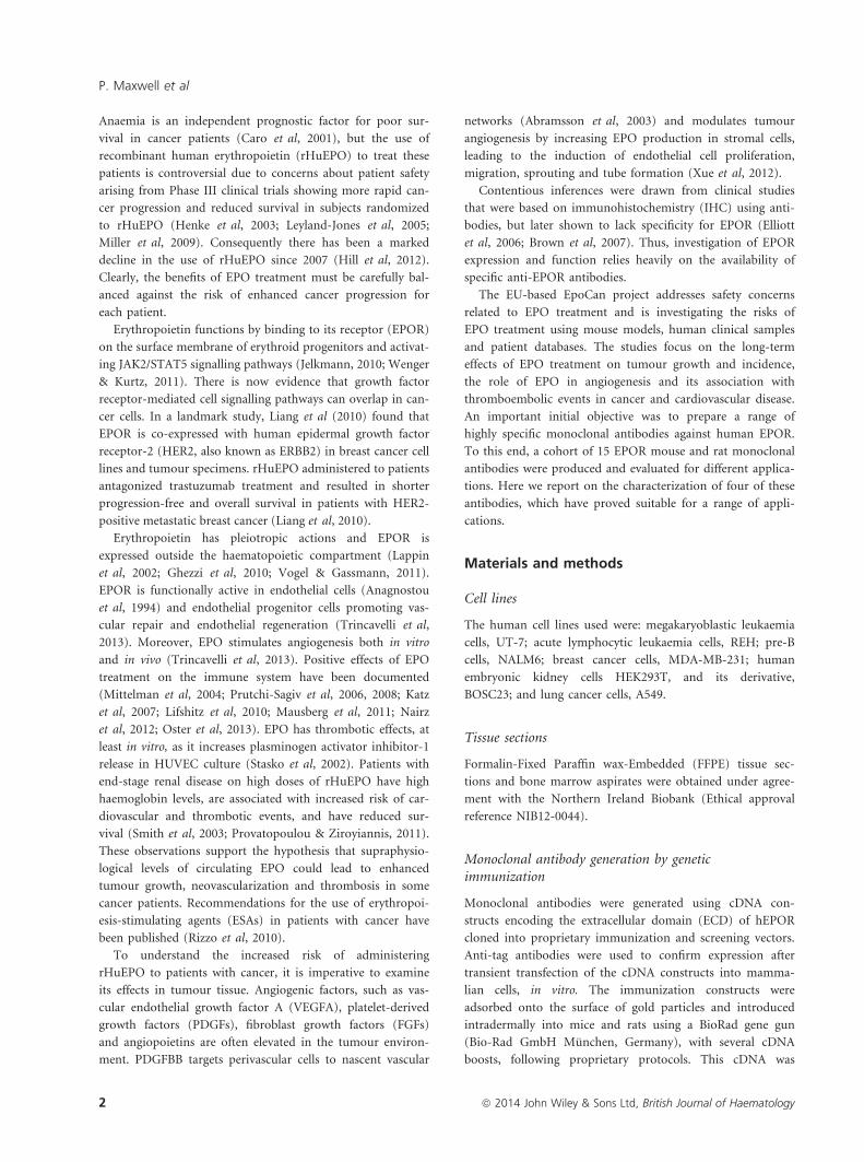
(A)
(B)(C)
(a)
(b)
(c)
(a)
(b)
(c)
Fig 1. Specificity of individual anti-EPOR hybridoma clones. (A) Hybridoma supernatants were analysed by flow cytometry on cells transiently
transfected with the human EPOR extracellular domain (ECD) cDNA cloned into a proprietary Aldevron test vector (green curves). Depending
on the antibody source, a goat anti-mouse or anti-rat IgG R-phycoerythrin (R-PE) conjugate were used as secondary antibodies. Negative con-
trols: Mammalian cells, transfected with an irrelevant control cDNA cloned in the corresponding expression vector (red curves). (B) BOSC23 cells
expressing HA-tagged hEPOR or EGFR were incubated at 4°C with primary antibodies (10 lg/ml) followed by fluorescein isothiocyanate (FITC)-
conjugated secondary antibodies. Black line: GM1202, GM1203 and GM1204; Green line: anti-HA antibody. Red and blue lines: secondary anti-
body only. Depending on the antibody source, a goat anti-mouse IgG R- conjugate (#1030-09, Southern Biotech, Birmingham, AL, USA), or a
goat anti-rat IgG R-PE conjugate (#3030, Southern Biotech) were used as secondary antibodies at 10 lg/ml. As negative control BOSC23 cells,
transfected with an irrelevant control cDNA cloned into the corresponding expression vector, were incubated with each monoclonal antibody and
detected with the secondary antibody described above (black curves). (C) Cells were incubated with primary antibodies (4 lg/ml for UT-7 cells;
10 lg/ml for A549 and MDA-MB-231 cells), followed by FITC-conjugated secondary antibodies (15 lg/ml). Black line: GM1202, GM1203 and
GM1204; Red line: secondary antibody only. EPOR, erythropoietin receptor; EGFR, epithelial growth factor receptor.
Novel Anti-Erythropoietin Receptor Antibodies
ª 2014 John Wiley & Sons Ltd, British Journal of Haematology 5

Flow cytometry analysis of EPOR
The GM1202, GM1203 and GM1204 antibodies, raised
against the ECD, recognized hEPOR by fluorescence-acti-
vated cell sorting (FACS) analysis, on Bosc23 cells transfected
with HA tagged hEPOR or EGFR cDNA constructs. The
three antibodies showed similar levels of hEPOR expression
as compared to anti-HA antibody (positive control). Isotype
controls (data not shown) and secondary antibodies were
used as negative controls. The specificity of the antibodies
was verified by the lack of reactivity in HA-EGFR transfected
cells. Using FACS analysis, the same antibodies also detected
endogenous hEPOR in UT-7, A549 and MDA-MB-231 cells.
hEPOR was detected with higher sensitivity in UT-7 cells
(4 lg/ml antibody concentration) than in A549 and MDA-
MB-231 (10 lg/ml).
Immunofluorescence
COS7 cells were transfected with HA-hEPOR cDNA and
stained with GM1202 and GM1203 directed against the ECD
and GM1201 directed against the ICD (Fig 2A) using anti-HA
as a positive control. The three antibodies detected hEPOR and
specificity was verified using isotype controls (data not shown).
HEK293T cells were transfected with DNA encoding a
fusion protein of ECFP and EPOR, incubated for 24 h and
then stained with GM1201, directed against the ICD, and
GM1202 or GM1203, directed against the ECD, of EPOR
(Fig 2B). Colour coding was set to equal values for ECFP
and Alexa555 (secondary antibody label) channels with the
exception listed in the legend to Fig 2B. For the anti-EPOR
antibodies GM1202 and GM1203 (rows 2 and 3), perfect
co-localization at the membrane of immunofluorescence with
ECFP fluorescence was observed, indicating specific recogni-
tion of EPOR protein by the antibodies. In contrast, the anti-
bodies did not detect the ECFP-EGFR-EPOR fusion protein,
which was sufficiently expressed (as can be seen in the ECFP
channel) and therefore served as a negative control (upper
row). GM1201 antibody was only able to detect EPOR
protein in permeabilized cells, indicating correct orientation
of the ECFP-EPOR fusion protein and supporting its speci-
ficity for the ICD of EPOR. Interestingly, this antibody also
detects the ICD in a fusion protein made from ECFP, the
ECD of EGFR and the ICD of EPOR. Reactivity of the
GM1201, GM1202 and GM1203 antibodies with hEPOR was
also demonstrated in UT-7, A549 and MDA-MB-231 cells,
showing significantly higher sensitivity in UT-7 cells.
Endogenous EPOR expression in tumour cell lines
The ability of the antibodies to recognize endogenous EPOR
expression in tumour cells was tested by immunofluorescence
(Fig 3) and Western blot analysis (Fig 4) using the UT-7 cell
line as well as EPOR-silenced breast carcinoma MDA-MB-
231 cells and EPOR-silenced lung carcinoma A549 cells (shE-
POR) and the corresponding control cells (shSCR). RNA
analysis in these cells showed that the EPOR mRNA level in
UT-7 is 270 � 8 times higher than in MDA-MB-231 control
cells (MDA-MB-231-shSCR) and 56 � 2 times higher than
in A549 control cells (A549-shSCR). Further RNA analysis in
EPOR-silenced MDA-MB-231 and A549 cells showed that
EPOR mRNA expression declined by 82 � 3% and 70 � 3%
respectively, which validated these cellular models for testing
GM1201 antibody by Western blot. In line with these RNA
data, Western blotting showed a main signal around 63 kDa
in these three cell lines and was negative for EPOR-silenced
cells (Fig 4). This signal was much higher in UT-7 than in
MDA-MB-231 and A549 control cells, reflecting that Western
blot signal parallels EPOR mRNA levels in these cells. More-
over, GM1201 has the potential to detect higher molecular
weight EPOR forms, which are much weaker than the main
63 kDa form. Collectively, these data indicate that GM1201
reliably detects endogenous EPOR by Western blotting.
Identification of antibodies that specificallyimmunoprecipitate overexpressed and endogenous humanEPOR
Using overexpressed HA-hEPOR as a source, GM1201,
GM1202 and GM1203, were identified as antibodies with the
highest immunoprecipitating efficiency (Fig 5A). Of note, the
rat-derived antibody GM1201 was as efficient as the com-
mercial anti-HA-tag antibody by IP (Fig 5A). The GM1201
antibody was also used to detect untagged immunoprecipi-
tated hEPOR in Western blots. Besides this antibody, two
mouse-derived antibodies directed against the ECD of EPOR,
GM1202 and GM1203, were able to efficiently recover
endogenous hEPOR in immunoprecipitates from UT-7, A569
and MDA-MB-231 cells (Fig 5B–D). In the case of very low
endogenous hEPOR expression in A549 or MDA-MB-231
cells, the use of these mouse antibodies prevented the detec-
tion of cross-reacting bands, which were precipitated if the
same antibody (GM1201) was used in IP and Western blot
[A549: Fig 5C (*); MDA-MB-231: data not shown].
In all cases and with all antibodies, IP was able to signifi-
cantly enrich the EPOR compared to the cell lysate (10% of
the IP input). Specificity of the immunoprecipitated bands
was confirmed by knockdown of EPOR using shRNAs in
A549 cells and MDA-MB-231 cells (Fig 5C, D). The recovery
of hEPOR in immunoprecipitates using GM1202 and
GM1203 antibodies was further confirmed by Nano-HPLC–
MS/MS, where seven EPOR-specific peptides were identified
in trypsin-digested samples (Fig 6). Together, these data
revealed a specific and highly efficient hEPOR immunopre-
cipitating capacity of GM1201, GM1202 and GM1203.
Immunohistochemistry
Differential expression of EPOR mRNA expression was con-
firmed in NALM-6, REH and UT-7 cell lines by Q-PCR
P. Maxwell et al
6 ª 2014 John Wiley & Sons Ltd, British Journal of Haematology
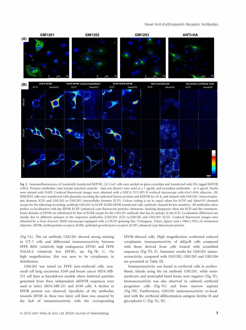
(Fig 7A). The rat antibody GM1201 showed strong staining
in UT-7 cells and differential immunoreactivity between
FFPE REH (relatively high endogenous EPOR) and FFPE
NALM-6 (relatively low EPOR), see Fig 7B, C. On
high magnification, this was seen to be cytoplasmic in
distribution.
GM1201 was tested on FFPE non-erythroid cells, non-
small cell lung carcinoma A549 and breast cancer MDA-MB-
231 cell lines as knockdown models where lentiviral particles
generated from three independent shEPOR sequences were
used to infect MDA-MB-231 and A549 cells. A decline in
EPOR protein was observed. Specificity of the antibodies
towards EPOR in these two latter cell lines was ensured by
the lack of immunoreactivity with the corresponding
EPOR-silenced cells. High magnification confirmed reduced
cytoplasmic immunoreactivity of shEpoR cells compared
with those derived from cells treated with scrambled
sequences (Fig 7D, E). Summary results for GM1201 immu-
noreactivity, compared with GM1202, GM1203 and GM1204
are presented in Table III.
Immunoreactivity was found in erythroid cells in erythro-
blastic islands using the rat antibody GM1201, while meta-
myelocyte and neutrophil band forms were negative (Fig 7F).
Immunoreactivity was also observed in cultured erythroid
progenitor cells (Fig 7G) and bone marrow aspirate
(Fig 7H). Furthermore, GM1201 immunoreactivity co-local-
ized with the erythroid differentiation antigens ferritin H and
glycophorin C (Fig 7G, H).
GM1201 GM1202 GM1203 ANTI-HA(A)
(B)
Fig 2. Immunofluorescence of transiently transfected hEPOR. (A) Cos7 cells were seeded on glass coverslips and transfected with HA-tagged hEPOR
cDNA. Primary antibodies (and isotype matched controls – data not shown) were used at a 7 lg/ml, and secondary antibodies – at 4 lg/ml. Nuclei
were stained with DAPI. Confocal fluorescent images were obtained with a LEICA TCS SP5 II confocal microscope with 63x/1.4NA objective. (B)
HEK293T cells were transfected with plasmids encoding the indicated fusion proteins and hEPOR for 24 h, and stained with GM1201 (intracytoplas-
mic domain; ICD) and GM1202 or GM1203 (extracellular domain; ECD). Colour coding is set to equal values for ECFP and Alexa555 channels
except for the following recording: antibody GM1201 in ECFP-EGFR-EPOR transfected cells (antibody channel 8x less sensitive). All antibodies show
perfect co-localization with the EPOR-ECFP (enhanced cyan fluorescent protein) chimaeras. Staining disappears when the ECD and the transmem-
brane domain of EPOR are substituted by that of EGFR except for the GM1201 antibody that has its epitope in the ICD. Localization differences are
mostly due to different epitopes of the respective antibodies (GM1201: ICD vs.GM1202 and GM1203: ECD). Confocal fluorescent images were
obtained by a Zeiss Axiovert 200M microscope equipped with a CSU10 spinning disc (Yokogawa, Tokyo, Japan) and a 100x/1.3NA oil immersion
objective. EPOR, erythropoietin receptor; EGFR, epithelial growth factor receptor; ECFP, enhanced cyan fluorescent protein.
Novel Anti-Erythropoietin Receptor Antibodies
ª 2014 John Wiley & Sons Ltd, British Journal of Haematology 7

GM1202
GM1203
GM1201 UT7 A549 MDA-MB-231
(A)
(B)
(C)
Fig 3. Detection of endogenous hEPOR by immunofluorescence. UT-7 cells were collected and fixed in 3% paraformaldehyde in phosphate-buf-
fered saline at room temperature (RT) for 30 min. Primary antibodies (and isotype controls – not shown) were diluted to 4 lg/ml in quenching
buffer. Secondary antibodies [fluorescein isothiocyanate (FITC)-conjugated AffinityPure Goat Anti-Mouse IgG for GM1202 and GM1203 antibod-
ies and FITC-conjugated AffinityPure Goat Anti-Rat IgG for the GM1201 antibody] were diluted to 4 lg/ml. MDA-MB-231 and A549 cells were
seeded on glass coverslips. Primary antibodies (and Isotype controls – not shown) were diluted to 20 lg/ml and secondary antibodies were used
at 7�5 lg/ml. Nuclei were stained with DRAQ5 (2�5 lmol/l) for 30 min at RT. Confocal fluorescent images were obtained by a LEICA TCS SP5
II confocal microscope with a 63x objective.
P. Maxwell et al
8 ª 2014 John Wiley & Sons Ltd, British Journal of Haematology

Discussion
A spectrum of strongly held views concerning EPOR func-
tion in tumours is evident in the literature, ranging from
claims that malignant cells are devoid of functional EPOR-
mediated signalling pathways to assertions that there is an
overlap of cell signalling pathways that has pathogenic signif-
icance (Ghezzi et al, 2010).
Previous immunohistochemical studies on tumour tissue
have drawn controversial conclusions about the expression
of EPOR, based on antibodies that were later shown to cross-
react with other cellular proteins (Elliott et al, 2006; Brown
et al, 2007). The ideal antibody for IHC would be monoclonal
rather than polyclonal, possess immunoreactivity against a
defined EPOR domain and lack cross-reactivity with other tis-
sue constituents. Unfortunately, most studies reported to date
have used polyclonal antibodies or monoclonal antibodies of
undefined specificity. Undoubtedly, the resulting discrepancies
have masked the important debate about the safety of treating
anaemic cancer patients with rHuEPO. Two other related fac-
tors that have received scant consideration are the occurrence
of EPOR splice variants (Arcasoy et al, 2003) and the possible
involvement of a heterodimeric form of the receptor compris-
ing one EPOR and one common b chain component (Brox-
meyer, 2013). Whereas homodimeric EPOR has been
extensively studied, the existence of the heterodimeric complex
is debated and requires further study.
The aim of the EpoCan consortium is to produce, charac-
terize and validate a panel of EPOR monoclonal antibodies
that would be readily available to the research community.
These include antibodies raised against the ECD of EPOR for
FACS analysis; those that recognize the denatured EPOR
TCE IP
70
55
+
+ + + + + +
Ctr. vector
HA-hEPOR
GM
1203
HA
Ctr.
GM
1201
GM
1201
GM
1202
GM
1201
C
tr. IP
GM
1203
G
M12
02
70
55
IP Ctr.
GM
1203
C
tr. G
M12
01
TCE
(B)
GM
1201
Ctr.
IP
GM
1203
GM
1202
70
55
+ + + + +
+ + + +
shSCR
shEPOR
IP TCE Ctr.
GM
1203
C
tr. G
M12
01 (C)
*
*
GM
1203
GM
1202
70
55
+ + +
+ + +
shSCR
shEPOR
TCE
(D)
IP
(A)
Ctr.
IP
Ctr.
GM
1203
Fig 5. Antibodies that immunoprecipitate hEPOR. (A) Human HA-EPOR expressing plasmids (HA-hEPOR) or the empty control vector (Ctr.
vector) were transfected in HEK293T cells. Precipitated EPOR was detected using the GM1201 antibody. 1/10 of the extract (4 lg) was loaded as
total cell extract (TCE). Mouse anti-HA antibody 12CA5, (HA), was used as a positive IP control, mouse IgG was used as a negative control
(Ctr. IP). GM1201, GM1203: GM1201 antibodies or GM1203 antibodies coupled to Protein-A agarose were incubated in the absence of cell
extract and loaded. (B) IP of EPOR from UT-7 cells. Ctr. IP: IP with normal mouse antibodies. Ctr. GM1203, Ctr. GM1201: Antibodies coupled
to Protein-A agarose were incubated in the absence of cell extract. TCE: 10% of the input for the IP (75 lg) was loaded. (C) IP of hEPOR from
A549 lung carcinoma cells expressing control (shSCR) and three EPOR-specific small hairpin RNA (shEPOR). GM1201 was used for Western blot
detection. Cross-reacting bands (*) were detected when GM1201 was used in IP and WB. Ctr. IP: IP with normal mouse antibodies; Ctr
GM1203, Ctr. GM1201: antibodies coupled to Protein-A agarose incubated in the absence of cell extract. TCE: 10% of the protein input for the
IP (150 lg) was loaded. (D) IP of hEPOR from MDA-MB-231 cells transfected with shSCR or shEPOR. TCE: 10% of the protein input for the
IP (150 lg) was loaded. TCE, total cell extract; IP, immunoprecipitation; Ctr., control.
63
48
GM1201
Tubulin
shSCR shEPOR shSCR shEPORMDA-MB-231 A549 UT-7
Fig 4. EPOR Western blot with GM1201 antibody. MDA-MB-231
and A549 cells were seeded in 60-mm plates at subconfluency and
maintained for 72 h. UT-7 cells were grown in T-25 flasks for 48–72 h. Then MDA-MB-231 and A549 plates as well as UT-7 pellets
were lysed in 350 ll of Laemmli buffer 19. Cell lysates of UT-7 cells
(0�2 ll) and EPOR-silenced MDA-MB-231 and A549 cells (shEPOR)
(15 ll) as well as their corresponding controls (shSCR) (15 ll) weresubjected to Western blot analysis with GM1201 antibody. Tubulin
was used as loading control. The black arrowhead indicates the main
specific EPOR Western blot signal (which specifically declines in
EPOR-silenced cells), and the white arrowhead indicates the non-
specific signal (does not decline in EPOR-silenced cells).
Novel Anti-Erythropoietin Receptor Antibodies
ª 2014 John Wiley & Sons Ltd, British Journal of Haematology 9
(A)
(B)
(C)
P. Maxwell et al
10 ª 2014 John Wiley & Sons Ltd, British Journal of Haematology

(A)
UT-7
REH
NALM-6
(B)
(C)
(D)
Parental
SCR
shEPOR
Parental
(E)
SCR
shEPOR
(F)
(G) (H)(a) (b) (c)
(d) (e) (f)
(a) (b) (c)
(d) (e) (f)
0
5
10
15
20
25
30
35
40
NALM-6 REH UT-7
Rel
ativ
e EPOR
fold
cha
nge
(Nor
mal
ized
to 1
8S)
Fig 7. Immunocytochemical analysis of anti-EPOR antibodies in FFPE cancer cell lines and normal tissues. (A) Relative mRNA expression of EPOR
in NALM-6, REH and UT-7 cell lines depicted as fold-change relative to NALM-6; n = 3, error bars indicate the standard error. (B) EPOR immuno-
cytochemistry using GM1201 (4 lg/ml) in FFPE UT-7 cells. (C) EPOR immunoreactivity using GM1201 (4 lg/ml) in FFPE REH human pre-B ALL
cells compared with FFPE NALM-6 human non-T/non-B ALL cells. (D) EPOR immunocytochemical analysis using GM1201 (4 lg/ml) in FFPE
MDA-MB-231 cells. shEPOR cells represent parental cells transfected with three EPOR-specific shRNA seuences. (E) EPOR immunocytochemical
analysis using GM1201 (4 lg/ml) in FFPE A549 cells. shEPOR cells represent parental cells transfected with three EPOR-specific shRNA sequences.
(F) Immunohistochemical staining of bone marrow for EPOR. Bone marrow aspirate from a patient with polycythaemia vera stained with rat anti-
EPOR (GM1201). Red arrow denotes an erythroblastic island consisting of a central macrophage surrounded by erythroblasts (brown staining).
Nuclei were counterstained with haematoxylin (blue). Green arrows denote metamyelocytes and neutrophil band forms, which are clearly negative
for EPOR staining. Final magnification 9750. (G) Immunohistochemical dual staining of erythroid progenitor cells. To illustrate erythroblast differ-
entiation, cryopreserved bone marrow mononuclear cells were cultured for 14 d in MethoCultTM H4034 Optimum and dual stained with rat anti-
EPOR (GM1201) and either rabbit anti-ferritin H or rabbit anti-glycophorin C. Co-immunoreactivity of EPOR (a, green) and ferritin heavy chain (b,
red) is evident during erythroid differentiation (c, merge). EPOR is expressed more prominently in an early erythroblast (green; d and f) and to a les-
ser degree in later stages as demonstrated by cells with glycophorin C expression (red; e and f). Nuclei were counterstained with DAPI (blue). Cultures
were obtained from the Stem Cell Technologies Human Bone Marrow Proficiency Testing Program and used with permission. Final magnification
9630. (H) Immunohistochemical dual staining of bone marrow. Bone marrow aspirate from a patient with erythroid hyperplasia dual stained with
rat anti-EPOR (GM1201) and either rabbit anti-ferritin H or rabbit anti-glycophorin C showing co-immunoreactivity of EPOR (a, green) and ferritin
H (b, red) in panel c (merged). EPOR (d, green) and glycophorin C (e, red) show similar cellular localization (merged images, f). The red star in (d–f)indicates a differentiating erythroblast with low immunoreactivity for both EPOR and glycophorin C. Nuclei were counterstained with DAPI (blue). Final
magnification 9630. All slides were scanned using the Hamamatsu Nanozoomer 2.0 HT scanner, C9600 series. Each specimen was scanned at brightfield
409magnification using nine layers and 3�0 lm spacing, with an off set of either 0 or +3. Scale bars, 10 lm.
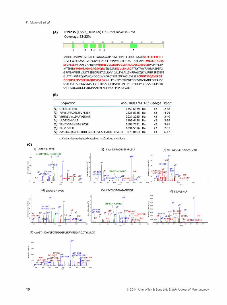
Fig 6. Identification of hEPOR by Nano-LC-ESI-MS/MS of GM1202 and GM1203 immunoprecipitates. (A) Illustration of identified hEPOR spe-
cific peptides in the amino acid sequence of EPOR Precursor P19235. Identified specific peptides are schematically illustrated as green boxes and
highlighted in red in the amino acid sequence. (B) The table represents the amino acid sequences of the identified peptides (1–7), their singly
protonated molecular ions [M+H+], charge states and cross-correlation Xcorr values. (C) Tandem mass spectroscopy ion trap collision-induced
dissociation (CID) spectra of peptides (1–7). Fragments used for search: b; b-H2O; b-NH3; y; y-H2O; y-NH3.
Novel Anti-Erythropoietin Receptor Antibodies
ª 2014 John Wiley & Sons Ltd, British Journal of Haematology 11

and/or potential EPOR isoforms by Western blot; and those
that would be useful for IHC studies of clinical specimens.
In the current study, peptide immunization was used
to generate antibodies for assays on denatured proteins,
such as Western blotting and IHC, whereas novel genetic
immunization protocols were used to generate antibodies that
recognize EPOR in its native conformation for flow cytometry,
immunofluorescence and IP assays. In total, 15 monoclonal
antibody mother clones, were pretested in different assays for
human EPOR. They comprised six rat monoclonal antibodies
generated by immunization of synthetic peptides based on the
cytoplasmic domain of the hEPOR, as well as four murine and
five rat monoclonal antibodies generated against the hEPOR
ECD domain by genetic immunization.
The specificity of the EPOR antibodies has been validated
by the use of EPOR-silenced cells. For example, we show that
an endogenous Western blot signal using the GM1201 anti-
body is specifically down-regulated in EPOR-silenced cells,
strongly indicating that this antibody recognizes endogenous
EPOR in these cells. Although the GM1201 antibody may
also detect non-specific signals in the A549 cell line (Fig 3)
the EPOR-silenced cells clearly permit discrimination
between a true EPOR-dependent signal and the non-specific
signal. The number of monoclonal antibodies was reduced to
a panel of four for Western blotting, IHC, IP, immunofluo-
rescence and flow cytometry.
For simplicity, the monoclonal antibodies have been sys-
tematically named, based on an in-house nomenclature system
at Aldevron Freiburg (see Table I). GM1201 is a rat monoclo-
nal antibody raised against one of six synthetic peptides used
to immunize rats, which is located in the cytoplasmic domain
of hEPOR (Table I). This antibody reveals specific down-regu-
lation of EPOR in EPOR-silenced MDA-MB-231 and A549
cells, confirming that it also recognizes endogenous EPOR in
these cells. Importantly, GM1201 was sensitive and specific in
immunohistochemical studies of both cultured erythroid cells
and bone marrow aspirates. For example using FFPE sections
of bone marrow and bone marrow aspirates, it was possible to
visualize erythroblastic islands in a patient with polycythaemia
vera, and to demonstrate co-localization of EPOR with either
ferritin H or glycophorin C in differentiating erythroid pro-
genitors in a patient with erythroid hyperplasia.
GM1202 and GM1203 are mouse monoclonal antibodies,
raised by genetic immunization against the ECD of hEPOR.
By co-localization of an ECFP-tagged EPOR and the signal
obtained with our new antibodies GM1202 and GM1203 we
show that GM1202 and GM1203 detect EPOR when it is
expressed on the membrane (Fig 4). Furthermore, GM1202
and GM1203 have proved useful in immunopreciptation
experiments in combination with GM1201 (Fig 7). GM1204
is a rat monoclonal antibody raised by genetic immunization
of the ECD of hEPOR which detects both human and mur-
ine EPOR by FACS (mouse data not shown).
In a thorough, often overlooked study, Arcasoy et al (2003)
isolated and characterized several novel cDNAs for EPOR
splice variants expressed in cancer cells. Predicted amino acid
sequences of these cDNAs indicated splice variants encoding
soluble EPOR, variants containing insertions from intron 6 or
intron 7, and membrane-bound EPOR peptides with intracy-
toplasmic truncations. These multiple EPOR isoforms in
human cancer cells may modulate the cellular effects of recom-
binant EPO. Recently, Elliott et al (2013) reported on differ-
ences in detection of EPOR in primary human tumour tissue
samples using different antibodies. Using an anti-hEPOR
monoclonal antibody, they could not detect EPOR protein in
normal human and matching cancer tissues from breast, lung,
colon, ovary or skin. Detection of EPOR in breast cancer tis-
sues using a polyclonal antibody was interpreted as cross-reac-
tivity. However, it cannot currently be ruled out that the
epitope recognized by the specific monoclonal antibody could
be missing in EPOR isoforms found in tumour tissues.
The availability of monoclonal antibodies directed against
specific exons, as those described herein, will enable, for the
first time, the investigation of the resulting EPOR protein
isoforms in different tissues. Thus, one antibody may recog-
nize an epitope common to many EPOR isoforms, indicating
Table III. Specificity of EPOR antibodies in FFPE cell line models.
Immunogen Antibody Isotype Epitope location UT-7
REH/NALM-6 model
shEPOR isogenic
models – reduced
expression in shEPOR
cells?Lung tissue –
effective?REH NALM-6 Difference? MDA-MB-231 A549
1 Peptide GM1201 r-IgG2b h-EPOR
cytoplasmic
domain
2+ 3+ 1+ Yes Yes Yes Yes
2 Genetic
Immunization
GM1202 m-IgG1a h-EPOR ECD 3+ 3+ 2+ Yes No No No
3 GM1203 m-IgG1a h-EPOR ECD 3+ 2+ 1+ Yes Yes No No
4 GM1204 r-IgG2a h-EPOR ECD – N/E N/E N/A N/E N/E N/E
FFPE, formalin-fixed paraffin wax-embedded; h-EPOR, human erythropoietin receptor; ECD, extracellular domain; N/E, not evaluated; N/A, not
applicable.
Shaded areas are meant to distinguish between peptide immunization and genetic immunization.
P. Maxwell et al
12 ª 2014 John Wiley & Sons Ltd, British Journal of Haematology

a broader EPOR expression pattern compared to other anti-
bodies whose epitope might only be present in fewer EPOR
isoforms, with a more limited expression pattern.
The new antibodies will enable many interesting topics in
EPOR biology to be explored. These include resolution of
the major clinical question of which patients can be treated
safely with EPO and its derivatives, dissection of the signal-
ling mechanisms in non-erythroid cells and investigation of
cancer cell:stromal cell interactions in tumours.
Acknowledgements
We gratefully acknowledge the Northern Ireland Molecular
Pathology Laboratory and the Northern Ireland Biobank for
assistance and in supplying human samples and the Department
of Haematology, Belfast City Hospital for supplying anonymized
bone marrow samples. This study was supported by the FP7-
Health European commission EpoCan grant (282551).
Authorship contributions
John Thompson and Fritz Grunert generated the antibodies and
performed initial screenings. Perry Maxwell, Florinda Melendez,
Kyle B. Matchett, Heidelinde Jaekel, Herbert Lindner, Nathalie
Ben-Califa, Andre Bernardini, Ulf Brockmeier, Markus
Thiersch, Edith Schneider-Gasser, Alexandra Irvine and Anne
Jordan planned and performed experiments, as well as analysed
data. Perry Maxwell, Kyle B. Matchett, Julian Aragones, Ludger
Hengst, Joachim Fandrey, Howard Oster, Moshe Mittelman,
Robert Cuthbert, Mohamed El-Tanani, Max Gassmann, David
Dangoor, Terence Lappin and Drorit Neumann planned
experiments, analysed data and wrote the manuscript.
Competing interests
The authors have no competing interests.
References
Abramsson, A., Lindblom, P. & Betsholtz, C.
(2003) Endothelial and nonendothelial sources
of PDGF-B regulate pericyte recruitment and
influence vascular pattern formation in tumors.
The Journal of Clinical Investigation, 112, 1142–
1151.
Anagnostou, A., Liu, Z., Steiner, M., Chin, K., Lee,
E.S., Kessimian, N. & Noguchi, C.T. (1994)
Erythropoietin receptor mRNA expression in
human endothelial cells. Proceedings of the
National Academy of Sciences of the United States
of America, 91, 3974–3978.
Arcasoy, M.O., Jiang, X. & Haroon, Z.A. (2003)
Expression of erythropoietin receptor splice
variants in human cancer. Biochemical and
Biophysical Research Communications, 307, 999–
1007.
Brown, W.M., Maxwell, P., Graham, A.N., Yakk-
undi, A., Dunlop, E.A., Shi, Z., Johnston, P.G.
& Lappin, T.R. (2007) Erythropoietin receptor
expression in non-small cell lung carcinoma: a
question of antibody specificity. Stem Cells, 25,
718–722.
Broxmeyer, H.E. (2013) Erythropoietin: multiple
targets, actions, and modifying influences for
biological and clinical consideration. Journal of
Experimental Medicine, 210, 205–208.
Caro, J.J., Salas, M., Ward, A. & Goss, G. (2001)
Anemia as an independent prognostic factor for
survival in patients with cancer: a systemic,
quantitative review. Cancer, 91, 2214–2221.
Elliott, S., Busse, L., Bass, M.B., Lu, H., Sarosi, I.,
Sinclair, A.M., Spahr, C., Um, M., Van, G. &
Begley, C.G. (2006) Anti-Epo receptor antibod-
ies do not predict Epo receptor expression.
Blood, 107, 1892–1895.
Elliott, S., Swift, S., Busse, L., Scully, S., Van, G.,
Rossi, J. & Johnson, C. (2013) Epo receptors are
not detectable in primary human tumor tissue
samples. PLoS One, 8, e68083.
Ghezzi, P., Bernaudin, M., Bianchi, R., Blomgren,
K., Brines, M., Campana, W., Cavaletti, G., Cer-
ami, A., Chopp, M., Coleman, T., Digicaylioglu,
M., Ehrenreich, H., Erbayraktar, S., Erbayraktar,
Z., Gassmann, M., Genc, S., Gokmen, N., Gras-
so, G., Juul, S., Lipton, S.A., Hand, C.C., Latini,
R., Lauria, G., Leist, M., Newton, S.S., Petit, E.,
Probert, L., Sfacteria, A., Siren, A.L., Talan, M.,
Thiemermann, C., Westenbrink, D., Yaqoob, M.
& Zhu, C. (2010) Erythropoietin: not just about
erythropoiesis. Lancet, 375, 2142.
Graham, F.L. & van der Eb, A.J. (1973) A new
technique for the assay of infectivity of human
adenovirus 5 DNA. Virology, 52, 456–467.
Gross, M., Ben-Califa, N., McMullin, M.F., Percy,
M.J., Bento, C., Cario, H., Minkov, M. & Neu-
mann, D. (2014) Polycythaemia-inducing muta-
tions in the erythropoietin receptor (EPOR):
mechanism and function as elucidated by
epidermal growth factor receptor-EPOR
chimeras. British Journal of Haematology, 165,
519–528.
Henke, M., Laszig, R., Rube, C., Schafer, U.,
Haase, K.D., Schilcher, B., Mose, S., Beer, K.T.,
Burger, U., Dougherty, C. & Frommhold, H.
(2003) Erythropoietin to treat head and neck
cancer patients with anaemia undergoing radio-
therapy: randomised, double-blind, placebo-con-
trolled trial. Lancet, 362, 1255–1260.
Hill, J.W., Cong, Z., Hess, G., McGarvey, N. &
Nordyke, R.J. (2012) Hemoglobin decline in
chemotherapy patients prior to and after policy
changes affecting use of erythropoiesis-stimulat-
ing agents: 2006–2009. Journal of International
Medical Research, 40, 1532–1545.
Jelkmann, W. (2010) Erythropoietin: back to
basics. Blood, 115, 4151–4152.
Katz, O., Gil, L., Lifshitz, L., Prutchi-Sagiv, S.,
Gassmann, M., Mittelman, M. & Neumann, D.
(2007) Erythropoietin enhances immune
responses in mice. European Journal of Immu-
nology, 37, 1584–1593.
Lappin, T.R., Maxwell, A.P. & Johnston, P.G.
(2002) EPO’s alter ego: erythropoietin has mul-
tiple actions. Stem Cells, 20, 485–492.
Leyland-Jones, B., Semiglazov, V., Pawlicki, M.,
Pienkowski, T., Tjulandin, S., Manikhas, G.,
Makhson, A., Roth, A., Dodwell, D., Baselga,
J., Biakhov, M., Valuckas, K., Voznyi, E., Liu,
X. & Vercammen, E. (2005) Maintaining nor-
mal hemoglobin levels with epoetin alfa in
mainly nonanemic patients with metastatic
breast cancer receiving first-line chemotherapy:
a survival study. Journal of Clinical Oncology,
23, 5960–5972.
Liang, K., Esteva, F.J., Albarracin, C., Stemke-
Hale, K., Lu, Y., Bianchini, G., Yang, C.Y., Li,
Y., Li, X., Chen, C.T., Mills, G.B., Hortobagyi,
G.N., Mendelsohn, J., Hung, M.C. & Fan, Z.
(2010) Recombinant human erythropoietin
antagonizes trastuzumab treatment of breast
cancer cells via Jak2-mediated Src activation
and PTEN inactivation. Cancer Cell, 18, 423–
435.
Lifshitz, L., Tabak, G., Gassmann, M., Mittelman,
M. & Neumann, D. (2010) Macrophages as
novel target cells for erythropoietin. Haemato-
logica, 95, 1823–1831.
Mausberg, A.K., Meyer, Zu., Horste, G., Dehmel,
T., Stettner, M., Lehmann, H.C., Sheikh, K.A. &
Kieseier, B.C. (2011) Erythropoietin ameliorates
rat experimental autoimmune neuritis by induc-
ing transforming growth factor-beta in macro-
phages. PLoS One, 6, e26280.
Miller, C.P., Lowe, K.A., Valliant-Saunders, K.,
Kaiser, J.F., Mattern, D., Urban, N., Henke, M.
& Blau, C.A. (2009) Evaluating erythropoietin-
associated tumor progression using archival tis-
sues from a phase III clinical trial. Stem Cells,
27, 2353–2361.
Mittelman, M., Zeidman, A., Kanter, P., Katz, O.,
Oster, H., Rund, D. & Neumann, D. (2004)
Erythropoietin has an anti-myeloma effect – a
hypothesis based on a clinical observation sup-
ª 2014 John Wiley & Sons Ltd, British Journal of Haematology 13
Novel Anti-Erythropoietin Receptor Antibodies

ported by animal studies. European Journal of
Haematology, 72, 155–165.
Nairz, M., Sonnweber, T., Schroll, A., Theurl, I. &
Weiss, G. (2012) The pleiotropic effects of
erythropoietin in infection and inflammation.
Microbes and Infection, 14, 238–246.
Oster, H.S., Prutchi-Sagiv, S., Halutz, O., Shabtai,
E., Hoffman, M., Neumann, D. & Mittelman,
M. (2013) Erythropoietin treatment is associated
with an augmented immune response to the
influenza vaccine in hematologic patients. Exper-
imental Hematology, 41, 167–171.
Provatopoulou, S.T. & Ziroyiannis, P.N. (2011)
Clinical use of erythropoietin in chronic kidney
disease: outcomes and future prospects. Hippo-
kratia, 15, 109–115.
Prutchi-Sagiv, S., Golishevsky, N., Oster, H.S.,
Katz, O., Cohen, A., Naparstek, E., Neumann,
D. & Mittelman, M. (2006) Erythropoietin treat-
ment in advanced multiple myeloma is associ-
ated with improved immunological functions:
could it be beneficial in early disease? British
Journal of Haematology, 135, 660–672.
Prutchi-Sagiv, S., Lifshitz, L., Orkin, R., Mittelman,
M. & Neumann, D. (2008) Erythropoietin
effects on dendritic cells: potential mediators in
its function as an immunomodulator? Experi-
mental Hematology, 36, 1682–1690.
Rizzo, J.D., Brouwers, M., Hurley, P., Seidenfeld, J.,
Arcasoy, M.O., Spivak, J.L., Bennett, C.L., Bohlius,
J., Evanchuk, D., Goode, M.J., Jakubowski, A.A.,
Regan, D.H. & Somerfield, M.R. (2010) American
Society of Hematology/American Society of
Clinical Oncology clinical practice guideline update
on the use of epoetin and darbepoetin in adult
patients with cancer. Blood, 116, 4045–4059.
Smith, K.J., Bleyer, A.J., Little, W.C. & Sane, D.C.
(2003) The cardiovascular effects of erythropoie-
tin. Cardiovascular Research, 59, 538–548.
Stasko, J., Drouet, L., Soria, C., Mazoyer, E., Caen,
J. & Kubisz, P. (2002) Erythropoietin and gran-
ulocyte colony-stimulating factor increase plas-
minogen activator inhibitor-1 release in HUVEC
culture. Thrombosis Research, 105, 161–164.
Trincavelli, M.L., Da Pozzo, E., Ciampi, O., Cuboni,
S., Daniele, S., Abbracchio, M.P. & Martini, C.
(2013) Regulation of erythropoietin receptor activ-
ity in endothelial cells by different erythropoietin
(EPO) derivatives: an in vitro study. International
Journal of Molecular Sciences, 14, 2258–2281.
Vogel, J. & Gassmann, M. (2011) Erythropoietic
and non-erythropoietic functions of erythropoi-
etin in mouse models. Journal of Physiology, 589,
1259–1264.
Wenger, R.H. & Kurtz, A. (2011) Erythropoietin.
Comprehensive Physiology, 1, 1759–1794.
Xue, Y., Lim, S., Yang, Y., Wang, Z., Jensen, L.D.,
Hedlund, E.M., Andersson, P., Sasahara, M.,
Larsson, O., Galter, D., Cao, R., Hosaka, K. &
Cao, Y. (2012) PDGF-BB modulates hematopoi-
esis and tumor angiogenesis by inducing eryth-
ropoietin production in stromal cells. Nature
Medicine, 18, 100–110.
14 ª 2014 John Wiley & Sons Ltd, British Journal of Haematology
P. Maxwell et al
Copyright © 2022 FDOKUMEN