Azure Integration Services – Connectivity Everywhere - Infosys
Upload
independentCategory
view
4download
0
Normalizing effect of heroin maintenancetreatment on stress-induced brain connectivity
Andre Schmidt,1,2,3 Marc Walter,1,3 Hana Gerber,1,3 Erich Seifritz,4 Rudolf Brenneisen,5
Gerhard A. Wiesbeck,1,3 Anita Riecher-Rossler,1,3 Undine E. Lang1,3 andStefan Borgwardt1,2,3,6
Recent evidence has shown that a single maintenance dose of heroin attenuates psychophysiological stress responses in heroin-
dependent patients, probably reflecting the effectiveness of heroin-assisted therapies for the treatment of severe heroin addiction.
However, the underlying neural circuitry of these effects has not yet been investigated. Using a cross-over, double-blind, vehicle-
controlled design, 22 heroin-dependent and heroin-maintained outpatients from the Centre of Substance Use Disorders at the
University Hospital of Psychiatry in Basel were studied after heroin and placebo administration, while 17 healthy controls from the
general population were included for placebo administration only. Functional magnetic resonance imaging was used to detect brain
responses to fearful faces and dynamic causal modelling was applied to compute fear-induced modulation of connectivity within
the emotional face network. Stress responses were assessed by hormone releases and subjective ratings. Relative to placebo, heroin
acutely reduced the fear-induced modulation of connectivity from the left fusiform gyrus to the left amygdala and from the right
amygdala to the right orbitofrontal cortex in dependent patients. Both of these amygdala-related connectivity strengths were
significantly increased in patients after placebo treatment (acute withdrawal) compared to healthy controls, whose connectivity
estimates did not differ from those of patients after heroin injection. Moreover, we found positive correlations between the left
fusiform gyrus to amygdala connectivity and different stress responses, as well as between the right amygdala to orbitofrontal
cortex connectivity and levels of craving. Our findings indicate that the increased amygdala-related connectivity during fearful face
processing after the placebo treatment in heroin-dependent patients transiently normalizes after acute heroin maintenance treat-
ment. Furthermore, this study suggests that the assessment of amygdala-related connectivity during fear processing may provide a
prognostic tool to assess stress levels in heroin-dependent patients and to quantify the efficacy of maintenance treatments in drug
addiction.
1 Department of Psychiatry (UPK), University of Basel, Wilhelm Klein-Strasse 27, 4012 Basel, Switzerland2 Medical Image Analysis Center, University Hospital Basel, Schanzenstrasse 55, 4031 Basel, Switzerland3 Department of Clinical Research (DFK), University of Basel, Basel, Switzerland4 Department of Psychiatry, Psychotherapy and Psychosomatics, Hospital of Psychiatry, University of Zurich, 8032 Zurich,
Switzerland5 Department of Clinical Research (DCR), University of Bern, 3010 Bern, Switzerland6 Department of Psychosis Studies, Institute of Psychiatry, King’s College London, De Crespigny Park 16, SE58AF London, UK
Correspondence to: Andre Schmidt, Ph.D.,
University of Basel, Department of Psychiatry (UPK)
Schanzenstrasse 55, 4031 Basel
Switzerland
E-mail: [email protected]
Keywords: heroin maintenance; acute heroin treatment; functional MRI; stress; amygdala connectivity
Abbreviations: DCM = dynamic causal modelling; OFC = orbitofrontal cortex
doi:10.1093/brain/awu326 BRAIN 2015: 138; 217–228 | 217
Received June 10, 2014. Revised August 7, 2014. Accepted September 21, 2014. Advance Access publication November 20, 2014
� The Author (2014). Published by Oxford University Press on behalf of the Guarantors of Brain.
This is an Open Access article distributed under the terms of the Creative Commons Attribution Non-Commercial License (http://creativecommons.org/licenses/by-nc/4.0/), which permits
non-commercial re-use, distribution, and reproduction in any medium, provided the original work is properly cited. For commercial re-use, please contact [email protected]
by guest on January 6, 2015D
ownloaded from

IntroductionDysfunction of stress pathways increases relapse vulnerabil-
ity in drug-addicted individuals (Sinha, 2008). Stress-
induced drug craving and hypothalamus-pituitary-adrenal
axis responses are even predictive of the outcome of co-
caine relapse (Sinha et al., 2006), thus suggesting potential
biological markers for relapse propensity. The aversive
state in drug addiction is partly mediated by activation of
corticotropin-releasing factor in prefrontal and limbic brain
areas, such as the orbitofrontal cortex (OFC) and the
amygdala (Koob and Kreek, 2007; Koob, 2013). The
OFC has direct projections to the amygdala (Ghashghaei
et al., 2007) and is functionally connected with the amyg-
dala during the integration of emotional and motivational
information (Schoenbaum and Roesch, 2005). For ex-
ample, human neuroimaging studies reveal that the OFC
and the amygdala are coactivated during withdrawal-
induced craving (Koob and Volkow, 2010), probably
through downstream modulation of the OFC on limbic re-
gions (Rolls, 2004). OFC–amygdala interactions may thus
reflect connections of vulnerability that are susceptible to
relapses after periods of abstinence.
A study in heavy alcohol drinkers showed that acute al-
cohol ingestion reduced functional connectivity between the
right amygdala and OFC during fearful face processing
(Gorka et al., 2013), an effect that was only evident for
the right amygdala–OFC connectivity, whereas the left cou-
pling seemed to be sensitive only for positive emotions.
Given that acute alcohol intoxication reduces subjective
and physiological responses to stress (Sayette et al., 1992;
Hefner and Curtin, 2012), the authors concluded that this
reduced right amygdala-OFC interplay may lead to reduced
negative affect after alcohol intoxication (Gorka et al.,
2013). In the same vein, depressed patients with bipolar
disorder showed significantly greater right prefrontal–
amygdala connectivity during fearful face processing than
healthy controls, which was reduced after antidepressant
treatment (Perlman et al., 2012). These findings are in
line with studies showing the impact of stress on the
right OFC (Hanson et al., 2010) and on right OFC–amyg-
dala connectivity (Fan et al., 2014).
The prescription of pharmaceutical heroin has repeatedly
been found to be an effective treatment for severe heroin
addiction (van den Brink et al., 2003; Haasen et al., 2007;
Oviedo-Joekes et al., 2009) and for heroin addicts who
have failed to benefit from methadone maintenance treat-
ment (Blanken et al., 2010), although methadone mainten-
ance failures may often be due to low dosing levels (Strain
et al., 1993; Farre et al., 2002). Compared with healthy
control subjects, chronic heroin users exhibit increased
functional connectivity between the right amygdala and
the OFC during resting state (Ma et al., 2010). Although
recent evidence has demonstrated that a daily maintenance
dose of heroin acutely reduced levels of stress hormones,
withdrawal, craving and anxiety (Walter et al., 2013), it
has not yet been explored whether this heroin-induced at-
tenuation of psychophysiological stress responses is also
mirrored in reduced OFC-amygdala connectivity during
fearful processing, analogously to the effect of acute alco-
hol ingestion.
The processing of facial affects provides a suitable frame-
work for the study of OFC-amygdala interactions. That is,
fearful face processing underlies integrated activity between
the inferior occipital gyrus that receive input from the
retina, the fusiform gyrus, which already processes facial
valences, the amygdala and the ventral prefrontal cortex,
subsuming the OFC, which evaluates emotional stimuli
(Haxby et al., 2002; Fusar-Poli et al., 2009b). Within this
visual-limbic-prefrontal network, the amygdala is thought
to play a key role in the rapid detection of facial affect and
in orchestrating network integrity during evaluation of af-
fective stimuli (Adolphs, 2008; Pessoa and Adolphs, 2010).
It has been suggested that prefrontal regions, such as the
OFC, regulate amygdala activity during evaluation of emo-
tional face stimuli (Nomura et al., 2004). However, visual
processing of fearful face emotions is also mediated
through a re-entry mechanism of connections from the
visual cortex to amygdala and connections from the amyg-
dala to the visual cortex (Vuilleumier et al., 2004;
Herrington et al., 2011). This visual-limbic coupling
during fearful face processing can be increased by psycho-
social stress (Li et al., 2014) and hyper-connectivity be-
tween the amygdala and visual cortex may underlie
increased negative emotion processing and anxiety (Frick
et al., 2013). We have previously reported significant
heroin effects on amygdala activity during fear processing
in heroin-dependent patients compared to a placebo treat-
ment (acute heroin withdrawal) (Schmidt et al., 2014),
which suggests abnormalities in the underlying connectiv-
ity, in particular to the visual cortex (i.e. fusiform gyrus).
In this study, the objective was to detect how heroin
acutely modulated right OFC–amygdala connectivity com-
pared to a placebo treatment in heroin-dependent patients
(experimentally-induced state of acute withdrawal; Schmidt
et al., 2014) and healthy controls. In particular, we used
functional MRI and dynamic causal modelling (DCM;
Friston et al., 2003) to assess modulation of OFC–amyg-
dala connectivity during fearful face processing. DCM
explicitly allowed us to compute the directionality of fear-
induced connectivity among the face network (Dima et al.,
2011; Diwadkar et al., 2012; Volman et al., 2013) and
how this is altered after pharmacological manipulations
(Grefkes et al., 2010; Schmidt et al., 2013). Besides explor-
ing acute heroin effects on amygdala–OFC connectivity, the
second aim of this study was to address whether the acute
heroin effect on local amygdala activity (Schmidt et al.,2014) was due to abnormal connectivity from the fusiform
gyrus. Finally, to validate the biological basis of our model,
we further tested whether the amygdala-related connectivity
strengths were related to previously reported psychophysio-
logical stress responses in these patients (Walter et al.,
2013). We hypothesized that heroin injection would
218 | BRAIN 2015: 138; 217–228 A. Schmidt et al.
by guest on January 6, 2015D
ownloaded from

reduce amygdala-related connectivity within the fear net-
work relative to the placebo treatment, in accompaniment
to its effect on psychophysiological stress responses (Walter
et al., 2013). Notably, fearful faces were not used to stress
participants, but rather to assess whether amygdala-related
connectivity strengths during fearful face processing differ-
entiated between the stress-related placebo treatment and
the heroin treatment.
Materials and methodsThe presented DCM analyses are based on the functional MRIdata previously published by Schmidt et al. (2014). Details ofimage acquisition and the mass-univariate statistical paramet-ric mapping analyses are described in the previous publicationand in the Supplementary material. Here, we provide a sum-mary of the participants’ characteristics, study design and de-tails of data analysis with DCM and Bayesian model selection.Furthermore, data on psychophysiological stress responses andplasma levels were taken from recently published studies(Walter et al., 2013, 2014). As these data had to be adaptedexplicitly to subjects who completed the face paradigm, a briefdescription of these methods is also provided.
Clinical trial registration information
Clinical trial information can be found at http://clinicaltrials.gov/show/NCT01174927. Name: Effects of Diacetylmorphine(DAM) on Brain Function and Stress Response. Number:NCT01174927.
Participants
The study population consists of 17 healthy control subjects[females/males: 4/13; mean age � standard deviation (SD):42.2 � 2.6 years] and 22 patients with opioid dependence ac-cording to ICD-10 criteria (females/males: 8/14; meanage � SD: 41.1 � 7.2 years). All participants were smokersand groups were matched for age [F(1,37) = 0.173;P = 0.680], gender (�2 = 0.742; P = 0.389) and cannabis con-sumption (four healthy control subjects and seven patients,�2 = 0.325; P = 0.567). Patients had a past history of intraven-ous heroin consumption, with current maintenance for at least6 months, and with an unchanged heroin dose during the pre-vious 3 months. In more detail, the patient age at first heroinuse was 19.09 � 3.27 years, with a current duration of de-pendence of 21 � 6.40 years and a daily maintenance doseof 309.55 � 121.48 mg (methadone-equivalent doses of77.39 � 30.37 mg). Patients were excluded from participationif they currently had additional physical diseases or a psychi-atric disorder including other comorbid conditions such assubstance dependencies. We did not exclude patients with apast history of psychiatric disorders or substance dependence.Clinically experienced psychiatrists (M.W.) conducted a struc-tured clinical interview for DSM-IV Axis II Disorders to assessthe diagnosis of comorbid personality disorders. Patients weretold to abstain from illicit drug consumption during the studyand to abstain from alcohol intake and smoking 72 and 2 hbefore scanning, respectively. Thus, patients with a positivealcohol breathalyzer test were excluded. Nevertheless, urine
samples of nine patients (no healthy controls) were tested posi-tive for cocaine at one or both points of the measurement(�2 = 9.041; P = 0.003). We have incorporated this potentialconfounder into our analyses. Healthy control subjects wererecruited from the general population by advertisement in thesame geographical area. Participants who consumed 420 galcohol per day, or who had any psychiatric, neurological,or severe medical illness history were excluded. After completedescription of the study to the subjects, written informed con-sent was obtained.
Study design
Placebo and heroin were administered through an indwellingintravenous catheter over a period of 30 s, using a cross-over,double-blind, vehicle-controlled design. Heroin hydrochloridewas dissolved on site in 5 ml sterile water and aspirated into asyringe, as previously described (Stohler et al., 1999). Eachpatient was scanned twice, with a short interval betweenscans (mean 9 � 3.8 days). Of the 22 patients included, 11received heroin at the first scan and saline at the second.Those subjects who received heroin before scanning were ad-ministered vehicle after scanning, whereas the subjects whoreceived saline before scanning were administered heroinafter scanning. Patients received two doses of heroin per day,one before/after scanning in the morning, and one in the even-ing. Thus, the last regular heroin injection was 12 h before theexperiment. The healthy controls participated only in the pla-cebo condition.
Fear processing paradigm
Study subjects participated in one 6-min experiment withevent-related functional MRI (Fusar-Poli et al., 2009a), inwhich they were presented with 10 different facial identities,each expressing 50% or 100% intensities of fear or a neutralexpression.(Young et al., 2002). There were thus 30 differentfacial stimuli in total; each face was presented twice for 2 s.The order of facial identities and expression type was pseudo-randomized, such that there was no successive presentation ofthe same facial expression type. The interstimulus interval wasvaried from 3 to 8 s according to a Poisson distribution, withan average interval of 5.9 s; the individuals then viewed a fix-ation cross. The face paradigm was conducted �30 min aftertreatments.
Assessment of psychophysiologicalstress responses and plasma levels
Craving (‘desire to use heroin’) was assessed 60 min after pla-cebo/heroin treatment using the 45-item Heroin CravingQuestionnaire (Tiffany, 1999), which measures positive andnegative aspects of craving on five theory-derived nine-itemscales. The German version of the State-Trait AnxietyInventory was used to quantify state-anxiety after both treat-ments in patients as well as in healthy controls (Spielbergeret al., 1970). Symptoms of withdrawal were reported on avisual analogue scale (values ranging from 0 to 10). Samplesof adrenocorticotropic hormone and cortisol were collectedthrough an intravenous catheter at baseline, 20 and 60 minafter substance administration. Salivary cortisol was analysed
Heroin maintenance treatment and amygdala connectivity BRAIN 2015: 138; 217–228 | 219
by guest on January 6, 2015D
ownloaded from

with a time-resolved immunoassay with fluorescence detection.Total cortisol concentrations were measured in serum with theImmulite 2000 Cortisol-Test (Siemens). The measurementrange of the test is at 1–50 mg/dl; the analytical sensitivity is0.20 mg/dl. Adrenocorticotropic hormone (ACTH) was mea-sured in EDTA plasma with the ACTH Immulite-Test(Siemens). The intra-assay precision was 56.1% for concen-trations 450 pg/ml; the interassay precision was 9.4% for con-centrations 451 pg/ml. The plasma concentrations of heroin(diacetylmorphine) and morphine were measured in venousammonium-heparinized plasma and assessed using high-performance liquid chromatography on a 125 � 2 mm i.d.Nucleosil 50 C-8 ec column with a particle size of 5mm anda 8 � 3 mm i.d. precolumn packed with Nucleosil 120 C-8 anda particle size of 3 mm followed by diode-array detection.Sample preparation and instrumental conditions were asdescribed previously (Bourquin et al., 1999).
Network connectivity analysis
Dynamic causal modelling
DCM (Friston et al., 2003) is a generic Bayesian system iden-tification technique used to compute effective connectivity be-tween brain regions for inferences related to ‘hidden’neurophysiological mechanisms. In DCM for functional MRI,the dynamics of the neural states underlying regional bloodoxygen level-dependant response are modelled by a bilineardifferential equation that describes how the neural states (x)change over time (t) as a function of endogenous interregionalconnections (matrix A), as well as modulatory effects on theseconnections (matrix B), and driving inputs (matrix C) (Fristonet al., 2003; Stephan et al., 2007). The endogenous connec-tions represent coupling strengths in the absence of input tothe system, whereas the modulatory effects represent task-spe-cific alterations in this connectivity. In this study, we focusedon the fear-induced modulation of visual-limbic-prefrontalconnections (modulatory effect), but also examined heroin ef-fects on endogenous connections and driving inputs.
f x;uð Þ ¼dx
xt¼ Aþ
Xmi¼1
uBðjÞ
!xþ Cu
Volumes of interest
We selected visual-limbic-prefrontal volumes of interest in ac-cordance with previous DCM studies of emotional face pro-cessing (Fairhall and Ishai, 2007; Dima et al., 2011;Herrington et al., 2011; Goulden et al., 2012; Sladky et al.,2013; Volman et al., 2013). In particular, these volumes ofinterest comprised the bilateral fusiform gyrus and amygdalaand the right OFC. The bilateral fusiform gyrus and amygdalawere selected based on studies indicating bilateral activity inthese regions during fearful versus neutral face processing(Fusar-Poli et al., 2009b). As the aim of this study was toexplore whether the acute heroin-induced reduction of stresshormone release, craving and anxiety, was reflected in alteredbrain connectivity during fearful face processing, we onlyincorporated the right OFC as prefrontal region into ourmodel, on the basis of following sources: (i) evidence showingthat the right OFC can be critically influenced by stress
(Hanson et al., 2010), as well as its connectivity to the rightamygdala (Fan et al., 2014); (ii) a previous study in heavyalcohol drinkers, which showed that acute alcohol ingestionreduced functional connectivity between the right amygdalaand right OFC during fearful face processing (Gorka et al.,2013), thus suggesting that this reduced right amygdala–OFCconnectivity may lead to reduced negative affect after alcoholintoxication (Sayette et al., 1992; Hefner and Curtin, 2012). Inthis study, we tested whether acute heroin treatment in de-pendent patients induced a similar effect; and (iii) previousstudies emphasizing the association of the right OFC duringstates of drug craving (Volkow et al., 1999). The coordinateswere based on the maxima across all subjects for the contrastof fearful minus neutral faces within the same anatomical areaas defined by the PickAtlas toolbox (Fig. 1A) (Maldjian et al.,2003). For each subject, time series from these regions of inter-est were extracted within spheres of 10-mm radii centred onthe peak for the effects of interest F contrast (P5 0.05, ad-justed) as previously performed (Sladky et al., 2013).
Defining model architecture
Starting from this initial model, we created three different vari-ations per model, in which fearful faces (modulatory effect)were allowed to modulate the endogenous connections(Fig. 1B). All presented stimuli (neutral and fearful faces)were entered into the bilateral fusiform gyrus, the visualinput region of our model. These three variations, namelymodulation of (i) forward connection; (ii) backward connec-tion; or (iii) both were guided by studies emphasizing themodulation of bottom-up or top-down connectivity inducedby facial affect among the fusiform gyrus, amygdala andOFC (Nomura et al., 2004; Vuilleumier et al., 2004; Fairhalland Ishai, 2007; Dima et al., 2011; Herrington et al., 2011).Although previous studies indicated interhemispheric func-tional connectivity between the fusiform gyrus and the amyg-dala (Irwin et al., 2004; Davies-Thompson and Andrews,2012), this interhemispheric visual and limbic coupling hasbeen found during resting state and during the presentationof faces versus objects or places. In other words, while themodel captured inter-hemispheric visual and limbic connectiv-ity induced by the driving input of all stimuli, fearful faceswere not allowed to modulate these interhemispheric endogen-ous connections. A graphical overview of our model architec-ture is depicted in Fig. 1.
Bayesian model selection and averaging
Bayesian model selection is an essential procedure of DCMstudies, as it can be used to test competing hypotheses forthe neural mechanisms generating the data. Bayesian modelselection rests on comparing the evidence of a predefined setof models (the model architecture). A random-effects Bayesianmodel selection approach has been suggested for group studies,as this is capable of quantifying the degree of heterogeneity ina population, while being extremely robust to potential out-liers (Stephan et al., 2009). The probability that one model ismore likely than any other model can be expressed by theexceedance probability of each model. After inferring themost likely network architecture underlying a specific neuralprocess, one can compare the parameter estimates obtainedfrom Bayesian model selection for between-group inferences.
Statistical comparison of model parameter estimates acrossgroups is only valid if those estimates stem from the
220 | BRAIN 2015: 138; 217–228 A. Schmidt et al.
by guest on January 6, 2015D
ownloaded from

same model. If different models or families are found to beoptimal across groups, Bayesian model averaging has beenrecommended as the standard approach for clinical DCM stu-dies (Seghier et al., 2010; Stephan et al., 2010). Bayesianmodel averaging averages posterior parameter estimates overmodels, weighted by the posterior model probabilities(Penny et al., 2010). Thus, models with a low posterior prob-ability contribute little to the estimation of the marginalposterior.
Group statistics of dynamic causal modelling
parameters
After Bayesian model averaging, we used the resulting poster-ior means from the averaged DCM for examining differencesamong groups. Statistical analysis of group differences in con-nection strengths concerned the posterior means of couplingestimates, when using Bayesian model averaging for all threemodels. In other words, we compared the parameter estimatesfrom participant-specific DCMs that were averaged over thethree models (by using Bayesian model averaging) separatelywithin each group and treatment. Thus, we were able to testfor differences in 12 parameters describing the endogenousconnections (matrix A), study how eight of these parameterswere modulated by fearful faces (modulatory effect, matrix B),and use two parameters to describe the driving input bilateralinto the fusiform gyrus. Paired t-tests were used to comparethe heroin and placebo treatment in patients, whereas twosample t-tests were used to compare patients with healthy
control subjects. The statistical threshold was adjusted formultiple comparisons using the Bonferroni correction.Thus, the threshold was adjusted by dividing the P-value bythe number of connections tested (12, eight and two,respectively).
Results
Network connectivity analysis
Optimal model search
Model 2, incorporating the modulation of backward
connections, was clearly the best fitting model in healthy
controls (exceedance probability: 70%). Although Model 2
was also superior to other models after the heroin
treatment (exceedance probability: 36%), it was only
marginally superior to Model 1 (exceedance probability:
34%) and Model 3 (exceedance probability: 30%). In the
placebo condition, there was a preference for Model 1
(modulation of forward connections) (exceedance
probability: 40%), followed by Model 3 (exceedance
probability: 30%) and Model 2 (exceedance probability:
29%). Bayesian model selection results are depicted in
Fig. 2.
Figure 1 Model architecture. (A) Task-related brain activation within the anatomical mask during fearful versus neutral face processing in the
right (x = 32, y = �70, z = �10; cluster size 895) and left fusiform gyrus (x = �38, y = �62, z = �22; cluster size 1081), right (x = 26, y = �4,
z = �14; cluster size 271) and left amygdala (x = �20, y = �2, z = �22; cluster size 229) and right OFC (x = 34, y = 24, z = �8; cluster size
736). Activations are corrected for FWE cluster-level at P5 0.05 with a voxel-level threshold of P5 0.001. (B) Specification of model archi-
tecture. The sources comprising the models were the bilateral fusiform gyrus (FG), bilateral amygdala (AMG) and right OFC. From this basic
layout, three models were created by considering where the modulatory effect of fearful faces might be expressed, namely on forward (M1),
backward (M2) or bilateral connections (M3) among the visual-limbic-prefrontal network.
Heroin maintenance treatment and amygdala connectivity BRAIN 2015: 138; 217–228 | 221
by guest on January 6, 2015D
ownloaded from

Heroin effects on driving input andendogenous connections
We found no heroin effect on driving inputs relative to the
placebo treatment in patients, neither for the input into
the left [t(21) = 0.207, P = 0.838] nor for the input into
the right fusiform gyrus [t(21) = �0.608, P = 0.550].
Relative to healthy controls, no significant effects on the
driving inputs were found after heroin [left fusiform
gyrus: F(1,37) = 0.744, P = 0.872; right fusiform gyrus:
F(1,37) = 0.003, P = 0.255] and placebo treatment in pa-
tients [left fusiform gyrus: F(1,37) = 0.534, P = 0.973;
right fusiform gyrus: F(1,37) = 0.815, P = 0.558].
No significant differences were found between the heroin
and placebo treatment in patients regarding endogenous
connections, or between the heroin treatment in patients
and healthy controls. Compared with healthy controls,
patients after placebo treatment manifested increased con-
nectivity from the left fusiform gyrus to the left amygdala
[F(1,37) = 6.183, P = 0.029, uncorrected for multiple com-
parisons]. Posterior parameters after Bayesian model aver-
aging for driving inputs and endogenous connections are
depicted in Table 1.
Heroin effect on the fear-inducedmodulation of connectivity
Relative to placebo in patients, heroin significantly reduced
the fear-induced modulation of connectivity from the left
fusiform gyrus to the left amygdala [t(21) = 2.95,
P = 0.006, corrected for multiple tests] and from the right
amygdala to the right OFC [t(21) = 2.38, P = 0.027, uncor-
rected] (Fig. 3). Importantly, this heroin effect was found to
be independent of whether patients consumed cocaine
(n = 9) [fusiform gyrus!amydgala: F(1,8) = 4.46,
P = 0.002, corrected; amygdala!OFC: F(1,8) = 2.44,
P = 0.041, uncorrected] or not (n = 13) [fusiform gyr-
us!amydgala: F(1,12) = 3.02, P = 0.011, uncorrected;
amygdala!OFC: F(1,12) = 2.19, P = 0.049, uncorrected].
Compared with healthy controls, patients under placebo
exhibited a significantly higher fear-induced modulation of
connectivity from left fusiform gyrus to the amygdala
[F(1,37) = 19.16, P = 0.002, corrected] and from the right
amygdala to the right OFC ([F(1,37) = 3.89, P = 0.045, un-
corrected] (Fig. 3). It is noteworthy that these increased
connectivity estimates were evident in patients without [fu-
siform gyrus!amydgala: F(1,28) = 14,69, P = 0.001, cor-
rected; amygdala!OFC: F(1,28) = 3.18, P = 0.046,
uncorrected] or with cocaine consumption [fusiform gyr-
us!amydgala: F(1,24) = 10,03, P = 0.001, corrected;
amygdala!OFC: F(1,24) = 0.73, P = 0.048]. No difference
was found between healthy controls and patients under
heroin, regardless of whether cocaine was consumed or
not. Bayesian model averaging results for each treatment
separately are summarized in Table 2.
Plasma levels
Heroin (diacetylmorphine) plasma concentrations decreased
from 898 ng/ml (SD: 807) to 170 (SD: 260) and 8 (SD: 33)
ng/ml at 3, 10 and 60 min, respectively. 6-Acetylmorphine
exhibited a similar time-concentration profile to that of
Figure 2 Bayesian model selection results. Bayesian model selection results among all three dynamic causal models. The models differed in
whether they included fear-induced modulation of forward (Model 1), backward (Model 2) or both forward and backward connections (Model 3)
among the visual-limbic-prefrontal network. Results for each treatment are expressed in terms of exceedance probability, the relative probability
that this model is more likely than any other of the models tested, given the treatment data.
222 | BRAIN 2015: 138; 217–228 A. Schmidt et al.
by guest on January 6, 2015D
ownloaded from

Table 1 DCM driving inputs and endogenous connections
Healthy controls Placebo treatment Heroin treatment
Driving inputs
Left FG �0.0187 (0.0224)* �0.0185 (0.0243)* �0.0206 (0.0338)*
Right FG �0.0264 (0.0272)* �0.0120 (0.0248)* �0.016732 (0.0240)*
Endogenous connections
Left FG!right FG �0.0068 (0.0830) 0.0474 (0.0929)* 0.0353 (0.1206)
Left FG!left AMG 0.1500 (0.0626)* 0.0626 (0.1452) 0.1535 (0.1071)*
Right FG!left FG 0.0053 (0.1322) 0.0643 (0.1233)* 0.0564 (0.0996)
Right FG!right AMG 0.0084 (0.1876) 0.0278 (0.1405) 0.1105 (0.1242)*
Right FG!OFC 0.0679 (0.1572) 0.0065 (0.1728) 0.0943 (0.1008)*
Left AMG!left FG �0.0024 (0.0345) �0.0034 (0.0416) 0.0028 (0.0478)
Left AMG!Right AMY 0.0061 (0.0316) 0.0123 (0.0198)* 0.0209 (0.0308)*
Right AMG!Right FG �0.0005 (0.0395) 0.0074 (0.0422) �0.0017 (0.0378)
Right AMG!left AMG 0.0141 (0.0212)* 0.0134 (0.0310) 0.0313 (0.0332)*
Right AMG!OFC 0.0120 (0.0120)* 0.0174 (0.0364)* 0.0170 (0.0280)*
OFC!right FG �0.0135 (0.0235)* 0.0071 (0.0397) 0.0005 (0.0269)
OFC!right AMG 0.0137 (0.0420) 0.0214 (0.0370)* 0.0155 (0.0237)*
Values are mean (SD). Values were obtained after Bayesian model averaging across all three dynamic causal models. FG = fusiform gyrus; AMG = amygdala.
*Significant t-tests within each group compared with zero (P’s5 0.05).
Figure 3 Differences in fear-induced modulation of effective connectivity among groups and treatments. Fear-induced modula-
tion of the left fusiform gyrus!amygdala and right amygdala!OFC connectivity for each treatment condition. Significant differences between
healthy controls and patients after placebo and heroin administration are indicated at *P5 0.05 uncorrected and at **P5 0.00625 corrected for
multiple comparisons. Error bars indicate standard errors. FG = fusiform gyrus; AMG = amygdala.
Table 2 DCM parameter estimates for the modulation of connectivity induced by fearful faces
Modulatory parameters Healthy controls Patients under placebo Patients under heroin
Left FG!Left AMG 0.0084 (0.0129)* 0.0471 (0.0502)* 0.0055 (0.0330)
Right FG!Right AMG �0.0063 (0.0229) 0.0055 (0.0595) 0.0145 (0.0359)
Right FG!Right OFC �0.0099 (0.0332) 0.0245 (0.0813) �0.0063 (0.0512)
Left AMG!Left FG �0.0052 (0.0246) �0.0013 (0.0114) 0.0020 (0.0119)
Right AMG!Right FG �0.0002 (0.0119) 0.0009 (0.0120) �0.0017 (0.0095)
Right AMG!Right OFC 0.0001 (0.0081) 0.0072 (0.0114)* �0.0011 (0.0104)
Right OFC!Right FG �0.0031 (0.0066) 0.0014 (0.0117) �0.0006 (0.0059)
Right OFC!Right AMG 0.0037 (0.0104) 0.0009 (0.0095) 0.0015 (0.0097)
Values for the modulatory effect of fearful faces were obtained after Bayesian model averaging across all three dynamic causal models. FG = fusiform gyrus;
AMG = amygdala; *Significant t-tests within each group compared with zero (P’s5 0.05).
Heroin maintenance treatment and amygdala connectivity BRAIN 2015: 138; 217–228 | 223
by guest on January 6, 2015D
ownloaded from

diacetylmorphine, decreasing from 910 ng/ml (SD: 549) to
317 ng/ml (SD: 167) and 60 ng/ml (SD: 33) at 3, 10 and
60 min, respectively. In contrast, the mean concentrations
of morphine at 3, 10 and 60 min after heroin injection were
349 ng/ml (SD: 140), 295 ng/ml (SD: 122), and 217 ng/ml
(SD: 92), respectively. Morphine-6-b-D-glucuronide plasma
concentrations steadily increased over the study period,
from 140 ng/ml (SD: 25) to 235 ng/ml (SD: 28) and
509 ng/ml (SD: 44) at 3, 10 and 60 min, respectively.
Plasma levels are depicted in Fig. 4.
Psychophysiological stress responses
The results of stress hormone release have already been
published (Schmidt et al., 2014), but we report them here
for the sake of completeness. Significantly lower levels of
craving [t(21) = 8.32, P = 0.0001], withdrawal symptoms
[t(21) = 6.64, P = 0.0001], state-anxiety [t(21) = 6.17,
P = 0.001], as well as concentrations of serum cortisol
[t(21) = 4.95, P = 0.0001], saliva cortisol [t(21) = 4.47,
P = 0.0002] and adrenocorticotropic hormone
[t(21) = 5.17, P = 0.001] were observed after heroin relative
to the placebo treatment in patients. Healthy controls
showed significantly lower anxiety scores than patients
under placebo [F(1,37) = 5.38, P = 0.0001], whereas no dif-
ference was found in patients after heroin administration
[F(1,37) = 3.55, P = 0.072]. Compared with healthy con-
trols, patients exhibited significantly higher levels of adre-
nocorticotropic hormone [F(1,37) = 16.80, P = 0.001],
serum cortisol [F(1,37) = 1.02, P = 0.0027], and salivary
cortisol [F(1,37) = 4.52, P = 0.001] after placebo treatment,
whereas patients under heroin showed significantly lower
levels of adrenocorticotropic hormone [F(1,37) = 0.004,
P = 0.0001] but not of serum cortisol [F(1,37) = 5.41,
P = 0.694] or salivary cortisol [F(1,37) = 2.65, P = 0.831].
These parameters were not related to the results for plasma
levels.
Relation between amygdala connec-tivity and stress responses
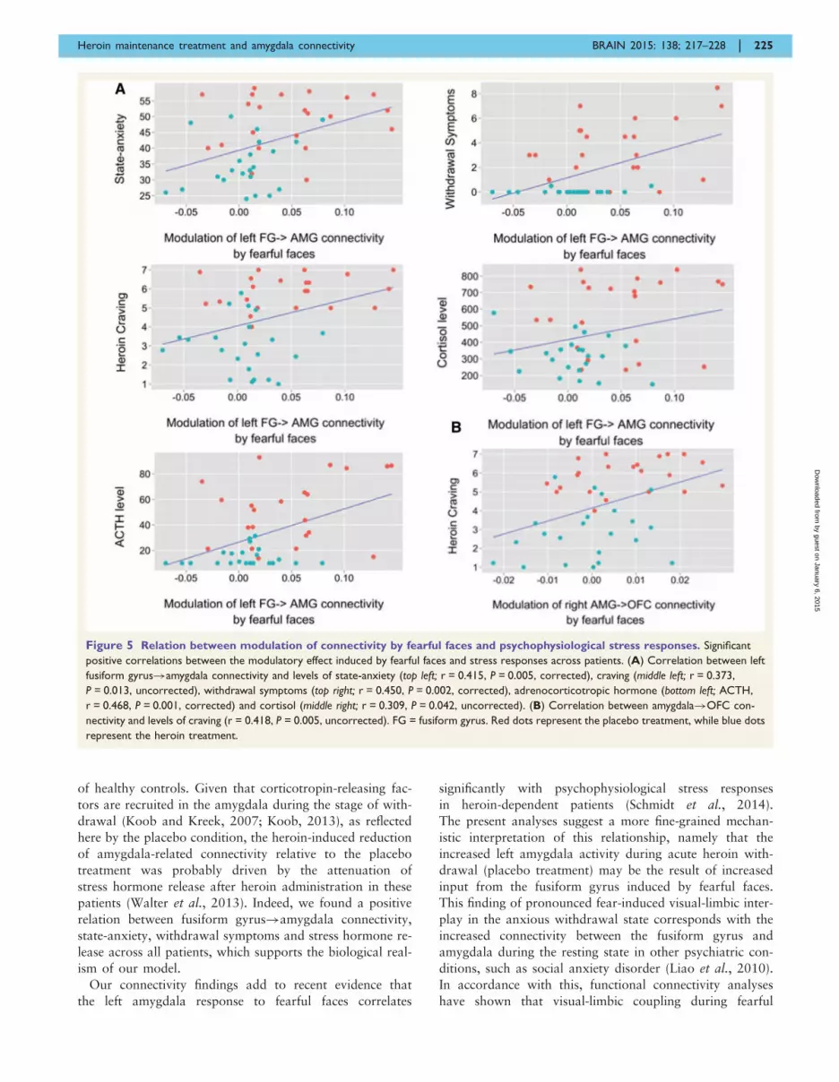
Across patients, we found significant positive correlations
between the left fusiform gyrus!amygdala connectivity
and levels of state-anxiety (r = 0.415, P = 0.005, corrected),
craving (r = 0.373, P = 0.013, uncorrected), withdrawal
symptoms (r = 0.450, P = 0.002, corrected), serum cortisol
(r = 0.309, P = 0.042, uncorrected) and adrenocorticotropic
hormone (r = 0.468, P = 0.001, corrected) (Fig. 5A). The
right amygdala!OFC connectivity also correlated posi-
tively with levels of craving (r = 0.418, P = 0.005, uncor-
rected) (Fig. 5B).
No relationships were found between connectivity esti-
mates and plasma levels.
DiscussionThis paper presents a model-based investigation of amyg-
dala-related connectivity that underlies the emotional regu-
lation effect of heroin substitution in dependent patients.
Our findings showed that stress-related placebo treatment
in patients was accompanied by significantly increased fear-
induced modulation of left fusiform gyrus!amygdala and
right amygdala!OFC connectivity. Critically, heroin ad-
ministration reduced both of these amygdala-related con-
nectivity strengths to a level that did not differ from those
Figure 4 Plasma concentrations after acute heroin treatment. Plasma concentrations (mean � SE) of heroin (diacetylmorphine, DAM),
morphine, 6-acetylmorphine (6AM) and morphine-6-b-D-glucuronide (M6G) 3, 10 and 60 min after heroin administration.
224 | BRAIN 2015: 138; 217–228 A. Schmidt et al.
by guest on January 6, 2015D
ownloaded from
of healthy controls. Given that corticotropin-releasing fac-
tors are recruited in the amygdala during the stage of with-
drawal (Koob and Kreek, 2007; Koob, 2013), as reflected
here by the placebo condition, the heroin-induced reduction
of amygdala-related connectivity relative to the placebo
treatment was probably driven by the attenuation of
stress hormone release after heroin administration in these
patients (Walter et al., 2013). Indeed, we found a positive
relation between fusiform gyrus!amygdala connectivity,
state-anxiety, withdrawal symptoms and stress hormone re-
lease across all patients, which supports the biological real-
ism of our model.
Our connectivity findings add to recent evidence that
the left amygdala response to fearful faces correlates
significantly with psychophysiological stress responses
in heroin-dependent patients (Schmidt et al., 2014).
The present analyses suggest a more fine-grained mechan-
istic interpretation of this relationship, namely that the
increased left amygdala activity during acute heroin with-
drawal (placebo treatment) may be the result of increased
input from the fusiform gyrus induced by fearful faces.
This finding of pronounced fear-induced visual-limbic inter-
play in the anxious withdrawal state corresponds with the
increased connectivity between the fusiform gyrus and
amygdala during the resting state in other psychiatric con-
ditions, such as social anxiety disorder (Liao et al., 2010).
In accordance with this, functional connectivity analyses
have shown that visual-limbic coupling during fearful
Figure 5 Relation between modulation of connectivity by fearful faces and psychophysiological stress responses. Significant
positive correlations between the modulatory effect induced by fearful faces and stress responses across patients. (A) Correlation between left
fusiform gyrus!amygdala connectivity and levels of state-anxiety (top left; r = 0.415, P = 0.005, corrected), craving (middle left; r = 0.373,
P = 0.013, uncorrected), withdrawal symptoms (top right; r = 0.450, P = 0.002, corrected), adrenocorticotropic hormone (bottom left; ACTH,
r = 0.468, P = 0.001, corrected) and cortisol (middle right; r = 0.309, P = 0.042, uncorrected). (B) Correlation between amygdala!OFC con-
nectivity and levels of craving (r = 0.418, P = 0.005, uncorrected). FG = fusiform gyrus. Red dots represent the placebo treatment, while blue dots
represent the heroin treatment.
Heroin maintenance treatment and amygdala connectivity BRAIN 2015: 138; 217–228 | 225
by guest on January 6, 2015D
ownloaded from

face processing can be increased by psychosocial stress
(Li et al., 2014). Furthermore, a recent study showed that
non-relapse participants exhibited reduced resting state
functional connectivity between the amygdala and visual
processing regions compared to controls and relapsed
cocaine-addicted participants, which suggests that this
may be a marker of early relapse risk (McHugh et al.,
2014). Our results thus suggest that the stress-related
state during acute heroin withdrawal is accompanied by
increased fear-induced modulation of visual-limbic connect-
ivity. Crucially, heroin injection normalized this connectiv-
ity, as no difference from healthy controls was observed,
which may point to a negative reinforcement mechanism at
the neural system level.
Furthermore, relative to the placebo treatment in pa-
tients, heroin injection also reduced the right amygdala–
OFC connectivity, giving a level comparable to that
observed in healthy controls. It is interesting that the
degree of amygdala!OFC connectivity was positively
related to patients’ craving behaviour. This result is consist-
ent with evidence demonstrating functional connectivity be-
tween the OFC and amygdala during withdrawal-induced
craving (Koob and Volkow, 2010). The contribution of the
OFC to heroin craving has further been demonstrated in a
previous PET study (Sell et al., 2000). The OFC seems to
be important for stress-induced reinstatement of drug-seek-
ing, i.e. it drives the motivation for new drug intake
(Schoenbaum and Shaham, 2008), during which it is recip-
rocally connected with the amygdala (Lasseter et al., 2009).
Therefore, the finding of increased amygdala!OFC con-
nectivity during acute heroin withdrawal may indicate
that there is both stress-induced increase in heroin craving
and resulting reinforced motivation for renewed heroin
intake. This stress-induced compulsion to consume the
drug was normalized by acute heroin administration, as
indicated by reduced amygdala–OFC connectivity. In con-
trast to this acute heroin-induced normalization of OFC–
amygdala connectivity in heroin-dependent patients, a
previous investigation in methadone-maintained patients
showed that the OFC still remained high in response to
heroin-related cues after the daily methadone dose
(Langleben et al., 2008), which suggested that methadone
maintenance patients remain at risk for relapse into illicit
heroin use. Thus, in accordance with data supporting the
superiority of heroin compared to methadone maintenance
in severely opioid-dependent subjects, especially if treat-
ment is maintained for 12 months or longer (van den
Brink et al., 2003; Oviedo-Joekes et al., 2009), heroin-
induced normalization of OFC connectivity may reflect a
benefit compared to methadone maintenance treatment in
preventing relapses to self-administrations in prone pa-
tients. However, this is clearly a speculative observation
at the present time and it is important to emphasize that
we do not wish to make any claims here for an advantage
of heroin maintenance therapy over methadone mainten-
ance therapy. The results from the two studies are not dir-
ectly comparable, owing to a number of major
methodological differences, including different measure-
ment protocols and fundamentally different paradigms.
For instance, Langleben et al. (2008) measured functional
MRI responses to heroin-related cues pre- and post-treat-
ment, whereas we used a fearful face task to assess stress
responses after heroin and placebo administration.
There are important limitations to our studies. Effective
connectivity was computed using a fairly simplistic neur-
onal face network, without considering, for example, the
precuneus or a more detailed partitioning of visual face-
sensitive processing areas, as done in previous DCM studies
(Fairhall and Ishai, 2007; Dima et al., 2011). However,
given the impact of stress, craving and anxiety on visual-
limbic and prefrontal-limbic connections (Koob and
Volkow, 2010; Frick et al., 2013; Fan et al., 2014; Li
et al., 2014) and that heroin acutely reduced levels of
stress hormones, withdrawal, craving and anxiety (Walter
et al., 2013), we were explicitly interested in how heroin
acutely modulated fear-induced modulation of fusiform
gyrus-amygdala and amygdala–OFC connectivity.
Nevertheless, including more visual regions implicated in
face processing—such as the inferior occipital gyrus—may
influence how information enters into the fusiform gyrus
and propagates further across the network. In this respect,
there is evidence that fusiform gyrus activity decreases with
increasing repetitions of faces (Reber et al., 2005), which is
perhaps reflected by the negative driving inputs into the
fusiform gyrus in this study. Repeated exposure to identical
emotional faces also modulates fusiform gyrus-amygdala
connectivity (Herrington et al., 2011). It is therefore im-
portant in further studies to address how such priming ef-
fects evolve across different visual processing areas and
propagate further to the limbic and prefrontal system.
Furthermore, because of the cross-sectional design of this
study, we were not able to infer how this acute heroin
effect on amygdala-related connectivity developed over
the duration of the maintenance therapy and how long it
lasted. This should be addressed in longitudinal studies and
compared with long-acting steady-state managements, such
as methadone maintenance therapies using adequate metha-
done dosing levels, given that high doses of methadone
were more effective than low doses in the reduction of illicit
opioid use (Strain et al., 1999; Farre et al., 2002). A dis-
tinction must be drawn between the placebo-induced state
of withdrawal in the current study and the withdrawal state
in active drug users. Although the experimentally induced
increase in withdrawal signs after placebo treatment
allowed us to study negative reinforcement mechanisms,
this approach also impedes clinical inferences. Therefore,
further analyses of this sort are needed in various levels
of withdrawal, as well as before and after substitution
treatments. Expectation effects might have influenced our
findings, given that healthy controls were aware that they
were going to receive a placebo treatment only. Finally, we
cannot exclude the possibility that some of the patients had
a history of psychiatric disorder, including other comorbid
conditions, which is a potential confounding factor.
226 | BRAIN 2015: 138; 217–228 A. Schmidt et al.
by guest on January 6, 2015D
ownloaded from

In conclusion, our findings extend previous evidence for
hyperactive amygdala-related network connectivity in ab-
stinent heroin addicts (Xie et al., 2011) and suggest that
the stressful state during acute heroin withdrawal is asso-
ciated with increased amygdala-related connectivity during
fearful face processing. The results further indicate that a
single dose of heroin to heroin-maintained patients transi-
ently normalizes amygdala-related brain connectivity
during fearful face processing. More research with follow-
up measurements is required to quantify whether the re-
cording of amygdala-related connectivity during fearful
face processing may provide a prognostic tool to assess
stress levels in heroin-dependent patients before and after
maintenance treatments.
AcknowledgementsWe would like to acknowledge the infrastructural support
of the Medical Image Analysis Centre, University Hospital
Basel. We also thank the staff of the heroin-assisted treat-
ment in Basel (Janus) who helped us to conduct this study.
FundingThis study was supported by the Swiss National Science
Foundation (SNSF) (32003B-127544) (M.W., G. A. W.,
A. R-R., S.B.) and Freiwillige Akademische Gesellschaft
Basel (A.S.). All authors declare that they have no conflicts
of interest.
Supplementary materialSupplementary material is available at Brain online.
ReferencesAdolphs R. Fear, faces, and the human amygdala. Curr Opin
Neurobiol 2008; 18: 166–72.Blanken P, Hendriks VM, van Ree JM, van den Brink W. Outcome of
long-term heroin-assisted treatment offered to chronic, treatment-re-
sistant heroin addicts in the Netherlands. Addiction 2010; 105:
300–8.
Bourquin D, Bundeli P, Lehmann T, Brenneisen R. Diacetylmorphine
and its metabolites in plasma by HPLC with diode-array and atmos-
pheric pressure ionization mass spectrometric detection. J Liq
Chrom Rel Technol 1999; 22: 2663–74.
Davies-Thompson J, Andrews TJ. Intra- and interhemispheric connect-
ivity between face-selective regions in the human brain.
J Neurophysiol 2012; 108: 3087–95.Dima D, Stephan KE, Roiser JP, Friston KJ, Frangou S. Effective con-
nectivity during processing of facial affect: evidence for multiple
parallel pathways. J Neurosci 2011; 31: 14378–85.
Diwadkar VA, Wadehra S, Pruitt P, Keshavan MS, Rajan U, Zajac-
Benitez C, et al. Disordered corticolimbic interactions during affect-
ive processing in children and adolescents at risk for schizophrenia
revealed by functional magnetic resonance imaging and dynamic
causal modeling. Arch Gen Psychiatry 2012; 69: 231–42.
Fairhall SL, Ishai A. Effective connectivity within the distributed cor-
tical network for face perception. Cereb Cortex 2007; 17: 2400–6.
Fan Y, Herrera-Melendez AL, Pestke K, Feeser M, Aust S, Otte C,
et al. Early life stress modulates amygdala-prefrontal functional con-
nectivity: implications for oxytocin effects. Hum Brain Mapp 2014;
35: 5328–39.
Farre M, Mas A, Torrens M, Moreno V, Camı J. Retention rate and
illicit opioid use during methadone maintenance interventions: a
meta-analysis. Drug Alcohol Depend 2002; 65: 283–90.Frick A, Howner K, Fischer H, Kristiansson M, Furmark T. Altered
fusiform connectivity during processing of fearful faces in social
anxiety disorder. Transl Psychiatry 2013; 3: e312.Friston KJ, Harrison L, Penny W. Dynamic causal modelling.
Neuroimage 2003; 19: 1273–302.Fusar-Poli P, Crippa JA, Bhattacharyya S, Borgwardt SJ, Allen P,
Martin-Santos R, et al. Distinct effects of {delta}9-tetrahydrocanna-
binol and cannabidiol on neural activation during emotional pro-
cessing. Arch Gen Psychiatry 2009a; 66: 95–105.
Fusar-Poli P, Placentino A, Carletti F, Landi P, Allen P, Surguladze S,
et al. Functional atlas of emotional faces processing: a voxel-based
meta-analysis of 105 functional magnetic resonance imaging studies.
J Psychiatry Neurosci 2009b; 34: 418–32.
Ghashghaei HT, Hilgetag CC, Barbas H. Sequence of information
processing for emotions based on the anatomic dialogue be-
tween prefrontal cortex and amygdala. Neuroimage 2007; 34:
905–23.
Gorka SM, Fitzgerald DA, King AC, Phan KL. Alcohol attenuates
amygdala-frontal connectivity during processing social signals in
heavy social drinkers: a preliminary pharmaco-fMRI study.
Psychopharmacology (Berl) 2013; 229: 141–54.
Goulden N, McKie S, Thomas EJ, Downey D, Juhasz G, Williams SR,
et al. Reversed frontotemporal connectivity during emotional face
processing in remitted depression. Biol Psychiatry 2012; 72: 604–11.
Grefkes C, Wang LE, Eickhoff SB, Fink GR. Noradrenergic modula-
tion of cortical networks engaged in visuomotor processing. Cereb
Cortex 2010; 20: 783–97.Haasen C, Verthein U, Degkwitz P, Berger J, Krausz M, Naber D.
Heroin-assisted treatment for opioid dependence: randomised con-
trolled trial. Br J Psychiatry 2007; 191: 55–62.
Hanson JL, Chung MK, Avants BB, Shirtcliff EA, Gee JC,
Davidson RJ, et al. Early stress is associated with alterations in
the orbitofrontal cortex: a tensor-based morphometry investigation
of brain structure and behavioral risk. J Neurosci 2010; 30:
7466–72.
Haxby JV, Hoffman EA, Gobbini MI. Human neural systems for face
recognition and social communication. Biol Psychiatry 2002; 51:
59–67.Hefner KR, Curtin JJ. Alcohol stress response dampening: selective
reduction of anxiety in the face of uncertain threat.
J Psychopharmacol 2012; 26: 232–44.
Herrington JD, Taylor JM, Grupe DW, Curby KM, Schultz RT.
Bidirectional communication between amygdala and fusiform gyrus
during facial recognition. Neuroimage 2011; 56: 2348–55.
Irwin W, Anderle MJ, Abercrombie HC, Schaefer SM, Kalin NH,
Davidson RJ. Amygdalar interhemispheric functional connectivity
differs between the non-depressed and depressed human brain.
Neuroimage 2004; 21: 674–86.
Koob GF. Negative reinforcement in drug addiction: the darkness
within. Curr Opin Neurobiol 2013; 23: 559–63.
Koob G, Kreek MJ. Stress, dysregulation of drug reward pathways,
and the transition to drug dependence. Am J Psychiatry 2007; 164:
1149–59.Koob GF, Volkow ND. Neurocircuitry of addiction. Neuropsycho-
pharmacology 2010; 35: 217–38.Langleben DD, Ruparel K, Elman I, Busch-Winokur S, Pratiwadi R,
Loughead J, et al. Acute effect of methadone maintenance dose on
brain FMRI response to heroin-related cues. Am J Psychiatry 2008;
165: 390–4.
Heroin maintenance treatment and amygdala connectivity BRAIN 2015: 138; 217–228 | 227
by guest on January 6, 2015D
ownloaded from

Lasseter HC, Ramirez DR, Xie X, Fuchs RA. Involvement of the lat-eral orbitofrontal cortex in drug context-induced reinstatement of
cocaine-seeking behavior in rats. Eur J Neurosci 2009; 30: 1370–81.
Li S, Weerda R, Milde C, Wolf OT, Thiel CM. Effects of acute psy-
chosocial stress on neural activity to emotional and neutral faces ina face recognition memory paradigm. Brain Imaging Behav 2014,
doi: 10.1007/s11682-013-9287-3.
Liao W, Qiu C, Gentili C, Walter M, Pan Z, Ding J, et al. Altered
effective connectivity network of the amygdala in social anxiety dis-order: a resting-state FMRI study. PLoS One 2010; 5: e15238.
Ma N, Liu Y, Li N, Wang CX, Zhang H, Jiang XF, et al. Addiction
related alteration in resting-state brain connectivity. Neuroimage2010; 49: 738–44.
Maldjian JA, Laurienti PJ, Kraft RA, Burdette JH. An automated
method for neuroanatomic and cytoarchitectonic atlas-based inter-
rogation of fMRI data sets. Neuroimage 2003; 19: 1233–9.McHugh MJ, Demers CH, Salmeron BJ, Devous MD, Stein EA,
Adinoff B. Cortico-amygdala coupling as a marker of early relapse
risk in cocaine-addicted individuals. Front Psychiatry 2014; 5: 16.
Nomura M, Ohira H, Haneda K, Iidaka T, Sadato N, Okada T, et al.Functional association of the amygdala and ventral prefrontal cortex
during cognitive evaluation of facial expressions primed by masked
angry faces: an event-related fMRI study. Neuroimage 2004; 21:
352–63.Oviedo-Joekes E, Brissette S, Marsh DC, Lauzon P, Guh D, Anis A,
et al. Diacetylmorphine versus methadone for the treatment of
opioid addiction. N Engl J Med 2009; 361: 777–86.Penny WD, Stephan KE, Daunizeau J, Rosa MJ, Friston KJ,
Schofield TM, et al. Comparing families of dynamic causal
models. PLoS Comput Biol 2010; 6: e1000709.
Perlman SB, Almeida JR, Kronhaus DM, Versace A, Labarbara EJ,Klein CR, et al. Amygdala activity and prefrontal cortex-amygdala
effective connectivity to emerging emotional faces distinguish
remitted and depressed mood states in bipolar disorder. Bipolar
Disord 2012; 14: 162–74.Pessoa L, Adolphs R. Emotion processing and the amygdala: from a
‘low road’ to ‘many roads’ of evaluating biological significance. Nat
Rev Neurosci 2010; 11: 773–83.Reber PJ, Gitelman DR, Parrish TB, Mesulam MM. Priming effects in
the fusiform gyrus: changes in neural activity beyond the second
presentation. Cereb Cortex 2005; 15: 787–95.
Rolls ET. The functions of the orbitofrontal cortex. Brain Cogn 2004;55: 11–29.
Sayette MA, Smith DW, Breiner MJ, Wilson GT. The effect of alcohol
on emotional response to a social stressor. J Stud Alcohol 1992; 53:
541–5.Schmidt A, Borgwardt S, Gerber H, Wiesbeck GA, Schmid O, Riecher-
Rossler A, et al. Acute effects of heroin on negative emotional pro-
cessing: relation of amygdala activity and stress-related responses.Biol Psychiatry 2014; 76: 289–96.
Schmidt A, Smieskova R, Aston J, Simon A, Allen P, Fusar-Poli P,
et al. Brain connectivity abnormalities predating the onset of psych-
osis: correlation with the effect of medication. JAMA Psychiatry2013; 70: 903–12.
Schoenbaum G, Roesch M. Orbitofrontal cortex, associative learning,
and expectancies. Neuron 2005; 47: 633–6.
Schoenbaum G, Shaham Y. The role of orbitofrontal cortex in drugaddiction: a review of preclinical studies. Biol Psychiatry 2008; 63:
256–62.
Seghier ML, Zeidman P, Neufeld NH, Leff AP, Price CJ. Identifying
abnormal connectivity in patients using dynamic causal modeling ofFMRI responses. Front Syst Neurosci 2010; 4; pii. 142.
Sell LA, Morris JS, Bearn J, Frackowiak RS, Friston KJ, Dolan RJ.
Neural responses associated with cue evoked emotional states andheroin in opiate addicts. Drug Alcohol Depend 2000; 60: 207–16.
Sinha R. Chronic stress, drug use, and vulnerability to addiction. Ann
N Y Acad Sci 2008; 1141: 105–30.
Sinha R, Garcia M, Paliwal P, Kreek MJ, Rounsaville BJ. Stress-
induced cocaine craving and hypothalamic-pituitary-adrenal re-
sponses are predictive of cocaine relapse outcomes. Arch Gen
Psychiatry 2006; 63: 324–31.
Sladky R, Hoflich A, Kublbock M, Kraus C, Baldinger P, Moser E,
et al. Disrupted effective connectivity between the amygdala and
orbitofrontal cortex in social anxiety disorder during emotion dis-
crimination revealed by dynamic causal modeling for fMRI. Cereb
Cortex 2013, doi: 10.1093/cercor/bht279.
Spielberger C, Gorsuch R, Lusheme R. STAI, manual for the state-
trait-anxiety-inventory. Palo Alto: Consulting Psychologists Press;
1970.
Stephan KE, Penny WD, Daunizeau J, Moran RJ, Friston KJ. Bayesian
model selection for group studies. Neuroimage 2009; 46: 1004–17.Stephan KE, Penny WD, Moran RJ, den Ouden HE, Daunizeau J,
Friston KJ. Ten simple rules for dynamic causal modeling.
Neuroimage 2010; 49: 3099–109.Stephan KE, Weiskopf N, Drysdale PM, Robinson PA, Friston KJ.
Comparing hemodynamic models with DCM. Neuroimage 2007;
38: 387–401.Stohler R, Dursteler KM, Stormer R, Seifritz E, Hug I, Sattler-Mayr J,
et al. Rapid cortical hemoglobin deoxygenation after heroin and
methadone injection in humans: a preliminary report. Drug
Alcohol Depend 1999; 57: 23–8.
Strain EC, Bigelow GE, Liebson IA, Stitzer ML. Moderate- vs high-
dose methadone in the treatment of opioid dependence: a rando-
mized trial. JAMA 1999; 281: 1000–5.
Strain EC, Stitzer ML, Liebson IA, Bigelow GE. Dose-response effects
of methadone in the treatment of opioid dependence. Ann Intern
Med 1993; 119: 23–7.
Tiffany ST. Cognitive concepts of craving. Alcohol Res Health 1999;
23: 215–24.van den Brink W, Hendriks VM, Blanken P, Koeter MW, van
Zwieten BJ, van Ree JM. Medical prescription of heroin to treat-
ment resistant heroin addicts: two randomised controlled trials. BMJ
2003; 327: 310.
Volkow ND, Wang GJ, Fowler JS, Hitzemann R, Angrist B, Gatley SJ,
et al. Association of methylphenidate-induced craving with changes
in right striato-orbitofrontal metabolism in cocaine abusers: impli-
cations in addiction. Am J Psychiatry 1999; 156: 19–26.
Volman I, Verhagen L, den Ouden HE, Fernandez G, Rijpkema M,
Franke B, et al. Reduced serotonin transporter availability de-
creases prefrontal control of the amygdala. J Neurosci 2013; 33:
8974–9.Vuilleumier P, Richardson MP, Armony JL, Driver J, Dolan RJ.
Distant influences of amygdala lesion on visual cortical activa-
tion during emotional face processing. Nat Neurosci 2004; 7:
1271–8.
Walter M, Denier N, Gerber H, Schmid O, Lanz C, Brenneisen R,
et al. Orbitofrontal response to drug-related stimuli after heroin
administration. Addict Biol 2014, doi: 10.1111/adb.12145.
Walter M, Gerber H, Kuhl HC, Schmid O, Joechle W, Lanz C, et al.
Acute effects of intravenous heroin on the hypothalamic-pituitary-
adrenal axis response: a controlled trial. J Clin Psychopharmacol
2013; 33: 193–8.
Xie C, Li SJ, Shao Y, Fu L, Goveas J, Ye E, et al. Identification of
hyperactive intrinsic amygdala network connectivity associated with
impulsivity in abstinent heroin addicts. Behav Brain Res 2011; 216:
639–46.
Young A, Perret D, Calder A, Sprengelmeyer R. P E. Facial expres-
sions of emotion: stimuli and tests (FEEST). Suffolk, England:
Thames Valley Test Co.; 2002.
228 | BRAIN 2015: 138; 217–228 A. Schmidt et al.
by guest on January 6, 2015D
ownloaded from
Copyright © 2022 FDOKUMEN