Non-viral Vector Mediated RNA Interference Technology for ...
9
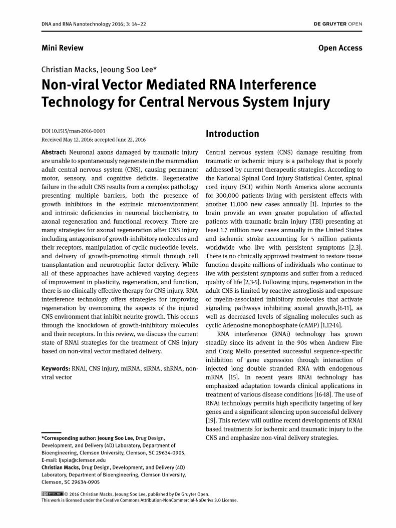
DNA and RNA Nanotechnology 2016; 3: 14–22 Introduction Central nervous system (CNS) damage resulting from traumatic or ischemic injury is a pathology that is poorly addressed by current therapeutic strategies. According to the National Spinal Cord Injury Statistical Center, spinal cord injury (SCI) within North America alone accounts for 300,000 patients living with persistent effects with another 11,000 new cases annually [1]. Injuries to the brain provide an even greater population of affected patients with traumatic brain injury (TBI) presenting at least 1.7 million new cases annually in the United States and ischemic stroke accounting for 5 million patients worldwide who live with persistent symptoms [2,3]. There is no clinically approved treatment to restore tissue function despite millions of individuals who continue to live with persistent symptoms and suffer from a reduced quality of life [2,3-5]. Following injury, regeneration in the adult CNS is limited by reactive astrogliosis and exposure of myelin-associated inhibitory molecules that activate signaling pathways inhibiting axonal growth,[6-11], as well as decreased levels of signaling molecules such as cyclic Adenosine monophosphate (cAMP) [1,12-14]. RNA interference (RNAi) technology has grown steadily since its advent in the 90s when Andrew Fire and Craig Mello presented successful sequence-specific inhibition of gene expression through interaction of injected long double stranded RNA with endogenous mRNA [15]. In recent years RNAi technology has emphasized adaptation towards clinical applications in treatment of various disease conditions [16-18]. The use of RNAi technology permits high specificity targeting of key genes and a significant silencing upon successful delivery [19]. This review will outline recent developments of RNAi based treatments for ischemic and traumatic injury to the CNS and emphasize non-viral delivery strategies. DOI 10.1515/rnan-2016-0003 Received May 12, 2016; accepted June 22, 2016 Abstract: Neuronal axons damaged by traumatic injury are unable to spontaneously regenerate in the mammalian adult central nervous system (CNS), causing permanent motor, sensory, and cognitive deficits. Regenerative failure in the adult CNS results from a complex pathology presenting multiple barriers, both the presence of growth inhibitors in the extrinsic microenvironment and intrinsic deficiencies in neuronal biochemistry, to axonal regeneration and functional recovery. There are many strategies for axonal regeneration after CNS injury including antagonism of growth-inhibitory molecules and their receptors, manipulation of cyclic nucleotide levels, and delivery of growth-promoting stimuli through cell transplantation and neurotrophic factor delivery. While all of these approaches have achieved varying degrees of improvement in plasticity, regeneration, and function, there is no clinically effective therapy for CNS injury. RNA interference technology offers strategies for improving regeneration by overcoming the aspects of the injured CNS environment that inhibit neurite growth. This occurs through the knockdown of growth-inhibitory molecules and their receptors. In this review, we discuss the current state of RNAi strategies for the treatment of CNS injury based on non-viral vector mediated delivery. Keywords: RNAi, CNS injury, miRNA, siRNA, shRNA, non- viral vector Mini Review Open Access © 2016 Christian Macks, Jeoung Soo Lee, published by De Gruyter Open. This work is licensed under the Creative Commons Attribution-NonCommercial-NoDerivs 3.0 License. Christian Macks, Jeoung Soo Lee* Non-viral Vector Mediated RNA Interference Technology for Central Nervous System Injury *Corresponding author: Jeoung Soo Lee, Drug Design, Development, and Delivery (4D) Laboratory, Department of Bioengineering, Clemson University, Clemson, SC 29634-0905, E-mail: [email protected] Christian Macks, Drug Design, Development, and Delivery (4D) Laboratory, Department of Bioengineering, Clemson University, Clemson, SC 29634-0905
-
Upload
khangminh22 -
Category
Documents
-
view
5 -
download
0
Transcript of Non-viral Vector Mediated RNA Interference Technology for ...
DNA and RNA Nanotechnology 2016; 3: 14–22
IntroductionCentral nervous system (CNS) damage resulting from traumatic or ischemic injury is a pathology that is poorly addressed by current therapeutic strategies. According to the National Spinal Cord Injury Statistical Center, spinal cord injury (SCI) within North America alone accounts for 300,000 patients living with persistent effects with another 11,000 new cases annually [1]. Injuries to the brain provide an even greater population of affected patients with traumatic brain injury (TBI) presenting at least 1.7 million new cases annually in the United States
and ischemic stroke accounting for 5 million patients worldwide who live with persistent symptoms [2,3]. There is no clinically approved treatment to restore tissue function despite millions of individuals who continue to live with persistent symptoms and suffer from a reduced quality of life [2,3-5]. Following injury, regeneration in the adult CNS is limited by reactive astrogliosis and exposure of myelin-associated inhibitory molecules that activate signaling pathways inhibiting axonal growth,[6-11], as well as decreased levels of signaling molecules such as cyclic Adenosine monophosphate (cAMP) [1,12-14].
RNA interference (RNAi) technology has grown steadily since its advent in the 90s when Andrew Fire and Craig Mello presented successful sequence-specific inhibition of gene expression through interaction of injected long double stranded RNA with endogenous mRNA [15]. In recent years RNAi technology has emphasized adaptation towards clinical applications in treatment of various disease conditions [16-18]. The use of RNAi technology permits high specificity targeting of key genes and a significant silencing upon successful delivery [19]. This review will outline recent developments of RNAi based treatments for ischemic and traumatic injury to the CNS and emphasize non-viral delivery strategies.
DOI 10.1515/rnan-2016-0003Received May 12, 2016; accepted June 22, 2016
Abstract: Neuronal axons damaged by traumatic injury are unable to spontaneously regenerate in the mammalian adult central nervous system (CNS), causing permanent motor, sensory, and cognitive deficits. Regenerative failure in the adult CNS results from a complex pathology presenting multiple barriers, both the presence of growth inhibitors in the extrinsic microenvironment and intrinsic deficiencies in neuronal biochemistry, to axonal regeneration and functional recovery. There are many strategies for axonal regeneration after CNS injury including antagonism of growth-inhibitory molecules and their receptors, manipulation of cyclic nucleotide levels, and delivery of growth-promoting stimuli through cell transplantation and neurotrophic factor delivery. While all of these approaches have achieved varying degrees of improvement in plasticity, regeneration, and function, there is no clinically effective therapy for CNS injury. RNA interference technology offers strategies for improving regeneration by overcoming the aspects of the injured CNS environment that inhibit neurite growth. This occurs through the knockdown of growth-inhibitory molecules and their receptors. In this review, we discuss the current state of RNAi strategies for the treatment of CNS injury based on non-viral vector mediated delivery.
Keywords: RNAi, CNS injury, miRNA, siRNA, shRNA, non-viral vector
Mini Review Open Access
© 2016 Christian Macks, Jeoung Soo Lee, published by De Gruyter Open. This work is licensed under the Creative Commons Attribution-NonCommercial-NoDerivs 3.0 License.
Christian Macks, Jeoung Soo Lee*
Non-viral Vector Mediated RNA Interference Technology for Central Nervous System Injury
*Corresponding author: Jeoung Soo Lee, Drug Design, Development, and Delivery (4D) Laboratory, Department of Bioengineering, Clemson University, Clemson, SC 29634-0905, E-mail: [email protected] Macks, Drug Design, Development, and Delivery (4D) Laboratory, Department of Bioengineering, Clemson University, Clemson, SC 29634-0905

Non-viral Vector Mediated RNA Interference Technology for Central Nervous System Injury 15
RNAi mechanismThe process of RNAi is initiated both endogenously and exogenously. Figure 1 summarizes the different modes for the processing of endogenous miRNAs and exogenous shRNAs or siRNAs within a cell. The natural endogenous mechanism relies upon short RNA segments referred to as micro-RNAs (miRNAs) [15,20,21]. These miRNAs are approximately 22 nucleotides long and reach their final active form following trimming by enzymatic complexes. The miRNA molecule is initially transcribed as a long primary transcript referred to as a pri-miRNA. This pri-miRNA transcript is then cleaved within the nucleus by the RNase Drosha into a shorter hairpin structure or pre-miRNA. The transporter protein Exportin-5 mediates transfer of the pre-miRNA to the cytoplasm where the RNase Dicer processes the pre-miRNA to its active miRNA segments [22-24]. The active miRNA segments trigger degradation or inhibit translation of target mRNAs to
which they bind with full or partial complementarity, respectively [25]. In an attempt to adapt these mechanisms, researchers have developed techniques using exogenously applied small interfering RNAs (siRNAs), short-hairpin RNAs (shRNAs), and artificial miRNAs [26]. Artificially produced pri-miRNAs are processed in the same way as endogenous counterparts, whereas both the long siRNA molecules and shRNAs are processed similarly to pre-miRNAs. The siRNA molecules (20-25 base pairs) resemble final stage miRNAs in structure and bypass processing by both Drosha and Dicer prior to incorporation to the RNA-induced silencing complex (RISC). In both siRNAs and fully complementary miRNAs, the sense strand is discarded and the antisense strand is used for recognition and subsequent cleavage of complementary mRNA molecules, effectively halting their translation [26].
Several challenges impede the development of clinically relevant treatments. For successful RNAi
Figure 1: The diagram summarizes the different modes for processing of endogenous miRNAs and exogenous shRNAs or siRNAs in a cell. Endogenous miRNAs begin as long primary RNA (Pri-miRNA) transcripts, which are consecutively processed by the RNases Drosha to a 70-nucleotide stem-loop structure called a pre-miRNA, which is further cleaved by Dicer to form miRNA/miRNA duplexes. Active miRNAs can then lower protein expression through either full or partial complementarity with an mRNA target. The exogenous shRNA and siRNAs are designed to act through full complementarity with an mRNA target. Delivered siRNAs bypass processing by both Drosha and Dicer, whereas the hairpin shaped shRNAs still require processing by Dicer to liberate the siRNA duplex.

16 C. Macks, J. Soo Lee
therapy, the main challenge is successful delivery to the target site. Naked RNAs are readily degraded by nucleases in the body and have difficulty reaching the desired tissues or organs because they are distributed throughout the body. The RNAs, especially siRNAs, show short circulation time due to their size (below 10nm) and are readily excreted through renal filtration [17]. Even when they reach to the desired site, naked RNAs cannot pass the cell membrane due to their negative charge. Furthermore, there are some unintended toxic effects caused when delivered siRNAs interact with endogenous miRNA pathways or downregulate off-target genes [18, 26]. As a result, unmodified RNAs typically have a low rate of success [19, 27].
CNS BarriersDelivery of nucleic acids to the CNS is limited by the blood brain barrier (BBB), a key limiting factor for success of potential therapies [12, 29]. CNS homeostasis is maintained by a set of specialized barriers that control
the passage and retention of nutrients, proteins, and other small molecules within specialized compartments. Since cerebral spinal fluid (CSF) is able to exchange substances directly with the tissues of the CNS, the passage of substances from blood must be strictly controlled to maintain homeostasis. For this purpose, the BBB guards against molecules uncontrollably penetrating the vasculature of the CNS [29-31]. In the CNS, the microvasculature walls are formed by a single layer of endothelial cells with tight junctions that render the wall impermeable to all except a select group of molecules [32]. Small water soluble and lipid soluble molecules such as oxygen, carbon dioxide, and steroid hormones, can easily cross the membrane, whereas all other metabolic products such as glucose must be actively transported [29]. Instead of trans-cellular transportation, charged molecules must be selectively transported through the cell to gain entry to the CNS compartment. The tight endothelial barrier is accompanied by astrocytes and pericytes, which form a secondary barrier surrounding a majority of the capillaries, further restricting molecular passage [29, 33] (Figure 2). However, the BBB is not ubiquitous and
Figure 2: Transport mechanism across Blood Brain Barrier applied to RNAi. The BBB is comprised of brain endothelial cells connected by tight junctions and a thick basement membrane. The blood brain barrier is a major obstacle to therapeutic delivery, especially for nega-tively charged RNAs for RNAi. A) Naked siRNAs are unable to pass through the endothelial cell membrane due to their negatively charge, B) Non-viral vectors that are positively charged can neutralize the negative charged RNAs and allow passage through the BBB via adsorptive transcytosis.

Non-viral Vector Mediated RNA Interference Technology for Central Nervous System Injury 17
specific secretory or sensory zones such as the olfactory bulb do not have the BBB. Intra-nasal delivery is therefore an attractive route of delivery to the CNS [31]. In addition to the BBB, the blood-cerebrospinal fluid barrier (BCFB), formed by networks of capillaries, acts as a filtration system for blood plasma, preventing direct exchange of molecules with the CNS tissue. Though passage through the BCFB can allow therapeutic molecules access to the brain, the distribution of therapeutic molecules passing through the BBB is far higher, and as a result is the method of choice when seeking entry to the CNS [29].
Non-Viral Vector DeliveryWhile several viral vectors have demonstrated efficient gene knockdown, they often lack specificity and evoke immune reactions and inflammation [17, 34]. To overcome
these concerns, non-viral strategies such as naked RNAs, lipid- [35-37], peptide-[38], and polymer-based [27, 38-40] carriers for RNAi therapeutics are undergoing development with improved safety and reduced immunogenicity. Several classes of lipids including cationic [40, 41], anionic [42], and uncharged [41] are commonly used as carriers for the delivery of nucleic acid therapeutics [34]. The polymers utilized for delivery of nucleic acids can be charged or uncharged including poly (caprolactone) [26], poly (ethylene glycol) [38], and polyethylenimine (PEI) [27, 38]. Table 1 shows an overview of non-viral mediated RNAi strategies discussed in this review.
Chemical Modification of siRNAsApplication of naked siRNAs in vivo is limited by instability and low membrane penetration ability of the
Table 1: Overview of non-viral mediated RNAi strategies
Delivery Method Molecule Nucleic Acid Background Reference
Chemical Modification 5‘ end: deoxythymidine over-hangs and 2‘-O-methyl group on the sugar 3‘ end: phosphothioate linkages in backbone
siRNA Oligodendrocyte specific knockdown following convection enhanced delivery to the corpus callosum via a cannula
Querbes, et al.5
Chemical Modification Accell siRNAs from Dharmacon siRNA Direct intracerebroventricular injection with knockdown specific to non-divi-ding neuron populations
Nakajima, et al.44
Lipoplex Cationic lipid JetSiTM combined with neutral lipid DOPE
siRNA Delivery of encapsulated siRNAs against luciferase to neuronal cell cultures in vitro
Hassini, et al.35
Lipoplex Lipids MLRI and TransFastTM Transfection Reagent
siRNA Direct injection of lipoplex solution to the lateral ventrical or cisterna magna
Zou, et al.34
Lipoplex RNA/DNA hybrids delivered with bolaamphiphile cationic lipid carriers
siRNA Local injection of hybridized siRNAs complexed with bolaamphiphilic catio-nic carriers
Afonin et al.46
Exosome Exosomes isolated from primary dendritic cells and modified with rabies viral glycoprotein (RVG)
siRNA Injection of siRNA loaded exosomes via the tail vain
Alvarez-Everti, et al.51
Polymeric Nanoparticle Tetanus toxin peptide Tet1 modi-fied polyethylenimine (PEI)
siRNA Delivery of Tet1-modified PEI nanopar-ticles
Park, et al.38
Polymeric Nanoparticle Cyclodextrin molecules modified with PEG chains
siRNA Neuron-specific knockdown of luci-ferase reporter gene in mouse embryo-nic hypothalamic cells in vitro
O’Mahony, et al.56
Polymeric Nanoparticle Polyethyleneglycol-polycapro-lactone (PEG-PCL) nanoparticles modified with HIV-1 Tat peptide
siRNA Intranasal delivery of nanoparticles in eight-week old rats
Kanazawa et al.31
Polymeric Nanoparticle Polyamidoamine dendrimer with an ester linkage grafted to L-Arginine (e-PAMAM-Arg)
siRNA Intranasal delivery of siRNA targeting high mobility group box 1 (HMGB1) in the post-ischemic rat brain model
Il-Doo Kim et al. 12
Polymeric Nanoparticle Non-liposomal cationic amphip-hile Interferin (Polypus Trans-fection)
siRNA Direct injection of Aquaporin-4 (AQP4) siRNA local to the site of injury in a rat cortical impact model
Fukuda, et al.72
Polymeric Nanoparticle Jet-PEI nanoparticle shRNA Intravenous injection of nanopartic-les containing plasmids encoding for metalloprotinase12 shRNAs
Chelluboina, et al.4
18 C. Macks, J. Soo Lee
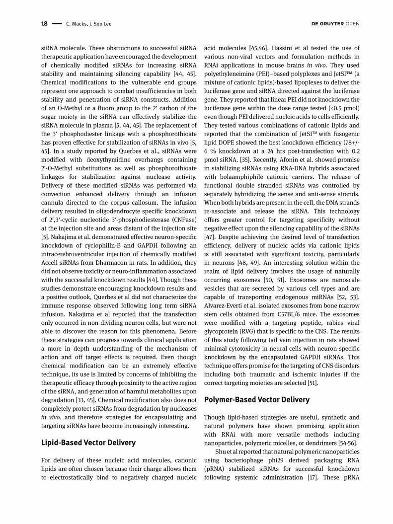
siRNA molecule. These obstructions to successful siRNA therapeutic application have encouraged the development of chemically modified siRNAs for increasing siRNA stability and maintaining silencing capability [44, 45]. Chemical modifications to the vulnerable end groups represent one approach to combat insufficiencies in both stability and penetration of siRNA constructs. Addition of an O-Methyl or a fluoro group to the 2’ carbon of the sugar moiety in the siRNA can effectively stabilize the siRNA molecule in plasma [5, 44, 45]. The replacement of the 3’ phosphodiester linkage with a phosphorothioate has proven effective for stabilization of siRNAs in vivo [5, 45]. In a study reported by Querbes et al., siRNAs were modified with deoxythymidine overhangs containing 2’-O-Methyl substitutions as well as phosphorothioate linkages for stabilization against nuclease activity. Delivery of these modified siRNAs was performed via convection enhanced delivery through an infusion cannula directed to the corpus callosum. The infusion delivery resulted in oligodendrocyte specific knockdown of 2′,3′-cyclic nucleotide 3′-phosphodiesterase (CNPase) at the injection site and areas distant of the injection site [5]. Nakajima et al. demonstrated effective neuron-specific knockdown of cyclophilin-B and GAPDH following an intracerebroventricular injection of chemically modified Accell siRNAs from Dharmacon in rats. In addition, they did not observe toxicity or neuro-inflammation associated with the successful knockdown results [44]. Though these studies demonstrate encouraging knockdown results and a positive outlook, Querbes et al did not characterize the immune response observed following long term siRNA infusion. Nakajima et al reported that the transfection only occurred in non-dividing neuron cells, but were not able to discover the reason for this phenomena. Before these strategies can progress towards clinical application a more in depth understanding of the mechanism of action and off target effects is required. Even though chemical modification can be an extremely effective technique, its use is limited by concerns of inhibiting the therapeutic efficacy through proximity to the active region of the siRNA, and generation of harmful metabolites upon degradation [33, 45]. Chemical modification also does not completely protect siRNAs from degradation by nucleases in vivo, and therefore strategies for encapsulating and targeting siRNAs have become increasingly interesting.
Lipid-Based Vector Delivery
For delivery of these nucleic acid molecules, cationic lipids are often chosen because their charge allows them to electrostatically bind to negatively charged nucleic
acid molecules [45,46]. Hassini et al tested the use of various non-viral vectors and formulation methods in RNAi applications in mouse brains in vivo. They used polyethyleneimine (PEI)–based polyplexes and JetSI™ (a mixture of cationic lipids)-based lipoplexes to deliver the luciferase gene and siRNA directed against the luciferase gene. They reported that linear PEI did not knockdown the luciferase gene within the dose range tested (<0.5 pmol) even though PEI delivered nucleic acids to cells efficiently. They tested various combinations of cationic lipids and reported that the combination of JetSITM with fusogenic lipid DOPE showed the best knockdown efficiency (78+/- 6 % knockdown at a 24 hrs post-transfection with 0.2 pmol siRNA. [35]. Recently, Afonin et al. showed promise in stabilizing siRNAs using RNA-DNA hybrids associated with bolaamphiphile cationic carriers. The release of functional double stranded siRNAs was controlled by separately hybridizing the sense and anti-sense strands. When both hybrids are present in the cell, the DNA strands re-associate and release the siRNA. This technology offers greater control for targeting specificity without negative effect upon the silencing capability of the siRNAs [47]. Despite achieving the desired level of transfection efficiency, delivery of nucleic acids via cationic lipids is still associated with significant toxicity, particularly in neurons [48, 49]. An interesting solution within the realm of lipid delivery involves the usage of naturally occurring exosomes [50, 51]. Exosomes are nanoscale vesicles that are secreted by various cell types and are capable of transporting endogenous miRNAs [52, 53]. Alvarez-Everti et al. isolated exosomes from bone marrow stem cells obtained from C57BL/6 mice. The exosomes were modified with a targeting peptide, rabies viral glycoprotein (RVG) that is specific to the CNS. The results of this study following tail vein injection in rats showed minimal cytotoxicity in neural cells with neuron-specific knockdown by the encapsulated GAPDH siRNAs. This technique offers promise for the targeting of CNS disorders including both traumatic and ischemic injuries if the correct targeting moieties are selected [51].
Polymer-Based Vector Delivery
Though lipid-based strategies are useful, synthetic and natural polymers have shown promising application with RNAi with more versatile methods including nanoparticles, polymeric micelles, or dendrimers [54-56].
Shu et al reported that natural polymeric nanoparticles using bacteriophage phi29 derived packaging RNA (pRNA) stabilized siRNAs for successful knockdown following systemic administration [17]. These pRNA

Non-viral Vector Mediated RNA Interference Technology for Central Nervous System Injury 19
nanoparticles are capable of delivering multiple siRNAs with different therapeutic targets and exhibit minimal cytotoxicity [57, 58]. The size of these nanoparticles ranges from 20-40 nm [17], which is within the range of 10 to 100nm that is considered as optimal particle size range for non-viral vectors [59, 60]. In a study by Abdelmawla et al., pRNA nanoparticles were modified by folate conjugation for targeting folate receptor positive (FR+) tumors. The particles showed selective uptake in the FR+ cells with minimal activation of inflammatory response and negligible uptake into the liver, spleen, and kidneys [61]. The ability of pRNA nanoparticles to avoid immune activation and clearance, coupled with their ability to target specific cells through small molecule conjugation, would allow for adaptation to CNS delivery through the addition of molecules targeting the BBB.
Polyethylenimine (PEI) is a polycationic polymer that has shown success in gene delivery to the CNS [62-64]. Though it has shown successful application in several diseases, significant cytotoxicity is often a concern when higher doses are required to elicit an effect [65]. This is the limitation of PEI as a gene delivery carrier in clinical CNS application. In a study by Gwak et al a cationic amphiphilic co-polymer, poly(lactide-co-glycolide)-graft-polyethylenimine (PgP) was developed and successfully applied both in vitro and in vivo. This PgP polymer has been developed for combinatorial delivery of drugs and nucleic acids for axon regeneration after CNS injury. They reported that PgP can efficiently knockdown (~65%) GFP by transfecting PgP/GFP siRNA polyplexes in B35 cells in media containing 10% serum, which was similar to that obtained with RNAiMAX/GFP siRNA (~70 %). [66]
Another polymer that has been applied towards RNAi technology following modification with amphiphilic and cationic molecules is cyclodextrin (CD) [66-69]. O’Mahony et al. sought to create a neuron-specific delivery system for siRNA by modifying CDs through “click” chemistry. This strategy optimizes a non-viral delivery system using an ‘ABCD’ concept [70] with the four portions being the nucleic acid cargo, the portion that complexes the nucleic acid, a stabilizing portion such as polyethylene glycol (PEG), and a targeting moiety. O’Mahony et al. reported that the addition of PEG chains to the CD molecules increased stability of the molecule, but at the same time reduced the siRNA delivery through reducing interaction with the cell membrane. The use of PEG modification does not always result in poor activity of RNAi technology as is shown by Kanazanwa et al. when delivering polyethylene glycol-polycaprolactone (PEG-PCL) nanoparticles modified with HIV-1 Tat peptide. The delivery of these particles was accomplished through intranasal delivery to eight-week
old rats, the results demonstrated successful delivery of siRNAs specifically to the CNS tissue with limited toxicity
[31]. This technique has the added benefit of improving the efficacy of the non-invasive siRNA delivery.
Another polymeric vehicle that has shown promise as a vehicle for mediating RNAi is polyamidoamine (PAMAM) dendrimer grafted with L-arginine [71]. Il-Doo Kim et al. reported that PAMAM-Arg modified with an ester linkage (e-PAM-Arg) was capable of binding and delivering siRNAs against high mobility group box 1 (HMGB1). In the animal models for this study the e-PAM-Arg/siRNA complex was injected stereotaxically into the rat’s cortex. The e-PAM-Arg/siRNA complexes were able to achieve 40% siRNA transfection in neuron cells following injection to the rat brains. In addition, it was found that successful siRNA-mediated HMGB1 knockdown inhibited neuronal death and also decreased infarct volume in post-ischemic rat brains [71]. These positive results can bolster the confidence in a successful future treatment for ischemic injuries in the CNS.
In a study reported by Fukuda et al, successful RNAi in the treatment of rat cortical impact TBI model was accomplished through knockdown of Aquaporin-4 (AQP4) by local injection of siRNAs complexed with the commercially available non-liposomal cationic amphiphilic Interferin (Polypus Transfection) [72]. In addition to successful AQP4 knockdown, this study also reported decreased astrogliosis at early time points, decreased edema, increased microglia activation, and increased neuron cell survival compared to a non-specific siGLO control [72]. They report that significant functional recovery was achieved for up to 3 days post-injury compared to siGLO as a control [67]. In a study by Chelluboina et al., Jet-PEI nanoparticle vectors complexed with plasmids expressing shRNAs against metalloproteinase 12 (MMP-12) were delivered intravenously to treat cerebral ischemia [4]. The shRNA against metalloproteinase-12 (MMP-12) was encoded in plasmid vectors and expressed through strong Pol III promoters including U6 and H1, which exhibit intense levels of expression [73-76]. The study highlighted MMP-12 as a potential therapeutic target for ischemia and demonstrated that shRNA in plasmid vectors could effectively knockdown MMP-12 when intravenously administered [4]. Though capable of significant knockdown, shRNA delivery can be associated with a higher cell toxicity that has been attributed to intense expression by successfully transfected cells [26, 76].
To address the challenge of non-specific uptake and provide targeting capability, Park et al conjugated branched PEI with tetanus toxin peptide (Tet1), which has high affinity for dorsal root ganglion and primary motor

20 C. Macks, J. Soo Lee
neurons. These Tet1-modified nanoparticles showed successful targeting of differentiated PC-12 neuron cells, primary dorsal root ganglion cells, and primary neurons in mixed culture with primary astrocytes. The Tet1 modified PEI nanoparticles exhibit promise as a delivery vector capable of targeting the CNS in vivo proven by their specific binding to neuron cells [38].
Conclusions and OutlookEmerging RNAi technologies applied to CNS treatment require extensive research before they can undergo successful clinical application. In this mini-review, we cover recent progress made in RNAi technology based on non-viral gene carriers that are classified in chemical modification of RNA, lipid–based, and polymer-based carries used in CNS injury. RNAi therapeutic technologies have been developed with enhanced knockdown of target gene expression, but still need to reduce off-target effects, immune response, and cytotoxicity. Despite the improvements necessary for the acceptance of these technologies as established clinical therapies, the progress of these technologies to this point offers promise that future patients with these disorders have access to therapies capable of restoring the function of their cognitive and motor skills towards their original capabilities.
References[1] Domingo, A., Al-Yahya, A., Asiri, Y., Eng, J., Lam, T., Spinal
Cord Injury Rehabilitation Evidence Research Team. A Systemic Review of the Effects of Pharmacological Agents on Walking Function in People with Spinal Cord Injury. Journal of Neurotrauma. 2012, 29, 865-879.
[2] Kelso, M.L., Pauly. J,R,. Therapeutic Targets for Neuropro-tection and/or Enhancement of Functional Recovery Following Traumatic Brain Injury. Progress in Molecular Biology and Translational Science. 2011, 98, 85-131.
[3] Ghajar, J. Traumatic brain injury. Lancet. 2000, 356, 923-929.[4] Chelluboina, B., Warhekar, A., Dillard, M., Klopfenstein, J.D.,
Pinson, D.M., Wang, D.Z., Veeravalli, K.K. Post-transcriptional inactivation of matrix metalloproteinase-12 after focal cerebral ischemia attenuates brain damage. Scientific Reports. 2014, 5,1-11.
[5] Querbes, W., Ge, P., Zhang, W., Fan, Y., Costigan, J., Charisse, K., Maier, M., Nechev, L., Manoharan, Kotelianski, V., Sah, D. Direct CNS Delivery of siRNA Mediates Robust Silencing in Oligodendrocytes. Oligonucleotides. 2008, XX, 1-8.
[6] Mehta, N.R., Nguyen, T., Bullen, J.W., Griffin, J.W., Schnaar, R.L. Myelin-associated glycoprotein (MAG) protects neurons form acute toxicity using a ganglioside-dependent mechanism. ACS Chem Neurosci. 2010, 1(3), 215-222.
[7] McKeon, R.J., Jurynec, M.J., Buck, C.R. The chondroitin sulfate proteo glycans neurocan and phosphacan are expressed by reactive astrocytes in the chronic CNS glial scar. J. Neurosci. 1999, 19, 10778-10788.
[8] Koprivica, V., Cho, K.S., Park, J.B., Yiu, G., Atwal, J., Gore, B., Kim, J.A., Lin, E., Tessier-Lavigne, M., Chen, D.F., He, Z. EGFR activation mediates inhibition of axon regeneration by myelin and chondroitin sulfate proteoglycans. Science. 2005, 310, 106-110.
[9] Scnell, L., Schwab, M.E. Axon regeneration in rat spinal cord produced by an antibody against myelin-associated neurite growth inhibitors. Nature. 1990, 343, 269-272.
[10] GrandPre, T., Li, S., Strittmatter, S.M. Nogo-66 receptor agonist peptide promotes axonal regeration. Nature. 2002, 417, 547-551.
[11] Fournier, A.E., Gould, G.C., Liu, B.P., Strittmatter, S.M. Truncated soluble Nogo binds Nogo-66 and blocks inhibition of axon growth by myelin. J. Neurosci. 2002, 22, 8876-8883.
[12] Kim, I-D., Shin, J-H., Kim, S-W., Choi, S., Ahn, J., Han, P-L., Park, J-S., Lee, J-K. Intranasal Delivery of HMGB1 siRNA Confers Target Gene Knockdown and Robust Neuroprotection in the Postischemic Brain. Molecular Therapy. 2012, 20(4), 829-839.
[13] Andor, T., Sato, S., Toyooka, T., Kobayashi, H., Nawashiro, H., Ashida, H., Obara, M. Photochemical Wave-Driven Delivery of siRNAs Targeting Intermediate Filament Proteins Promotes Functional Recovery after Spinal Cord Injury in Rats. PLOS ONE. 2012, 7(12), 1-11.
[14] Zukor, K., Belin, S., Weng, C., Keelan, N., Wang, X., He, Z.. Short Hairpin RNA against PTEN Enhances Regenerative Growth of Corticospinal Tract Axons after Spinal Cord Injury. J. Neurosci. 2013, 33(39), 15350-15361.
[15] Fire, A., Xu, S., Montgomery, M.K., Kostas, S.A., Driver, S.E., Mello, C.C. Potent and specific genetic interference by double-stranded RNA in Caenorhabditic elegans. Nature. 1998, 391(19), 806-810.
[16] Afonin, K., Grabow, W.W., Walker, F.M., Bindewald, E., Dobrovolskaia, M.A., Shapiro, B.A., Jaeger, L. Design and self-assembly of siRNA-functionalized RNA nanoparticles for use in automated nanomedicine. Nature Proc. 2011, 6, 2022-2034.
[17] Shu, Y., Cinier, M., Shu, D., Guo, P. Assembly of multifunctional phi29 pRNA nanoparticles for specific delivery of siRNA and other therapeutics to targeted cells. Methods. 2011, 54:201-214.
[18] Gherardini, L., Bardi, G., Gennaro, M., Pizzorusso, T. Novel siRNA delivery strategy: a new “strand” in CNS translational medicine?. Cell. Mol. Life Sci. 2014, 71:1-20.
[19] Li, C., Parker, A., Menocal, E., Xiang ,S., Borodyansky, L., Fruehauf, J.H. Delivery of RNA Interference. Cell Cycle. 2006, 5(18), 2103-2109.
[20] Lee, R.C., Feinbaum, R.L., Ambros, V. The C. elegans Hetero-chronic Gene lin-4 Encodes Small RNAs with Antisense Complementarity to lin-14. Cell. 1993, 75, 843-854.
[21] Salta, E., Strooper, B.D. Non-coding RNAs with essential roles in neurodegenerative disorders. Lancet Neurol. 2012, 11: 189-200.
[22] Lee, Y., Ahn, C., Han, J., Choi, H., Kim, J., Yim, J., Lee, J., Provost, P., Radmark, O., Kim, S., Kim, V.N. The nuclear RNase III Drosha initiates microRNA processing. Nature. 2003, 425, 415-419.
Non-viral Vector Mediated RNA Interference Technology for Central Nervous System Injury 21
[23] Han, J., Lee, Y., Yeom, K-H., Kim, K-H., Jin, H., Kim, V.N. The Drosha-DGCR8 complex in primary microRNA processing. Genes & Development. 2004, 18: 3016-3027.
[24] Zhang, X., Zeng, Y. The terminal loop region controls microRNA processing by Drosha and Dicer. Nucleic Acids Research. 2010, 38(21), 7689-7697.
[25] Lim, L., Lau, N.C., Garrett-Engele, P., Grimson, A., Schelter, J.M., Castle, J., Bartel, D.P., Linsley, P.S., Johnson, J.M. Microarray analysis shows that some miRNAs downregulate large number of target mRNAs. Nature. 2005, 433, 769-773.
[26] McBride, J.L., Boudreau, R.L., Harper, S.Q., Staber, P.D., Monteys, A.M., Martins, I., Gilmoer, B.L., Burstein, H., Peluso, R.W., Polisky, B., Carter, B.J., Davidson, B.L.. Artificial miRNAs mitigate shRNA-mediated toxicity in the brain: Implications for the therapeutic development of RNAi. PNAS. 2008, 105(15), 5868-5873.
[27] Höbel, S., Aigner, A. Polyethylenimines for siRNA and miRNA delivery in vivo. WIREs Nanomed Nanobiotechnol. 2013, 5, 484-501.
[28] Lei, C., Cui, Y., Zheng, L., Chow, P.K., Wang, C.H. Development of a gene/drug dual delivery system for brain tumor therapy: Potent inhibition via RNA interference and synergistic effects. Biomaterials. 2013, 34, 7483-7494.
[29] Tiwari, B., Amiji, M. A review of nanocarrier-based CNS delivery systems. Current Drug Delivery. 2006, 3, 219-232.
[30] Wekerle, H. Immune Protection of the Brain – Efficient and Delicate. The Journal of Infectious Disease. 2002,186(Suppl2), S140-S144.
[31] Kanazawa, T., Akiyama, F., Kakizaki, Y., Seta, Y. Delivery of siRNA to the brain using a combination of nose-to-brain delivery and cell-penetrating peptide-modified nano-micelles. Biomaterials. 2013, 34, 9220-9226.
[32] Yeh, W-L., Lu, D-Y., Lin, C-J., Liou, H-C., Fu, W-M. Inhibition of Hypoxia-Induced Increase of Blood-Brain Barrier Permeability by YC-1 through the Antagonism of [HIF-1α Accumulation and VEGF Expression. Molecular Pharmacology. 2007, 72(2), 440-449.
[33] Kreuter, J. Nanoparticulate systems for brain delivery of drugs. Advanced Drug Delivery Reviews. 2001, 47, 65-91.
[34] Zhou, J., Shum, K.T., Burnett, J.C., Rossi, J.J. Nanoparticle-Based Delivery of RNAi Therapeutics: Progress and Challenges. Pharmaceuticals. 2013, 6, 85-107.
[35] Hassini, Z., Lemkine, G.F., Erbacher, P., Palmier, K., Alfama, G., Giovannangeli, C, Behr, J-P., Demeneix, BA . Lipid-mediated siRNA delivery down-regulates exogenous gene expression in the mouse brain at picomolar levels. J Gene Med. 2005, 7, 198-207.
[36] Bruun, Larsen, T.B., Jølck, R.I., Eliasen, R., Holm, R., Gjetting, T., Andersen, T.L. Investigation of enzyme-sensitive lipid nanoparticles for delivery of siRNA to blood-brain barrier and glioma cells. International Journal of Nanomedicine. 2015, 10, 5995-6008.
[37] Cardoso Simões, S., de Almeida, L.P., Plesnila, N., Pedroso de Lima, M.C., Wagner, E., Culmsee, C. Tf-lipoplexes for neuronal siRNA delivery: A promising system to mediate gene silencing in the CNS. Journal of Controlled Release. 2008, 132, 113-123.
[38] Park, I-K., Lasiene, J., Chou, S-H., Horner, P.J., Pun, S.H. Neuron-specific delivery of nucleic acids mediated by Tet1-modified poly(ethylenimine). J Gene Med. 2007, 9, 691-702.
[39] Miura, Y., Takenaka, T., Toh, K., Wu, S., Nishihara, H., Kano, M.R., Ino, Y., Nomoto, T., Matsumoto, Y., Koyama, H., Cabral, H., Nishiyama, N., Kataoka, K. Cyclic RGD-Linked Polymeric Micelles for Targeted Delivery of Platinum Anticancer Drugs to Glioblastoma through the Blood-Brain Tumor Barrier. ACS Nano. 2013, 7(10), 8583-8592.
[40] Tosi, G., Musumeci, T., Ruozi, B., Carbone, C., Belletti, D., Pignatello, R., Vandelli, M.A., Puglisi, G. The “fate” of polymeric and lipid nanoparticles for brain delivery and targeting: Strategies and mechanism of blood-brain barrier crossing and trafficking into the central nervous system. Journal of Drug Delivery Science and Technology. 2016, 32, 66-76.
[41] Girao da Cruz, M.T., Simões, S., Pedroso de Lima, M.C. Improving lipoplex-meidated gene transfer into C6 glioma cells and primary neurons. Experimental Neurology. 2004, 187, 65-75.
[42] Cheng, X., Lee, R. The role of helper lipids in lipid nanoparticles (LNPs) designed for oligonucleotide delivery. Advanced Drug Delivery Reviews. 2016, 99, 129-137.
[43] Baysal, Ucar, G., Gultekinoglu, M., Ulubayram, K., Yabanoglu-Ciftci, S. Donepezil loaded PLGA-b-PEG nanoparticles: their ability to induce destabilization of amyloid fibrils and to cross blood brain barrier in vitro. J Neural Transm. (2016), DOI: 10.1007/s00702-016-1527-4.
[44] Nakajima, H., Kubo, T., Semi, Y., Itakura, M., Kuwamura, M., Izawa, T., Azuma, Y.T., Takeuchi, T. A rapid, targeted, neuron-selective, in vivo knockdown following a single intrace-rebroventricular injection of a novel chemically modified siRNA in the adult rat brain. Journal of Biotechnology. 2012, 157, 326-333.
[45] Shim, M.S., Kwon, Y.J. Efficient and targeted delivery of siRNA in vivo. FEBS Journal. 2010, 227, 4814-4827.
[46] Gupta, K., Afonin, K.A., Viard, M., Herrero, V., Kasprzak, W-C., Kagiampakis, I., Kim, T., Koyfman, A.Y., Puri, A., Stepler, M., Sappe, A., KewalRamani, V.N., Grinberg, S., Linder, C., Heldman, E., Blumenthal, R., Shapiro, BL. Bolaamphiphiles as Carriers for siRNA Delivery: From Syntheses to Practical Application. Journal of Controlled Release. 2015, 213, 143-151.
[47] Afonin, K., Viard, M., Martins, A.N., Lockett, S.J., Maciag, A.E., Freed, E.O., Heldman, E., Jaeger, L., Bluthmenthal, R., Shapiro, B.A. Activation of different split functionalities on re-association of RNA-DNA hybrids. Nature Nanotechnology. 2013, 44, 1-8.
[48] Krichevsky, A.M., Kosik, K.S. RNAi functions in cultured mammalian neurons. Proc Natl Acad Sci USA. 2002, 99, 11926–11929.
[49] Lingor, P., Michel, U., Shöll, U., Bähr, M., Kügler, S. Transfection of ‘‘naked’’ siRNA results in endosomal uptake and metabolic impairment in cultured neurons. Biochem Biophys Res Commun. 2004, 315, 1126–1133.
[50] Andaloussi, S., Lakhal, S., Mäger, I., Wood, M.J. Exosomes for targeted siRNA delivery across biological barriers. Advanced Drug Delivery Reviews. 2013, 65: 391-397.
[51] Alvarez-Erviti, L., Seow , Y., Yin, H., Betts, C., Lakhal, S., Wood, M.J. Delivery of siRNA to the mouse brain by systemic injection of targeted exosomes. Nature Biotechnology.2011, 29(4), 241-247.
[52] Valadi, H., Ekström, K., Bossios, A., Sjöstrand, M., Lee, J.J., Lötvall, J.O. Exosome-mediated transfer of mRNAs and
22 C. Macks, J. Soo Lee
microRNAs is a novel mechanism of genetic exchange between cells. Nat. Cell Biol. 2007, 9, 654-659.
[53] Skog, J., Wurdinger, T., van Rjin, S., Meijer, D., Gainche, Sena-Esteves, M., Curry, W.T., Carter, R.S., Krichevsky, A.M., Breakefield, X.O. Glioblastoma microvescicles transport RNA and proteins that promote tumor growth and provide diagnostic biomarkers. Nat. Cell Bio. 2008, 10, 1470-1476.
[54] Godinho, B., McCarthy, D.J., Torres-Fuentes, C., Beltrán, C.J., McCarthy, J., Ogier, J.R., Darcy, R., O’Driscoll, C.M., Cryan, J.E.. Differential nanotoxicological and neuroinflammatory liabilities of non-viral vectors for RNA interference in the central nervous system. Biomaterials. 2014, 35, 489-499.
[55] Perez, A.P., Mudiña-Weilenmann, C., Romero, E.L., Morilla, M.J.l. Increased brain radioactivity by intranasal 32P-labeled siRNA dendriplexes within in situ-forming mucoadhesive gels. International Journal of Nanomedicine. 2012, 7, 1373-1385.
[56] O’Mahony, A.M., Godinho, M., Cryan, J.F., O’Driscoll, C.M. Non-Viral Nanosystems for Gene and Small Interfering RNA Delivery to the Central Nervous System: Formulating the Solution. Journal of Pharmaceutical Sciences. 2013, 1-16.
[57] Guo, P., Coban, O., Snead, N.M.,Trebley, J., Hoeprich, S., Guo, S., Shu, Y. Engineering RNA for Targeted siRNA Delivery and Medical Application. Adv. Drug Deliv. Rev. 2011, 62, 650-666.
[58] Guo, P. RNA Nanotechnology: Engineering, Assembly and Applications in Detection, Gene Delivery and Therapy. J. Nanosci. Nanotechnol. 2005, 5(12), 1964-1982.
[59] Jain, K.K. The role of nanobiotechnology in drug delivery. Drug Discov. Today. 2005, 10(21), 1435-1442.
[60] Li, W.,Szoka, F.C. Lipid-based Nanoparticles for Nucleic Acid Delivery. Pharm. Res. 2007, 24(3),438-449.
[61] Abdelmawla, S., Guo, S., Zhang, L., Pulukuri, S.M., Patankar, P., Conley, P., Trebley, J., Guo, P., Li, Q-X. Pharmacological Charac-terization of Chemically Synthesized Monomeric phi29 pRNA Nanoparticles for Systemic Delivery. Molecular Therapy. 2011, 19(7), 1312-1322.
[62] Abdallah, B., Hassan, A., Benoist, C., Goula, D., Behr, J.P., Demeneix, B.A. A powerful nonviral vector for in vivo gene transfer into the adult mammalian brain. Hum Gene Ther. 1996, 7, 1947-1954.
[63] Boussif, O., Lezoualc’h, F., Zanta, M.A., Mergny, M.D., Scherman, D., Demeneix, B., Behr, J.P. A versatile vector for gene and oligonucleotide transfer into cells in culture and in vivo: polyethylenimine. Proc Natl Acad Sci U S A. 1995, 92, 7297-7301.
[64] Lemkine, G.F., Mantero, S., Migné, C., Raji, A., Goula, D., Normandie, P., Levi, G., Demeneix, B.A. Preferential transfection of adult mouse neural stem cells and their immediate progeny in vivo with polyethylenimine. Mol Cell Neurosci. 2002, 19,165-174.
[65] Fougerolles, A., Vornlocher, H-P., Maraganore, J., Lieberman, J. Interfering with disease: a progress report on siRNA-based therapeutics. Nature Reviews. 2007,6, 443-453.
[66] Gwak, S-J., Nice, J., Zhang, J., Green, B., Macks, C., Bae, S., Webb, K., Lee, J.S. Cationic, amphiphilic nopolymer micelles as nucleic acid carriers for enhanced transfection in rat spinal cord. Acta Biomaterialia. 2016, 35, 98-108.
[67] O’Mahony, A., Ogier, J., Darcy, R., Cryan, J.F., O’Driscoll, C.M. Cationic and PEGylated Amphiphilic Cyclodextrins: Co-Formulation Opportunities for Neuronal Sirna Delivery. PLOS ONE. 2013, 8(6), 1-9.
[68] Oritz Mellet, O., Garcia Fernández, J.M., Benito, J.M. Cyclodextrin-based gene delivery systems. Chem Soc Rev. 2011, 40, 1586-1608.
[69] Mendez-Ardoy, A., Urbiola, K., Aranda, C., Oritz-Mellet, C., Garcia Fernández, J.M., de Ilarduya, C.T. Polycationic amphiphilic cyclodextrin-based nanoparticles for therapeutic gene delivery. Nanomedicine. 2011, 6, 1697-1707.
[70] Kostarelos, K., Miller, A.D. Synthetic, self-assembly ABCD nanoparticles; a structural paradigm for viable synthetic non-viral vectors. Chem Soc Rev. 2005, 34, 970-994.
[71] Kim, I-D., Lim, C.M., Kim, J.B., Nam, H.Y., Nam, K., Kim, S.W., Park, J.S., Lee, J.K. Neuroprotection by biodegradable PAMAM ester (e-PAMAM-R)- mediated HMGB1 siRNA delivery in primary cortical culture and in the postischemic brain. Journal of Controlled Release. 2010, 142, 422-430.
[72] Fukuda, A.M., Adami, A., Pop, V., Bellone, J.A., Coats, J.S., Hartman, R.E., Ashwal, S., Obenaus, A., Badaut, J. Posttraumatic reduction of edema with aquaporin-4 RNA interference improves acute and chronic functional recovery. Journal of Cerebral Blood Flow & Metabolism. 2013, 33, 1621-1632.
[73] Ramachandran, P.S., et al. Recent Advances in RNA Interference Therapeutics for CNS Diseases. Neurotherapeutics. 2013, 10, 473-485.
[74] Paddison, P.J., Caudy, A.A., Berstein, E., Hannon, G.J., Conklin, D.S. Short hairpin RNAs (shRNAs) induce sequence-specific silencing in mammalian cells. Genes & Development. 2002, 16, 948-958.
[75] Xia, H., Mao, Q., Eliason, S.L., Harper, I.H., Orr, H.T., Paulson, H.L., Kotin, R.M., Davidson, B.L. RNAi suppresses polyglutamine-induced neurodegeneration in a model of spinocerebral ataxia. Nature Medicine. 2004, 10(8), 816-820.
[76] Zeitelhofer, M., Vessey, J.P., Yunli, X., Tübing, F., Thomas, S., Kiebler, M., Dahm, R. High-Efficiency Transfection of Short Hairpin RNAs-Encoding Plasmids Into Primary Hippocampal Neurons. Journal of Neuroscience Research. 2009, 87, 289-300.