Bioabsorbable interference screws in ACL reconstruction
8
Bioabsorbable Interference Screws in ACL Reconstruction Derek B. Purcell, MD, J.R. Rudzki, MD, MS, and Rick W. Wright, MD The purpose of this article is to discuss the bioabsorbable interference screws currently available for graft fixation in anterior cruciate ligament reconstruction. A brief review of the literature regarding the use of bioabsorbable interference screws is included. The screw design, insertion technique, graft options, and various manufactured sizes for each screw are addressed. Relevant screw characteristics to consider include availability of guidewire, tap, and tunnel notching devices to ease insertion. Oper Tech Sports Med 12:180-187 © 2004 Elsevier Inc. All rights reserved. KEYWORDS anterior cruciate ligament reconstruction, interference fixation, bioabsorbable S ince Lambert 1 introduced interference screw fixation of bone-patellar tendon-bone grafts, a number of de- sign advances have occurred. Kurosaka et al 2 developed the current concept of interference screw fixation with a custom designed fully threaded 9.0-mm cancellous screw exhibiting increased stiffness and linear load compared with Lambert’s original 6.5-mm AO screw. 2 Metallic screw designs with blunt threads have been developed to de- crease injury to the graft construct during insertion. The introduction of cannulated screw designs has allowed in- sertion of the screw over a guidewire in an effort to mini- mize screw-tunnel divergence. A wide variety of screw lengths and diameters have become available from manu- facturers. Headed femoral tunnel screws have been devel- oped in an effort to decrease graft motion and graft injury at the tunnel opening. The advent of bioabsorbable inter- ference screws has generated a great deal of interest and further research in graft fixation. Bioabsorbable interference screws offer a number of ad- vantages over metallic implants. The use of biologic mate- rials avoids the use of metals that distort future magnetic resonance imaging of the reconstructed knee. 3 The major advantage of bioabsorbable interference screws is revision ligament reconstruction surgery. Retained metal screws may be problematic at the time of revision. However, bio- absorbable screws may have degraded and been replaced with bone, allowing revision to be performed similar to a primary procedure. If the bioabsorbable screws persist at the time of revision, many of the screws may simply be overdrilled when new tunnels are made. An additional advantage is the decreased likelihood of graft laceration, which has been reported with metal interference screws. 4 The major disadvantage of bioabsorbable interference screws is screw failure. 5 Failure usually occurs during screw insertion and is related to several factors including drive shape, drive diameter, drive length, and core diam- eter. 6,7 Manufacturers have altered drive designs to various shapes to improve force transmission from the drive mechanism to the screw. The drive design often extends the length of the screw to transmit force throughout the screw and therefore decrease the likelihood of failure. Some companies recommend notching the femoral tunnel and/or the tibial tunnel with the manufacturer’s desig- nated notcher as well as using a tap before screw insertion. Both screw design and insertion recommendations vary from product to product. In addition to insertion failure, case reports exist of late bioabsorbable screw fracture pre- senting as a loose intra-articular body. 8,9 Graft fixation is considered to be the weak link in the early postoperative period of anterior cruciate ligament (ACL) reconstruction. Regardless of whether metallic, ti- tanium, or bioabsorbable screws are used for bone-patellar tendon-bone ACL reconstructions, no difference has been found in clinical outcomes. 10 A study of bone-patellar ten- don-bone graft fixation in porcine knees comparing bio- absorbable and metal interference screws exhibited no sig- nificant difference in mean ultimate failure load, mean yield load, or stiffness of fixation. 11 Long-term studies of bioabsorbable interference fixation of hamstring grafts are not yet available, but some authors have reported good short-term results. 10 Investigations have focused on how to best increase interference screw fixation strength of soft-tissue grafts. Increased screw length for tibial tunnel hamstring fixation exhibited increased mean maximal load Department of Orthopaedic Surgery, Washington University School of Med- icine, St Louis, MO. Address reprint requests to Rick W. Wright, MD, Division of Sports Medi- cine, Department of Orthopaedic Surgery, Washington University School of Medicine, Suite 11300, West Pavilion, OneBarnes-Jewish Hos- pital Plaza, St Louis, MO 63110. E-mail: [email protected] 180 1060-1872/04/$-see front matter © 2004 Elsevier Inc. All rights reserved. doi:10.1053/j.otsm.2004.07.014
-
Upload
independent -
Category
Documents
-
view
2 -
download
0
Transcript of Bioabsorbable interference screws in ACL reconstruction
BSD
Sstcewdcismlfoaff
vrralmawpt
D
A
1
ioabsorbable Interferencecrews in ACL Reconstruction
erek B. Purcell, MD, J.R. Rudzki, MD, MS, and Rick W. Wright, MD
The purpose of this article is to discuss the bioabsorbable interference screws currentlyavailable for graft fixation in anterior cruciate ligament reconstruction. A brief review of theliterature regarding the use of bioabsorbable interference screws is included. The screwdesign, insertion technique, graft options, and various manufactured sizes for each screware addressed. Relevant screw characteristics to consider include availability of guidewire,tap, and tunnel notching devices to ease insertion.Oper Tech Sports Med 12:180-187 © 2004 Elsevier Inc. All rights reserved.
KEYWORDS anterior cruciate ligament reconstruction, interference fixation, bioabsorbable
oaw
ssdesmtsSanBfcs
e(ttfdanybnsts
ince Lambert1 introduced interference screw fixationof bone-patellar tendon-bone grafts, a number of de-
ign advances have occurred. Kurosaka et al2 developedhe current concept of interference screw fixation with austom designed fully threaded 9.0-mm cancellous screwxhibiting increased stiffness and linear load comparedith Lambert’s original 6.5-mm AO screw.2 Metallic screwesigns with blunt threads have been developed to de-rease injury to the graft construct during insertion. Thentroduction of cannulated screw designs has allowed in-ertion of the screw over a guidewire in an effort to mini-ize screw-tunnel divergence. A wide variety of screw
engths and diameters have become available from manu-acturers. Headed femoral tunnel screws have been devel-ped in an effort to decrease graft motion and graft injuryt the tunnel opening. The advent of bioabsorbable inter-erence screws has generated a great deal of interest andurther research in graft fixation.
Bioabsorbable interference screws offer a number of ad-antages over metallic implants. The use of biologic mate-ials avoids the use of metals that distort future magneticesonance imaging of the reconstructed knee.3 The majordvantage of bioabsorbable interference screws is revisionigament reconstruction surgery. Retained metal screws
ay be problematic at the time of revision. However, bio-bsorbable screws may have degraded and been replacedith bone, allowing revision to be performed similar to arimary procedure. If the bioabsorbable screws persist athe time of revision, many of the screws may simply be
epartment of Orthopaedic Surgery, Washington University School of Med-icine, St Louis, MO.
ddress reprint requests to Rick W. Wright, MD, Division of Sports Medi-cine, Department of Orthopaedic Surgery, Washington UniversitySchool of Medicine, Suite 11300, West Pavilion, OneBarnes-Jewish Hos-
hpital Plaza, St Louis, MO 63110. E-mail: [email protected]
80 1060-1872/04/$-see front matter © 2004 Elsevier Inc. All rights reserved.doi:10.1053/j.otsm.2004.07.014
verdrilled when new tunnels are made. An additionaldvantage is the decreased likelihood of graft laceration,hich has been reported with metal interference screws.4
The major disadvantage of bioabsorbable interferencecrews is screw failure.5 Failure usually occurs duringcrew insertion and is related to several factors includingrive shape, drive diameter, drive length, and core diam-ter.6,7 Manufacturers have altered drive designs to varioushapes to improve force transmission from the driveechanism to the screw. The drive design often extends
he length of the screw to transmit force throughout thecrew and therefore decrease the likelihood of failure.ome companies recommend notching the femoral tunnelnd/or the tibial tunnel with the manufacturer’s desig-ated notcher as well as using a tap before screw insertion.oth screw design and insertion recommendations varyrom product to product. In addition to insertion failure,ase reports exist of late bioabsorbable screw fracture pre-enting as a loose intra-articular body.8,9
Graft fixation is considered to be the weak link in thearly postoperative period of anterior cruciate ligamentACL) reconstruction. Regardless of whether metallic, ti-anium, or bioabsorbable screws are used for bone-patellarendon-bone ACL reconstructions, no difference has beenound in clinical outcomes.10 A study of bone-patellar ten-on-bone graft fixation in porcine knees comparing bio-bsorbable and metal interference screws exhibited no sig-ificant difference in mean ultimate failure load, meanield load, or stiffness of fixation.11 Long-term studies ofioabsorbable interference fixation of hamstring grafts areot yet available, but some authors have reported goodhort-term results.10 Investigations have focused on howo best increase interference screw fixation strength ofoft-tissue grafts. Increased screw length for tibial tunnel
amstring fixation exhibited increased mean maximal load
ahsaitsmi
bmptatosssifmudmmaato
BIMTfgsessgddfim1(
(ptbfiawsh
Bioabsorbable interference screws 181
t failure and mean pullout force.6,12 Greater screw lengthas been shown to be more important than increasingcrew diameter in a calf tibial hamstring model.6 Someuthors recommend underdrilling and sequentially dilat-ng the tibial tunnel during preparation to improve graft-unnel fit.10 In general, the screw and tunnel diameterhould be equal. However, some manufacturers recom-end screws with a greater diameter than the tunnel to
mprove tibial tunnel soft-tissue fixation.A large number of bioabsorbable screws are produced
y different manufacturers with varying features. Theost common materials used are polylactic acid (PLA),oly-L-lactic acid (PLLA), and polyglycolic acid. Addi-ional materials have been added by some manufacturerss an osteoconductive material. Varying the degree of crys-allinity has significant influence on the degradation ratesf these polymers. Highly crystalline screws degrade morelowly than amorphous materials. The screw drives differomewhat among manufacturers. Drives spanning thecrew length have a lower incidence of screw breakage onnsertion.7 Drive shape influences the occurrence of screwailure during insertion as well.7 Tapered screw designs
ay allow for easier screw tunnel engagement. Some man-facturers offer larger guidewires for insertion, which mayecrease the likelihood of buckling and ease screw place-ent. Some systems offer taps as well as notching instru-ents to improve insertion. A number of screw sizes are
vailable from each manufacturer. The purpose of thisrticle is to discuss the current bioabsorbable screws onhe market, insertion techniques, graft options, and vari-
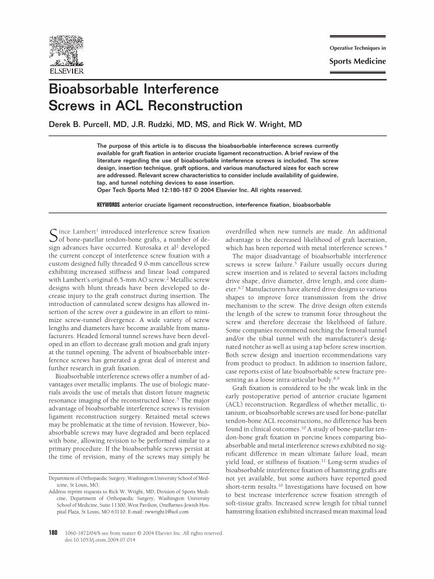
Figure 1 Sample of bioabsorbable interference fixation d(Arthrex), Sheathed Femoral (Arthrex), BioRCI HA (Smvatec), EndoPearl (Linvatec), Wedge (Linvatec), Gentleto right).
us manufactured sizes for each screw. t
ioabsorbablenterference Screwsitek (Westwood, MA) Products
he Absolute absorbable interference screw may be used foremoral or tibial fixation of soft tissue or bone-tendon-bonerafts (Fig. 1). The screw is made of amorphous L-PLA. Thecrew is cannulated for use with a guide-tipped screwdriver thatxtends beyond the length of the screw. The extension of thecrewdriver is an effort to eliminate the need for a guide pin forcrew placement. Alternatively, the screw may be placed underuidewire (1.1 mm) direction. The screw drive is hex head inesign, and the length of the screw represents the length of therive. A cannulated notcher for the femoral tunnel is availableor use. The screw tip is pointed with a starter thread to easensertion. The screw is available in multiple diameters (7-11
m) and 2 standard lengths (23 or 30 mm). However, the1-mm diameter screw is not available in the 23-mm lengthTable 1).
The Biocryl screw is a composite of tricalcium phosphate30%) and polylactic acid (70%) (�-TCP/PLA). The tricalciumhosphate serves as an osteoconductive material presenthroughout the screw, not just the outer surface. The screw maye used for femoral or tibial soft-tissue or bone-tendon-bonexation. The screw hub is chamfered to allow for a theoreticaltraumatic tissue fixation. The screw drive is hexagonal. Theorking distance of the screw drive is the length of the screw. A
tarter tap is available for insertion and recommended for use inard cortical bone. Mitek recommends that no more than 3 to 4
: Absolute (Mitek), Intrafix (Mitek), Bio-Cortical Distalephew), Bioscrew (Linvatec), Bioscrew Xtralock (Lin-
s (Arthrotek), and Bioabsorbable Wedge (Stryker) (left
evicesith & NThread
urns should be used with the starting tap to avoid loss of fixa-

Table 1 Bioabsorbable Interference Screws
Manufacturer Name Grafts Sites Composition CannulatedGWSize DD DL Notcher Tap Sizes (mm)
Mitek Absolute st or btb Femur and tibia Amorphous L-PLA Yes 1.1mm Hexagonal Full Yes No 7�23 or 308�23 or 309�23 or 3010�23 or 3011�30
Biocryl st or btb Femur and tibia �-TCP/PLA Yes 1 Hexagonal Full Yes 7�23 or 308�23 or 309�23 or 3010�23 or 3011�30
Intrafix st Tibia �-TCP/PLA Yes 1.1 Hexagonal Full No No 6,7,8,9, or 10�30Arthrex Sheathed Femoral st or btb Femur and tibia Crystalline PLLA Yes 1.1 Hexagonal Full Yes No 7,8,9 or 10�23
Full Thread Tibial st or btb Tibia Crystalline PLLA Yes 1.1 Hexagonal Full Yes No 7,8,9,10,11 or12�28
Round Delta Tapered st Femur Non-crystalline PLLA Yes 1.1 Hexagonal Full Yes No 8.5,7.5,8 or 8.5�289,9.5,10 or 11�28
Delta Tapered st Tibia Non-crystalline PLLA Yes 1.1 Hexagonal Full Yes No 7.5,8.5,9 or 9.5�3510,10.5,11 or 12�35
Bio-Cortical Proximal st Tibia PLLA Yes 1.1 Hexagonal Full No No 8,9 or 10�20Bio-Cortical Distal st Tibia PLLA No N/A Hexagonal Full No No 8,9,10 or 11�17Retro Screw st Tibia PLLA No N/A Hexagonal Full Yes No 8,9 or 10�20Bio-Tenodesis Screw st Tibia (not tunnel) PLLA Yes 2.4 Hexagonal No No 4�10
4.75 or 5.5�157,8 or 9�238�12
Smith & Nephew BioRCI st Femur and tibia PLA Yes 1.5mm Torx Full Yes Yes 7�25 or 30BioRCI HA PLA and HA 8�25,30 or 35
9�25,30 or 3510�30 or 35
Abbreviations: st, soft tissue; btb, bone-tendon-bone; GW, guidewire; DL, drive length; HA, hydroxyapatite.
182D.B.Purcell,J.R.Rudzki,and
R.W.W
right

tawa3
wt�iPttsgigddittstTtcaSsiatdtTamotsIe
AAccIuatftrigAgi
al
uctftAitAS1At
fTssdrt
fnifefiwpmatic
gabttcometbfpat
tnRte
Bioabsorbable interference screws 183
ion when the screw is placed. The Biocryl screw is placed overcannulated guidewire (1 mm). The insertion set is availableith a ratcheted screwdriver with Jacobs chuck. Screws are
vailable in 7-, 8-, 9-, and 10-mm diameters with 23- and0-mm lengths. An 11- � 30-mm screw is also available for use.The Intrafix system is used for tibial soft-tissue fixation
ith 4 bundle hamstring grafts (Fig. 1). Both the Intrafixapered screw and the accepting sheath are composed of-TCP/PLA (tricalcium phosphate/polylactic acid). The TCP
s included to serve as an osteoconductive agent. The TCP/LA composite is created with Mitek’s proprietary Micro Par-icle Dispersion technology to create a homogeneous blend ofhe 2 materials. The sheath portion of the implant has 4eparate ridged channels to allow each limb of the tendonraft to be separately fixed to the wall of the tunnel for max-mal graft exposure to bone during healing. The size of theraft and thus the size of the tunnel, sheath, and screw areetermined with the Mitek sizing block. The tibial tunnel isrilled 1 mm less than the final diameter, and only the cortex
s drilled with the final diameter drill. The tunnel is sequen-ially dilated with a dilator system over a guidewire (1.1 mm)o the final diameter. If the tunnel diameter is less than theheath diameter, then the corresponding Sheath Trial is usedo prenotch the tunnel before the graft is placed. The Mitekie Tensioner is used to equally tension each limb of the
endon graft separately to approximately 40 lb. The knee isycled per the usual protocol before fixation and held in theppropriate flexion/extension position for graft fixation. Theheath Trial is inserted to prepare the osseous tunnels and toeparate and compress the tendon graft limbs. When the grafts tight or the tunnel is small, the Sheath Trial may need to bedvanced with a mallet. After Sheath Trial removal, the In-rafix Sheath is inserted over a guidewire (1.1 mm) until theerotation tab is flush with the anterior tibial cortex. Eachendon limb is placed into a respective channel of the sheath.he Intrafix screw is inserted over the guidewire into theccepting sheath. The screw should pass into the sheath withinimal torque. Should a small amount of sheath extrusion
ccur, Mitek recommends cutting this back to the level of theibia. The Intrafix screw is headed with a hexagonal drivehaft. The Intrafix Sheath is available in a 30-mm length. Thentrafix Screw is available in 6-, 7-, 8-, 9-, and 10-mm diam-ters with a standard 30-mm length (Table 1).
rthrex (Naples, FL) Productsrthrex manufactures multiple bio-interference screws. Theomposition of the screws is PLLA with differing amounts ofrystallinity to allow for varying degradation rates. The Bio-nterference Screw Set includes the instruments necessary tose the Arthrex bioabsorbable screw system. The set includesquick-connect ratchet handle, screw driver shafts, femoral
unnel notcher, screw removal shafts to either remove orurther insert broken or damaged screws, and guide pin. Aorque-measuring device is available for attachment to theatchet screwdriver to quantify insertion torque. The major-ty of the screws are cannulated to allow placement over auidewire (1.1 mm) to minimize screw-tunnel divergence.n alternative is the Guide Pin Tip Screwdriver with an elon-ated tip extending beyond the screw length to allow for
nsertion of the femoral interference screw without the use of dguidewire. The screw drive is hexagonal and extends theength of the screw shaft.
The Sheathed Bio-Interference Femoral Screw may besed for both soft-tissue and bone-tendon-bone grafts. Screwomposition is PLLA (Fig. 1). A head is present on the screwo reduce graft motion at the intraarticular opening of theemoral tunnel. Screw threads are blunt to minimize injury tohe graft. The femoral notcher is used before screw insertion.fter insertion of the guidewire, the screw sheath is inserted
nto the femoral tunnel to prevent graft rotation and lacera-ion. The screw is subsequently inserted over a guidewire.lternatively, the screw may be placed with the Guide Pin Tipcrewdriver. Available sizes include the 7-, 8-, 9-, and0-mm diameters with a standard 23-mm length (Table 1).rthrex recommends the use of a screw equal to or 1 mm less
han the tunnel diameter.The Full Thread Bio-Interference Tibial Screw is designed
or tibial fixation of soft-tissue and bone-tendon-bone grafts.he screw is headless with blunt threads. Chemical compo-ition is PLLA. Insertion is performed over a guide pin. Thecrew has a standard length of 28 mm with multiple availableiameters of 7, 8, 9, 10, 11, and 12 mm (Table 1). Arthrexecommends using a screw diameter that is 1 to 2 mm largerhan the tunnel diameter for soft-tissue grafts.
The Round Delta Tapered Bio-Interference Screw is foremoral fixation of soft-tissue grafts. The screw consists ofoncrystalline PLLA to allow for faster degradation of the
mplant. A head is present to decrease graft motion at theemoral tunnel entrance. A taper of 1.5 mm is present to easentry of the screw into the femoral tunnel and to increasexation as the screw is inserted fully into the tunnel. A guide-ire should be used for screw insertion. The screw may belaced with standard Arthrex bio-interference screw instru-entation. However, a separate cannulated screwdriver is
vailable for use with the Delta Tapered screw that engageshe round head of the screw. A standard 28-mm screw lengths available. Multiple screw diameters are manufactured in-luding 6.5, 7.5, 8, 8.5, 9, 9.5, 10, and 11 mm (Table 1).
To improve femoral soft-tissue fixation with soft-tissuerafts, a Coring Reamer is available to create a bone block forttachment to the soft-tissue graft on the femoral side. Theone block is obtained during tibial tunnel preparation. Theibial tunnel guide pin is drilled to within 1 to 2 cm of theibial plateau. The guide pin is removed and replaced with aollared pin. The corresponding Coring Reamer is insertedver the guidewire, and the tunnel is completed. The proxi-al tibial bone obtained is fashioned into a button the diam-
ter of the femoral tunnel. The button is secured as a leader tohe proximal aspect of the soft-tissue graft with a no. 2raided polyethylene suture. The graft is introduced into theemoral tunnel, bone block first. After the Round Delta Ta-ered Bio-Interference Screw is placed, the bone block willct as an additional wedge to prevent graft slippage within theunnel.
The Delta Tapered Bio-Interference Screw is designed foribial fixation of soft-tissue grafts. The composition remainsoncrystalline PLLA. The screw design is very similar to theound Delta Tapered Bio-Interference Screw with the same
heoretical considerations as previously mentioned. How-ver, the screw is headless and manufactured only in a stan-
ard 35-mm length to maximize purchase of the soft-tissue
guhptlma1tlma
mictItpalonp1ta1
ofaiatfiptogdicl
sttflsaI1Td
asaAstnhaTmttiTsntpdlam
SToCrstpsf
are, an
184 D.B. Purcell, J.R. Rudzki, and R.W. Wright
raft within the tibial tunnel. The femoral notcher may besed to create tibial cortical grooves to decrease the likeli-ood of graft rotation during screw insertion. The screw islaced over a guidewire with the use of the standard or delta-apered screwdriver. Placement of the screw between the 4imbs of a hamstring tendon graft is possible to allow maxi-
al contact of the graft with the tunnel wall. Screw diametersvailable are 7.5, 8.5, 9, 9.5, 10, 10.5, 11, and 12 mm (Table). Arthrex recommends a screw 0.5 mm greater than theunnel diameter. Should insertion torque be less than 15 inchb when placing the screw, then the manufacturer recom-
ends supplemental fixation with an additional device suchs the Bio-Tenodesis Screw.
The Bio-Cortical Interference Screw is designed for proxi-al and distal tibial tunnel fixation of soft-tissue grafts in
nstances of decreased bone density. The screws are a PLLAonstruct. The proximal screw is cannulated and inserted tohe level of the tibial plateau joint line over a guide pin.nsertion to this level is purported to decrease graft motion athe tunnel orifice as well as increase fixation strength byroviding cortical bone fixation. The proximal screw is avail-ble in 8-, 9-, and 10-mm diameters with a single 20-mmength (Table 1). The guidewire is removed after placementf the proximal screw, and the distal screw is inserted. Can-ulation for the distal screw is not present, and the screw islaced with the angled side flush with the tibial cortex (Fig.). The distal screw provides additional fixation while sealinghe distal tibial cortex to decrease the likelihood of postoper-tive hematoma formation. Screw diameters of 8, 9, 10, and1 mm are available with a standard 17-mm length (Table 1).The RetroScrew is a tibial soft-tissue fixation device made
f PLLA. The screw is headed and inserted in a retrogradeashion. The head theoretically decreases motion of the graftt the tibial tunnel opening. Equipment necessary for screwnsertion includes the RetroScrew Driver, shoehorn cannula,nd FiberStick suture (no. 2 FiberWire). The intra-articularibial tunnel must be notched with a 4-mm burr to prepareor screw insertion. Before graft passage, the FiberStick stitchs shuttled up the tibial tunnel and out the anteromedialortal. The graft is passed, and the femoral side is fixed withhe desired technique. The RetroScrew Driver is threadedver the FiberStick suture up the tibial tunnel, anterior to theraft. The Retroscrew is placed antegrade over the anterome-ial FiberStick suture, secured with a mulberry knot, and
nserted through the anteromedial portal with the shoehornannula. After cannula removal, the screw is seated to the
Figure 2 Drive designs: cruciate, hexagonal, trilobe, squ
aser mark line on the driver by pulling on the FiberStick t
uture. The knee is then cycled and positioned for final fixa-ion. The graft is tensioned, and the screw is inserted byurning the screwdriver counterclockwise until the head isush with the proximal tibial tunnel opening. The FiberStickuture is pulled out the anteromedial portal. Additional fix-tion may be accomplished by placing a distal Bio-Corticalnterference Screw. The RetroScrew is available in 8-, 9-, and0-mm diameters with a standard 20-mm length (Table 1).he manufacturer recommends use of a screw 1 mm larger iniameter than the tibial tunnel.Supplemental tibial soft-tissue interference fixation may be
chieved with the Bio-Tenodesis Screw system. The masteret for the system includes cannulated drills, headed reamers,nd multiple screwdrivers with a tear drop ratcheted handle.dditional instruments are optional and include additionalize drills, guide pin, no. 2 FiberWire suture, disposable su-ure passing wire, and cannulated drill sleeves. The Bio-Te-odesis Screw is composed of PLLA, and the screws areeaded. If additional soft-tissue fixation is desired, then annterior tibial drill hole is made with the 2.4-mm guidewire.he guidewire is overdrilled to create a hole that will accom-odate the size of the tendon graft. A whip-stitch is placed in
he end of the soft-tissue graft, and a mulberry knot is tied athe end of the graft. A screw that equals the size of the tunnels chosen and placed over the suture in a retrograde fashion.he suture passing wire is placed through the cannulatedcrewdriver to shuttle the suture through the drive mecha-ism. The suture is pulled through the screwdriver to deliverhe screw and tendon to the drive shaft. The tendon is thenushed into the hole over the guidewire. When the desiredepth and tension are achieved, the screw is inserted to the
evel of the screw head. Multiple screw diameters are avail-ble (4 � 10 mm; 4.75 and 5.5 � 15 mm; 7, 8, and 9 � 23m; 8 � 12 mm) (Table 1).
mith & Nephew (Andover, MA) Productshe BioRCI screw may be used for the fixation of soft-tissuer bone-tendon-bone grafts in the femoral or tibial tunnels.omposition of the screw is PLA. The screw has a soft
ounded thread design and Torx design screw drive thatpans the length of the screw (Fig. 2). A screw with reversedhreads for use with right knees is available to potentiallyrevent rotation of the graft to an anterior position duringcrew insertion, which would block screw visualization. Theemoral screws have an oversized head. A 1.5-mm guidewire,
d torx (left to right).
ap, and tunnel notch device are available. The femoral screw

iTmfliflelkhpaa
LTtcaTtdTafa
etign3aptladGpcd
ttsstDcsTmonw
t
1statdsTaogbStmmWWdattmmpwfspTsW2
cgatimdmrButsdmgaieE(
ATs
Bioabsorbable interference screws 185
s inserted 5 mm deep to the femoral tunnel cortical opening.he tibial screw is likewise inserted over a guidewire. Theanufacturer recommends starting the screw at 30° to 40° ofexion and then extending the knee to ensure full extension
s possible. The tibial screw is then fully inserted at the pre-erred knee position for graft fixation. Multiple diameters andengths are available for the left knee (7 � 25 and 7 � 30 withither 7- or 8-mm heads; 8 and 9 mm in 25-, 30-, or 35-mmengths; 10 � 30 or 35 mm) (Table 1). Reverse thread rightnee femoral screws are made in 7 � 25 mm with an 8-mmead and 8 � 25 mm dimensions. A BioRCI-HA screw is alsoroduced and is similar to the BioRCI screw except for theddition of hydroxyapatite to the PLA (Fig. 1). The hydroxy-patite is added to serve as an osteoconductive agent.
invatec (Largo, FL) Productshe BioScrew may serve as interference fixation of bone-
endon-bone grafts at both tibial and femoral tunnels. Highlyrystalline PLLA makes up the screw. The screw is headlessnd cannulated for placement over a guidewire (1.14 mm).he screw drive is a trilobed design extending the length of
he screw (Fig. 2). A locking device is built into the screw-river to hold the guidewire in place during screw insertion.he Guardsman Graft Protector slides over the screwdrivernd screw to minimize graft injury during insertion. Manu-actured sizes are 7-, 8-, and 9-mm diameters with 20-, 25-,nd 30-mm lengths (Table 2).
The BioScrew Xtralok screw provides soft-tissue interfer-nce fixation on the tibial side (Fig. 1). The screw is similar tohe BioScrew design with a few modifications. Linvatec mod-fied the trilobe drive system to decrease screw failure. Auidewire (1.14 mm) system is used for screw insertion. Aew universal BioScrew driver must be used with the screw. A5° angled face is present on the proximal end of the screw tollow flush placement with the anterior tibial cortex. Theroximal aspect expands 1 mm in diameter to increase fixa-ion at the cortical surface of the anterior tibia. The screwength should be equal to or less than the tibial tunnel length,nd diameter should not exceed 2 mm larger than the tunneliameter. A notch may be made at the tunnel orifice with theraFix Tunnel Notcher to ease screw insertion. Insertion iserformed until the screw is flush with the anterior tibialortical surface. The screw is available in 9-, 10-, and 11-mmiameters in both 35- and 40-mm lengths (Table 2).The SmartScrew ACL is designed for the fixation of bone-
endon-bone or soft-tissue grafts within the tibial and femoralunnels. Screw composition is 96L/4D PLA copolymer. Thecrew drive design is square and extends the length of thecrew. A single-use protection sheath may be packaged withhe 7-, 8-, and 9-mm screws for use during femoral insertion.ilation/notching of the femoral tunnel is performed with theannulated dilator/notcher before screw insertion. A counter-ink is present on the dilator/notcher to ease screw insertion.he screw is cannulated and placed over a guide pin (1.6m). Before inserting the tibial screw, the manufacturer rec-
mmends expanding the tibial tunnel opening with the can-ulated dilator. Diameters include 7.3, 8, 9, 10, and 11 mmith 25-, 30-, and 35-mm lengths (Table 2).The Wedge is an impacted interference fixation device for
he femoral tunnel when using bone-tendon-bone grafts (Fig. t
). Composition is a 96L/4D PLA copolymer. The instrumentet comes with the sizer, notcher, extractor, driver, and ex-raction drill bit. The drive screws halfway into the implantnd is used for implant impaction. The bone-tendon bone-endon-bone graft is prepared, and the femoral tunnel isrilled to the depth and width of the femoral bone block. Themaller of the 2 bone blocks is chosen for the femoral tunnel.he Wedge Notcher device is used to place a notch in thenterior aspect of the femoral tunnel at the 2 o’clock positionn a left knee and the 10 o’clock position on a right knee. Theraft is placed per the usual surgeon preference. The bonelock must be entirely within the femoral tunnel. The Wedgeizer is then placed between the bone block and femoralunnel wall, and the gap size is read directly from the laserarked etching. The ideal gap is 2 to 3 mm. Gap size deter-ines the size of the Wedge used (gap �3 mm, 5-mmedge; 3-5 mm, 7-mm Wedge; �5 mm, 9-mm or largeredge). The Wedge is then inserted through the anterome-
ial portal with the insertion device into the previously cre-ted notch. The grooved portion is placed anteriorly againsthe femoral tunnel wall, and the pyramidal portion will facehe graft bone plug. The Wedge is impacted into place with aallet. If advancement does not occur with each stroke of theallet, then the Wedge should be removed and a smaller sizelaced. The device is advanced until the back surface is flushith the tunnel. The inserter is then unscrewed and removed
rom the device. The graft is then fixed on the tibial side perurgeon preference. If the Wedge requires removal, the im-lant drive hole is overdrilled with the Extraction Drill Bit.he Wedge Extractor is screwed into the device fully, and thelap hammer mechanism will remove the implant. The
edge is available in 5-, 7-, and 9-mm diameters with both0- and 25-mm lengths (Table 2).The EndoPearl device is a supplemental device used in
ombination with interference screw fixation of soft-tissuerafts at the femoral tunnel (Fig. 1). The EndoPearl acts as andditional interference device proximal to the traditional in-erference screw in femoral fixation of soft-tissue grafts. Themplant is made of highly crystalline PLLA. The size should
atch the diameter of the femoral tunnel. If the tunnel isilated, a size 1 mm less than the tunnel diameter is recom-ended for use. The depth of the femoral tunnel should
eflect the length of the screw plus the size of the EndoPearl.efore graft insertion, the ends of the graft are prepared in thesual manner. The device is sutured to the proximal end ofhe soft-tissue graft via the EndoPearl eyelets. The chosencrew is placed adjacent to the graft while touching the En-oPearl, and the graft is marked with a surgical pen. Theark allows for appropriate tunnel depth insertion of the
raft and EndoPearl device while ensuring that the screw willbut the surface of the implant after placement. The graft andmplant are inserted in the usual fashion, and a bio-interfer-nce screw is placed to the level of the previous mark. ThendoPearl is available in 7-, 8-, and 9-mm diametersTable 2).
rthrotek (Warsaw, IN) Productshe Gentle Threads interference screw provides fixation foroft-tissue or bone-tendon-bone grafts at the femoral and
ibial tunnels. The screw is composed of the LactoSorb co-
Table 2 Bioabsorbable Interference Screws
Manufacturer Name Grafts Sites Composition CannulatedGWSize DD DL Notcher Tap Sizes (mm)
Linvatec BioScrew btb Femur and tibia Crystalline PLLA Yes 1.14 Tri-lobed Full Yes No 7�20,25 or 308�20,25 or 309�20,25 or 30
BioScrew XtraLok st Tibia Crystalline PLLA Yes 1.14 Tri-lobed Full Yes No 9�35 or 4010�35 or 4011�35 or 40
SmartScrew ACL st or btb Femur and tibia 96L/4D PLA Yes 1.6 Square Full Yes No 7.3�25,30 or 358�25,30 or 359�25,30 or 3510�25,30 or 3511�25,30 or 35
Wedge btb Femur 96L/4D PLA No No N/A Partial Yes No 5�20 or 257�20 or 259�20 or 25
EndoPearl st Femur Crystalline PLLA N/A N/A N/A N/A N/A N/A 7,8, or 9Arthrotek Gentle Threads st or btb Femur and tibia PLLA/PGA Yes 1.5 Cruciate Full Yes No Femur 7�20 or 25
Femur 8�20 or 25Femur 9�20 or 25Femur 10�25Tibia 7�20,25 or 30Tibia 8�20,25 or 30Tibia 9�20,25 or 30Tibia 10�20Revision 11 or 12�25
Stryker BioabsorbableWedge
st or btb Femur or tibia Crystalline PLLA Yes 1 Square Full Yes Yes 7,8, or 9�23
7,8,9 or 10�287,8,9,10 or 11�35
Biosteon Wedge st or btb Femur or tibia HA and crystalline PLLA Yes 1 Star Full Yes Yes 7,8 or 9�237,8 or 9�289,10,11 or 12�35
Abbreviations: st, soft tissue; btb, bone-tendon-bone; GW, guidewire; DL, drive length; HA, hydroxyapatite.
186D.B.Purcell,J.R.Rudzki,and
R.W.W
right

pafoipmwfwfof2imsAnd
STStpslitSo(
sfditcsAtaf21
DBl
frtdna
APTtSmttHbol
R
1
1
1
Bioabsorbable interference screws 187
olymer consisting of 82/18 mixture of PLLA/poly-glycoliccid. Both round-head and fully threaded designs are manu-actured. The drive design is cruciate and extends the lengthf the screw (Fig. 2). The screw and driver are cannulated fornsertion. A notcher is used within the femoral tunnel beforelacement of the guidewire (1.5 mm). After guidewire place-ent, a dilator is tapped into the tunnel to a depth consistentith the screw length. The headed screw is recommended for
emoral interference fixation. The tibial tunnel is preparedith the dilator to the estimated depth of the tibial screw. A
ully threaded screw is used for tibial fixation and is placedver guidewire direction. The round head screws are manu-actured in 7-, 8-, and 9-mm diameters with both 20- and5-mm lengths (Table 2). A 10- � 25-mm round head screw
s also available. Fully threaded screws come in 7, 8, and 9m diameters with 20, 25, and 30 mm lengths (Table 2). A
ingle 10 � 20-mm fully threaded screw may be used as well.rthrotek also has a “Revision Screw” for large revision tun-els that is 25 mm in length with both 11- and 12-mmiameters available.
tryker (Santa Clara, CA) Productshe Bioabsorbable Wedge ACL Interference Screw fromtryker is indicated for use with both soft-tissue and bone-endon-bone grafts at the femoral or tibial site (Fig. 1). Com-osition is crystalline PLLA. The screw design incorporates amall head. The screw drive is square and spans the screwength (Fig. 2). Full cannulation of the screw is present fornsertion over a guidewire (1 mm). Notching and tapping ofhe femoral tunnel is recommended before screw insertion.crew sizes include 7, 8, or 9 mm by 23-mm length; 7, 8, 9,r 10 mm by 28 mm; and 7, 8, 9, 10, or 11 mm by 35 mmTable 2).
The Biosteon Wedge interference screw may be used foroft-tissue or bone-tendon-bone grafts at both the tibial andemoral tunnels. The screw composition incorporates hy-roxyapatite (25%) with crystalline PLLA (75%) to allow for
nclusion of an osteoconductive material. The screw is essen-ially fully threaded with a small head. The drive system is aruciate star pattern extending the length of the screw. Thecrew is cannulated for guidewire (1 mm)-directed insertion.
tunnel notcher is recommended for use in the femoralunnel. Multiple tap sizes come with the instrumentation andre recommended by the manufacturer. Implants are manu-actured in 7-, 8-, and 9-mm diameters with either 23- or8-mm lengths. The 35-mm length screw is available in 9-,0-, 11-, and 12-mm diameters (Table 2).
iscussionioabsorbable interference screws are an alternative to metal-
ic implants for ACL reconstruction. The biomaterials used
or screw production vary and exhibit differing degradationates. Screws have been developed with indications for soft-issue fixation, bone-tendon-bone fixation, or both. Screwesigns have evolved with different characteristics and tech-ique recommendations to ease insertion, improve fixation,nd decrease the incidence of screw failure during insertion.
uthors’referred Characteristics
he authors believe several design characteristics are impor-ant considerations when selecting bioabsorbable screws.crew insertion may be more difficult with these screws thanetal alternatives. Drive lengths that span the screw length,
aps, and tunnel notching devices can be critical complimen-ary insertion aides. Guidewires may also ease insertion.owever, small guidewire sizes present the possibility of wireend or breakage. A variety of sizes available for use offerptions when soft-tissue grafts are unexpectedly small orarge or bone density is different than anticipated.
eferences1. Lambert KL: Vascularized patellar tendon graft with rigid internal fix-
ation for anterior cruciate ligament insufficiency. Clin Orthop 172:85-89, 1983
2. Kurosaka M, Shinichi Y, Andrish JT: A biomechanical comparison ofdifferent surgical techniques of graft fixation in anterior cruciate liga-ment reconstruction. Am J Sports Med 15:225-229, 1987
3. Shellock FG, Mink JH, Curtin S, et al: MR imaging and metallic im-plants for anterior cruciate ligament reconstruction: Assessment of fer-romagnetism and artifact. J Magn Reson Imaging 2:225-228, 1992
4. Matthews LB, Softer SR: Pitfalls in the use of interference screws foranterior cruciate ligament reconstruction. Arthroscopy 5:225-226,1989
5. Brand J, Weiler A, Caborn DNM, et al: Graft fixation in cruciate liga-ment reconstruction. Am J Sports Med 28:761-774, 2000
6. Weiler A, Hoffman RFG, Siepe CJ, et al: The influence of screw geom-etry on hamstring tendon interference fixation. Am J Sports Med 28:356-359, 2000
7. Weiler A, Windhagen HG, Raschke MJ, et al: Biodegradable interfer-ence screw fixation exhibits pull-out force and stiffness similar to tita-nium screws. Am J Sports Med 26:119-128, 1998
8. MacDonald P, Arneja A: Biodegradable screw presents as a loose intra-articular body after anterior cruciate ligament reconstruction. Arthros-copy 19:E54, 2003
9. Shafer BL, Simonian PT: Broken poly-L-lactic acid interference screwafter ligament reconstruction. Arthroscopy 18:E35, 2002
0. Fu FH, Bennett GH, Ma B, et al: Current trends in anterior cruciateligament reconstruction part II. Operative procedures and clinical cor-relations. Am J Sports Med 28:124-130, 2000
1. Kousa P, Jarvinen TLN, Kannus P, et al: Initial fixation strength ofbioabsorbable and titanium interference screw in anterior cruciate lig-ament reconstruction. Am J Sports Med 29:420-25, 2001
2. Selby JB, Johnson DL, Hester P, et al: Effect of screw length on bioab-sorbable interference screw fixation in a tibial bone tunnel. Am J Sports
Med 29:614-619, 2001