'Non-self' mutation in a Drosophila model of expanded CAG ...
239
‘Non-self’ mutation in a Drosophila model of expanded CAG repeat neurodegenerative disease A thesis submitted for the degree of Doctor of Philosophy March 2019 Andrew William Scott, BSc. (Hons) School of Biological Sciences, Department of Molecular and Biomedical Science The University of Adelaide
-
Upload
khangminh22 -
Category
Documents
-
view
5 -
download
0
Transcript of 'Non-self' mutation in a Drosophila model of expanded CAG ...

‘Non-self’ mutation in a Drosophila model of expanded CAG
repeat neurodegenerative disease
A thesis submitted for the degree of Doctor of Philosophy
March 2019
Andrew William Scott, BSc. (Hons)
School of Biological Sciences, Department of Molecular and Biomedical Science
The University of Adelaide


I
Table of Contents
Index of Figures and Tables ........................................................................... III
Declaration ................................................................................................. VII
Acknowledgements ....................................................................................... IX
Abbreviations ................................................................................................ XI
Nomenclature ............................................................................................. XV
Abstract .................................................................................................... XVII
CHAPTER 1: Introduction ................................................................................ 1
1.1 Expanded repeat neurodegenerative disease .................................................................. 2
1.1.1 Polyglutamine (PolyQ) ............................................................................................... 2
1.1.2 Repeat-associated non-AUG (RAN) translation ........................................................ 3
1.1.3 Single-stranded RNA (ssRNA) .................................................................................... 4
1.1.4 Double-stranded RNA (dsRNA) ................................................................................. 5
1.2 Innate inflammation ......................................................................................................... 7
1.2.1 Pattern recognition receptors (PRRs) ....................................................................... 8
1.2.2 Innate immune signalling .......................................................................................... 9
1.2.3 Degradative pathways............................................................................................. 11
1.2.4 Mitochondria as an antiviral hub ............................................................................ 12
1.2.5 Inflammation and neurodegeneration ................................................................... 15
1.3 Glial cells and other non-neuronal cells of the nervous system .................................... 17
1.3.1 The development and normal functions of glia ...................................................... 17
1.3.2 The neurovascular unit (NVU) ................................................................................. 20
1.3.3 Glial cells in neurodegenerative disease ................................................................. 21
1.3.4 Glial cells in Drosophila ........................................................................................... 24
1.4 Drosophila as a model organism in immunity and neurodegeneration ......................... 30
CHAPTER 2: Materials & Methods ................................................................ 35
2.1 Materials ......................................................................................................................... 35
2.2 Methods .......................................................................................................................... 39
CHAPTER 3: ‘Non-self’ recognition of repeat dsRNA ..................................... 43
3.2 The viral suppressor protein CrPV1A rescues repeat dsRNA-mediated pathology ....... 46
3.3 Transcript analysis of the inflammatory response to expanded repeat dsRNA ............. 49

II
3.3 Chapter Discussion ......................................................................................................... 60
CHAPTER 4: Innate inflammatory pathways in the mediation of dsRNA pathology .................................................................................................... 69
4.1 The JAK/STAT pathway contributes modestly to dsRNA pathology .............................. 71
4.2 Mitochondrial quality control mediates repeat dsRNA pathology ................................ 78
4.3 Chapter Discussion ......................................................................................................... 86
CHAPTER 5: The contribution of glial cell subtypes in repeat dsRNA-mediated neurodegeneration ...................................................................................... 93
5.1 Phenotypical confirmation of the expanded repeat dsRNA construct combinations ... 95
5.2 Pan-glial expression of repeat dsRNA is highly pathogenic ........................................... 97
5.3 Subperineural glial expression of repeat dsRNA is pathogenic ..................................... 99
5.4 Wrapping glial expression of repeat dsRNA is pathogenic .......................................... 104
5.5 Ensheathing glial expression of repeat dsRNA is pathogenic or mildly neuroprotective based on dsRNA construct combination ............................................................................ 109
5.6 Astrocyte-like glial expression of repeat dsRNA is pathogenic or mildly neuroprotective based on dsRNA construct combination ............................................................................ 117
5.7 Cortex glial expression of repeat dsRNA causes limited pathogenicity ...................... 122
5.8 Phagocytic effectors contribute modestly to the dsRNA eye pathology..................... 126
5.9 Chapter Discussion ....................................................................................................... 133
CHAPTER 6: Final Discussion ...................................................................... 141
6.1 Summary of results ...................................................................................................... 141
6.2 Implications for expanded repeat neurodegenerative diseases ................................. 144
6.3 Considerations for this study ....................................................................................... 146
6.4 Future directions .......................................................................................................... 149
Appendix ................................................................................................... 153
Appendix A ......................................................................................................................... 153
References ................................................................................................. 155
III
Index of Figures and Tables
Chapter 1
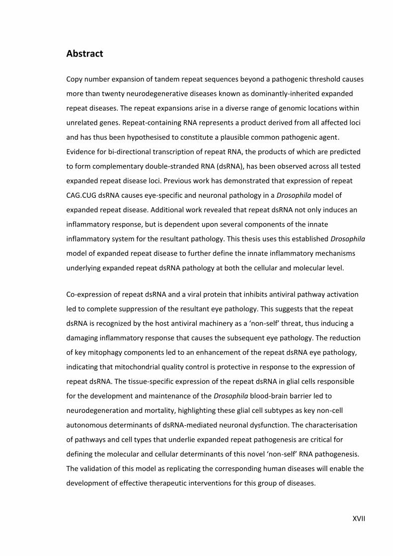
Figure 1.1: Location and composition of repeat sequences giving rise to dominantly-inherited expanded repeat disease. ........................................................................................... 3
Figure 1.2: Homology between selected DExD/H-box helicase proteins in human and Drosophila. .................................................................................................................................. 7
Table 1.1: Cellular localisation of known RNA pattern recognition receptors in vertebrates and their ligands ......................................................................................................................... 9
Figure 1.3: Diagram of a prototypical innate immune signalling pathway. ............................. 10
Figure 1.4: Schematic diagram of the autophagy pathway. .................................................... 12
Figure 1.5: Overview of RLR-MAVS-mediated antiviral signalling in vertebrates. ................... 14
Figure 1.6: Vertebrate glial cells and their developmental origins. ......................................... 19
Figure 1.7: The glial cells of Drosophila melanogaster. ........................................................... 26
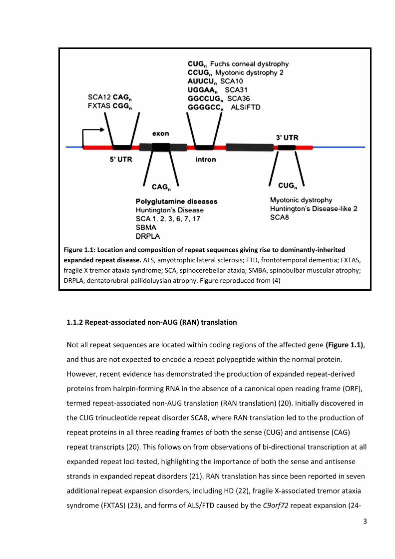
Figure 1.8: Expression of CAG.CUG repeat constructs using the GAL4/UAS system. .............. 32
Figure 1.9: Graphical representation of findings in the rCAG.CUG dsRNA Drosophila eye model of expanded repeat disease. ......................................................................................... 33
Chapter 3
Figure 3.1: Ectopic expression of CrPV1A completely rescues dsRNA-mediated pathology. .. 47
Figure 3.2: Summary diagram regarding CrPV1A-mediated suppression of expanded repeat dsRNA eye pathology. .............................................................................................................. 48
Figure 3.3: The antimicrobial peptide Drosomycin is significantly upregulated in response to repeat dsRNA. ........................................................................................................................... 50
Figure 3.4: Drosophila TNF orthologue Eiger is not significantly altered in response to repeat dsRNA. ...................................................................................................................................... 52
Figure 3.5: Expression levels of the antiviral peptides Vago and Nazo are not significantly altered in response to repeat dsRNA. ...................................................................................... 53
Figure 3.6: The STING/Relish/dIKKβ-mediated antiviral pathway in Drosophila. .................... 54
Figure 3.7: Expression of CG33926 is significantly downregulated in response to repeat dsRNA. ...................................................................................................................................... 55
Figure 3.8: Expression of IκB gene Charon is not significantly altered in response to repeat dsRNA. ...................................................................................................................................... 57
IV
Figure 3.9: Antiviral effector molecule STING is not significantly altered in response to repeat dsRNA. ...................................................................................................................................... 58
Figure 3.10: NF-κB transcription factor Relish is not significantly altered in response to repeat dsRNA. ...................................................................................................................................... 60
Chapter 4
Figure 4.1: Overview of the Drosophila JAK/STAT pathway. ................................................... 71
Figure 4.2: Reduction of upd1 makes no obvious contribution to repeat dsRNA-mediated eye pathology. ................................................................................................................................ 73
Figure 4.3: Upd3 contributes modestly to repeat dsRNA-mediated eye pathology. .............. 74
Figure 4.4: Alteration of STAT92E has limited effect on repeat dsRNA-mediated eye pathology. ................................................................................................................................ 76
Figure 4.5: Overview of mitochondrial quality control pathways. .......................................... 79
Figure 4.6: Reduction of PINK1 enhances repeat dsRNA-mediated eye pathology................ 80
Figure 4.7: Parkin restricts repeat dsRNA-mediated eye pathology. ...................................... 81
Figure 4.8: Reduction of Ref(2)P enhances repeat dsRNA-mediated eye pathology. ............ 83
Figure 4.9: Reduction of ECSIT enhances repeat dsRNA-mediated eye pathology. ............... 85
Figure 4.10: Proposed interaction between mitophagy and expanded repeat dsRNA pathology. ................................................................................................................................ 87
Chapter 5
Table 5.1: Summary of the glial subtype-specific drivers. ....................................................... 94
Figure 5.1: The relative levels of eye disruption caused through the expression of the CAG.CUG~100
repeat dsRNA construct combinations within the Drosophila eye. ................... 96
Figure 5.2: Pan-glial expression of a ‘Weak’ dsRNA construct combination causes locomotor dysfunction and impacts survival. ........................................................................................... 98
Figure 5.3: Subperineural glia-specific expression of a ‘Weak’ dsRNA construct combination causes locomotor dysfunction but does not impact survival. ............................................... 101
Figure 5.4: Subperineural glia-specific expression of a ‘Medium’ dsRNA construct combination impacts locomotor function and survival. ....................................................... 102
Figure 5.5: Wrapping glia-specific expression of a ‘Weak’ dsRNA construct combination does not lead to defects in locomotor function or survival. .......................................................... 106
Figure 5.6: Wrapping glia-specific expression of either ‘Medium’ or ‘Strong’ dsRNA construct combinations lead to age-dependent climbing defects and reduced survival. .................... 107

V
Figure 5.7: Tract ensheathing glia-specific expression of a ‘Weak’ dsRNA construct combination leads to locomotor dysfunction and a moderate impact on survival. ..............111
Figure 5.8: Tract ensheathing glia-specific expression of either ‘Medium’ or ‘Strong’ dsRNA construct combinations cause varied effects on locomotor function whilst not impacting survival. ...................................................................................................................................112
Figure 5.9: Neuropil ensheathing glia-specific expression of a ‘Weak’ dsRNA construct combination causes locomotor dysfunction but does not impact survival. ..........................114
Figure 5.10: Neuropil ensheathing glia-specific expression of either ‘Medium’ or ‘Strong’ dsRNA construct combinations do not lead to defects in locomotor function, while a moderate impact on survival is observed in the ‘Strong’ dsRNA line. ...................................115
Figure 5.11: Astrocyte-like glia-specific expression of a ‘Weak’ dsRNA construct combination causes locomotor dysfunction but does not impact survival. ...............................................119
Figure 5.12: Astrocyte-like glia-specific expression of either ‘Medium’ or ‘Strong’ dsRNA construct combinations do not lead to defects in locomotor function, while survival is impacted in both dsRNA lines. ...............................................................................................120
Figure 5.13: Cortex glia-specific expression of a ‘Weak’ dsRNA construct combination does not affect locomotor dysfunction or survival. ........................................................................124
Figure 5.14: Cortex glia-specific expression of either ‘Medium’ or ‘Strong’ dsRNA construct combinations affect locomotor function early but do not progress with age, while differing effects on survival are observed. ...........................................................................................125
Figure 5.15: Reduction of Draper (drpr) has minimal effect on repeat dsRNA-mediated eye pathology. ...............................................................................................................................127
Figure 5.16: Expression of individual Draper (drpr) isoforms have minor effects on repeat dsRNA-mediated eye pathology. ............................................................................................129
Figure 5.17: Effect of Mcr alteration on repeat dsRNA-mediated eye pathology. ................132
Figure 5.18: Summary of results for individual glial cell subtypes expressing expanded repeat dsRNA. ....................................................................................................................................135
Chapter 6
Figure 6.1: Summary of experimental observations in the expanded repeat dsRNA Drosophila model. ..................................................................................................................143

VI

VII
Declaration
I certify that this work contains no material which has been accepted for the award of any
other degree or diploma in my name, in any university or tertiary institution and, to the best
of my knowledge and belief, contains no material previously published or written by another
person, except where due reference has been made in the text. In addition, I certify that no
part of this work will, in the future, be used in a submission in my name, for any other
degree or diploma in any university or other tertiary institution without the prior approval of
the University of Adelaide and where applicable, any partner institution responsible for the
joint-award of this degree.
I give consent to this copy of my thesis when deposited in the University Library, being made
available for loan and photocopying, subject to the provisions of the Copyright Act 1968.
I also give permission for the digital version of my thesis to be made available on the web,
via the University’s digital research repository, the Library Search and also through web
search engines, unless permission has been granted by the University to restrict access for a
period of time.
I acknowledge the support I have received for my research through the provision of an
Australian Government Research Training Program Scholarship.
Andrew Scott Date

VIII

IX
Acknowledgements
I would like to my supervisors, Prof. Rob Richards and Dr. Louise O’Keefe, for always offering
support and expert advice throughout the length of my Honours and PhD studies. Thank you
for introducing me to the wonderful world of Drosophila genetics and helping me sharpen
my scientific thinking, writing and speaking. I have truly had a fantastic time as a member of
the lab and a lot of that is thanks to you.
I would also like to thank all members of the Richards/O’Keefe laboratory, both past and
present, for creating a fantastic working environment. Special thanks to previous members
Amanda Choo, Cheng Shoou Lee and Danielle Webber for taking the time to teach me
valuable experimental skills and the sacred art of troubleshooting. Additionally, a special
thanks to Laura Hewson who has shared the same PhD rollercoaster ride for a number of
years, for the many discussions (scientific or otherwise). To the many wonderful Honours
students who have been in the Richards lab, thanks for bringing a fun vibe to the lab and for
laughing at my humorous jokes. A specific acknowledgement to undergraduate Karen
Raymond, a passionate student who contributed to some of the work described in this
thesis (noted in the relevant results section) and who was a breath of fresh air and
enthusiasm into what at times can be an all-consuming journey.
Finally, special thanks to my family who have been with me every step of my university
tenure, for offering unconditional support through some particularly challenging moments.
Thanks for showing interest in an area that I am yet to properly explain to you, and for
providing me with the ability to step away when required and breathe.

X
XI
Abbreviations
°C: degrees Celsius
μg: microgram
μL: microlitre
Aβ: amyloid beta
AD: Alzheimer’s disease
ADAR: Adenosine deaminase acting on RNA
AGO: Argonaute
AGS: Aicardi-Goutieres syndrome
ALG: astrocyte-like glia
ALS: Amyotrophic lateral sclerosis
AMP: antimicrobial peptide
A-T: Ataxia telangiectasia
ATXN: Ataxin
BAC: bacterial artificial chromosome
BBB: blood-brain barrier
C1q: Complement component 1q
CARD: Caspase recruitment domain
cDNA: complementary DNA
CG: cortex glia
cGAS: Cyclic GMP-AMP synthase
cm: centimetre
CrPV: Cricket paralysis virus
DAMP: damage/danger-associated molecular pattern
Dcr: Dicer
DCV: Drosophila C virus
DM: Myotonic dystrophy
Dome: Domeless
Drp1: Dynamin related protein 1
DRPLA: Dentatorubral-pallidoluysian atrophy
Drpr: Draper

XII
dsRNA: double-stranded RNA
ECM: extracellular matrix
EDTA: ethylene diamine tetra-acetic acid
EG: ensheathing glia
elav: embryonic lethal abnormal vision
ER: endoplasmic reticulum
FTD: Frontotemporal dementia
FXTAS: Fragile X tremor-ataxia syndrome
GFP: Green fluorescent protein
GMR: glass multimer reporter
HD: Huntington’s disease
HDL2: Huntington’s disease-like-2
Hop: Hopscotch
HTT: Huntington
IAP: Inhibitor of apoptosis
IFIH1: Interferon-induced helicase C domain-containing protein 1
IFN: Interferon
IKK: IκB kinase
IL: Interleukin
IMD: Immune deficiency
IMM: inner mitochondrial membrane
IR: inverted repeat
IRF3: Interferon regulatory factor 3
JAK: Janus kinase
JNK: c-Jun N-terminal kinase
LGB: lateral glioblast
LGP2: Laboratory of Genetics and Physiology 2
Loqs: Loquacious
M: molar
MANF: Mesencephalic astrocyte-derived neurotrophic factor
MAVS: Mitochondrial antiviral-signalling protein
MBNL: Muscleblind-like

XIII
Mcr: Macroglobulin complement-related
MDA5: Melanoma differentiation-associated protein 5
mg: milligram
MiC: MANF immunoreactive cell
MiMIC: Minos mediated integration cassette
mL: millilitre
mM: millimolar
mm: millimetre
MQ: MilliQ™ purified water
mRNA: messenger RNA
MS: Multiple sclerosis
mtDNA: mitochondrial DNA
mtROS: mitochondrial ROS
NEG: neuropil ensheathing glia
NF-κB: nuclear factor kappa-light-chain-enhancer of activated B cells
ng: nanogram
NG2: Nerve-glial antigen 2
NLR: NOD-like receptor
nM: nanomolar
NOD: Nucleotide-binding oligomerization domain
NPC: neuroepithelial precursor cell
nrv2: nervana 2
NVU: neurovascular unit
OMM: outer mitochondrial membrane
OPC: oligodendrocyte precursor cell
ORF: open reading frame
PAMP: pathogen-associated molecular pattern
PBac: PiggyBac
PCR: polymerase chain reaction
PD: Parkinson’s disease
PG: perineural glia
PINK1: PTEN-induced kinase 1

XIV
pmol: picomole
polyQ: polyglutamine
PRR: pattern recognition receptor
qRT-PCR: quantitative real-time polymerase chain reaction
RAN: repeat-associated non-AUG
Ref(2)P: Refractory to sigma P
repo: reversed polarity
RIG-I: Retinoic acid-inducible gene I
RING: rapid iterative negative geotaxis
RISC: RNA-induced silencing complex
RLR: RIG-I-like receptor
RNAi: RNA interference
ROS: reactive oxygen species
Rp49: Ribosomal protein 49
RT-PCR: reverse transcription polymerase chain reaction
SCA: Spinocerebellar ataxia
SG: stress granule
siRNA: small interfering RNA
SJ: septate junction
SMBA: Spinal bulbar muscular atrophy
SOP: sensory organ progenitor
SPG: subperineural glia
SQSTM1: Sequestosome 1
ssRNA: single-stranded RNA
STAT: Signal transducer and activator of transcription
STING: Stimulator of Interferon genes
TAE: tris-acetate EDTA
TDP43: TAR-DNA binding protein
TEG: tract ensheathing glia
TJ: tight junction
TLR: Toll-like receptor
TNF: Tumour necrosis factor

XV
UAS: upstream activation sequence
Upd: Unpaired
UPR: unfolded protein response
UPS: ubiquitin proteasome system
UTR: untranslated region
vDNA: viral DNA
VNC: ventral nerve cord
VSR: viral suppressor of RNAi silencing
w/v: weight/volume
Nomenclature
The Drosophila nomenclature used in this thesis is based on conventional notation as stated
on the Drosophila database, Flybase (www.flybase.org). Genes are shown by italicised text
(e.g. htt) and proteins are shown by non-italicised text (e.g. htt).

XVI
XVII
Abstract
Copy number expansion of tandem repeat sequences beyond a pathogenic threshold causes
more than twenty neurodegenerative diseases known as dominantly-inherited expanded
repeat diseases. The repeat expansions arise in a diverse range of genomic locations within
unrelated genes. Repeat-containing RNA represents a product derived from all affected loci
and has thus been hypothesised to constitute a plausible common pathogenic agent.
Evidence for bi-directional transcription of repeat RNA, the products of which are predicted
to form complementary double-stranded RNA (dsRNA), has been observed across all tested
expanded repeat disease loci. Previous work has demonstrated that expression of repeat
CAG.CUG dsRNA causes eye-specific and neuronal pathology in a Drosophila model of
expanded repeat disease. Additional work revealed that repeat dsRNA not only induces an
inflammatory response, but is dependent upon several components of the innate
inflammatory system for the resultant pathology. This thesis uses this established Drosophila
model of expanded repeat disease to further define the innate inflammatory mechanisms
underlying expanded repeat dsRNA pathology at both the cellular and molecular level.
Co-expression of repeat dsRNA and a viral protein that inhibits antiviral pathway activation
led to complete suppression of the resultant eye pathology. This suggests that the repeat
dsRNA is recognized by the host antiviral machinery as a ‘non-self’ threat, thus inducing a
damaging inflammatory response that causes the subsequent eye pathology. The reduction
of key mitophagy components led to an enhancement of the repeat dsRNA eye pathology,
indicating that mitochondrial quality control is protective in response to the expression of
repeat dsRNA. The tissue-specific expression of the repeat dsRNA in glial cells responsible
for the development and maintenance of the Drosophila blood-brain barrier led to
neurodegeneration and mortality, highlighting these glial cell subtypes as key non-cell
autonomous determinants of dsRNA-mediated neuronal dysfunction. The characterisation
of pathways and cell types that underlie expanded repeat pathogenesis are critical for
defining the molecular and cellular determinants of this novel ‘non-self’ RNA pathogenesis.
The validation of this model as replicating the corresponding human diseases will enable the
development of effective therapeutic interventions for this group of diseases.


1
CHAPTER 1: Introduction
Neurodegenerative disease constitutes an umbrella term describing a number of debilitating
conditions characterised by neuronal loss and subsequent inhibition of cognitive and/or
motor function. The probability of developing a neurodegenerative disease increases with
age (particularly beyond 60 years of age) which, when married with the ageing global
population, presents a significant and rising socioeconomic concern for both affected
families and the broader public (1). However, despite decades of intense research focus into
the mechanisms that underlie neurodegeneration, progress towards effective therapeutic
intervention remains limited, serving to highlight the complexity of the causal agents
mediating neurodegenerative disease development and progression.
Major neurodegenerative diseases include Alzheimer’s disease (AD), Parkinson’s disease
(PD), Huntington’s disease (HD), amyotrophic lateral sclerosis (ALS) and frontotemporal
dementia (FTD). Typically, two forms of each disease have been identified; inherited familial
forms are early-onset and arise through genetic mutation, whilst the more common
sporadic late-onset forms are thought to develop through contributions from both genetic
risk factors and environmental stressors. The characterisation of causal disease genes and
genetic risk factors implicated in neurodegenerative disease has provided clues into the
responsible underlying pathways.
Of these diseases, HD distinguishes itself by way of constituting a monogenic disorder,
whereas all of AD, PD, ALS and FTD can arise through mutations in a number of separate
(but often related) genes (2). Indeed, HD belongs to a distinct group of monogenic disorders,
together referred to as expanded repeat neurodegenerative diseases. Investigation into the
pathophysiology of HD and related CAG expanded repeat disorders forms the primary focus
of this thesis.

2
1.1 Expanded repeat neurodegenerative disease
The copy number expansion of a repeat sequence beyond a pathogenic threshold gives rise
to more than 20 dominantly-inherited neurodegenerative disorders (Figure 1.1) (3, 4). The
repeat sequences are highly unstable, leading to their expansion via defective DNA
mismatch repair (5-7). As such, expanded repeat diseases generally exhibit a molecular
phenomenon known as anticipation, whereby further expansion of the repeat length
through subsequent generations correlates with more severe clinical symptoms and an
earlier age-of-onset (8-10). Despite the individual repeat expansion lesions occurring in
unrelated genes (Figure 1.1), there exists substantial overlap between the diseases in terms
of neurological symptoms and copy number repeat disease thresholds, suggesting that a
common pathogenic pathway may underlie this family of diseases (4). Indeed, the recent
discovery of a hexanucleotide GGGGCC repeat constituting the most common cause of
familial ALS/FTD (11, 12) has highlighted the idea that other forms of neurodegenerative
disease may also share commonality in disease development and progression.
1.1.1 Polyglutamine (PolyQ)
There are several pathogenic products that can be derived from repeat expansion loci,
including both RNA and protein species that have been proposed as key toxic agents.
Expanded CAG trinucleotide repeat sequences that occur within the coding region of the
affected gene are typically translated into corresponding polyglutamine (polyQ) tracts
(Figure 1.1), which are thought to constitute the primary pathogenic agent in the so-termed
polyglutamine diseases (13-16). Indeed, polyQ toxicity has been demonstrated in a range of
animal models, including C. elegans, Drosophila and mouse (17). The molecular
mechanism(s) by which polyQ proteins drive cellular dysfunction are not completely clear;
mitochondrial dysfunction, transcriptional dysregulation, disrupted axonal transport and
inflammation have all been proposed to contribute to polyQ-mediated pathology (18, 19).
3
1.1.2 Repeat-associated non-AUG (RAN) translation
Not all repeat sequences are located within coding regions of the affected gene (Figure 1.1),
and thus are not expected to encode a repeat polypeptide within the normal protein.
However, recent evidence has demonstrated the production of expanded repeat-derived
proteins from hairpin-forming RNA in the absence of a canonical open reading frame (ORF),
termed repeat-associated non-AUG translation (RAN translation) (20). Initially discovered in
the CUG trinucleotide repeat disorder SCA8, where RAN translation led to the production of
repeat proteins in all three reading frames of both the sense (CUG) and antisense (CAG)
repeat transcripts (20). This follows on from observations of bi-directional transcription at all
expanded repeat loci tested, highlighting the importance of both the sense and antisense
strands in expanded repeat disorders (21). RAN translation has since been reported in seven
additional repeat expansion disorders, including HD (22), fragile X-associated tremor ataxia
syndrome (FXTAS) (23), and forms of ALS/FTD caused by the C9orf72 repeat expansion (24-
Figure 1.1: Location and composition of repeat sequences giving rise to dominantly-inherited
expanded repeat disease. ALS, amyotrophic lateral sclerosis; FTD, frontotemporal dementia; FXTAS,
fragile X tremor ataxia syndrome; SCA, spinocerebellar ataxia; SMBA, spinobulbar muscular atrophy;
DRPLA, dentatorubral-pallidoluysian atrophy. Figure reproduced from (4)

4
26). How do these non-canonical disease gene products contribute to expanded repeat
disease? Experimental work in yeast, Drosophila and cell lines investigating RAN translated
peptides derived from the C9orf72 locus have demonstrated that impairment to
nucleocytoplasmic transport may underlie neuronal dysfunction in ALS/FTD (27-29), while
endoplasmic reticulum (ER) stress and proteasome impairment have also been highlighted
as potential pathological mechanisms (30). Indeed, the field of RAN translation is still in its
infancy, as such it is likely that the current dedicated research focus will provide a much
greater understanding of RAN translation. This includes both the mechanisms surrounding
the initiation of RAN translation in a disease context and how the resultant proteins
contribute to disease development and progression. Furthermore, how the diverse array of
RAN-translated polypeptides derived from 4 and 5 base repeat sequences may contribute to
expanded repeat disease is yet to be uncovered.
1.1.3 Single-stranded RNA (ssRNA)
In diseases where either polyQ or RAN translated proteins (or both) are produced, it is likely
that they participate in the pathogenesis of the given disease, though the extent of this
contribution remains unclear. In addition, disease-specific protein products are yet to be
described in all forms of expanded repeat neurodegenerative disease. However, underlying
all expanded repeat loci is the production of repeat-containing RNA molecules, making RNA
a plausible common pathogenic agent (4, 31). Single stranded RNA (ssRNA) products derived
from repeat loci form hairpin structures (32, 33) which have been proposed to cause cellular
dysfunction through several mechanisms.
At the forefront is the ability of RNA hairpins to interact with and sequester RNA-binding
proteins, thus interrupting their normal function (34). For instance, co-localisation and
subsequent dysregulation of the Muscleblind (MBNL) family of splicing factors by repeat
RNA foci has been implicated in several expanded repeat diseases, including DM1 and 2 (35,
36), HDL2 (37), SCA8 (38) and FXTAS (39). Loss of MBNL alternative splicing recapitulates
many of the clinical symptoms associated with DM (40, 41), suggesting that RNA-mediated
sequestration of RNA-binding proteins may underlie key aspects of expanded repeat disease
pathogenesis. Of note, antisense repeat transcripts derived from SCA2 (42) and ALS/FTD (43)

5
repeat loci have also been demonstrated to form RNA foci, suggesting that both strands
produced from expanded repeat loci can potentially act to sequester RNA-binding proteins
and thus contribute to disease pathogenesis. Of note, a repeat-containing antisense
transcript to HTT (denoted as HTTAS_v1) was demonstrated to negatively regulate HTT
expression (44). The expression level of HTTAS_v1 was decreased in HD brain tissue,
suggesting that the transcript may be protective as opposed to detrimental (44). Thus, the
generation of antisense transcripts spanning expanded repeat loci may not be inherently
deleterious in the context of disease.
1.1.4 Double-stranded RNA (dsRNA)
In addition to ssRNA, the observation of bi-directional transcription across expanded repeat
loci (21) has raised the intriguing possibility of the opposing repeat transcripts forming
perfectly double-stranded RNA (dsRNA) molecules. Indeed, work from Lawlor et al in a
Drosophila model of CAG expanded repeat disease demonstrated that expression of 100
copies of either CAG (rCAG100) or CUG (rCUG100) ssRNA was not detrimental, while co-
expression of both transcripts together (rCAG100.CUG100) led to striking toxicity when
expressed in eye tissue, as well as dysfunction when expressed neuronally (45). The
observed pathology was dependent upon Dicer-2 (45), a key component of the RNA
interference (RNAi) pathway, which acts to process dsRNA into small 21 nucleotide RNA
fragments (21mers) in order to facilitate silencing of homologous transcripts (46, 47).
Indeed, an enrichment of CAG7 21mers was detected in rCAG100.CUG100 flies, indicating 1)
the activity of the RNAi pathway in response to the presence of expanded repeat RNA, and
2) that the RNAi pathway may underlie the pathology (45). These findings were confirmed in
an independent Drosophila model of DM1, where co-expression of 250 copies of both the
sense CUG and antisense CAG transcripts led to Dicer-2 dependent pathology (48). In
contrast to the dsRNA-mediated pathology observed in Lawlor et al and Yu et al, the
expression of ssRNA CGG/CCG repeat sequences in isolation led to toxicity in a Drosophila
model of fragile x-associated tremor/ataxia syndrome (FXTAS), while co-expression of the
repeat sequences ameliorated the toxicity in an RNAi-dependent fashion (49). This work
provides evidence that the RNAi pathway can also act protectively in response to repeat
dsRNA, more in line with its canonical role in antiviral transcript silencing (50). Interestingly,

6
CAG7 21mers have also been detected in human HD brain samples, and the neurotoxicity
exerted by these repeat RNAs was dependent upon Ago2, another key component of the
RNAi pathway (51). Thus, the RNAi pathway is also active in the vertebrate system in
response to expanded repeat RNA, further supporting its participation in disease
pathogenesis.
A subsequent microarray analysis conducted on Drosophila expressing rCAG100.CUG100
dsRNA found that a number of transcripts common to the innate immune response were
significantly altered in the presence of repeat dsRNA (52). Further analysis revealed that the
Toll immune signalling pathway was required for the dsRNA pathology, while autophagy
plays a key role in restricting the observed toxicity (52). Finally, the presence of repeat
dsRNA induced an inflammatory response, as measured through the transcriptional
upregulation of both Drosomycin, a downstream peptide synthesised by the Toll signalling
pathway, and Eiger, the Drosophila orthologue of potent inflammatory cytokine tumour
necrosis factor (TNF) (52). Taken together, the results suggest that expanded repeat dsRNA
could represent a foreign or ‘non-self’ molecule to the host innate immune system, thus
invoking an inflammatory response that could be detrimental to cellular function and
survival. Of note, the rCAG100.CUG100 dsRNA Drosophila model described in Lawlor et al and
Samaraweera et al is the model used for the majority of work in this thesis (45, 52).
Intriguingly, Dicer-2 also participates in inflammatory signalling independent of its role in the
RNAi pathway. The infection of flies with Drosophila C virus (DCV) leads to the induction of
the antiviral peptide Vago in a Dicer-2 but not RNAi dependent manner (53), while Dicer-2
can also regulate Toll signalling (and subsequently Drosomycin induction) in response to
microbial and viral challenge (54). Thus, the upregulation of Drosomycin observed in
response to expanded repeat dsRNA (52) may also represent Dicer-2 activity separate from
the RNAi pathway. Finally, Dicer-2 shares significant domain homology with members of the
mammalian RIG-1-like receptor (RLR) family of RNA sensing molecules (Figure 1.2) (55, 56),
which act to detect viral or other ‘non-self’ RNA molecules and induce a downstream
antiviral signalling response (57). Taken together, these findings strongly point to a role for
Dicer-2 in RNA sensing not only in the RNAi pathway, but also as part of the inducible
antiviral RNA inflammatory response.

7
Indeed, there is precedence for participation of the inflammatory response in expanded
repeat disease pathogenesis. Upregulated inflammatory signalling is detectable in HD gene
carriers preceding the onset of clinical manifestations (58, 59), while the activation of
microglia, the resident immune cell within the CNS, correlates with neuronal dysfunction in
HD gene carriers (60, 61). Thus, the inflammatory response shapes as an excellent candidate
pathway to investigate in the context of expanded repeat neurodegenerative disease.
1.2 Innate inflammation
The normal role of the innate inflammatory system is one of protection. It represents the
first line of defence against dangerous pathogens and other ‘non-self’ material that
challenge the host (62) and promotes tissue repair following injury or insult (63). A fully
functioning innate immune system is of utmost importance to prevent a pathogen invader
from successfully replicating within the host body (64). However, failure of the initial acute
inflammatory response to degrade/remove the threat and thereby resolve the situation can
lead to chronic inflammatory activation, resulting in tissue damage and cellular dysfunction
that can give rise to a number of autoimmune and autoinflammatory diseases (65, 66). In
addition, a wealth of compelling research now places inflammation as a key underlying
pathogenic mechanism in neurodegenerative disease (67). This includes a range of
neurological disease-causing mutations in genes that regulate inflammatory signalling
through either positive or negative mechanisms (68). In this manner, the innate immune
Figure 1.2: Homology between selected DExD/H-box helicase proteins in human and Drosophila.
Significant helicase domain homology is observed between RIG-I-like receptors IFIH1 (encoding
MDA5), RIG-I, human Dicer and Drosophila Dicer-1 and Dicer-2. Helicase domain location denoted by
yellow box. Alignment performed using the MegAlign Pro program (DNAStar). Figure reproduced from
(343) (Appendix A).

8
system represents somewhat of a double-edged sword in organismal health, one that must
be tightly-regulated to prevent detrimental outcomes. Thus, a greater understanding of the
mechanisms governing inflammatory signalling under both homeostatic and disease
conditions is required.
1.2.1 Pattern recognition receptors (PRRs)
In order to initiate inflammatory defence mechanisms against pathogens, they must first be
detected through the actions of a group of receptor molecules collectively termed pattern
recognition receptors (PRRs). These receptors act to sense a wide range of stimuli that fall
into several groups; conserved structures found on microbes known as pathogen-associated
molecular patterns (PAMPs), and endogenously derived danger/damage-associated
molecular patterns (DAMPs) that are released from dying/dysfunctional cells to indicate
homeostatic disruption (69). Following detection, PRRs interact with downstream adaptor
molecules to augment distinct inflammatory signalling cascades specified to the threat (70).
Many PRR families are conserved through to invertebrates, including the Toll-like receptors
(TLR)s, of which Toll was initially discovered in Drosophila (71). Major groups of vertebrate
PRRs include TLRs, the RNA-sensing RIG-I-like receptors (RLRs), and the inflammasome-
forming Nod-like receptors (NLRs) (72).
Of particular interest for the RNA-based model of expanded repeat disease used in this
thesis are the RNA-sensing PRRs that predominantly serve antiviral based roles (Table 1.1). A
number of human TLR molecules are capable of detecting RNA species; TLR3, which engages
viral dsRNA ligands, and TLR7/8, both sensors of viral and bacterial ssRNA (73).
Inflammasome sensor NLRP3 can respond to a range of viral/bacterial ssRNA and dsRNA
triggers but relies on upstream receptors to initiate signalling (74). Finally, the RLR members
RIG-I and MDA5 (encoded by IFIH1) act to detect dsRNA viral replication intermediates (57).
Less is understood regarding the third RLR member LGP2, though recent evidence has
highlighted its importance in augmenting RIG-I/MDA5 antiviral signalling (75). Drosophila
lacks a characterised orthologue of the receptors RIG-I and MDA5, though Dicer-2 may serve
as a functional equivalent in invertebrates through a viral RNA-sensing role independent of
its activity in the invertebrate RNAi pathway (53-55).

9
The PRR-mediated detection of pathogenic or ‘non-self’ material begins a cascade of
signalling that results in the propagation of a stimuli-specific response, which forms the
activation step of the innate immune response.
1.2.2 Innate immune signalling
The integration of PRR signalling through to the induction of inflammatory mediators that
coordinate pathogen defence and tissue repair is critical to ensure an appropriate response
is raised against any offending stimuli. Adaptor molecules, kinases and downstream
transcription factors all participate in signal transduction pathways (Figure 1.3). Indeed,
further complexity is added to the response by the significant crosstalk that exists between
some innate immune pathways, knowledge of which is still relatively limited in many cases.
This cross-talk can occur at the stage of ligand recognition, signalling infrastructure or
transcription factor(s) (76). This level of functional complexity can aid to produce additional
layers of specificity through a synergistic or complementary response, or act as a
Table 1.1: Cellular localisation of known RNA pattern recognition receptors in
vertebrates and their ligands

10
compensatory mechanism following the inactivation of a particular inflammatory pathway
via mutation or pathogen-mediated antagonism (77).
The particular transcriptional program(s) activated are dependent upon the ligand/receptor
interaction. Microbial infection typically induces expression of inflammatory cytokines such
as TNF and members of the interleukin (IL) family, whereas viral RNA ligands lead to the
transcription of interferon stimulated genes (ISGs), including cytokines that specifically
augment an antiviral response (69, 70). Cytokines (along with other induced molecules such
as chemokines) are pleiotropic mediators of inflammation that communicate signals within
and between cells to coordinate the recruitment of immune cells to the site of
infection/injury and elimination of the pathogen/infected cells through cell death
mechanisms (69, 78).
Pathogen infection and other insults can also activate mechanisms tied to host stress
responses. For example, a number of stressors such as hypoxia, heat shock, mitochondrial
dysfunction and viral challenge induce the formation of RNA stress granules (SGs) (79). RNA
Figure 1.3: Diagram of a prototypical innate
immune signalling pathway. Pathogens or other
‘non-self’ molecules are detected by the
appropriate PRR (in blue), leading to its interaction
with an adaptor molecule (yellow). Typically, a
signal transduction complex will form consisting of
the adaptor and other components, including (but
not limited to) kinase, protease and ligase enzymes
(green). This results in the subsequent activation of
one or more transcription factors (orange), leading
to the downstream induction of target
inflammatory effector molecules. Figure adapted
from (76).

11
binding proteins (RBPs) located within SGs act to harbour non-essential mRNA transcripts to
prevent their translation, thus relieving a portion of translational demand on the cell upon
stress (80). Stalled mRNAs can then either be released through dissociation of the RBP
complexes if the stress is resolved, or shuttled to processing bodies (P-bodies) for
degradation (81, 82). In addition, several dedicated degradative mechanisms can also be
upregulated as a means to remove the potentially threatening material.
1.2.3 Degradative pathways
In order to prevent the chronic activation of inflammatory signalling, cellular clearance
mechanisms operate to degrade trigger molecules (including PAMPs and DAMPs) and
damaged organelles that are capable of propagating further inflammation. A range of
specialised clearance pathways exist; the ubiquitin proteasome system (UPS) and unfolded
protein response (UPR) mediate protein degradation (83, 84), while RNA exosomes and
stress granules (SGs) target RNA species (85, 86).
At the forefront of cellular degradation is autophagy, a process by which cytoplasmic
molecules and damaged organelles are targeted for recycling/removal. This occurs first
through the incorporation of targeted cargo into a double-membrane vesicle known as an
autophagosome, followed by transportation to and fusion with a lysosome to facilitate
degradation (Figure 1.4) (87). Notably, autophagy plays an important role in degrading the
disease-associated misfolded proteins characteristic of several neurodegenerative diseases;
including polyQ repeat-containing mutant Htt in HD (88), Aβ42 in AD (89), mutant α-Syn in
PD (90) and mutant TDP-43 in ALS/FTD (91). Indeed, specialised forms of autophagy exist
based on target substrates; notable forms include xenophagy, which degrades bacterial and
viral pathogens, and mitophagy, a critical process for mitochondrial quality control via the
removal of damaged/dysfunctional mitochondria (92).
In addition to maintaining a healthy pool of mitochondria to provide the energy required by
the cell, the removal of damaged mitochondria is critical for upholding homeostasis. Under
stress, mitochondria can release a range of DAMPs to further stimulate an inflammatory
response. Mitochondrial DNA (mtDNA), ATP and mitochondrial ROS (mtROS) derived from

12
damaged mitochondria are all potent activators of the NLRP3 inflammasome (93-95), while
mtDNA can also induce Type I interferon signalling by binding TLR9 (96) and the cGAS/STING
DNA sensing antiviral pathway (97, 98). Thus, the efficient removal of mitochondrial trigger
molecules through mitophagy is essential for preventing inappropriate inflammatory
signalling. Notably, mutations in key mitophagy regulators PINK1 or Parkin both lead to
autosomal recessive familial PD (99, 100), highlighting the importance of mitophagy in
neurodegenerative disease.
1.2.4 Mitochondria as an antiviral hub
Mitochondria themselves are also key hubs of the innate inflammatory response, in
particular as a platform to launch antiviral RNA signalling. In addition to the previously noted
Figure 1.4: Schematic diagram of the autophagy pathway. Initially, an isolation membrane is formed
and expanded to envelop the cellular contents targeted for degradation, which can include
cytoplasmic molecules and damaged organelles. The completed autophagosome is then transported
and fuses with a lysosome to facilitate degradation of the cargo. Figure adapted from (87).

13
range of mitochondrially-derived DAMPs that drive NLRP3 inflammasome activation in
response to metabolic stress (101), healthy mitochondria can also serve as a platform for
antiviral RNA signalling.
Upon the sensing of cytosolic viral ‘non-self’ RNA products, receptors RIG-I and MDA5
interact via their respective caspase activation and recruitment (CARD) domains with the
adaptor molecule mitochondrial antiviral signalling protein (MAVS), which is anchored the
outer mitochondrial membrane (OMM) (102). This interaction facilitates the recruitment of
adaptor molecules and kinases to MAVS, ultimately resulting in NF-κB and interferon
regulatory factor 3 (IRF3) mediated transcription of pro-inflammatory cytokines and
interferon stimulated genes (ISGs) respectively (Figure 1.5) (102, 103). In addition, MAVS
promotes inflammasome signalling through its function as an adaptor molecule (104, 105)
and can also trigger inflammasome activation via membrane permeabilization (106). Finally,
MAVS (and thus the RLR antiviral RNA pathway) can initiate antiviral cell death in response
to viral challenge, including both apoptosis (107-109) and necroptosis (110, 111). Indeed,
several viral pathogens encode inhibitors of MAVS-mediated cell death (108), highlighting its
importance in restricting the spread and replication of viruses.
Intriguingly, recent in vitro evidence has shown that both RIG-I and MDA5 oligomerize on
their respective dsRNA targets to form filaments (112-115). This RLR oligomerization is
important for the nucleation and formation of MAVS aggregates, the functional signalling
complexes that drive downstream activation of NF-κB and IRF3 (116-118). Recent evidence
has demonstrated that MAVS aggregation is inhibited through physical interactions with N-
terminal truncated MAVS isoforms and PINK1/Parkin-mediated mitophagy (119).
Furthermore, scaffold protein FAF1 can form aggregates that bind to and prevent MAVS
aggregation under homeostatic conditions (120). Indeed, a number of other protein-protein
interactions regulate MAVS activation (both positively and negatively) at the mitochondrial
level (121), demonstrating that RLR/MAVS-mediated signalling is tightly controlled to ensure
that antiviral signalling occurs at the appropriate time and magnitude in response to
pathogenic or sterile challenge.

14
Thus, mitochondria serve not only as a metabolic hub within the cell, but also as a platform
to initiate and augment innate inflammatory signalling and augment inflammation through
the release of trigger molecules. Indeed, the propagation of inflammation can lead to cell
death and dysfunction, and not surprisingly has been implicated in systemic and CNS
disease.
Figure 1.5: Overview of RLR-MAVS-mediated antiviral signalling in vertebrates. Viral or other ‘non-
self’ dsRNA is detected by the cytoplasmic RNA sensors RIG-I and MDA5. RIG-I recognizes short
dsRNA, whereas MDA5 recognizes long dsRNA. Both RIG-I and MDA5 form a repeating filament
structure upon the dsRNA that interacts with mitochondrial adaptor protein MAVS via their CARD
domains. MAVS molecules form a functional signalling complex that recruits kinases and other
adaptor molecules, ultimately leading to NF-κB and IRF3-mediated induction of pro-inflammatory
cytokines and IFN stimulated genes respectively. Additionally, RLR-MAVS signalling can also
promote antiviral cell death as a mechanism to restrict the spread of viral material.

15
1.2.5 Inflammation and neurodegeneration
Genetic lesions affecting inflammatory components give rise to a number of monogenic
autoinflammatory diseases, either through gain of inflammatory signalling or loss of
negative regulation (66). What is now becoming increasingly clear is that mutations leading
to inflammatory dysregulation also underlie major neurodegenerative diseases, including
AD, PD and ALS/FTD (68).
Genetic variants in the innate immune genes TREM2, PLCG2 and ABI3 are all risk factors in
late-onset AD (LOAD) (122, 123). In addition, APOE, a major susceptibility gene for LOAD,
drives immune dysregulation through a TREM2-mediated pathway (124). Indeed, amyloid-β
induces inflammation (125-127), though recent work has demonstrated that it also functions
protectively as an antimicrobial peptide (128-130). Thus, the role of amyloid-β in
neuroinflammation appears multi-faceted and is in need of further definition.
Many of the genes responsible for familial PD are critical for mitochondrial dynamics and
quality control, in particular PINK1 and Parkin (131) but also including LRRK2 (132), DJ-1
(133, 134) and VPS35 (135). Furthermore, the accumulation of α-synuclein (derived from the
SNCA gene, mutated in autosomal-dominant PD) impairs mitochondrial dynamics (136) and
can also trigger inflammation directly (137, 138). Failure to remove damaged mitochondria
leads to the release of trigger molecules including mtDNA and ROS that propagate
inflammation (139, 140), thus highlighting mitochondrial-mediated inflammation as a key
contributor to PD pathogenesis.
Several familial genes tied to ALS/FTD are involved in stress responses. ALS-linked mutations
in the autophagy adaptor proteins p62/sequestosome 1 and optineurin lead to impaired
autophagy and cellular trafficking (141, 142), while other ALS and FTD-linked genes also
function in autophagic processes (143). Additionally, TARDBP and FUS both encode RNA-
binding proteins (TDP-43 and FUS respectively) that associate with stress granules (80, 144).
Both TDP-43 and FUS also regulate RNA splicing (145, 146), implicating RNA metabolism in
the pathogenesis of ALS and FTD. Finally, the most frequent cause of inherited ALS and FTD,
an expanded hexanucleotide GGGGCC repeat within the C9orf72 gene, impairs autophagy

16
(147) and dysregulates immune signalling (148, 149). Thus, dysfunctional degradative
machinery appears a key underlying contributor to both ALS and FTD.
As previously discussed, inflammatory activation precedes the onset of clinical symptoms in
HD gene carriers (58, 59). In addition, known HD disease gene products (polyQ and RNA)
have been shown to induce inflammatory activation (52, 150). This raises the possibility that
the products, once expanded beyond a pathogenic threshold, are detected by the innate
immune system as foreign or ‘non-self’, leading to a damaging inflammatory response
within the host. Indeed, endogenous repeat dsRNA molecules (such as Alu repeats) are
typically subject to post-transcriptional modifications such as adenosine-to-inosine (A-to-I)
editing to mark them as ‘self’ molecules and prevent the activation of RNA sensing
machinery (151, 152). Mutations in the A-to-I editing enzyme adenosine deaminase acting
on RNA 1 (ADAR1) lead to aberrant Type I interferon signalling and the development of the
auto-inflammatory neurological disorder Aicardi-Goutieres syndrome (AGS) (153, 154).
Furthermore, augmented Type I interferon activity through gain-of-function mutations in
IFIH1 (encoding RNA sensor MDA5) also gives rise to AGS (153, 155). Intriguingly, genetic
deletion of either MDA5 or its downstream antiviral adaptor MAVS rescues embryonic
lethality and aberrant inflammatory signalling observed in Adar1 null mice (152, 156), thus
highlighting ‘non-self’ sensing of dsRNA as a key driver of inflammatory dysfunction in
neurological disease (157). Finally, the requirement of RLR-like sensor Dicer-2 in driving
dsRNA-mediated toxicity in Drosophila (45), and the ability of repeat dsRNA to induce an
inflammatory response (52) raise the possibility that ‘non-self’ recognition of expanded
repeat dsRNA may comprise a key pathogenic factor in repeat expansion disorders.
In addition, given the role of non-cell autonomous pathways in the inflammatory response,
the distinct cell types within the CNS are important to consider when assessing the role of
inflammation in neurodegenerative disease. Historically, approaches have taken a neuron-
centric view, though in recent years the focus has turned to the non-neuronal glial cells that
habituate the CNS, and as such their functional diversity and importance in
neurodegeneration is beginning to emerge.

17
1.3 Glial cells and other non-neuronal cells of the nervous system
Long thought of as passive neuronal support cells, the currently identified roles of glial cells
within the nervous system are diverse and of critical importance to neuronal development,
health and function (158). Thus, it is not surprising that glial cells have been heavily
implicated in the development and progression of neurodegenerative disease through both
helpful and harmful mechanisms (159). In line with the diverse roles they perform within the
nervous system, a range of glial cell types have been described based on distinct
morphological and functional characteristics (Figure 1.6 A). While the majority of in vitro
and in vivo analyses into glial form and function has been in a mammalian context, at least
some of these features are evolutionarily conserved through to invertebrates such as
Drosophila and C. elegans, where the utility of a simpler model system has been extremely
useful in unravelling key questions in cell biology (160, 161). Indeed, even within specific
glial subtypes there exists remarkable heterogeneity that has only recently become clear
(162-165), highlighting that there is likely still much to learn regarding glial function in both
the healthy and compromised CNS.
1.3.1 The development and normal functions of glia
Microglia comprise approximately 10-20% of the total glial population in mammals (159,
166) and act primarily as the resident immune cells of the CNS. They originate from
mesodermal progenitors in the yolk-sac during embryogenesis, then subsequently invade
the developing CNS and begin to proliferate rapidly (Figure 1.6 B) (167, 168). Under
homeostatic conditions, microglia constantly scan the CNS environment for pathogens and
other homeostatic disruptions through the expression of a cluster of receptors (169). In
addition, they participate in synapse remodelling (170, 171), neurogenesis through the
removal of apoptotic neuronal bodies (172) and neuronal support through the secretion of
neurotrophic factors (173, 174). Upon detection of CNS injury or insults such as brain injury,
pathogens or other ‘non-self’ molecules, microglia undergo “reactive gliosis”; altering their
morphology and transcriptional signature in a threat-dependent manner (175). These
responses can be of a helpful manner through controlled removal of the threat/danger
signal, followed by tissue repair and remodelling (176). However, dysfunctional microglia

18
and/or the persistence of an insult/injury can result in the escalation of pro-inflammatory
signalling, cell death and tissue damage (177, 178). Thus, microglial function within the CNS
needs to be remarkably fine-tuned to ensure appropriate responses to CNS stimuli.
Astrocytes represent the most abundant cell type within the vertebrate CNS, encompassing
approximately 30% of the total glial cell population (179). At least two classes of astrocytes
are present: fibrous astrocytes inhabit white matter and comprise cylindrical processes
reminiscent of long fibres, while protoplasmic astrocytes are located in grey matter and are
characterised by finely branching processes (180, 181). Both astrocytes and
oligodendrocytes (collectively termed macroglia) originate from neuroepithelial cells based
in the neural tube and forebrain (158) that then form radial glia during approximately
embryonic day 9-10 (E9-10) in mice (Figure 1.6 B) (182). Radial glial cells initially give rise
only to neuronal cells before switching to produce glial precursors after neurogenesis (183,
184). Following this, a number of secreted molecules including sonic hedgehog (SHH),
fibroblast growth factors (FBFs) and cytokines provide positional cues to the precursor cells,
specifying differentiation into either astrocytes or oligodendrocyte precursor cells (OPCs)
(158, 182).
Astrocytes form distinct tiling across the entire CNS (181), connected by gap junctions
spanning the entire network that allow efficient cell-cell communication (185, 186). Under
normal conditions, astrocytes play a central role in neuronal circuit plasticity, regulating
both the formation and pruning of synapses during development via the release of
molecular signals (187-189). Through their close association with synapses, astrocytes are
also responsible for facilitating a homeostatic environment for proper neuronal
communication, including the uptake and clearance of neurotransmitters to prevent
excitotoxicity (190, 191). Similar to microglia, astrocytes can transition to a “reactive” state
upon sensing CNS injury/insult, facilitating a set of functional changes that can either
promote CNS repair or exacerbate tissue damage depending on the stimuli (192, 193).
Oligodendrocytes comprise the second distinct macroglial cell type and are responsible for
myelination of axonal fibres within the CNS (194). Myelination provides faster nerve impulse
conduction along axons, important for maintaining neural connectivity in larger vertebrate

19
A
B
Figure 1.6: Vertebrate glial cells and their developmental origins. A) The neurovascular unit (NVU) is
comprised of glial and other non-neuronal cell types. Vascular cells (pericytes and endothelial cells)
enclose blood capillaries in order to form the blood-brain barrier (BBB) in conjunction with secreted
structural proteins that form the basement membrane. Astrocyte end feet make contact with the
vasculature, facilitating rapid communication between neurons and the BBB to fine-tune nutrient
supply. Oligodendrocytes myelinate neuronal projections, while microglia scan the CNS
microenvironment for homeostatic disruptions. Diagram reproduced with modifications from (270).
B) Lineage diagram of vertebrate glial cells. Neural crest cells give rise to Schwann cell precursors,
which then migrate to the PNS to generate both myelinating and non-myelinating Schwann cells.
Neuroepithelial progenitor cells (NPCs) produce radial glial cells, which then generate neurons and,
following a ‘gliogenic switch’, both oligodendrocyte precursor cells (OPCs) and astrocytes. OPCs
subsequently differentiate into mature oligodendrocytes. Mesodermally-derived primitive
macrophages migrate to the CNS during embryonic development, where they form the mature
microglial population. Corresponding cell types (if featured in both diagrams) are colour-coded.

20
brains (195). As with astrocytes, oligodendrocyte precursor cells (OPCs) derive from radial
glial cells, migrating from the ventricular zone of the embryonic spinal cord to the CNS
during early development and give rise to mature oligodendrocytes of the adult brain
(Figure 1.6 B) (196, 197). The proliferation of OPCs continues into adulthood in order to
replace dying oligodendrocytes and thus maintain myelin coverage (198). The ensheathment
of vertebrate peripheral nerves is a role carried out by neural crest-derived Schwann cells,
which can differentiate into both myelinating and non-myelinating forms (Figure 1.6 B)
(199). Of note, OPCs are often referred to as NG2 (Nerve-glial antigen 2) glial progenitor cells
based on their stable expression of the proteoglycan NG2 (200). Intriguingly, the self-
renewing NG2 cells have been proposed to also generate astrocytes (201) and even
neurons, though this ability remains controversial (202-204). Aside from their role in
remyelinating nerves following oligodendrocyte death/CNS injury, little more is known
about putative NG2 cell functionality (205). Therefore, NG2 cells will be grouped with
oligodendrocytes in future discussion.
1.3.2 The neurovascular unit (NVU)
Composed of neurons, glial cells and other non-neuronal cell types, the neurovascular unit
(NVU) acts as a means to connect the energy-intensive brain with appropriate blood supply
(206). The NVU represents a specialized complex that encompasses components of the
vertebrate blood-brain barrier (BBB) and the surrounding neurovasculature (207). Neurons,
microglia and astrocytes are present, as well as vascular cells including pericytes and
endothelial cells (Figure 1.6 A). Endothelial cells form the core structure of the BBB, acting
as a physical barrier separating the tightly regulated CNS environment and the systemic
circulatory system (208). Connecting endothelial cells are diffusive molecular complexes
known as tight junctions (TJs) that allow selective transport of molecules and ions across the
BBB (209, 210). Surrounding the endothelium is a basement membrane comprised of
secreted structural proteins that form a distinct extracellular matrix (ECM). The ECM
provides additional regulation of BBB permeability, while also acting as a scaffold for growth
factors and other structural support molecules (211, 212). The final specialised NVU cell
type, pericytes are situated between the endothelium, astrocytes and neurons (Figure 1.6
A). Similar to astrocytes, they communicate with neighbouring cells through gap junctions

21
(213) and regulate a number of vital BBB processes including the regulation of endothelial
cells and tight junctions to control permeability (214), clearance of foreign material (215)
and immune signalling (216). Interestingly, pericytes also display multipotent stem cell
activity following ischemic injury, suggesting that they take on new functionality following
CNS insult (217, 218).
In an NVU context, astrocytes extend processes (also known as astrocyte endfeet) to cover
blood vessels (Figure 1.6 A) where they aid in the development of tight junctions (219) and
maintain endothelium integrity through bi-directional signalling (220, 221). Maintaining
intimate physical contact with both neurons and the CNS vasculature allows astrocytes to
act as a conduit to receive and relay messages between neurons and the vasculature to fine-
tune metabolic homeostasis (208, 222). Microglia maintain their role as resident CNS
sentinels, scanning for signs of neurovascular damage (223).
The NVU is of vital importance for coupling the brain with blood flow in order to meet the
high metabolic demand of neural activity. In addition, the BBB protects the strictly
homeostatic environment within the CNS from systemic microbes and immune cells that can
instigate neuronal damage and death (224). Thus, the NVU further illustrates the essential
role glia and other non-neuronal cells play in preserving proper neuronal function.
1.3.3 Glial cells in neurodegenerative disease
While glial cells are now recognized as indispensable for CNS function, their dysregulation
not only promotes neurodegeneration but even appears to be a proximal cause in some
cases (225). Factors such as ageing and genetic lesions can cause dysfunctional microglia and
astrocytes that react inappropriately to CNS insults such as disease gene products, leading to
heightened inflammatory signalling and neuronal cell death (2). As such, it is not surprising
that maladaptive glial cells have now been highlighted as potential therapeutic targets to
ameliorate neurodegenerative disease (179, 226, 227).
As the resident immune cells of the CNS, microglia have garnered much of the research
focus on the contribution of glial cells to neurodegeneration. Genome-wide association
studies have recently identified coding variants in the microglial genes TREM2, PLCG2 and

22
ABI3 as genetic risk factors for AD (122, 123), implicating microglia as a causal contributor in
the disease. Further supporting a causal role, reactive microglia have been observed
preceding the onset of clinical symptoms in HD (61, 228) and ALS (229). In addition, pro-
inflammatory microglia drive disease progression through interactions with disease-specific
gene products including Aβ (126, 230) and Tau in AD (231), α-synuclein in PD (232, 233),
mHTT in HD (150) and mSOD1 in ALS (234, 235). Failure of microglia to effectively remove
disease products instead facilitates their redistribution and further propagation throughout
the CNS (236-238), a phenomenon remarkably conserved through to phagocytic glia in
Drosophila (239). Thus, the efficient degradation of disease gene products through microglia
is critical to prevent neuroinflammation.
Indeed, through normal ageing microglia progressively lose their homeostatic functionality
and take on a primed, pro-inflammatory phenotype that can exacerbate neuronal damage
through an exaggerated inflammatory response to CNS insult/injury (177, 240, 241). The
removal of genetically-induced senescent microglia and astrocyte cells prevents cognitive
decline in a mouse model of tau-dependent neurodegeneration (242), highlighting ageing
microglia as a potential therapeutic target. Inflammation stemming from sepsis increases
the risk of developing neurodegeneration in later life (243), suggesting that systemic
molecules may also contribute to the sensitization of microglia within the CNS and thus
drive an exaggerated neuroinflammatory response to a subsequent CNS challenge.
Like microglia, astrocytes also display reactivity states in response to CNS stimuli that can
have important implications for CNS disease progression (181). Disease lesions disrupt
normal astrocyte function and result in non-cell autonomous neuronal dysfunction and
death (244-246). Recent work from Liddelow and colleagues has demonstrated that LPS-
induced reactive microglia can induce a toxic astrocytic state through the release of TNF, IL-
1α and C1q (247). These reactive astrocytes (termed A1 astrocytes following pro-
inflammatory macrophage and microglial nomenclature) are potent promotors of neuronal
and oligodendrocyte cell death in vivo and, importantly, were observed in post-mortem CNS
tissue derived from AD, PD, HD and ALS patients, indicating that disease-specific CNS insults
drive a similar transition (247). In addition, reactive astrocytes transition to a microglial-
induced pro-inflammatory A1 phenotype through normal ageing, suggesting that A1

23
astrocytes may also underlie age-related cognitive decline (248). Pharmacological inhibition
of A1 astrocyte conversion ameliorates neurodegeneration in mouse models of PD (249),
further highlighting the therapeutic potential in targeting astrocyte reactivity.
Interestingly, reactive astrocytes induced through ischemic injury display a neuroprotective
phenotype (termed A2) (193), demonstrating that astrocytes can be helpful or harmful
depending upon the form of environmental challenge. Indeed, reactive A2 astrocytes
function to promote wound repair and axonal regeneration following spinal cord injury (250-
252). In addition to reactive gliosis, astrocytic degeneration is a common feature in
neurodegenerative disease (253-256), indicating that astrocytic loss of function may also
contribute to neuronal dysfunction. Astrocytes are capable of degrading Aβ plaques via
lysosomal activity (257-259) and the close association of reactive pro-inflammatory
astrocytes and microglia with amyloid deposits in AD (260) may reflect the loss of this
phagocytic capability. Taken together, these results highlight the importance of astrocytes in
non-cell autonomous neuronal decline and support the participation of multiple cell types in
the pathogenesis of neurodegenerative disease.
While oligodendrocytes lack the defined reactivity of microglia and astrocytes in response to
stimuli, their dysfunction and loss contributes to a number of CNS disorders. Most notably,
oligodendrocyte loss and the subsequent demyelination of nerves in the CNS are hallmarks
of multiple sclerosis (MS), an autoimmune inflammatory disorder that results in axonal
degradation and neuronal dysfunction (261). Whether the autoimmune response precedes
oligodendrocyte and myelin loss or vice versa is still unclear (262), though recent work has
shown that oligodendrocyte death triggers an autoimmune response directed against myelin
that results in neurological dysfunction (263), providing evidence that oligodendrocyte loss
may represent the initial trigger for demyelinating diseases.
However, evidence of oligodendrocyte dysfunction in neurodegenerative disease is not
limited to MS. Oligodendrocyte degeneration precedes disease onset in mouse models of
ALS (264, 265) and AD (266), while demyelination is a recurring feature in the motor cortex
and spinal cord regions of ALS patients (264, 265) and in pre-symptomatic HD patients (267).
Additionally, oligodendrocyte-specific expression of an expanded mHTT protein leads to

24
progressive demyelination and neurodegeneration in mice (268). Glial-secreted TNF causes
selective cell death of oligodendrocytes (269), therefore it is tempting to speculate that
inflammatory signalling upon CNS insult may lead to oligodendrocyte death as part of a non-
cell autonomous response, thus further exacerbating inflammation and neurodegeneration
in CNS disorders.
Disruption of the neurovascular unit is another structural feature frequently observed in
neurodegenerative disease (270, 271). Permeabilization of the blood-brain barrier allows the
infiltration of blood-derived neurotoxic molecules such as fibrinogen, a coagulation factor
that promotes neuroinflammation in MS and AD via engagement with microglia and Aβ
respectively (272, 273). Systemic immune cells including neutrophils, phagocytes and
lymphocytes can also enter the CNS following BBB disruption and drive neuronal tissue
damage (274, 275). BBB leakage is an early pathogenic event in both mouse and fly models
of HD (276, 277), suggesting that it may prove a promising therapeutic target for
intervention. Notably, the BBB impairment observed in Drosophila was also replicated
through expression of the mutant expanded Ataxin 3 protein, the genetic lesion responsible
for the expanded repeat disease spinocerebellar ataxia 3 (SCA3), the most prevalent cause
of dominantly inherited ataxia (277). As such, the BBB may be extremely sensitive to the
presence of repeat-containing disease gene products, a hallmark of the expanded repeat
family of diseases.
An appreciation for the diversity of essential roles that glial cells play under both normal and
disease conditions is fast emerging. However, with this increased research focus has come
the realisation that glia are far more complex and heterogeneous than first thought (158).
As such, the conserved diversity of glial cells through to invertebrates, along with a powerful
genetic toolkit means that Drosophila provides an excellent in vivo model system to further
our understanding of glial cell function and their contribution to disease pathology.
1.3.4 Glial cells in Drosophila
Despite representing the most abundant cell type within the CNS, our understanding of glial
biology during both healthy and diseased CNS states remains limited, in part due to the

25
complexity of glial cells and their functions in vertebrates. In this regard, simpler yet
evolutionarily conserved model organisms such as Drosophila are remarkably useful in
providing insights into basic cell biology (160, 278). Indeed, similar to vertebrates, in
Drosophila there exist a number of distinct glial cell types based on morphology, localisation
and function (Figure 1.7).
Surface glia groups perineural glia and subperineural glia, both of which cover the entire
brain and ventral nerve cord in two adjacent single-cell layers (279). Together, they
comprise the Drosophila blood-brain barrier which separates the CNS and circulating
hemolymph (279, 280). In addition, they also ensheath peripheral nerves that project into
and out of the CNS (280). Within the larval brain, surface glial cells derive from four mapped
clusters of currently unidentified glia progenitors that spread over the entire brain
hemisphere (281). Within the ventral nerve cord, perineural glial cells derive from two CNS
neuro-glioblasts; NB2-5 exclusively and NB5-6 which also generates subperineural glial cells
(Figure 1.7 C) (282). Additionally, three sensory organ progenitors (SOPs) within the PNS give
rise to perineural glia (Figure 1.7 C) (283). Post-embryonically, perineural glia undergo
extensive proliferation which likely produces the perineural glial cells of the adult brain
(284). Subperineural cells are generated embryonically within the ventral nerve cord where
they stem from five distinct but spatially unrelated neuro-glioblasts (NB1-1, NB2-2, NB5-6,
NB1-3 & NB7-4, some of which also give rise to other glial subtypes including cortex glia)
(Figure 1.7 C) (281, 285). In addition, subperineural glia of the PNS are generated from both
a single CNS-derived neuro-glioblast (NB1-3) and two PNS-derived SOPs, one of which is a
shared progenitor with peripheral wrapping glia (283). During development, subperineural
glia do not proliferate but instead expand greatly in size and connect to one another by
forming septate junctions (280). Individual cells are connected to one another through
septate junctions that tightly control paracellular transport, reminiscent of tight junctions
that link vertebrate endothelial cells (286). Thus, surface glial cells share many
characteristics with the mammalian BBB and are likely to be equally important in preserving
the tightly-regulated CNS environment.
Underneath surface glial cells in the peripheral nervous system (PNS) lie wrapping glia,
which act to wrap individual axonal fibres in a strikingly similar fashion to non-myelinating

26
B A
C
Figure 1.7: The glial cells of Drosophila melanogaster. A) Overview of the larval Drosophila
CNS. The neuronal cortex region (gray) contains neuronal cell bodies and most glial cell bodies.
The neuropil (white) is composed of axonal and dendritic projections together with glial cell
processes to form a synaptically dense region. The dotted line represents the region shown in
B) Cross-section of the larval ventral nerve cord displaying glial subtypes. Morphological
organisation is conserved in the adult brain. Perineural and subperineural glia enclose the
entire CNS and PNS to form the Drosophila blood-brain barrier. Ensheathing glia border
neuropil regions to separate them into distinct compartments in addition to wrapping axonal
tracts, while astrocyte-like glia extend fine processes into the neuropil. Cortex glia form a
honeycomb-like structure around neuronal cell bodies. Wrapping glia ensheath peripheral
nerves. Image reproduced with modifications from (278). C) Origin of glial cells within the
ventral nerve cord. Details of glial cell development in Drosophila are located within the text.
Corresponding cell types in diagrams B-C are colour-coded.

27
Schwann cells in the vertebrate periphery (287, 288). Wrapping glia stem from progenitors
together with subperineural glial cells within the ventral nerve cord; the neuro-glioblast
NB1-3 and a ventral SOP (Figure 1.7 C) (283). Like subperineural glia they do not proliferate,
but rather extend in length to span peripheral nerves (283). Recent work has also
demonstrated the importance of wrapping glia-derived signalling in the development of the
peripheral BBB (289), suggesting that they can also regulate peripheral nerve insulation via
non-cell autonomous mechanisms. Additionally, wrapping glial cell differentiation is reliant
upon EGF receptor activation (289), a mechanism shared with the formation of myelin in
vertebrates (290). This suggests that, despite the lack of myelin in Drosophila, wrapping glia
share commonalities with both myelinating and non-myelinating peripheral cells.
Two distinct glial subsets populate the neuropil region of the Drosophila CNS, ensheathing
glia and astrocyte-like glia, collectively termed neuropil glia (Figure 1.7 B). Neuropil glia
share common progenitors in both the larval brain and ventral nerve cord. Within the larval
brain, they are derived from a single neuro-glioblast cluster known as basal procephalic
neuropile glia (BPLG), whereas the lateral glioblast (LGB) generates the neuropile glia of the
ventral nerve cord (Figure 1.7 C) (285). However, these embryonically derived neuropile glia
(also termed primary glia) undergo programmed cell death during metamorphosis which is
followed by a second wave of gliogenesis that produces the adult (secondary) neuropile glia
(Figure 1.7 C) (291). The secondary neuropile glia are produced from multipotent neuro-
glioblast precursors within the brain and a set of unidentified progenitors in the ventral
nerve cord (282, 291).
Ensheathing glia extend across the edge of the synapse-rich neuropil to form boundaries
separating the distinct lobes (279). In addition, a morphologically-distinct subset of
ensheathing glia associate closely with axon tracts that connect the neuropil compartments
(292), thus showing similarity to oligodendrocytes in this regard. Ensheathing glia function as
the resident phagocytes of the brain to clear axonal debris following injury (293, 294) and in
this manner share functional similarity with mammalian microglia. Indeed, via signalling
from the glial engulfment receptor Draper, ensheathing glia can modulate the magnitude of
the phagocytic response to scale with injury severity, providing evidence that Drosophila
glial cells may also display reactivity in an injury-dependent manner (295). Furthermore,

28
ensheathing glia show an age-dependent decline in phagocytic ability via reduced expression
of Draper (296) that can lead to neurodegeneration (297), highlighting glial senescence as an
evolutionarily conserved process that contributes to neurological decline.
Astrocyte-like glia infiltrate the neuropil regions with elaborate branching processes (279); a
single astrocyte is capable of extending processes into multiple distinct neuropil
compartments (292). Their densely ramified morphology bears striking resemblance to
protoplasmic astrocytes of the mammalian brain (298). Indeed, Drosophila astrocytes
display functional conservation as well; they retain important roles in neurotransmitter
homeostasis (299, 300) and synapse remodelling (301, 302). Whether they exhibit reactivity
in response to stimuli in Drosophila is unknown, though they do show tiling behaviour
similar to vertebrate astrocytes (298), suggesting that they may at least be capable of cell-
cell communication. This is supported by the observation that focal axonal injury leads to
widespread glial activation within the Drosophila CNS (295), which may rely on glial coupling
to transmit a distant injury signal.
Though ensheathing and astrocyte-like glia show a number of functional similarities with
vertebrate microglia and astrocytes respectively, they have not yet been demonstrated to
display the potent immune signalling that is characteristic of reactive glial cells upon CNS
insult (175). However, recent work has uncovered a novel microglial-like cell type that
infiltrates the Drosophila CNS transiently during development (303). The cells, termed MANF
immunoreactive Cells (MiCs), appear in the pupal brain following distinct disruptions to
homeostasis; the triggering of either inflammation or autophagy, or the silencing of the
neurotrophic factor MANF (303). MiCs display motility and express both Draper and the
immune transcription factor Relish, suggesting that they are potentially capable of
performing at least two microglial functions within the developing CNS; phagocytosis and
inflammatory signalling (303). The understanding of this cryptic cell type is very limited,
leaving several important questions unresolved. Are there other methods to induce their
appearance? Are they derived from a dedicated precursor or do they differentiate from
another cell type following homeostatic disruption? Do they interact with other cell types
within the CNS? Further investigation of these cells may provide insights into how the

29
Drosophila CNS reacts under conditions of stress and the if MiCs constitute “reactive” glial
cells in invertebrates.
The final glial subtype in Drosophila, cortex glia, populate the cortical region of the CNS
between surface glia and the neuropil (Figure 1.7 B). Here, they wrap individual neuronal
cell bodies and forming a distinct honeycomb-like structure; a single cortex glial cell can
ensheath approximately 100 neuronal bodies (279). This spongiform morphology bears
resemblance to protoplasmic astrocytes that associate closely with neuronal cell bodies
(304, 305). Within the larval brain they are derived from the same set of unidentified neuro-
glioblast clusters as surface glia (282, 285). In the ventral nerve cord they are generated
from two defined progenitors; NB6-4 which represents a dedicated cortex glia progenitor,
whilst NB7-4 which gives rise to both cortex and subperineural glial cells (Figure 1.7 C) (285).
Surprisingly little is known about the functions carried out by cortex glia, though recent work
suggests that they play an important role in regulating neuronal excitability; defective cortex
glial cells lead to a pronounced susceptibility to epilepsy-like seizures following
environmental stressors (306, 307). Thus, in controlling neuronal firing, cortex glia also share
functionality with mammalian astrocytes (305). In addition, cortex glia appear vital in
clearing apoptotic neuronal corpses during development through expression of Draper
(308), showing that a number of Drosophila glial subtypes can act as semi-professional
macrophages. Whether this functional redundancy allows different glial subtypes to
compensate for one another following injury/insult is not clear. However, disruption of
cortex glia morphogenesis leads to the resultant invasion of astrocyte-like glia into the
cortex region and vice-versa (309), indicating a level of communication between these glial
subtypes.
Indeed, glial cells appear to be central players in Drosophila models of neurodegeneration in
both helpful and harmful capacities (310). Glial-mediated inflammatory signalling is
necessary for neurological decline in a Drosophila model of ataxia-telangiectasia (A-T) (311,
312) and through normal ageing (313), suggesting that invertebrate glia are capable of
launching inflammatory responses. Notably, these findings were observed through the
targeting of all Drosophila glial cells. As such, determining the individual glial subtype(s)
regulating inflammation will be an important area of future research. In this regard, genetic

30
tools designed to manipulate and observe individual glial subtypes are now widely available
(160, 292), further positioning Drosophila as an excellent animal model with which to
interrogate glial cell biology.
1.4 Drosophila as a model organism in immunity and neurodegeneration
A fast generation time, well annotated genome and widely available genetic toolkit means
that Drosophila has proven a powerful model to dissect the molecular underpinnings of
important biological processes. As part of the toolkit comes the GAL4/UAS bipartite
expression system, which facilitates the specific expression of candidate transgenes under
control of an upstream activation sequence (UAS) by the GAL4 protein (314). GAL4, an
exogenous transcriptional activator derived from yeast, acts under the control of a spatial
and/or temporal promoter known as a ‘driver’. Upon GAL4 binding to UAS sites, the
candidate transgene is expressed in a specific spatiotemporal manner, allowing the analysis
of candidate gene function in targeted tissue-types. Adding to the utility of the system,
transgenic RNAi libraries are widely available for conditional target gene inactivation (315,
316). Thus, the GAL4/UAS system represents a powerful and efficient method for targeted
gene manipulation (317) and has been extremely beneficial for Drosophila-based research
into the main themes of this thesis, namely innate inflammation (including glial cells) (160,
318, 319) and human neurodegenerative disease (320, 321).
A number of key innate inflammatory pathways are conserved from human to fly, including
NF-κB signalling and the Toll pathway, initially discovered in Drosophila (71). In addition, the
more compact genome of Drosophila offers less redundancy, an advantage when dissecting
the complex pathways that shape innate immunity and inflammation (318). This degree of
conservation is still being uncovered – only recently has a Drosophila orthologue of key
antiviral protein Stimulator of Interferon Genes (STING) been characterised (322, 323),
which functions in the antimicrobial response despite the lack of an interferon-based
pathway in the fly. Indeed, there are other differences to consider; an antibody-mediated
adaptive immune system is absent in Drosophila, though recent evidence suggests that
immunological memory may still occur via an RNAi-mediated mechanism (324).
Furthermore, while lacking antiviral RNA sensors RIG-I and MDA5 as well as their adaptor

31
molecule MAVS, the Drosophila Dicer proteins share significant homology with both RLRs
(Figure 1.2) and Dicer-2 appears to act as an RNA sensor as part of the inducible antiviral
response in Drosophila (53-55). Overall, the advantages of Drosophila as a in vivo model far
outweigh its limitations in the field of immunity and inflammation.
Despite superficial differences, the Drosophila brain shows remarkable conservation with its
vertebrate counterparts in terms of CNS development (325), signalling and regulatory
mechanisms (326), as well as behavioural outputs such as sleep and memory (327). As such,
the fly has proven a fruitful model for tackling the pathogenic mechanisms underlying
human neurodegenerative disease (321). Drosophila models have been generated for AD
(328-330), PD (331-334) and ALS/FTD (335-338). Additionally, expanded repeat
neurodegenerative diseases have also been extensively modelled (339, 340), including the
rCAG~100.CUG~100 dsRNA model utilized in this research (45, 52). This model was developed
to investigate the role of bi-directionally transcribed repeat dsRNA in expanded repeat
neurodegenerative disease, following data highlighting the occurrence of bi-directional
transcription at many expanded repeat loci (21). The GAL4/UAS system is used to express
constructs containing the untranslated repeat sequences, which are predicted to form either
hairpin structures in the case of expression of rCAG~100 or rCUG~100 alone, or perfectly
double-stranded RNA when both are expressed in conjunction (Figure 1.8) (45, 341). As
previously discussed, expression of either the rCAG~100 or rCUG~100 repeat constructs alone
does not cause pathology (Figure 1.8) (45, 342), whereas both constructs together
(rCAG~100.CUG~100) lead to pathology when expressed in the Drosophila eye (Figure 1.8), and
neuronal dysfunction when expressed either neuronally or in glial cells (45, 52). Of particular
note, the high level of neuronal dysfunction observed via glial expression of the repeat
dsRNA indicates that non-cell autonomous mechanisms contribute to the pathology (Figure
1.9) (52).
Further dissection of the pathogenic mechanisms underlying the dsRNA eye pathology
demonstrated that the repeat dsRNA not only induces an inflammatory response, but that
inflammation is also required for the pathology (52). RNAi pathway components Dicer-2 and
Ago2 are required for the resultant pathology (Figure 1.9) (45, 343) (Appendix A),
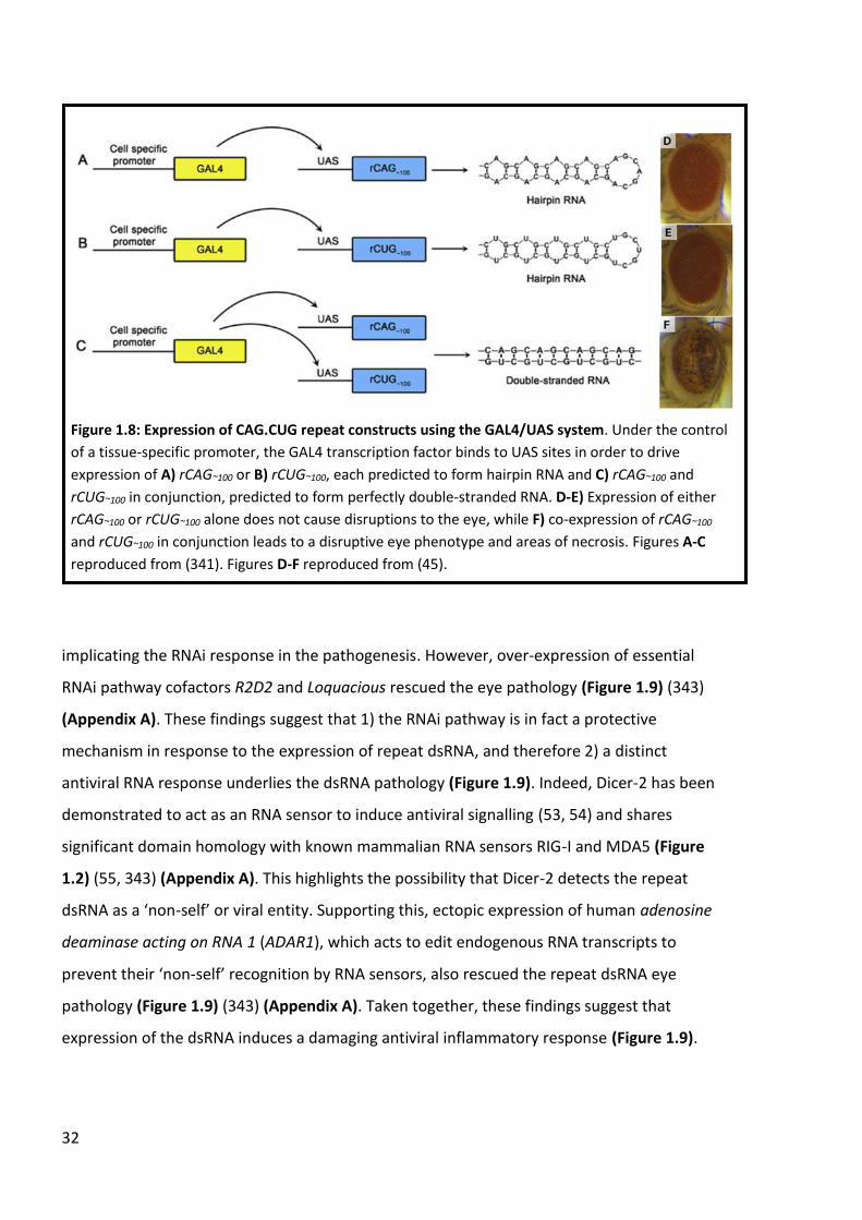
32
implicating the RNAi response in the pathogenesis. However, over-expression of essential
RNAi pathway cofactors R2D2 and Loquacious rescued the eye pathology (Figure 1.9) (343)
(Appendix A). These findings suggest that 1) the RNAi pathway is in fact a protective
mechanism in response to the expression of repeat dsRNA, and therefore 2) a distinct
antiviral RNA response underlies the dsRNA pathology (Figure 1.9). Indeed, Dicer-2 has been
demonstrated to act as an RNA sensor to induce antiviral signalling (53, 54) and shares
significant domain homology with known mammalian RNA sensors RIG-I and MDA5 (Figure
1.2) (55, 343) (Appendix A). This highlights the possibility that Dicer-2 detects the repeat
dsRNA as a ‘non-self’ or viral entity. Supporting this, ectopic expression of human adenosine
deaminase acting on RNA 1 (ADAR1), which acts to edit endogenous RNA transcripts to
prevent their ‘non-self’ recognition by RNA sensors, also rescued the repeat dsRNA eye
pathology (Figure 1.9) (343) (Appendix A). Taken together, these findings suggest that
expression of the dsRNA induces a damaging antiviral inflammatory response (Figure 1.9).
Figure 1.8: Expression of CAG.CUG repeat constructs using the GAL4/UAS system. Under the control
of a tissue-specific promoter, the GAL4 transcription factor binds to UAS sites in order to drive
expression of A) rCAG~100 or B) rCUG~100, each predicted to form hairpin RNA and C) rCAG~100 and
rCUG~100 in conjunction, predicted to form perfectly double-stranded RNA. D-E) Expression of either
rCAG~100 or rCUG~100 alone does not cause disruptions to the eye, while F) co-expression of rCAG~100
and rCUG~100 in conjunction leads to a disruptive eye phenotype and areas of necrosis. Figures A-C
reproduced from (341). Figures D-F reproduced from (45).
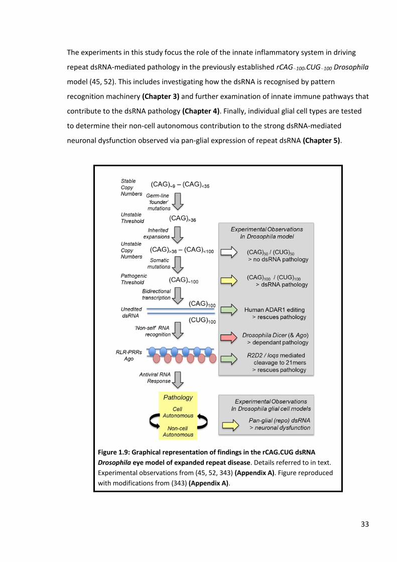
33
The experiments in this study focus the role of the innate inflammatory system in driving
repeat dsRNA-mediated pathology in the previously established rCAG~100.CUG~100 Drosophila
model (45, 52). This includes investigating how the dsRNA is recognised by pattern
recognition machinery (Chapter 3) and further examination of innate immune pathways that
contribute to the dsRNA pathology (Chapter 4). Finally, individual glial cell types are tested
to determine their non-cell autonomous contribution to the strong dsRNA-mediated
neuronal dysfunction observed via pan-glial expression of repeat dsRNA (Chapter 5).
Figure 1.9: Graphical representation of findings in the rCAG.CUG dsRNA
Drosophila eye model of expanded repeat disease. Details referred to in text.
Experimental observations from (45, 52, 343) (Appendix A). Figure reproduced
with modifications from (343) (Appendix A).

34

35
CHAPTER 2: Materials & Methods
2.1 Materials
Enzymes
Power SYBR® Green PCR master mix – Applied Biosystems
GoTaq® Flexi DNA Polymerase – Promega
DNAse I – Ambion
Kits
RNeasy® mini kit – Qiagen
Wizard® SV Gel and PCR Clean-Up System – Promega
High Capacity cDNA Reverse Transcription kit – Applied Biosystems
Other reagents
Deoxyribonucleoside triphosphate set (PCR grade) - Promega
1kb Plus DNA ladder – Invitrogen
TRIzol™ - Invitrogen
Oligonucleotides
All oligonucleotides are standard PCR and were obtained from Sigma-Aldrich unless
specified. Sequences are present 5’ to 3’.
General oligos – obtained from Promega
Random hexamers
Primers for PCR and sequencing
UASp-Fwd: GGCAAGGGTCGAGTCGATAG
UASp-Rev: AGGTTTAACCAGGGGATGCT

36
UASp-Seq-Fwd: CAAGGGTCGAGTCGATAG
UASp-Seq-Rev: TAACAAGTATGAATGTCAGGTT
R2D2-Rev: GGACGCAGTAGTCACGCAG
dADAR-Rev: AGAACTGCGCCATCCTTAAACTG
hADAR1-Rev: GCCATTGTAATGAACAGGTGGTT
hADAR2-Rev: CCCACGTAAAAGGGAGGCTC
Primers for qRT-PCR
Vago-Fwd: TGCAACTCTGGGAGGATAGC
Vago-Rev: AATTGCCCTGCGTCAGTTT
rp49-Fwd: GACGCTTCAAGGGACAGTATCTG
rp49-Rev: AAACGCGGTTCTGCATGAG
Nazo-Fwd: GCTGATCGGAGGACTACTGC
Nazo-Rev: TTGAAATTTCCCTCCGTAAGTC
STING-Fwd: CCGGTGTCTATCGTCCTTTC
STING-Rev: CGCTTTAGTTCCTGCATCTG
CG33926-Fwd: GCGACCGTCATTGGATTGG
CG33926-Rev: TGATGGTCCCGTTGATAGCC
Charon-Fwd: TCTCCAATCACGGTAAACAATG
Charon-Rev: GAACTTTGGTCGGATCTA CTGG
Drosomycin-Fwd: CGTGAGAACCTTTTCCAATATGATG
Drosomycin-Rev: TCCCAGGACCACCAGCAT
The following primers for qRT-PCR were obtained from Geneworks
Eiger-Fwd: CTGCTCGTGAATGCGATTCAT
Eiger-Rev: TGCAGTATGCACGATTCCGA
Buffers and solutions
Agarose gel loading dye (6X): 30% glycerol, 0.2% (w/v) bromophenol blue, 0.2% (w/v) xylene
cyanol.

37
TAE buffer: 40mM Tris-acetate, 20mM sodium acetate, 1mM EDTA (pH 8.2).
Squishing buffer: 10mM Tris-HCl (pH 8.2), 1mM EDTA, 25mM NaCl
Drosophila media
Drosophila stocks were maintained on fortified (F1) medium composed of 1% (w/v) agar,
18.75% compressed yeast, 10% treacle, 10% polenta, 1.5% acid mix (47% propionic acid,
4.7% orthophosphoric acid) and 2.5% tegosept (10% para-hydroxybenzoate in ethanol).
Drosophila stocks
Transgenic repeat lines
The following recombinant dsRNA lines were used and have been described previously (45,
341).
rCAG~100.rCUG~100S1 - rCUG~100 [H,I]; rCAG~100 [G,I]
rCAG~100.rCUG~100S2 - rCAG~100 [A,E]; rCUG~100 [E,F]
rCAG~100.rCUG~100M2 - rCAG~100 [J,K]; rCUG~100 [J,G]
rCAG~100.rCUG~100M3 - rCAG~100 [J,K]; rCUG~100 [E,F]
rCAG~100.rCUG~100W2 - rCUG~100 [D]; rCAG~100 [G]
Controls
4xUAS – 4 transgenes containing UAS sites without an insert in the attB system (344) [22A,
58A, 68E, 96E]
2xUAS – 2 transgenes containing UAS sites without an insert in the attB system (344) [51C,
68E]
UAS – 1 transgene containing UAS sites without an insert in the attB system (344) [51C]
Stocks obtained from Bloomington Drosophila Stock Centre (BDSC) are denoted with their
corresponding Bloomington stock number (BL).

38
GAL4 driver lines
GMR-GAL4 – BL 1104 (345)
elavII-GAL4 – BL 8765 (346)
Repo-GAL4 – BL 7415 (347)
nrv-GAL4 – BL 6799 (348)
R54C07-GAL4 (referred to as SPG-GAL4) – BL 50472 (349)
R75H03-GAL4 (referred to as TEG-GAL4) – BL 39908 (349)
R56F03-GAL4 (referred to as NEG-GAL4) – BL 39157 (349)
R86E01-GAL4 (referred to as ALG-GAL4) – BL 45914 (349)
R54H02-GAL4 (referred to as CG-GAL4) – BL 45784 (349)
Mutant insertion alleles
upd1YM55 – BL 4767
upd3d11639 – BL 19355
drprMI07659 – BL 43909
McrEY07421 – BL 15997
ECSITPL00455 – BL 19508
Over-expression
UAS-CrPV1A 148 #1 was obtained from Christophe Antoniewski (350)
UAS-CrPV1A 148 #2 was obtained from Christophe Antoniewski (350)
UAS-CrPV1A 108 #1 was obtained from Christophe Antoniewski (350)
UAS-CrPV1A 108 #2 was obtained from Christophe Antoniewski (350)
PBac(STAT92E-GFP.FLAG) – BL 38670
UAS-Draper (Isoform I) – BL 67035 (351)
UAS-Draper (Isoform II) – BL 67036 (351)
UAS-Draper (Isoform III) – BL 67037 (351)
UAS-parkin – BL 51651
UAS-Ref(2)P-GFP – (352)
RNAi lines
P(UAS-Stat92E.RNAi)1 – BL 26899

39
The following lines are from the Vienna Drosophila Research Centre (VDRC) collection and
were obtained from the VDRC stock centre.
upd1v3282
upd3v106869
drprv27086
Mcrv100197
PINK1v21860
PINK1v109614
parkinv47636
Ref(2)Pv105338
ECSITv31287
ECSITv106141
2.2 Methods
DNA manipulation
Agarose gel electrophoresis
Gel electrophoresis was performed using 1% agarose in 1xTAE buffer supplemented with
ethidium bromide (EtBr) at a final concentration of 0.5μg/mL. DNA fragments were
separated by submerging the gel in a tank containing 1xTAE buffer and applying 100V to the
tank. DNA fragments were visualised by UV light exposure.
Gel purification
DNA bands were excised from agarose gels under UV light and purified using the Wizard® SV
Gel and PCR Clean-Up System kit according to manufacturer’s instructions (Promega).
Drosophila genomic DNA preparation
1 whole adult fly per genotype was frozen on dry ice in a 0.5mL tube. The fly was then
mashed in 20μL of squishing buffer (see section 2.1) mixed with 5μL 1g/μL Proteinase K (for
a final concentration of 200μg/mL) for 20-30 seconds before incubation at 55°C for 3 hours.

40
Proteinase K was inactivated via incubation at 95°C for 2 minutes, followed by storage at
4°C.
PCR amplification of Drosophila genomic DNA
PCR amplification was performed using a Bio-Rad DNAEngine Peltier Thermal Cycler.
Genomic DNA was amplified using GoTaq® Flexi DNA Polymerase reagents with GoTaq®
Green Reaction Buffer according to the manufacturer’s guidelines (Promega). The cycling
conditions were 95°C for 2 minutes, then 35 cycles of 95°C for 1 minute, 60°C for 1 minute
and 72°C for 1 minute, followed by a final 72°C for 5 minutes.
Drosophila husbandry
Flies were raised at 18°C or 25°C with 70% humidity on F1 medium. Crosses were performed
at 25°C unless detailed otherwise.
RNA purification
RNA isolation
30 heads from 1-day old adult female flies were collected per genotype for RNA extractions.
The heads were snap frozen in liquid nitrogen and stored at -80°C. The tissue samples were
homogenised in 1mL TRIzol™ with a plastic pestle and further homogenised with a 20-gauge
needle. The homogenate was then pelleted by spinning at 13000 rpm for 10 minutes at 4°C.
The resulting supernatant was transferred to fresh RNAse free tubes and 200μL chloroform
was added and mixed via shaking vigorously. The samples were then spun at 13000 rpm for
15 minutes at 4°C and the upper aqueous phase was transferred to fresh RNAse free tubes.
An equal volume of 100% ethanol was added to the sample and mixed briefly via vortexing.
The sample mixtures were then loaded into RNeasy® columns and the remaining purification
steps were carried out according to the instructions of the Rneasy® mini kit. The RNA was
eluted in 30μL Nuclease-Free water and stored at -80°C until further use.

41
Quantitative real-time PCR (qRT-PCR)
Reverse transcription
800ng of RNA was DNAse treated using DNAse I according to the manufacturer’s instructions
(Ambion) and then reverse transcribed using the High Capacity cDNA Reverse Transcription
kit to produce cDNA according to manufacturer’s instructions (Applied Biosystems). Reverse
transcription reactions were performed at 25°C for 10 minutes, 37°C for 2 hours and then
85°C for 5 minutes.
qRT-PCR reactions
Reverse transcription reactions were diluted 1/5 in Nuclease-free water and 2μL was used as
the template in a final reaction volume of 10μL. Triplicate reactions were performed for
each template with 1.26pmol of each primer together with 1X Power SYBR® Green PCR
master mix in a QuantStudio 7 Flex Real-Time PCR System (Applied Biosystems). The cycling
conditions were 95°C for 10 minutes followed by 40 cycles of 95°C for 15 seconds and 60°C
for 1 minute. A standard curve was generated for each primer pair by including serial
dilutions from cDNA (neat, 1/2, 1/5, 1/10). A dissociation curve was produced for each
primer pair to confirm that a single product was amplified in each reaction. The relative
amount of cDNA was quantified using the relative standard curve method and the results
were subsequently exported to Microsoft Excel. The quantity of amplified cDNA product for
each primer pair was normalised against the quantity of cDNA produced with primers for
the housekeeping gene Ribosomal protein 49 (Rp49) for the corresponding sample.
Rapid Iterative Negative Geotaxis (RING) assay
0-1 day old adult male flies were collected in sets of between 10-20 flies and allowed to
recover from anaesthesia overnight at 25°C. A minimum of 40 flies were collected per
genotype when possible. Flies were transferred to standard vials and into the RING
apparatus as described in Gargano et al., (353). The apparatus was tapped several times to
transfer the flies to the bottom of the tube, and then imaged after 5 seconds (Google Pixel
camera). Experimental flies and their appropriate control were imaged side-by-side. Five
consecutive trials were performed per set of flies per timepoint, with the flies allowed to

42
recover for 1 minute between each trial. The images were then transferred into the Fiji Is
Just ImageJ (FIJI) image analysis program and a line was drawn to represent the halfway
mark of each vial. Flies were then scored as either “successful” if they passed the halfway
mark, or “unsuccessful” if they failed to pass the halfway mark. The percentage of
“successful” flies per trial was recorded as “% climbing ability”. The “% climbing ability” for
each trial formed a single data point, with the total number of trials for each genotype
plotted as a scatter graph displaying the average mean and standard deviation using
GraphPad Prism 7. The average “% climbing ability” was then calculated for each genotype
and compared to appropriate age-matched controls. When comparing two datasets, a 2-
tailed Student’s t-test was performed to determine significance, while a one-way ANOVA
and Dunnett’s test were performed to determine significance when comparing more than
two datasets. The significance threshold was set at p<0.05. The same sets of flies were aged
by transferring to fresh food every 2-3 days. RING assays were performed at timepoints of 1,
8 and 15 days of age. Set of flies that fell below 10 individuals during ageing were omitted
from further analysis.
Survival assay
The same flies analysed in RING assays were also assayed for their survival, with the number
of individuals per genotype recorded per week at 1, 8, 15, 22 and 29 days of age. The
number of individuals counted at 1 day of age was designated as 100% survival for each
genotype, and subsequent counts were divided by this total count to represent the
percentage of individuals remaining.
Eye photography
Eyes of at least 20 one day old female flies were viewed per genotype if possible, with 3
representative eyes photographed per genotype. Eyes were imaged using an Olympus SZX7
microscope fitted with an SZX-AS aperture along with an Olympus ColourView IIIU Soft
Imaging System and ToupView image acquisition software. Adobe Photoshop CS was used
for image preparation. Anterior is to the left in all presented images.

43
CHAPTER 3: ‘Non-self’ recognition of repeat dsRNA
The innate immune system acts as a protective first-line defence against invading pathogens
and other ‘non-self’ material. Pattern recognition receptors (PRRs) act as sensors of a wide
range of ‘non-self’ stimuli, including conserved structures on bacteria and viruses, as well as
endogenous molecules released under conditions of stress/injury that constitute danger
signals (69). Detection of a threat is followed by the induction of an inflammatory response,
resulting in removal of the offending molecules through degradation and/or cell death
signalling (354). However, persistence of the threat through genetic or environmental
disruption of inflammatory pathways can lead to elevated cell death and tissue damage.
Indeed, genetic lesions in a number of genes involved in inflammatory processes have been
identified as causal or risk factors in neurodegenerative disease (68). Additionally, elevated
cytokine levels are consistently observed in Huntington’s Disease (HD) gene carriers and
precede the onset of clinical symptoms (58, 59), indicating that inflammation likely
represents a proximal causal factor rather than consequence of disease pathology.
The relative conservation of mammalian and fly innate immune pathways allows the use of
Drosophila to genetically dissect the role of inflammation under conditions of homeostasis
and disease (318, 355). Previous work in the Drosophila eye tissue model of dsRNA-
mediated pathology utilized here has demonstrated the requirement for inflammation in
the development of pathology, including the activity of the Toll-like receptor (TLR) innate
immune pathway (52) and Dicer-2, which shares homology with the RIG-I-like receptor (RLR)
family of antiviral RNA sensors in vertebrates (45). Additionally, expression of the repeat
dsRNA induced upregulation of the antimicrobial peptide Drosomycin and the potent
cytokine/cell death Eiger, the orthologue of mammalian TNF (52). Taken together, this
evidence led to the hypothesis that repeat dsRNA is recognized as a ‘non-self’ molecule by
the innate immune system, thus provoking a damaging cascade of inflammatory signalling
that manifests as pathology (4, 52).
Dicer-2 plays a key role in the invertebrate RNA interference (RNAi) pathway; where it acts
to detect viral dsRNA products and process them into 21-nucleotide small-interfering RNAs
(siRNAs) (356, 357). The processing step also involves the essential co-factor R2D2, which

44
forms a complex with Dicer-2 to interact with RNA duplexes (358, 359). Following
processing, the siRNAs are loaded into the Argonaute-2 (Ago2) mediated RNA-induced
silencing complex (RISC), where it cleaves the siRNA and utilizes the guide strand to locate
complementary viral mRNA in order to facilitate their subsequent silencing and/or
degradation (360-362). Indeed, Ago2 also contributes to the pathology observed in the
repeat dsRNA eye pathology, though not to the extent of Dicer-2 (341). Intriguingly
however, over-expression of R2D2 was demonstrated to potently suppress the repeat
dsRNA pathology while loss-of-function mutations enhanced the pathology (343) (Appendix
A). These findings led to the hypothesis that the RNAi pathway may not drive dsRNA
pathogenesis, but in fact act as a competing pathway to restrict the influence of the repeat
dsRNA.
Notably, Dicer-2 also displays potent antiviral activity independently of the RNAi pathway;
Drosophila C virus (DCV) infection leads to Dicer-2 dependent upregulation of the antiviral
peptide Vago and the control of viral load through an unknown signal transduction pathway
(53). Both Ago2 and R2D2 were dispensable for the pathway, thus uncoupling Dicer-2 from
its previously established role in RNAi processing (53). Though not strictly independent of
the RNAi pathway, Dicer-2 also regulates the production of reverse-transcribed viral DNA
(vDNA) that serves to amplify the antiviral response in a manner distinct from siRNA
production but dependent upon Ago2 (363). Taken together, Dicer-2 (and Ago2 to an
extent) play important roles in a number of antiviral mechanisms, and these functions can
be either helpful or harmful depending upon stimuli context.
Indeed, RNAi machinery displays a high level of adaptive evolution across a number of
invertebrate species (364), highlighting the importance and considerable selection pressure
placed upon antiviral genes. However, viruses themselves also display adaptation to the
antiviral defence system in Drosophila as part of the ongoing arms race waged between host
and virus. A number of insect viruses encode viral suppressors of RNAi silencing (VSRs) that
allow them to inhibit RNAi machinery in order to maximise their replication and spread (50).
Interestingly, VSRs display diversity in the steps at which they inhibit the RNAi pathway;
Drosophila C virus encodes a VSR (DCV-1A) containing a dsRNA binding domain that allows it
to block Dicer-2 from cleaving viral dsRNA (350), while the cricket paralysis virus (CrPV)

45
encodes CrPV1A, a VSR that interacts with Ago2 to prevent its endonuclease activity
downstream of siRNA production (350). Additionally, CrPV1A can also promote the
ubiquitination of Ago2, leading to its degradation through an E3-ubiquitin ligase complex
(365). Finally, CrPV1A can inhibit host transcription and disrupt the assembly of RNA stress
granules independently of its RNAi silencing function, demonstrating that CrPV1A acts to
silence multiple antiviral pathways upon infection (366). Indeed, CrPV1A may also act to
suppress other uncharacterised antiviral mechanisms based on its diverse range of
interactions with host machinery.
The RNAi pathway acts to promote degradation of viral material, thus promoting cell
survival. However, focal cell death is also a critical factor in antiviral defence, serving as a
potent means to restrict the spreading of a virus (and indeed all pathogens) to adjacent
uninfected cells (367). When dsRNA arises endogenously, RNA-editing enzymes act to
catalyse the conversion of these repeat sequences to molecules that are recognized as ‘self’
by pattern recognition machinery. In vertebrates, ADAR1 converts adenosine (A) to inosine
(I) in endogenous dsRNA molecules including Alu RNA repeats, masking their presence to
dsRNA sensor MDA5 (encoded by IFIH1) and thus preventing inflammation and cell death
(152, 156, 368, 369). Loss of editing or increased receptor sensitivity (through mutations in
ADAR1 and IFIH1 respectively) are both proximal causes of the auto-inflammatory disease
Aicardi-Goutieres Syndrome (AGS) (154, 155), highlighting the importance of ‘non-self’ to
‘self’ conversion as a regulatory mechanism. In line with this, ectopic expression of human
ADAR1 rescues repeat dsRNA-mediated eye pathology in Drosophila (343) (Appendix A). The
bi-directionally transcribed CAG.CUG repeat used in this model provides an ideal target for
human ADAR1, which preferentially edits repetitive RNA sequences (370). Thus, masking the
dsRNA through A-to-I conversion (CAG-to-CIG) appears to prevent its detection by
Drosophila inflammatory machinery.
Therefore, several of lines of evidence point towards repeat dsRNA acting as a ligand to
drive antiviral inflammation in Drosophila. The work described in this chapter further
investigates the hypothesis that expanded repeat dsRNA is recognized by host antiviral
pathways as a ‘non-self’ or viral-like molecule, thus leading to a damaging inflammatory
response that is responsible for the observed pathology.

46
3.2 The viral suppressor protein CrPV1A rescues repeat dsRNA-mediated
pathology
The cricket paralysis virus (CrPV) infects a broad range of insect species (371) and causes
lethality in Drosophila (372). In order to avoid antiviral silencing through the RNAi pathway,
the virus encodes the CrPV1A protein, a potent viral suppressor that targets and inhibits the
activity of the Ago2 protein; both through a direct interaction that blocks the Ago2
endonuclease function (350) and the recruitment of ubiquitination machinery to degrade
Ago2 (365). In this manner, CrPV1A blocks Ago2 activity downstream of the Dicer-2/R2D2
processing complex, and thus does not affect the cleavage of viral dsRNA into siRNAs (350).
The requirement for both Dicer-2 and Ago2 in repeat dsRNA eye pathology has led to the
suggestion that inflammatory signalling stemming from viral-like recognition of the repeats
is a key driver of the cellular dysfunction (45, 341). In order to further pursue this
hypothesis, the CrPV1A viral suppressor was ectopically expressed in conjunction with the
repeat dsRNA specifically within the Drosophila eye using the GMR-GAL4 driver. For this
purpose, two distinct forms of CrPV1A were examined; the full-length (148 amino acid)
protein is an efficient silencer of Ago2 (350), whereas the truncated (108 amino acid) protein
lacks key residues that facilitate interactions with Ago2 and is thus unable to interfere with
antiviral signalling (350, 365). The identities of the CrPV1A full-length and mutant constructs
were confirmed through sequencing of one each of the full-length and mutant lines
obtained (343) (Appendix A).
Ectopic expression of two independent full-length CrPV1A constructs alone did not cause
disruptions to the eye (Figure 3.1 B & C) and a similar lack of phenotype was observed with
each of two independent truncated CrPV1A mutant constructs (Figure 3.1 D & E). As
previously established, expression of repeat dsRNA within the eye caused loss of
pigmentation and structural patterning as well as the development of black necrotic spots
on the posterior side of the eye (Figure 3.1 A'). Strikingly however, co-expression of the
dsRNA with full length CrPV1A completely rescued all aspects of the dsRNA-mediated
pathology in both lines (Figure 3.1 B’ & C’), indicating that CrPV1A-mediated antagonism of
the antiviral response may mask the repeat dsRNA from RNA pattern recognition machinery.

47
Furthermore, both truncated mutant constructs failed to rescue the dsRNA pathology
(Figure 3.1 D’ & E’), thus demonstrating the requirement of CrPV1A-mediated antiviral
silencing activity for the suppression.
The ability of the CrPV1A viral suppressor protein to completely rescue the phenotype
supports the hypothesis that expanded repeat dsRNA is recognized as ‘non-self’ in a manner
similar to viral material. Since CrPV1A does not inhibit dsRNA processing but rather the
catalytic activity of Ago2 (350, 365), and that the canonical RNAi pathway appears to act as a
competing pathway in restricting the dsRNA pathology (343) (Appendix A), it is plausible
Figure 3.1: Ectopic expression of CrPV1A completely rescues dsRNA-mediated pathology. In each
case the GMR-GAL4 driver is used to express the constructs in the eye. A) Expression of a single UAS
transgene with no insert as a control eye. B-C) Expression of two independent full-length (FL) UAS-
CrPV1A constructs alone. D-E) Expression of two independent truncated UAS-CrPV1A mutant
constructs alone. A’) Co-expression of repeat dsRNA together with a single UAS transgene. B’-C’) Co-
expression of repeat dsRNA together with two independent full-length (FL) UAS-CrPV1A constructs.
D’-E’) Co-expression of repeat dsRNA together with two independent truncated UAS-CrPV1A mutant
constructs. The full length CrPV1A encodes a 148 amino acid long protein. The truncated CrPV1A
encodes a 108 amino acid long protein. The experiment was performed at 25°C using the
CAG.CUG~100M3 dsRNA line.

48
that Dicer-2 and Ago2 act together in a manner distinct from their roles in RNAi to drive
inflammation (Figure 3.2). Indeed, Drosophila Dicer-2 is related to RIG-I-like receptors RIG-I
and IFIH1 (MDA5) through shared homology in their DExD/H helicase domains (55). The
helicase domain is critical for the observed RNAi-independent antiviral functions of Dicer-2,
including Vago induction (53) and viral DNA (vDNA)-mediated amplification of antiviral
activity (363). Ago2 is not required for Vago induction (53) but is essential for vDNA
production (363), suggesting that this pathway may represent another target of the CrPV1A
protein.
Figure 3.2: Summary diagram regarding CrPV1A-mediated suppression of expanded repeat dsRNA
eye pathology. While RNAi components Dicer-2 and Ago2 are required for the dsRNA pathology
(red), key RNAi binding cofactor R2D2 acts to suppress the pathology (green), suggesting that the
RNAi antiviral mechanism constitutes a protective pathway in response to expression of repeat
dsRNA. Thus, Dicer-2 appears to act in its established role as an RNA sensor, likely together with
Ago2, to drive the dsRNA eye pathology through an antiviral cell death mechanism. CrPV1A
antagonizes Ago2 to block the RNAi pathway, but also appears to ablate the antiviral inflammatory
mechanism underlying the dsRNA eye pathology.

49
3.3 Transcript analysis of the inflammatory response to expanded repeat
dsRNA
Previous work has demonstrated that the inflammatory response is not only required for the
repeat dsRNA pathology but is also triggered by the presence of the ‘non-self’ dsRNA itself
(45, 52). Thus, qRT-PCR was performed to investigate A) if components of other
inflammatory pathways are upregulated in response to repeat dsRNA, and B) whether co-
expression of CrPV1A can ablate any observed induction, thus indicating potential targets
for further investigation. The elavII-GAL4 driver was utilized to express the relevant
constructs in all neuronal cells, where the expression of repeat dsRNA causes age-
dependent neurodegeneration (45). Neuronal tissue samples were taken from one day old
flies in order to maintain a temporal window consistent with previous transcript analysis
(52). Additionally, as the neurodegeneration observed in elavII>dsRNA flies is age-
dependent (45), this timepoint minimises both the possibility of transcript loss due to
dead/dying cells within the CNS and the dilution of altered RNA levels by transcripts from
unaffected (in this case non-elavII expressing) cells in within the sample tissue (Drosophila
heads).
An important final note; lack of significant alteration to gene expression in response to the
repeat dsRNA does not rule out the given gene in participating in the dsRNA pathology.
Modest or even no change in transcript levels may be adequate to surpass a functional
threshold and thus contribute to the pathology. In addition, post-transcriptional/post-
translational regulation of components are both common features in innate immune
signalling pathways (373, 374), both of which will not be reflected in this analysis.
Nevertheless, qRT-PCR can provide an effective insight into dsRNA-triggered pathway
activation via the transcription of downstream response genes.
Drosomycin is an antimicrobial peptide (AMP) synthesised as a result of signalling from the
Toll inflammatory pathway (375) and has previously been shown to be upregulated in the
presence of repeat dsRNA (52). Therefore, Drosomycin transcript levels were analysed to
confirm the upregulation and to determine if co-expression of CrPV1A blocks this response.
Indeed, Drosomycin was significantly upregulated in elavII>dsRNA flies (Figure 3.3).

50
Introduction of CrPV1A did not significantly suppress Drosomycin upregulation when
compared to elavII>dsRNA, though a small trend was observed (Figure 3.3). Thus,
Drosomycin induction (and Toll pathway activity) is clearly observable in response to dsRNA
expression but does not appear to be a target of the CrPV1A viral suppressor.
Overexpression of Drosomycin alone (or indeed several other AMPs alone) in neurons or glia
leads to age-dependent neurodegeneration in flies, indicating that AMPs can become
neurotoxic at high levels (376). The flies used in this qRT-PCR analysis were newly-eclosed,
and thus it is likely that Drosomycin does not accumulate at the level required for
neurotoxicity at the early time point tested in this study. The Drosophila Toll pathway is
canonically triggered after detection of gram-positive bacteria and fungi as opposed to viral
RNA intermediates (377), though Toll pathway mutants exhibit increased susceptibility to
Drosophila X virus (DXV) (378) and oral infection of Drosophila C virus (DCV) (379). In
addition, recent evidence has demonstrated that Dicer-2 can bind Toll directly and drive
induction of Drosomycin upon microbial challenge (54), thus providing a plausible
explanation for the Drosomycin upregulation observed here. Alternatively (though
potentially not mutually exclusive), the upregulation of Drosomycin may represent a non-cell
Figure 3.3: The antimicrobial peptide Drosomycin is significantly
upregulated in response to repeat dsRNA. In each case
expression is driven in all neuronal cells using the elavII-GAL4
driver. Expression of repeat dsRNA alone leads to a significant
upregulation of Drosomycin compared to the UAS empty
transgene control (p=0.0077), as did co-expression of repeat
dsRNA and CrPV1A compared to the control (p=0.0360). Co-
expression of CrPV1A with repeat dsRNA does not significantly
suppress the induction of Drosomycin compared to repeat dsRNA
expression alone (p=0.4240). Depicted for each dataset are the
mean and standard error of the mean (SEM). 2-tailed Student’s t-
tests were used to determine significance; ** p<0.01, * p<0.05,
ns p>0.05. Expression levels are normalized to the housekeeping
gene Rp49. Each dataset represents 3 biological replicates plated
in triplicate. The experiment was performed using the
CAG.CUG~100S1 dsRNA and CrPV1A FL #1 lines.

51
autonomous Toll-mediated response to damage/danger associated molecular patterns
(DAMPs) and other trigger molecules, released from cells that are damaged/killed by the
focal dsRNA-induced inflammatory response.
Given the likely role of cell death signalling in dsRNA pathology, the expression level of
potent cytokine and cell death signalling molecule Eiger was also investigated. Eiger is the
sole Drosophila orthologue of tumour necrosis factor (TNF), a subject of intensive research
for its key roles in inflammation and programmed cell death (380). Indeed, these key roles
are conserved through to Eiger, which interacts with its receptors Wengen and Grindelwald
(orthologues of mammalian TNF receptors) to activate the c-Jun N-terminal kinase (JNK)
pathway-dependent cell death (381-383). While Eiger mutants are rendered susceptible to
pathogen challenge and tumour progression (384, 385), overexpression of Eiger leads to
developmental defects through heightened cell death signalling (381, 383). Elevated levels
of mammalian TNF is a constantly observed feature in neurodegenerative disease (386,
387), and in pre-symptomatic HD disease gene carriers (58, 59). Finally, upregulation of Eiger
transcript levels has previously been observed in the repeat dsRNA model used in this study
(52). Therefore, Eiger transcript levels were assessed to determine if CrPV1A-driven
antagonism of the antiviral response affects Eiger induction.
Eiger was not significantly altered in elavII>dsRNA flies, while also remaining unchanged
when CrPV1A was co-expressed with the dsRNA (Figure 3.4). Given the potent cell death
signalling initiated through Eiger, this lack of alteration may represent the possibility that
the cells containing elevated Eiger are undergoing cell death and thus losing Eiger signal.
Indeed, Eiger is a highly pleiotropic inflammatory cytokine with a number of physiological
roles (388). As such, the focus was shifted to investigate inducible antiviral pathways, which
are more likely to respond to the presence of dsRNA given its ‘non-self’/viral recognition
through Dicer-2. The RNAi response is an important mechanism for combating viral
infection, achieving this via the production of siRNAs as opposed to inducing the expression
of antiviral peptides (389). However, aside from the RNAi response, the inducible Drosophila
antiviral response remains poorly understood (390). Thus, peptides that are synthesised
through the Drosophila antiviral response were investigated as readouts of antiviral pathway
activity. Recent work has demonstrated that the RNAi response confers resistance to a

52
broad range of viruses, while antiviral activity through the evolutionarily conserved JAK-STAT
pathway is restricted to viruses from the Dicistoviridae family; DCV and CrPV (391).
Additionally, Dicer-2 can respond to DCV independently of its role in RNAi in order to
upregulate the antiviral peptide Vago and control infection (53), a process that is
evolutionarily conserved in the mosquito Culex quinquefasciatus (392). Finally, recent
evidence has uncovered co-operation between IMD pathway components Relish and dIKKβ
with the Drosophila STING orthologue to induce the expression of a battery of antiviral
response genes in response to DCV and CrPV (393). This included the novel antiviral peptide
Nazo, which strongly attenuates DCV and CrPV infection when overexpressed (393).
To determine whether the antiviral pathways regulating Vago and Nazo are active in
response to the presence of repeat dsRNA, the expression levels of both antiviral peptides
were analysed through qRT-PCR. Neither Vago nor Nazo were significantly altered in
response to expression of repeat dsRNA, while co-expression of CrPV1A with the dsRNA also
did not affect expression levels of the antiviral peptides (Figure 3.5 A & B). Given that
expression of either Vago or Nazo is not elevated in response to repeat dsRNA, any potential
antagonism of the antiviral response through CrPV1A is likely masked. Based on the
Figure 3.4: Drosophila TNF orthologue Eiger is not
significantly altered in response to repeat dsRNA. In each
case expression is driven in all neuronal cells using the
elavII-GAL4 driver. Eiger transcript levels were not
significantly impacted by the presence of repeat dsRNA
alone (p=0.2807) or through co-expression of CrPV1A and
the dsRNA (p=0.6927) when compared to the UAS empty
transgene control. Eiger levels were not significantly
different between dsRNA alone and together with CrPV1A
(p=0.4057). Depicted for each dataset are the mean and
standard error of the mean (SEM). 2-tailed Student’s t-tests
were used to determine significance; ns p>0.05. Expression
levels are normalized to the housekeeping gene Rp49. Each
dataset represents 3 biological replicates plated in triplicate.
The experiment was performed using the CAG.CUG~100S1
dsRNA and CrPV1A FL #1 lines.

53
observation that Ago2 is dispensable for the induction of Vago (53), it is unlikely that CrPV1A
targets this pathway, though this cannot be completely ruled out. Nazo is upregulated
following CrPV infection (393), though whether CrPV1A targets this pathway is also
unknown. However, since Ago2 was shown to be dispensable for Nazo induction, the
authors hypothesised that this novel pathway may constitute an additional layer of antiviral
defence independent of DCV/CrPV-derived VSRs (393).
Figure 3.5: Expression levels of the antiviral peptides Vago and Nazo are not significantly
altered in response to repeat dsRNA. In each case expression is driven in all neuronal cells using
the elavII-GAL4 driver. A) Vago transcript levels were not significantly impacted by the presence
of repeat dsRNA alone (p=0.4843) or through co-expression of CrPV1A and the dsRNA
(p=0.3030) when compared to the UAS empty transgene control. Vago levels were not
significantly different between dsRNA alone and together with CrPV1A (p=0.9008). B) Nazo
transcript levels were not significantly impacted by the presence of repeat dsRNA alone
(p=0.8324) or through co-expression of CrPV1A and the dsRNA (p=0.9225) when compared to
the UAS empty transgene control. Nazo levels were not significantly different between dsRNA
alone and together with CrPV1A (p=0.8769). Depicted for each dataset are the mean and
standard error of the mean (SEM). 2-tailed Student’s t-tests were used to determine
significance; ns p>0.05. Expression levels are normalized to the housekeeping gene Rp49. Each
dataset represents 3 biological replicates plated in triplicate. The experiments were performed
using the CAG.CUG~100S1 dsRNA and CrPV1A FL #1 lines.
A B

54
The observation that expression of dsRNA does not induce Vago suggests that the role of
Dicer-2 in regulating this pathway is independent of the mechanism(s) responsible for the
pathology. Indeed, knowledge of the pathway from Dicer-2 to Vago remains limited,
including whether other antiviral peptides are induced through this mechanism. Thus,
identification of the essential participants in this pathway (both interactors and induced
genes) is required before a definitive assessment of its role in mediating dsRNA pathology
can be undertaken. Nazo has only recently been characterised as antiviral effector molecule
that controls CrPV replication (393), and as such the antiviral mode of action of Nazo
remains unknown. Regardless, it would be of interest to determine if Nazo can modify the
dsRNA eye phenotype, given that its overexpression potently restricts both DCV and CrPV
replication (393).
In addition to Nazo, nine other transcripts were also induced via the STING/Relish/dIKKβ-
dependent antiviral pathway in response to DCV in both flies and Drosophila S2 cells,
including a number of uncharacterised genes (Figure 3.6) (393). One of these, CG33926, was
induced potently by all three of Relish, STING and dIKKβ (393). Given its strong induction
following DCV infection, CG33926 was investigated in the context of repeat dsRNA
Figure 3.6: The STING/Relish/dIKKβ-mediated antiviral
pathway in Drosophila. Current pathway proposed from
experimental data obtained by Goto et al. Drosophila C
virus (DCV) is detected by an unknown viral sensor(s),
leading to downstream signalling through STING, dIKKβ
and NF-κB transcription factor Relish. Relish (possibly in
conjunction with other transcription factors) then
induces the expression of a range of antiviral effector
genes including STING itself, Nazo, Charon and the
uncharacterised CG33926. The regulatory protein Kenny
(dIKKγ) inhibits this antiviral signalling pathway,
potentially through its role in mediating autophagic
degradation of the IKK complex. Genes in this pathway
tested via qRT-PCR in this study are STING, Relish, Nazo,
Charon and CG33926. Figure adapted from (393).

55
expression. Expression of the dsRNA alone led to a striking reduction in CG33926 expression
when compared to the UAS control, a finding also observed when CrPV1A was co-expressed
with the dsRNA (Figure 3.7). No significant difference was observed when comparing
elavII>dsRNA and elavII>dsRNA/CrPV1A flies (Figure 3.7). Therefore, the expression of
repeat dsRNA appears to potently inhibit the induction of CG33926.
Unfortunately, the lack of information regarding CG33926 function limits the interpretation
of this result. Indeed, RNAi knockdown of CG33926 promotes DCV replication, but not to the
magnitude achieved via knockdown of dIKKβ, Relish, STING or Nazo (393), and thus it
potentially shares functional redundancy with other genes that were also induced. Of note,
CG33926 is also upregulated following infection from the DNA virus Kallithea virus (394),
further strengthening its proposed role as an antiviral effector. Interesting however, is that
the presence of dsRNA inhibits CG33926 expression as opposed to triggering its induction
(Figure 3.7). One plausible explanation is that expression of the dsRNA triggers the
upregulation of Kenny (also known as dIKKγ), a regulatory protein that promotes autophagic
degradation of the IκB signalling complex to prevent constitutive IMD pathway activity
(395). In support of this, Kenny restricts the expression of CG33926 (in addition to Nazo and
Figure 3.7: Expression of CG33926 is significantly
downregulated in response to repeat dsRNA. In each
case expression is driven in all neuronal cells using the
elavII-GAL4 driver. CG33926 transcript levels were
significantly reduced in the presence of repeat dsRNA
alone (p=0.0099) and through co-expression of CrPV1A
and the dsRNA (p=0.0126) when compared to the UAS
empty transgene control. CG33926 levels were not
significantly different between dsRNA alone and
together with CrPV1A (p=0.6087). Depicted for each
dataset are the mean and standard error of the mean
(SEM). 2-tailed Student’s t-tests were used to determine
significance; ** p<0.01, * p<0.05, ns p>0.05. Expression
levels are normalized to the housekeeping gene Rp49.
Each dataset represents 3 biological replicates plated in
triplicate. The experiment was performed using the
CAG.CUG~100S1 dsRNA and CrPV1A FL #1 lines.

56
STING) following DCV infection, likely through inhibition of the STING/Relish/dIKKβ signalling
pathway (393).
This raises the question as to why Kenny downregulates CG33926 but not Nazo in response
to repeat dsRNA, given that both genes appear to rely on dIKKβ signalling for induction
(393). It is plausible that this reflects the tailoring of antiviral gene transcription in a threat-
specific manner; the set of antiviral genes induced through challenge with DCV is likely to
display both overlap and distinct differences with the corresponding set upregulated in
response to repeat dsRNA. As such, it would be of interest to determine which of the 10
elevated transcripts identified in Goto et al., as DCV-responsive are also induced following
expression of repeat dsRNA. Indeed, CG33926 was induced following DCV infection (393)
but strongly repressed in response to the dsRNA (Figure 3.7), which serves to highlight the
complexity of the Drosophila antiviral response.
Among the other candidates, the IκB gene Charon is one of the few that has been previously
characterised (393). Charon (also known as Pickle) is vital for Relish-dependent transcription
of antimicrobial genes (396), but also inhibits the formation of Relish-Relish homodimers to
prevent aberrant IMD signalling following pathogenic challenge (397). Interestingly,
neurodegeneration in a Drosophila model of Ataxia-Telangiectasia (A-T) is dependent upon
Relish-mediated transcription of innate immune genes (311), and as such regulation of
Relish by Charon may be vital in the context of neurodegenerative disease. Thus, expression
levels of Charon were assessed in the presence of dsRNA.
Expression of repeat dsRNA led to a reduction in Charon transcript levels when compared to
the UAS control, though this was not significant (Figure 3.8). Indeed, a similar decrease in
Charon was observed compared to the control when CrPV1A was co-expressed with the
dsRNA, though again significance was not reached (Figure 3.8). Finally, levels of Charon were
almost identical between elavII>dsRNA and elavII>dsRNA/CrPV1A flies (Figure 3.8). Given
the trend observed, it would be of interest to gain a greater sample size to determine
whether or not the reduction is a true biological effect as opposed to variation between
samples.

57
Like CG33926, Charon is also negatively regulated by Kenny (dIKKγ) (393). Under
homeostatic conditions, Kenny is selectively targeted to autophagosomes for degradation,
whereas in response to commensal bacteria it interacts with dIKKβ and instead targets the
IKK signalling complex for degradation, thus terminating the immune response (395). As
such, a possible scenario to explain CG33926 and Charon downregulation is that expression
of repeat dsRNA induces Kenny, which then acts to negatively regulate dIKKβ-dependent
signalling through degradation of the IKK complex. Co-expression of CrPV1A with the dsRNA
failed to restore both CG33926 and Charon levels to that of the corresponding UAS controls
(Figures 3.7 & 3.8), providing evidence that CrPV1A does not target Kenny. In line with this,
RNAi knockdown of Kenny attenuates viral replication (393), and thus targeting it would be
detrimental to the virus. In line with this, the transcript levels of two of the core regulators
of this novel antiviral pathway, STING and Relish, were also monitored in response to the
repeat dsRNA.
The innate immune effector STING was first discovered in mammals as a potent regulator of
type-I interferon (IFN) production in response to DNA-based pathogens (398, 399). Only
recently has a conserved invertebrate STING orthologue been uncovered; responding to
Figure 3.8: Expression of IκB gene Charon is not
significantly altered in response to repeat dsRNA. In each
case expression is driven in all neuronal cells using the
elavII-GAL4 driver. Charon transcript levels were not
significantly impacted by the presence of repeat dsRNA
alone (p=0.1923) or through co-expression of CrPV1A and
the dsRNA (p=0.090) when compared to the UAS empty
transgene control. Charon levels were not significantly
different between dsRNA alone and together with CrPV1A
(p=0.8462). Depicted for each dataset are the mean and
standard error of the mean (SEM). 2-tailed Student’s t-tests
were used to determine significance; ns p>0.05. Expression
levels are normalized to the housekeeping gene Rp49. Each
dataset represents 3 biological replicates plated in
triplicate. The experiment was performed using the
CAG.CUG~100S1 dsRNA and CrPV1A FL #1 lines.
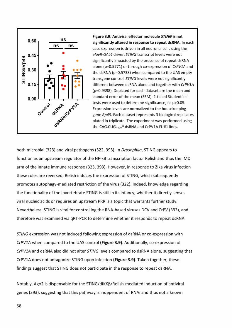
58
both microbial (323) and viral pathogens (322, 393). In Drosophila, STING appears to
function as an upstream regulator of the NF-κB transcription factor Relish and thus the IMD
arm of the innate immune response (323, 393). However, in response to Zika virus infection
these roles are reversed; Relish induces the expression of STING, which subsequently
promotes autophagy-mediated restriction of the virus (322). Indeed, knowledge regarding
the functionality of the invertebrate STING is still in its infancy, whether it directly senses
viral nucleic acids or requires an upstream PRR is a topic that warrants further study.
Nevertheless, STING is vital for controlling the RNA-based viruses DCV and CrPV (393), and
therefore was examined via qRT-PCR to determine whether it responds to repeat dsRNA.
STING expression was not induced following expression of dsRNA or co-expression with
CrPV1A when compared to the UAS control (Figure 3.9). Additionally, co-expression of
CrPV1A and dsRNA also did not alter STING levels compared to dsRNA alone, suggesting that
CrPV1A does not antagonize STING upon infection (Figure 3.9). Taken together, these
findings suggest that STING does not participate in the response to repeat dsRNA.
Notably, Ago2 is dispensable for the STING/dIKKβ/Relish-mediated induction of antiviral
genes (393), suggesting that this pathway is independent of RNAi and thus not a known
Figure 3.9: Antiviral effector molecule STING is not
significantly altered in response to repeat dsRNA. In each
case expression is driven in all neuronal cells using the
elavII-GAL4 driver. STING transcript levels were not
significantly impacted by the presence of repeat dsRNA
alone (p=0.5771) or through co-expression of CrPV1A and
the dsRNA (p=0.5738) when compared to the UAS empty
transgene control. STING levels were not significantly
different between dsRNA alone and together with CrPV1A
(p=0.9398). Depicted for each dataset are the mean and
standard error of the mean (SEM). 2-tailed Student’s t-
tests were used to determine significance; ns p>0.05.
Expression levels are normalized to the housekeeping
gene Rp49. Each dataset represents 3 biological replicates
plated in triplicate. The experiment was performed using
the CAG.CUG~100S1 dsRNA and CrPV1A FL #1 lines.

59
CrPV1A target. Indeed, this does not rule out the possibility that STING participates in the
dsRNA pathology, though how STING interacts with viral RNA is not known. One possible
scenario could be that Dicer-2 and STING co-operate in antiviral detection and signalling.
Dicer-2 acts as an RNA sensor through its helicase domain, which is conserved with
mammalian RNA receptors RIG-I and MDA5 (53). Therefore, it would be of interest to
determine if Dicer-2 participates in this response as a means to sense viral infection and
activate STING.
The NF-κB transcription factor Relish regulates antiviral activity as part of the IMD pathway
(372, 400) and as part of the STING/dIKKβ/Relish-mediated response (393). Additionally,
Relish is a regulator of antimicrobial gene expression through the IMD pathway in response
to bacterial pathogens (401, 402). Relish can homodimerize with itself or form heterodimers
with other NF-κB factors DIF and Dorsal to fine tune the battery of genes that are induced
following pathogenic challenge (403). Intriguingly, only recently has Relish also been
demonstrated to control autophagy in both development (404) and following Zika virus
infection in co-operation with STING (322). Conversely, Relish activity is required for
neurodegeneration (311, 376) and has been implicated in age-dependent neurological
decline (313). Given that its activation can be either of a beneficial or harmful nature in a
context-dependent manner, Relish transcript levels were measured in response to
expression of the repeat dsRNA.
Similar to STING, expression of Relish was not significantly altered in either elavII>dsRNA or
elavII>dsRNA/CrPV1A flies when compared to the UAS control (Figure 3.10). Additionally,
co-expression of CrPV1A with the dsRNA did not affect Relish transcription when compared
to expression of the dsRNA alone (Figure 3.10). Taken together, these results indicate that
Relish expression is not upregulated in response to the presence of repeat dsRNA. However,
it must be taken into consideration that Relish is regulated post-translationally; only upon
proteolytic cleavage and removal of its inhibitory IκB domain does Relish translocate to the
nucleus to drive transcriptional activation of NF-κB responsive genes (405, 406).
Furthermore, the transcriptional program initiated through Relish is dependent upon its
dimer configuration; either Toll or IMD downstream genes can be induced, or both (403).

60
Indeed, the dimer configuration partner of Relish for antiviral gene induction is unknown;
though Charon, a negative regulator of Relish-Relish homodimerization (397), is one of the
induced antiviral genes following DCV infection (393). In this manner it may promote
heterodimerization of Relish with an unknown antiviral transcription factor to bolster the
antiviral response. Thus, a closer investigation of how Relish protein function (or lack of
through use of mutant lines) is regulated in the presence of repeat dsRNA may prove more
informative to this end.
3.3 Chapter Discussion
The ability for cells to distinguish between ‘self’ molecules and foreign or abnormal ‘non-
self’ molecules that likely constitute danger is critical not only for host defence but to
protect essential cellular components from self-inflicted inflammatory damage (407).
Indeed, a number of endogenously-produced molecules require modification from cellular
machinery in order to gain ‘self’ status and thus avoid PRR detection (68). Double-stranded
Alu RNA repeats require modification from ADAR1 in the form adenosine to inosine (A-I)
editing in order to prevent their detection as ‘non-self’ by the RNA sensor MDA5 (368). Loss
of ADAR1 leads to aberrant inflammatory signalling and lethality in mice, which can be
Figure 3.10: NF-κB transcription factor Relish is not
significantly altered in response to repeat dsRNA. In each
case expression is driven in all neuronal cells using the
elavII-GAL4 driver. Relish transcript levels were not
significantly impacted by the presence of repeat dsRNA
alone (p=0.8293) or through co-expression of CrPV1A and
the dsRNA (p=0.4873) when compared to the UAS empty
transgene control. Relish levels were not significantly
different between dsRNA alone and together with CrPV1A
(p=0.5384). Depicted for each dataset are the mean and
standard error of the mean (SEM). 2-tailed Student’s t-
tests were used to determine significance; ns p>0.05.
Expression levels are normalized to the housekeeping
gene Rp49. Each dataset represents 3 biological replicates
plated in triplicate. The experiment was performed using
the CAG.CUG~100S1 dsRNA and CrPV1A FL #1 lines.
61
rescued through the genetic deletion of either the RNA sensor MDA5 (152) or downstream
antiviral signal adaptor MAVS (156). ADAR1 loss-of-function or IFIH1 (which encodes MDA5)
gain-of-function mutations are both associated with the autoinflammatory neurological
disorder Aicardi-Goutieres syndrome (AGS) (154, 155, 408), highlighting the importance of
‘non-self’ to ‘self’ editing as an immunomodulatory mechanism in human disease.
Indeed, repeat RNA and its subsequent bi-directional transcription are both features
common to expanded repeat disorders (21), and as such it has been hypothesised that
dsRNA formed in this manner may represent a molecular trigger for inflammation (4). In a
Drosophila model of expanded repeat disease, pathology caused via the expression of
expanded repeat CAG.CUG dsRNA is dependent upon Dicer-2 and Ago-2 (Figure 3.2) (45,
341). However, the pathology is suppressed by R2D2, an essential co-factor of Dicer-2 in the
RNAi pathway, which suggests that Dicer-2 and Ago2 drive the dsRNA-mediated toxicity via
the alternate antiviral RNA pathway, given that the RNAi pathway appears protective (343)
(Appendix A). Indeed, Dicer-2 shares homology with the mammalian RNA sensors RIG-I and
MDA5 (55) and appears to act in this manner upon viral infection in Drosophila (53).
Additionally, the pathology is completely suppressed through co-expression of the human
ADAR1 gene (343) (Appendix A), suggesting that conferring ‘self’ status to the dsRNA masks
its presence from antiviral machinery. Taken together, these results support the theory that
repeat dsRNA molecules derived from expanded repeat loci are detected as ‘non-self’ in a
manner similar to viral material, leading to cellular damage via inflammatory signalling.
To further challenge this hypothesis, a viral suppressor encoded by the cricket paralysis virus
(CrPV) was utilized. The protein, known as CrPV1A, antagonises Ago2 activity without
inhibiting Dicer2/R2D2-mediated processing of dsRNA (350), allowing the virus to escape
RNAi silencing and successfully replicate. Strikingly, co-expression of the full-length CrPV1A
viral suppressor with repeat dsRNA leads to a complete suppression of eye pathology.
Conversely, a truncated form of CrPV1A unable to inhibit the antiviral response fails to
suppress the dsRNA pathology. This finding suggests that repeat dsRNA is recognised as a
‘non-self’ entity by the host antiviral system, and that CrPV1A ablates the pathway(s)
responsible for the subsequent damaging inflammatory response.

62
Following this, qRT-PCR was performed on flies expressing dsRNA alone and in conjunction
with CrPV1A in an attempt to uncover pathways that 1) are upregulated in response to
repeat dsRNA, and 2) are attenuated by CrPV1A, thus highlighting them as candidates to
genetically dissect using the established dsRNA eye model. The neuronal driver elavII>GAL4
was chosen to drive expression of repeat dsRNA and CrPV1A, while the timepoint tested was
0-1 days of age, both to remain consistent with previous transcript analysis on the dsRNA
model (52).
STING and Relish, regulators of a recently described antiviral pathway (393), were not
significantly altered following expression of the dsRNA, thus making any attenuation of this
pathway by CrPV1A difficult to discern. Indeed, this novel antiviral pathway is critical for the
control of both DCV and CrPV infection and was therefore proposed to act as a secondary
inducible defence mechanism should the respective viral suppressors antagonize the RNAi
response (393). It is important to note that absence of STING or Relish upregulation does not
rule out their respective participation in the dsRNA-mediated pathology. Relish in particular
is subject to critical post-translational regulation, it requires separation from an inhibitory
domain in order to operate as a transcription factor (405, 406). In addition, Relish can
homodimerize to induce target genes of the IMD pathway, or form heterodimers with
another NF-κB transcription factor, DIF, to activate signalling from both the Toll and IMD
pathways (403). As such, transcript levels may not accurately portray Relish signalling in
response to dsRNA.
Knowledge of the Drosophila STING orthologue is still limited and the mechanisms
controlling its activation and regulation need to be further investigated. In vertebrates the
cytosolic cDNA sensor cyclic-GAMP synthase (cGAS) produces cGAMP which binds and
activates STING (409, 410), leading to a potent type-I IFN response against viral pathogens
(398, 399). Drosophila lack a functional cGAS ortholog able to bind DNA and elicit an
interferon transcriptional response (323), suggesting that it may serve to activate a different
subset of inflammatory genes in insects. Indeed, Drosophila STING co-operates with Relish
to induce antimicrobial peptides from the IMD pathway (323) and to upregulate antiviral
autophagy following Zika virus infection (322). Taken together, both Relish and STING
regulate inflammation as effectors of multiple distinct but often linked pathways. Therefore,
63
quantitating the downstream target transcripts of these pathways may prove more
informative when assessing their responsiveness (if any) to repeat dsRNA.
Thus, in addition to STING and Relish, the transcript levels of several genes that were
induced by the STING/dIKKβ/Relish signalling complex upon viral infection (393) were
assessed quantitatively as readouts of pathway activity. Namely, the newly-characterised
antiviral peptide Nazo, the IκB gene Charon and the uncharacterised CG33926 were all
chosen for their potent upregulation following DCV infection and their reliance upon the
signalling complex for induction (393). Both Nazo and Charon transcript levels were not
significantly altered in the presence of repeat dsRNA or through co-expression of CrPV1A
with the dsRNA, though Charon was modestly reduced in both experimental lines.
Conversely, CG33926 was potently downregulated in response to dsRNA alone and
dsRNA/CrPV1A co-expression. The differential expression of Nazo, Charon and CG33926
when comparing observations in Goto et al., to the results obtained in this chapter serve to
highlight the complexity of the Drosophila antiviral response to ‘non-self’ stimuli. Indeed,
while the RNAi response provides comprehensive host protection against a range of viruses
(391), inducible antiviral pathways appear to respond in a virus-specific manner (53, 391,
393). The inducible pathways have been hypothesised to act as secondary antiviral defence
mechanisms; upregulated should viruses antagonise the RNAi pathway or cell death
machinery through the activity of VSRs (393, 411, 412). Thus, the observation that CrPV1A
does not alter expression of genes within the STING/dIKKβ/Relish antiviral pathway suggests
that this pathway may be induced as a secondary response to compensate for DCV/CrPV-
mediated inhibition of the RNAi pathway.
The modest reduction of Charon and significant downregulation of CG33926 raise the
possibility of negative regulation within this pathway. Indeed Kenny, the Drosophila
orthologue of IKK complex regulator NEMO (also known as IKKγ), was demonstrated as a
negative regulator of dIKKβ antiviral signalling (393). Thus, it is plausible that Kenny is
upregulated in response to the repeat dsRNA, thus inhibiting the STING/dIKKβ/Relish
induction of antiviral target genes. Kenny prevents constitutive activation of the IMD
pathway by regulating autophagic degradation of the IKK complex (395) and may perform a
similar function upon detection of the repeat dsRNA. The fact that Nazo is not also

64
downregulated likely reflects the specificity in antiviral transcriptional programs, that DCV
and dsRNA induce different target genes.
Another facet to investigate is the function(s) of CG33926, of which current knowledge is
scarce aside from its upregulation following viral challenge (393, 394). One can speculate
that it may encode an antiviral peptide similar to Nazo, or perhaps form part of a
degradative complex designed to remove viral material. Indeed, a number of other
uncharacterised genes are induced by the STING/dIKKβ/Relish complex following DCV
infection (393). Thus, given the potent downregulation of CG33926 in response to the
dsRNA, it would be of interest to determine if these genes are also differentially regulated
when comparing DCV and repeat dsRNA challenge.
Another antiviral peptide induced through DCV infection is Vago, though it differs to Nazo in
that its activation is Dicer-2-dependent rather than reliant upon the STING/dIKKβ/Relish
complex (53). However, similar to Nazo, Vago expression was not significantly altered in
response to repeat dsRNA, nor when CrPV1A was co-expressed. Thus, this Dicer-2
dependent axis of antiviral activity appears unresponsive to repeat dsRNA, suggesting that
Dicer-2 has additional antiviral functionality independent of both this pathway and its role in
RNA interference. Notably, the VSR encoded by DCV (DCV-1A) inhibits the RNAi pathway
through its binding to dsRNA, preventing Dicer-2 from accessing the dsRNA to direct its
processing (350, 362). As such, the observation that Vago is induced in response to DCV (53,
393) suggests that Dicer-2 can still contribute to inducible antiviral defence through its
helicase domain, which is remarkably conserved with the corresponding domain in
mammalian RNA sensors RIG-I and MDA5 (55). Thus, while Vago-mediated antiviral activity
does not appear to participate to the dsRNA-induced response, it is highly likely that Vago is
not the only downstream target of Dicer-2. Uncovering the components and targets of
Dicer-2 mediated antiviral signalling may provide a number of novel candidate genes to
investigate in the context of repeat dsRNA pathogenesis.
In agreement with previous results (52), the Toll pathway-induced antimicrobial peptide
Drosomycin was significantly elevated in response to dsRNA, but this induction was not
blocked through the expression of CrPV1A. Given that the Drosophila Toll pathway functions

65
primarily in antibacterial and antifungal defence (413) it is perhaps not surprising that
CrPV1A does not inhibit Toll signalling. However, a number of Drosophila Toll receptors
contribute to dsRNA pathology (52) and Toll pathway mutants are rendered more
susceptible to Drosophila X virus (DXV) (378) and oral DCV infection (379). Furthermore,
Dicer-2 can modulate Toll post-transcriptionally to induce Drosomycin in response to viral
challenge (54), highlighting the potential for crosstalk between inflammatory pathways.
Indeed, Toll proteins in Drosophila do not act as PRRs per se but instead rely on upstream
receptors for pathogen detection (413). In this regard, Dicer-2 may again function as an RNA
sensor to activate antiviral Toll signalling independently of RNAi. Whether Dicer-2 mediated
activation of the Toll pathway induces canonical target genes (other than Drosomycin) or
represents a distinct but overlapping transcriptional program tailored to antiviral function
remains poorly understood but could be investigated by performing qRT-PCR analysis on
other canonical Toll targets.
Lastly, the TNF orthologue Eiger was not significantly altered following expression of the
dsRNA. Indeed, Eiger has previously been shown to be upregulated in this dsRNA model
(52), and so this discrepancy may represent biological variation between the samples.
Complicating matters further, Eiger is a positive regulator of cell death (388), and so any
cells containing elevated Eiger may be undergoing cell death and thus weakening the total
transcript levels detectable. Blocking Eiger-mediated cell death downstream of Eiger itself
may prove useful in preventing this in future analysis.
Therefore, from the results obtained in this study, the repeat dsRNA does not appear to
induce the newly characterised STING/dIKKβ/Relish signalling pathway, nor Dicer-2
mediated upregulation of Vago. In addition, the co-expression of CrPV1A with the dsRNA did
not significantly alter the expression of any of the genes tested, suggesting that the VSR
encoded by CrPV does not target either of these pathways. However, because none of the
target genes tested (excluding Drosomycin) were induced in response to the dsRNA, any
potential effect of CrPV1A may be masked. Thus, it cannot be completely ruled out that
CrPV1A antagonises one or both of these pathways. Indeed, CG33926 was in fact potently
downregulated (and Charon to a lesser extent, though this was not significant) in response
to the dsRNA, suggesting instead that regulatory components of the STING/dIKKβ/Relish

66
may be activated. Kenny (also known as dIKKγ) negatively regulates the IκB kinase signalling
complex through autophagic degradation (395) and thus presents as another candidate to
test via qRT-PCR. Of note, the temporal window used for transcript analysis in this study was
restricted to newly-eclosed flies. Given that the neuronal dysfunction observed in
elavII>dsRNA flies is age-dependent (45), future studies could incorporate a longer time
course for transcript analysis. Additionally, determining the degree that CrPV1A functionally
rescues dsRNA-mediated neuronal dysfunction over time should also be considered.
As such, the precise pathway that CrPV1A blocks to potently suppress the repeat dsRNA
pathology is still unknown. CrPV1A employs several known methods to ensure a favourable
environment for viral replication; inhibiting the key RNAi protein Ago2 (350, 365), initiating
host transcriptional shutoff and disrupting RNA stress granule formation (366, 414).
However, the RNAi pathway acts to restrict the dsRNA pathology (343) (Appendix A),
suggesting that CrPV1A-mediated inhibition of this pathway would likely further enhance
the pathology as opposed to the observed suppression. The potential role of stress granules
in the dsRNA pathology is intriguing; they are vitally important for preventing the translation
of abnormal proteins following stress (415), while antiviral stress granules (termed avSGs)
act to isolate viral RNA together with ‘non-self’ RNA sensors in order to drive RLR-MAVS
mediated inflammatory signalling (416). This raises the possibility that avSGs could function
similarly in Drosophila to isolate Dicer-2 and the repeat dsRNA, leading to heightened
antiviral signalling that may cause tissue damage and the subsequent pathology observed.
Thus, determining whether Dicer-2 colocalizes with stress granule machinery may provide
insight into whether this RNA sensor-avSG interaction is conserved in Drosophila.
An alternate (but not mutually exclusive) possibility is that CrPV1A ablates an antiviral cell
death response. Focal, non-lytic programmed cell death in virus-infected cells is important
for restricting the replication and spread of viral material (367). As such, it is not surprising
that a number of mammalian and insect viruses deploy inhibitors of apoptosis (IAPs) to
avoid the activation of cell death machinery (411). Conversely, viral proteins can also act to
induce lytic cell death as a means to lyse infected cells and promote dissemination of viral
replicates to neighbouring uninfected cells (417). Thus, antiviral cell death must be tightly
regulated to avoid the destruction of essential tissues and facilitate the spreading of viral
67
material (367). Indeed, this form of focal cell death is effective for stopping the spread of
extrinsic threats such as viruses, but would likely cause tissue damage when responding to
the expanded repeat dsRNA, which represents inherited ‘non-self’ material and is thus
expressed in every cell.
What form of cell death could participate in the dsRNA pathology? In vertebrates, antiviral
cell death can occur in several forms. Apoptosis is regarded as “clean” non-lytic cell death
for its ability to contain the contents of the dying cell, preventing the escape of danger
signals and subsequent inflammation (418). Necroptosis, a regulated form of necrosis,
favours permeabilization of cellular membranes and thus facilitates the release of factors
that promote further inflammation (419). Necroptosis also participates in the antiviral
response (420, 421) and is targeted by viral cell death suppressors, highlighting its
importance in this role (422, 423). The inflammatory caspase-mediated pyroptosis also
favours the release of inflammatory factors from dying cells and is thus also lytic in nature,
but achieves this through the formation of a membrane pore facilitated via Gasdermin D
(424). Information regarding the antiviral actions of pyroptosis are limited, though it has
been demonstrated to be induced following infection from several human viruses (425). No
known viral suppressors have been demonstrated to antagonise pyroptosis directly (426).
Given the inflammatory activation observed in response to repeat dsRNA (45), it would
suggest that a lytic, inflammatory form of cell death is most likely to mediate the dsRNA
pathology. The appearance of necrotic black spots on the Drosophila eye following repeat
dsRNA expression further support the idea that a form of necrosis is involved. However, core
necroptotic and pyroptotic components lack characterised orthologues in Drosophila,
though a spreading form of necrotic cell death has recently been described (427). Therefore,
characterisation of the potentially lytic cell death occurring in the Drosophila expanded
repeat disease model used in this study should form part of future work.
The most compelling result described in this chapter provides evidence that expanded
CAG.CUG repeat dsRNA is detected as ‘non-self’ similar to a viral pathogen. The expression
of the viral suppressor protein CrPV1A completely rescues the dsRNA eye pathology,
suggesting that the antiviral response directed against the dsRNA is a key driver of the
pathology. Expansion of the repeat beyond a pathogenic threshold likely renders the RNA as

68
‘non-self’, which is supported by the observation that 50 copies of the CAG.CUG dsRNA
repeat is not sufficient to cause pathology (343) (Appendix A), whereas expression of 100
copies leads to the established phenotype (45). Alas, the precise mechanism ablated by
CrPV1A that leads to the rescued pathology remains unknown, but would likely comprise
the key next step in our understanding of the mechanisms that underlie dsRNA-mediated
dysfunction.

69
CHAPTER 4: Innate inflammatory pathways in the mediation of
dsRNA pathology
The current understanding regarding the role of innate inflammation in neurodegenerative
disease is one of duality. On one hand, it represents the critical first line of defence against
invading pathogens, injury and other danger signals (428). It can promote the degradation of
disease gene products through the upregulation of appropriate quality control pathways
(429, 430) and drive tissue repair following neuronal injury (431, 432). On the other hand,
the dysregulation of immune pathways through genetic lesions, ageing and epigenetic
changes can lead to a chronic neuroinflammatory response within the CNS that is highly
detrimental to neuronal function and survival (2). In line with this, mutations in a number of
inflammatory components are causal or risk factors for neurodegenerative disease; each
lesion reflecting either the loss of homeostatic functionality and/or the gain of deleterious
pro-inflammatory signalling (68).
In Huntington’s Disease (HD), heightened microglial and cytokine activation precedes the
onset of clinical symptoms in HD gene carriers (58, 59), while microglial activation correlates
with the onset and progression of neurological decline in HD (59, 61, 433). A similar
correlation has also been observed in a knock-in mouse model of spinocerebellar ataxia type
1 (SCA1) (434), suggesting that inflammation may underlie the pathogenesis of CAG repeat
expansion disorders more generally. Indeed, the established Drosophila model of CAG.CUG
repeat expansion used in this study and described previously (45, 52) has provided several
lines of evidence that support the participation of inflammatory pathways in CAG repeat
disorders. Firstly, work has demonstrated that several inflammatory components are
required for the observed pathological features, namely the antiviral protein Dicer-2 (45)
and several members of the Toll family of pattern recognition receptors (PRRs) (52).
Secondly, neuronal expression of the repeat dsRNA induces an inflammatory response as
observed by the significant upregulation of the Toll pathway-induced Drosomycin and
potent inflammatory cell death mediator Eiger, the Drosophila orthologue of tumour
necrosis factor (TNF) (52). Finally, autophagy restricts the dsRNA-mediated pathology,
suggesting that degradation of the dsRNA is neuroprotective (52). Thus, it appears that

70
multiple inflammatory pathways underlie the repeat dsRNA pathogenesis, both in restricting
and driving the observed pathology.
The Drosophila eye has proven a popular tissue model to perform functional genetic screens
(435); the precise structural arrangement of the ommatidia means that disruptions to eye
development often manifest as easily detectable phenotypes (436). Additionally, the eye is a
non-essential tissue, therefore strong levels of toxicity can be tolerated without causing
lethality to the fly. The work in this chapter uses the established repeat dsRNA Drosophila
eye model (45, 52) in order to investigate whether other components/mechanisms involved
in the broader Drosophila immune response contribute to the pathology.
One final note surrounds the methods used to reduce expression of candidate genes in this
chapter. Given the role of Dicer-2 and other components of the RNAi pathway in mediating
dsRNA-pathology (45, 343) (Appendix A) , the use of RNAi to knockdown gene expression
was avoided when possible in order to prevent the potential of the RNAi machinery
interfering with any changes to the dsRNA pathology. However, endogenous mutant lines
were not available for all candidate genes tested, and so RNAi knockdown was the only
obtainable method for reducing expression. Additionally, in several cases (mainly where
endogenous mutants were unavailable) mutant lines containing transposon insertions into
the target gene were utilised. While these insertions often disrupt expression of the
endogenous target gene, many of the insertions contain flanking UAS sites designed to drive
misexpression of adjacent genes when combined with a GAL4 driver, potentially including
the target gene itself (437). Thus, where insertional mutants have been used, consideration
has been made that the target gene may be subject to disruption and/or misexpression
when combined with the GMR-GAL4 driver. Regardless, these lines are informative as part
of a preliminary investigation into the contribution of the candidate gene in the dsRNA-
mediated eye pathology; an alteration to the dsRNA pathology indicates that the gene
contributes and thus warrants future investigation of a more in-depth nature (if the
appropriate tools are available).

71
4.1 The JAK/STAT pathway contributes modestly to dsRNA pathology
The Janus kinase/signal transducer and activator of transcription (JAK/STAT) signalling
pathway is an important regulator of innate immunity, haematopoiesis and embryonic
development, roles that are conserved from vertebrates through to Drosophila (438-440).
The mammalian JAK/STAT pathway is highly complex - there are four JAKs (JAK1/2/3/TYK4)
and seven STATs (STAT1/2/3/4/5a/5b/6), while more than 30 cytokine/receptor
combinations can activate the pathway (including members of the interleukin and interferon
families) (439). In Drosophila there are only single JAK (Hopscotch) and STAT (STAT92E)
orthologues, however the signalling cascade remains highly conserved (Figure 4.1), thus
allowing a simpler in vivo dissection of pathway function and interactions (441, 442).
There are three related ligands that initiate the Drosophila JAK/STAT pathway, the Unpaired
family of cytokines (Upd1/2/3). The diverse range of vertebrate JAK/STAT transcriptional
responses is due in part to the large number of different ligand/receptor combinations that
can initiate the pathway (439, 443). While this diversity is vastly reduced in the Drosophila (3
Figure 4.1: Overview of the Drosophila
JAK/STAT pathway. In response to factors such
as viral challenge and tissue damage, the
Unpaired family of cytokine ligands (Upd1/2/3 –
represented as Upd) are induced. All three Upds
bind to a single receptor – Domeless (Dome).
Hopscotch (Hop, JAK in mammals) is then
recruited to the receptor and phosphorylates
(represented as red stars) STAT92E, which
facilitates STAT92E dimerization. The STAT92E
dimer transitions to the nucleus and drives
transcription of genes that participate in immune
and stress-related responses.

72
ligands compared to >30 in vertebrates) there still exists some specificity. All three ligands
are induced through tissue damage (444, 445), whilst Upd2 and Upd3 are also upregulated
following bacterial or viral challenge (391, 446). Once induced, the Unpaired ligands engage
the Domeless (Dome) receptor, which bears similarity with the IL-3 and IL-6 receptor families
(447). This ligand-receptor interaction recruits Hopscotch, which is subsequently
phosphorylated along with the cytoplasmic section of the receptor (440). This allows the
recruitment and phosphorylation of STAT92E, facilitating its dimerization and translocation
into the nucleus where it binds to promoters to activate transcription of target genes (448).
Target genes include turandotA (totA) and turandotM (totM), humoral factors secreted from
the Drosophila fat body following systemic stressors such as septic injury (446, 449, 450). Of
particular interest, JAK/STAT signalling also forms part of the antiviral response directed
against both Drosophila C virus (DCV) and cricket paralysis virus (CrPV) (391, 451). Thus,
several components of the Drosophila JAK/STAT pathway, namely Upd1, Upd3 and STAT92E,
were investigated to assess if the pathway participates in the dsRNA-mediated pathology.
In order to test for the participation of Upd1 in dsRNA pathology, GMR-GAL4 was utilized to
co-express both the dsRNA in conjunction with two independent forms of upd1 reduction
(Figure 4.2). Compared to control flies (Figure 4.2 A), the reduction of upd1 alone through
either the introduction of a heterozygous endogenous point mutation (Figure 4.2 B) or RNAi
knockdown (Figure 4.2 C) does not lead to disruption within the eye. As previously
demonstrated, eye-specific expression of repeat dsRNA (GMR>dsRNA) leads to a pathogenic
phenotype, most notably a loss of pigmentation and ommatidial patterning compared to
control flies (Figure 4.2 A & A’). Reduction of upd1 in a GMR>dsRNA background (Figure 4.2
B’ & C’) did not appear to modify the phenotype, suggesting that Upd1 is not rate-limiting
for the dsRNA pathology.
Like both Hopscotch and STAT92E, Upd1 was first characterised for its role in embryonic
patterning, where its embryonic induction and secretion activates the JAK/STAT pathway
and ultimately results in the expression of the segmentation genes even-skipped (eve), fushi
tarazu (ftz) and runt (452). However, Upd1 is not required for the induction of stress-related
or antiviral genes, which is in contrast to Upd2 and Upd3 (391, 446). Thus, any potential
inflammatory antiviral activity mediated by the JAK/STAT pathway to remove the ‘non-self’

73
dsRNA may require ligand/receptor interactions through Upd2 and/or Upd3. To determine if
antiviral activity through the JAK/STAT pathway could play a role in the dsRNA pathology,
Upd3 was manipulated in the GMR>dsRNA background (Figure 4.3).
Compared to the control (Figure 4.3 A), alteration of upd3 through a transgenic P-element
insertional mutation alone led to ommatidial disruption, smaller eye size and the presence
of black necrotic spots (Figure 4.3 B). These phenotypic characteristics were fully penetrant
for this genotype (data not shown). Knockdown of upd3 via the expression of an RNAi
construct did not cause any noticeable eye phenotype alone (Figure 4.3 C). Of note, the
transgenic insertion used (upd3d11639) contains flanking UAS sites that can cause
misexpression of adjacent genes, possibly including upd3 itself (453). Thus, the observed
phenotype (Figure 4.3 B) may be caused through the misexpression of endogenous genes
Figure 4.2: Reduction of upd1 makes no obvious contribution to repeat dsRNA-mediated eye
pathology. In each case the GMR-GAL4 driver is used to express the constructs in the eye. A)
Expression of a single UAS transgene with no insert. B) A heterozygous upd1 loss-of-function allele
alone. C) Expression of a upd1 RNAi construct alone. A’) Co-expression of repeat dsRNA together
with a single UAS transgene. B’) Co-expression of repeat dsRNA together with a heterozygous upd1
loss-of-function allele. C’) Co-expression of repeat dsRNA together with a upd1 RNAi construct. The
experiment was performed at 25°C using the CAG.CUG~100M3 dsRNA line.

74
flanking the insertion. The use of an endogenous upd3 mutation lacking UAS sites could be
used to test this hypothesis. Indeed, other upd3 mutations also display reductions in eye size
(454), suggesting that Upd3 plays an important role in eye development, most likely through
JAK/STAT signalling.
The presence of the insertional upd3 mutation in conjunction with GMR>dsRNA caused a
severe eye phenotype (Figure 4.3 B’) compared to the control dsRNA expressed alone,
though was reminiscent of the upd3 mutation alone (Figure 4.3 B) in phenotype. RNAi
knockdown of upd3 in a GMR>dsRNA background (Figure 4.3 C’) led to a reduction in eye
size compared to GMR>dsRNA alone (Figure 4.3 A’) that was consistent among eyes scored.
Because either the upd3 mutation or dsRNA alone both lead to eye phenotypes (Figure 4.3 B
Figure 4.3: Upd3 contributes modestly to repeat dsRNA-mediated eye pathology. In each case
the GMR-GAL4 driver is used to express the constructs in the eye. A) Expression of a single UAS
transgene with no insert. B) A heterozygous upd3 transgenic P-element insertion alone. C)
Expression of a upd3 RNAi knockdown construct alone. A’) Co-expression of repeat dsRNA
together with a single UAS transgene. B’) Co-expression of repeat dsRNA together with a
heterozygous upd3 transgenic P-element insertion. C’) Co-expression of repeat dsRNA together
with a upd3 RNAi knockdown construct. The experiment was performed at 25°C using the
CAG.CUG~100M3 dsRNA line.

75
& A’) it is likely that the phenotype observed when combined (Figure 4.3 B’) is additive
rather than synergistic in nature. Nevertheless, the smaller eye observed when upd3 is
knocked down with GMR>dsRNA (Figure 4.3 C’) suggests that Upd3 contributes in restricting
dsRNA eye pathology.
The observation that Upd3, but not Upd1, contributes to dsRNA pathogenesis could
represent a role for Upd3 in JAK/STAT-mediated antiviral activity as demonstrated by others
(391). In contrast, Upd1 may remain more strictly dedicated to developmental processes
(452). Upd2 was not tested due to a lack of suitable lines available, but is also upregulated in
response to DCV and CrPV (391). As such it would be of interest to investigate the effect of
Upd2 reduction upon the dsRNA pathology and whether a genetic interaction as seen in
Figure 4.3 C’ is observed. The upregulation of Upd2 and Upd3 in response to the sensing of
CrPV is particularly relevant given the role of the CrPV viral suppressor in ameliorating the
dsRNA pathology (see Chapter 3). As such, monitoring the expression levels of the Unpaired
genes in the presence of dsRNA may aid in determining their level of participation in any
potential JAK/STAT response to repeat dsRNA.
While the Unpaired ligands display partial redundancy within the JAK/STAT pathway (442,
455), STAT92E is the sole downstream transcription factor of the pathway, and thus
participates in all aspects of JAK/STAT function. Interestingly, STAT92E can also be non-
canonically activated through a Draper signalling cascade following axonal injury, which
leads to a feed-forward loop of draper transcription to increase phagocytic capacity and
confer neuroprotection (295). Additionally, west nile virus (WNV) infection in a mosquito cell
line (Culex quinquefasciatus) leads to STAT92E activation through Dicer-2 mediated
induction of the antiviral peptide Vago (392). The canonical JAK/STAT receptor Domeless
was not required for this activation, suggesting that another unknown receptor participates
in this aspect of STAT92E signalling (392). Notably, Dicer-2 and Vago are both required for an
analogous antiviral response in Drosophila (53), though whether the role of STAT92E is
conserved was not explored. Thus, STAT92E is a vital transcriptional factor in the context of
innate immunity following exposure to a range of stimuli, including bacterial and viral
pathogens.

76
In order to investigate if STAT92E contributes to the repeat dsRNA pathology, separate lines
were used in order to increase (through a bacterial artificial chromosome [BAC] insertion
containing a DNA fragment including the STAT92E gene under the endogenous STAT
promotor) or decrease (through RNAi knockdown) STAT92E levels in a GMR>dsRNA
background. Neither ectopic expression nor reduction of STAT92E caused an eye phenotype
alone (Figure 4.4 B & C). In conjunction with expression of the repeat dsRNA, ectopic
expression of STAT92E did not lead to any noticeable changes to the eye compared to the
control (Figure 4.4 A’ & B’), while knockdown led to a modest return of red pigmentation
within the eye (Figure 4.4 C’), though this could be due to the high level of red pigment
observed when the construct is expressed alone (Figure 4.4 C). Indeed, minor structural
Figure 4.4: Alteration of STAT92E has limited effect on repeat dsRNA-mediated eye pathology.
In each case the GMR-GAL4 driver is used to express the constructs in the eye. A) Expression of a
single UAS transgene with no insert. B) A transgenic BAC insertion containing the STAT92E gene.
C) Expression of an inverted repeat construct targeting STAT92E alone. A’) Co-expression of
repeat dsRNA together with a single UAS transgene. B’) Co-expression of repeat dsRNA together
with a transgenic BAC insertion containing the STAT92E gene. C’) Co-expression of repeat dsRNA
together with an inverted repeat construct targeting STAT92E. The experiment was performed at
25°C using the CAG.CUG~100M3 dsRNA line.

77
disruptions observed through expression of the dsRNA alone were still present (Figure 4.4
C’).
These results suggest that STAT92E makes a minor (if any) contribution to the repeat dsRNA
pathology. Given that ‘non-self’ viral recognition through Dicer-2 appears vital for the
dsRNA-mediated pathogenesis (see Chapter 3) (45), it is plausible that this interaction is
through an antiviral cell death response, though other roles performed by the JAK/STAT
pathway cannot be discounted. If Dicer-2-mediated upregulation of STAT92E in response to
viral challenge is indeed a conserved mechanism in invertebrates (53, 392), then the minor
suppression of the dsRNA pathology following STAT92E reduction (Figure 4.4 C’) may
represent the inhibition of a single immune pathway controlled by Dicer-2 upon sensing the
dsRNA. Quantitating STAT92E expression in flies expressing the dsRNA in Dicer-2 wildtype
and mutant backgrounds would aid in determining if this represents a valid hypothesis.
The JAK/STAT pathway is rate-limiting for the induction of some viral genes and the overall
control of viral load following infection with DCV and CrPV (391). However, constitutive
activation of the pathway via Hopscotch does not result in a similar upregulation and is
therefore not sufficient to drive the response alone (451). This indicates that antiviral genes
may integrate signals from a number of separate pathways following viral infection, and
thus blocking a single pathway may only have minimal effect on overall antiviral function.
Taken together, the reduction of either Upd3 or STAT92E appear to have opposing (albeit
minor in the case of STAT92E) effects on the dsRNA pathology. This dichotomy may reflect
the observation that STAT92E activity can be initiated from effector molecules outside of the
canonical JAK/STAT pathway (295, 392, 456). This would in part mirror observations that
Dicer-2 is required for dsRNA pathology but functions in this manner independently of its
canonical role in the RNAi pathway, which suppresses the pathology (45, 343) (Appendix A).
Further examination of the JAK/STAT pathway would aid in confirming these interactions
and provide additional insight into whether STAT92E operates independently of the
JAK/STAT pathway in an antiviral role.

78
4.2 Mitochondrial quality control mediates repeat dsRNA pathology
A wealth of evidence now places mitochondria at the centre of innate immunity in addition
to its role as the powerhouse of the cell (140). Mitochondrial participation in immunity is
multi-faceted; they act as a platform for RIG-I-like receptor signalling through MAVS (102,
457), generate mitochondrial ROS (mROS) to kill invading pathogens (458, 459) and trigger
inflammation upon injury/stress via the release of mROS (460, 461) and/or mitochondrial
DNA (94, 462). Indeed, the release of damage-associated molecular patterns (DAMPs) and
other pro-inflammatory molecules derived from damaged mitochondria can lead to systemic
inflammatory disease (96, 460, 462) and drive neuroinflammation (139, 463).
As such, a number of mitochondrial quality control pathways are in place to remove
damaged/dysfunctional mitochondria and prevent persistent inflammation (Figure 4.5)
(464-466). Mitochondria are dynamic organelles and constantly undergo the processes of
fusion, in order to share metabolites and healthy mtDNA (467), and fission (mediated by
Dynamin-related protein 1 – Drp1), to sequester away harmful material into mitochondria
that are subsequently shuttled towards degradation (466, 468). Mitophagy provides the
means for damaged mitochondria to be degraded by autophagic machinery to prevent their
accumulation (469). The importance of mitophagy is demonstrated by the observation that
inherited loss-of-function mutations in mitophagy regulators PINK1 and Parkin cause
autosomal recessive early-onset PD (99, 100).
The conservation of mitochondrial quality control pathways between vertebrates and
Drosophila has proven useful in dissecting the pathways underlying mitochondrial
function/dysfunction (Figure 4.5) (470). This includes modelling key PD features through the
use of PINK1 and parkin mutants (332, 334) and dissecting their regulatory roles in
mitophagy (471). Of particular relevance to expanded repeat disease, over-expression of
PINK1 is neuroprotective in a Drosophila mutant Huntington (mHtt) model of HD through
the upregulation of mitophagy (472), suggesting that mitochondrial quality control aids in
removing HD-specific polyQ expansions. Thus, to determine whether mitochondrial quality
control is rate-limiting in an RNA model of HD (a different gene product), the repeat dsRNA

79
eye model (GMR>dsRNA) was used to screen against several genes known for roles in
mitochondrial homeostasis.
Given the reported neuroprotective role of PINK1 in HD (472), it was examined for its role in
dsRNA pathology. Under homeostatic mitochondrial conditions, PINK1 is imported into the
inner mitochondrial membrane (IMM) where it undergoes constitutive degradation (466).
However, loss of membrane potential in dysfunctional mitochondria leads to the
accumulation of PINK1 on the outer mitochondrial membrane (OMM), where its kinase
activity recruits Parkin and progresses mitophagy (473-475). Familial PD mutations in PINK1
affect its ability to recruit Parkin, which further highlights mitophagy as a key pathway in
neurodegeneration (473, 475).
Reducing PINK1 levels via RNAi knockdown alone did not cause any noticeable phenotype
compared to the control (Figure 4.6 A, B & C). In support of neuroprotective PINK1-
Figure 4.5: Overview of mitochondrial
quality control pathways. Mitochondrial
fission, mediated by Drp1, allows the
separation of healthy (green) and
dysfunctional (red) mitochondria. The
damaged mitochondria are depolarized,
leading to the accumulation of PINK1 on the
outer mitochondrial membrane (OMM). The
E3 ubiquitin ligase Parkin is then recruited to
the damaged mitochondria, adding ubiquitin
chains (Ub, yellow circles) to protein
substrates, including ECSIT. Ubiquitinated
mitochondria attract autophagy receptors
including p62 and Optineurin, followed by
the recruitment of LC3-positive phagosomes
(in orange), thus facilitating degradation of
the damaged mitochondria. Mammalian
protein names are denoted in black,
Drosophila orthologues (if any) are denoted
in blue. Proteins underlined are tested in
this chapter.

80
mediated mitophagy in HD, reducing PINK1 expression led to enhanced dsRNA pathology in
both knockdown lines (Figure 4.6 B’ & C’). Co-expression of dsRNA with the PINK1v21860 RNAi
construct led to a reduction in eye pigment and the development of black necrotic areas on
the posterior side of the eye (Figure 4.6 B’), while co-expression with the PINK1v109614 RNAi
construct displayed a more potent loss of pigmentation but did not develop necrotic areas
(Figure 4.6 C’). Thus, PINK1 appears to restrict dsRNA pathology.
Given the observed role of PINK1 in mediating expanded repeat disease pathology both here
(Figure 4.6) and elsewhere (472), the investigation was extended to Parkin, an E3 ubiquitin
ligase that acts downstream of PINK1 in the mitophagy pathway. Upon recruitment to
dysfunctional mitochondria by PINK1, Parkin is activated and begins to ubiquitinate protein
substrates on the OMM, ultimately facilitating the recruitment of autophagic machinery to
degrade the material through autophagosomal-lysosomal activity (476). Aside from
Figure 4.6: Reduction of PINK1 enhances repeat dsRNA-mediated eye pathology. In each case
the GMR-GAL4 driver is used to express the constructs in the eye. A) Expression of a single UAS
transgene with no insert. B-C) Expression of two independent PINK1 RNAi knockdown constructs
alone. A’) Co-expression of repeat dsRNA together with a single UAS transgene. B’-C’) Co-
expression of repeat dsRNA together with two independent PINK1 RNAi knockdown constructs.
The experiment was performed at 25°C using the CAG.CUG~100M3 dsRNA line.

81
mitophagy, Parkin also plays an evolutionarily conserved role in pathogen degradation
through ubiquitin-mediated autophagy (477), indicating that it can also regulate the removal
of ‘non-self’ material through a common degradative mechanism. Either over-expression of
parkin via a UAS construct (Figure 4.7 B) or reduction via RNAi knockdown (Figure 4.7 C) did
not cause any observable eye phenotype compared to the control (Figure 4.7 A). However,
over-expression of parkin in conjunction with GMR>dsRNA led to a suppression of dsRNA
pathology, characterised by the return of red pigmentation and slight improvement to
ommatidial patterning compared to GMR>dsRNA alone (Figure 4.7 A’ & B’). In contrast,
reducing parkin expression in a GMR>dsRNA background enhanced the dsRNA pathology as
observed by the presence of necrotic black spots distributed on the posterior side of the eye
(Figure 4.7 C’).
Figure 4.7: Parkin restricts repeat dsRNA-mediated eye pathology. In each case the GMR-GAL4
driver is used to express the constructs in the eye. A) Expression of a single UAS transgene with no
insert. B) Expression of a UAS-parkin over-expression construct alone. C) Expression of a parkin
RNAi knockdown construct alone. A’) Co-expression of repeat dsRNA together with a single UAS
transgene. B’) Co-expression of repeat dsRNA together with a UAS-parkin over-expression
construct. C’) Co-expression of repeat dsRNA together with a parkin RNAi knockdown construct.
The experiment was performed at 25°C using the CAG.CUG~100M3 dsRNA line.
82
The observation that increased parkin restricts the dsRNA pathology supports previous work
demonstrating that the upregulation of mitochondrial quality control is protective in HD
(472), while also indicating that multiple products (RNA and protein) from the huntington
(htt) disease gene affect common pathways. Interestingly, whereas Parkin is necessary for
the neuroprotective effect of PINK1 over-expression, parkin over-expression alone is not
protective in the HD model, indicating that the ability of PINK1 to discern between healthy
and dysfunctional mitochondria is critical (472).
Downstream of Parkin-mediated ubiquitination activity, a number of autophagic adaptors
are recruited to mitochondria including p62/SQSTM1 (Sequestosome 1) and Optineurin,
which act to shuttle the damaged mitochondria to autophagosomes for degradation (478,
479). Interestingly, mutations in either p62 or optineurin are both linked to familial and
sporadic forms of ALS (480, 481), highlighting defects in mitochondrial quality control as a
contributor to motor neuron death. Drosophila contains a conserved p62 orthologue known
as Refractory to Sigma P (Ref(2)P) but lacks a characterised optineurin counterpart,
suggesting that mammalian mitophagy may have developed additional compensatory or
redundant mechanisms to prevent total loss of degradation (465).
Like p62, Drosophila Ref(2)P has been demonstrated as a participant in mitophagy, acting
downstream of PINK1 and Parkin to promote the clustering of damaged mitochondria and
their subsequent degradation (482). In addition, Ref(2)P also localizes to proteinaceous
aggregates in the Drosophila brain that form under conditions of ageing,
autophagy/proteasome loss of function and in fly models of neurodegenerative disease
(483). Finally, Ref(2)P acts as a viral restriction factor to control the replication of Sigma virus
(484, 485) and Zika virus (322). Thus, Ref(2)P contributes to a number of the pathways in the
multi-armed Drosophila inflammatory system, so it was investigated whether Ref(2)P
restricts repeat dsRNA pathology within the eye.
Over-expression of Ref(2)P through the use of a UAS-Ref(2)P construct did not cause any
noticeable phenotype compared to a control (Figure 4.8 A & B), and a similar lack of
phenotype was observed when Ref(2)P was reduced via RNAi (Figure 4.8 C). Over-expression
of Ref(2)P in a GMR>dsRNA background led to no obvious modification of the dsRNA

83
pathology compared to GMR>dsRNA alone (Figure 4.8 A’ & B’). It must be noted that the
UAS-Ref(2)P carries a GFP fluorescent tag and it is not known if the addition of a tag affects
the normal function of the Ref(2)P protein. However, Ref(2)P knockdown enhanced the
dsRNA pathology, characterised by the presence of black necrotic areas distributed
throughout the eye (Figure 4.8 C’).
Therefore, Ref(2)P appears to play a role in restricting the repeat dsRNA pathology, likely to
be through its role as on autophagy receptor. Whether this is through the Drosophila
mitophagy pathway (482) or as part of an antiviral/autophagy response similar to its role in
combating Zika virus (322) is not known. However, given that the dsRNA appears to be
detected as ‘non-self’ or viral material (Chapter 3) and dsRNA pathology is restricted by
Figure 4.8: Reduction of Ref(2)P enhances repeat dsRNA-mediated eye pathology. In each case
the GMR-GAL4 driver is used to express the constructs in the eye. A) Expression of a single UAS
transgene with no insert. B) Expression of a UAS-Ref(2)P transgenic over-expression construct
alone. C) Expression of a Ref(2)P RNAi knockdown construct alone. A’) Co-expression of repeat
dsRNA together with a single UAS transgene. B’) Co-expression of repeat dsRNA together with a
UAS-Ref(2)P transgenic over-expression construct. C’) Co-expression of repeat dsRNA together
with a Ref(2)P RNAi knockdown construct. The experiment was performed at 25°C using the
CAG.CUG~100M3 dsRNA line.

84
PINK1 and Parkin (Figures 4.6 & 4.7), it is plausible that both mitophagy and antiviral
signalling act together to facilitate the clearance of repeat dsRNA. Another possible
explanation is that Ref(2)P represents a link between antiviral signalling and mitophagy,
where autophagosome formation and cargo degradation downstream of the respective
initial signalling cascades represents a common endpoint of resolution. Recent evidence
indicates that Ref(2)P is induced through a Relish (NF-κB) and STING dependent pathway in
response to Zika virus (322). Thus, it would be of interest to determine whether both Relish
and STING also contribute to the dsRNA pathology.
Another protein that links mitochondrial quality control and antiviral signalling is
evolutionarily-conserved signalling intermediate in Toll pathways (ECSIT). Initially discovered
as a conserved adaptor protein within the Toll/IL-1 signalling pathway (486), recent evidence
has demonstrated a key role for ECSIT in bridging RIG-I-like receptors RIG-I and MDA5 to
their adaptor molecule MAVS on the mitochondrial membrane following viral challenge
(487). In addition, ECSIT participates in mitochondrial complex I (MCI) assembly in both
vertebrates and Drosophila (488, 489) and is ubiquitinated by Parkin in order to recruit
autophagic machinery to damaged mitochondria (490). Finally, ECSIT has been hypothesised
to act as a sensor of AD pathogenesis through its role as a mitochondrial signalling hub and
its location within an AD susceptibility locus (491). Surprisingly, aside from its conserved
roles in Toll signalling and mitochondrial complex I formation (486, 489), knowledge on the
functions (conserved or otherwise) of ECSIT in Drosophila is scarce. However, the known
roles of vertebrate ECSIT in antiviral signalling and mitochondrial quality control highlight it
as a potential contributor to mitochondrial-mediated inflammation in Drosophila. Thus,
ECSIT was tested for its participation in dsRNA pathology.
A heterozygous ECSIT insertional mutant displayed no signs of disruption (Figure 4.9 B),
while reduction of ECSIT through two independent RNAi constructs also did not cause
phenotypes alone (Figure 4.9 C & D). Strikingly, the heterozygous ECSIT mutant was lethal
when combined with GMR>dsRNA (Figure 4.9 B’), indicating a high level of cellular toxicity.
In addition, RNAi knockdown of ECSIT led to enhancement of the dsRNA pathology in both
independent constructs, as seen by the appearance of black necrotic areas concentrated on

85
the posterior side of the eye and ommatidial disruption (Figure 4.9 C’ & D’). Indeed, it is
possible that the lethality observed when using the ECSIT insertional mutant (Figure 4.9 B’)
could be due to GAL4-driven misexpression of adjacent genes (including ECSIT itself), though
the direction of pathological alteration appears consistent with the reduction of ECSIT
enhancing toxicity (as seen in Figure 4.9 C’ & D’).
The limited knowledge surrounding ECSIT function in Drosophila means that the
mechanisms by which ECSIT restricts the dsRNA pathology are largely speculative. However,
based on the reported role of ECSIT in mitochondrial quality control (490), and that loss of
ECSIT leads to enhanced dsRNA pathology reminiscent of known mitophagy regulators
PINK1 and parkin (Figures 4.6 & 4.7), mitophagy constitutes a plausible common
Figure 4.9: Reduction of ECSIT enhances repeat dsRNA-mediated eye pathology. In each case the
GMR-GAL4 driver is used to express the constructs in the eye. A) Expression of a single UAS
transgene with no insert. B) A heterozygous ECSITPL00455 PBac insertional mutant alone. C-D)
Expression of two independent ECSIT RNAi knockdown constructs alone. A’) Co-expression of
repeat dsRNA together with a single UAS transgene. B’) Co-expression of repeat dsRNA together
with a heterozygous ECSITPL00455 PBac insertional mutant. C’-D’) Co-expression of repeat dsRNA
together with two independent ECSIT RNAi knockdown constructs. The experiment was
performed at 25°C using the CAG.CUG~100M3 dsRNA line.

86
mechanism. Similar to Ref(2)P, ECSIT-mediated restriction of the dsRNA pathology may
represent crosstalk between mitophagy and innate inflammatory signalling. In addition to its
involvement in antiviral responses, ECSIT-mediated Toll signalling in macrophages leads to
antibacterial activity through the production of mitochondrial ROS (mROS) (459), indicating
that ECSIT can integrate signals from multiple inflammatory pathways. Notably, the Toll
pathway is not only required for the dsRNA pathology but is also induced through the
expression of dsRNA (52). Therefore, it would be of interest to determine the effect of ECSIT
over-expression on the dsRNA eye phenotype, given that loss of ECSIT enhances pathology
and thus appears to act in a competing pathway to Toll.
Taken together, these results implicate mitochondria as a key component of CAG expanded
repeat neurodegenerative disease pathogenesis, potentially through the separate but
intrinsically-linked inflammatory and mitochondrial quality control pathways.
4.3 Chapter Discussion
The activation of innate inflammatory pathways is a tightly controlled balancing act; its
dampening confers susceptibility to pathogen challenge and often their persistence,
whereas chronic immune activation leads to tissue damage and dysfunction and is, not
surprisingly, a common feature in neurodegenerative disease (428, 492). A range of diverse
genetic lesions that constitute causal/risk factors in neurodegenerative disease affect
components of the broader inflammatory response; either by inhibiting neuroprotective
functions such as autophagy and mitophagy, or the amplification of pro-inflammatory
signalling (68). The relative conservation of inflammatory mechanisms coupled with lower
functional redundancy among pathways means that Drosophila provides a robust model
organism to investigate inflammation in the context of neurodegenerative disease
development and progression (355, 440).
Previous studies using the Drosophila model of expanded repeat disease described here
have implicated both Dicer-2- and Toll-dependent inflammatory signalling as drivers of
disease pathology in the eye tissue model (45, 52). This chapter identifies additional
components of the broader inflammatory system that contribute to the repeat dsRNA

87
Figure 4.10: Proposed interaction between mitophagy and expanded repeat dsRNA
pathology. Mitochondrial fission separates healthy (green) mitochondria and dysfunctional
(red) mitochondria. Additionally, fission may also act to sequester the repeat dsRNA (and
other danger signals) into damaged mitochondria, which are normally shuttled to autophagic
receptors and the resultant autophagosomes, where they undergo targeted degradation.
This prevents the release of inflammatory triggers such as mtDNA, mtROS and potentially the
expanded repeat dsRNA. The proteins tested in this study, namely PINK1, Parkin, ECSIT and
Ref(2)P, are all components of a mitophagy pathway. Dysfunctional mitophagy (through the
reduction of mitophagy components) leads to the persistence of damaged mitochondria and
the subsequent release of trigger molecules including the repeat dsRNA. This may in turn
drive chronic inflammation and the activation of cell death machinery that causes enhanced
dsRNA-mediated pathology.

88
mediated pathology, in particular a number of proteins that participate in mitochondrial
quality control, thus implicating mitochondrial turnover as a key process in CAG repeat
expansion disease (Figure 4.10). Mitochondria represent a vital signalling hub for innate
immunity and inflammation, both as a signalling platform (102) and as a mediator through
the release of inflammatory factors such as mROS and mtDNA in response to stress and
other insults (493). As such, pathways that ensure mitochondrial integrity and function are
of vital importance in preventing uncontrolled inflammation and cell death (469, 494).
Loss of canonical mitophagy regulators PINK1 or parkin both enhance the dsRNA pathology
considerably, providing evidence that mitophagy is a key restrictive process in the presence
of repeat dsRNA. Additionally, over-expression of parkin partially rescues the dsRNA
pathology, which suggests that the ability of mitophagy to degrade dsRNA may be limited by
the boundaries of endogenous upregulation. This could be further tested by over-expressing
parkin in conjunction with a stronger dsRNA line. Over-expression of PINK1 is
neuroprotective in a Drosophila model of HD generated through expression of mHtt (472),
and in future should be tested with the dsRNA model used in this study. When taken
together, the results described here and in Khalil et al., indicate that upregulation of
mitophagy may represent a promising therapeutic target in HD, and that multiple products
(RNA and protein) of the same disease-gene (in this case the expanded CAG repeat in Htt)
are restricted by a common pathway. Intriguingly, recent evidence has described the
occurrence of mitophagy operating either in parallel with Parkin (495) or completely
independent of Parkin (496), which likely highlights a level of functional
redundancy/compensation present in order to prevent compete ablation of mitophagy upon
homeostatic insult. Indeed, both PINK1 and Parkin appear largely dispensable for mitophagy
under normal physiological conditions (497, 498), and as such the stress-induced
PINK1/Parkin axis may represent an entirely distinct mechanism to basal mitophagy.
While the functions of PINK1 and Parkin mainly encompass their role in mitophagy
regulation, there are a number of proteins that participate in mitophagy in addition to roles
mediating other aspects of the broader mitochondrial inflammatory response. Ref(2)P
(functionally conserved with mammalian p62) is a viral restriction factor (484, 485) that
functions in mitochondrial quality control as an autophagic receptor, acting as a conduit

89
between ubiquitinated mitochondria and autophagosomes (482). Indeed, these functions
are often linked; Ref(2)P-mediated antiviral autophagy of Zika virus is induced through
Relish/STING dependent inflammatory signalling (322). Thus, it is not surprising that
reduction of Ref(2)P also enhances the dsRNA pathology, though whether this restrictive
role derives from its participation in antiviral immunity or mitophagy, or both, is a question
that requires further dissection.
Unlike parkin, over-expression of Ref(2)P does not ameliorate pathology, which indicates
that it is not a rate-limiting molecule in the upregulation of Drosophila mitophagy. In
support of this, several lines of study have reported that mammalian p62/Ref(2)P is
important for Parkin-mediated mitochondrial clustering (499, 500) but is dispensable for
mitophagy itself (499, 501). However, p62-dependent removal of damaged mitochondria is
critical in restricting NLRP3 inflammasome signalling (502), which suggests that the
participation of p62 in mitophagy may be context dependent. In Drosophila, Ref(2)P is able
to rescue mitochondrial dysfunction in PINK1 mutants in a parkin-dependent manner which
demonstrates that, in invertebrates at least, Ref(2)P does indeed function downstream of
PINK1 and parkin as part of a mitochondrial quality pathway (482). This in part may reflect
functional overlap between mammalian autophagy adaptor proteins that bind both
ubiquitin and autophagosome components (469) that is likely to be lower in Drosophila. For
instance, Optineurin also functions as a Parkin-mediated selective autophagy receptor that
displays partial overlap with p62 in mammalian cells (479) but lacks a functional orthologue
in Drosophila. Thus, Ref(2)P may perform a similar but less-redundant role in Drosophila
mitochondrial quality control.
Alternatively, does Ref(2)P interact with repeat dsRNA via its participation in Relish/STING-
mediated antiviral autophagy? In mammals, STING functions as a sensor of cytosolic DNA
(399), while the Drosophila STING orthologue appears be capable of sensing both DNA and
RNA viral material (323, 393) in addition to acting downstream of Relish in Zika virus
infection (322). Notably, the Drosophila STING counterpart has only very recently been
characterised (322, 323), and so GAL4-UAS expression constructs that facilitate genetic
manipulations of STING could not be obtained for this project. The Drosophila Ref(2)P
protein contains an N-terminal Phox and Bem 1 (PB1) domain, a ZZ zinc finger domain and a

90
ubiquitin associated (UBA) domain at the C-terminal end (484). The PB1 domain is required
for viral interactions (484, 485), while the UBA and PB1 (to a lesser extent) domains are
essential for protein aggregation and clearing dysfunctional mitochondria (482). Thus,
analysis of Ref(2)P mutants lacking either the PB1 or UBA domains in the context of repeat
dsRNA pathology would be informative in determining whether loss of mitochondrial quality
control or antiviral signalling (or both) contribute to enhancement of the pathology.
The finding that ECSIT also appears to restrict dsRNA pathology, in conjunction with a lack of
knowledge regarding its functionality in Drosophila, raises several intriguing questions. Is the
reported participation of ECSIT in mammalian mitochondrial quality control (490) conserved
in Drosophila? Is the role of ECSIT as a mitochondrial-localised antiviral signalling adaptor
(487) also conserved, given that a Drosophila orthologue of MAVS is yet to be characterised?
If not, does the conserved role of ECSIT in Toll signalling (486) represent an alternative
innate immune pathway that responds to ‘non-self’ dsRNA? ECSIT also facilitates the TLR-
dependent release of mROS from mammalian macrophages in order to kill bacterial
pathogens (459) and is integral both in vertebrates and Drosophila for the formation of
mitochondrial complex I (MCI) (488, 489). Thus, similar to Ref(2)P, ECSIT could plausibly
restrict dsRNA pathology through its participation in a range of mitochondrial-associated
pathways. The mammalian ECSIT protein comprises of an N-terminal mitochondrial
targeting sequence (MTS) and two binding domains (486, 503). The binding domains are
designated as TNF-associated receptor factor 6 (TRAF6) binding domain and TAK1 binding
domain respectively and are required for NF-κB signalling through Toll receptors (503).
Additionally, the C-terminal TAK1 binding domain is sufficient for interactions with the RNA
sensors RIG-I and MDA5 and is thus important for mediating signal transduction between
the receptors and the mitochondrial MAVS adaptor protein (487). Drosophila ECSIT has been
demonstrated to bind to both fly and mammalian TRAF6 (486), and indeed Drosophila
contains a conserved TAK1 that is important for NF-κB signalling (504, 505). Thus, it is
reasonable to suggest that this binding domain may also be present in Drosophila. Whether
the MTS is also conserved is not known. Therefore, much like Ref(2)P, analysis of mutated
ECSIT proteins would aid in identifying the role of ECSIT in mediating dsRNA pathology.

91
Taken together, mitochondrial quality control, in particular mitophagy, appears vital for
restricting expanded repeat dsRNA eye pathology in the model used in this study. A
plausible hypothesis is that this constitutes a method by which the repeat dsRNA can be
sequestered into mitochondria destined for degradation via mitophagy, thus preventing the
accumulation of dsRNA as an inflammatory trigger (Figure 4.10). Reducing the expression of
mitophagy components (PINK1, parkin, potentially also Ref(2)P and ECSIT in this context)
below a functional threshold leads to dysfunctional mitophagy, allowing the persistence and
release of molecules (dsRNA, mtDNA and mtROS) that can propagate a damaging
inflammatory response. The expansion of the dsRNA eye screen to additional components of
mitochondrial quality control pathways, including mitochondrial fission and fusion which
precede mitophagy, will challenge this hypothesis and should be considered as part of
future studies.
Based on preliminary work presented in this chapter, the participation of the Drosophila
JAK/STAT pathway in mediating the dsRNA pathology appears minimal, which may reflect
the ability of alternate inflammatory pathways to compensate for loss of JAK/STAT
signalling. Reduction of the Upd1 ligand did not affect the dsRNA eye phenotype, while Upd3
appeared to play a minor role in restricting the pathology. All three Unpaired ligands signal
through a single receptor known as Domeless (441) and display a level of redundancy in their
function (455), suggesting that any contribution(s) to dsRNA pathology observed via
reducing upd expression may be compensated for. Reduction of the downstream
transcriptional activator STAT92E led to a mild suppression of the dsRNA eye phenotype,
though whether this is due to the increased red pigment level in the RNAi line itself is not
known and could be tested with an independent STAT92E knockdown or mutant. STAT92E
can be activated independently of the canonical JAK/STAT pathway in Drosophila following
injury or viral challenge (53, 295) and so whether any suppression represents the inhibition
of the JAK/STAT-mediated transcriptional program or that of an alternate STAT-activating
pathway could not be discerned in this study. Therefore, extending the dsRNA eye
phenotype screen to the JAK/STAT receptor Domeless or its adaptor molecule Hopscotch
would provide further insight into the participation of JAK/STAT signalling in the presence of
repeat dsRNA.

92
The most compelling results in this chapter implicate mitochondrial quality control in repeat
dsRNA-mediated pathology. The efficient turnover of mitochondrial material is essential to
maintain a pool of functional mitochondria and to prevent the accumulation of
mitochondrial associated trigger molecules including mtROS and mtDNA that can propagate
inflammatory signalling and neurodegeneration (139, 506). Evidence provided in this
chapter and elsewhere (472) has implicated dysfunctional mitochondrial turnover as a driver
of expanded repeat disease. The repeat dsRNA used in this study has been demonstrated to
induce an inflammatory response (52), and so a plausible scenario exists where dsRNA is
sequestered into damaged mitochondria destined for degradation through mitophagy, thus
maintaining a healthy pool of mitochondria and preventing inflammation (Figure 4.10).
Supporting this, mitochondrial fission events often produce asymmetrical daughter
mitochondria, where one displays decreased membrane potential and is targeted for
mitophagy (468). Thus, going forward, it would be of interest to test regulatory components
of both mitochondrial fission and fusion in the context of dsRNA pathology – does the
inhibition of fission prevent the shuttling and subsequent degradation of dsRNA?
Additionally, the investigation of mitochondrial quality control in other expanded repeat
models could provide a common contributing mechanism to expanded repeat disease.

93
CHAPTER 5: The contribution of glial cell subtypes in repeat dsRNA-
mediated neurodegeneration
Long thought of as simply support for neuronal cells, a wealth of experimental evidence has
now implicated glial cells as key drivers in the development of neurodegenerative disease,
providing a non-cell autonomous mechanism for diseases previously viewed of as purely
neuron-centric in pathogenesis (225).
Much of this renewed focus has targeted the resident CNS immune cells, microglia, for their
ability to transition to a “reactive” state in response to specific stimuli such as pathogenic
insult and brain injury that can promote CNS repair or drive neuronal toxicity (177, 507).
However, recent evidence has also uncovered a similar change of state in astrocytes via
microglial signalling that led to neuronal cell death, further highlighting the importance of
non-cell autonomous communication in driving neurodegenerative disease (247). It is
noteworthy that the changes of state observed in both microglia and astrocytes are not
simply the gain of neurotoxic signalling, but also the loss of their normal homeostatic
functions performed within the CNS, including the promotion of cellular repair, phagocytic
clearance of neuronal debris and trophic support (177, 179). Finally, the disruption of non-
neuronal cells responsible for forming the blood-brain barrier (BBB) can also promote
neurodegeneration through processes including the accumulation of toxic systemic and
pathogenic molecules and diminished BBB transport efficiency (508). Thus, it is clear that
glial cells can mediate neurodegeneration through both gain-of-function and loss-of-
function mechanisms, underlying the importance of a tightly regulated CNS environment.
Despite the lack of distinct one-for-one orthologues between humans and Drosophila, many
of the functional and morphological features between vertebrate and invertebrate glia are
conserved (310). Like their vertebrate counterparts, there exists distinct glial subtypes
within the Drosophila CNS, differentiated through both their described functions and their
relationships with neuronal cells (160). Their utility as a relatively simple in vivo model, as
well as the range of widely available genetic and molecular tools to explore the CNS,
highlight Drosophila as an excellent model to gain further insight into glial cell biology and
the non-cell autonomous axis of neurodegenerative disease. Previous work using Drosophila

94
has highlighted glial cells as a crucial non-cell autonomous driver of expanded repeat dsRNA-
mediated pathology, where pan-glial expression of the dsRNA leads to striking reductions in
both neuronal function and survival (52). In order to tease apart the contributions made to
this high level of pathology by glial cell subtypes, GAL4 driver lines specific to the individual
subtypes were obtained (Table 5.1).
There are several considerations to take into account when using Drosophila to investigate
the role of glial cells in neurodegeneration. Firstly, the GAL4 expression pattern is derived
from an endogenous regulatory enhancer element, which relies on the putative expression
pattern of the chosen enhancer to display robust tissue specificity (314). Consequently,
although GAL4 drivers are identified for specificity as best possible, there exists the
possibility of expression “leakage” into non-targeted tissue types. Indeed, while the glial
GAL4 drivers described here (Table 5.1) were chosen on the basis on glial subtype-specificity
according to previous large-scale analysis of glial-specific drivers (292), some display trace
levels of non-cell type exclusive tissue expression. One such example is the cortex glial-
specific R54H02-GAL4 driver used in this study (Table 5.1); while the expression pattern is
Table 5.1: Summary of the glial subtype-specific drivers. Other frequently used tissue-specific
drivers are listed for reference.

95
largely specific to cortex glia, evidence of GFP-reporter expression was also observed in
surface glia and the non-CNS Malpighian tubules (309). Thus, the potential of non-cell type
exclusive expression must be factored into the interpretation of the results, particularly in
cases where evidence has already been presented. Secondly, the relative strength of
expression of each driver must be considered. As with the tissue-specificity, a comparison of
the chosen drivers regarding expression was performed and major quantitative differences
in expression were not noted (292).
Indeed, a number of the most widely used GAL4 drivers also display evidence of non-tissue
specific expression. The developing eye-specific driver GMR-GAL4 has been reported to
express in some neuronal cells, in addition to residual expression in the trachea and larval
wing discs (509, 510). Elav-GAL4, a commonly used pan-neuronal driver, has also shown
transient expression in embryonic glial cells and wing discs (511, 512). Thus, GAL4-UAS non-
cell type exclusive expression is not a phenomenon exclusive to the glial subtype drivers
utilized in this chapter, but must be considered in the interpretation nonetheless.
With these considerations in mind, the glial subtype-specific driver lines utilized in this
chapter (Table 5.1) were selected for their high degree of tissue specificity (292). The data
presented here can be thought of as a preliminary investigation using the best available
genetic tools, designed to gain an insight into the glial subtypes contributing to the
expanded repeat dsRNA-mediated pathology, therefore paving the way for future studies of
a more targeted nature.
5.1 Phenotypical confirmation of the expanded repeat dsRNA construct
combinations
One advantage of the established expanded repeat dsRNA model (45) is the spectrum of
severity of disruptive eye phenotypes caused through the expression of different
combinations of repeat transgenic insertions (341). Thus, one line each of the ‘Weak’,
‘Medium’ and ‘Strong’ dsRNA expression phenotypes were selected to be used in this
chapter. The independent use of these different phenotypes provides a range of
pathological severities with which to interrogate the glial subtypes, potentially highlighting

96
any toxicity thresholds present. Indeed, pan-glial expression of either a ‘Medium’ or ‘Strong’
dsRNA construct combination has been previously shown to cause lethality, while the
‘Weak’ construct combination is viable but displays a neurodegenerative phenotype and
reduced survival (52). Thus, in addition to neurodegeneration, viability and survival can also
provide insights into glial cell contributions to the dsRNA pathology.
The independent dsRNA lines were first expressed in an eye-specific manner to confirm their
relative pathology (Figure 5.1). Since the ‘Weak’ line comprises of two repeat transgenes
(1xCAG~100/1xCUG~100) compared to four transgenes (2xCAG~100/2xCUG~100) for both the
Figure 5.1: The relative levels of eye disruption caused through the expression of the
CAG.CUG~100 repeat dsRNA construct combinations within the Drosophila eye. The ‘Weak’ line
contains two transgenes (1xCAG~100 & 1xCUG~100) while the ‘Medium’ and ‘Strong’ lines each
contain four transgenes (2xCAG~100 & 2xCUG~100). In each case the GMR-GAL4 driver is used to
express the constructs in the eye. A) Expression of two independent UAS transgenes lacking
inserts. B) Expression a ‘Weak’ repeat dsRNA construct combination. C) Expression of four
independent UAS transgenes lacking inserts. D) Expression of a ‘Medium’ repeat dsRNA
construct combination. E) Expression of a ‘Strong’ repeat dsRNA construct combination. All
crosses were performed at 25°C. The dsRNA lines used in this experiment are CAG.CUG~100W2,
CAG.CUG~100M2 and CAG.CUG~100
S2 (45, 341).
97
‘Medium’ and ‘Strong’ lines, separate UAS construct-containing lines representing the
appropriate number of transgenes were used as controls.
Eye-specific expression of the chosen ‘Weak’ dsRNA construct combination leads to no
apparent structural disruptions or loss of pigmentation compared to the 2xUAS control
(Figure 5.1 A-B). This result in is agreeance with previously observed results, where no
phenotypical effects are produced through the expression of ‘Weak’ dsRNA construct
combinations (341). In contrast, expression of the ‘Medium’ dsRNA construct combination
results in both structural defects and loss of pigmentation compared to the 4xUAS control
(Figure 5.1 C-D). In line with this, the ‘Strong’ dsRNA line enhances this disruption, along
with the appearance of black necrotic spots scattered throughout the eye (Figure 5.1 E).
Taken together, these results confirm the pathology gradient of the dsRNA construct
combinations when expressed within the eye.
5.2 Pan-glial expression of repeat dsRNA is highly pathogenic
To confirm that this pathology gradient is also present in glial cells, the pan-glial driver Repo-
GAL4 was utilized to express the dsRNA construct combinations in order to both recapitulate
the previously observed neuronal dysfunction (52) and to serve as a standard to which the
contribution of the specific glial subtypes could be assessed. Of note, Repo-Gal4 expresses in
all glial cells except midline glia (347). Midline glia are distinct in that they derive from the
meso-ectoderm as opposed to the neuroectoderm and undergo programmed cell death
before adulthood (513, 514) and thus will not be discussed here.
In concordance with previous results, pan-glial expression of either the ‘Medium’ or ‘Strong’
dsRNA construct combinations both resulted in complete lethality, highlighting the
susceptibility of glial cells when challenged with the repeat dsRNA. In contrast, the ‘Weak’
dsRNA line was viable, allowing the use of the dsRNA-expressing flies in the RING assay to
assess locomotor function. An impact on locomotor function was observed in 1 day old
‘Weak’ dsRNA flies (Figure 5.2 A) shown by a significantly decreased climbing ability
(p<0.0001) which persisted to at least 8 days (p<0.0001) (Figure 5.2 B). The high level of
locomotor dysfunction mirrors previous observations, where pan-glial expression of a

98
B
D C
A 1 Day Old 8 Days Old
Figure 5.2: Pan-glial expression of a ‘Weak’ dsRNA construct combination causes locomotor
dysfunction and impacts survival. In each case the Repo-GAL4 driver is used to express the construct
combinations in all glial cells. A-B) The climbing ability of Repo-GAL4 flies was assessed at A) 1 day of age
and B) 8 days of age using the RING assay system. Shown are the average mean and standard deviation
for each dataset. Each data point represents a single trial for a vial of 10-20 flies. 2-tailed Student’s t-tests
were performed to determine significance. **** p<0.0001. C-D) The survival of Repo-GAL4 flies displayed
in graphical C) and tabulated D) form. The dsRNA line used for this set of experiments is CAG.CUG~100W2.
The control line used for this set of experiments is 2xUAS.

99
‘Weak’ dsRNA construct combination did not cause lethality but severely impacted the
climbing ability of the affected flies (52). In addition, the ‘Weak’ dsRNA flies displayed
impaired survival compared to control flies (Figure 5.2 C), to the point where only 9 flies
(17.65% of the total collected at day 1) remained after 15 days (Figure 5.2 D). As a result,
RING assays were unable to be performed on the Repo-GAL4 flies at 15 days due to low
numbers in the dsRNA group.
The high level of neurotoxicity caused through pan-glial expression of repeat dsRNA
demonstrates the sensitivity of glial cells to repeat dsRNA. Though pan-neuronal expression
of repeat dsRNA also leads to a neurodegenerative phenotype (45), the impact on both
locomotor function and viability are reduced in magnitude when compared to that of pan-
glial expression. Given that locomotor function is a phenotypical readout of neurons, these
observations also highlight glial cells as important non-cell autonomous determinants of
dsRNA-mediated neuronal dysfunction.
Following on from successful replication of the severe neurodegenerative phenotype
observed through pan-glial expression of the repeat dsRNA, the next stage involved
repeating the locomotor assessment of dsRNA-expressing flies using glial subtype-specific
GAL4 drivers. The purpose of these experiments was to tease apart the specific Drosophila
glial subsets and attempt to pair their known biological functions within the CNS and their
contribution in driving the dsRNA-mediated neurodegeneration, subsequently providing
insight into the potential biological pathways that are rate-limiting in the expanded repeat
disease model.
5.3 Subperineural glial expression of repeat dsRNA is pathogenic
Sub-perineural glia (SPG), along with perineural glia (PG) represent the glial subtypes (also
known as surface glia) responsible for the formation of the Drosophila blood-brain barrier
(BBB), a protective covering designed to separate the central and peripheral nervous
systems (PNS) from the circulating hemolymph. In particular, subperineural glia are critical
for establishing both diffusive and chemical barriers between the two areas (280, 515).
Connecting the large sheet-like subperineural cells are septate junctions, which are

100
analogous to the tight junctions located between epithelial cells in the vertebrate BBB (280).
Subperineural glial cells share common progenitors with perineural and cortex glia within
both the larval brain and ventral nerve cord (281, 285). Additionally, within the PNS they
arise from several sensory organ progenitors (SOPs), one of which also generates wrapping
glial cells (283). During development subperineural glial cells do not proliferate, instead
expanding greatly in size in order to connect to one another via septate junctions (280).
Maintaining BBB integrity is recognized as critical to normal neuronal function in both
vertebrates and invertebrates; disruption of the barrier allows the migration of circulating
systemic molecules and exogenous microbes into the tightly regulated CNS, often leading to
detrimental neuroinflammation and subsequent CNS dysfunction (271, 516). As such,
breakdown of the BBB has been linked to a number of neurodegenerative diseases,
including AD and HD (508). Therefore, the effect of BBB-specific expression of repeat dsRNA
was investigated through the use of a GAL4 driver specific to subperineural glia (denoted as
SPG-GAL4). This driver has been previously shown to be uniformly expressed in all
subperineural glia cells with no known off-target expression (292).
Targeted expression of the ‘Weak’ dsRNA construct combination in subperineural glial cells
led to a significant climbing deficiency after 1 day (p=0.0035) (Figure 5.3 A) that further
progressed at day 15 (p<0.0001) (Figure 5.3 B). However, the observed locomotor
dysfunction was not accompanied by a detrimental impact on survival (Figure 5.3 C & D),
suggesting that neurodegeneration and survival are not always intrinsically linked to one
another as observed with pan-glial expression of the repeat dsRNA (Figure 5.2). To confirm
the contribution of subperineural glia to the dsRNA-mediated neuronal pathology, the
‘Medium’ and ‘Strong’ dsRNA construct combinations were examined under control of the
SPG>GAL4 driver. Strikingly, subperineural-specific expression of the ‘Strong’ construct
combination resulted in complete lethality, and therefore could not be assessed for
neuronal function. However, this result provides an immediate indication of the importance
of subperineural glia in mediating the dsRNA pathology. This is supported by the results
obtained in flies expressing the ‘Medium’ dsRNA construct combination in subperineural
cells (Figure 5.4). The ‘Medium’ flies develop a climbing defect that is present after 1 day
(p=0.0412) (Figure 5.4 A) and is further enhanced at 15 days (p=0.0083) (Figure 5.4 B). In

101
B
C D
1 Day Old 15 Days Old
A
Figure 5.3: Subperineural glia-specific expression of a ‘Weak’ dsRNA construct combination causes
locomotor dysfunction but does not impact survival. In each case the SPG-GAL4 driver is used to
express the construct combinations in subperineural glial cells. A-B) The climbing ability of SPG-
GAL4 flies was assessed at A) 1 day of age and B) 15 days of age using the RING assay system.
Shown are the average mean and standard deviation for each dataset. Each data point represents a
single trial for a vial of 10-20 flies. 2-tailed Student’s t-tests were performed to determine
significance. ** p>0.01, **** p<0.0001. C-D) The survival of SPG-GAL4 flies displayed in graphical C)
and tabulated D) form. The dsRNA line used for this set of experiments is CAG.CUG~100W2. The
control line used for this set of experiments is 2xUAS.

102
A
C
B
1 Day Old 15 Days Old
D
Figure 5.4: Subperineural glia-specific expression of a ‘Medium’ dsRNA construct combination
impacts locomotor function and survival. In each case the SPG-GAL4 driver is used to express the
construct combinations in subperineural glial cells. A-B) The climbing ability of SPG-GAL4 flies was
assessed at A) 1 day of age and B) 15 days of age using the RING assay system. Shown are the
average mean and standard deviation for each dataset. Each data point represents a single trial
for a vial of 10-20 flies. 2-tailed Student’s t-tests were performed to determine significance. *
p<0.05, ** p<0.01. C-D) The survival of SPG-GAL4 flies displayed in graphical C) and tabulated D)
form. The dsRNA line used for this set of experiments is CAG.CUG~100M2. The control line used for
this set of experiments is 4xUAS.

103
addition, the survival of the flies is severely impacted, with only four SPG>dsRNA (‘Medium’)
flies (9.76%) remaining after 29 days (Figure 5.4 C & D). Given the high mortality rate for the
‘Medium’ flies, it is again possible that the surviving flies represent the healthiest subjects,
thus performing to a better standard in the locomotor assay. Regardless, the ‘Medium’ flies
tested still developed a significant decline in climbing ability, further highlighting the rate-
limiting role of subperineural glia in the neurodegenerative phenotype. Formation and
maintenance of the BBB is essential for protecting the CNS from circulating systemic factors
and pathogenic agents, whilst providing a tightly regulated environment to facilitate proper
neuronal signalling. As such, it is not surprising that the presence of pathogenic dsRNA
specifically within the SPG leads to a high level of neurodegeneration and mortality. It can
be speculated that expression of the dsRNA leads to permeabilization of the BBB, allowing in
the migration of systemic microbes and immune cells that can both exacerbate
inflammation and kill cells within the CNS. This raises the question of whether a non-cell
autonomous inflammatory response directed against subperineural cell-localized dsRNA
precedes and plays a causal role in BBB breakdown, in addition to the post-BBB breakdown
response to circulating factors.
In concordance with these results is recent work investigating the pathology of expanded
polyQ tracts within Drosophila glial cells. Yeh and colleagues demonstrated that BBB-specific
expression of the expanded polyQ disease proteins Ataxin3 (Atxn3) and Huntington (Htt)
each cause BBB leakage and restrict lifespan (277). In addition, the polyQ proteins affect BBB
development at the early pupal stage, while only a modest impact on permeability was
observed after the BBB had already been formed (277). This suggests that disruption to the
development of the BBB, as opposed to any disturbance of the fully formed barrier itself,
leads to the permeabilization and the associated CNS dysfunction. The work of Yeh et al
taken together with the results described here indicate that both the RNA and protein gene
products derived from repeat expansion lesions initiate CNS dysfunction when expressed
within subperineural glial cells. Whether or not the dsRNA affects the BBB in a similar
fashion/timepoint to the polyQ disease proteins is unknown, though it can be hypothesised
that the dsRNA causes dysfunction during development (given that the neuronal dysfunction
was observed from day 1) and with ageing (as the dysfunction worsened with age).

104
Aside from forming a second BBB layer on top of the subperineural cells, little is known
regarding additional functions carried out by perineural glia (517). Thus, whether or not the
perineural glia contribute to the dsRNA-mediated pathology should also be examined in
future as a method of determining if the strong phenotype observed in subperineural cells
forms part of a wider disruption to the BBB or is specific to other functions performed by
subperineural glia.
5.4 Wrapping glial expression of repeat dsRNA is pathogenic
Wrapping glia are unique compared to other glial subtypes discussed here in that they are
specific to the PNS rather than the CNS. The wrapping glial cells themselves are ensheathed
within surface glia that form a peripheral barrier referred to as part of the Drosophila BBB,
though is more reminiscent of the vertebrate blood-nerve barrier (BNB) (518). The primary
function of wrapping glia is to enclose individual axons projecting both into and out of the
CNS (280). In this manner, they are roughly homologous to the CNS-localised ensheathing
glia (EG), in particular tract-ensheathing glial (TEG) cells. This is supported by the
observation that a considerable number of glial-specific GAL4 drivers that express in TEG
also display expression in wrapping glia (292). When comparing the Drosophila PNS to that
of vertebrates, wrapping glia closely resemble the differentiated Schwann cells that form
non-myelinating Remak fibres (287, 288). Wrapping glia stem from progenitors in shared
with subperineural cells in the ventral nerve cord; the neuro-glioblast NB1-3 and a ventral
SOP (vSOP) (283). Also similar to subperineural cells, wrapping glia do not proliferate but
lengthen considerably during development to ensheath peripheral nerves (283).
While considered separate entities, the PNS is frequently implicated in CNS-related
disorders including PD and ALS (519). Consistent with this, a number of genes studied for
their role in Drosophila PNS development have been identified as major contributors in
models of human neurodegenerative disease (520). Of particular relevance is a study linking
the Drosophila sensory organ development regulator senseless to the pathogenesis of
spinocerebellar-ataxia 1 (SCA1), a disease grouped with HD as a CAG-mediated repeat
expansion disease (521). Therefore, wrapping glia provide an excellent tissue to investigate
potential contributions that the PNS makes in the development of repeat dsRNA pathology.

105
The driver nervana2-GAL4 (nrv2-GAL4) was selected to drive repeat dsRNA expression
within wrapping glia. While initially reported to express in nerve cells (348, 522), further
studies revealed that the expression was largely restricted to glial cells (280, 523, 524).
Recent studies using the nrv2-GAL4 driver report strong expression specifically restricted to
wrapping glia in addition to residual expression within some subperineural glial cells (288,
289). Thus, while potential non-wrapping glial cell expression of the dsRNA must be factored
into the observed results, the nrv2-GAL4 provides the most specific tool to explore if
peripheral dsRNA pathology can non-cell autonomously lead to nerve cell
dysfunction/death.
Wrapping glial-driven expression of the ‘Weak’ dsRNA construct combination did not cause
any observable neuronal dysfunction in RING assays performed at 1 day of age (p=0.8176)
(Figure 5.5 A) and at 8 days of age (p=0.7370) (Figure 5.5 B). In addition, the ‘Weak’ dsRNA
flies displayed a lower rate of mortality over 22 days (85.91% survival) compared with
control flies (64.13% survival) (Figure 5.5 C-D). This suggests that either expression of dsRNA
within PNS glia is not pathogenic, or that the ‘Weak’ dsRNA construct combination is not
potent enough to pass the threshold required for cellular dysfunction. To further explore
this, both the ‘Medium’ and ‘Strong’ dsRNA construct combinations were also expressed in a
wrapping glia-specific pattern. After 1 day both sets of dsRNA flies were not significantly
affected in nerve cell function (‘Medium’ p=0.2663 & ‘Strong’ p=0.0626), though the ‘Strong’
flies did exhibit a trend of reduced climbing ability (Figure 5.6 A). However, after 8 days both
the ‘Medium’ and ‘Strong’ flies displayed highly significant climbing defects (p<0.0001 &
p<0.0001 respectively) (Figure 5.6 B). In addition, a striking decline in survival was observed
for both sets of dsRNA-expressing flies between 8-15 days; almost complete mortality was
observed (Figure 5.6 C-D). Therefore, the stronger dsRNA construct combinations cause a
high level of pathology when expressed in peripheral glia, clearly displaying a non-CNS
contribution to neuronal dysfunction.
These results also provide strong evidence of a pathogenic threshold in wrapping glia that,
when exceeded, leads to cellular dysfunction. It is plausible that these observations reflect
the ability of wrapping glia to effectively respond to and remove minor threats, an ability
potentially superseded by a damaging inflammatory response when challenged by a more

106
A B
C D
1 Day Old 8 Days Old
Figure 5.5: Wrapping glia-specific expression of a ‘Weak’ dsRNA construct combination does not
lead to defects in locomotor function or survival. In each case the nrv2-GAL4 driver is used to express
the construct combinations in wrapping glial cells. A-B) The climbing ability of nrv2-GAL4 flies was
assessed at A) 1 day of age and B) 8 days of age using the RING assay system. Shown are the average
mean and standard deviation for each dataset. Each data point represents a single trial for a vial of 10-
20 flies. 2-tailed Student’s t-tests were performed to determine significance. ns p>0.05. C-D) The
survival of nrv-GAL4 flies displayed in graphical C) and tabulated D) form. The dsRNA lines used for
this set of experiments are CAG.CUG~100W2. The control line used for this set of experiments is 2xUAS.
The collection of experimental data and analysis was performed under supervision by Karen Raymond.

107
A B
C
1 Day Old 8 Days Old
D
Figure 5.6: Wrapping glia-specific expression of either ‘Medium’ or ‘Strong’ dsRNA construct
combinations lead to age-dependent climbing defects and reduced survival. In each case the nrv2-
GAL4 driver is used to express the construct combinations in wrapping glial cells. A-B) The climbing
ability of nrv2-GAL4 flies was assessed at A) 1 day of age and B) 8 days of age using the RING assay
system. Shown are the average mean and standard deviation for each dataset. Each data point
represents a single trial for a vial of 10-20 flies. A one-way ANOVA was performed to determine
significance, with Dunnett’s test performed post hoc in the case of significant differences among the
datasets. ns p>0.05, **** p<0.0001. C-D) The survival of nrv-GAL4 flies displayed in graphical C) and
tabulated D) form. The dsRNA lines used for this set of experiments are CAG.CUG~100M2 &
CAG.CUG~100S2. The control line used for this set of experiments is 4xUAS. The collection of
experimental data and analysis was performed under supervision by Karen Raymond.

108
substantial ‘non-self’ molecule load. However, whether wrapping glia can function in a
phagocytic capacity is not well known, though given their close morphological resemblance
to CNS ensheathing glia it appears likely that this could be a shared function (292). In
addition, wrapping glia-derived signalling is critical for both the proliferation of perineural
glia and the expansion of septate junction contacts between subperineural glial cells,
highlighting their important role in BBB formation (289). Therefore, the expression of dsRNA
within wrapping glia may lead to disruptions to BBB integrity, allowing systemically-derived
molecules and cells to enter the nervous system where they can contribute to the
inflammatory response.
Notably, these results are similar to the outcomes observed with subperineural glial-specific
expression of the dsRNA (Section 5.3), further supporting the idea that dsRNA-mediated
BBB dysfunction plays a key role in the glial-derived phenotypes in this model. Based on
previous observations using nrv2-GAL4 (288), it is possible that the dsRNA construct
combinations may be expressed weakly in subperineural glia when using this driver and that
this expression could contribute to the pathology in the ‘Medium’ and ‘Strong’ lines (Figure
5.6). Observations that the ‘Weak’ dsRNA construct combination is pathogenic in
subperineural glia (Figure 5.3) but not in wrapping glia (Figure 5.5) suggest it is unlikely that
this low level of residual expression would be sufficient to cause any form of cellular
dysfunction, though this would require further confirmation. If indeed dsRNA-mediated
breakdown of the BBB is a common pathogenic mechanism in both subperineural and
wrapping glial cells, the stronger phenotypes observed in the SPG>dsRNA flies may
represent the fact that the BBB surrounding both the brain and the peripheral nerves are
affected as opposed to only peripheral nerves in nrv2>dsRNA flies.
Whether the high mortality observed in ‘Medium’ and ‘Strong’ dsRNA lines is a consequence
of significant neuronal dysfunction or a separate systemic mechanism is unknown. However,
ubiquitous expression of the ‘Weak’, ‘Medium’ and ‘Strong’ dsRNA construct combinations
all cause complete lethality (data not shown). This suggests that the mortality observed here
stems from neuronal dysfunction, given that nrv2>dsRNA flies are able to successfully
emerge from pupation.

109
Taken together, these results demonstrate that peripheral wrapping glia are able to
negatively influence CNS function when challenged with repeat dsRNA, potentially through
the inhibition of correct BBB formation and the subsequent infiltration of systemically-
derived cells and molecules/danger signals into the CNS.
5.5 Ensheathing glial expression of repeat dsRNA is pathogenic or mildly
neuroprotective based on dsRNA construct combination
Ensheathing glia (EG) represent the most morphologically diverse glial subtype; they are the
first of the glial subtypes able to infiltrate the neuropil, their flattened cell bodies extending
to form boundaries to isolate distinct neuropil compartments (279, 285). In addition, an
anatomically distinct form of ensheathing glia appear to wrap axon tracts as they project
from the neuropil, similar in morphology to peripheral wrapping glia that ensheath nerve
fibres outside of the CNS (292). Both ensheathing and astrocyte-like glia (collectively termed
as neuropil glia) share common progenitors during embryogenesis in both the brain and
ventral nerve cord (285). Embryonically-derived neuropil glia (primary glia) are unique in
that they undergo cell death during pupal development, followed by the generation of adult
(secondary) neuropil glia through a second wave of gliogenesis (291). Whether all secondary
neuropil glia derive from common progenitors is not well understood.
The role of ensheathing glia in a phagocytic capacity within the Drosophila CNS has been
well documented, including the engulfment of axonal debris following injury (293) and
removal of apoptotic neuronal cells during development (525). Ensheathing glia express
components of the Draper pathway that are vital for recognition and glial engulfment of
neuronal debris (293). This process is tightly regulated by differentially spliced Draper
isoforms that can either promote axonal engulfment or terminate the phagocytic activity to
restore the glial cells to a resting state (351). The age-related decline of Draper translation
observed in the ensheathing glia of aged flies leads to a striking reduction in phagocytic
responses (296). Importantly, neurodegeneration has been demonstrated to be an outcome
of defective Draper-mediated phagocytic activity (297, 308), highlighting the importance of
ensheathing glia function in the context of neurodegenerative disease. Thus, expression of

110
the repeat dsRNA was investigated in ensheathing cells to determine if their dysfunction
formed part of the pathology in Repo>dsRNA flies.
Of note, the glial cell specific GAL4 driver screen performed by Kremer et al. did not identify
an expression pattern that was specific to all (neuropil-associating and tract-associating)
ensheathing glial cells, potentially because such a driver may not exist (292). As a result, two
separate EG-specific lines were used for this set of experiments; R75H03-GAL4 (TEG-GAL4)
drives expression in tract ensheathing glia (TEG) specifically, while R56F03-GAL4 (NEG-
GAL4) drives expression specifically within neuropil ensheathing glia (NEG) and a small
population of TEG cells (292). Therefore, the results have been interpreted in the knowledge
that not all ensheathing glia are expressing the repeat dsRNA in each case.
Using the tract ensheathing glia-specific driver to express the ‘Weak’ dsRNA construct
combination in flies led to a significant reduction in climbing ability after 1 day (p=0.0001)
(Figure 5.7 A) that persisted at day 15 (p=0.0001) (Figure 5.7 B). In addition, TEG>dsRNA
(‘Weak’) flies experienced a decreased rate of survival (45.30% survival at day 29) compared
to the control flies (83.48% survival at day 29) (Figure 5.7 C-D). Thus, the results show that
tract ensheathing glia-specific expression of a ‘Weak’ dsRNA construct combination caused
detrimental impacts on both neuronal function and survival, suggesting that tract-
associating ensheathing glia contribute to the dsRNA-mediated pathology.
To confirm the participation of tract ensheathing glia in the neurodegenerative phenotype,
either ‘Medium’ or ‘Strong’ dsRNA construct combinations were also expressed using the
TEG-GAL4 driver. Interestingly, while the ‘Strong’ dsRNA flies displayed a climbing defect at
day 1 (p=0.0080), the ‘Medium’ flies displayed improved climbing ability compared to the
control (p=0.0001) (Figure 5.8 A). The ‘Medium’ line continued to display improved climbing
ability at day 15 (p=0.0011), while remarkably the ‘Strong’ flies also significantly improved
compared to the control (p=0.0332) (Figure 5.8 B). Survival of both of the dsRNA lines were
not noticeably impacted compared to the control, though any potential impact on survival
could be masked by the poor survival of the control line (Figure 5.8 C-D). Thus, in tract
ensheathing glia, the ‘Weak’ dsRNA construct combination causes a higher level of
pathology than either of the stronger dsRNA construct combinations. This contrasts with the

111
D
A B 1 Day Old 15 Days Old
C
Figure 5.7: Tract ensheathing glia-specific expression of a ‘Weak’ dsRNA construct combination
leads to locomotor dysfunction and a moderate impact on survival. In each case the TEG-GAL4
driver is used to express the construct combinations in tract ensheathing glial cells. A-B) The
climbing ability of TEG-GAL4 flies was assessed at A) 1 day of age and B) 15 days of age using the
RING assay system. Shown are the average mean and standard deviation for each dataset. Each
data point represents a single trial for a vial of 10-20 flies. 2-tailed Student’s t-tests were
performed to determine significance. **** p<0.0001. C-D) The survival of TEG-GAL4 flies displayed
in graphical C) and tabulated D) form. The dsRNA line used for this set of experiments is
CAG.CUG~100W2. The control line used for this set of experiments is 2xUAS.

112
1 Day Old
A B
C
15 Days Old
D
Figure 5.8: Tract ensheathing glia-specific expression of either ‘Medium’ or ‘Strong’ dsRNA
construct combinations cause varied effects on locomotor function whilst not impacting survival. In
each case the TEG-GAL4 driver is used to express the construct combinationss in tract ensheathing
glial cells. A-B) The climbing ability of TEG-GAL4 flies was assessed at A) 1 day of age and B) 15 days of
age using the RING assay system. Shown are the average mean and standard deviation for each
dataset. Each data point represents a single trial for a vial of 10-20 flies. A one-way ANOVA was
performed to determine significance, with Dunnett’s test performed post hoc in the case of
significant differences among the datasets. * p<0.05, ** p<0.01, **** p<0.0001. C-D) The survival of
TEG-GAL4 flies displayed in graphical C) and tabulated D) form. The dsRNA lines used for this set of
experiments are CAG.CUG~100M2 & CAG.CUG~100
S2. The control line used for this set of experiments is
4xUAS.

113
pathology gradient observed in SPG>dsRNA flies (Figures 5.3 & 5.4), demonstrating how
different glial cell subtypes are affected by the presence of ‘non-self’ molecules.
To determine if this phenomenon is specific to tract ensheathing glia or encompasses the
broader ensheathing glial cell morphologies, the dsRNA construct combinations were
expressed specifically in the neuropil-associating EG (NEG) using the NEG-GAL4 driver. In
concordance with the TEG>dsRNA (‘Weak’) findings, NEG-specific expression of the ‘Weak’
construct combination led to a significant reduction in climbing ability at day 1 (p<0.0001)
(Figure 5.9 A) that persisted to day 15 (p<0.0001) (Figure 5.9 B). Survival was not impacted
compared to the control (Figure 5.9 C-D), though again the control line displayed reduced
survival. Given the consistent climbing defects observed in flies expressing the ‘Weak’ dsRNA
construct combination in either tract or neuropil ensheathing glia, it appears that repeat
dsRNA causes dysfunction in these cells using weaker construct combinations.
However, a consistent gradient of dsRNA-mediated pathology (Weak<Medium<Strong) is
not observed; NEG-specific expression of the ‘Medium’ construct combination displays
improved climbing ability compared to the control (p=0.0001), while climbing improvement
is also observed in the ‘Strong’ dsRNA flies (p=0.0376) (Figure 5.10 A). However, an age-
related decline in climbing ability is observed in both sets of dsRNA flies, whereby the
‘Medium’ and ‘Strong’ flies no longer display any significant climbing improvement
compared to the control (p=0.1356 & p=0.7064 respectively) (Figure 5.10 B). In addition, the
‘Strong’ dsRNA flies display a reduction in survival, though this appears to predominantly
occur after the final RING assay timepoint of day 15, while the ‘Medium’ flies are not
impacted (Figure 5.10 C-D). The lack of pathology in the stronger NEG>dsRNA lines is
consistent with the results observed using the TEG-GAL4 driver (Figure 5.8), suggesting that
this phenomenon is common to all ensheathing glia as opposed to specific morphologies.
Supporting this is the finding in Kremer et al that the majority of ensheathing glia-specific
GAL4 lines expressed to some degree in both tract and neuropil ensheathing glia, suggesting
that many commonalities exist between the distinct ensheathing glial morphologies (292).

114
1 Day Old A B
C
15 Days Old
D
Figure 5.9: Neuropil ensheathing glia-specific expression of a ‘Weak’ dsRNA construct
combination causes locomotor dysfunction but does not impact survival. In each case the NEG-
GAL4 driver is used to express the construct combinations in neuropil ensheathing glial cells. A-B)
The climbing ability of NEG-GAL4 flies was assessed at A) 1 day of age and B) 15 days of age using
the RING assay system. Shown are the average mean and standard deviation for each dataset.
Each data point represents a single trial for a vial of 10-20 flies. 2-tailed Student’s t-tests were
performed to determine significance. **** p<0.0001. C-D) The survival of NEG-GAL4 flies
displayed in graphical C) and tabulated D) form. The dsRNA line used for this set of experiments is
CAG.CUG~100W2. The control line used for this set of experiments is 2xUAS.

115
1 Day Old A B
C
15 Days Old
D
Figure 5.10: Neuropil ensheathing glia-specific expression of either ‘Medium’ or ‘Strong’ dsRNA
construct combinations do not lead to defects in locomotor function, while a moderate impact
on survival is observed in the ‘Strong’ dsRNA line. In each case the NEG-GAL4 driver is used to
express the construct combinationss in neuropil ensheathing glial cells. A-B) The climbing ability of
NEG-GAL4 flies was assessed at A) 1 day of age and B) 15 days of age using the RING assay system.
Shown are the average mean and standard deviation for each dataset. Each data point represents a
single trial for a vial of 10-20 flies. A one-way ANOVA was performed to determine significance,
with Dunnett’s test performed post hoc in the case of significant differences among the datasets. ns
p>0.05, * p<0.05, **** p<0.0001. C-D) The survival of NEG-GAL4 flies displayed in graphical C) and
tabulated D) form. The dsRNA lines used for this set of experiments are CAG.CUG~100M2 &
CAG.CUG~100S2. The control line used for this set of experiments is 4xUAS.

116
The observation with both ensheathing glia drivers in that the ‘Weak’ dsRNA construct
combination displays a higher degree of pathology than both the ‘Medium’ and ‘Strong’
construct combinations raises the question as to whether the neuroprotective functions of
ensheathing glia are upregulated to scale with the magnitude of the perceived threat or
injury. This idea is supported by recent work showing that STAT92E-mediated feed-forward
signalling of the Draper phagocytic pathway is amplified and becomes more widespread
across glial cells within the Drosophila CNS in response to the severity of axonal injury (295).
Therefore, it is plausible that the stronger dsRNA lines induce an enhanced phagocytic
response that is capable of removing/degrading the ‘non-self’ dsRNA and subsequently
avoiding glial dysfunction. In contrast the ‘Weak’ dsRNA construct combination may only
induce a modest phagocytic response that is unsuccessful in removing the threat, leading to
a persistent inflammatory response that is damaging within the CNS. It must be noted that
the reported scaled phagocytic response is through the detection of axonal injury rather
than glial dysfunction (295), though the possibility exists that ensheathing glia are able to
recognize the ‘non-self’ dsRNA on neighbouring glia of the same subtype and act
accordingly. While Drosophila glia show limited physical interactions between both cells of
the same subtype and neighbouring subtypes (292), a non-cell autonomous signalling
mechanism could allow neighbouring glial cells to assist in the clearance of dysfunctional
cells. Whether this process could involve other glial subtypes is not certain; neuropil
ensheathing glia form defined boundaries with cortex glia and surround astrocyte glial cell
bodies, while astrocyte processes have also been observed projecting into the layer of
ensheathing glia within the neuropil (292).
Recent work has also demonstrated the important role of Draper-mediated phagocytosis in
neuropil glia; facilitating the removal of apoptotic neuronal bodies during pupal
metamorphosis (525). Thus, an alternative plausible explanation is that the stronger
construct combinations induce apoptosis in the affected cells which leads to their
subsequent removal, minimizing tissue damage. Conversely, the ‘Weak’ dsRNA construct
combination-derived dsRNA may not warrant the induction of phagocytic and apoptotic
machinery, and as such is able to persist and cause problems that manifest in adulthood as
neuronal dysfunction.

117
Taken together, the ensheathing glia appear to be able to resist the pathology associated
with the presence of the stronger dsRNA construct combinations, whilst expression of a
‘Weak’ dsRNA construct combination causes ensheathing glial dysfunction and subsequent
neurodegeneration. While the mechanism(s) regarding this disparity between the dsRNA
construct combinations is currently unknown, the ability of glial cells to detect and scale the
magnitude of their phagocytic response to ‘non-self’ material may play a key role. In
addition, this process may involve communication between distinct glial subtypes. Recent
experimental work has demonstrated the requirement of an axon-derived degeneration
cues to induce Draper-mediated signalling (526); whether damaged ensheathing glial cells
(and indeed any subtype of glial cells) are able to produce similar ‘eat me’ signals to trigger
phagocytic activity from neighbouring glia is also not well understood. Temporal control of
the ensheathing glia-specific drivers using an inducible inhibitor of GAL4 such as GAL80
could also be considered to determine the critical timepoint at which the ‘non-self’ repeat
dsRNA is either removed or allowed to persist.
5.6 Astrocyte-like glial expression of repeat dsRNA is pathogenic or mildly
neuroprotective based on dsRNA construct combination
Astrocyte-like glia (ALG) comprise the second Drosophila glial subtype that associates with
the neuropil; infiltrating the region with dense branch-like processes (279, 298). As
previously discussed, primary both astrocyte-like and ensheathing glia derive from common
progenitors during embryogenesis before undergoing cell death, after which secondary
neuropil glia are generated via a set of uncharacterised precursors (291). Recent work has
demonstrated the requirement of Notch pathway signalling for the differentiation of newly
generated neuropil glia into astrocytes in the larval ventral nerve cord (527), while it is
unclear what signalling pathways control the differentiation of secondary neuropil glia into
the adult astrocyte-like and ensheathing glia (528).
The highly ramified structure of Drosophila astrocyte-like cells bears a striking morphological
resemblance to mammalian astrocytes, in addition to sharing a number of functional
similarities including synaptic remodelling and neurotransmission (160, 298). Furthermore,
astrocyte-like glia have also been demonstrated to display phagocytic activity during

118
Drosophila development; steroid signalling prompts larval astrocytes to transform into bona
fide phagocytes capable of both pruning neurons and eliminating neuronal debris (302).
Interestingly, astrocyte-like glia do not appear to act in this manner in the adult Drosophila
brain; they lack expression of key engulfment effectors Drpr/dCed6 and do not respond to
neuronal injury (293). However, work in mouse has shown that mammalian astrocytes
continue to display phagocytic activity in the adult CNS, in part through MEGF10, the
orthologue of the Drosophila Draper (188).
In order to investigate if the presence of ‘non-self’ repeat dsRNA can affect astrocyte
function and thus contribute to the observed pan-glial pathology observed (Figure 5.2), the
astrocyte-like glia-specific R86E01-GAL4 (ALG-GAL4) driver line was obtained. ALG-specific
expression of the ‘Weak’ dsRNA construct combination led to a significant reduction in
climbing ability at day 1 compared to the control (p<0.0001) (Figure 5.11 A) that persisted
through to day 15 (p<0.0001) (Figure 5.11 B). However, the survival of the ALG>dsRNA
(‘Weak’) flies was not affected, displaying a similar survival trajectory to the control (Figure
5.11 C-D). The loss of climbing ability without a decline in survival suggests that the presence
of the ‘Weak’ dsRNA construct combination in astrocyte-like glia may be adequate to cause
neuronal dysfunction but not of a pathogenic level to cause mortality. Similar results were
observed when the weak line was expressed specifically within subperineural (Figure 5.3)
and neuropil ensheathing glia (Figure 5.9), while the ‘Weak’ line expressed in tract
ensheathing glia caused increased mortality in addition to a climbing defect (Figure 5.7).
Conversely, expression of either the ‘Medium’ or ‘Strong’ dsRNA construct combinations did
not lead to climbing dysfunction; rather the ‘Medium’ displayed significantly improved
climbing ability compared to the control (p=0.0062) whereas the ‘Strong’ line was not
affected (p=0.0687) (Figure 5.12 A). At 15 days the ‘Medium’ line maintained a climbing
improvement (p=0.0016), while the ‘Strong’ line again displayed no difference compared to
the control (p=0.2998) (Figure 5.12 B). Interestingly, both dsRNA construct combinations
were severely impacted in terms of survival (Figure 5.12 C-D). However, because the
increased mortality occurred after the day 15 RING assay timepoint, the potential death of

119
A B
C
1 Day Old 15 Days Old
D
Figure 5.11: Astrocyte-like glia-specific expression of a ‘Weak’ dsRNA construct combination
causes locomotor dysfunction but does not impact survival. In each case the ALG-GAL4 driver is
used to express the construct combinations in astrocyte-like glial cells. A-B) The climbing ability
of ALG-GAL4 flies was assessed at A) 1 day of age and B) 15 days of age using the RING assay
system. Shown are the average mean and standard deviation for each dataset. Each data point
represents a single trial for a vial of 10-20 flies. 2-tailed Student’s t-tests were performed to
determine significance. **** p<0.0001. C-D) The survival of ALG-GAL4 flies displayed in graphical
C) and tabulated D) form. The dsRNA line used for this set of experiments is CAG.CUG~100W2. The
control line used for this set of experiments is 2xUAS.

120
A B
C
1 Day Old 15 Days Old
D
Figure 5.12: Astrocyte-like glia-specific expression of either ‘Medium’ or ‘Strong’ dsRNA construct
combinations do not lead to defects in locomotor function, while survival is impacted in both dsRNA
lines. In each case the ALG-GAL4 driver is used to express the construct combinations in astrocyte-like
glial cells. A-B) The climbing ability of ALG-GAL4 flies was assessed at A) 1 day of age and B) 15 days of
age using the RING assay system. Shown are the average mean and standard deviation for each
dataset. Each data point represents a single trial for a vial of 10-20 flies. A one-way ANOVA was
performed to determine significance, with Dunnett’s test performed post hoc in the case of significant
differences among the datasets. ns p>0.05, ** p<0.01. C-D) The survival of ALG-GAL4 flies displayed in
graphical C) and tabulated D) form. The dsRNA lines used for this set of experiments are
CAG.CUG~100M2 & CAG.CUG~100
S2. The control line used for this set of experiments is 4xUAS.

121
the most severely affected flies is not a factor when assessing the climbing performance of
the ‘Medium’ and ‘Strong’ flies.
The ALG>dsRNA RING assay results presented bear a striking similarity to those obtained
when using the ensheathing glia drivers (Section 5.5), namely the loss of climbing ability in
flies expressing the ‘Weak’ dsRNA construct combination and the somewhat paradoxical
improvement/lack of decline in flies expressing the more potent ‘Medium’ or ‘Strong’
construct combinations. This raises the question of whether scaled phagocytic signalling is a
shared mechanism amongst all neuropil glia. Given that astrocyte-driven CNS debris
clearance appears to be restricted to larval development (293, 302), this scenario would
implicate the developmental phase as the critical stage at which the dsRNA can either be
degraded effectively or persist into adulthood and instigate cellular dysfunction. Restricting
EG/ALG-specific expression of the dsRNA to different developmental stages in the flies using
a temperature-inducible GAL80 construct or drug-inducible GeneSwitch construct would be
highly informative to this end and would likely highlight the critical temporal window for
intervention.
Cell-to-cell communication between mammalian astrocytes has been well-established (529)
and potential evidence of glial cell communication after severe axonal injury in adult flies
has been recently reported (295). Therefore, if hypothesising on the underlying mechanisms
in an adult-centric manner, it is possible that the more pathogenic dsRNA construct
combination-mediated toxicity in ALG induces the release of signals to nearby EG cells,
instructing the clearance of the affected cells and resolving the insult. Conversely, neuronal
injury signals in vertebrates can also induce an astrocytic change of state into so-called
‘reactive’ astrocytes (179). After acute injury astrocytes respond to initiate debris clearance
and form a “scar” to prevent the spreading of inflammation (250, 302). However, reactive
astrocytes can also be induced into a neurotoxic state through microglial signalling, leading
to the release of pro-inflammatory molecules and neuronal cell death (247).
Whether a similar transformation in Drosophila astrocyte-like glia can occur is unknown;
indeed they are capable of phagocytic activity and repair after CNS injury (302), though a
clear microglial orthologue capable of inducing a pro-inflammatory astrocytic state is yet to
be described. However, a recent report of a novel microglial-like cell type (termed MANF
Immunoreactive Cells or MiCs) appearing under certain conditions in the Drosophila pupal

122
CNS (303) may provide the closest microglial orthologue currently described. The MiCs
expressed both the glial engulfment receptor Draper and immune transcription factor
Relish, though the ability of these cells to communicate with both neurons and glial cells has
not been investigated. Interestingly, MiCs were found to localise to the neuropil region of
the brain, an area occupied by astrocytic processes (303), raising the possibility of a localised
interaction between the two cell types. Indeed, MiCs were only detected using the pan-glial
driver Repo-GAL4 and did not appear when using glial subtype-specific driver lines,
suggesting that the appearance of MiCs either represent a global glial cell response or a
failure of the glial subtype drivers to match the expression strength of Repo-GAL4 (303).
However, the specific glial drivers used in this study have all been reported to express at a
stronger level than two independent Repo-GAL4 drivers (292), thus not excluding the
potential of signalling between astrocyte-like glia and MiCs. Further study into the
conditions required for MiC induction and their signalling program will be important for
shedding light on the exciting possibility of microglial-like immune activity within Drosophila
and in response to the expression of repeat dsRNA.
5.7 Cortex glial expression of repeat dsRNA causes limited pathogenicity
Cortex glia (CG) are situated within the cortical region of the Drosophila CNS, forming a
sponge-like structure of membranes that surround and ensheath individual neuronal cell
bodies (279). Cortex glia share common progenitors with surface glia in both the larval brain
and ventral nerve cord during development (285). Embryonically-derived cortex glial cells
are maintained through to adulthood, though proliferation does occur during the larval
stages to form secondary cortex glia (282). Given their close contact with neuronal bodies,
cortex glial cells have been suggested to provide trophic and metabolic support to neurons
(530), though little more is known about their normal biological roles within the CNS.
Despite this, recent work has highlighted the importance of cortex glia in proper neuronal
function. Compromised cortex glial development through the loss of ceramide
phosphoethanolamine synthase (cpes) prevented adult cortex glia from successfully
ensheathing neuronal cell bodies, leading to light-induced epileptic seizures in a Drosophila
model of photosensitive epilepsy (PSE) (306).

123
Therefore, experiments were undertaken to test if the presence of repeat dsRNA within
cortex glial cells could cause a decline in function, contributing to the high level of
neurotoxicity observed using Repo-GAL4. Using the CG-GAL4 driver, the dsRNA construct
combinations were expressed specifically within cortex glia and the resulting CG>dsRNA flies
were assayed for locomotor function and survival. Expression of the ‘Weak’ dsRNA construct
combination in cortex glial cells did not lead to locomotor dysfunction in flies 1 day old
(p=0.2799) (Figure 5.13 A), however after 15 days (Figure 5.13 B) the dsRNA flies displayed
better climbing ability compared to the control flies (p=0.0436). In addition, the survival of
‘Weak’ flies was not impacted (Figures 5.13 C & 5.13 D). This suggests that the ‘Weak’
dsRNA construct combination is not sufficiently abundant nor pathogenic to drive
neurodegenerative processes when expressed specifically within cortex glia.
Expression of the either ‘Medium’ or ‘Strong’ dsRNA construct combinations led to
significantly reduced climbing ability in flies after 1 day (p=0.0421 & p=0.0065 respectively)
(Figure 5.14 A). However, the reduced climbing ability of both dsRNA groups did not display
age-related progression compared to the control flies (p=0.9993 & p=0.9999 respectively)
after 15 days (Figure 5.14 B). Though the climbing ability of both the ‘Medium’ and ‘Strong’
dsRNA flies were unaffected with age, the survival of the ‘Medium’ flies was severely
impacted, with only 12.5% of flies surviving after 29 days, compared to 71.7% of the control
flies and 64.17% of the ‘Strong’ flies (Figure 5.14 C & D). Given that the dying flies likely
represent those most sensitive to any form of potential dsRNA-mediated pathology, there
exists the possibility that any locomotor dysfunction in the ‘Medium’ flies after 15 days is
masked by way of the death of the affected flies, thus leaving only the “fittest” flies
remaining. However, the survival of the ‘Strong’ flies is not impacted in a similar fashion,
indicating that other stochastic factors such as bacterial growth may be responsible for the
increased mortality of the ‘Medium’ flies.
Cortex glia are unique in that they alone populate the cortical regions of the Drosophila CNS
and are thus the only glial cells in direct contact with neuronal cell bodies (292). As such, it
stands to reason that tightly regulating cortex glia homeostasis is of vital importance in
maintaining neuronal function. The observation that cortex glial cells express draper (drpr)
and ced6, vital components responsible for the clearance of apoptotic cells within the

124
A B
D C
1 Day Old 15 Days Old
Figure 5.13: Cortex glia-specific expression of a ‘Weak’ dsRNA construct combination does not
affect locomotor dysfunction or survival. In each case the CG-GAL4 driver is used to express the
construct combinations in cortex glial cells. A-B) The climbing ability of CG-GAL4 flies was assessed
at A) 1 day of age and B) 15 days of age using the RING assay system. Shown are the average mean
and standard deviation for each dataset. Each data point represents a single trial for a vial of 10-20
flies. 2-tailed Student’s t-tests were performed to determine significance. ns p>0.05, * p<0.05. C-D)
The survival of CG-GAL4 flies displayed in graphical C) and tabulated D) form. The dsRNA line used
for this set of experiments is CAG.CUG~100W2. The control line used for this set of experiments is
2xUAS.

125
A B
C D
15 Days Old 1 Day Old
Figure 5.14: Cortex glia-specific expression of either ‘Medium’ or ‘Strong’ dsRNA construct
combinations affect locomotor function early but do not progress with age, while differing effects
on survival are observed. In each case the CG-GAL4 driver is used to express the construct
combinations in cortex glial cells. A-B) The climbing ability of CG-GAL4 flies was assessed at A) 1 day
of age and B) 15 days of age using the RING assay system. Shown are the average mean and
standard deviation for each dataset. Each data point represents a single trial for a vial of 10-20 flies.
A one-way ANOVA was performed to determine significance, with Dunnett’s test performed post
hoc in the case of significant differences among the datasets. ns p>0.05, * p<0.05, ** p<0.01. C-D)
The survival of CG-GAL4 flies displayed in graphical C) and tabulated D) form. The dsRNA lines used
for this set of experiments are CAG.CUG~100M2 & CAG.CUG~100
S2. The control line used for this set of
experiments is 4xUAS.

126
Drosophila CNS (531), has led to the proposal that cortex glial cells exhibit phagocytic
function within the adult Drosophila cortex where known phagocytic ensheathing glia are
unable to act (293). In support of this, recent evidence has shown that cortex glial-specific
knockdown of draper leads to the accumulation of apoptotic neuronal corpses during
development, leading to age-dependent neurodegeneration (308).
Given the lack of neurodegeneration as observed in Figure 5.13 & Figure 5.14, it could be
speculated that cortex glia are capable of clearing ‘non-self’ dsRNA in development in a cell-
autonomous fashion. This could potentially occur through the incorporation of dsRNA into
neuronal corpses targeted for degradation if draper is required, given its role as an
extracellular sensor. Interestingly, cortex glial expression of the ‘Medium’ dsRNA construct
combination causes a high level of mortality, though it is unclear as to whether this mortality
represents an age-dependent failure of phagocytic machinery or stochastic factors,
particularly given that the ‘Strong’ flies do not show a similar level of mortality. As it stands,
cortex glia remain vastly understudied (160) and so further research will be critical to
understanding the normal biological functions of cortex glia and how they can respond to
neuronal insults such as the repeat dsRNA utilized here.
5.8 Phagocytic effectors contribute modestly to the dsRNA eye pathology
The lack of neuronal dysfunction observed when the stronger dsRNA construct
combinations are expressed in neuropil glia (and cortex glia) (see sections 5.5, 5.6 & 5.7)
raises the possibility that non-cell autonomous phagocytosis is a neuroprotective
mechanism in response to dsRNA challenge. The importance of the glial engulfment
receptor Draper in mediating CNS phagocytosis is well documented (310). In addition,
Draper has been demonstrated to clear Aβ42 and ameliorate behavioural phenotypes in a
Drosophila model of AD (532), highlighting its importance as a neuroprotective agent in
neurodegenerative disease models. Indeed, glial cells migrate to the eye imaginal disc during
development to aid in the formation of visual circuitry (292, 533) and remain important
within the adult eye supporting photoreceptor cells (534). Therefore, the established dsRNA
eye screening model (described in Chapter 4) was utilized to determine if Draper can
suppress expanded repeat dsRNA-mediated eye pathology.

127
Draper expression decreases with age in Drosophila, impairing glial cells in their response to
CNS injury (296). In addition, RNAi silencing of draper leads to degeneration in muscle cells
as well as the CNS (297). As such, the potential contribution of Draper to the dsRNA
pathology was first investigated in flies with reduced draper expression (Figure 5.15). The
introduction of a heterozygous insertion mutation did not show any structural disruptions
alone (Figure 5.15 B), while RNAi knockdown of draper alone caused disrupted ommatidial
patterning compared to the control (compare Figure 5.15 A & C). This supports previous
observations that draper silencing leads to tissue degeneration (297). The heterozygous
draper insertional mutant had no obvious effect on the dsRNA pathology (Figure 5.15 B’),
while combination of the RNAi draper knockdown with GMR>dsRNA led to a loss of
pigmentation and ommatidial disruption (Figure 5.15 C’), though this appears to be an
additive effect when compared to expression of the individual components alone (Figure
Figure 5.15: Reduction of Draper (drpr) has minimal effect on repeat dsRNA-mediated eye
pathology. In each case the GMR-GAL4 driver is used to express the constructs in the eye. A)
Expression of a single UAS transgene with no insert. B) A heterozygous draper MiMIC
transgenic insertion construct alone. C) Expression of a draper RNAi knockdown construct
alone. A’) Co-expression of repeat dsRNA together with a single UAS transgene. B’) Co-
expression of repeat dsRNA together with a heterozygous draper MiMIC transgenic insertion
construct. C’) Co-expression of repeat dsRNA together with a draper RNAi knockdown
construct. The experiment was performed at 25°C using the CAG.CUG~100M3 dsRNA line.

128
5.15 C & A’). Taken together, these results suggest that Draper is important under normal
homeostatic conditions but may not play a neuroprotective role in response to ‘non-self’
dsRNA challenge.
Of note, the eye was used as the tissue model for this experimental work, as opposed to glial
cells within the CNS where Draper has been heavily characterised (293, 294). Indeed, the
observation that loss of Draper specifically within the eye causes disruptions to eye
patterning suggests that Draper may perform a similar homeostatic role within the eye.
Additionally, apoptotic cell death is a normal and required process during eye development
(535), and so the known role of Draper in processing apoptotic cells during CNS
development (308) may also be key to ensure proper structural patterning within the eye.
In order to further assess if Draper can contribute to dsRNA pathology, three alternate
Draper isoforms (Draper-I, Draper-II & Draper-III) under the control of UAS sites were
individually expressed (Figure 5.16). Previous work characterising these isoforms revealed
that Draper-I is the predominant isoform present within the CNS and strongly promotes glial
engulfment functionality following axonal injury (351). In contrast, Draper-II acts to
negatively regulate Draper-I signalling and glial engulfment, thus representing a novel
mechanism for the fine-tuning of glial phagocytosis under injury conditions (351). The role of
Draper-III in regulating glial engulfment is unknown; expression of Draper-III is not sufficient
to drive nor inhibit phagocytosis (351).
Expression of Draper-I or Draper-III alone (Figure 5.16 B & 5.16 D respectively) did not lead
to any obvious phenotypic changes compared to the control (Figure 5.16 A). However,
expression of Draper-II alone (Figure 5.16 C) led to the appearance of black necrotic spots
and slight abnormalities in eye shape towards the posterior side which were fully penetrant
among scored eyes. This suggests that constitutive inhibition of Draper-mediated
engulfment is detrimental to normal eye structure. Intriguingly, expression of Draper-II was
not observed during development as opposed to both Draper-I and Draper-III (351).
Furthermore, Draper is critical for the clearance of apoptotic neuronal bodies during
development (308, 525). Therefore, the necrotic spots may represent neuronal corpses,
allowed to persist into adulthood due to a curtailed phagocytic response. Co-expression of

129
Draper-I with GMR>dsRNA led to a restoration of red pigmentation in the eye and
patterning (Figure 5.16 B’) when compared to expression of the dsRNA alone (Figure 5.16
A’). The restoration of pigmentation and patterning was also observed when Draper-III was
co-expressed, though these changes were almost negligible (Figure 5.16 D’). Expression of
Draper-II in conjunction with dsRNA shows necrotic spots along with minor disruptions to
eye shape and structure (Figure 5.16 C’), consistent with the phenotype being the additive
effect of repeat dsRNA or Draper-II expression alone (Figure 5.16 A’ & C respectively).
Thus, promotion of glial engulfment through Draper-I activity may play a minor
neuroprotective role in response to the presence of repeat dsRNA, while blockage of this
phagocytic activity causes dysfunction to normal eye development regardless of whether
dsRNA is present. However, it is important to note that endogenous expression of Draper
was not ablated in this experiment, and thus may contribute to the phenotypic contributions
Figure 5.16: Expression of individual Draper (drpr) isoforms have minor effects on repeat
dsRNA-mediated eye pathology. In each case the GMR-GAL4 driver is used to express the
constructs in the eye. A) Expression of a single UAS transgene with no insert. B-D) Ectopic
expression of three different Draper isoforms alone. A’) Co-expression of repeat dsRNA together
with a single UAS transgene. B’-D’) Co-expression of repeat dsRNA together with three different
Draper isoforms. The experiment was performed at 25°C using the CAG.CUG~100M3 dsRNA line.

130
(or lack thereof) observed. Indeed, glial expression of Draper-II in wild type flies completely
ablates glial engulfment while overexpression of either Draper-I or Draper-III failed to
enhance phagocytic activity beyond that of control flies (351). Since overexpression of
Draper-I appears to suppress both the roughness and pigment loss caused via dsRNA
expression (compare Figure 5.16 A’ & B’), it is plausible that enhanced Draper engulfment
activity through increased Draper-I may prove beneficial when challenged with disease gene
products. Supporting this, overexpression of Draper-I in a wild type Draper background
reverses Aβ42 accumulation and the associated neurological phenotypes in a Drosophila
model of AD (532).
Taken together, these results indicate that Draper may play a minor restrictive role in repeat
dsRNA pathology. Loss of Draper or negative regulation of engulfment through Draper-II
expression causes a disruptive eye phenotype alone, highlighting the important homeostatic
function of Draper-mediated signalling. However, Draper loss does not further enhance the
dsRNA pathology, suggesting that other degradative pathways may be upregulated in
response to the presence of ‘non-self’ dsRNA. In contrast, overexpression of Draper mildly
suppresses the dsRNA pathology and could reflect increased phagocytic capacity within the
eye.
Activation of Draper-mediated phagocytic activity often requires signalling and/or
recognition of so-called “eat-me” signals that present on apoptotic cells or neuronal debris
(308). Phosphatidylserine (PS) is a conserved and well characterised example; the
phospholipid becomes exposed on the outer membrane of apoptotic cells to be recognized
by phagocytic machinery, including Draper in Drosophila (536, 537). Indeed, proteins can
also act as ligands via their binding to cells destined for phagocytosis/apoptosis. This
includes macroglobulin complement-related (Mcr), a thioester-containing protein (TEP)
closely related to mammalian complement proteins (538, 539). Mcr was first characterised
as a required binding component for the phagocytosis of the pathogenic yeast species
Candida albicans in Drosophila (540), thus demonstrating its importance in inflammation.
Two independent studies have also demonstrated that Mcr is critical for the formation of
septate junctions (SJs), connections between epithelial cells that form paracellular barriers

131
to separate specialized organ compartments, including the Drosophila blood-brain barrier
(BBB) (541, 542).
More recent evidence has uncovered a dual role for Mcr in activating Draper-mediated
phagocytosis; inducing autophagy in neighbouring cells during development and recruiting
macrophages to epithelial wounding sites (543). Notably, the mammalian Draper orthologue
Megf10 is bound and activated by the complement protein C1q in order to phagocytose
apoptotic cells (544), indicating that this interaction is highly conserved. C1q is important in
initiating debris elimination through the complement cascade by marking pathogens and
excess synaptic material (189, 545). However, C1q can also promote microglia-mediated
neurodegeneration via synaptic pruning in AD mouse models (230) and through its secretion
from microglia to induce neurotoxic astrocyte reactivity, a process observed in a number of
neurodegenerative diseases (247). Therefore, the process by which ligands activate
phagocytic machinery needs to be tightly controlled to avoid elimination of essential cells.
Thus, Mcr was altered in a GMR>dsRNA background to determine if it participated in dsRNA
pathology. Introduction of an Mcr P-element insertional mutation did not cause any
noticeable eye phenotype alone (Figure 5.17 B), while RNAi knockdown of Mcr alone led to
a very minor disruption to eye patterning compared to the control (Figure 5.17 A & C). The
RNAi knockdown-induced disruption may indicate that, like Draper, the importance of Mcr
in clearing apoptotic cells during development is extended to the eye.
While the heterozygous Mcr mutation appeared to mildly suppress the dsRNA pathology
(Figure 5.17 A’ & B’), RNAi knockdown of Mcr was lethal in a GMR>dsRNA background
(Figure 5.17 C’), suggesting that Mcr could be important for restricting dsRNA pathology.
The McrEY07421 P-element insertion contains UAS sites at the 3’ end which may lead to the
misexpression of neighbouring genes (including Mcr) when combined with GMR-GAL4. Thus,
the possibility of misexpression leading to the suppression of the dsRNA pathology must be
considered. Indeed, both Mcr alterations modify the dsRNA eye pathology, highlighting Mcr
as a candidate for future targeted studies concerning its role in dsRNA-mediated
pathogenesis. Additionally, transcript analysis of GMR>McrEY07421 could be undertaken to
determine the nature of Mcr alteration in this line. Finally, the use of an endogenous Mcr

132
mutant line could be used to test for potentially confounding misexpression, though none
were available for testing.
The lethality observed when combining GMR>dsRNA with the Mcr RNAi knockdown (Figure
5.17 C’) may be caused through the inhibition of homeostatic Mcr developmental functions
compounded with dsRNA-mediated cellular dysfunction. Alternatively, it could potentially
reflect the loss of the ability of Mcr to aid in clearance of the dsRNA in cells targeted for
phagocytosis. Of note, a previous microarray analysis identified Mcr as significantly
upregulated in response to the presence of repeat dsRNA (341), suggesting that Mcr may
indeed form part of a protective response directed against ‘non-self’ dsRNA. Given the
importance of Mcr in development (541-543), a GAL4 inhibitor such as the temperature-
sensitive GAL80 could be used in order to block Mcr RNAi knockdown during development.
Figure 5.17: Effect of Mcr alteration on repeat dsRNA-mediated eye pathology. In each case the
GMR-GAL4 driver is used to express the constructs in the eye. A) Expression of a single UAS
transgene with no insert. B) A heterozygous P-element transgenic insertion alone. C) Expression of
an Mcr RNAi knockdown construct alone. A’) Co-expression of repeat dsRNA together with a single
UAS transgene. B’) Co-expression of repeat dsRNA together with a heterozygous P-element
transgenic insertion. C’) Co-expression of repeat dsRNA together with an Mcr RNAi knockdown
construct. The experiment was performed at 25°C using the CAG.CUG~100M3 dsRNA line.

133
This would allow adult flies to emerge (lethal in Figure 5.17 C’) and permit further
examination of the apparent “helpful or harmful” interactions between Mcr and the repeat
dsRNA.
5.9 Chapter Discussion
Once thought of as purely neuron-centric in terms of cause and consequence, a large body
of research has brought glial cells to the forefront as non-cell autonomous drivers of
neurodegenerative disease pathogenesis (225, 546). Emerging evidence has demonstrated
that both loss of normal homeostatic CNS function and/or gain of pathogenic signalling
function can promote neurodegeneration, highlighting the requirement for a tightly
regulated CNS environment (242, 247, 547). The experiments presented in this chapter
describe a preliminary study into the contributions made by individual Drosophila glial
subtypes in mediating dsRNA pathology and how their normal biological functions can
provide insight into the underlying non-cell autonomous disease mechanisms. The extreme
level of neuronal dysfunction observed via pan-glial expression of the repeat dsRNA was first
recapitulated to serve as a point of comparison when determining the contributions made
by glial subtypes. As previously observed, only flies expressing the weaker dsRNA construct
combination were viable but suffered severe neurodegeneration and mortality. This strongly
indicated a non-cell autonomous mechanism underlying the pathology and warranted the
further dissection of glial cell functionality in this disease model. As summarised in Figure
5.18, expression of repeat dsRNA specifically within subperineural or wrapping glia appeared
to most faithfully recapitulate the graded level of pathology observed through pan-glial
dsRNA expression. The expression of dsRNA in subperineural glial cells resulted in significant
pathology, including complete lethality in the ‘Strong’ dsRNA flies and climbing deficiencies
in the ‘Weak’ and ‘Medium’ flies. In addition, expression of either the ‘Medium’ or ‘Strong’
dsRNA construct combinations specifically within peripheral wrapping glia led to severe
neurodegeneration and mortality, while the ‘Weak’ dsRNA construct combination did not
cause any pathology.
What commonalities exist between subperineural and wrapping glial cells? Firstly, both cell
types are derived from a shared set of progenitors in the ventral nerve cord and PNS (283).
134
Secondly, while subperineural glia constitute the main structural aspect of the invertebrate
BBB, signals derived from wrapping glia are required for the expansion of septate junctions
connecting subperineural cells, thus both demonstrating importance in BBB development
(289). Thirdly, both cell types do not proliferate through development but instead expand
greatly in size postembryonically (283). Therefore, it appears unlikely that either cell type
can be compensated for through proliferation in the event of death or dysfunction. Finally,
the septate junctions that connect subperineural cells appear most sensitive to pan-glial
expression of the HD polyQ-expanded protein (277), further supporting the idea that
subperineural and wrapping glial-specific expression of dsRNA both share BBB dysfunction
as a common cause of the observed pathology. Whether any non-neuronal cells within the
CNS augment the observed pathology through neuroinflammatory signalling is unknown,
given that any pro-inflammatory roles of Drosophila glial cells are not well characterised at
this stage.
Importantly, disruptions to BBB integrity have been reported in a mouse model of HD (548),
in addition to post-mortem tissue samples and induced pluripotent stem cells (iPSCs)
derived from HD patients (548, 549). Further work in HD mice has demonstrated that BBB
breakdown is an early pathological event that precedes symptom onset (276). The extent of
systemic molecule and cellular infiltration into the CNS in HD models is yet to be explored in
detail, though hyperactivation of peripheral immune cells in HD has been linked to CNS
dysfunction in several studies (550, 551).
In contrast to a scale of progressive pathology, repeat dsRNA-mediated neurotoxicity in
neuropil glia (TEG, NEG & ALG) appears largely restricted to the weakest dsRNA construct
combination used, while the stronger dsRNA construct combinations only display limited
neuronal dysfunction and but lead to age-dependent mortality in NEG and ALG flies (Figure
5.18). This goes directly against the established pathology gradient observed in the
Drosophila eye (Figure 5.1) and raises the possibility of neuroprotective functions in these
glial cells. Both ensheathing and astrocyte-like glia display phagocytic function through the
Draper glial engulfment pathway to remove axonal debris (293, 302). Indeed, astrocyte-like
glia only appear to have this capability during development (293), which potentially
highlights the developmental stages as a critical “make or break” timepoint whereby the

135
A
B
Figure 5.18: Summary of results for individual glial cell subtypes expressing expanded repeat dsRNA.
The GAL4 drivers used are listed in Table 5.1. W, M and S refer to the ‘Weak’, ‘Medium’ and ‘Strong’
dsRNA construct combinations respectively. A) Summary of RING assay results. In each case the %
climbing ability is compared to the relevant control flies. No impact indicates no significant loss of
climbing ability, ↓ p<0.05, ↓↓ p<0.01, ↓↓↓ p<0.001. Lethal indicates that no flies successfully
emerged from pupation. For all glial subtypes, Unaged represents RING assays taken at Day 1. Aged
represents the last timepoint tested for individual subtypes. For All glia and WG, Aged represents Day
8. For SPG, TEG, NEG, ALG and CG, Aged represents Day 15. B) Summary of survival results. Colour code
indicates the difference in percentage flies remaining between the experimental flies and the
appropriate control flies. Lethal indicates that no flies successfully emerged from pupation. Complete
mortality indicates the death of all flies before the testing timepoint. Not tested indicates that the flies
did not suffer complete mortality but were not tested at the timepoint. SPG, subperineural glia; WG,
wrapping glia; TEG, tract ensheathing glia; NEG, neuropil ensheathing glia; ALG, astrocyte-like glia; CG,
cortex glia

136
repeat dsRNA is either degraded or persists to exert toxicity within the CNS. Furthermore,
the Draper pathway can be upregulated globally within the Drosophila CNS in order to scale
with the severity of axonal injury (295). Thus, it is tempting to speculate that the stronger
dsRNA construct combinations elicit a more substantial phagocytic or apoptotic (or both)
response capable of handling the increased ‘non-self’ material load, whereas the ‘Weak’
dsRNA construct combination induces a dampened response that allows the dsRNA to
persist and cause the subsequent neuronal dysfunction.
Can glia with phagocytic function recognise and engulf dysfunctional glial cells as well as
neuronal debris? Inhibition of Draper prevents the clearance of axonal debris (293, 294) and
so it would be of great interest to determine whether proper neuronal function can be
maintained in flies expressing ensheathing/astrocyte-specific dsRNA when phagocytic
capacity is ablated. Intriguingly, astrocyte-like glia can also initiate neurite clearance and
synaptic pruning in development through non-Draper pathways (301, 302), indicating that a
level of functional redundancy may be present to prevent complete ablation of phagocytic
function upon pathogenic challenge.
When compared to the mammalian CNS, ensheathing glia and astrocyte-like glia appear to
be roughly related to microglia and astrocytes respectively as far as function and
morphology are concerned. Microglia act as resident phagocytes in the CNS, migrating to
sites of neuronal injury and pathogenic insult (177), while astrocytes display phagocytic
activity through the Draper orthologue MEGF10 in order to engulf synapses, in addition to a
conserved role in neurotransmitter homeostasis (160, 188). However, both microglia and
astrocytes can exhibit functional changes of state upon CNS insult/injury to promote pro-
inflammatory signalling and neuronal cell death (179, 507). Furthermore, these functional
and transcriptional shifts towards pathology become more pronounced with age and
coincide with neuronal decline (248, 552, 553). Though ensheathing and astrocyte-like glial
cells of the Drosophila CNS have not yet been shown to undergo similar helpful-to-harmful
shifts in function, aged flies are significantly delayed in their engulfment response to axonal
debris after injury which is paired with a concomitant decrease in Draper protein levels
(296). Indeed, flies expressing the stronger dsRNA constructs in either neuropil ensheathing
or astrocyte-like glia display age-dependent mortality (Figure 5.18 B), which may represent a

137
similar age-related decline in glial engulfment activity. Thus, it will be an exciting future
prospect to determine if Drosophila neuropil glia also take on a neuroinflammatory change
of state with age, particularly in the context of neurodegenerative disease models such as
the repeat dsRNA model used in this work.
Finally, recent evidence has uncovered the appearance of microglial-like MANF
Immunoreactive Cells (MiCs) that appear within the developing CNS under conditions of
upregulated immune signalling, autophagy and silencing of the neurotrophic factor MANF
(303). Through their expression of Draper and the immune regulator Relish, MiCs appear to
display the capacity for both phagocytic activity and immune signalling (303), raising the
intriguing possibility of an interaction with neuropil glia upon dsRNA challenge. However,
further characterisation surrounding the conditions under which MiCs are induced and their
functions within the CNS will need to occur in order to support this hypothesis.
Cortex glia are distinct in this work in that there does not appear to be any defined pattern
of pathology based on dsRNA construct combination (Figure 5.18). The ‘Weak’ dsRNA
construct combination displayed no detectable neuronal dysfunction, whereas the ‘Medium’
and ‘Strong’ construct combinations displayed age-dependent mortality, though this was
more pronounced in the ‘Medium’ line. Indeed, cortex glia are also unique in terms of CNS
localisation; they are the sole occupant of the cortical region, forming vast honeycomb-like
structures that ensheath neuronal cell bodies (279). Being the only glial cell type to be in
direct contact with neuronal bodies, it stands to reason that cortex glia themselves require
protection in order to insulate neurons against CNS injury and pathogenic insults. Cortex glia
have been reported to express Draper and other components of the phagocytic machinery
(531) but do not appear to function in this capacity in the context of axonal injury (293).
However, cortex glial specific expression of Draper is vital for the programmed clearance of
apoptotic neurons during development (308), which could mean that cortex glial-driven
phagocytic activity may serve a basal function during development, as opposed to transient
upregulation in response to either neuronal injury and/or steroidal signalling in neuropil glia
(293, 302). This idea is supported by the fact that expression of the ‘Weak’ dsRNA construct
combination that caused pathology in neuropil glia did not affect flies when expressed in
cortex glia (Figure 5.18), suggesting that a basal level of degradation may suffice for turning

138
over the repeat dsRNA and preventing a heightened neuroinflammatory response. Of
course, the possibility exists that neuropil glia can extend membranes into cortex glial
territory upon dsRNA insult to aid in removing the threat non-cell autonomously. The
normal roles of cortex glia are still not well defined (160), therefore further basic research
may uncover novel cortex glia functions that can better describe the results observed here.
Given the key role of Draper in non-cell autonomous phagocytosis and the possibility of this
process acting protectively in response to dsRNA, the Drosophila eye was used as a
qualitative model to determine if Draper could suppress the expanded repeat dsRNA
pathology. Upregulation of the engulfment receptor Draper also led to a modest
suppression in dsRNA pathology, while Draper reduction in a GMR>dsRNA background did
not lead to a reciprocal enhancement. Draper has been well characterised as a glial
engulfment receptor that is upregulated in response to axonal injury (293-295, 351) and is
capable of clearing Aβ42 in the Drosophila brain (532), but whether Draper can also respond
to and clear dsRNA material is unknown. Given that Draper activation is modulated based on
injury severity within the CNS (295), it should be determined if ectopic expression of the
positive phagocytic regulator isoform Draper (I) leads to a more pronounced suppression of
dsRNA eye pathology caused via a “Strong” dsRNA construct combination (as shown in
Figure 5.1 E) as opposed to the “Medium” dsRNA construct combination used in Figure 5.16.
The reduction of Draper ligand Mcr caused lethality upon RNAi reduction in a GMR>dsRNA
background, suggesting that Mcr may restrict dsRNA pathology. A possible scenario is that
Mcr reduction in conjunction with dsRNA expression causes lethality due to the loss of
function in its roles in septate junction formation and autophagy (541-543). Consistent with
this hypothesis, expression of repeat dsRNA in subperineural glial cells, responsible for
septate junction formation, was highly pathogenic, while autophagy restricts the dsRNA eye
pathology (52). The introduction of a heterozygous Mcr insertional mutation supressed the
dsRNA pathology, though this may be due to GMR-GAL4 driven misexpression of Mcr and
neighbouring genes. Transcript analysis in future could prove helpful in determining how
Mcr levels are altered in this mutant line. Indeed, that fact that the insertional Mcr mutant
also modifies the dsRNA pathology further highlights Mcr as a candidate gene for future
work to further define its role in expanded repeat dsRNA pathogenesis.

139
The results described in this chapter act as a pilot study into the contributions of individual
glial subtypes towards dsRNA-mediated nerve cell dysfunction that has been observed
previously (52). Based on pairing the results with the known biological functions of each glial
subtype, the findings suggest that BBB breakdown is a key event in driving the pathogenic
process, while phagocytosis may be vital for degrading the repeat dsRNA and preventing its
persistent activation of inflammation.
While the approach taken in this study focuses on the contribution of single glial subtypes, it
is highly likely that non-glial cell autonomous communication and responses from other glial
subtypes underlie the results observed. A limitation of the GAL4-UAS system is that only a
single specific cell type can generally be manipulated (314), making it difficult to explore the
potential for non-cell autonomous mechanisms. However, the establishment of
independent binary expression systems including LexA/LexAop (554) and the Q system (555)
provide the necessary tools to dissect the non-glial cell autonomous roles hypothesised in
this chapter. For example, if a knockdown of draper is produced specifically within
ensheathing glia, are neighbouring cortex glia and/or astrocyte-like glia more adversely
affected by the presence of repeat dsRNA or indeed any disease product or injury? Non-cell
autonomous pathology can also arise through gain of toxic pro-inflammatory function,
recently demonstrated through communication from microglia to astrocytes in mammals
(247). While evidence of glial-derived inflammatory signalling in Drosophila is limited,
further characterisation of the migratory immunoreactive MiCs (303) may provide evidence
supporting the key role glial cells play in CNS disease development and progression.
In summary, the results presented in this chapter highlight the glial cell subtypes involved in
BBB development and phagocytosis as promising areas for future research regarding dsRNA-
mediated neuronal dysfunction. This opens new avenues for study not only in CAG repeat
disorders, but potentially in other forms of repeat expansion neurodegenerative disease.

140

141
CHAPTER 6: Final Discussion
6.1 Summary of results
More than 20 dominantly-inherited neurodegenerative disorders are caused by the
expansion of a repeat sequence beyond a pathogenic threshold (3). A key question is
whether many different pathways or a common pathogenic pathway underlies this group of
disorders (4). Additionally, which candidate pathway(s) represent a plausible common
mechanism(s) in these diseases? Several expanded repeat loci encode polyglutamine (polyQ)
tracts which have been hypothesised to constitute the pathogenic agent in such disorders
(termed polyglutamine disorders) (19), though not all disease-causing repeat sequences
occur in a coding region of the affected gene. Additionally, several expanded repeat loci are
subject to repeat-associated non-AUG (RAN) translation, and the atypical repeat
polypeptides translated at such loci have been hypothesised to drive disease pathogenesis
(556, 557). However, the relative contribution to disease pathogenesis of the different
repeat polypeptides derived from each reading frame of such loci, in particular the ‘out of
frame’ 4 and 5 base repeats, is yet to be determined.
Repeat-containing RNA constitutes a disease gene product common to all dominantly-
inherited expanded repeat disorders and is therefore a plausible common pathogenic agent.
Bi-directional transcription across these repeat loci, a process observed in all assessed
expanded repeat disorders (21), produces complementary RNA sequences that are
predicted to form perfectly double-stranded RNA (dsRNA). Our lab has previously
established a Drosophila model of expanded CAG repeat disease (rCAG~100.CUG~100) to
investigate expanded repeat dsRNA as a pathogenic agent; discovering that the innate
inflammatory system is both required for repeat dsRNA-mediated pathology and
upregulated in the presence of the repeat dsRNA (4, 45, 52). The primary aim of this thesis
was to further define the innate inflammatory mechanisms underlying expanded repeat
dsRNA pathology at both the cellular and molecular pathway level.
One focus of the work (presented in Chapter 3) was to investigate how the expanded repeat
dsRNA was recognized by pattern recognition machinery, similar to a pathogenic threat.

142
Using the Drosophila eye as a tissue model, expression of expanded repeat dsRNA led to
disruptive eye pathology (including ommatidial disruption and areas of necrotic black spots)
that has been previously documented in this model (45). However, co-expression of CrPV1A,
a viral suppressor protein that antagonizes the antiviral protein Argonaute-2, potently
suppressed the eye pathology (Figure 6.1). This suggests that the endogenously-derived
rCAG~100.CUG~100 molecules are detected by antiviral sensors as a ‘non-self’ threat, thus
inducing a damaging antiviral inflammatory response that leads to the resultant pathology.
Following on from this, transcript analysis via qRT-PCR was performed on flies expressing
either the repeat dsRNA alone or in conjunction with CrPV1A neuronally to uncover
potential candidate pathways that participate in the dsRNA-mediated pathology. Supporting
previous work, the Toll inflammatory pathway-induced peptide Drosomycin was significantly
upregulated following dsRNA expression (52). However, this induction was not attenuated
by CrPV1A, suggesting that Drosomycin likely does not represent the underlying pathogenic
pathway leading to dsRNA pathology. Components of the recent-characterised
STING/IKKβ/Relish antiviral pathway (393) were not significantly altered, highlighting the
complexity and multiplicity of the Drosophila antiviral signalling response. Intriguingly, the
uncharacterised gene CG33926, which is potently induced following viral challenge (393,
394), was significantly downregulated in the presence of repeat dsRNA. Thus, it is likely that
the expression of ‘non-self’ repeat dsRNA induces a distinct but overlapping set of
inflammatory genes to that of the Drosophila RNA viruses.
The Drosophila eye was used as a qualitative tissue model to further define innate
inflammatory pathways for their participation in expanded repeat dsRNA-mediated
pathology (Chapter 4). The most compelling finding was that mitochondrial quality control is
important for restricting the dsRNA-mediated pathology (Figure 6.1); the reduction of key
conserved mitophagy genes PINK1 and Parkin led to enhanced eye pathology. In addition,
reduction of mitochondrial antiviral genes Ref(2)P (p62 in mammals) and ECSIT led to a
similar enhancement, though whether this implicates their role(s) in antiviral signalling or
mitochondrial quality control (or both) in restricting the dsRNA pathology remains to be
seen. Preliminary analysis of the evolutionarily-conserved JAK/STAT antiviral pathway
suggests that it participates minimally in mediating dsRNA pathology, which may reflect the
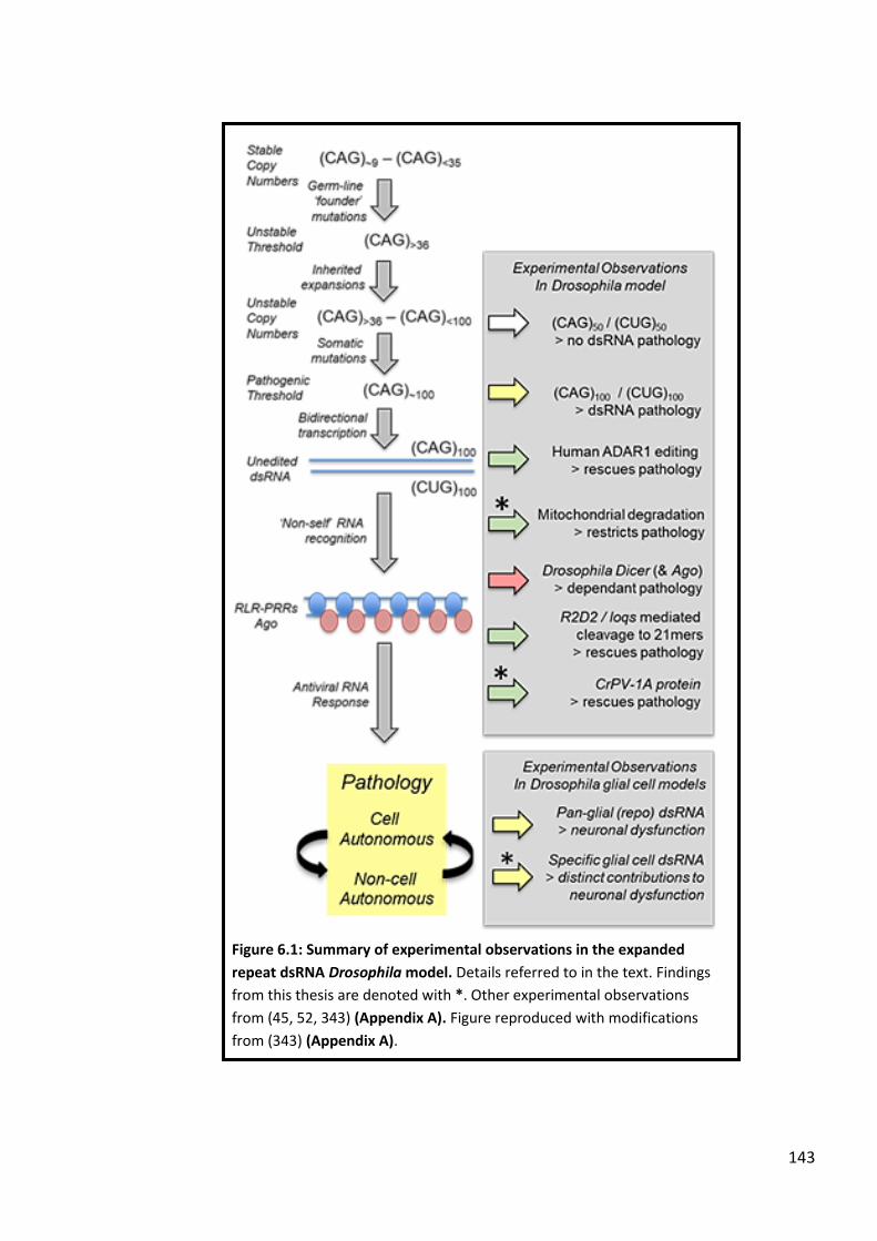
143
Figure 6.1: Summary of experimental observations in the expanded
repeat dsRNA Drosophila model. Details referred to in the text. Findings
from this thesis are denoted with *. Other experimental observations
from (45, 52, 343) (Appendix A). Figure reproduced with modifications
from (343) (Appendix A).

144
ability of other pathways to compensate for the loss of JAK/STAT signalling. Taken together,
the screening experiments performed in Chapter 4 highlighted mitochondria as a key target
for future investigation regarding dsRNA-mediated pathogenesis.
Based on previous work demonstrating that expression of expanded repeat dsRNA in
Drosophila glial cells drives severe neuronal dysfunction (52), the experiments in Chapter 5
aimed to dissect the non-cell autonomous contributions of individual glial cell subtypes to
the neurodegeneration observed. The experimental results highlighted that glial cell types
involved in the formation and maintenance of the Drosophila blood-brain barrier (BBB),
namely subperineural glia and wrapping glia, are sensitive to the expression of expanded
repeat dsRNA. Interestingly, glial subtypes capable of phagocytic activity, ensheathing glia
and astrocyte-like glia, appeared resistant to the expression of more pathogenic dsRNA
construct combinations, while sensitive to the expression of a weaker construct
combination. This indicates that they may respond to repeat dsRNA challenge in a graded
potency-dependent manner, reminiscent of glial phagocytic reactivity in Drosophila
following injury (295). Using the expanded repeat dsRNA eye model, both phagocytic
receptor Draper and phagocytic ligand Mcr modestly restrict the pathology, suggesting that
phagocytosis is protective in response to the expression of repeat dsRNA. Thus, this work
has implicated the Drosophila BBB and phagocytosis as two potential non-cell autonomous
determinants of expanded repeat dsRNA-mediated neuronal pathology that warrant further
definition (Figure 6.1).
6.2 Implications for expanded repeat neurodegenerative diseases
Though research on expanded repeat neurodegenerative disease has uncovered several
commonalities between the individual disease forms, the pathogenic molecular mechanisms
that underlie disease development remain unresolved (3). In this regard, the use of a
genetically simpler in vivo model such as Drosophila can be used to model key aspects of
expanded repeat disease and dissect candidate pathogenic pathways in a more time
effective manner. The conservation of fundamental molecular pathways and cell types
between humans and Drosophila means that findings obtained in flies are applicable to key
mechanistic aspects of the corresponding human condition. This thesis utilizes Drosophila to

145
model one such commonality observed in these diseases – the production of bi-directionally
transcribed expanded repeat RNA from affected loci, predicted to form perfectly double-
stranded RNA (dsRNA) (21). The work described in this study identifies several mechanisms
that participate towards the pathology observed in this expanded repeat dsRNA model.
Firstly, the finding that a viral suppressor protein can effectively ablate the observed dsRNA
pathology suggests that the expanded CAG.CUG repeat dsRNA is recognized by the
inflammatory system as a ‘non-self’ molecule, similar to the RNA of a viral pathogen. This
supports previous work highlighting inflammatory signalling as a key pathogenic event in
HD; both in the CAG.CUG dsRNA Drosophila model (45, 52) and in preclinical HD patients
(58, 59). Given that bi-directionally transcribed RNA is a common feature across repeat loci
(21), these findings may be applicable to the broader range of expanded repeat disorders,
including those defined by repeat sequences other than a trinucleotide CAG sequence such
as the pentanucleotide sequences that cause SCA10 and SCA31. Additionally, constitutive
antiviral inflammation via the ‘non-self’ recognition of endogenous RNA underlies the
pathogenesis of the neurological auto-inflammatory disorder Aicardi-Goutieres syndrome
(AGS) and several related disorders (153). The ablation of antiviral RNA sensing machinery
has been demonstrated to rescue auto-inflammatory pathology associated with genetic
causes of AGS, highlighting antiviral signalling as a promising therapeutic target (152, 156).
Indeed, the results described in this study provide evidence that antiviral inflammatory
signalling also underlies CAG (and potentially other) expanded repeat disorders, and thus
represents a potential therapeutic target.
Secondly, that dysfunctional mitochondrial quality control (namely mitophagy) exacerbates
expanded repeat dsRNA pathology further supports the protective nature of mitophagy in
HD (472). Taken together with the work describing ‘non-self’ recognition of expanded repeat
dsRNA, mitophagy may represent a method to remove and degrade such molecular triggers
before the onset of sustained and damaging inflammatory signalling. Indeed, mitochondrial
dysfunction has been highlighted as a key aspect of polyglutamine disorders (558), though
the majority of evidence stems from research on the mutant HTT protein and thus is specific
to HD. The findings described here provide evidence that mitochondrial dysfunction

146
underlies repeat RNA pathology, and so may constitute a rate-limiting feature common to
other expanded repeat disorders.
Finally, the glial cells that govern development of the Drosophila blood-brain barrier (BBB)
appear to be highly sensitive to the expression of expanded repeat dsRNA. This finding
dovetails with recent evidence demonstrating Drosophila BBB sensitivity to expanded polyQ
proteins (277), indicating that multiple gene products derived from expanded repeat loci can
drive neuronal dysfunction via BBB disruption. In addition, glial cells capable of performing
phagocytosis were resistant to the presence of repeat dsRNA, suggesting that these cells
(and/or potentially adjacent phagocytic glial cells) can degrade the dsRNA and thus prevent
neuronal dysfunction. Thus, BBB integrity and glial-mediated phagocytosis shape as two
processes important in restricting expanded repeat dsRNA pathology, further adding to the
wealth of experimental evidence now highlighting glial cells as key non-cell autonomous
drivers of neurodegeneration (546).
6.3 Considerations for this study
The use of Drosophila as an in vivo model organism provides a number of advantages,
including the range of powerful tools available to genetically dissect candidate pathways and
events. However, several caveats were present in this study that need to be taken into
consideration when interpreting results.
While the expanded repeat Drosophila model used in this thesis highlights co-expressed
sense (CAG) and antisense (CUG) repeat sequences (forming dsRNA) as a key pathogenic
factor in expanded repeat disease, independent work in a Drosophila model of FXTAS has
demonstrated that CGG and CCG expanded repeat sequences are toxic when expressed in
isolation but lose this toxicity in an RNAi-dependent fashion when co-expressed (49).
Although appearing at odds with the dsRNA model used here, there are several key
differences between the models that impede their direct comparison. Firstly, while RAN
translation does not appear to contribute to the RNA-mediated pathology observed in the
CAG.CUG dsRNA model used in this thesis (559), it is plausible that the mechanism
contributes to the ssRNA-driven toxicity observed in the FXTAS model (49). Further

147
characterisation of the Drosophila FXTAS model used in Sofola et al demonstrated that RAN
translation of the sense CGG repeat sequence in a +1 shifted reading frame (GGC) encodes a
polyglycine (polyG) tract that drives pathology within the Drosophila eye (23). Additionally,
both the CGG and CCG repeat sequences are capable on encoding alanine when subject to
RAN translation in their respective +2 reading frames (GCG and GCC) (23, 560), the resulting
polyalanine (polyA) tract of which has also been demonstrated to cause pathology within
the Drosophila eye (561). Indeed, the work performed by Sofola et al predates the discovery
of the RAN translation mechanism (20) and so the respective CGG/CCG repeat lines were not
examined for the presence of this phenomenon at the time. Notably however, earlier
characterisation of the CGG line found that co-expression of the protein chaperone Hsp70
suppressed the associated eye pathology (562). Thus, co-expression of the CGG and CCG
repeat sequences may inhibit the ability of the sequences to undergo RAN translation into
toxic polypeptides, thus ameliorating the eye pathology observed when either sequence is
expressed alone (49).
Another key difference relates to the composition of the repeat sequences themselves. The
CAG/CUG sequences expressed to generate the expanded repeat dsRNA model used in this
thesis are uninterrupted to a length of approximately 100 copies (45). In comparison, the
CGG/CCG sequences utilized in Sofola et al both contain several interruption triplets
interspersed between 90 copies of the respective repeat, shortening the uninterrupted
sequence to approximately 70 copies (49, 562). Length of uninterrupted repeat has been
demonstrated to be a key determinant in expanded repeat dsRNA-mediated pathology (48,
343) (Appendix), which may mirror the length dependency of mammalian RNA sensors for
dsRNA binding and antiviral signalling (563). Thus, the length and composition of the repeat
sequences examined by Sofola et al may not be of a sufficient threshold to be recognised as
‘non-self’ by pattern recognition machinery, avoiding a pathogenic antiviral response.
Despite differences however, the RNAi antiviral pathway appears to be protective in both
Drosophila expanded repeat disease models (49, 343) (Appendix). Thus, it would be of
interest to determine if other components of the antiviral response (namely Dicer-2 and
R2D2) are also rate-limiting in the Sofola et al model and, if so, whether these interactions
are of a helpful or harmful nature.

148
When using the eye-based dsRNA pathology as a genetic screening tissue model, Drosophila
lines containing loss-of-function mutations were not available for some candidate genes.
When this was the case, RNAi-based knockdown lines (315, 316) were utilized to reduce
expression of candidate genes. However, the important rate-limiting role of RNAi pathway
component Dicer-2 (and to a lesser extent Ago2) in the expanded repeat dsRNA pathology
means that the use of RNAi-based knockdown to reduce candidate gene expression can
potentially interfere with observed changes to the dsRNA pathology. While no consistent
phenotypes arose using RNAi knockdowns to implicate RNAi machinery as a confounding
factor, the results were interpreted with this possibility in mind. In line with this, insertional
mutations can also lead to the misexpression of adjacent genes (including the target itself)
when combined with a GAL4 driver. Thus ideally, the findings observed using the dsRNA eye-
based screen should be validated with endogenous loss-of-function mutations for each
candidate gene if/when they become available. Finally, endogenous heterozygous mutations
for a particular gene are not restricted to the specified tissue as dictated by GAL4-UAS
expression (for example the eye, neurons or glial cells). Thus, it is possible that impacts on
lethality/viability in these flies may be caused via global gene reduction as opposed to
tissue-specific effects.
The cellular specificity of GAL4 promoter constructs used in this study, particularly those
used to investigate the individual glial subtypes must also be considered. While the GAL4
driver lines used to express the dsRNA in glial cell subtypes were chosen based on their
demonstrated specificity (292), one must consider the possibility of off-target tissue
expression. Off-target expression and/or mosaicism have been reported in some of the glial
cell subtype drivers (292, 309), though these effects are generally at low levels. Indeed,
widely-used GAL4 drivers GMR-GAL4 and Elav-GAL4 also display trace expression in other
tissue types (509, 511). Thus, while it is highly unlikely that any off-target tissue expression is
of a sufficient magnitude to influence the work described in this study, the possibility of this
must be factored into the interpretation of the results.
Finally, a note regarding the expanded repeat dsRNA Drosophila model used in this study.
The production of dsRNA in this model is via co-expression of independent rCAG~100 and
rCUG~100 constructs in trans (45), as compared to natural bi-directional transcription across

149
expanded repeat loci which occurs via cis-acting transcriptional machinery (21). Therefore,
only the product(s) of bi-directional transcription across repeat loci, is modelled in this
study. Indeed, it is the disease gene product (rCAG~100.rCUG~100), not the mechanism of bi-
directional transcription itself, that is implicated in expanded repeat disease pathogenesis.
Thus, this model provides an excellent method to investigate how the presence of repeat
dsRNA leads to the disease pathology observed.
6.4 Future directions
This study has provided evidence that ‘non-self’ recognition of expanded repeat dsRNA by
the innate inflammatory system contributes to the resultant dsRNA-mediated disease
pathology within the Drosophila eye, and that co-expression of a viral suppressor protein
(CrPV1A) can ablate this pathology. The key next step is to determine the pathway(s) that
CrPV1A antagonizes to suppress the dsRNA pathology. The necrotic eye pathology observed
suggests that a form of antiviral cell death is activated, however the nature (non-lytic vs
lytic) and effectors of the pathway are yet to be determined. Thus, characterisation of cell
death in the dsRNA model should be an important future focus. In line with this, the
possibility of an interaction(s) between CrPV1A and Dicer-2 mediated antiviral signalling
should be investigated, as it is likely to represent a key step of CrPV1A-driven pathology
suppression. Of note, this may not necessarily constitute a direct inhibition of Dicer-2 itself
by CrPV1A, but of downstream intermediary molecules (possibly including Ago2). Indeed,
while the role of Dicer-2 as a pattern recognition receptor is well supported (53-55), little is
known regarding the signalling components downstream of Dicer-2 that drive antiviral
inflammation – for example, a functional equivalent of MAVS is yet to be identified in
Drosophila. Thus, the characterisation of Dicer-2 signalling intermediates forms an essential
next step in defining the inflammatory mechanism(s) that underlie the expanded repeat
dsRNA pathology.
The finding that Drosomycin was significantly upregulated while CG33926 was potently
downregulated in response to the expression of repeat dsRNA is intriguing. Additionally, co-
expression of CrPV1A did not ablate either of these alterations, suggesting that these
pathways may represent protective mechanisms. Indeed, the flies tested were at an early

150
timepoint (0-1 days old), and as such it would be of interest to perform transcript analysis
over an extended time course to determine if these changes correlate with the age-
dependent neurodegeneration observed in elavII>dsRNA flies (45). Furthermore, performing
functional assays to confirm that CrPV1A can ablate dsRNA pathology in the neuronal tissue
model should be considered. Both lines of further analysis would be valuable in dissecting
the antiviral pathways that underlie expanded repeat dsRNA pathogenesis from those that
constitute protective responses.
Evidence that mitophagy is vital for restricting dsRNA pathology is consistent with the
hypothesis that mitochondrial dysfunction is a key driving factor in neurodegenerative
disease (139), and that the processes that govern mitochondrial quality control warrant
further investigation with regards to expanded repeat disorders. Mitochondrial fission is
responsible for the separation of healthy and dysfunctional mitochondria and precedes
mitophagy, so it is plausible that this constitutes a method for the cell to partition the repeat
dsRNA (along with other inflammatory triggers) into mitochondria that are targeted for
degradation. Thus, the dsRNA eye pathology screen should be extended to investigate key
regulators of mitochondrial fission/fusion to determine if this process operates upstream of
mitophagy to limit dsRNA toxicity.
The glial subtype analyses regarding dsRNA-mediated neuronal dysfunction opens up a
number of opportunities for further study, based on the normal functions of glial cells within
the Drosophila CNS. Disruption to BBB development and/or maintenance appears a likely
critical pathogenic event in the observed neuronal pathology, though evidence of BBB
disruption needs to be confirmed in the affected flies. Determining whether peripheral
inflammatory cells/molecules infiltrate the CNS following dsRNA challenge should be also
considered and may provide insight into the role of systemic molecules in disease
pathogenesis. Several glial subtypes within the CNS are capable of phagocytic function(s)
(160) and it is plausible based on the results described in this study to suggest that they
perform this process to remove repeat dsRNA and minimise the resultant neurotoxicity.
Challenging these specific glial cells with the repeat dsRNA following inhibition of
phagocytosis (for example through Draper ablation) will prove insightful in 1) determining if
repeat dsRNA is indeed targeted for removal when present within the CNS, and 2)
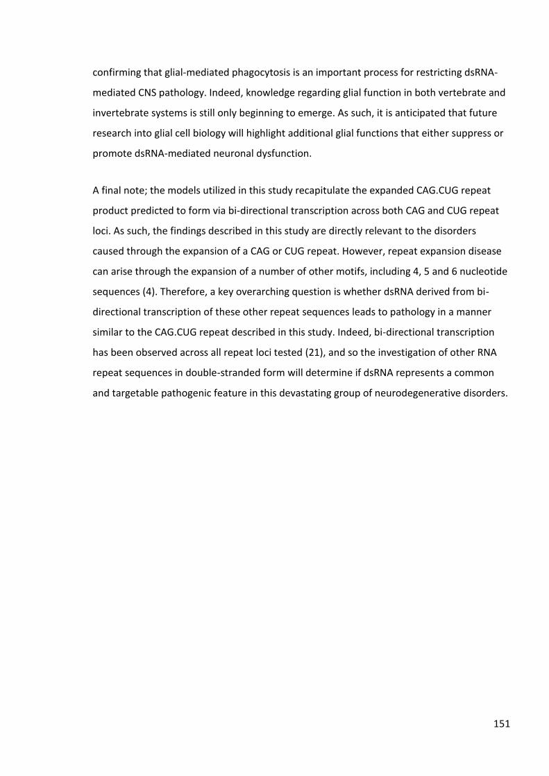
151
confirming that glial-mediated phagocytosis is an important process for restricting dsRNA-
mediated CNS pathology. Indeed, knowledge regarding glial function in both vertebrate and
invertebrate systems is still only beginning to emerge. As such, it is anticipated that future
research into glial cell biology will highlight additional glial functions that either suppress or
promote dsRNA-mediated neuronal dysfunction.
A final note; the models utilized in this study recapitulate the expanded CAG.CUG repeat
product predicted to form via bi-directional transcription across both CAG and CUG repeat
loci. As such, the findings described in this study are directly relevant to the disorders
caused through the expansion of a CAG or CUG repeat. However, repeat expansion disease
can arise through the expansion of a number of other motifs, including 4, 5 and 6 nucleotide
sequences (4). Therefore, a key overarching question is whether dsRNA derived from bi-
directional transcription of these other repeat sequences leads to pathology in a manner
similar to the CAG.CUG repeat described in this study. Indeed, bi-directional transcription
has been observed across all repeat loci tested (21), and so the investigation of other RNA
repeat sequences in double-stranded form will determine if dsRNA represents a common
and targetable pathogenic feature in this devastating group of neurodegenerative disorders.

152

153
Appendix
Appendix A
van Eyk, C.L.*, Samaraweera, S.E.*, Scott, A.*, Webber, D.L.*, Harvey, D.P., O’Keefe, L.V.,
Cropley, J.E., Suter, C., Richards, R.I. (2019) ‘Non-self’ Mutation: Double-stranded RNA
elicits antiviral pathogenic response in a Drosophila model of expanded CAG repeat
neurodegenerative diseases Human Molecular Genetics doi: 10.1093/hmg/ddz096
*Joint first authors.
The following submitted manuscript contains work described in Chapter 3. The work is
described in full in the chapter, however the manuscript is included for reference and to
place this work in the broader context of findings from the Richard’s Lab. The work included
in this thesis was performed by A.S.

154
Co-Author Contributions
By signing the Statement of Authorship, each author certifies that:
i. the candidate’s stated contribution to the publication is accurate (as detailed above);
ii. permission is granted for the candidate in include the publication in the thesis; and
iii. the sum of all co-author contributions is equal to 100% less the candidate’s stated contribution.
Title of Paper ‘Non-self’ Mutation: Double-stranded RNA elicits antiviral pathogenic response in a
Drosophila model of expanded CAG repeat neurodegenerative diseases
Publication Status Published
Accepted for Publication
Submitted for Publication
Unpublished and Unsubmitted work written in manuscript style
Publication Details -

†These authors contributed equally to this work.
Received: February 22, 2019. Revised: April 11, 2019. Accepted: May 6, 2019
© The Author(s) 2019. Published by Oxford University Press. All rights reserved.For Permissions, please email: [email protected]
1
Human Molecular Genetics, 2019, Vol. 00, No. 00 1–13
doi: 10.1093/hmg/ddz096Advance Access Publication Date: 9 May 2019General Article
G E N E R A L A R T I C L E
Non-self mutation: double-stranded RNA elicitsantiviral pathogenic response in a Drosophila model ofexpanded CAG repeat neurodegenerative diseasesClare L. van Eyk1,†, Saumya E. Samaraweera1,†, Andrew Scott1,†,Dani L. Webber1,†, David P. Harvey1, Olivia Mecinger1, Louise V. O’Keefe1,Jennifer E. Cropley2,3, Paul Young2,3, Joshua Ho2,3,4, Catherine Suter2,3 andRobert I. Richards1,*1Department of Molecular and Biomedical Science, School of Biological Sciences, The University of Adelaide,Adelaide, South Australia 5000, Australia, 2Victor Chang Cardiac Research Institute, Lowy Packer Building,Liverpool St, Darlinghurst, Sydney 2010, Australia, 3Faculty of Medicine, University of New South Wales,Kensington, New South Wales 2042, Australia and 4School of Biomedical Sciences, Li Ka Shing Faculty ofMedicine, The University of Hong Kong, Pokfulam, Hong Kong
*To whom correspondence should be addressed at: Department of Molecular and Biomedical Sciences, The University of Adelaide, Adelaide,South Australia 5000, Australia. Tel: +61 8 8313 7541; Fax: +61 8 8313 4362; Email: [email protected]
Abstract
Inflammation is activated prior to symptoms in neurodegenerative diseases, providing a plausible pathogenic mechanism.Indeed, genetic and pharmacological ablation studies in animal models of several neurodegenerative diseases demonstratethat inflammation is required for pathology. However, while there is growing evidence that inflammation-mediatedpathology may be the common mechanism underlying neurodegenerative diseases, including those due to dominantlyinherited expanded repeats, the proximal causal agent is unknown. Expanded CAG.CUG repeat double-stranded RNA causesinflammation-mediated pathology when expressed in Drosophila. Repeat dsRNA is recognized by Dicer-2 as a foreign or‘non-self’ molecule triggering both antiviral RNA and RNAi pathways. Neither of the RNAi pathway cofactors R2D2 norloquacious are necessary, indicating antiviral RNA activation. RNA modification enables avoidance of recognition as ‘non-self’by the innate inflammatory surveillance system. Human ADAR1 edits RNA conferring ‘self’ status and when co-expressedwith expanded CAG.CUG dsRNA in Drosophila the pathology is lost. Cricket Paralysis Virus protein CrPV-1A is a knownantagonist of Argonaute-2 in Drosophila antiviral defense. CrPV-1A co-expression also rescues pathogenesis, confirminganti-viral-RNA response. Repeat expansion mutation therefore confers ‘non-self’ recognition of endogenous RNA, therebyproviding a proximal, autoinflammatory trigger for expanded repeat neurodegenerative diseases.
Dow
nloaded from https://academ
ic.oup.com/hm
g/advance-article-abstract/doi/10.1093/hmg/ddz096/5487598 by U
NIVER
SITY OF AD
ELAIDE user on 26 June 2019

2 Human Molecular Genetics, 2019, Vol. 00, No. 00
Introduction
Dynamic mutation is the common mechanism of expansion forexisting repeat sequences that accounts for about 30 inheriteddiseases. In some cases the repeat expansion causes loss ofgene function, and the resultant phenotype can be attributed toloss of function of the protein normally encoded by the affectedgene (1). About 20 expanded repeat diseases are dominantlyinherited and occur in otherwise unrelated genes, therefore theymust gain some type of common attribute that causes diseases(2–4). These diseases share neurodegeneration as a commonsymptom, yet are typically distinguished by the neurons firstaffected and therefore the symptoms that lead to their initialclinical presentation and differential diagnosis. As the diseasesprogress their pathology tends to spread and so their symptomsconverge, as overlapping neurons are affected (5).
Despite their identification more than two decades ago,the mechanism(s) by which these dominant mutations causesymptoms is/are as yet unclear. While most of these diseasesonly have an expanded repeat as their single mutationmechanism, the recent identification of repeat expansion inthe C9orf72 gene has been a particularly notable addition (6,7).Repeat expansion in the C9orf72 gene is but one of numerousgenetic causes of amyotrophic lateral sclerosis and/or fronto-temporal dementia (ALS/FTD). ALS/FTD causing mutationsin other genes are not expanded repeats and in some casesinvolve the loss of protein function. Commonalities in the lostfunctions of the different ALS/FTD genes are therefore likelyindicators of common pathways of pathogenesis. It is thereforenoteworthy that several of these proteins have functions inRNA Stress Granule metabolism (TDP43, FUS, VCP, CHMP2B)(8–11) and innate inflammatory pathways (sequestosome 1(p62), progranulin, optineurin, TBK1) (12–15).
Late age-at-onset neurodegenerative diseases are increas-ingly being found associated with activation of inflammation(16–22). In the past this association has been dismissed on thebasis that the inflammatory machinery was just coming in toclean up after the proximal cause. However, where timing hasbeen assessed the inflammatory activation precedes neuronalpathology. Presymptomatic Huntington’s Disease (HD) genemutation carriers have elevated circulating levels of cytokines(16,17). Mouse models of prion disease and Alzheimer’s diseasereveal in both cases that microglial cell activation precedes nervecell pathology (23,24). The molecular mechanism for triggeringinflammatory activation thereby causing neurodegenerationhas not been clear.
Inflammation is the front line cellular defense mechanismthat responds to a diverse variety of danger signals (25). Someof these danger signals or trigger molecules are components ofpathogens, including RNA (22). In such cases the inflammatorysurveillance relies on the molecular architecture to distinguishthese ‘non-self’ molecules from the endogenous molecules inthe cell. In addition, cells have resident, endogenous triggermolecules that are normally kept below levels required forinflammatory activation by one or other degradative processesor are masked by ligation to inhibitors. A recently reportedexample of such an endogenous trigger molecule is double-stranded (ds) Alu RNA, which is recognized by the IFIH1 patternrecognition receptor unless modified by the ADAR1 editingenzyme (26–28). Pathogens can therefore also be detectedwhen their presence results in the increase and/or activationof such endogenous trigger molecules. Upon activation theinflammatory response has an early acute phase during whichtrigger molecules (that threaten the organism) are degraded
in order to contain the pathogen and destroy its ability toreplicate. If the threat is not eliminated, then a chronic phaseis activated in order to alarm neighboring cells and prime theirresponse. Chronic inflammation can include the programmeddeath of cell foci that are activated during the acute phase. Whileinflammation is effective in restricting exogenous pathogens,this mechanism is the cause of pathology when mutations leadto the perpetual synthesis of endogenous trigger moleculesthat lack ‘self’ architecture, such as repeat RNA. Mutationsthat impair the cell’s degradative capacity or cause constitutiveactivation of pattern recognition pathways, such as thosewhich increase receptor sensitivity (29), can also give rise toautoinflammatory disease (25).
Cells recognize the presence of viral RNA by means ofeither the absence of ‘self’ recognition patterns or the presenceof ‘non-self’ patterns, or both, and in so doing distinguishpotentially dangerous RNAs from endogenous RNAs. Specificpattern recognition receptors for RNA include the RIG-I-like receptors (RLRs) (30). The RLRs are members of a largerfamily of proteins that include Dicer, RIG-I and IFIH1 (MDA-5)(Supplementary Material, Fig. S1). Drosophila has two membersof the RLR family—Dicer-1 and Dicer-2—with Dicer-2 having beenfound to act as both a component of the RNAi machinery and apattern recognition receptor for antiviral RNA response (31). Theantiviral RNA response in Drosophila is heavily dependent uponArgonaute-2 (32,33), but independent of R2D2, a co-factor in theRNAi pathway (31).
Previously, we and the others explored the hypothesis thatone or other forms of expanded repeat RNA are pathogenic andhave found that (CAG.CUG)100 dsRNA is able to cause pathol-ogy when expressed in the Drosophila eye (34,35). Furthermore,this pathology is both dependent upon components of innatesurveillance (TLRs and TLR signaling) and also activates theinflammatory response (as indicated by elevated drosomycin andTNF/eiger) (36). Pathology was found to be dependent upon Dicerand Argonaute proteins, and the (CAG.CUG)100 RNA was cleaveddown to 21mers of (CAG.CUG)7. This latter finding appeared toindicate the participation of the RNAi machinery (34,35); how-ever, herein we tested this hypothesis and found that key com-ponents of RNAi processing (R2D2 and loquacious) are not onlyunnecessary for (CAG.CUG)100 dsRNA pathology, but also appearto constitute an alternative pathology avoidance pathway. Wehave therefore pursued the hypothesis that (CAG.CUG)100 dsRNAis recognized as ‘non-self’ RNA in the manner typical of viralRNA sequences. The human ADAR1 RNA-editing enzyme con-trols innate immune responses to RNA and when ectopicallyexpressed in Drosophila rescues (CAG.CUG)100 dsRNA pathology.Ectopic expression of cricket paralysis virus protein CrPV-1A,which facilitates viral infection by suppressing the antiviral celldeath response (37–39), rescues (CAG.CUG)100 dsRNA pathologyin Drosophila.
ResultsRNAi co-factors R2D2 and loquacious not required fordsRNA pathology
The requirement for (CAG.CUG)100 dsRNA pathology on certaincomponents of the RNAi machinery (specifically Dicer-1, Dicer-2, Argonaute-2) has previously been reported (34–36). Given theability of Drosophila Dicer proteins to also act as pattern recog-nition receptors (32,33,40–46) we assessed the need for othercomponents of the Drosophila RNAi machinery in (CAG.CUG)100
dsRNA pathology. R2D2 is an obligate component of the RNAi
Dow
nloaded from https://academ
ic.oup.com/hm
g/advance-article-abstract/doi/10.1093/hmg/ddz096/5487598 by U
NIVER
SITY OF AD
ELAIDE user on 26 June 2019

Human Molecular Genetics, 2019, Vol. 00, No. 00 3
pathway in Drosophila along with Dicer-2 and Argonaute-2 (47,48),therefore the role of R2D2 in (CAG.CUG)100 dsRNA pathologywas assessed by its heterozygous deletion or Gal4/UAS over-expression.
RNAi has become a widely used experimental tool inDrosophila, as a means to investigate gene function in a particularpathway, by lowering levels of targeted RNAs. There is, however,substantial overlap between the RNAi pathway and the antiviralresponse mechanism in Drosophila (31,49). The common rolesin these pathways played by Dicer-2 and Argonaute-2, render theRNAi targeted reduction of candidate modifiers of the antiviralresponse problematic. The studies herein have therefore beenrestricted to the analyses of loss-of-function mutations (asheterozygotes) or Gal4-UAS directed ectopic over-expressionof the relevant protein.
While Drosophila carrying a loss-of-function mutation inDicer-1 have reduced (CAG.CUG)100 dsRNA pathology, thosewith loss of R2D2 have enhanced pathology (Fig. 1A’–D’;Supplementary Material, Fig. S2). Furthermore, over-expressionof R2D2 rescues the (CAG.CUG)100 dsRNA pathology (Fig. 1I’ and J’;Supplementary Material, Fig. S2). The reciprocal effects of amore severe phenotype caused by reduction in R2D2 andrescued phenotype with R2D2 over-expression suggest thatthe pathway in which R2D2 participates is actually in compe-tition with that which causes (CAG.CUG)100 dsRNA pathology.Loquacious also acts as an RNAi co-factor. Loquacious proteinfunction is complicated by alternative splicing that gives riseto isoforms with distinct roles in the processing of differentsmall RNA populations. Loquacious mutations can therefore dif-ferentially affect isoform functions (50,51). Nevertheless, neitherof two independent loquacious mutations (loqsKO and loqsf00791)exacerbated expanded CAG.CUG repeat pathology (Fig. 1E’and F’), consistent with RNAi pathways being not required fordsRNA pathology but instead being a competing pathway tothat which causes the pathology. Furthermore, miRNA sequenceprofiles were assessed in (CAG.CUG)100 dsRNA expressingDrosophila to determine whether small RNA processing isaltered. This analysis revealed minimal differences (+/− 1.2-foldchange) in a small number of miRNAs, between flies expressing(CAG.CUG)100 dsRNA versus control (Supplementary Material,Table S1). Those miRNAs (miR-184, miR-263b, miR-274, miR-932,miR-1017) showing altered levels were tested for the ability ofincreased or decreased levels of these miRNAs to alter pathologyin Drosophila lines also expressing (CAG.CUG)100 dsRNA and nonewere found to impact eye phenotype (Supplementary Material,Fig. S3).
Since both Dicer and Argonaute proteins are required for(CAG.CUG)100 dsRNA pathology (Supplementary Material, Fig. S4)this suggests that these proteins work in concert to recognize(CAG.CUG)100 dsRNA as a viral RNA and give rise to the resultantpathology through pattern recognition pathway instigatedantiviral RNA response rather than the RNAi pathway in whichR2D2 and loquacious normally participate.
Human ADAR1 (but not human ADAR2) editing rescuesdsRNA pathology
RNA modification is a major form of distinguishing ‘self’ from‘non-self’ RNAs. RNA modifying enzymes typically recognizedistinct sequence and/or structural features in the RNA. Theexpanded CAG repeat contains the normal recognition sequenceof adenosine deaminase of RNA (ADAR) enzymes. Theseenzymes are also typically affected by the secondary structureof the RNA. The ds structure of (CAG.CUG)100 dsRNA therefore
represents a normal target for ADARs. The ADAR enzymes arealso of great interest for their relationship with known RNA-binding pattern recognition receptors (IFIH1 in humans). Loss-of-function mutations in IFIH1 are able to rescue the phenotypecaused by loss of ADAR1 in mice (51,52). Furthermore, loss-of-function mutation in human ADAR1 is found to be one geneticcause of the congenital neurodegenerative disease Aicardi-Goutieres Syndrome (AGS) (53). Gain-of-function mutationsin the IFIH1 pattern recognition receptor are another causeof AGS (29), strongly implicating RNA modification in AGSpathogenesis.
ADAR1 mutations in some individuals give rise to bilateralstriatal necrosis (54), an intriguing link to HD given that thisbrain region is typically the first to exhibit pathology in HD.Based on the finding that mutations in ADAR1 cause AGS and thesubstrate specificity of the ADAR enzymes it has been proposedthat ADAR1 may limit the cytoplasmic accumulation of thedsRNA generated from genomic repetitive elements (55). Thefinding (56) of interferon-stimulated gene expression signaturesin the heterozygous ADAR1 mutation carrier parents of affectedoffspring with both ADAR1 alleles mutated, suggests that ADAR1is a rate-limiting determinant of interferon induction and innateinflammatory response. Therefore, given the role of ADAR1 inhuman pathology and CAG RNA sequence specificity of ADARproteins, human and Drosophila ADAR enzymes were assessedherein for their impact on Drosophila (CAG.CUG)100 dsRNApathology.
RNAseq analysis of CAG7mer from Drosophila expressing(CAG.CUG)100 dsRNA alone revealed the activity of endogenousDrosophila ADAR on ∼6% of CAG7mer RNAs, with most having asingle edited A>I in the final CAG repeat (Fig. 2E). Thus, whileCAG.CUG is a substrate for Drosophila ADAR this low level ofediting suggests that it is either a poor substrate or the level ofendogenous enzyme is rate-limiting. The Drosophila genome hasa single ADAR gene, while three human genes encode ADARproteins. Human ADAR1 and ADAR2 are well characterizedand expressed, whereas ADAR3 is not known to have catalyticfunction (57). Human ADAR1 is differentially expressed withalternative splicing giving rise to constitutive (hADAR1c) andinterferon-induced (hADAR1i) forms (58). We therefore tested theability of both human hADAR1c and hADAR1i as well as hADAR2and the Drosophila isoform dADAR to affect the (CAG.CUG)100
dsRNA pathology.Co-expression with either human hADAR1c or hADAR1i along
with (CAG.CUG)100 dsRNA saw a complete rescue of pathology(Fig. 2B’ and C’). Little effect was seen with human ADAR2(Fig. 2F’), while alteration in Drosophila ADAR levels also hadminimal impact (Fig. 2G’), consistent with dADAR being moreclosely related to hADAR2 than hADAR1 (59). Co-expression ofDicer-2 with (CAG.CUG)100 dsRNA causes lethality in the S1 line;however, lethality and eye phenotype were completely rescuedby co-expression of hADAR1i (Supplementary Material, Fig. S5).Steady state levels of (CAG.CUG)100 dsRNA were determinedby quantitative RT-PCR on triplicate RNA samples from fliesexpressing (CAG.CUG)100 dsRNA and either human ADAR1 orADAR2 or Drosophila ADAR (Supplementary Material, Fig. S6).While all gave lower levels than flies expressing (CAG.CUG)100
dsRNA alone, the comparable level for flies also expressinghADAR1, hADAR2 or dADAR indicates that lower steady-stateRNA levels alone do not account for phenotype rescue seen withhADAR1, as hADAR2 and dADAR have no phenotype rescue.ADAR1 is therefore specifically capable of masking ‘non-self’status upon (CAG.CUG)100 dsRNA such that it no longer inducesinflammatory pathology.
Dow
nloaded from https://academ
ic.oup.com/hm
g/advance-article-abstract/doi/10.1093/hmg/ddz096/5487598 by U
NIVER
SITY OF AD
ELAIDE user on 26 June 2019

4 Human Molecular Genetics, 2019, Vol. 00, No. 00
Figure 1. R2D2 is not required for the dsRNA phenotype in the Drosophila melanogaster eye. In each case the GMR-GAL4 driver is used to express the control or candidate
alone (A–L) and together with rCAG.rCUG∼100 M3 dsRNA line (A’–L’). (A) GMR-GAL4 does not have an obvious phenotype alone. (A’) Expression of rCAG.rCUG∼100dsRNA results in disruption to ommatidial patterning and loss of pigmentation. (B) Reducing Dicer-1 levels using Dcr1Q1147X/+ does not alter the phenotype of the eye.
(B’) Reducing Dicer-1 levels while co-expressing dsRNA results in a suppression of the dsRNA rough eye phenotype (when compared to A’). (C and D) Reducing r2d2
levels using r2d21 or r2d2s165fsx do not alter the phenotype of the eye. (C’ and D’) Heterozygous loss-of-function mutants r2d21 or r2d2s165fsx flies with GMR-GAL4 driven
expression of rCAG.rCUG∼100 dsRNA results in an enhanced eye phenotype with increased regions of necrosis (when compared to A’). (E and F) Heterozygous loqsKO
and loqsf00791 have no eye phenotype alone. (E’ and F’) The dsRNA phenotype is enhanced by each of the loqs mutants (when compared to A’). (G) GMR-GAL4 does not
have an obvious phenotype alone. (G’) Expression of rCAG.rCUG∼100 dsRNA results in disruption to ommatidial patterning and loss of pigmentation. (H) GMR-GAL4
driven expression of UAS-Dcr-1 has no eye phenotype alone but (H’) enhances rCAG.rCUG∼100 dsRNA eye phenotype (when compared to G’). (I and J) Over-expression
of r2d2 with either of two independent UAS-r2d2 lines (II or III) does not affect the normal patterning of the eye. (I’ & J’) Co-expression of either these UAS-r2d2 over-
expression constructs together with dsRNA rCAG.rCUG∼100 suppresses the dsRNA phenotype, restoring both regular patterning and pigmentation to the eye (when
compared to G’).
Copy number requirement for expanded repeat(CAG.CUG) dsRNA pathology
The antiviral RNA defense mechanism involves the binding ofmultiple RIG-I-like pattern recognition receptors along singlemolecules of RNA to form a filament structure that is then bound
by mitochondrial antiviral signaling protein (MAVS) (60,61). Alength requirement for the RNA molecule appears related tothe need for such filaments to exceed a threshold for MAVSbinding and/or activation (61,62). Therefore, experiments wereundertaken to assess the length dependence of the expandedCAG.CUG repeat to cause antiviral-mediated pathology.
Dow
nloaded from https://academ
ic.oup.com/hm
g/advance-article-abstract/doi/10.1093/hmg/ddz096/5487598 by U
NIVER
SITY OF AD
ELAIDE user on 26 June 2019

Human Molecular Genetics, 2019, Vol. 00, No. 00 5
Figure 2. ds RNA pathology is dependent on editing by ADAR. GMR-GAL4 was used to express constructs in the eye or ELAV(II)-GAL4 to drive expression in all
neurons. Ectopic-expression of either hADAR1-c or hADAR1-i reduce the rCAG.rCUG∼100 dsRNA eye phenotype. (A) GMR-GAL4 driven expression of 4xUAS with no
repeat construct has no phenotype. (A’) GMR-GAL4 driven expression of rCAG.rCUG∼100 causes disruption to patterning of the eye and loss of pigmentation (line S1).
(B and C) GMR-GAL4 driven expression ectopic-expression of either constitutive (hADAR1-c) or interferon inducible (hADAR1-i) human ADAR1 isoforms does not disrupt
the patterning of the eye. (B’ and C’) GMR-GAL4 driven co-expression of either hADAR1-c or hADAR1-i together with rCAG.rCUG∼100 rescues the rCAG.rCUG∼100 eye
phenotype (when compared to A’). (D) RNAseq of hADAR1 rescued eyes/neurons (GMR/Elav). CAG cleavage products (CAG7mer) are detected following GAL4 driven
expression of rCAG.rCUG∼100 in the either the eye (GMR) or in all neurons (pan neuronal, Elav). CAG7mer abundance is significantly reduced in either tissue following
GAL4 driven co-expression of hADAR1-i with rCAG.rCUG∼100. (E) RNAseq of CAG7mer showing editing of final A in 6% RNAs. CAG7mer is A>I edited in 6% of RNAs
predominantly at position 20 (the last CAG). In the minority of CAG cleavage products that are not 21mers that are edited, it is usually the most 3’ A residue that is edited,
suggesting endogenous dADAR editing is targeted to the last A in the sequence. (F and G) Rescue is specific to hADAR1. GMR-GAL4-driven expression ectopic-expression
of either human hADAR2 or Drosophila dADAR-3/4 does not disrupt the patterning of the eye. (F’and G’) GMR-GAL4 driven expression ectopic-expression of either human
hADAR2 or Drosophila dADAR-3/4 does not reduce the eye phenotype caused by co-expression of rCAG.rCUG∼100 (when compared to A’).
The Drosophila model of expanded CAG repeat disease uti-lized herein has previously been found to exhibit expandeddsRNA pathology for 100 copies of the repeat (34). Using a similarDrosophila model, Yu et al. (35) have also found that (CAG)250
and (CUG)250 RNAs cause pathology when co-expressed in theeye, whereas the (CAG)34.(CUG)19 combination of RNAs (belowthe human disease threshold) do not. Therefore, the ability ofshorter length (CAG.CUG) repeats of 50 copies to cause pathologywas assessed. This copy number is well below the putativedisease threshold (∼100 copies) (63) but above the DNA insta-bility threshold (∼36 copies) for HD and related expanded CAGrepeat diseases. Fifty CAG.CUG repeats (150 bases) is also wellabove that required for RNAi processing, as a ds repeat lengthof 19–29 base pairs is sufficient for Dicer-mediated cleavage (64).Multiple lines of Drosophila were established that express fourUAS-transgenes, two encoding (CAG)50 RNA and the other two(CUG)50 RNA. None of these (CAG.CUG)50 expressing lines exhib-ited pathology even though comparable levels of (CAG.CUG)RNA were expressed (Fig. 3). The (CAG.CUG)100 lines exhibitedmuch greater pathology when Drosophila Dicer proteins were
also ectopically over-expressed (25), so Dicer-2 was also ectopi-cally over-expressed in (CAG.CUG)50 lines. Even in the pres-ence of excess Dicer-2 in no case was pathology observed for(CAG.CUG)50, in clear contrast to Dicer-2 increasing pathologyin (CAG.CUG)100 lines (Fig. 3B’–E’). Therefore, the dsRNA pathol-ogy that we have observed in this Drosophila model exhibitslength dependence typical of that required for the antiviral RNAresponse, as well as copy number dependence consistent withthat in the human expanded CAG.CUG repeat diseases (63).
Cricket paralysis virus CrPV-1A suppressor of Argonaute2 rescues (CAG.CUG)100 dsRNA pathology
Length dependence for expanded repeat dsRNA pathology isconsistent with the antiviral recognition mechanism where mul-tiple pattern recognition receptor molecules bind to long ds RNAmolecules, forming a filament structure. This filament assemblytakes place in RNA stress granules and if unresolved (by granu-lophagy) brings about MAVS-mediated inflammatory activation,
Dow
nloaded from https://academ
ic.oup.com/hm
g/advance-article-abstract/doi/10.1093/hmg/ddz096/5487598 by U
NIVER
SITY OF AD
ELAIDE user on 26 June 2019
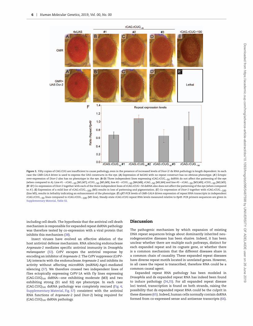
6 Human Molecular Genetics, 2019, Vol. 00, No. 00
Figure 3. Fifty copies of CAG.CUG are insufficient to cause pathology, even in the presence of increased levels of Dicer-2/ ds RNA pathology is length dependent. In each
case the GMR-GAL4 driver is used to express the UAS constructs in the eye. (A) Expression of 4xUAS with no repeat construct has no obvious phenotype. (A’) Ectopic
over-expression of Dicer-2 also has no phenotype in the eye. (B–D) Three independent lines expressing rCAG.rCUG∼50 dsRNA do not affect the patterning of the eye
(when compared to A). Line #1 – rCAG∼50 [M1,M7]; rCUG∼50 [M5,M9], line #2 – rCUG∼50 [M4,M8]; rCAG∼50 [M2,M4] and line #3 – rCAG∼50 [M3,M9]; rCUG∼50 [M2,M3].
(B’–D’) Co-expression of Dicer-2 together with each of the three independent lines of rCAG.rCUG∼50 dsRNA also does not affect the patterning of the eye (when compared
to A’). (E) Expression of a mild line of rCAG.rCUG∼100 (M5) results in loss of patterning and pigmentation. (E’) Co-expression of Dicer-2 together with rCAG.rCUG∼100(line M5), results in lethality indicating an enhancement of the phenotype. (F) qRT-PCR levels of GMR-GAL4 driven expression of repeat RNA transcripts in independent
rCAG.rCUG∼50 lines compared to rCAG.rCUG∼100 (M5 line). Steady-state rCAG.rCUG repeat RNA levels measured relative to Rp49. PCR primers sequences are given in
Supplementary Material, Table S2.
including cell death. The hypothesis that the antiviral cell deathmechanism is responsible for expanded repeat dsRNA pathologywas therefore tested by co-expression with a viral protein thatinhibits this mechanism (38).
Insect viruses have evolved an effective ablation of thehost antiviral defense mechanism. RNA silencing endonucleaseArgonaute-2 mediates specific antiviral immunity in Drosophilamelanogaster (32). CrPV escapes the antiviral response byencoding an inhibitor of Argonaute-2. The CrPV suppressor (CrPV-1A) interacts with the endonuclease Argonaute-2 and inhibits itsactivity without affecting microRNA (miRNA)-Ago1-mediatedsilencing (37). We therefore crossed two independent lines offlies ectopically expressing CrPV-1A with fly lines expressing(CAG.CUG)100 dsRNA—one exhibiting moderate (M3) and twoexhibiting strong (S1 and S2) eye phenotype. In each case(CAG.CUG)100 dsRNA pathology was completely rescued (Fig. 4;Supplementary Material, Fig. S7) consistent with the antiviralRNA functions of Argonaute-2 (and Dicer-2) being required for(CAG.CUG)100 dsRNA pathology.
Discussion
The pathogenic mechanism by which expansion of existingDNA repeat sequences brings about dominantly inherited neu-rodegenerative diseases has been elusive. Indeed, it has beenunclear whether there are multiple such pathways, distinct foreach expanded repeat and its cognate gene, or whether thereis a common mechanism that the different diseases share ina common chain of causality. These expanded repeat diseaseshave diverse repeat motifs located in unrelated genes. However,in all cases the repeat is transcribed, therefore RNA could be acommon causal agent.
Expanded repeat RNA pathology has been modeled inDrosophila and ds expanded repeat RNA has indeed been foundto induce pathology (34,35). For all expanded repeat diseaseloci tested, transcription is found on both strands, raising thepossibility that ds expanded repeat RNA could be the culprit inthese diseases (65). Indeed, human cells normally contain dsRNAformed from co-expressed sense and antisense transcripts (66).
Dow
nloaded from https://academ
ic.oup.com/hm
g/advance-article-abstract/doi/10.1093/hmg/ddz096/5487598 by U
NIVER
SITY OF AD
ELAIDE user on 26 June 2019

Human Molecular Genetics, 2019, Vol. 00, No. 00 7
Figure 4. Cricket paralysis viral protein (CrPV-1A) antagonist of Argonaute ablates rCAG.rCUG∼100 dsRNA pathology when co-expressed in the Drosophila eye. In each case
the GMR-GAL4 driver is used to express the constructs in the eye. (A) Expression of 4xUAS with no repeat construct has no obvious phenotype. (A’) Ectopic expression
of CrPV-1A alone has no phenotype in the eye. (B–D) Lines of Drosophila expressing rCAG.rCUG∼100 dsRNA exhibit variable eye phenotype with one mild (B line M3)
and two strong lines of flies (C, line S1; D, line S2) chosen to assess CrVP-1A impact. (B’–D’) in all three lines expressing rCAG.rCUG∼100 dsRNA, together with the
co-expression of CrVP-1A exhibit complete inhibition of rough eye phenotype.
Furthermore, cytoplasmic ds expanded repeat RNAs have beendetected in the brains of individuals affected with C9ORF72repeat expansion caused ALS/FTD (67).
Dicer and Argonaute proteins are necessary for Drosophilaexpanded repeat dsRNA pathology (34,35) (Fig. 1; SupplementaryMaterial, Fig. S4). These observations together with cleavage oflong RNAs down to (CAG.CUG)7 21mers, have been interpretedas the RNAi pathway being responsible and the 21mers thecausal agent. This hypothesis is tested herein by assessing therequirement for further components of the RNAi pathway, R2D2and loquacious. In both cases reduction of these co-factors ledto an increase in pathology. Indeed, not only does reduction ofR2D2 increase pathology, but also an increase of R2D2 reducespathology. These relationships are consistent with the RNAipathway and cleavage of the (CAG.CUG)100 RNA to 21mers beingin competition with the responsible pathogenic pathway. Sincethe Dicer and Argonaute proteins are also required for antiviralRNA-mediated cell death we hypothesize that this pathway isthe one responsible for (CAG.CUG)100 RNA pathology.
Both the activation of, and requirement for, the innateinflammatory surveillance system, have been reported inthis Drosophila model of expanded repeat dsRNA pathology(36). Inflammatory activation correlates with various neu-rodegenerative diseases but has been typically thought ofas a consequence of cell death—coming in to clean up themess that something else has started. There is, however,increasing evidence that inflammatory activation precedesneurodegeneration, consistent with a causal role both in thehuman diseases (16,17) and in experimental animal diseasemodels (23,24).
Nucleic acids are the common means of encoding geneticinformation for both hosts and pathogens, therefore innate hostdefense requires a mechanism to distinguish harmless endoge-nous nucleic acids from those that are foreign and potentiallyharmful. A large number and variety of modification reactionsalter specific nucleotides in host nucleic acids to essentially ‘bar-code’ those that are ‘self’ and allow their distinction from thosethat are ‘non-self’ or foreign. RNA editing is a form of modi-fication in which the coding potential of the target nucleotideis changed, most notably the sequence specific conversion of
adenosine to inosine in RNA sequences, catalyzed by adeno-sine deaminase of RNA (ADAR). Editing of endogenous RNAs byADAR1 is required to prevent activation of the cytosolic innateimmune system (51–54,68).
In the expanded CAG repeat diseases, ADAR editing is ofparticular interest as the recognition motif/modification siteis contained in the expanded CAG repeat. Furthermore, ADARenzymes have a general preference for dsRNA. Therefore,expanded CAG.CUG dsRNA is an ideal target for ADAR editing.The ability of human ADAR1, but neither human ADAR2 norDrosophila ADAR, to confer ‘self’ status on expanded CAG.CUGdsRNA is noteworthy as ADAR1 is a known component ofinflammatory activation (51–54,68). The human ADAR1 gene hastwo promoters, one for constitutive expression and the otherinterferon-inducible (69). The acute inflammatory responseproduces a pulse of interferon signaling as one of its ‘feed-forward’ responses aimed at resolving the trigger molecules/danger signals that activate inflammation. Interferon-inducedincrease in ADAR1 levels therefore enables the editing of RNAs,thereby reducing the level of ‘non-self’ RNA signal. Together withinduced increases in RNA turnover processes, this mechanismcan degrade RNA trigger molecule/danger signal threats.
ADAR1 also has a functional relationship with the RIG-I-likepattern recognition receptor IFIH1 (MDA5), a member of the samefamily of proteins as Dicer (51,52). Loss-of-function mutationsin ADAR1 cause early lethality in mice that can be rescuedby deletion of IFIH1. ADAR1 is responsible for the editing ofendogenous RNAs to enable their recognition as ‘self’ (27,28,70).Absence of ADAR1 sees the ‘non-self’ forms of these RNAsrecognized by IFIH1 that then activates inflammation (26,28).The endogenous RNAs recognized by IFIH1 include not onlyAlu dsRNA but also products of the unfolded protein response(UPR) pathway. One of the UPR response proteins, IRE-1, cleavesRNAs in a manner that renders them inducers of IFIH1. Loss-of-function mutations in ADAR1 and gain-of-function mutationsin IFIH1 are both causes of AGS, a congenital disorder thatincludes neurodegeneration, with affected newborns having theappearance of being subjected to in utero inflammatory injury(53,71). The gain-of-function mutations in IFIH1 that cause AGSalso increase the affinity of this PRR for its RNA ligands (29).
Dow
nloaded from https://academ
ic.oup.com/hm
g/advance-article-abstract/doi/10.1093/hmg/ddz096/5487598 by U
NIVER
SITY OF AD
ELAIDE user on 26 June 2019

8 Human Molecular Genetics, 2019, Vol. 00, No. 00
The sensitivity to ADAR1 editing of the pathology caused by(CAG.CUG)100 dsRNA in this Drosophila model is therefore con-sistent with the recognition of the product of repeat expansionmutation as a ‘non-self’ RNA trigger molecule/danger signalthat then activates inflammation. ‘Non-self’ driven antiviralRNA recognition is an important process for virus removal asevident by the finding that HIV evades this process by recruitingthe RNA 2′-O-methyltransferase FTSJ3 to avoid innate immunesensing (72).
A growing body of evidence is consistent with there being adistinction between repeat instability threshold and a greatercopy number threshold for pathology (1,63,73–80). Geneticvariations in mismatch repair enzymes have been shown tobe major modifiers of age-at-onset of Huntington’s Disease (75)and other polyglutamne diseases (76), as well as mouse modelsof these diseases (74,77–80). These enzymes control the rate ofincrease by somatic mutation from its inherited (germ-line) copynumber and imply the existence of a higher copy number diseasethreshold of approximately 100 copies (63). Copy numbers above36 are unstable, with the shorter unstable repeats (e.g. 40 copies)taking longer to reach the disease threshold than inherited80 copy alleles (typically associated with juvenile HD) (1,63).Such a mechanism explains genetic anticipation: the inverserelationship between inherited repeat copy number and age-at-onset of disease symptoms (1,81). The expanded (CAG.CUG)dsRNA pathology in this Drosophila model exhibits length depen-dence consistent with the antiviral RNA response mechanismbeing responsible. In addition, the observed copy numberdependence is consistent with distinct thresholds for DNAinstability and pathology in the human expanded CAG repeatdiseases (63).
In order to be infectious, viruses must evade detection and/orresponse mechanisms of the innate surveillance system. Thisresponse can include the programmed death of cells if the viralthreat is not degraded. Cell death is orchestrated in both thecell in which the threat is detected and in adjacent sensitizedcells to give a focal ‘scorched earth’ response that denies thevirus both the opportunity to replicate and to infect adjacentcells. CrPV has evolved a mechanism of specific interferencewith the anti-viral RNA response. The CrPV-1A protein is aspecific inhibitor of Argonaute and RNA stress granule formation(37,39). Stress granules are the location of filament formationbetween viral RNA and the RIG-I-like receptors. In vertebratesthese filaments are then bound by the protein MAVS on themitochondrial outer surface that in turn activates the antivi-ral response (60,61). Co-expression of CrPV-1A with expandedCAG.CUG repeat dsRNA completely rescues the pathology other-wise caused by this dsRNA. Therefore, the same cellular defensepathway of cell death that is normally utilized against viruses(and evaded by CrPV) is responsible for the pathology causedby the presence of expanded CAG.CUG repeat dsRNA, indicatingits recognition by the cell as a viral ‘non-self’ RNA, a gain-of-function that accounts for dominant inheritance.
Inflammatory activation, with increased circulating cytokines(16) and microglial activation (17) precedes neuronal pathologyin premanifest HD carriers. Inflammatory genes are activated incells that are destined to undergo neurodegeneration. Theseinclude the induction of interferon-regulated genes in thecataracts of patients with myotonic dystrophy due to expansionof either CUG (DM1) or CCUG (DM2) (18). Increased inflammatorygene expression is evident in HD brain (19) and inflammatorygene signatures found in ALS motor neurons (20). Inflammationis activated at the right time and place to be the causalmechanism of disease.
In addition to the requirement for inflammatory activation inthe expanded CAG.CUG model described herein (36), other ani-mal models of dominantly inherited neurodegenerative diseasescaused by the expansion of various repeat motifs have showndependence on components of inflammation for pathology. Lossof MyD88 attenuates early Purkinje cell loss in a spinocerebellarataxia type-6 mouse model (82), while p62 plays a protectiverole in polyglutamine disease model flies (83). Furthermore, defi-ciency of toll-like receptors 2, 3 or 4 extends life expectancy inHD mice (84). Genetic ablation of inflammation typically resultsin rescue of disease phenotypes—indicating that inflammatoryactivation is required for pathogenesis. While the agent causinginflammatory activation has not been identified in these lattercases, a zebrafish model for C9orf72 ALS/FTD reveals RNA as apathogenic agent (85). In addition, many of the genes mutatedin non-repeat expanded cases of ALS have roles in inflamma-tory activation pathways and RNA metabolism. Furthermore,their mutation typically leads to an increase in inflammatoryactivation (12,15).
Repeat expansions are therefore ‘non-self’ mutations thatconvert expanded repeat RNA from a harmless molecule whenin low copy number to a nucleic acid that lacks the molecu-lar architecture to be distinguished as ‘self’. Expanded repeatRNA that exceeds a copy-number threshold is a persistent trig-ger of pattern recognition receptor signaling and inflamma-tory responses. Preventing the detection of the proximal repeatRNA trigger by either masking the ‘non-self’ RNA (with over-expression of human ADAR1) or blocking viral-detection path-ways (with co-expression of a viral antagonist) both preventpathology. Elongated repeat sequences are conducive to multi-merisation of repeat structures that are known to trigger chronicinflammatory activation. These characteristics not only accountfor autoinflammatory cell death being the pathogenic mecha-nism for neurodegeneration, they are also consistent with theage-related somatic mutation of inherited (but sub-pathogenic)unstable copy numbers being the rate-limiting determinant ofdisease onset.
While studies herein describe the acquisition ‘non-self’recognition by RNA, this principle can also apply to other geneproducts. For example, structures adopted by prion proteinsactivate inflammation (23) and glycated forms of proteins arepreferential activators of the RAGE pattern recognition receptor(86). ‘Non-self’ mutation can include alteration in structure orpost-translational modification that confers pattern receptorrecognition. ‘Non-self’ mutation therefore provides a specificmechanism for pathogenic activation of inflammation that canaccount for numerous neurodegenerative diseases.
Materials and MethodsFly husbandry
All crosses were performed at 25◦C with 70% humidity unlessotherwise indicated. Drosophila melanogaster stocks were main-tained on fortified (F1) medium composed of 1% (w/v) agar,18.75% compressed yeast, 10% treacle, 10% polenta, 1.5% acidmix (47% propionic acid, 4.7% orthophosphoric acid) and 2.5%tegosept (10% para-hydroxybenzoate in ethanol).
Fly stocks
Controls stocks used were 4xUAS (empty vector) as we havepreviously described (34), UAS-LacZRNAi was kindly provided byRichard Carthew (87) and UAS-mCherry RNAi was obtained from
Dow
nloaded from https://academ
ic.oup.com/hm
g/advance-article-abstract/doi/10.1093/hmg/ddz096/5487598 by U
NIVER
SITY OF AD
ELAIDE user on 26 June 2019

Human Molecular Genetics, 2019, Vol. 00, No. 00 9
Bloomington Drosophila Stock Centre (BDSC). Driver lines usedwere GMR-GAL4 (76) and P{GAL4-elav.L}2/CyO (referred to aselavII-GAL4) (88). Pathogenic dsRNA rCAG∼100.rCUG100∼100 linesused are as previously described (34). Dcr-1Q1147X and Dcr-2L811fsx
(89) as well as UAS-Dcr1, UAS-Dcr2 and UAS-r2d2 (90) were kindlyprovided by Richard Carthew, as were UAS-AGO2 (II) and UAS-AGO2 (III) (90) lines. AGO2414 (91) was obtained from the Kyotostock centre. AGO2V966M (92) was obtained from the BDSC. r2d21
(47), r2d2s165fsx (93) and loqsf00791 (94) were obtained from BDSC.loqsKO was kindly provided by Qinghua Liu and Dennis McKearin(95). UAS-miR-184 and ΔmiR-184 were kindly provided by UlrikeGaul (96). UAS-miR-263b and ΔmiR-263b were kindly providedby Stephen Cohen (97). UAS-miR-1017 was kindly provided byEric Lai (98). Df(3L)BSC577 (including miR-274) and Df(3L)BSC577(including miR-932) were obtained from BDSC. UAS-hADAR-1c,UAS-hADAR1i and UAS-hADAR2 (99) and UAS-dADAR (100) werekindly provided by Liam Keegan. ADAR1F1 was kindly providedby Michael Palladino (101). CrPV1A lines (39) were obtained fromChristophe Antoniewski. Genotypes of fly lines were confirmedby PCR (Supplementary Materials, Fig. S8 and Table S2 of oligoPCR primers).
Generation of untranslated CAG∼50 and CUG∼50 repeatconstructs
Generation of (rCAG.rCUG)100 lines is described in Lawlor et al.(34). CAG∼50 repeats were digested from pBluescript CAG∼50 (102)using flanking Hind III. This fragment was then ligated into theHind III site of pENTR/D-TOPO IVM2 MYC FLAG. The presenceof the insert was confirmed by restriction digestion with Acc65Iand XbaI. Plasmids were also sequenced and screened for insertorientation and integrity. Constructs containing either CAG∼50 orCTG∼50 were subcloned into pDEST-UAST by LR clonase recom-bination. Constructs were confirmed by digestions with therestriction enzymes BsrG I and BamH I and by sequencing.
Generation of transgenic lines
Microinjections to generate transgenic lines were performed byBestGene Inc., Chino Hills, CA, USA. Transformants were mappedto determine the chromosome of insertion and balanced stockswere generated by standard genetic crosses. Single insertionlines for either the CAG∼50 or CTG∼50 constructs were labeledrCAG∼50 M1-10 and rCUG∼50 M1-10.
Generating recombinant lines
Recombinants carrying two independent insertions of the sametransgene (either rCAG∼50 or rCTG∼50) on the same chromosomewere generated by crossing trans-heterozygous virgin females tomale w1118 flies. Standard genetic crosses were then performedto generate three lines each carrying four transgenes, i.e. twoexpressing rCAG.2 and two expressing rCUG∼50, as follows:
rCAG.rCUG∼50#1 – rCAG∼50 [M1,M7]; rCUG∼50 [M5,M9]rCAG.rCUG∼50#2 – rCUG∼50 [M4,M8]; rCAG∼50 [M2,M4]rCAG.rCUG∼50#3 – rCAG∼50 [M3,M9]; rCUG∼50 [M2,M3].
RNA isolation and quantitative real-time PCR
Expression in the fly eye enables qualitative assessment of thephenotypic consequences of transgene expression, however the
GMR promoter only drives RNA expression in a small proportionof cells (even though some of these are nerve cells) with themajority of non-expressing cells of the fly head effectivelydiluting out responses. Therefore, as in previous studies inthis model (34,36), RNA was isolated from the whole heads ofeither GMR-Gal4 (eye) or ElavII-Gal4 (pan-neuronal) expressingfly lines of various UAS-transgene genotypes and subjectedto quantitative reverse transcriptase real-time PCR (qRT-PCR)as described (34,36). Approximately 100 male Drosophila headsfrom newly eclosed flies were collected and snap frozen foreach genotype, before homogenization in Trizol (Invitrogen).Total RNA was extracted using chloroform and precipitatedwith isopropanol, then further purified using the RNeasy minikit (Qiagen). RNA to be used for RNAseq was precipitated insodium acetate and ethanol and shipped under ethanol onice. 1 μg of total RNA per sample was treated with DNAseI (Invitrogen) and reverse-transcribed with oligo(dT)18 andSuperScript III (Invitrogen). Quantitative real-time PCR wasperformed in a LightCycler (Roche Molecular Biochemicals)using Power SYBR green mix (Applied Biosystems) and eitherGAL4-specific primers (forward: 5′-CACTGACCCCGTCTGCTTTG-3′, and reverse: 5′-GGTTCGGACCGTTGCTACTG-3′) or primersspecific for the repeat-containing transcript. The transgeneexpression level was quantified using the delta Ct method forrelative quantification and expressed relative to the level ofGAL4 transcript for each line.
The quantity of amplified cDNA product for each primer pairwas normalized to the quantity of product with primers for thehousekeeping gene, ribosomal protein 49 (Rp49), for the samesample. The average of triplicate reactions for each sample wasobtained and the standard deviation of the mean was calculated.The significance between samples was determined by using atwo-tailed Student’s t-test. The significance threshold was set atP < 0.05.
RNAseq identification of CAG 21-mer repeat editing andchanges in miRNA profiles
RNAseq was as previously described in Samaraweera et al. (36).Barcoded small RNA libraries were prepared from 1 μg totalRNA isolated from neurons and eyes using the NEBnext SmallRNA Library Prep set for Illumina (NEB). 2S RNA was depletedat the RT primer hybridization step using a terminator oligoblocking strategy, as described in http://www.ncbi.nlm.nih.gov/pmc/articles/PMC4077022/. Libraries were sequenced onan Illumina HiSeq 2500 in Rapid Run mode. Fastq files weretrimmed with Cutadapt (v1.6) to lengths of 18–35 nt, with 10%adapter sequence error allowed. CAG repeats were identifiedin the sequencing data by searching for exact matches to18–35 nt strings of CAG repeating units. Edited repeats wereidentified by allowing replacement of any to all CAG unitswith CGG.
Eye photos
Eyes of at least 15 flies were viewed and representative eyesimaged. Flies were photographed at 24–48 h post-eclosion. Lightphotos were taken with an Olympus SZX7 dissection microscopefitted with an SZX-AS aperture. Images were captured witha Colorview IIIu camera and AnalysisRuler image acquisitionsoftware. In all cases, anterior is to the left. Image preparationwas performed using Adobe Photoshop 6.0.
Dow
nloaded from https://academ
ic.oup.com/hm
g/advance-article-abstract/doi/10.1093/hmg/ddz096/5487598 by U
NIVER
SITY OF AD
ELAIDE user on 26 June 2019

10 Human Molecular Genetics, 2019, Vol. 00, No. 00
Supplementary MaterialSupplementary Material is available at HMG online.
AcknowledgementsWe thank Richard Carthew, Qinghua Liu, Ulrike Gaul, StephenCohen, Eric Lai, Liam Keegan, Dennis McKearin, Michael Pal-ladino, Christophe Antoniewski, Bloomington Drosophila StockCentre and Vienna Drosophila Research Centre for providinglines of Drosophila and Kim Hemsley, Keh Yee Ho, John Hayball,Dan Kastner, Michael Lardelli, Shane Liddelow, Arabinda Nayak,Sarah Robertson, Allen Roses and Thalia Skoumbourdis for thehelpful discussions.
Conflict of Interest statement. None declared.
FundingNational Health and Medical Research Council of AustraliaProject (grant APP1069348 to R.I.R. and C.S.); the National AtaxiaFoundation (USA; to C.L.vE). C.L.vE, S.E.S. and A.S. were recipientsof Australian Post-graduate Awards.
Author contributionsC.L.vE., S.E.S., A.S., D.L.W., D.L.H., O.M. and L.V.O conductedand analyzed Drosophila experiments. J.E.C., P.Y., J.H. and C.S.conducted and analyzed RNAseq experiments. L.V.O and R.I.R.designed the experiments. R.I.R wrote the first and final draftsof the paper and its revision.
References1. Richards, R.I. (2001) Dynamic mutations: a decade of unsta-
ble expanded repeats. Hum. Mol. Genet., 10, 2187–2194.2. La, A.R. and Taylor, J.P. (2010) Repeat expansion disease:
progress and puzzles in disease pathogenesis. Nat. Rev.Genet., 11, 247–258.
3. van, C.L. and Richards, R.I. (2012) Dynamic mutations:where are they now? In Hannan, A. (ed.), Tandem Repeatsand Dynamic Mutations Leading to Neurological Disorders.Advances in Experimental Medicine and Biology, SpringerPublishing, NY, Vol. 769, pp. 55 –77.
4. Richards, R.I. (2017) Dynamic mutations, ReferenceModule in Life Sciences, Science Direct, Elsevier, ISBN9780128096338, https://doi.org/10.1016/B978-0-12-809633-8.06369-X. (http://www.sciencedirect.com/science/article/pii/B978012809633806369X).
5. Ross, C.A. (1995) When more is less: pathogenesis ofglutamine repeat neurodegenerative diseases. Neuron, 15,493–496.
6. DeJesus-Hernandez, M., Mackenzie, I.R., Boeve, B.F.,Boxer, A.L., Baker, M., Rutherford, N.J., Nicholson, A.M.,Finch, N.A., Flynn, H., Adamson, J. et al. (2011) ExpandedGGGGCC hexanucleotide repeat in noncoding region ofC9ORF72 causes chromosome 9p-linked FTD and ALS.Neuron, 72, 245–256.
7. Renton, A.E., Majounie, E., Waite, A., Simón-Sánchez, J.,Rollinson, S., Gibbs, J.R., Schymick, J.C., Laaksovirta, H., vanSwieten, J.C., Myllykangas, L. et al. (2011) A hexanucleotiderepeat expansion in C9ORF72 is the cause of chromosome9p21-linked ALS-FTD. Neuron, 72, 257–268.
8. Bentmann, E., Haass, C. and Dormann, D. (2013) Stressgranules in neurodegeneration—lessons learnt from TARDNA binding protein of 43 kDa and fused in sarcoma. FEBSJ., 280, 4348–4370.
9. Li, Y.R., King, O.D., Shorter, J. and Gitler, A.D. (2013) Stressgranules as crucibles of ALS pathogenesis. J. Cell Biol., 201,361–372.
10. Buchan, N., Silverman, N. and Cherry, S. (2014) Immu-nity in Drosophila melanogaster—from microbial recogni-tion to whole-organism physiology. Nat. Rev. Immunol., 14,796–810.
11. Talbot, K. and Ansorge, D. (2006) Recent advances in thegenetics of amyotrophic lateral sclerosis and frontotem-poral dementia: common pathways in neurodegenerativedisease. Hum. Mol. Genet., 15, R182–R187.
12. Fecto, F., Yan, J., Vemula, S.P., Liu, E., Yang, Y., Chen, W.,Zheng, J.G., Shi, Y., Siddique, N., Arrat, H. et al. (2011) SQSTM1mutations in familial and sporadic amyotrophic lateralsclerosis. Arch Neurol., 68, 1440–1446.
13. Freischmidt, A., Wieland, T., Richter, B., Ruf, W., Schaeffer, V.,Müller, K., Marroquin, N., Nordin, F., Hübers, A., Weydt, P.et al. (2015) Haploinsufficieny of TBK1 causes familial ALSand fronto-temporal dementia. Nat. Neurosci., 18, 631–636.
14. Lui, H., Zhang, J., Makinson, S.R., Cahill, M.K., Kelley, K.W.,Huang, H.Y., Shang, Y., Oldham, M.C., Martens, L.H., Gao, F.et al. (2016) Progranulin deficiency promotes circuit-specificsynaptic pruning by microglia via complement activation.Cell, 65, 921–935.
15. Tanishima, M., Takashima, S., Honda, A., Yasuda, D.,Tanikawa, T., Ishii, S. and MaruYama, T. (2017) Iden-tification of optineurin as an interleukin-1 receptor-associated kinase 1-binding protein and its role in regu-lation of MyD88-dependent signaling. J. Biol. Chem., 292,17250–17257.
16. Bjorkqvist, M., Wild, E.J., Thiele, J., Silvestroni, A., Andre, R.,Lahiri, N., Raibon, E., Lee, R.V., Benn, C.L., Soulet, D. et al.(2008) A novel pathogenic pathway of immune activationdetectable before clinical onset in Huntington’s disease.J. Exp. Med., 205, 1869–1877.
17. Politis, M., Lahiri, N., Niccolini, F., Su, P., Wu, K., Giannetti,P., Scahill, R.I., Turkheimer, F.E., Tabrizi, S.J. and Piccini, P.(2015) Increased central microglial activation associatedwith peripheral cytokine levels in premanifest Hunting-ton’s disease gene carriers. Neurobiol. Disease, 83, 115–121.
18. Rhodes, J.D., Lott, M.C., Russell, S.L., Moulton, V.,Sanderson, J.,zx Wormstone, I.M. and Broadway, D.C.(2012) Activation of the innate immune response andinterferon signalling in myotonic dystrophy type 1 andtype 2 cataracts. Hum. Mol. Genet., 21, 852–862.
19. Labadorf, A., Hoss, A.G., Lagomarsino, V., Latourelle, J.C.,Hadzi, T.C., Bregu, J., MacDonald, M.E., Gusella, J.F., Chen, J.F.,Akbarian, S. et al. (2015) RNA sequence analysis of humanHuntington disease brain reveals an extensive increase ininflammatory and developmental gene expression. PLoSOne, 10, e0143563. doi:10.1371/journal.pone.0143563.
20. Batra, R., Hutt, K., Vu, A., Rabin, S.J., Baughn, M.W., Libby, R.Y.,Hoon, S., Ravits, J. and Yeo, G.W. (2016) Gene expression sig-natures of sporadic ALS motor neuron populations. bioRχ ivdoi:https://doi.org/10.1101/038448.
21. Richards, R.I., Robertson, S.A., O’Keefe, L.V., Fornarino,D., Scott, A., Lardelli, M. and Baune, B.T. (2016) Theenemy within: innate surveillance-mediated cell death, thecommon mechanism of neurodegenerative disease. Front.Neurosci., 10, 193 doi:10.3389/fnins.2016.00193.
Dow
nloaded from https://academ
ic.oup.com/hm
g/advance-article-abstract/doi/10.1093/hmg/ddz096/5487598 by U
NIVER
SITY OF AD
ELAIDE user on 26 June 2019

Human Molecular Genetics, 2019, Vol. 00, No. 00 11
22. Richards,R.I.,Robertson, S.A. and Kastner, D.L. (2018) Neuro-degenerative diseases have genetic hallmarks of autoin-flammatory disease. Hum. Mol. Genet., 27(R2), R108–R118.
23. West Greenlee, H., Lind, M., Kokemuller, R., Mammadova, N.,Kondru, N., Manne, S., Smith, J., Kanthasamy, A. andGreenlee, J. (2016) Temporal resolution of misfolded prionprotein transport, accumulation, glial activation, and neu-ronal death in the retinas of mice inoculated with scrapie.Am. J. Pathol., 186, 2302–2309.
24. Boza-Serrano, A., Yang, Y., Paulus, A. and Deierborg, T. (2018)Innate immune alterations are elicited in microglial cellsbefore plaque deposition in the Alzheimer’s disease mousemodel 5xFAD. Sci. Rep., 8, 1550 doi:10.1038/s41598-018-19699-y.
25. Manthiram, K., Zhou, Q., Aksentijevich, I. and Kastner, D.L.(2017) The monogenic autoinflammatory diseases definenew pathways in human innate immunity and inflamma-tion. Nat. Immunol., 18, 832–842.
26. Ahmad, S., Mu, X., Yang, F., Greenwald, E., Park, J.W., Jacob, E.and Zhang, C.-Z. (2018) Breaching self-tolerance to Aluduplex RNA underlies MDA5-mediated inflammation. Cell,172, 797–810.
27. Chung, H., Calis, J.J.A., Wu, X., Sun, T., Yu, Y., Sarbanes, S.L.,Thi, V.L.D., Shilvock, A.R., Hoffmann, H.-H., Rosenburg, B.R.and Rice, C.M. (2018) Human ADAR1 prevents endogenousRNA from triggering translational shutdown. Cell, 172,811–824.
28. Uggenti, C. and Crow, Y.J. (2018) Sort yourself out. Cell, 172,640–642.
29. Rice, G.I., Del, Y., Jenkinson, E.M., Forte, G.M., Anderson, B.H.,Ariaudo, G., Bader-Meunier, B., Baildam, E.M., Battini, R.,Beresford, M.W. et al. (2014) Gain-of-function mutationsin IFIH1 cause a spectrum of human disease phenotypesassociated with upregulated type I interferon signaling.Nat. Genet., 46, 503–509.
30. Chiang, J.J., Davis, M.E. and Gack, M.U. (2014) Regulationof RIG-I-like receptor signaling by host and viral proteins.Cytokine Growth Factor Rev., 25, 491–505.
31. Deddouche, S., Matt, N., Budd, A., Mueller, S., Kemp, C.,Galiana-Arnoux, D., Dostert, C., Antoniewski, C., Hoffmann,J.A. and Imler, J.L. (2008) The DExD/H-box helicase Dicer-2mediates the induction of antiviral activity in Drosophila.Nat. Immunol., 9, 1425–1432.
32. van, R.P., Saleh, M.-C., Berry, B., Foo, C., Houk, A.,Antoniewski, C. and Andino, R. (2006) The RNA silenc-ing endonuclease Argonaute 2 mediates specific antivi-ral immunity in Drosophila melanogaster. Genes Dev., 20,2985–2995.
33. Tassetto, M., Kunitomi, M. and Andino, R. (2017) Circulatingimmune cells mediate a systemic RNAi-based adaptiveantiviral response in Drosophila. Cell, 169, 314–325.
34. Lawlor, K.T., O’Keefe, L.V., Samaraweera, S., van, C.,McLeod, C.J., Maloney, C., Dang, T., Suter, C. and Richards,R.I. (2011) Double stranded RNA is pathogenic in Drosophilamodels of expanded repeat neurodegenerative diseases.Hum. Mol. Genet., 20, 3757–3768.
35. Yu, Z., Teng, X. and Bonini, N.M. (2011) Triplet repeat-derived siRNAs enhance RNA-mediated toxicity in a Droso-philamodel formyotonicdystrophy.PLoSGenet., 7, e1001340.
36. Samaraweera, S.E., O’Keefe, L.V., Price, G.R., Venter, D.J. andRichards, R.I. (2013) Distinct roles for toll and autophagypathways in double-stranded RNA toxicity in a Drosophilamodel of expanded repeat neurodegenerative diseases.Hum. Mol. Genet., 22, 2811–2819.
37. Nayak, A., Berry, B., Tassetto, M., Kunitomi, M., Acevedo, A.,Deng, C., Krutchinsky, A., Gross, J., Antoniewski, C. andAndino, R. (2010) Cricket paralysis virus antagonizes Arg-onaute 2 to modulate antiviral defense in Drosophila. Nat.Struct. Mol. Biol., 17, 547–554.
38. Khong, A., Kerr, C.H., Yeung, C.H., Keatings, K., Nayak, A.,Allan, D.W. and Jan, E. (2017) Disruption of stress granuleformation by the multifunctional cricket paralysis virus 1Aprotein. J. Virol., 91, e01779–e01716 doi:10.1128/JVI.01779-16.
39. Nayak, A., Young Kim, D., Trnka, M.J., Kerr, C.H., Lidsky, P.V.,Stanley, D.J., Monique Rivera, B., Li, K.H., Burlingame, A.L.,Jan, E. et al. (2018) A viral protein restricts Drosophila RNAiimmunity by regulating Argonaute activity and stability.Cell Host Micro., 24, 542–557.
40. Ding, S.-W. (2010) RNA-based antiviral immunity. Nat. Rev.Immunol., 10, 632–644.
41. Ulvila, J., Hultmark, D. and Rämet, M. (2010) RNA silencingin the antiviral innate immune defence—role of DEAD-boxRNA helicases Scand. J. Immunol., 71, 146–158.
42. Zhou, R. and Rana, T.M. (2013) RNA-based mechanisms reg-ulating host-virus interactions. Immunol. Rev., 253, 97–111.
43. Buchan, J.R., Kolaitis, R.M., Taylor, J.P. and Parker, R. (2013)Eukaryotic stress granules are cleared by autophagy andCdc48/VCP function. Cell, 153, 1461–1474.
44. Soares, Z.G., Gonçalves, A.N., de Oliveira, K.P. andMarques, J.T. (2014) Viral RNA recognition by the Drosophilasmall interfering RNA pathway. Microbes Infect., 16,1013–1021.
45. Wang, Z., Wu, D., Liu, Y., Xia, X., Gong, W., Qiu, Y.,Yang, J., Zheng, Y., Li, J., Wang, Y.-F. et al. (2015) DrosophilaDicer-2 has an RNA interference-independent functionthat modulates toll immune signalling. Sci. Adv., 1,e1500228.
46. Hartmann, G. (2017) Nucleic acid immunity. Adv. Immunol.,133, 121–169.
47. Liu, Q., Rand, T.A., Kalidas, S., Du, F., Kim, H.E., Smith, D.P.and Wang, X. (2003) R2D2, a bridge between the initiationand effector steps of the Drosophila RNAi pathway. Science,301, 1921–1925.
48. Okamura, K., Robine, N., Liu, Y., Liu, Q. and Lai, E.C.(2011) R2D2 organizes small regulatory RNA pathways inDrosophila. Mol. Cell. Biol., 31, 884–896.
49. Jackson, A.L. and Linsley, P.S. (2010) Recognizing and avoid-ing siRNA off-target effects for target identification andtherapeutic application. Nat. Rev. Drug Dis., 9, 57–67.
50. Hartig, J.V., Esslinger, S., Bottcher, R., Saito, K. andForstermann, K. (2009) Endo-siRNAs depend on a newisoform of loquacious and target artificially introduced,high-copy sequences. EMBO J., 28, 2932–2944.
51. Liddecoat, B.J., Piskol, R., Chalk, A.M., Ramaswami, G.,Higuchi, M., Hartner, J.C., Li, J.B., Seeburg, P.H. andWalkley, C.R. (2015a) RNA editing by ADAR1 prevents MDA5sensing of endogenous dsRNA as nonself. Science, 349,1115–1120.
52. Liddecoat, B.J., Chalk, A.M. and Walkley, C.R. (2015b) ADAR1,inosine and the immune sensing system: distinguishingself from non-self. Wiley Interdiscip. Rev. RNA, 7, 157–172.
53. Crow, Y.J., Chase, D.S., Lowenstein Schmidt, J.,Szynkiewicz, M., Forte, G.M., Gornall, H.L., Oojageer, A.,Anderson, B., Pizzino, A., Helman, G. et al. (2015a) Char-acterization of human disease phenotypes associated withmutations in TREX1, RNASEH2A, RNASEH2B, RNASEH2C,SAMHD1, ADAR, and IFIH1. Am. J. Med. Genet. A, 167,296–312.
Dow
nloaded from https://academ
ic.oup.com/hm
g/advance-article-abstract/doi/10.1093/hmg/ddz096/5487598 by U
NIVER
SITY OF AD
ELAIDE user on 26 June 2019

12 Human Molecular Genetics, 2019, Vol. 00, No. 00
54. Livingston, J.H., Lin, J.-P., Dale, R.C., Gill, D., Brogan, P.,Munnich, A., Kurian, M.A., Gonzalez-Martinez, V., De,C.G.E.L., Falconer, A. et al. (2014) A type I interferon signatureidentifies bilateral striatal necrosis due to mutations inADAR1. J. Med. Genet., 51, 76–82.
55. Rice, G.I., Kasher, P.R., Forte, G.M.A., Mannion, N.M.,Greenwood, S.M., Szynkiewicz, M., Dickerson, J.E.,Bhaskar, S.S., Zampini, M., Briggs, T.A. et al. (2012) Mutationsin ADAR1 cause Aicardi-Goutieres syndrome associatedwith a type I interferon signature. Nat. Genet., 44, 1243–1248.
56. Rice, G.I., Melki, I., Frémond, M.L., Briggs, T.A., Rodero, M.P.,Kitabayashi, N., Oojageer, A., Bader-Meunier, B., Belot, A.,Bodemer, C. et al. (2017) Assessment of type I interferonsignaling in pediatric inflammatory disease. J. Clin.Immunol., 37, 123–132.
57. O’Connell, M.A., Mannion, N.M. and Keegan, L.P. (2015) TheEpitranscriptome and innate immunity. PLoS Genet., 11,e1005687. doi:10.1371/journal. pgen.1005687.
58. Pestel, K., Funk, C.C., Snyder, J.M., Price, N.D., Treuting, P.M.and Stetson, D.B. (2015) Isoforms of RNA-editing enzymeADAR1 independently control nucleic acid sensor MDA5-driven autoimmunity and multi-organ development.Immunity, 43, 933–944.
59. Hogg, M., Paro, S., Kegan, L.P. and O’Connell, M.A. (2011) RNAediting by mammalian ADARs. Adv. Genet., 73, 87–120.
60. Paro, S., Imler, J.-L. and Meignin, C. (2015) Sensing viralRNAs by dicer/RIG-I like ATPases across species. Curr. Opin.Immunol., 32, 106–113.
61. Yoneyama, M., Onomoto, K., Jogi, M., Akaboshi, T. and Fujita,T. (2015) Viral RNA detection by RIG-I-like receptors. Curr.Opin. Immunol, 32, 48–53.
62. Kato, H., Takeuchi, O., Mikamo-Satoh, E., Hirai, R., Kawai, T.,Matsushita, K., Hiiragi, A., Dermody, T.S., Fujita, T. andAkira, S. (2008) Length-dependent recognition of double-stranded ribonucleic acids by retinoic acid–induciblegene-I and melanoma differentiation-associated gene 5.J. Exp. Med., 205, 1601–1610.
63. Kaplan, S., Itzkovitz, S. and Shapiro, E. (2007) A universalmechanism ties genotype to phenotype in trinucleotidediseases. PLoS Comput. Biol., 3, e235. doi:10.1371/ journal.pcbi.0030235.
64. Ge, Q., Ilves, H., Dallas, A., Kumar, P., Shorenstein, J.,Kazakov, S.A. and Johnston, B.H. (2010) Minimal-lengthshort hairpin RNAs: the relationship of structure and RNAiactivity. RNA, 16, 106–117.
65. Batra, R., Charizanis, K. and Swanson, M.S. (2010) Partners incrime: bidirectional transcription in unstable microsatellitedisease. Hum. Mol. Genet., 19, R77–R82.
66. Portal, M.M., Pavet, V., Erb, C. and Gronemeyer, H. (2015)Humancellscontainnaturaldouble-strandedRNAswithpo-tential regulatory functions. Nat. Struct. Mol. Biol., 22, 89–97.
67. Rodriguez, S., Schrank, B.R., Sahin, A., Al-Lawati, H.,Costantino, I., Benz, E., Fard, D., Albers, A.D., Cao, L.,Gomez, A.C. et al. (2018) Genome-encoded cytoplas-mic double-stranded RNAs, found in C9ORF72 ALS-FTD brain provoke propagated neuronal death. bioRxivdoi:http://dx.doi.org/10.1101/248328.
68. George, C.X., Ramaswami, G., Li, B. and Samuel, C.E. (2016)Editing of cellular self-RNAs by adenosine deaminaseADAR1 suppresses innate immune stress responses. J. Biol.Chem., 291, 6158–6168.
69. George, C.X. and Samuel, C.E. (1999) Human RNA-specificadenosine deaminase ADAR1 transcripts possess alterna-tive exon 1 structures that initiate from different pro-
moters, one constitutively active and the other interferoninducible. Proc. Natl. Acad. Sci. USA., 96, 4621–4626.
70. Mannion, N.M., Greenwood, S.M., Young, R., Cox, S.,Brindle, J., Read, D., Nellåker, C., Vesely, C., Ponting, C.P.,McLaughlin, P.J. et al. (2014) The RNA-editing enzymeADAR1 controls innate immune responses to RNA. Cell Rep.,9, 1482–1494.
71. Crow, Y.J. and Manel, N. (2015b) Aicardi-Goutieres syn-drome and the type I interferonopathies. Nat. Rev. Immunol.,15, 429–440.
72. Ringeard, M., Marchand, V., Decroly, E., Motorin, Y. andBennasser, Y. (2019) FTSJ3 is an RNA 2’-O-methyltransferaserecruited by HIV to avoid innate immune sensing. Nature,565, 500–504. doi: 10.1038/s41586-018-0841-4.
73. Zhao, X.-N. and Usdin, K. (2015) The repeat expansion dis-eases: the dark side of DNA repair. DNA Repair, 32, 96–105.
74. Schmidt, M.H.M. and Pearson, C.E. (2016) Disease-associated repeat instability and mismatch repair. DNARepair, 38, 117–126.
75. Genetic Modifiers of Huntington’s Disease (GeM-HD) Con-sortium (2015) Identification of genetic factors that modifyclinical onset of Huntington’s disease. Cell, 162, 516–526.
76. Bettencourt, C., Hensman-Moss, D., Flower, M., Wiethoff, S.,Brice, A., Goizet, C., Stevanin, G., Koutsis, G., Karadima, G.,Panas, M. et al. (2016) DNA repair pathways underlie acommon genetic mechanism modulating onset in polyg-lutamine diseases. Ann. Neurol., 79, 983–990.
77. Kennedy, L. and Shelbourne, P.F. (2000) Dramatic mutationinstability in HD mouse striatum: does polyglutamine loadcontribute to cell-specific vulnerability in Huntington’s dis-ease? Hum. Mol. Genet., 9, 2539–2544.
78. Kennedy, L., Evans, E., Chen, C.M., Craven, L., Detloff, P.J.,Ennis, M. and Shelbourne, P.F. (2003) Dramatic tissue-specific mutation length increases are an early molecularevent in Huntington disease pathogenesis. Hum. Mol. Genet.,12, 3359–3367.
79. Shelbourne, P.F., Keller-McGandy, C., Bi, W.L., Yoon, S.R.,Dubeau, L., Veitch, N.J., Vonsattel, J.P., Wexler, N.S., US–Venezuela Collaborative Research Group, Arnheim, N. andAugood, S.J. (2007) Triplet repeat mutation length gains cor-relate with cell-type specific vulnerability in Huntingtondisease brain. Hum. Mol. Genet., 16, 1133–1142.
80. Budworth, H., Harris, F.R., Williams, P., Lee, D.Y., Holt, A.,Pahnke, J., Szczesny, B., Acevedo-Torres, K., Ayala-Peña, S.and McMurray, C.T. (2015) Suppression of somatic expan-sion delays the onset of pathophysiology in a mouse modelof Huntington’s disease. PLoS Genet., 11, e1005267.
81. Richards, R.I. and Sutherland, G.R. (1992) Dynamic muta-tions: a new class of mutations causing human disease.Cell, 70, 709–712.
82. Aikawa, T., Mogushi, K., Iijima-Tsutsui, K., Ishikawa, K.,Sakurai, M., Tanaka, H., Mizusawa, H. and Watase, K. (2015)Loss of MyD88 alters neuroinflammatory response andattenuates early Purkinje cell loss in a spinocerebellarataxia type 6 mouse model. Hum. Mol. Genet., 24, 4780–4791.
83. Saitoh, Y.N., Fujikake, N., Okamoto, Y., Popiel, H.A.,Hatanaka,Y.,Ueyama,M.,Suzuki,M.,Gaumer,S.,Murata,M.,Wada, K. and Nagai, Y. (2015) p62 plays a protective rolein the autophagic degradation of polyglutamine oligomersin polyglutamine disease model flies. J. Biol. Chem., 290,1422–1453.
84. Griffioen, K., Mattson, M.P. and Okun, E. (2018) Deficiencyof toll-like receptors 2, 3 or 4 extends life expectancy inHuntington’s disease mice. Heliyon, 4, e00508.
Dow
nloaded from https://academ
ic.oup.com/hm
g/advance-article-abstract/doi/10.1093/hmg/ddz096/5487598 by U
NIVER
SITY OF AD
ELAIDE user on 26 June 2019

Human Molecular Genetics, 2019, Vol. 00, No. 00 13
85. Swinnen, B., Bento-Abreu, A., Gendron, T.F., Boeynaems, S.,Bogaert, E., Nuyts, R., Timmers, M., Scheveneels, W.,Hersmus, N., Wang, J. et al. (2018) A zebrafish model forC9orf72 ALS reveals RNA toxicity as a pathogenic mech-anism. Acta Neuropath., 135, 427–443, doi: 10.1007/s00401-017-1796-5.
86. Vicente Miranda, H., Gomes, M.A., Branco-Santos, J.,Breda, C., Lázaro, D.F., Lopes, L.V., Herrera, F., Giorgini, F. andOuteiro, T.F. (2016) Glycation potentiates neurodegenera-tion in models of Huntington’s disease. Sci. Rep., 6, 36798doi: 10.1038/srep36798.
87. Kennerdell, J.R. and Carthew, R.W. (2000) Heritable genesilencing in Drosophila using double-stranded RNA. Nat.Biotechnol., 18, 896–898.
88. Luo, L., Liao, Y.J., Jan, L.Y. and Jan, Y.N. (1994) Distinct mor-phogenetic functions of similar small GTPases: DrosophilaDrac1 is involved in axonal outgrowth and myoblast fusion.Genes Dev., 8, 1787–1802.
89. Lee, Y.S., Nakahara, K., Pham, J.W., Kim, K., He, Z.,Sontheimer, E.J. and Carthew, R.W. (2004) Distinct rolesfor Drosophila Dicer-1 and Dicer-2 in the siRNA/miRNAsilencing pathways. Cell, 117, 69–81.
90. Dietzl, G., Chen, D., Schnorrer, F., Su, K.C., Barinova, Y.,Fellner, M., Gasser, B., Kinsey, K., Oppel, S., Scheiblauer, S.et al. (2007) A genome-wide transgenic RNAi library forconditional gene inactivation in Drosophila. Nature, 448,151–156.
91. Okamura, K., Ishizuka, A., Siomi, H. and Siomi, M.C. (2004)Distinct roles for Argonaute proteins in small RNA-directedRNA cleavage pathways. Genes Dev., 18, 1655–1666.
92. Kim, J., Inoue, K., Ishii, J., Vanti, W.B., Voronov, S.V.,Murchison, E., Hannon, G. and Abeliovich, A. (2007) AMicroRNA feedback circuit in midbrain dopamine neurons.Science, 317, 1220–1224.
93. Wang, X.H., Aliyari, R., Li, W.X., Li, H.W., Kim, K., Carthew, R.,Atkinson, P. and Ding, S.W. (2006) RNA interference directs
innate immunity against viruses in adult Drosophila.Science, 312, 452–454.
94. Thibault, S.T., Singer, M.A., Miyazaki, W.Y., Milash, B.,Dompe, N.A., Singh, C.M., Buchholz, R., Demsky, M.,Fawcett, R., Francis-Lang, H.L. et al. (2004) A complementarytransposon tool kit for Drosophila melanogaster using Pand piggyBac. Nat. Genet., 36, 283–287.
95. Park, J.K., Liu, X., Strauss, T.J., McKearin, D.M. and Liu, Q.(2007) The miRNA pathway intrinsically controls self-renewal of Drosophila germline stem cells. Curr. Biol, 17,533–538.
96. Iovino, N., Pane, A. and Gaul, U. (2009) miR-184 has multipleroles in Drosophila female germline development. Dev. Cell,17, 123–133.
97. Hilgers, V., Bushati, N. and Cohen, S.M. (2010) DrosophilamicroRNAs 263a/b confer robustness during developmentby protecting nascent sense organs from apoptosis. PLoSBiol., 8, e1000396.
98. Flynt, A.S., Greimann, J.C., Chung, W.J., Lima, C.D. andLai, E.C. (2010) MicroRNA biogenesis via splicing andexosome-mediated trimming in Drosophila. Mol. Cell, 38,900–907.
99. Heale, B.S., Keegan, L.P., McGurk, L., Michlewski, G., Brindle,J., Stanton, C.M., Caceres, J.F. and O’Connell, M.A. (2009)Editing independent effects of ADARs on the miRNA/siRNApathways. EMBO J., 28, 3145–3156.
100. Keegan, L.P., Brindle, J., Gallo, A., Leroy, A., Reenan, R.A. andO’Connell, M.A. (2005) Tuning of RNA editing by ADAR isrequired in Drosophila. EMBO J., 24, 2183–2193.
101. Palladino, M.J., Keegan, L.P., O’Connell, M.A. and Reenan,R.A. (2000) A-to-I pre-mRNA editing in Drosophila is pri-marily involved in adult nervous system function andintegrity. Cell, 102, 437–449.
102. McLeod, C.J., O’Keefe, L.V. and Richards, R.I. (2005) Thepathogenic agent in Drosophila models of ‘polyglutamine’diseases. Hum. Mol. Genet., 14, 1041–1048.
Dow
nloaded from https://academ
ic.oup.com/hm
g/advance-article-abstract/doi/10.1093/hmg/ddz096/5487598 by U
NIVER
SITY OF AD
ELAIDE user on 26 June 2019

van Eyk et al ‘Non-self’ Mutation in Expanded Repeat Neurodegenerative Diseases 1
Supplementary Data:
Table of contents
Figures
S1 - Homology between Dicer proteins and other members of the RIG-I family (IFIH1 and RIG-I).
Homology identified using Clustal alignment in MegAlign Pro program from DNASTAR (Lasergene).
S2 - Variable severity eye phenotype Drosophila rCAG.rCUG~100 lines (Weak, Medium and Strong)
expressed with altered levels of RNAi co-factor R2D2 at 25°C. [relates to Fig. 1]
S3 - Altered miRNAs have a minimal effect on double stranded meditated toxicity [relates to Fig. 1]
S4 - Argonaute-2 is a rate-limiting factor in double-stranded RNA mediated toxicity [relates to Fig. 1]
S5 - hADAR-1 function overrides that of dicer2 in the rCAG.rCUG~100 dsRNA phenotype in the
Drosophila melanogaster eye. [relates to Fig. 2]
S6 - qRT-PCR levels of uncleaved repeat transcripts in rCAG.rCUG~100 dsRNA flies with co-over-
expression of Drosophila or human ADAR proteins. [relates to Fig. 3]
S7 – A: Ectopic expression of CrPV1A completely rescues dsRNA-mediated pathology. B: Sequence alignment of Cricket Paralysis Virus 1A coding sequences. [relate to Fig. 4]
S8 - Verification of genotypes of Drosophila lines by PCR
S9 - Graphical Abstract of ‘Non-Self’ Mutation and Experiments in the Drosophila model of expanded
CAG repeat diseases.
Tables
S1 - Drosophila miRNAs changes with rCAG.rCUG~100 dsRNA expression [relates to Fig. 1] S2 - Oligodeoxynucleotide primers utilized for verification of genotypes of Drosophila lines.
[relates to Fig. S8]

van Eyk et al ‘Non-self’ Mutation in Expanded Repeat Neurodegenerative Diseases 2
Figure S1 –
Homology between Dicer proteins and other members of the RIG-I family (IFIH1 and RIG-I)
- Homology determined using Clustal alignment in MegAlign Pro program from DNASTAR
(Lasergene)
A – overview alignment (human IFIH1, RIG-I, Dicer, Drosophila Dicer1, Dicer2)
B – sequence homology alignment (consensus, human IFIH1, RIG-I, Dicer, Drosophila Dicer1, Dicer2, logo)

van Eyk et al ‘Non-self’ Mutation in Expanded Repeat Neurodegenerative Diseases 3

van Eyk et al ‘Non-self’ Mutation in Expanded Repeat Neurodegenerative Diseases 4
Figure S2 – [relates to Figure 1]
Variable severity eye phenotype Drosophila rCAG.rCUG~100 lines (Weak, Medium and Strong)
expressed with altered levels of RNAi co-factor R2D2 at 25°C.
A, E and I) EV51C is the empty vector control exhibiting no eye phenotype when crossed with GMR-
Gal4.
B) Weak, C) Medium and D) Strong rCAG.rCUG~100 lines eye phenotypes exhibited when crossed
with GMR-Gal4.
E – H) When crossed in to heterozygous loss-of-function r2d21 mutation line, increased severity of
eye phenotype is evident in the Medium rCAG.rCUG~100 line (G) and lethality for the Strong
rCAG.rCUG~100 line (H).
I – L) When additional r2d2 was ectopically co-expressed from the UAS-r2d2 transgene, reduction in
the severity of the Medium (K) and Severe (L) lines is evident.

van Eyk et al ‘Non-self’ Mutation in Expanded Repeat Neurodegenerative Diseases 5
Figure S3 [relates to Figure 1]
Altered miRNAs have a minimal effect on double stranded meditated toxicity
In each case the GMR-GAL4 driver is used to express the constructs in the eye. A) The GMR-GAL4 driver alone
has no phenotype in the eye. A’) Expression of dsRNA causes disruption to patterning of the eye and loss of
pigmentation. Heterozygous mutation of B) miR-184 using the miR-184 allele, C) miR-263b using the miR-
263b allele D) miR-274 using the deficiency Df(3L)BSC577 and E) miR-932 using the deficiency
Df(3L)BSC577 does not affect the patterning of the eye alone and B’-E’) does not have a significant effect on
the dsRNA phenotype. F) GMR-GAL4 driven expression of a UAS construct alone has no phenotype in the eye.
F’) Expression of the dsRNA together with UAS alone causes mild disruption to patterning and loss of
pigmentation. G) UAS-miR-184 causes a significant phenotype alone showing disruption to patterning, loss of
pigmentation and necrotic spots and G’) is lethal when expressed together with dsRNA. H) Expression of UAS-
miR-263b alone causes a change in the pigmentation of the eye that is H’) also seen together with dsRNA. I)
Expression of UAS-miR-1017 does not have a phenotype alone and I’) does not have a significant effect together
with dsRNA. This experiment was performed at 25C. The dsRNA used in this experiment is
rCAG.rCUG~100M3.
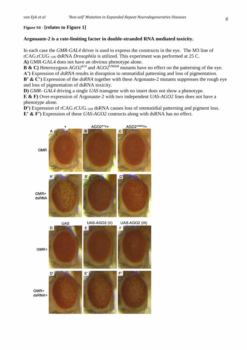
van Eyk et al ‘Non-self’ Mutation in Expanded Repeat Neurodegenerative Diseases 6
Figure S4 - [relates to Figure 1]
Argonaute-2 is a rate-limiting factor in double-stranded RNA mediated toxicity.
In each case the GMR-GAL4 driver is used to express the constructs in the eye. The M3 line of
rCAG.rCUG~100 dsRNA Drosophila is utilized. This experiment was performed at 25 C.
A) GMR-GAL4 does not have an obvious phenotype alone.
B & C) Heterozygous AGO2414 and AGO2V966M mutants have no effect on the patterning of the eye.
A’) Expression of dsRNA results in disruption to ommatidial patterning and loss of pigmentation.
B’ & C’) Expression of the dsRNA together with these Argonaute-2 mutants suppresses the rough eye
and loss of pigmentation of dsRNA toxicity.
D) GMR- GAL4 driving a single UAS transgene with no insert does not show a phenotype.
E & F) Over-expression of Argonaute-2 with two independent UAS-AGO2 lines does not have a
phenotype alone.
D’) Expression of rCAG.rCUG~100 dsRNA causes loss of ommatidial patterning and pigment loss.
E’ & F’) Expression of these UAS-AGO2 contructs along with dsRNA has no effect.

van Eyk et al ‘Non-self’ Mutation in Expanded Repeat Neurodegenerative Diseases 7
Figure S5 – [relates to Fig 2]
hADAR-1 function overrides that of dicer2 in the rCAG.rCUG~100 dsRNA phenotype in the
Drosophila melanogaster eye.
rCAG.rCUG~100 toxicity in the eye is not restored by over-expressing dicer2 in flies also over-
expressing hADAR1 FL. The GMR-GAL4 driver was used to express all constructs.
A) Ectopic expression of dicer2 (together with empty UAS vector) has no phenotype in the eye.
A’) Co-expression of dicer2 and rCAG.rCUG~100 dsRNA is lethal in the S1 line.
B) Co-expression of dicer2 together with hADAR1-i does not affect the patterning of the eye.
B’) hADAR1-i rescues lethality and eye phenotype in S1 flies co-expressing rCAG.rCUG~100 dsRNA
and dicer2 in the eye.

van Eyk et al ‘Non-self’ Mutation in Expanded Repeat Neurodegenerative Diseases 8
Figure S6 [relates to Fig 3]
qRT-PCR levels of uncleaved repeat transcripts in rCAG.rCUG~100 dsRNA flies with co-over-
expression of Drosophila or human ADAR proteins.
qRT-PCR levels of GMR-GAL4 driven expression levels of repeat transcript in (A) three independent
rCAG.rCUG~100 dsRNA lines compared to rCAG.rCUG~100 dsRNA lines co-expressing either (B)
Drosophila dADAR-3/4 or (C) human hADAR1-i or (D) hADAR2 (samples as per Figure 2 A’, C’, F’
and G’). Repeat expression is measured relative to Rp49.
A B C D

van Eyk et al ‘Non-self’ Mutation in Expanded Repeat Neurodegenerative Diseases 9
Figure S7 - [relates to Figure 4]
Figure S7A: Ectopic expression of CrPV1A completely rescues dsRNA-mediated
pathology. In each case the GMR-GAL4 driver is used to express the constructs in the eye. A)
Expression of a single UAS transgene with no insert as a control eye. B-C) Expression of two
independent full-length (FL) UAS-CrPV1A constructs alone. D-E) Expression of two independent
truncated UAS-CrPV1A mutant constructs alone. A’) Co-expression of repeat dsRNA together with a
single UAS transgene. B’-C’) Co-expression of repeat dsRNA together with two independent full-
length (FL) UAS-CrPV1A constructs. D’-E’) Co-expression of repeat dsRNA together with two
independent truncated UAS-CrPV1A mutant constructs. The full length CrPV1A encodes a 148 amino
acid long protein. The truncated CrPV1A encodes a 108 amino acid long protein. The experiment was
performed at 25°C using the CAG.CUG~100M3 dsRNA line.
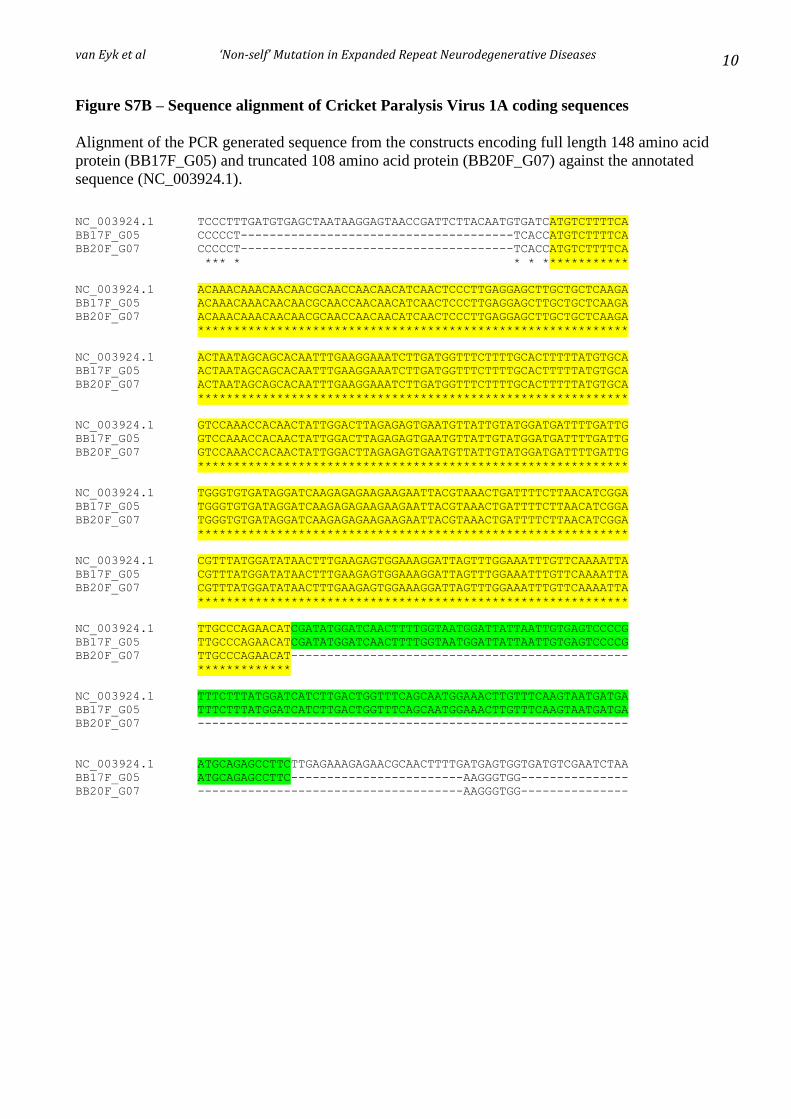
van Eyk et al ‘Non-self’ Mutation in Expanded Repeat Neurodegenerative Diseases 10
Figure S7B – Sequence alignment of Cricket Paralysis Virus 1A coding sequences
Alignment of the PCR generated sequence from the constructs encoding full length 148 amino acid
protein (BB17F_G05) and truncated 108 amino acid protein (BB20F_G07) against the annotated
sequence (NC_003924.1).
NC_003924.1 TCCCTTTGATGTGAGCTAATAAGGAGTAACCGATTCTTACAATGTGATCATGTCTTTTCA
BB17F_G05 CCCCCT--------------------------------------TCACCATGTCTTTTCA
BB20F_G07 CCCCCT--------------------------------------TCACCATGTCTTTTCA
*** * * * ************
NC_003924.1 ACAAACAAACAACAACGCAACCAACAACATCAACTCCCTTGAGGAGCTTGCTGCTCAAGA
BB17F_G05 ACAAACAAACAACAACGCAACCAACAACATCAACTCCCTTGAGGAGCTTGCTGCTCAAGA
BB20F_G07 ACAAACAAACAACAACGCAACCAACAACATCAACTCCCTTGAGGAGCTTGCTGCTCAAGA
************************************************************
NC_003924.1 ACTAATAGCAGCACAATTTGAAGGAAATCTTGATGGTTTCTTTTGCACTTTTTATGTGCA
BB17F_G05 ACTAATAGCAGCACAATTTGAAGGAAATCTTGATGGTTTCTTTTGCACTTTTTATGTGCA
BB20F_G07 ACTAATAGCAGCACAATTTGAAGGAAATCTTGATGGTTTCTTTTGCACTTTTTATGTGCA
************************************************************
NC_003924.1 GTCCAAACCACAACTATTGGACTTAGAGAGTGAATGTTATTGTATGGATGATTTTGATTG
BB17F_G05 GTCCAAACCACAACTATTGGACTTAGAGAGTGAATGTTATTGTATGGATGATTTTGATTG
BB20F_G07 GTCCAAACCACAACTATTGGACTTAGAGAGTGAATGTTATTGTATGGATGATTTTGATTG
************************************************************
NC_003924.1 TGGGTGTGATAGGATCAAGAGAGAAGAAGAATTACGTAAACTGATTTTCTTAACATCGGA
BB17F_G05 TGGGTGTGATAGGATCAAGAGAGAAGAAGAATTACGTAAACTGATTTTCTTAACATCGGA
BB20F_G07 TGGGTGTGATAGGATCAAGAGAGAAGAAGAATTACGTAAACTGATTTTCTTAACATCGGA
************************************************************
NC_003924.1 CGTTTATGGATATAACTTTGAAGAGTGGAAAGGATTAGTTTGGAAATTTGTTCAAAATTA
BB17F_G05 CGTTTATGGATATAACTTTGAAGAGTGGAAAGGATTAGTTTGGAAATTTGTTCAAAATTA
BB20F_G07 CGTTTATGGATATAACTTTGAAGAGTGGAAAGGATTAGTTTGGAAATTTGTTCAAAATTA
************************************************************
NC_003924.1 TTGCCCAGAACATCGATATGGATCAACTTTTGGTAATGGATTATTAATTGTGAGTCCCCG
BB17F_G05 TTGCCCAGAACATCGATATGGATCAACTTTTGGTAATGGATTATTAATTGTGAGTCCCCG
BB20F_G07 TTGCCCAGAACAT-----------------------------------------------
*************
NC_003924.1 TTTCTTTATGGATCATCTTGACTGGTTTCAGCAATGGAAACTTGTTTCAAGTAATGATGA
BB17F_G05 TTTCTTTATGGATCATCTTGACTGGTTTCAGCAATGGAAACTTGTTTCAAGTAATGATGA
BB20F_G07 ------------------------------------------------------------
NC_003924.1 ATGCAGAGCCTTCTTGAGAAAGAGAACGCAACTTTTGATGAGTGGTGATGTCGAATCTAA
BB17F_G05 ATGCAGAGCCTTC------------------------AAGGGTGG---------------
BB20F_G07 -------------------------------------AAGGGTGG---------------

van Eyk et al ‘Non-self’ Mutation in Expanded Repeat Neurodegenerative Diseases 11
Figure S8 – Verification of genotypes of Drosophila lines by PCR
Gel electrophoresis of PCR products using oligodeoxynucleotide primers specific for the genes
indicated (see Table S2 for oligodeoxynucleotide sequences). All PCR products (indicated by vertical
red arrows) are of the anticipated size as predicted by the distance apart of oligodeoxynucleotide
primers.
Markers R2D2 R2D2 hADAR1-I hADAR1-c hADAR2 dADAR
R2D2 (II)
R2D2 (III)
hADAR1-i
hADAR1-c
hADAR2
dADAR
1650bp
1000bp
500bp

van Eyk et al ‘Non-self’ Mutation in Expanded Repeat Neurodegenerative Diseases 12
Figure S9 – Graphical Abstract of ‘Non-Self’ Mutation and Experiments in the Drosophila
model of expanded CAG repeat diseases.
Abbreviations
RLR-PRRs = RNA binding RIG-I-like pattern recognition receptors
Dicer, Ago = components of RNAi and antiviral RNA recognition and defense pathways
R2D2, loqs = components of RNAi pathway
21mers = RNA comprised of 7 copies of the CAG repeat (typical RNAi product)
ADAR1 = human adenosine deaminase of RNA
CrPV-1A = Cricket Paralysis Virus 1A protein

van Eyk et al ‘Non-self’ Mutation in Expanded Repeat Neurodegenerative Diseases 13
Table S1 Drosophila miRNAs changes with rCAG.rCUG~100 dsRNA expression [relates to Fig 1]
miRNA
Control
Reads per
million
dsRNA
Reads per
million
log2 (ratio)
dsRNA vs control
Experiment 1 elavII-GAL4
miR-932 4732.415 317.347 -3.898
miR-7 17414.671 2742.788 -2.667
miR-34 1153.333 238.882 -2.271
miR-988 3894.429 1272.877 -1.613
miR-317 4897.807 1802.952 -1.442
miR-100 10042.600 4238.855 -1.244
miR-8 15573.307 7551.822 -1.044
miR-125 4245.060 2476.007 -0.778
miR-184 4963.964 3145.576 -0.658
miR-274 3391.638 2174.353 -0.641
miR-34* 1503.964 985.172 -0.610
miR-279 26039.310 17696.478 -0.557
let-7 4099.515 2915.411 -0.492
miR-133 1874.442 1340.880 -0.483
bantam 36736.862 27408.704 -0.423
miR-970 8893.677 7046.159 -0.336
miR-263b 2059.681 1644.278 -0.325
miR-263a 9436.163 11567.488 0.294
pre-miR-34 14594.186 19869.087 0.445
pre-miR-193 6538.495 9705.251 0.570
pre-miR-263a 793.881 1332.162 0.747
miR-1017 2002.345 3658.214 0.869
Experiment 2 elavc155-GAL4
miR-184 48388.914 9845.824 -2.297
miR-274 5763.316 2064.591 -1.481
pre-miR-34 16768.186 9640.060 -0.799
miR-957 4220.718 2613.293 -0.692
miR-278 4210.636 3063.587 -0.459
miR-263b 2973.029 2317.074 -0.360
miR-995 1406.486 1111.321 -0.340
pre-miR-193 5060.073 3998.965 -0.340
miR-932 2283.649 1820.061 -0.327
miR-285 12319.355 10111.228 -0.285
miR-1003 7356.325 6058.586 -0.280
miR-193 3916.988 3247.482 -0.270
miR-1017 917.493 1204.759 0.393
miR-987 986.809 1305.156 0.403
miR-87 1616.955 2585.461 0.677
miR-317 9278.270 15641.987 0.753
miR-133 1305.663 2667.965 1.031
let-7 12081.160 26889.387 1.154
pre-miR-210 142.413 1010.924 2.828
pre-miR-284 148.714 1475.134 3.310
miR-210 1192.236 22028.601 4.208
bantam 520.501 15582.345 4.904
miR-34 37.809 14031.665 8.536
The miRNA with common changes in both experiments are shown in bold. ie. Those with a 1.2 fold change, between dsRNA and the
control sample, and a minimum of 1000 reads per million in at least one of the samples (control or dsRNA within an experiment).
Control= 4xUAS, dsRNA in Experiment 1= rCAG.rCUG~100S1, dsRNA in Experiment 2= rCAG.rCUG~100
S2.

van Eyk et al ‘Non-self’ Mutation in Expanded Repeat Neurodegenerative Diseases 14
Methods for Table S1
Alterations of miRNA profiles were analysed using the data sets generated by deep sequencing of flies
expressing expanded repeats throughout the nervous system. Briefly, in Experiment 1 the elavII-GAL4
driver was used to express rCAG.rCUG~100S1 (dsRNAS1), rCAG~100
#1 and rCUG~100#1 whereas in
Experiment 2, rCAG.rCUG~100S2 (dsRNAS2) was expressed using the elavC155-GAL4 driver. In each
experiment the small RNA population was sequenced in each of the repeat expressing samples using
Illumina small RNA sequencing and was compared to each driver with no repeat expression (4xUAS-
control). Independent pan-neuronal drivers together with independent rCAG.rCUG~100 lines were used
with the expectation of identifying miRNA changes that can be replicated using different genetic
backgrounds to exclude biological and experimental artefacts in order to obtain robust changes caused
by dsRNA.
The sequencing analysis produced between 1.5-2.6 million reads that were uniquely mapped to the
Drosophila genome (BDGP 5.0, 2006). These reads were then mapped to the annotated miRNA
precursors (D.melanogaster miRBase version 14). Reads mapping to multiple precursors (pre-miR)
were further analysed to identify the ‘best match’ based on the longest alignment. These reads were
further matched to the annotated mature miRNA sequence or predicted star sequence (miRNA*).
Reads that did not map to miRNA precursors were removed from the analysis. The total number of
reads mapping to miRBase was normalised across the samples to allow comparison.
Significant changes in miRNA abundance were identified based on two parameters. Firstly the
complete list was filtered to include miRNAs that recorded a minimum of 1000 reads in at least one of
the samples (control or dsRNA within an experiment). This list was then filtered based on fold change
between the samples. Additionally, in Experiment 1 miRNAs that were significantly changed in either
rCAG~100#1 or rCUG~100
#1 compared to the control were excluded, in order to generate a list of altered
miRNAs that are unique to double-stranded RNA expression.
In order to identify common changes to the miRNA profile between the two experiments, these
analyses were expanded to include miRNAs with a log2 (ratio) of >+0.26 or <-0.26; that is a 1.2 fold
change, between dsRNA and the control sample, and a minimum of 1000 reads in at least one of the
samples (control or dsRNA within an experiment) was included. This generated a list of 22 and 23
miRNAs with a significant change in abundance in the two experiments respectively. miR-184, miR-
263b, miR-274 and miR-932 are decreased and miR-1017 is increased in dsRNA compared to the
control in both Experiment 1 and 2, indicating the potential for a functionally relevant effect,
independent of the different genetic backgrounds used in the two experiments.

van Eyk et al ‘Non-self’ Mutation in Expanded Repeat Neurodegenerative Diseases 15
Table S2
Oligodeoxynucleotide primers utilized for verification of genotypes of Drosophila lines.
A common (pUAST forward) primer was used for each PCR in conjunction with a unique reverse
primer as indicated.
Primer name Sequence (5’-3’)
pUAST forward GAAGAGAACTCTGAATAGGG
R2D2 reverse GGACGCAGTAGTCACGCAG
hADAR1 reverse GCCATTGTAATGAACAGGTGGTT
hADAR2 reverse CCCACGTAAAAGGGAGGCTC
dADAR reverse AGAACTGCTCCATCCTTAAACTG
pUASp forward GGCAAGGGTCGAGTCGATAG
pUASp reverse AGGTTTAACCAGGGGATGCT
pUASp sequencing forward CAAGGGTCGAGTCGATAG
pUASp sequencing reverse TAACAAGTATGAATGTCAGGTT

155
References
1. Erkkinen MG, Kim M-O, Geschwind MD. Clinical Neurology and Epidemiology of the Major Neurodegenerative Diseases. Cold Spring Harbor Perspectives in Biology. 2018;10(4).
2. Gan L, Cookson MR, Petrucelli L, La Spada AR. Converging pathways in neurodegeneration, from genetics to mechanisms. Nature Neuroscience. 2018;21(10):1300-9.
3. La Spada AR, Taylor JP. Repeat expansion disease: progress and puzzles in disease pathogenesis. Nat Rev Genet. 2010;11(4):247-58.
4. Richards RI, Samaraweera SE, van Eyk CL, O'Keefe LV, Suter CM. RNA pathogenesis via Toll-like receptor-activated inflammation in expanded repeat neurodegenerative diseases. Front Mol Neurosci. 2013;6:25.
5. Wells RD. Molecular Basis of Genetic Instability of Triplet Repeats. Journal of Biological Chemistry. 1996;271(6):2875-8.
6. Richards RI. Dynamic mutations: a decade of unstable expanded repeats in human genetic disease. Human Molecular Genetics. 2001;10(20):2187-94.
7. Zhao X-N, Usdin K. The Repeat Expansion Diseases: The dark side of DNA repair. DNA Repair. 2015;32:96-105.
8. Andrew SE, Goldberg YP, Kremer B, Telenius H, Theilmann J, Adam S, et al. The relationship between trinucleotide (CAG) repeat length and clinical features of Huntington's disease. Nat Genet. 1993;4(4):398-403.
9. Harley HG, Rundle SA, MacMillan JC, Myring J, Brook JD, Crow S, et al. Size of the unstable CTG repeat sequence in relation to phenotype and parental transmission in myotonic dystrophy. American journal of human genetics. 1993;52(6):1164-74.
10. Gijselinck I, Van Mossevelde S, van der Zee J, Sieben A, Engelborghs S, De Bleecker J, et al. The C9orf72 repeat size correlates with onset age of disease, DNA methylation and transcriptional downregulation of the promoter. Molecular Psychiatry. 2015;21:1112.
11. DeJesus-Hernandez M, Mackenzie Ian R, Boeve Bradley F, Boxer Adam L, Baker M, Rutherford Nicola J, et al. Expanded GGGGCC Hexanucleotide Repeat in Noncoding Region of C9ORF72 Causes Chromosome 9p-Linked FTD and ALS. Neuron. 2011;72(2):245-56.
12. Renton AE, Majounie E, Waite A, Simon-Sanchez J, Rollinson S, Gibbs JR, et al. A hexanucleotide repeat expansion in C9ORF72 is the cause of chromosome 9p21-linked ALS-FTD. Neuron. 2011;72(2):257-68.
13. Cummings CJ, Zoghbi HY. Trinucleotide repeats: mechanisms and pathophysiology. Annual review of genomics and human genetics. 2000;1:281-328.
14. Michalik A, Van Broeckhoven C. Pathogenesis of polyglutamine disorders: aggregation revisited. Human Molecular Genetics. 2003;12(suppl_2):R173-R86.
15. Sánchez I, Mahlke C, Yuan J. Pivotal role of oligomerization in expanded polyglutamine neurodegenerative disorders. Nature. 2003;421:373.

156
16. Orr HT, Zoghbi HY. Trinucleotide repeat disorders. Annu Rev Neurosci. 2007;30:575-621.
17. Marsh JL, Lukacsovich T, Thompson LM. Animal Models of Polyglutamine Diseases and Therapeutic Approaches. Journal of Biological Chemistry. 2009;284(12):7431-5.
18. Olejniczak M, Urbanek MO, Krzyzosiak WJ. The Role of the Immune System in Triplet Repeat Expansion Diseases. Mediators Inflamm. 2015;2015:873860.
19. Takahashi T, Katada S, Onodera O. Polyglutamine Diseases: Where does Toxicity Come from? What is Toxicity? Where are We Going? Journal of Molecular Cell Biology. 2010;2(4):180-91.
20. Zu T, Gibbens B, Doty NS, Gomes-Pereira M, Huguet A, Stone MD, et al. Non-ATG–initiated translation directed by microsatellite expansions. Proceedings of the National Academy of Sciences. 2011;108(1):260-5.
21. Batra R, Charizanis K, Swanson MS. Partners in crime: bidirectional transcription in unstable microsatellite disease. Hum Mol Genet. 2010;19(R1):R77-82.
22. Bañez-Coronel M, Ayhan F, Tarabochia Alex D, Zu T, Perez Barbara A, Tusi Solaleh K, et al. RAN Translation in Huntington Disease. Neuron. 2015;88(4):667-77.
23. Todd Peter K, Oh Seok Y, Krans A, He F, Sellier C, Frazer M, et al. CGG Repeat-Associated Translation Mediates Neurodegeneration in Fragile X Tremor Ataxia Syndrome. Neuron. 2013;78(3):440-55.
24. Ash Peter EA, Bieniek Kevin F, Gendron Tania F, Caulfield T, Lin W-L, DeJesus-Hernandez M, et al. Unconventional Translation of C9ORF72 GGGGCC Expansion Generates Insoluble Polypeptides Specific to c9FTD/ALS. Neuron. 2013;77(4):639-46.
25. Mori K, Weng S-M, Arzberger T, May S, Rentzsch K, Kremmer E, et al. The <em>C9orf72</em> GGGGCC Repeat Is Translated into Aggregating Dipeptide-Repeat Proteins in FTLD/ALS. Science. 2013;339(6125):1335-8.
26. Zu T, Liu Y, Bañez-Coronel M, Reid T, Pletnikova O, Lewis J, et al. RAN proteins and RNA foci from antisense transcripts in <em>C9ORF72</em> ALS and frontotemporal dementia. Proceedings of the National Academy of Sciences. 2013;110(51):E4968-E77.
27. Freibaum BD, Lu Y, Lopez-Gonzalez R, Kim NC, Almeida S, Lee K-H, et al. GGGGCC repeat expansion in C9orf72 compromises nucleocytoplasmic transport. Nature. 2015;advance online publication.
28. Jovičić A, Mertens J, Boeynaems S, Bogaert E, Chai N, Yamada SB, et al. Modifiers of C9orf72 dipeptide repeat toxicity connect nucleocytoplasmic transport defects to FTD/ALS. Nature Neuroscience. 2015;18:1226.
29. Zhang K, Donnelly CJ, Haeusler AR, Grima JC, Machamer JB, Steinwald P, et al. The C9orf72 repeat expansion disrupts nucleocytoplasmic transport. Nature. 2015;525:56.
30. Zhang Y-J, Jansen-West K, Xu Y-F, Gendron TF, Bieniek KF, Lin W-L, et al. Aggregation-prone c9FTD/ALS poly(GA) RAN-translated proteins cause neurotoxicity by inducing ER stress. Acta Neuropathologica. 2014;128(4):505-24.
31. Todd PK, Paulson HL. RNA-mediated neurodegeneration in repeat expansion disorders. Ann Neurol. 2010;67(3):291-300.

157
32. Handa V, Yeh HJC, McPhie P, Usdin K. The AUUCU Repeats Responsible for Spinocerebellar Ataxia Type 10 Form Unusual RNA Hairpins. Journal of Biological Chemistry. 2005;280(32):29340-5.
33. Sobczak K, de Mezer M, Michlewski G, Krol J, Krzyzosiak WJ. RNA structure of trinucleotide repeats associated with human neurological diseases. Nucleic Acids Res. 2003;31(19):5469-82.
34. Sicot G, Gomes-Pereira M. RNA toxicity in human disease and animal models: From the uncovering of a new mechanism to the development of promising therapies. Biochimica et Biophysica Acta (BBA) - Molecular Basis of Disease. 2013;1832(9):1390-409.
35. Goodwin M, Mohan A, Batra R, Lee K-Y, Charizanis K, Gómez Francisco José F, et al. MBNL Sequestration by Toxic RNAs and RNA Misprocessing in the Myotonic Dystrophy Brain. Cell Reports. 2015;12(7):1159-68.
36. Mankodi A, Urbinati CR, Yuan QP, Moxley RT, Sansone V, Krym M, et al. Muscleblind localizes to nuclear foci of aberrant RNA in myotonic dystrophy types 1 and 2. Hum Mol Genet. 2001;10(19):2165-70.
37. Rudnicki DD, Holmes SE, Lin MW, Thornton CA, Ross CA, Margolis RL. Huntington's disease–like 2 is associated with CUG repeat-containing RNA foci. Annals of Neurology. 2007;61(3):272-82.
38. Mutsuddi M, Marshall CM, Benzow KA, Koob MD, Rebay I. The Spinocerebellar Ataxia 8 Noncoding RNA Causes Neurodegeneration and Associates with Staufen in Drosophila. Current Biology. 2004;14(4):302-8.
39. Iwahashi CK, Yasui DH, An HJ, Greco CM, Tassone F, Nannen K, et al. Protein composition of the intranuclear inclusions of FXTAS. Brain. 2006;129(1):256-71.
40. Charizanis K, Lee K-Y, Batra R, Goodwin M, Zhang C, Yuan Y, et al. Muscleblind-like 2-Mediated Alternative Splicing in the Developing Brain and Dysregulation in Myotonic Dystrophy. Neuron. 2012;75(3):437-50.
41. Kanadia RN, Johnstone KA, Mankodi A, Lungu C, Thornton CA, Esson D, et al. A Muscleblind Knockout Model for Myotonic Dystrophy. Science. 2003;302(5652):1978-80.
42. Li PP, Sun X, Xia G, Arbez N, Paul S, Zhu S, et al. ATXN2-AS, a gene antisense to ATXN2, is associated with spinocerebellar ataxia type 2 and amyotrophic lateral sclerosis. Annals of Neurology. 2016;80(4):600-15.
43. Gendron TF, Bieniek KF, Zhang Y-J, Jansen-West K, Ash PEA, Caulfield T, et al. Antisense transcripts of the expanded C9ORF72 hexanucleotide repeat form nuclear RNA foci and undergo repeat-associated non-ATG translation in c9FTD/ALS. Acta Neuropathologica. 2013;126(6):829-44.
44. Chung DW, Rudnicki DD, Yu L, Margolis RL. A natural antisense transcript at the Huntington's disease repeat locus regulates HTT expression. Human Molecular Genetics. 2011;20(17):3467-77.
45. Lawlor KT, O'Keefe LV, Samaraweera SE, van Eyk CL, McLeod CJ, Maloney CA, et al. Double-stranded RNA is pathogenic in Drosophila models of expanded repeat neurodegenerative diseases. Hum Mol Genet. 2011;20(19):3757-68.

158
46. Kim K, Lee YS, Harris D, Nakahara K, Carthew RW. The RNAi pathway initiated by Dicer-2 in Drosophila. Cold Spring Harb Symp Quant Biol. 2006;71:39-44.
47. Lee YS, Nakahara K, Pham JW, Kim K, He Z, Sontheimer EJ, et al. Distinct roles for Drosophila Dicer-1 and Dicer-2 in the siRNA/miRNA silencing pathways. Cell. 2004;117(1):69-81.
48. Yu Z, Teng X, Bonini NM. Triplet Repeat–Derived siRNAs Enhance RNA–Mediated Toxicity in a Drosophila Model for Myotonic Dystrophy. PLOS Genetics. 2011;7(3):e1001340.
49. Sofola OA, Jin P, Botas J, Nelson DL. Argonaute-2-dependent rescue of a Drosophila model of FXTAS by FRAXE premutation repeat. Human Molecular Genetics. 2007;16(19):2326-32.
50. Kingsolver MB, Huang Z, Hardy RW. Insect antiviral innate immunity: pathways, effectors, and connections. J Mol Biol. 2013;425(24):4921-36.
51. Bañez-Coronel M, Porta S, Kagerbauer B, Mateu-Huertas E, Pantano L, Ferrer I, et al. A Pathogenic Mechanism in Huntington's Disease Involves Small CAG-Repeated RNAs with Neurotoxic Activity. PLOS Genetics. 2012;8(2):e1002481.
52. Samaraweera SE, O'Keefe LV, Price GR, Venter DJ, Richards RI. Distinct roles for Toll and autophagy pathways in double-stranded RNA toxicity in a Drosophila model of expanded repeat neurodegenerative diseases. Hum Mol Genet. 2013;22(14):2811-9.
53. Deddouche S, Matt N, Budd A, Mueller S, Kemp C, Galiana-Arnoux D, et al. The DExD/H-box helicase Dicer-2 mediates the induction of antiviral activity in drosophila. Nat Immunol. 2008;9(12):1425-32.
54. Wang Z, Wu D, Liu Y, Xia X, Gong W, Qiu Y, et al. Dicer-2 has an RNA interference-independent function that modulates Toll immune signaling. Sci Adv. 2015;1(9):e1500228.
55. MacKay CR, Wang JP, Kurt-Jones EA. Dicer's role as an antiviral: still an enigma. Curr Opin Immunol. 2014;26:49-55.
56. Paro S, Imler JL, Meignin C. Sensing viral RNAs by Dicer/RIG-I like ATPases across species. Curr Opin Immunol. 2015;32:106-13.
57. Bruns AM, Horvath CM. Antiviral RNA recognition and assembly by RLR family innate immune sensors. Cytokine & Growth Factor Reviews. 2014;25(5):507-12.
58. Bjorkqvist M, Wild EJ, Thiele J, Silvestroni A, Andre R, Lahiri N, et al. A novel pathogenic pathway of immune activation detectable before clinical onset in Huntington's disease. J Exp Med. 2008;205(8):1869-77.
59. Politis M, Lahiri N, Niccolini F, Su P, Wu K, Giannetti P, et al. Increased central microglial activation associated with peripheral cytokine levels in premanifest Huntington's disease gene carriers. Neurobiol Dis. 2015;83:115-21.
60. Pavese N, Gerhard A, Tai YF, Ho AK, Turkheimer F, Barker RA, et al. Microglial activation correlates with severity in Huntington disease: a clinical and PET study. Neurology. 2006;66(11):1638-43.
61. Tai YF, Pavese N, Gerhard A, Tabrizi SJ, Barker RA, Brooks DJ, et al. Microglial activation in presymptomatic Huntington's disease gene carriers. Brain. 2007;130(7):1759-66.

159
62. Medzhitov R, Janeway C. Innate Immunity. New England Journal of Medicine. 2000;343(5):338-44.
63. Rock KL, Latz E, Ontiveros F, Kono H. The sterile inflammatory response. Annu Rev Immunol. 2010;28:321-42.
64. Janeway CA, Jr., Medzhitov R. Innate immune recognition. Annu Rev Immunol. 2002;20:197-216.
65. Mogensen TH. Pathogen recognition and inflammatory signaling in innate immune defenses. Clin Microbiol Rev. 2009;22(2):240-73, Table of Contents.
66. Manthiram K, Zhou Q, Aksentijevich I, Kastner DL. The monogenic autoinflammatory diseases define new pathways in human innate immunity and inflammation. Nature Immunology. 2017;18:832.
67. Stephenson J, Nutma E, Valk P, Amor S. Inflammation in CNS neurodegenerative diseases. Immunology. 2018;154(2):204-19.
68. Richards RI, Robertson SA, Kastner DL. Neurodegenerative diseases have genetic hallmarks of autoinflammatory disease. Human Molecular Genetics. 2018;27(R2):R108-R18.
69. Takeuchi O, Akira S. Pattern recognition receptors and inflammation. Cell. 2010;140(6):805-20.
70. Akira S, Uematsu S, Takeuchi O. Pathogen recognition and innate immunity. Cell. 2006;124(4):783-801.
71. Anderson KV, Bokla L, Nusslein-Volhard C. Establishment of dorsal-ventral polarity in the Drosophila embryo: the induction of polarity by the Toll gene product. Cell. 1985;42(3):791-8.
72. Kigerl KA, de Rivero Vaccari JP, Dietrich WD, Popovich PG, Keane RW. Pattern recognition receptors and central nervous system repair. Exp Neurol. 2014;258:5-16.
73. De Nardo D. Toll-like receptors: Activation, signalling and transcriptional modulation. Cytokine. 2015;74(2):181-9.
74. Hayward JA, Mathur A, Ngo C, Man SM. Cytosolic Recognition of Microbes and Pathogens: Inflammasomes in Action. Microbiology and Molecular Biology Reviews. 2018;82(4):e00015-18.
75. Satoh T, Kato H, Kumagai Y, Yoneyama M, Sato S, Matsushita K, et al. LGP2 is a positive regulator of RIG-I- and MDA5-mediated antiviral responses. Proc Natl Acad Sci U S A. 2010;107(4):1512-7.
76. Thaiss CA, Levy M, Itav S, Elinav E. Integration of Innate Immune Signaling. Trends in Immunology. 2016;37(2):84-101.
77. Nish S, Medzhitov R. Host Defense Pathways: Role of Redundancy and Compensation in Infectious Disease Phenotypes. Immunity. 2011;34(5):629-36.
78. Turner MD, Nedjai B, Hurst T, Pennington DJ. Cytokines and chemokines: At the crossroads of cell signalling and inflammatory disease. Biochim Biophys Acta. 2014;1843(11):2563-82.

160
79. Fan AC, Leung AKL. RNA Granules and Diseases: A Case Study of Stress Granules in ALS and FTLD. In: Yeo GW, editor. RNA Processing: Disease and Genome-wide Probing. Cham: Springer International Publishing; 2016. p. 263-96.
80. Li YR, King OD, Shorter J, Gitler AD. Stress granules as crucibles of ALS pathogenesis. The Journal of Cell Biology. 2013;201(3):361-72.
81. Parker R, Sheth U. P Bodies and the Control of mRNA Translation and Degradation. Molecular Cell. 2007;25(5):635-46.
82. Luo Y, Na Z, Slavoff SA. P-Bodies: Composition, Properties, and Functions. Biochemistry. 2018;57(17):2424-31.
83. Walter P, Ron D. The Unfolded Protein Response: From Stress Pathway to Homeostatic Regulation. Science. 2011;334(6059):1081-6.
84. Kleiger G, Mayor T. Perilous journey: a tour of the ubiquitin–proteasome system. Trends in Cell Biology. 2014;24(6):352-9.
85. Kilchert C, Wittmann S, Vasiljeva L. The regulation and functions of the nuclear RNA exosome complex. Nature Reviews Molecular Cell Biology. 2016;17:227.
86. Protter DSW, Parker R. Principles and Properties of Stress Granules. Trends in Cell Biology. 2016;26(9):668-79.
87. Levine B, Mizushima N, Virgin HW. Autophagy in immunity and inflammation. Nature. 2011;469(7330):323-35.
88. Ravikumar B, Duden R, Rubinsztein DC. Aggregate-prone proteins with polyglutamine and polyalanine expansions are degraded by autophagy. Hum Mol Genet. 2002;11(9):1107-17.
89. Tian Y, Bustos V, Flajolet M, Greengard P. A small-molecule enhancer of autophagy decreases levels of Abeta and APP-CTF via Atg5-dependent autophagy pathway. FASEB journal : official publication of the Federation of American Societies for Experimental Biology. 2011;25(6):1934-42.
90. Webb JL, Ravikumar B, Atkins J, Skepper JN, Rubinsztein DC. Alpha-Synuclein is degraded by both autophagy and the proteasome. The Journal of biological chemistry. 2003;278(27):25009-13.
91. Barmada SJ, Serio A, Arjun A, Bilican B, Daub A, Ando DM, et al. Autophagy induction enhances TDP43 turnover and survival in neuronal ALS models. Nat Chem Biol. 2014;10(8):677-85.
92. Gatica D, Lahiri V, Klionsky DJ. Cargo recognition and degradation by selective autophagy. Nature Cell Biology. 2018;20(3):233-42.
93. Gurung P, Lukens JR, Kanneganti T-D. Mitochondria: diversity in the regulation of the NLRP3 inflammasome. Trends in Molecular Medicine. 2015;21(3):193-201.
94. Shimada K, Crother Timothy R, Karlin J, Dagvadorj J, Chiba N, Chen S, et al. Oxidized Mitochondrial DNA Activates the NLRP3 Inflammasome during Apoptosis. Immunity. 2012;36(3):401-14.
95. Zhong Z, Liang S, Sanchez-Lopez E, He F, Shalapour S, Lin X-j, et al. New mitochondrial DNA synthesis enables NLRP3 inflammasome activation. Nature. 2018;560(7717):198-203.
161
96. Caielli S, Athale S, Domic B, Murat E, Chandra M, Banchereau R, et al. Oxidized mitochondrial nucleoids released by neutrophils drive type I interferon production in human lupus. The Journal of Experimental Medicine. 2016;213(5):697-713.
97. Rongvaux A, Jackson R, Harman Christian CD, Li T, West AP, de Zoete Marcel R, et al. Apoptotic Caspases Prevent the Induction of Type I Interferons by Mitochondrial DNA. Cell. 2014;159(7):1563-77.
98. White Michael J, McArthur K, Metcalf D, Lane Rachael M, Cambier John C, Herold Marco J, et al. Apoptotic Caspases Suppress mtDNA-Induced STING-Mediated Type I IFN Production. Cell. 2014;159(7):1549-62.
99. Kitada T, Asakawa S, Hattori N, Matsumine H, Yamamura Y, Minoshima S, et al. Mutations in the parkin gene cause autosomal recessive juvenile parkinsonism. Nature. 1998;392:605.
100. Valente EM, Abou-Sleiman PM, Caputo V, Muqit MM, Harvey K, Gispert S, et al. Hereditary early-onset Parkinson's disease caused by mutations in PINK1. Science. 2004;304(5674):1158-60.
101. Tschopp J. Mitochondria: Sovereign of inflammation? European Journal of Immunology. 2011;41(5):1196-202.
102. Seth RB, Sun L, Ea C-K, Chen ZJ. Identification and Characterization of MAVS, a Mitochondrial Antiviral Signaling Protein that Activates NF-κB and IRF3. Cell. 2005;122(5):669-82.
103. Kumar H, Kawai T, Kato H, Sato S, Takahashi K, Coban C, et al. Essential role of IPS-1 in innate immune responses against RNA viruses. The Journal of Experimental Medicine. 2006;203(7):1795-803.
104. Guan K, Wei C, Zheng Z, Song T, Wu F, Zhang Y, et al. MAVS Promotes Inflammasome Activation by Targeting ASC for K63-Linked Ubiquitination via the E3 Ligase TRAF3. The Journal of Immunology. 2015;194(10):4880-90.
105. Subramanian N, Natarajan K, Clatworthy Menna R, Wang Z, Germain Ronald N. The Adaptor MAVS Promotes NLRP3 Mitochondrial Localization and Inflammasome Activation. Cell. 2013;153(2):348-61.
106. Franchi L, Eigenbrod T, Muñoz-Planillo R, Ozkurede U, Kim Y-G, Chakrabarti A, et al. Cytosolic Double-Stranded RNA Activates the NLRP3 Inflammasome via MAVS-Induced Membrane Permeabilization and K<sup>+</sup> Efflux. The Journal of Immunology. 2014;193(8):4214-22.
107. Guan K, Zheng Z, Song T, He X, Xu C, Zhang Y, et al. MAVS Regulates Apoptotic Cell Death by Decreasing K48-Linked Ubiquitination of Voltage-Dependent Anion Channel 1. Molecular and Cellular Biology. 2013;33(16):3137-49.
108. Lei Y, Moore CB, Liesman RM, O'Connor BP, Bergstralh DT, Chen ZJ, et al. MAVS-Mediated Apoptosis and Its Inhibition by Viral Proteins. PLOS ONE. 2009;4(5):e5466.
109. Yu C-Y, Chiang R-L, Chang T-H, Liao C-L, Lin Y-L. The Interferon Stimulator Mitochondrial Antiviral Signaling Protein Facilitates Cell Death by Disrupting the Mitochondrial Membrane Potential and by Activating Caspases. Journal of Virology. 2010;84(5):2421-31.

162
110. Brault M, Olsen TM, Martinez J, Stetson DB, Oberst A. Intracellular Nucleic Acid Sensing Triggers Necroptosis through Synergistic Type I IFN and TNF Signaling. The Journal of Immunology. 2018;200(8):2748-56.
111. Wang W, Wang W-H, Azadzoi KM, Su N, Dai P, Sun J, et al. Activation of innate antiviral immune response via double-stranded RNA-dependent RLR receptor-mediated necroptosis. Scientific Reports. 2016;6:22550.
112. Berke IC, Modis Y. MDA5 cooperatively forms dimers and ATP‐sensitive filaments upon binding double‐stranded RNA. The EMBO Journal. 2012;31(7):1714-26.
113. Berke IC, Yu X, Modis Y, Egelman EH. MDA5 assembles into a polar helical filament on dsRNA. Proceedings of the National Academy of Sciences. 2012;109(45):18437-41.
114. Peisley A, Lin C, Wu B, Orme-Johnson M, Liu M, Walz T, et al. Cooperative assembly and dynamic disassembly of MDA5 filaments for viral dsRNA recognition. Proceedings of the National Academy of Sciences. 2011;108(52):21010-5.
115. Peisley A, Wu B, Yao H, Walz T, Hur S. RIG-I Forms Signaling-Competent Filaments in an ATP-Dependent, Ubiquitin-Independent Manner. Molecular Cell. 2013;51(5):573-83.
116. Hou F, Sun L, Zheng H, Skaug B, Jiang Q-X, Chen Zhijian J. MAVS Forms Functional Prion-like Aggregates to Activate and Propagate Antiviral Innate Immune Response. Cell. 2011;146(3):448-61.
117. Wu B, Hur S. How RIG-I like receptors activate MAVS. Current Opinion in Virology. 2015;12:91-8.
118. Xu H, He X, Zheng H, Huang LJ, Hou F, Yu Z, et al. Structural basis for the prion-like MAVS filaments in antiviral innate immunity. eLife. 2014;3:e01489.
119. Qi N, Shi Y, Zhang R, Zhu W, Yuan B, Li X, et al. Multiple truncated isoforms of MAVS prevent its spontaneous aggregation in antiviral innate immune signalling. Nature Communications. 2017;8:15676.
120. Dai T, Wu L, Wang S, Wang J, Xie F, Zhang Z, et al. FAF1 Regulates Antiviral Immunity by Inhibiting MAVS but Is Antagonized by Phosphorylation upon Viral Infection. Cell Host & Microbe. 2018;24(6):776-90.e5.
121. Jacobs JL, Coyne CB. Mechanisms of MAVS Regulation at the Mitochondrial Membrane. Journal of Molecular Biology. 2013;425(24):5009-19.
122. Jonsson T, Stefansson H, Steinberg S, Jonsdottir I, Jonsson PV, Snaedal J, et al. Variant of TREM2 associated with the risk of Alzheimer's disease. The New England journal of medicine. 2013;368(2):107-16.
123. Sims R, van der Lee SJ, Naj AC, Bellenguez C, Badarinarayan N, Jakobsdottir J, et al. Rare coding variants in PLCG2, ABI3, and TREM2 implicate microglial-mediated innate immunity in Alzheimer's disease. Nat Genet. 2017.
124. Krasemann S, Madore C, Cialic R, Baufeld C, Calcagno N, El Fatimy R, et al. The TREM2-APOE Pathway Drives the Transcriptional Phenotype of Dysfunctional Microglia in Neurodegenerative Diseases. Immunity. 2017;47(3):566-81.e9.

163
125. Gitter BD, Cox LM, Rydel RE, May PC. Amyloid beta peptide potentiates cytokine secretion by interleukin-1 beta-activated human astrocytoma cells. Proceedings of the National Academy of Sciences. 1995;92(23):10738-41.
126. Coraci IS, Husemann J, Berman JW, Hulette C, Dufour JH, Campanella GK, et al. CD36, a Class B Scavenger Receptor, Is Expressed on Microglia in Alzheimer's Disease Brains and Can Mediate Production of Reactive Oxygen Species in Response to β-Amyloid Fibrils. The American Journal of Pathology. 2002;160(1):101-12.
127. El Khoury JB, Moore KJ, Means TK, Leung J, Terada K, Toft M, et al. CD36 Mediates the Innate Host Response to β-Amyloid. The Journal of Experimental Medicine. 2003;197(12):1657-66.
128. Bourgade K, Garneau H, Giroux G, Le Page AY, Bocti C, Dupuis G, et al. beta-Amyloid peptides display protective activity against the human Alzheimer's disease-associated herpes simplex virus-1. Biogerontology. 2015;16(1):85-98.
129. Kumar DKV, Choi SH, Washicosky KJ, Eimer WA, Tucker S, Ghofrani J, et al. Amyloid-β peptide protects against microbial infection in mouse and worm models of Alzheimer’s disease. Science Translational Medicine. 2016;8(340):340ra72-ra72.
130. White MR, Kandel R, Tripathi S, Condon D, Qi L, Taubenberger J, et al. Alzheimer's Associated β-Amyloid Protein Inhibits Influenza A Virus and Modulates Viral Interactions with Phagocytes. PLOS ONE. 2014;9(7):e101364.
131. Pickrell AM, Youle RJ. The roles of PINK1, parkin, and mitochondrial fidelity in Parkinson's disease. Neuron. 2015;85(2):257-73.
132. Wang X, Yan MH, Fujioka H, Liu J, Wilson-Delfosse A, Chen SG, et al. LRRK2 regulates mitochondrial dynamics and function through direct interaction with DLP1. Human Molecular Genetics. 2012;21(9):1931-44.
133. Irrcher I, Aleyasin H, Seifert EL, Hewitt SJ, Chhabra S, Phillips M, et al. Loss of the Parkinson's disease-linked gene DJ-1 perturbs mitochondrial dynamics. Human Molecular Genetics. 2010;19(19):3734-46.
134. Wang X, Petrie TG, Liu Y, Liu J, Fujioka H, Zhu X. Parkinson’s disease-associated DJ-1 mutations impair mitochondrial dynamics and cause mitochondrial dysfunction. Journal of Neurochemistry. 2012;121(5):830-9.
135. Wang W, Wang X, Fujioka H, Hoppel C, Whone AL, Caldwell MA, et al. Parkinson's disease–associated mutant VPS35 causes mitochondrial dysfunction by recycling DLP1 complexes. Nature Medicine. 2015;22:54.
136. Xie W, Chung KKK. Alpha-synuclein impairs normal dynamics of mitochondria in cell and animal models of Parkinson’s disease. Journal of Neurochemistry. 2012;122(2):404-14.
137. Beraud D, Hathaway HA, Trecki J, Chasovskikh S, Johnson DA, Johnson JA, et al. Microglial activation and antioxidant responses induced by the Parkinson's disease protein alpha-synuclein. J Neuroimmune Pharmacol. 2013;8(1):94-117.
138. Beraud D, Twomey M, Bloom B, Mittereder A, Ton V, Neitzke K, et al. alpha-Synuclein Alters Toll-Like Receptor Expression. Front Neurosci. 2011;5:80.

164
139. van Horssen J, van Schaik P, Witte M. Inflammation and mitochondrial dysfunction: A vicious circle in neurodegenerative disorders? Neuroscience Letters. 2017.
140. West AP. Mitochondrial dysfunction as a trigger of innate immune responses and inflammation. Toxicology. 2017;391:54-63.
141. Gal J, Ström A-L, Kwinter DM, Kilty R, Zhang J, Shi P, et al. Sequestosome 1/p62 links familial ALS mutant SOD1 to LC3 via an ubiquitin-independent mechanism. Journal of Neurochemistry. 2009;111(4):1062-73.
142. Mccann EP, Guillemin GJ, Blair IP, Fifita JA, Williams KL, Tan V, et al. Defects in optineurin- and myosin VI-mediated cellular trafficking in amyotrophic lateral sclerosis. Human Molecular Genetics. 2015;24(13):3830-46.
143. Nguyen DKH, Thombre R, Wang J. Autophagy as a common pathway in amyotrophic lateral sclerosis. Neuroscience Letters. 2018.
144. Bentmann E, Haass C, Dormann D. Stress granules in neurodegeneration--lessons learnt from TAR DNA binding protein of 43 kDa and fused in sarcoma. FEBS J. 2013;280(18):4348-70.
145. Colombrita C, Onesto E, Megiorni F, Pizzuti A, Baralle FE, Buratti E, et al. TDP-43 and FUS RNA-binding Proteins Bind Distinct Sets of Cytoplasmic Messenger RNAs and Differently Regulate Their Post-transcriptional Fate in Motoneuron-like Cells. Journal of Biological Chemistry. 2012;287(19):15635-47.
146. Polymenidou M, Lagier-Tourenne C, Hutt KR, Huelga SC, Moran J, Liang TY, et al. Long pre-mRNA depletion and RNA missplicing contribute to neuronal vulnerability from loss of TDP-43. Nature Neuroscience. 2011;14:459.
147. Sellier C, Campanari ML, Julie Corbier C, Gaucherot A, Kolb-Cheynel I, Oulad-Abdelghani M, et al. Loss of C9ORF72 impairs autophagy and synergizes with polyQ Ataxin-2 to induce motor neuron dysfunction and cell death. Embo j. 2016;35(12):1276-97.
148. Lall D, Baloh RH. Microglia and C9orf72 in neuroinflammation and ALS and frontotemporal dementia. The Journal of clinical investigation. 2017;127(9):3250-8.
149. O’Rourke JG, Bogdanik L, Yáñez A, Lall D, Wolf AJ, Muhammad AKMG, et al. C9orf72 is required for proper macrophage and microglial function in mice. Science. 2016;351(6279):1324-9.
150. Crotti A, Benner C, Kerman BE, Gosselin D, Lagier-Tourenne C, Zuccato C, et al. Mutant Huntingtin promotes autonomous microglia activation via myeloid lineage-determining factors. Nature Neuroscience. 2014;17:513.
151. Levanon K, Eisenberg E, Rechavi G, Levanon EY. Letter from the editor: adenosine‐to‐inosine RNA editing in <em>Alu</em> repeats in the human genome. EMBO reports. 2005;6(9):831-5.
152. Liddicoat BJ, Piskol R, Chalk AM, Ramaswami G, Higuchi M, Hartner JC, et al. RNA editing by ADAR1 prevents MDA5 sensing of endogenous dsRNA as nonself. Science. 2015;349(6252):1115-20.
153. Crow YJ, Chase DS, Lowenstein Schmidt J, Szynkiewicz M, Forte GM, Gornall HL, et al. Characterization of human disease phenotypes associated with mutations in TREX1,

165
RNASEH2A, RNASEH2B, RNASEH2C, SAMHD1, ADAR, and IFIH1. Am J Med Genet A. 2015;167A(2):296-312.
154. Rice GI, Kasher PR, Forte GM, Mannion NM, Greenwood SM, Szynkiewicz M, et al. Mutations in ADAR1 cause Aicardi-Goutieres syndrome associated with a type I interferon signature. Nat Genet. 2012;44(11):1243-8.
155. Rice GI, del Toro Duany Y, Jenkinson EM, Forte GM, Anderson BH, Ariaudo G, et al. Gain-of-function mutations in IFIH1 cause a spectrum of human disease phenotypes associated with upregulated type I interferon signaling. Nat Genet. 2014;46(5):503-9.
156. Mannion NM, Greenwood SM, Young R, Cox S, Brindle J, Read D, et al. The RNA-editing enzyme ADAR1 controls innate immune responses to RNA. Cell Rep. 2014;9(4):1482-94.
157. Gallo A, Vukic D, Michalík D, O’Connell MA, Keegan LP. ADAR RNA editing in human disease; more to it than meets the I. Human Genetics. 2017;136(9):1265-78.
158. Zuchero JB, Barres BA. Glia in mammalian development and disease. Development. 2015;142(22):3805-9.
159. Barres BA. The Mystery and Magic of Glia: A Perspective on Their Roles in Health and Disease. Neuron. 2008;60(3):430-40.
160. Freeman MR. Drosophila Central Nervous System Glia. Cold Spring Harbor Perspectives in Biology. 2015.
161. Shaham S. Glial development and function in the nervous system of Caenorhabditis elegans. Cold Spring Harbor perspectives in biology.7(4):a020578-a.
162. Haim LB, Rowitch DH. Functional diversity of astrocytes in neural circuit regulation. Nature Reviews Neuroscience. 2016;18:31.
163. Viganò F, Dimou L. The heterogeneous nature of NG2-glia. Brain Research. 2016;1638:129-37.
164. Wes PD, Holtman IR, Boddeke EWGM, Möller T, Eggen BJL. Next generation transcriptomics and genomics elucidate biological complexity of microglia in health and disease. Glia. 2016;64(2):197-213.
165. Mathys H, Adaikkan C, Gao F, Young JZ, Manet E, Hemberg M, et al. Temporal Tracking of Microglia Activation in Neurodegeneration at Single-Cell Resolution. Cell Reports. 2017;21(2):366-80.
166. Soulet D, Rivest S. Bone-marrow-derived microglia: myth or reality? Current Opinion in Pharmacology. 2008;8(4):508-18.
167. Alliot F, Godin I, Pessac B. Microglia derive from progenitors, originating from the yolk sac, and which proliferate in the brain. Developmental Brain Research. 1999;117(2):145-52.
168. Ginhoux F, Greter M, Leboeuf M, Nandi S, See P, Gokhan S, et al. Fate Mapping Analysis Reveals That Adult Microglia Derive from Primitive Macrophages. Science. 2010;330(6005):841-5.
169. Hickman SE, Kingery ND, Ohsumi TK, Borowsky ML, Wang L-c, Means TK, et al. The microglial sensome revealed by direct RNA sequencing. Nature Neuroscience. 2013;16:1896.

166
170. Paolicelli RC, Jawaid A, Henstridge CM, Valeri A, Merlini M, Robinson JL, et al. TDP-43 Depletion in Microglia Promotes Amyloid Clearance but Also Induces Synapse Loss. Neuron.95(2):297-308.e6.
171. Schafer DP, Lehrman EK, Kautzman AG, Koyama R, Mardinly AR, Yamasaki R, et al. Microglia sculpt postnatal neural circuits in an activity and complement-dependent manner. Neuron. 2012;74(4):691-705.
172. Sierra A, Encinas JM, Deudero JJP, Chancey JH, Enikolopov G, Overstreet-Wadiche LS, et al. Microglia Shape Adult Hippocampal Neurogenesis through Apoptosis-Coupled Phagocytosis. Cell Stem Cell. 2010;7(4):483-95.
173. Nakajima K, Tohyama Y, Maeda S, Kohsaka S, Kurihara T. Neuronal regulation by which microglia enhance the production of neurotrophic factors for GABAergic, catecholaminergic, and cholinergic neurons. Neurochemistry International. 2007;50(6):807-20.
174. Parkhurst Christopher N, Yang G, Ninan I, Savas Jeffrey N, Yates John R, Lafaille Juan J, et al. Microglia Promote Learning-Dependent Synapse Formation through Brain-Derived Neurotrophic Factor. Cell. 2013;155(7):1596-609.
175. Burda Joshua E, Sofroniew Michael V. Reactive Gliosis and the Multicellular Response to CNS Damage and Disease. Neuron. 2014;81(2):229-48.
176. Deczkowska A, Keren-Shaul H, Weiner A, Colonna M, Schwartz M, Amit I. Disease-Associated Microglia: A Universal Immune Sensor of Neurodegeneration. Cell. 2018;173(5):1073-81.
177. Butovsky O, Weiner HL. Microglial signatures and their role in health and disease. Nature Reviews Neuroscience. 2018.
178. Tang Y, Le W. Differential Roles of M1 and M2 Microglia in Neurodegenerative Diseases. Mol Neurobiol. 2015.
179. Liddelow SA, Barres BA. Reactive Astrocytes: Production, Function, and Therapeutic Potential. Immunity. 2017;46(6):957-67.
180. Miller R, Raff M. Fibrous and protoplasmic astrocytes are biochemically and developmentally distinct. The Journal of Neuroscience. 1984;4(2):585-92.
181. Sofroniew MV, Vinters HV. Astrocytes: biology and pathology. Acta Neuropathologica. 2010;119(1):7-35.
182. Rowitch DH, Kriegstein AR. Developmental genetics of vertebrate glial–cell specification. Nature. 2010;468:214.
183. Malatesta P, Hartfuss E, Gotz M. Isolation of radial glial cells by fluorescent-activated cell sorting reveals a neuronal lineage. Development. 2000;127(24):5253-63.
184. Noctor SC, Flint AC, Weissman TA, Dammerman RS, Kriegstein AR. Neurons derived from radial glial cells establish radial units in neocortex. Nature. 2001;409:714.
185. Fróes MM, Correia AH, Garcia-Abreu J, Spray DC, Campos de Carvalho AC, Neto MV. Gap-junctional coupling between neurons and astrocytes in primary central nervous system cultures. Proceedings of the National Academy of Sciences of the United States of America. 1999;96(13):7541-6.

167
186. Giaume C, McCarthy KD. Control of gap-junctional communication in astrocytic networks. Trends in Neurosciences. 1996;19(8):319-25.
187. Christopherson KS, Ullian EM, Stokes CCA, Mullowney CE, Hell JW, Agah A, et al. Thrombospondins Are Astrocyte-Secreted Proteins that Promote CNS Synaptogenesis. Cell. 2005;120(3):421-33.
188. Chung W-S, Clarke LE, Wang GX, Stafford BK, Sher A, Chakraborty C, et al. Astrocytes mediate synapse elimination through MEGF10 and MERTK pathways. Nature. 2013;504:394.
189. Stevens B, Allen NJ, Vazquez LE, Howell GR, Christopherson KS, Nouri N, et al. The Classical Complement Cascade Mediates CNS Synapse Elimination. Cell. 2007;131(6):1164-78.
190. Rothstein JD, Dykes-Hoberg M, Pardo CA, Bristol LA, Jin L, Kuncl RW, et al. Knockout of Glutamate Transporters Reveals a Major Role for Astroglial Transport in Excitotoxicity and Clearance of Glutamate. Neuron. 1996;16(3):675-86.
191. Sattler R, Rothstein JD. Regulation and dysregulation of glutamate transporters. Handbook of experimental pharmacology. 2006(175):277-303.
192. Sofroniew MV. Molecular dissection of reactive astrogliosis and glial scar formation. Trends in neurosciences. 2009;32(12):638-47.
193. Zamanian JL, Xu L, Foo LC, Nouri N, Zhou L, Giffard RG, et al. Genomic Analysis of Reactive Astrogliosis. The Journal of Neuroscience. 2012;32(18):6391-410.
194. Nave K-A. Myelination and support of axonal integrity by glia. Nature. 2010;468:244.
195. Hartline DK, Colman DR. Rapid Conduction and the Evolution of Giant Axons and Myelinated Fibers. Current Biology. 2007;17(1):R29-R35.
196. Cai J, Qi Y, Hu X, Tan M, Liu Z, Zhang J, et al. Generation of Oligodendrocyte Precursor Cells from Mouse Dorsal Spinal Cord Independent of Nkx6 Regulation and Shh Signaling. Neuron. 2005;45(1):41-53.
197. Warf B, Fok-Seang J, Miller R. Evidence for the ventral origin of oligodendrocyte precursors in the rat spinal cord. The Journal of Neuroscience. 1991;11(8):2477-88.
198. Young Kaylene M, Psachoulia K, Tripathi Richa B, Dunn S-J, Cossell L, Attwell D, et al. Oligodendrocyte Dynamics in the Healthy Adult CNS: Evidence for Myelin Remodeling. Neuron. 2013;77(5):873-85.
199. Bhatheja K, Field J. Schwann cells: Origins and role in axonal maintenance and regeneration. The International Journal of Biochemistry & Cell Biology. 2006;38(12):1995-9.
200. Nishiyama A, Komitova M, Suzuki R, Zhu X. Polydendrocytes (NG2 cells): multifunctional cells with lineage plasticity. Nature Reviews Neuroscience. 2009;10:9.
201. Zhu X, Bergles DE, Nishiyama A. NG2 cells generate both oligodendrocytes and gray matter astrocytes. Development. 2008;135(1):145-57.
202. Kang SH, Fukaya M, Yang JK, Rothstein JD, Bergles DE. NG2+ CNS Glial Progenitors Remain Committed to the Oligodendrocyte Lineage in Postnatal Life and following Neurodegeneration. Neuron. 2010;68(4):668-81.

168
203. Nishiyama A, Suzuki R, Zhu X. NG2 cells (polydendrocytes) in brain physiology and repair. Frontiers in Neuroscience. 2014;8(133).
204. Rivers LE, Young KM, Rizzi M, Jamen F, Psachoulia K, Wade A, et al. PDGFRA/NG2 glia generate myelinating oligodendrocytes and piriform projection neurons in adult mice. Nature Neuroscience. 2008;11:1392.
205. Dimou L, Gallo V. NG2-glia and their functions in the central nervous system. Glia. 2015;63(8):1429-51.
206. Iadecola C. The Neurovascular Unit Coming of Age: A Journey through Neurovascular Coupling in Health and Disease. Neuron. 2017;96(1):17-42.
207. Abbott NJ, Rönnbäck L, Hansson E. Astrocyte–endothelial interactions at the blood–brain barrier. Nature Reviews Neuroscience. 2006;7:41.
208. McConnell HL, Kersch CN, Woltjer RL, Neuwelt EA. The Translational Significance of the Neurovascular Unit. Journal of Biological Chemistry. 2017;292(3):762-70.
209. Stamatovic SM, Keep RF, Andjelkovic AV. Brain endothelial cell-cell junctions: how to "open" the blood brain barrier. Curr Neuropharmacol. 2008;6(3):179-92.
210. Zihni C, Mills C, Matter K, Balda MS. Tight junctions: from simple barriers to multifunctional molecular gates. Nature Reviews Molecular Cell Biology. 2016;17:564.
211. Blanchette M, Daneman R. Formation and maintenance of the BBB. Mechanisms of Development. 2015;138:8-16.
212. Del Zoppo GJ, Milner R, Mabuchi T, Hung S, Wang X, Koziol JA. Vascular matrix adhesion and the blood-brain barrier. Biochemical Society transactions. 2006;34(Pt 6):1261-6.
213. Winkler EA, Bell RD, Zlokovic BV. Central nervous system pericytes in health and disease. Nature Neuroscience. 2011;14:1398.
214. Sweeney MD, Ayyadurai S, Zlokovic BV. Pericytes of the neurovascular unit: key functions and signaling pathways. Nature Neuroscience. 2016;19:771.
215. Sagare AP, Bell RD, Zhao Z, Ma Q, Winkler EA, Ramanathan A, et al. Pericyte loss influences Alzheimer-like neurodegeneration in mice. Nature Communications. 2013;4:2932.
216. Pieper C, Marek JJ, Unterberg M, Schwerdtle T, Galla H-J. Brain capillary pericytes contribute to the immune defense in response to cytokines or LPS in vitro. Brain Research. 2014;1550:1-8.
217. Nakagomi T, Kubo S, Nakano-Doi A, Sakuma R, Lu S, Narita A, et al. Brain Vascular Pericytes Following Ischemia Have Multipotential Stem Cell Activity to Differentiate Into Neural and Vascular Lineage Cells. STEM CELLS. 2015;33(6):1962-74.
218. Sakuma R, Kawahara M, Nakano-Doi A, Takahashi A, Tanaka Y, Narita A, et al. Brain pericytes serve as microglia-generating multipotent vascular stem cells following ischemic stroke. Journal of Neuroinflammation. 2016;13(1):57.
219. Alvarez JI, Katayama T, Prat A. Glial influence on the blood brain barrier. Glia. 2013;61(12):1939-58.

169
220. De Bock M, Decrock E, Wang N, Bol M, Vinken M, Bultynck G, et al. The dual face of connexin-based astroglial Ca2+ communication: A key player in brain physiology and a prime target in pathology. Biochimica et Biophysica Acta (BBA) - Molecular Cell Research. 2014;1843(10):2211-32.
221. Tanigami H, Okamoto T, Yasue Y, Shimaoka M. Astroglial integrins in the development and regulation of neurovascular units. Pain research and treatment. 2012;2012:964652.
222. Liu C-Y, Yang Y, Ju W-N, Wang X, Zhang H-L. Emerging Roles of Astrocytes in Neuro-Vascular Unit and the Tripartite Synapse With Emphasis on Reactive Gliosis in the Context of Alzheimer’s Disease. Frontiers in Cellular Neuroscience. 2018;12(193).
223. Thurgur H, Pinteaux E. Microglia in the Neurovascular Unit: Blood–Brain Barrier–microglia Interactions After Central Nervous System Disorders. Neuroscience. 2018.
224. Lécuyer M-A, Kebir H, Prat A. Glial influences on BBB functions and molecular players in immune cell trafficking. Biochimica et Biophysica Acta (BBA) - Molecular Basis of Disease. 2016;1862(3):472-82.
225. Lobsiger CS, Cleveland DW. Glial cells as intrinsic components of non-cell-autonomous neurodegenerative disease. Nature Neuroscience. 2007;10:1355.
226. Cartier N, Lewis C-A, Zhang R, Rossi FMV. The role of microglia in human disease: therapeutic tool or target? Acta neuropathologica. 2014;128(3):363-80.
227. Colonna M, Butovsky O. Microglia Function in the Central Nervous System During Health and Neurodegeneration. Annual Review of Immunology. 2017;35(1):441-68.
228. Sapp E, Kegel KB, Aronin N, Hashikawa T, Uchiyama Y, Tohyama K, et al. Early and Progressive Accumulation of Reactive Microglia in the Huntington Disease Brain. Journal of Neuropathology & Experimental Neurology. 2001;60(2):161-72.
229. Hall ED, Oostveen JA, Gurney ME. Relationship of microglial and astrocytic activation to disease onset and progression in a transgenic model of familial ALS. Glia. 1998;23(3):249-56.
230. Hong S, Beja-Glasser VF, Nfonoyim BM, Frouin A, Li S, Ramakrishnan S, et al. Complement and microglia mediate early synapse loss in Alzheimer mouse models. Science. 2016.
231. Asai H, Ikezu S, Tsunoda S, Medalla M, Luebke J, Haydar T, et al. Depletion of microglia and inhibition of exosome synthesis halt tau propagation. Nature Neuroscience. 2015;18:1584.
232. Kim C, Ho DH, Suk JE, You S, Michael S, Kang J, et al. Neuron-released oligomeric alpha-synuclein is an endogenous agonist of TLR2 for paracrine activation of microglia. Nat Commun. 2013;4:1562.
233. Su X, Maguire-Zeiss KA, Giuliano R, Prifti L, Venkatesh K, Federoff HJ. Synuclein activates microglia in a model of Parkinson's disease. Neurobiology of Aging. 2008;29(11):1690-701.

170
234. Frakes Ashley E, Ferraiuolo L, Haidet-Phillips Amanda M, Schmelzer L, Braun L, Miranda Carlos J, et al. Microglia Induce Motor Neuron Death via the Classical NF-κB Pathway in Amyotrophic Lateral Sclerosis. Neuron. 2014;81(5):1009-23.
235. Zhao W, Beers DR, Henkel JS, Zhang W, Urushitani M, Julien J-P, et al. Extracellular mutant SOD1 induces microglial-mediated motoneuron injury. Glia. 2010;58(2):231-43.
236. Hopp SC, Lin Y, Oakley D, Roe AD, DeVos SL, Hanlon D, et al. The role of microglia in processing and spreading of bioactive tau seeds in Alzheimer's disease. J Neuroinflammation. 2018;15(1):269.
237. Njie eG, Boelen E, Stassen FR, Steinbusch HWM, Borchelt DR, Streit WJ. Ex vivo cultures of microglia from young and aged rodent brain reveal age-related changes in microglial function. Neurobiology of Aging. 2012;33(1):195.e1-.e12.
238. Svahn AJ, Don EK, Badrock AP, Cole NJ, Graeber MB, Yerbury JJ, et al. Nucleo-cytoplasmic transport of TDP-43 studied in real time: impaired microglia function leads to axonal spreading of TDP-43 in degenerating motor neurons. Acta Neuropathologica. 2018;136(3):445-59.
239. Pearce MMP, Spartz EJ, Hong W, Luo L, Kopito RR. Prion-like transmission of neuronal huntingtin aggregates to phagocytic glia in the Drosophila brain. Nat Commun. 2015;6.
240. Perry VH, Holmes C. Microglial priming in neurodegenerative disease. Nature Reviews Neurology. 2014;10:217.
241. Spittau B. Aging Microglia-Phenotypes, Functions and Implications for Age-Related Neurodegenerative Diseases. Frontiers in aging neuroscience. 2017;9:194-.
242. Bussian TJ, Aziz A, Meyer CF, Swenson BL, van Deursen JM, Baker DJ. Clearance of senescent glial cells prevents tau-dependent pathology and cognitive decline. Nature. 2018.
243. Widmann CN, Heneka MT. Long-term cerebral consequences of sepsis. The Lancet Neurology. 2014;13(6):630-6.
244. Faideau M, Kim J, Cormier K, Gilmore R, Welch M, Auregan G, et al. In vivo expression of polyglutamine-expanded huntingtin by mouse striatal astrocytes impairs glutamate transport: a correlation with Huntington's disease subjects. Human Molecular Genetics. 2010;19(15):3053-67.
245. Hsiao H-Y, Chen Y-C, Huang C-H, Chen C-C, Hsu Y-H, Chen H-M, et al. Aberrant astrocytes impair vascular reactivity in Huntington disease. Annals of Neurology. 2015;78(2):178-92.
246. Tripathi P, Rodriguez-Muela N, Klim JR, de Boer AS, Agrawal S, Sandoe J, et al. Reactive Astrocytes Promote ALS-like Degeneration and Intracellular Protein Aggregation in Human Motor Neurons by Disrupting Autophagy through TGF-β1. Stem Cell Reports. 2017;9(2):667-80.
247. Liddelow SA, Guttenplan KA, Clarke LE, Bennett FC, Bohlen CJ, Schirmer L, et al. Neurotoxic reactive astrocytes are induced by activated microglia. Nature. 2017;541:481.

171
248. Clarke LE, Liddelow SA, Chakraborty C, Münch AE, Heiman M, Barres BA. Normal aging induces A1-like astrocyte reactivity. Proceedings of the National Academy of Sciences. 2018;115(8):E1896-E905.
249. Yun SP, Kam T-I, Panicker N, Kim S, Oh Y, Park J-S, et al. Block of A1 astrocyte conversion by microglia is neuroprotective in models of Parkinson’s disease. Nature Medicine. 2018;24(7):931-8.
250. Anderson MA, Burda JE, Ren Y, Ao Y, O’Shea TM, Kawaguchi R, et al. Astrocyte scar formation aids central nervous system axon regeneration. Nature. 2016;532:195.
251. Faulkner JR, Herrmann JE, Woo MJ, Tansey KE, Doan NB, Sofroniew MV. Reactive Astrocytes Protect Tissue and Preserve Function after Spinal Cord Injury. The Journal of Neuroscience. 2004;24(9):2143-55.
252. Okada S, Nakamura M, Katoh H, Miyao T, Shimazaki T, Ishii K, et al. Conditional ablation of Stat3 or Socs3 discloses a dual role for reactive astrocytes after spinal cord injury. Nature Medicine. 2006;12:829.
253. Broe M, Kril J, Halliday GM. Astrocytic degeneration relates to the severity of disease in frontotemporal dementia. Brain. 2004;127(10):2214-20.
254. Olabarria M, Noristani HN, Verkhratsky A, Rodríguez JJ. Concomitant astroglial atrophy and astrogliosis in a triple transgenic animal model of Alzheimer's disease. Glia. 2010;58(7):831-8.
255. Rossi D, Brambilla L, Valori CF, Roncoroni C, Crugnola A, Yokota T, et al. Focal degeneration of astrocytes in amyotrophic lateral sclerosis. Cell Death And Differentiation. 2008;15:1691.
256. Yeh CY, Vadhwana B, Verkhratsky A, Rodriguez JJ. Early astrocytic atrophy in the entorhinal cortex of a triple transgenic animal model of Alzheimer's disease. ASN neuro. 2011;3(5):271-9.
257. Basak JM, Verghese PB, Yoon H, Kim J, Holtzman DM. Low-density lipoprotein receptor represents an apolipoprotein E-independent pathway of Abeta uptake and degradation by astrocytes. The Journal of biological chemistry. 2012;287(17):13959-71.
258. Wyss-Coray T, Loike JD, Brionne TC, Lu E, Anankov R, Yan F, et al. Adult mouse astrocytes degrade amyloid-β in vitro and in situ. Nature Medicine. 2003;9:453.
259. Xiao Q, Yan P, Ma X, Liu H, Perez R, Zhu A, et al. Enhancing Astrocytic Lysosome Biogenesis Facilitates Aβ Clearance and Attenuates Amyloid Plaque Pathogenesis. The Journal of Neuroscience. 2014;34(29):9607-20.
260. Orre M, Kamphuis W, Osborn LM, Jansen AHP, Kooijman L, Bossers K, et al. Isolation of glia from Alzheimer's mice reveals inflammation and dysfunction. Neurobiology of Aging. 2014;35(12):2746-60.
261. Compston A, Coles A. Multiple sclerosis. The Lancet. 2008;372(9648):1502-17.
262. Stys PK, Zamponi GW, van Minnen J, Geurts JJG. Will the real multiple sclerosis please stand up? Nature Reviews Neuroscience. 2012;13:507.
263. Traka M, Podojil JR, McCarthy DP, Miller SD, Popko B. Oligodendrocyte death results in immune-mediated CNS demyelination. Nature Neuroscience. 2015;19:65.

172
264. Kang SH, Li Y, Fukaya M, Lorenzini I, Cleveland DW, Ostrow LW, et al. Degeneration and impaired regeneration of gray matter oligodendrocytes in amyotrophic lateral sclerosis. Nature Neuroscience. 2013;16:571.
265. Philips T, Bento-Abreu A, Nonneman A, Haeck W, Staats K, Geelen V, et al. Oligodendrocyte dysfunction in the pathogenesis of amyotrophic lateral sclerosis. Brain. 2013;136(2):471-82.
266. Desai MK, Sudol KL, Janelsins MC, Mastrangelo MA, Frazer ME, Bowers WJ. Triple-transgenic Alzheimer's disease mice exhibit region-specific abnormalities in brain myelination patterns prior to appearance of amyloid and tau pathology. Glia. 2009;57(1):54-65.
267. Ciarmiello A, Cannella M, Lastoria S, Simonelli M, Frati L, Rubinsztein DC, et al. Brain White-Matter Volume Loss and Glucose Hypometabolism Precede the Clinical Symptoms of Huntington's Disease. Journal of Nuclear Medicine. 2006;47(2):215-22.
268. Huang B, Wei W, Wang G, Gaertig Marta A, Feng Y, Wang W, et al. Mutant Huntingtin Downregulates Myelin Regulatory Factor-Mediated Myelin Gene Expression and Affects Mature Oligodendrocytes. Neuron. 2015;85(6):1212-26.
269. Akassoglou K, Bauer J, Kassiotis G, Pasparakis M, Lassmann H, Kollias G, et al. Oligodendrocyte Apoptosis and Primary Demyelination Induced by Local TNF/p55TNF Receptor Signaling in the Central Nervous System of Transgenic Mice: Models for Multiple Sclerosis with Primary Oligodendrogliopathy. The American Journal of Pathology. 1998;153(3):801-13.
270. Nelson AR, Sweeney MD, Sagare AP, Zlokovic BV. Neurovascular dysfunction and neurodegeneration in dementia and Alzheimer's disease. Biochimica et Biophysica Acta (BBA) - Molecular Basis of Disease. 2016;1862(5):887-900.
271. Zlokovic BV. The Blood-Brain Barrier in Health and Chronic Neurodegenerative Disorders. Neuron. 2008;57(2):178-201.
272. Davalos D, Kyu Ryu J, Merlini M, Baeten KM, Le Moan N, Petersen MA, et al. Fibrinogen-induced perivascular microglial clustering is required for the development of axonal damage in neuroinflammation. Nature Communications. 2012;3:1227.
273. Petersen MA, Ryu JK, Akassoglou K. Fibrinogen in neurological diseases: mechanisms, imaging and therapeutics. Nature Reviews Neuroscience. 2018;19:283.
274. Fiala M, Liu QN, Sayre J, Pop V, Brahmandam V, Graves MC, et al. Cyclooxygenase-2-positive macrophages infiltrate the Alzheimer’s disease brain and damage the blood–brain barrier. European Journal of Clinical Investigation. 2002;32(5):360-71.
275. Zenaro E, Pietronigro E, Bianca VD, Piacentino G, Marongiu L, Budui S, et al. Neutrophils promote Alzheimer's disease–like pathology and cognitive decline via LFA-1 integrin. Nature Medicine. 2015;21:880.
276. Di Pardo A, Amico E, Scalabrì F, Pepe G, Castaldo S, Elifani F, et al. Impairment of blood-brain barrier is an early event in R6/2 mouse model of Huntington Disease. Scientific Reports. 2017;7:41316.

173
277. Yeh P-A, Liu Y-H, Chu W-C, Liu J-Y, Sun YH. Glial expression of disease-associated poly-glutamine proteins impairs the blood–brain barrier in Drosophila. Human Molecular Genetics. 2018;27(14):2546-62.
278. Freeman Marc R, Rowitch David H. Evolving Concepts of Gliogenesis: A Look Way Back and Ahead to the Next 25 Years. Neuron. 2013;80(3):613-23.
279. Awasaki T, Lai S-L, Ito K, Lee T. Organization and Postembryonic Development of Glial Cells in the Adult Central Brain of <em>Drosophila</em>. The Journal of Neuroscience. 2008;28(51):13742-53.
280. Stork T, Engelen D, Krudewig A, Silies M, Bainton RJ, Klämbt C. Organization and Function of the Blood–Brain Barrier in <em>Drosophila</em>. The Journal of Neuroscience. 2008;28(3):587-97.
281. Hartenstein V, Spindler S, Pereanu W, Fung S. The Development of the Drosophila Larval Brain. In: Technau GM, editor. Brain Development in Drosophila melanogaster. New York, NY: Springer New York; 2008. p. 1-31.
282. Omoto JJ, Lovick JK, Hartenstein V. Origins of glial cell populations in the insect nervous system. Current Opinion in Insect Science. 2016;18:96-104.
283. von Hilchen CM, Bustos ÁE, Giangrande A, Technau GM, Altenhein B. Predetermined embryonic glial cells form the distinct glial sheaths of the <em>Drosophila</em> peripheral nervous system. Development. 2013;140(17):3657-68.
284. Avet-Rochex A, Kaul AK, Gatt AP, McNeill H, Bateman JM. Concerted control of gliogenesis by InR/TOR and FGF signalling in the <em>Drosophila</em> post-embryonic brain. Development. 2012;139(15):2763-72.
285. Hartenstein V. Morphological diversity and development of glia in Drosophila. Glia. 2011;59(9):1237-52.
286. Limmer S, Weiler A, Volkenhoff A, Babatz F, Klämbt C. The Drosophila blood-brain barrier: development and function of a glial endothelium. Frontiers in Neuroscience. 2014;8:365.
287. Nave K-A, Salzer JL. Axonal regulation of myelination by neuregulin 1. Current Opinion in Neurobiology. 2006;16(5):492-500.
288. Rodrigues F, Schmidt I, Klämbt C. Comparing peripheral glial cell differentiation in Drosophila and vertebrates. Cellular and Molecular Life Sciences. 2011;68(1):55-69.
289. Matzat T, Sieglitz F, Kottmeier R, Babatz F, Engelen D, Klämbt C. Axonal wrapping in the <em>Drosophila</em> PNS is controlled by glia-derived neuregulin homolog Vein. Development. 2015;142(7):1336-45.
290. Taveggia C, Zanazzi G, Petrylak A, Yano H, Rosenbluth J, Einheber S, et al. Neuregulin-1 Type III Determines the Ensheathment Fate of Axons. Neuron. 2005;47(5):681-94.
291. Omoto JJ, Yogi P, Hartenstein V. Origin and development of neuropil glia of the Drosophila larval and adult brain: Two distinct glial populations derived from separate progenitors. Developmental Biology. 2015;404(2):2-20.
292. Kremer MC, Jung C, Batelli S, Rubin GM, Gaul U. The glia of the adult Drosophila nervous system. Glia. 2017;65(4):606-38.

174
293. Doherty J, Logan MA, Tasdemir OE, Freeman MR. Ensheathing glia function as phagocytes in the adult Drosophila brain. J Neurosci. 2009;29(15):4768-81.
294. MacDonald JM, Beach MG, Porpiglia E, Sheehan AE, Watts RJ, Freeman MR. The Drosophila Cell Corpse Engulfment Receptor Draper Mediates Glial Clearance of Severed Axons. Neuron. 2006;50(6):869-81.
295. Doherty J, Sheehan AE, Bradshaw R, Fox AN, Lu T-Y, Freeman MR. PI3K Signaling and Stat92E Converge to Modulate Glial Responsiveness to Axonal Injury. PLOS Biology. 2014;12(11):e1001985.
296. Purice MD, Speese SD, Logan MA. Delayed glial clearance of degenerating axons in aged Drosophila is due to reduced PI3K/Draper activity. Nature Communications. 2016;7:12871.
297. Draper I, Mahoney LJ, Mitsuhashi S, Pacak CA, Salomon RN, Kang PB. Silencing of <em>drpr</em> Leads to Muscle and Brain Degeneration in Adult <em>Drosophila</em>. The American Journal of Pathology. 2014;184(10):2653-61.
298. Stork T, Sheehan A, Tasdemir-Yilmaz Ozge E, Freeman Marc R. Neuron-Glia Interactions through the Heartless FGF Receptor Signaling Pathway Mediate Morphogenesis of Drosophila Astrocytes. Neuron. 2014;83(2):388-403.
299. Stacey SM, Muraro NI, Peco E, Labbé A, Thomas GB, Baines RA, et al. <em>Drosophila</em> Glial Glutamate Transporter Eaat1 Is Regulated by Fringe-Mediated Notch Signaling and Is Essential for Larval Locomotion. The Journal of Neuroscience. 2010;30(43):14446-57.
300. Muthukumar AK, Stork T, Freeman MR. Activity-dependent regulation of astrocyte GAT levels during synaptogenesis. Nature Neuroscience. 2014;17:1340.
301. Hakim Y, Yaniv SP, Schuldiner O. Astrocytes Play a Key Role in Drosophila Mushroom Body Axon Pruning. PLOS ONE. 2014;9(1):e86178.
302. Tasdemir-Yilmaz OE, Freeman MR. Astrocytes engage unique molecular programs to engulf pruned neuronal debris from distinct subsets of neurons. Genes & development. 2014;28(1):20-33.
303. Stratoulias V, Heino T. MANF silencing, immunity induction or autophagy trigger an unusual cell type in metamorphosing Drosophila brain. Cellular and Molecular Life Sciences. 2015;72(10):1989-2004.
304. Bushong EA, Martone ME, Jones YZ, Ellisman MH. Protoplasmic Astrocytes in CA1 Stratum Radiatum Occupy Separate Anatomical Domains. The Journal of Neuroscience. 2002;22(1):183-92.
305. Halassa MM, Fellin T, Takano H, Dong J-H, Haydon PG. Synaptic Islands Defined by the Territory of a Single Astrocyte. The Journal of Neuroscience. 2007;27(24):6473-7.
306. Kunduri G, Turner-Evans D, Konya Y, Izumi Y, Nagashima K, Lockett S, et al. Defective cortex glia plasma membrane structure underlies light-induced epilepsy in <em>cpes</em> mutants. Proceedings of the National Academy of Sciences. 2018;115(38):E8919-E28.

175
307. Melom JE, Littleton JT. Mutation of a NCKX Eliminates Glial Microdomain Calcium Oscillations and Enhances Seizure Susceptibility. The Journal of Neuroscience. 2013;33(3):1169-78.
308. Etchegaray JI, Elguero EJ, Tran JA, Sinatra V, Feany MB, McCall K. Defective Phagocytic Corpse Processing Results in Neurodegeneration and Can Be Rescued by TORC1 Activation. The Journal of Neuroscience. 2016;36(11):3170-83.
309. Coutinho-Budd JC, Sheehan AE, Freeman MR. The secreted neurotrophin Spatzle 3 promotes glial morphogenesis and supports neuronal survival and function. Genes & development. 2017;31(20):2023-38.
310. Logan MA. Glial contributions to neuronal health and disease: new insights from Drosophila. Current Opinion in Neurobiology. 2017;47:162-7.
311. Petersen AJ, Katzenberger RJ, Wassarman DA. The Innate Immune Response Transcription Factor Relish Is Necessary for Neurodegeneration in a <em>Drosophila</em> Model of Ataxia-Telangiectasia. Genetics. 2013;194(1):133-42.
312. Petersen AJ, Rimkus SA, Wassarman DA. ATM kinase inhibition in glial cells activates the innate immune response and causes neurodegeneration in <em>Drosophila</em>. Proceedings of the National Academy of Sciences. 2012;109(11):E656-E64.
313. Kounatidis I, Chtarbanova S, Cao Y, Hayne M, Jayanth D, Ganetzky B, et al. NF-κB Immunity in the Brain Determines Fly Lifespan in Healthy Aging and Age-Related Neurodegeneration. Cell Reports. 2017;19(4):836-48.
314. Brand AH, Perrimon N. Targeted gene expression as a means of altering cell fates and generating dominant phenotypes. Development. 1993;118(2):401-15.
315. Dietzl G, Chen D, Schnorrer F, Su KC, Barinova Y, Fellner M, et al. A genome-wide transgenic RNAi library for conditional gene inactivation in Drosophila. Nature. 2007;448(7150):151-6.
316. Ni JQ, Liu LP, Binari R, Hardy R, Shim HS, Cavallaro A, et al. A Drosophila resource of transgenic RNAi lines for neurogenetics. Genetics. 2009;182(4):1089-100.
317. Duffy JB. GAL4 system in Drosophila: a fly geneticist's Swiss army knife. Genesis. 2002;34(1-2):1-15.
318. Ramet M. The fruit fly Drosophila melanogaster unfolds the secrets of innate immunity. Acta Paediatr. 2012;101(9):900-5.
319. Sabin LR, Hanna SL, Cherry S. Innate antiviral immunity in Drosophila. Current opinion in immunology. 2010;22(1):4-9.
320. Bilen J, Bonini NM. Drosophila as a model for human neurodegenerative disease. Annu Rev Genet. 2005;39:153-71.
321. McGurk L, Berson A, Bonini NM. <em>Drosophila</em> as an <em>In Vivo</em> Model for Human Neurodegenerative Disease. Genetics. 2015;201(2):377-402.
322. Liu Y, Gordesky-Gold B, Leney-Greene M, Weinbren NL, Tudor M, Cherry S. Inflammation-Induced, STING-Dependent Autophagy Restricts Zika Virus Infection in the <em>Drosophila</em> Brain. Cell Host & Microbe. 2018;24(1):57-68.e3.

176
323. Martin M, Hiroyasu A, Guzman RM, Roberts SA, Goodman AG. Analysis of <em>Drosophila</em> STING Reveals an Evolutionarily Conserved Antimicrobial Function. Cell Reports. 2018;23(12):3537-50.e6.
324. Tassetto M, Kunitomi M, Andino R. Circulating Immune Cells Mediate a Systemic RNAi-Based Adaptive Antiviral Response in Drosophila. Cell. 2017;169(2):314-25.e13.
325. Hirth F, Reichert H. Conserved genetic programs in insect and mammalian brain development. BioEssays. 1999;21(8):677-84.
326. Lessing D, Bonini NM. Maintaining the brain: insight into human neurodegeneration from Drosophila melanogaster mutants. Nature Reviews Genetics. 2009;10:359.
327. Dierick HA, Greenspan RJ. ‘Am not I a fly like thee?’ From genes in fruit flies to behavior in humans. Human Molecular Genetics. 2004;13(suppl_2):R267-R73.
328. Crowther DC, Kinghorn KJ, Miranda E, Page R, Curry JA, Duthie FAI, et al. Intraneuronal Aβ, non-amyloid aggregates and neurodegeneration in a Drosophila model of Alzheimer’s disease. Neuroscience. 2005;132(1):123-35.
329. Finelli A, Kelkar A, Song H-J, Yang H, Konsolaki M. A model for studying Alzheimer's Aβ42-induced toxicity in Drosophila melanogaster. Molecular and Cellular Neuroscience. 2004;26(3):365-75.
330. Greeve I, Kretzschmar D, Tschäpe J-A, Beyn A, Brellinger C, Schweizer M, et al. Age-Dependent Neurodegeneration and Alzheimer-Amyloid Plaque Formation in Transgenic <em>Drosophila</em>. The Journal of Neuroscience. 2004;24(16):3899-906.
331. Feany MB, Bender WW. A Drosophila model of Parkinson's disease. Nature. 2000;404(6776):394-8.
332. Greene JC, Whitworth AJ, Kuo I, Andrews LA, Feany MB, Pallanck LJ. Mitochondrial pathology and apoptotic muscle degeneration in <em>Drosophila parkin</em> mutants. Proceedings of the National Academy of Sciences. 2003;100(7):4078-83.
333. Liu Z, Wang X, Yu Y, Li X, Wang T, Jiang H, et al. A Drosophila model for LRRK2-linked parkinsonism. Proc Natl Acad Sci U S A. 2008;105(7):2693-8.
334. Park J, Lee SB, Lee S, Kim Y, Song S, Kim S, et al. Mitochondrial dysfunction in Drosophila PINK1 mutants is complemented by parkin. Nature. 2006;441(7097):1157-61.
335. Chen Y, Yang M, Deng J, Chen X, Ye Y, Zhu L, et al. Expression of human FUS protein in Drosophila leads to progressive neurodegeneration. Protein & Cell. 2011;2(6):477-86.
336. Feiguin F, Godena VK, Romano G, D’Ambrogio A, Klima R, Baralle FE. Depletion of TDP-43 affects Drosophila motoneurons terminal synapsis and locomotive behavior. FEBS Letters. 2009;583(10):1586-92.
337. Watson MR, Lagow RD, Xu K, Zhang B, Bonini NM. A Drosophila Model for Amyotrophic Lateral Sclerosis Reveals Motor Neuron Damage by Human SOD1. Journal of Biological Chemistry. 2008;283(36):24972-81.
338. Xu Z, Poidevin M, Li X, Li Y, Shu L, Nelson DL, et al. Expanded GGGGCC repeat RNA associated with amyotrophic lateral sclerosis and frontotemporal dementia causes neurodegeneration. Proceedings of the National Academy of Sciences. 2013;110(19):7778-83.

177
339. Koon AC, Chan HY. Drosophila melanogaster As a Model Organism to Study RNA Toxicity of Repeat Expansion-Associated Neurodegenerative and Neuromuscular Diseases. Front Cell Neurosci. 2017;11:70.
340. Yu Z, Bonini NM. Modeling Human Trinucleotide Repeat Diseases in Drosophila. In: Atkinson N, editor. International Review of Neurobiology. 99: Academic Press; 2011. p. 191-212.
341. Samaraweera SE. Double-stranded RNA as a pathogenic agent in a Drosophila model of dominant expanded repeat diseases [Thesis]. Adelaide, SA: University of Adelaide; 2012.
342. McLeod CJ, O'Keefe LV, Richards RI. The pathogenic agent in Drosophila models of 'polyglutamine' diseases. Hum Mol Genet. 2005;14(8):1041-8.
343. van Eyk CL, Samaraweera SE, Scott A, Webber DL, Harvey DP, Mecinger O, et al. ‘Non-self’ Mutation: Double-stranded RNA elicits antiviral pathogenic response in a Drosophila model of expanded CAG repeat neurodegenerative diseases. Human Molecular Genetics. 2019.
344. Bischof J, Maeda RK, Hediger M, Karch F, Basler K. An optimized transgenesis system for <em>Drosophila</em> using germ-line-specific φC31 integrases. Proceedings of the National Academy of Sciences. 2007;104(9):3312-7.
345. Freeman M. Reiterative Use of the EGF Receptor Triggers Differentiation of All Cell Types in the Drosophila Eye. Cell. 1996;87(4):651-60.
346. Luo L, Liao YJ, Jan LY, Jan YN. Distinct morphogenetic functions of similar small GTPases: Drosophila Drac1 is involved in axonal outgrowth and myoblast fusion. Genes & development. 1994;8(15):1787-802.
347. Sepp KJ, Schulte J, Auld VJ. Peripheral Glia Direct Axon Guidance across the CNS/PNS Transition Zone. Developmental Biology. 2001;238(1):47-63.
348. Sun B, Wang W, Salvaterra PM. Functional Analysis and Tissue-Specific Expression of Drosophila Na+,K+-ATPase Subunits. Journal of Neurochemistry. 1998;71(1):142-51.
349. Jenett A, Rubin Gerald M, Ngo T-TB, Shepherd D, Murphy C, Dionne H, et al. A GAL4-Driver Line Resource for Drosophila Neurobiology. Cell Reports. 2012;2(4):991-1001.
350. Nayak A, Berry B, Tassetto M, Kunitomi M, Acevedo A, Deng C, et al. Cricket paralysis virus antagonizes Argonaute 2 to modulate antiviral defense in Drosophila. Nat Struct Mol Biol. 2010;17(5):547-54.
351. Logan MA, Hackett R, Doherty J, Sheehan A, Speese SD, Freeman MR. Negative regulation of glial engulfment activity by Draper terminates glial responses to axon injury. Nat Neurosci. 2012;15(5):722-30.
352. Chang Y-Y, Neufeld TP. An Atg1/Atg13 Complex with Multiple Roles in TOR-mediated Autophagy Regulation. Molecular Biology of the Cell. 2009;20(7):2004-14.
353. Gargano JW, Martin I, Bhandari P, Grotewiel MS. Rapid iterative negative geotaxis (RING): a new method for assessing age-related locomotor decline in Drosophila. Experimental Gerontology. 2005;40(5):386-95.
354. Medzhitov R. Origin and physiological roles of inflammation. Nature. 2008;454:428.

178
355. Buchon N, Silverman N, Cherry S. Immunity in Drosophila melanogaster--from microbial recognition to whole-organism physiology. Nat Rev Immunol. 2014;14(12):796-810.
356. Galiana-Arnoux D, Dostert C, Schneemann A, Hoffmann JA, Imler JL. Essential function in vivo for Dicer-2 in host defense against RNA viruses in drosophila. Nat Immunol. 2006;7(6):590-7.
357. Sabin LR, Zheng Q, Thekkat P, Yang J, Hannon GJ, Gregory BD, et al. Dicer-2 Processes Diverse Viral RNA Species. PLOS ONE. 2013;8(2):e55458.
358. Liu Q, Rand TA, Kalidas S, Du F, Kim H-E, Smith DP, et al. R2D2, a Bridge Between the Initiation and Effector Steps of the <em>Drosophila</em> RNAi Pathway. Science. 2003;301(5641):1921-5.
359. Liu X, Jiang F, Kalidas S, Smith D, Liu Q. Dicer-2 and R2D2 coordinately bind siRNA to promote assembly of the siRISC complexes. RNA. 2006;12(8):1514-20.
360. Okamura K, Ishizuka A, Siomi H, Siomi MC. Distinct roles for Argonaute proteins in small RNA-directed RNA cleavage pathways. Genes & development. 2004;18(14):1655-66.
361. Rand TA, Petersen S, Du F, Wang X. Argonaute2 Cleaves the Anti-Guide Strand of siRNA during RISC Activation. Cell. 2005;123(4):621-9.
362. van Rij RP, Saleh MC, Berry B, Foo C, Houk A, Antoniewski C, et al. The RNA silencing endonuclease Argonaute 2 mediates specific antiviral immunity in Drosophila melanogaster. Genes & development. 2006;20(21):2985-95.
363. Poirier EZ, Goic B, Tomé-Poderti L, Frangeul L, Boussier J, Gausson V, et al. Dicer-2-Dependent Generation of Viral DNA from Defective Genomes of RNA Viruses Modulates Antiviral Immunity in Insects. Cell Host & Microbe. 2018;23(3):353-65.e8.
364. Palmer WH, Hadfield JD, Obbard DJ. RNA-Interference Pathways Display High Rates of Adaptive Protein Evolution in Multiple Invertebrates. Genetics. 2018;208(4):1585-99.
365. Nayak A, Kim DY, Trnka MJ, Kerr CH, Lidsky PV, Stanley DJ, et al. A Viral Protein Restricts <em>Drosophila</em> RNAi Immunity by Regulating Argonaute Activity and Stability. Cell Host & Microbe. 2018;24(4):542-57.e9.
366. Khong A, Kerr CH, Yeung CH, Keatings K, Nayak A, Allan DW, et al. Disruption of Stress Granule Formation by the Multifunctional Cricket Paralysis Virus 1A Protein. J Virol. 2017;91(5).
367. Upton Jason W, Chan Francis K-M. Staying Alive: Cell Death in Antiviral Immunity. Molecular Cell. 2014;54(2):273-80.
368. Ahmad S, Mu X, Yang F, Greenwald E, Park JW, Jacob E, et al. Breaching Self-Tolerance to Alu Duplex RNA Underlies MDA5-Mediated Inflammation. Cell. 2018;172(4):797-810.e13.
369. Chung H, Calis JJA, Wu X, Sun T, Yu Y, Sarbanes SL, et al. Human ADAR1 Prevents Endogenous RNA from Triggering Translational Shutdown. Cell. 2018;172(4):811-24.e14.
370. Tan MH, Li Q, Shanmugam R, Piskol R, Kohler J, Young AN, et al. Dynamic landscape and regulation of RNA editing in mammals. Nature. 2017;550:249.

179
371. Bonning BC, Miller WA. Dicistroviruses. Annual Review of Entomology. 2010;55(1):129-50.
372. Costa A, Jan E, Sarnow P, Schneider D. The Imd pathway is involved in antiviral immune responses in Drosophila. PloS one. 2009;4(10):e7436-e.
373. Carpenter S, Ricci EP, Mercier BC, Moore MJ, Fitzgerald KA. Post-transcriptional regulation of gene expression in innate immunity. Nature Reviews Immunology. 2014;14:361.
374. Zhou Y, He C, Wang L, Ge B. Post-translational regulation of antiviral innate signaling. European Journal of Immunology. 2017;47(9):1414-26.
375. Tauszig S, Jouanguy E, Hoffmann JA, Imler JL. Toll-related receptors and the control of antimicrobial peptide expression in Drosophila. Proc Natl Acad Sci U S A. 2000;97(19):10520-5.
376. Cao Y, Chtarbanova S, Petersen AJ, Ganetzky B. Dnr1 mutations cause neurodegeneration in Drosophila by activating the innate immune response in the brain. Proc Natl Acad Sci U S A. 2013;110(19):E1752-60.
377. Valanne S. Functional genomic analysis of the Drosophila immune response. Dev Comp Immunol. 2014;42(1):93-101.
378. Zambon RA, Nandakumar M, Vakharia VN, Wu LP. The Toll pathway is important for an antiviral response in <em>Drosophila</em>. Proceedings of the National Academy of Sciences of the United States of America. 2005;102(20):7257-62.
379. Ferreira ÁG, Naylor H, Esteves SS, Pais IS, Martins NE, Teixeira L. The Toll-Dorsal Pathway Is Required for Resistance to Viral Oral Infection in Drosophila. PLOS Pathogens. 2014;10(12):e1004507.
380. Probert L. TNF and its receptors in the CNS: The essential, the desirable and the deleterious effects. Neuroscience. 2015;302:2-22.
381. Igaki T, Kanda H, Yamamoto‐Goto Y, Kanuka H, Kuranaga E, Aigaki T, et al. Eiger, a TNF superfamily ligand that triggers the <em>Drosophila</em> JNK pathway. The EMBO Journal. 2002;21(12):3009-18.
382. Andersen DS, Colombani J, Palmerini V, Chakrabandhu K, Boone E, Röthlisberger M, et al. The Drosophila TNF receptor Grindelwald couples loss of cell polarity and neoplastic growth. Nature. 2015;522:482.
383. Moreno E, Yan M, Basler K. Evolution of TNF Signaling Mechanisms. Current Biology. 2002;12(14):1263-8.
384. Schneider DS, Ayres JS, Brandt SM, Costa A, Dionne MS, Gordon MD, et al. Drosophila eiger Mutants Are Sensitive to Extracellular Pathogens. PLOS Pathogens. 2007;3(3):e41.
385. Vidal M. The dark side of fly TNF. Cell cycle (Georgetown, Tex). 2010;9(19):3851-6.
386. Frankola KA, Greig NH, Luo W, Tweedie D. Targeting TNF-α to elucidate and ameliorate neuroinflammation in neurodegenerative diseases. CNS & neurological disorders drug targets. 2011;10(3):391-403.

180
387. McCoy MK, Tansey MG. TNF signaling inhibition in the CNS: implications for normal brain function and neurodegenerative disease. Journal of neuroinflammation. 2008;5:45-.
388. Igaki T, Miura M. The Drosophila TNF ortholog Eiger: emerging physiological roles and evolution of the TNF system. Semin Immunol. 2014;26(3):267-74.
389. Bronkhorst AW, Miesen P, van Rij RP. Small RNAs tackle large viruses: RNA interference-based antiviral defense against DNA viruses in insects. Fly (Austin). 2013;7(4):216-23.
390. Mussabekova A, Daeffler L, Imler J-L. Innate and intrinsic antiviral immunity in Drosophila. Cellular and Molecular Life Sciences. 2017;74(11):2039-54.
391. Kemp C, Mueller S, Goto A, Barbier V, Paro S, Bonnay F, et al. Broad RNA Interference–Mediated Antiviral Immunity and Virus-Specific Inducible Responses in <em>Drosophila</em>. The Journal of Immunology. 2013;190(2):650-8.
392. Paradkar PN, Trinidad L, Voysey R, Duchemin JB, Walker PJ. Secreted Vago restricts West Nile virus infection in Culex mosquito cells by activating the Jak-STAT pathway. Proc Natl Acad Sci U S A. 2012;109(46):18915-20.
393. Goto A, Okado K, Martins N, Cai H, Barbier V, Lamiable O, et al. The Kinase IKKβ Regulates a STING- and NF-κB-Dependent Antiviral Response Pathway in <em>Drosophila</em>. Immunity. 2018;49(2):225-34.e4.
394. Palmer WH, Medd NC, Beard PM, Obbard DJ. Isolation of a natural DNA virus of Drosophila melanogaster, and characterisation of host resistance and immune responses. PLOS Pathogens. 2018;14(6):e1007050.
395. Tusco R, Jacomin A-C, Jain A, Penman BS, Larsen KB, Johansen T, et al. Kenny mediates selective autophagic degradation of the IKK complex to control innate immune responses. Nature Communications. 2017;8(1):1264.
396. Ji Y, Thomas C, Tulin N, Lodhi N, Boamah E, Kolenko V, et al. Charon Mediates Immune Deficiency-Driven PARP-1-Dependent Immune Responses in Drosophila. Journal of immunology (Baltimore, Md : 1950). 2016;197(6):2382-9.
397. Morris O, Liu X, Domingues C, Runchel C, Chai A, Basith S, et al. Signal Integration by the IκB Protein Pickle Shapes Drosophila Innate Host Defense. Cell Host & Microbe. 2016;20(3):283-95.
398. Ishikawa H, Barber GN. STING is an endoplasmic reticulum adaptor that facilitates innate immune signalling. Nature. 2008;455:674.
399. Ishikawa H, Ma Z, Barber GN. STING regulates intracellular DNA-mediated, type I interferon-dependent innate immunity. Nature. 2009;461:788.
400. Huang Z, Kingsolver MB, Avadhanula V, Hardy RW. An antiviral role for antimicrobial peptides during the arthropod response to alphavirus replication. Journal of virology. 2013;87(8):4272-80.
401. Ghosh S, May MJ, Kopp EB. NF-kappa B and Rel proteins: evolutionarily conserved mediators of immune responses. Annu Rev Immunol. 1998;16:225-60.

181
402. Hedengren M, BengtÅsling, Dushay MS, Ando I, Ekengren S, Wihlborg M, et al. <em>Relish</em>, a Central Factor in the Control of Humoral but Not Cellular Immunity in <em>Drosophila</em>. Molecular Cell. 1999;4(5):827-37.
403. Tanji T, Yun E-Y, Ip YT. Heterodimers of NF-κB transcription factors DIF and Relish regulate antimicrobial peptide genes in <em>Drosophila</em>. Proceedings of the National Academy of Sciences. 2010;107(33):14715-20.
404. Nandy A, Lin L, Velentzas PD, Wu LP, Baehrecke EH, Silverman N. The NF-κB Factor <em>Relish</em> Regulates <em>Atg1</em> Expression and Controls Autophagy. Cell Reports. 2018;25(8):2110-20.e3.
405. Stöven S, Ando I, Kadalayil L, Engström Y, Hultmark D. Activation of the <em>Drosophila</em> NF‐κB factor Relish by rapid endoproteolytic cleavage. EMBO reports. 2000;1(4):347-52.
406. Stöven S, Silverman N, Junell A, Hedengren-Olcott M, Erturk D, Engström Y, et al. Caspase-mediated processing of the <em>Drosophila</em> NF-κB factor Relish. Proceedings of the National Academy of Sciences. 2003;100(10):5991-6.
407. Medzhitov R, Janeway CA. Decoding the Patterns of Self and Nonself by the Innate Immune System. Science. 2002;296(5566):298-300.
408. Livingston JH, Lin JP, Dale RC, Gill D, Brogan P, Munnich A, et al. A type I interferon signature identifies bilateral striatal necrosis due to mutations in ADAR1. J Med Genet. 2014;51(2):76-82.
409. Sun L, Wu J, Du F, Chen X, Chen ZJ. Cyclic GMP-AMP Synthase Is a Cytosolic DNA Sensor That Activates the Type I Interferon Pathway. Science. 2013;339(6121):786-91.
410. Wu J, Sun L, Chen X, Du F, Shi H, Chen C, et al. Cyclic GMP-AMP Is an Endogenous Second Messenger in Innate Immune Signaling by Cytosolic DNA. Science. 2013;339(6121):826-30.
411. Clem RJ. Viral IAPs, then and now. Seminars in Cell & Developmental Biology. 2015;39:72-9.
412. Marques JT, Imler J-L. The diversity of insect antiviral immunity: insights from viruses. Current Opinion in Microbiology. 2016;32:71-6.
413. Valanne S, Wang JH, Ramet M. The Drosophila Toll signaling pathway. Journal of immunology (Baltimore, Md : 1950). 2011;186(2):649-56.
414. Khong A, Jan E. Modulation of Stress Granules and P Bodies during Dicistrovirus Infection. Journal of Virology. 2011;85(4):1439-51.
415. Mahboubi H, Stochaj U. Cytoplasmic stress granules: Dynamic modulators of cell signaling and disease. Biochimica et Biophysica Acta (BBA) - Molecular Basis of Disease. 2017;1863(4):884-95.
416. Onomoto K, Jogi M, Yoo J-S, Narita R, Morimoto S, Takemura A, et al. Critical Role of an Antiviral Stress Granule Containing RIG-I and PKR in Viral Detection and Innate Immunity. PLOS ONE. 2012;7(8):e43031.
417. Zhou X, Jiang W, Liu Z, Liu S, Liang X. Virus Infection and Death Receptor-Mediated Apoptosis. Viruses. 2017;9(11).

182
418. Heckmann BL, Tummers B, Green DR. Crashing the computer: apoptosis vs. necroptosis in neuroinflammation. Cell Death & Differentiation. 2018.
419. Kaczmarek A, Vandenabeele P, Krysko DV. Necroptosis: the release of damage-associated molecular patterns and its physiological relevance. Immunity. 2013;38(2):209-23.
420. Nailwal H, Chan FK-M. Necroptosis in anti-viral inflammation. Cell Death & Differentiation. 2018.
421. Thapa RJ, Ingram JP, Ragan KB, Nogusa S, Boyd DF, Benitez AA, et al. DAI Senses Influenza A Virus Genomic RNA and Activates RIPK3-Dependent Cell Death. Cell Host & Microbe. 2016;20(5):674-81.
422. Upton Jason W, Kaiser William J, Mocarski Edward S. DAI/ZBP1/DLM-1 Complexes with RIP3 to Mediate Virus-Induced Programmed Necrosis that Is Targeted by Murine Cytomegalovirus vIRA. Cell Host & Microbe. 2012;11(3):290-7.
423. Upton JW, Kaiser WJ, Mocarski ES. Virus Inhibition of RIP3-Dependent Necrosis. Cell Host & Microbe. 2010;7(4):302-13.
424. Man SM, Karki R, Kanneganti T-D. Molecular mechanisms and functions of pyroptosis, inflammatory caspases and inflammasomes in infectious diseases. Immunological Reviews. 2017;277(1):61-75.
425. Wang Y, Qin Y, Wang T, Chen Y, Lang X, Zheng J, et al. Pyroptosis induced by enterovirus 71 and coxsackievirus B3 infection affects viral replication and host response. Scientific Reports. 2018;8(1):2887.
426. Danthi P. Viruses and the Diversity of Cell Death. Annual Review of Virology. 2016;3(1):533-53.
427. Yang Y, Hou L, Li Y, Ni J, Liu L. Neuronal necrosis and spreading death in a Drosophila genetic model. Cell Death Dis. 2013;4:e723.
428. Heneka MT, Kummer MP, Latz E. Innate immune activation in neurodegenerative disease. Nat Rev Immunol. 2014;14(7):463-77.
429. Ciechanover A, Kwon YT. Degradation of misfolded proteins in neurodegenerative diseases: therapeutic targets and strategies. Experimental &Amp; Molecular Medicine. 2015;47:e147.
430. Liu EY, Cali CP, Lee EB. RNA metabolism in neurodegenerative disease. Disease Models & Mechanisms. 2017;10(5):509-18.
431. Zilkha-Falb R, Kaushansky N, Kawakami N, Ben-Nun A. Post-CNS-inflammation expression of CXCL12 promotes the endogenous myelin/neuronal repair capacity following spontaneous recovery from multiple sclerosis-like disease. Journal of Neuroinflammation. 2016;13(1):7.
432. Sakai S, Shichita T. Inflammation and neural repair after ischemic brain injury. Neurochemistry International. 2018.
433. Pavese N, Gerhard A, Tai YF, Ho AK, Turkheimer F, Barker RA, et al. Microglial activation correlates with severity in Huntington disease. A clinical and PET study. 2006;66(11):1638-43.

183
434. Cvetanovic M, Ingram M, Orr H, Opal P. Early activation of microglia and astrocytes in mouse models of spinocerebellar ataxia type 1. Neuroscience. 2015;289:289-99.
435. Lenz S, Karsten P, Schulz JB, Voigt A. Drosophila as a screening tool to study human neurodegenerative diseases. Journal of Neurochemistry. 2013;127(4):453-60.
436. Kumar JP. Building an ommatidium one cell at a time. Developmental Dynamics. 2012;241(1):136-49.
437. Bellen HJ, Levis RW, He Y, Carlson JW, Evans-Holm M, Bae E, et al. The <em>Drosophila</em> Gene Disruption Project: Progress Using Transposons With Distinctive Site Specificities. Genetics. 2011;188(3):731-43.
438. Hombrıa JC-G, Brown S. The Fertile Field of <em>Drosophila</em> JAK/STAT Signalling. Current Biology. 2002;12(16):R569-R75.
439. Kiu H, Nicholson SE. Biology and significance of the JAK/STAT signalling pathways. Growth Factors. 2012;30(2):88-106.
440. Myllymaki H, Ramet M. JAK/STAT pathway in Drosophila immunity. Scand J Immunol. 2014;79(6):377-85.
441. Wright VM, Vogt KL, Smythe E, Zeidler MP. Differential activities of the Drosophila JAK/STAT pathway ligands Upd, Upd2 and Upd3. Cellular Signalling. 2011;23(5):920-7.
442. Zeidler MP, Bausek N. The Drosophila JAK-STAT pathway. JAKSTAT. 2013;2(3):e25353.
443. Murray PJ. The JAK-STAT Signaling Pathway: Input and Output Integration. The Journal of Immunology. 2007;178(5):2623-9.
444. Pastor-Pareja JC, Wu M, Xu T. An innate immune response of blood cells to tumors and tissue damage in Drosophila. Disease Models & Mechanisms. 2008;1(2-3):144-54.
445. Lee J-H, Lee C-w, Park S-H, Choe K-M. Spatiotemporal regulation of cell fusion by JNK and JAK/STAT signaling during <em>Drosophila</em> wound healing. Journal of Cell Science. 2017;130(11):1917-28.
446. Agaisse H, Petersen U-M, Boutros M, Mathey-Prevot B, Perrimon N. Signaling Role of Hemocytes in Drosophila JAK/STAT-Dependent Response to Septic Injury. Developmental Cell. 2003;5(3):441-50.
447. Brown S, Hu N, Hombrıa JC-G. Identification of the first invertebrate interleukin JAK/STAT receptor, the Drosophila gene domeless. Current Biology. 2001;11(21):1700-5.
448. Zeidler MP, Bach EA, Perrimon N. The roles of the Drosophila JAK/STAT pathway. Oncogene. 2000;19:2598.
449. Boutros M, Agaisse H, Perrimon N. Sequential Activation of Signaling Pathways during Innate Immune Responses in Drosophila. Developmental Cell. 2002;3(5):711-22.
450. Ekengren S, Tryselius Y, Dushay MS, Liu G, Steiner H, Hultmark D. A humoral stress response in Drosophila. Current Biology. 2001;11(9):714-8.
451. Dostert C, Jouanguy E, Irving P, Troxler L, Galiana-Arnoux D, Hetru C, et al. The Jak-STAT signaling pathway is required but not sufficient for the antiviral response of drosophila. Nature Immunology. 2005;6:946.

184
452. Harrison DA, McCoon PE, Binari R, Gilman M, Perrimon N. Drosophila unpaired encodes a secreted protein that activates the JAK signaling pathway. Genes & development. 1998;12(20):3252-63.
453. Thibault ST, Singer MA, Miyazaki WY, Milash B, Dompe NA, Singh CM, et al. A complementary transposon tool kit for Drosophila melanogaster using P and piggyBac. Nature Genetics. 2004;36:283.
454. Wang L, Sexton TR, Venard C, Giedt M, Guo Q, Chen Q, et al. Pleiotropy of the Drosophila JAK pathway cytokine Unpaired 3 in development and aging. Developmental Biology. 2014;395(2):218-31.
455. Hombría JC-G, Brown S, Häder S, Zeidler MP. Characterisation of Upd2, a Drosophila JAK/STAT pathway ligand. Developmental Biology. 2005;288(2):420-33.
456. Li WX. Canonical and non-canonical JAK–STAT signaling. Trends in Cell Biology. 2008;18(11):545-51.
457. Zemirli N, Arnoult D. Mitochondrial anti-viral immunity. Int J Biochem Cell Biol. 2012;44(9):1473-6.
458. Kim HJ, Kim C-H, Ryu J-H, Kim M-J, Park CY, Lee JM, et al. Reactive Oxygen Species Induce Antiviral Innate Immune Response through IFN-λ Regulation in Human Nasal Epithelial Cells. American Journal of Respiratory Cell and Molecular Biology. 2013;49(5):855-65.
459. West AP, Brodsky IE, Rahner C, Woo DK, Erdjument-Bromage H, Tempst P, et al. TLR signalling augments macrophage bactericidal activity through mitochondrial ROS. Nature. 2011;472(7344):476-80.
460. Bulua AC, Simon A, Maddipati R, Pelletier M, Park H, Kim K-Y, et al. Mitochondrial reactive oxygen species promote production of proinflammatory cytokines and are elevated in TNFR1-associated periodic syndrome (TRAPS). The Journal of experimental medicine. 2011;208(3):519-33.
461. Zhou R, Yazdi AS, Menu P, Tschopp J. A role for mitochondria in NLRP3 inflammasome activation. Nature. 2010;469:221.
462. Zhang Q, Raoof M, Chen Y, Sumi Y, Sursal T, Junger W, et al. Circulating mitochondrial DAMPs cause inflammatory responses to injury. Nature. 2010;464:104.
463. Lin MT, Beal MF. Mitochondrial dysfunction and oxidative stress in neurodegenerative diseases. Nature. 2006;443(7113):787-95.
464. Batlevi Y, La Spada AR. Mitochondrial autophagy in neural function, neurodegenerative disease, neuron cell death, and aging. Neurobiology of Disease. 2011;43(1):46-51.
465. Ni H-M, Williams JA, Ding W-X. Mitochondrial dynamics and mitochondrial quality control. Redox Biology. 2015;4:6-13.
466. Youle RJ, van der Bliek AM. Mitochondrial Fission, Fusion, and Stress. Science. 2012;337(6098):1062-5.
467. Twig G, Shirihai OS. The interplay between mitochondrial dynamics and mitophagy. Antioxidants & redox signaling. 2011;14(10):1939-51.

185
468. Twig G, Elorza A, Molina AJA, Mohamed H, Wikstrom JD, Walzer G, et al. Fission and selective fusion govern mitochondrial segregation and elimination by autophagy. The EMBO Journal. 2008;27(2):433-46.
469. Pickles S, Vigié P, Youle RJ. Mitophagy and Quality Control Mechanisms in Mitochondrial Maintenance. Current Biology. 2018;28(4):R170-R85.
470. Von Stockum S, Nardin A, Schrepfer E, Ziviani E. Mitochondrial dynamics and mitophagy in Parkinson's disease: A fly point of view. Neurobiol Dis. 2016;90:58-67.
471. Poole AC, Thomas RE, Andrews LA, McBride HM, Whitworth AJ, Pallanck LJ. The PINK1/Parkin pathway regulates mitochondrial morphology. Proceedings of the National Academy of Sciences. 2008;105(5):1638-43.
472. Khalil B, El Fissi N, Aouane A, Cabirol-Pol MJ, Rival T, Liévens JC. PINK1-induced mitophagy promotes neuroprotection in Huntington’s disease. Cell Death &Amp; Disease. 2015;6:e1617.
473. Matsuda N, Sato S, Shiba K, Okatsu K, Saisho K, Gautier CA, et al. PINK1 stabilized by mitochondrial depolarization recruits Parkin to damaged mitochondria and activates latent Parkin for mitophagy. The Journal of Cell Biology. 2010;189(2):211-21.
474. Vives-Bauza C, Zhou C, Huang Y, Cui M, de Vries RL, Kim J, et al. PINK1-dependent recruitment of Parkin to mitochondria in mitophagy. Proc Natl Acad Sci U S A. 2010;107(1):378-83.
475. Narendra DP, Jin SM, Tanaka A, Suen D-F, Gautier CA, Shen J, et al. PINK1 Is Selectively Stabilized on Impaired Mitochondria to Activate Parkin. PLOS Biology. 2010;8(1):e1000298.
476. Nguyen TN, Padman BS, Lazarou M. Deciphering the Molecular Signals of PINK1/Parkin Mitophagy. Trends Cell Biol. 2016.
477. Manzanillo PS, Ayres JS, Watson RO, Collins AC, Souza G, Rae CS, et al. The ubiquitin ligase parkin mediates resistance to intracellular pathogens. Nature. 2013;501:512.
478. Geisler S, Holmström KM, Skujat D, Fiesel FC, Rothfuss OC, Kahle PJ, et al. PINK1/Parkin-mediated mitophagy is dependent on VDAC1 and p62/SQSTM1. Nature Cell Biology. 2010;12:119.
479. Wong YC, Holzbaur ELF. Optineurin is an autophagy receptor for damaged mitochondria in parkin-mediated mitophagy that is disrupted by an ALS-linked mutation. Proceedings of the National Academy of Sciences. 2014;111(42):E4439-E48.
480. Fecto F, Yan J, Vemula S, et al. Sqstm1 mutations in familial and sporadic amyotrophic lateral sclerosis. Archives of Neurology. 2011;68(11):1440-6.
481. Maruyama H, Morino H, Ito H, Izumi Y, Kato H, Watanabe Y, et al. Mutations of optineurin in amyotrophic lateral sclerosis. Nature. 2010;465:223.
482. de Castro IP, Costa AC, Celardo I, Tufi R, Dinsdale D, Loh SHY, et al. Drosophila ref(2)P is required for the parkin-mediated suppression of mitochondrial dysfunction in pink1 mutants. Cell Death Dis. 2013;4:e873.

186
483. Nezis IP, Simonsen A, Sagona AP, Finley K, Gaumer S, Contamine D, et al. Ref(2)P, the Drosophila melanogaster homologue of mammalian p62, is required for the formation of protein aggregates in adult brain. The Journal of cell biology. 2008;180(6):1065-71.
484. Carre-Mlouka A, Gaumer S, Gay P, Petitjean AM, Coulondre C, Dru P, et al. Control of sigma virus multiplication by the ref(2)P gene of Drosophila melanogaster: an in vivo study of the PB1 domain of Ref(2)P. Genetics. 2007;176(1):409-19.
485. Wyers F, Petitjean AM, Dru P, Gay P, Contamine D. Localization of domains within the Drosophila Ref(2)P protein involved in the intracellular control of sigma rhabdovirus multiplication. Journal of virology. 1995;69(7):4463-70.
486. Kopp E, Medzhitov R, Carothers J, Xiao C, Douglas I, Janeway CA, et al. ECSIT is an evolutionarily conserved intermediate in the Toll/IL-1 signal transduction pathway. Genes & development. 1999;13(16):2059-71.
487. Lei CQ, Zhang Y, Li M, Jiang LQ, Zhong B, Kim YH, et al. ECSIT Bridges RIG-I-Like Receptors to VISA in Signaling Events of Innate Antiviral Responses. Journal of Innate Immunity. 2015;7(2):153-64.
488. Vogel RO, Janssen RJRJ, van den Brand MAM, Dieteren CEJ, Verkaart S, Koopman WJH, et al. Cytosolic signaling protein Ecsit also localizes to mitochondria where it interacts with chaperone NDUFAF1 and functions in complex I assembly. Genes & development. 2007;21(5):615-24.
489. Garcia CJ, Khajeh J, Coulanges E, Chen EIj, Owusu-Ansah E. Regulation of Mitochondrial Complex I Biogenesis in Drosophila Flight Muscles. Cell Reports. 2017;20(1):264-78.
490. Carneiro FRG, Lepelley A, Seeley JJ, Hayden MS, Ghosh S. An Essential Role for ECSIT in Mitochondrial Complex I Assembly and Mitophagy in Macrophages. Cell Reports. 2018;22(10):2654-66.
491. Soler-López M, Badiola N, Zanzoni A, Aloy P. Towards Alzheimer's root cause: ECSIT as an integrating hub between oxidative stress, inflammation and mitochondrial dysfunction. BioEssays. 2012;34(7):532-41.
492. Amor S, Peferoen LA, Vogel DY, Breur M, van der Valk P, Baker D, et al. Inflammation in neurodegenerative diseases--an update. Immunology. 2014;142(2):151-66.
493. Cloonan SM, Choi AMK. Mitochondria: sensors and mediators of innate immune receptor signaling. Current Opinion in Microbiology. 2013;16(3):327-38.
494. Osellame LD, Duchen MR. Quality control gone wrong: mitochondria, lysosomal storage disorders and neurodegeneration. Br J Pharmacol. 2014;171(8):1958-72.
495. Yun J, Puri R, Yang H, Lizzio MA, Wu C, Sheng Z-H, et al. MUL1 acts in parallel to the PINK1/parkin pathway in regulating mitofusin and compensates for loss of PINK1/parkin. eLife. 2014;3:e01958.
496. Strappazzon F, Nazio F, Corrado M, Cianfanelli V, Romagnoli A, Fimia GM, et al. AMBRA1 is able to induce mitophagy via LC3 binding, regardless of PARKIN and p62/SQSTM1. Cell Death And Differentiation. 2014;22:419.

187
497. Lee JJ, Sanchez-Martinez A, Zarate AM, Benincá C, Mayor U, Clague MJ, et al. Basal mitophagy is widespread in <em>Drosophila</em> but minimally affected by loss of Pink1 or parkin. The Journal of Cell Biology. 2018;217(5):1613-22.
498. McWilliams TG, Prescott AR, Montava-Garriga L, Ball G, Singh F, Barini E, et al. Basal Mitophagy Occurs Independently of PINK1 in Mouse Tissues of High Metabolic Demand. Cell Metabolism. 2018;27(2):439-49.e5.
499. Narendra D, Kane LA, Hauser DN, Fearnley IM, Youle RJ. p62/SQSTM1 is required for Parkin-induced mitochondrial clustering but not mitophagy; VDAC1 is dispensable for both. Autophagy. 2010;6(8):1090-106.
500. Okatsu K, Saisho K, Shimanuki M, Nakada K, Shitara H, Sou Y-s, et al. p62/SQSTM1 cooperates with Parkin for perinuclear clustering of depolarized mitochondria. Genes to Cells. 2010;15(8):887-900.
501. Lazarou M, Sliter DA, Kane LA, Sarraf SA, Wang C, Burman JL, et al. The ubiquitin kinase PINK1 recruits autophagy receptors to induce mitophagy. Nature. 2015;advance online publication.
502. Zhong Z, Umemura A, Sanchez-Lopez E, Liang S, Shalapour S, Wong J, et al. NF-κB Restricts Inflammasome Activation via Elimination of Damaged Mitochondria. Cell. 2016;164(5):896-910.
503. Wi SM, Moon G, Kim J, Kim S-T, Shim J-H, Chun E, et al. TAK1-ECSIT-TRAF6 Complex Plays a Key Role in the TLR4 Signal to Activate NF-κB. Journal of Biological Chemistry. 2014;289(51):35205-14.
504. Park JM, Brady H, Ruocco MG, Sun H, Williams D, Lee SJ, et al. Targeting of TAK1 by the NF-κB protein Relish regulates the JNK-mediated immune response in Drosophila. Genes & development. 2004;18(5):584-94.
505. Vidal S, Khush RS, Leulier F, Tzou P, Nakamura M, Lemaitre B. Mutations in the Drosophila dTAK1 gene reveal a conserved function for MAPKKKs in the control of rel/NF-κB-dependent innate immune responses. Genes & development. 2001;15(15):1900-12.
506. Nissanka N, Moraes CT. Mitochondrial DNA damage and reactive oxygen species in neurodegenerative disease. FEBS Letters. 2018;592(5):728-42.
507. Hickman S, Izzy S, Sen P, Morsett L, El Khoury J. Microglia in neurodegeneration. Nature Neuroscience. 2018;21(10):1359-69.
508. Sweeney MD, Sagare AP, Zlokovic BV. Blood–brain barrier breakdown in Alzheimer disease and other neurodegenerative disorders. Nature Reviews Neurology. 2018;14:133.
509. Li WZ, Li SL, Zheng HY, Zhang SP, Xue L. A broad expression profile of the GMR-GAL4 driver in Drosophila melanogaster. Genetics and molecular research : GMR. 2012;11(3):1997-2002.
510. Ray M, Lakhotia SC. The commonly used eye-specific sev-GAL4 and GMR-GAL4 drivers in Drosophila melanogaster are expressed in tissues other than eyes also. Journal of genetics. 2015;94(3):407-16.

188
511. Berger C, Renner S, Lüer K, Technau GM. The commonly used marker ELAV is transiently expressed in neuroblasts and glial cells in the Drosophila embryonic CNS. Developmental Dynamics. 2007;236(12):3562-8.
512. Casas-Tintó S, Arnés M, Ferrús A. <em>Drosophila</em> enhancer-Gal4 lines show ectopic expression during development. Royal Society Open Science. 2017;4(3).
513. Awad TA, Truman JW. Postembryonic Development of the Midline Glia in the CNS ofDrosophila:Proliferation, Programmed Cell Death, and Endocrine Regulation. Developmental Biology. 1997;187(2):283-97.
514. Jacobs JR. The Midline Glia of Drosophila: a molecular genetic model for the developmental functions of Glia. Progress in Neurobiology. 2000;62(5):475-508.
515. Mayer F, Mayer N, Chinn L, Pinsonneault RL, Kroetz D, Bainton RJ. Evolutionary Conservation of Vertebrate Blood–Brain Barrier Chemoprotective Mechanisms in <em>Drosophila</em>. The Journal of Neuroscience. 2009;29(11):3538-50.
516. Desalvo MK, Mayer N, Mayer F, Bainton RJ. Physiologic and anatomic characterization of the brain surface glia barrier of Drosophila. Glia. 2011;59(9):1322-40.
517. Edwards TN, Meinertzhagen IA. The functional organisation of glia in the adult brain of Drosophila and other insects. Progress in neurobiology. 2010;90(4):471-97.
518. Nakatsuji Y. What is the difference between the blood–nerve barrier and blood–brain barrier? Clinical and Experimental Neuroimmunology. 2017;8(1):13-4.
519. Wakabayashi K, Mori F, Tanji K, Orimo S, Takahashi H. Involvement of the peripheral nervous system in synucleinopathies, tauopathies and other neurodegenerative proteinopathies of the brain. Acta Neuropathologica. 2010;120(1):1-12.
520. Charng W-L, Yamamoto S, Bellen HJ. Shared mechanisms between Drosophila peripheral nervous system development and human neurodegenerative diseases. Current Opinion in Neurobiology. 2014;27:158-64.
521. Tsuda H, Jafar-Nejad H, Patel AJ, Sun Y, Chen H-K, Rose MF, et al. The AXH Domain of Ataxin-1 Mediates Neurodegeneration through Its Interaction with Gfi-1/Senseless Proteins. Cell. 2005;122(4):633-44.
522. Sun B, Xu P, Salvaterra PM. Dynamic visualization of nervous system in live <em>Drosophila</em>. Proceedings of the National Academy of Sciences. 1999;96(18):10438-43.
523. Sun B, Xu P, Wang W, Salvaterra PM. In vivo modification of Na+,K+-ATPase activity in Drosophila. Comparative Biochemistry and Physiology Part B: Biochemistry and Molecular Biology. 2001;130(4):521-36.
524. Pereanu W, Shy D, Hartenstein V. Morphogenesis and proliferation of the larval brain glia in Drosophila. Developmental Biology. 2005;283(1):191-203.
525. Hilu-Dadia R, Hakim-Mishnaevski K, Levy-Adam F, Kurant E. Draper-mediated JNK signaling is required for glial phagocytosis of apoptotic neurons during Drosophila metamorphosis. Glia. 2018;66(7):1520-32.

189
526. Lu T-Y, MacDonald JM, Neukomm LJ, Sheehan AE, Bradshaw R, Logan MA, et al. Axon degeneration induces glial responses through Draper-TRAF4-JNK signalling. Nature Communications. 2017;8:14355.
527. Peco E, Davla S, Camp D, M. Stacey S, Landgraf M, van Meyel DJ. <em>Drosophila</em> astrocytes cover specific territories of the CNS neuropil and are instructed to differentiate by Prospero, a key effector of Notch. Development. 2016;143(7):1170-81.
528. Ren Q, Awasaki T, Wang Y-C, Huang Y-F, Lee T. Lineage-guided Notch-dependent gliogenesis by <em>Drosophila</em> multi-potent progenitors. Development. 2018;145(11).
529. Giaume C, Venance L. Intercellular calcium signaling and gap junctional communication in astrocytes. Glia. 1998;24(1):50-64.
530. Stork T, Bernardos R, Freeman MR. Analysis of Glial Cell Development and Function in Drosophila. Cold Spring Harbor Protocols. 2012;2012(1):pdb.top067587.
531. Awasaki T, Tatsumi R, Takahashi K, Arai K, Nakanishi Y, Ueda R, et al. Essential Role of the Apoptotic Cell Engulfment Genes draper and ced-6 in Programmed Axon Pruning during Drosophila Metamorphosis. Neuron. 2006;50(6):855-67.
532. Ray A, Speese SD, Logan MA. Glial Draper Rescues Aβ Toxicity in a <em>Drosophila</em> Model of Alzheimer's Disease. The Journal of Neuroscience. 2017;37(49):11881-93.
533. Silies M, Yuva Y, Engelen D, Aho A, Stork T, Klämbt C. Glial Cell Migration in the Eye Disc. The Journal of Neuroscience. 2007;27(48):13130-9.
534. Chotard C, Salecker I. Glial cell development and function in the Drosophila visual system. Neuron Glia Biology. 2007;3(1):17-25.
535. Bonini NM. Surviving Drosophila eye development. Cell Death And Differentiation. 1996;4:4.
536. Tung TT, Nagaosa K, Fujita Y, Kita A, Mori H, Okada R, et al. Phosphatidylserine recognition and induction of apoptotic cell clearance by Drosophila engulfment receptor Draper. The Journal of Biochemistry. 2013;153(5):483-91.
537. Williamson AP, Vale RD. Spatial control of Draper receptor signaling initiates apoptotic cell engulfment. The Journal of Cell Biology. 2018;217(11):3977-92.
538. Shokal U, Eleftherianos I. Evolution and Function of Thioester-Containing Proteins and the Complement System in the Innate Immune Response. Frontiers in Immunology. 2017;8(759).
539. Williams M, Baxter R. The structure and function of thioester-containing proteins in arthropods. Biophysical reviews. 2014;6(3):261-72.
540. Stroschein-Stevenson SL, Foley E, O'Farrell PH, Johnson AD. Identification of Drosophila Gene Products Required for Phagocytosis of Candida albicans. PLOS Biology. 2005;4(1):e4.

190
541. Bätz T, Förster D, Luschnig S. The transmembrane protein Macroglobulin complement-related is essential for septate junction formation and epithelial barrier function in <em>Drosophila</em>. Development. 2014;141(4):899-908.
542. Hall S, Bone C, Oshima K, Zhang L, McGraw M, Lucas B, et al. <em>Macroglobulin complement-related</em> encodes a protein required for septate junction organization and paracellular barrier function in <em>Drosophila</em>. Development. 2014;141(4):889-98.
543. Lin L, Rodrigues FSLM, Kary C, Contet A, Logan M, Baxter RHG, et al. Complement-Related Regulates Autophagy in Neighboring Cells. Cell. 2017;170(1):158-71.e8.
544. Iram T, Ramirez-Ortiz Z, Byrne MH, Coleman UA, Kingery ND, Means TK, et al. Megf10 Is a Receptor for C1Q That Mediates Clearance of Apoptotic Cells by Astrocytes. The Journal of Neuroscience. 2016;36(19):5185-92.
545. Gasque P. Complement: a unique innate immune sensor for danger signals. Molecular Immunology. 2004;41(11):1089-98.
546. Kaminsky N, Bihari O, Kanner S, Barzilai A. Connecting Malfunctioning Glial Cells and Brain Degenerative Disorders. Genomics, Proteomics & Bioinformatics. 2016;14(3):155-65.
547. Benraiss A, Wang S, Herrlinger S, Li X, Chandler-Militello D, Mauceri J, et al. Human glia can both induce and rescue aspects of disease phenotype in Huntington disease. Nature Communications. 2016;7:11758.
548. Drouin-Ouellet J, Sawiak SJ, Cisbani G, Lagacé M, Kuan W-L, Saint-Pierre M, et al. Cerebrovascular and blood–brain barrier impairments in Huntington's disease: Potential implications for its pathophysiology. Annals of Neurology. 2015;78(2):160-77.
549. Lim RG, Quan C, Reyes-Ortiz AM, Lutz SE, Kedaigle AJ, Gipson TA, et al. Huntington’s Disease iPSC-Derived Brain Microvascular Endothelial Cells Reveal WNT-Mediated Angiogenic and Blood-Brain Barrier Deficits. Cell Reports. 2017;19(7):1365-77.
550. Bouchard J, Truong J, Bouchard K, Dunkelberger D, Desrayaud S, Moussaoui S, et al. Cannabinoid Receptor 2 Signaling in Peripheral Immune Cells Modulates Disease Onset and Severity in Mouse Models of Huntington's Disease. The Journal of Neuroscience. 2012;32(50):18259-68.
551. Kwan W, Träger U, Davalos D, Chou A, Bouchard J, Andre R, et al. Mutant huntingtin impairs immune cell migration in Huntington disease. The Journal of clinical investigation. 2012;122(12):4737-47.
552. Floden AM, Combs CK. Microglia demonstrate age-dependent interaction with amyloid-beta fibrils. Journal of Alzheimer's disease : JAD. 2011;25(2):279-93.
553. Hefendehl JK, Neher JJ, Sühs RB, Kohsaka S, Skodras A, Jucker M. Homeostatic and injury-induced microglia behavior in the aging brain. Aging Cell. 2014;13(1):60-9.
554. Lai S-L, Lee T. Genetic mosaic with dual binary transcriptional systems in Drosophila. Nature Neuroscience. 2006;9:703.
555. Potter CJ, Tasic B, Russler EV, Liang L, Luo L. The Q system: a repressible binary system for transgene expression, lineage tracing, and mosaic analysis. Cell. 2010;141(3):536-48.

191
556. Cleary JD, Pattamatta A, Ranum LPW. Repeat associated non-ATG (RAN) translation. Journal of Biological Chemistry. 2018.
557. Cleary JD, Ranum LPW. Repeat-associated non-ATG (RAN) translation in neurological disease. Human Molecular Genetics. 2013;22(R1):R45-R51.
558. Carmo C, Naia L, Lopes C, Rego AC. Mitochondrial Dysfunction in Huntington's Disease. Advances in experimental medicine and biology. 2018;1049:59-83.
559. van Eyk CL, O'Keefe LV, Lawlor KT, Samaraweera SE, McLeod CJ, Price GR, et al. Perturbation of the Akt/Gsk3-β signalling pathway is common to Drosophila expressing expanded untranslated CAG, CUG and AUUCU repeat RNAs. Human Molecular Genetics. 2011;20(14):2783-94.
560. Krans A, Kearse MG, Todd PK. Repeat-associated non-AUG translation from antisense CCG repeats in fragile X tremor/ataxia syndrome. Annals of Neurology. 2016;80(6):871-81.
561. van Eyk CL, McLeod CJ, O'Keefe LV, Richards RI. Comparative toxicity of polyglutamine, polyalanine and polyleucine tracts in Drosophila models of expanded repeat disease. Human Molecular Genetics. 2011;21(3):536-47.
562. Jin P, Zarnescu DC, Zhang F, Pearson CE, Lucchesi JC, Moses K, et al. RNA-Mediated Neurodegeneration Caused by the Fragile X Premutation rCGG Repeats in Drosophila. Neuron. 2003;39(5):739-47.
563. Yoneyama M, Onomoto K, Jogi M, Akaboshi T, Fujita T. Viral RNA detection by RIG-I-like receptors. Current Opinion in Immunology. 2015;32(0):48-53.