New polyester arterial prostheses from great britain: an in vitro and in vivo evaluation
17
Annals of Biomedical Engineering, Vol. 14, pp. 351-367, 1986 0090-6964/86 $3.00 + .00 Printed in the USA. All rights reserved. Copyright 1986 Pergamon Journals Ltd. NEW POLYESTER ARTERIAL PROSTHESES FROM GREAT BRITAIN: AN IN VITRO AND IN VIVO EVALUATION Robert G. Guidoin, Martin King*, Michel Marois, Louisette Martin, Daniel Marceau Unit6 des Biomat~riaux H6pital St-Francois d'Assise Universit6 Laval Qu6bec, Qu6bec, Canada Robin Hood, Roshan Maini Vascutek Limited Inchinnan, Renfrewshire, Scotland Two models of knitted velour polyester prostheses have been developed in Great Britain, i.e. the VP1200K and the VP50K Triaxial. The evaluation of these new devices in vitro and in vivo in dogs has demonstrated that, while the first model has similar surgical, mechanical and healing characteristics in the short term to other com- mercial knitted velour prostheses, the second model has lower water permeability and superior strength and dimensional stability. On the basis of these results, clinical investigations can be undertaken. Keywords- Arterial prosthesis, Poly(ethylene terephthalate), Warp-knit polyester fab- ric, Thoracoabdominal bypass graft, Polyester fibers, Dimensional stability, Histo- logic evaluation, Animal models. INTRODUCTION For more than 30 years, porous textile arterial prostheses made from poly(ethy- lene terephthalate) (Dacron) fibers have been used in peripheral vascular surgery (1). The success of these devices has been significant in allowing many patients suffer- ing from vascular disease to keep their extremities and lower limbs and to maintain an active working life (2). Nevertheless, complications do occur because of either a *Visiting professor. Permanent address: Clothing and Textiles, University of Manitoba, Winnipeg, Manitoba, Canada. Acknowledgements-This work was supported in part by Vascutek Limited, Ayr, Scotland, and the Medical Research Council of Canada (grant MT 7879). The authors extend their gratitude to A. Downs, R. Paynter, J. Pollock, G. Drouin, J. Couture, C. Gosselin and S.A. Brown for their help and guidance. The technical assistance of J. Bastien, G. Brossard, S. Bourassa, H. Desbiens, D. Gagnon, D. Martel, G. Mongrain and L. Paquet is also gratefully acknowledged. We are indebted to Ethicon for providing sutures, to Schering for the antibiotics and to Allen and Hanburys for the heparin. Address correspondence to Dr. R. Guidoin, Unit~ des Biomat6riaux, Laboratoire de Chirurgie Exp6rimentale, Facult6 de M6decine, Universit6 Laval, Qu6bec, G1K 7P4, Canada. 351
-
Upload
independent -
Category
Documents
-
view
4 -
download
0
Transcript of New polyester arterial prostheses from great britain: an in vitro and in vivo evaluation
Annals of Biomedical Engineering, Vol. 14, pp. 351-367, 1986 0090-6964/86 $3.00 + .00 Printed in the USA. All rights reserved. Copyright �9 1986 Pergamon Journals Ltd.
NEW POLYESTER ARTERIAL PROSTHESES FROM GREAT BRITAIN: AN IN VITRO AND
IN VIVO EVALUATION
Robert G. Guidoin, Martin King*, Michel Marois, Louisette Martin, Daniel Marceau
Unit6 des Biomat~riaux H6pital St-Francois d'Assise Universit6 Laval Qu6bec, Qu6bec, Canada
Robin Hood, Roshan Maini
Vascutek Limited Inchinnan, Renfrewshire, Scotland
Two models o f knitted velour polyester prostheses have been developed in Great Britain, i.e. the VP1200K and the VP50K Triaxial. The evaluation o f these new devices in vitro and in vivo in dogs has demonstrated that, while the first model has similar surgical, mechanical and healing characteristics in the short term to other com- mercial knitted velour prostheses, the second model has lower water permeability and superior strength and dimensional stability. On the basis o f these results, clinical investigations can be undertaken.
Keywords- Arterial prosthesis, Poly(ethylene terephthalate), Warp-knit polyester fab- ric, Thoracoabdominal bypass graft, Polyester fibers, Dimensional stability, Histo- logic evaluation, Animal models.
I N T R O D U C T I O N
F o r m o r e t h a n 30 years , p o r o u s text i le a r t e r i a l p r o s t h e s e s m a d e f r o m p o l y ( e t h y -
lene t e r eph tha la t e ) (Dac ron) f ibers h a v e been used in pe r iphe ra l vascu la r su rgery (1).
T h e success o f these devices has b e e n s ign i f i c an t in a l l o w i n g m a n y pa t i en t s su f f e r -
ing f r o m v a s c u l a r d i sease to keep the i r ex t r emi t i e s a n d l o w e r l imbs a n d to m a i n t a i n
an ac t ive w o r k i n g l i fe (2). N e v e r t h e l e s s , c o m p l i c a t i o n s do o c c u r b e c a u s e o f e i the r a
*Visiting professor. Permanent address: Clothing and Textiles, University of Manitoba, Winnipeg, Manitoba, Canada.
Acknowledgements-This work was supported in part by Vascutek Limited, Ayr, Scotland, and the Medical Research Council of Canada (grant MT 7879). The authors extend their gratitude to A. Downs, R. Paynter, J. Pollock, G. Drouin, J. Couture, C. Gosselin and S.A. Brown for their help and guidance. The technical assistance of J. Bastien, G. Brossard, S. Bourassa, H. Desbiens, D. Gagnon, D. Martel, G. Mongrain and L. Paquet is also gratefully acknowledged. We are indebted to Ethicon for providing sutures, to Schering for the antibiotics and to Allen and Hanburys for the heparin.
Address correspondence to Dr. R. Guidoin, Unit~ des Biomat6riaux, Laboratoire de Chirurgie Exp6rimentale, Facult6 de M6decine, Universit6 Laval, Qu6bec, G1K 7P4, Canada.
351
352 Robert G. Guido& et al.
deterioration of the physiological condition of the patient or a deterioration of the prosthesis (3). To reduce the frequency of this latter group, the manufacturers should propose arterial prostheses that retain their dimensional stability and resistance to degradation (4,5).
The objective of this study was to evaluate two new models of knitted velour prostheses that have been developed in Great Britain. Samples were subjected to a series of standard in vitro tests, and a series of thoracoabdominal implantations in dogs over periods ranging from 4 h to 6 months (6).
MATERIAL A N D METHODS
1. Selection o f Vascular Prostheses
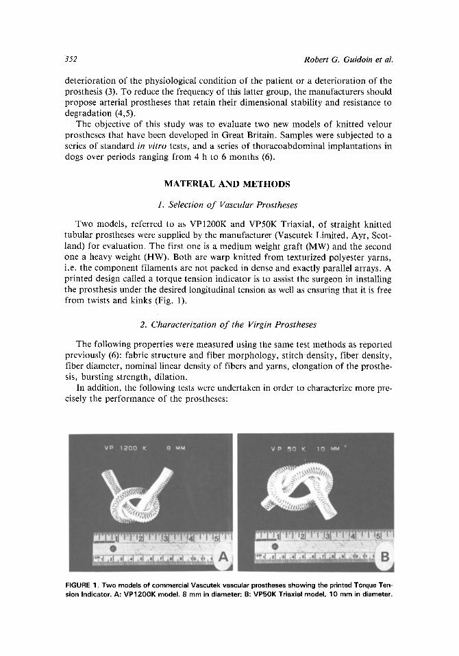
Two models, referred to as VP1200K and VP50K Triaxial, of straight knitted tubular prostheses were supplied by the manufacturer (Vascutek Limited, Ayr, Scot- land) for evaluation. The first one is a medium weight graft (MW) and the second one a heavy weight (HW). Both are warp knitted from texturized polyester yarns, i.e. the component filaments are not packed in dense and exactly parallel arrays. A printed design called a torque tension indicator is to assist the surgeon in installing the prosthesis under the desired longitudinal tension as well as ensuring that it is free from twists and kinks (Fig. 1).
2. Characterization o f the Virgin Prostheses
The following properties were measured using the same test methods as reported previously (6): fabric structure and fiber morphology, sti tchdensity, fiber density, fiber diameter, nominal linear density of fibers and yarns, elongation of the prosthe- sis, bursting strength, dilation.
In addition, the following tests were undertaken in order to characterize more pre- cisely the performance of the prostheses:
FIGURE 1. Two models of commercial Vascutek vascular prostheses showing the printed Torque Ten- sion Indicator. A: VP1200K model. 8 mm in diameter; B: VP50K Triaxial model, 10 mm in diameter.
Arterial Protheses from Great Britain 353
(a) Porosity: It refers to the percentage of void space within the boundaries of a material compared to its total volume, which is the sum of the solid mat- ter and the void space. The value for porosity, P, is calculated by the follow- ing equation:
P = 100 1 10
where M = mass per unit area of the fabric (g m-2) , h = fabric thickness (mm), df = fiber density (g cm-3).
(b) Water permeability: It refers to the rate flow of water through the wall of the dry prosthesis (7). This property should be referred to as the "water perme- ability" and not the "porosi ty" as frequently found in the medical literature. It provides an index of the interstitial leakage rate of the prostheses wall (8). Because the pressure of the applied head of water (120 m m Hg) may compact the textile structure with item, we extended the measure of the volume of water that passed through the prosthesis wall during each of the first 5 min of the test.
(c) Blood permeability: It provides a direct indication as to how quickly the wall will reach hemostasis. Flattened dry fabric specimens are exposed to heparin- ized fresh dog's blood at 120 mm Hg applied pressure over a 5-min period and the volume of blood that passed through is collected and measured after each minute.
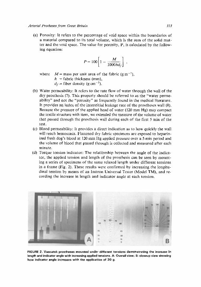
(d) Torque tension indicator: The relationship between the angle of the indica- tor, the applied tension and length of the prosthesis can be seen by mount- ing a series of specimens of the same relaxed length under different tensions in a frame (Fig. 2). These results were confirmed by increasing the longitu- dinal tension by means of an Instron Universal Tester (Model TM), and re- cording the increase in length and indicator angle at each tension.
FIGURE 2. Vascutek prostheses mounted under different tensions demonstrating the increase in length and indicator angle with increasing applied tensions. A: Overall view; B: closeup view showing how indicator angle increases with the application of 20 g.
354 Robert G. Guidoin et aL
3. In Vivo Evaluation
The protocol used for the in vivo evaluation was the thoracoabdominal bypass in dogs described originally by McCune (9) and now widely accepted (10).
(a) Implantation in animals: Adult female mongrel dogs weighing between 15 and 25 kg were selected according to the Canadian Council on Animal Care Regu- lations. Preclotted grafts, 30 cm long, were implanted as a thoracoabdomi- nal bypass for prescheduled periods of 4 h, 24 h, 48 h, 1 week, 1 month and 6 months using the protocol previously described (11). On those dogs im- planted for 6 months angiographies were performed 48 h before the sacrifice.
(b) Pathological investigations: The explanted grafts were examined and photo- graphed macroscopically. Pathological analysis was undertaken using both light microscopy and scanning electron microscopy on the two anastomotic regions and on two additional regions at one-third and two-thirds the distance along the length of each prosthesis. Representative samples were taken from both kidneys for pathological examination to identify the presence of possi- ble infarcts caused by emboli detached from the luminal surface of the prosthesis.
(c) Textile analysis of explanted prostheses: The remaining three sections of each graft were labelled proximal, central and distal according to their origin. The remaining tissue adhering to the prostheses was removed by boiling in 5~ sodium bicarbonate solution, immersing in commercial bleach at room tem- perature, exhaustively washing in distilled water and air drying. Additional analyses were performed on each section to identify changes that occurred dur- ing implantation. They consisted of stitch density, bursting strength, fiber diameter and surface morphology. The average results for the three sections were compared with those from preclotted and cleaned control specimens that had not been implanted.
RESULTS
1. Characterization o f the Virgin Prostheses
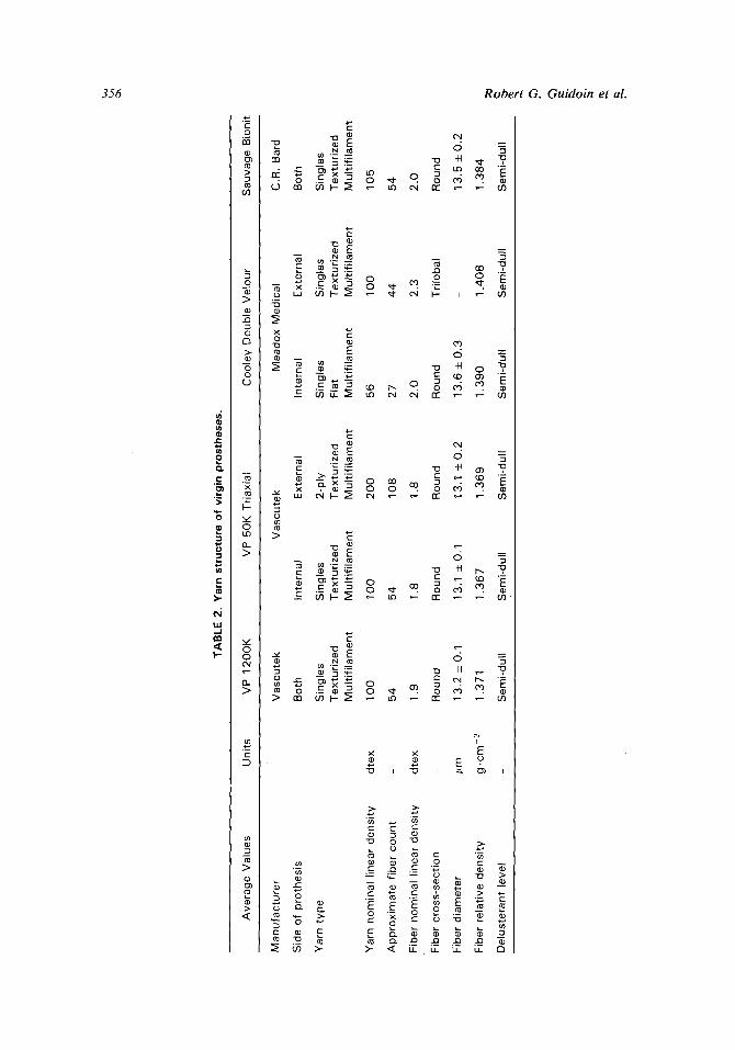
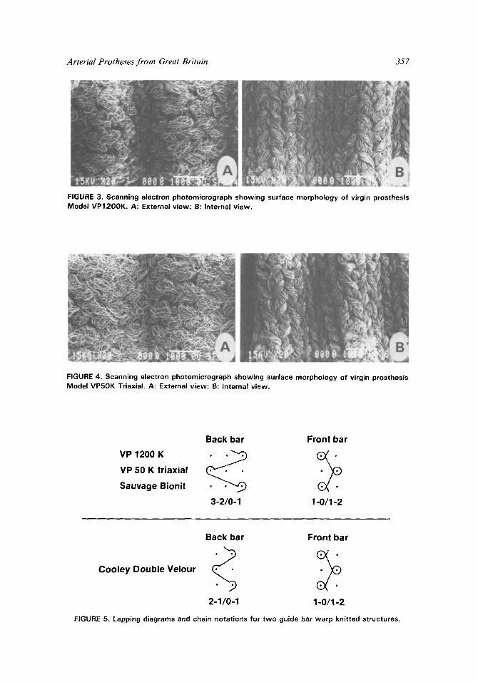
Microscopic analysis of both models revealed a warp knitted structure (Figs. 3-5). Details of other structural characteristics of the fabrics and yarns are given in Ta- bles 1 and 2 and in Figs. 6-8. Also included are the values for other commercial prostheses for comparison. The MW, i.e. VPI200K, appears equivalent to the Bionit and the HW, i.e. VP50K Triaxial, has a heavier, thicker, denser and stronger con- struction, being less water permeable.
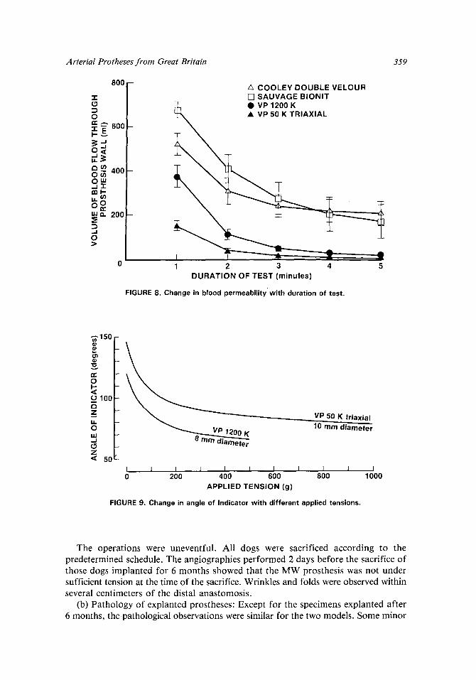
The relationships between the torque tension indicator angle and the applied lon- gitudinal tension have been plotted for both models in Fig. 9. The heavy weight graft (HW) can support higher tensions and has a greater resistance to stretching.
2. In Vivo Evaluation
(a) Implantation: The preclotting, handling and suturing the two models evalu- ated are equivalent to those of widely accepted commercial knitted prostheses using the same experimental protocol. The 1-week implantation of the MW graft was per- formed in duplicate.
h.
t~
TAB
LE 1
. Fa
bric
str
uctu
re a
nd p
erfo
rman
ce p
rope
rtie
s of
vir
gin
pros
thes
es.
Ave
rag
e V
alue
s U
nits
V
P 1
20
0K
V
P5
0K
Tri
axia
l C
oo
ley
Dou
ble
Ve
lou
r S
au
vag
e B
ioni
t
Ma
nu
fact
ure
r V
asc
ute
k V
asc
ute
k M
ea
do
x M
edic
als
C.R
. B
ard
Fab
ric
Str
uctu
re:
Typ
e o
f fa
bri
c
Sti
tch
typ
e:
fro
nt
bar
ba
ck b
ar
Kn
itte
d f
abri
c co
un
t ~-
. w
ale
s cm
- 1
cour
se~
cm
-1
Sti
tch
de
nsi
ty
cm -2
Mas
s pe
r un
it a
rea
g.m
2
Fab
ric
thic
knes
s m
m
Por
osit
y %
Typ
e o
f cr
imp:
fr
eq
ue
ncy
cm
1
de
pth
m
m
Per
form
ance
Pro
pert
ies:
E
long
atio
n %
2
33
B
urst
ing
stre
ng
th
kPa
21
20
Wa
ter
pe
rme
ab
ility
m
l-cm
-2
.min
-1
20
00
Dila
tion
% a
t 1
20
mm
Hg
9.6
2-ba
r w
arp
kni
t 2-
bar
wa
rp k
nit
2-ba
r w
arp
kn
it
2-ba
r w
arp
kn
it
clos
ed t
rico
t la
ps
clos
ed t
rico
t la
ps
clos
ed t
rico
t la
ps
clos
ed t
rico
t la
ps
open
cor
d la
ps
op
en
cor
d la
ps
open
tri
cot
laps
op
en c
ord
laps
14
12
20
14
15
16
28
2
4
21
0
187
56
0
33
6
182
26
3
28
0
23
0
0.5
7
0.7
4
0.6
9
0.5
5
77
74
71
7
0
helic
al
helic
al
circ
ular
ci
rcul
ar
8.7
7
.2
4.8
8
.4
1.4
3
1.3
5
1.4
4
1.3
2
21
6
95
2
50
3
19
0
11
60
2
37
0
11
40
1
77
0
16
20
5.4
5.
1 8
.9
L~
tn
L~
o~
TA
BL
E 2
. Y
arn
stru
ctu
re o
f vi
rgin
pro
sthe
ses.
Ave
rag
e V
alue
s U
nits
V
P 1
20
0K
V
P 5
0K T
riax
ial
Co
ole
y D
oubl
e V
elo
ur
Sa
uva
ge
Bio
nit
Ma
nu
fact
ure
r V
asc
ute
k V
asc
ute
k M
ea
do
x M
edic
al
C.R
. B
ard
Sid
e of
pro
thes
is
Bot
h In
tern
al
Ext
erna
l In
tern
al
Ext
erna
l B
oth
Yar
n ty
pe
S
ingl
es
Sin
gles
2-
ply
Sin
gles
S
ingl
es
Sin
gles
T
ext
uri
zed
T
ext
uri
zed
T
ext
uri
zed
F
lat
Te
xtu
rize
d
Te
xtu
rize
d
Mu
ltif
ilam
en
t M
ult
ifila
me
nt
Mu
ltif
ilam
en
t M
ult
ifila
me
nt
Mu
ltif
ilam
en
t M
ult
ifila
me
nt
dte
x 1 O
0 1 O
0 2
00
56
1 O
0 1
05
54
54
10
8 27
4
4
54
dte
x 1.
9 1.
8 1.
8 2
.0
2.3
2
,0
Rou
nd
Rou
nd
Rou
nd
Rou
nd
Tri
loba
l R
ound
#m
13
.2+
_0
.1
13.1
+_0
.1
13.1
_+0
.2
13
.6_
+0
.3
13
.5_
+0
.2
g.c
m -
3 1.
371
1.3
67
1
.36
9
1.3
90
1
.40
8
1.3
84
Sem
i-du
ll S
emi-
dull
Sem
i-du
ll S
emi-
dull
Sem
i-du
ll S
emi-
dull
Yar
n no
min
al l
inea
r d
en
sity
Ap
pro
xim
ate
fib
er
cou
nt
Fib
er n
omin
al l
inea
r d
en
sity
Fib
er c
ross
-sec
tion
Fib
er d
iam
ete
r
Fib
er r
elat
ive
de
nsi
ty
Del
uste
rant
lev
el
C~
Arterial Protheses f rom Great Britain 357
FIGURE 3. Scanning electron photomicrograph showing surface morphology of virgin prosthesis Model VP1200K. A: External view; B: internal view.
FIGURE 4. Scanning electron photomicrograph showing surface morphology of virgin prosthesis Model VP50K Triaxial. A: External view; B: internal view.
VP 1200 K
VP 50 K triaxial
Sauvage Bionit
Back bar Front bar
3-2/0-1 1-0/1-2
Cooley Double Velour
Back bar Front bar
2-1/0-1 1-0/1-2
FIGURE 5. Lapping diagrams and chain notations for two guide bar warp knitted structures.
358 Robert G. Guidoin et al.
2 0 -
~5 t -
~ 1 2
A COOLEY DOUBLE VELOUR ~- [ ] SAUVAGE BIONIT
l �9 VP 1200 K +_1.8% �9 VP 50 K TRIAXlAL
- 0
L , , L L L L I 50 100 150 200 250 300 350
INTERNAL PRESSURE (mmHg)
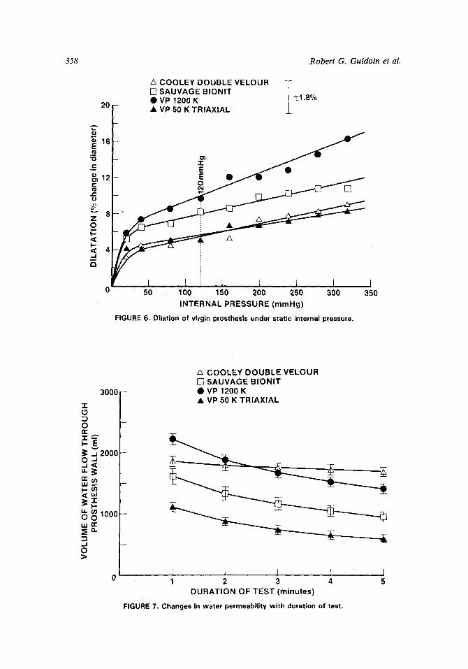
FIGURE 6. Dilation of vi~'gin prosthesis under static internal pressure.
0
l -
q
w I--
u.I
. , J 0
3000
I ~-~E2ooo
~ -
1000 -- O
/~ COOLEY DOUBLE VELOUR [ ] SAUVAGE BIONIT @ VP 1200 K �9 VP 50 K TRIAXIAL
I ~ I 1 1 2 3 4
DURATION OF TEST (minutes)
FIGURE 7. Changes in water permeability with duration of test.
Arterial Protheses f rom Great Britain 359
800
-i"
0
z ~ 600
u) 400 O(n
,,' o. 200
..,J O ~>
A COOLEY DOUBLE VELOUR [ ] SAUVAGE BIONIT
~ X �9 VP 1200 K �9 VP 50 K TRIAXIAL
I I "r Z 1 2 3 4 5
DURATION OF TEST ( m i n u t e s )
FIGURE 8. Change in blood permeability wi th durat ion of test.
150
~ZO~o 100 I
=' (3 8 mm diameter z "~ 5
I I I L I I I I ] 1 0 200 400 600 800
APPLIED TENSION (g)
FIGURE 9. Change in angle of Indicator wi th di f ferent applied tensions.
VP 50 K tr iaxial 10 mm d i a m e t e r
I 1 0 0 0
The operations were uneventful. All dogs were sacrificed according to the predetermined schedule. The angiographies performed 2 days before the sacrifice of those dogs implanted for 6 months showed that the MW prosthesis was not under sufficient tension at the time of the sacrifice. Wrinkles and folds were observed within several centimeters of the distal anastomosis.
(b) Pathology of explanted prostheses: Except for the specimens explanted after 6 months, the pathological observations were similar for the two models. Some minor
360 Robert G. Guidoin et al.
variations were observed, but these must be attributed to differences in blood prop- erties and physiology between animals rather than to characteristics of the prostheses themselves (12).
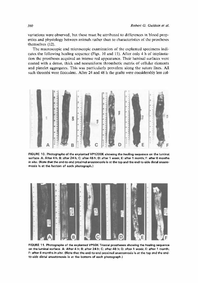
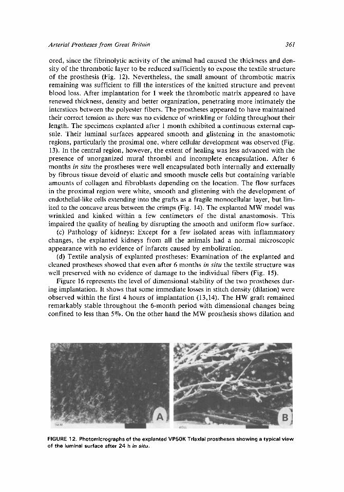
The macroscopic and microscopic examination of the explanted specimens indi- cates the following healing sequence (Figs. 10 and 11). After only 4 h of implanta- tion the prostheses acquired an intense red appearance. Their luminal surfaces were coated with a dense, thick and nonuniform thrombotic matrix of cellular elements and platelet aggregates. This was particularly prevalent along the suture lines. All such thrombi were flocculent. After 24 and 48 h the grafts were considerably less col-
FIGURE 10. Photographs of the explanted VP1200K showing the healing sequence on the luminal surface. A: After 4 h; B: after 24 h; C: after 48 h; D: after 1 week; E" after 1 month; F: after 6 months in situ. (Note that the end-to-end proximal anastomosis is at the top and the end-to-side distal anasto- mosis is at the bottom of each photograph.)
FIGURE 11. Photographs of the explanted VP50K Triaxial prostheses showing the healing sequence on the luminal surface. A: After 4 h; B: after 24 h; C: after 48 h; D: after 1 week; E: after 1 month; F: after 6 months in situ. (Note that the end-to-end proximal anastomosis is at the top and the end- to-side distal anastomosis is at the bottom of each photograph.)
Arterial Protheses from Great Britain 361
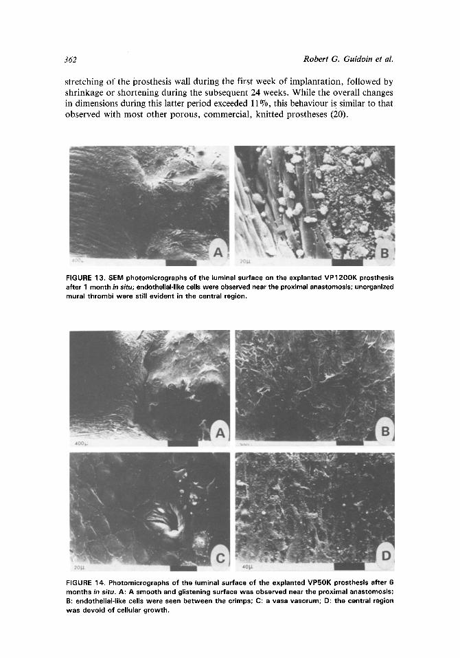
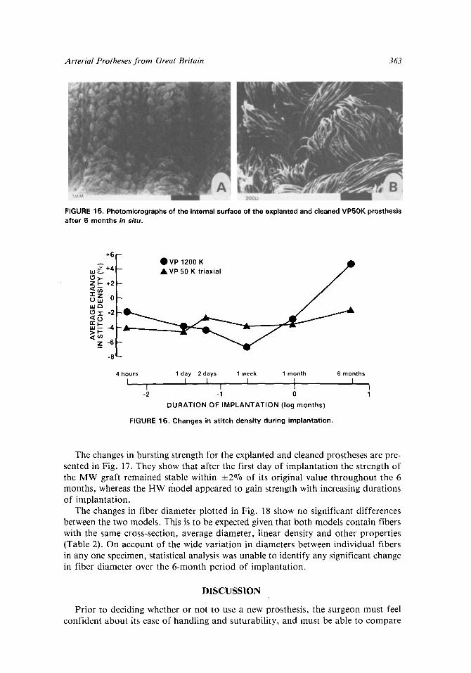
ored, since the fibrinolytic activity of the animal had caused the thickness and den- sity of the thrombotic layer to be reduced sufficiently to expose the textile structure of the prosthesis (Fig. 12). Nevertheless, the small amount of thrombotic matrix remaining was sufficient to fill the interstices of the knitted structure and prevent blood loss. After implantation for 1 week the thrombotic matrix appeared to have renewed thickness, density and better organization, penetrating more intimately the interstices between the polyester fibers. The prostheses appeared to have maintained their correct tension as there was no evidence of wrinkling or folding throughout their length. The specimens explanted after 1 month exhibited a continuous external cap- sule. Their luminal surfaces appeared smooth and glistening in the anastomotic regions, particularly the proximal one, where cellular development was observed (Fig. 13). In the central region, however, the extent of healing was less advanced with the presence of unorganized mural thrombi and incomplete encapsulation. After 6 months in situ the prostheses were well encapsulated both internally and externally by fibrous tissue devoid of elastic and smooth muscle ceils but containing variable amounts of collagen and fibroblasts depending on the location. The flow surfaces in the proximal region were white, smooth and glistening with the development of endothelial-like cells extending into the grafts as a fragile monocellular layer, but lim- ited to the concave areas between the crimps (Fig. 14). The explanted MW model was wrinkled and kinked within a few centimeters of the distal anastomosis. This impaired the quality of healing by disrupting the smooth and uniform flow surface.
(c) Pathology of kidneys: Except for a few isolated areas with inflammatory changes, the explanted kidneys from all the animals had a normal microscopic appearance with no evidence of infarcts caused by embolization.
(d) Textile analysis of explanted prostheses: Examination of the explanted and cleaned prostheses showed that even after 6 months in situ the textile structure was well preserved with no evidence of damage to the individual fibers (Fig. 15).
Figure 16 represents the level of dimensional stability of the two prostheses dur- ing implantation. It shows that some immediate losses in stitch density (dilation) were observed within the first 4 hours of implantation (13,14). The HW graft remained remarkably stable throughout the 6-month period with dimensional changes being confined to less than 5~ On the other hand the MW prosthesis shows dilation and
FIGURE 12. Photomicrographs of the explanted VP50K Triaxial prostheses showing a typical view of the luminal surface after 24 h in situ.
362 Robert G. Guidoin et aL
stretching of the prosthesis wall during the first week of implantation, followed by shrinkage or shortening during the subsequent 24 weeks. While the overall changes in dimensions during this latter period exceeded 11%, this behaviour is similar to that observed with most other porous, commercial, knitted prostheses (20).
FIGURE 13. SEM photomicrographs of the luminal surface on the explanted VP1200K prosthesis after 1 month in situ; endothelial-like cells were observed near the proximal anastomosis; unorganized mural thrombi were still evident in the central region.
FIGURE 14. Photomicrographs of the luminal surface of the explanted VP50K prosthesis after 6 months in situ. A: A smooth and glistening surface was observed near the proximal anastomosis; B: endothelial-l ike cells were seen between the crimps; C: a vasa vasorum; D: the central region was devoid of cellular growth.
Arterial Protheses from Great Britain 363
FIGURE 15. Photomicrographs of the internal surface of the explanted and cleaned VP50K prosthesis after 6 months in situ.
+6 F OVP 1200 K Q ~~176 +4l'- � 9 50Ktriaxial f
-2 I--
Z
-8
4hours 1day 2days 1week 1 month 6months
I I [ 1 l 1 t L I I
-2 -1 0 1
DURATION OF IMPLANTATION (log months)
FIGURE 16. Changes in stitch density during implantation.
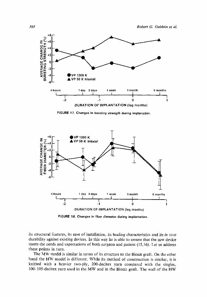
The changes in bursting strength for the explanted and cleaned prostheses are pre- sented in Fig. 17. They show that after the first day of implantation the strength of the MW graft remained stable within +2% of its original value throughout the 6 months, whereas the HW model appeared to gain strength with increasing durations of implantation.
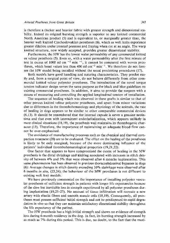
The changes in fiber diameter plotted in Fig. 18 show no significant differences between the two models. This is to be expected given that both models contain fibers with the same cross-section, average diameter, linear density and other properties (Table 2). On account of the wide variation in diameters between individual fibers in any one specimen, statistical analysis was unable to identify any significant change in fiber diameter over the 6-month period of implantation.
DISCUSSION
Prior to deciding whether or not to use a new prosthesis, the surgeon must feel confident about its ease of handling and suturability, and must be able to compare
364 Robert G. Guidoin et al.
+8 "C Z ~ +6 ---i-
§
uJcn 02 (3(3 ,~z
,,,~n---!: ~ V ; 15200~ K -~ iaxial
4 hours 1 day 2 days 1 week 1 month 6 months I I I I I I t I 1
-2 -1 0 1 DURATION OF IMPLANTATION (log months)
FIGURE 17. Changes in bursting strength during implanation.
T Z "61-- OVP 1200 K ~ -T"
,~+4 ~_ T A VP 50 K triaxial
4 hours 1 day 2 days 1 week 1 month I i 1 J i 1 1
I
-2 -1 0
DURATION OF IMPLANTATION (log months)
FIGURE 18. Changes in fiber diameter during implantation.
6 months I
its structural features, its ease of installation, its healing characteristics and its in vivo
durability against existing devices. In this way he is able to ensure that the new device meets the needs and expectations of both surgeon and patient (15,16). Let us address these points in turn.
The MW model is similar in terms of its structure to the Bionit graft. On the other hand the H W model is different. While its method of construction is similar, it is knitted with a heavier two-ply, 200-decitex yarn compared with the singles, 100-105-decitex yarn used in the MW and in the Bionit graft. The wall of the H W
Arterial Protheses from Great Britain 365
is therefore a thicker and heavier fabric with greater strength and dimensional sta- bility. Indeed its original bursting strength is superior to any knitted commercial North American product (5) and is equivalent to, or marginally greater than, the heavier weft knitted Czechoslovakian prostheses (6), which as weft knits experience greater dilation under internal pressure and fraying when cut at an angle. The warp knitted structure, now widely accepted, provides greater dimensional stability.
Furthermore, the HW has the lowest water permeability of any commercial knitted or velour prosthesis (5). Even so, with a water permeability after the first minute of test in excess of 1000 ml cm -2 min -1, it cannot be compared with woven pros- theses, which boast values less than 400 ml cm 2 min- l . We therefore do not fore- see the HW model being installed without the usual preclotting procedures (17).
Both models have good handling and suturing characteristics. They preclot eas- ily and, from a surgical point of view, do not behave differently from other com- mercial knitted velour polyester prostheses. The introduction of the novel torque tension indicator design serves the same purpose as the black and blue guidelines on existing commercial prostheses. In addition, it aims to provide the surgeon with a means of measuring and controlling the applied longitudinal tension of installation.
The sequence of healing which was observed in these grafts is similar to that of other porous knitted velour polyester prostheses, and apart from minor variations due to differences in the thrombohematology and physiology of the animals, the rate of healing in dogs appears to be similar to other comparable commercial devices (6,13). It should be remembered that the internal capsule is never a genuine neoin- tima and that even with intermittent endothelialization, which appears unlikely in most clinical situations (18,19), the prosthesis wall maintains its thrombogenic char- acter (13). Therefore, the importance of maintaining an adequate blood flow can- not be over-emphasized.
The avoidance of manufacturing processes such as the chemical and thermal com- paction treatment (20) are to be evaluated. The effect on the healing of the prostheses is likely to be only marginal, because of the more dominating influence of the patients' individual thrombohematological properties (19,21,22).
One factor that appears to have compromised the extent of healing in the MW prosthesis is the distal shrinkage and kinking associated with increases in stitch den- sity of between 4~ and 5~ that were observed after 6 months implantation. This same phenomenon has been observed in previous thoracoabdominal bypasses in dogs (6): Average changes in stitch density exceeding 10070 shrinkage were observed after 6 months in situ, (23,24); the behaviour of the MW prostheses is not different to existing weft knit models.
We have previously commented on the importance of installing polyester vascu- lar prostheses of sufficient strength in patients with longer life expectancies because of the slow but inevitable loss in strength experienced by all polyester prostheses dur- ing implantation (19,25-27). No amount of tissue infiltration will recreate a new artery with elastin fibers and smooth muscle cells (18,19). Consequently, all pros- theses must possess sufficient initial strength and not be predisposed to rapid degra- dation in vivo so that they can maintain satisfactory dimensional stability throughout the life expectancy of the patient.
The HW prosthesis has a high initial strength and shows no evidence of strength loss during 6-month residency in the dog. In fact, its bursting strength increased by as much as 7% during this period. This is due, no doubt, to the fact that the maxi-
366 Robert G. Guidoin et al.
mum bursting strength of the fabric is not fully realized in the virgin prosthesis. The high yarn tensions required to knit the texturized yarn at such high stitch densities create internal residual stress concentrations within the fabric that are reduced only after the prosthesis is exposed to the cyclic mechanical stresses of the pulsatile blood flow. The longer-term durability of the HW model appears encouraging, but addi- tional in vitro investigations are needed for both types of prostheses to clarify their long-term in vivo performance. The fatigue effect has to be evaluated under con- trolled conditions by exposure to a cyclic sinusoidal wave of pseudo-extracellular fluid in a Vivocycleur (28). By controlling the mean pressure, the maximum pressure amplitude, the cyclic frequency, the longitudinal tension and the temperature within the ranges known to exist in humans, the effects of mechanical fatigue on the integrity and stability of arterial prosthesis will be better understood.
CONCLUSIONS
These in vitro tests and this in vivo evaluation as a thoracoabdominal bypass over periods of up to 6 months in the dog has demonstrated that they can be easily installed and have similar healing characteristics to other porous polyester grafts. Therefore, clinical investigations can be undertaken. Additional dynamic in vitro tests should be conducted using equipment such as the Vivocycleur in order to help pre- dict their longer term performance.
REFERENCES
1. DeBakey, M. The development of vascular surgery. Am. J. Surg. 137:697-738, 1979. 2. Glickman, M.H., R.L. Hurwitz, S.A. Kimmins and W.E. Evans. Employment following peripheral
vascular surgery: an increasingly critical issue. Surgery 93:50-53, 1983. 3. Leijala, M. Complications of synthetic arterial grafting. A retrospective study of 489 patients. PhD
Thesis. University of Helsinki, 1976. 4. Mortensen, J.D. Safety and performance of currently available vascular prostheses. A review. ASAIO
J. 4:125-138, 1981. 5. Guidoin, R., M. King, C. Gosselin, P. Blais, K. Gunasekera, M. Marois and A. Cardou. Les proth6ses
art6rielles en polyester. Rev. Eur. Biotech. Med. 4:13-25, 1982. 6. King, M.W., R. Guidoin, K. Gunasekera, L. Martin, M. Marois, P. Blais, J.M. Maarek and C. Gosse-
lin. An evaluation of Czechoslovakian polyester arterial prostheses. ASAIO J. 7:114-133, 1984. 7. Buxton, B.F., D.C. Wukasch, C. Martin, W.J. Liebig, G.L. Hallman and D.A. Cooley. Practical
considerations in fabric and vascular grafts. Introduction of a new bifurcated graft. Am. J. Surg. 125:288-293, 1973.
8. ISO DP 7198, Cardiovascular Implants-Draft Standard for Synthetic Tubular Vascular Prostheses, Association for the Advancement of Medical Instrumentation, Arlington, VA, October 1984.
9. McCune, W.S. and B. Blades. The viability of long blood vessel grafts. Ann. Surg, 124:769-781, 1954. 10. Burkel, W.E., D.W. Winter, J.W. Ford, R.H. Kahn, L.M. Graham and J.C. Stanley. Sequential stud-
ies of healing in endothelial seeded vascular prostheses: histological and ultrastructure characteris- tics of graft incorporation. J. Surg. Res. 30:305-324, 1981.
11. Go~au-Brissonni~re, A., R. Guidoin, M. Marois, B. Boyce, J.C. Pechere, P. Blais, H.P. Noel and C. Gosselin. Thoracoabdominal bypass as a method of evaluating vascular grafts in the dog. Biomater. Med. Devices Artif. Organs 9:195-212, 1981.
12. Bernier, J. and R. Guidoin. Importance des facteurs thrombodynamiques dans la thrombose aiguE darts les prothbses art6rielles. Rev. Eur. Biotech. Med. 4:103-113, 1982.
13. Guidoin, R., C. Gosselin, L. Martin, M. Marois, F. Laroche, M. King, K. Gunasekera, D. Domurado, M.F. Sigot-Luizard and P. Blais. Polyester prostheses as substitutes in the thoracic aorta of dogs. I. Evaluation of commercial prostheses. J. Biomed. Mater. Res. 17:1049-1077, 1983.
14. Nunn, D.B., M.H. Freeman and P.C. Hudgins. Postoperative alterations in size of Dacron aortic grafts. An ultrasonic evaluation. Ann. Surg. 189:741-745, 1979.
Arter ia l Protheses f r o m Great Bri tain 367
15. Melliere, D., G. Lasry and F. Lange. R~sultats et indications des prothbses en chirurgie art6rielle. J. Chirc. (Paris) 116:285-296, 1979.
16. King, M., P. Blais, R. Guidoin, E. Prowse, M. Marois, C. Gosselin and H.P. Noel. Polyethylene terephthalate (Dacron) vascular prostheses-material and fabric construction aspects. In: Biocom- patibility o f Clinical Implants Materials, edited by D.F. Williams. Boca Raton, Florida: CRC, 1981, Vol. 2, pp. 177-207.
17. Yates, S.G., A.A.B. Barros, K. Berger, L.G. Fernandez, S.J. Wood, E.A. Rittenhouse, C.C. Davis, P.B. Mansfield and L.R. Sauvage. The preclotting of porous arterial prostheses. Ann. Surg. 188:611-622, 1978.
18. Berger, K., L.R. Sauvage, A.M. Rao and S.J. Wood. Healing of arterial prostheses in man: its incom- pleteness. Ann. Surg. 175:118-127, 1972.
19. Guidoin, R., M. King, P. Blais, M. Marois, C. Gosselin, P. Roy, R. Courbier, M. David and H.P. Noel. A biological and structural evaluation of retrieved Dacron arterial prostheses. NBS Special Report 601:29-129, 1981.
20. Sawyer, P.N., B. Stanczewski, R. Turner and H. Hoffman. Evaluation of chemical compacting tech- niques on the performance, thrombosis rate, histology and healing of experimental similarly fabri- cated Dacron prostheses. Trans. Am. Soc. Artif. Intern. Organs 24:215-222, 1978.
21. Sauvage, L~R., K.E. Berger, S.J. Wood, S.G. Yates II, J.C. Smith and P.B. Mansfield. Interspe- cies healing of porous arterial prostheses. Arch. Surg. 109:698-705, 1974.
22. Bruck, S.D. Considerations of species related hematological differences on the evaluation of biomaterials. Biomater. Med. Devices Artif. Organs 5:97-113, 1977.
23. Guidoin, R., L. Martin, M. Marois, C. Gosselin, M. King, K. Gunasekera, D. Domurado, M.F. Sigot- Luizard, M. Sigot and P. Blais. Polyester prostheses as substitutes in the thoracic aorta of dogs. II. Evaluation of albuminated polyester grafts stored in ethanol. J. Biomed. Mater. Res. 18:1059-1072, 1984.
24. Guidoin, R.G., W.M. King, J. Awad, L. Martin, D. Domurado, M. Marois, M.F. Sigot-Luizard, C. Gosselin, K. Gunasekera and D. Gagnon. Albumin coated and critical point dried polyester prostheses as substitutes in the thoracic aorta of dogs. Trans. Am. Soc. Artif. Intern. Organs 29:290-295, 1983.
25. King, M , R. Guidoin, C. Gosselin, F. Godard, M. Marois, K. Gunasekera, P. Blais and A. Gar- ton. Protocole d'6tude d'une greffe art6rielle en polyester aprbs ex6rbse. Les standards sont-ils utiles ou dbsagreables? Rev. Eur. Biotech. Med. 4:26-33, 1982.
26. King, W.M.R. Guidoin, P. Blais, A. Garton and A. Gunasekera. Degradation of polyester arterial prostheses: a physical or chemical mechanism? In: Corrosion and Degradation o f Implant Materi- als, edited by A.C. Fraker and C.D. Griffin. STP 859, Philadelphia: ASTM, 1985, pp. 294-307.
27. Godard, F., M. King, R. Guidoin, M. Marois, A. Garton P., Blais, C. Gosselin and K. Gunasekera. Formation an6vrismale sur proth6se art6rielle synthetique-analyse pathologique, structurale et chi- mique d'une greffe en polyester apr~s 84 mois d'implantation. J. Mal. Vasc. (Paris) 6:167-171, 1981.
28. Marceau, D., A. Cardou, R. Guidoin, M. King and C. Gosselin. Development d'un syst6me d'essai pour l'6tude du comportement dynamique des prothbses art6rielles alloplastiques: le Vivocycleur. Rev. Eur. Biotech. Med. 6:31-38, 1984.