New Insights and Advances in Pathogenesis and Treatment of ...
12
REVIEW published: 01 March 2022 doi: 10.3389/fped.2022.714054 Frontiers in Pediatrics | www.frontiersin.org 1 March 2022 | Volume 10 | Article 714054 Edited by: Séamus Hussey, National Children’s Research Centre (NCRC), Ireland Reviewed by: Ruggiero Francavilla, University of Bari Aldo Moro, Italy Corentin Babakissa, Université de Sherbrooke, Canada *Correspondence: Shi-Xue Dai [email protected]; [email protected] † These authors have contributed equally to this work and share first authorship Specialty section: This article was submitted to Pediatric Gastroenterology, Hepatology and Nutrition, a section of the journal Frontiers in Pediatrics Received: 24 May 2021 Accepted: 25 January 2022 Published: 01 March 2022 Citation: Li Q-Q, Zhang H-H and Dai S-X (2022) New Insights and Advances in Pathogenesis and Treatment of Very Early Onset Inflammatory Bowel Disease. Front. Pediatr. 10:714054. doi: 10.3389/fped.2022.714054 New Insights and Advances in Pathogenesis and Treatment of Very Early Onset Inflammatory Bowel Disease Qi-Qi Li 1† , Hui-Hong Zhang 1† and Shi-Xue Dai 1,2,3 * 1 The Second School of Clinical Medicine, Southern Medical University, Guangzhou, China, 2 Department of Gastroenterology, Guangdong Provincial Geriatrics Institute, National Key Clinical Specialty, Guangdong Provincial People’s Hospital, Guangdong Academy of Medical Sciences, Guangzhou, China, 3 Department of Gastroenterology, Guangdong Provincial People’s Hospital, Guangdong Academy of Medical Sciences, South China University of Technology, Guangzhou, China Very early onset inflammatory bowel disease (VEO-IBD) is characterized by multifactorial chronic recurrent intestinal inflammation. Compared with elderly patients, those with VEO-IBD have a more serious condition, not responsive to conventional treatments, with a poor prognosis. Recent studies found that genetic and immunologic abnormalities are closely related to VEO-IBD. Intestinal immune homeostasis monogenic defects (IIHMDs) are changed through various mechanisms. Recent studies have also revealed that abnormalities in genes and immune molecular mechanisms are closely related to VEO-IBD. IIHMDs change through various mechanisms. Epigenetic factors can mediate the interaction between the environment and genome, and genetic factors and immune molecules may be involved in the pathogenesis of the environment and gut microbiota. These discoveries will provide new directions and ideas for the treatment of VEO-IBD. Keywords: microRNA, circular RNA, biologics, immunity, gut microbiota INTRODUCTION Very early onset inflammatory bowel disease (VEO-IBD) refers to a subgroup of pediatric patients diagnosed with IBD before the age of 6 years (1); it includes subclasses of infantIBD and neonatal IBD diagnosed before the age of 2 years and 28 days, respectively. Epidemiological data show that the incidence of VEO-IBD has been increasing rapidly, and one study showed that the incidence had reached 7.2% (2). The increasing incidence of VEO-IBD suggests that it is urgent to understand its pathogenesis. The latest and largest genetic association study collected genome- wide association data for over 75,000 patients and controls and identified 163 susceptibility loci for IBD. Interestingly, twin and family studies of IBD showed that for a child with an affected sibling, the risk increases 26 times for Crohn’s disease (CD) and increases 9 times for ulcerative colitis (UC). This suggests that both genetic and environmental factors may affect the pathogenesis of VEO-IBD. However, the pathogenesis of VEO-IBD is still not fully clear. To help solve the existing problems, this narrative review starts from the genetic pathogenesis of VEO-IBD, systematically and comprehensively summarizes the existing pathogenesis and treatment, and provides a potential breakthrough point for the therapeutics of VEO-IBD.
-
Upload
khangminh22 -
Category
Documents
-
view
0 -
download
0
Transcript of New Insights and Advances in Pathogenesis and Treatment of ...
REVIEWpublished: 01 March 2022
doi: 10.3389/fped.2022.714054
Frontiers in Pediatrics | www.frontiersin.org 1 March 2022 | Volume 10 | Article 714054
Edited by:
Séamus Hussey,
National Children’s Research Centre
(NCRC), Ireland
Reviewed by:
Ruggiero Francavilla,
University of Bari Aldo Moro, Italy
Corentin Babakissa,
Université de Sherbrooke, Canada
*Correspondence:
Shi-Xue Dai
†These authors have contributed
equally to this work and share first
authorship
Specialty section:
This article was submitted to
Pediatric Gastroenterology,
Hepatology and Nutrition,
a section of the journal
Frontiers in Pediatrics
Received: 24 May 2021
Accepted: 25 January 2022
Published: 01 March 2022
Citation:
Li Q-Q, Zhang H-H and Dai S-X
(2022) New Insights and Advances in
Pathogenesis and Treatment of Very
Early Onset Inflammatory Bowel
Disease. Front. Pediatr. 10:714054.
doi: 10.3389/fped.2022.714054
New Insights and Advances inPathogenesis and Treatment of VeryEarly Onset Inflammatory BowelDiseaseQi-Qi Li 1†, Hui-Hong Zhang 1† and Shi-Xue Dai 1,2,3*
1 The Second School of Clinical Medicine, Southern Medical University, Guangzhou, China, 2Department of Gastroenterology,
Guangdong Provincial Geriatrics Institute, National Key Clinical Specialty, Guangdong Provincial People’s Hospital,
Guangdong Academy of Medical Sciences, Guangzhou, China, 3Department of Gastroenterology, Guangdong Provincial
People’s Hospital, Guangdong Academy of Medical Sciences, South China University of Technology, Guangzhou, China
Very early onset inflammatory bowel disease (VEO-IBD) is characterized by multifactorial
chronic recurrent intestinal inflammation. Compared with elderly patients, those with
VEO-IBD have a more serious condition, not responsive to conventional treatments, with
a poor prognosis. Recent studies found that genetic and immunologic abnormalities
are closely related to VEO-IBD. Intestinal immune homeostasis monogenic defects
(IIHMDs) are changed through various mechanisms. Recent studies have also revealed
that abnormalities in genes and immune molecular mechanisms are closely related to
VEO-IBD. IIHMDs change through various mechanisms. Epigenetic factors can mediate
the interaction between the environment and genome, and genetic factors and immune
molecules may be involved in the pathogenesis of the environment and gut microbiota.
These discoveries will provide new directions and ideas for the treatment of VEO-IBD.
Keywords: microRNA, circular RNA, biologics, immunity, gut microbiota
INTRODUCTION
Very early onset inflammatory bowel disease (VEO-IBD) refers to a subgroup of pediatric patientsdiagnosed with IBD before the age of 6 years (1); it includes subclasses of infantIBD and neonatalIBD diagnosed before the age of 2 years and 28 days, respectively. Epidemiological data showthat the incidence of VEO-IBD has been increasing rapidly, and one study showed that theincidence had reached 7.2% (2). The increasing incidence of VEO-IBD suggests that it is urgentto understand its pathogenesis. The latest and largest genetic association study collected genome-wide association data for over 75,000 patients and controls and identified 163 susceptibility loci forIBD. Interestingly, twin and family studies of IBD showed that for a child with an affected sibling,the risk increases 26 times for Crohn’s disease (CD) and increases 9 times for ulcerative colitis(UC). This suggests that both genetic and environmental factors may affect the pathogenesis ofVEO-IBD. However, the pathogenesis of VEO-IBD is still not fully clear. To help solve the existingproblems, this narrative review starts from the genetic pathogenesis of VEO-IBD, systematicallyand comprehensively summarizes the existing pathogenesis and treatment, and provides a potentialbreakthrough point for the therapeutics of VEO-IBD.
Li et al. Pathogenesis and Treatment for VEO-IBD
GENETIC FACTORS
Gene AbnormalitiesOwing to technological progress in genetic testing andDNA sequencing, many genome-wide association studies(GWAS) have been improved, showing new single nucleotidepolymorphisms (SNPs) (3). However, the explainablesusceptibility loci and genetic risk factors discovered thusfar only account for 20–25% of the heritability (genetic risk)(4). Moreover, monogenic mutations have been found mostly inchildren aged under 6 years, and most conventional polygenicIBD patients are older than 7 years.
Discovered in 2001, nucleotide-binding oligomerizationdomain containing 2 (NOD2) was the first susceptibility genefor CD. It encodes a protein that acts as an intracellularreceptor for bacterial products in monocytes and transducessignals activating NFκB. Polymorphisms in NOD2 are one of thegreatest genetic risk factors for Crohn’s disease. Three differentnon-synonomous NOD2 polymorphisms, R702W, G908R, andL1007fsincC, account for ∼80% of all NOD2-associated casesof Crohn’s disease and they have been reported to cause aloss of receptor function in response to muramyl dipeptide(MDP) stimulation (5). It has been shown that the perceptionof NOD protein on bacteria is associated with the induction ofautophagy (6). DCs from CD patients with susceptibility variantsin the NOD2 gene are deficient in autophagy induction, andthe localization of bacteria in autophagolysosomes is reduced(7). Additionally, the genome map of CD patients shows thatNOD2 deficiency and mutationsare related to CD in the ileum.Therefore, the interaction between ileal microflora and mucosalimmunity is changed by NOD2 mutation, which is a high-risk factor for multiple complications of ileal CD and indicatesincreased susceptibility to CD.
miRNA AbnormalitiesCD and UC have differences not only in the tissue miRNAspectrum but also in the peripheral blood miRNA spectrum.Recently, several studies have analyzed the differential expressionof miRNAs in tissue samples and blood between IBD patientsand healthy controls, showing that miRNAs may be regarded asnovel biomarkers of these diseases (8). Therefore, the recognitionof different miRNA expression profiles may provide a method todetermine the course of the disease at an early stage.
Serum miR-146a and miR-146b decrease with IFX treatmentand long-term glucocorticoid (GC) treatment (weeks) but notwith short-term GC treatment (days) (9). Previous studies haveshown that miR-146a and miR-146b are responsive to endotoxin,while increasing miR-146a or miR-146b is dependent oninflammatory stimuli (10). miR-146b was previously describedas a monitoring biomarker for IBD, positively correlated withendoscopic disease activity, and more specific than serum c-reactive protein (9). In this study, serum miR-320a was found todecrease in response to both infliximab (IFX) treatment and long-term steroids (weeks) but not to decrease during shorter coursesof GC treatment. In the resting colonic mucosa of patients withUC and CD, the levels of miRNA miR-320a were higher than
those in controls, which may be caused by the sensitivity of theresting colonic mucosa to environmental factors (11).
Normally, miRNA miR-126 decreases with anti-TNF-α andshows a decreasing trend with GCs. A previous study alsoshowed that miR-126 expression is higher in IBD biopsies thanin controls and in vitro, and overexpression of miR-126 leadsto intestinal mucosal barrier dysfunction (12). After the miRNAdifferential expression changes are confirmed, miRNA may alsobecome a target of future treatment.
Circular RNAsNon-coding RNAs (ncRNAs), circular RNAs (circRNAs)produced by reverse splicing of exons from precursor mRNAs,are ncRNAs that mainly act as elements of regulation. Increasingevidence has shown that cyclic RNAs can regulate geneexpression through adsorption of miRNAs or interactions withother molecules at the transcriptional or posttranscriptionallevel. Furthermore, the evolutionary conservation of cyclic RNAsand their specific loop structure formed by phosphodiesters ’5 to3’ leads to their resistance tonucleic acid exonucleases, causinga relatively stable expression in the cytoplasm. These featuressuggest that cyclic RNAs may be ideal biomarkers.
Several studies have shown that circRNA expressiondysregulation plays a role in the progression of some cancers andsome specific autoimmune diseases (13). CircRNA-004662 hasbeen found to be a better and possible diagnostic biomarker forCD in terms of IBD pathogenesis and it may be a new candidate
gene to differentiate CD from UC (14). It has also been foundthat circRNA-103765 in peripheral blood mono-nuclear cells
(PBMCs) of patients with active IBD is significantly upregulated,
and IFX treatment can significantly reverse circRNA-103765expression. In vitro studies have shown that TNF-α induces
circRNA-103765 expression and it promotes apoptosis, whilesilencing circRNA-103765 protects against TNF-α-induced
apoptosis of human intestinal epithelial cells (ECs). Thus,blocking circRNA-103765 may be a novel approach for the
treatment of IBD patients (15). A study found that the expression
of circRNA-102685 was upregulated in the colonic tissue ofCD patients compared to healthy controls. Therefore, in CD
pathogenesis, circRNA-102685 may regulate the expression oftarget genes through miR-146 (16). HuR (encoded by the Elavl1
gene) has become the main posttranscriptional regulator ofintestinal epithelial homeostasis and it is a widely studied RNA
binding protein (RBP) (17). To regulate ATG16L1 translation,HuR regulates autophagy by interacting with circPABPN1 in
intestinal epithelial cells. Autophagy is generally considered to
benefit cell, tissue, and organ homeostasis and it is involved inintestinal mucosal defense and barrier function (18). It has been
shown that transcription of CDKN2B-AS11 into circular RNAwith specific functions increases cell proliferation, increases
cell adhesion, and decreases apoptosis (19, 20). Some studies
have found that in UC patients, CDKN2B-AS1 is significantly
downregulated, whereas linear and circular CDKN2B-AS1
affects the proliferation of colonic epithelial cells. A decreasein the expression of CDKN2B-AS1 enhances the formation
of the colonic epithelium monolayer barrier by destroying
Frontiers in Pediatrics | www.frontiersin.org 2 March 2022 | Volume 10 | Article 714054
Li et al. Pathogenesis and Treatment for VEO-IBD
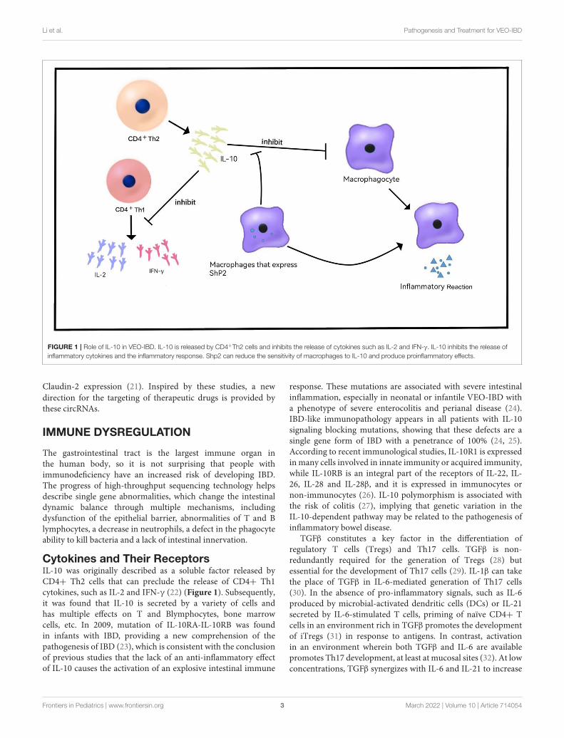
FIGURE 1 | Role of IL-10 in VEO-IBD. IL-10 is released by CD4+Th2 cells and inhibits the release of cytokines such as IL-2 and IFN-γ. IL-10 inhibits the release of
inflammatory cytokines and the inflammatory response. Shp2 can reduce the sensitivity of macrophages to IL-10 and produce proinflammatory effects.
Claudin-2 expression (21). Inspired by these studies, a new
direction for the targeting of therapeutic drugs is provided bythese circRNAs.
IMMUNE DYSREGULATION
The gastrointestinal tract is the largest immune organ inthe human body, so it is not surprising that people withimmunodeficiency have an increased risk of developing IBD.The progress of high-throughput sequencing technology helpsdescribe single gene abnormalities, which change the intestinaldynamic balance through multiple mechanisms, includingdysfunction of the epithelial barrier, abnormalities of T and Blymphocytes, a decrease in neutrophils, a defect in the phagocyteability to kill bacteria and a lack of intestinal innervation.
Cytokines and Their ReceptorsIL-10 was originally described as a soluble factor released byCD4+ Th2 cells that can preclude the release of CD4+ Th1cytokines, such as IL-2 and IFN-γ (22) (Figure 1). Subsequently,it was found that IL-10 is secreted by a variety of cells andhas multiple effects on T and Blymphocytes, bone marrowcells, etc. In 2009, mutation of IL-10RA-IL-10RB was foundin infants with IBD, providing a new comprehension of thepathogenesis of IBD (23), which is consistent with the conclusionof previous studies that the lack of an anti-inflammatory effectof IL-10 causes the activation of an explosive intestinal immune
response. These mutations are associated with severe intestinalinflammation, especially in neonatal or infantile VEO-IBD witha phenotype of severe enterocolitis and perianal disease (24).IBD-like immunopathology appears in all patients with IL-10signaling blocking mutations, showing that these defects are asingle gene form of IBD with a penetrance of 100% (24, 25).According to recent immunological studies, IL-10R1 is expressedinmany cells involved in innate immunity or acquired immunity,while IL-10RB is an integral part of the receptors of IL-22, IL-26, IL-28 and IL-28β, and it is expressed in immunocytes ornon-immunocytes (26). IL-10 polymorphism is associated withthe risk of colitis (27), implying that genetic variation in theIL-10-dependent pathway may be related to the pathogenesis ofinflammatory bowel disease.
TGFβ constitutes a key factor in the differentiation ofregulatory T cells (Tregs) and Th17 cells. TGFβ is non-redundantly required for the generation of Tregs (28) butessential for the development of Th17 cells (29). IL-1β can takethe place of TGFβ in IL-6-mediated generation of Th17 cells(30). In the absence of pro-inflammatory signals, such as IL-6produced by microbial-activated dendritic cells (DCs) or IL-21secreted by IL-6-stimulated T cells, priming of naïve CD4+ Tcells in an environment rich in TGFβ promotes the developmentof iTregs (31) in response to antigens. In contrast, activationin an environment wherein both TGFβ and IL-6 are availablepromotes Th17 development, at least at mucosal sites (32). At lowconcentrations, TGFβ synergizes with IL-6 and IL-21 to increase
Frontiers in Pediatrics | www.frontiersin.org 3 March 2022 | Volume 10 | Article 714054
Li et al. Pathogenesis and Treatment for VEO-IBD
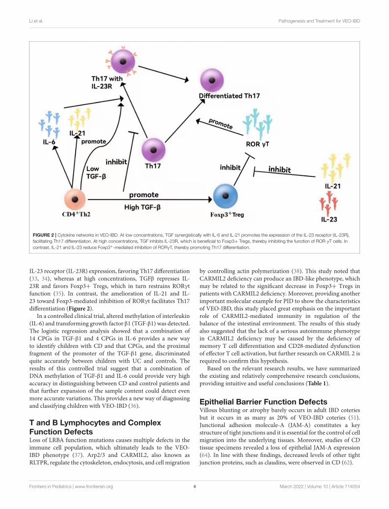
FIGURE 2 | Cytokine networks in VEO-IBD. At low concentrations, TGF synergistically with IL-6 and IL-21 promotes the expression of the IL-23 receptor (IL-23R),
facilitating Th17 differentiation. At high concentrations, TGF inhibits IL-23R, which is beneficial to Foxp3+ Tregs, thereby inhibiting the function of ROR γT cells. In
contrast, IL-21 and IL-23 reduce Foxp3+-mediated inhibition of RORγT, thereby promoting Th17 differentiation.
IL-23 receptor (IL-23R) expression, favoring Th17 differentiation(33, 34), whereas at high concentrations, TGFβ represses IL-23R and favors Foxp3+ Tregs, which in turn restrains RORγtfunction (35). In contrast, the amelioration of IL-21 and IL-23 toward Foxp3-mediated inhibition of RORγt facilitates Th17differentiation (Figure 2).
In a controlled clinical trial, altered methylation of interleukin(IL-6) and transforming growth factor β1 (TGF-β1) was detected.The logistic regression analysis showed that a combination of14 CPGs in TGF-β1 and 4 CPGs in IL-6 provides a new wayto identify children with CD and that CPGs, and the proximalfragment of the promoter of the TGF-β1 gene, discriminatedquite accurately between children with UC and controls. Theresults of this controlled trial suggest that a combination ofDNA methylation of TGF-β1 and IL-6 could provide very highaccuracy in distinguishing between CD and control patients andthat further expansion of the sample content could detect evenmore accurate variations. This provides a new way of diagnosingand classifying children with VEO-IBD (36).
T and B Lymphocytes and ComplexFunction DefectsLoss of LRBA function mutations causes multiple defects in theimmune cell population, which ultimately leads to the VEO-IBD phenotype (37). Arp2/3 and CARMIL2, also known asRLTPR, regulate the cytoskeleton, endocytosis, and cell migration
by controlling actin polymerization (38). This study noted thatCARMIL2 deficiency can produce an IBD-like phenotype, whichmay be related to the significant decrease in Foxp3+ Tregs inpatients with CARMIL2 deficiency. Moreover, providing anotherimportant molecular example for PID to show the characteristicsof VEO-IBD, this study placed great emphasis on the importantrole of CARMIL2-mediated immunity in regulation of thebalance of the intestinal environment. The results of this studyalso suggested that the lack of a serious autoimmune phenotypein CARMIL2 deficiency may be caused by the deficiency ofmemory T cell differentiation and CD28-mediated dysfunctionof effector T cell activation, but further research on CARMIL 2 isrequired to confirm this hypothesis.
Based on the relevant research results, we have summarizedthe existing and relatively comprehensive research conclusions,providing intuitive and useful conclusions (Table 1).
Epithelial Barrier Function DefectsVillous blunting or atrophy barely occurs in adult IBD coteriesbut it occurs in as many as 20% of VEO-IBD coteries (51).Junctional adhesion molecule-A (JAM-A) constitutes a keystructure of tight junctions and it is essential for the control of cellmigration into the underlying tissues. Moreover, studies of CDtissue specimens revealed a loss of epithelial JAM-A expression(64). In line with these findings, decreased levels of other tightjunction proteins, such as claudins, were observed in CD (62).
Frontiers in Pediatrics | www.frontiersin.org 4 March 2022 | Volume 10 | Article 714054

Li et al. Pathogenesis and Treatment for VEO-IBD
TABLE 1 | List of gene mutations associated with monogenic VEO-IBD and
IBD-like colitis.
Genes Clinical
syndromes
Studies
Immune dysregulation
NOD2 Susceptibility gene Travassos et al. (6)
ATG16L1 Susceptibility gene Homer et al. (39)
TRIM22 NOD2 signaling
defects
Li et al. (40)
IL-10 Neonatal or
infantile VEO-IBD
Kotlarz et al. (24)
IL-10RA Neonatal or
infantile VEO-IBD
Glocker et al. (23)
IL-10RB Neonatal or
infantile VEO-IBD
Glocker et al. (23)
FOXP3 IPEX Torgerson and Ochs
(41)
Hyperinflammatory and autoimmune disorders
XIAP X-linked
lymphoproliferative
syndrome 2
Latour and Aguilar (42)
SLC11A1 Susceptibility gene Sechi et al. (43)
CTLA4 Autoimmune
lymphoproliferative
syndrome
Kuehn et al. (44)
PLCG2 Autoinflammation
and PLCγ2-
associated
antibody
deficiency, and
immune
dysregulation
(APLAID)
Zhou et al. (45)
STAT3 Multisystem
autoimmune
disease
Duerr et al. (46)
IL23R Susceptibility gene Duerr et al. (46)
CCR6 Susceptibility gene Duerr et al. (46)
TNFSF15 Susceptibility gene Duerr et al. (46)
Shp2 Susceptibility gene Xiao et al. (47)
miR-320a CD and UC Fasseu et al. (11)
let-7c CD and UC Banerjee et al. (48)
T cell, B cell, and complex function defects
LRBA CVID 8 Alangari et al. (37)
ZAP70 ZAP70 deficiency Chan et al. (49)
WAS Wiscott-Aldrich
syndrome
Catucci et al. (50)
CARMIL2 VEO-IBD, SCID Magg et al. (38)
RAG2 Omenn syndrome Kelsen et al. (51)
RAG1 Omenn syndrome Villa et al. (52)
DCLRE1C/ARTEMIS95 Omenn syndrome Villa et al. (52)
MALT1 SCID Punwani et al. (53)
DOCK8 Hyper
immunoglobulin E
syndrome
Sanal et al. (54)
CD40LG Hyper
immunoglobulin M
syndrome
Levy et al. (55)
(Continued)
TABLE 1 | Continued
Genes Clinical
syndromes
Studies
AICDA Hyper
immunoglobulin M
syndrome
Quartier et al. (56)
Epithelial barrier function defects
COL7A1 Dystrophic
epidermolysis
bullosa
Zimmer et al. (57)
ADAM17 ADAM17
deficiency
Chalaris et al. (58)
IKBKG X-linked
ectodermal
immunodeficiency
(NEMO)
Karamchandani-Patel
et al. (59)
FERMT1 Kindler syndrome Sadler et al. (60)
TTC7A TTC7A deficiency Avitzur et al. (61)
GUCY2 Familial diarrhea Uhlig (4)
CDKN2B-
AS11
UC Rankin et al. (21)
CircRNA_103765 IBD Ye et al. (15)
Claudin2 IBD Zeissig et al. (62)
HuR Colitis Pott et al. (18)
miR-126 IBD Chen et al. (12)
Others
FUT2 Susceptibility gene McGovern et al. (63)
Defects in intestinal epithelial barrier function can be involvedin VEO-IBD processes, including loss-of-function mutationsin ADAM17 resulting in ADAM17 deficiency (65), IKBKG(encoding NEMO) mutation producing X-linked ectodermaldysplasia and immunodeficiency (59), and COL7A1 mutationcausing dystrophic epidermolysis bullos (57). FERMT1 mutationresults in Kindler syndrome (66), and TTC7A (61) or gain-of-function mutations in GUCY2 cause familial diarrhea (4, 67).
INTESTINAL ENVIRONMENTAL FACTORS
It has been proven that the interaction between the gutmicrobiota, metabolites, and the gut immune systemis essential for maintaining a healthy gut (68). Specificalterations in the composition and function of the gutmicrobiota may be used as microbial biomarkers for thediagnosis of IBD, disease activity, response to therapy, andprediction of outcomes.
For newly diagnosed children with IBD, an increase in thenumber of Proteobacteria in the intestinal microbiota and adecrease in the number of Faecalibacterium prausnitzii appearto be associated with a complex disease phenotype and asubsequent need for biological therapy or surgery (69). Themost common finding was an increase in adherent-aggressiveEscherichia coli in the gut of IBD patients. This infectiousagent can adhere to and cross the intestinal mucus barrierand invade the upper intestinal cortex. Second, compared with
Frontiers in Pediatrics | www.frontiersin.org 5 March 2022 | Volume 10 | Article 714054
Li et al. Pathogenesis and Treatment for VEO-IBD
non-gastroenteritis patients, Salmonella fecal culture-positivegastroenteritis patients were significantly associated with anincreased risk of new UC and CD. The most recent nationalcase–control study from Sweden also demonstrated a positiveassociation between a Salmonella diagnosis and the likelihoodof IBD, and this study found that Clostridium difficile wasassociated with higher rates of UC and CD (70). Additionally,the latest and most comprehensive meta-analysis found a 57%lower risk of Helicobacter pylori exposure and inflammatorybowel disease, including CD and UC (71), and a meta-analysisthat included only Asian studies reported consistent results(72). This protective association may be mediated, at least inpart, by the specific components of H. pylori strains throughimmune regulation.
In addition to bacteria, some studies have found thatVEO-IBD is also linked to enteroviruses. In a study ofenterovirus, norovirus G-I, norovirus G-II, rotavirus,astrovirus, and sand wave virus RNA in fecal samplesfrom 33 children with IBD and 17 children withoutIBD, viral RNA was detected only in children withoutIBD (3% vs. 0%) (73). Given changes in the intestinalmicrobiota in IBD patients, current clinical guidelinesrecommend testing for C. difficile in all IBD patients withexacerbating or newly emerging diarrhea and testing forcytomegalovirus in severely active IBD patients, especiallywhen steroids and medications are used together to treatrefractory disease.
Patients with CD exhibit significant differences in theirgut metabolome, including lower concentrations of short-chainfatty acids (74, 75), higher concentrations of amino acids (75),and a dysregulated bile acid composition, including higherconcentrations of conjugated bile acids and lower concentrationsof secondary bile acids (76). In a cohort study, a strongassociation was found between an increase in the number ofantibiotic prescriptions in the first year of life and the onset ofIBD in childhood. In this study, the use of antibiotics, particularlyin infancy, could lead to changes in the gut microbiota,suggesting they have a vital role in development of the immunesystem (77).
THERAPEUTIC METHODS
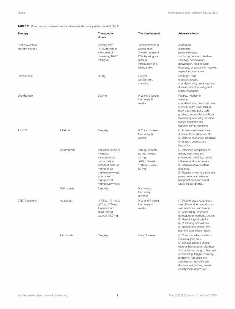
MedicationsThe standard therapeutic choices for VEO-IBD include 5-ASA,steroids, immunomodulators (6MP, azathioprine, methotrexate),and anti-TNF antibodies. At present, there are few studiesthat have been carried out in the pediatric patient population,and the relevant clinical data are insufficient, most of whichare similar to polygenic IBD. It is more important tounderstand the data on drug-related dosage and adversereactions (Table 2). Among the available medications, infliximab,vedolizumab, and ustekinumab are used in the treatmentof VEO-IBD.
Monoclonal antibodies against tumor necrosis factor α(TNF-α), such as infliximab (IFX) or adalimumab (ADA), are safe andeffective in inducing and maintaining remission in moderate-to-severe pediatric Crohn’s disease (CD), and ulcerative colitis
(UC) patients (78, 79). Based on the experience of a tertiarycenter in Japan, IFX treatment seems to be more effective fornon-ulcerative colitis type (NUCT) and non-ulcerative colitistype without perianal disease (NUC-NPD) patients, and it seemsthat their height and weight are improved after treatment (80).However, the use of TNF inhibitors is limited, even amongTNF responders, because of systemic side effects, includingimmunosuppression and cardiotoxicity. In addition, up to one-third of patients do not respond to TNF-α antagonist therapy,and ∼20% of primary responders may experience responseloss each year (81, 82). Therefore, in recent years, the FDAhas approved some bio-similars ofTNF-α antagonists for thetreatment ofVEO-IBD. The biosimilar agent infliximab is animmunoglobulin G(IgG) anti-TNF-α monoclonal antibody thatbinds to soluble and transmembrane forms of TNF-α, which canfurther impede its interaction with the TNF receptors TNFR1(P55) and TNFR2 (P75) on the surface of target cells. Therefore,it can be used to treat pediatric IBD (83, 84). Nonetheless, thebiosimilar adalimumab has not yet been approved for pediatricIBD (85).
Vedolizumab (VDZ) is a humanized monoclonal antibodythat specifically identifies lymphocyte integrin 4β7 receptors andprevents them from migrating from the blood vessels into theintestinal mucosa, thereby reducing the flow of white blood cellsinto inflammatory tissues. As an intestinal selective anti-integrindrug, it has been reported to have a low risk of infection (86, 87).In the first study of VDZ in children with VEO-IBD, this anti-integrin agent was shown to be safe and effective in the studypopulation. Similarly, in the Porto group study, 16 pediatricpatients found VDZ was safe and well-tolerated−1 developedupper respiratory tract infection (6.3%), and two developed jointpain (12.5%) (88). Conrad et al. also evaluated VDZ for severeIBD in children with similar results (89). Another multicenterstudy published in 2016 demonstrated the efficacy and safetyof VDZ in the pediatric population (90). VDZ is not approvedfor pediatric patients but has demonstrated clinical efficacy forpediatric IBD. Its remission rates of UC and CD are 76 and 42%,respectively (89, 90).
Ustekinumab, a therapeutic human IgG1 monoclonalantibody (mAb) targeting the interleukin (IL)-12/IL-23 sharedp40 subunit, is approved in adolescents (12 years of age andolder) for the treatment of moderate and severe psoriasis, as wellas for the treatment of adult celiac disease and UC (91)whileceliac disease currently has gluten-free diet as the only therapy.In a multicenter prospective cohort of children, the effectivenessof ustekinumab in treating refractory UC was demonstrated (92),which is similar to the results of a retrospective study of pediatricIBD, suggesting that ustekinumab is effective and safe in childrenwith IBD (93). Some case reports suggest that 50% of childrenwith IBD have a clinical response to ustekinumab (94, 95). In acohort of pediatric patients with CD, patients using ustekinumabhad significant improvements in their abbreviated pediatric CDactivity index (aPCDAI) scores, clinical remission rates, albumin,and hematocrit, and 89.5% of patients had no significant adverseevents (96). The use of off-label drugs is increasing in childrenwith IBD and generally they are being reported as safe andeffective (93).
Frontiers in Pediatrics | www.frontiersin.org 6 March 2022 | Volume 10 | Article 714054
Li et al. Pathogenesis and Treatment for VEO-IBD
TABLE 2 | Dose, interval, adverse reactions to medications for pediatric and VEO-IBD.
Therapy Therapeutic
doses
The time interval Adverse effects
Exclusive enteral
nutrition therapy
Starting from
10–20 ml/(kg*d),
the speed of
increasing 10–20
ml/(kg*d)
Total treatment: 6
weeks, then
2-week course of
EEN tapering and
gradual
introduction of a
habitual diet
Anatomical
extension,
perianal disease,
stricturing behavior, diarrhea,
vomiting, constipation,
dehydration, feeding tube
blockage, dyspnea and hypoxia,
aspiration pneumonia.
Ustekinumab 90mg Every 8
weeks/every
4 weeks
Arthralgia, skin
eruption, cough,
spondylarthritis, cardiovascular
disease, infection, malignant
tumor, headache.
Vedolizumab 300mg 0, 2 and 6 weeks,
then every 8
weeks
Nausea, headache,
malaise,
spondylarthritis, bronchitis, liver
function injury, fever, fatigue,
back pain, limb pain, rash,
pruritus, progressive multifocal
leukoencephalopathy, infusion
related reactions and
hypersensitivity reactions.
Anti-TNF Infliximab 5 mg/kg 0, 2 and 6 weeks,
then every 8
weeks
(1) Acute infusion reactions:
urticaria, fever, dyspnea, etc.
(2) Delayed response: arthralgia,
fever, rash, edema, and
headache.
Adalimumab Induction period of
4 weeks,
subcutaneous
immunization
Standard dose: 20
mg/kg or 40
mg/kg every week.
Low dose: 10
mg/kg or 20
mg/kg every week.
<40 kg: 0 week,
80mg. 2 week,
40 mg.
>40 kg:0 week,
160mg. 2 week,
80 mg.
(3) Infectious complications:
urinary tract infection,
pneumonia, cellulitis, mastitis,
influenza and tuberculosis.
(4) Cardiovascular system
response.
(5) Reactions: multiple sclerosis,
paresthesia, and seizures.
Malignant neoplasms and
lupus-like syndrome.
Golimumab 2 mg/kg 0, 4 weeks,
then every
8 weeks
CTLA4 agonists Abatacept < 75 kg, 10 mg/kg
≥75 kg, 750 mg
the maximum
dose cannot
exceed 1000mg
0, 2, and 4 weeks,
then every 4
weeks
(1) Discoid lupus, cutaneous
vasculitis, erythema nodosum,
skin infections, skin tumors.
(2) Conditional infections:
pathogenic pneumonia, sepsis.
(3) Hematological toxicity.
(4) Pulmonary sarcoidosis.
(5) Tuberculous uveitis, eye
pigment layer inflammation.
Ipilimumab 3 mg/kg Every 3 weeks (1) Common adverse effects:
insomnia, joint pain.
(2) Serious adverse effects:
oliguria, hematuresis, diarrhea,
stomachache, cough, chest pain
or wheezing, fatigue, memory
problems, hallucinations,
seizures, or neck stiffness,
diureses weight loss, sweat,
constipation, depression.
Frontiers in Pediatrics | www.frontiersin.org 7 March 2022 | Volume 10 | Article 714054
Li et al. Pathogenesis and Treatment for VEO-IBD
From limited evidence, dual biotherapy may be a safe optionfor patients with refractory IBD who have failed multiplebiotherapies and for managing the extra-intestinal presentationof IBD (97). A cohort of refractory pediatric IBD reported theeffectiveness and safety of dual biologics or a combination ofbiologics and JAK inhibitors (98). In a case series and review ofthe literature, eight children received a combination of infliximaband vedolizumab, and five children received a combination ofinfliximab and ustekinumab, which shows combining biologicalagents to be safe and beneficial in selected patients (99). However,larger studies are required to confirm the preliminary safety datathat were observed.
Ruxolitinib, a selective JAK1/2 inhibitor, was found in asingle-center retrospective study of patients with refractory VEO-IBD with AIP to be primarily used for dual therapy whencomplete remission was not achieved with primary therapy. Allpatients in this study showed clinical improvement and did notrequire complete parenteral nutrition or steroids. Other potentialbenefits of ruxolitinib included a lack of immunogenicity, a rapidonset of action, and a short half-life. In addition, ruxolitinibcan be administered entirely with sufficient enteral absorptionto achieve a clinical response in a cohort with severe intestinaldisease. However, this study still has limitations and couldnot determine whether ruxolitinib will be effective or safe ingeneral use (100).
Exclusive Enteral Nutrition TherapyExclusive enteral nutrition therapy (EEN) is the preferredtreatment for European VEO-IBD patients. In a propensityscore matching cohort analysis of children with Crohn’s diseaseinduced by total enteral nutritionor glucocorticoids (CSs) (101),EEN, and CSs were found to be equally effective in inducingremission. Through a central retrospective analysis, EEN wasfound to be more effective than CSs in improving nutritionalstatus and growth recovery, with relatively few side effects. Moreimportantly, EEN can achieve mucosal healing (MH), which isthe target of CD treatment. When applied in an early stage,MH reduces the incidence of hospitalization, surgical resection,and fistula formation, providing a new pattern for the treatmentof very early inflammatory bowel disease. Some data show thatthe intestinal flora, amino acids, and fecal metabolites of CDpatients have significant changes before and after EEN treatment(102), providing a biochemical detection method for judging theefficacy of EEN.
Hematopoietic Stem Cell TransplantationDue to events such as severe or opportunistic infectionsand malignancies associated with biologic methods, stemcell transplantation has entered clinical trials as a morepermanent treatment for IBD. Allogeneic hematopoietic stem celltransplantation (allo-HSCT) is an established therapeutic optionfor VEO-IBD. In a retrospective investigation of autologoushematopoietic stem cell transplantation for CD, MSC therapymay be an alternative to endovenous and fistula treatment (103).In terms of IPEX and IL-10 signaling deficits, HSCT has beenshown to improve colitis and gastrointestinal fistula (104, 105).VEO-IBD patients with IL-10R deficiency can also be cured by
allo-HSCT (23, 106). However, 11 patients with transplantedIL-10 and IL-10R deficiency showed a very high frequencyof primary graft rejection (3/11), and these data suggest thatpatients with either IL-10 or IL-10R deficiency need to have theirtransplant regimen adjusted to reduce the risk of rejection (105,106). Therefore, the use of post-transplant cyclophosphamideand bone marrow transplantation with T cell-identical cells havebeen considered potential therapies for patients with IL-10Rdeficiency (107). Similarly, studies have shown that allogeneichematopoietic stem cell transplantation can successfully treatXIAP-deficient idiopathic colitis with specific conditioningregimens (108). For patients with a refractory IBD phenotypeand an increased risk of mortality due to XIAP deficiency, HSCTshould be considered as early as possible, as it can address theirrisk of intestinal inflammation and the development of life-threatening hemophilic lymphocytosis (109). However, HSCTis not effective for all cases of VEO-IBD. IBD lacking NEMOor TTC7A cannot be improved after HSCT and it may evenworsen (110, 111). Therefore, we believe that the applicationof therapeutic HSCT in certain conditions is promising, but itshould always be personalized.
Bone marrow MSCs can promote wound healing and tissueregeneration by secreting TGF-β and fibroblast growth factor.This property offers a new approach to the treatment of CDwith fistulae.
SurgeryDespite recent advances in treatments, surgery still plays animportant role in the management of VEO-IBD. Althoughsurgery cannot cure VEO-IBD, in some cases, it can help resolveacute complications and maintain remission, allowing disease-free intervals, and nutritional recovery (112), and has a hugeimpact on physical and mental development (113). The mainindications for surgery in CD are unresponsive and refractoryto maximum medical treatment, fistula, perforation, stricturingdisease, and severe perianal disease. Meanwhile, acute indicationsfor UC surgery also include toxic megacolon, which is rarein children. A systematic review noted that surgical rates forCD ranged from 10 to 72%, while colectomy rates for UCranged from 0 to 50% (113). Minimally invasive surgery hasalso been used for the radical treatment of CD and UC since2002. In recent years, robotic surgery, a single-hole approach andminimally invasive treatment of perianal fistula CD have beenadopted (114).
Other TreatmentsTwo recent randomized controlled trials (RCTs) providedadditional insight by suggesting that rebuilding the intestinalmicrobiota composition through fecal microflora transplantation(FMT) can improve UC activity in this patient subgroup (115,116). Although a few nonrandomized control studies have beenperformed in older children (youngest child 7 years old) (117),the efficacy of FMT for VEO-IBD is unclear.
The use of immunosuppressive drugs in the treatmentregimen increases the risk of infectious diseases and infection-related complications in children with IBD. Therefore,
Frontiers in Pediatrics | www.frontiersin.org 8 March 2022 | Volume 10 | Article 714054
Li et al. Pathogenesis and Treatment for VEO-IBD
vaccination to prevent related infections is an importantaspect of long-term care of this disease.
CONCLUSION
Current studies show that the pathogenesis of VEO-IBD includesgenetic factors, immune molecular factors, and changes inthe intestinal environment. With research progress on thesusceptibility genes of IBD, the localization of the susceptibilitygenes of IBD helps identify and distinguish the diseasephenotype, track the clinical progress, and ultimately developnew targeted therapies. However, due to the lack of clinicalfollow-up data in VEO-IBD children, there are still greatchallenges in terms of the drug efficacy and the research anddevelopment (R&D) of new drugs. It is not enough to drawlessons from the adult treatment experience alone. In thefuture, rapid diagnosis and management of children should becarried out, diagnosis, and treatment criteria based on geneticabnormalities should be established, and a clinical databaseshould be expanded. We need to establish control groups andexplore the influence of environmental factors on the incidence,treatment and prognosis of VEO-IBD. Finally, a precisionmedicine model needs to be achieved, namely, individualizedtreatment for VEO-IBD children.
AUTHOR CONTRIBUTIONS
Q-QL and H-HZ drafted the article and approved the finalmanuscript as submitted.
FUNDING
This study was supported by the High-Level PersonnelProgram of Guangdong Provincial People’s Hospital(2021DFJH0008/KY012021458), Starting Program forNational Natural Science Foundation of China at GuangdongProvincial People’s Hospital (8207034250), National NaturalScience Foundation of China (NSFC, No. 81300370), ChinaPostdoctoral Science Foundation (CPSF, No. 2018T110855of Special Support Program and No. 2017M622650 ofGeneral Support Program), Natural Science Foundationof Guangdong (NSFG, No. 2018A030313161), SouthernMedical University (12440000771868596D), S-XD is asupervisor at the Second Clinical School of SouthernMedical University and an adjunct associate professor at
Southern Medical University, and supervised Q-QL and
H-HZ in New Insights and Advances in Pathogenesis
and Treatment of Very Early Onset InflammatoryBowel Disease.
REFERENCES
1. Miller TL, Lee D, Giefer M, Wahbeh G, Suskind DL. Nutritional therapy in
very early-onset inflammatory bowel disease: a case report. Digest Dis Sci.
(2017) 62:2196–200. doi: 10.1007/s10620-017-4616-9
2. Kelsen JR, Sullivan KE, Rabizadeh S, Singh N, Snapper S, Elkadri A, et
al. North american society for pediatric gastroenterology, hepatology, and
nutrition position paper on the evaluation andmanagement for patients with
very early-onset inflammatory bowel disease. J Pediatr Gastroenterol Nutr.
(2020) 70:389–403. doi: 10.1097/MPG.0000000000002567
3. Zhang YZ, Li YY. Inflammatory bowel disease: pathogenesis. World J
Gastroenterol. (2014) 20:91–9. doi: 10.3748/wjg.v20.i1.91
4. Uhlig HH. Monogenic diseases associated with intestinal inflammation:
implications for the understanding of inflammatory bowel disease. Gut.
(2013) 62:1795–805. doi: 10.1136/gutjnl-2012-303956
5. Parkhouse R, Monie TP. Dysfunctional Crohn’s disease-associated
NOD2 polymorphisms cannot be reliably predicted on the basis
of RIPK2 binding or membrane association. Front Immunol. (2015)
6:521. doi: 10.3389/fimmu.2015.00521
6. Travassos LH, Carneiro LA, Ramjeet M, Hussey S, Kim YG, Magalhaes JG,
et al. Nod1 and Nod2 direct autophagy by recruiting ATG16L1 to the plasma
membrane at the site of bacterial entry. Nat Immunol. (2010) 11:55–62.
doi: 10.1038/ni.1823
7. Cooney R, Baker J, Brain O, Danis B, Pichulik T, Allan P, et al.
NOD2 stimulation induces autophagy in dendritic cells influencing
bacterial handling and antigen presentation. Nat Med. (2010) 16:90–7.
doi: 10.1038/nm.2069
8. Jensen MD, Andersen RF, Christensen H, Nathan T, Kjeldsen
J, Madsen JS. Circulating microRNAs as biomarkers of adult
Crohn’s disease. Eur J Gastroenterol Hepatol. (2015) 27:1038–44.
doi: 10.1097/MEG.0000000000000430
9. Batra SK, Heier CR, Diaz-Calderon L, Tully CB, Fiorillo AA, van den
Anker J, et al. Serum miRNAs are pharmacodynamic biomarkers associated
with therapeutic response in pediatric inflammatory bowel disease. Inflamm
Bowel Dis. (2020) 26:1597–606. doi: 10.1093/ibd/izaa209
10. Paterson MR, Kriegel AJ. MiR-146a/b: a family with shared
seeds and different roots. Physiol Genomics. (2017) 49:243–52.
doi: 10.1152/physiolgenomics.00133.2016
11. Fasseu M, Treton X, Guichard C, Pedruzzi E, Cazals-Hatem D, Richard C,
et al. Identification of restricted subsets of mature microRNA abnormally
expressed in inactive colonic mucosa of patients with inflammatory bowel
disease. PLoS ONE. (2010) 5: e13160. doi: 10.1371/journal.pone.0013160
12. Chen T, Xue H, Lin R, Huang Z. MiR-126 impairs the intestinal
barrier function via inhibiting S1PR2 mediated activation of PI3K/AKT
signaling pathway. Biochem Biophys Res Commun. (2017) 494:427–32.
doi: 10.1016/j.bbrc.2017.03.043
13. Li X, Yang L, Chen LL. The biogenesis, functions, and challenges of circular
RNAs.Mol Cell. (2018) 71:428–42. doi: 10.1016/j.molcel.2018.06.034
14. Yin J, Hu T, Xu L, Li P, Li M, Ye Y, et al. Circular RNA expression profile in
peripheral blood mono-nuclear cells from Crohn disease patients. Medicine
(Baltimore). (2019) 98:e16072. doi: 10.1097/MD.0000000000016072
15. Ye Y, Zhang L, Hu T, Yin J, Xu L, Pang Z, et al. CircRNA_103765 acts as a
pro-inflammatory factor via sponging miR-30 family in Crohn’s disease. Sci
Rep. (2021) 11:565. doi: 10.1038/s41598-020-80663-w
16. Qiao YQ, Cai CW, Shen J, Zheng Q, Ran ZH. Circular RNA expression
alterations in colon tissues of Crohn’s disease patients. Mol Med Rep.
(2019) 19:4500–6. doi: 10.3892/mmr.2019.10070
17. Giammanco A, Blanc V, Montenegro G, Klos C, Xie Y, Kennedy S, et al.
Intestinal epithelial HuR modulates distinct pathways of proliferation and
apoptosis and attenuates small intestinal and colonic tumor development.
Cancer Res. (2014) 74:5322–35. doi: 10.1158/0008-5472.CAN-14-0726
18. Pott J, Kabat AM, Maloy KJ. Intestinal epithelial cell autophagy is required
to protect against TNF-induced apoptosis during chronic colitis in mice. Cell
Host Microbe. (2018) 23:191–202. doi: 10.1016/j.chom.2017.12.017
19. Holdt LM, Stahringer A, Sass K, Pichler G, Kulak NA, Wilfert
W, et al. Circular non-coding RNA ANRIL modulates ribosomal
RNA maturation and atherosclerosis in humans. Nat Commun. (2016)
7:12429. doi: 10.1038/ncomms12429
20. Holdt LM, Hoffmann S, Sass K, Langenberger D, Scholz M, Krohn K, et al.
Alu elements in ANRIL non-coding RNA at chromosome 9p21 modulate
Frontiers in Pediatrics | www.frontiersin.org 9 March 2022 | Volume 10 | Article 714054
Li et al. Pathogenesis and Treatment for VEO-IBD
atherogenic cell functions through trans-regulation of gene networks. PLoS
Genet. (2013) 9:e1003588. doi: 10.1371/journal.pgen.1003588
21. Rankin CR, Lokhandwala ZA, Huang R, Pekow J, Pothoulakis C, Padua
D. Linear and circular CDKN2B-AS1 expression is associated with
inflammatory bowel disease and participates in intestinal barrier formation.
Life Sci. (2019) 231:116571. doi: 10.1016/j.lfs.2019.116571
22. Fiorentino DF, Bond MW, Mosmann TR. Two types of mouse T helper
cell. IV. Th2 clones secrete a factor that inhibits cytokine production
by Th1 clones. J Exp Med. (1989) 170: 2081–95. doi: 10.1084/jem.170.
6.2081
23. Glocker EO, Kotlarz D, Boztug K, Gertz EM, Schaffer AA, Noyan F, et
al. Inflammatory bowel disease and mutations affecting the interleukin-
10 receptor. N Engl J Med. (2009) 361:2033–45. doi: 10.1056/NEJMoa
0907206
24. Glocker EO, Frede N, Perro M, Sebire N, Elawad M, Shah
N, et al. Infant colitis–it’s in the genes. Lancet. (2010)
376:1272. doi: 10.1016/S0140-6736(10)61008-2
25. Shouval DS, Ouahed J, Biswas A, Goettel JA, Horwitz BH, Klein C, et al.
Interleukin 10 receptor signaling: master regulator of intestinal mucosal
homeostasis in mice and humans. Adv Immunol. (2014) 122:177–210.
doi: 10.1016/B978-0-12-800267-4.00005-5
26. Wolk K, Sabat R. Interleukin-22: A novel T- and NK-cell derived cytokine
that regulates the biology of tissue cells. Cytokine Growth Factor Rev. (2006)
17:367–80. doi: 10.1016/j.cytogfr.2006.09.001
27. Franke A, Balschun T, Karlsen TH, Sventoraityte J, Nikolaus S, Mayr
G, et al. Sequence variants in IL10, ARPC2 and multiple other loci
contribute to ulcerative colitis susceptibility. Nat Genet. (2008) 40:1319–23.
doi: 10.1038/ng.221
28. Nakamura K, Kitani A, Strober W. Cell contact-dependent
immunosuppression by CD4(+)CD25(+) regulatory T cells is mediated
by cell surface-bound transforming growth factor beta. J Exp Med.
(2001) 194:629–44. doi: 10.1084/jem.194.5.629
29. Das J, Ren G, Zhang L, Roberts AI, Zhao X, Bothwell AL, et al. Transforming
growth factor beta is dispensable for the molecular orchestration of Th17 cell
differentiation. J Exp Med. (2009) 206:2407–16. doi: 10.1084/jem.20082286
30. Ghoreschi K, Laurence A, Yang XP, Tato CM,McGeachyMJ, Konkel JE, et al.
Generation of pathogenic T(H)17 cells in the absence of TGF-beta signalling.
Nature. (2010) 467:967–71. doi: 10.1038/nature09447
31. Bettelli E, Carrier Y, Gao W, Korn T, Strom TB, Oukka M, et al. Reciprocal
developmental pathways for the generation of pathogenic effector TH17 and
regulatory T cells. Nature. (2006) 441:235–8. doi: 10.1038/nature04753
32. Hu W, Troutman TD, Edukulla R, Pasare C. Priming microenvironments
dictate cytokine requirements for T helper 17 cell lineage commitment.
Immunity. (2011) 35:1010–22. doi: 10.1016/j.immuni.2011.10.013
33. Korn T, Bettelli E, GaoW, Awasthi A, Jager A, Strom TB, et al. IL-21 initiates
an alternative pathway to induce proinflammatory T(H)17 cells. Nature.
(2007) 448:484–7. doi: 10.1038/nature05970
34. Nurieva R, Yang XO, Martinez G, Zhang Y, Panopoulos AD, Ma L, et al.
Essential autocrine regulation by IL-21 in the generation of inflammatory
T cells. Nature. (2007) 448:480–3. doi: 10.1038/nature05969
35. Zhou L, Lopes JE, Chong MM, Ivanov II, Min R, Victora GD, et al. TGF-
beta-induced Foxp3 inhibits T(H)17 cell differentiation by antagonizing
RORgammat function.Nature. (2008) 453:236–40. doi: 10.1038/nature06878
36. Mitsuyama K, Sata M, Rose-John S. Interleukin-6 trans-signaling in
inflammatory bowel disease. Cytokine Growth Factor Rev. (2006) 17:451–61.
doi: 10.1016/j.cytogfr.2006.09.003
37. Alangari A, Alsultan A, Adly N, Massaad MJ, Kiani IS, Aljebreen A, et al.
LPS-responsive beige-like anchor (LRBA) gene mutation in a family with
inflammatory bowel disease and combined immunodeficiency. J Allergy Clin
Immunol. (2012) 130:481–8. doi: 10.1016/j.jaci.2012.05.043
38. Magg T, Shcherbina A, Arslan D, Desai MM, Wall S, Mitsialis V, et al.
CARMIL2 deficiency presenting as very early onset inflammatory bowel
disease. Inflamm Bowel Dis. (2019) 25:1788–95. doi: 10.1093/ibd/izz103
39. Homer CR, Richmond AL, Rebert NA, Achkar JP, McDonald C.
(2010). ATG16L1 and NOD2 interact in an autophagy-dependent
antibacterial pathway implicated in Crohn’s disease pathogenesis.
Gastroenterol. (1641) 139:1630–41. doi: 10.1053/j.gastro.2010.
07.006
40. Li Q, Lee CH, Peters LA, Mastropaolo LA, Thoeni C, Elkadri A, et al.
Variants in TRIM22 that affect NOD2 signaling are associated with very-
early-onset inflammatory bowel disease.Gastroenterol. (2016) 150:1196–207.
doi: 10.1053/j.gastro.2016.01.031
41. Torgerson TR., Ochs HD. Immune dysregulation, polyendocrinopathy,
enteropathy, X-linked: Forkhead box protein three mutations and lack
of regulatory T cells. J Allergy Clin Immunol. (2007) 120: 744–
52. doi: 10.1016/j.jaci.2007.08.044
42. Latour S, Aguilar C. XIAP deficiency syndrome in humans. Semin Cell Dev
Biol. (2015) 39:115–23. doi: 10.1016/j.semcdb.2015.01.015
43. Sechi LA, Gazouli M, Sieswerda LE, Molicotti P, Ahmed N, Ikonomopoulos
J, et al. Relationship between Crohn’s disease, infection with Mycobacterium
avium subspecies paratuberculosis, and SLC11A1 gene polymorphisms
in Sardinian patients. World J Gastroenterol. (2006) 12:7161–4.
doi: 10.3748/wjg.v12.i44.7161
44. Kuehn HS, Ouyang W, Lo B, Deenick EK, Niemela JE, Avery DT,
et al. Immune dysregulation in human subjects with heterozygous
germline mutations in CTLA4. Science. (2014) 345:1623–7.
doi: 10.1126/science.1255904
45. Zhou Q, Lee GS, Brady J, Datta S, Katan M, Sheikh A, et al. A hypermorphic
missense mutation in PLCG2, encoding phospholipase Cgamma2, causes
a dominantly inherited auto-inflammatory disease with immunodeficiency.
Am J Hum Genet. (2012) 91:713–20. doi: 10.1016/j.ajhg.2012.08.006
46. Duerr RH, Taylor KD, Brant SR, Rioux JD, Silverberg MS, Daly MJ, et al. A
genome-wide association study identifies IL23R as an inflammatory bowel
disease gene. Science. (2006) 314:1461–3. doi: 10.1126/science.1135245
47. Xiao P, Zhang H, Zhang Y, Zheng M, Liu R, Zhao Y, et al. Phosphatase
Shp2 exacerbates intestinal inflammation by disrupting macrophage
responsiveness to interleukin-10. J Exp Med. (2019) 216:337–49.
doi: 10.1084/jem.20181198
48. Banerjee S, Xie N, Cui H, Tan Z, Yang S, Icyuz M, et al. MicroRNA
let-7c regulates macrophage polarization. J Immunol. (2013) 190:6542–9.
doi: 10.4049/jimmunol.1202496
49. Chan AC, Kadlecek TA, Elder ME, Filipovich AH, Kuo WL, Iwashima
M, et al. ZAP-70 deficiency in an autosomal recessive form of
severe combined immunodeficiency. Science. (1994) 264:1599–601.
doi: 10.1126/science.8202713
50. Catucci M, Castiello MC, Pala F, Bosticardo M, Villa A. Autoimmunity
in wiskott-aldrich syndrome: an unsolved enigma. Front Immunol. (2012)
3:209. doi: 10.3389/fimmu.2012.00209
51. Kelsen JR, Russo P, Sullivan KE. Early-onset inflammatory bowel
disease. Immunol Allergy Clin North Am. (2019) 39:63–79.
doi: 10.1016/j.iac.2018.08.008
52. Villa A, Notarangelo LD, Roifman CM. Omenn syndrome: inflammation
in leaky severe combined immunodeficiency. J Allergy Clin Immunol.
(2008) 122:1082–6. doi: 10.1016/j.jaci.2008.09.037
53. Punwani D, Wang H, Chan AY, Cowan MJ, Mallott J, Sunderam U, et
al. Combined immunodeficiency due to MALT1 mutations, treated by
hematopoietic cell transplantation. J Clin Immunol. (2015) 35:135–46.
doi: 10.1007/s10875-014-0125-1
54. Sanal O, Jing H, Ozgur T, Ayvaz D, Strauss-Albee DM, Ersoy-
Evans S, et al. Additional diverse findings expand the clinical
presentation of DOCK8 deficiency. J Clin Immunol. (2012) 32:698–708.
doi: 10.1007/s10875-012-9664-5
55. Levy J, Espanol-Boren T, Thomas C, Fischer A, Tovo P, Bordigoni P, et
al. Clinical spectrum of X-linked hyper-IgM syndrome. J Pediatr. (1997)
131:47–54. doi: 10.1016/s0022-3476(97)70123-9
56. Quartier P, Bustamante J, Sanal O, Plebani A, Debre M, Deville A,
et al. Clinical, immunologic and genetic analysis of 29 patients with
autosomal recessive hyper-IgM syndrome due to Activation-Induced
Cytidine Deaminase deficiency. Clin Immunol. (2004) 110:22–9.
doi: 10.1016/j.clim.2003.10.007
57. Zimmer KP, Schumann H, Mecklenbeck S, Bruckner-Tuderman L.
Esophageal stenosis in childhood: dystrophic epidermolysis bullosa
without skin blistering due to collagen VII mutations. Gastroenterol.
(2002) 122:220eroldoi: 10.1053/gast.2002.30428
58. Chalaris A, Gewiese J, Paliga K, Fleig L, Schneede A, Krieger K, et al.
ADAM17-mediated shedding of the IL6R induces cleavage of the membrane
Frontiers in Pediatrics | www.frontiersin.org 10 March 2022 | Volume 10 | Article 714054
Li et al. Pathogenesis and Treatment for VEO-IBD
stub by gamma-secretase. Biochim Biophys Acta. (2010) 1803:234–45.
doi: 10.1016/j.bbamcr.2009.12.001
59. Karamchandani-Patel G, Hanson EP, Saltzman R, Kimball CE, Sorensen
RU, Orange JS. Congenital alterations of NEMO glutamic acid 223
result in hypohidrotic ectodermal dysplasia and immunodeficiency with
normal serum IgG levels. Ann Allergy Asthma Immunol. (2011) 107:50–6.
doi: 10.1016/j.anai.2011.03.009
60. Sadler E, Klausegger A, Muss W, Deinsberger U, Pohla-Gubo G, Laimer
M, et al. Novel KIND1 gene mutation in Kindler syndrome with severe
gastrointestinal tract involvement. Arch Dermatol. (2006) 142:1619–24.
doi: 10.1001/archderm.142.12.1619
61. Avitzur Y, Guo C, Mastropaolo LA, Bahrami E, Chen H, Zhao Z, et al.
Mutations in tetra-tricopeptide repeat domain 7A result in a severe form of
very early onset inflammatory bowel disease.Gastroenterol. (2014) 146:1028–
39. doi: 10.1053/j.gastro.2014.01.015
62. Zeissig S, Burgel N, Gunzel D, Richter J, Mankertz J, Wahnschaffe U, et
al. Changes in expression and distribution of claudin 2, 5, and 8 lead to
discontinuous tight junctions and barrier dysfunction in active Crohn’s
disease. Gut. (2007) 56:61–72. doi: 10.1136/gut.2006.094375
63. McGovern DP, Jones MR, Taylor KD, Marciante K, Yan X, Dubinsky M, et al.
Fucosyltransferase 2 (FUT2) non-secretor status is associated with Crohn’s
disease. HumMol Genet. (2010) 19:3468–76. doi: 10.1093/hmg/ddq248
64. Vetrano S, Rescigno M, Cera MR, Correale C, Rumio C, Doni A,
et al. Unique role of junctional adhesion molecule-a in maintaining
mucosal homeostasis in inflammatory bowel disease. Gastroenterology.
(2008) 135:173–84. doi: 10.1053/j.gastro.2008.04.002
65. Blaydon DC, Biancheri P, Di WL, Plagnol V, Cabral RM, Brooke MA, et al.
Inflammatory skin and bowel disease linked to ADAM17 deletion. N Engl J
Med. (2011) 365:1502–8. doi: 10.1056/NEJMoa1100721
66. Ussar S, Moser M, Widmaier M, Rognoni E, Harrer C, Genzel-
Boroviczeny O, et al. Loss of Kindlin-1 causes skin atrophy and
lethal neonatal intestinal epithelial dysfunction. PLoS Genet. (2008)
4:e1000289. doi: 10.1371/journal.pgen.1000289
67. Fiskerstrand T, Arshad N, Haukanes BI, Tronstad RR, Pham KD, Johansson
S, et al. Familial diarrhea syndrome caused by an activating GUCY2C
mutation. N Engl J Med. (2012) 366:1586–95. doi: 10.1056/NEJMoa1110132
68. Haberman Y, Tickle TL, Dexheimer PJ, Kim MO, Tang D, Karns R, et
al. Pediatric Crohn disease patients exhibit specific ileal transcriptome
and microbiome signature. J Clin Invest. (2014) 124:3617–33.
doi: 10.1172/JCI75436
69. Olbjorn C, Cvancarova SM, Thiis-Evensen E, Nakstad B, Vatn MH, Jahnsen
J, et al. Fecal microbiota profiles in treatment-naive pediatric inflammatory
bowel disease - associations with disease phenotype, treatment, and outcome.
Clin Exp Gastroenterol. (2019) 12:37–49. doi: 10.2147/CEG.S186235
70. Axelrad JE, Olen O, Askling J, Lebwohl B, Khalili H, Sachs MC, et al.
Gastrointestinal infection increases odds of inflammatory bowel disease in a
nationwide case-control study. Clin Gastroenterol Hepatol. (2019) 17:1311–
22. doi: 10.1016/j.cgh.2018.09.034
71. Castano-Rodriguez N, Kaakoush NO, Lee WS, Mitchell HM. Dual role of
Helicobacter and Campylobacter species in IBD: a systematic review and
meta-analysis. Gut. (2017) 66:235–49. doi: 10.1136/gutjnl-2015-310545
72. Wu XW, Ji HZ, Yang MF, Wu L, Wang FY. Helicobacter pylori infection
and inflammatory bowel disease in Asians: a meta-analysis. World J
Gastroenterol. (2015) 21:4750–6. doi: 10.3748/wjg.v21.i15.4750
73. Kolho KL, Klemola P, Simonen-Tikka ML, Ollonen ML, Roivainen M.
Enteric viral pathogens in children with inflammatory bowel disease. J Med
Virol. (2012) 84:345–7. doi: 10.1002/jmv.23193
74. Treem WR, Ahsan N, Shoup M, Hyams JS. Fecal short-chain
fatty acids in children with inflammatory bowel disease. J Pediatr
Gastroenterol Nutr. (1994) 18:159–64. doi: 10.1097/00005176-199402000-
00007
75. Bjerrum JT, Wang Y, Hao F, Coskun M, Ludwig C, Gunther U, et
al. Metabonomics of human fecal extracts characterize ulcerative colitis,
Crohn’s disease and healthy individuals. Metabolomics. (2015) 11:122–33.
doi: 10.1007/s11306-014-0677-3
76. Duboc H, Rajca S, Rainteau D, Benarous D, Maubert MA, Quervain
E, et al. Connecting dysbiosis, bile-acid dysmetabolism, and
gut inflammation in inflammatory bowel diseases. Gut. (2013)
62:531–9. doi: 10.1136/gutjnl-2012-302578
77. Canova C, Ludvigsson JF, Di Domenicantonio R, Zanier L, Barbiellini AC,
Zingone F. Perinatal and antibiotic exposures and the risk of developing
childhood-onset inflammatory bowel disease: a nested case-control study
based on a population-based birth cohort. Int J Environ Res Public Health.
(2020) 17:2409. doi: 10.3390/ijerph17072409
78. Hyams J, Crandall W, Kugathasan S, Griffiths A, Olson A, Johanns J, et
al. Induction and maintenance infliximab therapy for the treatment of
moderate-to-severe Crohn’s disease in children. Gastroenterol. (2007) 132:
863–73. doi: 10.1053/j.gastro.2006.12.003
79. Hyams JS, Lerer T, Griffiths A, Pfefferkorn M, Stephens M, Evans J, et al.
Outcome following infliximab therapy in children with ulcerative colitis. Am
J Gastroenterol. (2010) 105:1430–430doi: 10.1038/ajg.2009.759
80. Takeuchi I, Kaburaki Y, Arai K, Shimizu H, Hirano Y, Nagata S,
et al. Infliximab for very early-onset inflammatory bowel disease: a
tertiary center experience in Japan. J Gastroenterol Hepatol. (2020)
35:593terol doi: 10.1111/jgh.14836
81. Gisbert JP, Marin AC, McNicholl AG, Chaparro M. Systematic review
with meta-analysis: the efficacy of a second anti-TNF in patients with
inflammatory bowel disease whose previous anti-TNF treatment has failed.
Aliment Pharmacol Ther. (2015) 41:613l Thedoi: 10.1111/apt.13083
82. Gisbert JP, Panes J. Loss of response and requirement of infliximab
dose intensification in Crohn’s disease: a review. Am J Gastroenterol.
(2009) 104:760eroldoi: 10.1038/ajg.2008.88
83. Aboobacker S, Al Aboud AM. Infliximab-abda. In: StatPearls. Treasure
island, FL: StatPearls Publishing (2021).
84. Fatima R, Bittar K, Aziz M. Infliximab. In: StatPearls. Treasure island, FL:
StatPearls Publishing (2021).
85. Ellis CR, Azmat CE. Adalimumab. In: StatPearls. Treasure island, FL:
StatPearls Publishing (2021).
86. Ng SC, Hilmi IN, Blake A, Bhayat F, Adsul S, Khan QR, et al. Low frequency
of opportunistic infections in patients receiving vedolizumab in clinical
trials and post-marketing setting. Inflamm Bowel Dis. (2018) 24:2431–
431:doi: 10.1093/ibd/izy153
87. Meserve J, Aniwan S, Koliani-Pace JL, Shashi P, Weiss A, Faleck D,
et al. Retrospective analysis of safety of vedolizumab in patients with
inflammatory bowel diseases. Clin Gastroenterol Hepatol. (2019) 17:1533–
533:doi: 10.1016/j.cgh.2018.09.035
88. Fabiszewska S, Derda E, Szymanska E, Osiecki M, Kierkus J. Safety
and effectiveness of vedolizumab for the treatment of pediatric patients
with very early onset inflammatory bowel diseases. J Clin Med. (2021).
10:2997. doi: 10.3390/jcm10132997
89. Conrad MA, Stein RE, Maxwell EC, Albenberg L, Baldassano
RN, Dawany N, et al. Vedolizumab therapy in severe pediatric
inflammatory bowel disease. Inflamm Bowel Dis. (2016) 22:2425–
425:doi: 10.1097/MIB.0000000000000918
90. Singh N, Rabizadeh S, Jossen J, Pittman N, Check M, Hashemi
G, et al. Multi-Center experience of vedolizumab effectiveness in
pediatric inflammatory bowel disease. Inflamm Bowel Dis. (2016) 22:2121–
121doi: 10.1097/MIB.0000000000000865
91. Chavannes M, Martinez-Vinson C, Hart L, Kaniki N, Chao CY, Lawrence S,
et al. Management of paediatric patients with medically refractory crohn’s
disease using ustekinumab: a multi-centred cohort study. J Crohns Colitis.
(2019) 13:578olitidoi: 10.1093/ecco-jcc/jjy206
92. Dhaliwal J, McKay HE, Deslandres C, Debruyn J, Wine E, Wu A, et al. 1-
year outcomes with ustekinumab therapy in infliximab-refractory paediatric
ulcerative colitis: a multicentre prospective study. Aliment Pharmacol Ther.
(2021) 53:1300–300doi: 10.1111/apt.16388
93. Dayan JR, Dolinger M, Benkov K, Dunkin D, Jossen J, Lai J, et
al. Real world experience with ustekinumab in children and young
adults at a tertiary care pediatric inflammatory bowel disease center. J
Pediatr Gastroenterol Nutr. (2019) 69:61–7. doi: 10.1097/MPG.00000000000
02362
94. Bishop C, Simon H, Suskind D, Lee D, Wahbeh G. Ustekinumab in pediatric
crohn disease patients. J Pediatr Gastroenterol Nutr. (2016) 63:348–51.
doi: 10.1097/MPG.0000000000001146
Frontiers in Pediatrics | www.frontiersin.org 11 March 2022 | Volume 10 | Article 714054
Li et al. Pathogenesis and Treatment for VEO-IBD
95. Cameron FL, Garrick V, Russell RK. Ustekinumab in treatment of
refractory paediatric crohn disease. J Pediatr Gastroenterol Nutr. (2016)
62:e30. doi: 10.1097/MPG.0000000000000608
96. Kim FS, Patel PV, Stekol E, Ali S, Hamandi H, Heyman MB, et
al. Experience using ustekinumab in pediatric patients with medically
refractory crohn disease. J Pediatr Gastroenterol Nutr. (2021) 73:610–4.
doi: 10.1097/MPG.0000000000003230
97. Haider M, Lashner B. Dual targeted therapy for the management
of inflammatory bowel disease. J Clin Gastroenterol. (2021) 55:661–6.
doi: 10.1097/MCG.0000000000001583
98. Dolinger MT, Spencer EA, Lai J, Dunkin D, Dubinsky MC. Dual
biologic and small molecule therapy for the treatment of refractory
pediatric inflammatory bowel disease. Inflamm Bowel Dis. (2021) 27:1210–4.
doi: 10.1093/ibd/izaa277
99. Olbjorn C, Rove JB, Jahnsen J. Combination of biological agents
in moderate to severe pediatric inflammatory bowel disease: a case
series and review of the literature. Paediatr Drugs. (2020) 22:409–
16.doi: 10.1007/s40272-020-00396-1
100. Rudra S, Shaul E, Conrad MA, Patel T, Moore A, Dawany N, et al.
Ruxolitinib: targeted approach for treatment of autoinflammatory very
early onset inflammatory bowel disease. Clin Gastroenterol Hepatol. (2021)
21:S1542–3565. doi: 10.1016/j.cgh.2021.07.040
101. Connors J, Basseri S, Grant A, Giffin N, Mahdi G, Noble A, et al. Exclusive
enteral nutrition therapy in paediatric crohn’s disease results in long-term
avoidance of corticosteroids: results of a propensity-score matched cohort
analysis. J Crohns Colitis. (2017) 11:1063–70. doi: 10.1093/ecco-jcc/jjx060
102. Diederen K, Li JV, Donachie GE, de Meij TG, de Waart DR, Hakvoort T, et
al. Exclusive enteral nutrition mediates gut microbial and metabolic changes
that are associated with remission in children with Crohn’s disease. Sci Rep.
(2020) 10:18879. doi: 10.1038/s41598-020-75306-z
103. Hugot JP, Chamaillard M, Zouali H, Lesage S, Cezard JP, Belaiche J, et
al. Association of NOD2 leucine-rich repeat variants with susceptibility to
Crohn’s disease. Nature. (2001) 411:599–63. doi: 10.1038/35079107
104. Kucuk ZY, Bleesing JJ, Marsh R, Zhang K, Davies S, Filipovich
AH. A challenging undertaking: stem cell transplantation for
immune dysregulation, polyendocrinopathy, enteropathy, X-linked
(IPEX) syndrome. J Allergy Clin Immunol. (2016) 137:953–5.
doi: 10.1016/j.jaci.2015.09.030
105. Engelhardt KR, Shah N, Faizura-Yeop I, Kocacik UD, Frede N, Muise AM,
et al. Clinical outcome in IL-10- and IL-10 receptor-deficient patients with
or without hematopoietic stem cell transplantation. J Allergy Clin Immunol.
(2013) 131:825–30. doi: 10.1016/j.jaci.2012.09.025
106. Kotlarz D, Beier R, Murugan D, Diestelhorst J, Jensen O, Boztug K, et al.
Loss of interleukin-10 signaling and infantile inflammatory bowel disease:
implications for diagnosis and therapy. Gastroenterol. (2012) 143:347–55.
doi: 10.1053/j.gastro.2012.04.045
107. Murugan D, Albert MH, Langemeier J, Bohne J, Puchalka J, Jarvinen
PM, et al. Very early onset inflammatory bowel disease associated
with aberrant trafficking of IL-10R1 and cure by T cell replete
haploidentical bonemarrow transplantation. J Clin Immunol. (2014) 34:331–
9. doi: 10.1007/s10875-014-9992-8
108. Tsuma Y, Imamura T, Ichise E, Sakamoto K, Ouchi K, Osone S, et
al. Successful treatment of idiopathic colitis related to XIAP deficiency
with allo-HSCT using reduced-intensity conditioning. Pediatr Transplant.
(2015) 19:E25–8. doi: 10.1111/petr.12405
109. Lekbua A, Ouahed J, O’Connell AE, Kahn SA, Goldsmith JD, Imamura T,
et al. Risk-factors associated with poor outcomes in VEO-IBD secondary to
XIAP deficiency: A case report and literature review. J Pediatr Gastroenterol
Nutr. (2019) 69:e13–8. doi: 10.1097/MPG.0000000000002297
110. Chen R, Giliani S, Lanzi G, Mias GI, Lonardi S, Dobbs K, et al. Whole-
exome sequencing identifies tetratricopeptide repeat domain 7A (TTC7A)
mutations for combined immunodeficiency with intestinal atresias. J Allergy
Clin Immunol. (2013) 132:656–64. doi: 10.1016/j.jaci.2013.06.013
111. Klemann C, Pannicke U, Morris-Rosendahl DJ, Vlantis K, Rizzi M, Uhlig
H, et al. Transplantation from a symptomatic carrier sister restores host
defenses but does not prevent colitis in NEMO deficiency. Clin Immunol.
(2016) 164:52–6. doi: 10.1016/j.clim.2016.01.010
112. Lourenco R, Azevedo S, Lopes AI. Surgery in pediatric crohn disease:
Case series from a single tertiary referral center. GE Port J Gastroenterol.
(2016) 23:191–6. doi: 10.1016/j.jpge.2016.03.007
113. Abraham BP, Mehta S, El-Serag HB. Natural history of pediatric-onset
inflammatory bowel disease: a systematic review. J Clin Gastroenterol.
(2012) 46:581–9. doi: 10.1097/MCG.0b013e318247c32f
114. Pini-Prato A, Faticato MG, Barabino A, Arrigo S, Gandullia P, Mazzola
C, et al. Minimally invasive surgery for paediatric inflammatory bowel
disease: personal experience and literature review. World J Gastroenterol.
(2015) 21:11312–20. doi: 10.3748/wjg.v21.i40.11312
115. Paramsothy S, Kamm MA, Kaakoush NO, Walsh AJ, van den
Bogaerde J, Samuel D, et al. Multidonor intensive faecal microbiota
transplantation for active ulcerative colitis: A randomised placebo-
controlled trial. Lancet. (2017) 389:1218–28. doi: 10.1016/S0140-6736(17)3
0182-4
116. Rossen NG, Fuentes S, van der Spek MJ, Tijssen JG, Hartman JH,
Duflou A, et al. Findings from a randomized controlled trial of fecal
transplantation for patients with ulcerative colitis.Gastroenterol. (2015) 110–
8. doi: 10.1053/j.gastro.2015.03.045
117. Hourigan SK, Oliva-Hemker M. Fecal microbiota transplantation in
children: a brief review. Pediatr Res. (2016) 80: 2–6. doi: 10.1038/pr.2016.48
Conflict of Interest: The authors declare that the research was conducted in the
absence of any commercial or financial relationships that could be construed as a
potential conflict of interest.
Publisher’s Note: All claims expressed in this article are solely those of the authors
and do not necessarily represent those of their affiliated organizations, or those of
the publisher, the editors and the reviewers. Any product that may be evaluated in
this article, or claim that may be made by its manufacturer, is not guaranteed or
endorsed by the publisher.
Copyright © 2022 Li, Zhang and Dai. This is an open-access article distributed
under the terms of the Creative Commons Attribution License (CC BY). The use,
distribution or reproduction in other forums is permitted, provided the original
author(s) and the copyright owner(s) are credited and that the original publication
in this journal is cited, in accordance with accepted academic practice. No use,
distribution or reproduction is permitted which does not comply with these terms.
Frontiers in Pediatrics | www.frontiersin.org 12 March 2022 | Volume 10 | Article 714054