New Histone Incorporation Marks Sites of UV Repair in Human Cells
13
New Histone Incorporation Marks Sites of UV Repair in Human Cells Sophie E. Polo, 1 Danie ` le Roche, 1 and Genevie ` ve Almouzni 1, * 1 Laboratory of Nuclear Dynamics and Genome Plasticity, UMR 218 CNRS/Institut Curie, 26 rue d’Ulm, 75248 Paris cedex 5, France *Contact: [email protected] DOI 10.1016/j.cell.2006.08.049 SUMMARY Chromatin organization is compromised during the repair of DNA damage. It remains unknown how and to what extent epigenetic information is preserved in vivo. A central question is whether chromatin reorganization involves re- cycling of parental histones or new histone in- corporation. Here, we devise an approach to follow new histone deposition upon UV irradia- tion in human cells. We show that new H3.1 his- tones get incorporated in vivo at repair sites. Remarkably we find that H3.1, which is depos- ited during S phase, is also incorporated outside of S phase. Histone deposition is dependent on nucleotide excision repair (NER), indicating that it occurs at a postrepair stage. The histone chaperone chromatin assembly factor 1 (CAF-1) is directly involved in the histone deposition pro- cess in vivo. We conclude that chromatin resto- ration after damage cannot rely simply on his- tone recycling. New histone incorporation at repair sites both challenges epigenetic stability and possibly contributes to damage memory. INTRODUCTION Cells are exposed to a variety of genotoxic insults that con- stantly threaten genome integrity. The cellular response to DNA damage involves specific repair pathways such as nucleotide excision repair (NER), which removes helix-dis- torting DNA lesions, including UV-induced cyclobutane pyrimidine dimers (CPD) and 6,4 PhotoProducts (6,4-PP) (de Laat et al., 1999; Friedberg, 2001; Reardon and Sancar, 2005). However, in eukaryotic cells, such repair machiner- ies operate on chromatin-embedded DNA substrates (Green and Almouzni, 2002), and DNA folding with histone proteins into chromatin (Kornberg, 1977) poses structural constraints likely to challenge detection and repair of DNA lesions. Furthermore, chromatin organization is a source of epigenetic information, which is not encoded by the DNA sequence but through histone variants, post- translational modifications, and higher-order chromatin structures involving nonhistone proteins (Vaquero et al., 2003). These additional layers of information are important for genome functions. A current challenge is to understand how to integrate chromatin structure within the scheme of DNA repair and how this is associated with maintenance (or loss) of epigenetic information. While molecular ma- chineries that operate at the DNA level for DNA-damage signaling and repair have been characterized in great details, their coordinated action with factors involved in chromatin modulation is still poorly understood. A current model, which delineates how repair of DNA lesions operates within chromatin, is the ‘‘access-repair- restore’’ model (Green and Almouzni, 2002; Smerdon, 1991). This model proposes that, in a first step, chromatin organization is transiently disrupted to facilitate access of the repair machinery to DNA lesions. A subsequent step is then necessary to restore the preexisting chromatin structure. Much progress has been made in the character- ization of factors promoting rearrangements of chromatin structure in the early stages of the DNA-damage response, especially in the context of DNA double-strand breaks (re- cently reviewed in Peterson and Cote [2004] and van Atti- kum and Gasser [2005]). Regarding chromatin restoration, histone chaperones (Loyola and Almouzni, 2004) are likely be involved, and, among them, chromatin assembly factor 1 (CAF-1) represents an attractive candidate. CAF-1 is a conserved nuclear complex consisting of p150, p60 (Kaufman et al., 1995), and p48 subunits (Ver- reault et al., 1996) in human cells. The recent discovery of its specific association with the replicative histone var- iant H3.1 in human cells (Tagami et al., 2004) has offered novel insights into a potential role of CAF-1 for the estab- lishment and/or maintenance of histone variant identity in specific chromatin domains. CAF-1 has the unique ability to promote in vitro chromatin assembly in a DNA synthe- sis-coupled manner on replicating (Smith and Stillman, 1989; Stillman, 1986) and newly repaired DNA (Gaillard et al., 1996). Furthermore, it is recruited to UV-damaged chromatin in vivo (Green and Almouzni, 2003; Martini et al., 1998). Thus, CAF-1 is at the right place to participate in chromatin restoration coupled to repair of UV lesions in a cellular context. However, it remains unknown how res- toration of chromatin structure is actually achieved in vivo. A major unresolved issue relates to histone dynamics within damaged chromatin and to what extent epigenetic information is preserved. It is unclear whether preexisting nucleosomal histones are replaced by new histones within Cell 127, 481–493, November 3, 2006 ª2006 Elsevier Inc. 481
-
Upload
independent -
Category
Documents
-
view
5 -
download
0
Transcript of New Histone Incorporation Marks Sites of UV Repair in Human Cells

New Histone Incorporation MarksSites of UV Repair in Human CellsSophie E. Polo,1 Daniele Roche,1 and Genevieve Almouzni1,*1Laboratory of Nuclear Dynamics and Genome Plasticity, UMR 218 CNRS/Institut Curie, 26 rue d’Ulm, 75248 Paris cedex 5, France
*Contact: [email protected] 10.1016/j.cell.2006.08.049
SUMMARY
Chromatin organization is compromised duringthe repair of DNA damage. It remains unknownhow and to what extent epigenetic informationis preserved in vivo. A central question iswhether chromatin reorganization involves re-cycling of parental histones or new histone in-corporation. Here, we devise an approach tofollow new histone deposition upon UV irradia-tion in human cells. We show that new H3.1 his-tones get incorporated in vivo at repair sites.Remarkably we find that H3.1, which is depos-ited during S phase, is also incorporated outsideof S phase. Histone deposition is dependenton nucleotide excision repair (NER), indicatingthat it occurs at a postrepair stage. The histonechaperone chromatin assembly factor 1 (CAF-1)is directly involved in the histone deposition pro-cess in vivo. We conclude that chromatin resto-ration after damage cannot rely simply on his-tone recycling. New histone incorporation atrepair sites both challenges epigenetic stabilityand possibly contributes to damage memory.
INTRODUCTION
Cells are exposed to a variety of genotoxic insults that con-
stantly threaten genome integrity. The cellular response to
DNA damage involves specific repair pathways such as
nucleotide excision repair (NER), which removes helix-dis-
torting DNA lesions, including UV-induced cyclobutane
pyrimidine dimers (CPD) and 6,4 PhotoProducts (6,4-PP)
(de Laat et al., 1999; Friedberg, 2001; Reardon and Sancar,
2005). However, in eukaryotic cells, such repair machiner-
ies operate on chromatin-embedded DNA substrates
(Green and Almouzni, 2002), and DNA folding with histone
proteins into chromatin (Kornberg, 1977) poses structural
constraints likely to challenge detection and repair of
DNA lesions. Furthermore, chromatin organization is a
source of epigenetic information, which is not encoded
by the DNA sequence but through histone variants, post-
translational modifications, and higher-order chromatin
structures involving nonhistone proteins (Vaquero et al.,
2003). These additional layers of information are important
for genome functions. A current challenge is to understand
how to integrate chromatin structure within the scheme of
DNA repair and how this is associated with maintenance
(or loss) of epigenetic information. While molecular ma-
chineries that operate at the DNA level for DNA-damage
signaling and repair have been characterized in great
details, their coordinated action with factors involved in
chromatin modulation is still poorly understood.
A current model, which delineates how repair of DNA
lesions operates within chromatin, is the ‘‘access-repair-
restore’’ model (Green and Almouzni, 2002; Smerdon,
1991). This model proposes that, in a first step, chromatin
organization is transiently disrupted to facilitate access of
the repair machinery to DNA lesions. A subsequent step
is then necessary to restore the preexisting chromatin
structure. Much progress has been made in the character-
ization of factors promoting rearrangements of chromatin
structure in the early stages of the DNA-damage response,
especially in the context of DNA double-strand breaks (re-
cently reviewed in Peterson and Cote [2004] and van Atti-
kum and Gasser [2005]). Regarding chromatin restoration,
histone chaperones (Loyola and Almouzni, 2004) are likely
be involved, and, among them, chromatin assembly factor
1 (CAF-1) represents an attractive candidate.
CAF-1 is a conserved nuclear complex consisting of
p150, p60 (Kaufman et al., 1995), and p48 subunits (Ver-
reault et al., 1996) in human cells. The recent discovery
of its specific association with the replicative histone var-
iant H3.1 in human cells (Tagami et al., 2004) has offered
novel insights into a potential role of CAF-1 for the estab-
lishment and/or maintenance of histone variant identity in
specific chromatin domains. CAF-1 has the unique ability
to promote in vitro chromatin assembly in a DNA synthe-
sis-coupled manner on replicating (Smith and Stillman,
1989; Stillman, 1986) and newly repaired DNA (Gaillard
et al., 1996). Furthermore, it is recruited to UV-damaged
chromatin in vivo (Green and Almouzni, 2003; Martini
et al., 1998). Thus, CAF-1 is at the right place to participate
in chromatin restoration coupled to repair of UV lesions in
a cellular context. However, it remains unknown how res-
toration of chromatin structure is actually achieved in vivo.
A major unresolved issue relates to histone dynamics
within damaged chromatin and to what extent epigenetic
information is preserved. It is unclear whether preexisting
nucleosomal histones are replaced by new histones within
Cell 127, 481–493, November 3, 2006 ª2006 Elsevier Inc. 481

damaged chromatin or if they are recycled (Figure 1A). Un-
derstanding how this cellular process is achieved is crucial
to evaluate how stable epigenetic information is when fac-
ing genetoxic insults. Indeed, on the one hand, a simple
recycling of old histones would ensure the faithful mainte-
nance of epigenetic integrity. On the other hand, incorpo-
rating new histones could both challenge this integrity and
be used as a marking system of damaged chromatin to
monitor postrepair status.
In this paper, we focus on H3.1 histone variant during
UV-damage response in human cells to address the issue
of chromatin restoration and histone dynamics within
damaged chromatin in vivo. We develop a novel approach
to visualize de novo incorporation of H3.1 histones at NER
sites, and we explore the underlying mechanism, taking
advantage of repair-deficient cells, and knocking down
candidate histone chaperones. We show that local depo-
sition of new H3.1 histones is dependent on NER pro-
ficiency, and our data support a direct involvement of
CAF-1 in this process. We can thus exclude a mechanism
involving a strict recycling of preexisting histones. We dis-
cuss how local incorporation of new histone variants cou-
pled to repair of UV lesions might result in maintenance
and/or loss of epigenetic information at UV damage sites
as well as contribute to a memory of damage experience.
RESULTS
Visualization of De Novo Incorporation
of H3.1 Histones at NER Sites In Vivo
As a first step toward investigation of histone dynamics at
repair sites, it was important to set up a system that en-
abled us to visualize incorporation of new histones into
UV-damaged chromatin in vivo. We thus developed a
novel assay, based on transient transfection of cells with
epitope-tagged histones, which could be easily distin-
guished from endogenous ones (Figure 1B). For this pur-
pose, we chose the H3.1-HA-Flag variant (referred to as
e-H3.1), given its specific association with CAF-1 in vivo
(Tagami et al., 2004). Moreover, H3.1 variant is incorpo-
rated into chromatin only during S phase, even when con-
stitutively expressed (Ahmad and Henikoff, 2002; Fig-
ure S1C), in contrast to H3.3, whose global incorporation
into chromatin in a cell-cycle independent manner, ob-
scures the analysis (data not shown). The use of this model
system was critical for three reasons. First, the chosen ex-
pression vector enables e-H3.1 to be produced at low
levels compared to endogenous H3 within the soluble
fraction (Figure S1A) throughout the cell cycle (not shown),
and e-H3.1 can thus be used as a tracer; this system
avoids the cytotoxic effects of histone excess (Gunjan
et al., 2005) and introduces minimal bias in the use of ex-
ogenous histones by the chromatin assembly machinery.
Second, the HA-Flag epitope did not prevent e-H3.1 in-
corporation into chromatin, as observed on mitotic chro-
mosomes (Figure S1B; Tagami et al., 2004), and this
tagged variant is efficiently incorporated into canonical
nucleosomes, as previously shown (Tagami et al., 2004);
482 Cell 127, 481–493, November 3, 2006 ª2006 Elsevier Inc.
we also checked that the epitope did not interfere with
e-H3.1 replication-coupled incorporation into chromatin
(Figure S1C), which supports the physiological relevance
of following such epitope-tagged histones. Third, 20 hr
posttransfection, e-H3.1 histones were expressed at a de-
tectable level in around 40% of the cells (Figure S1B) with
limited global incorporation into chromatin, since this re-
quired passage through S phase; this system thus enabled
us to investigate local incorporation events of soluble
e-H3.1.
After local UV irradiation of transfected cells (20 hr post-
transfection), we monitored local concentrations of deter-
gent-resistant e-H3.1 at UV damage sites (Figure 1C). At
early time points after irradiation (5 min), while we could
detect XPC as a marker of NER, we could not observe
any local concentration of e-H3.1 (Figure 1C). However,
30 min to 1 hr post-UV irradiation, we detected local re-
cruitment of e-H3.1 to UV damage sites and repair
patches, as detected by CPD immunostaining and BiodU
incorporation (Figure 1C). This recruitment occurs to-
gether with CAF-1 in a substantial fraction of transfected
HeLa cells (65%, 9% ± standard deviation [SD], data ob-
tained from three independent experiments) (Figure 1C) as
well as in MCF7 cells (not shown). Importantly, we also ob-
tained similar results in normal diploid fibroblasts (64%,
4% ± SD, data obtained from two independent experi-
ments) (Figure S2), which ruled out that this phenomenon
was a peculiarity of tumor cells. Noteworthy, given that the
local accumulation of e-H3.1 was observed in a substantial
fraction of transfected cells, it was unlikely to be restricted
to cells with high transgene expression levels nor to spe-
cific chromatin regions. Furthermore, the local concentra-
tion of e-H3.1 at UV-damage sites was observed at UV
doses down to 25 J/m2 (Figure S3). Thus, this phenome-
non is not restricted to high UV doses, which strengthens
the physiological relevance of our observations. We also
verified that e-H3.1 detergent-resistant fraction truly cor-
responded to chromatin bound histones since e-H3.1
staining was lost upon DNase treatment (Figure 1D). Fi-
nally, e-H3.1 retention at late time points post UV (6 hr),
while the histone chaperone CAF-1 already detached
from chromatin (Figure 1C), favors an incorporation of
these histones into chromatin. Taken together, our find-
ings show that soluble H3.1 histones can be incorporated
de novo into chromatin at sites of UV damage.
H3.1 Incorporation at UV-Damage Sites Occurs
Outside S phase in an NER-Dependent Manner
To gain mechanistic insight into how H3.1 histones are
deposited at UV-damage sites, we sought to character-
ize associated factors and cellular processes. First, to
exclude the possibility that the observed deposition of
H3.1 was taking place at replication foci, we selected cells
outside S phase using PCNA staining as a reference, and
we also used a different filter set for local UV irradiation
(8 mm versus 3 mm pore filters). This resulted in recruitment
of e-H3.1 to larger nuclear areas, which were unambigu-
ously related to the UV treatment and distinguished from

Figure 1. De Novo Incorporation of H3.1 Variant at UV-Damage Sites In Vivo
(A) Restoring chromatin structure after repair of DNA damage: two working hypotheses. Old histones can be recycled and/or replaced by new
histones, which may differ from the preexisting ones in terms of histone variants and posttranslational modifications, thus challenging the stability
of epigenetic information.
(B) Experimental scheme.
(C) Immunolocalization of chromatin bound e-H3.1 (anti-HA antibody) in e-H3.1-transfected HeLa cells at the indicated time points after local UV
irradiation. UV-damage sites are visualized by CPD immunodetection, BiodU incorporation (repair synthesis), and local recruitment of the NER factor
XPC or CAF-1 p60. Scale bar, 10 mm.
(D) Plot representing the effect of DNase treatment on the local incorporation of e-H3.1 following local UV irradiation (mean ± SD, 30 min postirradi-
ation in HeLa cells).
Cell 127, 481–493, November 3, 2006 ª2006 Elsevier Inc. 483

Figure 2. De Novo Incorporation of H3.1 Variant at UV-Damage Sites Occurs outside S Phase in an NER-Dependent Manner
(A) Immunodetection of chromatin bound (detergent-resistant) PCNA in HeLa cells discriminates between cells in S phase and outside S phase.
Nuclei of cells in S phase show a global staining pattern (early-, mid- or late-replication profiles), whereas cells outside S phase either show no staining
(-UV: undamaged cells) or local staining at UV-damage sites (Local UV). Similar staining profiles are obtained for CAF-1 p150 and p60 subunits (not
shown). Local UV irradiation is performed using 3 and 8 mm pore filters. Chromatin bound e-H3.1 colocalizes with PCNA at sites of local UV irradiation
in both cases.
(B) Association of soluble e-H3.1 with CAF-1 outside S phase. HeLa cells stably expressing e-H3.1 were synchronized by a double thymidine block in
G1, S, and G2 phases as controlled by flow cytometry using asynchronous cells (AS) as a reference (upper panel). Cell extracts containing soluble
histones were prepared and subjected to immunoprecipitation using anti-Flag M2 antibody (SIGMA) (F) or Sepharose CL-4B (SIGMA) as a negative
control (C), before western blot analysis of the immunoprecipitation products (lower panel). I: 10% of input.
(C) Amount of endogenous H3.1 in the soluble fraction outside S phase. Asynchronous (AS) and G0-arrested MCF7 cells were lysed to prepare cell
extracts containing soluble histones. The efficiency of G0 arrest was verified by CAF-1 downregulation (left panel) (Polo et al., 2004). Several dilutions
of the samples (as indicated) were used for specific detection of H3 variants by Triton-acid-urea gel analysis (right panel).
(D) Immunolocalization of chromatin bound XPC, PCNA, CAF-1 p60, and e-H3.1 at the indicated time points after local UV irradiation in XPG�/� cells.
UV-damage sites are visualized by costaining for CPDs. The percentages indicate the fraction of transfected cells showing local recruitment of e-H3.1
at UV-damage sites. Scale bars, 10 mm.
replication foci (Figure 2A), arguing that local deposition of
e-H3.1 was UV-specific and not restricted to S phase.
To support the physiological relevance of these find-
ings, we carefully examined the status of H3.1 outside S
phase. First, we tested by coimmunoprecipitation the as-
sociation of soluble e-H3.1 with the histone chaperone
CAF-1, an association so far described only in S phase
or in asynchronous cell populations (Groth et al., 2005; Ta-
gami et al., 2004). We found that soluble e-H3.1 readily
associates with CAF-1 outside S phase, as efficiently as
in S phase (Figure 2B). Second, we analyzed the status
484 Cell 127, 481–493, November 3, 2006 ª2006 Elsevier Inc.
of endogenous histones. A Triton-acid-urea gel analysis
enabled us to distinguish histone variants based on mi-
gration properties (Zweidler, 1978). Using extracts from
cycling and quiescent cells, we could thus assess the
amount of endogenous H3.1 within the soluble fraction
outside S phase. Only a 2- to 3-fold decrease in soluble
H3.1 was observed in quiescent cells compared to cycling
cells (Figure 2C). Given the proportion of S phase cells in
each population (35% in cycling cells versus 2% in G0 ar-
rested cells, as determined by BrdU immunodetection),
this difference in H3.1 cannot be explained by a strict S
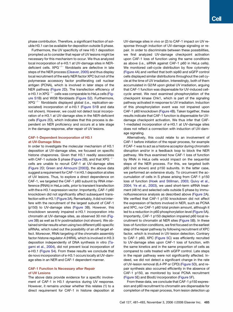
phase contribution. Therefore, a significant fraction of sol-
uble H3.1 can be available for deposition outside S phase.
Furthermore, the UV specificity of new H3.1 deposition
prompted us to consider that repair of UV lesions might be
necessary for this mechanism to occur. We thus analyzed
local incorporation of e-H3.1 at UV-damage sites in NER-
deficient cells. XPG�/� fibroblasts are defective in late
steps of the NER process (Cleaver, 2005) and thus display
local recruitment of the early NER factor XPC but not of the
polymerase accessory factor proliferating cell nuclear
antigen (PCNA), which is involved in later steps of the
NER pathway (Figure 2D). The transfection efficiency of
e-H3.1 in XPG�/� cells was comparable to HeLa cells (Fig-
ure S1B) and WI38 fibroblasts (Figure S2). Furthermore,
XPG�/� fibroblasts displayed global (i.e., replication-as-
sociated) incorporation of e-H3.1 (Figure S1B and data
not shown). However, we could not detect local incorpo-
ration of e-H3.1 at UV-damage sites in the NER-deficient
cells (Figure 2D), which indicates that this process is de-
pendent on NER proficiency and occurs at a late stage
in the damage response, after repair of UV lesions.
CAF-1-Dependent Incorporation of H3.1
at UV-Damage Sites
In order to investigate the molecular mechanism of H3.1
deposition at UV-damage sites, we focused on specific
histone chaperones. Given that H3.1 readily associates
with CAF-1 outside S phase (Figure 2B), and that XPG�/�
cells are unable to recruit CAF-1 at UV-damage sites
(Figure 2D; Green and Almouzni [2003]), the above data
suggest a requirement for CAF-1 in H3.1 deposition at sites
of UV lesions. Thus, to explore a direct dependence on
CAF-1, we targeted the CAF-1 p60 subunit by RNA inter-
ference (RNAi) in HeLa cells, prior to transient transfection
with the e-H3.1 expression vector. Importantly, CAF-1 p60
knockdown did not significantly affect subsequent trans-
fection with e-H3.1 (Figure 3A). Remarkably, it did not inter-
fere with the recruitment of the largest subunit of CAF-1
(p150) to UV-damage sites (Figure 3B). However, this
knockdown severely impaired e-H3.1 incorporation into
chromatin at UV-damage sites, as observed 30 min (Fig-
ure 3B) as well as 6 hr postirradiation (not shown). We ob-
tained similar results when using two different p60-specific
siRNAs, which ruled out the possibility of an off-target ef-
fect. Moreover, RNAi targeting of the chromatin assembly
factor histone regulator A (HIRA), which is involved in H3.3
deposition independently of DNA synthesis in vitro (Ta-
gami et al., 2004), did not prevent local incorporation of
e-H3.1 (Figure S4). From these results we conclude that
de novo incorporation of e-H3.1 occurs locally at UV-dam-
age sites in an NER and CAF-1 dependent manner.
CAF-1 Function Is Necessary after Repair
of UV Lesions
The above data provide evidence for a specific involve-
ment of CAF-1 in H3.1 dynamics during UV response.
However, it remains unclear whether this relates (1) to a
direct requirement for CAF-1 in histone deposition at
C
UV-damage sites in vivo or (2) to CAF-1 impact on UV re-
sponse through induction of UV-damage signaling or re-
pair. In order to discriminate between these possibilities,
we first analyzed UV-damage checkpoint activation
upon CAF-1 loss of function using the same conditions
as above (i.e., siRNA against CAF-1 p60 in HeLa cells).
We monitored cell-cycle distribution by flow cytometry
(Figure 4A) and verified that both sip60 and siGFP control
cells displayed similar distributions throughout the cell cy-
cle at the time of UV irradiation. Interestingly, both of them
accumulated in G2/M upon global UV irradiation, arguing
that CAF-1 function was dispensable for UV-induced cell-
cycle arrest. We next examined phosphorylation of the
checkpoint kinase Chk1, which is part of the signaling
pathway activated in response to UV irradiation. Induction
of this phosphorylation event was not impaired upon
CAF-1 p60 knockdown (Figure 4B). Taken together, these
results indicate that CAF-1 function is dispensable for UV-
damage checkpoint activation. We thus infer that CAF-
1-mediated incorporation of e-H3.1 at UV-damage sites
does not reflect a connection with induction of UV-dam-
age signaling.
Alternatively, this could relate to an involvement of
CAF-1 before initiation of the repair process, for example
if CAF-1 was to act as a histone acceptor during chromatin
disruption and/or in a feedback loop to block the NER
pathway. We thus examined how CAF-1 loss of function
by RNAi in HeLa cells would impact on the sequential
steps of the NER process. For this, we targeted both
p60 (not shown) and p150 subunits. In the latter case,
we performed an extensive study. To circumvent the ac-
cumulation of cells in S phase arising from CAF-1 p150
loss of function (Hoek and Stillman, 2003; Quivy et al.,
2004; Ye et al., 2003), we used short-term siRNA treat-
ment (48 hr) and selected cells outside S phase by immu-
nofluorescence analysis as described above (Figure 2A).
We verified that CAF-1 p150 knockdown did not affect
the expression of factors involved in NER, such as PCNA
and XPC, nor CAF-1 p60 total protein amount, although it
led to a reduction in p60 phosphorylation level (Figure 5A).
Importantly, CAF-1 p150 depletion impaired p60 local re-
cruitment to chromatin at NER sites (Figure 5B). In these
loss-of-function conditions, we first focused on the earliest
step of the repair pathway by following recruitment of XPC
factor, which is involved in UV-lesion detection. Contrary
to CAF-1 p60, XPC (Figure 5C) was efficiently recruited
to UV-damage sites upon CAF-1 loss of function, with
the same kinetics and in the same proportion of cells as
compared to cells treated with siGFP control. Late steps
in the repair pathway were not significantly affected. In-
deed, we did not detect a significant change in the rate
of UV-lesion removal (6,4-PP or CPD) (Figure 5D), and re-
pair synthesis also occurred efficiently in the absence of
CAF-1 p150, as monitored by local PCNA recruitment
(Figure 5E) and BiodU incorporation (Figure 5F).
From these data, we conclude that CAF-1 p150 expres-
sion and p60 recruitment to chromatin are dispensable for
completion of the repair process, from lesion detection up
ell 127, 481–493, November 3, 2006 ª2006 Elsevier Inc. 485

Figure 3. CAF-1 Requirement for H3.1 Incorporation at UV-Damage Sites
(A) Efficiency of p60 siRNA and e-H3.1 transfection analyzed by immunofluorescence (upper panel) and western blot (lower panel) on HeLa cells
treated with a control siRNA targeted against GFP (siGFP) or a p60-specific siRNA (sip60#1 or sip60#2) and subsequently transfected with e-H3.1
vector. Percentages of p60 and e-H3.1 expressing cells are indicated.
(B) Immunolocalization of chromatin bound CAF-1 p60, p150, and e-H3.1 30 min after local UV irradiation of the same cells. The percentages indicate
the fraction of transfected cells showing local e-H3.1 recruitment (e-H3.1 positive). We used Triton-resistant CAF-1 p150 staining as a reference to
select cells outside S phase. Scale bars, 10 mm. Data were obtained from at least two independent experiments.
to repair synthesis. Our results thus eliminate the hypoth-
esis of a feedback loop of CAF-1 function on the NER pro-
cess and the possibility that CAF-1 could act in early steps
of the UV-damage response. Our data rather provide new
evidence that CAF-1 plays a critical role after repair of UV
lesions in the cell and directly stimulates de novo deposi-
tion of H3.1 histones at NER sites, a process that is not re-
stricted to S phase cells. Interestingly, we also observed
local deposition of e-H3.1 at sites of laser microirradiation
(Figure 6), which mainly generates oxidative base damage
and DNA breaks. New histone deposition is thus not re-
stricted to the response to UV-C induced pyrimidine di-
mers, extending the generality of our findings to other
types of DNA damage. Getting back to our initial question
(Figure 1A), not only do we show that new histones are
incorporated at sites of DNA damage in vivo, but we
486 Cell 127, 481–493, November 3, 2006 ª2006 Elsevier Inc.
also provide mechanistic insights into the deposition
process.
DISCUSSION
In eukaryotes, the cellular response to genotoxic insults in-
volves both repair of DNA lesions and changes at the level
of chromatin, including chromatin disruption and subse-
quent restoration. A long-standing issue with respect to
such chromatin dynamics in the restoration step is to de-
termine whether it involves histone recycling and/or depo-
sition of new histones. This is obviously a critical question
in the context of reestablishing the epigenetic status of
a damaged region. In the present study, we have exam-
ined, for the first time, the dynamics of the H3.1 histone
variant during UV response in human cells. We show that

Figure 4. CAF-1 Function Is Dispensable
for Induction of UV-Damage Signaling
HeLa cells were treated with a control siRNA
targeted against GFP or a p60-specific siRNA
before global UV irradiation at 10 J/m2 (+), us-
ing unirradiated cells (�) as a reference.
(A) Cell-cycle distribution was analyzed by flow
cytometry (upper panel) at the time of UV irradi-
ation (0 hr) and 30 hr afterwards. CAF-1 p60
knockdown was controlled by western blot
(lower panel).
(B) Chk1 phosphorylation on Serine 317 was
monitored by immunofluorescence 2 hr and
5 hr after UV irradiation. Scale bar, 10 mm.
de novo deposition of H3.1 variants occurs at UV-damage
sites in a UV-repair-dependent manner and that the his-
tone chaperone CAF-1 is specifically and directly involved
in this process.
Histone Dynamics during Chromatin Restoration
at Repair Sites
While incorporation of newly synthesized histones by
de novo chromatin assembly is well characterised during
DNA replication (Kaufman and Almouzni, 2006), it was un-
clear whether a similar mechanism would take place dur-
ing repair, when extensive DNA and histone syntheses do
not occur. In particular, NER is characterized by a short
patch of repair synthesis of about 30 nucleotides in length
(Reardon and Sancar, 2005). A tentative scenario for his-
tone dynamics during the repair response would thus in-
volve recycling of preexisting histones rather than new his-
tone deposition. In this work, however, we show de novo
Cell 127, 481–493, November 3, 2006 ª2006 Elsevier Inc. 487

488 Cell 127, 481–493, November 3, 2006 ª2006 Elsevier Inc.

Figure 6. e-H3.1 Deposition at Sites of Laser Microirradiation(A) Scheme of the experiment.
(B) Immunodetection of e-H3.1 and CAF-1 p60 subunit 30 min after exposure to the laser beam. Sites of microirradiation are visualized by gH2AX
staining. Scale bar, 10 mm.
incorporation of H3.1 histones at NER sites in response to
local UV irradiation. Given our unselective irradiation ap-
proach, incorporation of new H3.1 histones is unlikely to
be restricted to specific chromatin regions, in particular
to regions initially devoid of nucleosomes such as active
ribosomal genes (Conconi et al., 2005). Furthermore, the
strong overlap with the damaged chromatin region argues
that new histone deposition is not a global response to
local UV damage, in marked contrast to p53-dependent
chromatin relaxation reported to affect the whole nucleus
(Rubbi and Milner, 2003).
Remarkably, our data challenge the idea that H3.1 incor-
poration into chromatin is restricted to S phase and sup-
port a more general coupling of H3.1 deposition to DNA
synthesis events. It is noteworthy that the fraction of newly
synthesized H3.1 is likely to be limited outside S phase, in
light of the tight coupling between H3.1 synthesis and DNA
replication (Osley, 1991; Wu et al., 1982). Yet, if we con-
sider the entire pool of histones immediately available
C
for deposition onto DNA, our data suggest that a significant
fraction of H3.1 can be available outside S phase. This pool
could be sufficient to ensure new H3.1 incorporation in
response to low UV doses. However, we do not exclude
that high requirements upon exposure to high UV doses
may need more histones and that these situations could
(1) favor more active histone recycling at damage sites,
(2) stimulate new H3.1 synthesis, or (3) inhibit H3.1 degra-
dation.
While we focus here on H3.1 histones, which specifi-
cally associate with CAF-1 in vivo (Tagami et al., 2004),
we do not rule out the possibility that other H3 variants
could also be incorporated at NER sites. We should stress
that the cell-cycle independent incorporation of H3.3 can
mask local deposition events and makes this analysis
more difficult. However, in a few cells exposed to high
UV doses, we could observe a local concentration of
e-H3.3 at UV-damage sites (data not shown). In this respect,
it is noteworthy that visualization of a local accumulation of
Figure 5. CAF-1 Function Is Dispensable for NER
(A) Immunofluorescence (left) and western blot analyses (right) of HeLa cells 48 hr posttransfection with a control siRNA targeted against GFP (siGFP)
or a p150-specific siRNA (sip150). Percentages of p150-expressing cells are indicated below. *: phosphorylated p60.
(B, C, and E) p60, XPC, and PCNA recruitment to UV-damage sites (CPD) analyzed by immunofluorescence at the indicated times after local UV ir-
radiation in siRNA-treated HeLa cells. Percentages of cells showing XPC recruitment to UV lesions are indicated below. Percentages of cells showing
global (replication-associated) or local (repair-associated) recruitment of p60 and PCNA to chromatin are plotted on the right.
(D) Time course of UV lesion removal. We analyzed DNA extracted from siRNA- treated and UV-irradiated HeLa cells as described in Experimental
Procedures. A plot compiling the results relative to the initial amount of UV damage and corrected for the total amount DNA is presented for 6,4-PPs
(left) and CPDs (right) (mean ± SD). �: unirradiated cells.
(F) Repair synthesis at UV damage sites (CPD) revealed by BiodU incorporation in siRNA treated HeLa cells. In situ DNA synthesis was performed in
the presence (+) or absence (�) of BiodU as a control. Percentages of cells showing global or local BiodU incorporation (DNA synthesis associated
with replication and repair, respectively) are plotted on the right. Scale bars, 10 mm.
ell 127, 481–493, November 3, 2006 ª2006 Elsevier Inc. 489

ectopically expressed H3.3 by immunofluorescence anal-
ysis could be achieved so far only upon H3.3 overexpres-
sion and/or in response to massive transcription activation
(Ahmad and Henikoff, 2002; Janicki et al., 2004; Schwartz
and Ahmad, 2005). Thus, incorporation of this variant at
damage sites, which is a limited phenomenon detectable
under extreme conditions, most likely reflects an unspe-
cific mechanism to fill gaps.
Regardless of which histone variant gets incorporated
de novo at repair sites, a prerequisite for such a process
is local eviction of nucleosomal histones. Interestingly, a
recent study in yeast provides evidence for loss of nucleo-
somes at a double-strand break (Tsukuda et al., 2005).
While direct evidence for nucleosome eviction at NER sites
is still a matter of debate (Thoma, 1999), our findings sup-
port the view that nucleosomes are initially disassembled
to permit incorporation of new histones at UV-damage
sites in human cells. The mechanism and extent of histone
eviction as well as the fate of evicted histones are still open
issues. Furthermore, the incorporation of new histones
within damaged chromatin does not preclude partial recy-
cling of old histones. This would be ensured by histone
chaperones that would assist their reassembly. In this
respect, the histone chaperone antisilencing function 1
(ASF1) is an interesting candidate both as a donor of new
histones and as an acceptor for evicted histones, given
its documented synergy with CAF-1 in vitro (Mello et al.,
2002) and its role in chromatin disassembly in yeast (Ad-
kins and Tyler, 2004; Schwabish and Struhl, 2006) and in
buffering excess S phase histones in human cells (Groth
et al., 2005). However, due to the multiplicity of ASF1 func-
tions, extensive studies will be required to elucidate its
precise role(s) in histone dynamics at damage sites.
CAF-1 Function at Repair Sites
In this report, we also provide insight into the function of
CAF-1 during the UV response in vivo by exploring the dy-
namics of the replicative H3.1 variant. Furthermore, our
loss-of-function studies showing that CAF-1 p150 is re-
cruited to damage independently of CAF-1 p60, but not
vice versa, support a key role for CAF-1 large subunit as
the molecular link to UV damaged chromatin in vivo.
This is consistent with the reported interaction of this sub-
unit with PCNA (Moggs et al., 2000; Shibahara and Still-
man, 1999), whereby the CAF-1 complex would be tar-
geted to sites of repair synthesis. Interestingly, the p150
subunit can support de novo histone incorporation only
in connection with p60. Indeed, the siRNA against p60 still
permits p150 recruitment, yet no H3.1 can be deposited
(Figure 3). We also noticed that CAF-1 p150 and p60
knockdowns have strinkingly different outcomes. While
CAF-1 p150 loss of function results in S phase accumula-
tion followed by checkpoint activation (Hoek and Stillman,
2003; Quivy et al., 2004; Ye et al., 2003), p60 knockdown
does not affect cell-cycle distribution (Nabatiyan and
Krude [2004]; this study). These observations suggest
that, although both subunits are required for CAF-1 func-
tion in chromatin assembly, they are also likely to display
490 Cell 127, 481–493, November 3, 2006 ª2006 Elsevier Inc.
distinct properties, which are currently under investigation
(J.-P. Quivy, A. Gerard, D.R., and G.A., unpublished data).
Importantly, we show for the first time in human cells
that CAF-1 is dispensable for repair of UV damage. Simi-
larly, in budding yeast, the NER pathway is functional in
CAF-1 mutant strains (Game and Kaufman, 1999), yet
these strains display a moderate increase in UV sensitivity
(Game and Kaufman, 1999; Kaufman et al., 1997) and
show genome instability (Myung et al., 2003). Thus,
CAF-1 importance in response to UV damage in vivo is
most likely related to its specific contribution downstream
of the repair process. A similar function of CAF-1 could
also be at work in response to other types of DNA damage
(Lewis et al., 2005; Linger and Tyler, 2005; Moggs et al.,
2000; Nabatiyan et al., 2006; Okano et al., 2003). Our
data, showing that e-H3.1 also accumulates at sites of
laser microirradiation, clearly support this possibility. Col-
lectively, our findings underscore a critical role for the his-
tone chaperone CAF-1 in de novo deposition of H3.1 at
UV-damage sites, most likely as an H3.1-H4 histone donor
that is directly involved in chromatin restoration coupled to
NER. This fundamental process should be considered as
an integral part of the damage response for our under-
standing of genetic and epigenetic stability.
Epigenetic Stability Versus Memory of Damage
at Repair Sites
While CAF-1-mediated deposition of H3 and H4 histones
at repair sites contributes to restoration of a proper chro-
matin structure, it is only the very first step in de novo chro-
matin assembly. To fully restore chromatin organization,
additional histone and nonhistone proteins are necessary.
Furthermore, our results showing that incorporation of
new histone occurs raise the important issue of how epi-
genetic memory can be preserved. In this respect, it will
be important to evaluate to which extent the incorporation
of new histones can spread from the damage site, given
that CAF-1 was shown to stimulate histone deposition
in vitro several hundred bp distant from the initial damage
(Gaillard et al., 1997). Indeed, large-scale epigenetic
changes would have a stronger impact on genome func-
tion. Furthermore, we anticipate that modifications asso-
ciated with newly synthesized histones (Sobel et al.,
1995) should be detected at least transiently at NER sites.
Given that both histone posttranslational modifications
and histone variants can contribute to epigenetic marking
(Henikoff and Ahmad, 2005; Turner, 2002), the question
that ensues is whether, how, and when original marks will
be reestablished. Neighboring chromatin regions that did
not undergo extensive rearrangements during repair may
then serve as an epigenetic template for newly incorpo-
rated histones. Indeed, histone-modifying enzymes can
be recruited to specific histone modifications through de-
fined protein modules and thereby allow the spreading of
these modifications to neighboring nucleosomes (Jenu-
wein and Allis, 2001; Turner, 2002). Alternatively, a partial
recycling of old histones might contribute to the mainte-
nance of epigenetic information by a semiconservative
mechanism, similar to the one already proposed for DNA
replication: this model involves disruption of parental
(H3-H4)2 tetramers into H3-H4 dimers, which can associ-
ate with newly deposited ones (Tagami et al., 2004). Con-
versely, the incorporation of H3.1 variants into a defined
chromatin region can also be viewed as an imprint for
newly repaired chromatin, as a memory of the damage
event. A critical issue is to determine if such an imprint is
part of a short-term response, which could impact on dam-
age signaling (maintenance and/or recovery), or if it is a
long-term mark. In this respect, it would be interesting to
examine the persistence of H3.1 variants at their incorpo-
ration sites over several cell cycles. Such a memory of
damage could play a role in processes such as radiation-
induced genomic instability, which arises in the progeny
of the damaged cells after several generations (Little, 2003).
Collectively, these data provide evidence for the local
dynamics of variant histones at UV-damage sites in vivo
and put forward CAF-1 as a key player in this process.
This fundamental mechanism contributes to the mainte-
nance of chromatin organization along with genome integ-
rity in response to genotoxic insults.
EXPERIMENTAL PROCEDURES
Cell Culture
HeLa cells (gift from M. Bornens, Curie Institute, Paris, France), HeLa
cells stably transfected with H3.1-Flag-HA (HemAgglutinin) or H3.3-
Flag-HA (Tagami et al., 2004), WI38 diploid fibroblasts (LGC Promo-
chem, Molsheim, France), MCF7 cells (gift from O. Delattre, Curie Insti-
tute, Paris, France), and XPG�/� cells (XP3BR, gift from A. Sarasin,
IGR, Villejuif, France) were grown in Petri dishes (Falcon Plastics,
Cockeysville, MD) in the appropriate medium complemented with
10% foetal calf serum and 100 U/ml penicillin and 100 mg/ml strepto-
mycin (Invitrogen Company). HeLa, MCF7, and XPG�/� cells were
grown in DMEM, and WI38 cells were grown in MEM a medium.
MCF7 cells were arrested in G0 by 48 hr treatment with 10 nM
ICI182780 (Carroll et al., 2000; Polo et al., 2004).
Cell Irradiation
For UV irradiation, cells were treated with UV-C (254 nm) using a low-
pressure mercury lamp, and conditions were set using a VLX-3W
dosimeter (Vilbert-Lourmat). Cells were either subjected to global
(10 J/m2) or local UV irradiation (150 J/m2, using 3mm or 8mm pore fil-
ters (Millipore) as described (Gerard et al., 2006; Green and Almouzni,
2003; Mone et al., 2001).
For laser microirradiation, cells were presensitized by incubation
with 10 mM BrdU and 1.6 mM Hoechst 33258 before microirradiation
with a UV-A laser line (406 nm, 50% output, 10 ms, beam expander
4) focused through a 1003 oil objective on a Deltavision RT micro-
scope (Applied Precision) (adapted from Limoli and Ward [1993],
Lukas et al. [2003], and Rogakou et al. [1999]).
Flow Cytometry
Cells were fixed in ice-cold 70% ethanol before DNA staining with
50 mg/ml propidium iodide (Sigma Aldrich) in phosphate buffer saline
containing 0.5 mg/ml RNase A (Amersham). DNA content was ana-
lyzed by flow cytometry using a FACSCalibur flow cytometer and
CellQuest Pro software (Becton Dickinson).
Antibodies
Primary antibodies against CAF-1 subunits were anti-p150 (ab7655
Abcam), monoclonal anti-p60 (ab8133 Abcam), and polyclonal anti-
p60 characterized in our laboratory (Green and Almouzni, 2003).
Monoclonal anti-p60 mAb8133 recognizes only phosphorylated p60,
whereas polyclonal anti-p60 recognizes both phosphorylated and un-
phosphorylated forms. Other primary antibodies were anti HIRA (gift
from P. Adams, Fox Chase center, PA), anti-PCNA PC10 (Dako) and
FL-261 (Santa Cruz), anti-XPC (gift from F. Hanaoka, Osaka University,
Osaka, Japan), anti-CPD (Kamiya Biomedicals), anti-6,4-PP (gift from
T. Matsunaga, Kanazawa University, Kanazawa, Japan), anti-gH2AX
(JBW301, Upstate), anti-phospho-Chk1 Ser317 (Cell Signaling), anti-
H3 (ab7834, Abcam), anti-HA (3F10, Roche), and anti-bactin (AC15,
Sigma Chemical Company). Secondary antibodies coupled to Fluores-
cein IsoThioCyanate, Texas red, Cyanin3, or Horse Radish Peroxidase
were purchased from Jackson Laboratory. Highly cross-absorbed
anti-mouse Alexa Fluor 488, anti-rabbit Alexa Fluor 680 (Molecular
Probes), and anti-rat Cyanin3 antibodies were used for triple labeling.
Immunofluorescence
Immunofluorescence on paraformaldehyde fixed cells, image capture,
and processing were performed as described previously (Green and Al-
mouzni, 2003). DNase treatment was carried out as described (Martini
et al., 1998). Percentages of positively stained cells were obtained by
scoring over 200 cells in at least two independent experiments. To
take into account e-H3.1 transfection efficiency, we calculated system-
atically the ratio between the fraction of cells exhibiting local e-H3.1 ac-
cumulation and the fraction of transfected cells. To focus on cells out-
side S phase, we used either PCNA or CAF-1 Triton-resistant staining,
which displays characteristic patterns in S phase nuclei (Figure 2A).
Cell Extracts and Western Blot
Total and Triton-treated cell extracts were made and subjected to
western blotting as described (Martini et al., 1998). Cell extracts con-
taining soluble histones were obtained by cell lysis in high salt-extrac-
tion buffer (50 mM Tris pH 7.5, 300 mM NaCl, 0.5% NP-40, and prote-
ase inhibitors), after centrifugation (14,000 rpm, 15 min, 4�C) to remove
insoluble components.
Triton-Acid-Urea Gel Analysis
For specific detection of H3 variants, samples were run on a Triton-
acid-urea (TAU) gel (12% polyacrylamide, 6 M urea, 5% acetic acid,
0.4% Triton X-100) overnight at 200 V in 5% acetic acid (Zweidler,
1978). After protein transfer on nitrocellulose membrane (Thiriet and
Albert, 1995), H3 variants were detected using anti-H3 antibody.
Analysis of UV-Lesion Removal
Total genomic DNA was extracted from UV-C irradiated cells (10 J/m2).
DNA samples in 0.2 M NaOH were incubated at 37�C for 15 min and
boiled and spotted onto Hybond N+ membrane (Amersham) using
a Bio-Dot apparatus (Bio-Rad). Two dilutions (1 and 0.5 mg) were spot-
ted for each time point. The membrane was dried at 80�C for 1 hr and
blocked with 5% milk before immunodetection of UV lesions (6,4-PPs
and CPDs). Total DNA was subsequently visualized on the same mem-
brane by ethidium bromide intercalation. Quantitation was performed
using Quantity One analysis software (Bio-Rad).
In Situ DNA Synthesis
Cells were grown on collagen-fibronectin-treated glass coverslips and
subjected to local UV irradiation. They were allowed to recover for
20 min postirradiation at 37�C. Biotin-16-deoxyuridine (BiodU) incor-
poration was performed by in situ run-on on Triton-permeabilized cells
for 40 min at 37�C under conditions described for isolated nuclei (Tad-
dei et al., 1999). FITC-conjugated streptavidin (Enzo) and biotinylated
anti-streptavidin antibody (Abcys) were used for biotin immunodetec-
tion on paraformaldehyde-fixed cells.
siRNA Design and Cell Transfection
siRNA oligonucleotides (MWG-Biotech and Sigma Genosys) were
targeted against the following mRNA sequences: CAF-1 p150,
Cell 127, 481–493, November 3, 2006 ª2006 Elsevier Inc. 491

AAGGAGAAGGCGGAGAAGCAG (Quivy et al., 2004); CAF-1 p60#1,
AAGCGUGUGGCUUUCAAUGUU; CAF-1 p60#2, AAUCUUGCUCGU
CAUACCAAA; green fluorescent protein (GFP), AAGCUGGAGUAC
AACUACAAC. siRNAs were chemically synthesized with the Silencer
siRNA construction kit (Ambion) according to manufacturer’s instruc-
tions. SiRNA transfections were performed using oligofectamine re-
agent (Invitrogen) in Optimem1 medium and standard culture medium
lacking antibiotics (Invitrogen). The final concentration of siRNA in the
culture medium was 30 nM.
Cells were transiently transfected with H3.1-Flag-HA expression
vector pOZ-FH-C, a Moloney Murine Leukemia Virus-derived vector,
which allows a low expression level of the transgene (Nakatani and
Ogryzko, 2003; Tagami et al., 2004). Transfection was performed using
Lipofectamine2000 (Invitrogen) according to manufacturer’s instruc-
tions 20 hr before subsequent cell treatment.
Supplemental Data
Supplemental Data include four figures and can be found with this
article online at http://www.cell.com/cgi/content/full/127/3/481/DC1/.
ACKNOWLEDGMENTS
We are grateful to E. Heard, E. Martini, E. Moustacchi, J-P. Quivy, and
D. Ray-Gallet for critical reading of the manuscript and to members of
UMR218 for helpful discussions. This work was supported by la Ligue
Nationale contre le Cancer (Equipe labelisee la Ligue), the Commissar-
iat a l’Energie Atomique (CEA) (LRC no. 26), Contract RTN (HPRN-CT-
2002-00238), a collaborative program ‘‘Parametres Epigenetiques’’
between the Curie Institute and the CEA, Network of Excellence Epige-
nome (LSHG-CT-2004-503433), ACI-DRAB (n� 04393), and Cancero-
pole. S. E. P. is supported by an ARC fellowship.
Received: March 15, 2006
Revised: July 11, 2006
Accepted: August 22, 2006
Published: November 2, 2006
REFERENCES
Adkins, M.W., and Tyler, J.K. (2004). The histone chaperone Asf1p
mediates global chromatin disassembly in vivo. J. Biol. Chem. 279,
52069–52074.
Ahmad, K., and Henikoff, S. (2002). The histone variant H3.3 marks
active chromatin by replication-independent nucleosome assembly.
Mol. Cell 9, 1191–1200.
Carroll, J.S., Prall, O.W., Musgrove, E.A., and Sutherland, R.L. (2000).
A pure estrogen antagonist inhibits cyclin E-Cdk2 activity in MCF-7
breast cancer cells and induces accumulation of p130-E2F4 com-
plexes characteristic of quiescence. J. Biol. Chem. 275, 38221–38229.
Cleaver, J.E. (2005). Cancer in xeroderma pigmentosum and related
disorders of DNA repair. Nat. Rev. Cancer 5, 564–573.
Conconi, A., Paquette, M., Fahy, D., Bespalov, V.A., and Smerdon,
M.J. (2005). Repair-independent chromatin assembly onto active ribo-
somal genes in yeast after UV irradiation. Mol. Cell. Biol. 25, 9773–
9783.
de Laat, W.L., Jaspers, N.G., and Hoeijmakers, J.H. (1999). Molecular
mechanism of nucleotide excision repair. Genes Dev. 13, 768–785.
Friedberg, E.C. (2001). How nucleotide excision repair protects against
cancer. Nat. Rev. Cancer 1, 22–33.
Gaillard, P.H., Martini, E.M., Kaufman, P.D., Stillman, B., Moustacchi,
E., and Almouzni, G. (1996). Chromatin assembly coupled to DNA re-
pair: a new role for chromatin assembly factor I. Cell 86, 887–896.
Gaillard, P.H., Moggs, J.G., Roche, D.M., Quivy, J.P., Becker, P.B.,
Wood, R.D., and Almouzni, G. (1997). Initiation and bidirectional prop-
492 Cell 127, 481–493, November 3, 2006 ª2006 Elsevier Inc.
agation of chromatin assembly from a target site for nucleotide exci-
sion repair. EMBO J. 16, 6281–6289.
Game, J.C., and Kaufman, P.D. (1999). Role of Saccharomyces cere-
visiae chromatin assembly factor-I in repair of ultraviolet radiation
damage in vivo. Genetics 151, 485–497.
Gerard, A., Polo, S.E., Roche, D., and Almouzni, G. (2006). Methods for
studying chromatin assembly coupled to DNA repair. Methods Enzy-
mol. 409, 358–374.
Green, C.M., and Almouzni, G. (2002). When repair meets chromatin.
First in series on chromatin dynamics. EMBO Rep. 3, 28–33.
Green, C.M., and Almouzni, G. (2003). Local action of the chromatin
assembly factor CAF-1 at sites of nucleotide excision repair in vivo.
EMBO J. 22, 5163–5174.
Groth, A., Ray-Gallet, D., Quivy, J.P., Lukas, J., Bartek, J., and Al-
mouzni, G. (2005). Human Asf1 regulates the flow of S phase histones
during replicational stress. Mol. Cell 17, 301–311.
Gunjan, A., Paik, J., and Verreault, A. (2005). Regulation of histone syn-
thesis and nucleosome assembly. Biochimie 87, 625–635.
Henikoff, S., and Ahmad, K. (2005). Assembly of variant histones into
chromatin. Annu. Rev. Cell Dev. Biol. 21, 133–153.
Hoek, M., and Stillman, B. (2003). Chromatin assembly factor 1 is es-
sential and couples chromatin assembly to DNA replication in vivo.
Proc. Natl. Acad. Sci. USA 100, 12183–12188.
Janicki, S.M., Tsukamoto, T., Salghetti, S.E., Tansey, W.P., Sachida-
nandam, R., Prasanth, K.V., Ried, T., Shav-Tal, Y., Bertrand, E., Singer,
R.H., and Spector, D.L. (2004). From silencing to gene expression:
real-time analysis in single cells. Cell 116, 683–698.
Jenuwein, T., and Allis, C.D. (2001). Translating the histone code. Sci-
ence 293, 1074–1080.
Kaufman, P.D., and Almouzni, G. (2006). Chromatin assembly. In DNA
Replication and Human Disease, M. DePamphilis, ed. (Woodbury, NY:
Cold Spring Harbor Laboratory Press), pp. 121–140.
Kaufman, P.D., Kobayashi, R., Kessler, N., and Stillman, B. (1995). The
p150 and p60 subunits of chromatin assembly factor I - a molecular
link between newly synthesized histones and DNA replication. Cell
81, 1105–1114.
Kaufman, P.D., Kobayashi, R., and Stillman, B. (1997). Ultraviolet radi-
ation sensitivity and reduction of telomeric silencing in Saccharomy-
ces cerevisiae cells lacking chromatin assembly factor-I. Genes Dev.
11, 345–357.
Kornberg, R.D. (1977). Structure of chromatin. Annu. Rev. Biochem.
46, 931–954.
Lewis, L.K., Karthikeyan, G., Cassiano, J., and Resnick, M.A. (2005).
Reduction of nucleosome assembly during new DNA synthesis impairs
both major pathways of double-strand break repair. Nucleic Acids
Res. 33, 4928–4939.
Limoli, C.L., and Ward, J.F. (1993). A new method for introducing dou-
ble-strand breaks into cellular DNA. Radiat. Res. 134, 160–169.
Linger, J., and Tyler, J.K. (2005). The yeast histone chaperone chroma-
tin assembly factor 1 protects against double-strand DNA-damaging
agents. Genetics 171, 1513–1522.
Little, J.B. (2003). Genomic instability and bystander effects: a histori-
cal perspective. Oncogene 22, 6978–6987.
Loyola, A., and Almouzni, G. (2004). Histone chaperones, a supporting
role in the limelight. Biochim. Biophys. Acta 1677, 3–11.
Lukas, C., Falck, J., Bartkova, J., Bartek, J., and Lukas, J. (2003). Dis-
tinct spatiotemporal dynamics of mammalian checkpoint regulators
induced by DNA damage. Nat. Cell Biol. 5, 255–260.
Martini, E., Roche, D.M., Marheineke, K., Verreault, A., and Almouzni,
G. (1998). Recruitment of phosphorylated chromatin assembly factor 1
to chromatin after UV irradiation of human cells. J. Cell Biol. 143, 563–
575.
Mello, J.A., Sillje, H.H., Roche, D.M., Kirschner, D.B., Nigg, E.A., and
Almouzni, G. (2002). Human Asf1 and CAF-1 interact and synergize
in a repair-coupled nucleosome assembly pathway. EMBO Rep. 3,
329–334.
Moggs, J.G., Grandi, P., Quivy, J.P., Jonsson, Z.O., Hubscher, U.,
Becker, P.B., and Almouzni, G. (2000). A CAF-1-PCNA-mediated chro-
matin assembly pathway triggered by sensing DNA damage. Mol. Cell.
Biol. 20, 1206–1218.
Mone, M.J., Volker, M., Nikaido, O., Mullenders, L.H., van Zeeland,
A.A., Verschure, P.J., Manders, E.M., and van Driel, R. (2001). Local
UV-induced DNA damage in cell nuclei results in local transcription
inhibition. EMBO Rep. 2, 1013–1017.
Myung, K., Pennaneach, V., Kats, E.S., and Kolodner, R.D. (2003).
Saccharomyces cerevisiae chromatin-assembly factors that act dur-
ing DNA replication function in the maintenance of genome stability.
Proc. Natl. Acad. Sci. USA 100, 6640–6645.
Nabatiyan, A., and Krude, T. (2004). Silencing of chromatin assembly
factor 1 in human cells leads to cell death and loss of chromatin as-
sembly during DNA synthesis. Mol. Cell. Biol. 24, 2853–2862.
Nabatiyan, A., Szuts, D., and Krude, T. (2006). Induction of CAF-1 ex-
pression in response to DNA strand breaks in quiescent human cells.
Mol. Cell. Biol. 26, 1839–1849.
Nakatani, Y., and Ogryzko, V. (2003). Immunoaffinity purification of
mammalian protein complexes. Methods Enzymol. 370, 430–444.
Okano, S., Lan, L., Caldecott, K.W., Mori, T., and Yasui, A. (2003). Spa-
tial and temporal cellular responses to single-strand breaks in human
cells. Mol. Cell. Biol. 23, 3974–3981.
Osley, M.A. (1991). The regulation of histone synthesis in the cell cycle.
Annu. Rev. Biochem. 60, 827–861.
Peterson, C.L., and Cote, J. (2004). Cellular machineries for chromo-
somal DNA repair. Genes Dev. 18, 602–616.
Polo, S.E., Theocharis, S.E., Klijanienko, J., Savignoni, A., Asselain, B.,
Vielh, P., and Almouzni, G. (2004). Chromatin assembly factor-1,
a marker of clinical value to distinguish quiescent from proliferating
cells. Cancer Res. 64, 2371–2381.
Quivy, J.P., Roche, D., Kirschner, D., Tagami, H., Nakatani, Y., and Al-
mouzni, G. (2004). A CAF-1 dependent pool of HP1 during heterochro-
matin duplication. EMBO J. 23, 3516–3526.
Reardon, J.T., and Sancar, A. (2005). Nucleotide excision repair. Prog.
Nucleic Acid Res. Mol. Biol. 79, 183–235.
Rogakou, E.P., Boon, C., Redon, C., and Bonner, W.M. (1999). Mega-
base chromatin domains involved in DNA double-strand breaks
in vivo. J. Cell Biol. 146, 905–916.
Rubbi, C.P., and Milner, J. (2003). p53 is a chromatin accessibility
factor for nucleotide excision repair of DNA damage. EMBO J. 22,
975–986.
Schwabish, M.A., and Struhl, K. (2006). Asf1 mediates histone eviction
and deposition during elongation by RNA polymerase II. Mol. Cell 22,
415–422.
Schwartz, B.E., and Ahmad, K. (2005). Transcriptional activation trig-
gers deposition and removal of the histone variant H3.3. Genes Dev.
19, 804–814.
Shibahara, K., and Stillman, B. (1999). Replication-dependent marking
of DNA by PCNA facilitates CAF-1-coupled inheritance of chromatin.
Cell 96, 575–585.
Smerdon, M.J. (1991). DNA repair and the role of chromatin structure.
Curr. Opin. Cell Biol. 3, 422–428.
Smith, S., and Stillman, B. (1989). Purification and characterization of
CAF-I, a human cell factor required for chromatin assembly during
DNA replication in vitro. Cell 58, 15–25.
Sobel, R.E., Cook, R.G., Perry, C.A., Annunziato, A.T., and Allis, C.D.
(1995). Conservation of deposition-related acetylation sites in newly
synthesized histones H3 and H4. Proc. Natl. Acad. Sci. USA 92,
1237–1241.
Stillman, B. (1986). Chromatin assembly during SV40 DNA replication
in vitro. Cell 45, 555–565.
Taddei, A., Roche, D., Sibarita, J.B., Turner, B.M., and Almouzni, G.
(1999). Duplication and maintenance of heterochromatin domains.
J. Cell Biol. 147, 1153–1166.
Tagami, H., Ray-Gallet, D., Almouzni, G., and Nakatani, Y. (2004). His-
tone h3.1 and h3.3 complexes mediate nucleosome assembly path-
ways dependent or independent of DNA synthesis. Cell 116, 51–61.
Thiriet, C., and Albert, P. (1995). Rapid and effective western blotting of
histones from acid-urea-Triton and sodium dodecyl sulfate polyacryl-
amide gels: two different approaches depending on the subsequent
qualitative or quantitative analysis. Electrophoresis 16, 357–361.
Thoma, F. (1999). Light and dark in chromatin repair: repair of UV-
induced DNA lesions by photolyase and nucleotide excision repair.
EMBO J. 18, 6585–6598.
Tsukuda, T., Fleming, A.B., Nickoloff, J.A., and Osley, M.A. (2005).
Chromatin remodelling at a DNA double-strand break site in Saccha-
romyces cerevisiae. Nature 438, 379–383.
Turner, B.M. (2002). Cellular memory and the histone code. Cell 111,
285–291.
van Attikum, H., and Gasser, S.M. (2005). The histone code at DNA
breaks: a guide to repair? Nat. Rev. Mol. Cell Biol. 6, 757–765.
Vaquero, A., Loyola, A., and Reinberg, D. (2003). The constantly
changing face of chromatin. Sci. Aging Knowledge Environ. 14, RE4.
Verreault, A., Kaufman, P.D., Kobayashi, R., and Stillman, B. (1996).
Nucleosome assembly by a complex of CAF-1 and acetylated histones
H3/H4. Cell 87, 95–104.
Wu, R.S., Tsai, S., and Bonner, W.M. (1982). Patterns of histone variant
synthesis can distinguish G0 from G1 cells. Cell 31, 367–374.
Ye, X., Franco, A.A., Santos, H., Nelson, D.M., Kaufman, P.D., and
Adams, P.D. (2003). Defective S phase chromatin assembly causes
DNA damage, activation of the S phase checkpoint, and S phase
arrest. Mol. Cell 11, 341–351.
Zweidler, A. (1978). Resolution of histones by polyacrylamide gel elec-
trophoresis in presence of nonionic detergents. Methods Cell Biol. 17,
223–233.
Cell 127, 481–493, November 3, 2006 ª2006 Elsevier Inc. 493