Flow carbonylation of sterically hindered ortho-substituted ...
(CANCER RESEARCH 47, 5924-5931, November 15, 1987]
New Coupling Agents for the Synthesis of Immunotoxins Containing a HinderedDisulfide Bond with Improved Stability in VivoPhilip E. Thorpe,1 Philip M. Wallace, Phillip P. Knowles, Michele G. Reif, Alex N. F. Brown, Graham J. Watson,
Kegina E. Knyba, Edward J. Wawrzynczak, and David C. BlakeyDrug Targeting Laboratory, Imperial Cancer Research Fund, P. O. Box 123, Lincoln's Inn Fields, London WC2A 3PX, England
ABSTRACT
Two new coupling agents were synthesized for making immunotoxinscontaining disulfide bonds with improved stability in vivo: sodium 5-4-succinimidyloxycarbonyl-a-methyl benzyl thiosulfate (SMBT) and 4-succinimidyloxycarbonyl-a-methyl-a(2-pyridyldithio)toluene (SMPT).Both reagents generate the same hindered disulfide linkage in which amethyl group and a benzene ring are attached to the carbon atom adjacentto the disulfide bond and protect it from attack by thiolate anions.
An immunotoxin consisting of monoclonal anti-Thy-1.1 antibody(OX7) linked by means of the SMPT reagent to chemically deglycosy-lated ricin A-chain had better stability in vivo than an immunotoxinprepared with 2-iminothiolane hydrochloride (2IT) which generates anunhindered disulfide linkage. About 48 h after i.v. injection into mice,one-half of the SMPT-linked immunotoxin present in the blood was inintact form and one-half as released free antibody, whereas equivalentbreakdown of the 2IT-linked immunotoxin was seen at about 8 h afterinjection. Consequently, the blood levels of the SMPT-linked immunotoxin remained higher than those of the 211-Iinked immunotoxin despiteloss of immunotoxin from the blood by other mechanisms. Forty-eight hafter injection, 10% of the injected dose of the SMPT-linked immunotoxin remained in the bloodstream as compared with only 1.5% of the211-linked immunotoxin.
The ability of immunotoxins prepared with the new reagents to inhibitprotein synthesis by Thy-l.l-expressing AKR-A/2 lymphoma cells invitro was identical to that of immunotoxins prepared with 2IT or N-succinimidyl-3-(2-pyridyldithio)propionate (SPDP). Clonogenic assaysshowed that fewer than 0.01% of AKR-A/2 cells survived exposure tohigh concentrations of OX7-abrin A-chain immunotoxins prepared withSMBT, 2IT, or SPDP. Twelve clones of cells which had survivedtreatment with the SMBT-linked immunotoxin were isolated. None ofthe clones was selectively resistant to the SMBT-linked immunotoxinwhen retested in cytotoxicity assays.
In conclusion, immunotoxins prepared with the new coupling agentsshould have improved antitumor activity in vivo because they are longerlived and do not break down so readily to release free antibody whichcould compete for the target antigens.
INTRODUCTION
Novel antitumor agents called "immunotoxins" have beensynthesized in several laboratories by covalently linking the A-chain of ricin and other toxins to antibodies against tumor-associated antigens (reviewed in Refs. 1-5). These reagents bindto antigens on the target cell surface, are endocytosed, and theA-chain then traverses the membrane, probably of the endocyticvesicle, and kills the cell by inactivating its ribosomes.
Conjugation of the antibody and A-chain is generally accomplished by means of cross-linking agents that introduce a disulfide bond between the two proteins. Immunotoxins preparedwith nonreducible linkages are consistently less cytotoxic thantheir disulfide-bonded counterparts indicating that reductivecleavage of the disulfide bond to release the A-chain in thecytosol may be an important step in the cytotoxic process (6,
Received 5/11/87; revised 8/21/87; accepted 8/24/87.The costs of publication of this article were defrayed in part by the payment
of page charges. This article must therefore be hereby marked advertisement inaccordance with 18 U.S.C. Section 1734 solely to indicate this fact.
1To whom requests for reprints should be addressed.
7). The two disulfide coupling agents used in most laboratoriesare SPDP2 (8) and 2IT (9). These agents are simple to use and
give consistent products that are stable in in vitro systems andlong-term storage.
The disulfide bond formed by the SPDP and 2IT reagentsappears to be unstable in vivo. We have shown that, afterinjection into mice, immunotoxins prepared with these reagentsbreak down with a half-life of about 8 h to release free antibody.This is true of immunotoxins prepared from the A-chains ofabrin (10), native ricin (11) and ricin which has been chemicallydeglycosylated to prevent its clearance by the carbohydrate-recognition systems of the liver (11). In other laboratories,evidence has been found which supports (12-14) or opposeslability (IS, 16). In those studies in which lability was notobserved, the immunotoxins were prepared from native ricinA-chain and were cleared from the bloodstream so rapidly thatthe slower event of immunotoxin breakdown may not have beenevident.
Breakdown of the linkage in the immunotoxin is a problemfor two reasons: (a) there is less intact immunotoxin availableto locate and kill the tumor cells; (b) the released antibody cancompete with the immunotoxin for the target antigens (17) and,being much longer-lived (11), has greater opportunity to bindto them. The effectiveness of further injections of immunotoxincould therefore be diminished because the tumor cell antigensare masked by the released antibody from the first immunotoxininjection.
In the present study we synthesized two new coupling agents,SMBT and SMPT. These reagents were then used to prepareimmunotoxins containing the same hindered disulfide linkagein which a methyl group and a benzene ring protect the disulfidebond from attack by thiolate anions. Immunotoxins preparedwith the new reagents have improved in vivo stability and theirtoxicity to target cells is practically identical to that of immunotoxins prepared with SPDP or 2IT. A similar coupling agent,SPDB, with greater resistance to reduction was recently described by Worrell et al. (18). In vivo stability and cytotoxicitydata for SPDB-linked immunotoxins have not yet been reported.
MATERIALS AND METHODS
Materials
Seeds of Abrus precatorius were kindly provided by Dr. S. Olsnes(Norsk Hydro's Institute for Cancer Research, Oslo, Norway). The
seeds were of Indian origin. Crushed castor beans (Ricinus communis)were a gift from Croda Premier Oils, Ltd., Hull, England. The beanswere from Central Africa.
2The abbreviations used are: SPDP, A'-succinimidyl-3-(2-pyridyldithio)-pro-pionate; SMBT, sodium S-4-succinimidyloxycarbonyl-a-methyl benzyl thiosulfate; SMPT, 4-succinimidyloxycarbonyl-a-methyl-a(2-pyridyldithio)toluene;SBT, sodium S-4-succinimidyloxycarbonyl benzyl thiosulfate; 2IT, 2-iminothiolane hydrochloride; SPDB, JV-succinimidyl-3-(2 pyridyldithio)butyrate; DTT, di-thiothreitol; dg.ricA, deglycosylated ricin A-chain; abrA, abrin A-chain; 1C.,,.concentration that reduced [3H]leucine incorporation by 50%; SDS, sodiumdodecyl sulfate; OX7, monoclonal antibody directed against Thy-1.1; RIO, monoclonal antibody directed against human glycophorin; GSH, reduced glutathione.
5924
Research. on January 16, 2015. © 1987 American Association for Cancercancerres.aacrjournals.org Downloaded from

HINDERED DISULFIDE COUPLING AGENTS
The hybridoma cell line, MRC OX7, secreting a mouse IgGl subclassantibody to the Thy- 1.1 antigen, was kindly provided by Dr. A. F.Williams (MRC Cellular Immunology Unit, University of Oxford).Details of its derivation have been published by Mason and Williams(19). The hybridoma cell line, LICR-LON-R10, secreting a mouseIgGl subclass antibody to human glycophorin was kindly supplied byDr. P. A. W. Edwards (Ludwig Institute, Sutton, England).
The Thy- 1.1-expressing AKR-A lymphoma cell line was obtainedfrom Professor!. MacLennan (Department of Experimental Pathology,Birmingham University, Birmingham, England). It was recloned toremove a mutant subpopulation which was resistant to immunotoxinsprepared using the SPDP reagent but sensitive to immunotoxins prepared using the 2IT reagent (10). The recloned line is designated AKR-
A/2.Tissue culture medium RPMI 1640 and fetal calf serum were pur
chased from Gibco-Biocult, Ltd. (Paisley, Scotland). Agarose (SeaPlaque) was from FMC Corporation (Rockland, ME). Microplates with96 flat-bottomed wells and tissue culture plates with 24 flat-bottomedwells were purchased from Flow Laboratories (Irvine, Scotland).
Sodium [125I]iodide(IMS 30) and L-[4,5-3H]leucine (TRK 170) were
obtained from Amersham International (Amersham, England). Thelodo-Gen reagent for protein iodination was from Pierce (United Kingdom) Ltd. (Chester, England).
Chromatography media were Sephacryl S-200, Sepharose 4B, Seph-adex G25 (fine grade), and Blue Sepharose CL-6B from PharmaciaLtd. (Milton Keynes, England).
SPDP was purchased from Pharmacia, Ltd., and 2IT from Sigma,Ltd. (Poole, England). Thin layer chromatography (SiO2) plates werefrom Merck (Kieselgel 60F). All other reagents were of analytical grade.
Synthesis of SBT
Bromotoluic acid (5.29 g; 25 mmol) was suspended in dioxan (10ml) and was mixed with a solution of sodium thiosulfate (6.45 g; 26mmol) in water (6 ml). The mixture was stirred at 40°Cfor 3 h during
which time the solid dissolved and the thiosulfate derivative of toluicacid then crystallized out. The crystals (m.p. approximately 255°Cwith
decomposition) were washed with cold water and dried under vacuumat 45°Cto constant weight (3.90 g; 14 mmol; 55%). The solid was
dissolved in dry dimethylformamide (5 ml) and mixed with a solutionof W-hydroxysuccinimide (1.82 g; 16 mmol) and dicyclohexylcarbodi-imide (2.97 g; 14 mmol) each in dry dimethylformamide (5 ml). Themixture was stirred for 16 h at room temperature and the urea wasremoved by filtration. The solvent was removed from the filtrate byrotary evaporation at 40 C using an oil pump and the Ar-succinimidyl
derivative was recrystallized from methanol/CHClj. The yield of thewhite crystals was 3.47 g (66%). The overall yield for the synthesis was36%. Melting point determinations showed decomposition at 120'C.
The analysis
Requires: C 37.14, H 3.36, N 3.62, S 16.60, Na 5.95Found: C 36.63, H 2.78, N 3.33, S 16.27, Na 6.01
was consistent with the structure shown in Fig. 1 «, ,11¡ Vi i.
Fig. 1. The SBT reagent (formula weight, 385.4).
Synthesis of SMBT
p-Ethylbenzoic acid (5.15 g; 34 mmol) was dissolved in CH2C12(45ml) and solid /V-bromosuccinimide (6.81 g; 38 mmol) was added followed by benzoylperoxide (0.08 g; 0.34 mmol) in CH2C12(1 ml). Themixture was refluxed for 24 h. The white solid that remained wasredissolved by the addition of further CH2C12(40 ml) to the reactionmixture and the solution was extracted twice with water to removesuccinimide. The (IMI solution was dried with anhydrous sodiumsulfate and the solvent removed by rotary evaporation under reducedpressure. The white solid residue, «-bromoethylbenzoic acid, was re-
crystallized from isopropyl alcohol to give white crystals (m.p. 140°C)
in good yield (5.62 g; 25 mmol; 72%). A solution of a-bromoethylben-zoic acid (0.60 g; 2.6 mmol) in dioxan (6 ml) was mixed with a solutionof sodium thiosulfate (0.65 g; 2.6 mmol) in water (6 ml). The mixturewas stirred for 16 h at room temperature and the solvents were removedin a vacuum at 40°C.The solid was washed with (lici, and the
thiosulfate derivative was recrystallized from water. The white crystals(shrinkage at 143"C; decomposition at 200°C)were recovered in poor
yield (0.20 g; 0.72 mmol; 25%). The crystals were thoroughly dried anddissolved in dry dimethylformamide. The solution was mixed withsolutions of dicyclohexylcarbodiimide (0.148 g; 0.72 mmol) and N-hydroxysuccinimide (0.090 g; 0.78 mmol) each in dry dimethylform-amide (0.4 ml). The urea that had crystallized out after leaving thesolution at room temperature for 16 h was removed by filtration. Thesolvent was removed from the filtrate by rotary evaporation at roomtemperature using an oil pump. The oily residue was redissolved inmethyl ethyl ketone and undissolved solid was removed by filtration.The solvent was removed from the filtrate by rotary evaporation underreduced pressure and (11(1, was added to the residue which precipitated the product as a white solid that was dried under vacuum (0.17 g;0.43 mmol; 72%; m.p. approximately 121"C with decomposition). The
overall yield for the synthesis was 13%. The analysis
Requires: C 39.09, H 3.54, N 3.51, S 16.06, Na 5.76Found: C 38.88, H 3.68, N 3.46, S 15.69, Na 5.68
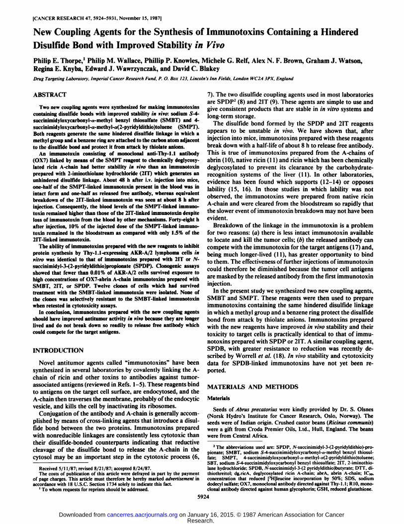
was consistent with the structure shown in Fig. 2 (Ci2Hi2NOgS2Na).
CH,
Fig. 2. The SMBT reagent (formula weight, 399.4).
Synthesis of SMPT
/>-EthyIbenzoic acid was a-brominated and converted to the thiosulfate derivative as described in the preceding section. The thiosulfate(6.0 g; 20 mmol) was hydrolyzed by adding 5 N HC1 (50 ml) and stirringat room temperature under nitrogen for 6 h. The reaction mixture wasthen extracted three times with ethyl acetate (50 ml) and dried overanhydrous sodium sulfate. The solvent was then removed in a vacuumto leave a-thioethylbenzoic acid (3.0 g; 16 mmol; 80%) as a white solidwhich was stored under nitrogen.
2-Pyridinesulfenylchloride was prepared by bubbling chlorine gasthrough a solution of 2,2-dipyridyldisuIfide (2.3 g; 10 mmol) in drydichloromethane (20 ml) for 30 min at room temperature. The solventwas then removed by rotary evaporation under reduced pressure and asolution of a-thioethylbenzoic acid (1.9 g; 10 mmol) in dry dioxan (10ml) was added. The mixture was stirred vigorously overnight at roomtemperature under nitrogen. The yellow solid produced during thereaction was then partitioned between 0.05 M sodium phosphate bufferand ethyl acetate keeping the pH constant at 7.0. The organic layerwas removed, dried over anhydrous sodium sulfate, and the solventremoved by rotary evaporation under reduced pressure to leave a yellowoil. Recrystallization from ethyl acetate/dichloromethane yielded colorless crystals (m.p. 130-132°C).The analysis
Requires: C 57.71, H 4.50, N 4.81, S 22.01Found: C 57.92, H 4.63, N 4.89, S 21.99
was consistent with the product being 4-carboxy-a-methyl-«-(2-pyri-dyldithio)toluene (C,4H13NO2S2).
To a solution of the 2-pyridyldithio derivative (1.9 g; 6.6 mmol) indry dioxan (10 ml) were added dicyclohexylcarbodiimide (1.4 g; 6.8mmol) and A'-hydroxysuccinimide (0.79 g; 6.8 mmol) each dissolved in
dry dioxan (approximately 4 ml). The mixture was stirred for 4 h atroom temperature, filtered to remove the urea, and the solvent removedby rotary evaporation under reduced pressure. The product was purifiedby short column chromatography on Silica Gel H. Elution was effectedwith a gradient of CH2Cl2/ethyl acetate from 0 to 50% (v/v). Onremoval of the solvent, a colorless oil (0.6 g; 1.5 mmol; 24%) remained.
5925
Research. on January 16, 2015. © 1987 American Association for Cancercancerres.aacrjournals.org Downloaded from
HINDERED DISULFIDE COUPLING AGENTS
The product was homogeneous when analyzed by thin layer chromatography (Si<>•,ethyl ;tcetak-:< 11 ( I,, 1:1) but attempts to crystallize it
were unsuccessful. The overall yield for the synthesis was 2%. Nuclearmagnetic resonance and infra-red analyses showed ¿H(a, methyl alcohol) 8.36 (IH, m, pyridyl); 7.96 (2H, m, phenyl); 7.73 to 7.53 (5H, m,phenyl and pyridyl); 7.06 (IH, m, pyridyl); 4.30 (IH, m, CHCH3); 2.96(4H, S, jV-hydroxysuccinimide ester); and 1.76 (3H, m, CHCH3) *„»,(CH2C12) 2920, 1770, 1740, and 1605 cnT1. These analyses were
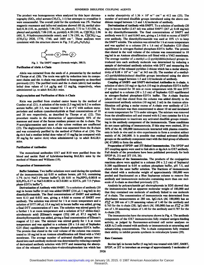
consistent with the structure shown in Fig. 3 (C,8H|6N2Q,S2).
CH3
Fig. 3. The SMPT reagent (formula weight, 388.5).
Purification of Abrin A-Chain
Abrin was extracted from the seeds of A. precatorius by the methodof Thorpe et al. (20). The toxin was split by reduction into its component chains and the A-chain was purified to homogeneity as describedpreviously (21). The purified toxin and the A-chain (abrA) had medianlethal dose values of 1.4 /ig/kg and 12 mg/kg, respectively, whenadministered i.p. to adult BALB/c mice.
Deglycosylation and Purification of Ricin A-Chain
Ricin was purified from crushed castor beans by the method ofCumber« al. (21). A solution of the toxin (2.5 mg/ml) in 0.2 M sodiumacetate buffer, pH 3.5, was treated for l h at 4°Cwith sodium meta-
periodate and sodium cyanoborohydride at final concentrations of 10and 20 mM respectively, as described by Thorpe et al. (22). Thisprocedure results in the destruction of approximately 50% of themannose and most of the fucose residues present on the A-chain. TheyV-acetylglucosamine and most of the xylose residues are unaffected(23). The deglycosylated ricin A-chain was separated from the B-chainand was extensively purified by the method of Fulton et al. (24). Thedg.ricA had a median lethal dose value of 15 mg/kg (as compared with30 mg/kg for native ricA) when administered i.p. to adult BALB/cmice.
Purification of Antibodies
The monoclonal antibodies OX7 and RIO were purified from theblood and ascitic fluid of hybridoma-bearing BALB/c mice by themethod of Mason and Williams (19).
Preparation of Immunotoxins
Buffer Solutions. Two buffer solutions were used during the synthesisof the immunotoxins: (a) 0.05 M sodium borate, pH 9.0, containing1.7% (w/v) NaCI ("borate buffer"); (b) 0.01 M Na2HPO4-0.0018 MKH2PO«-0.17M NaCl-0.0034 M KC1-0.001 M EDTA, pH 7.5 ("phos-phate-EDTA buffer").
Derivatization of Antibody with SMBT. To a solution of antibody (20mg) in borate buffer (4 ml) was added SMBT (216 //I: 1 mg/ml) in drydimethylformamide. The final concentrations of SMBT and antibodywere 0.13 and 0.032 mM, giving a 4-fold M excess of SMBT overantibody. The solution was stirred for l h at room temperature and asolution of DM' (40 i/1; 15.4 mg/ml) in borate buffer was added, giving
a final DTT concentration of 1 mM. The solution was stirred gently fora further l h at room temperature and a solution of 5,5'-dithio-bis(2-nitrobenzoic acid) [Ellman's reagent (25)] (40 pi; 87.2 mg/ml) indimethylformamide was added, giving a final concentration of Ellman's
reagent of 2.2 mM. The mixture was stirred gently for l h at roomtemperature and was applied to a column (20 x 1.6 cm) of SephadexG25 (fine) equilibrated in nitrogen-flushed phosphate-EDTA buffer.The protein that eluted in the void volume of the column was concentrated to 10 mg/ml in an Amicon ultrafiltration cell fitted with a YM2membrane. The average number of activated disulfide groups introduced into each antibody molecule was determined by reducing a sampleof derivatized antibody solution with DTT and measuring the absorption of the released 3-carboxylato-4-nitrothiophenolate ion which has
a molar absorptivity of 1.36 x IO4 M"1 cm"1 at 412 nm (25). The
number of activated disulfide groups introduced using the above conditions ranged between 1.5 and 1.8/molecule of antibody.
Derivatization of Antibody with SMPT. To a solution of antibody (20mg) in borate buffer (2.67 ml) was added SMPT (267 Ml;0.48 mg/ml)in dry dimethylformamide. The final concentrations of SMPT andantibody were 0.11 and 0.045 mM, giving a 2.4-fold Mexcess of SMPTover antibody. The dimethylformamide was used at 10% v/v to keepthe SMPT soluble. The solution was stirred for l h at room temperatureand was applied to a column (30 x 1.6 cm) of Sephadex G25 (fine)equilibrated in nitrogen-flushed phosphate-EDTA buffer. The proteinthat eluted in the void volume of the column was concentrated to 10mg/ml in an Amicon ultrafiltration cell fitted with a YM2 membrane.The average number of a-methyl-a-(2-pyridyldithio)toluoyl groups introduced into each antibody molecule was determined by reducing asample of derivatized antibody solution with DTT and measuring theabsorption of the released pyridine-2-thione which has a molar absorptivity of 8.08 x IO3 M~' cnT' at 343 nm (8). The number of a-methyl-
a(2-pyridyldithio)toluoyl disulfide groups introduced using the aboveconditions ranged between 1.5 and 2.0/molecule of antibody.
Coupling of SMBT- and SMPT-derivatized Antibodies to A-chain. Asolution of abrA (10 mg) or dg.ricA (10 mg) in phosphate-EDTA buffer(7 ml) was treated for 30 min at room temperature with 50 mM DTTand applied to a column (30 x 2.2 cm) of Sephadex G25 equilibratedin nitrogen-flushed phosphate-EDTA buffer. The A-chain fraction(about 35 ml) that eluted from the column was added directly to theconcentrated antibody solution (10 mg/ml; 2 ml) in the Amicon ultra-filtration cell giving a molar excess of A-chain over antibody of 2.5-fold. The mixture was then concentrated to about 10 ml and incubatedat room temperature for 72 h under nitrogen. The mixture was removedfrom the ultrafiltration cell and treated with 0.2 mM cysteine for 6 h atroom temperature to inactivate any activated disulfide groups remaining in the antibody component of the immunotoxin. These conditionsdo not cause splitting of immunotoxin. If this step were omitted, 20 to30% of the M, 180,000 immunotoxin interacted with plasma constituents in both in vivo and in vitro experiments to form a covalent adducimainly of M, 240,000. It is possible that residual activated disulfidegroups on the antibody component react with the thiol group of albumin(M, 67,000) to form the adduci.
Preparation of SPDP- and 2IT-linked Immunotoxins. The SPDP and2IT coupling agents were used to link abrA or dg.ricA to OX7 antibody.Full details of the procedures have been published previously for bothSPDP (6, 21) and 2IT (10, 26).
Purification of the Immunotoxins. The products of the conjugationreactions above were applied to a column (90 x 2.2 cm) of SephacrylS-200 equilibrated in 0.05 M sodium phosphate buffer, pH 7.5, andeluted with the same buffer solution. The fractions of immunotoxinthat eluted with a molecular weight of approximately 180,000 werepooled and fractionated on a Blue Sepharose column to remove freeantibody and immunotoxin molecules containing more than one molecule of A-chain as described previously (27).
Analysis by polyacrylamide gel electrophoresis in SDS showed thatthe immunotoxins had an apparent molecular weight of 180,000 andthat they contained one molecule of antibody linked to one moleculeof A-chain. The concentration of immunotoxin was determined fromabsorbance measurements at 280 nm. IgG-ricA (M, 180,000) has an1C!,1,,,at 280 nm of 1.29 assuming values of 1.40 for the antibody and0.765 for the A-chain (28). IgG-abrA (M, 180,000) has an E°t^at 280
nm of 1.30 assuming values of 1.40 for the antibody and 0.787 for theA-chain (28).
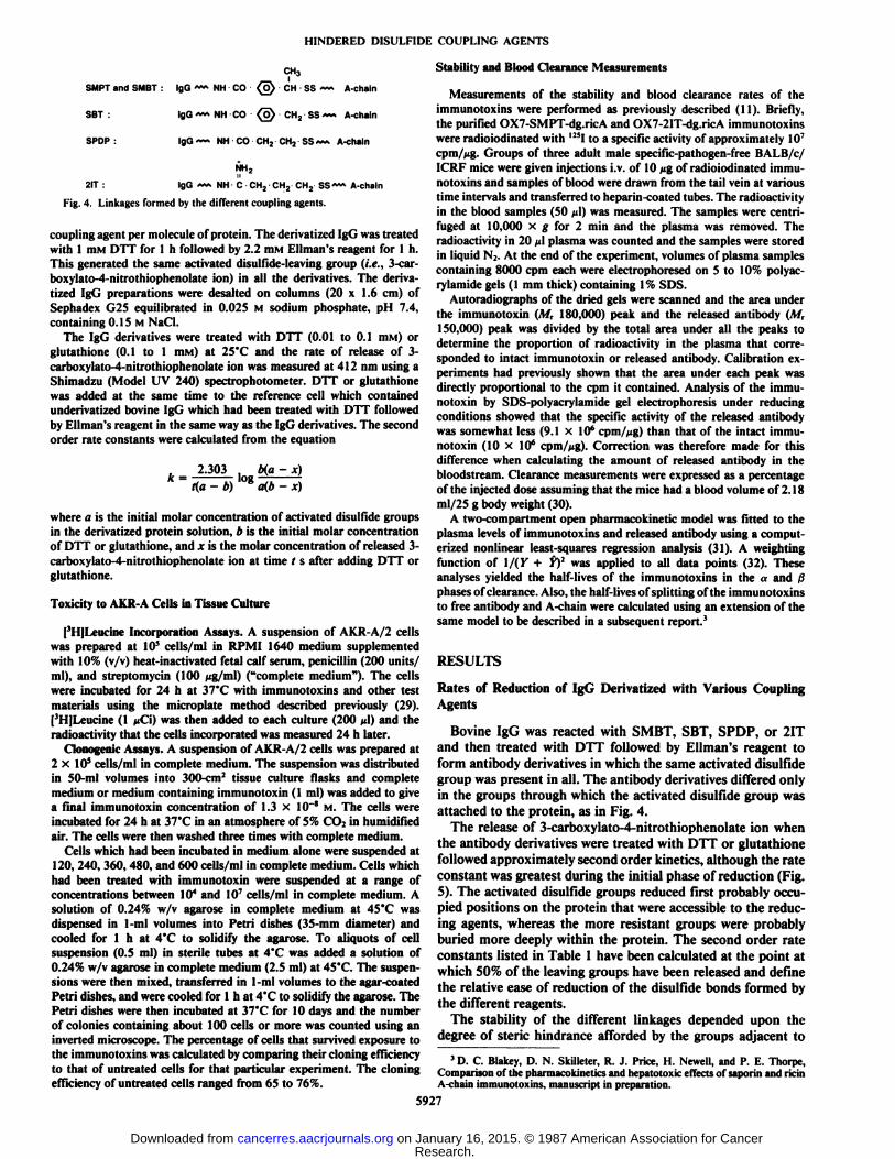
The immunotoxins have the structures shown in Fig. 4. The antibodycomponents of the OX7 immunotoxins fully retained antigen-bindingactivity, as judged by fluorescence-activated cell sorter analyses onAKK A/2 cells treated with antibody or immunotoxin at saturating andsubsaturating concentrations. The A-chain components fully retainedtheir ability to inhibit protein synthesis in reticulocyte lysates (29).
Rate Constants
Bovine IgG in borate buffer (5 mg/ml) was treated with SBT, SMBT,SPDP, or 2IT to introduce an average of approximately 5 molecules of
5926
Research. on January 16, 2015. © 1987 American Association for Cancercancerres.aacrjournals.org Downloaded from
HINDERED DISULFIDE COUPLING AGENTS
CH3
SMPT and SMBT : IgG — NH CO (o) CH SS— A-chain
SBT : IgG — NH CO (0) CH2 SS — A-chain
SPOP : lgG~~ NH CO CH2 CH2 SS~~ A-chain
2IT :
NH2
IgG — NH C CH2 CHj CH2 SS~~ A-chain
Fig. 4. Linkages formed by the different coupling agents.
coupling agent per molecule of protein. The derivatized IgG was treatedwith 1 mM DTT for l h followed by 2.2 mM Ellman's reagent for 1 h.
This generated the same activated disulfide-leaving group (i.e., 3-car-boxylato-4-nitrothiophenolate ion) in all the derivatives. The derivatized IgG preparations were desalted on columns (20 x 1.6 cm) ofSephadex G2S equilibrated in 0.025 M sodium phosphate, pH 7.4,containing 0.15 M NaCl.
The IgG derivatives were treated with DTT (0.01 to 0.1 mM) orglutathione (0.1 to 1 mM) at 25°Cand the rate of release of 3-
carboxylato-4-nitrothiophenolate ion was measured at 412 nm using aShimadzu (Model UV 240) spectrophotometer. DTT or glutathionewas added at the same time to the reference cell which containedunderivatized bovine IgG which had been treated with DTT followedby Ellman's reagent in the same way as the IgG derivatives. The second
order rate constants were calculated from the equation
2.303 b(a-x)t(a - b) °8a(b - x)
where a is the initial molar concentration of activated disulfide groupsin the derivatized protein solution, b is the initial molar concentrationof DTT or glutathione, and x is the molar concentration of released 3-carboxylato-4-nitrothiophenolate ion at time l s after adding DTT orglutathione.
Toxicity to AKR-A Cells in Tissue Culture
[3H]Leucine Incorporation Assays. A suspension of AKk A/2 cellswas prepared at 10s cells/ml in KI'.MI 1640 medium supplementedwith 10% (v/v) heat-inactivated fetal calf serum, penicillin (200 units/ml), and streptomycin (100 /ig/ml) ("complete medium"). The cellswere incubated for 24 h at 37°Cwith immunotoxins and other test
materials using the microplate method described previously (29).[3H]Leucine (1 //Ci) was then added to each culture (200 //I) and the
radioactivity that the cells incorporated was measured 24 h later.Clonogenic Assays. A suspension of AKk A/2 cells was prepared at
2 x IO5cells/ml in complete medium. The suspension was distributedin 50-ml volumes into 300-cm2 tissue culture flasks and complete
medium or medium containing immunotoxin (1 ml) was added to givea final immunotoxin concentration of 1.3 x 10 * M. The cells wereincubated for 24 h at 37°Cin an atmosphere of 5% CO2 in humidified
air. The cells were then washed three times with complete medium.Cells which had been incubated in medium alone were suspended at
120, 240, 360, 480, and 600 cells/ml in complete medium. Cells whichhad been treated with immunotoxin were suspended at a range ofconcentrations between 10' and 10 cells/ml in complete medium. Asolution of 0.24% w/v agarose in complete medium at 45*C was
dispensed in I ml volumes into Petri dishes (35-mm diameter) andcooled for l h at 4"( ' to solidify the agarose. To aliquots of cellsuspension (0.5 ml) in sterile tubes at 4"( was added a solution of0.24% w/v agarose in complete medium (2.5 ml) at 45°C.The suspen
sions were then mixed, transferred in 1-ml volumes to the agar-coatedPetri dishes, and were cooled for l h at 4°Cto solidify the agarose. ThePetri dishes were then incubated at 37*C for 10 days and the number
of colonies containing about 100 cells or more was counted using aninverted microscope. The percentage of cells that survived exposure tothe immunotoxins was calculated by comparing their cloning efficiencyto that of untreated cells for that particular experiment. The cloningefficiency of untreated cells ranged from 65 to 76%.
Stability and Blood Clearance Measurements
Measurements of the stability and blood clearance rates of theimmunotoxins were performed as previously described (11). Briefly,the purified OX7-SMPT-dg.ricA and OX7-2IT-dg.ricA immunotoxinswere radioiodinated with ' '"I to a specific activity of approximately Hi
cpm/Mg. Groups of three adult male specific-pathogen-free BALB/c/ICRF mice were given injections i.v. of 10 ng of radioiodinated immunotoxins and samples of blood were drawn from the tail vein at varioustime intervals and transferred to heparin-coated tubes. The radioactivityin the blood samples (50 /<!)was measured. The samples were centri-fuged at 10,000 x g for 2 min and the plasma was removed. Theradioactivity in 20 /'I plasma was counted and the samples were storedin liquid N2. At the end of the experiment, volumes of plasma samplescontaining 8000 cpm each were electrophoresed on 5 to 10% polyac-rylamide gels (1 mm thick) containing 1% SDS.
Autoradiographs of the dried gels were scanned and the area underthe immunotoxin (M, 180,000) peak and the released antibody (M,150,000) peak was divided by the total area under all the peaks todetermine the proportion of radioactivity in the plasma that corresponded to intact immunotoxin or released antibody. Calibration experiments had previously shown that the area under each peak wasdirectly proportional to the cpm it contained. Analysis of the immunotoxin by SDS-polyacrylamide gel electrophoresis under reducingconditions showed that the specific activity of the released antibodywas somewhat less (9.1 x IO6 cpm/jig) than that of the intact immunotoxin (10 x 10' cpm/fig). Correction was therefore made for this
difference when calculating the amount of released antibody in thebloodstream. Clearance measurements were expressed as a percentageof the injected dose assuming that the mice had a blood volume of 2.18ml/25 g body weight (30).
A two-compartment open pharmacokinetic model was fitted to theplasma levels of immunotoxins and released antibody using a computerized nonlinear least-squares regression analysis (31). A weightingfunction of \/(Y + Yf was applied to all data points (32). Theseanalyses yielded the half-lives of the immunotoxins in the a and ßphases of clearance. Also, the half-lives of splitting of the immunotoxinsto free antibody and A-chain were calculated using an extension of thesame model to be described in a subsequent report.3
RESULTS
Rates of Reduction of IgG Derivatized with Various CouplingAgents
Bovine IgG was reacted with SMBT, SBT, SPDP, or 2ITand then treated with DTT followed by Ellman's reagent to
form antibody derivatives in which the same activated disulfidegroup was present in all. The antibody derivatives differed onlyin the groups through which the activated disulfide group wasattached to the protein, as in Fig. 4.
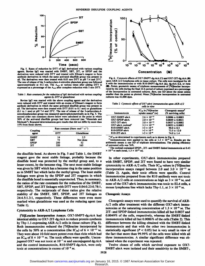
The release of 3-carboxylato-4-nitrothiophenolate ion whenthe antibody derivatives were treated with DTT or glutathionefollowed approximately second order kinetics, although the rateconstant was greatest during the initial phase of reduction (Fig.5). The activated disulfide groups reduced first probably occupied positions on the protein that were accessible to the reducing agents, whereas the more resistant groups were probablyburied more deeply within the protein. The second order rateconstants listed in Table 1 have been calculated at the point atwhich 50% of the leaving groups have been released and definethe relative ease of reduction of the disulfide bonds formed bythe different reagents.
The stability of the different linkages depended upon thedegree of steric hindrance afforded by the groups adjacent to
3 D. C. Blakey, D. N. Skilleter, R. J. Price, H. Newell, and P. E. Thorpe,
Comparison of the pharmacokinetics and hepatotoxic effects of saporin and ricinA-chain immunotoxins, manuscript in preparation.
5927
Research. on January 16, 2015. © 1987 American Association for Cancercancerres.aacrjournals.org Downloaded from
HINDERED DISULFIDE COUPLING AGENTS
eO•*-•oTI
100-1
75-
50-
25-
0J
200 400 600 800
Time (secs)Fig. 5. Rates of reduction by DTT of IgG derivatized with various coupling
agents. Bovine IgG was reacted with SMBT, SBT, 2IT, or SPDP and thederivatives were reduced with DTT and treated with Ellman's reagent to form
antibody derivatives in which the same activated disulfide group was present inall. The derivatives were then treated with 0.03 mm DTT at pH 7.4 and 25'C.The rate of release of the 3-carboxylato-4-nitrothio phenolate group was followedspectrometrically at 412 nm. The A.,,., at various times after adding DTT isexpressed as a percentage of the A4i: after complete reduction with S mM DTT.
Table 1 Rale constants for the reduction of IgG derivatized with various couplingagents by DTT or glutathione
Bovine IgG was reacted with the above coupling agents and the derivativeswere reduced with DTT and treated with an excess of Ellman's reagent to form
antibody derivatives in which the same activated disulfide group was present inall. The derivatives were then treated with DTT (0.01 to O.I mM) or glutathione(0.1 to 1 HIM)at pH 7.4 and 25'C. The rate of release of the 3-carboxylato-4-
nitrothiophenolate group was measured spectrophotometrically as in Fig. 5. Thesecond order rate constants shown below were calculated at the point at which50% of the activated disulfide groups had been removed (see "Materials andMethods"). Repeated determinations gave results that did not differ by more than
15% from those shown.
100
CouplingagentSMBT
SBTSPDP2ITRate
constant (liters -mol'-s"1)DTT
Glutathione14
2.572 Not done
250 52320 165
the disulfide bond. As shown in Fig. 5 and Table 1, the SMBTreagent gave the most stable linkage, probably because thedisulfide bond was protected by the methyl group and, to alesser extent, by the benzene ring. The next most stable linkagewas given by SBT which has a benzene ring in the same positionas in SMBT but which lacks the methyl group. The least stablelinkages were given by the SPDP and 2IT reagents in whichthe disulfide bond is essentially unprotected. Thus, in summary,the ratios of the rate constants for the reduction of the SMBT,SBT, SPDP, and 2IT linkages with DTT were 0.04:0.23:0.78:1,respectively. The reciprocals of these ratios give the relativestability of the SMBT, SBT, SPDP, and 2IT linkages as24:4.5:1.3:1, respectively. These differences were even moremarked when glutathione was used as the reducing agent (seeTable 1).
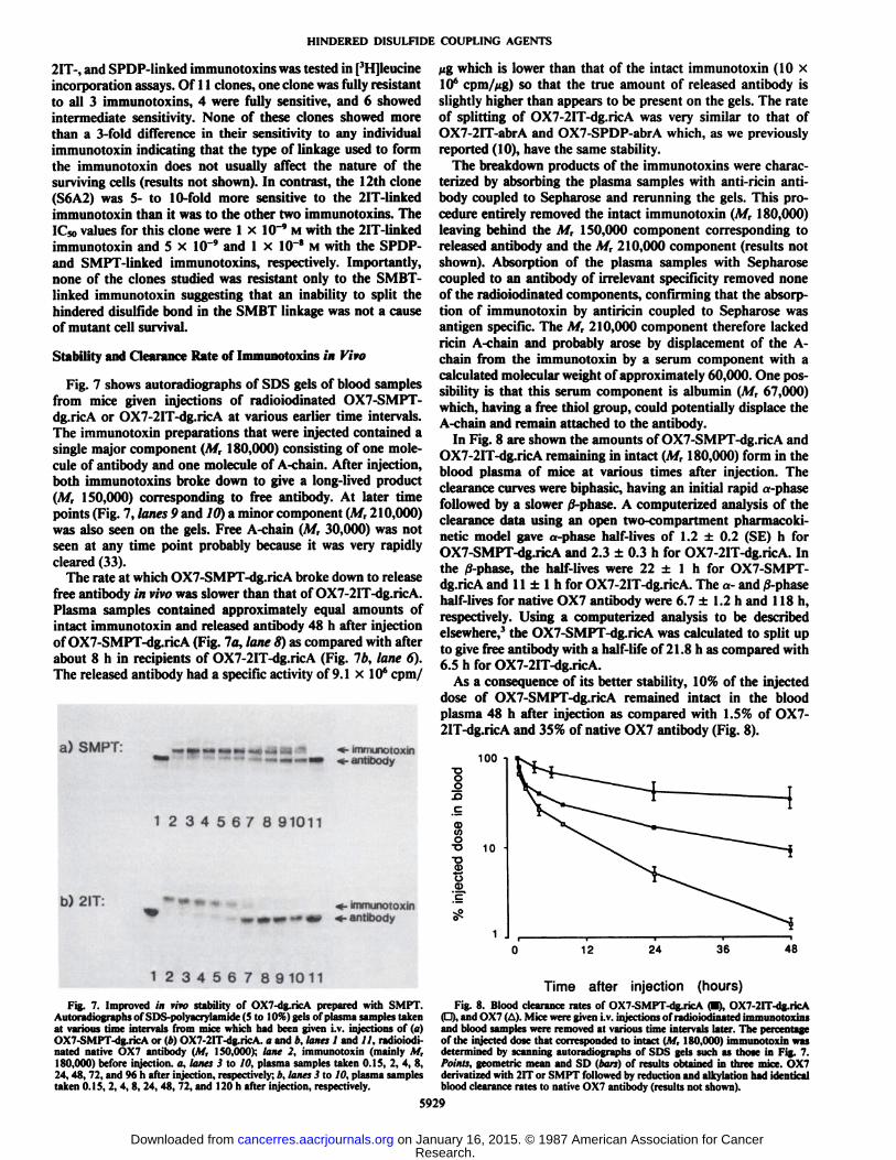
Cytotoxicity to AKR-A/2 Lymphoma Cells|'lI|lA-nain- Incorporation Assays. OX7-SMPT-dg.ricA had
identical ability to OX7-2IT-dg.ricA to reduce protein synthesisby Thy-1.1-expressing AKR-A/2 cells in tissue culture (Fig. 6).Both immunotoxins reduced the [3H]leucine incorporated bythe cells by 50% at a concentration (the IC50) of 6 x 10~13M.
They were about 10-fold more potent even than ricin which hadan IC50 of 8 x 10~12M. The toxic effects were specific. Uncon-jugated OX7 was not toxic at 10~7M and unconjugated dg.ricA
and the control immunotoxin, RIO-SMPT-dg.ricA, were onlytoxic at concentrations in excess of 10~8M.
o —o. o5 I
0) —C O
ì â5V "-
SO
1013 1012io'11 io'10 109 108 io7
Concentration (M)
10-6
Fig. 6. Cytotoxic effects of OX7-SMPT-dg.ricA (O) and OX7-2IT-dg.ricA (•)upon AKR-A/2 lymphoma cells in tissue culture. The cells were incubated for 48h with the immunotoxins or with RIO-SMPT-dg.ricA (A), dg.ricA (D), or ricin(•).Points, geometric means of triplicate measurements of | 'I Ijleueinc incorporated by the cells during the tinnì24-h period of culture expressed as a percentageof the incorporation in untreated cultures, linn, one SD about the mean unlesssmaller than the points as plotted. Mean [3H]leucine incorporation in untreatedcultures was 42,000 dpm.
Table 2 Cytotoxic effects ofOX7-abrA immunotoxins upon AKR-A/2cells in vitro
ImmunotoxinOX7-SMBT-abrA
OX7-SPDP-abrAOX7-2IT-abrAOX7-abrA cocktail'RIO-SMBT-abrARIO-SPDP-abrAR10-2IT-abrA1C»
in | '1nii-ucmc Clonogenic assays*uptake assays" (M) (% survivingcells)2.6
x 10-2.0 x 10-3.0 x 10-3.3 x 10->3 x 10->3 x 10->3 x 10-'2
0.0086 ±0.0003
0.0049 ±0.00212 0.0037 ±0.00022 0.0053 ±0.0003
90.0 ±8.573.5 ±12.074.0 ±1.4
" 1C»as determined in experiments such as is shown in Fig. 6.*Immunotoxins were applied to the cells at 1.3 x in " M. Results are the
arithmetic means ±one SD of triplicate determinations. The plating efficiencyof untreated cells was 76%.
' The cocktail contained SPDP-, 2IT- and SMBT-linked immunotoxin at 0.43x 10~' M each (total, 1.3 x 10~*M).
In other experiments, OX7-abrA immunotoxins preparedwith SMBT, SPDP, and 2IT were found to have very similarcytotoxicity to AKR-A/2 cells. Their IC50 values in [3H]leucineincorporation assays ranged between 2.0 and 3.0 x 10~12 M
(Table 2). Again, their toxic effects were specific. Controlimmunotoxins prepared from the RIO antibody were not toxicto AKR-A/2 cells at concentrations as high as 3 x 10~" M, and
none of the OX7-abrA immunotoxins was toxic to EL4 cells, amouse lymphoma line which lacks Thy-1.1, at 3 x 10~8 M.
Clonogenic Assays
Clonogenic assays were used to quantify the survival of AKR-A/2 cells after treatment with the different OX7-abrA immunotoxins at the saturating concentration of 1.3 x 10~8 M. The
2IT- and SPDP-linked immunotoxins killed all but 0.0037 and0.0049% of the cells, respectively, whereas the SMBT-linkedimmunotoxin killed all but 0.0086% of the cells (Table 2). Thisdifference between the killing obtained with the SMBT-linkedimmunotoxin and that with the other two immunotoxins isstatistically significant (P < 0.05) but is very small in view ofthe fact that more than 99.99% of the cells were killed with allthree immunotoxins. A similar significant difference was obtained when the experiment was repeated.
Twelve clones of cells which survived exposure to OX7-SMBT-abrA were isolated and their sensitivity to the SMBT-,
5928
Research. on January 16, 2015. © 1987 American Association for Cancercancerres.aacrjournals.org Downloaded from
HINDERED DISULFIDE COUPLING AGENTS
2IT-, and SPDP-linked immunotoxins was tested in [3H]leucine
incorporation assays. Of 11 clones, one clone was fully resistantto all 3 immunotoxins, 4 were fully sensitive, and 6 showedintermediate sensitivity. None of these clones showed morethan a 3-fold difference in their sensitivity to any individualimmunotoxin indicating that the type of linkage used to formthe immunotoxin does not usually affect the nature of thesurviving cells (results not shown). In contrast, the 12th clone(S6A2) was 5- to 10-fold more sensitive to the 2IT-linkedimmunotoxin than it was to the other two immunotoxins. TheIC50 values for this clone were 1 x IO"9 M with the 2IT-linkedimmunotoxin and 5 x 10"' and 1 x 10~8 M with the SPDP-
and SMPT-linked immunotoxins, respectively. Importantly,none of the clones studied was resistant only to the SMBT-linked immunotoxin suggesting that an inability to split thehindered disulfide bond in the SMBT linkage was not a causeof mutant cell survival.
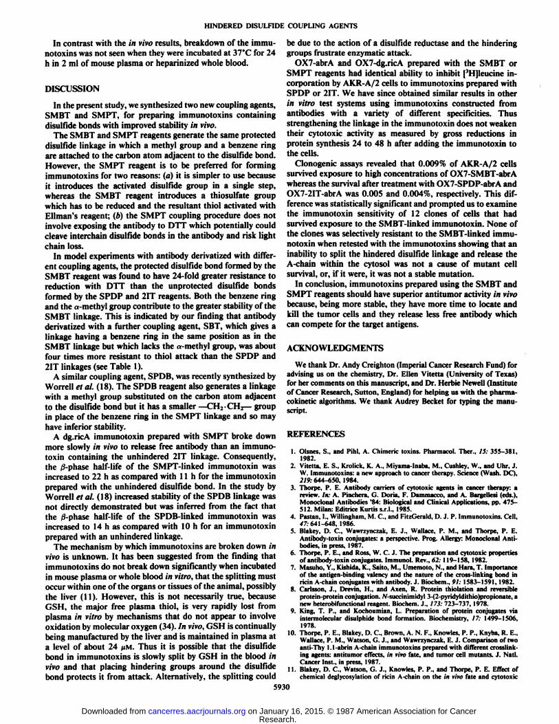
Stability and Clearance Rate of Immunotoxins in Vivo
Fig. 7 shows autoradiographs of SDS gels of blood samplesfrom mice given injections of radioiodinated OX7-SMPT-dg.ricA or OX7-2IT-dg.ricA at various earlier time intervals.The immunotoxin preparations that were injected contained asingle major component (M, 180,000) consisting of one molecule of antibody and one molecule of A-chain. After injection,both immunotoxins broke down to give a long-lived product(M, 150,000) corresponding to free antibody. At later timepoints (Fig. 7, lanes 9 and 10) a minor component (A/r 210,000)was also seen on the gels. Free A-chain (M, 30,000) was notseen at any time point probably because it was very rapidlycleared (33).
The rate at which OX7-SMPT-dg.ricA broke down to releasefree antibody in vivo was slower than that of OX7-2IT-dg.ricA.Plasma samples contained approximately equal amounts ofintact immunotoxin and released antibody 48 h after injectionof OX7-SMPT-dg.ricA (Fig. 7a, lane 8) as compared with afterabout 8 h in recipients of OX7-2IT-dg.ricA (Fig. Ib, lane 6).The released antibody had a specific activity of 9.1 x 10" cpm/
a) SMPT: •*•immunotoxin«-antibody
1 2345678 91011
b) 2IT: «-immunotoxin4- antibody
123456789 10 11
Fig. 7. Improved in vivo stability of OX7-dg.ricA prepared with SMPT.Autoradiographs of SDS-polyacrylamide (5 to 10%) gels of plasma samples takenat various time intervals from mice which had been given i.v. injections of (a)OX7-SMPT-dg.ricA or (*) OX7-2IT-dg.ricA. a and b, lanes 1 and //, radioiodinated native OX7 antibody (M, 150,000); lane 2, immunotoxin (mainly M,
MÕÕwhich is lower than that of the intact immunotoxin (10 xIO6 cpm//ig) so that the true amount of released antibody is
slightly higher than appears to be present on the gels. The rateof splitting of OX7-2IT-dg.ricA was very similar to that ofOX7-2IT-abrA and OX7-SPDP-abrA which, as we previouslyreported (10), have the same stability.
The breakdown products of the immunotoxins were characterized by absorbing the plasma samples with anti-ricin antibody coupled to Sepharose and rerunning the gels. This procedure entirely removed the intact immunotoxin (M, 180,000)leaving behind the M, 150,000 component corresponding toreleased antibody and the M, 210,000 component (results notshown). Absorption of the plasma samples with Sepharosecoupled to an antibody of irrelevant specificity removed noneof the radioiodinated components, confirming that the absorption of immunotoxin by antiricin coupled to Sepharose wasantigen specific. The M, 210,000 component therefore lackedricin A-chain and probably arose by displacement of the A-
chain from the immunotoxin by a serum component with acalculated molecular weight of approximately 60,000. One possibility is that this serum component is albumin (\f, 67,000)which, having a free thiol group, could potentially displace theA-chain and remain attached to the antibody.
In Fig. 8 are shown the amounts of OX7-SMPT-dg.ricA andOX7-2IT-dg.ricA remaining in intact (M, 180,000) form in theblood plasma of mice at various times after injection. Theclearance curves were biphasic, having an initial rapid »-phasefollowed by a slower /3-phase. A computerized analysis of theclearance data using an open two-compartment pharmacoki-netic model gave a-phase half-lives of 1.2 ±0.2 (SE) h forOX7-SMPT-dg.ricA and 2.3 ±0.3 h for OX7-2IT-dg.ricA. Inthe /3-phase, the half-lives were 22 ±1 h for OX7-SMPT-dg.ricA and 11 ±l h for OX7-2IT-dg.ricA. The a- and 0-phasehalf-lives for native OX7 antibody were 6.7 ±1.2 h and 118 h,respectively. Using a computerized analysis to be describedelsewhere,3 the OX7-SMPT-dg.ricA was calculated to split upto give free antibody with a half-life of 21.8 h as compared with6.5 h for OX7-2IT-dg.ricA.
As a consequence of its better stability, 10% of the injecteddose of OX7-SMPT-dg.ricA remained intact in the bloodplasma 48 h after injection as compared with 1.5% of OX7-2IT-dg.ricA and 35% of native OX7 antibody (Fig. 8).
100
CD(Oo•o•§10
12 24 36 48
Time after injection (hours)Fig. 8. Blood clearance rates of OX7-SMPT-dg.ricA (•).OX7-2IT-dg.ricA
(D). and OX7 (A). Mice were given i.v. injections of radioiodinated immunotoxinsand blood samples were removed at various time intervals later. The percentageof the injected dose that corresponded to intact (M, 180,000) immunotoxin wasdetermined by scanning autoradiographs of SDS gels such as those in Fig. 7.
180,000) before injection, a, lanes 3 to 10, plasma samples taken 0.1S, 2, 4, 8, Points, geometric mean and SD (bars) of results obtained in three mice. OX724, 48, 72, and 96 h after injection, respectively; />.lanes 3 to 10, plasma samples derivati/od with 2IT or SMPT followed by reduction and alkylation had identicaltaken 0.15, 2, 4, 8, 24, 48, 72, and 120 h after injection, respectively. blood clearance rates to native OX7 antibody (results not shown).
5929
Research. on January 16, 2015. © 1987 American Association for Cancercancerres.aacrjournals.org Downloaded from
HINDERED DISULFIDE COUPLING AGENTS
In contrast with the in vivo results, breakdown of the immu-notoxins was not seen when they were incubated at 37°Cfor 24
h in 2 ml of mouse plasma or heparinized whole blood.
DISCUSSION
In the present study, we synthesized two new coupling agents,SMBT and SMPT, for preparing immunotoxins containingdisuHìilebonds with improved stability in vivo.
The SMBT and SMPT reagents generate the same protecteddisulfide linkage in which a methyl group and a benzene ringare attached to the carbon atom adjacent to the disulfide bond.However, the SMPT reagent is to be preferred for formingimmunotoxins for two reasons: (a) it is simpler to use becauseit introduces the activated disulfide group in a single step,whereas the SMBT reagent introduces a thiosulfate groupwhich has to be reduced and the resultant thiol activated withEllman's reagent; (b) the SMPT coupling procedure does not
involve exposing the antibody to DTT which potentially couldcleave interchain disulfide bonds in the antibody and risk lightchain loss.
In model experiments with antibody derivatized with different coupling agents, the protected disulfide bond formed by theSMBT reagent was found to have 24-fold greater resistance toreduction with DTT than the unprotected disulfide bondsformed by the SPDP and 2IT reagents. Both the benzene ringand the «-methylgroup contribute to the greater stability of theSMBT linkage. This is indicated by our finding that antibodyderivatized with a further coupling agent, SBT, which gives alinkage having a benzene ring in the same position as in theSMBT linkage but which lacks the a-methyl group, was aboutfour times more resistant to thiol attack than the SPDP and2IT linkages (see Table 1).
A similar coupling agent, SPDB, was recently synthesized byWorrell et al. (18). The SPDB reagent also generates a linkagewith a methyl group substituted on the carbon atom adjacentto the disulfide bond but it has a smaller —CH2-CH2—groupin place of the benzene ring in the SMPT linkage and so mayhave inferior stability.
A dg.ricA immunotoxin prepared with SMPT broke downmore slowly in vivo to release free antibody than an immunotoxin containing the unhindered 2IT linkage. Consequently,the /3-phase half-life of the SMPT-linked immunotoxin wasincreased to 22 h as compared with 11 h for the immunotoxinprepared with the unhindered disulfide bond. In the study byWorrell et al. (18) increased stability of the SPDB linkage wasnot directly demonstrated but was inferred from the fact thatthe 0-phase half-life of the SPDB-linked immunotoxin wasincreased to 14 h as compared with 10 h for an immunotoxinprepared with an unhindered linkage.
The mechanism by which immunotoxins are broken down invivo is unknown. It has been suggested from the finding thatimmunotoxins do not break down significantly when incubatedin mouse plasma or whole blood in vitro, that the splitting mustoccur within one of the organs or tissues of the animal, possiblythe liver (11). However, this is not necessarily true, becauseGSH, the major free plasma thiol, is very rapidly lost fromplasma in vitro by mechanisms that do not appear to involveoxidation by molecular oxygen (34). In vivo, GSH is continuallybeing manufactured by the liver and is maintained in plasma ata level of about 24 UM. Thus it is possible that the disulfidebond in immunotoxins is slowly split by GSH in the blood invivo and that placing hindering groups around the disuifidebond protects it from attack. Alternatively, the splitting could
be due to the action of a disulfide reducÃaseand the hinderinggroups frustrate enzymatic attack.
OX7-abrA and OX7-dg.ricA prepared with the SMBT orSMPT reagents had identical ability to inhibit [3H]leucine incorporation by AKR-A/2 cells to immunotoxins prepared withSPDP or 2IT. We have since obtained similar results in otherin vitro test systems using immunotoxins constructed fromantibodies with a variety of different specificities. Thusstrengthening the linkage in the immunotoxin does not weakentheir cytotoxic activity as measured by gross reductions inprotein synthesis 24 to 48 h after adding the immunotoxin tothe cells.
Clonogenic assays revealed that 0.009% of AKR-A/2 cellssurvived exposure to high concentrations of OX7-SMBT-abrAwhereas the survival after treatment with OX7-SPDP-abrA andOX7-2IT-abrA was 0.005 and 0.004%, respectively. This difference was statistically significant and prompted us to examinethe immunotoxin sensitivity of 12 clones of cells that hadsurvived exposure to the SMBT-linked immunotoxin. None ofthe clones was selectively resistant to the SMBT-linked immunotoxin when retested with the immunotoxins showing that aninability to split the hindered disulfide linkage and release theA-chain within the cytosol was not a cause of mutant cellsurvival, or, if it were, it was not a stable mutation.
In conclusion, immunotoxins prepared using the SMBT andSMPT reagents should have superior antitumor activity in vivobecause, being more stable, they have more time to locate andkill the tumor cells and they release less free antibody whichcan compete for the target antigens.
ACKNOWLEDGMENTS
We thank Dr. Andy Creighton (Imperial Cancer Research Fund) foradvising us on the chemistry, Dr. Ellen Vitetta (University of Texas)for her comments on this manuscript, and Dr. Herbie Newell (Instituteof Cancer Research, Sutton, England) for helping us with the pharma-cokinetic algorithms. We thank Audrey Becket for typing the manuscript.
REFERENCES
1. Olsnes. S., and Pihl. A. Chimeric toxins. Pharmacol. Ther., IS: 355-381,1982.
2. Vitetta, E. S., Krolick, K. A., Miyama-Inaba. M., Cushley, W.. and Uhr, J.W. Immunotoxins: a new approach to cancer therapy. Science (Wash. DC),2/9.-644-650, 1984.
3. Thorpe, P. E. Antibody carriers of cytotoxic agents in cancer therapy: areview. In: A. Pinchera, G. Doria, F. Dammacco, and A. Bargellesi (eds.).Monoclonal Antibodies '84: Biological and Clinical Applications, pp. 475-
512. Milan: Editrice Kurtis s.r.l., 1985.4. Pastan, I., Willingham, M. < . and FitzGerald, D. J. P. Immunotoxins. Cell,
¿7:641-648, 1986.5. Blakey, D. C, Wawrzynczak, E. J., Wallace, P. M., and Thorpe, P. E.
Antibody-toxin conjugates: a perspective. Prog. Allergy: Monoclonal Antibodies, in press, 1987.
6. Thorpe, P. E., and Ross, W. C. J. The preparation and cytotoxic propertiesof antibody-toxin conjugates. Immunol. Rev., 62: 119-158. 1982.
7. Masuho, Y., Kishida, K., Saito, M., Umemoto, N., and Hará,T. Importanceof the antigen-binding valency and the nature of the cross-linking bond inricin A-chain conjugates with antibody. J. Biochem., 91: 1583-1591, 1982.
8. Carlsson, J., Drevin, II.. and Axen, R. Protein thiolation and reversibleprotein-protein conjugation. N-succinimidyl 3-(2-pyridyldithio)propionate. anew heterobifunctional reagent. Biochem. J., 173: 723-737, 1978.
9. King, T. P., and Kochoumian, I . Preparation of protein conjugates viaintermolecular disulphide bond formation. Biochemistry, 17: 1499-1506,1978.
10. Thorpe, P. E., Blakey, D. C., Brown, A. N. F., Knowles, P. P., Knyba, R. E.,Wallace, P. M., Watson, G. J., and Wawrzynczak, E. J. Comparison of twoanti-Thy 1.1-abrin A-chain immunotoxins prepared with different crosslink-ing agents: antitumor effects, in rito fate, and tumor cell mutants. J. Nati.Cancer Inst., in press, 1987.
11. Blakey. D. C., Watson, G. J., Knowles, P. P., and Thorpe, P. E. Effect ofchemical deglycosylation of ricin A-chain on the in vivo fate and cytotoxic
5930
Research. on January 16, 2015. © 1987 American Association for Cancercancerres.aacrjournals.org Downloaded from

HINDERED DISULFIDE COUPLING AGENTS
activity of an immunotoxin composed of ricin A-chain and anti-Thy 1.1antibody. Cancer Res., 47:947-952, 1987.
12. Strand, M., Scheinberg, D. A., and Gansow, O. A. Monoclonal antibodyconjugates for tumor imaging and therapy. In: R. F. Beers and E. G. Bassett(eds.), Cell Fusion: Gene Transfer and Transformation, pp. 385-393. NewYork: Raven Press, 1984.
13. Worrell, N. R., Cumber, A. J., Parnell, G. D., Ross, W. C. J., and Forrester,J. A. Fate of an antibody-ricin A chain conjugate administered to normalrats. Biochem. Pharmacol., 55:417-423, 1986.
14. Letvin, N. L., Goldmacher, V. S., Ritz, J., Yetz, J. M., Schlossman, S. F.,and Lambert. J. M. In vivoadministration of lymphocyte-specific monoclonalantibodies in nonhuman primates. In vivo stability of disulphide-linkedimmunotoxin conjugates. J. Clin. Invest., 77:977-984, 1986.
15. Raso, V., and Basala, M. Monoclonal antibodies as cell targeted carriers ofemail-ill K and non-covalently attached toxins. NATO Adv. Study Inst. Ser.A Life Sci., 82: 119-138,1984.
16. Bourrie, B. J. P., Casellas. P., Blythman, H. E., and Jansen, F. K. Study ofthe plasma clearance of antibody-ricin A-chain immunotoxins. Evidence forspecific recognition sites on the A-chain that mediate rapid clearance of theimmunotoxin. Eur. J. Biochem., 755:1-10, 1986.
17. Blakey, D. C, and Thorpe, P. E. An overview of therapy with immunotoxinscontaining ricin or its A-chain. Antibody, Immunoconjugates, Radiophar-maceuticals, 1: in press, 1987.
18. Worrell, N. R., Cumber, A. J., Parnell, G. D., Mirza, A., Forrester, J. A.,and Ross, W. C. J. Effect of linkage variation on pharmacokinetics of ricinA chain-antibody conjugates in normal rats. Anticancer Drug Design, /:179-188, 1986.
19. Mason, D. W., and Williams, A. F. The kinetics of antibody binding tomembrane antigens in solution and at the cell surface. Biochem. J., 187: i20, 1980.
20. Thorpe, P. E., Cumber, A. J., Williams, N., Edwards, D. C., Ross, W. C. J.,and Davies, A. J. S. Abrogation of the nonspecific toxicity of abrin conjugatedto anti-lymphocyte globulin. Clin. Exp. Immunol., 43: 195-200. 1981.
21. Cumber, A. J., Forrester, J. A., Foxwell, B. M. J., Ross, W. C. J., andThorpe. P. E. Preparation of antibody-toxin conjugates. Methods Enzymol..112: 207-225, 1985.
22. Thorpe, P. E., Detre, S. I., Foxwell, B. M. J., Brown, A. N. F., Skilleter, D.N., Wilson, G., Forrester, J. A., and Stirpe, F. Modification of the carbohydrate in ricin with metaperiodate-cyanoborohydride mixtures. Effects ontoxicity and in vivo distribution. Eur. J. Biochem., 147:197-206, 1985.
23. Vitella, E. S., and Thorpe, P. E. Immunotoxins containing ricin A or Bchains with modified carbohydrate residues act synergistically in killingneoplastic B cells in vitro. Cancer Drug Delivery, 2:191-198, 1985.
24. Fulton, J. R., Blakey, D. C., Knowles, P. P., Uhr, J. W., Thorpe, P. E., andVitetta, E. S. Purification of ricin Ai, A2 and B-chains and characterizationof their toxicity. J. Biol. Chem., 261: 5314-5319, 1986.
25. Ellman, G. L. Tissue sulphydryl groups. Arch. Biochem. Biophys., 82: 70-77, 1959.
26. Wawrzynczak, E. J., and Thorpe, P. E. Methods for preparing immunotoxins:effects of the linkage on activity and stability. In: C. W. Vogel (ed.), Immunoconjugates: Antibody Conjugates in Radioimaging and Therapy of Cancer.New York: Oxford University Press, pp. 28-55, 1987.
27. Knowles, P. P., and Thorpe, P. E. Purification of immunotoxins containingricin A-chain and abrin A-chain using Blue Sepharose CL-6B. Anal.Biochem., 160:440-443, 1987.
28. Olsnes, S., Refsnes. K., Christensen, T. B., and Phil, A. Studies on thestructure and properties of the lectins from Abrus precatorius and Ricinuscommuni!. Biochim. Biophys. Acta, 405: 1-10, 1975.
29. Thorpe, P. E., Brown, A. N. F., Ross, W. C. J., Cumber, A. J., Detre, S. I.,Edwards, D. C., Davies, A. J. S., and Stirpe, F. Cytotoxicity acquired byconjugation of an anti-Thy 1.1 monoclonal antibody and the ribosome-inactivating protein, gelonin. Eur. J. Biochem., 116:447-454,1981.
30. Wish, L., I unii. J., and Storey, R. H. Direct determinations of plasma, cell,and organ-blood volumes in normal and hypervolemic mice. Proc. Soc. Exp.Biol. Med., 74:644-648. 1950.
31. Jennrich, R. J., and Sampson, P. F. Application of stepwise regression tonon-linear least-squares estimation. Technometrics, 10:63-12, 1968.
32. Ottaway, J. H. Normalization in the fitting of data by iterative methods.Biochem. J., 134: 729-744, 1973.
33. Blakey, D. C., and Thorpe, P. E. Effect of chemical deglycosylation on thein vivo fate of ricin A-chain. Cancer Drug Delivery, 3: 189-196, 1986.
34. Anderson, M. E., and Meister, A. Dynamic state of glutathione in bloodplasma. J. Biol. Chem., 255: 9530-9533, 1980.
5931
Research. on January 16, 2015. © 1987 American Association for Cancercancerres.aacrjournals.org Downloaded from
1987;47:5924-5931. Cancer Res Philip E. Thorpe, Philip M. Wallace, Phillip P. Knowles, et al. in VivoContaining a Hindered Disulfide Bond with Improved Stability New Coupling Agents for the Synthesis of Immunotoxins
Updated version
http://cancerres.aacrjournals.org/content/47/22/5924
Access the most recent version of this article at:
E-mail alerts related to this article or journal.Sign up to receive free email-alerts
Subscriptions
Reprints and
To order reprints of this article or to subscribe to the journal, contact the AACR Publications
Permissions
To request permission to re-use all or part of this article, contact the AACR Publications
Research. on January 16, 2015. © 1987 American Association for Cancercancerres.aacrjournals.org Downloaded from
Copyright © 2022 FDOKUMEN