
Microbial study of Casar de Cáceres cheese throughout ripening
NEF
SAa
Cb
vC
A(idNceraaat2artcmlpctlcelwFbrsmNr
*“3EAtcrsst
Please cite this article in press as: de Olmos S, et al., Neurodegeneration and prolonged immediate early gene expression through-out cortical areas of the rat brain following acute administration of dizocilpine, Neuroscience (2009), doi: 10.1016/j.neuro-science.2009.09.022
Neuroscience xx (2009) xxx
0d
ARTICLE IN PRESS
EURODEGENERATION AND PROLONGED IMMEDIATE EARLY GENEXPRESSION THROUGHOUT CORTICAL AREAS OF THE RAT BRAIN
OLLOWING ACUTE ADMINISTRATION OF DIZOCILPINEKs
PciAdp(Npewh
i(itrdvl(raa1idstca
dhotmnRoato
. DE OLMOS,a C. BENDER,a J. S. DE OLMOSa
ND A. LORENZOa,b*
Instituto de Investigación Médica Mercedes y Martín Ferreyra (INIMEC–ONICET), Friuli 2434, 5016-Córdoba, Argentina
Departamento de Farmacología, Facultad de Ciencias Químicas, Uni-ersidad Nacional de Córdoba, Haya de la Torre y Medina Allende,iudad Universitaria, 5000-Córdoba, Argentina
bstract—N-methyl-D-aspartate receptor antagonist drugsNMDA-A), such as dizocilpine (MK801), induce long-last-ng behavioral disturbances reminiscent to psychotic disor-ers in humans. To identify cortical structures affected byMDA-A, we used a single dose of MK801 (10 mg/kg) thataused low and high neurodegeneration in intact and orchi-ctomized male rats, respectively. Degenerating somas (neu-onal death) and axonal/synaptic endings (terminal degener-tion) were depicted by a silver technique, and functionallyffected cortical neuronal subpopulations by Egr-1, c-Fos,nd FosB/�FosB-immunolabeling. In intact males, MK801riggered a c-Fos induction that remained high for more than4 h in selected layers of the retrosplenial, somatosensorynd entorhinal cortices. MK801-induced neurodegenerationeached its peak at 72 h. Degenerating somas were restrictedo layer IV of the granular subdivision of the retrosplenialortex, and were accompanied by suppression of Egr-1 im-unolabeling. Terminal degeneration extended to selected
ayers of the retrosplenial, somatosensory and parahip-ocampal cortices, which are target areas of retrosplenialortex. Induction of FosB/�FosB by MK801 also extended tohe same cortical layers affected by terminal degeneration,ikely reflecting the damage of synaptic connectivity. In or-hiectomized males, the neurodegenerative and functionalffects of MK801 were exacerbated. Degenerative somas inayer IV of the retrosplenial cortex significantly increased,ith a parallel enhancement of terminal degeneration andosB/�FosB-expression in the mentioned cortical structures,ut no additional areas were affected. These observationseveal that synaptic dysfunction/degeneration in the retro-plenial, somatosensory and parahippocampal corticesight underlie the long-lasting impairments induced byMDA-A. © 2009 IBRO. Published by Elsevier Ltd. All rights
eserved.
Corresponding author. A. Lorenzo, Instituto de Investigación MédicaM y M Ferreyra”, Friuli 2434 - 5016-Córdoba, Argentina. Tel: �54-51-468-1466; fax: �54-351-469-5163.-mail address: [email protected] (A. Lorenzo).bbreviations: A–Cu–Ag, amino–cupric–silver; ENT, entorhinal cor-
ex; FJB, fluoro-jade B; IEGs, immediate early genes; MK801, dizo-ilpine; NHS, normal horse serum; NMDA-A, N-methyl-D-aspartateeceptor antagonists; ORC, orchiectomized; PBS, phosphate-bufferedaline; RSD, dysgranular retrosplenial cortex; RSG, granular retro-
eplenial cortex; V1, primary visual cortex; V2L, secondary visual cor-ex, lateral area; V2MM, secondary visual cortex, mediomedial area.
306-4522/09 $ - see front matter © 2009 IBRO. Published by Elsevier Ltd. All rightoi:10.1016/j.neuroscience.2009.09.022
1
ey words: Egr-1, FosB, MK-801, neurodegeneration, retro-plenial cortex, somatosensory cortex.
hencyclidine, ketamine and dizocilpine (MK801) are disso-iative anaesthetics that belong to the family of non-compet-
tive N-methyl-D-aspartate receptor-antagonist drugs (NMDA-). In humans, aside of their anaesthetic properties, theserugs induce behavioural changes reminiscent of schizo-hrenia symptoms that, in same cases, can persist for weeksLuby et al., 1959; Jentsch and Roth, 1999). Therefore,MDA-A are widely used as a pharmacological model ofsychosis (Farber, 2003). In addition, NMDA-A are consid-red potential drugs of abuse because they are illicitly usedith recreational purposes due to the psychedelic and/orallucinogenic effects (Morgan et al., 2009).
In the rat, a single application of MK801 (�4 mg/kg)nduces behavioural alterations that remain even after a weekWöhrl et al., 2007; Manahan-Vaughan et al., 2008), indicat-ng that functional changes persist long after the exposure tohe drug. The neuroanatomical substrate affected by MK801emains elusive, and might provide insights into psychoticisorders. Low doses of MK801 (�3 mg/kg) induce transientacuolization and induction of heat shock proteins in selected
ayers of the granular subdivision of the retrosplenial cortexRSG) (Olney et al., 1989; Sharp et al., 1991), indicating aeversible stress in these neurons. The induction of immedi-te early gene proteins (IEGs) after low doses of MK801 waslso documented in the RSG (Gass et al., 1993; Gao et al.,998; Zhang et al., 1999), further indicating the functional
nvolvement of this cortical structure. MK801-dependent in-uction of IEGs has also been evidenced in the somatosen-ory and parahipppocampal cortices, the hippocampus, andhalamic nuclei (Vaisanen et al., 2004), but whether thesehanges are independent or linked to the functional alter-tions in RSG remains unknown.
In the female rat, MK801 (�3 mg/kg) causes neuronaleath particularly in the RSG, but also in olfactory structures,ippocampus and parahippocampal cortex. In addition, ax-nal and synaptic degeneration also affects olfactory struc-
ures, hippocampus, retrosplenial, parahippocampal and so-atosensory cortices (Bueno et al., 2003) indicating that theeurodegenerative effect of the drug expands far beyond theSG. Interestingly, due to the protective effect of testoster-ne, male rats are much less vulnerable to MK801-toxicity,nd somatodendritic degeneration is confined exclusively to
he RSG (de Olmos et al., 2008). Thus, the neurotoxic effectf MK801 in the male rat might offer a unique opportunity to
valuate the impact of the selective damage of RSG neuronss reserved.
ot
asIsbeaacpnntM2KcgKiM
rfdw(tawtmo
A
Edhmgcmfoa
M
IvddSpa2s
ahnw1Mswa
P
Fcfossset(wtO
D
NtrwsjTddtstwtasf0apmdb
Ssemwfscma0otps
S. de Olmos et al. / Neuroscience xx (2009) xxx2
ARTICLE IN PRESS
n the anatomofunctional alterations in target cortical struc-ures.
IEGs are transcription factors widely used for thenatomofunctional identification of the substrates that re-pond to a stimulus. Combining the analysis of differentEGs might provide a better strategy to identify neuronalubpopulations that are affected by conditions that induceoth, transient and prolonged functional alterations. Forxample, due to its quick and transitory expression, c-Fosllows the identification of neurons that respond to ancute stimulus, but is less useful for persistent or chroniconditions (Kovács, 1998). On the contrary, FosB, andarticularly �FosB proteins, are appropriate IEGs to trackeuronal populations reacting to chronic conditions or toeuroplastic changes that persist long-after the exposure
o the stimulus (Chen et al., 1997; Nestler et al., 1999;cClung et al., 2004; Carle et al., 2007; Perrotti et al.,008). On the other hand, Egr-1 (also known as zif 268,rox-24, NGFI-A), which exhibits a high basal expression,an be a marker of abnormal synaptic activity or neurode-eneration (Beckmann and Wilce, 1997; Knapska andaczmarek, 2004). Curiously, there are no studies analys-
ng the expression of IEGs after a neurotoxic dose ofK801 that induces long-lasting changes.
The aim of our study was to link MK801-induced neu-onal death in the retrosplenial cortex with the anatomo-unctional changes in target cortical areas. We used aose of MK801 that induces death of RSG-neurons thatas sparse in intact males but profuse in orchidectomized
ORC) animals and, concomitantly, low and high levels oferminal degeneration aroused in the retrosplenial cortexnd target cortical areas. To analyse neurodegeneration,e employed a silver technique that depicted both, soma-
odendritic and terminal degeneration. Additionally, anato-ofunctional changes were evidenced by immunostainingf three different IEGs, c-Fos, FosB/�FosB and Egr-1.
EXPERIMENTAL PROCEDURES
nimals
xperiments were performed on male Wistar rats from the Institutoe Investigación Médica Mercedes y Martín Ferreyra vivarium,oused with food and water ad libitum. At 45 days of age, a group ofale rats were anaesthetized i.p. with 5% chloral hydrate (0.5 ml/100body weight) and bilaterally orchiectomized, and returned to their
ages until use. Experiments were carried out with intact and ORCales of 70 to 76 days, weighing 315–350 g. All experiments con-
ormed to named local and international guidelines on the ethical usef animals. Every effort was made to minimize discomfort of thenimals and the overall number of animals used.
K801 treatment
ntact and ORC males received either a single i.p. injection ofehicle, 0.9% of sodium chloride (NaCl) (control), or 10 mg/kg ofizocilpine hydrogen maleate, (�)-5-methyl-10,11-dihydro-5H-ibenzo[a,d]cyclohepten-5,10-imine hydrogen maleate (MK801;igma-Aldrich, St. Louis, MO, USA). This dose of MK801 reliablyroduces irreversible neurodegeneration in adult male rats (Fix etl., 1993, 1995). Behavioural signs of MK801 treatment appeared–4 min after the injection, which included increased locomotion,
tereotyped head weaving, backward and forward movements, cPlease cite this article in press as: de Olmos S, et al., Neurodegeneratiout cortical areas of the rat brain following acute administration of dizocscience.2009.09.022
taxia and recumbency. After 24 h of MK801 treatment, animalsad completely recovered, moving around in their cages and didot present any significant weight loss. Two animals per groupere used for the study of the time course (3, 8, 24, 48, 72 and20 h) of the neurodegeneration and c-Fos expression induced byK801 in intact and ORC rats. The survival time for the following
et of experiments analysing FosB/FosB and Egr-1 expressionas carried out at the peak of the neurodegeneration, which wast 72 h post-treatment, with a minimum of four animals per group.
erfusion and histological procedure
or brain fixation, all animals were anaesthetized i.p. with 30%hloral hydrate; perfused transcardially and fixed with 4% para-ormaldehyde in 0.2 M borate buffer (pH 7.4). Brains were leftvernight in the skull and afterward removed and placed in 30%ucrose. Once the brains sank in the sucrose, serial sagittalections of 40 �m were obtained in a freezing microtome andtored at 4 °C until processing. Four series of sister sections wereither stored in phosphate-buffered saline (PBS) 0.01 M in ordero immediately process for immunohistochemistry or fluoro-jade BFJB) technique (Schmued and Hopkins, 2000) and the fifth seriesas stored in 4� % paraformaldehyde in order to asses neuro-
oxicity using the amino-cupric-silver technique (A–Cu–Ag) (delmos et al., 1994).
etection of neuronal degeneration
euronal degeneration was analysed by two different methods,he A–Cu–Ag, and the FJB techniques. The A–Cu–Ag is a moreecent version of the cupric–silver method (de Olmos et al., 1981)hich is suitable for staining degenerating perikarya, dendrites,tem axons and their terminal ramifications in brain tissue sub-ected to different experimental and neuropathological conditions.he procedure was carried out following the protocol previouslyescribed (de Olmos et al., 1994). Briefly, sections were rinsed inouble-distilled water and incubated in a pre-impregnating solu-ion of silver nitrate at 50 °C. After cooling to room temperature,ections were rinsed with acetone and transferred to a concen-rated impregnating silver diamine solution for 40 min. Sectionsere then immersed in a reducing formaldehyde/citric acid solu-
ion for 25 min and then the reaction was stopped in 0.5% aceticcid. Bleaching was done in two steps to eliminate the non-pecific deposits of silver on the tissue, first in 6% potassiumerricyanide, washed in double-distilled water, then transferred to.06% potassium permanganate for 20 s. After washing sectionsgain, stabilization was done in 2% sodium thiosulfate, washed,laced in a fixer solution for 1 min, and then the sections wereounted and placed on a slide warmer (30 °C) until they were fullyry. The dry slides were cleared by immersion in xylene for 10 minefore coverslipping.
For FJB staining we followed the protocol described bychmued and Hopkins (2000), in which mounted brain sections onlides were immersed in a 1% sodium hydroxide solution (80%thanol) for 5 min. Slides were then placed in 70% alcohol for 2in and then rinsed in distilled water for 2 min. Afterwards slidesere transferred to a solution of 0.06% potassium permanganate
or 10 min and then rinsed in distilled water for 2 min. The stainingolution was prepared from a 0.01% stock solution of FJB (Chemi-on International, Temecula, CA, USA) that was prepared by theanufacturer’s instructions. The stock solution was diluted bydding 0.1% acetic acid, resulting in a final dye concentration of.0004%. The working stain solution was prepared within 10 minf use and was not reused. Slides were stained for 20 min andhen rinsed in distilled water (3�1 min). The slides were thenlaced on a slide warmer (50 °C) until they were fully dry. The drylides were cleared by immersion in xylene for 2 min before
overslipping.on and prolonged immediate early gene expression through-ilpine, Neuroscience (2009), doi: 10.1016/j.neuro-
I
SsMwbbpnnBirsutFtbiaPfKF0ASadwabh
In
AsiniflsnOIpTva
cvscfiscietracW
0(na
S
Cbgrasmc
Na
I0c1eassttsHnto
eatanbaaoa(dntir(v((spsd
S. de Olmos et al. / Neuroscience xx (2009) xxx 3
ARTICLE IN PRESS
mmunohistochemistry and antibodies
ections were first incubated for 1 h at room temperature in aolution of 3% hydrogen peroxide and 10% methanol in PBS 0.01
to quench endogenous peroxidase. Thereafter, the sectionsere washed three times in 0.01 M PBS and incubated in alocking solution of 5% normal horse serum (NHS) for 1 h. Afterlocking, sections were directly incubated for 48 h at 4 °C with aolyclonal antibody against, c-Fos (s.c.-52, Santa Cruz Biotech-ology, CA, USA, 1:1000); Egr-1 (s.c.-110, Santa Cruz Biotech-ology, CA, USA, 1:1500) or FosB/FosB (s.c.-48, Santa Cruziotechnology, CA, USA, 1:1500) diluted in 0.01 M PBS contain-
ng 1% NHS. The FosB/FosB antibody was raised against theesidues 75–150 of the FosB molecule and has been demon-trated, by Western blotting analyses, that proteins with a molec-lar weight corresponding to �FosB can be recognized. Becausehe drug was administered 72 h before analysis, we considered allosB-like immunoreactivity, detected with the pan-FosB antibody,
o reflect FosB/FosB (Perrotti et al., 2004, 2005). Following incu-ation in the primary antibody, sections were washed three times
n 0.01 M PBS and incubated for 2 h in biotinylated secondaryntibody (Vector Laboratories, CA, USA); diluted 1:200 in 0.01 MBS containing 1% NHS, washed three times in 0.01 M PBS, and
ollowed by an avidin–biotin–peroxidase complex (Vectastain ABCit, Vector Laboratories, CA, USA) for 1 h at room temperature.inally, sections were incubated for 5 min with a solution containing.05% 3–3=-diamino-benzidine tetra hydrochloride (DAB, Sigma-ldrich, St. Louis, MO, USA) and 0.01% hydrogen peroxidase.ections were mounted onto gelatine-coated slides, dehydratednd cover slipped prior to viewing with a light microscope. Forouble staining of FosB/�FosB and A–Cu–Ag technique, sectionsere first stained with the A–Cu–AG technique and immediatelyfter stabilizing in thiosulfate, sections were placed in 5% NHS forlocking, followed by the protocol described above for immuno-istochemistry.
mage analysis and quantitative assessment ofeuronal number
rgyrophilic somas revealed by the A–Cu–Ag technique were as-essed at 10� and 20� objective lens. At these magnifications
ntense silver stain of the cell body and processes of degeneratingeurons were easily identified, as well as the terminal degeneration
n several cortical areas. FJB positive cells were visualized in auorescent microscope (Carl Zeiss, Jena, Germany) and corre-ponding sections of A–Cu–Ag presented approximately the sameumber of cells. A previous study (Fix et al., 1995; Auer, 1996; delmos et al., 2008) showed that neuropathological changes in layer
V of the RSG induced by MK801 increases caudally, with an ap-roximate peak between �5.3 and 7.3 mm respect to Bregma.herefore, serial sagittal sections were considered optimum for re-ealing the extent of degeneration induced by MK801 within the RSGnd other cortical areas.
Quantitative assessment was accomplished with a light mi-roscope at 20� objective lens, equipped with a Leica LC200ideo camera, which acquired and saved images with a Photo-hop 7.0 program. Afterwards, positive immunoreactive cells wereounted in selected cortical brain areas using the SCION programrom the NIH. The counting was carried out by a treatment blindnvestigator and performed using a predefined area of an identicalize (0.16 mm2) and shape for each brain region. Automatedounts of IEG positive nuclei were obtained from each area ofnterest, maintaining constant background intensity across differ-nt areas. Counts from the left and right hemisphere were ob-
ained from three sections within layer IV of the RSG, dysgranularetrosplenial cortex (RSD), secondary visual cortex, mediomedialnd lateral areas, (V2MM and V2L), and layer III of the entorhinalortex (ENT). The lateral medial (coordinates from Paxinos and
atson, 2007) sections included for detailed analysis were lateral tPlease cite this article in press as: de Olmos S, et al., Neurodegeneratiout cortical areas of the rat brain following acute administration of dizocscience.2009.09.022
.9; 1.13 and 1.4 (RSG and RSD); lateral 1.55, 1.9 and 2.10 mmV2MM); lateral 4.20, 4.32, 4.50 mm (V2L and ENT). The meanumber of IEG positive cells per animal was used for statisticalnalysis.
tatistical analysis
ounts obtained from the left and right hemisphere in differentrain sections throughout an area of interest were averaged toenerate one value of IEG positive nuclei per region. Data from eachegion were analysed using a three-way ANOVA (treatment�rea�orchiectomy) or two-way ANOVA (layer�treatment). Resultshowing significant overall changes were subjected to post hoc New-an–Keuls test with values of P�0.05 being considered as statisti-
ally significant.
RESULTS
eurotoxic effect of MK801 in cortical areas of intactnd ORC male rats
ntact and ORC male rats were treated with a single i.p. of.9% NaCl (control) or 10 mg/kg of MK801 and the time-ourse of neurodegeneration was evaluated at 24, 48, 72 and20 h post-treatment. In adjacent sections, neurotoxicity wasxamined with the A–Cu–Ag and FJB techniques. In controlnimals, no FJB- or A–Cu–Ag-positive neurons were ob-erved in any cortical area at any time point. On the contrary,ystematic neuronal degeneration was observed in MK801-reated animals, in which dying somas were confined mainlyo layer IV of the RSG, with a similar pattern of degeneratingomas depicted with both FJB and A–Cu–Ag techniques.owever, degenerating dendrites, axons and synaptic termi-als were only revealed by using the A–Cu–Ag method, and
herefore this technique was selected for further descriptionf the neurodegenerative effect of MK801.
In the RSG, somatic and argyrophilic-terminal degen-ration was observed in MK801-treated animals at 24 h,nd increased throughout the course of the 72 h post-reatment in both intact and ORC rats, although degener-tion was more conspicuous in the later (Fig. 1). Signs ofeurotoxicity were greatly reduced at 120 h post-treatment inoth intact and ORC rats, and this was accompanied by andvanced profile of neuronal disintegration, with shrunkenrgyrophilic cell bodies, and fragmented dendrites and ax-ns, indicating that neurodegeneration reached its peakround 72 h post-treatment in both intact and ORC ratsFig. 1). In ORC males, in addition to the dying somasescribed in layer IV of RSG, few and sparse degeneratingeuronal somas were also found in layer III of ENT and inhe posterolateral cortical amygdaloid nucleus. In both,ntact and ORC rats, argyrophilic-terminal degenerationeached its peak at 72 h, affecting layers I, IV–V of RSGFig. 1), layers I and IV of RSD, areas of the secondaryisual cortex, including mediomedial (V2MM), mediolateralV2ML) and lateral (V2L) parts, and primary visual cortexV1). Argyrophilic-terminal degeneration was also ob-erved in the temporal cortex, association area (TeA),erirhinal cortex (PRh), ENT, postsubiculum (Post) and in theubiculum (S) (Fig. 2). The pattern of the argyrophilic-terminalegeneration was similar in intact and ORC males, although
he intensity was much greater in the latter. Altogether, these
on and prolonged immediate early gene expression through-ilpine, Neuroscience (2009), doi: 10.1016/j.neuro-

rglmn
Ees
WMI
(2n2bOoesIE
Fs11aati
S. de Olmos et al. / Neuroscience xx (2009) xxx4
ARTICLE IN PRESS
esults show that, in male rats, MK801-induced somatic de-eneration was almost exclusively confined to neurons in
ayer IV of RSG, and was accompanied by concomitant ter-inal degeneration in several cortical areas that are con-ected to the retrosplenial cortex.
ffect of an acute neurotoxic dose of MK801 on thexpression of Egr-1 in different cortical braintructures in intact and ORC male rats
e analyzed the effect of a single neurotoxic dose ofK801 (10 mg/kg) on the expression of Egr-1, which is an
ig. 1. Time-course of MK801-induced neurodegeneration in the RSGingle i.p. dose of 0.9% NaCl (control) or MK801 (10 mg/kg), and neuro20 h post-treatment. Control animals (not shown) have no signs of n.2 mm) of intact and ORC males at the indicated time-points. At 24 hrgyrophilic cell bodies with corkscrew dendrites (insert). At 72 h podvanced cell death, with shrunken cells bodies and fragmented denerminal degeneration expands to layers I, IV and V. Increased intenndicated in the upper-left panel. Scale bar�200 �m.
EG that has been implicated in normal synaptic activity l
Please cite this article in press as: de Olmos S, et al., Neurodegeneratiout cortical areas of the rat brain following acute administration of dizocscience.2009.09.022
Beckmann and Wilce, 1997; Knapska and Kaczmarek,004), and is altered in processes of neuroplasticity and/oreurodegeneration (Thiriet et al., 2001; Slattery et al.,005). The expression of Egr-1 was conspicuous in mostrain structures, with no apparent differences in intact andRC males treated with saline (control), indicating thatrchiectomy per se had no significant effect on the basalxpression of Egr-1. In control animals (intact and ORC),trong immunoreactivity of Egr-1 was observed in layers II,II, IV and VI of RSG (Fig. 3A), RSD, V2MM and V2L. TheNT also revealed strong basal expression of Egr-1 in
t and ORC male rats. Intact and ORC male rats were treated with aneration was analyzed with the A–Cu–Ag technique at 24, 48, 72 andneration. Shown are representative sagittal sections of the RSG (lat.
atment early signs of neurodegeneration are observed, evidenced byent neurodegeneration reaches the peak, accompanied by signs ofote that somatic degeneration is confined to layer IV of RSG, whileuronal degeneration is observed in ORC males. Cortical layers are
of intacnal dege
eurodegepost-tre
st-treatmdrites. Nsity of ne
ayers III, V and VI. At 72 h post MK801-treatment, in which
on and prolonged immediate early gene expression through-ilpine, Neuroscience (2009), doi: 10.1016/j.neuro-
tstwmariiswdsaoTsaPctdPc
teMc
Ecc
Ibmstiidiaclam(peTpttn
taTitaatiFaEsViiatitwM
gto�
FefrtapllAcDpvp
S. de Olmos et al. / Neuroscience xx (2009) xxx 5
ARTICLE IN PRESS
he peak of the neurotoxic effect is observed, the expres-ion of Egr-1 in intact male rats was reduced in layer IV ofhe RSG, with noticeable anatomical correspondenceith the degenerating somas detected by the A–Cu–Agethod. No appreciable changes in Egr-1 expression werepparent in other cortical layers. To further analyze theelation of Egr-1-expression with neuronal death, Egr-1mmunoreactivity was examined in ORC rats. Similar tontact males, MK801-treated ORC animals also exhibited atriking inhibition of Egr-1 in layer IV of RSG (Fig. 3A, B),hich was anatomically coincident with the presence ofying cell bodies (Fig. 3B, D). Egr-1-positive nuclei werecored in control and MK801-treated males, and statisticalnalysis confirmed a significant reduction of Egr-1 in layer IVf RSG, but not in the other cortical areas analyzed (Fig. 3C).he three-way ANOVA (treatment�area�ORC) showed aignificant effect of treatment (F1,12�36.20; P�0.00006),rea (F4,48�4.12; P�0.005), treatment�area (F4,48�6.46,�0.0003) and ORC�area (F4,48�7.71; P�0.00008). Otheromparisons were not significant. Newman–Keuls post hocest showed that MK801-treated intact and ORC animalsiffered from the respective saline groups in RSG,�0.0001. The effect of MK801 treatment was signifi-
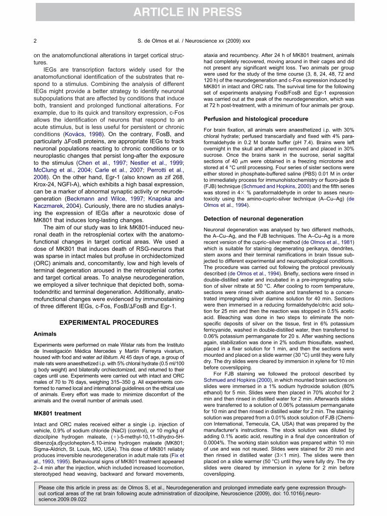
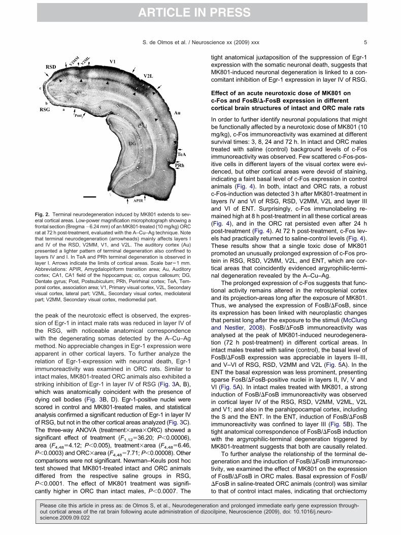
ig. 2. Terminal neurodegeneration induced by MK801 extends to sev-ral cortical areas. Low-power magnification microphotograph showing a
rontal section (Bregma �6.24 mm) of an MK801-treated (10 mg/kg) ORCat at 72 h post-treatment, evaluated with the A–Cu–Ag technique. Notehat terminal neurodegeneration (arrowheads) mainly affects layers Ind IV of the RSD, V2MM, V1, and V2L. The auditory cortex (Au)resented a lighter pattern of terminal degeneration also confined to
ayers IV and I. In TeA and PRh terminal degeneration is observed inayer I. Arrows indicate the limits of cortical areas. Scale bar�1 mm.bbreviations: APIR, Amygdalopiriform transition area; Au, Auditoryortex; CA1, CA1 field of the hippocampus; cc, corpus callosum; DG,entate gyrus; Post, Postsubiculum; PRh, Perirhinal cortex; TeA, Tem-oral cortex, association area; V1, Primary visual cortex, V2L, Secondaryisual cortex, lateral part; V2ML, Secondary visual cortex, mediolateralart; V2MM, Secondary visual cortex, mediomedial part.
antly higher in ORC than intact males, P�0.0007. The t
Please cite this article in press as: de Olmos S, et al., Neurodegeneratiout cortical areas of the rat brain following acute administration of dizocscience.2009.09.022
ight anatomical juxtaposition of the suppression of Egr-1xpression with the somatic neuronal death, suggests thatK801-induced neuronal degeneration is linked to a con-
omitant inhibition of Egr-1 expression in layer IV of RSG.
ffect of an acute neurotoxic dose of MK801 on-Fos and FosB/�-FosB expression in differentortical brain structures of intact and ORC male rats
n order to further identify neuronal populations that mighte functionally affected by a neurotoxic dose of MK801 (10g/kg), c-Fos immunoreactivity was examined at different
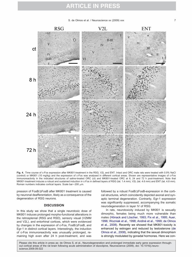
urvival times: 3, 8, 24 and 72 h. In intact and ORC malesreated with saline (control) background levels of c-Fosmmunoreactivity was observed. Few scattered c-Fos-pos-tive cells in different layers of the visual cortex were evi-enced, but other cortical areas were devoid of staining,
ndicating a faint basal level of c-Fos expression in controlnimals (Fig. 4). In both, intact and ORC rats, a robust-Fos-induction was detected 3 h after MK801-treatment inayers IV and VI of RSG, RSD, V2MM, V2L and layer IIInd VI of ENT. Surprisingly, c-Fos immunolabeling re-ained high at 8 h post-treatment in all these cortical areas
Fig. 4), and in the ORC rat persisted even after 24 host-treatment (Fig. 4). At 72 h post-treatment, c-Fos lev-ls had practically returned to saline-control levels (Fig. 4).hese results show that a single toxic dose of MK801romoted an unusually prolonged expression of c-Fos pro-ein in RSG, RSD, V2MM, V2L, and ENT, which are cor-ical areas that coincidently evidenced argyrophilic-termi-al degeneration revealed by the A–Cu–Ag.
The prolonged expression of c-Fos suggests that func-ional activity remains altered in the retrosplenial cortexnd its projection-areas long after the exposure of MK801.hus, we analysed the expression of FosB/�FosB, since
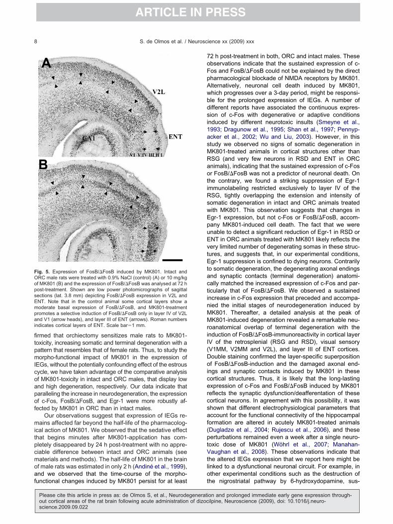
ts expression has been linked with neuroplastic changeshat persist long after the exposure to the stimuli (McClungnd Nestler, 2008). FosB/�FosB immunoreactivity wasnalysed at the peak of MK801-induced neurodegenera-ion (72 h post-treatment) in different cortical areas. Inntact males treated with saline (control), the basal level ofosB/�FosB expression was appreciable in layers II–III,nd V–VI of RSG, RSD, V2MM and V2L (Fig. 5A). In theNT the basal expression was less prominent, presentingparse FosB/�FosB-positive nuclei in layers II, IV, V andI (Fig. 5A). In intact males treated with MK801, a strong
nduction of FosB/�FosB immunoreactivity was observedn cortical layer IV of the RSG, RSD, V2MM, V2ML, V2Lnd V1; and also in the parahippocampal cortex, includinghe S and the ENT. In the ENT, induction of FosB/�FosBmmunoreactivity was confined to layer III (Fig. 5B). Theight anatomical correspondence of FosB/�FosB inductionith the argyrophilic-terminal degeneration triggered byK801-treatment suggests that both are causally related.
To further analyse the relationship of the terminal de-eneration and the induction of FosB/�FosB immunoreac-ivity, we examined the effect of MK801 on the expressionf FosB/�FosB in ORC males. Basal expression of FosB/FosB in saline-treated ORC animals (control) was similar
o that of control intact males, indicating that orchiectomy
on and prolonged immediate early gene expression through-ilpine, Neuroscience (2009), doi: 10.1016/j.neuro-

pHwFws�RMinao(t5a(iaatd((F
w(wdt
tdnasan6s(i0nsppica
F(atslS( ect of trt P�0.000
S. de Olmos et al. / Neuroscience xx (2009) xxx6
ARTICLE IN PRESS
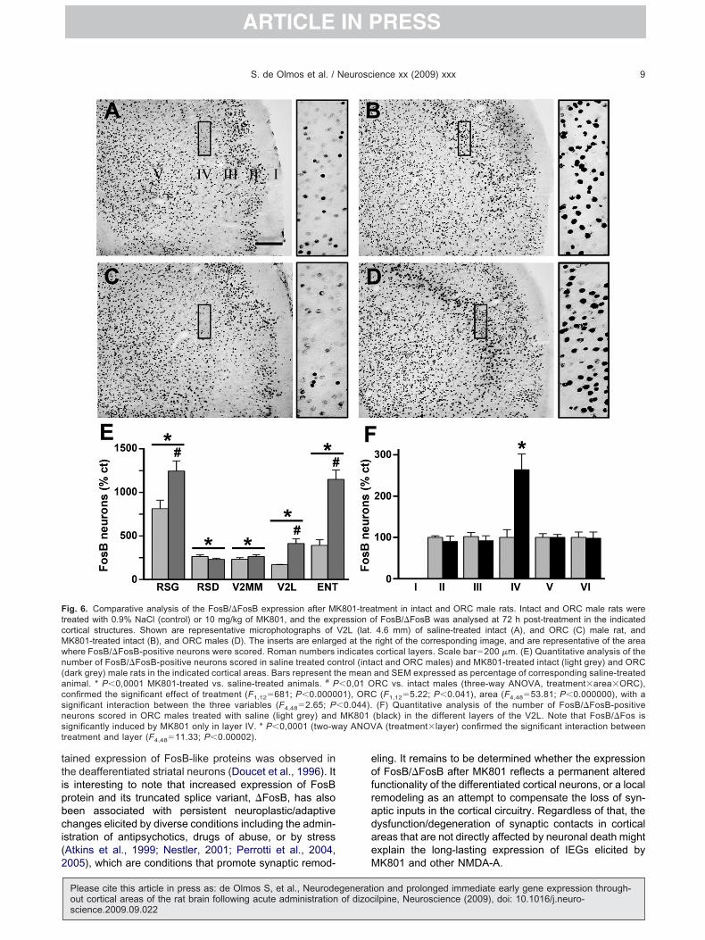
er se did not alter FosB/�FosB expression (Fig. 6A, C).owever, similar to intact males, in ORC males, treatmentith MK801 (Fig. 6D, E) triggered a dramatic increase ofosB/�FosB immunoreactivity in those cortical areashere terminal degeneration was detected by A–Cu–Agtaining. To obtain a quantitative assessment, FosB/FosB-positive nuclei were scored in layer IV of RSG,SD, V2MM, V2L and layer III of ENT in control andK801-treated intact and ORC males. Statistical compar-
sons indicated that the number of FosB/�FosB-positiveuclei significantly increases in MK801-treated animals inll cortical areas analysed, confirming the significant effectf MK801-treatment (Fig. 6E). The three-way ANOVAtreatment�area�ORC) confirmed the significant effect ofhe treatment (F1,12�681; P�0.000001), ORC (F1,12�.22; P�0.041), area (F4,48�53.81; P�0.000000), with
significant interaction between the three variablesF4,48�2.65; P�0.044). Newman–Keuls post hoc compar-sons showed no differences between saline-treated intactnd ORC rats, confirming that orchiectomy per se does notffect the basal expression of FosB/�FosB immunoreac-ivity. Both, intact and ORC animals presented significantifferences with their respective saline controls in the RSGP�0.0001), RSD (P�0.0001), V2MM (P�0.0001), V2LP�0.0001), ENT (P�0.0001). Interestingly, the number of
ig. 3. MK801 induces somatic neurodegeneration and suppression ocontrol) or MK801 (10 mg/kg) and neurodegeneration and Egr-1 expre immunocytochemical staining of Egr-1 expression in control (A) anhe A–Cu–Ag staining in an adjacent section of an ORC male (D). Thomatic degeneration in layer IV of RSG coincides with suppression oayer IV of RSG of control and MK801-treated intact (light gray) and OEM expressed as percentage of the saline-treated control animal. *
three-way ANOVA, treatment�area�ORC), showed significant effreatment�area (F4,48�6.46, P�0.0003) and ORC�area (F4,48�7.71
osB/�FosB-positive nuclei induced by MK801-treatment c
Please cite this article in press as: de Olmos S, et al., Neurodegeneratiout cortical areas of the rat brain following acute administration of dizocscience.2009.09.022
as significantly higher in ORC than intact males in RSGP�0.01), V2L (P�0.0009) and ENT (P�0.006) (Fig. 6E),hich was also coincident with the higher density of terminalegeneration detected in ORC males with the A–Cu–Ag
echnique.To confirm that the induction of FosB/�FosB after MK801
reatment is restricted to the cortical layer affected by terminalegeneration, the number of FosB/�FosB immunoreactiveuclei in all cortical layers of the V2L was scored in controlnd MK801-treated ORC males. The data indicate that,imilar to terminal degeneration, MK801-treatment inducesselective and significant increase in FosB/�FosB immu-
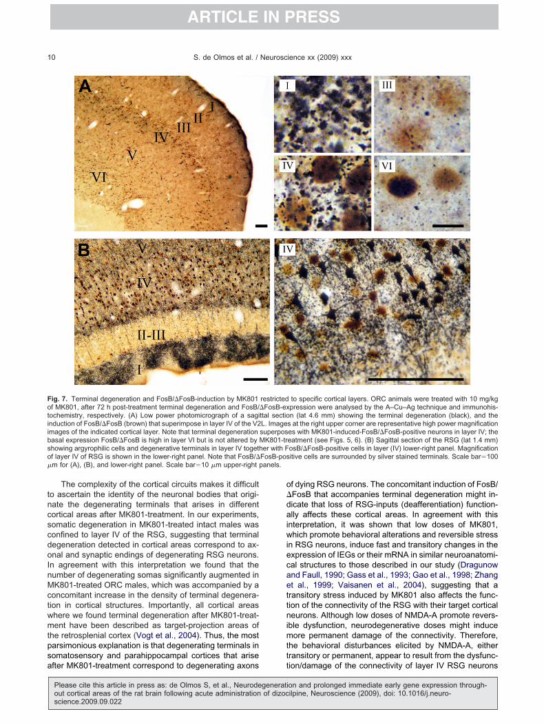
oreactivity in layer IV, but not in layers I, II, III, V or VI (Fig.F). The two-way ANOVA (treatment�layer) confirmed theignificant interaction between treatment and layerF4,48�11.33; P�0.00002). Post hoc Newman–Keuls testndicated a significant difference only in layer IV (P�.0001). To further reveal the anatomical overlap of termi-al degeneration and the FosB/�FosB-induction, doubletaining of FosB/�FosB and A–Cu–Ag technique wereerformed. Complete anatomical juxtaposition of silver-ositive degenerative terminals and FosB/�FosB-positive
mmunoreactive somas in defined layers of the mentionedortical areas was observed, as shown for layer IV of RSGnd V2L (Fig. 7). Altogether, these experiments provide
xpression in RSG. Intact and ORC male rats were treated with salineas analysed in the RSG (lat. 1.4 mm) at 72 h post-treatment. Shown-treated ORC males (B), and neuronal degeneration as evidenced byd area is enlarged at the right of the corresponding image. Note thatxpression. (C) Quantitative assessment of Egr-1 positive neurons ins (dark grey) in different cortical areas. Bars represent the mean and1 vs. saline-treated control animal, # P�00007 ORC vs. intact maleseatment (F1,12�36.20; P�0.000 06), area (F4,48�4.12; P�0.005),08). Other comparisons were not significant. Scale bar�200 �m.
f Egr-1 eression wd MK801e indicatef Egr-1 eRC maleP�0.000
ompelling support to the interpretation that enhanced ex-
on and prolonged immediate early gene expression through-ilpine, Neuroscience (2009), doi: 10.1016/j.neuro-
pbd
IMtabEom
fcawn
dm1eeO
F(iMR
S. de Olmos et al. / Neuroscience xx (2009) xxx 7
ARTICLE IN PRESS
ression of FosB/�FosB after MK801 treatment is causedy neuronal deafferentation, likely as a consequence of theegeneration of RSG neurons.
DISCUSSION
n this study we show that a single neurotoxic dose ofK801 induces prolonged morpho-functional alterations in
he retrosplenial (RSG and RSD), sensory visual (V2MMnd V2L), and entorhinal cortices, which were evidencedy changes in the expression of c-Fos, FosB/�FosB, andgr-1 in distinct cortical layers. Interestingly, the inductionf c-Fos immunoreactivity was unusually prolonged, re-
ig. 4. Time course of c-Fos expression after MK801-treatment in thecontrol) or MK801 (10 mg/kg) and the expression of c-Fos was anmmunoreactivity in the indicated structures of saline-treated ORC
K801-treatment induces a robust and sustained induction in c-Fos inoman numbers indicates cortical layers. Scale bar�200 �m.
aining high even after 24 h post-treatment, and was i
Please cite this article in press as: de Olmos S, et al., Neurodegeneratiout cortical areas of the rat brain following acute administration of dizocscience.2009.09.022
ollowed by a robust FosB/�FosB-expression in the corti-al structures, which coincidently depicted axonal and syn-ptic terminal degeneration. Contrarily, Egr-1 expressionas significantly suppressed, accompanying the somaticeurodegeneration in layer IV of RSG.
In rats, neurotoxicity induced by MK801 is sexuallyimorphic, females being much more vulnerable thanales (Hönack and Löscher, 1993; Fix et al., 1995; Auer,996; Wozniak et al., 1998; Andiné et al., 1999; de Olmost al., 2008). Recently we showed that MK801-toxicity isnhanced by estrogen and reduced by testosterone (delmos et al., 2008), indicating that the sexual dimorphism
2L and ENT. Intact and ORC male rats were treated with 0.9% NaCldifferent cortical areas. Shown are representative images of c-FosMK801-treated ORC at 8, 24 and 72 h post-treatment. Note that
layers of RSG (lat. 1.4 mm), V2L (lat. 4.6 mm) and ENT (lat. 4.8 mm).
RSG, Valysed in(ct) anddefined
s strongly modulated by gonadal hormones. Here we con-
on and prolonged immediate early gene expression through-ilpine, Neuroscience (2009), doi: 10.1016/j.neuro-
fitpmIcoapof
mitpcmoaf
7oFpAwbdsi1asMRaotiRswEpuEvtEtactinMMriI(Doicercsaf(ptVtlo
FOopsEmpai
S. de Olmos et al. / Neuroscience xx (2009) xxx8
ARTICLE IN PRESS
rmed that orchiectomy sensitizes male rats to MK801-oxicity, increasing somatic and terminal degeneration with aattern that resembles that of female rats. Thus, to study theorpho-functional impact of MK801 in the expression of
EGs, without the potentially confounding effect of the estrousycle, we have taken advantage of the comparative analysisf MK801-toxicity in intact and ORC males, that display lownd high degeneration, respectively. Our data indicate thataralleling the increase in neurodegeneration, the expressionf c-Fos, FosB/�FosB, and Egr-1 were more robustly af-
ected by MK801 in ORC than in intact males.Our observations suggest that expression of IEGs re-
ains affected far beyond the half-life of the pharmacolog-cal action of MK801. We observed that the sedative effecthat begins minutes after MK801-application has com-letely disappeared by 24 h post-treatment with no appre-iable difference between intact and ORC animals (seeaterials and methods). The half-life of MK801 in the brainf male rats was estimated in only 2 h (Andiné et al., 1999),nd we observed that the time-course of the morpho-
ig. 5. Expression of FosB/�FosB induced by MK801. Intact andRC male rats were treated with 0.9% NaCl (control) (A) or 10 mg/kgf MK801 (B) and the expression of FosB/�FosB was analysed at 72 host-treatment. Shown are low power photomicrographs of sagittalections (lat. 3.8 mm) depicting FosB/�FosB expression in V2L andNT. Note that in the control animal some cortical layers show aoderate basal expression of FosB/�FosB, and MK801-treatmentromotes a selective induction of FosB/�FosB only in layer IV of V2Lnd V1 (arrow heads), and layer III of ENT (arrows). Roman numbers
ndicates cortical layers of ENT. Scale bar�1 mm.
unctional changes induced by MK801 persist for at least t
Please cite this article in press as: de Olmos S, et al., Neurodegeneratiout cortical areas of the rat brain following acute administration of dizocscience.2009.09.022
2 h post-treatment in both, ORC and intact males. Thesebservations indicate that the sustained expression of c-os and FosB/�FosB could not be explained by the directharmacological blockade of NMDA receptors by MK801.lternatively, neuronal cell death induced by MK801,hich progresses over a 3-day period, might be responsi-le for the prolonged expression of IEGs. A number ofifferent reports have associated the continuous expres-ion of c-Fos with degenerative or adaptive conditions
nduced by different neurotoxic insults (Smeyne et al.,993; Dragunow et al., 1995; Shan et al., 1997; Pennyp-cker et al., 2002; Wu and Liu, 2003). However, in thistudy we observed no signs of somatic degeneration inK801-treated animals in cortical structures other thanSG (and very few neurons in RSD and ENT in ORCnimals), indicating that the sustained expression of c-Fosr FosB/�FosB was not a predictor of neuronal death. On
he contrary, we found a striking suppression of Egr-1mmunolabeling restricted exclusively to layer IV of theSG, tightly overlapping the extension and intensity ofomatic degeneration in intact and ORC animals treatedith MK801. This observation suggests that changes ingr-1 expression, but not c-Fos or FosB/�FosB, accom-any MK801-induced cell death. The fact that we werenable to detect a significant reduction of Egr-1 in RSD orNT in ORC animals treated with MK801 likely reflects theery limited number of degenerating somas in these struc-ures, and suggests that, in our experimental conditions,gr-1 suppression is confined to dying neurons. Contrarily
o somatic degeneration, the degenerating axonal endingsnd synaptic contacts (terminal degeneration) anatomi-ally matched the increased expression of c-Fos and par-icularly that of FosB/�FosB. We observed a sustainedncrease in c-Fos expression that preceded and accompa-ied the initial stages of neurodegeneration induced byK801. Thereafter, a detailed analysis at the peak ofK801-induced degeneration revealed a remarkable neu-
oanatomical overlap of terminal degeneration with thenduction of FosB/�FosB-immunoreactivity in cortical layerV of the retrosplenial (RSG and RSD), visual sensoryV1MM, V2MM and V2L), and layer III of ENT cortices.ouble staining confirmed the layer-specific superpositionf FosB/�FosB-induction and the damaged axonal end-
ngs and synaptic contacts induced by MK801 in theseortical structures. Thus, it is likely that the long-lastingxpression of c-Fos and FosB/�FosB induced by MK801eflects the synaptic dysfunction/deafferentation of theseortical neurons. In agreement with this possibility, it washown that different electrophysiological parameters thatccount for the functional connectivity of the hippocampalormation are altered in acutely MK801-treated animalsDugladze et al., 2004; Rujescu et al., 2006), and theseerturbations remained even a week after a single neuro-oxic dose of MK801 (Wöhrl et al., 2007; Manahan-aughan et al., 2008). These observations indicate that
he altered IEGs expression that we report here might beinked to a dysfunctional neuronal circuit. For example, inther experimental conditions such as the destruction of
he nigrostriatal pathway by 6-hydroxydopamine, sus-on and prolonged immediate early gene expression through-ilpine, Neuroscience (2009), doi: 10.1016/j.neuro-
ttipbci(2
eofradae
FtcMwn(acsnst
S. de Olmos et al. / Neuroscience xx (2009) xxx 9
ARTICLE IN PRESS
ained expression of FosB-like proteins was observed inhe deafferentiated striatal neurons (Doucet et al., 1996). Its interesting to note that increased expression of FosBrotein and its truncated splice variant, �FosB, has alsoeen associated with persistent neuroplastic/adaptivehanges elicited by diverse conditions including the admin-stration of antipsychotics, drugs of abuse, or by stressAtkins et al., 1999; Nestler, 2001; Perrotti et al., 2004,
ig. 6. Comparative analysis of the FosB/�FosB expression after Mreated with 0.9% NaCl (control) or 10 mg/kg of MK801, and the exportical structures. Shown are representative microphotographs ofK801-treated intact (B), and ORC males (D). The inserts are enlarghere FosB/�FosB-positive neurons were scored. Roman numbersumber of FosB/�FosB-positive neurons scored in saline treated codark grey) male rats in the indicated cortical areas. Bars represent tnimal. * P�0,0001 MK801-treated vs. saline-treated animals. # Ponfirmed the significant effect of treatment (F1,12�681; P�0.0000ignificant interaction between the three variables (F4,48�2.65; Peurons scored in ORC males treated with saline (light grey) andignificantly induced by MK801 only in layer IV. * P�0,0001 (two-wreatment and layer (F4,48�11.33; P�0.00002).
005), which are conditions that promote synaptic remod- M
Please cite this article in press as: de Olmos S, et al., Neurodegeneratiout cortical areas of the rat brain following acute administration of dizocscience.2009.09.022
ling. It remains to be determined whether the expressionf FosB/�FosB after MK801 reflects a permanent alteredunctionality of the differentiated cortical neurons, or a localemodeling as an attempt to compensate the loss of syn-ptic inputs in the cortical circuitry. Regardless of that, theysfunction/degeneration of synaptic contacts in corticalreas that are not directly affected by neuronal death mightxplain the long-lasting expression of IEGs elicited by
tment in intact and ORC male rats. Intact and ORC male rats weref FosB/�FosB was analysed at 72 h post-treatment in the indicated4.6 mm) of saline-treated intact (A), and ORC (C) male rat, andright of the corresponding image, and are representative of the areacortical layers. Scale bar�200 �m. (E) Quantitative analysis of thect and ORC males) and MK801-treated intact (light grey) and ORCand SEM expressed as percentage of corresponding saline-treatedRC vs. intact males (three-way ANOVA, treatment�area�ORC),
(F1,12�5.22; P�0.041), area (F4,48�53.81; P�0.000000), with a(F) Quantitative analysis of the number of FosB/�FosB-positive
(black) in the different layers of the V2L. Note that FosB/�Fos isA (treatment�layer) confirmed the significant interaction between
K801-trearession oV2L (lat.ed at theindicatesntrol (intahe mean�0,01 O01), ORC�0.044).MK801ay ANOV
K801 and other NMDA-A.
on and prolonged immediate early gene expression through-ilpine, Neuroscience (2009), doi: 10.1016/j.neuro-
tncscdoInMctwmtpsa
o�daiwiecaettnimtt
Fotiibso FosB-pos� anels.
S. de Olmos et al. / Neuroscience xx (2009) xxx10
ARTICLE IN PRESS
The complexity of the cortical circuits makes it difficulto ascertain the identity of the neuronal bodies that origi-ate the degenerating terminals that arises in differentortical areas after MK801-treatment. In our experiments,omatic degeneration in MK801-treated intact males wasonfined to layer IV of the RSG, suggesting that terminalegeneration detected in cortical areas correspond to ax-nal and synaptic endings of degenerating RSG neurons.
n agreement with this interpretation we found that theumber of degenerating somas significantly augmented inK801-treated ORC males, which was accompanied by a
oncomitant increase in the density of terminal degenera-ion in cortical structures. Importantly, all cortical areashere we found terminal degeneration after MK801-treat-ent have been described as target-projection areas of
he retrosplenial cortex (Vogt et al., 2004). Thus, the mostarsimonious explanation is that degenerating terminals inomatosensory and parahippocampal cortices that arise
ig. 7. Terminal degeneration and FosB/�FosB-induction by MK801f MK801, after 72 h post-treatment terminal degeneration and FosB/ochemistry, respectively. (A) Low power photomicrograph of a saginduction of FosB/�FosB (brown) that superimpose in layer IV of the V2mages of the indicated cortical layer. Note that terminal degenerationasal expression FosB/�FosB is high in layer VI but is not altered byhowing argyrophilic cells and degenerative terminals in layer IV togethf layer IV of RSG is shown in the lower-right panel. Note that FosB/�m for (A), (B), and lower-right panel. Scale bar�10 �m upper-right p
fter MK801-treatment correspond to degenerating axons t
Please cite this article in press as: de Olmos S, et al., Neurodegeneratiout cortical areas of the rat brain following acute administration of dizocscience.2009.09.022
f dying RSG neurons. The concomitant induction of FosB/FosB that accompanies terminal degeneration might in-icate that loss of RSG-inputs (deafferentiation) function-lly affects these cortical areas. In agreement with this
nterpretation, it was shown that low doses of MK801,hich promote behavioral alterations and reversible stress
n RSG neurons, induce fast and transitory changes in thexpression of IEGs or their mRNA in similar neuroanatomi-al structures to those described in our study (Dragunownd Faull, 1990; Gass et al., 1993; Gao et al., 1998; Zhangt al., 1999; Vaisanen et al., 2004), suggesting that aransitory stress induced by MK801 also affects the func-ion of the connectivity of the RSG with their target corticaleurons. Although low doses of NMDA-A promote revers-
ble dysfunction, neurodegenerative doses might induceore permanent damage of the connectivity. Therefore,
he behavioral disturbances elicited by NMDA-A, eitherransitory or permanent, appear to result from the dysfunc-
to specific cortical layers. ORC animals were treated with 10 mg/kgpression were analysed by the A–Cu–Ag technique and immunohis-n (lat 4.6 mm) showing the terminal degeneration (black), and the
s at the right upper corner are representative high power magnificationes with MK801-induced-FosB/�FosB-positive neurons in layer IV; theeatment (see Figs. 5, 6). (B) Sagittal section of the RSG (lat 1.4 mm)osB/�FosB-positive cells in layer (IV) lower-right panel. Magnificationitive cells are surrounded by silver stained terminals. Scale bar�100
restricted�FosB-exttal sectioL. ImagesuperposMK801-trer with F
ion/damage of the connectivity of layer IV RSG neurons
on and prolonged immediate early gene expression through-ilpine, Neuroscience (2009), doi: 10.1016/j.neuro-

wh
diR2tanas1nersisdi1ofeaait
tctacaVs(it(Ioh(adtrdap
AAPa
fS
A
A
A
A
B
B
C
C
C
d
d
d
D
D
D
D
F
S. de Olmos et al. / Neuroscience xx (2009) xxx 11
ARTICLE IN PRESS
ith their target neurons in the somatosensory and para-ippocampal cortices.
It was proposed that the mechanism of MK801-in-uced toxicity is mediated by a complex imbalance of
nhibitory/excitatory inputs that triggers excitotoxicity inSG neurons (Olney and Farber, 1995; Rujescu et al.,006). The particularly high vulnerability of RSG neuronso MK801-induced excitotoxic damage remains unsolved,nd might depend on particular intrinsic features of theseeurons. Previously, other authors observed that MK801nd phencyclidine induce an initial activation, followed by auppression of Egr-1 mRNA (Gass et al., 1993; Gao et al.,998). We observed that suppression of Egr-1 accompa-ied neuronal death of RSG neurons, suggesting that itsxpression might be required for the survival of this neu-onal population. Although our experiments were not de-igned to solve this issue, the absence of Egr-1 couldndicate that transcriptional activation of proteins down-tream of Egr-1 is severely depressed by the excitotoxicamage triggered by MK801. It is noteworthy that the RSG
s particularly rich in Zn�2 fibers (Casanovas-Aguilar et al.,998; Miró-Bernié et al., 2006), and Egr-1 activity dependsn the cellular levels of Zn�2 (Park and Koh, 1999). There-ore, it is possible to speculate that MK801 might trigger anxcitotoxic process that induces an AMPA/Kainate-medi-ted imbalance of Zn�2 in RSG neurons, affecting Egr-1ctivity and down-stream protein-expression, compromis-
ng neuronal viability. Further experiments will be requiredo add support to this possibility.
In the rat, the retrosplenial cortex is a nodal point forhe integration and the subsequent distribution of the re-iprocal information between the limbic and visual cortices,he hippocampal formation, and the thalamus (Van Groennd Wyss, 1990, 1992, 2003). Lesions of the retrosplenialortex in rats produce deficits in the spatial performancend memory (Harker and Whishaw, 2002; Aggleton andann, 2004; Van Groen et al., 2004), which are similar toome of the behavioural alterations induced by NMDA-AMorgan et al., 2004, 2009). In humans, functional neuro-maging studies suggest that the retrosplenial cortex par-icipate in the integration of emotions and episodic memoryMaddock, 1999; Maguire, 2001; von Zerssen et al., 2001).nterestingly, a number of reports described the similaritiesf the behavioural disturbances elicited by NMDA-A inumans with psychotic syndromes such as schizophreniaJavitt and Zukin, 1991; Olney and Farber, 1995; Jentschnd Roth, 1999; Farber, 2003). Thus, the dysfunction/amage of the connectivity of RSG neurons with theirarget somatosensory and parahippocampal cortices,ather than the stress of RSG neurons per se, might un-erlie the behavioural abnormalities elicited by NMDA-A innimals and humans, and might also play a key role insychotic disorders.
cknowledgments—This work has been supported by grants ofgencia Nacional de Promoción Científica y Tecnológica (ANPCyT)ICT# 05–12360 to J.deO. and PICT# 06–01941 to A.L. S.deO.,
nd A.L. are career members of CONICET, and C.B. is a fellowPlease cite this article in press as: de Olmos S, et al., Neurodegeneratiout cortical areas of the rat brain following acute administration of dizocscience.2009.09.022
rom CONICET. This work is dedicated to the memory of Dr. Jose. de Olmos.
REFERENCES
ggleton JP, Vann SD (2004) Testing the importance of the retro-esplenial navigation system: lesion size but not strain matters: areply to Harker and Whishaw. Neurosci Biobehav Rev 28:525–531.
ndiné P, Widermark R, Axelson R, Nyberg G, Olofsson G, MartensonE, Sandberg M (1999) Characterizacion of MK-801-induced be-havior as a putative rat model of psychosis. J Pharmacol Exp Ther290:1393–1408.
tkins JB, Chlan-Fourney J, Nye HE, Hiroi N, Carlezon WA, Nestler EJ(1999) Region-specific induction of deltaFosB by repeated admin-istration of typical versus atypical antipsychotic drugs. Synapse33:118–128.
uer RN (1996) Effect of age and sex on N-methyl-D-aspartate an-tagonist-induced neuronal necrosis in rats. Stroke 27:743–746.
eckmann AM, Wilce PA (1997) Egr transcription factors in the ner-vous system. Neurochem Int 31:477–510.
ueno A, de Olmos S, Heimer L, de Olmos JS (2003) NMDA-antag-onist MK-801 induced neuronal degeneration in Wistar rat braindetected by the amino-cupric silver method. Exp Toxicol Pathol54:319–334.
arle TL, Ohnishi YN, Ohnishi YH, Alibhai IN, Wilkinson MB, Kumar A,Nestler EJ (2007) Proteasome-dependent and -independentmechanisms for FosB destabilization: identification of FosB degrondomains and implications for DeltaFosB stability. Eur J Neurosci25:3009–3019.
asanovas-Aguilar C, Reblet C, Pérez-Clausell J, Bueno-López JL(1998) Zinc-rich afferents to the rat neocortex: projections to thevisual cortex traced with intracerebral selenite injections. J ChemNeuroanat 15:97–109.
hen J, Kelz MB, Hope BT, Nakabeppu Y, Nestler EJ (1997) ChronicFos-related antigens: stable variants of deltaFosB induced in brainby chronic treatments. J Neurosci 17:4933–4941.
e Olmos JS, Beltramino CA, de Olmos S (1994) An amino-cupric-silver method for the early detection of degenerative and regres-sive changes in neuronal perikarya, dendrites, stem-axons andaxon terminals caused by neurotoxicants and hypoxia. Neurotoxi-col Teratol 16:545–561.
e Olmos JS, Ebbesson SOE, Heimer L (1981) Silver methods for theimpregnation of degenerating axoplasm. In: Neuroanatomicaltract-tracing methods (Heimer L, Robards MJ, eds), pp 117–170.New York: Plenum Press.
e Olmos S, Bueno A, Bender C, Lorenzo A, de Olmos J (2008) Sexdifferences and influence of gonadal hormones on MK801-inducedneuronal degeneration in the granular retrosplenial cortex of therat. Brain Struct Funct 213:229–238.
oucet JP, Nakabeppu Y, Bedard PJ, Hope BT, Nestler EJ, JasminBJ, Chen JS, Iadarola MJ, St-Jean M, Wigle N, Blanchet P,Grondin R, Robertson GS (1996) Chronic alterations in dopami-nergic neurotransmission produce a persistent elevation ofdeltaFosB-like protein(s) in both the rodent and primate striatum.Eur J Neurosci 8:365–381.
ragunow M, Butterworth N, Waldvogel H, Faull RL, Nicholson LF(1995) Prolonged expression of Fos-related antigens, Jun B andTrkB in dopamine-denervated striatal neurons. Brain Res MolBrain Res 30(2):393–396.
ragunow M, Faull RLM (1990) Mk801 induces cfos protein in tha-lamic and neocortical neurons of rat brain. Neurosci Lett 111:39–45.
ugladze T, Lepsveridze E, Breustedt J, Kehrer C, Heinemann U,Gloveli T (2004) Effects of phencyclidines on signal transfer fromthe entorhinal cortex to the hippocampus in rats. Neurosci Lett354:185–188.
arber NB (2003) The NMDA receptor hypofunction model of psycho-
sis. Ann N Y Acad Sci 1003:119–130.on and prolonged immediate early gene expression through-ilpine, Neuroscience (2009), doi: 10.1016/j.neuro-
F
F
G
G
H
H
J
J
K
K
L
M
M
M
M
M
M
M
M
N
N
O
O
P
P
P
P
P
P
R
S
S
S
S
S
T
V
V
V
V
V
S. de Olmos et al. / Neuroscience xx (2009) xxx12
ARTICLE IN PRESS
ix AS, Horn JW, Wightman KA, Johnson CA, Long GG, Storts RW,Farber N, Wozniak DF, Olney JW (1993) Neuronal vacuolizationand necrosis induced by the noncompetitive N-methyl-D-aspartate(NMDA) antagonist MK(�)801 (dizocilpine maleate): a light andelectron microscopic evaluation of the rat retroesplenial cortex.Exp Neurol 123:204–215.
ix AS, Wozniak DF, Truex LL, McEwen M, Miller JP, Olney JW (1995)Quantitative analysis of factors influencing neuronal necrosis in-duced by MK-801 in the rat posterior cingulate/retroesplenial cor-tex. Brain Res 696:194–204.
ao XM, Hashimoto T, Tamminga CA (1998) Phencyclidine (PCP)and dizocilpine (MK801) exert time-dependent effects on the ex-pression of immediate early genes in rat brain. Synapse 29:14–28.
ass P, Herdegen T, Bravo R, Kiessling M (1993) Induction andsuppression of immediate early genes in specific rat brain regionsby the non-competitive N-methyl-D-aspartate receptor antagonistMK-801. Neuroscience 53:749–758.
arker KT, Whishaw IQ (2002) Impaired spatial performance in ratswith retrosplenial lesions: importance of the spatial problem andthe rat strain in identifying lesion effects in a swimming pool.J Neurosci 22:1155–1164.
önack D, Löscher W (1993) Sex differences in NMDA receptormediated responses in rats. Brain Res 620:167–170.
avitt DC, Zukin SR (1991) Recent advances in the phencyclidinemodel of schizophrenia. Am J Psychiatry 148:1301–1308.
entsch JD, Roth RH (1999) The neuropsychopharmacology of phencyc-lidine: from NMDA receptor hypofunction to the dopamine hypothesisof schizophrenia. Neuropsychopharmacology 20:201–225.
napska E, Kaczmarek L (2004) A gene for neuronal plasticity in themammalian brain: Zif268/Egr-1/NGFI-A/Krox-24/TIS8/ZENK. ProgNeurobiol 74:183–211.
ovács KJ (1998) c-Fos as a transcription factor: a stressful (re)viewfrom a functional map. Neurochem Int 33:287–297.
uby ED, Cohen BD, Rosenbaum G, Gottlieb JS, Kelley R (1959)Study of a new schizophrenomimetic drug; sernyl. AMA Arch Neu-rol Psychiatry 81:363–369.
addock RJ (1999) The retrosplenial cortex and emotion: new insightsfrom functional neuroimaging of the human brain. Trends Neurosci22:310–316.
aguire EA (2001) The retrosplenial contribution to human navigation:a review of lesion and neuroimaging findings. Scand J Psychol42:225–238.
anahan-Vaughan D, von Haebler D, Winter C, Juckel G, HeinemannU (2008) A single application of MK801 causes symptoms of acutepsychosis, deficits in spatial memory, and impairment of synapticplasticity in rats. Hippocampus 18:125–134.
cClung CA, Nestler EJ (2008) Neuroplasticity mediated by alteredgene expression. Neuropsychopharmacology 33:3–17.
cClung CA, Ulery PG, Perrotti LI, Zachariou V, Berton O, Nestler EJ(2004) DeltaFosB: a molecular switch for long-term adaptation inthe brain. Brain Res Mol Brain Res 132:146–154.
iró-Bernié N, Ichinohe N, Pérez-Clausell J, Rockland KS (2006)Zinc-rich transient vertical modules in the rat retrosplenial cortexduring postnatal development. Neuroscience 138:523–535.
organ CJ, Mofeez A, Brandner B, Bromley L, Curran HV (2004)Acute effects of ketamine on memory systems and psychoticsymptoms in healthy volunteers. Neuropsychopharmacology 29:208–218.
organ CJ, Muetzelfeldt L, Curran HV (2009) Ketamine use, cognitionand psychological well-being: a comparison of frequent, infrequentand ex-users with polydrug and non-using controls. Addiction104:77–87.
estler EJ (2001) Molecular basis of long-term plasticity underlyingaddiction. Nat Rev Neurosci 2:119–128.
estler EJ, Kelz MB, Chen J (1999) DeltaFosB: a molecular mediatorof long-term neural and behavioral plasticity. Brain Res 835:
10–17.Please cite this article in press as: de Olmos S, et al., Neurodegeneratiout cortical areas of the rat brain following acute administration of dizocscience.2009.09.022
lney J, Farber NB (1995) Glutamate receptor dysfunction and schizo-phrenia. Arch Gen Psychiatry 52:998–1024.
lney J, Labruyere J, Price M (1989) Pathological changes induced incerebrocortical neurons by phencyclidine and related drugs. Sci-ence 244:1360–1362.
ark JA, Koh J (1999) Induction of an immediate early gene egr-1 byzinc through extracellular signal-regulated kinase activation in cor-tical culture. Its role in zinc-induced neuronal death. J Neurochem73:450–456.
axinos G, Watson C (2007) The rat brain stereotaxic coordinates, 6thed. Amsterdam, The Netherland: Academic Press.
ennypacker KR, Thai L, Hong JS, McMillian MK (2002) Prolongedexpression of AP-1 transcription factors in the rat hippocampus aftersystemic kainate treatment. J Neurosci Methods 14:3998–4006.
errotti LI, Bolaños CA, Choi KH, Russo SJ, Edwards S, Ulery PG,Wallace DL, Self DW, Nestler EJ, Barrot M (2005) DeltaFosBaccumulates in a GABAergic cell population in the posterior tail ofthe ventral tegmental area after psychostimulant treatment. EurJ Neurosci 21:2817–2824.
errotti LI, Hadeishi Y, Ulery PG, Barrot M, Monteggia L, Duman RS,Nestler EJ (2004) Induction of deltaFosB in reward-related brainstructures after chronic stress. J Neurosci 24:10594–10602.
errotti LI, Weaver RR, Robison B, Renthal W, Maze I, Yazdani S,Elmore RG, Knapp DJ, Selley DE, Martin BR, Sim-Selley L,Bachtell RK, Self DW, Nestler EJ (2008) Distinct patterns ofDeltaFosB induction in brain by drugs of abuse. Synapse 62:358 –369.
ujescu D, Bender A, Keck M, Hartmann AM, Ohl F, Raeder H,Giegling I, Genius J, McCarley RW, Möller HJ, Grunze H (2006) Apharmacological model for psychosis based on N-methyl-D-aspar-tate receptor hypofunction: molecular, cellular, functional and be-havioral abnormalities. Biol Psychiatry 59:721–729.
chmued LC, Hopkins KJ (2000) Fluoro-Jade B: a high affinity fluo-rescent marker for the localization of neuronal degeneration. BrainRes 874:123–130.
han Y, Carlock LR, Walker PD (1997) NMDA receptor overstimula-tion triggers a prolonged wave of immediate early gene expression:relationship to excitotoxicity. Exp Neurol 144:406–415.
harp FR, Jasper P, Hall J, Noble L, Sagar SM (1991) MK801 andketamine induce heat shock protein hsp72 in injured neurons inposterior cingulate and retrosplenial cortex. Ann Neurol 30:801–809.
lattery DA, Morrow JA, Hudson AL, Hill DR, Nutt DJ, Henry B (2005)Comparison of alterations in c-fos and Egr-1 (zif268) expressionthroughout the rat brain following acute administration of differentclasses of antidepressant compounds. Neuropsychopharmacol-ogy 7:1278–1287.
meyne FR, Vendrell M, Hayward M, Baker SJ, Miao GG, Schilling K,Robertson LM, Curran T, Morgan JI (1993) Continuous c-fos ex-pression precedes programmed cell death in vivo. Nature363:166–169.
hiriet N, Zwiller J, Ali SF (2001) Induction of the immediate earlygenes egr-1 and c-fos by methamphetamine in mouse brain. BrainRes 919:31–40.
aisanen J, Ihalainen J, Tanila H, Castrén E (2004) Effects of NMDA-receptor antagonist treatment on c-fos expression in rat brainareas implicated in schizophrenia. Cell Mol Neurobiol 24:769–780.
an Groen T, Kadish I, Wyss JM (2004) Retrosplenial cortex lesions ofarea Rgb (but not of area Rga) impair spatial learning and memoryin the rat. Behav Brain Res 154:483–491.
an Groen T, Wyss JM (1990) Connections of the retrosplenial gran-ular a cortex in the rat. J Comp Neurol 300:593–606.
an Groen T, Wyss JM (1992) Connections of the retrosplenial dys-granular cortex in the rat. J Comp Neurol 315:200–216.
an Groen T, Wyss JM (2003) Connections of the retrosplenial gran-
ular b cortex in the rat. J Comp Neurol 463:249–263.on and prolonged immediate early gene expression through-ilpine, Neuroscience (2009), doi: 10.1016/j.neuro-
V
v
W
W
W
Z
S. de Olmos et al. / Neuroscience xx (2009) xxx 13
ARTICLE IN PRESS
ogt BA, Vogt L, Farber NB (2004) Cingulate cortex and diseasemodels. In: The rat nervous system, 3rd ed (Paxinos G, ed), pp705–728. Amsterdam, Boston: Elsevier Academic Press.
on Zerssen GC, Mecklinger A, Opitz B, von Cramon DY (2001)Conscious recollection and illusory recognition: an event-relatedfMRI study. Eur J Neurosci 13:2148–2156.
öhrl R, Eisenach S, Manahan-Vaughan D, Heinemann U, von Hae-bler D (2007) Acute and long-term effects of MK-801 on directcortical input evoked homosynaptic and heterosynaptic plasticity in
the CA1 region of the female rat. Eur J Neurosci 26:2873–2883.Please cite this article in press as: de Olmos S, et al., Neurodegeneratiout cortical areas of the rat brain following acute administration of dizocscience.2009.09.022
ozniak D, Dikranian K, Ishimaru M, Nardi A, Corso T, Tenkova T,Olney JW, Fix AS (1998) Disseminated corticolimbic neuronaldegeneration induced in rat brain by MK-801: potential relevanceto Alzheimer disease. Neurobiol Dis 5:305–322.
u A, Liu Y (2003) Prolonged expression of c-Fos and c-Jun in thecerebral cortex of rats after deltamethrin treatment. Brain Res MolBrain Res 110:147–151.
hang X, Fan X, Mohapel P, Yu PH, Boulton AA (1999) MK-801-induced expression of Fos protein family members in the rat ret-
rosplenial granular cortex. J Neurosci Res 57:719–729.(Accepted 13 September 2009)
on and prolonged immediate early gene expression through-ilpine, Neuroscience (2009), doi: 10.1016/j.neuro-
Copyright © 2022 FDOKUMEN