Neural responses to fearful eyes in children with conduct problems and varying levels of...
12
Psychological Medicine http://journals.cambridge.org/PSM Additional services for Psychological Medicine: Email alerts: Click here Subscriptions: Click here Commercial reprints: Click here Terms of use : Click here Neural responses to fearful eyes in children with conduct problems and varying levels of callous–unemotional traits C. L. Sebastian, E. J. McCrory, M. R. Dadds, C. A. M. Cecil, P. L. Lockwood, Z. H. Hyde, S. A. De Brito and E. Viding Psychological Medicine / Volume 44 / Issue 01 / January 2014, pp 99 - 109 DOI: 10.1017/S0033291713000482, Published online: 19 March 2013 Link to this article: http://journals.cambridge.org/abstract_S0033291713000482 How to cite this article: C. L. Sebastian, E. J. McCrory, M. R. Dadds, C. A. M. Cecil, P. L. Lockwood, Z. H. Hyde, S. A. De Brito and E. Viding (2014). Neural responses to fearful eyes in children with conduct problems and varying levels of callous–unemotional traits. Psychological Medicine, 44, pp 99-109 doi:10.1017/S0033291713000482 Request Permissions : Click here Downloaded from http://journals.cambridge.org/PSM, IP address: 134.219.32.20 on 09 Dec 2013
Transcript of Neural responses to fearful eyes in children with conduct problems and varying levels of...
Psychological Medicinehttp://journals.cambridge.org/PSM
Additional services for Psychological Medicine:
Email alerts: Click hereSubscriptions: Click hereCommercial reprints: Click hereTerms of use : Click here
Neural responses to fearful eyes in children with conduct problems andvarying levels of callous–unemotional traits
C. L. Sebastian, E. J. McCrory, M. R. Dadds, C. A. M. Cecil, P. L. Lockwood, Z. H. Hyde, S. A. De Brito and E. Viding
Psychological Medicine / Volume 44 / Issue 01 / January 2014, pp 99 - 109DOI: 10.1017/S0033291713000482, Published online: 19 March 2013
Link to this article: http://journals.cambridge.org/abstract_S0033291713000482
How to cite this article:C. L. Sebastian, E. J. McCrory, M. R. Dadds, C. A. M. Cecil, P. L. Lockwood, Z. H. Hyde, S. A. De Brito and E. Viding (2014).Neural responses to fearful eyes in children with conduct problems and varying levels of callous–unemotional traits.Psychological Medicine, 44, pp 99-109 doi:10.1017/S0033291713000482
Request Permissions : Click here
Downloaded from http://journals.cambridge.org/PSM, IP address: 134.219.32.20 on 09 Dec 2013

Neural responses to fearful eyes in childrenwith conduct problems and varying levels ofcallous–unemotional traits
C. L. Sebastian1,2*, E. J. McCrory1, M. R. Dadds3, C. A.M. Cecil1, P. L. Lockwood1, Z. H. Hyde1,S. A. De Brito4 and E. Viding1*
1Division of Psychology and Language Sciences, University College London, UK2Department of Psychology, Royal Holloway, University of London, Egham, Surrey, UK3School of Psychology, University of New South Wales, Sydney, NSW, Australia4School of Psychology, University of Birmingham, Edgbaston, Birmingham, UK
Background. Children with conduct problems (CP) are a heterogeneous group. Those with high levels of callous–unemotional traits (CP/HCU) appear emotionally under-reactive at behavioural and neural levels whereas those withlow levels of CU traits (CP/LCU) appear emotionally over-reactive, compared with typically developing (TD) controls.Investigating the degree to which these patterns of emotional reactivity are malleable may have important translationalimplications. Instructing participants with CP/HCU to focus on the eyes of fearful faces (i.e. the most salient feature) canameliorate their fear-recognition deficits, but it is unknown whether this is mediated by amygdala response. It is alsounknown whether focusing on fearful eyes is associated with increased amygdala reactivity in CP/LCU.
Method. Functional magnetic resonance imaging (fMRI) was used to measure neural responses to fearful and calm facesin children with CP/HCU, CP/LCU and TD controls (n=17 per group). On half of trials participants looked for a blue dotanywhere within target faces; on the other half, participants were directed to focus on the eye region.
Results. Reaction time (RT) data showed that CP/LCU were selectively slowed in the fear/eyes condition. For the samecondition, CP/LCU also showed increased amygdala and subgenual anterior cingulate cortex (sgACC)/orbitofrontalcortex (OFC) responses compared with TD controls. RT and amygdala response to fear/eyes were correlated inCP/LCU only. No effects of focusing on the eye region were observed in CP/HCU.
Conclusions. These data extend the evidence base suggesting that CU traits index meaningful heterogeneity in conductproblems. Focusing on regulating reactive emotional responses may be a fruitful strategy for children with CP/LCU.
Received 26 September 2012; Revised 7 February 2013; Accepted 14 February 2013; First published online 19 March 2013
Key words: Callous-unemotional traits, conduct problems, fear, fMRI, reactive aggression.
Introduction
Conduct disorder (CD) and conduct problems (CP)refer to a persistent pattern of antisocial behaviour inyoung people, and represent a significant public healthcost (Romeo et al. 2006). Children with CP are a hetero-geneous group. Levels of callous–unemotional (CU)traits, that is a lack of guilt and empathy, have beenshown to differentiate individuals with CP in termsof aetiology, behaviour and neurocognitive processing(Frick & Viding, 2009).
Research suggests that affective processing stylesdiffer between children with CP and low levels of
CU traits (CP/LCU) and those with high levels of CUtraits (CP/HCU). Behavioural data indicate that chil-dren with CP/HCU show a hypo-reactive responseprofile to affective cues (Loney et al. 2003; Sharp et al.2006), coupled with difficulties in processing andrecognizing others’ fearful and sad facial and vocalexpressions (Blair et al. 2001, 2005). By contrast,CP/LCU children may show an exaggerated or hyper-reactive response profile to emotional stimuli, and ahostile attribution bias where neutral stimuli are con-strued as threatening (Frick et al. 2003a; Dadds et al.2006). This emotional reactivity is often coupled withpoor emotion regulation skills (Frick & Morris, 2004),resulting in aggression that is usually reactive in nature(Frick et al. 2003b). By contrast, aggressive behaviour inCP/HCU children may be either reactive or proactive,that is, used in pursuit of a goal (Frick & Viding, 2009).
Neuroimaging data have also shown neurocognitivedifferences in affective processing between CP/CU
* Author for correspondence: E. Viding, Division of Psychology andLanguage Sciences, University College London, 26 Bedford Way,London WC1H 0AP, UK.
(Email: [email protected]) [E. Viding](Email: [email protected]) [C. L. Sebastian]
Psychological Medicine (2014), 44, 99–109. © Cambridge University Press 2013doi:10.1017/S0033291713000482
ORIGINAL ARTICLE

subtypes. Studies contrasting CP/HCU against typi-cally developing (TD) controls have found evidencefor reduced amygdala response to others’ fearful facialexpressions (Marsh et al. 2008; Jones et al. 2009), mirror-ing behavioural evidence of emotional hypo-reactivityin this group. Similarly, a recent study from our groupdirectly contrasting CP/HCU and CP/LCU found asignificantly greater amygdala response to fearfulfaces presented below the level of conscious awarenessin children with CP/LCU compared with CP/HCU(Viding et al. 2012).
However, findings from studies investigating CPindependent of CU traits present a mixed picture,with some reporting reduced amygdala responses tonegative facial expressions (Passamonti et al. 2010) andnegatively valenced pictures (Sterzer et al. 2005) relativeto TD controls, and others report increased amygdalaresponse using similar stimuli (Herpertz et al. 2008).One potential explanation was suggested by a recentstudy (Sebastian et al. 2012), which found that amygdalaresponse to negatively valenced cartoon stimuli in CPchildren was positively associated with CP symptomsafter controlling for CU traits, and negatively associatedwith CU traits after controlling for CP symptoms.Patterns of opposing influences on amygdala reactivitymay thus exist within the same CP sample.
Behavioural and neuroimaging data have convergedon fear processing as an important source of differencebetween CP/LCU, CP/HCU and TD controls (Marshet al. 2011). However, the cognitive mechanisms under-pinning these differences remain a subject of debate.Facial fear is unique in that it is identified chieflyby eye region information (Adolphs et al. 2005).One study found that a deficit in recognizing fearfulexpressions in adolescent males with high levels ofCU traits could be temporarily ameliorated by instruct-ing participants to attend to the eye region of the face(Dadds et al. 2006). A follow-up study using eye track-ing (Dadds et al. 2008) found that (non-CP) adolescentswith high CU scores made fewer and shorter fixationsto the eye region of fearful faces under free viewingconditions than those with low CU scores. It is there-fore possible that reduced amygdala response to fearin CP/HCU children (Marsh et al. 2008; Jones et al.2009) is secondary to reduced attention to the eyes(Moul et al. 2012). One aim of the current study wasto investigate whether directing attention to the eyeregion of a fearful face would normalize amygdalaresponse in CP/HCU relative to TD controls.
A second important aim was to investigate theeffects of directing attention to the eye region inchildren with CP/LCU. Although there is evidence tosuggest increased emotional reactivity to emotionalstimuli in this group (e.g. Frick et al. 2003a,b), fewneuroimaging studies have explored the mechanisms
underlying this reactivity, or how this reactivitymay be modulated. For example, directing attentionto the eyes might have no effect on amygdala response.Equally, however, attending to eyes may serve toaugment amygdala response relative to the degree ofactivation observed when attending to the wholeface. In the current study we investigated whetherinstruction to focus on the eye region during fear pro-cessing interfered with performance of a concurrenttask, predicting that this effect would be greater inthe CP/LCU group relative to TD controls.
We devised a task in which participants judgedwhether a blue dot was present or absent from targetfaces that were either fearful or calm. In half of theblocks of each valence (fear versus calm), the dot waspresented anywhere within the face (i.e. the wholeface, including the eyes, needed to be scanned); inthe other half the dot was presented in the eye regiononly. Participants were directed to attend to eitherthe whole face or the eye region accordingly. Ourrationale for using the dot task was twofold: first,accurate performance ensured that participants werefocusing on the instructed region of the face; second,it introduced an implicit emotion regulation com-ponent, in which successful task performance dependson automatically regulating responses to distract-ing affective information (fearful faces) (Ochsner &Gross, 2005). This allowed us to test two hypotheses.First, we hypothesized that CP/HCU would activatethe amygdala to a greater extent to fearful faceswhen instructed to focus on the eye region comparedwith the whole face. Second, given evidence ofemotion regulation deficits in CP/LCU, we predictedthat this group would show a greater amygdalaresponse (relative to TD controls) to fearful faceswhen instructed to focus on the eye region comparedwith other conditions; and that this would beaccompanied by a selective reduction in task perform-ance, representing a reduced ability to implicitly regu-late emotion in pursuit of a goal.
Method
Participants
Participants largely overlapped with a sample reportedpreviously (Sebastian et al. 2012; Viding et al. 2012).Full details of sample recruitment are reported in thesestudies and in the online Supplementary Material.Participant characteristics are displayed in Table 1.The study was approved by the University CollegeLondon Research Ethics Committee (Project ID:0622/001).
Fifty-five males aged 10–16 years were scanned:38 with a research classification of current CP based
100 C. L. Sebastian et al.

Table 1. Demographic data, presented by group
Characteristics and questionnaires
TD controls (n=17) CP/LCU (n=17) CP/HCU (n=17)
p valuea Post hoc*Mean S.D. Range Mean S.D. Range Mean S.D. Range
Age (years)b 13.51 1.60 10–16 14.54 1.58 12–16 13.99 1.94 10–16 0.227SESb 2.73 0.83 2–5 2.76 1.24 1–5 3.12 1.08 2–5 0.496Full IQ score from two-subtest WASIb 106.71 12.27 79–129 102.88 11.51 86–124 98.35 11.64 79–120 0.130Ethnicityb,e 15:1:1 – – 10:4:3 – – 13:1:3 – – 0.357Handednessb,f 12:4:1 – – 13:4:0 – – 15:2:0 – – 0.675ICUd 24.00 5.81 15–36 35.35 7.87 15–44 53.35 5.60 45–62 <0.001 1<2<3
CASIConduct disorderd 0.53 0.8 0–2 8.14 3.64 4–16 13.36 6.77 6–26 <0.001 1<2<3Attention deficit hyperactivity disorderg 9.71 6.04 1–21 21.84 11.44 7–41 30.29 9.64 12–45 <0.001 1<2<3Generalized anxiety disorderg 3.59 3.16 1–11 6.90 4.42 1–20 8.24 5.02 1–17 0.008 1<3Major depressive episodeg,h 2.71 1.93 2–10 5.73 3.41 2–13 5.88 3.61 2–12 0.006 1<2/3Alcohol use and disordersc 1.18 1.7 0–6 4 5.61 0–21 4.47 7.13 0–25 0.161Drug use and disordersc 0 0 0–0 2.47 5.27 0–21 1.00 2.55 0–10 0.111
SES, Socio-economic status; WASI, Wechsler Abbreviated Scale of Intelligence; ICU, Inventory of Callous–Unemotional Traits; CASI, Child and Adolescent SymptomInventory; TD, typically developing; CP/LCU, conduct problems and low levels of callous–unemotional traits; CP/HCU, conduct problems and high levels of callous–unemotionaltraits; S.D., standard deviation.* p<0.05, Bonferroni corrected.a All p values obtained using t tests except for Ethnicity and Handedness (Fisher’s exact tests used).bMeasures taken at screening phase, parent report.c Child at scanning session.dMeasures taken at screening phase, parent and teacher report.e White:Black:Mixed.f Right:Left:Ambidextrous.gMeasures taken at scanning session, parent report.h Missing data from one participant with conduct problems.
Neuralresponses
tofearful
eyesin
conductproblem
s101
on combined parent and teacher report on the Childand Adolescent Symptom Inventory-4R (CASI-4R;Gadow & Sprafkin, 2009) Conduct Disorder subscale(CASI-CD); and 17 age-, IQ-, handedness- and socio-economic status (SES)-matched TD controls. Datafrom CP children were excluded because of: excessivemotion and poor task accuracy (one CP); motion plussuspected autism spectrum and tic disorder (one CP);scanner refusal (one CP); and technical problems (oneCP). The 34 remaining participants with CP wereassigned to low versus high CU trait groups (CP/LCUv. CP/HCU, n=17 per group) on the basis of a mediansplit on combined parent- and teacher-reported scoreson the Inventory of Callous-Unemotional Traits (ICU;Essau et al. 2006). Median ICU score within the CPgroup was 44.5; all TD controls scored below this CPgroup median.
For all groups, exclusion criteria included a previousdiagnosis of any neurological or psychotic disorder, ora current prescription for psychiatric medication. [Welater found that three participants (two CP/LCU, oneCP/HCU) had been medicated for attention deficithyperactivity disorder (ADHD) symptoms duringscanning. However, analyses conducted with andwithout these participants were very similar, and sotheir data were retained in reported analyses.] Torecruit a representative sample of children with CP,common co-morbidities [ADHD, generalized anxietydisorder (GAD), major depressive disorder (MDE)and substance/alcohol abuse] were not used as exclu-sion criteria but current parent-reported symptomcounts were obtained using the CASI-4R.
Experimental task
Stimuli comprised fearful and calm faces of four indi-viduals taken from the NimStim (two male, twofemale; mouths closed). Face stimuli were presentedas ovals measuring 7.5 cm by 5 cm, in greyscale andwith hair cropped. Stimuli were presented on a whitebackground. Four block types were presented using a2×2 factorial design with the factors Emotion (fear,calm) and Region (eye region, whole face). Sixteenblocks were presented in four sets of four blocks con-taining one of each condition: fear/eyes, calm/eyes,fear/face, calm/face. Block order was randomizedwithin each set of four blocks.
Participants indicated with a keypress response onevery trial whether there was a blue dot present onthe face or not. In the ‘eyes’ blocks, half the stimulihad a dot present within the eye region of the face(but not covering the eye) whereas for the other halfthere was no dot present. In ‘face’ blocks, the bluedot was located in the wider face area. The locationof the dot varied and was counterbalanced across
Emotion conditions. Each block lasted 30 s, comprising2.5 s instructions, 20 s face stimuli, and 7.5 s fixationcross between blocks. The instruction screen remindedparticipants of the correct keypress responses. Italso told participants whether to look at the eyes or theface for the coming block. Participants knew to expectthat during ‘eyes’ blocks the blue dotwould only be pre-sented near the eye region but during ‘face’ blocks itwould only be presented in the wider face. The stimulicomprised eight trials of 2500ms each: 1750ms facepresentation and 750ms inter-stimulus interval (ISI)fixation cross. The eight trials consisted of the twomale and two female faces presented both with andwithout a dot present. On ‘dot present’ trials, the dotappeared concurrently with the face. Trial order withineach block was pseudo-randomized to prevent allstimuli of one type (i.e. dot, no-dot, male or female)being presented together. Participants practised thetask outside the scanner, with calm faces of differingidentities to those shown in the full experiment.
Psychometric and questionnaire measures
Participants completed the Wechsler Abbreviated Scaleof Intelligence (WASI; Weschler, 1999) two-subtestversion for group matching purposes, in additionto Alcohol/Drug Use Disorder Identification Tests(AUDIT and DUDIT; Babor et al. 2001; Berman et al.2005). A parent or guardian also completed theCASI-4R scales for ADHD, GAD and MDE to ascertainsymptom counts for common co-morbidities with con-duct problems (Table 1).
Functional magnetic resonance imaging (fMRI)data acquisition
A Siemens Avanto 1.5-TMRI scanner with a 32-channelhead coil was used to acquire a 5.5-min three-dimensional (3D) T1-weighted structural scan, and 209multi-slice T2*-weighted echo planar volumes withblood oxygen level-dependent (BOLD) contrast (onerun of 10min). The echo planar imaging (EPI) sequencewas designed to optimize signal detection and reducedrop-out in the orbitofrontal cortex (OFC) and amyg-dala (Weiskopf et al. 2006), and used the followingacquisition parameters: 35 2-mm slices acquired in anascending trajectory with a 1-mm gap, echo time (TE)=50ms, repetition time (TR)=2975ms, slice tilt =–30°(T>C), flip angle=90°, field of view (FOV)=192mm,matrix size=64×64. Fieldmaps were also acquired foruse in the unwarping stage of data pre-processing.
fMRI data analysis
Imaging data were analysed using SPM8 (www.fil.ion.ucl.ac.uk/spm). Pre-processing followed a standard
102 C. L. Sebastian et al.

sequence: the first five volumes were discarded;data were realigned; unwarped using a fieldmap;normalized by segmentation of the T1 scan with avoxel size of 2×2×2mm; and smoothed with an8-mm Gaussian filter. A block analysis comparedneural activity in a 2×2 factorial design with regressorsrepresenting fear/eyes, calm/eyes, fear/face and calm/face conditions, with each block of 20 s duration.Two additional regressors of no interest were included:modelling fixation (duration 7.5 s) and instructions(duration 2.5 s). These six regressors were modelledas boxcar functions convolved with a canonicalhaemodynamic response function. The six realignmentparameters were modelled as effects of no interest.For 13 participants (three TD controls, six CP/LCU,four CP/HCU), extra regressors were included tomodel a small number of corrupted images resultingfrom excessive motion. These images (no more than10% of each participant’s data) were removedand the adjacent images interpolated to preventdistortion of the between-subjects mask. Data werehigh-pass filtered at 128 s to remove low-frequencydrifts.
At the first level, the main effects of each factor(Emotion and Region) were calculated, along withthe interaction term (Emotion×Region). Contrastimages were entered into separate second-level ana-lyses, where Group (TD control, CP/LCU, CP/HCU)served as a between-subjects variable in one-wayANOVAs. For whole-brain analyses, an initialthreshold of p<0.005, k510 (uncorrected) was used(Lieberman & Cunningham, 2009), with resultsreported as significant if they reached p<0.05, family-wise error (FWE) corrected at the cluster level. Asthe amygdala was the a priori region of interest(ROI), we also conducted ROI analyses in this regionbilaterally using two 3-mm-radius spheres centred onanatomically defined central amygdala coordinatesused in a previous study contrasting fearful andcalm faces (Phillips et al. 2001) [±20 −8 −16, after con-version from coordinates reported in Talairach space(±20 −8 −13)]. Results are reported if they survivesmall volume correction (SVC) across the bilateralmask at p<0.05, FWE corrected.
Results
Behavioural data
Mean reaction times (RTs) and percentage errorswere calculated for each participant for each of thefour conditions: fear/eyes, calm/eyes, fear/face, calm/face. Missed trial rates were low (mean across allgroups and conditions=0.98%, S.D.=1.94) and wereexcluded from subsequent analyses.
RTs
For RT data, a mixed-model ANOVA was conductedwith within-subjects factors of Emotion (fear, calm)and Region (eyes, face) and with a between-subjectsfactor of Group (TD control, CP/LCU, CP/HCU).There were no main effects of Region (p=0.46) orGroup (p=0.82) but there was a marginal main effectof Emotion (F1,48 =3.25, p=0.078), with marginallyslower RTs across fear stimuli as a whole. Therewas also a significant Emotion×Region interaction(F1,48=5.41, p=0.024) and a trend-level three-wayinteraction between Emotion, Region and Group(F2,48=2.30, p=0.11). As we had an a priori hypothesisof group differences, we deconstructed the three-wayinteraction into separate Emotion×Region analyses ineach group. These data showed that the Emotion×Region interaction in the full sample was drivenby the CP/LCU group only (F1,16 =8.80, p=0.009). Inthe TD controls, F1,16 =1.59, p=0.23, and in CP/HCU,F1,16 =0.001, p=0.97. Post-hoc t tests in CP/LCU showedthat the interaction in this group was driven by signifi-cantly slower responses to fear/eyes than calm/eyes(mean RT for fear/eyes=728ms and for calm/eyes=697ms, t16=3.22, p=0.005). Mean RTs to fear/faceversus calm/face stimuli did not differ in the CP/LCUgroup (p=0.25) (Fig. 1).
Errors
The total percentage error rate across groups andconditions was 3.17% (S.D. =3.65). A mixed-modelANOVA was conducted as for the RT data. No maineffects were significant (for Emotion, p=0.80; forRegion, p=0.78; for Group, p=0.12). No interactionsreached significance, although there was a marginalRegion×Group interaction (p=0.11) and a marginalthree-way interaction between Emotion, Region andGroup (p=0.095). On the basis of an a priori hypothesisfor a three-way interaction, we explored further.Although the CP/LCU group made significantly moreerrors than TD controls for the calm/eyes condition(p=0.031), no other effects were significant, and errordata are not discussed further.
fMRI data
Main effects
For completeness, main effects across all groups arereported in the online Supplementary Material usingwhole-brain analyses with a threshold of p<0.005uncorrected, k510. The primary contrast of interestwas the Emotion×Region contrast (fear/eyes>calm/eyes)> (fear/face>calm/face). This mirrors the behav-ioural data interaction analysis above, and indicatesincreased response to fearful eyes relative to the
Neural responses to fearful eyes in conduct problems 103

other conditions. We also report overall responses tofear>calm (and the reverse).
ROI analyses
We explored fMRI data in relation to the two specifichypotheses regarding amygdala response, using ROIanalyses with bilateral amygdala spheres as describedin the Method.
The first hypothesis was that directing attention tothe eyes would lead to increased amygdala responseto fear in CP/HCU. Looking within CP/HCU only,there was no Emotion×Region interaction effect for(fear/eyes>calm/eyes)> (fear/face>calm/face) in theamygdala, and also no significant difference whenlooking at responses to the simple effect fear/eyes>fear/face in CP/HCU (at either p<0.05 FWE-SVC, orat p<0.005, uncorrected, k510). We then looked atcomparisons between CP/HCU and TD controls onthese two contrasts. Neither contrast showed an effectin the amygdala when data were collapsed acrossgroups. There were also no group differences for theEmotion×Region interaction contrast (fear/eyes>calm/eyes)> (fear/face>calm/face). However, for thefear/eyes> fear/face contrast, CP/HCU showed a sig-nificantly greater response to fear/eyes relative tofear/face than did TD controls in the right amygdala[peak=(20 −10 −14), k=5, t=3.04, z=2.89, FWE-SVCp=0.018]. This was driven by the TD controls, whoshowed a significantly greater response to fear/facethan to fear/eyes (t16=–3.56, p=0.003, based on mean
contrast estimates across the cluster). Responsesin CP/HCU did not differentiate between conditions(t16=0.83, p=0.42).
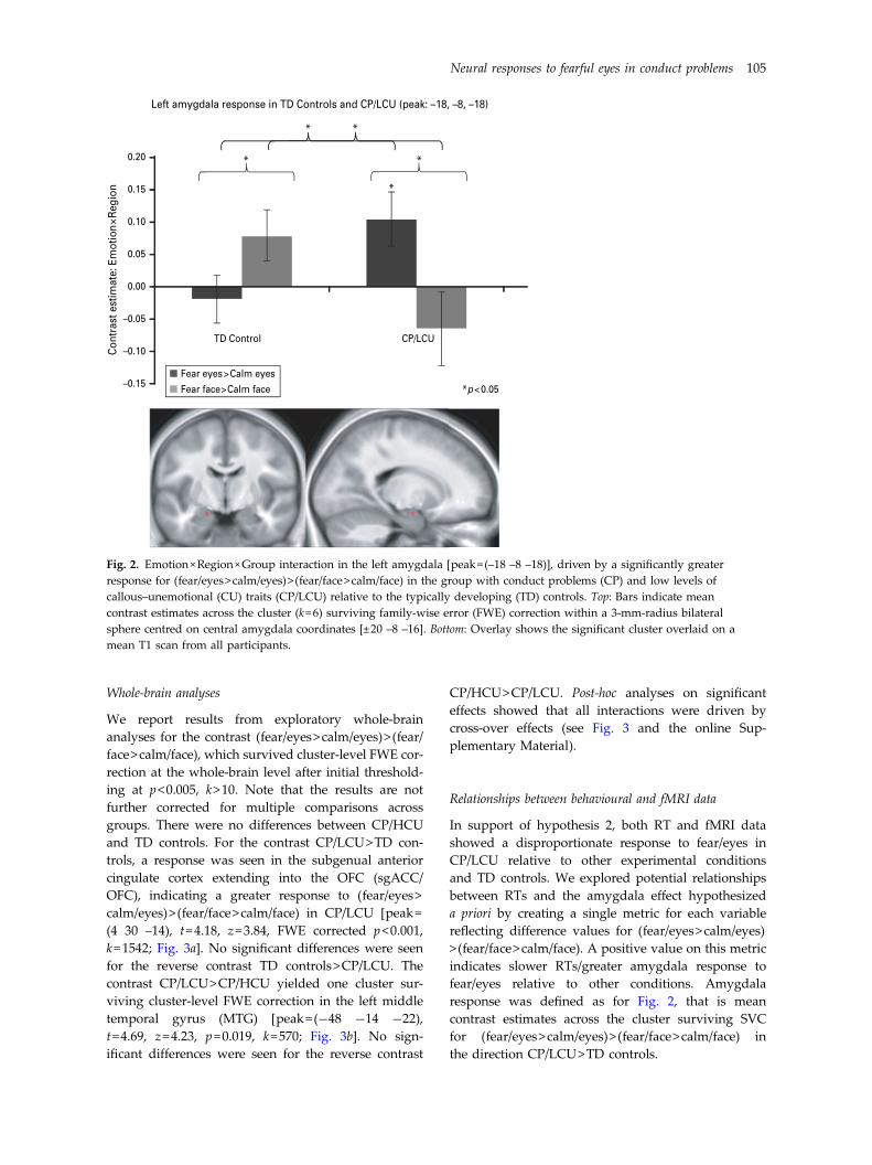
The second hypothesiswas that CP/LCUwould showa greater amygdala response to fear/eyes relative toother conditions than would TD controls. For theEmotion×Region interaction contrast (fear/eyes>calm/eyes)> (fear/face>calm/face), there was a significanteffect in the left amygdala in the direction CP/LCU>TD controls, suggesting an increased response to fear-ful eyes in CP/LCU [peak=(−18 −8 −18), k=6, t=3.16,z=2.99, FWE-SVC p=0.013]. Mean contrast estimatesacross the cluster (Fig. 2) show that the interaction wasdriven by a significantly greater amygdala response tofear/eyes>calm/eyes than to fear/face>calm/face inCP/LCU (t16=2.19, p=0.043), and a significant differencein the opposite direction in TD controls (t16=–2.22,p=0.041). Comparing TD control and CP/LCU groupsdirectly, there was a significantly greater response tofear/eyes>calm/eyes in CP/LCU than in TD controls(t32=2.21, p=0.034), and to fear/face>calm/face in TDcontrols than in CP/LCU (t32=2.09, p=0.045). The onlysignificant simple effect was a greater response tofear/eyes relative to calm/eyes in CP/LCU (t16=2.51,p=0.023).
Although we had no specific hypotheses regardingamygdala response comparing CP/HCU and CP/LCU, for completeness we report that there were nosignificant differences between these groups for theEmotion×Region interaction contrast within our ROI,even at uncorrected levels.
50
*
*
TD Control CP/LCU CP/HCU
Fear eyes >Calm eyes
Fear face>Calm face
*p < 0.01
40
30
20
10
Mea
n RT
Diff
eren
ce (m
s)
0
–10
–20
–30
–40
Fig. 1. Mean reaction time (RT) differences plotted by Group and Condition. The significant Emotion×Region interaction isdriven by the group with conduct problems (CP) and low levels of callous–unemotional (CU) traits (CP/LCU), who showedsignificantly slower RTs to fear/eyes than calm/eyes (the dark grey bar for CP/LCU). This group also showed significantlyslower RTs for the interaction term (fear/eyes>calm/eyes)> (fear/face>calm/face) (the difference between the dark and lightgrey bars), that is, the fear/eyes condition had a disproportionate slowing effect on RTs in CP/LCU but not in the other twogroups.
104 C. L. Sebastian et al.
Whole-brain analyses
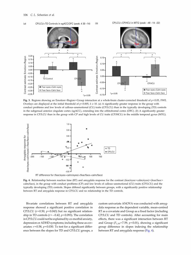
We report results from exploratory whole-brainanalyses for the contrast (fear/eyes>calm/eyes)> (fear/face>calm/face), which survived cluster-level FWE cor-rection at the whole-brain level after initial threshold-ing at p<0.005, k>10. Note that the results are notfurther corrected for multiple comparisons acrossgroups. There were no differences between CP/HCUand TD controls. For the contrast CP/LCU>TD con-trols, a response was seen in the subgenual anteriorcingulate cortex extending into the OFC (sgACC/OFC), indicating a greater response to (fear/eyes>calm/eyes)> (fear/face>calm/face) in CP/LCU [peak=(4 30 –14), t=4.18, z=3.84, FWE corrected p<0.001,k=1542; Fig. 3a]. No significant differences were seenfor the reverse contrast TD controls>CP/LCU. Thecontrast CP/LCU>CP/HCU yielded one cluster sur-viving cluster-level FWE correction in the left middletemporal gyrus (MTG) [peak=(−48 −14 −22),t=4.69, z=4.23, p=0.019, k=570; Fig. 3b]. No sign-ificant differences were seen for the reverse contrast
CP/HCU>CP/LCU. Post-hoc analyses on significanteffects showed that all interactions were driven bycross-over effects (see Fig. 3 and the online Sup-plementary Material).
Relationships between behavioural and fMRI data
In support of hypothesis 2, both RT and fMRI datashowed a disproportionate response to fear/eyes inCP/LCU relative to other experimental conditionsand TD controls. We explored potential relationshipsbetween RTs and the amygdala effect hypothesizeda priori by creating a single metric for each variablereflecting difference values for (fear/eyes>calm/eyes)> (fear/face>calm/face). A positive value on this metricindicates slower RTs/greater amygdala response tofear/eyes relative to other conditions. Amygdalaresponse was defined as for Fig. 2, that is meancontrast estimates across the cluster surviving SVCfor (fear/eyes>calm/eyes)> (fear/face>calm/face) inthe direction CP/LCU>TD controls.
0.20
Left amygdala response in TD Controls and CP/LCU (peak: –18, –8, –18)
* *
*
*
*
TD Control CP/LCU
Fear eyes>Calm eyes
Fear face>Calm face *p < 0.05
0.15
0.10
0.05
Con
tras
t est
imat
e: E
mot
ion×
Reg
ion
0.00
–0.05
–0.10
–0.15
Fig. 2. Emotion×Region×Group interaction in the left amygdala [peak=(–18 –8 –18)], driven by a significantly greaterresponse for (fear/eyes>calm/eyes)> (fear/face>calm/face) in the group with conduct problems (CP) and low levels ofcallous–unemotional (CU) traits (CP/LCU) relative to the typically developing (TD) controls. Top: Bars indicate meancontrast estimates across the cluster (k=6) surviving family-wise error (FWE) correction within a 3-mm-radius bilateralsphere centred on central amygdala coordinates [±20 –8 –16]. Bottom: Overlay shows the significant cluster overlaid on amean T1 scan from all participants.
Neural responses to fearful eyes in conduct problems 105
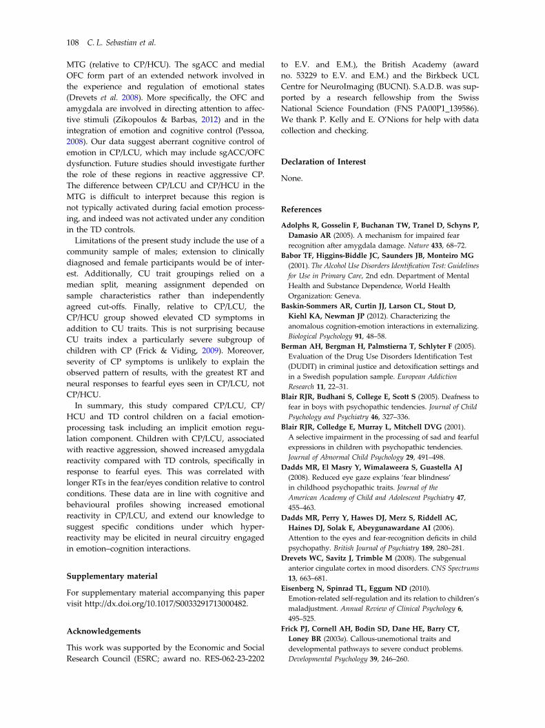
Bivariate correlations between RT and amygdalaresponse showed a significant positive correlation inCP/LCU (r=0.50, p=0.043) but no significant relation-ship in TD controls (r=−0.42, p=0.093). The correlationinCP/LCUcouldnotbe explainedbyco-morbidanxiety,depression orADHDsymptoms; including these as cov-ariates: r=0.58, p=0.030. To test for a significant differ-ence between the slopes for TD and CP/LCU groups, a
custom univariate ANOVAwas conducted with amyg-dala response as the dependent variable, mean-centredRT as a covariate and Group as a fixed factor (includingCP/LCU and TD controls). After accounting for maineffects, there was a significant interaction between RTand Group (F1,30 =7.59, p=0.01), showing a significantgroup difference in slopes indexing the relationshipbetween RT and amygdala response (Fig. 4).
0.200.15
0.10
0.05
0.00
–0.05
–0.10
–0.15
0.15
0.10
0.05
0.00
–0.05
–0.10
–0.15
–0.20
–0.25
–0.30
–0.35
(a) (b)CP/LCU>TD Controls in sgACC/OFC (peak: 4 30 –14) CP/LCU> CP/HCU in MTG (peak: –48 –14 –22)
* *
*
* *
*
* *
*
*
TD Control CP/LCU
CP/LCU CP/HCU
*p<0.05*p<0.05C
ontr
ast e
stim
ate:
Em
otio
n×R
egio
n
Con
tras
t est
imat
e: E
mot
ion
×Reg
ion **
Fear eyes>Calm eyes
Fear face>Calm face Fear eyes>Calm eyes
Fear face>Calm face
Fig. 3. Regions showing an Emotion×Region×Group interaction at a whole-brain cluster-corrected threshold of p<0.05, FWE.Overlays are displayed at the initial threshold of p<0.005, k510. (a) A significantly greater response in the group withconduct problems and low levels of callous–unemotional (CU) traits (CP/LCU) than in the typically developing (TD) controlsin the subgenual anterior cingulate cortex (sgACC), extending into the orbitofrontal cortex (OFC). (b) A significantly greaterresponse in CP/LCU than in the group with CP and high levels of CU traits (CP/HCU) in the middle temporal gyrus (MTG).
TD
CP/LCU
RT difference for (fear/eyes>calm/eyes)>(fear/face>calm/face)
Am
ygda
la r
espo
nse
for
(fea
r/ey
es>c
alm
/eye
s)>(
fear
/face
>cal
m/fa
ce)
50 100 1500–50–100–150–200
1.0
0.8
0.6
0.4
0.2
–0.2
–0.4
–0.6
0
Fig. 4. Relationship between reaction time (RT) and amygdala response for the contrast (fear/eyes>calm/eyes)> (fear/face>calm/face), in the group with conduct problems (CP) and low levels of callous–unemotional (CU) traits (CP/LCU) and thetypically developing (TD) controls. Slopes differed significantly between groups, with a significantly positive relationshipbetween RT and amygdala response in CP/LCU and no relationship in the TD controls.
106 C. L. Sebastian et al.

Discussion
The current study investigated behavioural and neuralconsequences of directing attention to the eye regionof fearful versus calm faces in children with CP and dif-fering levels of CU traits. Contrary to our first hypoth-esis, amygdala response to fearful faces in childrenwith CP and high CU traits (CP/HCU) did not increasewhen participants looked for a dot near the eye regionof fearful faces compared with searching across thewhole face. However, in line with our second hypoth-esis, children with CP and low CU traits (CP/LCU)showed increased left amygdala response to the fear/eyes condition relative to both other conditions andTD controls. This was accompanied by increasedRTs, with the RT increase specific to fearful eyes corre-lating with amygdala response in CP/LCU but not inTD controls. CP/LCU also showed increased neuralresponses to fearful eyes in the sgACC/OFC (relativeto TD controls) and left MTG (relative to CP/HCU).
It is important to consider why directing attentionto the eye region during fear processing did notresult in increased amygdala response in CP/HCU.One interpretation is that amygdala response in thisgroup is largely immutable to the effects of mani-pulating attentional focus. Under this interpretation,improved fear recognition when focusing on the eyeregion (Dadds et al. 2006) would not be mediated byincreased amygdala response. An alternative expla-nation relates to the nature of task demands. It hasbeen suggested that fear-processing deficits in CP/HCU are associated with a reduced ability to reflex-ively shift attention to the salient eye region, a processpotentially mediated by the basolateral amygdala(Gamer & Buchel, 2009; Moul et al. 2012). In the fear/eyes condition, attention was already focused on theeye region, meaning that no amygdala-mediatedreflexive shift was needed; by contrast, the fear/facecondition may paradoxically elicit greater amygdalaresponse because of the need for a reflexive gazeshift. This idea is supported by the pattern of resultsseen in TD controls in analyses for hypothesis 1. Thisgroup showed increased right amygdala responseto fear/face relative to fear/eyes whereas CP/HCUshowed no difference between conditions. Althoughspeculative, the pattern of results in TD controls mayreflect a typical orienting response that involves theamygdala, and the lack of difference between con-ditions in CP/HCU could reflect atypical processing.
It is also important to consider that the instructionto look for a dot may have introduced unforeseen pro-cessing biases that limited modulation of amygdalaresponses to fear. A recent study investigating CP/HCU responses to fearful eye gaze during a spatialattention task (White et al. 2012) found reduced
activation in a dorsoparietal-orienting network com-pared with controls, but no effect in the amygdala. Itwas suggested that CP/HCU amygdala hypo-activitymay be specifically elicited when task demands arelow. Similarly, it may be that the present task wasnot optimized for detecting conditions under which afear-processing deficit in the amygdala might be eli-cited or ameliorated in CP/HCU.
Previous studies have shown a hyper-reactive affec-tive profile in CP/LCU (Frick et al. 2003a; Dadds et al.2006). The current data suggest that emotional reac-tivity may be augmented when attention is directedto the eye region, which is high in affective salience(Adolphs et al. 2005). RT data further show a specificslowing during the fear/eyes condition. The positiverelationship between RTs and amygdala reactivity inCP/LCU suggests that increased reactivity as indexedby amygdala response is associated with a reductionin task performance. It is unlikely that these resultsare driven by anxiety because TD and CP/LCU groupsdid not differ on this measure. It is also unlikely thatthe results can be explained by other symptoms onwhich the groups differed (i.e. ADHD and MDE)because the CP/HCU group also showed elevatedsymptoms but did not show the same pattern ofresults. Instead, increased amygdala reactivity andslower RTs suggest that children with CP/LCU havedifficulty implicitly or automatically regulatingemotional responses in pursuit of a goal (Ochsner &Gross, 2005). This complements well-documentedreports of difficulties with explicit emotion regulationin everyday life (Frick & Morris, 2004). Indeed, diffi-culties with automatic emotion regulation may contrib-ute to the development of expressed behaviours suchas reactive aggression (Eisenberg et al. 2010).
These data are in line with a recent study exploringinteractions between attention and affective processingin adults with externalizing behaviours (Baskin-Sommers et al. 2012). Using an instructed fear para-digm, this study found that externalizing behaviourswere not associated with a global hyper-reactivityeffect. Instead, increased emotional reactivity andamygdala response, relative to low-externalizingparticipants, was seen specifically when attentionwas focused on threat-related information. Together,these data suggest the importance of understandingthe specific conditions under which emotional hyper-reactivity is seen in externalizing conditions such asCP/LCU. This is necessary to elucidate neurocognitivemechanisms underpinning such behaviours, and mayprovide insights that will improve current approachesto intervention.
Although not predicted a priori, increased neuralresponses to fear/eyes were seen in CP/LCU insgACC/OFC (relative to TD controls) and in the left
Neural responses to fearful eyes in conduct problems 107
MTG (relative to CP/HCU). The sgACC and medialOFC form part of an extended network involved inthe experience and regulation of emotional states(Drevets et al. 2008). More specifically, the OFC andamygdala are involved in directing attention to affec-tive stimuli (Zikopoulos & Barbas, 2012) and in theintegration of emotion and cognitive control (Pessoa,2008). Our data suggest aberrant cognitive control ofemotion in CP/LCU, which may include sgACC/OFCdysfunction. Future studies should investigate furtherthe role of these regions in reactive aggressive CP.The difference between CP/LCU and CP/HCU in theMTG is difficult to interpret because this region isnot typically activated during facial emotion process-ing, and indeed was not activated under any conditionin the TD controls.
Limitations of the present study include the use of acommunity sample of males; extension to clinicallydiagnosed and female participants would be of inter-est. Additionally, CU trait groupings relied on amedian split, meaning assignment depended onsample characteristics rather than independentlyagreed cut-offs. Finally, relative to CP/LCU, theCP/HCU group showed elevated CD symptoms inaddition to CU traits. This is not surprising becauseCU traits index a particularly severe subgroup ofchildren with CP (Frick & Viding, 2009). Moreover,severity of CP symptoms is unlikely to explain theobserved pattern of results, with the greatest RT andneural responses to fearful eyes seen in CP/LCU, notCP/HCU.
In summary, this study compared CP/LCU, CP/HCU and TD control children on a facial emotion-processing task including an implicit emotion regu-lation component. Children with CP/LCU, associatedwith reactive aggression, showed increased amygdalareactivity compared with TD controls, specifically inresponse to fearful eyes. This was correlated withlonger RTs in the fear/eyes condition relative to controlconditions. These data are in line with cognitive andbehavioural profiles showing increased emotionalreactivity in CP/LCU, and extend our knowledge tosuggest specific conditions under which hyper-reactivity may be elicited in neural circuitry engagedin emotion–cognition interactions.
Supplementary material
For supplementary material accompanying this papervisit http://dx.doi.org/10.1017/S0033291713000482.
Acknowledgements
This work was supported by the Economic and SocialResearch Council (ESRC; award no. RES-062-23-2202
to E.V. and E.M.), the British Academy (awardno. 53229 to E.V. and E.M.) and the Birkbeck UCLCentre for NeuroImaging (BUCNI). S.A.D.B. was sup-ported by a research fellowship from the SwissNational Science Foundation (FNS PA00P1_139586).We thank P. Kelly and E. O’Nions for help with datacollection and checking.
Declaration of Interest
None.
References
Adolphs R, Gosselin F, Buchanan TW, Tranel D, Schyns P,Damasio AR (2005). A mechanism for impaired fearrecognition after amygdala damage. Nature 433, 68–72.
Babor TF, Higgins-Biddle JC, Saunders JB, Monteiro MG(2001). The Alcohol Use Disorders Identification Test: Guidelinesfor Use in Primary Care, 2nd edn. Department of MentalHealth and Substance Dependence, World HealthOrganization: Geneva.
Baskin-Sommers AR, Curtin JJ, Larson CL, Stout D,Kiehl KA, Newman JP (2012). Characterizing theanomalous cognition-emotion interactions in externalizing.Biological Psychology 91, 48–58.
Berman AH, Bergman H, Palmstierna T, Schlyter F (2005).Evaluation of the Drug Use Disorders Identification Test(DUDIT) in criminal justice and detoxification settings andin a Swedish population sample. European AddictionResearch 11, 22–31.
Blair RJR, Budhani S, College E, Scott S (2005). Deafness tofear in boys with psychopathic tendencies. Journal of ChildPsychology and Psychiatry 46, 327–336.
Blair RJR, Colledge E, Murray L, Mitchell DVG (2001).A selective impairment in the processing of sad and fearfulexpressions in children with psychopathic tendencies.Journal of Abnormal Child Psychology 29, 491–498.
Dadds MR, El Masry Y, Wimalaweera S, Guastella AJ(2008). Reduced eye gaze explains ‘fear blindness’in childhood psychopathic traits. Journal of theAmerican Academy of Child and Adolescent Psychiatry 47,455–463.
Dadds MR, Perry Y, Hawes DJ, Merz S, Riddell AC,Haines DJ, Solak E, Abeygunawardane AI (2006).Attention to the eyes and fear-recognition deficits in childpsychopathy. British Journal of Psychiatry 189, 280–281.
Drevets WC, Savitz J, Trimble M (2008). The subgenualanterior cingulate cortex in mood disorders. CNS Spectrums13, 663–681.
Eisenberg N, Spinrad TL, Eggum ND (2010).Emotion-related self-regulation and its relation to children’smaladjustment. Annual Review of Clinical Psychology 6,495–525.
Frick PJ, Cornell AH, Bodin SD, Dane HE, Barry CT,Loney BR (2003a). Callous-unemotional traits anddevelopmental pathways to severe conduct problems.Developmental Psychology 39, 246–260.
108 C. L. Sebastian et al.

Frick PJ, Kimonis ER, Dandreaux DM, Farrell JM (2003b).The 4-year stability of psychopathic traits in nonreferredyouth. Behavioral Sciences and the Law 21, 713–736.
Frick PJ, Morris AS (2004). Temperament and developmentalpathways to conduct problems. Journal of Clinical Child andAdolescent Psychology 33, 54–68.
Frick PJ, Viding E (2009). Antisocial behavior from adevelopmental psychopathology perspective. Developmentand Psychopathology 21, 1111–1131.
Gadow KD, Sprafkin J (2009). The Symptom Inventories:An Annotated Bibliography. Checkmate Plus: Stony Brook,New York.
Gamer M, Buchel C (2009). Amygdala activation predictsgaze toward fearful eyes. Journal of Neuroscience 29,9123–9126.
Herpertz SC, Huebner T, Marx I, Vloet TD, Fink GR,Stoecker T, Shah NJ, Konrad K, Herpertz-Dahlmann B(2008). Emotional processing in male adolescents withchildhood-onset conduct disorder. Journal of ChildPsychology and Psychiatry 49, 781–791.
Jones AP, Laurens KR, Herba CM, Barker GJ, Viding E(2009). Amygdala hypoactivity to fearful faces in boys withconduct problems and callous-unemotional traits. AmericanJournal of Psychiatry 166, 95–102.
Lieberman MD, Cunningham WA (2009). Type I and Type IIerror concerns in fMRI research: re-balancing the scale.Social Cognitive and Affective Neuroscience 4, 423–428.
Loney BR, Frick PJ, Clements CB, Ellis ML, Kerlin K (2003).Callous-unemotional traits, impulsivity, and emotionalprocessing in adolescents with antisocial behaviorproblems. Journal of Clinical Child and Adolescent Psychology32, 66–80.
Marsh AA, Finger EC, Mitchell DGV, Reid ME, Sims C,Kosson DS, Towbin KE, Leibenluft E, Pine DS, Blair RJR(2008). Reduced amygdala response to fearful expressionsin children and adolescents with callous-unemotional traitsand disruptive behavior disorders. American Journal ofPsychiatry 165, 712–720.
Marsh AA, Finger EC, Schechter JC, Jurkowitz IT,Reid ME, Blair RJ (2011). Adolescents with psychopathictraits report reductions in physiological responsesto fear. Journal of Child Psychology and Psychiatry 52, 834–841.
Moul C, Killcross S, Dadds MR (2012). A model ofdifferential amygdala activation in psychopathy.Psychological Review 119, 789–806.
Ochsner KN, Gross JJ (2005). The cognitive control ofemotion. Trends in Cognitive Sciences 9, 242–249.
Passamonti L, Fairchild G, Goodyer IM, Hurford G,Hagan CC, Rowe JB, Calder AJ (2010). Neuralabnormalities in early-onset and adolescence-onset conductdisorder. Archives of General Psychiatry 67, 729–738.
Pessoa L (2008). On the relationship between emotion andcognition. Nature Reviews Neuroscience 9, 148–158.
Phillips ML, Medford N, Young AW, Williams L,Williams SC, Bullmore ET, Gray JA, Brammer MJ (2001).Time courses of left and right amygdalar responses tofearful facial expressions. Human Brain Mapping 12,193–202.
Romeo R, Knapp M, Scott S (2006). Economic cost of severeantisocial behaviour in children – and who pays it. BritishJournal of Psychiatry 188, 547–553.
Sebastian CL, McCrory EJ, Cecil CA, Lockwood PL,De Brito SA, Fontaine NM, Viding E (2012). Neuralresponses to affective and cognitive theory of mind inchildren with conduct problems and varying levels ofcallous-unemotional traits. Archives of General Psychiatry69, 814–822.
Sharp C, van Goozen S, Goodyer I (2006). Children’ssubjective emotional reactivity to affective pictures: genderdifferences and their antisocial correlates in an unselectedsample of 7–11-year-olds. Journal of Child Psychology andPsychiatry 47, 143–150.
Sterzer P, Stadler C, Krebs A, Kleinschmidt A, Poustka F(2005). Abnormal neural responses to emotional visualstimuli in adolescents with conduct disorder. BiologicalPsychiatry 57, 7–15.
Tottenham N, Tanaka JW, Leon AC, McCarry T, Nurse M,Hare TA, Marcus DJ, Westerlund A, Casey BJ, Nelson C(2009). The NimStim set of facial expressions: judgmentsfrom untrained research participants. Psychiatry Research168, 242–249.
Viding E, Sebastian CL, Dadds MR, Lockwood PL,Cecil CAM, De Brito SA, McCrory EJ (2012). Amygdalaresponse to pre-attentive masked fear is associated withcallous-unemotional traits in children with conductproblems. American Journal of Psychiatry 169, 1109–1116.
Weiskopf N, Hutton C, Josephs O, Deichmann R (2006).Optimal EPI parameters for reduction ofsusceptibility-induced BOLD sensitivity losses: awhole-brain analysis at 3 T and 1.5 T. NeuroImage 33,493–504.
Weschler D (1999). Wechsler Abbreviated Scale of Intelligence(WASI). Harcourt Assessment: San Antonio, TX.
White SF, Williams WC, Brislin SJ, Sinclair S, Blair KS,Fowler KA, Pine DS, Pope K, Blair RJ (2012). Reducedactivity within the dorsal endogenous orienting of attentionnetwork to fearful expressions in youth with disruptivebehavior disorders and psychopathic traits. Development andPsychopathology 24, 1105–1116.
Zikopoulos B, Barbas HJ (2012). Pathways for emotions andattention converge on the thalamic reticular nucleus inprimates. Journal of Neuroscience 32, 5338–5350.
Neural responses to fearful eyes in conduct problems 109