Nar gks622 full-text-lowres
10
TDP2 promotes repair of topoisomerase I-mediated DNA damage in the absence of TDP1 Zhihong Zeng 1 , Abhishek Sharma 1 , Limei Ju 1 , Junko Murai 2 , Lieve Umans 3 , Liesbeth Vermeire 3 , Yves Pommier 2 , Shunichi Takeda 4 , Danny Huylebroeck 3 , Keith W. Caldecott 1, * and Sherif F. El-Khamisy 1,5, * 1 School of Life Sciences, Genome Damage and Stability Centre, University of Sussex, Science Park Road, Falmer, Brighton BN1 9RQ, UK, 2 Center for Cancer Research, National Cancer Institute, Bethesda, Maryland, USA, 3 Laboratory of Molecular Biology (Celgen), Department of Development and Regeneration, University of Leuven, Belgium, 4 Department of Radiation Genetics, Kyoto University, Kyoto, Japan and 5 Department of Biochemistry, Faculty of Pharmacy, Ain Shams University, Cairo, Egypt Received March 11, 2012; Revised May 31, 2012; Accepted June 3, 2012 ABSTRACT The abortive activity of topoisomerases can result in clastogenic and/or lethal DNA damage in which the topoisomerase is covalently linked to the 3 0 - or 5 0 -terminus of a DNA strand break. This type of DNA damage is implicated in chromosome trans- locations and neurological disease and underlies the clinical efficacy of an important class of anticancer topoisomerase ‘poisons’. Tyrosyl DNA phosphodiesterase-1 protects cells from abortive topoisomerase I (Top1) activity by hydrolyzing the 3 0 -phosphotyrosyl bond that links Top1 to a DNA strand break and is currently the only known human enzyme that displays this activity in cells. Recently, we identified a second tyrosyl DNA phosphodiesterase (TDP2; aka TTRAP/EAPII) that possesses weak 3 0 -tyrosyl DNA phosphodiesterase (3 0 -TDP) activity, in vitro. Herein, we have examined whether TDP2 contributes to the repair of Top1- mediated DNA breaks by deleting Tdp1 and Tdp2 separately and together in murine and avian cells. We show that while deletion of Tdp1 in wild-type DT40 cells and mouse embryonic fibroblasts de- creases DNA strand break repair rates and cellular survival in response to Top1-induced DNA damage, deletion of Tdp2 does not. However, deletion of both Tdp1 and Tdp2 reduces rates of DNA strand break repair and cell survival below that observed in Tdp1 / cells, suggesting that Tdp2 contributes to cellular 3 0 -TDP activity in the absence of Tdp1. Consistent with this idea, over-expression of human TDP2 in Tdp1 / /Tdp2 // DT40 cells in- creases DNA strand break repair rates and cell survival above that observed in Tdp1 / DT40 cells, suggesting that Tdp2 over-expression can partially complement the defect imposed by loss of Tdp1. Finally, mice lacking both Tdp1 and Tdp2 exhibit greater sensitivity to Top1 poisons than do mice lacking Tdp1 alone, further suggesting that Tdp2 contributes to the repair of Top1-mediated DNA damage in the absence of Tdp1. In contrast, we failed to detect a contribution for Tdp1 to repair Top2-mediated damage. Together, our data suggest that Tdp1 and Tdp2 fulfil overlapping roles following Top1-induced DNA damage, but not following Top2-induced DNA damage, in vivo. INTRODUCTION DNA strand breaks are induced by a variety of endogen- ous or exogenous genotoxic agents. Accumulation of these breaks threatens genome stability and underlies the clinical utility of several anti-cancer strategies including radiotherapy and chemotherapy. DNA breaks can arise directly from attack of endogenous reactive oxygen species or by exposure to ionizing radiation. They can also arise indirectly through the abortive activity of DNA topoisomerases, which undergo a rapid catalytic cycle in which the topoisomerase becomes covalently attached to the 3 0 - or 5 0 -terminus of a DNA strand break (1). The hydrolytic removal of the covalently *To whom correspondence should be addressed. Tel: +1 273 877519; Fax:+1 273 678121; Email: [email protected] Correspondence may also be addressed to Sherif F. El Khamisy. Tel: +1 273 678381; Fax: +1 273 678121; Email: [email protected] The authors wish it to be known that, in their opinion, the first two authors should be regarded as joint First Authors. Nucleic Acids Research, 2012, 1–10 doi:10.1093/nar/gks622 ß The Author(s) 2012. Published by Oxford University Press. This is an Open Access article distributed under the terms of the Creative Commons Attribution Non-Commercial License (http://creativecommons.org/licenses/ by-nc/3.0), which permits unrestricted non-commercial use, distribution, and reproduction in any medium, provided the original work is properly cited. Nucleic Acids Research Advance Access published June 26, 2012
-
Upload
independent -
Category
Documents
-
view
0 -
download
0
Transcript of Nar gks622 full-text-lowres

TDP2 promotes repair of topoisomerase I-mediatedDNA damage in the absence of TDP1Zhihong Zeng1, Abhishek Sharma1, Limei Ju1, Junko Murai2, Lieve Umans3,
Liesbeth Vermeire3, Yves Pommier2, Shunichi Takeda4, Danny Huylebroeck3,
Keith W. Caldecott1,* and Sherif F. El-Khamisy1,5,*
1School of Life Sciences, Genome Damage and Stability Centre, University of Sussex, Science Park Road,Falmer, Brighton BN1 9RQ, UK, 2Center for Cancer Research, National Cancer Institute, Bethesda, Maryland,USA, 3Laboratory of Molecular Biology (Celgen), Department of Development and Regeneration, University ofLeuven, Belgium, 4Department of Radiation Genetics, Kyoto University, Kyoto, Japan and 5Department ofBiochemistry, Faculty of Pharmacy, Ain Shams University, Cairo, Egypt
Received March 11, 2012; Revised May 31, 2012; Accepted June 3, 2012
ABSTRACT
The abortive activity of topoisomerases can result inclastogenic and/or lethal DNA damage in whichthe topoisomerase is covalently linked to the 30- or50-terminus of a DNA strand break. This type ofDNA damage is implicated in chromosome trans-locations and neurological disease and underliesthe clinical efficacy of an important class ofanticancer topoisomerase ‘poisons’. Tyrosyl DNAphosphodiesterase-1 protects cells from abortivetopoisomerase I (Top1) activity by hydrolyzing the30-phosphotyrosyl bond that links Top1 to a DNAstrand break and is currently the only knownhuman enzyme that displays this activity in cells.Recently, we identified a second tyrosyl DNAphosphodiesterase (TDP2; aka TTRAP/EAPII) thatpossesses weak 30-tyrosyl DNA phosphodiesterase(30-TDP) activity, in vitro. Herein, we have examinedwhether TDP2 contributes to the repair of Top1-mediated DNA breaks by deleting Tdp1 and Tdp2separately and together in murine and avian cells.We show that while deletion of Tdp1 in wild-typeDT40 cells and mouse embryonic fibroblasts de-creases DNA strand break repair rates and cellularsurvival in response to Top1-induced DNA damage,deletion of Tdp2 does not. However, deletion of bothTdp1 and Tdp2 reduces rates of DNA strand breakrepair and cell survival below that observed inTdp1�/� cells, suggesting that Tdp2 contributesto cellular 30-TDP activity in the absence of Tdp1.
Consistent with this idea, over-expression ofhuman TDP2 in Tdp1�/�/Tdp2�/�/� DT40 cells in-creases DNA strand break repair rates and cellsurvival above that observed in Tdp1�/� DT40cells, suggesting that Tdp2 over-expression canpartially complement the defect imposed by lossof Tdp1. Finally, mice lacking both Tdp1 and Tdp2exhibit greater sensitivity to Top1 poisons than domice lacking Tdp1 alone, further suggesting thatTdp2 contributes to the repair of Top1-mediatedDNA damage in the absence of Tdp1. In contrast,we failed to detect a contribution for Tdp1 torepair Top2-mediated damage. Together, our datasuggest that Tdp1 and Tdp2 fulfil overlapping rolesfollowing Top1-induced DNA damage, but notfollowing Top2-induced DNA damage, in vivo.
INTRODUCTION
DNA strand breaks are induced by a variety of endogen-ous or exogenous genotoxic agents. Accumulation ofthese breaks threatens genome stability and underlies theclinical utility of several anti-cancer strategies includingradiotherapy and chemotherapy. DNA breaks can arisedirectly from attack of endogenous reactive oxygenspecies or by exposure to ionizing radiation. They canalso arise indirectly through the abortive activity ofDNA topoisomerases, which undergo a rapid catalyticcycle in which the topoisomerase becomes covalentlyattached to the 30- or 50-terminus of a DNA strandbreak (1). The hydrolytic removal of the covalently
*To whom correspondence should be addressed. Tel: +1 273 877519; Fax: +1 273 678121; Email: [email protected] may also be addressed to Sherif F. El Khamisy. Tel: +1 273 678381; Fax: +1 273 678121; Email: [email protected]
The authors wish it to be known that, in their opinion, the first two authors should be regarded as joint First Authors.
Nucleic Acids Research, 2012, 1–10doi:10.1093/nar/gks622
� The Author(s) 2012. Published by Oxford University Press.This is an Open Access article distributed under the terms of the Creative Commons Attribution Non-Commercial License (http://creativecommons.org/licenses/by-nc/3.0), which permits unrestricted non-commercial use, distribution, and reproduction in any medium, provided the original work is properly cited.
Nucleic Acids Research Advance Access published June 26, 2012

linked topoisomerase from DNA termini is required forrepair of the break and thus for restoration of geneticintegrity. The prototype example of this hydrolyticactivity was first identified by Nash and coworkers (2) in1996 and was named tyrosyl DNA phosphodiesterase-1(TDP1). Homozygous mutation of TDP1 underlies thecerebellar degeneration observed in a human hereditarydisease with spinocerebellar ataxia and axonal neuropathy(3). Consistent with its role during repair of Top1-linkedDNA breaks, cellular depletion of TDP1 results in accu-mulation of Top1-linked DNA breaks and sensitizes cellsto Top1 poisons such as camptothecin (CPT) (4–6).Although TDP1 is the main 30-tyrosyl DNA phospho-
diesterase (30-TDP) in human cells, cells lacking TDP1exhibit residual Top1-DNA processing activity, suggestingthat other cellular mechanisms may be able to compensatefor TDP1 (7,8). In a hunt for such activities, we recentlyidentified TTRAP/EAPII, a metal-dependent phospho-diesterase of unknown function that associates withCD40, tumor necrosis factor (TNF) receptor, TNFreceptor-associated factors (9), and that can influenceTGF-b signalling (10,11). We demonstrated that TTRAPpossesses both weak 30-TDP activity and robust 50-tyrosylDNA phosphodiesterase (50-TDP) activity in vitro, andconsequently denoted this protein TDP2 (12). Consistentwith its 50-TDP activity, TDP2 is required for cellularresistance to Top2 poisons, which induce DNA strandbreaks in which Top2 is covalently linked to 50-DNAtermini through a 50-phosphotyrosyl bond (13).However, cells depleted or lacking Tdp2 are not hypersen-sitive to Top1 poisons, suggesting that the relatively weak30-TDP activity of this protein is not required for repair ofTop1-induced DNA damage (12,13). Herein, we have ad-dressed this question in detail and examined whetherTDP2 is required for repair of Top1-induced DNAdamage in the absence of TDP1. Our data demonstratethat while TDP1 is the primary source of cellular 30-TDPactivity and resistance to Top1-induced DNA strandbreaks, TDP2 promotes the repair of Top1-inducedDNA damage in cells in which TDP1 is absent.
MATERIALS AND METHODS
Generation of Tdp1/Tdp2 double knockout mice andmouse embryonic fibroblasts
Tdp1�/� mice were generated, maintained and genotypedas described previously (14) in an outbred mixed 129Olaand C57BL/6 background. TTRAP (Tdp2�/�) mice weregenerated by targeted deletion of exons 1–3 using a Cre/Lox system and maintained as an outbred 129Ola andCD1 background (see Supplementary Figure S6 for thetargeting strategy and confirmation of knockout alleles).A detailed description of these mice will be described else-where. To generate Tdp1�/�/Tdp2�/� mice, Tdp1+/� andTdp2+/� mice were interbred to generate Tdp1+/�/Tdp2+/�
double heterozygote mice, which were then interbred togenerate the appropriate F2 progeny. Genotyping for thewild-type Tdp2 allele was conducted using primers 50-CCTTCATTACTTCTCGTAGGTTCTGGGTC-30 (LV043)and 50-ACCCGCTCTTCACGCTGCTTCC-30 (A183).
Primers LV103 and 50-TACACCGTGCCATAATGACCAAC-30 (A185) were used to amplify the mutant Tdp2allele deficient in exons 1–3. Polymerase chain reaction(PCR) conditions were 94�C for 30 s, 60�C for 1min and72�C for 1min, for 35 cycles, resulting in the amplificationof a 429-bp fragment from the wild-type allele or a 561-bpfragment from the mutant allele. All animals were housedwithin the School of Life Sciences and maintained in ac-cordance with the institutional animal care and ethicalcommittee at the University of Sussex. For the generationof WT, Tdp1�/�, Tdp2�/� and Tdp1�/�/Tdp2�/� mouseembryonic fibroblasts (MEFs), embryos from appropriatematings were harvested 14-d.p.c and were dissociated,trypsinized and plated in Dulbecco’s modified Eagle’smedium (DMEM) with 10% foetal calf serum (FCS).Primary MEFs were maintained in DMEM media supple-mented with 10% FCS, at 37�C and maintained at 5%oxygen. Genotypes were confirmed by PCR and loss ofTdp1 and/or Tdp2 protein was confirmed by activityassays on cell lysates (see below). Xrcc1�/� MEFs were akind gift from Prof Larry Thompson.
Generation of Tdp1�/� and Tdp1�/�/Tdp2�/�/�
DT40 cells
DT40 chicken cells were maintained in RPMI 1640medium containing 10�5 M b-mercaptoethanol, penicillin,streptomycin, 10% FCS and 1% chicken serum (Sigma)at 39�C. The generation of Tdp2�/�/� DT40 cells wasdescribed previously (13). To generate Tdp1�/� cells,genomic Tdp1 sequences were PCR amplified fromDT40 genomic DNA (clone 18) to generate left andright arms for the targeting constructs using the primers50-CCCAAGCTTGCACAAGCACGCCCTTTTGAG-30
and 50-CGCGGATCCCATTCCTTGAGCACAGGAGAAC-30 for the left arm and 50-CGCGGATCCGCCTGTTGTGGGACAGTTCTCAAGC-30 and 50-AAGGAAAAAAGCGGCCGCCACAGCTGTTTCTGTGCGGTCTG-30 for the right arm. The PCR amplified products weresubcloned into pCR2.1-TOPO vector (Invitrogen) andconfirmed by sequencing. Fragments encoding the leftarm (2.9-kb) and right arm (2.2-kb) were recovered fromthe above pCR2.1-TOPO constructs using HindIII/BamH1 and BamH1/NotI, respectively, and subclonedinto pCR2.1-TOPO vector. A BamH1 fragmentencoding the puromycin (Puro) or hygromycin (Hyg) se-lection cassette was then inserted into the pCR2.1-TOPOconstructs at the BamH1 site separating the left and rightarms, completing the Puro-resistant (Puror) andHyg-resistant (Hygr) TDP1-targeting constructs. Togenerate Tdp1�/� cells, 2� 107 cells (clone 18) were firstelectroporated (Bio-Rad) with 30 mg of NotI-linearizedPuror targeting construct (to disrupt the first allele)followed by, after confirmation of successful targeting asdescribed below, the Hygr targeting construct (to disruptthe second allele). Following each round of transfection,transfected clones were selected for 8–10 days in thepresence of medium containing 0.5mg/ml of puromycinhydrochloride (Sigma) and/or 2.5mg/ml hygromycin B(Sigma), as appropriate. To detect successful targetingof Tdp1 alleles, genomic DNA was isolated from
2 Nucleic Acids Research, 2012

drug-resistant clones, digested with NcoI and subjected toSouthern blot analysis using a 0.55-kb probe amplifiedfrom genomic DNA clone 18 using the primers 50-CGCAAGCCTAAATCAAAAGC-30 and 50-CCTGTTGCTCAACGCTGATA-30. Following the two consecutiverounds of Tdp1 gene targeting, two Tdp1�/� clones wererecovered (denoted clone 10 and clone 12) andcharacterized further, as indicated.
To generate Tdp1�/�/Tdp2�/�/� DT40 cells, the puro-mycin (Puro) resistance cassette present in Tdp2�/�/�
DT40 cells (clone 8) (13) was first removed by transienttransfection of a tamoxifen-inducible Cre recombinase ex-pression construct (pANcreMer-Neo) (a gift from HelfridHochegger). Cre expression was induced in transientlytransfected cell populations by incubation in growthmedium containing 50 nM 5-hydroxytamoxifen (Sigma)for 1 day and single clones were selected by serialdilution to 1 cell/ml and amplified in 96-well plates for5–7 days. Successful excision of the Puro-resistancecassette was confirmed by assessing the sensitivity ofsingle clones to 0.5 mg/ml puromycin hydrochloride(Sigma). Tdp1 was then disrupted in one of thePuro-sensitive Tdp2�/�/� clones (clone 3) by sequentialtransfection with Puro-resistance and Hyg-resistanceTdp1 targeting constructs, as described above.Disruption of Tdp1�/� was confirmed in relevant clonesby Southern blot analysis as described above and disrup-tion of Tdp2�/�/� as reported previously (13). For com-plementation with human TDP2 (hTDP2), Tdp1�/�/Tdp2�/�/� mutant DT40 cells (clone 14) were electro-porated with pcDNA3.1-HisC-TDP2 or pcDNA3.1-HisC empty vector as previously described (13) andpooled populations of transfected cells selected inmedium containing 1.5mg/ml G418 (Invitrogen) for6 days.
Cell culture and western blotting
For detection of hTDP2 expression in DT40 cells, cellswere lysed in 150mm NaCl, 1% Nonidet P-40, 0.5%sodium deoxycholate, 0.1% sodium dodecyl sulphate(SDS), 50mm Tris–Cl, pH 8.0 and 1mm phenylmethane-sulfonylfluoride, complete protease inhibitor mixture(Roche) and analyzed by SDS–polyacrylamide gel electro-phoresis (SDS–PAGE) and immunoblotting usinganti-TDP2 polyclonal antibody (SY1340).
Alkaline single-cell agarose gel electrophoresis assays
MEFs or DT40 cells were incubated with 20 mM CPT for60min at 37�C or exposed to 20Gy ionizing radiation.Where indicated, cells were subsequently incubated indrug-free complete media for the indicated repairperiods. DNA strand breakage was quantified byalkaline comet assays essentially as described (15).Briefly, cells were suspended in pre-chilled phosphatebuffered saline (PBS) and mixed with equal volume of1.2% low-gelling-temperature agarose (Sigma, Type VII)maintained at 42�C. Cell suspension was immediatelylayered onto pre-chilled frosted glass slides (Fisher)pre-coated with 0.6% agarose and maintained in thedark at 4�C until set, and for all further steps. Slides
were immersed in pre-chilled lysis buffer (2.5M NaCl,10mM Tris–HCl, 100mM ethylenediaminetetraaceticacid (EDTA) pH8.0, 1% Triton X-100 and 1%dimethylsulfoxide (DMSO); pH 10) for 1 h, washed withpre-chilled distilled water (2� 10min) and placed for45min in pre-chilled alkaline electrophoresis buffer(50mM NaOH, 1mM EDTA and 1% DMSO).Electrophoresis was then conducted at 1V/cm for25min, followed by neutralization in 400mM Tris–HClpH 7.0 for 1 h. Finally, DNA was stained with SybrGreen I (1:10 000 in PBS) for 30min. Average tailmoments from 50 cells/sample were measured usingComet Assay IV software (Perceptive Instruments, UK).Data are the average±s.e.m. of three independent experi-ments. Statistical analyses were conducted using studentt-test.
Clonogenic survival assays
Primary MEFs were maintained at low oxygen (5%) andcells at passage 7 were plated in duplicate into 10-cmdishes (2000 cells for untreated cells, 6000 cells for treat-ment with 2.5 or 5 mM CPT and 10 000 cells for treatmentwith 7.5 and 10 mM CPT) and incubated at 37�C and 5%oxygen for at least 8 h. DT40 cells were plated in 5mlmedium containing 1.5% wt/vol methylcellulose (Sigma)in 6-well plates at 50, 500 and 5000 cells/well. Cells werethen mock-treated or treated with the indicated doses ofCPT or etoposide for 1 h at 37�C (MEFs) or during thecourse of the experiment (DT40). In case of MEFs, fol-lowing treatment with CPT, cells were washed with PBS(3�) and then incubated for 7–10 days in drug-freemedium to form colonies, which were then fixed with90% ethanol and stained with 1% methylene blue. Wetypically obtained 60 colonies (a colony defined as �25cells) out of 2000 cells plated (i.e. plating effi-ciency& 0.03). Since colonies were very small anddispersed, we used a magnifier scope to count (Colonycounter, Stuart, model SC6). Survival was calculated bydividing the average number of colonies on treated platesby the average number of colonies on untreated plates.Data are the mean±s.e.m. of three biological replicates.
DNA substrates and in vitro repair assays
Gel-purified oligonucleotides were labelled with 32P at the5-terminus or the 30-terminus essentially as described (13).For the 50-TDP substrate, a 50-Y-18-mer [50-Y-TCC GTTGAA GCC TGC TTT-30] (Midland Certified ReagentCompany, TX) was annealed with a 20-mer [50-AGAAAGC AGG CTT CAA CGG A-30] and the resulting 2-bprecessed 30-terminus filled with ddTTP and [�-32P] dCTPusing klenow DNA polymerase. For the 43-mer30-phosphotyrosyl SSB (nick) substrate, a radiolabeled 30-Y-18-mer [32P-50-TCC GTT GAA GCC TGC TTT-Y-30]was annealed with a 25-mer [50-GAC ATA CTA ACTTGA GCG AAA CGG T-30] and a 43-mer [50-CCG TTTCGC TCA AGT TAG TAT GTC AAA GCA GGC TTCAAC GGA T-30]. 32P-labelled oligonucleotide duplexeswere incubated at 50 nM with 5–20 mg total cell extract,in 8 ml total volume in 25mM 4-(2-hydroxyethyl)-1-piperazineethanesulfonic acid pH 8.0, 130mM KCl,
Nucleic Acids Research, 2012 3

1mM dithiothreitol and 10mM MgCl2 unless otherwiseindicated. Reactions were incubated for 1 h at 37�C andstopped by addition of formamide loading buffer.Reaction products were fractionated by denaturingPAGE and analysed by phosphorimaging.
Immunostaining for cH2AX
MEFs grown on plastic coverslips were incubated with1 mM CPT for 30min at 37�C, washed 3� with PBS andre-incubated in CPT-free medium for 30 or 60min repairperiods. Cells were fixed with 3% paraformaldehyde for10min at room temperature (RT). Cells were thenincubated with 0.2% Triton for 2min at RT and subse-quently rinsed with 3� PBS and incubated with 2%bovine serum albumin (BSA) for 30min at RT to blocknonspecific binding, followed by incubation withanti-mouse phospho-Histone (S139; Millipore) gH2AXmonoclonal antibodies (1:1000 in BSA) for 30min atRT. Immunopositive signal was finally detected usingalexa Fluor 555 goat anti-mouse secondary antibodies(1:600 in BSA). Nuclei were counterstained with 40,6-diamidino-2-phenylindole and the number of gH2AXfoci was counted from 50 non-S-phase cells. Data arethe average of three independent experiment ± s.e.m.
CPT sensitivity in vivo
Administration of (S)-(+)-CPT was adapted from (16).Briefly, CPT was dissolved in PBS supplemented with25% DMSO and diluted to a concentration of 0.25mg/ml. Conditions were first established that resulted inminimal toxicity to wild type and Tdp1�/� mice. A fixeddose of CPT at 4mg/g body weight was then administeredby intraperitoneal injection of 3-month old littermate wildtype, Tdp1�/�, Tdp2�/� and Tdp1�/�/Tdp2�/� mice.Animals were monitored daily for general health andbody weight for a period of 10 days, followed by weeklymonitoring of survivors for 3 months. Experiments wererepeated on four independent littermates, each containedall indicated genotypes.
RESULTS
To examine whether Tdp2 contributes to the 30-TDPactivity in vertebrate cells, we generated MEFs in whichTdp1 and Tdp2 were deleted both separately and together.Note that successful inactivation of Tdp1 in the Tdp1�/�
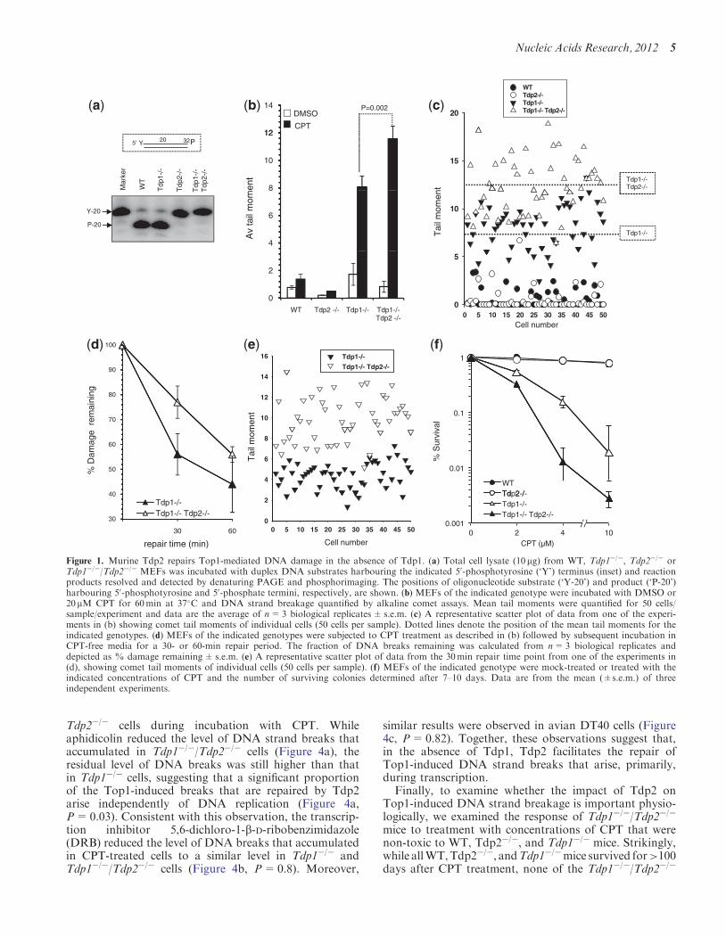
mice employed to generate these MEFs has been shownpreviously (14), and inactivation of Tdp2 is describedbrieflyhere (SupplementaryFigure S6) andwill be describedin detail elsewhere. Cell extract fromTdp2�/� andTdp1�/�/Tdp2�/� MEFs lacked detectable 50-TDP activity in vitro,confirming Tdp2 disruption in these cells (Figure 1a). Toexamine whether Tdp2 might compensate for loss of Tdp1in MEFs, we compared WT, Tdp1�/�, Tdp2�/� andTdp1�/�/Tdp2�/� cells for their ability to repair DNAdamage induced by the Top1 poison CPT, using alkalinecomet assays. While Tdp1�/� cells accumulated �4-foldmore DNA breaks than did wild-type cells, DNA breaksdid not increase above background in CPT-treatedTdp2�/�MEFs (Figure 1b). However, co-deletion of Tdp1
andTdp2 resulted in the accumulation of significantly moreDNA breaks than did deletion of either gene alone (Figure1b and c, P< 0.01) or loss of the critical single-strand breakrepair scaffold protein, Xrcc1 (Supplementary Figure S1).Moreover, Tdp1�/�/Tdp2�/�MEFs exhibited reduced rateof repair during subsequent incubation inCPT-freemedium(Figure 1d and e). Consistentwith these data,whileTdp2�/�
MEFs exhibited normal levels of clonogenic survival fol-lowing CPT treatment, Tdp1�/�/Tdp2�/� MEFs weremore sensitive to CPT than were Tdp1�/� MEFs (Figure1f).We conclude from these experiments that Tdp2 contrib-utes to the repair of Top1-induced DNA damage in murinecells, in absence of Tdp1.
Next, we examined whether Tdp2 also contributes tothe repair of Top1-induced DNA damage in avian DT40cells, by again deleting Tdp1 and Tdp2 separately andtogether. Note that Tdp2�/�/� DT40 cells have beendescribed previously (13), and the targeting strategy andsuccessful deletion of Tdp1 is shown here (Figure 2a and2b). Extracts from Tdp1�/� DT40 cells did not exhibitdetectable 30-TDP activity in vitro, even in reactions con-taining divalent metal and thus under conditions in whichTDP2 is active, consistent with Tdp1 being the majorsource of 30-TDP activity in DT40 cells (Figure 2c). Asexpected, Tdp1�/� DT40 cells accumulated �3-fold moreDNA strand breaks than wild-type cells, following incu-bation with the Top1 poison CPT (Figure 2d). Tdp2�/�/�
cells also accumulated higher levels of DNA breaks thandid wild-type DT40 cells, though this difference was notstatistically significant (P=0.92; student’s t-test).Importantly, Tdp1�/�/Tdp2�/�/� cells accumulated signifi-cantly higher DNA breaks than Tdp1�/� cells, suggestingthat Tdp2 contributes to the repair of Top1 damage inDT40 cells in the absence of Tdp1 (Figure 2d). Indeed,ectopic expression of hTDP2 in Tdp1�/�/Tdp2�/�/� cellsreduced the accumulation of DNA strand breaks inducedby CPT below that observed in Tdp1�/� cells (Figure 3aand b). Furthermore, Tdp1�/�/Tdp2�/�/� cells were moresensitive than Tdp1�/� or Tdp2�/�/� cells to Top1 breaksinduced by CPT (Figure 3c), but not more sensitive toDNA breaks induced by g-radiation (SupplementaryFigure S3). Similar results were observed for theanti-cancer Top1 poisons NSC 724998, NSC 725776 orMJ-III-65 (1) as measured by viability assays(Supplementary Figure S4). In contrast, the reverse wasnot true since deletion of Tdp1 did not further sensitiseTdp2�/�/� cells to the Top2 poison etoposide, suggestingthat Tdp1 is unable to contribute significantly to 50-TDPactivity in DT40 cells, even in the absence of Tdp2(Supplementary Figures S2 & S3). We conclude fromthese data that TDP2 can contribute significantly to therepair of Top1-induced DNA damage in the absence ofTdp1, in both avian and murine cells.
Top1-associated DNA breaks arise by collision ofTop1 intermediates with DNA replication forks orelongating RNA polymerases. To examine the source ofthe Top1-induced DNA breaks that Tdp2 can repair, weused chemical inhibitors of DNA transcription and repli-cation. First, we inhibited DNA replication in MEFs byincubating cells with aphidicolin and then compared theaccumulation of DNA strand breaks in Tdp1�/� and
4 Nucleic Acids Research, 2012
Tdp2�/� cells during incubation with CPT. Whileaphidicolin reduced the level of DNA strand breaks thataccumulated in Tdp1�/�/Tdp2�/� cells (Figure 4a), theresidual level of DNA breaks was still higher than thatin Tdp1�/� cells, suggesting that a significant proportionof the Top1-induced breaks that are repaired by Tdp2arise independently of DNA replication (Figure 4a,P=0.03). Consistent with this observation, the transcrip-tion inhibitor 5,6-dichloro-1-b-D-ribobenzimidazole(DRB) reduced the level of DNA breaks that accumulatedin CPT-treated cells to a similar level in Tdp1�/� andTdp1�/�/Tdp2�/� cells (Figure 4b, P=0.8). Moreover,
similar results were observed in avian DT40 cells (Figure4c, P=0.82). Together, these observations suggest that,in the absence of Tdp1, Tdp2 facilitates the repair ofTop1-induced DNA strand breaks that arise, primarily,during transcription.Finally, to examine whether the impact of Tdp2 on
Top1-induced DNA strand breakage is important physio-logically, we examined the response of Tdp1�/�/Tdp2�/�
mice to treatment with concentrations of CPT that werenon-toxic to WT, Tdp2�/�, and Tdp1�/� mice. Strikingly,while allWT,Tdp2�/�, andTdp1�/�mice survived for>100days after CPT treatment, none of the Tdp1�/�/Tdp2�/�
12
14DMSO
CPT
P=0.00220
WT Tdp2-/- Tdp1-/- Tdp1-/- Tdp2-/-
20Y 32P5’
Mar
ker
WT
Tdp
1-/-
Tdp
2-/-
Tdp
1-/-
Tdp
2-/-
8
10
12
15
Tdp1-/-Tdp2-/-
Y-20
P-20
4
6
Av
tail
mom
ent
10
Tdp1-/-Tai
l mom
ent
0
2
WT Tdp2 -/- Tdp1-/- Tdp1-/-0
5
Tdp2 -/- 0 5 10 15 20 25 30 35 40 45 50Cell number
90
100
16 Tdp1-/-
Tdp1-/- Tdp2-/- 1
70
80
10
12
14
0.1
40
50
60
% D
amag
e r
emai
ning
4
6
8
Tai
l mom
ent
0.01
WT Td 2 /
% S
urvi
val
30
30 60
Tdp1-/-Tdp1-/- Tdp2-/-
repair time (min)
0 5 10 15 20 25 30 35 40 45 500
2
Cell number
0.001 0 2 4 10
Tdp2-/-Tdp1-/- Tdp1-/- Tdp2-/-
CPT (µM)
(a) (b) (c)
(d) (e) (f)
Figure 1. Murine Tdp2 repairs Top1-mediated DNA damage in the absence of Tdp1. (a) Total cell lysate (10 mg) from WT, Tdp1�/�, Tdp2�/� orTdp1�/�/Tdp2�/� MEFs was incubated with duplex DNA substrates harbouring the indicated 50-phosphotyrosine (‘Y’) terminus (inset) and reactionproducts resolved and detected by denaturing PAGE and phosphorimaging. The positions of oligonucleotide substrate (‘Y-20’) and product (‘P-20’)harbouring 50-phosphotyrosine and 50-phosphate termini, respectively, are shown. (b) MEFs of the indicated genotype were incubated with DMSO or20 mM CPT for 60min at 37�C and DNA strand breakage quantified by alkaline comet assays. Mean tail moments were quantified for 50 cells/sample/experiment and data are the average of n=3 biological replicates± s.e.m. (c) A representative scatter plot of data from one of the experi-ments in (b) showing comet tail moments of individual cells (50 cells per sample). Dotted lines denote the position of the mean tail moments for theindicated genotypes. (d) MEFs of the indicated genotypes were subjected to CPT treatment as described in (b) followed by subsequent incubation inCPT-free media for a 30- or 60-min repair period. The fraction of DNA breaks remaining was calculated from n=3 biological replicates anddepicted as % damage remaining±s.e.m. (e) A representative scatter plot of data from the 30min repair time point from one of the experiments in(d), showing comet tail moments of individual cells (50 cells per sample). (f) MEFs of the indicated genotype were mock-treated or treated with theindicated concentrations of CPT and the number of surviving colonies determined after 7–10 days. Data are from the mean (±s.e.m.) of threeindependent experiments.
Nucleic Acids Research, 2012 5

mice survived beyond 10 days (Figure 4d). These datasuggest that Tdp2 contributes to the repair of Top1-induced DNA damage in vivo, in absence of Tdp1.
DISCUSSION
DNA topoisomerases introduce transient breaks in thegenome to release torsional stress. Failure to resealbroken DNA strands results in protein-linked DNAbreaks, which in turn are implicated in neurodegenerationand underlie the clinical utility of topoisomerase poisonsas chemotherapeutic agents (3,17). TDPs liberate DNAtermini from the covalently stalled topoisomerase bycleaving the covalent phosphotyrosyl bond linking thetopoisomerase to DNA, a process that is tightly regulatedby post-translational protein modifications (18).Eukaryotes possess two distinct TDPs as defined bytheir enzymatic activities in vitro. These are a metal
independent TDP1, which primarily acts on DNAbreaks with 30-phosphotyrosyl termini and a metal-de-pendent TDP2, which acts on DNA breaks with 50-phosphotyrosyl termini (2,12,13). TDP2, however, alsopossesses weak 30-TDP activity in vitro and in fact wasidentified in a screen for novel TDP1-like activities thatcomplement the sensitivity of Tdp1-mutant budding yeastcells to Top1-induced DNA damage. Consequently, wehave compared in this study the role of TDP2 in process-ing Top1-induced DNA breaks in the presence andabsence of TDP1 activity, by using murine and avianDT40 cells in which Tdp1, Tdp2 or both were deleted.
Avian DT40 and murine cells lacking Tdp2 exhibitednormal sensitivity to CPT, whereas cells lacking Tdp1were hypersensitive. Notably, however, co-deletion ofTdp1 and Tdp2 resulted in greater cellular sensitivitythan did deletion of Tdp1 alone, suggesting that Tdp2contributes to cellular resistance to Top1-induced DNA
14 kb
14kb (WT)
Tdp18.7kb (Hygro)
8.3kb (Puro)
14
probe
NcoI HygroTDP1 locus
Tdp2
14.3kb (HisD)
9.9kb (WT)
6.3kb (Bsr)
NcoI8.7 kb
8.3 kb
Puro
NcoI
Targeted first allele (Hygro)
25
Targeted second allele (Puro)
20
25
18Y32P5’ 25
43
DMSO
CPT
Av
tail
mom
ent
10
15
18-Y
18-P
5
0WT Tdp2-/-/- Tdp1-/- Tdp1-/- Tdp1-/-
Tdp2-/-/-Tdp1-/-Tdp2-/-/-clone 10 clone 12
clone 14 clone 21
(a) (b)
(d)(c)
Figure 2. Avian Tdp2 repairs Top1-mediated DNA damage in absence of Tdp1. (a) Schematic representation of the wild type and targeted chickenTdp1 locus. Closed boxes indicate the exons. ‘Hygro’ is a hygromycin cassette and ‘Puro’ is a puromycin cassette. The schematic representation of thetargeting locus of the chicken Tdp2 gene has been described previously (13). (b) Genomic DNA from DT40 cells of the indicated genotype wasdigested with NcoI and Southern blotting was conducted using the probe depicted in (a) (for Tdp1, top) or as described in ref 13 (for Tdp2, bottom).(c) Total cell lysate (2, 10 and 20 mg protein) from avian cells of the indicated genotype was incubated with a nicked duplex DNA substrateharbouring a 30-phosphotyrosine (‘18-Y’) terminus (top) for 1 h at 37�C and reaction products were fractionated by denaturing PAGE anddetected by phosphorimaging. The positions of oligonucleotide substrate (‘18-Y’) and product (‘P-20’) harbouring 30-phosphotyrosine and30-phosphate termini, respectively, are shown. (d) DT40 cells of the indicated genotype were incubated with DMSO or 20 mM CPT for 60min at37�C and DNA strand breakage quantified by alkaline comet assays. Mean tail moments were quantified for 50 cells/sample/experiment and data arethe average of n=3 biological replicates± s.e.m.
6 Nucleic Acids Research, 2012

breaks in cells lacking Tdp1. Analogous results wereobserved in alkaline comet assays and in g-H2AXimmunostaining assays (Supplementary Figure S5), sug-gesting that Tdp2 contributes to the repair ofTop1-induced DNA breaks in the absence of Tdp1. Inagreement with these data, over-expression of hTDP2 issufficient to complement the hypersensitivity ofTdp1-mutant budding yeast to CPT (12), and in thisstudy to restore cell survival and DNA strand break
repair rates in CPT-treated Tdp1�/�/Tdp2�/�/� DT40cells to a level greater than that observed in Tdp1�/�
DT40 cells. The simplest explanation for these data isthat Tdp1 is the primary source of 30-TDP activity inwild-type cells, but that Tdp2 contributes measurably tothe repair of some of these breaks if Tdp1 is absent.Incubation with the transcription inhibitor DRB pre-
vented the appearance of the ‘extra’ Top1-inducedbreaks that accumulate in Tdp1-deleted cells if Tdp2 is
DMSO
CPT
6
8
10
12
16
14
Av
tail
mom
ent
hTDP2vector
TDP2
actin
Tdp1-/- Tdp2-/-/-
0
2
4
WT Tdp2-/-Tdp1-/- +hTDP2 + Vector
Tdp1-/- Tdp2-/-/-
-
% S
urvi
val
WT
Tdp1-/-
Tdp2-/-/-
Tdp1-/- Tdp2-/-/-
-+hTDP2
+ Vector
00.001
0.01
0.1
1
10
100
2.5 5 7.5CPT (nM)
(a)
(c)
(b)
Figure 3. Over-expression of hTDP2 protects avian DT40 cells lacking Tdp1 from Top1-mediated DNA damage. (a) DT40 cells stably transfectedwith empty vector or vector encoding full-length hTDP2 ‘hTDP2’ were treated with DMSO vehicle or 20mM CPT for 1 h at 37�C. DNA strandbreaks were quantified by alkaline comet assays in 50 cells/sample/experiment and data are the average of n=3 biological replicates± s.e.m.(b) Total cell lysate (30 mg protein) from Tdp1�/�/Tdp2�/�/� cells harbouring empty vector or vector encoding hTDP2 was fractionated by SDS–PAGE and immunoblotted using anti-hTDP2 polyclonal antibody (SY1340). Actin immunoblots were used as a loading control. (c) DT40 cells of theindicated genotype were treated with the indicated concentrations of CPT and the number of surviving colonies was calculated from n= 3 biologicalreplicates± s.e.m.
Nucleic Acids Research, 2012 7

WT
Tdp2-/-
Tdp1 / 14
16 WT
Tdp2-/-
Tdp1 /
MEFsMEFs
Tdp1-/- Tdp2 -/-
- -
6
8
10
12
- -
Tdp1-/- Tdp2-/-P=0.03
P=0.84
Av
Tai
l Mom
ent
0
2
4
6
8
10
12
14
16
18
DMSO CPT DMSO CPT
0
2
4
6
DMSO CPT DMSO CPT
Av
Tai
l Mom
ent
- Aphidicolin + Aphidicolin - DRB + DRB
DT40 Mice
Tdp1-/- Tdp2-/-/-
WT
Tdp2-/-/-
Tdp1-/-
4
5
6
P=0.82
70
80
90
100
1
2
3
20
30
40
50
60
WT
Tdp2-/-
Tdp1 /
% S
urvi
val
Av
Tai
l Mom
ent
0
DMSO CPT DMSO CPT
-DRB + DRB
0
10
0 2 4 6 8 10
- -
Tdp1-/- Tdp2-/-
Days
(a)
(c) (d)
(b)
Figure 4. Tdp2 preferentially repairs transcription-associated Top1-DNA breaks. MEFs (a and b) or DT40 cells (c) of the indicated genotype wereincubated with DMSO vehicle or with 50 mM of either aphidicolin or 5,6-dichlorobenzimidazole 1-b-D-ribofuranoside (DRB) for 2 h at 37�C,followed by an additional incubation with 20mM ‘CPT’ for 1 h at 37�C. DNA strand breaks were quantified by alkaline comet assays for 50cells/sample/experiment and data are the average of n=3 biological replicates± s.e.m. (d) 3-month-old littermate wild type, Tdp1�/�, Tdp2�/� andTdp1�/�/Tdp2�/� mice at 4 weeks old were ip-injected with CPT at 4 mg/g body weight. Animals were monitored daily for general health and bodyweight for a period of 10 days, followed by weekly monitoring of survivors for 3 months. The % of surviving mice was calculated from fourindependent littermates of each genotype.
8 Nucleic Acids Research, 2012

additionally deleted, suggesting that the Top1-inducedbreaks that Tdp2 repairs in the absence of Tdp1 ariseprimarily during transcription. Recently, Tdp2 wasshown to regulate the level of ribosomal RNA processing,consistent with the possibility that Tdp2 plays a rolein removing DNA breaks at sites of transcription (19).In non-cycling tissues active transcription is the mainsource for Top1-breaks and their progressive accumula-tion may result in death of post-mitotic neurons. It is thusintriguing to speculate that TDP2 deficiency in humanmay result in neurodegeneration similar to that observedfor TDP1 deficiency.
Structural studies suggest that the peptide-bindingpocket in Tdp1 is unable to accommodate full-lengthTop1 and thus prior degradation of topoisomerase atDNA breaks is required for processing by Tdp1(20,21). This notion was further supported by cellularand cell-free studies (7,22). While it is possible that the30-TDP activity of Tdp2 similarly prefers DNA breakslinked to short peptides, it is also possible that Tdp2 canremove full-length Top1 from DNA 30-termini. This mayexplain the specific requirement of Tdp2 for maintainingribosomal transcription upon proteasome inhibition (18).Whether or not human TDP1 operates at Top2 breaksin the absence of TDP2 is unclear. Studies in yeast,which lack a Tdp2 homologue, suggest that yeastTdp1 is involved in the repair of Top2-induced DNAdamage (23). Similarly, in human and DT40 cells,over-expression of human TDP1 has been reported toprotect from Top2-mediated DNA damage (24,25). Inagreement with the latter observation, weak activity ofrecombinant human TDP1 was reported on DSB sub-strates harbouring a 4-base pair 50-phosphotyrosineoverhang typical of Top2-induced breaks, in vitro (25).However, mice and murine cells lacking Tdp1 have notbeen reported to be sensitive to Top2-induced DNAdamage (5,26). Furthermore, using colony survivalassays Tdp1 deletion did not confer measurable sensitiv-ity to Top2 poisons, even in the absence of Tdp2(Supplementary Figure 2) suggesting that Tdp1 is notinvolved in the repair of Top2-induced DNA damagein higher eukaryotes.
In summary, we present evidence implicating Tdp2 inthe repair of Top1-mediated DNA damage in culturedcells and in vivo, in the absence of Tdp1. Our datasuggest that Tdp1 and Tdp2 fulfil overlapping roles fol-lowing Top1-mediated DNA damage and highlight theirpossible utility as novel drug targets during cancertherapy.
SUPPLEMENTARY DATA
Supplementary Data are available at NAR Online:Supplementary Figures 1–6 and Supplementary Methods.
ACKNOWLEDGEMENTS
We thank Shih-Chieh Chiang for technical assistance.
FUNDING
Z.Z. and L.J. were funded by MRC grants to K.W.C.[MR/J006750/1]; A.S. was funded by Wellcome trustgrants to S.E.K. [085284 and 091043]; Generation of theTdp2 (Ttrap)�/� mouse in the DH laboratory was sup-ported by the EC FP6 Integrated Project EndoTrack andInteruniversity Attraction Pole IUAP-P6/20 and, in anearly phase, VIB7 funding. Center for Cancer Research,the Intramural Program of the US National CancerInstitute, NIH [to J.M. and Y.P.]. Funding for openaccess charge: Wellcome Trust Fellowship (to S.E.K.).
Conflict of interest statement. None declared.
REFERENCES
1. Pommier,Y., Leo,E., Zhang,H. and Marchand,C. (2010) DNAtopoisomerases and their poisoning by anticancer andantibacterial drugs. Chem. Biol., 17, 421–433.
2. Yang,S.W., Burgin,A.B. Jr, Huizenga,B.N., Robertson,C.A.,Yao,K.C. and Nash,H.A. (1996) A eukaryotic enzyme that candisjoin dead-end covalent complexes between DNA and type Itopoisomerases. Proc. Natl Acad. Sci. USA, 93, 11534–11539.
3. Takashima,H., Boerkoel,C.F., John,J., Saifi,G.M., Salih,M.A.,Armstrong,D., Mao,Y., Quiocho,F.A., Roa,B.B., Nakagawa,M.et al. (2002) Mutation of TDP1, encoding a topoisomeraseI-dependent DNA damage repair enzyme, in spinocerebellarataxia with axonal neuropathy. Nat. Genet., 32, 267–272.
4. El-Khamisy,S.F., Saifi,G.M., Weinfeld,M., Johansson,F.,Helleday,T., Lupski,J.R. and Caldecott,K.W. (2005) DefectiveDNA single-strand break repair in spinocerebellar ataxia withaxonal neuropathy-1. Nature, 434, 108–113.
5. Interthal,H., Chen,H.J., Kehl-Fie,T.E., Zotzmann,J., Leppard,J.B.and Champoux,J.J. (2005) SCAN1 mutant Tdp1 accumulates theenzyme—DNA intermediate and causes camptothecinhypersensitivity. EMBO J., 24, 2224–2233.
6. Zhou,T., Akopiants,K., Mohapatra,S., Lin,P.S., Valerie,K.,Ramsden,D.A., Lees-Miller,S.P. and Povirk,L.F. (2009)Tyrosyl-DNA phosphodiesterase and the repair of30-phosphoglycolate-terminated DNA double-strand breaks.DNA Repair, 8, 901–911.
7. El-Khamisy,S.F., Hartsuiker,E. and Caldecott,K.W. (2007) TDP1facilitates repair of ionizing radiation-induced DNA single-strandbreaks. DNA Repair, 6, 1485–1495.
8. El-Khamisy,S.F. (2011) To live or to die: a matter of processingdamaged DNA termini in neurons. EMBO Mol. Med., 3, 78–88.
9. Pype,S., Declercq,W., Ibrahimi,A., Michiels,C., VanRietschoten,J.G., Dewulf,N., de Boer,M., Vandenabeele,P.,Huylebroeck,D. and Remacle,J.E. (2000) TTRAP, a novel proteinthat associates with CD40, tumor necrosis factor (TNF)receptor-75 and TNF receptor-associated factors (TRAFs), andthat inhibits nuclear factor-kappa B activation. J. Biol. Chem.,275, 18586–18593.
10. Varady,G., Sarkadi,B. and Fatyol,K. (2011) TTRAP is a novelcomponent of the non-canonical TRAF6-TAK1 TGF-betasignaling pathway. PLoS One, 6, e25548.
11. Esguerra,C.V., Nelles,L., Vermeire,L., Ibrahimi,A.,Crawford,A.D., Derua,R., Janssens,E., Waelkens,E., Carmeliet,P.,Collen,D. et al. (2007) Ttrap is an essential modulator ofSmad3-dependent Nodal signaling during zebrafish gastrulationand left-right axis determination. Development, 134, 4381–4393.
12. Ledesma,F.C., El Khamisy,S.F., Zuma,M.C., Osborn,K. andCaldecott,K.W. (2009) A human 50-tyrosyl DNAphosphodiesterase that repairs topoisomerase-mediated DNAdamage. Nature, 461, 674–678.
13. Zeng,Z., Cortes-Ledesma,F., El Khamisy,S.F. and Caldecott,K.W.(2011) TDP2/TTRAP is the major 50-tyrosyl DNAphosphodiesterase activity in vertebrate cells and is critical forcellular resistance to topoisomerase II-induced DNA damage.J. Biol. Chem., 286, 403–409.
Nucleic Acids Research, 2012 9

14. Katyal,S., El-Khamisy,S.F., Russell,H.R., Li,Y., Ju,L.,Caldecott,K.W. and McKinnon,P.J. (2007) TDP1 facilitateschromosomal single-strand break repair in neurons and isneuroprotective in vivo. EMBO J., 26, 4720–4731.
15. El-Khamisy,S.F., Katyal,S., Patel,P., Ju,L., McKinnon,P.J. andCaldecott,K.W. (2009) Synergistic decrease of DNA single-strandbreak repair rates in mouse neural cells lacking both Tdp1 andaprataxin. DNA Repair, 8, 760–766.
16. Giovanella,B.C., Hinz,H.R., Kozielski,A.J., Stehlin,J.S. Jr,Silber,R. and Potmesil,M. (1991) Complete growth inhibitionof human cancer xenografts in nude mice by treatment with20-(S)-camptothecin. Cancer Res., 51, 3052–3055.
17. Pommier,Y., Pourquier,P., Fan,Y. and Strumberg,D. (1998)Mechanism of action of eukaryotic DNA topoisomerase I anddrugs targeted to the enzyme. Biochim. Biophys. Acta, 1400,83–105.
18. Hudson,J.J.R., Chiang,S.-C., Wells,O.S., Rookyard,C. andEl-Khamisy,S.F. (2012) SUMO modification of theneuroprotective protein TDP1 facilitates chromosomalsingle-strand break repair. Nat. Commun., 3, 733.
19. Vilotti,S., Biagioli,M., Foti,R., Dal Ferro,M., Lavina,Z.S.,Collavin,L., Del Sal,G., Zucchelli,S. and Gustincich,S. (2011)The PML nuclear bodies-associated protein TTRAP regulatesribosome biogenesis in nucleolar cavities upon proteasomeinhibition. Cell Death Differ., 19, 488–500.
20. Davies,D.R., Interthal,H., Champoux,J.J. and Hol,W.G. (2003)Crystal structure of a transition state mimic for Tdp1 assembled
from vanadate, DNA, and a topoisomerase I-derived peptide.Chem. Biol., 10, 139–147.
21. Davies,D.R., Interthal,H., Champoux,J.J. and Hol,W.G. (2004)Explorations of peptide and oligonucleotide binding sites oftyrosyl-DNA phosphodiesterase using vanadate complexes.J. Med. Chem., 47, 829–837.
22. Interthal,H. and Champoux,J.J. (2011) Effects of DNA andprotein size on substrate cleavage by human tyrosyl-DNAphosphodiesterase (TDP1). Biochem. J., 436, 559–566.
23. Nitiss,K.C., Malik,M., He,X., White,S.W. and Nitiss,J.L. (2006)Tyrosyl-DNA phosphodiesterase (Tdp1) participates in the repairof Top2-mediated DNA damage. Proc. Natl Acad. Sci. USA, 103,8953–8958.
24. Barthelmes,H.U., Habermeyer,M., Christensen,M.O., Mielke,C.,Interthal,H., Pouliot,J.J., Boege,F. and Marko,D. (2004) TDP1overexpression in human cells counteracts DNA damage mediatedby topoisomerases I and II. J. Biol. Chem., 279, 55618–55625.
25. Murai,J., Huang,S.Y., Das,B.B., Dexheimer,T.S., Takeda,S. andPommier,Y. (2012) Tyrosyl-DNA phosphodiesterase 1 (TDP1)repairs DNA damages induced by topoisomerases I and II, andbase alkylation in vertebrate cells. J. Biol. Chem., 287,12848–12857.
26. Hirano,R., Interthal,H., Huang,C., Nakamura,T., Deguchi,K.,Choi,K., Bhattacharjee,M.B., Arimura,K., Umehara,F., Izumo,S.et al. (2007) Spinocerebellar ataxia with axonal neuropathy:consequence of a Tdp1 recessive neomorphic mutation?EMBO J., 26, 4732–4743.
10 Nucleic Acids Research, 2012


![A Bíblia como Literatura - Lendo as nar- rativas bíblicas [1]](https://static.fdokumen.com/doc/165x107/63379727fe1f34a1c3009993/a-biblia-como-literatura-lendo-as-nar-rativas-biblicas-1.jpg)