Myopia Manual
324
Myopia Manual Edition January 2022 Changes since the last update January 2021 are marked in red An impartial documentation of all the reasons, therapies and recommendations Unbiased summary of the literature, some ideas about linkages between the various published results, and recommendations for shortsighted people and people who don't want to become shortsighted at all. Dr. rer. nat. Klaus Schmid, Physicist Copyright Notice © Klaus Schmid 2002, 2003, 2004, 2006, 2007, 2008, 2009, 2010, 2011, 2012, 2013, 2014, 2015, 2016, 2017, 2018, 2019, 2020, 2021, 2022. All rights reserved. No part of this publication may be reproduced, stored in a retrieval system, or transmitted in any form or by any means, electronic, mechanical or oth‐ erwise, without the written permission of Klaus Schmid.
-
Upload
khangminh22 -
Category
Documents
-
view
0 -
download
0
Transcript of Myopia Manual

Myopia Manual Edition January 2022
Changes since the last update January 2021 are marked in red
An impartial documentation of all the reasons, therapies
and recommendations
Unbiased summary of the literature, some ideas about linkages between the various published results, and recommendations for shortsighted people and people who don't want to become
shortsighted at all.
Dr. rer. nat. Klaus Schmid, Physicist Copyright Notice
© Klaus Schmid 2002, 2003, 2004, 2006, 2007, 2008, 2009, 2010, 2011, 2012, 2013, 2014, 2015, 2016, 2017, 2018, 2019, 2020, 2021, 2022. All rights reserved. No part of this publication may be reproduced, stored in a retrieval system, or transmitted in any form or by any means, electronic, mechanical or oth‐erwise, without the written permission of Klaus Schmid.

Preface
Especially worth mentioning appears to be:
‐ That the neurotransmitter Dopamine has a substantial impact on the onset of myopia is already known since some years. Now more details of what has an influence on dopamine and its metabolism are appearing and first results of dopamine eye drops showed some promising results on animals.
‐ The primary systemic agent to prevent myopia, especially pathologic high myopia appears to be vita‐min D by its positive impact on dopamine and oxidative stress in the eye.
‐ The color of the light to which the eye is exposed gets increasing attention: The blue, shortwave com‐ponent of an illumination is reported to be protective, which explains the positive effect of time spent outdoor as well. Additionally, the bandwith of illumination appears to be important. On the other hand, LLLT (low level laser therapy) with red led light directed at the retina had a positive effect as well‐
‐ Research being mainly focused on optical effects involving the retina, however, inflammation appears to play a significant role as well. Therefore means to limit the progression of myopia by limiting inflam‐mation might be helpful.
‐ The negative effect of extensive accommodation on the onset and the progression of myopia is known since a long time. The life‐experience ophthalmologist Viikari is, however, that in many cases an existing juvenile hyperopia is the basis of this extensive and harmful accommodation, which can be avoided by using plus lenses. This is especially important, as the developing eye of children is generally hyperopic. With the research world being set to explain myopia by a retina‐image model, some practitioners ex‐plain myopia more by models based on mechanical forces and resulting degradation of connective tis‐sue. The fact that accommodation generally causes a temporal elongation of the eye is mostly ig‐norded, and the attention of research is less focused on the origin of myopia – in most of the cases ex‐cessive accommodation – and more on how to cope with myopia.
‐ "People with a certain variant of the gene ‐ called APLP2 ‐ were five times more likely to develop myo‐pia in their teens if they read for an hour or more each day as a child. … those who carried the APLP2 risk variant but spent less time reading had no additional risk of developing myopia."1078 This means simply not myopia is inherited, but the sensitivity with respect to environmental conditions.
‐ Some unspectacular but maybe helpful result is the positive impact of making the children to go to bed early at night and to avoid long time nearwork in the evening and on weakends.
‐ A solitary but promising report is the successful use of LLLT (Low‐Level Laser Therapy) against pro‐gression of myopia and maybe even for the reduction of myopia.
‐ Tables might be helpful, where the success of various interventions against the progression of myo‐pia were evaluated (section 3.27).

Disclaimer
This book is intended as an informational guide to be used as a supplement, not a substitute for profes‐sional medical advice. While the information and advice in this book is believed true and accurate, the author cannot accept any legal responsibility.
Acknowledgements
This book is an effort to put pieces of the myopia puzzle together. These pieces were found in numer‐ous books and scientific publications, and therefore I am extremely grateful to all these professionals for their work and their sharing of their results.
Additionally, quite a number of readers of the Internet version of this book helped with very construc‐tive, encouraging and competent comments.
Finally, thanks to my son Bernhard Schmid who was very helpful by taking care of all the data process‐ing problems, which had to be solved to complete this book.

This book is dedicated to my wife Veronica and my children Nadine and Bernhard, whose shortsightedness caused me
to write this book.
Hopefully it will be helpful not only for them.

v
Table of Contents
Introduction .............................................................................................................................................. 1 1 What is Myopia? ................................................................................................................................ 4 1.1 How does it Feel being Myopic?.......................................................................................................... 4 1.2 Basic Terminology of the Anatomy of the Eye..................................................................................... 5 1.3 Accommodation .................................................................................................................................. 5
1.3.1 Myopia and Emmetropia ............................................................................................................. 5 1.3.2 Theory of Accommodation .......................................................................................................... 7 1.3.2.1 The Helmholtz Model .......................................................................................................... 7 1.3.2.2 The Schachar Model ............................................................................................................ 7 1.3.2.3 The Two‐Phase‐Model ......................................................................................................... 8 1.3.2.4 Two other, Controversial Hypotheses ................................................................................. 8
1.4 Refractive Myopia................................................................................................................................ 9 1.4.1 Tonic Accommodation and Night Myopia ................................................................................. 10 1.4.2 Pseudomyopia ........................................................................................................................... 11 1.4.3 Other Types of Myopia .............................................................................................................. 12
1.5 Axial Myopia ...................................................................................................................................... 13 1.6 "What Type of Myopia Do I Have?"................................................................................................... 13 1.7 Consequences and Risks of Higher Myopia ....................................................................................... 13 1.8 Myopia and Age................................................................................................................................. 18 1.9 Accommodation and Age / Presbyopia ............................................................................................. 18 1.10 Age Related Geometrical Changes of the Eye ................................................................................... 20 1.11 The Refraction ................................................................................................................................... 21
1.11.1 Basic Procedure ......................................................................................................................... 21 1.11.2 A Method for Refraction: Fogging ............................................................................................. 25 1.11.3 The Range of Clarity................................................................................................................... 26
2 What Causes Myopia in General? ..................................................................................................... 27 2.1 Is Myopia Inherited?.......................................................................................................................... 27 2.2 Connective Tissue Disorders.............................................................................................................. 30 2.3 Active Growth by Imaging Effects...................................................................................................... 31 2.4 Mechanical Effects............................................................................................................................. 31 2.5 General Overview of the Causes of Myopia ...................................................................................... 32
3 Myopia – Observations and Experimental Results ............................................................................ 33 3.1 Distribution of Myopia by Region, Age, Gender and Ethnicity .......................................................... 33 3.2 Accommodation and Near Work ....................................................................................................... 37
3.2.1 Experiences and Results ............................................................................................................ 37 3.2.1.1 General Experiences and Results....................................................................................... 37 3.2.1.2 The Effect of Accommodation on Axial Length and Scleral Thinning ................................ 40 3.2.1.3 The Effect of Accommodation on Image Quality............................................................... 43

vi
3.2.1.4 Timing‐ and Hysteresis‐ Effects of Accommodation .......................................................... 43 3.2.1.5 Dark Focus and Accommodative Hysteresis ...................................................................... 45 3.2.1.6 The Strength of Accommodation....................................................................................... 46 3.2.1.7 Accommodation Spasm ..................................................................................................... 47 3.2.1.8 Myopia caused by Hyperopia ............................................................................................ 48 3.2.1.9 "Screen sightedness" by SmartPhones and Tablet‐PCs ..................................................... 49 3.2.1.10 Aniso‐Accommodation ...................................................................................................... 50 3.2.1.11 Is there a Connection between Blur Sensitivity and Accommodation Deficits?................ 50 3.2.1.12 Accommodation and the Nervous System ........................................................................ 51 3.2.1.13 Accommodation and Biochemistry.................................................................................... 52 3.2.1.14 "Emmetropization" towards Myopia via Accommodation? .............................................. 52 3.2.1.15 Myopia before Modern Times ........................................................................................... 53 3.2.1.16 Summary of Results about Accommodation ..................................................................... 54
3.2.2 Proposed Therapies Based on the Focusing and Accommodation Issue................................... 55 3.2.2.1 Relaxing and Exercising...................................................................................................... 55 3.2.2.2 Biofeedback ....................................................................................................................... 57 3.2.2.3 Undercorrection for Near Work, Plus‐, Bifocal‐ and Progressive‐ Glasses ........................ 58 3.2.2.3.1 General Results ............................................................................................................... 58 3.2.2.3.2 Plus Addition, Bifocals and their Prismatic Effects.......................................................... 62 3.2.2.3.3 Plus glasses – are they Effective via Reduced Accommodation or via Modified Vergence? 63 3.2.2.3.4 Bifocal and Multifocal Contact Lenses............................................................................ 64 3.2.2.3.5 Intermittent, Short Term Wearing of Plus Glasses ......................................................... 65 3.2.2.3.6 Permanent Undercorrection instead of Undercorrection for Near Work only .............. 65 3.2.2.3.7 Why is there no Agreement about the Usefulness of Plus‐Glasses? .............................. 68 3.2.2.3.8 Is the Accommodation System Getting too Lazy by the Plus‐Glasses?........................... 70 3.2.2.3.9 Plus‐Glasses to Reverse Myopia?.................................................................................... 70 3.2.2.3.10 Plus Glasses or Undercorrection – a Striking Result ..................................................... 70 3.2.2.3.11 Plus Glasses – a Summary............................................................................................. 71 3.2.2.3.12 Comparison of the Various Optical Methods................................................................ 74 3.2.2.3.13 Psychological Problems with Special Glassear Workes for Near Work......................... 75 3.2.2.3.14 How to Break an Accommodation Spasm?................................................................... 75 3.2.2.3.15 Get New Glasses? ......................................................................................................... 76 3.2.2.3.16 Summary of the Accommodation Based Therapies...................................................... 77
3.3 The Effects of Image Quality.............................................................................................................. 79 3.3.1 Basic Results .............................................................................................................................. 79 3.3.2 Connective Tissue Related Results ............................................................................................ 81 3.3.3 Dopamine and Light Exposure – and General Effects of Dopamine.......................................... 85 3.3.4 Remarks on the Image Quality Model ....................................................................................... 88 3.3.5 "Emmetropization" towards Myopia via Image Quality? .......................................................... 89 3.3.6 Intermittent, Short Term Wearing of Plus Glasses .................................................................... 92

vii
3.3.7 Duration of Undercorrection ..................................................................................................... 94 3.3.8 Contrast and Spatial Frequency................................................................................................. 94 3.3.9 Monochromatic Aberrations and other Optical Deficiencies .................................................... 95 3.3.10 Longitudinal Chromatic Aberrrations / Myopic and Hyperopic Defocus.................................. 99 3.3.11 Peripheral Defocus and Dual‐Focus Lenses ............................................................................... 99 3.3.11.1 Peripheral Defocus – New Applications for Spectacle Glasses and Contact Lenses........ 103
3.3.12 Summary of the Effects of the Image Quality.......................................................................... 104 3.4 Basic Question – what Elongates the Eye........................................................................................ 105
3.4.1 Image Quality and Accommodation ........................................................................................ 105 3.4.2 Myopia by Accommodation or Myopia by Degraded Image Quality?..................................... 107 3.4.3 Emmetropization by Growth or Mechanical Stretching or Biochemical Thinning?................. 110
3.5 Phoria, Convergence, Astigmatism and Anisometropia .................................................................. 110 3.5.1 Phoria....................................................................................................................................... 110 3.5.2 The AC/A Ratio......................................................................................................................... 112 3.5.3 The CA/C Ratio, and some more Types of Vergence ............................................................... 114 3.5.4 Hysteresis of Vergence ............................................................................................................ 115 3.5.5 Glasses, Contact Lenses and Vergence.................................................................................... 115 3.5.6 Prescription of Prisms.............................................................................................................. 116 3.5.7 The Impact of Vergence on the Axial Length........................................................................... 118 3.5.8 Astigmatism ............................................................................................................................. 118 3.5.9 Anisometropia ......................................................................................................................... 118 3.5.10 Summary of the Vergence Related Effects .............................................................................. 118
3.6 Saccades and Focusing .................................................................................................................... 119 3.7 Mechanical Properties, Stress, Strain and Pressure ........................................................................ 121
3.7.1 Impact of Mechanics on Biochemistry..................................................................................... 121 3.7.2 Intraocular Pressure (IOP) ....................................................................................................... 121 3.7.3 The Ciliary and other Muscles ................................................................................................. 124 3.7.4 The Lens................................................................................................................................... 126 3.7.5 The Pupil Size ........................................................................................................................... 127 3.7.6 The Ocular Shape..................................................................................................................... 127 3.7.7 The Zonular Fibers ................................................................................................................... 129
3.8 Relations between the Dimensions of the Cornea, the Crystalline Lens and the Ciliary Muscle .... 129 3.9 Growth or Stretching and Thinning or Biochemical Thinning of the Sclera in Myopic Eyes?.......... 130 3.10 Outdoor Activities............................................................................................................................ 132 3.11 Exposure to Bright Sunlight ............................................................................................................. 135 3.12 Seasonal Impact............................................................................................................................... 135 3.13 Physical Exercises............................................................................................................................. 135 3.14 Illumination / Contrast / Light / Day‐ and Night‐Rhythm ................................................................ 136
3.14.1 Level of Illumination ................................................................................................................ 136 3.14.2 Color of Illumination – Wavelength of the Light ..................................................................... 144 3.14.2.1 Individual Results ............................................................................................................. 144
viii
3.14.2.2 The Effect of Broadband Illumination.............................................................................. 148 3.14.2.3 Melanopsin and Neuropsin.............................................................................................. 149 3.14.2.4 Blue Light Emitted from Electronic Screens: What are the Risks?................................... 149 3.14.2.5 LLLT – Low‐Level Laser Therapy....................................................................................... 150
3.14.3 Contrast ................................................................................................................................... 151 3.14.4 Day‐ and Night‐Rhythm ........................................................................................................... 151 3.14.5 Flickering Light and Flashing Light ........................................................................................... 154
3.15 Vision Training / Vision Therapy / Behavioral Optometry ............................................................... 155 3.16 Temperature.................................................................................................................................... 155 3.17 Blood Circulation ............................................................................................................................. 157 3.18 Some Specific Biochemical Issues.................................................................................................... 159
3.18.1 The Immune System ................................................................................................................ 159 3.18.2 Inflammation towards Progressive and Pathologic / Malignant Myopia ................................ 161 3.18.3 Oxidative Stress and Antioxidant Defense............................................................................... 162 3.18.4 Enzymes Glutathione, Glutathione Peroxidase, Superoxide Dismutase, G6PD...................... 164 3.18.5 The Blood‐Retinal Barrier ........................................................................................................ 165 3.18.6 The Vitreous Body.................................................................................................................... 165 3.18.7 Nitric Oxide (NO)...................................................................................................................... 166 3.18.8 Cross‐Linking of the Collagen of the Sklera by UV‐Radiation and Roboflavin ......................... 168 3.18.9 More Biochemical and Biomechanical Effects......................................................................... 169
3.19 Mental Issues................................................................................................................................... 170 3.19.1 Stress and Cortisol ................................................................................................................... 170 3.19.2 Personality and Mentality........................................................................................................ 173
3.20 Why are some People not becoming Myopic, Independent from the Circumstances? .................. 175 3.21 A Study: Factors Associated with Myopia in School Children.......................................................... 176 3.22 Myopic Changes in Pregnancy ......................................................................................................... 177 3.23 COVID‐19 and Myopia ..................................................................................................................... 177 3.24 Impact of Nutritional Components.................................................................................................. 177
3.24.1 Carbohydrates, Blood Sugar Level, Insulin Metabolism .......................................................... 178 3.24.2 Is there a Connection between the Blood Sugar Level and Negative‐Lens‐Induced Myopia?181 3.24.3 Calcium, Vitamin D and Sunlight.............................................................................................. 182 3.24.4 Vitamin D and Dopamine......................................................................................................... 185 3.24.5 Selenium and Dopamine.......................................................................................................... 185 3.24.6 Magnesium .............................................................................................................................. 185 3.24.7 Copper and Zinc ....................................................................................................................... 186 3.24.8 Chromium ................................................................................................................................ 188 3.24.9 Manganese .............................................................................................................................. 188 3.24.10 Potassium ................................................................................................................................ 188 3.24.11 Fluoride.................................................................................................................................... 189 3.24.12 Salt 189 3.24.13 Fat and Cholesterol.................................................................................................................. 189

ix
3.24.14 Vitamin A ................................................................................................................................. 190 3.24.15 B‐Vitamins................................................................................................................................ 190 3.24.15.1 Riboflavin plus UVA [blue light] ....................................................................................... 191
3.24.16 Some More Antioxidants ......................................................................................................... 191 3.24.16.1 Selenium .......................................................................................................................... 191 3.24.16.2 Flavonoids and Related Compounds, and Vitamin E ....................................................... 192
3.24.17 Folic Acid and Homocysteine................................................................................................... 193 3.24.17.1 Observations.................................................................................................................... 193 3.24.17.2 An Explanation................................................................................................................. 194
3.24.18 Bilberry and Anthocyanins....................................................................................................... 195 3.24.19 Lactoferrin ............................................................................................................................... 195 3.24.20 Crocetin ................................................................................................................................... 196 3.24.21 Other Components of the Diet ................................................................................................ 196 3.24.22 Overall Nutritional Status and Myopia .................................................................................... 197
3.25 Pharmaceuticals............................................................................................................................... 198 3.25.1 Atropine................................................................................................................................... 198 3.25.1.1 Comparison of Various Concentration of Atropine ......................................................... 201 3.25.1.2 Atropine plus Ortokeratology .......................................................................................... 202
3.25.2 7‐Methylxanthine .................................................................................................................... 202 3.25.3 Other Pharmazeutical Agents.................................................................................................. 203 3.25.4 Dopamine – a Summary .......................................................................................................... 204 3.25.4.1 What Impact has a low Level of Dopamine? ................................................................... 204 3.25.4.2 What Increases the Level of Dopamine? ......................................................................... 204 3.25.4.3 Is the Couple Dopamine / Melatonin a Key‐Player? ........................................................ 205
3.26 Other Means to Slow Down or Stop Progression of Myopia........................................................... 206 3.26.1 Contact Lenses......................................................................................................................... 206 3.26.2 Orthokeratology / Overnight Corneal Reshaping (OCR) .......................................................... 209 3.26.3 Orthoculogy ............................................................................................................................. 212 3.26.4 Reinforcement of the Sclera .................................................................................................... 213 3.26.5 Acupuncture and Acupressure ................................................................................................ 213 3.26.6 Electrostimualtion ................................................................................................................... 214
3.27 Summary of Randomized Trials to Slow Progression of Myopia ..................................................... 214 3.27.1 Single Trials .............................................................................................................................. 214 3.27.2 Comparisons of the Efficacy of the Various Measures to Slow the Progression of Myopia.... 214 3.27.3 Rebound Effects....................................................................................................................... 216
3.28 Correction of Myopia by Surgery..................................................................................................... 217 3.28.1 Manipulation of the Cornea .................................................................................................... 217 3.28.1.1 Radial Keratotomy RK ...................................................................................................... 217 3.28.1.2 Photorefractive Keratectomy (PRK)................................................................................. 217 3.28.1.3 Laser In Situ Keratomileusis (LASIK, Epi‐LASIK), Laser Epithelial Keratomileusis
(LASEK), Femto‐LASIK....................................................................................................... 218

x
3.28.1.4 Small Incision Lenticule Exraction (SMILE)....................................................................... 218 3.28.1.5 Intrastromal Corneal Ring................................................................................................ 219 3.28.1.6 Artificial Cornea ............................................................................................................... 219
3.28.2 Manipulation of the Lens System inside the Eye..................................................................... 219 3.28.2.1 Exchange of the biological lens:....................................................................................... 220 3.28.2.2 Adding an additional lens: ............................................................................................... 220 3.28.2.3 Risk‐comparison between LASIK and implantable contact lenses (ICL): ......................... 221
3.28.3 Enforcing the Sclera ................................................................................................................. 221 3.29 A Few Recommendations ................................................................................................................ 222 3.30 Summary: What Causes Myopia?.................................................................................................... 223
3.30.1 School and Myopia .................................................................................................................. 224 3.30.2 Is Myopia Caused by Mechanical or by Biochemical Processes?............................................. 224 3.30.3 Is Myopia caused by Mechanical or Optical Processes – are Saccades a Linkage? ................. 225 3.30.4 Prevention of Progression of Myopia or Prevention of Myopia? ............................................ 226 3.30.5 Working against Myopia or against the Consequences of Myopia?........................................ 226 3.30.6 Functional‐ versus Structural‐ Deficits..................................................................................... 226 3.30.7 Is Myopia Inherited or Acquired? ............................................................................................ 227 3.30.8 Congenital Myopia, and Inherited Diseases which are related to Myopia............................. 228 3.30.9 From Simple to High and Progressive Myopia......................................................................... 231 3.30.10 Summary of the Summary: Are the Published Results really Contradictory? Maybe not!..... 231
3.31 For Further Reading –Extensive Compilations................................................................................. 236 4 Additional Information ................................................................................................................... 239 4.1 Optical Correction............................................................................................................................ 239
4.1.1 Glasses ..................................................................................................................................... 239 4.1.2 The Material of the Glass......................................................................................................... 239 4.1.3 The Coating of the Glass .......................................................................................................... 240
4.2 Contact Lenses................................................................................................................................. 240 4.2.1 Basic Types of Lenses – Soft Lenses versus Hard Lenses ......................................................... 241 4.2.2 How about Lenses for Permanent Wear?................................................................................ 245 4.2.3 Potential Complications........................................................................................................... 246 4.2.4 Parameters of the Material for Contact Lenses....................................................................... 247 4.2.4.1 Oxygen Transmission ....................................................................................................... 247 4.2.4.2 Wettability and Resistance against Deposits ................................................................... 249 4.2.4.3 Hardness and Stability ..................................................................................................... 250 4.2.4.4 Specific Weight and Refractive Index .............................................................................. 251 4.2.4.5 UV Blocking ...................................................................................................................... 251 4.2.4.6 Color / Tint of the Lens .................................................................................................... 251
4.2.5 Parameters of the Geometry of Contact Lenses...................................................................... 251 4.2.6 Surface and Edge Finishing ...................................................................................................... 255 4.2.7 The Fitting of Contact Lenses................................................................................................... 255 4.2.8 The "Right" Power of Contact Lenses ...................................................................................... 256

xi
4.2.9 Determining the Refraction after Wearing Contact Lenses..................................................... 256 4.2.10 Maintenance of the Lenses...................................................................................................... 257 4.2.10.1 Maintenance of Hard RGP Lenses.................................................................................... 257 4.2.10.2 Maintenance of Soft Lenses............................................................................................. 258
4.2.11 "I Cannot Wear Contact Lenses".............................................................................................. 259 4.2.12 Presbyopia ............................................................................................................................... 260 4.2.13 Contact Lenses and Nutrition .................................................................................................. 262
4.3 Refractive Surgery............................................................................................................................ 263 4.4 Useful Links...................................................................................................................................... 266
5 References ......................................................................................................................................268


1
Introduction
The Situation - and the Target of this Book Old‐fashioned traditional medicine stated that myopia is an inherited condition and the only solution is to prescribe glasses. People who objected to this "therapy" (it's not really a therapy, but only a short term covering of symptoms) were treated as ignorant.
In many papers, however, it was reported that today very many people are becoming myopic even though their parents or grandparents were not. On the other hand, life today is very different from that of our ancestors – just to mention the changed working environment and changed nutrition. Therefore, it is rather obvious that these changes in the environment have an impact on the incidence of myopia. Definitely, some people are more sensitive to these changes than others – by heredity.
If we can map all these negative influences and understand how they are affecting myopia, then we have a chance at minimizing myopia in some, decreasing its progression in others and in some cases, preventing it altogether. At the very least, we may be able to minimize the very serious consequence of blindness.
The idea of finding just one mechanism for myopia and solving this problem once and forever is very tempting, and some authors give the impression that they are close to this goal, and that all the other researchers, optometrists and ophthalmologists are wrong. Strangely enough, these authors often dis‐agree with each other. The conclusion is not that some researchers are right, and of the rest of the re‐searchers are wrong – all of them are right in their specific view of their experiments and experiences. Clearly, various different mechanisms exist that can lead to myopia. It is more important to find com‐monalities in the results than the contradictions.
Moreover, a long chain of processes (e.g. genetic, biochemical, environmental, nutritional) can cause myopia (like many other medical problems), and therefore very different interventions targeting these single processes can be successful.
Unfortunately, it appears that too little fi‐nancial resources are available for research in myopia, since the industry financially benefits from the existence and progression of myopia than from its prevention. Just one number: In 1993 the USA spent 3.4 billion $ US on optical correction of myopia1. Defi‐nitely, the number is substantially higher today – not counting the personal inconven‐ience, the inability to work in a planned pro‐fession, and the suffering because of the
used sources
Interested layman professional
typical reader
popular literature
scientificliterature

2
permanent progression of myopia: in some countries myopia is the first or second leading cause of blindness2.
I will outline numerous essential results reported by researchers and practitioners. Since I have no per‐sonal involvement, i.e. no personal results to promote, I feel I am able to do this in an unbiased and neutral way. Additionally, I will try to find common patterns for the results.
The author of this book doesn't pretend to be a judge about whose myopia theory is right and whose theory is wrong – in the framework of their experimental conditions all of them can considered to be right in a way! Therefore, my personal views are generally clearly marked as "Notes".
The typical reader for whom this book was written is somebody who has myopia, or whose relative has myopia, and who is willing to take the tough route through a lot of scientific results. Maybe even some professionals in the area of myopia can benefit by getting a guide to numerous research results.
Some readers might be bored or frightened by these numerous scientific results given in this book. I de‐cided, however, to include them to make all the recommendations and conclusions credible. There are already numerous books on the market that focus on mere recommendations without going into the details of the scientific background – I did not want to add another one to this category.
The purpose of this book is
(1) to increase the overall knowledge about myopia, particularly progressive myopia in an easy‐to‐read but scientifically correct format without skipping opinions and results which are still controversial
(2) to suggest the best ways to handle myopia.
The advice given in this book does not replace the treatment by a health professional, but it should en‐able the reader to enter detailed discussions with her/his personal optometrist, ophthalmologist or physician.
Do not expect simple "cook book" approaches to make your myopia disappear. The reader can expect to gain a better understanding of the various mechanisms and factors that produce myopia and to learn some of the means to treat its symptoms.
Do not expect to find an easy answer about the exact cause of your specific myopia. Obviously, myopia can be caused by a lot of different off‐balanced processes in the human system. You will find, however, a lot of material that will enable you to consider which of your personal conditions might be part of the problem.
You will find that there are many controversies among the experts. Also, many of the cited references indicate the need for more research.
Additionally, most of the published results were collected data, which were found to be associated with myopia, and they are not definite proofs about what causes myopia. For most of these causes, how‐ever, there are potential scientific explanations given for the risk of misinterpreting associated facts as

3
causes is as follows3: It can be said, e.g. that most of the children who run across the street and get hit by an automobile were wearing tennis shoes, so therefore, tennis shoes must have caused the acci‐dents. Obviously, the tennis shoes were only a common association, not a related factor. This is the problem in sorting out causes from associations in the study of myopia.
Nevertheless, Research has already collected a tremendous amount of information, which can be very helpful to preserve your eyesight! It does not make sense simply to wait until research has solved the myopia problem – maybe this will be too late for you.
Sections 1 to 3 detail the basic material about myopia, the remaining sections contain material which can help to create a link between the sometimes contradictory publications about myopia, and some background information which might be of interest for involved people.
Myopia is a result of more than what happens within the optical system. It is also affected by nutrition and other aspects of lifestyle. As Eaton et al. stated4:
"These diseases are the results of interaction between genetically controlled biochemical processes and a myriad of biocultural influences – lifestyle factors – that include nutrition, exercise, and exposure to noxious substances."
Additionally, the tremendous mental stress, which accompanies higher education and highly demand‐ing jobs, plays a very significant role in the onset and progression of myopia (see section 3.19.1). Con‐sequently, relaxation from this stress is very important but often underestimated (see section 3.2.2.1). By the way, near work should always be accompanied by some stress of concentration.
The author welcomes every kind of feedback – please send an email to
4
1 What is Myopia?
1.1 How does it Feel being Myopic?
Most of the readers of this book don't have to read this, because they know how it feels being myopic. They are hoping to gain some improvement, or even to cure the condition.
Other people, however, are reading this book to get help for their child or a friend. They don't have personal experience with myopia.
Being myopic means you cannot focus on distant objects without an optical device like glasses or con‐tact lenses. Without these devices distant objects are blur – the higher the myopia the more blurry they are. Simulations of the effects of various eye problems on the vision are shown in the Internet5.
Some people might consider glasses or contact lenses to be at the most a minor inconvenience. Chil‐dren, however, often feel really handicapped, and people doing sports can feel the same. In any case, if there is a higher grade of myopia the loss of the optical device leaves the person feeling helpless.
As long as there is complete vision achieved with optical devices there might be a substantial psycho‐logical problem, but not a real medical problem. People with higher grades of myopia, however, are threatened by a permanent degradation of their vision or even blindness (see section 1.7).
At the least, then, myopia is inconvenient, and can be risky. This should be enough reason to find out about ways to minimize it. Like for every problem in life prevention is the best remedy – in spite of the fact that most often people will take action only when the damage is already happening. There is a German proverb saying "damage makes you wise" – but better get wise without too much damage. Therefore, please read the complete book.

1 WHAT IS MYOPIA? 5
1.2 Basic Terminology of the Anatomy of the Eye
For clarification of the terminology, a cross section of the eye is shown in Figure 1.
Cornea
Posterior chamber
Anterior chamber
RetinaChoroid
Sclera
Optic nerve
Central retinal artery and vein
Lens Vitreous body
Zonular fibers
Iris
Pupil
Fovea – the maculais the area for centralvision around the fovea
Ciliary muscle / ciliary body
Conjunctiva
Figure 1 The anatomy of the eye
1.3 Accommodation
Accommodation is the adjustment of the refractive power of the lens of the eye to achieve an exact im‐age of the object on the retina.
1.3.1 Myopia and Emmetropia
Myopia (or shortsightedness, or nearsightedness) is a condition in which distant objects are not dis‐played sharply on the retina by the optical system of the eye, because the rays converge already before they hit the retina.

6
Figure 2 The accommodation effort of myopes and non‐myopes
Figure 2 shows effective accom‐modation, when a proper focusing on the retina is achieved. E.g., in typical axial myopia even with re‐laxed accommodation proper fo‐cus of distant objects cannot be achieved. The emmetropic eye can adjust for all distances by an ap‐propriate accommodation effort. Low accommodation amplitude results in an exact vision of a nar‐row range of distances only.
The states of myopia and emmetropia are defined according to the handling of parallel rays of light (i.e. far distance):
Emmetropia is most often defined as a state, where parallel rays of light can be focused properly on the retina. For myopia this focus point lies in front of the retina.
Obviously, emmetropia can be achieved not by one specific ocular model, but by a range of ocular con‐figurations, which may all lead to a good vision at distant objects.
Emmetropia and hyperopia (farsightedness) blur into each other:
Proper distant focus can be achieved without accommodation as well as with some residual ac‐commodation.
Near focus (e.g. for reading) is largely dependent on the person's ability to accommodate (i.e. the individual amplitude of accommodation), which decreases with age (see section 1.8).
The reasons for myopia can be (see Figure 3):
The refractive power of the lens system is too high: refractive myopia. Parameters of the lens sys‐tem are the curvature of the cornea, the curvature of the lens at the front as well at the back, and the refractive indices of the anterior chamber, the lens and the vitreous body
Distance of proper focusing
30 cmreading distance
> 10 mdistant focus
Accommodationeffort
relaxed
maximumamplitude
typical axial myopiamyopia with low accommodation amplitudetypical range of emmetropia

1 WHAT IS MYOPIA? 7
The distance between the lens system and the retina is too large: axial myopia.
The critical reason is the second one, as it can lead to the dangerous progressive myopia by excessive stretching of the sclera.
In the next sections the basics of accommodation, i.e. the ad‐justment of the eye to different distances of the objects, and some specific sub‐categories of myopia will be discussed.
Literature‐references to ophthalmology in general and to literature about myopia are given at the end of this book in chapter 56, 7, 8, 9, 10, 11, 12, 13, 14, 15, 16, 17.
1.3.2 Theory of Accommodation
1.3.2.1 The Helmholtz Model
The general view of ophthalmic science, based on Helmholtz, is:
Due to its own original shape the lens would get into the shape of a round ball without a force on it. This shape corresponds to near focus.
The pulling action of the zonular fibers, which connect the lens with the ring‐shaped ciliary muscle, can flatten the lens. This shape corresponds to focus on a distant object.
Consequence:
If the ciliary muscle is relaxed, there is a pulling tension on the lens via the zonular fibers, and therefore the focus is set to "distant object".
If the ciliary muscle is contracted, the diameter of this ring‐shaped muscle is decreased, and there is no more pulling action on the lens via the zonular fibers and therefore the focus is set to "near object".
1.3.2.2 The Schachar Model
A modified, and in a way very much contrary theory, the Schachar theory is described as follows:18, "The Schachar theory suggests that the ciliary muscle contracts during accommodation, placing more tension on the equatorial zonules while relaxing the anterior and posterior zonules. (In the Helmholtz
Figure 3 The focusing of the myopic eye

8
theory, all the zonules relax). This causes an increase in the equatorial diameter of the lens, decreasing the peripheral volume while increasing the central volume. As the central volume increases, so does the power of the lens." 19, 20, 21. This means, as Burd stated22, "that increasing the zonular tension increases rather than decreases the power of the lens."
Both theories agree, however, that it is the ciliary muscle, which controls accommodation ‐ in contrast to the theories described in section 1.3.2.4.
The idea that accommodation is a more complex function than the Helmholtz model suggests is sup‐ported by the results of Gao L et al.: "The ocular accommodation has a great influence on refractive components in children. It is not only the process by which the refractive power of the lens is in‐creased. Furthermore, the lens itself moved forward relatively... AD [anterior chamber depth] increased while both LT [lens thickness] and VL [vitreous chamber depth] decreased significantly after cycloplegia regardless of their refractive state. However, AL increased for hyperopic eyes and decreased for myopic eyes after cycloplegia."23
An overall comparison between the Helmholtz theory and the Schachar theory concludes that the ob‐servations are more in favor of the Schachar theory.24
Additional information about the impact of accommodation on the ocular shape can be found in sec‐tion 3.7.6.
1.3.2.3 The Two-Phase-Model
Mütze described the accommodation process a two‐stage process with a rough focusing and a fine ad‐justment25, where the phase of fine adjustment can cause an accommodation spasm.
Note: This reminds of the saccades mentioned in section 3.6.
1.3.2.4 Two other, Controversial Hypotheses
Based on experiments, which he conducted on his own eyes, McCollim published the following very dif‐ferent model26, 27:
"Compression of the globe by the extraocular muscles [which move the axes of the eyeballs] can cause the lens to accommodate" ... "accommodation can be actuated without the intervention of the ciliary muscle".
"... a single factor, external pressure on the globe, produces two separate effects, in opposite direc‐tions: anteriorly it accommodates the lens [by forcing the vitreous against the lens], and pos‐teriorly it elongates the globe."

1 WHAT IS MYOPIA? 9
The fact that it was found by measurements that the eye is elongating during near accommoda‐tion28, 29 supports aspects of this thesis.
Because of these experimental results, it was concluded by McCollim26 that "with repeated periods of prolonged accommodation the lens would never have sufficient time to return completely to the unac‐commodated state", and30 "It is proposed here that the contribution of the lens in myopia is as much as or more so than the contribution of axial length" i.e. there is a substantial time lag for the reshaping of the lens.
Conclusion: While the reshaping is taking place the refraction will diagnose myopia. After the reshaping the refraction will not find myopia.
Note: This time lag for the reshaping of the lens was generally found in myopic eyes (see section 3.2.1). The model of a longer lasting time lag of the shape change of the lens could explain a progression of myo‐pia: The fitted glasses would become inadequate, and could induce a further increase of myopia (see section 3.2.1.3). In fact, the progression would depend on how long the eye takes to return to shape. This effect of a time lag for readjustment is called hysteresis.
Scanlan published another, very unconventional theory. He stated31: "...the respiratory system applies air pressure to the rear of the eyeballs influencing the shape of the eyeball and hence influences how clearly we see at different distances (refractive state)." and concludes "...myopia arises due to a re‐duced level of air pressure against the rear of the eyeball, causing a refractive error." According to Scanlan, accommodation and near work "...requires that pressure in the respiratory system pressing on the back of the eyeball be reduced compared to the pressure necessary for distance work."
Note: The fact that myopia appears to have a close relation to stress (see section 3.19.1) gives some support to this theory. It appears doubtful whether effects off the respiratory system are solely respon‐sible for the onset and progression of myopia, but there might be a contribution of it.
1.4 Refractive Myopia
Refractive myopia is caused by specific deviations of the optical properties of the elements of the lens system of the eye, e.g. an anomalous curvature of the cornea or a specific refractive index of the lens, but not an axial increase in the length of the eye.
Besides mere static geometrical anomalies of the lens system of the eye, there are two rather dynamic refractive effects:
10
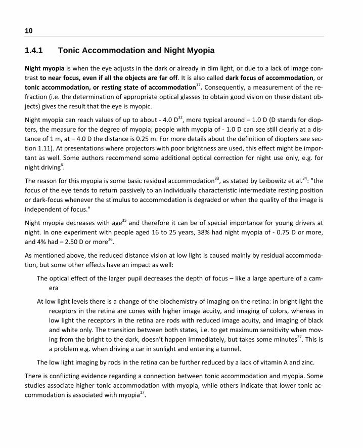
1.4.1 Tonic Accommodation and Night Myopia
Night myopia is when the eye adjusts in the dark or already in dim light, or due to a lack of image con‐trast to near focus, even if all the objects are far off. It is also called dark focus of accommodation, or tonic accommodation, or resting state of accommodation17. Consequently, a measurement of the re‐fraction (i.e. the determination of appropriate optical glasses to obtain good vision on these distant ob‐jects) gives the result that the eye is myopic.
Night myopia can reach values of up to about ‐ 4.0 D32, more typical around – 1.0 D (D stands for diop‐ters, the measure for the degree of myopia; people with myopia of ‐ 1.0 D can see still clearly at a dis‐tance of 1 m, at – 4.0 D the distance is 0.25 m. For more details about the definition of diopters see sec‐tion 1.11). At presentations where projectors with poor brightness are used, this effect might be impor‐tant as well. Some authors recommend some additional optical correction for night use only, e.g. for night driving6.
The reason for this myopia is some basic residual accommodation33, as stated by Leibowitz et al.34: "the focus of the eye tends to return passively to an individually characteristic intermediate resting position or dark‐focus whenever the stimulus to accommodation is degraded or when the quality of the image is independent of focus."
Night myopia decreases with age35 and therefore it can be of special importance for young drivers at night. In one experiment with people aged 16 to 25 years, 38% had night myopia of ‐ 0.75 D or more, and 4% had – 2.50 D or more36.
As mentioned above, the reduced distance vision at low light is caused mainly by residual accommoda‐tion, but some other effects have an impact as well:
The optical effect of the larger pupil decreases the depth of focus – like a large aperture of a cam‐era
At low light levels there is a change of the biochemistry of imaging on the retina: in bright light the receptors in the retina are cones with higher image acuity, and imaging of colors, whereas in low light the receptors in the retina are rods with reduced image acuity, and imaging of black and white only. The transition between both states, i.e. to get maximum sensitivity when mov‐ing from the bright to the dark, doesn't happen immediately, but takes some minutes37. This is a problem e.g. when driving a car in sunlight and entering a tunnel.
The low light imaging by rods in the retina can be further reduced by a lack of vitamin A and zinc.
There is conflicting evidence regarding a connection between tonic accommodation and myopia. Some studies associate higher tonic accommodation with myopia, while others indicate that lower tonic ac‐commodation is associated with myopia17.

1 WHAT IS MYOPIA? 11
More recently, Tarutta et al. reported38: "The high values of accomodation tonus (both HAT [habitual accomodation tonus, difference between primary and cycloplegic refraction] and ART [accomodation rest tonus]) were associated with higher rates of myopia progressing."
In another paper Tarutta et al. reported39: "Vegetal Tonus corresponds to the accommodative rest and is determined by vegetal innervation balance of ciliary muscle. … In eyes with moderate and high hy‐peropia the portion of negative vegetal accommodative tonus rises."
After vacations (summer or winter), the dark focus was found to be lower and the progression of myo‐pia to be suspended40.
Note: This reduction of myopia progression when the dark focus is lower can be interpreted as a "proof" that myopia is at least substantially caused by accommodative stress.
1.4.2 Pseudomyopia
If there is a transient spasm or excessive stress in the ciliary muscle during excessive near work, the zonular fibers are relaxed (and the ciliary muscle is not relaxed) even at distant‐focus, giving the im‐pression of a myopic eye. Especially when there is a complaint of migraine pseudomyopia should be taken into consideration41.
It is said that this pseudomyopia frequently precedes axial myopia, and that it is most frequently found with young people6, or that this spasm of accommodation is often wrongly diagnosed as myo‐pia, and the prescribed minus glasses are afterwards the reason for the development of real axial myopia41.
Another mechanism for pseudomyopia has been postulated: It was stated7 that as an automatic reac‐tion to mental stress the axes of the two eyes are set parallel and the focus is set to "distant", and the pupils are opened wide. In a hunter‐gatherer lifestyle, this has a survival advantage in that it may lead to the early detection of danger. However, if a child working at near focus ‐ like during a test ‐ will sub‐ject the eyes to competing forces:
Between near focus and distant focus (ciliary muscle), and
Between inward adjustment of the axes of the eyes (convergence for near work) and parallel ad‐justment.
This can lead to a spasm of the involved muscles and further to pseudomyopia (and maybe to perma‐nent myopia).

12
Besides these effects of the ciliary muscle, transient myopia, which is caused by a hysteresis (i.e. a longer time necessary for readjustment) of accommodation, can be based on:
A transient ocular elongation caused by accommodation (see section 3.7.6).
A hysteresis of the shape of the lens as well (see sections 1.3.2.4 and 3.7.4).
1.4.3 Other Types of Myopia
There are some other types of myopia:
Keratoconus is when the shape of the cornea is not uniform but more pointed; this can add addi‐tional refractive power of up to –20 D.
In some unusual cases, an extraordinary refractive power of the cornea and / or the lens may re‐sult in myopia with a normal length of the eye.42
With increasing age the refractive index of the cornea can be changed, to result in very moderate myopia.
Beyond 40 to 50 years presbyopia appears, i.e. the lens loses some (and later all) of its flexibility due to structural changes. Fewer authors attribute presbyopia to a weakening of the ciliary muscle by age43. Generally, this results in problems with near work and accommodation. If, however, the lens shape is "frozen" in a slightly accommodated state, problems with distant focusing may appear, which corresponds to myopia.
Diabetes can change the refractive index of the lens, leading to myopia if the blood sugar is ele‐vated, and to hyperopia (farsightedness) if the blood sugar is low44 (see section 3.24.1).
Antibiotics like sulfonamides, tetracyclines and corticosteroids can induce myopia45.
Amblyopia is when the visual acuity is reduced without visible pathologic defects. In most cases one eye only is affected. The affected eye is often highly myopic. This is named anisometropic amblyopia (see section 3.30.8).

1 WHAT IS MYOPIA? 13
1.5 Axial Myopia
Axial myopia occurs if the length of the eyeball is more than the average length of about 24 mm46. In this case the ratio of the length of the eye (anteroposterior dimension) to the height/width of the eye (transverse dimension) is larger than 1.0. Roughly 1 mm in length corresponds to ‐ 3.0 D.
The increase in the length of the eye is said to happen only at daytime47.
There are several forms of axial myopia:
Simple myopia (sometimes called school myopia), which normally starts at age 10 – 12, stays nor‐mally under ‐ 6 D and remains quite stable after the age of 20 years. No structural defects of the eye can be diagnosed in this case.
Benign progressive myopia up to 12 D, which is often stabilized at an age of 30 years. Most likely structural / biochemical defects of the eye can be diagnosed.
Malign myopia, which does not stop progressing at all. Up to – 30 D can be reached, with serious consequences, which may lead to blindness. Structural / biochemical defects of the eye can be diagnosed.
Pathological myopia, if there are already pathological changes in the eye (see section 1.7), inde‐pendent from the refractive error.
1.6 "What Type of Myopia Do I Have?"
The main and most worrying question is, whether it is a simple (not dangerous) myopia, or a myopia that can lead to a permanent damage of the vision (see section 1.7). This question can be answered only be an optometrist or an ophthalmologist, who will check the background of the eye for some signs of already appearing damage.
It was stated that only 1 in 1000 people have true myopia48.
1.7 Consequences and Risks of Higher Myopia
Some numbers from the statistics about the consequences of higher myopia6:
England, 1966: Myopia was responsible for 8.8% of blind registrations.
England, 1972, age between 50 and 59: Myopia was responsible for 18.2% of blind registrations, only behind diabetic retinopathy.
Bavaria/Germany, 1992, up to age 18: Myopia was responsible for 11.5% of blind registrations49.

14
Myopic macular degeneration is the seventh greatest cause of registered blindness in adults in Europe and in the United States, but has become the leading cause of blindness in Taiwan50.
Myopia accounted for 5% of the causes for blindness of people aged 20 to 59 years in Denmark51.
2% of Americans have pathologic myopia52.
For refractions over – 9.00 D 52.4 % of the myopes in Australia show retinopathies, for over – 10.00 D 89.6 % of the myopes in China show retinopathies53.
As the rate of myopic people is still increasing significantly today, the problem of resulting blindness can also be expected to rise further.
By the way: My motivation in writing this book is to inform people of this risk, so they might be able to reduce it by preventative measures.
It is not the intention to frighten you with these data, but to trigger you to do your very best to
avoid these potential consequences of higher grades of myopia. Most of the readers of this book will never be affected by the potential risks of myopia, but I am very glad if some of the readers can avoid or reduce permanent eye damage by following
advice given in this book.
A basic effect of high myopia is that the posterior sclera shows substantial thinning by the elongation of the eye. The normal sclera has a thickness of about 1.35 mm on the back of the eye. A highly myopic eye has a typically reduced thickness of the sclera of about 0.2 to 0.5 mm 7 and a thinned choroid as well. It is, however, still an open question, whether the thinning of the sclera is due to:
An optically regulated mechanism,
An excessive mechanical stretching force,
A defective connective tissue of the sclera.
Section 3 contains more information about these issues.
Basic reasons of most of the serious consequences of myopia are vitreochorioretinal dystrophies, i.e. disturbed structures of the layers of vitreous, choroid, and retina. It was found that 52.6% of people with weak myopia and 86.4% of people with high myopia had this disorder54.

1 WHAT IS MYOPIA? 15
Some basic pathological consequences of high myopia can be6, 55:
Retinal detachment:
There is an elevated risk for retinal detachment, i.e. the retina is separated from the choroid and the sclera, often accompanied with tearing of the retina. Retinal lattice degeneration and retinal breaks are often early signs of later retinal detachment56. Some publications, however, are contradictory with respect to an increasing risk with the degree of myopia. Some people are saying that there is an increased risk for myopes, which is, however, not dependant on the degree of myopia9. Other sources state a risk for retinal detachment e.g. for 0 D to – 4.75 D a risk of 1/6662, for – 5.00 D to – 9.75 D a risk of 1/1335, and for more than – 10.00 D a risk of 1/1486. In other words, an additional risk factor of 3 for low myopia, and up to 300 for high myopia57 was reported.
Besides the stress on the retina by a permanent elongation of the myopic eye, excessive stress by accommodation can be responsible for retinal detachment, as Arruga stated58: "One physiological condition of importance in retinal detachment is the movement of the anterior part of the retina in accommodation."
See also section 3.7.6 about the elongation of the eye during accommodation.
Vitreous liquefaction and detachment:
The vitreous body between lens and retina consists of 98% water and 2% fibers of collagen. It gradually becomes liquefied with age, and especially in myopic eyes 56, 59. This is due to a loss in the regular arrangement of the fibers. In early stages, small objects can be seen when looking at bright and uniform backgrounds (called fleeting flies, or floaters): "Most of the time they are nothing to worry about, but sometimes they can be a symptom of a retinal tear60.". In later stages, the vitreous body can collapse and lose its connection to the retina. This separation is connected with the risk of retinal detachment and corresponding damage of the retina. Imme‐diate medical examination is necessary if symptoms like flashing lights or a rain of soot can be seen. No treatment is available for vitreous detachment by itself. About 6% of "normal" people between age 54 to 65 and 65% of the people between age 65 to 85 have a vitreous detach‐ment. The higher rate of vitreous detachment of myopic people is sometimes explained by the larger volume, which has to be filled by the vitreous body.
It was concluded that the liquefaction is caused by the functional disorder of the blood‐retinal barrier in myopia61.
Various Myopic maculopathies:
Myopic macular degeneration (MMD) / Choroidal neovascularization (CNV) / is also a conse‐quence of "normal" macular degeneration, and it is caused by abnormal blood vessels that grow under the center of the retina. It generally occurs among people over 30 and can result in

16
a progressive loss of vision. The worldwide incidence of CNV due to pathologic myopia is esti‐mated to be 50,000 new cases per year excluding Asia, where the rate may be even greater due to a higher prevalence of pathologic myopia62 (see section 3.25 about a treatment for CNV).
Myopic macular degeneration is said to be the seventh greatest cause of blindness of adults in Europe and the USA, and has become the leading cause of blindness in Taiwan50, and the most common pathological consequence of myopia. Prevalence of MMD for high myopia was re‐ported to be between about 10% and 60% in the various studies63.
There can be a thinning of the choroid and the retina and a loss of capillary vessels in eyes with high myopia2 and as a consequence an atrophic loss of retinal cells (i.e. cells are dying), result‐ing in a loss of vision in this area13 (visual field defect).
In pathological myopia the death of retina cells (apoptosis) can occur due to various biochemi‐cal processes, e.g. related to oxidative events (see section 3.18).
Furthermore, there can be bleedings in the retina and the choroid, leading to a partial loss of vision13.
Posterior Staphyloma:
In pathologic myopia there can be a herniation‐like deformation ("out‐pouching") of a thinned sclera, which can hardly be corrected with lenses. It also leads to other complications.
Glaucoma:
The increased pressure within the eye that often accompanies myopia (see section 3.7.2), can damage the optic nerve. The results of older techniques for measuring the intraocular pressure of myopes were wrong: even when the pressure was high, the softer myopic tissue was inter‐fering with the measuring process in a way that the result was a normal, i.e. lower pressure than in reality. Later a measurement called "applanation tonometry" was invented. This tech‐nique is claimed to be independent from the rigidity of the sclera. It is said that open‐angle glaucoma occurs twice as often with the myopic eye as with the normal eye6.
Cataract:
The lens is loses its transparency. It is reported that myopia induces an earlier onset of cata‐racts. Statistical data are lacking57.
Chui TY et al. reported64: "… data indicated that approximately 15 D of refractive error doubles the spacing between retinal neurons, thereby halving peripheral resolution acuity relative to the emmetropic eye."
Soft contact lenses, and the complications of refractive surgery, can lead to infections, which may cause blindness57.

1 WHAT IS MYOPIA? 17
Each myopic person is strongly advised to see an ophthalmologist at the slightest sign of visual ab‐normities, and people with high grade of myopia should have the background of their eyes checked regularly!
On the other hand, even if there is a high degree of myopia at a young age already, there is still hope, as Goldschmidt and Fledelius found65 after the observation of the development of the myopia of 14 year olds with at least – 6 D over 40 years "...the adult visual prognosis for working age appeared better than usually claimed. There seems to be a correlation between degree of myopia at age 14 and con‐secutive visual loss, but it was not possible to identify subjects at high risk at that early age."
Notes: In many cases, a general systemic problem may cause one of these diseases, and also myopia. This
then explains the noted association, without implying that myopia is the cause of the disease. In these cases the therapy should focus more on the systemic problem, and not primarily on the optical myopia problem only (which should be positively influenced by the therapy as well).
As progressive, pathological myopia is based on defects of the connective tissue, the connective tis‐sue related coronary problem mitral valve prolapse (MVP) might have an increased probability, which makes preemptive measures still more appropriate, as stated by Yeo et al.66: "Although most patients with MVP are asymptomatic or have minor symptoms, it is associated with sig‐nificant morbidity."
Statistics from France, reported by Leveziel et al.67:
"Myopia severity was defined as mild (‐0.5 to ‐3 D), moderate (‐3 to ‐6 D), high (‐6 to ‐10 D) and very high (more than ‐10 D).
The prevalence of mild, moderate, high and very high myopia was, respectively, 65.95%, 26.14%, 6.72% and 1.19%. The prevalence of macular complications in the high and very high myopia groups was 0.5% [0.39‐0.64] and 4.27% [3.49‐5.17]. The prevalence of blindness or vision impairment was observed in 10.10% [8.91‐11.39%] of the very high myopic group.
At 60 years old or over, the prevalences of blindness or vision impairment were, respectively, 9.75% [7.91‐11.85%] and 25.71% [21.00‐30.87%] in the high and very high myopia groups."
18
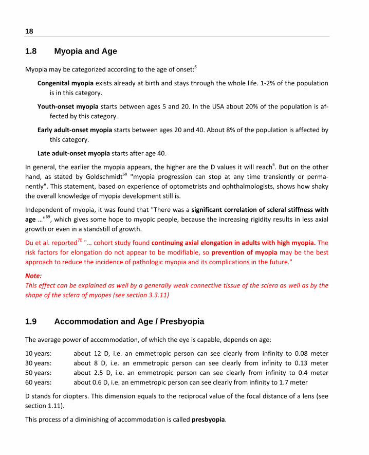
1.8 Myopia and Age
Myopia may be categorized according to the age of onset:6
Congenital myopia exists already at birth and stays through the whole life. 1‐2% of the population is in this category.
Youth‐onset myopia starts between ages 5 and 20. In the USA about 20% of the population is af‐fected by this category.
Early adult‐onset myopia starts between ages 20 and 40. About 8% of the population is affected by this category.
Late adult‐onset myopia starts after age 40.
In general, the earlier the myopia appears, the higher are the D values it will reach6. But on the other hand, as stated by Goldschmidt68 "myopia progression can stop at any time transiently or perma‐nently". This statement, based on experience of optometrists and ophthalmologists, shows how shaky the overall knowledge of myopia development still is.
Independent of myopia, it was found that "There was a significant correlation of scleral stiffness with age …"69, which gives some hope to myopic people, because the increasing rigidity results in less axial growth or even in a standstill of growth.
Du et al. reported70 "… cohort study found continuing axial elongation in adults with high myopia. The risk factors for elongation do not appear to be modifiable, so prevention of myopia may be the best approach to reduce the incidence of pathologic myopia and its complications in the future."
Note: This effect can be explained as well by a generally weak connective tissue of the sclera as well as by the shape of the sclera of myopes (see section 3.3.11)
1.9 Accommodation and Age / Presbyopia
The average power of accommodation, of which the eye is capable, depends on age:
10 years: about 12 D, i.e. an emmetropic person can see clearly from infinity to 0.08 meter 30 years: about 8 D, i.e. an emmetropic person can see clearly from infinity to 0.13 meter 50 years: about 2.5 D, i.e. an emmetropic person can see clearly from infinity to 0.4 meter 60 years: about 0.6 D, i.e. an emmetropic person can see clearly from infinity to 1.7 meter
D stands for diopters. This dimension equals to the reciprocal value of the focal distance of a lens (see section 1.11).
This process of a diminishing of accommodation is called presbyopia.

1 WHAT IS MYOPIA? 19
Shovlin summarized43: "Today, two schools of thought predominate about what causes presbyopia. One, that presbyopia results from a hardening of the lens and stiffening of the lens capsule with age so that the lens cannot change shape. And two, that the ciliary muscle, as it ages, loses its ability to con‐tract and change its configuration. However, both are happening simultaneously."
Additionally, there are numerous other changes of the aging eye.71
In general, people are becoming more hyperopic (i.e. farsighted) with old age, which appears to corre‐late more with the Schachar theory of accommodation than with the more widely accepted Helmholtz theory of accommodation24 (see section 1.3.2).
Consequently, almost everybody has to wear glasses or contact lenses during a more or less extended period in life. An older person with moderate myopia can often read by simply taking the glasses off. And for wearers of contact lenses there is the chance to wear bifocal contact lenses. This is a reason to avoid corrective surgery.
The graphical presentation of this mechanism is shown in Figure 4.
Ran
ge o
facc
omod
atio
n
Distant-focus
Very-nearaccom-
modation
Age Age
Reading
own accomodation
correction, e.g. by glasses
Emmetropia – normal vision Myopia
Ran
ge o
facc
omod
atio
n
in daily life required range of accommodation for distant-focus / reading
Very-nearaccom-
modation
Reading
Distant-focus
Figure 4 The change of the accommodation amplitude with age

20
Figure 5 is a somehow different representation of the relation be‐tween the accommodation effort and the distance for which proper focusing can be achieved with in‐creasing age. Even emmetropic persons need glasses to cover the range from reading distance to distant focus. Their glasses have to take care of the reading distance, the myopes's have to take care of
the long distance.
It has been claimed that only very few non‐myopic people over 60 are still able to accommodate, and it was concluded from research that a hotter climate accelerated the decrease in accommodative power16.
On the other hand, many people report that with older age their myopia decreases because of the mentioned aging process of the lens and the ciliary muscle.
Note: Quite frequently older people report that their myopia is decreasing, attributing this to presbyopia. The reason, however, might be as well a beginning of diabetes, which changes the refractive power of the lens (see section 3.24.1).
1.10 Age Related Geometrical Changes of the Eye
The refractive power of the cornea shrinks from 47 to 48 D in infants to 43 to 44 D by the age of 3 to 9 months and remains afterwards rather stable534.
The crystalline lens experiences by far more laterations. The lens power shrinks from 41 D in infancy to 22 to 23 D at the age of 14 years534.
Figure 5 The accommodation effort with respect to age and distance
Distance of proper focusing
30 cmreading distance
> 10 mdistant focus
Accommodationeffort
relaxed
maximumamplitude
typical young myopic persontypical older myopic persontypical older emmetropic person

1 WHAT IS MYOPIA? 21
It was concluded by Garner et al.72, "that the balance between the decrease in crystalline lens power and the increase in vitreous length is the major factor in maintaining the tendency to emmetropia in these children."
A disturbance in the described reduction of the lens power might already contribute to the appearing of myopia:
Mutti D et al. reported525: "Before myopia onset, the crystalline lens thinned, flattened, and lost power at similar rates for emmetropes and children who became myopic. The crystalline lens stopped thin‐ning, flattening, and losing power within ±1 year of onset in children who became myopic compared with emmetropes statistically adjusted to match the longer VCDs [vitreous chamber depth] of children who became myopic."
In general, it is said that about 80 percent of young children are hyperopic, and this hyperopia can in‐crease until to an age of 7 to 8 years and decrease thereafter until to an age of about 19 to 20 years217.
1.11 The Refraction
1.11.1 Basic Procedure
To determine if myopia exists, people have to see an optometrist or an ophthalmologist. It is generally recommended, especially for children,7, 12 that any remaining accommodation (i.e. stress of the ciliary muscle) is eliminated as much as possible before doing the test. Cycloplegic agents can achieve this, i.e. by the application of eye drops, which relax the ciliary muscle. The residual accommodation of the ciliary muscle, which exists if no cycloplegic agent is applied results in overcorrection of myopia73. If this relaxing of the ciliary muscle is not done, pseudomyopia may still exist. In reality, however, hardly any practitioner appears to use these cycloplegic agents – maybe because it takes too much time until the agents work (about 30 minutes).
A comparison between non‐cycloplegic and cycloplegic autorefraction showed that non‐cycloplegic measurements generally result in too high minus diopters74. Zhao et al. reported73: "Noncycloplegic autorefraction was found to be highly inaccurate in school‐age children and, thus, not suitable for studies of refractive error or for prescription of glasses in this population."
In detail, they found that the error is largest for hyperopes and smaller for myopes, but still ‐0.41(+/‐ 0.46D) for myopia of ‐2.00 D or more. An error, which was still larger – 0.84 D, was reported by Fotedar, who concluded75: "Precycloplegic autorefraction substantially overestimated the proportion of children with myopia".

22
Li T et al. reported76 "The proportion of myopia decreased from 78.1 per cent before cycloplegia to 71.4 per cent after cycloplegia, while the proportion of hyperopia increased from 12.1 per cent before cycloplegia to 21.4 per cent after cycloplegia." This means that an accommodation spasm resulted in an unjustified high minus evaluation. Additionally they found that this error was disappearing after an age of 12 year of the examined children.
Gu et al. reported based on experiments with children of mean age 9.7 years77: "Non‐cycloplegic refrac‐tive error overestimates myopia by approximately one diopter."
It was stated that more than 90%(!) of children under an age of ten years are hyperopic, i.e. their eye is (still) too short78.
If these children are not wearing plus glasses, they have to accommodate even when focusing at far. Consequences:
The inadequate high accommodation will result in an adjustment of the axes the eyes that can be mistakenly diagnosed as phoria (see section 3.5.1), and as the images of both eyes do not match properly, the image of one eye might be suppressed by the brain. This can result in a permanent damage of the eye, which "was switched off".
The permanent and excessive accommodation (especially at near work) can easily result in a spasm of the ciliary muscle and corresponding negative effects.
A flawed refraction can very likely be the entry to myopia.
Therefore, strong effort should be made to have the refraction of children tested by an ophthalmolo‐gist, who has a very good professional background and who is taking the problem of myopia of chil‐dren very seriously.
A comparison between autorefraction and "normal" prescription showed a tendency of overcorrec‐tion by autorefraction79.
Therefore, by not using these drops, often more glasses can be sold, and herewith a cycle of a perma‐nent increase in myopia can be initiated, if no cycloplegic agent is used.
Notes: - Open question: Which effect (autorefraction versa lack of cycloplegia) is contributing what
share in the overcorrection?
- According to other results and corresponding theories there is also a substantial accommoda‐tion hysteresis besides the one caused by the ciliary muscle and other ocular muscles, i.e. a hys‐

1 WHAT IS MYOPIA? 23
teresis (i.e. a time lag for getting back to the original shape) of the ocular shape (see section 3.7.6) and even of the lens (see section 3.7.4). Cycloplegic agents cannot eliminate all these hysteresis (pseudomyopia) effects.
- The omission of cycloplegic agents can contribute to the progression of myopia because the prescribed glasses can be too strong, which causes a further progression of myopia (see section 3.2 and 3.3).
- According to personal experience cycloplegia is rarely done (this attitude might be very different in different countries).
- Another potential source of errors is a chart for reading, which is badly illuminated: In this case, hidden night myopia may lead to an over‐correction (see section 1.4.1).
- Psychological stress can have an immediate impact on refraction (section 3.19). Stress could oc‐cur in school, or during an eye examination. If glasses are prescribed as a result, they in turn can initiate myopia that didn’t previously exist. The Bates method (section 3.2.2.1) focuses on this issue.
- As it is shown in the following sections, this over‐correction can induce a further increase of "real" axial myopia.
- The fitting of glasses should not be based on autorefraction only; it should be double‐checked by a conventional refraction.
In 95% of the cases, the results of the refraction can be reproduced within 0.5 D ‐ assuming there is no effect of pseudomyopia or hysteresis of accommodation at all. These hysteresis effects can make re‐fraction difficult, and can finally contribute to a permanent "lens‐induced" progression of myopia (see section 3.3).
In different countries and by different authors different units are used to describe the amount of myo‐pia and loss of visual acuity:
Diopter, abbreviated D: The refractive power of a lens (to correct the myopia) can be expressed by the reciprocal value of the focal distance: ‐1 D corresponds with a focal distance of 1 m. People with myopia of ‐1.0 D can see still clearly at a distance of 1 m (at ‐2.0 D the distance is 0.5 m). The refractive power of the prescribed glasses is given in D with a minus in front.
Snellen chart: This standard is more than 100 years old. 20/20 means that you can see at a distance of 20 feet what a "normal" person can see from a distance of 20 feet. 20/40 means that you can see at a distance of 20 feet what a "normal" person can see from a distance of 40 feet, and so on. Standard distance for myopia tests is 5 to 6 m. 20/40 is required in most States of the US to pass the Driver's License Test.

24
Decimal quotients: In some countries the basic principle of the Snellen chart is used ‐ the quotient of the distance where the patient can read to the distance where "normal" people can read. The result is given, however, in decimal units. A vision of 1.0 corresponds therefore to the vis‐ual acuity of a "normal" person. Some people, especially kids can have better resolutions up to a vision of about 2.0. Sometimes this decimal number is given in %, i.e. a visus of 1.0 corre‐sponds to 100%.
Visual acuity or visual efficiency: Sometimes another conversion from the Snellen chart numbers to percent is used. The table below contains the corresponding information.
Angle‐resolution: The visual acuity of the "normal" person (i.e. vision 20/20 or 1.0) corresponds with an angle‐resolution of rays, which are incident on the eye, of 1 minute.
Besides plain spherical myopia, often astigmatism is diagnosed: This is the contribution of non‐spherical irregularities of the eye, which can be compensated by added cylindrical lens‐components. The power of these cylindrical lenses is given in D, too (same definition as above). The prescription will give additionally the circular direction of the astigmatism in degrees. Combined spherical and cylindrical glasses are called toric.
The "best spherical fit" for toric glasses (i.e. for eyes with astigmatism) can be roughly estimated as: Spherical D + ½ ∙ astigmatism D.
Visual acuity can be described by several parameters, of which the D figure or its equivalent is one. Other factors include astigmatism. A very rough cross‐reference is shown in Table 180, 81:
Diopter D Snellen chart, 20/ ... Decimal Visual acuity / efficiency82
‐ 0.50 20/25 to 20/30 0.8 to 0.7 95% to 90%
‐ 1.00 20/30 to 20/50 0.7 to 0.4 90% to 75%
‐ 3.00 20/300 0.07 15%
‐ 4.00 20/400 0.05 10%
Table 1 Some different methods to measure visual acuity
Sometimes the numbers for the diopters D are given in a different representation without the decimal point, e.g. ‐ 1.00 as 100, ‐ 6.50 as 650 etc.

1 WHAT IS MYOPIA? 25
If you want to keep track on your myopia by yourself you can use Snellen Charts, which can be downloaded from the Internet and printed82, or which can be displayed and used directly on your com‐puter monitor83.
Very frequent visits to an optometrist for measurement of refraction bears the risk of a subsequent overcorrection, if the optometrist is not very well informed about myopia prevention and all the recent research results.
The result of a study in the USA was that, as Donahue stated84 "... a significant percentage of [pre‐school] children are probably prescribed glasses unnecessarily." In this study, specialized pediatric oph‐thalmologists were most careful in prescribing glasses.
A simple method to check any over‐ or undercorrection85, 86:
The refractive index of red and green light is different, and green light is focused about 0.5 diopters closer to the retina than red light.
Therefore, if you can see numbers or letters on a distant chart clearer and with more contrast when looking through a red glass or foil than through a green one, there is undercorrection.
If the contrast is better for the green picture, there is overcorrection.
As a result, a cold‐color light results in less accommodative stress for the eye than a warm‐color light.
Note: The first unnecessary glasses might be the first step towards myopia (see section 3.2)
The regulations about who is authorized to examine the eye and to prescribe glasses or contact lenses are different for the various countries (e.g. optometrists, ophthalmologists87 ...). Definitely, the selec‐tion of the right professional is extremely important, but it is hard to give any selection criteria. A dis‐cussion with the respective professional about some of the issues described in this book may be helpful to assess whether the professional is familiar with the current research results.
1.11.2 A Method for Refraction: Fogging
To release any spasm of accommodation it was stated that long time experience showed that the appli‐cation of strong plus glasses (or plus additions) prior to the refraction was more efficient than cycloplegic agents were, as Viikari expressed41: "... when the eye is atropinized [it] hold out for many years in the spasm position".
It was claimed that a faulty refraction where a spasm of the ciliary muscle was not considered results very often in a diagnosis of myopia (with the corresponding prescription of minus glasses) where in fact there is hypermetropia (farsightedness, which requires the prescription of plus glasses. An existence of
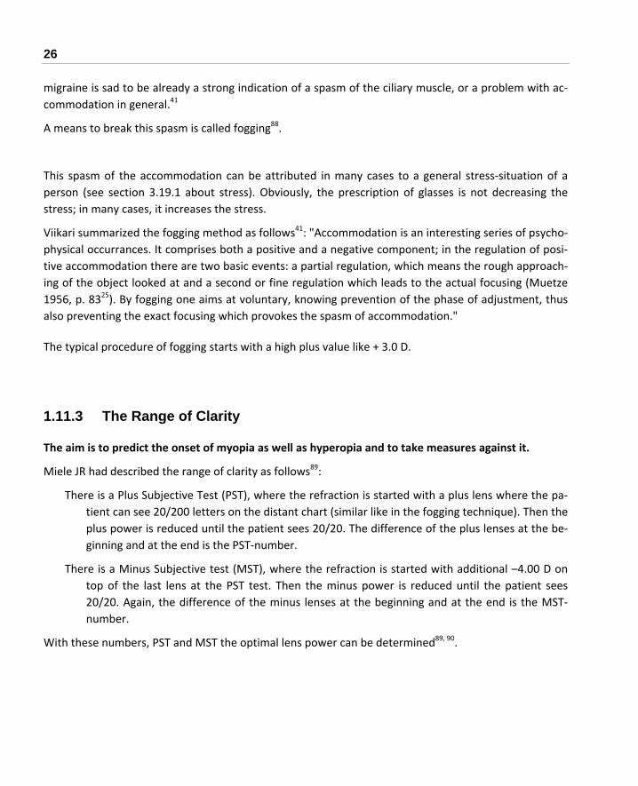
26
migraine is sad to be already a strong indication of a spasm of the ciliary muscle, or a problem with ac‐commodation in general.41
A means to break this spasm is called fogging88.
This spasm of the accommodation can be attributed in many cases to a general stress‐situation of a person (see section 3.19.1 about stress). Obviously, the prescription of glasses is not decreasing the stress; in many cases, it increases the stress.
Viikari summarized the fogging method as follows41: "Accommodation is an interesting series of psycho‐physical occurrances. It comprises both a positive and a negative component; in the regulation of posi‐tive accommodation there are two basic events: a partial regulation, which means the rough approach‐ing of the object looked at and a second or fine regulation which leads to the actual focusing (Muetze 1956, p. 8325). By fogging one aims at voluntary, knowing prevention of the phase of adjustment, thus also preventing the exact focusing which provokes the spasm of accommodation."
The typical procedure of fogging starts with a high plus value like + 3.0 D.
1.11.3 The Range of Clarity
The aim is to predict the onset of myopia as well as hyperopia and to take measures against it.
Miele JR had described the range of clarity as follows89:
There is a Plus Subjective Test (PST), where the refraction is started with a plus lens where the pa‐tient can see 20/200 letters on the distant chart (similar like in the fogging technique). Then the plus power is reduced until the patient sees 20/20. The difference of the plus lenses at the be‐ginning and at the end is the PST‐number.
There is a Minus Subjective test (MST), where the refraction is started with additional –4.00 D on top of the last lens at the PST test. Then the minus power is reduced until the patient sees 20/20. Again, the difference of the minus lenses at the beginning and at the end is the MST‐number.
With these numbers, PST and MST the optimal lens power can be determined89, 90.

2 WHAT CAUSES MYOPIA IN GENERAL? 27
2 What Causes Myopia in General?
Chapter 3 will summarize the research. This chapter gives an overview of some of the main hypotheses concerning the development of myopia.
2.1 Is Myopia Inherited?
In the past, the official medical view considered myopia to be simply inherited, and in PubMed a search for myopia plus genetic results in numerous results.
No question, heredity plays a key role91, 92 – everybody is determined by heredity and environment. In the same way, however, as there are talents of various levels, which can be compensated for to a great extent by personal energy, hard work, and methodology, all the possibilities should be tried to influ‐ence the "chemical factory" of the body by optimizing external parameters, e.g. by appropriate nutri‐tion and adequate behavior.
Fact is,
Myopia is dramatically increasing compared to the past1, 7, 93, 94, and many children are becoming progressively myopic with none of the parents or grandparents being myopic, and even popu‐lations like Eskimos, where myopia was extremely rare before, are getting highly myopic.
Myopia can be easily created artificially in experiments with animals95.
Myopia occurs in different frequencies in different regions of a country96.
Myopic parents have more often myopic children97: if both parents are myopic, the risk for the chil‐dren to become myopic is sevenfold.
Two arguments about this controversy:
Goldschmidt states98, " Twin studies indicate a strong genetic influence and a weak environmental impact, while extreme myopia prevalences among selected population groups (university stu‐dents) point to the opposite."
Note: In case myopia is caused primarily by personal habits (which includes e.g. reading), a reported genetic influence might be more related to identical habits of twins than to a biochemical cause.
Morgan states99, "... evidence for low prevalences of myopia in Indians growing up in India, while Indians in Singapore have much higher prevalence rates."

28
The conclusion is that myopia itself is not inherited. Rather, biochemistry is, and it reacts differently to various environments.
Jones‐Jordan et al, however, insisted genetic facors are dominating100.
As Lyhne et al.101, who did research with twins expressed very well:
"... results indicate a high heritability for ocular refraction and its determiners and thus suggest that en‐vironmental impact on refraction is not significant. However, the epidemiological association between educational length (near work) and myopia, the evidence on increasing myopia prevalence within a few generations, and the theory of gene‐environment interaction imply that some individuals might be ge‐netically liable to develop myopia if exposed to certain environmental factors".
With respect to a different medical problem the relation between genes and lifestyle was put in very clear words:
"Genes are always a factor of our health, but that is the only factor we can't do anything about. Genes only represent our predisposition & potential to encounter certain problem or disease. It is our life and our lifestyle as whole that makes the final decision on whether we are going to experience certain problem or not.102"
In other words, it is often not the myopia, which is inherited, but the susceptibility to specific envi‐ronmental conditions like excessive near work.
Moreover, specific genetic dispositions can make vulnerable to specific environmental conditions.
Note:
The fact that some people do not develop myopia in spite of very adverse conditions is a clear indica‐tion that heredity is involved in any case, the question is just to what extent.
Morgan has a different view103:
"Overall, while there may be a small genetic contribution to school myopia, detectable under condi‐tions of low environmental variation, environmental change appears to be the major factor increasing the prevalence of myopia around the world. There is, moreover, little evidence to support the idea that individuals or populations differ in their susceptibility to environmental risk factors."

2 WHAT CAUSES MYOPIA IN GENERAL? 29
Note: Morgan refers explicitly to school myopia and not to the higher grades of myopia and progressive myo‐pia. The fact, however, that many individuals do not become myopic in spite of very adverse environmental conditions speaks against the strong thesis of Morgan ("...little evidence to support the idea that indi‐viduals or populations differ in their susceptibility to environmental risk factors.")
A reader of this Myopia Manual expressed another likely possibility with respect to the inheriting of myopia: "The parents probably become myopic because they live in similar conditions as their off‐spring."104
Another point of view stated by Morgan99: "Thus, parents with longer than average eyes would tend to have children with longer than average eyes ... however, if average eye size is increasing due to envi‐ronmental effects, a high proportion of children would become myopic."
In principle almost all the facts about myopia that will be presented in the next sections are either con‐nected with the way of living of people, or with biochemical processes. Largely, however not com‐pletely, these biochemical processes are determined by heredity.
A few authors have reported a connection of few specific chromosomes with myopia105, but contradic‐tion with other results followed immediately106, 107. One expert states: Future genetic therapies of myo‐pia are highly unlikely, because it appears that numerous genes are having an impact on myopia97.
On the other hand, many papers show the fact that myopia often runs "in the family"108, 109, and that myopia is very clearly more common in some populations than in others (see section 3.1).
By the way: Influences like nutrition during pregnancy have a high congenital impact on the child's health. In addition, the "individual biochemical factory" is built very early in life and cannot be changed later. An example is the copper metabolism ‐ which might have an impact on myopia110, 111.
Basically any biochemical heritage can become effective via one of these two alternatives:
Via a biochemical process which cannot be modified, and whose results cannot be modified either.
Via a biochemical process which can be modified, or whose results can be modified. An example: People suffering from favism, an enzyme defect (a variant of G6PD deficiency) which leads to acute anemia after eating e.g. fava beans. Simple advice after somebody is diagnosed with this deficiency: Do not eat these beans. The literature gives further examples of "Nutritional Regu‐lation of Gene Expression112".
It is my aim to offer assistance in the second case and to motivate people not to become fatalistic. Fur‐ther, biochemistry is not in itself good or bad: An effect that has a disadvantage in one respect, can have an advantage in another respect (e.g. the G6PD deficiency mentioned above is said to offer pro‐tection against Malaria).

30
A simple analogy: If the engine of a car is prone to overheating through a design fault, the damage can be avoided in most cases by carefully watching the cooling water level, selecting top grade motor oil, and avoiding mechanical overload. Why not apply the same strategy to the human body, instead of hid‐ing behind the word heredity?
Some facts, which can contribute to the answering of the question whether myopia is inherited can be found in section 3.1 ("Distribution of Myopia by Region, Age, Gender and Ethnicity").
As some people do not become myopic even under very adverse conditions, it could always be claimed that myopia is caused by genetic heritage, even if it was demonstrated that without these adverse condition myopia did not occur.
The reason for this conflict is that the three potential causes for myopia,
connective tissue disorders (see section 2.2)
active growth by imaging effects (see section 2.3)
mechanical effects (see section 2.4)
are in one way or another influenced by the genetic heritage of the individual person.
Maybe there is a rather simple distinction whether the myopia of an individual person is caused mainly by genetics only or by interworking of environment and genetics: Low W et al. reported113 "Neither near work nor outdoor activity was associated with preschool myopia."
Finally, Verhoeven VJM et al. found114: "Individuals at high genetic risk in combination with university‐level education had a remarkably high risk of myopia (OR 51.3; 95 % CI 18.5‐142.6), while those at high genetic risk with only primary schooling were at a much lower increased risk of myopia (OR 7.2, 95 % CI 3.1‐17.0)." [OR = Overall Risk, CI = Confidence Interval]
See also section 3.30.7.
2.2 Connective Tissue Disorders
In the case of connective tissue disorders, the structure of components of the myopic eye shows signifi‐cant deficits. These structural deficits can have various origins:
Clearly inherited (like in case of the Marfan syndrome).
Appearing as a form of genetic variation, which happened after birth.
An acquired disorder, e.g. caused by interactions of the immune system, or by inappropriate nutri‐tion, or by other environmental or behavioral circumstances.
2 WHAT CAUSES MYOPIA IN GENERAL? 31
An example for the impact of the connective tissue on the highly myopic eye by Chang et al.115: "... that the fiber bundles of highly myopic eye were of thinner lamellar arrangement, with looser colla‐gen matrix distribution and decreased collagen fiber diameter."
Computer tomography measurements showed that the more severe the myopia is the more reduced is the density of the sclera116.
Section 3.3.2 describes some results with respect to the connective tissue.
2.3 Active Growth by Imaging Effects
After birth, the eye is too short for an optically correct focus. During growth, the eye expands by some feedback mechanisms to the proper length. This process is called emmetropization117. Animal experi‐ments have investigated this process in detail, but there are still a lot of open questions about what goes wrong when some people become myopic, or even progressive myopic, when others in the same environment don't. This active growth mechanism will be described in more detail in section 3.3.
Note: Obviously, the growth of the eye in young age is characterized at least partly by an increase in the num‐ber of cells. The extension of the eye in myopia, sometimes described as growth as well, is characterized mainly by a change of biomechanical properties (see section 3.3).
2.4 Mechanical Effects
There is quite general agreement that myopia has some connection with accommodation. One explana‐tions is the influence of the strain of the various muscles which are involved when doing near work – not just only the ciliary muscle for accommodation, but also various extraocular muscles which are controlling the position of the axis of the eye (see sections 1.3.2.4 and 3.7).
In this context, there is the interesting result that the thickness of the ciliary body increases with in‐creasing myopia and axial length118.
The influence of accommodation on myopia is discussed in more detail in section 1.11.2 and section 3.2.

32
2.5 General Overview of the Causes of Myopia
A very schematic overview of possible paths to myopia is shown in Figure 6. Each of these possibilities could be greatly ex‐panded.
From the fact that under the same external conditions (i.e. near work, illumination etc.) not everybody gets myopic it can be concluded that the influence of the personal biochemistry / personal anatomy plays a deciding role.
In general, the importance of a proper refraction – especially for children – cannot be overestimated (see section 1.11).
Figure 6 The potential paths to myopia
inherited biochemistry and anatomy
e.g. basic connective tissue
acquired biochemistrye.g. by nutrition, stress, environment
impact by mechanical straine.g. by accommodation, convergence
impact by image qualitye.g. blur image, illumination
myopia

3 MYOPIA – OBSERVATIONS AND EXPERIMENTAL RESULTS 33
3 Myopia – Observations and Experimental Results
This section contains:
Environmental, behavioral or biochemical parameters found to be connected with myopia
Therapies that have been proposed to prevent myopia or progression of myopia.
As there are sometimes interactions between these individual issues, a certain amount of repetition cannot be avoided.
Some observations and conclusions of the individual authors appear to be rather controversial. If we are to accept their observations as accurate, we must conclude that either
There is a connecting logic in the background which has yet to be revealed, or
There are various independent mechanisms, which have myopia as the common result.
3.1 Distribution of Myopia by Region, Age, Gender and Ethnicity
We can draw conclusions about the impact of different lifestyles, different environment, different nutri‐tion, and different genetic heredity by comparing the variation in rates of myopia according to age in different regions. Some results are:
An evaluation of statistical data for reported blindness due to malignant myopia in different states of the USA was done by compiling a chart with the rate of myopia per state, and distance to seacoast, annual hours of sunshine and the nutritional concentration of calcium, fluoride and selenium in each state. The results were:96 ‐ less sunshine, ‐ less calcium, ‐ less fluoride, ‐ less selenium and ‐ closer to a seacoast resulted in a higher probability of malignant myopia.
Myopia rates are higher in urban than in rural areas.119, 120, 121.
Note: Of course, farmers don't have much time to read.

34
The frequency of myopia at children in various countries is122: ‐ Germany ‐ 10%, ‐ Taiwan, South Korea ‐ 70%, ‐ Japan ‐ 95%, ‐ Singapore ‐ 74%123, ‐ Mexico ‐ 44%124
Myopia recently worsened as a problem, as stated by Lin et al.125: "In Taiwan, myopia was not a problem some 50 years ago"; today about 15% of the population have over ‐ 7.0 D.
50 to 60 % of the Japanese are said to be myopic, but only 2 % of the people in South America are myopic126. The author of this publication hints at a substantial difference with respect to spon‐taneity between these two populations.
1971 to 1972 only 25% of the individuals in the USA were myopic, 1994 to 2004 this number in‐creased to 41.6%127.
The incidence of myopia in Japan was increasing from 15% in 1920 to 36% in 1940 to between 50 and 60% in 1985126.
Sherpa and Tibetan children in Nepal have the same ancestry and genetic history, but the preva‐lence of myopia is 2.7% for Sherpa children and 21.7% for Tibetan children128. This difference was attributed to more rigorous schooling and higher advanced technology in Tibet.
Note: Generally more rigorous schooling and higher advanced technology are going hand in hand with a change in nutrition and increased mental stress. For the impact of nutrition on myopia see section 3.24, for the impact of stress see section 3.19.1.
Results of a study on Eskimos129 are shown in Table 2.
Age % Myopic
Over 50 0%
41 to 50 Less than 5%
31 to 40 23%
26 to 30 43%
21 to 25 88%
Table 2 Percentage of myopes among Eskimos

3 MYOPIA – OBSERVATIONS AND EXPERIMENTAL RESULTS 35
Data about the degree of myopia in various populations130 are shown in Table 3.
Population Low myopia ‐1.00 to ‐5.00 D
High myopia ‐5.10 to ‐10.00 D
Extreme myopia more than ‐10.00 D
Chinese 41% 14.7% 0.8%
Malays 37.8% 8.5% 3.0%
Asia, age 5 to 65131
Indians 34.3% 7.7% 0.9%
USA, age 4 to 74130 43% 3.2% 0.2%
North America, Sioux Indians, age 3 to adult 132
32.4% 4.1%
Table 3 Percentage of myopes among various populations
A study from 2001133, giving the percentage of myopic males (at least ‐0.5 D) between age 16 and 25 in Singapore:
82.2 % Chinese
68.7% Indians
65.0% Malays
Another statistic from USA (children, age 5‐17 years)134 is shown in Table 4.
Ethnicity Myopes (shortsighted)
Hyperopes (farsighted)
Asians 18.5% 6.3%
Hispanics 13.2% 12.7%
Whites 4.4% 19.3%
African Americans 6.6% 6.4%
Table 4 Percentage of myopes among various populations in the USA

36
Typical childhood progression rates were found to be between – 0.2 D and – 0.6 D in Europe and USA, and between – 0.5 D and – 0.8 D in Japan135.
In spite of similar myopia rates of Malays and Chinese, the age dependent progression profile ap‐pears to be rather different136.
Results from Singapore and Hong Kong show that myopia is 1.5 to 2.5 times more prevalent among adult Chinese than in corresponding European‐derived populations, and that women have sig‐nificantly higher myopia rates than men137, 138.
Moreover, as stated by Choo139, "...severity of myopia rises by about 1.5 D per year in Singa‐pore children, compared with 0.5 D per year in US children."
The higher rate of female myopes is confirmed by results about Greek students: 46% female stu‐dents, and 29.7% male students are myopic140.
Dayan found141: "During the 13 years from 1990 to 2002, the prevalence of myopia significantly in‐creased among the Israeli population. Although there was an association with the level of edu‐cation, gender, ethnicity, and origin, the prevalence of myopia increased on an annual basis, independent of these factors."
Malays, Chinese and Indians in Singapore have a higher prevalence of myopia than the same popu‐lations of Malays, Chinese and Indians in Malaysia142, which can be explained by the different environment, like requirements for extensive studying.
The urban generational axial length shift was estimated to be approximately 1 mm longer than that of the rural area. These results suggest different environmental effects on the ocular develop‐ment in these two populations of Chinese children143.
"The global burden of myopia is growing. Myopia affected nearly 30% of the world population in 2020 and this number is expected to rise to 50% by 2050."144
Note: Generally, reports about an enhanced prevalence of myopia among certain ethnic populations like Chi‐nese, Japanese, Korean and Jewish populations can hardly be interpreted as a potential genetic disposi‐tion: Among these populations with an enhanced prevalence of myopia frequently exists a traditionally very positive attitude for education, learning and reading with corresponding increased demand for near work and excessive accommodation.
A very comprehensive report about the prevalence and progression of myopia was published by the National Research Council of the USA145.

3 MYOPIA – OBSERVATIONS AND EXPERIMENTAL RESULTS 37
The high importance of the level of light to which people are exposed was investigated by French AN et al., which reported146: "European Caucasian children in Northern Ireland have a greater prevalence of myopia, hyperopia, and astigmatism when compared to children living in Sydney. Risk factors for myopia such as parental myopia, parental education, and educational standards do not appear to ex‐plain the differences." See section 3.14.1 about the Impact of illumination on myopia.
A mathematical model for the prevalence and the incidence of myopia was established by PR Greene et al.147
3.2 Accommodation and Near Work
In principle, results and experiences about accommodation (that is, changes in lens shape for near work) are related to artificially negative‐lens‐induced myopia, and myopia which was induced by forcing the eye to permanent near focus (see section 3.3).
3.2.1 Experiences and Results
3.2.1.1 General Experiences and Results
Basically excessive near work has the identical effect and works the same way as experimental lens in‐duced myopa (LIM), which was described in numerous publications.
In general the influence of near work alone is not easy to isolate, as extensive near work means exten‐sive in‐door work (often at low levels of illumination, see section 3.14.1), which is mostly connected with a potential lack of vitamin D because of a lack of exposure to sunlight (see section 3.24.3). More‐over, children who are interested very much in reading often have a more introverted personality (see section 3.19.2) and are less interested in physical activities.
There are numerous indications that people, who are doing extensive near work, i.e., who accommo‐date extensively, are more often myopic. Some observations are:
The appearance of myopia at people in USA and Greenland coincides with the introduction of uni‐versal schooling,148 and the rate of myopia of school children in Berlin depended very strongly on the level of the school they visited: the more advanced the type of school, the higher the rate of myopia. Rates were between 55 and 35%149.
The progression of myopia of school kids is slower during summer holidays150, 151, 152.
Comparisons between myopia rates in 1882 and 1964 showed that the rate is very different for in‐dividual professions93 (students in the 30% range, unskilled workers in the 2 to 3% range): While the total rate of myopia was increasing, the myopia rate for all the individual professions

38
except unskilled work was decreasing. Explanation: The overall increase in myopia, according to these results, can be largely attributed to a changed ratio of job distributions from unskilled work towards professional and office work.
Highly elevated rates of myopia were found for craftspeople of various professions, who have to do extensive near work, e.g. for typesetters (in year 1930153), tailoring (in the years 1953154 and 1961155). Up to 77% of the persons in these professions were found to be myopic.
A test with monkeys (already in 1961)156, whose visual space was restricted to an average of fifteen inches distance, showed that all of them developed some myopia.
There are indications that extended accommodation can create a chronic spasm of the ciliary mus‐cle, and experiments on monkeys showed that this stretching will begin after spasms for 2 to 4 months so spastic myopia and axial length myopia exist at the same time157.
Note: A permanent load can damage any muscle, not only the ocular muscles158, 159.
A study of university students showed a strong relationship between reading and other near work with myopia was found, however no relationship between myopia and working on video dis‐play terminals (VDT) or watching TV160. Watching TV at close distance however was promoting myopia161, 162.
Note: These results appear to be contradictory. As will be shown in section 3.3 the effect of VDTs and TVs can be expected to depend largely on the quality of the displayed images. Due to the limited image resolution of TVs and VDTs (LCD screens are better) a watching of these screens from near distance will create a blur image on the retina, with the potential consequences as de‐scribed in section 3.3.
For Chinese schoolchildren a correlation of myopia with the reading of books was found163, in Finland a relation between myopia progression and time spent on reading/close work and on reading distance was found164.
Rosenfield et al. stated165 that "...results demonstrate that myopes are less sensitive to the pres‐ence of blur, and may at least partially explain why previous reports have demonstrated a lar‐ger lag of accommodation...". On the other hand, the compensatory eye growth in experimen‐tal myopia (see section 3.3) was always in the right direction, even in the presence of very poor images166.
Rosenfield et al. stated167 that "... findings do not support the proposal that the development of myopia in young adults is accompanied by a reduced accommodative response during near work."

3 MYOPIA – OBSERVATIONS AND EXPERIMENTAL RESULTS 39
Myopic children have less tonic accommodation (i.e. dark focus accommodation) than normal, i.e. emmetropic children168, 169, 170. After near work, however, the tonic accommodation of myopes increases more than for emmetropes. A large shift in tonic accommodation after near work was found to be typical for periods of acquisition and progression of myopia (some kind of ac‐commodative hysteresis)171.
Very contrary to these results is the statement by Kushner172: "Overcorrecting minus lens therapy for intermittent exotropia [see section 3.5] does not appear to cause myopia."
Note: A potential explanation is that the risk is not so much originating from the excess accommoda‐tion caused by the negative lens, but by the increased esophoria caused by the negative lenses (esophoria means that the axes of the two eyes are adjusted too much inwards when accom‐modating). Esophoria is frequently associated with myopia (see section 3.5.1 about phoria).
Near work, i.e. accommodation was shown to increase the temperature of the anterior segment of the eyeball, and to cause a hyperproduction of intraocular fluid173.
Note: This hyperproduction of intraocular fluid can increase the intraocular pressure; and an elevated ocular pressure was often found to correlate with myopia (see section 3.7.2). About the impact of the temperature on myopia see section 3.16).
Stress in connection with near work was made responsible for the development of myopia: It was stated that stress induces distant‐accommodation, which conflicts with the actual task near‐accommodation. This conflict was claimed to cause either a spasm of the ciliary muscle, or pho‐ria174 (phoria means the exes of the eye are not adjusted appropriately; see section 3.5 about phoria and section 3.19 about mental issues).
A helpful support of accommodation by illumination was found: see section 3.14.1.
Obviously near‐work induced transient myopia (NITM) is increased by mental stress: see section 3.19.1.
Ip et al. found when examining Australian school children175: "Although myopia was not signifi‐cantly associated with time spent in near work after adjustment for other factors, there were significant independent associations with close reading distance and continuous reading. These associations may indicate that the intensity rather than the total duration of near work is an important factor."
Langaas reported176: Children with myopia showed greater instability of accommodation (0.38 D) than children with emmetropia (0.26 D) at the 4.00 D target on Visit 1 and this instability of ac‐commodation weakly predicted myopic progression.

40
An argument against the importance of near work was given by Jones‐Jordan et al. who summa‐rized their results177: "Before myopia onset, near work activities of future myopic children did not differ from those of emmetropes. Those who became myopic had fewer outdoor/sports ac‐tivity hours than the emmetropes before, at, and after myopia onset. Myopia onset may influ‐ence children's near work behavior, but the lack of difference before onset argues against a major causative role for near work. Less outdoor/sports activity before myopia onset may ex‐ert a stronger influence on development than near work." See section 0 about the impact of outdoor activities on myopia.
When analyzing the reading habits of school children it was found that when starting reading the distance was about 28 cm, but after a while it decreased to just about 11 cm, which is too near for a stressless accommodation178.
Li et al.179 summarized the results of questionaires given to Chinese children. Significantly associ‐ated with myopia was:
‐ continuous reading (better not more than 30 minutes), ‐ TV viewing distance (better at least 3 meters), ‐ reading distance (better at least 30 cm), ‐ nib to finger distance (better at least 2 cm), ‐ head tilt when writing (better no tilt).
Li et al. reported180: Results: "WD [working distance] and LI [light intensity] were positively asso‐ciated with SER [spherical equivalent refraction]. … When WD and LI were split up, the detri‐mental threshold was approximately 40 cm for WD and 6300 lux for LI."
In section 3.3.1 some research results are mentioned, where short intervals of relaxing the accommo‐dation prohibited myopia or reduced the progression of myopia.
3.2.1.2 The Effect of Accommodation on Axial Length and Scleral Thinning
Accommodation causes a substantial hysteresis of the ocular shape: For normal people, after ac‐commodation, as Walker et al. stated190, "... ocular shape had become more prolate [i.e. stretched]. This shape remained unchanged after 1 hour of sustained accommodation and then returned to baseline dimensions after 2 h of accommodation ... Ocular shape returned to baseline dimensions after 45 min of accommodative relaxation."
It was measured (with partial coherence interferometry) that the eye generally elongates during accommodation. Drexler et al.28 explained: "… by the accommodation‐induced contraction of the ciliary muscle, which results in forward and inward pulling of the choroids, thus decreasing

3 MYOPIA – OBSERVATIONS AND EXPERIMENTAL RESULTS 41
the circumference of the sclera, and leads to an elongation of the axial eye length." and by Shum et al.29: "… the elongation was more pronounced in emmetropes than in myopes."
More results about increased axial length by accommodation are described in section 3.7.6
Mallen et al. reported181: "During relatively short periods of accommodative stimulation, axial length increases in both emmetropic and myopic young adults. At higher levels of accommo‐dative stimulation, a significantly greater transient increase in axial length is observed in my‐opic subjects than in their emmetropic counterparts... The mean axial elongation with a – 6 D stimulus to accommodation was 0.037 mm in emmetropes and 0.058 mm in myopes."
Vasudevan et al. found182: "EOMs [early onset myopes] and LOMs [late onset myopes] demon‐strated larger NITM [near work induced transient myopia] than the EMMs [emmetropes] and exhibited NITM additivity [increased NITM after a second hour of near work], but the EOMs [early onsets myopes] also exhibited prolonged decay of NITM compared with the EMMs and LOMs. PMs [progressive myopes], but not SMs [stable myopes], exhibited additivity of NITM."
Note: Isn't this a proof that myopes have a degraded connective tissue, which is lacking elasticity? No association between parental refractive error of the parents and NITM magnitude and its de‐
cay time for the children was found by Lin et al.183
Collins MJ et al. summarized their measurements530: "Small but significant increases in axial length were evident during and to a lesser extent, immediately after the accommodation task."
Near work causes not only accommodation via the ciliary muscle, but also a load on the oblique muscle, which handles the vergence. A load on this muscle can cause an elongation of the eye independent from accommodation as outlined in section 3.7.3
EC Woodman‐Pieterse et al. reported184: "… demonstrates the first evidence of a small but statisti‐cally significant thinning of the anterior sclera during accommodation. These changes were more prominent in myopes …"
Morgan I reported about experiments with chicken185: "Eyes wearing negative lenses were signifi‐cantly longer than fellow eyes by 25 h, while eyes wearing diffusers did not differ from fellows until 72 h. By 48 h, the inter‐ocular difference in lens‐wearing animals was 5 times greater than in those wearing diffusers."

42
Notes:
Maybe this stretching of the eye which is caused by accommodation and which can be expected to be more pronounced at myopes (weaker connective tissue) is responsible for the accommodation lag of myopes due to some feedback mechanism between stretching and accommodation.
Section 3.17 describes that stretched axial length means reduced choroidal blood flow, where it is still open whether this reduced blood flow is a reason for further progression of myopia or just a conse‐quence. The fact, that dopamine reduces the progression of myopia (see section 3.25.4) and increases retinal bloof flow (see section 3.17) is an indication that reduced blood fow just by temporal accom‐modation and stretching might be responsible for onset and progression of myopia.
Temporal elongation means stretching and thinning of scleral tissue, and stretching of scleral fibroblasts is changing significantly the gene expression of these fibroblasts186. This means, accommodation has a direct impact of the biochemistry of the eye; this builds a link between mechanical and biochemical models of myopia, and a link between functional and structural models of myopia as well (see section 3.7.1
It appears to be very plausible that a temporal stretching of an already weak scleral tissue will result in a permanent stretching, thinning and elongation.
People with a high degree of myopia have a structurally weakened sclera and therefore the stretch‐ing caused by accommodation is very likely resulting in an additional structural weakening, which can be expected to increase the degree of myopia still more.
The conclusion is that especially people which are already substantially myopic should wear plus lenses for extensive near work, not only for the prevention of further progression but especially for the prevention of dangerous pathological changes of the sclera of the eye (see section 1.7).
The elongation caused by accommodation might be the reason of the accommodation lag of myopes – there will be permanent interworking between accommodation strength and related elongation of the eye. The reason of this transient increased elongation can be seen, however in a degraded struc‐ture of the connective tissue of the sclera. (see section 3.3.2)

3 MYOPIA – OBSERVATIONS AND EXPERIMENTAL RESULTS 43
3.2.1.3 The Effect of Accommodation on Image Quality
Harb et al. stated187: "Within subjects, accommodative lags significantly increased with closer reading distances, however there was no significant relationship between lag and refractive state. The variabil‐ity in the accommodative response significantly increased with closer reading distances ... Increased lags and the variability in accommodation at higher accommodative demands suggest that an increase in overall blur at closer reading distances might be related to the development of refractive state."
In similar experiments, it was found that generally at a reading distance of 30 cm no perfectly focusing accommodation is performed188. The resulting degradation of the image quality can contribute to the development of myopia (see section 3.3 about the impact of a reduced image quality).
Schaeffel et al. summarized:91: "The current theory is that lag of accommodation during reading shifts the image plane behind the retina and thereby stimulates retinal neurons to release growth promoting factors that enhance scleral growth. This is thought to be mechanistically equivalent to the effects of a negative lens in animal experiments."
The accommodative lag is defined as the difference between accommodative demand and accommo‐dative response. Thus, lag is directly the residual refractive error189.
Note: The word "growth" appears to be somehow misleading, as – at least for higher grades of myopia ‐ there is no growth of structurally intact tissue (which is normally meant by the word growth), but a stretching of degraded tissue and a change of the biomechanical properties (see section 3.3.2).
3.2.1.4 Timing- and Hysteresis- Effects of Accommodation
Some results about the time it takes to accommodate and to recover are:
Accommodation causes a substantial hysteresis of the ocular shape: With normal people, after ac‐commodation, as Walker et al. stated190, "... ocular shape had become more prolate [i.e. stretched]. This shape remained unchanged after 1 hour of sustained accommodation and then returned to baseline dimensions after 2 h of accommodation ... Ocular shape returned to base‐line dimensions after 45 min of accommodative relaxation." (see section 3.7.4).
Myopes showed reasonable aftereffect of accommodation17, i.e. it took an average of 35 seconds for early‐onset myopes and 63 seconds for late‐onset myopes until the accommodation of the ciliary muscle was released. Non‐myopes, however, showed no myopic aftereffect. It was

44
speculated that this transient pseudomyopia is either the cause or a precursor of permanent myopia 191.
Additionally, it was shown that during the progression of myopia there is a significant near work after‐effect (sometimes called nearwork‐induced transient myopia ‐ NITM17), causing transient myopia that still existed 10 seconds and 30 seconds after the near task 192, 193.
Correspondingly, there is a reduced facility, i.e. a reduced maximum frequency of accommo‐dation: "Mean distance facility was significantly lower (9.7 cycles per minute (cpm)) in the my‐opic group compared with the mean distance facility in the emmetropic group (15.6 cycles per minute). There was no significant difference in the near facilities of the two groups (11.5 cpm in myopes versa 12.9 cpm in emmetropes)."194
More recent experiments confirmed the fact that myopic children are showing a significantly larger near work‐induced transient myopia, but demonstrated additionally that this near work‐induced transient myopia was still evident after 3 minutes, which is significantly longer than what was previously reported for adults195.
There is a time lag of accommodation for myopes 165, 196. However, Rosenfield et al. stated167 that "...stable myopes also exhibited the largest lag of accommodation." Mutti et al. found197: "Sub‐stantive and consistent elevations in accommodative lag ... did not occur in children who be‐came myopic before the onset of myopia or during the year of onset. Increased accommoda‐tive lag occurred in children after the onset of myopia. Elevated accommodative lag is unlikely to be a useful predictive factor for the onset of myopia."
Note: A simple explanation of this time lag can be seen in the fact that the eye is elongated by ac‐commodation (see section 3.2.1.2): The sclera of myopes has a somewhat degraded stability, and at the moment the accommodation starts it is already partly compensated by this imme‐diate elongation of the eye.
For children it is generally recommended (Note: but very often not done) to do the refraction after applying a cycloplegic agent. The residual accommodation, which exists if no cycloplegic agent is used results in overcorrection of myopia (see section 1.11).
Sivaraman reported198: "The magnitude of the NITM was higher in myopes compared to em‐metropes for both five minute and 60 minute viewing time."
Note: An explanation can be the degraded connective tissue of the sclera at myopes: In section 3.2.1.2 it was shown that accommodation causes the eye to strech temporarily, and accord‐ing to Sivaraman's results this stretching is more pronounced at myopes.

3 MYOPIA – OBSERVATIONS AND EXPERIMENTAL RESULTS 45
Notes: This aftereffect and hysteresis of accommodation can be responsible for a steady progression of
myopia in school kids: If the kid has to change in class permanently and fast between near work and reading from the distant blackboard, the mentioned delay can give the impression that the current glasses are too weak. New and stronger glasses, however, can easily induce additional myopia as described in section 3.3, and these cycles can happen repeatedly. The long duration of the aftereffect (which was still evident after 3 minutes) strongly supports this model. Bifocal glasses might help in these cases, but care has to be taken when fitting these bifocals (see section 3.2.2.3).
Maybe the anomalies of myopic accommodation can be explained by a lack of nitric oxide (NO) in the smooth ciliary muscle (see sections 3.18.5).
Open question: What is responsible for the time delay and the aftereffect of the accommodation, a muscular problem, or a hysteresis problem of the flexibility of the lens? It appears to be rather likely that a muscular problem is a key issue for myopia, i.e. not a consequence, but a reason.
Another explanation would be a spasm of the ciliary muscle of myopes during extended ac‐commodation, which needs to take more time to disappear after the near work is finished.
Maybe the hysteresis of the reshaping of the lens before accommodation and after accommo‐dation contributes to this effect as well (see section 1.3.2.4).
3.2.1.5 Dark Focus and Accommodative Hysteresis
As mentioned in section 1.4.1 it is called dark focus when due to darkness there is no more accommo‐dative stimulus.
Ebenholtz reported that when there was longer time of near accommodation before switsching off the light it can take many hours to release the existing near accommodation. He stated199: "Since accom‐modative hysteresis implies a long lasting increase in ciliary muscle tonus, it is conceivable that the propensity toward hysteresis effects is related to the incidence of near‐work induced myopia."
Note: It has to be concluded that reading or doing near work before going to sleep is extremely harmful.

46
3.2.1.6 The Strength of Accommodation
Some results about the strength and amplitude of accommodation of myopes are:
Abbot et al. stated200: "A reduced accommodation response to negative lens‐induced accommoda‐tive demand was found in progressing myopes but not in stable myopes." In addition, eyes with myopia were found to have lower amplitude of accommodation.
Fong stated201 "Eyes with lower amplitudes of accommodation must use more of their accom‐modative reserve for near work. Myopia may be an adaptation that develops in eyes with re‐duced accommodative amplitudes", and less accurate accommodation202. And a reduced ac‐commodation was speculated to create a blur signal, which might be responsible for develop‐ment of myopia107. Consequently, under‐accommodation may precede the development of myopia as clinical data suggest 203.
Siegwart and Norton summarized earlier reports204: "Although there is disagreement about when it begins, progressing myopic children tend to have a larger accommodative lag [i.e. underac‐commodation] than do emmetropic children."
Siegwart and Norton concluded204: "… if eyes that are unable to use myopia to slow elonga‐tion are [what is happening during normal emmetropization], in addition, exposed to hyper‐opia [i.e. a lack of sufficient accommodation] through nearwork and underaccommodation, this might lead to myopia development."
They add that additionally genetics plays a part, especially by the individual capability "to use myopia to slow elongation".
Note: In other words, an overloaded and insufficient accommodation accompanies myopia, and has a high likelihood to be a main cause for myopia. About "… to use myopia to slow elongation …": this should take place only if there is spent suffi‐cient time without near accommodation.
It was found by McBrien et al.205 that "...late onset myopes having the largest amplitude of ac‐commodation, followed by early onset myopes, emmetropes, and hyperopes."
Note: This might appear to be in contrast to the statements above; however, if one considers exces‐sive accommodation as some kind of a cramp there is no more conflict.
Overall, these findings can be interpreted as a strong argument for plus lenses to avoid the onset of myopia.

3 MYOPIA – OBSERVATIONS AND EXPERIMENTAL RESULTS 47
In this respect, it is interesting that wearers of glasses accommodate less and have lower conver‐gence demands than emmetropes or wearers of contact lenses due to optical reasons206, 207, 208 (for – 5.00 diopters the difference is described as very roughly 0.50 diopters at reading dis‐tance). Moreover, hard contact lenses were often found to stop progression of myopia (not soft contact lenses, see section 3.26.1).
Some positive effects of pharmaceuticals like atropine can be at least partly attributed to their attenuating effect on the accommodation (see section 3.25).
Chinese people have lower amplitudes of accommodation than Caucasians209, and Chinese have a higher probability of becoming myopic.
Notes: From an evolutionary point of view there might be a linkage between both facts: ‐ Without any accommodation the only one way to focus is to adjust the length of the eye as happens in emmetropization (see section 3.3.5), and for dominating near work this means to become myopic.
‐ Question: is there a rule like "the lower the accommodation before the onset of myopia, the higher the probability and the degree of the myopia that will develop later"?
The paper "The Secret of Myopia (Near‐Sightedness)" gives an easy‐to‐read and convincing descrip‐tion of the result of accommodation overload.210
For information about the related issue tonic accommodation (resting state accommodation) see sec‐tion 1.4.1, the interaction between accommodation and convergence is discussed in sections 3.5.2 and 3.5.3.
3.2.1.7 Accommodation Spasm
Numerous authors claimes that an accommodation spasm is a problem when determining the refrac‐tion, and to avoid this spasm, e.g. cycloplegica like atropine are applied to relax the ciliary muscle (see sections 1.11 and 1.11.2).
As accommodation was found to cause an elongation of the eye (see section 3.2.1.2) is appears to be very plausible that an extended spasm of the accommodation will lead to an extended and rather likely permanent elongation.
This spasm, medical term asthenopia, is not disappearing easily, as Kajita et al. reported:211 "… results suggested that the ciliary muscle was also actively working in asthenopia caused by accom‐modative spasm even if the patient was looking at a distant target."

48
In more detail, Kanda et al. reported212 " The average ± standard deviation refractive power in the pa‐tients was significantly more negative than that in the healthy subject (‐3.12 ± 1.06 vs. ‐1.49 ± 0.17 D). … The spherical aberration in patients was more negative than that in the healthy subjects".
Ninomiya et al. came to similar results213: " Excessive accommodative tone is manifested objectively by negative spherical aberration in eyes with accommodative spasm."
On one hand, the spasm has obviously a negative mechanical impact on myopia, on the other hand the reported spherical aberration can or will contribute independently to myopia.
About the impact of spherical aberration and myopia, see section 3.3.9.
Another aspect of accommodation spasm was reported by Khatsukov214: " Blood and oxygen supply, respiration and cerebral cortex function especially those cortical parts in which visual analyzer is repre‐sented were studied in children and adolescents with myopia and accommodation spasm. It was found that respiration function and oxygen regime in myopia children and adolescents are 2‐3 years behind those in normal children and adolescents. … 20‐day interval hypoxic training in children and adoles‐cents raised efficacy of the external respiration and circulation, normalized bioelectric activity. The ma‐jority of the patients improved their visual acuity up to complete normalization."
Note: Obviously, any means, which will cause relaxation to body and mind, are helpful to avoid the onset or the progression of myopia, as an impaired respiration is frequently an expression of substantial tension of body and especially of mind as well.
3.2.1.8 Myopia caused by Hyperopia
Viikari summarized her experiences215: "Contrary to the textbooks, which devide refraction errors into tree groups the author believes that there is only one refractive error, hyperopia, together with its vari‐ous grades. The myopia and astigmatism are distortions, artefacts, due to the correction mechanisms of the eye, the accommodation performed by the ciliary muscle with its autonomous innervation. Under continous strain, and presumable over‐excibility the result may end not only to pseudo‐myopia and pseudo‐astigmatism but at the same time to over activation, a possibly sentization of the entire auto‐nomic nerveous system."
In other words, according to this model the excessive accommodative stress caused by the hyperopia leads to rather permanent cramps of the ciliary muscle and to an accommodation‐caused elongation of the eye (see section 3.2.1.2), which on the long run weakens the connective tissue of the sclera and re‐sults in myopia and sometimes‐progressive myopia.216

3 MYOPIA – OBSERVATIONS AND EXPERIMENTAL RESULTS 49
It has to be noted, that when being hyperopic the accommodation muscle is strained even when fo‐cusing at distant objects.
This has to be considered together with the fact that about 80 percent of young children are hyper‐opic, and this hyperopia can increase until to an age of 7 to 8 years and decrease thereafter until to an age of about 19 to 20 years. It is still said that up to +1.5 or +2.5 D there is no need for glasses as there are no problems with reading 217.
This traditional view, however, does not take into account the tremendous load on accommodation and its corresponding force to elongate the eye – see section 3.2.1.2. The large range of accommoda‐tion of children is only hiding the devastating long‐term results of this over‐accommodation.
The bad habit of children to work "with the nose on the paper", i.e. to work at too close distance be‐cause of their smaller body worsens the situation.
Notes: • Trivial analogy: a rubber band, which is stretched too much, will finally loose its elasticity and
become too long. Clearly, plus glasses for near work would eliminate the early onset of myopia in all these cases.
• ∙The process of creating myopia by accommodation can be simply interpreted as a control‐mechanism of emmetropisation (see section 3.2.1.14.
• ∙On the other hand, rather many people are not becoming myopic in spite of excessive near‐work, which indicates that biochemical effects like the dopamine metabolism (see section 3.3.3) are essential, too.
3.2.1.9 "Screen sightedness" by SmartPhones and Tablet-PCs
As there is a connection between accommodation effort and myopia as outlined above it is obvious that the recent and extensive use of smartphones and small tablet PCs with their small fonts and a re‐sulting close reading distance will have a strong negative impact on the onset and progression of myo‐pia.
Allamby218 reported some warning facts about the extensive use of smartphones and small tablet PCs:
"… there has been a 35 per cent increase in the number of people with advancing myopia (short sightedness) since the launch of smartphones in 1997. He has warned the problem could increase by 50 per cent in the next ten years." and
"… the average smartphone user holds the handset 30 cm from their face ‐ with some people holding it just 18cm away ‐ compared to newspapers and books, which are held 40cm away from the eyes."

50
Hansen MH et al. reported219 about myopia prevalence in a Danish cohort aged 16‐17 years and its relation to use of screen‐based electronic devices that the use of screen devices >6 hr/day was associated with an increased overall risk for myopia by a factor of 2 compared with screen de‐vice use <2 hr/day.
S McCann et al. reported220: "Multinomial logistic regression revealed that myopic refractive error was statistically significantly associated with increasing daily smartphone data usage (odds ratio 1.08, 95% CI 1.03‐1.14) … Seventy‐three per cent of students believed that digital tech‐nology may adversely affect their eyes."
3.2.1.10 Aniso-Accommodation
For the case, that the object, which is looked at has a different distance from each eye Charman stated221: "It is shown that the asymmetric convergence caused by the changes in fixation required to read a line of text results in unequal accommodation demands to the two eyes. Since experimental evi‐dence suggests that the required aniso‐accommodation response cannot be achieved, and that ac‐commodation in both eyes tends to match the response to the lower of the two demands, in general the retinal image in at least one eye must be out‐of‐focus."
This effect can result in a risk particularly for children, which are reading in unfavorable positions, i.e. lying on the side when reading (common when reading in bed). Correspondingly, it was reported that when children are tilting the head when writing this favors myopia179.
Charman stated the conclusion221: "...maintain a symmetric posture and ... keep working distance as long as practicable."
3.2.1.11 Is there a Connection between Blur Sensitivity and Accommodation Deficits?
As the detected blur of an image can trigger the accommodation function there is the possibility that the reduced blur sensitivity of myopes is the reason for deviations of the accommodation performance.
The results are, however, contradictory:
Rosenfield et al. stated165: "These results demonstrate that myopes are less sensitive to the pres‐ence of blur, and may at least partially explain why previous reports have demonstrated a lar‐ger lag of accommodation in this refractive group."
Schmid et al. stated222: "There was no correlation between blur thresholds and refractive error magnitude, refractive error progression over the past year, or contrast sensitivity."

3 MYOPIA – OBSERVATIONS AND EXPERIMENTAL RESULTS 51
Note: The reason for the contradictory results must be seen in differences in the setup of the experiments.
3.2.1.12 Accommodation and the Nervous System
The ciliary muscle, which performs the accommodation, is triggered by actions of the sympathetic and the parasympathetic nervous system. The parasympathetic system is responsible for the contraction of the ciliary muscle, and the sympathetic system is responsible for the relaxation, as Holland stated: 223 "In effect, the sympathetic system facilitates a rapid shifting of accommodation from near out to far – as one would expect in a "fight or flight" situation."
In general, as Holland summarized other studies223 "... mental activity is accompanied by a shifting in accommodation towards far."
Correspondingly, Davies reported224: "Increasing the cognitive demand led to a significant reduction in the accommodative response in all subjects."
Note: Obviously, near work, which requires mental concentration creates a conflict: The mental activity triggers accommodation for far, the near work requires accommodation for near. Plus‐glasses for near work should be able to solve or at least reduce this conflict.
There are two different hypotheses about the nervous system existing, both supported by numerous papers, as Chen CJ, Schmid KL and Brown B reported17:
Myopia is caused by "a deficit in the sympathetic innervation."
Myopia is caused by "a deficit in the dual [i.e. both sympathetic and parasympathetic] innervation."
The nitric oxide (NO) metabolism plays a role in the functioning of the sympathetic and the parasympa‐thetic nervous system225. Results with respect to the connection between NO metabolism and myopia are discussed in section 3.18.7.
Additionally, in section 3.2.1.7 it was mentioned that there is a linkage between accommodation spasm and respiration, which indicates an involvement of the nervous system.
Note: In general, near work will always be accompanied by a significant mental concentration, which by it‐self (not talking about the accommodative stress), will always create some mental stress.

52
3.2.1.13 Accommodation and Biochemistry
Rada summarized360: " ...the chick choroid [the layer between retina and sclera] undergoes a rapid and dramatic increase in thickness in response to myopic defocus. This thickness change may be the result of changes in choroidal blood flow...or by the contraction and relaxation of non‐vascular smooth mus‐cle cells present within the choroidal stroma. It is hypothesized that choroidal thickening is a rapid mechanism for reducing the refractive error, by pushing the retina to the focal point. Concomitant with choroidal thickening, the rate of vitreous chamber elongation slows dramatically, as does proteoglycan synthesis in the chick sclera."
In other words, near focus initiates multiple biochemical processes, and it is likely that contractions of smooth muscles are involved. The ciliary muscle, which is responsible for accommodation, consists of smooth muscle fibers.
See also section3.30.2 about the interworking between mechanics and biochemistry.
3.2.1.14 "Emmetropization" towards Myopia via Accommodation?
In section 3.3.5 it will be discussed “Emmetropization towards Myopia via Image Quality”. There might be a similar effect caused by accommodation:
The length of the human eye increases from birth on226: “This length averages 17.3mm at birth. This av‐erages increase to 20mm at one year of age. Then 22 mm at three years of age. The eyes stop to grow between 8 and 13 years of age, at the average length of 23mm”. This fact should be the reason that 90% of children under an age of ten years are hyperopic, i.e. their eye is (still) too short78.
Emmetropization is the process, which causes the eye to adapt its optical properties to achieve best vision. This process is taking place mainly at very young age204.
Notes: Excessive accommodation at young age can therefore be considered as contributing substantially to the onset of myopia, and the use of plus lenses at a higher age when the myopia is already established has a rather limited success rate only.
As was said before (see section 3.2.1.1), accommodation causes the eye to stretch. Therefore, it might be possible, that this accommodation is partly responsible for the stretching of the eye during the growth of children, and that and excessive accommodation during this time might cause excessive stretching and permanent degradation of the connective tissue of the sclera, i.e. progressive myopia.
The author of the Myopia Manual did not find this causality being published before, but it appears to be a possibility. It would add a strong reason for children to use plus glasses for near work.

3 MYOPIA – OBSERVATIONS AND EXPERIMENTAL RESULTS 53
Results about biochemical processes during emmetropization and the build‐up of myopia are not con‐tradictory to the mechanical model, as every mechanical impact on body structures generates bio‐chemical processes.
As a reason why myopia would be caused by signals to the retina only and not by simple mechanical reasons the literature396 mentions that myopia can be created by closing up an eye (form deprivation myopia, FDM). In section 1.4.1, however, it is described that even in darkness a residual accommoda‐tion may exist.
Results published by Metiapally and McBrien when working with tree shrews could be interpreted as support for this thesis227: "Constant +4 D lens wear produced +6.9 D relative hyperopia, while +6 and +9.5 D lens wear did not induce hyperopia. Lens‐induced defocus changes in refractive state were sig‐nificantly correlated with vitreous chamber depth changes."
In other words, plus lenses were working in the opposite direction as excessive accommodation or negative lenses, as long as their power did not exceed a certain threshold when focussing is no longer possible.
These results from research coincide with results from life‐long practical experience by Viikari41 Her statement was262:
"What else would myopia be than emmetropization, which has ended up exceeding the zero point provoked by long‐term accommodation!" (see section 1.11 where some of Viikari's experiences are shortly outlined).
Whereas emmetropization by mechanical stretching is just a hypothesis, the "official" scientific result is described in section 3.3.5 "Emmetropization towards Myopia via Image Quality".
3.2.1.15 Myopia before Modern Times
Besides, the fact that we are genetically adjusted to an ancient environment where extensive near work like to day did not exist, in the time before glasses and optical examinations became common this situa‐tion has to be considered:
Only nowadays at a time of extensive schooling it is required that perfect vision is necessary for short and long distance at the same time – children have to do near work and at the same time being able to read everything at the blackboard properly.
In previous times when people like craftsmen became myopic because of extensive near work there was no need for perfect vision at distant, and they did not get minus glasses which would increase the accommodative load when doing their near work. As a result, the myopic increase of the length of their

54
eyes became stable without any more progression – the length was simply adjusted to their visual hab‐its and stayed like this.
3.2.1.16 Summary of Results about Accommodation
Quite a number of people react with myopia when exposed to extensive near work and accommoda‐tion, and myopes have an accommodation problem.
The question is still228, whether myopia is caused by:
Too strong accommodation, i.e. near work, potentially at a too short working distance
Too weak accommodation (under‐accommodation), by which the focus is put at a distance behind the retina – at least temporarily, or accommodation hysteresis, i.e. a delayed relaxation of ac‐commodation after near work
The impact of the accommodation‐caused temporary stretching of the sclera on the biochemistry of the sclera (see section 3.7.1)
The reduced image quality caused by accommodation (see section 3.2.1.3)
An unbalance of the vergence system (vergence is the adjustment of the axes of the two eyes to each other according to the distance of the object, see section 3.5)
Too weak connective tissue to cope with the extra stress on this connective tissue during accom‐modation
An overreaction on the imaging effect, which accompanies (potentially inadequate) accommoda‐tion (see section 3.3)
Abnormal physical or physiological properties of components of the eye
Apparently, however, near work creates myopia only in connection with other, so far in detail still un‐known processes.
Moreover, the reason for myopia can be rather specific, i.e. different for different people, and differ‐ent for different grades of myopia.
There is the saying:"...any system, under stress, will adapt to that stress, in the direction of the stress, to accommodate that stress."
Note: The nowadays‐excessive use of smartphones and small tablet‐PCs causes excessive long‐time accom‐modation as the writing on these gadgets is rather small and the reading distance is rather near. There‐fore, a corresponding increase in myopia appears to be very likely.

3 MYOPIA – OBSERVATIONS AND EXPERIMENTAL RESULTS 55
3.2.2 Proposed Therapies Based on the Focusing and Accommodation Is-sue
3.2.2.1 Relaxing and Exercising
The first one who published experimental results about the interworking between accommodation and myopia as early as 1840 was Berthold who already designed a device called myopodiorthoticon at this time229.
According to the "Bates – method" (Bates was an American ophthalmologist, first introducing his method in 1903) the prescription of glasses, with which a full correction is achieved, is accelerating the progression of myopia230, 231, 232. This idea, which was claimed to be based on experience, was promoted at approximately the same time by the German ophthalmologist Wiser (around 1900)233, and the American ophthalmologist Raphaelson 234 applied a similar approach.
Their basic ideas, still supported today by many publications and classes235 ,236, 237, 238, 239:
Make regular relaxing exercises of the eye: Exercise the ciliary muscle and all the other muscles which move the eyeball – make training to see distant objects in a relaxed way, as Attenbor‐ough stated230 "... re‐learning the art and skill of seeing ... good sight is the result of a relaxed state of mind and body ... poor sight is the result of tension...". Seeing is actually considered to be strongly connected with the personal mind.
Notes: ‐ This sounds a little bit esoteric, but what is happening in the mind is having a strong impact on the body's biochemistry. Therefore, as the biochemistry has definitely an impact, the modi‐fication of the mind must have an impact as well.
‐ However, this is a two way process: biochemistry has an impact on the mind as well.
Do "bathing in light" as an exercise, and make an exercise called palming, in which the palms of the hands cover the eyes to use transitions from full darkness to illumination.
Avoid full correction of myopia, and avoid using your glasses as frequently as possible (see section 3.2.2.3). The proponents of this method claim to have very positive results.
Note: The positive results rise the question, whether they can be explained by the mechanical theories mentioned in section 3.7.
(Parson, a critic of the Bates method writes240: "The Bates Method relies for its effect on the ac‐tivity of the eye pupil. All the Bates drills, such as palming, blinking, flashing and the concentra‐tion of the mind on nearby objects in the imagination drills, encourage the contraction of the

56
pupil. This contraction increases the depth of field of the lens and hence improves visual acuity. As it is temporary, there is no permanent improvement in vision.")
Optometrist Bowan241 recommends therefore the following easy exercise: "After 20 or 30 minutes of close work, look away from your work to something that has printing on it, like a clock, a poster, a sign outside the nearby window – whatever target you have that has a notable detail on it. While staring at the details, numbers or lettering, tighten your toes downward inside your shoes, then progress up your legs, through your torso, fists, arms and neck, tensing all your muscles intently for about five seconds and then quickly release them all at one time … (The technical explanation for why this works is a basic fact of the brain that when you stimulate the voluntary nervous system as you did, the involuntary sys‐tem is forcibly relaxed)".
The Bates method actually goes beyond the exercising of accommodation; it is also a method to fight the negative effect of stress on the eye (for more specific reports about the impact of stress on myopia see section 3.19).
The claim of some people that real axial myopia (not pseudomyopia) can be really reversed is very strongly rejected by the scientific world, and hardly any hard facts for this claim can be presented.
Notes: ‐ Also with respect to the relatively new results presented in the next section it makes sense to do some exercise for accommodation and relaxing! There is a difference, however, between concentrated gym‐nastics of the eye and occasionally relaxing during near work. Additionally, the hysteresis of accom‐modation offers good reasons for these relaxing exercises (see section 3.2.1 about accommodation, section 3.7.6 about ocular shape and section 3.7.4 about the ocular lens).
‐ A clearly elongated eye cannot be made shorter by the proposed exercises, but there appears to be a good chance to avoid overcorrection and therefore a progression of myopia.
‐ It is not only the long‐term accommodation, which accompanies the extended near work, which is common today; it is also the long‐term adjustment of the eyeball into one direction only. Maybe this lack of load‐change of the respective extraocular muscle system adds to the development of myopia as much as the excessive accommodation. Bates' exercises take care of this effect as well. In section 1.3.2.4 a theory of accommodation is described, which connects myopia and the extraocular muscles.
‐ Some followers of the Bates method seem to ignore scientific results instead of synthesizing their per‐sonal experiences with research findings (as is attempted in this book).
‐ In fact, it looks like the basics of these methods are confirmed by recent scientific results, but some‐times the somehow esoteric and rather emotional promotion of some followers is misleading.

3 MYOPIA – OBSERVATIONS AND EXPERIMENTAL RESULTS 57
The partly related method vision training, also called vision therapy, visual training, behavioral op‐tometry, developmental optometry, is described in section 3.15.
Commercially, there is a treatment called "NeuroVision" on the market, in which computer based exer‐cises are performed for 10 to 12 weeks242, 243.
It has to be emphasized that deliberate relaxing and exercising does not necessarily lead to the real and necessary relaxation, which is not only a physical state but a state of the mind as well.
3.2.2.2 Biofeedback
"The key is biofeedback, defined as a technique wherein a patient learns to control a bodily process or function of which he is not normally aware."248
The results of biofeedback exercising244 are said by most authors to be limited to improving "visual acu‐ity", but with little effect on refraction, i.e. the myopia97, 245, 246. Trachtman, however, reported posi‐tive results247: "The results showed a median 1.00 Diopter reduction in myopia after an average of 19 training sessions, with an average improvement in uncorrected visual acuity from 20/170 to 20/32."
In the light of the results about image quality (see section 3.3) this improved visual acuity, however might be able to avoid the progression of myopia.
There is a (rather expensive) "Accommotrac® Vision Trainer" on the market248, which uses the biofeed‐back principle.
Notes: ‐ There is the claim of Accommotrac®: "Blood pressure and heart rate, for example, can be controlled; so can the ciliary body." Not only this statement will get little approval from cardiologists, but also myo‐pia involves far more than the action of the ciliary muscle. Therefore, the use of this device is question‐able.
‐ The fact that biofeedback appears to improve visual acuity, but not refraction reminds of computer software, with which the sharpness and the contrast of copies of an images can be improved, but – naturally – the "real" picture cannot be improved. How about if biofeedback works somehow similar? Evaluation of the retinal image is improved, but the retinal image itself stays as before (i.e. no change in the refraction).

58
3.2.2.3 Undercorrection for Near Work, Plus-, Bifocal- and Progressive- Glasses
For information about permanent undercorrection, see section 0.
3.2.2.3.1 General Results
Maybe the first one who promoted the use of plus glasses against myopia was Raphaelson about 100 years ago249.
The principle of this treatment is to avoid full correction, i.e. to avoid full accommodation for (exten‐sive) near work – it was already mentioned in sections 1.3.2 and 3.7.6 that the eye elongates during ac‐commodation, which corresponds with axial myopia.
As a consequence excessive accommodation and the corresponding spasm of the ciliary muscle results in an increased risk for retinal detachment (see section 1.7), and a faulty refraction which does not con‐sider pseudomyopia (see section 1.11.)
The fact that a high number of children under an age of ten years is (still) hyperopic is another reason to apply the concept of plus glasses. For information the general issue refraction see section 1.11.
The concept that plus lenses for near work might help against the onset of myopia in many cases and that undercorrection for near work might help against the progression of already existing myopia fol‐lows rather straightforward from the facts that:
Extensive accommodation and its various negative side effects can be reduced by plus additions (see section 3.2.1 about accommodation)
Experimental myopia can be caused by extensive accommodation (see section 3.3 about the ef‐fects of image quality)
For children, reading means extensive accommodation, learning and heavy stress (see section 3.2.1.12 about accommodation and the nervous system and section 3.19.1 about the relation between stress and myopia)
Plus‐lenses (or plus additions) are effective not only when applying for near work, but also to avoid accommodative spasm during refraction: a spasm of the ciliary muscle during refraction can result in the prescription of minus glasses for myopes where in fact there is a hypermetropia, which requires the prescription of plus glasses.41
The following methods for undercorrection are used:
Usage of glasses with less power for extensive near work

3 MYOPIA – OBSERVATIONS AND EXPERIMENTAL RESULTS 59
Usage of bifocal glasses (the top part of the glasses is adjusted for distant vision, the bottom part is adjusted for near work and has less power, i.e. some plus added
Usage of progressive glasses (similar as bifocal glasses, but there is a step‐less transition between the part for distant vision and the part for near vision)
Usage of plus glasses for extensive near work, together with contact lenses, which are fitted for distant vision.
Usage of bifocal contact lenses (an add power as low as + 0.50 D was reported to be efficient)250.
The relationship between the power of the addition and the reduced strength of the accommodation is shown in the following Table 5:
Power of the addition in diopter
Distance at which an accommoda‐tion for "infinity" is achieved
+ 0.50 D 2.00 m
+ 1.00 D 1.00 m
+ 1.25 D 0.80 m
+ 1.50 D 0.67 m
+ 1.75 D 0.60 m
+ 2.00 D 0.50 m
+ 2.25 D 0.45 m
+ 2.50 D 0.40 m
+ 3.00 D 0.33 m
+ 3.50 D 0.30 m
+ 4.00 D 0.25 m
Table 5 Power of the plus‐addition versa reduced strength of accommodation
Common additions for near work are between + 1.0 D and + 2.0 D, but as well lower numbers251 (be‐tween + 0.20 D and + 1.28 D) as well as higher numbers41 (+ 3.0 D to + 4.0 D) were recommended).
The issue "undercorrection for near work" can be viewed as a simple problem of the ocular muscles, which are responsible for accommodation, and appropriate emmetropization. It can, however, as well be viewed as a neurological issue. Bowan summarized252 the stress caused by reading and the positive effect of plus lenses on it: "It actually provokes an avoidance response much like the "fight or flight" re‐
60
sponse: the heart rate increases; the pupils dilate; respiration can increase; adrenaline is produced; the perspiration rate increases ‐ just as if an emergency were occurring. Two studies demonstrated this (Harmon, Pierce) and also that reading lenses decreased these responses." According to this model, the effect of the plus lenses with the prism is that the stress is reduced by moving the object far enough away (for more information about stress and myopia, see section 3.19.1.)
The results about emmetropization lead to the conclusion by Wildsoet253 that "full refractive correc‐tion of myopia will lead to accelerated progression" Fitting lenses with zero power in front of myopic eyes, however, led to a recovery from myopia, whereas the application of corrective glasses, (like the fitting of glasses which is usually done!) prevented the recovery from myopia254 (see also section 3.3.5 about emmetropization).
This principle of undercorrection for near work was found to result in a substantial reduction of the progression of myopia, or even in a reduction of myopia255, 256, 228, 257, 258, 259, 260, 261, 262, 263, 264, 265.
In other studies and papers, however, the positive results could not always be confirmed14, 228, 266, 267, 268, or showed a positive effect for the first year of the treatment only269. A documentation of various stud‐ies showed sometimes‐positive results and sometimes no significant difference for bifocals270. A poten‐tial explanation for these negative results is given in section 3.9
Some individual results are:
In detail Morgan stated99: "The COMET study reported that the progressive addition lenses were more effective with children with lower myopia, lower accommodative responses and closer reading distances, and less effective with children with higher myopia, better accommodative responses and longer reading distances."
Gwiazda summarized results of this COMET study when comparing the effect of progressive addi‐tion lenses (PALs) with a 2.0 D addition and single vision lenses (SVLs)271: "The main result was a statistically significant but clinically inconsequential 3‐year treatment benefit of PALs of 0.20 D ... Additional analyses showed that children with reduced accommodative responses and near esophoria had a larger treatment effect (0.64 D)."
Notes: This matches with my suspicion that cases of lower myopia are caused primarily by an "over‐emmetropization" by intensive near work (see section 3.3.5), but that cases of higher myopia are caused primarily by defects of the connective tissue (see section 3.30 for corresponding

3 MYOPIA – OBSERVATIONS AND EXPERIMENTAL RESULTS 61
conclusions about the treatment). Clearly, both effects will be frequently or even mostly com‐bined.
Besides bifocal glasses bifocal contact lenses with a +2.00 D add gave positive results as well, as Walline et al.272 reported: "Soft multifocal contact lens wear resulted in a 50% reduction in the progression of myopia and a 29% reduction in axial elongation during the 2‐year treatment period compared to a historical control group."
An experiment with identical twins was published, comparing the results of singlevision soft con‐tact lenses and progressive spectacles, showing a positive effect of the progressive glasses273.
Plus glasses have a prismatic effect as well, which has an impact on the vergence mechanism274, i.e. they are reducing esophoria275 (vergence is the adjustment of the axes of the two eyes for proper focusing at the respective distance, esophoria is a fault in this adjustment, i.e. the axes are too much inwards; for details see section 3.5).
It was reported that plus glasses were successfully applied already at children at age three276.
(Due to their small body size children are doing near work at an extremely short distance very frequently)
Ophthalmologist Viikari summarizes her lifelong experience by the recommendation of additions of even as high as + 3.0 D262.
Consequently, for people who are wearing low power glasses the recommendation is to take them off for extensive near work. For kids who are not myopic yet, but might be at risk, there are recommendations to use plus glasses for extensive near work, as Weale stated16: "...several studies suggest that the degree and prevalence and early onset of myopia can be reduced worldwide by the early provision of reading glasses. They are to be viewed less as corrections than as bars to accommodative excess. The obstacles to the implementation of the requisite health policy are cultural rather than scientific or economic."
Suemaru et al. found277: "The results of this study indicate that, compared with SVLs [single vision lenses], the PALs [progressive addition lenses] provide a similar level of comfort and compli‐ance with spectacle wear for myopic children".
Li et al. summarized the results of several studies278: "A meta‐analysis of nine of these trials showed that MLs [multifocal lenses] with powers ranging from +1.50 to +2.00D were associated with a statistically significantly decrease in myopia progression in school‐aged children compared with SVLs [single vision lenses]. The benefit was greater in children with a higher level of myo‐pia at baseline and sustained for a minimum of 24 months. Asian children appeared to have greater benefit from intervention with MLs than white children.
Various studies gave mixed results for bifocal (or progressive) glasses 279, 280, 281. Grosvenor et al. stated 282: "... some showing myopia control with bifocals and some not ... there is evidence

62
that bifocals slow myopia progression in children with nearpoint esophoria, but not in children with exophoria at near [esophoria and exophoria are faulty adjustments of the axes of the two eyes for short distance – at esophoria the axes are too much inwards, at exophoria they are too much outwards, see section 3.5 for details]."
A 2‐year study by Yang et al. with school children with a + 1.50 D near addition showed283 "Com‐pared with SV [Single Vision] lenses, myopia progression was found to be retarded by PALs [progressive vision lenses] to some extent in Chinese children ..."
Similar results were reported by Young284.
Another trial showed that bifocals slowed the progression of esophoric myopes in the first 24 months only, but later on myopia progressed at a similar rate as for children with single‐vision glasses; the previously achieved difference in the degree of myopia was maintained, how‐ever.285
The results of another trail by Cheng D et al were286: "Myopic progression averaged
‐1.55 D … for those who wore single‐vision lenses,
‐0.96 D … for those who wore bifocals, and
‐0.70 D … for those who wore prismatic bifocals."
Kowalski reported some initial problems when using the bifocals287: " One week after receipt of the study glasses, children wearing PALs [progressive addition lenses] showed a higher frequency of three visual symptoms related to adaptability: looking down from the blackboard and get‐ting items on their desk in focus, blur when reading, and difficulty going down steps." However, "By 1 month, these differences disappeared and the frequency of all visual symptoms remained low and similar for both treatment groups".
Greene288, 289 developed computer models to predict the refraction status over time making the case for reading glasses to stabilize College myopia.
3.2.2.3.2 Plus Addition, Bifocals and their Prismatic Effects
Maybe the "mixed results" for glasses mentioned above are caused mainly by the difficulties to fit the "right" bifocal or progressive glasses, take care of the very individual AC/C ratio (see sections 3.5.2 and 3.5.3) by prismatic glasses as well.
Generally, it is recommended to combine the low plus lenses with appropriate prisms to take care of the vergence252 (see chapter 3.5.6 about additional prisms)
Viikari, however, summarized her lifelong experience as ophthalmologist that prism add‐ons are not necessary for the plus additions290.

3 MYOPIA – OBSERVATIONS AND EXPERIMENTAL RESULTS 63
Special care has to be taken when fitting bifocals to achieve the undercorrection for near work291:
The ratio of action between accommodation (A) and convergence (C), called the AC/A ratio can be upset in a negative way (see also section 3.5) – especially if just one power (e.g. +1.50 D) is added (see also section 3.5.2).
When using progressive bifocals the kids may look through the wrong part of the glasses without knowing it, i.e. to look at short distance through the upper part, which is fully correcting for distant vision. This cannot happen with presbyopes for which bifocals are normally fitted, be‐cause they can see clear through the right part anyway.
Therefore, special attention has to be paid to the setting of the bifocal height. According to their reading habits, the kids need bifocals set at the pupil, not level with the eyelashes as for adults.
Cheng D et al.292 reported a significant reduction of myopia progression for bifocal spectacles with + 1.50 D additions and with an appropriate prism compared with bifocals without prism.
At experiments with chicks Nimri et al.293 found that part time wearing for at least 6 h per day of bifocal spectacle lenses had a significant positive effect on myopia control.
3.2.2.3.3 Plus glasses – are they Effective via Reduced Accommodation or via Modified Vergence?
Wearing glasses (not contact lenses) has not only an effect on accommodation, but generally also on vergence (see section 3.5.5) – and vergence, especially the esophoria (see section 3.5.1) is closely asso‐ciated with myopia.
From this point of view, there is a significant difference between an increased reading distance, an un‐dercorrection, bifocals or progressive glasses, and plus glasses, e.g. combined with contact lenses if necessary.
Wearing of plus glasses was explicitly claimed to be effective against myopia234, 238 (not only for pre‐vention, but also for improving an already existing myopia) and offers the best means to reduce the potentially dangerous esophoria.
It has to be emphasized that plus glasses for near work do not take the load from the oblique muscles which in this case take care of the vergence, especially when the near work is done at rather short dis‐tance. Maybe some negative results of plus glasses can be explained by this load on the oblique mus‐cles. In section 3.7.3 the effect of the oblique muscles on myopia is discussed.
64
Note: The supporters of the plus‐lens‐therapy mainly argue on the basis of personal experience only without going into much detail of explanations, i.e. without mentioning its positive effect on vergence. There is, however, a scientific linkage between plus lenses, phoria/vergence, and myopia via the degraded‐image‐quality‐model (see sections 3.3 and 3.5).
3.2.2.3.4 Bifocal and Multifocal Contact Lenses
Obviously, the situation for spectacle glasses with the plus addition at the lower part and for contact lenses with a concentric plus addition is very different:
‐ For spectacle glasses, the eye is not always focusing through the center of the glasses.
‐ For contact lenses, the eye is focusing always rather exactly through the center of the lenses.
A study with a pair of identical twins wearing soft contact lenses showed positive results for bifocal contact lenses, as Aller reported294: "The child wearing SVSCLs [Single Vision Soft Contact Lenses] over the first year showed significant myopic progression, increasing ‐1.19 D (binocular average), while the child wearing BSCLs [Bifocal Soft Contact Lenses] showed no progression (+0.13 D). The latter child showed limited progression (‐0.28 D) over the second year, while switching from SVSCLs to BSCLs arrested progression in the other child (+0.44 D after one year). Axial length data were consistent with the refractive findings."
Walline et al. summarized their results for multifocal soft contact lenses295:"Soft multifocal contact lens wear resulted in a 50% reduction in the progression of myopia and a 29% reduction in axial elongation during the 2‐year treatment period compared to a historical control group."
Lam CSY et al. reported296: "… myopia in children who wore the DISC [Defocus Incorporated Soft Contact] lenses [with an addition of +2.50 D] for five or more hours/day progressed 46% (mean difference=‐0.382 D, p=0.001; 95% CI ‐0.59 to ‐0.17) less than those in the SV [Single Vision] group."
It was noted that for glasses the peripheral defocus is not very stable and that bifocal contact lenses should be preferred as for these the lenses are rather well centered in front of the eye, independent from eye movements297.
Overall, as of 2014 reductions of myopia progression by soft multifocal contact lenses between 30% and 80% were reported127.
As of 2015 Walline reported298: "… a recent randomized clinical trial fitted progressing myopic chil‐dren with executive bifocals for 3 years and found a 39% slowing of myopia progression for bi‐focal‐only spectacles and 50% treatment effect for bifocal spectacles with base‐in prism, al‐

3 MYOPIA – OBSERVATIONS AND EXPERIMENTAL RESULTS 65
though there was not a significant difference in progression between the bifocal‐only and bi‐focal plus prism groups."
According to a summarizing report by Smith et al. 1031 treatment with for soft bifocals resulted in an average slowing of myopia progression by 48% (average of multiple independent trials).
JJ Walline reported299 about bifocal contact lenses: "Adjusted 3‐year myopia progression was ‐0.60 D for high add power [+ 2.5 D], ‐0.89 D for medium add power [+1.5 D], and ‐1.05 D for sin‐gle‐vision contact lenses. The difference in progression was 0.46 D (95% CI, 0.29‐0.63) for high add power vs single vision, 0.30 D (95% CI, 0.13‐0.47) for high add vs medium add power, and 0.16 D (95% CI, ‐0.01 to 0.33) for medium add power vs single vision."
Hair et al. analyzed different concepts for multifocal contact lenses, i.e. center distance designs versa center near designs and reported300 "… the center‐near MFCL design caused hyperopic defocus at multiple peripheral locations, which is not hypothesized to slow myopia progres‐sion."
See section 3.3.11.1 about advanced versions of multifocal contact lenses.
3.2.2.3.5 Intermittent, Short Term Wearing of Plus Glasses
This issue is related to both accommodation and image quality as well.
To avoid a duplication of content, see section 3.3.6
3.2.2.3.6 Permanent Undercorrection instead of Undercorrection for Near Work only
The effect of permanent undercorrection appears to be still disputed, as some controversial reports show:
Permanent undercorrection might be harmful: Many followers of the Bates' method propose a permanent undercorrection. A trial, however, showed that a permanent undercorrection of +0.75 D was increasing the progression of myopia at children instead of slowing it down301. Similar resilts were reported by Vasudevan et al302: "Under‐correction of myopia produced a small but progressively greater degree of myopic progression than did full correction. The pre‐sent finding is consistent with earlier clinical trials and modeling of human myopia."
The authors of this paper mentioned above, however, state explicitly that their results apply for permanent undercorrection only, and that previous positive results of progressive reading addition256 are still valid. Chung et al. wrote301: "Although we have shown that a general un‐dercorrection of the myopia tends to accelerate the progression of myopia, it is significant that

66
a full distance correction for myopia, taken in conjunction with a progressive reading addi‐tion, reduces the progression of myopia (Leung & Brown)."
A different interpretation of these results was given by Eulenberg A303: He presented reasons that the first prescription have been an undercorrection, and that the first 6 months should be considered a "run‐in" period. He concluded: "With the "6‐month run‐in" graph, we see that the difference in change in refraction over the last 18 months of the experiment is within the mar‐gin of error.... If the experiment had continued for another 12 months, the "undercorrected" group might well have come out with less myopia even without factoring out a run‐in period.... during the last six months of the experiment, the "undercorrected" group progressed less than the "fully corrected" group."
Permanent undercorrection might have no effect: In an earlier and similarly designed study four groups were examined: full time wearers of glasses, myopes who switched from distance to full‐time wear, distance wearers, and non‐wearers. The result stated by Ong et al.268: "...that the 3‐year refractive shifts are not significantly different among the four groups."
More recently, Adler et al. found:304: " Undercorrection produced a slight but not statistically significant increase in myopic progression over the 18‐month period equal to 0.17 D, com‐pared to full correction. A similar trend towards an increase in progression was noted in fe‐males and in children with near esophoria."
Permanent undercorrection might be helpful: Additionally, to answer the question whether full correction or undercorrection are more suitable to reduce the progression of myopia, school‐children were fitted with glasses where one eye was fully corrected for distance and the other eye was undercorrected by up to 2.00 D (monovision). As a result, Phillips found305, 306: "All chil‐dren accommodated to read with the distance corrected (dominant) eye. Thus, the near cor‐rected eye experienced myopic defocus at all levels of accommodation. Myopia progression in the near corrected eyes was significantly slower than in the distance corrected eyes." and "...suggesting that sustained myopic defocus slows axial elongation of the human eye."
Tarutta E et al.307 made an experiment: 48 children aged 5–8 years … with emmetropia and risk factors of myopia development were prescribed the continuous wearing of plus lenses to in‐duce myopia of 1.0 D. No cases of myopia onset were observed during the 9 ‐year follow ‐up period." The control group, however, showed an increase in refraction.
Note: Apparently it is important at which age the permanent undercorrection takes place – at young age, during the main emmetropization process ist works positive (as in experiments with ani‐mals), at a later age the result is negative.

3 MYOPIA – OBSERVATIONS AND EXPERIMENTAL RESULTS 67
There is some blur adaptation when people with myopia do not use full distance correction, as Rosen‐field et al. wrote308: "A significant change in letter and grating visual acuity was observed during the course of the 3‐h period of sustained blur..." and "However, no significant change in refractive error, measured using noncycloplegic autorefraction, was found. These results demonstrate significant blur adaptation in subjects with uncorrected myopia, which does not result from a change in refractive state. We hypothesize that the improvement in visual resolution results from perceptual adaptation to the blurred image, which may occur at central sites within the visual cortex." In other words, people who do not use proper correction might feel they are improving their myopia, but in fact, they are not.
As Bowan noted309, 310, this blur adaptation can be explained by a so‐called anti‐aliasing311 image proc‐essing by the brain (see also section 3.5.6).
Notes: - On one hand the result that undercorrection increases myopia matches the results given in sec‐
tion 3.3, where optical blur, which is a consequence of permanent undercorrection, is shown to be a cause for myopia as well.
- On the other hand the result that undercorrection reduces myopia matches the results given in section 3.3, where plus lenses caused the eyes of animals to shorten, i.e. to become hyperopic.
- Maybe the relation between the time which is spent for near work and which is spent for dis‐tant viewing could explain this conflict: It may depend simply, whether the eye can spend enough time with successful focusing (i.e. when it has a sharp image), or whether for a very long time the eye detects a blur image only.
- I feel that there should be simply reasonable ("enough") time of the day at which there is "per‐fect" focusing of the eye, and that accommodation during the day should not be completely eliminated (i.e. the accommodative system should not be completely out of action). Additionally there should be some time with an undercorrection to enable emmetropization against myopia. Therefore, success depends on your normal daily vision‐environment.
Permanent overcorrection is increasing myopia without any doubt (see section 3.3 as well).
Until there is a definite answer to the question about permanent undercorrection, the conclusion is that it is best for the eye:
‐ to have permanently a clear focus ‐ to avoid excessive accommodation load.
Obviously, bifocals are matching these conditions best, but it appears to be hard to make use of this concept, as there are some ophthalmologists, optometrists and opticians, who do not like to apply this concept, and these bifocals are not so easy to fit properly.

68
Therefore a solution without these bifocals is to use glasses with proper distance correction perma‐nently, but to exchange them for glasses with a plus addition for longer time near work like reading, do‐ing homework, making handicrafts, writing tests in school etc.
Very careful determination of the right refraction is therefore an extremely important issue (see sec‐tion 1.11).
The onset and the progression of myopia cannot be explained by the process of accommodation alone; of very high impact is the image quality as well, therefore attention should be paid to sections 3.3 (The Effects of Image Quality) and 3.4 (Myopia by Accommodation versa Myopia by Image Quality).
3.2.2.3.7 Why is there no Agreement about the Usefulness of Plus-Glasses?
The reported contradictory results for the prevention of the onset or the progression of myopia could be based on these reasons:
The detailed process of the fitting of plus glasses or plus additions was different in the various stud‐ies. The use of cycloplegia (i.e. agents to relax the ciliary muscle) and autorefraction (i.e. auto‐matic measurement of the refraction), e.g., can make a difference by avoiding overcorrection.
When using bifocals the position of the plus addition appears to be essential, as it was stated265: "The effectiveness of the bifocal in this study may well depend upon the very high position of the add fitted to the child. … The bifocal fitted too low or too small a bifocal or a bifocal, which is not used, does not provide a proper test of the effectiveness of the bifocal. Under reasonably well controlled conditions the bifocal appears to be effective in controlling the pro‐gression of myopia."
In this respective paper, it was reported "For the Caucasian subjects the annual rate across all age groups is ‐0.02 diopters for the right eye and ‐0.03 diopters for the left eye of the bifocal subjects. The control subjects have a mean annual rate of ‐0.53 for the right eye and ‐0.52 for the left eye …"
Plus‐glasses worn at near work do not take the load from the muscles, which are causing the ver‐gence, the oblique muscles. See section 3.7.3 for the effect of these muscles on the elongation of the eye and herewith on myopia.
Plus glasses do not necessarily reduce the permanent tonic accommodation (see section 1.4.1 about tonic accommodation).

3 MYOPIA – OBSERVATIONS AND EXPERIMENTAL RESULTS 69
The myopia of the involved persons was based on different biochemical or mechanical/anatomic processes.
One study, e.g., reports about significantly larger effects of progressive lenses at lower accom‐modative responses at near, and with lower myopia269.
Note: This decreased effect of progressive lenses for higher myopia can be explained by the conclusion that higher myopia is less determined by optical effects, and is more determined by an overall weaker con‐nective tissue. Correspondingly, in these cases a strong emphasis should be put on systemic improve‐ments, e.g. by nutrition.
Another issue, which might be responsible for the controvery about the efficiency of plus glasses, is the peripheral defocus as shown in Figure 7.
It shows that for glasses with perfect cen‐tral vision there is a peripheral defocus which acts as overcorrection and makes a further progression of myopia rather likely.
The more stretched the eye is, the smaller is the radius at the back of the eye, and the larger is the peripheral defocus. The conse‐quence is, that the higher the myopia is,
the more pronounced is the negative effect of this peripheral defocus.
Obviously, undercorrection cannot solve this problem in general, as in this case there is a reduced image quality for distant vision in the center of the image plane.
For more information about peripheral defocus, see section 3.3.11
Figure 7 The peripheral defocus
lens
plane where accurateimage-quality would beachieved
myopic eye
actual plane whereperipheral defocusis achived
The peripheral defocus acts like peripheral overcorrectionwith a corresponding signal for further elongation of the eye

70
3.2.2.3.8 Is the Accommodation System Getting too Lazy by the Plus-Glasses?
There could be the argument that in the long term the lack of accommodative effort is making the ac‐commodative system unable to work any more, i.e. to accommodate properly for near vision.
Notes: Counter‐arguments are:
- Many people are doing hardly any near work during their daily life and they are still able to fo‐cus exactly for near if it is appropriate. Our ancient ancestors did not do nearwork of extremely long duration like for reading at all. On the other hand, they did fine handicrafts like embroi‐dery, leather plaiting, flint chipping etc. – many dark winter days were spent like this.
- Even if plus glasses are used for extensive near work there are many occasions in daily life where short term accommodation for near is taking place without the usage of the plus glasses, which results in permanent training anyway.
On the other hand, a lifelong underusage of accommodation might promote an early onset of pres‐byopia.
3.2.2.3.9 Plus-Glasses to Reverse Myopia?
In section 3.3.1 it will be summarized that not only by minus glasses artificial myopia can be generated, but also that by plus glasses hyperopia can be generated at animals.
Maybe an intermittent wearing of plus‐glasses, which is causing already a slight blurring, might cause an effect like that at humans.
Viikari's experience as longtime ophthalmologist leads her to recommend plus‐additions of 3.0 D for near work262. This might already have a slight blurring effect, which might explain her numerous posi‐tive experiences in preventing and treating myopia.
3.2.2.3.10 Plus Glasses or Undercorrection – a Striking Result
B Swiatczak and F Schaeffel reported312 a striking result about an experiment with ten emmetropes and ten myopes: In almost all of the myopic eyes the retina no longer distinguishes between defocus blur imposed by positive lenses (which should result in more temporary hyperopia) and a lowpass filtering blur of an image (which generally promotes myopia). As a consequence almost all of the myopic eyes reacted with elongation instead of shortening, which for most of the emmetropc eyes was the case.

3 MYOPIA – OBSERVATIONS AND EXPERIMENTAL RESULTS 71
Note: This means for myopes (but not for emmetropes) that using undercorrection for distant viewing creates a blur, which is in effect a form deprivation blur, which should promote myopia. Therefore this is an ar‐gument that permanent undercorrection is harmful for myopes. What is the reason for this effect? Maybe it are the aberrations caused by the elongation of the myopic eye. As a consequence there appears to be a difficult balance for myopes – as well some undercorrectio as well as some overcorrection can be a reason for progression of myopia. Now the question is whether this behaviour of the retina of myopes is a consequence of myopia, or vice versa, myopia is the consequence of it. For near work or reading, however, the positive effect of plus additions should not be affected. Conclusion:
Every accommodation causes the eye to elongate temporarily (see section 3.2.1.2) and for children, which in general have a weaker connective tissue the elongation may become already permanent.
Plus lenses to relieve the accommodation are very helpful, but the moment myopia is established it can hardly be reversed. Therefore, providing children with plus lenses for near work is strongly rec‐ommended.
3.2.2.3.11 Plus Glasses – a Summary
Overall, the use of plus glasses (for non‐myopes) or plus additions (for myopes) for extensive near work (best with appropriate prism) offers an easy first step to prevent further progression of short‐sightedness, or to prevent shortsightedness at all.
Permanent undercorrection, however, is obviously harmful (see section 3.2.2.3.10).
And still more logical, you must keep a proper distance for reading – even with plus additions! – the more distant the paper the better! A larger distance has the same effect as a plus addition (not count‐ing the vergence effects which will be discussed in section 3.5)!
Therefore, always take care to avoid excessive accommodation, e.g.
‐ by a general undercorrection of the contact lenses and the use of additional glasses for perfect distant viewing, or
‐ by full correction of the contact lenses and the use of plus glasses for near work, or ‐ by wearing bifocals

72
Notes: ‐ Maybe the claimed positive effect of plus glasses can be attributed primarily to the treatment of the esophoria (which is often associated with myopia, see section 3.5.1), and less to the reduction of load for the ciliary muscle by the accommodation.
‐ Correspondingly, negative lenses and overcorrection with negative lenses increase esophoria172.
‐ According to the generally and scientifically agreed fact of emmetropization (section 3.3.5), accom‐modation (section 3.2), accommodation induced elevated IOP (section 3.7.2) and artificially negative lens induced myopia (section 3.3) the positive effect of adding of plus power for near work is convinc‐ing.
‐ As there were no negative results published for the use of simply adding some plus power for extensive near work, the recommendation should be: Try it!
‐ If you want to try now the use of plus glasses for near work, you should get glasses from an optician who is fitting the glasses according to the geometry of your face, and you should avoid the simple glasses from the shelf. A report from a reader of this paper indicates that unfitted glasses of this kind can lead to astigmatism, a result that can be explained by the reduced image quality in certain re‐gions of the retina.
‐ Astonishingly, there is a lot of discussion about plus glasses and bifocals, but hardly anyone is talking about keeping a proper (i.e. not too near) reading distance! Doing near work with a larger distance, however, is equivalent with plus glasses and a shorter distance (not mentioning the potentially dis‐turbing vergence effect of a shorter distance, and the prismatic effect of plus glasses, see section 3.5).
‐ Nevertheless, it is hardly likely that optical means like this undercorrection for near work alone are enough to fight pathological or progressive myopia. It has to be supported by following the other recommendations, too.
For older people there is another, additional mechanism of plus glasses / plus additions: With the be‐ginning of presbyopia the lens loses flexibility, it "freezes" its shape (see section 1.4.3). A lens, which is mostly in the accommodated shape, stays in this shape even when the ciliary muscle is relaxed, result‐ing in the symptoms of myopia. As a consequence plus glasses / plus additions for near work should be helpful for these people, too, because they keep the lens in a more "far‐distance‐shape". On the other hand, if all the accommodation efforts are taken off from the lens, it is loses its flexibility still earlier.
Moreover, a device called "Myopter", which is designed to eliminate the accommodative effort, is sold313.
Finally, especially for children the seating position has an impact on the proper reading distance: if the chair is too low with respect to the height of the table the reading distance will be too short, and reading when lying on the belly results definitely in an inadequate distance.

3 MYOPIA – OBSERVATIONS AND EXPERIMENTAL RESULTS 73
By the way, it was found that a "traditional" upright (90‐degree) working position is not really recom‐mended, and that a more relaxed, 135‐degree ("slouch"‐) position is clearly better for the spine314. This position results additionally in a larger distance between the eye and, e.g., the book or the computer.
As a conclusion to the difficult and complex situation which learning children are facing, Skeffington, who is the originator of behavioral optometry (see section 3.15 about behavioral optometry / vision therapy), stated315:
"To help children to learn, carefully prescribed learning lenses for use in the classroom are irreplaceable."
Or as ophthalmologist Viikari summarized her lifelong practical experience316: "... on starting prevention as early as possible ... [children] should preferably been born with plus glasses on our noses." and41 "If the fight against short‐sightedness is to have any chance of success, the first essential is to stop alto‐gether the manufacture of ordinary whole minus lenses and make only half lenses or bifocals with plano lower parts..." and317:
"Once again: my main thesis regarding the prevention of myopia, is to start the prevention with plus glasses (in close work) beforehand, before there is any sight of minus and myopia at all!"
Note: These experience‐based recommendations are matching very well with science‐based results like the one reported, e.g., in section 3.3.6
The emphasis here is very clearly on the prevention of myopia (and the prevention of progression of myopia) and not on any reversal of myopia.
Potentially negative consequences of permanent (!) undercorrection are discussed in section 3.2.2.3.6

74
3.2.2.3.12 Comparison of the Various Optical Methods
In Table 6 various optical methods discussed in the sections above, are compared. However, some of the criteria mentioned here will be in subsequent sections. Of special importance appears to be the column "Effect on vergence".
Optical method Near accommoda‐
tion
Effect on vergence
(section 3.5)
Near vision
Distant vision
Usage / overall vision
Potential effect on myopia
Large reading distance
Reduced near esophoria
Good Easy Highly positive for everybody
Plus glasses
Reduced accommoda‐
tion Reduced esophoria
Reduced / in‐sufficient acuity
Myopes: for near work
only
Highly positive primarily for prevention (section 3.2.2.3.11)
Glasses ‐ full correction
Full accom‐modation
Increased esophoria
Good Easy Negative (section 3.2)
Glasses ‐ per‐manent under‐correction
Insufficient acuity
Only for near work o.k.
Negative (section 3.2.2.3)
Glasses ‐ un‐dercorrection for near work
Need two glasses
Glasses ‐ bifocal or progressive
Reduced accommoda‐
tion
Increased
esophoria (but less than with full correction)
Good
Easy, if fit‐ting is o.k.
Positive (section 3.2.2.3)
Hard contact lenses ‐ full cor‐rection
Full accom‐modation
Good Easy Positive (section 3.26.1)
Hard contact lenses ‐ perma‐nent undercor‐rection
None Insufficient
acuity Only for near work o.k.
Positive (section 3.26.1), but also negative (section 3.3)
Hard contact lenses and plus glasses for near work
Reduced accommoda‐
tion
Reduced near esophoria
Good
Good Easy Highly positive (sections 3.26.1, 3.2.2.3.11)
Table 6 Comparison of various optical methods

3 MYOPIA – OBSERVATIONS AND EXPERIMENTAL RESULTS 75
Notes: - The column "Potential effect on myopia" reflects my personal overall assessment, based on
the referenced sections about accommodation and about vergence/phoria.
- ‐As discussed in detail in section 3.5, myopia is closely associated with esophoria and a high AC/A ratio, which both result in a too‐much‐inwards‐adjustment of the optical axes of the eyes at near focus.
3.2.2.3.13 Psychological Problems with Special Glassear Workes for Near Work
The described principle of undercorrection, or bifocal‐ and plus‐glasses is very often hard to accept by people:
With fully correcting glasses there is immediately good vision for all distances (at least for young people with full range of accommodation), which gives the feeling that "everything is o.k. from now on", and if people don’t worry about the future myopia is no longer an issue. And in general, people don't want to recognize a problem until there is already some damage.
Using undercorrection, bifocal‐ and plus‐glasses people are faced permanently with the issue of non‐perfect vision. This will be tolerated only, if people are concerned about the future, but many people are hardly willing to face problems in general, and still less to face problems which arise in the future.
Therefore, this therapy may meet with resistance.
In the best case, people who are at risk to become myopic will use plus glasses for extensive near work already before becoming myopic to prevent myopia. I guess, however that people will read my book (and everything else about myopia) only if the problem is already there (it's like with everything in life, "damage makes you wise" ‐ a translation of the German proverb "Aus Schaden wird man klug").
On the other hand, people who are already myopic may have the feeling that "now, being myopic anyway, it does not matter anyway." This group, however, should be fully aware that high myopia is not just a lens‐related optical problem, but that it can have very serious consequences for the general preservation of the eyesight, potentially leading to blindness (see section 1.7).
I
3.2.2.3.14 How to Break an Accommodation Spasm?
In section 3.2.1.7 accommodation spasm was already discussed in some detail.
Sometimes an accommodation spasm is said to be the cause for myopia and the progression of myopia. Therefore, any method to break this accommodation spasm should be very helpful.

76
Viikari described the “fogging” method as a tool to break an accommodation spasm by using strong plus lenses which prevent focusing at all, (e.g. + 3.0 d) especially during refraction41 (see section 1.11 and section 1.11.2).
Bowan318 described another method to relax a focusing spasm, called "toe curls":
"After 20 or 30 minutes of close work, look away from your work to something 20 feet away or more that has printing on it, like a clock, a poster, a sign outside the nearby window ‐‐ whatever target you have that has notable detail on it.
While staring at the details, numbers, or lettering, tighten your toes downward inside your shoes, then progress up your legs, through your torso, fists, arms and neck, tensing all your muscles intently for about five seconds and then quickly release them all at one time.
If the target clears, that means there was a spasm of focus that just relaxed. If it does not clear, then there was not! (Or, you may have done a wimpy, wimpy job ‐‐ you may want to do the ex‐ercise one more time to make sure.)
Or, for the facial (10th cranial nerve) variation, use a deep stroking pressure massage on the brow, and down the nose and across the cheekbones for 20 seconds. It’s generally quite refreshing."
Note: This reminds somehow of acupressure (see section 3.26.5).
In section 3.3.1 some research results are mentioned, where short intervals of relaxing the accommo‐dation prohibited myopia or reduced the progression of myopia.
3.2.2.3.15 Get New Glasses?
Schaeffel noted319‐that most of the myopic people have the experience that the prescription of new glasses is accompanied with an accelerated progression of myopia, for which, however there is no proof in studies yet. Up to 0,5 D undercorrection might be partly compensated by an increased contrast sensi‐tivity, which might give the feeling of proper vision without really providing all the details of an image.
Note: Results of the refraction might change somehow from day to day, and might be depending of the activi‐ties right before the refraction (section 1.3.2.4: "Because of these experimental results, it was concluded by McCollim26 that "with repeated periods of prolonged accommodation the lens would never have suf‐ficient time to return completely to the unaccommodated state"). In case refraction is taken on "bad days" with a tendency of over‐refraction the result will be progression of myopia.

3 MYOPIA – OBSERVATIONS AND EXPERIMENTAL RESULTS 77
3.2.2.3.16 Summary of the Accommodation Based Therapies
A schematic over‐view of the ac‐commodation‐based therapies is shown in Figure 8.
In spite of the fact that the promoters of this method argue mainly from personal experiences only, scientific explanations for its potential efficiency exist254 (see also sections 3.2.1 and 3.5).
Note: Even if some of the claims of the promoters of the accommodation‐based therapies appear to be exag‐gerated ("reversing of myopia is always possible"), there is no doubt that any reduction of accommo‐dative stress (like large reading distance, plus lenses, bifocals, good illumination) is helpful and neces‐sary to avoid the appearance of myopia and to stop the progression of myopia.
Nevertheless, in any case, even with plus additions keep a proper distance for near work to reduce the load from the oblique muscles, which are responsible for the vergence.
Additionally, no negative side effects of these "therapies" were mentioned anywhere in the literature, therefore: try it!
Figure 8 The accommodation based therapies
Myopia is associated with extensive near work
Means to reduce the effect of extensive near focusing:
Largerworking distance
Plusglasses
(additions)
Bifocals orprogressive
glasses
Intermittingrelaxing &exercising
Similar in their attempt to take load from the ciliary muscle and the vergence system
Impact on the vergence system
Prismaticglasses
Impact on the ocular growth via image quality

78
Ip et al. supported the need to avoid excessive accommodation by their results when examining Austra‐lian school children175: "Although myopia was not significantly associated with time spent in near work after adjustment for other factors, there were significant independent associations with close reading distance and continuous reading. These associations may indicate that the intensity rather than the total duration of near work is an important factor."
In one paper, however, Schaeffel et al. stated91: "If variable genetic factors are major determinants of myopia in children, then modifying the visual experience (that is, doing near work with reading glasses), may not be very effective in inhibiting myopia development."
Note: The logic of this statement appears to be questionable, as the following, controversial statement says: "... results indicate a high heritability for ocular refraction and its determiners and thus suggest that en‐vironmental impact on refraction is not significant. However, the epidemiological association between educational length (near work) and myopia, the evidence of increasing myopia prevalence within a few generations, and the theory of gene‐environment interaction imply that some individuals might be ge‐netically liable to develop myopia if exposed to certain environmental factors."101 Extra glasses for reading are creating a modification of these environmental factors, and therefore may be effective in protecting against myopia.
In contrast to the statement above, Mutto DO summarized:320 "The ciliary muscle may play a greater role in emmetropization and myopia than previously thought. Time spent outdoors, not near work, may be the more important environmental variable in myopia. The effect of time outdoors shows an important interaction with a substantial genetic contribution to the risk of myopia."
A less technical but more philosophical summary can be found in the Internet321: "Human beings are highly adaptable organisms. The body will alter function to accommodate needs for which the system isn't able to compensate for. After enough use, the adaptation, if successful, may become part of the system for which it was meant to modify. Incipient nearsightedness brought on by focus problems is an adaptation our technological society is adopting."
About the positive effect of outdoor activities to counteract the negative effect of excessive accom‐modation see section 3.8
Viikari emphasized, however322: "Once again: my main thesis regarding the prevention of myopia, is to start the prevention with plus glasses (in close work) beforehand, before there is any sight of minus and myopia at all!"

3 MYOPIA – OBSERVATIONS AND EXPERIMENTAL RESULTS 79
It is important to note, that the limit between "accommodation based effects" and "image quality based effects" is rather ambiguous as there are good arguments for both sides when interpreting specific results.
3.3 The Effects of Image Quality
3.3.1 Basic Results
Not too long ago (between 1977 and 1979) it was discovered that a lack of visual acuity on the retina leads to myopia in animals97, 323.
This axial myopia can be artificially generated in animals by117, 156, 324, 325:
Covering the eyes of animals with frosted glasses (form‐deprivation myopia, FDM), or showing de‐focused pictures
Applying strong minus glasses326 (lens‐compensation myopia, called LCM, or lens induced myopia, called LIM; added plus glasses cause lens‐compensation hyperopia, called LCH. )
Note: This matches the results shown in section 3.2, where information about the correlation of per‐manent accommodation with myopia was given, and where it was warned of over‐correction. Additionally it was reported before that near accommodation results in an immediate elonga‐tion of the eye.
Forcing the eye into permanent near focus by restricting the distance of objects that can be seen (this effect is illumination dependent).
Keeping the eyes in permanent darkness327.
Keeping a developing eye closed by suturing an eyelid (not by keeping the animal in a dark envi‐ronment).
Defects in the retina, which are caused by toxication328 or malformation lead to myopia, and nys‐tagmus, an instable trembling of the eye, leads to high‐grade myopia as well.
Some findings and facts for this artificial myopia are:
At partly lens‐covered eyes only the respective part of the sclera is changed202, 329, i.e. the effect of the image quality is a local effect.

80
The worse the acuity of the image on the retina, the higher the myopia.
The feedback mechanism between bad focus / elongated eye is taking place already in the retina.
Applying glasses works both ways, i.e. applying plus glasses results in a shortening of the eye, re‐sulting in hyperopia330.
Feldkaemper et al. stated331: "… the eye becomes more sensitive to image degradation at low light, the human eye may also be more prone to develop myopia if the light levels are low dur‐ing extended periods of near work."
A defocused image and especially a reduced contrast have not only an impact (extension) on the vitreous body, but also on the length of the anterior chamber332. In the early stage of myopia for children it was found, however that the vitreous chamber was already elongating, but the anterior chamber depth was still unchanged333.
Note: This implies that the impact of image degradation affects not only the area of the eye which is close to the retina, i.e. the back of the eye, but also the rather distant front part of the eye. Con‐clusion: The growth adjustment of the eye appears to be an almost systemic process, which matches with some of the results given in section 3.3.2.
Flickering light can stimulate the release of dopamine and reduce the degree of the artificially in‐duced myopia334, 335, and increase choroidal blood flow2.
Eyes grow in length only during the day; at deprivation by translucent glasses they grow during night and day47.
Ohngemach et al. stated336: "Intermittent periods of normal vision inhibited deprivation myopia more if they occurred in the evening than in the morning" and relatively short periods (1 to 4 hours) were very efficient to reduce or to prevent form deprivation337.
Regular interrupting the deprivation can reduce the induced myopia, as Napper et al. stated338: "... several short periods of normal visual stimulation per day were more effective in preventing ... myopia ... than was one single period of the same total duration..."
Note: These results give a scientific justification for the experience driven recommendations of the Bates‐method (see section 3.2.2.1).
After artificially introduced myopia the eye recovers to emmetropia after the cover is removed from the eye. If the myopia is corrected with glasses, no recovering to emmetropia took place339, 340.

3 MYOPIA – OBSERVATIONS AND EXPERIMENTAL RESULTS 81
Negative lenses, which cause myopic, elongated eyes, also cause a thinning of the choroid (the layer between the sclera and the retina). Vice versa, positive lenses, which are causing hyper‐opic, shortened eyes, also cause an also rapid increase in the thickness of the choroid341, 342.
It takes only a short time like 10 minutes for the eye to detect whether the added lens is a plus lens or a minus lens, and to cause changes in the choroidal thickness and the corresponding vitreous chamber depth which persisted still some hours later343.
Schaeffel summarized the status of the research about image quality344:
"It is only the high spatial frequency content that is important ... If high spatial frequencies in the retinal image are required to avoid myopia, even small amounts of defocus could be a ma‐jor risk factor. ... It would not be advisable to under‐correct myopic people, as this could stimu‐late deprivation myopia."
"But this cannot be the whole story ... There is another mechanism that determines whether the focal plane is in front or behind the photoreceptor layer ... This mechanism provides a strong inhibitory signal for eye growth if the image is in front of the retina, even though high spatial frequency components are lacking."
"The inhibitory signal is much more powerful ... four periods of only two minutes a day with positive lenses block deprivation myopia completely..."
Nickla345 summarized the results of experiments with optical quality by saying that without appro‐priate and accurate optical stimulus the eye growth system going is into a "default" mode of excessive growth.
3.3.2 Connective Tissue Related Results
The results of deprivation and defocus as described so far sound like a normal and healthy growth of the eye, controlled by optical effects. The connective tissue of the modified sclera, however, is neither normal nor healthy (this is valid for the sclera of highly myopic humans as well)346.
Rada summarized360: "Scleral remodeling, as with any tissue, is a dynamic process that involves contin‐ual synthesis and degradation of extracellular matrix." Numerous enzymes, proteinases and cytokines are involved in this process.
The following list is a summary of some of the most significant research results. They demonstrate that there is a very strong correlation between higher grades of myopia and defects of the connective tissue.
Scleral samples of artificially myopic tree shrew eyes (an animal frequently used for these experi‐ments) were significantly thinner and torn more easily.347 Similarly, highly myopic human eyes show scleral thinning at the posterior pole346.

82
The structure of the fibers of the sclera was found to be different compared to normal eyes348.
There is a reduction of the amount of collagen and of the synthesis of proteoglycans349 [proteins, which are a main component of connective tissue besides collagen].
Any agent that is blocking the cross linking of newly formed collagen dramatically increased myo‐pia117.
Norton et al. stated350 "… deprived sclera contained less proteoglycan, or that the proteoglycans were less glycosylated or less sulfated." This led to his conclusion, "...that form deprivation slows or reverses the normal process of extracellular matrix accumulation in the sclera of this mammal."
Rada et al. stated351: "The turnover rate of … scleral proteoglycans is vision dependent and is ac‐celerated in the posterior sclera of chick eyes during the development of experimental myopia. The loss of proteoglycans from the scleral matrix involves proteolytic cleavage …"
Jones et al. stated352: "… eye growth induced by retinal‐image degradation involves increases in the activities of multiple scleral proteinases [enzymes with the capability to dissolve proteins] that could modify the biomechanical properties of scleral structural components and contribute to tissue remodeling and growth."
Funata et al. stated348: "… a gradual increase in the size of the collagen bundles and fibrils from the inner to the outer layer of the sclera was observed in the control eyes, but was not evident in the myopic eyes."
Kusakari et al. stated353: "Collagen fibrillar diameters of the fibrous sclera in the posterior segment of myopic eyes were smaller than in control..." and "...collagen bundles of the fibrous sclera [of myopic eyes] spread into the cartilaginous sclera, whereas in control eyes the distinction was clear."
McBrien stated339: "… deprivation, which induced approximately 6 D of myopia, was accompanied by a three‐fold increase in the active form of gelatinase A … an enzyme involved in collagen degradation." Rada et al. stated354: "... visual deprivation is associated with an increased amount of the 72‐kd progelatinase and a decreased amount of TIMP [tissue inhibitors of metal‐loproteinases] within the posterior sclera." This means, there is an imbalance between tissue degrading agents and agents, which stop tissue degrading towards tissue degrading.
McBrien355 also found unaltered collagen fibrils after induced myopia of short duration. It was only when the animals remained myopic for many month that altered collagen fibrils were found."
Furthermore, McBrien hade made clear that not only mechanical effects are responsible for the stretching of the sclera356: "Analysis of the dry tissue weight of the sclera has demon‐

3 MYOPIA – OBSERVATIONS AND EXPERIMENTAL RESULTS 83
strated that the cause of scleral thinning in myopia is due to actual loss of scleral tissue as opposed to simply passive stretch of the sclera."
Note: Even mechanical stretching alone, however, would result in a thinning of the sclera with a corresponding loss of tissue density. Overall, McBrien outlined this sequence for the development of ocular elongation and high myopia356:
Signal from retina / choroid Decreased tissue growth factor TGF‐β Decreased collagen synthesis Increased activity of metalloproteinase, an collagen degrading enzyme Tissue loss Ocular elongation
Rada summarized360: "… an imbalance between the levels of 72‐kd progelatinase and its inhibitor may play a role in the remodeling processes of the posterior sclera during the development of form‐deprivation myopia."
It was suggested by Siegwart et al.357 that the sclera in deprived eyes "... offered less resistance to vitreous‐driven expansion of the eyes."
Form‐deprivation resulted in the building of hypertrophic cells (chondrocytes), i.e. in the enlarge‐ment of cells instead of the building of new additional cells358. In other words, there is no growth, but a stretching by a degradation of the quality of the tissue.
Note: It appears plausible that these enlarged cells are showing a reduced stability, which would ex‐plain the stretching of the sclera in an extended myopic eyeball.
Gentle et al. stated359: "Collagen type I expression was reduced in the sclera of myopic eyes, how‐ever, collagen III and V expression was unchanged relative to control..." "...reduced scleral col‐lagen accumulation in myopic eyes results from decreased collagen synthesis and accelerated collagen degradation." Collagen type I builds the main type of collagen in the sclera360.

84
Rada et a. stated361: " Changes in the steady state levels of gelatinase A and TIMP‐2 mRNA lead to changes in gelatinase activity within the fibrous sclera and mediate, at least in part, the proc‐ess of visually regulated ocular growth and scleral remodeling."
Norton summarized362: "In the chick model of induced myopia, the cartilage grows, causing the eye to elongate. In the tree shrew (a small mammal closely related to primates) there is no loss of fibroblast cells that has been documented, but there is a loss of extracellular matrix. The amount of collagen is reduced, there is a loss of hyaluronan, selected degradative enzymes in‐crease (but not all MMPs) and some tissue inhibitors of the MMPs (TIMPs) change. The result is that the biomechanical characteristics of the sclera change ‐ the "creep rate" increases. We think that the increased creep rate (probably due to the lamellae of the sclera more easily slip‐ping across each other) allows normal intraocular pressure to cause the eye to elongate. No one knows if the same is true in humans."
After induced form‐deprivation myopia the electrolyte balance in the vitreous was disturbed: po‐tassium and phosphate decreased, while chloride concentration increased. It was hypothesized that this change is caused by a reduction in the metabolic activity of the retina.363.
Mertz et al. stated364: "...visual conditions that cause increased rates of eye elongation (diffusers or negative lens wear) produce a sharp decrease in all‐trans‐retinoic acid synthesis [from retinol, i.e. vitamin A] to levels barely detectable ... visual conditions which result in decreased rates of ocular elongation (recovery from diffusers of positive lens wear) produce a four‐ to fivefold in‐crease in the formation of all‐trans‐retinoic acid". Correspondingly, Morgan stated99 "synthesis [of retinoid acid] is increased under conditions that suppress eye growth..."
Supplementation of retinoic acid appears not to be helpful, because McFadden found365: "... retinal‐retinoic‐acid increased in myopic eyes with accelerated elongation and was lower in eyes with inhibited elongation. Retinoic acid levels in the choroid/sclera combined mirrored these directional changes. Feeding retinoic acid RA (25 mg/kg) repeatedly to guinea pigs, also resulted in rapid eye elongation (up to 5 times normal)."
Note: It was shown that retinoic acid administered in the dark mimics the effect of light for some pro‐teins expressed in the eye366; this offers a link to the results about the level of illumination, which will be presented in section 3.14.1.
The feeding with 25 mg/kg retinoic acid, however, has no relevance to any normal supplemen‐tation, this dose is extremely high.
Chicken with sutured eyelids showed apoptotic cells (apoptosis is a programmed cell death) in the retina367 (see also section 3.18.1).
The peptide glucagon, and the gene ZENK play a role in experimental myopia of chicks99.

3 MYOPIA – OBSERVATIONS AND EXPERIMENTAL RESULTS 85
This remodeling is typical for the adjustment process during growth, when the normally growing eye is optimized for best image resolution. Myopia occurs only when this feedback mechanism is disturbed.
Summary:
Obviously, the remodeling, which is a normal process during emmetropization, has also rather destruc‐tive and degrading features This makes it easy to understand that if a feedback mechanism is out of tune, it will lead to myopia. Some people may develop malignant myopia through such a mechanism. In other words, the process leading to myopia is not only a passive one, which is determined by simple mechanical stretching of a healthy sclera, but an active one with significant biochemical alterations346, which includes that a biochemically degraded sclera is mechanically stretched.
It is still an open question whether the degradation of the sclera is completely caused by biochemical effects only. In this view, the reduced mechanical stability of the sclera is simply the effect of a changed biochemistry. The alternative is that mechanical forces initiate biochemical modifications (a process called mechanotransduction), which lead to the degradation346.
In other words, the big open question is whether the biochemistry of eyes, which will become myopic later, is already disturbed before the onset of myopia.
A real and detailed understanding of the causation of deprivation myopia or lens‐induced myopia, is still missing. There are some arguments that the experiments with animals are not fully valid for hu‐mans368.
3.3.3 Dopamine and Light Exposure – and General Effects of Dopamine
See also the related section 3.14.1, "Level of Illumination".
Some results about the impact of dopamine on the biochemistry of the eye are:
∙Nebbioso et al. Stated: " An increase of the retinal levels of dopamine activates D1 and D2 dopa‐minergic receptors present throughout the retina, generating a signal that inhibits axial growth once the eye has reached emmetropization."369
∙Norton T and Siegwart J stated: " We propose a model in which the ambient illuminance levels produce a continuum of effects on normal refractive development and the response to myo‐piagenic stimuli such that low light levels favor myopia development and ele‐vated levels are protective. Among possible mechanisms, elevation of retinal dopamine activity seems the most likely."370

86
∙Lan W et al. summarized experiments with chicks: " In line with previous studies, we found that retinal dopamine release (as reflected by vitreal DOPAC content) was severely re‐duced during development of deprivation myopia."371
The dopamine receptor availability in the eye is in fact dependent on the sunshine exposure, which was demonstrated by Tsai et al372.
Note: Nowadays people spending more and more time to work indoors and when being outdoors they seek the shadow because of fear of skin cancer and wear sunglasses – which is all in contrast to the environment to which our ancestors were adjusted by evolution.
Devadas et al. stated373 that the level of dopamine is (among others) controlled by "a retinal dark‐light switch … in the light‐state it secretes dopamine, while in the dark state it secretes mela‐tonin …". Dopamine and melatonin are blocking each other375.
Flickering light can stimulate the release of dopamine and reduce the degree of the artificially in‐duced myopia334, 335 and increase choroidal blood flow2.
A drop in the level of the neurotransmitter dopamine (released by specific retina cells) in the vitre‐ous body accompanies experimental myopia, and agonists for dopamine (i.e. agents that are supporting the action of dopamine) can at least slow down this deprivation myopia324, 374. Cor‐respondingly, dopamine antagonists (i.e. agents which are blocking the action of dopamine) can enforce myopia375.
About Amacrine cells (types of neurons), Whikehart stated376: "...evidence indicates that amacrine cells (some of which use dopamine) serve as intermediate cells for the lateral transfer of sig‐nals across the retina", i.e. between the ganglion cells. Junqueira et al., however, stated377: "...their function is also obscure". Stone stated378: "... results ... suggest that dopaminergic amacrine cells may well be involved more generally in physiologic modifications of eye growth, not just in the form‐deprivation myopia". This result, however, is disputed379. Colchicine, which destroys amacrine cells, promotes eye growth substantially380.
MyCarthy et al.381 reported "… suggesting that the protective effect of normal vision against form‐deprivation is mediated through the stimulation of dopamine release and activation of D2‐dopamine receptors."
Gao Q et al.382 reported: "FD [form deprivation] by suturing eyelids is an effective technique to in‐duce a significant myopic shift, vitreous chamber and axial elongation in rabbits as a model of myopia development. These changes associated with FD were retarded by intravitreal injec‐tions of DA [dopamine]."
Cohen Y et al. reported383: " The results showed that under light‐dark cycles, vitreal DOPAC concen‐tration was strongly correlated with log illuminance, and was significantly correlated with

3 MYOPIA – OBSERVATIONS AND EXPERIMENTAL RESULTS 87
the developing refraction, corneal radius of curvature, and axial length values. On day 90, low vitreal DOPAC concentrations were associated with myopia (‐2.41 ± 1.23 D), flat cornea, deep anterior and vitreous chambers, and thin lens."
In experiments an effect of dopamine antagonists on the accommodation parameters "rate of ac‐commodation" and "recovering from accommodation" were found384.
Norton and Siegwart385 proposed a model according to which the positive effect of time spent out‐doors (see section 0) could be explained by the elevated illumination level outdoors and its ac‐tivation of dopamineric pathways.
Backhouse et al. reported386: "In chicks, an increase in daily light exposure continuously during the day is more effective at inhibiting myopia than adding an equivalent dose within a 2 h period of bright light. A weak time‐of‐day effect also appears to be present in the response to bright light exposure. Our results suggest that future light‐based myopia therapies in humans may be more effective if light levels are increased over the whole day, rather than through short peri‐ods of bright light exposure."
Dopamine induces vasodilation, herewith improving blood circulation and smooth muscle con‐tractility387, 388. For the impact of blood circulation on myopia see section 3.17.
Notes: Overall, however, it is suggested that the effect of dopamine takes place via a modified neuro‐transmission in the retina.
Lan et al. reported389: "Bright light stimulates choroidal thickening in chickens, although the re‐sponse is smaller than with experimentally imposed myopic defocus, and it occurs with some time delay. It nevertheless suggests that choroidal thickening is also involved in myopia inhi‐bition by bright light."
In an overall review about dopamine and myopia, Zhou et al. stated390 that dopamine release, which protects against myopia is not only depending on the intensity of light, but also on im‐age contrast. Furthermore, not only dopamine itself can protect against the onset and the pro‐gression of myopia, dopamine agonists, too, can have a similar effect.
Schaeffel summarized532 work by Tori and Wang: Light of short wavelength might prevent myopia progression and increase the release of dopamine.
It should be noted that a n‐3 polyunsaturated fatty acid deficient diet appears to have a negative im‐pact on the level of dopamine, see section 3.24.13.
See also the related section 3.14.1, "Level of Illumination" and and section 3.24.3. about the impact of the individual vitamin D status on the level of dopamine.

88
3.3.4 Remarks on the Image Quality Model
Some critical remarks against too simplifying conclusions drawn from image quality experiments are:
Most of the tests with experimental myopia were done with chicks; the chick, however, does not possess retinal blood supply391, and also the sclera of chicks and mammals are very different99. For monkeys, at some species excessive accommodation is involved in experimental myopia, at other species it is not391.
Moreover, Schaeffel et al. stated91 that "there are also striking differences in the development of deprivation myopia in different populations of chickens." The result of experiments with mice was summarized by Schaeffel et al392: "Prolonged occlusion produces a significant myopic shift in B6 mice, but not in D2 mice [B6 and D2 are two different strains of mice]."
Note: These results by Schaeffel underline the thesis that myopia is not caused by genetic heritage or by environment, but by the interworking of both (see section 3.30.7).
With all those experiments, myopia can be initiated to a predictable degree. People, however, don't react obviously not uniformly, i.e. when exposed to the same environment, the same tasks and the same nutrition, some people become myopic and some will not. In other words, for people there are ways to counterbalance the impact of myopia initiating events, which seem not to exist for the tested animals. The target of myopia prevention should be, to pro‐mote and enforce these counterbalancing mechanisms.
No explanation of the delay / lag of accommodation at myopes was given so far by the image qual‐ity model.
For a potential impact of the vergence issue on the results of experimental myopia see section 3.5.
These experiments can induce stress on the animals; stress, however, was found to be able to pro‐mote myopia as well (see section 3.19). Moreover, some of the experiments are increasing the temperature in the chick eye393; for the impact of the temperature on myopia see section 3.16. The stress model and the temperature model, however, cannot explain the different effects of positive and negative lenses (sign detection).
An anecdotal evidence of the image quality model: Somebody told me his myopia started in child‐hood when in school a reflection of the sun was hitting the eye from the side and this distur‐bance gave a disturbed image of the writings on the blackboard.

3 MYOPIA – OBSERVATIONS AND EXPERIMENTAL RESULTS 89
3.3.5 "Emmetropization" towards Myopia via Image Quality?
The title of this section sounds very controversial, as emmetropia is in contrast to myopia (see section 1.3.1). This issue is, however, more complex (see Figure 9):
Literally the word emmetropization means that the eye is going to the state of em‐metropia (see section 1.3.1), i.e. to a state where proper focusing of distant objects can be achieved with a relaxed accommo‐dation.
Further the word emmetropization is used to describe the ability of the eye to adjust the eye‐growth in length during develop‐ment for optimal optical imaging. Wildsoet et al. stated394: "...when optical vergence information is restricted to one plane, this plane becomes the end‐point of em‐metropization." and "That eyes may em‐metropize to distances other than real in‐
finity is not a new observation ... labo‐ratory‐raised animals tend to be more myopic ... when their environment is
purposefully restricted". In other words, emmetropization is used as well as the name of a mechanism, which adjusts the eye length according to the preferred viewing distance.
In the history of human development both definitions matched, because mostly the dominating view‐ing distance was the far distance.
Today, with often excessive near work, both definitions of emmetropia are controversial:
If the eye adjusts to the dominating viewing distance it will often become not emmetropic, but my‐opic! Therefore, "emmetropization" in this case does not lead to emmetropia, but to myopia!
Emmetropization in the sense of an adjustment of the length of the eye to achieve proper vision for "the individual, mostly used viewing distance" is necessary when considering the individually different optical parameters of the eye before emmetropization takes place. Emmetropization has to be seen as a very smart mechanism to balance for inborn optical differences and deficiencies.
There were mathematical simulations of emmetropization and myopia performed, taking into consid‐eration interactions between accommodation, vergence and optical blur395, and level of illumination464
Figure 9 The shift of the accommodation effort caused by myopia
Distance of proper focusing
30 cmreading distance
> 10 mdistant focus
Accommodationeffort
relaxed
maximumamplitude
myopic eye, resulting from permanent near focus typical range of emmetropia
dominating viewing distance

90
(see sections 3.5.10, 3.14.1). Astigmatism appears to play a part in emmetropization as well (see sec‐tion 3.5.6).
It is "agreement of the scientific world" that emmetropization is driven by image quality signals on the retina only 396.
The "more by practical experience driven world" however, argues, as well by "emmetropization towards myopia", but by mechanical stretching and recommends for near work the use of un‐dercorrection ‐ or of plus lenses as prophylaxis for non‐myopes to prevent myopia ‐ and its progression as discussed in section 3.2.2.3 and as summarized in section 3.15 (for the poten‐tially negative effect of permanent undercorrection see section 3.2.2.3.6).
This process of "emmetropization towards myopia" explains well why a steady prescription of glasses with higher power results very often in automatically increasing myopia.
In a paper it was summarized that experiments with various animals (monkeys, tree shrews, guinea pigs, chicks) showed essentially the same: added minus lenses resulted in myopia, added plus lenses resulted in hyperopia330.
The time of the interference with added lenses is important, as Metiapally and McBrien stated330: "Thus, not only the level of defocus is important in inducing appropriate compensatory responses but also the point in postnatal ocular development when defocus is initiated. This has important implica‐tions as to the timing of when any intervention treatments might be applied in studies to prevent myo‐pia in adolescent children."
Notes: - In a way, for a situation of permanent near work the development of myopia can be interpreted
as permanent and efficient "emmetropization", i.e. not as a weakness, but as a very effective ability to adapt to a situation in the sense of evolutionary adaptation to the dominating dis‐tance of vision. So, shortsightedness develops as an adaptive response to a situation where short focus is called for. The biggest handicap is, however, the resulting potential damage to the back of the eye, the sclera (see section 1.7).
- As emmetropization appears to work with a very different strength for various people, this process might be described by some kind of personal "emmetropization factor", which is dif‐ferent for each individual (apparently genetically determined) but which is also depending on personal habits like reading, illumination, accommodation response and last not least the indi‐vidual biochemistry. Accordingly, myopia could be considered as an "over‐emmetropization".
- A dangerous event is the coincidence of "emmetropization towards myopia" according to the individual working/reading habits and a reduced strength of the connective tissue: This leads with an elevated probability to an elevated "emmetropization factor", i.e. to higher degrees of myopia or to progressive myopia.

3 MYOPIA – OBSERVATIONS AND EXPERIMENTAL RESULTS 91
- As many people don't become myopic even when doing extensive near work, and without any plus glasses etc., the individual biochemical differences must be very essential. In this case the individual biochemistry results in a reduced personal emmetropization factor. The main tool, which is available to us to influence this biochemistry, is nutrition. In sections 3.18.3, 3.24 these biochemical issues are discussed in more detail.
Besides the effect of "over‐emmetropization" another potential reason for myopia is a malfunction of the sign detection mechanism, and strong evidence for this was reported301, i.e. the problem would be not too much emmetropization, but emmetropization in the wrong direction.
Notes: - Even in this case, the resulting recommendation is undercorrection for near work (not perma‐
nent undercorrection!).
- Even if the published conclusion (that the problem is a malfunction of the sign detection mecha‐nism) is true, this does not lead to a solution so far: The mechanism of the sign detection is not known yet97, and therefore no tool to interfere here is available.
Experiments with animals showed that emmetropization can even lead to a recovery from a previously induced myopia when unrestricted vision is restored397, as long as the animals were young. Corre‐spondingly, fitting lenses with zero power in front of myopic eyes led to a recovery from myopia, whereas the application of corrective glasses (like the fitting glasses which is usually done!) pre‐vented the recovery from myopia254.
While obviously near work creates signals for an increased axial length, an inappropriate elongation might be avoided by corresponding "stop" signals, as Morgan at al. expressed398: "Recent studies on natural STOP growth signals suggest that they are evoked by relatively brief periods of imposed myopic defocus, and can overcome strong pressures towards increased axial elongation. While stop signals have only been successfully used in chickens to prevent excessive axial elongation, similar signals are generated in mammals and non‐human primates."
Apparently, however, the reduced image quality is not the only effect, which influences emmetropiza‐tion. Results of by Metiapally and McBrien showed227: "Constant +4 D lens wear produced +6.9 D rela‐tive hyperopia, while +6 and +9.5 D lens wear did not induce hyperopia. Lens‐induced defocus changes in refractive state were significantly correlated with vitreous chamber depth changes."
In other words, plus lenses were working in the opposite direction as excessive accommodation or negative lenses, as long as their power did not exceed a certain threshold when respective signals are no longer generated because of a too poor image quality.

92
Note: When transferring these results from experiments with animals to humans, it might be concluded, that a permanent undercorrection from very young age on is of benefit as long as it does not exceed a certain threshold.
In the model described above myopia is the result of a "natural" process of emmetropization towards myopia.
In contrast to this model, Viikari found41 that myopia is in most cases a result of an accommodation spasm, which is contrary to the model of myopia caused by image quality signals as the "official" sci‐ence tells.
Note: Or can emmetropization be simply driven by mechanical stretching during accommodation as dis‐cussed in sction 3.2.1.14?
3.3.6 Intermittent, Short Term Wearing of Plus Glasses
Note: Before this section was placed under the section "Accommodation and Near Work". As, however, the ef‐fect of the short‐term use of plus lenses can not only be explained by the accommodative action of the ciliary muscle, it was now placed under the section dealing with image quality – obviously the effects of short term myopic defocus are caused by a process related to image quality..
These results were confirmed by the results of Norton456 (see also section 3.4):
Tree shrews were fitted with a negative lens (‐ 5 D). For 45 minutes only each day the tree shrews were forced to focus at a distance > 1 m, and during this time the negative lens was replaced by various other negative lenses, or a plano lens, or vari‐ous plus lenses (+ 3, + 4, + 5, + 6, + 10 D).
Results: a) A negative replacement lens resulted in severe myopia
b) A plano lens replacement lens resulted in no myopia and no axial elongation of the eye, i.e. "the plano lens effectively competed against the – 5 D lens".
c) A positive replacement lens (+ 3, + 4, + 5 D) resulted in about half of the cases in myopia, in the other half myopia was prevented. The + 6 D and the + 10 D lenses, however, were ineffective in blocking the myopia.

3 MYOPIA – OBSERVATIONS AND EXPERIMENTAL RESULTS 93
Conclusion:
Even the rather short time of 45 minutes for wearing plano lenses or plus lenses can be enough to compensate the negative effect of the strong negative lenses.
Similar results were reported by Schaeffel97: Two minutes per hour of wearing plus lenses were enough to cause hyperopia, even when the rest of the days minus lenses were applied. Two minutes per hour were found to be the minimum interval to be effective. and344 "The inhibitory signal is much more powerful ... four periods of only two minutes a day with positive lenses block deprivation myopia completely..."
And Zhu, Winawer and Wallman stated399 that "Even when chicks wore negative lenses [‐ 6 D or ‐10 D] for the entire day except for 8 minutes of wearing positive lenses, the eyes compensated for the posi‐tive lenses, as though the negative lenses had not been worn. ... Brief periods of myopic defocus im‐posed by positive lenses prevent myopia caused by daylong wearing of negative lenses. .... regular, brief interruptions of reading might have use as a prophylaxis against progression of myopia."
Matching with these results, McBrien reported about experiments with tree shrews400:
Continuous wear of ‐9.5 D lenses binocularly induced a ‐10.8 D myopic shift in refraction. Full‐time wear of ‐9.5 D lenses binocularly, interrupted by 1 hour of 0‐power lens wear binocularly, caused a my‐opic shift of 3.6 D over 12 days, whereas wearing ‐9.5 D lenses, interrupted by 1 hour every day of +4.0 D lens wear binocularly, whether it was continuous or divided into two 30‐minute periods, caused a myopic shift of only 0.7 D over 12 days. Conclusions: Daily intermittent +4 D positive lens wear effec‐tively inhibits experimentally induced myopia and may prove a viable approach for preventing myo‐pia progression in children.
By the way, it was told that the use of plus glasses is popular among pilots, for which perfect far‐vision is essential401.

94
3.3.7 Duration of Undercorrection
There is hardly any discussion about the duration of the different undercorrected or fully corrected states:
a) A person might do near work 90 % of the time, using undercorrection matching to this near work. Therefore, the remaining 10 % of the time a maybe harmful degraded image quality might appear.
b) A person might do near work 10 % of the time only, using undercorrection matching to this near work. Therefore, 90 % of the remaining time maybe harmful because of degraded image quality.
c) A person might do near work 90 % of the time, using full correction, which is not matching to this near work. Therefore, 90 % of the time a maybe harmful accommodation will exist.
These examples are showing that a more specific condideration of an appropriate correction appears to be useful.
3.3.8 Contrast and Spatial Frequency
With respect to the impact of the image contrast on the onset of myopia it was found:
Moore et al. summarized their results402: "Reducing light levels effectively reduces the contrast of the visual environment. Reducing contrast has been found to produce myopia, and our re‐sults agree."
Schmid KL et al. came to different conclusions, however403: "Our data do not support the sugges‐tion that common reductions in letter size or contrast of reading material (as might occur for photocopied reading materials) cause greater accommodation inaccuracy and greater near work‐induced adaptation effects that would exacerbate myopia development in young adults."
In a newer report the same author Schmid KL, however, reported results that are more de‐tailed404: "Emmetropia is maintained in the majority of animals at 47.5% contrast and higher. High levels of myopia occur in the majority of animals at contrasts lower than 4.2%." He fur‐ther summarized: "An unanswered question that remains is whether contrast itself provides the signal for emmetropization ... or whether the prevailing contrast level modulates the ret‐ina's ability to detect the key visual signal for emmetropization."
Conclusion: Take care to have a good image contrast. The elevation of the level of illumination can help to achieve this (see section 3.14.1 about the level of illumination).

3 MYOPIA – OBSERVATIONS AND EXPERIMENTAL RESULTS 95
On the other hand myopia can be the cause of a reduced contrast, maybe herewith causing a pro‐gression of myopia: It was found that contrast sensitivity is reduced for myopes with more than – 6.25 D. Contact lenses were able to improve clearly the contrast sensitivity in these cases.
Some authors attribute the loss of contrast sensitivity for severe myopia higher than – 12 D to retinal function disturbances405 and not to mere optical imaging effects.
Stoimenova found about contrast406: "Despite having corrected visual acuity, myopes exhibited reduced sensitivity to contrast in comparison to emmetropes. Furthermore, the contrast sensitivity decreased with an increasing degree of myopia, and the rate of decline was higher for negative than for positive contrast." See also section 3.3 about image quality.
Note: This reduced contrast sensitivity, i.e. reduced image quality of myopes may create a negative feedback loop, causing myopes to become still more myopic.
3.3.9 Monochromatic Aberrations and other Optical Deficiencies
A spherical aberration is defined as a situation when a convex lens fails to focus parallel rays to a single point. This is due to deviations in the lens system. The spherical aberration of the eye is caused by the fact that the lens surface is not exactly spherical, but flattened at the periphery.
These aberrations can have a negative impact by the reduced image quality as described in section 3.3.1 and can therefore cause or at least contribute to the onset or progression of myopia.
Consequently, conditions, which cause this optical periphery to play a bigger part, will increase the amount of aberrations: Stress can widen the opening of the iris (see section 3.19.1) as well as a low level of illumination (see section 3.14.1).
Obviously the mechanism of emmetropization works extremely exactly97, as Artal et al. stated407: "In most of the younger subjects, total ocular aberrations are lower than corneal aberrations...", i.e. the emmetropization works, not only for the complete eye, but also for local imaging areas within the eye. For older persons these results were not valid, which could explain the fact that myopia is pro‐gressing fastest in young age, and comes often to a stop later.
It can be concluded from these results that myopia is never completely caused by mechanical forces only – optical imaging and biochemistry has to play an important part. On the other hand it was found by Wildsoet et al.394 that "accommodation plays an important role in deciphering distance information in the visual environment during emmetropization".

96
Some reports with respect to aberrations and similar optical deficiencies are:
Aberration is called positive if the "minority rays" are focused in front of the rays, which are passing the spherical part of the lens, and is called negative if they are focused behind them.
The sign of spherical aberration was found to change by accommodation, but there are results that it changes towards negativity408 and there are results that it depends on the observer409.
Rigid contact lenses, which have a positive effect on the progression of myopia (see sections 3.26.1 and 4.2), reduce the amount of positive spherical aberration, which results in a better optical quality than the one achieved by soft contact lenses or spectacle lenses410,411.
Carney et al. stated412: "A tendency for the cornea to flatten less rapidly in the periphery with in‐creasing myopia was shown."
Choo and Holden summarized413: "Recent work has suggested that relative peripheral hyperopic defocus can also cause myopia to progress, even in the absence of central vision. This opens up new possibilities for optical intervention to slow the progression of myopia."
Nio et al. stated414: "Both spherical and irregular aberrations increased the depth of focus, but de‐creased the modulation transfer [i.e. the image quality and the image contrast] at high spatial frequencies [i.e. fine image patterns] at optimum focus."
Spherical aberration increases with increasing dilation of pupils, and optical quality decreases as myopia increases415.
Spectacle correction gives an exact optical image only for light, which is passing the lens in the cen‐ter of it. It was reported416 that deviations from this perfect image caused by light passing in the periphery of the lens induce peripheral hyperopia or peripheral myopia. Therefore, differ‐ent optical corrections, e.g. glasses of different design (e.g. aspherical design), different size and the choice glasses versa contact lenses might have an impact on emmetropization.
Tabernero J et al. reported417: "It was found that the peripheral retinal shape is more irregular even in only moderately myopic eyes, perhaps because the scleral stiffness is affected already at an early stage of myopia development."
In contrast, Mathur A et al. found418 that "… aberration differed only modestly between the two groups [myopes and emmetropes], implying that it is unlikely that high levels of aberra‐tion contribute to myopia development.
Chinese Myopic eyes show a hyperopic shift in the periphery, hyperopic eyes show a myopic shift in the periphery.419
For children between 6 and 12 years it was shown that spectacle lenses, which were designed to reduce peripheral defocus, could reduce the progression of myopia. For older children this ef‐fect could not be observed420.

3 MYOPIA – OBSERVATIONS AND EXPERIMENTAL RESULTS 97
Tarrant J and Wildsoet CF concluded421: "Aberrations could explain the differences in accommoda‐tive responses between emmetropes and myopes."
Gamba et al. reported422: "… study demonstrates that the presence of larger amounts of high or‐der aberrations produces an increase in accommodative lag. This result could suggest that the increased accommodative amount found in myopes may be associated with the larger amount of high order aberrations in myopes."
Note: As the accommodative process is playing a major part in the onset of myopia it is essential to keep these aberrations very low, e.g. by using bright light which reduces the aperture of the iris and increases the depth of field.
First trials to reduce the progression of myopia by reducing the peripheral errors of glasses423 and con‐tact lenses424 gave positive results.
Accommodation delivers signals to cause eye growth. This can be concluded from the facts that the lo‐cal shaping of the eye is triggered even by local aberrations (could be called "micro‐emmetropization"), and as the process of accommodation causes immediate spherical aberrations. There are conflicting re‐sults whether accommodation causes positive or negative aberration. The question is: Does the sign and the magnitude of the accommodation‐caused‐aberration determine whether people become myopic? This question matches with the suspicion which was expressed in one paper by Chung et al.301: "...strong evidence that myopia is caused by a malfunction of the sign detection mechanism in em‐metropization..." However, Carkeet et al. stated425 "...results do not provide any evidence for aberra‐tion‐driven form‐deprivation as a major mechanism of myopia development." Additionally Cheng et al. stated426 "...spherical aberrations [at myopes] was not significantly different from emmetropic eyes."
In contrast to the results of Cheng, Buehren et al. found that myopes showed more aberrations than emmetropes, and these differences were increased after 2 hours of reading. They concluded427: "The differences between the groups are primarily due to changes in the corneal wavefront associated with a narrower lid aperture during reading for the myopes. These differences are enhanced by longer peri‐ods spent reading, larger pupils and consequently low light levels. We suggest lid induced corneal changes caused by reading in downgaze provides a theoretical framework that could explain the known features of myopia development [by reduced retinal image quality]. The inherited characteristics of fa‐cial and lid anatomy would provide a mechanism for a genetic component in the genesis of myopia."
Note: These lid‐induced aberrations would explain at least partly the high rate of myopia among Asians.

98
Matching with these results, Collins et al. reported428: "The myopic group showed significant reduc‐tions in various aspects of retinal image quality compared with the emmetropes... The interaction be‐tween low and high order aberrations of the eye play a significant role in reducing the retinal image quality of myopic eyes compared with emmetropes."
If aberrations play a role in the development of myopia, there is be another strong argument in favor of bright illumination, because this results in a small aperture of the iris, and less aberrations (for in‐formation about the impact of the level of illumination see section 3.14.1).
A fashion to wear very small glasses results in principle in substantial aberrations, as the correct im‐age created by the glasses is superimposed by a blur image of the rays, which are passing alongside the glasses.
About general optical deficiencies in the human lens system it was stated429: "Scientists believe that people become progressively more myopic because spectacles and conventional contact lenses cannot focus the eye sufficiently to stop it straining. The resulting eye growth causes further deterioration of sight." To overcome this problem, individual contact lenses will be fitted for a test with 400 people429.
Note: On one hand, this treatment might be successful by solving the reduced‐image‐quality‐problem; on the other hand, there will be still the problem of excessive accommodation remaining.
McBrien, Morgan and Mutti547summarized presentations from the 12th International Myopia Confer‐ence: " Studies presented on the role of ocular aberrations at the conference increasingly indicate that they do not play any particular role in the etiology of myopia. In fact, they are more likely a conse‐quence to the development of myopia." Figure 11 in section 3.3.11 explains the reason for peripheral defocus.

3 MYOPIA – OBSERVATIONS AND EXPERIMENTAL RESULTS 99
3.3.10 Longitudinal Chromatic Aberrrations / Myopic and Hyperopic Defocus
lens myopic defocus
lenshyperopic defocus
blue light, short wavelength
red light, long wavelength
Figure 10 Myopic and hyperopic defocus / longitudinal chromatic aberration
See section 3.14.2 about specific effects of the color of illumination.
3.3.11 Peripheral Defocus and Dual-Focus Lenses
In section 3.2.2.3 there were bifocal glasses mentioned already; the effect of these bifocals is that the eye is either viewing and focusing through the upper "long distance part" or through the lower (plus‐addition) "short distance part" to reduce the accommodative load when doing near work.
Smithe EL et al. found430, that even when by a hole in a negative lens a sharp image in the fovea in the center of the retina was generated, the peripheral defocus of the negative lens resulted in myopia.
Berntsen DA et al. found431: "The adjusted one‐year change in central SE [spherical equivalent] myopia was ‐0.38 D for children with absolute superior myopic defocus … and ‐0.65 D for children with abso‐lute superior hyperopic defocus", i.e. superior myopic defocus helps to slow myopia progression.

100
Definitions:432
Hyperopic defocus: State of the eye in which the retinal image is focused behind the retina. It may oc‐cur when placing a negative lens in front of an emmetropic presbyopic eye.
Myopic defocus: State of the eye in which the retinal image is focused in front of the retina. It may oc‐cur when placing a positive lens in front of an emmetropic eye.
Correspondingly, Schaeffel summarized that it appears that the accommodation is mainly controlled by the image at the fovea (i.e. the spot in the center of the eye, here is the best image quality), but the emmetropization is controlled mainly by the image quality at the peripheral retina53.
Figure 11 shows that for glasses with per‐fect central vision there is a peripheral defocus which acts as overcorrection and makes a further progression of myopia rather likely.
The more stretched, i.e. the more my‐opic the eye is already, the smaller is the radius at the back of the eye, and the larger is the peripheral defocus.
The consequence is that the higher the myopia is, the more pronounced is the negative effect of this peripheral defo‐cus, and the harder it is to stop the pro‐gression of myopia.
New optical designs for spectacle lenses and contact lenses are aiming at eliminating this effect by in‐troducing a "peripherally myopic defocus" where the peripheral image plane is adjusted to the actual shape of the cornea by adding a peripheral plus addition.
Note: Maybe the negative effect of the peripheral defocus is larger for near accommodation than for far ac‐commodation; if this is the case, excessive near work can be responsible for myopia by another effect than accommodation. It depends actually on the exact optical properties and performance of the lens of the eye and the shape of the retina at various viewing distances. No wonder bright light has a positive effect on myopia (see section 3.14.1), as optical deficiencies are less effective in this case.
Figure 11 The peripheral defocus
lens
plane where accurateimage-quality would beachieved
myopic eye
actual plane whereperipheral defocusis achived
The peripheral defocus acts like peripheral overcorrectionwith a corresponding signal for further elongation of the eye
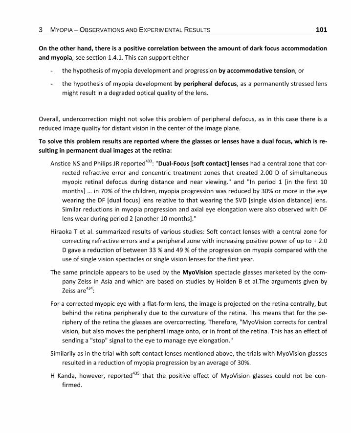
3 MYOPIA – OBSERVATIONS AND EXPERIMENTAL RESULTS 101
On the other hand, there is a positive correlation between the amount of dark focus accommodation and myopia, see section 1.4.1. This can support either
- the hypothesis of myopia development and progression by accommodative tension, or
- the hypothesis of myopia development by peripheral defocus, as a permanently stressed lens might result in a degraded optical quality of the lens.
Overall, undercorrection might not solve this problem of peripheral defocus, as in this case there is a reduced image quality for distant vision in the center of the image plane.
To solve this problem results are reported where the glasses or lenses have a dual focus, which is re‐sulting in permanent dual images at the retina:
Anstice NS and Philips JR reported433: "Dual‐Focus [soft contact] lenses had a central zone that cor‐rected refractive error and concentric treatment zones that created 2.00 D of simultaneous myopic retinal defocus during distance and near viewing." and "In period 1 [in the first 10 months] … in 70% of the children, myopia progression was reduced by 30% or more in the eye wearing the DF [dual focus] lens relative to that wearing the SVD [single vision distance] lens. Similar reductions in myopia progression and axial eye elongation were also observed with DF lens wear during period 2 [another 10 months]."
Hiraoka T et al. summarized results of various studies: Soft contact lenses with a central zone for correcting refractive errors and a peripheral zone with increasing positive power of up to + 2.0 D gave a reduction of between 33 % and 49 % of the progression on myopia compared with the use of single vision spectacles or single vision lenses for the first year.
The same principle appears to be used by the MyoVision spectacle glasses marketed by the com‐pany Zeiss in Asia and which are based on studies by Holden B et al.The arguments given by Zeiss are434:
For a corrected myopic eye with a flat‐form lens, the image is projected on the retina centrally, but behind the retina peripherally due to the curvature of the retina. This means that for the pe‐riphery of the retina the glasses are overcorrecting. Therefore, "MyoVision corrects for central vision, but also moves the peripheral image onto, or in front of the retina. This has an effect of sending a "stop" signal to the eye to manage eye elongation."
Similarily as in the trial with soft contact lenses mentioned above, the trials with MyoVision glasses resulted in a reduction of myopia progression by an average of 30%.
H Kanda, however, reported435 that the positive effect of MyoVision glasses could not be con‐firmed.

102
One one hand, one publication, however, could not confirm the positive effect of a new lens design to reduce relative peripheral hyperopia436, on the other hand a more recent publication of the same author found myopia progression reductions by 33% and 34% by similar lens designs437.
Schaeffel mentioned another new lens design532: "… there was a multifocal spectacle lens pre‐sented by Carly Lam et al.Glasses438 from Hong Kong (the MSMD lens ‘multifocal segment lens’). A 2‐year randomized study in Hong Kong schoolchildren showed very convincing inhibi‐tion of myopia (0.1 D progression with the MSMD lenses, vs 0.4 D progression per year in the controls, and no progression at all in 21% of the children, with 59% reduced progression on average, …. Lam et al. also showed that there was no effect on visual acuity at any luminance level tested."
Glasses with this new lens‐design are marketed by Hoya under the name MiyoSmart.439
Results by Liu Y and Wildsoet C440 about two‐zone bifocal spectacle lenses applied to chicks con‐firmed a positive effect of this type of glasses, which are based on a peripheral defocus.
Berntsen et al.441 compared the myopic progression when wearing progressive addition lenses (PALs) and single vision lenses (SVLs). The change in myopia after 1 year was – 0.38 D for PALs and – 0.65 D for SVLs.
Backhouse et al. reported442: " Correcting the on‐axis refractive error in moderate to high myopia with conventional spherical spectacle lenses results in hyperopic defocus in the peripheral ret‐ina. Correcting the same eyes with conventional spherical soft contact lenses results in signifi‐cant myopic defocus in the peripheral retina. … If the refractive status of the peripheral retina does influence myopia progression, then these results suggest that myopia progression should be slower with conventional contact lens wear than with conventional spectacle wear. How‐ever, previous studies comparing myopia progression with conventional spectacles and con‐ventional contact lenses have reported no such difference."
This peripheral defocus occurs as well with contact lenses as Kang et al. reported443: " Under‐, full, and over‐correction of central refractive error with single vision SCLs [soft contact lenses] caused a hyperopic shift in both central and peripheral refraction at all positions in the horizon‐tal meridian. All levels of SCL correction caused the peripheral retina, which initially experi‐enced absolute myopic defocus at baseline with no correction, to experience absolute hyper‐opic defocus. This peripheral hyperopia may be a possible cause of myopia progression re‐ported with different types and levels of myopia correction."
Smith et al.444 summarized their results and the results of various other authors about the impact of peripheral defocus very clearly: " results are in agreement with the hypothesis that peripheral vision can influence eye shape and potentially central refractive error in a manner that is in‐dependent of central visual experience."

3 MYOPIA – OBSERVATIONS AND EXPERIMENTAL RESULTS 103
Bullimore summarized995 that a change of peripheral defocus is responsible for the positive effect of orthokeratology on the prevention of myopia progression. (See section 3.26.2)
Notes: - This peripheral defocus depends totally on the individual design of the individual contact lens or
spectacle lens, and each manufacture has an own design, which interworks with very individual cor‐nea and sclera optical properties in the peripheral region.
- The peripheral hyperopic defocus is not just a problem of the lens design when correcting an exist‐ing myopia, but as well an issue before becoming myopic, because of the inborn geometry of the eye.
- This arises the question, whether rather small glasses (as used often for good looks and not for good vision) have a similar positive effect, as the light, which is not passing the glasses should create a myopic defocus as well. In case of stronger myopia, however, this "MyoVison"‐Effect of very small glasses could be become negative and enhance the progression of myopia due to its blurring effect.
3.3.11.1 Peripheral Defocus – New Applications for Spectacle Glasses and Contact Lenses
Some products are445, and L Ramon et al. published a general analysis of all the various concepts for bi‐focal and multifocal contact lenses446.
Peripheral defocus aspheric spectacle glasses: e.g. Zeiss MyoVision and Essilor Myopilux. The reported effect of these glasses was rather limited.
Multifocal contact lenses: e.g. Cooper Vision MySight and Myopia Control DISC (which is basis of a contact lens by HOYA as well). It was reported to curb myopia progression in average by 59%, to slow axial eye growth in average by 60%, and to halt myopia progression in 21.5% of Children. Basis of the design are concentric rings od various refractive power.
"MyoSmart" (also called Defocus Incorporated Multiple Segments, DIMS) by HOYA 439, 447, 448: About 300 tiny plus‐defocus elements (+ 3.5 D) are inserted in the center of the spectacle glasses, with each defocus element of about 1 mm diameter.
Lam et al reported for these DIMS spectacle glasses449: "For subjects who completed the 2‐year trial …, the mean myopia progression … over 2 years in the DIMS group (n=79) and the SV [single vision] group (n=81) was −0.38±0.06 D and −0.93±0.06 D, respec vely. The total increase in AL was 0.21±0.02 mm and 0.53±0.03 mm, respectively. Schoolchildren wearing DIMS lenses had myopia progression significantly reduced by 59% (mean difference −0.55±0.09 D, p<0.0001) and axial elon‐gation decreased by 60% (mean difference 0.32±0.04 mm, p<0.0001) compared with those wear‐

104
ing SV lenses." Lam et al. claimed450, "Myopia control effect was sustained in the third year in children who had used the DIMS spectacles in the previous 2 years and was also shown in the chil‐dren switching from SV to DIMS lenses."
Bao et al. reported451 reults with a similar design: "… treatment spectacle lenses have a spherical front surface with 11 concentric rings formed by contiguous aspherical lenslets (diameter of 1.1 mm). The area of the lens without lenslets provides distance correction. The geometry of aspherical lenslets has has been calculated to generate a VoMD [volume of myopic defocus] in front of the retina at any eccentricity, serving as a myopia control signal." Result: For single vision lenses the mean change was − 0.81±0.06 D a er 12 months, for the new design with highly aspheric design the change was reduced to − 0.27±0.06 D.
3.3.12 Summary of the Effects of the Image Quality
Simplified the effects of the image quality can be summarized:
Image quality can control the dimension, the shape and especially the quality of the sclera.
The image quality has an impact on numerous biochemical processes.
It looks like that the reshaping of the sclera happens not via growth, i.e. cell multiplication, but via cell enlargement or stretching, and structural degradation (see section 3.9).
Aberrations appear to play a major part in accommodative process and the onset of myopia.
From the fact that continuous darkness causes myopia Norton et al. concluded327: "... continued visual guidance is necessary to maintain a match between the axial length and the focal plane or for recovery to occur. Absence of light is myopiagenic in tree shrews that have developed with normal diurnal light‐ing." In other words, the development of myopia is here considered to be some kind of "default proc‐ess" of the eye, which is taking place even without the stimulus of an image.
On the other hand, there is a positive correlation between the amount of dark focus accommodation and myopia, see section 1.4.1. This can support either
- the hypothesis of myopia development and progression by accommodative tension, or
- the hypothesis of myopia development by peripheral defocus (see section 3.3.11), as a perma‐nently stressed lens might result in a degraded optical quality of the lens.

3 MYOPIA – OBSERVATIONS AND EXPERIMENTAL RESULTS 105
The initiation and progression of myopia by the image quality is still compatible with a genetic influ‐ence on myopia:
Genetics may be responsible for the metabolism, and the power of the feedback process that control eye growth452. This is in addition to genetically caused weak basic connective tissue, which can be a rea‐son for myopia as well.
From an engineering point of view, progressive myopia and lid suture myopia in primates showed the same open loop feedback process, i.e. there is no feedback in the process of progression involved453.
Note: In general, in publications about the impact of degraded image quality on the onset of myopia the negative role of near work and the corresponding advice for plus lenses is denied. However, the im‐age quality based model can hardly explain why myopia is becoming tremenduously more wide‐spread than before.
Additionally, there is some likelihood that a degraded image quality causes stress on the muscular system of the eye, which then causes the elongation of the eye by stretching.
3.4 Basic Question – what Elongates the Eye
In the section 3.2.1.14 "Emmetropization" towards Myopia via Accommodation? and section 3.3.5 "Emmetropization" towards Myopia via Image Quality? two respective models were outlined.
Some more results are reported in the following section.
3.4.1 Image Quality and Accommodation
The question to be asked is: What is causing myopia, excess accommodation or a reduced image qual‐ity?
The answer is, according to the experimental results, that there is a close interference between ac‐commodation and image quality:
Harb et al. stated187: "Within subjects, accommodative lags significantly increased with closer reading distances, however there was no significant relationship between lag and refractive state. The variability in the accommodative response significantly increased with closer reading distances ... Increased lags and the variability in accommodation at higher accommodative de‐mands suggest that an increase in overall blur at closer reading distances might be related to the development of refractive state."

106
In similar experiments, it was found that generally at a reading distance of 30 cm no perfectly focusing accommodation is performed188. The resulting degradation of the image quality can contribute to the development of myopia.
Gwiazda et al. stated196: "Both types of AC/A ratios are elevated in myopic children ... Myopic chil‐dren with esophoria underaccommodate at near. This suggests that a child who is esophoric must relax accommodation to reduce accommodative convergence and maintain binocular vi‐sion. The reduction in accommodation could produce blur during near work, which could in‐duce myopia as in animal models".
Conclusion: There is no controversy between the observations of myopia caused by degraded image quality and the observations of myopia caused by excessive near work.
Note: The research results indicate that both, excess accommodation and a reduced image quality can play a role in the onset of myopia. It might depend on the individual person whether one effect dominates.
It appears that a blur image has a different effect for myopes and emmetropes: Vera‐Diaz summarized earlier results 454 "... myopic children show reduced accommodative responses to negative lens‐induced blur...", and added new results454: "Compared to pre‐adaptation level, myopes showed a significant in‐crease in the near accommodative response after 3 min of blur adaptation, while accommodation to the near target in emmetropes did not change."
Note: Open Question: Is this modified accommodation of myopes a result of their myopia, or are they myopic because of their modified accommodation? An answer would be helpful for fighting the onset and pro‐gression of myopia.
The effects of various environmental conditions were investigated and summarized by Yi JH and Li RR455: "The annual mean myopia progression (0.38 ± 0.15 D) in the intervention group was significantly lower than that in the control group (0.52 ± 0.19 D; P<0.01). The children in the two groups spent simi‐lar amounts of time in near‐vision activities, but the children in the intervention group spent less time in middle‐vision activities (P<0.01) and more outdoor activities (13.7 ± 2.4 vs 6.2 ± 1.6 hrs/wk; P<0.01). When considering all children in the study, there were 4 factors that significantly correlated with less myopia progression: more outdoor activities, more time spent wearing glasses, more time spent in natural light and less time using a computer."
An interworking is taking place between image quality and the level of illumination, as discussed in sec‐tion 3.14.1

3 MYOPIA – OBSERVATIONS AND EXPERIMENTAL RESULTS 107
3.4.2 Myopia by Accommodation or Myopia by Degraded Image Quality?
It was described above that as well
- excessive accommodation as well as
- degraded image quality
were made responsible for the development of myopia.
As some followers of each of these two theories state that only one of these theories can be correct the following results published by Norton456 appear to show that it is not an "either or", but an "and":
Tree shrews were fitted with a negative lens (‐ 5 D).
For 45 minutes each day the tree shrews were forced to focus at a distance > 1 m, and during this time the negative lens was replaced by various other negative lenses, or a plano lens, or various plus lenses.
Results: a) A negative replacement lens resulted in severe myopia
b) A plano lens replacement lens resulted in no myopia and no axial elongation of the eye
c) A positive replacement lens resulted in about half of the cases in myopia, in the other half myopia was prevented.
Conclusion: Both theories are valid. Excessive accommodation can cause myopia, and regular relaxa‐tion from myopia can prevent it.
Excessive use of plus lenses, however, which results in a degraded image quality results often in the onset or the progression of myopia (result c)).
These results, which are reflecting the effect of the degraded image, are matching very well with the results presented in 3.2.2.3.6 (Permanent Undercorrection instead of Undercorrection for Near Work only).
Wu S et al. made a point against the concept that myopia would be caused by reduced image quality only853: "Currently, it is confirmed that the development of myopia is associated with the disorders of accom‐modation. As a dominant factor for accommodation, ciliary muscle contraction/relaxation can regulate the physiological state of the lens and play a crucial role in the development of myopia. …

108
We found that the arrangements of ciliary muscles in LIM [lens induced myopia] guinea pigs were broken, dissolved or disorganized …. Monitoring of K [potassium] flux in ciliary muscles from LIM guinea pigs demonstrated myopia‐triggered K influx. … Overall, our results will facilitate the under‐standing of the mechanism associated with inhibitory Na /K ‐ATPase [adenosine triphosphate] in lens induced myopia and which consequently lead to the disorder of microenvironment within ciliary mus‐cles from LIM guinea pigs, paving the way for a promising adjuvant approach in treating myopia in clini‐cal practice."
Notes: As it was already mentioned in section 3.2.1.3 that close near work and strong accommodation reduces the image quality a rather likely synthesis of as well the accommodation model as well the image qual‐ity model appears to be:
Short distance to near work or reading results in strong accommodation, which degrades the accuracy of accommodation (either by muscular effects or effects of neurotransmission) and herewith degrades the image quality – the result is myopia.
Moreover, it appears plausible that muscles, which are stressed by excessive load, might have a problem with fine‐tuning (just thing of doing hard manual labour and then very fine manual adjustments – your hands won't be very sensible).
Moreover, every photographer knows that at near distances the focusing becomes more difficult and more inaccurate, which degrades image quality, which favors myopia.
A model, which matches some of the reported results, might be therefore:
Imperfect image quality, maybe caused by extensive accommodation Local degradation of the scleral tissue by biochemical processes + Muscular tension / intraocular pressure, e.g. by extensive accommodation Ocular elongation

3 MYOPIA – OBSERVATIONS AND EXPERIMENTAL RESULTS 109
An alternate model, which matches some of the reported results too, might be:
Excessive accommodation Local degradation of the ciliary muscle tissue Inadequate accommodation, accommodation lag Reduced image quality Ocular elongation
The resulting advice is:
- Avoid short viewing distance and excessive accommodation,
- Take care of excellent illumination, best with a short wavelength, i.e. "cold" light to provide best image quality by high contrast
- Take care of regular relaxation,
- Avoid permanent plus correction or substantial undercorrection as it results in degraded image quality for the respective viewing distance (see note below).
Note: There is some likelihood that a degraded image quality causes stress on the muscular system of the eye, which then causes the elongation of the eye by stretching.
Obviously, it depends on the duration and the strength of accommodation, and the duration and extent of degraded image quality. Therefore people who are most of the day doing near work with plus lenses or plus additions which still give them a perfect image should not be at great risk by the occasionally reduced image quality at dis‐tant vision with these plus glasses or plus additions. In this case, there would be no excessive accommo‐dation, and only a short time of reduced image quality.
Actually, it does hardly matter whether myopia is caused by muscular accommodation or image qual‐ity processes: In both cases, there is agreement that excessive near work or near adaptation is re‐sponsible.

110
3.4.3 Emmetropization by Growth or Mechanical Stretching or Biochemi-cal Thinning?
Numerous publications talk about "growth" of the eye in connection with emmetropization and myo‐pia.
"Growth", however implies generally an extension by building new cells, in contrast to stretching where existing cells and structures are lengthened by mechanical force and subsequent thinning. Moreover, a biochemical degradation of scleral tissue was found to be responsible for the thinning of the sclera as well.
Myopia, expecially higher degrees of myopia are undoubtedly accompanied by thinning: "When ocular elongation accelerates during myopia development, the fibrous sclera thins in mammals396 "
For emmetropization before the onset of myopia, however, there appears to be no information avail‐able whether the cause of the elongation is builtup of new cells (i.e. real growth) or stretching.
For more information about the question "growth or stretching" see section 3.9.
3.5 Phoria, Convergence, Astigmatism and Anisometropia
When focusing on near object three things are happening automatically:
Accommodation
Convergence, i.e. the adjustment of the optical axes more inward compared to distance focusing
Narrowing of the pupils.
3.5.1 Phoria
Phoria is a defect, in which this adjustment of the optical axes of both eyes to focus an object is not properly coordinated. This defect can be permanent, or it can be transient, e.g. under stress, nervous‐ness, tiredness etc. Rather pronounced misalignment of the axes is called strabismus.
It can be that the axes of the two eyes are adjusted too much inwards when accommodating – near esophoria – or too much outwards – exophoria. Esophoria and exophoria are transient effects; if the maladjustment is permanent the corresponding effects are called esotropia and exotropia.

3 MYOPIA – OBSERVATIONS AND EXPERIMENTAL RESULTS 111
Some results are:
It was reported that higher myopia rates are occurring especially with esophoria at near203, 457 Chung et al. stated458: "The results support the hypothesis that near esophoria is associated with high myopia". The higher the esophoria was, the higher was the myopia progression rate. This matches plausibly with the correlation between elevated AC/A ratios (see section 3.5.2) and myopia. An explanation is the optical blur (i.e. not‐perfect imaging) caused by esophoria.
Brown et al. stated280: "In the progressive lens group, change toward more exophoria at near was associated with less myopia progression."
Esophoria at near was positively treated with added plus lenses, respective bifocals275.
Moreover it Brown et al. reported280 that "... there was only 46% as much myopia progression in the progressive lenses‐esophoric group as in the progressive lenses‐non‐esophoric group." This means that progressive lenses (see section 3.2.2.3) were especially effective for myopes with esophoria.
Additionally, a practitioner wrote that in his praxis pseudo‐myopia, i.e. a spasm of the accommoda‐tion was frequently accompanied by esophoria7.
Note: Can it be that this elevated inwards convergence at near initiates an over‐accommodation via the CA/C ratio (see section 3.5.3)?
Mutti et al. stated459: "There is no association [between myopia and esophoria] when phoria is evaluated while children wear their current, habitual corrections. Yet many studies report a high prevalence of esophoria among children with myopia wearing a new, rather than a habit‐ual correction."
Note: This could be explained by the AC/A ratio (see section 3.5.2). A new and increased correction implies a stronger accommodation at near, and a resulting increased convergence.
In phoria there is some misalignment of the muscles of the eye, especially the oblique muscles, and at the more established form of Phoria, at strabismus, surgery for these muscles is performed.
In section 3.7.3 it is described that a load on these muscles can elongate the eyeball, and create herewith temporary and in the long run permanent myopia.
Whether a near esophoria is preceding myopia, i.e. causing myopia, or just accompanying myopia is not absolutely clear.

112
Note: Additionally it might be possible that esophoria and myopia are not causing each other, but are caused together by another biochemical or biomechanical process.
3.5.2 The AC/A Ratio
A related parameter is the "AC/A ratio", i.e. the ratio of automatic inwards convergence of the axes of the eyes with respect to the accommodative effort. In other words: The nearer an object is, and the higher the accommodative effort is, the more the visual axes of the eyes will be adjusted inwards, trig‐gered by the accommodative effort. The AC/A ration describes the degree of inwards adjustment in re‐lation to the accommodative effort. In esophoria the AC/A ration is too high.
Often AC/A ratios, and results based on measured AC/A ratios are hardly comparable, because they are based on different definitions and different techniques for measuring459, 196.
As it was mentioned in section 1.11 phoria can be easily misdiagnosed when children are in fact hy‐peropic78.
Some results are:
Gwiazda et al. stated196: "Both types of AC/A ratios are elevated in myopic children ... Myopic chil‐dren with esophoria underaccommodate at near. This suggests that a child who is esophoric must relax accommodation to reduce accommodative convergence and maintain binocular vi‐sion. The reduction in accommodation could produce blur during near work, which could in‐duce myopia as in animal models".
In another publication by Gwiazda it is said460: "Compared with children who remained em‐metropic, those who became myopic had elevated response AC/A ratios at 1 and 2 years be‐fore the onset of myopia, in addition to at onset and 1 year later. The significantly higher AC/A ratios in the children who became myopic are a result of significantly reduced accommoda‐tion. Accommodative convergence was significantly greater in myopes only at onset ... These findings suggest that the abnormal oculomotor factors found before the onset of myopia may contribute to myopigenesis by producing hyperopic retinal defocus when a child is engaged in near‐viewing tasks."
The problem of under‐accommodation of myopes was already mentioned in section 3.2.
Mutti et al. stated 459that "... the response AC/A ratio was an important risk factor for the onset of myopia ... data suggest that the onset of myopia follows rather quickly, within 1 year, if the AC/A ratio is high.", i.e. it was not just accompanying myopia.

3 MYOPIA – OBSERVATIONS AND EXPERIMENTAL RESULTS 113
Lu et al. stated461: "Ocular motor parameters are different in myopia eyes. AC/A is one of key pa‐rameters among them, and AC/A is increased in myopia, which indicates out of focus of retinal image in the developing myopia... There was significant difference in phoria in near between the myopia group and emmetropia group, but none in that of distance."
A study by Chen et al.462 with children in Hong Kong, however, could not confirm these results: "AC/A ratios appeared higher in progressing myopic children but the difference was not statis‐tically significant."
Attention has to be paid to the AC/A ratio when fitting bifocal glasses to ease accommodation for near work (see section 3.2.2).
Notes: ‐ A high AC/A ratio corresponds in its effect to esophoria.
‐ The AC/A ration might depend on the distance of the object in a non‐linear way. Possible consequence: For reading a distance might be chosen, where convergence is adequate, but where extra accommo‐dative effort is necessary, because the distance is actually too near. This excessive accommodation ef‐fort (see section 3.2 about the impact of accommodation) alone could contribute to myopia – as well via the image effect thesis (see section 3.3) as well via the mechanical thesis (see section 3.7).
The schematic relationship between accommodation and convergence is shown in Figure 12.
Effort of accommodation
Effo
rt o
f con
verg
ence
dashed line: the convergence corresponds exactly with the accommodation / the (near) distance of the object
solid line: at medium reading distances the convergence is too strong for the accommodation / the distance of the object: nearpoint esophoria, high AC/A
recommended reading distance
Figure 12 The relation between accommodation and convergence

114
Note: It is not mentioned in the literature, but it appears to make sense:
If the AC/A ratio is too high, and proper convergence is achieved only at a distance that is shorter than the distance recommended for reading, the use of prismatic glasses, which compensate for the over‐convergence at near distance might be very useful. With glasses like this (no undercorrection as men‐tioned in section 3.2.2.3) a proper reading distance with normal accommodation could be achieved.
3.5.3 The CA/C Ratio, and some more Types of Vergence
It is not only the case that accommodation initiates a certain amount of vergence (AC/A) as described in section 3.5.2. Vice versa vergence triggers a certain amount of accommodation ("convergence ac‐commodation")463. This fact is described by the convergence accommodation/convergence (CA/C) ratio.
It was stated by Blackie et al.464 that "...a low CA/C ratio exacerbate the progression of near‐work in‐duced myopia".
In section 1.4.1 the dark focus of accommodation, or tonic accommodation was mentioned. A corre‐sponding effect for vergence exists: the tonic vergence or dark vergence, which is the adjustment of the axes of the two eyes in the dark or when closed. These two effects (i.e. tonic accommodation and tonic vergence) respond differently to an increase in illumination. There is a large individual bandwidth, as Jiang et al. stated465: " The critical luminance level at which the two responses were coupled for the 12 observers ranged from 0.01 to 0.45 cd/m2, with distinct individual differences."
Disparity vergence or fusional vergence is the effort to match the images of an object on the retinas of both eyes by the adjustment of the two visual axes.
At a more permanent fixation to near points (e.g. while reading) the disparity vergence and the ac‐commodative vergence are gradually replaced by vergence adaptation.
It was said that the AC/A ration rises with age, and that CA/C is falling with age (considered range of age 16 to 48 years)466.
Notes: ‐ Obviously the mechanisms of vergence are highly complex, and are closely related to accommodation. A strategy to handle progressive myopia should therefore pay high attention on a well‐balanced vergence (see also section 3.2.2.3).
‐ It appears that in the results of experimental myopia (see section 3.3) little attention was paid to the vergence issue; it looks like models that consider convergence, could explain some of these results from a different point of view464.

3 MYOPIA – OBSERVATIONS AND EXPERIMENTAL RESULTS 115
- Reading in bed, especially lying on the side might be rather critical: Besides the effect that it leads mostly to a too near reading distance, the reading distance can be different for the two eyes, with un‐known consequences of accommodation, vergence, AC/A and CA/A ratios.
- It can be concluded, that wearing plus glasses but still doing near work at very short distance has to be avoided, because of the convergence, which is caused by the short distance. Reason: this conver‐gence can still have an impact on accommodation.
3.5.4 Hysteresis of Vergence
In section 3.2.1.3 and section 3.2.1.5 hysteresis effects of accommodation were discussed. In fact, an analogue hysteresis effect exists for the vergence467. Note: As a consequence, an impact on myopia could happen via this effect as well, either via the "reduced im‐age quality model" (see section 3.3) or via the mechanical stress model (see sections 2.4 and 3.2).
3.5.5 Glasses, Contact Lenses and Vergence
Glasses have a prismatic effect, which is proportional to the degree of myopia and works towards nearpoint esophoria. This effect does not exist for contact lenses. For contact lenses, however, there is a higher degree of accommodation. As a consequence, Grosvenor et al. stated468: "If the AC/A ratio is moderate, the two effects tend to cancel each other out, but with a high AC/A ratio, the accommoda‐tive convergence effect may predominate, and with a low AC/A ratio, the lens centration effect [i.e. the not existing prismatic effect of contact lenses] may predominate."
The claimed positive effects of plus lenses on myopia (see section 3.2.2.3) can be at least partially based on this prismatic effect of the plus lenses, i.e. a smaller exophoric shift274.
The data in Table 7 correspond well with the claim that minus lenses tend to increase myopia, whereas positive lenses are said to decrease myopia (see section 3.2.2.3), and the result that myopia is often associated with esophoria (see section 3.5.1).
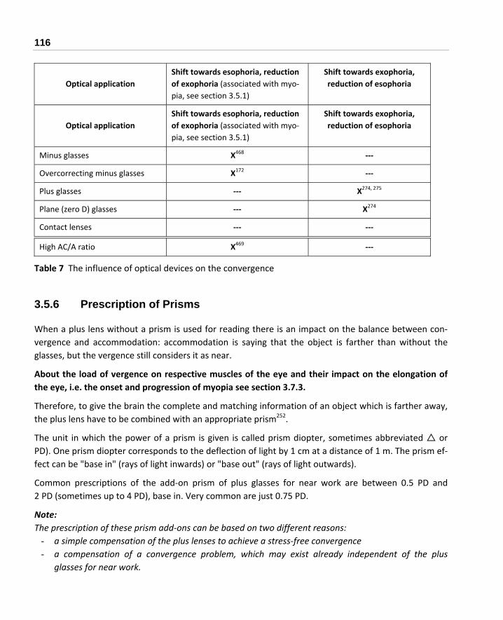
116
Optical application
Shift towards esophoria, reduction of exophoria (associated with myo‐pia, see section 3.5.1)
Shift towards exophoria, reduction of esophoria
Optical application
Shift towards esophoria, reduction of exophoria (associated with myo‐pia, see section 3.5.1)
Shift towards exophoria, reduction of esophoria
Minus glasses X468 ‐‐‐
Overcorrecting minus glasses X172 ‐‐‐
Plus glasses ‐‐‐ X274, 275
Plane (zero D) glasses ‐‐‐ X274
Contact lenses ‐‐‐ ‐‐‐
High AC/A ratio X469 ‐‐‐
Table 7 The influence of optical devices on the convergence
3.5.6 Prescription of Prisms
When a plus lens without a prism is used for reading there is an impact on the balance between con‐vergence and accommodation: accommodation is saying that the object is farther than without the glasses, but the vergence still considers it as near.
About the load of vergence on respective muscles of the eye and their impact on the elongation of the eye, i.e. the onset and progression of myopia see section 3.7.3.
Therefore, to give the brain the complete and matching information of an object which is farther away, the plus lens have to be combined with an appropriate prism252.
The unit in which the power of a prism is given is called prism diopter, sometimes abbreviated or PD). One prism diopter corresponds to the deflection of light by 1 cm at a distance of 1 m. The prism ef‐fect can be "base in" (rays of light inwards) or "base out" (rays of light outwards).
Common prescriptions of the add‐on prism of plus glasses for near work are between 0.5 PD and 2 PD (sometimes up to 4 PD), base in. Very common are just 0.75 PD.
Note: The prescription of these prism add‐ons can be based on two different reasons:
- a simple compensation of the plus lenses to achieve a stress‐free convergence - a compensation of a convergence problem, which may exist already independent of the plus
glasses for near work.

3 MYOPIA – OBSERVATIONS AND EXPERIMENTAL RESULTS 117
As already mentioned in section 3.5.5, the plus lens has a prismatic effect even without a specific prism add‐on. This prismatic effect depends also on the adjustment of the bridge measurement (distance be‐tween the lenses) with respect to the pupillary distance. A de‐centration of the optical center of the plus glasses to the nasal side of the frame results already in some desired base in prism.
Note: Consequently, those cheap off the shelf reading glasses can mostly be expected to have some prismatic effect, and this prismatic effect can be base in (desired) or base out (undesired). Therefore, better get custom made reading glasses.
Properly fitted plus lenses with prism are an essential part of vision therapy (see section 3.15), as Bo‐wan stated252: "Recently, another function of these lenses has become apparent in certain cortically hy‐persensitive Brains: these Brains suffer from image aliasing309,310,311 creating an illusion of movement and other oddities (mirages) because of the textual stripes. The plus and/or prism eliminates the mi‐rages and may affect Brain cognitive processes directly for poorly understood – but clinically easily ob‐served – reasons."
Note: Obviously, it needs a skilled optometrist to determine the data of appropriate plus glasses for near work. The off‐the‐shelf reading glasses from department stores are not matching these quality re‐quirements.
There might be another strong reason for the prescription of prism lenses: Bayramlar reported470: "We observed axial‐length elongation during near fixation both with and without cycloplegia. Based upon this result, one can conclude that convergence, a component of near reflex, rather than accommoda‐tion, may cause axial‐length elongation during near looking… Much use of convergence, not accom‐modation, may be one of the contributing factors in adult onset and adult progression of myopia."
In this paper, the validity of these results was not strictly limited to adults, however; the experiment was performed with 15‐year‐old male persons.
Note: This potential impact of vergence stress is another very strong reason to keep a sufficiently large dis‐tance for near work and reading.

118
3.5.7 The Impact of Vergence on the Axial Length
About the load of vergence on respective muscles of the eye and their impact on the elongation of the eye, i.e. the onset and progression of myopia see section 3.7.3.
3.5.8 Astigmatism
Another optical defect, astigmatism, was found before to have hardly an impact on myopia progression rates in two papers471, 472.
More recently it was reported by Gwiazda et al.473 that "... children with significant against‐the rule astigmatism as infants are more myopic...", and the high prevalence of astigmatism and myopia at American Indians474, 475 makes a correlation between astigmatism and myopia more likely.
In contrast to the results by Gwiazda, Czepita stated476: "With‐the‐rule astigmatism predisposes the creation of myopia. Against‐the‐rule as well as oblique astigmatism has no influence on the creation of myopia."
Schaeffel summarized that when the eye is determining the amount of appropriate accommodation astigmatism is ignored53.
3.5.9 Anisometropia
It is called Anisometropia when the refraction of both eyes is significantly different from each other. Thre question is whether in this case the prescribed glasses should be different as well, and according to the results of the refraction.
Viikari recommends to use in this case the results of the refraction of the eye with less diopters41.
3.5.10 Summary of the Vergence Related Effects
It appears plausible that an obvious disturbance of the control circle: near focus – accommodation – convergence can happen. These disturbances can lead to poorly focused, blurred images on the retina, which in turn can lead to myopia as shown above. A remedy, however, to prevent myopia or myopia progression by interfering with this convergence feedback circle, has not yet been established.
Together with the results of the influences on image quality, there are indications that there is a chain of processes for a development towards myopia107, 196, which looks like Figure 13:
Bowan called this interaction between accommodation and convergences “accommovergence”, and therefore recommended the use of plus additions combined with prisms477.

3 MYOPIA – OBSERVATIONS AND EXPERIMENTAL RESULTS 119
Environmental factors like near work, illumination, and mental stress �
Optical blur by weak accommodation, strabismus (esophoria, AC/A and CA/C ratios, astig‐matism) etc.
� Retinal output signals (image‐controlled model, see section 3.3),
and / or Excessive mechanical stress (mechanical model, see section 3.7)
� Axial "growth" or stretching of the sclera, i.e. axial myopia
Figure 13 Paths to myopia
There exists a quantitative mathematical model in which the interactions between accommodation, vergence and optical blur were used to explain the development of emmetropization and myopia395.
Notes: - This sequence looks rather straightforward. What appears to be missing frequently in the literature
is the consideration of the fact that (especially highly) myopic eyes have substantial structural and biochemical deficiencies and anomalies, and that therefore the feedback mechanism as outlined above is not working in a moderate manner, but often excessively with some pathologic attributes. And why exactly is this chain of events not having any impact on some people at all?
- Obviously, the knowledge of the interaction between the "opto‐mechanical world" and the bio‐chemical world is still missing. Nutrition (see section 3.24) might be one of the keys.
- Maybe there are blood sugar related effects on the AC/A ratio as well (see section 3.24.2).
3.6 Saccades and Focusing
To focus an object, the eye is making about 30 to 80 fast eye movements (oscillations) per second, with an angle amplitude of about 10" to 30" (one " equals to 1/3600 of one degree)478. The purpose is to trigger permanently the sensitivity for contrast. Images are said to become grayish on the retina if the stable focusing is longer than 1 to 2 seconds. These saccades are said to play a role in the interac‐tion accommodation / vergence, too479.
Some people (not really the scientific world) are recommending special training glasses with a pattern of fine holes in a dark surface, called pinhole glasses, to encourage the eye to perform these saccades.

120
Note: In the scientific literature about myopia research there is hardly any mention of saccades, which is rather astonishing, as it appears to be essential for image acuity, and appears to be quite related to the experiments, where induced myopia in animals was successfully reduced by flickering light324. Addition‐ally, excessive saccadic movements might cause additional oxidative stress.
The myopic eye has a specific problem correlated with saccadic eye movements, as David et al. stated480: " ...if account is taken of the increased force required to provide normal saccadic movement of myopic (larger) eyes, then the shear force [a force that occurs in the thin eye wall shell supporting the vitreous body] is up to seven times greater than that experienced for emmetropes."
Note: This additional force for saccadic movements might create two problems:
‐ If the extraocular muscles cannot sustain this additional power, a degraded image and/or focus might result again, contributing to a further progression of myopia.
‐ Due to this increased force, which is necessary for the myopic eye, a progression of myopia could be caused, according to the thesis presented in section 3.7.
For myopes with more than 6 D a significantly slower eye velocity in saccadic movements was found, and if (soft) contact lenses were used instead of glasses, a significantly slower eye velocity was re‐corded as well. A potential explanation was the additional mechanical effort due to the increased mass.481
High‐frequency microfluctuations of accommodation were reported for thicker ciliary bodies482, and the thickness of the ciliary body was found to be positively correlated with myopia535.
Moreover, glucose metabolism has an impact on the saccadic eye movements483, 484, e.g. diabetic pa‐tients have disturbed saccades (asymmetry between the two eyes, longer latency). For the impact of the glucose metabolism on myopia see section 3.24.1.
Hartwig et al., however found485: "Durations, amplitudes, and peak velocities of the main saccades and the numbers of overshoots, undershoots, and exact fixations were analyzed. For all analyzed parame‐ters, no significant differences were found between myopes and emmetropes. … In myopes, only the peak velocity showed a weak correlation with refractive error and axial length, but this failed to reach statistical significance when allowance was made for multiple testing."
Ghasia et al. reported486: " We found an increase in the amplitude of microsaccades in the presence of uncorrected refractive error, but the microsaccade frequency and velocity remained unchanged. The microsaccade amplitude systematically increased with an increase in uncorrected refractive error."
Goffart et al. found487 by experiments with monkeys that saccade movements were significantly dif‐ferent depending on the level of illumination.

3 MYOPIA – OBSERVATIONS AND EXPERIMENTAL RESULTS 121
Yang et al. reported488: " The results show poor binocular coordination of saccades in children is dis‐tance dependant: coordination is particularly poor at near and could compromise single binocular vi‐sion;"
3.7 Mechanical Properties, Stress, Strain and Pressure
3.7.1 Impact of Mechanics on Biochemistry
Theories, which are attributing myopia exclusively to image related effects, or biochemical effects, or mechanical effects are not necessarily in competition with each other: It is still an open question whether the process leading to a degradation of the sclera is completely biochemical, and the reduced mechanical stability of the sclera is simply the effect of a changed biochemistry, or whether mechanical forces are initiating biochemical modifications (a process called mechanotransduction), which lead to the degradation346. There can be also a combination of both effects.
This model was supported by the result that scleral fibroblasts (i.e. cells of the connective tissue), which were mechanically stretched, showed significant changes in gene expression. These changes were already observed after 30 minutes of stretching186. This time length is typical of near‐work ac‐commodation.
Baumert et al. reported708: "Prolonged unaccustomed exercise involving muscle lengthening (eccentric) actions can result in ultrastructural muscle disruption, impaired excitation– contraction coupling, in‐flammation and muscle protein degradation.
Note: Accommodation generally results in a temporary elongation, i.e. stretching of the sclera of the eye (see sections 3.2.1.1 and 3.30.2). Conclusion: Accommodation can result in gene expression of scleral fibro‐blasts.
3.7.2 Intraocular Pressure (IOP)
Overall, there is a general agreement in the literature that a higher intraocular pressure is associated with myopia489, 490, 491, 492, 493.
A newer report came to the opposite conclusion. SM Liet al. reported494: "In this sample of Chinese children, myopia progression over 2 years was inversely related to IOP, suggesting that IOP had essentially no rela‐tionship with myopia progression in school children. The lower IOP in progressing myopic eyes may indicate more compliant sclerae." In other words, a degraded connective tissue of the sclera is typical for progres‐sive myopia.

122
The impact of accommodation on the IOP was explained by Viikari K495: "The accommodation spasm strains and swells the accommodation muscle (m. ciliaris, whose processus ciliares are responsible for secreting the aqueous humor). Also, the lens of the eye thickens, producing stronger refraction. *On top of not working in an ideal fashion, these swollen anatomical structures take up space* and narrow the angle: the circulation and outflow of the aqueous humor are decreased, and the intra‐ocular pres‐sure increases."
Correspondingly, in section 3.2.1.2 research results were summarized that accommodation results di‐rectly in an elongation of the eye.
Note: Myopia, i.e. the stretching of the eye might be explained by simple mechanical stretching by an in‐creased IOP:
For children aged 9‐11 years, however, no association between IOP and myopia was found in one study496, whereas another study by Lin et al. stated497 that "...IOP of patients less than 19 yrs is signifi‐cantly higher than patients more than 30 yrs..."
Note: It is claimed that with new technology the measurement of the IOP is completely independent from the rigidity of the sclera, but maybe there are still limitations of the measurement process ("completely in‐dependent" is hard to believe from the point of physics).
Yan L et al. reported498: There was no significant difference in IOP between progressing myopes and emmetropes when no accommodation was induced (16.22±4.11 vs 17.01±3.72, respectively, t=‐0.93, P>0.05). However, IOP significantly increased with accommodation in progressing myopes (mean change +1.02±2.07mmHg from 0D to 6D, F=5.35, P<0.01), but remained unchanged (mean change ‐0.76±3.22mmHg from 0D to 6D, F=1.46, P>0.05) in emmetropes."
Notes: - This increased IOP for progressing myopes has to have an increased stretching effect on the sclera.
- Ist there a difference in the working of the ciliary muscle the reason of this increased IOP? It looks like this is the case: Pucker AD et al. reported 521: "… in children the posterior ciliary muscle fibers are thicker in myopia … but paradoxically, the apical ciliary muscle fibers are thicker in hyperopia."
Consequently for myopes there is an elevated probability of glaucoma499; a study showed typical prob‐abilities for glaucoma of 1.5% for eyes without myopia, 4.2% for low myopia and 4.4% for moderate to high myopia500.

3 MYOPIA – OBSERVATIONS AND EXPERIMENTAL RESULTS 123
When people with progressive myopia do near work, there is an uncompensated hyperproduction of intraocular fluid, which causes elevated ocular hypertension because there is an insufficient outflow173. Correspondingly, for the time of recovery from experimental myopia an increase in the fluid outflow during emmetropization was found501.
A general increase of IOP with accommodative stress258 and with accommodation and convergence to close distance502 was found.
In contrast, Read SA reported503: "IOP [intraocular pressure] decreases significantly with accommoda‐tion, and changes similarly in progressing myopic and emmetropic subjects."
Some authors, however, state that a higher IOP follows the onset of myopia and does not cause myo‐pia 504, 505.
Note: Is the structural change of the myopic eye's connective tissue also blocking the fluid to flow off, causing an elevated IOP?
Experiments have shown that growing eyes of chickens elongate during the day and shorten during the night, which correlates with the IOP (high at daytime, low at night). There were, however some phase differences between the rhythms in IOP and ocular elongation – with the IOP ahead of the elongation. It was therefore proposed by Nickla et al.626 "that the rhythm in IOP influences ocular elongation in ways other than by simply inflating the eye, for example, by influencing underlying rhythms in scleral extracellular matrix production."
Notes: ‐ The observed phase difference between IOP and elongation could as well be explained by the effect of a hysteresis of the mechanical properties of the connective tissue.
‐ On the other hand, the pressure within the eye is uniform, but the control of the length of the eye due to emmetropization (section 3.3.5) can be very local as well, if this serves a better image quality. This is an argument against a completely IOP‐driven myopia.
One author claimed to have success in treating myopia with the IOP‐lowering beta‐blocker metipranolol506, but other authors could not confirm this by using the different beta‐blocker timolol507.
From other experiments it was concluded that a higher IOP affects neuronal functions of the eye508.
Note: This would give a link with the fact that myopia is connected with dopamine metabolism and elevated IOP.

124
Several components of nutrition were found to be able to influence the IOP492:
Rutin, a bioflavonoid was reported to lower IOP509.
A deficiency of chromium was reported to increase IOP510.
Elevated blood glucose was reported to increase IOP511.
Folate from food was reported to lower IOP512.
All these dietary components were related to myopia besides their impact on IOP (see section 3.24).
Moreover, an elevated IOP appears to be highly related to emotional effects and stress513, i.e. sensitive people are more at risk of an elevated IOP (see section 3.19 for the impact of stress on myopia, and sec‐tion 3.19.2 for the relation between personality and myopia).
A study by Lee et al. 514 "identified a modest cross‐sectional positive association between current smoking and intraocular pressure."
3.7.3 The Ciliary and other Muscles
In section 3.2.1.2 it was already described in some detail, that accommodation causes an elongation of the eyeball.
Long ago Bates515 claimed and showed respective experiments that the oblique muscles of the eye, which are responsible for the movement of the eyeball can lengthen the eye, in other words can cre‐ate temporary myopia.
These oblique muscles are part of surgery for strabismus, which demonstrates, that during conver‐gence for near work there will be substantial load on these muscles.
Greene 516 has explored whether the elongation of the eyeball can be simply explained mechanically, by "the stresses experienced by the posterior sclera as a result of accommodation, convergence, vitreous pressure, and the extraocular muscles."
Basis of the engineering‐like analysis was that:
Substantial forces are applied to the eyeball, e.g. the extraocular muscles can generate 150g (peak) and routinely produce 40g during large amplitude saccades517 (see section 3.6 about saccades). In contrast, the force applied by the ciliary muscle was found to be only 0.6g.
Congenital nystagmus, which is characterized by extreme saccadic movements, is mostly ac‐companied by myopia518.

3 MYOPIA – OBSERVATIONS AND EXPERIMENTAL RESULTS 125
A substantial increase of the intraocular pressure (IOP, see section 3.7.2) of up to additional 14mm Hg was found during accommodation and convergence at close distance519.
The relationship between ciliary muscle thickness (CMT) and myopia was investigated by Buckhurst et al.520: "Increased CMT is associated with myopia. We speculate that the lack of correlation in myopic subjects between CMT and axial length, but not between CMT and OV [ocular vol‐ume], is evidence that disrupted feedback between the fovea and ciliary apparatus occurs in myopia development."
Bailey et al. reported535:"Thicker ciliary body measurements were associated with myopia and a longer axial length."
Pucker AD et al. reported more dtailed results521: "… in children the posterior ciliary muscle fibers are thicker in myopia … but paradoxically, the apical ciliary muscle fibers are thicker in hyper‐opia."
McCollim's experiments showed522 that the oblique muscles of the eye and the stress on them are essential for at least temporary elongation of the eye.
Wu S reported853: "We found that the arrangements of ciliary muscles in LIM [lens induced myo‐pia] guinea pigs were broken, dissolved or disorganized …. Monitoring of K [potassium] flux in ciliary muscles from LIM guinea pigs demonstrated myopia‐triggered K influx."
The fact that nearwork‐induced transient myopia (NITM) is substantially greater at myopic children, and shows a longer regression time523 is an indication for the involvement of the muscular system in myopia by muscular spasms.
The result of the analysis was that the mechanical forces are strong enough to explain the elongation of the eyeball seen in myopia. There are a variety of individual geometrical parameters (like geometrical arrangement of the muscles) and material‐dependent parameters (like the quality of the connective tis‐sue of the sclera), which can explain, why not everybody who is doing a lot of near work is becoming myopic (there is a lot of individuality for parameters like this524).
In other words: Myopia can be explained as the result of the joint effects of muscular force and hydrau‐lic pressure (IOP) on a weakened tissue structure.
A specific theory about the interaction of the muscles was mentioned in section 1.3.2.4.

126
Notes: - The alternative theory of accommodation mentioned in section 1.3.2.4 is similar to this analysis ‐
both are emphasizing the effect of the extraocular muscles.
- These results are in line with the alternative model that myopia is primarily caused by excessive accommodation force (see section 3.2) in contrast to the current research, which is focused almost entirely on the model of image degradation and related biochemical signaling.
- The long term, substantial load on ocular muscles can contribute to a degradation of the connec‐tive tissue of the sclera via an elevated temperature (see section 3.16). Near work was in fact found to increase the temperature of the eye (see section 3.2.1).
3.7.4 The Lens
There exists another potential mechanical model, which might be used to explain myopia. Points to consider are:
As reported in section 3.2.1 there is a time delay for the return of a myope’s lens shape after ac‐commodation (referred to as hysteresis).
The experiment reported in section 1.3.2.4 showed that it can take very long (several years!) until a lens goes back to its original shape, if the accommodative stress was long lasting.
Mutti D et al. reported525: "Before myopia onset, the crystalline lens thinned, flattened, and lost power at similar rates for emmetropes and children who became myopic. The crystalline lens stopped thinning, flattening, and losing power within ±1 year of onset in children who be‐came myopic compared with emmetropes statistically adjusted to match the longer VCDs [vit‐reous chamber depth] of children who became myopic."
Together with the (optical) model of negative‐lens‐induced myopia (see section 3.3) a hysteresis of the shape of the lens might:
Induce myopia by blurring the image when focusing a distant object, but when the lens is still at a residual accommodative state
Induce myopia by an inappropriate negative lens, which is fitted according to this transient residual accommodative state.
This hysteresis of the shape of the lens might be caused by an effect of the ciliary muscle, or by an ef‐fect of the elasticity of the lens.
No information exists about what might cause different modes of elasticity of the lens for myopes and non‐myopes; maybe the swelling of the lens in a hyperglycemic state contributes to this effect (see sec‐tion 3.24.1).

3 MYOPIA – OBSERVATIONS AND EXPERIMENTAL RESULTS 127
3.7.5 The Pupil Size
Tsukahara et al. reported526: "We found that fluid intelligence [Fluid intelligence is defined as reasoning ability, and the ability to generate, transform, and manipulate different types of novel information in real time527], working memory capacity, and attention control did correlate with baseline pupil size ex‐cept in the brightest lighting conditions. … We conclude that fluid intelligence does correlate with baseline pupil size and that this is related to the functional organization of the resting‐state brain through the locus coeruleus‐norepinephrine system."
Note: Large pupil size means reduced image quality as for a camera lens with large aperture (depth of focus effect). Now we learn that the larger pupil, which results in reduced image quality, which is correlated with myopia correlates with higher fluid intelligence. In short, higher intelligence may result in higher likelihood of myopia. larger pupil more intelligence reduced image quality myopia
3.7.6 The Ocular Shape
Some results about the ocular shape are:
Schmid et al. stated528: "Eye shapes (a) varied substantially among subjects and (b) differed con‐siderably from the corresponding shapes of spherical model eyes with identical axial eye lengths.".
The ratio of the length to the transverse dimension in eyes shows that the higher the myopia the higher the ratio, i.e. the eye is stretched primarily in length529.
Partial coherence interferometry was used to measure the eye. The result showed that the eye generally elongates during accommodation, with this explanation by Drexler et al. 28: "… by the accommodation‐induced contraction of the ciliary muscle, which results in forward and inward pulling of the choroids, thus decreasing the circumference of the sclera, and leads to an elonga‐tion of the axial eye length … the elongation was more pronounced in emmetropes than in myopes".
Collins MJ et al. summarized their measurements530: "Small but significant increases in axial length were evident during and to a lesser extent, immediately after the accommodation task."
128
Notes: - To understand this model, just think of a balloon, which is squeezed, and which expands
consequently in the other direction. - The result that emmetropic eyes elongate more during accommodation appears astonishing
and leads to the conclusion that not only mere mechanical effects are responsible for myo‐pia.
There is a substantial hysteresis of the ocular shape: Walker et al. stated190 that with normal peo‐ple, after accommodation "... ocular shape had become more prolate [i.e. stretched]. This shape remained unchanged after 1 hour of sustained accommodation and then returned to baseline dimensions after 2 hours of accommodation ... Ocular shape returned to baseline di‐mensions after 45 min of accommodative relaxation." See also section 1.3.2.4 about the impact of accommodation on the ocular shape. Note: This hysteresis should be a major problem for the accuracy of refractive measurements for fit‐ting glasses. One could even consider this elongation with accommodation as some kind of "slow accommodation". It appears rather plausible that with extensive near work, this elonga‐tion becomes permanent, which corresponds to myopia.
Mutti et al. stated531: "The eyes of myopic children were both elongated and distorted..." ... "In‐creased ciliary‐choroidal tension is proposed as a potential cause of ocular distortion in myopic eyes."
Notes: ‐ If an ocular body has a weak support by the connective tissue, the increased ciliary‐choroidal tension and the increased hydraulic force during extended accommodation could explain the permanent axial extension: In the transverse direction the ocular shape is maintained by the ciliary muscle, but there is less or insufficient support in the axial direction (length) of the eye. The weak connective tissue might be correlated to the effects mentioned in section 3.3.2 and 3.24.16.
‐ Can a specific inborn ocular shape cause an increased risk of developing myopia (e.g. via dif‐ferent resulting stress on the ciliary fibers)?
Schaeffel summarized532 "Taking all studies together, there is convincing evidence that thickening of the choroid predicts future inhibition of eye growth, while thinning predicts future myopia development." Note: On one hand is appears just plausible that elongation and thinning of the sclera is accompanied by a thinning of the choroid, on the other hand this thinning of the choroid, which supplies the surrounding tissue with oxygen might be interpreted as an indication for the importance of the blood circulation (see section 3.17).

3 MYOPIA – OBSERVATIONS AND EXPERIMENTAL RESULTS 129
For Eskimos and for Chinese, both populations with an elevated rate of myopia, the anterior cham‐ber angle declines more rapidly with age533.
For more information about the effect of accommodation on the axial length of the eye see section 3.2.1.2.
3.7.7 The Zonular Fibers
Note: The following model has apparently not been discussed in the literature, but seems to be plausible:
If the zonular fibers (which are attached to the lens, and whose pulling action on the lens regulates ac‐commodation, see section 1.2) are lacking strength and elasticity, they have a problem in pulling the lens into a flat shape when the ciliary muscle relaxes. The result will be a myopic refraction, leading to the prescription of glasses even if the eyeball is not elongated yet, or to a blurred image with poten‐tially corresponding negative effects (see section 3.3).
Furthermore, reduced accommodation amplitude can be expected.
The basic reason for the weakness of the zonular fibers should be weak, degraded connective tissue. Weak, degraded connective tissue is closely related to myopia, especially to progressive myopia (see e.g. sections 2.2, 3.18.9).
3.8 Relations between the Dimensions of the Cornea, the Crystalline Lens and the Ciliary Muscle
The refractive power of the cornea shrinks from 47 to 48 D in infants to 43 to 44 D by the age of 3 to 9 months and remains afterwards rather stable534.
The crystalline lens experiences by far more laterations. The lens power shrinks from 41 D in infancy to 22 to 23 D at the age of 14 years534.
At emmetropes, the change of the optical parameters of the corneal‐ /lens‐system and the axial growth takes place in a well‐coordinated manner, i.e. as well myopia as well as hyperopia are avoided.
Mutti et al., reported that this balance is disturbed at the onset of myopia534: "The ocular components that are in balance in emmetropia are obviously out of balance in myopia: the eye is too long for the optical power of the cornea and crystalline lens." and "The crystalline lens ceased to thin, flatten, and lose power even as the eye continued to grow."
Possible explanations they are giving are:

130
- Peripheral defocus influences the balanced growth of the eye (see section 3.3.11 about peripheral defocus).
- Extensive growth of the ciliary muscle. Bailey et al. reported535: "Thicker ciliary body measurements were associated with myopia and a longer axial length." Mutti et al. concluded534: " The abnormally thick ciliary muscle for a given axial length in myopia may provide the mechanical restriction that would interfere with the equatorial expansion needed to maintain the correlation between ocular growth and optical compensation by the crystalline lens. Anterior mechanical restriction may limit far peripheral expansion, resulting in the relatively more prolate ocular shapes found in myopic eyes."
The mentioned publications do not answer the question whether the increase of dimensions is caused by growth or by stretching. In section 3.9 this issue will be discussed.
Note: As the ciliary muscle is in charge of accommodation the question arises, whether extensive near work and extensive accommodation are in fact essential contributors for the onset of myopia.
3.9 Growth or Stretching and Thinning or Biochemical Thinning of the Sclera in Myopic Eyes?
- Growth means extension by adding new cells, - stretching and thinning means an extension of existing structures with consequent thinning, - biochemical thinning means a loss of substance with a subsequent thinning due to biochemical, not
mechanical processes.
In section 3.2.1.2 it was already described that accommodation causes at least a temporary elongation of the eye.
Sergienko reported the results of the examination of 172 eyes of 86 subjects536: "The hyper‐metropic and emmetropic eyes possessed stiff sclera. … The absolute majority of the myopic eyes revealed a biomechanical weakness of the scleral shell. A higher degree of myopia was as‐sociated with increased AL elongation."
Form‐deprivation resulted in the building of hypertrophic cells (chondrocytes), i.e. in the enlarge‐ment of cells instead of the building of new additional cells358. In other words, there is no growth, but a stretching by a degradation of the quality of the tissue.

3 MYOPIA – OBSERVATIONS AND EXPERIMENTAL RESULTS 131
Scleral fibroblasts (i.e. cells of the connective tissue), which were mechanically stretched, showed significant changes in gene expression. These changes were already observed after 30 minutes of stretching186. This time length is typical of near‐work accommodation (see section 3.7.1).
Nickla Dl et al. stated537: "In chicks, ocular growth inhibition is associated with choroidal thickening and growth stimulation with choroidal thinning, suggesting a mechanistic link between the two responses."
In section 3.16 it is described that an elevated temperature causes a degradation of the connective tissue, and that feverish sicknesses of children are often followed by the onset of myopia, and that intensive accommodation increases the temperature of the eyeball.
McBrien summarized356: "Analysis of the dry tissue weight of the sclera has demonstrated that the cause of scleral thinning in myopia is due to actual loss of scleral tissue as opposed to simply passive stretch of the sclera."
Note: Even mechanical stretching alone, however, would result in a thinning of the sclera with a corre‐sponding loss of tissue density.
All these facts lead to the conclusion that myopia is not accompanied by a "growth"‐ elongation of the eye, but clearly by a stretching, either by biochemical degradation and / or by added mechanical force.
Obviously, when permanent stretching and a degradation of the connective tissue was already taking place it can hardly be expected that it can be reversed, and in case it caused already a structural weak‐ening there is a probability of progressing myopia.
This could explain negative results of plus lenses for children, which already became myopic, and whose sclera is already changed in its mechanical properties.
Why are some people not becoming myopic even when doing excessive nearwork? It could be explaned by an individual connective tissue of the sclera, which has elastic properties and no plastic properties, i.e. the stretching is not permanent and like a rubber string, in contrast to the plastic properties like a piece of dough.
According to the stretching / thinning model, the eye can be compared to a balloon, which is ex‐panded by accommodation, especially extended accommodation.
For eyes with a very elastic sclera the "balloon" will return to its original size after the accommoda‐tion‐load, for eyes with a more plastic sclera the expansion will tend to be permanent.
The conclusion is that the use of plus lenses to avoid accommodation spasms and permanent stretch‐ing has to start before the stretching begins, i.e. it has to be preventative to be effective.

132
Note: Following the degradation / stretching / thinning model, this stretching might be caused by:
General weakness of the connective tissue of the sclera, and / or local degradation of the scleral tissue caused by an image‐quality‐guided biochemical process Muscular tension by accommodation / intraocular pressure Ocular elongation
The question where growth ends and where stretching begins was investigated by Chung538 et al. They found that stretching begins between 25.6 and 26 mm axial length, which corresponds with about 5 to 6 D, which means pathological myopia begins at about 6 D.
3.10 Outdoor Activities
Outdoor activities were found to have a substantial positive effect on the prevention of myopia or the reduction of the progression of myopia as numerous papers showed539, 540, 541, 542.
Rose et al. reported543: "Higher levels of total time spent outdoors, rather than sport per se, were as‐sociated with less myopia and a more hyperopic mean refraction, after adjusting for near work, pa‐rental myopia, and ethnicity." It can be concluded that this fact is due to the relaxing effect of the time spent outdoors." Furthermore, they speculated that the positive effect of outdoor activities is caused by the release of the retinal transmitter dopamine (see section 3.3.3about the effect of dopa‐mine on myopia).
Guggenheim et al. reported544: "Thus, for children nonmyopic at age 11, the hazard ratio (95% confi‐dence interval, CI) for incident myopia was 0.66 (0.47‐0.93) for a high versus low amount of time spent outdoors, and 0.87 (0.76‐0.99) per unit standard deviation above average increase in moder‐ate/vigorous physical activity. Conclusion: Time spent outdoors was predictive of incident myopia in‐dependently of physical activity level. Fong K summarized545: "the 2005 Sydney Myopia Study showed that while Chinese pupils in the city did more near work than their Singapore counterparts, they had fewer cases of myopia. Researchers sug‐gested that this was due to Sydney pupils having four times more physical activity compared to stu‐dents in Singapore."

3 MYOPIA – OBSERVATIONS AND EXPERIMENTAL RESULTS 133
Even having just outdoor activities during school class recess were found to have a significant effect on myopia onset and myopia progression at Taiwanese children546. McBrien, Morgan and Mutti547 summarized presentations from the 12th International Myopia Confer‐ence 2008 as the hot topic:
"... recent evidence that children who spend more time outdoors are less likely to become my‐opic as one of the most significant findings reported."
"... have shown that children with the risk factor for myopia of myopic parents, are at only slightly greater risk than children without myopic parents if they spend sufficient time out‐side."
"... suggest that greater time spent outside can also over‐ride the greater risk associated with near work and schooling."
"... suggesting that the effect is likely to be seen in children from all ethnic groups. The agree‐ment between these studies is unusual in the area of the etiology of myopia, which is gener‐ally renowned for its controversies, rather than for consensus."
"... Firstly, protection does not seem to result from a “substitution” effect, in which children re‐place time spent on reading and writing and other forms of near work with outdoor activi‐ties. Secondly, the critical factor seems to be total time outdoors (in daylight hours), rather than sport or physical activity, since indoor sporting activities are not protective, while both active and more passive activities outdoors are. The studies are in rough agreement on the protective exposures ‐ around 2 to 3 hours a day outside of school hours seems to be suffi‐cient to markedly lower the risk of myopia."
Notes: The positive effect of outdoor activities ccould could be based on these issues: a) the reduced load of accommodation – in general zero accommodation (see section 3.2)
b) the elevated level of dopamine (see section 3.3.3)
c) the additional amount of vitamin D which is generated by the sunlight (see section 3.24.3)
d) the level of illumination which is normally higher outdoors (see section 3.14.1)
e) the color of illumination, as the outdoor illumination has a shorter wavelength than most of the ar‐tificial indoor illumination (see section 3.14.2
f) UV A light induces elevated NO levels548, for the impact of NO on myopia see section 3.18.7)
g) UVA light and riboflavin improves the cross‐linking of the collagen in the sclera and herewith the stability of the sclera against stretching (3.18.8)
h) enhanced physical exercises (see section 3.13)
i) reduced mental stress (see section 3.19.1)
j) children with frequent outdoor activities have a more extroverted mentality in general (see section 3.19.2

134
Since "indoor sporting activities are not protective", the explanations given under b) to g) appear to be more convincing, as the other potential explanations would apply to indoor sporting activi‐ties as well.
Walline reported, however298: "… outdoor time has shown to be effective for reducing the onset of myopia but not for slowing the progression of myopic refractive error." This was confirmed by studies published by Li SM549. In contrast, Jin found that outdoor activities can as well slow down the progression of myopia, in his study from ‐ 0.27 ± 0.52 D/year form the control group to ‐ 0.10 ± 0.65 D/year for the group with addi‐tional outdoor‐time550.
Note: An explanation for the result that the progression was not slowed down can be that f) and g) are re‐sponsible for the positive effect of outdoor activities, UV‐light, however, is not reaching the eye (i.e. the sclera) at people wearing glasses or contact lenses, as these materials are generally eqipped with UV‐blockers. Maybe in some trials the children took off their glasses for the outdoor activities.
Outdoor activities are easier to do and are more common for people, who live in rural areas than for those in the cities, which contributes to the fact that myopia is more widespread in the densely popu‐lated cities.
In sections 3.1 and 3.24.3 it is summarized: "An evaluation of statistical data for reported blindness due to malignant myopia in different states of the USA was done by compiling a chart with the rate of myopia per state, and distance to seacoast, annual hours of sunshine and the nutritional concentra‐tion of calcium, fluoride and selenium in each state. One result was an inverse correlation between malignant myopia and calcium content in the water as well as annual hours of sunshine96.
A longer time spent outdoors is also an indication of a higher exposure to sunshine.
See also
- section 3.14 about Level Illumination / Light / Day‐ and Night‐Rhythm, and
- section 3.3.3 about the role of dopamine in the onset of myopia
3 MYOPIA – OBSERVATIONS AND EXPERIMENTAL RESULTS 135
3.11 Exposure to Bright Sunlight
McKnight et al. reported551: "myopic refractive error was inversely associated with objectively meas‐ured ocular sun exposure … There was an inverse relationship between myopic refractive error and ocular sun exposure, with more than double the prevalence of myopia in the lowest quartile of con‐junctival autofluorescence than the highest quartile (33.0% vs 15.6%)".
Note: These results can at least partly explain the high incidence of myopia in Asian countries, where exposure to the sunlight is traditionally avoided. The current fright of skin cancer adds to an increase in the onset of myopia.
3.12 Seasonal Impact
Mandel stated818: "There were seasonal variations in moderate and severe myopia according to birth month, with prevalence highest for June/July births and lowest for December/January ... Mild myopia was not associated with season of birth or perinatal light exposure."
Donovan et al. summarized their results552: "Myopia progression in summer months was approxi‐mately 60% of that seen in winter, and axial elongation was likewise significantly less in sum‐mer. It is unclear whether more time spent outdoors in summer vs. winter is a contributing fac‐tor, or the difference in progression rates is a result of "seasonal" variations in the intensity or amount of close work performed."
Cui D et al. reported the results of a clinical trial with 235 children 8 to 14 years old553: " Eye elonga‐tion and myopia progression seem to decrease in periods with longer days and to increase in periods with shorter days."
Gwiazda J et al.554 reported for myopic children 6 to 12 years old a mean progression of – 0.35 D in winter and –0.14 D in summer. This was attributed to the children's extended time spent outdoors in summer.
3.13 Physical Exercises
No direct correlations between myopia and physical exercises are available. Two results, however, are giving strong indications:
In urban areas myopia rates are higher than in rural areas.(see section 3.1). Generally people in the city have less physical activity than people in the countryside.

136
(Sure, there are many other differences. For example, immune system would be healthier in the country, more Vitamin D, less reading...)
All forms of physical exercises reduces the intraocular pressure (IOP)555. For the impact of the IOP on myopia see section 3.7.2.
Okuno et al. reported556: "Dynamic exercise ... produces increased tissue blood flow in the retina in the immediate post exercise period, while blood flow increases more persistently in the choroid‐retina." and "The plasma NO metabolite levels increased significantly."
For the interrelation between the blood circulation and myopia see section 3.17, for the inter‐relation between NO metabolism and myopia see section 3.18.7.
In a study, which confirmed the positive effect of outdoor activities, it was found that indoor sports do not have a positive on the development of myopia557.
Read SA et al. reported558: "Myopic children exhibit significantly lower daily light exposure, but no significant difference in physical activity compared to emmetropic children. This suggests the important factor involved in documented associations between myopia and outdoor activity is likely exposure to bright outdoor light rather than greater physical activity."
3.14 Illumination / Contrast / Light / Day- and Night-Rhythm
3.14.1 Level of Illumination
Some results about the impact of the level of illumination are:
The progression of myopia of school kids is slower during summer holidays150, 151, 152. This might be attributed to the brighter light in summer.
Cui et al559. reported that the progression of myopia for children in Denmark depends strongly on the day length. For an average of about 2782 hours of daylight the axial eye growth was 0.12 mm, for about 1681 hours of daylight the axial eye growth was 0.19 mm.
A once promoted theory by Trichtel was that excessive heating up of the background of the eye (or the whole body) is responsible for myopia7. The eye was said to be overheated because of too much incident light through over‐large pupils. It was said that this large opening of the pupils is common to all myopes (claimed to be based on many references), and is additionally caused by stress. At near accommodation the width of the pupil of myopes was said to be even larger than of other people.
Newer publications, however, report that the opening of the pupils of myopes is not wider560, 561, and that low levels of light can help myopia to progress331, 562. Generally the pupil is narrow‐

3 MYOPIA – OBSERVATIONS AND EXPERIMENTAL RESULTS 137
ing when accommodation is occurring563; maybe this was helpful during evolution of mankind to use better depth of focus for near objects (each photographer knows that depth of focus is a problem for near objects, which can be solved by a smaller aperture of the iris of the camera).
Overall, this theory of Trichtel can be considered refuted.
An evaluation of statistical data for reported blindness due to malignant myopia in different states of the USA was done by compiling a chart with the rate of myopia per state, and distance to seacoast, annual hours of sunshine and the nutritional concentration of calcium, fluoride and selenium in each state. One result was an inverse correlation between malignant myopia and calcium content in the water as well as annual hours of sunshine96.
A test with monkeys (already in 1961)156, whose visual space was restricted to an average of fifteen inches, showed that all of them developed some myopia, but that groups with high and low level of illumination showed less myopia than the one with medium level illumination. The high level matches about the number of 300 Lux for reading, the medium level matches a level "provided in a ten by ten foot room with an eight foot ceiling and a 100 or 150 Watt bulb at the ceiling" as Young stated156 – which is considered to be common for reading. The illumination level of the low level group made reading impossible.
Note: These results are similar to the one found about thirty years later when the deprivation issue and the effects of an inappropriate minus lens were discovered as initiators of myopia.
If the eyes of chicken were covered with plastic sheets that produce a hardly noticeable degrada‐tion of the image quality on the retina, no myopia developed at an illumination level of 500 Lux. If the illumination level, however, was reduced to 5 Lux a myopia of –5 D developed within 10 days.
Similarly, myopia developed when, at a surrounding brightness of 500 Lux, the animals received sunglasses that reduced the incidence of light on the retina by a factor of 100. If only one eye is treated with the sunglasses, only this eye is becoming myopic564, 565.
Ashby R et al. reported566: "Exposing chicks to high light levels, either sunlight or intense laboratory lights, retards the development of experimental myopia. These results, in conjunction with re‐cent epidemiologic findings, suggest that daily exposure to high light levels may have a protec‐tive effect against the development of school‐age myopia in children."
Ashby and Schaeffel567 summarized their experiments with chicks: "… the development of depriva‐tion myopia was also significantly suppressed under bright light, even though depth of focus does not play a role under the diffusers." … "High illuminance levels reduce the rate of com‐pensation for negative lenses and enhance the rate for positive lenses, but do not change the set point of emmetropization (target refraction). The retardation of myopia development by

138
light is partially mediated by dopamine, as the injection of a dopamine antagonist abolishes the protective effect of light, at least in the case of deprivation myopia."
Note: From the fact that the compensation for positive lenses is increased at high levels of illumina‐tion, it can be concluded that the use of plus glasses for near work in combination with bright illumination is beneficial.
Ashby R and Morgan I reported568: "Exposing chicks to high light levels retards the development of experimental myopia and reduces the rate of compensation for negative lenses, but enhances the rate of compensation for positive lenses. The retardationof deprivation myopia by light is mediated, in part, by dopamine …"
Moore et al. summarized their results569: "Reducing light levels effectively reduces the contrast of the visual environment. Reducing contrast has been found to produce myopia, and our results agree."
Schmid KL et al. came to different conclusions, however570: "Our data do not support the sugges‐tion that common reductions in letter size or contrast of reading material (as might occur for photocopied reading materials) cause greater accommodation inaccuracy and greater near work‐induced adaptation effects that would exacerbate myopia development in young adults."
A result by Feldkaemper et al. 331 from the experiments about myopia caused by low image quality is: "… the eye becomes more sensitive to image degradation at low light, the human eye may also be more prone to develop myopia if the light levels are low during extended periods of near work".
It was also described7 that a higher level of illumination increases significantly the capability for accommodation, and that presbyopes (i.e. older people with reduced accommodation) can read more easily in high illumination – clearly independent from the increased depth of focus, which is caused by a narrower opening of the pupil.
On the other hand, generally the accuracy of accommodation decreases with decreasing brightness564 (which resembles the depth of field of cameras).
The development of chicks towards emmetropization was observed at various levels of illumina‐tion571: (10.000 Lux, 500 Lux and 50 Lux). Result: After 90 days 50 Lux resulted in a mean myopia of –2.41 D, 500 Lux resulted in +0.03 D, and 10.000 Lux resulted in hyperopia of +1.1 D.
Cohen Y summarized their results: "Thus, under light‐dark cycles, light intensity is an environmental factor that modulates the process of emmetropization, and the low intensity of ambient light is a risk factor for developing myopia."

3 MYOPIA – OBSERVATIONS AND EXPERIMENTAL RESULTS 139
Similar results were obtained from experiments with monkeys at illumination levels of 15‐630 lux (which corresponds to normal lighting levels) and 25,000 lux for 6 hours a day. The explanation of Smith EL et al. is572 "These results are in agreement with the hypothesis that the protective effects of outdoor activities against myopia in children are due to exposure to the higher light levels encountered outdoors."
Backhouse et al. compared the development of myopia at chicks by monocular deprivation573: "… groups received either constant 2000 lux (n = 11) during the light period or 300 lux for 10 h with a 2 h period of bright light (10 000 lux), either in the morning (n = 10), midday (n = 10) or evening (n = 10), giving the same total daily light exposure as the 2000 lux group." Results: "Myopia in the constant 2000 lux group (‐4.94 ± 1.21 D) was significantly less than in the 300 lux control group (‐9.73 ± 0.96 D; p = 0.022). However, compared to the 300 lux control group, 2 h periods of 10 000 lux did not produce significant effects on refraction when presented ei‐ther in the morning (‐9.98 ± 0.85; p = 1.00), midday (‐8.00 ± 1.26; p = 0.80), or evening (‐13.14 ± 1.16 D; p = 0.20), although significantly less myopia was induced in the midday group com‐pared to the evening group (p = 0.018)." In other words, continuous light exposure is most ef‐fective.
Somehow in contrast to the results above experiments with chicken were summarized by Lan et al.574: "exposure to continuous bright light for 1 or 2 hours every day had no significant protec‐tive effect against deprivation myopia. Inhibition of myopia became significant after 5 hours of bright light exposure but extending the duration to 10 hours did not offer an additional bene‐fit. In comparison, repeated cycles of 1:1 or 7:7 minutes of bright light enhanced the protec‐tive effect against myopia and could fully suppress its development."
Light was found to increase (among others) dopamine levels324, 575. This fact matches with the state‐ments in section 3.3.1. Moreover, this effect of light on the dopamine level can explain the positive effect of outdoor activities576.
Norton TT el al. stated370: "We propose a model in which the ambient illuminance levels pro‐duce a continuum of effects on normal refractive development and the response to myo‐piagenic stimuli such that low light levels favor myopia development and elevated lev‐els are protec‐tive. Among possible mechanisms, elevation of retinal dopamine activity seems the most likely."
Norton TT summarized577: Human studies have provided strong evidence that exposure to time outdoors is protective against the onset of myopia. A causal factor may be that the light levels outdoors (30,000‐130,000 lux) are much higher than light levels indoors (typically less than 500 lux). Studies using animal models have found that normal animals exposed to low illuminance levels (50 lux) can develop myopia. The myopia and axial elongation, produced in animals by monocular form deprivation, is reduced by light levels in the 15,000 to 25,000 range. Myopia induced with a negative‐power lens seems less affected, perhaps because the lens provides a

140
powerful target for the emmetropization mechanism. Animal studies suggest that raising the light levels may have their effect by increasing retinal dopamine activity, probably via the D2 receptor pathway, altering gene expression in the retina and reducing the signals that produce axial elongation."
A mathematical model by which the appearance of emmetropization (i.e. eye growth to achieve emmetropia) and myopia was quantitatively explained by the interaction between accommo‐dation, vergence and optical blur395 was extended to include the effect of the level of illumina‐tion. Result: bad light increases the development and progression of myopia, especially of late onset myopia464.
Depth of focus has an impact on image acuity as Atchison et al. stated578: "For uncorrected myopes of 3.0 D or less, visual acuity was nearly as good with a 1‐mm pupil as for corrected myopes". Therefore, a steady opening of the pupil above average might trigger a feedback circle in case of already existing imaging deficiencies, i.e. promote myopia.
In other words, a good illumination causes a good depth of focus (like at a camera), which re‐duces a potential blur of the image, which might avoid myopia development by a reduced im‐age quality.
For dilated pupils (which are accompanying low levels of illumination) increased spherical aberra‐tion and decreased optical quality was measured579, as the peripheral areas of the lens do not show the features of a perfect optical system (see section 3.3.9 about aberration, and section 3.3.5 about aberration and emmetropization).
This matches with the results published by Buehren et al. They found that myopes showed more aberrations than emmetropes, and these differences were increased after 2 hours of reading. They concluded427: "The differences between the groups are primarily due to changes in the corneal wavefront associated with a narrower lid aperture during reading for the myopes. These differences are enhanced by longer periods spent reading, larger pupils and conse‐quently low light levels."
Notes: Corresponding with this fact, the individual relation of the pupil size per level of illumination may have an impact in both directions – too large and too small might be both negative:
‐ A below average pupil size results in a low level of illumination on the retina, with the men‐tioned negative effects.
‐ An above average pupil size results in a degraded depth of focus, with the mentioned nega‐tive effects.
To some extent the illumination level has an impact on the important issue of the vergence (see section 3.5.3):

3 MYOPIA – OBSERVATIONS AND EXPERIMENTAL RESULTS 141
It was stated by Owens et al. 580: "... convergence is a more important distance cue than ac‐commodation under low illumination..." and by Kersten et al. 581: "Accommodation to a dim tar‐get corresponded closely to the convergence accommodation".
Note: It can be concluded that under low light a convergence problem like esophoria can have a negative impact on accommodation, e.g. create over‐accommodation.
Vannas et al. stated582: "...there was a trend towards a higher prevalence of myopia among con‐scripts living above the Arctic Circle, consistent with the hypothesis that ambient lighting might influence refractive development. Other novel associations with myopia were the decreased use of sunglasses and brown iris color."
Notes: Several explanations for these results of "a higher prevalence of myopia among conscripts living above the Arctic Circle" could be offered: The lack of complete darkness during summer, the lack of sufficient light during winter, and a lack of vitamin D caused by the lack of light during winter (see section 3.24.3).
The association between myopia and a decreased use of sunglasses could be explained, e.g., by temperature effects (see section 3.16).
Norton and Siegwart385 proposed a model according to which the positive effect of time spent out‐doors (see section 0) could be explained by the elevated illumination level outdoors and its ac‐tivation of dopamineric pathways.
Lan W et al. summarized their results583 "Bright light stimulates choroidal thickening in chickens, al‐though the response is smaller than with experimentally imposed myopic defocus, and it oc‐curs with some time delay. It nevertheless suggests that choroidal thickening is also involved in myopia inhibition by bright light."
Read SA et al. reported584: "Myopic children (n = 41, mean daily light exposure 915 ± 519 lx) exhib‐ited significantly lower average light exposure compared to 41 age‐ and gender‐matched em‐metropic children (1272 ± 625 lx, p < 0.01). The amount of daily time spent in bright light con‐ditions (>1000 lx) was also significantly greater in emmetropes (127 ± 51 minutes) compared to myopes (91 ± 44 minutes, p < 0.001). No significant differences were found between the av‐erage daily physical activity levels of myopes and emmetropes (p > 0.05).
In somewhat more detail, Read SA reported585: "Categorized according to their objectively meas‐ured average daily light exposure and adjusting for potential confounders (age, sex, baseline axial length, parental myopia, nearwork, and physical activity), children experiencing low aver‐age daily light exposure (mean daily light exposure: 459 ± 117 lux, annual eye growth: 0.13 mm/y) exhibited significantly greater eye growth than children experiencing moderate (842 ±

142
109 lux, 0.060 mm/y), and high (1455 ± 317 lux, 0.065 mm/y) average daily light exposure lev‐els."
CM Jung et al. reported "Bright light exposure significantly reduced plasma cortisol levels at both circadian phases studied …".
About the impact of cortisol on myopia see section3.19.1.
W Lan et al. reported586 results about experiments with chicks: " Compared with the reference group, exposure to continuous bright light for 1 or 2 hours every day had no significant protec‐tive effect against deprivation myopia. Inhibition of myopia became significant after 5 hours of bright light exposure but extending the duration to 10 hours did not offer an additional benefit. In comparison, repeated cycles of 1:1 or 7:7 minutes of bright light enhanced the protective effect against myopia and could fully suppress its development.
Conclusions: The protective effect of bright light depends on the exposure duration and, to the intermittent form, the frequency cycle. Compared to the saturation effect of continuous bright light, low frequency cycles of bright light (1:1 min) provided the strongest inhibition effect. However, our quantitative results probably might not be directly translated into humans, but rather need further amendments in clinical studies."
Ulaganathan et al. reported results of associations with light exposure and longitudinal axial length and choroid changes587: "In summary, diurnal variations in AL [Axial Length] were higher in amplitude in myopes compared to emmetropes and were also associated with longitudinal changes in AL. These findings suggest that diurnal variations may be associated with longer‐term axial eye growth. Time spent in bright light also significantly influenced the amplitude of daily AL variations, with more time exposed to bright light associated with a smaller ampli‐tude of diurnal AL change. Choroidal thickness exhibited an inverse association with the AL changes, implying a potential role for the choroid in eye growth."
Li et al. reported180: Results: "WD [working distance] and LI [light intensity] were positively asso‐ciated with SER [spherical equivalent refraction]. … When WD and LI were split up, the detri‐mental threshold was approximately 40 cm for WD and 6300 lux for LI."
Goffart et al. found487 by experiments with monkeys that saccade movements were significantly different depending on the level of illumination.
Landis et al. reported588 "We found that mice exposed to either scotopic [1.6 × 10⁻³ candela/m2],or photopic [4.7 × 10³ cd/m2] lighting developed significantly less severe myopic refractive shifts … than mice exposed to mesopic [1.6 × 101 cd/m2] lighting … DA [dopamine] activity, was high‐est under photopic light regardless of lens defocus treatment.

3 MYOPIA – OBSERVATIONS AND EXPERIMENTAL RESULTS 143
Note: It is surprising that the lowest level of illumination was more efficient against myopia than the medium level of illumination.
In section 3.3.11 the effect of peripheral defocus, which is related to image quality and therefore to level of illumination is discussed.
Summary:
Good illumination is highly recommended especially for near work – as Feldkaemper et al. stated331: "Because the experiments show that the eye becomes more sensitive to image degradation at low light, the human eye may also be more prone to develop myopia if the light levels are low during extended periods of near work." And Ashby et al. confirmed566: "… results, in conjunction with recent epidemi‐ologic findings, suggest that daily exposure to high light levels may have a protective effect against the development of school‐age myopia in children."
Notes: This argument is very convincing, as every photographer knows that high levels of illumination results in sharper pictures with a higher contrast. Moreover, reduced image quality was made responsible for myopia (see section 3.3), and therefore low levels of illumination will favor myopia.
Apparently, our grandmothers were right, when they warned:
"kids, don't read at bad light!"
Another conclusion is that people should wear sunglasses only for very bright light and not to follow a fashion trend only.
Additionally it has to be noted that in the past the majority of people worked outdoors, where in gen‐eral a by far higher level of illumination exists.
Some recommended levels of illumination for comparison589 are shown in Table 8:
Condition lux ftcd
Sunlight 107,527 10,000
Full daylight 10,752 1000
Normal office work 500
Detailed drawing work 1,500 – 2,000
Prolonged and exact visual tasks 5,000 – 10,000
Table 8 Typical levels of illumination

144
For the related issues of
- outdoor activities see section 3.8
- dopamine (which is sunshine dependent) and myopia see section 3.3.3.
3.14.2 Color of Illumination – Wavelength of the Light
3.14.2.1 Individual Results
As light of longer wavelength (red) is dispersed less than light of shorter wavelength (blue), the ex‐act image of a multicolor (e.g. white) object exists in different planes, e.g. the exact image on the retina exists for one wavelength (color) only. For the other wavelength the image on the retina is not perfectly focused (chromatic aberration; this effect is often used by the optome‐trist to check for overcorrection). Corresponding with this fact is that persons who wore blue filters showed initially some degree of myopia590.
See section 3.3.10 for an image about chromatic aberration.
Other authors came to the same conclusion – i.e. a recommendation to eliminate infrared light, which is close to visible light – via a completely different model. They concluded from experi‐ments that the hyperthermia, which is caused by the infrared light, increases the activity of enzymes, which causes a loss of the spatial structure of hyaluronic acid and collagen bio‐molecules.591
In line with these results, Long Q et al. found592 "Compared with the guinea pigs raised in normal conditions, the guinea pigs raised in long‐wavelength and mixed lights were more myopic and also showed longer vitreous chamber depth at 2, 4, and 6 weeks."
The progression of myopia of school kids is slower during summer holidays150, 151, 152. This might be attributed to the longer time spent outdoors, where the wavelength of the light is shorter that indoors.
Neitz J and Neitz N summarized their experiments with colored lens filters which exposed the eye mainly to the blue, shortwave light component which dominates outdoors593: "Eyes wearing the experimental treatment lens grew 10 times slower then eyes wearing the control lens."
Long Q et al. reported594: " Compared with the guinea pigs raised in normal conditions, the guinea pigs raised in long‐wavelength and mixed lights were more myopic and also showed longer vitreous chamber depth at 2, 4, and 6 weeks."

3 MYOPIA – OBSERVATIONS AND EXPERIMENTAL RESULTS 145
Hathaway reported595, that elementary students exposed to full spectrum fluorescent lighting with ultraviolet light supplements showed in general better growth, attendance, achievement and development than the other students. High‐pressure sodium lamps with their warm lighting gave the worst results.
Cowan et al. reported596, "This finding is consistent with our hypothesis that blue light function may have utility as an assay of CNS [central nervous system] DA [dopamine] tone." For the impact of dopamine on myopia, see section 3.3.3.
Quian et at.597 summarized previous results of various authors: "Relative to the broadband white light (BL), postnatal guinea pigs develop myopia in a monochromic middle‐wavelength light (ML, 530 nm) environment and develop hyperopia in a monochromic short‐wavelength light (SL, 430 nm) environment." They found additionally by experiments with guinea pigs that a re‐cuperation from the exposure to SL or ML light can take very long.
Jiang et al. reported598: "Blue light inhibited axial eye growth, even when animals [guinea pigs] were reared with negative lenses."
Foulds et al. reported about experiments with chicks599: "Red light [90% red, 10% yellow‐green] in‐duced progressive myopia (mean refraction ± SD at 28 days, ‐2.83 ± 0.25 diopters [D]). Progressive hyperopia was induced by blue light [85% blue, 15% green] (mean refraction at 28 days, +4.55 ± 0.21 D). … Myopia induced by 21 days of red light (‐2.21 ± 0.21 D) was reversed to hyperopia (+2.50 ± 0.29 D) by subsequent 21 days of blue light. Hyperopia induced by 21 days of blue light (+4.21 ± 0.19 D) was reversed to myopia (‐1.23 ± 0.12 D) by 21 days of red light."
Torii H et al. summarized their experiments600, "that high myopic patients with the non‐VL [violet light] transmitting pIOLs [phakic intraocular lens] implanted are almost two times more myopic in the change of refraction and four times longer in the change of axial length, compared to those implanted with the VL transmitting pIOLs. This result indicated that the VL transmitting pIOL suppressed myopia progression and axial length elongation compared with the non‐VL transmitting one. In conclusion, our study showed the VL possibly has an anti‐myopia effect for human adults with high myopia."
Schaeffel summarized532 work by Tori and Wang: Light of short wavelength might prevent myopia progression and increase the release of dopamine.
This matches with the earlier observation that he mechanical stability of corneas was improved by treatment with vitamin B2 (riboflavin) and UV irradiation, which introduced additional cross‐links (see sections 3.24.15and 4.2.13).
The mechanical stability of corneas was improved by treatment with vitamin B2 (riboflavin) and UV irradiation, which introduced additional cross‐links (see sections 3.24.15and 4.2.13).

146
Ostrin summarized work by various authors972: "Results showed that guinea pigs raised in green light had significantly higher levels of pineal gland melatonin, whereas guinea pigs raised in blue light had decreased melatonin, presumably as a result of increased ipRGC‐induced mela‐tonin suppression. Additionally, guinea pigs raised in green light developed more myopic re‐fractive errors than those raised in blue light. The authors speculated that the increased mela‐tonin experienced by green light potentially induced alterations in retinal growth factors or changes in ocular circadian rhythms that ultimately led to increased eye growth.
An anecdotal experience of a reader of this Manual601: "Nonetheless, in my pseudo‐research, with Blue Light and Red Light, I noted no differences between before and after 40 hours when using 450nm light. (Could read at 7 inches, before and after) and a major decline after 13 hrs of 660nm red light (Could read at 7 inches before, 5 inches after)".
Rucker et al. reported602: "We alternately exposed one eye of each chick to a … simulating myopic defocus (“MY defocus”: image focused in front of retina; hence, red contrast higher than blue) and the other eye to a grating of the same spatial frequency simulating hyperopic defocus (“HY defocus”: blue contrast higher than red). … The eyes compensated in the appropriate direc‐tions: The choroids of the eyes exposed to the HY simulation showed significantly more thin‐ning (less thickening) over the course of the experiment than the choroids of the eyes ex‐posed to the MY simulation … The rate of scleral GAG [glycosaminoglycans] synthesis in the eye exposed to the HY simulation was significantly greater than in the eye exposed to the MY simulation."
Rucker et al. summarized603: "Despite the older age range [39.9 ± 8.9 years], a greater reduction in eye growth (0.29 mm) after 5 years was found in the UV‐transmitting group [group wearing UV‐transmitting lenses]. … Moreover, any benefit of exposing the eye to UV light is likely to be outweighed by the greater risk of photo‐oxidation, corneal keratitis, and lenticular opacities.
Note: Obviously this positive effect can be attributed to improved cross‐linking of collagen (see section 3.24.15).
Yu et al. reported604: "… guinea pigs exposed to SL [short wavelength light, 440 nm, 500 Lux] were less myopic than to WL [white light, 580 Lux] (2.06 ± 1.69D vs. ‐1.00 ± 1.88D), accompanied with shorter AL [axial length] and less retinal RA [retinoic acid, which is known to increase myopia]. SL reduced retinal RA even after exogenous RA supplementation and decelerated LIM [lens induced myopia] compared to WL
Dfuji et al. reported605 a case in which blue light transmitting glasses and engagemnent in two hours of outdoor activities resulted for a boy with high myopia and anisometropic amblyopia resulted in reduced axial length and thickened choroid.
3 MYOPIA – OBSERVATIONS AND EXPERIMENTAL RESULTS 147
Norton et al. reported606: "… data support the conclusion that the emmetropization mechanism cannot maintain emmetropia in narrowband lighting". The reason was said to be a lack of lon‐gitudinal chromatic aberrations.
Najjar et al. reported607 about experiments with chicks: "this study, we show that exposure to mod‐erate intensities [about 230 lux] of blue‐enriched white light [9700 K] can slow axial elonga‐tion, reduce aberrant ocular structural changes and accelerate recovery from increased axial ocular growth in a chicken model of form‐deprivation myopia [as compared to light with 3900 K]."
Mori et al. reported608: "The mean change in axial length in the VL glasses group [violet light‐transmitting eyeglasses (VL glasses) which can transmit violet light 360–400 nm in wavelength], was significantly smaller than in the placebo glasses group when time for near‐work was less than 180 min and when the subjects were limited to those who had never used eyeglasses be‐fore this trial … The suppressive rate for axial elongation in the VL glasses group was 21.4% for two years."
Thakur et al‐ reported609: "…adults were exposed to blue (460 nm), green (521 nm), red (623 nm), and white light conditions for 1‐hour each on 4 separate experimental sessions conducted on 4 different days. In each light condition, hyperopic defocus [by a minus lens of 3D] was induced to the right eye with the fellow eye experiencing no defocus. … Axial length increased from baseline after red light (mean difference ± standard error in the defocussed eye and non‐defocussed eye = 11.2 ± 2 μm and 6.4 ± 2.3 μm, … respectively) and green light exposure (9.2 ± 3 μm and 7.0 ± 2.5 μm …) with a significant decrease in choroidal thickness … after 1‐hour of exposure. Blue light exposure resulted in a reduction in axial length in both the eyes (−8.0 ± 3 μm, P < 0.001 in the defocused eye and −6.0 ± 3 μm, P = 0.11 in the non‐defocused eye) with no significant changes in the choroidal thickness."
Notes: Obviously the color of the light alone, i.e. without any significant accommodation and focusing effort can lead to increased axial length and decreased choroidal thickness and therefore myopia or its pro‐gression.
All this supports the results of the positive effect of outdoor acticities where cold light is dominating (see section 3.10
Additionally, This could lead to the conclusion that near work should be done in cold, ‐ i.e. most likely cold fluorescent or LED‐light, what is discussed in more detail in section 3.14.2.4. See also section 3.16 about the impact of the temperature.

148
In section 1.11 it was already mentioned:
If you can see numbers or letters on a distant chart clearer and with more contrast when looking through a red glass of foil than through a green one, there is undercorrection.
If the contrast is better for the green pichture, there is overcorrection.
Therefore, a cold‐color light results in less accommodative stress for the eye than a warm‐color light.
Moreover, there are reports about positive results of treating patients with more general visual disor‐ders by the effect of an illumination with specific colors610.
See also the related section 3.24.15.1 about the use of Riboflavin and UV‐light for enforcing the sclera.
3.14.2.2 The Effect of Broadband Illumination
It was concluded once that the eye does not use this chromatic effect to detect the sign of a defo‐cus (too near / too far)97, i.e. that chromatic aberration is not essential for emmetropization. This is in contrast to a statement by Schmid et al. 611: "...data hint that chromatic aberration may have some role as a cue to defocus in emmetropization."
An experiment by Kroger et al. 612 about the accommodative response during near work showed, however that "The refractive state was largely independent of the color temperature of the il‐lumination light (white paper) and the color of commercially available papers (white illumina‐tion). Selective elimination of long wavelengths, however, significantly reduced the accom‐modation stimulus by about 0.5 diopters." These authors "suggest that the visual system pri‐marily uses long wavelengths, if available, during reading tasks."
Yoon et al. reported613 about experiments with chicks: " In broadband light, longitudinal chromatic aberration (LCA) provides emmetropization signals from both wavelength defocus and the resulting chromatic cues. Indoor illuminants vary in their spectral output, potentially limiting the signals from LCA. … We conclude that axial growth and refraction were dependent on the lighting condition in a manner predicted by wavelength defocus signals arising from LCA. … Broad spectrum artificial illuminants with a brighter blue component slow axial growth."

3 MYOPIA – OBSERVATIONS AND EXPERIMENTAL RESULTS 149
3.14.2.3 Melanopsin and Neuropsin
Chakraborty et al. reported614: "Melanopsin‐expressing retinal ganglion cells (mRGCs), which detect light, are important for visual function, and have connections with retinal dopamine cells. … Collectively our findings show that disruption of retinal melanopsin signaling alters the rate and magnitude of normal refractive development, yields greater susceptibility to form‐deprivation myopia, and changes dopamine signaling."
Wikipedia summarized615 " Melanopsin photoreceptors are sensitive to a range of wavelengths and reach peak light absorption at blue light wavelengths around 480 nanometers."
Jiang reported616: "Here, we show that violet light prevents lens defocus–induced myopia in mice. This violet light effect was dependent on both time of day and retinal expression of the violet light sensitive atypical opsin, neuropsin (OPN5). These findings identify Opn5‐expressing retinal ganglion cells as crucial for emmetropization in mice and suggest a strategy for myopia preven‐tion in humans."
Note: These are some more hints at the positive effect of blue light, which is dominating the outdoor illumina‐tion, which was shown to be very important to prevent myopia and its progression.
3.14.2.4 Blue Light Emitted from Electronic Screens: What are the Risks?
Zhi‐Chun Zhao et al. wrote617: "spectrum, short‐wave blue light with wavelength between 415 nm and 455 nm is closely related to eye light damage. This high energy blue light passes through the cornea and lens to the retina causing diseases such as dry eye, cataract, age‐related macular degeneration, even stimulating the brain, inhibiting melatonin secretion, and enhancing adrenocortical hormone production, which will destroy the hormonal balance and directly affect sleep quality."
On the other hand, as Schaeffel stated618, "The amount of blue light you're exposed to when you go outside is so much greater than anything you'll get from a computer screen.
Note: To prevent sleep disturbances the use of devices emitting blue light should be avoided in the late eve‐ning, as melatonin will be reduced. See sections 3.14.4 and 3.25.4.3 about melatonin.

150
3.14.2.5 LLLT – Low-Level Laser Therapy
LLLT uses low levels of red or near infrared light. Its wavelength ranges from 600 nm to 1100 nm, and its output can reach 700 mW.
Xiong et al. reported619: "Changes in axial length (AL) were 0.23 ± 0.06 mm for children wearing spectacles, 0.06 ± 0.15 mm for children wearing OK [orthokeratology] lens, and ‐ 0.06 ± 0.15 mm for children treated with LLLT for 6 months. Changes in subfoveal choroidal thickness (SFChT) observed at the 6‐month examination were ‐16.84 ± 7.85 μm, 14.98 ± 22.50 μm, and 35.30 ± 31.75 μm for the control group, OK group, and LLLT group, respectively."
This means LLLT not only prevented progression of myopia, but even resulted in a slight reduc‐tion of existing myopia.
They explained the positive effect of LLLT by its effect on the nitric oxide system, the decrease of oxidative stress and reduction of inflammatory cytokines.
Ghamdi et al. reviewed publications about LLLT saying620, "… many articles report that it produces higher rates of ATP, RNA, and DNA synthesis in stem cells and other cell lines. Thus, LLLT im‐proves the proliferation of the cells without causing any cytotoxic effects."
Jiang et al. reported about his results with 246 children 117 in the RLRL (repeated low‐level red‐light) and 129 in the control group621: "The RLRL treatment was provided by a desktop light therapy device [a type which is commonly used for the treatment of amblyopia] which emits red light of 650 nm in wavelength, at an illuminance level of approximately 1600 lux and a power of 0.29 mW for a 4‐mm pupil (Class I Classification), and was administered at home un‐der supervision of parents, 3 minutes per session, twice per day with a minimum interval of 4‐hours, five days per week. … Adjusted 12‐month axial elongation and SER [spherical equiva‐lent refraction] progression were 0.13 mm and ‐0.20 D (95% CI: ‐0.29 to ‐0.11) for RLRL treatment, 0.38 mm (95% CI: 0.34 to 0.42) and ‐0.79 D (95%CI, ‐0.88 to ‐0.69) for SVS [control group]. … In our study, we demonstrate that RLRL treatment was able to achieve greater than 0.05 mm axial length shortening in 70.1% of the participants at 1‐month and 31.6% at 12‐months"
LLLT was found to be helpful for various eye problems like macular degeneration as well622, 623.
Note: This positive effect appears to be surprising, as red light during normal focusing was more likely having the opposite effect as mentioned above. The difference is that LLLT is appied for short times only and di‐rectly into the eyes, and laser light is technically different from other light (The light from a laser is said to be coherent, which means the wavelengths of the laser light are in phase in space and time).

3 MYOPIA – OBSERVATIONS AND EXPERIMENTAL RESULTS 151
3.14.3 Contrast
AC Aleman et al. reported624: " subjects, we found that the choroid, the heavily perfused layer behind the retina in the eye, becomes about 16 μm thinner in only one hour when subjects read black text on white background but about 10 μm thicker when they read white text from black background. Stud‐ies both in animal models and in humans have shown that thinner choroids are associated with myopia development and thicker choroids with myopia inhibition. Therefore, reading white text from a black screen or tablet may be a way to inhibit myopia, while conventional black text on white background may stimulate myopia."
Hinterlong et al. reported625: "Multimedia exposure under brighter classroom conditions may contrib‐ute to the myopia incidence in schoolchildren. Teaching with digital projection technology is increas‐ingly prevalent. This may increase children's odds of developing myopia, which has far‐reaching and lifelong deleterious effects."
Contrast is an issue in section 3.3 as well.
3.14.4 Day- and Night-Rhythm
Investigations have shown that growing eyes of chickens elongate during the day and shorten during the night, following the changes of the IOP with some delay.626 Liu JH reported627: "However, the mag‐nitude of this IOP elevation at night was significantly less in the myopia group. In the sleep period, IOP was less in the myopia group than in the control group…There is a 24‐hour rhythm of supine IOP in the myopic group, but the phase timing is different from that in the control subjects."
In experiments with mice it was found, that extended periods of light exposure result in a trend to‐wards myopia, increase in axial length and retina thinning628.
Chakraborty R et al. reported629: "AL [Axial Length] underwent significant diurnal variation that was consistently observed across the 2 measurement days. The longest AL was typically observed at the second measurement session (mean time, 12:26) and the shortest AL at the final session of each day (mean time, 21:06)." and "A positive association was found between the variations of AL and IOP [In‐tra Ocular Pressure] and AL and VCD [Vitreous Chamber length] and a negative association between AL and choroidal thickness".
There is, however, a rhythm in axial length and choroidal thickness even in constant darkness630, appar‐ently due to the circadian rhythm (i.e. night‐/day‐rhythm), which is triggering numerous biochemical processes. Rada summarized360: "In eyes that are developing myopia, the growth rhythm breaks down."
152
Another effect of the day‐ and night‐rhythm was reported by Ohngemach336 in a paper about the do‐pamine metabolism: "Intermittent periods of normal vision inhibited deprivation myopia more if they occurred in the evening than in the morning."
Yun et al. generalized631: "We hypothesize that declining dynamic range and variation of environmental cues may contribute to health dysfunctions, and that judicious expansion of biologic dynamic ranges may be beneficial... Daytime sheltering, optical shading, and nighttime use of artificial light may reduce circadian luminal variation. The resulting melatonin alterations may contribute to systemic dysfunc‐tions."
The result of a study in 1999 was that night‐lights increase substantially the probability of myopia for kids632: the probability was found to be increased more than three‐fold for night‐lights and more than five‐fold for full light in the sleeping room.
Later other studies came to different results633, 634, and the dispute between the researchers is still open635, focusing on the question whether the studies were properly and neutrally designed. It looks like that the brightness of the dimmed night light might be an essential parameter636.
It was proposed to use red night‐lights instead of a white one, because it was said that the red one does not have an impact on melatonin production.637
The night‐light issue cannot be judged here, but there are some related observations, which offer sev‐eral potential explanations:
Illumination has a high impact on melatonin production. Glickman et al. stated638: "Less than 1 Lux of monochromatic light elicited a significant suppression of nocturnal melatonin", and mela‐tonin and dopamine block each other375. While some researchers found that direct injections of melatonin have no impact on myopia639, Rada et al. reported the opposite640: "Systemic ad‐ministration of melatonin resulted in significant changes in anterior chamber depth, vitreous chamber depth, and choroidal thickness of form‐deprived and/or control eyes." Other re‐searchers like Kusakari47 believe in an "intimate relation between the development of myopia and the day‐night rhythm of melatonin in the pineal and the retina". And Ohngemach et al. stated336 that "… results show that the pineal gland [which produces melatonin] has a surpris‐ingly large influence on both the retinal dopamine receptor gene transcription and dopamine release". Additionally, eyes were found to grow in length only during the day47, and myopia probably increases with shortened sleep161. The amacrine cells in the retina were found to play a key role in the control of the dopamine signaling, and these cells are influenced by melatonin metabolism373. Consequently, a night‐light can disturb the regular day‐night rhythm with the respective re‐sults.

3 MYOPIA – OBSERVATIONS AND EXPERIMENTAL RESULTS 153
For the impact of dopamine on myopia, see section 3.3.3
Furthermore, oxidative stress appears to be present in myopic eyes, combined with a lack of the antioxidant gluathione peroxidase – and melatonin (which is produced by the pineal gland) was found to stimulate production of gluathione peroxidase641.
A night‐light is normally kept on, if there is a serious sleeping disorder. This disorder can be caused by a disturbed dopamine/melatonin balance, or by a lot of other biochemical or psychological disturbances. In this case, the night‐light is not the reason for myopia, but a symptom of a dis‐order, which leads to myopia via other mechanisms.
Just one example: Vitamin B6 deficiency can cause insomnia642, and vitamin B6 is as well a key substance for connective tissue integrity643.
This means that switching off the night‐light does not necessarily stop the development of myopia. The underlying reason for the sleeping disorder needs to be addressed.
A test with students had the result that a reduced daily exposure to darkness resulted in increased probability of myopia progression644, while other influences like amount of near work were controlled.
Note: This result could be caused by a change of the illumination‐triggered metabolism of melatonin.
There appears to be an effect of night light other than on the optical sytem645:Hamsters which were exposed to 5 Lux at night showed symptoms of depression and a modified brain struc‐ture. Their brains had a "significantly reduced density of dendritic spines – hairlike growths of brain cells, which are used to send chemical messages from one cell to another."
Stone RA et al. summarized646: "Experimental myopia induced by the wearing of a concave specta‐cle lens alters the retinal expression of a significant proportion of intrinsic circadian clock genes, as well as genes encoding a melatonin receptor and the photopigment melanopsin. Together this evidence suggests a hypothesis that the retinal clock and intrinsic retinal cir‐cadian rhythms may be fundamental to the mechanism(s) regulating refractive development, and that disruptions in circadian signals may produce refractive errors."
∙Ayaki M et al reported647 : "… children disclosed that the high myopia groups had the shortest sleep duration, worst subjective sleep scores, and latest bedtime." They question whether this sleep disorder is caused by myopic retinal complications or the poor unaided vision.
Chakraborty R et al. reported648: Results: Myopes (22:19 ± 1.8 hrs) exhibited a dim light melatonin onset phase‐delay of 1 hr 12 min compared to emmetropes (21:07 ± 1.4 hrs). Urinary aMT6s melatonin levels were significantly lower among myopes (29.17 ± 18.67) than emmetropes (42.51 ± 23.97). Myopes also had a significant delay in sleep onset, greater sleep onset latency, shorter sleep duration and more evening‐type diurnal preference than emmetropes,"

154
Liu et al. summarized results of a study with 6295 children aged 6 to 9 years649: "Our results suggest that ‘sleeping late’ is a risk factor for myopia prevalence at baseline (odds ratio [OR] = 1.55). 2‐year myopia incidence (odds ratio [OR] = 1.44) and progression over 24 months, after adjust‐ing for residency area, age, gender, sleep duration, and time spent outdoors."
Hussain et al. reported650: "Myopes were found to have significantly lower concentration of serum dopamine … myopes showed significantly lower serum melatonin concentration … Myopes exhibited lower concentration of tear dopamine ... myopes showed significantly lower tear melatonin concentration. … The observed changes in the decreased concentration of Dopa‐mine and Melatonin among young adult myopes and its association with refraction indicates the role of altered circadian rhythm in the human myopia mechanism."
3.14.5 Flickering Light and Flashing Light
Flickering light (flickering frequency around 10 times per second) can
stimulate the release of dopamine and reduce the degree of artificially induced myopia, and
increase choroidal blood flow (see section 3.3.1). The retinal blood vessel dilation is increased651, 652, but this retinal vessel dilation by flickering light is reduced if the level of nitric oxide (NO) is re‐duced653 (see section 3.17 about myopia and blood circulation, and section 3.18.7 about myo‐pia and NO).
Additionally, this response to flickering light is reduced in patients with elevated blood sugar651 or with diabetes654 (for the impact of blood sugar or hyperglycemia on myopia see section 3.24.1)
Czepita et al. claimed655 "It was found that the use of fluorescent lamps was associated with an in‐crease in the occurrence of hyperopia [i.e. long‐sightedness]."
On the other hand, flashing light (flashing frequency 15 times per minute, 2 seconds light on and 2 sec‐onds light off) was found to promote myopia and eye growth656. In experiments with guinea pigs a fre‐quency of 0.5 Hz caused the largest myopic shift compared with frequencies of 5, 1, 0,25 and 0.1 Hz657.
Note: Is there a causal connection between the effects of flickering light and saccades, as both have a similar magnitude of their frequencies (see section 3.6 about saccades)?

3 MYOPIA – OBSERVATIONS AND EXPERIMENTAL RESULTS 155
3.15 Vision Training / Vision Therapy / Behavioral Optometry
Vision training658, 659, 660, 782, 661, 662 (also called vision therapy, visual training, behavioral optometry, de‐velopmental optometry) is a concept to improve the eyesight by specific training of all the compo‐nents and functions of the eye and the vision. This includes the lens, the ciliary muscles, the extraocu‐lar muscles, and the also the brain, which is involved in the coordination of seeing and the correspond‐ing signal processing.
Behavioral optometry663 is specifically based on the experience that vision problems be the cause of significant behavioral and learning problems and that a solution of the vision problem can directly solve these problems. Skeffington stated315: "What we are considering is a visual problem, which is different from an eye problem".
Vision training is related to the Bates‐Method (see section 3.2.2), but goes far beyond, incorporating additional issues like Convergence (see section 3.5), saccades and focusing (see section 3.6), mechanics (see section 3.7), illumination (see section 3.8) and mental issues (see section 3.19).
Vision training uses many devices like lenses, prisms and computer software, but there are also exer‐cises for which no specific tools are necessary.
It is emphasized that the exercises have to be specifically tailored to the specific problem of the individ‐ual patient, which means they need to be based on a professional eye examination. The person who is directing the vision training is normally a professional optometrist.
Note: The success of this method depends highly on the skill and experience of the supervising optometrist. Care should be taken to find an optometrist who has in fact a strong professional overall‐background in eye care and who is not just a self‐made follower of some vision‐training concepts.
3.16 Temperature
It was mentioned in section 3.14.1 that an elevated temperature of the eye, caused by illumination, has been suggested by Trichtel as a cause for myopia. He summarized experiences, that feverish sick‐nesses of children were often followed by the onset of myopia7. The suspected mechanism was the fact that accelerated biochemical reactions degrade the connective tissue of the sclera664. Numerous re‐ports in the literature show that e.g. the activity of collagenase, an enzyme that degrades collagen, can be increased substantially by inflammations and related elevated temperatures. Additionally it was re‐ported that the strength of connective tissue is generally rather temperature dependent.

156
It is hardly questionable that an elevated temperature is damaging the structure of the sclera, resulting in myopia591, 665, 666.
The temperature of the eye can be elevated by various circumstances:
Excessive illumination was claimed to increase the temperature of the retina (see theory outlined in section 3.14.1).
It was shown that light with long wavelength could be expected to be harmful for myopia by rais‐ing the temperature (this increases the activity of enzymes, which causes a loss of the spatial structure of hyaluronic acid and collagen biomolecules, see section 3.14.2).
Note: This gives a reason for using "cold" light bulbs, see section 3.16, Color of Illumination.
Near work was shown to increase the temperature of the eyeball173.
Note: The substantial forces applied during accommodation and vergence (see section 3.7) could ex‐plain this temperature rise.
Stress, like during examinations, was found to elevate body temperature: the higher the scores in the examination, the higher the temperature difference between examination and no‐examination667.
Feverish sicknesses668, or generally an activated immune system, are increasing overall body tem‐perature.
Note: Maybe there is an interaction between temperature and blood circulation (see section 3.17) – poor blood circulation may reduce the transfer of the heat generated during the hard work of accommoda‐tion. Melatonin was found to decrease the body temperature669, 670 and myopes have significantly higher serum melatonin than non‐myopes671, 672 (which is, however in contrast to results reorted in 3.14.4), possibly the eye reacts in this sequence:
Accommodation stress Elevated temperature Degraded connective tissue and elevated melatonin Decreased dopamine Myopia
3 MYOPIA – OBSERVATIONS AND EXPERIMENTAL RESULTS 157
About the interworking between melatonin and dopamine, see section 3.25.4.3 Summary:
An elevated temperature is a candidate for causing myopia. This alone, however, gives little help to prevent it, as it is a consequence of other processes.
The fact that an elevated temperature significantly decreases the stability of connective tissue (i.e. of the sclera) is important in connection with the thesis that during near work substantial mechanical forces are applied to the eyeball – forces which act in the direction of axial myopia (see section 3.7). This mechanism also has significance in connection with increased activities of the immune system, which elevates the temperature, too, and which was found to be relevant for myopia (see section 3.18).
Consequence: With the arguments discussed in section 3.9 ("Growth or Stretching and Thinning or Biochemical Thin‐ning of the Sclera in Myopic Eyes?") in mind it is highly recommended to avoid longer lasting near ac‐commodation during or shortly after a feverish illness.
3.17 Blood Circulation
It was found by Ravalico et al. 673 that "with regard to increasing axial length and refractive error a progressive reduction in ocular pulse amplitude and pulsatile ocular blood flow was noted".
Lam et al. stated674: "The pulsatile ocular blood flow was negatively correlated with axial length" (i.e. the longer the eye the lower the pulsatile ocular blood flow), and Reiner et al. stated675 that "myopic eye growth produced from vision degradation leads to dramatic reductions in choroidal blood flow". Very specific Dimitov et al. stated676 that "Central retinal and posterior ciliary blood velocity decreases with the increase of the degree of myopia."
There was a significantly increased resistance index for the blood flow in the posterior ciliary ar‐tery677. This decreased blood flow was seen in both human and animal studies.
Khatsukov found214: " It was found that respiration function and oxygen regime in myopia children and adolescents are 2‐3 years behind those in normal children and adolescents. … 20‐day in‐terval hypoxic training in children and adolescents raised efficacy of the external respiration and circulation, normalized bioelectric activity. The majority of the patients improved their vis‐ual acuity up to complete normalization."
On the other hand, an increased blood circulation explained the success of a specific surgical treat‐ment678, and development of experimental (form–deprivation) myopia was suppressed by flickering light, which increased choroidal blood flow (and dopamine release)2.

158
Some authors however, were concluding that this reduced blood flow is a consequence, but not a rea‐son for myopia.
Overall, it was concluded that increased choroidal blood flow is protecting the eye from lengthen‐ing679.
Additionally, in eyes that are recovering from artificial (form deprivation) myopia a temporally in‐creased choroidal blood flow was observed together with an increase in choroidal thickness680.
A severely reduced blood circulation in the retina (retinal ischemia) can severely damage retinal neurons739.
Blood circulation supplies the tissue with oxygen. H Wu reported681 "Collectively, these findings de‐fined an essential role of hypoxia in scleral ECM [extracelluler matrix] remodeling and myopia development, suggesting a therapeutic approach to control myopia by ameliorating hypoxia."
E Grudzinskaia wrote682: "… technique which showed a lower volume of blood supplied to the retina and choroid. These observations are in line with delayed blood flow noticed in angiography. … Reduced ocular blood flow in myopia has been detected using various diagnostic techniques, and the introduction of new ones allows for more accurate measurements. Many researchers consistently reported lower blood flow parameters in myopic eyes regardless of the used di‐agnostic method. It is unclear whether this is a primary change that causes secondary thin‐ning of ocular tissues or quite the opposite; that is, the mechanical stretching of the eye wall reduces its thickness and causes a secondary lower demand of tissues for oxygen."
Wu et al. considered scleral hypoxia as a target for myopia contraol683.
Huemer et al. wrote684: "Our data indicate that dopamine has a pronounced enhancing effect on the retinal perfusion in humans".
Notes: ‐ Nitric oxide was found to increase blood circulation (see section 3.18.5).
‐ Even if a reduced blood circulation is not causing the onset of myopia, the decreased blood flow as a consequence of myopia can contribute to a further progression of myopia.
‐ The exact mechanism of reduced blood circulation in the myopic eye appears to be still open. Maybe the structural degradation of connective tissue in high myopia is responsible for this effect in a simi‐lar way, as people with problems of the connective tissue frequently appear to have a low blood pressure (no reports are known, however, about a correlation between low overall blood pressure and myopia).
- Maybe there is an interaction between blood circulation and temperature (see section 3.16) – a lack in blood circulation may reduce the transfer of the heat, which is generated during the hard work of accommodation.

3 MYOPIA – OBSERVATIONS AND EXPERIMENTAL RESULTS 159
- The insufficient supply with oxygen observed at myopes can be interpreted as an accommodation overload, which requires higher rates of oxygen.
3.18 Some Specific Biochemical Issues
3.18.1 The Immune System
Some results about the significant interaction between the immune system and myopia (especially more severe myopia) are:
Antibodies to collagen were found in the serum of myopic people. However, a control group, and surprisingly the most severe group of myopes, had no such antibodies The authors draw these conclusions685:
- The immune response to collagen can serve as a definite indicator of pathologic changes in the sclera.
- An accumulation of collagen antibodies in the serum can be considered a normal reaction to changes in collagen structure
- The lack of collagen antibodies may be considered a prognostic factor of an unfavorable course of myopia.
Circulating immune complexes (CIC) in the serum and the anterior eye chamber were measured. Patients with complicated myopia had a highly elevated CIC level, indicating autoimmune proc‐esses being responsible for biochemical changes in the eye tissue686.
Examination of patients with myopia has revealed disturbances of the immune system by a de‐creased absolute amount of T‐lymphocytes and an increased content of immunoglobulins in blood serum. The shown sensitization of the body to eye tissue antigens hints at the participa‐tion of autoimmune processes in the pathogenesis of myopia. The authors conclude that an in‐hibition of the thymus function leads to degenerative changes in the sclera and destruction of collagenic fibers687.
A study found that asthma and other allergies, as well as myopia, were more frequent for ex‐tremely mathematically and/or verbally excellent students (see section 3.19.2)
As mentioned in among the rate of asthma and other allergies, and myopia was substantially higher688, which is showing a connection between myopia and autoimmune reactions.
Several authors found a correlation of myopia with a variety of infectious diseases in childhood689.
It was described that children of mothers who suffered from infections during pregnancy were four times more likely than normal to become myopic before the age of 8 years690.
160
Fledelius et al. stated691: "The elevated myopia figure of 43% among juvenile chronic arthritis (JCA) patients suggests an association between myopia and JCA: In lack of more precise indicators and in accordance with older literature, an explanation might be a weakening effect of chronic inflammation on scleral connective tissue".
Markwardt et al. stated692: "... histamine caused contraction of the human ciliary muscle cells in a concentration‐dependent fashion." Accommodation is (generally assumed to be) performed by contraction of the ciliary muscle.
A disturbed calcium metabolism was found for myopes258.
In section 3.1 it was reported that myopia is less frequent in rural areas. A potential explanation is given by the result that children in early contact with agricultural animals have less asthma and allergies693.
For eyes with pathologic myopia an elevated rate of apoptosis (i.e. programmed cell death) in pho‐toreceptor cells of the retina was observed694. Grodzicky et al. stated695 that apoptosis "is a physiological process of cell death that normally occurs when cells are damaged or no longer needed." Apoptosis is often strongly related to the immune system, and can lead to autoim‐munity.
Note: Selenium deficiency can lead to decreased numbers of amacrine cells and of photoreceptor cells (see section 3.24.16.1).
Sheiko stated696: "It was established that moderate myopia is accompanied by the functional ten‐sion of the immune systems, namely by the secondary immunodeficiency of the cellular part…".
Wei CC et al. reported697: "A cohort study was conducted and confirmed that children with AC had a higher incidence and subsequent risk of myopia (hazard ratio 2.35 …) compared to those without AC [Allergic Conjunctivitis]."
A personal observation: Many people with high grade myopia are hardly ever sick, but have prob‐lems with harmless insect bites, or are suffering from asthma, all of which are all indications of an overactive immune system.
Note: Infections both raise temperature and activate the immune system. Both of these may induce myopia, for example immune system overactivity can produce higher levels of collagenase, a collagen (i.e. connective tissue) degrading enzyme. Moreover, infections are very likely the ini‐tiators of later autoimmune diseases698.
The related topic inflammation is discussed in section 3.18.2

3 MYOPIA – OBSERVATIONS AND EXPERIMENTAL RESULTS 161
3.18.2 Inflammation towards Progressive and Pathologic / Malignant Myo-pia
It appears to be very plausible that lower degrees of myopia are largely caused by optical effects, which lead via emmetropization to a moderate elongation of the eyeball. As Celorio et al. expressed699: "...[results] support the belief that progressive myopia is a condition distinct from the more common mild degrees of myopia."
High, progressive or even pathologic myopia, however, can be said to have – additionally – the basis in exaggerated reactions of the immune system, i.e. in processes of inflammation.
Very few reports about malignant myopia are available so far. The result of one of those reports was an inverse correlation between malignant myopia and calcium content in the water as well as annual hours of sunshine96. Annual hours of sunshine can serve as a measure of vitamin D supply. See section 3.24.3 for the interrelation between myopia and calcium/vitamin D.
Already in 1854 the autopsy of myopes led to the conclusion that myopia is connected with a process of inflammation700.
Wei Q et al. reported701: "Conclusions: Proinflammatory cytokines and angiogenic growth factors were elevated in the VH [vitreous humor] of HM [high myopia, more than –6 D] patients, suggesting that an elevated inflammatory status and higher levels of angiogenic factors are present in eyes with HM."
Coviltir et al. summarized702 various publications about biochemical processes taking place in myopia progression: "Many studies have shown the role of the inflammatoryprocess in myopia and the expression of some proteinsrelated to changes in collagen fibers, scleral thinning, andaxial length elongation."
Wan et al. reported703 about experiments with animals: " We found that inhibiting inflammatory re‐sponses in the eye slowed myopia progression, whereas increasing inflammation by LPS and PGN enhanced myopia advancement. … Our results showed that immune and inflammatory reactions would alter the progression of myopia through modulation of inflammation responses."
Yuan et al. reported704: "If choroids inflammation can weaken and cause the sclera to stretch, resulting in myopia as seen in Vogt‐Koyanagi‐Harada disease, it is possible that persistent low‐grade chronic inflammation in the retina/choroid could cause progressive stretching of the sclera and axial elongation. IL‐6 and MMP‐2 [both markers of inflammation] were significantly higher in the highly myopic eyes than in the non‐high myopic eyes …(IL‐6: 11.90 vs. 4.38 pg/mL … MMP‐2: 13.10 vs. 8.82 ng/mL) while adjusting for age, gender, and intraocular pressure. There was a sig‐nificant positive association between levels of IL‐6 and MMP‐2 in aqueous humor and the axial lengths of the eye globes."
The positive effect of atropine was expained by a suppression of inflammation940 (see section 3.25.1).

162
Yin et al. concluded705: "… findings provide clinical and experimental evidence that inflammation plays a crucial role in the development of myopia."
In section 3.24.3 the importance of vitamin D was discussed. Filgueiras et al. reported706 about the con‐nection between Vitamin D and inflammation: "The majority of the studies (five out of eight) found association of vitamin D status with biomarkers of oxidative stress and inflammation such as C‐reactive protein (CRP), interleukin‐6 (IL‐6), cathepsin S, vascular cell adhesion molecule‐1 (VCAM‐1), malondialdehyde (MDA), myeloperoxidase, 3‐nitrotyrosine, and superoxide dismutase (SOD). Vitamin D status is associated with oxidative stress and inflammation in the majority of the studies with children and adolescents."
In section 3.25.4 the importance of dopamine was discussed. Yan et al. reported707: "… our results demonstrate that DA is an endogenous inhibitor of NLRP3 inflammasome activation, suggesting that DA is a potential antiinflammatory chemical, in addition to a neurotransmitter."
Note: Maybe the overload of the ciliary muscle during excessive accommodation can cause substantial ocular inflammation, as Baumert et al. reported708: "Prolonged unaccustomed exercise involving muscle lengthening (eccentric) actions can result in ultrastructural muscle disruption, impaired excitation– contraction coupling, inflammation and muscle protein degradation. It is astonishing that the current scientific research focuses almost entirely on image quality aspects and ignores more simple muscular related effects.
3.18.3 Oxidative Stress and Antioxidant Defense
Numerous research papers showed a correlation between higher grades of myopia and an elevated ac‐tivity of oxidative processes:
Generally myopia, especially progressive myopia was correlated with damages by oxidative proc‐esses709, 841.
Florence stated710: "The eye is an organ with intense AOS (activated oxygen species) activity, and it requires high levels of antioxidants to protect its unsaturated fatty acids".
The higher the myopia of patients with retinal detachment was, the higher was the lipid peroxida‐tion, which is a free radical caused process709. Malondialdehyde, a breakdown product of lipid peroxidation, was found in increased levels in diabetes and higher myopia711.
Among patients undergoing retinal detachment surgery the group of patients with more than 10 Diopter show a significantly higher TBARS concentration712 in the subretinal fluid than any of the other groups studied. The TBARS (thiobarbituric acid reactive substances) concentration is an indicator for the concentration of damaging reactive oxygen‐containing molecules.

3 MYOPIA – OBSERVATIONS AND EXPERIMENTAL RESULTS 163
For children and adolescents with progressive myopia and retinal complications a reduced ratio be‐tween the antioxidant activity and the radical formation in the lacrimal fluid was found.713
For eyes with pathologic myopia an elevated rate of apoptosis in photoreceptor cells of the retina was observed694. Grodzicky stated695 that apoptosis, or programmed cell death "is a physiologi‐cal process of cell death that normally occurs when cells are damaged or no longer needed." This process of apoptosis of retinal cells is often related to the proteases called caspases714, and Nunes et al. stated715 that "antioxidant deficiency [in this publication a deficiency in vitamin E and selenium] significantly increased caspases‐like activity" in chick skeletal muscle cells.
Generally it was stated by Behndig et al.716: "If inflammatory reactions occur, ocular tissues are at risk for damage induced by superoxide radicals and peroxynitrite, the reaction product with ni‐tric oxide."
Merida S et al. reported717: "Therefore, the aqueous humor TAC [total antioxidant capacity] levels significantly dropped in the HM [high myopia] group. A correlation between the TAC levels and refractive error or eye axial length in the myopia patients was also found. Depletion of TAC as an indicator of the overall capability to neutralize oxidative stress suggests excess ROS [reactive oxidant species], which may cause oxidative damage to deoxyribonucleic acid, pro‐teins and lipids."
Wei Q et al. reported718: " The expression of L‐PGDS, NRF‐1 and GXP3, all of which have strong an‐tioxidative capability, decreases in the patients who have fundus chorioretinal degeneration with AL longer than 29.0 mm."
Wimalawansa et al. summarized719: "Vitamin D is one of the key controllers of systemic inflam‐mation, oxidative stress and mitochondrial respiratory function, and thus, the aging process in humans. In turn, molecular and cellular actions form 1,25(OH)2D slow down oxidative stress, cell and tissue damage, and the aging process. On the other hand, hypovitaminosis D impairs mitochondrial functions, and enhances oxidative stress and systemic inflammation. Thus, it is not surprising that hypovitaminosis D increases the incidence and severity of several age‐related common diseases, such as metabolic disorders that are linked to oxidative stress.]
Fedor M et al. reported876: "The average serum concentration of zinc in myopic patients was signifi‐cantly lower (0.865 ± 0.221 mg L‐1) in comparison to the control group (1.054 ± 0.174 mg L‐1). There was significantly higher Cu/Zn ratio in myopic patients (1.196 ± 0.452) in comparison to that in the control group (0.992 ± 0.203). The average serum concentration of selenium in the study group was significantly lower (40.23 ± 12.07 μg L‐1) compared with that in the control group (46.00 ± 12.25 μg L‐1). … Low serum concentration of zinc and selenium in myopic chil‐dren may imply an association between insufficiency of these antioxidant microelements and the development of the myopia and could be the indication for zinc and selenium supple‐

164
mentation in the prevention of myopia. Significantly, higher Cu/Zn ratio in the study group can suggest the relationship between myopia and oxidative stress."
3.18.4 Enzymes Glutathione, Glutathione Peroxidase, Superoxide Dismutase, G6PD
Rada summarized360: "Scleral remodeling, as with any tissue, is a dynamic process that involves contin‐ual synthesis and degradation of extracellular matrix." Numerous enzymes, proteinases and cytokines are involved in this process.
The body produces several enzymes for the protection of cells and tissue against other enzymes or against oxidants. An imbalance can cause an increased rate of cell and tissue turnover, resulting in damage and degradation.
A decreased level of these antioxidants can be a reason for a vulnerability of the connective tissue; an increased level can be a reaction of the body to counteract against an oxidative attack. Some results are:
Depending on the degree of the myopia of patients with retinal detachment an increased lipid per‐oxidation was found. Simulation of this process with animals resulted in a decrease of the (protecting) glutathione peroxidase activity720. Glutathione peroxidase contains selenium, whose impact on myopia has been mentioned before (section 3.24.16.1).
Myopes with a lack of copper‐zinc superoxide dismutase are at a substantially higher risk of vitre‐ous degeneration152.
The activity of superoxide dismutase (SOD), which was increased (together with NO) by supple‐mental zinc (section 3.24.7), prevented axial elongation in myopia in animals689. Matching this result, the Chinese myopia medication "Nacre" is increasing the SOD activity in the eye734.
There was a higher activity of glucose‐6–phosphate dehydrogenase (G6PD) at myopes with pro‐gressive myopia than in myopes with stable myopia721
Note: From this result it cannot be concluded that a higher level of G6PD causes myopia! Most likely, when myopia occurs, the defense system against oxidative damage is activated, herewith trig‐gering the production of G6PD. So far there are no reports available about the correlation be‐tween inborn G6PD deficiency and myopia. It appears to be plausible that inborn G6PD defi‐ciency might accompany myopia.

3 MYOPIA – OBSERVATIONS AND EXPERIMENTAL RESULTS 165
3.18.5 The Blood-Retinal Barrier
Definition of the blood‐retinal barrier: "Specialized nonfenestrated tightly‐joined endothelial cells that form a transport barrier for certain substances between the retinal capillaries and the retinal tissue."722
Rizzolo stated723: "The retinal pigment epithelium (RPE) is a monolayer that separates the outer surface of the neural retina from the choriocapillaris. Because the choriocapillaris is fenestrated, it is the RPE that forms the outer blood‐retinal barrier and regulates the environment of the outer retina."
Experiments with animals showed an abnormally increased permeability of the blood‐retinal barrier in experimental myopia. The question, however, is as Kitaya et al. stated724, whether "impaired blood‐retinal barrier function might be a secondary effect of myopia development rather than the cause of myopia."
Notes: ‐ Additionally, it is possible that the impaired blood‐retinal barrier as well as the myopia are caused not by each other, but are caused together by a special biochemical process.
‐ If an impaired blood‐retinal barrier was responsible for myopia, selenium supplementation might be ef‐ficient against myopia via its retina‐protecting effect (see section 3.24.16.1).
Glucose level appears to be related to myopia as well (see section 3.24.1), and glucose has a significant impact on the blood‐retinal barrier725.
The blood‐aqueous barrier appears to be intact in young high myopes with healthy posterior vitreous body and retina726.
3.18.6 The Vitreous Body
The vitreous body consists mainly of a mixture of fluid and gel, where the gel acts, besides other func‐tions, as a shock absorber against sudden stress and strain, which includes the forces caused by the saccades (see section 3.6). Gel is composed of collagen. When collagen structure changes, the relation between gel and fluid will be affected, in turn possibly destabilizing the retinal surface, which can cause retinal detachment.
Liquid is about 20% of the vitreous body at age 18 and progresses to more than 50% in old age727.
Morita et al. stated728: "The results ... suggest that liquefaction of the vitreous begins at a relatively young age in patients with high myopia and progresses with age and axial elongation, thus resulting in a frequent occurrence of posterior vitreous detachment."
It was concluded that liquefaction is caused by the functional disorder of the blood‐retinal barrier in myopia61 (see section 3.18.5 about the blood‐retinal barrier).

166
Some recommendations for maintaining the health of the vitreous body were given:
Avoid and/or protect against bright sunlight or sunlamps
Avoid excessive supplementation of vitamin C
Avoid excessive, unbalancing supplementation of zinc or copper
Avoid excessive intake of foods rich in phosphorous
3.18.7 Nitric Oxide (NO)
Nitric oxide has multiple effects on the eye729.
On one hand NO reduces myopia:
There are indications that NO can serve to relax ciliary muscles730, 731 and therefore Tokoro stated732 that "... chemicals related to NO may be useful drugs to treat myopia." In other words, Schmidt stated733: "The endothelian dysfunction and reduced NI‐generation are accompanied by increased tonus of the smooth muscles..." The ciliary muscle, which is responsible for ac‐commodation, consists of smooth muscle fibers.
Zinc was found to prevent experimental myopia, accompanied by an increase of NO and NO syn‐thase (NOS) and superoxide dismutase (SOD)689. The same was claimed for the Chinese medi‐cine "Nacre"734 (see section 3.25).
Chiou stated729: "It is possible that high myopia can be prevented/treated with inhibitors of iNOS activity and/or iNOS induction."
BJ Carr reported735, "… that myopia‐inhibition by atropine is dependent on induction of nitric ox‐ide (NO), and that exogenous NO is sufficient to inhibit experimentally‐induced form‐deprivation myopia (FDM) on its own."
On the other hand NO increases myopia:
Experimental myopia was induced by the use of translucent goggles (form deprivation) or negative lenses. Injection with a Nitrous Oxide blocking agent prevented myopia736, 737, 738.
The retinal degenerative lesion, which is found in high myopia, can be attributed to an overproduc‐tion of NO729.
Note: A damage of the retina, caused by the degradation of NO which produces toxic peroxinitrate, can be avoided by extracellular zinc, magnesium, and most likely by the vitamins B6 and B12.739

3 MYOPIA – OBSERVATIONS AND EXPERIMENTAL RESULTS 167
Additional effects are:
Chiou stated729: "The retinal degenerative lesion is caused by iNOS induction and overproduction of NO which leads to apoptosis of retina."
Experimental myopia caused a reduction in iNOS, which is the most powerful one of the NO gen‐erators740.
NO has a significant effect on the intraocular pressure IOP, as well an increase as well as a decrease was reported741, 742, 743. For the impact of the IOP on myopia see section 3.7.2.
NO can reduce retinal dopamine744, 745. For the impact of dopamine on myopia see section 3.3.1. For the biochemical process of light adaptation there is an interaction of dopamine and NO746.
Tamm et al. stated747: "The presence of intrinsic NOS‐positive nerve cells concentrated in the inner parts of the ciliary muscle might indicate a physiological role of nitric oxide for disaccommoda‐tion or fluctuations during accommodation." For the impact of accommodation on myopia see section 3.2.
Ando et al. stated748: " These data indicate that NO is an important stimulator of choroidal neovas‐cularization and that reduction of NO by pharmacological or genetic means is a good treatment strategy."
Zhu found749: "Form‐deprivation reduced the NO concentration, the immunoreactivity of iNOS, and the expression of iNOS mRNA. The gradual restoration of normal vision was found after uncovering the eyes. But the expression of iNOS mRNA was significantly higher than that of the controls at the first week after the unocclusion."
Merida S reported717: "No significant difference appeared in the total nitrite/nitrate [nitrite NO2, ni‐trate NO3] levels between the C [control] and LM [low myopia] groups, but a large significant difference was found in the total nitrite/nitrate levels of the HM [high myopia] group."
An explanation:
Chiou stated729: "Both underproduction and overproduction of NO could lead to various eye diseases. ... Providing NOS substrates or NO donors to lower the intraocular pressure, increase ocular blood flow, relax ciliary muscle etc. On the other hand, immunological NOS (iNOS) is inducible only in pathological conditions by endotoxins, inflammation and certain cytokines...retinal degenerative lesion is caused by the iNOS induction and overproduction of NO..."
A simplified summary:
It appears that NO is good, but the excessive amounts of NO produced by iNOS are bad.
A review about the role NO plays for avoiding oxidative stress of the eye was compiled by Bosch‐Morell et al.750

168
3.18.8 Cross-Linking of the Collagen of the Sklera by UV-Radiation and Roboflavin
It is well known that people with a reduced mechanical strength of the connective tissue are at a sub‐stantially increased risk for progressive myopia.
The solubility of collagen was increased and the proportion of insoluble collagen was reduced in the skin of both vitamin B2 as well as vitamin B6 deficient rats and the mechanical stability of scleras was improved by treatment with vitamin B2 (and UV irradiation), introducing additional cross‐links.
Wollensak reported865: "Collagen crosslinking induced by riboflavin‐UVA [i.e. vitamin B2 and ul‐traviolet A irradiation], glyceraldehyde, and glutaraldehyde led to a significant increase in bio‐mechanical strength in human and porcine sclera. Using these methods, collagen crosslinking might become a treatment possibility for progressive myopia."
Ding Han et al. reported751: "The posterior sclera collagen crosslinking induced by riboflavin‐UVA can slow down the progress of myopia and increase the sclera biomechanical strength in the guinea pig model of form‐deprived myopia." For the UVA light they used a diode, which was emitting light of 370 nm.
As a consequence, the combination of vitamin B2 (riboflavin) and ultraviolet A light was suggested for the treatment of progressive myopia, because this treatment improved the cross‐linking of scleral collagen very significantly in experiments with animals867, 868. It was said, however, that further testing is necessary before its clinical application.
Maybe the positive effect of outdoor activities on myopia can be explained at least partially by the UV light to which the eye will be exposed outdoors.
Notes: The increase of indoor activities and the resulting decrease of exposure to ultraviolet irradiation may play, together with a changed diet (see section 4.1.2), a substantial role in the increase in myo‐pia.
Maybe the positive effect of outdoor activities on myopia can be explained at least partially by the UV light to which the eye will be exposed outdoors.
Following this explanation sunglass should be used only in rather bright sunlight.

3 MYOPIA – OBSERVATIONS AND EXPERIMENTAL RESULTS 169
3.18.9 More Biochemical and Biomechanical Effects
Various biochemical and biomechanical effects, which were found in connection with experimental myopia (i.e. with animals), have been described in section 3.3.2. Some more of these effects, which were found at myopic people, are:
Increased excretion of acid mucopolysaccharides was found in the urine of patients with advanced myopia752, 689. Mucopolysaccharides are basic components of collagen and the connective tis‐sue, which was found to be defective in the sclera of highly myopic patients. This indicates dis‐turbances in their immune system.
People with a reduced content of collagen and a delayed decrease of soluble fractions of collagen with age in the posterior scleral regions of the eye were found to be at risk for myopia related problems753
Matching these results is the experience that myopes have a decreased corneal thickness, as well as decreased endothelial density754.
Myopic eyes have a tendency to show a reduced content of collagen in the posterior scleral region and diminished tensile strength of the fibers753.
Rada found360: "This overall thinning observed in highly myopic human eyes is associated with thin‐ning of collagen fiber bundles as well as with a reduction in the size of the individual collagen fibrils...either due to abnormal fibril formation or due to the presence of accentuated break‐down or catabolism of the sclera." As these results were found in eyes after the death of do‐nors, however, "... the changes in the collagen fibrils may have occurred long after the myo‐pia itself developed, and thus be a consequence, rather than a cause, of the myopia."
Fibroblasts (which are constituents of collagen, and which are the only cell type in the sclera) de‐crease with increasing myopia, but increase during recovery from myopia139.
Myopes have a high probability of posterior vitreous detachment (PVD), i.e. a liquefaction of the hyaluronic acid matrix of the vitreous and a collapse of the interfused network of collagenous fibrils, and finally detachment of the vitreous from the retina. This indicates that myopia is very often accompanied or caused by structural deficits of the connective tissue, which are caused by biochemical effects.
Some deviations from the normal status of the hormones testosterone, 17‐ketosteroid, 17‐beta‐estradiol and cortisol were found755, however no straightforward conclusions could be drawn.
Note: Estrogen increases endothelial nitric oxide756 (see section 3.18.5 about the impact of NO on myopia), and extended estrogen treatment increases the activity of lysyl oxidase, an enzyme

170
which is essential for the cross linking of collagen the structure of the collagen of the sclera is degraded in myopia)757.
For people with extreme myopia the concentration of the amino acid glutamate was 10‐fold in‐creased890.
Note: Glutamate is important for the NO metabolism (see section 3.18.5 about the impact of NO on myopia).
3.19 Mental Issues
3.19.1 Stress and Cortisol
Several authors have discussed stress as a significant reason for myopia7, 758 (see also section 3.2.1.12 about accommodation and the nervous system) Stress cannot induce myopia directly, but it can change parameters that influence the functionality of the eye. Here are some of the observations and state‐ments about myopia in connection with stress:
The opening of the pupil is increased252 (called mydriasis): this reduces the depth of focus and in‐creases aberrations (see section 3.3.9 about aberrations).
Workers closely checking merchandise for faults (requiring high and sustained concentration) be‐came myopic, while colleagues doing other near work were unaffected93.
It was said that under stress, there is a reflex to adjust the eye to distant vision, i.e. relax the ciliary muscle and set the axes of the eye correspondingly parallel. Consequently, there is a conflict between stress and near work174, where the ciliary muscle has to be activated, and the optical axes have to be adjusted to vergence. Result can be a degraded image, which may lead to myopia.
Another source, however, states that the stress‐triggered sympathetic nervous system causes the ciliary muscle to contract, i.e. to adjust for near vision759. According to this model, there can be a negative impact on myopia, too: A permanent stress induced near accommodation re‐sults in a reduced image quality for distant vision and additionally can have an effect equivalent to excessive near work. Both effects can be responsible for the onset or progression of myopia.
Stress was shown to elevate the body temperature575, and herewith damage connective tissue (see section 3.16 on the impact of the temperature on myopia).
There are conflicting results about the level of cortisol in myopes: Results of an increased cortisol level760, 761 and results of insignificant differences were found762. A solution for this contradic‐

3 MYOPIA – OBSERVATIONS AND EXPERIMENTAL RESULTS 171
tion was offered by stating that in general the cortisol level is increasing under transient stress. After long‐term exposure to stress, however, there will be a decrease of the cortisol output763. Cortisol has a high impact on connective tissue by mediating reactions of the immune system, e.g. by decreasing inflammations, and by creating structurally defective connective tissue764.
Overall, the impact of stress on cortisol level creates a definite risk for becoming myopic.
It was stated that the Bates‐method (section 3.2.2) had achieved its results at least partly be reducing stress, i.e. by creating more relaxed vision230, 231.
Note: It is unknown, whether the experiments where degraded vision led to myopia created substan‐tial stress for the respective animals.
Stress can increase the intraocular pressure (IOP)765, and there is a substantial impact of IOP on myopia (see section 3.7.2).
Stress can cause the vitamin C level to drop rapidly766, and vitamin C is important for connective tis‐sue integrity.
Stress has a high impact on calcium metabolism. The impact of calcium metabolism on myopia is discussed in section 3.24.3.
Stress can have an impact on blood sugar level and insulin production, which in turn can have an impact on myopia (see section 3.24.1).
Stress can increase the excretion of chromium767. The impact of chromium metabolism on myopia is discussed in section 3.24.8.
During the summertime and school holidays, there is less progression of myopia149, 150, 152. Several explanations are possible: Less stress, less near work, more physical activities, or more expo‐sure to the sun (vitamin D?).
Psychosomatic, i.e. stress and anxiety induced feelings were reported to be able to influence myo‐pia768.
The analysis of ocular examinations after the extraordinary stress of an earthquake showed that 30% of people were diagnosed with pseudo myopia, and 8% with various forms of strabismus or phoria. These people had not complained about their vision before769.
Note: From this observation it can be concluded that frequent mental stress can easily lead to a per‐manent prescription of inappropriately strong glasses, resulting in a permanent progression of myopia.
It was reported that the first prescription of glasses was frequently the time of serious personal stress235.

172
The Bates method (see section 3.2.2.1) emphasizes relaxation of the eye as well as exercises of ac‐commodation.
The higher level of stress in some Asian countries like Taiwan, Singapore and Hong Kong (triggered by ambitious schooling) has been at least partly blamed for the substantially higher prevalence of myopia in these countries.770
Wolffsohn et al. stated771: "The data show that for EOMs [early onset myopes] the level of cogni‐tive activity operating during the near and far tasks determines the persistence of NITM [near‐work induced transient myopia]; persistence being maximal when active cognition at near is followed by passive cognition at far." The tasks were either passively watching or active arithmetic calculations.
Davies et al. stated772: "Increasing the cognitive demand led to a significant reduction in the ac‐commodative response. The lag was clinically greater for LOMs [late onset myopes] compared to EMMs [emmetropes] at both distance and near. Mean heart period showed a significant re‐duction with increasing levels of workload. LOMs exhibit a relative elevation of the sympathetic system activity compared to EMMs."
The conflict that mental activity triggers accommodation towards far, and near work which re‐quires accommodation for near was discussed already in section 3.2.1.12
Bowan summarized252 the stress caused by reading and the positive effect of plus lenses on it: "It actually provokes an avoidance response much like the "fight or flight" response: the heart rate increases; the pupils dilate; respiration can increase; adrenaline is produced; the perspiration rate increases ‐ just as if an emergency were occurring. Two studies demonstrated this (Harmon, Pierce) and also that reading lenses decreased these responses."
Less sleep, high pressure by extensive studying and less satisfaction with the own personal situa‐tion, which all of them contributes to stress result in an increased likelihood of myopia (see section 3.21). Moreover, it was reported that insufficient sleep up‐ or downregulated 711 genes773.
CM Jung et al. reported774 "Bright light exposure significantly reduced plasma cortisol levels at both circadian phases studied …".
Zhang et al. reported about effects of the Covid‐19 pandemic775: There were significant associa‐tions between anxiety and spectacle power [odds ratios (OR) = 0.89; 95% CI [confidence inter‐val]: 0.81–0.98], [and] sleep time (OR = 0.53; 95% CI: 0.35–0.79) … The greater the degree of myopia, the higher the anxiety score. However, myopia was not found to be associated with depression."

3 MYOPIA – OBSERVATIONS AND EXPERIMENTAL RESULTS 173
Notes: Stress can substantially contribute to the rapid increase of myopia in populations like the Eskimos: For these populations the traditional lifestyle brought a lot of mental stability, now replaced by modern life‐style and modern media.
Stress, too, can be the reason that relaxing exercises (see section 3.2.2.1) are not a general remedy for myopia: relaxation cannot be achieved by simple physical exercising; to be successful it needs a relaxed mind as well.
In general, near work will always be accompanied by a significant mental concentration, which by it‐self, (not talking about the accommodative stress), will always create some mental stress.
For information about the impact of the nervous system on accommodation (which can be stress re‐lated) see section 3.2.1.12.
3.19.2 Personality and Mentality
Compared with other variables personality and mentality are harder to cover in scientific research. Some statements that were made are:
In section 3.2.1 I already reported a correlation between a better education, which is generally connected with more reading, and myopia. For general statements like "myopes are more in‐telligent" this has to be considered.
Indeed, statistical surveys have shown that myopic children perform better in school, inde‐pendent of the social status of the parents689, 776, 688, 777, 778.
Correspondingly, according to results from Singapore, educational levels, personal income, pro‐fessional occupations, and better housing are associated with higher rates of myopia137.
In section 3.16 it was already reported that the higher the scores in an examination were, the higher was the difference in body temperature between examination and no‐examination, and that an elevated temperature can damage the connective tissue of the sclera. Possible explana‐tion: Students, who take examinations more seriously, achieve better results, but consequently they are more at risk of myopia.
Besides the correlation with intellectual performance, there was an elevated rate of introversion for myopes found among students779.
50 to 60 % of the Japanese are said to be myopic, but only 2 % of the people in South America are126. The same author hints at a substantial difference with respect to spontaneity between these two populations.
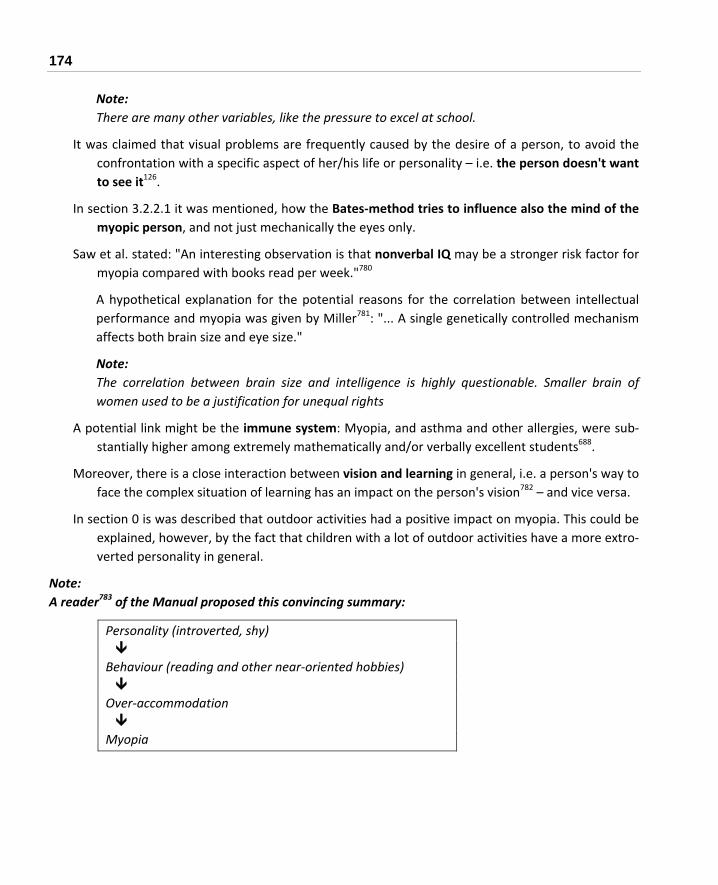
174
Note: There are many other variables, like the pressure to excel at school.
It was claimed that visual problems are frequently caused by the desire of a person, to avoid the confrontation with a specific aspect of her/his life or personality – i.e. the person doesn't want to see it126.
In section 3.2.2.1 it was mentioned, how the Bates‐method tries to influence also the mind of the myopic person, and not just mechanically the eyes only.
Saw et al. stated: "An interesting observation is that nonverbal IQ may be a stronger risk factor for myopia compared with books read per week."780
A hypothetical explanation for the potential reasons for the correlation between intellectual performance and myopia was given by Miller781: "... A single genetically controlled mechanism affects both brain size and eye size."
Note: The correlation between brain size and intelligence is highly questionable. Smaller brain of women used to be a justification for unequal rights
A potential link might be the immune system: Myopia, and asthma and other allergies, were sub‐stantially higher among extremely mathematically and/or verbally excellent students688.
Moreover, there is a close interaction between vision and learning in general, i.e. a person's way to face the complex situation of learning has an impact on the person's vision782 – and vice versa.
In section 0 is was described that outdoor activities had a positive impact on myopia. This could be explained, however, by the fact that children with a lot of outdoor activities have a more extro‐verted personality in general.
Note: A reader783 of the Manual proposed this convincing summary:
Personality (introverted, shy) Behaviour (reading and other near‐oriented hobbies) Over‐accommodation Myopia

3 MYOPIA – OBSERVATIONS AND EXPERIMENTAL RESULTS 175
3.20 Why are some People not becoming Myopic, Independent from the Circumstances?
In general, as described before, these two basic mechanisms were made responsible for the onset of myopia:
‐ Near work associated stress
‐ Degraded image quality
Potential explanations for the fact that some people do not become myopic in spite of heavy near work associated stress are:
- Their connective tissue of the sclera is very elastic. In contrast to a rather un‐elastic, i.e. more plastic elastic tissue their sclera is not returning completely to its original dimension after the stress is gone. It compares to a balloon, which still has some kind of memory effect after the air was released. Additionally, some people who are "immune" against myopia might have a mus‐cular eye‐system, which is able to realax well after muscular stress.
- They have a mindset, which enables them to get rid of any kind of stress, which includes muscu‐lar stress and tension very fast.
- They have a specific dopamine metabolism and concentration in the eye; higher dopamine lev‐els were found to work against the onset and progression of myopia, see section 3.3.3.
Notes: However, why are some people not becoming myopic in case simply a somehow degraded image quality is responsible for the onset oy myopia only? It is very unlikely that the optical properties of the eyes of the people have changed so dramatically within the last decades that it ccould explain the dramatical increase in myopia during recent times. The same is valid for the biochemical metabolism of the people.
Still, it is possible these individual properties have existed since a very long time already, and only the modern environment enabled them to show their consequences. In other words, as it will be summa‐rized in more detail in section 3.30.9, the reason that some people ar not becoming myopic can be seen just in a genetic architecture which makes them immune against the consequences of modern environ‐ment, especially against the load caused by very extensive nearwork. This, however, is different from just saying "their myopia is simply inherited".
The potential and sometimes expressed conclusion that "myopia is just genetic, nothing can be done against it" ignores the fact that there are ways to eliminate or at least reduce the harmful features and herewith the negative results and consequences of the modern environment too.

176
3.21 A Study: Factors Associated with Myopia in School Children
You et al. summarized the results of a study with Beijing school children784:
"In our population‐based study on a study population of more than 15,000 children [age 7 – 18 years] in Greater Beijing, the prevalence of myopia (≤ ‐ 1.00 diopters in the worse eye) and myopic refractive er‐ror were significantly associated with
‐ higher age,
‐female gender,
‐ key school type,
‐ higher family income,
‐ parental myopia,
‐ dim reading illumination,
‐ longer daily studying duration, [normally accompanied by long time near work]
‐ shorterduration of watching television (or computer),
‐ higher self‐reportedprotein intake,
‐ feeling well about life and status,
‐ feeling tired,
‐ less physical activity [especially outdoor activities]
‐ short time of sleeping"
Boxes indicate that these issues were more explicitly mentioned in the discussion section of this paper due to their importance.
Obviously, several of these issues are interconnected, e.g. longer daily studying duration frequently ac‐companies less physical activieties, short time of sleeping, feeling tired and feeling well about life.
Only the items female gender and parental myopia are out of control of the specific individuals.
.

3 MYOPIA – OBSERVATIONS AND EXPERIMENTAL RESULTS 177
3.22 Myopic Changes in Pregnancy
There is a tendency that during pregnancy a shift towards myopia or an increase of myopia takes place. This shift, however, is generally reversed after giving birth785 or after lactation786.
Note: Consequently, fitting of new glasses during pregnancy should be handled with extra care to avoid an overcorrection at the time after the pregnancy.
3.23 COVID-19 and Myopia
Wang J et al. reported787 about children in China: "A substantial myopic shift (approximately ‐0.3 diopters [D]) was found in the 2020 school‐based photoscreenings compared with previous years (2015‐2019) for younger children aged 6 (‐0.32 D), 7 (‐0.28 D), and 8 (‐0.29 D) years. The prevalence of myopia in the 2020 photoscreenings was higher than the highest prevalence of myopia within 2015‐2019 for children aged 6 (21.5% vs 5.7%), 7 (26.2% vs 16.2%), and 8 (37.2% vs 27.7%) years. The differences in spherical equivalent refraction and the prevalence of myo‐pia between 2020 and previous years were minimal in children aged 9 to 13 years."
Erdinest et al. reported788: "The results showed that average increase in spherical equivalent refrac‐tion and axial length, … during the year of lockdowns was ‐0.73 ± 0.46D/0.46 ± 0.31 mm re‐spectively, while the average increase in the year prior was ‐0.33 ± 0.27D/0.24 ± 0.21 mm."
Note: Potential explanations: - Reduced Time spent outdoors - More prolonged accommodation when staying at home reading or using smartphones.
3.24 Impact of Nutritional Components
This section contains published information about two issues:
nutritionally affected biochemical parameters where myopes show a difference
experiences where nutrition was found to have an impact on myopia.
The results summarized in this section are based mainly on statistics. To be fair it should be noted that interpretation of the statistics can be very difficult: does a parameter, found to occur together with myopia really have an impact on the mechanism of myopia? It might occur due to an unrelated associa‐tion only. (Remember? It can be said, e.g. that most of the children who run across the street and get hit by an automobile were wearing tennis shoes, so therefore, tennis shoes must have caused the acci‐

178
dents. Obviously, the tennis shoes were only a common association, not a related factor.) As, however, people whose eyesight is threatened by myopia cannot wait for final scientific results the advice can only be to follow any recommendation, as long as following this recommendation does not imply a new risk.
Generally, nutrition can have a positive impact on myopia either
• by improving the stability of the connective tissue, especially of the sclera, and/or by
• suppressing an exaggerated feedback which is triggered by image quality effects as outlined in section 3.3.
Yang et al. summarized789 "Recent research suggests that collagens in the sclera play an important role in the development of myopia."
Note: There is no doubt that nutrition has an impact on the health of the individual collagen.
3.24.1 Carbohydrates, Blood Sugar Level, Insulin Metabolism
Some research papers showed an interaction between myopia and glucose metabolism:
Hyperglycemia, when the blood sugar rises, as in diabetes or after meals, causes hydropic swelling of the crystalline lens, and myopia. The reason of this thickening of the lens is a decrease in the tension of the zonular fibers. Vice versa there can be farsightedness as the blood sugar falls790, 44. The refractive error in cases of diabetes can change 1 or 2 D within a few hours791.
For diabetic patients, refractive changes can go up to – 4.00 D, and changes can take several weeks. The degree of myopia after medication for diabetes was started is not somehow pro‐portional to the residual hyperglycemia, but there is a transient hyperopic change when control of the hyperglycemia is becoming effective792.
Hypoglycemia causes frequently reduced contrast sensitivity793, which can have an impact on myo‐pia as well (see section 3.3).
Glucose metabolism has an impact on the saccadic movements of the eye, which are essential for focusing483 (see section 3.6).
Overall, diabetics appear to have a higher rate of myopia794.
Accommodation is reduced in diabetic schoolchildren795.
Malondialdehyde, a breakdown product of lipid peroxidation, was found in increased levels in dia‐betes and in people with high myopia711.

3 MYOPIA – OBSERVATIONS AND EXPERIMENTAL RESULTS 179
Dielemans et al. stated511: "Newly diagnosed diabetes mellitus and high levels of blood glucose are associated with elevated IOP and high tension glaucoma."
Du et al. stated796: "Diabetes‐like glucose concentration increases superoxide production in retinal cells, and the superoxide contributes to impaired viability and increased cell death under those circumstances."
Note: Oxidative processes (see section 3.18.3) as well as retinal metabolism (see section 3.3) have been credited with causing myopia.
Lane stated258: "Young myopes statistically consume more than triple the ratio of refined carbohy‐drates to total carbohydrates, as compared to hyperopes, and this difference is most highly significant..." Moreover, the consumption of sucrose per head has risen e.g. in England from 6.6 kg in 1815 to 54.5 kg in 1979797.
Note: Maybe the effect lies not only in the high consumption of refined carbohydrates, but also to a great extent in the overall sub‐optimal nutrition, e.g. a lack of folic acid and numerous other nu‐trients.
A change in nutrition toward a focus on food rich in refined carbohydrates and sugar was blamed for the dramatic increase in myopia: diets high in refined carbohydrates are increasing blood sugar (hyperglycemia), insulin levels (hyperinsulinemia), and insulin resistance, which in turn causes the growth of the eyeball798, 799.
The explanations, which were given by Cordain et al.798:
‐ Elevation of insulin increases the levels of the insulin growth factor‐1 (IGF‐1), "a potent stimu‐lator of growth in all tissues".
‐ "Reduced levels of the insulin like growth factor binding protein‐1 (IGFBP‐1) may reduce the effectiveness of the body's natural retinoids in activating genes that would normally limit scleral cell proliferation" (for the role of retinoic acid on myopia see section 3.3.1).
For example it is explained800 that in pacific islands where people kept to the original diet of fish, yam and coconut no increase in myopia was observed, in spite of long schooling hours of the children. In contrast, a myopia rate of over 18% was reported for Hong Kong fishermen, who had never attended school801. The rate was even higher for those with schooling.
As another evidence for this insulin‐argument it is noted by Cordain et al.798 that "population studies have demonstrated that people of Asian and Chinese descent tend to be more insulin resistant (which, however, often increases to some degree the production of insulin) than peo‐ple of European descent" – and the prevalence of myopia is especially high in Asia.
180
Glucose level has an impact on the functioning of the blood‐retinal barrier, which is related to my‐opia (see section 3.18.5).
In experiments with mice hyperglycemia combined with a lack of insulin leaded to a significantly increased number of mast cells in the sclera802.
Note: An increased number of mast cells should result in an enhanced activity of the immune system, which can reduce the stability of the sclera. For information about myopia and the immune sys‐tem see section 3.18.1.
Flickering light can protect against artificial myopia (see section 3.14.5), most likely by retinal vessel dilation. High blood glucose significantly reduces this flicker induced retinal vessel dilation651.
Wu reported803: "Incidence of moderate‐high myopia was also related to baseline age, nuclear opacities, glaucoma, male gender and diabetes history."
Feldkaemper et al. found that insulin can have rather complex effects804: "Given that insulin is used in the therapy of human metabolic disorders and proposed as an agent to treat corneal epithe‐lial disease, its powerful myopigenic effect, which is mostly due to its effects on the optics of the anterior segment of the eye, merits further investigation."
Penha AM et al. reported805: "Insulin injections induced significant amounts or relative myopia in plus lens treated eyes and further enhanced myopia development during minus lens treat‐ment."
Note: Can already a diet, which is excessively rich in carbohydrates and which therefore, causes the insulin level to increase, increase the probability of the onset or progression of myopia?
Sheng et al. reported806: "In chick eyes, exogenous insulin prevents the choroidal thickening caused by wearing positive lenses and increases ocular elongation and scleral glycosaminoglycan (GAG) synthesis, an indicator of eye growth."
C Berticat et al. reported807: "The consumption of refined carbohydrates significantly increased the probability of myopia for girls (odds ratio [OR] = 1.07; 95% confidence interval [CI], 1.02–1.13) but decreased it for boys (OR = 0.94; 95% CI, 0.89–0.98)."
A number of nutritional components are important for the avoiding of hyperglycemia. They were found to be related with myopia as well: Manganese, Chromium, vitamin D and flavonoids (see section 3.24).
Moreover, hyperglycemia influences biochemical issues, which were found to be related with myopia as well: NO level, cytokine production, oxidative stress, and microcirculation.
This might support the thesis that hyperglycemia can play a role in the development of myopia.

3 MYOPIA – OBSERVATIONS AND EXPERIMENTAL RESULTS 181
Moreover, it was found that a disturbed blood sugar metabolism has an impact on the pulsatile ocular blood flow808. The blood flow appears to be connected with myopia (see section 3.17).
The excessive increase in the consumption of soft drinks can be expected to contribute significantly to hyperglycemia.
Notes: ‐ Hyperglycemia (i.e. elevated blood sugar level) can be caused not only by nutrition (and inheritance),
but also by stress and a lacks of exercise, etc. It is not restricted to people with type I diabetes (who are permanently depending on insulin), and older people with type II diabetes, but can occur tempo‐rarily independent of age, i.e. also in children, where the development and progression of myopia is most critical.
‐ Additionally, it was emphasized that it is very important to keep a diet that avoids steep increases in blood sugar level, i.e. to have a "low‐glycaemic index‐low‐fat‐high‐protein" diet809, 810. ‐ Very fre‐quently these steep increases in blood sugar (hyperglycemia) later cause significantly lowered levels of blood sugar (hypoglycemia).
‐ The findings summarized above do not mean that many myopes are diabetic, or that diabetes is a frequent reason for myopia. They mean, however that carbohydrate and insulin metabolism might very well have an impact on the development of myopia.
‐ In contrast to the report about low rates of myopia in the pacific islands, a very substantial increase in myopia was observed for Eskimos (see section 3.1), and obviously, their dietary habits had been changed significantly.
‐ Maybe another issue of the changed nutrition is the changed sodium/potassium ratio.
3.24.2 Is there a Connection between the Blood Sugar Level and Negative-Lens-Induced Myopia?
In section 3.24.1 it was described that an elevated blood sugar level can induce at least transient myo‐pia. In section 3.3 it was described that inappropriate, additional negative lenses can cause permanent axial myopia.
Note: Personal suspicion: If an eye becomes temporarily myopic due to elevated blood sugar level, and if therefore negative lenses are prescribed, these lenses are inappropriate during phases with normal blood sugar level. They might cause in this interval lens induced myopia, resulting in an elongated eye‐ball, i.e. axial myopia.
If these cycles are sufficiently long, and are repeated, a permanent progression to myopia might follow.

182
Additionally, the hyperglycemic swelling of the lens might change the mechanical properties of the lens, contributing to an accommodative hysteresis (see section 1.3.2.4) and the aftereffect of accom‐modation / the accommodative lag (see section 3.2).
3.24.3 Calcium, Vitamin D and Sunlight
Calcium, magnesium and vitamin D have strong interactions. Because of this, they are described to‐gether.
Tideman JW et al. stated811: "Lower 25(OH)D concentration in serum [an indication of a lack of vi‐tamin D] was associated with longer AL [axial length] and a higher risk of myopia in these young children. This effect appeared independent of outdoor exposure and may suggest a more di‐rect role for 25(OH)D in myopia pathogenesis" … " The relationships between 25(OH)D and AL or myopia were investigated using linear and logistic regression analysis. Average 25(OH)D con‐centration was 68.8 nmol/L (SD ± 27.5; range 4‐211); aver‐age AL 22.35 mm (SD ± 0.7; range 19.2‐25.3); and prevalence of myopia 2.3 % (n = 62). After adjustment for covariates, 25(OH)D concentration (per 25 nmol/L) was inversely associated with AL (β ‐0.043; P < 0.01), and after additional adjusting for time spent outdoors (β ‐0.038; P < 0.01)." " This effect appeared inde‐pendent of outdoor exposure and may suggest a more direct role for 25(OH)D in myopia pathogenesis."
An evaluation of statistical data for reported blindness due to malignant myopia in different states of the USA was done by compiling a chart with the rate of myopia per state, and distance to seacoast, annual hours of sunshine and the nutritional concentration of calcium, fluoride and selenium in each state. One result was an inverse correlation between malignant myopia and calcium content in the water as well as annual hours of sunshine96. Annual hours of sunshine can serve as a measure of vitamin D supply.
Note: If there are more hours of sunshine it is more likely that more time is spent outdorrs, which was found to be positive (see section 3.8).
Starting in the 1930s Knapp, an American ophthalmologist was the first author to describe the im‐pact of calcium and vitamin D according to his experiences in his ophthalmologic praxis812:
‐ "In the course of being fed vitamin D deficient, low calcium diets, the eyes of dogs and rats revealed clinical myopia."
‐ "Further research was conducted on humans, not patients with the usual slow progressive short sight, but painstakingly selected, highly progressive myopes from many thousands of pa‐tients. They were fed supplements of vitamin D and calcium. Over 50% of this rapidly progres‐

3 MYOPIA – OBSERVATIONS AND EXPERIMENTAL RESULTS 183
sive group, whose myopia would reasonably be expected to progress rapidly, instead showed a decrease in their myopia, or remained stationary. One third of the series registered a reduction in myopia."
A publication of an ophthalmologist was getting high attention, because he claimed that by intake of animal protein progression of myopia in kids was reduced to only one third as before813. Later analysis revealed, however that what he accounted as the benefit of protein was the benefit of the calcium in the calcium caseinate he used 258.
Note: Another possible explanation of this result is the fact that casein increases cNOS activity (see section 3.18.7 about the effect of NO on myopia.
Mutti summarized their research814: " myopes appear to have lower average blood levels of vitamin D than non‐myopes."
Experimentally induced myopia in animals was severely increased if a calcium deficient diet was used. Surprisingly, this was not the case with a vitamin D deficient diet. Overall, however, Hodos concludes665, "… variables that effect eye growth include nutritional deficiencies of cal‐cium and vitamin D."
The highest concentration of calcium in the hair was measured in cases of increasing myopia, and calcium concentration was highest for myopes and lowest for emmetropes258. The literature about hair analysis states that elevated calcium levels are indicating not an oversupply with calcium but an accelerated calcium turnover in the body, caused e.g. by chronic stress, allergies or chronic infections815.
Preterm children are frequently myopic. Children with very low birth weight received supplementa‐tion with calcium and phosphorus – in the unsupplemented group 61% became myopic, in the group with supplements only 11% became myopic816.
During the summertime, the time of school holidays, the progression rate of myopia is significantly lower150, 151, 152. Part of the explanation may be a higher exposure to the sun, which means more supply of vitamin D, and more physical activities.
The positive effect of a medication containing "Nacre" (see section 3.25) was attributed to an in‐creased content of calcium in various tissues of the eye734.
The great increase in myopia found for Eskimos129 may be related to nutritional changes, e.g. a re‐duction in fat‐soluble vitamins (i.e. vitamins A and D) by a factor of 10817. Other possible factors are the reduced time spent outdoors and the increased amount of near work connected with schooling.
There is a relation of myopia and caries of the teeth689. There is no doubt that caries is (among other reasons) related to calcium metabolism.

184
A connection between the "exposure to natural light [which has an impact on the vitamin D me‐tabolism] during the early perinatal period" was drawn by Mandel818: "There were seasonal variations in moderate and severe myopia according to birth month, with prevalence highest for June/July births and lowest for December/January ... Mild myopia was not associated with season of birth or perinatal light exposure."
Prepas published the hypothesis819: "Close focusing in the absence of UV light [i.e. in the presence of artificial light] may provoke axial myopia."
Mutti DO et al. summarized their results820: "The hypothesis that time outdoors might create dif‐ferences in vitamin D could not be evaluated fully because time outdoors was not significantly related to myopia in this small sample. However, adjusted for differences in the intake of die‐tary variables, myopes appear to have lower average blood levels of vitamin D than non‐myopes."
Calcitriol, the active metabolite of vitamin D, appears to increase the level of dopamine821. Dopa‐mine was found to play an important role in the onset of myopia, see section 3.3.3. Maybe the positive effect of sunlight and vitamin D is based on the positive effect on dopamine.
Choi JA reported822: "Low serum 25(OH)D concentration was associated with myopia prevalence in Korean adolescents [aged 13‐18 years]. This relationship was particularly notable in adoles‐cents with high myopia."
Similar results were reported from Western Australia as Yazar et al reported823: "… lower serum 25(OH)D3 concentration was associated with higher risk of having myopia (odds ratio [OR] for <50 vs. >50 nmol/L".
J McMillan reported824 a significant improvement of astigmatism und keratoconus by vitamin D supplementation.
Jung BJ et al. reported825: "Linear regression analysis showed that as 25(OH) D level increased by 1 ng/mL, myopic refractive error significantly decreased by 0.01 D (P < 0.001) after adjusting for potential confounders including sex, age, height, education level, economic status, physical ac‐tivity, and sunlight exposure time."
Myopia and skin cancer are inversely correlated826.
Willem J et al. reported827: "After adjustment for covariates, 25(OH)D concentration (per 25 nmol/L) was inversely associated with AL [axial length], and after additional adjusting for time spent outdoors. Risk of myopia (per 25 nmol/L) was OR 0.65 (95 % CI 0.46–0.92). Lower 25(OH)D concentration in serum was associated with longer AL and a higher risk of myopia in these young children. This effect appeared independent of outdoor exposure and may suggest a more direct role for 25(OH)D in myopia pathogenesis."

3 MYOPIA – OBSERVATIONS AND EXPERIMENTAL RESULTS 185
Gao et al. reported for Chinese children828: "Logistic regression analysis showed that after control‐ling for gender, parental myopia, after‐school class, and outdoor activities, the prevalence of developing moderate and high myopia was 2.051 times (95% confidence interval: 1.272‐3.724) higher in the serum 25(OH)D deficiency group than in the serum 25(OH)D sufficiency group."
However, A review published in The Journal of the American Osteopathic Association found Vitamin D can't be metabolized without sufficient magnesium levels, meaning Vitamin D remains stored and in‐active for as many as 50 percent of Americans838.
3.24.4 Vitamin D and Dopamine
Cakir et al. stated829 :Vitamin D is known to increase levels of dopamine and its metabolites in the brain.
Trinko et al. stated830: "… data demonstrate that dopamine circuits are modulated by D3 signaling, and may serve as direct or indirect targets for exogenous calcitriol."
Kesby et al. discussed in an extensive paper specific implications of vitamin D status on the neuro‐transmitter dopamine831 .
The importance of dopamine on myopia is discussed in section 3.3.3 and section 3.14.2.
3.24.5 Selenium and Dopamine
Dopamine turnover was increased when rats were fed selenium deficient diet. The decrease in brain antioxidant protection caused by a nutritional deficit of selenium and a following decrease in glu‐tathione peroxidase activity was made responsible for this effect.832
The key role which dopamine plays is described in section 3.3.3
3.24.6 Magnesium
Due to an inherited disorder in the magnesium metabolism hypomagnesemia (i.e. a lack of magne‐sium), combined with hypercalciuria (i.e. increases excretion of calcium in the urine) can occur. Often this defect is accompanied by myopia and nystagmus833, 834, 835.
It was concluded that the Marfan syndrome is related to metabolic magnesium deficiency836 and to ex‐cessive hyaluronic acid synthetase837. People with Marfan syndrome are showing, among others, an ex‐treme flexibility and mobility of their joints.

186
A review published in The Journal of the American Osteopathic Association found Vitamin D can't be metabolized without sufficient magnesium levels, meaning Vitamin D remains stored and inactive for as many as 50 percent of Americans.838
There is an old report by Algan839 titeled "The treatment of progressive malignant myopia with magne‐sium chelates of flavones. Apropos of 400 cases" (no details are available).
Notes: In general, magnesium is claimed to have a relaxing effect on smooth muscles, and therefore it might prevent or reduce accommodation cramps of the smooth ciliary muscle.
Furthermore magnesium has a positive effect on inflammation, and low magnesium intake is linked to chronic inflammation (about myopia and inflammation see section 3.18.2).
3.24.7 Copper and Zinc
Some results about the correlation between copper and zinc metabolism and myopia are:
Vinetskaia et al. stated840: "Progressive myopia was found associated with reduced copper content [in the tears] and changed ratio zinc to iron, this indicating certain metabolic disorders in the connective tissue system, in the scleral membrane first of all, and in the antioxidant defense system."
Qiang et al. stated162: "... there was a close relationship between juvenile myopia and ... zinc/copper ratio in hair ..."
For myopes above – 6.0 D a statistically significantly decreased concentration of copper in the se‐rum was measured841
Note: A reduced content of copper in the serum can be observed only for an already substantial lack of copper842.
Avetisov et al. stated843: "Copper measurements in scleral tissue of 14 cadaveric emmetropic eyes and in 10 eyes with myopia of various degrees have shown a significant reduction of copper levels in the equatorial and posterior segments of myopic sclera and abnormal distribution of copper in the tissue, this indicating disordered metabolism of this trace element."
Patients with progressive myopia received injections with a copper‐pyridoxine (pyridoxine is vita‐min B6) compound near to the sclera to stimulate the formation of collagen and improve the cross‐linking of connective tissue fibers. After 3 year the refraction was stable for 64% of the patients844.
3 MYOPIA – OBSERVATIONS AND EXPERIMENTAL RESULTS 187
Patients with low levels of diet‐responsive copper‐zinc superoxide dismutase (CuZnSOD, an anti‐oxidant enzyme) had a very substantially increased risk to develop structural defects in the vit‐reous body (vitreous floaters, which are common for myopes) than patients with a high level of CuZnSOD, 152.
Eye drops containing zinc were inhibiting experimentally induced myopia in animals. It was con‐cluded that this positive effect is due to a measured increased activity of superoxide dismutase (SOD), which prevents excessive processes of oxidation, and of nitric oxide (NO) and nitric ox‐ide synthase (NOS)845.
An Indian mixture containing "zinc, ascorbic acid and micronutrients" was taken in capsules, and was reported to improve the myopia in certain cases846.
The Chinese myopia medication "Nacre" (see section 3.25) is containing zinc, copper and numerous trace elements, which results in increased SOD, NOS and NO activity734.
Note: Due to the numerous ingredients the principles of its efficiency are hard to judge.
A Zinc compound (the Indian medicine "Yashad Bhasma") was evaluated. A two months use pro‐duced improvements in myopia. The results were, however, statistically not significant847.
Fedor M et al. reported848: "The average serum concentration of zinc in myopic patients was sig‐nificantly lower (0.865 ± 0.221 mg L‐1) in comparison to the control group (1.054 ± 0.174 mg L‐1). There was significantly higher Cu/Zn ratio in myopic patients (1.196 ± 0.452) in compari‐son to that in the control group (0.992 ± 0.203)."
Zinc has a substantial impact on the dopamine metabolism966. About myopia and dopamine see section 3.25.4
Fedor M et al. reported876: "The average serum concentration of zinc in myopic patients was sig‐nificantly lower (0.865 ± 0.221 mg L‐1) in comparison to the control group (1.054 ± 0.174 mg L‐1). There was significantly higher Cu/Zn ratio in myopic patients (1.196 ± 0.452) in comparison to that in the control group (0.992 ± 0.203). … Low serum concentration of zinc and selenium in myopic children may imply an association between insufficiency of these antioxidant mi‐croelements and the development of the myopia and could be the indication for zinc and se‐lenium supplementation in the prevention of myopia. Significantly, higher Cu/Zn ratio in the study group can suggest the relationship between myopia and oxidative stress."

188
3.24.8 Chromium
Some results about the correlation between chromium metabolism and myopia are:
Hair analysis was showing substantially decreased chromium levels for myopes, and especially for progressing myopia258. This was explained by a higher intake of refined carbohydrates, e.g. sugar. Sugar is said to deplete body stores of chromium. Matching with this is a result showing that myopes consumed only 39% as much fiber as hyperopes (i.e. farsighted people), which is interpreted as an increased consumption of refined food by myopes. Tissue chromium concen‐tration was found to be negatively correlated with intraocular pressure (IOP) also (see section 3.7.2 on the impact of the IOP on myopia).
The explanation given by Lane258 is that "... the importance of chromium in visual function is probably due to its role as the potentiator of insulin at the insulin‐receptor sites in blood ves‐sels supplying the ciliary muscle."
For the directly myopia related results of the impact of carbohydrates and insulin metabolism see section 3.24.1.
The ratio of chromium to vanadium in erythrocytes is depressed for myopes.849
The structure of the retinal pigment epithelium showed more damage for Chromium deficient rats850.
Chromium deficiency showed abnormal reactions of the immune system in the retina of rats851.
In myopic eyes of chicks a significant decrease of chromium in the aqueous humor was found734.
3.24.9 Manganese
Persons with high hair manganese concentrations were found to show increasing hyperopia or de‐creasing myopia258.
A deficiency in manganese results in a loss of photoreceptor cells and capillary anomalies in the ret‐ina852.
3.24.10 Potassium
The concentration of potassium in the hair of myopes was found to be very significantly lower than in emmetropes or hypertropes258 (highest concentration at hypertropes).

3 MYOPIA – OBSERVATIONS AND EXPERIMENTAL RESULTS 189
Wu S reported853: "we found that the arrangements of ciliary muscles in LIM [lens induced myopia] guinea pigs were broken, dissolved or disorganized …. Monitoring of K [potassium] flux in ciliary mus‐cles from LIM guinea pigs demonstrated myopia‐triggered K influx. … Overall, our results will facilitate the understanding of the mechanism associated with inhibitory Na /K ‐ATPase [adenosine triphos‐phate] in lens induced myopia and which consequently lead to the disorder of microenvironment within ciliary muscles from LIM guinea pigs, paving the way for a promising adjuvant approach in treat‐ing myopia in clinical practice."
3.24.11 Fluoride
A source in the Internet reported854: "Higher incidences of myopia as a result of excessive fluoride in‐take during childhood have been reported in India (Raja Reddy, 2000) as well as in Sosnivka, Ukraine (Miroshnychenko, 2000). It was found that myopia was much more prevalent in girls with dental fluoro‐sis than boys, worsening at the ages of 10 to 11 and 12 to 13. (Miroshnychenko, 2000)."
Most likely, this negative effect on myopia is caused by defects of the connective tissue, induced by ex‐cessive doses of fluoride855.
3.24.12 Salt
Li Q et al. reported856: "The results revealed that the high‐salt diet aggravated ischaemia/reperfusion‐induced thinning of the retina. … retinal ischaemia/reperfusion mainly results in neuronal degeneration, including thinning of the retina, retinal ganglion cell death. … a high‐salt diet aggravates ischae‐mia/reperfusion‐induced retinal neuronal impairment by activating pro‐apoptotic and pro‐inflammatory signalling pathways and inhibiting vasodilation."
Inhibiting vasodilation has an impact on blood circulation, see section 3.17
3.24.13 Fat and Cholesterol
Lim LS et al. summarized their results857: "Higher saturated fat and cholesterol intake are associated with longer AL [axial Length] in otherwise healthy Singapore Chinese schoolchildren."
In section 3.3.3 the im pact of dopamine on myopia is discussed, and SO Ahmad858 reported about experiments with rats "… findings support a role for n‐3 PUFAs [n‐3 polyunsaturated fatty acid] neurons and suggest that altered dopamine cell number, as well as function, behavioral effects observed in rats raised on n‐3 PUFA‐deficient diets.
Pan et al. reported859: "We demonstrated that daily gavage of ω‐3 PUFAs (300 mg docosahexaenoic acid [DHA] plus 60 mg eicosapentaenoic acid [EPA]) significantly attenuated the development

190
of form deprivation myopia in guinea pigs and mice, as well as of lensinduced myopia in guinea pigs."
3.24.14 Vitamin A
In section 3.3.1 it was stated that retinoic acid is significantly involved in experimentally induced myo‐pia (less all‐trans‐retinoic acid synthesis when myopia is built up). Whether vitamin A, the precursor of all‐trans‐retinoic acid, has any impact on this mechanism is unknown.
Besides this vitamin A (and zinc) are essential for night vision.
3.24.15 B-Vitamins
Some results about the interaction between B‐vitamins and myopia are:
As reported in section 3.24.3 myopia is often accompanied by a disturbed calcium metabolism, measured by elevated calcium levels in the hair. Vitamin B6 (pyridoxine) is said to be able to correct this disturbed calcium metabolism860.
Vitamin B6 and B3 (pantothenic acid) are said to be helpful in regulation of intraocular pressure (IOP)162.
There was a correlation found between deficiencies of vitamin B1, B2 and B6, nystagmus, and dis‐turbed eye tracking861, 862. Nystagmus is an uncontrolled, often rhythmical movement of the eyeball, and often accompanies myopia.
According to the title of a Japanese publication, juvenile myopia was treated with massive doses of vitamin B1863.
Peroxide oxidation of lipids is intensified in progressive, complicated myopia. Also, antioxidant de‐fense parameters in the tear fluid are decreased, resulting in structural and pathological changes in the retina. Success was achieved by applying locally an antioxidant medication con‐taining vitamin B6 (and emoxypin, a structurally analogue of vitamin B6) via a film, which was placed behind the lower eyelid every evening for 15 days864, 713.
A lack of vitamin B6 is connected with an increase in the level of homocysteine, which can contrib‐ute to myopia. Homocysteine is discussed in section 3.24.17.

3 MYOPIA – OBSERVATIONS AND EXPERIMENTAL RESULTS 191
3.24.15.1 Riboflavin plus UVA [blue light]
Wollensak reported865: "Collagen crosslinking induced by riboflavin‐UVA [i.e. vitamin B2 and ultra‐violet A irradiation], glyceraldehyde, and glutaraldehyde led to a significant increase in bio‐mechanical strength in human and porcine sclera. Using these methods, collagen crosslinking might become a treatment possibility for progressive myopia."
M Sun et al. tested crosslinking induced by riboflavin‐UVA866: " The biomechanical stability of sclera was increased. The scleral stress and Young modulus at 8% strain corresponded to 184% and 183%, respectively, of the control values at 12 months. No retinal damage was detected on histology."
As a consequence, the combination of vitamin B2 (riboflavin) and ultraviolet A light was sug‐gested for the treatment of progressive myopia, because this treatment improved the cross‐linking of scleral collagen very significantly in experiments with animals867, 868. It was said, how‐ever, that further testing is necessary before its clinical application.
Li X summarized experiments with Guinea pigs869: "Guinea pigs underwent 30 min of whole‐body UVA irradiation after each gavage for 2weeks. For control groups, guinea pigs were adminis‐tered vitamin C and underwent either whole‐body UVA irradiation without 0.1% riboflavin so‐lution or whole‐body fluorescent lamp irradiation with or without0.1% riboflavin solution. Re‐sultantly, myopia models were established with an increased axial length and myopic diopter. Compared with myopic eyes in the control groups, the net increase in axial length, dioptre and strain assessment decreased significantly, and the net decrease in sclera thickness, ulti‐mate load, and stress assessment decreased significantly in experimental groups."
Krasselt et al870 concluded from experiments with rabbits: "… gentle cross‐linking with riboflavin and UV‐A or blue light might be a clinical approach in future."
Lai et al. confirmed871 by experiments with Guinea pigs the ability of UVA light and riboflavin to in‐hibit lens induced myopia, but the inhibitory effect was reduced after stopping the treatment.
Note: The increase of indoor activities and the resulting decrease of exposure to ultraviolet irradiation may play, together with a changed diet, a substantial role in the increase in myopia.
3.24.16 Some More Antioxidants
3.24.16.1 Selenium
Some results about the correlation between selenium metabolism and myopia are:

192
An evaluation of statistical data for reported blindness due to malignant myopia in different states of the USA was done by compiling a chart with the rate of myopia per state, and distance to seacoast, annual hours of sunshine and the nutritional concentration of calcium, fluoride and selenium in each state. As a result among others an inverse correlation between malignant myopia and selenium was found 96.
Rats fed a diet higher in selenium (200 μg/kg diet) had fewer cellular degenerating capillaries in the retina and a higher central choroid than those fed a diet of 100 μg/kg. Both levels of sele‐nium, however, resulted in a normal activity of erythrocyte glutathione peroxidase, which is considered to be a marker for selenium sufficiency872.
Notes: - Retinal defects are closely related to higher grades of myopia (see sections 1.7 and 3.18.5).
- A reduced thickness of the choroid accompanies experimental myopia (see section 3.3.1).
- Statements of sufficient selenium in the diet may be wrong.
The concentration of selenium in the retina of animals with excellent vision is up to 100 times as high as in the retina of animals with weak vision873.
Selenium deficient diets reduced the amacrine cells and the photoreceptor cells in the retina of rats and mice851, 874. Amacrine cells were found to be important in deprivation experiments, where they were found to be involved in the dopamine signaling (see sections 3.3 and 3.24.4).
The expression of a selenoprotein in the retina of chickens was found to be increased after image degradation. This increase, however, took place after applying positive lenses (hyperopic defo‐cus) as well as negative lenses (myopic defocus)875.
Fedor M et al. reported876: " The average serum concentration of selenium in the study group was sig‐nificantly lower (40.23 ± 12.07 μg L‐1) compared with that in the control group (46.00 ± 12.25 μg L‐1). … Low serum concentration of zinc and selenium in myopic children may imply an association between insufficiency of these antioxidant microelements and the development of the myopia and could be the indication for zinc and selenium supplementation in the prevention of myopia."
3.24.16.2 Flavonoids and Related Compounds, and Vitamin E
This family contains over 4000 different compounds. Some results of their effect are:
Orally to chicks administered black‐currant significantly inhibited the onset of myopia which is normally caused by the application of negative lenses877.
It was claimed that cyaninoside chloride is effective to treat progressive myopia by protecting col‐lagen against enzymatic attack by collagenase, showing antioxidant activity878, 879.

3 MYOPIA – OBSERVATIONS AND EXPERIMENTAL RESULTS 193
The magnesium chelate of flavones named Flacitran was found in long‐term treatments to have a positive effect on complications of malignant myopia. This was explained by its ability to hinder the buildup of degrading enzymes collagenase, elastase, hyaluronidase, and of histamines880. Collagen, elastase and hyaluronic acid are components of the connective tissues, and the end‐ing "ase" indicates the degrading property. Histamines are mediators of inflammations.
A medication containing anthocyanosides (e.g. bilberries) plus vitamin E had positive influence on progressive myopia and its consequences881. Similarly, diabetic retinopathies (which are caused by damages in capillary vessels) were successfully treated with anthocyanosides882. And there are numerous commercial claims that anthocyanosides from bilberries are helpful to treat myopia.
A medication containing troxerutin plus vitamin E had a positive influence on the progression of myopia883. The effect was claimed to be based on the antioxidant anti‐hyaluronidase properties of vitamin E, and the anti‐hyaluronidase and anti‐histamine properties of troxerutin.
Bhutto et al. stated884: "...findings indicate that the decrease in retinal capillaries in vitamin E–deficient rats is secondary to retinal degeneration."
Vitamin E can inhibit the hyperglycemia induced elevated production of superoxide in the ret‐ina796.
A rutin compound improved the retinal microcirculation885. For the relevance of microcirculation to myopia see section 3.17.
3.24.17 Folic Acid and Homocysteine
3.24.17.1 Observations
Some results about the interaction between homocysteine‐ and folic acid‐metabolism and myopia are:
A strong correlation of food‐folate intake with a reversal of myopia, and with some reduction of the intraocular pressure (IOP) was found. Supplementation with the pharmaceutical form ap‐pears to be less effective than intake of food‐folate512.
Note: The positive effect could be explained not only by the positive effect of folate, but also with food ingredients which are can frequently be found in food which is rich in folate, e.g. flavonoid.

194
Numerous publications report high and very high myopia (among other symptoms) for people suf‐fering from homocystinuria, an inherited defect that leads to abnormal high levels of the amino acid homocysteine in the serum886, 887, 888, 889.
The concentration of the amino acid methionine was 10‐fold increased for people with extreme myopia890.
Mulvihill et al. stated891: "Young persons with marked and progressive myopia … should be screened for homocystinuria."
Marcucci et al. stated892: "...in animal models homocysteine is able to activate metalloproteinases." About metalloproteinases, agents that can degrade connective tissue, Jones et al. stated352: "Scleral matrix metalloproteinases, serine proteinase activity and hydrational capacity are in‐creased in myopia induced by retinal image degradation." In other words, an elevated level of homocysteine can have similar biochemical effects as retinal image degradation.
3.24.17.2 An Explanation
A very short explanation of the biochemistry that links the referenced observations893, 894, 895:
Homocysteine is built from the other amino acid methionine by the metabolism as an intermediate product: Normally homocysteine is quickly recycled to methionine or finally transformed to cysteine. For these transformations especially folic acid, vitamin B6 and vitamin B12 are needed. A missing transformation can have genetic reasons, or a lack of appropriate quantities of folic acid, vitamin B6 and vitamin B12.
Elevated levels of homocysteine, which is a powerful oxidant, can be the source of various seri‐ous health problems, including degraded cross‐linking of collagen.
A high intake of proteins, especially of meat, contains a high quantity of methionine and increases therefore the demand especially of folic acid and of vitamin B6.

3 MYOPIA – OBSERVATIONS AND EXPERIMENTAL RESULTS 195
3.24.18 Bilberry and Anthocyanins
IHN Omar et al.896 investigated the effect of bilberry on the progression of high myopia (about –10 D) in children with an age of 9 years. Bilberry extract was given for 1 year, and then stopped for the 2. year.
Bilberry group Control group
Difference in refraction after 12 month 0.12 D 0.39 D
Difference in axial length after 12 months 0.26 1.03
Difference in refraction between month 12 and 24 0.1 D 0.3 D
Difference in axial length between month 12 and 24 0.2 0.3
Obviously, the positive effect remained still after the break of 12 months.
Kamiya reported897 significantly increased amplitude of accommodation by taking fermeted bilberry ex‐tract.
Y Nomi et al. reported898: "In vitro study, AC [anthocyanin] had a relaxing effect on ciliary muscle which is important to treat both myopia and glaucoma. And AC stimulate the regeneration of rhodopsin in frog rod outer segment. Furthermore, AC could inhibit the axial length and ocular length elonga‐tion in a negative lens‐induced chick myopia model. In addition, we summarized clinical studies of AC intake improved dark adaptation and transient myopic shift and the improvement on retinal blood circulation in normal tension glaucoma patients."
About blood circulation and myopia see section 3.17
Plants rich in anthocyanins are such as blueberry, cranberry, bilberry, black raspberry, red raspberry, and blackberry, blackcurrant, cherry, eggplant (aubergine) peel, black rice, ube, Okinawan sweet potato, concord grape, muscadine grape, red cabbage, and violet petals.
3.24.19 Lactoferrin
Ikeda et al. reported899 results about oral administration of lactoferrin (an iron‐binding protein pre‐sent in saliva, tears, and mother's milk) on mice with lens induced myopia (LIM): "The eyes with a minus lens showed a refractive error shift and an axial length elongation in the control group, thus indicating the successful induction of myopia. However, there were no significant differ‐ences in the aforementioned parameters in the LF group. While LIM increased IL‐6 expression and MMP‐2 activity, it decreased collagen 1A1 content. However, orally administered LF re‐versed these effects. Thus, oral administration of LF suppressed lens‐induced myopia devel‐

196
opment by modifying the extracellular matrix remodeling through the IL‐6‐MMP‐2 axis in mice."
Kuruyleva et al. reported900: " Concentration ratio of lactoferrin and lysozyme in TF ([Ltf]/[Lys]) was evaluated as the ratio of protein band intensities. … [Ltf]/[Lys] ratio was greater in moderate myopia than in high myopia: 0.88 [0.75; 1.01] and 0.6723 [0.60; 0.78], respectively. Em‐metropes demonstrated the highest [Ltf]/[Lys] ratio ‐ 1.59 [1.43; 1.98]." Note: Lactoferrin is a widely available supplement and is supposed to help the immune system, pre‐vent inflammation and support iron absorption.
Lepanto et al. reported901: "Since the first anti‐microbial function attributed to Lf [lactoferrin], sev‐eral activities have been discovered, including the relevant anti‐inflammatory one, especially associated to the down‐regulation of pro‐inflammatory cytokines, as IL‐6. … Lf administration reduces local and/or systemic inflammation."
3.24.20 Crocetin
Crocetin is an agent derived from saffron.
Mori et al. reported902: "The change in SER [spherical equivalent refractions] in the placebo group, ‐0.41 ± 0.05 D (mean ± standard deviation), was significantly more myopic compared to that in the cro‐cetin group, ‐0.33 ± 0.05 D (p = 0.049). The AL elongation in the placebo group, 0.21 ± 0.02 mm, was significantly bigger than that in the crocetin group, 0.18 ± 0.02 mm (p = 0.046). In conclusion, dietary crocetin may have a suppressive effect on myopia progression in children …"
3.24.21 Other Components of the Diet
Some results about the impact of a few other nutrients on myopia are:
Lane stated860: "Young myopes who are not increasing in myopia have a significantly lowered distribu‐tion of flesh protein intake compared to the distribution for young persons whose eyes are getting rela‐tively more myopic..."
Note: The more indirect reason might be that a diet more rich in meat would require higher amounts in other components of the diet (like vitamin B6 to keep homocysteine levels down, see section 3.24.17). Similar indirect nutritional effects appear to be very common.

3 MYOPIA – OBSERVATIONS AND EXPERIMENTAL RESULTS 197
It was claimed that glutamate, which is used extensively in the food industry is responsible for numer‐ous health problems, including blurred vision and myopia903. In section 3.18.9 is referenced: "For people with extreme myopia the concentration of the amino acid glutamate was 10‐fold increased."890
3.24.22 Overall Nutritional Status and Myopia
So far nutritional components were mentioned, whose specific lack was found to be correlated with myopia. On the other hand, myopia exists primarily in developed countries, where under‐nutrition is no problem at all.
Some statistics:
Josephson stated904: "... noted that the percent of schoolchildren with myopia rose and fell with the severity of the depression, and linked that to nutrition: In 1925, it was reported that 25% of the schoolchildren attending a group of clinics in New York were afflicted with near‐sightedness. With the advent of the depression, the figure rose steadily from over 40% in 1932 and to 72% in 1935. Reflecting re‐employment and improved nutrition in 1936, the percentage incidence of near‐sightedness dropped to about 51%. In 1937, the figure dropped to 42%."
Edwards et al. stated905: "Children who developed myopia had a generally lower intake of many of the food components than children who did not become myopic. The differences were statisti‐cally significant for energy intake, protein, fat, vitamins B1, B2, and C, phosphorus, iron and cholesterol. Despite these differences, children who became myopic were neither shorter nor lighter..."
Note: When children become myopic the nutrition‐based conclusion does not have to be that they are getting less of specific nutrients. More likely, their individual demand for specific nutrients is higher.
Some explanations:
The lack of specific nutritional components shows its effect on myopia only when it is connected with extended near work, i.e. in countries with a high rate of literacy. This high rate of literacy is often correlated with a lot of indoor‐time, i.e. a potential lack of vitamin D (sunlight) and of physical activities.
In developed countries the content of the individual components of the nutrition has been changed by far more than in countries where under‐nourishing is still a problem, but where the tradi‐tional food is still the same as long ago.
Moreover, the critical issue might be not the absolute content of a specific nutrient, but the balance between various nutrients (see section 3.24).
198
Spanheimer et al. stated906: "Malnutrition is associated with defects in connective tissue metabo‐lism..."
3.25 Pharmaceuticals
Wang et al.907 prepared an extensive review on pharmaceuticals with respect to myopia.
3.25.1 Atropine
Overall, attempts to treat myopia by pharmaceutical agents (not considering the nutrients discussed in section 3.24) haven't led to generally accepted, positive results. Some results are949, 908:
The main pharmaceutical agent tried for myopia, is atropine909. Atropine is used commonly as a cycloplegic agent to relax the ciliary muscle completely before determining the refraction, es‐pecially for children. It was reported in many papers that atropine shows positive results at least for several years281, 910, 911, 912, 913, 914, 915, 916, 917, 918.
Grosvenor et al., however, stated919 it "does not stop the progression of myopia, but only delays it", i.e. a positive impact during application was counterbalanced by an increased progression after stopping the application.
This argument of the delayed myopia was investigated by Tong et al., and they summarized their results920: "After stopping treatment, eyes treated with atropine demonstrated higher rates of myopia progression compared with eyes treated with placebo. However, the absolute myopia progression after 3 years was significantly lower in the atropine group compared with placebo" (‐4.29 +/‐ 1.67 D versa 5.22 +/‐ 1.38 D).
A report of weekly applications of a 1% atropine drop per eye told of a roughly 50% success rate to stop the progression of myopia for children921. Similar positive results were obtained with the application of 0.05% atropine solution every evening922. This is claimed to be the summary of a ten‐year experience with hundreds of children. Additionally, Luu CD et al. found923 that the are no side effects of common atropine doses: "Daily atropine usage [1% atropine sulphate] over 2 years for the treatment of myopia has no significant effect on retinal function as dem‐onstrated by recordings of mfERG [multifocal electroretinogram]."
Another approach is to combine the application of atropine with the concept of undercorrection for near work. Shih YF found281: "The 0.5% atropine with multi‐focal lenses can slow down the progression rate of myopia. However, multi‐focal lenses alone showed no difference in effect compared to control."

3 MYOPIA – OBSERVATIONS AND EXPERIMENTAL RESULTS 199
Daily nighttime Atropine application of only 0.025% resulted in a prevention of the onset of myo‐pia for young children for a 1‐year period924.
During application, attention has to be paid to avoid phototropic effects caused by an excess of light hitting the retina (as the pupil is staying wide open)139 and to provide binocular glasses for near work. There are other cycloplegic agents, which were examined (like tropicamide), with no convincing success, however925.
Cooper J et al. summarized926: "Atropine 0.02% is the highest concentration that does not produce significant clinical symptoms from accommodation paresis or pupillary dilation. This would be an appropriate starting point in evaluating a low dosage of atropine to slow myopic progres‐sion."
A result from experiments with chicks only, published by Schwahn et al. 927, is: "Atropine sup‐pressed myopia only at doses at which severe nonspecific side effects were observed in the ret‐ina."
Tests with experimental myopia at animals showed that injections of atropine, which can help to increase dopamine levels in the eye, were successful only at high doses, which were accompa‐nied by serious side effects in the retina927. In this experiment Atropine increased the release of dopamine (for the impact of dopamine on myopia see section 3.3.3).
The effect of atropine on myopia cannot only be explained by its impact on the ciliary muscle (i.e. the blocking of accommodation) and the dopamine metabolism, but also by its reduction of the release of histamine928, an agent of the immune system, which also helps to attack connective tissue. McBrien929 confirmed that the reduction of experimental myopia is taking place via a nonaccommodative mechanism.
Atropine works not just by relaxing the ciliary muscle, but it modifies the sclera as well. Gallego et al. reported930: If the signals that induce growth remain during atropine treatment, morpho‐logical changes in sclera are produced: the scleral fibrous layer thickened, and the sceral carti‐laginous layer thinned. These changes resulted in refractive error recovery, and the ocular growth was stopped. The data suggested the atropine was acting throughout the scleral fibrous layer."
From Experiments with chicks, it was concluded that the effect of atropine is not based on the blockade of accommodation, but on effects on the neurotransmission in the retina931.
The maximum dose of atropine, which did not result in clinical symptoms, was found to be 0.02%932.
Chia A et al.933, 934 compared the effect of 0.5%, 0,1% and 0.01% on the progression of myopia. The result was that 0.01% is most effective and its effect was more lasting than the other concen‐trations.
200
For children with progressive myopia ≥ 1D/year atropine 0.05% was used successfully935
Clark TY reported their results for the application of 0.01% atropine eyedrops936: "A retrospective case‐control study was performed on 60 children (6‐15 years) with initial myopic spherical equivalents from ‐0.25 to ‐8.00 diopters … After 1.1±0.3 years follow‐up, atropine subjects had significantly lower rates of myopic progression (‐0.1±0.6 D/year) than controls (‐0.6±0.4 D/year) (P=0.001), including 24 of 32 (75%) with slow progression ≤‐0.25 D/year versus only 5 of 28 (18%) controls. Three atropine and 4 control subjects had rapid progression ≥‐1.00 D/year".
According to a summarizing report by Smith et al. 1031 treatment with for Atropine resulted in an average slowing of myopia progression by 77% (average of multiple independent trials).
Schwahn et al. reported937: "Atropine increased the release of the neurotransmitter dopamine …" and dopamine was reported to slow down progression oy myopia (see sction 3.25.4)
Note: In case the positive effect of stropine is just based on the release of atropine, Grosvenor919 is right when he says atropine is delaying the myopia only [as it may proceed when the additional dopamine is no longer available].
Results of atropine with the higher concentration of 1.0 % are938: After 4 years the final mean myo‐pia progression per year in the experimental group was significantly decreased when compared to that in the control group: ‐0.29± 0.17 D vs ‐0.89±0.44 D.
Obviously the effect of atropine is not based on its effect on accommodation, as it is reducing the progression oy myopia on birds, where it is not affecting accommodation445.
The question of how permanent the effect of atropine is after discontinuation was investigated by A Chia et al.939: "Pupil size and near visual acuity returned to pre‐atropine levels in all groups, but accommodation at 36 months was less in the 0.5% eyes (13.24 ± 2.72 D) compared to the 0.1% (14.45 ± 2.61 D) and 0.01% eyes (14.04 ± 2.90 D, P < 0.001). The overall increase in SE [spherical equivalent] over the entire 36 months [24 months atropine, 12 month off] in the 0.5%, 0.1% and 0.01% groups was ‐1.15 ± 0.81 D, ‐1.04 ± 0.83 D and ‐0.72 ± 0.72 D, respectively (P < 0.001) ….. Conclusion: There was a myopic rebound after atropine was stopped, and it was greater in eyes that had received 0.5% and 0.1% atropine. The 0.01% atropine effect, however, was more modulated and sustained."
Lin et al. reported940: "We found atropine downregulated inflammation in MFD [monocular form deprivation] eyes. The expression levels of c‐Fos, nuclear factor κB (NFκB), interleukin (IL)‐6, and tumor necrosis factor (TNF)‐α were upregulated in myopic eyes and downregulated upon treatment with atropine."

3 MYOPIA – OBSERVATIONS AND EXPERIMENTAL RESULTS 201
Note: The explanation that the efficacy of atropine is based on its anti‐inflammatory effect would ex‐plain that there is a rebound effect: When atropine is discontinued inflammatory processes might start again.
Anders et al. concluded941 that the effect of atropine on myopia is independent from dopamine metabolism.
Kaymak et al. reported942: "The atropine effect on axial length growth averaged to 0.08 mm (28%) inhibition per year. Effects on refraction were not statistically significant. … Conclusion: The observed atropine effects were not very distinctive: Statistical analysis confirmed that atropine reduced axial length growth, but to an extent of minor clinical relevance. It was also shown that beneficial effects of 0.01% atropine may not be obvious in each single case, which should be communicated with parents and resident ophthalmologists."
Yam et al. reported943: "Low concentration atropine induced a choroidal thickening effect along a concentrationdependent response throughout the treatment period. The choroidal thickening was associated with a slower SE progression and AL elongation among all the treatment groups."
3.25.1.1 Comparison of Various Concentration of Atropine
Results of a trial with 400 children ages 6‐12 years to whom atropine drops were appied once nightly, published by Chia et al.944: The mean myopia progression at 2 years was 0,5 % ‐0.30±0.60 D 0.1 % ‐0.38±0.60 D 0.01 % ‐0.49±0.63 D
In comparison, myopia progression in ATOM1 was 1 % ‐0.28±0.92 D placebo ‐1.20±0.69 D They concluded. "Atropine 0.01% has minimal side effects compared with atropine at 0.1% and 0.5%, and retains comparable efficacy in controlling myopia progression."
Myopic changes measured at the LAMP study945 for different low concentrations of atropine were after one year: 0.05 % ‐ 0.27 +/‐ 0.61 D 0.025 % ‐ 0.46 +/‐ 0.45 D 0.01 % ‐ 0.59 +/‐ 0.61 D placebo ‐ 0.81 +/‐ 0.53 D

202
"All concentrations were well tolerated without an adverse effect on vision‐related quality of life."
Another comparison of different atropine concentrations946: 0.5 % atropine 4.3 % poor responders with > 1.5 D progression per year 0.1 % 6.4 % poor responders 0.01 % 9.3 % poor responders and 0.05 % 4 % increase > 0.75 D per year 0.025 % 17 % increase > 0.75 D per year 0.01 % 33 % increase > 0.75 D per year
The mean width of the pupil at different illuminations and different concentrations of atropine is445: control 0.01% 0,1% 0,5% Low light: 4,4 mm 5.5 mm 6.9 mm 7.8 mm Bright light: 3.9 mm 5.1 mm 6.7 mm 7.5 mm
3.25.1.2 Atropine plus Ortokeratology
Zhou et al. reported947: "Following two years of treatment, the average spherical equivalent refraction change was 0.88 ± 0.31 D [0.01 % atropine plus orthokeratology] and 1.14 ± 0.63 D [0.01 % atropine] in the combination group and atropine group, respectively."
3.25.2 7-Methylxanthine
In a trial by Trier et al.948 68 myopic children took 7‐methylxanthine tablets for two years, and as a re‐sult the progression of myopia was substantially slowed down.
They reported: "… in the second year of the intervention period, average axial growth among children with a high base‐line axial growth rate treated for all 24 months with 7‐mx, was reduced to 0.22 mm per year from an initial 0.52 mm per year at base‐line."
After stopping the treatment, however, the progression increased again. The treatment was reported to be safe and without side effects.

3 MYOPIA – OBSERVATIONS AND EXPERIMENTAL RESULTS 203
3.25.3 Other Pharmazeutical Agents
Related antimuscarinic agents like pirenzipine are being evaluated949. Some first and positive re‐sults were reported950, 951, 952. Pirenzipine is said to be more selective, i.e. not having the side ef‐fects as severe as atropine on the iris (mydriasis), on the accommodation and the ciliary mus‐cle953, 954.
Wallien et al. summarized randomized controlled studies1030: "The largest positive effects for slow‐ing myopia progression were exhibited by anti‐muscarinic medications. At one year, children receiving pirenzepine gel (two studies), cyclopentolate eye drops (one study), or atropine eye drops (two studies) showed significantly less myopic progression compared with children re‐ceiving placebo."
Adrenergic agents (like timolol or metipranolol) are intended to decrease the intraocular pressure (IOP) as a potential reason for myopia. One author claimed to have success in treating myopia with the IOP‐lowering beta‐blocker Metipranolol506, but other authors could not confirm this by using the other beta‐blocker timolol507. Further research in this area is going on955.
The Chinese medicine "nacre" was used for tests with experimental myopia of animals. Nacre eye drops are made of the shell of pearls and consist of numerous trace minerals and amino acids. It was claimed that experimental eye elongation was reduced compared to control groups, and SOD activity and NOS activity, as well as NO content were increased. Moreover, the content of various minerals (calcium, zinc, chromium) in eye tissues was changed by this medication 734.
Note: Due to the numerous ingredients the principles of its efficiency are hard to judge.
For the treatment of choroidal neovascularization, which can accompany pathologic myopia (CNV, see section 1.7) there is a rather new therapy with the agent verteporfin (trade name Visudyne). Activation of verteporfin by light (photodynamic therapy), in the presence of oxy‐gen, creates short‐lived reactive oxygen radicals, which results in local endothelium damage, resulting in occlusion of vessels.
Chiou stated729: "It is possible that high myopia can be prevented/treated with inhibitors of iNOS activity and/or iNOS induction." Currently, however, there appears to be no practical treat‐ment with these agents yet.
Trier et al. steted956: "Methylxanthine, a metabolite of caffeine, increases collagen concentration and the diameter of collagen fibrils in the posterior sclera, and may be useful for treatment or prevention of conditions … such as axial myopia …"
For the future there are expectations of handling myopia more effectively by influencing the biochemis‐try of the retina, e.g. by increasing the dopamine level, than by influencing the strain on the ciliary mus‐cle as was tried before957, 958.

204
3.25.4 Dopamine – a Summary
See also section 3.3.3 and section 3.24.5.
3.25.4.1 What Impact has a low Level of Dopamine?
• For myopia development and dopamine see section 3.3.3 and section 3.24.5
• A spasm of the ciliary muscle caused by excessive accommodation was made responsible for the onset of myopia: see section 3.2.1.7. A lack of dopamine in the brain can cause muscle spasms in general959.
• In section 3.19.2 a linkage between personality and myopia was discussed. Dopamine was found to have a strong impact on the brain and herewith the personality960, 961 . Simply generalized, the mes‐sage was "low dopamine results in myopia and introversion".
• In section 3.17 it was mentioned that dopamine can increase the retinal perfusion, and that myopia is accompanied by reduced choroids blood flow.
3.25.4.2 What Increases the Level of Dopamine?
The importance of a high level of dopamine for the inhibition of the onset and the progression of myo‐pia was already described in section 3.3.3
• Light, especially blue light, see section 3.3.3
• Flickering light, see section 3.3.3
• Pharmaceuticals like Atropine937, see section 3.25.1
• Vitamin D, see section 3.24.4
• Selenium, see section 3.24.5
• Curcumin962, 963, 964
• Other components of the diet: Omega 3965, Zinc966, Dairy foods such as milk, cheese and yogurt, Unprocessed meats such as beef, chicken and turkey, Omega‐3 rich fish such as salmon and mack‐erel, Eggs, Fruit and vegetables, in particular bananas, Nuts such as almonds and walnuts, Dark chocolate967
• Experiments with dopamine eye drops on animals showed positive results for preventing form dep‐rivation myopia and lens induces myopia968, 969.

3 MYOPIA – OBSERVATIONS AND EXPERIMENTAL RESULTS 205
3.25.4.3 Is the Couple Dopamine / Melatonin a Key-Player?
Effects of dopamine on myopia were mentioned above in numerous sections, i.e. in 3.3.1, 3.3.3, 0, 3.14, 3.17, 3.18.7, 3.24 and 3.25.1.
Effects of melatonin on myopia were mentioned in sections 3.3.3, 3.14.4 and 3.16.
A search in PubMed with the words "dopamine melatonin" results in 862 hits (as of 2019‐04‐26), which indicates the close relationship with each other.
Some results with respect of the interworking between melatonin and dopamine are:
Myopes have significantly higher serum melatonin concentrations than non‐myopes671, 672.
Iuvone et al. reported about experiments with chicks970: "Melatonin inhibited the effect of dopa‐mine at all dopamine concentrations, suppressing the maximal response to the neurotransmit‐ter by approximately 70%. …"
Doyle et al. reported about experiments with the mous retina971: "melatonin. Our results also indi‐cate that circadian rhythms of retinal dopamine depend upon the rhythmic presence of mela‐tonin, but that cyclic light can drive dopamine rhythms in the absence of melatonin."
Ostrin summarizes numerous resultsby various authors972:
"In the inner retina, melatonin modulates dopamine release."
"Melatonin and dopamine are both under circadian control, and have antagonistic effects in the retina, with melatonin release in darkness, and dopamine release in response to light. Melatonin inhibits dopamine release from amacrine cells."
"A study in chicks showed that systemic administration of melatonin resulted in significant altera‐tions in growth of several ocular tissues in both control animals and those undergoing experi‐mental myopia. Specifically, significant changes were observed in anterior and vitreous chamber depths and choroidal thickness in all animals receiving melatonin. The results sug‐gest that melatonin is involved in the regulation of ocular growth diurnal rhythms, although the exact mechanisms remain elusive."
"Results showed that guinea pigs raised in green light had significantly higher levels of pineal gland melatonin, whereas guinea pigs raised in blue light had decreased melatonin, presumably as a result of increased ipRGC‐induced melatonin suppression. Additionally, guinea pigs raised in green light developed more myopic refractive errors than those raised in blue light."
G Yan et al. reported973: "High energy‐consumption in retinal pigmented epithelium (RPE) cells poses oxidative stress (OS) and contributes to mitochondrial dysfunction (MD) for retinal de‐generation‐associated diseases. In the present … we confirmed the protective role of Mela‐tonin against H O ‐induced mitochondrial dysfunction in RPE cells."

206
Hussain et al. reported650: "Myopes were found to have significantly lower concentration of serum dopamine … myopes showed significantly lower serum melatonin concentration … Myopes ex‐hibited lower concentration of tear dopamine ... myopes showed significantly lower tear mela‐tonin concentration. … The observed changes in the decreased concentration of Dopamine and Melatonin among young adult myopes and its association with refraction indicates the role of altered circadian rhythm in the human myopia mechanism."
Note: Conclusion: Excessive accommodation > high‐energy consumption > melatonin‐level is increased > dopamine level is decreased > eye is lengthened, onset or progression of myopia. Abbreviated: Excessive accommodatin > myopia The related impact of temperature on the onset of myopia is discussed in section 3.16 This still leaves the question, why this circle of events does not lead to myopia for many people.
With respect to this effects of melatonin and its interworking with dopamine it has to emphasized that the positive role of dopamine is established in the literature, see section 3.3.3
3.26 Other Means to Slow Down or Stop Progression of Myopia
3.26.1 Contact Lenses
Numerous experiences and experiments of ophthalmologists have shown that wearing RGP (rigid gas permeable) contact lenses instead of glasses can slow down the progression of myopia substantially974, 975, 976, 987,988. New studies, however, denied this977, 978.
This positive effect has not been claimed for soft contact lenses979 and silicone hydrogel lenses980. In contrast, it was found that a switch from glasses to soft contact lenses increased the progression of myopia substantially, also adding a greater change in near‐point phoria and a steepening of the corneal curvature981.
Walline et al. reported982: "The RGP wearers' myopia progressed less than that of the SCL [soft contact lens] wearers. The corneal curvature of the SCL wearers steepened more than that of the RGP wear‐ers, but the axial growth was not significantly different between the groups."
Jiminez R et al. stated983: "The higher accommodative lags found in this study with SCL could indicate that prolonged use of SCL in near tasks may provoke a continuous hyperopic retinal defocus, a risk factor for the onset and progression of myopia, as indicated in numerous studies."

3 MYOPIA – OBSERVATIONS AND EXPERIMENTAL RESULTS 207
The amount of oxygen transmitted through the lens appears to have an impact, too, als Blacker found984: "… low‐Dk/t hydrogel contact lens wearers [myopia] increasing more during the study."
As an explanation for the inconsistent results, flaws in the design of the studies were made responsi‐ble985.
In a summary of randomized controlled trials no evidence of positive effects of contact lenses (RGP) were found1030 (see section 3.27).
The potential positive effect of hard / rigid contact lenses might be attributed to several different mechanisms:
Image quality and contrast are better with hard contact lenses than with soft lenses410. Corre‐spondingly, substantial aberrations986 were found at soft contact lenses. Poor image quality and poor contrast are correlated with the appearance and progression of myopia (see section 3.3).
Note: Is this the reason for the missing positive effect of soft contact lenses on the progression of myopia?
A flat fitted lens can flatten the cornea (see section 3.26.2). It was stated that the positive effect of contact lenses cannot be explained by a flattened cornea only987, 988. In an individual case, it was found that an orthokeratology lens (see section 3.26.2 about orthokeratology), which was worn on one eye only, resulted in a substantially reduced increase in axial eye length989. In other words, the hard contact lens reduced the progression of myopia by other effects than the flattening of the cornea.
Note: Older hard contact lenses had a very low oxygentransmission, and to supply the cornea with sufficient oxygen the lenses were fitted rather or even highly flat. This flat fitting is no longer done with the RGP, because the oxygen supply is sufficiently achieved through the lens itself. The old flat fitted lens has a similar effect as the lenses for orthokeratology, which appears to have a positive effect on the progression on myopia (see section 3.26.2)
Wearing glasses results in less accommodation compared with a normal eye, or compared with wearing contact lenses206.
It was said235 that already Bates (see section 3.2.2) hinted at a connection between chronic visual problems and a lack of blinking. Wearing hard contact lenses promotes blinking by the slight ir‐ritation.
The optics of some RGP outside the optical zone in the center might be working similarly as the dual‐focus lenses mentioned in section 3.3.11. The very large variety of the optics of the vari‐ous RGP lenses might be a reason of the conflicting results about the effectof RGP on myopia progression.

208
Maybe the minor, but permanent irritation of the hard contact lens causes an increases blood cir‐culation. Increased blood circulation was made responsible for the positive effect on myopia (see section 3.17), but in the literature this effect of hard contact lenses was not claimed to cause the positive effect myopia.
Note: Maybe the fact that newer designs and materials of hard contact lenses are less irritating, can explain the result of a study977, which found no positive result of hard contact lenses on the pro‐gression of myopia. Potential argument: less irritation results in less blinking, which results in a steeper increase in myopia
The corneal surface, which is optically improved by the hard contact lenses, can cause fewer aber‐rations. See section 3.3.9 about the interaction between aberrations and myopia.
Hard contact lenses can correct astigmatism up to a rather high degree. Whether astigmatism causes myopia appears to be not yet finally decided (see section 3.5).
The transition to contact lenses has some impact on the AC/A ratio due to the higher accommoda‐tion, and due to an eliminated prismatic effect of the glasses468 (see sections 3.5.2 and 3.5.4). These effects, however, are identical for hard and soft contact lenses.
Rigid contact lenses might reduce the amount of spherical aberration, which can have an impact on myopia. For hard orthokeratology lenses Kang P stated990: "… study confirms that with OK treatment, the peripheral retina experiences myopic defocus, which is conjectured to underlie the observed slowing of myopia progression." (see section 3.3.9)
Note: In contrast to today earlier versions of hard contact lenses were made of material with very low oxygen transmission, and therefore they were fitted very flat to enable sufficient oxygen supply. This makes them very similar to the orthokeratology lenses, which are discussed in section 3.26.2. Here might lay the explanation for the fact that the positive effect of contact lenses on myopia was reported preimarily at a time when these old materials was used.
Note: Contact Lenses and plus glasses for extensive near work appear to be a useful combination.
Contact lenses are necessary when there is a substantial difference in the refraction of both eyes, be‐cause with glasses the difference of both images on the retina is too large to result in one combined image (see also section 3.30.8 about amblyopia).
Finally, yet importantly, switching from glasses to contact lenses might enhance the self‐esteem sub‐stantially, especially for children and older girls.
For more information about contact lenses see section 4.2.

3 MYOPIA – OBSERVATIONS AND EXPERIMENTAL RESULTS 209
For Contact lenses with peripheral defocus or multifocal features see section 3.3.11
3.26.2 Orthokeratology / Overnight Corneal Reshaping (OCR)
(General sources991, 992, 993, 994, 995)
If the base radius of the contact lens (see section 4.2.5) is substantially larger than the radius of the cornea of the eye, the lens is said to be fitted flat, and will cause the cornea to change towards the flat‐ter shape of the contact lens. As a flatter cornea has less refractive power it can compensate for a low‐grade myopia. Decreases of typically 1.0 to 2.0 D can be reached, but a limit of up to 7.0 D was re‐ported996 as well, which might be enough for many people to do without glasses or contacts lenses.
As the flattened cornea will go back to its original shape over time, the procedure has to be regularly repeated, i.e. for a lifetime, it you want to preserve its effect.
Bulimore et al. explained the mechanism of orthokeratology995: "Rather, orthokeratology flattens the central area of the cornea thereby providing a clear image on the central retina, while the mid‐peripheral cornea is steeper, imposing myopic defocus on the peripheral retina. There is compelling evidence that peripheral refractive error is important in the incidence and progression of myopia. While myopic eyes have excessive axial length, they also have a more prolate shape as the eyes have grown longer axially than equatorially. As a result, myopic eyes tend to have more hyperopic periph‐eral refractions, compared with their foveal refractive error … Several subsequent studies have con‐firmed that reshaping of the cornea with orthokeratology converts relative peripheral hyperopic defo‐cus before treatment to relative peripheral myopic defocus afterorthokeratology."
Reports about a permanent benefit of orthokeratology:
In an individual case, it was found that an orthokeratology lens, which was worn on one eye only, resulted in a substantially reduced increase in axial eye length989. In other words, the hard contact lens reduced the progression of myopia by other effects than the flattening of the cornea.
Similar results were obtained by comparing an orthokeratology group and a control group, as Kakita T reported997: "The increase in axial length during the 2‐year study period was 0.39 ± 0.27 mm and 0.61 ± 0.24 mm, respectively, and the difference was significant."
Similarly, positive results were obtained by Cho et al. who measured the axial length of the eye and found a reduced in crease in lenght993. They found, however998, "there are substantial varia‐tions in changes in eye length among children and there is no way to predict the effect for indi‐vidual subjects."
210
Walline JJ et al. reported999: "The corneal reshaping group had an annual rate of change in axial lengths that was significantly less than the soft contact lens wearers (mean difference in an‐nual change = 0.16 mm, p = 0.0004). Vitreous chamber depth experienced similar changes (mean difference in annual change = 0.10 mm, p = 0.006)."
Walline JJ, Holden BA et al. summarized the results1000: " While corneal reshaping contact lenses are effective at temporarily reducing or eliminating myopia, claims about the progress of myopia being controlled with corneal reshaping contact lenses should not be made until further stud‐ies are published in peer‐reviewed literature."
Cho P and Cheung SW reported1001: " On average, subjects wearing ortho‐k lenses had a slower in‐crease in axial elongation by 43% compared with that of subjects wearing single‐vision glasses. Younger children tended to have faster axial elongation and may benefit from early or‐tho‐k treatment."
For hard orthokeratology lenses Kang P stated990: "… study confirms that with OK treatment, the peripheral retina experiences myopic defocus, which is conjectured to underlie the observed slowing of myopia progression."
As of 2014, a reduction of myopia progression by orthokeratology of between about 30% and 60% war reported127.
It was emphasized, however, that beyond some indications that orthokeratology might slow the progression of myopia, the advantage of it might be twofold, as Reim stated1002:
- "Most patients who drop out of GP lens wear do so because of discomfort. Sleeping with the lens on the eye dramatically reduces lid interaction to the point where comfort has not been a problem."
- "... good distance vision after lens removal and no correction required during the day. This is the major reason why adults choose orthokeratology, and it is a very compelling point for active adolescents."
This is in contrast to another publication by Tsukiyama1003: "Overnight orthokeratology lens wear alters the anterior corneal shape rather than the posterior radius of the corneal curvature and the anterior chamber depth. This finding suggests that the primary factor in the refractive effect of orthokeratology is change in the anterior corneal shape rather than overall corneal bending."
Li et al. published a meta‐analysis and found that the effect of orthokeratology is not just based on the flattening of the cornea1004: "At 6 months, 1 year, 1.5 years and 2 years, mean differences in axial elongation were ‐0.13 mm, ‐0.19 mm, ‐0.23 mm, and ‐0.27 mm (p < 0.01), respectively. The effect was greater in Asian children than Caucasian (‐0.28 mm versus ‐0.22 mm) and in

3 MYOPIA – OBSERVATIONS AND EXPERIMENTAL RESULTS 211
children with moderate to high myopia when compared to children with low myopia (‐0.35 mm versus ‐0.25 mm)."
As of 2015, a meta‐analysis of seven studies came to the result that myopia progression was re‐duced by about 45%1005.
Cho P et al. summarized1039 : "… it appears that children who discontinued orthokeratology treat‐ment resumed the progression rate of myopia that they would likely have demonstrated if they did not receive orthokeratology treatment."
A report warned that overnight wearing of orthokeratology contact lenses might be harmful in chil‐dren1006.
In more detail, an epithelial thinning in the center and a thickening at the edge is causing the re‐fractive changes1007.
Additionally it was reported that the optical quality of the cornea is degraded by orthokeratology, i.e. that astigmatism and higher order aberrations are increasing1008. Hiraoka et al reported1009: "Orthokeratology significantly increases ocular higher‐order aberrations and compromises contrast sensitivity function, depending on the amount of myopic correction." (See section 3.3.9 about the effect of aberrations on myopia.)
Kwok et al. summarized1010: " It is noted that the physiological and biophysical bases of orthokera‐tology are virtually unknown, and further research on the human cornea is indicated to scien‐tifically establish the safety of orthokeratology. Prospective patients, and their parents in the case of children, should be fully informed of the risks." Additionally, Walline et al. warned1011: " While corneal reshaping contact lenses are effective at temporarily reducing or eliminating myopia, claims about the progress of myopia being controlled with corneal reshaping contact lenses should not be made until further studies are published in peer‐reviewed literature."
Paune et al. found1012 "Smaller BOZD [back optic zone diameter] … slows AL [axial length] elonga‐tion better than standard OK [orthokeratology] lenses."
Loerthscher et al reported1013: "We conclude that MOK [multi focal orthokeratology] lenses signifi‐cantly reduce eye growth compared to conventional OK lenses over 18 months."
Note: So far it was assumed that orthokeratology works mainly by flattening the cornea transiently. These results by Paune and Loertscher, however, are either ‐ indicating that optical issues are playing an important role as well. This is astonishing as these lenses are worn at night with hardly any optical signal, or ‐ showing that a simple spherical flattening of the cornea is inferior to a curve which is taking care of peripheral defocus (see section 3.3.11.1).

212
Besides this there appears to be a substantial rebound effect of orthokeratology (see section 3.27.3).
Wang et al. reported about experiments with children, which used orthokeratology for 12 months1014: "… only greater SE (spherical equivalent, i.e. higher degree of myopia) and greater increase of CLT (crystalline lens thickness) were associated with smaller increases of AL in mul‐tivariable analyses."
Ding et al. reported1015: "… improved accommodative accuracy were observed during OK treat‐ment, but began to regress after the cessation of OK. A significant positive correlation be‐tween improved accommodative accuracy and slowed axial elongation was only observed dur‐ing the first 6 months of treatment.
Note: If orthokeratology really reduces the progression of myopia, then "normal" contact lenses, which are fit‐ted very flat, might have a similar effect. Swarbrick, Alharbi et al. however, compared this effect of normal gas permeable (GP) lenses and or‐
thokeratology lenses (both used over night) and found that the GP lenses had no effect on prevent‐ing myopia progression1016.
3.26.3 Orthoculogy
For mild cases of myopia, it was claimed that by wearing flat contact lenses for about two minutes a re‐establishing of distant focus could be achieved.
Yee stated in a hypotheses‐paper1017, "… it can be demonstrated that in cases of mild myopia, it is mainly due to the myopic or "bulged" shape of the crystalline lens." and "The stimulation activates a neuromuscular response to alter the curvature of the crystalline lens. After removing the contact lens, the crystalline lens would permanently "flatten" out accordingly to bring about distant focusing. The crystalline lens will still retain its ability to flex to a "bulged" shape for near focusing." and "With mild myopic cases, it is only necessary to attend to the crystalline lens."
Note: Why should a short tem wearing of a flat contact lens have a different effect on the optical system than normal glasses? The flat lens will act as a low power minus lens, with the tear film acting as an addi‐tional minus lens.

3 MYOPIA – OBSERVATIONS AND EXPERIMENTAL RESULTS 213
3.26.4 Reinforcement of the Sclera
Some positive results for a reinforcement of the sclera were reported. The purpose of this treatment is, to place some support behind the sclera, and was done, e.g., by:
An injection with a polymer composition, containing copper‐pyridoxine (i.e. containing copper and vitamin B6) was placed near to the sclera to promote collagen formation844.
In a surgical operation the synthetic material mersilene was implanted, and a connective 1018tissue capsule was built around it, with new collagen fibers growing through its cellular structure1019.
In a surgical operation a strip of the sclera from a donor eye is placed around the eyeball to pre‐vent further stretching52.
It appears that these techniques are mainly applied in Eastern Europe, especially at the Helmholz Insti‐tute in Moscow, and more recently in China. There are, however, negative reports from other coun‐tries1020.
People, which are very familiar with this technique, have published very serious warning for these op‐erations1021. Very serious complications include inflammations and infections, an increased internal ocular pressure (IOP), and changes of the muscular balance of the eye.
Note: It should be considered that there is hardly a fix in case an operation like this fails.
3.26.5 Acupuncture and Acupressure
Some moderate decrease of up to –1.0 D by acupuncture was reported1022, and some success was re‐ported for acupressure, too1023. In trials with laser puncture it was concluded that the positive effect is based on improvements of the accommodation / convergence system1024. Additionally, a positive effect on myopia development was claimed for infrasound ocular pneumomassage1025.
Note: Acupuncture depends to an extraordinary degree on the personal skill of the practitioner (and many people seem to work in this field with not necessarily this high qualification).
This reminds one of homeopathy: On one hand it seems to be esoteric, on the other hand there are nu‐merous examples that homeopathy works in specific medical cases1026.
A study to verify the benefits of acupressure could not confirm positive claims with respect to myo‐pia1027.

214
The industry offers equipment for a massage of the eye1028, claiming positive effects on myopia without giving scientific proofs.
3.26.6 Electrostimualtion
One experiment reported that transconjunctival electrostimulation had positive effects on the accom‐modation, and slowed the progress of progressive myopia.1029
3.27 Summary of Randomized Trials to Slow Progression of Myopia
3.27.1 Single Trials
Walline et al. went through numerous medical dadabases to summarize randomized trials about the question, which treatment slows down the progression of myopia.
Their result1030: " Undercorrection of myopia [note: permanent undercorrection] was found to increase myopia progression slightly in two studies; children who were undercorrected progressed on average 0.15 D (95% confidence interval (CI) ‐0.29 to 0.00) more than the fully corrected SVLs wearers at one year. Rigid gas permeable contact lenses (RGPCLs) were found to have no evidence of effect on myopic eye growth in two studies (no meta‐analysis due to heterogeneity between studies). Progressive addi‐tion lenses (PALs), reported in four studies, and bifocal spectacles, reported in four studies, were found to yield a small slowing of myopia progression. For seven studies with quantitative data at one year, children wearing multifocal lenses, either PALs or bifocals, progressed on average 0.16 D (95% CI 0.07 to 0.25) less than children wearing SVLs. The largest positive effects for slowing myopia progression were exhibited by anti‐muscarinic medications. At one year, children receiving pirenzepine gel (two studies), cyclopentolate eye drops (one study), or atropine eye drops (two studies) showed significantly less myopic progression compared with children receiving placebo (mean differences (MD) 0.31 (95% CI 0.17 to 0.44), 0.34 (95% CI 0.08 to 0.60), and 0.80 (95% CI 0.70 to 0.90), respectively)".
For trials with soft contact lenses with positive progression on the outer circles, see section 3.3.11.
3.27.2 Comparisons of the Efficacy of the Various Measures to Slow the Progression of Myopia
a) As of 2015, Smith J et al. summarized the results of various sources in an extensive review1031: "Of all the methods studied to slow the progression of myopia, topical pharmaceutical agents, or‐thokeratology contact lenses, and soft bifocal contact lenses were found to be the most effective, commercially available modalities." Here, treatment with Atropine resulted in an average slowing

3 MYOPIA – OBSERVATIONS AND EXPERIMENTAL RESULTS 215
of myopia progression by 77%, for soft bifocals, this number was 48%, and for orthokeratology, the number was 43%.
b) The Brian Holden VISION INSTITUTE was publishing an Internet calculator1032 by which the average progression of the myopia of a person with a specific age after using a specific measure can be cal‐culated online.
Here an example for the myopia progression for a child with age 7 years and myopia of 1,00 diop‐ters until age 17 years is shown:
‐ No attempt of myopia control: ‐ 6.72 D at age 17 years
‐ High dose of atropine (1%): ‐ 2.32 D
‐ Low dose atropine (0,01 – 0,05%): ‐ 3.34 D
‐ Mulifocal soft contact lenses: ‐ 3,92 D
‐ Executive bifocals: ‐ 4.14 D
‐ Orthokeratology: ‐ 4,26 D
‐ Progressive addition spectacles: ‐ 5,06 D
‐ Peripheral defocus spectacles: ‐ 5.75 D
c) Jinai Huang et al. summarized the results of numerous interventions to slow down the progression of myopia1033 by calculating the mean differences in the reduced myopia progression per year com‐pared to single vision spectacles:
- Atropine high (1% or 0.5%): 0.68 D (i.e. in average 0.68 D better than single vision spectacle lenses)
- Atropine moderate dose (0.1%): 0.53 D
- Atropine low (0.01%): 0.53 D
- Prismatic bifocal spectacle lenses: 0.25 D
- Peripheral defocus modifying contact lenses: 0.21 D
- More outdoor activities: 0.14 D
- Progressive addition spectacle lenses: 0.14 D
- Peripheral defocus modifying spectacle lenses: 0.12 D
- Bifocal spectacle lenses: 0.09 D
- Rigid gas permeable contact lenses: 0.04 D
- Soft contact lenses: ‐ 0.09 D (i.e. in average 0.09 D worse than single vision spectacle lenses)
- Undercorrected single vision spectacle lenses: ‐ 0.11 D

216
d) J Cooper1034 summarized analyses by J Huang1033 (meta analysis) and J Cooper1035:
Myopia pregression treatment Effectiveness Effectiveness calc. by Huand calc. by Cooper
Atropine high dosage 65% 85%
Atropine moderate dosage 65% 76%
Atropine low dosage 45% 60%
Orthokeratology 45% 45%
Multifocal soft contact lens 33% 40%
Progressive/bifocal glasses 12% 16%
Single vision 0% 0%
Under correction ‐9% ‐8%
e) Zhao et al. reported1036: "After oneyear, … participants aged ≤10 years were better controlled in the SA [spectacles and 0.01% atropine] in low‐myopia group, whereas those aged ≥11 years were bet‐ter controlled in the OK [orthokeratology] in high‐myopia group."
C F. Wildsoet et al. published a very detailed description of numerous studies with the title "IMI ‐ Inter‐ventions for Controlling Myopia Onset and Progression Report"1037.
M A Bullimore published a summary "Myopia Control 2020: Where are we and where are we head‐ing?"1038
3.27.3 Rebound Effects
Obviously, however, that for the various concepts to slow the progression of myopia substantial re‐bound effects after discontinuation may exist:
Cho P et al. summarized1039: "… it appears that children who discontinued orthokeratology treatment resumed the progression rate of myopia that they would likely have demonstrated if they did not receive orthokeratology treatment."
Ding et al. reported1015: "… improved accommodative accuracy were observed during OK treatment, but began to regress after the cessation of OK [orthokeratology]. A significant positive correlation between improved accommodative accuracy and slowed axial elongation was only observed dur‐ing the first 6 months of treatment.

3 MYOPIA – OBSERVATIONS AND EXPERIMENTAL RESULTS 217
Chia A et al. summarized939: "There was a myopic rebound after atropine was stopped, and it was greater in eyes that had received 0.5% and 0.1% atropine. The 0.01% atropine effect, however, was more modulated and sustained."
Note: It can be expected that only means which are applied "forever" or at least until adulthood will prevent progression of myopia. Therefore it looks like that only mere optical plus additions etc. and environmental influences like time outdoors and maybe diet will have a permanent positive effect on the prevention of myopia progression.
3.28 Correction of Myopia by Surgery
The methods described here are those that change the refractive properties of the eye itself, in contrast to external devices like glasses, and traditional contact lenses.
Surgical techniques should be used only when the degree of myopia has stabilized.
Candidates of these treatments should be aware, however, that the result of the surgery will not be stable and the myopia will progress if the environemtal conditions which caused the myopia (like ex‐cessive nearwork) still persist.
3.28.1 Manipulation of the Cornea
3.28.1.1 Radial Keratotomy RK
(General sources1040, 1041)
Radial cuts are made in the cornea to change the radius of the surface. The method is used to treat myopia in the range of – 2.0 to – 6.0 D. Potential problems: Due to a limited acuity of the new refrac‐tion, glasses or contact lenses might still be required. Glare at night can occur.
3.28.1.2 Photorefractive Keratectomy (PRK)
(General sources1042, 734)
Removing corneal material by a laser reshapes the surface of the cornea. The method is used to treat myopia in the range of – 1.0 to – 6.0 D. Best results are achieved for myopia of up to – 3.5 D. Potential problems: Under‐ or over‐correction can occur, but predictability is normally very good. Corneal haze can occur, and pain after the surgery is common.

218
3.28.1.3 Laser In Situ Keratomileusis (LASIK, Epi-LASIK), Laser Epithelial Keratomileusis (LASEK), Femto-LASIK
(General sources957, 734, 1043, 122)
The difference from PRK is that before operating the laser a surface segment of the cornea is flipped aside after cutting it off from the side. After the reshaping procedure with the laser, this surface seg‐ment is flipped back, allowing a very fast healing and providing an undamaged surface.
Different versions of this procedure are:
In LASIK, the surgeon cuts a flap in the (deeper) stroma of the cornea (about 150 μm, i.e. 0.15 mm deep). After the surgery, the flap is put back in place.
In Epi‐LASIK the surgeon cuts a flap in the epithelial layer of the cornea, similar as in LASIK, but to a depth of only about 50 μm (0.05 mm).
In LASEK, the epithelial layer of the cornea is rolled away by a swab wetted with alcohol. After the surgery, this layer will be rolled back again and covered temporarily by a contact lens.
In Femto‐LASIK, the epithelial layer of the cornea is removed by a specific laser.
The cutting of the flap in LASIK can be done mechanical by a blade or by a laser1044.
The method is used to treat myopia in the range of – 1.0 to – 12.0 D (or even up to – 15 D). Predictabil‐ity is normally very good (the lower the myopia, the better the predictability), and the lower the myo‐pia, the better the stability of the new refraction1045. Haze can still occur. Machet1046 stated: "Just as the good can be very good, the bad can be very bad".
Waxler stated1047: "Unfortunately industry provided a biased accounting of adverse events resulting in a rate of 1.0% rather than 20.0% at six months post‐Lasik."
For some hints before deciding on a surgery like this see section 4.3.
3.28.1.4 Small Incision Lenticule Exraction (SMILE)
Here by laser an internal disc within the cornea structure is isolated and afterwards removed. It is claimed that side effects of Lasik can be avoided as the surface of the cornea is not involved, and that the results of myopia corrections are stable1048.

3 MYOPIA – OBSERVATIONS AND EXPERIMENTAL RESULTS 219
3.28.1.5 Intrastromal Corneal Ring
(General source1049)
A plastic ring is placed under the surface of the cornea, making the curvature of the cornea more flat. The ring is inserted in two semicircles in two surgically prepared channels in the surface of the cornea. The thicker the ring is, the higher is the achieved correction of the refraction. The method is used to treat myopia in the range of – 1.0 to – 4.0 D (or even up to – 6.0 D). The procedure is reversible by tak‐ing the ring out, or changing it. Results are not always predictable1080, but the long‐term experiences appear to be positive1050.
3.28.1.6 Artificial Cornea
(General source1051)
An implanted biocompatible polymer might be used one day to modify the refractive properties of the cornea. Xie et al. stated1051 that artificial corneas "…have the potential to become an alternative to spectacles and contact lenses for the correction of refractive error."
There are three different types of implants discussed: an implant that sits on the surface (i.e. replaces a segment of the epithelium, synthetic onlay), an implant that sits inside the cornea (i.e. in the stroma, synthetic inlay), and an implant, which replaces a full segment of the cornea (i.e. replaces a segment of the epithelium, the stroma and the endothelium).
3.28.2 Manipulation of the Lens System inside the Eye
(General source1052, reports about early implants e.g. in 19861053, 1054)
There are four alternatives changing the refraction of the lens system inside the eye:
Removal of the biological lens
Exchange the biological lens
Addition an additional lens into the anterior chamber (i.e. in front of the iris)
Addition an additional lens into the posterior chamber (i.e. between iris and own lens).
There is quite a substantial risk of severe complications for these operations e.g. like retinal detach‐ment, or decrease in lens transmittance1055:
"If today the only means available for correcting myopia consisted of [surgery] and tomorrow some‐body invented glasses or contacts, he'd win the Nobel Prize hands down."1056
220
3.28.2.1 Exchange of the biological lens:
This surgery is similar to the common surgery for patients with a cataract and is done at refractions over – 10 D.
For older people successful posterior chamber implantation of multifocal intraocular lenses was re‐ported1057, 1058.
For patients with a cataract there are new reports about artificial lenses, which are able to accommo‐date in quite the same way as the original lens1059. The future will show, whether this will be an alterna‐tive for myopic patients, too.
3.28.2.2 Adding an additional lens:
The additional lens can be inserted either into the anterior chamber (i.e. in front of the iris), or into the posterior chamber (i.e. between iris and own lens). This technique is applied for refractions over – 8 D up to over – 25 D.
First results of these techniques were quite promising1060, 1061, 1062 and favor the additional lens (IOL, in‐traocular lens) compared to the exchange of the lens (CLE, clear lens extraction)1063.
It was said, that the main risk when using an implanted lens in the anterior chamber is that it might be‐come loose and damage the back of the cornea, while the main risk when using an implanted lens in the posterior chamber immediately in front of the own lens is that the lens becomes blur and has to be removed1064.
An analysis of studies between 1996 and 2004 about refractive lens exchange showed that the risk for retinal detachment was 1.85% compared to 1.5% of highly myopic people without surgery1065.
Overall, further trials are necessary and many surgeons are still hesitating1066, on the other hand there are .
When monofocal and multifocal intraocular lenses are compared, the optical quality favors monofocal lenses1067.
There are new developments in the design of the lenses: for bifocal applications diffractive lenses were found to be superior compared with refractive lenses1068. Diffractive lenses are built by tiny microstruc‐tures on the surface of the lens; refractive lenses are like conventional lenses1069.

3 MYOPIA – OBSERVATIONS AND EXPERIMENTAL RESULTS 221
3.28.2.3 Risk-comparison between LASIK and implantable contact lenses (ICL):
An analysis of the actual and the theoretical risks for implantable contact lenses for moderate to high myopia showed that the risk for visual loss by these lenses is even lower than for LASIK or PRK1070, 1071.
Note: On the one hand, these operations are rather similar to standard and low‐risk operations in ophthal‐mology, on the other hand, every operation is carrying some risk of infection, and additionally there is the serious general risk of long‐term effects. These long‐term effects are not so important for older cata‐ract patients, but are especially important for young patients.
3.28.3 Enforcing the Sclera
X Dong et al. reported1072 about the efficacy of modified Snyder‐Thompson posterior scleral reinforce‐ment round scleral patches in Chinese children with high myopia: "Results: AL [axial length] had in‐creased by 0.29 ± 0.33 mm in the PSR [posterior scleral reinforcement] group and 0.82 ± 0.33 mm in the control group at the final follow‐up. The change in the SE [spherical equivalent] was 0.31 ± 0.81 D in the PSR group and 2.25 ± 1.02 D in the control group."

222
3.29 A Few Recommendations
Some results of the research about image quality, accommodation and related issues1073, 1074 are sum‐marized in Table 9:
For near work keep a reasonably large distance to your book / paper / computer screen, even when wearing plus additions. Note: Reading in bed leads mostly to a distance, which is too near.
Do not use glasses or contact lenses, which are stronger than necessary, i.e. avoid any over‐correction. You might use "main glasses / contact lenses" which are slightly under‐correcting and you use additional glasses for perfect distant vision.
Even if you are not myopic yet, better use plus glasses for extensive near work.
If you are already myopic, use bifocal or progressive glasses or bifocal contact lenses, or use plus glasses additionally to your contact lenses for extensive near work.
Interrupt your near work every 30 minutes by focusing on distant objects and relax your eyes especially in the evening before going to sleep.
Wearing plus glasses for a short time every day may be of some help.
BASIC PRINCIPLE: avoid degraded image quality, and avoid longer time / excessive accom‐modatio
Cold‐color light should be preferred to warm‐color light.
Do not read and don't do near work at bad light, 500 Lux are the minimum, but substan‐tially higher levels are recommended depending on the visual task (e.g., for detailed drawing work 1,500 to 2,000 lux).
Some exercising of the accommodation by alternating focusing near and far objects can be helpful (but don't expect miracles from classes which are offered about this issue).
Take care to have enough sleep at the proper time and in darkness. Do not keep a light switched on in the kid's room at night (there is a controversy in the literature about night lights, but better be on the safe side).
Perform regular physical exercises, preferably outdoors, and use sun‐glasses in rather bright sunlight only
Take care of getting enough vitamin D (see section 3.24) and consider Riboflavib (see sec‐tion 3.24.15.1)
No risk, but avoid a perma‐nent and substan‐tial un‐dercor‐rection without the con‐sultation of an oph‐thalmolo‐gist, as an inappro‐priate under‐correction or inap‐propriate bifocals may cre‐ate, e.g., a depriva‐tion ef‐fect202.
Discuss with your ophthalmologist the application of atropine drops.
Consider rigid gas permeable contact lenses (RGP). Soft contact lenses were not reported to show a positive effect, but soft bifocal contact lenses showed a positive effect, too.
Consider orthokeratology.
None if properly and pro‐fessionally applied.
Table 9 A Few Recommendations
Similar recommendations were published by the government of Singapore in the Internet1075.

3 MYOPIA – OBSERVATIONS AND EXPERIMENTAL RESULTS 223
Note: Science confirms now some old and basis ideas of Bates and Wiser mentioned above. It does not con‐firm, however, the exaggerated promises of some of the followers, i.e. to reverse myopia
3.30 Summary: What Causes Myopia?
Three main mechanisms appear to lead to myopia:
Optically initiated effects, including some effects caused by accommodation. These involve some so far in detail unspecified biochemical processes. The normally balanced eye growth of child‐hood (emmetropization) doesn’t occur.
Mechanical effects, including effects caused by accommodation, which enforce an elongation of the eyeball – in connection with an unfortunate geometry and weak connective tissue.
Systemic connective tissue degrading processes, which are sometimes rather pathologic, and which often appear to have a genetic reason.
All the other mechanisms, given above as potential causes of myopia, are possibly using various sub‐mechanisms of one of these processes, and may take place simultaneously.
Near work and accommodation apparently play a key role in the development of myopia. However, not everyone is affected by these factors. Therefore other processes must be very important as well. These processes are obviously playing a big role in progressive and in pathologic myopia.
Maybe all the pathways mentioned above are biochemically or logically combined, e.g.:
Optical effects might in fact result in connective tissue degrading effects to remodel the shape of the eye.
Mechanical effects might induce transient optical blur, cause over correction, and cause myopia by these optical effects.
Mechanical effects might cause connective tissue degrading effects (e.g. via temperature or by simple stretching).
Consequently, preventing myopia or stopping the progression of myopia might be achieved one day by interrupting the (sometimes rather long) sequence of biochemical process‐event‐steps, which finally lead to modified or degraded connective tissue structures in the eye.
This can only be done in one of the following ways:
Modify personal behavior and/or environment.
Modify nutrition.

224
Make a massive pharmaceutical intervention.
The advice resulting from personal behavior were referenced already in section 3.15 and section 3.26.1.
Finally, it should be remembered (see section 3.19.2) that there are reasons that via biochemistry myo‐pia is linked to the personality of the myopic person, which cannot be handpicked, either. And if you are happy with your personality, you might consider myopia as a price to pay for it.
3.30.1 School and Myopia
The simple, non‐pathologic, non‐progressive form of myopia is sometimes called school myopia. Ac‐cording to the results presented above schooling may, however, have some basic influences on myopia, which might play a role for progressive myopia, too.
Being a student implies a lot of near work and accommodation, and additionally a frequent change between near work and viewing the writing on the distant blackboard. This environment, to‐gether with the hysteresis of accommodation, of the ocular shape and even of the ocular lens (see section 3.7) can result in children being unable to read the blackboard properly, and they will get glasses. This cycle may repeat and repeat, resulting in a progression of myopia.
Depending on the personality of the child, and of the performance requirements set by the school, the child may be stressed by educational requirements; for the impact of stress on myopia see section 3.19.
3.30.2 Is Myopia Caused by Mechanical or by Biochemical Processes?
It looks like that there can hardly be a compromise between the proponents of a completely mechani‐cal explanation ("accommodation‐forces are responsible") of myopia and the proponents of a com‐pletely biochemical explanation ("image quality is responsible", "genetics are responsible", "nutrition is responsible").
There is, however, a linkage between the two kinds of explanation: mechanical forces initiate biochemi‐cal changes, a process called mechanotransduction, which was already discussed for explaining the de‐velopment of myopia346.
This model was supported by the result that scleral fibroblasts, which were mechanically stretched, showed significant changes in gene expression186.
Notes: Together with the fact that accommodation generally results in a temporary elongation of the eye (see section 3.2.1.1) this impact of mechanical strain on gene expression and herewith on biochemical proc‐esses could alone explain a development of myopia.

3 MYOPIA – OBSERVATIONS AND EXPERIMENTAL RESULTS 225
The biochemical argument received a strong support by these linkages, and dopamine appears to be a key component:
Time spent outdoors is positive to prevent the onset of myopia (see section0)
Time spent outdoors increases status of vitamin D and level of illumination (see section 3.24.3 about vitamin D and section 3.14.1 about the level of illumination).
Vitamin D and sunlight increase the level of domamine (see sections 3.3.3, 3.24.3 and 3.24.4)
Dopamine has a positive impact on the onset and the progression of myopia (see section 3.3.3)
3.30.3 Is Myopia caused by Mechanical or Optical Processes – are Sac-cades a Linkage?
The main conflicting opinions are "myopia is caused by excessive near work and the resulting mechani‐cal stress" and "optical fucusing effects trigger signals on the retina which cause myopia". No compro‐mise appears to be possible.
A proposal for an explanation:
Near work and a low level of illumination as well as glucose metabolism can cause faulty accom‐modation by badly coordinated saccades (see section 3.6)
Faulty accommodation (accommodation lag etc.) means not only delayed or inaccurate accom‐modation but also repeated attempts of over‐accommodation.
Faulty accommodation results in signals to the retina indicating reduced image quality, which again tries to correct the accommodation.
Excessive accommodation attempts means muscular overload and inflammation accompanied by degradation of connective tissue (see section 3.18.2).

226
3.30.4 Prevention of Progression of Myopia or Prevention of Myopia?
The progression of myopia is fought with the same tools that prevent it in the first place.
Generally it is very difficult, however, to convince people (especially children) to take action before a problem appears. It is the more necessary to take this prophylactic action if there is a problem of myo‐pia already in the family.
Schaeffel summarized1076:
"From extensive research in recent times, three major approaches have emerged to interfere with myopia progression in children:
(1) promoting exposure to bright light and enforce outdoor activity,
(2) adapting/improving optical corrections and visual behavior [i.e. avoid excessive accommo‐dation during near work] to generate inhibitory signals for eye growth in the retina, and
(3) applying atropine eye drops at low doses."
3.30.5 Working against Myopia or against the Consequences of Myopia?
The proposed means can work to stop the progression of myopia, e.g. by stabilizing the degraded con‐nective tissue, but they can also be helpful in preventing the serious consequences of higher grades of myopia (which were mentioned in section 1.7).
This prevention of consequences is in fact the most important issue, considering the tragedy of a poten‐tial blindness!
3.30.6 Functional- versus Structural- Deficits
Structural defects can be responsible for more severe myopia, i.e. the process of viewing and focusing is working normally, but the structure of the tissue is degraded. Alternatively, myopia can be due to functional defects, i.e. the structure of the tissue is normal, but the process of viewing and focusing is not working normally.
To be more specific, there can be these cases:
Structural deficits alone (e.g. Marfan syndrome)

3 MYOPIA – OBSERVATIONS AND EXPERIMENTAL RESULTS 227
Functional deficits alone – they can be optical (e.g. heavy esophoria at near) or biochemical (e.g. an exaggerated feedback loop of image related effects, see section 3.3)
Structural deficits cause additional functional deficits (e.g. degraded connective tissue results in decreased stability of the sclera and decreased performance of the ciliary smooth muscle)
Functional deficits cause additional structural deficits (e.g. form deprivation experiments cause degraded connective tissue)
A third reason causes both structural and functional deficits (e.g. mental stress with all its implica‐tions: degraded connective tissue, accommodative spasms etc.)
Note: It appears to be logical that
‐ for functional deficits optical means are most appropriate (at least until there are means to control the feedback loop of image related effects)
‐ for structural deficits systemic means, i.e. via nutrients are most appropriate. Structural deficits are probably the major reason for progressive myopia and pathological myopia.
The mental issue can have a very strong impact on myopia (see section 3.19), and mental conditions can influence functional processes as well as processes which may lead to structural problems.
3.30.7 Is Myopia Inherited or Acquired?
This question was raised already in section 2.1. The results of this section, together with the results dis‐cussed in section 3.3.5 ("Emmetropization" towards Myopia) lead to the summarizing statement:
Except for some few rather pathologic cases myopia is not inherited, it is the emmetropization factor, which is inherited.
In other words, it is mostly the strong interaction between the personal "old" genetic heritage and the "new" environment, which leads to myopia.
A writer in the Internet expressed this as follows1077:
"Genes are always a factor of our health, but that is the only factor we can't do anything about. Genes only represent our predisposition & potential to encounter certain problem or disease. It is our life and our lifestyle as whole that makes the final decision on whether we are going to experience certain problem or not."

228
Viikari has expressed this in other words41:
"By mean of suggestion, belief in hereditary factors has probably been the most harmful influence of all. What is indeed inherited is a set of general reactions to life's situations, including spasm of ac‐commodation."
Finally, it has to be repeated from section 2.1 what Verhoeven VJM reported114: "Individuals at high ge‐netic risk in combination with university‐level education had a remarkably high risk of myopia (OR 51.3; 95 % CI 18.5‐142.6), while those at high genetic risk with only primary schooling were at a much lower increased risk of myopia (OR 7.2, 95 % CI 3.1‐17.0)." [OR = Overall Risk, CI = Confidence Interval]
When talking about genetic inheritance, one has to talk at the same time about nutrition and environ‐ment. This can be summarized as:
Inheritance counts only in so far, as a sensivity or insensitivity towards environmental influences is inherited.
Research results gave a proof for this, as Parry for Mail Online summarized1078: "They found people with a certain variant of the gene ‐ called APLP2 ‐ were five times more likely to develop myopia in their teens if they read for an hour or more each day as a child. … those who car‐ried the APLP2 risk variant but spent less time reading had no additional risk of developing myopia."
3.30.8 Congenital Myopia, and Inherited Diseases which are related to Myopia
There are two possible explanations for myopia at birth (congenital myopia):
Without any pathologic biochemical malfunctioning the eye can be simply too long already at birth – according to the observed phenomena of large variations in size and shape of all kind of or‐gans524. If the deviation from the normal length is too large, emmetropization can hardly fix it.
Open question: Will myopia progress in this case?
Amblyopia is a reduced visual acuity without visible pathologic defects. In most cases one eye only is affected. The affected eye is often highly myopic, which is named anisometropic ambly‐opia. To prevent the handicapped eye from being switched off the therapy consists generally of a part time occlusion of the well functioning eye (penalization), or of a treatment with Atro‐pine1079. This switching off of one eye is often achieved also by the use of atropine eye drops. Unequal contact lenses need to be used if the handicapped eye has myopia of more than about 4 diopters, as glasses result in images of too different size on the retina.

3 MYOPIA – OBSERVATIONS AND EXPERIMENTAL RESULTS 229
Note: It appears to be quite open, whether the myopia of the handicapped eye is caused by the am‐blyopia itself (according to optical effects described in section 3.3), or whether the congenital one‐sided high myopia causes amblyopia.
Severe biochemical malfunctioning is already present – malfunctioning which often shows symp‐toms other than high myopia. Most of these diseases are affecting the connective tissue. A number of these defects have been described (overall there are more than 150 genetic defects which are related to myopia99 and related degraded connective tissue:
‐ Stickler's syndrome, accompanied by bone, heart, ear and eye problems (besides myopia af‐fecting vitreous body, retina, focusing, glaucoma), but usually patients do not have all the problems. Houchin stated1080 that Stickler's syndrome "is believed to be the most common syndrome in the United States and Europe, but one of the rarest to be diagnosed". Children with Stickler syndrome have a depressed nasal bridge, short nose and joint hypermobility1081.
Note: These features remind of the features of many Asians, and Asians have substantially elevated rates of myopia.
‐ Marfan's syndrome, accompanied by extreme mobility of the joints, dislocations of the lens of the eye, and heart problems.
‐ Mitral valve prolapse (MVP), a heart problem, which is considered to be a generalized con‐nective tissue problem. It is accompanied by an elevated rate of myopia (can be caused by Marfan's syndrome, but does not have to be caused by it)1082.
‐ Barlow syndrome / mitral valve prolapse syndrome (a mild form of the Marfan's syndrome?), accompanied by heart problems, and extreme mobility of the joints.
‐ Ehlers‐Danlos syndrome, accompanied by accompanied by extreme mobility of the joints, os‐teoporosis, and ruptures of blood vessels.
‐ Kearns‐Sayre syndrome, accompanied by paralyzed muscles of the eye, and heart problems.
‐ Seckel syndrome, accompanied by reduced growth and pigmentary retinopathy.
‐ Cohen syndrome, accompanied by psychomotor retardation and hyperextensibility of the joints.
‐ Prader‐Willi syndrome, a genetic human obesity syndrome.
- Nystagmus, i.e. uncontrolled, often rhythmical movements of the eyeball. Nystagmus can be congenital or develop at a later time. Nystagmus was correlated to a deficiency in B vitamins (see section 3.24.15) and Magnesium (see section 3.24.4).
- Homocystinuria, i.e. an elevated level of homocysteine (see section 3.24.17).

230
- Celiac disease which degrades the connective tissue.
‐ Ptosis or Blepharoptosis, an uncontrolled hanging down or drooping of one or both eyelids. There is, as Gusek‐Schneider found "an overall higher frequency of myopia in human ptosis ... in unilateral ptosis, a higher frequency of myopia in the ptotic, than in the fellow eye."1083 The rates of astigmatism and amblyopia are increased as well. While it appears to be open what causes the ptosis in the first place (e.g. a defect of the local nerve system, a weak local con‐nective tissue, a local muscle problem), the resulting myopia was attributed to the optical ef‐fect of deprivation1084 (see section 3.3). Therefore, a fixing of the lid position appears to be of top priority1085.
As can be seen above, a hypermobility of the joints, which is based on a connective tissue problem fre‐quently accompanies inherited myopia.
The result of a study about students in Turkey was that myopia is slightly more common among stu‐dents with hypermobility1086.
Note: The degree of myopia of the students with hypermobility was not investigated in this study – it would be plausible if the degree of myopia of the students with hypermobility is higher than that of the other stu‐dents.
Prematurity as another cause for myopia1087 has to be mentioned here, even as prematurity can not really be described as a congenital issue.
Until more research results will be published, the best that can be done in all these cases appears to be to follow overall recommendations which are given e.g. in this book. The malfunctioning biochemistry is unlikely to be exactly corrected, but hopefully some counterbalancing mechanisms can be activated. In this way, some degree of help may be found to correct the negative biochemistry. At least it is worth trying!
The syndromes mentioned above are describing some very distinct diseases. Far more often there are just some deviations from the "normal" status or from "normal" biochemical functioning, which has as one of its consequences the appearance of myopia. Often this specific biochemical process is inher‐ited, but it can be as well be caused during pregnancy or by the circumstances of the birth. Alterna‐tively, some behavior, habit, or nutrition at a later age simply does not match with one of these bio‐chemical processes.
Therefore, pathological myopia can be considered as always congenital, in a certain way.
Note:

3 MYOPIA – OBSERVATIONS AND EXPERIMENTAL RESULTS 231
The reason for cases of congenital myopia does not always have to be a degraded connective tissue, which affects the sclera; it can be as well a metabolic defect which causes an exaggerated feedback process which is triggered by an image‐quality‐effect as outlined in section 3.3.
3.30.9 From Simple to High and Progressive Myopia
In principle high and progressive myopia can have to two origins:
- An already from the beginning degraded and weak connective tissue which causes the eye to stretch and elongate, independent from optical or muscular influences
- Optical and/or muscular influences caused "simple" myopia, stretching herewith the eye. When the connective tissue of the eye is already in a somewhat less stable condition than average this stretch‐ing might be the entry into further stretching, i.e. progressive myopie.
Conclusion: Especially if there is an indication of an inherited degraded connective tissue or a myopia in the family it is the more important to use all the available means to prevent the onset of myopia right from the beginning.
3.30.10 Summary of the Summary: Are the Published Results really Contradictory? Maybe not!
Maybe the published results about the reasons for myopia and its progression are not as contradictory as they seem. Maybe the following personal view (which might have been expressed by other people before as well) explains many (not all) of the published results:
During accommodation the eye is squeezed by the ciliary muscle in transverse direction, and as a consequence elongates via the hydraulic intraocular pressure IOP (see sections 1.3.2.4, 3.7.6 and 3.7.2).
Poor image quality leads to a local degradation of the connective tissue, which allows remodeling of the ocular shape (see section 3.3.2). Possibly this remodeling is caused via tissue‐turnover‐regulating processes, and mechanisms that involve processes of the immune system.
Both effects, an increased pressure in the direction of elongation, and a biochemically‐weakened structure work in the same direction: myopia. Moreover mechanical stress can initiate bio‐chemical alterations via a process called mechanotransduction.
Other observed influences, e.g., the impact of mental stress, illumination, and weaker inborn con‐nective tissue support the mentioned basic effects of pressure and local tissue degradation. Obviously myopia is correlated with lower amplitude / lag of accommodation (section 3.2) and

232
with a somewhat degraded connective tissue. A softer relaxed ciliary muscle, and softer zonu‐lar fibers result in less tension for pulling the lens into the flat distant‐focus shape.
An important key word about all these potential mechanisms is emmetropization, i.e. the ability of the eye to adjust the eye‐growth in length during development for optimal optical imaging – with a corre‐sponding myopic adjustment if near focus is a major environmental requirement. Combined with the tissue degrading effects found at experimental myopia it could be concluded that near work might con‐trol eye growth through tissue degrading effects. Consequently, a strategy to prevent (progression of) myopia should improve near work conditions, and/or tissue degrading biochemical conditions (i.e. to reduce what is called in section 3.3.5 the "emmetropization factor").
K. Viikari expressed her lifelong experience as an ophthalmologist that eye problems are frequently sys‐temic problems of the whole body and mind in these words1088: "Prevention of myopia has a very cen‐tral position in it but the accommodation event, through the nervous system, covers all of our bodily functions. Shortly said the whole of our well‐being and hence there is no branch in medicine which does not have something to do with eye stress."
As a consequence, the most important tools to prevent onset and progression of myopia would be:
Reduction of accommodative and convergence stress e.g. via reading with appropriate distance only or via appropriate undercorrection for reading. Wearing plus glasses for a few minutes a few times per day can block the onset of myopia (see sections 3.4 and 3.19).
Maintain a permanent good image quality, e.g. via good illumination, and by proper correction for distant viewing. Avoid long‐term undercorrection with respect to the respective viewing distance.
Strengthening of the connective tissue, e.g. via optimized nutrition which helps to prevent deg‐radation, which might be caused by local effects (e.g. via poor image quality) or by systemic processes.
Outdoor activities can compensate for accommodative stress, can contribute to a sufficient sup‐ply of vitamin D, and, e.g., the outdoor illumination can have a positive effect on the day‐night rhythm and the outdoor UV‐light can strengthen the sclera.
Physical exercises help to reduce stress, increase blood circulation and NO metabolism.
Keep a healthy sleep‐time / wake‐time rhythm.

3 MYOPIA – OBSERVATIONS AND EXPERIMENTAL RESULTS 233
The optical aspects of these advices are summarized in Figure 14
Figure 14 Summary of advices for optical correction
According to this model, the development of myopia can be simplified as shown in Figure 15 and Figure 16.
Tissue degradationvia image quality
Tissue degradationvia genetics
Tissue degradationvia nutrition
Stretching forceby
accommodation,i.e.
extensivenear work
Factors to makestretching permanent
Temporarystretching
Figure 15 The process of myopia development ‐ very simplified
bad:• permanant overcorrection• extensive accommodation
good:• appropriate correction for a clear vision• occasional plus lenses / undercorrection
bad:• permanant undercorrection
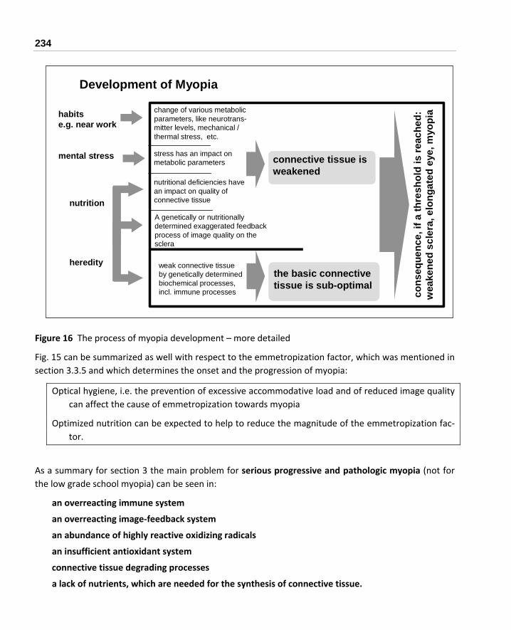
234
Figure 16 The process of myopia development – more detailed
Fig. 15 can be summarized as well with respect to the emmetropization factor, which was mentioned in section 3.3.5 and which determines the onset and the progression of myopia:
Optical hygiene, i.e. the prevention of excessive accommodative load and of reduced image quality can affect the cause of emmetropization towards myopia
Optimized nutrition can be expected to help to reduce the magnitude of the emmetropization fac‐tor.
As a summary for section 3 the main problem for serious progressive and pathologic myopia (not for the low grade school myopia) can be seen in:
an overreacting immune system
an overreacting image‐feedback system
an abundance of highly reactive oxidizing radicals
an insufficient antioxidant system
connective tissue degrading processes
a lack of nutrients, which are needed for the synthesis of connective tissue.
change of various metabolic parameters, like neurotrans-mitter levels, mechanical / thermal stress, etc.
nutritional deficiencies havean impact on quality ofconnective tissue
habitse.g. near work
mental stress
nutrition
weak connective tissue by genetically determined biochemical processes,incl. immune processes
hereditythe basic connective tissue is sub-optimal
connective tissue is weakened
cons
eque
nce,
ifa
thre
shol
d is
reac
hed:
wea
kene
d sc
lera
,elo
ngat
ed e
ye,m
yopi
a
stress has an impact onmetabolic parameters
Development of Myopia
A genetically or nutritionally determined exaggerated feedback process of image quality on the sclera

3 MYOPIA – OBSERVATIONS AND EXPERIMENTAL RESULTS 235
The difference between not very problematic, stable myopia and progressive myopia can be explained as shown in Figure 17.
adequate adjustment of the length of the eye
too strong modification of the sclera triggered by
optical impulse or mechanical force
emmetropia
progressive myopia
degraded connective tissue, either by poor basic
(systemic) structure or by too fast degradation
excessive modification of the
sclera triggered by optical im
pulse
or mechanical force
"school"-myopia
Figure 17 The different paths to "school"‐myopia and progressive myopia
The means for prevention of myopia and prevention of progression of myopia can be roughly described as shown in Figure 18.
Figure 18 The means for the prevention of the pro‐gression of myopia
genetical disposition for myopiavia biochemistry
•improve connective tissue
•reduce optical feedback on "growth" of the sclera

236
Optimizing the optical environment is important for everybody. In addition, optimizing biochemistry via nutrition is extremely important when fighting progressive, more pathological myopia.
All these advices are especially important for children, but they are as well important for adults of all ages.
3.31 For Further Reading –Extensive Compilations
The International Myopia Institute (IMI) has published in October 2018 a series of White Papers, which were written by numerous leading scientists and which are summarizing in detail the state of the re‐search about Myopia in several sections1089:
IMI – Clinical Management Guidelines Report
IMI – Clinical Myopia Control Trials and Instrumentation Report
IMI – Defining and Classifying Myopia: A Proposed Set of Standards for Clinical and Epidemiologic Studies
IMI – Myopia Genetics Report
IMI – Industry Guidelines and Ethical Considerations for Myopia Control Report
IMI – Interventions for Controlling Myopia Onset and Progression Report
IMI – Report on Experimental Models of Emmetropization and Myopia
These White Papers can be downloaded free of charge.
Brennan et al. published1090 an extensive review of various means of myopia control "Efficacy in Myopia Control".
Note: In these papers it is considered to be doubtless fact, that myopia has its origin in signals to the retina only, and therefore mechanical reasons are ignored. This is, however, in contrast to research results mentioned in section 3.2.1.2, where the elongation of the eyeball already by short‐term accommodation is described.

3 MYOPIA – OBSERVATIONS AND EXPERIMENTAL RESULTS 237
Recommendations for preventing or inhibiting the progression of myopia
Recommendations from the optical side (mainly to compensate the negative effects of excessive near work)
Potential risks
For near work keep a reasonably large distance to your book / paper / computer screen, even when wearing plus additions. Note: Reading in bed leads mostly to a distance, which is too near.
Do not use glasses or contact lenses, which are stronger than necessary, i.e. avoid any overcorrection. You might use "main glasses / contact lenses" which are slightly under‐correcting and you use additional glasses for perfect distant vision.
If you are not myopic yet, better use plus glasses for extensive near work. If you are already myopic, use bifocal or progressive glasses or bifocal contact lenses, or
use plus glasses additionally to your contact lenses for extensive near work. Interrupt your near work every 30 minutes by focusing on distant objects and relax your
eyes especially in the evening before going to sleep. Wearing plus glasses for a short time every day may be of some help. Cold‐color light should be preferred to warm‐color light. Do not read and don't do near work at bad light, 500 Lux are the minimum, but substan‐
tially higher levels are recommended depending on the visual task (e.g., for detailed drawing work 1,500 to 2,000 lux).
Some exercising of the accommodation by alternating focusing near and far objects can be helpful (but don't expect miracles from classes which are offered about this issue).
Take care to have enough sleep at the proper time and in darkness. Do not keep a light switched on in the kid's room at night (there is a controversy in the literature about night lights, but better be on the safe side).
Perform regular physical exercises, preferably outdoors, and use sunglasses in rather bright sunlight only
No risk, but avoid a permanent and sub‐stantial un‐dercorrec‐tion without the consul‐tation of an ophthal‐mologist, as an inappro‐priate un‐dercorrec‐tion or in‐appropriate bifocals may create, e.g., a depriva‐tion ef‐fect202.
Discuss with your ophthalmologist the application of atropine drops. Consider rigid gas permeable contact lenses (RGP). Soft contact lenses were not reported to
show a positive effect, but soft bifocal contact lenses showed a positive effect, too. Consider orthokeratology.
None if properly and profession‐ally applied.
Table 10 Optical recommendations for preventing myopia or inhibiting the progression of myopia

238
Recommendations for preventing, or inhibiting the progression of myopia
Recommendations from the behavioral and the nutritional side (mainly to reduce an elevated personal emmetropization factor)
Potential risks
Reduce negative mental stress, possibly by appropriate physical and mental exercises, and playing.
Physical exercises can as well have a positive impact on the blood cir‐culation in the eye, and promote NO metabolism.
Especially Outdoor activities were shown to be very effective to pre‐vent myopia (preferably 2 to 3 hours a day).
Avoiding stress can prevent a potential risk for your profes‐sional career.
Keep a healthy and balanced diet, which is low in sugar and low in refined carbohydrates (incl. white wheat), low in sodium, low in fat except omega‐3 (fish) oil, and have plenty of (if possible unproc‐essed) vegetables and fruits.
There are many books for a healthy diet on the market, with tables showing the contents in specific nutrients1091.
None
If the progression of your myopia is worrying you, additional supple‐ments of multiple vitamins (especially the vitamins E, B2, B6, folic acid), minerals (especially of calcium, selenium, copper and zinc) and especially also of flavonoids are recommended.
Only for the B vitamins doses substantially higher than the recom‐mended daily doses may be helpful, (for flavonoids no recommended doses are available).
Especially if your ancestors came from regions with plenty of sun, you may easily have an individual lack of vitamin D. A 25(OH)D blood test is highly recommended. In general, our ancestors spent by far more time outdoors, which gave them plenty of vitamin D. Therefore it is recommended to have your Vitamin D status checked and to take supplements in case the level is not optimal.
Do not take higher doses of vitamin A than the recom‐mended daily allow‐ance for it and keep the appropriate bal‐ance between copper and zinc, and calcium and magnesium.
Table 11 Behavioral and nutritional recommendations for preventing myopia or inhibiting the progres‐sion of myopia

4 ADDITIONAL INFORMATION 239
4 Additional Information
This section contains numerous hopefully helpful hints and explanations dealing with the myopia issue. These hints and explanations cover the correction of myopia by glasses, contact lenses or surgical op‐eration, as well as the dosage of dietary supplements.
4.1 Optical Correction
4.1.1 Glasses
Often the customer is not aware of the big variety in the quality of optical glasses. This section should help to ask the optician the right questions when ordering new glasses (even if you are wearing contact lenses you need spare glasses).
4.1.2 The Material of the Glass
The refractive index of the lens material determines the thickness of the lens: The higher the index, the thinner the lens (which is important for higher degrees of myopia). For mineral ("real") glasses the in‐dex is between 1.5 and 1.9, for organic ("plastic") glasses the index is between 1.5 and 1.7. Generally the rule is, the higher the refractive index, the higher the price of the glasses. Therefore highly myopic people who generally need thick glasses have to spend more money to get thinner (better looking) glasses.
Often not only the thickness of the lens is important, but also the weight: Organic lenses are lighter than mineral lenses, and for the mineral lenses the rule is, the higher the refractive index, the higher the specific weight.
A few numbers for comparison:
The Edge thickness for a lens with 65 mm diameter, a power of D = ‐ 10.00 and a refractive index 1.5 is 13.3 mm, and for a refractive index 1.9 the edge thickness is 7.4 mm.
The weight of a lens with 65 mm diameter, a power of D = ‐ 10.00 and a refractive index 1.5, made of a mineral glass is 56 gram, whereas the weight for an organic glass is only 33 gram.

240
A few typical numbers for the specific weight are shown in Table 12:
Material Refractive index
Specific weight in gram/cm3
Organic glass 1.5 1.32
1.6 1.34
Mineral glass 1.5 2.55
1.6 2.67
1.7 3.19
1.8 3.62
1.9 4.02
Table 12 Data of some materials used for glasses
Another parameter of the lens material is the dispersion, i.e. the difference of the refractive index for various color components, which can result in disturbing color effects on the edge of the lens. Gener‐ally, the higher the refractive index, the higher the problem of dispersion.
Additionally, the glass should have a high absorption of UV light.
4.1.3 The Coating of the Glass
Without a coating, there would be very disturbing reflections on the surface of the glass, for example making the eyes "invisible" to other people, and which create reflections of light which are very disturb‐ing during night driving.
For organic glasses there is another reason for coating: The relatively soft surface has to be protected from getting scratched too easily.
Overall, up to ten layers have to be brought to the lens, and significant differences in quality (and price) can be found for lenses from different manufacturers, and for different product lines of the same manufacturer.
4.2 Contact Lenses
(General sources1092, 1093, 1094, 976)
Already in 1999 it was reported that worldwide about 80 million people wore contact lenses, with about 33 million people in the USA at this time.

4 ADDITIONAL INFORMATION 241
A person who is switching from glasses to contact lenses experiences a magnification of the image on the retina. Table 13 compares the image size on the retina for people with glasses and for people with contact lenses1095 (image size for contact lenses are rather similar to emmetropic people).
Myopia in D Increase of image size by contact lenses com‐
pared to glasses
‐ 5.00 7 %
Table 13 The image size when using con‐tact lenses compared to glasses
‐ 7.50 10 %
‐ 10.00 14 %
‐ 15.00 23 %
As the accuracy of vision is, among others, determined by the "granularity" of the retina (see section 1.11) for higher grades of myopia a smaller image results in poorer vision.
Additionally it was found that contact lenses improved the contrast sensitivity for high myopes (more than ‐ 6.25D)405.
Contact lenses have some general advantages, e.g., no dimming by condensed water or rain. Features greatly depend on the type of lens.
4.2.1 Basic Types of Lenses – Soft Lenses versus Hard Lenses
The different types of contact lenses are:
Hard lenses (this type of contact lenses was developed first):
- Rigid gas permeable (RGP) lenses, which allow substantial oxygen supply for the cornea through the lens.
- Lenses with low or no transmission of oxygen, type PMMA or CAB (obsolete, should not be used any more).
Soft lenses, which are containing between 35% and 85% water, and which are soft and flexible when they are wet:
- Conventional soft lenses, with no specific time schedule for replacement (unplanned re‐placement).

242
- Planned‐replacement lenses, which are regularly replaced typically every month, or quar‐terly, or every 1‐2 weeks (depending on the instructions of the practitioner and the lens manufacturer).
- Disposable lenses, which are used once and then discarded (no maintenance for cleaning).
For hard lenses the shape remains the same after being put on to the cornea. For soft lenses the shape is modified, because the lens adjusts to a great extent to the shape of the cornea. This can be expected to result in spherical aberrations, which decrease the image quality.
Typical features of the individual types of lenses are shown in Table 14 and Table 15:
Hard RGP contact lenses
Advantage Disadvantage
Suitable for stabilization of myopia? Can correct some astigmatism without toric lens design.
Excellent optical quality and quality of the sur‐face.
Little absorption of chemicals. Less frequently serious eye infections than with soft lenses.
Fewer deposits on the lens. Can be polished if scratched; edges and power can be manually optimized.
Additional oxygen supply by exchange of tears during blinking.
Average lifetime of the lens several years Easier maintenance. Lenses for very high grades of myopia available. Normally all lenses with an accuracy of 0.25 D available.
Needs highly qualified professional for proper fit‐ting (especially difficult to fit when the lid ten‐sion is high).
Longer time for getting used to the lens (up to 3 weeks).
Occasionally less comfortable (depends largely on quality of fitting).
Dust may be more disturbing than with soft lenses (because of smaller size).
Loss of the lens is more likely than with soft lenses (because of smaller size).
Not suitable in cases of highly fragile epithelium of the cornea.
Not so suitable if worn occasionally only. Few types of lens have a scratch sensitive coat‐ing.
Table 14 Features of hard (RGP) contact lenses

4 ADDITIONAL INFORMATION 243
Soft contact lenses
Advantage Disadvantage
General and conventional type (i.e. no spe‐cific time schedule for replacement)
Better compatibility, often immediately comfortable.
Easier to fit (but maybe only because a miss‐fitting is not realized in the short term).
Less sensitivity to dust. Rarely any dislocation or loss of the lens, e.g. during sport activities.
Suitable for frequent changes between contact lenses and glasses.
Easy absorption of chemicals, e.g. from cleaning.
Badly fitted lenses stay unnoticed for a longer period, which may create long‐term damages.
Higher rate of complications were re‐ported (maybe caused partly by un‐professional fitting).
Less crisp vision than with hard lens. Average lifetime of the lens less than 1 year.
Gets damaged easily. No oxygen supply by exchange of tears during blinking.
More accumulation of surface deposits. More complicated/expensive mainte‐nance.
Frequently not available for high grades of myopia, and sometimes with an ac‐curacy of 0.50 D only.
Difficult to verify lens parameters. Correction of astigmatism requires toric lens.
Planned replacement
Less lens degradation. Easy to have spare lenses.
Less variety of lens parameters avail‐able.
Disposable No cleaning of lenses. Less lens degradation. Easy to have spare lenses.
Still less variety of lens parameters available.
Potential edge and surface problems depending on manufacturing process.
Table 15 Features of soft contact lenses
It was estimated that the probability for microbial keratitis (disease of the cornea) is about 3‐fold for daily‐wear soft lenses, and about 20 fold for extended wear soft lenses, compared with daily‐wear hard RGP lenses1096. Other authors claim that this rate is mainly a matter of proper fitting and mainte‐

244
nance976. The time for recovery was reported to be substantially shorter for incidents with hard RGP lenses than for soft lenses1097. Additionally, it was reported that the corneas of RGP wearers contained less bacteria1098. Therefore, it was reported that most of the people could not wear their soft lenses any more after 15 years1064 (see section 4.2.3 informs about potential complications).
Those who wear the lenses overnight have a risk of eye infection 10 to 15 times that of users who insert and remove the lenses daily1099.
Lenses for continuous wear – like for 30 nights and days – with very high oxygen transmissibility are available as RGP lenses and as soft lenses. As with normal lenses, it was found that RGP lenses for con‐tinuous wear showed fewer adverse events1100, especially fewer bacterial infections1101.
People, who use lenses for continuous wear should do so under the strict control of an experienced practitioner, who follows established guidelines1102.
A survey among members of the Contact Lens Section of the American Academy of Optometry showed that among these professionals RGP lenses were rated superior to soft lenses in most respects, espe‐cially quality of vision, eye health, ease of care/handling, and deposit resistance1103.
Interestingly, a report from USA says that higher education makes hard lenses more popular than lower education, where wearers of hard lenses had a higher average income, and among soft lenses people with lower education favor disposable lenses1104.
There are significant regional differences as well: In the USA only 12% of new fits are done with RGP lenses, in Japan more than 50% of new fits are using RGP lenses1105.
There are by far more soft lenses sold than hard lenses – which creates, by the way, more revenues for the industry, and allows individual opticians to enter the business, who are hardly qualified for the more demanding fitting of hard lenses.
The new lenses made of silicon hydrogel can eliminate the disadvantages of the low oxygen transmissi‐bility of the traditional soft lenses. Problems with microbial infections can still exist in a few cases, how‐ever, especially with extended wear1106.
The fitting process of these silicon hydrogel lenses appears to be more demanding than the fitting of conventional soft lenses, as Holden stated1107: "We're back in the fitting business with silicone hy‐
Therefore the tradeoff appears to be to choose between a
More healthy, but maybe a little bit less comfortable lens – the hard RGP lens, and a
More comfortable, but maybe somewhat less healthy lens – the soft lens.

4 ADDITIONAL INFORMATION 245
drogels. Manufacturers should pay attention to the edge shape of these stiffer lenses, and practitioners should make sure they've selected the appropriate diameter and base curve." It looks like, however, that these lenses are hardly available with a broad span of variable parameters so far. Therefore, le‐sions of the cornea due to not‐optimal fitting might develop – it depends highly on the skill of the op‐tometrist.
There is a variety of silicon hydrogel materials1108, and the water content can range from 0% to 60%1109. The lenses with high water content can be expected to be more similar to soft lenses, and the lenses with low water content can be expected to be more similar to RGP lenses.
4.2.2 How about Lenses for Permanent Wear?
Generally, it is differentiated between:
Daily wear, for usage up to about 18 hours, and
Extended wear, for overnight use, e.g. for several days, and
Continuous wear for up to 30 days and nights.
It was claimed that lenses made of silicone hydrogel are already completely safe for 30‐day extended wear1110, 1111. There are, however, still reports about increased amounts of deposits on the lens and de‐hydration of the lens1112, and an increased amount of metaplasia (abnormal cell structures) of the cor‐nea1113. Morgan stated1114: "There is a significantly higher incidence of SK [severe keratitis, an inflamma‐tion of the cornea] in wearers who sleep in contact lenses compared with those who only use lenses during the waking hours. Those who choose to sleep in lenses should be advised to wear silicone hy‐drogel lenses, which carry a five times decreased risk of SK for extended wear compared with hydrogel lenses." Morgan concluded additionally1115: "Practitioners should anticipate fewer adverse events with rigid versus soft lenses prescribed for continuous wear. Experienced wearers are less likely to discon‐tinue compared with neophytes."
Comparing silicone hydrogel lenses with very high oxygen transmissibility and daily wear lenses with low oxygen transmissibility it was found by Bergenske1116: "During the 3 years, lotrafilcon A lens wearers who wore their lenses continuously for up to 30 nights showed stable, long‐term improvements in many signs of corneal health and symptoms along with less myopic progression versus daily wearers of low‐Dk/t hydrogel lenses. Many biomicroscopy signs and symptoms worsened among neophytes wear‐ing daily‐wear low‐Dk/t hydrogel lenses. The use of lotrafilcon A lenses may minimize many ocular changes from soft contact lens wear."

246
4.2.3 Potential Complications
When getting contact lenses fitted, and for daily care extra cleanliness and accuracy are necessary. The following list of a few of the numerous potential incidents should not prevent anybody from wearing contact lenses. It should be, however, a good reason to be sensitive, to handle the maintenance with great care and to see the ophthalmologist (or optometrist) without any delay, if something unusual is noticed.
The practitioner can check the status of the cornea by the application of fluorescein to the eye. Damages to the cornea appear as stains. From the location of the stains conclusions about the fitting or corneal damages (e.g. caused by dust particles) can be drawn.
Frequent deposits on the lens (or even a blurred lens) are an indication that something is wrong, either with the mechanical properties of the lens, with some chemical toxication of the lens, or with too long wearing. A visit to your practitioner is strongly recommended.
If the lens cannot be removed as usual, there might be a tight lens syndrome. Do not apply force, better apply some saline solution or wetting drops, or see your practitioner.
The lack of oxygen generally (e.g. at night or when using lenses with low oxygen transmissibility) causes a swelling of the cornea, an edema. Depending on the transmission of oxygen by the lens, edemas occur to various degrees even when wearing at daytime only. It is not just a me‐chanical swelling, but a shift in various biochemical processes.
There are bacteria, which are growing best when exposed to an environment with low content of oxygen. Additionally, these edemas make it easier for bacteria to invade. These facts result in extra risks when wearing contact lenses over night1064 (or lenses with low transmissibility in general).
Extended lack of oxygen in the cornea can lead to the building of new blood vessels from the con‐junctiva to the cornea, called vascularization.
A chronic inflammation of the conjunctiva may become giant papillary conjunctivitis (GPC), with papillae developing on the conjunctiva up to a size of 2 mm.
Small wounds in the cornea can become an ulceration of the cornea.
More generally, bacterial and fungal infections caused by dirty lenses, as well as residual toxic or allergic traces of agents for lens care can cause serious damage to the eye, especially if they are taken care of rather late.
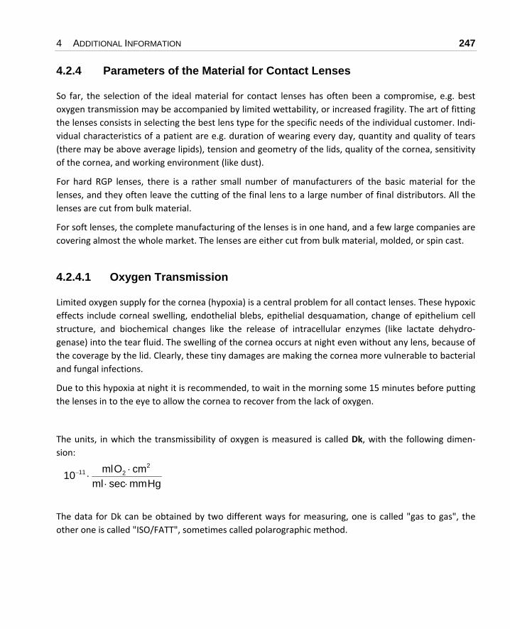
4 ADDITIONAL INFORMATION 247
4.2.4 Parameters of the Material for Contact Lenses
So far, the selection of the ideal material for contact lenses has often been a compromise, e.g. best oxygen transmission may be accompanied by limited wettability, or increased fragility. The art of fitting the lenses consists in selecting the best lens type for the specific needs of the individual customer. Indi‐vidual characteristics of a patient are e.g. duration of wearing every day, quantity and quality of tears (there may be above average lipids), tension and geometry of the lids, quality of the cornea, sensitivity of the cornea, and working environment (like dust).
For hard RGP lenses, there is a rather small number of manufacturers of the basic material for the lenses, and they often leave the cutting of the final lens to a large number of final distributors. All the lenses are cut from bulk material.
For soft lenses, the complete manufacturing of the lenses is in one hand, and a few large companies are covering almost the whole market. The lenses are either cut from bulk material, molded, or spin cast.
4.2.4.1 Oxygen Transmission
Limited oxygen supply for the cornea (hypoxia) is a central problem for all contact lenses. These hypoxic effects include corneal swelling, endothelial blebs, epithelial desquamation, change of epithelium cell structure, and biochemical changes like the release of intracellular enzymes (like lactate dehydro‐genase) into the tear fluid. The swelling of the cornea occurs at night even without any lens, because of the coverage by the lid. Clearly, these tiny damages are making the cornea more vulnerable to bacterial and fungal infections.
Due to this hypoxia at night it is recommended, to wait in the morning some 15 minutes before putting the lenses in to the eye to allow the cornea to recover from the lack of oxygen.
The units, in which the transmissibility of oxygen is measured is called Dk, with the following dimen‐sion:
The data for Dk can be obtained by two different ways for measuring, one is called "gas to gas", the other one is called "ISO/FATT", sometimes called polarographic method.
HgmmsecmlcmOml10
2211
⋅⋅⋅
⋅−

248
Typical values for ISO/FATT‐Dk are between about 10 and 200, where the gas to gas numbers are be‐tween 40% and 100% higher than the ISO/FATT numbers. Mostly the data are given in ISO/FATT units. Clearly, the higher the number, the better the oxygen transmission.
It appears that for the lenses currently on the market the average Dk/L numbers of hard RGP lenses are somewhat higher than that of the soft lenses. The leading manufacturers of soft lenses, however, have a very broad portfolio of lenses with very different Dk/L numbers – a good reason to ask your practitio‐ner carefully what lenses you are supposed to get.
As Dk describes the properties of the material only, it does not take into account the thickness of the lens. Therefore a number Dk/L is defined, where L relates to the thickness of the lens. The dimension of Dk/L is:
The thickness of the lenses in the center is between about 0.08 mm and 0.15 mm for hard RGP lenses and between about 0.04 mm and 0.15 for soft lenses (in reality the fact that the thickness varies over the surface of the lens, and is smallest in the center, has to be considered for the Dk/L calculation as well).
Table 16 shows a summary of a few reports about the impact of various Dk/L numbers, not taking into account additional oxygen supply by the exchange of tears for hard lenses:
Dk/L Impact on the cornea
23 Required for minimum oxygen supply to the basal epithelium of the cornea of the open eye1117.
24 Required for daily wear without cornea swelling1118.
35 Required for minimum oxygen supply for the whole thickness of the cornea of the open eye1117.
87 Required for wearing at night without any additional swelling1118.
89 Required for minimum oxygen supply to the basal epithelium of the cornea of the closed eye1117.
125 Required for minimum oxygen supply for the whole thickness of the cornea of the closed eye1117. No increase in epithelial lactate levels1119. Still no normal oxygen supply1120.
Decreased thickness of the cornea after 3 months, but no change after 1 month of extended wear1121.
Table 16 The impact of the oxygen transmission Dk/L of contact lenses
HgmmsecmlcmOml10 29
⋅⋅⋅
⋅−

4 ADDITIONAL INFORMATION 249
As a summary, and considering the state of the art of currently available lenses, Dk/L numbers should be over 25 for daily wear in any case, but lenses with numbers over 35 are widely available and are clearly more recommended.
Another way to measure the oxygen supply to the cornea is the EOP (equivalent oxygen percentage) number, which indicates the percentage of oxygen directly at the cornea. At sea level and without a lens EOP equals about 21%.
For extended wear a minimum EOP of about 18% (corresponds to about Dk/L 85) is recommended, for daily wear a minimum EOP of about 10% (corresponds to about Dk/L 25) is recommended.
During sleeping, the EOP is only 7.7%, which explains the swelling of the cornea at night. As it takes some time for the eye to return to a normal oxygen status after sleeping, it is advised to wait some time (about 15 minutes) before using the contact lenses.
The exchange of tears by blinking when wearing hard lenses can contribute an additional EOP of about 3% to 4% (for the old hard lenses made of PMMA material this was the only one way of oxygen supply).
The higher the Dk of a lens, the shorter is generally the life expectancy of the lens1122, and for soft lenses there is a general relation: The higher the water content, the higher the oxygen transmission.
As result of a study it was concluded: "High‐Dk silicone hydrogel lenses can be worn for up to 3 years with virtual elimination of the hypoxic consequences observed with low‐Dk lenses made from conven‐tional lens materials."1123
There is evidence that the lower the Dk/L of the lens is, the larger the thinning of the epithelium of the cornea is1124. This thinning of the epithelium was still observed with lenses with Dk=140 (continuous wear), and the thinning was still evident after 3 months with no contact lenses1125. This thinning is sometimes primarily attributed to a lack of oxygen, sometimes primarily to the mechanical pressure which is applied by the lens.
4.2.4.2 Wettability and Resistance against Deposits
A lens with excellent oxygen transmission, but poor wettability and poor resistance against deposits is not acceptable.
There is a scale for the wetting capability, the wetting angle. It can be measured by two procedures (with different resulting numbers):
The "captive bubble" method measures the angle between the edge of an air bubble and the lens surface, with the lens immersed in water or saline.
The "sessile drop" method measures the angle between the edge on a drop of water and the lens surface.

250
For both numbers the rule is: The lower the angle, the better the wettability.
There exists no measurement procedure for the resistance against deposits, which consist of tear mucin and protein (and bacteria). Whether there is a problem with deposits, depends heavily on the consistency of the tears of the individual person. If there are irritations of the eye due to a lack of oxy‐gen or mechanical stress of the lens on the cornea, deposits will increase substantially.
Different lens types can have significantly different resistance against deposits, and it is up to the prac‐titioner to select the proper material – and it is up to the user to be very careful about daily cleaning. Mainly the deposit problem is the reason for the recommendation to change regularly to new lenses – especially for lenses with a very high Dk.
Sometimes the wetting capability of brand new RGP lenses is caused by deposits from the manufactur‐ing process. Professionals have specific solvents to remove these deposits1126.
4.2.4.3 Hardness and Stability
The hardness of lens materials is only specified for hard lenses. It is measured in Rockwell (a sharp sam‐ple is pressed onto the material, and the remaining depth of the damage is measured) and in Shore (a hard ball is dropped onto the material, and the height to which the sample is bouncing back is meas‐ured). The Rockwell numbers give more of an indication for the resistance against scratches; the Shore numbers describe more the elasticity. The higher the oxygen transmission of the lens is, the softer the material in many cases.
A soft lens does may not be more comfortable, as blinking may cause a deformation of the lens and a transient edge lifting, which interacts with the upper eyelid1127.
A hard, rather inflexible lens will be more suitable for masking astigmatism of the cornea than a flexible lens, which will just take the shape of the cornea.
Warping of the lens may be an issue for the longer term: It is the capability of the lens to keep exactly its shape, and not to become distorted. Very thin, hard lenses are more likely at risk for warping or even for breaking.
Sometimes the shape of the contact lens is changing in a way that the fitting of the lens becomes more flat (larger Base Curve Radius, see section 4.2.5). This "extra lens which consists of tears" under the con‐tact lens adds to the power of the contact lens, which results in an overcorrection with all the negative influence on a progression of myopia. There are two ways to avoid this problem: Get contact lenses with a high degree of stability (your optometrist might consult you), and/or have the geometry of your lenses checked regularly.

4 ADDITIONAL INFORMATION 251
4.2.4.4 Specific Weight and Refractive Index
These two parameters are sometimes expected to be important ‐ mainly for contact lenses of higher grades of Diopter because of a higher volume of the optical part: A heavier lens (i.e. of higher specific weight and lower refractive index) might have more a tendency to move downward, i.e. centration is more likely to become a problem.
Detailed analysis and experiments, however, have shown that "the lens mass is not a significant predic‐tor of lens dynamics." More important is the center of gravity of the lens. Carney et al. stated1128 that "... lenses with a center of gravity location further behind the lens vertex show better stability." The center of gravity is shifted into this direction by an increasing diameter, decreasing lens thickness (and increasing negative power and increasing lens curvature, but these two are determined already for other reasons).
Anyway, a lower weight of the lens can be achieved by manufacturing it from a material with lower specific weight, with minimal center thickness (depending on the specific material), or by a specific lens cut called lenticulation1129.
4.2.4.5 UV Blocking
Most lenses are coming with a substantial UV blocking capability; this should never be considered, however, as a substitute for wearing sunglasses.
4.2.4.6 Color / Tint of the Lens
There are tints, which are used for cosmetic reasons, i.e. to enhance or even to change the color of the iris of the eye, and there are more pale tints, which are applied to make the lens more visible to ease the handling of the lenses. This pale tint, e.g., can make it easier to find a lens, which dropped down.
4.2.5 Parameters of the Geometry of Contact Lenses
In principle, hard and soft lenses are described by the same set of parameters. For hard lenses, how‐ever, these parameters are by far more important, and the lenses are available with by far more de‐tailed numbers of these parameters. Because of this large variety of hard lenses, the final lenses for an individual customer are custom made, and the practitioner is working with a set of trial lenses only, which is normally do not match with the final required refractive power. Soft lenses, however, are gen‐erally "ready to go" because of their rather limited spread of parameters – sometimes the refractive power is the only parameter.

252
The geometrical parameters are:
Diameter of the Lens (TD, Total Diameter)
Typical diameters of hard RGP lenses are between 9.00 mm and 10.50 mm; the selected size depends e.g. on the diameter of the iris, and the position of the upper eyelid, i.e. whether the upper edge of the lens should stay covered by the lid ("lid attachment") or not ("interpalpe‐bral").
It was noted1130, "High powers require larger diameters for lens stability because the heavier lens may sink down if it is not caught by the eyelid."
Generally, lens diameters above 9.5 mm were found to be more comfortable1131.
Typical diameters of soft lenses are between 13 mm and 15 mm.
Astonishingly, a report found that oxygen uptake increased with increasing diameter of hard lenses (and their typical diameter)1132. A large diameter of a hard lens may create, however, problems for fitting, if no aspheric design is used (see "Aspheric Design", below).
Base Curve Radius (BCR, or Back Optic Zone Radius BOZR)
This very important number determines the fit of the lens on the cornea, and it is for hard RGP lenses between 6.50 mm and 9.00 mm; this radius is available with a granularity of 0.1 mm or better of 0.05 mm. A lens manufacturer, which delivers 0.05 mm granularity, should be pre‐ferred, because this allows a more accurate fitting of the lens – especially if the selected lens material is rather rigid.
For soft lenses, the exact fit to the cornea is by far less important, because due to their flexibil‐ity they can adjust to the radius of the cornea. Very often soft lenses are coming with one ra‐dius of about 8.6 mm only; sometimes there is a choice between numbers like 8.4 mm, 8.7 mm, and 9.00 mm.
Sometimes the curvature of the cornea is not described by its radius in mm, but by its corre‐sponding Diopter number. The formula for conversion is:
where D is the Base Curve Radius in Diopter, and R is the Base Curve Radius in mm.
Because of the lens built by the tear‐liquid between cornea and contact lens, a transition to a flatter lens than the previous lens results in a lens with less Diopter (weaker power), whereas a transition to a steeper lens results in a lens with stronger power. 0.05 mm change in BCR corre‐sponds to about 0.25 Diopter.
RD 5.337
=

4 ADDITIONAL INFORMATION 253
Aspheric Design, Secondary Curve Radius (or Back Peripheral Radius BPR)
When contact lenses were first introduced, both sides of the lens were spherical. However, this matches the shape of the cornea only for very small lenses. In reality the cross section of the cornea looks more like an ellipsoid or a paraboloid. Carney et al. stated412: "A tendency for the cornea to flatten less rapidly in the periphery with increasing myopia was shown, however."
Consequently, there were other back surfaces developed, created by one of these methods:
‐ An aspherical outer curve is joined to the inner spherical curve, i.e. the lens becomes progres‐sively flatter towards the edge. As the cornea itself is aspheric, a closer fit to the cornea can be achieved.
‐ Mathematically, the aspherical part of the lens follows the shape of a conic section, where a parameter e describes the eccentricity of the selected conic section: for spheres e equals zero, for ellipsoids e is between 0 and less than 1, for paraboloids e equals 1. Typically, the cornea corresponds with a value for e between 0.4 and 0.6.
‐ A disadvantage is that it is more difficult to verify the parameters of a lens, or to compare lenses from different manufacturers – and they are often more expensive.
‐ Various spherical curves are joined onto each other, with the curves with the larger radius towards the edge of the lens. This lens design is normally used for very special cases and cus‐tom design only.
Most common (and cheaper) lenses are, however, still simple spherical designs.
Shape of the Edge, Edge Lift
The shape of the edge of the lens, where the outer surface is meeting the inner surface, is pri‐marily important for hard lenses only. This shape is mathematically hard to describe and usu‐ally at least partly hand made at the manufacturer's laboratory. Nevertheless, the specific shape of the edge is very important to feel comfortable with the lens, as well as for an efficient tear exchange.
A larger axial edge lift (up to 0.3 mm) was found to be less comfortable than a lower axial edge lift (around 0.1 mm), which gave a better‐centered lens as well1133.
More experienced practitioners are able to modify the edge of a lens according to the specific needs of a customer.
Center Thickness
The thickness of the lenses in the center is between about 0.08 mm and 0.15 mm for hard lenses and between about 0.04 mm and 0.15 for soft lenses. The thickness of the lens has not only an impact on the oxygen transmission, but also on the flexibility of the lens: To avoid in certain cases a warping of the hard lens, a lens with increased thickness is provided.

254
Back Optic Zone Diameter (BOZD or OZ)
The back optic zone is the area, which has the "right" optic without any disturbing optical effect of the aspheric design, or a secondary curve. This diameter should be not too small for good vi‐sion.
The Power of the Contact Lenses
The numbers for the refraction of the glasses and for the refraction of the contact lenses are different, especially for higher grades of myopia. The formula is:
where d is the distance between the cornea and the glasses in m (sometimes called vertex dis‐tance).
An example: With Dglasses = ‐ 10.00, and d = 16 mm the power of the contact lenses Dlens = ‐ 8.62
By the way, this effect can also be visible when the refraction is measured für spectacle wear‐ers and when the frame for testing has a different vertex distance than the finals spectacle glasses, possibly resulting in over‐correction.
Additionally, the refractive power of the tear‐liquid filled gap between the cornea and the con‐tact lens has to be considered. For a flat fitted lens (i.e. with a larger BOZR), this gap acts as an additional minus lens. The power of this additional lens can be between zero D for a lens fitted exactly to the cornea and ‐ 1.00 D for rather flat fitted lenses and has to be considered when determining the power of the final lens:
A change of 0.05 mm in the BCR (Base Curve Radius), or BOZR (Back Optic Zone Radius) results in a change of 0.25 D of the lens system, i.e. a lens that is 0.05 mm flatter has in effect ‐ 0.25 D more refractive power.
Toric Components
To compensate astigmatism a toric component can be added to the lens, similarly like for con‐ventional glasses.
Special cuts of contact lenses, which take care of presbyopia are discussed in section 4.2.12.
glasses
glasseslens
Dd1DD
⋅−=

4 ADDITIONAL INFORMATION 255
4.2.6 Surface and Edge Finishing
A few big companies make the basic material of the lenses, but numerous smaller vendors do the final manufacturing especially of the RGP lenses. Different materials are requiring different surface finishing processes, and different manufacturers of the lenses may achieve different qualities. This should be considered when switching to a new material or another optometrist who is buying from a different vendor.
Frequently the edges of RGP lenses are still finished manually. Consequently, this process depends on the skill of individual people and appropriate quality control.
It was reported that there is a quality problem for daily lenses, i.e. lenses, which are used only once 1134: Due to this cost effective mass production about 2% of these lenses might be of reduced qual‐ity, e.g. have a higher surface roughness. In the worst case, this might lead to a permanent incompati‐bility of contact lenses.
4.2.7 The Fitting of Contact Lenses
A description of the fitting process is beyond the scope of this book, and there are very good sources about this issue available1092. Therefore just a few remarks:
There are several successful strategies of how to fit the best lens (especially for hard RGP lenses). Basis of each fitting process is a measurement of the topography of the eye, especially the various radii of the cornea, measured in different directions, and in the center as well as in the periphery.
A general differentiation is between a more flat fitting and a steeper fitting: Flat fitting means that the base curve radius (BCR / BOZR) is clearly larger than the radius of the cornea. This results in some edge clearance, which allows for enhanced exchange of tears and oxygen, and good mobility of the lens, but the lens may become more off‐centered (which may have unwanted long‐term effects on the shape of the cornea). Steep fitting means that the base curve radius closely matches the radius of the cornea. This results in a good central position, but if the lens is sitting too tight (i.e. too steep fitting) a "tight lens syndrome" may develop.
Fitting of hard lenses is more demanding than the fitting of soft lenses, but a bad fitting of a hard lens is noticed earlier, generally before some damage happens to the eye.
Special harm can be done by overly tight lenses, i.e. which don't move during blinking.
An irritation can be caused not only by a lens that was fitted too steep (most uncomfortable) or too flat, but also by the sliding of the upper lid over the edge of a rather small lens1131. A larger lens might help in these cases.

256
When changing from older, non‐gas‐permeable lenses (PMMA‐type) to highly gas permeable lenses the fitting should be done either without any break in between, or with a break of several weeks1092.
It is very helpful for the fitting practitioner, if the patient can describe the problem very clearly. There‐fore it is helpful, to get the basic knowledge about the fitting process by yourself. This also enables you to ask the right questions (and to find out whether you are at the right practitioner).
Experience is a key issue to become an expert for fitting. Therefore, an ophthalmologist or an optome‐trist who is really focusing on contact lenses as the main job is most likely a better choice than some‐body who is doing this task only occasionally.
4.2.8 The "Right" Power of Contact Lenses
In section 3.2.2.3.12 a summary of the various optical aspects which determine the "right" power of the glasses or contact lenses was given.
The thesis to avoid any avoidable accommodative load for the eye leads to these tradeoff‐recommendations for contact lens wearers:
Have fully correcting contact lenses but wear additional plus glasses for extensive near work, or
Have under‐correcting contact lenses but wear additional minus glasses for perfect vision for far (e.g. for driving).
Be aware that the power of contact lenses is lower than that of the respective glasses. The numbers for the refraction of the glasses and for the refraction of the contact lenses are different, especially for higher grades of myopia. The formula is:
d is the distance between the cornea and the glasses in m (sometimes called vertex distance).
4.2.9 Determining the Refraction after Wearing Contact Lenses
When the refraction of contact lens wearers is determined – e.g. to fit spare glasses – it has to be con‐sidered:
Flat fitted lenses flatten the cornea (see section 3.26.2), herewith reducing the Diopter of myopia.
If lenses of low oxygen transmission (i.e. PMMA material) had been used, it takes 3 weeks until an accurate refraction could be determined. Rengstorff stated1135: "...myopia decreases over the first 3 days after stopping PMMA wear and then increases over several weeks until it stabi‐
glasses
glasseslens
Dd1DD
⋅−=

4 ADDITIONAL INFORMATION 257
lizes." Therefore the transition to a material with high oxygen transmission can be accompa‐nied by a wrong refraction and improper fitting
If the determination of the refraction, however, serves only the purpose of fitting spare glasses, these effects can be ignored, as these glasses are used in general immediately after wearing the contact lenses.
4.2.10 Maintenance of the Lenses
The maintenance procedure is different for hard RGP and soft lenses, but there are some general rec‐ommendations:
Generally, follow the routine recommended by the manufacturer of your lenses, and by your prac‐titioner.
If you are not satisfied with your lenses, talk to your practitioner and ask him what other brands of lens care products, or what other procedures would be compatible with your lens type, and try them. There is a very big variety of lens care products, made by the manufacturer of the lens as well as products made by independent suppliers. There is always the possibility that you are sensitive against a preservative or tensides, which is used in a lens care agent.
4.2.10.1 Maintenance of Hard RGP Lenses
Essential for the maintenance of hard RGP lenses is the fact that they contain less than 1% of water, i.e. absorption of agents into the lens material is by far less a problem than for soft lenses. The steps of treatment are (not considering the necessary rinsing between individual steps):
a) Cleaning after the use to remove surface deposits.
b) Disinfecting over night.
c) Wetting before inserting into the eye.
d) Occasional enzyme cleaning to remove proteins, if necessary.
The cleaning solution for step a) contains tensides and, depending on the brand, some abrasive parti‐cles (a few lens types do not like abrasive agents!)
Normally the steps b) and c) are performed with the same type of agent, but this might not be the best for you: There are solutions which are very effective for disinfecting1136, but which can be somehow irritating for wetting, when they come in direct contact with the eye1137, 1138, and vice versa1139. Therefore, it could be very helpful for you to use different solutions for step b) and for step c), even though most solutions are claimed to work for both b) and c.

258
There are also solutions on the market that combine the steps a), b) and c), but do not be astonished if this compromise does not satisfy you; anyway, it is good that these products exist for the emergency case, e.g. for traveling.
Generally, the solutions of the various brands have a different composition. Therefore, in case of a problem, a switch of the brands can be very effective. Sometimes there can be interference as well, i.e. a cleaner from a certain brand might be comfortable with a disinfecting/wetting solution from one brand but not with another brand.
Occasional enzyme cleaning is efficient, but if heavy deposits build up frequently there might be some‐thing wrong with the lens. Enzyme cleaners can be based on either animal‐ or plant‐enzymes, maybe you are more sensitive to one of them (there is always a chance that traces are left on the surface).
Special care has to be taken when cleaning the lenses by hand to avoid warping them – therefore some practitioners are recommending to put the lens into the palm of the hand for cleaning, and not to rub it between the tips of two fingers.
A change of the lens parameters is not necessarily visible to the patient: the lens can become flatter in fitting, which means stronger in power. This gives no immediate cause for complaint to myopes. Nevertheless, the resulting overcorrection can increase the myopia further as outlined before. Re‐sulting advice: insist that your practitioner checks the parameters of the lens at least annually, and favor a material, which is more stable.
4.2.10.2 Maintenance of Soft Lenses
Essential for the maintenance of soft lenses is the fact that they contain a very substantial amount of water (35% to 85%), i.e. absorption of agents into the lens material is a problem. The steps of treatment are (not considering the necessary rinsing between individual steps):
a) Cleaning after the use to remove surface deposits.
b) Disinfecting by the chemical method, or by hydrogen peroxide, or by heat, or by UV‐light, or by microwave radiation (depending on the individual lens).
c) Disinfecting is especially important for soft lenses, as some types of fungi can grow within 20 hours through a soft lens; bacteria and virus, however, cannot get inside a undamaged soft lens.
d) Storing, and wetting before inserting into the eye.
e) Occasional enzyme cleaning to remove proteins, if necessary.

4 ADDITIONAL INFORMATION 259
For soft lenses the variety of lens care systems, which often combine steps mentioned above, is sub‐stantially higher than for hard lenses. The requirements are more dependent on the individual lens type. There are also "all in one" solutions on the market, similarly as for hard lenses.
The fact that proper lens care is more difficult for soft lenses than for hard RGP lenses is one of the rea‐sons for the trend towards disposable soft lenses.
In any case, the specified intervals for renewing the lens should not be ignored.
4.2.11 "I Cannot Wear Contact Lenses"
This conclusion "I cannot wear contact lenses" might be not generally valid, but based on
Specific lens care liquids for cleaning or storing or wetting which you cannot tolerate.
Advice: Try other lens care liquid with another added preservative.
A specific lens material which is not suitable for you (this might be influenced by the manufacturing process of the specific vendor). Silicone‐hydrogel lenses, e.g., might create problems in case of elevated levels of lipids and proteins are in the tear liquid. Additionally, the specific combina‐tion lens material / lens care liquid can be the problem, if the lens material stores especially components of the liquid, against which the eye is sensitive.
Advice: Try other lens materials.
The geometry of your lenses.
Advice: Ask your optometrist to try a modified geometry. Soft contact lenses are often sub‐scribed as "one geometry fits all"; this might be true for most of the people, but not for every‐body. For soft contact lenses, different geometries are available as well as for RGP.
An irritation of the eyelid by the edge of the lens.
Advice: Ask your optometrist to try a modified geometry, especially a lens with a larger diame‐ter might help.
"Dry eyes".
Advice: Frequently dry eyes are the main obstacles against the wearing of contact lenses. There can be several medical reasons which cause dry eyes, but there can be as well a more simple reason for dry eyes: too less blinking. It was stated1140: "The reason for this difficulty is that the frequency of blinking typically decreases during tasks that require concentration. As blink fre‐quency decreases, there is more time for the tear film to evaporate.
A simple exercise to improve the blinking was given1141: "The exercise consists of a person placing his/her fingers lightly on the outside corners of both eyes. By blinking rapidly several

260
times, most people notice a pulling sensation as they blink. Blinking should be repeated slowly, ensuring that the eyelids close completely and that pulling sensations are no longer present. This may be achieved by concentrating on moving only the eyelid, during the blink. This exer‐cise should be done several times each day and for one to two minutes each time the exercise is performed."
Additionally it was said1142: "Practitioners generally recommend soft lenses made with a low water content because these lenses dehydrate less than high‐water lenses do in a dry envi‐ronment."
With respect of the influence of the diet on the tear function, Caffery stated1143: "A review of the ocular literature suggests that sufficient dietary protein, vitamins A, B6 and C, potassium, and zinc may be necessary for normal tear function. Excesses of dietary fats, salt, cholesterol, alcohol, protein, and sucrose have been associated with or suggested as causes of tear dysfunc‐tion." Additionally, omega‐3 fatty acids, which can be found, e.g. in fish oil and flaxseed were recommended for dry eyes1144.
Note: Overall, before you take more serious and final steps like surgery, ask your contact lens practitioner for different liquids, different materials, or, finally, go to a different practitioner – and take care of your diet and your viewing habits.
4.2.12 Presbyopia
For presbyopia, the contact lenses are required to replace the accommodation of the human lens, i.e. to offer focus as well for near objects as well as for distant objects, as with bifocal glasses (numerous other means to treat presbyopia were discussed in the literature20).
Wearers of glasses accommodate less and have lower convergence demands than emmetropes or wearers of contact lenses, i.e. myopic people wearing contact lenses should be affected by presbyopia more early than myopic people, which are wearing glasses208.
There are some different concepts to handle presbyopia with contact lenses1145. Generally, there are two groups of systems, i.e. for alternating vision, where an image is produced either for distant view or near view, and simultaneous vision, where the evaluation of the different images is done in the brain. In any case, it will take a while until the patient is used to the new vision.
Specific contact lenses for presbyopia:
The optical zone of the lens is split into an upper section for distant focusing and a lower section for near focusing (alternating vision).

4 ADDITIONAL INFORMATION 261
A problem is that the lens has to be kept in the right rotational and vertical position. The lens is difficult to fit, and is normally fitted flat.
The optical zone of the lens is split into a center section for distant focusing and a peripheral sec‐tion for near focusing (simultaneous vision).
This lens is fitted more on the steeper side to have the lens properly centered. When the aper‐ture of the iris is small, e.g. at bright light, near focusing may be a problem. As with all methods which are using simultaneous vision, the contrast of the images is reduced.
Notes: - The steep fitting, which is necessary to keep the movement of the lens at a minimum, might
create a problem particularly for older people whose amount of tear liquid is frequently re‐duced.
- As with older age the ability to see in the dark and the contrast sensitivity fades generally, every optical device which increases these handicaps is problematic, especially when this de‐vice is worn permanently, i.e. when there is no way to improve the vision when needed, like for driving at night.
Into the optical zone of the lens fine microscopic steps are inserted. These steps create via diffrac‐tion two focus points – one for distant vision and one for near vision (simultaneous vision).
An exactly centered position, and the aperture of the iris is not as important as in the method described on top. The reduction of the contrast, however, is similar.
In general, the visual performance of rigid gas permeable contact lenses was better than the perform‐ance of soft contact lenses.1146
Other methods to cope with presbyopia are:
Monovision: The lens of one eye is adjusted for distant vision; the lens of the other eye is adjusted for near vision.
This method is less expensive, and easier to fit than the previous methods. Good success rates have been reported for this method. For driving (especially night driving) the use of additional glasses, which compensate for the undercorrection of one eye, is recommended. Especially for reading additions that were not greater than 2.50 D the visual results were reported to be quite satisfying1147.
Note: In a professional discussion forum several optometrists expressed their opinion that monovision is clearly more comfortable to most patients than bifocals.
Good results were also reported for the combinations of monovision and LASIK1148.

262
Permanent undercorrection to allow reading, use additional glasses for distant vision, e.g. for driv‐ing.
This method, however, can satisfy only in the early stages of presbyopia, when the resulting uncorrected distant vision is still acceptable for daily life (except driving), i.e. when it is around 1.00 D only. Moreover, this method can be expected to delay the total presbyopia, because the permanent accommodative effort keeps the own lens longer elastic.
Intraocular lenses which can accommodate.
In the trial status are intraocular lenses for people undergoing cataract surgery (i.e. whose lens was removed), which can accommodate via the ciliary muscle. While this sounds very promis‐ing for cataract patients, the risk of this operation for normal presbyopes without a cataract problem appears to be large, but this may change within a few years1149.
Note: Because of their shorter remaining life expectancy, long‐term effects of surgery might be less important for patients with presbyopia than for young myopes.
4.2.13 Contact Lenses and Nutrition
Some nutritional components are especially supporting a healthy cornea, which is important when wearing contact lenses:
Vitamin A is generally necessary for the metabolism of mucous membranes, and a long‐term nutri‐tional deficiency causes severe damages of the cornea, which can lead to blindness.
Local, lesions of the cornea are often treated with eye drops containing pantothenic acid (vitamin B5) or retinoic acid (vitamin A).
The mechanical stability of corneas was improved by treatment with vitamin B2 (riboflavin) and UV irradiation, introducing additional cross‐links.
Therefore, and as vitamin B2 was shown to have a positive effect on the connective tissue in general it is advised to have sufficient vitamin B2 in the diet.
Note: From this point of view, contact lenses, which are blocking UV light almost completely, are not of benefit in every respect.
The cornea needs manganese for the maintenance of its cell structure.
The stability of the tear film on the cornea is essential for comfortably wearing contact lenses. It was found that taking a standard supplement containing the recommended daily dose of vita‐
4 ADDITIONAL INFORMATION 263
mins (e.g. A, B1, B2, B6, E) and trace elements (e.g. calcium, manganese) improved tear stabil‐ity1150.
4.3 Refractive Surgery
Refractive surgeries like PRK, LASIK and LASEK (the basic principle was outlined in section 3.28.1) have become a big industry, and estimates for the USA were 1,900 000 procedures in 2002, up from 465 000 in 19981151.
In the USA, "at least" several hundred ophthalmologists had LASIK or PRK for themselves – of about 20.000 ophthalmologists in practice1152.
Nevertheless, people who are expecting that they are getting rid of glasses or contact lenses by refrac‐tive surgery forever should consider:
The rate of complications at LASIK was estimated to be at 7.2%1153 and more recently 2%1154, and for 11.6% of the eyes were re‐treatment was found to be necessary1155. Keratitis, an inflamma‐tion of the cornea, was observed in 0.7% to 32% of the LASIK treatments1156. Bacterial infec‐tions appear to be especially a potential problem for persons exposed to a healthcare set‐ting1157.
On the other hand, it was claimed that the earlier rate of 20% of complications of laser treat‐ment dropped below one percent1064.
Furthermore, it was said that most problems arise due to a faulty pre‐examination and infor‐mation of the patients1064.
Some data about the probability that no more glasses or contact lenses are necessary: A report states that for ‐4.25 D up to ‐8.00 D 86.5% of the patients stayed within 1.00 D of intended cor‐rection, and for above ‐8.25 D 82.9% of the patients stayed within 1.00 D of intended correc‐tion.
The regression was ‐0.5 D for patients between ‐0.5 D and ‐4.0 D (only during 1st to 3rd month), ‐0.8 D for patients between ‐4.25 D and ‐8.0 D (1st to 6th month), and ‐1.2 D for patients above ‐8.25 (1st to 9th month)1045.
For follow‐up LASIK procedures to eliminate residual myopia the rate of complications is sub‐stantially elevated1158.
This means, you have a good chance, to need still some glasses or contact lenses – which you wanted to get rid of forever.
264
Additionally, even if myopia is (rather) stable, there are often some moderate changes during the lifetime, which are easy to correct with glasses or contact lenses. And for your presbyopia you need some glasses or contact lenses anyway.
For a group of 1308 patients the one‐year re‐treatment incidence was 12.1% for myopic eyes, with an increase to 14% for patients older than 40 years1159.
Barclay stated1159: "In most cases, LASIK flaps could be lifted using manual technique up to three years after the initial surgery."
Note: This means the stability of the LASIK‐flap is rather limited for quite a long time.
With LASEK this problem does not exist1160.
There is a serious risk to develop dry eyes after the LASIK surgery1161.
The climatic environment, i.e. temperature and humidity, play an important part with respect of the complications associated with LASIK surgery: the colder and the less humid, the better1162.
Machet stated1046: "Just as the good can be very good, the bad can be very bad."
An example of the potential risk of LASIK and similar surgery was the recall of software for a LASIK system as summarized by Waknine1163: "The action was based on reports of corneal abnormali‐ties ("central islands") that may not be correctable with laser therapy, according to an alert sent from MedWatch, the FDA's safety information and adverse event reporting program. Use of the algorithms has also resulted in decreased visual acuity that may not be correctable with use of glasses or contact lenses."
Villa reported1164: "Patients undergoing LASIK procedures display an increase of halo phenomena around lights in night vision conditions, even when the results of the surgery are considered entirely satisfactory according to current international standards of predictability, efficacy and safety. Secondary astigmatism, coma and spherical aberration are the higher order aberrations up to the sixth order that significantly correlated with halo disturbance index."
Obviously, the brand of the equipment for the laser operation still plays a major part, as Feltham reported1165: "A successful refractive outcome was achieved in 78% (235/302) of surgeries us‐ing the Technolas laser and in 88% (266/302) using the NIDEK laser. Predictor variables for not achieving refractive success were pre‐operative refractive error of above ‐5.00 DS, age more than 40 years, and surgery performed with the Technolas laser."
Some questions you should ask before getting surgery done, are1166:
What is your success rate for achieved visual acuity (that is, 20/40 or better)?
4 ADDITIONAL INFORMATION 265
How many operations have you performed personally?
May I speak with any of the patients on whom you have done this procedure?
How many of them have required second operations? (It should be under 10%.)
What is the best correction I can expect?
How much will the operation cost me?
What are the chances I may not see as well as with glasses or contact lenses?
Do you operate on both eyes the same day?
What are the possible complications?
What anesthesia will I receive during the operation?
How long will the procedure take?
What is the recovery period?
When will I be able to return to work?
If the operation fails to correct my vision, will I be able to go back to contact lenses?
What if, years from now, I develop cataracts? Will this operation prohibit or interfere with later cat‐aract surgery?
What are the long‐term risks of this surgery?
Most refractive surgeons prefer an undercorrection instead of an overcorrection, because this leaves room for later corrections1167 (maybe you prefer a slight undercorrection because of the presbyopia you might face later, see section 4.2.11).
Generally it is recommended that only one eye at a time is operated, to preserve at least the vision of one eye in the worst case of complications.
Refractive surgery is not recommended for people with diabetes, or rheumatic diseases1168.
For people who had photorefractive surgery like PRK it was found that high dose of vitamin A and E supplementation may accelerate the healing of the cornea and may reduce corneal haze formation1169.
Candidates of these treatments should be aware, however, that the result of the surgery will not be stable and the myopia will progress if the environemtal conditions which caused the myopia (like ex‐cessive nearwork) still persist.
More information about LASIK can be found in the "Official Patient's Sourcebook on Myopia"1170, and in the "Official Patient's Sourcebook on LASIK Surgery"1171 – and in YouTube1172.

266
4.4 Useful Links
Scientific publications
The United States National Library of Medicine (NLM) maintains a database called MEDLINE of 11 million indexed journal citations and abstracts now covering nearly 4,500 journals published in the United States and more than 70 other countries. MEDLINE includes references to articles indexed from 1966 to the present.
Abstracts of the respective publications can be searched and retrieved via the address:
http://www.ncbi.nlm.nih.gov/entrez/query.fcgi?CMD=search&DB=PubMed
Full text articles can be bought from a library in your country (sometimes articles can be bought directly by accessing a link which is sometimes shown together with the abstract). Addresses of the respective libraries can be found at:
http://www.nlm.nih.gov/ild/main.cfm (outside United States)
http://nnlm.gov/members/ (United States)
Numerous sources of information about all kind of issues which are related to myopia can be found in: Parker JN, Parker PM (editors), The 2002 Official Patient's Sourcebook on Myopia ("A reference manual for self‐directed patient research"), ICON Health Publications, http://www.icongrouponline.com/health
Other sites about myopia and related issues, which are especially worth visiting
http://www.kaisuviikari.com/panacea.htm and http://www.kaisuviikari.com/books/PANACEA_by_Kaisu_Viikari_1978.pdf http://www.kaisuviikari.com/book/
http://www.nb.net/~sparrow/myopia.html
http://www.chinamyopia.org/mainenglish.htm
http://www.i‐see.org and http://www.i‐see.org/otis_brown/
http://www.myopiaprevention.org/action_plan.html
http://dmoz.org/Health/Conditions_and_Diseases/Eye_Disorders/Refractive_Errors/Myopia/
http://members.aol.com/myopiaprev/index.htm
http://www.agingeye.net/myopia/myopiaindex.php
Connective tissue disorder site
http://www.ctds.info/

4 ADDITIONAL INFORMATION 267
Organizations in the field of myopia
A record of vision science research institutes worldwide can be found at
http://www.visionscience.com/vsInstitutes.html
Eyes and contact lenses in general
http://www.eyemdlink.com/Condition.asp?ConditionID=131
Some manufacturers of contact lenses or material for contact lenses
Bausch & Lomb: http://www.bausch.com/us/resource/visioncare/
Ciba Vision: http://www.cibavision.com/ and
http://www.cibavision.co.uk
Johnson & Johnson: http://www.jnjvision.com/
Menicon: http://www.menicon.co.jp/english/
Some manufacturers of care products for contact lenses
http://www.allergan.com/sitemap/index.htm
http://www.alconlabs.com/
Refractive surgery
http://drmcdonald.eyemdlink.com/EyeProcedures.asp

268
5 References
1 Srinivas C., Various etiopathological studies of simple myopia in Myopia Updates, Proceedings of the 6th International Conference on Myopia, Springer, Tokyo 1998
2 Shih Y.‐F., Relationship between choroidal blood flow and myopia in Myopia Updates, Proceedings of the 6th International Conference on Myopia, Springer, Tokyo 1998
3 Sardi B. Personal information 4 Eaton SB, Konner M, Shostak M, Stone agers in the fast lane: chronic degenerative diseases in evolutionary perspective.
Am J Med 1988 Apr;84(4):739‐49 5 http://www.visionsimulations.com 6 Grosvenor T., Goss D. A., Clinical management of myopia, Butterworth‐Heinemann, Boston 1999 7 Trichtel F., Zur Enstehung und Therapie der Myopie, Enke, Stuttgart 1986 8 Cassel G. H., Billig M. D., Randall H. G. , The eye book, The Johns Hopkins University Press, Baltimore 1998 9 Lang G. K., Ophthalmology: A short textbook, Thieme Medical Pub., New York 2000 10 Tokoro T. (Ed.), Myopia Updates, Proceedings of the 6th International Conference on Myopia, Springer, Tokyo 1998 11 Lin L.L.‐K., Shih Y,‐F., Hung P.T. (Eds.), Myopia Updates II, Proceedings of the 7th International Conference on Myopia,
Springer, Tokyo 2000 12 Hollwich F., Ophthalmology, Thieme Medical Pub., New York 1985 13 Reim M., Augenheilkunde, Enke, Stuttgart 1990 14 Optometric Clinical Practice Guideline, Care Of The Patient With Myopia, Reference Guide for Clinicians, Prepared by
the American Optometric Association, Consensus Panel on Care of the Patient with Myopia. http://www.aoa.org/conditions/docs/CPG15%20Myopia.doc
15 Goss DA, Grosvenor TP, Keller JT, Marsh‐Tootle, Norton TT, Zadnik K. Optometric clinical practice guideline, Care of the patient with myopia, American Optometric Association, 1997, http://www.aoanet.org/clincare/pdf/CPG‐15.pdf
16 Weale RA. Epidemiology of refractive errors and presbyopia. Surv Ophthalmol. 2003 Sep‐Oct;48(5):515‐43 17 Chen JC, Schmid KL, Brown B. The autonomic control of accommodation and implications for human myopia
development: a review. Ophthalmic Physiol Opt. 2003 Sep;23(5):401‐22 18 http://www.agingeye.net/otheragingeye/presbyopia.php 19 Schachar RA. Qualitative effect of zonular tension on freshly extracted intact human crystalline lenses: implications for
the mechanism of accommodation. Invest Ophthalmol Vis Sci. 2004 Aug;45(8):2691‐5 20 Presbyopia: Cause and Treatment, http://www.emedicine.com/oph/topic723.htm 21 Surgical treatment of presbyopia, http:elliseye.com/surgical_treatment_of_presbyop.html 22 Burd HJ, Judge SJ, Flavell MJ, Mechanics of accommodation of the human eye. Vision Res. 1999 May;39(9):1591‐6 23 Gao L et al. [Effect of ocular accommodation on refractive components in children]. Yan Ke Xue Bao. 2002
Dec;18(4):208‐13 24 www.emedicine.com/oph/topic723.htm 25 Viikari referencing Muetze K: Die Akkommodation des Menschlichen Auges, Berlin 1956 at
http://www.kaisuviikari.com/statements.htm 26 McCollim, On the nature of myopia and the mechanism of accommodation. Med Hypothesis 1989;28:197‐211 27 McCollim, A Unique Case of Self‐Induced Myopia and Monocular Diplopia, http://aarclin4.net/12725.html 28 Drexler W, Findl O, Schmetterer L, Hitzenberger CK, Fercher AF, Eye elongation during accommodation in humans:
differences between emmetropes and myopes, Invest Ophthalmol Vis Sci 1998 Oct; 39(11): 2140‐7 29 Shum PJ, Ko LS, Ng CL, Lin SL. A biometric study of ocular changes during accommodation. Am J Ophthalmol. 1993
Jan;115(1):76‐81 30 McCollim, http://aarcolin4.net

5 REFERENCES 269
31 Scanlan P.J., Respiratory system directly influences visual system: Scanlan's general theory of myopia. J Modern Vision Sci. Feb 2006, www.jmvs.org
32 Hope G.M. et al., Night myopia, Survey of Ophthalmology 1984 Sept, Oct; 29(2): 129‐136 33 Epstein D., Accomodation as the primary cause of night myopia, Klin Monatsbl Augenheilkd 1982 Nov; 181(5): 400‐1 34 Leibowitz H.W. et al., New evidence for the intermediate position of relaxed accommodation, Doc Ophthalmol 1978
Oct 16; 46(1): 133‐47 35 Charman W.N., Night myopia and driving, Ophthalmic Physiol Opt 1996 Nov; 16(6): 474‐85 36 Fejer T.P. et al., Night myopia: implications for the young driver, Can J Ophthalmol 1992 Jun; 27(4): 172‐6 37 http://webvision.med.utah.edu/light_dark.html 38 Tarutta EP et al. [Accomodation tonus in myopia and its potential prognostic value]. Vestn Oftalmol. 2012 Mar‐
Apr;128(2):34‐7. 39 Tarutta et al. [Study of habitual and vegetal accommodative tonus in children with myopia and hyperopia]. Vestn
Oftalmol. 2010 Nov‐Dec;126(6):18‐21. 40 Jiang BC, Schatz S, Seger K. Myopic progression and dark focus variation in optometric students during the first
academic year. Clin Exp Optom. 2005 May;88(3):153‐9. 41 Kaisu Viikari, Panacea – The Clinical Significance of Ocular Accommodation, ISBN 951 99166 3 6, Turku 1978, also
available at www.kaisuviikari.com/panacea.htm and at http://www.kaisuviikari.com/books/PANACEA_by_Kaisu_Viikari_1978.pdf
42 personal communication with a reader of the Myopia Manual a557 43 Shovlin J, Presbyopia: New Insight into An Age‐Old Problem. Review of Optometry 12/15/2002, 139(12) 44 Berman E.L., Clues in the eye: Ocular signs of metabolic and nutritional disorders, Geriatrics 1995 July; 50(7): 34‐44 45 Coats DK (editor), Acute transient myopia in a child. Medscape Ophthalmology 3(2), 2002 46 Zhou X.‐D. et al., A computed tomographic study of the relation between ocular axial biometry and refraction in
Myopia Updates, Proceedings of the 6th International Conference on Myopia, Springer, Tokyo 1998 47 Kusakari T., Scleral changes and melatonin in form‐deprivation myopia in Myopia Updates, Proceedings of the 6th
International Conference on Myopia, Springer, Tokyo 1998 48 www.doyletics.com/art/bewgart.htm 49 Krumpaszky H.G. et al., Cause of blindness in Bavaria. Evaluation of a representative sample from blindness
compensation records of Upper Bavaria, Klin Monatsbl Augenheilkd 1992 Feb; 200(2): 142‐6 50 Ho T.‐C. et al., Longitudinal followup of lacquer cracks in myopic maculapathy using digitized indocyanine green
angiography and scanning laser ophthalmology in Myopia Updates II, Proceedings of the 7th International Conference on Myopia, Springer, Tokyo 2000
51 Rosenberg T, Klie F. Current trends in newly registered blindness in Denmark. Acta Ophthalmol Scand 1996 Aug;74(4):395‐8
52 Noorani HZ. Scleral support surgery for pathologic myopia. Issues Emerg Health technol 2002 Sep;(39):1‐4 53 Schaeffel F. Klinische Risikofaktoren der Myoieprogression, Ophthalmologe. 2012 Aug;109(8):738‐48. 54 Il'nitskii V.V. et al., Peripheral vitreochorioretinal dystrophies in myopia patients; Vestn Oftalmol 1993 Jul‐Sep; 109(4):
18‐20 55 Tano Y. Patholgic myopia: where are we now? Am J Ophthalmol 2002 Nov;134(5):645‐60 56 Chen W.‐C. et al., Retinal lattice degeneration and traction tear of myopia in high school students in Myopia Updates II,
Proceedings of the 7th International Conference on Myopia, Springer, Tokyo 2000 57 Fledelius H.C., Myopia and significant visual impairment: Global aspects in Myopia Updates II, Proceedings of the 7th
International Conference on Myopia, Springer, Tokyo 2000 58 Arruga, Ocular Surgery, 1953; referenced in Kaisu Viikari, Panacea – The Clinical Significance of Ocular Accommodation,
ISBN 951 99166 3 6, Turku 1978

270
59 Lane B.C., Dietary risk factors for fibrillar and non‐fibrillar vitreous degeneration in myopia, Myopia: Pathogenesis, Prophylaxis of Progression and Complications: Proceedings of International Symposium Moscow, 1988
60 http://www.nlm.nih.gov/medlineplus/ency/article/002085.htm 61 Hosaka A, Acta Ophthalmol Suppl 1988; 185: 95‐9 62 Novartis Ophthalmics press release Atlanta 2/5/2001 63 A Haarman et al. The Complications of Myopia: A Review and Meta‐Analysis Invest Ophthalmol Vis Sci. 2020;61(4):49.
21‐complications 64 Chui TY et al. Retinal stretching limits peripheral visual acuity in myopia. Vision Res. 2005 Mar;45(5):593‐605 65 Goldschmidt E, Fledelius HC. High myopia progression and visual impairment in a nonselected group of Danish 14‐year‐
olds followed over 40 years. Optom Vis Sci. 2005 Apr;82(4):239‐43. 66 Yeo TC, et al. Clinical and echocardiographic features of mitral valve prolapse patients in a local population. Singapore
Med J 1996 Apr;37(2):143‐6 67 Leveziel et al., Prevalence of macular complications related to myopia ‐ Results of a multicenter evaluation of myopic
patients in eye clinics in France. Acta Ophthalmol. 2019 Sep 10. 68 Goldschmidt E., Can myopia progression be controlled? in Myopia Updates II, Proceedings of the 7th International
Conference on Myopia, Springer, Tokyo 2000 69 Friberg TR, Lace JW. A comparison of the elastic properties of human choroid and sclera. Exp Eye Res. 1988
Sep;47(3):429‐36 70 Du R, Xie S, Igarashi‐Yokoi T, Watanabe T, Uramoto K, Takahashi H, Nakao N, Yoshida T, Fang Y, Ohno‐Matsui K.
Continued Increase of Axial Length and Its Risk Factors in Adults With High Myopia. JAMA Ophthalmol. 2021 Aug 26. 71 Hemenger RP, et al. Change with age of the refractive index gradient of the human ocular lens. Invest Ophthalmol Vis
Sci. 1995 Mar;36(3):703‐7. 72 Garner LF, Yap MK, Kinnear RF, Frith MJ. Ocular dimensions and refraction in Tibetan children. Optom Vis Sci. 1995
Apr;72(4):266‐71. 73 Zhao J, et al. Accuracy of noncycloplegic autorefraction in school‐age children in China. Optom Vis Sci. 2004
Jan;81(1):49‐55. 74 Choong YF, et al. A comparison of autorefraction and subjective refraction with and without cycloplegia in primary
school children. Am J Ophthalmol. 2006 Jul;142(1):68‐74 75 Fotedar et al., Necessity of cycloplegia for assessing refractive error in 12‐year‐old children: a population‐based study.
Am J Ophthalmol. 2007 Aug;144(2):307‐9. 76 Li T et al. Effect of cycloplegia on the measurement of refractive error in Chinese children. Clin Exp Optom. 2019
Mar;102(2):160‐165 77 Gu F, Gao HM, Zheng X, Gu L, Huang J, Meng J, Li J, Gao L, Wang J, Zhang R, Shen J, Ying GS, Cui H. Effect of Cycloplegia
on Refractive Error Measure in Chinese School Students. Ophthalmic Epidemiol. 2021 Nov 12:1‐11. 78 Gutzeit A, So geht es nicht, in an interview in Concept Ophthalmologie issue 2/2008, p. 10 a553 79 Hasebe S, et al. Myopia control trial with progressive addition lenses in Japanese schoolchildren: baseline measures of
refraction, accommodation, and heterophoria. Jpn J Ophthalmol. 2005 Jan‐Feb;49(1):23‐30 80 http://www.lpf.com/source/rk/20something.html 81 http://orgap.py.ruhr‐uni‐bochum.de/euphoria/container/uFile116.pdf 82 http://www.vision‐training.com/Vision%20test/Myopia%20test.htm 83 http://www.smbs.buffalo.edu/oph/ped/IVAC/IVAC.html 84 Donahue SP, How often are spectacles prescribed to "normal" preschool children? J AAPOS. 2004 Jun;8(3):224‐9 85 Optometry: Science, Techniques and Clinical Management ‐ (6 Juli 2009) von Mark Rosenfield und Nicola Logan von
Butterworth Heinemann 86 Ophthalmology Secrets (The Secrets Series) ‐ (22 Januar 2002) von James F. Vander und Janice A. Gault von Hanley &
Belfus

5 REFERENCES 271
87 Optometrist vs Ophthalmologist, Do You Know The Difference? http:www.medicinenet.com/scrip/main/art.asp?articlekey=22559
88 http://ophthalmictechnician.org/index.php/tech‐tips/160‐to‐fog‐or‐not‐to‐fog 89 Miele JR. J. R. Miele, The Range of Clarity, Journal of Optometric Vision Development, Vol. XVII, Dec. 1986, p.24‐27 90 These pages were handouts from lectures which Joseph R. Miele, O.D., gave some time ago in the USA. The aim is to
predict the onset of myopia as well as hyperopia and to take measures against it. They can be downloaded at http://www.myopia‐manual.de/Miele/…
91 Schaeffel F, et al. Molecular biology of myopia. Clin Exp Optom. 2003 Sep;86(5):295‐307 92 Saw SM. A synopsis of the prevalence rates and environmental risk factors for myopia. Clin Exp Optom. 2003
Sep;86(5):289‐94 93 Goldschmidt E., Epidemiology of myopia: Scandinavian and Hong Kong experiences in Myopia Updates, Proceedings of
the 6th International Conference on Myopia, Springer, Tokyo 1998 94 Hu D.‐N., studies of genetic and environmental factors in the occurence of myopia based on epidemiologic Data in
Myopia Updates, Proceedings of the 6th International Conference on Myopia, Springer, Tokyo 1998 95 Schaeffel F. et al., Myopia development as a result of visual deprivation? System analysis and possible biochemical
correlates in Myopia Updates, Proceedings of the 6th International Conference on Myopia, Springer, Tokyo 1998 96 Daubs J.G., Some geographic, environmental and nutritive concomitants of malignant myopia, Ophthal. Physiol. Opt.,
1984, 4(2): 143‐149 97 Schaeffel E, Das Raetsel der Myopie, Ophthalmologie 2002; 99: 120‐141 98 Goldschmidt E. The mystery of myopia. Acta Ophthalmol Scand. 2003 Oct;81(5):431‐6 99 Morgan IG. The biological basis of myopic refractive error. Clin Exp Optom 2003 Sep;86(5):276‐88 100 Jones‐Jordan LA et al. The contributions of near work and outdoor activity to the correlation between siblings in the
Collaborative Longitudinal Evaluation of Ethnicity and Refractive Error (CLEERE) Study. Invest Ophthalmol Vis Sci. 2014 Sep 9;55(10):6333‐9. doi: 10.1167/iovs.14‐14640.
101 Lyhne N. et al., The importance of genes and environment for ocular refraction and its determiners: a population based study among 20‐45 year old twins; Br. J. Ophthalmol 2001 Dec; 85(12): 1470‐6
102 http://curezone.com/dis/read.asp?C0=178 103 Morgan I, Rose K. How genetic is school myopia? Prog Retin Eye Res. 2005 Jan;24(1):1‐38 104 Trachtman D, personal information 105 Young T.L. et al., Evidence that a locus for familial high myopia maps to chromosome 18p, Am. J. Hum. Genet. 1988
Jul; 63(1): 109‐19 106 Naiglin L. et al., Familial high myopia: evidence of an autosomal dominant mode of inheritance and genetic
heterogeneity, Ann. Genet 1999; 42(3): 140‐6 107 Thorn F, Grice K, Held R, Gwiazda J. Myopia: Nature, nurture, and the blur hypothesis in Myopia Updates II,
Proceedings of the 7th International Conference on Myopia, Springer, Tokyo 2000 108 Grosvenor T., Goss D. A., Clinical management of myopia, Butterworth‐Heinemann, Boston 1999 p. 49 109 Chien‐Jen Chen et al., Epidemiological studies on multiple risk factors for myopia in Taiwan: Gene‐environment
interaction in Myopia Updates II, Proceedings of the 7th International Conference on Myopia, Springer, Tokyo 2000 110 Feller D.J. et al., Dopamine and norepinephrine in discrete areas of the copper‐deficient rat brain, J. of
Neurochemistry 1980 May; 34(5): 1259‐1263 111 Keen C.L. et al., Effect of copper deficiency on prenatal development and pregnancy outcome, Am. J. Clin. Nutr. 1998
May; 67(5 Suppl): 1003S‐1011S 112 Cousins R.J., Nutritional regulation of gene expression in Shils M.E. et al. (Eds.), Modern nutrition in health and
disease, Lippincott Williams & Wilkins, Baltimore 1999 113 Low W et al. Family history, near work, outdoor activity, and myopia in Singapore Chinese preschool children. Br J
Ophthalmol. 2010 May 14

272
114 Verhoeven VJM et al. Education influences the role of genetics in myopia. Eur J Epidemiol. 2013 Dec;28(12):973‐80 115 Chang S.H.‐C. et al., A review of myopia studies in Taiwan in Myopia Updates II, Proceedings of the 7th International
Conference on Myopia, Springer, Tokyo 2000 116 Khodzhabekyan NV, et al. The x‐ray density of sclera and shape of the eyes with myopic refraction. Proceedings of the
10th International Myopia Conference, Cambridge 2004 117 Norton T.T., Siegwart JT. Animal models of emmetropization : matching axial length to the focal plane; J. Am Optom
Assoc 1995; 66(7): 405‐414 118 Oliveira C, et al. Ciliary body thickness increases with increasing axial length. Am J Ophthalmol. 2005 Aug;140(2):324‐5
a485 119 Chang S H‐C, Y‐F Shih, Lin L L‐K, A review of myopia studies in Taiwan, in Proceedings of the 7th International
Conference on Myopia, Springer, Tokyo 200,0 p. 134 120 Saw SM, Hong RZ, Zhang MZ, Fu ZF, Ye M, Tan D, Chew SJ, Near‐work activity and myopia in rural and urban
schoolchildren in China, J Pediatr Ophthalmol Strabismus 2001 May‐Jun; 38(3): 149‐55 121 Garner LF, et al., Prevalence of myopia in Sherpa and Tibetan children in Nepal. Optom Vis Sci 1999 May;76(5):282‐5 122 Scharfblick für alle? Focus 47, 1999 123 Quek TP, et al. Prevalence of refractive errors in teenage high school students in Singapore. Ophthalmic Physiol Opt.
2004 Jan;24(1):47‐55. 124 Villarreal GM, et al. Prevalence of myopia among 12‐ to 13‐year‐old schoolchildren in northern Mexico. Optom Vis Sci.
2003 May;80(5):369‐73. 125 Lin LL‐K, Shih YF, Morbidity of myopia among schoolchildren in Taiwan, in Proceedings of the 7th International
Conference on Myopia, Springer, Tokyo 2000, p. 7 126 Schultz‐Zehden W, Bischof F, Auge und Psychosomatik, Deutscher Aerzte Verlag, Köln, 1986, p. 188 127 Bickle KM, Myopia Control Today, http://www.clspectrum.com/printarticle.aspx?articleID=111479 128 Garner LF, Owens H, Kinnear RF, Frith MJ. Prevalence of myopia in Sherpa and Tibetan children in Nepal. Optom Vis
Sci. 1999 May;76(5):282‐5 129 Young FA, Francis A, The transmissionof refractive errors within Eskimo families, Am J Optom and Arch Am Acad
Optom 1969; 46(9), referenced in The prevention of acquired myopia, http://members.aol.com/myopiaprev/page2.htm
130 McCarty, CA, Livingston PM, Taylor HR, Prevalence of myopia in adults: implications for refractive surgeons, J Refract Surg 1997 May‐Jun; 13(3): 229‐34
131 Chandran S, Comparative study of refractive errors in West Malaysia, J Brit Ophthalmol 1972; 56: 492‐495 132 Boniuk V, Refractive problems in native peoples, Can J. Ophthalmol 1973; 8: 229‐233 133 Wu HM, et al. Does education explainethnic differences in myopia prevalence? A population‐based study of young
adult males in Singapore. Optom Vis Sci 2001;78:234‐239 134 Kleinstein RN, et al. Refractive error and ethnicity in children. Arch Ophthalmol. 2003 Aug;121(8):1141‐7 135 Grosvenor T., Goss D. A., Clinical management of myopia, Butterworth‐Heinemann, Boston 1999, p. 23 136 Yeow PT, Progression of myopia in different ethnic groups in Malaysia, Med J Malaysia 1994 Jun; 49 (2): 138‐41 137 Wong TY, Foster PJ, Hee J, Ng TP, Tielsch JM,.Chew SJ, Johnson GJ, Seah SK. Prevalence and risk factors for refractive
errors in adult Chinese in Singapore, Invest Ophthalmol Vis Sci 2000 Aug; 41(9): 2486‐94 138 http://news.1chinastar.com/news.shtml?l=english&a=express&p=1233882 139 Choo V. A look at slowing progression of myopia. Lancet. 2003 May 10;361(9369):1622‐3 140 Mavracanas TA, et al., Prevalence of myopia in a sample of Greek students. Acta Ophthalmol Scand 2000
Dec;78(6):656‐9 141 Dayan YB, et al. The changing prevalence of myopia in young adults: a 13‐year series of population‐based prevalence
surveys. Invest Ophthalmol Vis Sci. 2005 Aug;46(8):2760‐5 ...
5 REFERENCES 273
142 Saw SM, et al. thnicity‐specific prevalence rates of refractive errors vary in Asian children in neighboring Malaysia and Singapore. Br J Ophthalmol. 2006 Jun 29
143 Lin Z et al. Generational Difference of Axial Length and Its Risk Factors in Urban and Rural China. J Ophthalmol. 2019 Nov 26;2019:1607064. 20‐AL‐01
144 IMI Impact of Myopia. Invest Ophthalmol Vis Sci. 2021;62(5):2. 145 Myopia: Prevalence and Progression. Committee on Vision, National Research Council, 1989,
http://www.nap.edu/catalog/1420.html 146 French AN, O'Donoghue L, Morgan IG, Saunders KJ, Mitchell P, Rose KA. Comparison of refraction and ocular biometry
in European Caucasian children living in Northern Ireland and Sydney, Australia. Invest Ophthalmol Vis Sci. 2012 Jun 26;53(7):4021‐31. doi: 10.1167/iovs.12‐9556.
147 Greene PR, Vigneau ES, Greene J. Exponential prevalence and incidence equations for myopia. Clin Exp Optom. 2015 May;98(3):210‐3. doi: 10.1111/cxo.12266.
148 Richler A, The distribution of refraction in three isolated communities in Western Newfoundland, Am. J. Optom. Physiol. Optics 57 1980, 861 referenced in Trichtel F., Zur Enstehung und Therapie der Myopie, Enke, Stuttgart 1986, p. 56
149 Scholz D, Die Beziehung zwischen Myopie und Schulerfolg, Wachstum und sozialen Faktoren, Öff. Ges. Wesen 26, 1972, 330, referenced in Trichtel F., Zur Enstehung und Therapie der Myopie, Enke, Stuttgart 1986, p. 59
150 Goss DA, Rainey BB, Relation of childhood myopia progression rates to time of year, J Am Optom Assoc, 1998; 69; 262‐266, referenced in Grosvenor T., Goss D. A., Clinical management of myopia, Butterworth‐Heinemann, Boston 1999, p.S. 52
151 Goss DA, Rainey BB, J Am Optom Assoc 1998 Apr; 69(4): 262‐266 152 Fulk GW, Cyert LA, Parker DA, Seasonal variation in myopia progression and ocular elongation, Optom Vis Sci 2002
Jan; 79(1): 46‐51 153 Duke E, An investigation into the effect upon the eyes of occupations involving close work, Brit J Ophthalmol 14,
(1930), 609, referenced in Trichtel F., Zur Enstehung und Therapie der Myopie, Enke, Stuttgart 1986Trichtel p. 61 154 Ponomarenko PA, Versuch einer Verhütung der Berufskurzsichtigkeit be den Fadengeberinnen und –aufnehmerinnen
der Webereien Orechovo‐Zuev, Vestn Oftalmol, 32,1 (1953), 42, refrenced in Trichtel F., Zur Enstehung und Therapie der Myopie, Enke, Stuttgart 1986Trichtel p. 61
155 Cernea P, Sandulescu G, Kurzsichtigkeit und Naharbeit, Oftamologia (Bucaresti), 5, (1961), 345, referenced in Trichtel F., Zur Enstehung und Therapie der Myopie, Enke, Stuttgart 1986Trichtel p. 61
156 Young FA, The effect of nearwork illumination level on monkey refraction, Am J Optom and Arch Am Acad Optom 1962 Feb; 39(2): 60‐67
157 Young, Francis A, unpublished study, referenced in The prevention of acquired myopia, http://members.aol.com/myopiaprev/page2.htm
158 Clarkson PM, Sayers SP, Etiology of exercise‐induced muscle damage, Can J Appl Physiol 1999 Jun; 24(3): 234‐48 159 Brown S, Day S, Donnelly A, Indirect evidence of human skeletal muscle damage and collagen breakdown after
eccentric muscle damage, J Sports Sci 1999 May; 17(5): 397‐402 160 Kinge B, Midelfart A, Jacobsen G, Rystad J, The influence of near‐work on development of myopia among university
students. A thre—year longitudinal study among engineering students in Norway, Acta Ophthamol Scand 2000 Feb; 78(1): 26‐29
161 Chen C‐C, Lin L L‐K, Shih Y‐F, Hsiao C‐H K, Hung PT, Epidemiological studies on multiple risk factors for myopia in Taiwan: gene‐environment interaction, in Proceedings of the 7th International Conference on Myopia, Springer, Tokyo 2000, p. 19
162 Qiang M, Zhao R, A logistic regression analysis of relations between juvenile myopia and TV‐watching, trace elements, and psychological characteristics, Chung Hua Yu Fang I Hsueh Tsa Chih 1991, Jul; 25(4): 222‐224

274
163 Saw SM, et al., Near‐work activity, night lights, and myopia in the Singapore‐China study. Arch Ophthalmol 2002 May;120(5):620‐7
164 Parssinen O, Lyyra AL, Myopia and myopic progression among schoolchildren: a three‐year follow‐up study. Invest Ophthalmol Vis Sci 1993 Aug;34(9):2794‐802
165 Rosenfield M, Abraham‐Cohen JA, Blur sensitivity in myopes. Optom Vis Sci 1999 May;76(5):303‐7 166 Schaeffel F, Diether S, The growing eye: an autofocus system that works on very poor images. Vision Res 1999
May;39(9):1585‐9 167 Rosenfield M, Desai R, Portello JK, Do progressing myopes show reduced accommodative responses? Optom Vis Sci
2002 Apr;79(4):268‐73 168 Yap M, Garner L, Kinnear R, Frith M, Tonic accommodation and refractive change in children, in Proceedings of the 7th
International Conference on Myopia, Springer, Tokyo 2000, p. 59 169 Zadnik K, Mutti DO, Kim HS, Jones LA, Qui PH, Moeswchberger ML, Tonic accommodation, age, and refractive error in
children, Invest Ophthalmol Vis Sci 1999 May; 40(6): 1050‐60 170 Woung LC, Lue YF, Shih YF, Accommodation and pupillary response in early‐onset myopia among schoolchildren,
Optom Vis Sci 1998 Aug; 75(8): 611‐6 171 Gwiazda J, Bauer J, Thorn F, Held R, Shifts in tonic accommodation after near work are related to refractive errors in
children, Ophthalmic Physiol Opt 1995 Mar; 15(2): 93‐7 172 Kushner BJ. Does overcorrecting minus lens therapy for intermittent exotropia cause myopia? Arch Ophthalmol 1999
May;117(5):638‐42 173 Sergienko NM, Kondratenko IuN, Accommodative hyperemia of the ciliary bods as one of the pathogenetic factors in
myopia, Oftalmol Zh 1989; (8): 474‐6 174 Trichtel F., Zur Enstehung und Therapie der Myopie, Enke, Stuttgart 1986, p. 7 and 47 175 Ip JM et al. Role of near work in myopia: findings in a sample of Australian school children. Invest Ophthalmol Vis Sci.
2008 Jul;49(7):2903‐10 a565 176 Langaas T, Riddell PM. Accommodative instability: relationship to progression of early onset myopia. Clin Exp Optom.
2012 Mar;95(2):153‐9. doi: 10.1111/j.1444‐0938.2011.00699.x. Epub 2012 Jan 29. 177 Jones‐Jordan LA, Mutti DO et al. Visual activity before and after the onset of juvenile myopia. Invest Ophthalmol Vis
Sci. 2011 Mar 29;52(3):1841‐50. doi: 10.1167/iovs.09‐4997. Print 2011 Mar. 178 http://myopiofree.i‐see.org/ReadDist.html ‐ data compiled by J Raphaelson … 179 Li SM et al., Near Work Related Parameters and Myopia in Chinese Children: the Anyang Childhood Eye Study. PLoS
One. 2015 Aug 5;10(8):e0134514 180 Li et al. A Novel Approach to Quantify Environmental Risk Factors of Myopia: Combination of Wearable Devices and
Big Data Science Transl Vis Sci Technol . 2020 Dec 10;9(13):17 181 Mallen EA et al. Transient Axial Length Change during the Accommodation Response in Young Adults. Invest
Ophthalmol Vis Sci. 2006 Mar;47(3):1251‐4 182 Vasudevan B, Ciuffreda KJ. Additivity of near work‐induced transient myopia and its decay characteristics in different
refractive groups. Invest Ophthalmol Vis Sci. 2008 Feb;49(2):836‐41 a561 183 Lin Z et al. Nearwork‐induced transient myopia and parental refractive error. Optom Vis Sci. 2013 May;90(5):507‐16 184 EC Woodman‐Pieterse et al. Anterior scleral thickness changes with accommodation in myopes and emmetropes. Exp
Eye Res. 2018 Dec;177:96‐103. ‐ 185 Morgan I et al. Form deprivation and lens‐induced myopia: are they different? Ophthalmic Physiol Opt. 2013 May ;
33(3): 355–361. 21‐LIM FDM 186 Cui W, Bryant MR, Sweet PM, McDonnell PJ. Changes in gene expression in response to mechanical strain in human
scleral fibroblasts. Exp Eye Res. 2004 Feb; 78(2):275‐284 187 Harb EN, et al. Behavioral characteristics of accommodation during periods of sustained reading. Proceedings of the
10th International Myopia Conference, Cambridge 2004

5 REFERENCES 275
188 Schaeffel F, Weiss S, Seidel J. How good is the match between the plane of the text and the plane of focus during reading? Ophthalmic Physiol Opt. 1999 Mar;19(2):180‐92
189 https://jov.arvojournals.org/article.aspx?articleid=2204022 190 Walker TW, Mutti DO. The effect of accommodation on ocualer shape. Optom Vis Sci 2002 Jul;79(7):424‐30 191 Ciuffreda KJ, Wallis DM, Myopes show increased susceptibility to nearwork aftereffects, Invest Ophthalmol Vis Sci
1998 Sep; 39 (10): 1797‐1803 192 Vers‐Diaz F, Strang N, Winn B. Nearwork induced transient myopia during progression. Curr Eye Res 2002
Apr;24(4):289‐95 193 Ciuffreda J, Lee M. Differential refractive susceptibility to sustained nearwork. Ophthalmic Physiol Opt 2002
Sep(5):372‐9 194 O'Leary DJ, Allen PM. Facility of accommodation in myopia. Ophthalmic Physiol Opt 2001 Sep;21(5):352‐5 195 Wolffsohn JS, et al. Nearwork‐induced transient myopia in preadolescent Hong Kong Chinese. Invest Ophthalmol Vis
Sci 2003 May;44(5):2284‐9 196 Gwiazda J, Grice K, Thorn F, Response AC/A ratios are elevated in myopic children, Ophthalmin Physiol Opt 1999 Mar;
19(2): 173‐9 197 Mutti DO et al. Accommodative lag before and after the onset of myopia. Invest Ophthalmol Vis Sci. 2006
Mar;47(3):837‐46. 198 Sivaraman V et al. Near work‐induced transient myopia in Indian subjects. Clin Exp Optom. 2015 Oct 25. 199 Ebenholtz SM. Accommodative hysteresis: a precursor for induced myopia? Invest Ophthalmol Vis Sci. 1983
Apr;24(4):513‐5 200 Abbot ML, Schmid KL, Strang NC, Differences in the accommodation stimulus response curves of adult myopes and
emmetropes, Ophthalmic Physiol Opt 1998 Jan; 18(1): 13‐20 201 Fong DS, Is myopia related to amplitude of accommodation? Am J Ophthalmol 1997 Mar; 123(3): 416‐8 202 Schaeffel F, Zrenner E, Steuerung des Augenlaengenwachstums durch Sehen, Deutsches Aerzteblatt 1997 Apr; 94(17):
A1121‐A1128 203 Gwiazda J, Grice K, Held R, Thorn F, Bauer J, Insufficient accommodation and near esophoria: Precursores or
concomitants of juvenile‐onset myopia? in Proceedings of the 6th International Conference on Myopia, Springer, Tokyo 1998, p. 92
204 Siegwart JT Jr, Norton TT. Perspective: how might emmetropization and genetic factors produce myopia in normal eyes? Optom Vis Sci. 2011 Mar;88(3):E365‐72.
205 McBrien NA, Millodot M. Amplitude of accommodation and refractive error. Invest Ophthalmol Vis Sci. 1986 Jul;27(7):1187‐90
206 Grosvenor T., Goss D. A., Clinical management of myopia, Butterworth‐Heinemann, Boston 1999, p. 88 207 Koh LH, Charman NW, A search for differences in size perception of near objects in young myopes corrected by either
spectacles or contact lenses. Cont Lens Anterior Eye. 1998;21(4):115‐20. 208 Buerki E, Augenaerztliche Kontaktlinsenanpassung, Enke Verlag, 1991 209 Edwards MH, Law LF, Lee CM, Leung KM, Lui WO, Clinical norms for amplitude of accommodation in Chinese.
Ophthalmic Physiol Opt 1993 Apr;13(2):199‐204 210 The Secret of Myopia (Near‐Sightedness), http://avalonfalling.wordpress.com/2009/02/07/the‐secret‐of‐myopia‐
near‐sightedness/ 211 Kajita M et al. Accommodative microfluctuation in asthenopia caused by accommodative spasm. Fukushima J Med Sci.
2001 Jun;47(1):13‐20. 212 Kanda H, et al. Serial measurements of accommodation by open‐field Hartmann‐Shack wavefront aberrometer in eyes
with accommodative spasm. Jpn J Ophthalmol. 2012 Nov;56(6):617‐23. doi: 10.1007/s10384‐012‐0187‐7. Epub 2012 Sep 26
213 Ninomiya S et al. Wavefront analysis in eyes with accommodative spasm. Am J Ophthalmol. 2003 Dec;136(6):1161‐3.
276
214 Khtsukov BKh [Blood and oxygen supply of visual cortex analyzer in children with normal vision and myopia]. Patol Fiziol Eksp Ter. 2002 Oct‐Dec;(4):22‐4.
215 Viikari K, Panacea – "Daughter" of Tetralogia – The Clinical Significance of Ocular Accommodation, http://www.kaisuviikari.com/books/PANACEA_by_Kaisu_Viikari_1978.pdf
216 Viikari K. Ocular Accommodation Strain, ISBN 978‐952‐330‐755‐1, 2016 217 http://www.auge‐online.de/Erkrankungen/Fehlsichtigkeiten/fehlsichtigkeiten.html 218 Allamby D, http://www.dailymail.co.uk/health/article‐2394611/Have‐got‐screen‐sightedness‐Smartphones‐causing‐
sight‐problems‐soar‐warns‐eye‐surgeon.html 219 Hansen MH et al. Low physical activity and higher use of screen devices are associated with myopia at the age of 16‐
17 years in the CCC2000 Eye Study. Acta Ophthalmol. 2019 Sep 9. 220 McCann S et al. Samtphone as apossible risk factor for myopia. Cin. Exp. Optom 2020 May 25. 221 Charman WN. Aniso‐accommodation as a possible factor in myopia development. Ophthalmic Physiol Opt. 2004
Sep;24(5):471‐9 222 Schmid KL, Robert Iskander D, Li RW, Edwards MH, Lew JK. Blur detection thresholds in childhood myopia : single and
dual target presentation. Vision Res. 2002 Jan;42(2):239‐47 223 Holland K. The science of behavioural optometry. Optometry Today 2002; March 8; 36‐38 224 Davies LN, et al. Cognition, ocular accommodation, and cardiovascular function in emmetropes and late‐onset
myopes. Invest Ophthalmol Vis Sci. 2005 May;46(5):1791‐6 225 Alm P, et al. Nitric oxide synthase‐containing neurons in rat parasympathetic, sympathetic and sensory ganglia: a
comparative study. Histochem J. 1995 Oct;27(10):819‐31 226 Fledelius and Christensen (1996), British Journal of Ophthalmology, 80(10), 918‐921.Vaughan and Asbury (1986).
General Ophthalmology (11th ed.) Lange: Los Altos, CA referenced at http://www.madsci.org/posts/archives/2001‐02/982562929.An.r.html a573
227 Mertapally S, McBrien NA, The effect of positive lens defocus on ocular growth and emmetropization in the tree shrew. J Vis. 2008 Mar 3;8(3):1.1‐12 a563
228 Fulk GW, Cyert LA, Parker DE, A randomized trial of the effect of single‐vision vs. bifocal lenses on myopia progression in children with esophoria, Optom Vis Sci 2000 Aug; 77(8): 395‐401
229 Medicinische Jahrbücher des kaiserl‐königl. österrecichischen Staates, Band 23, http://books.google.de/books?id=Hio1AAAAcAAJ&pg=PA154&lpg=PA154&dq=Myopodiorlhotikon&source=bl&ots=txH0CD9‐K1&sig=H5vRsZeiV9rUJHulZvN3xt5W80g&hl=de&ei=88lCTdnJOpDA8QOQlN0R&sa=X&oi=book_result&ct=result&resnum=2&ved=0CB4Q6AEwAQ#v=onepage&q=Myopodiorlhotikon&f=false
230 Attenborough A, http://www.altguide.com/therapy/info/bates.html, Bates method for better eyesight without glasses,
231 Bates HB, Perfect Sight Without Glasses. Information about reprints at www.i‐see.org/perfect_sight 232 Eulenberg A, The case for the preventability of myopia, http://www.i‐see.org/prevent_myopia.html 233 Tippelskirch, Naturgemaesse Heilung von Augenleiden, Bruno Wilkens Verlag Hannover, 1960 234 "Dr. Jacob Raphaelson's Story". http://chinamyopia.org/jacobenglish.htm 235 Liberman J, Take off your Glasses and See. Crown Pub. 1996 236 http://www.vision‐training.com/Training/Myopia.htm 237 http://www.i‐see.org 238 http://www.geocities.com/otisbrown17268/ 239 Marsh D, Restoring Your Eyeseigh – a Taoist Approach, Healing Arts Press 2007 240 Parson R, Myopia: A Pernicious Bad Habit, http://www.i‐see.org/parsons.html 241 Bowan M, Toward a unified theory of ametropia: refractive error as a lesion,
http://www.simplybrainy.com/Whatsadoctortodo.html

5 REFERENCES 277
242 Lim KL, Fam HB. NeuroVison treatment for low myopia following LASIK regression. J Refrakct Surg, 2006 Apr;22(4):406‐8
243 www.neuro‐vision.com/index.html and www.snec.com.sg/eye/neurovision.asp 244 Trachtman JN. Biofeedback of accommodation to reduce myopia: a review. Am J Optom Physiol Opt 1987
Aug;64(8):639‐43 245 Grosvenor T., Goss D. A., Clinical management of myopia, Butterworth‐Heinemann, Boston 1999, p. 105 246 Koslowe KC, et al. Evaluation of accommotrac biofeedback training for myopia control. Optom Vis Sci 1991
May;68(5):338‐43 247 Trachtman J et al, Myopia Reduction with Biofeedback Training of Accommodation, Journal of Behavioral Optometry,
Volume 10, Number 4, p. 87‐93, 1999 a556 248 http://www.accommotrac.com a556 249 Dr. Jacob Raphaelson's Story,www.chinamyopia.org/jacobenglish.htm 250 Aller Thomas at http://www.odwire.org/forum/showthread.php?t=14746&page=2 251 Jiang BC et al. Optimal dioptric value of near addition lenses intended to slow myopic progression. Optom Vis Sci.
2008 Nov;85(11):1100‐5. a571 252 Bowan MD, Low Plus Lenses And Prisms, www.simplybrainy.com/PlusPrisms.html 253 Wildsoet CF, Active emmetropization – evidence for its existence and ramifications for its existence and ramifications
for clinical practice. Ophthalmic Physiol Opt 1997 Jul;17(4):279‐90 254 McBrien NA, Gentle A, Cottriall C. Optical correction of induced exial myopia in the tree shrew: implications for
emmetropization. Optom Vis Sci 1999 Jun;76(6):419‐27 255 Oakley KH, Young FA. Bifocal control of myopia. Am J Optom Physiol Opt 1975 Nov;52(11):758‐64 256 Leung JT, Brown B, Progresion of myopia in Hong Kong Chinese schoolchildren is slowed by wearing progressive
lenses, Optom Vis Sci 1999 Jun; 76(6): 346‐54 257 Tokoro T, Kabe S, Treatment of the myopia and the changes in optical components, Report II, Full or under‐correction
of myopia by glasses, Acta Soc Ophthalmol Jpn, 1965, 69, I40 – I44, referenced in Grosvenor T., Goss D. A., Clinical management of myopia, Butterworth‐Heinemann, Boston 1999 Grosvenor p. 125
258 Lane BC, Myopia prevention and reversal: new data confirms the interaction of accommodative stress and deficit‐inducing nutrition, Journal of IAPM, November 1982
259 Brown O, http://www.i‐see.org/otis_brown 260 Colgate S, http://www.geocities.com/otisbrown17268/AboutUs.txt 261 Gwiazda JE et al, Accommodation and Related Risk Factors Associated with Myopia Progression and Their Interaction
with Treatment in COMET Children. Invest Ophthalmol Vis Sci. 2004 Jul;45(7):2143‐2151 262 Viikari Kaisu, Learn to understand & prevent myopia, Dec. 2009, ISBN 978‐952‐498‐289‐4 and
http://www.kaisuviikari.com/book/COMPLETE‐BOOK_KV_MYOPIA_PREVENTION_ENG_12‐2009.pdf 263 Weihong Lan et al. The effectiveness of progressive addition lens on Chinese juvenile‐onset acquired myopia: the first
year report. Ophthal. Physiol. Opt. 2006 26 (Suppl. 1) F002 264 Aller a et al. Results of a one‐year prospective clinical trial (CONTROL) of the use of bifocal soft contact lenses to
control myopia progression. Ophthal. Physiol. Opt. 2006 26 (Suppl. 1) F003 265 Bifocal Control of Myopia, www.myopia.org/bifocals.htm 266 Grosvenor T., Goss D. A., Clinical management of myopia, Butterworth‐Heinemann, Boston 1999, p. 113 267 Edwards MH, et al. The Hong Kong progressive lens myopia control study: study design and main findings. Invest
Ophthalmol Vis Sci 2002 Sep;43(9):2852‐8 268 Ong E, Grice K, Held R, Gwiazda. Effects of spectacle intervention on the progression of myopia in children. Optom Vis
Sci 1999 Jun;76(6):363‐9 269 Gwiazda J, et al. A randomized clinical trial of progressive addition lenses versus single vision lenses on the
progression of myopia in children. Invets Ophthalmol Vis Sci 2003 Apr;44(4):1492‐500

278
270 Optometric Clinical Practice guideline, Care of Patient with Myopia, American Optometric Association, www.aoa.org/documents/CPG‐15.pdf
271 Gwiazda J, Progressive Addition Lenses Slow Myopic Progression in Some Children: Insights From Comet, Optom Vis Sci. 2009 Jan;86(1):P. 55 a572
272 Walline JJ et al. Multifocal Contact Lens Myopia Control. Optom Vis Sci. 2013 Sep 19 273 Aller ta, Wildsoet C, Bifocal soft contact lenses as a possible myopia control treatment: a case report involving
identical twins. Clin Exp Optom. 2008 Jul;91(4):394‐9. 274 Choy CK, et al. Addition lens alleviates reading‐induced ocular stress. Clin Exp Optom. 2000 Jan‐Feb;83(1):12‐15 275 Eckstein AK, Fischer M, Esser J. [Normal accommodative convergence excess – long term follow‐up of conservative
therapy with bifocal eyeglasses]. Klin Monatsbl Augenheilkd. 1998 Apr;12(4):218‐25 276 Bowan Merrill at http://www.odwire.org/forum/showthread.php?t=14746&page=2 277 Suemaru et al. Visual symptoms and compliance with spectacle wear in myopic children: double‐masked comparison
between progressive addition lenses and single vision lenses. Acta Med Okayama. 2008 Apr;62(2):109‐17 a560 278 Li SM et al. Multifocal versus single vision lenses intervention to slow progression of myopia in school‐age children: a
meta‐analysis. Surv Ophthalmol. 2011 Sep‐Oct;56(5):451‐60. doi: 10.1016/j.survophthal.2011.06.002. Epub 2011 Aug 2.
279 Goss DA, Rainey BB, Irvin J. Effectiveness of Myopia Control with Bifocals as a Function of Near Phoria and Relative Accommodation Moidpoint. www.opt.indiana.edu/research/posters/myopia2001.pdf
280 Brown B, Edwards MH, Leung JT. Is esophoria a factor in slowing of myopia by progression lenses? Optom Vis Sci 2002 Oct;79(19):638‐42
281 Shih YF, et al. An intervention trial on efficacy of atropine and multi‐focal glasses in controlling myopic progression. Acta Ophthalmol Scand. 2001 Jun;79(3):233‐6
282 Grosvenor T., Goss D. A., Clinical management of myopia, Butterworth‐Heinemann, Boston 1999, p. 121 283 Yang / et al., The effectiveness of progressive addition lenses on the progression of myopia in Chinese children.
Ophthalmic Physiol Opt. 2009 Jan;29(1):41‐8.Click here to read 284 Young F, Brown O, Am J Optom Physiol Optics, Vol 52, Nov. 1975, http://myopiafree.i‐see.org/bifig1.gif 285 Fulk GW, Cyert LA, Parker DE. A randomoized clinical trial of bifocal glasses for myopic children with esophoria: results
after 54 months. Optometry 2002 Aug;73(8):470‐6 286 Cheng D, Schmid KL, et al. Randomized trial of effect of bifocal and prismatic bifocal spectacles on myopic
progression: two‐year results. Arch Ophthalmol. 2010 Jan;128(1):12‐9 287 Kowalski PM, et al. Adaptability of Myopic Children to Progressive Addition Lenses with a Modified Fitting Protocol in
the Correction of Myopia Evaluation Trial (COMET). Optom Vis Sci. 2005 Apr;82(4):328‐37 288 Greene P et al. Exploring Reading Glasses to Stabilize College Myopia, JSM Ophthalmology 3(3): 1034 289 Greeene P, Zachary W, Medina A, Exploring Reading Glasses to Stabilize College Myopia, JSM Ophthalmology, 3(3),
1034 290 Viikari Kaisu, personal information 291 Bowan M, personal information 292 Cheng D, Woo GC, Drobe B, Schmid KL. Effect of bifocal and prismatic bifocal spectacles on myopia progression in
children: three‐year results of a randomized clinical trial. JAMA Ophthalmol. 2014 Mar 1;132(3):258‐64 293 Nimri et al. The effect of part‐time wear of 2‐zone concentric bifocal spectacle lenses on refractive error development
& eye growth in young chicks. Exp Eye Res. 2019 Mar;180:184‐191. ‐ 294 Aller TA, Wildsoet C. ifocal soft contact lenses as a possible myopia control treatment: a case report involving identical
twins. Clin Exp Optom. 2008 Jul;91(4):394‐9. h157 295 Walline JJ, Greiner KL, McVey ME, Jones‐Jordan LA. Multifocal contact lens myopia control. Optom Vis Sci. 2013
Nov;90(11):1207‐14

5 REFERENCES 279
296 Lam CSY et al. Defocus Incorporated Soft Contact (DISC) lens slows myopia progression in Hong Kong Chinese schoolchildren: a 2‐year randomised clinical trial. Br J Ophthalmol. 2014 Jan;98(1):40‐5
297 Berntsen DA1, Barr CD, Mutti DO, Zadnik K. Peripheral defocus and myopia progression in myopic children randomly assigned to wear single vision and progressive addition lenses. Invest Ophthalmol Vis Sci. 2013 Aug 27;54(8):5761‐70
298 Walline JJ. Myopia Control: A Review. Eye Contact Lens. 2015 Oct 27. 299 Walline JJ Effect of High Add Power, Medium Add Power, or Single‐Vision Contact Lenses on Myopia Progression in
Children: The BLINK Randomized Clinical Trial . JAMA 2020 Aug 11;324(6):571‐580. 300 Hair LA, Steffensen EM, Berntsen DA. The Effects of Center‐near and Center‐distance Multifocal Contact Lenses on
Peripheral Defocus and Visual Acuity. Optom Vis Sci. 2021 Aug 1;98(8):983‐994. 301 Chung K, Mohidin N, O'Leary DJ. Undercorrection of myopia enhances rather than inhibits myopia progression. Vis Res
2002; 42(22):2555‐2559 302 Under‐correction of human myopia‐‐is it myopigenic?: a retrospective analysis of clinical refraction data. J Optom.
2014 Jul‐Sep;7(3):147‐52. 303 Eulenber A. O'Leary Myopia Undercorrection Study Reconsidered. http://www.i‐see.org/oleary_critique.html a555 304 Adler D, Millodot M. The possible effect of undercorrection on myopic progression in children. Clin Exp Optom. 2006
Sep;89(5):315‐21. a554 305 Phillips JR. Monovision slows juvenile myopia progression unilaterally. Br J Ophthalmol. 2005 Sep;89(9):1196‐200 306 Guggenheim JA, To CH. Monovision slows myopia progression. Br J Ophthalmol. 2005 Sep;89(9):1076‐7 307 Tarutta E et al. Long ‐term effects of optical defocus on eye growth and refractogenesis. Pomeranian J Life Sci.
2016;62(1):25‐30. 308 Rosenfield M, Hong SE, George S. Blur adaptation in Myopes. Optom Vis Sci. 2004 Sep;81(9):657‐662 309 Bowan M, contribution at http://www.odwire.org/forum/showthread.php?t=3945 310 Bowan M, The Visual Aliasing Syndrome, http://www.simplybrainy.com/simplybrainy_021.htm and
http://www.simplybrainy.com/simplybrainy_032.htm 311 http://en.wikipedia.org/wiki/Anti‐aliasing 312 B Swiatczak and F Schaeffel Emmetropic, But Not Myopic Human Eyes Distinguish Positive Defocus From Calculated
Blur Ophthalmol Vis Sci. 2021;62(3):14. 313 http://myopia.org 314 Bashir, referenced in "Aching back? Sitting up straight could be the culprit"
http://www.sciencedaily.com/releases/2006/11/061127112844.htm 315 Skeffington AM, Learning Lenses in the Beginning School Grades. www.oep.org/learnlen.htm 316 Viikari Kaisu at http://www.kaisuviikari.com/blog/ 317 Viikari Kaisu, personal communication 318 Bowan M, http://simplybrainy.com/toward‐a‐unified‐theory‐of‐ametropia/ 319 https://www.focus.de/gesundheit/ratgeber/sehen/fehlsichtigkeit/schwindende‐sehschaerfe‐wahrheitoder‐mythos‐
die‐brille‐macht‐die‐augen‐schlechter_id_7703212.html 320 Mutti DO, Hereditary and environmental contributions to emmetropization and myopia. Optom Vis Sci. 2010
Apr;87(4):255‐9 321 Shady Grove Eye and Vision Care, http://www.youreyesite.com/files/whyclose.html 322 Viikari Kaisu, personal information 323 Raviola E, Wiesel TN, An animal model on myopia. N Engl J Med 1985 Jun 20;312(25):1609‐15 324 Schaeffel F, Diether S, Feldkaemper M, Hagel G, Kaymak, Ohngemach S, Schwahn H, Myopia development as a result
of visual deprivation? Systems analysis and possible biochemical correlates, in Proceedings of the 6th International Conference on Myopia, Springer, Tokyo 1998, p. 255
325 Wallmann J, How is emmetropization controlled? Results of research on experimental myopia, in Proceedings of the 6th International Conference on Myopia, Springer, Tokyo 1998, p. 13

280
326 Schaeffel F, Glasser A, Howland HC. Accommodation, refractive error and eye growth in chicken. Vision Res. 1988;28(5):639‐57
327 Norton TT, Amedo AO, Siegwart JT Jr. Darkness causes myopia in visually experienced tree shrews. Invest Ophthalmol Vis Sci. 2006 Nov;47(11):4700‐7.
328 Wildsoet CF, Pettigrew JD, Kainic acid‐induced eye emlargement in chickens: Differential effects on anterior and posterior segments, Invest Ophthalmol Vis Sci (1988), 29:311‐319, referenced in Stone RA, Neural mechaniosms and eye control, in Proceedings of the 6th International Conference on Myopia, Springer, Tokyo 1998, p. 243
329 Wallman J, et al. Local retinal regions control local eye growth and myopia. Science. 1987 Jul 3;237(4810):73‐7. 330 Metiapally S, McBrien MA. The effect of positive lens defocus on ocular growth and emmetropization in the tree
shrew. J Vis. 2008 Mar 3;8(3):1.1‐12 a559 331 Feldkaemper M, Diether S, Kleine G, Schaeffel F, Interactions of spatial and luminance information in the retina of
chickens during myopia development; Exp Eye Res 1999 Jan; 68(1): 105‐15 332 Barutchu A, Crewther SG, Crewther DP, Effects of optical defocus and spatial contrast on anterior chamber depth in
chicks. Clin Experiment Ophthalmol 2002 Jun;30(3):217‐20 333 Mei Q, Rong Z, Early signs of myopia in Chinese schoolchildren. Optom Vis Sci 1994 Jan;71(1):14‐6 334 Rohrer B, Iuvone PM, Stell WK, Stimulation of dopamineric amacrine cells by stroboscopic illumination or fibroblast
growth factor (bFGF), FGF‐2) injections:possible roles in prevention of form‐deprivation myopia in the chick. Brain Res 1996;686:169‐181
335 Schwahn HN, Schaeffel F, Flicker parameters are different for suppression of myopia and hyperopia; Vision Res, 1997; 37(19): 2661‐2673
336 Ohngemach S, Feldkaemper M, Schaeffel F, Pineal control of the dopamine D2‐receptor gene and dopamine release in the retina of the chicken and their possible relation to growth rhythms of the eye, J Pineal Res 2001; 31: 145‐154
337 Smith EL, Hung LF, Kee CS, Qiao Y, Effects of brief periods of unrestricted vision on the development of form‐deprivation myopia in monkeys. Invest Ophthalmol Vis Sci 2002 Feb;43(2):291‐9
338 Napper GA, et al., The effect of an interrupted daily period of normal visual stimulation on form deprivation myopia in chicks. Vision Res 1997 Jun;37(12):1557‐64
339 McBrien N, Structural and metabolic changes associated with recovery from experimentally induced myopia: A brief review, in Proceedings of the 6th International Conference on Myopia, Springer, Tokyo 1998, p. 278
340 Gentle A, McBrien NA, Modulation of scxleral DNA synthesis in development of and recovery from induced axial myopia in the tree shrew, Exp Eye Res 1999 Feb; 68(2): 155‐63
341 Wildsoet C, Wallman J, Choroidal and scleral mechanisms of compensation for spectacle lenses in chicks. Vision Res. 1995 May;35(9):1175‐94
342 Hung LF, Wallman J, Smith EL 3rd. Vision‐dependent changes in the choroidal thickness of macaque monkeys. Invest Ophthalmol Vis Sci. 2000 May;41(6):1259‐69
343 Zhu X, Park TW, Winawer J, Wallman J. In a matter of minutes, the eye can know which way to grow. Invest Ophthalmol Vis Sci. 2005 Jul;46(7):2238‐41
344 Schaeffel F. Myopia: the importance of seeing fine detail. Curr Biol. 2006 Apr 4;16(7):R257‐9 345 Nickla DL Ocular diurnal rhythms and eye growth regulation: Where we are 50 years after Lauber. Exp Eye Res. 2013
Jan 5. pii: S0014‐4835(12)00380‐6. doi: 10.1016/j.exer.2012.12.013. 346 McBrien NA, Gentle A. Role of the sclera in the development and pathological complications of myopia. Prog Retin
Eye Res. 2003 May;22(3):307‐38 347 Phillips JR, McBrien NA, Form deprivation myopia: elastic properties of sclera; Ophthalmic Physiol Opt 1995 Sep;
15(5): 357‐62 348 Funata M, Tokoro T, Scleral change in experimentally myopic monkeys, Graefes Arch Clin Exp Ophthalmol 1990;
228(2): 174‐9

5 REFERENCES 281
349 Rada JA, Nickla DL, Troilo D, Decreased proteoglycan synthesis associated with form deprivation myopia in mature primate eyes. Invets Ophthalmol Vis Sci 2000 Jul;41(8):2050‐8
350 Norton TT, Rada JA, Reduced extracellular matrix in mammalian sclera with induced myopia, Vision Res 1995 may; 35(9): 1271‐81
351 Rada JA, Achen VR, Rada KG, Proteoglycan turnover in the sclera of normal and experimentally myopic chick eyes, Invest Ophthalmol Vis Sci 1998 Oct; 39(11): 1990‐2002
352 Jones BE, Thompson EW, Hodos W, Waldbillig RJ, Chader GJ, Scleral matrix metalloproteinases, serine proteinase activity and hydrational capacity are increased in myopia induced by retinal image degradation, Exp Eye Res 1996 Oct; 63(4): 369‐381
353 Kusakari T, Sato T, Tokoro T. Visual deprivation stimulates the exchange of the fibrous sclera into the cartilaginous sclera in chicks. Exp Eye Res 2001 Oct;73(4):533‐46
354 Rada JA, Brenza HL, Increased latent gelatinase activity in the sclera of visually deprived chicks. Invest Ophthalmol Vis Sci 1995 Jul;36(8):1555‐1565
355 McBrien NA, Cornell LM, Gentle A. Structural and ultrastructural changes to the sclera in a mammalian model of high myopia. Invest Ophthalmol Vis Sci. 2001 Sep;42(10):2179‐87
356 McBrien NA. Regulation of scleral metabolism in myopia and the role of transforming growth factor‐beta. Exp Eye Res. 2013 Feb 8. pii: S0014‐4835(13)00026‐2. doi: 10.1016/j.exer.2013.01.014.
357 Siegwart JT, Norton TT, Increased scleral creep in tree shrews with deprivation induced myopia, Invest Ophthalmol Vis Sci 1994; 35(4): 2068 (abstract 3772), referenced in Norton TT, Siegwart JT, Animal models of emmetropization: matching exial length to the focal plane, J Am Optom Assoc 1995 Jul; 66(7): 405‐14
358 Kusakari T, Sato T, Tokoro T. Regional scleral changes in form‐deprivation myopia in chicks. Exp Eye Res 1997 Mar;64(3):465‐76
359 Gentle A, Liu Y, Martin JE, Conti GL, McBrien NA. Collagen gene expression and the altered accumulation of scleral collagen during the development of high myopia. J Biol Chem 2003 Feb 26
360 Rada JA, Shelton S, Norton TT. The sclera and myopia. Exp. Eye Res. 2005 Sep 30 361 Rada JA, et al. Gelatinase A and TIMP‐2 expression in the fibrous sclera of myopic and recovering chick eyes. Invest
Ophthalmol Vis Sci. 1999 Dec;40(13):3091‐9 362 Norton TT, personal information 2006 363 Seko Y, Shimokawa H, Pang J, Tokoro T, Disturbance of electrolyte balance in vitreous of chicks with form‐deprivation
myopia. Jpn J Ophthalmol 2000 Jan‐Feb;44(1):15‐9 364 Mertz JR, Wallmann J, Choroidal retinoic acid synthesis: a possible mediator between refractive error and
compensatory eye growth, Exp Eye Res 2000 Apr; 70(4): 519‐27 365 McFadden SA,Howlett MH, Mertz JR, Retinoic acid signals the direction of ocular elongation in the guinea pig. Vision
Res. 2004 Mar;44(7):643‐53 366 Draeger U, Vitamin A and the developing eye, Eunice Kennedy Shriver Center,
http://www.umassmed.edu/shriver/research/biomedical/projects/vitamina.cfm 367 Wen D et al. [Apoptosis and c‐myc protein expression in the retinal of form‐deprivation myopia], Zhong Nan Da Xue
Xue Bao Yi Xue Ban. 2006 Apr;31(2):236‐40 368 Zadnik K, Mutti DO, How applicable are animal myopia models to human juvenile onset myopia? Vision Res 1995 May;
35(9): 1283‐1288 369 Nebbioso M et al. Role of the dopaminergic system in the development of myopia in children and adolescents. J Child
Neurol. 2014 Dec;29(12):1739‐46 370 Norton T, Siewart J, Light levels, refractive development, and myopia‐‐a speculative review. Exp Eye Res. 2013
Sep;114:48‐57 371 Lan W, Yang Z, Feldkaemper M, Schaeffel F. Changes in dopamine and ZENK during suppression of myopia in chicks by
intense illuminance. Exp Eye Res. 2016 Apr;145:118‐24.

282
372 Tsai HY, Chen KC, Yang YK, Chen PS, Yeh TL, Chiu NT, Lee IH. Sunshine‐exposure variation of human striatal dopamine D(2)/D(3) receptor availability in healthy volunteers. Prog Neuropsychopharmacol Biol Psychiatry. 2011 Jan 15;35(1):107‐10. Epub 2010 Sep 26.
373 Devadas M, Megaw P, Boelen MK, Morgan IG, LIght‐driven rhythms in scleral precursor synthesis, in Proceedings of the 6th International Conference on Myopia, Springer, Tokyo 1998, p. 358
374 Seko Y, Tanaka Y, Tokoro T, Amorphine inhibits the growth‐stimulating effect of retinal pigment epithelium on scleral cells in vitro, Cell Biochem Funct 1997 Sep; 15(3): 191‐6
375 Schaeffel F, Feinsteuerung des Augenlaengenwachstums durch Sehen, Neuroforum 1995; 4: 12‐21 376 Whikehart DR. Biochemistry of the eye. Butterworth Heinemann, 2003 377 Junqueira LC, Carneiro J, Kelley RO, Basic histology, Appleton & Lange, Stamford, 1998 378 Stone RA, Neural mechaniosms and eye control, in Proceedings of the 6th International Conference on Myopia,
Springer, Tokyo 1998, p. 241 379 Fischer AJ, Miethke P, Morgan IG, Stell WK. Cholinergic amacrine cells are not required for the progression and
atropine‐mediated suppression of form‐deprivation myopia. Brain Res. 1998 May 25;794(1):48‐60 380 Fischer AJ, Morgan IG, Stell WK. Colchicine causes excessive ocular growth and myopia in chicks. Vision Res. 1999
Feb.;39(4):685‐97 381 McCarthy CS, Megaw P, Devadas M, Morgan IG. Dopaminergic agents affect the ability of brief periods of normal
vision to prevent form‐deprivation myopia. Exp Eye Res. 2007 Jan;84(1):100‐7. Epub 2006 Nov 13. 382 Gao Q, Liu Q, Ma P, Zhong X, Wu J, Ge J. Effects of direct intravitreal dopamine injections on the development of lid‐
suture induced myopia in rabbits. Graefes Arch Clin Exp Ophthalmol. 2006 Oct;244(10):1329‐35. Epub 2006 Mar 21. 383 Cohen Y et al. mbient illuminance, retinal dopamine release and refractive development in chicks. Exp Eye Res. 2012
Oct;103:33‐40. doi: 10.1016/j.exer.2012.08.004. Epub 2012 Aug 21. 384 Yeung L, Hung PT, Lin LL, Yang CH, Chiou GC. Effects of dopamine antagonists in human eye accommodation. J Ocul
Pharmacol Ther. 2001 Feb;17(1):11‐7. 385 Norton TT, Siegwart JT Jr. Light levels, refractive development, and myopia‐‐a speculative review. Exp Eye Res. 2013
Sep;114:48‐57 386 Backhouse S, Collins AV, Phillips JR. Influence of periodic vs continuous daily bright light exposure on development of
experimental myopia in the chick. Ophthalmic Physiol Opt. 2013 Sep;33(5):563‐72 387 Tayebati SK et al. opamine and vascular dynamics control: present status and future perspectives. Curr Neurovasc
Res. 2011 Aug 1;8(3):246‐57. 388 Zeng C et al. The dopaminergic system in hypertension. Clin Sci (Lond). 2007 Jun;112(12):583‐97. 389 Lan W, Feldkaemper M, Schaeffel F. Bright Light Induces Choroidal Thickening in Chickens. Optom Vis Sci. 2013 Sep
26. 390 Zhou X et al. Dopamine signaling and myopia development: What are the key challenges. Progress in Retinal and Eye
Research 61 (2017) 60e71 391 Lawrence MS, Azar DT, Myopia and models and mechanisms of refractive error control. Ophthalmol Clin North Am
2002 Mar;15(1):127‐33 392 Schaeffel F, Burkhardt E, Howland HC, Williams RW, Measurement of refractive state and deprivation myopia in two
strains of mice. Optom Vis Sci. 2004 Feb;81(2):99‐110 393 Hodos W, Revzin AM, Kuewnzel WJ. Thermal gradients in the chick eye: a contributing factor in experimental myopia.
Invest Ophthalmol Vis SCI 1987 Nov;28(11):1859‐66 394 Wildsoet CF, Schmid KL, Emmetropization in chicks uses optical vergence and relative distance cues to decode
defocus. Vision Res 2001 Nov;41(24):3197‐204 395 Flitcroft DI, A model of the contribution of oculomotor and optical factors to emmetropization and myopia. Vision Res
1998 Oct;38(19):2869‐79

5 REFERENCES 283
396 International Myopia Institute, White Papers, https://www.myopiainstitute.org/imi‐white‐papers.html, section Emmetropization"
397 Qiao‐Grider Y, et al. Recovery from form‐deprivation myopia in rhesus monkeys. Invest Ophthalmol Vis Sci. 2004 Oct;45(19):3361‐72
398 Morgan I, Megaw P. Using natural STOP growth signals to prevent excessive axial elongation and the development of myopia. Ann Acad Med Singapore. 2004 Jan;33(1):16‐20
399 Zhu X, Winawer JA, Wallmann J. Potency of myopic defocus in spectacle lens compensation. Invest Ophthalmol Vis Sci. 2003 Jul;44(7):2818‐27
400 McBrien NA, Arumugam B, Metlapally S. The effect of daily transient +4 d positive lens wear on the inhibition of myopia in the tree shrew. Invest Ophthalmol Vis Sci. 2012 Mar 21;53(3):1593‐601. Print 2012 Mar.
401 Brown O at http://www.geocities.com/otisbrown17268/ a429 402 Moore, S. E., Irving, E. L., Sivak, J. G., Callender, M. G.: Decreased light levels affect the emmetropization process in
chickens. In: Vision Science and its Applications. OSA Technical Digest Series, Vol. l (Optical Society of America, Washington DC), S. 202‐205 (1998)
403 Schmid KL et al. The effect of common reductions in letter size and contrast on accommodation responses in young adult myopes and emmetropes. Optom Vis Sci. 2005 Jul;82(7):602‐11
404 Schmid KL, et al. The effect of manipulations to target contrast on emmetropization in chick. Vision Res. 2005 Sep 26 405 Liou SW, Chiu CJ. Myopia and contrast sensitivity function. Curr Eye Res 2001 Feb;22(2):81‐4 406 Stoimenova BD. The effect of myopia on contrast thresholds. Invest Ophthalmol Vis Sci. 2007 May;48(5):2371‐4.Click
here to read 407 Artal P, Berrio E, Guirao A, Piers P, Contribution of the cornea and internal surfaces to the change of ocular
aberrations with age. J Opt Soc Am A Opt Image SCI Vis 2002 Jan;19(1):137‐43 408 Ninomiya S, et al. Changes of ocular aberration with accommodation. Am J Ophthalmol 2002 Dec;134(6):924‐6 409 He JC, et al. Monochromatic aberrations in the accommodated human eye. Vision Res 2000;40(1):41‐8 410 Hong X, Himebaugh N, Thibos LN. On‐eye evaluation of optical performance of rigid and soft contact lenses. Optom
Vis Sci 2001 Dec;78(12):872‐80 411 Shen J et al. Peripheral Refraction With and Without Contact Lens Correction. Optom Vis Sci. 2010 Jul 1 412 Carney LG, Mainstone JC, Henderson BA. Corneal topography and myopia. A cross‐sectional study. Invest Ophthalmol
Vis Sci 1997 Feb;38(2):311‐20 413 Choo JD, Holden BA, The prevention of myopia with contact lenses. Eye Contact Lens. 2007 Nov;33(6 Pt 2):371‐2 414 Nio YK, et al. Spherical and irregular aberratiuons are important for the optimal performance of the human eye.
Ophthalmic Physiol Opt 2002 Mar;22(2):103‐12 415 Paquin MP, et al. Objective measurement of optical aberrations in myopic eyes. Optom Vis Sci 2002 May;79(5):285‐91 416 Tabernero J, Vazquez D, Seidemann A, Uttenweiler D, Schaeffel F. ffects of myopic spectacle correction and radial
refractive gradient spectacles on peripheral refraction. Vision Res. 2009 Jun 13. 417 Tabernero J, Schaeffel F. Low myopes have more irregular eye shapes than emmetropes. Invest Ophthalmol Vis Sci.
2009 May 27. 418 Mathur A et al. Myopia and peripheral ocular aberrations. J Vis. 2009 Sep 21;9(10):15.1‐12. 419 Chen X et al., Characteristics of peripheral refractive errors of myopic and non‐myopic Chinese eyes. Vision Res. 2009
Oct 13 420 Sankaridurg P et al., Spectacle Lenses Designed to Reduce Progression of Myopia: 12‐Month Results. Optom Vis Sci.
2010 Jul 8 421 Optom Vis Sci. 2011 Vol. 88, No. 3; 13th International Myopia Conference Symposium Summaries, Symposium 6,
Paper 4 by Tarrant J and Wildsoet CF … 422 Optom Vis Sci. 2011 Vol. 88, No. 3; 13th International Myopia Conference Symposium Summaries, Symposium 13,
Paper 1 by Gambra E et al.

284
423 Optom Vis Sci. 2011 Vol. 88, No. 3; 13th International Myopia Conference Symposium Summaries, Symposium 16, Paper 2 by Sankaridurg P et al.
424 Optom Vis Sci. 2011 Vol. 88, No. 3; 13th International Myopia Conference Symposium Summaries, Symposium 16, Paper 4 by Holden B et al.
425 Carkeet A, Luo HD, Tong L, Saw SM, Tan DT. Refractive error and monochromatic aberrations in Singaporean children. Vision Res 2002 Jun;42(14):1809‐24
426 Cheng X, et al. Relationship between refractive error and monochromatic aberrations of the eye. Optom Vis Sci 2003 Jan;80(1):43‐9
427 Buehren T, Collins MJ, Carney LG. Near work induced wavefront aberrations in myopia. Vision Res. 2005 May;45(10):1297‐312
428 Collins MJ, et al. Retinal image quality, reading and myopia. Vision Res. 2005 May 21; 429 Lister L, New contact lens puts an end to myopia in sight. www.timesonline.co.uk/tol/news/uk/article380812.ece 430 Smith EL 3rd, Hung LF, Huang J. Relative peripheral hyperopic defocus alters central refractive development in infant
monkeys. Vision Res. 2009 Sep;49(19):2386‐92. Epub 2009 Jul 24. 431 Berntsen DA, Barr CD, Mutti DO, Zadnik K. Peripheral Defocus and Myopia Progression in Myopic Children Randomly
Assigned to wear Single Vision and Progressive Addition Lenses. Invest Ophthalmol Vis Sci. 2013 Jul 9. pii: iovs.13‐11904v1. doi: 10.1167/iovs.13‐11904.
432 www. http://medical‐dictionary.thefreedictionary.com/defocused 433 Anstice NS, Phillips JR, Effect of dual‐focus soft contact lens wear on axial myopia progression in children.
Ophthalmology. 2011 Jun;118(6):1152‐61. Epub 2011 Jan 26. 434 http://www.better‐vision.zeiss.com.au/a/u/your‐individualised‐zeiss‐lens/myovisionTM‐the‐innovative‐spectacle‐
lenses‐for‐myopia‐control/ … 435 H Kanda et al. Effect of spectacle lenses designed to reduce relative peripheral hyperopia on myopia progression in
Japanese children: a 2‐year multicenter randomized controlled trial. Jpn J Ophthalmol. 2018 Sep;62(5):537‐543. ‐ 436 Sankaridurg P et al. Spectacle lenses designed to reduce progression of myopia: 12‐month results. Optom Vis Sci.
2010 Sep;87(9):631‐41. 437 Sankaridurg et al. DECREASE IN RATE OF MYOPIA PROGRESSION WITH A CONTACT LENS DESIGNED TO REDUCE
RELATIVE PERIPHERAL HYPEROPIA: ONE‐YEAR RESULTS. Invest Ophthalmol Vis Sci. 2011 Oct 28. 438 Lam CSY, Tang WC, Tse DY, Lee RPK, Chun RKM, Hasegawa K, Qi H, Hatanaka T, To Ch. Defocus Incorporated Multiple Segments (DIMS) spectacle lenses slow myopia progression: a 2‐year randomised clinical trial. British Journal of Opthalmology. Published Online First: 29 May 2019. 439 https://www.hoyavision.com/en‐hk/discover‐products/for‐eye‐care‐professionals/special‐lenses/miyosmart/ 440 Liu Y, Wildsoet C. The effect of two‐zone concentric bifocal spectacle lenses on refractive error development and eye
growth in young chicks. Invest Ophthalmol Vis Sci. 2011 Feb 22;52(2):1078‐86. Print 2011 Feb. 441 Berntsen DA, Barr CD, Mutti DO, Zadnik K. Peripheral defocus and myopia progression in myopic children randomly
assigned to wear single vision and progressive addition lenses. Invest Ophthalmol Vis Sci. 2013 Aug 27;54(8):5761‐70 442 Backhouse S, Fox S, Ibrahim B, Phillips JR. Peripheral refraction in myopia corrected with spectacles versus contact
lenses. Ophthalmic Physiol Opt. 2012 Jul;32(4):294‐303. doi: 10.1111/j.1475‐1313.2012.00912.x. Epub 2012 May 12. 443 Kang P et al. Effect of single vision soft contact lenses on peripheral refraction. Optom Vis Sci. 2012 Jul;89(7):1014‐21. 444 Smith EL 3rd, Hung LF, Huang J, Arumugam B. Effects of Local Myopic Defocus on Refractive Development in
Monkeys. Optom Vis Sci. 2013 Sep 19 445 https://www.medizin.uni‐
tuebingen.de/uktmedia/EINRICHTUNGEN/Kliniken/Augenklinik/Augenheilkunde+I/PDF_Archiv/F%C3%BCr+%C3%84rztliche+Kollegen/AUGENNEWS/April+2019_Myopie+.pdf?rewrite_engine=id
446 L Ramon et al. Bifocal and Multifocal Contact Lenses for Presbyopia and Myopia Control, Hindawi Journal of Ophthalmology Volume 2020, Article ID 8067657, 18 pages

5 REFERENCES 285
447 Wesemann, Hoya MyoSmart Ein radikal innovatives Brillenglas zur Myopiekontrolle, http://www.wwesemann.de/wp‐content/uploads/2019/08/2019_Wesemann_Hoya_MyoSmart.pdf
448 https://www.hoyavision.com/en‐hk/discover‐products/for‐spectacle‐wearers/special‐lenses/myosmart/ 449 Carly Siu Yin Lam, Defocus Incorporated Multiple Segments (DIMS) spectacle lenses slow myopia progression: a 2‐year
randomised clinical trial, Br J Ophthalmol 2020;104:363–368. https://bjo.bmj.com/content/104/3/363 450 Carly Lam et al. Myopia control effect of defocus incorporated multiple segments (DIMS) spectacle lens in Chinese
children: results of a 3‐year follow‐up study Br J Ophthalmol. 2021 Mar 17 451 Bao et al. One‐year myopia control efficacy of spectacle lenses with aspherical lenslets Br J Ophthalmol 2021;0:1–6. 452 Feldkamper M, Schaeffel F. Interactions of genes and environment in myopia. Dev Ophthalmol 2003;37:34‐49. 453 Medina A, PR Greene Progressive Myopia and Lid Suture Myopia are Explained by the Same Feedback Process: a
Mathematical Model of Myopia. J Nat Sci, Vol.1, No.6, e121, 2015 454 Vera‐Diaz FA, Gwiazda J, Thorn F, Held R. Increased accommodation following adaptation to image blur in myopes. J
Vis. 2004 Dec 28;4(12):1111‐9. 455 Yi JH, Li RR, [Influence of near‐work and outdoor activities on myopia progression in school children]. Zhongguo Dang
Dai Er Ke Za Zhi. 2011 Jan;13(1):32‐5. 456 Norton TT, Siegwart JT Jr., Amedo AO. Effectiveness of hyperopic defocus, minimal defocus, or myopic defocus in
competition with a myopiagenic stimulus in tree shrew eyes. Invest Ophthalmol Vis Sci. 2006 Nov;47(11):4687‐99 457 Grosvenor T., Goss D. A., Clinical management of myopia, Butterworth‐Heinemann, Boston 1999, p. 25 458 Chung KM, Chong E. Near esophoria is associated with high myopia. Clin Exp Optom 2000 Mar‐Apr;83(2):71‐75 459 Mutti DO, Jones LA, Moeschberger ML, Zadnik K, AC/A ratio, age, and refractive error in children. Invest Ophthalmol
Vis Sci 2000 Aug;41(9):2469‐78 460 Gwiazda J, Thorn F, Held R. Accommodation, Accommodative Convergence, and Response AC/A Ratios Before and at
the Onset of Myopia in Children. Optom Vis Sci. 2005 Apr;82(4):273‐8 461 Lu F etal. [A study on difference of ocular motor parameters in myopia.] Zhonghua Yan Ke Za Zhi. 2004 Sep;40(9):583‐
6 462 Chen JC, et al. AC/A ratios in myopic and emmetropic Hong Kong children and the effect of timolol. Clin Exp Optom.
2003 Sep;86(5):323‐330 463 http://arapaho.nsuok.edu/~salmonto/VSIII/Lecture4.pdf, a lecture given by the Northeastern State University 464 Blackie CA, Howland HC, An extension of an accommodation and convergence model of emmetropization to include
the effects of illumination intensity. Ophthalmic Physiol Opt 1999 Mar;19(2):112‐25 465 Jiang BC, Gish KW, Leibowitz HW, Effect of luminance on the relation between accommodation and convergence.
Optom Vis Sci 1991 Mar; 68(3): 220‐5 466 Heron G, Charman WN, Schor CM, Age changes in the interactions between the accommodation and vergence
systems. Optom Vis Sci 2001 Oct;78(19):754‐62 467 Hung GK. Adaptation model of accommodation and vergence. Ophthalmic Physiol Opt. 1992 Jul;12(3):319‐26 468 Grosvenor T., Goss D. A., Clinical management of myopia, Butterworth‐Heinemann, Boston 1999, p. 90 469 Gwiazda J, Thorn F. Development of refraction and strabismus. Curr Opin Ophthalmol. 1999 Oct;10(5):293‐9 470 Bayramlar H, et al. Does convergence, not accommodation, cause axial‐length elongation at near? A biometric study
in teens. Ophthalmic Res. 1999;31(4):304‐8 471 Paerssinen O, Astigmatism and school myopia, Acta Ophthalmol (Copenh) 1991 Dec;69(6):876‐90 472 Goss DA, Shewey WB, Rates of childhood myopia progression as a function of type of astigmatism. Clin Exp Optom
1990;73:159‐163, referenced in Grosvenor T., Goss D. A., Clinical management of myopia, Butterworth‐Heinemann, Boston 1999 Grosvenor, p. 25
473 Gwiazda J, McLellan J, Grice Kenneth, Thorn F, Is astigmatism related to emmetropization and the development of myopia in children? in Proceedings of the 7th International Conference on Myopia, Springer, Tokyo 2000, p. 51
474 http://www.unm.edu/~bioanth3/behavior/myopia.htm

286
475 Maples WC, et al., An epidemiological study of the ocular and visual profiles of Oklahoma Cherokees and Minnesota Chippewas. J Am Optom Assoc 1990 Oct;61(10):784‐8
476 Czepita D, Filipiak D. [The influence of the form of astigmatism on the creation of myopia] Klin Oczna. 2005;107(1‐3):73‐4
477 Bowan M, www.simplybrainy.com/simplybrainy_034.htm 478 Reim M, Augenheilkunde, Ferdinand Enke Verlag, Stuttgart, 1990, p. 46, p. 58 479 Mays LE, Gamlin PD, Neuronal circuitry controlling the near response, Curr Opin Neurobiol 1995 Dec; 5(6): 763‐8 480 David T, Smye S, James T, Dabbs T, Time‐dependent stress and displacement of the eye wall tissue of the human eye,
Med Eng Phys 1997 Mar; 19(2): 131‐9 481 Muller C, Stoll W, Schmal F. The effect of optical devices and repeated trials on the velocity of saccadic eye
movements. Acta Otolaryngol. 2003 May;123(4):471‐6 482 Schultz KE, Sinnott LT, Mutti DO, Bailey MD. Accommodative fluctuations, lens tension, and ciliary body thickness in
children. Optom Vis Sci. 2009 Jun;86(6):677‐84. 483 Hennekes R, Pillunat L, Asynchronism of saccadic eye movement in young diabetics as related to HbAlc. Graefes Arch
Clin Exp Ophthalmol 1985;223(1):50‐2 484 Alessandrini M, et al., Saccadic eye movement and visual pathways function in diabetic patients. An Otorrinolaringol
Ibero Am 2001;28(3):269‐80 485 Hartwig A et al. Binocular saccades in myopes and emmetropes. Optom Vis Sci. 2013 Sep;90(9):980‐7 486 Ghasia et al. Uncorrected Myopic Refractive Error Increases Microsaccade Amplitude Invest Ophthalmol Vis Sci .
2015 Apr;56(4):2531‐5 487 Goffart et al. Influence of background illumination on fixation and visually guided saccades in the rhesus monkey
Vision Research 46 (2006) 149–162 488 Yang et al. Binocular coordination of saccades at far and at near in children and in adults Journal of Vision (2003) 3,
554‐561 489 Kelly TS, Myopia and expansion glaucoma, In Doc Ophthalmol Proc Series 28, Third Annual conference on Myopia
1980, The Hague, referenced in Grosvenor T., Goss D. A., Clinical management of myopia, Butterworth‐Heinemann, Boston 1999 , p. 40
490 Tokoro T, Funata M, Akazawa Y, Influence of intraocular pressure on axial elongation. J Ocul Pharmacol 1990; 6(4): 285‐291
491 Jensen H, Myopia progression in young school children and intraocular pressure. Doc Ophthalmol 1992; 82(3): 249‐255
492 Quinn GE, Berlin Ja, Young TL, Ziylan S, Stone RA, Association of intraocular pressure and myopia in children. Ophthalmology 1995 feb; 102(2): 180‐185
493 Nomura H, et al. The relationship between intraocular pressure and refractive error adjusting for age and central corneal thickness. Ophthalmic Physiol Opt. 2004 Jan;24(1):41‐45
494 SM Li et al. Intraocular pressure and myopia progression in Chinese children: the Anyang Childhood Eye Study. Br J Ophthalmol. 2019 Mar;103(3):349‐354 ‐
495 Viikari K, personal information and http://kaisuviikari.com/panacea.htm 496 Lee AJ, et al. Intraocular pressure associations with refractive error and axial length in children. Br J Ophthalmol. 2004
Jan;88(1):5‐7 497 Lin Z, et al. [Refractive error and the intraocular pressure: findings in the Chinese eyes]. Yan Ke Xue Bao. 2003 Dec;
19(4):208‐10, 220 498 Yan L et al. Accommodation‐induced intraocular pressure changes in progressing myopes and emmetropes. Eye
(Lond). 2014 Nov;28(11):1334‐1340 499 Perkins ES, Morbidity from myopia, Sight Sav Rev 1979, (Spring, 49): 11‐19, referenced in Grosvenor T., Goss D. A.,
Clinical management of myopia, Butterworth‐Heinemann, Boston 1999, p. 40

5 REFERENCES 287
500 Mitchell P, et al., The relationship between glaucoma and myopia: the Blue Mountains Eye Study. Ophthalmology 1999 Oct;106(10):2010‐5
501 Junghans BM, Crewther SG, Liang H, Crewther DP, A role for choroidal lymphatics during recovery from form deprivation myopia? Optom Vis Sci 1999 Nov;76(11):796‐803
502 Young FA, The development and control of myopia in human and subhuman primates. Contacto 1975;19(6):16‐31, referenced in Greene P, Mechanical considerations in myopia:relative effects of accommodation, convergence, intraocular pressure, and the extraocular muscles. Am J Optom Physiol Opt 1980 Dec;57(12):902‐14
503 Read SA et al. Changes in intraocular pressure and ocular pulse amplitude with accommodation. Br J Ophthalmol. 2009 Oct 12.
504 Grosvenor T., Goss D. A., Clinical management of myopia, Butterworth‐Heinemann, Boston 1999, p. 51, 53 505 Edwards MH, Brown B, IOP in myopic children: the relationship bvetween increases in IIOP and the development of
myopia. Ophthalmic Physiol Opt 1996 May; 16(3): 243‐246 506 Tiburtius H, Tiburtius K, New treatment possibilities of progressive school myopia, Klin Monatsbl Augenheilkd 1991
Aug; 199(2): 120‐1 507 Goldschmidt E, Myopia in humans: can progression be arrested? Ciba Found Symp 1990; 155: 222‐9; discussion 230‐4 508 Tyler CW, Ryu S, Stamper R, The relation between visual sensitivity and intraocular pressure in normal eyes. Invest
Ophthalmol Vis Sci 1984 Jan; 25(1): 103‐105 509 Stocker FW, New ways of influencing the intraocular pressure. N Y State J Med 1949; 49: 58‐63 510 Lane BC, Diet and the glaucomas. J Am Coll Nutr 1991; 10(5):536 511 Dielemans I, de Jong PT, Stolk R, Vingerling JR, Grobbee DE, Hofman A, Primary open‐angle glaucoma, intraocular
pressure, and diabetes mellitus in the general elderly population. The Rotterdam study. Ophthalmology 1996 Aug; 103(8): 1271‐5
512 Lane BC, Food folate vs supplemental in myopia development & tearfil integrity. Optometry and Vision Science 1994, 71(12S), Poster # 24
513 Schultz‐Zehden W, Bischof F, Auge und Psychosomatik, Deutscher Aerzte Verlag, Köln, 1986, p. 168 514 Lee AJ, et al. Does smoking affect intraocular pressure? Findings from the blue mountain eye study. J Glaucoma 2003
Jun;12(3):209 12 515 Bates. The Cure of Imperfect Sight by Treatment Without Glasses, Central Fixation Publishing Co. 1920 516 Greene PR, Mechanical considerations in myopia:relative effects of accommodation, convergence, intraocular
pressure, and the extraocular muscles. Am J Optom Physiol Opt 1980 Dec;57(12):902‐14 517 Robinson DA, The mechanics of human saccadic eye movement. J Physiol 1964;174:245‐264 518 Spielmann A, Nystagmus. Curr Opin Ophthalmol 1994 Oct;5(5):20‐4 519 Various references in Greene PR, Mechanical considerations in myopia:relative effects of accommodation,
convergence, intraocular pressure, and the extraocular muscles. Am J Optom Physiol Opt 1980 Dec;57(12):902‐14 520 Buckhurst H, Gilmartin B, Cubbidge RP, Nagra M, Logan NS. Ocular biometric correlates of ciliary muscle thickness in
human myopia. Ophthalmic Physiol Opt. 2013 May;33(3):294‐304 521 Pucker AD, Sinnott LT, Kao CY, Bailey MD. Region‐specific relationships between refractive error and ciliary muscle
thickness in children. Invest Ophthalmol Vis Sci. 2013 Jul 12;54(7):4710‐6 522 McCollim R. An Accidental Finding and Implications for the Etiology of Myopia ‐ See more at:
http://www.ispub.com/journal/the‐internet‐journal‐of‐ophthalmology‐and‐visual‐science/volume‐9‐number‐1/1163113914an‐accidental‐finding‐and‐implications‐for‐the‐etiology‐of‐myopia.html#sthash.QNay21xj.uhTepAwO.dpuf
523 Yeo AC, Atchison DA, Schmid KL. Children's Accommodation During Reading of Chinese and English Texts. Optom Vis Sci. 2013 Jan 9. [
524 Williams RJ, Biochemical individuality, Keats Publishing Inc., New Canaan, 1998

288
525 Mutti DO et al. Corneal and crystalline lens dimensions before and after myopia onset. Optom Vis Sci. 2012 Mar;89(3):251‐62
526 Tsukahara et al. Is baseline pupil size related to cognitive ability? Yes (under proper lighting conditions) Cognition 2021 Jun;211:104643.
527 https://www.sciencedirect.com/topics/psychology/fluid‐intelligence 528 Schmid GF, Petrig BL, Riva CE, Logean E, Walti R, Measurement of eye length and eye shape by optical low coherence
reflectometry. Int Ophthalmol 2001;23(4‐6):317‐20 529 Zhou XD, Wang FR, Zhou SZ, Shi JS, A computed tomographic study of the relation between ocular axial biometry and
refraction, in Myopia Updates, Proceedings of the 6th International Conference on Myopia, Springer, Tokyo 1998 p. 112
530 Optom Vis Sci. 2011 Vol. 88, No. 3; 13th International Myopia Conference Symposium Summaries, Symposium 13, Paper 3by Collins MJ et al.
531 Mutti DO, Sholtz RI, Friedman NE, Zadnik K, Peripheral refraction and ocular shape in children. Invest Ophthalmol Vis Sci 2000 Apr;41(5):1022‐30
532 F Schaeffel, All about restraining eye growth: the 16th International Myopia Conference in Birmingham, UK. Ophthalmic Physiol Opt. 2018 May;38(3):210‐214. ‐
533 Wojciechowski R, et al. Age, gender, biometry, refractive error, and the anterior chamber angle among Alaskan Eskimos. Ophthalmology 2003 Feb;110(2):365‐75
534 Mutti DO, Mitchell GL, Sinnott LT, Jones‐Jordan LA, Moeschberger ML, Cotter SA, Kleinstein RN, Manny RE, Twelker JD, Zadnik K; CLEERE Study Group. Corneal and crystalline lens dimensions before and after myopia onset. Optom Vis Sci. 2012 Mar;89(3):251‐62.
535 Bailey MD, Sinnott LT, Mutti DO. Ciliary body thickness and refractive error in children. nvest Ophthalmol Vis Sci. 2008 Oct;49(10):4353‐60. Epub 2008 Jun 19. a655
536 Sergienko NM, Shargorogska I. The scleral rigidity of eyes with different refractions. Graefes Arch Clin Exp Ophthalmol. 2012 Mar 7.
537 Nickla DL, Zhu X, Wallman J. Effects of muscarinic agents on chick choroids in intact eyes and eyecups: evidence for a muscarinic mechanism in choroidal thinning. Ophthalmic Physiol Opt. 2013 May;33(3):245‐56
538 Yeon Woong Chung, The Association between Length in Myopic Eyes, Hindawi BioMed Research International Volume 2019, Article ID 8913582, 7 pages https://doi.org/10.1155/2019/8913582
539 Dirani M et al., Outdoor Activity and Myopia in Singapore Teenage Children. Br J Ophthalmol. Br J Ophthalmol. 2009 Aug;93(8):997‐1000
540 Deng L, Gwiazda J, Thorn F. Children's Refractions and Visual Activities in the School Year and Summer. Optom Vis Sci. 2010 Apr 1
541 Wu et al. Effects of outdoor activities on myopia among rural school children in Taiwan. Ophthalmic Epidemiol. 2010 Oct;17(5):338‐42.
542 Yi JH, Li RR. [Influence of near‐work and outdoor activities on myopia progression in school children.] Zhongguo Dang Dai Er Ke Za Zhi. 2011 Jan;13(1):32‐35.
543 Rose KA, Morgan IG, Ip J, Kifley A, Huynh S, Smith W, Mitchell P. Outdoor Activity Reduces the Prevalence of Myopia in Children. Ophthalmology. 2008 Aug;115(8):1279‐85. Epub 2008 Feb 21.
544 Guggenheim JA, Northstone K, McMahon G, Ness AR, Deere K, Mattocks C, Pourcain BS, Williams C. Time outdoors and physical activity as predictors of incident myopia in childhood: a prospective cohort study. Invest Ophthalmol Vis Sci. 2012 May 14;53(6):2856‐65. Print 2012 Jun.
545 Fong K, http://thestar.com.my/news/story.asp?file=/2012/8/30/nation/11897094&sec=nation 546 Wu PC et al. Outdoor Activity during Class Recess Reduces Myopia Onset and Progression in School Children.
Ophthalmology. 2013 Feb 22. pii: S0161‐6420(12)01075‐5. doi: 10.1016/j.ophtha.2012.11.009

5 REFERENCES 289
547 McBrien NA, Morgan IG, Mutti DO, What’s Hot in Myopia Research—The 12th International Myopia Conference, Australia, Optom Vis Sci. 2009 Jan;86(1):PP. 2‐3 a572
548 Asta Juzeniene and Johan Moan, Beneficial effects of UV radiation other than via vitamin D production, http://www.landesbioscience.com/journals/dermatoendocrinology/article/20013/
549 Li SM et al. Time Outdoors and Myopia Progression Over 2 Years in Chinese Children: The Anyang Childhood Eye Study. Invest Ophthalmol Vis Sci. 2015 Jul;56(8):4734‐40.
550 Jin JX et al. Effect of outdoor activity on myopia onset and progression in school‐aged children in northeast China: the Sujiatun Eye Care Study. BMC Ophthalmol. 2015 Jul 9;15:73
551 McKnight et al. Myopia in young adults is inversely related to an objective marker of ocular sun exposure: the Western Australian Raine cohort study. Am J Ophthalmol. 2014 Nov;158(5):1079‐85. doi: 10.1016/j.ajo.2014.07.033. Epub 2014 Jul 26.
552 Donovan L, Sankaridurg P, Ho A, Chen X, Lin Z, Thomas V, Smith EL 3rd, Ge J, Holden B. Myopia Progression in Chinese Children is Slower in Summer Than in Winter. Optom Vis Sci. 2012 Aug;89(8):1196‐202.
553 Cui D et al. Effect of Day Length on Eye Growth, Myopia Progression, and Change of Corneal Power in Myopic Children. Ophthalmology. 2013 Feb 1. pii: S0161‐6420(12)01040‐8. doi: 10.1016/j.ophtha.2012.10.022.
554 Gwiazda J, Deng L, Manny R, Norton TT; COMET Study Group. Seasonal variations in the progression of myopia in children enrolled in the correction of myopia evaluation trial. Invest Ophthalmol Vis Sci. 2014 Feb 4;55(2):752‐8
555 Qureshi IA, Effects of mild, moderate and severe exercise on intraocular pressure of sedentary subjects. Ann Hum Biol 1995 Nov‐Dec;22(6):545‐53
556 Okuno T et al., Ocular blood flow changes after dynamic exercise in humans. Eye 2006 (20), 796‐800 557 Rose KA, The Role of Outdoor Activity in the Development of Refractive Error, Optom Vis Sci. 2009 Jan;86(1):P. 52 558 Read SA et al. Light exposure and physical activity in myopic and emmetropic children. Optom Vis Sci. 2014
Mar;91(3):330‐41 559 Cui D, Trier K, Munk Ribel‐Madsen S. Effect of day length on eye growth, myopia progression, and change of corneal
power in myopic children. Ophthalmology. 2013 May;120(5):1074‐9 560 Woung LC, Lue YF, Shih YF, Accommodation and pupillary response in early‐onset myopia among schoolchildren,
Optom Vis Sci 1998 Aug; 75(8): 611‐6 561 Charman WN, Radhakrishnan H. Accommodation, pupil diameter and myopia. Ophthalmic Physiol Opt. 2009
Jan;29(1):72‐9 562 Young, Francis A, The effect of nearwork illumination level on monkey refraction, Am J Optom and Arch Am Acad
Optom 1969; 46(9), referenced in referenced in The prevention of acquired myopia, http://members.aol.com/myopiaprev/page2.htm
563 Lang G, Augenheilkunde, Georg Thieme Verlag Stuttgart New York, 2000, p. 228 564 Schaeffel F, Presseerklärung im Rahmen der 96. Tagung der Deutschen Ophthalmologischen Gesellschaft, 19. – 22.
September 1998, Berlin 565 Feldkaemper M, Schaeffel F. Are retinal image brightness and spatial frequency distribution independently processed
during deprivation myopia development? Investigative Ophthalmology and Visual Science 37: 1497(1996) 566 Ashby R, Ohlendorf A, Schaeffel F. The Effect of Ambient Illuminance on the Development of Deprivation Myopia in
Chicks. Invest Ophthalmol Vis Sci. 2009 Jun 10. 567 Ashby RS, Schaeffel F. The effect of bright light on lens compensation in chicks. Invest Ophthalmol Vis Sci. 2010
Oct;51(10):5247‐53. Epub 2010 May 5. 568 Optom Vis Sci. 2011 Vol. 88, No. 3; 13th International Myopia Conference Symposium Summaries, Symposium 5,
Paper 2 by Ashby R and Morgan I 569 Moore, S. E., Irving, E. L., Sivak, J. G., Callender, M. G.: Decreased light levels affect the emmetropization process in
chickens. In: Vision Science and its Applications. OSA Technical Digest Series, Vol. l (Optical Society of America, Washington DC), S. 202‐205 (1998)
290
570 Schmid KL et al. The effect of common reductions in letter size and contrast on accommodation responses in young adult myopes and emmetropes. Optom Vis Sci. 2005 Jul;82(7):602‐11
571 Cohen Y et al. Dependency between light intensity and refractive development under light‐dark cycles. Exp Eye Res. 2010 Nov 3
572 Smith EL 3rd et al. Protective effects of high ambient lighting on the development of form‐deprivation myopia in rhesus monkeys. Invest Ophthalmol Vis Sci. 2011 Dec 14
573 Backhouse S, Collins AV, Phillips JR. Influence of periodic vs continuous daily bright light exposure on development of experimental myopia in the chick. Ophthalmic Physiol Opt. 2013 Sep;33(5):563‐72
574 Lan W, Feldkaemper M, Schaeffel F. Intermittent episodes of bright light suppress myopia in the chicken more than continuous bright light. PLoS One. 2014 Oct 31;9(10):e110906.
575 Roberts JE, Visible light induced changes in the immune response through an eye‐brain mechanism (photoneuroimmunology), I Photochem Photobiol B 1995 Jul; 29(1): 3‐15
576 Norton TT1, Siegwart JT Jr. Light levels, refractive development, and myopia‐‐a speculative review. Exp Eye Res. 2013 Sep;114:48‐57.
577 Norton TT, What Do Animal Studies Tell Us about the Mechanism of Myopia‐ Protection by Light? Optom Vis Sci. 2016 Sep;93(9):1049‐51
578 Atchison DA, Smith G, Efron N, The effect of pupil size on visual acuity in uncorrected and corrected myopia, Am J Optom Physiol Opt 1979 May; 56(5): 315‐23
579 Paquin MP, Hamam H, Simonet P, Objective measurement of optical aberaations in myopic eyes. Optom Vis Sci 2002 May;79(5):285‐91
580 Owens, Liebowitz, Accommodation, convergence, and distance perceprtion in low illumination. Am J Optom Physiol Opt 1980 Sep;57(9):540‐50
581 Kersten D, Legge GE, Convergence accommodation. J Opt Soc Am 1983 Mar;73(3):332‐8 582 Vannas AE, et al. Myopia and natural lighting extremas: risk factors in Finnish army conscripts. Acta Ophthalmol
Scand. 2003 Dec;81(6):588‐95 583 Lan W1, Feldkaemper M, Schaeffel F. Bright light induces choroidal thickening in chickens. Optom Vis Sci. 2013
Nov;90(11):1199‐206. 584 Read SA1, Collins MJ, Vincent SJ. Light exposure and physical activity in myopic and emmetropic children. Optom Vis
Sci. 2014 Mar;91(3):330‐41. doi: 10.1097/OPX.0000000000000160. 585 Read SA1, Collins MJ, Vincent SJ. Light Exposure and Eye Growth in Childhood. Invest Ophthalmol Vis Sci. 2015 Oct
1;56(11):6779‐87. 586 Weizhong Lan, Marita Feldkaemper1, Frank Schaeffel. Intermittent Episodes of Bright Light Suppress Myopia in the Chicken More than Continuous Bright Light. PLOS ONE | www.plosone.org October 2014 | Volume 9 | Issue 10 |
e110906 ‐ 587 Ulaganathan et al. Daily axial length and choroidal thickness variations in young adults: Associations with light
exposure and longitudinal axial length and choroid changes. Exp Eye Res. 2019 Oct 19;189:107850. 588 Landis E et al. Ambient Light Regulates Retinal Dopamine Signaling and Myopia Susceptibility Invest Ophthalmol Vis
Sci 2021 Jan 4;62(1):28. 589 Illuminance – Recommended Light Levels http://www.engineeringtoolbox.com/light‐level‐rooms‐d_708.html 590 Millodot M, Stevenson RW, Electrophysiological evidence of adaptation to colored filters. Am J Optom Physiol Opt
1982 Jun;59(6):507‐10 591 Kubena T, Kubena K, Galatik A, Neumann P. Effect of infrared rays on the eye in progressive myopia. Cesk Slov
Oftalmol 1999 May;55(3):155‐9 592 Long Q et al. Illumination with monochromatic long‐wavelength light promotes myopic shift and ocular elongation in
newborn pigmented guinea pigs. Cutan Ocul Toxicol. 2009;28(4):176‐80.

5 REFERENCES 291
593 Optom Vis Sci. 2011 Vol. 88, No. 3; 13th International Myopia Conference Symposium Summaries, Kexnote Lecture 3 by Neitz J and Neitz N.
594 Long Q et al. llumination with monochromatic long‐wavelength light promotes myopic shift and ocular elongation in newborn pigmented guinea pigs. Cutan Ocul Toxicol. 2009;28(4):176‐80.
595 Hathaway WE. Effects of School Lighting on Physical Development and School Performance The Journal of Educational Research Volume 88, Issue 4, 1995
596 Cowan RL et al. Differential effects of D‐amphetamine on red and blue light‐induced photic activation: A novel BOLD fMRI assay of human dopamine function. Synapse. 2008 Apr;62(4):268‐72. doi: 10.1002/syn.20491.
597 Quian YF et al. Transfer from blue light or green light to white light partially reverses changes in ocular refraction and anatomy of developing guinea pigs. J Vis. 2013 Sep 26;13(11)
598 Jiang L et al. Interactions of chromatic and lens‐induced defocus during visual control of eye growth in guinea pigs (Cavia porcellus). Vision Res. 2014 Jan;94:24‐32
599 Foulds WS, Barathi VA, Luu CD. Progressive myopia or hyperopia can be induced in chicks and reversed by manipulation of the chromaticity of ambient light. Invest Ophthalmol Vis Sci. 2013 Dec 9;54(13):8004‐12
600 Torii et al. Violet Light Transmission is Related to Myopia Progression in Adult High Myopia. Sci Rep. 2017 Nov 6;7(1):14523.
601 Paul Jones, private Communication 602 F Rucker, J Wallman. Chick eyes compensate for chromatic simulatio s of hyperopic Vision Res. 2009 July ; 49(14):
1775–1783. and myopic defocus: Evidence that the eye uses longitudinal chromatic aberration to guide eye‐growth 603 Rucker et al. Experimental Eye Research 184 (2019) 172–182, Monochromatic and white light and the regulation of
eye growth 604 Yu et al. Short Wavelength (Blue) Light Is Protective for Lens‐Induced Myopia in Guinea Pigs Potentially Through a
Retinoic Acid‐Related Mechanism Invest Ophthalmol Vis Sci . 2021 Jan 4;62(1):21 605 Ofuji et al. Axial length shortening in a myopic child with anisometropic amblyopia after wearing violet light‐
transmitting eyeglasses for 2 years Am J Ophthalmol Case Rep . 2020 Dec 11;20 22 – blue light Ofuji R598 606 Norton et al. Tree shrews do not maintain emmetropia in initially‐focused narrow‐band cyan light Exp Eye Res. 2021
Mar 10;206:108525. 607 Najjar et al. Ocular growth and metabolomics are dependent upon the spectral content of ambient white light.
nature portfolio Scientific Reports | (2021) 11:7586 608 Mori K, Torii H, Hara Y, Hara M, Yotsukura E, Hanyuda A, Negishi K, Kurihara T, Tsubota K. Effect of Violet Light‐
Transmitting Eyeglasses on Axial Elongation in Myopic Children: A Randomized Controlled Trial. J Clin Med. 2021 Nov 22;10(22):5462.
609 Thakur S, Dhakal R, Verkicharla PK. Short‐Term Exposure to Blue Light Shows an Inhibitory Effect on Axial Elongation in Human Eyes Independent of Defocus. Invest Ophthalmol Vis Sci. 2021 Dec 1;62(15):22.
610 Liberman J, Light: Medicine of the future, Bear & Co, 1992 611 Schmid KL, Wildsoet CF. Contrast and spatial‐frequency requirements for emmetropization in chicks. Vision res 1997
Aug;37(15):2011‐21 612 Kroger RH, Binder S. Use of paper selectively absorbing long wavelengths to reduce the impact of educational near
work on human refractive development. Br J Ophthalmol 2000 Aug;84(8):890‐3 613 Yoon, Taylor and Rucker. Spectral composition of artificial illuminants and their effect on eye growth in chicks.
Experimental Eye Research Available online 27 April 2021, 108602 614 Chakraborty R, Landis EG, Mazade R, Yang V, Strickland R, Hattar S, Stone RA, Iuvone PM, Pardue MT. Melanopsin
modulates refractive development and myopia. Exp Eye Res. 2021 Nov 25:108866. 615 https://en.wikipedia.org/wiki/Melanopsin

292
616 Jiang X, Pardue MT, Mori K, Ikeda SI, Torii H, D'Souza S, Lang RA, Kurihara T, Tsubota K. Violet light suppresses lens‐induced myopia via neuropsin (OPN5) in mice. Proc Natl Acad Sci U S A. 2021 Jun 1;118(22):e2018840118.
617 Zhi‐Chun Zhao et al., Research progress about the effect and prevention of blue light on eyes, Int J Ophthalmol, Vol. 11, No. 12, Dec.18, 2018
618 Schaeffel, https://www.dw.com/en/if‐youre‐worried‐about‐your‐eyesight‐its‐not‐screens‐you‐need‐to‐fear/a‐47782701 ‐
619 Xiong et al. Orthokeratology and Low‐Intensity Laser Therapy for Slowing the Progression of Myopia in Children Biomed Res Int . 2021 Jan 27;2021:8915867
620 Ghamdi et al. Low‐level laser therapy: a useful technique for enhancing the proliferation of various cultured cells Lasers Med Sci . 2012 Jan;27(1):237‐49
621 Jiang Y, Zhu Z, Tan X, Kong X, Zhong H, Zhang J, Xiong R, Yuan Y, Zeng J, Morgan IG, He M. Effect of Repeated Low‐Level Red‐Light Therapy in Myopia Control in Children: A Multicenter Randomized Controlled Trial. Ophthalmology. 2021 Dec 1:S0161
622 Rojas et al. Low‐level light therapy of the eye and brain Eye and Brain 14 October 2011 22 – LLLT Rojas R603 623 Ohshiro et al. THE POSSIBILITY OF THE APPLICATION OFLOW REACTIVE LEVEL LASER THERAPY IN THEFIELD OF
OPHTHALMOLOGY Laser Tberapy 16.4: 189‐197 624 Aleman, Wang and Schaeffel Reading and Myopia: Contrast Polarity Matters SCIENTIFIC REPOrts | (2018) 8:10840 ‐ 625 Hinterlong et al. Association of multimedia teaching with myopia: A national study of school children. J Adv Nurs.
2019 Sep 30. 626 Nickla DL, Wildsoet C, Wallman J, The circadian rhythm in intraocular pressure and its relation to diurnal ocular
growth changes in chicks. Exp Eye Res 1998 Feb;66(2):183‐93 627 Liu JH et al. Twenty‐four‐hour pattern of intraocular pressure in young adults with moderate to severe myopia. Invest
Ophthalmol Vis Sci. 2002 Jul;43(7):2351‐5 628 Zhou X et al. Relative Axial Myopia Induced by Prolonged Light Exposure in C57BL/6 Mice. Photochem Photobiol. 2009
Nov 12. 629 Chakraborty R, Read SA, Collins MJ. Diurnal variations in axial length, choroidal thickness, intraocular pressure, and
ocular biometrics. Invest Ophthalmol Vis Sci. 2011 Jul 11;52(8):5121‐9 630 Nickla DL, Wildsoet CF, Troilo D, Endogenous rhythms in axial length and choroidal thickness in chicks: implications for
ocular growth regulation. Invest Ophthalmol Vis Sci 2002 Mar;42(3):584‐8 631 Yun AJ, et al. The dynamic range of biologic functions and variation of many environmental cues may be declining in
the modern age: implications for diseases and therapeutics. Med Hypotheses. 2005;65(1):173‐8. Epub 2005 Jan 5. 632 Quinn GE, Shin CH, Maguire MG, Stone RA, Myopia and ambient lighting at night, Nature vol. 399, pages 113‐114,
1999 633 Gwiazda J, Ong E, Held R, Thorn F, Myopia and ambient night‐light lighting, Nature vol. 404, page 144, 2000 634 Zadnik K, Jones LA, Irvin BC, Kleinstein RN, Manny RE, Shin JA, Mutti DO, Myopia and ambient night‐light lighting,
Nature vol. 404, pages 143‐144, 2000 635 Stone RA, Maguire MG, Quinn GE, Myopis and ambient night‐light lighting, Nature, vol. 404, page 144, 2000 636 Liu J et al. Emmetropisation under continuous but non‐constant light in chicks. Exp Eye Res. 2004 Nov;79(5):719‐728 637 Personal communication with a reader of the Myopia Manual a558 638 Glickman G, Levin R, Brainard GC, Ocular input for human melatonin regulation:relevance to breat cancer.
Neuroendocrinol Lett 2002 Jul;23 Suppl 2:17‐22 639 Hoffmann M, Schaeffel F, Melatonin and deprivation myopia in chickens, Neurochem Int 1996 Jan; 28(1): 95‐107 640 Rada JA, Wiechmann AF. Melatonin receptors in chick ocular tissues: implications for a role of melatonin in ocular
growth regulation. Invest Ophthalmol Vis Sci. 2006 Jan;47(1):25‐33. 641 Reiter RR, Robinson J, Melatonin, Bantam Books, 1995, p. 58 642 Mervyn L, Thorsons complete Guide to vitamins & minerals, Thorsons, London, 2000, p. 190

5 REFERENCES 293
643 Tinker D, Rucker RB, Role of selected nutrients in synthesis, accumulation, and chemical modification of connective tissue proteins. Physiological review 1985 Jul; 65(3): 607‐657
644 Loman J, et al., Darkness and near work: myopia and ist progression in third‐year law students. Ophthalmology 2002 May;109(5):1032‐8
645 www.eurekalert.org/pub_releases/2010‐11/osu‐lan111210.php 646 Stone RA, Pardue MT, Iuvone PM, Khurana TS. Pharmacology of myopia and potential role for intrinsic retinal
circadian rhythms. Exp Eye Res. 2013 Jan 8. pii: S0014‐4835(13)00003‐1. doi: 10.1016/j.exer.2013.01.001. 647 Ayaki et al. Decreased sleep quality in high myopia children. Sci Rep. 2016 Sep 21;6:33902 648 Chakraborty R et al. Myopia, or near‐sightedness, is associated with delayed melatonin circadian timing and lower
melatonin output in young adult humans Sleep. 2020 Oct 8;zsaa208. 649 Liu X N et al. Sleeping late is a risk factor for myopia development amongst school‐aged children in China Sci Rep . 2020 Oct 14;10(1):17194. 650 Hussain A, Gopalakrishnan A, Muthuvel B, Hussaindeen JR, Narayanasamy A, Sivaraman V. Young adults with myopia
have lower concentrations of neuromodulators‐dopamine and melatonin in serum and tear. Exp Eye Res. 2021 Jun 24:108684.
651 Dorner GT, et al. Hyperglycemia affects flicker‐induced vasodilation in the retina of healthy subjects. Vision Res. 2003 Jun;43(13):1495‐500
652 Kotliar KE, et al. Retinal vessel reaction in response to chromatic flickering light. Graefes Arch Clin Exp Ophthalmol. 2004 May;242(5):377‐92
653 Dorner GT, et al. Nitric oxide regulates retinal vascular tone in humans. Am J Physiol Heart Circ Physiol. 2003 Aug;285(2):H631‐6.Epub 2003 May 15.
654 Garhofer G, et al. Reduced response of retinal vessel diameters to flicker stimulation in patients with diabetes. Br. J. Ophthalmol. 2004 Jul;88(7):887‐91
655 Czepita et al. Refractive errors among students occupying rooms lighted with incandescent or fluorescent lamps. Ann Acad Med Stetin. 2004;50(2):51‐4
656 Cheng ZY et al. [Effects of flashing light on ocular growth and development of myopia in pigmented guinea pigs] Zhonghua Yan Ke Za Zhi. 2004 Sep;40(9):601‐4
657 Di Y et al. Myopia induced by flickering light in guinea pigs: a detailed assessment on susceptibility of different frequencies. Int J Ophthalmol. 2013 Apr 18;6(2):115‐9
658 http://www.i‐see.org/vtintro.html (an introduction to vision training by F. Eisner with numerous sources for further reading)
659 http://www.oep.org 660 http://www.covd.org 661 Scheiman M, et al. A randomized Clinical Trial of Treatments for Convergence Insufficiency in Children. Arch
Ophthalmol. 2005 Jan;123(1):14‐24 662 Gallop SJ, http://vision‐therapy‐pa.com 663 Bowan M at http://www.simplybrainy.com 664 Harris ED, McGroskery PA. Influence of temperature and fibril stability on degradation of cartilage collagen by
rheumatoid synovial collagenase, New Engl J Med 290 (1974), 1, referenced in Trichtel F., Zur Enstehung und Therapie der Myopie, Enke, Stuttgart 1986, p. 29
665 Hodos W, Avian models of experimental myopia: environmental factors in the regulation of eye growth, Ciba Foundation Symposium 155, John Wiley & Sons, 1990
666 Tokoro T, Experimental myopia in rabbits, Invest Ophthalmol 9 (1979), 926, referenced in Trichtel F., Zur Enstehung und Therapie der Myopie, Enke, Stuttgart 1986, p. 32
667 Briese E, Emotional hyperthermia and performance in humans, Physiol Behav 1995 Sep; 58(3): 615‐8 668 Trichtel F., Zur Enstehung und Therapie der Myopie, Enke, Stuttgart 1986, p. 32

294
669 Marrin K et al. A meta‐analytic approach to quantify the dose‐response relationship between melatonin and core temperature. Eur J Appl Physiol. 2013 Sep;113(9):2323‐9
670 Cagnacci A et al. Homeostatic versus circadian effects of melatonin on core body temperature in humans. J Biol Rhythms. 1997 Dec;12(6):509‐17.
671 Kearney S et al. Myopes have significantly higher serum melatonin concentrations than non‐myopes. Ophthalmic Physiol Opt. 2017 Sep;37(5):557‐567.
672 Kumar et al. Myopia, Melatonin and Conjunctival Ultraviolet Autofluorescence: A Comparative Cross‐sectional Study in Indian Myopes Curr Eye Res 2021 Mar 1.
673 Ravalico G, Pastori G, Croce M, Toffoli G, Pulsatile ocular blood flow in myopia, Ophthalmologica 1997: 211: 271‐273 674 Lam AK, Wong S, Lam CS, To CH, The effect of myopic axial elongation and posture on the pulsatile ocular blood flow
in young normal subjects. Optom Vis Sci 2002 May;79(5):300‐5 675 Reiner A, Shih YF, Fitzgerald ME, The relationship of choroidal blood flow and accommodation to the control of ocular
growth, Vision Res 1995 May; 35(9): 1227‐45 676 Dimitova G, Tamaki Y, Kato S, Nagahara M, Rectobulbar circulation in myopic patients with or without myopic
choroidal neovascularisation. Br J Ophthalmol 2002 Jul;86(7):771‐3 677 Akyol N, et al., Choroidal and retinal blood flow changes in degenerative myopia. Can J Ophthalmol 1996
Apr;31(3):113‐9 678 Golychev VN, Morozova IV, A combination of scleroplasty with intrascleral revascularization in myopia, Oftalmol Zh
1989; 3: 160‐162 679 Chang S H‐C, Shih Y‐F, Lin L L‐K, A review of myopia studies in Taiwan, in Proceedings of the 7th International
Conference on Myopia, Springer, Tokyo 2000, p. 133 680 Fitzgerald ME, Wildsoet CF, Reiner A, Temporal relationship of choroidal blood flow and thickness changes during
recovery from form deprivation myopia in chicks. Exp Eye Res 2002 May;74(5):561‐70 681 H Wu et al. Scleral hypoxia is a target for myopia control. Proc Natl Acad Sci U S A. 2018 Jul 24;115(30):E7091‐E7100 ‐ 682 E Grudzinskaia et al. Modern Diagnostic Techniques for the Assessment of Ocular Blood Flow in Myopia: Current State
of Knowledge. J Ophthalmol. 2018; 2018: 4694789. ‐ 683 Hao Wu et al. Scleral hypoxia is a target for myopia control, www.pnas.org, PNAS | vol. 115 | no. 30 | E7091–E7100 684 Huemer et al. Effects of dopamine on retinal and choroidal blood flow parameters in humans Br J Ophthalmol . 2007
Sep;91(9):1194‐8. 685 Lazuk AV, Slepova S, Tarutta P, Antibodies to collagen in patients with progressive myopia, in Proceedings of the 6th
International Conference on Myopia, Springer, Tokyo 1998, p. 120 686 Semenova GS, Meleshko VE, Gogina IF, Velozo L, Circulating immune complexes in the blood and anterior chamber
fluid in patients with glaucoma and diabetic angioretinopathy complicated by myopia and senile cataract, Vestn Oftalmol 1989 Jul‐Aug; 105(4): 70‐1
687 Puchkovskaia NA, Shul'gina NS, Bushueva NN, Degtiarenko TV, Usov NI, Disorders of the immune status of the body in patients with myopia, Oftalmol Zh 1988; (3): 146‐50
688 Dolezalova V, Mottlava D, Relation between myopia and intelligence, Cesk Oftalmol 1995 Sep; 51(4): 235‐9 689 Baldwin W, A review of statistical studies of relations between myopia and ethnic, behavioral, and physiological
characteristics, Am J Optometry & Physiol Optics Jul 1981; 58(7): 516‐527 690 Gardiner PA, James G, Association between maternal disease during pregnancy and myopia in the child, Br J
Ophthalmol 1960; 44: 172‐178, referenced in Baldwin W, A review of statistical studies of relations between myopia and ethnic, behavioral, and physiological characteristics, Am J Optometry & Physiol Optics Jul 1981; 58(7): 516‐527
691 Fledelius H, Zak M, Pedersen FK, Refraction in juvenile chronic arthritis: a lonf‐term follow‐up study, with emphasis on myopia, Acta Ophthalmol Scand 2001 Jun; 79(3): 237‐9
692 Markwardt KL, Magnino PE, Pang IH, Histamine induced contraction of human ciliary muscle cells. Exp Eye Res 1997 May;64(5):713‐7

5 REFERENCES 295
693 B5 Wissenschaftsnachrichten Gesundheit 06.10.2001 694 Xu GZ, Li WW, Tso MO. Apoptosis in human retinal degenerations. Trans Am Ophthalmol Soc 1996;94:411‐30;
discussion 430‐1 695 Grodzicky T, Elkon KB. Apoptosis: a case where too much or too little can lead to autoimmunity. Mt Sinai J Med 2002
Sep;69(4):208‐19 696 Sheiko VI, et al. [State of the neurodynamic and immune system in people with myopia]. Fiziol Zh. 2005;51(4):55‐60. 697 Wei CC et al. Allergic Conjunctivitis‐induced Retinal Inflammation Promotes Myopia Progression. EBioMedicine. 2018
Feb;28:274‐286. 698 Janeway CA, Travers P, Immunologie, Spektrum Akademischer Verlag, Heidelberg Berlin Oxford, 1997, p. 489, 495 699 Celorio JM, PruettRC. Prevalence of lattice degeneration and its relation to axial length in severe myopia. Am J
Ophthalmol. 1991 Jan 15;111(1):20‐3 700 Trichtel F., Zur Enstehung und Therapie der Myopie, Enke, Stuttgart 1986, p. 1 701 Wie Q et al. Proinflammatory and angiogenesis‐related cytokines in vitreous samples of highly myopic patients
Cytokine. 2020 Oct 28;137:155308. 702 Coviltir et al. UpdateonMyopiaRiskFactorsandMicroenvironmentalChanges HindawiJournal of OphthalmologyVolume
2019, Article ID 4960852, 703 Wan et al. Inflammatory responses in myopia progression (HUM7P.317) J Immunol May 1, 2014, 192 (1 Supplement)
184.26; 704 Yuan et al. Inflammatory cytokines in highly myopic eyes Sci Rep . 2019 Mar 5;9(1):3517 705 Yin et al. Role of Chronic Inflammation in Myopia Progression: Clinical Evidence and Experimental Validation
http://dx.doi.org/10.1016/j.ebiom.2016.07.021 706 Filgueiras et al. Vitamin D status, oxidative stress, and inflammation in children and adolescents: A systematic review
Crit Rev Food Sci Nutr . 2020;60(4):660‐669 707 Yan et al. Dopamine Controls Systemic Inflammation through Inhibition of NLRP3 Inflammasome Cell 160, 62–73,
January 15, 2015 708 Baumert et al. Genetic variation and exercise�induced muscle damage: implications for athletic performance, injury
and ageing Eur J Appl Physiol (2016) 116:1595–1625 709 Romero FJ, Bosch‐Morell F, Romero MJ, Jareno EJ, Romero B, Marin NM, Roma J, Lipid peroxidation products and
antioxidants in human disease, Environ Health Perspect 1998, Oct; 106 Suppl 5: 1229‐1234 710 Florence TM. The role of free radicals in disease. Aust N Z J Ophthalmol 1995 Feb;23(1):3‐7 711 Simonelli F, et al., Lipid peroxidation and human cataractogenesis in diabetes and severe myopia. Wxp Eye Res 1989
Aug;49(2):181‐7 712 Bosch‐Morell F, et al. Lipid peroxidation products in human subretinal fluid. Free Radic Biol Med 1996;20(7):899‐903 713 Vinetskaia MI, et al. Significance of lacrimal fluid peroxidation and anti‐radical defense parameters for prediction and
treatment of complicated myopia. Vestn Oftalmol 2000 Sep‐Oct;116(5):54‐6 714 Garcia M, Vecino E. Intracellular pathways leading to apoptosis of retinal cells. Arch Soc Esp Oftalmol 2003
Jul;78(7):351‐64 715 Nunes VA, et al. Antioxidant deficiency induces caspase activation in chick skeletal muscle cells. Braz J Biol Res. 2003
Aug;36(8):1047‐53 716 Behndig A, et al. Superoxide dismutase isoenzymes in the human eye. Invest Ophthalmol Vis Sci. 1998 Mar;39(3):471‐
5 717 Meridda S et al. Imbalance Between Oxidative Stress and Growth Factors in Human High Myopia. Frontiers in
Physiology 1 May 2020 | Volume 11 | Article 463 718 Wei Q et al. Pathological myopia‐induced antioxidative proteins in the vitreous humor Ann Transl Med 2020;8(5):193 719 Wimalawansa, Vitamin D Deficiency: Effects on Oxidative Stress, Epigenetics, Gene Regulation, and Aging Biology
(Basel) . 2019 May 11;8(2):30

296
720 Romero FJ, Bosch‐Morell F, Romero MJ, Jareno EJ, Romero B, Marin N, Roma J, Lipid peroxidation products and antioxidants in human disease, Environ Health Perspect 1998 Oct; 106 Suppl 5: 1229‐34
721 Kolosov VI, Kurochkin VN, Activity of biological oxidation enzymes in the blood of children at different stages of development and with different dynamics of myopia. Oftalmol Zh 1985; (5): 293‐6
722 http://www.books.md/B/dic/bloodretinalbarrier.php 723 Rizzolo LJ. Polarity and the development of the outer blood‐retinal barrier. Histol Histopathol. 1997 Oct;12(4):1057‐67 724 Kitaya N, et al. Changes in blood‐retinal barrier permeability in form deprivation myopia in tree shrews. Vision Res.
2000;40(17):2369‐77 725 Schneck ME, Fortune B, Adams AJ. The fast oscillation of the electrooculogram reveals sensitivits of the human outer
retina/retinal pigment epithelium to glucose level. Vision Res 2000;40(24):3447‐53 726 Shih YF, et al. The blood‐aqueous barrier in anisometria and high myopia. Ophthalmic Res. 1996;28(2):137‐40 727 Bishop PN. Structural Macromelecules and supramolecular organization of the vitreous gel. Progress Ret Eye Res
2000;19:323‐344 728 Morita H, Funata M, Tokoro T. A clinical study of the development of posterior vitreous detachment in high myopia.
Retina 1995;15(2):117‐24 729 Chiou GCY, Review: Effects of nitric oxide on eye diseases and their treatment, Journal of Ocular Pharmacology and
their Therapeutics 2001; 17(2): 189‐98 730 Kamikawatoko S, et al. Nitric oxide relaxes bovine ciliary muscle contracted by carbachol through elevation of cyclic
GMP. Exp Eye Res 1998 Jan;66(1):1‐7 731 Beauregard C, Liu Q, Chiou GC, Effects of nitric oxide donors and nitric oxide synthase substrates on ciliary muscle
contracted by carbachol and endothelin for possible use in myopia prevention, J Ocul Pharmacol Ther 2001 feb; 17(1): 1‐9
732 Tokoro T, Developmental mechanism of low myopia and its treatment, in Proceedings of the 7th International Conference on Myopia, Springer, Tokyo 2000, p. 73
733 Schmidt HHHW, No, endogener Botenstoff und Zellgift. MED. MO. PHARM. 17,6 (1994), 168‐185 734 Xu H, Huang K, Gao Q, Gao Z, Han X, A study on the prevention and treatment of myopia with nacre on chicks,
Pharmacological Research 2001; 44(1): 1‐6 735 BJ Carr, Molecular Targets of Atropine‐ and Mamba Toxin 3‐Mediated Inhibition of Form‐Deprivation Myopia in the
Chick: A Case of Mistaken Receptor Identity? A THESIS SUBMITTED TO THE FACULTY OF GRADUATE STUDIES IN PARTIAL FULFILMENT OF THE REQUIREMENTS FOR THE DEGREE OF DOCTOR OF PHILOSOPHY, April, 2017 ‐
736 Fujikado T, Kawasaki Y, Fujii J, Taniguchi N, Okada M, Suzuki A, Ohmi G, Tano Y, The effect of nitric oxide synthase inhibitor of form‐deprivation myopia. Curr Eye Res 1997 Oct; 16(10): 992‐6
737 Fujikado T, Tsujikawa K, Tamura M, Hosohata J, Kawasaki Y, Tano Y, Effect of a nitric oxide synthase inhibitor on lens‐induced myopia, Ophthalmic Res 2001 Mar‐Apr; 33(2): 75‐9
738 Hu D‐N, Roberts JE, McCormick SA, Role of uveal melanocytes in the development of myopia, in Proceedings of the 7th International Conference on Myopia, Springer, Tokyo 2000, p. 125
739 Honda Y. Cellular and molecular biology of ischemic retina. Nippon Ganka Gakkai Zasshi 1996 Dec;100(12):937‐55 740 Fujii S, Honda S, Sekiya Y, Yamasaki M, Yamamoto M, Saijoh K, Differential expression of nitric oxide synthase isoforms
in form‐deprived chick eyes, Curr Eye Res 1998 Jun; 17(6): 586‐93 741 Kiel JW, et al. Effects of nitric oxide synthays inhibition on ciliary blood fow, aqueous production and intraocular
pressure. Exp Eye Res 2001 Sep;73(3):355‐64 742 Doganay S, et al. Decreased nitric oxide production in primary open‐angle glaucoma. Eur J Ophthalmol 2002 Jan‐
Feb;12(1):44‐8 743 Chuman, et al. The effect of L‐arginine on intraocular pressure in the human eye. Curr Eye Res 2000 Jun;20(6):511‐6 744 Bugnon O, Schaad NC, Schorderet M. Nitric oxide modulates andogenoud dopamine release in bovine retina.
Neuroreport 1994 Jan 12;5(4):401‐4

5 REFERENCES 297
745 Neal MJ, Cunningham JR. Release of endogenous ascorbic acid preserves extracellular dopamine in the mammalian retina. Invest Ophthalmol Vis Sci 1999 Nov;40(12):2983‐7
746 Haamedi SN, Djamgoz MB, Dopamine and nitric oxide control both flickering and steady‐light‐induced cone contraction and horizontal cell spinule formation in the teleost (carp) retina:Serial interaction of dopamine and nitric oxide. J Comp Neurol 2002 Jul 22;449(2):120‐8
747 Tamm ER, et al. Nerve cells in the human ciliary muscle: ultrastructural and immunocytochemical characterization. Invest Ophthalmol Vis Sci 1995 Feb;36(2):414‐26
748 Ando A, et al. Blockade of nitric‐oxide synthase reduces choroidal neovascularization. Mol Pharmacol 2002 Sep;62(3):539‐44
749 Zhu YG, et al. [Effects of nitric oxide on form‐deprivation myopia in chicks]. Hunan Yi Ke Da Xue Xue Bao. 2003 Dec;28(6):631‐4
750 Bosch‐Morell et al. Lipid peroxidation products and antioxidants in human disease. Environ Health Perspect. 1998 Oct;106 Suppl 5:1229‐34
751 Ding Han et al. Protective effects of riboflavin‐UVA‐mediated posterior sclera collagen cross‐linking in a guinea pig model of form‐deprived myopia Int J Ophthalmol, Vol. 14, No. 3, Mar.18, 2021
752Scheerer R, A short observation on the cause of myopia, Klin Monatsbl Augenheilkd 1976 Dec; 169(6): 787‐788 753 Avetisov ES, Savitskaya NF, Vinetskaya MI, Iomdina EN, A study of biochemical qualities of normal and myopic eye
sclera in humans of different age groups, Metab Pediatr Syst Ophthalmol 1983; 7(4): 183‐8 754 Chang SW, et al., The cornea in young myopic adults. Br J Ophthalmol 2001 Aug;85(8):916‐20 755 Balacco‐Gabrieli C, Aetiopathogenesis of myopia: a new neuroendocrine‐genetic theory, Recenti Progressi in Medicina
1989 Apr; 80(4): 197‐200 756 McCarty MF, Up‐regulation of endothelial nitric oxide activity as a central strategy for prevention of ischemic stroke –
Just say NO to stroke! Med Hypotheses 2000 Nov;55(5):386‐403 757 Modern nutrition in health and disease, editors Shils ME, Olson JA, Shike M, Ross AC, Lippincott Williams & Wilkins,
1998, p. 245 758 Bowan MD, Stress and eye: new speculations on refractive error, J Behavioral Opotometry 1996; 7(5) 759 www.egbeck.de/skripten/12/bs12‐44.htm c806 760 Balacco‐Gabrieli C, Tundo R, A study on the effect of some steroid hormones in degenerative myopia, Doc Ophthalmol
Proc Ser 28: 129‐134, referenced in Proceedings of the 7th International Conference on Myopia, Springer, Tokyo 2000, p.95
761 Balacco‐Gabrieli C, Moramarco A, Regine F, Abdolrahimzadeh B, Correlation between steroid hormone balance and etiopathology of high myopia: Clinical trial, in Proceedings of the 6th International Conference on Myopia, Springer, Tokyo 1998, p. 379
762 Ojha, Singh R, Maurya O, Agrawal JK, Myopia and plasma cortisol, Indian J Ophthalmol 1989 Apr; 37(2): 91‐93 763 Rosmond R, Bjorntorp P, Low cortisol production in chronic stress, Lakartidningen 2000 Sep 20; 97(38): 4120‐4 764 Trichtel F., Zur Enstehung und Therapie der Myopie, Enke, Stuttgart 1986, p. 39 765 Schultz‐Zehden W, Bischof F, Auge und Psychosomatik, Deutscher Aerzte Verlag, Köln, 1986, p. 167 766 Elmadfa I, Leitzmann C, Ernaehrung des Menschen, Verlag Eugen Ulmer Stuttgart, 1990, p.329 767 Anderson RA, Bryden NA, Polansky MM, Thorp JW, Effects of carbohydrate loading and underwater exercise on
circulating cortisol, insulin and urinary losses of chromium and zinc. Eur J Appl Physiol 1991; 63(2): 146‐150 768 Schultz‐Zehden W, Bischof F, Auge und Psychosomatik, Deutscher Aerzte Verlag, Köln, 1986, p. 190 769 Avetisov ES, Gundorova RA, Shakarian AA, Oganesian AA, Effects of acute psychogenic stress on the state of several
functions of the visual analyzer, Vestn Oftalmol 1991 Jan‐Feb; 107(1): 17‐9 770 Grosvenor T, Why is there an epidenic of myopia? Clin Exp Optom. 2003 Sep;86(5):273‐5 771 Wolffsohn JS, et al. Refractive error, cognitive demand and nearwork‐induced transient myopia. Curr Eye Res. 2003
Dec;27(6):363‐70

298
772 Davies LN et al. Effect of cognition and refractive error on oculomotor and cardiovascular function. Proceedings of the 10th International Myopia Conference, Cambridge 2004
773 Möller‐Levet CS et al. Effects of insufficient sleep on circadian rhythmicity and expression amplitude of the human blood transcriptome. Proc Natl Acad Sci U S A. 2013 Feb 25.
774 CM Jung et al. Acute Effects of Bright Light Exposure on Cortisol Levels, J Biol Rhythms. 2010 June ; 25(3): 208–216. ‐ 775 Zhang H, Gao H, Zhu Y, Zhu Y, Dang W, Wei R, Yan H. Relationship Between Myopia and Other Risk Factors With
Anxiety and Depression Among Chinese University Freshmen During the COVID‐19 Pandemic. Front Public Health. 2021 Dec 1
776 Peckham CS, Gardiner PA, Goldstein H, Acquired myopia in 11‐year‐old‐children, Brit Med J I (1977), 542, referenced in Trichtel F., Zur Enstehung und Therapie der Myopie, Enke, Stuttgart 1986, p. 70
777 Sofaer JA, Emery AE, Genes for super‐intelligence? J Med Genet 1981 Dec; 18(6): 410‐3 778 Benbow CP, Physiological correlates of extreme intellectual precocity, Neuropsychologia 1986; 24(5): 719‐25 779 Beedle SL, Young FA, Values, personality, physical characteristics, and refractive error, Am J Optom Physiol Opt 1976
Nov; 53(11): 735‐9 780 Saw SM et al. IQ and the association with myopia in children. Invest Ophthalmol Vis Sci. 2004 Sep;45(9):2943‐8 781 Miller EM, On the correlation of myopia and intelligence, Genet Soc Gen Psychol Monogr 1992 Nov; 118(4): 361‐83 782 http://www.nb.net/~sparrow/vision.html 783 Kervyn G, personal information a676 784 You QS et al. Factors associated with myopia in school children in china: the beijing childhood eye study. PLoS One.
2012;7(12):e52668. doi: 10.1371/journal.pone.0052668. Epub 2012 Dec 27. 785 Pizzarello LD. Refractive changes in pregnancy. Graefes Arch Clin Exp Ophthalmol 2003 May 8 786 Sharma et al. Refractive issues in pregnancy. Aust N Z J Obstet Gynaecol. 2006 Jun;46(3):186‐8 787 Wang J et al. Progression of Myopia in School‐Aged Children After COVID‐19 Home Confinement JAMA Ophthalmol . 2021 Jan 14 788 Erdinest N, London N, Levinger N, Lavy I, Pras E, Morad Y. Decreased effectiveness of 0.01% atropine treatment for
myopia control during prolonged COVID‐19 lockdowns. Cont Lens Anterior Eye. 2021 Jul 3:101475. 789 Yang Y et al. Myopia: a collagen disease? Med Hypotheses. 2009 Oct;73(4):485‐7. 790 Furushima M, et al., Changes in refraction caused by induction of acute hyperglycemia in healthy volunteers. Jpn J
Ophthalmol 1999 Sep‐Oct;43(5):398‐403 791 Grosvenor T., Goss D. A., Clinical management of myopia, Butterworth‐Heinemann, Boston 1999, p. 94 792 Okamoto F, et al., Refractive changes in diabetic patients during intensive glycaemic control. Br J Ophthalmol 2000
Oct;84(10):1097‐102 793 Tabandeh H, Ranganath L, Marks V, Visual function during acute hypoglycaemia. Eur J Ophthalmol 1996 Jan‐
Mar;6(1):81‐6 794 Fledelius HC, Diabetes, thyroid disease, and rheumatoid arthritis: an association with myopia? Proceedings of the 7th
International Conference on Myopia, Springer, Tokyo 2000, p. 97 795 Kovalenko VV, Diabetes mellitus and the state of ocular accommodation in schoolchildren. Probl Endokrinol (Mosk)
1978 Jul;24(4):17‐19 796 Du Y, Miller CM, Kern TS. Hyperglycemia increases mitochondrial superoxide in retina and retinal cells. Free Radic Biol
Med. 2003 Dec 1;35(11):1491‐9 797 Cleave TL, The saccharine disease. John Wright & Sons Ltd. Bristol, UK, 6‐27, 798 Cordain L, Eaton SB, Brand Miller J, Lindeberg S, Jensen C, An evolutionary analysis of the aetiology and pathogenesis
of juvenile‐onset myopia. Acta Ophthalmol Scand 2002 Apr;80(2):125‐35 799 Cordain L, Eades MR, Eades MD. Hyperinsulinemic diseases of civilization: more than just Syndrome X. Comp Biochem
Physiol A Mol Integr Physiol. 2003 Sep;136(1):95‐112 800 Garner LF et al., Acta Ophthalmol (Copenh) 1985 Jun;63(3):323‐6

5 REFERENCES 299
801 Wong L, et al., Education, reading, and familial tendency as risk factors for myopia in Hong Kong fishermen. J Epidemiol Community Health 1993 Feb;47(1):50‐3
802 Jansson L, Naeser P. An increased number of mast cells in the sclera of alloxan‐diabetic mice. Acta Ophthalmol (Copenh). 1987 Apr;65(2):203‐5
803 Wu SY, et al. Nine‐year refractive changes in the barbados eye studies. Invest Ophthalmol Vis Sci. 2005 Nov;46(11):4032‐9
804 Feldkaemper MP, Neacsu I, Schaeffel F, Insulin acts as a powerful stimulator of axial myopia in chicks. Invest Ophthalmol Vis Sci. 2008 Jul 3. a564
805 Optom Vis Sci. 2011 Vol. 88, No. 3; 13th International Myopia Conference Symposium Summaries, Symposium 7, Paper 3 by Penha AM, Burkhardt E and Feldkaemper M.
806 Sheng C, Zhu X, Wallman J. In vitro effects of insulin and RPE on choroidal and scleral components of eye growth in chicks. Exp Eye Res. 2013 Aug 28. pii: S0014‐4835(13)00244‐3. doi: 10.1016/j.exer.2013.08.005
807 C Berticat et al. Probability of myopia in children with high refined carbohydrates consumption in France, BMC Ophthalmology (2020) 20:337
808 Perrott RL, North RV, Drasdo N, Ahmed KA, Owens DR, The influence of plasma glucose upon pulsatile ocular blood flow in subjects with type II diabetes mellitus. Diabetologia 2001 Jun;44(6):700‐5
809 Various books by Michel Montignac about diet and nutrition 810 Dumesnil JG, et al., Effect of a low‐glycaemic index‐low‐fat‐high protein diet on the atherogenic metabolic risk profile
of abdominally obese men, Br J Nutr 2001 Nov;86(5):557‐68 811 Tideman JW et al. Low serum vitamin D is associated with axial length and risk of myopia in young children. Eur J
Epidemiol. 2016 May;31(5):491‐9. 812 Knapp AA, Blindness: forty years of original research, J Intl Acad. Prev Med 1977; 4(1), 50‐73 813 Gardiner PA, Dietary treatment of myopia in children, The Lancet, 31 May 1958, 1152‐1155 814 Mutti DO, Marks AR. Blood levels of vitamin d in teens and young adults with myopia. Optom Vis Sci. 2011
Mar;88(3):377‐82. 815 Juchheim JK, Haaranalyse, Mineralstoffe und Ernaehrung, Karl F. Haug Verlag, Heidelberg, 1991, p. 34 816 Pohlandt F, Hypothesis: myopia of prematurity is caused by postnatal bone mineral deficiency. Eur J Pediatr 1994
Apr;153(4):234‐236 817 Price WA, Nutrition and physical degeneration, Keats Publishing Inc., New Canaan, 1997, p. 275 818 Mandel Y et al., Season of Birth, Natural Light, and Myopia. Ophthalmology. 2007 Aug 13; 819 Prepas SB. Light, literacy and the absence of ultraviolet radiation in the development of myopia., Med Hypotheses.
2007 Sep 3; 820 Mutti DO, Marks AR. Blood Levels of Vitamin D in Teens and Young Adults with Myopia. Optom Vis Sci. 2011 Jan 20. 821 Cass WA, Peters LE, Fletcher AM, Yurek DM. Evoked dopamine overflow is augmented in the striatum of calcitriol
treated rats. Neurochem Int. 2012 Jan;60(2):186‐91. Epub 2011 Nov 22. 822 Choi JA et al. Low Serum 25‐Hydroxyvitamin D Is Associated with Myopia in Korean Adolescents. Invest Ophthalmol
Vis Sci. 2014 Jan 30 823 Yazar S et al. Myopia is associated with lower vitamin D status in young adults. Invest Ophthalmol Vis Sci. 2014 Jun
26;55(7):4552‐9 824 J McMillan, Spectrum of Darkness, Agent of Light: Myopia, Keratoconus, Ocular Surface Disease, and Evidence for a
Profoundly Vitamin D‐dependent Eye. Cureus. 2018 Jun 5;10(6):e2744 ‐ 825 Jung BJ et al. Association between serum 25‐hydroxyvitamin D levels and myopia in general Korean adults. Indian J
Ophthalmol. 2020 Jan;68(1):15‐22. 826 Franchina M et al., Myopia and skin cancer are inversely correlated: results of the Busselton Healthy Ageing Study.
Med J Aust. 2014 May 19;200(9):521‐2.

300
827 Willem J et al. Low serum vitamin D is associated with axial length and risk of myopia in young children Eur J Epidemiol (2016) 31:491–499
828 Gao F, Li P, Liu YQ, Chen Y. Association study of the serum 25(OH)D concentration and myopia in Chinese children. Medicine (Baltimore). 2021 Jul 2;100(26)
829 Cakir T et al. An evaluation of sleep quality and the prevalence of restless leg syndrome in vitamin D deficiency. Acta Neurol Belg. 2015 Dec;115(4):623‐7.
830 Trink JR et al. Vitamin D3: A Role in Dopamine Circuit Regulation, Diet‐Induced Obesity, and Drug Consumption. eNeuro. 2016 May 19;3(2)
831 Kesby JP et al. The effects of vitamin D on brain development and adult brain function, J.P. Kesby et al. / Molecular and Cellular Endocrinology 347 (2011) 121–127
832 Castano A, et al., Low selenium diet increases the dopamine turnover in prefrontal cortex of the rat. Neurochem Int 1997 Jun;30(6):549‐55
833 Torralbo A, Pina E, Portoles J, Sanchez‐Fructuoso A, Barrientos A, Renal magnesium wasting with hypercalciuria, nephrocalcinosis and ocular disorders, Nephron 1995; 69(4): 472‐475
834 Rodriguez‐Soriano J, Vallo A, Garcia‐Fuentes M, Hypomagnesaemia of hereditary renal origin, Pediatr Nephrol 1987 Jul; 1(3): 465‐472
835 Geven WB, Monnens LA, Willems JL, Magnesium metabolism in childhood, Miner Electrolyte Metab 1993; 19(4‐5): 308‐313
836 Thalasselis A. Thalasselis syndrome and genetic theories on keratokonus. J Am Optom Assoc 1995 Aug;66(8):495‐9 837 Appel A, et al. Cell‐free synthesis of hyaluronic acid in Marfan syndrome. J Biol Chem 1979 Dec 10;254(23):12199‐203 838 https://www.sciencedaily.com/releases/2018/02/180226122548.htm ‐ 839 Algan, The treatment of progressive malignant myopia with magnesium chelates of flavones. Apropos of 400 cases
Bull Soc Belge Ophtalmol 1981 840 Vinetskaia MI, Iomdina EN, Study of lacrimal fluid trace elements in several eye diseases, Vestn Oftalmol 1994 Oct;
110(4): 24‐26 841 Shiue C, Ko LS, Study on serum copper and zinc levels in high myopia, Acta Ophthalmologica 1988, Supplement 185,
141‐142 842 Milne DB, Copper intake and assessment of copper status. Am J Clin Nutr 1998;67(suppl):1041S‐5S 843 Avetisov ES, Vinetskaia, Iomdina EN, Makhmudova FR, Boltaeva ZK, Tarutta EP, Copper metabolism in scleral tissue
and possibilities of its correction in myopia, Vestn Oftalmol 1991 Sep‐Oct; 107(5): 31‐4 844 Avetisov E, Tarutta EP, Iomdina E, Vinetskaya M, Andreyeva L, A new composition for the treatment of progressive
myopia and its efficiency, in Proceedings of the 6th International Conference on Myopia, Springer, Tokyo 1998, p.220 845 Huibi X, Kaixun H, Quihua G, Yushan Z, Xiuxian H, Prevention of axial elongation in myopia by the trace element zinc,
Biol Trace Elem Res 2001 Jan; 79(1): 39‐47 846 Srinivas C, Simple myopia with an indigenous drug (clinical study), in Proceedings of the 6th International Conference
on Myopia, Springer, Tokyo 1998, p. 201 847 Puri RN, Thakur V, Nema HV. Role of zinc (Yashad Bhasma) in arrest of myopia. Indian J Ophthalmol 1983;31
Suppl:816‐22 848 Fedor M et al. Serum Concentration of Zinc, Copper, Selenium, Manganese, and Cu/Zn Ratio in Children and
Adolescents with Myopia. Biol Trace Elem Res. 2016 Jul 16 849 Lane BC, Aggarwala KR. High myopes are biochemically different from hyperopes. Myopia 2000: Proceedings of the
VIII International Conference on Myopia, p. 232 850 Ueda Y, et al., The retinal pigment epithelium of Cr‐deficient rats. Life Sci 2002 Aug 16;71(13):1569‐77 851 Amemiya T, The eye and nutrition. Jpn J Ophthalmol 2000 May;44(3):320 852 Gong H, Amemiya T, Ultrastructure of retina of manganese‐deficient rats. Invest Ophthalmol Vis Sci 1996
Sep;37(10):1967‐74

5 REFERENCES 301
853 Wu S et al. Disrupted potassium ion homeostasis in ciliary muscle in negative lens‐induced myopia in Guinea pigs Arch Biochem Biophys. 2020 Jul 30;688:108403
854 PFPC Science – Myopia. http://64.177.90.157/science/html/myopia.html#Progressive%20Lenses 855 Ni X, et al. [Effects of overdose fluoride on metabolism of extracellular matrix collagen and proteoglycan in condylar
cartilage], Zhonghua Kou Qiang Yi Xue Za Zhi. 1997 Jan;32(1):52‐4. 856 Li Q et al., A high‐salt diet aggravates retinal ischaemia/reperfusion injury. Exp Eye Res. 2019 Nov;188:107784. 857 Lim LS et al. Dietary factors, myopia, and axial dimensions in children. Ophthalmology. 2010 May;117(5):993‐997.e4.
Epub 2010 Jan 15. 858 SO Ahmad et al. Reduced numbers of dopamine neurons in the substantia nigra pars compacta and ventral tegmental
area of rats fed an n‐3 polyunsaturated fatty acid‐deficient diet: a stereological study. Neurosci Lett. 2008 Jun 27;438(3):303‐7. ‐
859 Pan M, Zhao F, Xie B, Wu H, Zhang S, Ye C, Guan Z, Kang L, Zhang Y, Zhou X, Lei Y, Wang Q, Wang L, Yang F, Zhao C, Qu J, Zhou X. Dietary ω‐3 polyunsaturated fatty acids are protective for myopia. Proc Natl Acad Sci U S A. 2021 Oct 26;118(43)
860 Lane BC, Calcium, chromium, protein, sugar and accommodation in myopia, Doc Ophthal Proc Series, vol 28, 141‐148 (Third International Conference on Myopia Copenhagen, 1980)
861 Langohr HD, Petruch F, Schroth G, Vitamin B 1, B 2 and B 6 deficiency in neurological disorders. J Neurol 1981;225(2):95‐108
862 Langohr HD, et al. Vitamin B1, B2 and B6 deficiency in neurological disorders. J Neurol. 1981;225(2):95‐108 863 Omura H, Yokota Y. [Methods of treatment of juvenile myopia. 1. Use of massive doses of thiamine propyl disulfide
and thiamine tetrahydrofuryl disulfide]. J Clin Pathol 1963 Jan;17:79‐84 864 Iomdina EN, Kushnarevich NY, Vinetskaya MI, Tarutta EP, Lazuk AV, Antioxidant therapy of progressive and
complicated myopia in children, in Proceedings of the 7th International Conference on Myopia, Springer, Tokyo 2000, p. 171
865 Wollensak G, Spoerl E, Collagen crosslinking of human and porcine sclera. J Cataract Refract Surg. 2004 Mar;30(3):689‐95
866 M Sun etal. Evaluation of the safety and lond‐term scleral biomechanical stability of UVA cross linking on scleral collagen in Rhesus monkeys J Refract Surg 2020 Oct 1;36(10):696‐702.
867 Wollensak G et al. Cross‐linking of scleral collagen in the rabbit using riboflavin and UVA. Acta Ophthalmol Scand. 2005 Aug;83(4):477‐82
868 Wollensak G, Iomdina E. Long‐term biomechanical properties of rabbit sclera after collagen crosslinking using riboflavin and ultraviolet A (UVA). Acta Ophthalmol. 2008 Sep 18. a568
869 Li X et al. Riboflavin and ultraviolet A irradiation for the prevention of progressive myopia in a guinea pig model. Exp Eye Res. 2017 Dec;165:1‐6.
870 Krasselt et al. Various cross‐linking methods inhibit the collagenase I degradation of rabbit scleral tissue BMC Ophthalmol . 2020 Dec 14;20(1):488
871 Lai L, Lv X, Wu X, Xu Y, Chen Z, Li Y, Sun M, Zhang F. Comparing the Differences in Slowing Myopia Progression by Riboflavin/Ultraviolet A Scleral Cross‐linking before and after Lens‐induced Myopia in Guinea Pigs. Curr Eye Res. 2021 Dec 22:1‐9.
872 Eckhert CD, Lockwood MK, Shen B. Influence of selenium on the microvasculature of the retina. Microvasc Res 1993 Jan;45(1):74‐82
873 Margadant D, http://www.fly.to.tardigrada 874 Amemiya T. Retinal changes in the selenium deficient rat. Int J Vitam Nutr Res. 1985;55(3):233‐7 875 Ohngemach S, Buck C, Simon P, Schaeffel F, Feldkaemper M. Temporal changes of novel transcripts in the chicken
retina following imposed defocus. Mol Vis. 2004 Dec 28;10:1019‐27

302
876 Fedor M et al. Serum Concentration of Zinc, Copper, Selenium, Manganese, and Cu/Zn Ratio in Children and Adolescents with Myopia Biol Trace Elem Res. 2017 Mar;176(1):1‐9
877 Iida H et al., Effect of Black‐Currant Extract on Negative Lens‐Induced Ocular Growth in Chicks. Ophthalmic Res. 2010 Aug 10;44(4):242‐250
878 Miskulin M, Godeau G, Tixier AM, Robert AM, Experimental study of the effects of cyanoside chloride on collagen, and its potential value in ophthalmology, J Fr Ophtalmol 1984; 7(11): 737‐743
879 Sole P, Rigal D, Peyresblanques J, Effects of cyaninoside chloride and Heleniene on mesopic and scotopic vision in myopia and night blindness, J Fr Ophtalmol 1984; 7(1): 35‐39
880 Algan B, The treatment of progressive malignant myopia with magnesium chelates of flavones. A propos of 400 cases, Bull Soc Belge Ophtalmol 1981; 192: 103‐12
881 Politzer, Zur medikamentösen Therapie bei der progredienten Myopie, Klin Mbl Augenheilk 1977; 171:616‐619 882 Hosaka A, Eine Analyse unter Anwendung der Glaskörper‐Fluorophotometrie und Computersimulation. Acta
Ophthalmologica 185: 95‐99 883 Stache‐Schenk I, Der Augenspiegel 1982, 5: 2‐8 884 Bhutto IA, Amemiya T. Retinal vascular architecture is maintained in retinal degeneration: corrosion cast and electron
microscope study. Eye 2002 Aug;15(Pt 4):531‐8 885 Eckert G, Die Behandlung der tetinalen Durchblutungsstoerungen mit Trioxyaethylrutin, Der Augenarzt 1980; (3):180‐
184 886 Leuenberger S, Faulborn J, Sturrock G, Gloor B, Rehorek R, Baumgartner R, Vascular and ocular complications in a
child with homocystinuria, Schweiz Med Wochenschr 1984 Jun 2; 114(22): 793‐798 887 Juszko J, Kubalska J, Kanigowska K, Ocular problems in children with homocysteinuria, Klin Oczna 1994 Jun; 96(6‐7):
212‐215 888 Cruysberg JR, Boers GH, Trijbels JM, Deutman AF, Delay in diagnosis of homocytinuria: retrospective study of
consecutive patients, BMJ 1996 Oct 26; 313(7064): 1037‐1040 889 Mulvihill A, Yap S, O'Keefe M, Howard PM, Naughten ER, Ocular findings among patients with late‐diagnosed or
poorly controlled homocyteinuria compared with a screened, well controlled population, J AAPOS 2001 Oct; 5(5): 311‐5
890 Wuu JA, Wen LY, Chuang TY, Chang GG, Amino acid concentrations in serum and aqueous humor from subjects with extreme myopia or senile cataract, Clin Chem 1988 Aug; 34(8): 1610‐1613
891 Mulvihill A, et al., Ocular findings among patients with late‐diagnosed or poorly controlled homocystinuria compared with a screened, well‐controlled population. J AAPPOS 2001 Oct ;5(5):311‐5
892 Marcucci R. et al. Hyperhomocysteinemia and vitamin B6 deficiency: new risk markers for nonvalvular atrial fibrillation? Am Heart J. 2004 Sep;148(3):456‐61
893 Chaitow L, Thorsons guide to amino acids, Thorsons, 1991, p. 51 894 Frankel P, The methylation miracle, St. Martin's Paperbacks, 1999, p. 124 895 Biesalski HK et al., Ernaehrungsmedizin, Georg Thieme Verlag Stuttgart New York 1999 896 Omar et al. Effect of bilberry extract on slowing high‐myopia progression in children: 2‐year follow‐up study, Clin
Ophthalmol. 2018; 12: 2575–2579. ‐ 897 Kamiya et al. Effect of fermented bilberry extracts on visual outcomes in eyes with myopia: a prospective,
randomized, placebo‐controlled study. J Ocul Pharmacol Ther. 2013 Apr;29(3):356‐9 ‐ 898 Y Nomi et al. Therapeutic Effects of Anthocyanins for Vision and Eye Health. Molecules 2019, 24, 3311; 19‐Antho 899 Ikeda et al. Oral Bovine Milk Lactoferrin Administration Suppressed Myopia Development through Matrix
Metalloproteinase 2 in a Mouse Model Nutrients . 2020 Dec 5;12(12):E3744 900 Kuryleva et al. Study of the Lactoferrin Level in Tear Fluid as a Possible Marker of Progressive Myopia ARVO Annual
Meeting Abstract | March 2012 901 Lepanto et al. Lactoferrin in Aseptic and Septic Inflammation Molecules . 2019 Apr 3;24(7):1323

5 REFERENCES 303
902 Mori K et al., The Effect of Dietary Supplementation of Crocetin for Myopia Control in Children: A Randomized Clinical Trial. J Clin Med. 2019 Aug 7;8(8).
903 www.msgtruth.org/eyesight.htm 904 Josephson E, Nearsightedness is preventable, New York Chedney Press, 139, referenced in Eulenberg A, The case for
the preventability of myopia, http://www.i‐see.org/prevent_myopia.html 905 Edwards MH, Leung SSF, Lee WTK, Do variations in normal nutrition play a role in the development of myopia? Optom
Vis Sci. 1996 Oct;73(10):638‐43 906 Spanheimer R, et al., Collagen production in fasted and food‐restricted rats:response to duration and severety of food
deprivation. J Nutr 1991 Apr;121(4):518‐24 907 Wang et al. Pharmacotherapeutic candidates for myopia: A review Biomed Pharmacother . 2021 Jan;133:111092 908 Fechner PU, Teichmann KD. Medikamentöse Augentherapie, Ferdinand Enke Verlag Stuttgart 1991 909 Fan DS, et al. Topical atropine in retarding myopic progression and axial length growth in children with moderate to
severe myopia: a pilot study. Jpn J Ophthalmol. 2007 Jan‐Feb;51(1):27‐33. Epub 2007 Feb 9 910 http://www.medicaltribune.net/dispserchcontent.cfm?pg=1&id=10173 911 Saw SM et al. Interventions to retard myopia progression in children: an evidence‐based update. Ophthalmology.
2002 Mar;109(3):415‐21 912 Schmid KL, Wildsoet CF. Inhibitory effects of apomorphine and atropine and their combination on myopia in chicks.
Optom Vis Sci. 2004 Feb;81(2):137‐47 913 Chiang MF, et al. Treatment of childhood myopia with atropine eyedrops and bifocal spectacles. Binocul Vis
Strabismus Q. 2001;16(3):209‐15 914 Syniuta LA, Isenberg SJ. Atropine and bifocals can slow the progression of myopia in children. Binocul Vis Strabismus
Q. 2001;16(3):203‐8 915 Chua WH et al. Atropine for the Treatment of Childhood Myopia. Ophthalmology. 2006 Dec;113(12):2285‐91. Epub
2006 Sep 21 a534 916 Song YY et al. Atropine in Ameliorating the Progression of Myopia in Children with Mild to Moderate Myopia: A Meta‐
Analysis of Controlled Clinical Trials. J Ocul Pharmacol Ther. 2011 Jun 7. 917 Wu PC, Yang YH, Fang PC. The Long‐Term Results of Using Low‐Concentration Atropine Eye Drops for Controlling
Myopia Progression in Schoolchildren. J Ocul Pharmacol Ther. 2011 Aug 4. 918 Chia‐Yi Lee et al. Effects of topical atropine on intraocular pressure and myopia progression: a prospective
comparative study. Lee et al. BMC Ophthalmology (2016) 16:114 919 Grosvenor T., Goss D. A., Clinical management of myopia, Butterworth‐Heinemann, Boston 1999, p. 130 920 Tong L et al., Atropine for the treatment of childhood myopia: effect on myopia progression after cessation of
atropine. Ophthalmology. 2009 Mar;116(3):572‐9 921 http://www.lasik.com.sg/index.asp?MainId=Atropine&SubId=Eyedrops and Tony Ho, personal information 922 Lee JJ et al. Prevention of myopia progression with 0.05% atropine solution. J Ocul Pharmacol Ther. 2006
Feb;22(1):41‐6 923 Luu CD, et al. Multifocal electroretinogram in children on atropine treatment for myopia. Br J Ophthalmol. 2005
Feb;89(2):151‐3 924 Fang PC et al. Prevention of myopia onset with 0.025% atropine in premyopic children. J Ocul Pharmacol Ther. 2010
Aug;26(4):341‐5. 925 Grosvenor T., Goss D. A., Clinical management of myopia, Butterworth‐Heinemann, Boston 1999, p. 133 926 Cooper J et al. Maximum atropine dose without clinical signs or symptoms. Optom Vis Sci. 2013 Dec;90(12):1467‐72 927 Schwahn HN, Kaymak H, Schaeffel F. Effects of atropine on refractive development, dopamine release and slow retinal
potentials in the chick. Vis Neurosci 2000 Mar‐Apr;17(2):165‐76 928 Williams RM, Bienenstock J, Stead RH, Mast cells: the neuroimmune connection. Chem Immunol. Basel, Karger, 1995,
vol 61: 208‐235

304
929 McBrien NA, Moghaddam HO, Reeder AP. Atropine reduces experimental myopia and eye enlargement via a nonaccommodative mechanism. Invest Ophthalmol Vis Sci. 1993 Jan;34(1):205‐15
930 Gallego P et al., cleral changes induced by atropine in chicks as an experimental model of myopia. Ophthalmic Physiol Opt. 2012 Nov;32(6):478‐84. doi: 10.1111/j.1475‐1313.2012.00940.x. Epub 2012 Sep 17.
931 McBrien NA, Moghaddam HO, Reeder AP. Atropine reduces experimental myopia and eye enlargement via a nonaccommodative mechanism. Invest Ophthalmol Vis Sci. 1993 Jan;34(1):205‐15.
932 Cooper J. et al. Maximum Atropine Dose Without Clinical Signs or Symptoms. Optom Vis Sci. 2013 Sep 26 933 Chia A et al. Atropine for the treatment of childhood myopia: changes after stopping atropine 0.01%, 0.1% and 0.5%.
Am J Ophthalmol. 2014 Feb;157(2):451‐457 934 Chia A et al. Five‐Year Clinical Trial on Atropine for the Treatment of Myopia 2: Myopia Control with Atropine 0.01%
Eyedrops. Ophthalmology. 2015 Aug 11 935 J R Polling, A 3‐year follow‐up study of atropine treatment for progressive myopia in Europeans, Eye (Lond). 2020 Sep 21. 936 Clark TY, Clark RA. Atropine 0.01% Eyedrops Significantly Reduce the Progression of Childhood Myopia. J Ocul
Pharmacol Ther. 2015 Nov;31(9):541‐5. 937 Schwahn HN , Kaymak H, Schaeffel F. Effects of atropine on refractive development, dopamine release, and slow retinal potentials in the chick. Vis Neurosci. 2000 Mar‐Apr;17(2):165‐76. 938 Zhu Q et al. Efficacy and Safety of 1% Atropine on Retardation of Moderate Myopia Progression in Chinese School
Children International Journal of Medical Sciences 2020; 17(2): 176‐181. 939 Chia a et al. Atropine for the treatment of childhood myopia: changes after stopping atropine 0.01%, 0.1% and 0.5% Am J Ophthalmol . 2014 Feb;157(2):451‐457.e1. 940 Lin et al. Role of Chronic Inflammation in Myopia Progression: Clinical Evidence and Experimental Validation
EbioMedicine . 2016 Aug;10:269‐81 941 Anders et al. Little effect of 0.01% atropine eye drops as used in myopia prevention on the pattern electroretinogram
Doc Ophthalmol . 2019 Apr;138(2):85‐95 942 Kaymak H, Graff B, Schaeffel F, Langenbucher A, Seitz B, Schwahn H. A retrospective analysis of the therapeutic
effects of 0.01% atropine on axial length growth in children in a real‐life clinical setting. Graefes Arch Clin Exp Ophthalmol. 2021 Jun 18.
943 Yam JC, Jiang Y, Lee J, Li S, Zhang Y, Sun W, Yuan N, Wang YM, Yip BHK, Kam KW, Chan HN, Zhang XJ, Young AL, Tham CC, Cheung CY, Chu WK, Pang CP, Chen LJ. The Association of Choroidal Thickening by Atropine with Treatment Effects for Myopia: Two‐Year Clinical Trial of the LAMP Study. Am J Ophthalmol. 2021 Dec 20
944 Chia A et al. Atropine for the Treatment of Childhood Myopia: Safety and Efficacy of 0.5%, 0.1%, and 0.01% Doses (Atropine for the Treatment of Myopia 2). Ophthalmology. 2011 Sep 30
945 JC Yam et al. All concentrations were well tolerated without an adverse effect on vision‐related quality of life. Ophthalmology. 2019 Jan;126(1):113‐124 ‐
946 � Upadhyay A et al. Biological Mechanisms of Atropine Control of Myopia, Eye & Contact Lens Volume 46, Number 3, May 2020
947 Zhou H, Zhao G, Li Y. Adjunctive effects of orthokeratology and atropine 0.01% eye drops on slowing the progression of myopia. Clin Exp Optom. 2021 Jul 6:1‐7.
948 Trier K, Munk Ribel‐Madsen S, Cui D, Brøgger Christensen S. Systemic 7‐methylxanthine in retarding axial eye growth and myopia progression: a 36‐month pilot study. J Ocul Biol Dis Infor. 2008 Dec;1(2‐4):85‐93
949 Saw SM, Gazzard G, Au Eong KG, Tan DT. Myopia: attempts to arrest progression. Br J Ophthalmol 2002 Nov;86(11):1306‐11
950 Barclay L. Myopia may be pharmacologically controlled. Medscape Medical News, www.medscape.com/viewarticle/453554

5 REFERENCES 305
951 Ouyang CH, Chu RY, Hu WZ. [Effects of pirenzipine on lens induced myopia in the guinea‐pig]. Zhonghua Yan Ke Za Zhi. 2003 Jun;39(6):348‐51
952 Siatkowski RM, et al. Two‐year multicenter, randomized, double‐masked, placebo‐controlled, parallel safety and efficacy study of 2% pirenzepine ophthalmic gel in children with myopia. J AAPOS. 2008 Aug;12(4):332‐9. Epub 2008 Mar 24. a567
953 Bartlett JD, et al. A tolerrability study of pirenzipine ophthalmic gel in myopic children. J Ocul Pharmacol Ther. 2003 Jun;19(3):271‐9
954 http://www.fehlsichtig.de/PIRENZIPINE.htm 955 Winn B, et al. Effect of beta‐adrenoceptor antagonists on autonomic control of ciliary smooth muscle. Ophthalmic
Physiol Opt. 2002 Sep;22(5):359‐65 956 Trier K, et al. Biochemical and ultrastructural changes in rabbit sclera after treatment with 7‐methyxanthine,
theobromine, acetazolamide, or L‐ornithine. Br J Ophthalmol. 1999 Dec;83(12):1370‐5 957 Chew S‐J, Hoh S‐T, Tan J, Cheng H‐M, Muscarinic antagonists for myopia control, in Proceedings of the 6th
International Conference on Myopia, Springer, Tokyo 1998, p. 155 958 Cottriall CL, Brew J, Vessey KA, McBrien NA, Diisopropylfluorophoshate alters retinal neurotransmitter levels and
reduces experimentally‐induced myopia, Naunyn Schmiedebergs Arch Pharmacol 2001 Oct; 364(4): 372‐82 959 https://www.medicalnewstoday.com/articles/320637.php, MedicalNewsToday, Bethany Cadman, Dopamine, What
You Need to Know, 960 Susan S Lang, Oct 24 1996, Dopamine linked to personality trait and happiness 961 Anders Bjo¨ rklund and Stephen B. Dunnett, Fifty years of dopamine research, TRENDS in Neurosciences Vol.30 No.5 962 L Ferrara, Use of Curcumin on Eye Pathologies: a Review, https://www.researchgate.net/publication/330221322 963 S K Kulkarni, An Overview of Curcumin in Neurological Disorders, Indian J Pharm Sci. 2010 Mar‐Apr; 72(2): 149–154. 964 C Schiborr et al., The oral bioavailability of curcumin from micronized powder and liquid micelles is significantly
increased in healthy humans and differs between sexes, Mol. Nutr. Food Res. 2014, 58, 516–527 965 Michelle Healy‐Stoffel, N‐3 (OMEGA‐3) FATTY ACIDS: EFFECTS ON BRAIN DOPAMINE SYSTEMS AND POTENTIAL ROLE
IN THE ETIOLOGY AND TREATMENT OF NEUROPSYCHIATRIC DISORDERS CNS Neurol Disord Drug Targets. 2018 ; 17(3): 216–232.
966 PHYS ORG 21 March 2017, Amperometry, Angewandte Chemie International, Edition (2017).Zinc regulates the storage and release of neurotransmitters
967 https://www.bbcgoodfood.com/howto/guide/what‐dopamine‐diet, What is the dopamine diet? 968 Thomson K et al. Form‐Deprivation and Lens‐Induced Myopia Are Similarly Affected by Pharmacological Manipulation
of the Dopaminergic System in Chicks Invest Ophthalmol Vis Sci. 2020 Oct 1;61(12):4. 969 Thomson K et al. Topical application of dopaminergic compounds can inhibit deprivation myopia in chicks Exp Eye Res.
2020 Sep 10;200:108233. 970 Iuvone PM et al. Functional interaction of melatonin receptors and D1 dopamine receptors in cultured chick retinal neurons. J Neurosci. 1995 Mar;15(3 Pt 2):2179‐85. 971 Doyle SE et al. Circadian rhythms of dopamine in mouse retina: the role of melatonin. Vis Neurosci. 2002 Sep‐
Oct;19(5):593‐601. 972 Ostrin LA, Ocular and systemic melatonin and the influence of light exposure. Clin Exp Optom 2019; 102: 99–108. 973 G Yan et al., Melatonin antagonizes oxidative stress‐induced mitochondrial dysfunction in retinal pigmented
epithelium cells via melatonin receptor 1 (MT1). J Toxicol Sci. 2018;43(11):659‐669. 974 Khoo CY, Chong J, Rajan U, A 3‐year study on the effect of RGP contact lenses on myopic children. Singapore Med J
1999 Apr; 40(4): 230‐7 975 Perregrin J, Perregrin D, Quintero S, Grosvenor T, Silicone acrylate contact lenses for myopia control: 3‐year results,
Optom Vis Sci 1990, 67, 765‐769, referenced in Grosvenor T., Goss D. A., Clinical management of myopia, Butterworth‐Heinemann, Boston 1999, p. 157

306
976 Schelle H, Kontaktlinsen, Trias Verlag, Stuttgart, 2000 977 Katz J, et al. A randomized trial of rigid gas permeable contact lenses to reduce progression of children's myopia. Am J
Ophthalmol. 2003 Jul;136(1):82‐90 978 Marsh‐Tootle WL et al. Myopia Progression in Children Wearing Spectacles vs. Switching to Contact Lenses. Optom Vis
Sci. 2009 May 7 979 Andreo LK, Long‐term effects of hydrophilic contact lenses on myopia. Ann Ophthalmol 1990 Jun; 22(6): 224‐7, 229 980 Santodomingo‐Rubido J, et al. Refractive and biometric changes with silicone hydrogel contact lenses. Optom Vis Sci.
2005 Jun;82(6):481‐9 981 Fulk GW, Cyert LA, Parker DE, West RW. The effect of changing from glasses to soft contact lenses on myopia
progression in adolescents. Ophthalmol Physiol Opt 2003 Jan;23(1):71‐7 982 Walline JJ, Jones LA, Mutti DO, Zadnik K. A randomized trial od the effects of rigid contact lenses on myopia
progression. Arch Ophthalmol. 2004 Dec;122(12):1760‐6 983 Jiminez R et al. Contact lenses vs spectacles in myopes: is there any difference in accommodative and binocular
function? Graefes Arch Clin Exp Ophthalmol. 2010 Nov 23 984 Blacker A et al. Myopia Progression During Three Years of Soft Contact Lens Wear. Optom Vis Sci. 2009 Sep 7 985 Walline JJ, Rigid Contact Lenses And Myopia Progression. Contsct Lens Spectrum Nov 2001,
www.clspectrum.com/article.aspx?article=12051 986 Roberts B, et al. Higher order aberrations induced by soft contact lenses in normal eyes with myopia. Eye Contact
Lens. 2006 May;32(3):138‐42 987 Grosvenor T, Perrigin D, Perrigin J, Quintero S, Rigid gas‐permeable contact lenses for myopia control: effects of
discontinuation of lens wear. Optom Vis Sci 1991 May; 68(5): 385‐389 988 PerriginJ, Perrigin D, Quintero S, Grosvenor T, Silicone‐acrylate contact lenses for myopia control: 3‐year results.
Optom Vis Sci 1990 Oct; 67(19): 764‐9 989 Cheung SW, Cho P, Fan D. Asymmetrical increase in axial length in the two eyes of a monocular orthokeratology
patient. Optom Vis Sci. 2004 Sep;81(9):653‐656 990 Kang P, Swarbrick H. Time course of the effects of orthokeratology on peripheral refraction and corneal topography.
Ophthalmic Physiol Opt. 2013 Jan 24. doi: 10.1111/opo.12027. 991 Grosvenor T., Goss D. A., Clinical management of myopia, Butterworth‐Heinemann, Boston 1999, p. 163 992 Hom MM, Manual of contact lens prescribing and fitting, Butterworth‐Heinemann, 1997, p. 381 993 Walline JJ, Slowing Myopia Progression with Lenses, Contact Lens Spectrum / June 2007, 22‐27 h160 994 Walline JJ, Jones LA, Sinnott LT. Corneal reshaping and myopia progression. Br J Ophthalmol. 2009 Sep;93(9):1181‐5.
Epub 2009 May 4. 995 Bullimore MA et al. Overnight orthokeratology Contact Lens and Anterior Eye 43 (2020) 322–332 996 Concept Ophthalmologie 02/2008, page 18 h156 997 Kakita T et al. Influence of overnight orthokeratology on axial length elongation in childhood myopia. Invest
Ophthalmol Vis Sci. 2011 Jan 6. 998 Cho P, Cheung SW, Edwards M. The longitudinal orthokeratology research in children (LORIC) in Hong Kong: a pilot
study on refractive changes and myopic control. Curr Eye Res. 2005 Jan;30(1):71‐80 999 Walline JJ, Jones LA, Sinnott LT. Corneal Reshaping and Myopia Progression. Br J Ophthalmol. 2009 May 4. 1000 Walline JJ, Holden BA et al. The current state of corneal reshaping. Eye Contact Lens. 2005 Sep;31(5):209‐14. h159 1001 Cho P, Cheung SW. Retardation of myopia in Orthokeratology (ROMIO) study: a 2‐year randomized clinical trial.
Invest Ophthalmol Vis Sci. 2012 Oct 11;53(11):7077‐85. doi: 10.1167/iovs.12‐10565. Print 2012 Oct. 1002 Reim TR et al. Orthokeratology and Adolescent Myopia Control, Contact Lens Spectrum March 2003 h158 1003 Tsukiyama J et al, Changes in the anterior and posterior radii of the corneal curvature and anterior chamber depth by
orthokeratology. Eye Contact Lens. 2008 Jan;34(1):17‐20.

5 REFERENCES 307
1004 Li SM et al. Efficacy, Safety and Acceptability of Orthokeratology on Slowing Axial Elongation in Myopic Children by Meta‐Analysis. Curr Eye Res. 2015 Aug 3:1‐9
1005 Yuan Sun et al. Orthokeratology to control myopia progression: a meta‐analysis. PLoS One. 2015 Apr 9;10(4):e0124535. doi: 10.1371/journal.pone.0124535. eCollection 2015.
1006 Young AL et al. Orthokeratology lens–related corneal ulcers in children. Ophthalmology. 2004;111:590‐595 1007 Alharbi A, Swarbrick HA. The effects of overnight orthokeratology lens wear on corneal thickness. Invest Ophthalmol
Vis Sci. 2003 Jun;44(6):2518‐23 1008 Hiraoka T, et al. Optical quality of the cornea after overnight orthokeratology. Cornea, 2006 Dec;25 Suppl 1:S59‐63 1009 Hiraoka T et al. Contrast sensitivity function and ocular higher‐order aberrations following overnight orthokeratology.
Invest Ophthalmol Vis Sci. 2007 Feb;48(2):550‐6 1010 Kwok LS et al. Orthokeratology fpr myopic children: wolf in sheep's clothing? Clin Experiment Ophthalmol. 2005
Aug;33(4):343‐7 1011 Walline JJ, et al. The current state of corneal reshaping. Eye Contact Lens. 2005 Sep;31(5):209‐214 1012 Paune et al. The Role of Back Optic Zone Diameter in Myopia Control with Orthokeratology Lenses J Clin Med . 2021
Jan 18;10(2):336 1013 Loertscher et al. Multifocal Orthokeratology versus Conventional Orthokeratology for Myopia Control: A Paired‐Eye
Study J Clin Med . 2021 Jan 24;10(3):447 1014 Wang Z, Meng Y, Wang Z, Hao L, Rashidi V, Sun H, Zhang J, Liu X, Duan X, Jiao Z, Qie S, Yan Z. Crystalline lens thickness
change is associated with axial length elongation and myopia progression in orthokeratology. Cont Lens Anterior Eye. 2021 Nov 9:101534.
1015 Ding C, Chen Y, Li X, Huang Y, Chen H, Bao J. The associations of accommodation and aberrations in myopia control with orthokeratology. Ophthalmic Physiol Opt. 2021 Dec 5.
1016 Swarbrick HA , Alharbi A, Myopia control during orthokeratology lens wear in children using a novel study design. Ophthalmology. 2015 Mar;122(3):620‐30.
1017 Yee J. Correcting mild myopia by means of orthoculogy. Med Hypotheses. 2010 Nov 22. 1018 X Ji, et at., The effect of posterior scleral reinforcement for high myopia macular splitting. J Int Med Res.
2011;39(2):662‐6. 1019 Tarutta EP, Andreeva LD, Markosian GA, Iomdina EN, Lazuk AV, Kruzhkova GV, Reinforcement of the sclera with new
synthetic materials in progressive myopia, Vestn Oftalmol 1999 Sep‐Oct; 115(5): 8‐10 1020 Zhang J, Wu N, Retinal detachment‐severe complicatio after posterior scleral reinforcement operation, Chung Hua Ko
Tsa Chih 1997 May; 33(3): 210‐2 1021 Dembsky LK at http://www.eyecenter.com.ua/en/doctor/discus_club/02.htm 1022 Li B, Lng L, Chen J, Chen L, Xu W, Gao R, Yang B, Li W, Wu B, Observation on the relation between propagated
sensation along meridians and the therapeutic effect of acupuncture on myopia of youngsters, Zhen Ci Yan Jiu 1993; 18(2): 154‐8
1023 Liu H, Lu Y, Dong Q, Zhong X, Treatment of adolescent myopia by pressure plaster of semen implantis on otoacupoints, J Tradit Chin Med 1994 Dec; 14(4): 283‐6
1024 Tsikova TD, Laser puncture in the combined treatment of a weak degree of myopia in schoolchildren, Oftalmol Zh 1990; (1): 39‐42
1025 Obrubov SA, Tumasian AR. [To the treatment of progressive myopia in children] Vestn Oftalmol. 2005 Jul‐Aug;121(4):30‐2
1026 Gray B. Homeopathy science of myth? North Atlantic Books, Berkeley 2000 1027 Ostberg O, et al. On the merits of ancient Chinese eye acupressure practices. Appl Ergon. 1992 Oct;23(5):343‐8 1028 http://www.myopia.org.uk/index.php 1029 Okovitov VV. Transconjunctival electrostimulation of eye in pathogenic therapy of progressive myopia. Vestn
Oftalmol 1997 Sep‐Oct;113(5):24‐6

308
1030 Walline JJ, Lindsley K, Vedula SS, Cotter SA, Mutti DO, Twelker JD. Interventions to slow progression of myopia in children. Cochrane Database Syst Rev. 2011 Dec 7;(12):CD004916.
1031 Smith JM, Walline JJ. Controlling myopia progression in children and adolescents. Adolesc Health Med Ther. 2015; 6: 133–140.
1032 Brian Holden VISION INSTITUTE: https://ac.els‐cdn.com/S0161642015013561/1‐s2.0‐S0161642015013561‐main.pdf?_tid=46c8646a‐bebd‐4711‐bf24‐e1d49817c846&acdnat=1521370704_73b0c76dde2e9d9baf419b9bf5e41b78
1033 Huang J et al. Efficacy Comparison of 16 Interventions for Myopia Control in Children: A Network Meta‐analysis. Ophthalmology. 2016 Apr;123(4):697‐708.
1034 J Cooper et al. Use of Atropine to Slow the Progression of Myopia: A Literature Review and Guidelines for Clinical Use. Vision Development & Rehabilitation Volume 4, Issue 1 • March 2018 ‐
1035 J Cooper et al. Current status on the development and treatment of myopia. Optometry. 2012;83(5):179‐99. ‐ 1036 Zhao Q, Hao Q. Comparison of the Clinical Efficacies of 0.01% Atropine and Orthokeratology in Controlling the
Progression of Myopia in Children. Ophthalmic Epidemiol. 2021 Oct;28(5):376‐382. 1037 C F Wildsoet et al. IMI ‐ Interventions for Controlling Myopia Onset and Progression Report. Invest Ophthalmol Vis
Sci. 2019 Feb 28;60(3):M106‐M131. ‐ 1038 Bullimore MA Myopia Control 2020: Where are we and where are we heading? Ophthalmic Physiol Opt. 2020
May;40(3):254‐270. 1039 Cho P et al. Myopia and orthokeratology for myopia control Clin Exp Optom 2019; 102: 364–377 1040 Grosvenor T., Goss D. A., Clinical management of myopia, Butterworth‐Heinemann, Boston 1999, p. 181 1041 Lam DSC, Poon ASY, Leal JV, In search of excellence: From radial keratotomy to laser‐assisted in situ keratomileusis,
in Proceedings of the 6th International Conference on Myopia, Springer, Tokyo 1998, p. 163 1042 Grosvenor T., Goss D. A., Clinical management of myopia, Butterworth‐Heinemann, Boston 1999, p. 187 1043 Ditzen K, Huschka H, Laser in situ keratomileusis (LASIK) for myopia, in Proceedings of the 6th International
Conference on Myopia, Springer, Tokyo 1998, p. 169 1044 Talamo JH. Comparison of Mechanical and Laser LASIK Flap Craeation. http://www.ophthalmic.hyperguides.com 1045 Report on vision research trends from ARVO 2001, http://optistock.com/spotlight8.htm 1046 Machet JJ, LASIK overview: the good, the bad, and the ugly. Ocular Surg News 1997a, (Suppl): 38‐39, referenced in
Grosvenor T., Goss D. A., Clinical management of myopia, Butterworth‐Heinemann, Boston 1999, p. 199 1047 www.helpstoplasik.com … 1048 Pedersen IB. Three‐Year Results of Small Incision Lenticule Extraction for High Myopia: Refractive Outcomes and
Aberrations. J Refract Surg. 2015 Nov 1;31(11):719‐24 1049 Grosvenor T., Goss D. A., Clinical management of myopia, Butterworth‐Heinemann, Boston 1999, p. 200 1050 Schwartz AR, et al. Ten‐year follow‐up of 360 degrees intrastromal corneal rings for myopia. J Refract Surg. 2006
Nov;22(9):878‐83. 1051 Xie RZ, Stretton S, Sweeney DF. Artificial Cornea: Towards a synthetic onlay for correction of refractive error. Biosci
Rep. 2001 Aug;21(4):513‐36 1052 Grosvenor T., Goss D. A., Clinical management of myopia, Butterworth‐Heinemann, Boston 1999, p. 207 1053 Fechner PU, van der Heijde GL, Worst JG. [Intraocular lens for the correction of myopia of the phakic eye] Klin
Monatsbl Augenheilkd. 1988 Jul;193(1):29‐34. 1054 Biconcave Worst‐Fechner Lens / Feasibility Studies
http://www.ophtec.com/uploads/File/Studies/1_%20Feasibility%20studies.pdf 1055 Jiminez‐Alfaro I, Benitez del Castillo JM, Garcia‐Feijoo J, Gil de Bernabe JG, Serrano de La Iglesia JM, Safety of
posterior chamber phakic intraocular lenses for the correction of high myopia: anterior segment changes after posterior chamber intraocular lens implantation, Ophthalmology 2001 Jan; 108(1): 90‐9
5 REFERENCES 309
1056 "Myopic Haste", Forbes Magazine, May 6, 1985, referenced in Grosvenor T., Goss D. A., Clinical management of myopia, Butterworth‐Heinemann, Boston 1999, p. 207
1057 Dick HB, et al. Refractive lens exchange with an array multifocal intraocular lens. J Refract Surg 2002 Sep‐Oct;18(5):509‐18
1058 Tsaousis K et al. Binocularity enhances visual acuity of eyes implanted with multifocal intraocular lenses. J Refract Surg. 2013 Apr;29(4):246‐50. doi: 10.3928/1081597X‐20130318‐03.
1059 Biedenkopf A, Auffahrt GU, Becker KA, Martin M, Voelcker HE, Multifocale Intraocularlinsen in der refraktiven Chirurgie, Klin Monatsbl Augenheilkd 2002: IV. Wissenschaftliche Sitzung Multifocale Intraocularlinsen, 44P
1060 Implantable contact lens effective for myopics, Reuters Medical News, Nov 13, 2001 1061 Sanders DR, et al. U.S. Food and Drug Administration clinical trial of the Implantable Contact Lens for moderate to
high myopia. Ophthalmology 2003 Feb;110(2):255‐266 1062 Reuters Health Information 2004. FDA Approves Implantable Lens for Nearsightedness.
http://www.medscape.com/viewarticle/489191_print 1063 Arne JL. Phakic intraocular lens implantation versus clear lens extraction in highly myopic eyes of 30‐ to 50‐year‐old
patients. J Cataract Refract Surg. 2004 Oct;30(10):2092‐6 1064 Endlich ohne Brille leben. Focus 30, 2005 1065 Packard R. Refractive lens exchange for myopia: a new perspective? Curr Opin Ophthalmol. 2005 Feb;16(1):53‐6 1066 Review of Ophthalmology National Panel: Cataract Surgeons Proceed with Caution.
http://www.optistock.com/spotlight15.htm 1067 Elgohary MA, Beckingsale AB, Effect of illumination on visual function after monofocal and multifocal intraocular lens
implantation. Eye (2006) 20, 144‐149 1068 Lleo Perez A, et al. [Comparative clinical study of visual results between two different types of bifocal intraocular
lenses. Arch Soc Esp Oftalmol. 2003 Dec;78(12)665‐74 1069 http://www.optometry.co.uk/articles/20010504/lavin.pdf 1070 Sanders DR. Actual and theoretical risks for visual loss following use of the implantable contact lens for moderate to
high myopia. J Cataract Refract Surg 2003 Jul;29(7):1323‐32 1071 Vega C, Implantable Collamer Lens Safe, Effective for Myopia. http://www.medscape.com/viewarticle/488416 1072 Dong X et al. The efficacy of modified posterior scleral reinforcement with round scleral patches in Chinese children
with high myopia Graefes Arch Clin Exp Ophthalmol 2020 Jul;258(7):1543‐1547. 1073 Scharfblick für alle? Focus 47, 1999, with contributions from Schaeffel F, et al. 1074 Lagreze W, Schaeffel F. Preventing Myopia. Dtsch Arztebl Int 2017; 114: 575–80. 1075 Good Eye Care Habits, http://www.hpb.gov.sg/web/nmpp/EyeCare_Habits.html a569 1076 Schaeffel F, Myopia‐What is Old and What is New? Optom Vis Sci. 2016 Jun 29. 1077 http://curezone.com/dis/read.asp?C0=178 1078 How reading could be BAD for your child's eye sight: Scientists discover gene mutation that 'triggers short‐
sightedness in those who read more than an hour a day' http://www.dailymail.co.uk/health/article‐3225293/How‐reading‐BAD‐child‐s‐eye‐sight‐Scientists‐discover‐gene‐mutation‐triggers‐short‐sightedness‐read‐hour‐day.html
1079 Repka MX, et al. Two‐year follow‐up of a 6‐month randomized trial of atropine vs patching for treatment of moderate amblyopia in children. Arch Ophthalmol. 2005 Feb;123(2):149‐57
1080 Houchin B, http://www.sticklers.org/sip/def.html 1081 Snead MP, Yates JR. Clinical and molecular genetics of Stickler syndrome. J Med Genet 1999 May;36(5):353‐9 1082 Traboulsi EI, et al. Ocular findings in mitral valve prolapse syndrome. Ann Ophthalmol 1987 Sep;19(9):354‐7, 359 1083 Gusek‐Schneider GC, Martus, Stimulus deprivation in human congenital ptosis: a study of 95 patients. J Pediatr
Ophthalmol Strabismus. 2001 Nov‐Dec;38(6):340‐8 1084 Langford JD et al, Axial myopia in congenital ptosis: an animal model. Ophthal Plast Reconstr Surg. 1998
Jul;14(4):261‐5
310
1085 Wong CY et al. Long‐term results of autogenous palmaris longus frontalis sling in children with congenital ptosis. Eye. 2004 Aug 20
1086 Seckin U, et al. The prevalence of joint hypermobility among high school students. Rheumatol Int. 2004 Jan 24 1087 National Eye Institute, http://www.nei.nih.gov/health/rop/index.asp 1088 Kaisu Viikari, personal communication; see also Kaisu Viikari, Panacea – The Clinical Significance of Ocular
Accommodation, ISBN 951 99166 3 6, Turku 1978, also available at www.kaisuviikari.com/panacea.htm and at http://www.kaisuviikari.com/books/PANACEA_by_Kaisu_Viikari_1978.pdf
1089 International Myopia Institute, White Papers, https://www.myopiainstitute.org/imi‐white‐papers.html 1090 Brennan et al. Efficacy in Myopia Control, Prog Retin Eye Res . 2020 Nov 27;100923 1091 Dunne LJ, Nutrition almanac. McGraw‐Hill Publishing Company, 1990 1092 Hom MM, Manual of contact lens prescribing and fitting, Butterworth‐Heinemann, 1997 1093 Fletcher R, Lupelli L, Rossi A, Contact lens practoce. Blackwell Scientific Publications, 1994 1094 Buerki E, Augenaerztliche Kontaktlinsenanpassung. Ferdinand Enke Verlag, 1991 1095 Buerki E, Augenaerztliche Kontaktlinsenanpassung. Ferdinand Enke Verlag, 1991, p. 58 1096 Cheng KH, et al., Incidence of contact‐lens‐associated microbial keratitis and its related morbidity. Lancet 1999 Jul
17;354(9174):181‐5 1097 Hamano H, Ruben M, Kontaktlinsen. Orac Verlag, 1985, p. 75 1098 Sakamoto R, Sugimoto K. Will higher‐Dk materials give better corneal refractive therapy results and fewer
complications? Eye Contact Lens. 2004 Oct;30(4):252‐3 1099 Raeburn P, To your health:new contact lenses may be disposable, but risk isn't.
http://detnews.com/menu/stories/20350.htm 1100 Morgan PB et al. Adverse events and discontinuations with rigid and soft hyper Dk contact lenses used for
continuous wear. Optom Vis Sci. 2005 Jun;82(6):528‐35 1101 Gleason W, Albright RA. Menicon Z 30‐day continuous wear lenses: a clinical comparison to Acuvue 7‐day extended
wear lenses. Exe Contact Lens. 2003 Jan;29(1 Suppl):S149‐52 1102 Sweeney DF, Keay L, Carnt N, Holden BA. Practitioner guidelines for continuous wear with high Dk silicone hydrogel
contact lenses. Clin Exp Optom 2002;85(3):161‐167 1103 Bennett ES. GPs Obsolete in 2010? It Won't Happen. Contact Lens Spectrum;
www.clspectrum.com/article.aspx?article=12287 1104 Trends in contact lenses & lens care. The Bausch & Lomb annual report to vision care professionals, Dec 2001 1105 http://www.umsl.edu/~optebenn/rgplensapplication.html h139 1106 Lim L, et al. Microbial keratitis associated with extended wear of silicone hydrogel contact lenses. Br. J. Ophthalmol.
2002;86;355‐357 h127 1107 Holden B. Minimizing Discomfort. Contact Lens Spectrum; www.clspectrum.com/article.aspx?article=12787 h130 1108 Schafer, Choosing Lens Materials to Solve Dryness Complaints. Contact Lens Spectrum, March 2006 1109 www.technolens.de/inhalte/hydro‐sil.htm h140 1110 Cavanagh HD, et al. Effects of daily and overnight wear of hyper‐oxygen transmissible rigid and silicone hydrogel
lenses on bacterial binding to the corneal epithelium: 13‐month clinical trials. Eye Contact Lens. 2003 Jan;29(1 Suppl):S14‐6;
1111 Sweeney DF. Clinical signs of hypoxia with high‐Dk soft lens extended wear: Is the cornea convinced? Eye Contact Lens. 2003 Jan;29(Suppl):S22‐5
1112 Morgan PB, Efron N. In vivo dehydration of silicone hydrogel contact lenses. Eye Contact Lens. 2003 Jul;29(3):173‐6 1113 Gurdal C, et al. Effects of extended‐wear soft contact lenses on the ocular surface and central corneal thickness.
Ophthalmologica. 2003 Sep‐Oct;217(5):329‐36 1114 Morgan PB, et al. Incidence of keratitis of varying severity among contact lens wearers. Br J Ophthalmol. 2005
Apr;89(4):430‐6

5 REFERENCES 311
1115 Morgan PB, et al. Adverse events and discontinuations with rigid and soft hyper Dk contact lenses used for continuous wear. Optom Vis Sci. 2005 Jun;82(6):528‐35
1116 Bergenske P et al. Long‐term clinical results: 3 years of up to 30‐night continuous wear of lotrafilcon A silicone hydrogel and daily wear of low‐Dk/t hydrogel lenses. Eye Contact Lens. 2007 Mar;33(2):74‐80.Click here to read
1117 Harvitt DM, Bonanno JA, Re‐evaluation of the oxygen diffusion model for predicting minimum contact lens Dk/t values needed to avoid corneal anoxia. Optom Vis Sci 1999 Oct;76(10):712‐9
1118 Buerki E, Augenaerztliche Kontaktlinsenanpassung. Ferdinand Enke Verlag, 1991, p. 24 1119 Ichijima H, et al., Effects of RGP lens extended wear on glucose‐lactate metabolism and stromal swelling in the rabbit
cornea. CLAO J 2000 Jan;26(1):30‐6 h51 1120 Ichijima H, et al., Determination of axygen tension on rabbit corneas under contact lenses. CLAO J 1998
Oct;24(4):220‐6 h24 1121 Iskeleli G, et al., Changes in corneal radius radius and thickness in response to extended wear of rigid gas permeable
contact lenses. CLAO J 1996 Apr;22(2):133‐5 h27 1122 Jones L, et al., Life expectancy of rigid gas permeable and high water content contact lenses. CLAO J 1996
Oct;22(4):258‐61 1123 Sweeney DF. Clinical signs of hypoxia with high‐Dk soft lens extended wear: is the cornea convinced? Eye Contact
Lens. 2003 Jan;29(1 Suppl):S22‐5; discussion S26‐9, S192‐4 1124 Perez JG, Meijome JMG, Jalbert I, Sweeney DF, Ericsson P. Corneal epithelial thinning profile induced by long‐term
wear of hydrogel lenses. Cornea 2003;22(4):304‐307 1125 Gonzalez‐Meijome JM, et al. Changes in corneal structure with continuous wear of high‐Dk soft contact lenses: a pilot
study. Optom Vis Sci. 2003 Jun;80(6):440‐6 1126 Gromacki SJ, Solving Initial Gas Permeable Lens Nonwetting. Contact Lens Spectrum December 2003;
http://www.clspectrum.com/article.aspx?article=&loc=archive\2003\december\1203025.htm 1127 Cornish R, Sulaiman S, Do thinner rigid gas permeable contact lenses provide superior initial comfort? Optom Vis Sci
1996 Mar;73(3):139‐43 1128 Carney LG, et al., The influence of center of gravity and lens mass on rigid lens dynamics. CLAO J 1996 Jul;22(3):195‐
204 1129 Rakow PL, Managing decentration in rigid lens wearers. J Ophthalmic Nurs Technol 1998 Sep‐Oct;17(5):199‐202 1130 Gasson A, Morris J, The Contact Lens Manual, Butterworth Heinemann Health 2003, http://www.harcourt‐
international.com/e‐books/pdf/724.pdf 1131 Williams‐Lyn, et al., The effect of rigid lens back optic zone radius and diameter changes on comfort. ICLC 1993
Nov/Dec;20:223‐229 1132 Fink BA, Carney LG, Hill RM, Rigid lens tear pump efficiency:effects of overall diameter/base curve combinations.
Optom Vis Sci 1991 Apr;68(4):309‐313 1133 Sorbara L, et al., Centrally fitted versus upper lid‐attached rigid gas permeable lenses. Part II. A comparison of the
clinical performance. ICLC 1996 Jul/Aug;23:121‐127 1134 BUNTE Nr. 24, 2004, p. 85 1135 Rengstorff RH, Corneal rehabilitation. In ES Bennet, BA Weissman (eds.), Clinical Contact Lens Practice. Lippincott,
1991, referenced in Hom MM, Manual of contact lens prescribing and fitting, Butterworth‐Heinemann, 1997, p. 78 1136 Keeven J, et al., Evaluating the preservative effectiveness of RGP lens care solutions. CLAO J 1995;21(4):238‐241 1137 Lin CP, Boehnke M, Influence of rigid gas permeable contact lens solutions on corneal epithelial wound healing. Kao
Hsiung / Hsueh Ko Hsueh Tsa Chih 1997 Sep;13(9):562‐565 1138 Begley CG, et al., Effects of rigid gas permeable contact lens solutions on the human corneal epithelium. Optom Vis
Sci May;69(5):347‐353 1139 Chowhan MA, et al., In vitro comparison of soaking solutions for rigid gas‐permeable contact lenses. Clin Ther 1995
Mar;17(2):290‐295

312
1140 http://www.alconlabs.com/us/aj/disorders/dryeye/Index.jhtml 1141 http://www.eyecanada.com/dryeye/mechanical.php 1142 http://www.allaboutvision.com/contacts/hard_to_fit.htm 1143 Caffery BE. Influence of diet on tear function. Optom Vis Sci. 1991 Jan;68(1):58‐72 1144 Thimons JJ, Nutrition and dry eye: A natural relationship. Optometric Management July 2005 1145 Hansen DW, Mulifocal Contact Lenses The Next Generation. Contact Lens Spectrum, November 2002,
http://www.clspectrum.com/article.aspx?article=&loc=archive\2002\november\1102041.htm 1146 Ueda K, Inagaki Y. Contrast visual acuity with bifocal contact lenses. Eye Contact Lens. 2007 Mar;33(2):98‐202 1147 Johannsdottir KR, Stelmach LB, Optom Vis Sci 2001 Sep;78(9):646‐51 1148 Goldberg DB, Laser in situ keratomileusis monovision. J Cataract Refract Surg 2001 Sep;27(9):1449‐55 1149 http://www.medscape.com/viewarticle/455878_print 1150 Patel S, Plaskow J, Ferrier C, The influence of vitamins and trace element supplements on the stability of the pre‐
corneal tear film. Acta Ophthamol (copenh) 1993 Dec;71(6):825‐829 1151 Spotlight on 1999 refractive surgery volumes. http://www.optistock.com/spotlight2.htm 1152 http:drmcdonald.eyemdlink.com/EyeProcedure.asp?EyeProcedureID=14 1153 Augenwischerei bei der Augen‐OP. Sueddeutsche Zeitung, Jan 11, 2000 1154 Witte F, Der Laser macht fast jeden glücklich, Süddeutsche Zeitung 27.05.2008 1155 http://www.aao.org/aao/news/release/20030408.cfm 1156 Tehrani M, Dick HB, Diffuise Lamellar Keratitis After Laser‐Assisted in Situ Keratomileusis,
www.medscape.com/viewarticle/483399 1157 Bernardino SR, Solomon R, Donnenfeld ED, Perry HD. Bacterial Infection Following Refractive Surgery,
http://www.medscape.com/viewarticle/555132 1158 Slade SG, The complete book of laser surgery. Bantam Books, 2002 1159 Barclay L. Age >40 increases risk of retreatment after LASIK. Ophthalmology 2003;110:748‐754 1160 Wysong P, LASIK and LASEK Have Similar Outcomes. http://www.medscape.com/viewarticle/531589_print 1161 Barclay L. Superior hinge flap causes more complications from LASIK. Ophthalmology 2003;110(5):1023‐1029 1162 Temperature, Humidity Affect Outcome of Lasik Treatment, http://www.medscape.com/viewarticle/474169 1163 Waknine Y, Medscape Alerts, CustomCornea LASIK Algorithms for Myopia Recalled, June 06 2007 1164 Villa C. Night vision disturbances after successful LASIK surgery. Br J Ophthalmol. 2007 Aug;91(8):1031‐7. Epub 2007
Feb 21. 1165 Feltham MH et al. Variables affecting refractive outcome following LASIK for myopia. Eye. 2008 Sep;22(9):1117‐23.
Epub 2007 May 18. i65 1166 Parade Magazine May 16, 1999 1167 Cassel GH, Billig MD, Randall HG, The eye book. The Johns Hopkins University Press, 1998, p. 118 1168 Tewes F, http://www.augen‐laser‐klinik.de/note.html 1169 Vetrugno M, et al., A randomised, double masked, clinical trial of high dose vitamin A and vitamin E supplementation
after photorefractive keratectomy. Br J Ophthalmol 2001 May;85(5):537‐9 1170 Parker JN, et al. The 2002 official patients sourcebook on myopia, ICON Health Publications, San Diego 2002 1171 Parker JN, et al. The 2002 official patients sourcebook on laser eye surgery, ICON Health Publications, San Diego 2002 1172 http://www.youtube.com/watch?v=‐Erfo0tJAgQ