Myo10 is a key regulator of TNT formation in neuronal cells
12
Journal of Cell Science Myo10 is a key regulator of TNT formation in neuronal cells Karine Gousset 1, *, Ludovica Marzo 1 , Pierre-Henri Commere 2 and Chiara Zurzolo 1,` 1 Institut Pasteur, 25 Rue du Dr Roux, Unite ´ de Traffic Membranaire et Pathogene ` se, 75724 Paris Cedex 15, France 2 Institut Pasteur, 25 Rue du Dr Roux, Plate-Forme de Cytome ´ trie en Flux, 75724 Paris Cedex 15, France *Present address: Department of Biology, College of Science and Math, California State University, Fresno, 2555 East San Ramon Avenue M/S SB73, Fresno, CA 93740-8034, USA ` Author for correspondence ([email protected]) Accepted 9 July 2013 Journal of Cell Science 126, 4424–4435 ß 2013. Published by The Company of Biologists Ltd doi: 10.1242/jcs.129239 Summary Cell-to-cell communication is essential in multicellular organisms. Tunneling nanotubes (TNTs) have emerged as a new type of intercellular spreading mechanism allowing the transport of various signals, organelles and pathogens. Here, we study the role of the unconventional molecular motor myosin-X (Myo10) in the formation of functional TNTs within neuronal CAD cells. Myo10 protein expression increases the number of TNTs and the transfer of vesicles between co-cultured cells. We also show that TNT formation requires both the motor and tail domains of the protein, and identify the F2 lobe of the FERM domain within the Myo10 tail as necessary for TNT formation. Taken together, these results indicate that, in neuronal cells, TNTs can arise from a subset of Myo10-driven dorsal filopodia, independent of its binding to integrins and N-cadherins. In addition our data highlight the existence of different mechanisms for the establishment and regulation of TNTs in neuronal cells and other cell types. Key words: Myo10, TNTs, Dorsal filopodia, FERM domain, Intercellular transfer Introduction Cell-to-cell communication is vital for multicellular organisms. Almost a decade ago, Rustom and colleagues discovered new types of long-distance intercellular connections, called tunneling nanotubes (TNTs), allowing for the selective transport of membrane vesicles between cells (Rustom et al., Science. 2004). TNTs were described as long actin-rich tubular structures connecting distant cells that do not touch the substratum in culture (Rustom et al., Science. 2004). Since then, TNTs have been found in numerous cell types (Onfelt et al., 2006; Onfelt et al., 2004; Sowinski et al., 2008; Watkins and Salter, 2005, Wang et al. 2010; Lokar et al., 2010; Pasquier et al., 2013) enabling the transfer of signals, cytosolic materials and the spreading of pathogens (Sowinski et al., 2008; Gousset et al., 2009; Eugenin et al., 2009, Hase et al., 2009, Van Prooyen et al., 2010 Pasquier et al., 2013). Tunneling nanotubes are very heterogeneous and numerous disparities have emerged both in their structures and functions (For reviews see Davis and Sowinski, 2008; Marzo et al., 2012). Some of these differences might arise from their mechanisms of formation. In the neuronal model cell line PC12, where they were first described, the principal means of TNT formation resulted from directed filopodia-like protrusions with a subset forming as a result of cell-to-cell detachment (Bukoreshtliev et al., 2009). Subsequently, formation resulting from cell-to-cell detachment was observed in numerous cell types, including immune cells (Onfelt et al., 2006; Onfelt et al., 2004; Sowinski et al., 2008; Watkins and Salter, 2005), and diverse cell lines, such as those derived from normal rat kidney cells, human embryonic kidney cells, neural crest cells and human primary umbilical vein endothelial cells (For review see Abounit and Zurzolo, 2012). Both types of mechanism of formation can occur in the same cell type (Bukoreshtliev et al., 2009); however the molecular basis for their formation is still unclear. Recent studies have highlighted some early steps in TNT formation, such as the recruitment to the plasma membrane of the small GTPase RalA, filamin and the exocyst complex, by the transmembrane MHC class III protein LST1 (Schiller et al., 2012). This pathway, which also involves the known TNT inducer M-Sec (Hase et al., 2009), has been validated in immune cells, HeLa and HEK293T cells (Schiller et al., 2012). Other studies looking at stress-induced TNT formation in astrocytes point towards the involvement of p53 and the Akt/PI3K/mTor signaling pathways (Wang et al., 2011). However, no data are available concerning mechanisms of TNT formation in neuronal cells. For instance M-Sec appears restricted to myeloid lineages and neither microarray nor in situ hybridization experiments show its expression in neurons (personal communication; Hiroshi Ohno, RIKEN, Yokohama, Kanagawa, Japan). This implies that an M-Sec independent molecular mechanism must exist in neuronal cells. Furthermore, it is possible that different mechanisms of formation are involved in different cell types, and the predominance of one mechanism over the other could reflect the nature and functions of the cells. For example, mobile cells, such as immune cells, might readily form TNT-like structures using the cell-to-cell detachment mechanism, whereas immobile cells, such as neurons, might have to deploy actin-driven protrusions to reach distant cells. Using CAD cells, a mouse neuronal cell line of catecholaminergic origin (Qi et al. 1997), we have previously shown that numerous TNT-like structures can be found in culture (Gousset et al., 2009). We have determined that these structures vary greatly both in lengths and diameters, and they contain actin 4424 Research Article
-
Upload
independent -
Category
Documents
-
view
3 -
download
0
Transcript of Myo10 is a key regulator of TNT formation in neuronal cells

JournalofCellScience
Myo10 is a key regulator of TNT formation in
neuronal cells
Karine Gousset1,*, Ludovica Marzo1, Pierre-Henri Commere2 and Chiara Zurzolo1,`
1Institut Pasteur, 25 Rue du Dr Roux, Unite de Traffic Membranaire et Pathogenese, 75724 Paris Cedex 15, France2Institut Pasteur, 25 Rue du Dr Roux, Plate-Forme de Cytometrie en Flux, 75724 Paris Cedex 15, France
*Present address: Department of Biology, College of Science and Math, California State University, Fresno, 2555 East San Ramon Avenue M/S SB73, Fresno, CA 93740-8034, USA`Author for correspondence ([email protected])
Accepted 9 July 2013Journal of Cell Science 126, 4424–4435� 2013. Published by The Company of Biologists Ltddoi: 10.1242/jcs.129239
Summary
Cell-to-cell communication is essential in multicellular organisms. Tunneling nanotubes (TNTs) have emerged as a new type of
intercellular spreading mechanism allowing the transport of various signals, organelles and pathogens. Here, we study the role of the
unconventional molecular motor myosin-X (Myo10) in the formation of functional TNTs within neuronal CAD cells. Myo10 protein
expression increases the number of TNTs and the transfer of vesicles between co-cultured cells. We also show that TNT formation
requires both the motor and tail domains of the protein, and identify the F2 lobe of the FERM domain within the Myo10 tail as necessary
for TNT formation. Taken together, these results indicate that, in neuronal cells, TNTs can arise from a subset of Myo10-driven dorsal
filopodia, independent of its binding to integrins and N-cadherins. In addition our data highlight the existence of different mechanisms
for the establishment and regulation of TNTs in neuronal cells and other cell types.
Key words: Myo10, TNTs, Dorsal filopodia, FERM domain, Intercellular transfer
Introduction
Cell-to-cell communication is vital for multicellular organisms.
Almost a decade ago, Rustom and colleagues discovered new
types of long-distance intercellular connections, called tunneling
nanotubes (TNTs), allowing for the selective transport of
membrane vesicles between cells (Rustom et al., Science.
2004). TNTs were described as long actin-rich tubular
structures connecting distant cells that do not touch the
substratum in culture (Rustom et al., Science. 2004). Since
then, TNTs have been found in numerous cell types (Onfelt et al.,
2006; Onfelt et al., 2004; Sowinski et al., 2008; Watkins and
Salter, 2005, Wang et al. 2010; Lokar et al., 2010; Pasquier et al.,
2013) enabling the transfer of signals, cytosolic materials and the
spreading of pathogens (Sowinski et al., 2008; Gousset et al.,
2009; Eugenin et al., 2009, Hase et al., 2009, Van Prooyen et al.,
2010 Pasquier et al., 2013).
Tunneling nanotubes are very heterogeneous and numerous
disparities have emerged both in their structures and functions
(For reviews see Davis and Sowinski, 2008; Marzo et al., 2012).
Some of these differences might arise from their mechanisms of
formation. In the neuronal model cell line PC12, where they were
first described, the principal means of TNT formation resulted
from directed filopodia-like protrusions with a subset forming as
a result of cell-to-cell detachment (Bukoreshtliev et al., 2009).
Subsequently, formation resulting from cell-to-cell detachment
was observed in numerous cell types, including immune cells
(Onfelt et al., 2006; Onfelt et al., 2004; Sowinski et al., 2008;
Watkins and Salter, 2005), and diverse cell lines, such as those
derived from normal rat kidney cells, human embryonic kidney
cells, neural crest cells and human primary umbilical vein
endothelial cells (For review see Abounit and Zurzolo, 2012).
Both types of mechanism of formation can occur in the same cell
type (Bukoreshtliev et al., 2009); however the molecular basis for
their formation is still unclear. Recent studies have highlighted
some early steps in TNT formation, such as the recruitment to the
plasma membrane of the small GTPase RalA, filamin and the
exocyst complex, by the transmembrane MHC class III protein
LST1 (Schiller et al., 2012). This pathway, which also involves
the known TNT inducer M-Sec (Hase et al., 2009), has been
validated in immune cells, HeLa and HEK293T cells (Schiller
et al., 2012). Other studies looking at stress-induced TNT
formation in astrocytes point towards the involvement of p53 and
the Akt/PI3K/mTor signaling pathways (Wang et al., 2011).
However, no data are available concerning mechanisms of TNT
formation in neuronal cells. For instance M-Sec appears
restricted to myeloid lineages and neither microarray nor in situ
hybridization experiments show its expression in neurons
(personal communication; Hiroshi Ohno, RIKEN, Yokohama,
Kanagawa, Japan). This implies that an M-Sec independent
molecular mechanism must exist in neuronal cells. Furthermore,
it is possible that different mechanisms of formation are involved
in different cell types, and the predominance of one mechanism
over the other could reflect the nature and functions of the cells.
For example, mobile cells, such as immune cells, might readily
form TNT-like structures using the cell-to-cell detachment
mechanism, whereas immobile cells, such as neurons, might
have to deploy actin-driven protrusions to reach distant cells.
Using CAD cells, a mouse neuronal cell line of
catecholaminergic origin (Qi et al. 1997), we have previously
shown that numerous TNT-like structures can be found in culture
(Gousset et al., 2009). We have determined that these structures
vary greatly both in lengths and diameters, and they contain actin
4424 Research Article

JournalofCellScience
but not tubulin (Gousset et al., 2009). In addition, we have shown
that they allow the transport of organelles, such as lysosomes and
the spreading of infectious prion particles and proteinaceous
aggregates (Gousset et al., 2009; Langevin et al., 2010; Costanzo
et al., In Press). Since neuronal cells are mostly immobile, we
hypothesize that TNTs in neurons arise from a specific subset of
filopodia that are not attached to the substrate called dorsal
filopodia (Bohil et al., 2006). A number of proteins, including the
unconventional actin-based motor Myo10, vasodilator-stimulated
phosphoprotein (VASP), fascin and the constitutively active
mutant of Cdc42 were shown to induce dorsal filopodia in
numerous cell types including COS-7, Hek-293 and neuronal
CAD cells (Bohil et al., 2006). We focused our attention on
Myo10 because it is widespread and expressed at low levels in
different vertebrate tissues (Berg et al., 2000). Most importantly,
in the brain Myo10 localizes in cell protrusions, including the tips
of filopodia, and seems to be involved in the regulation of axonal
outgrowth and cell migration (Berg and Cheney, 2002; Sousa
et al., 2006, Raines et al., 2012).
Whereas the role of Myo10 in promoting substrate-attached
filopodia has been extensively studied and requires binding to
integrins (Zhang et al., 2004; Tokuo et al., 2007; Watanabe et al.,
2010), much less is known about its role in dorsal filopodia
(Bohil et al., 2006). Interestingly, during dorsal filopodia
induction, Myo10 acts downstream of Cdc42 and is
independent of VASP, another potent inducer of dorsal
filopodia (Bohil et al., 2006) thus, demonstrating the existence
of different mechanisms for dorsal filopodia formation.
The motor head domain of Myo10 allows the protein to move
along the actin cables towards the tips of filopodia (Berg and
Cheney, 2002). In addition, Myo10 contains a unique tail
domain, which enables it to bind to a number of different
proteins or lipids that are shown to have a role in filopodial
extension (Zhang et al., 2004, Plantard et al., 2010, Umeki et al.,
2011). The tail includes a PEST domain, three pleckstrin
homology (PH) domains for binding to phosphatidylinositol
(3,4,5)-trisphosphate [PtdIns(3,4,5)P3] (Plantard et al., 2010;
Umeki et al., 2011), a myosin tail homology 4 (MyTH4) domain
for binding to microtubules (Weber et al., 2004) and a band 4.1,
ezrin, radixin, moesin (FERM) domain (Zhang et al., 2004) that
interacts with the MyTH4 domain and is critical for binding to
different cargoes (Wei et al., 2011, Hirano et al., 2011). While the
FERM domain is composed of three subdomains (F1, F2 and F3
lobes), only the F2 and F3 lobes are required for Myo10 binding
to integrins (Zhang et al., 2004). This domain is also important
for binding to netrin receptors (Zhu et al., 2007) and to VE-
Cadherins in endothelial cells (Almagro et al., 2010), suggesting
that it is a general region for cargo binding. This hypothesis was
further supported by recent structural studies (Wei et al., 2011,
Hirano et al., 2011), showing that the netrin receptor DCC,
integrins and microtubules bind to the same region (Hirano et al.,
2011).
Here, we demonstrate that overexpression of Myo10 results in
the formation of functional TNTs and in an increase of vesicle
transfer between connected cells. Because other inducers of
dorsal filopodia do not have this effect, we propose that in
neuronal cells TNT formation arises from a specific subset of
Myo10-dependent dorsal filopodia. We show that both the motor
and tail domains of Myo10 are necessary for the induction of
functional TNTs. Further analyses of tail mutants demonstrated
that, whereas the PH2 domain is necessary, deletion of the F3
subdomain – a Myo10 integrin-binding site – does not affect
TNT formation. This suggests that binding to PtdIns(3,4,5)P3 but
not to integrins is required for TNT formation, which further
points at dorsal filopodia as the precursors of TNTs. Surprisingly,
N-cadherins – previously shown to localize at TNT attachment
sites (Lokar et al., 2010) and suitable Myo10 cargoes – are not
likely to be involved because they do not colocalize with Myo10.
However, we demonstrate that the F2 lobe of the FERM domain
is necessary for Myo10-dependent TNTs. Finally, we show that
the Myo10 pathway is not merging with the Akt pathway, which
had previously been shown to induce TNT in astrocytes (Wang
et al., 2011), supporting different regulations of TNT induction in
different cell types.
Results
Myo10 increases the formation of TNTs
We have previously shown that, at steady state, about 40% of
CAD cells are connected through TNTs, which vary greatly both
in lengths and diameters (Gousset et al., 2009). Since these
differences could derive from distinct mechanisms of formation,
we analyzed the establishment of TNTs in CAD cells by live cell
imaging. We observed that TNTs arise from extensions of
filopodia as well as following cell-to-cell detachment (Fig. 1A,B
and supplementary material Movies 1, 2). However, because
neuronal cells are mainly immobile in the brain, we focused on
the filopodia-driven mechanism (Fig. 1A and supplementary
material Movie 1), which should be predominant in these cells.
We hypothesized that dorsal filopodia are the precursors of this
type of TNT and, therefore, analyzed whether known inducers of
dorsal filopodia are involved (Bohil et al., 2006) in TNT
formation. To this end, we transfected CAD cells with constructs
encoding Myo10, VASP and fascin (Berg and Cheney, 2002;
Adams and Schwartz, 2000) coupled to GFP, and quantified the
number of formed TNT-like structures (supplementary material
Fig. S1A-C). To characterize the effects of each protein in TNT
induction, we displayed the relative percentage of cells with
TNTs compared with control cells (Fig. 1C). Interestingly,
overexpression of GFP-VASP and GFP-fascin decreased the
ability of cells to form TNTs (Fig. 1C). Nevertheless, similar to
what had previously been described (Bohil et al., 2006), GFP-
VASP was found at the tips of filopodia (supplementary material
Fig. S1E-b), and its overexpression increased dorsal filopodia
(supplementary material Fig.S1D,E-a). However, for the majority
of transfected cells, we observed recruitment of GFP-VASP to
the sites of cell-to-cell adhesion, thus, increasing contact zones to
levels not normally seen between CAD cells (supplementary
material Fig. S1D,Ec,d). This change in the morphology of CAD
cells resulted in a drastic decrease of TNT formation, with only
8.5% (60.6) of the cells able to form TNTs compared with
control cells (Fig. 1C).
By contrast, GFP-Myo10 increased the relative percentage of
cells with TNT-like structures on average by .50% compared
with control cells (Fig. 1C). TNTs induced by GFP-Myo10
overexpression were similar to those of control cells and only
contained filamentous actin (data not shown). These data indicate
that different types of filopodia exist and that filopodia-driven
TNTs arise from a specific subset of dorsal filopodia that is
Myo10-dependent. Furthermore, a mutant of Myo10 with a point
mutation (KK1215/6AA) in the PH2 domain that blocks the
binding of Myo10 to PtdIns(3,4,5)P3 (Plantard et al., 2010),
impaired its ability to induce TNTs (Fig. 1D). Thus, similar to
Myo10 is required for TNT formation 4425
JournalofCellScience
filopodia (Plantard et al., 2010, Umeki et al., 2011) the
recruitment of Myo10 at the plasma membrane and its binding
to PtdIns(3,4,5)P3 appears to be necessary for TNT induction.
Live image analyses showed the predominant presence of GFP-
Myo10 within TNTs, and its back and forth movement over time
(supplementary material Movie 3), reminiscent to the previously
described intrafilopodial motility of Myo10 (Berg and Cheney,
2002). In addition, Myo10 overexpression resulted not only in an
increase in the number of cells connected by tubular structures,
but also in an increase in the number of TNTs observed between
cells (Fig. 1E and supplementary material Movies 4, 5). Indeed,
statistical analyses after counting and sorting cells that are
connected by TNTs revealed that 46.8% (62.9) of cells
transfected with GFP-Myo10 formed three or more TNTs
between cells, compared with 18.8% (61.7) of control cells
(Fig. 1F).
Myo10 overexpression increases the unidirectional
transfer of vesicles
To determine whether Myo10-induced TNTs are functional (i.e.
are active in vesicle transfer), we set up a flow cytometry assay to
quantify the transfer of vesicles in our neuronal cell system,
similar to what was previously described (Gurke et al. 2008;
Bukoreshtliev et al., 2009). For these experiments, donor cells
were loaded with the lipid dye DiD and mixed with CFP-
transfected acceptor cells overnight (supplementary material Fig.
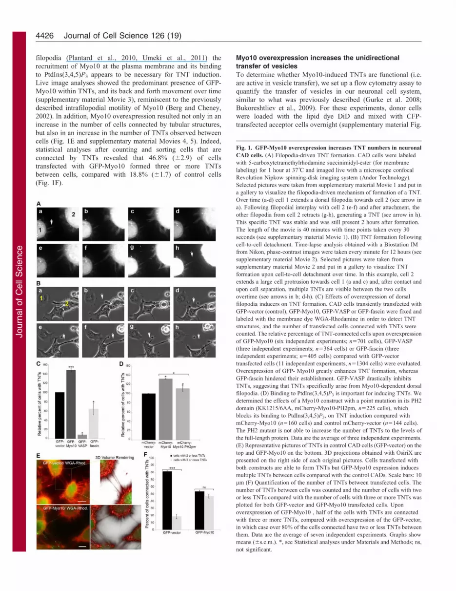
Fig. 1. GFP-Myo10 overexpression increases TNT numbers in neuronal
CAD cells. (A) Filopodia-driven TNT formation. CAD cells were labeled
with 5-carboxytetramethylrhodamine succinimidyl-ester (for membrane
labeling) for 1 hour at 37 C and imaged live with a microscope confocal
Revolution Nipkow spinning-disk imaging system (Andor Technology).
Selected pictures were taken from supplementary material Movie 1 and put in
a gallery to visualize the filopodia-driven mechanism of formation of a TNT.
Over time (a-d) cell 1 extends a dorsal filopodia towards cell 2 (see arrow in
a). Following filopodial interplay with cell 2 (e-f) and after attachment, the
other filopodia from cell 2 retracts (g-h), generating a TNT (see arrow in h).
This specific TNT was stable and was still present 2 hours after formation.
The length of the movie is 40 minutes with time points taken every 30
seconds (see supplementary material Movie 1). (B) TNT formation following
cell-to-cell detachment. Time-lapse analysis obtained with a Biostation IM
from Nikon, phase-contrast images were taken every minute for 12 hours (see
supplementary material Movie 2). Selected pictures were taken from
supplementary material Movie 2 and put in a gallery to visualize TNT
formation upon cell-to-cell detachment over time. In this example, cell 2
extends a large cell protrusion towards cell 1 (a and c) and, after contact and
upon cell separation, multiple TNTs are visible between the two cells
overtime (see arrows in b; d-h). (C) Effects of overexpression of dorsal
filopodia inducers on TNT formation. CAD cells transiently transfected with
GFP-vector (control), GFP-Myo10, GFP-VASP or GFP-fascin were fixed and
labeled with the membrane dye WGA-Rhodamine in order to detect TNT
structures, and the number of transfected cells connected with TNTs were
counted. The relative percentage of TNT-connected cells upon overexpression
of GFP-Myo10 (six independent experiments; n5701 cells), GFP-VASP
(three independent experiments; n5364 cells) or GFP-fascin (three
independent experiments; n5405 cells) compared with GFP-vector
transfected cells (11 independent experiments, n51304 cells) were evaluated.
Overexpression of GFP- Myo10 greatly enhances TNT formation, whereas
GFP-fascin hindered their establishment. GFP-VASP drastically inhibits
TNTs, suggesting that TNTs specifically arise from Myo10-dependent dorsal
filopodia. (D) Binding to PtdIns(3,4,5)P3 is important for inducing TNTs. We
determined the effects of a Myo10 construct with a point mutation in its PH2
domain (KK1215/6AA, mCherry-Myo10-PH2pm, n5225 cells), which
blocks its binding to PtdIns(3,4,5)P3, on TNT induction compared with
mCherry-Myo10 (n5160 cells) and control mCherry-vector (n5144 cells).
The PH2 mutant is not able to increase the number of TNTs to the levels of
the full-length protein. Data are the average of three independent experiments.
(E) Representative pictures of TNTs in control CAD cells (GFP-vector) on the
top and GFP-Myo10 on the bottom. 3D projections obtained with OsiriX are
presented on the right side of each original pictures. Cells transfected with
both constructs are able to form TNTs but GFP-Myo10 expression induces
multiple TNTs between cells compared with the control CADs. Scale bars: 10
mm (F) Quantification of the number of TNTs between transfected cells. The
number of TNTs between cells was counted and the number of cells with two
or less TNTs compared with the number of cells with three or more TNTs was
plotted for both GFP-vector and GFP-Myo10 transfected cells. Upon
overexpression of GFP-Myo10 , half of the cells with TNTs are connected
with three or more TNTs, compared with overexpression of the GFP-vector,
in which case over 80% of the cells connected have two or less TNTs between
them. Data are the average of seven independent experiments. Graphs show
means (6s.e.m.). *, see Statistical analyses under Materials and Methods; ns,
not significant.
Journal of Cell Science 126 (19)4426

JournalofCellScience
S2A). The amount of DiD-labeled vesicles transferred to the
acceptor CFP-transfected cells was then quantified by flow
cytometry (supplementary material Fig. S2B). As expected from
our previous studies, control CAD cells transfected with GFP-
vector can form functional TNTs (Gousset et al., 2009), allowing
transfer of DiD vesicles from the donor cells to the acceptor cells
that can be quantified by flow cytometry (supplementary material
Fig. S2C-F, GFP-vector). Overtime, we observed cell-to-cell
variability in the absolute number of vesicles transferring under
control conditions (6–14%) (supplementary material Fig. S2C-F,
GFP-vector). Thus, to compare different experimental conditions
we displayed the relative transfer of DiD vesicles normalized to
GFP-transfected donor cells – arbitrarily set at 100% (Fig. 2) as
previously shown (Gurke et al., 2008). To understand whether the
vesicle transfer observed in our experiments is the result of an
active intercellular transfer mechanism, we first analyzed the
transfer of vesicles obtained with fixed donor cells. As shown in
Fig. 2A, no significant transfer of DiD-labeled vesicles was
detected (Fig. 2A, Fixed; and supplementary material Fig. S2C),
suggesting that vesicle transfer did not derived from the transfer
of cell debris from the fixed donor population but requires an
active mechanism. Next, to assess for the involvement of
exosomes or membrane vesicles released within the medium,
we incubated acceptor cells with the supernatant of GFP-vector
donor cells. Under these conditions we detected only 28.8%
(63.7%) of transfer of DiD-labeled vesicles in acceptor cells,
demonstrating that, in our setting, over 70% of the vesicle
transfer requires cell-to-cell contact (Fig. 2A and supplementary
material Fig. S2D). Significantly, when donor cells were
transfected with GFP-Myo10 we observed a 1.4-fold increase
in the relative transfer of DiD-labeled vesicles compared with
control conditions (Fig. 2B and supplementary material Fig.
S2E). It is important to note that, in contrast to the previous
experiments where we specifically counted the number of TNTs
in CAD cells that express GFP-Myo10 (Fig. 1C), the flow
cytometry experiments cannot discriminate between GFP-Myo10
transfected cells (20–30% transfection efficiency) and non-
transfected cells. Thus, the 1.4-fold increase in vesicle transfer
measured upon GFP-Myo10 transfection compared with the
control cells (Fig. 2B) is highly significant.
To further characterize the role of Myo10 overexpression on
the transfer functionality of TNTs, we examined whether
expression of Myo10 in donor or acceptor cells affects vesicle
transfer. Thus, we analyzed the transfer of labeled vesicles from
donor cells in co-culture, when only the acceptor cells were
transfected with GFP-Myo10 (Fig. 2C and supplementary
material Fig. S2F). Despite increasing the number of TNTs,
overexpression of GFP-Myo10 in the acceptor cells had no effect
on the transfer of vesicles from the donor population (Fig. 2C and
supplementary material Fig. S2F). This suggests that, in CAD
cells, the transfer of vesicles within Myo10-dependent TNTs is
unidirectional, going from cells that created the tubes to acceptor
cells.
Endogenous Myo10 is necessary for formation and
function of TNTs
To assess whether Myo10 is required for TNT formation in CAD
cells, we used Myo10 shRNA lentiviral particles to reduce the
endogenous levels of Myo10 expression (Fig. 3A) and evaluated
the effects on the number of TNTs. The number of cells with
TNTs decreased by one-third when Myo10 expression was
downregulated (Fig. 3B), indicating that Myo10 is required for
the formation of a subset of TNTs in neuronal cells. Accordingly,
in Myo10 knockdown cells the transfer of DiD-labeled vesicles
was reduced to 78% (64.2%) compared with control cells
(Fig. 3C). Importantly, both the number of cells with TNTs
(Fig. 3B) and the transfer of vesicles (Fig. 3C) were rescued
upon overexpression of GFP-Myo10, demonstrating the
Fig. 2. Overexpression of Myo10 increases unidirectional transfer of DiD-labeled vesicles from donor to acceptor cells and needs cell-to-cell contact. To
account for the variability in the absolute transfer of vesicles between experiments (supplementary material Fig. 2C-F), we normalized our data to the GFP-
transfected control cells that we arbitrarily set at 100%. The relative percentage of transfer is presented for the each experimental condition. (A) The majority of
vesicle transfer requires cell-to-cell contact and an active mechanism. CAD cells transiently transfected with GFP-vector are able to transfer DiD-labeled vesicles
to acceptor cells. This transfer is highly restricted upon fixation of the donor cells and less than 30% of the vesicles are transferred through the medium. These data
are consistent with an active, cell-to-cell-dependent mechanism and correlates with the presence of TNTs within the cultures. These data are the average of three
independent experiments. (B) GFP-Myo10 overexpression in donor cells significantly increases the transfer of DiD-labeled vesicles compared with GFP-vector-
transfected cells, suggesting that Myo10 induces TNT formation. These data are the average of 18 independent experiments. (C) GFP-Myo10 overexpression
increases the transfer of DiD-labeled vesicles only when overexpression takes place in the donor cells. This suggests that the transfer of vesicles is unidirectional
and goes from the cells that create the tubes to the acceptor cells. These data are the average of three independent experiments. Graphs show means (6s.e.m.).
*, see Statistical analyses under Materials and Methods; ns, not significant.
Myo10 is required for TNT formation 4427

JournalofCellScience
specificity of the Myo10 effect on TNT formation and function. It
is important to note, however, that upon Myo10 knockdown the
loss of TNTs was not complete. These results may reflect
incomplete Myo10 knockdown (Fig. 3A) but, most probably,
point towards the existence of Myo10-independent pathways for
TNT formation – as previously described in different cell types
(for review see Abounit and Zurzolo, 2012). This is also in
agreement with our current observations that, in CAD cells,
TNTs ensue from filopodia-protrusions as well as following cell-
to-cell detachment (Movie 1, 2).
Full-length Myo10 is necessary for formation and function
of TNTs
As mentioned before, the Myo10 motor domain is required for
movement of Myo10 towards the tips of filopodia, whereas the
tail is required for binding of Myo10 to factors necessary for
filopodia elongation (Berg and Cheney, 2002; Zhang et al., 2004,
Plantard et al., 2010, Umeki et al., 2011). To determine the role
these domains might play in both TNT formation and function,
we used two Myo10 mutants: GFP-HMM-Myo10, consisting of
the head motor domain, neck and coiled-coil region of Myo10,
and GFP-Myo10-Headless, containing the neck, coiled-coil and
tail regions of Myo10 (Berg and Cheney 2002). We expressed
both deletion mutants in CAD cells and analyzed their
localization and effects on TNTs compared with the full-length
protein. As previously described (Berg and Cheney, 2002), GFP-
HMM-Myo10 is able to localize at the tips of filopodia, whereas
GFP-Myo10-Headless is cytosolic (supplementary material Fig.
S3A). Interestingly, and in contrast to the full-length protein
(Fig. 1C,E,F), both mutants were unable to increase the relative
number of cells with TNTs (Fig. 4A) or to create multiple TNTs
between cells (Fig. 4B,C and supplementary material Movies 6-
9). These data suggest that the localization of Myo10 at the tips
of filopodia and its ability to move within TNTs is not enough to
enhance TNT formation, and suggest that the tail domain of the
protein is also required. In agreement with these findings, we
found that, alone, neither construct can increase the transfer of
DiD-labeled vesicles to levels observed with GFP-Myo10
overexpression (Fig. 4D). These data highlight the correlation
described above between the number of TNTs in cells in culture
and the increase in the transfer of DiD-labeled vesicles, thus
further identifying TNTs as an important vesicle-transport
structure between neuronal cells.
Recently, it has been proposed that the headless form of
Myo10 functions as a dominant regulator of full-length Myo10
because it suppresses the filopodia-inducing activity of full-
length Myo10 upon coexpression in COS-7 cells (Raines et al.,
2012). Thus, we checked whether this also occurs with TNTs,
and co-transfected CAD cells with GFP-Myo10-Headless and
mCherry-Myo10. Under these conditions, we observed that
some of the headless protein relocalized from the cytosol
(supplementary material Fig. S3A) to the tips of filopodia,
together with the full-length protein (supplementary material Fig.
S3C). However, in these cells we did not observe any reduction
in the number of cells with TNTs compared with controls (cells
coexpressing mCherry-Myo10 and GFP-vector) (Fig. 4E). Thus,
in contrast to what was observed with substrate-attached
filopodia, the headless form is not a dominant regulator of
TNT formation, suggesting that the mechanisms by which
Myo10 induces substrate-attached filopodia and TNTs are
different and/or differently regulated.
The F2 subdomain of the FERM domain of Myo10 is
required for formation and function of TNTs
To better characterize the mechanism of Myo10-mediated TNT
induction, we decided to further investigate the role of the tail
domain, which we found to be essential both for the formation of
TNTs and for the transfer of labeled vesicles (Fig. 4A,D). To this
end, we used two previously described Myo10 mutants, GFP-
Myo10-DF2 and GFP-Myo10-DF3 that, respectively, lack the F2
or F3 lobes of the FERM domain located at the end of the Myo10
tail (Zhang et al., 2004). The F2 and F3 lobes of the FERM
domain are necessary for integrin binding and for the elongation
of attached filopodia (Zhang et al., 2004; Watanabe et al., 2010)
Fig. 3. Endogenous expression of Myo10 is necessary for TNT formation in CAD cells. (A) The endogenous levels of Myo10 were downregulated using
Myo10 shRNA lentiviral particles. (B) Downregulation of Myo10 results in a decrease in the number of TNTs in CAD cultures. The relative percentage of cells
connected by TNTs in Myo10-downregulated cells (n5456) compared with CAD cells treated with scramble shRNA lentiviral particles as control (n5425) was
evaluated. The ability of Myo10-downregulated cells to form TNTs decreases by one-third compared with control cells. The relative percentage of cells connected
by TNTs in Myo10-downregulated cells transfected with GFP-vector (control; n5272) can be rescued by overexpression of GFP-Myo10 (n5240). (C) In
correlation with the presence of TNTs observed by microscopy, flow cytometry analysis shows that the relative percentage of DiD-labeled vesicles transferred in
Myo10-downregulated cells decreases compared with the control cells. This defect in vesicle transfer can be rescued by overexpression of GFP-Myo10. Overall,
these experiments show that Myo10 expression is able to induce a subset of TNTs in neuronal cells, directly affecting their intercellular ability. Data represent the
average of at least three independent experiments. Graphs show means (6s.e.m.). *, see Statistical analyses under Materials and Methods: ns, not significant.
Journal of Cell Science 126 (19)4428

JournalofCellScience
but not for the induction of dorsal filopodia (Bohil et al., 2006).
In agreement with previous results both mutants localized at the
tips of filopodia, similar to full-length GFP-Myo10 (Zhang et al.,
2004), and were found within TNTs (supplementary material Fig.
S3B; Fig. 5A and Movies 10, 11). Furthermore, like the full-
length protein, GFP-Myo10-DF3 increased the number of cells
with TNTs and the number of TNTs between cells (Fig. 5B,C).
Consistently, flow cytometry experiments showed that GFP-
Myo10-DF3 also enhanced the transfer of DiD-labeled vesicles to
levels similar to those of GFP-Myo10 (Fig. 5D), demonstrating
that the F3 lobe is not required for TNT formation. By contrast,
GFP-Myo10-DF2 was unable to increase TNT formation
Fig. 4. See next page for legend.
Myo10 is required for TNT formation 4429

JournalofCellScience
(Fig. 5B,C) and the transfer of DiD-labeled vesicles (Fig. 5D).
The fact that the Myo10 construct that lacked the entire FERM
domain was still able to induce dorsal filopodia (Bohil et al.,
2006) is in sharp contrast to the inability of the GFP-Myo10-DF2mutant to increase functional TNTs, and indicates that the F2
lobe of the FERM domain is specifically required for the
formation of Myo10-dependent TNTs in neuronal cells.
A recent study in an urothelial cell line proposed that N-
cadherins has a role in TNT formation in that it provides
attachment sites to neighboring cells (Lokar et al., 2010). In
addition, in subconfluent human umbilical vein endothelial cells
(HUVECs) (Almagro et al., 2010), Myo10 has been shown to
mediate the transport of vascular endothelial-cadherins (VE-Cad)
within filopodia to the cell edges that are necessary for the correct
formation of cell–cell junctions. These independent observations
led us to propose that N-cadherin is a downstream binding
partner of Myo10. Thus, we coexpressed mCherry-Myo10 and
GFP-N-cadherin in CAD cells, and analyzed their localization in
subconfluent cells. However, we rarely found GFP-N-cadherin
within filopodia and only little colocalization with Myo10 was
observed (Fig. 5E), suggesting that other partners must be
involved in Myo10-induced formation of TNTs in neuronal cells.
Myo10 expression regulates the formation of TNTs in
neuronal cells in an Akt-pathway-independent manner
A recent study suggested that as a result of stress conditions, such
as serum starvation or oxidative stress, TNTs are induced by p53
and Akt activation in astrocytes (Wang et al., 2011). However,
this was not investigated in neuronal cells. Moreover, Akt has
been implicated in the regulation of myosin 5a activity by
increasing its interaction with F-actin and the transport of
GLUT4 vesicles (Yoshizaki et al., 2007). Although, in contrast to
myosin 5a, Myo10 is not a known vesicle carrier, we decided to
explore whether in neuronal cells there is a link between these
two mechanisms of TNT formation. First, we analyzed whether
Myo10-dependent TNT formation activates the Akt signaling
pathway. In neuronal cells, neither Myo10 downregulation
(Fig. 6A) nor Myo10 overexpression (Fig. 6B) led to a major
decrease or increase in endogenous Akt expression or Akt
phosphorylation levels. These data suggest that the induction of
Myo10-dependent TNTs in CAD cells is distinct from the TNT
formation observed in astrocytes under stress conditions.
Next, we analyzed whether oxidative stress induced TNTs in
CAD cells and whether this is directly linked to the activation of
Akt. Similar to what had been described previously (Wang et al.,
2011), treatment with H2O2 led to a drastic increase in the
number of cells with TNTs, with almost 70% of CAD cells
connected (Fig. 6C). However, western blot analyses revealed no
increase in the levels of endogenous Akt and Akt phosphorylation
in these cells (Fig. 6D). Thus, in contrast to what had been shown
in astrocytes (Wang et al., 2011), TNTs that formed in neuronal
cells following treatment with H2O2 are not the result of
activation of the Akt signaling pathway (Fig. 6D). These data
further demonstrate the existence of distinct mechanisms of TNT
formation in different cell types, even when a similar stimulus is
applied (in this case oxidative stress).
Discussion
The data presented here, on filopodia-driven TNT formation and the
subsequent intercellular transfer of vesicles, identify the
unconventional molecular motor Myo10 as a critical inducer of
TNTs in neuronal cells. Here, we show that a number of similarities
and differences exist between Myo10-dependent TNTs and
filopodia, and propose that a specific subset of Myo10-dependent
dorsal filopodia are the precursors of TNTs in neuronal cells. Similar
to filopodia (Plantard et al., 2010, Umeki et al., 2011), the PH2
domain is required forMyo10 activity in promoting TNTs (Fig. 1D).
This suggests that recruitment of Myo10 to the plasma membrane
through PtdIns(3,4,5)P3, which activates the motor allowing binding
to cargoes andmovement to the tips of filopodia (Umeki et al., 2011),
is also required in the case of TNTs. Likewise, using deletion
mutants, we show that neither the head nor the tail domains are
sufficient to induce the formation of functional TNTs, and only
overexpression of full-length Myo10 resulted in a substantial
increase in functional TNTs in our cell cultures. However, in
contrast to dorsal filopodia (Bohil et al., 2006), we demonstrated that
the F2 lobe of the FERM domain is critical for TNT induction.
FERM domains link cytoskeletal components and integral
membrane proteins. During filopodia elongation, Myo10 binds to
integrins through the F2 and F3 lobes of its FERM domain, and
brings them to the tips of filopodia creating adhesive platforms
(Zhang et al., 2004). However, Myo10-induction of dorsal
filopodia is integrin-independent and does not require the
FERM domain (Bohil et al., 2006). We show here that, in the
case of TNTs, the F3 deletion mutant mirrors full-length Myo10,
thereby demonstrating clearly that the F3 lobe is not required for
TNT formation and vesicle transfer. The F2 deletion mutant, by
contrast, is inactive, which demonstrates that this lobe is
essential. This suggests that, in contrast to attached-filopodia,
Myo10-dependent TNTs do not need to bind to integrin. Most
importantly, the F2 and F3 deletion mutants allowed the
distinction between dorsal filopodia and TNTs, the latter of
which specifically require the F2 lobe.
The function of the F2 lobe of the FERM domain of Myo10 is
not clear. Structural studies have shown that the F2 lobe is unique
Fig. 4. Full-length Myo10 is necessary for functional TNTs. The roles of
the motor or the tail domains of Myo10 on the induction of TNTs were
assessed. CADs cells were transiently transfected with GFP-vector (control),
GFP-Myo10, GFP-HMM-Myo10 (motor domain) and GFP-Myo10-Headless
(tail domain), and fixed and labeled with the membrane dye WGA-
Rhodamine in order to detect TNTs. (A) Only overexpression of full-length
GFP-Myo10 (n5331) increases the number of TNTs compared with the GFP-
vector control cells (n5660), GFP-HMM-Myo10 (n5484) or GFP-Myo10-
Headless (n5385). The levels of transfections for these constructs range from
60–70% for GFP-vector, 20–30% for GFP-Myo10 and GFP-Myo10-Headless
and 40–50% for GFP-HMM-Myo10. Data are the average of at least four
independent experiments. (B) Representative pictures of TNTs in the different
transfected cells are shown, together with 3D projections obtained using
OsiriX. Scale bars: 10 mm. (C) The number of TNTs between cells was
counted and plotted for all conditions. Only full-length GFP-Myo10-
expressing cells are able to form numerous multiple TNTs compared with the
other constructs. These data are the average of at least four independent
experiments. (D) Flow-cytometry experiments show that GFP-Myo10 greatly
enhances the transfer of DiD-labeled vesicles upon overexpression compared
with control cells and GFP-HMM-Myo10 and GFP-Myo10-Headless. These
data are the average of five independent experiments. (E) The Headless
isoform does not regulate the ability of Myo10 to increase TNTs. CAD cells
were co-transfected with mCherry-Myo10 and GFP-vector (control) or with
GFP-Headless, fixed and labeled with the membrane dye WGA-Rhodamine
in order to detect and count TNTs between transfected cells. We observed no
differences in the induction of TNTs when GFP-Headless was coexpressed
compared with GFP-vector control cells. Graphs show means (6s.e.m.).
*, see Statistical analyses under Materials and Methods; ns, not significant.
Journal of Cell Science 126 (19)4430

JournalofCellScience
and has a specific orientation compared with other FERM
subdomains (Wei et al. 2011). Deletion of the F2 lobe could,
therefore, disturb the correct orientation and folding of Myo10,
thereby affecting cargo recognition (Wei et al., 2011, Hirano
2011). Although most cargoes appear to bind to the F3 lobe, it is
conceivable that the F2 lobe interacts with a crucial binding
partner that is involved either in the attachment and/or in the
fusion of TNTs with the acceptor cells. This would explain why
this lobe is not important for dorsal filopodia but necessary for
the formation of functional TNTs. Whereas the specific cargoes
are still unknown, our data show that, in transfected cells, Myo10
and N-cadherin do not colocalize (Fig. 5E), N-cadherin being a
putative Myo10-binding partner (Almagro et al., 2010), which
was thought to be involved in TNT formation (Lokar et al.,
2010). Our findings suggest that interaction of N-cadherin with
Myo10 is not required for TNT formation in CAD cells.
Whether Myo10 is necessary for the initiation of filopodia, or
required for the transport of components necessary for actin
polymerization at the tips of filopodia, such as VASP or
PtdIns(3,4,5)P3, or whether it helps the growing actin filaments
by exerting a direct force at the tips remain to be determined.
Importantly, our study suggests that inside the Myo10-dependent
Fig. 5. The F2 subdomain of the FERM domain is required for functional TNT formation. (A) Representative images and 3D projections (OsiriX) of CADs
cells transiently transfected with GFP-Myo10-DF2 or GFP-Myo10-DF3, fixed and labeled with the membrane dye WGA-Rhodamine. Only GFP-Myo10-DF3 is
able to create numerous TNTs between cells. Scale bars: 10 mm. (B) The number of transfected cells connected with TNTs was counted. Differently from
GFP-Myo10-DF2 (n5280) both full-length GFP-Myo10 (n5310) and GFP-Myo10-DF3 (n5417) increase the number of TNTs. The level of transfection for these
constructs ranges from 35–45% for GFP-Myo10-DF2 and 45–55% for GFP-HMM-Myo10. These data are the average of at least three independent experiments.
(C) Quantification of the number of TNTs in transfected cells. TNTs between cells were counted, and the numbers of cells with two or less and three or more
TNTs were plotted. Full-length GFP-Myo10- and GFP-Myo10-DF3- but not GFP-Myo10-DF2-expressing cells, are able to form multiple TNTs. Data are the
average of at least three independent experiments. (D) The relative percentage of transfer of DiD-labeled vesicles, assessed by flow cytometry, is plotted. GFP-
Myo10 and GFP-Myo10-DF3 expression in donor cells increases the transfer of DiD-vesicles compared with GFP-vector and GFP-Myo10-DF2. These data are the
average of five independent experiments. Graphs show means (6s.e.m.). Overall, these data identify the F2 lobe of the FERM domain as a required component for
Myo10-dependent TNT formation in CAD cells. (E) No colocalization of GFP-N-cadherin with mCherry-Myo10 was observed in co-transfected CAD cells. Cells
co-transfected with mCherry-Myo10 and GFP-N-cadherin were fixed and imaged. GFP-N-cadherin is rarely found in filopodia and does not colocalize with
mCherry-Myo10. Scale bars: 10 mm. *, see Statistical analyses under Materials and Methods; ns, not significant.
Myo10 is required for TNT formation 4431

JournalofCellScience
TNTs the transfer of vesicle is unidirectional, from the cells that
created the tubes towards the recipient cells. This would be
consistent with an involvement of Myo10 in promoting the
polarized growth of actin cables inside the TNT and/or in vesicle
transport. Whether this involves Myo10 directly or requires other
myosins needs to be explored. Whereas the specific requirement of
the F2 lobe of the FERM domain for TNT formation allows one to
discriminate between Myo10-induced dorsal filopodia and TNTs,
we also demonstrate that TNTs do not arise from any dorsal
filopodia. Indeed, overexpression of known dorsal filopodia
inducers, such as VASP or fascin, failed to promote TNTs
(Fig. 1C). Interestingly, in addition to a drastic reduction in the
number of TNTs, overexpression of VASP changed the
morphology of CAD cells, increasing the cell-to-cell contact
zones (Sup. Fig. 1), a fact that is reminiscent to VASP and
cadherin-dependent formation of adhesion zippers described in
epithelial cells (Vasioukhin et al., 2000).
Downregulation and rescue experiments demonstrated that
Myo10 is necessary for the formation of a subset of TNTs and for
vesicle transfer (Fig. 3B,C). However, a large number of TNTs
remained in Myo10-knockdown cells, along with intercellular
transfer of vesicles (Fig. 3B,C). This could be because (1) we
were unable to completely silence Myo10 gene expression
(Fig. 3A); (2) CAD cells are able to form TNTs after cell-to-cell
separation (supplementary material Fig. S1 and Movie 2), a
process that might not require Myo10 or (3) Myo10 can enhance
de novo TNT formation but is not a limiting factor. It is worth
noting that similar results were observed with dorsal filopodia, in
which 90% gene silencing did not abrogate all dorsal filopodia
induction (Bohil et al., 2006), Similarly, downregulation of M-
Sec, a protein important for TNT formation in immune cells led
to a partial decrease in TNTs, with one-third of the M-Sec-
knockdown cells still able to form TNTs (Hase et al., 2009).
Finally, a recent article showing the involvement of LST1 in
TNT formation (Schiller et al., 2012) found an overall reduction
of 46% of cells with TNTs and a 30% decrease of the transfer of
vesicles after gene silencing compared with their control cells.
Overall, these experiments support the existence of multiple non-
redundant mechanisms of TNT formation in the same cell type,
possibly induced by different stimuli under different conditions.
Our data also point towards the existence of main differences
in the mechanisms of TNT formation in different cell types. We
show that, similar to astrocytes (Wang et al., 2011), neuronal
cells are able to induce TNT formation as a response to external
stress signals such as oxidative stress. However, it appears that
the mechanisms of TNT formation are the results of the
activation of different signaling pathways in the two cell types.
In astrocytes, the main signaling pathway following stress
conditions that leads to the formation of TNT is activation of
p53 and the Akt/mTOR signaling pathway (Wang et al., 2011).
By contrast, we show that in neuronal cells oxidative stress
increases TNTs in an Akt-independent manner, and no activation
of the Akt pathway in Myo10-overexpressing cells was observed.
These data suggest that multiple mechanisms of TNT formation
exist, and that different cell types have evolved different ways to
form TNTs. However, because Myo10 is expressed in most
tissues and different cell types (Berg et al., 2000; Yonezawa et al.,
2000), it will be interesting to determine whether it also plays a
role in LST1 or mSec-dependent TNT formation or whether its
role in TNT formation is restricted to neuronal cells.
In the brain, Myo10 is upregulated during nerve regeneration
following sciatic axotomy (Tanabe et al., 2003) or following
peripheral nerve injury (Plantman et al., 2013). In addition,
Myo10 is highly regulated during development of the nervous
system and exists in two forms (Sousa et al., 2006), the full-
length and the ‘headless’ isoform, which lacks the motor activity
and is differently regulated during development (Sousa et al.,
2006; Raines et al., 2012). Headless myosin is enriched in regions
of proliferating and migrating cells (Raines et al., 2012), and was
hypothesized to act as a dominant-negative isoform, regulating
Fig. 6. Similar to Myo10-dependent TNTs, the regulation of TNTs in
response to stress in CAD cells is independent of Akt activation. (A) CADs
cells downregulated for Myo10 (using Myo10 shRNA lentiviral particles) or
control CAD cells (using scramble shRNA lentiviral particles) were plated for
3 days, lysed, and 30 mg of protein was separated on an SDS-PAGE,
transferred and subjected to western blotting. As can be seen in the top band,
the levels of endogenous Myo10 are downregulated when using the Myo10
shRNA lentiviral particles but, under these conditions, levels of endogenous
Akt and phosphorylated Akt (Phospho-Akt) are not affected. (B) Myo10
levels and Akt activation was assessed in non-transfected CAD cells and cells
transiently transfected with GFP-vector (controls), and results were compared
with those derived from transfected cells expressing GFP-Myo10. The
samples were run on a gel and transferred to PVDF membranes for western
blot analysis. Overexpression of GFP-Myo10 greatly increases the levels of
Myo10 but does not affect the levels of Akt and phosphorylated Akt.
(C) Oxidative stress enhances the number of TNT-like structures observed in
CAD cells. CAD cells, treated or not (control) with 200 mM of H2O2 for 24
hours, were fixed and labeled with WGA-Rhodamine and TNTs were counted
24 hours post-treatment in untreated cells (n5735) and in treated cells
(n5588). Data are the average of three independent experiments (mean 6
s.e.m). (D) CAD cells plated for 3 days, were treated with 200 mM of H2O2 or
no H2O2 (control) for 24 hours, washed, lysed and 30 mg of protein was run on
an SDS-PAGE, transferred and subjected to western blotting. Treatment of
CAD cells with H2O2 did not increase the levels of endogenous Akt and
phosphorylated Akt. *, see Statistical analyses under Materials and Methods.
Journal of Cell Science 126 (19)4432

JournalofCellScience
Myo10 full-length activity in neurons (Sousa et al., 2006; Zhu
et al., 2007; Wang et al. 2009). Consistent with this hypothesis, in
cortical neurons, knockdown of full-length Myo10 reduces axon
outgrowth, whereas knockdown of headless Myo10 increases axon
outgrowth (Raines et al., 2012). In addition, headless Myo10 has
been shown to suppress the filopodia-inducing activity of the full-
length protein when coexpressed in COS-7 cells (Raines et al.,
2012). Here, however, we show that coexpression of both isoforms
does not block TNT induction (Fig. 4E), thus highlighting
differences in the regulation of Myo10 activity in filopodia and
TNTs. Interestingly, in the brain the netrin receptors deleted in
colorectal cancer (DCC) and neogenin (NEO1) have different
affinities for full-length Myo10 or the short headless form (Zhu
et al., 2007). Indeed, whereas both receptors bind to the FERM
domain of Myo10, neogenin associates more strongly to the
headless form of Myo10 whereas DCC has a greater binding
affinity for full-length Myo10. In addition, the expression of DCC
but not neogenin stimulates filopodia elongation (Zhu et al., 2007).
Thus, the levels of both forms of Myo10 in the brain and their
binding to the netrin receptors could be how TNT formation and
different downstream signaling pathways in the brain are
regulated. The significance of TNTs in the brain is not
understood. It is clear that distant cell communication has a
main role during development (For review see Abounit et al.,
2012). Therefore, the regulation of TNTs by Myo10 expression
could have an important role. In addition it was shown that the
misfolded infectious prion proteins (PrPSc) (Gousset et al., 2009;
Langevin et al., 2010) and intracellular Ab-fusion proteins (Wang
et al., 2011), as well as the aggregated form of Htt (Huntingtin)
exon 1 (Costanzo et al., 2013) take advantage of TNTs for
intercellular spread. This suggests that prion diseases and other
neurological diseases that occur as the result of protein misfolding,
such as Alzheimer, Parkinson and Huntington, use TNTs as a
common spreading mechanism (For review see Marzo et al., 2012;
Costanzo and Zurzolo, 2013). Interestingly, preliminary data in our
laboratory suggest that overexpression of GFP-Myo10 results in a
drastic increase in the transfer of PrPSc particles from chronically
infected ScCAD cells to non-infected CAD cells (our unpublished
data). Thus, it is possible that, upon stress, toxicity due to prion
infection or the presence of toxic aggregates, the levels of full-
length Myo10 are upregulated, which leads to an increase in TNT-
like structures allowing faster spreading of prions and prion-like
proteins in the brain (for a review see Costanzo and Zurzolo,
2013). Studies on the role of toxicity due to infection and/or the
presence of aggregates are currently underway in our laboratory.
Overall, this study highlights the key role that Myo10 has in
TNT formation within neuronal cells and the differences
compared with other cellular models. Further investigation will
be necessary to determine the specific role of the F2 lobe of the
FERM domain in TNT formation. It is also tempting to speculate
that the balance of the levels of full-length and the headless
isoform of Myo10 in the brain is a way to regulate TNT
formation. Therefore, whether and how these levels vary in cases
of brain injuries, toxic stimuli during prion infection or during the
progression of neurodegenerative diseases will deserve full
attention.
Material and MethodsCell lines, antibodies, transfection and transduction
Mouse neuronal CAD cells were a gift from Hubert Laude (Institut National de la
Recherche Agronomique, Jouy-en-Josas, France) and were cultured in Opti-MEM(Invitrogen) and 10% fetal bovine serum. Transient transfections were performed
with Lipofectamine 2000 (Invitrogen) in accordance with the manufacturer’s
instructions. The bovine GFP-Myo10, GFP-HMM-Myo10 and GFP-Myo10-
Headless were gifts from Richard E. Cheney (University of North Carolina,
Chapel Hill, NC, USA) and GFP-Myo10-DF2, GFP-Myo10-DF3, mCherry-Myo10
and mCherry-Myo10-KK1215/6AA (PH2 mutant) were gifts from Staffan
Stromblad (Center for Biosciences, Department of Biosciences and Nutrition,
Karolinska Institutet, Stockholm, Sweden). GFP-Fascin was a gift from Josephine
C. Adams (Dept. of Cell Biology, Cleveland Clinic, Cleveland, USA) and GFP-
VASP was obtained from Sandrine Etienne-Manneville (Pasteur Institute, Paris,
France). Human anti-Myo10 rabbit polyclonal antibody, which detects Myo10
from multiple species including mouse, was purchased from Sigma (HPA024223)
and Myo10 shRNA lentiviral particles, control shRNA lentiviral particles and
PolybreneH reagent were from Santa Cruz Biotechnology, Inc., and were used
according to the manufacturer’s instructions. Akt and Phospo-Akt antibodies were
purchased from Cell Signaling Technology, Inc. and wheat germ agglutinin
(WGA) tetramethylrhodamine conjugate (WGA-Rhodamine) and the lipophilic
dye DiD were purchased from Molecular Probes (Invitrogen). Hydrogen peroxide
(30% wt) was purchased from Sigma.
Myo10 shRNA lentiviral particle transduction and selection of stable clones
CAD cells were plated at a density of 40,000 in a 12-well plate for 24 hours before
transduction. Transduction was performed according to the manufacturers’
instructions (Santa Cruz Biotechnology, Inc). Stable clones that express both
control Myo10 and Myo10 short hairpin RNA (shRNA) were selected, and
maintained in culture by adding 5 mg/ml of puromycin dihydrochloride to
eliminate non-transduced cells. Western blotting was used to evaluate
downregulation of gene expression and to select for the best clones.
For rescue experiments, stable shRNA control and shRNA-Myo10 cells were
transfected with GFP-vector or GFP-Myo10, respectively, using Lipofectamine
2000. Twenty-four hours after transfection, TNTs were counted in cells or for
flow-cytometry analyses was carried out as described below.
Because the Myo10 shRNA lentiviral particles that we used in our experiments
had been designed to inhibit expression of Myo10 in mouse cells and we used a
bovine GFP-Myo10 construct for transient transfections, the construct was not a
target for shRNA and normal expression levels were observed.
Gel electrophoresis and western blots
Transfected cells, shRNA knockdown cells or cells treated with H2O2 (200 mM for
24 hours) were lysed in lysis buffer (0,5% Triton X-100, 0,5% DOC, 100 mM NaCl,
10 mM Tris-HCl pH 8) and the amount of protein per ml was quantified in a Bradford
assay. Thirty micrograms of protein, denatured in Laemmli buffer and boiled for
5 minutes, was loaded on 8% SDS-polyacrylamide gels and transferred into PVDF
membrane for western blot analyses with the appropriate primary antibodies. HRP-
conjugated secondary antibodies and ECLTM reagents fromAmersham (GEHealthcare)
were used for detection.
Detection of TNTs by using fluorescence microscopy
To evaluate the number of TNT-connected cells, TNTs were analyzed by
fluorescence microscopy. Cells were plated on T25 flasks overnight and
transfected the following day with the appropriate plasmids using Lipofectamine
2000. The next day, cells were counted and 200,000 cells were plated overnight on
Ibidi m-Dishes (Biovalley, France) at 37 C. Cells were then fixed for 20 minutes at
room temperature (RT) using fixative solution 1 (2% PFA, 0.05% glutaraldehyde
and 0.2 M HEPES in PBS) followed by an additional incubation of 20 minutes
with fixative solution 2 (4% PFA and 0.2 M HEPES in PBS). The cells were
carefully washed in PBS and labeled for 20 minutes at RT with WGA-Rhodamine
(1:300 in PBS), washed and sealed with Aqua-Poly mount (Polysciences, Inc.).
Image stacks covering the whole cellular volume were acquired using a widefield
microscope (Zeiss Axiovert 200M) controlled by Axiovision software. To evaluate
the number of TNT-connected cells (i.e. structures connecting two cells that do not
touch the substratum), manual analysis was performed and each experiment was
carried out at least in triplicate. Image analyses of raw data, such as Z-projections
and 3D reconstructions, were obtained using ImageJ software (http://rsb.info.nih.
gov/ij/) and OsiriX (Osirix Medical Imaging software) software programs.
Live imaging systems
The membrane of CAD cells was labeled with 5-carboxytetramethylrhodamine
succinimidyl-ester for 1 hour at 37 C, and cells were imaged live using an Andor
spinning-disk confocal microscope. Z-stacks of 0.3 mm steps were taken for each
time points and Z-projections were obtained in order to look at filopodia-driven
TNT formation over time (Movie 1). The same imaging system was used to
analyze the movements of GFP-Myo10 within TNTs in transiently transfected
CAD cells over specific periods of time (supplementary material Movie 3). Long
time-lapse movies were acquired with the Biostation IM from Nikon (Movie 2).
Image analyses of raw data was obtained using ImageJ (http://rsb.info.nih.gov/ij/)
software program.
Myo10 is required for TNT formation 4433

JournalofCellScience
Flow cytometry assay to quantify the transfer of DiD-labeled vesicles
CAD cells were transfected with the appropriate GFP-constructs (donor cells) and
CFP-vector (acceptor cells) in T25 dishes for 24 hours. The donor cells were then
counted and labeled with the membrane dye DiD (1:3000 in complete medium for
30 minutes at 37 C), the cells were centrifuged at 1000 rpm for 5 minutes, to
remove the DiD solution, resuspended in complete medium and incubated for 30
more minutes at 37 C (to internalize the dye). The cells were then centrifuged once
more to wash away any remaining dye. The labeled donor cells were then mixed
(1:1) with CFP-transfected acceptor cells and plated on 35-mm dishes overnight.
Each independent experiment was performed in triplicates. The following day,
cells were washed with PBS to remove any dead cells, scraped of the dishes with
500 ml PBS and passed through sterile 40-mm nylon cell strainers (BD FalconTM)
in order to obtain single-cell suspensions. Cells were then mixed with 500 ml of
4% PFA (2% final solution). For FACS analyses, DiD-labeled donor cells and
CFP-vector-transfected acceptor cells were analyzed at 633 nm and 488 nm
excitation wavelengths, respectively. Flow cytometry data were acquired using a
CyAn ADP flow cytometer (Dako Cytomation, Beckman Coulter, Inc.). Samples
were analyzed at low flow rate, corresponding to 50–150 events per second, and
each independent experiment was performed in triplicate (30,000 cells for each
condition). Doublet discrimination on the basis of signal processing was achieved
by plotting the peak height against the area of side scatter. The data were analyzed
using KaluzaH Flow Analysis Software (Beckman Coulter, Inc.).
Statistical analyses
In order to analyze the significance between two experimental conditions, we used
the Mann-Whitney U-Test. The z-score was calculated using the normal
approximation, such that z~U{
nxny
2ffiffiffiffiffiffiffiffiffiffiffiffiffiffiffiffiffiffiffiffiffiffiffiffi
nxny Nz1ð Þ
12
r .
The differences were considered significant at *P,0.05 when 21.96.z.1.96; at
**P,0.01 when 22.58.z.2.58; or ***P,0.001 when 23.291.z.3.291, wherez is the z-score for the normal approximation of the data. In graphs, asterisks abovecolumns indicate comparison with control cells, whereas brackets indicate anyother paired comparisons.
Acknowledgements
We thank R. E. Cheney, S. Stromblad, J. C. Adams, S. Etienne-Manneville for their generous gifts of constructs. We thank H. Ohnofor personal communications about M-Sec and R. E. Cheney for
discussions about Myo10. We thank members of the Zurzololaboratory for critical reading of the manuscript. Work in C.Z.’s labis supported by the European Union FP7 (Priority, Grant 222887), byANR (ANR-09-BLAN-0122, ANR-ERANET) and by the Pasteur-
Weizmann Foundation (2010–2012).
Author Contributions
C.Z. coordinated the project. K.G. planned, performed and analyzed
the experiments. L.M. performed the unpublished infectiousexperiments and provided technical help. P.-H.C. set up theparameters for the flow cytometry assays. K.G. and C.Z. wrote themanuscript. All authors discussed the results and manuscript text.
Funding
Work in C.Z.’s lab is supported by the European Union FP7[Priority, grant number 222887 to C.Z.], the Agence Nationale de la
Recherche (ANR) [grant numbers: ANR-09-BLAN-0122 and ANR-09-NEUR-002-03 to C.Z.] and the Pasteur-Weizmann Foundation(2010–2012) to CZ. K.G. was sponsored in part by the PasteurFoundation Fellowship Program.
Competing Interests
The authors declare that they have no competing interests.
Supplementary material available online at
http://jcs.biologists.org/lookup/suppl/doi:10.1242/jcs.129239/-/DC1
ReferencesAbounit, S. and Zurzolo, C. (2012). Wiring through tunneling nanotubes – from
electrical signals to organelle transfer. J. Cell Sci. 125, 1089-1098.
Adams, J. C. and Schwartz, M. A. (2000). Stimulation of fascin spikes by
thrombospondin-1 is mediated by the GTPases Rac and Cdc42. J. Cell Biol. 150,
807-822.
Almagro, S., Durmort, C., Chervin-Petinot, A., Heyraud, S., Dubois, M., Lambert,
O., Maillefaud, C., Hewat, E., Schaal, J. P., Huber, P. and Gulino-Debrac,
D. (2010). The motor protein myosin-X transports VE-cadherin along filopodia to
allow the formation of early endothelial cell-cell contacts. Mol. Cell Biol. 7, 1703-
1717.
Berg, J. S. and Cheney, R. E. (2002). Myosin-X is an unconventional myosin that
undergoes intrafilopodial motility. Nat. Cell Biol. 4, 246-250.
Berg, J. S., Derfler, B. H., Pennisi, C. M., Corey, D. P. and Cheney, R. E. (2000).
Myosin-X, a novel myosin with pleckstrin homology domains, associates with regions
of dynamic actin. J. Cell Sci. 113, 3439-3451.
Bohil, A. B., Robertson, B. W. and Cheney, R. E. (2006). Myosin-X is a molecular
motor that functions in filopodia formation. Proc. Natl. Acad. Sci. USA 103, 12411-
12416.
Bukoreshtliev, N. V., Wang, X., Hodneland, E., Gurke, S., Barroso, J. F. and Gerdes,
H. H. (2009). Selective block of tunneling nanotube (TNT) formation inhibits
intercellular organelle transfer between PC12 cells. FEBS Lett. 583, 1481-1488.
Costanzo, M. and Zurzolo, C. (2013). The cell biology of prion-like spread of protein
aggregates: mechanisms and implication in neurodegeneration. Biochem. J. 452, 1-17.
Costanzo, M., Abounit, S., Marzo, L., Danckaert, A., Chamoun, Z., Roux, P. and
Zurzolo, C. (2013). Transfer of polyglutamine aggregates in neuronal cells occurs in
tunneling nanotubes. J. Cell Sci. 126, 3678-3685.
Davis, D. M. and Sowinski, S. (2008). Membrane nanotubes: dynamic long-distance
connections between animal cells. Nat. Rev. Mol. Cell Biol. 9, 431-436.
Eugenin, E. A., Gaskill, P. J. and Berman, J. W. (2009). Tunneling nanotubes (TNT)
are induced by HIV-infection of macrophages: a potential mechanism for intercellular
HIV trafficking. Cell. Immunol. 254, 142-148.
Gousset, K., Schiff, E., Langevin, C., Marijanovic, Z., Caputo, A., Browman, D. T.,
Chenouard, N., de Chaumont, F., Martino, A., Enninga, J. et al. (2009). Prions
hijack tunnelling nanotubes for intercellular spread. Nat. Cell Biol. 11, 328-336.
Gurke, S., Barroso, J. F., Hodneland, E., Bukoreshtliev, N. V., Schlicker, O. and
Gerdes, H. H. (2008). Tunneling nanotube (TNT)-like structures facilitate a
constitutive, actomyosin-dependent exchange of endocytic organelles between normal
rat kidney cells. Exp. Cell Res. 314, 3669-3683.
Hase, K., Kimura, S., Takatsu, H., Ohmae, M., Kawano, S., Kitamura, H., Ito, M.,
Watarai, H., Hazelett, C. C., Yeaman, C. et al. (2009). M-Sec promotes membrane
nanotube formation by interacting with Ral and the exocyst complex. Nat. Cell Biol.
11, 1427-1432.
Hirano, Y., Hatano, T., Takahashi, A., Toriyama, M., Inagaki, N. and Hakoshima,
T. (2011). Structural basis of cargo recognition by the myosin-X MyTH4-FERM
domain. EMBO J. 30, 2734-2747.
Kerber, M. L. and Cheney, R. E. (2011). Myosin-X: a MyTH-FERM myosin at the tips
of filopodia. J. Cell Sci. 124, 3733-3741.
Langevin, C., Gousset, K., Costanzo, M., Richard-Le Goff, O. and Zurzolo. (2010).
Characterization of the role of dendritic cells in prion transfer to primary neurons.
Biochem. J. 431, 189-198.
Lokar, M., Iglic, A. and Veranic, P. (2010). Protruding membrane nanotubes:
attachment of tubular protrusions to adjacent cells by several anchoring junctions.
Protoplasma 246, 81-87.
Marzo, L., Gousset, K. and Zurzolo, C. (2012). Multifaceted roles of tunneling
nanotubes in intercellular communication. Front. Physiol 3, 72.
Onfelt, B., Nedvetzki, S., Yanagi, K. and Davis, D. M. (2004). Cutting edge:
membrane nanotubes connect immune cells. J. Immunol. 173, 1511-1513.
Onfelt, B., Nedvetzki, S., Benninger, R. K., Purbhoo, M. A., Sowinski, S., Hume,
A. N., Seabra, M. C., Neil, M. A., French, P. M. and Davis, D. M. (2006).
Structurally distinct membrane nanotubes between human macrophages support long-
distance vesicular traffic or surfing of bacteria. J. Immunol. 177, 8476-8483.
Pasquier, J., Guerrouahen, B. S., Al Thawadi, H., Ghiabi, P., Maleki, M., Abu-
Kaoud, N., Jacob, A., Mirshahi, M., Galas, L., Rafii, S. et al. (2013). Preferential
transfer of mitochondria from endothelial to cancer cells through tunneling nanotubes
modulates chemoresistance. J. Transl. Med. 11, 94. (Epub ahead of print).
Plantard, L., Arjonen, A., Lock, J. G., Nurani, G., Ivaska, J. and Stromblad,
S. (2010). PtdIns(3,4,5)P3 is a regulator of myosin-X localization and filopodia
formation. J. Cell Sci. 123, 3525-3534.
Plantman, S., Zelano, J., Novikova, L. N., Novikov, L. N. and Cullheim, S. (2013).
Neuronal myosin-X is upregulated after peripheral nerve injury and mediates laminin-
induced growth of neurites. Mol. Cell. Neurosci. 56, 96-101.
Qi, Y., Wang, J. K., McMillian, M. and Chikaraishi, D. M. (1997). Characterization
of a CNS cell line, CAD, in which morphological differentiation is initiated by serum
deprivation. J. Neurosci. 17, 1217-1225.
Raines, A. N., Nagdas, S., Kerber, M. L. and Cheney, R. E. (2012). Headless Myo10
is a negative regulator of full-length Myo10 and inhibits axon outgrowth in cortical
neurons. J. Biol. Chem. 287, 24873-24883.
Rustom, A., Saffrich, R., Markovic, I., Walther, P. and Gerdes, H. H. (2004).
Nanotubular highways for intercellular organelle transport. Science 303, 1007-1010.
Schiller, C., Diakopoulos, K. N., Rohwedder, I., Kremmer, E., von Toerne, C.,
Ueffing, M., Weidle, U. H., Ohno, H. and Weiss, E. H. (2013). LST1 promotes the
assembly of a molecular machinery responsible for tunneling nanotube formation.
J. Cell Sci. 126, 767-777.
Journal of Cell Science 126 (19)4434

JournalofCellScience
Sousa, A. D., Berg, J. S., Robertson, B. W., Meeker, R. B. and Cheney, R. E. (2006).Myo10 in brain: developmental regulation, identification of a headless isoform and
dynamics in neurons. J. Cell Sci. 119, 184-194.Sowinski, S., Jolly, C., Berninghausen, O., Purbhoo, M. A., Chauveau, A., Kohler,
K., Oddos, S., Eissmann, P., Brodsky, F. M., Hopkins, C. et al. (2008). Membrane
nanotubes physically connect T cells over long distances presenting a novel route for
HIV-1 transmission. Nat. Cell Biol. 10, 211-219.Tanabe, K., Bonilla, I., Winkles, J. A. and Strittmatter, S. M. (2003). Fibroblast
growth factor-inducible-14 is induced in axotomized neurons and promotes neurite
outgrowth. J. Neurosci. 23, 9675-9686.Tokuo, H., Mabuchi, K. and Ikebe, M. (2007). The motor activity of myosin-X
promotes actin fiber convergence at the cell periphery to initiate filopodia formation.
J. Cell Biol. 179, 229-238.Umeki, N., Jung, H. S., Sakai, T., Sato, O., Ikebe, R. and Ikebe, M. (2011).
Phospholipid-dependent regulation of the motor activity of myosin X. Nat. Struct.
Mol. Biol. 18, 783-788.
Van Prooyen, N., Gold, H., Andresen, V., Schwartz, O., Jones, K., Ruscetti, F.,Lockett, S., Gudla, P., Venzon, D. and Franchini, G. (2010). Human T-cell
leukemia virus type 1 p8 protein increases cellular conduits and virus transmission.
Proc. Natl. Acad. Sci. USA 107, 20738-20743.Vasioukhin, V., Bauer, C., Yin, M. and Fuchs, E. (2000). Directed actin
polymerization is the driving force for epithelial cell-cell adhesion. Cell 100, 209-219.Wang, J. J., Fu,X.Q.,Guo,Y.G.,Yuan,L.,Gao,Q.Q.,Yu,H.L., Shi,H.L.,Wang,X.Z.,
Xiong,W. C. and Zhu, X. J. (2009). Involvement of headless myosin X in the motility of
immortalized gonadotropin-releasing hormone neuronal cells. Cell Biol. Int. 33, 578-585.
Wang, X., Veruki, M. L., Bukoreshtliev, N. V., Hartveit, E. and Gerdes,H. H. (2010). Animal cells connected by nanotubes can be electrically coupled
through interposed gap-junction channels. Proc. Natl. Acad. Sci. USA 107, 17194-
17199.
Wang, Y., Cui, J., Sun, X. and Zhang, Y. (2011). Tunneling-nanotube development in
astrocytes depends on p53 activation. Cell Death Differ. 18, 732-742.
Watanabe, T. M., Tokuo, H., Gonda, K., Higuchi, H. and Ikebe, M. (2010). Myosin-X
induces filopodia by multiple elongation mechanism. J. Biol. Chem. 285, 19605-19614.
Watkins, S. C. and Salter, R. D. (2005). Functional connectivity between immune cells
mediated by tunneling nanotubules. Immunity 23, 309-318.
Weber, K. L., Sokac, A. M., Berg, J. S., Cheney, R. E. and Bement, W. M. (2004). A
microtubule-binding myosin required for nuclear anchoring and spindle assembly.
Nature 431, 325-329.
Wei, Z., Yan, J., Lu, Q., Pan, L. and Zhang, M. (2011). Cargo recognition mechanism
of myosin X revealed by the structure of its tail MyTH4-FERM tandem in complex
with the DCC P3 domain. Proc. Natl. Acad. Sci. USA 108, 3572-3577.
Yonezawa, S., Kimura, A., Koshiba, S., Masaki, S., Ono, T., Hanai, A., Sonta, S.,
Kageyama, T., Takahashi, T. and Moriyama, A. (2000). Mouse myosin X:
molecular architecture and tissue expression as revealed by northern blot and in situ
hybridization analyses. Biochem. Biophys. Res. Commun. 271, 526-533.
Yoshizaki, T., Imamura, T., Babendure, J. L., Lu, J. C., Sonoda, N. and Olefsky,
J. M. (2011). Myosin 5a is an insulin-stimulated Akt2 (protein kinase Bbeta) substrate
modulating GLUT4 vesicle translocation. Mol. Cell Biol. 27, 5172-5183.
Zhang, H., Berg, J. S., Li, Z., Wang, Y., Lang, P., Sousa, A. D., Bhaskar, A., Cheney,
R. E. and Stromblad, S. (2004). Myosin-X provides a motor-based link between
integrins and the cytoskeleton. Nat. Cell Biol. 6, 523-531.
Zhu, X. J., Wang, C. Z., Dai, P. G., Xie, Y., Song, N. N., Liu, Y., Du, Q. S., Mei, L.,
Ding, Y. Q. and Xiong, W. C. (2007). Myosin X regulates netrin receptors and
functions in axonal path-finding. Nat. Cell Biol. 9, 184-192.
Myo10 is required for TNT formation 4435