Mutations in the mitochondrial split gene COXI are ... - Hal-CEA
11
HAL Id: cea-02474786 https://hal-cea.archives-ouvertes.fr/cea-02474786 Submitted on 11 Feb 2020 HAL is a multi-disciplinary open access archive for the deposit and dissemination of sci- entific research documents, whether they are pub- lished or not. The documents may come from teaching and research institutions in France or abroad, or from public or private research centers. L’archive ouverte pluridisciplinaire HAL, est destinée au dépôt et à la diffusion de documents scientifiques de niveau recherche, publiés ou non, émanant des établissements d’enseignement et de recherche français ou étrangers, des laboratoires publics ou privés. Mutations in the mitochondrial split gene COXI are preferentially located in exons: a mapping study of 170 mutants Pierre Netter, Sylviane Robineau, Claire Lemaire To cite this version: Pierre Netter, Sylviane Robineau, Claire Lemaire. Mutations in the mitochondrial split gene COXI are preferentially located in exons: a mapping study of 170 mutants. Molecular and General Genetics MGG, Springer Verlag, 1995, 246 (4), pp.445-454. 10.1007/bf00290448. cea-02474786
-
Upload
khangminh22 -
Category
Documents
-
view
0 -
download
0
Transcript of Mutations in the mitochondrial split gene COXI are ... - Hal-CEA

HAL Id: cea-02474786https://hal-cea.archives-ouvertes.fr/cea-02474786
Submitted on 11 Feb 2020
HAL is a multi-disciplinary open accessarchive for the deposit and dissemination of sci-entific research documents, whether they are pub-lished or not. The documents may come fromteaching and research institutions in France orabroad, or from public or private research centers.
L’archive ouverte pluridisciplinaire HAL, estdestinée au dépôt et à la diffusion de documentsscientifiques de niveau recherche, publiés ou non,émanant des établissements d’enseignement et derecherche français ou étrangers, des laboratoirespublics ou privés.
Mutations in the mitochondrial split gene COXI arepreferentially located in exons: a mapping study of 170
mutantsPierre Netter, Sylviane Robineau, Claire Lemaire
To cite this version:Pierre Netter, Sylviane Robineau, Claire Lemaire. Mutations in the mitochondrial split gene COXIare preferentially located in exons: a mapping study of 170 mutants. Molecular and General GeneticsMGG, Springer Verlag, 1995, 246 (4), pp.445-454. �10.1007/bf00290448�. �cea-02474786�

Mol Gen Genet (1995) 246:445~454 © Springer-Verlag 1995
Pierre Netter • Sylviane Robineau • Claire Lemaire
Mutations in the mitochondrial split gene COXI are preferentially located in exons: a mapping study of 170 mutants
Received: 18 July 1994 / Accepted: 14 September 1994
Abstract We have analysed the precise location of a large number (170) of mutations affecting the structural gene for subunit I of the cytochrome c oxidase complex. This gene, COXI, is 12.9 kb long and the major part of the sequence (i.e. 11.3 kb) is composed of introns. Sever- al conclusions can be drawn from this study: (1) A sig- nificant proportion (84/170) of the mutations cannot be assigned to a single position within the gene by deletion mapping, in spite of clearly being located in it. These mutations are probably large deletions or multiple mu- tations. (2) Four mutants carry distant double muta- tions, which have been individually localized. (3) Eighty- two mutants have lesions that are restricted to very short regions of the gene and we therefore conclude that they are most probably due to single hits; amongst these single mutations, 41 are unambiguously located in ex- ons and 28 in introns. This result implies that, at least in this particular split gene, the probability of selection of a mutant phenotype in an exon is, on the average, 13.3 times greater than in an intron, in spite of the existence, within most of these introns, of open reading frames specifying intronic proteins. The evolutionary signifi- cance and biological implications of these results are discussed.
Key words Yeast - Cytochrome oxidase Mitochondria • Intronic proteins • Ribozymes
Introduction
This study focuses on the gene encoding subunit I of cytochrome c oxidase, which is the last complex of the mitochondrial respiratory chain. In the yeast Saccha- romyces cerevisiae, it is composed of 12 subunits ranging
Communicated by A. Kondorosi
P. Netter ([~) . S. Robineau • C. Lemaire Centre de G~n6tique Mol~culaire du CNRS, Laboratoire associ~ ~i l'Universit6 Pierre et Marie Curie, Avenue de la Terrasse, 91198-Gif-sur-Yvette, France
in size from 55 to 5 kDa (Taanman and Capaldi 1992). Subunits I, II and III (COXIp, COXIIp and COXIIIp) are specified, respectively, in the mitochondrial DNA by COXI, COXII and COXIII genes (Slonimski and Tza- goloff 1976; Coruzzi and Tzagoloff 1979; Bonitz et al. 1980; Thalenfeld and Tzagoloff 1980). The counterparts of these three polypeptides can be found in bacteria and are apparently sufficient, in these prokaryotic organ- isms, to build a fully active complex (Haltia et al. 1988; Hosler et al. 1992). The other subunits present in yeast are encoded by nuclear genes (Maarse et al. 1984; Seraphin et al. 1985; Cumsky et al. 1987; Wright et al. 1984; Taanman and Capaldi 1993; Lamarche et al. 1992; Calder and McEwen 1990; Wright et al. 1986; Patterson and Poyton 1986) and are synthesized on cy- toplasmic ribosomes before being imported and assem- bled in mitochondria. Subunit I of the cytochrome oxi- dase is highly conserved amongst all living organisms, from bacteria to animals and plants. This integral mem- brane protein exhibits 12 putative transmembrane he- lices (Rao and Argos 1986) and, together with subunits II and III, constitutes the catalytic core of the last res- piratory mitochondrial complex.
The structural gene for subunit I, COXI, is located on the mitochondrial DNA and shows a mosaic organ- isation: it is 12 877 nucleotides long but the eight exons specifying subunit I itself are only 1602 nucleotide long (corresponding to a protein of 534 amino acids) and are separated by seven introns (Bonitz et al. 1980; Hensgens et al. 1983). These introns are removed from a long RNA precursor, generating the mature m R N A for the cytochrome oxidase subunit (Carignani et al. 1986).
Such introns, which have been discovered and exten- sively studied in fungal mitochondria and chloroplasts (for review see Lambowitz and Belfort 1993), exhibit several very interesting properties. Their primary se- quences allow them to be folded after transcription of the pre-mRNA, giving rise to two different three-dimen- sional structures specific to so-called group I and group II introns (Davies et al. 1982; Michel and Dujon 1983). These structured introns have ribozyme activities, lead-

446
It, Avail I I I I I Hae TIT I ~ ] I I I Bgl l I I
Mbol I I I I I I I I II I I I I I I I
b A1 A2 A3 A4 A5 A6 A7 A8
ai3 ai4 ai7 I " n i l 11 ~i2 a i5 a i6
20;0 40b0 o / MB5
/ ' - - 'M21"~. i" L24
/ AHA7411 \ [ 'AN111 j / M3 \ / ~ M A 8 1 \
L513
6o'oo
,~-%\ \\\\\\\\\ \\\\" R ACO ! ~\\\\\\\\\\\\\\\\\'~
Fig. la-e Physical map of the COXI gene and of discriminating rho- clones, a Restriction map of the COXI gene: the positions of sites for restriction endonucleases AvaII, HaeIII, BglII and MboI
8200U t0 30601 , 8585 11645 13003
, , q N - N - N R F 4 \ \ \ \ \ \ [ N A 3 ~ I [ ' A 4 : I
F51 ~ ~<,.\\\\\\ RG?I x\\\\\\\\\\" AI,\\\\\\\\\\~
~ i::ii!il iii::!il ~?;i~il iiii;i~ i::i::i :.i(ii!ii ii::ii!i ::!::! i i~::!ilili~ l ~ Y 9 2 ~ I ND8N ~ E 4 ~
~ F 3 ~
are shown [deduced from (Bonitz et al. 1980) and (Hensgens et al. 1983)]. b Schematic representation of the COXI gene. The introns ai5 and ai6 are facultative, present in "long" strains KL14-4A and AB1-4A but absent in the "short" strain LM940-1 without any detectable phenotype. The black boxes correspond to exons A1 to A8, interspersed by introns ail to ai7. The open rectangles in introns represent the intronic open reading frames (ORFs). The nucleotides are numbered from 1 to 13003, starting at position
/~Ei!~!iiii!i!i!i~ii!ii2iEili~F'71ii!iiiiiiii!~ili!i!~iiiii!ii~ :
~i!!ii!!~i~ililili~ii!iR5iiiiiiiiiiii!iiiiiiii!iii!iiiii!ii!ii~iiiiil ~ R 6 ~
~RSii!~iii;iii!~iiiii~iiiiiiiiiiiiiii!i!i!iii
- 1 2 6 of the short strain of Bonitz et al. (1980), interrupted (for exons A5 to A7) by 3060 nucleotides of the long strain from Hensgens et al. (1983). e Physical map of the dicriminating rho clones used in this study. The uncertainties due to the junction fragments are represented by slanted edges. The rho- clones (MC21, MB5, etc.) from strains KL14-4A and ABI-4A, in which the introns ai5 and ai6 are present, are represented in white and hatched areas respectively, whereas the rho- clones (F1, D1, etc.) derived from strain LM940-1, in which these introns are absent, are represented in grey. The precise junctions of some rho clones have been determined by sequencing (see Table 1).
ing to in vitro self-splicing (Garriga and Lambowitz 1984; van der Horst and Tabak 1985; Peebles et al. 1986; Schmelzer and Schweyen 1986; van der Veen et al. 1986), which follows different molecular mechanisms in the two groups of introns (Michel et al. 1989a; Cech 1990). Furthermore, the sequence frequently allows the translation of intronic open reading frames (ORFs) most often in continuity with the upstream exons (for example, in the C O X I gene, six of the seven introns exhibit such ORFs: see Fig. 1).
Mutations can be selected which abolish, in vivo, the splicing of the introns. The mutat ions fall into two cate- gories: cis-acting mutations which modify the structure of the folded intron and, therefore, affect its ribozymic activity and trans-acting mutations which affect the proteins encoded by the introns.
These proteins may have various functions. (1) As just described, some are implicated, in vivo, in the RNA-splicing process itself. This so-called "maturase" activity was first described for the second and fourth introns of the mitochondrial gene cob-box (Lazowska et al. 1980; De La Salle et al. 1982; Banroques et al. 1986). In the C O X I gene, this trans activity has been demon- strated for the first intron ail (Carignani et al. 1983) and,
Intron ai 5 absent
' I I
I I
I I
I I
I
I
I ] Intron a i6 I ~l, absent
I
I I I I , I
indirectly, for the fourth one, ai4 (Dujardin et al. 1982). (2) Some of these proteins exhibit DNA-endonuclease properties and may therefore be implicated in the ge- nomic mobility (homing) of introns. In the gene COXI , the third (Sargueil et al. 1991; Szczepanek et al. 1994), fourth (Schapira et al. 1993) and fifth (Moran et al. 1992) intronic ORFs have been shown to specify such en- donucleases. (3) The protein encoded by the fourth in- tron of the cob-box gene stimulates recombination in various genetic systems (Kotylak et al. 1985; Goguel et al. 1989). (4) Two intronic proteins, encoded by the first and second introns, ail and ai2, of the C O X I gene, show suggestive similarities with viral reverse transcriptases (Michel and Lang 1985) and the corresponding activity has recently been demonstrated biochemically (Kennell et al. 1993). It appears, therefore, that intronic proteins are able to interact with nucleic acids and sometimes exhibit a dual activity, both on R N A and DNA (mat- urase + endonuclease; maturase + reverse transcrip- tase). Their origin is still a matter of speculation.
The initial goal of this work was to characterize mu- tants that are unable to grow on a non-fermentable medium as a consequence of point or discrete mutations affecting subunit I of the cytochrome c oxidase (Netter et al. 1992; Lemarre et al. 1994). A large collection of mutants was available, which had been previously mapped in the C O X I region (Kotylak and Slonimski 1977; Kruszewska and Slonimski 1984). For the devel- opment of the project one of our main interests was to map and characterize more precisely all these mutations in order to retain only those affecting exonic parts of the gene and therefore subunit I itself. We describe here the results of this detailed mapping analysis and the result- ing hypotheses concerning the organization of this mi- tochondrial split gene.

Table 1 Position and se- quence of the limits of seven rho- rho clones used in this study, clone The coordinates are the same as in the Fig. 1. Note that the length of the rho clone R9 Y92 (nucleotides 7363 to 9964) is D8 only 1237 bp because its K7 parental strain, LM940-1 is R9 devoid of the fifth intron E4 (1365 nucleotides long) A3
A4
Sequence of the terminal tandem repeat
Upstream Downstream limit limit
AAGATATAGTCCAAAT
TTA
TTATATTAAAA AAAAAAAAAA AATTAATATC
Length of the rho- unit (bp)
447
6803-6818 8286-8301 1483 6827 or 6828 7386 or 7387 559 7091-7093 7337-7339 246 7363 9964 1237 7496-7506 8115-8125 619 9870-9879 10464-10473 594 10179-10188 10578-10587 399
Materials and methods
Yeast strains
The mit- strains have already been described in Kotylak and Slonimski (1977) and in Kruszewska and Slonimski (1984). They are all derived from 777-3A (rho +, MATcz, opl, adel) except for some mutants in the V series which were selected in AB1-4D (rho ÷, MA Tcz, Ol)1, met3, adel). The rho- clones were selected in the following strains:
AB1-4A/8 (MATa, his4) for MC21, M21, AFIA7411, M3, L513, MB5, MA81, L24, F51 and ANll l ; KL14-4A (MATa, hisl, trp2) for RAC01, RF4, A1, A3, A4 and RG71; LM940-1 (MATa, his4) for F1, D1, Y92, DS, E4, Y94, F3, F4, F7, R1, RS, R6, P1, R8, K7 and R9, this last-mentioned strain lacking the introns ai5 and ai6 (Slonimski and Tzagoloff 1976; Carignani et al. 1979; Netter et al. 1982a).
Media
The media used for culturing were previously described in Du- jardin et al. (1980).
Crosses
Crosses between rho and mit strains were performed using aux- otrophic selection of the diploids. Fresh cultures of haploid parental strains were mixed on W0 (minimal medium). After 3 days incubation at 28°C, the resulting diploids were replicated on N3 (glycerol complete medium) and checked for growth after incubation at 28°C and, eventually, at 20°C and 36°C. In the case of multiple mutations (V356, G1765, Gll0), disjunction of indi- vidual mutations and study of their phenotypes involved analysis of diploid clones issuing from mitotic segregation and recombina- tion.
Preparation of mitochondrial DNA
Mitochondrial DNA was prepared as described in De La Salle et al. (1982).
Restriction endonuclease digestion and gel electrophoresis
The enzymatic digestions were made following the instructions of the enzyme manufacturers. The electrophoresis of DNA frag- ments was performed either on 1% agarose or on 6% polyacry- lamide gels as described (Lewin et al. 1978).
Cloning and nucleotide sequencing
The bacterial strains and plasmids used are described in Sam- brook et al. (1989). Single-stranded DNA was sequenced by the dideoxy method (Sanger et al. 1977).
Results
Physical map and sequence analysis of the rho- strains
Some of rho- strains used in this study have already been characterized previously (Carignani et al. 1979; Netter et al. 1982a). The others have been mapped by restric- t ion analysis, using a large number of endonucleases. The results obtained with AvaII, HaeIII, Bg/II and MboI are given as an example in Fig. la. The rho- muta- tions result from the loss of the majori ty of the mito- chondrial D N A followed by amplification of the re- maining sequences, in head-to-head ("palindrome") or head-to-tail ("tandem") repeats. The limits of a rho D N A (Fig. lc) are deduced from comparison of its re- striction map with the rho + wild-type map, especially by analysis of the restriction fragment corresponding to the junct ion of two rho units. Therefore, the precision of determinat ion of these limits depends on the presence of restriction sites in the corresponding region and on its pal indromic/ tandem organization. For example, the right limit of L24 was determined precisely because this rho clone harbours the restriction site SacI (coordinate 9948) but has lost NcoI (coordinate 9959). Its repeat unit therefore ends between these two sites, within the first 14 5' nucleotides of the sixth exon A6.
In the case of the rho- clones Y92, D8, K7, R9, E4, A3 and A4, the precise limits of the repeating unit were determined by sequencing. All the corresponding mito- chondrial DNAs were isolated by centrifugation on a CsC1 gradient, digested by restriction endonucleases and subcloned in Ml3-der ived vectors for sequence analysis (Table 1).
IO2
The mitochondrial D N A of Y92 was digested with BamHI and HindIII (coordinates 6750 and 8198). The junct ion fragment was sequenced and the rho- repetition unit (1483 nucleotides long) was shown to be limited by tandem repeats of 16 nucleotides. Therefore this rho clone contains the last 41 nucleotides of intron ai3, the entire exon A4 and almost all the intron ai4 except the last 48 nucleotides.
448
D8
The mitochondrial DNA of D8 was digested with Nsi I (coordinates 7138 and 7366). The sequence of the junc- tion (...TGCTGGAGATTAT...) demonstrates a tandem organization without repeats at the ends of the repeat unit (559 nucleotides long). D8 harbours the last 16 nu- cleotides of intron ai3, exon A4 and the first 64 nucle- otides of intron ai4.
K7
The mitochondrial DNA of K7 was digested with Nsi I (coordinate 7138). The rho- repeat unit (246 nucleotides) is bordered by a short repetition of three nucleotides, TTA. Its sequence includes the right end of exon A4 and the first 15 nucleotides of intron ai4.
R9
The mitochondrial DNAs of R9 was digested with Nsi I (coordinate 7366), showing no terminal repeat for a unit of 1237 nucleotides. It includes the intron ai4 (except the first 39 nucleotides) and the whole ofexon A4. Since it is derived from the short strain LM940-1, it lacks the in- tron ai5 and possesses only the first 14 nucleotides of the exon A6. This region is polymorphic between long and short strains (4/14 nucleotides being different). We be- lieve that this region is too short and contains too many mismatches to allow any recombination events: when crossed with a long strain, this rho clone will be unable to transmit by recombination its short sequence of 14 nucleotides and will therefore behave genetically as if it lacks the exon A6.
E4
The mitochondrial DNA of E4 was digested with M b o I (coordinates 7523, 7724, 8072, 8476 and 8511). Its repeat unit of 619 nucleotides is bordered by a repeat of 11 nucleotides (TTATATTAAAA). It is exclusively com- posed of sequences of intron ai4.
A3, A4
The mitochondrial DNAs of the rho- clones A3 and A4 were digested respectively with SacI (coordinates 9948) and AluI (coordinates 10281). A3, which is 594 nucle- otides long, is limited by two streches of ten As. and includes the last 81 nucleotides of intron ai5, the com- plete exon A6 and the first 388 nucleotides ofintron ai7. A4, which is 399 nucleotides long, is bordered by the tandem repeat AATTAATATC and composed exclu- sively of sequences derived from intron ai6.
AHAT , 'I I I 161 7'9'°''° LiiiiiiiiiiiiiiiiiiiilIiiiiiiiiiiiiill LS~S Ni~iiiiiiiliiiii!~li~iiiiiilliNi~iiiiiliiiiiilil!iiiiiiiliiiiiiiliiiiiiiN RAC0~ ~i~i~i~il~i~I~i~i~i~Ni~i~Ii~i~i~i~l~I MZ~ ~ii~ii~l I I I I I I
MS ililiiii iiiiiiii MBS iiiiiiiiiiiiiii MA81 ~iiiii~iii~ I AN111 liiiNiilN!i~iil L2~ iiiiiiiiiliiiiiiiIiiiiiiiiliiii!! F51 / fiiiiiii~iiiiiiiifiiiiiii RF& I A3 Aa I RG7] I F~ iiii!il °' Iiiill Y92 De N E~
F3 Fa F7 R1 R5 R6 P1 Re K7 R9
~31L, 15
fiii::i::iili::iii::
Niii~i~tliliiiiltiiiijiiliiiiiiiMiiiii
NiiiiiiiiliiiiiiiMiiNiiiiiiii]
I I I P~I I
iiii~ifiiiiiiigli!i!i!itiiiiiiiiliiiiiiitiiiiiiiiliiiiii ii!iiilIiiiii!iiliiiiiiii{iiiiiiiiliiiilililiiiiiiiil
I M~iiiiliii::ii::tiiii~i II fiiiiiiiifiiiiiiiliiiiiiiitiiiii
I Iiiiiiiiliiii!ililiiiii! I I tiiiiiiNiiiiii I IiiN I I
?~ 23 24 25
iiiiiiil!!i!ii!J !ilNiiiiil
iliiiilliiiiiiifiiiii~itiiiiiiilliiiiii~iiiiil fliN]giiiiiliii!iiitli!iii
t l II II II
iiiNiiiii~iii~ilNiiiii~iiii
iii[ iiiilgliiiglgiiiii!ilil NiN~iiiNilNii~i~ iiiigili!ililigliiiiiiit t t i}iiii}!i!i!ii!lgiiigiiiliiii!il!iiiiiiiiiiiii! Niig~lliN~iig] I
I l i i l
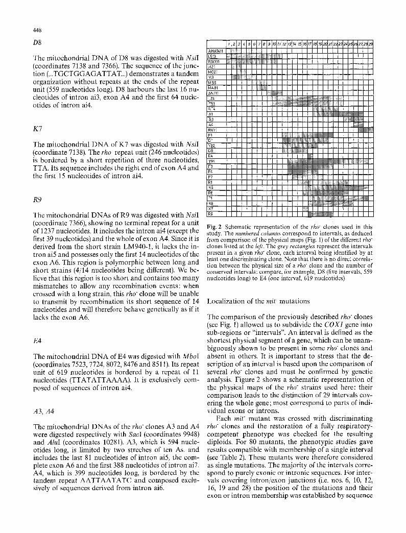
Fig. 2 Schematic representation of the rho- clones used in this study. The numbered columns correspond to intervals, as deduced from comparison of the physical maps (Fig. 1) of the different rho" clones listed at the left. The grey rectangles represent the intervals present in a given rho clone, each interval being identified by at least one discriminating clone. Note that there is no direct correla- tion between the physical size of a rho clone and the number of conserved intervals: compare, for example, D8 (five intervals, 559 nucleotides long) to E4 (one interval, 619 nucleotides)
Localization of the mit- mutations
The comparison of the previously described rho clones (see Fig. 1) allowed us to subdivide the C O X I gene into sub-regions or "intervals". An interval is defined as the shortest physical segment of a gene, which can be unam- biguously shown to be present in some rho- clones and absent in others. It is important to stress that the de- scription of an interval is based upon the comparison of several rho clones and must be confirmed by genetic analysis. Figure 2 shows a schematic representation of the physical maps of the rho- strains used here: their comparison leads to the distinction of 29 intervals cov- ering the whole gene; most correspond to parts of indi- vidual exons or introns.
Each m i t mutant was crossed with discriminating rho clones and the restoration of a fully respiratory- competent phenotype was checked for the resulting diploids. For 80 mutants, the phenotypic studies gave results compatible with membership of a single interval (see Table 2). These mutants were therefore considered as single mutations. The majority of the intervals corre- spond to purely exonic or intronic sequences. For inter- vals covering intron/exon junctions (i.e. nos. 6, 10, 12, 16, 19 and 28) the position of the mutations and their exon or intron membership was established by sequence

Table 2 The 29 intervals of the COXI gene. The muta- tions located in each interval are indicated. Many intervals 1 correspond to sub-segments of 2 introns or exons which are in- 3 dicated in the second column. 4 For intervals 6, 10, 12, 19 and 5 28, the position of the muta- 6 tions was established by se- 7 quence analysis (Netter et al. 8 1992; Lemarre et al. 1994; G. 9 Carignani, E. Bergantino, S. 10 Robineau and P. Netter, un- 11 published work; C. Lemaire, 12 S. Robineau and P. Netter, 13 unpublished work) except for 14 mutations denoted with a $. 15 The last category (interval 0) 16 corresponds to the mutations 17 which cannot be assigned un- 18 ambiguously to a given inter- 19 val (multiple mutations or 20 large deletions) 21
22 23 24 25 26 27 28 29
Interval Localization Mutations
449
Intron 1 Intron 1 Intron 1 Intron 1 Intron 1
Intron 2 Intron 2 Intron 2
Intron 3
Exon 4 Exon 4 Exon 4
Intron 4 Intron 4
Exon 5 Exon 5 Intron 5 Intron 5 Exon 6 Exon 6 Intron 6 Intron 6
Intron 7 and exon 8 Deletions or multiple mutations
G2399, V293 G3010 V94 V284 V281, V221, V375 G2166, V277 (seq.=intron 2) Gl194 W165 Gll0-1 V202, V231 (seq.=exon 3), V2645 G2027, G3013 G1099, G1765-1 (seq.=exon 4) G205, G588, G2567, Vl12, V217, V312, V356-1 G1003, G2276, V32, V106, V203, V207, V380 G481, G1547, V130 G247, G2508 (seq. =exon 4), V356-25 G192, G476-1, G1765-2, G2190, V274 V226 G291, G450, G2443, G3015, W113 (seq.=exon 5) V124, V243, V256 Gl10-2, V402, W164, W201 V36, V237, V241 V249, V370 G1979, V128 (seq.=exon 6) V255, V282, W109, V3795 G421 V38 G2084 (seq. = exon 7) G341, G433, G1375, G2029, G2124, G2312, G2394, V13, V19, V95, V209, V220, V238, V290, V300, V393, V400
G20, G74, G390, G656, G689, G698, G789, G728, G922, G1055, Gl160, Gl181, Gl186, G1264, G1273, G1287, G1305, G1734, G1859, G1921, G2133, G2220, G2342, G2343, G2354, G2469, G2496, V3, V16, V40, V41, V55, V73, V105, V129, V212, V213, V214, V215, V218, V222 V223, V228, V229, V230, V233, V240, V242, V244, V252, V258, V261 V262, V268, V271, V275, V279, V280, V289, V299, V307, V308, V310 V315, V334, V337, V338, V339, V351, V355, V358, V359, V374, V376 V377, V378, V381, V387, V383, V385, V391, V394, V395, V396
analysis (Netter et al. 1992; Lemarre et al. 1994; G. Carignani, E. Bergantino, S. Robineau and P. Netter, unpublished work; C. Lemaire, S. Robineau and P. Net- ter, unpublished work) except for the interval 29.
It should be noted that, in some instances, the quan- titative results of the genetic analysis allowed some am- biguity in the assignment of a given mutat ion to a single interval. These ambiguities are due to recombinat ion disturbance at the limits of a rho- repetition unit, called a border effect. These mutat ions are indicated by a $ symbol in Table 2 and Fig. 3 and will not be considered in the Discussion.
Mutants G l l 0 , G1765, V356 and G476, after ran- dom crosses and analysis of mitotic segregation, were shown to be due to double mutat ions named G l l 0 - 1 and - 2 for G l l 0 , G1765-1 and - 2 for G1765 and so on, localized in different intervals. Individual mutat ions exhibited in most cases their own phenotypic properties. Nevertheless, a fully wild-type phenotype was only ob- tained when all mutat ions were corrected, for example in crosses with long rho clones covering the two affected intervals.
Finally, for 84 mutants unambiguously located in the COXI region (after crosses with the long rho clone KL14-4A/RP131 which covers the entire COXI gene (Kotylak and Slonimski 1977), we were unable to identi- fy single mutat ions in a given interval. We believe that these mutat ions (grouped in interval 0, Table 2) are due to multiple distant and stringent mutat ions or to large deletions. For example, in mit-x mit- crosses, mutant G1859 was shown to be a deletion starting within the sixth intron and extending leftward at least to the fourth exon.
Discussion
Exonic or intronic mutat ions
Amongst 170 mit- mutants mapped to the COXI region, 86 have been shown to result from single mutat ions lo- cated within one of the 29 intervals, as deduced from crosses with the panel of rho clones used here and in previous publications (Carignani et al. 1983; Netter et

450
a
b 10
G1099 6,1765-1 G205 G588 G2567 V112 V217 V356-1
03010 V281 V2645 V293 V94 V221 G2166 V202 6'2027 6,2399 V284 V375 V277 6,1194 W165 6,110-1 V231 6,3013
• I I m I t I i i i
2000 4000 6000
G1003 G2276 V32 V106 V203 V207 V380
V130 G1547 G481
G291 G450 82443 G3015 Wl13
V124. V243 G1979
G192 V256 V'[28 6,476-1 6,110-2 V255
G247 6,1765-2 V402 V36 V282 G2508 G2190 W164 V237 V249 W109 G421 V356-25 V27/., V226 W201 V241 V370 V3795 V38
m I I i I i
io 8000 8585
G2084
,I 3060[ 11645
G341 G433 G1375 G2029 G212& G3212 G2394 V13 V19 V95 V209 V220 V238 V290 V300 V393 V400
13003
0.1
Fig. 3a,b Mapping of the mit- mutations of the COXI gene. a Distribution of the 89 mutations within the 29 intervals deduced from the physical maps of the discriminating rho- clones (see Fig. 1 and Table 2). b Deviation (nobs/no,lo) of the probability of appear- ance of a mutation in exonic or intronic regions of the gene from expected value. The values are calculated in Table 3 (right column). Symbols used in a are as given in the legend to Fig. la
al. 1982a; see Table 2). The remaining 84 mutants, which cannot be assigned a single physical location in spite of belonging to the C O X I gene, are probably due to multiple mutations or small deletions. These mutants will not be considered further in the Discussion, but we must keep in mind that by eliminating them, we obvi- ously introduce a bias in favour of the single mutations, i.e. nucleotide substitutions, additions or deletions.
The C O X I gene is 12.9 kb long; therefore the num- ber of single mutations corresponds to a mean frequen- cy of one mutation every 150 nucleotides. It is apparent (see Fig. 3) that the positions of the mutations, which have been clearly assigned to exons or introns (except for the interval 29), are not randomly distributed: 41 mutations have been localized in the 1140 nucleotides of exons A1 to A7 (i.e. on average, one mutation every 28 nucleotides in exons) whereas only 28 mutations have been identified in the 10 388 nucleotides of introns ail to ai6 (one mutation every 371 nucleotides in introns). The probability of the appearance of a mutation is always
higher in exonic and always lower in intronic regions. One can express this bias by the ratio of the observed number of mutations to the expected number (assuming a constant probability along the gene of one mutation every 150 nucleotides; see above). The values of the bias for the different regions of the gene, given in the Table 3, are always less than 1 for introns and greater than 1 for exons. This bias could be partially explained by the dif- ference in GC content in the exons (30.7%) and the in- trons (22%) of COXI , if one remembers that the pub- lished mi t mutations due to nucleotide substitutions in the mitochondrial DNA of S. cerevisiae frequently cor- respond to G C ~ AT transitions or G C ~ TA transver- sions. However, this cannot account for the major por- tion of the bias, which establishes that the selection of a mit- mutation is 13.3 (371/28) times more probable in an exon than in an intron.
It must be noted that the initial screening of the mit- mutants is only based upon the inability of the yeast cell to use gycerol as carbon source. This selection proce- dure introduces a bias in favour of the mutations lead- ing to a stringent respiratory-deficient phenotype. It tends to eliminate the leaky or partial mutants and, of course, those exhibiting phenotypes unrelated to respi- ration. In order to explain the difference between muta- tion frequencies in exons and introns, we must therefore analyse the targets which, in both cases, are able to con- fer a respiratory-negative phenotype when mutated.

Table 3 The bias in appearance of mit- mutations in exons vs introns of the COXI gene. The length of each exon or intron of the gene is indicated in the second column. The physical map does not permit us to discriminate unambiguously between the last intron ai7 and exon AS, which are therefore considered together (ai7+A8 = interval 29). The third column lists the number of mutations observed in each exon or intron, derived from the re- sults of the Table 2. The fourth column gives the expected number of mutations if the probability of appearance of a mit- mutation is constant along the gene (one mutation every 150 nucleotides). The numbers are obtained by dividing the length of the segment (exon- ic or intronic) by 150. The fifth column gives the bias of appear- ance of mit mutations in exons and introns, as a ratio of the observed to calculated numbers of mutations (see the Discussion)
Exon/ Length hobs V/calc Bias Intron (in bp) (number of (number of (nobs/ncalc)
observed calculated mutations) mutations)
A1 169 0 1.1 - all 2448 10 16.3 0.61 A2 36 0 0.24 - ai2 2515 3 16.8 0.18 A3 35 2 0.23 8.7 ai3 1514 2 10.1 0.20 A4 480 21 3.2 6.6 ai4 1010 6 6.7 0.89 A5 252 12 1.7 7.1 ai5 1365 5 9.0 0.55 A6 135 5 0.92 5.6 ai6 1536 2 10.2 0.20 A7 33 1 0.22 4.5 (ai7+a8) (879+470) (17) (9.3)
Exonic targets
Exonic sequences specify almost exclusively the peptide encoded by the gene (in our case COXIp, the first sub- unit of the cytochrome oxidase complex). Short exonic sequences (complementary to guide sequences in group I introns, intron binding sequences IBS1 and IBS2 in group II introns) are implicated in the splicing of flank- ing introns, and mutations affecting these sequences have been described (Perea and Jacq 1985); however such mutations affect only a small number of the exonic nucleotides. Therefore, in exons, the targets of muta- tions are mainly the codons specifying the amino acids of COXlp. The appearance of a mutant phenotype de- pends upon the importance of the residue in a given position and of the severity of the amino acid change, which must eliminate the activity of the respiratory complex IV.
There is an excess of mutations in exons (bias of 8.7, 6.6, 7.1, 5.6 and 4.5 for exons A3, A4, A5, A6 and A7 respectively; see Table 3) but their number seems to be roughly proportional to the exon length, except for the exon A1 described below. These results suggest that the corresponding segments of the protein (i.e. subunit I of the cytochrome oxidase) contribute equally to the activ- ity of the complex and can be similarly affected by muta- tions abolishing its functional properties. If this rule ex- tends to the C-terminal part of the protein, we could
451
predict that, amongst the 17 mutations roughly located in the (ai7 + A8) region, a majority should be located in the exon A8. In fact, taking into account the length of this exon (470 nucleotides) and the mean frequency of mutations in the exonic part of the gene (1 every 28 nucleotides), one expects 470/28 = 16.7 mutations. Us- ing the same calculation for intron aiT, only 2.3 muta- tions (879 nucleotides/371) are predicted.
The first exon A1 constitutes an exception: no muta- tion was observed whereas six (169/28 =6.0) were ex- pected within its 169 nucleotides, if one applies the val- ues for exonic sequences. This result could indicate that the N-terminal part of the protein, which includes the first and second transmembrane helices (Rao and Argos 1986), plays a less important role in the activity of the complex and can harbour many silent nucleotide changes. Futhermore, if one excludes a priori, possible reinitiation by the translation apparatus, this result in- dicates that mutations that generate nonsense codons (directly or by a frame-shift) are rare, at least in the first exon.
Intronic targets
Intronic sequences carry a complex mixture of struc- tural and functional information. Firstly, from 200 to 600 intronic nucleotides are devoted to the three-dimen- sional constitution of the catalytic core of the ribozyme (Davies et al. 1982; Michel and Dujon 1983). Secondly, in addition to this structural information, many introns harbour long ORFs, some of which encode reverse tran- scriptases, DNA-endonucleases and/or RNA-maturas- es (see the Introduction). The conservation of these ORFs is a good argument in favour of the reality of their supposed functions and suggests that the ORFs are not simply pseudogenes. One would then expect a high den- sity of structural and/or functional mutations in such pleiotropic regions; this is not observed. Several factors can be invoked to account for this apparent discrepan- cy.
(1) Structural (cis-acting) mutants of group I yeast mitochondrial introns, selected in vivo on the basis of their respiratory-deficient phenotype, have been shown to affect only a very limited number of residues, mainly those constituting what is now known to be the guanosine-binding site of the ribozyme (De La Salle et al. 1982; Weiss-Brummer et al. 1982; Netter et al. 1982b; Michel et al. 1989b). It seems, therefore, that the effective size of the ribozyme target, deduced from in vivo selec- tion of cis-acting mutants in yeast mitochondria, is much smaller than predicted by the structural models and by in vitro experiments. In the Tetrahymena group I intron, systematic mutagenesis of the ribozyme residues led to the conclusion that the majority of single and double substitutions significantly affected the rate of in vitro self-splicing of the intron (Couture et al. 1990). This apparent discrepancy between in vivo and in vitro results is not a general rule: in experiments per-

452
formed on the intron td of bacteriophage T4 thymidy- late synthase, a majority of the mutations introduced in conserved positions of this intron led to both splicing defects in vitro and to a detectable [thy] phenotype in vivo (Belfort et al. 1987). This variable susceptibility to nucleotide changes between these two biological sys- tems must reflect different requirements for the synthesis or the expression of the final product, i.e. subunits of the mitochondrial respiratory complexes in yeast and T4 thymidylate synthase in E. coli.
(2) The functions encoded by the introns must also be clearly distinguished. It has been demonstrated that DNA endonucleases are encoded by the third, fourth and fifth introns (Sargueil et al. 1991; Schapira et al. 1993; Moran et al. 1992), and reverse transcriptase by the first one (Kennell et al. 1993). However it has not been shown whether the loss of reverse transcriptase or DNA endonuclease activities affects RNA splicing and gives rise to a respiratory-deficient phenotype. It is most probable that the mit phenotype is due solely to the inactivation of RNA maturases.
In the COXI gene, trans-complementable mit muta- tions affecting the splicing process and therefore, most probably, the RNA maturase itself, have been demon- strated only in the first intron all (mutants G3010, V94, V284, V281, V221 and V375; see Carignani et al. 1983). These ail maturase mutants are unable to splice both the ail and ai2 intronic sequences from the long mRNA precursor, suggesting that the ail maturase is implicated in the splicing of the first two introns of the gene. In addition, variant strains devoid of the genomic sequence of the first intron still correctly splice ai2 intron from the pre-mRNA (Carignani et al. 1986). This result provides indirect evidence that the product of the ORF of the second intron (the sequence of which is very similar to the ORF of the first intron), also has RNA maturase properties.
Finally, in intron a4, indirect evidence of a transac- tire product implicated in the RNA splicing has been obtained for mutant G1765-2 (Dujardin et al. 1983) and for suppressors of cob-box mutations. In fact, the protein encoded by the fourth intron of COXI is 65% similar to the protein encoded by the fourth intron of the cob-box gene, which possesses RNA maturase activ- ity (Banroques et al. 1986). When this latter activity is lost by mutation, several single mutations affecting the protein encoded by intron ai4 can lead to the activation of latent maturase activity, in spite of the different pri- mary structure of the two peptides (Dujardin et al. 1982; P. Netter and S. Robineau, unpublished work).
Thus there is evidence that the ORFs of the first, second and fourth introns encode proteins related to RNA splicing. To explain why clear-cut transactive mit- mutants were found only in the first intron, we must assume that amino acid changes in the second and fourth intronic maturase-like proteins lead to a very leaky or discrete phenotype undetectable by our screen- ing procedure. It is conceivable that the ORFs in introns ai3, ai5 and ai6 could also encode discrete RNA mat-
urase activities. Some sequence similarities (such as the LAGLI-DADG motif) between these proteins and bona fide maturases could argue in favour of this hy- pothesis. It is then possible to imagine that a minimal selective advantage, associated with the correct splicing of the introns, could be sufficient to account for the maintenance of open reading frames, in spite of this not being detectable by laboratory experiments. The possi- bility also cannot be excluded that the same kind of undetectable selective advantage is associated with oth- er activities of intronic ORFs, such as reverse transcrip- tase, recombinase or endonuclease.
Intronic proteins
The region of the mitochondrial genome studied here may be represented as a discontinuous exonic gene (composed of eight exons and encoding COXIp subunit) interspersed with "intronic genes". We must then try to explain why the probability of appearance of mutations in these intronic genes is much lower than in the exonic gene. The respiratory complexes are composed of essen- tial and highly specialized proteins, which are the result of a very long and stringent process of selection. Thus it is difficult to imagine that they could acquire new func- tions as a result of simple mutations. The very high level of sequence conservation in these proteins in the whole of the living world, from bacteria to man, illustrates this high degree of specialization. Conversely, many single amino acid changes in these proteins have been shown to lead to respiratory-deficient phenotypes (Netter et al. 1992; Meunier et al. 1993; Lemarre et al. 1994; C. Lemaire, S. Robineau and P. Netter, unpublished work; for a review on cytochrome b, see Degli Esposti et al. 1993). The critical need to maintain a fully active res- piratory chain is illustrated by the natural overproduc- tion of the mRNA for the corresponding proteins. We have frequently observed, in studying respiratory-com- petent pseudo-wild revertants of mit- mutations, that a very low level of mRNA is sufficient to maintain a nor- mal synthesis of these proteins (data not shown). In the wild-type strain, the overproduction of mRNA consti- tutes a natural protection against mutations and could explain the small number of RNA maturase mutants: a splicing defect leading to a respiratory-deficient pheno- type would be obtained only in the case of (multiple) mutations completely abolishing the maturase activi- ties.
On the contrary, it seems that the intronic proteins are less specialized and more primitive. They frequently have conserved dual functions: the protein encoded by the first and second introns of COXI accumulates RNA maturase and reverse transcriptase activities (Carignani et al. 1983; Kennell et al. 1993); the protein encoded by the fourth intron of the cob-box gene shows DNA re- combinase and RNA maturase properties (De La Salle et al. 1982; Kotylak et al. 1985; Banroques et al. 1986; Goguel et al. 1989). It is conceivable that this phe-

453
nomenon constitutes a sign of the invasion of ribozymic introns by intronic protein genes, either by reverse tran- scription or by DNA homing, associated with selection pressure in favour of the acquisition of RNA splicing ability (Lambowitz and Belfort 1993). As a consequence of the conservation of these multiple functions, the in- tronic proteins may have been unable to reach a high level of specialization in each distinct activity. This hy- pothesis is favoured by the finding that mutants defec- tive in maturase activities are rarely due to discrete amino acid changes but, in the majority of cases, to nonsense (direct or frameshift) mutations leading to the loss of a large part of the molecule (Lazowska et al. 1980; Jacq et al. 1982; De La Salle et al. 1982). We pro- pose, therefore, that splicing-related activities of intron- ic proteins are largely unaffected by single amino acid replacements and that most nucleotide changes in in- tronic genes are functionally silent , whereas exonic proteins like C O X l p are highly susceptible to such re- placements. This differential sensitivity to amino acid changes would explain the very unequal distribution of the mutations observed in COXI gene.
Acknowledgements This work was supported by EC contract no. SCI 000476. We thank F. Michel and C. J. Herbert for suggestions and critical reading of the manuscript.
References
Banroques J, Delahodde A, Jacq C (1986) A mitochondrial RNA maturase gene transferred to the yeast nucleus can control mitochondrial mRNA splicing. Cell 46:837 844
Belfort M, Chandry PS, Pedersen-Lane J (1987) Genetic delin- eation of functional components of the group I intron in the phage T4 td gene. Cold Spring Harbor Syrup Quant Biol 52:181-192
Bonitz SG, Coruzzi G, Thalenfeld BE, Tzagoloff A, Macino G (1980) Assembly of the mitochondrial membrane system: physical map of the oxi3 locus of yeast mitochondrial DNA. J Biol Chem 255:11927-11941
Calder KM, McEwen JE (1990) Nucleotide sequence of the gene encoding cytochrome c oxidase subunit VII from Saccha- romyces cerevisiae. Nucleic Acids Res 18:1632
Carignani G, Dujardin G, Slonimski P (1979) Petite deletion map of the mitochondrial oxi3 region in S. cerevisiae. Mol Gen Genet 167:301-308
Carignani G, Groudinsky O, Frezza D, Schiavon E, Bergantino E, Slonimski P (1983) An mRNA maturase is encoded by the first intron of the mitochondrial gene for the subunit I of cy- tochrome oxidase in S. cerevisiae. Cell 35:733-742
Carignani G, Netter P, Bergantino E, Robineau S (1986) Expres- sion of the mitochondrial split gene coding for cytochrome oxidase subunit I in S. cerevisiae: RNA splicing pathway. Curr Genet 11:55-63
Cech TR (1990) Self-splicing of group I introns. Annu Rev Biochem 59: 543-568
Coruzzi G, Tzagoloff A (1979) Assembly of the mitochondrial membrane system: DNA sequence of subunit 2 of yeast cy- tochrome oxidase. J Biol Chem 254:9324-9330
Couture S, Ellington A, Gerber A, Cherry M, Doudna J, Green R, Hanna M, Pace U, Rajapogal J, Szostak J (1990) Mutational analysis of conserved nucleotides in a self-splicing group I intron. J Mol Biol 215:345 358
Cumsky MG, Trueblood CE, Ko C, Poyton RO (1987) Structural analysis of two genes encoding divergent forms of yeast cy- tochrome c oxidase subunit V. Mol Cell Biol 7:3511-3519
Davies RW, Waring RB, Ray JA, Brown TA, Scazzocchio C (1982) Making ends meet: a model for RNA splicing in fungal mitochondria. Nature 300:719-724
De La Salle H, Jacq C, Slonimski PP (1982) Critical sequences within mitochondrial introns: pleiotropic mRNA maturase and cis-dominant signals of the box intron controlling reduc- tase and oxidase. Cell 28:721-732
Degli Esposti M, De Vries S, Crimi M, Ghelli A, Patarnello T, Meyer A. (1993) Mitochondrial cytochrome b: evolution and structure of the protein. Biochim Biophys Acta 1143:243-271
Dujardin G, Jacq C, Slonimski PP (1982) Single base substitution in an intron of oxidase gene compensates splicing defects of the cytochrome b gene. Nature 298:628-632
Dujardin G, Pajot P, Groudinsky O, Slonimski PP (1980) Long range control circuits within mitochondria and between nucle- us and mitochondria. I. Methodology and phenomenology of suppressors. Mol Gen Genet 179:469-482
Garriga G, Lambowitz AM (1984) RNA splicing in Neurospora mitochondria: self-splicing of a mitochondrial intron in vitro. Cell 39:631-641
Goguel V, Bailone A, Devoret R, Jacq C (1989) The bI4 RNA mitochondrial maturase of S. cerevisiae can stimulate intra- chromosomal recombination in E. coli. Mol Gen Genet 216:70-74
Haltia T, Puustinen A, Finel M (1988) The Paracoccus denitrificans cytochrome aa3 has a third subunit. Eur J Biochem 172: 543- 546
Hensgens LAM, Bonen L, de Haan M, Horst G, Grivell L (1983) Two intron sequences in yeast mitochondrial COX I gene: homology among URF containing introns and strain depen- dant variation in flanking exons. Cell 32:379--389
Hosler J, Fetter J, Tecklenburg M, Espe M, Lerma C, Ferguson- Miller S (1992) Cytochrome aa3 of Rhodobacter sphaeroides as a model for mitochondrial cytochrome c oxidase. J Biol Chem 267: 24264-24272
Jacq C, Pajot P, Lazowska J, Dujardin G, Claisse M, Groudinsky O, De La Salle H, Grandchamps C, Labouesse M,Gargouri A, Guiard B, Spiridakis A, Dreyfus M, Slonimski P (1982) Role of introns in the yeast cytochrome b gene: cis- and trans-acting signal, intron manipulation, expression and intergenic com- munications. In: Slonimski P, Borst P, Attardi G (eds) Mito- chondrial genes. (Cold Spring Harbour Symposium on Mito- chondrial Genes) Cold Spring Harbor Laboratory, Cold Spring Harbor, New York, pp 155-183
Kennell JC, Moran JV, Perlman PS, Butow RA, Lambowitz AM (1993) Reverse transcriptase activity associated with maturase- encoding group II introns in yeast mitochondria. Cell 73:133- 146
Kotylak Z, Slonimski PP (1977) Mitochondrial mutants isolated by a new method based upon the use of the nuclear mutation opl. In: Bandlow W, Schweyen RJ, Wolf K, Kandewitz F (eds) Mitochondria 1977. de Gruyter, Berlin. pp 83-89
Kotylak Z, Lazowska J, Hawthorne DC, Slonimski PP (1985) Intron encoded proteins in mitochondria: key elements of gene expression and genomic evolution. In: Quagliarello E, Slater EC, Palmieri F, Saconne C, Kroon AM (eds) Achieve- ments and perspectives in mitochondrial research, vol 2. E1- seiner Amsterdam, pp 1-20
Kruszewska A, Slonimski P (1984) Mitochondrial and nuclear mitoribosomal suppressors that enable misreading of ochre codons in yeast mitochondria. Curr Genet 9:11-19
Lamarche AEP, Abate MI, Chan SHP, Trumpower BL (1992) Isolation and characterization of COX12, the nuclear gene for a previously unrecognized subunit of Saccharomyces cerevisiae cytochrome-c oxidase. J Biol Chem 267:22473-22480
Lambowitz AM, Belfort M (1993) Introns as mobile elements. Annu Rev Biochem 62:587-622
Lazowska J, Jacq C, Stonimski PP (1980) Sequence of introns and flanking exons in wild-type and box3 mutants of cytochrome b

454
reveals an interlaced splicing protein coded by an intron. Cell 22:333-348
Lemarre P, Robineau S, Colson AM, Netter P (1994) Sequence analysis of three deficient mutants of cytochrome oxidase subunit I of S. cerevisiae and their revertants. Curr Genet 26(5-6):546 552
Lewin A, Morimoto R, Rabinowitz M, Fukuhara H (1978) Re- striction enzyme analysis of mitochondrial DNAs of petite mutants of yeast. Mol Gen Genet 163:257-275
Maarse AC, Van LA, Riezman H, Gregor I, Schatz G, Grivell LA (1984) Subunit IV of yeast cytochrome c oxidase: cloning and nucleotide sequencing of the gene and partial amino acid se- quencing of the mature protein. EMBO J 3:2831-2837
Meunier B, Coster F, Lemarre P, Colson AM (1993) Insight into topological and functional relationships of cytochrome-c oxi- dase subunit 1 of S. cerevisiae by means of intragenic comple- mentation. FEBS Lett 321:159-162
Michel F, Dujon B (1983) Conservation of RNA secondary struc- tures in two intron families including mitochondrial-, chloro- plast- and nuclear encoded members. EMBO J 2:33-38
Michel F, Lang BF (1985) Mitochondrial class II introns encode proteins related to the reverse transcriptases of retroviruses. Nature 316:641-643
Michel F, Umesono K, Okesi H (1989a) Comparative and func- tional anatomy of group II catalytic introns - a review. Gene 82:5-30
Michel F, Hanna M, Green R, Bartel D, Szostak J (1989b) The guanosine binding site of Tetrahymena ribozyme. Nature 23: 391-395
Moran JV, Wernette CM, Mecklenburg KL, Butow RA, Perlman PS (1992) Intron 5 alpha of the COXI gene of yeast mitochon- drial DNA is a mobile group I intron. Nucleic Acids Res 20: 4069-4076
Netter P, Carignani G, Jacq C, Groudinsky O, Clavillier L, Slonimski PP (1982a) The cytochrome oxidase subunit I split gene in Saccharomyces cerevisiae: genetic and physical studies of the mtDNA segment encompassing the 'cytochrome b-ho- mologous' intron. Mol Gen Genet 188:551-559
Netter P, Jacq C, Carignani G, Slonimski PP (1982b) Critical sequences within mitochondrial introns: cis-dominant muta- tions of the "cytochrome-b-like" intron of the oxidase gene. Cell 28:733-738
Netter P, Robineau S, Sirand-Pugnet P, Fauvarque MO (1992) The unusual reversion properties of a mitochondrial mutation in the structural gene of subunit I of cytochrome oxidase of Saccharomyces cerevisiae reveal a probable histidine ligand of the redox center. Curr Genet 21:147-151
Patterson TE, Poyton RO (1986) COX8, the structural gene for yeast cytochrome c oxidase subunit VIII. DNA sequence and gene disruption indicate that subunit VIII is required for max- imal levels of cellular respiration and is derived from a precur- sor which is extended at both its NH 2 and COOH termini. J Biol Chem 261 : 17192-17197
Peebles CL, Perlman PS, Mecklenburg KL, Petrillo ML, Tabor JH, Jarrell KA, Cheng HL (1986) A self-splicing RNA excises an intron lariat. Cell 44:213-223
Perea J, Jacq C (1985) Role of the 5' hairpin structure in the splicing accuracy of the fourth intron of the yeast cob-box gene. EMBO J 4:3281-3288
Rao JKM, Argos P (1986) A conformational preference parameter to predict helices in integral membrane proteins. Biochim Bio- phys Acta 869:197-214
Sambrook J, Fritsch EF, Maniatis T (1989) Molecular cloning: a laboratory manual. Cold Spring Harbor Laboratory, Cold Spring Harbor, New York
Sanger F, Nicklen S, Coulson A (1977) DNA sequencing with chain terminating inhibitors. Proc Natl Acad Sci USA 74:5463-5467
Sargueil B, Delahodde A, Hatat D, Tian GL, Lazowska J, Jacq C (1991) A new specific DNA endonuclease activity in yeast mi- tochondria. Mol Gen Genet 225:340-341
Schapira M, Desdouets C, Jacq C, Perea J (1993) I-SceIII an intron-encoded DNA endonuclease from yeast mitochondria. Asymmetrical DNA binding properties and cleavage reaction. Nucleic Acids Res 21:3683-3689
Schmelzer C, Schweyen RJ (1986) Self-splicing of group II introns in vitro: mapping of the branch point and mutational inhibi- tion of lariat formation. Cell 46:557-565
Seraphin B, Simon M, Faye G (1985) Primary structure of a gene for subunit 5 of the cytochrome oxidase from S. cerevisiae. Curr Genet 9:435.439
Slonimski P, Tzagoloff A (1976) Localization in yeast mitochon- drial DNA of mutations expressed in a deficiency of cy- tochrome oxidase and/or coenzyme QH2-cytochrome c reduc- tase. Eur J Biochem 61:27-41
Szczepanek T, Macadre C, Meunier B, Lazowska J (1994) Two homologous introns from related Saccharomyces species differ in their mobility. Gene 139:1-7
Taanman JW, Capaldi RA (1992) Purification of yeast cytochrome c oxidase with a subunit composition resembling the mam- malian enzyme. J Biol Chem 267:22481-5
Taanman JW, Capaldi RA (1993) Subunit Via of yeast cy- tochrome c oxidase is not necessary for assembly of the en- zyme complex but modulates the enzyme activity. Isolation and characterization of the nuclear-coded gene. J Biol Chem 268:18754-18761
Thalenfeld BE, Tzagoloff A (1980) Assembly of the mitochondrial membrane system. Sequence of the oxi 2 gene of yeast mito- chondrial DNA. J Biol Chem 255:6173-6180
van der Horst G, Tabak HF (1985) Self-splicing of yeast mito- chondrial ribosomal and messenger RNA precursors. Cell 40: 759-766
van der Veen R, Arnberg AC, van der Horst G, Bonen L, Tabak HF, Grivell LA (1986) Excised group II introns in yeast mito- chondria are lariats and can be formed by self-splicing in vitro. Cell 44: 225-234
Weiss-Brummer B, Rodel G, Schweyen R, Kaudewitz F (1982) Expression of the split gene cob in yeast: evidence for a precur- sor of a maturase protein translated from intron 4 and preced- ing exons. Cell 29:527-536
Wright RM, Dircks LK, Poyton RO (1986) Characterization of c o x g , the nuclear gene encoding the yeast mitochondrial protein cytochrome c oxidase subunit VIIa. Subunit VIIa lacks a leader peptide and is an essential component of the holoenzyme. J Biol Chem 261:17183-17191
Wright RM, KO C, Cumsky MG, Poyton RO (1984) Isolation and sequence of the structural gene for cytochrome c oxidase sub- unit 6 from S. cerevisiae. J Biol Chem 259:15401-15407