Mutation analysis and characterization of ATR sequence variants in breast cancer cases from...
21
BioMed Central Page 1 of 21 (page number not for citation purposes) BMC Cancer Open Access Research article Mutation analysis and characterization of ATR sequence variants in breast cancer cases from high-risk French Canadian breast/ovarian cancer families Francine Durocher* 1 , Yvan Labrie 1 , Penny Soucy 1 , Olga Sinilnikova 2 , Damian Labuda 3 , Paul Bessette 4 , Jocelyne Chiquette 5 , Rachel Laframboise 6 , Jean Lépine 7 , Bernard Lespérance 8 , Geneviève Ouellette 1 , Roxane Pichette 8 , Marie Plante 9 , Sean V Tavtigian 10 and Jacques Simard 1,11 Address: 1 Cancer Genomics Laboratory, Oncology and Molecular Endocrinology Research Centre, Centre Hospitalier Universitaire de Québec and Laval University, Québec, G1V 4G2, Canada, 2 Unité Mixte de Génétique Constitutionnelle des Cancers Fréquents, Hospices Civils de Lyon/Centre Léon Bérard, Lyon, France, 3 Centre de cancérologie Charles Bruneau, Ste-Justine Hospital, Montréal, Canada, 4 Service de gynécologie, Centre Hospitalier Universitaire de Sherbrooke, Fleurimont, Canada, 5 Clinique des maladies du sein Deschênes-Fabia, Hôpital du Saint-Sacrement, Québec, G1S 4L8, Canada, 6 Service de médecine génétique, CHUQ, Pavillon CHUL, Québec, G1V 4G2, Canada, 7 Centre hospitalier régional de Rimouski, Rimouski, G5L 5T1, Canada, 8 Service d'hémato-oncologie, Hôpital du Sacré-Cœur, Montréal, Canada, 9 Service de gynécologie, CHUQ, L'Hôtel-Dieu de Québec, Québec, G1R 2J6, Canada, 10 Unit of Genetic Cancer Susceptibility, International Agency for Research on Cancer, World Health Organization, Lyon, France and 11 Canada Research Chair in Oncogenetics, Department of Anatomy and Physiology, Laval University, Québec, Canada Email: Francine Durocher* - [email protected]; Yvan Labrie - [email protected]; Penny Soucy - [email protected]; Olga Sinilnikova - [email protected]; Damian Labuda - [email protected]; Paul Bessette - [email protected]; Jocelyne Chiquette - [email protected]; Rachel Laframboise - [email protected]; Jean Lépine - [email protected]; Bernard Lespérance - [email protected]; Geneviève Ouellette - [email protected]; Roxane Pichette - [email protected]; Marie Plante - [email protected]; Sean V Tavtigian - [email protected]; Jacques Simard - [email protected] * Corresponding author Abstract Background: Ataxia telangiectasia-mutated and Rad3-related (ATR) is a member of the PIK-related family which plays, along with ATM, a central role in cell-cycle regulation. ATR has been shown to phosphorylate several tumor suppressors like BRCA1, CHEK1 and TP53. ATR appears as a good candidate breast cancer susceptibility gene and the current study was designed to screen for ATR germline mutations potentially involved in breast cancer predisposition. Methods: ATR direct sequencing was performed using a fluorescent method while widely available programs were used for linkage disequilibrium (LD), haplotype analyses, and tagging SNP (tSNP) identification. Expression analyses were carried out using real-time PCR. Results: The complete sequence of all exons and flanking intronic sequences were analyzed in DNA samples from 54 individuals affected with breast cancer from non-BRCA1/2 high-risk French Canadian breast/ovarian families. Although no germline mutation has been identified in the coding region, we identified 41 sequence variants, including 16 coding variants, 3 of which are not reported in public databases. SNP haplotypes were established and tSNPs were identified in 73 healthy unrelated French Canadians, providing a valuable tool for further association studies involving the ATR gene, using large cohorts. Our analyses led to the identification of two novel alternative splice transcripts. In contrast to the Published: 29 September 2006 BMC Cancer 2006, 6:230 doi:10.1186/1471-2407-6-230 Received: 19 April 2006 Accepted: 29 September 2006 This article is available from: http://www.biomedcentral.com/1471-2407/6/230 © 2006 Durocher et al; licensee BioMed Central Ltd. This is an Open Access article distributed under the terms of the Creative Commons Attribution License (http://creativecommons.org/licenses/by/2.0 ), which permits unrestricted use, distribution, and reproduction in any medium, provided the original work is properly cited.
-
Upload
independent -
Category
Documents
-
view
2 -
download
0
Transcript of Mutation analysis and characterization of ATR sequence variants in breast cancer cases from...

BioMed CentralBMC Cancer
ss
Open AcceResearch articleMutation analysis and characterization of ATR sequence variants in breast cancer cases from high-risk French Canadian breast/ovarian cancer familiesFrancine Durocher*1, Yvan Labrie1, Penny Soucy1, Olga Sinilnikova2, Damian Labuda3, Paul Bessette4, Jocelyne Chiquette5, Rachel Laframboise6, Jean Lépine7, Bernard Lespérance8, Geneviève Ouellette1, Roxane Pichette8, Marie Plante9, Sean V Tavtigian10 and Jacques Simard1,11Address: 1Cancer Genomics Laboratory, Oncology and Molecular Endocrinology Research Centre, Centre Hospitalier Universitaire de Québec and Laval University, Québec, G1V 4G2, Canada, 2Unité Mixte de Génétique Constitutionnelle des Cancers Fréquents, Hospices Civils de Lyon/Centre Léon Bérard, Lyon, France, 3Centre de cancérologie Charles Bruneau, Ste-Justine Hospital, Montréal, Canada, 4Service de gynécologie, Centre Hospitalier Universitaire de Sherbrooke, Fleurimont, Canada, 5Clinique des maladies du sein Deschênes-Fabia, Hôpital du Saint-Sacrement, Québec, G1S 4L8, Canada, 6Service de médecine génétique, CHUQ, Pavillon CHUL, Québec, G1V 4G2, Canada, 7Centre hospitalier régional de Rimouski, Rimouski, G5L 5T1, Canada, 8Service d'hémato-oncologie, Hôpital du Sacré-Cœur, Montréal, Canada, 9Service de gynécologie, CHUQ, L'Hôtel-Dieu de Québec, Québec, G1R 2J6, Canada, 10Unit of Genetic Cancer Susceptibility, International Agency for Research on Cancer, World Health Organization, Lyon, France and 11Canada Research Chair in Oncogenetics, Department of Anatomy and Physiology, Laval University, Québec, Canada
Email: Francine Durocher* - [email protected]; Yvan Labrie - [email protected]; Penny Soucy - [email protected]; Olga Sinilnikova - [email protected]; Damian Labuda - [email protected]; Paul Bessette - [email protected]; Jocelyne Chiquette - [email protected]; Rachel Laframboise - [email protected]; Jean Lépine - [email protected]; Bernard Lespérance - [email protected]; Geneviève Ouellette - [email protected]; Roxane Pichette - [email protected]; Marie Plante - [email protected]; Sean V Tavtigian - [email protected]; Jacques Simard - [email protected]
* Corresponding author
AbstractBackground: Ataxia telangiectasia-mutated and Rad3-related (ATR) is a member of the PIK-related family which plays,along with ATM, a central role in cell-cycle regulation. ATR has been shown to phosphorylate several tumor suppressorslike BRCA1, CHEK1 and TP53. ATR appears as a good candidate breast cancer susceptibility gene and the current studywas designed to screen for ATR germline mutations potentially involved in breast cancer predisposition.
Methods: ATR direct sequencing was performed using a fluorescent method while widely available programs were usedfor linkage disequilibrium (LD), haplotype analyses, and tagging SNP (tSNP) identification. Expression analyses werecarried out using real-time PCR.
Results: The complete sequence of all exons and flanking intronic sequences were analyzed in DNA samples from 54individuals affected with breast cancer from non-BRCA1/2 high-risk French Canadian breast/ovarian families. Althoughno germline mutation has been identified in the coding region, we identified 41 sequence variants, including 16 codingvariants, 3 of which are not reported in public databases. SNP haplotypes were established and tSNPs were identified in73 healthy unrelated French Canadians, providing a valuable tool for further association studies involving the ATR gene,using large cohorts. Our analyses led to the identification of two novel alternative splice transcripts. In contrast to the
Published: 29 September 2006
BMC Cancer 2006, 6:230 doi:10.1186/1471-2407-6-230
Received: 19 April 2006Accepted: 29 September 2006
This article is available from: http://www.biomedcentral.com/1471-2407/6/230
© 2006 Durocher et al; licensee BioMed Central Ltd.This is an Open Access article distributed under the terms of the Creative Commons Attribution License (http://creativecommons.org/licenses/by/2.0), which permits unrestricted use, distribution, and reproduction in any medium, provided the original work is properly cited.
Page 1 of 21(page number not for citation purposes)

BMC Cancer 2006, 6:230 http://www.biomedcentral.com/1471-2407/6/230
transcript generated by an alternative splicing site in the intron 41, the one resulting from a deletion of 121 nucleotidesin exon 33 is widely expressed, at significant but relatively low levels, in both normal and tumoral cells including normalbreast and ovarian tissue.
Conclusion: Although no deleterious mutations were identified in the ATR gene, the current study provides anhaplotype analysis of the ATR gene polymorphisms, which allowed the identification of a set of SNPs that could be usedas tSNPs for large-scale association studies. In addition, our study led to the characterization of a novel ∆33 splice form,which could generate a putative truncated protein lacking several functional domains. Additional studies in large cohortsand other populations will be needed to further evaluate if common and/or rare ATR sequence variants can be associatedwith a modest or intermediate breast cancer risk.
BackgroundAll common cancers show some degree of familial cluster-ing [1]. Most of the familial aggregation, especially inbreast cancer [2], results predominantly from inheritedsusceptibility [3]. Linkage studies in the 1990s led to thediscovery of several predisposition genes associated withmany rare familial cancer syndromes, thus providing fun-damental insights into various pathways of carcinogenesis[4]. Nevertheless, this approach has mainly been limitedto genes with relatively rare, highly penetrant alleles, forseveral reasons, such as a lack of power to detect allelesconferring modest or moderate risks that are believed tobe involved in common cancers [1,5-7]. Analyses of riskattributable to such alleles in the known breast cancer sus-ceptibility genes (e.g. BRCA1, BRCA2, TP53, PTEN, ATM)suggest they are responsible for ~25% of the familial com-ponent of breast cancer risk [6,8,9]. The number andproperties of genetic variants that account for the remain-ing 75% of inherited risk are largely unknown. It has beenproposed that a complex polygenic model is the bestexplanation for this missing genetic risk [10,11] and per-haps the majority of breast cancers arise in a susceptibleminority of women [2,12].
Under the Common Variant/Common Disease (CV/CD)model, disease susceptibility is suggested to result fromthe joint action of several common variants, with unre-lated affected individuals sharing a substantial proportionof disease alleles [13-15]. The alternative is the heteroge-neity hypothesis, which maintains that genetic suscepti-bility to common disease is caused by many different raregenetic variants, with a relatively large effect produced byeach allele [16-19]. If most cancer susceptibility is relatedto fundamental processes of cellular control, rare allelesmight turn out to be the more important component andshould be detectable by linkage analysis and/or the candi-date gene re-sequencing approach [5,6].
The central role of BRCA1 and BRCA2 genes in DNArepair, recombination, cell cycle control and transcription[20,21] has led to the investigation of the implication ofseveral similarly acting genes in breast and/or ovarian can-cer predisposition, including ATM (Ataxia telangiectasia-
mutated) [22-27], CHEK2 [28,29], TP53 [30], PTEN [31],STK11 [32] and a few other genes involved in DNA repair[33]. Ataxia-telangiectasia-mutated and Rad3-related(ATR) is a member of the phosphatidyl inositol-kinase(PIK)-related family which plays, along with ATM, a cen-tral role in cell-cycle regulation, by transducing DNA dam-age signals to downstream effectors of cell-cycleprogression [34]. In response to double-strand breakage,stalled replication forks or DNA adducts, ATR complexedwith ATR-interacting protein (ATRIP) is recruited andthen phosphorylates a number of proteins involved inDNA damage, including H2AX, 53BP, TP53, NBS1 andCHEK1 [35-38], thereby activating cell checkpoints, DNArepair or apoptosis. ATR is also able to bind to Rad17 andBRCA1 and to associate with components of the nucleo-some remodeling and deacetylating complex [39-41]. Fur-thermore, ATR has recently been shown to interact withthe Fanconi Anemia complex [42], which growingnumber of evidences link to the two BRCA genes [[21], forreview see [43]]. A recent study has also demonstratedthat the Mre11/Rad50/NBS1 (MRN) complex, a centralcomponent in the cellular response to ionizing radiationsand other causes of double-strand breaks, is required forATR-dependant phosphorylation mechanisms of the pro-tein Smc1 (Structural maintenance of chromosomes 1)[44]. ATR knockout studies showed that ATR is essentialfor somatic cell growth and genomic integrity in theembryo and that its deletion leads to genomic disruptionand early embryonic lethality in mice [45,46]. Moreover,it has been reported that disruption of the ATR gene leadsto an increase in the incidence of large benign tumors inheterozygotes, possibly indicating that deficiency in ATRaffects the rate of tumor initiation [45].
Based on the major role of ATR in cellular response toDNA damage and its multiple interactions with severalproteins such as BRCA1 [40,47], ATR represents an attrac-tive candidate gene to potentially explain a fraction of theremaining breast cancer susceptibility. The current studywas designed to assess the possible involvement of ATRgermline mutations in breast cancer susceptibility. Forthis purpose, the complete sequence of the 47 exons andflanking intronic sequences of the ATR gene were ana-
Page 2 of 21(page number not for citation purposes)

BMC Cancer 2006, 6:230 http://www.biomedcentral.com/1471-2407/6/230
lyzed in DNA samples from individuals affected withbreast cancer from non-related BRCA1- and BRCA2-nega-tive high-risk French Canadian breast/ovarian families.
MethodsAscertainment of families and DNA extractionThe recruitment of high-risk French Canadian breast and/or ovarian families started in 1996 through a researchproject, which thereafter evolved in a large ongoing inter-disciplinary research program designated INHERITBRCAs. More details regarding ascertainment criteria,experimental and clinical procedures as well as theINHERIT BRCAs research program have been describedelsewhere [48-52]. A major component was to identifyand characterize BRCA1 and BRCA2 mutations in FrenchCanadian high-risk families (CGL cohort) [52].
Subsequently, another component was designed for the"Localization and identification of new breast cancer sus-ceptibility loci/genes". Ethics approval for this latter studywas also obtained from the different institutions partici-pating in this research project and each participant know-ing their inconclusive BRCA1/2 test results status had tosign a specific informed consent for their participation inthis component. A subset of 54 high-risk French Canadianbreast/ovarian cancer families were recruited in thepresent study according to the following ascertainmentcriteria 1) three or more breast cancer cases diagnosedbefore the age of 65 (48 families), 2) two or more breastcancer cases (<65) if one breast cancer was diagnosedbefore 45 years (5 families), 3) or when there was a strongfamily history of breast/ovarian cancer (e.g. daughter-mother-grand-mother) (1 family). All participants had tobe at least 18 years of age and mentally capable. The diag-noses of breast and/or ovarian cancer were confirmed byobtaining a pathology report, and when two or more sub-jects were available within a family, the youngest subjectwas systematically chosen for this study. The mean age atdiagnosis of these 54 subjects affected with breast cancerwas 45.5 years old (30–59 years), while 46 of them havebeen diagnosed before 50 year of age and 11 were affectedby more than one breast cancer case. The analysis of thebreast cancer history revealed that 15 (28%), 18 (33%)and 19 (35%) families included 1–2, 3 or ≥ 4 case(s) in atmost 2nd degree relatives, respectively. When including allbreast cancer cases in the family history occurring in atmost 3rd degree relatives from the index case, 10 (18%),16 (30%) and 28 (52%) families have 1–2, 3 or ≥ 4case(s), respectively.
The BRCA1/2 status of each participant was previouslyassessed [52]. Briefly, to this day, genomic DNA sampleshave been first tested for a panel of 29 mutations, includ-ing 26 truncating mutations and 3 unclassified variants(two missense mutations and one in-frame deletion),
observed and/or reported in the French Canadian popula-tion [52]. Thereafter, DNA samples of individualsincluded in this study were sent to Myriad Genetic Labo-ratories (Salt Lake City, Utah, USA) for full-length BRCA1/2 sequencing following their Comprehensive BRACAnalysis®-BRCA1 and BRCA2 gene sequence analysis for susceptibilityto breast and ovarian cancer test, with the exception of 9 sub-jects for which DNA samples from another affected indi-vidual of the family (n = 7) or unaffected parents of cancercases (n = 2) were sent to Myriad as previously described[49,50,52]. Evidence of the absence of genomic rearrange-ments in BRCA1/2 genes was thereafter investigated byMultiplex Ligation-dependant Probe Amplification(MLPA) for 45 of the 54 subjects and BRCA1/2 Southernanalysis for 32 of the 54 individuals. For seven of theremaining subjects, MLPA was performed on anotherindividual of the family [53], while for two subjects thisanalysis was not performed.
Genomic DNA from 73 healthy unrelated French Cana-dian women was obtained from Dr Damian Labuda at theCentre de cancérologie Charles Bruneau, Hôpital Ste-Jus-tine, Montreal, Canada. The individuals who providedthese samples were recruited on a non-nominative basis,in the framework of long-term studies aiming the charac-terization of the genetic variability in human populations,approved by the Institutional Ethic Review Board. DNAfrom peripheral blood was isolated by conventionalmethods, either phenol-chlorophorm or using Gentra kits(Minneapolis, MN, USA). The mean age of these individ-uals was 45.2 years old; 2 (2.7%), 26 (35.6%), 23 (31.5),17 (23.3%) and 5 (6.8%) of them were between 25–29,30–39, 40–49, 50–59 and 60–69 year of age, respectively.
The validation group comprised 46 BRCA1/2-negativebreast cancer proband cases of French origin belonging tomultiple-case breast cancer families from the followingsources: high-risk breast cancer only and breast/ovariancancer families referred for genetic testing at the Depart-ment of Preventive Medicine at Creighton UniversitySchool of Medicine, Omaha, NE, and at the cancer geneticcounseling unit at Centre Léon Bérard, Lyon, France, anda population-based study including women diagnosedwith breast cancer below age 46 years, recruited throughthe Rhône region cancer registry, France. The cancer statusof index cases was confirmed through pathology reports.Cancers reported in relatives were verified through pathol-ogy reports, hospital records and death certificates. Indexcases have been screened for mutations in BRCA1 andBRCA2 [54,55]. All subjects provided written informedconsent for participation in the study. Approval for thestudy was obtained from the International Agency forResearch on Cancer (IARC) ethics committee.
Page 3 of 21(page number not for citation purposes)
BMC Cancer 2006, 6:230 http://www.biomedcentral.com/1471-2407/6/230
The mean age at diagnosis of these 46 French subjectsaffected with breast cancer was 39.7 years old (19–61years); 44 of them have been diagnosed before 50 year ofage and five were affected by more than one cancer case.The analysis of the breast cancer history revealed that 23(50%), 9 (20%) and 14 (30%) families included 1–2, 3 or≥ 4 case(s) in at most 2nd degree relatives, respectively.When including all breast cancer cases in the family his-tory occurring in at most 3rd degree relatives from theindex case, 12 (26%), 14 (31%) and 20 (43%) familieshave 1–2, 3 or ≥ 4 case(s), respectively.
PCR amplification, mutation analysis and variant characterizationThe intron-exon boundaries of the ATR gene were deter-mined by aligning GenBank mRNA records(NM_001184) with genomic sequence records(NC_000003). ATR spans approximately 130 kb and iscomposed of 47 exons (3q22-q24: 143650778-143780349). PCR amplicons using primers designed bythe Primer Express 2.0 software (Applied Biosystems, Fos-ter City, CA, USA) covered the entire mRNA encoding por-tions and flanking intronic sequences from genomicDNA. Forty primer pairs were used to amplify fragmentsranging in size from 351 bp to 1385 bp, which weresequenced with primers also indicated in the table [seeAdditional file 1]. ATR direct sequencing was performedon an ABI3731 automated sequencer using version 3.1 ofthe Big Dye fluorescent method according to the manufac-turer's instructions (Applied Biosystems, Foster City,USA). Sequence data were analyzed using the StadenpreGap4 and Gap4 programs.
LD analysis, haplotype estimation and tagging SNP selection (tSNP)To estimate the pattern of linkage disequilibrium (LD), all41 SNPs identified in our breast cancer case series havebeen genotyped. The LDA program [56] was used to cal-culate pairwise LD for each SNP pair. Lewontin's |D'| wasused as a measure of LD between SNPs [57,58].
Haplotype analysis was performed using PHASE 2.1.1software [59,60]. This program (PHASE) estimates haplo-type frequencies with a Bayesian-based algorithm andthen uses a permutation test to determine the significanceof differences in inferred haplotypes between cases andcontrols. All association tests were run under default con-ditions, with 1000 permutations. Haplotype frequencieswere estimated using the SNPs with minor allele fre-quency (MAF) ≥ 5% identified in both sample series(cases and controls). Haplotype blocks were identifiedusing genotyping data from control individuals as well asusing HapMap data from the CEPH cohort [61] using theHaploview [62,63] software. Tagging SNPs (tSNPs) fromeach LD block were then identified using the same soft-
ware. Splice site prediction scores were evaluated usingSSPNN [64] while protein alignment was performedusing ClustalW [65].
RNA isolation from cell lines and normal tissue samplesTotal RNA was extracted using TRI Reagent® (MolecularResearch Center inc, Cincinnati, OH, USA) according tothe manufacturer's instructions as previously described[66] from 1) EBV-transformed B-lymphoblastoid celllines from the 54 cases used for our mutation screening;2) nine cancer cell lines obtained from the American TypeCulture Collection (ATCC) including, two estrogen recep-tor (ER)-negative breast cancer cell lines (BT-20 and MDA-MB-231), four ER+ breast cancer cell lines (BT-474,CAMA-1, MCF7 and ZR75) and three prostate cancer celllines (22RV1, LNCaP, PC3); and 3) the HaCat humanskin keratinocyte cell line which was generously suppliedby Dr. N.E. Fusenig (German Cancer Research Center,Heidelberg, Germany) [67,68]. Total RNA samples fromnormal tissues were purchased directly either from Strata-gene (breast and ovary) (La Jolla, CA, USA), BioChainInstitute Inc. (leukocyte) (Hayward, CA, USA), or Clon-tech (all other normal tissue samples) (Palo Alto, CA,USA). RNA samples were then processed as previouslydescribed [66]. Thereafter, reverse transcription of 2.5 µgof standardized RNA samples was performed using 250 ngrandom hexamers and 200 U of SuperScript™ II RNase H-
Reverse Transcriptase (Invitrogen, Carlsbad, CA, USA) fol-lowing the supplier's protocol.
Characterization of the ∆33 and ins∆Int41 alternative splice transcripts∆33 alternative splice transcriptIn order to investigate if the SNP c.5739-4del9+T may leadto alternative splice transcript(s), a PCR reaction was per-formed, using the forward primer (5'- GCAGAT-GGAAAATCTACAACATGGA) and reverse primer (5'-TGATTTCCATATTGTAGAGATCTGCCA) designed toallow amplification of a specific ATR cDNA fragmentspanning nucleotides 5479 to 6348 of the wild-typemRNA, with cDNA samples from immortalized cell linesfrom two homozygous and one heterozygous individualsfor this variant, as well as two wild-type individuals. PCRproduct lengths were analyzed by migration on 1.5% aga-rose gel and sequenced in both orientations. Thereafteramplified ATR cDNA fragments were subcloned in thepCRII vector (TA cloning kit from Invitrogen) accordingto the manufacturer's instructions. After growing coloniesand extracting the plasmid DNA samples using the GFXMicro Plasmid Prep Kit (Amersham), sequencing of eachamplicon was performed as described above using the for-ward amplification primer. The only alternative splicetranscript observed, designated ∆33, yielded to a 749 bpPCR product, while the wild-type fragment length was870 bp.
Page 4 of 21(page number not for citation purposes)

BMC Cancer 2006, 6:230 http://www.biomedcentral.com/1471-2407/6/230
Then, to further investigate for the presence of alternativesplice transcript(s), which could be associated with theSNP c.5739-4del9+T, a series of primers were designed onexon-exon junctions to amplify different cDNA fragmentscovering exons 30–38 using the same cDNA samplesdescribed above. Four forward primers were designed onexon junctions 29–30, 30–31, 31–32 and 32–33 (F29-30:5'-GAACCAGACCAGATCATTCATTA-3', F30-31: 5'-TAACAGGTCCGAGTGGACAGA-3', F31-32: 5'-CAGCA-GATGGAAAATCTACAACAT-3', F32-33: 5'-GTGAGATT-GCACATGTTATGTGAG-3') and five reverse primers weredesigned on exon junctions 33–34, 34–35, 35–36, 36–37and 37–38 (R33-34: 5'-TTGTAATCTGGTCTTTTGTT-GAGG-3', R34-35: 5'-TGGTGAACATCACCCTTGGAC-3',R35-36: 5'-CACGCGGTCACATCCTTATATT-3', R36-37:5'-CCATATTGTAGAGATCTGCCAAAAT-3', R37-38: 5'-CCAGCTTTTTCCCATTCATAT-3'). Each forward primerwas used in combination with each reverse primer, thusresulting in twenty distinct overlapping PCR fragments.
Given that the intronic c.5739-4del9+T variant is locatedat the splice acceptor site of exon 34, four additionalreverse and forward primers were designed on the puta-tive ∆33-35 and 33–35 exon junctions to verify if there isa splice variant resulting specifically from exon 34 skip-ping (F∆33-35: 5'-CCAGCATTCTCCAGGGTGA-3', F33-35: 5'-TACTAAGCCTCAACAAAAGGGTGA-3', R∆33-35:5'-CTGGTGAACATCACCCTGGA-3', R33-35: 5'-GCCT-GGTGAACATCACCCTTT-3'). PCR reactions were per-formed for 40 cycles using cDNA samples from c.5739-4del9+T variant carriers, as well as two wild-type individ-uals, and analyzed as described above.
ins∆Int41 alternative splice transcriptIn order to investigate whether the SNP c.7041+8G/A maylead to alternative splice transcript(s), a strategy similar tothat described above was used. Two forward primers weredesigned on exon junctions 38–39 and 39–40 (F38-39: 5'-GTCAAAGTCATCTTATCCCATGC-3', F39-40: 5'-AACCG-GTTGATGGAAGTAGTTCCA-3') and two reverse primerswere designed on exon junctions 43–44 and 44–45 (R43-44: 5'-CTACTACTGTACCATGATGTAGGATCAG-3', R44-45: 5'-GTTTCTCCCTTATTGAAAAGACAATTG-3'. Further-more, two additional reverse and forward primers weredesigned specifically on putative exon41-intron41 (For-ward) and intron41-exon42 (Reverse) junctions to detecta splice variant in which the exon 41–42 junction couldhave been altered (F41-int41: 5'-CAATTCCTTGAT-TAATAAGGTTG-3', R42-int41: 5'-CTCTGCATCTTTTCT-TAAGCACTCA-3'). The F41-int41 primer was used incombination with R43-44 and R44-45 while the R42-int41 primer was used in combination with F38-39 andF39-40. PCR reactions were performed for 40 cycles usingcDNA samples from the c.7041+8G/A heterozygote car-rier, as well as two wild-type individuals and PCR prod-
ucts have been analyzed as described in the previous sub-section. The structure of the alternative splice variant(r.[7041_7042ins7041+1_7041+441], designated in thecurrent study as ins∆Int41, amplified using either F41-int41 or R42-int41 with appropriate external primers wasconfirmed by sequencing subcloned PCR products.
Quantitative RT-PCR (QRT-PCR) for the ∆33 splice transcriptPrimer and Taqman probe designPrimers and Taqman probes for ATR wild-type and ∆33splice transcript fragments were designed with the assist-ance of the Primer Express 2.0 software. The primer andprobe sequences were; ATR wild-type: forward primer: 5'-AACTGGGTAGCTCGACTAGAAATGA-3', reverse primer:5'-TTCATTGTAATCTGGTCTTTTGTTGAG-3', probe: 5'-FAM-TCCGGAGAGCCAGGA-3'; ATR splice transcript:forward primer: 5'-GAGGCTCCTACCAACGAGGAT-3',reverse primer: 5'-TCATTGTAATCTGGTCTGGAGAATG-3', probe: 5'-FAM-TGGTTTGATGCTATGCTC-3'. Allprobes were purchased from Applied Biosystems, as wereprimers and probe for 18S RNA, which was used as endog-enous control gene.
Subcloning and standard curvescDNA samples prepared from RNA extracted from immor-talized cell lines were used to amplify by PCR three frag-ments corresponding to the ATR cDNA region spanningnucleotides 5748–5858 (NM_001184), the ATR splicetranscript spanning nucleotides 5626–5857 and includ-ing a deletion of the last 121 nucleotides of exon 33, anda fragment spanning nucleotides 450–619 of 18S RNA.The fragments were thereafter subcloned in the pCR®IIvector (TA Cloning® from Invitrogen, Carlsbad, CA, USA)according to the manufacturer's instructions. To ensureamplification specificity, the reverse primer for the wild-type ATR fragment was designed on the junction betweenexons 33–34 while the reverse primer specific to the alter-nate splicing transcript was placed on the new exon 33–34junction created by the exon 33 3'-deletion. Plasmid con-structions were amplified and purified using PlasmidMaxi Kit (Qiagen, Mississauga, ON, Canada). Specificstandard curves were generated by making 2-fold serialdilutions of plasmid constructions in the appropriaterange for each quantitation assay.
QRT-PCR assaysQRT-PCR assays were performed in triplicate on an ABI7900 Sequence Detection System (Applied Biosystems) aspreviously described [66]. For all the assays, a reactionmixture was prepared in a final volume of 10 µl with 1XTaqman Master Mix Buffer (Applied Biosystems) whichincluded Taq Gold polymerase, 200 nM of Taqman®
probe, 900 nM of each primer for ATR wild-type and alter-native transcript assays or 50 nM of each primer for 18S
Page 5 of 21(page number not for citation purposes)
BMC Cancer 2006, 6:230 http://www.biomedcentral.com/1471-2407/6/230
RNA assays, and cDNA samples reverse-transcribed fromtotal RNA. The amount of cDNA used for quantitationwas 15 ng for wild-type, 150 ng for splice transcript and1.5 ng for 18S RNA.
QRT-PCR for the ins∆Int41 splice transcriptcDNA corresponding to 20, 200, 500 ng and 1 µg of totalRNA coming from the immortalized cell lines of thec.7041+8G/A heterozygote carrier, as well as nine wild-type individuals, were used to perform fluorescent-basedreal-time PCR quantification using the Light Cycler Real-Time PCR apparatus (Roche Inc, Nutley, NJ). The primersequences were; ATR wild-type: forward primer: 5'-GTCATATACACTCCCTTTTCTTTA-3', reverse primer: 5'-GTCATATACACTCCCTTTTCTTTA-3'; ATR splice tran-script: forward primer: 5'- ACCATTTACTTTGTCTCCATTA-3', reverse primer: 5'-GTCATATACACTCCCTTTTCTTTA-3'. Expression analyses were then carried out as previouslydescribed [69].
ResultsATR mutation analysis and variant characterizationAlthough no truncating mutation was found in the ATRcoding region of our French Canadian breast cancer cases,we identified 41 variants in ATR exonic and flankingintronic sequences (Table 1). These included 16 nucle-otide substitutions in the exons, 6 of which resulted inamino acid changes, and 25 variants in the intronicregions, consisting of 23 nucleotide substitutions, onedeletion and one insertion-deletion. Of the 41 variants,21 were novel while 20 were reported in the single nucle-otide polymorphism (dbSNP) database. Eight of the iden-tified variants were very common polymorphisms withMAF around 40%. Of these, three were intronic variants,one was located in the 3'-UTR region and four were cod-ing polymorphisms, only one of which caused an aminoacid change. This latter variant, Thr211Met, is located inexon 4 and was previously reported in the dbSNP data-base. Twenty-five of the identified variants had MAFaround or below 5% (Table 1). Of these, 14 polymor-phisms were found exclusively in 3 individuals (s1-3-6-7-13-16-17-18-19-23-24-31-32-33), and 9 were observedonly once. The six coding variants causing an amino acidchange were genotyped in an independent European Cau-casian validation group consisting of 46 unrelated breastcancer cases originating from high-risk non-BRCA1/2families (Table 2). Frequencies were similar in bothcohorts as well as with frequencies reported in the dbSNPdatabase. Comparison of most common variants withthose described in the recent study in Finnish familiesshows no notable differences in carrier frequency, withthe exception of the c.268C>T variant which has a carrierfrequency of 12.7% in the Finnish families and which wasnot observed in our cases [70]. This may be attributed topopulation-specific differences.
Genotype and MAF were determined in cases and con-trols, both of French Canadian origin, for all coding vari-ants as well as the intronic variants showing a MAF ≥ 5%.As indicated in Table 3, most genotype distributions sig-nificantly deviating from those expected under Hardy-Weinberg Equilibrium (HWE) involved rare SNPs (i.e. s6-7-13-16-18-32-33) found exclusively in 3 cases who seemto carry a specific allele.
Conservation of human ATR residuesAmong exonic variants resulting in amino acid substitu-tions, c.6394T>G (Tyr2132Asp) was located in the FAT(FRAP/ATM/TRRAP) domain and c.7274G>A(Arg2425Gln) was located in the PI3Kc (phosphoi-nositide 3-kinase related catalytic) domain (Figure 1)while the remaining amino acid substitutions werelocated outside catalytic domains. Comparison of mis-sense substitutions was performed across relevant speciesin order to obtain a more representative prediction of theimportance of specific residues on protein function.Alignment of ATR orthologue sequences illustrated inTable 4, revealed that, with the exception of Thr211Metand Val959Met, which are non-conserved residues(Thr211Met being conserved only in Pan troglodytes andCanis familiaris), Val316Ile and Lys764Glu are conservedin Pan troglodytes and in more distant species such as Canisfamiliaris, Mus musculus, Xenopus laevis and Fugu rubribes.Since Val316 and Lys764 residues are invariant fromhuman to fish, this could suggest that these positions areunder strong functional constraint. Tyr2132 and Arg2425are only conserved in higher species, namely Pan troglo-dytes and Canis familiaris, respectively.
LD analysisA graphical representation of the pairwise LD between all41 SNPs identified in cases, as measured by Lewontin's|D'|, is shown in Figure 2. As demonstrated, the majorityof SNPs are in strong LD with each other. Complete LDwas found between the two most distantly separatedintragenic SNPs (SNPs 1 and 41, inter-marker distance~130 kb, |D'| = 1) which suggested that LD at the ATRlocus did not decrease significantly with distance. For afew associations involving SNP2, the weaker LD valuesrange from 0.113 to 0.441. Indeed, the only pair of adja-cent SNPs displaying weak LD is SNP2 in association withSNP1 and SNP3 (D' = 0.118 and 0.113, respectively) andSNP40 with SNP39 (D' = 0.467). The SNP40 also dis-played a large spectrum of LD values, interestingly all ofthese SNPs involved with SNP40, show a MAF >5%.
Haplotype analysis and tSNP identificationTo reduce genotyping costs and efforts in future associa-tion studies, it is useful to identify tSNPs that would rep-resent the majority of observed haplotypes, but withminimal reduction in power to detect a possible associa-
Page 6 of 21(page number not for citation purposes)

BM
C C
ance
r 200
6, 6
:230
http
://w
ww
.bio
med
cent
ral.c
om/1
471-
2407
/6/2
30
Page
7 o
f 21
(pag
e nu
mbe
r not
for c
itatio
n pu
rpos
es)
Table 1: Observed coding and intronic sequence variants and genotype frequencies in familial breast cancer cases
SNP Designation Genotype Frequency
SNP SNP ID1 dbSNP ID Amino acid change Location Common homozygote (expected)2 Heterozygote (expected)2 Rare homozygote (expected)2 MAF
1 c.160-179deIT N/A - Intron 1 0.94 (0.91) 0.02 (0.09) 0.04 (0.00) 0.0462 c.60-51A>T N/A - Intron 1 0.81 (0.82) 0.19 (0.17) 0.00 (0.01) 0.0933 c. 293-43G>A N/A - Intron 3 0.94 (0.90) 0.02 (0.10) 0.04 (0.00) 0.0494 c. 293-19C>T N/A - Intron 3 0.94 (0.94) 0.06 (0.06) 0.00 (0.00) 0.0295* c.632C>T rs2227928 Thr211Met Exon 4 0.42 (0.38) 0.38 (0.47) 0.19 (0.15) 0.3856 c. 946G>A rs28897764 Va131611e Exon 4 0.94 (0.90) 0.02 (0.10) 0.04 (0.00) 0.0477 c. 1326A>G rs28897765 Lys442Lys Exon 5 0.94 (0.91) 0.02 (0.09) 0.04 (0.00) 0.0468 c. 1488C>T N/A Val496Val Exon 6 0.98 (0.98) 0.02 (0.02) 0.00 (0.00) 0.0109 c. 1732+I66A>G N/A - Intron 7 0.98 (0.98) 0.02 (0.02) 0.00 (0.00) 0.00910 c. 1732+279A>G N/A - Intorn 7 0.94 (0.94) 0.06 (0.06) 0.00 (0.00) 0.02811 c. 1776A>T rs2227930 Gly592Gly Exon 8 0.40 (0.34) 0.38 (0.49) 0.23 (0.17) 0.41512 c. 1815T>C rs2227929 Asp605Asp Exon 8 0.40 (0.35) 0.40 (0.48) 0.21 (0.17) 0.40613 c. 1950G>A rs28910270 Glu650Glu Exon 9 0.94 (0.91) 0.02 (0.09) 0.04 (0.00) 0.04714 c.2290A>G N/A Lys764Glu Exon 10 0.98 (0.98) 0.02 (0.10) 0.00 (0.00) 0.01015 c. 2634-74T>C rs9869842 - Inton 12 0.39 (0.34) 0.39 (0.49) 0.22 (0.17) 0.41716 c. 2875G>A rs28910271 Val959Met Exon 14 0.94 (0.90) 0.02 (0.10) 0.04 (0.00) 0.05217 c. 2977-97T>C N/A - Intron 14 0.94 (0.91) 0.02 (0.09) 0.04 (0.00) 0.04818 c. 3120G>A rs28910272 Leu1040Leu Exon 15 0.94 (0.91) 0.02 (0.09) 0.04 (0.00) 0.04819 c.3172-26T>A N/A - Intron 15 0.94 (0.91) 0.02 10.09) 0.04 (0.00) 0.04620 c.3357+128G>A rs13091637 - Intron 16 0.50 (0.51) 0.43 (0.41) 0.07 (0.08) 0.28721 c. 3725+51T>G N/A - Intron 19 0.98 (0.98) 0.02 (0.02) 0.00 (0.00) 0.00922 c. 3726-47A>G N/A - Intron 19 0.76 (0.74) 0.20 (0.24) 0.04 (0.02) 0.13923 c.3945+171C>T N/A - Intron 21 0.96 (0.93) 0.00 (0.07) 0.04 (0.00) 0.03824 c. 3946-48C>A N/A - Intron 21 0.94 (0.91) 0.02 (0.09) 0.04 (0.00) 0.04625 c. 4383-232T>G rs9855919 - Intron 24 0.43 (0.38) 0.36 (0.47) 0.21 (0.15) 0.38726 c.4383-177C>T rs10804682 - Intron 24 0.72 (0.74) 0.28 (0.24) 0.00 (0.02) 0.13927 c. 5208T>C rs2227931 Tyr1736Tyr Exon 30 0.38 (0.32) 0.37 (0.49) 0.25 (0.19) 0.44128 c. 5288+130G>A rs11916455 - Intron 30 0.44 (0.26) 0.31 (0.48) 0.25 (0.16) 0.40329 c. 5380+121C>A rs11927681 - Intron 31 0.87 (0.87) 0.13 (0.13) 0.00 (0.00) 0.06530 c. 5460T>C rs2227932 Tyr1820Tyr Exon 32 0.79 (0.78) 0.19 (0.21) 0.02 (0.01) 0.11531 c. 5558+119C>T N/A - Intron 32 0.96 (0.92) 0.00 (0.08) 0.04 (0.00) 0.03832 c. 5739-4dc19+T N/A - Intron 33 0.94 (0.91) 0.02 (0.09) 0.04 (0.00) 0.04633 c.5868C>T N/A Tyr1956Tyr Exon 34 0.96 (0.93) 0.00 (0.07) 0.04 (0.00) 0.03834 c. 6394T>G rs28910273 Tyr2132Asp Exon 38 0.98 (0.98) 0.02 (0.02) 0.00 (0.00) 0.00935 c. 6553-45C>G N/A - Intron 38 0.98 (0.98) 0.02 (0.02) 0.00 (0.00) 0.01036 c. 6897+88A>G N/A - Intron 40 0 98 (0 98) 0.02 (0.02) 0.00 (0.00) 0.00937 c. 7041+8G>A N/A - Intron 41 0.98 (0.98) 0.02 (0.02) 0.00 (0.00) 0.00938 c. 7041+55T>A N/A - Intron 41 0.98 (0.98) 0.02 (0.02) 0.00 (0.00) 0.00939 c. 7274G>A rs2229032 Arg2425Gln Exon 43 0.75 (0.73) 0.02 (0.25) 0.04 (0.02) 0.14740 c. 7875A>G rs1802904 Gln2625Gln Exon 47 0.80 (0.77) 0.16 (0.22) 0.04 (0.01) 0.12041 c. 7932+104T>C rs10935463 - Intron 47 0.39 (0.33) 0.37 (0.49) 0.24 (0.18) 0.422
1According to the nomenclature of the Human Genome Variation Society2As expected under Hardy-Weinberg equilibriumN/A: Information not available for this SNP (not reported in dbSNP)T allele is considered as the common allele according to the reference sequence NM_001184MAF : Minor Allele Frequency
BMC Cancer 2006, 6:230 http://www.biomedcentral.com/1471-2407/6/230
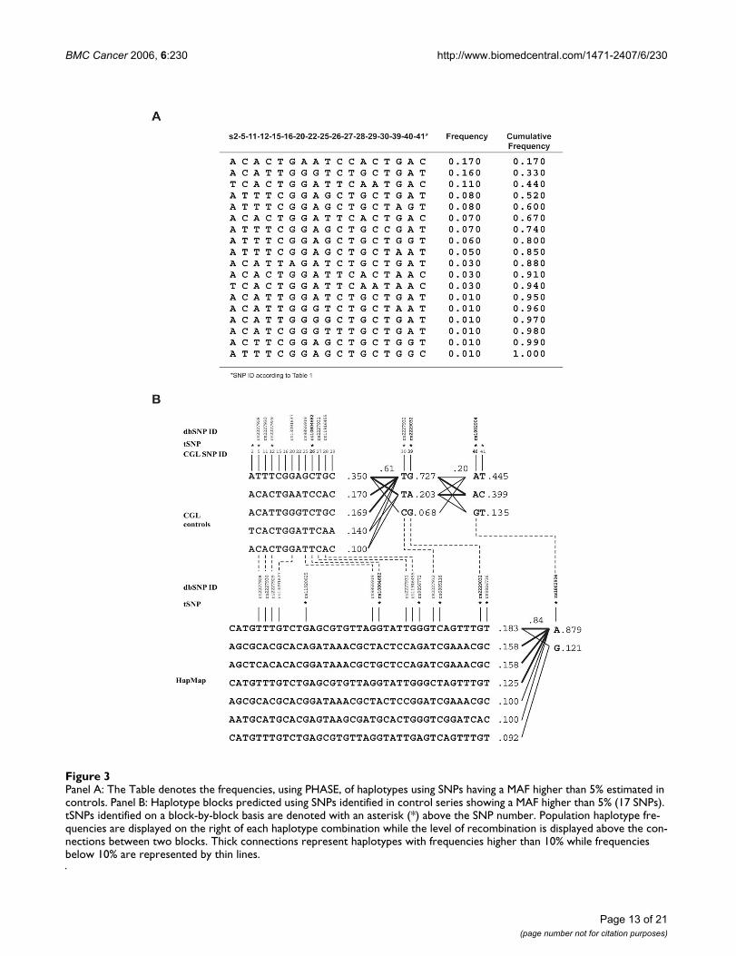
tion. When estimating the ATR haplotypes reconstructedfrom the 17 SNPs having a MAF ≥ 5% in breast cancercases, 24 haplotypes were identified from our data. Ofthese, 13 haplotypes exhibited a combined frequency≥1%, which represent 95% of all haplotypes estimated byPHASE in cases and controls combined (data not shown).
Thereafter, in order to identify ATR tSNPs useful for well-powered studies using larger sample sets, genotypes of 17coding and intronic SNPs showing a MAF ≥5% in healthyindividuals have been used for further haplotype analyses(Figure 3, Panel A). Genotyping data from controls onlywere purposely selected as they could well be more repre-sentative of the French Canadian population. When onlyhaplotypes displaying a MAF ≥5% are used, 85% of allestimated haplotypes are represented by the tSNPs identi-fied.
The identification of tSNPs was then carried out in twosubsequent steps, firstly by determining haplotype blocks,followed by identification of tSNP in each LD block.Based on the algorithm from Gabriel et al. [71], three LDblocks have been identified in the French Canadians bythe Haploview software (expectation maximisation algo-rithm) (Figure 3, Panel B). Thereafter, considering haplo-types having a MAF ≥5%, 8 tSNPs have been identified inthe 3 LD blocks, namely SNPs 2, 5, 12 and 26 found inblock 1, SNPs 30 and 39 in block 2, while block 3 consistsof SNPs 40 and 41. Furthermore, seven tSNPs clustered intwo LD blocks were selected in the ATR gene using theHapMap data from the CEPH/CEU cohort. It is of interest
to note that three tSNPs have been tagged in both theFrench Canadian and the CEPH/CEU sample sets.
Splicing consensus sequence analysisThe possible effect on splicing of all coding or intronicvariants located in a splice junction was assessed using theSSPNN website and revealed that the intronic variantc.5739-4del9+T (SNP32) located 4 nucleotides upstreamof exon 34 decreased exon 34 consensus acceptor sitescore from 0.94 to 0.31 (Figure 4, Panel A). The presenceof the intronic variant c.5739-4del9+T, located in intron33, became of interest regarding its possible implicationto generate a new alternative ATR transcript. Acceptor andDonor splice site sequences in this region are well con-served between mammalian species as illustrated in Fig-ure 4. Moreover it should be noted that the in silicoanalysis revealed a weak donor splice site score (0.11) forexon 33 while a putative donor site with a higher splicingscore (0.63) was predicted in exon 33, potentially gener-ating an alternative exon.
Another rare variant (c. 7041+8G>A) showed a significantalteration in the splicing score for a donor site, leading toa score from 0.51 to 0.24 (Figure 4, Panel B). In silico anal-ysis of the region surrounding the c.7041+8G/A variantrevealed that a putative intronic donor site located 441nucleotides downstream of the exon 41 could be alterna-tively used instead of the exon 41 donor site. The WT exon41 donor site found in other species displays a weak splic-ing score. The intronic putative donor site located inintron 41 has a higher splicing score in human (0.90) and
Table 2: ATR sequence variant allele frequencies in different cohorts
French-Canadian families Validation group Finnish families2 Reported in NCBI database
SNP SNP ID1 dbSNP ID n MAF n MAF n Carrier frequency MAF
- C.268C>T N/A 51 0.000 - - 126 0.127 N/A5* C.632C>T rs2227928 52 0.385 45 0.389 126 0.460 0.4256 c.946G>A rs28897764 50 0.047 42 0.037 126 0.000 N/A11 C.1776A>T rs2227930 53 0.415 - - 126 0.508 0.41212 C.1815T>C rs2227929 53 0.406 - - 126 0.468 0.37814 c.2290A>G N/A 50 0.010 41 0.000 126 0.000 N/A16 c.2875G>A rs28910271 48 0.052 46 0.000 126 0.000 0.01027 c.5208T>C rs2227931 49 0.441 - - 126 0.238 0.26330 c.5460T>C rs2227932 52 0.115 - - 126 0.262 0.10532 c. 5739-4del9+T N/A 54 0.046 42 0.012 126 0.000 N/A34 C.6394T>G rs28910273 54 0.009 44 0.000 126 0.008 0.01639 c.7274G>A rs2229032 51 0.147 41 0.170 126 0.278 0.10940 c.7875A>G rs1802904 50 0.120 - - 126 0.270 0.101
1 According to the nomenclature of the Human Genome Variation Society2 Heikkinen et al. 2005, Breast Cancer Research 7 :R495-R501N/A: Information not available for this SNP or not reported in dbSNP- Not genotyped* T allele is considered as the common allele according to the reference sequence NM_001184MAF : Minor Allele Frequency
Page 8 of 21(page number not for citation purposes)

BMC Cancer 2006, 6:230 http://www.biomedcentral.com/1471-2407/6/230
Page 9 of 21(page number not for citation purposes)
Table 3: Coding and intronic (MAF>5%) sequence variations and genotype frequencies in familial breast cancer cases and controls
SNP SNP ID1 Series # individual Common homozygote No. (expected)2
Heterozygote No. (expected)2
Rare homozygote No. (expected)2
MAF3 X2 p-value4
2 c.60-51A>T Cases 54 44 (44.3) 10 (9.2) 0 (0.5) 0.093 0.453Controls 62 49 (47.9) 11 (13.2) 2 (0.9) 0.121 0.192
5* c.632C>T Cases 52 22 (19.8) 20 (24,4) 10 (7.8) 0.385 0.176Controls 71 26 (29.8) 40 (32.4) 5 (8.8) 0.344 0.048
6 c.946G>A Cases 53 50 (48.1) 1 (4.7) 2 (0.1) 0.047 <0.001Controls 68 68 (68.0) 0 (0.0) 0 (0.0) 0.000 -
7 c.1326A>G Cases 54 51 (49.1) 1 (4.9) 2(0.0) 0.046 <0.001Controls 72 68 (68.1) 4 (3.9) 0(0.1) 0.028 0.808
8 c.1488C>T Cases 51 50 (50.0) 1 (1.0) 0 (0.0) 0.010 0.944Controls 73 73 (73.0) 0 (0.0) 0(0.0) 0.000 -
11 c.1776A>T Cases 53 21 (18.0) 20 (26.0) 12 (9.0) 0.415 0.105Controls 70 25 (29.6) 41 (31.8) 4 (8.6) 0.350 0.016
12 c.1815T>C Cases 53 21 (18.5) 21 (25.5) 11 (9.0) 0.406 0.194Controls 71 25 (25.5) 35 (34.1) 11 (11.4) 0.401 0.828
13 c.1950G>A Cases 53 50 (48.2) 1 (4.8) 2(0.0) 0.047 <0.001Controls 71 68 (68.0) 3 (2.9) 0(0.1) 0.021 0.856
14 c.2290A>G Cases 50 49 (49.0) 1 (1.0) a (0.0) 0.010 0.943Controls 72 72 (72.0) 0 (0.0) 0 (0.0) 0.000 -
15 c.2634-74T>C Cases 54 21 (18.3) 21 (26.3) 12 (9.4) 0.417 0.142Controls 51 15 (18.2) 31 (24.6) 5 (8.2) 0.402 0.059
16 c.2875G>A Cases 48 4S (43.2) 1 (4.8) 2(0.0) 0.052 <0.001Controls 68 64(64.0) 4 (3.9) 0(0.1) 0.028 0.803
18 c.3120G>A Cases 52 49 (47.3) 1 (4.7) 2 (0.0) 0.048 <0.001Controls 70 66(66.0) 4 (3.9) 0(0.1) 0.028 0.806
20 c.3357+128G>A Cases 54 27 (27.5) 23(22.1) 4(4.4) 0.287 0.765Controls 64 41 (40.7) 20 (20.7) 3 (2.6) 0.203 0.781
22 c.3726-47A>C Cases 54 41 (40.0) 11 (13.0) 2 (1.0) 0.139 0.275Controls 65 44(43.2) 18(19.6) 3(2.2) 0.185 0.518
25 c.4383-232T>G Cases 63 33 (19.9) 19 (25.2) 11 (7.9) 0.387 0.013Controls 65 23 (25.9) 36 (30.3) 6(8.8) 0.369 0.127
26 c.4383-177C>T Cases 54 39(40.0) 15(13.0) 0(1.0) 0.139 0.236Controls 65 37(37.0) 24 (24.1) 4(3.9) 0.246 0.967
27 c.5208T>C Cases 51 39 (16.3) 19 (25.0) 13 (9.7) 0.441 0.081Controls 65 23 (23.4) 32 (31.2) 10(10.4) 0.400 0.836
28 c.5288+130G>A Cases 36 16 (12.9) 11 (17.3) 9(5.8) 0.403 0.028Controls 55 20 (21.7) 29 (25.7) 6(7.6) 0.372 0.344
29 c.5380+121C>A Cases 46 40 (40.2) 6 (5.6) 0(0.2) 0.065 0.636Controls 62 JO (48.8) 10 (12.4) 2 (0.8) 0.113 0.125
30 c.5460T>C Cases 52 41 (40.1) 10(11.4) 1 (0.5) 0.115 0.676Controls 72 62 (62.4) 10 (9.3) 0(0.3) 0.069 0.526
32 c.5739-4del9+T Cases 54 51 (49.1) 1 (4.9) 2(0.0) 0.04 <0.001Controls 63 60 (60.0) 3 (2.9) 0 (0.0) 0.024 0.846
33 c.5868C>T Cases 53 51 (49.3) 0 (3.7) 2 (0.0) 0.038 <0.001Controls 67 67(67.0) 0 (0.0) 0 (0.0) 0.000 -
34 c.6394T>G Cases 54 53 (52.9) 1 (1.1) 0(0.0) 0.009 0.945Controls 66 66 (66.0) 0 (0.0) 0 (0.0) 0.000 -
37 c.7041+8G>A Cases 53 52(52.1) 1 (0.9) 0 (0.0) 0.009 0.945Controls 70 65 (63.2) 3 (6.6) 2(0.2) 0.050 <0.001
39 c.7274G>A Cases 51 38(37.2) 11 (12.8) 2 (1.0) 0.147 0.317Controls 70 39(40.9) 29 (25.2) 2(3.9) 0.236 0.210
40 c.7875A>G Cases 50 40(38.5) 8 (11.0) 2 (0.5) 0.120 0.086Controls 63 45 (45.4) 17 (16.1) 1 (1.5) 0.151 0.670
41 c.7932+104T>C Cases 51 20 (17.0) 19 (24.9) 12 (9.1) 0.422 0.009Controls 53 19 (18.7) 25 (25.6) 9(8.7) 0.406 0.874
1 According to the nomenclature of the Human Genome Variation Society2 As expected under Hardy-Weinberg equilibrium3 Minor allele frequency4 p-value for deviation from Hardy-Weinberg equilibrium (Pearson's chi-square)* T allele is considered as the common allele according to the reference sequence NM_001184
BMC Cancer 2006, 6:230 http://www.biomedcentral.com/1471-2407/6/230
Pan troglodyte (0.90), while no corresponding sequencecould be identified in other species analyzed.
Assessment of the presence of alternative transcriptsFollowing direct sequencing of cDNA PCR products in theregion surrounding the c.5739-4del9+T and the
c.7041+8G/A variants, we found a novel alternative splicemRNA (∆33 splice form) showing a deletion of the last121 nucleotides at the end of exon 33 (Figure 5, Panel A).The deletion causes a shift in the open reading frame(ORF) creating a premature stop codon in exon 34, yield-ing a putative truncated protein of 1889 amino acids
Table 4: Sequence variants detected in Human ATR and residues found in orthologues
SNP SNP ID* Amino acid change
Pan troglodytes
Canis familiaris
Mus musculus
Xenapus laevis
Fugu rubribes Strongylocentrotus purpuratus
Drosophila melanogaste
5** c.632C>T Thr211 Met Thr Thr Met Met Gly - Glu6 c. 946G>A Va1316Ile Val Val Val Val Val Ser Asp14 c. 2290A>G Lys764Glu Lys Lys Lys Lys Lys Ser Glu16 c. 2875G>A Val959Met Met Met Ile Met Ser Thr Phe34 c. 6394T>G Tyr2132Asp Tyr His Arg Gln Asn Lys Ala39 c. 7274G>A Arg2425Gln - Arg Gln Lys Lys Glu Thr
* According to the nomenclature of the Human Genome Variation Society- No corresponding residue in this species** T allele is considered as the common allele according to the reference sequence NM_001184
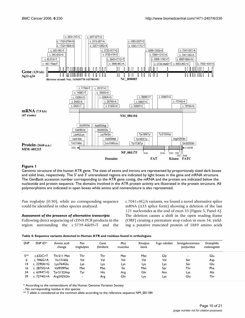
Genomic structure of the human ATR geneFigure 1Genomic structure of the human ATR gene. The sizes of exons and introns are represented by proportionaly sized dark boxes and solid lines, respectively. The 5' and 3' untranslated regions are indicated by light boxes in the gene and mRNA structure. The GenBank accession number corresponding to the ATR gene contig, the mRNA and the protein are indicated below the nucleotide and protein sequence. The domains involved in the ATR protein activity are illustrated in the protein structure. All polymorphisms are indicated in open boxes while amino acid nomenclature is also represented.
Page 10 of 21(page number not for citation purposes)

BMC Cancer 2006, 6:230 http://www.biomedcentral.com/1471-2407/6/230
which lacks the C-terminal part of the FAT domain, theentire kinase catalytic domain as well as the FATCdomain. Subcloning of PCR products covering this regionconfirmed the presence of this ∆33 splice form in theimmortalized lymphoblastoid cell lines from our breastcancer cases (Figure 6). Analysis through the UCSCgenome website revealed the presence of two human ESTscorresponding to the ∆33 splice transcript sequence(BG770191 and CD642306), supporting the presence ofsuch a transcript in humans. Interestingly this alternativetranscript uses the putative 3' splicing site predicted inexon 33 described above, which is conserved in most ofthe mammalian species analyzed. In order to confirm thatno additional alternative transcripts involving the skip-ping of exon 34 could be generated due to this c.5739-4del9+T variant, we used specific primers located on puta-tive exon 33–35 and ∆33-35 junctions in combinationwith upstream and donwstream primers for PCR amplifi-cation. No such detectable PCR product was observed.Besides, no EST corresponding to this potential transcriptwas found in the UCSC genome website.
No alternative splice transcript has been identified in theregion comprising the c.7041+8G/A variant using stand-ard procedures such as PCR amplification with externalprimers located on exon 38–39, 39–40, 43–44 and 44–45junctions, followed by subcloning. However, PCR ampli-fication using a specific primer located on a putativeexon41-intron41 junction or another primer located 441nucleotides downstream of exon 41 on the putativeIntron41-Exon42 junction identified by in silico analysisas described above, revealed the presence of such an alter-native transcript (Figure 5, Panel B). However, this alter-native transcript is not reported in UCSC database. Sincepreliminary expression analyses performed in immortal-ized cell lines of an c.7041+8G/A heterozygote carrier and9 wild-type individuals revealed an expression at the limitof detection (data not shown), no further expression anal-yses have been performed.
Characterization of ∆33 splice mRNA expressionFurther characterization of expression levels of ∆33 spliceform was performed in several normal tissues and cancercell lines. As shown in panel A (Figure 6), relative expres-sion levels of ∆33 splice form are highest in the breast andovary, with relative expression levels of approximately18% and 13% of the wild-type full-length splice form,while other examined tissues showed similar and lowerexpression levels. In breast cancer cell lines, ∆33 spliceform expression ranges from approximately 6% to 11% inrelation to total exon 33 expression (wild-type + ∆33splice form) and no significant variation is observedaccording to estrogen receptor or differentiation status.ATR wild-type expression levels standardized for 18S RNAlevels are also illustrated in panel B of Figure 6, and show
variable expression across tissue samples and somewhatslightly higher expression levels in cancer cell lines.
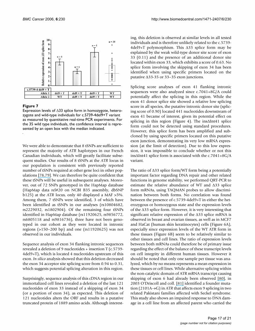
Is the c.5739-4del9+T variant associated with ∆33 splice transcripts?Assessment of association between the c.5739-4del9+Tvariant and the expression of the ∆33 splice form was per-formed using real-time PCR in RNA samples obtainedfrom lymphoblastoid cell lines of 38 of the screened cases,which included 35 wild-type individuals, one heterozy-gote and two homozygotes for the c.5739-4del9+T vari-ant. As illustrated in Figure 7, the presence of the ∆33splice transcript was detected in all individuals, includingwild-type individuals, therefore supporting that theexpression of this splice form is not associated with thepresence of the intronic c.5739-4del9+T variant. Further-more, more interestingly, expression levels do not seem tocorrelate with genotype status, as observed in hetero-zygous and homozygous individuals, and high expressionvariability is also observed in wild-type individuals,whose mean expression level was 2.733 ± 1.806.
Since no evidence suggested that the c.5739-4del9+T var-iant could be associated with the presence or the expres-sion of the ∆33 splice form, amplification of several cDNAfragments covering exons 30–38 using different combina-tions of primers located on exon-exon junctions (Figure5) was performed to detect any additional splice formresulting from the effect of this sequence change onmRNA splicing. No additional splice mRNA wasobserved.
DiscussionSince it is well established that the residual familial risk ofbreast cancer, not caused by BRCA1 or BRCA2 genes,could be explained by a polygenic or high-risk genes het-erogeneity model [72,73], we selected individuals affectedwith breast cancer without mutations in BRCA1/2 genesfrom high-risk families (one individual per family), inorder to increase the power of the study to find geneticvariants involved in breast cancer susceptibility. So far,several genes have been investigated based on their inter-action with BRCA1/2 or their involvement in DNA repairmechanisms. Since BRCA1/2 genes are intimately linkedto genomic stability, other genes involved in this pathwayare very good candidates to be BRCA3, and this is espe-cially true of ATM and ATR which play a central role ingenome stability maintenance. The ATM gene has beensuspected to be a breast cancer susceptibility locus, due tothe presence of breast cancer in A-T families, particularlyamong ATM heterozygotes [74]. ATM mutations havealready been reported to increase breast cancer suscepti-bility [9,27,75], while some other sequence variantslocated in this gene do not seem to be linked to breast can-cer [24].
Page 11 of 21(page number not for citation purposes)

BMC Cancer 2006, 6:230 http://www.biomedcentral.com/1471-2407/6/230
Based on the similar roles played by ATM and ATR as sen-sors of DNA damages, ATR may be considered a putativecandidate gene that could possibly explain a fraction ofthe remaining familial breast cancer risk. Association ofATR germline mutation with breast cancer susceptibilityhas been previously analyzed in Finnish 126 families [70],and no germline mutation was identified in this founderpopulation. The current study, performed in a FrenchCanadian cohort, also being a founder population, wasdesigned to assess the possible involvement of ATR germ-line deleterious mutations in breast cancer predisposition.
No deleterious germline mutation leading to a prematuretermination of the protein were identified in the codingregion. However, 41 sequence variants were identified,among which 16 were coding variants while 21 werenovel changes. In addition we find it unlikely that neitherof the common missense substitutions located in the FATand kinase domains (c.6394T>G and c.7274G>A) have asignificant effect on protein function because: (i) their fre-quencies are similar in cases and controls, especially forc.7274G>A whose MAF is greater than 20% in controlsand (ii) these residues are not well conserved in other spe-cies (Table 4). Indeed, the polymorphisms displaying asignificant deviation from HWE are composed of a groupof 14 uncommon polymorphisms identified in the same
3 breast cancer cases (2 homozygotes and 1 heterozygote),and therefore this most likely constitutes a single rela-tively rare allele. It has to be stated that no particular char-acteristics seem to emerge for the families bearing any ofthese rare variants, as both the French Canadian and thevalidation families have been recruited on the basis ofhigh-risk breast cancer families.
Comparison of polymorphism frequencies between ourcohort and the Finnish cohort [70] is not fully informativesince the latter does not distinguish the number of heter-ozygotes and homozygotes found in their cohort but onlythe number of carriers of a given polymorphism. How-ever, if we also use this method to calculate polymor-phism frequencies observed in our cohort, only SNP40displayed a notably lower frequency than that found inthe Finnish cohort. As stated earlier, both studies (Heik-kinen et al. and the present study) have been designed toidentify ATR deleterious germline mutations in breastcancer cases. No such mutation was found in either study,therefore ATR is unlikely to play a major role as a highpenetrance gene in breast cancer predisposition. Eventhough novel variants have been identified, the possibleinvolvement of polymorphisms or haplotypes observedin cases compared to those found in controls would needa lot more individuals to obtain a significant value ofassociation to breast cancer susceptibility [76,77]. Wethus sought to identify tSNPs that could be useful to otherstudies and populations.
Our pairwise linkage disequilibrium analysis (Figure 2)did not seem to identify any distinct LD blocks withinATR. This observation is supported by the fact that SNP1is in perfect LD with most other SNPs, including the mostdistal SNP41, and is also in accordance with what is seenin the French Canadian founder population which dis-plays large conserved haplotypes as reported at the BRCA1locus [49]. However, using the Haploview software, threedistinct LD blocks were identified at the ATR locus whenusing SNPs showing a MAF >5% in healthy French Cana-dian individuals (Figure 3). The breakage of strong LDseems to be located in the region of exon 31, and betweenexon 43 and exon 47.
Based on the same algorithm (Haploview), and using theSNPs genotyped in HapMap database showing a MAFhigher than 5%, two LD blocks could be identified; thefirst block comprising the SNPs located from intron 1 toexon 43, while the second block included all the remain-ing SNPs until exon 47. However, it should be noted thatthe majority of the SNPs used to determine haplotypeblocks have a MAF higher than 0.4, which represent com-mon SNPs found in many different populations andtherefore probably exclude the SNPs specifically observedin our French Canadian founder population.
Pairwise linkage disequilibrium (LD) measures of |D'| for the 41 SNPs identified in our breast cancer cases seriesFigure 2Pairwise linkage disequilibrium (LD) measures of |D'| for the 41 SNPs identified in our breast cancer cases series. All SNPs are denoted numerically with reference to Table 1.
Page 12 of 21(page number not for citation purposes)
BMC Cancer 2006, 6:230 http://www.biomedcentral.com/1471-2407/6/230
Page 13 of 21(page number not for citation purposes)
Panel A: The Table denotes the frequencies, using PHASE, of haplotypes using SNPs having a MAF higher than 5% estimated in controlsFigure 3Panel A: The Table denotes the frequencies, using PHASE, of haplotypes using SNPs having a MAF higher than 5% estimated in controls. Panel B: Haplotype blocks predicted using SNPs identified in control series showing a MAF higher than 5% (17 SNPs). tSNPs identified on a block-by-block basis are denoted with an asterisk (*) above the SNP number. Population haplotype fre-quencies are displayed on the right of each haplotype combination while the level of recombination is displayed above the con-nections between two blocks. Thick connections represent haplotypes with frequencies higher than 10% while frequencies below 10% are represented by thin lines.

BMC Cancer 2006, 6:230 http://www.biomedcentral.com/1471-2407/6/230
Page 14 of 21(page number not for citation purposes)
Panel A: Comparative analysis of splicing site sequences in ATR exon 32, 33, ∆33 and 34 flanking exon-intron junctions as well as prediction of impact of SNP32 (c.5739-4del9+T) on exon 34 acceptor siteFigure 4Panel A: Comparative analysis of splicing site sequences in ATR exon 32, 33, ∆33 and 34 flanking exon-intron junctions as well as prediction of impact of SNP32 (c.5739-4del9+T) on exon 34 acceptor site. Panel B: Comparative analysis of splicing site sequences in ATR exon 41, ins∆Int41 and exon 42 flanking exon-intron junctions as well as prediction of impact of SNP37 (c.7041+8G/A) on exon 41 donor site. Splice Site Prediction Program using Neural Network (SSPNN) score values are indi-cated in parenthesis below each sequence. Exonic nucleotides are represented by uppercase letters while intronic sequences are represented by lowercase letters. m = c or a, r = a or g, y = t or c and n = any nucleotide. N.A. = value not available. Con-sensus sequences of acceptor and donor sites were described by Burge et al. 1999 [84].

BMC Cancer 2006, 6:230 http://www.biomedcentral.com/1471-2407/6/230
Page 15 of 21(page number not for citation purposes)
Panel A: Alternative splicing of ATR ∆33 exonFigure 5Panel A: Alternative splicing of ATR ∆33 exon. Schematic representation of the design used to assess the existence of mRNA splice transcript encompassing exons 33 and 34 in immortalized cell lines of individuals affected with breast cancer. The puta-tive truncated ATR protein of 1889 amino acids lacks the functional domains identified in the wild type ATR protein. Panel B: Alternative splicing of ATR ins∆Int41 exon. Schematic representation of the design used to assess the existence of mRNA splice transcript encompassing exons 41 and 42 in immortalized cell lines of individuals affected with breast cancer. The putative trun-cated ATR protein of 2350 amino acids lacks a part of the kinase domains identified in the wild type ATR protein.

BMC Cancer 2006, 6:230 http://www.biomedcentral.com/1471-2407/6/230
Page 16 of 21(page number not for citation purposes)
Expression levels of ATR ∆33 splice form in cell lines and human tissues as measured by quantitative real-time PCR experi-mentsFigure 6Expression levels of ATR ∆33 splice form in cell lines and human tissues as measured by quantitative real-time PCR experi-ments. Panel A. Relative expression levels of ∆33 splice form were calculated as ∆33 splice form/(∆33 splice form + wild-type allele) in various human tissues and cell lines. Panel B. Standardized expression levels of WT exon 33 were calculated as WT exon 33/(∆33 splice form + wild-type allele) in various human tissues and cell lines.
BMC Cancer 2006, 6:230 http://www.biomedcentral.com/1471-2407/6/230
We were able to demonstrate that 8 tSNPs are sufficient torepresent the majority of ATR haplotypes in our FrenchCanadian individuals, which will greatly facilitate subse-quent studies. Our results of 8 tSNPs at the ATR locus inour population is consistent with previously reportednumber of tSNPs required at other gene loci in other pop-ulations [78,79]. We can therefore be quite confident thatthese tSNPs will be useful in subsequent analyses. Moreo-ver, out of 72 SNPs genotyped in the HapMap database(HapMap data rel#20 on NCBI B35 assembly, dbSNPb125) at the ATR locus, only 40 displayed a MAF >5%.Among them, 7 tSNPs were identified, 3 of which havebeen identified as tSNPs in our analyses (rs10804682,rs2229032, rs1802904). Of the remaining four tSNPsidentified in HapMap database (rs11920625, rs9856772,rs6805118 and rs9816736), three have not been geno-typed in our cohort as they were located in intronicregions (>150–200 bp) and one (rs11920625) was notobserved in our individuals.
Sequence analysis of exon 34 flanking intronic sequencesrevealed a deletion of 9 nucleotides + insertion T (c.5739-4del9+T), which is located 4 nucleotides upstream of thisexon. In silico analysis showed that this deletion decreasedthe exon 34 acceptor site splicing score from 0.94 to 0.31,which suggests potential splicing alteration in this region.
Surprisingly, sequence analysis of this cDNA region in ourimmortalized cell lines revealed a deletion of the last 121nucleotides of exon 33 instead of a skipping of exon 34(or a portion of exon 34), as expected. This deletion of121 nucleotides alters the ORF and results in a putativetruncated protein of 1889 amino acids. Although interest-
ing, this deletion is observed at similar levels in all testedindividuals and is therefore unlikely related to the c.5739-4del9+T polymorphism. This ∆33 splice form may beexplained by the weak wild-type donor site score of exon33 (0.11) and the presence of an additional donor sitelocated within exon 33, which exhibits a score of 0.63. Nosplice form involving the skipping of exon 34 has beenidentified when using specific primers located on theputative ∆33-35 or 33–35 exon junctions.
Splicing score analyses of exon 41 flanking intronicsequences were also analyzed since c.7041+8G/A couldpotentially affect the splicing in this region. While theexon 41 donor splice site showed a relative low splicingscore in all species, the putative intronic donor site (splic-ing score of 0.90) located 441 nucleotides downstream ofexon 41 became of interest, given its potential effect onsplicing in this region (Figure 4). The ins∆Int41 spliceform could not be detected using standard procedures.However, this splice form has been amplified and sub-cloned by using specific primers located on this putativeexon junction, demonstrating its very low mRNA expres-sion (at the limit of detection). Due to this low expres-sion, it was impossible to conclude whether or not thisins∆Int41 splice form is associated with the c.7041+8G/Avariant.
The ratio of ∆33 splice form/WT form being a potentiallyimportant factor regarding DNA repair and other relatedfunctions in genome stability, we performed QRT-PCR toestimate the relative abundance of WT and ∆33 spliceform mRNAs, using TAQMAN probes to allow discrimi-nation between both forms. No correlation was foundbetween the presence of c.5739-4del9+T in either the het-erozygous or homozygous state and the expression levelsof the ∆33 splice form. However, it is very interesting thatsignificant relative expression of the ∆33 splice mRNA isobserved in breast and ovarian tissues, as well as in MCF7and HaCat (human skin keratinocytes) cells (Figure 6A),especially since expression levels of the WT ATR form inthese tissues (Figure 6B) seem to be relatively similar toother tissues and cell lines. The ratio of expression levelsbetween both mRNAs could therefore be of primary issueregarding the effect of the balance of these transcript levelson cell integrity in different human tissues. However itshould be noted that only one sample per tissue was ana-lyzed, which by no means represents a mean expression inthese tissues or cell lines. While alternative splicing withinthe non-catalytic domain of ATR mRNA transcript causingskipping of exon 6 had already been observed [80], in2003 O'Driscoll and coll. [81] identified a founder muta-tion (2101A→G) in ATR that affects exon 9 splicing in tworelated Pakistani families affected with Seckel syndrome.This study also shows an impaired response to DNA dam-age in a cell line from an affected parent who carried the
Expression levels of ∆33 splice form in homozygote, hetero-zygote and wild-type individuals for c.5739-4del9+T variant as measured by quantitative real-time PCR experimentsFigure 7Expression levels of ∆33 splice form in homozygote, hetero-zygote and wild-type individuals for c.5739-4del9+T variant as measured by quantitative real-time PCR experiments. For the 35 wild type individuals, the confidence interval is repre-sented by an open box with the median indicated.
Page 17 of 21(page number not for citation purposes)
BMC Cancer 2006, 6:230 http://www.biomedcentral.com/1471-2407/6/230
mutation. Further characterization of ATR-Seckel cellsshowed impaired phosphorylation of ATR-dependentsubstrates, impaired G2/M checkpoint arrest and super-numery centrosomes in mitotic cells, clearly demonstrat-ing a role for ATR in the maintenance of centrosomestability [82]. More recently, two other splicing alterationsof ATR have been reported in clinical samples with pyot-horax-associated lymphoma [83].
ConclusionNo deleterious germline mutations have been identifiedin French Canadian breast cancer cases. However, we haveconducted the first detailed haplotype tagging analysis ofthe ATR gene within control individuals from the FrenchCanadian population. The data presented here clearlyidentified 8 ATR tSNPs, which will be useful for otherlarge-scale association studies. We did not find any germ-line mutations in the ATR gene potentially involved inbreast cancer predisposition. However, given that differ-ent splicing alterations of ATR have been associated withimpaired response to DNA damage, the notably signifi-cant expression of the novel ∆33 splice form observed inbreast and ovarian tissues could have a potential effect onDNA repair mechanisms in these cells, although exhaus-tive analyses should be required to verify this hypothesis.Further analyses in other populations and larger cohortswill be required to define the possible association of ATRgene polymorphisms with breast cancer susceptibility.
AbbreviationsATR: Ataxia Telangiectasia-mutated and Rad3-related
CV/CD: Common Variant/Common Disease
ATM: Ataxia Telangiectasia-mutated
PIK: Phosphatidyl Inositol-Kinase
ATRIP: ATR-Interacting Protein
MRN: Mre11/Rad50/NBS1
Smc1: Structural Maintenance of Chromosome 1
MLPA: Multiplex Ligation-dependant Probe Amplifica-tion
LD: Linkage Disequilibrium
tSNPs: Tagging SNPs
ATCC: American Type Culture Collection
ER: Estrogen Receptor
QRT-PCR: Quantitative RT-PCR
CT : Threshold Cycle number
SSPNN: Splice Site Prediction Program using Neural Net-works
dbSNP: Single Nucleotide Polymorphism database
HWE: Hardy-Weinberg Equilibrium
FAT: Frap/ATM/TRRAP
PI3Kc: Phosphoinositide 3-Kinase related catalytic
ORF: Open reading Frame
Competing interestsThe author(s) declare that they have no competing inter-ests.
Authors' contributionsFD and JS conceived and devised the overall strategy forthis study and authored the final version of this manu-script. YL and PS performed all DNA sequence and data-base analyses and drafted the manuscript. Haplotypeanalyses were carried out by YL while RNA expressionanalyses were carried out by PS. OS and ST providedinsightful comments and revisions of the final version ofthe text and carried out DNA sequencing of the patients ofthe validation group. DL provided DNA samples fromhealthy patients. PB, JC, RL, JP, BL, RP and MP are clini-cians that were in charge of collecting blood samples formaffected individuals in their respective institutions andhave been highly involved throughout the whole recruit-ment process to result disclosure. All authors read andapproved the final manuscript.
Additional material
AcknowledgementsThe authors are indebted to the participants and their families for their gen-erosity and providing DNA samples. We would like to thank Dr Martine Dumont, Gilles Leblanc, Carolle Samson and Martine Tranchant for sample management, mutation screening, and skillful technical assistance as well as Claire Brousseau, Marie-Andrée Lajoie, Pascale Léger, Hélène Malouin and
Additional File 1Oligonucleotide primers used for amplification and sequence analysis of the ATR gene. The table provides the primers used for amplification and sequencing of the ATR gene.Click here for file[http://www.biomedcentral.com/content/supplementary/1471-2407-6-230-S1.pdf]
Page 18 of 21(page number not for citation purposes)

BMC Cancer 2006, 6:230 http://www.biomedcentral.com/1471-2407/6/230
Josée Rhéaume, for genetic counselling and clinical data management at the Cancer Genomics Laboratory. We also thank Geneviève Ouellette for establishment of EBV-transformed B-lymphoblastoid cell lines and RNA and genomic DNA extractions, as well as Anne-Marie Moisan and Lucie Larouche for MLPA analyses. We thank Claudia Moreau at the Centre de Recherche de l'Hôpital Ste-Justine for help with control DNA samples. We would also like to thank Professor Bartha Maria Knoppers and her col-leagues from the Centre de recherche en droit public de l'Université de Montréal for their precious help with ELSI issues related to our research program. We also appreciate advice received from ethics committees. This work was supported by the Canadian Institutes of Health Research (CIHR) through the INHERIT BRCAs research program and the Fonds de la Recherche en Santé du Québec (FRSQ)/Réseau de Médecine Génétique Appliquée (RMGA). F.D. is a recipient of a Research Career Award in the Health Sciences from CIHR/Rx&D Health Research Foundation, and J.S. is Chairholder of the Canada Research Chair in Oncogenetics.
References1. Houlston RS, Peto J: Genetics and the common cancers. In
Genetic Predisposition to Cancer Edited by: Eeles RA, Easton DF, PonderBAJ, Eng C. New York: Oxford University Press; 2004:235-247.
2. Peto J, Mack TM: High constant incidence in twins and otherrelatives of women with breast cancer. Nat Genet 2000,26:411-414.
3. Lichtenstein P, Holm NV, Verkasalo PK, Iliadou A, Kaprio J, Kosken-vuo M, Pukkala E, Skytthe A, Hemminki K: Environmental and her-itable factors in the causation of cancer-analyses of cohortsof twins from Sweden, Denmark, and Finland. N Engl J Med2000, 343:78-85.
4. Vogelstein B, Kinzler KW: Cancer genes and the pathways theycontrol. Nat Med 2004, 10:789-799.
5. Hirschhorn JN, Daly MJ: Genome-wide association studies forcommon diseases and complex traits. Nat Rev Genet 2005,6:95-108.
6. Pharoah PD, Dunning AM, Ponder BA, Easton DF: Associationstudies for finding cancer-susceptibility genetic variants. NatRev Cancer 2004, 4:850-860.
7. Wang WY, Barratt BJ, Clayton DG, Todd JA: Genome-wide asso-ciation studies: theoretical and practical concerns. Nat RevGenet 2005, 6:109-118.
8. Thompson D, Easton D: The genetic epidemiology of breastcancer genes. J Mammary Gland Biol Neoplasia 2004, 9:221-236.
9. Renwick A, Thompson D, Seal S, Kelly P, Chagtai T, Ahmed M, NorthB, Jayatilake H, Barfoot R, Spanova K, et al.: ATM mutations thatcause ataxia-telangiectasia are breast cancer susceptibilityalleles. Nat Genet 2006, 38:873-875.
10. Antoniou AC, Pharoah PD, McMullan G, Day NE, Ponder BA, EastonD: Evidence for further breast cancer susceptibility genes inaddition to BRCA1 and BRCA2 in a population-based study.Genet Epidemiol 2001, 21:1-18.
11. Antoniou AC, Pharoah PD, McMullan G, Day NE, Stratton MR, PetoJ, Ponder BJ, Easton DF: A comprehensive model for familialbreast cancer incorporating BRCA1, BRCA2 and othergenes. Br J Cancer 2002, 86:76-83.
12. Pharoah PD, Antoniou A, Bobrow M, Zimmern RL, Easton DF, Pon-der BA: Polygenic susceptibility to breast cancer and implica-tions for prevention. Nat Genet 2002, 31:33-36.
13. Cargill M, Daley GQ: Mining for SNPs: putting the commonvariants-common disease hypothesis to the test. Pharmacoge-nomics 2000, 1:27-37.
14. Lohmueller KE, Pearce CL, Pike M, Lander ES, Hirschhorn JN: Meta-analysis of genetic association studies supports a contribu-tion of common variants to susceptibility to common dis-ease. Nat Genet 2003, 33:177-182.
15. Reich DE, Lander ES: On the allelic spectrum of human disease.Trends Genet 2001, 17:502-510.
16. Fearnhead NS, Wilding JL, Winney B, Tonks S, Bartlett S, Bicknell DC,Tomlinson IP, Mortensen NJ, Bodmer WF: Multiple rare variantsin different genes account for multifactorial inherited sus-ceptibility to colorectal adenomas. Proc Natl Acad Sci USA 2004,101:15992-15997.
17. Pritchard JK: Are rare variants responsible for susceptibility tocomplex diseases? Am J Hum Genet 2001, 69:124-137.
18. Pritchard JK, Cox NJ: The allelic architecture of human diseasegenes: common disease-common variant or not? Hum MolGenet 2002, 11:2417-2423.
19. Wang WY, Todd JA: The usefulness of different density SNPmaps for disease association studies of common variants.Hum Mol Genet 2003, 12:3145-3149.
20. Venkitaraman AR: Cancer susceptibility and the functions ofBRCA1 and BRCA2. Cell 2002, 108:171-182.
21. Venkitaraman AR: Tracing the network connecting BRCA andFanconi anaemia proteins. Nat Rev Cancer 2004, 4:266-276.
22. Chenevix-Trench G, Spurdle AB, Gatei M, Kelly H, Marsh A, Chen X,Donn K, Cummings M, Nyholt D, Jenkins MA, et al.: Dominant neg-ative ATM mutations in breast cancer families. J Natl CancerInst 2002, 94:205-215.
23. Stankovic T, Kidd AM, Sutcliffe A, McGuire GM, Robinson P, WeberP, Bedenham T, Bradwell AR, Easton DF, Lennox GG, et al.: ATMmutations and phenotypes in ataxia-telangiectasia familiesin the British Isles: expression of mutant ATM and the risk ofleukemia, lymphoma, and breast cancer. Am J Hum Genet 1998,62:334-345.
24. Szabo CI, Schutte M, Broeks A, Houwing-Duistermaat JJ, ThorstensonYR, Durocher F, Oldenburg RA, Wasielewski M, Odefrey F, Thomp-son D, et al.: Are ATM mutations 7271T-->G and IVS10-6T-->G really high-risk breast cancer-susceptibility alleles? CancerRes 2004, 64:840-843.
25. Thompson D, Antoniou AC, Jenkins M, Marsh A, Chen X, Wayne T,Tesoriero A, Milne R, Spurdle A, Thorstenson Y, et al.: Two ATMvariants and breast cancer risk. Hum Mutat 2005, 25:594-595.
26. Thompson D, Duedal S, Kirner J, McGuffog L, Last J, Reiman A, ByrdP, Taylor M, Easton DF: Cancer risks and mortality in hetero-zygous ATM mutation carriers. J Natl Cancer Inst 2005,97:813-822.
27. Thorstenson YR, Roxas A, Kroiss R, Jenkins MA, Yu KM, Bachrich T,Muhr D, Wayne TL, Chu G, Davis RW, et al.: Contributions ofATM mutations to familial breast and ovarian cancer. CancerRes 2003, 63:3325-3333.
28. CHEK2 Breast Cancer Case-Control Consortium:CHEK2*1100delC and susceptibility to breast cancer: a col-laborative analysis involving 10,860 breast cancer cases and9,065 controls from 10 studies. Am J Hum Genet 2004,74:1175-1182.
29. Meijers-Heijboer H, van den Ouweland A, Klijn J, Wasielewski M, deSnoo A, Oldenburg R, Hollestelle A, Houben M, Crepin E, van Veghel-Plandsoen M, et al.: Low-penetrance susceptibility to breastcancer due to CHEK2(*)1100delC in noncarriers of BRCA1or BRCA2 mutations. Nat Genet 2002, 31:55-59.
30. Gasco M, Yulug IG, Crook T: TP53 mutations in familial breastcancer: functional aspects. Hum Mutat 2003, 21:301-306.
31. Ueda K, Nishijima M, Inui H, Watatani M, Yayoi E, Okamura J, Yasu-tomi M, Nakamura Y, Miyoshi Y: Infrequent mutations in thePTEN/MMAC1 gene among primary breast cancers. Jpn JCancer Res 1998, 89:17-21.
32. Giardiello FM, Brensinger JD, Tersmette AC, Goodman SN, PetersenGM, Booker SV, Cruz-Correa M, Offerhaus JA: Very high risk ofcancer in familial Peutz-Jeghers syndrome. Gastroenterology2000, 119:1447-1453.
33. Kuschel B, Auranen A, McBride S, Novik KL, Antoniou A, LipscombeJM, Day NE, Easton DF, Ponder BA, Pharoah PD, et al.: Variants inDNA double-strand break repair genes and breast cancersusceptibility. Hum Mol Genet 2002, 11:1399-1407.
34. Goodarzi AA, Block WD, Lees-Miller SP: The role of ATM andATR in DNA damage-induced cell cycle control. Prog Cell CycleRes 2003, 5:393-411.
35. Ward IM, Chen J: Histone H2AX is phosphorylated in an ATR-dependent manner in response to replicational stress. J BiolChem 2001, 276:47759-47762.
36. Liu Q, Guntuku S, Cui XS, Matsuoka S, Cortez D, Tamai K, Luo G,Carattini-Rivera S, DeMayo F, Bradley A, et al.: Chk1 is an essentialkinase that is regulated by Atr and required for the G(2)/MDNA damage checkpoint. Genes Dev 2000, 14:1448-1459.
37. Tibbetts RS, Brumbaugh KM, Williams JM, Sarkaria JN, Cliby WA,Shieh SY, Taya Y, Prives C, Abraham RT: A role for ATR in theDNA damage-induced phosphorylation of p53. Genes Dev1999, 13:152-157.
Page 19 of 21(page number not for citation purposes)
http://www.ncbi.nlm.nih.gov/entrez/query.fcgi?cmd=Retrieve&db=PubMed&dopt=Abstract&list_uids=9463314
http://www.ncbi.nlm.nih.gov/entrez/query.fcgi?cmd=Retrieve&db=PubMed&dopt=Abstract&list_uids=9463314
http://www.ncbi.nlm.nih.gov/entrez/query.fcgi?cmd=Retrieve&db=PubMed&dopt=Abstract&list_uids=9463314
http://www.ncbi.nlm.nih.gov/entrez/query.fcgi?cmd=Retrieve&db=PubMed&dopt=Abstract&list_uids=9510470
http://www.ncbi.nlm.nih.gov/entrez/query.fcgi?cmd=Retrieve&db=PubMed&dopt=Abstract&list_uids=9510470
http://www.ncbi.nlm.nih.gov/entrez/query.fcgi?cmd=Retrieve&db=PubMed&dopt=Abstract&list_uids=9925639
BMC Cancer 2006, 6:230 http://www.biomedcentral.com/1471-2407/6/230
38. Zhao H, Piwnica-Worms H: ATR-mediated checkpoint path-ways regulate phosphorylation and activation of humanChk1. Mol Cell Biol 2001, 21:4129-4139.
39. Zou L, Cortez D, Elledge SJ: Regulation of ATR substrate selec-tion by Rad17-dependent loading of Rad9 complexes ontochromatin. Genes Dev 2002, 16:198-208.
40. Tibbetts RS, Cortez D, Brumbaugh KM, Scully R, Livingston D, ElledgeSJ, Abraham RT: Functional interactions between BRCA1 andthe checkpoint kinase ATR during genotoxic stress. GenesDev 2000, 14:2989-3002.
41. Schmidt DR, Schreiber SL: Molecular association between ATRand two components of the nucleosome remodeling anddeacetylating complex, HDAC2 and CHD4. Biochemistry 1999,38:14711-14717.
42. Pichierri P, Rosselli F: The DNA crosslink-induced S-phasecheckpoint depends on ATR-CHK1 and ATR-NBS1-FANCD2 pathways. Embo J 2004, 23:1178-1187.
43. Durocher F, Guénard F, Desjardins S, Ouellette G, Labrie Y: Inher-ited susceptibility to breast cancer: accomplishments andchallenges. In Molecular Genetics of Cancer Edited by: Sinnett D.Research Signpost; 2005:19-93.
44. Zhong H, Bryson A, Eckersdorff M, Ferguson DO: Rad50 depletionimpacts upon ATR-dependent DNA damage responses. HumMol Genet 2005, 14:2685-2693.
45. Brown EJ, Baltimore D: ATR disruption leads to chromosomalfragmentation and early embryonic lethality. Genes Dev 2000,14:397-402.
46. de Klein A, Muijtjens M, van Os R, Verhoeven Y, Smit B, Carr AM,Lehmann AR, Hoeijmakers JH: Targeted disruption of the cell-cycle checkpoint gene ATR leads to early embryoniclethality in mice. Curr Biol 2000, 10:479-482.
47. Foray N, Marot D, Gabriel A, Randrianarison V, Carr AM, PerricaudetM, Ashworth A, Jeggo P: A subset of ATM- and ATR-dependentphosphorylation events requires the BRCA1 protein. Embo J2003, 22:2860-2871.
48. Avard D, Bridge P, Bucci LM, Chiquette J, Dorval M, Durocher F, Eas-ton D, Godard B, Goldgar D, Knoppers BM, et al.: Partnering inoncogenetic research-The INHERIT BRCAs Experience:Opportunities and Challenges. Familial Cancer 2006, 5:3-13.
49. Vezina H, Durocher F, Dumont M, Houde L, Szabo C, Tranchant M,Chiquette J, Plante M, Laframboise R, Lepine J, et al.: Molecular andgenealogical characterization of the R1443X BRCA1 muta-tion in high-risk French-Canadian breast/ovarian cancerfamilies. Hum Genet 2005, 117:119-132.
50. Antoniou AC, Durocher F, Smith P, Simard J, Easton DF: BRCA1and BRCA2 mutation predictions using the BOADICEA andBRCAPRO models and penetrance estimation in high-riskFrench-Canadian families. Breast Cancer Res 2005, 8:R3.
51. Dorval M, Gauthier G, Maunsell E, Dugas MJ, Rouleau I, Chiquette J,Plante M, Laframboise R, Gaudet M, Bridge PJ, et al.: No evidence offalse reassurance among women with an inconclusiveBRCA1/2 genetic test result. Cancer Epidemiol Biomarkers Prev2005, 14:2862-2867.
52. Simard J, Dumont M, Moisan AM, Gaborieau V, Vezina H, DurocherF, Chiquette J, Plante M, Avard D, Bessette P, et al.: Evaluation ofBRCA1 and BRCA2 mutation prevalence, risk predictionmodels and a multi-step testing approach in French-Cana-dian high-risk breast and ovarian cancer families. J Med Genet. 2006, Aug 11; [Epub ahead of print]
53. Moisan A, Fortin J, Dumont M, Samson C, Bessette P, Chiquette J,Laframboise R, Lépine J, Lespérance B, Pichette R, et al.: No evi-dence of BRCA1/2 genomic rearrangements in high riskFrench-Canadian breast/ovarian cancer families. Genetic Test-ing 2006, 10:104-115.
54. Bonadona V, Sinilnikova OM, Chopin S, Antoniou AC, Mignotte H,Mathevet P, Bremond A, Martin A, Bobin JY, Romestaing P, et al.:Contribution of BRCA1 and BRCA2 germ-line mutations tothe incidence of breast cancer in young women: results froma prospective population-based study in France. Genes Chro-mosomes Cancer 2005, 43:404-413.
55. Sinilnikova OM, Mazoyer S, Bonnardel C, Lynch HT, Narod SA, GML: BRCA1 and BRCA2 mutations in breast and ovarian can-cer syndrome: Reflection on the Creighton University his-torical series of high risk families. Familial Cancer 2006, 5:15-20.
56. Ding K, Zhou K, He F, Shen Y: LDA-a java-based linkage disequi-librium analyzer. Bioinformatics 2003, 19:2147-2148.
57. Devlin B, Risch N: A comparison of linkage disequilibriummeasures for fine-scale mapping. Genomics 1995, 29:311-322.
58. Lewontin RC: The Interaction of Selection and Linkage. Ii.Optimum Models. Genetics 1964, 50:757-782.
59. Stephens M, Smith NJ, Donnelly P: A new statistical method forhaplotype reconstruction from population data. Am J HumGenet 2001, 68:978-989.
60. PHASE [http://www.stat.washington.edu/stephens/software.html]61. HapMap website [http://www.hapmap.org]62. Barrett JC, Fry B, Maller J, Daly MJ: Haploview: analysis and visu-
alization of LD and haplotype maps. Bioinformatics 2005,21:263-265.
63. Haploview [http://www.broad.mit.edu/mpg/haploview]64. Splice Site Prediction Program using Neural Networks
(SSPNN) [http://www.fruitfly.org/seq_tools/splice.html]65. ClustalW [http://www.ebi.ac.uk/clustalw]66. Fortin J, Moisan AM, Dumont M, Leblanc G, Labrie Y, Durocher F,
Bessette P, Bridge P, Chiquette J, Laframboise R, et al.: A new alter-native splice variant of BRCA1 containing an additional in-frame exon. Biochim Biophys Acta 2005, 1731:57-65.
67. Boukamp P, Stanbridge EJ, Foo DY, Cerutti PA, Fusenig NE: c-Ha-rasoncogene expression in immortalized human keratinocytes(HaCaT) alters growth potential in vivo but lacks correlationwith malignancy. Cancer Res 1990, 50:2840-2847.
68. Boukamp P, Petrussevska RT, Breitkreutz D, Hornung J, Markham A,Fusenig NE: Normal keratinization in a spontaneously immor-talized aneuploid human keratinocyte cell line. J Cell Biol 1988,106:761-771.
69. Luu-The V, Paquet N, Calvo E, Cumps J: Improved Real-Time RT-PCR method for high-throughput measurements using sec-ond derivative calculation and double correction. Biotech-niques 2005, 8:287-293.
70. Heikkinen K, Mansikka V, Karppinen SM, Rapakko K, Winqvist R:Mutation analysis of the ATR gene in breast and ovarian can-cer families. Breast Cancer Res 2005, 7:R495-R501.
71. Gabriel SB, Schaffner SF, Nguyen H, Moore JM, Roy J, Blumenstiel B,Higgins J, DeFelice M, Lochner A, Faggart M, et al.: The structure ofhaplotype blocks in the human genome. Science 2002,296:2225-2229.
72. Easton DF: How many more breast cancer predispositiongenes are there? Breast Cancer Res 1999, 1:14-17.
73. Weber BL, Nathanson KL: Low penetrance genes associatedwith increased risk for breast cancer. Eur J Cancer 2000,36:1193-1199.
74. Swift M, Sholman L, Perry M, Chase C: Malignant neoplasms inthe families of patients with ataxia-telangiectasia. Cancer Res1976, 36:209-215.
75. Broeks A, Urbanus JH, Floore AN, Dahler EC, Klijn JG, Rutgers EJ,Devilee P, Russell NS, van Leeuwen FE, van't Veer LJ: ATM-hetero-zygous germline mutations contribute to breast cancer-sus-ceptibility. Am J Hum Genet 2000, 66:494-500.
76. Peto J, Mack TM: High constant incidence in twins and otherrelatives of women with breast cancer. Nat Genet 2000,26:411-414.
77. Antoniou AC, Goldgar DE, Andrieu N, Chang-Claude J, Brohet R,Rookus MA, Easton DF: A weighted cohort approach for analys-ing factors modifying disease risks in carriers of high-risk sus-ceptibility genes. Genet Epidemiol 2005, 29:1-11.
78. Johnson GC, Esposito L, Barratt BJ, Smith AN, Heward J, Di GenovaG, Ueda H, Cordell HJ, Eaves IA, Dudbridge F, et al.: Haplotype tag-ging for the identification of common disease genes. NatGenet 2001, 29:233-237.
79. Weale ME, Depondt C, Macdonald SJ, Smith A, Lai PS, Shorvon SD,Wood NW, Goldstein DB: Selection and evaluation of taggingSNPs in the neuronal-sodium-channel gene SCN1A: implica-tions for linkage-disequilibrium gene mapping. Am J HumGenet 2003, 73:551-565.
80. Mannino JL, Kim W, Wernick M, Nguyen SV, Braquet R, AdamsonAW, Den Z, Batzer MA, Collins CC, Brown KD: Evidence foralternate splicing within the mRNA transcript encoding theDNA damage response kinase ATR. Gene 2001, 272:35-43.
81. O'Driscoll M, Ruiz-Perez VL, Woods CG, Jeggo PA, Goodship JA: Asplicing mutation affecting expression of ataxia-telangiecta-sia and Rad3-related protein (ATR) results in Seckel syn-drome. Nat Genet 2003, 33:497-501.
Page 20 of 21(page number not for citation purposes)
http://www.ncbi.nlm.nih.gov/entrez/query.fcgi?cmd=Retrieve&db=PubMed&dopt=Abstract&list_uids=8666377
http://www.ncbi.nlm.nih.gov/entrez/query.fcgi?cmd=Retrieve&db=PubMed&dopt=Abstract&list_uids=8666377
http://www.ncbi.nlm.nih.gov/entrez/query.fcgi?cmd=Retrieve&db=PubMed&dopt=Abstract&list_uids=2183932
http://www.ncbi.nlm.nih.gov/entrez/query.fcgi?cmd=Retrieve&db=PubMed&dopt=Abstract&list_uids=2183932
http://www.ncbi.nlm.nih.gov/entrez/query.fcgi?cmd=Retrieve&db=PubMed&dopt=Abstract&list_uids=2183932
http://www.ncbi.nlm.nih.gov/entrez/query.fcgi?cmd=Retrieve&db=PubMed&dopt=Abstract&list_uids=2450098
http://www.ncbi.nlm.nih.gov/entrez/query.fcgi?cmd=Retrieve&db=PubMed&dopt=Abstract&list_uids=2450098
http://www.ncbi.nlm.nih.gov/entrez/query.fcgi?cmd=Retrieve&db=PubMed&dopt=Abstract&list_uids=1248000
BMC Cancer 2006, 6:230 http://www.biomedcentral.com/1471-2407/6/230
Publish with BioMed Central and every scientist can read your work free of charge
"BioMed Central will be the most significant development for disseminating the results of biomedical research in our lifetime."
Sir Paul Nurse, Cancer Research UK
Your research papers will be:
available free of charge to the entire biomedical community
peer reviewed and published immediately upon acceptance
cited in PubMed and archived on PubMed Central
yours — you keep the copyright
Submit your manuscript here:http://www.biomedcentral.com/info/publishing_adv.asp
BioMedcentral
82. Alderton GK, Joenje H, Varon R, Borglum AD, Jeggo PA, O'DriscollM: Seckel syndrome exhibits cellular features demonstratingdefects in the ATR-signalling pathway. Hum Mol Genet 2004,13:3127-3138.
83. Liu A, Takakuwa T, Fujita S, Ham MF, Luo WJ, Daibata M, Aozasa K:Alterations of DNA damage-response genes ATM and ATRin pyothorax-associated lymphoma. Lab Invest 2005,85:436-446.
84. Burge C, Tuschl T, Sharp P: Splicing of precursors to mRNAs bythe spliceosomes. In The RNA World Edited by: Raymond F, Geste-land TC, John F. Atkins. Cold Spring Harbor, NY: Cold Spring HarborLaboratory Press; 1999:525-560.
Pre-publication historyThe pre-publication history for this paper can be accessedhere:
http://www.biomedcentral.com/1471-2407/6/230/prepub
Page 21 of 21(page number not for citation purposes)