metabolic abnormalities detected in a survey - of mentally ...

Multiple reassortment events among highly pathogenic avianinfluenza A(H5N1) viruses detected in Bangladesh
Nancy A. Gerloff a, Salah Uddin Khan b, Amanda Balish a, Ireen S. Shanta b,Natosha Simpson a, LaShondra Berman a, Najmul Haider b, Mee Kian Poh a,Ausraful Islam b, Emily Gurley b, Md. Abdul Hasnat b, T. Dey c, Bo Shu a, Shannon Emery a,Stephen Lindstrom a, Ainul Haque d, Alexander Klimov a,1, Julie Villanueva a,Mahmudur Rahman e, Eduardo Azziz-Baumgartner a, Md Ziaur Rahman b,Stephen P. Luby b, Nord Zeidner b, Ruben O. Donis a,Katharine Sturm-Ramirez b, C. Todd Davis a,n
a Influenza Division, Centers for Disease Control and Prevention, Atlanta, USAb International Centre for Diarrhoeal Disease Research, Bangladesh (icddr,b), Dhaka, Bangladeshc Forest Department, Ministry of Environment and Forest, Dhaka, Bangladeshd Department of Livestock Services, Ministry of Fisheries and Livestock, Dhaka, Bangladeshe Institute for Epidemiology, Disease Control and Research, Ministry of Health and Family Welfare, Dhaka, Bangladesh
a r t i c l e i n f o
Article history:Received 23 October 2013Returned to author for revisions17 November 2013Accepted 17 December 2013Available online 14 January 2014
Keywords:OrthomyxovirusPhylogeneticsAvian influenzaEvolutionAntigenicityReassortmentBangladesh
a b s t r a c t
In Bangladesh, little is known about the genomic composition and antigenicity of highly pathogenicavian influenza A(H5N1) viruses, their geographic distribution, temporal patterns, or gene flow withinthe avian host population. Forty highly pathogenic avian influenza A(H5N1) viruses isolated fromhumans and poultry in Bangladesh between 2008 and 2012 were analyzed by full genome sequencingand antigenic characterization. The analysis included viruses collected from avian hosts and environ-mental sampling in live bird markets, backyard poultry flocks, outbreak investigations in wild birds orpoultry and from three human cases. Phylogenetic analysis indicated that the ancestors of these virusesreassorted (1) with other gene lineages of the same clade, (2) between different clades and (3) with lowpathogenicity avian influenza A virus subtypes. Bayesian estimates of the time of most recent commonancestry, combined with geographic information, provided evidence of probable routes and timelines ofvirus spread into and out of Bangladesh.
Published by Elsevier Inc.
Introduction
Genetic reassortment between distinct phylogenetic and antigeniclineages of highly pathogenic avian influenza (HPAI) A(H5N1) virusesand other subtypes of influenza A viruses may produce phenotypicand antigenic variants that could lead to the emergence of a pandemicvirus (Garten et al., 2009; Yen and Webster, 2009). HPAI H5N1influenza virus was detected in Southeast Asia during the 1990s and,subsequently, led to widespread poultry epizootics and morbidity andmortality in H5N1 virus infected humans (Shortridge et al., 1998). Thehigh genetic diversity of the hemagglutinin (HA) genes of HPAI H5N1viruses resulted in a standardized nomenclature system that unifies
genetically related viruses into numbered clades (WHO, 2012). Theactively circulating clades often show a geographic pattern, andenzootic circulation in specific regions has led to further classificationinto more discrete clades that often persist within geographicallyrestricted areas (WHO, 2012; Younan et al., 2013). Moreover, antigenicdifferences between clades and subclades have been noted (Fouchierand Smith, 2010). In Bangladesh, where multiple clades of H5N1co-circulate, the viruses have opportunities to exchange genetic mate-rial with genetically divergent H5N1 viruses and, potentially, othernon-H5N1 subtypes that circulate in poultry or wild birds (WHO,2012). Despite the first identification of H5N1 virus in Bangladesh in2007, little is known about the genomic composition or antigenicvariation of H5N1 viruses detected in this country, their geographicdistribution, temporal patterns, or gene flow within the avian hostpopulation (Ahmed et al., 2012; Islam et al., 2012; OIE, 2012).
In Bangladesh, H5N1 viruses were first reported in poultry inMarch 2007 in northern and central regions, later spreading toother parts of the country and eventually becoming endemic (OIE,
Contents lists available at ScienceDirect
journal homepage: www.elsevier.com/locate/yviro
Virology
0042-6822/$ - see front matter Published by Elsevier Inc.http://dx.doi.org/10.1016/j.virol.2013.12.023
n Correspondence to: Influenza Division, NCIRD, Centers for DiseaseControl & Prevention, 1600 Clifton Road, NE, MS-D30, Atlanta, GA 30333, USA.Tel.: þ1 404 639 1428.
E-mail addresses: [email protected], [email protected] (C.T. Davis).1 Deceased on February 5, 2013.
Virology 450-451 (2014) 297–307

2012, 2013). The HA genes of the first viruses identified in thecountry belonged to clade 2.2 and, as of 2010, had diverged into adistinct third order clade, named 2.2.2, with restricted circulationin India and Bangladesh (Ahmed et al., 2012; WHO, 2012). The firsthuman case of H5N1 in the country occurred in January 2008, andthe isolated virus contained an HA gene with the same geneticcharacteristics as the circulating clade 2.2.2 H5N1 viruses detectedin poultry in the country (Brooks et al., 2009). Later, two morehuman cases infected by viruses with the same HA were reportedfrom Dhaka (WHO/GIP, 2013). In early 2011, after a crow outbreakin the south and central regions, H5N1 viruses from a cladepreviously not found in Bangladesh were detected in poultry(Islam et al., 2012; OIE, 2012, 2013). These viruses were geneticallysimilar to clade 2.3.2.1 viruses that were circulating in WesternIndia and Nepal since February 2011 (Nagarajan et al., 2012).Shortly after, viruses were found in poultry derived from yetanother H5N1 clade, termed 2.3.4.2, that was previously identifiedin southeast Asian countries, such as Lao PDR, Vietnam, China, andMyanmar (Islam et al., 2012; WHO, 2013a). In 2011 and 2012,clades 2.3.2.1 and 2.2.2 viruses co-circulated in Bangladesh andspread throughout the country (OIE, 2012).
In this study, we analyzed the molecular epidemiology and fullgenome sequences of 40 H5N1 influenza viruses detected throughvirologic surveillance and outbreak investigations in Bangladeshbetween 2008 and 2012 to identify reassortment events thatoccurred within a single HA clade, as well as among different
clades of H5N1 and between subtypes. To describe their micro-evolution, genotypes were named based on their HA gene cladeand this convention was applied to the neuraminidase (NA) geneand the six internal genes. Bayesian estimates of the time of mostrecent common ancestry were combined with geographic infor-mation to find probable routes and timelines of H5N1 emergencein Bangladesh. Using ferret antisera raised against a subset of theseviruses and other H5N1 HA clades, isolates collected over this fouryear period were characterized by hemagglutination inhibition toassess potential antigenic variability and the need to updatecandidate vaccine viruses for pandemic preparedness. Theseanalyses will help public health officials better understand thepotential value of continued influenza surveillance among poultryand human populations in Bangladesh.
Results
Influenza A(H5N1) virus sequencing and geographic locations
Full genome sequencing was performed for influenza A(H5N1)virus isolates including thirty-two avian isolates, five environmen-tal isolates, and three human isolates (Table 1). The three humanisolates were collected from children detected during population-based influenza surveillance in an urban community in Kamalapur,Dhaka as previously described (Brooks et al., 2010; International
Table 1Bangladesh avian influenza A (H5N1) viruses with date of collection, clade and host information.
Virus name Date of collection HA clade Host
A/Bangladesh/207095/2008 1/29/2008 2.2.2 Male, 16 monthsA/chicken/Bangladesh/0912/2010 1/4/2010 2.2.2 Domestic chickenA/chicken/Bangladesh/1012/2010 1/4/2010 2.2.2 Domestic chickenA/chicken/Bangladesh/0411/2010 1/12/2010 2.2.2 Domestic chickenA/poultry/Bangladesh/11255-C/2011 2/7/2011 2.2.2 PoultryA/chicken/Bangladesh/31289-1/2011 2/20/2011 2.2.2 Domestic chickenA/Bangladesh/5487/2011 3/7/2011 2.2.2 Male, 2 yearsA/Bangladesh/3233/2011 3/9/2011 2.2.2 Female, 1 year
A/crow/Bangladesh/1008/2011 1/20/2011 2.3.2.1 CrowA/crow/Bangladesh/1001/2011 1/20/2011 2.3.2.1 CrowA/crow/Bangladesh/1020/2011 1/20/2011 2.3.2.1 CrowA/crow/Bangladesh/1061/2011 1/20/2011 2.3.2.1 CrowA/chicken/Bangladesh/11303/2011 2/4/2011 2.3.2.1 Domestic chickenA/crow/Bangladesh/313T/2011 2/7/2011 2.3.2.1 CrowA/crow/Bangladesh/315T/2011 2/7/2011 2.3.2.1 CrowA/crow/Bangladesh/316T/2011 2/7/2011 2.3.2.1 CrowA/crow/Bangladesh/1054/2011 2/13/2011 2.3.2.1 CrowA/crow/Bangladesh/1056/2011 2/13/2011 2.3.2.1 CrowA/crow/Bangladesh/1058/2011 2/13/2011 2.3.2.1 CrowA/duck/Bangladesh/1849/2011 3/20/2011 2.3.2.1 DuckA/chicken/Bangladesh/3072/2011 5/23/2011 2.3.2.1 Domestic chickenA/chicken/Bangladesh/3075/2011 5/24/2011 2.3.2.1 Domestic chickenA/environment/Bangladesh/1017/2011 5/29/2011 2.3.2.1 EnvironmentA/waterfowl/Bangladesh/33025/2011 6/29/2011 2.3.2.1 WaterfowlA/goose/Bangladesh/4051T/2011 7/1/2011 2.3.2.1 GooseA/chicken/Bangladesh/4058/2011 7/14/2011 2.3.2.1 Domestic chickenA/duck/Bangladesh/4059T/2011 7/14/2011 2.3.2.1 DuckA/chicken/Bangladesh/4070T/2011 7/15/2011 2.3.2.1 Domestic chickenA/waterfowl/Bangladesh/31935/2011 7/17/2011 2.3.2.1 WaterfowlA/duck/Bangladesh/4117T/2011 7/24/2011 2.3.2.1 DuckA/duck/Bangladesh/4120T/2011 7/24/2011 2.3.2.1 DuckA/duck/Bangladesh/4124T/2011 7/24/2011 2.3.2.1 DuckA/environment/Bangladesh/1018/2011 9/29/2011 2.3.2.1 EnvironmentA/environment/Bangladesh/1011/2011 12/28/2011 2.3.2.1 EnvironmentA/environment/Bangladesh/1017-1/2011 12/30/2011 2.3.2.1 EnvironmentA/chicken/Bangladesh/42010/2012 1/8/2012 2.3.2.1 Domestic chickenA/duck/Bangladesh/32077/2012 2/20/2012 2.3.2.1 DuckA/environment/Bangladesh/1019-G/2012 2/28/2012 2.3.2.1 Environment
A/chicken/Bangladesh/3012/2011 2/19/2011 2.3.4.2 Domestic chickenA/chicken/Bangladesh/11RS-1984-30/2011 6/15/2011 2.3.4.2 Domestic chicken
N.A. Gerloff et al. / Virology 450-451 (2014) 297–307298
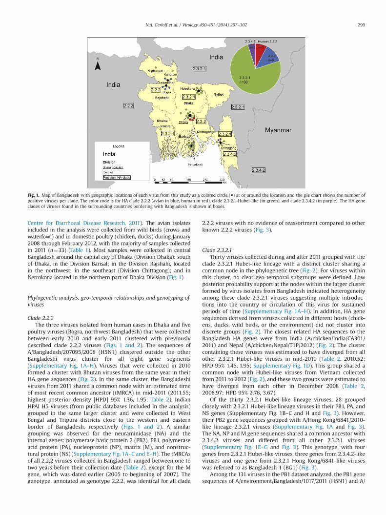
Centre for Diarrhoeal Disease Research, 2011). The avian isolatesincluded in the analysis were collected from wild birds (crows andwaterfowl) and in domestic poultry (chicken, ducks) during January2008 through February 2012, with the majority of samples collectedin 2011 (n¼33) (Table 1). Most samples were collected in centralBangladesh around the capital city of Dhaka (Division Dhaka); southof Dhaka, in the Division Barisal; in the Division Rajshahi, locatedin the northwest; in the southeast (Division Chittagong); and inNetrokona located in the northern part of Dhaka Division (Fig. 1).
Phylogenetic analysis, geo-temporal relationships and genotyping ofviruses
Clade 2.2.2The three viruses isolated from human cases in Dhaka and five
poultry viruses (Bogra, northwest Bangladesh) that were collectedbetween early 2010 and early 2011 clustered with previouslydescribed clade 2.2.2 viruses (Figs. 1 and 2). The sequences ofA/Bangladesh/207095/2008 (H5N1) clustered outside the otherBangladeshi virus cluster for all eight gene segments(Supplementary Fig. 1A–H). Viruses that were collected in 2010formed a cluster with Bhutan viruses from the same year in theirHA gene sequences (Fig. 2). In the same cluster, the Bangladeshiviruses from 2011 shared a common node with an estimated timeof most recent common ancestor (tMRCA) in mid-2011 (2011.55;highest posterior density [HPD] 95% 1.36, 1.95; Table 2). IndianHPAI H5 viruses (from public databases included in the analysis)grouped in the same larger cluster and were collected in WestBengal and Tripura districts close to the western and easternborder of Bangladesh, respectively (Figs. 1 and 2). A similargrouping was observed for the neuraminidase (NA) and theinternal genes: polymerase basic protein 2 (PB2), PB1, polymeraseacid protein (PA), nucleoprotein (NP), matrix (M), and nonstruc-tural protein (NS) (Supplementary Fig. 1A–C and E–H). The tMRCAsof all 2.2.2 viruses collected in Bangladesh ranged between one totwo years before their collection date (Table 2), except for the Mgene, which was dated earlier (2005 to beginning of 2007). Thegenotype, annotated as genotype 2.2.2, was identical for all clade
2.2.2 viruses with no evidence of reassortment compared to otherknown 2.2.2 viruses (Fig. 3).
Clade 2.3.2.1Thirty viruses collected during and after 2011 grouped with the
clade 2.3.2.1 Hubei-like lineage with a distinct cluster sharing acommon node in the phylogenetic tree (Fig. 2). For viruses withinthis cluster, no clear geo-temporal subgroups were defined. Lowposterior probability support at the nodes within the larger clusterformed by virus isolates from Bangladesh indicated heterogeneityamong these clade 2.3.2.1 viruses suggesting multiple introduc-tions into the country or circulation of this virus for sustainedperiods of time (Supplementary Fig. 1A–H). In addition, HA genesequences derived from viruses collected in different hosts (chick-ens, ducks, wild birds, or the environment) did not cluster intodiscrete groups (Fig. 2). The closest related HA sequences to theBangladesh HA genes were from India (A/chicken/India/CA301/2011) and Nepal (A/chicken/Nepal/T1P/2012) (Fig. 2). The clustercontaining these viruses was estimated to have diverged from allother 2.3.2.1 Hubei-like viruses in mid-2010 (Table 2, 2010.52;HPD 95% 1.45, 1.95; Supplementary Fig. 1D). This group shared acommon node with Hubei-like viruses from Vietnam collectedfrom 2011 to 2012 (Fig. 2), and these two groups were estimated tohave diverged from each other in December 2008 (Table 2,2008.97; HPD 95% 2.76, 3.67).
Of the thirty 2.3.2.1 Hubei-like lineage viruses, 28 groupedclosely with 2.3.2.1 Hubei-like lineage viruses in their PB1, PA, andNS genes (Supplementary Fig. 1B–C and H and Fig. 3). However,their PB2 gene sequences grouped with A/Hong Kong/6841/2010-like lineage 2.3.2.1 viruses (Supplementary Fig. 1A and Fig. 3).The NA, NP and M gene sequences shared a common ancestor with2.3.4.2 viruses and differed from all other 2.3.2.1 viruses(Supplementary Fig. 1E–G and Fig. 3). This genotype, with fourgenes from 2.3.2.1 Hubei-like viruses, three genes from 2.3.4.2-likeviruses and one gene from 2.3.2.1 Hong Kong/6841-like viruseswas referred to as Bangladesh 1 (BG1) (Fig. 3).
Among the 131 viruses in the PB1 dataset analyzed, the PB1 genesequences of A/environment/Bangladesh/1017/2011 (H5N1) and A/
Fig. 1. Map of Bangladesh with geographic locations of each virus from this study as a colored circle (●) at or around the location and the pie chart shows the number ofpositive viruses per clade. The color code is for HA clade 2.2.2 (avian in blue, human in red), clade 2.3.2.1-Hubei-like (in green), and clade 2.3.4.2 (in purple). The HA geneclades of viruses found in the surrounding countries bordering with Bangladesh is shown in boxes.
N.A. Gerloff et al. / Virology 450-451 (2014) 297–307 299

chicken/Bangladesh/3075/2011 (H5N1) were dissimilar to all otherH5N1 viruses in the dataset with 96.4% nucleotide identity to otherH5N1 viruses from Bangladesh. However, they showed 99.5%nucleotide sequence identity with A/Bangladesh/0994/2011(H9N2) and 98.5% identity with A/environment/Bangladesh/100/2010 (H9N2), both of which are low pathogenicity avian influenza(LPAI) H9N2 subtype viruses (Supplementary Fig. 1B). The twoviruses clustered separately in the phylogenetic tree and differed inthe tMRCA, with group 1 (A/environment/Bangladesh/1017/2011[H5N1] and A/Bangladesh/0994/2011 [H9N2]) diverging aroundthe end of 2010 (Table 2, 2010.75, HPD 95% 1.11, 1.93) and group 2(A/chicken/Bangladesh/3075/2011 [H5N1] and A/environment/Bangladesh/100/2010 [H9N2]) diverging at the end of 2008
(2008.95; HPD 95% 2.41, 4.16). Thus, the genomic composition ofthese unique reassortants, termed Bangladesh 2 (BG2), had HA, PAand NS genes from 2.3.2.1 Hubei-like lineages (H5N1); NA, NP andM genes from clade 2.3.4.2-like lineages (H5N1); PB2 genes from2.3.2.1 Hong Kong/6841-like lineages (H5N1); and PB1 genes fromseparate H9N2-like subtype viruses (Fig. 3).
Clade 2.3.4.2The HA gene sequence of A/chicken/Bangladesh/3012/2011
(collected in Chittagong, southeast Bangladesh, Fig. 1) belongedto clade 2.3.4.2 and shared closest common ancestry with anothervirus recently detected from Bangladesh (A/chicken/Bangladesh/11RS-1984-30/2011) (Fig. 2). Other viruses from the same cluster
Fig. 2. Phylogenetic tree of HA gene sequences including 40 Bangladeshi viruses (in boldface) and 83 HPAI/H5N1 viruses from public databases. The clades are highlighted withcolor code, light green for 2.3.2.1 Hubei-like, dark green (Hong Kong/6841-like), purple for clade 2.3.4.2, and blue for clade 2.2.2. Triangles are collapsed branches that are labeledwith the reference virus or lineages. This tree was inferred using the maximum likelihood method with GTR and gamma model in MEGA5 (Tamura et al., 2011).
N.A. Gerloff et al. / Virology 450-451 (2014) 297–307300

were collected in Southeast Asian countries, such as Myanmar,Vietnam, Lao PDR and China. Both viruses from Bangladesh wereestimated to have diverged from common ancestors in mid-2010(2010.59; HPD 95% 1.28, 2; Table 2) (Supplementary Fig. 1D). Theclade 2.3.4.2 viruses grouped closely in all genes, but had longbranch lengths relative to ancestors indicative of gaps in sequencedata in this group. The common node ages ranged from thebeginning of 2009 in their NP genes (2009.19; HPD 95% 1.2,2.29; Table 2, Supplementary Fig. 1A–H) to late 2010 in their NAgene (2010.7; HPD 95% 1.61, 2.8; Table 2).
Antigenic characterization
H5N1 isolates were tested by hemagglutination inhibition(HAI) with ferret antiserum against representatives of the cur-rently circulating clades of H5N1 virus and candidate vaccineviruses (Table 3; Supplementary Table 2A–D) (WHO, 2013a).Antisera generated against clades 2.2 and 2.2.2 viruses cross-reacted with Bangladesh viruses belonging to clade 2.2.2 withsignificantly higher HI titers than clades 2.3.2.1 and 2.3.4.2 viruses.Sera raised specifically to Bangladesh clade 2.2.2 viruses reactedwith other Bangladesh viruses from the same clade within 4-foldof titers to homologous viruses indicating increased specificity of2.2.2 viruses compared to earlier clade 2.2 reference sera(Supplementary Table 2A). The clade 2.2.2 virus isolated from ahuman case in 2008 was inhibited by older 2.2 antisera (i.e., A/barheaded-goose/Qinghai/1A/05) with slightly higher heterologoustiters compared to 2010 and 2011 clade 2.2.2 viruses suggestingsome antigenic drift among later 2.2.2 viruses. However, seraproduced against the 2008 virus reacted with 2010 and 2011viruses to heterologous titers within 4-fold or less of the homo-logous titer supporting the overall antigenic specificity of clade2.2.2 viruses in Bangladesh despite several years of viral evolution(Supplementary Table 2A). Sera raised against clade 2.3.2.1 virusesreacted to higher titers with viruses from the same clade, whilethese sera had little to no inhibitory effects on clade 2.2.2 or 2.3.4.2
viruses (Table 3 and Supplementary Table 2A–D). Sera raised toBangladesh viruses identified as clade 2.3.2.1, especially a recentvirus from 2012, showed higher titers to related Bangladeshviruses compared to sera raised against clade 2.3.2.1 viruses fromother countries (Supplementary Table 2D). Antisera generated tothe A/Hubei/1/2010 candidate vaccine virus reacted with severalBangladesh clade 2.3.2.1 viruses with HI titers Z8-fold differentfrom the HI titer with homologous antigen, while antisera to theA/common magpie/Hong Kong/5052/2007 candidate vaccinereacted with these viruses within 4-fold or less compared to titerswith the homologous antigen (Table 3). Expectedly, neither anti-serum to these clade 2.3.2.1 vaccine candidates showed significantinhibition of clade 2.2.2 or 2.3.4.2 viruses (Table 3 and Supp-lementary Table 2A–D). One virus with a clade 2.3.4.2 HA gene(A/chicken/Bangladesh/3012/2011) showed little inhibition byantiserum against clade 2.2.2 or 2.3.2.1 viruses. Additionally, clade2.3.4 sera raised to the A/Anhui/1/2005 candidate vaccine virusfailed to inhibit agglutination by the 2.3.4.2 Bangladesh virusindicating substantial antigenic drift (Table 3 and SupplementaryTable 2B).
Protein sequence characterization
Based on the predicted amino acid sequences, the multibasiccleavage site sequence of the clade 2.2.2 viruses was PQGERRRKKR*G(Bosch et al., 1981). All thirty 2.3.2.1 Hubei-like lineage virusescollected in Bangladesh shared an identical amino acid sequencePQRERRRKR*G at their cleavage site. The two 2.3.4.2 viruses sharedidentical HA cleavage site motifs (PQLRKRRKR*G) but differed fromclades 2.2.2 and 2.3.2.1. One subgroup of 2.2.2 viruses (all from 2010)shared a unique glycosylation site at positions 54–56 and clade2.3.2.1 virus A/chicken/Bangladesh/31289-1/2011 had a unique gly-cosylation site at positions 166–168 (mature H5 numbering). Theviruses that belonged to 2.3.4.2 had a unique glycosylation site atresidues 154–156 of the HA protein compared to other HA clades.The þ1 alternate open reading frame in the PB1 protein (PB1-F2),
Table 2Time of the most recent common ancestor (tMRCA) as calendar years (decimal dates) by HA clade and Bangladesh (BG) groups within a clade for each gene segment.
Gene HA NA PB2 PB1 PA NP M NS
Time of the most recent common ancestor – tMRCA (decimal date)Highest posterior density 95% (HPD 95% [upper, lower interval])Clade 2.2.2 2007.41 2006.67 2007.79 2006.69 2007.53 2007.21 2003.73 2007.13
5.34, 6.32 4.9, 5.72 4.87, 5.98 5.01, 6.6 5.22, 6.4 5.17, 6.99 4.72, 6.38 4.83, 6.5BG clade 2.2.2 group 1 (2008) 2008.94 2007.41 2008.68 2007.62 2008.42 2008.96 2004.92 2008.5
4.1, 4.56 4.24, 4.87 4.19, 4.96 4.25, 4.96 4.39, 5.22 4.1, 4.58 4.1, 4.65 4.1, 4.43BG clade 2.2.2 group 2 (2010) 2010.92 2009.65 2010.91 2009.16 2010.04 2010.32 2006.39 2010.28
2.18, 2.4 2.18, 2.49 2.18, 2.43 2.63, 3.47 2.7, 3.66 2.19, 2.65 2.31, 3.4 2.19, 2.75BG clade 2.2.2 group 3 (2011) 2011.55 2010.17 2011.56 2010.4 2011.31 2011.64 2007.75 2011.19
1.36, 1.95 1.4, 2.21 1.36, 1.96 1.38, 2.31 1.46, 2.35 1.24, 1.94 1.15, 1.85 1.18, 2.02Clade 2.3.2.1 2005.96 2004.96 2005.7 2005.69 2000.38 2003.72 2005.21 2006.57
5.71, 6.75 6.57, 7.97 5.85, 7.26 5.9, 7.21 9.36, 15.03 6.27, 8.14 6.01, 8.24 5.11, 6.3BG clade 2.3.2.1 2010.52 2010.7 2010.51 2010.52 2010.67 2009.06 2010.46 2010.57
1.45, 1.95 1.27, 1.75 1.42, 1.98 1.45, 1.96 1.33, 1.73 1.37, 2.19 1.36, 2.17 1.32, 2Hubei-like 2009.67 2010.15 [0.65] 2010.08 2009.72 2010.03 2008.9 2009.93 2010.26
2.16, 2.93 1.85, 2.3 1.85, 2.49 2.04, 2.91 1.89, 2.49 1.8, 2.15 1.94, 2.69 1.8, 1.8HK/6841-like 2009.01 2008.43 2008.52 2008.99 2008.18 2007.05 2009.7 2008.86
2.88, 3.49 3.33, 4.2 3.24, 4.11 2.93, 3.49 3.53, 4.63 3.29, 4.46 2.07, 2.9 2.94, 3.84BS/1161-like 2009.34 2010.19 2010.13 2009.81 2010.06 2007.89 2009.92 2010.06
2.44, 3.31 1.94, 2.15 1.94, 2.25 2.02, 2.83 1.94, 2.49 2.39, 3.54 1.98, 2.66 1.94, 2.5Clade 2.3.4.2 2006.16 2004.96 2005.88 2004.91 2006.02 2004.53 2005.21 2006.12
5.78, 6.35 6.57, 7.97 5.88, 6.9 6.58, 8.08 5.81, 6.6 5.82, 6.94 6.01, 8.24 5.75, 6.57BG clade 2.3.4.2 2010.59 2010.7 2010.49 2010.03 2010.27 2009.19 2010.64 2009.89 [0.6]
1.28, 2 1.61, 2.8 1.34, 2.22 1.55, 2.89 1.44, 2.44 1.2, 2.29 1.06, 2.37 1.56, 3.09BG H9N2-like group 1 2010.75
1.11, 1.93Group 2 2008.95
2.41, 4.16
N.A. Gerloff et al. / Virology 450-451 (2014) 297–307 301

associated with induction of apoptosis, encoded the 90 amino acidfull length protein in all Bangladesh H5 viruses (Chen et al., 2001).All clade 2.2.2 shared the substitution glutamic acid to lysine atresidue 627 (E627K) in the PB2 protein that is associated withincreased pathogenicity and transmission to human hosts (Shinyaet al., 2004; Subbarao et al., 1993). The M1 protein of all H5N1 virusesanalyzed in this study contained 30Asp (aspartic acid) and 215Ala(alanine), which were described to increase virulence in mice (Fanet al., 2009). The M2 ion channel protein contained a double aminoacid change at residues 27 (valine to alanine) and 31 (serine toasparagine) in A/environment/Bangladesh/1011/2011 and a singlemutation of residue 31 (serine to asparagine) in A/environment/Bangladesh/1018/2011. These changes may reduce susceptibility toantivirals amantadine and rimantadine (Hay et al., 1986). In the NA
protein no antiviral resistance markers were found (Gubareva et al.,2001).
Discussion
Upon analysis of 40 full genomes of H5N1 viruses collected inBangladesh from 2008 through 2012, evidence of separate reas-sortment events was identified between two of the circulatingclades of virus, 2.3.2.1 and 2.3.4.2, and between HPAI H5N1 andLPAI H9N2 viruses resulting in two different Bangladesh-specificgenotypes (BG1 and BG2). Assessments of geo-temporal relation-ships of viruses also identified possible routes of transmission andpatterns of virus spread into Bangladesh from bordering countries
Fig. 3. Estimated timeline for the emergence of H5N1 clades and genotypes in Bangladesh. Each oval contains the eight individual gene segments as parallel lines. Viruseswere color coded as follows: clade 2.2.2 (blue), 2.3.2.1 Hubei-like (light green), 2.3.2.1 Hong Kong/6841-like (dark green), 2.3.4.2 (purple) and H9N2 LPAI (black). Hypotheticalancestral viruses are shownwith the same color code but with a dashed outline. Genomes reflecting virus isolates characterized for this study are shownwith a solid outline.The estimated tMRCA of genes involved in reassortment are indicated below the genome by year with upper and lower HPD 95%. Abbreviations: Hubei/10-like, A/Hubei/1/10-like lineage; HK/6841-like, A/Hong Kong/6841/2010-like lineage.
N.A. Gerloff et al. / Virology 450-451 (2014) 297–307302

Table 3Antigenic characterization of H5N1 viruses isolated from Bangladesh representing each of the three clades detected from 2008 to 2012. Hemagglutination inhibition (HAI) titers of ferret antisera to viruses are shown and listed byHA clade. The homologous titer for each of the reference viruses/antisera is boldfaced and underlined. Italic indicate genetically related HA genes.
Clade Reference antigens Reference ferret antisera
VN/1203 IND/5 bhd/QI tk/TK EG/321 EG/3072 cm/HK ws/JP HK/6841 HUB/10 RG30 CR/BA ANH/1
1 A/VIETNAM/1203/2004 80 80 o10 80 o10 10 20 o10 10 80 o10 102.1.3.2 A/INDONESIA/5/2005 o10 1280 80 80 80 80 320 80 320 40 40 802.2 A/B-H GOOSE/QINGHAI/1A/05 X PR8 o10 1280 1280 1280 160 2560 640 40 640 160 320 802.2.1 A/TURKEY/TURKEY/1/2005 o10 1280 640 2560 320 1280 640 80 640 320 320 802.2.1 A/EGYPT/321-NAMRU3/2007 o10 320 320 320 320 640 80 10 160 80 20 202.2.1 A/EGYPT/N03072/2010 o10 320 320 1280 160 2560 160 10 640 320 40 1602.3.2.1 A/COMMON MAGPIE/HK/5052/2007 o10 320 o10 40 40 80 320 80 320 40 160 o102.3.2.1 HK/6841 A/WHOOPER SWAN/HOKKAIDO/4/2011 o10 160 o10 10 20 20 320 640 640 80 160 o102.3.2.1 HK/6841 A/HONG KONG/6841/2010 o10 80 o10 20 10 20 320 80 160 80 160 o102.3.2.1 Hubei A/HUBEI/1/2010 x PR8 IDCDC-RG30 o10 320 80 160 40 160 320 160 320 320 160 o102.3.2.1 Hubei A/CROW/BANGLADESH/1061/2011 o10 80 o10 20 10 20 20 40 80 40 80 o102.3.4 A/ANHUI/1/2005 o10 320 20 80 40 20 40 o10 20 20 o10 320
Clade Test antigens Reference ferret antisera
VN/1203 IND/5 bhd/QI tk/TK EG/321 EG/3072 cm/HK ws/JP HK/6841 HUB/10 RG30 CR/BA ANH/1
2.2.2 A/CHICKEN/BANGLADESH/31289-1/2011 o10 80 160 160 40 20 20 o10 o10 10 o10 802.2.2 A/POULTRY/BANGLADESH/11255-C/2011 o10 80 320 320 40 20 20 o10 10 o10 10 402.3.2.1 Hubei A/GOOSE/BANGLADESH/4051T/2011 o10 80 o10 10 10 40 160 80 160 40 80 o102.3.2.1 Hubei A/DUCK/BANGLADESH/4059T/2011 o10 80 o10 o10 10 20 80 80 160 40 80 o102.3.2.1 Hubei A/CHICKEN/BANGLADESH/4070T/2011 o10 80 10 20 20 40 160 160 320 80 160 o102.3.2.1 Hubei A/CROW/BANGLADESH/1058/2011 o10 160 o10 20 20 40 160 160 160 80 160 o102.3.2.1 Hubei A/CROW/BANGLADESH/313T/2011 o10 80 o10 10 10 40 320 80 320 80 160 o102.3.2.1 Hubei A/CROW/BANGLADESH/315T/2011 o10 80 o10 10 10 40 160 40 320 40 160 o102.3.2.1 Hubei A/CROW/BANGLADESH/316T/2011 o10 40 o10 10 10 40 320 80 320 80 160 o102.3.2.1 Hubei A/DUCK/BANGLADESH/4120T/2011 o10 80 o10 10 10 20 320 80 320 80 320 o102.3.2.1 Hubei A/DUCK/BANGLADESH/4124T/2011 o10 40 o10 o10 10 20 160 40 320 40 80 o102.3.2.1 Hubei A/CHICKEN/BANGLADESH/4058/2011 o10 80 10 20 10 40 320 80 640 80 160 o102.3.2.1 Hubei A/CHICKEN/BANGLADESH/3072/2011 o10 20 o10 o10 o10 o10 80 40 160 20 40 o102.3.2.1 Hubei A/CHICKEN/BANGLADESH/3075/2011 o10 40 o10 o10 o10 10 160 40 160 40 80 o102.3.2.1 Hubei A/ENVIRONMENT/BANGLADESH/1017/11 o10 40 o10 10 10 20 160 40 320 40 80 o102.3.2.1 Hubei A/CHICKEN/BANGLADESH/11303/2011 o10 80 o10 40 o10 40 320 20 320 80 160 o102.3.2.1 Hubei A/WATERFOWL/BANGLADESH/31935/11 o10 80 o10 o10 10 20 80 80 80 40 80 o102.3.2.1 Hubei A/WATERFOWL/BANGLADESH/33025/11 o10 20 o10 20 10 40 10 40 20 80 80 o102.3.4.2 A/CHICKEN/BANGLADESH/3012/2011 o10 160 o10 160 40 40 80 20 10 40 o10 20
N.A.G
erloffet
al./Virology
450-451(2014)
297–307
303

and geographically close areas, as well as possible transmission fromBangladesh into northern regions. The temporal analyses indicatedthat the first human case detected in Bangladesh, the 2008 clade2.2.2 virus, shared common ancestry with all other viruses from thatclade. Viruses collected in both Bhutan and Bangladesh from 2010shared joint descent and might have been introduced via tradethrough shared borders from India or migratory birds (Chakrabartiet al., 2009; Pawar et al., 2010). The lack of surveillance and sequenceavailability limited the detection of other regional sources of clade2.2.2 viruses. The viruses clustered together for all gene segmentsand are described as genotype 2.2.2 (synonymous with genotype Z)(Duan et al., 2008). Thus, despite the ongoing circulation of clade2.2.2 viruses from 2008 to 2011, this did not lead to detectablereassortment of viruses in this clade likely because viruses withdifferent genomes were either not yet present or circulating at lowlevels in Bangladesh. The lack of detection of sporadic reassortantscould also be attributable to fewer data available for analysis; forclade 2.2.2 viruses only 50 full genomes were available from publicdatabases.
The majority of viruses studied here belonged to clade 2.3.2.1(Hubei-like lineage) and were likely seeded in Bangladesh in late2010 or early 2011 either as a single introduction or multipleintroductions with related viruses. Our data suggest that this cladehas since spread to different locations across the country and intodifferent species, including chicken and crows (Islam et al., 2012).Most recent HA sequences from 2012 located at the tip of thebranches in phylogenetic trees suggested that the initially intro-duced viruses from the beginning of 2011 continued to circulatewithin the country and diverged further without additionalintroductions. In addition, some viruses of the Bangladeshi clustergrouped closely with viruses collected in Nepal suggesting cross-border exchange either through trade or wild bird movementacross northern parts of India (Nagarajan et al., 2012). A newgenotype (BG1) was characterized in 28 viruses with a 4:3:1genetic composition that has not been described previously inclade 2.3.2.1 Hubei-like lineage viruses. Full genome analysisshowed that they shared the same origin in their HA, PB1, PAand NS genes (2.3.2.1 Hubei-like viruses), but grouped withHK/6841-like viruses (clade 2.3.2.1) in their PB2 genes and clade2.3.4.2 viruses in the remaining genes (NA, NP and M).
The second genotype (BG2) identified in clade 2.3.2.1 viruseswas the result of reassortment of 2.3.2.1 Hubei-like viruses withtwo phylogenetically distinct PB1 gene sequences derived fromLPAI H9N2 subtype viruses and the remainder of the genomerelated to the more common BG1 reassortant (Fig. 3). The differenttMRCAs of both reassortant genotypes, BG1 and BG2, suggestedthat these resulted from separate events. Given that both LPAIH9N2 and HPAI H5N1 viruses circulate simultaneously in South-east Asia, including Bangladesh, the possibility of reassortmentevents between HPAI and LPAI viruses are more probable andlikely more common than what has been described previously(Gutiérrez et al., 2009; Negovetich et al., 2011; Vijaykrishna et al.,2008; Zhang et al., 2009). It is likely that the exchange of geneticmaterial was facilitated by the ongoing circulation of differentclades of H5N1 and H9N2 viruses in Bangladesh (OIE, 2013).Recently, a LPAI A(H7N9) virus emerged in humans and birds inChina in which all gene segments except for HA (H7) and NA (N9)were derived from LPAI H9N2 subtype viruses found in wild birds(Chen et al., 2013; Liu et al., 2013; Shi et al., 2013). This findinghighlights that the exchange of gene segments in influenza Aviruses is not restricted to a specific subtype but occurs betweensubtypes with different virologic features such as H5N1 and H9N2(as in Bangladesh) or H7N9 and H9N2 viruses in China. Notably,the H7N9 viruses have caused severe and fatal disease in infectedpersons despite lack of symptoms in H7N9 infected birds (Chenet al., 2013; Gao et al., 2013; WHO, 2013b). Ongoing full genome
monitoring for both HPAI H5N1 and LPAI viruses of diversesubtypes will be crucial to identify future reassortment in Bangla-desh and the Southeast Asian region in general.
The clade 2.3.4.2 virus, A/chicken/Bangladesh/3012/2011, wascollected in the Division Chittagong, which shares its southeasternborder with Myanmar, and was related to other clade 2.3.4.2viruses collected in Bangladesh and a virus detected in Myanmar.The longer branches in the phylogenetic trees combined with theestimated ancestral dates, indicated that these viruses divergedfrom each other at least 1–2 years before they were detected inBangladesh and might have been introduced separately. Clade2.3.4.2 viruses were only found in domestic chicken suggestingthese viruses might have been introduced through poultry trade(Mon et al., 2012). Ancestral viruses likely emerged from borderingMyanmar or more distant locations southeast of Bangladesh likeLao PDR, China or Vietnam where this clade persisted as early as2007 (WHO, 2012). Unlike the viruses that were derived from theother clades (2.3.2.1 and 2.2.2), 2.3.4.2 viruses did not consistentlygroup together in their internal genes and NA genes, indicatingthey reassorted prior to detection in Bangladesh. The low supporton some of the individual branches, however, could be attributedto the small dataset used in our analysis. Since 2011, the virusesdetected in Bangladesh were derived from clade 2.3.2.1, but notfrom 2.3.4.2 suggesting a lack of sustained circulation of thisgenetic group of H5N1 viruses. Alternatively, the long branchlengths separating these viruses from other clade 2.3.4.2 virusessuggests a lack of sequence data from related viruses or surveil-lance gaps both in and around Bangladesh. Interestingly, clade2.3.4.2 viruses have recently been detected in China and havecaused human infection in 2013 (OIE, 2013; WHO/GIP, 2013).
H5N1 virus clades 2.3.2.1 and 2.3.4.2 have been detected in manycountries, including Southeast Asian nations where both of thesevirus groups have circulated at high levels in recent years (Choi et al.,2013; Hu et al., 2013; Liu et al., 2010). The timeframe of estimateddivergence of clade 2.3.2.1, in particular, paired with the locationof the first virus detection, indicated possible transmission throughbird trade, but involvement of wild birds cannot be excluded. Thediscovery of H5N1 outbreaks in crows in Bangladesh may alsoindicate a small role that peridomestic birds may play in virusdispersal (Khan et al., 2013). Viruses from Bangladesh sharedcommon ancestors with viruses from geographically close countriessuch as India, Myanmar, Nepal, and Bhutan. HA gene sequences ofviruses from other countries clustered closely with HA genes ofviruses collected in this study confirming persistence of specific HPAIclades both inside of Bangladesh and within the region.
Results from hemagglutination inhibition testing of BangladeshH5N1 viruses with ferret antisera raised to clades from manydifferent countries indicated substantial antigenic drift betweenviruses from clades 2.2.2, 2.3.2.1 and 2.3.4.2. Furthermore, seraproduced specifically against Bangladesh viruses inhibited agglutina-tion of related Bangladesh isolates at higher reciprocal titers illus-trating antigenic specificity relative to viruses collected outside thecountry. This data suggests that antigenic variation among H5N1viruses exists both within Bangladesh and in neighboring regions. Assuch, host population immunity and existence of diverse viruses mayinfluence poultry vaccination policy and implementation. Poultryvaccine challenge studies that assess the need for vaccine viruses tobe antigenically matched should be performed. In addition, candidatevaccine viruses developed for pandemic planning purposes, such asA/Hubei/1/2010 (clade 2.3.2.1) and A/Anhui/1/2005 (clade 2.3.4), maynot antigenically cover more recently circulating viruses in Bangla-desh. Updates of candidate vaccine seed strains using more con-temporary viruses will be important as these groups of virusescontinue to diversify genetically and antigenically (WHO, 2013a).The close proximity of Bangladesh to other high-density poultry-dependent countries may also continue to increase the genetic and
N.A. Gerloff et al. / Virology 450-451 (2014) 297–307304

antigenic diversity of viruses found in Bangladesh and requiremitigation strategies at both a national and regional level (FAO,2013a, b).
The amino acid sequence of the cleavage site in the H5 HAproteins analyzed was clade-specific, as previously observed (Zhanget al., 2012), with all viruses maintaining at least five consecutivebasic amino acids. Clade 2.2.2 viruses from 2010 shared a glycosyla-tion site at position 54 of the mature HA protein compared to otherclades, and the viruses that belonged to clade 2.3.4.2 had aglycosylation site at residues 154–156. The lack of N-linked glycosy-lation at position 154, conserved in all but the clade 2.3.4.2 viruses, isnoteworthy in that this feature, in combinationwith other mutations,was described for H5N1 viruses that were aerosol transmissible in aferret model (Russell et al., 2012). As has been previously described,all clade 2.2.2 viruses detected in Bangladesh had the PB2 substitu-tion E627K associated with increased pathogenicity and potential formammalian adaptation (Shinya et al., 2004; Subbarao et al., 1993).The lack of this mutation in the clade 2.3.2.1 viruses detected in 2011and 2012 and the apparent decline in the number of clade 2.2.2viruses detected in this study may indicate a shift in the potentialadaptability of these viruses for mammalian replication. It remainsto be seen if the relative predominance of clade 2.3.2.1 virusesin Bangladesh will be sustained as in other countries such as Vietnam(Creanga et al., 2013). While the M1 protein of all viruses analyzedcontained mutations associated with increased virulence inmice (Fan et al., 2009), no other internal gene proteins were foundto possess known molecular markers of concern. In addition, nomarkers of reduced susceptibility to neuraminidase inhibitorswere found.
This comprehensive phylogenetic study of full genomesequences from H5N1 viruses presently circulating in Bangladeshindicates that the ancestors of some H5N1 viruses reassorted withother lineages of the same clades (2.3.2.1), with other clades ofH5N1 viruses (2.3.2.1 and 2.3.4.2) and even with other LPAI virussubtypes known to circulate in Bangladesh (International Centrefor Diarrhoeal Disease Research, 2011). These events created adiverse genetic pool of H5N1 viruses that contribute to thedispersal of multiple variants in the country and the regionssurrounding Bangladesh. Potential limitations of this study werethe lack of full genome data for viruses identified in neighboringcountries paired with surveillance gaps. This is particularly evidentfor clade 2.3.4.2 viruses that remain apparently undersampledbased on available sequence data in the public domain. To achievemore accurate estimates of times of the most recent commonancestors and dispersal of viruses, exact sample collection dates incombination with locations of collection are crucial. Continuousavian influenza surveillance in poultry in Bangladesh, togetherwith the molecular and antigenic characterization of the circulat-ing viruses, will reveal further diversification within the viruspopulation and help to explain virus dispersal from bordering andnearby countries in the future.
Materials and methods
Avian influenza virus surveillance and sample collection
All specimen were collected from 2008 through 2012 duringmultiple studies undertaken in Bangladesh by International Centrefor Diarrhoeal Disease Research, Bangladesh (icddr,b), and variousgovernment partners (Institute for Epidemiology, Disease Controland Research; Department of Livestock Services; Department ofForestry) as well as international partners (Centers for DiseaseControl and Prevention [CDC]; EcoHealth Alliance). These projectsincluded active live bird market surveillance, backyard poultrysurveillance from nationally representative village locations and
poultry and wild bird outbreak investigations. Individual orophar-yngeal or tracheal swabs were collected from live birds sold atmarkets or backyard raised poultry or from bird carcasses col-lected or live birds trapped during outbreak investigations. Envir-onmental swabs were sampled from surfaces of live bird markets,backyards where poultry flocks lived and from bird droppingsaround outbreak sites. Samples from H5N1-positive human caseswere collected as previously described (Brooks et al., 2010;International Centre for Diarrhoeal Disease Research, 2011).Geographic distribution of samples was mapped using coordinatesof the collection locations and the boundary map of Bangladesh inArcGIS 9.3 (Environmental System Research Institute, Redlands,CA, USA).
Virus isolation, subtype detection and full genome sequencing
Original specimens were screened at icddr,b for influenza Avirus using a real-time reverse transcription (RT)-PCR detection kittargeting the matrix (M) gene (CDC, 2013). Influenza A positivesamples were also screened for the presence of influenza A HAsubtypes H5 and H9 and tested with a H5 clade specific real-timeRT-PCR (CDC, 2013; Kis et al., 2013). Influenza A positive sampleswere sent to CDC for further characterization. At the CDC,influenza A(H5) virus PCR-positive samples were inoculated into10–11 day old embryonated chicken eggs (ECEs) and amniotic/allantoic fluid was harvested 24 hours post-inoculation and testedby hemagglutination assay (HA) using turkey red blood cells. Allinfectious materials were maintained in biosafety level 3 contain-ment, including enhancements required by the U.S. Departmentof Agriculture and the Select Agents program (http://www.cdc.gov/od/ohs/biosfty/bmbl5/bmbl5toc.htm). Only specimens thatyielded Z8 HA units were included in further analyses. GenomicRNA extracted from virus-infected amniotic/allantoic fluid usingthe RNeasy extraction kit (Qiagen, Valencia, CA) was used astemplate for generation of cDNA by random hexamer-primedreverse transcription (SuperScriptsIII, Life Technologies, Carlsbad,CA). The surface and internal protein genes were then amplifiedusing influenza A virus specific primers (available upon request) asoverlapping fragments with the Access Quick one-step RT-PCR kit(Promega, Madison, WI) and subsequently sequenced on anautomated Applied Biosystems 3730 system using cycle sequen-cing dye terminator chemistry (Life Technologies, Carlsbad, CA).Contigs of full length open reading frames were generated foreach gene (Sequencher 4.10.1, Gene Codes, Ann Arbor, MI). Genesequences were submitted to GISAID (http://platform.gisaid.org)prior to publication (Accession Numbers: EPI 448024-448111,448120-448279, 448883-448924, 353364, 353365, 353370, 353372,353379, 353381, 314772-314779, 219467-219474, 460194-460201).
Molecular characterization
For full genome phylogenetic comparison, publicly availableH5N1 sequences were included in datasets and annotated accord-ing to their HA clade designation. Sequences were aligned with theMUSCLE algorithm implemented in BioEdit (Edgar, 2004; Hall,1999). Alignments were manually edited for frame shifts, sequenceduplication and gaps. Trees to identify larger clusters wereinferred using the neighbor joining (NJ) method with a Kimura2-parameter implemented in MEGA5 (Tamura et al., 2011 ). Aminoacid comparison was also performed in BioEdit and MEGA5. Thepresence of a multibasic cleavage site indicative of high patho-genicity and glycosylation sites in the HA protein were determinedby comparing the coding region of each virus.
N.A. Gerloff et al. / Virology 450-451 (2014) 297–307 305

Antigenic characterization
Isolates were tested by hemagglutination inhibition assay (HAI)with ferret antisera raised against viruses representing variousH5N1 clades (1, 2.1.3.2, 2.2, 2.2.1, 2.2.2, 2.3.2.1, 2.3.4) includingthose strains previously identified as candidate vaccine viruses(Klimov et al., 2012). Antiserum was also raised to select virusescollected during this study from different phylogenetic clades.Briefly, sera from male ferrets greater than six months of age(Triple F Farms, Sayre, PA) were tested by HAI for the presence ofpre-existing antibody to seasonal influenza A viruses. To generateferret antisera used for HAI testing, serologically naive ferrets wereinoculated intranasally with 500 ml of diluted virus per nare with arange of doses depending on the virus. Ferrets were boosted withconcentrated virus and adjuvant at approximately 14 days post-infection (dpi) and were exsanguinated at approximately 28 dpi.Serum was stored at �20 1C until further use. As previouslydescribed for the HAI assay, viruses were standardized to 8 HAU/50 μl and added to serially diluted, receptor destroying enzyme(RDE)-treated antisera (DENKA SEIKEN, Campbell, CA) followed byincubation at room temperature and agglutination with turkey redblood cells (Klimov et al., 2012). The HAI titers were reported asthe reciprocal of the last dilution of antiserum that completelyinhibited hemagglutination.
Genotyping and reassortment analysis
The dataset included full genomes of 40 H5N1 viruses collectedin Bangladesh from 2008 through 2012, characterized andsequenced at CDC. For the analysis of the individual gene seg-ments, 83 full genome sequences of genetically related virusesfrom public databases with at least 90% of full sequence lengthwere included except for the PB1 gene, for which 89 virussequences were used (Supplemental Table 1). Six additional PB1sequences from H9N2 subtype viruses were included in the PB1gene dataset due to the relatedness of H5N1 and H9N2 virussequences upon basic local alignment search tool algorithm(BLAST) analysis (Altschul et al., 1990). The full genome sequencesof three H5N1 viruses isolated from specimens collected fromhumans during community based surveillance for respiratorysurveillance and pneumonia in the Dhaka (Kamalapur) were alsoincluded (Brooks et al., 2007; Brooks et al., 2010; InternationalCentre for Diarrhoeal Disease Research, 2011). Reference virussequences were used to represent H5N1 clades 2.2.2 and 2.3.4.2and the three lineages of clade 2.3.2.1: Hubei-like (A/Hubei/1/2010), Hong Kong/6841-like (A/Hong Kong/6841/2010) and barnswallow/1161-like (A/barn swallow/Hong Kong/D10-1161/2010).To describe the microevolution among viruses from Bangladesh,genotypes were named based on their HA gene clade annotationand then applied to the NA and the six internal genes.
Temporal distribution using Bayesian analysis
The time of most recent common ancestor (tMRCA) wasestimated using dated gene sequence alignments for each of theeight gene segments with the program package BEAST version1.7.5 (Drummond and Rambaut, 2007). For the temporal distribu-tion of the isolates, we used the collection date of the specimenthat was available for all 40 H5N1 viruses isolated in Bangladesh.When dates were unavailable, the date was assumed to be themedian of the collection year (if month was unknown) or themedian of the collection month (if only day was unknown).The tMRCA is expressed as a decimal date and for each tMRCA acredible interval (Bayesian confidence interval) is given as thehighest posterior density (HPD 95%) that represents an interval inthe domain of a posterior probability distribution. For the Bayesian
analysis, at least 50 million Monte Carlo Markov Chain (MCMC)generations were run in the SDR06 model either with HKY(Hasegawa–Kishino–Yano) or the GTR (general time reversible)model; both models were used with a gamma distribution, threepartitions and a 10% burn-in removal (Shapiro et al., 2006; Yang,1996). Effective sample size (ESS) was evaluated in Tracer for eachindividual run and data were only included when the ESS formolecular clock parameters were greater than 200 (Rambaut andDrummond, 2009). Tree files were generated with softwareincluded in the BEAST package and visualized in FigTree version1.4.0 (Rambaut, 2009).
Acknowledgments
We gratefully acknowledge the authors, and the originatingand submitting laboratories of the sequences from GISAID0sEpiFluTM Database, which were used in this analysis. The virusA/chicken/Bangladesh/11RS-1984-30/2011 subtype H5N1 waskindly provided by G. Cattoli, Istituto Zooprofilattico delle Venezie,Italy. This research was supported in part by an appointment to theResearch Participation Program at the Centers for Disease Controland Prevention administered by the Oak Ridge Institute forScience and Education through an interagency agreement withthe U.S. Department of Energy. Icddr,b is funded by project U01/CI000628-05, a cooperative agreement with CDC. We thank theEcoHealth Alliance for financial support of staff involved in samplecollection, which was funded by NIH/NSF Ecology and Evolution ofInfectious Diseases award from the Fogarty International Center(3R01-TW005869). The findings and conclusions in this report arethose of the authors and do not necessarily represent the views ofthe Centers for Disease Control and Prevention or the Agency forToxic Substances and Disease Registry.
Appendix A. Supporting information
Supplementary data associated with this article can be found inthe online version at http://dx.doi.org/10.1016/j.virol.2013.12.023.
References
Ahmed, S.S., Themudo, G.E., Christensen, J.P., Biswas, P.K., Giasuddin, M., Samad, M.A.,Toft, N., Ersboll, A.K., 2012. Molecular epidemiology of circulating highlypathogenic avian influenza (H5N1) virus in chickens, in Bangladesh, 2007–2010. Vaccine 30, 7381–7390.
Altschul, S.F., Gish, W., Miller, W., Myers, E.W., Lipman, D.J., 1990. Basic localalignment search tool. J. Mol. Biol. 215, 403–410.
Bosch, F.X., Garten, W., Klenk, H.D., Rott, R., 1981. Proteolytic cleavage of influenzavirus hemagglutinins: primary structure of the connecting peptide betweenHA1 and HA2 determines proteolytic cleavability and pathogenicity of Avianinfluenza viruses. Virology 113, 725–735.
Brooks, W.A., Alamgir, A.S., Sultana, R., Islam, M.S., Rahman, M., Fry, A.M., Shu, B.,Lindstrom, S., Nahar, K., Goswami, D., Haider, M.S., Nahar, S., Butler, E., Hancock,K., Donis, R.O., Davis, C.T., Zaman, R.U., Luby, S.P., Uyeki, T.M., Rahman, M., 2009.Avian influenza virus A (H5N1), detected through routine surveillance, in child,Bangladesh. Emerg. Infect. Dis. 15, 1311–1313.
Brooks, W.A., Breiman, R.F., Goswami, D., Hossain, A., Alam, K., Saha, S.K., Nahar, K.,Nasrin, D., Ahmed, N., El Arifeen, S., Naheed, A., Sack, D.A., Luby, S., 2007.Invasive pneumococcal disease burden and implications for vaccine policy inurban Bangladesh. Am. J. Trop. Med. Hyg. 77, 795–801.
Brooks, W.A., Goswami, D., Rahman, M., Nahar, K., Fry, A.M., Balish, A., Iftekhar-uddin, N., Azim, T., Xu, X., Klimov, A., Bresee, J., Bridges, C., Luby, S., 2010.Influenza is a major contributor to childhood pneumonia in a tropical devel-oping country. Pediatr. Infect. Dis. J. 29, 216–221.
CDC, 2013. CDC Laboratory Support for Influenza Surveillance (CLSIS). CDC, Atlanta.Chakrabarti, A.K., Pawar, S.D., Cherian, S.S., Koratkar, S.S., Jadhav, S.M., Pal, B., Raut,
S., Thite, V., Kode, S.S., Keng, S.S., Payyapilly, B.J., Mullick, J., Mishra, A.C., 2009.Characterization of the influenza AH5N1 viruses of the 2008-09 outbreaks inIndia reveals a third introduction and possible endemicity. PLoS ONE 4, e7846.
Chen, W.S., Calvo, P.A., Malide, D., Gibbs, J., Schubert, U., Bacik, I., Basta, S., O0Neill, R.,Schickli, J., Palese, P., Henklein, P., Bennink, J.R., Yewdell, J.W., 2001. A novel
N.A. Gerloff et al. / Virology 450-451 (2014) 297–307306

influenza A virus mitochondrial protein that induces cell death. Nat. Med. 7,1306–1312.
Chen, Y., Liang, W., Yang, S., Wu, N., Gao, H., Sheng, J., Yao, H., Wo, J., Fang, Q., Cui, D.,Li, Y., Yao, X., Zhang, Y., Wu, H., Zheng, S., Diao, H., Xia, S., Zhang, Y., Chan, K.H.,Tsoi, H.W., Teng, J.L., Song, W., Wang, P., Lau, S.Y., Zheng, M., Chan, J.F., To, K.K.,Chen, H., Li, L., Yuen, K.Y., 2013. Human infections with the emerging avianinfluenza A H7N9 virus from wet market poultry: clinical analysis andcharacterisation of viral genome. Lancet 381, 1916–1925.
Choi, J.-G., Kang, H.-M., Jeon, W.-J., Choi, K.-S., Kim, K.-I., Song, B., Lee, H.-S., Kim, J.-H., Lee, Y.-J., 2013. Characterization of clade 2.3.2.1 H5N1 highly pathogenicavian influenza viruses isolated from wild birds (Mandarin Duck and EurasianEagle Owl) in 2010 in Korea. Viruses 5, 1153–1174.
Creanga, A., Thi Nguyen, D., Gerloff, N., Thi Do, H., Balish, A., Dang Nguyen, H., Jang,Y., Thi Dam, V., Thor, S., Jones, J., Simpson, N., Shu, B., Emery, S., Berman, L.,Nguyen, H.T., Bryant, J.E., Lindstrom, S., Klimov, A., Donis, R.O., Davis, C.T.,Nguyen, T., 2013. Emergence of multiple clade 2.3.2.1 influenza A (H5N1) virussubgroups in Vietnam and detection of novel reassortants. Virology.
Drummond, A.J., Rambaut, A., 2007. BEAST: Bayesian evolutionary analysis bysampling trees. BMC Evol. Biol. 7, 214.
Duan, L., Bahl, J., Smith, G.J., Wang, J., Vijaykrishna, D., Zhang, L.J., Zhang, J.X., Li, K.S.,Fan, X.H., Cheung, C.L., Huang, K., Poon, L.L., Shortridge, K.F., Webster, R.G.,Peiris, J.S., Chen, H., Guan, Y., 2008. The development and genetic diversity ofH5N1 influenza virus in China, 1996–2006. Virology 380, 243–254.
Edgar, R.C., 2004. MUSCLE: multiple sequence alignment with high accuracy andhigh throughput. Nucleic Acids Res. 32, 1792–1797.
Fan, S., Deng, G., Song, J., Tian, G., Suo, Y., Jiang, Y., Guan, Y., Bu, Z., Kawaoka, Y., Chen,H., 2009. Two amino acid residues in the matrix protein M1 contribute to thevirulence difference of H5N1 avian influenza viruses in mice. Virology 384,28–32.
FAO, 2013a. Global livestock production and health atlas (GLiPHA). FAO.FAO, 2013b. Time series and cross sectional data relating to food and agriculture for
some 200 countries. FAO.Fouchier, R.A., Smith, D.J., 2010. Use of antigenic cartography in vaccine seed strain
selection. Avian Dis. 54, 220–223.Gao, R., Cao, B., Hu, Y., Feng, Z., Wang, D., Hu, W., Chen, J., Jie, Z., Qiu, H., Xu, K., Xu,
X., Lu, H., Zhu, W., Gao, Z., Xiang, N., Shen, Y., He, Z., Gu, Y., Zhang, Z., Yang, Y.,Zhao, X., Zhou, L., Li, X., Zou, S., Zhang, Y., Li, X., Yang, L., Guo, J., Dong, J., Li, Q.,Dong, L., Zhu, Y., Bai, T., Wang, S., Hao, P., Yang, W., Zhang, Y., Han, J., Yu, H., Li,D., Gao, G.F., Wu, G., Wang, Y., Yuan, Z., Shu, Y., 2013. Human infection with anovel avian-origin influenza A (H7N9) virus. N. Engl. J. Med. 368, 1888–1897.
Garten, R.J., Davis, C.T., Russell, C.A., Shu, B., Lindstrom, S., Balish, A., Sessions, W.M.,Xu, X., Skepner, E., Deyde, V., Okomo-Adhiambo, M., Gubareva, L., Barnes, J.,Smith, C.B., Emery, S.L., Hillman, M.J., Rivailler, P., Smagala, J., de Graaf, M.,Burke, D.F., Fouchier, R.A., Pappas, C., Alpuche-Aranda, C.M., Lopez-Gatell, H.,Olivera, H., Lopez, I., Myers, C.A., Faix, D., Blair, P.J., Yu, C., Keene, K.M., Dotson Jr.,P.D., Boxrud, D., Sambol, A.R., Abid, S.H., St George, K., Bannerman, T., Moore, A.L., Stringer, D.J., Blevins, P., Demmler-Harrison, G.J., Ginsberg, M., Kriner, P.,Waterman, S., Smole, S., Guevara, H.F., Belongia, E.A., Clark, P.A., Beatrice, S.T.,Donis, R., Katz, J., Finelli, L., Bridges, C.B., Shaw, M., Jernigan, D.B., Uyeki, T.M.,Smith, D.J., Klimov, A.I., Cox, N.J., 2009. Antigenic and genetic characteristics ofswine-origin 2009A(H1N1) influenza viruses circulating in humans. Science,325, pp 197–201.
Gubareva, L.V., Webster, R.G., Hayden, F.G., 2001. Comparison of the activities ofzanamivir, oseltamivir, and RWJ-270201 against clinical isolates of influenzavirus and neuraminidase inhibitor-resistant variants. Antimicrob. AgentsChemother. 45, 3403–3408.
Gutiérrez, R., Naughtin, M., Horm, S., San, S., Buchy, P., 2009. A(H5N1) virusevolution in South East Asia. Viruses 1, 335–361.
Hall, T.A., 1999. BioEdit:a user-friendly biological sequence alignment editor andanalysis program for Windows 95/98/NT. In: Nucleic Acids Symp Ser, 41st ed.
Hay, A., Zambon, M., Wolstenholme, A., Skehel, J., Smith, M., 1986. Molecular basisof resistance of influenza A viruses to amantadine. J. Antimicrob. Chemother.18, 19–29.
Hu, J., Zhao, K., Liu, X., Wang, X., Chen, Z., Liu, X., 2013. Two highly pathogenic avianinfluenza H5N1 viruses of clade 2.3.2.1 with similar genetic background butwith different pathogenicity in mice and ducks. Transbound Emerg. Dis. 60,127–139.
International Centre for Diarrhoeal Disease Research, B., 2011. Outbreak of mildrespiratory disease caused by H5N1 and H9N2 infections among youngchildren in Dhaka, Bangladesh. Health Sci. Bull. 9, 5–12.
Islam, M.R., Haque, M.E., Giasuddin, M., Chowdhury, E.H., Samad, M.A., Parvin, R.,Nooruzzaman, M., Rahman, M.M., Monoura, P., 2012. New introduction of clade2.3.2.1 avian influenza virus (H5N1) into Bangladesh. Transbound Emerg. Dis.59, 460–463.
Khan, S.U., Berman, L., Haider, N., Gerloff, N., Rahman, M.Z., Shu, B., Rahman, M.,Dey, T.K., Davis, T.C., Das, B.C., Balish, A., Islam, A., Teifke, J.P., Zeidner, N.,Lindstrom, S., Klimov, A., Donis, R.O., Luby, S.P., Shivaprasad, H.L., Mikolon, A.B.,2013. Investigating a crow die-off in January–February 2011 during theintroduction of a new clade of highly pathogenic avian influenza virus H5N1into Bangladesh. Arch. Virol..
Kis, Z., Jones, J., Creanga, A., Ferdinand, K., Inui, K., Gerloff, N., Davis, C.T., Nguyen, T.,Donis, R.O., 2013. Real-time RT-PCR assay to differentiate clades of H5N1 avianinfluenza viruses circulating in Vietnam. J. Virol. Methods 193, 452–458.
Klimov, A., Balish, A., Veguilla, V., Sun, H., Schiffer, J., Lu, X., Katz, J.M., Hancock, K.,2012. Influenza virus titration, antigenic characterization, and serologicalmethods for antibody detection. Methods Mol. Biol. 865, 25–51.
Liu, D., Shi, W., Shi, Y., Wang, D., Xiao, H., Li, W., Bi, Y., Wu, Y., Li, X., Yan, J., Liu, W.,Zhao, G., Yang, W., Wang, Y., Ma, J., Shu, Y., Lei, F., Gao, G.F., 2013. Origin anddiversity of novel avian influenza A H7N9 viruses causing human infection:phylogenetic, structural, and coalescent analyses. Lancet 381, 1926–1932.
Liu, Q., Ma, J., Kou, Z., Pu, J., Lei, F., Li, T., Liu, J., 2010. Characterization of a highlypathogenic avian influenza H5N1 clade 2.3.4 virus isolated from a tree sparrow.Virus Res. 147, 25–29.
Mon, P.P., Lapkuntod, J., Maw, M.T., Nuansrichay, B., Parchariyanon, S., Tiensin, T.,Htun, T., Padungtod, P., Kalpravidh, W., Sunn, K., Maclean, M., Amonsin, A.,2012. Highly pathogenic avian influenza (H5N1) in Myanmar, 2006–2010. Arch.Virol. 157, 2113–2123.
Nagarajan, S., Tosh, C., Smith, D.K., Peiris, J.S., Murugkar, H.V., Sridevi, R., Kumar, M.,Katare, M., Jain, R., Syed, Z., Behera, P., Cheung, C.L., Khandia, R., Tripathi, S.,Guan, Y., Dubey, S.C., 2012. Avian influenza (H5N1) virus of clade 2.3.2 indomestic poultry in India. PLoS ONE 7, e31844.
Negovetich, N.J., Feeroz, M.M., Jones-Engel, L., Walker, D., Alam, S.M., Hasan, K.,Seiler, P., Ferguson, A., Friedman, K., Barman, S., Franks, J., Turner, J., Krauss, S.,Webby, R.J., Webster, R.G., 2011. Live bird markets of Bangladesh: H9N2 virusesand the near absence of highly pathogenic H5N1 influenza. PLoS ONE 6, e19311.
OIE, 2012. WAHID interface. In: Summary of Immediate Notifications and Follow-ups. OIE.
OIE, 2013. Update on highly pathogenic avian influenza in animals (type H5 and H7).OIE.
Pawar, S., Chakrabarti, A., Cherian, S., Pande, S., Nanaware, M., Raut, S., Pal, B.,Jadhav, S., Kode, S., Koratkar, S., Thite, V., Mishra, A., 2010. An avian influenza A(H11N1) virus from a wild aquatic bird revealing a unique Eurasian-Americangenetic reassortment. Virus Genes 41, 14–22.
Rambaut, A., 2009. FigTree, 1.3.1st ed. Institute of Evolutionary Biology, Universityof Edinburgh, Edinburgh (Tree Figure Drawing Tool).
Rambaut, A., Drummond, A.J., 2009. Tracer, 1.5th ed.Russell, C.A., Fonville, J.M., Brown, A.E., Burke, D.F., Smith, D.L., James, S.L., Herfst, S.,
van Boheemen, S., Linster, M., Schrauwen, E.J., Katzelnick, L., Mosterin, A.,Kuiken, T., Maher, E., Neumann, G., Osterhaus, A.D., Kawaoka, Y., Fouchier, R.A.,Smith, D.J., 2012. The potential for respiratory droplet-transmissible A/H5N1influenza virus to evolve in a mammalian host. Science 336, 1541–1547.
Shapiro, B., Rambaut, A., Drummond, A.J., 2006. Choosing appropriate substitutionmodels for the phylogenetic analysis of protein-coding sequences. Mol. Biol.Evol. 23, 7–9.
Shi, J., Deng, G., Liu, P., Zhou, J., Guan, L., Li, W., Li, X., Guo, J., Wang, G., Fan, J., Wang,J., Li, Y., Jiang, Y., Liu, L., Tian, G., Li, C., Chen, H., 2013. Isolation andcharacterization of H7N9 viruses from live poultry markets—implication ofthe source of current H7N9 infection in humans. Chin. Sci. Bull. 58, 1857–1863.
Shinya, K., Hamm, S., Hatta, M., Ito, H., Ito, T., Kawaoka, Y., 2004. PB2 amino acid atposition 627 affects replicative efficiency, but not cell tropism, of Hong KongH5N1 influenza A viruses in mice. Virology 320, 258–266.
Shortridge, K.F., Zhou, N.N., Guan, Y., Gao, P., Ito, T., Kawaoka, Y., Kodihalli, S., Krauss,S., Markwell, D., Murti, K.G., Norwood, M., Senne, D., Sims, L., Takada, A.,Webster, R.G., 1998. Characterization of avian H5N1 influenza viruses frompoultry in Hong Kong. Virology 252, 331–342.
Subbarao, E.K., London, W., Murphy, B.R., 1993. A single amino acid in the PB2 geneof influenza A virus is a determinant of host range. J. Virol. 67, 1761–1764.
Tamura, K., Peterson, D., Peterson, N., Stecher, G., Nei, M., Kumar, S., 2011. MEGA5:molecular evolutionary genetics analysis using maximum likelihood, evolu-tionary distance, and maximum parsimony methods. Mol. Biol. Evol. 28,2731–2739.
Vijaykrishna, D., Bahl, J., Riley, S., Duan, L., Zhang, J.X., Chen, H., Peiris, J.S., Smith, G.J., Guan, Y., 2008. Evolutionary dynamics and emergence of panzootic H5N1influenza viruses. PLoS Pathog. 4, e1000161.
WHO, 2012. Continued evolution of highly pathogenic avian influenza A (H5N1):updated nomenclature. Influenza Other Respir. Viruses 6, 1–5.
WHO, 2013a. Antigenic and Genetic Characteristics of Zoonotic Influenza Virusesand Development of Candidate Vaccine Viruses for Pandemic Preparedness.
WHO, 2013b. Overview of the Emergence and Characteristics of the Avian InfluenzaA(H7N9) Virus. WHO.
WHO/GIP, 2013. Cumulative Number of Confirmed Human Cases for AvianInfluenza A(H5N1) Reported to WHO, 2003–2013. WHO, Geneva.
Yang, Z., 1996. Maximum-likelihood models for combined analyses of multiplesequence data. J. Mol. Evol. 42, 587–596.
Yen, H.L., Webster, R.G., 2009. Pandemic influenza as a current threat. Curr. TopMicrobiol. Immunol. 333, 3–24.
Younan, M., Poh, M.K., Elassal, E., Davis, T., Rivailler, P., Balish, A.L., Simpson, N.,Jones, J., Deyde, V., Loughlin, R., Perry, I., Gubareva, L., ElBadry, M.A., Truelove,S., Gaynor, A.M., Mohareb, E., Amin, M., Cornelius, C., Pimentel, G., Earhart, K.,Naguib, A., Abdelghani, A.S., Refaey, S., Klimov, A.I., Donis, R.O., Kandeel, A.,2013. Microevolution of highly pathogenic avian influenza A(H5N1) virusesisolated from humans, Egypt, 2007–2011. Emerg. Infect. Dis. 19, 43–50.
Zhang, P., Tang, Y., Liu, X., Liu, W., Zhang, X., Liu, H., Peng, D., Gao, S., Wu, Y., Zhang,L., Lu, S., Liu, X., 2009. A novel genotype H9N2 influenza virus possessinghuman H5N1 internal genomes has been circulating in poultry in eastern Chinasince 1998. J. Virol. 83, 8428–8438.
Zhang, Y., Sun, Y., Sun, H., Pu, J., Bi, Y., Shi, Y., Lu, X., Li, J., Zhu, Q., Gao, G.F., Yang, H.,Liu, J., 2012. A single amino acid at the hemagglutinin cleavage site contributesto the pathogenicity and neurovirulence of H5N1 influenza virus in mice. J.Virol. 86, 6924–6931.
N.A. Gerloff et al. / Virology 450-451 (2014) 297–307 307
Copyright © 2022 FDOKUMEN