Multi-Scale Investigation of Surface Topography of Ball Python (Python regius) Shed Skin in...
11
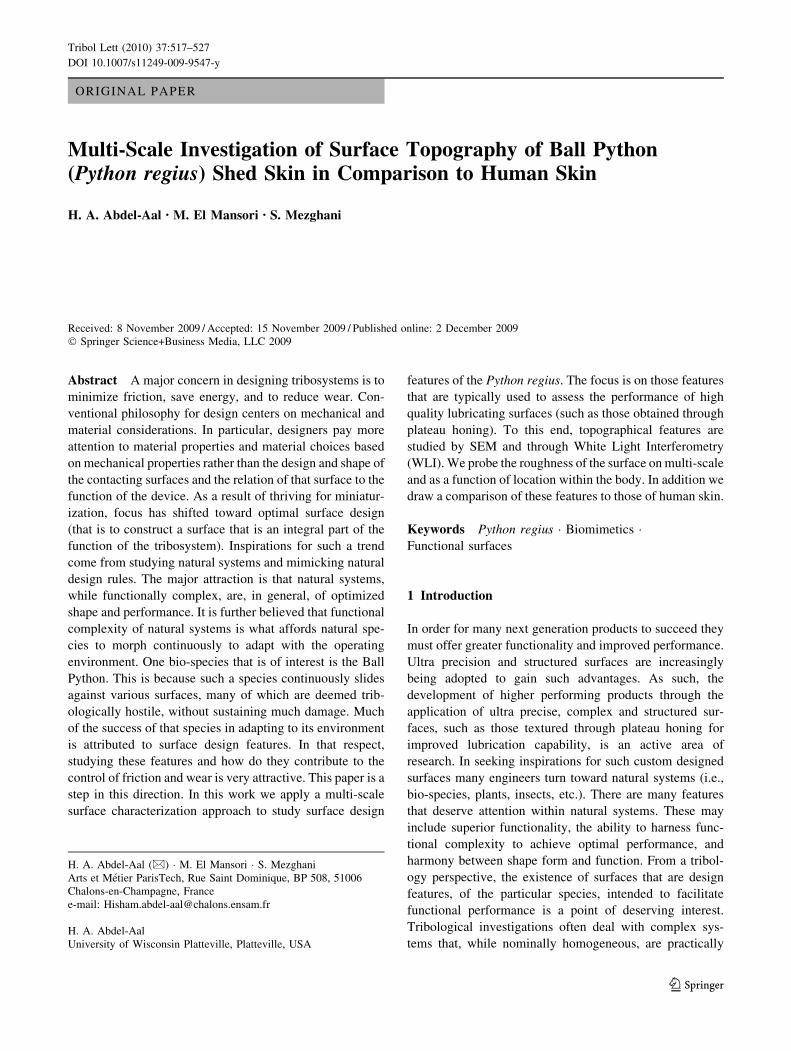
ORIGINAL PAPER Multi-Scale Investigation of Surface Topography of Ball Python (Python regius) Shed Skin in Comparison to Human Skin H. A. Abdel-Aal • M. El Mansori • S. Mezghani Received: 8 November 2009 / Accepted: 15 November 2009 / Published online: 2 December 2009 Ó Springer Science+Business Media, LLC 2009 Abstract A major concern in designing tribosystems is to minimize friction, save energy, and to reduce wear. Con- ventional philosophy for design centers on mechanical and material considerations. In particular, designers pay more attention to material properties and material choices based on mechanical properties rather than the design and shape of the contacting surfaces and the relation of that surface to the function of the device. As a result of thriving for miniatur- ization, focus has shifted toward optimal surface design (that is to construct a surface that is an integral part of the function of the tribosystem). Inspirations for such a trend come from studying natural systems and mimicking natural design rules. The major attraction is that natural systems, while functionally complex, are, in general, of optimized shape and performance. It is further believed that functional complexity of natural systems is what affords natural spe- cies to morph continuously to adapt with the operating environment. One bio-species that is of interest is the Ball Python. This is because such a species continuously slides against various surfaces, many of which are deemed trib- ologically hostile, without sustaining much damage. Much of the success of that species in adapting to its environment is attributed to surface design features. In that respect, studying these features and how do they contribute to the control of friction and wear is very attractive. This paper is a step in this direction. In this work we apply a multi-scale surface characterization approach to study surface design features of the Python regius. The focus is on those features that are typically used to assess the performance of high quality lubricating surfaces (such as those obtained through plateau honing). To this end, topographical features are studied by SEM and through White Light Interferometry (WLI). We probe the roughness of the surface on multi-scale and as a function of location within the body. In addition we draw a comparison of these features to those of human skin. Keywords Python regius Á Biomimetics Á Functional surfaces 1 Introduction In order for many next generation products to succeed they must offer greater functionality and improved performance. Ultra precision and structured surfaces are increasingly being adopted to gain such advantages. As such, the development of higher performing products through the application of ultra precise, complex and structured sur- faces, such as those textured through plateau honing for improved lubrication capability, is an active area of research. In seeking inspirations for such custom designed surfaces many engineers turn toward natural systems (i.e., bio-species, plants, insects, etc.). There are many features that deserve attention within natural systems. These may include superior functionality, the ability to harness func- tional complexity to achieve optimal performance, and harmony between shape form and function. From a tribol- ogy perspective, the existence of surfaces that are design features, of the particular species, intended to facilitate functional performance is a point of deserving interest. Tribological investigations often deal with complex sys- tems that, while nominally homogeneous, are practically H. A. Abdel-Aal (&) Á M. El Mansori Á S. Mezghani Arts et Me ´tier ParisTech, Rue Saint Dominique, BP 508, 51006 Chalons-en-Champagne, France e-mail: [email protected] H. A. Abdel-Aal University of Wisconsin Platteville, Platteville, USA 123 Tribol Lett (2010) 37:517–527 DOI 10.1007/s11249-009-9547-y
-
Upload
independent -
Category
Documents
-
view
3 -
download
0
Transcript of Multi-Scale Investigation of Surface Topography of Ball Python (Python regius) Shed Skin in...
ORIGINAL PAPER
Multi-Scale Investigation of Surface Topography of Ball Python(Python regius) Shed Skin in Comparison to Human Skin
H. A. Abdel-Aal • M. El Mansori • S. Mezghani
Received: 8 November 2009 / Accepted: 15 November 2009 / Published online: 2 December 2009
� Springer Science+Business Media, LLC 2009
Abstract A major concern in designing tribosystems is to
minimize friction, save energy, and to reduce wear. Con-
ventional philosophy for design centers on mechanical and
material considerations. In particular, designers pay more
attention to material properties and material choices based
on mechanical properties rather than the design and shape of
the contacting surfaces and the relation of that surface to the
function of the device. As a result of thriving for miniatur-
ization, focus has shifted toward optimal surface design
(that is to construct a surface that is an integral part of the
function of the tribosystem). Inspirations for such a trend
come from studying natural systems and mimicking natural
design rules. The major attraction is that natural systems,
while functionally complex, are, in general, of optimized
shape and performance. It is further believed that functional
complexity of natural systems is what affords natural spe-
cies to morph continuously to adapt with the operating
environment. One bio-species that is of interest is the Ball
Python. This is because such a species continuously slides
against various surfaces, many of which are deemed trib-
ologically hostile, without sustaining much damage. Much
of the success of that species in adapting to its environment
is attributed to surface design features. In that respect,
studying these features and how do they contribute to the
control of friction and wear is very attractive. This paper is a
step in this direction. In this work we apply a multi-scale
surface characterization approach to study surface design
features of the Python regius. The focus is on those features
that are typically used to assess the performance of high
quality lubricating surfaces (such as those obtained through
plateau honing). To this end, topographical features are
studied by SEM and through White Light Interferometry
(WLI). We probe the roughness of the surface on multi-scale
and as a function of location within the body. In addition we
draw a comparison of these features to those of human skin.
Keywords Python regius � Biomimetics �Functional surfaces
1 Introduction
In order for many next generation products to succeed they
must offer greater functionality and improved performance.
Ultra precision and structured surfaces are increasingly
being adopted to gain such advantages. As such, the
development of higher performing products through the
application of ultra precise, complex and structured sur-
faces, such as those textured through plateau honing for
improved lubrication capability, is an active area of
research. In seeking inspirations for such custom designed
surfaces many engineers turn toward natural systems (i.e.,
bio-species, plants, insects, etc.). There are many features
that deserve attention within natural systems. These may
include superior functionality, the ability to harness func-
tional complexity to achieve optimal performance, and
harmony between shape form and function. From a tribol-
ogy perspective, the existence of surfaces that are design
features, of the particular species, intended to facilitate
functional performance is a point of deserving interest.
Tribological investigations often deal with complex sys-
tems that, while nominally homogeneous, are practically
H. A. Abdel-Aal (&) � M. El Mansori � S. Mezghani
Arts et Metier ParisTech, Rue Saint Dominique, BP 508, 51006
Chalons-en-Champagne, France
e-mail: [email protected]
H. A. Abdel-Aal
University of Wisconsin Platteville, Platteville, USA
123
Tribol Lett (2010) 37:517–527
DOI 10.1007/s11249-009-9547-y
compositionally heterogeneous. Compositional heteroge-
neity is either inherent (structural), or evolutionary (func-
tional) [1]. Inherent heterogeneity is due to initial variation
in composition, material selection, component chemistry,
etc. Evolutionary heterogeneity, on the other hand, arises
because of the evolution of the local response of different
parts of a sliding assembly during operation. Subsystem
components, for example, since they entertain different
loads will react in a manner that is proportional to the local
loading conditions. Distinct responses cause system sub-
components to evolve into entities that differ from their
initial state. System heterogeneity, thus, introduces a level
of functional complexity to the sliding assembly. Functional
complexity, in turn, characterizes the interaction of system
subcomponents, and of the system as a unit, with the sur-
roundings. Most of such interaction, it is to be noted, takes
place through the surface. Natural systems, regardless of the
degree of functional complexity inherited within, display
harmonious characteristics and an ability to self-regulate.
Whence, as a general rule they operate at an optimal state
marked by economy-of-effort. Man-engineered systems
(MES), in contrast, do not exhibit such a level of optimized
performance.
Much of the ability of natural systems to self-regulate is
attributed to optimized relationship between shape, form,
and function especially when surface design is considered.
That is, shape and form in natural systems are always
targeted toward optimal function. Such customization,
however, is not advanced in MES. Bio-species, in that
respect, offer many a lesson. This is because biological
materials, through million years of existence, have evolved
optimized topological features that enhance wear and
friction resistance [2]. One species of remarkable tribo-
logical performance that may serve as an inspiration for
optimal surface texturing is that of snakes. This is due to
the objective-targeted design features associated with their
mode of legless locomotion.
Snakes are limbless animals. They have multi-modes of
motion (slithering, crawling, serpentine movement, etc.) that
take place during propulsion. Such motion modes are initi-
ated through muscular activity. That is, through a sequence
of contraction and relaxation of appropriate muscle groups.
Transfer of motion between the body of the snake and the
substrate depends on generation of sufficient tractions. This
process, generation of traction and accommodation of
motion, is handled through the skin. Thus, the skin of the
snake assumes the role of motion transfer and accommo-
dation of energy consumed during the initiation of motion.
The number, type, and sequence of muscular groups
responsible for the initiation of motion, and thus employed
in propulsion, will vary according to the particular mode of
motion to be initiated. It will also depend on the habitat and
the surrounding environment. This also will affect the
effort invested in initiation of motion and thereby also
affects the function of the different parts of the skin and the
amount of energy required to be accommodated. So that, in
general, different parts of the skin will have different
functional requirements. Moreover, the life habits of the
particular species, e.g., defense, hunting, and swallowing)
will require different deterministic functions of the differ-
ent parts of the skin. That is the snake species, is a true
representative of a heterogeneous tribosystem with a high
degree of functional complexity, despite which, they do not
suffer damaging levels of wear and tear.
Many researchers investigated the intriguing features
of the serpentine family. Adam and Grace [3] studied the
ultra structure of pit organ epidermis in Boid snakes to
understand infrared sensing mechanisms. Johnna et al. [4]
investigated the permeability of shed skin of pythons
(Python molurus, Elaphe obsolete) to determine the suit-
ability as a human skin analogue. Mechanical behavior of
snake skin was also a subject of several studies as well.
Jayne [5] examined the loading curves of six different
species in uniaxial extension. His measurements revealed
substantial variation in loads and maximum stiffness
among samples from different dorsoventral regions within
an individual and among homologous samples from dif-
ferent species. Rivera et al. [6] measured the mechanical
properties of the integument of the common garter snake
(Thomnophis sirtalis—Serpentine Colubridae). They
examined mechanical properties of the skin along the body
axis. Data collected revealed significant differences in
mechanical properties among regions of the body. In par-
ticular, and consistent with the demands of macrophagy, it
was found that the pre-pyloric skin is more compliant than
post-pyloric skin. Prompted by needs to design bio-inspired
robots several researchers probed the frictional features of
snake motion to understand the mechanisms responsible for
regulating legless locomotion. Hazel et al. [7] used AFM
scanning to probe the nano-scale design features of three
snake species. The studies of Hazel and Grace revealed the
asymmetric features of the skin ornamentation to which
both authors attributed frictional anisotropy.
In order to mimic the beneficial performance features of
the skin, an engineer should be provided with parametric
guidelines to aid with the objective-oriented design pro-
cess. These should not only be dimensional. Rather, they
should extend to include metrological parameters used to
characterize tribological performance of surfaces within
the MES domain. Thus, in order to deduce design rules
there exists a need for quantification of the relationship
governing micro-structure and strength topology of the bio-
surface; exploring the quantitative regulation of macro and
micro texture, and finally devising working formulae that
describe (and potentially predict) load carrying capacity
during locomotion in relation to geometrical configuration
518 Tribol Lett (2010) 37:517–527
123

at both the micro and the macro scale. This paper is a
preliminary step toward that goal.
In this work, we apply a multi-scale surface character-
ization approach to probe the design features of shed skins
obtained from a Ball Python (Python regius). Such a spe-
cies is of tribological interest because of its locomotion
taking place within a non-breakable boundary lubrication
regime. Such a performance feature is facilitated by the
topology of skin ornamentation. This renders such a spe-
cies of interest to designers of plateau honing engineers
where surfaces are designed for minimal lubricant con-
sumption and for designers of hip and knee prostheses
where maintaining a continuous boundary lubrication
regime is a must. The emphasis in the current work,
therefore, is on deducing those metrological aspects of the
shed skin that are deemed essential to quantify the quality
of tribological performance of industrial sliding assemblies
(e.g., cylinder-piston).
2 Background
2.1 The Python Species
Python regius, Fig. 1a, is a constrictor type non-venomous
snake species that is typically found in Africa. Due to the
reaction of the animal of curling into a spherical position,
where the head is tucked under the trunk, Fig. 1b, for
protection when distressed, it is also called ‘‘ball-python’’.
The build of the snake is non-uniform as the head–neck
region as well as that of the tail is thinner than the region
containing the trunk. The trunk meanwhile is the region of
the body where most of the snake body mass is concen-
trated. Consequently, it is more compact and thicker than
other parts with the overall cross section of the body is
more elliptical rather than circular. The tail section also is
rather conical in shape. Skin of the python contains blot-
ches imposed upon an otherwise black background. The
blotches are of a non-uniform shape and contour and are
colored in dark and light brown. The ventral part of the
body is typically cream or extremely light yellow, color
with occasional black spots scattered within.
2.2 General Features
The skin of reptiles, as well as that of snakes, is covered
with scales. These maybe thought of as elevated folds or
wrinkles within the skin. The pattern of the scales, shape,
number, and ornamentation differ according to the species.
A snake will typically hatch with a constant number of
scales. This number remains fixed throughout the life
duration of the snake. Each snake type has a different
number of scales that is unique to the particular species.
While the total number of scales remains constant, the size
and shape change during the life span of the snake to
accommodate growth and changes in size.
Fig. 1 A Ball Python (Pythonregius) species. a Normal
position, b curled-up ‘‘Ball’’
position
body
maj
orax
isA
A
O
H
O-OctagonH-Hexagon
Body Longitudinal major axis
unit building block cell of ventral skin
dorsal scalesventral scales
(a)(b)
Fig. 2 Details of scale structure
at two positions on the life
species. a Shields within the
head–throat region. b Ventral
scales close to the waist section
Tribol Lett (2010) 37:517–527 519
123

There are several functions for scales in a snake. They
provide protection from the environment, they regulate
energy exchange between the animal and environment,
further they aid in camouflage and capture of prey [8].
Scales also perform a tribological function as they regulate
frictional interaction, and help generate tractions upon
propulsion. In snakes, skin cell division occur periodically
and that causes the reptile to grow a second skin under-
neath the current (old) skin, and then ‘‘sheds’’ the old one.
That is due to the cell division process the snake periodi-
cally sheds its entire outer skin layer.
3 Materials and Methods
All observations reported herein pertain to shed skin
obtained from a 115 cm, 14-year-old male Ball Python
(Python regius) housed individually in a glass container
with news paper substrate. For optical microscopy obser-
vations skin was observed as is, whereas all samples for
Scan Electron Microscopy (SEM) observations were coated
by a vacuum-deposited Tungsten layer of thickness 10 nm.
Surface topography analysis took place through two meth-
ods: SEM imaging in topography mode and through
examination using a white light interferometer (WYKO
NT3300 3D Automated Optical Profiling System). For
comparison purposes, we obtained replicas of human skin.
Samples were obtained by replicating the skin of a 40-year-
old female in two places: back of the hand and upper inside
portion of the arm. All replicas were made using a light
silicone rubber impression material (Silflo�-Lexico, Plan-
dent, UK) after using alcohol to clean subject skin.
4 Results and Discussion
4.1 Initial Observations
Initial observations on the structure of the scales were
performed using two methods: photography of the life
species and optical microscopy. These were done without
any treatment of the skin.
4.2 Photographical Observations
Figure 2a and b depicts details of the surface structure of
two regions on the life snake. Figure 2a details the skin
geometry in the head region from the inner (sliding side),
whereas Figure 2b depicts the details of the stomach (belly
again the sliding side) of the snake. The photographs reveal
that polygons constitute the geometrical building block of
the surface. This polygon has eight sides, octagon, in the
general area of the mouth (represented by the letter O in the
figure). Past a line that joins the eyes, line AA, the pattern
of the skin changes to hexagonal. The size of individual
unit cells (octagons or hexagons) is not generally the same.
The size of the octagons in the mouth region is not uni-
form. However, compared to the hexagonal patterns within
the throat region, the area of the unit octagon is, in general,
greater than that of the hexagonal unit. Hexagonal cells on
Fig. 3 The structure of the
scales on the inside of the shed
skin at a region close to the mid
section of the species at two
orientations: back (dorsal) and
abdominal (ventral)
Fig. 4 The details of dorsal scales from the inside of shed skin. The
terminology used is: membrane to denote the major area of the scale
and boundary to denote the raised part forming the circumference of
the scale. Scale marker is 1 mm
520 Tribol Lett (2010) 37:517–527
123

the other hand are of uniform shape and size and seem to
be of uniform density per unit area.
Within the mouth zone (above the line AA) the aspect
ratio of the octagonal unit cell is qualitatively, uniform.
The major axis of the polygon, moreover, appears to point
in the same direction of the body major longitudinal axis
(BMLA) of the snake. Considering the function of the head
frontal part, feeding, some design features may be pointed
out. Pythons feed by constricting the prey then swallowing
it to be digested in the stomach. Upon inhaling the prey, the
jaws have to stretch to accommodate the volume, and
shape of the prey. The prey may offer some resistance that
leads to multi-axial displacements of the surface material.
Under such conditions, the design requirement of the sur-
face is to allow for global flexibility and to facilitate local
multi-axis displacements (stretching) all while minimizing
possible damage to the skin. Observing the build of the skin
above line AA, one would note that the skin is built of
small patches of uniform shape (octagon), and are linked
by a channel of what seems to be flexible strips of skin.
Such a design allows for multi-axis stretching of the sur-
face with possible damping of sudden jerks (caused by prey
resistance) provided through the flexible links. A similar
trend is noted in the throat region. Here the need to
accommodate volume dilatation is greater since the prey by
this point would, most likely, be subdued. Within this zone
hexagonal unit cells make up the surface.
The uniform distribution of hexagonal cells is likely to
aid the compliance of the surface and increase its flexi-
bility. Interestingly, such a hexagonal pattern is noted to be
the most efficient way to pack the largest number of similar
objects in a minimum space [9]. Naturally, had the skin
been made of larger patches, or one continuous sheet, the
probability of surface rupture would have increased. Fig-
ure 2b depicts the surface geometry in the sliding side of
the skin (general region of the belly). The photograph
reveals that the hexagonal pattern constitutes the basic
building block of the skin. The size of the hexagons differs
I
II
III
IVDark Skin
light skin
Fig. 5 Equivalent positions chosen on the snake shed skin for SEM
observations
Fig. 6 Comparison of the
quality of SEM pictures at two
positions on the shed skin at
91000. Upper row represents
pictures of the uncoated skin
samples while the lower rowrepresents those of the coated
samples
Tribol Lett (2010) 37:517–527 521
123

around the circumference of the body. Large cells are
particular to the main sliding area, whereas cells of smaller
size are particular to the back and the sides. Consistent with
the construction of the head–throat region, the aspect ratio
of the cells is variable. Of interest is the orientation of the
major axis of the skin unit cells with respect to the BMLA.
In the head–throat region the major axis of the cells is
oriented parallel to the body major axis, whereas in the
belly (ventral scales) the major cell axis is perpendicular to
the main body axis. Varenberg and Gorb [10], based on
experiments on the hexagonal structures found on tarsal
attachment pads of the bush cricket (Tettigonia viridissima)
suggest that variation in the aspect ratio of hexagonal
structures may alter the friction force of elastomers by at
least a factor of two. Additionally, we propose that the
perpendicular orientation of the cells, with respect to the
major axis of the snake, within the main sliding region aids
in shifting the weight, and hence the contact angle and area
of the snake upon sliding. Note that since the body of the
snake is of cylindrical shape, the highest curvature of the
skin will be oriented along the major cell axis. As such,
upon sliding, the area of contact, and therefore the total
tractions, will depend on the direction of motion (higher
sideways and minimal forward). That is the orientation of
the hexagon axis renders the friction forces isotropic. Such
an observation is consistent with the findings of Zhang
et al. [11] who studied the frictional mechanism and
anisotropy of Burmese python’s ventral scales. They
Fig. 7 Major features of SEM
observations of the skin
swatches (9250)
522 Tribol Lett (2010) 37:517–527
123
reported that the friction coefficient of the ventral scale had
closely relationship with moving direction. The frictional
coefficient for backward and lateral motion was one-third
higher than that in forward motion.
4.3 Structure of Shed Skin
4.3.1 Optical Microscopy Observations
In snakes, the epidermis is made up of different layers with
the innermost called the stratum germinativum. The outer
layers which are renewed during shedding, are, from the
inside, a-, mesos-, b-layer, and Oberhautchen. The Ober-
hautchen mainly consisting of b-keratin is in direct contact
with the environment and possesses a fine surface structure
called micro-ornamentation [12]. Details of the micro-
ornamentation were described by earlier authors [13–16].
Initial observations on the structure of the scales were
performed using optical microscopy without any treatment
of the skin. Figure 3 depicts the structure of the scales at
two positions within the skin in a region close to the waist
of the snake. The first was from the back (dorsal scale),
whereas the second position represented the belly of the
snake (ventral). Note that, although the general form of the
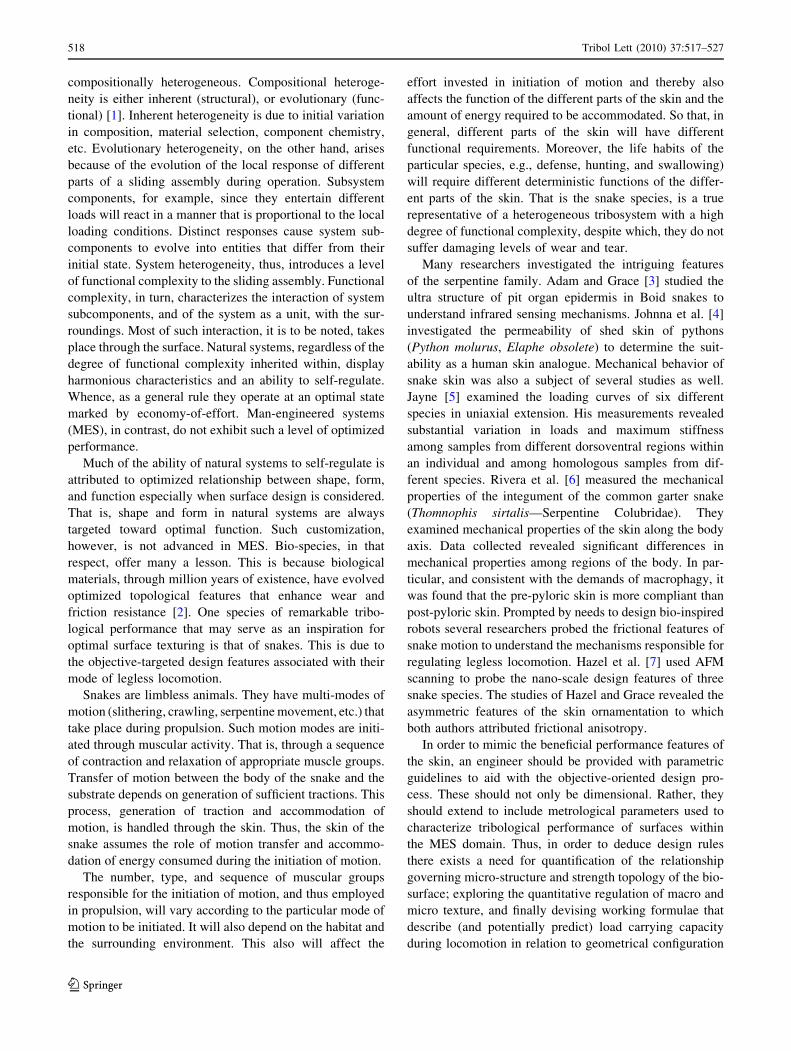
Fig. 8 Major features of SEM
observations of the skin
swatches (95000)
Tribol Lett (2010) 37:517–527 523
123
cells is quite similar for both positions the size of a unit cell
within the skin is quite different in both cases. In particular
the cell is wider for the ventral (belly) positions. Each cell
(scale) is also composed of a boundary and a membrane-
like structure. Note also the overlapping geometry of the
skin and the scales (the so-called scale and hinge structure).
The skin from the inner surface hinges back and forms a
free area, which overlaps the base of the next scale, which
emerges below this scale (Fig. 4).
4.3.2 Scan Electron Microscopy Observations
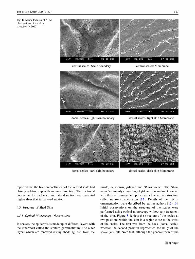
Four positions on the shed skin were identified for initial
examination. The choice of the positions was based on the
functional profile of each position in the live species (Fig. 5).
Position I is a representative of the neck region, position II
represents the beginning of the trunk (waist) region, position
III marks the boundary between the trunk and the tail region,
finally position IV represents the tail region (containing the
so-called subcaudal scales). Skin swatches from each of the
chosen positions were examined at different magnifications
(2509–150009) in topography mode. In order to suppress
charging phenomena and improve the quality of observation,
the surface of each sample was metalized by depositing a
10 nm thin layer of platinum (Pt) using a sputter coater
EMITECH K575X. Comparison of the uncoated and coated
pictures is given in Fig. 6.
For each position samples from the dark and the light
colored skin (see Fig. 5) were also examined along with
samples from the underside of the body. Major features of
the observations are shown in Figs. 7 and 8.
Note the inner structure that comprises pores. Two types
of pores (or micro pits) may be distinguished: those located
within the boundary and those located within the mem-
brane. Image analysis of the pictures indicates that the
diameter of the boundary pore ranges between 200 and
250 nm. An estimate of the diameter of the membrane
pores was estimated by Hazel et al. [7] using AFM analysis
to be in the range (50–75 nm). Worth noting, is that within
the same study [7] Hazel and co-workers scanned the
contour of the fibril tips, within the membrane, using AFM
and deduced that the tips are of asymmetric shape. Based
on this observation, combined to the inter-lamellar layout
of the fibrils, they suggested that such design features
would lead to anisotropic frictional properties of the body.
The locomotion mechanism responsible for such anisot-
ropy is, however, considered, beyond the scope of the
current work since the focus is on the topographical fea-
tures and the metrology of the surface.
Surface protrusions are also noted within the boundary.
These protrusions are of an asymmetric shape and irregular
0
200
400
600
800
1000
I II III VI
VentralDorsal-lightDorsal-Dark
Position on Body
Den
sity
of B
ound
ary
Por
es (
pore
/mm
2 )
Fig. 9 The variation in the density of the boundary pores (pore/mm2)
with position, and with color of skin
Fig. 10 Multi-scale WLI graphs depicting the topography of the skin building block (scale) boundary and membrane (scale bar is in lm)
524 Tribol Lett (2010) 37:517–527
123

distribution. The surface of the membrane also comprises
micro-nano fibril structures. These are not of consistent
shape and spacing. Note for example that the shape of
fibril located in the dark colored skin region is different
than that located within the light colored skin region
(compare the 50009 pictures). Moreover, the density of
the fibrils seems to be different within the different color
regions (denser within the dark colored region). The
spacing between different rows of fibrils also differs by
skin color and position within the body (ventral [ dorsal
light skin [ dorsal dark skin). Further analysis of images
revealed that the density of the boundary pores vary by
position. That is the number of pores per unit area is not
constant along the body, rather it changes relative to the
position. Figure 9 is a plot of the variation in the density of
the pores relative to the two sides of the skin (back dorsal
scales and abdominal ventral scales) and in relation to the
color of the skin (light patches (L) versus dark patches (D))
within the back also.
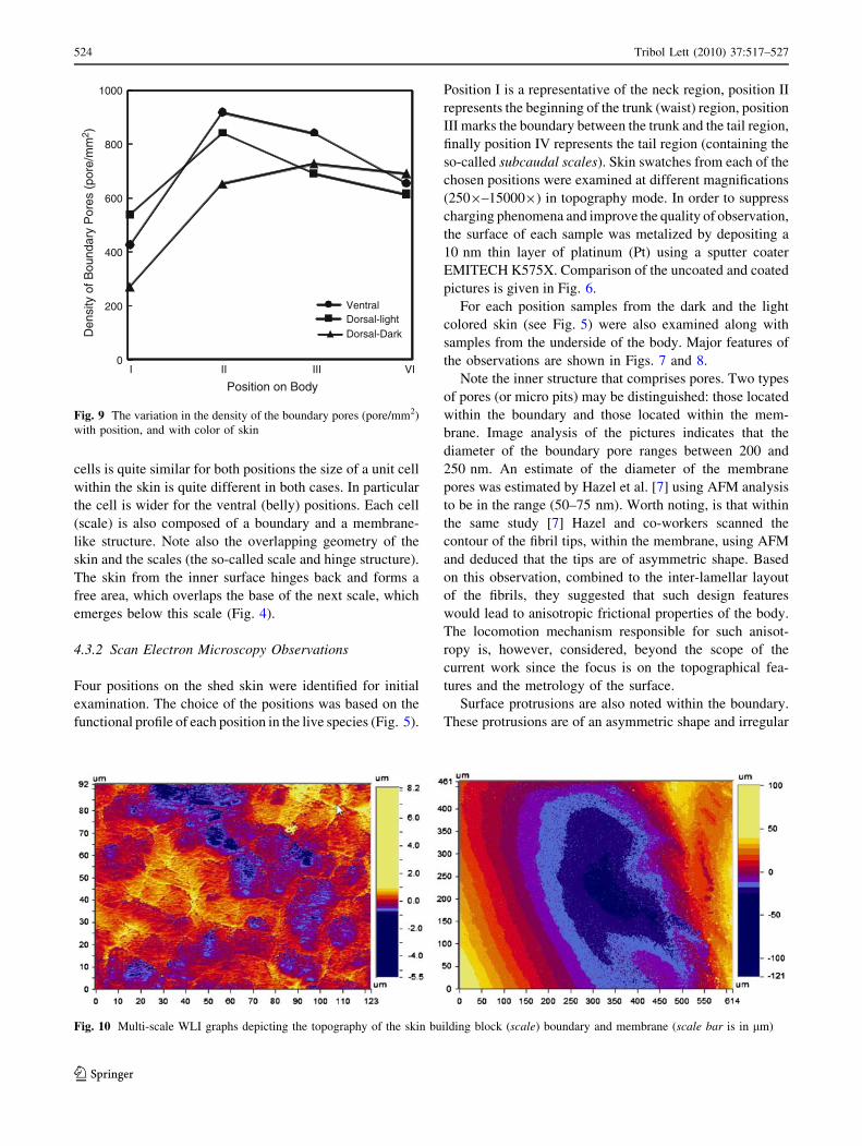
4.4 Metrology of the Surface
Examination of the surface topography features of the skin
using White Light Interferometry (WLI) on a swatches of
skin (1500 lm 9 1500 lm) yielded the basic parameters
that describe the surface (asperity radii and curvature, etc.).
Figure 10 depicts a typical WLI graph of the skin. The
shown interferogram pertains to a skin spot that is located
along the waist of the snake from the belly side (ventral).
Two interferograms are depicted: the one to the right hand
side of the figure represents the topography of the cell
membrane, whereas the one depicted to the left represents a
multi-scale scan for the whole skin swatch. Note the scale
on the right of the pictures as it indicates the deepest valley
and highest point of the skin topography. For these typical
skin swatches the value of the deepest part of the mem-
brane was about -120 lm, whereas the highest summit is
about 100 lm. The comparable values for the whole
swatch are about -5.5 and 8.2 lm, respectively.
Fig. 11 Comparison between the surface texture of snake skin and that of human skin replicas as revealed by White light interferometer (scalebar in lm)
Tribol Lett (2010) 37:517–527 525
123

To establish a measure of comparison between the
topography of the snake skin, we compared scans of snake
skin to human skin scans. The results are given in Fig. 11.
Human scans from two different positions are depicted
in the figure. The first scan is from skin located at the back
of the hand, whereas the second pertains to skin located at
the inside of the upper arm of a 40-year-old Caucasian
female. Samples were obtained by replicating the skin
using a replicating silicone. For the snake we chose to
compare scans of the ventral scales located in the waist
section of the body since it is a major load-bearing area
during locomotion. Again, it is noted that the peak summit
and valley values of the surface of the snake skin are in the
order of magnitude of one-third that of human skin. Such a
comparison highlights, qualitatively, the origins of the
superior tribological performance exhibited by the snake.
Note that under the conditions of examination in this work
quantitative comparison between both skin types are not
possible. This is because the tribological performance of
human skin is not a mere function of surface topography.
Rather, it also depends on the water content within skin
cells. As such, comparing the frictional features of snake
skin used in the current work, which is dry by default since
it is shed from the species, to human skin in vivo is
meaningless. Moreover, the friction of skin in general is
dominated by acoustic emission. Again, provisions to
monitor such effect were not undertaken in this study as it
was judged that such feature warrants detailed investiga-
tion (which is currently undergone in our laboratory).
4.5 Bearing Curve Analysis
To complete the analysis, we studied the load bearing
characteristics of the skin at each of the predetermined
positions (I through IV). Surface parameters were extracted
from SEM topography photographs. The complete set of
analyzed pictures provided a matrix of roughness param-
eters that describe the texture of the shed skin at variable
scales ranging from 1009 to 50009. Table 1a and b pro-
vides a summary of the parameters extracted from the
analysis. It can be seen that the scale of the analysis affects
the value of the parameters, which may point at a fractal
nature of the surface. Discussion of the implication of such
finding is considered out of the scope of this work. How-
ever, of interest is to point out one of the features that
directly relates to the design of the surface. Comparing the
ratios between the Reduced Peak Height Rpk, Core
Roughness Depth Rk, and Reduced Valley Depth Rvk
reveals symmetry between the positions (compare the
columns Rpk/Rk, Rvk/Rk, and Rvk/Rpk of Table 1b, and
Table 1 Effect of magnification on surface parameters
Cr/Cf Cl/Cf Rpk/Rk Rvk/Rk Rvk/Rpk
(a) Surface parameters based on 2509 pictures
Position I 0.718 0.861 0.391 0.159 1.144
Position II 2.011 2.010 0.612 0.545 0.656
Position III 2.066 1.628 0.733 0.436 0.621
Position VI 1.388 0.926 0.617 0.195 0.749
(b) Surface parameters based on 50009 pictures
Position I 1.930 1.207 0.654 0.285 0.679
Position II 1.273 1.803 0.478 0.404 0.812
Position III 1.671 1.622 0.484 0.359 0.800
Position VI 1.772 1.158 0.636 0.260 0.688
R/R
vKk
0
0.1
0.2
0.3
0.4
0.5
0.6
X250
X5000
Position on body
0
0.1
0.2
0.3
0.4
0.5
0.6
0.7
0.8
I II III IV I II III IV
R/R
pKk
Position on body
X250
X5000
Fig. 12 Plot of the ratio of the load bearing parameters Rvk/Rk and Rpk/Rk at two magnifications 9250 and 95000
526 Tribol Lett (2010) 37:517–527
123
Fig. 12a, b). This symmetry is interesting on the count that
positions II and III represent the boundaries of the main
load bearing regions (trunk). That is the regions on the
body where the snake has most of his body weight con-
centrated (refer to Fig. 5) and thereby it is the region that is
principally used in locomotion. Such symmetry may very
well be related to the wear resistance ability of the surface
or to the boundary lubrication quality of locomotion. Of
interest also, is to find if implementing a surface of such
characteristic parameters (functionally textured surface) in
plateau honing for example would be conducive to an anti-
scuffing and economical lubricant consumption perfor-
mance. Such a point is a subject of ongoing investigation.
5 Conclusions and Future Outlook
In this work, we presented the results of an initial study to
probe the geometric features of the skin of the Python
regius. It was found the structure of the unit cells is of
regionally similar shape (octagonal and hexagonal).
Although almost identical in size and density, the skin
constituents (pore density and essential size of the unit cell)
vary by position on the body. Comparison of the topogra-
phy of the snake skin to that of a human female revealed
that the surface roughness of the snake species is around
one-third that of the human samples.
Analysis of the surface roughness parameters implied a
multi-scale dependency of the parameters. This may point
at a fractal nature of the surface a proposition that needs
future verification. The analysis of bearing curve charac-
teristics revealed symmetry between the front and back
sections of the body. It also revealed that the trunk region is
bounded by two cross-sections of identical bearing curve
ratios. This has implications in design of textured surfaces
that retain an unbreakable boundary lubrication quality and
high wear resistance.
Clearly much work is needed to further probe the
essential features of the surface geometry. Namely, the
basic parametric make up of the topography, form, and
their relation to friction and wear resistance.
Acknowledgments The authors acknowledge Mr. Benjamin Favre
for assistance in SEM imaging and Mrs Ruth Ann Jones of Troup
county GA School System, for donating the shed skin used in this
work.
References
1. Abdel-Aal, H.A.: Complexity, synergy and emergence in tribo-
systems. Int. J. Mat. Prod. Tech. 38(1), 1–3 (2010)
2. Tong, J., Wang, H., Ma, Y., Ren, L.: Two-body abrasive wear of
the outside shell surfaces of mollusc Lamprotula fibrosa Heude,
Rapana venosa Valenciennes and Dosinia anus Philippi. Tribol.
Lett. 19, 331–338 (2005)
3. Adam B. Safer, Michael S. Grace: Infrared imaging in vipers:
differential responses of crotaline and viperine snakes to paired
thermal targets. Behav. Brain Res. 154(1), 55–61 (2004)
4. Johnna B. Roberts, Harvey B. Lillywhite: Lipids and the per-
meability of epidermis from snake. J. Exp. Zool. 228, 1–9 (2005)
5. Jayne, B.C.: Mechanical behavior of snake skin. J. Zool. 214,
125–140 (1988)
6. Rivera, G., Savitzky, A.H., Hinkley, J.A.: Mechanical properties
of the integument of the common gartersnake, Thamnophis sir-talis (Serpentes: Colubridae). J. Exp. Biol. 208, 2913–2922
(2005)
7. Hazel, J., Stone, M., Grace, M.S., Tsukruk, V.V.: Nanoscale
design of snake skin for reptation locomotion via friction
anisotropy. J. Biomech. 32, 477–484 (1999)
8. Sepkoski, J.: A compendium of fossil marine animal genera
(Reptilia entry). Bull. Am. Paleontol. 364, 560 (2002)
9. Ball, P.: The Self-Made Tapestry: Pattern Formation in Nature.
Oxford University Press, New York (2001)
10. Varenberg, M., Gorb, S.N.: Hexagonal surface micropattern for
dry and wet friction. Adv. Mater. 21, 483–486 (2009)
11. Zhang, H., Dai, Z., Yang, S.: Structure and friction characteristics
of snake abdomen. J. Nanjing Univ. Aeronaut. Astronaut. 40,
360–363 (2008)
12. Price, R.M.: Microdermatoglyphics: an appeal for standardization
of methodology and terminology with comments on recent
studies of North American natricines. J. Herpetol. 24, 324–325
(1990)
13. Ruibal, R.: The ultrastructure of the surface of lizard scales.
Copeia 4, 698–703 (1968)
14. Price, R.M., Kelly, P.: Microdermatoglyphics: basal patterns and
transition zones. J. Herpetol. 23, 244–261 (1989)
15. Chiasson, R.B., Bentley, D.L., Lowe, C.H.: Scale morphology in
Agkistrodon and closely related crotaline genera. Herpetologica
45, 430–438 (1989)
16. Gower, D.J.: Scale micro-ornamentation of uropeltid snakes.
J. Morphol. 258, 249–268 (2003)
Tribol Lett (2010) 37:517–527 527
123