MR Imaging of Hypervascular Lesions in the Cirrhotic Liver: A Diagnostic Dilemma
22
Note: This copy is for your personal non-commercial use only. To order presentation-ready copies for distribution to your colleagues or clients, contact us at www.rsna.org/rsnarights. 767 GASTROINTESTINAL IMAGING Cirrhosis is characterized by a spectrum of hepatocellular nodules that mark the progression from regenerative nodules to low- and high- grade dysplastic nodules, followed by small and large hepatocellular carcinomas (HCCs). Characterization of small nodules on the basis of imaging and histopathologic findings is complicated by an overlap in findings associated with each type of nodule, a reflection of their mul- tistep transitions. Vascularity patterns change gradually as the nodules evolve, with an increasing shift from predominantly venous to pre- dominantly arterial perfusion. Regenerative and low-grade dysplastic nodules demonstrate predominantly portal perfusion and contrast en- hancement similar to that of surrounding parenchyma. Differentiation of high-grade dysplastic nodules and well-differentiated HCCs on the basis of dynamic imaging and histologic findings is challenging, with a high rate of false-negative results. Some small nodules that lack hy- pervascularity may be early HCCs. Progressed small and large HCCs usually present no diagnostic difficulty because of their characteristic findings. Although characterization of hypervascular lesions in the cir- rhotic liver is difficult, it is a key step in disease management and is the radiologist’s responsibility. © RSNA, 2012 MR Imaging of Hyper- vascular Lesions in the Cirrhotic Liver: A Diag- nostic Dilemma 1 Abbreviations: ADC = apparent diffusion coefficient, DWI = diffusion-weighted imaging, HCC = hepatocellular carcinoma, SPIO = superpara- magnetic iron oxide, 3D = three-dimensional RadioGraphics 2012; Published online Content Codes: 1 From the Federal University of Rio de Janeiro, Av. Lineu de Paula Machado 896/601, Jardim Botânico, CEP 22470-040, Rio de Janeiro, Brazil (D.B.P., R.M.P., A.E.A., E.M., C.P.C., V.B.A., R.S.R.); and D’Or Institute for Research and Education, Rio de Janeiro, Brazil (D.B.P., R.M.P., A.E.A., J.A.O.N., R.S.R.). Presented as an education exhibit at the 2010 RSNA Annual Meeting. Received June 16, 2011; revision requested July 25 and received August 29; accepted September 23. For this journal-based CME activity, the authors, editor, and reviewers have no relevant relationships to disclose. Address correspondence to D.B.P. (e-mail: ). © RSNA, 2012 ONLINE-ONLY CME LEARNING OBJECTIVES ■ Describe the pathophysiologic mechanisms that oc- cur in the multistep process of carcino- genesis in cirrhotic nodules. ■ Discuss the MR imaging appearances of the spectrum of hypervascular lesions in the cirrhotic liver. ■ Explain the treat- ment of hypervascu- lar lesions on the ba- sis of the Barcelona Clinic Liver Cancer system.
-
Upload
independent -
Category
Documents
-
view
0 -
download
0
Transcript of MR Imaging of Hypervascular Lesions in the Cirrhotic Liver: A Diagnostic Dilemma

Note: This copy is for your personal non-commercial use only. To order presentation-ready copies for distribution to your colleagues or clients, contact us at www.rsna.org/rsnarights.
767GASTROINTESTINAL IMAGING
Cirrhosis is characterized by a spectrum of hepatocellular nodules that mark the progression from regenerative nodules to low- and high-grade dysplastic nodules, followed by small and large hepatocellular carcinomas (HCCs). Characterization of small nodules on the basis of imaging and histopathologic findings is complicated by an overlap in findings associated with each type of nodule, a reflection of their mul-tistep transitions. Vascularity patterns change gradually as the nodules evolve, with an increasing shift from predominantly venous to pre-dominantly arterial perfusion. Regenerative and low-grade dysplastic nodules demonstrate predominantly portal perfusion and contrast en-hancement similar to that of surrounding parenchyma. Differentiation of high-grade dysplastic nodules and well-differentiated HCCs on the basis of dynamic imaging and histologic findings is challenging, with a high rate of false-negative results. Some small nodules that lack hy-pervascularity may be early HCCs. Progressed small and large HCCs usually present no diagnostic difficulty because of their characteristic findings. Although characterization of hypervascular lesions in the cir-rhotic liver is difficult, it is a key step in disease management and is the radiologist’s responsibility. ©RSNA, 2012
MR Imaging of Hyper-vascular Lesions in the Cirrhotic Liver: A Diag-nostic Dilemma1
Abbreviations: ADC = apparent diffusion coefficient, DWI = diffusion-weighted imaging, HCC = hepatocellular carcinoma, SPIO = superpara-magnetic iron oxide, 3D = three-dimensional
RadioGraphics 2012; Published online Content Codes: 1From the Federal University of Rio de Janeiro, Av. Lineu de Paula Machado 896/601, Jardim Botânico, CEP 22470-040, Rio de Janeiro, Brazil (D.B.P., R.M.P., A.E.A., E.M., C.P.C., V.B.A., R.S.R.); and D’Or Institute for Research and Education, Rio de Janeiro, Brazil (D.B.P., R.M.P., A.E.A., J.A.O.N., R.S.R.). Presented as an education exhibit at the 2010 RSNA Annual Meeting. Received June 16, 2011; revision requested July 25 and received August 29; accepted September 23. For this journal-based CME activity, the authors, editor, and reviewers have no relevant relationships to disclose. Address correspondence to D.B.P. (e-mail: ).
©RSNA, 2012
ONLINE-ONLY CME
LEARNING OBJECTIVES
! Describe the pathophysiologic mechanisms that oc-cur in the multistep process of carcino-genesis in cirrhotic nodules.
! Discuss the MR imaging appearances of the spectrum of hypervascular lesions in the cirrhotic liver.
! Explain the treat-ment of hypervascu-lar lesions on the ba-sis of the Barcelona Clinic Liver Cancer system.

768 May-June 2012 radiographics.rsna.org
IntroductionThe incidence of hepatocellular carcinoma (HCC) and its death rates have increased during the past 10 years and are not expected to reach a plateau until 2020. Worldwide, HCC is the third most common cause of death from cancer (1,2). Prospective studies have shown that HCC now constitutes the main cause of death among pa-tients with cirrhosis; the annual incidence of HCC among such patients is 2.0%–6.6% (3). When it is detected after the onset of symptoms, patients with HCC have a dismal prognosis (5-year sur-vival rate, 0%–10%); however, patients with small HCCs may be cured (5-year survival rate, >50%). Ideally, tumors would be detected when they are solitary and smaller than 2 cm, a major diagnos-tic challenge (4,5). The widespread practice of surveillance, which is recommended for patients with cirrhosis, has increased the number of pa-tients who are diagnosed with early-stage HCC, when curative options may be pursued (5–7).
Cirrhotic livers are characterized by irrevers-ible remodeling of the hepatic architecture, including bridging fibrosis and a spectrum of hepatocellular nodules (5,8–10). Various types of hypervascular lesions are common among pa-tients with cirrhosis. The ability to differentiate between malignant and benign nodules is limited; nodules are primarily characterized on the basis of differences in vascularity. Regenerative and low-grade dysplastic nodules have predominantly portal venous blood supplies and demonstrate as much enhancement as the liver parenchyma. High-grade dysplastic nodules and HCCs dem-onstrate a loss of portal vascularization and have more nontriadal arteries. High-grade dysplastic nodules and early HCCs usually are hypovascu-lar, but they may enhance in the arterial phase, whereas those that are larger and more advanced usually appear as hypervascular nodules. The transition from regenerative and dysplastic nod-ules to HCC is not characterized by discrete steps; rather, it is marked by a continuum of vas-cular pattern changes. Many of the intermediate stages are atypical, making their characterization difficult. Typical HCCs may be diagnosed at im-aging. The algorithm for evaluating small nodules found during screening of patients at risk for HCC was recently changed: Now, a nodule larger than 1 cm that demonstrates arterial enhance-ment followed by washout at computed tomogra-phy (CT) or magnetic resonance (MR) imaging may be diagnosed as HCC (7,11,12).
In several studies, including a meta-analysis, the specificities of MR imaging and CT were found to be comparable for depicting HCC in the cir-rhotic liver, although other studies have reported that MR imaging has higher sensitivity than CT (81% versus 68%, 70% versus 50%, 77% versus 54%, and 85% versus 68% for MR imaging and CT, respectively) (13–17). The sensitivity of MR imaging for depicting HCC depends on the clini-cal setting and the size of the nodules. Krinsky et al (18) reported that MR imaging ws sensitive in only 33% of patients with known HCC before transplantation. When the nodules were stratified by size, the sensitivity of MR imaging was 100% for lesions larger than 2 cm, 52% for 1–2-cm le-sions, and only 4% for lesions smaller than 1 cm, findings that illustrate the difficulty in detecting small lesions.
Although MR imaging usually has higher sensitivity than CT, depicting and characterizing hypervascular lesions in patients with cirrhosis is challenging at any modality, especially when le-sions are small. Differentiating HCC from other hypervascular lesions is a key step in treating patients and is the responsibility of the radiolo-gist (8). In this article, we review the spectrum of hypervascular lesions that occur in the cirrhotic liver, discuss their appearances at MR imaging and the difficulty in characterizing small lesions, and present several case scenarios.
MR Imaging ProtocolsMR imaging may be performed with a 1.5- or 3.0-T system with an abdominal phased-array coil. The protocol for imaging patients with cirrhosis includes the following images: (a) breath-hold, T1-weighted, dual-echo, in- and out-of-phase, gradient-echo, with out-of-phase images obtained before in-phase images because of the shorter echo time of out-of-phase images;
coronal and axial T2-weighted, single-shot, turbo spin-echo images; axial T2-weighted, fat-suppressed, single-shot, turbo spin-echo im-ages; and axial unenhanced and contrast material–enhanced T1-weighted, fat-suppressed, three-dimensional, gradient-echo images with volumetric interpolated breath-hold examina-tion and dynamic evaluation (arterial-dominant, portal, and equilibrium phases). This sequence is followed by coronal T1-weighted, fat-suppressed, three-dimensional imaging. We also perform dif-fusion-weighted imaging with apparent diffusion coefficient (ADC) mapping.
Each sequence should be performed during end expiration to achieve a consistent breath-

holding pattern and facilitate comparison of the different types of images. If motion correction techniques are employed, inspiration images may be obtained, a method that may be helpful for patients who have difficulty holding their breath. Subtraction imaging should always be used to help analyze the contrast enhancement of nod-ules that demonstrate high signal intensity on T1-weighted images. Because the arterial-dominant phase is important for image interpretation, a technique that captures this phase in a timely manner should be used. The presence of gadolin-ium contrast material in hepatic arteries and por-tal veins and an absence of the material in hepatic veins are reliable landmarks of optimal timing of the arterial-dominant phase.
Diffusion-weighted ImagingDWI operates on the basis of incoherent intra-voxel motion and allows noninvasive quantifica-tion of water diffusion and microcapillary blood perfusion (19). Tissue cellularity and intact cell membranes are the main factors that determine the impedance of water diffusion. Tissues that are associated with restricted diffusion include those found in tumors, cytotoxic edema, and ab-scesses. Tissues with low cellularity or disrupted cell membranes, such as cysts, hemangiomas, and treated or necrotic tumors, allow a greater degree of water diffusion (20,21).
DWI may depict and help characterize fo-cal hepatic lesions. A small amount of diffusion weighting with a low value (<200 sec/mm2) nulls the intrahepatic vascular signal intensity, creat-ing so-called black-blood images and improving depiction of focal liver lesions. Studies have dem-onstrated the superiority of DWI over T2-weighted MR imaging for depiction of lesions (22,23). Images with higher values provide diffusion information that aids in lesion characterization (22,24,25). Malignant lesions have lower mean ADC values than do benign lesions, with varying degrees of overlap (22–24,26). However, it may be difficult to differentiate HCC from dysplastic nodules or an adjacent cirrhotic liver because they have similar ADC values (24,27,28).
DWI is also used to evaluate response to treatment (eg, chemotherapy, radiation therapy, and local ablation) in patients with HCC. An increased ADC value 1–2 weeks after transcath-eter arterial chemoembolization indicates early tumor response to therapy, which may be a sign of tumor necrosis before a change in tumor size occurs. Nonviable portions of tumor have high ADC values, and viable tumor portions have
low ADC values (20,21,29–32). Some studies have reported that DWI findings may be used to predict response to transcatheter arterial che-moembolization in patients with HCC; high pre-treatment ADC values are indicative of poor re-sponse to treatment and likely reflect the partially necrotic state of a tumor before intervention (33).
DWI is available at most facilities and may be incorporated into conventional protocols. Infor-mation from DWI should always be interpreted in conjunction with conventional MR imaging findings. The major limitation of DWI is its lack of standardization, which presents a challenge to its widespread adoption in body imaging.
Contrast AgentsGadolinium-based extracellular contrast agents are the most widely available MR imaging contrast agents worldwide. They are distributed within the extracellular interstitial space and shorten the T1 relaxation times of adjacent water protons, caus-ing enhanced signal intensity on T1-weighted images (34). Other MR imaging contrast agents, such as hepatobiliary-specific and superparamag-netic iron oxide (SPIO) agents, are also available at some facilities. Hepatobiliary-specific contrast agents are taken up by functioning hepatocytes and excreted in the bile. They cause T1 shortening and increase the signal intensity of the liver, bile ducts, and some hepatocyte-containing lesions on delayed T1-weighted images (34,35). SPIO is a reticuloendothelial-system-specific contrast agent that causes signal loss on T2- and T2*-weighted images. The iron oxide particles of the contrast agent are phagocytized by the macrophages, and they mainly accumulate in the liver (80%), as well as in the spleen and bone marrow (34,36,37).
Cirrhosis-associated NodulesCirrhosis is characterized by progressive fibrosis of the liver parenchyma and a spectrum of hepa-tocellular nodules, most of which are regenerative (38–40). Regenerative nodules play a role in the stepwise carcinogenesis of HCC, most frequently through dedifferentiation, which occurs in the fol-lowing order: regenerative nodule, low-grade dys-plastic nodule, high-grade dysplastic nodule, small HCC, and, finally, large HCC (38–40). The major changes that characterize the progression from re-generative nodules through the steps of HCC de-velopment are progressive loss of portal vascularity and increased arterial blood flow. Although char-acterization of nodules may be made challenging

770 May-June 2012 radiographics.rsna.org
by the presence of overlapping features (even at the histopathologic level) of some cirrhotic nod-ules, familiarity with their multistep progression and manifestations at MR imaging is important (Table). Visualization of nodules in the cirrhotic liver also may be complicated by fibrosis-related heterogeneity, and heterogeneous enhancement caused by changes in blood flow may be mistaken for tumor. Moreover, small nodules infrequently demonstrate typical findings of HCC (40).
Regenerative NodulesRegenerative nodules, also known as cirrhotic nodules, result from continuous injury to the liver parenchyma and appear as innumerable benign nodules surrounded by fibrous septa at histologic analysis (38,39). Most regenerative nodules do not progress in the dedifferentiation process. They may or may not be visible at MR imaging and are usually iso- to hypointense on T2-weighted im-
ages, with variable signal intensity on T1-weighted images. Those that contain substantial amounts of lipids, protein, and copper are hyperintense on T1-weighted images. After administration of gadolinium-based contrast material, regenerative nodules enhance as much as or slightly less than surrounding parenchyma (Fig 1) (8,41). They have normal hepatocellular and phagocytic func-tions and enhance to the same degree as adjacent liver on delayed T1-weighted images after admin-istration of hepatobiliary-specific or SPIO con-trast material (8,35). Rarely, regenerative nodules may infarct and exhibit hyperintense signal inten-sity on T2-weighted images, a finding that may be mistaken for HCC (41).
Other types of regenerative nodules are stea-totic and siderotic. Steatotic regenerative nodules result from fat deposition and are usually mul-tifocal. They appear as hyperintense lesions on in-phase gradient images and exhibit signal loss on out-of-phase images (Fig 2) (8). Siderotic nodules are caused by iron deposition and are
MR Imaging Appearances of Cirrhotic Liver Nodules
Type of Nodule
Appearance
T1-weighted Images
T2-weighted Images
Dynamic Images
Hepatobiliary Contrast Material
SPIO Contrast Material
Regenerative Varies Iso- or hypoin-tense
Enhances as much as liver paren-chyma
Enhances as much as liver parenchyma
Enhances as much as liver parenchyma
Low-grade dysplastic
Varies, often hyperintense
Iso- or hypoin-tense
Enhances as much as liver paren-chyma
Enhances as much as liver parenchyma
Enhances as much as liver parenchyma
High-grade dysplastic
Varies, often hyperintense
Iso- or hypoin-tense
Usually hypovascu-lar, but may en-hance in arterial phase
Enhances as much as liver parenchyma
Enhances as much as liver parenchyma
Early HCC Varies, often hyperintense
Iso- or hypoin-tense
Usually hypovascu-lar, but may en-hance in arterial phase
May or may not enhance (depending on degree of dif-ferentiation)
May or may not enhance (depending on degree of dif-ferentiation)
Progressed HCC
Varies, often iso- or hy-pointense
Moderately hyperintense
Enhancement in arterial phase and washout in portal or equilib-rium phase
May or may not enhance (de-pends on degree of differentia-tion)
May or may not enhance (depends on degree of dif-ferentiation)
Large HCC Heterogeneous, predomi- nantly hypo-intense
Heterogeneous, predomi- nantly hyper-intense
80%–90% enhance in arterial phase, washout in portal or equilibrium phase
No enhance- ment
No enhance-ment
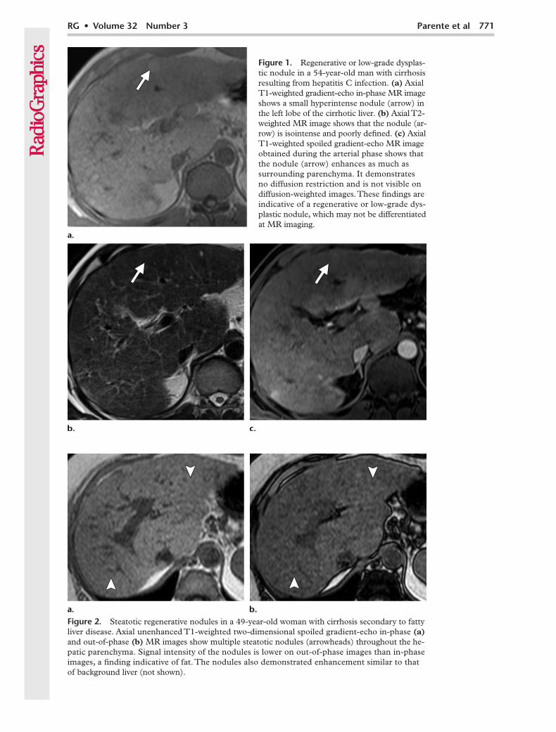
Figure 2. Steatotic regenerative nodules in a 49-year-old woman with cirrhosis secondary to fatty liver disease. Axial unenhanced T1-weighted two-dimensional spoiled gradient-echo in-phase (a) and out-of-phase (b) MR images show multiple steatotic nodules (arrowheads) throughout the he-patic parenchyma. Signal intensity of the nodules is lower on out-of-phase images than in-phase images, a finding indicative of fat. The nodules also demonstrated enhancement similar to that of background liver (not shown).
Figure 1. Regenerative or low-grade dysplas-tic nodule in a 54-year-old man with cirrhosis resulting from hepatitis C infection. (a) Axial T1-weighted gradient-echo in-phase MR image shows a small hyperintense nodule (arrow) in the left lobe of the cirrhotic liver. (b) Axial T2-weighted MR image shows that the nodule (ar-row) is isointense and poorly defined. (c) Axial T1-weighted spoiled gradient-echo MR image obtained during the arterial phase shows that the nodule (arrow) enhances as much as surrounding parenchyma. It demonstrates no diffusion restriction and is not visible on diffusion-weighted images. These findings are indicative of a regenerative or low-grade dys-plastic nodule, which may not be differentiated at MR imaging.
772 May-June 2012 radiographics.rsna.org
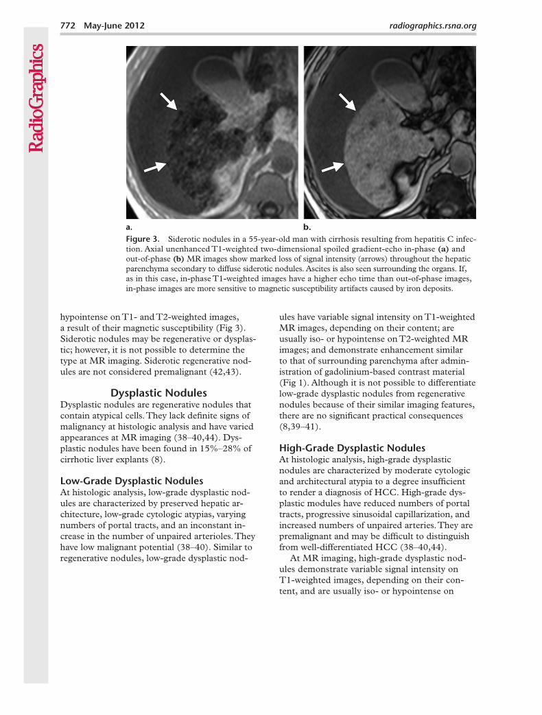
Siderotic nodules in a 55-year-old man with cirrhosis resulting from hepatitis C infec-tion. Axial unenhanced T1-weighted two-dimensional spoiled gradient-echo in-phase (a) and out-of-phase (b) MR images show marked loss of signal intensity (arrows) throughout the hepatic parenchyma secondary to diffuse siderotic nodules. Ascites is also seen surrounding the organs. If, as in this case, in-phase T1-weighted images have a higher echo time than out-of-phase images, in-phase images are more sensitive to magnetic susceptibility artifacts caused by iron deposits.
hypointense on T1- and T2-weighted images, a result of their magnetic susceptibility (Fig 3). Siderotic nodules may be regenerative or dysplas-tic; however, it is not possible to determine the type at MR imaging. Siderotic regenerative nod-ules are not considered premalignant (42,43).
Dysplastic NodulesDysplastic nodules are regenerative nodules that contain atypical cells. They lack definite signs of malignancy at histologic analysis and have varied appearances at MR imaging (38–40,44). Dys-plastic nodules have been found in 15%–28% of cirrhotic liver explants (8).
Low-Grade Dysplastic NodulesAt histologic analysis, low-grade dysplastic nod-ules are characterized by preserved hepatic ar-chitecture, low-grade cytologic atypias, varying numbers of portal tracts, and an inconstant in-crease in the number of unpaired arterioles. They have low malignant potential (38–40). Similar to regenerative nodules, low-grade dysplastic nod-
ules have variable signal intensity on T1-weighted MR images, depending on their content; are usually iso- or hypointense on T2-weighted MR images; and demonstrate enhancement similar to that of surrounding parenchyma after admin-istration of gadolinium-based contrast material (Fig 1). Although it is not possible to differentiate low-grade dysplastic nodules from regenerative nodules because of their similar imaging features, there are no significant practical consequences (8,39–41).
High-Grade Dysplastic NodulesAt histologic analysis, high-grade dysplastic nodules are characterized by moderate cytologic and architectural atypia to a degree insufficient to render a diagnosis of HCC. High-grade dys-plastic modules have reduced numbers of portal tracts, progressive sinusoidal capillarization, and increased numbers of unpaired arteries. They are premalignant and may be difficult to distinguish from well-differentiated HCC (38–40,44).
At MR imaging, high-grade dysplastic nod-ules demonstrate variable signal intensity on T1-weighted images, depending on their con-tent, and are usually iso- or hypointense on

Figure 4. High-grade dysplastic nodule or small HCC in a 52-year-old man with cirrhosis re-sulting from hepatitis C infection. (a, b) Axial unenhanced T1-weighted three-dimensional (3D) spoiled gradient-echo (a) and contrast-enhanced T2-weighted (b) MR images show a small isoin-tense nodule (arrow) in segment V of the liver. (c) Axial contrast-enhanced MR image obtained in the hepatic arterial phase shows intense enhancement of the nodule (arrow), a finding indicative of hypervascularity. (d) Axial contrast-enhanced MR image obtained in the equilibrium phase shows that the nodule (arrow) demonstrates no washout. Follow-up images showed a larger nodule that was diagnosed as HCC.
T2-weighted images. Most are hypovascular, although they may exhibit arterial enhancement similar to that seen in HCC (7,39,40). Estab-lishing a differential diagnosis for high-grade dysplastic nodules and early HCC on the basis of imaging and pathologic characteristics may be difficult, but some pathologic markers are
distinctive (Figs 4, 5) (7,8,39–41,44). Hepato-biliary and SPIO contrast agents are not useful for characterizing low- and high-grade dysplastic nodules and well-differentiated HCCs because both of these lesions enhance after administra-tion of contrast material (34,35).

774 May-June 2012 radiographics.rsna.org
Nodule-in-Nodule AppearanceA dysplastic nodule may harbor a focus of HCC (38,39). At T2-weighted MR imaging, such nod-ules appear hypointense, with the same degree of enhancement as surrounding parenchyma and a high-signal-intensity focus with arterial enhancement, a finding known as the nodule-in-nodule appearance (Fig 6) (39–41,44). The
nodule-in-nodule appearance is uncommon, occurring in approximately 6% of patients with dysplastic nodules (39).
Hepatocellular CarcinomaAt histologic analysis, HCC is characterized by an abnormally high number of muscular-ized, unpaired arterioles and capillarized vessels (38–40). Its MR imaging appearance varies de-pending on its size, grade, and biologic features (8,10,34,39,40,45–47).
Figure 5. High-grade dysplastic nodule or early HCC in a 46-year-old man with cirrhosis resulting from hepatitis C infection. (a) Axial T2-weighted fat-saturated MR image shows a small hypointense nodule (arrow) in the right lobe of the cirrhotic liver. (b) Axial unenhanced T1-weighted spoiled gradient-echo MR image shows the nodule (arrow), which is hyperintense. (c) Axial T1-weighted spoiled gradient-echo MR image obtained in the arterial phase shows the nodule (arrow), which enhances as much as surrounding parenchyma. (d) Axial T1-weighted spoiled gradient-echo MR image obtained in the equilibrium phase shows that the nodule (arrow) demonstrates no washout.

Figure 6. Nodule-in-nodule appearance at 4-month follow-up examination (same patient as in Fig 5). (a) Axial T2-weighted fat-saturated MR image shows a high-grade dysplastic nodule (ar-rowhead), the anterior portion of which is slightly hyperintense compared with previous images (cf Fig 5). (b) Axial unenhanced T1-weighted spoiled gradient-echo MR image shows the hyperintense nodule (arrow). (c) Axial T1-weighted spoiled gradient-echo MR image obtained in the arterial phase shows the nodule, the anterior portion of which is hypervascular (arrowhead). The rest of the nodule enhances similarly to the adjacent liver (arrow). (d) Axial T1-weighted spoiled gradient-echo MR image obtained in the equilibrium phase shows washout of the hypervascular portion of the nodule (arrowhead). These changes confirm that the nodule identified at the first examination was a high-grade dysplastic nodule that developed a focus of HCC, referred to as a nodule-in-nodule.
Small HCCsSmall HCCs are smaller than 2 cm and divided into two types: early, which are indistinctly (vaguely) nodular, and progressed, which are dis-tinctly nodular (40,44,48). At histologic analysis, early HCCs are well-differentiated, with neoplas-tic cells replacing normal cells; portal tracts may be present. It is difficult to differentiate small HCCs from surrounding parenchyma. Invasion
of portal tracts is frequently visible, a finding that helps distinguish early HCCs from high-grade dysplastic nodules. Early HCCs receive blood supply from a reduced number of trapped portal tracts and insufficiently developed nontriadal arteries. Cellular crowding and low blood sup-ply cause relative hypoxia, which may cause

776 May-June 2012 radiographics.rsna.org
Figure 7. Small progressed HCC in a 54-year-old woman with cirrhosis resulting from hepatitis C infection. (a) Axial T2-weighted MR image shows a slightly hyperintense nodule (arrow) in segment II of the liver. (b) Axial unenhanced T1-weighted 3D spoiled gradient-echo MR image obtained in the arterial phase shows the nodule (arrow), which is small and isointense. (c) Axial T1-weighted spoiled gradient-echo MR image obtained in the arterial phase shows that the nodule (arrow) demonstrates intense enhancement, a finding indicative of hypervascularity. (d) Axial T1-weighted spoiled gradient-echo MR image obtained in the equilibrium phase shows venous washout with capsule enhancement (arrow). Despite its small size, this nodule may be diagnosed as HCC on the basis of imaging findings alone, with no biopsy or second examination.
the steatotic changes that occur in about 40% of cases of early HCC. Early HCCs are rarely asso-ciated with microscopic vessel invasion, produce
no metastases, and have a 5-year survival rate of 89% (40,44,48,49). The frequency and natural history of early HCCs are poorly understood (48). They have been found to recur within 3 years of resection in only 8% of cases (49).

Imaging Findings.—Currently, a diagnosis of early HCC on the basis of imaging is unreliable. Such lesions tend to demonstrate high signal intensity on T1-weighted MR images and be hypo- or isointense on T2-weighted MR images (40). Early HCCs demonstrate relative arterial hypovascularity (most are hypo- or isointense in the arterial phase) and decreased portal sup-ply, which is indicated by hypointensity in the portal phase. Hypovascularity is likely caused by a loss of portal vascularization and insufficient development of unpaired arteries. Such lesions are expected to demonstrate progressively in-creased arterialization and a continued decrease in portal blood until they become typical HCCs (Fig 5) (7). Use of hepatobiliary contrast agents has recently been shown to improve diagnosis of HCCs of all sizes (<1 cm, 1–2 cm, and >2 cm). On hepatobiliary-phase MR images, liver parenchyma that contains functioning hepato-cytes demonstrates enhancement, and HCCs that contain malfunctioning hepatocytes dem-onstrate no enhancement and appear as hypoin-tense lesions (50,51). However, well-differen-tiated small HCCs may demonstrate enhance-ment on hepatobiliary-phase images, a result of residual hepatocyte activity and the reason for false-negative findings in some cases. Ahn et al (50) reported that in two patients, early HCCs could only be identified on hepatobiliary-phase images. Close follow-up is recommended for patients with lesions that are smaller than 1 cm and visible only on hepatobiliary-phase images (50). Early HCCs enhance as much as sur-rounding parenchyma after administration of SPIO contrast agents (40). Fatty changes are visible in 40% of cases.
In contrast, progressed small HCCs have morphologic and histologic characteristics similar to those of large HCCs and are easily differentiated from the background cirrhotic liver. At histologic analysis, no portal tracts are seen, but numerous nontriadal arteries and well-developed sinusoidal capillarization are present. Microscopic vessel invasion is present in nearly 27% of cases (49). Tumor-cell invasion of portal vein branches and intrahepatic metastases may be present and are associated with a 5-year sur-vival rate of 48% (40,44,48).
Progressed HCCs usually pose no diagnos-tic difficulty at MR imaging. These lesions tend to be well-defined, homogeneous, and round or oval, with variable signal intensity on T1-weighted images and, usually, moderately hyper-intense signal intensity on T2-weighted images. Most nodular HCCs demonstrate typical findings after administration of gadolinium-based contrast material, with enhancement during the arterial phase and washout in the portal or delayed phase that becomes less intense than surrounding liver (Fig 7) (40,48).
Early HCCs are the earliest recognizable form of HCC, although they are difficult to differenti-ate from high-grade dysplastic nodules. Estab-lishing a definitive diagnosis usually requires a biopsy; however, most hypovascular HCCs and those with equivocal imaging findings are early HCCs. Progressed HCCs are small nodular le-sions with characteristics similar to those of clas-sic HCCs and usually do not pose a diagnostic problem (40,48).
Although they are critical for making treat-ment decisions, imaging findings are not always conclusive for small HCCs. In 2011, the Ameri-can Association for the Study of Liver Disease (AASLD) updated its guidelines with the fol-lowing recommendations: Small HCCs that are larger than 1 cm may be diagnosed on the basis of imaging findings (contrast-enhanced CT or MR imaging) alone when findings are typical (hypervascularity followed by wash-out) of HCC. Lesions that are smaller than 1 cm should be re-examined every 3 months with the technique that was used to depict the nodule; most such lesions are cirrhotic and, therefore, stable at follow-up. If the nodule is stable, patients should resume routine screen-ing for HCC. If the nodule changes, four-phase (unenhanced, arterial, portal, and equilibrium) multidetector CT or MR imaging should be performed. Similarly, lesions larger than 1 cm should be evaluated with four-phase CT or dy-namic contrast-enhanced MR imaging. Typical findings lead to a diagnosis of HCC. If an atypi-cal appearance is present, a second examination with CT or MR imaging (whichever was not

778 May-June 2012 radiographics.rsna.org
Figure 8. Chart shows the algorithm for evaluating small nodules found at screening in patients at risk for HCC. MDCT = multidetector CT. (Adapted and re-printed, with permission, from reference 7.)
used the first time) may be performed. Typical findings of HCC confirm the diagnosis; biopsy is recommended for patients with atypical find-ings (Fig 8) (7). Patients with liver nodules that have a nonspecific vascular profile and negative biopsy results should undergo enhanced follow-up imaging. If biopsy results are negative, imag-ing should be repeated at 3–6-month intervals until the nodule disappears, enlarges, or has findings characteristic of HCC. If the lesion enlarges but remains atypical for HCC, another biopsy is recommended. Smaller lesions are less likely to be associated with microscopic vascular invasion and are more responsive to curative treatments. Thus, HCCs should be diagnosed when they are smaller than 2 cm (7).
Clinical decisions concerning the management of nodules in patients with negative biopsy results should take into account the lack of knowledge of the natural history of early-stage HCC and the limitations and pitfalls of guided biopsy. These limitations may lead to false-negative results in as many as 10% of cases, with small foci of HCC that may be missed at histologic analysis present in at least one-third of dysplastic nodules. Differ-ent grades of dysplastic changes also may affect different parts of the nodule. Little is known of the accuracy of differential diagnoses for low- and high-grade dysplastic nodules and HCCs created on the basis of a small biopsy specimen obtained from one part of a nodule (46).
Classification.—The Liver Imaging Reporting and Data System (LI-RADS) was recently cre-ated in an attempt to facilitate classification of liver lesions as either definitely benign (LR1) or definitely HCC (LR5). The designations of prob-ably HCC (LR4) and probably benign (LR2) may be used to classify lesions with some, but not all, features of HCC or benignity. The des-ignation LR3 is used for indeterminate lesions with equivocal imaging features that may not be categorized as probably benign or probably ma-lignant (52).
When evaluating nodular lesions in patients with cirrhosis, radiologists should recognize that hepatocarcinogenesis is a continuous process that has been arbitrarily divided for practical and didactic purposes. The continuous histo-logic changes that nodules undergo result in significant overlap of enhancement patterns and findings on T1- and T2-weighted MR images, complicating their imaging and histopatho-logic differential diagnosis. Differences in the
histologic criteria that are used to characterize hepatocellular nodules and changes in nomen-clature over time have also contributed to the overlap in histologic findings. As nodules dedif-ferentiate, portal vascularization progressively decreases, and blood supply from nontriadal arteries increases. At imaging, this process is seen as a progressive change from enhancement similar to that of the adjacent liver to a loss of portal vascularization that appears as an area of hypoenhancement in the portal phase, followed by an increase in the number of hypervascular nontriadal arteries in the arterial phase. Lesions that exhibit enhancement similar to that of the adjacent liver are probably regenerative and low-grade dysplastic nodules. A diagnosis of HCC may be made when hypervascularity is followed by washout at imaging. However, nodules with a loss of portal supply and development of an in-sufficient number of nontriadal arteries to pro-duce hypervascularity are difficult to diagnose. These equivocal findings of high-grade dysplas-tic nodule and early HCC are responsible for the high rate of false-negative imaging and his-tologic results.

There are some features that help classify cirrhotic nodules. For instance, size is an im-portant parameter. Although nodules that are smaller than 1 cm are probably benign and nodules larger than 2 cm are probably malig-nant, nodules that are between 1 and 2 cm are difficult to classify. Nodules that demonstrate interval growth are suspicious for HCC. Regen-erative nodules, low- and high-grade dysplastic nodules, and early HCCs have variable signal intensity on T1-weighted images; however, well-differentiated HCCs tend to be hyperintense. Hyperintensity on T2-weighted images is highly specific to HCCs and is rarely present in small lesions. Regenerative and low-grade dysplas-tic nodules demonstrate enhancement similar to that of adjacent liver, whereas high-grade dysplastic nodules and early HCCs tend to be hypovascular; high-grade dysplastic nodules and early HCCs also may demonstrate arterial en-hancement. Capsular enhancement and internal mosaic architecture are also highly specific to HCCs and are rarely seen in small nodules. The presence of fatty changes in a dominant nodule is also suspicious for HCC.
Case Scenarios.—The following five case sce-narios are presented for practical purposes.
1. A cirrhotic liver that demonstrates homo-geneous enhancement on contrast-enhanced images but lacks a dominant nodule on T1- and T2-weighted MR images probably contains only regenerative and low-grade dysplastic nodules.
2. A dominant nodule that is hyperintense on T1-weighted MR images and iso- or hypoin-tense on T2-weighted images with enhancement similar to that of the surrounding liver probably represents a regenerative or low-grade dysplas-tic nodule or, less likely, a high-grade dysplastic nodule. If it becomes hypovascular, it has prob-ably progressed to a high-grade dysplastic nodule or early HCC, and if it demonstrates high signal intensity on T2-weighted images, a progressed HCC should be suspected. When it demonstrates hypervascularity with washout, a diagnosis of HCC may be made on the basis of imaging find-ings alone.
3. A nodule that is hyperintense on T1-weighted MR images and iso- or hypointense on T2-weighted images with arterial enhancement and no washout is probably a high-grade dysplastic nodule or early HCC. In contrast, the absence of a nodule on T1- and T2-weighted images and the presence of arterial enhancement with no washout may indicate transient arterial enhancement.
4. A rarer lesion is the nodule-in-nodule (ie, a dysplastic nodule with an HCC focus), which classically manifests as a low-signal-intensity nod-ule on T2-weighted MR images with a hyperin-tense focus that enhances in the arterial phase.
5. A nodule that is hyperintense on T2-weighted MR images with arterial enhancement and venous washout may be diagnosed as HCC.
The likelihood of a nodule being HCC in-creases with its size; nodules smaller than 1 cm have low likelihood. Thus, recently updated guidelines recommend that patients with inde-terminate nodules smaller than 1 cm should be monitored. Nodules larger than 2 cm are likely malignant, whereas those between 1 and 2 cm should be closely monitored with imaging and repeated biopsy (Fig 8) (5,7).
Large HCCsLarge HCCs have a characteristic MR imaging appearance and are usually diagnosed with no difficulty. They typically are hypointense on T1-weighted images and moderately hyperintense on

780 May-June 2012 radiographics.rsna.org
Large HCC in a 55-year-old man with cirrhosis resulting from hepatitis C infection. (a) Axial T2-weighted MR image shows a heterogeneous mass (arrow) with slightly high signal intensity in segment VII, abutting the liver capsule. (b) Axial T1-weighted spoiled gradient-echo MR image shows that the mass is isointense (arrow). (c) Axial T1-weighted spoiled gradient-echo MR image obtained in the arterial phase shows heterogeneous hypervascular enhancement of the mass (arrow). (d) Axial T1-weighted spoiled gradient-echo MR image obtained in the equilibrium phase shows washout (arrow). A thin circumferential hypointense rim (arrowhead) is seen around the periphery of the tumor, a finding indicative of a capsule, with typical late enhancement after admin-istration of gadolinium-based contrast material.
T2-weighted images, with arterial enhancement and washout in the portal or delayed phase. They may be heterogeneous, have a mosaic pattern with patchy internal areas of hyperintensity on T2-weighted images, have variable signal intensity on T1-weighted images, or demonstrate inhomoge-neous enhancement after administration of gado-
linium-based contrast material (Fig 9) (8,39,41). Atypical manifestations are uncommon. Rarely, an HCC that is larger than 2 cm may be hypovascu-lar; such cases require biopsy in order to make a diagnosis (Fig 10) (7).
Large HCCs usually appear as heterogeneous lesions at MR imaging, with variable signal in-tensity depending on their content. Intralesional fat, hemorrhage, or necrosis may be present. Intralesional fat is characterized by signal loss on

Figure 10. Biopsy-proved hypovascular HCC in a 58-year-old woman with cirrhosis resulting from hepatitis C infection. (a) Axial T2-weighted MR image shows a slightly high-signal-intensity nodule (arrow) in the dome of the liver. (b) Axial unenhanced T1-weighted fat-saturated 3D spoiled gradient-echo MR image shows that the nodule (arrow) is isoin-tense. (c, d) Axial unenhanced T1-weighted fat-saturated 3D spoiled gradient-echo MR images obtained in the arterial (c) and equilib-rium (d) phases show that the nodule (arrow) is mainly hypovascular. (e) Axial respiration-triggered fat-suppressed single-shot spin-echo echo-planar diffusion-weighted MR image ( = 800 s/mm2) shows restricted diffusion within the nodule (arrow).

782 May-June 2012 radiographics.rsna.org
out-of-phase images (compared with in-phase images) and low signal intensity on fat-saturated images. Because the fat component usually en-hances differently than the rest of the lesion, it usually does not demonstrate hypervascularity after gadolinium-based contrast material is ad-ministered, but some enhancement may be seen (53). The hemorrhage component of HCCs is hyperintense on T1-weighted images and hy-pointense on T2-weighted images. Intralesional necrosis is hypointense on T1-weighted images and hyperintense on T2-weighted images with no enhancement (8,10,34,45,54,55).
Most large HCCs (65%–82%) have a capsule composed of fibrous tissue and compressed vessels that appears as a thin circumferential rim around the periphery of the tumor and is usually hypoin-tense on T1- and T2-weighted images, with typical late enhancement after administration of gadolin-ium-based contrast material (Figs 7, 9). Capsules that are thicker than 4 mm may be hyperintense on T2-weighted images (8,10,34,45,54).
Uptake of hepatobiliary and SPIO contrast agents varies according to the degree of differ-
entiation. Poorly differentiated HCCs do not enhance (compared with surrounding liver) after administration of hepatobiliary agents or accu-mulate SPIO agents; thus, they are more appar-ent on contrast-enhanced images. Well-differenti-ated HCCs demonstrate enhancement similar to that of adjacent liver parenchyma after adminis-tration of hepatobiliary and SPIO contrast agents and may not be depicted on contrast-enhanced images (34,35).
Treatment of Cirrhotic NodulesRadiologists play a central role in making treat-ment decisions for patients with cirrhosis. Algo-rithms for the follow-up of patients with cirrhotic nodules and guidelines for treatment in those with HCC are developed on the basis of imaging find-ings. An erroneous diagnosis of HCC has impor-tant implications for patient care. According to the Barcelona Clinic Liver Cancer ((BCLC) system, patients with stage 0 HCC (<2 cm) and no vas-cular invasion or spread and those with stage A HCC (a solitary tumor <5 cm or as many as three nodules that are <3 cm) should undergo curative treatment such as ablation, resection, or trans-plantation; 5-year survival rates for patients with
Figure 11. Chart shows the Barcelona Clinic Liver Cancer staging and treatment allocation system. = perfor-mance status test, = radiofrequency ablation, = transcatheter arterial chemoembolization. (Adapted and reprinted, with permission, from reference 7.).

Figure 12. Transient arterial enhancement in a 45-year-old man with hepatitis C infection–re-lated cirrhosis secondary to portal vein thrombosis. (a) Axial MR image obtained in the arterial phase shows a small area of homogeneous enhancement (arrowhead) in segment IV of the liver. A wedge-shaped area of enhancement (arrow), with the wide base toward the periphery, is also seen. (b) T2-weighted MR image shows a corresponding slightly hyperintense area indicative of a per-fusion defect (arrow), a finding that is not as rare as commonly thought.
stage 0 or A HCC are 50%–75% (56). Patients with stage B HCC (large or multinodular tumor with no vascular invasion, extrahepatic spread, or cancer-related symptoms), particularly those with compensated cirrhosis, should undergo chemoem-bolization; among these patients, expected 3-year survival rates are 50%. Patients with stage C HCC (advanced tumor with vascular involvement, ex-trahepatic spread, or physical impairment) should enroll in research trials to assess new antitumoral agents; these patients have 3-year survival rates of less than 10%. Finally, patients with stage D HCC (impaired physical status or excessive tumor bur-den and severe liver impairment) should undergo symptomatic treatment only; among patients with stage D HCC, 1-year survival rates are less than 10% (Fig 11) (56).
Other Conditions That May Mimic Hepatic Lesions
Transient Arterial Enhancing LesionsNonspecific, transient, arterially enhancing le-sions must be differentiated from small HCCs. The most common conditions that cause tran-sient hepatic arterial enhancement are sponta-neous or postbiopsy arterioportal shunting and pseudoaneurysm, HCC neovascularity, portal vein compression, and tumoral or nontumoral portal vein thrombosis. Arterioportal shunting and pseudoaneurysm may occur spontaneously
in the cirrhotic liver or form secondary to bi-opsy or ablation and are usually peripheral with a wedge-shaped area of enhancement, and they are isointense relative to surrounding paren-chyma on T1- and T2-weighted MR images and in the equilibrium phase. Less often, they may be nodular or have an irregular outline. More-over, such lesions may be mildly hyperintense on T2-weighted images and associated with mild, prolonged parenchymal enhancement. In HCC, neovascularization may appear as an area of arte-rial hypervascularity surrounding the malignancy. This area of enhancement may be ill-defined and lead to overestimation of tumor size in the arterial phase. Blood flow in portal veins may be reduced, a result of thrombosis or compression by a focal lesion. Portal vein thrombosis may result from tumor or direct invasion of the HCC or have a nontumoral cause, such as blood flow changes secondary to cirrhosis. In these cases, transient arterial enhancement is caused by oc-clusion or compression of the portal vein with reduced portal vascularity and a compensatory increase in arterial supply. Transient arterial en-hancement is seen as a wedge-shaped area that conforms to the segment or lobe with reduced portal supply (Fig 12) (41,57–62).

784 May-June 2012 radiographics.rsna.org
Focal confluent hepatic fibrosis occurs in end-stage liver disease and must be differentiated from HCC. It is characteristically wedge shaped, with the wide base oriented toward the liver capsule, and is associated with atrophy of the affected segment and capsular retraction. Typi-cally, fibrotic lesions are located in the anterior and medial segments of the liver. Focal confluent fibrosis is usually hypointense on T1-weighted MR images and hyperintense on T2-weighted images, with delayed contrast enhancement (Fig 13). Infrequently, confluent fibrosis may enhance in the arterial phase, a finding that mimics neo-plasm; in such cases, biopsy is necessary to make a diagnosis. The characteristic shape, location, volume loss, and delayed enhancement of con-fluent fibrosis may help in the diagnosis of this
condition (41,63–66). Focal confluent fibrosis also lacks Kupffer cells and may mimic HCC on SPIO contrast-enhanced images (8).
HemangiomasHemangiomas rarely occur in end-stage cirrhosis, probably because the cirrhosis obliterates existing hemangiomas. When present in the cirrhotic liver, they are often atypical and contain large areas of fibrosis (41). They do not wash out and instead remain isointense relative to the hepatic vascula-ture in multiple phases (65,67).
Rarely, other types of lesions—such as focal nodular hyperplasia or focal nodular hyperpla-sia–like nodules, hepatic adenoma, hypervascular metastases, Budd-Chiari–associated nodules, and intrahepatic cholangiocarcinoma—complicate the differential diagnosis of cirrhotic liver nodules (68–75). These lesions may be differentiated from HCC on the basis of clinical history and imaging findings (70).
Confluent fibrosis in a 60-year-old woman with hepatitis C infection–related cirrhosis. (a) Axial T2-weighted MR image shows a lesion with peripheral hyperintensity (arrow) in the right lobe of the liver. (b) Axial T1-weighted spoiled gradient-echo MR im-age obtained in the arterial phase shows no significant enhancement of the lesion (arrow). (c) Axial T1-weighted spoiled gradient-echo MR image obtained in the equilibrium phase shows delayed enhancement of the lesion (arrow), a characteristic finding of fibrosis. Capsular retraction (arrowhead) secondary to subtle volume loss is also seen.

ConclusionsAccurate diagnosis relies on radiologists’ familiar-ity with the multistep process of HCC develop-ment and the imaging findings associated with each stage. The major changes that characterize the progression of regenerative nodules through the steps of HCC development are progressive loss of portal vascularity and increased arterial blood flow (40,44,48). Regenerative nodules and low-grade dysplastic nodules are predominantly portally perfused and enhance as much as sur-rounding liver after administration of gadolinium-based contrast material (8,41). New vessel for-mation characterizes the progression of nodule development. Vascular patterns change gradually, with some high-grade dysplastic nodules and most HCCs exhibiting an increasing shift from predom-inantly venous perfusion to predominantly arterial perfusion. The major shift in angiogenesis typically occurs during the transition from low- to high-grade dysplasia (40). Early small HCCs are usu-ally composed of well-differentiated hepatocytes, challenging diagnosis on the basis of dynamic im-aging and biopsy results, and are associated with a high rate of false-negative results. Some small HCC nodules demonstrate no hypervascularity. Progressed small HCCs and large HCCs usually have typical imaging findings (hypervascularity followed by washout); however, in the absence of typical findings, differentiating among cirrhotic liver nodules is difficult and remains a challenge, even for experienced radiologists.
References 1. Edwards BK, Ward E, Kohler BA, et al. Annual
report to the nation on the status of cancer, 1975–2006, featuring colorectal cancer trends and impact of interventions (risk factors, screening, and treat-ment) to reduce future rates. Cancer 2010;116(3): 544–573.
2. Parkin DM, Bray F, Ferlay J, Pisani P. Global can-cer statistics, 2002. CA Cancer J Clin 2005;55(2): 74–108.
3. Llovet JM, Burroughs A, Bruix J. Hepatocellular carcinoma. Lancet 2003;362(9399):1907–1917.
4. Bruix J, Llovet JM. Two decades of advances in hepatocellular carcinoma research. Semin Liver Dis 2010;30(1):1–2.
5. Forner A, Vilana R, Ayuso C, et al. Diagnosis of hepatic nodules 20 mm or smaller in cirrhosis: prospective validation of the noninvasive diagnostic criteria for hepatocellular carcinoma. Hepatology 2008;47(1):97–104.
6. Bruix J, Sherman M. Management of hepatocellular carcinoma. Hepatology 2005;42(5):1208–1236.
7. Bruix J, Sherman M.Management of hepatocellu-lar carcinoma: an update. Hepatology 2011;53(3): 1020–1022.
8. Hanna RF, Aguirre DA, Kased N, Emery SC, Peterson MR, Sirlin CB. Cirrhosis-associated hepatocellular nodules: correlation of histopatho-logic and MR imaging features. RadioGraphics 2008;28(3): 747–769.
9. Brancatelli G, Federle MP, Ambrosini R, et al. Cir-rhosis: CT and MR imaging evaluation. Eur J Radiol 2007;61(1):57–69.
10. Krinsky GA, Lee VS. MR imaging of cirrhotic nod-ules. Abdom Imaging 2000;25(5):471–482.
11. Sangiovanni A, Manini MA, Iavarone M, et al. The diagnostic and economic impact of contrast imaging techniques in the diagnosis of small hepatocellular carcinoma in cirrhosis. Gut 2010;59(5):638–644.
12. Sherman M. Optimum imaging for small suspected hepatocellular carcinoma. Gut 2010;59(5):570–571.
13. Colli A, Fraquelli M, Casazza G, et al. Accuracy of ultrasonography, spiral CT, magnetic resonance, and alpha-fetoprotein in diagnosing hepatocellular carcinoma: a systematic review. Am J Gastroenterol 2006;101(3):513–523.
14. Hussain SM, Semelka RC, Mitchell DG. MR imag-ing of hepatocellular carcinoma. Magn Reson Imag-ing Clin N Am 2002;10(1):31–52.
15. Libbrecht L, Bielen D, Verslype C, et al. Focal lesions in cirrhotic explant livers: pathological evaluation and accuracy of pretransplantation imaging exami-nations. Liver Transpl 2002;8(9):749–761.
16. Rode A, Bancel B, Douek P, et al. Small nodule de-tection in cirrhotic livers: evaluation with US, spiral CT, and MRI and correlation with pathologic exam-ination of explanted liver. J Comput Assist Tomogr 2001;25(3):327–336.
17. Tomemori T, Yamakado K, Nakatsuka A, Sakuma H, Matsumura K, Takeda K. Fast 3D dynamic MR imaging of the liver with MR SmartPrep: com-parison with helical CT in detecting hypervascular hepatocellular carcinoma. Clin Imaging 2001;25(5): 355–361.
18. Krinsky GA, Lee VS, Theise ND, et al. Transplanta-tion for hepatocellular carcinoma and cirrhosis: sen-sitivity of magnetic resonance imaging. Liver Transpl 2002;8(12):1156–1164.
19. Le Bihan D. Diffusion/perfusion MR imaging of the brain: from structure to function. Radiology 1990; 177(2):328–329.
20. Qayyum A. Diffusion-weighted imaging in the abdo-men and pelvis: concepts and applications. Radio-Graphics 2009;29(6):1797–1810.
21. Taouli B, Koh DM. Diffusion-weighted MR imaging of the liver. Radiology 2010;254(1):47–66.
22. Parikh T, Drew SJ, Lee VS, et al. Focal liver lesion detection and characterization with diffusion-weighted MR imaging: comparison with standard breath-hold T2-weighted imaging. Radiology 2008; 246(3):812–822.

786 May-June 2012 radiographics.rsna.org
23. Bruegel M, Gaa J, Waldt S, et al. Diagnosis of he-patic metastasis: comparison of respiration-triggered diffusion-weighted echo-planar MRI and five T2-weighted turbo spin-echo sequences. AJR Am J Roentgenol 2008;191(5):1421–1429.
24. Taouli B, Vilgrain V, Dumont E, Daire JL, Fan B, Menu Y. Evaluation of liver diffusion isotropy and characterization of focal hepatic lesions with two single-shot echo-planar MR imaging sequences: prospective study in 66 patients. Radiology 2003; 226(1):71–78.
25. Bruegel M, Holzapfel K, Gaa J, et al. Characteriza-tion of focal liver lesions by ADC measurements using a respiratory triggered diffusion-weighted single-shot echo-planar MR imaging technique. Eur Radiol 2008;18(3):477–485.
26. Gourtsoyianni S, Papanikolaou N, Yarmenitis S, Maris T, Karantanas A, Gourtsoyiannis N. Respira-tory gated diffusion-weighted imaging of the liver: value of apparent diffusion coefficient measurements in the differentiation between most commonly en-countered benign and malignant focal liver lesions. Eur Radiol 2008;18(3):486–492.
27. Vossen JA, Buijs M, Liapi E, Eng J, Bluemke DA, Kamel IR. Receiver operating characteristic analysis of diffusion-weighted magnetic resonance imaging in differentiating hepatic hemangioma from other hypervascular liver lesions. J Comput Assist Tomogr 2008;32(5):750–756.
28. Goshima S, Kanematsu M, Kondo H, et al. Dif-fusion-weighted imaging of the liver: optimizing value for the detection and characterization of be-nign and malignant hepatic lesions. J Magn Reson Imaging 2008;28(3):691–697.
29. Chen CY, Li CW, Kuo YT, et al. Early response of hepatocellular carcinoma to transcatheter arterial chemoembolization: choline levels and MR diffusion constants—initial experience. Radiology 2006;239 (2):448–456.
30. Kamel IR, Liapi E, Reyes DK, Zahurak M, Bluemke DA, Geschwind JF. Unresectable hepatocellular car-cinoma: serial early vascular and cellular changes af-ter transarterial chemoembolization as detected with MR imaging. Radiology 2009;250(2):466–473.
31. Mannelli L, Kim S, Hajdu CH, Babb JS, Clark TW, Taouli B. Assessment of tumor necrosis of hepato-cellular carcinoma after chemoembolization: diffu-sion-weighted and contrast-enhanced MRI with histopathologic correlation of the explanted liver. AJR Am J Roentgenol 2009;193(4):1044–1052.
32. Braren R, Altomonte J, Settles M, et al. Validation of preclinical multiparametric imaging for prediction of necrosis in hepatocellular carcinoma after emboli-zation. J Hepatol 2011;55(5):1034–1040.
33. Yuan Z, Ye XD, Dong S, et al. Role of magnetic resonance diffusion-weighted imaging in evaluating
response after chemoembolization of hepatocellular carcinoma. Eur J Radiol 2010;75(1):e9–e14.
34. Gandhi SN, Brown MA, Wong JG, Aguirre DA, Sirlin CB. MR contrast agents for liver imaging: what, when, how. RadioGraphics 2006;26(6):1621–1636.
35. Seale MK, Catalano OA, Saini S, Hahn PF, Sahani DV. Hepatobiliary-specific MR contrast agents: role in imaging the liver and biliary tree. RadioGraphics 2009;29(6):1725–1748.
36. Faria SC, Ganesan K, Mwangi I, et al. MR imag-ing of liver fibrosis: current state of the art. Radio-Graphics 2009;29(6):1615–1635.
37. Ferrucci JT, Stark DD. Iron oxide-enhanced MR imaging of the liver and spleen: review of the first 5 years. AJR Am J Roentgenol 1990;155(5):943–950.
38. Terminology of nodular hepatocellular lesions. In-ternational Working Party. Hepatology 1995;22(3): 983–993.
39. Efremidis SC, Hytiroglou P. The multistep process of hepatocarcinogenesis in cirrhosis with imaging correlation. Eur Radiol 2002;12(4):753–764.
40. Efremidis SC, Hytiroglou P, Matsui O. Enhance-ment patterns and signal-intensity characteristics of small hepatocellular carcinoma in cirrhosis: patho-logic basis and diagnostic challenges. Eur Radiol 2007;17(11):2969–2982.
41. Willatt JM, Hussain HK, Adusumilli S, Marrero JA. MR Imaging of hepatocellular carcinoma in the cirrhotic liver: challenges and controversies. Radi-ology 2008;247(2):311–330.
42. Krinsky GA, Lee VS, Nguyen MT, et al. Siderotic nodules in the cirrhotic liver at MR imaging with explant correlation: no increased frequency of dys-plastic nodules and hepatocellular carcinoma. Radi-ology 2001;218(1):47–53.
43. Zhang J, Krinsky GA. Iron-containing nodules of cirrhosis. NMR Biomed 2004;17(7):459–464.
44. International Consensus Group for Hepatocellular Neoplasia. Pathologic diagnosis of early hepatocellu-lar carcinoma: a report of the International Consen-sus Group for Hepatocellular Neoplasia. Hepatology 2009;49(2):658–664.
45. Park YN, Yang CP, Fernandez GJ, Cubukcu O, Thung SN, Theise ND. Neoangiogenesis and sinu-soidal “capillarization” in dysplastic nodules of the liver. Am J Surg Pathol 1998;22(6):656–662.
46. Bolondi L, Gaiani S, Celli N, et al. Characterization of small nodules in cirrhosis by assessment of vas-cularity: the problem of hypovascular hepatocellular carcinoma. Hepatology 2005;42(1):27–34.
47. Earls JP, Theise ND, Weinreb JC, et al. Dysplastic nodules and hepatocellular carcinoma: thin-section MR imaging of explanted cirrhotic livers with pathologic correlation. Radiology 1996;201(1): 207–214.
48. Roncalli M, Park YN, Di Tommaso L. Histopatho-logical classification of hepatocellular carcinoma. Dig Liver Dis 2010;42(suppl 3):S228–S234.

49. Andreana L, Burroughs AK. Treatment of early hepatocellular carcinoma: how to predict and pre-vent recurrence. Dig Liver Dis 2010;42(suppl 3): S249–S257.
50. Ahn SS, Kim MJ, Lim JS, Hong HS, Chung YE, Choi JY. Added value of gadoxetic acid–enhanced hepatobiliary phase MR imaging in the diagnosis of hepatocellular carcinoma. Radiology 2010;255(2): 459–466.
51. Kim TK, Lee KH, Jang HJ, et al. Analysis of gado-benate dimeglumine–enhanced MR findings for characterizing small (1–2-cm) hepatic nodules in patients at high risk for hepatocellular carcinoma. Radiology 2011;259(3):730–738.
52. American College of Radiology. LI-RADS concepts: conceptual descriptions of categories. American College of Radiology. http://www.acr.org/Second-aryMainMenuCategories/quality_safety/LI-RADS /LiRads-Definitions.aspx. Updated March 1, 2011. Accessed May 31, 2011.
53. Mitchell DG, Palazzo J, Hann HW, Rifkin MD, Burk DL Jr, Rubin R. Hepatocellular tumors with high signal on T1-weighted MR images: chemical shift MR imaging and histologic correlation. J Com-put Assist Tomogr 1991;15(5):762–769.
54. Kelekis NL, Semelka RC, Worawattanakul S, et al. Hepatocellular carcinoma in North America: a mul-tiinstitutional study of appearance on T1-weighted, T2-weighted, and serial gadolinium-enhanced gra-dient-echo images. AJR Am J Roentgenol 1998;170 (4):1005–1013.
55. Ito K. Hepatocellular carcinoma: conventional MRI findings including gadolinium-enhanced dynamic imaging. Eur J Radiol 2006;58(2):186–199.
56. Forner A, Reig ME, de Lope CR, Bruix J. Current strategy for staging and treatment: the BCLC up-date and future prospects. Semin Liver Dis 2010;30 (1):61–74.
57. Ito K, Fujita T, Shimizu A, et al. Multiarterial phase dynamic MRI of small early enhancing hepatic le-sions in cirrhosis or chronic hepatitis: differentiating between hypervascular hepatocellular carcinomas and pseudolesions. AJR Am J Roentgenol 2004;183 (3):699–705.
58. Holland AE, Hecht EM, Hahn WY, et al. Impor-tance of small (< or = 20-mm) enhancing lesions seen only during the hepatic arterial phase at MR imaging of the cirrhotic liver: evaluation and com-parison with whole explanted liver. Radiology 2005; 237(3):938–944.
59. Gryspeerdt S, Van Hoe L, Marchal G, Baert AL. Evaluation of hepatic perfusion disorders with double-phase spiral CT. RadioGraphics 1997;17(2): 337–348.
60. Köseo÷lu K, Taúkin F, Ozsunar Y, Cilda÷ B, Kara-man C. Transient hepatic attenuation differences at biphasic spiral CT examinations. Diagn Interv Radiol 2005;11(2):96–101.
61. Ito K, Honjo K, Fujita T, Awaya H, Matsumoto T, Matsunaga N. Hepatic parenchymal hyperperfusion abnormalities detected with multisection dynamic MR imaging: appearance and interpretation. J Magn Reson Imaging 1996;6(6):861–867.
62. Itai Y, Matsui O. Blood flow and liver imaging. Radi-ology 1997;202(2):306–314.
63. Brancatelli G, Federle MP, Baron RL, Lagalla R, Midiri M, Vilgrain V. Arterially enhancing liver le-sions: significance of sustained enhancement on he-patic venous and delayed phase with magnetic reso-nance imaging. J Comput Assist Tomogr 2007;31 (1):116–124.
64. Baron RL, Peterson MS. From the RSNA refresher courses: screening the cirrhotic liver for hepatocel-lular carcinoma with CT and MR imaging—op-portunities and pitfalls. RadioGraphics 2001;21: S117–S132.
65. Dodd GD 3rd, Baron RL, Oliver JH 3rd, Federle MP. Spectrum of imaging findings of the liver in end-stage cirrhosis: part I—gross morphology and diffuse abnormalities. AJR Am J Roentgenol 1999; 173(4):1031–1036.
66. Ahn IO, de Lange EE. Early hyperenhancement of confluent hepatic fibrosis on dynamic MR imaging. AJR Am J Roentgenol 1998;171(3):901–902.
67. Dodd GD 3rd, Baron RL, Oliver JH 3rd, Federle MP. Spectrum of imaging findings of the liver in end-stage cirrhosis: part II—focal abnormalities. AJR Am J Roentgenol 1999;173(5):1185–1192.
68. Quaglia A, Tibballs J, Grasso A, et al. Focal nodular hyperplasia-like areas in cirrhosis. Histopathology 2003;42(1):14–21.
69. Reshamwala PA, Kleiner DE, Heller T. Nodular regenerative hyperplasia: not all nodules are created equal. Hepatology 2006;44(1):7–14.
70. Ruiz Guinaldo A, Martín Herrera L, Roldán Cuadra R. Hepatic tumors in patients with cirrhosis: an autopsy study. Rev Esp Enferm Dig 1997;89(10): 771–780.
71. Seymour K, Charnley RM. Evidence that metastasis is less common in cirrhotic than normal liver: a sys-tematic review of post-mortem case-control studies. Br J Surg 1999;86(10):1237–1242.
72. Vilgrain V, Lewin M, Vons C, et al. Hepatic nodules in Budd-Chiari syndrome: imaging features. Radi-ology 1999;210(2):443–450.
73. Maetani Y, Itoh K, Egawa H, et al. Benign hepatic nodules in Budd-Chiari syndrome: radiologic-pathologic correlation with emphasis on the central scar. AJR Am J Roentgenol 2002;178(4):869–875.
74. Choi BI, Lee JM, Han JK. Imaging of intrahepatic and hilar cholangiocarcinoma. Abdom Imaging 2004;29(5):548–557.
75. Blachar A, Federle MP, Brancatelli G. Hepatic cap-sular retraction: spectrum of benign and malignant etiologies. Abdom Imaging 2002;27(6):690–699.
AMA PRA Category 1 CreditTM.

Teaching Points May-June Issue 2012
MR Imaging of Hypervascular Lesions in the Cirrhotic Liver: A Diagnostic Dilemma
RadioGraphics 2012; Published online Content Codes:
Page 768Typical HCCs may be diagnosed at imaging. The algorithm for evaluating small nodules found during screening of patients at risk for HCC was recently changed: Now, a nodule larger than 1 cm that demon-strates arterial enhancement followed by washout at computed tomography (CT) or magnetic resonance (MR) imaging may be diagnosed as HCC (7,11,12).
Page 777Early HCCs are the earliest recognizable form of HCC, although they are difficult to differentiate from high-grade dysplastic nodules. Establishing a definitive diagnosis usually requires a biopsy; however, most hypovascular HCCs and those with equivocal imaging findings are early HCCs. Progressed HCCs are small nodular lesions with characteristics similar to those of classic HCCs and usually do not pose a diagnostic problem (40,48).
Page 778When evaluating nodular lesions in patients with cirrhosis, radiologists should recognize that hepa-tocarcinogenesis is a continuous process that has been arbitrarily divided for practical and didactic purposes. The continuous histologic changes that nodules undergo result in significant overlap of en-hancement patterns and findings on T1- and T2-weighted MR images, complicating their imaging and histopathologic differential diagnosis.
Page 778As nodules dedifferentiate, portal vascularization progressively decreases, and blood supply from nontriadal arteries increases. At imaging, this process is seen as a progressive change from enhance-ment similar to that of the adjacent liver to a loss of portal vascularization that appears as an area of hypoenhancement in the portal phase, followed by an increase in the number of hypervascular nontri-adal arteries in the arterial phase. Lesions that exhibit enhancement similar to that of the adjacent liver are probably regenerative and low-grade dysplastic nodules. A diagnosis of HCC may be made when hypervascularity is followed by washout at imaging. However, nodules with a loss of portal supply and development of an insufficient number of nontriadal arteries to produce hypervascularity are difficult to diagnose. These equivocal findings of high-grade dysplastic nodule and early HCC are responsible for the high rate of false-negative imaging and histologic results.
The likelihood of a nodule being HCC increases with its size; nodules smaller than 1 cm have low likeli-hood. Thus, recently updated guidelines recommend that patients with indeterminate nodules smaller than 1 cm should be monitored. Nodules larger than 2 cm are likely malignant, whereas those between 1 and 2 cm should be closely monitored with imaging and repeated biopsy (Fig 8) (5,7).