Lavender oil suppresses indoleamine 2,3-dioxygenase activity in human PBMC
Upload
independentCategory
view
0download
0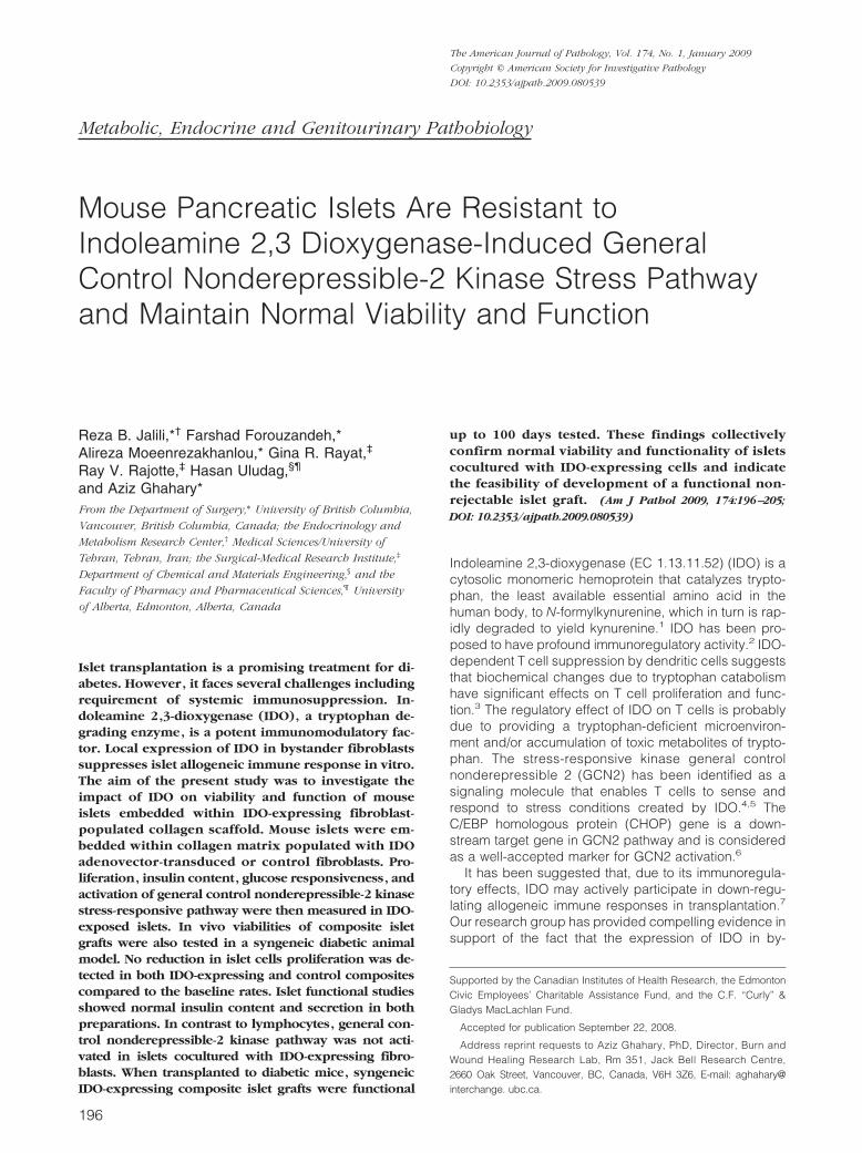
Metabolic, Endocrine and Genitourinary Pathobiology
Mouse Pancreatic Islets Are Resistant toIndoleamine 2,3 Dioxygenase-Induced GeneralControl Nonderepressible-2 Kinase Stress Pathwayand Maintain Normal Viability and Function
Reza B. Jalili,*† Farshad Forouzandeh,*Alireza Moeenrezakhanlou,* Gina R. Rayat,‡
Ray V. Rajotte,‡ Hasan Uludag,§¶
and Aziz Ghahary*From the Department of Surgery,* University of British Columbia,
Vancouver, British Columbia, Canada; the Endocrinology and
Metabolism Research Center,† Medical Sciences/University of
Tehran, Tehran, Iran; the Surgical-Medical Research Institute,‡
Department of Chemical and Materials Engineering,§ and the
Faculty of Pharmacy and Pharmaceutical Sciences,¶ University
of Alberta, Edmonton, Alberta, Canada
Islet transplantation is a promising treatment for di-abetes. However, it faces several challenges includingrequirement of systemic immunosuppression. In-doleamine 2,3-dioxygenase (IDO), a tryptophan de-grading enzyme, is a potent immunomodulatory fac-tor. Local expression of IDO in bystander fibroblastssuppresses islet allogeneic immune response in vitro.The aim of the present study was to investigate theimpact of IDO on viability and function of mouseislets embedded within IDO-expressing fibroblast-populated collagen scaffold. Mouse islets were em-bedded within collagen matrix populated with IDOadenovector-transduced or control fibroblasts. Pro-liferation, insulin content, glucose responsiveness, andactivation of general control nonderepressible-2 kinasestress-responsive pathway were then measured in IDO-exposed islets. In vivo viabilities of composite isletgrafts were also tested in a syngeneic diabetic animalmodel. No reduction in islet cells proliferation was de-tected in both IDO-expressing and control compositescompared to the baseline rates. Islet functional studiesshowed normal insulin content and secretion in bothpreparations. In contrast to lymphocytes, general con-trol nonderepressible-2 kinase pathway was not acti-vated in islets cocultured with IDO-expressing fibro-blasts. When transplanted to diabetic mice, syngeneicIDO-expressing composite islet grafts were functional
up to 100 days tested. These findings collectivelyconfirm normal viability and functionality of isletscocultured with IDO-expressing cells and indicatethe feasibility of development of a functional non-rejectable islet graft. (Am J Pathol 2009, 174:196–205;DOI: 10.2353/ajpath.2009.080539)
Indoleamine 2,3-dioxygenase (EC 1.13.11.52) (IDO) is acytosolic monomeric hemoprotein that catalyzes trypto-phan, the least available essential amino acid in thehuman body, to N-formylkynurenine, which in turn is rap-idly degraded to yield kynurenine.1 IDO has been pro-posed to have profound immunoregulatory activity.2 IDO-dependent T cell suppression by dendritic cells suggeststhat biochemical changes due to tryptophan catabolismhave significant effects on T cell proliferation and func-tion.3 The regulatory effect of IDO on T cells is probablydue to providing a tryptophan-deficient microenviron-ment and/or accumulation of toxic metabolites of trypto-phan. The stress-responsive kinase general controlnonderepressible 2 (GCN2) has been identified as asignaling molecule that enables T cells to sense andrespond to stress conditions created by IDO.4,5 TheC/EBP homologous protein (CHOP) gene is a down-stream target gene in GCN2 pathway and is consideredas a well-accepted marker for GCN2 activation.6
It has been suggested that, due to its immunoregula-tory effects, IDO may actively participate in down-regu-lating allogeneic immune responses in transplantation.7
Our research group has provided compelling evidence insupport of the fact that the expression of IDO in by-
Supported by the Canadian Institutes of Health Research, the EdmontonCivic Employees’ Charitable Assistance Fund, and the C.F. “Curly” &Gladys MacLachlan Fund.
Accepted for publication September 22, 2008.
Address reprint requests to Aziz Ghahary, PhD, Director, Burn andWound Healing Research Lab, Rm 351, Jack Bell Research Centre,2660 Oak Street, Vancouver, BC, Canada, V6H 3Z6, E-mail: aghahary@interchange. ubc.ca.
The American Journal of Pathology, Vol. 174, No. 1, January 2009
Copyright © American Society for Investigative Pathology
DOI: 10.2353/ajpath.2009.080539
196

stander fibroblasts through IDO genetic modification orinterferon-� treatment suppresses immune cell prolifera-tion.8–13 In addition, we also have shown that, by anunknown mechanism, only immune but not primary skincells are sensitive to the IDO suppressive effect.8,14 Inour recent study, we showed that bystander IDO-ex-pressing syngeneic fibroblasts have the ability to sup-press proliferation of lymphocytes stimulated by alloge-neic mouse islets in vitro.15 This promising finding setsthe stage for developing a nonrejectable composite graftconsisting of islets and IDO-expressing fibroblasts em-bedded within a collagen scaffold. However, that studydidn’t elucidate whether IDO by itself has any deleteriouseffect on viability and functionality of islets. Here, wetherefore asked whether a) exposing mouse islets to anIDO-induced low tryptophan microenvironment compro-mises their viability and function, b) the GCN2 pathway isactivated in islets exposed to IDO-expressing cells, andc) a three-dimensional fibroblast populated collagenscaffold is a favorable matrix for constructing a compos-ite islet graft.
Materials and Methods
Mouse Islet Isolation and Culture
Islets were obtained from 6 to 8-week-old male BALB/c orC57BL/6 (B6) mice (The Jackson Laboratories, Bar Har-bor, ME) as previously described.15 Briefly, mice wereanesthetized and pancreases were distended throughthe pancreatic duct with 2.5 ml of Hanks’ balanced saltsolution (Life Technologies, Gaithersburg, MD) contain-ing 2.0 mg/ml of collagenase (Type V; Sigma ChemicalCo., St Louis, MO). The distended pancreases were thenremoved and incubated at 37°C for 15 minutes. The isletswere purified by discontinuous centrifugation on Ficoll(Sigma) gradients. After centrifugation, islets were hand-picked and cultured in HAM’s F10 medium (Sigma) sup-plemented with 12 mmol/L HEPES, 2 mmol/L L-glutamine,10% heat-inactivated fetal calf serum, 100 U/ml penicillin,and 100 �g/ml streptomycin in 95% air, 5% CO2 at 37°C.Care and maintenance of all animals were in accordancewith the principals of laboratory animal care and theguidelines of institutional Animal Policy and WelfareCommittee.
Cell Cocultures
Cell cocultures were set up using a two-chamber cellculture system (6-well plates, Corning incorporated,Corning, NY; cell culture inserts, Millicell, Millipore Cor-poration, MA) in which IDO-expressing fibroblasts weregrown in the upper chambers while either lymphocytes,Jurkat cells, islets, or control fibroblasts were cultured inthe lower chambers. To induce IDO expression, B6mouse fibroblasts were infected with a recombinant IDOadenoviral vector carrying human IDO cDNA for 72 hoursat an multiplicity of infection of 100, as previously de-scribed.15 Control fibroblasts were infected with a mockvector. Lymphocytes were isolated from peripheral lymph
nodes of B6 mice by grinding lymph node tissues be-tween the rough edges of glass slides and were stimu-lated with concanavalin A (2 �g/ml, Sigma) at the start ofthe culture. Specificity of the IDO effect was determinedby addition of 1-methyl-tryptophan, an IDO inhibitor (Al-drich Chemical Co., Milwaukee, WI), to cocultures at thefinal concentration of 800 �mol/L.
Development of Islet-Fibroblast Composite inCollagen Gel Matrix
Mouse dermal fibroblasts were explanted from skin of B6mice and transduced with a recombinant adenoviral vec-tor carrying human IDO cDNA as described previously.15
Control fibroblasts were infected with a mock vector.Fibroblast-populated collagen gel matrices were pre-pared as described by Sarkhosh et al12 using IDO-ex-pressing or control fibroblasts. Mouse islets were addedto fibroblast-populated collagen gel before solidificationin 24 well plates. The composites were maintained in 95%air, 5% CO2 at 37°C for up to 14 days.
CHOP, IDO, and Insulin ReverseTranscriptase-PCR
Total RNA was isolated using a RNeasy kit (Qiagen,Maryland). cDNA was synthesized using the Thermo-Script reverse transcriptase (RT)-PCR System (Invitro-gen, Carlsbad, CA). The primers used were as follows:CHOP: sense 5�-CATACACCACCACACCTGAAAG- 3�,antisense 5�- CCGTTTCCTAGTTCTTCCTTGC-3�; IDO:sense 5�-GGCACACGCTATGGAAAACT-3�, antisense5�-CGGACATCTCCATGACCTTT-3�; mouse insulin 1:sense 5�- CCTGTTGGTGCACTTCCTAC-3�, antisense5�-TGCAGTAGTTCTCCAGCTGG-3�; glyceraldehyde-3-phosphate dehydrogenase: sense 5�-TGGCACAGT-CAAGGCTGAGA-3� antisense 5�-CTTCTGAGTGGCAGT-GATGG-3�. Amplified PCR products were separated by 2%agarose gel electrophoresis and visualized with ethidiumbromide staining.
GCN2 and CHOP Immunoblotting
Cells were harvested and lysed in lysis buffer (50 mmol/LTris-HCl, pH 7.4; 10 mmol/L EDTA; 5 mmol/L EGTA; 0.5%NP40; 1% Triton X-100, and protease inhibitor cocktail,Sigma). Equal amounts of total protein from cell lysates(30 �g) were separated by SDS-polyacrylamide gel elec-trophoresis and transferred to a polyvinylidene difluoridemembrane (Millipore). The blots were probed with thefollowing antibodies: anti phopho-GCN2 (Thr898, 1:1000dilution, Cell Signaling Technology INC., Beverly, MA),anti GCN2 (1:1000 dilution, Cell Signaling), and anti-GADD153/CHOP-10 produced in rabbit (1:250 dilution,Sigma). Horseradish peroxidase conjugated goat anti-rabbit IgG served as a secondary antibody for the en-hanced chemiluminescence detection system (Amer-sham Biosciences, UK).
IDO Effect on Islet Survival and Function 197AJP January 2009, Vol. 174, No. 1

IDO Immunoblotting and KynurenineMeasurement
Fibroblasts were harvested and lysed 72 hours postin-fection. Equal amounts of cell lysate were separated bySDS-polyacrylamide gel electrophoresis and transferredto polyvinylidene difluoride membrane as describedabove. The blots were then probed with polyclonal antiIDO antibody raised in rabbit by Washington Biotechnol-ogy Inc. (Baltimore, MD) at a final dilution of 1:5000.Horseradish peroxidase conjugated goat anti-rabbit IgGserved as a secondary antibody for the enhanced chemi-luminescence detection system (Amersham). The level ofkynurenine was measured as previously described.15
Briefly, proteins in conditioned medium were precipitatedby trichloroacetic acid. After centrifugation, 0.5 ml ofsupernatant was incubated with equal volume of Ehrlich’sreagent for 10 minutes at room temperature. Absorptionof resultant solution was measured at 490 nm byspectrophotometer.
Methyl Thiazolyl Tetrazolium Proliferation Assay
A colorimetric methyl thiazolyl tetrazolium (MTT) [3-(4,5-dimethylthiazol-2-yl)�2,5-diphenyl tetrazolium bromide]assay was used to evaluate the effects of IDO on cellproliferation. At the indicated time points, MTT (Sigma)solution (5 mg/ml) was added to cell cultures and incu-bated at 37°C for 5 hours. The formazin crystals weresolubilized in 500 �l of dimethyl sulfoxide at the end ofincubation, and the optical density of the solutions wasmeasured at 570 nm.
Insulin Immunostaining
Islets were recovered and fixed in Bouin’s solution for 2hours. Islets were then washed three times with 70%ethanol and embedded in paraffin. Five micron sectionsof these samples were stained with guinea pig anti-insulinantibody (1:1000 dilution; Dako Laboratories, Missis-sauga, ON, Canada) for 30 minutes followed by the ad-dition of biotinylated goat anti-guinea pig IgG secondaryantibody (1:200 dilution; Vector Laboratories, Burlin-game, CA). The avidin-biotin complex/horseradish per-oxidase (Vector Laboratories) and 3,3-diaminobenzidi-netetrahydrochloride (BioGenex, San Ramon, CA) wasused to produce a brown positive reaction. All sectionswere counterstained with Harris’ hematoxylin and eosin(H&E).
� Cell Apoptosis
Cleaved caspase-3 was used as a marker for �-cellapoptosis. To estimate � cell apoptosis rates, islets werestained for cleaved caspase-3 together with insulin. � cellapoptosis rates were then estimated by calculating thefrequency of the cells positively stained for both insulinand cleaved caspase-3. Insulin/cleaved caspase-3 dualimmunofluorescence staining was accomplished as fol-lows: islets were retrieved and fixed as described above.
Five-micron sections were incubated overnight at 4°Cwith guinea pig anti insulin antibody (1:500 dilution,Dako) and rabbit anti-cleaved caspase-3 antibody (1:100dilution, cell signaling, Beverly, MA). After three washingsteps with PBS-Tween 20 for 5 minutes each, sampleswere incubated with fluorescein-conjugated donkeyanti-rabbit antibody (1:200 dilution, Jackson Immu-noResearch Laboratories, West Grove, PA) and rhodam-ine-conjugated anti-guinea pig antibody (1:200 dilution,Abcam, Cambridge, MA) for 45 minutes in the dark.Finally, after washing with PBS-Tween 20 three times for5 minutes each, samples were mounted in VectashieldH-1200 (Vector Laboratories) containing 4,6-diamidino-2-phenylindole for nuclei staining. A Zeiss Axioplan 2 mi-croscope and Northern Eclipse image analysis softwarewere used to obtain the images.
Static Incubation Assay and Islet Insulin Content
Islet insulin secretory responsiveness was assessed usinga static incubation assay as described by Korbutt et al.16 Inbrief, mouse islets were washed twice with Ham’s F10medium and samples were taken for measurement oftotal cellular insulin content. Islets were cultured in 24-well plates and incubated in 1.5 ml of HAM’s F10 mediumsupplemented with 0.5% bovine serum albumin and ei-ther 2.8 or 20.0 mmol/L glucose for 120 minutes. At theend of the incubation, supernatants were collected formeasurement of insulin release using radioimmunoassay.Insulin secretion was calculated by dividing the insulinreleased into the supernatant by the cellular insulin con-tent of the islets (percentage of content). Stimulation in-dices were calculated by dividing the percentage of in-sulin released at 20.0 mmol/L glucose by the percentagereleased at 2.8 mmol/L glucose.
Determination of islet total insulin and DNA contentwas performed as described by Korbutt et al.16 In brief,samples were sonicated in 2 mmol/L acetic acid contain-ing 0.25% bovine serum albumin and centrifuged (800 � g,15 minutes). Insulin levels in the supernatants weremeasured in duplicate samples by radioimmunoassay(Diagnostic Products, Los Angeles, CA). The islet DNAcontent was quantified using PicoGreen kit (MolecularProbes, Eugene, OR) according to the manufacturer’sinstruction.
Transplantation of Islet-Fibroblast CompositeGrafts
Recipient B6 mice were rendered diabetic by a singleintraperitoneal injection of 200 mg/kg streptozotocin(Sigma), and diabetes was defined as a minimum of twoconsecutive blood glucose measurements � � 20mmol/L. Syngeneic islet plus IDO-expressing or controlfibroblast composite grafts (approximately 500 islets)were transplanted under the left kidney capsule of isoflu-rane-anesthetized diabetic mice. After transplantation,blood from the tail vein of each recipient was collectedtwo times a week between 7:00 and 9:00 AM to determinethe normalization of blood glucose levels. Blood glucose
198 Jalili et alAJP January 2009, Vol. 174, No. 1

levels were measured using a One Touch Ultra glucosemeter (Lifescan, Milpitas, CA), and grafts were deemedfunctioning when blood glucose levels decreased to�10.0 mmol/L. Nephrectomy of the graft-bearing kidneywas performed on recipients at the endpoint of the study(�100 days post-transplant) to confirm that hyperglyce-mia ensued, indicating that normal blood glucose wasgraft dependent. All animals were cared for according tothe guidelines of the Institutional Animal Policy and Wel-fare Committee.
Intraperitoneal Glucose Tolerance Test
An intraperitoneal glucose tolerance test (IPGTT) wasperformed in mice transplanted with composite grafts 6weeks after transplantation. After a 16-hour overnightfast, glucose (2 mg/g body weight) was injected i.p. intononanesthetized mice. Blood samples were obtainedfrom the tail vein at 0, 15, 30, 60, and 120 minutes. Areaunder the curve was determined using SigmaPlot soft-ware (Systat Software Inc., San Jose, CA).
Statistical Analysis
All data are reported as mean � SD of three or moreindependent observations. Statistical significance wascalculated using a two-tailed unpaired Students’ t-test ora one-way analysis of variance with post hoc test in caseof multiple comparisons. P values less than 0.05 wereconsidered to be significant.
Results
Selective Suppressive Effect of IDO on ImmuneVersus Islet Cells
To confirm the differential suppressive effect of IDO onimmune versus non-immune cells, we induced IDO ex-pression in B6 mouse dermal fibroblasts using a recom-binant adenoviral vector expressing IDO. Expression ofIDO protein in infected cells was shown using Westernblot analysis (Figure 1A). High levels of kynurenine—atryptophan metabolite—in conditioned media of IDO vec-tor infected cells further confirmed enzymatic activity ofIDO (Figure 1B). IDO-expressing fibroblasts were thencocultured with stimulated mouse lymphocytes, CD4�
Jurkat cells, mouse fibroblasts, or islets using a two-chamber coculture system. The results of a MTT assay oncocultured cells after 72 hours showed significant reduc-tion in cell proliferation rates in lymphocytes (23.9% �12.4) and Jurkat cells (21.8% � 10.7) but not in fibro-blasts and islets (Figure 1C). Proliferation of immune cellsdid not decrease when cocultured with control fibro-blasts. Furthermore, addition of 1-methyl-tryptophan, aspecific IDO inhibitor, resulted in partial recovery of im-mune cells proliferation (68.5% �14.3 in lymphocytesand 69.4 � 15.3 in Jurkat cells, Figure 1C).
GCN2 Kinase Pathway Activation in CellsExposed to IDO-Expressing Fibroblasts
The activation of the GCN2 kinase pathway is suggestedas the downstream mechanism for the suppressive effectof IDO. We therefore asked whether this stress-responsemechanism is selectively activated in different cell strainswhen cocultured with IDO-expressing cells. To addressthis question, we examined phosphorylation of GCN2and induction of intracellular CHOP in stimulated lympho-cytes, fibroblasts, and islets cocultured with IDO-ex-pressing fibroblasts. The results shown in the Figure 2indicate a differential pattern of GCN2 pathway activationin immune versus islet cells in response to IDO. As shownin the Figure 2A, coculture with IDO-expressing cellspromotes phosphorylation of GCN2 in mouse lympho-cytes but not in islets and fibroblasts. Similarly, on expo-sure to IDO, CHOP was induced only in mouse lympho-cytes at mRNA (Figure 2C) and protein (Figure 2E) levels.The quantitative analyses shown in Figure 2, B, D, and Findicated a significant and selective increase (more thansevenfold) in GCN2 phosphorylation and CHOP mes-sage and protein levels in lymphocytes cocultured with
Figure 1. Selective suppressive effect of IDO on immune versus non im-mune cells. Mouse fibroblasts were transduced to express IDO using anadenoviral vector. A: IDO protein expression in mock and IDO vectorinfected cells. The level of kynurenine (the product of IDO mediated tryp-tophan degradation) was measured in the conditioned media (B). C: Stimu-lated mouse lymphocytes, Jurkat cells, mouse fibroblasts, and islets werecocultured in two-chamber culture plates with IDO-expressing (open bars)or control fibroblasts (solid bars) for 72 hours. A competitive IDO inhibitor,1-methyl-tryptophan was added to one set of IDO-expressing cocultures(hatched bars). Cell proliferation rates were measured after 72 hours post-coculture using MTT assay. * denotes significant increase in kynurenine levelin IDO vector infected cell conditioned medium compared to the controlgroup (n � 3, P � 0.001); ** denotes significant difference in cell proliferationrate in comparison to the control group (n � 5, P � 0.001).
IDO Effect on Islet Survival and Function 199AJP January 2009, Vol. 174, No. 1

IDO-expressing cells that is partially reversed on IDOinhibition with 1-methyl-tryptophan.
Viability of Islets Embedded within IDO-Expressing Fibroblast Populated CollagenMatrix
Embedding islets within an extracellular matrix generallyimproves islet survival and function. However, the impactof local IDO expression on enhanced viability of isletswithin three-dimensional matrix has not been elucidated.To investigate the effect of IDO on survival of islets withina collagen gel scaffold, we prepared three-dimensionalcomposite cocultures embedded with mouse islets pluseither IDO-expressing or control fibroblasts. These com-posites were then incubated in vitro for up to 2 weeks. Ondays 1, 7, and 14 post-coculture, the collagen matriceswere digested and islets were retrieved and subjected toapoptosis and MTT assays. In parallel, one set of mouseislets was cultivated in a regular two-dimensional settingas a control.
Islet double-immunofluorescence staining of cleavedcaspase-3 and insulin was used to estimate �-cell apo-ptosis rates. As shown in the Figure 3A–E, � cell apopto-sis rates were significantly lower in matrix embeddedislets and furthermore, being in close vicinity of IDO-expressing fibroblasts for 2 weeks did not increase �-cellapoptosis rate (5.3% � 1.5 vs. 4.1% � 1.2, P � 0.05). TheMTT assay result indicated that, in contrast to two-dimen-sional culture, the islet cell proliferation rate was main-tained near baseline level when embedded within thecollagen matrix, regardless of being either alone, orcocultured with IDO-expressing or control fibroblasts.Additionally, MTT assays confirmed IDO did not signifi-
cantly decrease the islet cell proliferation rate in compos-ites containing IDO-expressing cells (96.8% � 5.3) com-pared with control composites (99.2% � 7.1; P � 0.05,Figure 3F).
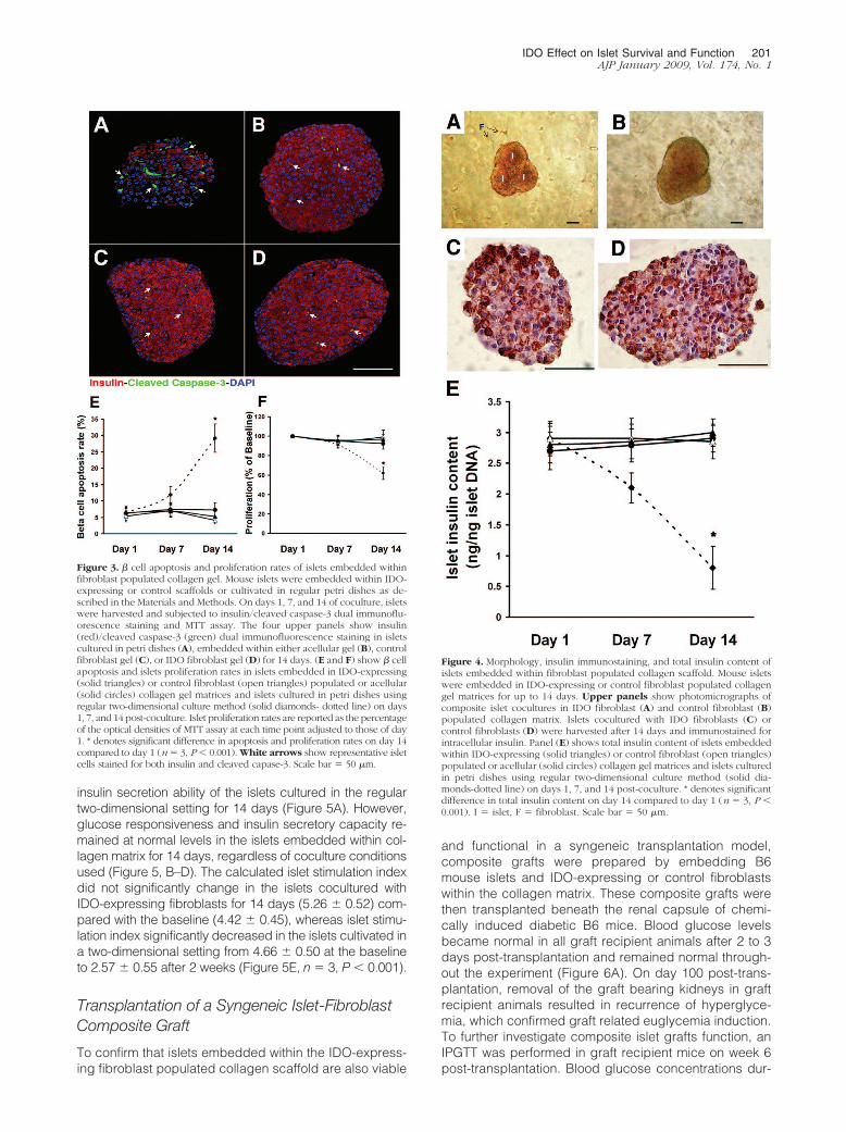
Morphology, Insulin Content and FunctionalCapacity of Islets Embedded within an IDO-Expressing Fibroblasts Populated CollagenMatrix
To gain perspective on the effect of IDO on islet function-ality, the islets that were embedded within the three-dimensional composites were retrieved on days 1, 7, and14 post-coculture and subjected to glucose-stimulatedinsulin secretion assay and insulin immunostaining. Totalinsulin content of islets was also measured. Figure 4, A–Dshows photomicrographs of the composite islet-fibro-blast-collagen matrix preparations, indicating normalspherical morphology of islets with smooth borders (Fig-ure 4, A and B). Insulin immunostaining confirmed thatthe insulin content of islets exposed to IDO-expressingfibroblasts within a collagen scaffold for 2 weeks (Figure4C) is comparable with islets cocultured with controlfibroblasts under similar experimental condition (Figure4D). Furthermore, total insulin content of islets exposed toIDO-expressing fibroblasts for 2 weeks was 3 � 0.22ng/ng islet DNA, which was not significantly different frominsulin content of control islets (2.85 � 0.28 ng/ng isletDNA, Figure 4E).
The insulin secretory capacity of islets was tested bycomparing the percentages of cellular insulin released inlow glucose (2.8 mmol/L) versus high glucose (20 mmol/L)media. The result showed a significant decrease in the
Figure 2. Selective activation of GCN2 andCHOP expression in immune cells as comparedto islets and fibroblasts. Stimulated mouse lym-phocytes, fibroblasts, or islets were coculturedwith IDO-expressing or control fibroblasts for 48hours. The phosphorylation of GCN2 and induc-tion of CHOP was then measured. A competitiveIDO inhibitor, 1-methyl-tryptophan was addedto one set of IDO-expressing cocultures. A:phopho-GCN2 (upper row) and total GCN2(middle row) Western blot. C: CHOP RT-PCRresult. E: result of Western blot analysis forCHOP. Upper arrow shows a 29 kDa bandcorresponding to CHOP. B, D and F: the meanratio of densities of phospho-GCN2, CHOP mes-sage, and protein bands to those of total GCN2,glyceraldehyde-3-phosphate dehydrogenase-1,and �-actin bands, respectively, in cells cocul-tured with either IDO expressing (solid bars),IDO expressing plus 1-methyl-tryptophan(hatched bars), or control (open bars) fibro-blasts. * denotes significant difference in phos-pho-GCN2 and CHOP level between the IDOexposed and the control lymphocytes (n � 3,P � 0.001). P-GCN2: phosphorylated GCN2.
200 Jalili et alAJP January 2009, Vol. 174, No. 1
insulin secretion ability of the islets cultured in the regulartwo-dimensional setting for 14 days (Figure 5A). However,glucose responsiveness and insulin secretory capacity re-mained at normal levels in the islets embedded within col-lagen matrix for 14 days, regardless of coculture conditionsused (Figure 5, B–D). The calculated islet stimulation indexdid not significantly change in the islets cocultured withIDO-expressing fibroblasts for 14 days (5.26 � 0.52) com-pared with the baseline (4.42 � 0.45), whereas islet stimu-lation index significantly decreased in the islets cultivated ina two-dimensional setting from 4.66 � 0.50 at the baselineto 2.57 � 0.55 after 2 weeks (Figure 5E, n � 3, P � 0.001).
Transplantation of a Syngeneic Islet-FibroblastComposite Graft
To confirm that islets embedded within the IDO-express-ing fibroblast populated collagen scaffold are also viable
and functional in a syngeneic transplantation model,composite grafts were prepared by embedding B6mouse islets and IDO-expressing or control fibroblastswithin the collagen matrix. These composite grafts werethen transplanted beneath the renal capsule of chemi-cally induced diabetic B6 mice. Blood glucose levelsbecame normal in all graft recipient animals after 2 to 3days post-transplantation and remained normal through-out the experiment (Figure 6A). On day 100 post-trans-plantation, removal of the graft bearing kidneys in graftrecipient animals resulted in recurrence of hyperglyce-mia, which confirmed graft related euglycemia induction.To further investigate composite islet grafts function, anIPGTT was performed in graft recipient mice on week 6post-transplantation. Blood glucose concentrations dur-
Figure 3. � cell apoptosis and proliferation rates of islets embedded withinfibroblast populated collagen gel. Mouse islets were embedded within IDO-expressing or control scaffolds or cultivated in regular petri dishes as de-scribed in the Materials and Methods. On days 1, 7, and 14 of coculture, isletswere harvested and subjected to insulin/cleaved caspase-3 dual immunoflu-orescence staining and MTT assay. The four upper panels show insulin(red)/cleaved caspase-3 (green) dual immunofluorescence staining in isletscultured in petri dishes (A), embedded within either acellular gel (B), controlfibroblast gel (C), or IDO fibroblast gel (D) for 14 days. (E and F) show � cellapoptosis and islets proliferation rates in islets embedded in IDO-expressing(solid triangles) or control fibroblast (open triangles) populated or acellular(solid circles) collagen gel matrices and islets cultured in petri dishes usingregular two-dimensional culture method (solid diamonds- dotted line) on days1, 7, and 14 post-coculture. Islet proliferation rates are reported as the percentageof the optical densities of MTT assay at each time point adjusted to those of day1. * denotes significant difference in apoptosis and proliferation rates on day 14compared to day 1 (n � 3, P � 0.001). White arrows show representative isletcells stained for both insulin and cleaved capase-3. Scale bar � 50 �m.
Figure 4. Morphology, insulin immunostaining, and total insulin content ofislets embedded within fibroblast populated collagen scaffold. Mouse isletswere embedded in IDO-expressing or control fibroblast populated collagengel matrices for up to 14 days. Upper panels show photomicrographs ofcomposite islet cocultures in IDO fibroblast (A) and control fibroblast (B)populated collagen matrix. Islets cocultured with IDO fibroblasts (C) orcontrol fibroblasts (D) were harvested after 14 days and immunostained forintracellular insulin. Panel (E) shows total insulin content of islets embeddedwithin IDO-expressing (solid triangles) or control fibroblast (open triangles)populated or acellular (solid circles) collagen gel matrices and islets culturedin petri dishes using regular two-dimensional culture method (solid dia-monds-dotted line) on days 1, 7, and 14 post-coculture. * denotes significantdifference in total insulin content on day 14 compared to day 1 (n � 3, P �0.001). I � islet, F � fibroblast. Scale bar � 50 �m.
IDO Effect on Islet Survival and Function 201AJP January 2009, Vol. 174, No. 1

ing IPGTT were similar in IDO composite graft recipientsand controls (Figure 6B). Comparing the area under theIPGTT curve showed similar graft function in IDO groupcompared to the control group (1401 � 120 (mmol/L) minvs. 1430 � 135 (mmol/L) min, respectively; P � 0.05,Figure 6C).
To examine the length of adenovirus-mediated IDOtransgene expression in the composite grafts, IDO mRNAlevel was measured in the grafts using RT-PCR at differ-ent time points post-transplantation. As shown in Figure6D, IDO is strongly expressed in the IDO compositegrafts for up to 6 weeks after transplantation. High levelsof insulin 1 expression were also detected in the graftsthroughout the experiment (Figure 6D), which further con-firmed maintenance of functional islets in the compositegrafts during and after the IDO transgene expressionperiod. These findings collectively indicate long term sur-
vival and normal functionality of islets exposed to IDO-expressing cells in a syngeneic islet transplantationmodel.
Discussion
In this study, we report that the viability and functionalityof islets exposed to IDO-expressing fibroblasts are notcompromised in vitro and in vivo, using different experi-mental approaches. The significance of this finding ismore appreciated when the potential application of localexpression of IDO is considered as a strategy for pro-tecting islet grafts from immune rejection. IDO is a potentimmunomodulatory enzyme that plays critical roles inregulation of T cell-mediated immune responses and hasprofound effects on T cell proliferation, differentiation,
Figure 5. Capacity of glucose-mediated insulin secretion in islets embeddedwithin fibroblast populated collagen matrix. Mouse islets were embeddedwithin IDO-expressing or control scaffolds or in regular petri dishes asdescribed in the Materials and Methods. On days 1, 7, and 14 of coculture,islets were harvested and subjected to static incubation assay to test theirglucose-stimulated insulin secretory capacity at low (2.8 mmol/L, open bars)versus high (20.0 mmol/L, solid bars) glucose concentrations. Insulin releaserates were measured in islets cultured in petri dishes (A), embedded withineither acellular gel (B), control fibroblast gel (C), or IDO fibroblast gel (D).The data are reported as the percentage of the released insulin to total insulincontent of the islets. (E) shows islet stimulation indices, which were calcu-lated by dividing the percentage of insulin released at high glucose by thepercentage released at low glucose concentrations. Solid diamonds-dottedline, islets cultured in regular petri dishes; solid circles, islets embeddedwithin acellular gel; open triangles, islets embedded within control fibroblastgel; solid triangles, islets embedded within IDO fibroblast gel. * denotessignificant decrease in islet stimulation index on day 14 vs. day 1 (n � 3, P �0.001).
Figure 6. Syngeneic islet-fibroblast composite graft survival and intragraftIDO transgene expression in vivo. A: B6 mouse islets were embedded withinIDO-expressing (solid line) or control fibroblast (dotted line) populatedcollagen matrices and transplanted to the kidney subcapsular spaces ofchemically induced diabetic B6 mice (3 mice per group). Blood glucoselevels of graft recipient animals were measured twice a week for 100 days.On day 100 post-transplantation (black arrow), graft bearing kidneys wereremoved, which resulted in recurrence of hyperglycemia (A). IPGTT wasperformed in graft recipients on week 6 post-transplantation. (B) and (C)show blood glucose concentrations during IPGTT and the area under theIPGTT curve, respectively. (D) shows result of intragraft insulin 1 (upperrow) and IDO RT-PCR (middle row) at the end of weeks 2, 4, 6, 8, and 14post-transplantation.
202 Jalili et alAJP January 2009, Vol. 174, No. 1

effector functions and viability.3 Considerable evidencenow supports the importance of the immunoregulatoryfunction of IDO, including studies of mammalian preg-nancy,17–19 tumor resistance,20 –23 chronic infec-tions24–26 and autoimmune diseases.27 Based on thesefacts, it has been suggested that cells expressing IDOmight be used to protect transplanted tissues and cellswithout the use of immunosuppressive drugs. We haverecently introduced a model of a local immunosuppres-sive barrier to protect immune responses against islets inwhich syngeneic bystander IDO-expressing fibroblastssuppress lymphocyte proliferation induced by allogeneicmouse islets.15 Our model is based on development of athree-dimensional composite graft consisting of isletsand IDO-expressing fibroblasts embedded within a col-lagen matrix.
Although the selective suppressive effect of IDO on Tcells versus skin cells has already been noted,8,14 therewas no evidence on the impact of low tryptophan micro-environment induced by IDO on other cell types includingislets. We therefore, as shown in the Figures 1 and 3,tested the proliferation rates of mouse islets and fibro-blasts when cocultured with IDO-expressing cells. Theresults of these experiments confirmed that exposure toIDO-expressing cells did not decrease islet cell prolifer-ation or increase the � cell apoptosis rate in vitro.
The molecular mechanism(s) by which IDO sup-presses T cells are still being investigated. It appears thatsome of the biological effects of IDO can be mediated vialocal depletion of tryptophan,28,29 whereas others aremediated via immunomodulatory tryptophan metabo-lites.30,31 As mentioned earlier, activation of the GCN2kinase pathway, a nutrient deficiency stress-responsivemechanism, has been suggested as a potential mecha-nism responsible for the IDO-induced suppressive effecton T cells.4,5 Activation of the GCN2 kinase pathwaycan trigger cell-cycle arrest, differentiation, compensa-tory adaptation, or apoptosis, depending on the celltype and the initiating stress.32,33 CHOP (also known asGADD153) is a DNA damage-inducible, nuclear leucinezipper protein involved in differentiation and apoptosis.CHOP is a downstream target gene in GCN2 pathwayand a well-accepted marker for GCN2 activation.6 Itsexpression is induced in a variety of stress responsessuch as endoplasmic reticulum stress,34 redox stress,35
and nutrient deprivation.36,37 Functional roles for CHOPhave been well described in induction of apoptosis, andit has also been implicated in the pathogenesis of diabe-tes by promoting � cell destruction.38 This study for thefirst time showed that GCN2 activation and CHOP ex-pression did not occur in mouse islets in response to IDOexposure whereas GCN2-mediated CHOP expression in-creased more than sevenfold in mouse lymphocytes un-der similar experimental condition. Thus, the selectiveunresponsiveness of mouse islets to IDO-induced GCN2activation and CHOP expression suggests that islet cellsare IDO resistant due to lack of GCN2 pathway respon-siveness to IDO-induced low tryptophan environment.
Although the mechanism(s) underlying selectiveGCN2-CHOP pathway activation in immune versus isletcells needs to be further elucidated, at the present time,
there are, at least, three potential explanations. First,activation of the GCN2 pathway is a nutrient deficiencystress response, and therefore actively dividing cells in-cluding highly proliferating T-cells, but not non-dividingislet cells, would be more sensitive to this environment. Infact, it has been shown that activated and proliferatingT-cells, but not resting T cells, cultured in IDO-inducedlow tryptophan environment rapidly undergo apoptosis.39
Moreover, Mellor et al showed that suppression of T-cellproliferation in the presence of IDO-expressing cells oc-curred after the majority of T-cells entered the cell prolif-eration cycle.40 Second, there may be a compensatorymechanism by which islet cells suppress IDO mediatedGCN2 activation. This suggestion is based on the factthat Pereira et al recently showed that the mouse proteinIMPACT, which binds GCN1 and inhibits GCN2 activa-tion, abolishes the expression of its downstream targetgenes ATF4 and CHOP.41 It should be emphasize thatIMPACT is highly expressed in IDO-expressing tissues(eg, placenta and testis) and also in pancreas.42 Assuch, the expression of IMPACT in pancreatic islet cellsmight function as a protective mechanism against IDO-induced GCN2 activation. Finally, a different and highaffinity transport system for tryptophan may exist in IDOresistant cells such as islet cells. Recently, a novel aminoacid transport activity with high affinity and unusual se-lectivity for tryptophan has been described in IDO-ex-pressing human monocyte-derived macrophages, whichis up-regulated during monocyte-derived macrophagedifferentiation but not in T-cells.43 This selective transportsystem, if it exists in other IDO resistant cell types, canenhance tryptophan uptake in these cells and thereforeovercome nutrient deficiency stress response in a lowtryptophan environment.
The collagen matrix in our composite grafts plays adual role since it works both as a scaffold to keep isletsand fibroblasts together and therefore maintains the in-tegrity of the graft and also works as an extracellularmatrix, which improves the survival and function of islets.Extracellular matrix is one of the most important constit-uents of the islet microenvironment. It is well documentedthat when interrelationship between islets and extracellu-lar matrix is disrupted following harsh process of isletisolation and purification, the function and survival ofisolated islets are significantly compromised.44,45 It wasalso noted that islets cultivated in regular two-dimen-sional setting in petri dishes gradually disintegrate duringculture in a time dependent manner.46 Therefore, it hasbeen suggested that entrapment of islets in a three-dimensional collagen matrix would maintain satisfactorymorphology, viability, and glucose-induced insulin secre-tory capability in islets.47 Our findings regarding improve-ment of cell survival and glucose stimulated insulin se-cretory capacity of the islets embedded within a collagenmatrix, regardless of coculture conditions, are in concor-dance with previous studies48–50 and confirm that em-bedding islets in a scaffold consisting collagen will main-tain integrity of islets and improve their viability andfunction.
Fibroblasts cocultured with islets in our proposed com-posite model can also improve islet cell viability. The
IDO Effect on Islet Survival and Function 203AJP January 2009, Vol. 174, No. 1

essential role of islet-derived fibroblasts in islet physio-logical competence was previously reported, and it wasshown that some fibroblast produced factors can pro-mote islet survival in culture.51 Miki et al recently showedthat coculture of the mouse, rat, and pig islets with islet-derived fibroblasts maintained islet viability, insulin se-cretion and glucose responsiveness.52 Cocultured fi-broblast would not cause any concern in terms ofoverproliferation since it was shown that fibroblasts infloating collagen matrices have low levels of DNA syn-thesis and become quiescent.53–55 Thus, bystander fi-broblasts will not have any negative impact on islet via-bility and function.
The findings shown in the Figure 6 further confirm thelong term survival and functionality of syngeneic isletswithin the composite grafts in a diabetic animal model.The IDO transgene was strongly expressed in the com-posite grafts for up to 6 weeks post-transplantation, asexpected following gene transfer using adenoviral vec-tors. The level of insulin mRNA was high in the IDO graftsthroughout the experimental period. These data collec-tively demonstrate that none of key elements in our com-posite islet graft including IDO enzymatic activity, fibro-blasts, and collagen matrix has a negative impact on longterm viability and function of transplanted islets.
In conclusion, the findings of the present study sug-gest that mouse islets are selectively resistant to IDO-mediated activation of GCN2 kinase stress pathway andthat coculture of mouse islets in a three-dimensional IDO-expressing fibroblast populated collagen matrix does nothave any deleterious effect on viability and insulin secre-tory capacity of islets in vitro and in vivo. These findingstogether with the already proven immunosuppressive ef-fect of IDO set the stage for developing a nonrejectableislet graft in the future.
Acknowledgments
We are grateful to Dr. Yunyuan Li (University of Alberta,Edmonton, Alberta, Canada) for constructing the IDOadenoviral vector.
References
1. Muller AJ, Prendergast GC: Indoleamine 2,3-dioxygenase in immunesuppression and cancer. Curr Cancer Drug Targets 2007, 7:31–40
2. Munn DH, Sharma MD, Lee JR, Jhaver KG, Johnson TS, Keskin DB,Marshall B, Chandler P, Antonia SJ, Burgess R, Slingluff CL, Jr., MellorAL: Potential regulatory function of human dendritic cells expressingindoleamine 2,3-dioxygenase. Science 2002, 297:1867–1870
3. Mellor AL, Munn DH: IDO expression by dendritic cells: Toleranceand tryptophan catabolism. Nat Rev Immunol 2004, 4:762–774
4. Fallarino F, Grohmann U, You S, McGrath BC, Cavener DR, Vacca C,Orabona C, Bianchi R, Belladonna ML, Volpi C, Santamaria P, FiorettiMC, Puccetti P: The combined effects of tryptophan starvation andtryptophan catabolites down-regulate T cell receptor {zeta}-chain andinduce a regulatory phenotype in naive T cells. J Immunol 2006,176:6752–6761
5. Munn DH, Sharma MD, Baban B, Harding HP, Zhang Y, Ron D, MellorAL: GCN2 kinase in T cells mediates proliferative arrest and anergyinduction in response to indoleamine 2,3-dioxygenase. Immunity2005, 22:633–642
6. Harding HP, Novoa I, Zhang Y, Zeng H, Wek R, Schapira M, Ron D:Regulated translation initiation controls stress-induced gene expres-sion in mammalian cells. Mol Cell 2000, 6:1099–1108
7. Hainz U, Jurgens B, Heitger A: The role of indoleamine 2,3-dioxyge-nase in transplantation. Transpl Int 2007, 20:118–127
8. Ghahary A, Li Y, Tredget EE, Kilani RT, Iwashina T, Karami A, Lin X:Expression of indoleamine 2,3-dioxygenase in dermal fibroblastsfunctions as a local immunosuppressive factor. J Invest Dermatol2004, 122:953–964
9. Li Y, Tredget EE, Ghahary A: Cell surface expression of MHC class Iantigen is suppressed in indoleamine 2,3-dioxygenase geneticallymodified keratinocytes: implications in allogeneic skin substitute en-graftment. Hum Immunol 2004, 65:114–123
10. Li Y, Tredget EE, Ghaffari A, Lin X, Kilani RT, Ghahary A: Localexpression of indoleamine 2,3-dioxygenase protects engraftment ofxenogeneic skin substitute. J Invest Dermatol 2006, 126:128–136
11. Sarkhosh K, Tredget EE, Li Y, Kilani RT, Uludag H, Ghahary A:Proliferation of peripheral blood mononuclear cells is suppressed bythe indoleamine 2,3-dioxygenase expression of interferon-gamma-treated skin cells in a co-culture system. Wound Repair Regen 2003,11:337–345
12. Sarkhosh K, Tredget EE, Karami A, Uludag H, Iwashina T, Kilani RT,Ghahary A: Immune cell proliferation is suppressed by the interferon-gamma-induced indoleamine 2,3-dioxygenase expression of fibro-blasts populated in collagen gel (FPCG). J Cell Biochem 2003,90:206–217
13. Sarkhosh K, Tredget EE, Uludag H, Kilani RT, Karami A, Li Y,Iwashina T, Ghahary A: Temperature-sensitive polymer-conjugatedIFN-gamma induces the expression of IDO mRNA and activity byfibroblasts populated in collagen gel (FPCG). J Cell Physiol 2004,201:146–154
14. Forouzandeh F, Jalili RB, Germain M, Duronio V, Ghahary A: Skincells, but not T cells, are resistant to indoleamine 2, 3-dioxygenase(IDO) expressed by allogeneic fibroblasts. Wound Repair Regen2008, 16:379–387
15. Jalili RB, Rayat GR, Rajotte RV, Ghahary A: Suppression of isletallogeneic immune response by indoleamine 2,3 dioxygenase-expressing fibroblasts. J Cell Physiol 2007, 213:137–143
16. Korbutt GS, Elliott JF, Ao Z, Smith DK, Warnock GL, Rajotte AR: Largescale isolation, growth, and function of porcine neonatal islet cells.J Clin Invest 1996, 97:2119–2129
17. Kudo Y, Boyd CA: The physiology of immune evasion during preg-nancy; the critical role of placental tryptophan metabolism and trans-port. Pflugers Arch 2001, 442:639–641
18. Mellor AL, Chandler P, Lee GK, Johnson T, Keskin DB, Lee J, MunnDH: Indoleamine 2,3-dioxygenase, immunosuppression, and preg-nancy. J Reprod Immunol 2002, 57:143–150
19. Munn DH, Zhou M, Attwood JT, Bondarev I, Conway SJ, Marshall B,Brown C, Mellor AL: Prevention of allogeneic fetal rejection by tryp-tophan catabolism. Science 1998, 281:1191–1193
20. Gajewski TF, Meng Y, Harlin H: Immune suppression in the tumormicroenvironment. J Immunother 2006, 29:233–240
21. Munn DH, Sharma MD, Hou D, Baban B, Lee JR, Antonia SJ, MessinaJL, Chandler P, Koni PA, Mellor AL: Expression of indoleamine 2,3-dioxygenase by plasmacytoid dendritic cells in tumor-draining lymphnodes. J Clin Invest 2004, 114:280–290
22. Munn DH: Indoleamine 2,3-dioxygenase, tumor-induced toleranceand counter-regulation. Curr Opin Immunol 2006, 18:220–225
23. Uyttenhove C, Pilotte L, Theate I, Stroobant V, Colau D, Parmentier N,Boon T, Van den Eynde BJ: Evidence for a tumoral immune resis-tance mechanism based on tryptophan degradation by indoleamine2,3-dioxygenase. Nat Med 2003, 9:1269–1274
24. Adams O, Besken K, Oberdorfer C, MacKenzie CR, Takikawa O,Daubener W: Role of indoleamine-2,3-dioxygenase in alpha/beta andgamma interferon-mediated antiviral effects against herpes simplexvirus infections. J Virol 2004, 78:2632–2636
25. Sakash JB, Byrne GI, Lichtman A, Libby P: Cytokines induce in-doleamine 2,3-dioxygenase expression in human atheroma-associ-ated cells: Implications for persistent Chlamydophila pneumoniaeinfection. Infect Immun 2002, 70:3959–3961
26. Silva NM, Rodrigues CV, Santoro MM, Reis LF, varez-Leite JI,Gazzinelli RT: Expression of indoleamine 2,3-dioxygenase, trypto-phan degradation, and kynurenine formation during in vivo infectionwith Toxoplasma gondii: Induction by endogenous gamma interferon
204 Jalili et alAJP January 2009, Vol. 174, No. 1
and requirement of interferon regulatory factor 1. Infect Immun 2002,70:859–868
27. Brown RR, Ozaki Y, Datta SP, Borden EC, Sondel PM, Malone DG:Implications of interferon-induced tryptophan catabolism in cancer,auto-immune diseases and AIDS. Adv Exp Med Biol 1991,294:425–435
28. Beatty WL, Belanger TA, Desai AA, Morrison RP, Byrne GI: Trypto-phan depletion as a mechanism of gamma interferon-mediated chla-mydial persistence. Infect Immun 1994, 62:3705–3711
29. Swanson KA, Zheng Y, Heidler KM, Mizobuchi T, and Wilkes DS:CDllc� cells modulate pulmonary immune responses by productionof indoleamine 2,3-dioxygenase. Am J Respir Cell Mol Biol 2004,30:311–318
30. Frumento G, Rotondo R, Tonetti M, Damonte G, Benatti U, FerraraGB: Tryptophan-derived catabolites are responsible for inhibition of Tand natural killer cell proliferation induced by indoleamine 2,3-dioxy-genase. J Exp Med 2002, 196:459–468
31. Terness P, Bauer TM, Rose L, Dufter C, Watzlik A, Simon H, Opelz G:Inhibition of allogeneic T cell proliferation by indoleamine 2,3-dioxy-genase-expressing dendritic cells: Mediation of suppression by tryp-tophan metabolites. J Exp Med 2002, 196:447–457
32. Anthony TG, McDaniel BJ, Byerley RL, McGrath BC, Cavener DR,McNurlan MA, Wek RC: Preservation of liver protein synthesis duringdietary leucine deprivation occurs at the expense of skeletal musclemass in mice deleted for eIF2 kinase GCN2. J Biol Chem 2004,279:36553–36561
33. Crosby JS, Chefalo PJ, Yeh I, Ying S, London IM, Leboulch P, ChenJJ: Regulation of hemoglobin synthesis and proliferation of differen-tiating erythroid cells by heme-regulated eIF-2alpha kinase. Blood2000, 96:3241–3248
34. Yang L, Carlson SG, McBurney D, Horton WE, Jr: Multiple signalsinduce endoplasmic reticulum stress in both primary and immortal-ized chondrocytes resulting in loss of differentiation, impaired cellgrowth, and apoptosis. J Biol Chem 2005, 280:31156–31165
35. Tang JR, Nakamura M, Okura T, Takata Y, Watanabe S, Yang ZH, LiuJ, Kitami Y, Hiwada K: Mechanism of oxidative stress-inducedGADD153 gene expression in vascular smooth muscle cells. Bio-chem Biophys Res Comm 2002, 290:1255–1259
36. Averous J, Bruhat A, Jousse C, Carraro V, Thiel G, Fafournoux P:Induction of CHOP expression by amino acid limitation requires bothATF4 expression and ATF2 phosphorylation. J Biol Chem 2004,279:5288–5297
37. Friedman AD: GADD153/CHOP, a DNA damage-inducible proteinreduced CAAT/enhancer binding protein activities and increasedapoptosis in 32D cl3 myeloid cells. Cancer Res 1996, 56:3250–3256
38. Lawrence MC, McGlynn K, Naziruddin B, Levy MF, Cobb MH: Inau-gural Article: differential regulation of CHOP-10/GADD153 gene ex-pression by MAPK signaling in pancreatic beta-cells. Proc Natl AcadSci USA 2007, 104:11518–11525
39. Plumas J, Chaperot L, Richard MJ, Molens JP, Bensa JC, Favrot MC:Mesenchymal stem cells induce apoptosis of activated T cells. Leu-kemia 2005, 19:1597–1604
40. Mellor AL, Keskin DB, Johnson T, Chandler P, Munn DH: Cells ex-pressing indoleamine 2,3-dioxygenase inhibit T cell responses. J Im-munol 2002, 168:3771–3776
41. Pereira CM, Sattlegger E, Jiang HY, Longo BM, Jaqueta CB,Hinnebusch AG, Ronald C, Wek LE, Mello AM, Castilho, BA: IMPACT,a protein preferentially expressed in the mouse brain, binds GCN1and inhibits GCN2 activation. J Biol Chem 2005, 280:28316–28323
42. Okamura K, Hagiwara-Takeuchi Y, Li T, Vu TH, Hirai M, Hattori M,Sakaki Y, Hoffman AR, Ito T: Comparative genome analysis of themouse imprinted gene impact and its nonimprinted human homologIMPACT: toward the structural basis for species-specific imprinting.Genome Res 2000, 10:1878–1889
43. Seymour RL, Ganapathy V, Mellor AL, Munn DH: A high-affinity,tryptophan-selective amino acid transport system in human macro-phages. J Leukoc Biol 2006, 80:1320–1327
44. Pinkse GGM, Bouwman WP, Jiawan-Lalai R, Terpstra OT, Bruijn JA,de Heer E: Integrin signaling via RGD peptides and anti-{beta}1antibodies confers resistance to apoptosis in islets of langerhans.Diabetes 2006, 55:312–317
45. Wang RN, Rosenberg L: Maintenance of beta-cell function and sur-vival following islet isolation requires re-establishment of the islet-matrix relationship. J Endocrinol 1999, 163:181–190
46. Montesano R, Mouron P, Amherdt M, Orci L: Collagen matrix pro-motes reorganization of pancreatic endocrine cell monolayers intoislet-like organoids. J Cell Biol 1983, 97:935–939
47. Chao SH, Peshwa MV, Sutherland DE, Hu WS: Entrapment of culturedpancreas islets in three-dimensional collagen matrices. Cell Trans-plant 1992, 1:51–60
48. Kaido T, Yebra M, Cirulli V, Rhodes C, Diaferia G, Montgomery AM:Impact of defined matrix interactions on insulin production by cul-tured human {beta}-cells: Effect on insulin content, secretion, andgene transcription. Diabetes 2006, 55:2723–2729
49. Nagata N, Gu Y, Hori H, Balamurugan AN, Touma M, Kawakami Y,Wang W, Baba TT, Satake A, Nozawa M, Tabata Y, Inoue K: Evalu-ation of insulin secretion of isolated rat islets cultured in extracellularmatrix. Cell Transplant 2001, 10:447–451
50. Perfetti R, Henderson TE, Wang Y, Montrose-Rafizadeh C, Egan JM:Insulin release and insulin mRNA levels in rat islets of Langerhanscultured on extracellular matrix. Pancreas 1996, 13:47–54
51. Rabinovitch A, Russell T, Mintz DH: Factors from fibroblasts promotepancreatic islet B cell survival in tissue culture. Diabetes 1979,28:1108–1113
52. Miki A, Narushima M, Okitsu T, Takeno Y, Soto-Gutierrez A, Rivas-Carrillo JD, Navarro-Alvarez N, Chen Y, Tanaka K, Noguchi H, Ma-tsumoto S, Kohara M, Lakey JR, Kobayashi E, Tanaka N, KobayashiN: Maintenance of mouse, rat, and pig pancreatic islet functions bycoculture with human islet-derived fibroblasts. Cell Transplant 2006,15:325–334
53. Carlson MA, Longaker MT: The fibroblast-populated collagen matrixas a model of wound healing: a review of the evidence. Wound RepRegen 2004, 12:134–147
54. Fringer J, Grinnell F: Fibroblast quiescence in floating or releasedcollagen matrices. Contribution of the erk signaling pathway andactin cytoskeletal organization. J Biol Chem 2001, 276:31047–31052
55. Rosenfeldt H, Grinnell F: Fibroblast quiescence and the disruption ofERK signaling in mechanically unloaded collagen matrices. J BiolChem 2000, 275:3088–3092
IDO Effect on Islet Survival and Function 205AJP January 2009, Vol. 174, No. 1
Copyright © 2022 FDOKUMEN


![Synthesis and inhibition study of monoamine oxidase, indoleamine 2,3-dioxygenase and tryptophan 2,3-dioxygenase by 3,8-substituted 5H-indeno[1,2-c]pyridazin-5-one derivatives](https://static.fdokumen.com/doc/165x107/6343bf46fc30a9d0e204e609/synthesis-and-inhibition-study-of-monoamine-oxidase-indoleamine-23-dioxygenase.jpg)