Motor Recovery and Cortical Reorganization After Mirror Therapy in Chronic Stroke Patients A Phase...
12
http://nnr.sagepub.com/ Repair Neurorehabilitation and Neural http://nnr.sagepub.com/content/25/3/223 The online version of this article can be found at: DOI: 10.1177/1545968310385127 2011 25: 223 originally published online 4 November 2010 Neurorehabil Neural Repair Smits, Gerard M. Ribbers and Johannes B.J. Bussmann Marian E. Michielsen, Ruud W. Selles, Jos N. van der Geest, Martine Eckhardt, Gunes Yavuzer, Henk J. Stam, Marion Randomized Controlled Trial Motor Recovery and Cortical Reorganization After Mirror Therapy in Chronic Stroke Patients: A Phase II Published by: http://www.sagepublications.com On behalf of: American Society of Neurorehabilitation can be found at: Neurorehabilitation and Neural Repair Additional services and information for http://nnr.sagepub.com/cgi/alerts Email Alerts: http://nnr.sagepub.com/subscriptions Subscriptions: http://www.sagepub.com/journalsReprints.nav Reprints: http://www.sagepub.com/journalsPermissions.nav Permissions: What is This? - Nov 4, 2010 OnlineFirst Version of Record - Feb 25, 2011 Version of Record >> at Fuzhou University on May 26, 2014 nnr.sagepub.com Downloaded from at Fuzhou University on May 26, 2014 nnr.sagepub.com Downloaded from
Transcript of Motor Recovery and Cortical Reorganization After Mirror Therapy in Chronic Stroke Patients A Phase...

http://nnr.sagepub.com/Repair
Neurorehabilitation and Neural
http://nnr.sagepub.com/content/25/3/223The online version of this article can be found at:
DOI: 10.1177/1545968310385127
2011 25: 223 originally published online 4 November 2010Neurorehabil Neural RepairSmits, Gerard M. Ribbers and Johannes B.J. Bussmann
Marian E. Michielsen, Ruud W. Selles, Jos N. van der Geest, Martine Eckhardt, Gunes Yavuzer, Henk J. Stam, MarionRandomized Controlled Trial
Motor Recovery and Cortical Reorganization After Mirror Therapy in Chronic Stroke Patients: A Phase II
Published by:
http://www.sagepublications.com
On behalf of:
American Society of Neurorehabilitation
can be found at:Neurorehabilitation and Neural RepairAdditional services and information for
http://nnr.sagepub.com/cgi/alertsEmail Alerts:
http://nnr.sagepub.com/subscriptionsSubscriptions:
http://www.sagepub.com/journalsReprints.navReprints:
http://www.sagepub.com/journalsPermissions.navPermissions:
What is This?
- Nov 4, 2010 OnlineFirst Version of Record
- Feb 25, 2011Version of Record >>
at Fuzhou University on May 26, 2014nnr.sagepub.comDownloaded from at Fuzhou University on May 26, 2014nnr.sagepub.comDownloaded from
Neurorehabilitation andNeural Repair25(3) 223 –233© The Author(s) 2011Reprints and permission: http://www.sagepub.com/journalsPermissions.navDOI: 10.1177/1545968310385127http://nnr.sagepub.com
Introduction
From 55% to 75% of stroke survivors have a paretic arm1 that may improve primarily within 6 months.2 Further inten-sive training can lead to improved motor function and associ-ated cortical reorganization.3 Yet these training programs often make use of expensive apparatus4 or require an intensive one-on-one interaction with a therapist,5 which hinders imple-mentation on a large scale. Mirror therapy may be a suitable alternative. Designed by Ramachandran et al,6 mirror therapy was originally developed to diminish phantom limb pain in amputees. The reflection of the unimpaired arm in a mirror gave patients the sensation of having 2 moving arms, which
led to a reduction in pain. In 1999, Altschuler et al7 introduced mirror therapy for recovery of hemiparesis following stroke. In a crossover design, they showed that motor performance of chronic stroke patients improved. Although several addi-tional studies were small and often not well controlled,8-10
385127 NNR25310.1177/1545968310385127Michielsen et alNeurorehabilitation and Neural Repair© The Author(s) 2011
Reprints and permission: http://www.sagepub.com/journalsPermissions.nav
1Erasmus MC, University Medical Center, Rotterdam, Netherlands2Rijndam Rehabilitation Center, Rotterdam, Netherlands
Corresponding Author:Marian E. Michielsen, Department of Rehabilitation Medicine, Erasmus MC, University Medical Center, Postbus 2040 3000 CE Rotterdam, NetherlandsEmail: [email protected]
Motor Recovery and Cortical Reorganization After Mirror Therapy in Chronic Stroke Patients: A Phase II Randomized Controlled Trial
Marian E. Michielsen, MSc1, Ruud W. Selles, PhD1, Jos N. van der Geest, PhD1, Martine Eckhardt, MSc2, Gunes Yavuzer, PhD1, Henk J. Stam, PhD1, Marion Smits, PhD1, Gerard M. Ribbers, PhD1, 2, and Johannes B.J. Bussmann, PhD1
Abstract
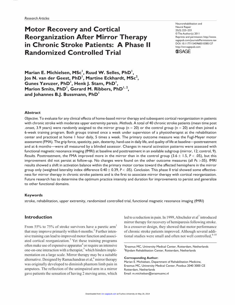
Objective. To evaluate for any clinical effects of home-based mirror therapy and subsequent cortical reorganization in patients with chronic stroke with moderate upper extremity paresis. Methods. A total of 40 chronic stroke patients (mean time post .onset, 3.9 years) were randomly assigned to the mirror group (n = 20) or the control group (n = 20) and then joined a 6-week training program. Both groups trained once a week under supervision of a physiotherapist at the rehabilitation center and practiced at home 1 hour daily, 5 times a week. The primary outcome measure was the Fugl-Meyer motor assessment (FMA). The grip force, spasticity, pain, dexterity, hand-use in daily life, and quality of life at baseline—posttreatment and at 6 months—were all measured by a blinded assessor. Changes in neural activation patterns were assessed with functional magnetic resonance imaging (fMRI) at baseline and posttreatment in an available subgroup (mirror, 12; control, 9). Results. Posttreatment, the FMA improved more in the mirror than in the control group (3.6 ± 1.5, P < .05), but this improvement did not persist at follow-up. No changes were found on the other outcome measures (all Ps >.05). fMRI results showed a shift in activation balance within the primary motor cortex toward the affected hemisphere in the mirror group only (weighted laterality index difference 0.40 ± 0.39, P < .05). Conclusion. This phase II trial showed some effective-ness for mirror therapy in chronic stroke patients and is the first to associate mirror therapy with cortical reorganization. Future research has to determine the optimum practice intensity and duration for improvements to persist and generalize to other functional domains.
Keywords
stroke, rehabilitation, upper extremity, randomized controlled trial, functional magnetic resonance imaging (fMRI)
Research Articles
at Fuzhou University on May 26, 2014nnr.sagepub.comDownloaded from
224 Neurorehabilitation and Neural Repair 25(3)
2 recent, high-quality, randomized controlled trials have also reported mirror therapy to improve motor function in patients with subacute11 and acute12 stroke.
Despite the encouraging clinical results, little is known about the underlying mechanisms of mirror therapy. Ramachandran et al6 referred to a “learned paralysis” in the brain, which could possibly be “unlearned” as a result of the mirror illusion.13 Other studies have attributed the positive effects of mirror therapy in stroke to motor imagery8 or the mirror neuron sys-tem.11 The prevailing idea is that observing mirrored move-ments causes additional neural activity in motor areas located in the affected hemisphere, which should eventually result in cortical reorganization and improved function. Research in healthy subjects has provided some evidence of such mecha-nisms, with the use of either transcranial magnetic stimula-tion14,15 or functional magnetic resonance imaging (fMRI).16 However, results of these studies are not conclusive, do not give insight into long-term neuronal changes, and do not neces-sarily apply to stroke patients.
The aim of this phase II study was 2-fold. First, we evalu-ated the effect of mirror therapy on upper extremity function in a group of chronic stroke patients. As the intervention had to be both effective and efficient, an unsupervised training program to be performed at home was developed and carried out with supervised weekly sessions. Our primary focus was on improvements in motor function, but to get a detailed insight in any effects of mirror therapy, outcomes were also measured at the other International Classification of Func-tioning, Disability and Health (ICF)17 domains. Second, we used fMRI to examine whether mirror therapy could induce cortical reorganization.
Materials and MethodsParticipants
After contacting 182 outpatients (hospitalized between January 1998 and September 2007) from the Rijndam Reha-bilitation Centre in Rotterdam, the Netherlands, we enrolled 40 patients. Inclusion criteria were knowledge of the Dutch language, a Brunnstrom score for the upper extremity between III and V18 (the 6 stages of the Brunnstrom score range from [I] flaccidity toward [VI] full-range voluntary extension and individual finger movements present but less accurate than on the opposite side), home dwelling status, and at least 1 year poststroke. Patients with neglect, comorbidities that influenced upper extremity usage, or a history of multiple strokes were excluded. For patients to participate in the fMRI experiment the following additional inclusion criteria applied: ability to perform a hand squeezing movement, no metal implants, no claustrophobia, and no severe obesity. The study was approved by the Medical Ethics Committee of the Erasmus Medical Centre, Rotterdam, and all patients gave written, informed consent before participating.
Sample Size
To calculate the necessary number of patients, we performed a power analysis on data from the literature. Assuming a clini-cal relevance of 10% on the Fugl-Meyer motor assessment19 (FMA) score, a standard deviation of 3.2,20 and a loss of patients at follow-up of 10%, we calculated that 20 patients in each group would be sufficient to have an 80% chance of detecting a statistically significant difference in improvements between the 2 groups.
Study DesignAll participants were randomly assigned to either the experi-mental group receiving mirror therapy (mirror group) or the control group. One of the authors otherwise not involved in the intervention used a computer random number generator to create the randomization sequence and constructed sealed envelopes containing the assignments. Patients received their group allocation assignments after baseline measurements were performed, just before the first training session.
Measurements of upper extremity function were per-formed before intervention (baseline), right after intervention (posttreatment), and 6 months after intervention (follow-up). All assessments were made by the same investigator, who was blinded to group allocation. Blinding of the patients or the physiotherapist was not possible because of the nature of the therapy. Examinations with fMRI were done in scan-ning sessions at baseline and posttreatment. Posttreatment clinical measurements and fMRI examinations were per-formed within a week after the last treatment session.
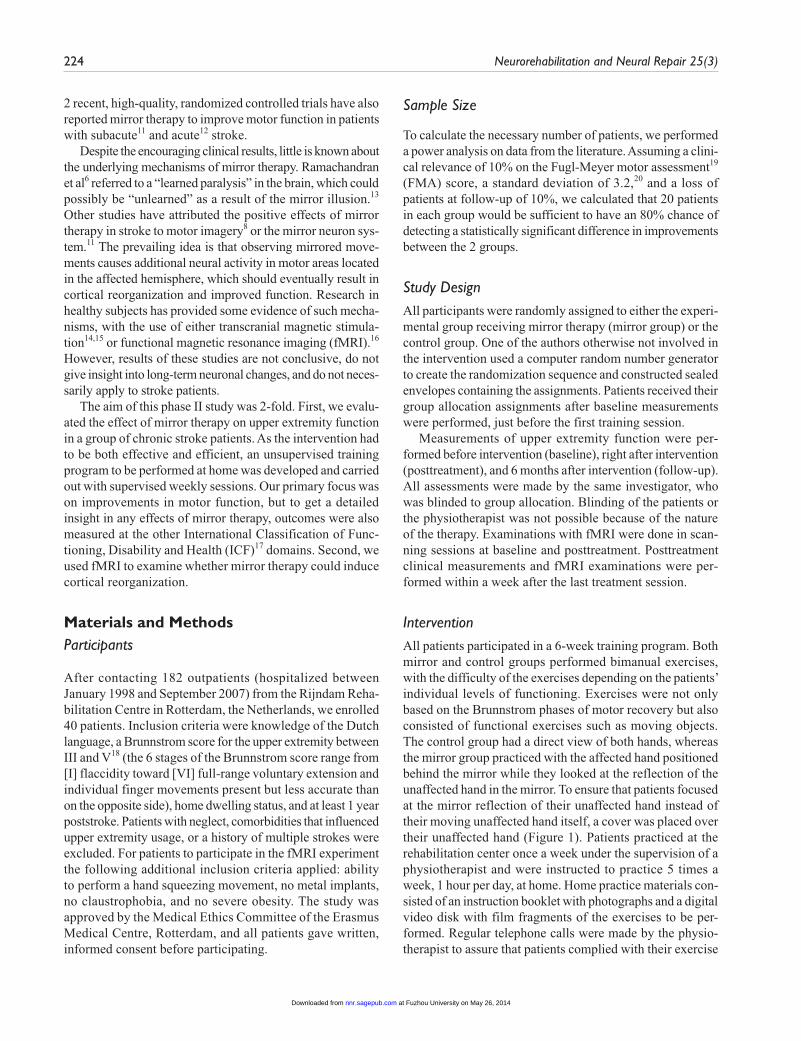
InterventionAll patients participated in a 6-week training program. Both mirror and control groups performed bimanual exercises, with the difficulty of the exercises depending on the patients’ individual levels of functioning. Exercises were not only based on the Brunnstrom phases of motor recovery but also consisted of functional exercises such as moving objects. The control group had a direct view of both hands, whereas the mirror group practiced with the affected hand positioned behind the mirror while they looked at the reflection of the unaffected hand in the mirror. To ensure that patients focused at the mirror reflection of their unaffected hand instead of their moving unaffected hand itself, a cover was placed over their unaffected hand (Figure 1). Patients practiced at the rehabilitation center once a week under the supervision of a physiotherapist and were instructed to practice 5 times a week, 1 hour per day, at home. Home practice materials con-sisted of an instruction booklet with photographs and a digital video disk with film fragments of the exercises to be per-formed. Regular telephone calls were made by the physio-therapist to assure that patients complied with their exercise
at Fuzhou University on May 26, 2014nnr.sagepub.comDownloaded from
Michielsen et al 225
regimens. Furthermore, patients were instructed to keep detailed accounts of their practice schedules and experiences. These diaries were inspected by the physiotherapist during each training session in the rehabilitation center.
Outcome MeasuresAs mirror therapy consists of exercises mainly on the level of body function, we chose as primary endpoint the difference in improvement between both treatment groups in motor func-tion as measured with the upper extremity part of the FMA (including arm, wrist, and hand function measurements). Additionally, in the body function ICF domain, we measured grip force with a Jamar handheld dynamometer (Sammons Preston, Bolingbrook, IL), spasticity with the Tardieu scale,21 and pain with a visual analog scale ranging from 0 to 100 mm. In the activity domain, we measured motor capacity with the Action Research Arm Test,22 self-perceived performance with the ABILHAND questionnaire,23 and actual performance in daily life during 24 hours with the Stroke-ULAM.24 The Stroke-ULAM consists of accelerometers placed on both arms and has been described in detail and tested for its validity in an earlier study. Because of the inconvenience of wearing the ULAM, measurements were not performed at 6-month follow-up. The ratio between the amount of use of the unaffected and the affected arms was used as outcome. In the participation domain, we measured quality of life with the EQ-5D.25 To evaluate the effects of mirror therapy at cortical levels, we calculated the difference between groups in the change in activation balance between affected and unaffected hemi-spheres as measured with fMRI (see the following).
fMRI ExperimentIn all, 12 patients from the mirror group and 9 patients from the control group were eligible to participate in this part of the study. During the scanning sessions, patients lay on their backs in the scanner with their upper arms comfortably resting on the scanner table alongside their torsos, with their elbows
flexed so that their hands were 20 cm apart above their waists. By means of 2 mirrors attached to the head coil above each patient’s head, patients were able to look in the direction of their feet and view both hands.
The experimental task consisted of 10 alternating 30-second periods of 5 rest and 5 active conditions (block design). In the active periods, patients had to open and close the affected hand; in the rest periods patients had to hold the hand still. Patients were instructed to pace the opening of their hand to a metronome with a rhythm of 0.5 Hz. The onsets of the rest and active conditions were indicated verbally by using simple words (start, rest) generated by a computer program (Matlab version 7.1; Mathworks, Sherborn, MA). Auditory stimuli were presented to the patients through MRI-compatible head-phones. The hand movement was practiced before the scan session started.
Imaging was performed on a 3T MR system (HD platform, GE Healthcare, Milwaukee, WI). For anatomical reference, a high-resolution, 3-dimensional, inversion recovery, fast spoiled gradient echo, T1-weighted image was acquired (TR/TE/TI 10.7/2.2/300 ms, 18° flip angle, matrix 416 × 256, and field of view 250 × 175 mm2). For functional imaging, a single-shot, T2*-weighted, gradient echo echo-planar imaging (EPI) sequence was used (TR/TE 3000/30 ms, 75° flip angle, matrix 64 × 96, field of view 220 × 220 mm2). The imaging volume covered the entire brain, including the cerebellum.
Statistical AnalysesClinical outcome. To test the study hypothesis, we used a
generalized estimating equations approach. Under the assump-tion that missing data are random and not due to group alloca-tion or treatment effect, this model estimates missing data values, thereby allowing the use of data from all participants, irrespective of whether they were measured at all time points. Each outcome measure was used as a separate response vari-able, and group (mirror vs control) and time (baseline vs post-treatment vs follow-up) were inserted in the model as predictors. The interaction of group × time was used to determine the efficacy of the intervention. Significance was set at .05.
fMRI data. The imaging data were analyzed using statistical parametric mapping software (SPM5; Wellcome Department of Cognitive Neurology, University College London, UK), implemented in Matlab version 7.1 (MathWorks, Natick, MA).
All functional images for each participant were realigned to the first scan of each session and then coregistered to the T1-weighted anatomical scan. Subsequently, images were trans-formed to standard Montreal Neurologic Institute space. To prevent warping around the lesions, we used a segmentation-based normalization approach.26 Finally, normalized images were spatially smoothed by using a Gaussian filter of 8-mm full width at half maximum.
Preliminary analyses showed that the realignment param-eters estimated during spatial preprocessing were sometimes
Figure 1. Setup for mirror therapy
at Fuzhou University on May 26, 2014nnr.sagepub.comDownloaded from
226 Neurorehabilitation and Neural Repair 25(3)
correlated with task design. Therefore, we decided not to model the realignment parameters in the design matrix as regressors of no interest, as this would have resulted in canceling out task-related activation. Instead, we used the ArtRepair Tool-box,27,28 which evaluates all volumes and detects the ones most affected by movement. Those volumes are repaired by interpolation to avoid side effects in the high-pass filter and then deweighted in the general linear model estimation to maintain unbiased estimates. The experimental block design was convolved with the canonical hemodynamic response function, and the resulting model was estimated using a high-pass filter at 128 seconds to remove low-frequency artifacts.
In the first-level analysis, contrast maps were calculated for the active periods versus rest for each patient and each session separately. In addition, contrasts were calculated for the pretreatment versus posttreatment sessions for each patient. Contrast images from patients with left-sided lesions were flipped about the mid-sagittal plane, so that the affected hemi-sphere corresponded to the right side of the brain for all patients.
Second-level analyses. We merged both groups and performed a random effect analysis on the baseline contrasts of task versus rest to show the typical activation patterns. To assess the dif-ferences between groups following therapy, we concentrated on effects within certain regions of interest (ROIs). Using the Anatomy Toolbox29 and the Anatomical Automatic Labeling Atlas (AAL),30 we defined ROIs for each hemisphere sepa-rately for the following areas: primary motor cortex (M1), dorsal premotor cortex (PMd), primary sensory cortex (S1), supplementary motor area (SMA), and cerebellum. M1 was constructed of Brodmann areas 4a and 4p; PMd of Brodmann area 6 excluding SMA and the ventral premotor cortex (area below z = +51); S1 of Brodmann areas 1, 2, and 3. SMA and the cerebellum were integrally derived from the AAL atlas.
These ROIs were used to calculate weighted laterality indexes (wLI), which measure the relative amount of activa-tion between the 2 hemispheres.31,32 We chose this measure because previous research showed that it correlates with residual clinical deficit and is sensitive to detecting subtle therapy-associated changes in cortical activation.33 Using this method, we first normalized for intersubject variations in global fMRI signal to reduce the chances of floor or ceiling values. For each condition, a threshold was defined as half the average value of the 5% highest t values within a certain ROI. Subsequently, within that ROI, the sum of the t values of all voxels above this threshold (sum-t) was computed. The wLI was then calculated as
wLI contra ipsi= −( ) +( )∑∑ ∑∑t tipsi contrat t/ ,
where the subscripts contra and ipsi, respectively, refer to contralateral and ipsilateral with respect to the side of
the lesion. wLI values can range between −1 and 1, with higher values indicating a larger contribution of the affected hemisphere.
Changes in wLI within each ROI for the 2 groups sepa-rately were assessed using the Wilcoxon rank sum test. Dif-ferences in the amount of change in the wLI between groups before and after treatment were assessed with the Mann–Whitney U test. We chose to use nonparametric statistics because of the risk of ceiling or floor effects (in spite of our use of the wLI instead of LI). Significance was set at .05.
Finally, a correlation analysis (Spearman’s correlation coefficient) was performed to assess the relationship between changes in wLI and changes in FMA scores.
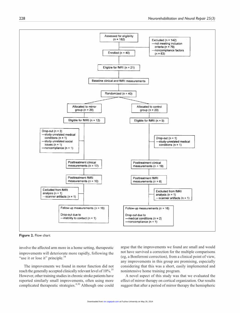
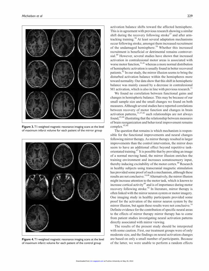
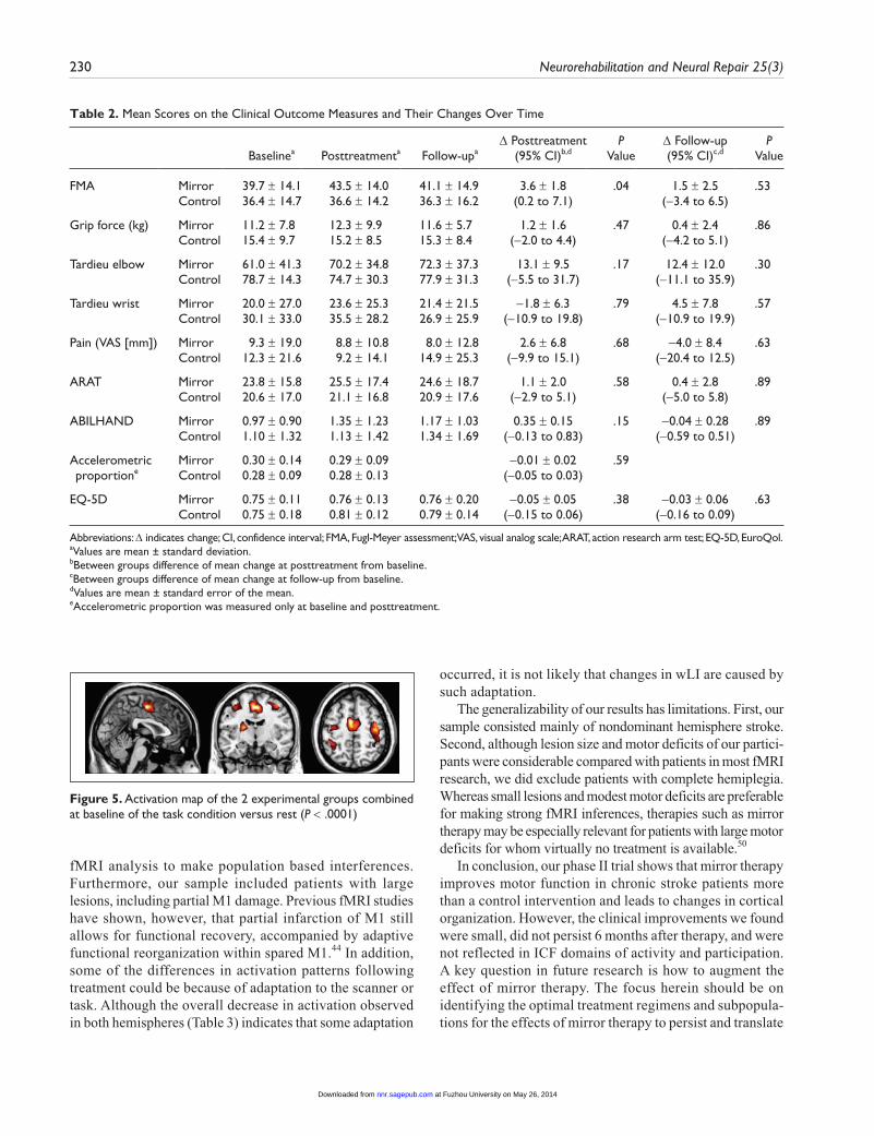
ResultsTable 1 presents demographic and clinical characteristics of the 2 treatment groups, showing no differences. Figure 2 shows the flow chart of the study. None of the reported drop-outs were because of group allocation or treatment effect. Figures 3 and 4 show the structural brain images of the par-ticipants of the fMRI subgroup.
Clinical MeasuresAll patients attended all 6 training sessions in the rehabili-tation center, and the home-kept diaries showed no differ-ences in total home-based practice time between the groups. All patients kept to their regimens, which resulted in an aver-age home training time of 30 hours.
Analyses followed the intention-to-treat principle. Table 2 shows the estimated marginal means and standard devia-tions for all measurement tools at baseline, posttreatment, and follow-up as well as the between-group comparisons of the change scores from baseline to posttreatment and from baseline to follow-up. Posttreatment, the mirror group had improved significantly more on the FMA than the control group (P = .04), but this difference was not present at follow-up (P = .53). No other significant effects were found between or within groups.
Cortical ReorganizationIn all, 3 patients who were scanned at baseline did not complete the training program and were not scanned post-treatment. Two other patients were discarded from further analysis because their data sets contained scanner artifacts. The remaining analyses were therefore conducted on data from 9 patients in the mirror group and 7 patients in the control group.
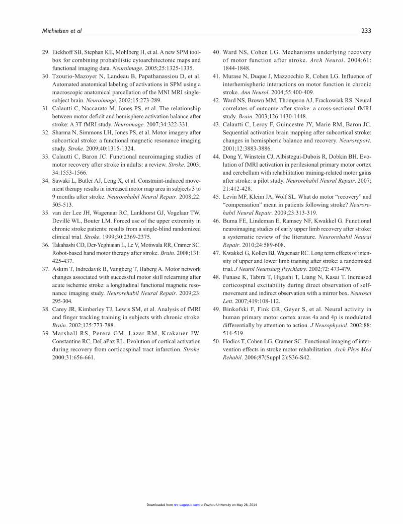
Figure 5 shows the activation maps as calculated in the random effects analysis of the task condition versus the rest condition. In general, in all sessions, activation patterns were
at Fuzhou University on May 26, 2014nnr.sagepub.comDownloaded from
Michielsen et al 227
in accordance with the expected activation for a hand motor task. Activity was observed bilaterally in the precentral and postcentral gyri (M1 and S1), the medial superior frontal gyrus (SMA), at the junction of the superior frontal sulcus and the precentral sulcus (PMC), and in the cerebellum.
Table 3 presents the wLI for each of the ROIs. Within M1, a difference in wLI change between the mirror group and control group was found. The activation in the mirror group shifted toward the affected hemisphere, whereas a small shift in activation toward the unaffected hemisphere was observed in the control group. In the other regions no differences in the wLI were observed between the 2 groups. No significant correlations were observed between pretreatment and post-treatment changes in wLI and FMA scores.
DiscussionOur study has 2 important findings. We showed that in patients with chronic stroke, practicing with a mirror resulted in modest, but statistically significant, improvements in upper extremity motor function. This effect disappeared in the follow-up measurements and did not transfer to the ICF domains activity and participation. In addition, we demons-trated that mirror therapy caused a shift in activation balance M1 toward the lesioned hemisphere, suggesting neural reor-ganization. As we did not measure cortical activation at the
6-month follow-up, we do not know whether this change persisted.
The results on our clinical outcome measures are in agreement with previous studies on patients with subacute11 and acute12 stroke. The size of the FMA change in the present study is not very different from that in a study of patients with more acute stroke. Mirror therapy may thus be beneficial at all stages after stroke. The fact that the current study was based on unsupervised, home-based treatment is in this respect promising.
The effects we observed on motor function did not persist at the 6-month follow-up and did not transfer to any other ICF domains. This is in disagreement with the study of Yavuzer et al,11 who reported improved motor function as well as improved activities of daily living also at 6-months’ follow-up. This discrepancy might be because of the more sensitive activities of daily living (ADL) measurement tool used by Yavuzer et al, as well as the difference in time post-onset. Whereas the subacute stroke patients in the study by Yavuzer et al were still in the rehabilitation center, the chronic stroke patients in our study were home based with daily living routines that were already well adjusted to their disabilities. Improvements in motor function may therefore cause less change in these routines. This lack of transfer to the activity and participation domain may also explain the lack of persis-tence of improvements in motor function: as patients do not
Table 1. Patient Characteristics
All Patients Patients With fMRI
Characteristic Mirror Group Control Group Mirror Group Control Group
No. of patients 20 20 9 7Age (years) 55.3 ± 12.0 58.7 ± 13.5 51.9 ± 9.3 59.0 ± 10.4Time since stroke (years) 4.7 ± 3.6 4.5 ± 2.6 6.2 ± 4.4 5.2 ± 2.5Gender (male/female) 7/13 13/7 2/7 5/2Affected side (dom/ndom) 6/14 6/14 2/6 3/4Type of stroke (infarct/hemorrhage) 14/6 14/6 7/2 6/1FMA score baseline 39.7 ± 14.1 36.4 ± 14.7 43.7 ± 12.5 36.9 ± 9.1FMA score posttreatment 43.5 ± 14.0 36.6 ± 14.2 48.2 ± 11.2 37.6 ± 8.5Location of stroke lesion
Cortical (with or without subcortical) 8 5 6 2Subcortical 7 5 3 3Brainstema 1 3 — 2Unknown 4 7 — —
Bamford classification LACS 8 8 3 1PACS 8 7 5 3TACS 3 2 1 1POCS 1 3 — 2
Abbreviations: fMRI, functional magnetic resonance imaging; dom, dominant; ndom, nondominant; LACS, lacunar stroke; PACS, partial anterior circulation stroke; TACS, total anterior circulation stroke; POCS, posterior circulation stroke.aBrainstem lesions are located above the corticospinal tract.
at Fuzhou University on May 26, 2014nnr.sagepub.comDownloaded from
228 Neurorehabilitation and Neural Repair 25(3)
involve the affected arm more in a home setting, therapeutic
improvements will deteriorate more rapidly, following the “use it or lose it” principle.34
The improvements we found in motor function did not reach the generally accepted clinically relevant level of 10%.35 However, other training studies in chronic stroke patients have reported similarly small improvements, often using more complicated therapeutic strategies.4,36 Although one could
argue that the improvements we found are small and would not have survived a correction for the multiple comparisons (eg, a Bonferroni correction), from a clinical point of view, any improvements in this group are promising, especially considering that this was a short, easily implemented and nonintensive home training program.
A novel aspect of this study was that we evaluated the effect of mirror therapy on cortical organization. Our results suggest that after a period of mirror therapy the hemispheric
Figure 2. Flow chart
at Fuzhou University on May 26, 2014nnr.sagepub.comDownloaded from
Michielsen et al 229
activation balance shifts toward the affected hemisphere. This is in agreement with previous research showing a similar shift during the recovery following stroke37 and after arm-tracking training.38 At least several adaptation mechanisms occur following stroke, amongst them increased recruitment of the undamaged hemisphere.39 Whether this increased recruitment is beneficial or detrimental remains controver-sial.40 However, several studies have shown that increased activation in contralesional motor areas is associated with worse motor function,31,41 whereas a more normal distribution of hemispheric activation is usually found in better recovered patients.42 In our study, the mirror illusion seems to bring the disturbed activation balance within the hemispheres more toward normality. Our data show that this shift in hemispheric balance was mainly caused by a decrease in contralesional M1 activation, which is also in line with previous research.31
We found no correlation between functional gains and changes in hemispheric balance. This may be because of our small sample size and the small changes we found on both measures. Although several studies have reported correlations between recovery of motor function and changes in brain activation patterns,31,37,42 such relationships are not always found,43,44 illustrating that the relationship between measures of brain reorganization and behavioral improvement remains complex.45,46
The question that remains is which mechanism is respon-sible for the functional improvements and neural changes following mirror therapy. As mirror therapy resulted in larger improvements than the control intervention, the mirror does seem to have an additional effect beyond repetitive task-orientated training.47 It is possible that by providing an image of a normal moving hand, the mirror illusion enriches the training environment and increases somatosensory input, thereby inducing excitability of the motor cortex.40 Research in healthy subjects using transcranial magnetic stimulation has provided some proof of such a mechanism, although these results are not conclusive.14,48 Alternatively, the mirror illusion might increase attention to the motor task, which is known to increase cortical activity49 and is of importance during motor recovery following stroke.42 In literature, mirror therapy is often linked with the mirror neuron system or motor imagery. One imaging study in healthy participants provided some proof for the activation of the mirror neuron system by the mirror illusion, but again these results were not conclusive.16 Definite evidence for the contribution of specific neural areas to the effects of mirror therapy mirror therapy has to come from patient studies investigating neural activation patterns directly associated with mirror viewing.
The results of the present study should be interpreted with some caution. First, our treatment groups were of only moderate size, and the findings on neural activation changes are based on only a small number of participants. Because of the latter, we were unable to perform a random effects
Figure 3. T1-weighted magnetic resonance imaging scans at the level of maximum infarct volume for each patient of the mirror group
Figure 4. T1-weighted magnetic resonance imaging scans at the level of maximum infarct volume for each patient of the control group
at Fuzhou University on May 26, 2014nnr.sagepub.comDownloaded from
230 Neurorehabilitation and Neural Repair 25(3)
fMRI analysis to make population based interferences. Furthermore, our sample included patients with large lesions, including partial M1 damage. Previous fMRI studies have shown, however, that partial infarction of M1 still allows for functional recovery, accompanied by adaptive functional reorganization within spared M1.44 In addition, some of the differences in activation patterns following treatment could be because of adaptation to the scanner or task. Although the overall decrease in activation observed in both hemispheres (Table 3) indicates that some adaptation
occurred, it is not likely that changes in wLI are caused by such adaptation.
The generalizability of our results has limitations. First, our sample consisted mainly of nondominant hemisphere stroke. Second, although lesion size and motor deficits of our partici-pants were considerable compared with patients in most fMRI research, we did exclude patients with complete hemiplegia. Whereas small lesions and modest motor deficits are preferable for making strong fMRI inferences, therapies such as mirror therapy may be especially relevant for patients with large motor deficits for whom virtually no treatment is available.50
In conclusion, our phase II trial shows that mirror therapy improves motor function in chronic stroke patients more than a control intervention and leads to changes in cortical organization. However, the clinical improvements we found were small, did not persist 6 months after therapy, and were not reflected in ICF domains of activity and participation. A key question in future research is how to augment the effect of mirror therapy. The focus herein should be on identifying the optimal treatment regimens and subpopula-tions for the effects of mirror therapy to persist and translate
Table 2. Mean Scores on the Clinical Outcome Measures and Their Changes Over Time
Baselinea Posttreatmenta Follow-upaΔ Posttreatment
(95% CI)b,dP
ValueΔ Follow-up (95% CI)c,d
P Value
FMA
MirrorControl
39.7 ± 14.136.4 ± 14.7
43.5 ± 14.036.6 ± 14.2
41.1 ± 14.936.3 ± 16.2
3.6 ± 1.8 (0.2 to 7.1)
.04 1.5 ± 2.5 (−3.4 to 6.5)
.53
Grip force (kg)
MirrorControl
11.2 ± 7.815.4 ± 9.7
12.3 ± 9.915.2 ± 8.5
11.6 ± 5.715.3 ± 8.4
1.2 ± 1.6 (−2.0 to 4.4)
.47 0.4 ± 2.4 (−4.2 to 5.1)
.86
Tardieu elbow
MirrorControl
61.0 ± 41.378.7 ± 14.3
70.2 ± 34.874.7 ± 30.3
72.3 ± 37.377.9 ± 31.3
13.1 ± 9.5 (−5.5 to 31.7)
.17 12.4 ± 12.0 (−11.1 to 35.9)
.30
Tardieu wrist
MirrorControl
20.0 ± 27.030.1 ± 33.0
23.6 ± 25.335.5 ± 28.2
21.4 ± 21.526.9 ± 25.9
−1.8 ± 6.3 (−10.9 to 19.8)
.79 4.5 ± 7.8 (−10.9 to 19.9)
.57
Pain (VAS [mm])
MirrorControl
9.3 ± 19.012.3 ± 21.6
8.8 ± 10.89.2 ± 14.1
8.0 ± 12.814.9 ± 25.3
2.6 ± 6.8 (−9.9 to 15.1)
.68 −4.0 ± 8.4 (−20.4 to 12.5)
.63
ARAT
MirrorControl
23.8 ± 15.820.6 ± 17.0
25.5 ± 17.421.1 ± 16.8
24.6 ± 18.720.9 ± 17.6
1.1 ± 2.0 (−2.9 to 5.1)
.58 0.4 ± 2.8 (−5.0 to 5.8)
.89
ABILHAND
MirrorControl
0.97 ± 0.901.10 ± 1.32
1.35 ± 1.231.13 ± 1.42
1.17 ± 1.031.34 ± 1.69
0.35 ± 0.15 (−0.13 to 0.83)
.15 −0.04 ± 0.28 (−0.59 to 0.51)
.89
Accelerometric proportione
MirrorControl
0.30 ± 0.140.28 ± 0.09
0.29 ± 0.090.28 ± 0.13
−0.01 ± 0.02 (−0.05 to 0.03)
.59
EQ-5D
MirrorControl
0.75 ± 0.110.75 ± 0.18
0.76 ± 0.130.81 ± 0.12
0.76 ± 0.200.79 ± 0.14
−0.05 ± 0.05 (−0.15 to 0.06)
.38 −0.03 ± 0.06 (−0.16 to 0.09)
.63
Abbreviations: Δ indicates change; CI, confidence interval; FMA, Fugl-Meyer assessment; VAS, visual analog scale; ARAT, action research arm test; EQ-5D, EuroQol.aValues are mean ± standard deviation.bBetween groups difference of mean change at posttreatment from baseline.cBetween groups difference of mean change at follow-up from baseline.dValues are mean ± standard error of the mean.eAccelerometric proportion was measured only at baseline and posttreatment.
Figure 5. Activation map of the 2 experimental groups combined at baseline of the task condition versus rest (P < .0001)
at Fuzhou University on May 26, 2014nnr.sagepub.comDownloaded from

231
Tab
le 3
. Vox
el C
ount
in A
ffect
ed a
nd U
naffe
cted
Hem
isph
ere
and
Wei
ghte
d La
tera
lity
Indi
ces
(wLI
) fo
r th
e Ba
selin
e an
d th
e Po
sttr
eatm
ent
fMR
I Exa
min
atio
ns a
nd t
he
Cor
resp
ondi
ng C
hang
es fo
r ea
ch o
f the
5 R
egio
ns o
f Int
eres
ta,b
Base
line
Post
trea
tmen
tU
naffe
cted
Affe
cted
Are
a
Una
ffect
ed
Affe
cted
Una
ffect
edA
ffect
edC
hang
e (9
5% C
I)P w
ithin
P betw
een
Cha
nge
(95%
CI)
P with
inP be
twee
n
M1
Mir
ror
6268
± 4
052
8010
± 4
250
3743
± 4
321
5353
± 2
810
2525
± 2
849
(335
to
3715
).0
4.0
526
58 ±
355
4 (−
74 t
o 53
90)
.07
.03
C
ontr
ol45
51 ±
423
764
08 ±
395
253
42 ±
322
754
19 ±
206
6−7
91 ±
325
8 (−
3804
to
2222
).6
199
0 ±
3473
(−
2223
to
4302
).5
0
PMd
Mir
ror
8182
± 4
157
12 0
81 ±
413
240
61 ±
385
971
78 ±
410
541
21 ±
443
3 (7
13 t
o 75
29)
.05
.15
4903
± 5
354
(788
to
9019
).0
4.4
3
C
ontr
ol63
39 ±
524
090
70 ±
699
456
53 ±
326
578
30 ±
670
268
6 ±
5231
(−
4152
to
5523
).8
712
40 ±
552
8 (−
3872
to
6353
).5
0
SMA
Mir
r or
7009
± 4
694
10 5
34 ±
408
337
57 ±
286
167
80 ±
409
732
52 ±
360
6 (4
80 t
o 60
24)
.03
.19
3754
± 5
382
(−38
3 to
789
1).1
1.9
6
C
ontr
ol71
83 ±
367
688
35 ±
647
365
85 ±
341
379
38 ±
508
559
8 ±
4039
(−
3183
to
4334
).8
789
7 ±
7159
(−
5723
to
7518
).7
4
S1M
irro
r10
895
± 80
4816
688
± 9
845
6022
± 5
680
9620
± 5
712
4874
± 6
854
(−39
4 to
10
139)
.11
.26
7068
± 7
890
(100
3-13
133
).0
4.4
9
C
ontr
ol81
21 ±
594
913
339
± 8
334
7988
± 6
118
1162
3 ±
5593
133
± 81
67
(−74
21 t
o 76
87)
.74
1716
± 6
113
(−39
37 t
o 73
69)
.40
CB
Mir
r or
5904
± 4
848
6045
± 7
228
8065
± 1
0457
4905
± 5
458
−216
1 ±
7862
(−
8204
to
3882
).8
6.8
711
40 ±
673
4 (−
4036
to
6316
).5
2.9
6
C
ontr
ol83
31 ±
721
510
473
± 8
905
8828
± 7
513
6965
± 7
159
−497
± 5
497
(−55
81 t
o 45
87)
.90
5307
± 9
090
(−48
99 t
o 11
915
).3
1
Are
aBa
selin
ePo
sttr
eatm
ent
Cha
nge
(95%
CI)
P with
inP be
twee
n
M1
Mir
ror
0.11
± 0
.21
0.44
± 0
.50
0.33
± 0
.39
(.03
to .6
3).0
5.0
3
C
ontr
ol0.
17 ±
0.3
10.
10 ±
0.3
8−0
.07
± 0.
28 (−0
.33
to 0
.19)
.40
PM
dM
irro
r0.
21 ±
0.1
70.
41 ±
0.3
80.
20 ±
0.3
0 (−
0.03
to
0.43
).1
1.4
3
C
ontr
ol0.
05 ±
0.5
00.
07 ±
0.4
40.
02 ±
0.3
0 (−
0.26
to
0.30
).5
0
SMA
Mir
ror
0.24
± 0
.29
0.33
± 0
.30
0.09
± 0
.21
(−0.
07 t
o 0.
25)
.17
.96
Con
trol
0.00
± 0
.30
0.11
± 0
.19
0.11
± 0
.23
(−0.
10 t
o 0.
32)
.31
S1
Mir
ror
0.12
± 0
.47
0.34
± 0
.51
0.22
± 0
.34
(−0.
04 t
o 0.
48)
.07
.49
Con
trol
0.18
± 0
.40
0.19
± 0
.49
0.01
± 0
.33
(−0.
30 t
o 0.
32.9
9
CB
Mir
ror
0.16
± 0
.59
0.33
± 0
.53
0.17
± 0
.49
(−0.
21 t
o 0.
54)
.52
.96
Con
trol
−0.0
5 ±
0.48
0.13
± 0
.64
0.18
± 0
.53
(−0.
31 t
o 0.
66)
.31
Abb
revi
atio
ns: M
1, p
rim
ary
mot
or c
orte
x; P
Md,
dor
sal p
rem
otor
cor
tex;
SM
A, s
uppl
emen
tary
mot
or a
rea;
S1, p
rim
ary
sens
ory
cort
ex; C
B, c
ereb
ellu
m.
a P va
lues
are
est
imat
ed u
sing
the
Wilc
oxon
(P w
ithin)
and
the
Man
n–W
hitn
ey U
(P be
twee
n) Tes
ts.
b All
valu
es a
re m
ean
± st
anda
rd d
evia
tion.
at Fuzhou University on May 26, 2014nnr.sagepub.comDownloaded from

232 Neurorehabilitation and Neural Repair 25(3)
to improvements in performance of daily activities and participation.
Declaration of Conflicting Interests
The author(s) declared no potential conflicts of interest with respect to the authorship and/or publication of this article.
Funding
The author(s) disclosed receipt of the following financial support for the research and/or authorship of this article: Fonds NutsOhra [SNO-T-0602-23]; Innovatiefonds Zorgverzekeraars [06-262]; Wetenschappelijk College Fysiotherapie [WU/2007/07] and Her-senstichting Nederland [15F07.54].
References
1. Jorgensen HS, Nakayama H, Raaschou HO, Vive-Larsen J, Stoier M, Olsen TS. Outcome and time course of recovery in stroke. Part II: time course of recovery. The Copenhagen Stroke Study. Arch Phys Med Rehabil. 1995;76:406-412.
2. Duncan P, Goldstein L, Matchar D, Divine G, Feussner J. Mea-surement of motor recovery after stroke. Outcome assessment and sample size requirements. Stroke. 1992;23:1084-1089.
3. Taub E, Miller NE, Novack TA, et al. Technique to improve chronic motor deficit after stroke. Arch Phys Med Rehabil. 1993;74:347-354.
4. Mehrholz J, Platz T, Kugler J, Pohl M. Electromechanical and robot-assisted arm training for improving arm function and activ-ities of daily living after stroke. Cochrane Database Syst Rev. 2008;(4):CD006876. doi:10.1002/14651858.CD006876.pub2.
5. Fasoli SE, Krebs HI, Hogan N. Robotic technology and stroke rehabilitation: Translating research into practice. Top Stroke Rehabil. 2004;11:11-19.
6. Ramachandran VS, Rogers-Ramachandran D, Cobb S. Touching the phantom limb. Nature. 1995;377:489-490.
7. Altschuler EL, Wisdom SB, Stone L, et al. Rehabilitation of hemiparesis after stroke with a mirror. Lancet. 1999;353: 2035-2036.
8. Stevens JA, Stoykov ME. Using motor imagery in the reha-bilitation of hemiparesis. Arch Phys Med Rehabil. 2003;84: 1090-1092.
9. Rothgangel AS, Morton AR, Hout JWE, van den Beurskens AJHM. Phantoms in the brain. Spiegeltherapie bij chronische CVA-patienten: een pilot-study. Ned Tijdschr Fysiotherapie. 2004;114:36-40.
10. Sathian K, Greenspan AI, Wolf SL. Doing it with mirrors: a case study of a novel approach to neurorehabilitation. Neuro-rehabil Neural Repair. 2000;14:73-76.
11. Yavuzer G, Selles R, Sezer N, et al. Mirror therapy improves hand function in subacute stroke: A randomized controlled trial. Arch Phys Med Rehabil. 2008;89:393-398.
12. Dohle C, Pullen J, Nakaten A, Kust J, Rietz C, Karbe H. Mirror therapy promotes recovery from severe hemiparesis: a randomized controlled trial. Neurorehabil Neural Repair. 2009; 23:209-217.
13. Ramachandran VS, Altschuler EL. The use of visual feedback, in particular mirror visual feedback, in restoring brain function. Brain. 2009;132:1693-1710.
14. Garry MI, Loftus A, Summers JJ. Mirror, mirror on the wall: viewing a mirror reflection of unilateral hand movements facilitates ipsilateral M1 excitability. Exp Brain Res. 2005;163: 118-122.
15. Fukumura K, Sugawara K, Tanabe S, Ushiba J, Tomita Y. Influ-ence of mirror therapy on human motor cortex. Int J Neurosci. 2007;117:1039-1048.
16. Matthys K, Smits M, Van der Geest JN, et al. Mirror-induced visual illusion of hand movements: a functional magnetic reso-nance imaging study. Arch Phys Med Rehabil. 2009;90:675-681.
17. World Health Organization. International Classification of Functioning, Disability and Health (ICF). Geneva, Switzerland: World Health Organization; 2001.
18. Sawner KA, LaVigne JM. Brunnstrom’s Movement Therapy in Hemiplegia: A Neurophysiological Approach. Philadelphia, PA: J. B. Lippincott; 1992.
19. Fugl-Meyer AR, Jaasko L, Leyman I, Olsson S, Steglind S. The post-stroke hemiplegic patient. 1. A method for evaluation of physical performance. Scand J Rehabil Med. 1975;7:13-31.
20. van der Lee JH, Beckerman H, Lankhorst GJ, Bouter LM. The responsiveness of the action research arm test and the Fugl-Meyer assessment scale in chronic stroke patients. J Rehabil Med. 2001;33:110-113.
21. Tardieu G, Shentoub S, Delarue R. Research on a technique for measurement of spasticity [in French]. Rev Neurol (Paris). 1954;91:143-144.
22. Hsueh IP, Hsieh CL. Responsiveness of two upper extremity function instruments for stroke inpatients receiving rehabilitation. Clin Rehabil. 2002;16:617-624.
23. Penta M, Tesio L, Arnould C, Zancan A, Thonnard JL. The ABILHAND questionnaire as a measure of manual ability in chronic stroke patients: Rasch-based validation and relationship to upper limb impairment. Stroke. 2001;32:1627-1634.
24. de Niet M, Bussmann JB, Ribbers GM, Stam HJ. The stroke upper-limb activity monitor: its sensitivity to measure hemiplegic upper-limb activity during daily life. Arch Phys Med Rehabil. 2007;88:1121-1126.
25. Lamers LM, McDonnell J, Stalmeier PF, Krabbe PF, Busschbach JJ. The Dutch tariff: results and arguments for an effective design for national EQ-5D valuation studies. Health Econ. 2006;15:1121-1132.
26. Crinion J, Ashburner J, Leff A, Brett M, Price C, Friston K. Spatial normalization of lesioned brains: performance evaluation and impact on fMRI analyses. Neuroimage. 2007; 37:866-875.
27. Mazaika P, Whitfield-Gabrieli S, Reiss A. Artifact repair for fMRI data from high motion clinical subjects. Paper presented at: Human Brain Mapping Conference; June 10-14, 2007; Chicago, IL.
28. Artifact repair software for fMRI data from clinical subjects. http://cibsr.stanford.edu/tools/ArtRepair/ArtRepairTopPage .htm. Accessed September 13, 2010.
at Fuzhou University on May 26, 2014nnr.sagepub.comDownloaded from
Michielsen et al 233
29. Eickhoff SB, Stephan KE, Mohlberg H, et al. A new SPM tool-box for combining probabilistic cytoarchitectonic maps and functional imaging data. Neuroimage. 2005;25:1325-1335.
30. Tzourio-Mazoyer N, Landeau B, Papathanassiou D, et al. Automated anatomical labeling of activations in SPM using a macroscopic anatomical parcellation of the MNI MRI single-subject brain. Neuroimage. 2002;15:273-289.
31. Calautti C, Naccarato M, Jones PS, et al. The relationship between motor deficit and hemisphere activation balance after stroke: A 3T fMRI study. Neuroimage. 2007;34:322-331.
32. Sharma N, Simmons LH, Jones PS, et al. Motor imagery after subcortical stroke: a functional magnetic resonance imaging study. Stroke. 2009;40:1315-1324.
33. Calautti C, Baron JC. Functional neuroimaging studies of motor recovery after stroke in adults: a review. Stroke. 2003; 34:1553-1566.
34. Sawaki L, Butler AJ, Leng X, et al. Constraint-induced move-ment therapy results in increased motor map area in subjects 3 to 9 months after stroke. Neurorehabil Neural Repair. 2008;22: 505-513.
35. van der Lee JH, Wagenaar RC, Lankhorst GJ, Vogelaar TW, Devillé WL, Bouter LM. Forced use of the upper extremity in chronic stroke patients: results from a single-blind randomized clinical trial. Stroke. 1999;30:2369-2375.
36. Takahashi CD, Der-Yeghiaian L, Le V, Motiwala RR, Cramer SC. Robot-based hand motor therapy after stroke. Brain. 2008;131: 425-437.
37. Askim T, Indredavik B, Vangberg T, Haberg A. Motor network changes associated with successful motor skill relearning after acute ischemic stroke: a longitudinal functional magnetic reso-nance imaging study. Neurorehabil Neural Repair. 2009;23: 295-304.
38. Carey JR, Kimberley TJ, Lewis SM, et al. Analysis of fMRI and finger tracking training in subjects with chronic stroke. Brain. 2002;125:773-788.
39. Marshall RS, Perera GM, Lazar RM, Krakauer JW, Constantine RC, DeLaPaz RL. Evolution of cortical activation during recovery from corticospinal tract infarction. Stroke. 2000;31:656-661.
40. Ward NS, Cohen LG. Mechanisms underlying recovery of motor function after stroke. Arch Neurol. 2004;61: 1844-1848.
41. Murase N, Duque J, Mazzocchio R, Cohen LG. Influence of interhemispheric interactions on motor function in chronic stroke. Ann Neurol. 2004;55:400-409.
42. Ward NS, Brown MM, Thompson AJ, Frackowiak RS. Neural correlates of outcome after stroke: a cross-sectional fMRI study. Brain. 2003;126:1430-1448.
43. Calautti C, Leroy F, Guincestre JY, Marie RM, Baron JC. Sequential activation brain mapping after subcortical stroke: changes in hemispheric balance and recovery. Neuroreport. 2001;12:3883-3886.
44. Dong Y, Winstein CJ, Albistegui-Dubois R, Dobkin BH. Evo-lution of fMRI activation in perilesional primary motor cortex and cerebellum with rehabilitation training-related motor gains after stroke: a pilot study. Neurorehabil Neural Repair. 2007; 21:412-428.
45. Levin MF, Kleim JA, Wolf SL. What do motor “recovery” and “compensation” mean in patients following stroke? Neurore-habil Neural Repair. 2009;23:313-319.
46. Buma FE, Lindeman E, Ramsey NF, Kwakkel G. Functional neuroimaging studies of early upper limb recovery after stroke: a systematic review of the literature. Neurorehabil Neural Repair. 2010;24:589-608.
47. Kwakkel G, Kollen BJ, Wagenaar RC. Long term effects of inten-sity of upper and lower limb training after stroke: a randomised trial. J Neurol Neurosurg Psychiatry. 2002;72: 473-479.
48. Funase K, Tabira T, Higashi T, Liang N, Kasai T. Increased corticospinal excitability during direct observation of self-movement and indirect observation with a mirror box. Neurosci Lett. 2007;419:108-112.
49. Binkofski F, Fink GR, Geyer S, et al. Neural activity in human primary motor cortex areas 4a and 4p is modulated differentially by attention to action. J Neurophysiol. 2002;88: 514-519.
50. Hodics T, Cohen LG, Cramer SC. Functional imaging of inter-vention effects in stroke motor rehabilitation. Arch Phys Med Rehabil. 2006;87(Suppl 2):S36-S42.
at Fuzhou University on May 26, 2014nnr.sagepub.comDownloaded from