Monograph2
60
TRUE UNGULATES FROM THE NAGRI TYPE LOCALITY (LATE MIOCENE), NORTHERN PAKISTAN Muhammad Akbar Khan* § , Muhammad Akhtar * and Tasneem Ikram ** * Zoology Department, Punjab University, Quid-e-Azam Campus, Lahore, Pakistan § Zoology Department, Government College University, Faisalabad, Pakistan ** Government College for Woman, Farooq Colony, Sargodha, Pakistan THE JOURNAL OF ANIMAL AND PLANT SCIENCES SUPPLEMENTARY SERIES Number 22, January, 2012 Pages 1 – 59 PUBLISHED BY PAKISTAN AGRICULTURAL SCIENTISTS FORUM
Transcript of Monograph2

1
TRUE UNGULATES FROM THE NAGRI TYPE LOCALITY
(LATE MIOCENE), NORTHERN PAKISTAN
Muhammad Akbar Khan*§, Muhammad Akhtar
* and Tasneem Ikram
**
*Zoology Department, Punjab University, Quid-e-Azam Campus, Lahore, Pakistan
§Zoology Department, Government College University, Faisalabad, Pakistan
**Government College for Woman, Farooq Colony, Sargodha, Pakistan
THE JOURNAL OF ANIMAL AND PLANT SCIENCES SUPPLEMENTARY SERIES
Number 22, January, 2012
Pages 1 – 59
PUBLISHED BY
PAKISTAN AGRICULTURAL SCIENTISTS FORUM

1
TRUE UNGULATES FROM THE NAGRI TYPE LOCALITY (LATE MIOCENE),
NORTHERN PAKISTAN
M. A. Khan*§, M. Akhtar
* and T. Ikram
**
*Zoology Department, Punjab University, Quid-e-Azam Campus, Lahore, Pakistan
§Zoology Department, Government College University, Faisalabad, Pakistan
**Government College for Woman, Farooq Colony, Sargodha, Pakistan
Correspondence author email: <[email protected]>
CONTENTS
Sr. No. Title Sr. No.
1 ABSTRACT 1
2 INTRODUCTION 1
3 METHODOLOGY 6
4 SYSTEMATIC PALAEONTOLOGY 8
5 DISCUSSION 49
6 CONCLUSIONS 52
7 REFERENCES 52
ABSTRACT
The early Late Miocene type locality of the Nagri Formation from the Indo-Siwaliks has yielded remains of the true
ungulates that are today extinct to the south Asian biogeographic realm. Thirteen species including Brachypotherium,
Hipparion, Listriodon and the bovids of the true ungulates from the village Sethi Nagri, district Chakwal, Punjab,
Pakistan, are recognized, described and discussed in details. The tooth positions of all thirteen species are documented.
Quantitatively, the taxa of the bovids are the most predominant. But Brachypotherium, Hipparion, Listriodon, tragulid
and giraffid fossils are approximately as common as each other at the type locality. Pachyportax, Dorcabune,
Miotragocerus and Gazella seem to be uniformly rare at Sethi Nagri. The new findings from the type locality are the
Giraffokeryx’s hemimandible and the deciduous premolar of Dorcatherium minus. The newly recovered hemimandible
and deciduous premolar enlarge our knowledge on the anatomic features of the Nagri true ungulates. The Nagri type
locality mammalian local fauna has similarities to late Miocene Eurasian faunas. The investigation comprises extensive
taxonomic descriptions of all species represented and an interpretation of the palaeoecology based on an analysis of the
community structure. It seems that the abundance of Hipparion, giraffids, rhinocerotids and bovids suggests a woodland
to savannah environment at or near the type locality during the early Late Miocene. There is little evidence to suggest
that there was a humid closed canopy forest interspersed with temporary and perennial waters and accompanying open
areas forest in the vicinity at the time of deposition.
Key words: Listriodon, Bovidae, Giraffidae, Tragulidae, Hipparion, Brachypotherium, Siwaliks.
INTRODUCTION
Ungulate refers to any animal with hooves
however, the “True Ungulates” are considered the
members of Artiodactyla and Perissodactyla.
Subungulates (Paenungulates) comprise Sirenia,
Proboscidea and Hyracoidea. In addition to hooves, most
Ungulates have developed reduced canine teeth,
bunodont molars due to herbivorous condition. Ungulates
diversified rapidly in the Eocene, but are thought to date
back as far as the late Cretaceous (Gentry and Hooker,
1988). Most Ungulates are herbivores and some
commonly known examples of Ungulates living today are
the goat, sheep, giraffe, deer, antelope, gazelle, camel,
hippopotamus, horse, zebra, donkey, cow and rhinoceros.
The Nagri type area of the Nagri Formation, Middle
Siwaliks has yielded very rich assemblage of the true
ungulates mainly recorded by Pilgrim (1913, 1926, 1937,
1939), Anderson (1927), Colbert (1935), Lewis (1937),
Pascoe (1964), Thomas (1977, 1984), Akhtar (1992),
Barry et al. (2002), Farooq (2006) and Khan A. M.
(2010). The fauna mainly consists of crocodiles,
chelonins, proboscidians, rhinocerotides, artiodactyls,
carnivores and primates (Table 1).

2
The main aim of this study has been to provide
the first complete documentation of true ungulates found
in the vicinity of the type locality of the Nagri Formation
by tackling tooth morphology, taxonomy, and
palaeontology of the Siwaliks of Pakistan. An
ecologically important group, the ungulates, was selected
for the study as the collected ungulate material presented
notable diversity and thus could provide significant
taxonomic and palaeoenvironmental information.
Table 1: Mammalian faunas of Nagri. Many species are
under taxaonomic revision.
Bovidae
Tragoportax browni T. salmontanus
T. perimense T. punjabicus
Miotragocerus gluten Elaschistoceros
khauristanensis
Selenoportax vexillarius S. lydekkeri
Pachyportax latidens P. nagrii
Gazella lydekkeri
Giraffidae
Giraffokeryx punjabiensis Giraffa priscilla
Giraffa punjabiensis
Anthracotheriidae
Merrycopolumus nanus M. dissimilis
Tragulidae
Dorcabune anthracotherioides Db. nagrii
Dorcatherium majus Dt. minus
Anthracotheriidae
Chocromeryx silistrense Merycopotamus
dissimilis
Suidae
Propotamochoerus uliginosus P. hysudricus
Equidae
Hipparion theobaldi H. nagriensis
H. perimense
Rhinocerotidae
Chilotherium intermedium C. blanfordi
Subchilotherium intermedium Alicornops sp.
Brachypotherium perimense
Chalicotheriidae
Chalicotherium salinum
Proboscidea
Gomphotherium angustidens
Pentalophodon falconeri Dinotherium indicum
Cercopithecidae
Sivapithecus sivalensis S. indicus
Ramapithecus punjabicus
Rodentia
Rhizomys sivalensis Rhizomys sp.
Carnivora
Progenella sp. Pathyaena sivalense
Percurocuta carnitex Sivaelurus chinjiensis
Siwaliks The Siwalik deposits are one of the most
comprehensively studied fluvial sequences in the world
(Lydekker, 1876, 1878; Matthew, 1929; Colbert, 1935;
Pilgrim, 1937, 1939; Hooijer, 1958; Pilbeam et al., 1977,
1979; Shah, 1980; Thomas, 1984; Hussain et al., 1992;
Flynn et al., 1995; Barry et al., 2002, 2005; Dennell et
al., 2006, 2008; Nanda, 2002, 2008; Khan, 2008; Sheikh
et al., 2008; Khan et al., 2009a, b, 2010a, b, c, 2011a, b,
c; Khan and Akhtar, 2011a, b). The Siwalik Hills are
located in the political boundaries of Pakistan, India,
Nepal, and Bhutan (Fig. 1), and range between 6 to 90
km in width (Acharyya, 1994). They gradually become
steeper and narrower in relief and width respectively,
from northern Pakistan to Bhutan (over 2000 km in
length) (Fig. 1).
The term Siwaliks denotes the Neogene
terrestrial sediments which are found in widely separated
areas all along the foot hills of Himalayas. The Himalaya
rose, and the sedimentary rocks of the Siwaliks were
deposited, because of the collision between the Indian
and Asian plates 40 to 50 Ma (Kumaravel et al., 2009).
These sedimentary deposits are over 6000 meters in
thickness and provides an amazing opportunity to
palaeontologists, geologists and natural history
researchers to study fluvial dynamics, palaeomagnetic
dating, palaeoclimatology, stratigraphic correlation,
isotope geochemistry, and vertebrate biochronology
across the last 20 Ma (Andrews and Cronin, 1982;
Pilbeam, 1982).
On the basis of lithology, Medlicott (1864)
divided the Siwaliks into Lower, Middle and Upper
subgroups and used the term “Siwalik Series” for the first
time. Oldham (1893) and Holland (1926) also used the
term Siwalik Series. Pilgrim (1910) showed that such a
division was also possible on the basis of fauna. On
palaeontological basis Pilgrim (1913) further
differentiated the six zones, with lithological
characteristics in the three divisions. He (Pilgrim, 1913)
described these rock units as Pinjor, Tatrot, Dhok Pathan,
Nagri, Chinji and Kamlial faunal zones. He also applied
the lithostratigraphic classification of Upper, Middle and
Lower Siwaliks. Later, Anderson (1927) and Cotter
(1933) applied the names in the sense of lithostratigraphic
units but referred them as stages. Lewis (1937) modified
this term as Chinji Formation, Nagri Formation and Dhok
Pathan Formation, while Kravtchenko (1964) used Soan
Formation for Pinjor and Tatrot zones. The Siwalik
Group is a thick sequence of fluvial clastic rocks shed
southward as the Himalaya was uplifted, beginning in the
late Oligocene. The Neogene sedimentary deposits
extend from western Pakistan to eastern India. At both
extremities the mountains turn southward around the
edges of the Indian plate and form the prominent
Himalayan syntaxes.
The deposits are composed of mudstones,
sandstones and coarsely bedded conglomerates deposited
at times when the region was a colossal basin during
Middle Miocene to Upper Pleistocene times. Rivers
flowing southwards from the Greater Himalayas,
resulting in extensive multi-ordered drainage systems,
deposited the sediments. After this deposition, the
sediments were uplifted through intense tectonic regimes

3
commencing in Upper Miocene times, subsequently
resulting in a unique topographical entity – the Siwaliks
(Chauhan, 2003). The Siwalik Group rocks extend along
the base of the broadest outcrop, occurring in the Potwar
Plateau of Pakistan (West et al., 2010).
Fig. 1. Distribution of the Siwalik sediments along the foot hills of Himalayas.
Potwar Plateau of northern Pakistan
The Potwar Plateau (Lat. 33° 00´ N; Long. 72°
30´ E) is situated in the northern Pakistan (Fig. 2). It is an
elevated area comprising some 30,000 km2 bounded in
the north by the Kala Chita and the Margala Hills, in the
south by the Salt Range, in the east by the Jhelum River
and in the west by the Indus River (Badgley et al., 2008)
(Fig. 2). The Neogene’s strata of the northern Pakistan
have been divided into the Kamlial, Chinji, Nagri, Dhok
Pathan and Soan formations. All these formations
typically consist of gently tilted strata that form shallow
strike-valleys and laterally extensive channel sandstones
form higher ridges as the surface expression of the large
structural synclinorium underlying the Potwar Plateau.
Fossils come out of these strata due to erosion and
accumulate on the outcrop surfaces between the ridges,
providing best conditions for sampling within well-
defined stratigraphic intervals (Pilbeam et al., 1977,
1997).
The Neogene Siwalik sequence from the Potwar
Plateau, northern Pakistan, is a particularly good example
of a long record of land mammals. This long faunal
sequence records numerous vertebrate taxa and biotic
events in the South Asian biogeographic realm (Pilbeam
et al., 1997). The most extensive of the Neogene
sediments, the Siwalik formations are widely distributed
through Pakistan (Figs. 1-2) (Keller et al., 1977; Opdyke
et al., 1979; Johnson N. et al., 1985; Barry et al., 2002).
Within Pakistan they are best exposed in the Potwar
Plateau (Fig. 2). The Potwar Plateau biostratigraphic and
paleomagnetic framework continues to build on work
published since the late 1970’s. Many key stratigraphic
sections measured and sampled (Opdyke et al., 1979;
Pilbeam et al., 1979; Tauxe, 1979; Barry et al., 1980;
Johnson N. et al., 1982, 1985; Tauxe and Opdyke, 1982)
have been supplemented by radiometric dates and
microstratigraphic studies in the Potwar Plateau (Johnson
G. et al., 1982; Badgley, 1986; Behrensmeyer, 1987;
Tauxe and Badgley, 1988; Badgley and Tauxe, 1990;
Flynn et al., 1995). Consequently, the Potwar Plateau
biostratigraphy is refined which represents almost the
entire Neogene from about Middle Miocene to
Pleistocene (Flynn et al., 1990; Jacobs et al., 1989, 1990;
Barry et al., 1982, 1990, 1991, 1995, 2002).
Geology and stratigraphy of Nagri
The described specimens in this article are
recovered from the outcrops nearby the Sethi Nagri
village (Lat. 32° 25' N: Long. 72° 14' E), a type locality
of the Nagri Formation of the Middle Siwaliks. The type
locality is designated nearby the Sethi Nagri village of
the Chakwal district, Punjab, Pakistan which is situated at
about 20 km south of Talagang, Chakwal district, Punjab,
Pakistan (Fig. 3). The deposits consist mainly of thick,
massive sandstone with occasional shale beds. At few
places fine and coarse-grained beds may be encountered.
In general the sandstone is immature and poor to
moderately sort. The sandstone bodies are mainly

4
composed of different storeys stacked both vertically and
laterally (Shah, 1980).
The cross-bed thickness varies from a few
centimeters to one meter in the lower part of the
Formation. The basal surface of these cross-beds is
usually erosional. The colour of sandstone varies from
greenish gray to light gray and dark gray very rarely off
white or gleaming white colours may be seen. Occasional
interclast pebbles are also present within sandstone
bodies. The conglomerates with varying thickness are
present along different horizons (Pilbeam et al., 1997).
Some limonitic staining is also present. It mainly shows a
salt and pepper texture. The shales are reddish, brown,
pale orange and sometimes chocolate coloured. The
palaeochannels are very common within the outcrops
(Barry et al., 2002).
Fig. 2. Map of the Potwar Plateau showing main fossil localities in the Punjab, northern Pakistan.
The fossiliferous area is situated in the south of
the Sethi Nagri village (Fig. 3). The average thickness of
the deposits is about 650 m (Barry et al., 2002).
Regionally the area is situated in the north of the Ghabir
River (Fig. 3). The section from which the remains were
excavated represents a typical sequence of fluvial
sedimentation and consists of of bluish grey, massive and
coarse sandstone with purple and orange clay and thick
brown sandstone. Sites surrounding the Ghabir River
present an abundance of vertebrate fossils that represent
almost large size mammals.
The areas are thoroughly excavated and the discovered
sites are indicated by ‘SN’ (abbreviation for the Sethi
Nagri outcroping). During excavations fifteen sites (SN1-
15) are found that are mostly situated the north of the
Ghabir River (Fig. 3B). The three sites only are
excavated from the south of the Ghabir River. The sites
towards the east are more fertile than those of towards the
west. The recovered specimens from these sites are
characterized by large size mammals and a few sites
represent small size mammals. The assemblage displays
the regional characteristics of the Nagri Formation of the
Middle Siwaliks. The fossils are mostly fragmentary in
nature and the postcranial fossils are more abundant than
the cranial ones. The weathering cracks, abrasion marks
and byte marks are noted frequently while observing the
specimens. Some sites (SN5-6) are highly fossiliferous
and seems to expose for the long time. The fauna mainly
consists of artiodactyls and perissodactyls. Lithofacies
suggest a fluvial depositional environment of the type
locality.
Barry et al. (1982) indicated an age for the Sethi
Nagri type locality between 7.4 to 9.5 Ma. Johnson N. et
al. (1985) date 10.8 Ma for the type locality, based on the
fission-track dating of the volcanic ash near the type
locality. Pilbeam et al. (1997) calculated the age 10.7 Ma
for the ‘Hipparion’ Datum, which is the oldest
occurrence of Hipparion in the Siwaliks. This date is

5
estimated for the localities on the top of the Ghabir kas long normal interval.
Fig. 3. A. Location of the Potwar Plateau in northern Pakistan; the studied area is encircled (map is modified
from Behrensmeyer and Barry, 2005 and the boundary dates are from Barry et al., 2002; Dennell et al.,
2008 and Nanda, 2008). B. Simplified geology map of the Nagri area indicating the fossiliferous sites
along the Ghabir River (SN – abbreviation for Sethi Nagri) from where the studied material is recovered.

6
The Nagri type area belongs to the Middle
Siwalk subgroup of the Siwaliks. Lewis (1937)
introduced the name “Nagri Formation” which was later
accepted by Stratigraphic Committee of Pakistan.
Johnson N. et al. (1982) established six magnetic polarity
sections over the Potwar Plateau region of Pakistan. In all
the six sections the dominant feature of the magnetic
polarity stratigraphy was a long normal polarity zone,
which was contained within the Nagri Formation. This
conspicuous normal polarity zone had been
radiometrically dated as 9.5 ± 0.6 million years.
The Nagri Formation (11.2-10.1 Ma) consists
mainly of massive sandstones; usually 15 m in thickness,
with mudstone inter beds and occasional shale beds.
Sandstone is of greenish grey, grey, or brownish grey in
color, medium to coarse grained in size, highly thick and
cross-bedded. It has a salt and pepper pattern produced by
magnetite and ilmenite. Claystone is brown, reddish grey
and orange and is sandy or silty. The thickness is about
500-900 m (Sheikh et al., 2008). The shales are reddish,
brown, pale orange and sometimes chocolate colored.
Conglomerates of varying thicknesses are present along
different horizons. These are represented by some
rounded pebbles of igneous and metamorphic rocks, in
the upper part of the Formation. Palaeosols vary from
place to place in central salt range and are red in color
containing calcium carbonate. The lower contact of the
Nagri Formation with underlying the Chinji Formation is
gradational and with that of the overlying Dhok Pathan
Formation is conformable.
Abbreviations: Ca, Circa; Myr, Million years; Ma,
Million years ago; MN, Mein Zones; GPTS,
Geomagnetic Polarity Time Scale; GRTS, Geomagnetic
Reversal Time Scale; SN1-SN15, Fossilized sites of Sethi
Nagri from numbers1 to 15; AMNH, American Museum
of Natural History; BMNH, British Museum of Natural
History; PMNH, Pakistan Museum of Natural History;
PUPC, Punjab University Palaeontological Collection,
Lahore, Pakistan; PC-GCUF, Palaeontological Collection
of Government College University, Faisalabad, Punjab,
Pakistan; I, Incisor; C, Canine; P, Premolar; M, Molar;
GSI, Geological survey of India; GSP, Geological Survey
of Pakistan; DP, Deciduous Premolar; W/L,
Width/Length ratio; Fms/Fm, Formations/Formation; r,
Right; l, Left; mm, Millimeters.
METHODOLOGY
The fossil remains of the true Ungulates include
isolated dentition, mandible and maxilla fragments. The
specimens are recovered from the Sethi Nagri type area
of the Nagri Formation (Fig. 3). Surface collection was
the primary method to recover the fossils from the type
locality. Some fossils were exposed and easily available
for the collection. Piercing instruments like chisels and
geological hammers were employed for the excavation of
partially embedded fossils. In due course numbers of
field trips are carried out to the various fossilized sites of
the Sethi Nagri village and the buried specimens were
dug out with the help of the light hammers, chisels and
fine needles. Careful measures were taken so as to
prevent the fossils from disintegrating during excavation.
Each specimen was wrapped with a cotton piece to avoid
the shocks of transportation. Eventually the collected
specimens were brought in the laboratory for taxonomic
and morphological analysis.
In order to remove dust particles and prepare the
specimens for clear observation, the specimens were
carefully washed and cleaned in the Palaeontology
laboratory of the Zoology Department of the Punjab
University, Lahore, Punjab, Pakistan (institutional
abbreviation – PUPC). Some specimens present in the
Palaeontology laboratory of Zoology Department, GC
University, Faisalabad (institutional abbreviation – PC-
GCUF) are included in this study. Clay and other hardly
adjoined sedimentary particles were removed with the
help of fine needles and brushes. Accidentally broken
fragments of the specimens were rejoined by using gums
and resins such as Magic Stone, Elfy, and Fixings etc. A
hand lens was used for keen observation of very small
and ambiguous morphological characters.
The measurements of the specimens were taken
in millimeters (mm) with the help of metric Vernier
Caliper. The morphological and metrical characters of the
specimens are described and their systematic
determination is discussed. The catalogue number of the
specimens consists of series i.e., yearly catalogue number
and serial catalogue number, so figures of the specimen
represent the collection year (numerator) and serial
number (denominator) of that year (e.g. 09/12).
Uppercase letter with superscript number stands for upper
dentition (e.g. M1) and with subscript number stands for
lower dentition (e.g. M1). In the discussion comparisons
are made with fossils from the Natural History Museum,
London (BMNH), the American Museum of Natural
History (AMNH), the Geological Survey of Pakistan
(GSP), the Geological Survey of India (GSI) and the
specimens of Palaeontology laboratory of the Zoology
department of the Punjab University (PUPC). The studied
material is the property of the Palaeontology laboratory
of the Zoology Department of the Punjab University,
Lahore, Pakistan.
Tooth Morphology: Tooth cusp nomenclature in this
article follows that of Heissig (1972), Janis and Scott
(1987a, b), Pickford (1988), Akhtar (1992), Gentry
(1994), and Cerdeno (1995) as shown in the figures 4-7.
An entostyle can be found on the center of the lingual
side of the upper molar and ectostylid is found on the
buccal side of the lower molar, completely or partly
separate from the rest of the occlusal surface. Tooth

7
length and breadth were measured at occlusal level.
Heights were measured on the mesostyle of the upper
molar, the metastylid of the lower molar and the
protoconid of the lower premolar.
4a 4b
4c 4d
5 6
7a 7b

8
7c 7d
7e 7f
Figs. 4-7: 4a. Hipparion LP2 (anterior to left, buccal up). 4b. Hipparion LP4 (anterior to left, buccal up). 4c. Hipparion LM2
(anterior to left, buccal up). 4d. Hipparion RP4 (anterior to right, buccal down). 5. Rhinoceros LM2 (anterior to
right, buccal up). 6. Suinae LM3 (buccal to right, anterior up). 7a. Topography of LP3 in Artiodactyla (anterior to
right, buccal up). 7b. Topography of LP4 in Artiodactyla (anterior to right, buccal up). 7c. Topography of LM2 in
Artiodactyla (anterior to right, buccal up). 7d. Topography of RP3 in Artiodactyla (anterior to right, buccal up).
7e. Topography of LP4 in Artiodactyla (anterior to right, buccal down). 7f. Topography of LM3 in Artiodactyla
(anterior to right, buccal down).
SYSTEMATIC PALAEONTOLOGY
Order Artiodactyla Owen, 1848
Family Suidae Gray, 1821
Subfamily Listriodontinae Gervais, 1859
Genus Listriodon Von Meyer, 1846
Type Species:: Listriodon splendens von Meyer, 1846.
Generic Diagnosis: Molars are lophodont. Tooth crests
are perfect and have sharp cutting edges. Teeth are
smaller in size than other genera of the family Suidae.
Talon in M3 present and varies in size in different species
of the genus and symphysis also present (Colbert, 1935).
The listriodonts are middle Miocene suids possessing
features such as primitive basicranium, unflared zygoma,
parietal lines not widely separated, no canine flanges,
rounded snout and low glenoids. They possess a very
elongated mandible, achieved both by elongation of the
symphysis as well as by retiring the ascending ramus. In
side view, the whole of M3 is visible as well as gap
behind M3. The symphysis is splayed outwards, so that
the lower canines emerge almost horizontally. The
incisive margin is evenly curved and projects
substantially in front of the canines. There is long
diastema between the canines and anterior premolar (P2).
The borders of diastema lie well below the occlusal
surface of the cheek teeth. P1 is reduced or lost in most
species. In Listriodontinae the I1 is spatulate and occludes
with I1-2. In Listriodon females, upper canines are usually
two rooted if they are not hypsodont although the lower
canines seem to be more nearly single rooted. I2 is a
robust triangular tooth set vertically in the premaxillae.
The tip is triangular in lingual view, with a lingual
cingulum and a central rib. The crown is slightly offset
from the root. There are two wear facets along the
occlusal edge of the tooth; there are two wear facets
along the occlusal edge of the tooth, the mesial facet
corresponds to the outer portion of the scoop-shaped
distal edge of I2, while the distal one is caused by wear
with the root ward half of the scoop in I2. In I2 the facet
caused by I2 is very prominent along the distal edge and
in the body of the scoop, while I1 occludes only at the tip.
Unworn I2 has bifurcate tip. M
1 is a square tooth with
four main cusps disposed in two lophs, with anterior and
posterior cingula. The anterior, median and posterior
accessory cusps although present in all suids are very

9
small in Listriodon, and soon disappear with wear
(Pickford, 1988; Van der Made, 1996).
Known Distribution: The genus Listriodon is known
from Europe, Africa as well as from the Lower Siwaliks
and lower portion of the Middle Siwaliks (Pickford,
1988). In Europe (MN4 – MN7) it is known in the basal
Middle Miocene deposits, in Africa it is known from
Ngorora Formation and from the Siwaliks known from
the Chinji Formation and the lower part of the Middle
Siwaliks (Pilgrim, 1926; Pickford, 1988, 2001; Pickford
and Morales, 2003; literature therein).
Listriodon pentapotamiae Falconer, 1868
Type Specimen: GSI B107, a complete right M2 and
fragment of right M3; also right and left P
4.
Type Locality: Khushalghar, Pakistan (Pickford, 1988).
Stratigraphic Range: Lower Siwaliks and lower portion
of the Middle Siwaliks (Colbert, 1935; Pickford, 1988,
Pickford and Morales, 2003; Khan et al., 2005b).
Diagnosis: A species of Listriodon similar in size to L.
splendens of Europe, but in which the upper central
incisors are shorter mesiodistally and smaller; the upper
canine shorter and narrower; P1 usually present, but
rudimentary; a large talon on the third molar, a strong
cingulum in the fourth premolar, the shortness and more
slenderness of symphysis (Pickford, 1988; Van der Made,
1996).
Studied Specimens: Upper dentition: PC-GCUF 10/04,
left first upper incisor (I1); PUPC 07/73, a maxillary
ramus with M1-2
. Lower dentition: PC-GCUF 10/05,
isolated left P4; PUPC 07/72, almost complete mandible
with the partial canines, the right hemimandible with M1-3
and the left hemimandible with M2-3.
Description
Upper Dentition: The upper incisor PC-GCUF 10/04
(Fig. 8(1)) is in early wear on its lingual aspect. A cutting
edge is present mesially which forms due to occlude with
I1-2. The tooth is wide mesiodistally and its apical edge
is divided into three lobes, the central one being the
narrowest. The first incisor is a spatulate tooth with
complete lingual cingulum. The occlusal tip has a deep
sulcus near its mesial edge. The root is narrower than the
crown.
PUPC 07/73 have lophodont upper first and
second molars in late wear (Fig. 8(2)). A small part of the
palatine is associated with the maxillary ramus. Cingulum
is strong anteriorly and somewhat weak posteriorly. An
evidence of cingulum is also present on the buccal as well
as on the lingual sides. The buccal margin of the molars
possesses relatively a well developed cingulum. The large
dentinal islets indicate the late age of the animal. The
posterior dentinal islet is more prominent than the
anterior one. The buccal cones are vertically higher than
the lingual ones. The enamel is thick. The molars are
square shaped with four main cusps disposed in two
lophs. The anterior, median and posterior accessory cusps
are disappeared due to the late wear. These are very small
in Listrodon and soon disappear with wear (Pickford,
1988). The upper first and second molars are so worn that
little occlusal morphology is preserved. The M1
and M2
have almost same size and appearance.
Mandible: PUPC 07/72 is a complete mandible bearing
partial canines with the M1-3 in the right hemimandible
and the M2-3 in the left hemimandible (Fig. 8(4)). The
mandible has long diastema and flat symphysis. The
ascent begins well behind M3, so there is a gap between
the M3 and the ascending ramus. The mandible is deep
and broken fromwhere the ascending ramii retiring
upwards. The symphysis is splayed outwards, so that the
lower canines emerge horizontally. The lower border of
the jaw below the third molar terminates in a prominent
flange and lingual tubercle which is separated from the
slightly descending angle by a low crest of bone. The
internal and external surfaces of the jaw distal to the third
molar are marked by well developed rugosities
representing muscle attachments. The length of the molar
series is 64 mm. The length of the mandible from anterior
to posterior (PUPC 07/72) is 128 mm and the depth of the
mandible at m3 is 44 mm.
Lower Dentition: The canines are broken at the apex in
PUPC 07/72 (Fig. 8(4)). Both canines have triangular
cross section. They emerge almost horizontally and
sweep outwards. The tooth appears to have grown during
all individual’s life span. The male lower canines are
permanently growing teeth of triangular section. In
females the tooth is oval in section, and has closed roots
(Pickford, 1988). The incisors and premolars are missing
in the recovered mandible (PUPC 07/72). Nevertheless,
the alveoli of the premolars are preserved ((Fig. 8(4)).
The lower fouth premolar, PC-GCUF 10/05 is
an isolated excellently preserved molar (Fig. 8(3)). The
P4 is rectangular in occlusal outline with a very prominent
innenhugel which is large and far offset from the main
cusp. The cingula are large and the posterior accessory
cusp is very prominent, placed closer to the buccal side of
the tooth. The prominent talonid cusp joined lingually
and buccally by a swollen cingulum.
The lower studied molars are early in wear
((Fig. 8(4)). The molars reflect lophodonty. The
transverse valleys are wide. The fact that the lophids
appear higher is possibly caused by a decrease of the
antera-posterior diameter at the base of the lophid,
resulting in wider transverse valleys with steeper slopes
and by an increase of the transverse diameter at the top of
the lophid. The M1 is a four conids tooth but fragile. The
conids are being disposed in two pairs forming lophs as

10
in the upper molars. However, the lower molar is
narrower than the upper and has less lingual and buccal
flare. The posterior accessory cusplet is prominent and
centrally placed ((Fig. 8(4)). The M2 is a larger version of
M1. The second molar is a four conid tooth with anterior,
median, and posterior accessory cusplets in the midline of
the crown ((Fig. 8(4)). The posterior accessory cusplet is
prominent and centrally placed. The molar is bunodont
with the usual suid layout of four main cusps arranged in
two lophs. The ectostylid is absent in the transverse
valley. The M3 differs from the M2 by the presence of
talonid and wide anterior lophid. The talonid of the M3 is
simple. It is really an enlarged cingulum, surrounding the
posterior accessory cusplet. The comparative dental
measurements are provided in table 2.
Comparison and Discussion
The sharp chisel shaped crest is a feature seen in
the molar teeth of deinotheriid proboscideans, lophodonts
pigs and some metatheres. The tooth under discussion is
too small to be referred to any of the proboscideans. In
lophodont metatheres the crest is imperfect while in
lophodont pigs i.e. listriodonts, the tooth crests are
perfect with very sharp cutting edges. All lophodont pigs
are placed in a single genus, Listriodon that consists of
three species, of these, the species Listriodon
pentapotamiae is the smallest and is known from the
middle Miocene of the Siwaliks (Pickford, 1988).
Structurally, it is the most primitive species.
The specimens examined here belong to species
L. pentapotamiae and are comparable to the specimens
studied by Colbert (1935), Pickford (1988) and Van der
Made (1996) (Table 2; Fig. 9). The most striking feature
of L. pentapotamiae’s mandible is its very long diastema
and flat symphysis which can be seen in the studied
sample PUPC 07/72. In Listriodon pentapotamiae the
male lower canine has triangular cross section which is
observed in the sample. The lower molars are
characterized by bilophids, possess elongated crown,
development of post talonid which is well raised and
tuberculated, and have a chisel shaped cutting edge. All
the features correspond to species Listriodon
pentapotamiae and consequently the recovered sample is
assigned to the Siwalik suid Listriodon pentapotamiae
which is very common in the Siwalik middle Miocene.
But the rare findings are found in the lower part of the
middle Siwaliks (Pickford, 1988). The new sample is also
found from the lower part of the Middle Siwaliks,
confirms its stratigraphic range in the earliest late
Miocene of the Siwaliks.
The genus Listriodon was founded by Meyer H.
von (1846) on dentition discovered from molasses of
Switzerland, which he described under the name L.
splendens. Falconer (1868) described the second molar of
maxilla under the name Tapirus pentapotamiae. In the
year 1876, Lydekker studied two isolated molars from the
Salt Range area of the Punjab, and referred it to the genus
Listriodon, one of which Falconer had named Tapirus
pentapotamiae. In 1884, he refigured and described these,
together with certain additional isolated molars, upper
and lower, from the same area. He assigned his material
to two species, the original L. pentapotamiae and L.
theobaldi Stehlin. Lydekker (1879) pointed out that the
two upper molars from the Laki Hills of Sind, figured by
Lydekker under the names (?) Hyotherium sp. and (?)
Hyotherium sindiense, belong in reality to the genus
Listriodon. Colbert (1935) described and referred some
maxillary and mandibular fragments to the genus
Listriodon. Lydekker (1876) distinguished Listriodon
theobaldi from Listriodon pentapotamiae on the basis of
size. He documented that structurally no constant
distinction could be drawn between smaller teeth of L.
theobaldi and the larger teeth of L. pentapotamiae.
Table 2: Comparative measurements of the cheek teeth of the L. pentapotamiae in mm (millimeters).* The studied
specimens. Referred data are taken from Colbert (1935), Pickford (1988), Van der Made (1996) and
Khan et al. (2005b).
Taxon Number Nature/ Position Length Width
L. pentapotamiae PC-GCUF 10/04* I1 17.0 9.70
PUPC 07/73* M1 19.5 20.0
M2 20.0 20.0
PC-GCUF 10/05* P4 13.0 11.5
PUPC 07/72* M1 17.0 12.0
M2 19.0 14.0
M3 28.0 17.0
PC-GCUF 08/22 I1 21.7 9.90
GSP 1424 I1 22.7 11.3
GSP 1378 I1
21.7 10.5
K 15/777 I1
21.7 9.80
K 13/772 I1
19.2 10.5
K 13/770 I1
19.6 11.0

11
K 13/767 I1
21.5 10.6
K 15/535 I1
20.0 10.3
K 13/774 I1
20.0 10.7
K 15/813 M1
15.5 17.0
M 13586 M1
17.3 16.3
M 13590 M1
15.7 13.8
AMNH 19644 M2 18.0 18.0
M 13257 M2
18.3 18.3
M 13586 M2
19.7 19.5
M 13590 M2
17.9 16.0
M 31869 M2
20.1 19.7
K 15/813 M2
19.6 20.0
K 15/813 M3
23.0 21.0
K 22/435 M3
26.7 24.0
K 13/808 M3
23.0 19.0
K 13/803 M3
22.9 20.0
M 13257 M3
21.0 20.4
M 31869 M3
23.5 20.3
GSP 1606 M3
21.7 20.5
AMNH 29836 M3 23.0 20.0
K 13/808 P4 15.3 12.3
K 13/436 P4 17.4 11.3
K 23/721 P4 16.1 12.5
K 14/492 P4 16.5 11.8
K 15/520 M1 19.0 14.2
GSP 4412 M1 16.8 13.0
GSP 4527 M1 15.7 14.0
GSP 4413 M1 17.3 14.0
GSP 949 M1 15.3 10.8
M 31867 M1 16.5 13.2
M 13587 M1 17.8 13.7
K 15/520 M2 22.0 17.4
GSP 4527 M2 22.0 17.0
GSP 4413 M2 22.0 16.9
GSP 4412 M2 21.0 16.6
GSP 4423 M2 23.0 17.0
GSP 4478 M2 23.4 18.3
M 31873 M2 20.5 14.7
M 13592 M2 21.5 16.7
AMNH 19519 M2 19.0 16.0
PUPC 99/18 M2 13.0 13.5
AMNH 19432 M2 19.0 16.0
K 41/858 M3 29.5 16.0
K 41/862 M3 29.5 18.0
K 41/870 M3 30.7 26.7
K 41/841 M3 25.0 16.4
K 19/138 M3 33.0 19.0
K 13/206 M3 29.4 17.7
K 13/806 M3 30.7 17.2
K 23/512 M3 33.7 19.7
GSP 4527 M3 36.5 20.0
GSP 4413 M3 35.3 18.7
GSP 4412 M3 32.5 18.8
GSP 1360 M3 31.7 18.2
M 31873 M3 29.5 16.8
M 13592 M3 30.6 19.0
AMNH 19424 M3 31.0 19.0
AMNH 19519 M3 28.0 16.0

12
Fig. 8. Listriodon pentapotamiae. 1. PC-GCUF 10/04, lI1. 2. PUPC 07/73, right maxillary ramus with M
1-2. 3. PC-
GCUF 10/05, lP4. a = occlusal view, b = lingual view, c = buccal view. 4. PUPC 07/72, almost complete
mandible with the partial canines, the right hemimandible with M1-3 and the left hemimandible with
M2-3: a = occlusal view, b = buccal view, c = lingual view. Scale bar equals 10 mm.

13
From the Siwaliks the genus Listriodon is
known by three species L. pentapotamiae, L. theobaldi
and L. guptai (Pilgrim, 1926; Colbert, 1935). Listriodon
theobaldi is much smaller than the L. pentapotamiae.
Pickford (1988) placed all the middle Miocene Siwalik
lophodont pigs in L. pentapotamiae which is considered
the smallest and primitive species of the genus Listriodon
(Van der Made, 1996).
Pilgrim (1926) has referred to the existence of
bunodont species of Listriodon in the lower Siwalik
horizon of Sind, the Kamlial zone; this may be compared
with L. lockharti and L. latidens of the Burdigalian and
Vindobonian of Europe. Listriodon pentapotamiae is
stratigraphically fairly long ranging species, extending
from the base of the Lower Siwaliks to the lower portions
of the Middle Siwalik beds (Pickford, 1988; Khan et al.,
2005b; present study). Listriodon pentapotamiae is a
fairly long ranging species, extending from the base of
the Lower Siwaliks well up into the Middle Siwalik beds.
Several specimens in the American Museum collection
from the lower portion of the Middle Siwaliks should
definitely establish the persistence of this genus beyond
its typical Chinji development, a fact that was of some
doubt to Matthew (1929).
Listriodon pentapotamiae is very close to L.
splendens, from the Miocene of southeastern Europe, a
fact that was pointed out in detail by many earlier
researchers (e.g. see Pilgrim 1926; Colbert, 1935;
Pickford, 1988; Van der Made, 1996). It is evident that
the species L. pentapotamiae is allied to L. splendens and
has reached the same stage of development as regards the
formation of the molar crests, but in those features in
which it differs from that species it seems to show a more
primitive structure, which approximates to that of the
bunodont forms L. lockharti and L. latidens (Van der
Made, 1996).
Listriodonts disappeared more or less
simultaneously everywhere, around the arrival of
Hipparion. In Europe, the last L. splendens is known in
MN 9/10 transition. In Pakistan, L. pentapotamiae is
known to co-occur with Hipparion (Hussain, 1971;
present study). In Africa, Lopholistriodon kidogosana is
found in Member D of the Ngorora Formation, which is
dated between 9.7 and 9.8 Ma and which has locally the
first Hipparion. In Europe, the density of data is greatest
and indicate that L. splendens became extinct a
considerable period after the entry of Hipparion (the
whole of MN 9). At about this time, there was a marked
drop in suoid diversity in Europe, but not in Pakistan
(Van der Made, 1996).
The origin of the Listriodontinae is unknown.
The oldest Suoidea known are the Tayassuidae from the
Oligocene of North America (Pearson, 1932), the
Palaeochoeridae from the Oligocene of Europe
(Ginsburg, 1974; Van der Made, 1994) and the
palaeochoerid Odoichoerus from the Eocene (?) of China
(Tong et al., 1986). The first suids appear in Europe in
MN 1 as immigrants. This indicates that Suidae probably
originated in Asia. The first record of Listriodontinae is
from Africa in Set I (Faunal Sets: Pickford, 1981) and in
Bugti, Pakistan. They are absent in Meswa Bridge (Set 0)
and in Pakistan, there is no earliest Miocene or Oligocene
record of mammals. Later members of the subfamily are
found in Europe and China, suggesting that the earliest
listriodonts evolved somewhere south of the Himalayas
(Van der Made, 1996).
L. pentapotamiae
0
5
10
15
20
25
30
0 5 10 15 20 25 30 35 40
LengthW
idth
Lower Fourth Premolar Lower First Molar
Lower Second Molar Lower Third Molar
Fig. 9. Scatter diagram showing dental proportions of
L. pentapotamiae’s studied sample. Referred
data are taken from Colbert (1935), Pickford
(1988), Van der Made (1996) and Khan et al.
(2005b).
Family Bovidae Gray, 1821
Tribe Boselaphini Knottnerus-Meyer, 1907
Genus Selenoportax Pilgrim, 1937
Type Species: Selenoportax vexillarius Pilgrim, 1937.
Generic Diagnosis: Moderate to large sized Siwalik
bovid; skull wide both at frontals and occipital, face
slightly bent down on the cranial axis; frontals
moderately depressed behind the horn-cores and form
slightly elevated surface between the horn-cores;
hypsodont to extremely hypsodont teeth, upper molars
quadrate with strong divergent styles, median ribs well
developed, entostyle strongly developed and ectostylid
moderately developed, enamel very rugose (Pilgrim,
1937). Crown is narrow at the base and broad at the apex
in Selenoportax whereas in Pachyportax the crown is not
constricted at the apex. Entostyle is strong and much
extending transversely in Pachyportax while in
Selenoportax it is not much extending transversely. In
Pachyportax posterior median rib is flattened whereas in
Selenoportax it is strong as anterior median rib (Pilgrim,
1937, 1939; Khan et al., 2009a).
Known Distribution: The genus Selenoportax is well
known from the Nagri and the Dhok Pathan formations of

14
the Middle Siwaliks (Pilgrim, 1937; Akhtar, 1992; Khan
et al., 2009a).
Selenoportax cf. vexillarius Pilgrim, 1937
Type Specimen: AMNH 19748, a skull lacking maxilla
and dentition and most of the basicranium.
Type Locality: Hasnot, Jhelum, Punjab, Pakistan
(Pilgrim, 1937).
Stratigraphic Range: Middle and Upper Siwaliks
(Pilgrim, 1937, 1939; Akhtar, 1992; Khan et al., 2009a).
Diagnosis: Cheek teeth large and strongly hypsodont,
enemal very rugose. Upper molars quadrate with strong
and divergent styles near the neck of the crown, ribs quite
large, entostyle and ectostylid strongly developed.
Central cavities without indentations and simple in
outlines, transverse anterior goat folds developed at front
of lower molars (Pilgrim, 1937, 1939).
Studied Specimens: Upper dentition: PC-GCUF 10/07,
isolated left M1. Lower dentition:
PC-GCUF 10/06,
isolated left incisor (I1); PUPC 09/117, isolated right M1;
PUPC 07/135, a fragment of right mandible having M1-3.
Description
Upper Dentition: The first upper molar PC-GCUF 10/07
(Fig. 10(1)) is in an excellent state of preservation and in
middle wear. The enamel is finely rugose and the
rugosity is more evident on the lingual side than on the
buccal one. The entostyle is strongly developed, exposing
the dentine at the apex. The principal cones are well
developed and the buccal cusps are higher than the
lingual ones, which at this stage of wear are not attached
to each other at the transverse valley. The protocone is V-
shaped. The styles and median ribs are well developed.
The central cavities are wide and no spur of enamel
seems to project into these central cavities.
Lower Dentition: The left lower incisor is in early wear
(Fig. 10(2)). It has a simple outline. The incisor has a
wide cutting edge with the outer angle pulled outwards.
In buccal view the crown is slightly inclined upwards
posteriorly.
PUPC 09/117 is a well preserved and in early
wear (Fig. 10(3)). The appearance of the molar indicates
a high crown and narrow tooth. The enamel is thick and
shows fine plications all over the crown. These plications
are more prominent and distinct on the buccal conids than
on the lingual ones. The anterior transverse flange is
developed on the anterior side of the lower molar. The
ectostylid is strongly developed and looks as an isolated
pillar in the transverse valley. As it is commonly
observed the lingual conids are higher than the buccal
ones. The protoconid is crescentic in shape. The
praeprotocristid is larger than the postprotocristid. The
metaconid is represented antero-lingually with two
slightly worn sloping cristids. The entoconid is slightly
higher than the metaconid and pointed in the middle. The
wear is more distinct to the center of the entoconid than
to the sloping cristids. The hypoconid is more V-shaped
than the protoconid. The metastylid and the entostylid are
strongly developed while the mesostylid is not distinct.
The median ribs are developed but these are distinct to
the base of the crown. The central cavities are moderately
wide and deep, having no indentation (Fig. 10(3)).
The fragile mandible PUPC 07/135 has many
vertical cracks and in a poor state of preservation (Fig.
11(4)). It is broken anteriorly and posteriorly. A small
part of ascending ramus is present posteriorly behind the
3rd
molar. The molars on the mandible are in an excellent
state of preservation but the premolars are missing. The
roots of the P4 and the P3 are preserved. The M1 has a
long and wide transverse valley between the anterior and
posterior ribs. There is an ectostylid present in the
transverse valley. The central cavities are wide and deep.
The anterior central cavity is compressed at its centre.
The M2 is comparatively larger than the M1. The
ectostylid is not visible owing to the deposition of sand
stone. The anterior transverse flange is large enough to
look as a goat fold. The metastylid and the entostylid are
strongly developed while the mesostylid is not distinct.
The 3rd
molar crown is high. The buccal side of the molar
is covered with the matrix and consequently, the occlusal
and lingual views are available for the morphological
study. The major conids and hypoconulid are well
developed. The hypoconulid is attached to the ascending
ramus posteriorly. The M3 also has strongly developed
metastylid and the entostylid as in the M1 and the M2.
The mesostylid is not prominent. The comparative dental
measurements are provided in table 3.
Comparison and Discussion
The seleno-hypsodonty pattern of the studied
material confirms its inclusion to Ruminantia. The
specimen has hypsodonty, greater strength of external
lobes and ribs, and fairly rapid increase in antero-
posterior diameter from base to summit of crown. The
specimen morphology differs from tragulids, cervids and
giraffids (Colbert, 1935; Pilgrim, 1937; Bhatti, 2005;
Farooq, 2006). The specimens, morphometrically clearly
indicate a large sized Miocene bovid. To this group
belong Selenoportax and Pachyportax of the Middle
Siwaliks. Crown is narrow at the base and broad at the
apex in Selenoportax whereas in Pachyportax the crown
is not extended at the apex. The general contour of the
studied specimens, the rugosity of the enamel, the srong
entostyles/ectostylids, the prominent median ribs, the
strong and divergent styles exclude the studied specimen
from the genus Pachyportax and favour its inclusion in
the genus Selenoportax. The recovered sample represents
features of Selenoportax (Pilgrim, 1937, 1939; Akhtar,
1992; Khan et al., 2009a) and proves its inclusion to the

15
Siwalik genus Selenoportax. The Siwalik Selenoportax is
recorded by two species: a small S. vexillarius and a large
S. lydekkeri (Khan et al., 2009a). The studied specimens
correspond to S. vexillarius morphometrically (Figs. 10-
12; Table 3) and assign to S. cf. vexillarius because of the
insufficient material.
Pilgrim (1937) erected the genus Selenoportax,
based on a collection from the various Siwaliks localities
of Pakistan and India. Akhtar (1992) added two species in
it; one is S. dhokpathanensis, based on a damaged
cranium. It differs from S. vexillarius by its gigantic size.
The second is S. tatrotensis, based upon a maxillary
ramus with right P3-M
3 and left P
4-M
3. More recently,
Khan et al. (2009a) reviewed the boselaphines from the
Middle Siwaliks of the Hasnot, Punjab, Pakistan, and
they considered that S. vexillarious and S. lydekkeri are
valid species in the Middle Siwaliks of the subcontinent.
Reviewing the Siwaliks Selenoportax species, they (Khan
et al., 2009a) synonymized S. dhkopathanensis Akhtar,
1992 with S. lydekkeri and S. tatrotensis Akhtar, 1992
with S. vexillarius.
Genus Pachyportax Pilgrim, 1937
Type Species: Pachyportax latidens (Lydekker) Pilgrim,
1937.
Generic Diagnosis: Boselaphinae of small to large or
very large size; closely allied to Strepsiportax but
differing from that genus by the much more massive
skull, with horn-cores longer, stouter, more twisted and
less curved inwardly; occipital condyles and foramen
magnum larger; mastoid process and squamosal shelf
more developed; supraoccipital exposed on the upper
surface of the occiput as a narrowly elliptical area much
extended transversely; basioccipital approaching a
rectangular shape, with posterior tuberosities not greatly
expanded; upper molars strongly hypsodont but less so
than in Selenoportax, quaderate, with strong entostyle,
external folds weaker and less divergent than in
Selenoportax, external ribs weaker than in Selenoportax,
in particular the median rib of the posterior lobe flattened,
enamel rather thick, somewhat less rugose than in
Selenoportax, with traces of cement (Pilgrim, 1937).
Known Distribution: The genus Pachyportax is present
in the Nagri and the Dhok Pathan formations of the
Middle Siwaliks (Lydekker, 1876; Pilgrim, 1937; Khan et
al., 2009a). It is also present in the Tatrot zone of the
Upper Siwaliks (Akhtar, 1992). The material under study
comes from the type locality of the Nagri Formation of
the Middle Siwaliks, Pakistan. Gentry (1999) describes
the species from Abu Dhabi.
Pachyportax cf. latidens Pilgrim, 1937
Type Specimen: GSI B560, a skull fragment (Pilgrim,
1939).
Type Locality: Nagri, Middle Siwaliks, Punjab, Pakistan
(Pilgrim, 1939).
Stratigraphic Range: Middle Siwaliks (Pilgrim, 1939;
Akhtar, 1992).
Diagnosis: A large Pachyportax, with quadrate upper
molars and strong entostyle extended transversely; the
crown is not constricted at the apex, relatively strong
styles and ribs, enamel moderately thick and rugose with
traces of cement. Crown is narrow at the base and broad
at the apex in Selenoportax whereas in Pachyportax the
crown is not constricted at the apex. Pachyportax has
strong entostyle extending transversely while in
Selenoportax it is not much extended transversely. In
Pachyportax posterior median rib is flattened whereas in
Selenoportax it is strong like anterior median rib
(Pilgrim, 1937, 1939).
Studied Specimens: Upper dentition: PUPC 09/46,
isolated right P3; PUPC 09/69, isolated left M
2.
Description
Upper Dentition: The recoverd material comprises only
upper dentition. The P3, PUPC 09/46 is a triangular tooth
(Fig. 13(1)). The anterior median rib is closed to the
parastyle forming a narrow vertical groove on antero-
buccal side of the premolar. The posterior groove is wide
and shallow. The premolar is in middle wear. The
paracone is round. The metastyle is prominent and
narrow. The cingulum is absent on both the lingual as
well as on the buccal side and a slight indication is
present buccally at the base of the metacone. A wide
cavity in the center of the tooth becomes extremely
narrow anteriorly.
Table 3: Comparative measurements of the cheek teeth of S.
vexillarius in mm (millimeters). * The studied
specimens. Referred data are taken from
Pilgrim (1937, 1939); Akhtar (1992), Khan
(2008) and Khan et al. (2009a).
Number Nature/Position Length Width
PC-GCUF 10/06* I1 21.0 13.0
PC-GCUF 10/07* M1 20.0 20.5
PUPC 09/117* M1 20.4 12.0
PUPC 07/135* M1 21.0 14.0
M2 26.6 15.0
M3 32.0 15.0
M2 27.9 16.1
M3 31.4 16.0
PUPC 98/78 M2 25.0 16.0
M3 36.0 15.0
PUPC 85/40 M1 19.7 12.5

16
PUPC 04/12 M2 20.0 12.5
PUPC 87/90 M3 38.0 16.5
AMNH 10514 M3 33.0 15.0
AMNH 29917 M1 18.0 13.0
AMNH 19844 M2 25.7 24.0
AMNH 19844 M2 25.9 16.5
AMNH 19514 M2 22.0 15.5
AMNH 29917 M2 21.0 15.0
AMNH 19514 M3 33.0 21.5
PUPC 87/19 M1 24.2 21.5
Fig. 10. Selenoportax cf. vexillarius. 1. PC-GCUF 10/07, lM1. 2. PC-GCUF 10/06, lI1. 3. PUPC 09/117, rM1. a = occlusal view, b
= lingual view, c = buccal view. Scale bar equals 10 mm.
Fig. 11. 4. PUPC 07/135, a fragment of right mandible having M1-3. a = occlusal view, b = lingual view, c = buccal
view. Scale bar equals 50 mm.

17
S. vexillarius
0
5
10
15
20
25
0 5 10 15 20 25 30 35 40Length
Wid
th
Upper First Molar Lower First Molar
Lower Second Molar Lower Third Molar
Fig. 12. Scatter diagram showing dental proportions of S.
cf. vexillarius’s studied sample. Referred data
are taken from Pilgrim (1937, 1939), Akhtar
(1992), Khan (2008) and Khan et al. (2009a).
PUPC 09/69 is a well preserved second molar
(Fig. 13(2)). It is in late early stage of wear. The contact
facets present antero-posterior sides of the molar. The
enamel is rugose. The molar has divergent styles. The
entostyle is present in the transverse valley between the
protocone and hypocone of the molar. The protocone is
relatively narrower transversely than the hypocone with
two running praeprotocrista and postprotocrista towards
the parastyle and mesostyle. The hypocone is slightly
higher and more crescentic than the protocone. The
anterior and posterior central cavities are wide, isolated
and deep. The anterior rib is strong whereas the posterior
one is flattened. The comparative measurements are
provided in table 4.
Table 4: Comparative measurements of the cheek teeth of P.
latidens in mm (millimeters). * The studied
specimens. Referred data are taken from
Pilgrim (1937, 1939), Akhtar (1992), Khan et al.
(2009a).
Number Nature/Position Length Width
PUPC 09/46* P3 16.0 15.0
GSI B218 P3 19.0 19.0
PUPC 09/69* M2 25.0 23.0
PUPC 98/59 M2 22.0 17.3
PUPC 96/40 M2 19.4 18.4
PUPC 96/3 M2 27.0 22.0
PUPC 86/37 M2 27.4 18.0
PUPC 86/36 M2 30.0 23.0
PUPC 83/718 M2 27.4 26.0
PUPC 83/646 M2 30.0 18.0
PUPC 83/744 M2 30.2 21.9
PUPC 86/210 M2 26 17.1
PUPC 00/100 M2 25.5 25.0
PUPC 04/14 M2 29.3 20.6
PUPC 98/60 M2 23.1 15.9
PUPC 97/103 M2 24.5 17.7
PUPC 86/203 M2 26.4 17.9
AMNH 29964 M2 28.0 25.0
AMNH 19730 M2 28.5 28.5
PUPC 96/42 M3 30.2 22.5
PUPC 01/24 M3 28.4 25.0
PUPC 96/38 M3 34.4 29.0
GSI B219 M3 34.5 28.0
AMNH 29914 M3 36.0 34.0
AMNH 29913 M3 31.0 29.0
AMNH 19730 M3 29.5 27.0
PUPC 83/840 M3 31.9 23.0
PUPC 87/88 M3 27.2 16.6
PUPC 04/15 M3 28.0 21.2
PUPC 00/87 M3 25.9 17.6
AMNH 29913 M3 31.0 29.0
AMNH 19730 M3 29.5 27.0
Fig. 13. Pachyportax cf. latidens.1. PUPC 09/46, rP3. 2. PUPC 09/69, lM
2. a = occlusal view, b = lingual view, c =
buccal view. Scale bar equals 10 mm.

18
DISCUSSION
Lydekker (1876) described a right M3 (GSI
B219) under the name Cervus latidens. In the same paper
he described a lower molar (GSI 23) also and referred it
to this species. Later on, he (1884) realized that an upper
and lower molar of a large ruminant from the Siwaliks of
the Punjab which were described and figured under the
name Cervus latidens do not belong to the family
Cervidae. To the same species Lydekker referred a left
maxilla with P2 – M
3 (GSI B218a) and provisionally
assigned these three specimens to the genus Oreas (?).
Lydekker (1878) described and figured a horn-core under
the name Capra sp.
Pilgrim (1937) applied the generic term
Pachyportax to all these specimens which were described
by Lydekker under the names Cervus latidens (1876),
Capra sp. (1878) and Oreas (?) latidens (1884). He
referred these specimens to Pachyportax latidens
(Lydekker) Pilgrim, making the type specimen an
isolated M3 (GSI B219) which was described by
Lydekker (1876) under the name Cervus latidens. Pilgrim
(1939) reported the occurrence of Pachyportax from
Nagri by describing a new species Pachyportax nagrii.
The species is based upon a hornless female cranium.
According to Gentry (1974), Pachyportax nagrii is a
probably invalid species. Akhtar et al. (1997) ascribed
Pachyportax nagrii from the Nagri Formation, based on
the left maxilla PUPC 86/77. Pachyportax nagrii is of
smaller size than those of Pachyportax latidens (Akhtar
et al., 1997).
Pachyportax is a gigantic sized boselaphine
(Lydekker, 1876, 1884; Gentry, 1999). Pachyportax
latidens although have been continuously present from
the Middle Siwaliks to Upper Siwaliks sequence but it is
more abundant in the Hasnot succession (Khan et al.,
2009a). Bibi (2007) discussed the origin of the early
bovines and grouped Selenoportax and Pachyportax with
them. Pachyportax have been recovered from the late
Miocene of the Middle Siwaliks (Lydekker 1876, 1884;
Pilgrim, 1937, 1939; Akhtar, 1992, 1995, 1996; Khan,
2008; Khan et al., 2009a) and from the early Pliocene of
the Upper Siwaliks (Akhtar, 1992).
The faunas of Negeringerawa, Namurungulea
and Nakali dated 10-8 Ma and the faunas from the
Mpsida, dated 7-6 Ma in Africa do not have the genus
Pachyportax (Hill et al., 1985; Nakaya, 1994; Kingston
et al., 2002). Pachyportax is also lacking from localities
of the same age such as the Afghani locality of Tagar
dated at 8.7-8 Ma (Sen et al., 1997) and Iranian locality
Marageh dated 9.5-7 Ma (Bernor, 1986). Pachyportax is
a typical Late Miocene taxon, occurring in the Nagri and
the Dhok Pathan formations of the Siwaliks (Akhtar et al.
1997). Recently, Khan et al. (2009a) ascribed
Pachyportax from the Middle Siwaliks of Hasnot,
Pakistan. Pachyportax was restricted to the Middle
Siwaliks because Himalyan Mountains acted as a barrier
in the dispersal of Pachyportax out of southerns Asia
prior to the late Miocene, isolating the Siwalik faunas
(Barry et al. 1982; Bernor, 1984).
P. cf. latidens
0
5
10
15
20
25
30
35
40
0 5 10 15 20 25 30 35 40
Length
Wid
th
Upper Third Premolar Upper Second Molar Upper Third Molar
Fig. 14. Scatter diagram showing dental proportions of P. latidens’s studied sample. Referred data are taken from
Pilgrim (1937, 1939), Akhtar (1992), Khan et al. (2009a).

19
Genus Tragoportax Pilgrim, 1937
Type Species: Tragoportax salmontanus Pilgrim, 1937.
Generic Diagnosis: Moderate to large sized Eurasian
bovid. Skull long and slender, brain-case rather slender,
temporal ridge very strong, occipital rather high and
narrow, lambdoidal crest prominent, occipital condyles
large, basiocoipital short, subtriangular, with a shallow
median furrow, paraoccipital process elongate and
narrow. Upper molars hypsodont, quadrate, with small
entostyles, enamel rugose, moderately strong styles and
ribs, central cavities connect at mid-wear, upper premolar
series large and long, P2 long with small parastyle; P
3
with large hypocone in relation to protocone. Retention
of plesiomorphic dental features and is para-phyletic,
large boselaphines from late Miocene assemblages,
greater size, slightly more reduced premolar rows, and
more inflated P4 metaconids (Pilgrim, 1937; Spassov and
Geraads, 2004).
Known Distribution: Eurasia, Africa and the Indian
subcontinent (Pilgrim, 1937, 1939; Spassov and Geraads,
2004; Kostopoulos, 2009).
Tragoportax punjabicus (Pilgrim, 1910)
Type Specimen: GSI B486, skull.
Type Locality: Dhok Pathan, Middle Siwaliks, Punjab,
Pakistan (Pilgrim, 1910, 1939).
Stratigraphic Range: Middle Siwaliks (Pilgrim, 1939;
Akhtar, 1992).
Diagnosis: A species slightly smaller than Tragoportax
browni, with relatively short upper premolar series; P2
rather longer than P3; upper molars with small entostyle;
moderately developed styles and ribs; central cavities
connect at mid wear and enamel moderately rugose
(Pilgrim, 1937).
Studied Specimens: Upper dentition: PC-GCUF 10/08,
isolated left P3; PUPC 09/66, isolated right M
1; PC-
GCUF 10/09, partial tooth probably M1. Lower dentition:
PC-GCUF 10/11, isolated right P3; PUPC 09/70, isolated
left P4; PUPC 07/77, isolated left M1; PUPC 07/86,
isolated left M1; PUPC 07/138, a mandibular ramus with
P4-M2.
Description
Upper Dentition: PC-GCUF 10/08 is in early wear and
triangular tooth showing all the morphological
characteristics (Fig. 15(1)). The enamel is somewhat
wrinkled and rugose. A prominent central cavity is
present. A small, very thin, transverse enamel layer
connects the posterior end of the protocone with the
hypocone. The paracone is comparatively higher than the
protocone. The anterior median rib is very prominent and
closer to the parastyle. A furrow of moderate depth is
present between the parastyle and the anterior median rib.
The posterior groove is wider than the anterior one. The
hypocone is inflated lingually.
PUPC 09/66 is in an early wear and an excellent
state of preservation (Fig. 15(2)). The crown is quadrate.
The crown height and width shows that it is a
subhypsodont tooth. A faint cingulum is present on the
antero internal and postero-internal surface of the molar.
The entostyle near to hypocone is present. The cones are
very well developed and broad. The protocone and the
hypocone are similar in their general appearance. They
are crescentric in shape. The metacone is higher than the
paracone vertically. Both these cones are spindle shaped,
broad in the center and narrowing at the sides. The styles
are well developed and divergent. The metastyle is
stronger than the parastyle. The mesostyle is also well
developed. The posterior rib of the molar is stronger than
the anterior one. The anterior cavity is deep and narrow
while the posterior cavity is broader than the anterior one.
PC-GCUF 10/09 is a partial tooth and most of the crown
portion is missing. The occlusal view is somewhat
available for the morphological study. The cavities and
the posterior median rib can be seen in the tooth. The
cones are crescentic and the rib is prominent in the molar.
The styles, some parts of the protocone, the metacone and
the hypocone are missing.
Lower Dentition: PC-GCUF 10/11 is an isolated dainty
premolar (Fig. 15(3)). The metaconid of the premolar is
backwardly directed. The entoconid of the premolar is
stronger than the metaconid. The paraconid of the tooth is
stronger than parastylid and placed antero-posterior axis
of the premolar. The buccal surface appears flat and the
lingul one presents two vertical grooves. The anterior one
is open.
The P4 PUPC 09/70 is in middle wear and has a
postprotoconulidcristid, a metaconid, and a
postmetacristid (Fig. 15(4)). The P4 has a strong
paraconid, metaconid and entoconid. The entoconid is
fused with the endostylid. The prominence of the
hypoconid is noteworthy and has a deep and narrow
valley in front of it. The P4 is extended antero-posteriorly.
The metaconid of the premolar is larger than the P3. It is
splayed lingually forming T-shaped on the P4, with an
open anterior valley (Fig. 15(4)).
PUPC 07/77 and PUPC 07/86 are the first
molars of the left lower molar series (Figs. 15(5-6)). The
protocone of PUPC 07/77 is missing and the other parts
are available for the crown study (Fig. 15(5)). PUPC
07/86 is broken anteriorly and posteriorly. The metastylid
and the parastylid are prominent whereas the mesostylid
is absent. The ectostylid is present but weak. The anterior
transverse flange is present.
PUPC 07/133 with P4-M2 is well preserved and
in an early wear (Fig. 16(7)). In the P4 the enamel is

20
finely wrinkled and it is thick on the lingual side. The P4
has a strong paraconid, metaconid and entoconid and the
metaconid of the P4 is splayed lingually forming T-
shaped with an open anterior valley (Fig. 16(7)). The M1
ectostylid is strong and almost circular in cross section.
The principlal conids are well developed and crescentic.
The metaconid and the entoconid are spindle shaped with
narrowing borders. The metastylid and the entostylid are
prominent. The anterior and the posteriro median ribs are
present. The anterior and the posterior central cavities are
narrow. The M2 is somewhat worn out on the lingual
side. The overall contour indicates that it is subhypsodont
and narrow crowned tooth. A very small ectostylid is
located in the transverse valley between the protoconid
and the hypoconid. The entoconid is highest vertically
than the other conids. The central cavities are narrow and
with simple outlines without any indentation. The
metastylid and the mesostylid are more prominent than
the entostylid. The comparative measurements are
provided in table 5.
Comparison and Discussion
Being a squared and tetratuberculated sample it
can be referred to some herbivorous mammalian group.
Crescentic cusps of selenodont nature represents that it
can safely be included in the order Artiodactyla (Zittel,
1925; Romer, 1974). The compressed outer cusps favour
its inclusion in the family Bovidae. The teeth are small
size and selenodont. The teeth may be distinguished at a
glance from teeth of Pachyportax and Selenoportax by
their smaller size and the weaker basal pillar (Gaudry,
1865; Arambourg and Piveteau, 1929; Pilgrim, 1937).
The studied P3 indicates inflated hypocone
which is the feature of the genus Tragoportax. The P4s
display a T-shaped feature of Tragoportax (Spassov and
Geraads, 2004). The described character somewhat
corresponds to numerous medium-sized boselaphine
Tragoportax from the Siwaliks to which this specimen
could be attributed. The teeth are same in size and
general morphology to T. punjabicus (Fig. 17; Table 5)
and consequently, the sample can be assigned to T.
punjabicus.
Tragoportax are known in the Siwaliks by five
species namely T. perimensis, T. islami, T. salmontanus,
T. browni and T. punjabicus (Pilgrim, 1937, 1939). The
species is distinguished based on the horn-cores.
Tragoportax perimensis and T. islami are represented by
the poor fossil record. Kostopoulos (2009) contributed in
an extensive systematic revision of the Samos bovids and
synonymised T. curvicornis and T. browni with T.
punjubicus. Kostopoulos (2009) adapted Moya-Sola’s
recommendations to synonymies T. browni with T.
punjabicus because both of them are indistinguishable
and have common stratigraphic origin from the Dhok
Pathan Formation of the Middle Siwaliks. However, a
more findings are required from the Middle Siwaliks for
the exact specific determination of the Siwalik
Tragoportax.
Table 5: Comparative measurements of the cheek teeth of T. punjabicus in mm (millimeters). * The studied
specimens. Referred data are taken from Pilgrim (1939) and Akhtar (1992).
Number Nature/ Position Length Width
PC-GCUF 10/08* P3
14.0 11.5
PUPC 09/66* M1
18.4 18.5
PC-GCUF 10/11* P3 14.0 7.00
PUPC 09/70* P4 15.5 8.20
PUPC 07/77* M1 18.0 12.3
PUPC 07/86* M1 17.4 11.5
PUPC 07/138* P4 14.6 9.00
M1 17.3 12.0
M2 19.7 12.0
GSI B486 P3 14.5 12.0
P4 11.0 15.5
M1 18.0 18.0
GSI B574 M1 18.0 19.0
GSI B563 M1 19.0 12.5
M2 21.0 13.0
GSI B564 P3 16.0 8.50
P4 17.5 10.0
M1 17.5 12.0
M2 20.5 14.0

21
Fig. 15. Tragoportax punjabicus. 1. PC-GCUF 10/08, lP3. 2. PUPC 09/66, rM
1. 3. PC-GCUF 10/11, rP3. 4. PUPC
09/70, lP4. 5. PUPC 07/77, lM1. 6. PUPC 07/86, lM1. a = occlusal view, b = lingual view, c = buccal view.
Scale bar equals 10 mm.
Fig. 16. Tragoportax punjabicus. 7. PUPC 07/138, a mandibular ramus with P4-M2. a = occlusal view, b = lingual
view, c = buccal view. Scale bar equals 10 mm.

22
T. punjabicus
0
2
4
6
8
10
12
14
16
18
20
0 2 4 6 8 10 12 14 16 18 20 22 24Length
Wid
th
Upper Third Premolar Upper First Molar Lower Third Premolar
Lower Fourth Premolar Lower First Molar Lower Second Molar
Fig. 17. Scatter diagram showing dental proportions
of T. punjabicus’s studied sample. Referred
data are taken from Pilgrim (1939) and
Akhtar (1992).
Genus Miotragocerus Stromer, 1928
Type Species: Miotragocerus monacensis Stromer, 1928.
Generic Diagnosis: Horn-cores triangular in cross
section; above the orbits; not particularly compressed;
converging anteriorly and forming a pronounced frontal
buttress; not particularly twisted but with twist restricted
to tips; gently diverging with tips neither turned in nor
out; posterior grooves; anterior keel blunt, stopping about
two-thirds from base forming at least one characteristic
anterior demarcation (bump); horn-core thinner and
rounder in cross section from point at which keel stops to
tip; horn-core axis more vertical and bases broader
anteroposteriorly, pedicals more poorly formed than in
Tragoportax. Anterior keel blunter, often with several
distinct growth bumps in males; less blunt, with a single
bump in females. No postcornual pits; frontals strongly
depressed behind horns; basicranium not particularly
angled in relation to palate; preorbital fossa deep;
supraorbital pits small, variable in number and position.
Premolars longer in relation to the molars than in
Tragoportax; P2 long; P
3 with small hypocone in relation
to protocone; upper molar central cavities connect at mid-
wear; entostyle small on upper molars. P4 cavity between
paraconid and metaconid open; P4 paraconid tends to be
larger than parastylid. Differs from Mesembriportax in
having less sinused frontals; differs from Protragocerus
in having longer, more compressed horn-cores with an
anterior keel (Solounias, 1981).
Known Distribution: Europe and south Asia (Pilgrim,
1937; Spassov and Geraads, 2004).
Miotragocerus cf. gluten (Pilgrim, 1937)
Type Specimen: AMNH 19746, Skull laking face and
most of the dentition.
Type Locality: West of Hasnot, upper boundary of the
Chinji, Lower Siwaliks, Punjab, Pakistan.
Stratigraphic Range: Lower and Middle Siwaliks
(Pilgrim, 1937; Thomas, 1984).
Abbreviated Diagnosis: Low crowned teeth with
strongly folded walls. The upper molar central cavities
connect at mid-wear and the entostyles are smaller than
in Tragoportax. The lower dentition is more primitive
than Tragoportax. The P4 cavity between the paraconid
and the metaconid is open and therefore P4 is similar to
P3. The metaconids of P3-4 are weak. The lower molars
have transversely situated protoconids and hypoconids
(Pilgrim, 1937, 1939; Solounias, 1981; Spassov and
Geraads, 2004).
Studied Material: Upper dentition: PUPC 07/138,
isolated left M1. Lower dentition: PC-GCUF 10/12,
isolated right P3.
Description
Upper Dentition: The upper dentition only includes one
molar. The molar PUPC 07/138 is brachydont (Fig.
18(1)). It is in early wear. The enamel is rugose. The
molar has smart entostyle. The buccal styles and ribs are
well developed and strongly projected in the molar (Fig.
18(1c)). The anterior median rib is well projected than the
posterior one. The mesostyle is robust pillar like
structure. The styles are divergent. The upper molar is
with strong folded walls.
Lower Dentition: PC-GCUF 10/12 has a weak groove
between the paraconid and the parastylid. The P3 is
unworn and well preserved (Fig. 18(2)). The
preprotoconulidcristid distinguishes from the
postprotoconulidcristid. The metaconid is located behind
the protoconid. It has flat lingual wall and enlarges from
the tip to the base, tending to close the medial valley. The
anterior lingual valley is much wider than the posterior
ones. The protoconid is the highest among the conids.
The postprotocristid leads to the entoconid through a thin
praemetacristid. The postentocristid is directed towards
the hypoconid. The posterior lingual valley is narrow
relative to the anterior one. A short and moderately
developed entostylid is present adjacent to the entoconid.
The anterior half of the tooth is elevated relative to the
posterior one. The cingulid is absent and enamel is
rugose. The hypoconid is separated from the protoconid
through a buccal groove. The comparative measurements
are provided in table 6.
Comparison and Discussion
The recovered sample indicates the medium size
bovid. Morphometrically, these specimens are typical of
Miocene boselaphines in appearance; the divergent styles

23
of the teeth make their inclusion in boselaphines. There
are many differences among the boselaphines from the
Dhok Pathan (Pilgrim, 1937, 1939; Thomas, 1984; Khan
et al., 2009a). Selenoportax and Pachyportax are large
size boselaphines found in the Dhok Pathan Formation
(Khan et al. 2009a). The Helicoportax, Elachistocerus
and Eotragus are comparatively small size boselaphines
(Pilgrim, 1937, 1939; Akhtar, 1992; Khan et al., 2009a).
The medium size boselaphines include Tragoportax and
Miotragocerus.
The studied molar and premolar are well
accentuated distinguished than those of Tragoportax
(Spassov and Geraads, 2004). The morphology of the
teeth show that the samples reflects the diagnostic
features of Miotragocerus and differentiate them to
Tragoportax, other Siwalik medium size boselaphine of
the common stratigraphic range, and should be assigned
to Miotragocerus (Figs. 18-19; Table 6). In the Siwalik,
Miotragocerus gluten represents from the Chinji and the
Nagri formations. The material resembles Miotragocerus
gluten and it can be assigned to M. gluten. However, the
material is insufficient for the specific determination and
designates M. cf. gluten for the recovered sample.
Table 6: Comparative measurements of the cheek
teeth of Miotragocerus (Pilgrim, 1937) in mm
(millimeters). * The studied specimens.
Referred data are taken from Pilgrim (1937).
Taxa Number Nature/
Position
Length Width
M. cf.
gluten
PUPC 07/138* M1 17.0 17.0
PC-GCUF
10/12*
P3 12.0 6.20
M. gluten AMNH 29862 P4 9.50 13.0
M1 13.0 15.0
M2 16.0 18.0
M3 16.0 16.0
AMNH 19993 M1 14.5 9.00
M2 16.5 11.0
Fig. 18. Miotragocerus cf. gluten. 1. PUPC 07/138, lM1. 2. PC-GCUF 10/12, rP3. a = occlusal view, b = lingual view,
c = buccal view. Scale bar equals 10 mm.

24
Upper First Molar
14.5
15
15.5
16
16.5
17
17.5
0 5 10 15 20
Length
Wid
th
M. cf. gluten M.gluten
Fig. 19. Scatter diagram showing dental proportions
of M. cf. gluten’s studied sample. Referred
data are taken from Pilgrim (1937).
Tribe Antilopini
Genus Gazella Blainville, 1816
Type Species: Gazella dorcas Linneaus, 1758.
Generic Diagnosis: A Gazella of the size and type of the
living G. bennetti but females hornless; skull long and
slender; face bent down on cranial axis at about 35°;
occipital rather high. Upper molars moderately
hypsodont, styles narrow and strong with entostyles very
small or absent, enamel moderately thick and rugose,
central cavities narrow and deep, anterior median rib
stronger than posterior one, premolar series rather long.
Lower molars extremely hypsodont approaching quadrate
shape, with small ectostylids, prominent goat folds,
central cavities fairly simple in outline, stylids and ribs
moderately developed. Horn cores moderately long,
spaced, slightly curved backward, broadly elliptical in
cross-section, fine ribs becoming rudimentary near the
tips, one deep furrow posteriorly (Pilgrim, 1937).
Known Distribution: The occurrence of Gazella is
recorded from the Lower Pliocene of Eurasia and several
Pleistocene localities of Africa. It is abundantly found in
the Lower Pliocene fauna of Asia and the southern parts
of Europe. It is also recorded from the Siwaliks of the
Subcontinent (Pilgrim, 1937, 1939; Thomas, 1984).
Gazella is reported from the late Miocene of Sivas,
Turkey (Bibi and Gulec, 2008) and Greece (Kostopoulos,
2009).
Gazella cf. lydekkeri Pilgrim, 1937
Type Specimen: AMNH 19663, a skull and conjoined
mandible (Pilgrim, 1937).
Type Locality: Dhok Pathan (the Middle Siwaliks),
Punjab, Pakistan (Pilgrim, 1937).
Stratigraphic Range: The Lower and the Middle
Siwaliks (Pilgrim, 1937; Khan, 2008).
Diagnosis: Hypsodont upper molars, strong styles,
entostyles very small or absent, enamel moderately thick
and rugose, central cavities narrow and deep, anterior
median rib stronger than posterior one, premolar series
rather long. Lower molars extremely hypsodont
approaching quadrate shape, with small ectostylids,
prominent goat folds, central cavities fairly simple in
outline, stylids and ribs moderately developed (Pilgrim,
1937).
Studied Specimens: Lower dentition: PC-GCUF 09/02,
a right mandibular ramus with M1-3; PUPC 07/71,
isolated left M3.
Description
Lower dentition: The studied material comprises only
lower dentition. A complete lower molar series is
preserved in PC-GCUF 09/02 but the hypoconulid in the
M3 (Fig. 20(1)).
The ramus is poorly preserved and the molars
are in early wear. The ectostylid size gradually decreases
towards posterior of the molar series and consequently,
the large ectostylid is in the M1 and the small one is in the
M3 (Fig. 20(1c)). A goat fold is present anteriorly in the
molars. The stylids are moderately developed and the
mesostylid is the weakest. The anterior median rib is
broad. The lingual conids are higher than the buccal ones.
The hypoconulid is broken distally in the M3.
PUPC 07/71 is in an early wear. The protoconid
and the talonid are damaged slightly (Fig. 20(2)). The
enamel is moderately thin on the lingual side while it is
thick and shining on the buccal sides. The conids are well
developed. The hypoconid appears to be more cresentric
in shape than the protoconid. The apex of the entoconid is
damaged. The metastylid is more developed than
entostylid. The hypoconulid is well developed, high with
a wide and inflated area. It is some what damaged
posteriorly. The central cavities are narrow. The anterior
central cavity is wider than the posterior one. The
comparative measurements are provided in table 7.
Comparison and Discussion
The studied specimens present the typical
structure of the molars for the genus Gazella and these
are the prominent median ribs, the narrow styles and the
appearance of the ectostylids in the lower molar. The
presence of these characteristics in the studied specimens
clearly confirms their inclusion to Gazella. The
morphometrical characters of the described specimens
resemble with the holotype of G. lydekkeri in the
structure of the conids, the development of the stylids and
ribs, and the presence of the ectostylids (Figs. 20-21;
Table 7). However, the material is not enough to the
specific identification. Therefore, G. cf. lydekkeri is
assigned for the sample.

25
Table 7: Comparative measurements (mm) of the
cheek teeth of G. lydekkeri. * The studied
specimens. Referred data are taken from
Pilgrim (1937, 1939); Akhtar (1992) and
Khan (2008).
Number Nature/Position Length Width
PC-GCUF 09/02* M1 15.0 10.0
M2 16.0 10.5
PUPC 07/71* M3 23.0 11.0
PUPC 04/08 M3 20.0 9.00
AMNH 19663 M1 10.0 12.0
PUPC 84/133 M1 12.0 6.00
PUPC 84/67 M1 14.5 9.00
PUPC 86/04 M2 15.0 10.0
PUPC 87/ 162 M3 22.0 10.5
PUPC 67/42 M3 20.0 9.00
PUPC 84/67 M1 15.0 9.00
M2 16.6 10.0
PUPC 87/160 M1 12.2 8.00
M2 14.0 9.00
PUPC 04/02 M1 11.0 8.20
M2 14.0 8.70
M3 19.0 8.60
The genus Gazella was erected by Blainville
(1816) and was recorded for the first time as fossil horn-
cores of G. stehlini in the European upper Vindobonian
(Gentry, 1966). Most species of Gazella from Europe are
founded on fragmentary material such as isolated teeth
and horn-cores. All these species are small. Pilgrim and
Hopwood (1928) simply summarized all the
nomenclature of fossil gazelles propagating the existence
of a large number of species. In their opinion the
curvature, degree of divergence, and size of horn-cores
should be the main criteria for species identification.
They did not consider age, sexual variation and specimen
deformation. Akhtar (1992) has noticed that these
characters change with the age of the individual of a
species in the living forms. Solounias (1981) has
suggested that the type and degree of longitudinal
grooving as well as the shape of the cross-section might
be better species features although their variability is not
known.
Fig. 20. Gazella cf. lydekkeri. 1. PC-GCUF 09/02, right mandibular ramus with M1-3. 2. PUPC 07/71, lM3. a = occlusal view, b =
lingual view, c = buccal view. Scale bar equals 10 mm.
Pilgrim (1937) erected a new species G.
lydekkeri from the Siwaliks of Pakistan. The holotype is
almost complete and comprising a skull and conjoined
mandible (AMNH 19663). It is much more like the living
forms of Gazella in having its longer and more slender
skull, the higher occipital and the shape and direction of
its horn-cores. Gazella capricornis, the best known of the
European Pontian species is represented by more material
than G. deperdita. It differs from G. lydekkeri in having
large size of the skull, and less hypsodonty. In this
respect, G. lydekkeri is more primitive than Gazella
capricornis, the most progressive feature of G. lydekkeri
is its hypsodonty.
The Chinese Pontian gazelles are more
progressive especially the species G. dorcadoides. In all
the Chinese species the skull seems to be less slender
than in G. lydekkeri although the width at the orbits is
greater. The horn-cores seem to be identical in their
morphology with G. lydekkeri and the nasals are
somewhat shorter. In G. dorcadoides and G. altidens the

26
teeth are still more hypsodont than in G. lydekkeri.
Gentry (1970) and Solounias (1981) considered G.
lydekkeri as an invalid species. Recently, Khan and
Farooq (2006) described ruminant fauna from the
Neogene of the Siwaliks of Pakistan. They discussed the
appearance of the ruminant species in the Siwalik Hills of
Pakistan. According to their report, G. lydekkeri and G.
padriensis Akhtar, 1992 appeared in the Middle Siwaliks
of the Siwalik Hills of Pakistan.
G. cf. lydekkeri
0
2
4
6
8
10
12
14
0 2 4 6 8 10 12 14 16 18 20 22 24 26Length
Wid
th
Lower First Molar Lower Second Molar Lower Third Molar
Fig. 21. Scatter diagram showing dental proportions
of G. cf. lydekkeri’s studied sample. Referred
data are taken from Pilgrim (1937, 1939);
Akhtar (1992) and Khan (2008).
Family Giraffidae Gray, 1821
Subfamily Palaeotraginae Pilgrim, 1911
Genus Giraffokeryx Pilgrim, 1910
Type Species: Giraffokeryx punjabiensis Pilgrim, 1910.
Generic Diagnosis: Medium size giraffid with four
horns, two at the anterior extremities of the frontal and
two on the fronto-parietal region. Posterior horn
overhanging the temporal fossa. Limbs and feet
presumably of medium length. Teeth are brachydont with
rugose enamel as in the other genera of the Giraffidae.
Giraffokeryx was a medium sized member of the
Giraffidae distinguished by two pairs of horn cores
(ossicones) (Matthew, 1929; Colbert, 1935).
Known Distribution: Indian subcontinent and Eurasia
(Pilgrim, 1910; Colbert, 1935; Geraads, 1986; Janis and
Scott, 1987a; Gerntry and Hooker, 1988; Bhatti, 2005;
Bhatti et al., 2007; Khan et al., 2010b).
Giraffokeryx punjabiensis Pilgrim, 1910
Lectotype: GSI B502, a third molar of the right maxilla.
Type Locality: Chinji, Lower Siwaliks, Punjab, Pakistan
(Colbert, 1935).
Stratigraphic Range: Lower Siwaliks and the lower
portion of the Middle Siwaliks (Colbert, 1935; Bhatti,
2005).
Diagnosis: Larger than the other species of the genus.
Upper molars are comparatively large and subhypsodont.
Parastyles and mesostyles are well pronounced.
Accessory column present blocking the transverse valley
(Colbert, 1935).
Studied Material: Upper dentition: PUPC 07/88,
isolated left P3; PUPC 09/67, partially preserved isolated
right P3; PUPC 07/133, isolated right M
2. Lower
dentition: PUPC 09/43, left hemimandible with M2-3,
broken canine and alveoli of P3-M1; PUPC 07/90,
isolated right M3.
Description
Upper dentition: PUPC 07/88 and PUPC 09/67 are in
middle wear. PUPC 07/88 is well preserved (Fig. 22(1))
whereas PUPC 09/67 is broken lingually and buccally
(Fig. 22(2)). The premolars are greater in length than in
width, and each tooth is characterized by a strong
parastyle, and an internal posterior swelling (Fig. 22(1-
2)). A well developed paracone rib is present close to the
parastyle forming two vertical grooves. The anterior
groove is narrow and the posterior groove is broad. The
three rooted premolars have thick and rugose. The
cingulum is not developed. The central cavity is well
developed. The internal side of the buccal crescent is
weakly divided into paracone and metacone. The
premolars look subquadrangular with an antero-lingual
protuberance of the lingual wall (Fig. 22(1)).
The second molar is approximately quadrate,
with the protocone and the metaconule of about equal
size and with strong parastyle and mesostyle (Fig. 22(3)).
The molar is in middle wear. The enamel is thick and
rugose. All the major cusps are well developed and
prominent. The buccal cusps are slightly higher than the
lingual ones. The entostyle is absent. The cingulum is
present anteriorly, posteriorly and lingually. The
praeprotocrista is narrow and touches to the parastyle.
The general contour of the paracone is like spindle with
maximum width in the middle. The metacone resembles
the paracone in general shape and it is pyramidal with
two sloping cristae. The hypocone is crecentic in shape
and it is connected with the metacone through a narrow
ridge posteriorly. The anterior central cavity is shallower
than the posterior one. The styles are well developed. The
mesostyle is strongly developed and the parastyle is more
prominent than the metastyle. The anterior median rib is
absent but the posterior one is very prominent and broad
at the tip of crown.
Mandible: PUPC 09/43 hemimandible is an incomplete
specimen retaining only broken canine, two last molars
and the base of the ascending ramus (Fig. 23(4)). The

27
anterior part of the symphysis is broken. The symphysis
is flat. The length of the hemimandible is 263 mm. The
body of the mandible is typical of giraffid species; it is
bucco-lingually narrow. The depth of the mandible below
P2 is 47 mm and it is 61 mm below M3. The diastema
between the canine and the P2 is excellenty preserved
having length of about 46 mm. It is hitherto recovered for
the first time for the species Giraffokeryx punjabiensis
from the Nagri type locality. Colbert (1933) restored
diastema based on comparison with Palaeotragus. The
length of the lower molar series is 79 mm and the
premolar series is 56 mm. The ventral edge of the
horizontal ramus is thick. The posterior edge of the angle
of the mandible is thin and the masseteric fossa is deep
(Fig. 23(4)). The lower molars in the hemimandible are in
the latest wear but they consist of the familiar ruminant
crescents, and in the third molar there is a talonid.
Lower dentition: PUPC 07/90 is a high crowned tooth
with hypoconulid and rugose enamel (Fig. 24(5)). Its
length is much more than the transverse width. The
cigulum is well developed anteriorly. The protoconid is
well developed and V-shaped. The metaconid is slightly
worn out and it is spindle shaped. It is slightly wider in
the middle with the narrow sloping cristids. The
postmetacristid is overlaping with the praeentocristid.
The praehypocristid and posthypocristid are simple. A
well developed ovate central cavity is present in the
hypoconulid. The mesostylid is well developed whereas
the metastylid and the entosylid are weakly developed.
The ectostylid is present on the buccal side in front of the
hypoconid and a supplementary one is present between
the hypoconid and the talonid. The comparative
measurements are provided in table 8.
Table 8: Comparative dental measurements of the cheek teeth of the Siwalik Giraffokeryx and Giraffa in mm (millimeters).
* The studied specimens. Referred data are taken from Matthew (1929), Colbert (1935) and Bhatti (2005).
Taxa Number Nature/Position Length Width
Giraffokeryx PUPC 07/88* P3 24.0 21.3
punjabiensis PUPC 09/67* P3 25.0 ?22
PUPC 07/133* M2 25.6 27.0
PUPC 09/43* M2 23.0 17.0
M3 38.0 20.0
PUPC 07/90* M3 39.0 13.0
AMNH 19475 P3 20.5 20.0
M1 22.0 24.0
M2 25.0 27.0
AMNH 19334 M1 25.5 25.0
AMNH 19311 M1 23.0 22.0
AMNH 19930 P3 22.0 20.0
AMNH 19472 M2 27.0 25.5
AMNH 19587 M2 25.0 17.0
M3 37.0 17.0
Giraffa priscilla PUPC 07/131* M1 25.0 25.0
PUPC 07/89* M1 27.0 27.0
PUPC 02/99 M1 24.0 24.0
Giraffa punjabiensis GSI K 13/349 M1 30.0 24.0
PUPC 86/84 M1 31.0 28.0
PUPC 95/23 M1 31.0 27.0

28
Fig. 22. Giraffokeryx punjabiensis. 1. PUPC 07/88, lP3. 2. PUPC 09/67, rP
3: occlusal view. 3. PUPC 07/133, rM
2. a =
occlusal view, b = lingual view, c = buccal view. Scale bar equals 10 mm.

29
Fig. 23. Giraffokeryx punjabiensis. 4. PUPC 09/43, left hemimandible with M2-3, broken canine and alveoli of P3-
M1. a = occlusal view, b = lingual view, c = buccal view. Scale bar equals 50 mm.
Fig. 24. Giraffokeryx punjabiensis. 5. PUPC 07/90, rM3. a = occlusal view, b = lingual view, c = buccal view. Scale
bar equals 10 mm.

30
Comparison and Discussion
The studied specimens include selenodont teeth
and these may be referred to some tylopods or ruminants.
Since the specimens under study have very rugose
enamel and this fine rugosity is not seen in any tylopods
so they can be referred to ruminants. In ruminants, such a
heavy rugosity is the characteristic of the giraffids
(Pilgrim, 1911). The Siwalik giraffids may be divided
into two groups, one consisting of the large forms and
other small forms (Sarwar and Akhtar, 1987). The small
forms include the genera Giraffokeryx and Giraffa, while
the large forms include the genera Bramatherium,
Hydaspitherium, Sivatherium, and Vishnutherium.
The teeth are small in size and can be included
Giraffokeryx and Giraffa. Giraffokeryx punjabiensis is
very close to Giraffa priscilla in size (Colbert, 1935). The
external folds (parastyles) are comparatively more
developed in the premolars of the Giraffa punjabiensis
which can be observed in the studied premolar.
Moreover, the anteroposterior length and transverse
width of the premolars are same to the already known
material of the species Giraffokeryx punjabiensis (Table
8). The second molar shows the typical morphology of
the species Giraffokeryx punjabiensis. The stylids and
median ribs are less pronounced in the studied specimens,
the feature of Giraffokeryx punjabiensis.
Morphometrically, the specimens resemble to the already
described samples of Giraffokeryx punjabiensis (Fig. 25)
and should be assigned to Giraffokeryx punjabiensis. The
hemimandible with diastema is new to science from
Nagri, Middle Siwaliks of Pakistan and reports for the
first time in this article.
Giraffokeryx was founded by Pilgrim (1910) on
the genotype Giraffokeryx punjabiensis. The genus and
species has been found from the Siwaliks of Pakistan and
India, and is known from Turkey (Geraads et al., 1995).
Pilgrim (1910) based Giraffokeryx upon a collection from
various Lower Siwalik localities of Pakistan and India.
The collection consisted of a skull, cranial fragments,
mandibular fragments and many isolated teeth from the
Nagri Formation of the Middle Siwaliks and the Chinji
Formation of the Lower Siwaliks, which are described
and figured by Pilgrim (1910, 1911) and Colbert (1935).
Giraffokeryx attributes of a giraffe ancestor and occupies
the right evolutionary position. Its features straddle its
Palaeomerycine antecedents on the one hand and the
Palaeotraginae assemblage that seems to have arisen from
them. Colbert (1935) concluded from his analysis of its
fossils that it had an elongated neck and drew it as a small
giraffe (e.g. see p. 331). The reconstruction by Savage
and Long (1986) shows it looking more like an okapi.
According to Mitchell and Skinner (2003) is that
Giraffokeryx is a primitive palaeotragine and an ancestral
species to Giraffa.
Giraffokeryx punjabiensis
0
5
10
15
20
25
30
0 5 10 15 20 25 30 35 40 45
Length
Wid
th
Upper Third Premolar Upper First Molar Upper Second MolarLower Second Molar Lower Third Molar
Fig. 25. Scatter diagram showing dental proportions of G.
punjabiensis’s studied sample. Referred data are
taken from Matthew (1929), Colbert (1935) and
Bhatti (2005).
Subfamily Giraffinae Zittel, 1893
Genus Giraffa Brunnich, 1771
Type Species: Giraffa giraffe Brunnich, 1771.
Generic Diagnosis: Medium sized giraffids with
extremely elongated neck and limbs, skull with a
moderately large post-orbital development; basicranial
and basifacial axes inclined at a small angle. Paired
parieto-frontal bony processes of small size and a median
naso-frontal protuberance in both sexes; in some species
paired occipital processes. A pre-lachrymal vacuity is
present (Colbert, 1935). Dentition very brachydont,
enamel very rugose, enamel folds penetrating deeply into
the crown and enamel islands not formed until a late
period of dentition, lobes very oblique to the axis.
External ribs of upper teeth very strongly marked,
outgrowths of enamel from the crescents into the central
cavity. Length not in excess of breadth, tubercles
variable, but generally rudimentary, cingulum absent.
Lower molars not elongated, tubercles in the external
valleys variables but a large one always present in M1 and
generally in M3 (Matthew, 1929; Colbert, 1935).
Known Distribution: Indian subcontinent, Greeco-
Iranian province and Africa (Pilgrim, 1910, 1911;
Matthew, 1929; Colbert, 1935; Gentry, 1997).
Giraffa cf. priscilla Matthew, 1929
Type: GSI B511, a left M3.
Type Locality: Upper portion of Chinji, Lower Siwaliks,
Punjab, Pakistan (Matthew, 1929).

31
Stratigraphic Range: Lower Siwaliks and lower portion
of the Middle Siwaliks (Matthew, 1929; Colbert, 1935;
present study).
Diagnosis: The broad and more brachydont teeth than
those of Giraffokeryx. The metastyle and anterior rib are
heavy; in M3 the more oblique-set inner crescents, broad
third lobe with strong accessory basal cusp in front of it,
as well as shorter crown (Matthew, 1929).
Studied Specimens: Upper dentition: PUPC 07/131,
isolated left M1; PUPC 07/89, isolated right M
1.
Description
Upper dentition: The recovered material includes only
upper dentition; one complete tooth and one partial tooth
(Fig. 26(1-2)). The molars are brachydont and square
shaped. The enamel sculpture is present. The buccal
cones are higher than the lingual ones. The cusps are
oblique to the axis of the tooth. The praepotocrista and
the praehypocrista are larger than the postprotocrista and
the posthypocrista Entotyle is present in the transverse
valley. The median ribs are prominent. The styles are
heavy; para-, meso- and metastyles are prominent in the
buccal side. The anterior cingulum is present. The central
cavity between protocane and paracone is closed at its
front, and a spur is present in it posteriorly. The central
cavity between hypocone and metacone is closed at its
rear, and a spur in the cavity is triangular shaped closing
the posterior ridge of the cavity at its center (Fig. 26(1-
2)).
Fig. 26. Giraffa cf. priscilla. 1. PUPC 07/131, lM1. 2. PUPC 07/89, rM1: occlusal view. a = occlusal view, b = lingual view, c =
buccal view. Scale bar equals 10 mm.
Comparison and Discussion
The molars are about the same size as in Giraffa
priscilla (Table 8) and the size is in practice the only
distinguishing criterion in Giraffa (Gentry, 1997). The
small size brachydont Siwalik giraffids include
Giraffokeryx and Giraffa (Colbert, 1935; Bhatti, 2005;
Khan et al., 2010b). The styles are very weak in
Giraffokeryx whereas these are strong in Giraffa. Median
ribs are absent or very weak in Giraffokeryx and these are
well pronounced in Giraffa (Matthew, 1929; Colbert,
1935). Furthermore, the crown is narrow in Giraffokeryx
and it is broad in Giraffa. The cusps are not straight line
in the studied molars. The recovered sample exhibits all
the features present in Giraffa and the sample can be
assigned to Giraffa.
The Siwalik Giraffa is represented by three
species G. sivalensis, G. punjabiensis and G. priscilla
(Colbert, 1935). The posterior half is reduced in G.
sivalensis and G. punjabiensis, however, it is much

32
reduced in G. sivalensis. The metastyle is strong in G.
priscilla and weak in G. sivalensis, G. punjabiensis.
Giraffa sivalensis and G. punjabiensis are somewhat
large species and G. priscilla is small species of the
Siwalik Giraffa. The recovered sample is pretty fit to G.
priscilla in morphometrically (Fig. 27; Table 8).
Nevertheless the sample is insufficient for the exact
specific determination and assigns to G. cf. priscilla.
Upper First Molar
0
5
10
15
20
25
30
0 5 10 15 20 25 30 35Length
Wid
th
Giraffokeryx punjabiensis Giraffa priscilla Giraffa punjabiensis
Fig. 27. Scatter diagram showing dental proportions
of G. cf. priscilla’s studied sample. Referred
data are taken from Matthew (1929), Colbert
(1935) and Bhatti (2005).
Family Tragulidae Milne-Edwards, 1864
Genus Dorcatherium Kaup 1833
Type Species: Dorcatherium naui Kaup, 1833.
Generic Diagnosis: Bunoselenodont to selenodont teeth
with more or less strong cingula and cingulidae and
mostly strong styli and stylidae at the molars. The upper
molars increase in size from M1 to M
3. The lower molars
show a special crest complex called the ‘Dorcatherium-
fold’. It is formed by the bifurcation of the posterior
slopes of the protoconid and the metaconid resulting in a
‘Σ’ shape. The premolars are comparatively long and
consist mainly of the buccal conids and crests. Lingual
crown elements are underrepresented. At the P4 the
entoconid fuses with the postprotocristid. The P3 has only
a short lingual entocristid originating at the hypoconid.
An exception is the P4, which is shorter and does not have
an anteroposterior longish shape. The cheek teeth are
high crown. The upper molars bear strongly developed
buccal styles. The lower molars are characterized, either
by well-developed ectostylid or by a vestigial ectostylid
(Kaup, 1833; Rössner, 2010).
Known Distribution: Dorcatherium is known from the
Lower Miocene of Europe by Kaup (1833) and
Arambourg & Piveteau (1929). It is also reported from
the Miocene deposits of East Africa by Arambourg
(1933), Whitworth (1958) and Hamilton (1973). It is
distributed from middle Miocene to early Pliocene in
Asia, and late early Miocene to early Pliocene in Africa
(Pickford et al., 2004). Dorcatherium is recorded from
the Siwaliks by Lydekker (1876), Colbert (1935), Prasad
(1968), Sahni et al. (1980), West (1980), Farooq (2006)
and Farooq et al. (2008).
Dorcatherium cf. minus Lydekker 1876
Type Specimen: GSI B195, right M1-2
.
Type Locality: Kushalgar near Attock and Hasnot,
Punjab, Pakistan (Colbert, 1935).
Stratigraphic Range: Lower to Middle Siwaliks.
Diagnosis: A small species of the genus Dorcatherium
with sub-hypsodont molar and broad crowned molars
having well developed cingulum, rugosity, styles,
moderately developed ribs and vestigial ectostylids
(Colbert, 1935).
Studied Specimens: Upper dentition: PC-GCUF 10/10,
isolated left dP4. Lower dentition: PUPC 07/69, a right
mandibular ramus with partial M1 and complete M2.
Description
Upper Dentition: The recovered upper dentition
comprises only one deciduous premolar (Fig. 28(1)). The
tooth is in early wear and extremely brachydont. The
central cavities are filled with matrix. The front and back
walls of the tooth are convergent. The anterior rib is
strong enough to form two vertical grooves: one between
the parastyle and the anterior median rib and the second
is between the anterior median rib and the mesostyle. The
occlusal length of 15.5 mm buccally and it is 6.4 mm
lingually. It has a notably large parastyle and a cingulum
on its lingual lobes (Fig. 28(1b)). The strong parastyle
and the convergent front and back walls of the tooth
suggest it is a dP4 and not a molar.
Lower Dentition: PUPC 07/69 is a fragile mandibular
fragment having a posterior half of the first molar and the
complete second molar (Fig. 28(2)). The second molar is
hypsodont and narrow crowned tooth. It is almost unworn
tooth. The metaconid is pointed and higher than the
protoconid and the hypoconid. The entoconid is more
pointed and higher than the hypoconid. The rudimentary
ectostylid is present. The posterior cavity is crescentic in
shape and opening out of the tooth at the lingual side. An
anterior cingulid is present. The anterior rib and stylid are
well developed. A prominent and narrow posterior rib is
present but posterior stylid is weak. The Dorcatherium
fold is present and directed posteriorly. It is formed by
the bifurcation of the posterior slopes of the protoconid
resulting formed a ‘Σ’ shape, a diagnostic feature of

33
Dorcatherium. The comparative measurements are
provided in table 9.
Comparison and Discussion
The upper and lower molars show all the
morphological features of the species D. minus as
described by Lydekker (1876) and Colbert (1935) e.g.,
the small sized upper and lower molars. The upper molars
are specifically characterized by their finely rugose
enamel, a comparatively weak mesostyle and well-
developed lingual cingulum, whereas the lower molars
are characterized by the slight rugosity and the vestigial
ectostylid (Colbert, 1935; Farooq, 2006). The described
specimens are found to closely resemble to the type
specimens regarding the measurements (Table 9).
Consequently, the material assigns to D. cf. minus, based
on the morphometric features (Fig. 30). The above said
discussion confirms that the specimens belong to the
species Dorcatherium cf. minus undoubtedly.
Fig. 28. Dorcatherium cf. minus. 1. PC-GCUF 10/10, ldP4. 2. PUPC 07/69, a right mandibular ramus with partial M1 and
complete M2. a = occlusal view, b = lingual view, c = buccal view. Scale bar equals 10 mm.
Dorcatherium cf. majus Lydekker, 1876
Type Specimen: GSI B197, two upper molars (right M1-
2).
Type Locality: Hasnot, Jhelum, Punjab, Pakistan.
Stratigraphic Range: Lower to Middle Siwaliks
(Colbert, 1935; Farooq, 2006; Farooq et al., 2008).
Diagnosis: Dorcatherium majus is a species larger than
Dorcatherium minus and is equal in size to Dorcabune
anthracotherioides. It is characterized by strong parastyle
and mesostyle, well-developed cingulum in upper molars
and stoutly developed ectostylid (Colbert, 1935).
Studied Specimens: Upper dentition: PC-GCUF 09/46,
isolated right M2.
Description
Upper Dentition: PC-GCUF 09/46 (Fig. 29(1)) is a well
preserved specimen, in early wear. The tooth is quadrate
in its general appearance. Transversely, it is more wide
anteriorly than posteriorly. The specimen under study is
brachydont and broad crown. The enamel is uniformly
thick and rugose. The cingulum is thick and well
developed on the lingual side, especially at the entrance
of the transverse valley, whereas on the anterior and
posterior sides of the tooth the cingulum becomes thin

34
and high. It is entirely absent around the buccal cones.
The anterior and posterior cavities are deep and wide. All
four major cusps are inclined towards the median
longitudinal axis of the molar, although the degree of
inclination is greater in the lingual cusps than the buccal
ones. The protocone is more worn than the other cusps. It
exhibits semi-crescentic shape, as its praeprotocrista is
longer than the postprotocrista. The praeprotocrista is
linked with the parastyle through a thin crista of the
enamel and the postprotocrista on the other hand is free.
The paracone is higher than the protocone. The
praeparacrista and the postparacrista seem to be equally
worn. The parastyle and the anterior median rib are well
developed and linked together at their base. The
metacone is the highest cone among all cones. It is also
equally worn anteriorly and posteriorly. The posterior
median rib is weaker than the anterior one. The mesostyle
is well developed, whereas the metastyle is comparatively
weak. The hypocone is more crescentic than the
protocone, because the praehypocrista and the
posthypocrista are almost equal in length, exhibiting V-
shaped structure. The praehypocrista is not linked with
the postprotocrista. The comparative measurements are
provided in table 9.
Fig. 29. Dorcatherium cf. majus. 1. PC-GCUF 09/46, isolated right M2. a = occlusal view, b = lingual view, c =
buccal view. Scale bar equals 10 mm.
Comparison and Discussion
The studied specimens prove their inclusion in
the family Tragulidae, based on the selenobunodont to
selenodont pattern with strong cingula (Rössner, 2010).
There are two genera Dorcatherium and Dorcabune of
family Tragulidae present in the Siwaliks (Colbert, 1935).
The Dorcabune is a large extinct tragulid of the Siwaliks
and close to the anthracotherioides having the most
bunodont molars. The anterior median rib is also heavier
in Dorcabune than Dorcatherium (Colbert, 1935; Farooq
et al., 2007a, b). The presence of the selenobunodonty
cusp pattern, cingula and strong styles in the sample
proves that the material belongs to the genus
Dorcatherium. The Siwalik Dorcatherium is represented
by three species Dt. majus, Dt. minus and Dt. minimus
(Colbert, 1935; West, 1980) and show variation in size
(Fig. 30). Dorcatherium minimus is erected by West
(1980), based on the single upper third molar and it is
probably considered invalid species (see Farooq, 2006).
Dorcatherium minus is a small Siwalik tragulid and
Dorcatherium majus is the large Siwalik species of
Dorcatherium (Farooq et al., 2008). The studied
specimen morphometrically pretty fit with D. majus
(Table 9; Fig. 30) and should assign to D. majus.
Nevertheless, the material is insufficient and assigns to D.
cf. majus.
Genus Dorcabune Pilgrim, 1910
Type Species: Dorcabune anthracotherioides Pilgrim,
1910.
Generic Diagnosis: Primitive large tragulids having
bunodont teeth. Isolated parastyle and mesostyle,
prominent cingulum and enamel rugosity are the
diagnostic characteristics of the upper molars, whereas
lower molars are characterized by their broadness, a wide
talonid in the third molar, and a pyramidal protoconid
with two posteriorly directed folds (Pilgrim, 1910, 1915;
Colbert, 1935). The upper molars of Dorcabune
anthracotherioides are characterized by their
brachydonty and bunodonty. The inner cusps of upper
molars are truly selenodont, whereas the outer ones are
quite bunodont and absolutely conical in their general
appearance. The median rib on the buccal face of the
paracone and metacone is so broad and prominent that it
occupies almost all the space between the styles. This
feature is very much pronounced in the paracone, the
buccal surface of which is in fact entirely rib. The
parastyle and mesostyle are strong, massive and isolated,

35
whereas the metastyle is very weakly developed. With
wear, the mesostyle clearly displays its closer association
with the metacone instead of fusing equally to both
paracone and metacone. The protocone, instead of being
a simple crescent, is more pyramidal in shape and
displays three equally strong folds, one proceeding
forwards and outwards, the second backwards and a third
backwards with a tendency sometimes inwards and
sometimes outwards. A strong cingulum runs antero-
posteriorly, but is very much pronounced round the
protocone. It often rises into a small tubercle at the
entrance of the transverse valley between the protocone
and hypocone. The enamel is heavy and has moderately
fine rugosity (Pilgrim, 1915; Colbert, 1935). The lower
molars are also characterized by well pronounced
brachydonty, bunodonty and presence of a typical
tragulid M structure at the rear of the tragonid. The
anterior arm of protoconid terminates on a broad shelf
almost parallel to the anterior margin of the tooth.
Entoconid is conical, producing out anteriorly a short
process in the direction of the mid line between the two
anterior cusps. The hypoconid is crescentic; its anterior
arm touches to the external process of the protoconid,
while its posterior arm runs inward and completely
encircles the posterior base of the entoconid (Pilgrim,
1915; Colbert, 1935).
Known Distribution: The genus is found in the Lower
Manchar of Bhagothoro, Pakistan, and the Lower and the
Middle Siwaliks, and China (Pilgrim, 1910, 1915;
Colbert, 1935; Han De-Fen, 1974; Farooq et al., 2007c).
Dorcabune cf. anthracotherioides Pilgrim, 1910
Type Specimen: GSI B580, a maxilla with molars.
Type Locality: Near Chinji, Chakwal, Punjab, Pakistan
(Colbert, 1935).
Stratigraphic Range: Lower to Middle Siwaliks
(Pilgrim, 1910, 1915; Colbert, 1935; Farooq et al.,
2007c).
Table 9: Comparative measurements of the cheek teeth of Dt. majus and Dt. minus in mm (millimeters). * The
studied specimens. Referred data are taken from Colbert (1935) and Farooq et al. (2007a, b, 2008).
Taxa Number Nature/Position Length Width
Dt. majus PUPC 09/46* M2 19.4 19.0
AMNH 19302 M2 18.5 21.5
GSI B198 M2 19.6 19.6
PUPC 85/15 M2 19.0 20.0
PUPC 85/21 M2 18.0 22.0
PUPC 87/328 M2 17.7 19.0
PUPC 67/191 M2 13.3 14.5
PUPC 68/33 M2 13.3 14.5
PUPC 68/250 M2 15.7 16.4
AMNH 19524 M2 16.0 11.0
GSI B593 M2 17.5 10.0
PUPC 63/243 M2 17.0 10.1
PUPC 84/115 M2 16.0 12.0
PUPC 86/02 M2 15.6 9.80
PUPC 86/05 M2 15.0 11.1
PUPC 86/152 M2 16.2 12.0
PUPC 98/61 M2 17.0 10.5
AMNH 19520 M2 17.0 10.5
Dt. minus PC-GCUF10/10* dP4 15.5 13.4
PUPC 07/69* M2 13.0 8.00
PUPC 68/41 M2 11.0 13.0
PUPC 68/355 M2 10.5 11.8
PUPC 86/81 M2 10.0 12.2
PUPC 95/01 M2 10.0 11.0
PUPC 02/01 M2 10.5 11.6
AMNH 29856 M2 11.3 12.0
GSI B195 M2 11.0 12.0
PUPC 68/294 M2 11.0 6.40
PUPC 68/311 M2 10.0 6.60
PUPC 68/312 M2 10.0 6.20
PUPC 68/313 M2 10.2 6.70
PUPC 85/59 M2 9.50 7.00
PUPC 02/158 M2 12.7 8.20
AMNH 19365 M2 13.0 12.0

36
Diagnosis: Dorcabune anthracotherioides is larger than
Dorcabune hyaemoschoides and almost equal to that of
Dorcatherium crassum (Pilgrim, 1915; Colbert, 1935).
The mandible bears a fairly deep groove starting beneath
the P4 and propagating towards the posterior side behind
the teeth. This groove exists also in Dorcatherium majus
and minus and in Dorcabune latidens but is absent in
Dorcabune nagrii (Pilgrim, 1915). The upper molars of
Dorcabune anthracotherioides are very similar to that of
Dorcabune hyaemoschoides and differ only by the
possession of prominent parastyle. The lower fourth
premolar (P4) is slightly shorter in length than the lower
third premolar (P3). P4 is broad and consisting of three
lobes, of which middle one is the highest and longest,
whereas first and the last lobes are equal in length,
though the third lobe is higher in unworn condition. Third
lobe is massive and crescent-shaped facing towards the
inner and the anterior sides. The posterior arm of the
crescent is running out to a level with the internal margin
of the tooth. A small notch separates this arm from a long
wing which runs backward from the summit of the
principal cusp and forms the inner wall of the tooth. This
wing is separated by a deep elongated cavity from the
crest, which connects the principal cusp to the hinder lobe
(Pilgrim, 1915). Dorcabune anthracotherioides
differentiates Dorcabune latidens by characterizing a less
deep mandible bearing moderately broader molars and
possessing much smaller size (Pilgrim, 1915).
Studied Specimens: Lower dentition: PUPC 07/87,
isolated left M2.
Description
Lower Dentition: The bunodont second lower molar is in
a good state of preservation (Fig. 31(1)). It is in an early
wear. It is hypsodont and narrow crown. The enamel is
heavy, thick and very rugose. The cingulid is well
developed on anterior and posterior sides but it is absent
buccally and lingually. A small singular tubercle is also
present between the hypoconid and the protoconid on the
buccal side. The praeprotocristid terminates in a broad
shelf, almost parallel to the anterior margin of the tooth.
The postprotocristid is bifurcated and one limb of the
bifurcation is attached to the postmetacristid while the
other one is attached to the praehypocristid producing M
structure (Fig. 31(1a)). The hypoconid is somewhat
crescentic in shape; the praehypocristed touches the
postprotocristid, whereas the posthypocristid runs
inwards and completely encircles the posterior base of the
entoconid. The metaconid is conical and bunodont. The
entoconid is conical with a short anterior process
proceeding between the two anterior conids. There is a
vertical groove between the metaconid and the entoconid
lingually. The comparative measurements are provided in
table 10.
Comparison and Discussion
The recovered molar from the Nagri type area
exhibits buno-selenodonty pattern. The selenodonty are
found in families of Bovidae, Cervidae, Giraffidae and
Camelidae and the semi-selenodonty with bunodont
pattern is found in family Tragulidae (Colbert, 1935). The
studied specimens reflect semi-selenodonty with
bunodont pattern and belong to family Tragulidae. The
Siwaliks are represented by two tragulid genera
Dorcatherium and Dorcabune (Colbert, 1935; Farooq et
al., 2007a-d). Dorcabune reflects bunodonty pattern and
Dorcatherium is somewhat selenodont (Fig. 31). The
bunodont conical cusp pattern of the studied samples
confirms its inclusion to Dorcabune (Fig. 32). The molar
has the same size of the already recovered sample of D.
anthracotherioides. The molar is comparable with the
holotype and the earlier described specimens (Table 10).
Therefore, the molar assigns to D. cf. anthracotherioides
(Colbert, 1935; Farooq et al., 2007c).
Dorcabune from the Siwaliks of Pakistan were
erected by Pilgrim (1915), named three species:
Dorcabune anthracotherioides, Dorcabune
hyaemoschoides, Dorcabune nagrii, and Dorcabune
latidens. Dorcabune anthracotherioides and Db. nagrii
are considered as valid species where as Db.
hyaemoschoides and Db. latidens are known by very poor
record (Colbert, 1935; Farooq, 2006). According to
Gentry (1978) Dorcabune is most probably an
anthracotheriid, however a number of collected dental
specimens from the Middle Siwaliks after Pilgrim (1910)
evidently prove its inclusion in Tragulina.

37
A.
Upper Second Molar
02468
1012141618202224
0 2 4 6 8 10 12 14 16 18 20 22Length
Wid
th
D. majus D. minus
B.
Lower Second Molar
0
2
4
6
8
10
12
14
0 2 4 6 8 10 12 14 16 18 20Length
Wid
th
D. majus D. minus
Fig. 30. Scatter diagram showing dental proportions of Dorcatherium’s studied sample. Referred data are taken
from Colbert (1935) and Farooq et al. (2007a, b, 2008).
Fig. 31. Dorcabune cf. anthracotherioides. 1. PUPC 07/87, lM2. a = occlusal view, b = lingual view, c = buccal view.
Scale bar equals 10 mm.

38
Table 10: Comparative measurements of the cheek
teeth of Dorcabune cf. anthracotherioides
in millimeters (mm). * The studied
specimens. Referred data are taken from
Colbert (1935) and Farooq et al. (2007c).
Number Nature/Position Length Width
PUPC 07/87* M2 22.4 16.0
PUPC 96/65 M2
20.30 13.30
PUPC 96/66 M2
19.00 12.00
PUPC 99/89 M2
19.60 11.55
AMNH 19355 M2
17.50 13.00
GSI B.682/683 M2
19.50 14.70
PUPC 85/28 M3
26.00 13.00
AMNH 19353 M3
28.00 14.00
GSI B682/683 M3
30.90 16.00
D. anthracotherioides
0
2
4
6
8
10
12
14
16
18
0 2 4 6 8 10 12 14 16 18 20 22 24 26 28 30 32 34Length
Wid
th
Low er Second Molar Low er Third Molar
Fig. 32. Scatter diagram showing dental proportions
of Db. cf. anthracotherioides’s studied sample.
Comparative data are taken from Colbert
(1935) and Farooq et al. (2007c).
Order Perissodactyla Owen, 1848
Family Equidae Gray, 1821
Subfamily Equinae Steinmann and Doderlein, 1890
Genus Hipparion de Chirstol, 1832
Type species: Hipparion prostylum Gervias, 1849.
Generic Diagnosis: Isolated and usually oval-elongated
protocones in upper molars during early to late stage of
wear. Tridactyl feet with elongate slender third
metapodials. Prominent nasomaxillary fossa with a
relatively well developed anterior rim. Hipparionine
horses with markded reduction of preorbital fossa in
length, dorsoventral height, medial depth and posterior
pocketing. Maxillary cheek teeth show a tendency
towards simplification of prefossette and postfossette
ornamentation with thinner enamel bands. Plicaballins
become simplified to a single morphology. Hypoglyph
usually, moderately to shallowly incised in middle adult
wear (Colbert, 1935).
Known Distribution: North America, Asia, Africa
(Colbert, 1935; Pilbeam et al., 1997).
“Hipparion theobaldi” (Sivalhippus Complex)
Type Specimen: GSI C153, a left maxilla with milk
molars.
Type Locality: Keypar, Middle Siwaliks (Colbert, 1935).
Stratigraphic Range: Middle Siwaliks (Ghaffar, 2005;
Naseem et al., 2009).
Diagnosis: A large Hipparion with tridactyl feet and
deep preorbital facial fossa separated from the orbit by a
relatively wide preorbital bar, fossa deeply pocketed
posteriorly, medially deep and with a well defined
continuous peripheral border including the anterior rim,
cheek teeth complexly ornamented with thickly banded
fossette plication and bifid to trifid plicaballins,
protocones distinctly flattened lingually and rounded
buccally, hypoglyphs deeply incised, P2 anterostyle
elongate (Colbert 1935).
Studied Material: Upper dentition: PUPC 07/61,
isolated left P2; PUPC 07/65, isolated left M
1; PUPC
07/66, isolated right M1; PUPC 07/57, isolated left M
2;
PUPC 07/58, isolated left M3. Lower dentition: PUPC
07/60, isolated right P2; PUPC 07/59, isolated right P3;
PUPC 07/78, isolated left P4; PUPC 07/124, isolated right
M3.

39
Description
Upper Dentition: PUPC 07/61 is a well preserved tooth
and it is in early wear. The premolar is almost triangular
with characteristically well developed anterostyle. It is
strongly elongated and pillar like (Fig. 33(1)). All the
major cusps are well developed and preserved. The
protocone is an isolated compressed pillar and elongated
in shape. It is covered by a moderately thick layer of
cement. The hypoconal groove is well developed and
placed posteriorly. The styles are well preserved, strongly
developed and prominent. The mesostyle is pillar like
structure and is similar to the parastyle in general
appearance. Both the styles are broad at the base and
narrow at the apex. The metastyle is moderately
developed and straight in shape. The hypostyle is weak
and not prominent like the other styles. The protoloph,
the metaloph, and the ectoloph are distinguished. The
crown is highly plicated (Fig. 33(1)). The prefossette and
postfossette are plicated with maximum enamel foldings.
The plicaballin consists of bifurcated folds.
PUPC 07/65 and PUPC 07/66 (Figs. 33(2-3)) are
the first upper molars of maxillary series. The molars are
hypsodont and well preserved. The enamel is moderately
thick and wrinkled. The layer of matrix is present all over
the crown. The protocone, paracone, metacone and
hypocone are preserved. The protoloph, ectoloph and
metaloph reflect the loph pattern clearly. The protocone
is not pillar like but sub-ovate and isolated. The paracone
and the hypocone are not broad while the metacone is
angulatory. The styles are well preserved. The mesostyle
is strongly developed while metastyle is weakly
developed. The pre-fossette and post-fossestte are richly
plicated with maximum enamel foldings at the posterior
border of prefossette and at the anterior border of
postfossette. The pliprotoloph, plihypostyle of
postfossette are well preserved. The hypoconal groove is
prominent and the fossettes are clear.
PUPC 07/57 is the well preserved second
hypsodont molar (Fig. 33(4)). The enamel is rugose,
moderately thick and wrinkled. The protocone is pillar
like, sub-ovate and isolated which is the diagnostic
character of Hipparion molar. The styles are well
preserved on the buccal side. The mesostyle is strongly
developed and more prominent than the parastyle and the
metastyle. The parastyle is weak comparatively than the
mesostyle and the metastyle. The ectoloph, protoloph and
metaloph are well preserved. The thick layer of the
cement is present lingually. The pliprotoloph, pre-fossttes
and plipost-fossettes are clearly visible. The hypoconal
groove is well developed, prominent and present
posteriorly of the molars.
The M3
is somewhat narrow posteriorly (Fig.
33(5)). The plicabillins are bifid and the hypoglaph is
deeply incised. The protocone is rounded lingually and
buccally. The tooth is in early wear. The pre- and
postfossettes are moderately complex.
Lower Dentition: PUPC 07/60 comprises right isolated
second premolar (Fig. 34(6)). The enamel is moderately
thick. The principal conids are well developed. The
protoconid is completely isolated from rest of the
premolar and it is an oval shape. The buccal conids are
spindle or cresentric in shape i.e. both are broad in the
middle and narrow anteroposteriorly. The entoconid are
triangular in shape and join with the metastylid through
an isthamus, giving the appearance of double knot. The
hypoconid is broad. The stylids are well developed and
prominent like the conids. The ptycostylid is a fold like
structure present at the mesial border of the hypoconid.
The mesostylid is triangular in its outline. It is joined
with metaconid by a narrow isthamus giving the
appearance of double knot like structure. There is no
demarcation of the ptycostylid and the anterior end of the
hypoconid.

40
Table 11: Comparative measurements of the cheek teeth of H. theobaldi in mm (millimeters). * The studied
specimens. Referred materials are taken from Colbert (1935) and Ghaffar (2005).
Taxon Number Nature/Position Length Width
H. theobaldi PUPC 07/61* P2 37.0 26.0
PUPC 07/65* M1 26.0 27.0
PUPC 07/66* M1
26.5 25.5
PUPC 07/57* M2 27.5 27.5
PUPC 07/58* M3 28.0 20.7
PUPC 07/60* P2 33.0 15.6
PUPC 07/59 * P3 30.5 20.0
PUPC 07/78* P4 25.3 20.0
PUPC 07/124* M3 33.0 13.0
AMNH 19857 P2 32.0 26.5
PUPC 83/284 P2 39.5 21.5
GSI C153
P2
38.5 26.0
PUPC 83/498
P2
40.0 22.0
AMNH 19466 M1
26.0 26.0
AMNH 19857 M1
25.0 21.0
AMNH 19836 M2
22.0 21.0
AMNH 19492 M2
20.0 22.0
AMNH 19711 M2
28.0 29.0
AMNH 19466 M2
26.0 26.0
AMNH 19466 M3
24.0 22.0
AMNH 19857 M3
25.0 23.0
PUPC 83/498 P2 31.0 12.0
PUPC 83/285 P2 31.0 15.0
PUPC 83/290 P2 30.0 12.0
PUPC 83/786 P2 32.5 13.5
PUPC 83/787 P2 33.0 13.0
PUPC 86/183 P2 30.5 14.0
PUPC 00/94 P2 31.0 18.5
PUPC 00/94 P3 25.0 18.0
PUPC 83/498 P3 26.5 13.0
PUPC 00/94 P4 25.0 18.0
PUPC 83/498 P4 25.0 24.0
PUPC 87/309 P4 26.0 15.5
PUPC 83/498 M3 28.0 11.5
PUPC 87/255 M3 26.5 13.0
PUPC 96/23 M3 28.5 13.5
PUPC 00/94 M3 30.0 13.5
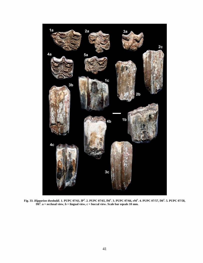
41
Fig. 33. Hipparion theobaldi. 1. PUPC 07/61, lP2. 2. PUPC 07/65, lM1. 3. PUPC 07/66, rM1. 4. PUPC 07/57, lM2. 5. PUPC 07/58,
lM3. a = occlusal view, b = lingual view, c = buccal view. Scale bar equals 10 mm.

42
Fig. 34. Hipparion theobaldi. 6. PUPC 07/60, rP2. 7. PUPC 07/59, rP3. 8. PUPC 07/78, lP4. 9. PUPC 07/124, rM3. a =
occlusal view, b = lingual view, c = buccal view. Scale bar equals 10 mm.
PUPC 07/59 includes the P3 of the right
mandible (Fig. 34(7)). It is in excellent state of
preservation and in an early stage of wear. The metaconid
and metasylid are joined by a narrow isthamus giving the
appearance of double knot. The lingual side of the tooth
is vertically higher than the buccal one. The ectostylid
and mesostylid are well preserved and prominent. The
ectosylid is prominent pillar like structure. The
protostylid is absent. There is a deep plication formed by
the union of the posterior side of the parastylid and the

43
anterior side of the protoconid. There are two prominent
invaginations lingually, named the metaflexid anteriorly
and the entoflexid posteriorly. The metaflexid is narrow
in the middle, while broad anteroposteriorly. The
entoflexid is elongated, curved anteriorly, while broad
posteriorly. The wall of the entoflexid is wrinkled.
The lower fourh premolar PUPC 07/78 is in
early wear (Fig. 34(8)). The protoconid and the
hypoconid are comparatively broad. The metaconid is
broad and triangular and united with narrow isthmus. The
protostylid is bulky and narrow, while the mesostylid and
the entostylid are weak. The metaflexid is narrow while
the entoflexid is triangular in shape and narrow. The
hypoconulid is absent but the mesostylid is present on the
lingual side.
Lingualflexids are shallow on all premolars.
Entoflexids are elongate and have complex anteriormost
borders where they separate metaconid/metastylid. The
premolars have ectoflexids which do not separate the
metaconids/metastylids. Metaconid is rounded on the P2,
having angular posterior borders on the P3-4. Metastylids
have marked angular posterior borders on all premolars.
Protostylids are present on P3-4.
The ectoflexid in the lower third molar PUPC
07/124 (Fig. 34(9)) does not separate metaconid and
metaflexid. The molar has angular facing borders of
metaconid and metastylid. There are no plicaballinids.
Lingualflexid is deeply incised and elongate. Entoflexid
is elongate and has a complex border. Metaflexid is
square and has complex border. The protostylid is present
in the molar. The hypoconulid is present which is
concave buccally. The comparative dental measurements
are provided in table 11.
Comparison and Discussion
The general appearance of the studied specimens
and strong pillar like isolated protocone exclude the
specimens from the genus Equus and favor their inclusion
in the genus Hipparion. The Middle Siwalik is
represented by four Hipparion species H. nagriensis, H.
perimense, H. theobaldi and H. antiopinum (Colbert,
1935). Two of them are found abundant in the Middle
Siwaliks (Iqbal et al., 2009). Hipparion nagriensis is
comparatively small species (Mac Fadden and
Woodburne, 1982; Naseem et al., 2009). The protocone
of H. perimense is flattened lingually and rounded
buccally (Ghaffar, 2005). Hipparion antilopinum is large
with complicated plications and oval shape protocone.
Hipparion theobaldi are large, having less complicated
plications. Hipparion theobaldi differs from H.
antilopinum of having compressed protocone, as
compared to round oval shape protocone in H.
antilopinum. Furthermore, the enamel borders of cavities
relatively simple in H. theobaldi and complicated in H.
antilopinum (Lydekker, 1882; Colbert, 1935).
The morphometric study of the specimens reveal
all the features of species H. theobaldi (Figs. 33-35) as
described by Lydekker (1882), Colbert (1935) and
Ghaffar (2005) and the specimens are assigned to H.
theobaldi. This species is characterized by the isolated,
compressed and pillar like protocone, the molar size is
greater than H. antilopinum and H. nagriensis, and
smaller than H. perimense. The enamel bordering of the
cavities are relatively simple. The specimens are
extremely hypsodont and show less complicated
plications. The studied specimens show the same basic
features of the species such as an anterostyle in premolars
and isolated protocone in cheek teeth, simple enamel
bordering of central cavities with large size. According to
Colbert (1935), it is heavy and larger species as compared
to H. antilopinum and H. nagriensis. All the studied
premolars reflect hipparionine features i.e., they are
longer than broad with longitudinally elongated
protocones and hypocones (Figs. 33-34). The enamel
plications are moderately complicated. These features are
also mentioned by Lydekker (1882) with type specimen.
The genus Hipparion was erected by Christol de
(1832) on the basis of fossil material collected from the
Turolian age locality of Mt-Luberon in the province of
Vaucluse in France. Christol de (1832) characterized this
Mt-Luberon horse with isolated protocones in the upper
molars and tridactyl feet. Christol did not designate a type
species of the genus. This was done by Gervais (1849)
when he described a syntypic series from Mt-Luberon,
including H. prostylum, H. mesostylum and H.
diplostylum.
The name Sivalhippus theobaldi was introduced
by Lydekker (1877a). Shortly thereafter Lydekker
(1877b) synonymized Sivalhippus with Hippotherium
retaining the name Hippotherium. Lydekker used the
combination Hippotherium theobaldi in two subsequent
publications (1882, 1883) until finally synonymizing the
genus with Hipparion as H. theobaldi (Lydekker, 1885,
1886). Later on, the name H. theobaldi was followed by
Pilgrim (1913), Matthew (1929), Colbert (1935),
Gromova (1952), Hussain (1971) as referring smaller
specimens to H. antilopinum and large to H. theobaldi.
Forsten (1968) however choose not to recognize H.
antelopinum arguing that all Siwalik hipparions belong to
one polytypic species H. primigenium Skinner and Mac
Fadden (1977) recognize the distinctiveness of both these
species but referred them Cormohipparion. Mac Fadden
(1984) similarly recognizing the species Cormohipparion
theobaldi. Phylogenetically Cormohipparion theobaldi
appears to lie close to primitive Eurasian Hipparion as
morphologically idealized by H. primigenium. The
specimen in AMNH and yale-GSP collection referred to
Cormohipparion theobaldi are more robust then any of
the metapodial material for Eurasian primitive
hipparionines.

44
Skinner and Mac Fadden (1977) created new
genus Cormohipparion and stated that all the
hipparionine should be included in this new genus. But
later on Bernor and Hussain (1985) stated that there is no
need to erect new genus. Moreover, Mac Fadden and
Skinner (1977) have also suggested that it is highly
probable that all the Siwalik hipparionines should be
included in the genus Cormohipparion with specific
differences based on dentition size and feet which are
characteristics, classically employed in hipparion
taxonomy. Forsten (1968) was the most extreme in
jumping all the Siwalik Hipparion specimens into single
highly variable population of H. primigenium. According
to him, the Siwalik Hipparion group is evolutionary
conservative, and compares closely with H. primigenium
and other primitive horses in complex cheek teeth
fossette ornamentation, double or complex plicaballins,
deeply incised hypoglyph and elongate P2 anterostyle. He
further argued that C. gracile and C. moldavicum are also
best derived from Eurasian horses similar to H.
primigenium. The presence of an anterior rim on facial
fossa is also a primitive character shared by several
Holarctic hipparionines.
Hipparion antilopium and H. theobaldi are
significantly different in facial, dental and possibly
posteranial features from species of North American
cormohipparions (Ghaffar, 2005). The North American
cormohipparions and Eurasian hipparionines obscure the
plylogentic relationship of Holarctic cormohipparion.
Subsequently, a long series of investigations on old world
hipparions, have incorporated these original
morphological characters in referring a vast array of
species to this one genus. Consequently, a strong central
dogma has arisen in the systematics of Hipparion. Most
palaeontologists believed that hipparion evolved in the
New World from an unidentified species of Merychippus.
But some recent investigators have argued that several
distinct lineages may be present in the Old World and the
Old World morphology evolutionary pattern may not be
accurate. As a result, the genus Hipparion has become
more circumscribed in its morphological characterization
and the number of species included within the genus.
Hipparion first appeared in the Siwaliks by a
single migration record in lithologic boundary of the
Nagri Formation (Hussain, 1971). The Nagri, type
locality of the Nagri Formation has yielded the richest
record of the Siwalik hipparions. Hipparion species are
important markers in faunal correlations known as fossil
record from middle Miocene to Pleistocene. The oldest
occurrence of the Siwalik hipparion is from the Nagri
Formation, ca 9.5 Ma (Hussain, 1971), while the old
world occurrence of Equus is from Hasnot, ca 2.48 Ma
(Barry et al., 1982) that is the oldest occurrence of Equus
in Southern Asia. Hussain (1971) made the first
contemporary revision of the Siwalik hipparions. He
suggested that Hipparion first appeared in the Siwaliks
by a single migration record in lithologic boundary of the
Nagri Formation and subsequently underwent
autochthonous evolution. He recognized three species of
the Siwalik Hipparion: (1) H. nagriensis, (2) H.
theobaldi, and (3) H. antilopium. More recently, Ghaffar
et al. (2003) considered the validity of the genus
Hipparion with its four species H. antilopinum, H.
sivalensis, H. theobaldi and H. nagriensis.
Recently, Bernor et al. (1996) again grouped the
Siwalik hipparion into two genera: Sivalhippus and
Hipparion. Sivalhippus is a large size genus with two
species, S. theobaldi and S. perimense, and a small size
genus Hipparion with one species H. sp. (Bernor et al.,
1996).
A.
H. theobaldi
0
5
10
15
20
25
30
0 10 20 30 40 50Length
Wid
th
Upper Second Premolar Upper First Molar
Upper Second Molar Upper Third Molar
B.
H. theobaldi
0
5
10
15
20
25
30
0 5 10 15 20 25 30 35
Length
Wid
th
Lower Second Premolar Lower Third Premolar
Lower Fourth Premolar Lower Third Molar
Fig. 35. Scatter diagram showing dental proportions
of H. theobaldi’s studied sample. The referred
materials are taken from Colbert (1935) and
Ghaffar (2005).

45
Family Rhinocerotidae Owen, 1848
Subfamily Rhinocerotinae Owen, 1845
Tribe Teleoceratini Hay, 1885
Genus Brachypotherium Roger, 1904
Type Species: Aceratherium perimense Falconer and
Cautley, 1847.
Generic Diagnosis: A rhinoceros of gigantic size with
hypsodont teeth. Skull rather short and deep, with
retracted nasals; zygomatic arch heavy; postglenoid
separate from posttympanie. Upper incisor present and
well developed. Molars with moderately developed
crochet, weaker antecrochet and rudimentary crista.
Protocone somewhat pinehed off. Lower molars narrow
and compressed. Mandibular symphysis narrow (Heissig,
1972).
Known Distribution: Southern Asia, South-Eastern Asia
and Western Asia (Heissig, 1972).
Brachypotherium perimense Falconer and Cautley,
1847
Cotypes: The specimens figured by Falconer and
Cautley, (1847: pl. LXXV, figs. 13-16, and LXXVI, figs.
14-17).
Type locality: Peram Island, India (Colbert, 1935).
Stratigraphic Range: Lower to Middle Siwaliks (Khan
A. M., 2010).
Diagnosis: Very large species of the genus
Brachypotherium with relatively high cheek teeth. All
generic features are extremely developed. Nasals are
shortened and hornless. The upper molars have weak
constrictions of the inner cusp; reduced antecrochet
usually present. Upper Premolars are molariform, usually
with highly convex exterior. Lower molars are almost
without buccal fold; cingula usually reduced and short
(Heissig, 1972).
Studied Specimens: Lower dentition: PUPC 07/52, a
right mandibular ramus having P3 – M3. PUPC 07/53, a
left mandibular ramus having P3 – M2.
Description
Mandible: PUPC 07/52 and PUPC 07/53 is a well
preserved right and left mandibular ramii respectively
(Figs. 36-37). The hemimandible PUPC 07/52 is
moderately long having 390 mm length. PUPC 07/52
mandible depth at the P3 is 62 mm, at the P4 is 67 mm, at
the M1 is 82 mm, at the M2 is 84 mm and at the M3 is 86
mm (Fig. 36). The horizontal ramii are thick and their
lower margins are slightly curved. The sagittal lingual
groove is wide posteriorly and absent anteriorly.
Lower Dentition: The molars are large and wide. All the
cheek teeth are in early wear and show the distinct
morphology of rhinoceros; the valleys are open lingually
(Figs. 36(1a), 37(1a)). The enamel is fairly thick, uniform
in thickness and rugose. The buccal and the lingual
cingula are absent. In P3 the paraconid has an anterior
extension. The protoconid is round and has a buccal
extension. The hypoconid is large and the entoconid is
round. The paralophid is long and the protolophid and the
metalophid are broad. The hypolophid is long. The
anterior valley is not well developed. The posterior valley
is U-shaped.
The paraconid is round in the P4. The
metalophid is not distinct from the hypolophid at the
point of wear. The anterior valley is not well developed
and V- shaped. The posterior valley is deep and U-
shaped. In M1 the paraconid is long and narrow but
shorter than that of the protoconid. The hypolophid is
long and prominent but not distinct from the metalophid
at their junction due to wear. The paralophid is long and
narrow than the protolophid. The anterior and the
posterior valleys are V – shaped. The damaged paraconid
is slightly round in the M2. The protoconid is long and
broad. The metaconid is short. The hypoconid is broad.
The entoconid is long, little worn and have lingual
extension. The paralophid is short. The protolophid and
the hypolophid are broad and long. The anterior valley is
slightly U-shaped. The posterior valley is deep and U-
shaped. The M3 hypolophid is long. The valleys are U-
shaped.
PUPC 07/53 is a well-preserved left mandibular
ramus with P3-M2 (Fig. 37). It is 280 mm in length. All
the cheek teeth are well preserved and show the distinct
morphology. The mandible is moderately long. The
vertical height below P3 is 62 mm, below P4 is 66 mm,
below M1 is 82 mm, and below M2 is 85 mm. The P3 has
anterior extension in the paraconid. The protoconid is
broad. The metaconid and the entoconid are round. The
hypoconid is wide. The metalophid and the protolophid
are joined. The anterior valley is not well developed. The
posterior valley is V – shaped. The buccal and lingual
cingula are absent.
P4 has slightly broad paraconid. The entoconid,
protoconid and metaconid are round but the metaconid is
not distinct from protoconid due to wear. The hypoconid
is long while the paralophid is short. The metaconid and
the entoconid have lingual extension. The protolophid is
long. The metalophid is broad. The hypolophid is long
and wide. The protolophid and the hypolophid seem to be
joining each other due to wear. The anterior valley is not
well developed while the posterior valley is U – shaped.
There is V – shaped trigonid.
The paraconid is short in M1. The protoconid is
broad. The metaconid is not distinct due to wear. The
hypoconid is indistinct but long. The entoconid is pointed
and has lingual extension. The paralophid is narrow. The

46
protolophid is long and broad. The hypolophid is long
and wide. The metalophid is broad. The posterior valley
is U-shaped. The paraconid is constricted in M2. The
protoconid is broken but broad. The metaconid is round.
The hypoconid is pointed and the entoconid is round. The
parolophid is short and narrow. The protolophid is worn.
The hypolophid is prominent and long. The anterior
valley is slightly V – shaped. The posterior valley is
wide, deep, long and U – shaped. The measurements are
provided in table 12.
Comparison and Discussion
The studied lower dentition from the Nagri type
area is identical to Brachypotherium perimense in
morphology to that described by Heissig (1972) from the
Nagri Formation of the Middle Siwaliks. PUPC 07/52
and PUPC 07/53 are comparable to AMNH 19454, as
identified of Aceratherium perimense (Colbert, 1935).
PUPC 07/652 and PUPC 07/53 (Table 12; Fig. 38) are
comparable to the sample described by Cerdeno and
Hussain (1997). The dental measurements of PUPC 07/52
and PUPC 07/53 show that both specimens probably
belong to the same animal.
Colbert (1935) recognized Aceratherium
perimense from the Lower and the Middle Siwalik
sediments while Heissig (1972) placed the species in
Brachypotherium. Heissig (1972) reported
Brachypotherium in the Kamlial Formation of the Lower
Siwaliks. Cerdeno and Hussain (1997) described fossil
remains of Brachypotherium perimense from the
Miocene Manchar Formation, Sind, Pakistan, whose
morphology is similar to those described by Heissig
(1972) from the Siwlaiks of Pakistan; the P1 being wider,
the M2 narrower, and the lower teeth having closer
dimensions. Other postcranial remains from the Sind are
smaller than those described by Heissig (1972), but this
difference in size may be due to older age of the Manchar
Formation (Lower Chinji) with respect to the latter
specimens that belongs to the middle and upper Chinji,
Nagri or Dhok Pathan formations (Cerdeno and Hussain,
1997). The difference of the dental remains size may also
be attributed to the age differences of animals.
Gentry (1987) while describing the
Brachypotherium sp. from Miocene of Saudi Arabia
considered the large size and flatness of the buccal wall
of upper molars and the small size of the paracone rib in
comparison with the large flat area, persistent internal
cingula on its upper cheek teeth and external cingula on
its upper and lower molars as important characteristics
for its generic identity. Antoine et al. (2000) considered
the European Brachypotherium brachypus as an Asiatic
migrant because closely related species have previous
occurrence in Pakistan and the surrounding areas. They
(Antoine et al. 2000) suggested that Aprotodon
fatehjangense (Pilgrim, 1910) described from Asia has a
very close resemblance with Brachypotherium brachypus
and must be regarded as recent synonym of
Brachypotherium Roger, 1904. Brachypotherium
fatehjangense is senior synonym of Aprotodon
fatehjangense (Antoine and Welcomme, 2000).
Brachypotherium is supposed to have a
preference for soft diet and a more forested environment
(Andrew et al., 1996, 1997), which is comparable to the
middle Miocene Dhok Pathan Formation in the Siwlaiks.
Brachypotherium perimense (Colbert 1935) is a large
species; lower teeth are charactersized by the smooth
external groove, hardly marked, as it is in other
teleocertines (Cerdeno and Hussain, 1997). Heissig
(2003) indicated that Brachypotherium perimense is the
most frequent species in times of transition and rare
during most humid and most arid times and this species
point out in the Nagri Formation the beginnings of less
humid conditions. Brachypotherium has often been
compared to hippos, and was certainly a marsh or lake
dweller (Geraads and Sarac, 2003).
Table 12: Comparative dental measurements of the
cheek teeth of Brachypotherium in mm
(millimeters). * The studied specimens.
Referred data are taken from Colbert
(1935), Heissig (1972), and Cerdino and
Hussain (1997).
Taxon Number Nature/
Position
Length Width
B.
perimense
PUPC 07/52* P3 41 31
P4 48 33
M1 56 32
M2 62 35
M3 55 35
PUPC 07/53* P3 40 31
P4 47 34
M1 56 33
M2 62 36
AMNH
19454
P3 40 26
P4 49 37
M1 53 36
M2 64 40
M3 72 35
M2 - 33
M2 55 30
M3 67 33

47
Fig. 36. Brachypotherium perimense. 1. PUPC 07/52, a right mandibular ramus having P3 – M3. a = occlusal view,
b = lingual view, c = buccal view. Scale bar equals 50 mm.

48
Fig. 37. Brachypotherium perimense. 2. PUPC 07/53, a left mandibular ramus having P3 – M2. a = occlusal view, b =
lingual view, c = buccal view. Scale bar equals 50 mm.

49
B. perimense
0
10
20
30
40
50
0 10 20 30 40 50 60 70 80 90Length
Wid
th
Low er Third Premolar Low er Fourth Premolar Low er First Molar
Low er Second Molar Low er Third Molar
Fig. 38. Scatter diagram showing dental proportions
of B. perimense’s studied sample. Referred
data are taken from Colbert (1935), Heissig
(1972), and Cerdeno and Hussain (1997).
DISCUSSION
Faunal Correlation: The fauna from the Nagri includes
at present the following species: Listriodon
pentapotamiae, Selenoportax cf. vexillarius, Pachyportax
cf. latidens, Tragoportax punjabicus, Miotragocerus cf.
gluten, Gazella cf. lydekkeri, Giraffokeryx punjabiensis,
Giraffa cf. priscilla, Dorcatherium cf. minus,
Dorcatherium cf. majus, Dorcabune cf.
anthracotherioides, Hipparion theobaldi,
Brachypotherium perimense. The thirteen taxa of the true
ungulates have been recognized from the late Miocene
deposits exposed in the Nagri type locality of the Nagri
Formation, northern Pakistan. This faunal list may be
compared with that of other late Miocene Siwalik
localities, Dhok Pathan and Hasnot and the middle
Miocene Siwalik locality, Chinji. This list obviously
indicates a faunal spectrum of only orders Artiodactyla
and Perissodactyla and several orders such as
Proboscidea, Carnivora are not included in this study.
However, well documented late Miocene mammalian
faunal successions are known from the Siwaliks
(Lydekker, 1876, 1878; Pilgrim, 1937, 1939; Matthew,
1929; Colbert, 1935; Thomas, 1984; Akhtar, 1992, 1996;
Bhatti, 2005; Farooq, 2006; Khan, 2007, 2008; Khan A.
M., 2010). Among the common taxa, the Nagri
Dorcatherium seems more primitive than the Dhok
Pathan one because of its heavy styles and more
bunodont teeth. The Nagri Hipparion also shows a
simplification in plicaballins. The Nagri stratigraphically
is situated below the Dhok Pathan Formation and the list
is characteristic of the early Late Miocene in the
chronologic successions of faunas in the Siwaliks (Barry
et al., 2002).
This faunal list is biased towards large
mammals, because many fossils were recovered during
quarry work, and excavations have been of very limited
extent. Still, this faunal association contains enough
significant elements to allow comparison with some other
upper Miocene faunas from Europe and the Near
Siwaliks. Although the latest Miocene (7.5 – 5.3 Ma)
faunas of the Siwaliks are well known (Pilgrim, 1937,
1939; Akhtar, 1992; Khan, 2007, 2008; Bibi, 2007; Khan
et al., 2009a; Khan, A. M., 2010), the early Late Miocene
(11 – 10 Ma) fauna are poorly known in the Siwaliks.
However, the Nagri fauna is one of the best
representatives for this age.
The Nagri fauna indicates strong resemblance to
the late Miocene fauna of Dhok Pathan and Hasnot
(Akhtar, 1992; Khan, 2007) and the early Late Miocene
faunas of East Africa (Pickford, 1981). However, the
Nagri fauna indicates weak resemblance to the late
middle Miocene of the Chinji Formation (Thomas, 1984).
The Nagri fauna is similar to the faunas from Samos and
Pikermi of Greece, Maragheh of Iran, and Dhok Pathan
Formation of the Siwaliks (Solounias, 1981; Thomas,
1984; Akhtar, 1992; Khan, 2007; Kostopoulos, 2009).
This fauna is particularly significant in providing
evidence regarding the late Miocene faunal interchange
between African and Eurasia (Pickford, 1981; Thomas,
1984; Pilbeam et al., 1997; Barry et al., 2002; Bibi and
Gulec, 2008; Kostopoulos, 2009). The occurrence of
several bovids (Selenoportax vexillarius, Pachyportax
latidens, Tragoportax punjabicus, Miotragocerus gluten,
Gazella lydekkeri), tragulids (Dorcatherium minus,
Dorcatherium majus, Dorcatherium nagrii, Dorcabune
anthracotherioides), hipparionines (“Hipparion
theobaldi” Sivalhippus Complex, Hipparion sp.) and
rhinos (Brachypotherium perimense) was also mentioned
in the late Miocene of the Siwalik, Greco-Iranian
province and Eurasia (Sen et al., 1997; Bibi et al., 2009;
Kostopoulos, 2009). The Nagri fauna is different from
that of Dhok Pathan and Hasnot by not having cervids,
Bramatherium, and large suids.
Gazella is abundantly recorded from Samos,
Pikermi, Maragheh, Dhok Pathan and Hasnot (Pilgrim,
1939; Akhtar, 1992; Khan, 2007; Bibi and Gulec, 2008;
Kostopoulos, 2009). The Siwalik Gazella lydekkeri most
closely approximates the morphology and perhaps the
evolutionary stage of the Eurasian Gazella capricornis
based on primarily on the degree of premolar row
reduction (Bibi and Gulec, 2008). Gazella lydekkeri is
known primarily from the Nagri Formation and the
known age range of Gazella lydekkeri upto the Mio-
Pliocene boundary or beyond (Thomas, 1984).
Selenoportax vexillarius and Pachyportax latidens are
recorded from the late Miocene and Pliocene faunas at
the Siwaliks, the main bone beds of Dhok Pathan and
Hasnot (Khan et al., 2009a). The Dhok Pathan and the
Hasnot faunas are contemporaneous (10.1-3.4 Ma) but
the Nagri faunas are older (ca 11.2-10.1 Ma). An age
range for these species that includes all these localities

50
would be quite broad, potentially as old as the base of the
Nagri up until 3.4 Ma, the upper limit to the Dhok Pathan
Formation (Barry et al., 2002). Tragoportax is present in
the Nagri Formation and also in the Dhok Pathan
Formation (Pilgrim, 1937, 1939; Bibi et al., 2009).
Tragoportax is recorded also from the late Miocene of
Pikermi, Molayan, Samos and Mytilini (Bibi and Gulec,
2008; Bibi et al., 2009).
Listriodon pentapotamiae have been found in
the Siwalik localities ranging in age from Middle
Miocene to early Late Miocene (Pickford, 1988; Van der
Made, 1996; present study). In Europe a few specimens
have been found in sites which have also yielded the
equid Hipparion as in the Nagri type section but by the
end of MN9 listriodonts were extinct everywhere. It is
noted that listriodonts became extinct in Europe, China,
India and Africa over a short period at the end of the
Middle Miocene and the beginning of the Late Miocene.
Thus, the available evidence about the broad tendencies
of listriodont evolution in the Indian subcontinent accords
with that from Europe and China, which supports the
view that for much of the middle Miocene and the early
Late Miocene, Europe, Asia and the Indian subcontinent
were all part of a single biogeographic region (Pickford
and Morales, 2003).
In summary then, the Nagri faunas are similar to
those from Dhok Pathan, Pikermi, Samos, Maragheh and
Sivas, though also some elements to older site like Chinji
(Pickford, 1981; Thomas, 1984; Akhtar, 1992; Pilbeam et
al., 1997; Sen et al., 1997; Barry et al., 2002; Bibi and
Gulec, 2008; Bibi et al., 2009; Kostopoulos, 2009).
Biostratigraphy: The stratigraphical ranges of the bovids
of Nagri are restricted to Late Miocene (Bibi, 2007;
Khan, 2007; Khan et al., 2009a). The suid Listriodon
pentapotamiae is restricted to the earliest Late Miocene
of the Siwaliks (Pickford, 1988; Van der Made, 1996).
More recently, the cervids have been recorded in the
latest Late Miocene of the Siwaliks: Dhok Pathan and
Hasnot (Ghaffar, 2005; Ghaffar et al., 2010). Prior to it
the first appearance of the cervids are recorded from the
late Pliocene of the Siwaliks (Barry et al., 2002). The
cervids are absent in the earliest Late Miocene of the
Siwaliks. The presence of Tragoportax confirms Late
Miocene age but allows no further refinement.
Nevertheless, Miotragocerus indicates a more primitive
bovid assemblage.
Listriodon pentapotamiae is recorded in the
Siwaliks from as early as the middle Miocene until the
earliest Late Miocene (Welcomme et al., 2001). In the
Chinji strata Listriodon pentapotamiae is the most
common suid (Pickford, 1988). Several Middle Siwalik
localities of the Nagri Formation exhibit the presence of
Listriodon pentapotamiae and however, the species is
widely distributed in the Middle Miocene (Pickford,
1988; Pickford and Morales, 2003). Brachyopotherium
perimense is a common species of the Siwalik late
Miocene (Heissig, 1972) and its stratigraphic range is the
earliest Late Miocene to the latest Late Miocene (Heissig,
1972).
Palaeotraginae is documented sporadically in the
Chinji Formation of the Lower Siwaliks (Colbert, 1933;
Bhatti et al., 2007). Giraffokeryx punjabiensis has already
been mentioned several localities of the late Middle
Miocene age (Bhatti, 2005), occupying a wide territory
from Western Europe to India (Bohlin, 1926; Gentry et
al., 1999; NOW database, 2003). A rare occurrence of the
species is found in the early Late Miocene and now from
the Nagri type section confirms its presence in the early
Late Miocene. The persistence of Giraffokeryx
punjabiensis and Listriodon pentapotamiae in the Nagri
type section seems to contradict their time range in the
Siwaliks, where they are considered to be typical Lower
Siwalik elements. Giraffokeryx punjabiensis and
Listriodon pentapotamiae are here rather in favour of an
early Late Miocene age. The Nagri specimens attributed
to Dorcatherium and Dorcobune lack any stratigraphic
indication. The specimens have the typical fossilization
status seen in specimens from the Nagri ravine,
suggesting an early Late Miocene age.
Accordingly, an estimated age of early Late
Miocene age is reasonable for the Nagri fauna, equating
to somewhere between about 11 to 10.1 Ma. This
estimate, based solely on the true ungulates, is in
agreement with previous estimates of 11.2-10.1 Ma for
the Nagri Formation sites of the Siwaliks using
radiometric dating data (Pilbeam et al., 1997; Barry et al.,
1980, 1982, 2002). Pilbeam et al. (1997) gave the
chronology of the top Ghabir sites of the type section and
suggested 10.75 and 10.84 Ma for this section. Barry et
al. (2002) suggested early Late Miocene age for the type
section. Their arguments were mainly
magnetostratigraphic (using ages from the Berggren et
al., 1985, and Cande and Kent, 1995 time scale ) and also
palaeontological (to get possible ages for the Siwalik
hipparionines). The fossiliferous level of the Nagri type
section was radiometrically bracketed between 10.1 and
11.2 Ma (Pilbeam et al., 1997; Barry et al., 2002) and
should consequently be correlated to the earliest
Vallesian, MN9 (Bernor et al., 1996).
In general, the Nagri true ungulates are strongly
indicative of the Middle Siwaliks, which have a
combined absolute range of 10.1-11.2 Ma (Barry et al.,
2002) and relate well to other late Miocene Eurasian
faunas. On the whole, the faunas suggest that the Nagri is
older than Dhok Pathan and Hasnot. The
biostratigraphical position of the Nagri locality relative to
Dhok Pathan and the differences in appearance and
disappearance of fauna suggest that the Nagri fauna is of
early Late Miocene age.

51
Palaeoecology and Palaeoenvironment:
Recent work on the geology of Nagri (Barry et al., 2002)
has documented that the deposits were formed as part of a
large river system by coexisting emergent and interfan
river system, with the larger emergent Nagri system
beginning at 10.1 Ma (Willis and Behrensmeyer, 1995).
Mammal fossils at the Nagri are recovered from fluvial
channel deposits. The interpretation of the Nagri
palaeoenvironment based on geological information is
supported by palaeontological evidence. The Nagri fauna
has an aquatic component that includes a mix of
freshwater animals that can tolerate slow-moving water
with a high sediment contents (e.g. crocodiles, turtles).
The land mammal fauna is dominated by a number of
artiodactyl and perissodactyl species that were likely
preferred an aquatic-margin habitat, as they are found
most commonly in fluviatile deposits (Barry et al., 2002).
Recovery of a fair number of well preserved terrestrial
vertebrates supports previous interpretations of the Nagri
palaeoenvironment, they appear to be preferentially
preserved in swampy and fluviatile settings. As for
palaeoenvironmental indications, the Nagri faunas are
consistently found in deposits that accumulated in or near
swamps and shallow lakes (Badgley and Behrensmeyer,
1980; Badgley, 1986; Badgley and Tauxe, 1990; Barry et
al., 1980, 1982, 2002) and suggest the presence of forest
in the Nagri region at the time of deposition.
The presence of the tragulids and the giraffids,
which were certainly browsers, definitely speaks in
favour of wet forested environments. Hipparion fauna
suggests a sclerophyllous evergreen woodland
environment, similar to today’s mixed monsoon forest
and grassland glades of north central India (Solounias,
1999). Fortelius et al. (1996) studied the body mass
diversity of the West Eurasian suoids in relationship to
environmental conditions, and suggested that the loss of
species of small size was correlated to a progressive
development of increasingly open and seasonal habitats.
During the late Miocene, suid diversity in western
Eurasia was very low, and suids in MN12 and MN13 are
only recorded in the largest size class (201-1000 kg).
Microstonyx major has been recorded from the type
section (Van der Made and Hussain, 1989). The presence
of Microstonyx major and Listriodon pentapotamiae in
the type area of the Nagri Formation confirm that the
environment appears to have been more humid than the
latest late Miocene (Pickford, 1988; Van der Made and
Hussain, 1989; Pickford et al., 2004). It should be noted
that the Nagri has some hypsodont members (Thomas,
1977; Akhtar, 1992). This provides evidence for an open
environment.
Palaeoenvironmental interpretations using
vertebrate fossils are often based largely on palaeodietary
reconstructions using dento-gnathic and postcranial
functional analyses in conjunction with the relative
abundances of ecologically differentiated taxa (Bibi and
Gulec, 2008). Tragoportax from the sites of Pikermi and
Samos (Solounias and Hayek, 1993) as well as Molayan
(Merceron et al., 2004) concluded that Tragoportax
species were variable mixed feeders with strong grazing
habits. An analysis of maxillary and zygomatic
morphology among living and fossil bovids indicated that
Tragoportax were mixed feeders while Gazella was a
mixed feeder or browser (Solounias et al., 1995). More
balanced mixed-feeding was interpreted for microwear of
Gazella (Merceron et al., 2004). A comprehensive study
of the postcranial morphology of extant and extinct
bovids by Köhler (1993) found similarities between the
build and proportions of Tragoportax amalthea and
certain deer (Cervus) and found a leafy diet and light
woodland habitat as most likely for this species. Köhler
also reconstructed Gazella as a browser inhabiting more
open country. Miotragocerus was interpreted as a leaf
and herb-eater inhabiting shrubland to light woodland.
In terms of number of specimens, the Nagri true
ungulate assemblage is dominated by bovids, giraffids
and tragulids with Hipparion and Listriodon,
Brachypotherium constituting much smaller percentages
(Appendix 1). The high proportion of boselaphines may
be representative of the presence of drier and more open
habitats than would be expected for the remaining bovids,
particularly if the fossil gazelle resembled the living
species in their ability to inhabit semi-arid to arid
environments. The specific richness of the boselaphins in
the Nagri type section suggests open areas biomass
(Thomas, 1984) as the living boselaphins (Boselaphus
tragocamelus and Tetracerus quadricornis) prefer open
areas.
Dorcatherium and Miotragocerus are familiar
for more or less closed and humid habitats (Köhler, 1993;
Gentry, 2005; Eronen and Rössner, 2007). This supports
the assumption of an earliest Late Miocene Siwalik
humid habitat with abundant cover. The taxonomic faunal
composition suggests a humid habitat pocket with
abundant cover indicating the dominance of forested
landscapes during the early Late Miocene times of the
Siwaliks. The presence of Dorcatherium in the type
section can be assumed a strong attachment to wet,
forested habitats with dense understory, where the
animals could hide in vegetation or water from predators
(Rössner, 2010).
In summary, the dominance of boselaphins in
the Nagri reflects an environment of mainly open forest.
A significant representation of tragulids with adaptations
to ecotonal wet and swampy habitats indicates humid
conditions. The alternation of dry and flood seasons
would have caused a highly differentiated mosaic ecotone
environment, which would have offered an outstanding
number of habitats and niches and consequently an
exceptionally large number of species. Because of
alternation of ground conditions seasonal migration
events might have occurred. Finally, the Nagri fauna

52
suggest the existence of a vast wetland environment with
alternating dry and flood seasons which forced a mosaic
of ecotonal habitats with many niches and corresponding
adaptations.
CONCLUSIONS
The study of the new true ungulate material
collected from the Nagri type area, the Middle Siwaliks,
Pakistan, allows recognizing 13 species from fifteen
fossil sites, SN1-15. This includes a species of
Listriodon, a species of Hipparion, a species of
Selenoportax, a species of Pachyportax, a great
abundance of the bovid Tragoportax, a Gazella, a
Miotragocerus, a species of Giraffokeryx, and only rare
representation of Dorcatherium, Dorcabune and Giraffa.
The early Late Miocene fossil ungulates from the Nagri
constitute an assemblage that is fairly typical of the
Siwalik region during this time.
From biochronological point of view, the co-
existence of Listriodon pentapotamiae, Hipparion
theolbaldi, Brachypotherium perimense, Selenoportax
and Pachyportax indicate close relationships to the early
Late Miocene of the Siwaliks. The true ungulate fauna
allows a biochronological estimate of early Late Miocene
for the Nagri assemblage based on similarities with
faunas from Dhok Pathan, Hasnot, Maragheh, Pikermi
and Samos, corresponding well to previous estimates
(Thomas, 1984; Barry et al., 2002; Bibi and Gulec, 2008;
Kostopoulos, 2009; Khan et al., 2009a). In conclusion,
the true ungulate assemblage of the Nagri type section
rather indicates an early Late Miocene age (earliest
Vallesian, MN 9) which is in agreement with the
radiometric dating data provided by Barry et al. (2002).
The fossil ungulates indicate the ancient
environment at Nagri comprised woodland to shrubland.
The type area may have comprised relatively more
humid, closed habitats based on the ecomorphology of
their taxa, particularly Miotragocerus and Dorcatherium,
which may have been an inhabitant of wetlands (Köhler,
1993). The Nagri occurrence increases again the
similarities between Indo-Siwalik faunas and those of the
Greco-Iranian province, the early Late Miocene ungulates
common to both realms.
ACKNOWLEDGEMENTS
The publication was made possible by funding
from the University of the Punjab, Lahore, Pakistan.
Sajjad Shah, Maskeen Ali, Abdul Razaq provided
efficient help during the field work and Adeeb Babar
prepared the figures. We would like to thank all the
people from the village Sethi Nagri for their hospitality
and their help.
REFERENCES
Acharyya, S. K. (1994). The Cenozoic Foreland basin
and tectonics of the Eastern Sub-Himalaya:
Problems and prospects. In Kumar, R. and Ghosh
S. K. (eds.) Siwalik Foreland Basin of Himalaya.
Himal. Geol., 15: 1-415.
Akhtar, M. (1992). Taxonomy and Distribution of the
Siwalik Bovids. Ph. D. Diss., University of the
Punjab, Lahore, Pakistan.
Akhtar, M. (1995). Pachyportax giganteus, new species
(Mammalia, Artiodactyla, Bovidae) from the Dhok
Pathan, district Chakwal, Punjab, Pakistan. Pak. J.
Zool., 27 (4): 337-340.
Akhtar, M. (1996). A new species of the genus
Selenoportax (Mammalia, Artiodactyla, Bovidae)
from Dhok Pathan, district Chakwal, Punjab,
Pakistan. Proc. Pak. Cong. Zool., 16: 91-96.
Akhtar, M., Z. B. Hameed, M. Amin and M. NAZIR
(1997). An evidence on the validity of the species
Pachyportax nagrii Pilgrim (mammalia,
Artiodactyla, Bovidae). Pak. J. Geol., 4: 1-3.
Anderson, R. V. (1927). Tertiary stratigraphy and
orogeny of the Northern Punjab. Geol. Soc. Am.
Bull., 38: 665-720.
Andrews, P. and J. E. Cronin (1982). The relationships of
Sivapithecus and Ramapithecus and the evolution
of the orangutan. Nature, 297: 541-546.
Andrews, P., T. Harrison, E. DELSON, R. L. BERNOR
and L. MARTIN (1996). Distribution and
biochronology of European and southwest Asian
Miocene catarrhines. In (R.L. Bernor, V.
Fahlbusch and H.W. Mittman, Eds), The Evoluton
of Western Eurasian Neogene Mammal Founas,
New York: Columbia University Press: pp. 168-
207.
Andrews, P., J. Lord and E. M. EVANS (1997). Patterns
of ecological diversity in fossil and modern
mammalian faunas. Biol. J. Linn. Soc., 11: 177-
205.
Antoine, P. O. and J. L. Welcomme (2000). A new
Rhinoceros from the Lower Miocene of the Bugti
Hills, Baluchistan, Pakistan: The earliest
Elasmotheriine. Palaeont., 43(5): 795-816.
Antoine, P. O., C. Bulot and L. Ginsburg (2000). Une
faune rare de rhinocérotidés (Mammalia,
Perissodactyla) dans leMiocène inférieur de
Pellecahus (Gers, France), Geob., 33: 249–255.
Arambourg, C. and J. Piveteau (1929). Les Vertebres du
Pontien de Salonique. Ann. Paleont., 18: 57-140.
Arambourg, C. (1933). Mammiferes Miocenes du
Turkana (Afrique Orientale). Ann. Paleont. Paris,
22: 121-148.
Badgley, C. E. and L. Tauxe (1990). Paleomagnetic
stratigraphy and time in sediments: studies in

53
alluvial Siwalik rocks of Pakistan. J. Geol., 98:
457-477.
Badgley, C. E. and A. K. Behrensmeyer (1980).
Paleoecology of Middle Siwalik sediments and
faunas, northern Pakistan. Palaeog., Palaeocl.,
Palaeoec., 30: 133-155.
Badgley, C. (1986). Taphonomy of mammalian fossil
remains from Siwalik rocks of Pakistan. Paleob.,
12: 119-142.
Badgley, C., D. Will and F. Lawrence (2008).
Taphonomy of Small-Mammal Fossil
Assemblages from the Middle Miocene Chinji
Formation, Siwalik Group, Pakistan. Nat. Sc. Mus.
monographs, 14: 145-166.
Barry, J. C., A. K. Behrensmeyer and M. Morgan (1980).
A geologic and biostratigraphic framework for
Miocene sediments near Khaur Village, northern
Pakistan. Postilla, 183: 1-19.
Barry, J. C., E. H. Lindsay and L. L. Jacobs (1982). A
biostratigraphic zonation of the Middle and Upper
Siwaliks of the Potwar Platue of northern Pakistan.
Palaeog., Palaeocl., Palaeoec., 37: 95-130.
Barry, J. C. and L. J. Flynn (1990). Key biostratigraphic
events in the Siwalik Sequence. In: European
Neogene Mamma Chronology (ed. E.H. Lindsay et
al.), pp. 557-571. New York.
Barry, J. C., M. E. Morgan, A. J. Wrinkler, L. J. Flynn, L.
H. Lindsay, L. L. Jacobs and D. Pilbeam (1991).
Faunal interchange and Miocene terrestrial
vertebrates of southern Asia. Paleob., 17: 231-245.
Barry, J. C., M. E. Morgan, L. J. Flynn, D. Pilbeam, L. L.
Jacobs, E. H. Lindsay, S. M. Raza and N.
Solounias (1995). Patterns of faunal turnover and
diversity in the Neogene Siwaliks of northern
Pakistan. Palaeog., Palaeocl., Palaeoec., 115: 209-
226.
Barry, J. C. M. E. Morgan, L. J. Flynn, D. Pilbeam, A. K.
Behrensmeyer, S. R. Mahmood, I. A. Khan, C.
Badgley, J. Hicks and J. Kelley (2002). Faunal and
environmental change in the late Miocene Siwaliks
of northern Pakistan. Paleob., 28 (2): 1-71.
Barry. J. C., S. Cote, L. Maclatchy, E. H. Lindsay, R.
Kityo and A. R. Rajpar, (2005). Oligocene and
Early Miocene Ruminants (Mammalia,
Artiodactyla) from Pakistan and Uganda. Palaeont.
Electr., 8(1): 1- 29: 885MB.
Behrensmeyer, A. and J. Barry (2005). Biostratigraphic
Surveys in the Siwaliks of Pakistan. A Method for
Standardized Surface Sampling of the Vertebrate
Fossil Record. Palaeont. Electr., 8(1): 1-24.
Behrensmeyer, A. K. (1987). Miocene fluvial facies and
vertebrate and vertebrate taphhonomy in northern
Pakistan. In recent developments in fluvial
sedimentology (ed. F. G. Ethridge. R. M Flores,
and M. D. Harvey). Society of Economic
Paleontologists and Mineralogists. Spec. Pub., 39:
169-176.
Berggren, W. A., D. V. Kent, J. J. Flynn and J. A. van
Couvering (1985). Cenozoic geochronology. Geol.
Soc. Am. Bull., 96: 1407-1418.
Bernor, R. L. and S. T. Hussain, 1985. An assessment of
the systematics phylogenetic and biogeographic
relationships of Siwalik Hipparionine Horses. J. V.
P., 5(1): 32-87.
Bernor, R. L. (1984). A zoogeographic theater and
biochronologic play: the time biofacies
phenomena of Eurasian and African Miocne
mammal provinces. Paleobio. Continent., 14: 121-
142.
Bernor, R. L. (1986). Mammalian biostrtigraphy,
geochronology, and zoogeographic relationships
of the Late Miocene Maragheh fauna, Iran. J. Vert.
Palaeont., 6: 76-95.
Bernor, R. L., G. D. Koufos, M. O. Woodburne and M.
Fortelius (1996). The evolutionary history and
biochronology of European and Southwest Asian
Late Miocene and Pliocene hipparionine horses. In
chapter 26: Evolution of Western Eurasian Neoge.
307-338 pp.
Bhatti, Z. H. (2005). Taxonomy, Evolutionary History &
Biogeography of the Siwalik Giraffids. Ph. D.
Diss., University of the Punjab, Pakistan.
Bhatti, Z. H., M. A. Qureshi, M. A. Khan, M. Akhtar, A.
Ghaffar and M. Ejaz (2007). Individual variations
in some premolars of species Giraffokeryx
punjabiensis (Mammalia, Giraffidae) from Lower
Siwalik (Chinji Formation) of Pakistan.
Contribution to Geology of Pakistan; Proc. Pak.
Geol. Cong. 2004, 5: 261-272.
Bibi, F. and E. S. Gulec (2008). Bovidae (Mammalia:
Artiodactyla) from the Late Miocene of Sivas,
Turkey. J. V.P., 28(2): 501-519.
Bibi, F. (2007). Origin, paleoecology and
paleobiogeography of early Bovini. Palaeog.,
Palaeocl., Palaeoec., 248(1-2): 60-72.
Bibi, F., M. Bukhsianidze, A. W. Gentry, D. Geraads, D.
S. Kostopoulos and E. S. Vrba (2009). The Fossil
Record and Evolution of Bovidae: State of the
Field. Palaeont. Elect., 12(3): 1-11.
Blainville, H. M. (1816). Sur plusieurs espèces d animaux
mammifères de 1’ order des Ruminants. Bull. Soc.
Phil. Paris, 73-82.
Bohlin, B. (1926). Die familie Giraffidae. Palaeotologia
Sinica Pekin. C4(I): 1-78.
Cande, S. C. and D. V. Kent (1995). Revised calibration
of the geomagnetic polarity time scale for the
Cretaceous and Cenozoic. J. Geoph. Res., B100:
6093-6095.
Cerdeno, E. and T. Hussain (1997). On the
Rhinocerotidae from the Miocene Manchar

54
Formation, Sind, Pakistan. Pak. J. Zool., 29(3):
263-276.
Cerdeno, E. (1995). Cladistic analysis of the Family
Rhinocerotidae (Perissodactyla). Am. Mus. Novit.,
3143: 1-25.
Chauhan, P. R. (2003). An overview of the Siwalik
Acheulian & Recosidering its chronological
relationship with the Soanian. A theoretical
perspective. The Sheffield graduate J. Arch., 7: 1-
17.
Christol, J. de (1832). [No title, Description of
Hipparion]. Annales des Science et de I’ Industrie
du Midi de la France, 1: 180-181.
Colbert, E. H. (1933). A skull and mandible of
Giraffokeryx punjabiensis (Pilgrim). Am. Mus.
Novit., 632: 1–14.
Colbert, E. H. (1935). Siwalik mammals in the American
Museum of Natural History. Trans. Am. Phil. Soc.,
N. S., 26: 1-401.
Cotter, G. P. (1933). The geology of the part of the
Attock district, West of Longitude 72 45; Mem.
Geol. Surv. India, 55: 63-161.
Dennell, R. W., R. Coard, A. Turner (2008). Predators
and Scavengers in Early Pleistocene southern
Asia. Quater. Int., 192: 78-88.
Dennell, R., R. Coard and A. Turner (2006). The
biostratigraphy and magnetic polarity zonation of
the Pabbi Hills, northern Pakistan: An Upper
Siwalik (Pinjor Stage) Upper Pliocene-Lower
Pleistocene fluvial sequence. Palaeog., Palaeocl.,
Palaeoec., 234: 168-185.
Eronen, J. T. and G. E. Rössner (2007). Wetland paradise
lost: Miocene community dynamics in large
herbivorous mammals from the German Molasse
Basin. Evolut. Ecol. Res., 9: 471– 494.
Falconer, H. (1868). Palaentological memoirs. Eds. by
R.I. Murchison, Lond., 1: 157-169.
Farooq, U. (2006). Studies of Evolutionary Trends in
Dentition of the Siwalik Tragulids. Ph. D. Diss.,
University of the Punjab, Pakistan.
Farooq, U., M. A. Khan and M. Akhtar (2007a).
Dorcabune nagrii (Ruminantia, Tragulidae) from
the Upper part of the Middle Siwaliks. J. Appl.
Sc., 7(10):1428-1431.
Farooq, U., M. A. Khan, M. Akhtar and A. M. Khan
(2007b). Dorcatherium majus, a study of upper
dentition from the Lower and Middle Siwaliks of
Pakistan. J. Appl. Sc., 7(9): 1299-1303.
Farooq, U., M. A. Khan, M. Akhtar and A. M. Khan
(2007c). Dorcabune anthracotherioides
(Artiodactyla, Ruminantia, Tragulidae) from
Hasnot, the Middle Siwaliks, Pakistan. Pakistan
Journal of Zoology, 39(6): 353-360.
Farooq, U., M. A. Khan and M. Akhtar (2007d).
Dorcabune nagrii (Ruminantia, Tragulidae) from
the Upper Part of the Middle Siwaliks. Journal of
Applied Sciences, 7(10): 1428-1431.
Farooq, U., M. A. Khan, M. Akhtar, Khan, A. M. (2008).
Lower Dentition of Dorcatherium majus
(Tragulidae, Mammalia) in the Lower and Middle
Siwaliks (Miocene) of Pakistan. Turk. J. Zool., 32:
91-98.
Flynn, L. J., J. C. Barry, M. E. Morgan, Pilbeam, D., L.
L. Jacobs and E. H. Lindsay (1995). Neogene
Siwalik Mammalian Lineages: Species longevities,
rates of change, and modes of speciation. Palaeog.,
Palaeocl., Palaeoec., 115: 249-264.
Flynn, L. J., D. Pilbeam, L. L. Jacobs, J. C. Barry, A. K.
Behrensmeyer and J. W. Kappelman (1990). The
Siwaliks of Pakistan: Time and faunas in a
Miocene terrestrial setting. J. Geol., 98: 589-604.
Forsten, A. M. (1968). Revision of the Palearctic
Hipparion. Act. Zool. Fennico, 119: 1-134.
Fortelius, M., Van der J. Made, R. Bernor (1996). Middle
and Late Miocene Suoidea of central Europe and
the eastern Mediterranean: evolution,
biogeography and paleoecology. In: Bernor, R., et
al. (Eds.), the evolution of Western Eurasian
Neogene mammal faunas. Columbia University
Press, NewYork, pp. 348–377.
Gaudry, A. (1865). Animaux fossils at geologie de I.
Attique, Paris, pp. 264-308.
Gentry, A. W. and J. J. Hooker (1988). The phylogeny of
Artiodactyla. In: The Phylogeny and Classification
of the Tetrapods. Vol. 2: Mammals, pp. 235-272
(ed. M. J. Benton), Systematics Association
Special Volume No. 35B Clarendon, Oxford.
Gentry, A. W. (1966). Fossil Antilopini of East Africa.
Bull. Brit. Mus. Nat. Hist. (Geol). 12: 45-106.
Gentry, A. W. (1970). The Bovidae (Mammalia) of the
Fort Ternan fossil fauna: pp. 243-323 in L. S. B.
Leakey and R. J. G. Savage (eds.). Fossil
Vertebrates of Africa, Vol. 2. Academic Press,
London.
Gentry, A. W. (1974). A new genus and species of
Pliocene boselaphine (Bovidae, Mammalia) from
South Africa. Ann. S. Afr. Mus., 65(5): 145-188.
Gentry, A. W. (1978). Bovidae; pp. 540-572 in V. J.
Maglio and H. B. S. Cooke (eds.), Evolution of
African Mammals. Harvard University Press,
Cambridge, Massachusetts.
Gentry, A. W. (1994). The Miocene differentiation of Old
World Pecora (Mammalia). Hist. Biol., 7: 115-
158.
Gentry, A. W. (1997). Fossil ruminants (Mammalia) from
the Manonga Valley. Tanania. In Neogene
Paleontlogy of the Manonga Valley, Tanzania, pp.
107-135 (ed. T. J. Harrison). Plenum Press, New
York.
Gentry, A. W. (1999). Fossil Pecorans from the
Baynunah Formation, Emirate of Abu Dhabi,

55
United Arab Emirates. In: Whybrow P.J. Hill A.,
eds. Fossil Vertebrates of Arabia. Yal University
Press, New Haven, 290-316.
Gentry, A. W., G. E. Rossner, and E. P. S. Heizman
(1999). Suborder Ruminantia, in Rossner, G. E.,
and Heissig, K. (eds.). The Miocene land
mammals of Europe: Munchen, Verlag Dr.
Friedrich Pfeil, 225-258.
Gentry, A. W. (2005). Ruminants of Rudabanya.
Palaeontographica Italica, 90: 283-302.
Geraads, D. and G. Sarac (2003). Rhinocerotidae from
the Middle Miocene Hominoid locality of Candir
(Turkey). Cour. Forsch. Inst. Senckenberg, 240:
217-231.
Geraads, D. (1986). Remarques sur la systrematique et la
phylogenic des Girffidae (Artiodactyla.
Mammalia). Geob., 19: 465-477.
Geraads, D., E. Guluc and G. Sarac (1995). Middle
Miocene Ruminants from Inonu, Central Turkey.
N. Jb. Geol. Palaont. Mh. Stuttgart, 8: 462- 474.
Gervais, P. (1849). Note sur la multiplicite des especes d’
hipparions (genre de Chevaux a trios droits) qui
sont enfouis a cucuron (Vaucluse). Comptes
Rendus, Academie des Sc. de Paris, 29: 284-286.
Ghaffar, A. (2005). Studies on Equids, Cervids and
Carnivora from the Siwalik Hills of Pakistan. Ph.
D. Diss., University of the Punjab, Lahore,
Pakistan.
Ghaffar, A., M. A. Khan, and M. Akhtar (2010). Early
Pliocene Cervids (Artiodactyla-Mammalia) from
the Siwaliks of Pakistan. Yerbilimleri (Earth
Sciences), 31(3), 217-231.
Ghaffar, A., A. Q. Nayyer, Z. H. Bhatti and M. Akhtar
(2003). Critical analysis of Siwalik equids. Sci.
Int. (Lahore), 15(2): 177-178.
Ginsburg, L. (1974). Les Tayassuidés des phosphorites
du Quercy. Palaeovert., 6: 55-85.
Gromova, V. (1952). Lc genre Hipparion (translated
from Russian by St. Aubin). Bur. Res. Min. Geol.,
12: 1-288.
Hamilton, W. R. (1973). The lower Miocene ruminants of
Gebel Zelten, Libya. Bulletin of the British
Museum (Natural History) London. Geol., 21 (3):
75-150.
Han, de-fen (1974). First discovery of Dorcabune in
China. Vertebrate Palasiat., 12(3): 217-221.
Heissig, K. (1972). Palaontolgische and geologische
Untersuchungen im Tertiar Vo Pakistan, 5.
Rhinocerotidae (Mamm.) aus den unteren und
mittleren Siwalik-Schichten. Bayerische Akademie
der Wissenschaften Mathematisch-
Naturwissenschaftliche Klasse, Abhandlungen,
Neue Folge, 152: 1-112.
Heissig, K. (2003). Change and Continuity in Rhinoceros
faunas of Western Eurasia from the Middle to the
Upper Miocene. Eeden, Stara Lensa, pp. 35-37.
Hill, A., R. Drake, L. Tauxe, M. Monaghan, J. C. Barry,
A. K. Behrensmeyer, G. Curtis, B. F. Jacobs, L.
Jacobs, N. Johnson, D. Pilbeam (1985). Neogene
paleontology and geochronology of the Baringo
Basin, Kenya. J. Human Evol., 14: 759–773.
Holland, T. H. (1926). Indian Geological Terminology,
Mem. Geol. Surv. India, LI, Pt. I.
Hooijer, D. A. (1958). Fossil Bovidae from the Malay
Archipelago and the Punjab. Zoolog.
Verhandlungen, 38: 1-110.
Hussain, S. T. (1971). Revision of Hipparion (Equidae,
Mammals) from the Siwalik Hills of Pakistan and
India. Bayerische Akademic der Wissenschaffen,
Abhandlungen, 147: 1-68.
Hussain, S. T., G. D. van den Bergh, K. J. Steensma, J.
A. de Visser, J. de Vos, M. Arif, J. Fan Dam, P. Y.
Sondaar and S. M. Malik (1992). Biostratigraphy
of the Plio-Pleistocene continental sediments
(Upper Siwaliks) of the Mangla-Samwal anticline,
Azad Kashmir, Pakistan. Proc. Koninklyke
Nederlandse Akademie van Wetenschappen. Ser.
B, 95: 65-80.
Iqbal, M., A. Liaqat, M. A. Khan and M. Akhtar (2009).
Some new remains of Hipparion from the Dhok
Pathan type locality, Pakistan. J. Anim. Plant Sci.,
19(3): 154-157.
Jacobs, L. L., L. J. Flynn and W. R. Downs (1989).
Neogene rodents of southern Asia. In: C. C. Black
and M. R. Dawson (Editors). Papers on Fossil
Rodents in Honor of Albert, F. Wood. Natural
History Museum, Los Angeles Company. Sc. Ser.,
33: 157-177.
Jacobs, L. L., L. J. Flynn, W. R. Downs and J. C. Barry
(1990). Quo vadis Antemus? The Siwalik muroid
record. In: E. H. Lindsay, V. Fahlbusch and P.
Mein (Editors). European Neogene Mammal
Biochronology. Plenum Press, New York, pp. 573-
586.
Janis, C. M. and Scott, K. M. (1987a). Grades and clades
in hornless ruminant evolution: the reality of
Gelocidae and the systematic position of
Lophiomeryx and Bachitherium. J. V.P., 7: 200-
216.
Janis, C. M. and Scott, K. M. (1987b). The inter-
relationship of Higher Ruminant families with
special emphasis on the members of the
Cervoidea. Am. Mus. Novit., 2893: 1-85.
Johnson, G. D., P. Zettler, C. W. Naeser, N. M. Johnosn,
D. M. Summers, C. D. Frost, N. D. Opdyke and R.
A. K. Tahirkheli (1982). The occurrence and
fission-track ages of Late Neogene and Quatrnary
volcanic seiments, Siwalik group, northern
Pakistan. Palaeog., Palaeocl., Palaeoec., 37: 63-93.
Johnson, N. M., N. D. Opdyke, G. D. Johnson, E. H.
Lindsay and R. A. K. Tahirkheli (1982). Magnetic
polarity, stratigraphy and ages of Siwalik group

56
rocks of the Potwar Plateau, Pakistan. Palaeog.,
Palaeocl., Palaeoec., 37: 17-42.
Johnson, N. M., J. Stix, L. Tauxe, P. F. Cerveny and R.
A. K. Tahirkheli (1985). Paleomagnetic
chronology, fluvial processes and tectonic
implications of the Siwalik deposits near Chinji
Village, Pakistan. J. Geol., 93: 27-40.
Kaup, J. J. (1833). Vier urweltliche Hirsche des
Darmstedter Museum. Archae. Min. Geo. Bergbau
und Huttenkunde, 6: 217-228.
Keller, H. M. R., A. K. Tahirkheli, M. A. Mirza, G. D.
Johnson, N. M. Johnson and N. D. Opdyke (1977).
Magnetic polarity stratigraphy of the Upper
Swialik deposits, Pabbi Hills, Pakistan. Ear.
Planet. Sc. Letters, 36: 187-201.
Khan, A. M. (2010). Taxonomy and Distribution of
Rhinoceroses from the Siwalik Hills of Pakistan
Ph. D. Diss. (unpublished), University of the
Punjab, Lahore, Pakistan.
Khan, M. A. and M. Akhtar (2011a). Proamphibos
kashmiricus (Bovini: Bovidae: Mammalia) from
the Pinjor Formation of Pakistan. Pakistan Journal
of Zoology, 43(4): 615-621.
Khan, M. A. and M. Akhtar (2011b). Dorcatherium cf.
nagrii from the Chinji Type Locality (Chakwal,
northern Pakistan) of the Chinji Formation, Lower
Siwaliks, Pakistan. Pakistan Journal of Zoology,
43(6): 1101-1109.
Khan, M. A., G. Iliopoulos, M. Akhtar, A. Ghaffar and
Zubaid-Ul-Haq (2011a). The longest tusk of cf.
Anancus sivalensis (Proboscidea, Mammalia) from
the Tatrot Formation of the Siwaliks, Pakistan.
Current Science, 100(2): 249-255.
Khan, M. A., M. Akhtar, A. M. Khan, A. Ghaffar, M.
Iqbal and K. Samiullah (2011b). New Fossil
Locality in the Middle Miocene of Lava from the
Chinji Formation of the Lower Siwaliks. Pakistan
Journal of Zoology, 43(1): 61-72.
Khan, M. A., M. Akhtar, G. Rohi, M. Iqbal and K.
Samiullah (2011c). Sivaceros gradiens Pilgrim
1937 (Mammalia, Bovidae, Boselaphini) from the
Lower Siwaliks of Dhok Bun Amir Khatoon,
Chakwal, Pakistan: Systematics and
Biostratigraphy. Turkish Journal of Zoology,
35(2): 281-286.
Khan, M. A. and M. U. Farooq (2006).
Paleobiogeography of the Siwalik Ruminants. Int.
J. Zool. Res., 2(2): 100-109.
Khan, M. A. (2007). Description of Selenoportax
vexillarius Molars from Dhok Pathan Village
(Middle Siwaliks), Pakistan. Pak. J. Biol. Sc.,
10(18): 3166-3169.
Khan, M. A. (2008). Fossil bovids from the late Miocene
of Padri, Jhelum, Pakistan. Pak. J. Zool., 40(1):
25-29.
Khan, M. A., D. S. Kostopoulos, M. Akhtar and M. Nazir
(2010a). Bison remains from the Upper Siwaliks of
Pakistan. Neu. Jahrb. für Geol. und Paläont.,
258(1): 121-128.
Khan, M. A., S. S. Butt, A. M. Khan and M. Akhtar
(2010b). A New Collection of Giraffokeryx
punjabiensis (Giraffidae, Ruminantia,
Artiodactyla) from the Lehri Outcrops, Jhelum,
Northern Pakistan. Pak. J. Sc., 62(2): 120-123.
Khan, M. A., M. Akhtar and M. Iqbal (2010c). The Late
Miocene Artiodactyls in the Dhok Pathan Type
Locality of the Dhok Pathan Formation, the
Middle Siwaliks, Pakistan. Pakistan Journal of
Zoology, Supplementary Series, 10: 1-90.
Khan, M. A., A. Ghafar, Z. Ali, U. Farooq, Z. B. Hameed
and M. Akhtar (2005b). Report on Mammalian
Fossils of Chinji Formation, Dhulian, Pakistan.
Am. J. Appl. Sc., 2(12): 1619-1628.
Khan, M. A., G. Iliopoulos and M. Akhtar (2009a).
Boselaphines (Artiodactyla, Ruminantia, Bovidae)
from the Middle Siwaliks of Hasnot. Pakistan.
Geob., 42: 739-753.
Khan, M. A., M. Malik, A. M., Khan, M. Iqbal and M.
Akhtar (2009b). Mammalian remains in the Chinji
type locality of the Chinji Formation: A new
collection. J. Anim. Plant Sci.., 19(4): 224-229.
Kingston, J. D., B. F. Jacobs, A. Hill and A. Deino
(2002). Stratigraphy, age and environments of the
late Miocene Mpesida Beds, Tugen Hills, Kenya.
J. Human Evol., 42: 95–116.
Köhler, M. (1993). Skeleton and habitat of recent and
fossil ruminants. Münch. geowissenschaftliche
Abh. (A), 25: 1–88.
Kostopoulos, D. S. (2009). The late Miocene mammal
faunas of the Mytilinii basin, Samos island,
Greece: New collection, 14. Bovidae. Beitr.
Palaont., 31: 339-383.
Kravtchenko, K. N. (1964). Soan Formation upper unit of
Siwalik group in Potwar, Sc. Indus., 2(3): 230-233.
Kumaravel, V., S. J. Sangode, N. Siva Siddaiah and R.
Kumar (2009). Major Element Geochemical
Variations in a Miocene-PlioceneSiwalik Paleosol
Sequence: Implications to Soil Forming Processes
in the Himalayan Foreland Basin. J. Geol. Soc.
India, 73: 759-772.
Lewis, G. E. (1937). A new Siwalik correlation (India),
Amercan Jour. Sci. Ser., 5(195). 33: 191-204.
Lydekker, R. (1876). Molar teeth and other remains of
Mammalia from the India Tertiaries. Pal. Ind.,
10(2): 19-87.
Lydekker, R. (1877a). Notices of new and other
vertebrata from Indian Tertiary and Secondary
rocks. Rec. Geol. Surv. Ind., 10: 30-42.
Lydekker, R. (1877b). Notices of new or rare mammals
from the Siwaliks. Rec. Geol. Surv. Ind., 10: 76-
83.

57
Lydekker, R. (1878). Indian Tertiary and Post-Tertiary
vertebrate. 3. Crania of Ruminants. Pal. Ind., 10:
88-181.
Lydekker, R. (1879). Further Notices of Siwalik
Mammalia. Rec. Geol. Surv. Ind., 12(1): 33-52.
Lydekker, R. (1882). Siwalik and Narbada Equidae.
Paleont. Indic. (X), Part 3, 2: 67-98.
Lydekker, R. (1883). Synopsis of the Fossil Vertebrata of
India. Rec. Geol. Surv. Ind., XVI: 61-93.
Lydekker, R. (1884). Additional Siwalik Perissodactyla
and Proboscidea. Memoirs of the Geological
Survey of India, Palaeont. Indic., 3: (10): 1-34.
Lydekker, R. (1885). Catalogue of Siwalik Vertebrata in
the Indian Museum. Part-1, Mammalia, Calcutta.
Lydekker, R. (1886). Catalogue of Fossil Mammalia in
the British Museum. Part 3, London, pp. 186.
Mac Fadden, B. J. and M. O. Woodburne (1982).
Systematics of the Neogene Siwalik hipparions
(Mammalia, Equidae) based on cranial and dental
morphology. J. V. P., 2(2): 185-218.
Mac Fadden, B. J. (1984). Systematics and phylogeny of
Hipparion, Neohipparion, Nannhippus and
Cormohipparion (Mammalia Equidae) from the
Miocene and Pliocene of the New World. Am.
Mus. Bull., 179: 1–196.
Made, J. Van der and S. T. Hussain (1989).
“Microstonyx” major (Suidae, Artiodactyla) from
the Type Area of the Nagri Formation, Siwalik
Group, Pakistan. Estudios Geol., 45: 409-416.
Made, J. Van der (1994). Suoidea from the Lower
Miocene of Cetina de Aragón, Spain. Rev. Esp.
Paleont., 9(1): 1-23.
Made, J. Van der (1996). Listriodontinae (Suidae,
Mammalia), their evolution, systematics and
distribution in time and space. Contr. Tert. Quater.
Geol., 33: 3-254.
Matthew, W. D. (1929). Critical observations upon
Siwalik mammals (exclusive of Proboscidea). Am.
Mus. Nat. Hist. Bull., 56: 437-560.
Medlicott, H. B. (1864). On the geological structure and
relations of the Southern portion of the Himalayan
range between the Rivers Ganges and Rauce.
Geol. Sur. Ind., Mem. III.
Merceron, G., Blondel, C., Brunet, M., Sen, S.,
Solounias, N., Viriot, L. and Heintz, E., 2004. The
Late Miocene paleoenvironment of Afghanistan as
inferred from dental microwear in artiodactyls.
Palaeog., Palaeocl., Palaeoec., 207: 143–163.
Meyer, H. von (1846). Mitteilungenan Prof. Bronn
gerichtet (Brief). Neues Jahrbuch fur
Minereralogie, Geologie und Palaontologie, 462–
476.
Nakaya, H. (1994). Faunal change of Late Miocene
Africa and Eurasia: mammalian fauna from the
Namurungule Formation, Samburu Hills, northern
Kenya. African Study Monographs, Supp. Iss., 20:
1–112.
Nanda, A. C. (2002). Upper Siwalik mammalian faunas
of India and associated events. J. Asian Ear. Sc.,
21: 47-58.
Nanda, A. C. (2008). Comments on the Pinjor
Mammalian Fauna of the Siwalik Group in
relation to the Post-Siwalik Faunas of Peninsular
India and Indo-Gangetic Plain. Quater. Int., 192:
6-13.
Naseem, L., M. A. Khan, M. Akhtar, M. Iqbal, A. M.
Khan and U. Farooq (2009). Hipparion from the
Nagri Type Locality of the Nagri Formation,
Middle Siwaliks, Pakistan: Systematics. J. Nat.
Sc., 7(1-2): 18-29.
NOW Database (2003). Neogene of the Old World.
Database of fossil mammals: www. helsinki. fi/
science/ now
Oldham, R. D. (1893). Geology of India. Calcutta.
Opdyke, N. D., E. Lindsay, G. D. Johnson, N. Johnson,
R. A. K. Tahirkheli and M. A. Mirza (1979).
Magnetic polarity, stratigraphy and vertebrate
paleontology of the Upper Siwalik Subgroup of
northern Pakistan. Palaeog., Palaeocl., Palaeoec.,
27: 1-34.
Pascoe, E. H. (1964). A manual of the Geology of India
and Burma, vol. 111. Indian Government Press,
Calcutta, 111: 886.
Pearson, H. S. (1932). Some Skulls of Perchoerus
[Thynohyus] from the White River and John Day
Formations. Bull. Am. Mus. Nat. Hist., 48(3): 61-
96.
Pickford, M. and J. Morales (2003). New Listriodontinae
(Mammalia, Suidae) from Europe and a review of
listriodont evolution, biostratigraphy and
biogeography. Geodiver., 25(2): 347-404.
Pickford, M. (1981). Preliminary Miocene Mammalian
Biostratigraphy for Western Kenya. J. Human
Evol., 10: 73-97.
Pickford, M. (1988). Revision of the Miocene Suidae of
the Indian Subcontinent. Münchner
Geowissenschaften Abh., 12: 1-91.
Pickford, M. (2001). Africa’s smallest ruminant: A new
tragulid from the Miocene of Kenya and the
biostratigraphy of East African Tragulidae. Geob.,
34(4): 437–447.
Pickford, M., B. Senut, C. Mourer-Chauvire (2004).
Early Pliocene Tragulidae and peafowls in the Rift
Valley, Kenya: Evidence for rainforest in East
Africa. Comptes Rendus Palevol., 3: 179–189.
Pilbeam, D., A. K. Behrensmeyer, J. C. Barry and S. M.
I. Shah (1979). Miocene sediments and faunas of
Pakistan. Postilla, 179: 1-45.
Pilbeam, D. (1982). New hominoid skull material from
the Miocene of Pakistan. Nature, 295: 232-234.

58
Pilbeam, D., J. Barry, G. E. Meyer, S. M. I. Shah, M. H.
L. Pickford, W. W. Bishop, H. Thomas and L. L.
Jacobs (1977). Geology and Palaeontology of
Neogene strata of Pakistan. Nature, London, 270:
684-689.
Pilbeam, D., M. Morgan, J. C. Barry and L. Flynn (1997).
Eurpean MN units and the Siwalik faunal
sequence of Pakistan. In R. L. Bernor, V.
Fahlbusch, and H-W. Mittmann, eds. The
evolution of Western Eurasian Neogene mammal
faunas. Columbia University Press, New York, pp.
96-105.
Pilgrim, G. E. and A. T. Hopwood (1928). Catalogue of
the Pontian Bovidae of Europe. Brit. Mus. Nat.
Hist. London.
Pilgrim, G. E. (1910). Notices of new Mammalian genera
and species from the Tertieries of India-Calcutta.
Rec. Geol. Sur. Ind., 40: 63-71.
Pilgrim, G. E. (1911). The fossil Giraffidae of India.
Mem. Geol. Sur. Ind. Palaeont. Indic. N. S., 4 (I):
1-29.
Pilgrim, G. E. (1913). Correlation of the Siwaliks with
Mammal Horizons of Europe. Rec. Geol. Sur. Ind.,
43(4): 264-326.
Pilgrim, G. E. (1915). The dentition of the tragulid genus
Dorcabune. Rec. Geol. Sur. Ind., 45: 226–238.
Pilgrim, G. E. (1926). The Tertiary formations of India
and the interrelation of marine and terrestrial
deposits. Proc. Pan-Pacific Sci. Cong. Austalia,
896-931.
Pilgrim, G. E. (1937). Siwalik antelopes and oxen in the
American Museum of Natural History. Bull.
Amer. Mus. Nat. Hist., 72: 729-874.
Pilgrim, G. E. (1939). The fossil Bovidae of India. Pal.
Ind., N. S., 26(1): 1-356.
Prasad, K. N. (1968). The vertebrate fauna from the
Siwalik beds of Haritayangar, Himachal Pradesh,
India. Palaeont. Ind., N. S., 39: 1-55.
Romer, A. S. (1974). Vertebrate palaeontology, III. The
University of Chicago Press, Chicago Hlinois, pp.
1-687.
Rössner, G. E. (2010). Systematics and palaeoecology of
Ruminantia (Artiodactyla, Mammalia) from the
Miocene of Sandelzhausen (southern Germany,
Northern Alpine Foreland Basin). Palaont. Z., 1-
40. (DOI 10.1007/s12542-010-0052-2).
Sahni, A., B. N. Tiwari and K. Kumar (1980). An
Additional Lower Siwalik Vertebrate Fauna from
the Kalagarh Area, District Pauri Garhwal, Uttar
Pradesh. Proc. Ind. Geol. Cong. Poona, 3: 81-90.
Sarwar, M. and M. Akhtar (1987). A new Sivatherine
giraffe from Pabbi Hills of Potwar, Pakistan.
Kashmir J. Geol., 5: 95-99.
Savage, R. J. G. and M. R. Long (1986). Mammal
Evolution – An Illustrated Guide. Giraffids. British
Museum (Natural History), London, pp. 1-228.
Sen, S., A. Blieck, G. Bouvrain, M. Brunet, D. Geraads,
E. Heintz and G. D. Koufos (1997). Late Miocene
mammals from Taghar, Khurdkabul Basin,
Afghanistan. Ann. de Paleont., 83: 233–266.
Shah, S. M. I. (1980). Stratigraphy and economic geology
of Central Salt Range. Rec. Geol. Surv. Pak., 52:
1-104.
Sheikh, I. M., M. K. Pasha, V. S. Williams, S. Q. Raza
and K. S. Khan (2008). Environmental Geology of
the Islamabad-Rawalpindi Area, Northern
Pakistan. In: Regional Studies of the Potwar-
Plateau Area, Northern Pakistan. Bull., 2078G.
Skinner, M. F. and B. J. Mac Fadden (1977).
Cormohipparion n. gen. (Mammalia, Equidae)
from the North American Miocene (Barstovian-
Clarendonian). J. Palaeont., 51(5): 912-926.
Solounias, N. and L. A. C. Hayek (1993). New methods
of tooth microwear analysis and application to
dietary determination of two extinct antelopes. J.
Zool., 229: 421–445.
Solounias, N. (1981). The turolian fauna from the Island
of Samos, Greece. Cont. Vert. Evol., 6: 99-232.
Solounias, N. (1999). The Paleoecology of the Pikermian
Biome and the savanna myth. In: Agustí, J., et al.
(Eds.), Hominoid evolution and climatic change in
Europe I: the evolution of Neogene terrestrial
ecosystems in Europe. Cambridge University
Press, Edinburgh, pp. 436–453.
Spassov, N. and D. Geraads (2004). Tragoportax Pilgrim,
1937 and Miotragocerus Stromer, 1928
(Mammalia, Bovidae) from the Turolian of
Hadjidimovo, Bulgaria, and a revision of the Late
Miocene Mediterranean Boselaphini. Geodiv.,
26(2): 339-370.
Tauxe, L. (1979). A new date for Ramapithecus. Nature,
282: 399-401.
Tauxe, L. and C. Badgley (1988). Stratigraphy and
remanence acquisition of a paleomagnetic reversal
in alluvial Siwalik rocks of Pakistan. Sediment.,
35: 697-715.
Tauxe, L. and N. D. Opdyke (1982). A time framework
based on magnetostratigraphy for the Siwalik
sediments of the Khaur area, northern Pakistan.
Palaeog., Palaeocl., Palaeoec., 37: 43-61.
Thomas H. (1977). Unn nouveau Bovide dans les
couches a Hominoidea du Nagri (Siwaliks
moyens), Plateau de Potwar, Pakistan:
Elachistoceras khauristanensis gen. et sp. nov.
(Bovidae, Artiodactyla, Mammalia). The
Evolution of Western Eurasian Neogene Mammal
Faunas (eds.). Columbia University Press, New
York. Bull. Soc. Geol. France, (7), XIX, (2), 375-
383.
Thomas, H. (1984). Les bovides ante-hipparions des
Siwaliks inferieurs (Plateau du Potwar), Pakistan.

59
Memoires de la Societe Geologique de France,
Paris, 145:1-68.
Tong, Yongsheng and Zhongru (1986). Odoichoerus, a
new Suoid (Artiodactyla, Mammalia) from the
early Tertiary of Guangxi. Vert. PalAsiatica,
24(2): 129-138, pl. l.
Welcomme, J. L., M. Benammi, J. Y. Crochet, L.
Marivaux, G. Metais, P. O. Antoine and I. Baloch
(2001). Himalayan Forelands: palaeontological
evidence for Oligocene detrital deposits in the
Bugti Hills (Balochistan, Pakistan). Geol. Magaz.,
138 (4): 397–405.
West, R. M. (1980). A minute new species of
Dorcatherium (Tragulidae, Mammalia) from the
Chinji Formation near Daud Khel, Mianwali
district, Pakistan, MILW. Public Mus. Contrib,
Biol. Geol., 3(33): 1-6.
West, R. M., J. H. Hutchison and J. Munthe (2010).
Miocene vertebrates from the Siwalik group,
Western Nepal. J. V. P., 11(1): 108-129.
Whitworth, T. (1958). Miocene ruminants of East Africa.
Fossil Mammals of Africa. Bull. Brit. Mus. Nat.
Hist., 15: 1-50.
Willis, B. J. and A. K. Behrensmeyer (1995). Fluvial
systems in the Siwalik Neogene and Wyoming
Paleogene. Palaeog., Palaeocl., Palaeoc., 114: 13-
35.
Zittel, K. A. (1925). Text Book of Palaeontology.
MacMillan & Co. Ltd. London, pp. 310.
APPENDIX 1
Studied Material
Listriodon pentapotamiae: Upper dentition: PC-GCUF
10/04, left first upper incisor (I1); PUPC 07/73, right
maxillary ramus with M1-2
. Lower dentition: PC-GCUF
10/05, isolated left P4; PUPC 07/72, almost complete
mandible with the partial canines, the right hemimandible
with M1-3 and the left hemimandible with M2-3.
Selenoportax cf. vexillarius: Upper dentition: PC-GCUF
10/07, isolated left M1. Lower dentition:
PC-GCUF
10/06, isolated left incisor (I1); PUPC 09/117, isolated
right M1; PUPC 07/135, a fragment of right mandible
having M1-3.
Pachyportax cf. latidens: Upper dentition: PUPC 09/46,
isolated right P3; PUPC 09/69, isolated left M
2.
Tragoportax punjabicus: Upper dentition: PC-GCUF
10/08, isolated left P3; PUPC 09/66, isolated right M
1;
PC-GCUF 10/09, partial tooth probably M1. Lower
dentition: PC-GCUF 10/11, isolated right P3; PUPC
09/70, isolated left P4; PUPC 07/77, isolated left M1;
PUPC 07/86, isolated left M1; PUPC 07/138, a
mandibular ramus with P4-M2.
Miotragocerus cf. gluten: Upper dentition: PUPC
07/138, isolated left M1. Lower dentition: PC-GCUF
10/12, isolated right P3.
Gazella cf. lydekkeri: Lower dentition: PC-GCUF 09/02,
a right mandibular ramus with M1-3; PUPC 07/71,
isolated left M3.
Giraffokeryx punjabiensis: Upper dentition: PUPC
07/88, isolated left P3; PUPC 09/67, partially preserved
isolated right P3; PUPC 07/133, isolated right M
2. Lower
dentition: PUPC 09/43, left hemimandible with M2-3,
broken canine and alveoli of P3-M1; PUPC 07/90,
isolated right M3.
Giraffa cf. Priscilla: Upper dentition: PUPC 07/131,
isolated left M1; PUPC 07/89, isolated right M
1.
Dorcatherium cf. minus: Upper dentition: PC-GCUF
10/10, isolated left dP4. Lower dentition: PUPC 07/69, a
right mandibular ramus with partial M1 and complete M2.
Dorcatherium cf. majus: Upper dentition: PC-GCUF
09/46, isolated right M2.
Dorcabune cf. anthracotherioides: Lower dentition:
PUPC 07/87, isolated left M2.
Hipparion theobaldi: Upper dentition: PUPC 07/61,
isolated left P2; PUPC 07/65, isolated left M
1; PUPC
07/66, isolated right M1; PUPC 07/57, isolated left M
2;
PUPC 07/58, isolated left M3. Lower dentition: PUPC
07/60, isolated right P2; PUPC 07/59, isolated right P3;
PUPC 07/78, isolated left P4; PUPC 07/124, isolated right
M3.
Brachypotherium perimense: Lower dentition: PUPC
07/52, a right mandibular ramus having P3 – M3. PUPC
07/53, a left mandibular ramus having P3 – M2.