moleculares en la identificación de la megalopa - CORE
248
APLICACIÓN DE TÉCNICAS MORFOLÓGICAS Y MOLECULARES EN LA IDENTIFICACIÓN DE LA MEGALOPA de Decápodos Braquiuros de la Península Ibérica Elena Marco-Herrero Programa de Doctorado en Biodiversidad y Biología Evolutiva Rd. 99/2011 Tesis Doctoral, Valencia 2015 APLICACIÓN DE TÉCNICAS MORFOLÓGICAS Y MOLECULARES EN LA IDENTIFICACIÓN DE LA MEGALOPA de Decápodos Braquiuros de la Península Ibérica Elena Marco-Herrero MEGALOPA “big eyes” Leach 1793
-
Upload
khangminh22 -
Category
Documents
-
view
2 -
download
0
Transcript of moleculares en la identificación de la megalopa - CORE

APLICACIÓN DE TÉCNICAS MORFOLÓGICAS Y
MOLECULARES EN LA IDENTIFICACIÓN DE LA MEGALOPA
de Decápodos Braquiuros de la Península Ibérica
Elena Marco-Herrero
Programa de Doctorado en Biodiversidad y Biología Evolutiva Rd. 99/2011
Tesis Doctoral, Valencia 2015
AP
LIC
AC
IÓN
DE
TÉ
CN
ICA
S M
OR
FO
LÓ
GIC
AS
Y M
OL
EC
UL
AR
ES
EN
LA
IDE
NT
IFIC
AC
IÓN
DE
LA
ME
GA
LO
PA
de D
ecáp
odos
Bra
quiu
ros
de la
Pen
ínsu
la Ib
éric
a
Ele
na M
arco
-Her
rero
MEGALOPA “big eyes” Leach 1793

Programa de Doctorado en Biodiversidad y Biología Evolutiva
Rd. 99/2011
APLICACIÓN DE TÉCNICAS MORFOLÓGICAS Y MOLECULARES EN LA
IDENTIFICACIÓN DE LA MEGALOPA DE DECÁPODOS BRAQUIUROS DE LA
PENÍNSULA IBÉRICA
TESIS DOCTORAL
Elena Marco-Herrero
Valencia, septiembre 2015
Directores José Antonio Cuesta Mariscal / Ferran Palero Pastor
Tutor Álvaro Peña Cantero


Als naninets


AGRADECIMIENTOS-AGRAÏMENTS
Colaboración y ayuda prestada por diferentes instituciones:
- Ministerio de Ciencia e Innovación (actual Ministerio de Economía y Competitividad) por
la concesión de una Beca de Formación de Personal Investigador FPI (BES-2010-
033297) en el marco del proyecto: Aplicación de técnicas morfológicas y moleculares en
la identificación de estados larvarios planctónicos de decápodos braquiuros ibéricos
(CGL2009-11225)
- Departamento de Ecología y Gestión Costera del Instituto de Ciencias Marinas de
Andalucía (ICMAN-CSIC)
- Club Náutico del Puerto de Santa María
- Centro Andaluz de Ciencias y Tecnologías Marinas (CACYTMAR)
- Instituto Español de Oceanografía (IEO), Centros de Mallorca y Cádiz
- Institut de Ciències del Mar (ICM-CSIC) de Barcelona
- Institut de Recerca i Tecnología Agroalimentàries (IRTA) de Tarragona
- Centre d’Estudis Avançats de Blanes (CEAB) de Girona
- Universidad de Málaga
- Natural History Museum of London
- Stazione Zoologica Anton Dohrn di Napoli (SZN)
- Universitat de Barcelona


AGRAÏSC – AGRADEZCO
En primer lugar quisiera agradecer a mis directores, el Dr. Jose A. Cuesta y el Dr. Ferran Palero,
por darme la oportunidad de desarrollar este trabajo y la posibilidad de conocer el mundo
científico. A Jose A. por el apoyo y por creer en mí para hacer esta tesis, además, gracias por
transferirme conocimientos, técnicas pero sobretodo la motivación e ilusión por trabajar con las
ya famosas “megalopas”. A Ferran por esos tiempos de su tiempo libre, e ingeniando
enseñándome en el mundo filogenético, también agradecerle sus conocimientos y sobretodo sus
buenos consejos de este mundo de la investigación.
Al Instituto de Ciencias Marinas de Andalucía, incluida la gran administración, y en
especial a Mª Ángeles y Fran. Al departamento de Ecología y Gestión Costera, a los más
zoólogos, Dr. Pilar Drake, compartiendo experiencias con Afro; a Dr. Alberto Arias, al que admiro
por su arte y por ser una leyenda viva, y al recién jubilado Dr. Antonio Rodríguez, el responsable
de iniciar la saga de los larvarios y dejar tan gran descendencia; a Mariana, que cuida de todos,
a Carlos por su compañía en el despacho, por su trabajo y ayuda; A Dulce e Irene, mucho
aprendí de ellas. Y para acabar con el departamento, a la parte oceanógrafos, gracias por
incluirme en los desayunos, comidas, cenas,... como una más. Ya no estás aquí, una medusera
ecuatoriana, pero gracias por los primeros años de Cádiz juntitas.
Agradecida de formar parte del grupo Megalopadn: A Dr. Nacho González-Gordillo,
gracias y mil gracias por el kit-kat, y por guiarme en mis inicios de las disecciones e ilustraciones
de las larvas. A Dr. Pere Abelló y Dr. Guillermo Guerao, por enseñarme tanto de estas peculiares
larvitas y transmitir la emoción de trabajar e investigar bajo lupa. A Dr. Enrique García-Raso y Dr.
Enrique Macpherson por tener la posibilidad de aprender de sus conocimientos y brindarme su
ayuda.
A la Dr Mª Grazia Mazzocchi y la Dr. Marta Pascual, por haberme permitido realizar las
estancias del doctorado con ellas y aprender en otros grupos de investigación.
Esto me lleva a Napoli, a todos los de Stazione Zoologica Anton Dohrn que estuvieron ahí
para ayudarme y sacarme de paseo!! Davide, Laura, Lucia, Iole, Giaunluca e Ferdinando
meraviglioso campionamento!!! Vistas al Vesuvio con pizza en mano, mi piace! Ahí obtuve
conocimiento y experiencia en el trabajo, pero una familia Napolitana y un hogar!!! Grazie mille a
Piera, Giorgio, Yesil y Yolanda, mi manchi!! Y en Barcelona y el maravilloso curso de filogenia!! Y
que buenos compañeros conocí allí, eixos Mallorquins! uno de adopción jajajaj Sergio no cuelas
como lugareño de la ISLA. Gracias Sergio por estar ahí, y oír mis estreses, en calidad de... ya

sabes de que!!! Tú apunta mi recomendación para cuando te toque a ti pasar este final ;). Ací
aprofite com a Mallorquina, per agraïr a Asvin, recent Doctora!!! I companya de congrés i altra
friki de les larves.
El mundillo de los braquiuros también me llevo a estar en el Natural History Museum, que
guaiii!!! Sumergida en la colección de los cangrejos. Y ver ejemplares de siglos atrás, gracias a
Paul F. Clark por darme esa oportunidad y permitirme aprende de él, otro apasionado de las
larvas de los crangrejos a la inglesa. Gracias a Felipe (canario-inglés) el curator de las plantas de
museo y que me enseñó la parte desconocida del NHM.
Pasando a los muestreos, a las benditas salidas de campo!! En primer lugar de estos
agradecimientos es a Antonio y David, por facilitarme en todo lo posible esos muestreos en luna
llena con marea entrante. Agradecer a Josep Mª i Guiomar (IRTA, Tarragona) y a Ferran (CEAB,
Girona) que nos ayudaron en las campañas del Mediterráneo. I gràcies a Eustaqui Palero per la
disponibilitat dels mostrejos a Cullera (València).
Mi inicio investigador, aquí agradecer enormemente a mi directora de tesis de Master, Dr.
Mercedes Fernández, de la unidad de zoología marina, del Isntituto Cavanilles, no hay palabras
exactas que reflejen el trato que recibí, y todo lo que aprendí de ella, gracias Merche! Bendita
asignatura la de “rastros de aves y mamíferos”, con Dr. Javier Aznar y Merche de profes, ellos
fueron los culpables de que empezase a investigar, a preguntar y querer respuestas de otra
manera, sí, sin lugar a dudas los mejores profesores que he tenido!! Aún recuerdo un brindis de
Javi en Cuenca, por un futuro brillante!!! Que buenos brindis y recuerdos. Agradecer a la gente
que forma la Unidad de Zoología Marina, queda lejos en el tiempo, pero el recuerdo fue tan
bueno que se mantiene, gràcies: Neus, Patri, Isa, Raúl, Juanma, Paula, esa salidas en barco a
ver cetáceos no se puede olvidar y las necropsias tampoco jajaja
Agradecer a las ”Single Ladies” Isa (la más veterana para mí, gracias, tú fuiste mi primera
Lady in Cái), fugada Marga, Sori, Marina, Cori- Kyro, Emma y Vicky por lo buenos momentos que
se pasan junto a ellas. Y a la gente que va y viene, y en su momento estuvo cerquita
compartiendo algún momento perruno, campero, bichero, playero, cachondeo, relax,…y hacen
que estar aquí sea más llevadero!!
Mil Mil Gracias, Gràcies, Thanks!!! a los que me habéis ayudado en la parte perruna, en
estar ahí, para mí pero también para mi sombra, a Anabel y Lennon (un punk2) gracias,!! Y como
no a Javi, un indispensable de mi vida, gracias por estar ahí en la pseudo-distancia, para Duna y
para mí, llevártela de vacaciones y sacarnos al campo, en medio de la nada y hacernos

desconectar, disfrutando del campo como los viejos tiempos, no solo nuestros, sino también
como los lugareños cabreros de hace 50 años jajajaj asilados en Mordor.
Al portento, mi pilar en Cádiz, gracias Parce por acompañarme aquí.
A les meues perletes del Túria, gràcies per fer-me sentir com si no me n’haguera anat de
la Terreta, sempre esteu ahí Raquel i Vera, Mari, Neus i el hippies, Sara, Imma, Horten, i Felipe,
la gent que mai desapareix. I a Paco, eixe afèrrim de València i al Micalet, ajjajaja peça important
de la meua vida, allí i ací. Gràcies per no perdres en la distància.
A un escritorl Norteño!! Quien dice que en la distancia no pueden echarte un cable para
pasar una tabla excel, Mila esker Andoni! y por los kit kats literarios ;)
El meu coret, els meus naninets i Duna!! Sense ells açó no sería possible, són els que están
darrere, facilitant-me que puga seguir i arribar lluny, i naninets aconseguit!!! I que dir dels xics de
la meua vida: el tete, Miquel i Joan, Silvia tenim que compartir-los jajajaj gràcies, simple i ras “per
estar” els millors.
Gràcies fins i tot als que no estan :)


ÍNDICE


Índice
Abreviaciones…………………..………….…………………………..……………….……………...19
Resumen………………………………………………………….……………………...………………23
Introducción general……………………………...…………………………………………………..45
Objetivos……………………………………………………………………………...………………….57
SECCIÓN I ACTUALIZACIÓN DE LA FAUNA DE BRAQUIUROS DE LA
PENÍNSULA IBÉRICA
CAPÍTULO 1 Annotated checklist of brachyuran crabs (Crustacea: Decapoda) of the Iberian
Peninsula (SW Europe)……………………….…………………………….……...…………….63
SECCIÓN II DESCRIPCIONES MORFOLÓGICAS DE LAS LARVAS DE
BRAQUIUROS IBÉRICOS A PARTIR DE HEMBRAS OVÍGERAS CULTIVADAS EN
LABORATORIO
CAPÍTULO 2 Morphology of the larval stages of Macropodia czernjawskii (Brandt, 1880)
(Decapoda, Brachyura, Inachidae) reared in the laboratory………………………..…..……81
CAPÍTULO 3 Morphology of the larval stages of a Mediterranean population of the
allochthonous Say’s mud crab, Dyspanopeus sayi (Decapoda: Brachyura:
Panopeidae)……………………………………………………………………………………….99
CAPÍTULO 4 Larval development of the pea crab Afropinnotheres monodi Manning, 1993
(Decapoda, Pinnotheridae) using plankton-collected and laboratory-reared specimens:
Effects of temperature……………………………………………..……………………………113


SECCIÓN III ”CÓDIGO DE BARRAS” DE ADN COMO HERRAMIENTA PARA LA
IDENTIFICACIÓN DE LARVAS COLECTADAS EN EL PLANCTON Y ESTUDIOS DE
SISTEMÁTICA MOLECULAR
CAPÍTULO 5 Morphology of the megalopa of the mud crab, Rhithropanopeus harrisii (Gould,
1841) (Decapoda, Brachyura, Panopeidae), identified by DNA barcode………...………..139
CAPÍTULO 6 Larval morphology of the family Parthenopidae, with the description of the
megalopa stage of Derilambrus angulifrons (Latreille, 1825) (Decapoda: Brachyura),
identified by DNA barcode…………………………………………………………..………….149
CAPÍTULO 7 The systematic position of Ergasticus (Decapoda, Brachyura) and allied genera,
a molecular and morphological approach…………………………………………………..…161
SECCIÓN IV CLAVE ILUSTRADA DE LAS MEGALOPAS DE BRAQUIUROS DE
LA PENÍNSULA IBÉRICA
CAPÍTULO 8 Illustrated key for the identification of brachyuran megalopae (Crustacea,
Decapoda) of Iberian Peninsula (SW Europe)…………….…………….…..……………….179
CONCLUSIONES…………………………………………..…...…………………..………………215
Referencias…………………………………………….…………………..………….………………221
APÉNDICE I………………………………………..…………..………………………….…….………229
APÉNDICE II……………………………………………………………………………………..………239


APLICACIÓN DE TÉCNICAS MORFOLÓGICAS Y MOLECULARES EN LA IDENTIFICACIÓN DE LA MEGALOPA Decápodos Braquiuros de la Península Ibérica
19
ABREVIACIONES


APLICACIÓN DE TÉCNICAS MORFOLÓGICAS Y MOLECULARES EN LA IDENTIFICACIÓN DE LA MEGALOPA Decápodos Braquiuros de la Península Ibérica
21
Aest aestetascos
An antena
Au anténula
C carpo
Ca región cardíaca
Ch quela
CL longitud cefalotórax
Co coxa
Cs espina cardíaca
CW anchura cefalotórax
Car carina
D dáctilo
En endopodo
Epb región epibranquial
Ex exópodo
Fr región frontal
F1-7 flagelo 1-7
He región hepática
I isquio
In región intestinal
ISp espina isquial
M mero
Me región mesobranquial
Meg región metagástrica
Mes región mesogástrica
Met región metabranquial
O región orbital
P propodio
Per1-5 pereiópodo 1-5
Pl1-5 pleonitos 1-5
Pr región protogástrica
Pro protópodo
Prot protuberancia
Prs proceso
Pu segmento peduncular
PrsSp proceso espinoso
R rostro
Sba esternito basal
Sp espina
Sst1-5 esternito1-5
Subs seta subterminal
T telson
U urópodo
Ur región urogástrica
Z zoea


APLICACIÓN DE TÉCNICAS MORFOLÓGICAS Y MOLECULARES EN LA IDENTIFICACIÓN DE LA MEGALOPA Decápodos Braquiuros de la Península Ibérica
23
RESUMEN


APLICACIÓN DE TÉCNICAS MORFOLÓGICAS Y MOLECULARES EN LA IDENTIFICACIÓN DE LA MEGALOPA Decápodos Braquiuros de la Península Ibérica
25
INTRODUCCIÓN
Entre los crustáceos decápodos, el Infraorden Brachyura Linnaeus, 1758 es el grupo más
diverso y de mayor éxito evolutivo, con aproximadamente 7.000 especies pertenecientes a 98
familias (Tsang et al. 2014). Los braquiuros, comúnmente llamados cangrejos, han conquistado
casi todos los hábitats y numerosos nichos ecológicos (De Grave et al. 2009; Ahyong et al.
2011). La mayoría de las especies son marinas, aunque también existen especies de agua dulce
o incluso especies terrestres.
El desarrollo larvario de los braquiuros suele constar de dos fases de vida libre y
planctónicas (con las escasas excepciones de aquellos con desarrollo directo, principalmente de
agua dulce): zoea (con varios estadios) y megalopa (Anger 2006). La megalopa es una fase de
transición entre la zoea planctónica y la fase juvenil y adulta, típicamente bentónicas (Rice 1981).
La notable variación de morfología, comportamiento y hábitat entre larvas y adultos representa
un gran problema a la hora de identificar las larvas del zooplancton. La morfología de las formas
larvarias es difícil de relacionar con la de los adultos y, aunque a veces las larvas se pueden
distinguir morfológicamente, no resulta sencillo atribuirlas a la forma adulta correcta (Bucklin
2010). La falta de datos a priori que relacionen los estadios larvarios con la especie a la que
pertenecen, ha ralentizado el avance en el conocimiento de la fase megalopa.
De las 140 especies de braquiuros conocidas en la Península Ibérica, solo se dispone de
descripciones fiables de la megalopa de 67 especies (< 48%). En la última década se han
empezado a aplicar nuevas técnicas que minimizan estas limitaciones y/o restricciones, y que
permiten avanzar a un mayor ritmo en el conocimiento de la morfología larval de los braquiuros y
sus aplicaciones, como la filogenia y sistemática moleculares (Ampuero et al. 2010; Spiridonov et
al. 2014).
Una de estas nuevas técnicas fue presentada en 2003 por el doctor Paul Hebert y
colaboradores quienes propusieron la utilización de una región pequeña del genoma como DNA
barcode (código de barras genético), al gen citocromo oxidasa 1 (Cox1) (Hebert et al. 2003). El
código de barras de ADN ha demostrado ser muy útil tanto para diferenciar especies (Costa et al.
2007) como para la diferenciación entre poblaciones de una misma especie (Palero et al. 2008;
García-Merchán et al. 2012). Además del Cox1, el gen mitocondrial de la subunidad ribosomal
16S también ha demostrado ser una herramienta eficiente en estudios sistemáticos de
crustáceos decápodos (Schubart et al. 2000; Ahyong et al. 2007). La aplicación de técnicas
moleculares para la identificación de megalopas en muestras del plancton, nos ha permitido

26 RESUMEN
incrementar el número de especies para las que se conoce este estadio larval, y que a partir de
ahora pueden ser identificadas directamente del plancton en base a su morfología (Weeb 2006).
Se podría concluir que una clasificación sistemática adecuada, que refleje las relaciones
filogenéticas entre los diferentes taxa, debería representar un compendio de todas las fuentes de
información disponibles, considerando siempre que existan los datos larvales.
Objetivos
El principal objetivo de esta tesis es optimizar la aplicación de técnicas morfológicas y
moleculares que faciliten la identificación de las megalopas de los braquiuros colectadas del
plancton en la Península Ibérica.
Una vez identificadas, las megalopas serán descritas cuando no se disponga de información
previa o re-descritas si las descripciones originales son incompletas. Estas nuevas descripciones
y re-descripciones permitirán, junto con las descripciones ya existentes, elaborar una clave
ilustrada.
Los objetivos específicos de cada Sección/Capítulo son:
SECCIÓN I Capítulo 1
o Generar una base de datos completa con las secuencias de ADN de dos marcadores
moleculares (16S y Cox1) para todas las especies de braquiuros de la Península Ibérica.
o Actualizar el listado de braquiuros de la Península Ibérica.
o Revisar la validez taxonómica y posición sistemática de las especies de braquiuros de la
Península Ibérica.
SECCIÓN II-III Capítulos 2-7
o Nuevas aportaciones en el conocimiento de la morfología de la megalopa de diferentes
especies:
Descripciones y re-descripciones de desarrollos larvarios completos a partir de
hembras ovígeras.
Descripciones y re-descripciones de desarrollos larvarios y del estadio megalopa,
obtenidas del plancton e identificados con técnicas moleculares (16S y Cox1).
SECCIÓN IV Capítulo 8
o Elaborar una clave ilustrada, que facilite la correcta identificación de las megalopas de los
braquiuros ibéricos.

APLICACIÓN DE TÉCNICAS MORFOLÓGICAS Y MOLECULARES EN LA IDENTIFICACIÓN DE LA MEGALOPA Decápodos Braquiuros de la Península Ibérica
27
MATERIAL Y MÉTODOS
En este resumen, los materiales y métodos se describen por separado para cada uno de los
capítulos.
RESULTADOS
Se han obtenido un total de 3.445 ejemplares, de los cuales 331 corresponden a adultos y 3.114
a megalopas. En los Apéndices I-II, se resume toda la información/resultados obtenida para cada
especie de braquiuro de la Península Ibérica.
Referente a los adultos, se han conseguido ejemplares de 132 especies de las 140
especies de braquiuros de la Península Ibérica. Se han obtenido secuencias “Código de barras”
para el marcador Cox1 de 118 especies y para el marcador 16S de 115 especies. De aquellas
especies para las que no se han conseguido, en Genbank se dispone de secuencias de 11
especies para el marcador 16S y de 9 especies para Cox1. Englobando todas las secuencias se
pueden identificar el 90% de las especies conocidas de braquiuros ibéricos.
En cuanto a las megalopas, de los 3.114 ejemplares colectados del plancton, se han
identificado mediante técnicas morfológicas y moleculares un total de 57 especies. De estas, 12
especies corresponden a braquiuros para los que no se conocía la morfología de la megalopa, y
se describen por primera vez, 4 especies descritas previamente, que consideramos la
descripción morfológica insuficiente, y 41 especies ya descritas, en las que el desarrollo puede
estar completo, o carecer de la descripción de algún/os caracteres y la consideramos aquí
incompleta. Estas últimas han sido analizadas y comparadas con las descripciones originales.
Además de los ejemplares obtenidos del plancton e identificados morfológica y molecularmente,
se han conseguido 8 especies provenientes de depósitos en Museos (NHM e ICM), que han sido
analizadas y comparadas, al igual que las identificadas con técnicas moleculares. Sumando las
27 especies para las que están descritas sus megalopas, pero de las que no obtuvimos
ejemplares, se consigue un total de 92 especies de las que disponemos de información
morfológica. Esto supone un 65.7% de las especies, aportando la presente tesis un 26.5% de
información morfológica de las megalopas de los braquiuros ibéricos a una clave de
identificación.
A continuación se detallan los resultados de cada capítulo:

28 RESUMEN
Introducción
Han pasado casi 50 años desde que un grupo de reputados carcinólogos (viz. Lipke B. Holthuis,
Isabella Gordon y Jacques Forest) finalizaran la obra póstuma de Ricardo Zariquiey Álvarez
(1968), “Crustáceos decápodos de la Península Ibérica”. Desde entonces no se ha publicado una
lista de la fauna de decápodos que cubra específicamente este área, y era necesaria una
actualización. Hay un esfuerzo concertado por parte de todos los carcinólogos para verificar la
validez de los taxones, utilizando múltiples herramientas como la caracterización ecológica,
morfología larvaria y técnicas moleculares (Schubart et al. 2001; Spivak y Schubart 2003; Marco-
Herrero et al. 2013). El presente trabajo resume todos los cambios en la carcinofauna ibérica
desde Zariquiey Álvarez (1968), y proporciona a los científicos una lista de clasificación
actualizada. Además, incluye una revisión a fondo del estado actual de la presencia en esta
región de especies exóticas de braquiuros.
Material y Métodos
Para la elaboración de esta lista se han revisado todas las publicaciones sobre braquiuros de la
Península Ibérica aparecidas desde 1968, además se han utilizado datos no publicados o en
preparación. También se han revisado varios especímenes del Museo de Historia Natural
(Londres), del Museo Nacional de Historia Natural (París) y de las Colecciones biológicas de
referencia del Instituto de Ciencias del Mar (Barcelona), usando morfología y/o técnicas
moleculares. Esta lista cubre todas las especies de braquiuros presentes en la Península Ibérica
e Islas Baleares, incluyendo especies de agua dulce, marina (de aguas profundas hasta
intermareal), y salobres (estuarios, lagunas costeras, pantanos, estanques). La actualización
sistemática sigue la clasificación de Ng et al. (2008), pero también se tienen en cuenta los
últimos cambios en determinados taxones (por ejemplo, Spiridonov et al. 2014).
Resultados
La lista actual de braquiuros de la Península Ibérica consta de un total de 140 especies, 35
especies más de las 105 especies válidas enumeradas en Zariquiey Álvarez (1968).
Observaciones
Los cambios en la sistemática han afectado a la clasificación original, se actualiza a 20
superfamilias, 36 familias y 77 géneros. Este incremento en el número de especies se debe que
algunas especies han sido citadas en aguas ibéricas debido a la expansión natural de su rango
SECCIÓN I Actualización de la fauna de braquiuros de la Península Ibérica
Capítulo 1 Annotated checklist of brachyuran crabs (Crustacea: Decapoda) of the Iberian
Peninsula (SW Europe)

APLICACIÓN DE TÉCNICAS MORFOLÓGICAS Y MOLECULARES EN LA IDENTIFICACIÓN DE LA MEGALOPA Decápodos Braquiuros de la Península Ibérica
29
de distribución desde áreas cercanas (Mediterráneo y Atlántico), otras especies son
introducciones mediadas por las actividades antropogénicas y algunas por descripciones de
nuevas especies. Además, se han sinonimizado dos especies. Algunos de estos cambios,
basados en evidencias de la morfología de las larvas y/o datos moleculares, se detallan en esta
revisión. Aunque no se espera que las descripciones de nuevas especies de cangrejos se
produzcan a un ritmo significativo, sí es esperable un incremento en el número de especies en la
Península Ibérica como resultado de la introducción de especies exóticas.
Introducción
El género Macropodia Leach, 1814 está representado en el Atlántico Noreste y en aguas del
Mediterráneo por 9 especies. Actualmente el desarrollo larvario completo se conoce sólo para 4
especies del género: M. tenuirostris (Ingle 1982; Salman 1981), M. rostrata (Ingle 1982), M.
longipes (Guerao y Abelló 1997) y M. parva (González-Gordillo y Rodríguez 2001). En el
presente estudio se describe e ilustra en detalle el desarrollo larvario completo (dos estadios
zoea y la megalopa) de Macropodia czernjawskii y se compara con otras especies del género.
Material y Métodos
Una hembra ovígera de Macropodia czernjawskii fue colectada en el intermareal de la playa El
Chato (Cádiz, SO España), el 10 de septiembre de 1999. El cultivo se realizó de 417 zoeas que
eclosionaron el 17 de septiembre. Para mejorar la observación de las estructuras en el
microscopio de la larvas se siguió un protocolo de tinción (Landeira et al. 2009). La descripción y
figuras están dispuestas de acuerdo con las normas propuestas por Clark et al. (1998).
Resultados
El desarrollo de las larvas de M. czernjawskii consta de dos zoeas y una megalopa. El desarrollo
larvario se completa en un mínimo de 8 días (aparición del primer cangrejo). La duración y la
supervivencia de cada estadio larval se muestran en la Fig. 1 del Capítulo 2. La descripción
detallada e ilustraciones se pueden observar en las Figs. 2-7 del Capítulo 2.
SECCIÓN II Descripciones morfológicas de las larvas de braquiuros ibéricos a partir de
hembras ovígeras cultivadas en laboratorio
Capítulo 2 Morphology of the larval stages of Macropodia czernjawskii (Brandt, 1880)
(Decapoda, Brachyura, Inachidae) reared in the laboratory

30 RESUMEN
Discusión
La superfamilia Majoidea Samouelle, 1819 cuenta con más de 900 especies (De Grave et al.
2009). Aunque ocupan diferentes hábitats marinos, comparten un conjunto de caracteres
larvarios que los distinguen del resto de braquiuros. Varios trabajos han utilizado las
características larvarias para estudiar las relaciones filogenéticas de los Majoidea (Rice 1980,
1988; Marques y Pohle 1998, 2003). La familia Inachidae consta de 204 especies en 37 géneros
(Ng et al. 2008; De Grave et. al 2009), pero tan solo para 26 especies (12 géneros) se tienen
datos larvarios, por lo que actualmente es demasiado pronto para definir caracteres que
distingan a esta familia. Por otro lado, algunas publicaciones indican que existen fuertes
diferencias intragenéricas (ver Oh y Ko 2011). Dentro del género Macropodia se encuentran 17
especies válidas, de las cuales 9 habitan en aguas de la Península Ibérica. El análisis de los
diferentes caracteres larvarios conocidos del género, y la nueva descripción de las zoeas y
megalopa de M. czerjawskii, permite separar las especies en 3 grupos combinando varios
caracteres, dejando a M. czerjawskii como única especie en un grupo distinto. El carácter que la
separa del resto de especies del género es la ausencia de espinas laterales en la furca del telson
en las zoeas, separándola también de la mayoría de majoideos.
Introducción
La distribución natural de Dyspanopeus sayi (Smith 1869), abarca la costa atlántica de América
del Norte desde Florida a Canadá (Nizinski 2003). Dyspanopeus sayi es una especie eurihalina y
euritérmica, que habita en estuarios y aguas marinas costeras someras (Schubart et al. 2012).
Se considera una especie invasora en otras partes del mundo como resultado de las actividades
humanas (Davidson y Simkanin 2012). La cita más reciente se da en el Mediterráneo occidental,
lo que constituyó el primer registro para la costa de la Península Ibérica (Schubart et al. 2012).
En el presente estudio se describe e ilustra en detalle el desarrollo larvario completo
(cuatro zoeas y una megalopa) de Dyspanopeus sayi a partir de cultivo en el laboratorio. Además
se comparan con estadios larvarios de D. sayi colectados en el plancton y con las descripciones
de los otros dos Panopeidae que habitan en la Península Ibérica.
Capítulo 3 Morphology of the larval stages of a Mediterranean population of the
allochthonous Say’s mud crab, Dyspanopeus sayi (Decapoda: Brachyura:
Panopeidae)

APLICACIÓN DE TÉCNICAS MORFOLÓGICAS Y MOLECULARES EN LA IDENTIFICACIÓN DE LA MEGALOPA Decápodos Braquiuros de la Península Ibérica
31
Material y Métodos
Tres hembras ovígeras de Dyspanopeus sayi fueron colectadas en la Bahía de los Alfacs (NO
Mediterráneo) en agosto de 2010, y el cultivo larvario se realizó a partir de 100 zoeas I. Además,
se llevó a cabo un muestreo cualitativo del plancton durante el 24 y 25 de septiembre de 2012 en
la Bahía de los Alfacs, Delta del Ebro.
Para mejorar la observación en el microscopio de las larvas se realizó un protocolo de
digestión y tinción (Marco-Herrero et al. 2012). Las descripción y figuras están dispuestas de
acuerdo a los estándares propuestos por Clark et al. (1998).
Resultados
En las muestras de plancton se identificaron un total de 9 zoeas I, 2 zoeas II y 1 zoea IV de
Dyspanopeus sayi que se utilizaron para la comparación merística y morfométrica con las larvas
obtenidas en el laboratorio. El desarrollo larvario de Dyspanopeus sayi consta de 4 zoeas y una
megalopa, y se completa en un mínimo de 15 días (aparición de la primera megalopa). La
descripción detallada y las ilustraciones del desarrollo se puede observar en las Figs. 3-9 del
Capítulo 3.
Discusión
Hay una población bien establecida de Dyspanopeus sayi en el área de la Bahía de Alfacs, Delta
del Ebro. La colecta de ejemplares adultos y hembras ovígeras desde 2005 (Schubart et al.
2012; Guerao obs. pers.) y la aparición en el plancton de las fases larvarias en septiembre de
2012 confirma que la especie está establecida.
La familia Panopeidae es un complejo con gran heterogeneidad entre las formas larvarias
(Martin 1988). Los estadios larvarios descritos han sido comparados con larvas del plancton
capturadas en la misma zona, con descripciones previas de esta especie y con descripciones de
los estadios larvarios de las otras dos especies de Panopeidae que habitan en la Península
Ibérica: Panopeus africanus y Rhithropanopeus harrisii. Se han encontrado diferencias
destacables en algunos caracteres de los estadios, zoea y la megalopa, lo cual podría poner en
duda la posición de estas especies dentro de la misma familia. Los estudios futuros utilizando
técnicas moleculares en combinación con la morfología de las larvas podrían arrojar luz sobre las
relaciones filogenéticas reales en esta familia de braquiuros.

32 RESUMEN
Introducción
La familia Pinnotheridae (De Haan, 1833) está compuesta de pequeños cangrejos simbióticos.
Debido a su pequeño tamaño y estilo de vida simbiótica, se sabe poco sobre su ciclo de vida,
rasgos reproductivos, desarrollo larvario y sistemática (Becker y Türkay 2010; Palacios-Theil et
al. 2009). Afropinnotheres monodi Manning, 1993 es un cangrejo africano que recientemente ha
llegado a las costas del SO de Europea (Subida et al. 2011). Se ha encontrado habitando en 7
especies diferentes de bivalvos con distintos grados de prevalencia (Drake et al. 2014). Además,
como en muchas especies africanas se reproduce durante todo el año (Drake et al. 1998, 2014).
Este período reproductivo largo, junto con su amplia gama de especies huésped, le proporciona
una clara ventaja para una expansión a nuevas áreas y establecerse de forma exitosa.
El objetivo de este estudio fue evaluar el efecto de la temperatura en la supervivencia y
duración del desarrollo larvario, así como completar la descripción de las zoeas y la megalopa.
Material y Métodos
En el estuario del río Guadalete, en la Bahía de Cádiz (SO España), se colectaron 62 zoeas
(Zoea I- Zoea IV) y 13 megalopas identificadas previamente como Pinnotheridae, entre finales de
primavera de 2006 y verano de 2012. Además, 2 hembras ovígeras fueron colectadas en el
interior de la almeja Scrobicularia plana en el Río San Pedro (Bahía de Cádiz) el 2 de diciembre
de 2011 y el 8 de mayo de 2012. La identificación de las larvas del plancton se basa en
secuencias parciales del gen 16S del ADNmt (Marco-Herrero et al. 2013, 2014). Las secuencias
16S ADNmt obtenidas se compararon con las especies de pinnoteridos ibéricas depositadas en
Genbank. Las disecciones, dibujos y mediciones siguen la metodología de trabajos anteriores
realizados según Marco-Herrero et al. (2012, 2014). Las descripciones y figuras se organizan de
acuerdo a los estándares propuestos por Clark et al. (1998). Para comprobar si la temperatura
afecta al desarrollo de cada estadio larvario se realizaron pruebas estadísticas.
Resultados
El desarrollo larvario de A. monodi consta de 4 zoeas y una megalopa. En las larvas cultivadas,
la duración de cada estadio zoea, y su patrón temporal de mortalidad, variaban dependiendo de
la temperatura (Figura 1 del Capítulo 4). El tiempo transcurrido desde la eclosión de las larvas
hasta la megalopa fue de alrededor de 25 días a 25°C, y más de 40 días a 19ºC. La descripción
Capítulo 4 Larval development of the pea crab Afropinnotheres monodi Manning, 1993
(Decapoda, Pinnotheridae) using plankton-collected and laboratory-reared
specimens: Effects of temperature

APLICACIÓN DE TÉCNICAS MORFOLÓGICAS Y MOLECULARES EN LA IDENTIFICACIÓN DE LA MEGALOPA Decápodos Braquiuros de la Península Ibérica
33
detallada y las ilustraciones de cada estadio larval se pueden observar en las Figs. 3-6 del
Capítulo 4.
Discusión
El desarrollo de la fase dispersiva de Afropinnotheres monodi esta modulado por la temperatura.
Debido a esto, se esperaría un mayor reclutamiento en las poblaciones parentales durante el
verano, mientras que el resto del año se daría en nuevas ubicaciones. Este patrón estacional del
desarrollo de la fase dispersiva podría contribuir a la expansión de esta especie invasora en
aguas europeas. La información que aporta este estudio podría ayudar a establecer una alerta
temprana para la detección de esta especie africana.
Introducción
El primer registro de una población de R. harrisii en la Península Ibérica fue realizado por Cuesta
et al. (1991) en el estuario del Guadalquivir. Rhithropanopeus harrisii es un cangrejo eurihalino
típicamente asociado con los hábitats de estuarios. En 1925, Connolly describió las cuatro
etapas zoea y la megalopa de esta especie, basado en cultivos de laboratorio. Más tarde,
también fue descrito por Hood (1962) y Chamberlain (1962), pero todas las descripciones son
incompletas según la propuesta de normalización de las descripciones larvarias de braquiuros
hecha por Clark et al. (1998).
El uso de marcadores moleculares ha demostrado ser una poderosa herramienta para
proporcionar una identificación precisa de muestras de plancton (Pan et al. 2008, Pardo et al.
2009, Ampuero et al. 2010, Marco-Herrero et al. 2013). La identificación de la megalopa
tradicionalmente se ha basado en características morfológicas a partir de cultivos en laboratorio,
pero a veces puede ser simplemente imposible conseguir una identificación precisa de muestras
del plancton. En este estudio se utilizó 16S como código de barras de ADN. El gen 16S ha
demostrado ser una herramienta eficaz en los estudios de los crustáceos decápodos (Schubart
et al. 2000, Porter et al. 2005; Ahyong et al. 2007).
En el presente estudio, en contraste con las descripciones tradicionales, las megalopas se
obtuvieron del plancton y fueron identificadas con el código de barras de ADN.
SECCIÓN III ”Código de barras” de ADN como herramienta para la identificación de
larvas colectadas en el plancton y estudios de sistemática molecular
Capítulo 5 Morphology of the megalopa of the mud crab, Rhithropanopeus harrisii (Gould,
1841) (Decapoda, Brachyura, Panopeidae), identified by DNA barcode

34 RESUMEN
Material y Métodos
En Julio de 2007 se colectaron 28 megalopas de R. harrisii y 4 de zoeas I en abril de 2011, en el
estuario del rio Guadalete (Cádiz-SW España). Las 4 zoea I, se cultivaron en el laboratorio hasta
que mudaron a megalopa. La identificación de las larvas del plancton se basa en secuencias
parciales del gen 16S del ADNmt (Marco-Herrero et al. 2013). Los dibujos y mediciones se
siguieron según la metodología detallada en el Capítulo 6.
Discusión
La re-descripción de la morfología larvaria de los braquiuros es inusual, aunque es necesario
cuando las descripciones anteriores son erróneas o incompletas. Se necesita una descripción
correcta de las fases larvarias para ser utilizada posteriormente en estudios filogenéticos y para
la identificación precisa de muestras de plancton. La morfología de la megalopas de R. harrisii
descritas en el presente trabajo no se ajustan por completo a las típicas de las megalopas de
Panopeidae, aunque Martin (1984) incluyó R. harrisii en el Grupo I, basándose en los caracteres
de las zoeas y megalopas. Algunos caracteres morfológicos varían ampliamente de los de otras
especies de panopeidos, lo que podría emitir algunas dudas sobre la posición de la especie en la
misma familia. Además, se obtuvieron megalopas anómalas de R. harrisii en los cultivos de
laboratorio. Esta morfología anómala se relaciona con los problemas asociados a las condiciones
de los cultivos.
Introducción
La familia Parthenopidae MacLeay de 1838 se divide actualmente en dos subfamilias:
Parthenopinae MacLeay, 1838 con 123 especies y Daldorfiinae Ng & Rodriguez, 1986, con 17
especies (Ng et al. 2008). La morfología de los adultos partenópidos se ha examinado
recientemente y se han propuesto varios cambios en su sistemática (Tan y Ng 2004; Tan y Low
2014). Sin embargo, hay muy poca información sobre su morfología larvaria y la mayoría de las
descripciones sólo consiguen los primeros estadios de la fase zoea. Solo se conocen dos
desarrollos larvarios completos realizados a partir de cultivos en el laboratorio: Platylambrus
serratus (H. Milne Edwards, 1834) y Enoplolambrus validus (De Haan, 1837). Para las especies
Capítulo 6 Larval morphology of the family Parthenopidae, with the description of the
megalopa stage of Derilambrus angulifrons (Latreille, 1825) (Decapoda:
Brachyura), identified by DNA barcode

APLICACIÓN DE TÉCNICAS MORFOLÓGICAS Y MOLECULARES EN LA IDENTIFICACIÓN DE LA MEGALOPA Decápodos Braquiuros de la Península Ibérica
35
restantes, las descripciones de su desarrollo son parciales o inexistentes. En el presente trabajo,
se comparan y analizan todos los datos larvarios disponibles relacionados con los partenópidos.
Derilambrus angulifrons se conoce en el Atlántico oriental, como el suroeste de España
(Cuesta Mariscal y González-Gordillo, 1992) y el Mar Mediterráneo (d'Udekem d'Acoz, 1999).
Otro partenópido que se encuentra en la Península y fue descrito a partir de muestras de
plancton por Thiriot en 1973 es Parthenopoides massena se, este cangrejo se distribuye en el
NE del Atlántico de Europa a Guinea y las costas del Mediterráneo (d'Udekem d'Acoz, 1999). En
el presente estudio la megalopa de Derilambrus angulifrons se identifica con técnicas
moleculares y se describe e ilustra en detalle por primera vez. Además, la megalopa de
Parthenopoides massena se compara con la descripción anterior de Thiriot (1973) y se separa
morfológicamente de Derilambrus angulifrons.
Material y Métodos
Tres megalopas de Derilambrus angulifrons fueron capturadas en julio de 2007 en el plancton del
estuario del Guadalete (Cádiz-SW España) y dos megalopas de Parthenopoides massena se
colectaron en dos estaciones diferentes en el mar Mediterráneo, una en el Golfo de Nápoles en
septiembre de 2009 y otra en las Islas Baleares en julio de 2010. La identificación de las larvas
del plancton se basa en secuencias parciales del gen 16S del ADNmt y el gen Cox1 (Marco-
Herrero et al. 2013).
Resultados
Las secuencias de las megalopas obtenidas en el estuario del rio Guadalete encajan
perfectamente con las de Derilambrus angulifrons y las de las megalopas de las Islas Baleares y
de Nápoles encajan con las secuencias de Parthenopoides massena. Ambas secuencias
obtenidas en este trabajo han sido depositadas en Genbank. Las larvas se describen de acuerdo
a los estándares propuestos por Clark et al. (1998). Las ilustraciones de la descripción se
pueden observar en las figs. 1-4 del Capítulo 7.
Discusión
Las relaciones sistemáticas de los Parthenopidae ha sido motivo de controversia durante mucho
tiempo. Desde 1862 hasta la actualidad, su posición sistemática ha cambiado de Calappidae
(Strahl 1862) a Brachyryncha (Yang 1971), pasando por Cancridae (Lebour 1928; Aikawa 1935)
y Oxyryncha (Bouvier 1940; Balss 1957). Guinot (1977; 1978) elevó Parthenopidae a una
superfamilia en la sección Heterotremata, que más tarde fue corroborado con la morfología de
las larvas (Rice 1980). Tan (2004) y Tan y Ng (2007) han llevado a cabo la revisión más reciente
y completa de Parthenopoidea. A pesar de todos estos estudios, sus relaciones filogenéticas

36 RESUMEN
están aún sin resolver, y sólo queda claro que la familia no está relacionada con Majoidea (Yang
1971; Ahyong et al. 2007). Sin embargo, en base a la morfología adulta se ha sugerido que
existen relaciones con Aethroidea, Calappoidea, Trapezoidea y Plagusiidae, entre otros (Tan y
Ng 2007), y que en base a la morfología de las larvas se relacionan con Cancroidea (Lebour
1928; Aikawa 1937) y Cyclometopa en general (Rice 1980).
Los estudios larvarios han contribuido a la resolución de problemas en la clasificación
sistemática de cangrejos braquiuros (Marques y Pohle 1998; Clark y Guerao 2008; Clark 2009;
Marco-Herrero et al. 2013). Sin embargo, todavía hay pocos datos sobre el desarrollo larvario
para los partenópidos y la mayoría de las descripciones larvarias se ocupan sólo de los primeros
estadios zoea de muestras del plancton.
Las larvas de partenópidos no poseen ningún carácter único que las distinga del resto de
las superfamilias de braquiuros (Yang 1971; Rice 1980), pero hay un conjunto de características
que se pueden usar para identificarlas, detalladas en el Capítulo 7. En el presente estudio se
describe por primera vez la megalopa de Derilambrus angulifrons a partir de muestras colectadas
del plancton e identificados por código de barras de ADN. Rice (1981) examinó el significado
filogenético de las megalopas de braquiuro y comentó que esta etapa era la única fase del ciclo
de vida braquiuro que no habían sido examinados previamente para las clasificaciones. Más
tarde Martin (1988) estudió el significado filogenético de la megalopa en el caso de los
Xanthidae. Es difícil aplicar la morfología de la megalopa para inferir relaciones filogenéticas para
Parthenopoidea, teniendo en cuenta que en la actualidad solo se conocen descripciones para
cinco especies. Se necesitan nuevos datos sobre la morfología larval de más géneros de
Parthenopinae y representantes de la subfamilia Daldorfiinae, así como nuevas filogenias
moleculares que comprenden miembros de todas las superfamilias Heterotremata, con una
representación más amplia de especies Parthenopidae, Cancridae, Aethridae y Calappidae para
determinar la posición filogenética de este taxón.
Introducción
La superfamilia Majoidea Samouelle, 1819 está representada por aproximadamente 950
especies que se encuentran distribuidas por todo el planeta ocupando múltiples hábitats, desde
zonas intermareales hasta profundidades de más de 1.000 metros (D'Udekem d'Acoz 1999; De
Grave et al 2009; Richer de Forges y Poore 2008). Las clasificaciones actuales de la superfamilia
Capítulo 7 The systematic position of Ergasticus (Decapoda, Brachyura) and allied genera, a
molecular and morphological approach

APLICACIÓN DE TÉCNICAS MORFOLÓGICAS Y MOLECULARES EN LA IDENTIFICACIÓN DE LA MEGALOPA Decápodos Braquiuros de la Península Ibérica
37
Majoidea se basan principalmente en la morfología de los adultos (Garth 1958; Griffin y Tranter
1986). Sin embargo, recientes revisiones taxonómicas parecen sugerir que los rasgos
morfológicos de los adultos pueden ser incongruentes con los caracteres larvarios (Clark y
Webber 1991; Marques y Pohle 2003). Los análisis basados en marcadores moleculares
parecen corroborar las relaciones filogenéticas basadas en la morfología larval (Hultgren et al.
2009).
Ergasticus clouei A. Milne-Edwards, 1882 es un cangrejo majoideo raro y la única especie
conocida del género (Ng et al. 2008). No se sabe mucho sobre la biología y del ciclo de vida de
esta especie. Sobre la base de ejemplares adultos, Ergasticus ha sido tradicionalmente asignado
a la familia Inachidae MacLeay, 1838 (Balss 1957; Manning y Holthuis 1981; Ng et al., 2008),
aunque algunos autores la han situado en Pisinae Dana, 1851 (Bouvier 1940; Zariquiey Álvarez
1968). Aunque el adulto de Ergasticus entra dentro de la definición actual de los Inachidae, un
estudio reciente basado en la morfología de la primera zoea cuestionó su posición sistemática
(Guerao y Abelló 2007). Sin embargo, dadas las dificultades encontradas para cultivar la zoea II
y megalopa, los resultados obtenidos en ese estudio fueron limitados e impidieron la evaluación
de la sistemática de Ergasticus.
El presente estudio tiene como objetivo resolver las incertidumbres de la asignación de
Ergasticus dentro de la familia Inachidae, describiendo la morfología completa de su desarrollo
larvario a partir de muestras obtenidas del plancton identificadas mediante análisis de ADN.
Además, se realizó un análisis filogenético completo, incluyendo secuencias de ADN de
representantes de varias familias de majoideos.
Material y Métodos
Dos campañas de investigación multidisciplinarias se llevaron a cabo durante el otoño de 2009 y
el verano de 2010. Se recogieron un total de 218 muestras de meso-zooplancton y 66 muestras
de macro-zooplancton. De estas, 2 zoeas I, 4 zoeas II y 2 megalopas se asignaron
provisionalmente a una especie de májido no identificada. Además, de forma independiente se
colectaron dos individuos de E. clouei en Almería, cerca de Cabo de Gata, y otro ejemplar adulto
de E. clouei en Mallorca. Las larvas se describieron siguiendo las normas estandarizadas
propuestas por Clark et al. (1998).
La metodología molecular utilizada en este estudio se detalla en el Capítulo 5 de esta
tesis. Con el fin de llevar a cabo un análisis filogenético completo, las alineaciones de cada
conjunto de datos de genes se realizaron utilizando v3.6 MUSCLE (Edgar 2004). Para evitar
ambigüedades, se utilizó Gblocks v0.91b software (Castresana 2000) para la alineación del gen

38 RESUMEN
16S rDNA. La selección combinada del esquema de partición de mejor ajuste para la alineación
y el modelo de sustitución de nucleótidos para cada partición se llevó a cabo utilizando el nuevo
método objetivo implementado en PartitionFinder (Lanfear et al. 2012). El software BEAST
(Drummond y Rambaut 2007) se utilizó para inferir las relaciones filogenéticas entre las
muestras, y para generar datos de consenso de los árboles posteriores. El enfoque del factor de
Bayes se utilizó para comparar los diferentes modelos (Nylander et al. 2004).
Resultados
La serie completa de las larvas (zoea I, zoea II y megalopa) del cangrejo Ergasticus clouei se
describe e ilustra, basándose en muestras de plancton del Mediterráneo. La zoea II y la
megalopa, previamente desconocidos, se describen aquí por primera vez. Las secuencias
parciales de los genes 16S rDNA y Cox1 confirmó la asignación de estas larvas a Ergasticus
clouei.
Se aplicaron métodos de análisis hasta obtener el árbol filogenético consenso (Fig. 1 del
Capítulo 5) que mostró una agrupación altamente significativa de la zoea y megalopa con el
adulto Ergasticus clouei, revelando la identidad de las larvas. Con el fin de probar con apoyo
estadístico las hipótesis previas establecidas (es decir Ergasticus pertenece a Inachidae), se
calcularon factores de Bayes comparando con la topología del árbol obtenido bajo un modelo sin
restricciones contra topologías limitadas. Los valores de probabilidad logarítmica obtenidos del
árbol sin restricciones (-8258,55 ± 0,78) fueron significativamente mayores que las obtenidos a
partir del árbol restringido (-8262,9 ± 1,07). Según el factor de Bayes obtenido (BF = 8.70), se
puede concluir que existe un fuerte apoyo para la eliminación de Ergasticus de la familia
Inachidae, y su agrupación con los Oregoniidae Garth, 1958.
Discusión
El presente estudio describe por primera vez el desarrollo larvario completo de Ergasticus clouei
gracias a la utilización del método de código de barras de ADN a partir de larvas recogidas en el
plancton. Los estados larvarios identificados genéticamente de E. clouei, muestran las
características generales indicadas por Rice (1980) para las larvas de Majoidea, sin embargo,
no encajaban en la típica definición actual de la familia Inachidae (Marco-Herrero et al. 2012;
Marques y Pohle 2003; Rice 1980). En relación con esto, la descripción detallada nos permitió
observar un carácter notable en las zoeas de E. clouei: la presencia de una espina en la parte
ventral de la furca del telson. Después de una revisión exhaustiva de la literatura, lo más
probable es que esta espina sea homóloga a la espina presente en algunos Majoidea no
inachidos (Rice 1980; Ingle 1992). Además, presentó una serie de caracteres, tales como la

APLICACIÓN DE TÉCNICAS MORFOLÓGICAS Y MOLECULARES EN LA IDENTIFICACIÓN DE LA MEGALOPA Decápodos Braquiuros de la Península Ibérica
39
morfología de las antenas y el patrón de setación de las piezas bucales que les coloca más
cerca de la familia Oregoniidae. Como tal, E. clouei debe ser posicionado en Oregoniidae porque
comparte con ellos más caracteres que con cualquier otra familia de Majoidea.
De acuerdo con la presente revisión de la morfología de las larvas y contrario a nuestras
expectativas dada la actual clasificación de los Majoidea, el análisis molecular filogenético no
mostró que E. clouei se agrupara con los géneros de los inachidos probados (Macropodia,
Podochela, Inachus y Metoporhaphis). En cambio, tanto las secuencias de las larvas como de
los adultos de E. clouei se agruparon con los géneros de Oregoniidae como Chionoecetes
Krøyer, 1838 y Hyas Leach, 1814. En este estudio, tanto la información morfológica de todos los
estadios larvarios, como los análisis de las secuencias de ADN (16S y Cox1), proporcionan
pruebas concluyentes para apoyar la eliminación de Ergasticus de la familia Inachidae, y situarlo
junto con los miembros de la familia Oregoniidae. Por lo tanto, nuestros resultados también
evidencian que las fases del desarrollo larvario de los braquiuros proporcionan características
morfológicas fiables para ayudar a resolver las relaciones filogenéticas entre géneros.
Introducción
La mayoría de los invertebrados marinos presentan ciclos de vida complejos, con varias fases de
desarrollo cuya morfología difiere mucho de la que finalmente adopta el adulto (Anger 2006).
Este es el caso de los crustáceos decápodos braquiuros, conocidos comúnmente como
cangrejos. Los braquiuros, con las escasas excepciones de aquellos con desarrollo directo,
pasan por una etapa larvaria planctónica con dos fases, zoea y megalopa, ambas muy diferentes
morfológicamente entre si y con respecto al adulto (Rice 1981). Este hecho supone un
inconveniente a la hora de identificarlas cuando son colectas en el plancton (Bucklin 2010).
La identificación de las megalopas se ha basado tradicionalmente en características
morfológicas, pero en ocasiones, es imposible conseguir una identificación precisa.
Existen claves para la identificación de larvas braquiuros para diferentes regiones (por
ejemplo; Ingle 1992; Paula 1996; Báez 1997; Bullard 2003; dos Santos y González-Gordillo
2004; Pessani et al. 1998, 2004; Rice & Tsukimura 2007; González et al. 2009; Korn y Kornienko
2010; Koetter et al. 2012), pero no existen estudios específicos para la Península Ibérica.
SECCIÓN IV. Clave ilustrada de las megalopas de braquiuros de la Península Ibérica
Capítulo 8 Illustrated key for the identification of brachyuran megalopae (Crustacea,
Decapoda) of Iberian Peninsula (SW Europe)

40 RESUMEN
Actualmente con la combinación de las claves de Ingle (1992) y Pessani et al. (2004) sólo se
pueden identificar 55 de las 140 especies de la Península Ibérica (Marco-Herrero et al. 2015).
El uso de marcadores moleculares es una herramienta poderosa para identificar con
precisión las muestras de plancton (Pan et al. 2008; Pardo et al. 2009; Ampuero et al. 2010;
Marco-Herrero et al. 2013). En este estudio, se han identificado las megalopas colectadas del
plancton mediante el uso de secuencias parciales de los genes mitocondriales 16S y Cox1 como
códigos de barras de ADN. El principal objetivo del presente estudio es la optimización de la
aplicación conjunta de técnicas moleculares, en particular el análisis de secuencias
mitocondriales de ADN (16S y Cox1), y el análisis morfológico, en la identificación precisa de las
megalopas de braquiuros ibéricos obtenido directamente de muestras planctónicas. Las nuevas
descripciones, junto con los anteriormente existentes, han permitido la creación de una clave de
identificación ilustrada, destinada a ayudar a la correcta identificación de este importante grupo
de organismos planctónicos.
Métodos
La clave Ilustrada proporciona información morfológica de la megalopa de 92 especies de
braquiuros de la Península Ibérica, pero todas las especies no han podido ser separadas en la
clave debido a las dificultades encontradas relacionadas con la poca variabilidad morfológica de
especies estrechamente relacionadas, especialmente algunos géneros (por ejemplo
Liocarcinus). La clave se basa en megalopas obtenidos a partir de muestras de plancton e
identificadas por los genes mitocondriales 16S y Cox1, a partir de cultivo de laboratorio,
colecciones de los museos y de la literatura de las larvas. Siempre que ha sido posible, las
muestras obtenidas del plancton se compararon con la descripción original y se volvieron a
dibujar a partir de los especímenes capturados. El trabajo realizado con las megalopas obtenidas
se detalla en el Apéndice I. En la clave de identificación se han utilizado solamente los
caracteres morfológicos externos de las larvas que son fáciles de observar utilizando un
microscopio estereoscópico, sin disección del espécimen (Plate I). Esta clave no refleja ninguna
disposición sistemática de las familias de los braquiuros.
Resultados
La clave ilustrada se puede observar en el Capítulo 8 de la presente tesis, además de las figuras
realizadas hasta el momento.

APLICACIÓN DE TÉCNICAS MORFOLÓGICAS Y MOLECULARES EN LA IDENTIFICACIÓN DE LA MEGALOPA Decápodos Braquiuros de la Península Ibérica
41
CONCLUSIONES
1. Se ha generado por primera vez una gran base de datos moleculares para los
braquiuros ibéricos. Se han conseguido secuencias para el marcador 16S de 115 especies, y
para el “código de barras” (Cox1) de 118 especies. De las 233 secuencias obtenidas, 118 son
nuevas aportaciones: 57 nuevas secuencias para el gen Cox1 y 61 nuevas secuencias para el
marcador 16S. El 90% de las especies ibéricas podrán ser identificadas a nivel molecular, con
los marcadores genéticos utilizados.
2. Se completó un inventario de la fauna de braquiuros presentes en la Península Ibérica,
actualizando el trabajo de Ricardo Zariquiey Álvarez (1968) para esta región y de Cédric
d'Udekem d'Acoz (1999) para Europa. Los cambios en la sistemática realizados en los últimos
años han afectado la clasificación original, por lo que ahora se cuenta con 20 superfamilias, 36
familias y 77 géneros. Además de nuevas aportaciones para la carcinofauna ibérica.
3. Hay 10 nuevas especies para la ciencia presentes en agua ibéricas:
Calappa tuerkayana, Chaceon inglei, Chaceon mediterraneus, Homologenus boucheti, Maja
brachydactyla, Macropodia deflexa, Macropodia parva, Monodeus guinotae Pilumnus sp. y Pisa
sp.
4. Se han encontrado 17 especies ya conocidas, pero citadas en la Península Ibérica
después de 1968: Afropinnotheres monodi, Brachynotus atlanticus, Brachynothus gemmellaroi,
Calappa pelii, Chaceon affinis, Cryptosoma cristatum, Cymonomus normani, Ethusina talismani,
Geryon trispinosus, Liocarcinus maculatus, Liocarcinus mcleayi, Microcassiope minor, Paractaea
monodi, Paragalene longicrura, Pisa carinimana, Velolambrus expansus y Xantho sexdentatus.
5. Hay un total de 10 especies exóticas introducidas en los últimos 30 años en aguas
ibéricas: Callinectes exasperatus, Callinectes sapidus, Charybdis feriata, Dyspanopeus sayi,
Eriocheir sinensis, Hemigrapsus takanoi, Pachygrapsus gracilis, Percnon gibbesi, Pilumnopeus
africanus y Rhithropanopeus harrisii.
6. Diferentes estudios morfológicos y moleculares sobre las especies: Brachynotus
gemmellaroi, Calappa tuerkayana, Geryon trispinosus, Macropodia parva y Monodaeus guinotae
apuntan a que se trata de posibles sinonimias de B. sexdentatus, C. granulata, G. longipes, M.
rostrata y M. couchii, respectivamente, pero se precisa aun confirmación.

42 RESUMEN
7. Se describen las megalopas de 12 especies para las cuales no se tenía conocimiento de
su morfología: Afropinnotheres monodi, Derilambrus angulifrons, Ergasticus clouei, Macropodia
czerjawskii, Liocarcinus maculatus, L. vernalis, L. zariquieyi, Planes minutus, Pilumnus sp., Pisa
sp., Portunus hastatus y Sirpus zariquieyi.
8. Se re-describen las megalopas de 4 especies: Atelecyclus undecimdentatus
Dyspanopeus sayi, Percnon gibbesi y Rhithropanopeus harrisii.
9. Los análisis de las secuencias de dos genes (16S rDNA y Cox1) revelaron grandes
diferencias entre Ergasticus y otros miembros de la familia Inachidae, incluyendo el género tipo
Inachus. El análisis filogenético realizado y la comparación de caracteres larvales sugieren la
eliminación de Ergasticus y géneros relacionados (Bothromaia, Pleisticanthoides,
Parapleisticantha y Pleistacantha) de la familia Inachidae y situarlos dentro de la familia
Oregoniidae como una subfamilia separada, Pleistacanthinae Števčić, 2005.
10. La base de datos molecular obtenida para los marcadores (16S y Cox1), supone una
contribución importante a futuro, para la realización de diferentes estudios y/o aplicaciones:
a. Estudios filogenéticos que servirán como base para resolver la filogenia de varios grupos
complicados y difíciles de estudiar en la Península Ibérica. En particular, se encuentran ya en
preparación los estudios de la familia Inachidae y del género Ebalia.
b. Estudiar la dinámica de poblaciones de cangrejos, sobre todo aquellas especies de
interés comercial, ya que se podrán determinar sus periodos de reclutamiento y su área de
dispersión, épocas de reproducción, zonas de dispersión y concentración larval, así como áreas
de asentamiento y reclutamiento, imprescindible para una gestión sostenible de sus pesquerías.
c. Proporciona una correcta identificación de ejemplares juveniles (muy difíciles de
reconocer incluso por expertos), ejemplares incompletos, o incluso realizar estudios sobre
relaciones tróficas entre especies (e.g. braquiuros en contenidos estomacales de Thunnus sp.).
d. Permite la detección temprana de especies tanto de aquellas que están ampliando su
rango de distribución como de especies invasoras, como por ejemplo el nuevo Pinnotheres sp.
Este cangrejo africano es simbionte, por lo que el adulto es más difícil de encontrar. En este
estudio en preparación, se detectó el desarrollo completo en el plancton mediante la morfología
larval y se confirmó mediante técnicas moleculares que no se trataba de ninguna de las especies
de pinnotheridos ibéricos conocidos.

APLICACIÓN DE TÉCNICAS MORFOLÓGICAS Y MOLECULARES EN LA IDENTIFICACIÓN DE LA MEGALOPA Decápodos Braquiuros de la Península Ibérica
43
11. Las nuevas megalopas descritas y re-descritas han ampliado la información morfológica
de esta fase larval de los cangrejos ibéricos con respecto a los trabajos de Ingle (1992) para
especies Atlánticas y de Pessani et al. (2004) para las Mediterráneas, que permitían identificar
las megalopas de hasta un total de 55 especies de braquiuros ibéricos. Esta tesis proporciona
información morfológica nueva para 37 especies, lo que supone un incremento del 26.5% y
permite incluir información morfológica de 92 especies en la clave que se ha desarrollado.
12. El estudio combinado de la morfología y marcadores moleculares nos permite concluir
que no todas las 92 especies pueden discriminarse a nivel de morfología de la megalopa. Se ha
encontrado poca o ninguna variabilidad fenotípica entre especies de algunos géneros, como por
ejemplo el caso de Liocarcinus y Brachynotus que necesitan un mayor esfuerzo y revisión
detallada de otros caracteres para obtener unos mejores resultados (en preparación).
13. Del estudio morfológico minucioso de las megalopas, se detectan caracteres
importantes, que se consideraban constantes, que muestran variabilidad intraespecífica, tanto en
ejemplares colectados del plancton como obtenidos en laboratorio. Por ejemplo: setación de los
urópodos y segmentación de la antena.
14. Se añade la descripción de la placa esternal de todos los ejemplares estudiados. Un
carácter apenas descrito hasta ahora. La nueva comparación de este carácter, nos permite
confirmar que es constante dentro de una misma especie, llegando a ser en algunos grupos
constante a nivel de género. Por lo tanto, en combinación con otros caracteres, se debería de
considerar un nuevo carácter morfológico de utilidad en la identificación de las megalopas.
15. El área de estudio comprende todas las aguas de la Península Ibérica, tanto las marinas
(desde la zona intermareal a profundidades aproximadas de 1.200 metros) como las
continentales. El gran tamaño de la zona a estudiar y los costos asociados a los muestreos de
plancton, impidieron que los muestreos fueran uniformes y periódicos en toda el área. Por tanto,
especies menos frecuentes y de distribución local pueden no estar incluidas en las muestras
analizadas. En cualquier caso, las secuencias disponibles permitirán identificar a nivel molecular
el 90% de las especies ibéricas.

44 RESUMEN

APLICACIÓN DE TÉCNICAS MORFOLÓGICAS Y MOLECULARES EN LA IDENTIFICACIÓN DE LA MEGALOPA Decápodos Braquiuros de la Península Ibérica
45
INTRODUCCIÓN GENERAL


APLICACIÓN DE TÉCNICAS MORFOLÓGICAS Y MOLECULARES EN LA IDENTIFICACIÓN DE LA MEGALOPA Decápodos Braquiuros de la Península Ibérica
47
Entre los crustáceos decápodos, el Infraorden Brachyura Linnaeus, 1758 es el grupo más
diverso y de mayor éxito evolutivo, con aproximadamente 7.000 especies pertenecientes a 98
familias (Tsang et al. 2014). El número de especies sigue incrementándose con gran rapidez en
los últimos años ya que aún existen hábitats poco estudiados, como por ejemplo los manglares
tropicales y las profundidades oceánicas (Ng et al. 2008). En la Sección I / Capítulo 1 de esta
tesis se presenta una actualización de las especies de la Península Ibérica:
Elena Marco-Herrero, Pere Abelló, Pilar Drake, J Enrique García-Raso, J Ignacio
González-Gordillo, Guillermo Guerao, Ferran Palero, José A Cuesta (2015) Annotated checklist
of brachyuran crabs (Crustacea: Decapoda) of the Iberian Peninsula (SW Europe) Scientia
Marina 79(2): 243-256
Los braquiuros, comúnmente llamados cangrejos, han conquistado casi todos los hábitats
y numerosos nichos ecológicos (De Grave et al. 2009; Ahyong et al. 2011). La mayoría de las
especies son marinas, aunque también existen especies de agua dulce o incluso especies
terrestres. Esta riqueza de especies y diversidad ecológica es aún más sorprendente cuando se
considera la edad de este grupo. Los primeros representantes del Infraorden Brachyura
aparecen tarde en el registro fósil en comparación con otros grupos de crustáceos decápodos,
en el Jurásico inferior (sobre 180 Mya para ser exactos) (Haug et al. 2015). A pesar de este
origen tardío, los cangrejos se diversificaron muy rápidamente tanto morfológica como
ecológicamente entre el Cretácico medio (alrededor de 100 Mya) y el Eoceno (unos 50 Mya)
(Brösing 2008; Luque 2015).
El ciclo de vida de los braquiuros, consiste de una etapa larvaria, generalmente de vida
libre y planctónica, con un número variable de estadios, seguida de una etapa juvenil que puede
ser libre o asociada a un hospedador (en el caso de especies simbiontes), también con un
número variable de estadios y finalmente una etapa adulta que se desarrolla en el hábitat propio
de cada especie, que es muy variable, como se ha señalado con anterioridad (Anger 2006).
El término “larva” hace referencia a cualquier forma inmadura post-embrionaria que difiere
morfológicamente del adulto y que, bien de forma gradual (anamorfosis) o por medio de cambios
más abruptos (metamorfosis), se desarrolla hasta alcanzar la forma del adulto (Martin et al.
2014). En el caso de los braquiuros, con las escasas excepciones de aquellos con desarrollo
directo (principalmente especies de agua dulce), el desarrollo larvario es metamórfico, y consta
de dos fases: zoea y megalopa (Figura 1). La fase zoea se caracteriza porque la locomoción de
la larva se realiza mediante los apéndices cefalotorácicos bucales, concretamente los

48 INTRODUCCIÓN GENERAL
maxilípedos. Esta fase puede constar de un número variable de estadios, desde sólo una o dos
zoeas (en los Majoidea Samouelle, 1819) hasta las 8 o 12 zoeas que pueden presentar algunos
Grapsoidea MacLeay, 1838, y Portunoidea Rafinesque, 1815.
Figura 1. Desarrollo larvario de braquiuros (cangrejos), dos fases planctónicas: zoea y megalopa.
Ejemplo esquemático de Afropinnotheres monodi.
La fase megalopa, un nombre que proviene del griego y significa “ojos grandes”, es en la
que se centra la presente tesis. Consta de un solo estadio, aunque en alguna familia, como los
Hymenosomatidae MacLeay, 1838 o alguna especie como Metopaulias depressus (González-
Gordillo et al. 2010) no se presenta, y el último estadio zoea muda directamente al primer juvenil.
La megalopa es una fase de transición entre la zoea planctónica y la fase juvenil y adulta
bentónica. Esta fase aún mantiene la capacidad natatoria (pero impulsada en este caso por los
pleópodos del pleon), y su morfología general recuerda más a la de un cangrejo adulto,
presentando estructuras que le permiten fijarse al sustrato y mudar al primer estadio juvenil.

APLICACIÓN DE TÉCNICAS MORFOLÓGICAS Y MOLECULARES EN LA IDENTIFICACIÓN DE LA MEGALOPA Decápodos Braquiuros de la Península Ibérica
49
Dado que es la fase intermedia entre el plancton y el bentos, difiere tanto morfológica como
ecológicamente respecto de las fases zoea y juvenil y adulta (Rice 1981).
Los caracteres larvarios propios de las fases zoea y megalopa incluyen tanto
características morfológicas como de comportamiento asociado a la alimentación y locomoción,
presentando adaptaciones a un estilo de vida planctónica y permitiendo la explotación de
recursos distintos de aquellos utilizados en la fase adulta (Anger 2006).
Esta notable variación de morfología, comportamiento y hábitat entre larvas y adultos,
representa un gran problema a la hora de identificar las larvas del zooplancton. La morfología de
las formas larvarias no guarda ninguna similitud con la de los adultos, por lo que son difíciles de
relacionar, y por tanto identificar a nivel de especie, incluso a veces las larvas se distinguen
morfológicamente, pero no resulta fácil atribuirla a la forma adulta correcta (Bucklin 2010). Este
problema supone una falta de información que impide estudios detallados sobre la dinámica de
poblaciones de los cangrejos, la determinación de sus periodos de reclutamiento y su área de
dispersión, en general, cualquier tipo de investigación sobre relaciones tróficas planctónicas, o
inventarios de biodiversidad de taxones o áreas concretas. Además, en el caso de las especies
de interés comercial, el poder identificar estos estados larvarios en el plancton es necesario para
una gestión sostenible de sus pesquerías (Eaton et al. 2003; Freire et al. 2002).
La existencia de un proceso de metamorfosis en los crustáceos decápodos fue reconocida
hace menos de 200 años por Thomson (1828), que observó que especies descritas del plancton,
como Zoea pelagica Bosc, 1802, eran realmente formas larvarias de braquiuros y no formas
adultas (Ingle 1992; Anger 2001). Siendo así, que el término “megalopa” (asignado a una
especie, Megalopa armata, por Leach en 1814), que define la última fase larvaria de los
braquiuros, se propuso por Williamson en 1962, reemplazando el término post-larva de Gurney
utilizado hasta el momento (Gurney 1939; Rice 1981, 1993).
A lo largo del S. XIX y la primera mitad del XX, la mayoría de las descripciones
morfológicas de los estados larvarios se basaron en ejemplares recogidos directamente del
campo. Esto dejó considerables dudas sobre la correcta identidad taxonómica o la integridad de
la serie del desarrollo que había sido reconstruido a partir de muestras de plancton, y que a
veces eran combinados con observaciones de cultivos en laboratorio, que en su gran mayoría
solo conseguían una o dos mudas larvarias. Además, las hembras ovígeras a veces eran
erróneamente identificadas o mal etiquetadas, y no se aportaba información de si fueron
depositadas en algún museo, cosa que en la actualidad se exige ya que permite confirmar y
revisar el material depositado en caso necesario (Ingle 1992).

50 INTRODUCCIÓN GENERAL
Una de las mayores aportaciones al avance del conocimiento de las larvas de los
braquiuros en la primera mitad del S. XX, fue la de Marie Victorie Lebour (1927, 1928), que
perfeccionó y describió la técnica del cultivo en laboratorio de estas larvas. En sus años de
investigación, dedicada a los estudios de los braquiuros, aportó información de la morfología
larval de 40 especies (Lebour 1931, 1934, 1944). A partir del trabajo de Lebour y con
aportaciones nuevas como Bocquet (1954), Bourdillon-Casanova (1960), Heegaard (1963),
Goldstein (1971) y Christiansen (1973), las descripciones se vuelven más fiables. En los últimos
50 años se han realizado cultivos larvales en laboratorio a partir de hembras ovígeras para
describir los diferentes estadios larvarios y conocer la morfología larval de una determinada
especie con un 100% de certeza (Rice 1993; Anger 2001). Con esta metodología, se han
descrito en la tesis dos desarrollos larvarios completos, formando parte de los Capítulos 2-3 de
la Sección II:
- Elena Marco-Herrero, Antonio Rodríguez, José A Cuesta (2012) Morphology of the
larval stages of Macropodia czernjawskii (Brandt, 1880) (Decapoda, Brachyura, Inachidae)
reared in the laboratory Zootaxa 3338: 33-48
- Elena Marco-Herrero, Guillermo Guerao, José A Cuesta (2013) Morphology of the
larval stages of a Mediterranean population of the allochthonous Say’s mud crab,
Dyspanopeus sayi (Decapoda: Brachyura: Panopeidae) Scientia Marina 77(2): 341-352
Además, un mejor manejo de los cultivos de larvas bajo condiciones controladas en el
laboratorio ha permitido realizar otro tipo de estudios relacionados con la ecofisiología,
comportamiento, nutrición, etc. Un ejemplo, donde se estudia el impacto de la temperatura en el
desarrollo larvario de Afropinnotheres monodi, se presenta en la tesis dentro de la Sección III, el
Capítulo 4.
- Elena Marco-Herrero, Pilar Drake, JIgnacio González-Gordillo, José A Cuesta (2015)
Larval development of the pea crab Afropinnotheres monodi Manning, 1993 (Decapoda,
Pinnotheridae) using plankton-collected and laboratory-reared specimens: Effects of
temperature Marine Biology Research (Aceptado)
Las metodologías empleadas en los estudios de la biología y taxonomía larval, llevadas a
cabo hasta el momento por la mayoría de los investigadores, tienen una serie de restricciones
y/o limitaciones para el avance de esta ciencia (Anger 2006):
1. El tamaño del material de estudio. Las larvas son muy pequeñas con respecto
los adultos, por lo tanto el esfuerzo para obtenerlas es mayor.

APLICACIÓN DE TÉCNICAS MORFOLÓGICAS Y MOLECULARES EN LA IDENTIFICACIÓN DE LA MEGALOPA Decápodos Braquiuros de la Península Ibérica
51
2. En estudios de laboratorio, la producción de larvas en cantidades suficientes
requiere de unos métodos tediosos y consume mucho tiempo. Por otra parte, las condiciones de
cría artificiales pueden comportar artefactos morfológicos, difíciles de identificar, y como
consecuencia pueden dar descripciones larvales erróneas.
3. Las muestras tomadas del campo no aportan información relativa a la duración
de incubación o muda, o sobre la alimentación o condiciones ambientales.
Además las larvas en fase megalopa, aún las identificadas a nivel de especie, son a
menudo morfológicamente bastante similares, y hay muy pocas claves para su identificación
(Haug et al. 2015).
La falta de datos a priori que relacionen los estados larvarios con la especie a la que
pertenecen, han causado un avance moderado en el conocimiento de la fase megalopa. De las
140 especies de braquiuros conocidas en la Península Ibérica, solo se dispone de descripciones
fiables de sus estados larvarios para 67 especies, de las cuales 10 son incompletas. Estas
descripciones incompletas, hacen que se tengan que actualizar los datos realizando re-
descripciones que aporten toda la información necesaria para su correcta caracterización
morfológica. En el Capítulo 5 de la Sección III se presenta 1 re-descripción:
- Elena Marco-Herrero, J Ignacio González-Gordillo, José A Cuesta (2014) Morphology of
the megalopa of the mud crab, Rhithropanopeus harrisii (Gould, 1841) (Decapoda,
Brachyura, Panopeidae), identified by DNA barcode Helgoland Marine Research 68: 201-208
Como se ha comentado anteriormente, la megalopa (Figura 2) difiere mucho en
morfología respecto a las otras fases del ciclo de vida de los braquiuros por ser una fase de
transición, por lo cual es muy útil conocer todos los rasgos posibles, y así poder validar
caracteres que nos permitan identificarlas pero también observar y comprender cuales pueden
ser característicos a nivel de género o familia. Dado que en el pasado se realizaron
descripciones que no han podido ser validas a nivel taxonómico por la carencia de rigor
científico, Clark et al. (1998) propusieron un estándar para las descripciones larvarias de este
taxón para homogenizar las descripciones y no perder información valiosa y que facilite futuros
estudios comparados. En la actualidad, se han realizado avances, y se han empezado a aplicar
las nuevas técnicas moleculares que minimizan estas limitaciones y/o restricciones antes
citadas, y que están permitiendo avanzar a un mayor ritmo en el conocimiento de la morfología
larval de los braquiuros y sus aplicaciones, como la filogenia y sistemática.

52 INTRODUCCIÓN GENERAL
Figura 2. Esquema de los caracteres externos más relevantes de una megalopa.

APLICACIÓN DE TÉCNICAS MORFOLÓGICAS Y MOLECULARES EN LA IDENTIFICACIÓN DE LA MEGALOPA Decápodos Braquiuros de la Península Ibérica
53
Una de estas nuevas técnicas fue presentada en 2003 por el doctor Paul Hebert de la
Universidad de Guelph, Ontario (Canadá), quien propuso la utilización de una región pequeña
del genoma mitocondrial como DNA barcode (código de barras genético), ofreciéndonos un
novedoso mecanismo para la identificación de especies (Herbt et al. 2003). El código de barras
de ADN, basado en la secuenciación de un fragmento del gen codificante para el citocromo
oxidasa, subunidad I, Cox1, resuelve las grandes dificultades que presenta la cría de las larvas
en cautividad. Por otro lado, el uso del código de barras de ADN no significa que la taxonomía
tradicional haya perdido importancia, sino que ahora cuenta con una nueva herramienta, un
complemento valiosísimo, que facilita en gran parte el trabajo de identificación de especies,
particularmente cuando se trata de grupos de organismos muy similares (con pocas diferencias
morfológicas), fases iniciales del ciclo de vida que presentan una morfología diferente a la del
adulto, o cuando los caracteres taxonómicos aún no están diferenciados (o incluso cuando se
trata de ejemplares incompletos).
Desde la propuesta inicial del doctor Hebert hasta la actualidad, han surgido numerosos
proyectos muy ambiciosos, así como la aplicación de esta técnica en el estudio e identificación
de estadios larvales, aunque no siempre utilizando Cox1 como marcador genético (Sewell et al.
2006; Wong et al. 2014). Pardo et al. (2009) y Ampuero et al. (2010) fueron los primeros que
describieron megalopas colectadas del plancton e identificadas mediante esta técnica molecular,
utilizando en ambos casos el marcador 16S. En la presente tesis se incluyen 3 artículos
pertenecientes a la Sección III (Capítulos 5-7) en las que se describe la megalopa de tres
especies identificadas directamente del plancton utilizando la metodología del código de barras
de ADN (uno de estos trabajos ha sido citado anteriormente):
- Elena Marco-Herrero, J Ignacio González-Gordillo, José A Cuesta (2014) Morphology
of the megalopa of the mud crab, Rhithropanopeus harrisii (Gould, 1841) (Decapoda,
Brachyura, Panopeidae), identified by DNA barcode Helgoland Marine Research 68(2): 201-
208
- Elena Marco-Herrero, J Ignacio González-Gordillo, Jose A. Cuesta (2015) Larval
morphology of the family Parthenopidae, with the description of the megalopa stage of
Derilambrus angulifrons (Latreille, 1825) (Decapoda: Brachyura), identified by DNA
barcode Journal of the Marine Biological Association of the United Kingdom 95(3): 513-521
- Elena Marco‐Herrero, Asvin P Torres, José A Cuesta, Guillermo Guerao, Ferran Palero,
Pere Abelló (2013) The systematic position of Ergasticus (Decapoda, Brachyura) and allied
genera, a molecular and morphological approach Zoologica Scripta 42(4): 427-439

54 INTRODUCCIÓN GENERAL
El código de barras de ADN ha demostrado ser muy útil tanto para diferenciar especies
(Harrison 2004; Lefébure et al. 2006; Costa et al. 2007) como para la diferenciación entre
poblaciones de una misma especie (Palero et al. 2008; García-Merchán et al. 2012). Además del
Cox1, el gen mitocondrial de la subunidad ribosomal 16S, también ha demostrado ser una
herramienta eficiente en estudios sistemáticos de crustáceos decápodos (Schubart et al. 2000;
Porter et al. 2005; Ahyong et al. 2007), tanto en el establecimiento de especies nuevas, como
para dilucidar la validez taxonómica cuestionable de especies muy cercanas (Schubart et al.
1998, 2001a, b; Spivak y Schubart 2003). Del mismo modo, ha permitido mostrar cómo
diferentes poblaciones de una misma especie, deberían ser realmente consideradas especies
diferentes (Cuesta y Schubart 1998; Schubart et al. 2001). Parece, por tanto, una herramienta
eficaz para la correcta y fiable asignación de una muestra a una especie en el caso de los
decápodos braquiuros.
Esta técnica molecular de identificación de braquiuros con secuencias genéticas (genes
16S y Cox1), es también un mecanismo que permite confirmar y/o detectar la presencia de
especies introducidas o desconocidas en aguas de la Península Ibérica, no solo de adultos, sino
de sus larvas, lo cual puede ser indicador de que una especie está bien establecida ya que
completa su ciclo en aguas de la Península. Un ejemplo de este avance en el conocimiento
temprano de la presencia de una nueva especie de braquiuro en aguas europeas es el artículo:
“Larval morphology and DNA barcode as valuable tools in early detection of biological invasions.
A new pea crab invading European waters as an example” (en preparación). En este caso tanto
la morfología larval, como las secuencias de 16S y Cox1, señalan que se trata de una especie de
Pinnotheridae no presente hasta ahora en aguas europeas, pero de la que aún no se han
encontrado ejemplares adultos y por tanto no ha sido identificada a nivel de especie.
El aplicar las técnicas moleculares en la identificación de megalopas de muestras del
plancton, nos ha llevado a un incremento del número de especies para las que se conoce este
estadio larval (Weeb 2006). Y que a partir de ahora podrán ser identificadas directamente del
plancton en base a su morfología. Hasta el momento no existía una clave de megalopas
específica para la Península Ibérica, así que para poder identificar las megalopas, se contaba
con la clave de Ingle (1992) para las especies del Océano Atlántico, y la clave de Pessani et al.
(2004) para las del Mar Mediterráneo. Entre ambas se pueden identificar en la Península 55 de
las 140 especies. En la tesis actual se aporta una nueva clave más completa para la Península
Ibérica y que ha sido realizada con la combinación de las metodologías morfológica y molecular
(Sección IV / Capitulo 8):

APLICACIÓN DE TÉCNICAS MORFOLÓGICAS Y MOLECULARES EN LA IDENTIFICACIÓN DE LA MEGALOPA Decápodos Braquiuros de la Península Ibérica
55
- Elena Marco-Herrero, Ferran Palero, José A Cuesta (2015) Illustrated key for the
identification of brachyuran (Crustacea, Decapoda) megalopae of Iberian Peninsula (SW
Europe) (en preparación)
Como en la mayoría de los grupos de crustáceos, la sistemática de los braquiuros ha ido
variando a lo largo de los últimos años. Hay pocos estudios que hayan abordado la filogenia
global del Infraorden Brachyura, pero entre ellos se pueden destacar algunos que han usado
técnicas moleculares como el de Spears et al. (1992) basado en el 18S rRNA, o Ahyong et al.
(2010), Tsang et al. (2014) realizada con 6 genes nucleares y dos genes mitocondriales. Los de
Rice (1980, 1981, 1983) basados en la morfología de las larvas, y también hay varios estudios
que utilizaron la morfología espermática (Jamieson 1991, 1994; Guinot et al. 1994; Jamieson et
al. 1995) y análisis de la morfología del intestino anterior (Brösing et al. 2002, 2007). Las
clasificaciones más recientes y aceptadas se basan en la combinación de estudios moleculares
con la morfología de los adultos (Ng et al. 2008; Lai et al. 2011; Karasawa et al. 2011).
Como se ha visto hasta ahora, y más allá de estudios descriptivos a nivel de especies, los
caracteres larvales son reconocidos como una fuente importante de información para los análisis
filogenéticos y sistemáticos. Haeckel (1866) ya propuso la importancia de estudiar la evolución
con un nuevo enfoque, creando una combinación entre la biología del desarrollo y la evolutiva
que hoy se conoce como "Biología Evolutiva del Desarrollo" o "Evo-Devo" (Gilbert 2003;
Hossfeld y Olsson 2003). Desde los tiempos de Haeckel, la morfología larval y las secuencias
sucesivas de las fases del desarrollo, se han utilizado con frecuencia como criterios para
desentrañar relaciones filogenéticas entre los taxones de crustáceos (Williamson 1982; Scholtz
2003).
La zoea, por ejemplo, ha sido objeto de especial atención en varios niveles taxonómicos
dentro del Infraorden Brachyura, en su mayoría dentro de familias (Rice 1980, 1981, 1983;
Marques y Pohle 1995, 1998, 2003; Pohle y Marques 1998, 2000; Clark 2000; Ng y Clark 2000a,
b; Santana et al. 2004). En algunos casos, la información de la morfología larval ha demostrado
que los caracteres de los adultos pueden ocultar convergencias y por tanto no reflejar
correctamente las verdaderas relaciones filogenéticas (Ng y Clark 2000a, b). Del mismo modo, la
morfología de la fase megalopa puede indicar relaciones filogenéticas dentro o entre familias de
los braquiuros (Martin 1988; Rice 1988). Entre los diferentes caracteres larvales estudiados para
los análisis filogenéticos se encuentran: número y posición de setas y espinas,
presencia/ausencia de determinados segmentos o apéndices, la aparición de setas en una

56 INTRODUCCIÓN GENERAL
secuencia con el desarrollo, su tasa de desarrollo, y la expresión de articulaciones en
determinados apéndices (Clark 2000, 2005).
Se podría concluir que una clasificación sistemática adecuada, que buscase reflejar las
relaciones filogenéticas entre los diferentes taxa, debería representar un compendio de todas las
fuentes de información de las que disponemos, considerando los datos larvales. Sin embargo, a
pesar de la información que proporcionan los estadios larvarios, el hecho de no poseer una
extensa documentación de los caracteres clave hace que la aplicación en las clasificaciones de
esta información se realice a niveles taxonómicos inferiores como familia o género, como
ejemplo el artículo del Capítulo 7 (citado anteriormente). Estudios recientes como en el caso de
la revisión de la superfamilia Portunoidea (Spiridonov et al. 2014), todavía se realizan
básicamente con la combinación de datos moleculares y la morfología de los adultos, y aunque
señalan el alto valor que aporta la morfología larvaria en los estudios filogenéticos, y la utilizan
en aquellos grupos para los que hay descripciones conocidas, también indican que hay un gran
escasez de datos en esta superfamilia, y que es algo a priorizar en futuros estudios.

APLICACIÓN DE TÉCNICAS MORFOLÓGICAS Y MOLECULARES EN LA IDENTIFICACIÓN DE LA MEGALOPA Decápodos Braquiuros de la Península Ibérica
57
OBJETIVOS


OBJETIVOS 59
El principal objetivo de esta tesis es optimizar la aplicación de técnicas morfológicas y
moleculares que faciliten la identificación de las megalopas de los braquiuros colectadas del
plancton en la Península Ibérica.
Las megalopas, una vez identificadas, serán descritas cuando no se disponga de
información previa, o re-descritas si las descripciones originales son incompletas. Estas nuevas
descripciones y re-descripciones junto con las existentes, permitirán elaborar una clave ilustrada,
dirigida a investigadores y técnicos, que facilite la correcta identificación de estos organismos tan
importantes en el zooplancton.
Los objetivos específicos de cada Sección/capítulo son:
SECCIÓN I
Capítulo 1
o Generar una base de datos completa con las secuencias de ADN de dos marcadores
moleculares (16S y Cox1) para todas las especies de braquiuros de la Península Ibérica.
o Actualizar el listado de braquiuros de la Península Ibérica.
o Revisar la validez taxonómica y posición sistemática de las especies de braquiuros de la
Península Ibérica.
SECCIÓN II-III
Capítulos 2-7
o Nuevas aportaciones en el conocimiento de la morfología de la megalopa de diferentes
especies.
Descripciones y re-descripciones de desarrollos larvarios completos a partir de
hembras ovígeras.
Descripciones y re-descripciones de desarrollos larvarios y del estadio
megalopa, obtenidas del plancton e identificados con técnicas moleculares (16S
y Cox1).
SECCIÓN IV
Capítulo 8
o Elaborar una clave ilustrada, que facilite la correcta identificación de las megalopas de
los braquiuros ibéricos.


ECCIÓN I
S ACTUALIZACIÓN DE LA
FAUNA DE BRAQUIUROS
DE LA PENÍNSULA IBÉRICA


APLICACIÓN DE TÉCNICAS MORFOLÓGICAS Y MOLECULARES EN LA IDENTIFICACIÓN DE LA MEGALOPA Decápodos Braquiuros de la Península Ibérica
63
CAPÍTULO 1
Annotated checklist of brachyuran crabs
(Crustacea: Decapoda) of the Iberian
Peninsula (SW Europe)
Elena Marco-Herrero, Pere Abelló, Pilar Drake, J Enrique García-Raso, J Ignacio González-
Gordillo, Guillermo Guerao, Ferran Palero, José A Cuesta (2015) Annotated checklist of
brachyuran crabs (Crustacea: Decapoda) of the Iberian Peninsula (SW Europe) Scientia Marina
79(2): 243-256


Annotated checklist of brachyuran crabs (Crustacea: Decapoda) of the Iberian Peninsula (SW Europe)
Elena Marco-Herrero 1, Pere Abelló 2, Pilar Drake 1, Jose Enrique García-Raso 3, Juan Ignacio González-Gordillo 4, Guillermo Guerao 5, Ferran Palero 6, Jose A. Cuesta 1
1 Instituto de Ciencias Marinas de Andalucía, CSIC, Campus de Excelencia Internacional del Mar, Avda. República Saharaui, 2, 11519 Puerto Real, Cádiz, Spain. E-mail: [email protected]
2 Institut de Ciències del Mar, CSIC, Passeig Marítim de la Barceloneta 37-49, 08003 Barcelona, Spain. 3 Universidad de Málaga, Departamento de Biología Animal, Facultad de Ciencias, Campus de Excelencia Internacional del Mar,
Campus de Teatinos s/n, 29071 Málaga, Spain. 4 CACYTMAR, Campus de Excelencia Internacional del Mar, Universidad de Cádiz, 11510 Puerto Real, Cádiz, Spain.
5 Passeig Fabra i Puig 344, 08031 Barcelona, Spain. 6 INRA, Univ. Nice Sophia Antipolis, CNRS, UMR 1355-7254 Institut Sophia Agrobiotech,
06900 Sophia Antipolis, France.
Summary: Almost 50 years have passed since a group of reputed carcinologists (viz. Lipke B. Holthuis, Isabella Gordon and Jacques Forest) finished the posthumous work of Ricardo Zariquiey Álvarez (1968) on decapod crustaceans of the Iberian Peninsula. No lists of decapod fauna specifically covering this area have been published since then, and an update is needed. The current list of brachyuran crabs of the Iberian Peninsula comprises 140 species, which is 35 species more than the 105 valid species listed in Zariquiey Álvarez (1968). Systematic changes have affected the original classification, so now there are 20 superfamilies, 36 families and 77 genera. Additional species have been recorded in Iberian waters due to natural range expansions from nearby areas (Mediterranean and Atlantic), introductions by anthropogenic activities, and description of new taxa. Also, two species were synonymized. Several of these changes, based on evidence from larval morphology and/or molecular data, are detailed in this review. Although descriptions of crab species new to science are not expected to occur at a significant rate, an increase in the number of species in the Iberian Peninsula is expected to result from the introduction of alien species.
Keywords: checklist; Brachyura; Crustacea; Decapoda; crab; Iberian Peninsula.
Lista comentada de los cangrejos braquiuros (Crustacea: Decapoda) de la península Ibérica (SO Europa)
Resumen: Han pasado casi 50 años desde que un grupo de reputados carcinólogos (viz. Lipke B. Holthuis, Isabella Gordon y Jacques Forest) finalizaran la obra póstuma de Ricardo Zariquiey Álvarez (1968), “Crustáceos decápodos de la Península Ibérica”. Desde entonces no se ha publicado una lista de la fauna de decápodos que cubra específicamente este área, y era necesaria una actualización. La lista actual de braquiuros de la Península Ibérica consta de un total de 140 especies, 35 espe-cies más de las 105 especies válidas enumeradas en Zariquiey Álvarez (1968). Los cambios en la sistemática han afectado la clasificación original, por lo que ahora hay 20 superfamilias, 36 familias y 77 géneros. Otras especies han sido citadas en aguas ibéricas debido a expansiones naturales de su rango de distribución desde áreas cercanas (Mediterráneo y Atlántico), a las introducciones mediadas por las actividades antropogénicas y a la descripción de nuevas especies. Además, se han sino-nimizado dos especies. Varios de estos cambios, basados en evidencias de la morfología de las larvas y/o datos moleculares, se detallan en esta revisión. Aunque no se espera que las descripciones de nuevas especies de cangrejos para la ciencia se produzcan a un ritmo significativo, si es esperable un incremento en el número de especies en la Península Ibérica como resultado de la introducción de especies exóticas.
Palabras clave: lista; Brachyura; Crustacea; Decapoda; cangrejo; península Ibérica.
Citation/Como citar este artículo: Marco-Herrero E., Abelló P., Drake P., García-Raso J.E., González-Gordillo J.I., Guerao G., Palero F., Cuesta J.A. 2015. Annotated checklist of brachyuran crabs (Crustacea: Decapoda) of the Iberian Peninsula (SW Europe). Sci. Mar. 79(2): 243-256. doi: http://dx.doi.org/10.3989/scimar.04161.27A
Editor: C. Froglia.
Received: October 17, 2014. Accepted: February 23, 2015. Published: March 31, 2015.
Copyright: © 2015 CSIC. This is an open-access article distributed under the Creative Commons Attribution-Non Com-mercial License (by-nc) Spain 3.0.
Scientia Marina 79(2)June 2015, 243-256, Barcelona (Spain)
ISSN-L: 0214-8358doi: http://dx.doi.org/10.3989/scimar.04161.27A

244 • E. Marco-Herrero et al.
SCI. MAR., 79(2), June 2015, 243-256. ISSN-L 0214-8358 doi: http://dx.doi.org/10.3989/scimar.04161.27A
INTRODUCTION
Almost 50 years have passed since a group of re-puted carcinologists (viz. Lipke B. Holthuis, Isabella Gordon and Jacques Forest) finished the posthumous work of Ricardo Zariquiey Álvarez (1968) on decapod crustaceans of the Iberian Peninsula. This geographic area has over 6000 km of coastline and is washed by the warm and oligotrophic Mediterranean Sea in the east and by the colder Atlantic Ocean in the west, which converge at the Strait of Gibraltar (Fig. 1). The high environmental heterogeneity and the prox-imity between the European and African continents provide suitable conditions for a particularly diverse marine fauna. The extensive information compiled by Zariquiey Álvarez regarding habitat, spawning sea-son and distribution of Iberian decapods made L.B. Holthuis (Zariquiey Álvarez 1968) state that “this pen-insula is, in the present moment, one of the best known areas of South Europe concerning decapod fauna”. After the work of Zariquiey Álvarez (1968), several authors have published updated lists of decapod fauna at different geographical scales, from European spe-cies by d’Udekem d’Acoz (1999) and Türkay (2001) to worldwide brachyuran decapods by Ng et al. (2008). However, none of these has specifically covered the diversity found around the Iberian Peninsula; and an update is needed for this area.
A great number of changes concerning the crusta-cean species found around the Iberian Peninsula have taken place in the last decades. These changes can be due to systematic modifications such as synonymiza-tions (qualitative) or due to non-corroborated pres-ence or newly reported species for the area (quantita-tive). The systematic research landscape on decapod crustaceans has changed dramatically in the last few decades as well. A general tendency during the last few years has been to increase the number of fami-lies, in most cases simply by raising the rank of extant subfamilies. Today’s most widely used classifications have all appeared after the work of Zariquiey Álvarez (1968), including those by Guinot (1977), Bowman and Abele (1982), Martin and Davis (2001) or Ng et al. (2008). De Grave et al. (2009) have also listed all known suprageneric taxa of decapod crustaceans, with estimates on the number of valid species within each group. There is a concerted effort by carcinolo-gists worldwide to check the validity of taxa using multiple tools such as ecological characterization, larval morphology and molecular techniques (Schu-bart et al. 2001, Reuschel and Schubart 2006, Marco-Herrero et al. 2013a).
The infraorder Brachyura Linnaeus, 1758 may be claimed to contain the highest degree of diversity among decapod crustaceans and includes both crab species with an important role in trophic webs as well as others of commercial interest. The main species of commercial interest found in Iberian waters are Maja brachydactyla, Maja squinado, Cancer pagurus and Necora puber; but Calappa granulata, Carcinus maenas, Carcinus aestuarii, Liocarcinus depurator, Geryon longipes and Uca tangeri are also important.
Occasionally, other species may be seen in markets, such as Macropipus tuberculatus, Paromola cuvieri and Cancer bellianus. Several allochthonous species have been recorded in recent years and some may even show well-established populations. The present work summarizes all changes in Iberian carcinofauna since Zariquiey Álvarez (1968), and provides scientists with an updated classification list. Furthermore, the current status of brachyuran alien species throughout this re-gion is thoroughly reviewed.
MATERIALS AND METHODS
The updated list of Iberian brachyuran crabs was drawn up in the context of the MEGALOPADN re-search project, which is focused on the use of mor-phological and molecular techniques for identifying planktonic larval stages. For the compilation of this list, all publications since 1968 about the distribution of brachyuran crabs were checked, including previous lists for Iberian or European regions, data from Internet databases such as WoRMS (http://www.marinespecies.org/), GBIF (http://www.gbif.org/species) and Obser-vadores del Mar (http://www.observadoresdelmar.es/), systematic data, new records, and unpublished or in preparation data. Several contributions need to be high-lighted here, mainly the works on European decapods carried out by d’Udekem d’Acoz (1999) and Türkay (2001), but also several specific works on Iberian car-cinofauna (García Raso 1984, 1985, 1989, 1993, 1996, García Raso et al. 1987, García and Corbera 2007). In order to clarify the taxonomic status of controversial species and genera, the authors have checked multiple vouchers from the Natural History Museum (London), Muséum National d’Histoire Naturelle (Paris) and the Biological Reference Collections of the Institute of Marine Sciences (Barcelona) using morphology or molecular techniques.
This checklist covers all brachyuran species present in the Iberian Peninsula and Balearic Islands (see Fig. 1), including marine (from deep water to intertidal),
Fig. 1. Map of the Iberian Peninsula and nearby waters showing the different areas considered here to characterize the spatial distribu-tion of brachyuran species. The 200- and 1000-metre isobaths are shown. Abbreviations: A, Gulf of Biscay; B, Portugal upwellings; C, Cape San Vicente; D, Gibraltar Strait; E, Cape Gata; F, Cape
Creus.

Brachyuran crabs of the Iberian Peninsula • 245
SCI. MAR., 79(2), June 2015, 243-256. ISSN-L 0214-8358 doi: http://dx.doi.org/10.3989/scimar.04161.27A
brackish (estuaries, costal lagoons, marshes, ponds) and freshwater species (note that Eriocheir sinensis is considered a freshwater species here, although it de-pends on seawater for reproduction). The updated sys-tematic classification follows Ng et al. (2008), but also considers the latest changes in particular taxa (e.g. new results by Spiridonov et al. (2014) on the Portunoidea). Superfamilies are listed by systematic order following the Sections and Subsections as currently accepted, and by alphabetical order within them. Families, genera and species are also listed by alphabetical order within their respective superfamilies and families. The tribe level has not been considered and the use of subgenera is left at a minimum.
All changes with respect to the work by Zariquiey Álvarez (1968) are explained, including new species, introduced alien species, synonyms, systematic modifi-cations, species that reach Iberian waters by increasing their distribution range, and species no longer found in the Iberian Peninsula.
RESULTS
A total of 140 crab species are reported around the Iberian Peninsula, and their distribution is indicated in Table 1. This represents about half of the 284 brachy-uran species known in European waters, of which 40 are freshwater crabs (d’Udekem d’Acoz 1999). It is also noteworthy that about two thirds of the currently accepted brachyuran superfamilies (Ng et al. 2008, Spiridonov et al. 2014) are represented in the Iberian carcinofauna.
REMARKS
Systematic changes have affected the original clas-sification of Brachyura, so instead of the 5 superfami-lies, 20 families and 58 genera considered in Zariquiey Álvarez (1968), a total of 20 superfamilies, 36 families and 77 genera are presented here.
The current account of brachyuran crabs of the Iberian Peninsula adds another 35 to the 105 valid species in Zariquiey Álvarez (1968). Though a total of 113 brachyuran species were listed in his seminal work, five of these (Parthenope miersii, Portunus sayi, Euchirograpsus americanus, Grapsus grapsus and Percnon planissimum) should not be considered here because they are synonyms or misidentifica-tions, or their presence in Iberian waters has not been confirmed. The Xantho incisus subspecies (X. incisus incisus and X. incisus granulicarpus) men-tioned in Zariquiey Álvarez (1968), and considered as proper species by some authors (e.g. Mavidis et al. 2008), are not valid anymore. Although the mor-phology may be questionable (see García Raso et al. 1987, Mavidis et al. 2008), a recent genetic study did not allow their differentiation and X. incisus is con-sidered here a synonym of X. hydrophilus (Reuschel and Schubart 2006).
Some additional species are now present in Iberian waters due to natural range expansions from nearby areas (Mediterranean and Atlantic), accidental intro-
ductions by anthropogenic activities, and species new to science. For example, two species (Pisa carinimana and Paractaea monodi) had not been recorded along the Iberian coasts before Zariquiey Álvarez (1968). Several of these modifications are detailed below, most of them based on evidence from larval morphology and/or molecular data.
Species no longer found in the Iberian Peninsula
As noted above, five species included in Zariquiey Álvarez (1968) should not be considered as present in Iberian waters:
1. Parthenope miersii (A. Milne-Edwards and Bouvier, 1898)
This species has been collected only twice, the first time corresponding to a male collected at 112 m depth in the Gulf of Cádiz and used as holotype by A. Milne-Edwards and Bouvier (1898, 1900) and the sec-ond time corresponding to another male at 135-150 m depth in the Cape San Vicente (Nunes-Ruivo 1961). D’Udekem d’Acoz (1999) questioned the validity of this species based on two male specimens only, and Türkay (2001) considered that P. miersii is a synonym of Spinolambrus macrochelos.
2. Portunus sayi (Gibbes, 1850)The records of this species in Cabo Espartel (NW
Africa close to Gibraltar Strait) and in the Balearic Is-lands (Zariquiey Álvarez 1968) should be considered juveniles of Portunus hastatus according to Türkay (1987).
3. Euchirograpsus americanus A. Milne-Edwards, 1880This species is only distributed in the western At-
lantic, and all reports in Mediterranean and eastern Atlantic must be referred to E. liguricus (see Türkay 1975).
4. Grapsus grapsus (Linnaeus, 1758)This species was reported along the coast of Portu-
gal, once in Setubal (Osório 1905) and later on in Ses-imbra (Vilela 1936). G. grapsus is mainly distributed in the western Atlantic, while Grapsus adscensionis is the main eastern Atlantic species of this genus (see Manning and Chace 1990). It is hard to imagine that G. grapsus could occur along the Iberian coast and pass unnoticed, taking into account the habitat (intertidal rocky shores) and typical size of this species. Given that there are no other reports for these species along the Iberian coast, G. grapsus was not included in this checklist.
5. Percnon planissimum (Herbst, 1804)Zariquiey Álvarez (1968) reports this species as
rare in the coastal and sub-coastal waters of Portugal. No reports have been published confirming its pres-ence in Iberian waters. In the last few years though, Percnon gibbesi has been collected throughout the Mediterranean, and specifically from Mediterranean localities on the Iberian coast.

246 • E. Marco-Herrero et al.
SCI. MAR., 79(2), June 2015, 243-256. ISSN-L 0214-8358 doi: http://dx.doi.org/10.3989/scimar.04161.27A
Table 1. – List of brachyuran species present in the Iberian Peninsula. Abbreviations: ALB, Alboran Sea; GC, Gulf of Cadiz; G-GB, Galicia-Gulf of Biscay; MED, Mediterranean Sea; WP, West Portugal. (+) present; (1) García Raso (unpublished data); (2) Rufino (unpublished data); (3) Cuesta et al. (unpublished data); (?) not confirmed. Species marked with an asterisk were questioned in recent and ongoing studies as
possible synonyms, and could be removed from the Iberian carcinofauna in the near future.
Taxa/SpeciesDistribution
G-GB WP GC ALB MED
BRACHYURA Linnaeus, 1758PODOTREMATA Guinot, 1977CYCLODORIPPOIDEA Ortmann, 1892
Cymonomidae Bouvier, 1898Cymonomus granulatus (Thomson, 1873) (+) (+) (+) (+) (+)Cymonomus normani Lankester, 1903 (+) (+)
DROMIOIDEA de Haan, 1833Dromiidae de Haan, 1833
Dromia personata (Linnaeus, 1758) (+) (+) (+) (+) (+)HOMOLODROMOIDEA Alcock, 1899
Homolodromiidae Alcock, 1899Dicranodromia mahieuxii A. Milne-Edwards, 1883 (+)
HOMOLOIDEA de Haan, 1839Homolidae de Haan, 1839
Homola barbata (Fabricius, 1793) (+) (+) (+) (+) (+)Homologenus boucheti Guinot and Richer de Forges, 1995 (+)Paromola cuvieri (Risso, 1816) (+) (+) (+) (+) (+)
Latreilliidae Stimpson, 1858Latreillia elegans Roux, 1830 (+) (+) (+)1 (+)
EUBRACHYURA de Saint Laurent, 1980HETEROTREMATA Guinot, 1977CALAPPOIDEA de Haan, 1833
Calappidae de Haan, 1833Calappa granulata (Linnaeus, 1758) (+) (+) (+) (+) (+)Calappa pelii Herklots, 1851 (+)Calappa tuerkayana Pastore, 1995 * (+)Cryptosoma cristatum Brullé, 1837 (+)2 (+)
CANCROIDEA Latreille, 1802Atelecyclidae Ortmann, 1893
Atelecyclus rotundatus (Olivi, 1792) (+) (+) (+) (+) (+)Atelecyclus undecimdentatus (Herbst, 1783) (+) (+) (+) (+) (+)
Cancridae Latreille, 1802Cancer bellianus Jonhson, 1861 (+) (+)Cancer pagurus Linnaeus, 1758 (+) (+) (+) (+)
CORYSTOIDEA Samouelle, 1819Corystidae Samouelle, 1819
Corystes cassivelaunus (Pennant, 1777) (+) (+) (+) (+) (+)DORIPPOIDEA MacLeay, 1838
Dorippidae MacLeay, 1838Medorippe lanata (Linnaeus, 1767) (+) (+) (+) (+)
Ethusidae Guinot, 1977Ethusa mascarone (Herbst, 1785) (+) (+) (+) (+)Ethusina talismani A. Milne-Edwards and Bouvier, 1897 (+)
ERIPHIOIDEA MacLeay, 1838Eriphiidae MacLeay, 1838
Eriphia verrucosa (Forskål, 1775) (+) (+) (+) (+) (+)GONEPLACOIDEA MacLeay, 1838
Goneplacidae MacLeay, 1838Goneplax rhomboides (Linnaeus, 1758) (+) (+) (+) (+) (+)
Progeryonidae Števčić, 2005Paragalene longicrura (Nardo, 1869) (+)
LEUCOSIOIDEA Samouelle, 1819Leucosiidae Samouelle, 1819
Ebalia cranchii Leach, 1817 (+) (+) (+) (+) (+)Ebalia deshayesi Lucas, 1846 (+) (+) (+) (+)Ebalia edwardsii Costa, 1838 (+) (+) (+)Ebalia granulosa H. Milne-Edwards, 1837 (+) (+) (+) (+)Ebalia nux A. Milne-Edwards, 1883 (+) (+) (+) (+) (+)Ebalia tuberosa (Pennant, 1777) (+) (+) (+) (+) (+)Ebalia tumefacta (Montagu, 1808) (+) (+) (+) (+) (+)Ilia nucleus (Linnaeus, 1758) (+) (+) (+)Merocryptus boletifer A. Milne-Edwards and Bouvier, 1894 (+)
MAJOIDEA Samouelle, 1819Epialtidae MacLeay, 1838
Acanthonyx lunulatus (Risso, 1816) (+) (+) (+)Anamathia rissoana (Roux, 1828) (+) (+) (+)Herbstia condyliata (Fabricius, 1787) (+) (+) (+) (+)Lissa chiragra (Fabricius, 1775) (+) (+) (+) (+)Pisa armata (Latreille, 1803) (+) (+) (+) (+) (+)Pisa carinimana Miers, 1879 (+) (+)Pisa hirticornis (Herbst, 1804) ? (+)Pisa muscosa (Linnaeus, 1758) (+) (+)Pisa nodipes (Leach, 1815) (+) (+) (+) (+)

Brachyuran crabs of the Iberian Peninsula • 247
SCI. MAR., 79(2), June 2015, 243-256. ISSN-L 0214-8358 doi: http://dx.doi.org/10.3989/scimar.04161.27A
Taxa/SpeciesDistribution
G-GB WP GC ALB MED
Pisa tetraodon (Pennant, 1777) (+) (+) (+) (+) (+)Pisa sp. Marco-Herrero et al. (in prep.) (+) (+)Rochinia carpenteri (Thomson, 1873) (+) (+) (+) (+) (+)
Inachidae MacLeay, 1838Achaeus cranchii Leach, 1817 (+) (+) (+) (+)Achaeus gracilis (Costa, 1839) (+) (+) (+) (+) (+)Dorhynchus thomsoni Thomson, 1873 (+) (+) (+) (+) (+)Inachus aguiarii Brito Capello, 1876 (+)3 (+) (+) (+)Inachus communissimus Rizza, 1839 (+) (+) (+)Inachus dorsettensis (Pennant, 1777) (+) (+) (+) (+) (+)Inachus leptochirus Leach, 1817 (+) (+) (+) (+) (+)Inachus phalangium (Fabricius, 1775) (+) (+) (+) (+)Inachus thoracicus Roux, 1830 (+) (+) (+) (+)Macropodia czernjawskii (Brandt, 1880) (+) (+) (+)Macropodia deflexa Forest, 1978 (+) (+)Macropodia linaresi Forest and Zariquiey Álvarez, 1964 (+) (+) (+) (+)Macropodia longipes (A. Milne-Edwards and Bouvier, 1899) * (+) (+) (+) (+) (+)Macropodia longirostris (Fabricius, 1775) (+) (+)Macropodia parva Van Noort and Adema, 1985 * (+) (+)Macropodia rostrata (Linnaeus, 1761) (+) (+) (+) (+) (+)Macropodia tenuirostris (Leach, 1814) (+) (+) (+) (+) (+)
Majidae Samouelle, 1819Eurynome aspera (Pennant, 1777) (+) (+) (+) (+) (+)Eurynome spinosa Hailstone, 1835 (+) (+) (+) (+)Maja brachydactyla Balss, 1922 (+) (+) (+) (+)Maja crispata Risso, 1827 (+) (+) (+) (+) (+)Maja goltziana d’Oliveira, 1888 (+) (+) (+)Maja squinado (Herbst, 1788) (+) (+)
Oregonidae Garth, 1958Ergasticus clouei A. Milne-Edwards, 1882 (+) (+) (+) (+) (+)
PALICOIDEA Bouvier, 1898Palicidae Bouvier, 1898
Palicus caronii (Roux, 1828) (+) (+)PARTHENOPOIDEA MacLeay, 1838
Parthenopidae MacLeay, 1838Derilambrus angulifrons (Latreille, 1825) (+) (+) (+)Distolambrus maltzami (Miers, 1881) (+) (+) (+)Parthenopoides massena (Roux, 1830) (+) (+) (+) (+) (+)Spinolambrus macrochelos (Herbst, 1790) (+) (+)1 (+) (+)Velolambrus expansus (Miers, 1879) (+)
PILUMNOIDEA Samouelle, 1819Pilumnidae Samouelle, 1819
Pilumnopeus africanus (De Man, 1902) (+)Pilumnus aestuarii Nardo, 1869 (+)Pilumnus hirtellus (Linnaeus, 1761) (+) (+) (+) (+) (+)Pilumnus inermis A. Milne-Edwards and Bouvier, 1894 (+) (+) (+) (+)Pilumnus spinifer H. Milne-Edwards, 1834 (+) (+) (+) (+) (+)Pilumnus villosissimus (Rafinesque, 1814) (+) (+) (+)Pilumnus sp. d’Udekem d’Acoz and Schubart (in prep.) (+) (+)
PORTUNOIDEA Rafinesque, 1815Carcinidae MacLeay, 1838
Carcinus aestuarii Nardo, 1847 (+) (+)Carcinus maenas (Linnaeus, 1758) (+) (+) (+) (+) (+)Portumnus latipes (Pennant, 1777) (+) (+) (+) (+) (+)Xaiva biguttata (Risso, 1816) (+) (+) (+)
Geryonidae Colosi, 1923Chaceon affinis (A. Milne-Edwards and Bouvier, 1894) (+)Chaceon mediterraneus Manning and Holthuis, 1989 (+)Chaceon inglei Manning and Holthuis, 1989 (+)Geryon longipes A. Milne-Edwards, 1881 (+) (+) (+) (+)Geryon trispinosus (Herbst, 1803) * (+) (+)
Pirimelidae Alcock, 1899Pirimela denticulata (Montagu, 1808) (+) (+) (+) (+) (+)Sirpus zariquieyi Gordon, 1953 (+) (+) (+) (+)
Polybiidae Ortmann, 1893Bathynectes longipes (Risso, 1816) (+) (+) (+)Bathynectes maravigna (Prestandrea, 1839) (+) (+) (+) (+) (+)Liocarcinus bolivari (Zariquiey Álvarez, 1948) (+) (+)Liocarcinus corrugatus (Pennant, 1777) (+) (+) (+) (+) (+)Liocarcinus depurator (Linnaeus, 1758) (+) (+) (+) (+) (+)Liocarcinus holsatus (Fabricius, 1798) (+) (+)Liocarcinus maculatus (Risso, 1827) (+) ? (+) (+)Liocarcinus marmoreus (Leach, 1814) (+) (+) (+) (+)Liocarcinus mcleayi (Barnard, 1947) (+) (+)Liocarcinus navigator (Herbst, 1794) (+) (+) (+) (+) (+)Liocarcinus pusillus (Leach, 1815) (+) (+) (+)Liocarcinus vernalis (Risso, 1816) (+) (+) (+) (+) (+)

248 • E. Marco-Herrero et al.
SCI. MAR., 79(2), June 2015, 243-256. ISSN-L 0214-8358 doi: http://dx.doi.org/10.3989/scimar.04161.27A
New species present in Iberian waters (since 1968)
In this first group we have included 10 species new to science and reported in Iberian waters after the work of Zariquiey Álvarez (1968). Note that Monodaeus guinotae is considered as an invalid species and is not included in the checklist.
1. Homologenus boucheti Guinot and Richer de Forges, 1995
This deep-sea species was described to comprise the eastern Atlantic populations of Homologenus rostratus (A. Milne-Edwards 1880). In the Iberian Peninsula, it had only been reported (as H. rostratus) in the south of Portugal (García Raso 1996).
2. Calappa tuerkayana Pastore, 1995This new species was described by Pastore (1995)
from the Ionian Sea, and has been reported in the
Balearic Islands (García 2002, García and Corbera 2007) and Atlantic waters (d’Udekem d’Acoz 2001). The validity of C. tuerkayana was already questioned by Holthuis (2001), and new molecular evidence indicates that this species represents juvenile stages of Calappa granulata (Abelló and Palero in prep.). Therefore, C. tuerkayana should be excluded from the Iberian checklist in the near future.
3. Pisa sp. Marco-Herrero et al. (in prep.)A megalopa stage collected in Balearic waters
has been assigned to the genus Pisa using molecular data. However, the DNA sequence obtained does not correspond to any of the species of this genus found in Iberian waters (Marco-Herrero et al. in prep.). In addition, a small specimen of this genus, morpho-logically different to other known Iberian species, has been found in the Alboran Sea (García Raso et al. unpublished).
Taxa/SpeciesDistribution
G-GB WP GC ALB MED
Liocarcinus zariquieyi Gordon, 1968 (+) (+)Macropipus tuberculatus (Roux, 1830) (+) (+) (+) (+) (+)Necora puber (Linnaeus, 1767) (+) (+) (+) (+) (+)Polybius henslowii Leach, 1820 (+) (+) (+) (+) (+)
Portunidae Rafinesque, 1815Callinectes exasperatus (Gerstaecker, 1856) (+)Callinectes sapidus Rathbun, 1896 (+) (+) (+) (+)Charybdis (Charybdis) feriata (Linnaeus, 1758) (+)Portunus (Portunus) hastatus (Linnaeus, 1767) (+) (+)
Thiidae Dana, 1852Thia scutellata (Fabricius, 1793) (+) (+) (+) (+) (+)
XANTHOIDEA MacLeay, 1838Panopeidae Ortmann, 1893
Dyspanopeus sayi (Smith, 1869) (+)Panopeus africanus A. Milne-Edwards, 1867 (+) (+) (+)Rhithropanopeus harrisii (Gould, 1841) (+) (+)
Xanthidae MacLeay, 1838Microcassiope minor (Dana, 1852) (+) ?Monodaeus couchii (Couch, 1851) (+) (+) (+) (+)Paractaea monodi Guinot, 1969 (+) (+)Xantho hydrophilus (Leach, 1814) (+) (+) (+) (+) (+)Xantho pilipes A. Milne-Edwards, 1867 (+) (+) (+) (+) (+)Xantho poressa (Olivi, 1792) (+) (+) (+) (+)Xantho sexdentatus (Miers, 1881) (+)
THORACOTREMATA Guinot, 1977GRAPSOIDEA MacLeay, 1838
Grapsidae MacLeay, 1838Pachygrapsus gracilis (Saussure, 1858) (+)Pachygrapsus marmoratus (Fabricius, 1787) (+) (+) (+) (+) (+)Pachygrapsus maurus (Lucas, 1846) (+) (+)Pachygrapsus transversus (Gibbes, 1850) (+) (+) (+) (+)Planes minutus (Linnaeus, 1758) (+) (+) (+) (+) (+)
Plagusiidae Dana, 1851Euchirograpsus liguricus H. Milne-Edwards, 1853 (+) (+) (+) (+)
Percnidae Števčić, 2005Percnon gibbesi (H. Milne-Edwards, 1853) (+) (+) (+)
Varunidae H. Milne-Edwards, 1853Asthenognathus atlanticus Monod, 1933 (+) (+) (+)Brachynotus atlanticus Forest, 1957 (+) (+)Brachynotus foresti Zariquiey Álvarez, 1968 (+)Brachynotus sexdentatus (Risso, 1827) (+) (+) (+)Eriocheir sinensis H. Milne-Edwards, 1853 (+) (+) (+)Hemigrapsus takanoi Asakura and Watanabe, 2005 (+)
OCYPODOIDEA Rafinesque, 1815Ocypodidae Rafinesque, 1815
Uca (Afruca) tangeri (Eydoux, 1835) (+) (+)PINNOTHEROIDEA de Haan, 1833
Pinnotheridae de Haan, 1833Afropinnotheres monodi Manning, 1993 (+)Nepinnotheres pinnotheres (Linnaeus, 1758) (+) (+) (+) (+) (+)Pinnotheres pisum (Linnaeus, 1767) (+) (+) (+) (+) (+)

Brachyuran crabs of the Iberian Peninsula • 249
SCI. MAR., 79(2), June 2015, 243-256. ISSN-L 0214-8358 doi: http://dx.doi.org/10.3989/scimar.04161.27A
4. Macropodia deflexa Forest, 1978Forest described this Macropodia species as being
closely related to M. czernjawskii, and its taxonomic validity has been questioned by d’Udekem d’Acoz (1999). The species has been reported in the Gulf of Biscay, Galicia, Portugal and the Gulf of Cádiz (Forest 1978, Fernández Cordeiro et al. 2006).
5. Macropodia parva Van Noort and Adema, 1985D’Udekem d’Acoz (1999) suggested that this spe-
cies could be attributed to juvenile stages of M. ros-trata. Molecular evidence obtained by the authors sup-port this assumption (Marco-Herrero et al. in prep.), so this species should also be excluded from the Iberian checklist in the near future.
6. Maja brachydactyla Balss, 1922This species was established by the recognition of
two distinct taxa within M. squinado sensu lato: M. squinado sensu stricto in Mediterranean waters and M. brachydactyla in Atlantic waters. The first hints given by Neumann (1998) were recently confirmed by DNA analyses (Sotelo et al. 2008). It should be pointed out that M. brachydactyla is also known to occur in the western Alboran Sea (Abelló et al. 2014).
7. Pilumnus sp. d’Udekem d’Acoz and Schubart (in prep.)The taxonomy of Pilumnus is controversial, with no
clear distinction between several species (d’Udekem d’Acoz 1999, Mavidis et al. 2009). Recent molecular studies on the European representatives of this genus have established six different operative taxonomic units, one of them corresponding to a yet undescribed species present in the Gulf of Cádiz (Oliveira-Biener et al. 2010, Schubart and Aichinger 2013).
8. Chaceon mediterraneus Manning and Holthuis, 1989This deep-sea species is known so far from Medi-
terranean waters only. It has been reported in both the western (Cartes 1993) and eastern (Kitsos et al. 2005) Mediterranean basins.
9. Chaceon inglei Manning and Holthuis, 1989This species was described based on a female
specimen obtained during the Challenger expedition. It has been reported from Iceland, Scotland, southwest England, the Bay of Biscay, Madeira, and the Canary and Azores Islands (Manning and Holthuis 1989). In Iberian waters, it has been reported (as Geryon affinis) off Vigo (northwest Spain) (d’Udekem d’Acoz 1999, Araújo et al. 2009).
Invalid species:1. Monodaeus guinotae Forest, 1976
This species has been recorded in southwest Por-tugal (d’Udekem d’Acoz 1999), southwest Spain, the Alboran Sea (García Raso 1996) and the Balearic Islands (García and Gracia 1996). The differences between this species and Monodaeus couchii are very small, and Mavidis et al. (2008) consider this species identical to M. couchii. Indeed, the recent molecular phylogeny study by Reuschel and Schubart (2006) in-
dicates that M. guinotae should be considered a syno-nym of M. couchii.
Species reported after 1968
This second group includes 17 species that were known by 1968 but had not been reported in Iberian waters. It also includes one invalid species:1. Cymonomus normani Lankaster, 1903
This eastern Atlantic species was reported in Portu-guese waters by Türkay (1976).
2. Calappa pelii Herklots, 1851This is a West African species collected once in the
Chafarinas Islands (Silvestre, identification confirmed by Galil et al. 2002)
3. Cryptosoma cristatum Brullé, 1837This species has been reported from the Macaron-
esian archipelagos (Madeira, Azores, Canarias, Cape Verde), and also in Málaga and Alboran Sea by García Raso (1993). There is an unpublished photography of a specimen collected in Algarve (Portugal) in 2008 (M.M. Rufino pers. comm.).
4. Ethusina talismani A. Milne-Edwards and Bouvier, 1897
This is a West African species, reported from South Portugal by García Raso (1996).
5. Paragalene longicrura (Nardo, 1869)This is a rare species with a wide distribution,
from the Aegean Sea (eastern Mediterranean) to Madeira (eastern Atlantic), reported in the Balear-ic Islands by García Socias (1985) and Gili and Macpherson (1987).
6. Pisa carinimana Miers, 1879This species is known to occur from the Canary Is-
lands (topotypic locality) to Angola, and was recently observed for the first time in Madeira (Ramalhosa et al. 2014). It was collected in Melilla (Mediterranean North Africa) by Zariquiey Álvarez (1968) and for the first time in the Alboran Sea by García Raso (1981, 1984), and in the Gulf of Cádiz by González-Gordillo et al. (1990).
7. Velolambrus expansus (Miers, 1879)Also reported as Parthenope expansa, this species
is distributed from the eastern Atlantic to the eastern Mediterranean. It was collected from the Alboran Sea by García Raso (1989, 1996).
8. Chaceon affinis (A. Milne-Edwards and Bouvier, 1894)This eastern Atlantic species was found off Gali-
cia (Northwestern Spain) by González Gurriarán and Méndez (1986).
9. Geryon trispinosus (Herbst, 1803)This species, very similar to the northeastern Atlan-
tic G. longipes, has been captured off Galicia (Urgorri et al. 1990) and Portugal (Vilela 1936, Türkay 1976).

250 • E. Marco-Herrero et al.
SCI. MAR., 79(2), June 2015, 243-256. ISSN-L 0214-8358 doi: http://dx.doi.org/10.3989/scimar.04161.27A
10. Liocarcinus maculatus (Risso, 1827)This species has been confounded with L. pu-
sillus and L. zariquieyi, but Froglia and Manning (1982) summarized their distinctive morphological traits. It is mainly present in the Mediterranean Sea, with some occurrences in the eastern Atlantic. It has been collected in the Alboran Sea (García Raso 1984, 1996) and along the Catalan coast (Abelló et al. 1988, 2002).
11. Liocarcinus mcleayi (Barnard, 1947)A synonym of Xaiva mcleayi and Polybius mcleayi.
It was recorded in Portugal (as Macropipus zariquieyi) by Neves (1978), and in the South of Spain (Barbate, Cádiz) by García Raso and Manjón Cabeza (1996).
12. Paractaea monodi Guinot, 1969This species was reported from North Africa (Me-
lilla) as Actaea rufopunctata (Zariquiey, 1968). Speci-mens (as Paractaea rufopunctata) were caught in the Alboran Sea (García Raso and Barrajón 1983, García Raso 1990). It is also known from the Balearic Islands (Corbera et al. 1993).
13. Microcassiope minor (Dana, 1852)An Atlantic species that has also been reported
from Almeria and the Alboran Sea (García Raso and López de la Rosa 1992).
14. Xantho sexdentatus (Miers, 1881)This tropical and subtropical Atlantic species is dis-
tributed from Senegal to the western Sahara (d’Udekem d’Acoz 1999), and the closest records to the Iberian Peninsula so far correspond to the Azores and Canary Islands (Fransen 1991). A specimen, identified by DNA barcoding in the context of the MEGALOPADN project as X. sexdentatus, has been collected in Rota (Cádiz). This constitutes a new report for the Iberian fauna.
15. Brachynotus atlanticus Forest, 1957This western Atlantic species has been reported for
Iberian waters in the Gulf of Cádiz (García Raso 1985, González-Gordillo et al. 1990) and the Alboran Sea (García Raso 1984, 1985).
16. Afropinnotheres monodi Manning, 1993This African pinnotherid was recently reported
for the first time in the Gulf of Cádiz by Subida et al. (2011), which also constituted the first record in Euro-pean waters. This was the third report for this species worldwide. Although it is probably a case of natural range expansion, an introduction by fouling should not be discarded, given that the mussel Mytilus gallopro-vincialis is one of its main hosts (Drake et al. 2014).
Invalid species:1. Brachynothus gemmellaroi (Rizza, 1839)
The westernmost record for this Mediterranean endemic species was reported as B. gemmellari in the Ebro Delta (Guerao et al. 1995). Although it is still considered a valid species (e.g. WoRMS), initial DNA evidence suggests that B. gemmellari should be con-
sidered an ecophenotype of Brachynotus sexdentatus (Schubart et al. 2001).
Newly introduced alien species
Human introduction of alien/allochthonous species has become an important biodiversity concern (Zene-tos et al. 2010). Crab species are not an exception and up to ten alien species have recently been found in Ibe-rian waters, namely: Hemigrapsus takanoi, Eriocheir sinensis, Percnon gibbesi, Dyspanopeus sayi, Rhithro-panopeus harrisii, Callinectes sapidus, Charybdis fe-riata, Callinectes exasperatus, Pachygrapsus gracilis and Pilumnopeus africanus (Cuesta Mariscal et al. 1991, Abelló and Hispano 2006, García-de-Lomas et al. 2010, Castejón and Guerao 2013, Marco-Herrero et al. 2013b, Almon et al. 2014, Cuesta et al. 2015). The first six species from this list show established popula-tions in Iberian waters:
1. Hemigrapsus takanoi Asakura and Watanabe, 2005The first European records of this varunid crab
from Asia were identified as Hemigrapsus penicillatus (de Haan, 1835) since H. takanoi was not described at that time. Asakura and Watanabe (2005) described H. takanoi as a new species and differentiated it from H. penicillatus. In the Iberian coast, H. takanoi was first reported from Laredo (Gulf of Biscay) (Noël et al. 1997), and it is now well established in several locali-ties of this region (Dauvin et al. 2009).
2. Eriocheir sinensis H. Milne-Edwards, 1853The Chinese mitten crab is native to the east coast of
China, from Hong Kong to North Korea. In the Iberian Peninsula, E. sinensis has been established in the Gua-dalquivir Estuary, SW Iberian Peninsula (Garcia-de-Lomas et al. 2010). It has been reported from the Tagus estuary (Cabral and Costa 1999) and Zumaia (Gulf of Biscay) (Martínez and Adarraga 2006), but there are no data about stable populations in these localities.
3. Rhithropanopeus harrisii (Gould, 1841)The Harris mud crab is native to the Atlantic coast
of North America, from the southern Gulf of Saint Lawrence (Canada) to the Gulf of Mexico. R. harrisii has been established in the Guadalquivir and Guadalete estuaries, SW Iberian Peninsula (Cuesta Mariscal et al. 1991, Rodríguez et al. unpublished data) and it was also reported in the Mondego estuary (Portugal) (Gon-çalves et al. 1995).
4. Dyspanopeus sayi (Smith, 1869)Say’s mud crab is native to the northwestern At-
lantic Ocean from Canada to Florida. This species has been established in the Ebro Delta, NE Iberian Penin-sula (Schubart et al. 2012, Marco-Herrero et al. 2013b).
5. Percnon gibbesi (H. Milne-Edwards, 1853) Zariquiey Álvarez (1968) reported Percnon planis-
simum (Herbst, 1804) as being a species very rarely present in Portuguese waters, but these records have not been confirmed. Instead, the Atlantic species Perc-

Brachyuran crabs of the Iberian Peninsula • 251
SCI. MAR., 79(2), June 2015, 243-256. ISSN-L 0214-8358 doi: http://dx.doi.org/10.3989/scimar.04161.27A
non gibbesi has been recently recorded in different localities throughout the Mediterranean (see Katsane-vakis et al. 2011). In the Iberian Peninsula, the species has been reported in the Balearic Islands (García and Reviriego 2000, Müller 2001), Alicante (Acosta 2003), the Columbretes Islands and Barcelona (Abelló et al. 2003), Murcia (Félix-Hackradt et al. 2010), Almeria (Junta de Andalucía, GEOBIO 2010), Valencia (Palero unpublished data) and Granada (de la Roza, personal comm.). Megalopa stages and early juvenile specimens have recently been collected from Cullera (Valencia). Citizen science reports, mediated through the website “Observadores del Mar”, show that the species is now widely reported along the Mediterranean coasts from Cape Palos to Catalonia, as well as in the Balearic Islands. It is not yet clear whether this Mediterranean expansion from the Atlantic is a natural process or was mediated by human activities (accidental transport in ballast water or specimens released from pet trade).
6. Callinectes sapidus Rathbun, 1896Some adult specimens of the American blue crab
were recently captured in the Ebro Delta, but more data are needed to determine whether this species is definitely established. The species can be considered rare in other areas of the Iberian Peninsula (Castejón and Guerao 2013), although a recent report of one ovigerous female from the Sado estuary might indicate the establishment of a small population (Ribeiro and Verissimo 2014).
Some casual reports are known for the remaining alien species of this checklist, including a single adult female of the Indo-Pacific portunid Charybdis feriata caught in Barcelona (Abelló and Hispano 2006), one male specimen of Callinectes exasperatus collected in the Bay of Cádiz (Cuesta et al. 2015), and four speci-mens of Pilumnopeus africanus and two specimens of Pachygrapsus gracilis collected in Galicia (NW Spain) by Almon et al. (2014).
When species native from distant localities are re-ported within Iberian waters, there is little doubt that they were introduced through human activities (inten-tional or accidentally). However, this is not necessarily the case for species native from nearby areas in West and North Africa which have been recently found in the Alboran Sea and Gulf of Cádiz (Calappa pelii, Crypto-soma cristatum, and Afropinnotheres monodi). These were not considered as introduced species in the pre-sent account, but this hypothesis cannot be discarded.
Systematic remarks
The scientific names of some species considered by Zariquiey Álvarez (1968) have changed due to new systematic studies or synonymizations, and these are listed in Table 2. Other systematic changes refer to higher taxonomic levels, and these will be addressed here.
The first main change in the systematics of brachy-uran crabs after 1968 was the proposal of new sections and subsections by Guinot (1977, 1978, 1979), de Saint Laurent (1979, 1980), and Guinot and Bouchard
(1998). Based on the male and female genital aper-tures, these authors separated brachyuran crabs into Dromiacea and Eubrachyura, or the subsections Podotremata, Heterotremata and Thoracotremata. Morphological and molecular analyses do not reveal monophyly within Podotremata, so the most recent classifications divide it into three sections: Dromia-cea, Cyclodorippoidea and Raninoida (De Grave et al. 2009). According to these changes, the old term Reptantia (present in Zariquiey Álvarez 1968) was re-moved from the classification. Considering just those superfamilies present in Iberian waters, most changes correspond to splits of old taxa into several new superfamilies. For example, the superfamily Corys-toidea (which comprised the families Atelecyclidae, Cancridae, Corystidae, Pirimelidae and Thiidae) now comprises Corystidae only, while Atelecyclidae and Cancridae have been placed in the new superfamily Cancroidea, and Pirimelidae and Thiidae have been relocated within the Portunoidea (Spiridonov et al. 2014). All changes at the superfamily level are listed in Table 3, including new family composition.
Some of the most important changes affect the as-signment of genera to new families, which cannot be appreciated in Table 3. For example, the former Maji-dae family suffered important changes due to its eleva-tion into a superfamily (Majoidea) and the elevation to family level of previous subfamilies: Majidae, Epialti-dae, and Inachidae. Some authors have raised Pisidae as well (Hendrickx 1995), but we followed here the more conservative classification of Ng et al. (2008) considering Pisiinae as a subfamily of Epialtidae. A recent study based on larval morphology and DNA did not support a clear separation between epialtid and pisid crabs (Hultgren and Stachowitz 2008). Neverthe-
Table 2. – Previous and current names of brachyuran species present in the Iberian Peninsula renamed since Zariquiey Álvarez (1968),
listed by alphabetical order.
Previous names(as in Zariquiey Álvarez 1968)
Current names(as in Ng et al. 2008)
Achaeus gordonae Achaeus gracilisActaea rufopunctata Paractaea monodiBathynectes superbus Bathynectes maravignaCarcinus mediterraneus Carcinus aestuariiDicranodromia mayheuxi Dicranodromia mahieuxiiEbalia cranchi Ebalia cranchiiEbalia edwardsi Ebalia edwardsiiHeterocrypta maltzani Distolambrus maltzamiMacropipus arcuatus Liocarcinus navigatorMacropipus bolivari Liocarcinus bolivariMacropipus corrugatus Liocarcinus corrugatusMacropipus depurator Liocarcinus depuratorMacropipus holsatus Liocarcinus holsatusMacropipus marmoreus Liocarcinus marmoreusMacropipus puber Necora puberMacropipus pusillus Liocarcinus pusillusMacropipus vernalis Liocarcinus vernalisMacropipus zariquieyi Liocarcinus zariquieyiMaja verrucosa Maja crispataMedaeus couchi Monodaeus couchiiParthenope angulifrons Derilambrus angulifronsParthenope macrochelos Spinolambrus macrochelosParthenope massena Parthenopoides massenaPinnotheres pinnotheres Nepinnotheres pinnotheresPisa corallina Pisa hirticornisXantho incisus granulicarpus Xantho hydrophilusXantho incisus incisus Xantho hydrophilus

252 • E. Marco-Herrero et al.
SCI. MAR., 79(2), June 2015, 243-256. ISSN-L 0214-8358 doi: http://dx.doi.org/10.3989/scimar.04161.27A
less, larval morphology and DNA data suggest that Ergasticus clouei should be moved from Inachidae to Oregoniidae, a family that was not present in Iberian waters (Marco-Herrero et al. 2013a).
Within the Grapsoidea superfamily, the former Grapsidae is now restricted to the previous subfamily Grapsinae, while other subfamilies that acquired famil-ial level (e.g. Varunidae, Plagusiidae and Percnidae) are also present in Iberian waters (see Schubart et al. 2002, Schubart and Cuesta 2010). Some genera and species have also changed their placement, such as Euchirograpsus that was considered a Varuninae and is now within the Plagusiidae.
The superfamily Portunoidea is still under discus-sion. Geryonid crabs, which were previously consid-ered as part of Xanthidae, now belong to Geryonidae. Schubart and Reuschel (2009) proposed a new taxon-omy based on molecular evidence for the Pirimelidae (traditionally placed in Cancroidea), Polybiidae, Car-cinidae and Thiidae (according to Ng et al. 2008, but currently in its own superfamily Thioidea), and these changes obtained further support from Spiridonov et al. (2014). Several portunoid genera are currently un-der study and new modifications are expected.
The Parthenopidae family has also experienced strong changes after the work of Tan and Ng (2007). In the case of Iberian species, all generic names have been modified (new combinations) and the three species of Parthenope are now considered as one species of Spinolambrus and the monotypic genera Derilambrus and Parthenopoides. The former parthenopid genus Heterocrypta, a monotypic genus also found in Iberian waters, is now named Distolambrus.
Another important change affects the systematic position of Asthenognathus atlanticus, a former pin-notherid crab that is now considered a member of the subfamily Asthenognathinae (Grapsoidea: Varunidae). This modification, based on larval and molecular evi-dences provided by Cuesta et al. (2005), was already included in Ng et al. (2008).
Within the Ocypodidae, Spivak and Cuesta (2009) proposed to elevate the subgenus Afruca to genus level, based on larval morphology and supported by previous phylogenies of the genus Uca, and then named the spe-cies inhabiting the southwest of the Iberian Peninsula as Afruca tangeri. However, this proposal has not been followed in later studies and Afruca tangeri is main-tained in this checklist as Uca (Afruca) tangeri.
Table 3. – Previous and current names (and family composition) of the superfamilies of brachyuran crabs present in the Iberian Peninsula, listed by systematic order.
Previous names(as in Zariquiey Álvarez 1968)
Current names(as in Ng et al. 2008 and Spiridonov et al. 2014)
Superfamily Family Superfamily Family
Dromiacea Dromiidae Homolodromioidea HomolodromiidaeDromioidea Dromiidae
Homolidae Homoloidea HomolidaeLatreillidae Latreillidae
Oxystomata Dorippidae Dorippoidea DorippidaeEthusidae
Cyclodorippoidea CymonomidaeCalappidae Calappoidea CalappidaeLeucosiidae Leucosioidea Leucosiidae
Corystoidea Corystidae Corystoidea CorystidaeAtelecyclidae Cancroidea AtelecyclidaeCanceridae CancridaePirimelidae*Thiidae*
Brachyryncha Goneplacidae Goneplacoidea GoneplacidaeProgeryonidae
Grapsidae Grapsoidea GrapsidaePercnidaePlagusiidaeVarunidae
Ocypodidae Ocypodoidea OcypodidaePalicidae Palicoidea PalicidaePinnotheridae Pinnotheroidea PinnotheridaePortunidae Portunoidea Carcinidae
PortunidaePirimelidae*PolybiidaeThiidae*Geryonidae**
Xanthidae Pilumnoidea PilumnidaeXanthoidea Xanthidae
PanopeidaeEriphioidea Eriphiidae
Oxyrhyncha Majidae Majoidea MajidaeEpialtiidaeInachidaeOregoniidae
Parthenopidae Parthenopoidea Parthenopidae
* Pirimelidae and Thiidae have been relocated in Portunoidea according to Spiridonov et al. (2014)**Geryonids were considered to belong to Xanthidae in Zariquiey Álvarez (1968)

Brachyuran crabs of the Iberian Peninsula • 253
SCI. MAR., 79(2), June 2015, 243-256. ISSN-L 0214-8358 doi: http://dx.doi.org/10.3989/scimar.04161.27A
Finally, morphological studies by Forest (1978) and García Raso et al. (1987) have questioned the validity of Macropodia longipes and considered its possible synonymy with Macropodia tenuirostris. Studying the larval development of both species, Guerao and Abelló (1997) also mentioned that “… the two species are closely related phylogenetically”. Both species are maintained as reported in Zariquiey Álvarez (1968), but their taxonomic status will be clarified with ongo-ing molecular phylogenetics research.
The Iberian carcinofauna: future changes
Further modifications are still ongoing in brachy-uran systematics, so changes in the Iberian carcino-fauna are expected to come in the near future. The improvements on the application of molecular tools and phylogenetic inference methods and the use of larval morphology are expected to bring further changes in the systematics of brachyuran crabs. These will have an impact at several taxonomic levels, from species to superfamilies. For example, preliminary studies carried out by our team on the molecular phylogeny of the Inachidae family, or some genera like Ebalia, Liocarcinus and Pisa, point to the presence of synonymy, the necessity to split some taxa into new species, and the erection of new genera. The validity of some species is currently questioned, such as the case of Calappa tuerkayana (possible synonym of Calappa granulata), Geryon trispinosus (possible synonym of Geryon longipes), Macropodia parva (as synonym of Macropodia rostrata) and Macropodia longipes (as synonym of Macropodia tenuirostris).
Although descriptions of crab species new to sci-ence are not expected to occur at a significant rate, an increase on the number of taxa in the Iberian Pen-insula will surely result from the human introduction of alien species, as well as the natural expansion of species from West and North Africa and the east-ern Mediterranean. Likely candidates to expand the checklist include Pinnotheres pectunculi (presumably present in the Iberian Peninsula as a native species; d’Udekem d’Acoz pers. comm.), Hemigrapsus san-guineus, Portunus sayi and maybe Potamon ibericum (introduced to the Cagne river in southern France be-tween 1975 and 1983 [Noël and Guinot, 1983]). Spe-cies of warmer waters will expand their geographic range through “tropicalization” and climate change will favour the expansion of thermophilic species (Verges et al. 2014). Although not all of their pre-dictions have been fulfilled, the likely arrival of alien species was already mentioned by Almaça (1985) and García Raso et al. (1987), and new species are ex-pected to arrive in the near future.
ACKNOWLEDGEMENTS
Thanks are due to Félix Pérez (ICM), Enrique Macpherson (CEAB), Paul Clark (NHM), Laure Cor-bari (MNHN), Paula Martin-Lefevre (MNHN) and to J.-C. Dauvin (Univ. Caen Basse-Normandie) for
facilitating access to key specimens to complete the present study. Thanks are also due to Cedric d’Udekem d’Acoz, Pierre Nöel and Carlo Froglia for criticism and suggestions that clearly improved the manuscript. This research was carried out within the framework of the MEGALOPADN project (CGL2009-11225) funded by the “Ministerio de Economía y Competividad (MI-NECO)” Spanish Plan R+D+I and FEDER. Marco-Herrero acknowledges the pre-doctoral FPI Fellowship (BES-2010-033297), and the present study was carried out as part of her PhD Thesis.
REFERENCES
Abelló P., Hispano C. 2006. The capture of the Indo-Pacific crab Charybdis feriata (Linnaeus, 1758) (Brachyura: Portunidae) in the Mediterranean Sea. BioInvasions Rec. 1(1): 13-16.
Abelló P., Valladares F. J., Castellón, A. 1988. Analysis of the structure of decapod crustacean assemblages off the Catalan coast (North-West Mediterranean). Mar. Biol. 98: 39-49.http://dx.doi.org/10.1007/BF00392657
Abelló P., Carbonell, A., Torres P. 2002. Biogeography of epiben-thic crustaceans on the shelf and upper slope off the Iberian Peninsula Mediterranean coasts: implications for the establish-ment of natural management areas. Sci. Mar. 66S2: 183-198.
Abelló P., Visauta E., Bucci A., et al. 2003. Noves dades sobre l’expansió del cranc Percnon gibbesi (Brachyura: Grapsidae) a la Mediterrània occidental. Boll. Soc. Hist. Nat. Balears 46: 73-77.
Abelló P., Guerao G., Salmerón F., et al. 2014. Maja brachydactyla (Brachyura, Majidae) in the western Mediterranean. Mar. Bio-diver. Rec. 7: e77.
Acosta R. 2003. Cita de cangrejo araña en aguas de Alicante. Quercus 212: 37.
Almaça C. 1985. Consideraçoes zoogeograficas sobre a fauna ibérica de Brachyura (Crustacea, Decapoda). Arq. Mus. Boc. (Séria A) Vol. III, 4: 51-68.
Almon B., Pérez J, Souto J., et al. 2014. ALIEN SPECIES An exam-ple of introduction of exotic species by fouling. Abstract XVIII SIEBM: 90.
Araújo R., Biscoito M., Santana J.I., et al. 2009. First record of the deep-sea red crab Chaceon inglei (Decapoda: Geryonidae) from Madeira and the Canary Islands (northeastern Atlantic Ocean). Bocagiana 230: 1-6.
Asakura A., Watanabe S. 2005. Hemigrapsus takanoi, new spe-cies, a sibling species of the common Japanese intertidal crab H. penicillatus (Decapoda: Brachyura: Grapsoidea). J. Crustac. Biol. 25: 279-292.http://dx.doi.org/10.1651/C-2514
Bowman T.E., Abele L.G. 1982. Classification of the recent Crusta-cea. Biol. Crustac. 1: 1-27.
Cabral H.N., Costa M.J. 1999. On the occurrence of the Chinese mitten crab Eriocheir sinensis in Portugal (Decapoda, Brachyu-ra). Crustaceana 72: 55–58.http://dx.doi.org/10.1163/156854099502853
Cartes J.E. 1993. Deep-sea decapod fauna of the western Mediter-ranean: bathymetric distribution and biogeographic aspects. Crustaceana 65: 29-40.http://dx.doi.org/10.1163/156854093X00342
Castejón D., Guerao G. 2013. A new record of the American blue crab, Callinectes sapidus Rathbun, 1896 (Decapoda: Brachy-ura: Portunidae), from the Mediterranean coast of the Iberian Peninsula. BioInvasions Rec. 2(2): 141–143.http://dx.doi.org/10.3391/bir.2013.2.2.08
Corbera J., Ballesteros E., Garcia L. 1993. XXXVII. Els crustacis decàpodes. In: Alcover J.A., Ballesteros E., Fornós J.J. (eds) Història Natural de l’Arxipèlag de Cabrera. Palma de Mallorca: CSIC- Edit. Moll, pp. 579-587.
Cuesta Mariscal J. A., García Raso J.E., González Gordillo I. 1991. Primera cita de Rhithropanopeus harrisii (Crustacea Decap-oda, Brachyura, Xanthidae) en la Península Ibérica. Bol. IEO 7(2):149-153.
Cuesta J.A., Schubart, C.D., Felder, D.L. 2005. Systematic position of the Asthenognathinae Stimpson, 1858 and Pseudopinnixa carinata Ortman (Decapoda, Brachyura): new findings from larval and DNA comparisons. Abstracts 6th ICC: 127.

254 • E. Marco-Herrero et al.
SCI. MAR., 79(2), June 2015, 243-256. ISSN-L 0214-8358 doi: http://dx.doi.org/10.3989/scimar.04161.27A
Cuesta J.A., Drake P., Arias A.M. 2015. First record of the blue crab Callinectes exasperatus (Decapoda, Brachyura, Portunidae) for European waters. Mar. Biodiver. Rec. 8: e36.http://dx.doi.org/10.1017/S175526721500010X
Dauvin J.C., Tous Rius A., Ruellet T. 2009. Recent expansion of two invasive crabs species Hemigrapsus sanguineus (de Haan, 1835) and H. takanoi Asakura and Watanabe 2005 along the Opal Coast, France. Aquat. Invasions 4(3): 451-465.http://dx.doi.org/10.3391/ai.2009.4.3.3
De Grave S., Pentcheff N., D., Ahyong S.T. 2009. A classification of living and fossil genera of decapod crustaceans. Raff. Bull. Zool. Suppl. 21: 1-109.
De Saint Laurent M. 1979. Vers une nouvelle classification des Crustacés Décapodes Reptantia. Bulletin de I’Office National des Pêches Republique Tunisienne, Ministere de UA agricul-ture 3: 15-31.
De Saint Laurent 1980. On the classification and phylogeny of Crustacea Deacopda Brachyura-Heterotremata and Thora-cotremata Guinot, 1977. C. R. Hebd. Acad. Scie. Ser. D 290(20): 1317-1320.
Drake P., Marco-Herrero E., Subida M.D., et al. 2014. Host use pat-tern of the pea crab Afropinnotheres monodi: potential effects on its reproductive success and geographical expansion. Mar. Ecol. Prog. Ser. 498: 203-215.http://dx.doi.org/10.3354/meps10623
D’Udekem d’Acoz C. 1999. Inventaire et distribution des crustacés décapodes de l’Atlantique nord-oriental, de la Méditerranée et des eaux continentales adjacentes au nord de 25°N. Paris: Mu-séum National d’Histoire Naturelle. Patrimoines Naturels 40: 383 pp.
D’Udekem d’Acoz C. 2001. Remarks on the genera Balssia Kemp, 1922 and Acanthonyx Latreille, 1828 in the Azores, and first record of Calappa tuerkayana Pastore, 1995 (Crustacea, De-capoda) in the Atlantic Ocean. Arquipel. Life Mar. Sci. 18-A: 53-59.
Félix-Hackradt F., Hackradt C., Treviño-Otón J., et al. 2010. Con-tinued expansion of Percnon gibbesi (Crustacea: Decapoda: Plagusiidae) into western Mediterranean waters. Mar. Biodiver. Rec. 3: e22.http://dx.doi.org/10.1017/S1755267210000175
Fernández Cordeiro A., Pino Pérez J.J., Pino Pérez R. 2006. Nuevos datos sobre la distribución de algunos crustáceos decápodos (Crustacea, Decapoda, Brachyura) para las costas de Galicia. NACC (Bioloxía) 15: 89-93.
Forest J. 1978. Le genre Macropodia Leach dans les eaux atlan-tiques européennes (Crustacea Brachyura Majidae) Cah. Biol. Mar 19: 323-342.
Fransen C.H.J.M. 1991. Preliminary report on Crustacea collected in the eastern part of the North Atlantic during the CANCAP and Mauritania expeditions of the former Rijksmuseum van Natuurlijke Historie, Leiden. Nationaal Naturhistorisch Mu-seum, Leiden, pp 1-200.
Froglia C., Manning R.B. 1982. Notes on Liocarcinus pusillus (Leach) and related species. Quad. Lab. Tecnol. Pesca 3(2-5): 257-266.
Galil B., Froglia C., Noël P. 2002. CIESM Atlas of Exotic species in the Mediterranean. Vol. 2. Crustaceans: decapods and stomato-pods. Briand F. Ediciones CIESM Publishers, Monaco, 192 pp.
García Ll. 2002. Presencia de Calappa tuerkayana Pastore, 1995 (Decapoda: Brachyura: Calappidae) en el Mediterráneo Occi-dental. Boll. Soc. Hist. Nat. Balears 45: 217-223.
García Ll., Corbera J. 2007. Els crancs de les Balears. Inca: Edicions Documenta Balear, 104 pp.
García Ll., Gracia, F. 1996. Sobre algunes espècies de crustacis decàpodes interessants de les illes Balears (Crustacea, Decap-oda). Boll. Soc. Hist. Nat. Balears 39: 177-186.
García Ll., Reviriego B. 2000. Presència del cranc subtropical Perc-non gibbesi (H. Milne Edwards, 1853) (Crustacea, Decapoda, Grapsidae) a les Illes Balears. Primera cita a la Mediterrània occidental. Boll. Soc. Hist. Nat. Balears 43: 81-89.
García-de-Lomas J., Dana E.D., López-Santiago J., et al. 2010. Management of the Chinese mitten crab, Eriocheir sinensis (H. Milne Edwards, 1853) in the Guadalquivir Estuary (Southern Spain). Aquat. Invasions 5(3): 323-330.http://dx.doi.org/10.3391/ai.2010.5.3.11
García Raso J.E. 1981. Presencia de Pisa carinimana Miers, 1879 (Crustacea, Decapoda) en las costas de la Península Ibérica. Arq. Mus. Bocage, ser. B 1(15): 173-179.
García Raso J. E. 1984. Brachyura of the coast of Southern Spain. Spixiana 7(2): 105-113.
García Raso J.E. 1985. Presencia de una población de Brachyno-tus atlanticus Forest, 1957 (Crustacea, Decapoda, Brachyura: Grapsidae) en el sur de la Península Ibérica. Bol. Soc. Port. Entomol. Supl. 19-26.
García Raso J.E. 1989. Resultados de la II Campaña del Instituto Español de Oceanografía para la exploración de los fondos de Coral Rojo en el mar de Alborán. Bol. IEO 5(2): 27-36.
García Raso J.E. 1990. Study of a Crustacea Decapoda Taxocoe-nosis of Posidonia oceanica beds from the Southern of Spain. P.S.Z.N.I. Mar. Ecol. 11(4): 309-326.http://dx.doi.org/10.1111/j.1439-0485.1990.tb00386.x
García Raso J.E. 1993. New record of other African species of Crus-tacea Decapoda, Cycloes cristata (Brullé), from European and Mediterranean waters. Bios 1(1): 215-221.
García Raso J.E. 1996. Crustacea Decapoda (Excl, Sergestidae) from Ibero-Moroccan waters. Results of BALGIM-84 Expedi-tion. Bull. Mar. Sci. 58(3): 730-752.
García Raso J.E., Barrajón A. 1983. Contribución al conocimiento de los Xanthidae MacLeay (Crustacea, Decapoda, Brachyura) del Sur de España. Mon. Trab. Zool. Málaga (1981-1982) 3-4: 3-14.
García Raso J.E., López de la Rosa I. 1992. Presencia de Micro-cassiope minor (Dana) (Crustacea: Decapoda: Xanthidae) en aguas europeas del Mediterráneo Occidental. Cah. Biol. Mar. 33: 75-81.
García Raso J.E., Manjón-Cabeza M.E. 1996. New record of Lio-carcinus mcleayi (Barnard, 1947), new combination, (Decap-oda, Barchyura, Portunidae) from South Europe. Crustaceana 69(1): 84-93.http://dx.doi.org/10.1163/156854096X00114
García Raso J.E., González-Gurriarán E., Sardá F. 1987. Estudio comparativo de la fauna de crustáceos decápodos braquiuros de tres áreas de la Península Ibérica (Galicia, Málaga, Cataluña). Inv. Pesqu. 51(Supl. 1): 43-55.
García Socias Ll. 1985. Sobre la presencia en aguas de Mallorca de Paragalene longicrura (Nardo, 1868) y Euchirograpsus liguri-cus H. Milne Edwards, 1853 (Crustacea, Decapoda, Brachyura). Boll. Soc. Hist. Nat. Balears 29: 123-127.
Gili J.M., Macpherson E. 1987. Crustáceos Decápodos capturados en cuevas submarinas del litoral Balear. Inv. Pesq. 51:285-291.
Gonçalves F., Ribeiro R., Soares A.M. 1995. Rhithropanopeus har-risii (Gould) An American crab in the estuary of the Mondego River, Portugal. J. Crustac. Biol. 15: 756-762.http://dx.doi.org/10.2307/1548824
González-Gordillo J.I., Cuesta Mariscal J.A., Pablos F. 1990. Adiciones al conocimeinto de los crustaceos decapodos de las zonas mediolitorales e infralitorales de las costas suratlanticas andaluzas (Suroeste Espafia). I. Brachyura. Cah. Biol. Mar 31: 417-429.
González-Gurriarán E., Méndez M. 1986. Crustáceos Decápodos das costas de Galicia. l. Brachyura. Cuadernos da Área de Ciencias Biolóxicas, Seminario de Estudos Galegos, Vol 2 (2ª edición). O Castro-Sada, A Coruña. Ed. Do Castro, pp. 1-242.
Guerao G., Abelló P. 1997. Larval development of the spider crab Macropodia longipes Brachyura: Majidae: Inachinae. J. Crus-tac. Biol. 173: 459-471.http://dx.doi.org/10.2307/1549440
Guerao, G., Cuesta, J.A., Abelló, P., et al. 1995. Larval develop-ment of Brachynotus gemmellari (Rizza, 1839) (Brachyura, Grapsidae) reared under laboratory conditions. J. Plank. Res. 17: 1143-1161.http://dx.doi.org/10.1093/plankt/17.5.1143
Guinot D. 1977. Propositions pour une nouvelle classification des Crustacés Décapodes Brachyoures. C. R. hebd. Séanc. Acad. Sci., Paris, n. sér. A Zool. 285: 1049-1052.
Guinot D. 1978. Principes d’une classification évolutive des Crus-tacés Décapodes Brachyoures. Bull. Biol. France Belgique, N.S. 112 (3): 211-292.
Guinot D. 1979. Données nouvelles sur la morphologie, la phylo-genèse et la taxonomie des Crustacés Décapodes Brachyoures. Mém. Mus. Natn. Hist. Nat. Paris (A) 112: 1-354.
Guinot D., Bouchard M. 1998. Evolution of the abdominal holding systems of brachyuran crabs (Crustacea, Decapoda, Brachyura). Zoosystema 20 (4): 613-694.
Hendrickx M.E. 1995. Checklist of brachyuran crabs (Crustacea: Decapoda) from the eastern tropical Pacific. Bull. K. Belg. Inst. Nat. Wet. 65: 125-150.
Holthuis L.B. 2001. Nomenclatural notes on Mediterranean species of Calappa Weber, 1795 (Crustacea: Decapoda: Brachyura). Zool. Verh. Leiden 334: 99-102.

Brachyuran crabs of the Iberian Peninsula • 255
SCI. MAR., 79(2), June 2015, 243-256. ISSN-L 0214-8358 doi: http://dx.doi.org/10.3989/scimar.04161.27A
Hultgren K.M., Stachowicz J.J. 2008. Molecular phylogeny of the brachyuran crab superfamily Majoidea indicates close congru-ence with trees based on larval morphology. Mol. Phylogenet. Evol. 48(3): 986-996.http://dx.doi.org/10.1016/j.ympev.2008.05.004
Katsanevakis S., Poursanidis D., Yokes B., et al. 2011. Twelve years after the introduction of the crab Percnon gibbesi (H. Milne Edwards, 1853) in the Mediterranean: current distribu-tion and invasion rates. J. Biol. Res. (Thessaloniki) 16: 224-236.
Kitsos M.S., Doulgeraki S., Tselepides A., et al. 2005. Diet com-position of the bathyal crabs, Chaceon mediterraneus Manning and Holthuis and Geryon longipes A. Milne-Edwards (De-capoda, Geryonidae) collected at different depths in the eastern Mediterranean. Crustaceana 78(2): 171-184.http://dx.doi.org/10.1163/1568540054020569
Manning R.B., Chace F.A. 1990. Decapod and stomatopod crus-tacea from Ascension Island, South Atlantic Ocean. Smiths. Contr. Zool. 503: i-v + 1-91.
Manning R.B., Holthuis L.B. 1989. Two new genera and nine new species of geryonid crabs (Crustacea, Decapoda, Geryonidae). Proc. Biol. Soc. Wash. 102(1): 50-77.
Marco-Herrero E., Torres A.P., Cuesta J.A., et al. 2013a. The systematic position of Ergasticus (Decapoda, Brachyura) and allied genera, a molecular and morphological approach. Zool. Scripta 42(4): 427-439.http://dx.doi.org/10.1111/zsc.12012
Marco-Herrero E., Guerao G., Cuesta, J.A. 2013b. Morphology of the larval stages from a Mediterranean population of the allochthonous Say’s mud crab, Dyspanopeus sayi (Decapoda: Brachyura: Panopeidae). Sci. Mar. 77(2): 341-352.http://dx.doi.org/10.3989/scimar.03815.15A
Martin J.W., Davis G.E. 2001. An updated classification of the re-cent Crustacea. Sci. Ser. Nat. Hist. Mus. Angeles 39: i-vii, 1-124.
Martínez J., Adarraga I. 2006. Programa de vigilancia y control de introducción de especies invasoras en los ecosistemas litorales de la costa vasca. 1. Costa de Gipuzkoa. Informe de la Sociedad Cultural de Investigación Submarina, 267 pp.
Mavidis M., Kitsos M., Turkay M., et al. 2009. The taxonomical sta-tus of the genus Pilumnus Leach, 1815 (Pilumnidae, Decapoda, Crustacea) in the Mediterranean Sea, focusing on three species in the Aegean Sea. J. Biol. Res. 11: 13-20.
Mavidis M., Turkay M., Koukouras A. 2008. The genera Atergatis, Microcassiope, Monodaeus, Paractea, Paragalene, and Xantho (Decapoda, Xanthidae) in the Mediterranean Sea. Crustaceana 81: 1035-1053.http://dx.doi.org/10.1163/156854008X360798
Milne-Edwards A., Bouvier E.-L. 1898. Crustacés nouveaux prov-enant des campagnes du Travailleur et du Talisman. Bull. Mus. d’Hist. Nat. 4(2): 75-77.
Milne-Edwards A., Bouvier E.-L. 1900. Crustacés décapodes. Première partie. Brachyures et anomoures, in Expéditions scientifiques du Travailleur et du Talisman pendant les années 1880, 1881, 1882, 1883. Masson, Paris, 396 p.
Müller C. 2001. Erstnachweis der flachkrabbe Percnon gibbesi (Crustacea: Decapoda: Grapsidae) für die balearischen inseln. Senck. Marit. 31(1): 83-89.
Neumann V. 1998. A review of the Maja squinado (Crustacea: De-capoda: Brachyura) species-complex with a key to the eastern Atlantic and Mediterranean species of the genus. J. Nat. Hist. 32(10-11): 1667-1684.http://dx.doi.org/10.1080/00222939800771191
Neves A.M. 1978. Macropipus zariquieyi Gordon, 1968 (Decapoda, Brachyura) species nueva para a fauna portuguesa. Bol. Soc. Port. Cienc. Nat. 18: 19-21.
Ng P.K., Guinot D., Davie P.J. 2008. Systema brachyurorum: Parte I. An annotated checklist of extant brachyuran crabs of the world. Raff. Bull. Zool. 17: 1-286.
Noël P.Y., Guinot D. 1983. Non-indigenous freshwater crabs in France: a new occurrence of a potamid near Nice. In: Gherardi F. (ed.), Biological Invaders in Inland Waters: Profiles, Distri-bution and Threats. Springer, pp. 77-90.
Noël P.Y., Tardy E., d’Udekem d’Acoz C. 1997. Will the crab Hem-igrapsus penicillatus invade the coasts of Europe? C. R. Acad. Sci. Paris, III: Sciences de la Vie 320: 741-745.
Nunes-Ruivo L. 1961. Crustacea Decapoda (I Galatheidea et Brach-yura). In Résultats scientifiques de la campagne du N.R.P. « Faial» dans les eaux côtières du Portugal (1957) 4: 1-36 + pl. 1-2.
Oliveira-Biener U., Melzer R.R., Miller M.A. 2010. Pilumnus Leach (Decapoda: Pilumnidae Samouelle) from Mediterranean
and adjacent Atlantic waters: a COI analysis. Invertebr. Syst. 24: 182-193.http://dx.doi.org/10.1071/IS09051
Osório B. 1905. Breve contribuição para o conhecimento da fauna cârcinologica de Portugal. Journ. Sci. math. phys. Nat. Lisboa, ser. 2, 7: 149-150.
Pastore M. 1995. The genus Calappa in the Ionian Sea. Oebalia 21: 187-196.
Ramalhosa P., Canning-Clode J., Biscoito M. 2014. First record of Pisa carinimana (Decapoda: Epialtidae) from Madeira Island (Northeastern Atlantic Ocean). Bocagiana 239: 1-7.
Reuschel S., Schubart C.D. 2006. Phylogeny and geographic differ-entiation of Atlanto-Mediterranean species of the genus Xantho (Crustacea: Brachyura: Xanthidae) based on genetic and mor-phometric analyses. Mar. Biol. 148(4): 853-866.http://dx.doi.org/10.1007/s00227-005-0095-1
Ribeiro F., Veríssimo A. 2014. A new record of Callinectes sapidus in a western European estuary (Portuguese coast). Mar. Bio-diver. Rec. 7: e36.http://dx.doi.org/10.1017/S1755267214000384
Schubart C.D., Aichinger B.E. 2013. Determination of evolutionary units in European representatives of the crab genus Pilumnus. Cent. Eur. J. Biol. 9(1): 104-113.http://dx.doi.org/10.2478/s11535-013-0242-5
Schubart C.D., Cuesta J.A. 2010. Phylogenetic relationships of the Plagusiidae Dana, 1851, (Brachyura) with description of a new genus and recognition of Percnidae Števčić, 2005, as an independent family. Crustaceana Monographs (Studies on Brachyura: a Homage to Danièle Guinot) 11: 279-299.
Schubart C.D., Reuschel S. 2009. A proposal for a new classifica-tion of Portunoidea and Cancroidea (Brachyura: Heterotremata) based on two independent molecular phylogenies. In: Martin J.W, Crandall K.A, Felder D.L. (eds), Decapod Crustacean Phy-logenetics. Crustacean Issues 18: 533-549.http://dx.doi.org/10.1201/9781420092592-c27
Schubart C.D., Cuesta J.A., Rodriguez A. 2001. Molecular phy-logeny of the crab genus Brachynotus (Brachyura: Varunidae) based on the 16S rRNA gene. Hydrobiologia, 449: 41-46.http://dx.doi.org/10.1023/A:1017564229866
Schubart C.D., Cuesta J.A., Felder D.L. 2002. Glyptograpsidae, a new brachyuran family from Central America: larval and adult morphology, and a molecular phylogeny of the Grapsoidea. J. Crustac. Biol. 22(1): 28-44.http://dx.doi.org/10.1163/20021975-99990206
Schubart C.D., Guerao G., Abelló P. 2012. First record and evidence of an established population of the North American mud crab Dyspanopeus sayi (Brachyura: Heterotremata: Panopeidae) in the western Mediterranean. Sci. Mar. 76(1): 79-85.http://dx.doi.org/10.3989/scimar.03361.16A
Sotelo G., Morán P., Fernández L., et al. 2008. Genetic variation of the spiny spider crab Maja brachydactyla in the northeastern Atlantic. Mar. Ecol. Prog. Ser. 362: 211-223.http://dx.doi.org/10.3354/meps07433
Spiridonov V.A., Neretina T.V., Schepetov D. 2014. Morphologi-cal characterization and molecular phylogeny of Portunoidea Rafinesque, 1815 (Crustacea Brachyura): Implications for un-derstanding evolution of swimming capacity and revision of the family-level classification. Zool. Anz. 253(5): 404-429.
Spivak E.D., Cuesta J.A. 2009. The effect of salinity on larval development of Uca tangeri (Eydoux, 1835) (Brachyura: Ocy-podidae) and new findings of the zoeal morphology. Sci. Mar. 73(2): 297-305.http://dx.doi.org/10.3989/scimar.2009.73n2297
Subida M.D., Arias A.M., Drake P., et al. 2011. On the occurrence of Afropinnotheres monodi Manning, 1993 (Decapoda: Pinnoth-eridae) in European waters. J. Crustac. Biol. 31(2): 367-369.http://dx.doi.org/10.1651/10-3381.1
Tan S.H., Ng P. K. 2007. Descriptions of new genera from the subfamily Parthenopinae (Crustacea: Decapoda: Brachyura: Parthenopidae). Raff. Bull. Zool. 16: 95-119.
Türkay M. 1975. Zur Kenntnis der Gattung Euchirograpsus mit Bemerkungen zu Brachygrapsus und Litocheira (Crustacea, Decapoda). Senck. Biol. 56(1/3): 103-132.
Türkay M. 1976. Decapoda Reptantia von der portugiesischen und marokkanischen Küste. Auswertung der Fahrten 8, 9c (1967), 23 (1971) und 36 (1975) von F.S. “Meteor”. “Meteor” Forschungserg. (D) 23: 23-44.
Türkay M. 1987. On the Mediterranean and eastern Atlantic records of Portunus sayi Crustacea Decapoda Portunidae. Inv. Pesq. 51 Suppl. 1: 63-66.

256 • E. Marco-Herrero et al.
SCI. MAR., 79(2), June 2015, 243-256. ISSN-L 0214-8358 doi: http://dx.doi.org/10.3989/scimar.04161.27A
Türkay M. 2001. Decapoda. In: Costello M.J., Emblow C., White R.: European register of marine species. A check-list of the marine species in Europe and a bibliography of guides to their identification. Patrimoines Naturels 50: 284-292.
Urgorri V., Solorzano M.R., Besteiro C., et al. 1990. Crustáceos Decápodos Braquiuros de Galicia existentes en las colecciones del Museo de Historia Natural «Luis Iglesia» (Galicia). Bol. R. Soc. Esp. Hist. Nat. (Sec. Biol.) 85(1-4): 5-15.
Verges A., Steinberg P.D., Hay M.E., et al. 2014 The tropicalization of temperate marine ecosystems: climate-mediated changes in herbivory and community phase shifts. Proc. R. Soc. London (Biol.) 281(1789): 20140846.
http://dx.doi.org/10.1098/rspb.2014.0846Vilela H. 1936. Coleção oceanografica de D. Carlos I. Catálogo dos
Crustáceos Decapodes e Estomatópodes. Bull. Soc. Portug. Sci. Nat. 12: 215-242.
Zariquiey Álvarez R. 1968. Crustáceos decápodos ibéricos. Inv. Pesq., Barcelona 32: i-xv + 1-510.
Zenetos A., Gofas S., Verlaque V., et al. 2010. Alien species in the Mediterranean Sea by 2010. A contribution to the application of European Union’s Marine Strategy Framework Directive (MSFD). Part I. Spatial distribution. Medit. Mar. Sci. 11/2: 381-493.http://dx.doi.org/10.12681/mms.87

ECCIÓN II
S DESCRIPCIONES LARVARIAS DE
BRAQUIUROS IBÉRICOS A PARTIR DE
HEMBRAS OVÍGERAS Y CULTIVOS EN
LABORATORIO


APLICACIÓN DE TÉCNICAS MORFOLÓGICAS Y MOLECULARES EN LA IDENTIFICACIÓN DE LA MEGALOPA Decápodos Braquiuros de la Península Ibérica
81
CAPÍTULO 2
Morphology of the larval stages of Macropodia
czernjawskii (Brandt, 1880) (Decapoda,
Brachyura, Inachidae) reared in the laboratory
E Marco-Herrero, A Rodríguez, JA Cuesta (2012) Morphology of the larval stages of Macropodia
czernjawskii (Brandt, 1880) (Decapoda, Brachyura, Inachidae) reared in the laboratory Zootaxa
3338: 33-48


Accepted by P. Castro: 1 May 2012; published: 7 Jun. 2012
ZOOTAXAISSN 1175-5326 (print edition)
ISSN 1175-5334 (online edition)Copyright © 2012 · Magnolia Press
Zootaxa 3338: 33–48 (2012) www.mapress.com/zootaxa/ Article
33
Morphology of the larval stages of Macropodia czernjawskii (Brandt, 1880) (Decapoda, Brachyura, Inachidae) reared in the laboratory
ELENA MARCO-HERRERO1, ANTONIO RODRÍGUEZ2 & JOSÉ A. CUESTA3
Instituto de Ciencias Marinas de Andalucía (ICMAN-CSIC), Avenida República Saharaui, 2, 11519 Puerto Real, Cádiz, Spain. E-mail: [email protected]; [email protected]; [email protected]
Abstract
The complete larval development of Macropodia czernjawskii (Brandt, 1880), is described and illustrated for the firsttime. Larvae were reared in the laboratory and development consisted of two zoeal stages and a megalopa. The main dif-ference in the zoeal stages is the absence of lateral spines on the telson furcae, which allow it to be distinguished from theremaining species of Macropodia as well as from the zoeae of most majoids.
Key words: Brachyura, Majoidea, Inachidae, zoea, megalopa, Macropodia czernjawskii
Introduction
The spider crab genus Macropodia Leach, 1814, is represented in the northeastern Atlantic and Mediterraneanwaters by nine species: M. czernjawskii (Brandt, 1880), M. deflexa Forest, 1978, M. intermedia Bouvier, 1940, M.linaresi Forest & Zariquiey-Álvarez, 1964, M. longipes (Milne-Edwards & Bouvier, 1899), M. longirostris (Fabr-icius, 1775), M. parva Noort & Adema, 1985, M. rostrata (Linnaeus, 1761), and M. tenuirostris (Leach, 1814).Macropodia czernjawskii is found in the Eastern Atlantic and Mediterranean Sea (D'Udekem d'Acoz 1999), whereit inhabits rocky intertidal pools and bottoms with algae at depths of 0.3–80 m (García Raso 1984; Zariquiey-Álv-arez 1968).
The complete larval development reared in the laboratory is known for only four species of Macropodia: M.tenuirostris (Salman 1981), M. rostrata (Ingle 1982, 1992), M. longipes (Guerao & Abelló 1997) and M. parva(González-Gordillo & Rodríguez 2001). Lebour (1927, 1928) had previously described the larval development ofM. deflexa (as M. egyptia), M. tenuirostris (as M. longirostris), and M. rostrata, but descriptions and illustrationswere brief and incomplete. The first zoea of M. linaresi was described by Guerao et al. (1998).
The complete larval development (two zoeal stages and the megalopa) of M. czernjawskii is herein describedand illustrated in detail and compared with the known development of other species of the genus.
Material and methods
One ovigerous individual of Macropodia czernjawskii was collected by hand from intertidal pools off El Chatobeach (Cadiz, southwestern Spain) (36o 28’ 30’’ N 06o 15’ 40’’ W), on 10 September 1999. The ovigerous crab wasplaced in an aquarium containing filtered and well-aerated sea water at a salinity of 32 ± 1 ‰ and keep at 26 ± 1?C.A total of 417 zoeae hatched on 17 September, the 300 most actively swimming zoeae were transferred to 2 L glassbottles (150 ind. L-1) with aeration, and constant temperature (25 ± 1°C) for mass culture. Zoea I larvae were fedwith a mix of rotifer Brachionus plicatilis (Müller, 1786) (fed with Nannochloropsis gaditana Lubián, 1982) andnauplii of Artemia sp., and from ZII to first crab with only fresh nauplii of Artemia sp. All reared larvae were main-tained under the same constant conditions of temperature and salinity mentioned above. Seawater was changeddaily, and culture was checked daily for exuviae and dead larvae and it was finished when all megalopae moulted tothe first crab instar. Exuviae and specimens of all stages were fixed in 4% neutral formalin for later examination.

MARCO-HERRERO ET AL.34 · Zootaxa 3338 © 2012 Magnolia Press
For an easier microscopic observation of larval structures and setation a digestion-stain procedure (adjustmentof that described by Landeira et al. 2009) was carried out. Entire specimens were first placed for 10 minutes in awatch glass with 2 ml of heated lactic acid. Immediately after, 3 drops of Clorazol Black stain (0.4 g ClorazolBlack powder dissolved in 75 ml 70% Ethanol) were added to the heated solution. The specimen was removedfrom the solution after 5–10 minutes and placed on a slide with lactic acid before proceeding with the dissection ofthe mouthparts.
Drawings and measurements were made using a Wild MZ6 and Zeiss Axioskop compound microscope withNomarski interference, both equipped with a camera lucida. All measurements were made by using an ocularmicrometer. Descriptions and measurements of different larval stages were based on at least 10 specimens of eachstage, but due to the exceptional feature found in the telson (absence of lateral spines on the furcae), 30 additionalzoea I, and 25 zoea II, were also checked for only this character. Description and figures are arranged according tothe standards proposed by Clark et al. (1998).
Measurements taken in zoeal stages were: rostro-dorsal length (RDL) measured from frontal margin to tip ofdorsal spine; cephalothorax length (CL) measured from frontal margin (between the eyes) to posterolateral cepha-lothoracic margin; cephalothoracic dorsal spine length (DSL) distance from base to tip of dorsal spine; antennalength (AL) from base of the antennal peduncle to tip of the spinous process. For the megalopa, cephalothoraxlength (CL) measured from the frontal to posterior margin of cephalothorax; cephalothorax width (CW) as thecephalothorax maximum width.
The parental female and complete larval series have been deposited in the Museo Nacional de Ciencias Natu-rales (MNCN) under accession number MNCN 20.04/867 (parental female), MNCN 20.04/867 (Zoeae I), MNCN20.04/8678 (Zoeae II) and MNCN 20.04/8679 (Megalopae).
Results
The larval development of M. czernjawskii consists of two zoeal stages and a megalopa. At 25 ± 1°C and 32 ± 1 ‰salinity the larval development is completed in a minimum of 8 days (appearance of the first crab). The durationand survival of each larval stage is show in Fig. 1. The first zoeal stage is described in detail, and only the main dif-ferences in subsequent stages are noted.
FIGURE 1. Rearing records of Macropodia czernjawskii (Brandt, 1880) reared at 25 ± 1°C and 32 ± 1 ‰ salinity. ZI, zoea I;ZII, zoea II; M, megalopa; C1, first crab.

Zootaxa 3338 © 2012 Magnolia Press · 35LARVAL DEVELOPMENT OF MACROPODIA CZERNJWASKII
Description of larvae
First zoea(Figs. 2A, B, H; 3A, D; 4A, D; 5A, D; 7A, D)
Size: RDL = 1.152 ± 0.03; CL = 0.569 ± 0.05 mm; DSL = 0.688 ± 0.09 mm; AL = 0.607 ± 0.05 mm, N= 10.Cephalothorax (Figs. 2A, B, H): Globose and smooth with well-developed dorsal spine, slightly curved back-
ward; rostral and lateral spines absent; anteromedian ridge present; dorsomedian tubercle absent; pair of anterodor-sal and posterodorsal simple setae; posterolateral margin with densely plumose “anterior seta”, two sparselyplumose setae and minute denticles; eyes sessile.
Antennule (Fig. 3A): Uniramous, unsegmented and conical, endopod absent; exopod with 4 terminal aes-thetascs (two long and two shorter) and 1 simple seta.
Antenna (Fig. 3D): Biramous, spinuous process of protopod very long with two rows of distal spinules; unseg-mented and short endopod; exopod slightly shorter than protopod, with 2 medial simple setae and distal spinules.
Mandible: Incisor and molar process developed, irregularly dentate; palp absent.Maxillule (Fig. 4A): Coxal endite with 7 plumodenticulate setae; basial endite with 4 terminal setae (3 cuspi-
date, 1 plumodenticulate), 2 subterminal plumodenticulate setae and 1 proximal plumose seta; endopod 2-seg-mented with 0, 3 sparsely plumose setae; epipodal and exopodal seta absent.
Maxilla (Fig. 4D): Coxal endite not bilobed with 7 plumodenticulate setae; basial endite bilobed with 5 + 4plumodenticulate setae; unsegmented endopod not bilobed, with 4 setae (3 sparsely plumose, 1 distal simpleshorter); exopod (scaphognathite) with 9 marginal plumose setae plus one stout plumose process.
First maxilliped (Fig. 5A): Epipod present without setae. Coxa without setae; basis with 9 medial sparsely plu-modenticulate setae arranged as 2+2+2+3; endopod 5-segmented, longer than exopod, with 3, 2, 1, 2, 5 (4 terminal+ 1 subterminal) sparcely plumodenticulate setae; exopod 2-segmented with 4 terminal plumose natatory setae.
Second maxilliped (Fig. 5D): Coxa without setae; basis with one sparcely plumodenticulate seta; endopod 3-segmented, with 0, 0, 4 setae, (2 subterminal + 2 terminal); exopod 2-segmented with 4 terminal plumose natatorysetae.
Third maxilliped: Present as biramous buds.Pereiopods: Present as incipient buds, cheliped bilobed.Pleon (Fig. 7A, D): Five somites; somite I without setae; somite II–V with pair of minute simple setae on pos-
terodorsal margin; somite II with pair of forwardly directed dorsolateral processes, somites III–V with long and ter-minally acute posterolateral processes.
Pleopods: Incipient pleopods bud on somites II–V.Telson (Fig. 7A): Bifurcated, with deep median cleft; 2 pairs of 3 serrulate setae on posterior margin, medial
setae longest; telson furcae without spines, and distally spinulate.
Second zoea(Figs. 2C, D; 3B, E; 4B, E; 5B, E; 7B, E)
Size: RDL = 1.030 ± 0.07; CL = 0.606 ± 0.01 mm; DSL = 0.576 ± 0.08 mm; AL = 0.684 ± 0.05 mm, N=10.Cephalothorax (Figs. 2C, D): Anteromedian ridge more pronounced than zoea I; 3 pairs of anterodorsal simple
setae; well developed supraocular process; eyes stalked and movable.Antennule (Fig. 3B): Exopod terminally with 6 terminal aesthetascs (3 long, 3 shorter) and 1 simple seta.Antenna (Fig. 3E): Endopod more elongated.Mandible: Palp bud present.Maxillule (Fig. 4B): Basial endite with 5 terminal setae (3 cuspidate, 2 plumodenticulate), 2 subterminal plu-
modenticulate setae and 1 proximal plumose seta; exopodal seta present.Maxilla (Fig. 4E): Basial endite with 5 + 5 sparsely plumodenticulate setae; endopod now with fourth seta
sparsely plumose and of the same length of the rest; scaphognathite (exopod) with 18 plumose marginal setae.First maxilliped (Fig. 5B): Exopod with 6 terminal plumose natatory setae.Second maxilliped (Fig. 5E): Basis without setae; exopod with 6 terminal plumose natatory setae.Pleon (Figs. 7B, E): Posterolateral spines more elongated.Pleopods (Figs. 7B, E): Biramous more elongated, endopod buds present.

MARCO-HERRERO ET AL.36 · Zootaxa 3338 © 2012 Magnolia Press
FIGURE 2. Macropodia czernjawskii (Brandt, 1880). Zoea I, cephalothorax, A: lateral view; a: posterolateral margin detail; B:frontal view. Zoea II, cephalothorax, C: lateral view; D: frontal view. Megalopa, E: dorsal view; F: sternum; G: lateral view ofcephalothorax.

Zootaxa 3338 © 2012 Magnolia Press · 37LARVAL DEVELOPMENT OF MACROPODIA CZERNJWASKII
FIGURE 3. Macropodia czernjawskii (Brandt, 1880). Antennule, A: zoea I; B: zoea II; C: megalopa. Antenna, D: zoea I; E:zoea II; F: megalopa. Mandible, G: megalopa.

MARCO-HERRERO ET AL.38 · Zootaxa 3338 © 2012 Magnolia Press
FIGURE 4. Macropodia czernjawskii (Brandt, 1880). Maxillule, A: zoea I; B: zoea I; C: megalopa. Maxilla, D: zoea I; E: zoeaII; F: megalopa.

Zootaxa 3338 © 2012 Magnolia Press · 39LARVAL DEVELOPMENT OF MACROPODIA CZERNJWASKII
FIGURE 5. Macropodia czernjawskii (Brandt, 1880). First maxilliped, A: zoea I; B: zoea II; C: megalopa. Second maxilliped,D: zoea I; E: zoea II; F: megalopa. Third maxilliped, G: megalopa.

MARCO-HERRERO ET AL.40 · Zootaxa 3338 © 2012 Magnolia Press
FIGURE 6. Macropodia czernjawskii (Brandt, 1880). Megalopa, A: Cheliped, with detail of tubercle on merus; B: detail ofdistal part of propodus and dactylus; C: second pereiopod; D: fourth pereiopod; E: Pleopod.

Zootaxa 3338 © 2012 Magnolia Press · 41LARVAL DEVELOPMENT OF MACROPODIA CZERNJWASKII
FIGURE 7. Macropodia czernjawskii(Brandt, 1880). Abdomen, dorsal view, A: zoea I; B: zoea II; C: megalopa. Abdomen,lateral view, D: zoea I; E: zoea II; F: megalopa.

MARCO-HERRERO ET AL.42 · Zootaxa 3338 © 2012 Magnolia Press
Megalopa(Figs. 2E–G; 3C, F, G; 4C, F; 5C, F, G; 6A–D; 7C, F)
Size: CL = 0.833 ± 0.045 mm; CW = 0.631 ± 0.035 mm; N= 10Cephalothorax (Figs. 2E, G): Longer than broad, with small rostrum, directed ventrally; each protogastric
region with dorsally directed blunt process with pair of plumose setae; one tubercle on mesogastric region and onposterodorsal margin; prominent long spine present on cardiac region; four pairs of simple setae on frontal regionas drawn.
Antennule (Fig. 3C): Peduncle 3-segmented, without setae; unsegmented endopod without setae; exopod 2-segmented, proximal segment with 1 and distal segment with 4 aesthetascs.
Antenna (Fig. 3F): Peduncle 3-segmented with 1, 0, 1 setae respectively, proximal segment with stout ventrallydirected process; flagellum 4-segmented with 0, 4, 0, 3 setae respectively.
Mandible (Fig. 3G): Palp unsegmented with one terminal simple seta.Maxillule (Fig. 4C): Basial endite with 7 terminal setae (4 cuspidate, 3 plumodenticulate), 5 subterminal
sparsely plumodenticulate setae and 1 proximal plumose seta; endopod reduced, unsegmented and without setae.Maxilla (Fig. 4F): Coxal endite with 5 terminal plumose setae; basial endite bilobed with 3 + 5 sparsely plu-
modenticulate setae; endopod unsegmented and without setae; exopod with 18–20 marginal plumose setae plus 1small simple seta on each lateral surface.
First maxilliped (Fig. 5C): Epipod without setae; coxal endite with 6 plumose setae; basial endite with 7sparsely plumodenticulate setae; endopod reduced, unsegmented and without setae; exopod 2-segmented, with 4plumose setae on distal segment.
Second maxilliped (Fig. 5F): Epipod present, without setae; protopod without setae. Endopod 4-segmentedwith 0, 1 (plumose), 2 (1 simple and 1 plumodenticulate), and 4 (plumodenticulate) setae; exopod 2-segmented,with 4 terminal plumose setae on distal segment.
Third maxilliped (Fig. 5G): Epipod with 1 terminal long seta; protopod with 1 simple seta; endopod 5-seg-mented, with 7, 3, 3, 7, 4 setae respectively; exopod 2-segmented with 4 plumose setae on distal segment.
Pereiopods (Figs. 6 A–D): Cheliped with a small proximal ventral tubercle on merus; pereiopods II–V slenderand setose, with dactyl terminally acute; ischium of pereiopods II–III with prominent curved hook-shape spines.Setation as illustrated.
Sternum (Fig. 2 F): Setation as shown in the illustration.Pleon (Figs. 7C, F): Five somites, somite VI absent; somite I without setae; somite II with one pair of postero-
dorsal simple setae; somite III with two pairs of posterodorsal simple setae; somite IV–V with one pair posterodor-sal of 3 simple setae.
Pleopods (Fig. 6E): Present on somites II–V; endopods with 2 cincinuli; exopods with 8 long plumose natatorysetae.
Telson (Figs. 7C, F): Longer than broad without setae.
Discussion
The Majoidea is one of the most species-rich groups of Brachyura and has more than 900 species (Ng et al. 2008;De Grave et al. 2009). Although these many species inhabit different marine habitats and have a diversity of adap-tations as well as a wide variety of zoeal and megalopal forms, they share a set of larval characters that distinguishthem from the rest of the brachyuran superfamilies: only two zoeal stages, the scaphognathite of the zoea I has atleast nine marginal plumose setae and the apical stout process is greatly reduced, zoea II with developed pleopods(Rice 1980; Van Dover et al. 1982), megalopa lacking sensory setae on the dactylus of the fifth pereiopods, uro-pods may be absent, and when present, they have no more than eight setae and the antennal flagellum never morethan five segments (Rice 1988).
Several works have used larval features to study the phylogeny and familial relationships of Majoidea (Rice1980, 1988; Clark & Webber 1991; Marques & Pohle 1998, 2003; Pohle & Marques 2000). With respect to Inachi-dae, these studies agree in considering the family to be monophyletic when Macrocheira kaempferi (Temminck,1836) and Stenorhynchus Lamarck, 1818 are removed. Some of these authors even consider Inachidea as the most

Zootaxa 3338 © 2012 Magnolia Press · 43LARVAL DEVELOPMENT OF MACROPODIA CZERNJWASKII
derived majoid family. Recent studies (Hultgren et al. 2009; Hultgren & Stachowicz 2008) combined larval mor-phology data and molecular evidence to also support the monophyly of Inachidae, but as inachid species are poorlyrepresented in these analyses it is not possible to reach definitive conclusions at present. In general, the molecularresults are congruent with those derived from larval morphology in the rest of the majoid families.
Inachidae consists of 204 species in 37 genera (Ng et al. 2008; De Grave et al. 2009), but larval data are onlyavailable for 26 species (12 genera). Therefore, it is currently too early to define larval features that characterizethis family, which is made even more difficult since the known data indicate that there are strong intergeneric dif-ferences (see Oh & Ko 2011). Some of this larval morphological evidence has led authors to suggest removing spe-cies like Platymaia wyvillethomsoni Miers, 1886 and Ergasticus clouei A. Milne-Edwards, 1882, from Inachidae(Oh & Ko 2011; Guerao & Abelló 2007). Therefore, new larval descriptions of genera without larval data, and newmolecular analyses that represent a wider number of inachid genera are needed to shed light on the real familialcomposition and phylogenetic relationships.
Macropodia comprises 17 valid species, of which nine inhabit northeast Atlantic and Mediterranean waters.There is currently larval data for only six species, all of them belonging to this Atlanto-Mediterranean group. As ithas been previously pointed out (Guerao & Abelló 1997; González-Gordillo & Rodríguez 2001) the morphology ofthe larval stages of the genus Macropodia is very similar among the different species. It is therefore not easy to findconsistent characters that can be used to distinguish them. First, the morphometry can be compared (data shown inTable 1). Although differences are obvious, especially between the largest (M. tenuirostris) and the smaller larvae(M. czernjaswkii), these kinds of differences have to be considered with care due to the latitudinal (temperature)effect on size, as previously demonstrated in other species (Metacarcinus magister (Dana, 1852), as Cancer magis-ter, Shirley et al. (1987)). In the present study this is clear for M. rostrata, which shows differences between the lar-vae from the U.K. and those from SW Spain (see Table 1). Even in larvae collected from the same area, thesemeasurements need to be carefully analyzed since there are also data that correlate larval intraspecific differencesin size with parental female size (Sato & Suzuki 2010), and differences that depend on the season of the year(Pardo et al. 2009). More interesting are differences in ratios, for example between the DSL and CL, which makesit possible to see which larvae have a long dorsal cephalothoracic spine, and not just obtain an absolute measure. Inthis case, while the zoea I of M. rostrata from the U.K. has the longest DS (1.3–1.4 mm), it is the zoea I of M. lina-resi that has the highest DSL/CL ratio (1.98–2.0) (see Table 1).
Table 2 summarizes the main morphological and meristic features that differ among Macropodia larval stages.Other minor differences (especially in the number of setae) are not listed because they may be more related to thesize of the larvae rather than being a remarkable difference. According to these data, three main groups can be dis-tinguished: The first comprises M. rostrata, M. parva and Macropodia S13, and is characterized by the antennalmorphology of the zoeae, which have a rounded tip of the protopod, and exopod and protopod without spinules, themegalopae without a cheliped isquial spine and only one small tubercle in the merus of the cheliped. Macropodiatenuirostris and M. longipes form a second group that is characterized by the antenna of zoeae having protopod andexopod spinulated with acute tips, and megalopae with one spine on the isquium and two well-developed spines onthe merus of the cheliped. Macropodia linaresi can be included in this group but only based on features of the zoeaI, because there are currently no data on the zoea II and megalopa. The third group is represented solely by M.czernjawskii. It shows intermediate characters, sharing the antennal morphology of the zoeae of M. tenuirostris, M.longipes and M. linaresi, and the spinulation of the cheliped of the megalopa of M. rostrata and M. parva. How-ever, it does have a particular trait that distinguishes it from other Macropodia zoeae: the absence of a lateral spineon the telson furcae. This character is really exceptional in Majoidea, as all known zoeae in this superfamily have atleast one pair of well-developed lateral spines on the telson furcae, with only a few exceptions: four species ofDoclea Leach, 1815 (see Krishnan & Kannuandi 1988) that do not have spines on the telson furcae, and Pyromaiatuberculata (Lockington, 1877) that only has a pair of small dorsal spines (see Fransozo & Negreiros-Fransozo1997; Luppi & Spivak 2003). Macropodia has been defined, within inachids, as the most derived genus due toreduction in number of segments, appendages and setation, for example, the setation of the endopods of maxillule(0, 3), maxilla (2+2) and the second maxilliped (0, 0, 4), among others. This absence of spines on the telson furcaecould be in line with this characteristic, and more larval descriptions of the remaining species of this genus are nec-essary in order to confirm this.

MARCO-HERRERO ET AL.44 · Zootaxa 3338 © 2012 Magnolia Press

Zootaxa 3338 © 2012 Magnolia Press · 45LARVAL DEVELOPMENT OF MACROPODIA CZERNJWASKII

MARCO-HERRERO ET AL.46 · Zootaxa 3338 © 2012 Magnolia Press
Within each group mentioned above, separations can be made as follows: the zoea I of M. linaresi has a long,straight dorsal spine of the carapace, which is shorter and slightly curved distally in M. tenuirostris and M. lon-gipes. Differences between these two last species are more difficult to find. In the zoeal stages the only differencesare the antennules formula (see Table 2), the setation of the scaphognathite of the maxilla of zoea II (which couldbe related to size), and two dorsal setae on abdominal somite 1 that are present in M. tenuirostris and absent in M.longipes. There are evident differences in the overall morphology of the megalopa, as the cardiac spine of thecephalothorax is more than twice as long as the protograstic spines, and the first segment of the antennal peduncledoes not have setae in M. longipes, while in M. tenuirostris the megalopa cardiac and protograstic spines have asimilar length, and one setae is present in the first segment of the antennal peduncle. Likewise, differences betweenthe larval stages of M. rostrata and M. parva are not easy to observe. In this case there are additional difficultiesdue to differences reported in M. rostrata larvae from different geographical origins. Starting with this intraspecificvariability in M. rostrata, two main groups can be separated based on the presence or absence of exopodal seta onthe maxillule of zoea II: larvae from the Isle of Man and Plymouth (Ingle 1982) as well as from S. Torpes Bay (SWPortugal) (described by Paula (1987) as Macropodia S13) have exopodal seta; and larvae from Carthage-Salam-mbo (Tunisia) (Ingle 1982) and San Pedro River (SW Spain) (unpublished material from larvae reared by AR) donot have exopodal seta. Other features for making comparisons are confusing due to inaccuracies between the orig-inal description by Ingle (1982) and a posterior re-description (Ingle 1992). For example, in his first work Ingle(1982) only described one medial seta on the exopod of the antenna of zoea I, and that the megalopa had setation ofthe antennal peduncle 0, 0, 1; however, later in his 1992 description he described the antennal exopod of the zoea Ias having two medial setae, and 1, 0, 1 setae on the antennal peduncle of the megalopa. Although Paula (1987) ini-tially attributed the larvae described as Macropodia S13 to M. linaresi, M. tenuirostris or M. longipes, it shouldactually be identified as M. rostrata based on the zoeal antennal morphology. Alternatively, these larvae couldbelong to M. longirostris or M. intermedia, which are two other species present in the area and for which there arecurrently no larval data. The minor differences with respect to M. rostrata from the U.K. are the same as those ofthe larvae from SW Spain (see Table 2), and are therefore presumably related to intraspecific variability due to geo-graphical origin.
There are no significant differences when the larval stages of M. rostrata and M. parva are compared (seeTable 2), because re-examination of the larval stages of several specimens of M. parva deposited at the Instituto deCiencias Marinas (accession number MJ/2000-3) showed that some differential characters, such as the presence ofone seta at the basis of the second maxilliped of zoea II and two dorsal setae on the telson of the megalopa,described by González-Gordillo & Rodríguez (2001) were absent in the re-examined larvae. Therefore, this couldbe a mistake in the original description or a less frequent feature. These characters are in any case not useful forcharacterizing these larvae.
González-Gordillo & Rodríguez (2001) suggested that M. rostrata and M. parva could be subspecies due tostrong homogeneity in the larval morphology, and the similar morphological characteristics of adults, which evencoexist in the same habitat in the Gulf of Cádiz. In addition, D´Udekem d´Acoz (1999) questioned the validity ofM. parva due to the inaccurate description of the species, which is mainly based on small specimens. Initial datafrom an ongoing work on the molecular phylogeny of Macropodia suggest that M. rostrata and M. parva should beconsidered as the same species due to similar sequences of the mitochondrial genes 16S and Cox1 (Marco-Herreroet al. unpublished data). Therefore, the slight differences observed in larval stages (see Tables 1, 2) should beattributed to intraspecific variability.
Acknowledgments
We are grateful to Peter Castro and one anonymous referee for useful suggestions and corrections that clearlyimproved the manuscript. This study was funded by the Spanish “Ministerio de Ciencia e Innovación, Plan Nacio-nal I+D” and the European FEDER funds through project MEGALOPADN (CGL2009–11225). The “Ministerio deCiencia e Innovación” also supported a FPI PhD grant to the first author; results here presented are part of her PhDthesis project.

Zootaxa 3338 © 2012 Magnolia Press · 47LARVAL DEVELOPMENT OF MACROPODIA CZERNJWASKII
Literature cited
Clark, P.F., Calazans, D.K. & Pohle, G.W. (1998) Accuracy and standardization of brachyuran larval descriptions. InvertebrateReproduction and Development, 33, 127–144.
Clark, P.F. & Webber, W.R. (1991) A redescription of Macrocheira kaempferi (Temminck, 1836) zoeas with a discussion of theclassification of the Majoidea Samouelle, 1819 (Crustacea: Brachyura). Journal of Natural History, 25, 1259–1279.
D'Udekem d'Acoz, C. (1999) Inventaire et distribution des crustacés décapodes de l'Atlantique nord-oriental, de la Méditerr-anée et des eaux continentales adjacentes au nord de 25°N. (Vol. 40). Paris, Muséum National d’Histoire Naturelle, 383pp.
De Grave, S. (2009) A classification of living and fossil genera of Decapod Crustaceans. The Raffles Bulletin of Zoology, 21,1–109.
Fransozo, A. & Negreiros-Fransozo, M.L. (1997) Larval Stages of Pyromaia tuberculata (Lockington, 1877) (Decapoda, Maji-dae, Inachinae) reared in the Laboratory. Crustaceana, 70, 304–323.
García Raso, J.E. (1984) Brachyura of the coast of Southern Spain (Crustacea, Decapoda). Spixiana, 7, 105–113.González-Gordillo, J.I. & Rodríguez, A. (2001) The complete larval development of the spider crab, Macropodia parva (Crus-
tacea, Decapoda, Majidae) from laboratory culture. Invertebrate Reproduction and Development, 39, 135–142.Guerao, G. & Abelló, P. (1997) Larval Development of the Spider Crab Macropodia longipes (Brachyura: Majidae: Inachinae).
Journal of Crustacean Biology, 17, 459–471 Guerao, G., Abelló, P. & Torres, P. (1998) Morphology of the first zoea of the spider crab Macropodia linaresi (Brachyura,
Majidae, Inachinae). Societat d'Historia Natural de les Balears, 41, 13–18.Guerao, G. & Abelló, P. (2007) The first zoea of Inachus aguiarii, Inachus communissimus and Ergasticus clouei (Decapoda,
Brachyura, Majoidea) with implications for the systematics of the family Inachidae. Zootaxa, 1429, 55–68.Hultgren, K.M., Guerao, G., Marques, F. & Palero, F. (2009) Assessing the contribution of molecular and larval morphological
characters in a combined phylogenetic analysis of the superfamily Majoidea. In: J. W. Martin, K. A. Crandall & D. L.Felder (Eds), Decapod Crustacean Phylogenetics. Crustacean Issues, pp. 437–455.
Hultgren, K.M. & Stachowicz, J.J. (2008) Molecular phylogeny of the brachyuran crab superfamily Majoidea indicates closecongruence with trees based on larval morphology. Molecular Phylogenetics and Evolution, 48, 986–996.
Ingle, R.W. (1982) Larval and post-larval development of the slender-legged spider crab, Macropodia rostrata (Linnaeus)(Oxyrhyncha: Majidae: Inachinae) reared in the laboratory. Bulletin of the British Museum of Natural History (Zoology),42, 207–225.
Ingle, R.W. (1992) Larval stages of northeastern Atlantic crabs: An illustrated key (Vol. 1st edition), Chapman & Hall, London& New York, 363 pp.
Krishnan, T. & Kannupandi, T. (1988) Larval Development of the Spider Crab Doclea gracilipes Stimpson, 1857 (Decapoda:Brachyura: Majidae) Reared in the Laboratory. Journal of Crustacean Biology, 8, 420–429.
Landeira, J.M., Lozano-Soldevilla, F. & González-Gordillo, J.I. (2009) Morphology of first seven larval stages of the stripedsoldier shrimp, Plesionika edwardsii (Brandt, 1851) (Crustacea: Decapoda: Pandalidae) from laboratory reared material.Zootaxa, 1986, 51–66.
Lebour, M.V. (1927) Studies of the Plymouth Brachyura. I. The Rearing of Crabs in Captivity, with a Description of the LarvalStages of Inachus dorsettensis, Macropodia longirostris and Maia squinado. Journal of the Marine Biological Associationof the United Kingdom, new series, 14, 795–821.
Lebour, M.V. (1928) The Larval Stages of the Plymouth Brachyura. Proceedings of the Zoological Society of London, 98,473–560.
Luppi, T.A. & Spivak, E.D. (2003) Postembryonic development of Pyromaia tuberculata (Lockington, 1877): a review of lar-val and postlarval morphology. Scientia Marina, 67, 201–214.
Marques, F. & Pohle, G. (1998) The use of structural reduction in phylogenetic reconstruction of decapods and a phylogenetichypothesis for 15 genera of Majidae: testing previous larval hypotheses and assumptions. Invertebrate Reproduction andDevelopment, 33, 241–262.
Marques, F. & Pohle, G. (2003) Searching for larval support for majoid families (Crustacea: Brachyura) with particular refer-ence to Inachoididae Dana, 1851. Invertebrate Reproduction and Development, 43, 71–82.
Ng, P.K.L., Guinot, D. & Davie, P.J.F. (2008) Systema Brachyurorum: Part I. An annotated checklist of extant brachyuran crabsof the world. The Raffles Bulletin of Zoology, 17, 1–286.
Oh, S.M. & Ko, H.S. (2011) The zoeal development of Platymaia wyvillethomsoni Miers, 1886 (Crustacea: Decapoda:Majoidea: Inachidae) described from laboratory reared material. Invertebrate Reproduction and Development, 1–9.
Pardo, L.M., Ampuero, D. & Veliz, D. (2009) Using morphological and molecular tools to identify megalopae larvae collectedin the field: the case of sympatric Cancer crabs. Journal of the Marine Biological Association of the United Kingdom, 89,481–490.
Paula, J. (1987) Planktonic stages of brachyuran crabs from the South-Western Iberian coast (Crustacea, Decapoda,Brachyura). Journal of Natural History, 21, 717–756.
Pohle, G. & Marques, F. (2000) Larval stages of Paradasygyius depressus (Bell, 1835) (Crustacea: Decapoda: Brachyura: Maji-dae) and a phylogenetic analysis for 21 genera of Majidae. Proceedings of the Biological Society of Washington, 113,739–760.

MARCO-HERRERO ET AL.48 · Zootaxa 3338 © 2012 Magnolia Press
Rice, A.L. (1980) Crab zoeal morphology and its bearing on the classification of the Brachyura. The Transactions of the Zoo-logical Society of London, 35, 271–372.
Rice, A.L. (1988) The megalopa stage in majid crabs, with a review of spider crab relationships based on larval characters. In:Fincham (Ed), Aspects of Decapod Crustacean Biology. Zoological Society of London, pp. 27–46.
Salman, S. (1981) Larval development of Macropodia tenuirostris (Leach) (Crustacea, Brachyura, Majidae), reared in the lab-oratory. Journal of Natural History, 15, 931–938.
Sato, T. & Suzuki, H. (2010) Female size as a determinant of larval size, weight, and survival period in the coconut crab, Birguslatro. Journal of Crustacean Biology, 30, 624–628.
Shirley, S.M., Shirley, T.C. & Rice, A.L. (1987) Latitudinal variation in the Dungeness crab, Cancer magister: zoeal morphol-ogy explained by incubation temperatures. Marine Biology, 95, 371–376.
Van Dover, C.L., Factor, J.R. & Gore, R.H. (1982) Developmental Patterns of Larval Scaphognathites: An Aid to the Classifi-cation of Anomuran and Brachyuran Crustacea. Journal of Crustacean Biology, 2, 48–53.
Zariquiey-Álvarez, R. (1968) Crustáceos decápodos ibéricos. Investigación Pesquera. 32, 1–510.

APLICACIÓN DE TÉCNICAS MORFOLÓGICAS Y MOLECULARES EN LA IDENTIFICACIÓN DE LA MEGALOPA Decápodos Braquiuros de la Península Ibérica
99
CAPÍTULO 3
Morphology of the larval stages of a
Mediterranean population of the allochthonous
Say’s mud crab, Dyspanopeus sayi (Decapoda:
Brachyura: Panopeidae)
E Marco-Herrero, G Guerao, JA Cuesta (2013) Morphology of the larval stages of a
Mediterranean population of the allochthonous Say’s mud crab, Dyspanopeus sayi (Decapoda:
Brachyura: Panopeidae) Scientia Marina 77(2): 341-352


Morphology of the larval stages of a Mediterranean population of the allochthonous Say’s mud crab,
Dyspanopeus sayi (Decapoda: Brachyura: Panopeidae)
ELENA MARCO-HERRERO 1, GUILLERMO GUERAO 2 and JOSÉ A. CUESTA 1
1 Instituto de Ciencias Marinas de Andalucía (ICMAN), CSIC, Avenida República Saharaui, 2, 11519 Puerto Real, Cádiz, Spain. E-mail: [email protected], [email protected]
2 IRTA, Cultius Aqüàtics, Ctra. Poble Nou km 5.5, 43540 Sant Carles de la Ràpita, Tarragona, Spain.
SUMMARY: In this study the complete larval development (including four zoeal stages and a megalopa stage) of Dyspanopeus sayi is described and illustrated in detail based on larvae reared in the laboratory. Ovigerous females were collected in Alfacs Bay, Ebro Delta (NW Mediterranean) where the species was recently introduced. These larval stages were compared with others collected in the plankton from the same area, the larval stages described previously for this species and the larval stages of the two other Panopeidae that inhabit the Iberian Peninsula: Panopeus africanus and Rhithropanopeus harrisii. Differences were found in both zoeal and megalopal larval characters between D. sayi and the other two species, which could raise doubt about their placement in the same family.
Keywords: Panopeidae, Dyspanopeus sayi, allochthonous species, larval stages, zoea, megalopa.
RESUMEN: Morfología de los estadios larvarios de una población Mediterránea del cangrejo alóctono, Dyspanopeus sayi (decapoda: brachyura: panopeidae). – En este estudio se describe e ilustra en detalle el desarrollo larvario (4 estadios zoea y un estadio megalopa) de Dyspanopeus sayi, a partir de larvas cultivadas en el laboratorio. Las hembras ovígeras fueron capturadas en la Bahía de Alfacs (NO Mediterráneo), donde la especie ha sido recientemente introducida. Los estadios larvarios descritos han sido comparados con larvas del plancton capturadas en la misma zona, con descripciones previas de esta especie y con descripciones de los estadios larvarios de las otras dos especies de Panopeidae que habitan en la Península Ibérica: Panopeus africanus y Rhithropanopeus harrisii. Se han encontrado diferencias destacables en algunos caracteres de los estadios, zoea y megalopa, lo cual podría poner en duda la posición de estas especies dentro de la misma familia.
Palabras clave: Panopeidae, Dyspanopeus sayi, especie alóctona, estadio larvario, zoea, megalopa.
Scientia Marina 77(2)June 2013, 341-352, Barcelona (Spain)
ISSN: 0214-8358doi: 10.3989/scimar.03815.15A
INTRODUCTION
The native distribution of Say’s mud crab, Dys-panopeus sayi (Smith 1869), encompasses the Atlan-tic coast of North America from Florida to Canada (Nizinski 2003). It is considered an invasive species in other parts of the world as a result of human activi-ties and has probably been accidentally transported in ballast water like many other marine invertebrates (Davidson and Simkanin 2012). Currently, outside of its native range, D. sayi has been recorded from south-west England, Queens Dock, Swansea (Wales) (Ingle
1980, Clark 1986), on the French and Dutch coasts of the North Sea (Vaz et al. 2007), and more recently in the Black Sea (Micu et al. 2010). It is also present in the Mediterranean Sea, where it has been collected in Venice, the Marano and Varano lagoons, and in the Po River Delta (western Adriatic Sea) (Froglia and Speranza 1993, Mizzan 1995, Florio et al. 2008). The most recent record is from the western Mediter-ranean, which constituted the first record for the coast of the Iberian Peninsula (Schubart et al. 2012). Figure 1 shows the current distribution of Dyspanopeus sayi worldwide.

342 • E. Marco-Herrero et al.
SCI. MAR., 77(2), June 2013, 341-352. ISSN 0214-8358 doi: 10.3989/scimar.03815.15A
Dyspanopeus sayi is a euryhaline and eurythermic species that inhabits estuaries and shallow coastal marine waters (see Schubart et al. 2012). This spe-cies is the second Panopeidae that has established large populations in estuarine habitats of the Iberian Peninsula. The first species was Rhithropanopeus harrisii (Gould 1841), reported for the first time in the Guadalquivir River (south Atlantic coast of Spain) (Cuesta et al. 1991). Another panopeid inhabiting the Iberian Peninsula is Panopeus africanus A. Milne-Edwards, 1867, which is an endemic crab with a wide distribution from Angola to Portugal (Rodríguez and Paula 1993).
A number of authors have partially or completely studied the larval development of the three species: Rodríguez and Paula (1993) described Panopeus af-ricanus; Connolly (1925), Hood (1962), Chamberlain (1962), and Kurata (1970) studied Rhithropanopeus harrisii and Marco-Herrero et al. (2012) recently re-described the megalopa stage; and Birge (1883), Hy-man (1925), Chamberlain (1957, 1961), Kurata (1970), Clark (2007) and Schubart et al. (2012) studied Dys-panopeus sayi. With the exception of the zoea I, the descriptions of the remaining three zoeal stages and the megalopa of Dyspanopeus sayi are incomplete, not detailed and poorly illustrated according to the mod-ern standardization of brachyuran larval descriptions (Clark et al. 1998).
In the present study the complete larval develop-ment (four zoeal stages and a megalopa) of Dyspan-opeus sayi is described and illustrated in detail based on larvae reared in the laboratory. We compared these larval stages with those collected in the plankton, and the larval stages described previously for the other two Panopeidae that inhabit the Iberian Peninsula, namely P. africanus and R. harrisii.
MATERIALS AND METHODS
Three ovigerous females of Dyspanopeus sayi of 16 to 18 mm cephalothorax length were collected by beam trawls from sandy-muddy bottoms of Alfacs Bay, Ebro Delta (40°40’N, 0°40’E) covered by the seagrass Cy-modocea nodosa and the alga Caulerpa prolifera (see Pérez and Camp 1986, Fusté 1988), in August 2011. Specimens were transported to the laboratories of the IRTA (Institut de Recerca i Tecnologia Agroali-mentàries) in Sant Carles de la Ràpita. The females were kept in 40-L aquariums. After hatching, actively swimming zoea I were transferred to 500-mL beakers (n=5). A total of 20 larvae were placed in each beaker. The subsequent rearing cultures were conducted at a constant salinity of 34, a temperature of 18±1°C and a natural photoperiod of ca. 12 h light per day (early spring condition). After the water had been changed, the Chlorophyceae Tetraselmis chuii and Artemia sp. naupii were provided daily as feed.
A qualitative plankton survey was carried out on 24 and 25 September 2012 in the Alfacs Bay, Ebro Delta using a plankton net with a mouth opening of 0.25 m2 and mesh size of 250 µm. Samples were taken during daytime, fixed in ethanol (96%), and later sorted in the laboratory under a Wild MZ6 compound microscope.
For easier observation of larvae structures and se-tation under microscope, a digestion-stain procedure (Marco-Herrero et al. 2012) was carried out. Initially, entire specimens were placed for 10 minutes in a watch glass with 2 ml of heated lactic acid. Immediately after, 3 drops of Clorazol Black stain (0.4 g Clorazol Black powder dissolved in 75 ml 70% ethanol) were added to the heated solution. After 5-10 minutes, the specimen was removed from the solution and placed on a slide
fig 1. – Worldwide distribution of Say’s mud crab Dyspanopeus sayi (Smith, 1869). References: 1, Nizinski (2003); 2, Ingle (1980), Clark (1986); 3, Vaz et al. (2007); 4, Micu et al. (2010); 5, Florio et al. (2008); 6, Froglia and Speranza (1993), ICES (2005); 7, Schubart et al.
(2012).

Larval morphology of Dyspanopeus sayi • 343
SCI. MAR., 77(2), June 2013, 341-352. ISSN 0214-8358 doi: 10.3989/scimar.03815.15A
with lactic acid, in order to proceed with the dissection of the appendages.
Drawings and measurements were made using a Wild MZ6 and Zeiss compound microscope with Nomarski interference, both equipped with a camera lucida. All measurements were made by an ocular mi-crometer. Descriptions and measurements of different larval stages were based on at least 10 specimens of each stage from culture larvae. In Figure 7 first and second maxilliped of zoea I are drawn without exopod, and plumose natatory setae of the maxillipeds exopods of zoeae II-IV are drawn truncated. Description and figures are arranged according to the standards pro-posed by Clark et al. (1998).
Measurements taken for the zoeal stages were: rostrodorsal length (RDL), distance from the tip of the rostral spine to the tip of the dorsal spine; cephalotho-rax length (CL) between eyes (base of the rostrum) to the posterolateral carapace margin; cephalothorax width (CW) as the distance between the tips of lateral spines; rostral spine length (RL) from base of eye to tip of rostral spine; and antennal length (AL) from base of eye to tip of the protopod. Measurements for the megalopa included: CL measured from the frontal to posterior margin of carapace; CW as the carapace maximum width.
A larval series has been deposited at the Biological Collections of Reference of the Institut de Ciències del Mar (ICM-CSIC) in Barcelona under accession num-bers ICMD13022501-3.
RESULTS
In the plankton samples a total of 9 zoeae I, 2 zoeae II and 1 zoea IV of Dyspanopeus sayi were identified and used for morphological and meristical comparison with those reared in the laboratory.
The larval development of Dyspanopeus sayi con-sists of four zoeal stages and a megalopa. At 18±1°C and 34 salinity the zoeal development was completed in a minimum of 15 days (appearance of the megalopa). The duration and survival of each larval stage is shown
in Figure 2. The first zoeal stage is redescribed (see Schubart et al. 2012) and only the main differences in subsequent stages are described.
Larval description
Dyspanopeus sayi (Smith, 1869)
Zoea I(Figs 3A, G; 5 A, E; 6 A, E; 7 A, E; 8 A, D)
Size: RDL=1863.5±12.4 µm; CL=514.4 ± 31.9 µm; CW=612.5±31.5 µm; RL=786.0±12.2 µm; AL=832.3±50.0 µm; N=10.
Cephalothorax (Fig. 3A, G): dorsal spine straight and well developed with small tubercles over the sur-face; rostral spine straight and slightly longer than dor-sal spine; ventral caparace margin without setae, 1 pair of posterodorsal simple setae; eyes sessile.
Antennule (Fig. 5A): uniramous, unsegmented and conical; endopod absent; exopod with 4 terminal aes-thetascs (two long and two shorter) and 1 simple seta.
Antenna (Fig. 5E): protopod long, equal in length to rostral spine, with rounded tip and without spines; exopod reduced to a minute bud with 1 small terminal simple seta; endopod absent.
Mandible: incisor and molar process developed; palp absent.
Maxillule (Fig. 6A): coxal endite with 7 plumoden-ticulate setae; basial endite with 5 terminal setae (3 cus-pidate and 2 plumodenticulate); endopod 2-segmented with 1,2 subterminal + 4 terminal sparsely plumose setae respectively; epipod and exopod setae absent.
Maxilla (Fig. 6E): coxal endite bilobed with 4+4 plumodenticulate setae; basial endite bilobed with 5+4 plumodenticulate setae; unsegmented endopod bilobed, with 3 and 2 subterminal + 3 terminal sparsely plumose setae, respectively; exopod (scaphognathite) with 4 mar-ginal plumose setae plus one stout plumose process.
First maxilliped (Fig. 7A): coxa with 1 sparcely plumose seta; basis with 10 medial sparsely plumod-enticulate setae arranged as 2+2+3+3; endopod 5-seg-mented, longer than exopod, with 3,2,1,2,5 (1 subter-minal + 4 terminal) sparcely plumodenticulate setae; exopod 2-segmented with 4 terminal plumose natatory setae.
Second maxilliped (Fig. 7E): coxa without setae; basis with 4 sparsely plumodenticulate setae arranged 1+1+1+1; endopod 3-segmented, with 1, 1, 5 (2 subter-minal + 3 terminal) setae; exopod 2-segmented with 4 terminal plumose natatory setae.
Third maxilliped: absent.Pleon (Fig. 8A, D): five pleonites. Pleonite 1 with-
out setae; pleonites 2-5 with a pair of minute simple setae on posterodorsal margin; pleonite 2 with pair of forwardly directed dorsolateral processes and ple-onite 3 with smaller dorsolateral processes backward directed.
Pleopods: absent.
fig 2. – Percentage survival and duration of larval stages of Dyspan-opeus sayi reared under laboratory conditions (18±1°C and 34±1
salinity). ZI-IV, zoea I-IV; M, megalopa.

344 • E. Marco-Herrero et al.
SCI. MAR., 77(2), June 2013, 341-352. ISSN 0214-8358 doi: 10.3989/scimar.03815.15A
Telson (Fig. 8A, D): bifurcated, with deep median cleft; 2 pairs of 3 serrulate setae on posterior margin, medial setae longest; furcae with dorsal spine on mid part, and not spinulated.
Zoea II(Figs 3B, E; 6B; 8E)
Size: RDL=2033±28.8 µm; CL=582.6±20.6 µm; CW=593±41.6 µm; RL=830±60.3 µm; AL=889.3±39 µm, N=10.
Cephalothorax (Fig. 3B, E): anteromedian ridge
more pronounced than zoea I; 2 pairs of anterodorsal simple setae; 4 setae on ventral margin including 1 plumose anterior seta and 3 sparsely setose posterior setae; eyes stalked and movable.
Antennule: exopod with 5 terminal aesthetascs (2 long and 3 shorter) plus one small seta.
Antenna: Endopod present as a small bud.Maxillule (Fig. 6B): basial endite with 8 setae (4
terminals cuspidate, 3 subterminal plumodenticulate and 1 proximal plumose seta); exopodal seta present.
Maxilla: scaphognathite with 11 plumose marginal setae.
fig 3. – Dyspanopeus sayi. Cephalothorax, lateral view. A, zoea I; B, zoea II; C, zoea III; D, zoea IV; E, cephalothorax, lateral spine, zoea II-IV; F, detail of the frontal view of rostrum of zoea IV; G, detail of distal part of dorsal spine of zoea I. Scale bars = 0.5 mm.

Larval morphology of Dyspanopeus sayi • 345
SCI. MAR., 77(2), June 2013, 341-352. ISSN 0214-8358 doi: 10.3989/scimar.03815.15A
First maxilliped: exopod with 6 terminal plumose natatory setae.
Second maxilliped: exopod with 6 terminal plumose natatory setae.
Third maxilliped: present as undifferentiated bud.Pleon (Fig. 8E): pleonite 1 with 1 mid-dorsal seta.Pleopods: all present as undifferentiated buds.
Zoea III(Figs 3C, E; 5F; 6F; 7B; 8F)
Size: RDL=2644±57.9 µm; CL=862.5±17.6 µm; CW=756.5±35 µm; RL=988.3±72.3 µm;
AL=1051.3±74.1 µm, N=10.Cephalothorax (Fig. 3C, E): anteromedian ridge
more pronounced than zoea II; 3 pairs of anterodorsal simple setae; 11 setae on ventral margin including 1 plumose anterior seta and 10 sparsely setosed posterior setae.
Antennule: exopod with 6 aesthetascs (4 terminal and 2 subterminal).
Antenna (Fig. 5F): endopod more elongated.Maxillule: coxal endite with 8 terminal plumoden-
ticulate setae; basial endite with 9 setae (3 subterminal plumodenticulate setae, 5 terminal cuspidate setae and 1 proximal plumose seta).
fig 4. – Dyspanopeus sayi. Megalopa. A, cephalothorax, lateral view; B, sternum; C, whole dorsal view. Scale bars = 0.5 mm.

346 • E. Marco-Herrero et al.
SCI. MAR., 77(2), June 2013, 341-352. ISSN 0214-8358 doi: 10.3989/scimar.03815.15A
Maxilla (Fig. 6F): basial endite with 5+5 plumod-enticulate setae; scaphognatite with 18-19 plumose marginal setae.
First maxilliped (Fig. 7B): endopod segment 5 with 6 (2 subterminal +4 terminal) sparsely plumodenticulate setae; exopod with 8 terminal plumose natatory setae.
Second maxilliped: exopod with 9 terminal plumose natatory setae.
Third maxilliped: unsegmented and without setae, differentiated in endopod and exopod buds.
Pereiopods: unsegmented and without setae. First
pair bilobed (cheliform), pereiopod 2-5 as elongated buds.
Pleon (Fig. 8F): six pleonites plus telson.Pleopods (Fig. 8F): present on pleonites 2-6 as
small buds, endopods absent.
Zoea IV(Figs 3D-F; 5B, G; 6C, G; 7C, F; 8B, G)
Size: RDL=3080.5±51.6 µm; CL=1059.0±14.1 µm; CW=927.5±24.7 µm; RL=1312.0±35.3 µm;
fig 5. – Dyspanopeus sayi. Antennule. A, zoea I; B, zoea IV; C, megalopa. Mandible, D, megalopa. Antenna, E, zoea I; e, detail of exopod; F, zoea III; G, zoea IV; H, megalopa. Scale bars = 0.1 mm.

Larval morphology of Dyspanopeus sayi • 347
SCI. MAR., 77(2), June 2013, 341-352. ISSN 0214-8358 doi: 10.3989/scimar.03815.15A
AL=1378.0±31.7 µm, N=10.Cephalothorax (Fig. 3D-F): anteromedian ridge
more pronounced than zoea III; 4 pairs of anterodorsal simple setae; 16 setae on ventral margin including 1 plumose anterior seta and 15 sparsely setosed posterior setae.
Antennule (Fig. 5B): endopod present; exopod with 10-11 aesthetascs (5 subterminal and 5-6 terminal).
Antenna (Fig. 5G): endopod more elongated.Maxillule (Fig. 6C): coxal endite with 9-10 plumod-
enticulate setae; basial endite with 11-12 setae (4-5
subterminal plumodenticulate, 5 terminal cuspidate, 2 proximal plumose seta); epipodal seta present.
Maxilla (Fig. 6G): coxal endite with 5+4 plumod-enticulate setae; basial endite with 5-6+6-7 plumod-enticulate setae; scaphognatite with 24-27 plumose marginal setae.
First maxilliped (Fig. 7C): coxa with 2 sparsely plu-mose setae; exopod with 9 terminal plumose natatory setae. Epipodite present.
Second maxilliped (Fig. 7F): exopod with 11 termi-nal plumose natatory setae.
fig 6. – Dyspanopeus sayi. Maxillule. A, zoea I; B, zoea II; C, zoea IV; D, megalopa. Maxilla, E, zoea I; F, zoea III; G, zoea IV; H, megalopa. Scale bars = 0.1 mm.

348 • E. Marco-Herrero et al.
SCI. MAR., 77(2), June 2013, 341-352. ISSN 0214-8358 doi: 10.3989/scimar.03815.15A
fig 7. – Dyspanopeus sayi. First maxilliped: A, zoea I; B, zoea III; C, zoea IV; D, megalopa. Second maxilliped: E, zoea I; F, zoea IV; G, megalopa. Scale bars = 0.1 mm.
fig 8. – Dyspanopeus sayi. Abdomen, lateral view: A, zoea I; B, zoea IV; C, megalopa. Abdomen, dorsal view: D, zoea I; E, zoea II; F, zoea III; G, zoea IV; H, megalopa. Scale bars = 0.5 mm.

Larval morphology of Dyspanopeus sayi • 349
SCI. MAR., 77(2), June 2013, 341-352. ISSN 0214-8358 doi: 10.3989/scimar.03815.15A
Third maxilliped: rudimentary, slightly segmented, without setae.
Pereiopods: all slightly segmented, without setae. Pleon (Fig. 8B, G): Pleonite I with 3 mid-dorsal
setae.Pleopods (Fig. 8B, G): biramous buds now with
endopod present more elongated than previous stage.Telson (Fig. 8B, G): one extra pair of short setae on
inner posterior margin.
Megalopa(Figs 4 A-C; 5C, D H; 6D, H; 7D, G; 8C, H; 9)
Size: CL=1130±100 µm; CW=1060±100 µm; N=15Cephalothorax (Fig. 4A-C): frontal margin is
obliquely downward with 2 lateral spines, rostrum ending in a median triangular notch with bifid tip; the peduncle of eyes with 9 small setae on dorsal part; 1 pair of protogastric and mesobranchial protuberance present; setation as shown.
Antennule (Fig. 5C): peduncle 3-segmented, with 4,2,2 plus 2 pairs of 3 long plumodenticulate setae, respectively; endopod unsegmented with 1 medial, 1 subterminal and 4 terminal setae; exopod 4-segmented, with 0,5,4,4 aesthetascs and 0,0,1,2 setae.
Antenna (Fig. 5H): peduncle 3-segmented with 4,2,2 setae; flagellum 8-segmented with 0,0,3,0,4,0,4,3 setae, respectively.
Mandible (Fig. 5D): palp 2-segmented, with 8 ter-minal setae on distal segment.
fig 9. – Dyspanopeus sayi. Megalopa. A, third maxilliped; B, cheliped; C, pereiopod 3; D, dactylus pereiopod 4; E, dactylus pereiopod 5; F, pleopod 2; G, uropod. Scale bars = 0.1 mm.

350 • E. Marco-Herrero et al.
SCI. MAR., 77(2), June 2013, 341-352. ISSN 0214-8358 doi: 10.3989/scimar.03815.15A
Maxillule (Fig. 6D): coxal endite with 14 plumose setae; basial endite with 2 setae on lower margin, 10 subterminal sparsely plumodenticulate setae and 8 terminal cuspidate setae; endopod unsegmented with 1 proximal and 2 terminal setae.
Maxilla (Fig. 6H): coxal endite with 7+6 terminal plumose setae; basial endite with 7+8 sparsely plumod-enticulate setae; endopod unsegmented with 1 long sim-ple seta; scaphognathite with 38-40 marginal plumose setae plus 2 small simple setae on each lateral surface.
First maxilliped (Fig. 7D): coxal endite with 2 proximal and 6 terminal plumodenticulate setae; basial endite with 13 plumodenticulate terminal setae plus 7 proximal simple setae; endopod unsegmented with 4 setae on distal part; exopod 2-segmented, with 2 termi-nal plumodenticulate setae on proximal segment, and 5 plumose setae on distal segment; epipod well devel-oped with 6 setae.
Second maxilliped (Fig. 7G): protopod without se-tae; endopod 5-segmented with 2 simple, 2 simple, 1 plumodenticule, 2 simple + 4 plumodenticulate, and 3 proximal simple + 6 terminal plumodenticulate setae, respectively; exopod 2-segmented, with 1 medial sim-ple seta on proximal segment and 5 terminal plumose setae on distal segment; epipodite reduced with 1 ter-minal seta.
Third maxilliped (Fig. 9A): protopod with 19 plumodenticulate setae; endopod 5-segmented, with 12,10,6,9,9 plumodenticulate setae respectively; exo-pod 2-segmented with 2 medial simple setae on proxi-mal segment and 5 terminal plumose setae on distal segment plus 1 subterminal simple seta; epipodite well development with 4 proximal plumodenticulate setae and 12 long setae on distal part.
Pereiopods (Fig. 9B-E): cheliped sparsely setose as shown, prominent curved spine on ischium; pereiopods 2-5 thin and setose, inner margin of dactyli with 3 stout spines and 2 shorter lateral spines, except dactylus of pereiopod 5 with no spines on inner margin and only one shorter lateral spine.
Sternum (Fig. 4B): maxilliped sternites completely fused with 8 setae, cheliped sternites with 3 setae each, sternal sutures are interrupted medially.
Pleon (Fig. 8C, H): six pleonites plus telson; seta-tion as shown.
Pleopods (Figs 8C, 9F, G): biramous, present on pleonites 2-5; endopod with 3 cincinuli in all four pairs; exopod with 12-14 long plumose natatory setae; uropods uniramous with 1, 7 long plumose natatory setae on proximal and distal segment respectively.
Telson (Fig. 8C, H): rectangular, truncated with a pair of lateral setae, and posterior margin with 2 sparsely plumose setae, 2 pairs of dorsal setae, and 1 ventral pair.
DISCUSSION
There is a well-established population of Dys-panopeus sayi in the area of Alfacs Bay, Ebro Delta,
NW Mediterranean, as evidenced by the collection of mature males and ovigerous females since 2005 (see Schubart et al. 2012; Guerao pers. obs.) and by the oc-currence of larval stages in the plankton of the bay in September 2012. The presence of zoea I, zoea II and zoea IV in these samples from the inner part of the bay suggests that this species carries out its complete larval development in the bay, independently of the possibil-ity of larval dispersal offshore. The scattered popula-tions of this species in the Mediterranean compared with the continuous distribution long the French and Dutch coastline (see Fig. 1) is due to the low number and location of appropriate habitats in the Mediterra-nean, as there are no estuaries with a gradient of mixed salinities. The most similar habitats are lagoons, deltas, and closer bays, which are ecosystems with brackish waters; however, these habitats are not abundant in the Mediterranean basin. This scarcity of available habitats also explains the low number of populations of the oth-er panopeid Rhithropanopeus harrisii introduced into the Mediterranean, in contrast with its wider distribu-tion on the European Atlantic coasts (Projecto-García et al. 2010).
The zoeal stages collected in the Alfacs Bay plankton were measured and dissected. No significant differences were found in size, morphology or setation with respect to the cultured material. This is important information for examining morphological descriptions of larvae be-cause meristic data obtained from specimens reared in the laboratory can be considered as valid, regardless of possible anomalies due to culture conditions (González-Gordillo and Rodríguez 2000, Wehrtmann and Albor-noz 2003, Marco-Herrero et al. 2012).
According to previously published data, the dura-tion of zoeal development of D. sayi ranges from 14 days at 21°C to 27 days at 14°C (Chamberlain 1957). Kurata (1970) reported complete zoeal development in a minimum of 15 days (April culture) and 14 days (May culture) but did not give data on temperature. Data obtained in the present study, 15 days at 18°C, fall within the range mentioned above and corroborate the well-known relationship between temperature and duration of decapod larval development (Anger 2001).
Previous descriptions of larval stages of D. sayi, except the zoea I described by Clark (2007) and Schu-bart et al. (2012), are incomplete, brief or inaccurate. Illustrations are also incomplete and in some cases are of low quality. The present study provides, for the first time, data on cephalothorax setation for zoea II-IV, in addition to information on the right setation pattern of mouthparts. Setation is described for the megalopa stage and illustrations of the sternum are provided for the first time, in addition to illustrations of the ischial spine of the cheliped, spinulation and the dactyli of the pereiopods. The following are some of the noteworthy differences: a fourth pair of inner serrulated setae was observed on the telson margin in zoea IV in the present study rather than in zoea III as reported by Chamberlain (1961); and the short “feeler” on the dactylus of pereio-

Larval morphology of Dyspanopeus sayi • 351
SCI. MAR., 77(2), June 2013, 341-352. ISSN 0214-8358 doi: 10.3989/scimar.03815.15A
pod 5 described by Kurata (1970) was not observed in the present study. Other differences in the setation pat-tern (fewer setae) could be due to miscounts.
Panopeidae in its present composition (see Ng et al. 2008) is a complex family with high heterogene-ity between larval forms (Martin 1988). In some cases, this variability in larval morphology has been used as evidence to support taxonomic changes in the family, such as the recent establishment of the genus Acantholobulus Felder and Martin 2003, for some spe-cies of Panopeus H. Milne-Edwards, 1843. The larval stages of D. sayi fit in Group I of the classification of the Xanthid larvae by Martin (1984), which also in-cludes R. harrisii and Panopeus africanus; however, it should be pointed out that other panopeid species have been placed in other groups of this classification based on larval morphology. This may suggest that this family needs to be studied further, taking into account other evidence based on adult morphology, and using techniques such as DNA analysis combined with larval morphology. Although they are all in Martin’s Group I, the zoeal stages of the three Iberian panopeids show clear differences that allow easy identification. They also show differences in the megalopa stage, a larval phase which is not considered in the classification by Martin (1984). The main differences observed in the zoeal stages are observed in the ratio between the dor-sal and rostral spine lengths, as well as in the antennal and pleonal morphology (see Table 1). Zoeal stages of Panopeus africanus have well-developed dorsal and rostral spines that are similar in size in zoea I; how-ever, the dorsal spine is longer than the rostral spine in the other three zoeal stages. In R. harrisii and D. sayi, both cephalothoracic spines are well developed, but the rostral spine is longer than the dorsal spine, and in R. harrisii it is more than twice as long as the dorsal spine. The antenna of the zoeae of the three species
are characterized by a reduced exopod, although it is less reduced in Panopeus africanus, which even has a fourth zoeal stage that shows two terminal setae in-stead of one, as in the rest of the zoeal stages and in the other two species. In all cases the protopod is as long as the rostral spine, but the tip is acute in P. africanus and R. harrisii and rounded in D. sayi with no spinula-tion. Panopeus africanus, however, has strong spines increasing in size towards the tip in zoea I and with fewer spinules in subsequent stages; in R. harrisii there are only minute spinules in the distal part. Differences were also observed in the pleon morphology, such as the number of dorsolateral processes, which were only present in pleonite 2 in R. harrisii and in pleonites 2 and 3 in P. africanus and D. sayi. This is one of the features characterizing Group I; therefore, R. harrisii must be considered an exception with respect to this character. Other differences in pleonal features are shown in Table 1.
The megalopa stage also shows clear differences between the three species (see Table 1). The rostrum is similar in P. africanus and D. sayi, although the spines at the basal angles, called “horns” in some papers, are more developed and acute in P. africanus. The mega-lopa of R. harrisii does not have these spines on the rostrum. The chelipeds are also similar in P. africanus and D. sayi but the strongly curved ischial spine is not present in R. harrisii (in some cases there is a small spine but it is never curved). The main difference in the megalopa stage is the number of segments of the antennal flagellum: there are eight in P. africanus and D. sayi, and six in R. harrisii. Other differences are seen in the mandibular palp, sternum, the dactyli of pereiopods, and uropods, and are shown in Table 1.
These larval differences allow larvae of the three panopeids inhabiting Iberian Peninsula waters to be accurately identified; however, the fact that species be-
table 1. – Main morphological and meristic differences between larval stages of Dyspanopeus sayi, Panopeus africanus and Rhithropan-opeus harrisii. Abbreviations: dsl, cephalothorax dorsal spine length; rsl, cephalothorax rostral spine length; sp., spines; plp, posterolateral
process; seg., number of segment; s., setation; sbls, subterminal long setae.
Dyspanopeus sayi Panopeus africanus Rhithropanopeus harrisii
Reference Present study Rodríguez and Paula, 1993 Kurata (1970), Marco-Herrero et al. (submitted)Zoeal stages dsl/rsl ratio dsl < rsl dsl ≥ rsl 2 dsl < rsl Antennal protopod sp. absent present (ZI-III) present (minute) Antennal protopod tip rounded acute acute Pleonite 3 dorsolateral process present present absent Pleonite 5 plp / pleonite length plp ≤ pleonite length plp ≤ pleonite length plp > 2 pleonite length Telson lateral sp. absent 2 absentMegalopa stage Spines on frontal margin present present absent Antennal flagellum seg. (s.) 8(0,0,3,0,4,0,4,3) 8(0,0,2-4,0,3-4,0,4,3-4) 6(0,0,0,0,0,0) Mandibular palp seg. (s.) 2 (0,8) 3 (0,0,8-9) 2 (0,5) Cheliped ischial spine present (curved) present (curved) sometimes (never curved) Maxilliped sternite s. 8 6* 6 Cheliped sternite s. 3 3* 4-6 Pereiopods 2-5 sternites s. 0 0* 1-4 Pereiopod 5, dactylus sp/sbls 1/0 0/3 0/0 Uropods s. 1, 7 1, 7-8 0, 3-4
*Marco-Herrero et al. (unpublished)

352 • E. Marco-Herrero et al.
SCI. MAR., 77(2), June 2013, 341-352. ISSN 0214-8358 doi: 10.3989/scimar.03815.15A
longing to the same family may show large differences could raise doubts about whether they really belong to the same family. Future studies using molecular tech-niques in combination with larval morphology could shed light on the real phylogenetic relationships in this complex brachyuran family.
ACKNOWLEDGEMENTS
This study was funded by the Spanish “Ministerio de Ciencia e Innovación, Plan Nacional I+D” and the European FEDER funds through project MEGALO-PADN (CGL2009-11225). The “Ministerio de Ciencia e Innovación” also supported a FPI PhD grant to EMH (BES-2010-033297); results here presented are part of her PhD thesis project. Financial support to GG (post-doctoral fellowship; INIA) was provided by the Min-istry of Science and Research. The authors would like to thank Agustí Benito, a fisherman of Sant Carles de la Ràpita, for his collaboration in catching crabs. Paul Clark kindly shared his own unpublished description of the first zoeal stage of Dyspanopeus sayi. We are grate-ful to two anonymous referees for useful suggestions and corrections that clearly improved the manuscript.
REFERENCES
Anger K. 2001. The biology of decapod crustacean larvae. A.A. Balkema, Rotterdam, the Netherlands, 420 pp.
Birge E.A. 1883. Notes on the development of Panopeus sayi (Smith). Johns Hopkins Univ. Stud. Biol. Lab. 2(4): 411-426.
Chamberlain N.A. 1957. Larval development of Neopanope texana sayi. Biol. Bull. (Woods Hole) 113: 338.
Chamberlain N.A. 1961. Studies on the larval development of Neo-panope texana sayi (Smith) and other crabs of the family Xan-thidae (Brachyura). Chesapeake Bay Institute, Johns Hopkins Univ. Tech. Rep. 22: 1-37.
Chamberlain N.A. 1962. Ecological studies on the larval develop-ment of Rhithropanopeus harrisii (Xanthidae, Brachyura). Chesapeake Bay Institute, Johns Hopkins Univ. Tech. Rep. 28: 1-47.
Connolly C.J. 1925. The larval stages and megalops of Rhithropan-opeus harrisii (Gould). Contrib. Can. Biol. Stud. Biol Stn. Can. 2(2): 329-334.
Cuesta J.A., García-Raso J.E., González-Gordillo J.I. 1991. Prim-era cita de Rhithropanopeus harrisii (Gould, 1814) (Crustacea, Decapoda, Brachyura, Xanthidae) en la Península Ibérica. Bol. Inst. Esp. Oceanogr. 7: 149-153.
Clark P.F. 1986. Northeast Atlantic crabs. Marine Conservation Society, Ross-on-Wye, UK, 252 pp.
Clark P.F., Calazans D.K., Pohle G.W. 1998. Accuracy and stand-ardization of brachyuran larval descriptions. Inver. Reprod. Dev. 33: 127-144.
Clark P.F. 2007. Xanthoid crab (Crustacea: Decapoda: Brachyura) classification and phylogeny: an appraisal using the chaetotaxy of first stage zoeae. Thesis. pp 1-473, Appendix I Larval De-scriptions pp. 1-227, Appendix II Larval Figures Acantholobu-lus bermudensis to Lophozozymus picto. pp. 1-308, Appendix III Larval Figures Lybia plumosa to Zozymodes xanthoides. pp. 309-590. Univer. Marine Station Millport, Univer. of London.
Davidson I.C., Simkanin C. 2011. The biology of Ballast water 25 years later. Biol. Invasions. 14: 9-13.
Felder D.L., Martin J.E. 2003. Establishment of a new genus for Panopeus bermudensis benedict and Rathbun, 1891 and sev-eral other xanthoid crabs from the Atlantic and Pacific oceans (Crustacea: Decapoda: Xanthoidea). Proc. Biol. Soc. Wash. 116 (2): 438-452.
Florio M., Breber P., Scirocco T., Specchiulli A., Cilenti L., Lumare L. 2008. Exotic species in Lesina and Varano lakes, Gargano National Park (Italy). Transit. Waters Bull. 2: 69-79.
Froglia C., Speranza S. 1993. First record of Dyspanopeus sayi (Smith, 1869) in the Mediterranean Sea (Crustacea: Decapoda: Xanthidae). Quad. Ist. Ricerca Pesca Marit. 5: 163-166.
Fusté X. 1988. Crustáceos decápodos de la Bahía de els Alfacs (Delta del Ebro). Inv. Pesq. 52: 617-623.
González-Gordillo J.I., Rodríguez A. 2000. Larval development of Philocheras fasciatus (Risso, 1816) reared in the laboratory, comparison with plankton larvae and occurrence of accelerated development. J. Plank. Res. 22(10): 1909-1923.
Hyman O.W. 1925. Studies on the larvae of crabs of the family Xanthidae. Proc U.S. Nat. Mus. 67(3): l-22.
Hood R.M. 1962. Studies on the larval development of Rhithropan-opeus harrisii (Gould) of the family Xanthidae (Brachyura). Gulf Res. Rept. 1(3): 122-130.
Ingle R.W. 1980. British Crabs. Oxford University Press, London and New York, 222 pp.
Kurata H. 1970. Studies on the life histories of decapod Crustacea of Georgia; Part III. Larvae of decapod Crustacea of Georgia. Final Report. University of Georgia Marine Institute, Sapelo Island, GA, 274 pp.
Marco-Herrero E., Rodriguez A., Cuesta J.A. 2012. Morphology of the larval stages of Macropodia czernjawskii (Brant, 1880) (De-capoda, Brachyura, Inachidae) reared in the laboratory. Zootaxa 3338: 33-48.
Martin J.W. 1984. Notes and bibliography on the larvae of xanthid crabs, with a key to the known xanthid zoeas of the western Atlantic and Gulf of Mexico. Bull. Mar. Sci. 34: 220-239.
Martin J.W. 1988. Phylogenetic significance of the brachyuran megalopa: Evidence from the Xanthidae. Symp. Zool. Soc. Land. 59: 69-102.
Micu D., Nita V., Todorova V. 2010. First record of Say’s mud crab Dyspanopeus sayi (Brachyura: Xanthoidea: Panopeidae) from the Black Sea. Mar. Biodiver. Rec. 3: 1-6.
Mizzan L. 1995. Notes on the presence and diffusion of Dyspan-opeus sayi (Smith, 1869) (Crustacea, Decapoda, Xanthidae) in the Venetian Lagoon. Boll. Mus. Civ. Stor. Nat. Venezia 44: 121-130.
Ng P.K.L., Guinot D., Davie P. 2008. Systema brachyurorum: Parte I. An annotated checklist of extant brachyuran crabs of the world. Raffles B. Zool. 17: 1-286.
Nizinsk M.S. 2003. Annotated checklist of decapod crustaceans of Atlantic coastal and continental shelf waters of the United States. Proc. Biol. Soc. Wash. 116(1): 96-157.
Pérez M., Camp J. 1986. Distribución espacial y biomasa de las fanerógamas marinas de las bahías del delta del Ebro. Inv. Pesq. 50: 519-530.
Projecto-Garcia J., Cabral H., Schubart C.D.S. 2010. High regional differentiation in a North American crab species throughout its native range and invaded European waters: a phylogeographic analysis. Biol. Invasions 12(1): 253-263.
Rodríguez A., Paula J. 1993. The larval and postlarval development of the mud crab Panopeus africanus A. Milne Edwards (Crusta-cea: Decapoda: Xanthidae), reared in the laboratory. J. Crustac. Biol. 13: 296-308.
Schubart C.D., Guerao G., Abelló P. 2012. First record and evidence of an established population of the North American mud crab Dyspanopeus sayi (Brachyura: Heterotremata: Panopeidae) in the western Mediterranean. Sci. Mar. 76(1): 79-85.
Vaz S., Hofstede R., Martin J., Dewarumez J.M., Verin Y., Le Roy D., Heessen, Daan N. 2007. Benthic invertebrates community structure inferred from bottom trawl hauls observations and its relationships to abiotic conditions in the southern North Sea. ICES Technical Report ‘Structure and dynamics of the benthos in ICES waters’ CM 2007/A: 03, 21 pp.
Wehrtmann I.S., Albornoz L. 2003. Larvae of Nauticaris magel-lanica (Decapoda: Caridea: Hippolytidae) reared in the labora-tory differ morphologically from those in nature. J. Mar. Biol. Ass. U.K. 83(5): 949-957.
Scient. ed.: E. Macpherson.Received January 27, 2013. Accepted February 28, 2013.Published online April 15, 2013.

APLICACIÓN DE TÉCNICAS MORFOLÓGICAS Y MOLECULARES EN LA IDENTIFICACIÓN DE LA MEGALOPA Decápodos Braquiuros de la Península Ibérica
113
CAPÍTULO 4
Larval development of the pea crab
Afropinnotheres monodi Manning, 1993
(Decapoda, Pinnotheridae) using plankton-
collected and laboratory-reared specimens:
Effects of temperature
E Marco-Herrero, P Drake, JI González-Gordillo, JA Cuesta (2015) Larval development of the
pea crab Afropinnotheres monodi Manning, 1993 (Decapoda, Pinnotheridae) using plankton-
collected and laboratory-reared specimens: Effects of temperature Marine Biology Research
(Aceptado)


MBR-2014-0269-Accepted
Marine Biology Research
1
Larval development of the pea crab Afropinnotheres monodi Manning, 1993 (Decapoda, Pinnotheridae) using
plankton-collected and laboratory-reared specimens: Effects of temperature.
ELENA MARCO-HERRERO1*
, PILAR DRAKE1, J. IGNACIO GONZÁLEZ-GORDILLO
2 & JOSE A. CUESTA
1
1Departmento de Ecología y Gestión Costera, Instituto de Ciencias Marinas de Andalucía (CSIC), Puerto Real, Cádiz, Spain and 2
CACYTMAR, Campus de Excelencia Internacional del Mar, Universidad de Cádiz, Puerto Real, Cádiz, Spain.
Running head: Larval development of Afropinnotheres monodi
*Correspondence: Elena Marco-Herrero, Departamento de Ecología y Gestión Costera, Instituto de Ciencias Marinas
de Andalucía, CSIC, Avenida República Saharaui, 2, 11519, Puerto Real, Cádiz, Spain. E-mail:
Abstract
The aim of this study was to assess the effect of temperature on the survival and duration of larval development in
the African pea crab Afropinnotheres monodi, as well as to describe its larval stages. We studied larvae reared in the
laboratory and also specimens collected from plankton of the Gulf of Cadiz at two different temperatures. According
with results of this study, larval development of A. monodi involves four zoea stages and one megalopa, and lasts
around 25 days at 25°C, and longer than 40 days at 19°C. Such temperature-related duration of this dispersive phase
may be causing a higher recruitment of new individuals to parental populations during summer, but a higher
dispersal to new locations during the rest of the year: a seasonal pattern of dispersion which could favour the
successful expansion of this non-native species into European waters. The identification of the larval stages in
plankton samples, and of adult specimens, was carried out using morphological characters and molecular techniques.
Both the 16S mtDNA sequence of this species now available in GenBank and the larval descriptions provided by this
study could help to establish an early alert for the detection of this African species on its northward expansion.
Keywords: Afropinnotheres monodi, megalopa, morphology, Pinnotheridae, temperature effects, zoea, 16Smt DNA.

2 Larval development of Afropinnotheres monodi
Introduction
Pinnotheridae De Haan, 1833 is a brachyuran family comprised of small symbiotic crabs. Due to their small size and
symbiotic life style little is known about their life history, reproductive traits, larval development and systematics
(Becker & Türkay 2010; Palacios-Theil et al. 2009). Recent molecular studies have reduced the genera so far
attributed to the subfamily Pinnotherinae sensu stricto De Haan, 1833 to only twenty-five and 152 species (Palacios-
Theil et al. 2009). Known species of this subfamily are characterised by a typical trilobated telson in the zoea stages.
There are only larval data for 8 of its genera viz: Afropinnotheres Manning, 1993, Buergeres Ng and Manning, 2003,
Gemotheres Campos, 1996, Nepinnotheres Manning, 1993, Orthotheres Sakai, 1969, Ostracotheres H. Milne
Edwards, 1853, Pinnotheres Bosc, 1802, and Zaops Rathbun, 1900.
Afropinnotheres monodi Manning, 1993 is an African pea crab that recently arrived onto southwestern
European coasts, having been reported from several localities in the Gulf of Cadiz and south of Portugal (Subida et
al. 2011). Until then, this small pinnotherid crab had a recorded distribution restricted to four localities, two in
Morocco and two in Mauritania, but with no data on their hosts (Manning 1993). The populations inhabiting in the
Gulf of Cádiz have now been studied, and data are available on their hosts as well as on the period of reproduction
(Drake et al. 2014). A monodi species has a high number of bivalve hosts, and has been collected with varying
degrees of prevalence, from Scrobicularia plana (da Costa, 1778), Cerastoderma glaucum (Bruguière, 1789),
Chamelea gallina (Linnaeus, 1758), Donax trunculus Linnaeus, 1758, Mactra stultorum (Linnaeus, 1758), Spisula
solida (Linnaeus, 1758), Ruditapes decussates (Linnaeus, 1758), and the mussel Mytilus galloprovincialis Lamarck,
1819, in which the ovigerous females reach a larger size and therefore with a higher number of eggs (Drake et al.
2014). In a similar way to other species originated from African, A. monodi reproduce throughout the year, although
the lowest number of ovigerous female was observed in autumn and of zoea I in winter (Drake et al. 1998). This long
reproductive period, together with its wide range of host species, offers a clear advantage for a successful
establishment and expansion to new areas. However, the maximum distance that larvae can disperse tends to be
temperature-dependent, in temperate areas, with a remarkable temperature seasonal pattern (Lindley 1990; Dickey-
Collas et al. 2000; Pfeiffer-Hoyt & McManus 2005).
The aim of this study was to assess the effect of temperature on the survival, and on the duration of larval
development for the species, as well as to describe the larval stages. This could help to determine how the number of
larval stages and duration of larval development could contribute to the successful expansion of the species in

MBR-2014-0269-Accepted
Marine Biology Research
3
European waters. Knowledge of the larval morphology could allow for the identification of the various stages found
in plankton samples, and to help differentiate them from larvae of other European pea crab species. Taking into
account the difficulties in pinnotherid taxonomy, especially for non-specialists, a molecular marker could also help in
the identification of the larval stages of A. monodi.
Material and Method
Plankton collected larvae
The estuary of the Guadalete River in the Bay of Cadiz (SW Spain) was sampled in various periods between the late
spring of 2006 and the summer of 2012. Plankton net with a mesh size of 500 µm was deployed at a fixed point on
the docks at the Marina of the Puerto Santa María, Cadiz, Spain (for more sampling details see Olaguer-Feliú et al.
2010). Samples were taken at intervals of 24 hours and transported to the laboratory where decapod larval stages
were sorted and fixed in ethanol (90%), for posterior morphological and molecular studies.
Larval cultures
Two ovigerous females were recovered from inside the clam Scrobicularia plana in the Rio San Pedro inlet (36º31’
N, 6º12’ W), Bay of Cadiz (SW Spain) on 2 December 2011 and 8 May 2012. They were then placed in aquaria
containing filtered and well-aerated sea water with a salinity of 32 ± 1‰ and kept at 19 ± 2ºC (December) and 25 ±
2ºC (May). A total of 311 and 417 zoeae hatched on 4 December 2011 and 17 May 2012, respectively. The 100 most
actively swimming zoeae of each hatch were transferred individually to plates with 6 containers of 10 mL each, and
the rest were placed in 2 L glass bottles with aeration for mass culture. Due to the small size of all larval stages, from
zoea I to megalopa, they were fed ad libitum with the rotifer Brachionus plicatilis Müller, 1787 (fed with
Nannochlorosis gaditana Lubián, 1982). All reared larvae were maintained under the same constant conditions of
temperature and salinity mentioned above for the ovigerous females. Cultures were checked for exuviae and dead
larvae. Exuviae and specimens of all stages were fixed in ethanol (90%) for later examination.
Material studied from plankton samples
A total of 62 unidentified zoeae (various stages between zoea I-IV) and 13 unidentified megalopae attributed to
Pinnotheridae were collected during the period of study from the plankton of Guadalete River. Although several of
these larvae were identified initially using the 16S mtDNA marker, measurements and dissections were also carried
out for comparison with the larvae of Afropinnotheres monodi reared at the laboratory from ovigerous females.

4 Larval development of Afropinnotheres monodi
Larval morphology and taxonomic account
Dissections, drawings and measurements were made using the same methodology as described in previous works by
the present authors (for details see Marco-Herrero et al. 2012, 2014). The long setae on the distal exopod segments of
the first and second maxillipeds were drawn truncated. Descriptions and figures were arranged according to the
standards proposed by Clark et al. (1998). Parental females and series of all larval stages reared in the laboratory and
plankton samples of Afropinnotheres monodi, were deposited at the ICMAN Decapod collection under numbers of
accession ######## (pending).
In the taxonomic account the first zoeal stage is described in detail, and only the main differences in subsequent
stages are also noted.
Molecular identification
The identification of plankton larval stages was based on partial sequences of the 16S mtDNA gene. DNA extraction
from larvae, PCR and sequencing followed the same protocols used in previous works by the present authors (for
more details see Marco-Herrero et al. 2013, 2014). The 16S mtDNA sequences obtained were compared with those
from Iberian pinnotherid species deposited in Genbank: Pinnotheres pisum (Linnaeus, 1767), AM180694 and
Nepinnotheres pinnotheres (Linnaeus, 1758), EU935001, and a new as yet unpublished sequence of Afropinnotheres
monodi obtained in the present study. These new sequences obtained from larvae and adults of A. monodi were
deposited in Genbank under accession numbers ######### (pending).
Data analysis
The effect of water temperature on the duration of each larval stage was ascertained by carrying out one-way
ANOVA tests and Student-Newman-Keuls a posteriori tests. Prior to the statistical analyses data were logged
transformed to homogenized variances. When data did not met ANOVA assumptions, statistical differences were
assessed using non-parametrical Kruskall-Wallis ANOVA tests and box and whisker plots. Differences in mortality
rates related to development stage and temperature were tested by using chi-squared test.
Results
Taxonomy

MBR-2014-0269-Accepted
Marine Biology Research
5
Class Malacostraca Latreille, 1802
Order Decapoda Latreille, 1802
Family Pinnotheridae De Haan, 1833
Genus Afropinnotheres Manning, 1993
Afropinnotheres Manning, 1993: 130. Type species:
Afropinnotheres monodi Manning, 1993
Afropinnotheres monodi Manning, 1993
Zoea I (Figures 3A, a, 4A-C, 5A-D, 6A, B), Zoea II (Figures 3B; 6C), Zoea III (Figures 3C; 6D),
Megalopa (Figures 3E-G; 4G-I; 5H-L; 6F-J)
Material examined
Zoea I Size: RDL =1.265 ± 0.03 mm; CL = 0.567 ± 0.017 mm; CW = 0.786 ± 0.019 mm; RL = 0.411 ± 0.026 mm;
DL = 0.397± 0.029 mm; N=10.
Zoea II Size: RDL = 1.733 ± 0.17 mm; CL = 0.630 ± 0.013 mm; CW = 1.069 ± 0.015 mm; RL = 0.592 ± 0.079 mm;
DL = 0.613 ± 0.056 mm, N=10.
Zoea III Size: RDL = 2.122 ± 0.175 mm; CL = 0.831 ± 0.052 mm; CW = 756.5 ± 35 mm; RL = 0.709 ± 0.063 mm ;
DL = 0.757 ± 0.090 mm, N=10.
Zoea IV Size: RDL = 2.437 ± 0.149 mm; CL = 0.917 ± 0.037 mm; CW = 1.380 ± 0.103 mm; RL = 0.797 ± 0.092
mm; DL = 0.862 ± 0.034 mm, N=10.
Megalopa Size: CL = 0.6489 ± 0.022 mm; CW = 0.553 ± 0.025 mm; N= 10.
Description
Zoea I
Cephalothorax (Figure 3A, a): Dorsal and rostral spines straigth and well developed. Lateral spines long and down
directed in the typical position in Pinnotheridae, close to posterior angle of ventral margin. One pair of
posterodorsal and 3 pairs of anteromedian simple setae. Posterior and ventral margin without setae. Eyes
sessile.

6 Larval development of Afropinnotheres monodi
Antennule (Figure 4A): Biramous, unsegmented and conical. Endopod absent. Exopod with 3 terminal aesthetascs (2
long, 1 short), without setae.
Antenna (Figure 4B): Protopod process present as a minute simple seta. Endopod present as small bud. Exopod
absent.
Mandible (Figure 4C): Well developed, incisor and molar process developed. Palp absent.
Maxillule (Figure 5A): Coxal endite with 5 plumodenticulate setae. Basial endite with 7 terminal setae (5 terminals
cuspidate, 2 subterminal plumodenticulate). Endopod 2-segmented, proximal segment without setae, and with
4 terminal (2+2) sparsely plumose setae on distal segment. Epipod and exopod setae absent.
Maxilla (Figure 5B): Coxal endite single lobed, with 5-6plumodenticulate setae. Basial endite bilobed, with 4+5
plumodenticulate setae. Unsegmented endopod bilobed, 1 long plumodenticulate seta on proximal lobe, and 2
long plumodenticulate setae on distal lobe. Exopod (scaphognathite) with 4 plumose marginal setae plus one
stout plumose process.
First maxilliped (Figure 5C): Coxa with 1 sparsely plumose seta. Basis with 10 medial sparsely plumodenticulate
setae arranged as 2+2+3+3. Endopod 5-segmented with 2,2,1,2,5 (1 subterminal + 4 terminal) sparsely
plumodenticulate setae. Exopod unsegmented, with 4 terminal plumose natatory setae.
Second maxilliped (Figure 5D): Coxa without setae. Basis with 4 sparsely plumodenticulate setae arranged 1+1+1+1.
Endopod 2-segmented with 0, 1 subterminal serrulate + 4 terminal (2 long plumodenticulate, 2 short, 1
plumodenticulate and 1 simple) setae. Exopod unsegmented with 4 terminal plumose natatory setae.
Third maxilliped: Absent.
Pleon (Figures 3A; 6A): 5 pleonites. Pleonite 1 without setae. Pleonites 2-5 with a pair of minute simple setae on
posterodorsal margin. Pleonite 2 with pair of forwardly directed dorsolateral processes and pleonite 3 with
smaller dorsolateral processes laterally directed.
Pleopods: Absent.
Telson (Figures 6A,B): Trilobed with 2 pairs of 3 serrulate setae on posterior margin, inner setae longest; each lateral
lobes covered with spinules distally.
Zoea II
Cephalothorax (Figure 3B): Eyes stalked and movable.
Antennule: Exopod with 4 terminal aesthetascs plus one small seta.

MBR-2014-0269-Accepted
Marine Biology Research
7
Antenna: Protopod process (seta) reduced in size.
Maxillule: Exopodal seta present.
Maxilla: Scaphognathite with 8 plumose marginal setae.
First maxilliped: Exopod with 6 terminal plumose natatory setae.
Second maxilliped: Exopod with 6 terminal plumose natatory setae.
Pleon (Figure 6C): Pleonite 1 with one mid-dorsal seta.
Zoea III
Cephalothorax (Figure 3C): Ventral margin with 1 highly plumose and 3 sparsely setose setae.
Antennule: Exopod with 6 aesthetascs (4 terminal and 2 subterminal).
Antenna: Protopod process absent. Endopod enlarged.
Maxillule: Coxal endite with 8 terminal plumodenticulate setae. Basial endite with 10 setae (3 subterminal
plumodenticulate, 6 terminal cuspidate and 1 proximal plumose seta).
Maxilla: Basial endite with 6+5 plumodenticulate setae. Scaphognathite with 14-15 plumose marginal setae.
First maxilliped: Exopod with 8 terminal plumose natatory setae.
Second maxilliped: Exopod with 7-8 terminal plumose natatory setae.
Third maxilliped: Present as undifferentiated buds.
Pereiopods: All present as buds, slightly segmented, first pair chelate.
Pleon (Figure 6D): Pleonite 1 with 3 mid-dorsal setae.
Pleopods: Present on pleonites 2-5 as small buds, endopods absent.
Zoea IV
Cephalothorax (Figure 3D): Ventral margin with 1 highly plumose and 4 sparsely setose setae.
Antennule (Figure 4D): Endopod bud present. Exopod with 7 aesthetascs (2+2 subterminal and 3 terminal).
Antenna (Figure 4E): Endopod more elongated.
Mandible (Figure 4F): Palp present as unsegmented bud without setae.
Maxillule (Figure 5E): Coxal endite with 6-7plumodenticulate setae. Basial endite with 10-11 setae (4 subterminal
plumodenticulate, 6 terminals cuspidate, 1 proximal plumose seta). Exopod and epipod setae present.

8 Larval development of Afropinnotheres monodi
Maxilla (Figure 5F): Coxal endite with 9plumodenticulate setae. Basial endite with 6+6 plumodenticulate setae.
Scaphognathite with 18-19 plumose marginal setae.
First maxilliped: Coxa with 2 sparsely plumose setae. Exopod with 9 terminal plumose natatory setae.
Second maxilliped: Exopod with 9 terminal plumose natatory setae.
Third maxilliped (Figure 5G): Biramous. Endopod and exopod present as slightly segmented buds, without setae.
Epipod bud present.
Pereiopods: Cheliped and pereiopods slightly segmented without setae.
Pleon (Figure 6E): Pleonite I with 4 mid-dorsal setae.
Pleopods (Figure 6E): Biramous buds more elongated with endopod present.
Megalopa
Cephalothorax (Figures 3E, F): Slightly longer than broad. Rostrum small, ventrally deflected (approximately 70º),
with median longitudinal depression. Protogastric, cardiac and mid-posterior region with tubercles. Eyes
stalked.
Antennule (Figure 4G): Peduncle 3-segmented, with 6 (5 plumodenticulate, 1 simple), 2 simple, 1 simple setae,
respectively. Endopod unsegmented, proximal with 1subterminal and 3 terminal simple setae. Exopod 4-
segmented, with 0,0,5,5-6 aesthetascs and 0,0,1,0 setae respectively.
Antenna (Figure 4H): Peduncle 3-segmented without setae and flagellum 3-segmented with 0, 3 (2 long sparsely
setose, 1 long simple) and 2 (1 long sparsely setose, 1 simple) setae, respectively.
Mandible (Figure 4I): Palp 2-segmented with 2 terminal simple setae on distal segment.
Maxillule (Figure 5H): Coxal endite with 13 plumose setae. Basial endite with 2 simple setae on lower margin, 6
subterminal plumodenticulate setae and 6 terminal cuspidate setae. Endopod unsegmented with 1 terminal
seta. Exopod seta reduced to simple setae.
Maxilla (Figure 5I): Coxal endite bilobed with 9+5 terminal plumose setae. Basial endite bilobed with
5+8plumodenticulate setae. Endopod unsegmented without setae. Scaphognathite with 29-33 marginal
plumose setae plus 2 small simple setae, one on each lateral surface.
First maxilliped (Figure 5J): Coxal endite with 5 plumodenticulate setae. Basial endite with 3-4 plumodenticulate
terminal setae. Endopod unsegmented with 3 simple setae. Exopod 2-segmented with 2 terminal
plumodenticulate setae on proximal segment, and 4 plumose setae on distal segment.

MBR-2014-0269-Accepted
Marine Biology Research
9
Second maxilliped (Figure 6K): Protopod without setae. Epipodite of triangular shape with 2 terminal long seta.
Endopod 4-segmented with 0, 1 long sparsely setose, 5 (4 plumodenticulate, 1 simple), 3 plumodenticulate
setae, respectively, dactylus inserted subterminally on propodus. Exopod 2-segmented with 1 medial and 1
subterminal simple setae on proximal segment and 4 terminal plumose setae on distal segment.
Third maxilliped (Figure 5L): Protopod with 9 plumodenticulate setae. Epipodite well development with 19 proximal
plumodenticulate and 10 long terminal setae. Endopod 4-segmented, ischium and merus fused with 1 simple
basal setae and 4 marginal plumodenticulate and 2 medial simple and 2 terminal simple setae, carpus with 4
(3 plumodenticulate, 1 simple) terminal and 1 medial simple setae, propodus with 4 (3 terminal, 1
subterminal) plumodenticulate setae, dactylus with 3 (2 terminal, 1 subterminal) plumodenticulate setae.
Exopod 2-segmented, proximal segment without setae and 3 terminal plumose setae on distal segment.
Pereiopods (Figures 6F, G): All segments well differentiated. Cheliped sparsely setose as shown. Pereiopods 2-5 thin
and setose.
Sternum (Figure 3G): Maxillipeds and cheliped sternites fused with 6 simple setae. Sternites of pereiopods 2-5 with
2,1,1,1 simple setae respectively.
Pleon (Figure 6H): Six pleonites, setation as shown.
Pleopods (Figures 6I, J): Biramous, present on pleonites 2-5. Endopod of pleonites 2-4 with 3 cincinuli and exopod
with 8 long terminal plumose natatory setae. Endopod of pleonite 5 with 2 cincinuli and exopod with 6 long
terminal plumose natatory setae. Uropods absent.
Telson (Figure 6H): Rounded with 2 pairs of simple setae on terminal margin.
Molecular study
The 16S mtDNA sequences obtained from adult and larvae of Afropinnotheres monodi consist of 552 bp (excluding
the primers). All sequences fit 100%, and only one haplotype is shared for all specimens analyzed. Pairwise genetic
distances between A. monodi and the other Iberian pinnotherids species, Pinnotheres pisum, (Genbank AM180694,
553 bp) and Nepinnotheres pinnotheres (Genbank: EU935001, 550 bp), indicate stronger differences with P. pisum
(0.991) than with N. pinnotheres (0.056). Furthermore, studying mutation rate of these sequences respect to that of
Afropinnotheres monodi, 50 mutations (9.05% divergence rate) are observed in P. pisum and 30 mutations (5.45%
divergence rate) in N. pinnotheres. These clear differences allow accurate identification of the three species based on
this molecular marker.

10 Larval development of Afropinnotheres monodi
Effects of temperature on the larval development
As observed both in specimens recovered from the field (natural plankton) and those in laboratory cultures, the larval
development of A. monodi consists of four zoea stages and one megalopa.
In reared larvae, the duration of each zoea development stage, and its temporal pattern of mortality, varied
depending on the temperature (Figure 1). The time from larval hatching to the megalopa stage was around 25 days at
25°C, and longer than 40 days at 19°C; concurrently, 25% of zoeae reared at 25°C and 19°C was still alive after 18
and 33 days post-hatching, respectively.
Mean (±SE) duration of each zoea stage fluctuated between: 5.68 ± 0.16 days (d) at 25ºC and 8.20 ± 0.09 d
at 19ºC for Zoea I; 4.83 ± 0.16 d at 25ºC and 9.46 ± 0.42 d at 19ºC for Zoea II; 6.14 ± 0.55 d at 25ºC and 16.88 ±
0.35 d at 19ºC for Zoea III; and 8.50 ± 0.70 d at 25ºC for Zoea IV (Figure 2). For each zoea stage, the development
duration was significantly shorter for larvae reared at 25°C than for those reared at 19°C (P < 0.05). No significant
differences in development duration were observed between Zoea I, II and III reared at 25°C, whereas larvae spent a
significantly longer period as Zoea IV at this temperature; similarly, there was no significant difference between the
development duration of Zoea I and II stages of larvae reared at 19°C, whereas they spent a longer period as Zoea III
(P < 0.05).
The mean mortality rate of each zoea stage was always lower for larvae reared at 25°C than at 19°C (Table
I), although such differences were not statistically significant (P > 0.05). An increased pattern of mortality rates was
observed from early to older zoea stages at both tested temperatures. Namely, differences in mortality among
different zoea stages at each temperature were statistically significant (P < 0.01), except between Zoea I and II and
between Zoea III and IV reared at 19and 25°C, respectively (P > 0.05). In addition, there was a higher mortality (P <
0.05) between slower growing specimens (development duration above the mean) for Zoea I at both temperatures,
and for Zoea II and III at 25°C (Table I).
Morphological comparison of plankton and reared larvae
A total 25 zoeae of various stages, and 13 megalopae recovered from the plankton were identified by comparison of
their 16S mtDNA sequences, and once morphological characters were established the rest of zoeae (37 specimens)
were identified by morphology. All sequences obtained fit 100% with the 16S mtDNA sequence of adult specimens
of Afropinnotheres monodi, and any others corresponded to other native pinnotherids (Pinnotheres pisum and
Nepinnotheres pinnotheres). All these specimens were used for size measurements and for morphological

MBR-2014-0269-Accepted
Marine Biology Research
11
comparison. No differences in size or morphology (including setation patterns) were found between plankton and
laboratory reared larvae, for this reason larvae from both origins were used in the descriptions.
Discussion
Dispersal capability of A. monodi
Among the few complete larval development records known for Pinnotherinae s.s. the number of larval stages varied
from between 2 to 4, although 3 was the most frequent (Palacios-Theil et al. 2009). Afropinnotheres monodi with 4
zoea stages may be considered to have an extended development, which represents an advantage for dispersal
success. In fact, although all zoea stages of A. monodi have been collected in estuarine waters, pointing out to a
possible retention mechanism, the species has been also found in bivalves that inhabit breakwaters and intertidal rock
shores (Drake et al. 2014), suggesting a partial larval exportation out of the most sheltered estuarine habitats.
The global duration of the planktonic phase also plays a relevant role in species dispersal. The duration of
the larval development of decapod crustaceans is greatly dependent on temperature (Dawirs 1979; Lárez et al. 2000;
Anger et al. 2003; Barría et al. 2005). Indeed, a 6°C decrease in the average temperature of larval culture of the
African pea crab A. monodi seems to double the time spent by individuals in its most dispersive zoea phase (Figure
1). In the studied area, coastal seawater temperatures follow a clear seasonal pattern, with monthly mean
temperatures between ≈ 15 ± 1°C in January-February and ≈ 25 ± 1°C in July-August (Navarro & Ruiz 2006;
García-Lafuente et al. 2012); similarly, the lowest chlorophyll a concentrations are observed in winter, whereas the
chlorophyll a maximum appears in spring followed by a second bloom either in summer or fall (Establier et al. 1990;
Navarro & Ruiz 2006). Accordingly, although larval stages and ovigerous females of A. monodi were found all the
year round in the Bay of Cadiz, an autumnal decrease in the reproductive activity has been observed (Drake et al.
1998, 2014). The temperatures used in the larval cultures in this study corresponded to both extremes of the
temperature range during the period of maximal larval density of A. monodi in the field. Thus, as suggested for other
decapod crustaceans from temperate areas (Dawirs 1979; Lindley 1990), we hypothesise that the duration of the
planktonic phase of A. monodi in the studied area is modulated by temperature and, consequently, its dispersal
capacity follows a seasonal pattern. Thus a higher recruitment of new individuals to parental populations would be
expected during the warmest summer, and a higher dispersal to new locations during the rest of the year.

12 Larval development of Afropinnotheres monodi
Food availability and suitability are other factors affecting the duration of larval development (Anger 2001;
D’Urban Jackson et al. 2014). Artemia nauplii have been successfully used as larval food for some brachyurans
(Dawirs 1979; Anger 1983, 1991; Gonçalves et al. 1995; Barría et al. 2005). Since the small size of A. monodi larvae
did not permit the use of Artemia nauplii as food, they were fed instead with rotifers pre-fed with algae. At the tested
temperatures, the high global larval mortality and the higher mortality rate observed for larvae with slower
developing growth (unhealthy larvae) may indicate that rotifers and algae were not the most suitable food for rearing
pea crab larvae. However, algae and concentrated plankton were successfully used to feed larvae of other pinnotherid
species (Sandoz & Hopkins 1947; Atkins 1955). Furthermore, zoea mortality rates shown in this study were lower
than those derived from available information on A. monodi zoea abundance in the studied area: zoea IV represented
0.2% and 1.1% of A. monodi zoea collected when the mean water temperature was 24.9ºC (July) and 19.3ºC
(October), respectively (derived from data published in Drake et al. 1998). Mortality could be selectively removing
the less fit individuals of the population in the field, and as a result larval duration of survivors could be slightly
shorter than that estimated under culture conditions (Dickey-Collas et al. 2000).
Pinnotherids parasitize commercially exploited bivalves (Silas & Alagarswami1967; Sun et al. 2006; Mena
et al. 2014), with a significant loss of production detected in some shellfish farms (Trottier et al. 2012). As males and
females of Afropinnotheres monodi display an asymmetrical use of different bivalve hosts, the strongest infestation
by this pea crab was expected to be found in shellfish exploitations located where their various hosts used coexist;
that is, in sheltered waters as bays, inlets, rías and harbours (Drake et al. 2014). Furthermore, as this is an African
species that seems to be in a clear northward expansion, under the current scenario of increasing temperature, it
could represent a threat to European bivalve aquaculture in the near future.
Larval morphology
The larval morphology of A. monodi is, in general terms, similar to that of other Pinnotherinae s.s. However, the
setation pattern of the sternum of the megalopa is a feature never described before for the Pinnotherinae s.s.;
therefore, comparison with other species was not possible. Since this is a useful character for intrageneric
comparison (Marco-Herrero et al. 2012) it has been described herein. Another interesting feature is the setation of the
ventral margin of the cephalothorax, and A. monodi has 4 and 5 setae in Zoea III and IV, respectively. This setation
has only been described in zoeae of three other Pinnotherinae (Pinnotheres pisum, Viridotheres gracilis (Bürger,
1895) and Nepinnotheres pinnotheres), and in all these cases marginal setae appear in Zoea II and the subsequent

MBR-2014-0269-Accepted
Marine Biology Research
13
stages. In A. monodi the first seta is highly plumose, similar to the “anterior” seta previously described like that
typical of majids zoeae (Clark et al. 1998).
A first zoea stage from plankton samples of Selvagens Islands was tentatively attributed to A. monodi by
Lindley et al. (2002). The brief description, the position of lateral spines of the cephalothorax, the bifurcated telson,
and the general illustration indicates clearly that this larva does not belong to Afropinnotheres monodi, and not even
to any other Pinnotheridae.
The description of the first zoea stage of Afropinnotheres larissae Machkevsky, 1992 by Machkevsky
(1999) is brief and incomplete. Thus, differences in setation pattern with respect to the first zoea of A. monodi should
be attributed to mistakes or overlooked setae, rather than to real intrageneric differences. The general morphology
clearly resembles that of A. monodi, although the dorsal, rostral and lateral spines are shorter and the ratio between
dorsal and rostral spine lengths is smaller.
Larval development of the two native species of Iberian waters, Nepinnotheres pinnotheres and Pinnotheres
pisum, was described by Atkins (1955). The zoea stages of N. pinnotheres (as Pinnotheres veterum), and P. pisum
were also previously briefly described by Lebour (1928), and the first zoea of P. pisum latter by Rice (1975). The 4
zoea stages of A. monodi are easily distinguished from those of P. pisum by the presence of a well-developed dorsal
spine on the cephalothorax of the former species (see Table II). Although N. pinnotheres has well-developed dorsal,
rostral and lateral spines on the cephalothorax, as is in A. monodi, N. pinnotheres has only 2 zoea stages. Therefore
N. pinnotheres zoeae I and II show characters of more advanced stages like periopods, and pleopods buds on the
Zoea I, and mandibular palp in the Zoea II.
Megalopae of N. pinnotheres can be easily differentiated from those of A. monodi and P. pisum by having
an antennal flagellum with only 2 segments instead of 3 in the other two species (see Table II). Megalopae of P.
pisum can be distinguished from those of A. monodi by: the second segment of antennal flagellum of P. pisum
without setae, whereas those of A. monodi present 3 long setae; the antennular flagellum is unsegmented in P. pisum
and 4-segmented in A. monodi; pleon of P. pisum presents only 5 pleonites and that of A. monodi 6 pleonites.
Conclusion
As the dispersal capability of A. monodi seems to be modulated by temperature, we hypothesise that higher
recruitment of new individuals to parental populations should occur during the warmest summer, while higher
dispersal to new locations should take place during the rest of the year. This temperature-related feature could

14 Larval development of Afropinnotheres monodi
facilitate a faster northward expansion of A. monodi, and consequently, the infestation of European bivalve
aquaculture installations by this pea crab in the near future. Thus, the information provided on the morphology of
larval stages and on the genetic marker may be of use in establishing an early alert for detection of this African
species in more northern European locations. Nevertheless, a long-term monitoring of seasonal larval abundance
pattern in the area is needed to confirm the proposed hypothesis.
Acknowledgements
We want to thank the “Real Club Nautico” of the Puerto Santa María (Cádiz, Spain) for kindly allowing us to use
their facilities. We are especially grateful to Reyes Sánchez García and Ainhoa Olaguer-Feliú who helped in
collecting the larvae and Carlos Sánchez Nieto for his assistance in the laboratory work. Thanks are due to
Translations group and especially to Alicia Moguilevsky by revision and correction of English. Three anonymous
reviewers, the subject editor Matz Berggren and the editor Franz Uiblein provided useful comments and corrections
that clearly improved the manuscript.
Funding
The research was carried out within the framework of the projects MEGALOPADN (CGL2009-11225), “Transporte
y reclutamiento larvario de crustáceos bentónicos litorales: importancia de los agentes forzadores costeros y regimen
mareal” (CTM2005-00024/MAR) and AFROBIV (CGL2014-53557-P), co-financed by the “Ministerio de Economía
y Competividad (MINECO)” Spanish Plan R+D+I and ERDF, European Union. Elena Marco-Herrero acknowledges
the pre-doctoral FPI Fellowship supported by MINECO (BES-2010-033297).
References
Anger K.1983. Temperature and the larval development of Hyas araneus L. (Decapoda, Majidae); extrapolation of laboratory
data to field conditions. Journal of Experimental Marine Biology and Ecology 69: 203-215.
Anger K.1991. Effects of temperature and salinity on the larval development of the Chinese mitten crab Eriocheir sinensis
(Decapoda: Grapsidae). Marine Ecology Progress Series 72: 103-110.
Anger K.2001. The Biology of Decapod Crustacean Larvae. Crustacean Issues, A.A. Balkema. The Netherland, 420 pages.
Anger K, Thatje S, Lovrich G, Calcagno J. 2003. Larval and early juvenile development of Paralomis granulosa reared at
different temperatures: tolerance of cold and food limitation in a lithodid crab from high latitudes. Marine Ecology Progress
Series 253: 243-251.

MBR-2014-0269-Accepted
Marine Biology Research
15
Atkins D.1955. The post-embryonic development of British Pinnotheres (Crustacea). Proceeding of Zoological Society of London
124: 687-715.
Barría EM, Jara CG, Paschke KA. 2005. La temperatura como factor de variabilidad en el desarrollo y morfología de larvas zoeas
de Acanthocyclus hassleri Rathbun (Decapoda, Brachyura, Atelecyclidae) cultivadas en laboratorio. Investigaciones
Marinas 33: 25-41.
Becker C, Türkay M. 2010. Taxonomy and morphology of European pea crabs (Crustacea: Brachyura: Pinnotheridae). Journal of
Natural History 44: 1555-1575.
Clark PF, Calazans DK, Pohle GW. 1998. Accuracy and standardization of brachyuran larval descriptions. Invertebrate Reproduction
& Development 33: 127-144.
Dawirs RR. 1979. Effects of temperature and salinity on larval development of Pagurus bernhardus (Decapoda, Paguridae).
Marine Ecology Progress Series 1: 323-329.
Dickey-Collas M, McQuaid N, Armstrong MJ, Allen M, Briggs RP. 2000. Temperature-dependent stage durations of Irish Sea
Nephrops larvae. Journal of Plankton Research 22: 749-760.
Drake P, Arias AM, Rodríguez A. 1998. Seasonal and tidal abundance patterns of decapod crustacean larvae in a shallow inlet
(SW Spain). Journal of Plankton Research 20: 585-601.
Drake P, Marco-Herrero E, Subida MD, Arias AM, Cuesta JA. 2014. Host use pattern of the pea crab Afropinnotheres monodi:
potential effects on its reproductive success and geographical expansion. Marine Ecology Progress Series 498: 203-215.
D’Urban Jackson T, Torres G, Giménez L. 2014. Survival and development of larvae of two decapod crustaceans under limited
access to prey across a thermal range. Journal of Plankton Research 36: 1476-1487.
Establier R, Blasco J, Lubián LM, Gómez-Parra A. 1990. Phytoplankton and hydrography of the bay of Cadiz (SW Spain) from
April 1986 to December 1988. Scientia Marina 54: 203-209.
García-Lafuente J, Delgado J, Navarro G, Calero C, Díez-Minguito M, Ruiz J, Sánchez-Garrido JC. 2012. About the tidal
oscillations of temperature in a tidally driven estuary: The case of Guadalquivir estuary, southwest Spain. Estuarine Coastal
& Shelf Science111: 60-66.
Gonçalves F, Ribeiro R, Soares AMVM. 1995. Laboratory study of effects of temperature and salinity on survival and larval
development of a population of Rhithropanopeus harrisii from the Mondego River estuary, Portugal. Marine Biology 121:
639-645.
Lárez MB, Palazón-Fernández JL, Bolaños CJ. 2000.The effect of salinity and temperature on the larval development of Mithrax
caribbaeus Rathbun, 1920 (Brachyura, Majidae) reared in the laboratory. Journal of Plankton Research 22: 1855-1869.
Lebour MV. 1928. The Larval Stages of the Plymouth Brachyura. Proceeding of Zoological Society of London 98: 473-560.

16 Larval development of Afropinnotheres monodi
Lindley JA. 1990. Regressions for estimating development times of the pelagic larvae of Paguridae and Porcellanidae. Journal of
Plankton Research 12: 673-678.
Lindley JA, Hernández F, Tejera E, Jiménez S. 2002. An unusual pinnotherid zoea attributed to Afropinnotheres monodi
Manning, 1993 (Brachyura: Pinnotheroidea) from the Selvage Islands (Eastern Atlantic Ocean). Bocagiana 205: 1-5.
Machkevsky VK. 1992. A new crab species, Pinnotheres larissae sp. n. (Decapoda Brachyura Pinnotheridae) from the mangrove
oyster Crassostrea tulipa (Bivalvia, Ostreidae) of West Africa. Russian Journal of Arthropoda Research 1: 83-88.
Machkevsky VK. 1999. The morphology of the crab Afropinnotheres larissae (Machkevsky, 1992) zoeae and the ways of
infestation with pea crab of the mangrove oyster. Ekologiya Morya 49: 63-68. [In Russian].
Manning RB. 1993. West African pinnotherid crabs, subfamily Pinnotherinae (Crustacea, Decapoda, Brachyura). Bulletin
duMuséum National d’Histoire Naturalle 15: 125-178.
Marco-Herrero E, Rodríguez A, Cuesta JA. 2012. Morphology of the larval stages of Macropodia czernjawskii (Brandt, 1880)
(Decapoda, Brachyura, Inachidae) reared in the laboratory. Zootaxa 3338: 33-48.
Marco-Herrero E, Torres AP, Cuesta JA, Guerao G, Palero F, Abelló P. 2013. The systematic position of Ergasticus (Decapoda,
Brachyura) and allied genera, a molecular and morphological approach. Zoologica Scripta 42: 427-439.
Marco-Herrero E, González-Gordillo JI, Cuesta JA. 2014. Morphology of the megalopa of the mud crab, Rhithropanopeus harrisii
(Gould, 1841) (Decapoda, Brachyura, Panopeidae), identified by DNA barcode. Helgoland Marine Research 68:201-208.
Mena S, Salas-Moya C, Wehrtmann IS. 2014. Living with a crab: effect of Austinotheres angelicus (Brachyura, Pinnotheridae)
infestation on the condition of Saccostrea palmula (Ostreoida, Ostreidae). Nauplius 22: 151-158.
Navarro G, Ruiz J. 2006. Spatial and temporal variability of phytoplankton in the Gulf of Cádiz through remote sensing images. Deep-
Sea Research 53: 1241-1260.
Olaguer-Feliú AO, Flores AAV, Queiroga H, González-Gordillo JI. 2010. Shelf and estuarine transport mechanisms affecting the
supply of competent larvae in a suite of brachyuran crabs with different life histories. Marine Ecology Progress Series 410: 125-
141.
Palacios-Theil E, Cuesta JA, Campos E, Felder DL. 2009.Molecular genetic re-examination of subfamilies and polyphyly in the family
Pinnotheridae (Crustacea: Decapoda). In: Martin J, Crandall K, Felder D, editors. Decapod Crustacean phylogenetics. CRC
Press, Boca Raton: 447-472.
Pfeiffer-Hoyt AS, McManus MA. 2005. Modeling the effects of environmental variability on Balanus glandula larval
development. Journal of Plankton Research 27: 1211-1228.
Rice AL. 1975. The first zoeal stages of Cancer pagurus L., Pinnotheres pisum (Pennant) and Macrophthalmus depressus Rüppel
(Crustacea, Decapoda, Brachyura). Bulletin of British Museum, Natural History (Zoology) 28: 237-247.

MBR-2014-0269-Accepted
Marine Biology Research
17
Sandoz M, Hopkins SH. 1947. Early life history of the oyster crab, Pinnotheres ostreum (Say). The Biological Bulletin 93:250-
258.
Silas EG, Alagarswami K. 1967. On an instance of parasitisation by the pea-crab (Pinnotheres sp.) on the back-water clam
(Meretrix casta (Chemnitz)) from India, with a review of the work on the systematics, ecology, biology and ethology of pea
crabs of the genus Pinnotheres Latreille. Procceding of the Marine Biological Association of India (Symposium Series 2) 3:
1161−1227.
Subida MD, Arias AM, Drake P, García Raso JE, Rodríguez A, Cuesta JA. 2011. On the occurrence of Afropinnotheres monodi
Manning, 1993 (Decapoda: Pinnotheridae) in European waters. Journal of Crustacean Biology 31: 367-369.
Sun W, Sun S, Yuqi W, Baowen Y, Weibo S. 2006. The prevalence of the pea crab, Pinnotheres sinensis, and its impact on the
condition of the cultured mussel, Mytilus galloprovincialis, in Jiaonan waters (Shandong Province, China). Aquaculture
253: 57−63.
Trottier O, Walker D, Jeffs AG. 2012. Impact of the parasitic pea crab Pinnotheres novaezelandiae on aquacultured New Zealand
green-lipped mussels, Perna canaliculus. Aquaculture 344-349: 23−28.
Tables and figures
Table I. Mean mortality (%) of different larval stages at 19 and 25°C, as well as mortality for larvae with rapid (RD,
below mean stage duration) and slow (SD, above mean stage duration) development. Remarked in bold: zoeal
mean mortalities that were significantly higher (Chi-squared test, P < 0.05) at 19 °C than at 25 °C; and SD
mortality that were significantly higher than the corresponding SD.
19 °C 25 °C
Mean RD SD Mean RD SD
Z I 33.00 20.97 52.63 21.00 0.00 51.22
Z II 55.00 50.00 58.82 41.8 33.33 51.35
Z III 78.4 86.36 66.67 69.6 43.75 83.33
Z IV md md md 85.7 88.89 80.0

18 Larval development of Afropinnotheres monodi
Table II. Main meristic and morphological differences between zoeal and megalopa stages of Afropinnotheres
monodi, Nepinnotheres pinnotheres and Pinnotheres pisum. Abbreviations: DS, dorsal spine; (-), absent; (+),
present; F, flagellum; seg., number of segments.
Afropinnotheres monodi Nepinnotheres pinnotheres Pinnotheres pisum
Reference Present study Atkins (1955) Atkins (1955)
No. Zoeal stages 4 2 4
Zoea
Cephalothoracic DS (+) (+) (-)
Z I pereiopods (-) buds (-)
Z I pleopods (-) buds (-)
Z II pereiopods (-) elongated buds (-)
Z II pleopods (-) elongated buds (-)
Megalopa
Antennular F seg. 4 4 1
Antennal F seg. 3 2 3
No. pleonites 6 6 5
Figure 1. Temporal pattern of larval development stages of Afropinnotheres monodi Manning, 1993 reared at 19 and
25 °C.

MBR-2014-0269-Accepted
Marine Biology Research
19
Figure 2. Mean duration of different zoeal stages of Afropinnotheres monodi Manning, 1993 reared at 19 and 25 °C.
Figure 3. Afropinnotheres monodi Manning, 1993. Lateral view, A: Zoea I, B: Zoea II, C: Zoea III, D: Zoea IV, a:
Detail of the frontal view of rostrum of zoea I. Megalopa, E: Dorsal view, F: Lateral view, G: Sternum. Scale
bars = 0.5 mm.

20 Larval development of Afropinnotheres monodi
Figure 4. Afropinnotheres monodi Manning, 1993. Antennule, A: Zoea I, D: Zoea IV, G: Megalopa. Mandible, C:
Zoea I, F: Zoea IV, I: Megalopa. Antenna, B: Zoea I, E: Zoea IV, H: Megalopa. Scale bars = 0.1 mm.
Figure 5. Afropinnotheres monodi Manning, 1993. Maxillule, A: Zoea I, E: Zoea II, H: Megalopa. Maxilla, B: Zoea
I, F: Zoea IV, I: Megalopa. First maxilliped, C: Zoea I, J: Megalopa. Second maxilliped, D: Zoea I, K: Megalopa.
Third maxilliped; G: Zoea IV, L: Megalopa. Scale bars = 0.1 mm.

MBR-2014-0269-Accepted
Marine Biology Research
21
Figure 6. Afropinnotheres monodi Manning, 1993. Abdomen, dorsal view, A: Zoea I, C: Zoea II, D: Zoea III, E:
Zoea IV, H: megalopa. Telson, B: Zoea I. Megalopa, F: Cheliped, G: Pereiopods, I: Pleopods I-III, J: Last
pleopod. Scale bars = 0.5 mm (A, C-E, G, H), Scale bars = 0.1 mm (B, F, I, J).

22 Larval development of Afropinnotheres monodi

ECCIÓN III
S “CÓDIGO DE BARRAS” DE ADN COMO
HERRAMIENTA PARA LA
IDENTIFICACIÓN DE LARVAS
COLECTADAS EN EL PLANCTON Y
ESTUDIOS DE SISTEMÁTICA
MOLECULAR


APLICACIÓN DE TÉCNICAS MORFOLÓGICAS Y MOLECULARES EN LA IDENTIFICACIÓN DE LA MEGALOPA Decápodos Braquiuros de la Península Ibérica
139
CAPÍTULO 5
Morphology of the megalopa of the mud crab,
Rhithropanopeus harrisii (Gould, 1841)
(Decapoda, Brachyura, Panopeidae), identified
by DNA barcode
E Marco-Herrero, JI González-Gordillo, JA Cuesta (2014) Morphology of the megalopa of the
mud crab, Rhithropanopeus harrisii (Gould, 1841) (Decapoda, Brachyura, Panopeidae), identified
by DNA barcode Helgoland Marine Research 68(2): 201-208


ORIGINAL ARTICLE
Morphology of the megalopa of the mud crab, Rhithropanopeusharrisii (Gould, 1841) (Decapoda, Brachyura, Panopeidae),identified by DNA barcode
Elena Marco-Herrero • J. Ignacio Gonzalez-Gordillo •
Jose A. Cuesta
Received: 13 June 2013 / Revised: 10 January 2014 / Accepted: 16 January 2014 / Published online: 28 January 2014
� Springer-Verlag Berlin Heidelberg and AWI 2014
Abstract The morphology of the megalopa stage of the
panopeid Rhithropanopeus harrisii is redescribed and
illustrated in detail from plankton specimens identified by
DNA barcode (16S mtDNA) as previous descriptions do
not meet the current standard of brachyuran larval
description. Several morphological characters vary widely
from those of other panopeid species which could cast
some doubt on the species’ placement in the same family.
Besides, some anomalous megalopae of R. harrisii were
found among specimens reared at the laboratory from
zoeae collected in the plankton. These anomalous mor-
phological features are discussed in terms of problems
associated with laboratory rearing conditions.
Keywords Rhithropanopeus harrisii � Panopeidae �Megalopa � Barcode � 16S � Morphology � Anomalies
Introduction
Currently, three species of Panopeidae are known for the
Iberian Peninsula, Panopeus africanus (A. Milne
Edwards, 1867), Dyspanopeus sayi (Smith, 1869) and
Rhithropanopeus harrisii (Gould, 1841). While P. afric-
anus is an Iberian native species distributed from the Gulf
of Cadiz (SW Spain) to the Mondego estuary (NW Por-
tugal), the other two panopeids are introduced species.
These are among the most widespread introduced
brachyuran species in the world. D. sayi is native to the
Atlantic coast of North America from Florida to Canada
(Nizinski 2003) and has been introduced to coastal areas
of southwest England, Queens Dock, Swansea (Wales)
(Ingle 1980; Clark 1986), to the French and Dutch coasts
of the North Sea (Vaz et al. 2007), the Black Sea (Micu
et al. 2010), and more recently to the Mediterranean Sea,
Venice, the Marano and Varano lagoons, the Po River
Delta (western Adriatic Sea) (Froglia and Speranza 1993;
Mizzan 1995; Florio et al. 2008) and to the east coast of
the Iberian Peninsula (Schubart et al. 2012).The first
report of a population of R. harrisii for the Iberian Pen-
insula was made by Cuesta et al. (1991) for the Gua-
dalquivir estuary, but populations are present in many
European Atlantic estuaries, as well as in some Medi-
terranean locations. The species has been extensively
studied from several perspectives such as ecology, phy-
logeography and larval biology (Goncalves et al. 1995;
Forward 2009; Projecto-Garcia et al. 2010).
Rhithropanopeus harrisii is a euryhaline crab typically
associated with sheltered estuarine habitats. Connolly
(1925) described its four zoeal stages and the megalopa,
based on larvae reared from eggs in the laboratory. Further
descriptions were provided by Hood (1962) and Cham-
berlain (1962), but the best illustrations of the larval stages
are shown in Costlow and Bookhout (1971) (as underlined
by Forward 2009). Nevertheless, all descriptions are
incomplete compared to the current standard of brachyuran
larval descriptions proposed by Clark et al. (1998).
Communicated by H.-D. Franke.
E. Marco-Herrero (&) � J. A. Cuesta
Instituto de Ciencias Marinas de Andalucıa, CSIC, Avda.
Republica Saharaui, 2, 11519 Puerto Real, Cadiz, Spain
e-mail: [email protected]
J. A. Cuesta
e-mail: [email protected]
J. I. Gonzalez-Gordillo
Departamento de Biologıa, Universidad de Cadiz, Campus de
Excelencia Internacional del Mar (Ceimar),
11510 Puerto Real, Cadiz, Spain
e-mail: [email protected]
123
Helgol Mar Res (2014) 68:201–208
DOI 10.1007/s10152-014-0381-8

Traditionally, descriptions of larvae have been accom-
plished from specimens cultivated in the laboratory under
controlled conditions (temperature, salinity, density and
absence of predators), and the specimens commonly orig-
inated from a single or sometimes from two ovigerous
females. These circumstances may contribute to conceal
the morphological variability of larvae that can be found in
the field, a phenomenon already discussed in the literature
for brachyuran larvae (Cuesta et al. 2002).
The use of molecular markers has demonstrated to be a
powerful tool in providing accurate identifications for
plankton specimens (Pan et al. 2008; Pardo et al. 2009;
Ampuero et al. 2010; Marco-Herrero et al. 2013a). The
identification of megalopae has traditionally been based on
morphological characteristics, but sometimes, it is impos-
sible to get an accurate identification with this approach. In
the present study, we used partial sequences of the mito-
chondrial gene 16S as DNA barcode to identify the meg-
alopae collected in the plankton. The 16S marker has
proven to be an effective tool in studies of decapod crus-
taceans (Schubart et al. 2000; Porter et al. 2005; Ahyong
et al. 2007), not only for the establishment of new species,
but also to elucidate the taxonomic validity of closely
related species (Schubart et al. 1998, 2001; Spivak and
Schubart 2003).
In contrast to traditional descriptions, the megalopae of
the present study were obtained from the plankton and
identified by DNA barcode. Furthermore, in order to pro-
vide a definite morphological description of the megalopa
stage of R. harrisii, comparisons were made not only with
previous descriptions, but also with another set of mega-
lopae which were reared in the laboratory from four zoeae I
collected in the plankton.
Materials and methods
Collection of the megalopae
Twenty-eight megalopae of R. harrisii were collected in
July 2007 and four zoeae I in April 2011, all from the
plankton of the Guadalete estuary (Cadiz-SW Spain)
(36�35’24.0900N 6813046.1900W).
Rearing and description of the megalopae
All megalopae collected were preserved directly in 80 %
ethanol. The four zoeae I were placed in beakers containing
filtered and well-aerated sea water at a salinity of
32 ± 1 % and a temperature of 26 ± 1 �C. The larvae
were fed with the rotifer Brachionus plicatilis (fed with
Nannochloropsis gaditana). Rearing was finished when all
zoeae had molted to the megalopa instar. Megalopa
descriptions were based on 10 specimens identified by
DNA barcode.
To facilitate the microscopical observation of larvae
structures, a digestion-stain procedure was carried out.
Firstly, entire specimens were placed for about 10 min in a
watch glass with 2 ml of heated lactic acid. Immediately
afterward, three drops of Clorazol Black stain (0.4 g Clo-
razol Black powder dissolved in 75 ml 70 % EtOH) were
added to the heated solution. After 5–10 min, the specimen
was removed from the solution and placed on a slide with
lactic acid in order to proceed with the dissection of the
appendages (Landeira et al. 2009).
Drawings and measurements were made using a Le-
ica MZ6 and Zeiss Axioskop compound microscope with
Nomarski interference, both equipped with a camera lu-
cida. All measurements were made by an ocular microm-
eter. The measurements taken were cephalothorax length
(CL) as the distance from the tip of the rostrum to the
posterior margin of the cephalothorax and cephalothorax
width (CW) as the maximum width of the cephalothorax.
Two megalopae identified by DNA barcode were deposited
at the Biological Collections of Reference of the Institut de
Ciencies del Mar (ICM-CSIC) in Barcelona, under acces-
sion numbers ICMD13121701 and ICMD13121702.
DNA extraction, amplification and sequencing
The identification of the megalopae was based on partial
sequences of the 16S rDNA gene. Total genomic DNA was
extracted from muscle tissue from 1 to 2 pereiopods of
each megalopa and incubated for 1–24 h in 300 ll lysis
buffer at 65 �C. Protein was precipitated by addition of
100 ll of 7.5 M ammonium acetate and subsequent cen-
trifugation, and DNA precipitation was obtained by addi-
tion of 300 ll isopropanol and posterior centrifugation.
The resulting pellet was washed with ethanol (70 %),
dried, and finally resuspended in Milli-Q distilled water.
Target mitochondrial DNA from the large subunit rRNA
(16S) gene was amplified with polymerase chain reaction
(PCR) and the following cycling conditions for reactions:
2 min at 95 �C, 40 cycles of 20 s at 95 �C, 20 s at
45–48 �C, 45 s at 72 �C, and 5 min at 72 �C. Primers 1472
(50-AGA TAG AAA CCA ACC TGG-30) (Crandall and
Fitzpatrick 1996) and 16L2 (50-TGC CTG TTT ATC AAA
AAC AT-30) (Schubart et al. 2002) were used to amplify
540 bp of 16S. PCR products were sent to NewBiotechnic
and Biomedal companies to be purified and then two-
directional sequencing.
Sequences were edited using the software Chromas
version 2.0. The final sequences were blasted on GenBank
database to get the best BLAST matches for an accurate
identification. Sequences are accessible in GenBank under
the accession numbers KJ125076-KJ125077.
202 Helgol Mar Res (2014) 68:201–208
123

Results
Barcode identification
Using the BLAST utility (http://blast.ncbi.nlm.nih.gov/
Blast.cgi), the sequences obtained from the megalopae
were compared with those deposited in GenBank. The
sequences perfectly fit those of R. harrisii, more specifi-
cally, no difference (100 % match) was found between the
16S sequence for 546 bp and sequences of R. harrisii from
Woodland Beach, Delaware, USA (ULLZ 3836), GenBank
accession number AJ274697.
Nevertheless, three out of four megalopae reared in the
laboratory from specimens collected as zoeae I in the
plankton did not show the general morphology and all
setation patterns of those megalopal stage of R. harrisii
which had been directly collected in the plankton.
According to the DNA barcode, however, these
specimens clearly belong to the same species. We have
considered these specimens as ‘‘anomalous megalopa’’
and have provided an additional description of this type
of larva.
Description of the megalopa
(Figs. 1a–e; 2a, b, d, e, g; 3a, c, d; 4a–e; 5a, d, e)
Size: CL = 1.18 ± 0.05 mm; CW = 1.02 ± 0.05 mm;
N = 5
Cephalothorax (Fig. 1a, b) Rostrum is short and obli-
quely downward with 2 lateral simple setae at base, ante-
rior end with a median triangular notch; the pedunculated
eyes with 8 small simple setae each; hepatic region swol-
len; one pair each of protogastric, mesobranchial and car-
diac protuberances present; and broader posterior part,
margins setose.
Fig. 1 Rhithropanopeus harrisii (Gould, 1841). Megalopa, a frontal
view; b dorsal view; c lateral view of the cephalothorax; d, e sternum;
f anomalous megalopa, dorsal view
Fig. 2 Rhithropanopeus harrisii (Gould, 1841). Megalopa, a anten-
nule; b antenna; c anomalous antenna; d mandible; e maxillule;
f endopod of maxillule of the anomalous specimen; g maxilla;
h endopod of maxilla of the anomalous specimen
Helgol Mar Res (2014) 68:201–208 203
123

Antennule (Fig. 2a) Peduncle three-segmented, with 3
short simple setae on first segment, 2 short simple setae on
median segment and 2 short simple setae plus 2 pairs of
long plumodenticulate setae on distal segment; endopod
unsegmented with 1 basal simple seta, 1 subterminal sim-
ple seta and 3 terminal simple setae; exopod three-seg-
mented, with 10 aesthetascs (arranged 0, 4, 6) and 4 setae
(arranged 0, 2, 2 setae).
Antenna (Fig. 2b) Peduncle three-segmented with 6 setae
(arranged 4, 1, 1); flagellum six-segmented with 10 simple
setae (arranged 0, 0, 1, 4, 3, 2).
Mandible (Fig. 2d) Palp two-segmented, with 5 terminal
short plumodenticulate setae on distal segment.
Maxillule (Fig. 2e) Coxal endite with 12 plumose setae;
basial endite with 16 setae (3 terminal plumodenticulate, 1
terminal sparsely plumose, 7 terminal cuspidate, 3
subterminal plumodenticulate, and 2 proximal plumoden-
ticulate); endopod unsegmented with 1 proximal and 2
terminal simple setae; and long epipodal seta present.
Maxilla (Fig. 2g) Coxal endite bilobed with 2 ? 3 ter-
minal plumose setae; basial endite bilobed with 6 ? 6
sparsely plumodenticulate setae; endopod unsegmented
and without setae; scaphognathite with 45–47 marginal
plumose setae plus 2 small simple setae on each lateral
surface.
First maxilliped (Fig. 3a) Epipod well developed, trian-
gular shaped, with 5 long simple setae and 1 proximal
plumodenticulate seta; coxal endite with 5 inner simple
setae and 7 terminal plumose setae; basial endite with 1
inner ? 4 subterminal ? 11 terminal sparsely plumoden-
ticulate setae plus 2 terminal short simple setae; endopod
unsegmented with 4 short terminal simple setae; exopod
two-segmented, with 5 long terminal plumose setae on
distal segment.
Fig. 3 Rhithropanopeus harrisii (Gould, 1841). Megalopa, a first
maxilliped; b endopod of first maxilliped of the anomalous specimen;
c second maxilliped; d third maxilliped
Fig. 4 Rhithropanopeus harrisii (Gould, 1841). Megalopa, a cheli-
ped, with detail of the ischium spine; b second pereiopod; c third
pereiopod; d fourth pereiopod; e fifth pereiopod
204 Helgol Mar Res (2014) 68:201–208
123

Second maxilliped (Fig. 3c) Reduced epipod with 2
simple setae and 1 plumodenticulate seta; endopod five-
segmented, with 1 simple, 2 simple, 1 simple, 4 plumo-
denticulate ? 1 short simple, and 3 proximal simple ? 6
terminal plumodenticulate setae, respectively; exopod two-
segmented, with 2 simple setae on proximal segment and 5
long terminal plumose setae on distal one.
Third maxilliped (Fig. 3d) Epipod well developed with a
proximal marginal row of 6 plumose setae and 14 long
simple setae; protopod with a marginal row of 7 plumose
setae and 1 simple ? 3 plumose inner setae; endopod five-
segmented, with 19, 14, 6, 9 and 9 setae, respectively;
exopod two-segmented with 5 long plumose setae on distal
segment.
Pereiopods (Fig. 4a–e) Pereiopods 2–5 thin and setose,
with long subterminal setae on dactyli. Cheliped robust and
setose without remarkable recurved spines, only sometimes
a small spine, never recurved.
Sternum (Fig. 1d, e) Maxilliped sternites completely
fused with 6 simple setae, cheliped sternites with 4 or 6
simple setae each, pereiopod sternites 2–5 with 3 or 4, 2 or
3, 1 or 2, and 0 simple setae, respectively; sternal sutures
are interrupted medially. There are two forms according to
setation; the most common is illustrated in Fig. 1.
Pleon (Fig. 5a) Six somites plus telson; setation as
shown.
Pleopods (Figs 5d, e) Biramous except uropods present
on somites 2–5; endopod with 3 cincinuli; exopod with 10
long plumose natatory setae; uropod with 3 or 4 natatory
setae on distal segment.
Description of anomalous megalopae
(Figs. 1f, 2c, f, h, 3b, 5b, c)
Size: CL = 1.12–1.14 mm; CW = 0.92–0.98 mm;
N = 2
All three specimens exhibited the following deviations
from the typical form: cephalothorax with different shape,
bearing vestiges of zoeal lateral spines, and a reduced
number of setae (Fig. 1f); antennular peduncle with
remains of exopodal and protopodal processes as spines
(Fig. 2c); endopod of maxillule with a setation pattern of 1,
2, 2, 2 as in the zoeal endopod of the maxillule (Fig. 2f);
endopod of maxilla with setation 3, 2, 2 as in the zoeal
maxillar endopod (Fig. 2h); endopod of first maxilliped
with 3 terminal long setae plus 1 ? 1 ? 1 long inner
plumose setae (Fig. 3b); telson with 2–3 terminal setae in
the place of furcal arms and 1 pair of marginal setae as
zoeal stage (Figs 5b, c).
Discussion
Redescriptions of brachyuran larval stages are unusual,
although they are necessary when previous descriptions are
brief, incomplete, inaccurate or deficient, making them
useless for reliable identifications. There are some cases of
redescriptions in the recent literature. For instance, Aratus
pisonii (H. Milne Edwards, 1837) was redescribed by
Cuesta et al. (2006) considering that the previous
description by Warner (1968) referred to a clearly anom-
alous megalopa. The most recent redescription of D. sayi
by Marco-Herrero et al. (2013b) was necessary because the
several previous descriptions were brief and inaccurate and
thus inappropriate for comparative taxonomic studies.
Correct descriptions of larval stages are needed for phy-
logenetic studies and accurate identifications of plankton-
Fig. 5 Rhithropanopeus harrisii (Gould, 1841). Megalopa, a: pleon,
dorsal view; (b–c) telson of an anomalous megalopa; d uropod;
e third pleopod
Helgol Mar Res (2014) 68:201–208 205
123

collected specimens. In the case of R. harrisii, the several
previous descriptions of the megalopa from both labora-
tory-reared larval stages and from plankton-collected
specimens are all incomplete and inaccurate and do not
meet the standard proposed by Clark et al. (1998), currently
followed by the majority of decapod larval morphologists.
Since the previous descriptions do not allow for an
accurate identification of plankton-collected specimens, the
DNA barcode was used instead. Current molecular tools
ensure a correct identification of specimens collected in the
field, which present clear advantages over specimens which
have been reared in the laboratory. In particular, field-
collected larvae allow for obtaining a better representation
of natural morphological variability compared with larvae
originated from only one or two ovigerous females cultured
in the laboratory. In the present study, the 16S sequences of
the 10 studied megalopae, collected in the Guadalete
estuary for morphological description, fit at 100 % the 16S
sequence of R. harrisii from Delawere (USA) deposited in
GenBank.
The morphology of the megalopae of R. harrisii descri-
bed in the present work do not completely match the typical
characters of the megalopa stages of panopeids, although
Martin et al. (1984), based on zoeal morphology, included
R. harrisii in the Group I together with the majority of
panopeids. Even when the classification was based on
megalopal features, the species was attributed to Group I
(Martin 1988). The main differences relate to rostrum
morphology, the number of segments of the antennular
flagellum and the spinulation of the ischium of the cheliped.
The typical panopeid megalopa rostrum presents a
remarkable spine at each basal angle, called ‘‘horns’’ in
some papers, but these are missing in R. harrisii. The
antennular flagellum of R. harrisii shows six segments
while eight segments are present in other panopeids such as
D. sayi (see Marco-Herrero et al. 2013b) and P. africanus
(see Rodrıguez and Paula 1993). The number of segments
of the antennular flagellum is considered to be a conser-
vative character at family level in other taxa (Cuesta 1999).
Finally, the absence of a remarkable recurved spine on the
cheliped ischium is another marked contrast to the majority
of panopeids. Together with the above-mentioned differ-
ences, this feature could challenge the phylogenetic posi-
tion of this species. Future molecular phylogenetic studies
will help to resolve this question raised by the larval
morphology.
The setation patterns of maxillule, maxilla, first, second
and third maxillipeds, and sternum are described in the
present work for the first time. As to the setation pattern of
the sternal plates, some variability was observed, although
the proportions between sternites were always similar.
In the identification key to the megalopa stages of the
Mediterranean Brachyura by Pessani et al. (2004),
R. harrisii is differentiated by bearing three long plumose
terminal setae on the distal segment of the uropod in
contrast to ‘‘uropod exopod with more than 3 setae.’’
Megalopae in the present study showed either three or four
setae, and in one case, this variability occurred in the same
specimen. The same variability in the setation on the
exopods of the uropods has already been described by
Kurata (1970).
In the present work, we also studied megalopae grown
from zoeae, which I had been collected in the plankton and
raised in the laboratory. There is some evidence that the
culture conditions (temperature and/or feeding) were sub-
optimal. The megalopae which developed under these
conditions showed an anomalous morphology. This kind of
anomalies has already been reported in other species and
not only for larvae raised in the laboratory (Willems 1982;
Cuesta and Anger 2001), but also for larvae collected in the
field (Cuesta et al. 2002). In all these cases, the anomalies
referred to morphological character of the zoeal phase,
such as the presence of short lateral spines in the cepha-
lothorax and the setation patterns of maxillule and maxilla
endopods. The available data suggest that morphological
anomalies in the megalopa stage are the result of subopti-
mal environmental conditions (temperature, salinity, food),
and that such deficiencies can occur not only during lab-
oratory rearing but also in the natural environment.
Acknowledgments We want to thank the ‘‘Real Club Nautico’’ of
the Puerto Santa Marıa (Cadiz, Spain) for kindly allowing us to use
their facilities. We are especially grateful to Reyes Sanchez Garcıa
and Ainhoa Olaguer-Feliu who helped in collecting the larvae, Carlos
Sanchez Nieto for his assistance in the laboratory work, and Dulce
Subida for her careful language corrections. Thanks are also due to
anonymous referees and Prof. Heinz-Dieter Franke for his editorial
work. The research was carried out within the framework of the
projects MEGALOPADN (CGL2009-11225) and ‘‘Transporte y re-
clutamiento larvario de crustaceos bentonicos litorales: importancia
de los agentes forzadores costeros y regimen mareal’’ (CTM2005-
00024/MAR), both co-financed by the ‘‘Ministerio de Economıa y
Competividad (MINECO)’’ Spanish Plan R ? D ? I and European
Union FEDER funds. Elena Marco-Herrero acknowledges the pre-
doctoral FPI Fellowship supported by MINECO (BES-2010-033297).
References
Ahyong ST, Lai JCY, Sharkey D, Colgan DJ, Ng PKL (2007)
Phylogenetics of the brachyuran crabs (Crustacea: Decapoda):
The status of Podotremata based on small subunit nuclear
ribosomal RNA. Mol Phylogenet Evol 45:576–586
Ampuero D, Palma AT, Veliz D, Pardo LM (2010) Description,
seasonal morphological variation, and molecular identification of
Paraxanthus barbiger megalopae obtained from the natural
environment. Helgol Mar Res 64:117–123
Chamberlain NA (1962) Ecological studies on the larval development
of Rhithropanopeus harrisii (Xanthidae, Brachyura), Chesa-
peake Bay Institute. Johns Hopkins Univ Tech Rep 28:1–47
Clark PF (1986) North-east Atlantic crabs: an atlas of distribution.
Marine Conservation Society, England
206 Helgol Mar Res (2014) 68:201–208
123

Clark PF, Calazans DK, Pohle GW (1998) Accuracy and standard-
ization of brachyuran larval descriptions. Invert Reprod Dev
33:127–144
Connolly CJ (1925) The larval stages and megalops of Rhithropa-
nopeus harrisii (Gould). Contrib Can Biol Stud Biol Stn Can
2:329–334
Costlow JD, Bookhout CG (1971) The effect of cyclic temperatures on
larval development in the mud-crab Rhithropanopeus harrisii. In:
Crisp DJ (ed) Fourth European Marine Biology Symposium.
Cambridge University Press, Cambridge, pp 211–220
Crandall KA, Fitzpatrick JFJ (1996) Crayfish molecular systematics:
using a combination of procedures to estimate phylogeny. Syst
Biol 45:1–26
Cuesta JA (1999) Morfologıa larval de la familia Grapsidae
(Crustacea, Decapoda, Brachyura). Dissertation, University of
Seville, Spain
Cuesta JA, Anger K (2001) Larval morphology of the sesarmid crab
Armases angustipes (Dana, 1852) (Crustacea, Decapoda, Grapsoi-
dea) reared under laboratory conditions. J Crust Biol 21:821–838
Cuesta JA, Garcıa-Raso JE, Gonzalez-Gordillo JI (1991) Prim-era cita
de Rhithropanopeus harrisii (Gould, 1814) (Crustacea, Deca-
poda, Brachyura, Xanthidae) en la Penınsula Iberica. Bol Inst
Esp Oceanogr 7:149–153
Cuesta JA, Luppi TA, Rodrıguez A, Spivak ED (2002) Morphology
of the megalopal stage of Chasmagnathus granulatus Dana,
1851 (Crustacea: Decapoda: Brachyura: Varunidae), with com-
ments on morphological anomalies. Proc Biol Soc Wash
115:391–402
Cuesta JA, Garcıa-Guerrero MU, Rodrıguez A, Hendrickx ME (2006)
Larval morphology of the sesarmid crab Aratus pisonii (H. Milne
Edwards, 1837) (Decapoda, Brachyura, Grapsoidea) from lab-
oratory reared material. Crustaceana 79:175–196
Florio M, Breber P, Scirocco T, Specchiulli A, Cilenti L, Lumare L
(2008) Exotic species in Lesina and Varano lakes, Gargano
National Park (Italy). Transit Waters Bull 2:69–79
Forward RB Jr (2009) Larval biology of the crab Rhithropanopeus
harrisii (Gould): a synthesis. Biol Bull 216:243–256
Froglia C, Speranza S (1993) First record of Dyspanopeus sayi
(Smith, 1869) in the Mediterranean Sea (Crustacea: Decapoda:
Xanthidae). Quad Ist Ricerca Pesca Maritt 5:163–166
Goncalves F, Ribeiro R, Soares AM (1995) Rhithropanopeus harrisii
(Gould), an American crab in the estuary of the Mondego River,
Portugal. J Crust Biol 15:756–762
Hood RM (1962) Studies on the larval development of Rhithropa-
nopeus harrisii (Gould) of the family Xanthidae (Brachyura).
Gulf Res Rep 1:122–130
Ingle RW (1980) British crabs. Oxford University Press, London
Kurata H (1970) Studies on the life histories of decapod Crustacea of
Georgia; Part III. Larvae of decapod Crustacea of Georgia.
Dissertation, University of Georgia
Landeira JM, Lozano-Soldevilla F, Gonzalez-Gordillo JI (2009)
Morphology of first seven larval stages of the striped soldier
shrimp, Plesionika edwardsii (Brandt, 1851) (Crustacea: Deca-
poda: Pandalidae) from laboratory reared material. Zootaxa
1986:51–66
Marco-Herrero E, Torres AP, Cuesta JA, Guerao G, Palero F, Abello
P (2013a) The systematic position of Ergasticus (Decapoda,
Brachyura) and allied genera, a molecular and morphological
approach. Zool Scripta 42:427–439
Marco-Herrero E, Guerao G, Cuesta JA (2013b) Morphology of the
larval stages of a Mediterranean population of the allochthonous
Say’s mud crab, Dyspanopeus sayi (Decapoda: Brachyura:
Panopeidae). Sci Mar 77:341–352
Martin JW (1988) Phylogenetic significance of the brachyuran
megalopa: evidence from the xanthidae. Symp Zool Soc Lond
59:69–102
Martin JW, Felder DL, Truesdale FM (1984) A comparative study of
morphology and ontogeny in juvenile stages of four western
Atlantic xanthoid crabs (Crustacea: Decapoda: Brachyura).
Philos Trans R Soc Lond B 303:537–604
Micu D, Nita V, Todorova V (2010) First record of Say’s mud crab
Dyspanopeus sayi (Brachyura: Xanthoidea: Panopeidae) from
the Black Sea. Mar Biodiver Rec 3:1–6
Mizzan L (1995) Notes on the presence and diffusion of Dyspa-
nopeus sayi (Smith, 1869) (Crustacea, Decapoda, Xanthidae)
in the Venetian Lagoon. Boll Mus Civ Stor NatVenezia
44:121–130
Nizinski MS (2003) Annotated checklist of decapod crustaceans of
Atlantic coastal and continental shelf waters of the United States.
Proc Biol Soc Wash 116:96–157
Pan M, Mc Beath A, Hay S, Pierce G, Cunningham C (2008) Real-
time PCR assay for detection and relative quantification of
Liocarcinus depurator larvae from plankton samples. Mar Biol
153:859–870
Pardo LM, Ampuero D, Veliz D (2009) Using morphological and
molecular tools to identify megalopae larvae collected in the
field: the case of sympatric Cancer crabs. J Mar Biol Ass UK
89:481–490
Pessani D, Tirelli T, Flagellal S (2004) Key for the identification of
Mediterranean brachyuran megalopae. Mediterranean Mar Sci
5:53–64
Porter ML, Perez-Losada M, Crandall KA (2005) Model-based multi-
locus estimation of decapod phylogeny and divergence times.
Mol Phylogenet Evol 37:355–369
Projecto-Garcia J, Cabral H, Schubart CD (2010) High regional
differentiation in a North American crab species throughout its
native range and invaded European waters: a phylogeographic
analysis. Biol Invasions 12:253–263
Rodrıguez A, Paula J (1993) The larval and postlarval development of
the mud crab Panopeus africanus A. Milne Edwards (Crustacea:
Decapoda: Xanthidae), reared in the laboratory. J Crust Biol
13:296–308
Schubart CD, Diesel R, Hedges SB (1998) Rapid evolution to
terrestrial life in Jamaican crabs. Nature 393:363–365
Schubart CD, Cuesta JA, Diesel R, Felder DL (2000) Molecular
phylogeny, taxonomy, and evolution of non marine lineages
within the American grapsoid crabs (Crustacea: Brachyura). Mol
Phylogenet Evol 15:179–190
Schubart CD, Cuesta JA, Rodrıguez A (2001) Molecular phylogeny
of the crab genus Brachynotus (Brachyura: Varunidae) based on
the 16S rRNA gene. Hydrobiology 449:41–46
Schubart CD, Cuesta JA, Felder DL (2002) Glyptograpsidae, a new
brachyuran family from Central America: Larval and adult
morphology, and a molecular phylogeny of the Grapsoidea.
J Crust Biol 22:28–44
Schubart CD, Guerao G, Abello P (2012) First record and evidence of
an established population of the North American mud crab
Dyspanopeus sayi (Brachyura: Heterotremata: Panopeidae) in
the western Mediterranean. Sci Mar 76:79–85
Spivak ED, Schubart CD (2003) Species status in question: a
morphometric and molecular comparison of Cyrtograpsus affinis
and C. altimanus (Decapoda, Brachyura, Varunidae). J Crust
Biol 23:212–222
Vaz S, ter Hofstede R, Martin J, Dewarumez JM, Verin Y, Le Roy D,
Heessen H, Daan N (2007) Benthic invertebrates community
structure inferred from bottom trawl hauls observations and its
Helgol Mar Res (2014) 68:201–208 207
123

relationships to abiotic conditions in the southern North Sea.
ICES Technical Report ‘Structure and dynamics of the benthos
in ICES waters’ CM 2007/A 03, pp 1–21
Warner GF (1968) The larval development of the mangrove tree crab
Aratus pisonii (H. Milne Edwards), reared in laboratory
(Brachyura, Grapsidae). Crustaceana 2:249–258
Willems KA (1982) Larval development of the land crab Gecarcinus
lateralis lateralis (Freminville, 1835) (Brachyura: Gecarcinidae)
reared in the laboratory. J Crust Biol 2:180–201
208 Helgol Mar Res (2014) 68:201–208
123

APLICACIÓN DE TÉCNICAS MORFOLÓGICAS Y MOLECULARES EN LA IDENTIFICACIÓN DE LA MEGALOPA Decápodos Braquiuros de la Península Ibérica
149
CAPÍTULO 6
Larval morphology of the family Parthenopidae,
with the description of the megalopa stage of
Derilambrus angulifrons (Latreille, 1825)
(Decapoda: Brachyura), identified by DNA
barcode
E Marco-Herrero, JI González-Gordillo, JA Cuesta (2015) Larval morphology of the family
Parthenopidae, with the description of the megalopa stage of Derilambrus angulifrons (Latreille,
1825) (Decapoda: Brachyura), identified by DNA barcode Journal of the Marine Biological
Association of the United Kingdom 95(3): 513-521


Larval morphology of the familyParthenopidae, with the description of themegalopa stage of Derilambrus angulifrons(Latreille, 1825) (Decapoda: Brachyura),identified by DNA barcode
elena marco-herrero1
, j. ignacio gonza’ lez-gordillo2
and jose’ a. cuesta1
1Instituto de Ciencias Marinas de Andalucıa, CSIC. Avda. Republica Saharaui, 2, 11519 Puerto Real, Cadiz, Spain, 2Campus deExcelencia Internacional del Mar (Ceimar), Departamento de Biologıa, Universidad de Cadiz, 11510 Puerto Real, Cadiz, Spain
Although Parthenopidae is a brachyuran decapod family comprising almost 140 species, there is little knowledge about itslarval morphology. There are only two complete larval developments reared in the laboratory and some larval stages describedfor seven species. In the present work these data are compared and analysed. A summary is made of the larval features thatcharacterize parthenopids that can be used to distinguish them from other brachyuran larvae. In addition, the megalopa stageof Derilambrus angulifrons and Parthenopoides massena was collected from plankton and identified by DNA barcodes. Themorphology of the megalopa of D. angulifrons is described for the first time, and that of P. massena is compared with a pre-vious description.
Keywords: larval morphology, megalopa, DNA barcode, Parthenopidae, Derilambrus angulifrons, Parthenopoides massena, brachyura,Decapoda, plankton, crabs
Submitted 16 December 2013; accepted 7 August 2014; first published online 15 December 2014
I N T R O D U C T I O N
The family Parthenopidae MacLeay, 1838 is currently dividedinto two subfamilies: Parthenopinae MacLeay, 1838 andDaldorfiinae Ng & Rodriguez, 1986. Daldorfiinae comprisesfour genera with 17 species and Parthenopinae 32 generaand 123 species (Ng et al., 2008).
The adult morphology of the parthenopids has been exam-ined recently and several changes in its systematics were pro-posed (Tan & Ng, 2007; Tan & Low, 2014). However, there isvery little information about their larval morphology andmost larval descriptions deal only with the first zoeal stages(ZI). Complete larval development is only known for twospecies, Platylambrus serratus (H. Milne Edwards, 1834) byYang (1971) and Enoplolambrus validus (De Haan, 1837) byKurata and Matsuda (1980) and Terada (1985). For theremaining species, the larval development descriptions arepartial or unavailable. The first known description, assignedto Lambrus massena (Roux, 1830), was published byGourret (1884), and later Cano (1893) described three zoealstages as Lambrus sp. Already in the 19th century, Aikawa(1937) described the first zoea of Enoplolambrus validus (asLambrus validus) and Lebour (1944) identified and illustratedone megalopa from plankton attributed to Parthenopidae.
Bourdillon-Casanova (1960) and Heegaard (1963) reportedthe first zoeal stage of Parthenopoides massena (as Lambrusmassena). Thiriot (1973) also reported the ZI ofDistolambrus maltzami (Miers, 1881) (as Heterocrypta mal-tzami) reared in the laboratory and five zoeal stages and onemegalopa from plankton of P. massena. Heegaard (1963)described the first zoeal stage of Derilambrus angulifrons(Latreille, 1825) (as Lambrus angulifrons) and Kurata (1970)illustrated and described the first zoea of Heterocrypta granu-lata (Gibbes, 1850). More recently, Guerao and Abello (1999)described the first zoeal stage of Spinolambrus macrochelos(Herbst, 1790) (as Parthenope macrochelos), and Ng andClark (2000) described the first zoeal stage of Rhinolambruspelagicus (Ruppell, 1830), both from larvae hatched in thelaboratory. Rice and Williamson (1977) and Paula (1987)attributed larvae described from plankton samples to parthe-nopids but did not identify genus or species.
In the present work, we compare and analyse all these data,revise the larvae from plankton attributed to this family, andmake a summary of the larval features that characterizeparthenopids and which can be used to distinguish themfrom other brachyuran larvae.
Many brachyurans are clearly distinguishable in adult formbut have larval and juvenile forms that are difficult to identifyto species level. In some instances, the larvae are distinguish-able but not easily matched with the correct adult form. Aclassic tool for helping to identify larvae collected in thefield is to use complete descriptions of larvae obtained inlaboratory cultures from clearly identified parental females.
Corresponding author:E. Marco-HerreroEmail: [email protected]
513
Journal of the Marine Biological Association of the United Kingdom, 2015, 95(3), 513–521. # Marine Biological Association of the United Kingdom, 2014doi:10.1017/S0025315414001908

Current molecular tools such as DNA barcoding ensure thatspecimens collected in the field are identified correctly.These specimens collected in the field have clear advantagesover specimens which have been reared in the laboratory;for example, Gonzalez-Gordillo & Rodrıguez (2000) reportedmorphological differences between larvae collected in theplankton and those reared in the laboratory from ovigerousfemales, although both inhabit the same locality.
The use of molecular markers has demonstrated to be apowerful tool for accurately identifying plankton specimens(Pan et al., 2008; Pardo et al., 2009; Ampuero et al., 2010;Marco-Herrero et al., 2013). In the present study, we identi-fied the megalopa stages of Derilambrus angulifrons andParthenopoides massena, collected in the plankton, usingpartial sequences of the mitochondrial genes 16S and Cox1as DNA barcodes.
Derilambrus angulifrons is known from the easternAtlantic: south-western Spain (Cuesta Mariscal & Gonzalez-Gordillo, 1992) and the Mediterranean Sea (d’Udekemd’Acoz, 1999) at depths from 2 m (Stevcic, 1990) to 40 m(Zariquiey Alvarez, 1968). In this area this species lives onsandy mud, muddy detritus and coralligenous bottoms(d’Udekem d’Acoz, 1999). Parthenopoides massena is distrib-uted in the east Atlantic from northern Europe to Guineaand Mediterranean coasts (d’Udekem d’Acoz, 1999) wherethey inhabit mainly sandy and calcareous algae bottoms at3–141 m depth (Zariquiey Alvarez, 1968; Stevcic, 1990).
In the present study the megalopa of Derilambrus anguli-frons is described and illustrated in detail for the first timeand the megalopa of Parthenopoides massena is comparedwith the previous description by Thiriot (1973).
M A T E R I A L S A N D M E T H O D S
Collection of the megalopaeMegalopae were collected in the course of three different pro-jects. Three megalopae of Derilambrus angulifrons were cap-tured in July 2007 from the plankton of the Guadaleteestuary (Cadiz-SW Spain) (36835′24.09′′N 6813′46.19′′W) ina campaign of plankton sampling in this estuary in thecontext of the project ‘Transporte y reclutamiento larvariode crustaceos bentonicos litorales: importancia de losagentes forzadores costeros y regimen mareal’ (CTM2005-00024/MAR). Two megalopae of Parthenopoides massenawere collected in two different stations in the MediterraneanSea, one in the Gulf of Naples (40849′10.51′′N14814′05.09′′E) in September 2009 and another one off theBalearic Islands (39843.27′N 02813.07′E) in July 2010.
Morphological descriptionsDrawings and measurements were made using a Wild MZ6and Zeiss Axioskop compound microscope with Nomarskiinterference, both equipped with a camera lucida. All mea-surements were made using an ocular micrometer.Descriptions were based on all collected megalopae. The fol-lowing measurements were taken for the megalopa: cephalo-thorax length (CL), measured from the tip of rostrum toposterior margin of cephalothorax; and cephalothorax width(CW), measured as the cephalothorax maximum width
(mesobranchial regions). In Figures 3B, C and 4B theplumose setae are drawn truncated.
The larvae are described using the basic malacostracansomite plan from anterior to posterior and appendage seg-ments are described from proximal to distal, endopod thenexopod (Clark et al., 1998).
DNA extraction, amplification and sequencingThe identification of larval stages was based on partialsequences of the 16S rDNA and Cox1 genes. Total genomicDNA was extracted from muscle tissue from pereiopods ofthe megalopae, and incubated for 1–24 h in 300 ml lysisbuffer at 658C. Protein was precipitated by addition of100 ml of 7.5 M ammonium acetate and subsequent centrifu-gation, and DNA precipitation was obtained by addition of300 ml of isopropanol and posterior centrifugation. Theresulting pellet was washed with ethanol (70%), dried, andfinally resuspended in Milli-Q distilled water.
Target mitochondrial DNA from the 16S rRNA and Cox1genes was amplified with polymerase chain reaction (PCR)using the following cycling conditions: 2 min at 958C, 40cycles of 20 s at 958C, 20 s at 45–488C, 45 s (16S) or 47 s(Cox1) at 728C, and 5 min 728C. Primers 1472 (5′- AGATAG AAA CCA ACC TGG -3′) (Crandall & Fitzpatrick,1996) and 16L2 (5′-TGC CTG TTT ATC AAA AAC AT-3′)(Schubart et al., 2002) were used to amplify 540 bp of 16S,while primers COH6 (5′- TAD ACT TCD GGR TGD CCAAAR AAY CA -3′) and COL6b (5′- ACA AAT CAT AAAGAT ATY GG -3′) (Schubart & Huber, 2006) allowed ampli-fication of 670 bp of Cox1. PCR products were sent to NewBiotechnic and CISA-INIA companies to be purified andthen bidirectionally sequenced.
Sequences were edited using the software Chromas version2.0. The obtained final DNA sequences were compared withthose from adult specimens of several Iberian brachyurancrabs obtained in the context of the MEGALOPADNproject. Adult and larval sequences for both genes are depos-ited in GenBank under accession numbers (KP057806-KP057819).
R E S U L T S
Barcode identificationIn the context of the MEGALOPADN project we haveobtained the DNA mitochondrial sequences of 16S andCox1 genes for almost all the Iberian brachyuran crabs.Therefore we can compare the sequences obtained from themegalopae with those in our alignments and database. ForParthenopidae we have got the sequences of the Iberian repre-sentatives of Derilambrus angulifrons, Distolambrus maltzani,Parthenopoides massena and Spinolambrus macrochelos. Thesequences of the megalopae from Guadalete estuary perfectlyfit those of Derilambrus angulifrons and those of the megalo-pae from the Balearic Islands and Naples with the sequencesof Parthenopoides massena. No differences (100% match)were found between the 16S (546 bp) and Cox1 (667 bp)sequences of D. angulifrons and the Guadalete estuary mega-lopae. Also the Mediterranean megalopae sequences math100% with 16S sequence of P. massena. In the case of Cox1,while the Naples megalopa sequence (613 bp) also matches
514 elena marco-herrero et al.

100% with those of P. massena, the Balearic Island megalopasequence differs in 4 mutations out of 667 bp from the Cox1sequence of P. massena.
M E G A L O P A D E S C R I P T I O N
Family Parthenopidae MacLeay, 1838Genus Derilambrus Tan & Ng, 2007
Derilambrus angulifrons (Latreille, 1825)(Figures 1 & 2)
Size: CL ¼ 1.78 + 0.08 mm; CW ¼ 0.91 + 0.06 mm; N ¼ 3Cephalothorax (Figure 1A, B) Longer than broad, with long,thin and straight rostrum with 3 pairs of minute setae; apair of lobes on the mesobranchial regions with hepaticregions moderately inflated; 2 tubercles, 1 on metagastricregion and 1 on urogastric region; prominent long spinepresent on cardiac region backwards with few minuteunpaired setae; setation as drawn; dorsal organ present; eyesstalked.Antennule (Figure 2A) Peduncle 3-segmented with 7, 2, 2simple setae; unsegmented endopod with 1 medial, 1 subter-minal and 3 terminal simple setae; exopod 4-segmented
with 0, 0, 1, 2 simple setae; segments 2–4 with 4, 4 and 3aesthetascs respectively.Antenna (Figure 2B, C) Crenulated peduncle 3-segmentedwith 2, 1, 1 simple setae respectively, proximal segment withstout and ventrally directed process; flagellum 7-segmentedwith 0, 0, 0, 4, 0, 3, 5 simple setae respectively.Mandible (Figure 2D) Palp 2-segmented with 8 plumodenti-culate terminal setae on distal segment.Maxillule (Figure 2E) Coxal endite with 8 plumose setae plus 4plumodenticulate setae on margin; basial endite with 14 mar-ginal cuspidate, 10 subterminal plumodenticulate, and 2 prox-imal plumose setae; endopod unsegmented with 1 terminalsimple setae; long exopodal simple seta present.Maxilla (Figure 2F) Coxal endite bilobed with 9 + 5 terminalplumose setae; basial endite bilobed with 5 + 5 sparsely plu-modenticulate setae; endopod unsegmented with 3 short plu-modenticulate setae on base; exopod (scaphognathite) with47–48 marginal plumose setae plus 3 small simple setae, 2dorsal and 1 ventral, on lateral surface.First maxilliped (Figure 3A) Epipod triangular shaped with 8setae, 2 proximal plumodenticulate and 6 distal long setae;coxal endite with 13 plumose setae; basial endite with 17sparsely plumodenticulate setae; endopod reduced, unseg-mented and with 2 simple setae; exopod 2-segmented with 1plumodenticulate distal seta on proximal segment and 5 ter-minal plumose setae on distal segment.
Fig. 1. Derilambrus angulifrons (Latreille, 1825) Megalopa, (A) general dorsalview; (B) lateral view of the cephalothorax. Parthenopoides massena (Roux,1830) Megalopa, (C) dorsal view; (D) lateral view of the cephalothorax. Scalebars¼ 0.5 mm.
Fig. 2. Derilambrus angulifrons (Latreille, 1825) Megalopa, (A) antennule; (B)antenna, (C) detail of the peduncle of antenna; (D) mandible; (E) maxillule; (F)maxilla. Scale bars ¼ 0.2 mm.
larval morphology of the family parthenopidae 515

Second maxilliped (Figure 3B) Epipod reduced without setae;protopod with 1 simple seta; endopod 5-segmented with 1(simple), 2 (simple), 1 (long simple), 7 (plumodenticulate)and 9 (3 cuspidate, 6 plumodenticulate) setae, respectively;exopod 2-segmented with 1 medial simple seta on proximalsegment and 5 terminal plumose setae on distal segment.Third maxilliped (Figure 3C) Epipod with 6 subterminal and 1terminal long setae; protopod with 12 plumodenticulate setae;endopod 5-segmented, margin of the proximal segment den-ticulate, and 19, 10, 6, 8, 7 sparsely plumose setae respectively;exopod 2-segmented with 1 distal simple seta on proximalsegment and 7 terminal plumose setae on distal segment.Pereiopods (Figure 3D–G) Cheliped setation as drawn, fixedfinger lower margin with 2 prominent teeth; pereiopods II–V thin and setose, inner margin of dactyl with 3 stoutventral spines and 1 pair subterminal shorter spines; setationas illustrated. Long setae (feelers) on dactylus of pereiopod Vabsent.Sternum (Figure 4C) Maxilliped sternites completely fusedwith 2 simple setae, cheliped sternites with 3 simple setaeeach, pereiopod sternites 2–5 without setae; sternal suturesare interrupted medially.Pleon (Figure 4A, B) Six pleonites; pleonite I without setae;setation of pleonites II–VI as shown; pleonite VI reduced.Pleopods (Figure 4B, D & E) Present on pleonites II-VI; endo-pods unsegmented with 3 cincinuli; exopod unsegmented with
11–14 long plumose natatory setae; uropod 2-segmented,proximal segment without setae, distal segment with 4 termin-al plumose natatory setae.Telson (Figure 4A) Reduced, subquadrate, with 1 pair ofdorsal setae.
D I S C U S S I O N
The systematic relationships of Parthenopidae have beencontroversial for a long time. In several works since 1862 tothe present, its systematic position has changed fromCalappidae (Strahl, 1862) to Brachyryncha (Yang, 1971),passing through Cancridae (Lebour, 1928; Aikawa, 1935)and Oxyryncha (Bouvier, 1940; Balss, 1957). Guinot (1977,1978) elevated the Parthenopidae to a superfamily in thesection Heterotramata, which was later corroborated withlarval morphology (according to Rice, 1980), and currentlythis is the most widely accepted status. Tan (2004) and Tan& Ng (2007) have carried out the most recent and comprehen-sive revision of Parthenopoidea, which Ng et al. (2008)follows. According to these authors, Parthenopoidea containsonly one family, Parthenopidae, divided into two subfamilies,Daldorfiinae (4 genera and 17 species) and Parthenopinae (32genera and 123 species). In spite of all these studies, its phylo-genetic relationships are still unresolved, and it is only clear
Fig. 3. Derilambrus angulifrons (Latreille, 1825) Megalopa, (A) firstmaxilliped; (B) second maxilliped; (C) third maxilliped; (D) secondpereiopod; (E) fifth pereiopod; (F) detail of the dactylus of pereiopods II–V(G) cheliped. Scale bars ¼ (A–E) 0.2 mm and (F) 0.5 mm.
Fig. 4. Derilambrus angulifrons (Latreille, 1825) Megalopa, (A) pleon, dorsalview; (B) pleon, lateral view; (C) sternum; (D) third pleopod; (E) uropod.Parthenopoides massena (Roux, 1830) Megalopa, (F) uropod. Scale bars ¼(A, B) 0.5 mm and (C, D) 0.2 mm.
516 elena marco-herrero et al.

that it is not related to Majoidea (Yang, 1971; Ahyong et al.,2007). However, it has been suggested that based on adultmorphology there are relationships with Aethroidea,Calappoidea, Trapezoidea and Plagusiidae, among others(see Tan & Ng, 2007), and based on larval morphologythere are relationships with Cancroidea (Lebour, 1928;Aikawa, 1937) and Cyclometopa in general (Rice, 1980).
Larval studies have contributed to the resolution of pro-blems in the systematic classification of brachyuran crabs(Rice, 1980; Marques & Pohle, 1998; Clark & Guerao, 2008;Clark, 2009; Marco-Herrero et al., 2013) because the morph-ology of larval stages gives an insight into the relationshipsbetween brachyuran taxa. Larval characters may reflect rela-tionships even better than adult morphology (Rice, 1980).Nevertheless, there are still few data on larval developmentfor parthenopids and most larval descriptions deal only withthe first zoeal stages and partial descriptions of intermediatezoeae from plankton samples. In the present study wecompare all known descriptions of the larval stages of parthe-nopids (see Tables 1 & 2).
In parthenopid larvae there is no single character that dis-tinguishes them from the rest of the brachyuran superfamilies(see Yang, 1971; Rice, 1980) but there is a set of features thatcan be used to identify them. Summarizing the set of charac-ters proposed by Yang (1971) and Rice (1980), including somemodifications and new features, the 9 diagnostic character-istics of the parthenopid zoeal stages are: (i) the cephalothoraxhas well developed and smooth dorsal, lateral, and rostralspines and the dorsal and rostral spines are longer than ceph-alothorax length; (ii) the antenna shows a long protopodalprocess (but never reaching the tip of the rostral spine) with2 rows of spinules, an exopod about 2/3 of the protopodlength with 2 unequal length terminal setae (the longer setacan reach the tip of protopod, and in some cases is describedas setulose); (iii) endopod of maxillule and maxilla with 1,2 +2 + 2 and 2 + 2 + 3 setae respectively; (iv) basis of maxilli-peds 1 and 2 with 2 + 2 + 2 + 2 and 1 + 1 + 1 + 1 setaerespectively; (v) endopod of maxilliped 2 with 1,1,4 setae;(vi) dorsolateral processes are present on pleonal somites IIand III; (vii) usually long acute posterolateral processes onsomites III-V; (viii) telson forks bear one pair of well-developed dorsomedial spines and sometimes there are 1 or2 lateral setae present; (ix) three pairs of posterior processeson telson through development. Moreover, Yang (1971)described another character: a well-developed forehead andposterodorsal protuberances on the cephalothorax thatappears in the majority of parthenopids (absent inRhinolambrus pelagicus by Ng & Clark, 2000), although thisis also very common in larvae of other brachyurans.
According to the few previous studies describing the com-plete larval development of parthenopids the number of zoealstages is variable. Four were described for Enoplolambrusvalidus (see Terada, 1985) and five for Parthenopoidesmassena (see Thiriot, 1973) and Platylambrus serrata (seeYang, 1971), although in this last case an extra sixth zoealstage was also recorded. The common characters related tochanges through development are, besides the general increasein the number of setae, the appearance of the sixth somite ofthe pleon from zoea III on, and the addition of one plumoden-ticulate seta on the distal segment of the endopod of the firstmaxilliped also from zoea III on.
The megalopa stage has only been described for threespecies of parthenopids, P. serrata, P. massena and E.
Tab
le1.
Mor
phol
ogic
alco
mpa
riso
nof
the
know
nzo
eaI
ofP
arth
enop
idae
DE
AN
DIM
AE
NV
AH
EG
RP
AS1
4P
AM
AP
LSE
RH
PE
SPM
AH
eega
ard
(196
3)T
hiri
ot(1
973)
Ter
ada
(198
5)K
urat
a(1
970)
Pau
la(1
987)
Thi
riot
(197
3)Y
ang
(197
1)N
gan
dC
lark
(200
0)G
uer
aoan
dA
bello
(199
9)
No
zoea
lsta
ges
ndnd
4nd
nd5
5–
6nd
ndC
DR
Lsp
Pre
sent
Pre
sent
Pre
sent
Pre
sent
Pre
sent
Pre
sent
Pre
sent
Pre
sent
Pre
sent
Exp
/Ant
enna
(len
gth
rati
o)1/
41/
31/
31/
41/
31/
31/
31/
31/
4M
axill
ule
End
(s)
0,0,
2+
2+
210,
2+
2+
221,
2+
2+
20,
2+
2+
220,
2+
2+
220,
0,2+
2+
21,2
1,2+
2+
20,
2+
2+
221,
2+
2+
2M
axill
ule
Cox
al/B
asia
l(s)
8/5
6/5
7/5
ndnd
6/5
6/5
7/5
7/5
Max
illa
End
(s)
2+
2+
22+
2+
22+
2+
32+
2+
22+
2+
32+
2+
32+
2+
32+
2+
32+
2+
3M
axill
aC
oxal
/Bas
ial(
s)11
/78/
88/
8nd
nd8/
88/
88/
88/
8M
xp1
End
(s)
2,2,
2,2,
422,
2,0,
2,52
2,2,
1,2,
5nd
nd2,
2,1,
2,42
2,2,
1,2,
52,
2,1,
2,5
2,2,
1,2,
5M
xp1
Bas
is(s
)2+
2+
2+
22+
2+
2+
22+
2+
2+
2nd
nd2+
2+
2+
22+
2+
2+
22+
2+
2+
22+
2+
2+
2M
xp2
End
(s)
1,32
1,1,
41,
1,4
0,1,
421,
1,4
1,1,
41,
1,4
1,1,
41,
1,4
Mxp
2B
asis
(s)
121+
1+
121+
1+
1+
1nd
nd1+
1+
1+
11+
1+
1+
11+
1+
1+
11+
1+
1+
1T
elso
nfu
rca
(sp)
1D/1
L1D
1D1D
1D/2
L1D
1D1D
/1L
1D/1
L
CD
RLs
p,ce
phal
otho
rax
dors
al,r
otra
lan
dla
tera
lsp
ines
;D,d
orsa
l;D
EA
N,D
erila
mbr
usan
gulif
rons
;DIM
A,D
isto
lam
brus
mal
tzan
i;E
nd,e
ndop
od;E
NV
A,E
nopl
olam
brus
valid
us;E
xp,e
xopo
d;H
EG
R,H
eter
ocry
pta
gran
ulat
e;L,
late
ral;
Mxp
,max
illip
ed;n
d,no
data
;PA
MA
,Par
then
opoi
des
mas
sena
;PA
S14,
unid
enti
fied
part
heno
pid
larv
aeco
llect
edin
the
plan
kton
;PLS
E,P
laty
lam
brus
serr
ata;
RH
PE
,Rhi
nola
mbr
uspe
lagi
cus;
s,se
tati
on;s
p,sp
ines
;SP
MA
,Spi
nola
mbr
usm
acro
chel
os.
1 Pro
babl
ya
mis
take
,it
is2-
segm
ente
d.2 P
roba
bly
auth
ors’
mis
take
s,so
me
seta
over
look
ed.
larval morphology of the family parthenopidae 517

validus, and now in the present study it is also described for D.angulifrons. Although it is early to draw conclusions about thetypical morphological characters for megalopa of partheno-pids, all known megalopae share the features listed inTable 2. The main distinctive characters are: (i) the presenceof well-developed rostral and cardiac spines horizontallydirected, (ii) antennal flagellum with seven segments, (iii) 3simple setae on the scaphognathite surface and (iv) dactylusof fifth pereiopod without feelers (only 1 long seta describedin P. serrata) and with 3 ventral spines and 1 pair of subter-minal spines.
In the present study the megalopa of Derilambrus anguli-frons is described for the first time based on three specimenscollected in the plankton and identified by DNA barcode.These megalopae show all common characters describedabove as typical of parthenopid megalopae. The main distinct-ive feature that separates them from the only other knownmegalopae of the family with an overlapping distribution,Parthenopoides massena, is the length of the cardiac spine.In D. angulifrons the cardiac spine is longer, exceeding thethird somite of the pleon, while that of P. massena is shorterand never reaches the third pleonal somite. In the presentstudy, two megalopae of P. massena collected in the planktonhave also been identified by DNA barcode techniques.Comparing them with the megalopa described by Thiriot(1973) from plankton samples confirmed that the assignmentof these megalopae to P. massena was correct. Nevertheless,we found one difference between the two megalopaestudied: the antennal flagellum is 7-segmented, while Thiriot(1973) described only 4 segments. This fact affects the keyfor the identification of Mediterranean brachyuran megalopaeby Pessani et al. (2004) who based the identification ofParthenopoides massena (according to Thiriot, 1973) on thenumber of antennal segments. This dichotomy separates P.massena (8–9-segmented) from Cancer pagurus Linnaeus,1758 and two species of Atelecyclus Leach, 1814(11-segmented), the numbers for P. massena should be cor-rected to 10-segmented, which will still make a valid separ-ation possible. A feature not described by Thiriot (1973) isthe sternal plate, which in the two specimens studied herehas the same setation as D. angulifrons (see Figure 4C). In
addition, the number of setae of the uropods described byThiriot (1973) was 0, 4–7, but in the two specimens studiedhere it was 0, 6 and 1, 6.
With respect to the other larval stages collected in theplankton samples and attributed to Parthenopidae, not allthe zoeae described by Rice and Williamson (1977) asASM16-ASM19 fit exactly with the features mentionedabove for parthenopid zoeae. While ASM16 and ASM17 areclearly zoeae II-V of unidentified parthenopids, ASM18 andASM19 show remarkable differences, for example they havedifferent types of antennae (exopod very reduced), and thespines of the cephalothorax have spinules. ASM18 alsodiffers in the setation of the endopod of the maxillule andsecond maxilliped, and in the case of ASM19 (zoeae II–III)the telson has a fourth pair of the distal process. AlthoughRice & Williamson (1977) state that these differences corres-pond to intergeneric variability and that the specimens defin-itely belong to the parthenopids we believe that some of thedifferences, especially those in the mouthpart setationpattern, are not acceptable as intrafamilial variability.Unfortunately there are still a lot of brachyuran familieswithout larval data. Therefore, at this point it is not possibleto attribute ASM18 and ASM19 to another family with cer-tainty, although in some aspects they are close toXanthoidea and Cancroidea.
Paula (1987) described zoea I of unidentified parthenopidsas Parthenope S14 and Parthenopidae S15. Parthenope S14clearly corresponds to a zoea of Parthenopidae, with a setationof the endopod of the maxillule 0, 2 + 2 + 2. The absence ofthis seta in the proximal segment was also described in thezoeal stages of P. massena, according to Heegaard (1963)and Thiriot (1973), and D. angulifrons (see Heegaard, 1963)and Rhinolambrus pelagicus (see Ng & Clark, 2000), althoughit is present in other species (see Table 1). Normally this is nota setation pattern that shows variability at intrafamilial level;therefore, the significance of this variability is not currentlyeasy to evaluate due to the low number of species studied.Kurata and Matsuda (1980) describe ‘1 rudimentary seta onproximal segment which may be very difficult to see in earlystages’; therefore that this seta was overlooked by someauthors cannot be discarded. Paula (1987) states that
Table 2. Morphological comparison of the known megalopa of Parthenopidae
DEAN ENVA PAMA PAMAa PAMAb PLSEPresent study Terada (1985) Thiriot (1973) Present study Present study Yang (1971)
Rostral + Cardiac (sp) Present Present Present Present Present PresentCardiac (sp) length Until 4th Pls Until 5th Pls1 Until 3rd Pls Until 3rd Pls Until 3rd Pls Until 2nd PlsMR + HR Prominent No prominent2 Prominent Prominent Prominent Prominent + spAntenna Pe (s) 2,1,1 3,1,13 2,1,1 2,1,1 2,1,1 1,1,1Antenna Fl (s) 0,0,0,4,0,3,5 0,4,3,44 0,4,3,3-55 0,0,0,4,0,3,5 0,0,0,4,0,3,5 0,0,0,3,0,3,46
Maxilla Ssc (s) 2D + 1V nd 3D 2D + 1V 2D + 1-2V 3D/V5th P Feelers Absent 1 subterminal Absent Absent Absent 1 subterminalUropod (s) 0,4 1,5 0,4–7 0,6 1,6 0,4
Fl, flagellum; HR, hepatic region; MR, mesobranchial region; P, pereiopod; PAMAa, Parthenopoides massena megalopa from Balearic Island plankton;PAMAb, P. massena megalopa from Gulf of Naples plankton; Pe, peduncle; Pls, pleon somite; Ssc, scaphognathite surface; V, ventral; rest of abbreviationsas in Table 1.1Until 3rd Pls according to Kurata & Matsuda (1980).2Prominent and with spines, according to Kurata & Matsuda (1980).30,1,0, according to Kurata & Matsuda (1980).40,0,0,2,0,3,4, according to Kurata & Matsuda (1980).5Thiriot (1973) overlooked the segmentation of the first three segments.6Based on the drawing of Fig 8a by Yang (1971).
518 elena marco-herrero et al.

Parthenopidae S15 resembles ASM19 (Rice & Williamson,1977); therefore, according to the issues mentioned above,these larvae must not be attributed to this family.
There is also a megalopa collected in the plankton attribu-ted to Parthenopidae by Lebour (1944). She gave a briefdescription and illustration, and based on the elongated che-liped, long rostral and cardiac spines, and lack of feelers onthe dactyl of the fifth pereiopods, it was attributed toParthenopidae. All these characters support this identification,except the general shape of the cephalothorax and the longchelipeds, as they are very different with respect to the restof the known megalopae of parthenopids. Especially thechelae that clearly resemble those of the adult forms. It is pos-sible that this stage could be an intermediate anomalous spe-cimen between megalopa and first crab.
Cano (1891) described a megalopa that he assigned toGoneplax rhomboides Linnaeus, 1758, but later Ingle &Clark (1983) when they described the complete larval develop-ment of G. rhomboides showed that Canos megalopa does notbelong to this species. However, according to the description,although brief and incomplete, in the figures it is clear that itcorresponds to a parthenopid larva because it shares the char-acters described above for parthenopid megalopa.
Rice (1981) examined the phylogenetic significance of thebrachyuran megalopae and commented that this stage wasthe only phase of the brachyuran life cycle that had notbeen previously examined for classificatory evidence. LaterMartin (1988) studied the phylogenetic significance of the bra-chyuran megalopa in the case of Xanthidae. It is difficult toapply the megalopa morphology to infer phylogenetic rela-tionships for Parthenopoidea considering that currentlythere are only known descriptions for five species. The mostconspicuous features are the characteristic cephalothoraxwith long rostral and cardiac spines, and a pair of lobes onthe mesobranchial region with hepatic regions moderatelyinflated. The long rostral and cardiac spines are featuresshared with Cancridae (see for example the megalopae ofAtelecyclus rotundatus by Hong & Ingle (1987) and Cancerpagurus by Ingle (1981)), but it can be distinguished fromthem by the number of segments of the antennal flagellumand setae of the uropods, as well as by the absence of feelerson the dactylus of the fifth pereiopod.
Relationships between Parthenopidae and Cancridae havebeen proposed in the past (Lebour, 1928; Aikawa, 1935) butthere have been no new studies on this matter since then.The first molecular phylogeny including data of parthenopidswas made in the context of their systematic position withrespect to Majoidea (Hultgren & Stachowicz, 2008), where itis clear that there are no relationships with majoids, and in aglobal phylogeny of Podotremata (Ahyong et al., 2007)where its systematic relationships was not resolved. In bothcases, representatives of Cancridae were not included in themolecular phylogenies. However, in a recent exhaustive phyl-ogeny of brachyuran crabs (Tsang et al., 2014) an importantnumber of taxa have been analysed and on this occasion repre-sentatives of Crancridae have been included. The results placeParthenopidae in the same clade as Aethridae, Cancridae andCalappidae, with a closer relationship with Calappa philargius(Linnaeus, 1758), the only representative of Calappidae. Whilerelationships with Cancridae are as expected those withCalappidae are not supported by larval data.
New data on the larval morphology of more genera ofParthenopinae and representatives of the subfamily
Daldorfiinae, as well as new molecular phylogenies compris-ing members of all Heterotramata superfamilies, with awider representation of Parthenopidae, Cancridae, Aethridaeand Calappidae species are needed to determine the phylogen-etic position of this taxon.
A C K N O W L E D G E M E N T S
We are especially grateful to Carlos Sanchez Nieto for hisassistance in the laboratory work, Catherine Stonehouse forher careful language corrections and Asvin P. Torres(Instituto Espanol de Oceanografıa) and. Maria GraziaMazzocchi (Stazione Zoologica Anton Dohrn) who helpedin collecting the megalopae of P. massena.
F I N A N C I A L S U P P O R T
The research was carried out within the framework of the pro-jects MEGALOPADN (CGL2009-11225) and ‘Transporte yreclutamiento larvario de crustaceos bentonicos litorales:importancia de los agentes forzadores costeros y regimenmareal’ (CTM2005- 00024/MAR), both co-financed by the‘Ministerio de Economıa y Competividad (MINECO)’Spanish Plan R + D + I and FEDER. Elena Marco-Herreroacknowledges the pre-doctoral FPI Fellowships supported byMINECO (BES-2010-033297 and EEBB-I-13-07501).
R E F E R E N C E S
Ahyong S.T., Lai J.C., Sharkey D., Colgan D.J. and Ng P.K. (2007)Phylogenetic of the brachyuran crabs (Crustacea: Decapoda): thestatus of Podotremata based on small subunit nuclear ribosomalRNA. Molecular Phylogenetics and Evolution 45, 576–586.
Aikawa H. (1935) Inachidae Zoeae. Larval characters of the familyInachidae and allied forms. Journal of the Zoological Society of Japan47, 217–226.
Aikawa H. (1937) Further notes on brachyuran larvae. Records ofOceanographic Works of Japan 9, 87–162.
Ampuero D., Veliz D. and Pardo L.M. (2010) Description, seasonal mor-phological variation, and molecular identification of Paraxanthus bar-biger megalopae obtained from the natural environment. HelgolandMarine Research 64, 117–123.
Balss H. (1957) Decapoda. Dr. H.G. Bronn’s Klassen und Ordnungen desTierreichs 1, 1505–1672.
Bourdillon-Casanova L. (1960) Le meroplancton du Golfe de Marseille:les larves de crustaces decapodes. Recueil des Travaux de la StationMarine d’Endoume 30, 1–286.
Bouvier E.L. (1940) Decapodes Marcheurs. Faune France 37, 1–404.
Cano G. (1891) Sviluppo postembrionale dei Goneplacidi. AttiAccademica Scienze di Torino 26, 449–456.
Cano G. (1893) Sviluppo e Morfologie degli Oxyrhynchi. Mitteilungen ausdem Zoologischen Station in Neapel 10, 527–583.
Clark P.F. (2009) The bearing of larval morphology on brachyuran phyl-ogeny. In Martin J.W., Crandall K.A. and Felder D.L. (eds) Decapodcrustacean phylogenetics. Crustacean Issues 18, 221–241.
Clark P.F., Calazans D.K. and Pohle G.W. (1998) Accuracy and stand-ardization of brachyuran larval descriptions. InvertebrateReproduction and Development 33, 127–144.
larval morphology of the family parthenopidae 519

Clark P.F. and Guerao G. (2008) A description of Calocarcinus africanusCalman, 1909 (Brachyura, Xanthoidea) first zoeal stage morphologywith implications for Trapeziidae systematics. Proceedings of theBiological Society of Washington 121, 475–500.
Crandall K.A. and Fitzpatrick J.F.J. (1996) Crayfish molecular systema-tics: using a combination of procedures to estimate phylogeny.Systematic Biology 45, 1–26.
Cuesta Mariscal J.A. and Gonzalez-Gordillo J.I. (1992) Presencia deParthenope angulifrons Latreille, 1825, Inachus communissimusRizza, 1839 y Sirpus zariquieyi Gordon, 1953 (Decapoda, Brachyura)en las costas Sur atlanticas espanolas. Boletın de la Real SociedadEspanola de Historia Natural (Section Biologıa) 88, 207–212.
D’Udekem d’Acoz C. (1999) Inventaire et distribution des crustaces deca-podes de l’Atlantique nord-oriental, de la Mediterranee et des eaux con-tinentales adjacentes au nord de 258N. Paris: Museum Nationald’Histoire Naturelle.
Gonzalez-Gordillo J.I. and Rodrıguez A. (2000) The complete larvaldevelopment of the spider crab, Macropodia parva (Crustacea,Decapoda, Majidae) from laboratory culture. InvertebrateReproduction and Development 39, 135–142.
Gourret P. (1884) Considerations sur la fauna pelagique du Golfe deMarseille suivies d’une etude anatomique et zoologique de LaSpadella Marioni espece de l’ordre des cheotognathes (Leuckart).Annuals du Museum d’Histoire Naturelle du Marseille 2, 1–175.
Guerao G. and Abello P. (1999) The first zoeal stage of Parthenopemacrochelos (Herbst, 1790) hatched in the laboratory (Crustacea:Brachyura: Parthenopidae). Scientia Marina 63, 9–14.
Guinot D. (1977) Propositions pour une nouvelle classification desCrustaces Decapodes Brachyoures. Comptes rendush hebdomadairesdes Seances de l’Academie des Sciences, Paris 285, 1049–1052.
Guinot D. (1978) Principes d’une classification evolutive des CrustacesDecapodes Brachyoures. Bulletin biologique de la France et de laBelgique 112, 211–292.
Heegaard P. (1963) Decapod larvae from the Gulf of Napoli hatched incaptivity. Videnskabelige Meddelelser Naturhistorisk Forening iKøbenhavn 125, 449–493.
Hong S.Y. and Ingle R.W. (1987) Larval development of the circular crab,Atelecyclus rotundatus (Olivi) (Crustacea: Brachyura: Atelecyclidae)reared in the laboratory. Journal of Natural History 21, 1539–1560.
Hultgren K.M. and Stachowicz J.J. (2008) Molecular phylogeny of thebrachyuran crab superfamily Majoidea indicates close congruencewith trees based on larval morphology. Molecular Phylogenetics andEvolution 48, 986–996.
Ingle R.W. (1981) The larval and post-larval development of the ediblecrab, Cancer pagurus Linnaeus (Decapoda: Brachyura). Bulletin ofthe British Museum (Natural History) Zoology 40, 211–236.
Ingle R.W. and Clark P.F. (1983) The larval development of the angularcrab, Goneplax rhomboides (Linnaeus) (Decapoda: Brachyura).Bulletin of the British Museum (Natural History) Zoology 44, 163–177.
Kurata H. (1970) Studies on the life histories of decapod Crustacea ofGeorgia; Part III. Larvae of decapod Crustacea of Georgia. PhDthesis. University of Georgia, Athens, GA.
Kurata H. and Matsuda T. (1980) Larval stages of a parthenopid crab,Parthenope validus, reared in the laboratory and variation of egg sizeamong crabs. Bulletin of the Nansei Regional Fisheries ResearchLaboratory 12, 31–42.
Lebour M.V. (1928) Studies of the Plymouth Brachyura. II. The larvalstages of Ebalia and Pinnotheres. Journal of the Marine BiologicalAssociation of the United Kingdom (New Series) 15, 109–123.
Lebour M.V. (1944) Larval crabs from Bermuda. Zoology 29, 113–128.
Marco-Herrero E., Torres A.P., Cuesta J.A., Guerao G., Palero F. andAbello P. (2013) The systematic position of Ergasticus (Decapoda,Brachyura) and allied genera, a molecular and morphologicalapproach. Zoologica Scripta 42, 427–439.
Marques F. and Pohle G. (1998) The use of structural reduction in phylo-genetic reconstruction of decapods and a phylogenetic hypothesis forfifteen genera of Majidae: testing previous hypotheses and assump-tions. Invertebrate Reproduction and Development 33, 241–262.
Martin J.W. (1988) Phylogenetic significance of the brachyuran mega-lopa: evidence from the Xanthidae. Symposium of the ZoologicalSociety of London 59, 69–102.
Ng P.K. and Clark P.F. (2000) The eumedonid file: a case study of sys-tematic compatibility using larval and adult characters (Crustacea:Decapoda: Brachyura). Invertebrate Reproduction and Development38, 225–252.
Ng P.K., Guinot D. and Davie P.J. (2008) Systema Brachyurorum: partI. An annotated checklist of extant brachyuran crabs of the world.Raffles Bulletin of Zoology 17, 1–286.
Pan M., McBeath A.J., Hay S.J., Pierce G.J. and Cunningham C.O.(2008) Real-time PCR assay for detection and relative quantificationof Liocarcinus depurator larvae from plankton samples. MarineBiology 153, 859–870.
Pardo L.M., Ampuero D. and Veliz D. (2009) Using morphological andmolecular tools to identify megalopae larvae collected in the field: thecase of sympatric Cancer crabs. Journal of the Marine BiologicalAssociation of the United Kingdom 89, 481–490.
Paula J. (1987) Planktonic stages of brachyuran crabs from the south-western Iberian coast (Crustacea, Decapoda, Brachyura). Journal ofNatural History 21, 717–756.
Pessani D., Tirelli T. and Flagella S. (2004) Key for the identification ofMediterranean brachyuran megalopae. Mediterranean Marine Science5, 53–64.
Rice A.L. (1980) Crab zoeal morphology and its bearing on the classifica-tion of the Brachyura. Transactions of the Zoological Society of London35, 271–372.
Rice A.L. (1981) The megalopa stage in brachyuran crabs. ThePodotremata Guinot. Journal of Natural History 15, 1003–1011.
Rice A.L. and Williamson D.I. (1977) Planktonic stages of CrustaceaMalacostraca from Atlantic seamounts. Meteor Forschungsergehnisse26, 28–64.
Schubart C.D., Cuesta J.A. and Felder D.L. (2002) Glyptograpsidae, anew brachyuran family from Central America: larval and adult morph-ology, and a molecular phylogeny of the Grapsoidea. Journal ofCrustacean Biology 22, 28–44.
Schubart C.D. and Huber M.G.J. (2006) Genetic comparisons of Germanpopulations of the stone crayfish, Austropotamobius torrentium(Crustacea: Astacidae). Bulletin Francais de la Peche et de laPisciculture 380–381, 1019–1028.
Stevcic (1990) Check-list of the Adriatic decapod Crustacea. ActaAdriatica 31, 183–274.
Strahl C. (1862) Uber einige neue von Hrn. F. Jagor eingesandteThalassinen und die systematische Stellung dieser Familie.Monatsber Konigliche Akademie der Wissenschaften zu Berlin 1861,1055–1072.
Tan S.H. (2004) A systematic revision of the Parthenopidae (Crustacea:Decapoda: Brachyura). PhD thesis. National University of Singapore,Singapore.
Tan S.H. and Low M.E. (2014) The Mediterranean and Eastern Atlanticspecies of Spinolambrus Tan & Ng, 2007: S. macrochelos (Herbst,1790), S. notialis (Manning & Holthuis, 1981), and S. verrucosus
520 elena marco-herrero et al.

(Studer, 1883), with a note on the identity of Lambrus spinosissimusOsorio, 1923 (Crustacea: Decapoda: Brachyura: Parthenopidae).Zootaxa 3753, 96–100.
Tan S.H. and Ng P.K. (2007) Descriptions of new genera from the sub-family Parthenopinae (Crustacea: Decapoda: Brachyura:Parthenopidae). Raffles Bulletin of Zoology 16, 95–119.
Terada M. (1985) On the larval development of Parthenope(Platylambrus) valida De Haan (Brachyura, Parthenopinae).Zoological Science, Tokyo 2, 731–737.
Thiriot A. (1973) Stades larvaires de parthenopidae mediterranees:Heterocrypta maltzani miers et Parthenope massena (H.Milne-Edwards). Cahiers de Biologie Marine 14, 111–134.
Tsang L.M., Schubart C.D., Ahyong S.T., Lai J.C.Y., Au E.Y.C., ChanT-C., Ng P.K.L. and Chu K.H. (2014) Evolutionary history of truecrabs (Crustacea: Decapoda: Brachyura) and the origin of freshwatercrabs. Molecular Biology and Evolution 31, 1326.
Yang W.T. (1971) The larval and postlarval development of Parthenopeserrata reared in the laboratory and the systematic position ofthe Parthenopinae (Crustacea, Brachyura). Biological Bulletin 140,166–189.
and
Zariquiey Alvarez R. (1968) Crustaceos decapodos ibericos.Investigaciones Pesqueras, Barcelona 32, 1–510.
Correspondence should be addressed to:E. Marco-HerreroInstituto de Ciencias Marinas de Andalucıa,CSIC. Avda. Republica Saharaui, 2, 11519 Puerto Real,Cadiz, Spainemail: [email protected]
larval morphology of the family parthenopidae 521


APLICACIÓN DE TÉCNICAS MORFOLÓGICAS Y MOLECULARES EN LA IDENTIFICACIÓN DE LA MEGALOPA Decápodos Braquiuros de la Península Ibérica
161
CAPÍTULO 7
The systematic position of Ergasticus
(Decapoda, Brachyura) and allied genera, a
molecular and morphological approach
E Marco‐Herrero, AP Torres, JA Cuesta, G Guerao, F Palero, Pere Abelló (2013) The systematic
position of Ergasticus (Decapoda, Brachyura) and allied genera, a molecular and morphological
approach Zoologica Scripta 42(4): 427-439


The systematic position of Ergasticus (Decapoda, Brachyura)and allied genera, a molecular and morphological approachELENA MARCO-HERRERO, ASVIN P. TORRES, JOS�E A. CUESTA, GUILLERMO GUERAO, FERRAN PALERO &PERE ABELL �O
Submitted: 5 December 2012Accepted: 23 March 2013doi:10.1111/zsc.12012
Marco-Herrero, E., Torres, A.P., Cuesta, J.A., Guerao, G., Palero, F. & Abell�o, P. (2013).The systematic position of Ergasticus (Decapoda, Brachyura) and allied genera, a molecularand morphological approach. —Zoologica Scripta, 42, 427–439.The complete larval series (zoea I, zoea II and megalopa) of the crab Ergasticus clouei isdescribed and illustrated based on plankton samples from Mediterranean waters. The zoeaII and megalopal stages, previously unknown, are described here for the first time. Nucleo-tide sequence analysis of two gene regions (16S rDNA and Cox1 genes) confirmed theassignment of these larvae to Ergasticus clouei. The molecular analyses and the morphologyof the larval stages revealed large differences between Ergasticus and Inachus, the type genusof the family Inachidae. In fact, E. clouei larvae presented a series of morphological charac-ters, such as antennal shape and mouthparts setation pattern that placed them closer to thefamily Oregoniidae. The phylogenetic analyses also showed significant support for themonophyly of the Oregoniidae + Ergasticus group. The data argue for removal of Ergasticusand the related genera (Bothromaia, Pleisticanthoides, Parapleisticantha and Pleistacantha) fromthe Inachidae and their placement within the Oregoniidae as a separate subfamily, Pleist-acanthinae �Stev�ci�c, 2005. Our results demonstrate that larval stages provide reliable mor-phological traits, independent from those of adults, to help resolving relationships amongMajoidea genera.Corresponding author: Elena Marco-Herrero. Instituto de Ciencias Marinas de Andaluc�ıa(ICMAN-CSIC), Avda. Rep�ublica Saharaui, 2, 11519 Puerto Real, C�adiz, Spain. E-mail:[email protected] Marco-Herrero, Instituto de Ciencias Marinas de Andaluc�ıa (CSIC), Avda. Rep�ublicaSaharaui, 2, 11519, Puerto Real, C�adiz, Spain. E-mail: [email protected] P. Torres, Instituto Espa~nol de Oceanograf�ıa, Centre Oceanogr�afic de les Balears (COB),Moll de Ponent s/n, 07015, Palma, Spain. E-mail: [email protected]�e A. Cuesta, Instituto de Ciencias Marinas de Andaluc�ıa (CSIC), Avda. Rep�ublica Saharaui, 2,11519, Puerto Real, C�adiz, Spain. E-mail: [email protected] Guerao, IRTA, Unitat de Cultius Aq€uatics, Ctra. de Poble Nou, Km 5,5, 43540, SantCarles de la R�apita, Tarragona, Spain. E-mail: [email protected] Palero, Unidad Mixta Gen�omica y Salud CSISP-UV, Instituto Cavanilles, Universidad deValencia, C/ Catedr�atico Beltran, 2, 46980, Paterna, Valencia, Spain. E-mail: [email protected] Abell�o, Institut de Ci�encies del Mar (CSIC), Passeig Mar�ıtim 37-49, 08003, Barcelona,Catalunya, Spain. E-mail: [email protected]
IntroductionMembers of the crab superfamily Majoidea Samouelle,1819 comprise one of the most diversified groups withinBrachyura (Ng et al. 2008). The superfamily is representedby approximately 950 species distributed all around the pla-net and occupying multiple habitats, from intertidal zonesto depths over 1000 m (D’Udekem d’Acoz 1999; Richer deForges & Poore 2008; De Grave et al. 2009). Probably dueto this large morphological diversity, members of the
Majoidea have had a confusing taxonomic history (Miers1879; Garth 1958; Griffin & Tranter 1986; Martin &Davis 2001; Ng et al. 2008). Current familial and subfami-lial classifications in the Majoidea are mostly based on adultmorphology, especially on eyestalk or antennal shape andspination patterns (Garth 1958; Griffin & Tranter 1986).However, recent taxonomic revisions seem to suggest thatthese adult morphological traits may in some cases beincongruent with larval characters (e.g. see Clark &
ª 2013 The Norwegian Academy of Science and Letters, 42, 4, July 2013, pp 427–439 427
Zoologica Scripta

Webber 1991; Marques & Pohle 2003). In the largest phy-logenetic study published to date, including sequences ofboth mitochondrial (16S, Cox1) and nuclear (28S) markersfor 37 majoid species, Hultgren & Stachowicz (2009) foundthat phylogenetic relationships inferred from genetic dataare in some cases incongruent with adult morphology.Most interestingly, the molecular-based analyses corrobo-rated phylogenetic relationships based on larval morphol-ogy (Hultgren et al. 2009).Despite larval morphology providing a valuable set of
characters to resolve majoid systematics, the larval forms ofmany species are still undescribed. Most plankton-capturedlarval stages are not identified to species level due to thescarceness of full larval descriptions and due to the special-ized and time-consuming identification work needed forvisualizing the precise morphological features. The lowvariability observed may not allow specific identificationsbased on morphological criteria even if larval descriptionsare available (Ingle 1992). However, with the rapid devel-opment of new tools based on molecular analysis, moreaccurate species identification has been made available inrecent years (see Hebert et al. 2003). One obvious advan-tage of DNA barcoding comes from the fact that geneticmarkers do not change during the ontogeny of the organ-ism. Therefore, molecular-based identification is most use-ful when there are no obvious means to match adults withlarval stages or when larval rearing cannot be completed(Palero et al. 2008; Ampuero et al. 2010).Ergasticus clouei A. Milne-Edwards, 1882 is a rare majoid
crab and the only known species of the genus (Ng et al.2008). Specimens have been reported along the westerncoasts of Africa and Europe, from Cape Verde Islands to theBay of Biscay, including the Ac�ores, Madeira and the Can-ary Islands as well as throughout the Mediterranean (Zar-iquiey �Alvarez 1968; Manning & Holthuis 1981; Guerao &Abell�o 2007). Ergasticus has been recorded within a largebathymetric range, from 70 to 1000 m (D’Udekem d’Acoz1999), but it is mostly found between 250 and 800 m, thatis, from the continental shelf break to the upper and middleslope (Manning & Holthuis 1981; Abell�o et al. 2002). Theprecise biogeographic range of the species is not yet fullyunderstood due to the sparse captures in benthic or epiben-thic samples. Hardly anything is known about the biologyand life history of the species. As in most Majoidea, E. cloueishows a strong sexual dimorphism in claw length, beingmuch longer and stronger in adult males than in females(Zariquiey �Alvarez 1968). Even though no information isavailable on the reproductive biology of the species, oviger-ous females have been recorded in May, June and July.On the basis of adult characters, Ergasticus has been tra-
ditionally assigned to the Inachidae MacLeay, 1838 (Balss1957; Manning & Holthuis 1981; Ng et al. 2008), although
some authors have placed it within the Pisinae Dana, 1851(Bouvier 1940; Zariquiey �Alvarez 1968). Inachid crabs aregrouped together mostly for showing eyes without orbitsand generally long eyestalks, either non-retractile or retrac-tile against the sides of carapace or against an acute postoc-ular spine affording no concealment (Garth 1958; Manning& Holthuis 1981; Griffin & Tranter 1986). Although adultErgasticus fall within the current adult definition of Inachi-dae, given that their eyes are retractile against an acutepostocular spine, a recent study based on the morphologyof the first zoeal stage questioned its systematic position(Guerao & Abell�o 2007). However, given the difficultiesfound in reaching further zoea and megalopa stages, theresults obtained in that study were limited and preventedthe assessment of Ergasticus systematics.The present study aims at resolving the uncertainties on
the assignment of Ergasticus to the Inachidae by describingthe complete morphology of all its larval stages, identifiedthrough DNA analyses of plankton samples and comparingthem to previous descriptions of the larval stages of othermajoid genera. Furthermore, a complete phylogenetic anal-ysis was performed, including DNA sequences from repre-sentatives of several majoid families.
Material and methodsSampling methodsTwo multidisciplinary research surveys were conducted onboard R/V ‘Sarmiento de Gamboa’ off the Balearic Archi-pelago (western Mediterranean) during late autumn (29November to 18 December 2009) and summer (11 to 30July 2010). These surveys aimed, among other objectives,at studying the meroplankton communities found at twostations over 200 and 900 m depth (shelf break and middleslope, respectively). These stations were located west andsouth of Mallorca Island (Balearic and Algerian sub-basins,respectively) and belong to areas with distinct water massesand different environmental conditions (Pinot et al. 2002;L�opez-Jurado et al. 2008).A total of 218 depth-stratified mesozooplankton samples
were collected using a multinet HYDRO-BIOS in 2009and a Multiple Opening–Closing Net and EnvironmentalSensing System (MOCNESS) in 2010 (Olivar et al. 2012).The mouth opening of these nets was 0.25 and 1 m2,respectively, and their mesh size was 333 lm. Both deviceswere towed at ~2 knots, performing oblique-stratified haulsfrom near bottom to the surface. A total of 66 macrozoo-plankton samples were collected using an Isaac-Kidd mid-water trawl (IKMT) of 3 m2 with a codend mesh size of3 mm. The fishing speed was three knots and the effectivetow duration was 30 min. Immediately after collection,IKMT samples were preserved in ethanol 96%, while theremaining samples were stored and fixed in buffered 5%
428 ª 2013 The Norwegian Academy of Science and Letters, 42, 4, July 2013, pp 427–439
Systematics of Ergasticus and allied genera � E. Marco-Herrero et al.

formalin, because they were mainly aimed for ichthyo-plankton studies. Once in the laboratory, decapod crusta-cean larvae were sorted and identified to species level anddevelopmental stage whenever possible, using availabledescriptions and keys (Dos Santos & Lindley 2001; DosSantos & Gonzalez-Gordillo 2004; Pessani et al. 2004). Intotal, two zoeae I, four zoeae II and two megalopae wererecorded and tentatively assigned to an unidentified majoidspecies.Samples from several adult majoid species were collected
independently by demersal trawling during a fisheryresearch survey (MEDITS-ES-2003) carried out in May2003 along the western Mediterranean on board R/V ‘Cor-nide de Saavedra’. In particular, two E. clouei individualswere collected off Almeria near Cape Gata at depthsbetween 500 and 600 m and kept in ethanol 96%. Anotheradult specimen of E. clouei was also collected by an epiben-thic beam trawl off Mallorca during the IDEADOS-2010research survey in July 2010 on board F/V ‘Punta desVent’. The E. clouei adult specimens included in this studyhave been deposited at the Biological Collections of Refer-ence of the Institut de Ci�encies del Mar (CSIC) inBarcelona under accession numbers ICMD290/1994 andICMD13032201.
Morphological descriptionsDrawings and measurements were made using a WildMZ6 and Zeiss Axioskop (Carl Zeiss Microscopy, Jena,Germany) compound microscope with Nomarski interfer-ence, both equipped with a camera lucida. All measurementswere made using an ocular micrometer. Descriptions arebased on two zoeae I, three zoeae II and two megalopae,and measurements of different larval stages are based on allspecimens obtained. The following measurements weretaken for the zoeal stages: cephalothoracic dorsal spinelength (DL) distance measured from base to tip of the dor-sal spine; cephalothoracic rostral spine length (RL) distancemeasured from base to tip of the rostral spine; rostrodorsallength (RDL) distance measured from the tip of the rostralspine to the tip of the dorsal spine; cephalothorax length(CL) measured from between eyes (base of the rostrum) tothe postero-lateral cephalothorax margin; cephalothoraxwidth (CW) measured from the tip of one lateral spine tothe tip of the other lateral spine. For the megalopa: cepha-lothorax length (CL) measured from the base of rostrum toposterior margin of cephalothorax; cephalothorax totallength (CTL) measured from the tip of the rostrum to pos-terior margin of cephalothorax and cephalothorax width(CW) as the cephalothorax maximum width (excluding thehepatic protuberances).The larvae are described using the basic malacostracan
somite plan from anterior to posterior, and appendage seg-
ments are described from proximal to distal, endopod thenexopod (Clark et al. 1998). All larval specimens have beendissected and used for descriptions, with the exception ofone zoea II that has been deposited at the Centre Ocea-nogr�afic de les Balears in Palma de Mallorca (Spain), withcatalogue number ID2-0710-E9N2-ZII.
DNA extraction, amplification and sequencingThe identification of larval stages was based on partialsequences of the 16S rDNA and Cox1 genes. Total geno-mic DNA was extracted from muscle tissue from two pere-iopods of one megalopa, from the pleon of one zoea II,and from one pereiopod of each of the three adult speci-mens of Ergasticus clouei, and incubated for 1–24 h in300 lL lysis buffer at 65 °C. Protein was precipitated byaddition of 100 lL of 7.5 M ammonium acetate and subse-quent centrifugation, and DNA precipitation was obtainedby addition of 300 lL of isopropanol and posterior centri-fugation. The resulting pellet was washed with ethanol(70%), dried and finally resuspended in Milli-Q distilledwater (Merck Millipore, Darmstadt, Germany).Target mitochondrial DNA from the 16S rRNA and
Cox1 genes was amplified with polymerase chain reaction(PCR) using the following cycling conditions: 2 min at95 °C, 40 cycles of 20 s at 95 °C, 20 s at 45–48 °C, 45 s(16S) or 47 s (Cox1) at 72 °C and 5 min 72 °C. Primers1472 (5′- AGA TAG AAA CCA ACC TGG -3′) (Crandall& Fitzpatrick 1996) and 16L2 (5′-TGC CTG TTT ATCAAA AAC AT-3′) (Schubart et al. 2002) were used toamplify 540 bp of 16S, while primers COH6 (5′- TADACT TCD GGR TGD CCA AAR AAY CA -3′) andCOL6b (5′- ACA AAT CAT AAA GAT ATY GG -3′)(Schubart & Huber 2006) allowed amplification of 670 bpof Cox1. PCR products were sent to New Biotechnic andBiomedal companies to be purified and then bidirectionallysequenced.Sequences were edited using the software CHROMAS ver-
sion 2.0 (Technelysium Pty Ltd, Brisbane, Australia). Adultand larval sequences for both genes are deposited in Gen-bank under accession numbers KC866326-KC866329 andKC866335-KC866338. The obtained DNA sequences werecompared with sequences from adult specimens of severalIberian brachyuran crabs obtained in the context ofthe MEGALOPADN project or from public databases(Table S1).
Phylogenetic analyses and hypothesis testingIn order to carry out a complete phylogenetic analysis,alignments of each gene data set were conducted usingMUSCLE v3.6 (Edgar 2004). To avoid alignment ambiguityfor the 16S rDNA gene, gaps and hypervariable regionswere excluded from further analysis using GBLOCKS software
ª 2013 The Norwegian Academy of Science and Letters, 42, 4, July 2013, pp 427–439 429
E. Marco-Herrero et al. � Systematics of Ergasticus and allied genera

v0.91b (Castresana 2000). The combined selection of thebest-fit partitioning scheme for the alignment and thenucleotide substitution model for each partition was carriedout using the new objective method implemented in PARTI-
TIONFINDER (Lanfear et al. 2012). The BEAST software(Drummond & Rambaut 2007) was used to infer phyloge-netic relationships among samples (two independent runsstarting from a random tree; estimated base frequencies;Yule tree prior; 50 000 000 generations, sampling every1000th tree with a 10% burn-in) and to generate consensusdata from the posterior trees.It has been shown that the ‘uncorrelated relaxed-clock’
models, in which the mutation rates in each branch areallowed to vary within particular constraints, perform bet-ter than strict molecular clock or the correlated models(Drummond et al. 2006). Therefore, the Bayesian relaxed-clock uncorrelated lognormal approach was used here asimplemented in BEAST v1.7.4 (Drummond & Rambaut2007) with the corresponding model of sequence evolutionpreviously inferred for each gene partition.Other than the unconstrained search, BEAST runs were
carried out using the same conditions, but including a con-strained search in order to test the hypothesis of the genusErgasticus belonging to the same monophyletic clade as theother inachid genera analysed (i.e. Macropodia Leach, 1814;Podochela Stimpson, 1860; Inachus Weber, 1795 and Met-oporhaphis Stimpson, 1860). The Bayes factor approach wasused to compare the different models (Nylander et al.2004), evaluating the hypothesis (H0) that our constrainedand unconstrained topologies explain the data equally well,vs. the alternative hypothesis (H1) that constrained BIsearches provide a poorer explanation of the data. The Ba-yes factor is calculated as twice the difference in the har-monic mean 2 lnL scores (2 ln B01) between alternativehypotheses (Brandley et al. 2005) and these values are com-pared to the framework provided by Kass & Raftery (1995)where <0 is evidence against H1, 0–2 provides no evidencefor H1, 2–6 is positive support for H1, 6–10 is strong sup-port for H1, and >10 is very strong support for H1 (seeNylander et al. 2004; Brandley et al. 2005).
ResultsAmong the decapod crustacean larvae found in the samples,those referred to Ergasticus clouei were captured during July2010 at shelf break station located south of Mallorca. Thezoeal stages were captures with MOCNESS, between 250and 100 m, and the megalopa stage was captured withIKMT from 272 m to surface.
Larval descriptionThe zoea I is completely redescribed and in subsequentstages only differences are highlighted.
Ergasticus clouei A. Milne-Edwards 1882 (Figs 1–6).Zoea I. Size: RDL = 2.69–2.90 mm; CL = 1.05–1.06
mm; CW = 1.40 mm; RL = 0.61–0.66 mm; DL = 1.33–1.27 mm; N = 2.Cephalothorax (Fig. 1A). With long dorsal spine, strongly
curved distally backwards without setae; rostral spineslightly longer than antenna; lateral spines present; each la-tero-ventral margin with one densely plumose ‘anteriorseta’, followed by two additional sparsely plumose setae;one pair of antero-dorsal setae, one pair of postero-dorsalsetae present; eyes sessile.Antennule (Fig. 2A). Uniramous, smooth, conical; endo-
pod absent; exopod unsegmented with four terminal aes-thetascs of different diameter/width and 1 minute seta.Antenna (Fig. 2D). Biramous, protopod very long with
two rows of spinules (one with 13–15 spinules of differentsizes, second with only 4 minute spinules); endopod budpresent; one-segmented exopod shorter than the spinousprocess, with two unequal subterminal setae.
A
B
C
D
b
Fig. 1 Ergasticus clouei. General lateral view, —A. zoea I; —B. zoeaII. Ventral margin of cephalothorax detail, b: zoea II. Megalopa,—C. dorsal view; —D. lateral view. Scale bars = 0.5 mm.
430 ª 2013 The Norwegian Academy of Science and Letters, 42, 4, July 2013, pp 427–439
Systematics of Ergasticus and allied genera � E. Marco-Herrero et al.

Mandible. Incisor and molar processes differentiated;mandibular palp (endopod) absent.Maxillule (Fig. 3A). Coxal endite with seven setae; basial
endites with seven setae (four cuspidate); endopod two-seg-mented, proximal segment with one seta, distal segmentwith two medial, two subterminal and two terminal setae;exopodal seta absent.Maxilla (Fig. 3D). Coxal endite bilobed with 4 + 4
setae; basial endite bilobed with 5 + 4 setae; unsegmentedendopod not bilobed, with six terminal setae and microtri-chia on lateral margin; exopod (scaphognathite) marginwith nine plumose setae, including distal process.First maxilliped (Fig. 4A). Coxa with one seta; basis with
10 setae arranged 2 + 2 + 3 + 3; endopod five-segmentedwith 3, 2, 1, 2, 5 (one subterminal, four terminal) setae,respectively; exopod incipiently two-segmented, distal seg-ment with four terminal long natatory plumose setae.Second maxilliped (Fig. 4B). Coxa without setae; basis with
three setae arranged 1 + 1 + 1; endopod three-segmentedwith 0, 1, 6 setae, respectively; exopod incipiently two-
segmented, distal segment with four terminal long natatoryplumose setae.Third maxilliped. Present as small bud.Pereiopods. Present as small buds.Pleon (Figs 1A and 5A). With five pleomeres; pleomeres
2 and 3 with one pair of dorso-lateral processes; pleomeres3–5 with one pair of short postero-lateral processes; pleo-mere 1 without setae, pleomeres 2–5 with one pair ofpostero-dorsal setae; pleopods absent.Telson (Fig. 5A, a). Telson furcae with one pair of ventral
and two pairs of dorsal spines; inner margin with threepairs of serrulate setae.
Zoea II. Size: RDL = 3.34–3.97 mm; CL = 1.08–1.51 mm; CW = 1.71 mm; RL = 0.84–0.98 mm; DL =1.61–2.02 mm; N = 3.Cephalothorax (Fig. 1B, b). Antero-median region with
five pairs of setae, one pair of setae near the base of dorsalspine; each latero-ventral margin with two additional setae(one plumose + one sparsely plumose). Eyes stalked.Antennule (Fig. 2B). Exopod with seven aesthetascs;
endopod bud present.
AB
C
E
FG
D
Fig. 2 Ergasticus clouei. Antennule, —A. zoea I; —B. zoea II; —C.megalopa. Antenna, —D. zoea I; —E. Zoea II; —F. megalopa.Mandible, —G. megalopa. Scale bars = 0.1 mm.
A
B
C
E
F
D
Fig. 3 Ergasticus clouei. Maxillule, —A. zoea I; —B. zoea II; —C.megalopa. Maxilla, —D. zoea I; —E. zoea II; —F. megalopa. Scalebars = 0.1 mm.
ª 2013 The Norwegian Academy of Science and Letters, 42, 4, July 2013, pp 427–439 431
E. Marco-Herrero et al. � Systematics of Ergasticus and allied genera

Antenna (Fig. 2E). Endopod longer, almost reaching halflength of protopod. Protopod with two rows (one with 20–22 spinules of different sizes, and another one with only 4–5 minute spinules).Mandible. Palp bud present.Maxillule (Fig. 3B). Basial endite with 10 setae (five cus-
pidate); one long plumose exopodal seta on outer margin.Maxilla (Fig. 3E). Basial endite with 5 + 5 setae; scap-
hognathite with 19–20 marginal plumose setae.First and second maxillipeds. Exopod distal segment with
six long plumose natatory setae.Third maxilliped and pereiopods. More prominent buds
than in first stage; cheliped bilobed.Pleon (Figs 1B and 5B). With six pleomeres, first pleo-
mere with two long mid-dorsal setae, pleomeres 2–5 withone pair of mid-dorsal simple setae; pleomeres 2–5 withlong pleopod buds, endopod buds present.Telson (Fig. 5B, b). Inner margin with one pair of addi-
tional setae.
MegalopaSize: CL = 1.90–1.85 mm; CTL = 2.31–2.30 mm;
CW = 1.21–1.24 mm; N = 2.Cephalothorax (Fig. 1C, D). Longer than broad, with
long, thin and straight rostrum; hepatic regions with oneanterior subacute tubercle; each protogastric region withdorsally directed blunt process with two setae; one tubercleon mesogastric region and posterodorsal margin; promi-nent long curved spine present on cardiac region; setationas drawn. Dorsal organ present. Eyes stalked.Antennule (Fig. 2C). Peduncle three-segmented with 2, 1,
1 simple setae; unsegmented endopod with one medial, onesubterminal and two terminal simple setae; exopod four-segmented with 0, 1, 0, 1 simple setae, second segmentwith 11 and third segment with four aesthetascs.Antenna (Fig. 2F). Peduncle three-segmented with 1, 0,
3 simple setae, respectively, proximal segment with stoutand ventrally directed process; flagellum five-segmentedwith 0, 0, 4, 0, 3 simple setae, respectively.
A
B
C
E
D
Fig. 4 Ergasticus clouei. First maxilliped, —A. zoea I; —C.megalopa. Second maxilliped, —B. zoea I; —D. megalopa. Thirdmaxilliped, —E. megalopa. Scale bars = 0.1 mm.
A B
a b
C
E
D
Fig. 5 Ergasticus clouei. Pleon, dorsal view, —A. zoea I; —B. zoeaII; —C. megalopa. Detail telson, a: zoea I; b: zoea II. Megalopa,—D. 3rd pleopod; —E. uropod. Scale bars A–C = 0.5 mm, D–E = 0.1 mm.
432 ª 2013 The Norwegian Academy of Science and Letters, 42, 4, July 2013, pp 427–439
Systematics of Ergasticus and allied genera � E. Marco-Herrero et al.

Mandible (Fig. 2G). Palp two-segmented with five termi-nal plumo-denticulate setae on distal segment.Maxillule (Fig. 3C). Coxal endite with five subterminal
plumose setae and seven plumose setae on margin; basialendite with 20 setae: six marginal cuspidate, nine subtermi-nal plumo-denticulate and five proximal plumose setae;endopod unsegmented with two terminal setae.Maxilla (Fig. 3F). Coxal endite bilobed with 8 + 4 termi-
nal plumose setae; basial endite bilobed with 6 + 6 sparselyplumodenticulate setae; endopod unsegmented and withoutsetae; exopod (scaphognathite) with 34 marginal plumosesetae and one small simple seta on each lateral surface.First maxilliped (Fig. 4C). Epipod triangular shaped with-
out setae; coxal endite with five plumose setae; basial enditewith 14 sparsely plumodenticulate setae; endopod reduced,unsegmented with one subterminal seta; exopod two- seg-mented, with four terminal plumose setae on distal segment.Second maxilliped (Fig. 4D). Epipod reduced without
setae; protopod without setae; endopod five-segmentedwith 0, 1 (simple), 1 (long simple), 5 (plumo-denticulate)and 5 (two cuspidate, three plumo-denticulate) setae;
exopod two-segmented, with one submedial simple seta onproximal segment and four terminal plumose setae on distalsegment.Third maxilliped (Fig. 4E). Epipod relatively small with
one subterminal and three terminal long setae; protopodwith seven plumo-denticulate setae; endopod five-seg-mented, with 14, 9, 3, 6, 4 setae, respectively, ischium withdenticulate margin; exopod two-segmented with one medialsimple seta on proximal segment and four terminal plu-mose setae on distal segment.Pereiopods (Figs 1C and 6A, B, b1, b2). Cheliped with a
small proximal ventral tubercle on coxa, setation as drawn;pereiopods 2–5 slender and setose, with dactyli terminallyacute; each ischium of pereiopods 2–3 with small spine;pereiopods 2–4 each with one plumo-denticulate seta andone stout serrulate spine on inner margin of dactylus. Seta-tion as illustrated.Sternum (Fig. 6C). Setation as illustrated.Pleon (Figs 1C and 5C). Six pleomeres plus telson; pleo-
mere 1 without setae; setation of pleomeres 2–6 as shown;sixth pleomere reduced.Pleopods (Figs 5D,E). Present on pleomeres 2–5; endo-
pods unsegmented with three cincinnuli; exopod unseg-mented with 12 long plumose natatory setae. Uropodstwo-segmented, proximal segment without setae, distal seg-ment with two terminal plumose natatory setae.Telson (Fig. 5C). Small with one pair of dorsal setae.
DNA analysisThe initial length of the aligned dataset for the 16S rRNAand Cox1 genes was 446 and 611 bp, respectively. Afterrunning GBlocks, a total of 1027 positions were kept forfurther analyses (97% of the original 1057 positions). Thebest-fit partitioning scheme for the alignment included fourpartitions (one per codon position within Cox1 and anotherpartition including the 16S gene region) and the nucleotidesubstitution models selected for each partition wereTrNef+G (COI_1st), HKY+I (COI 2nd), HKY+G (COI3rd) and GTR+I+G (16S rRNA). The performance of theBEAST runs was assessed using TRACER v1.5 (http://tree.bio.ed.ac.uk/software/tracer/), a freeware graphical tool forvisualization and diagnostics of MCMC output. The effec-tive sample size was >200 in all BEAST runs, indicating con-vergence of the MCMC chains. The consensusphylogenetic tree (Fig. 7) showed a highly significant clus-tering of the zoea and megalopa with the adult Ergasticusclouei, pointing out the actual identity of the larvae.In order to test the statistical support for previously estab-
lished hypotheses (i.e. Ergasticus belongs to Inachidae),Bayes factors were computed comparing the tree topologyobtained under the unconstrained model against the con-strained topologies. The log-likelihood values obtained from
A B
b1
b2
C
Fig. 6 Ergasticus clouei. Megalopa, —A. cheliped; —B. 3rdpereiopod, b1: dactylus II; b2: dactylus V; —C. sternum. Scalebars = 0.5 mm.
ª 2013 The Norwegian Academy of Science and Letters, 42, 4, July 2013, pp 427–439 433
E. Marco-Herrero et al. � Systematics of Ergasticus and allied genera

the unconstrained tree (�8258.55 � 0.78) were significantlylarger than those obtained from the constrained tree(�8262.9 � 1.07). According to the large Bayes factorobtained (BF = 8.70), it can be concluded that there isstrong support for the removal of Ergasticus from the Inachi-dae and its clustering with the Oregoniidae Garth, 1958.
DiscussionThe present study describes for the first time the completelarval development of Ergasticus clouei thanks to the use ofDNA barcoding methods on larvae collected from theplankton. The genetically identified larval stages of E. cloueishow the general characteristics listed by Rice (1980) forMajoidea larvae: presence of two zoeal stages, with at leastnine marginal setae on the scaphognathite of the first zoeaand with developed pleopods in the second zoea. However,the morphology of the larval stages of E. clouei did not fitinto the typical Inachidae as defined at present (see Marco-Herrero et al. 2012; Marques & Pohle 2003; Rice 1980).Clear differences were found, such as the presence of twosubterminal setae in the exopod of the antenna, rostral andlateral carapace spines, additional spines on the telson fur-cae, subterminal setae on the distal endopod segment ofthe maxillule or the basis of first maxilliped with2 + 2 + 3 + 3 setae (see Table 1).In relation with this, the detailed description carried out in
this study allowed us to notice a remarkable character ofE. clouei zoeal stages unnoticed by Guerao & Abell�o (2007):the presence of furcal spines in the ventral side of the telson.After a thorough review of the literature, this spine wasfound most likely to be homologous to the large lateral spinepresent in some non-inachid Majoidea (Rice 1980; Ingle1992). If this is the case, the number of spines of the telson
would be a character shared by E. clouei with the MajidaeSamouelle, 1819, members of which also possess three spines(one large and two small) on each telson furca, but all in alateral position (Rodriguez 2002; Guerao et al. 2008).The fact that rostral and lateral spines are not found in
Inachidae, but are present in Oregoniidae (Table 2), Maji-dae and some Pisinae (see Santana et al. 2004), that the se-tation pattern of the basis of the first maxilliped(2 + 2 + 3 + 3) is present in Oregoniidae and some Pisinaebut not in Inachidae and that the presence of three spineson the telson furcae is also observed in Majidae (but not inInachidae) further supports the necessity to remove Ergasti-cus from the Inachidae. The morphology of the larvalantenna seems to be particularly important in this regard.As described by Clark & Webber (1991) for the Japanesegiant spider crab Macrocheira kaempferi (Temminck, 1836),the antennal type observed in Ergasticus (with two subter-minal setae on the exopod) is found in Oregoniidae andMajidae genera, but not in Inachidae (see Pohle 1991;Rodriguez 2002). Finally, the fact that larvae of Oregonii-dae and E. clouei are the only majoids that possess twomid-dorsal setae on the fifth pleomere of the second zoeaand a five-segmented antennal flagellum in the megalopaindicates that they are closely related. As such, E. cloueishould be placed in Oregoniidae because it shares withthem more characters than with any other majoid families.In agreement with the present review of larval morphol-
ogy and contrary to our expectations given the currentclassification of the Majoidea, the molecular phylogeneticanalyses also did not show E. clouei grouping with thetested inachid genera (Macropodia, Podochela, Inachus andMetoporhaphis). Instead, both the larvae and adult E. cloueisequences clustered with Oregoniidae genera such as
Fig. 7 Phylogenetic tree based on 16S rRNA and Cox1 genes sequence data set, showing the position of the larval specimens geneticallyanalysed.
434 ª 2013 The Norwegian Academy of Science and Letters, 42, 4, July 2013, pp 427–439
Systematics of Ergasticus and allied genera � E. Marco-Herrero et al.

Table1Morph
olog
ical
comparisonbetw
eenlarval
stages
ofselected
Inachidaegene
ra(according
toNget
al.2
008)
Taxa
Cyrto
maia
owstoni
Ergasticus
clouei
Eurypodius
latre
ille
Macrocheira
kaem
pferi
Platym
aia
wyvillethomsoni
Pleistacantha
sanctijohannis
Stenorhynchus
lanceolatusS.
seticornis*
Inachidaes.s.
Achaeuscranchii*
A.tuberculatus
Inachus
thoracicus
Macropodia
czernjwaskii
Podochela
riisei
Zoea
ICephalothorax
rsp
Present
Present
Present
Present
Present
Present
Absent
Absent
Absent
Absent
Absent
Cephalothorax
lspPresent
Present
Present
Present
Absent
Present
Present(3)Ab
sent*
Absent
Absent
Absent
Absent
Antenna
exopod
(s)
2subt
2subt
2subt
2subt
2subt
2subt
2medial
2medial
2medial
2medial
2medial
Maxillule
endopod(s)
1,2+4
1,2+4
1,2+4
1,2+4
1,2+4
1,2+4
0,4
0,4*
0,4
0,3
0,4
Maxilla
endopod(s)
4(?)
66
65
65
3*4
44
Maxillacoxal
end(s)
5+4
4+4
4+4
4+4
5+4
nd4+4
nd3+4
3+4
3+4
Maxilliped
1basis
(s)
0+1+2+2(?)
2+2+3+3
2+2+3+3
2+2+3+3
2+2
+2+3
nd2+2+2+3
2+2+3+3*
2+2+2+3
2+2+2+3
2+2+2+3
Maxilliped
2basis
(s)
2(?)
32(3
inZII)
33
nd3
0*1
12
Pleomere3dlp
Present
Present
Present
Present
Present
Present
Present
Absent
Absent
Absent
Absent
Zoea
IIPleomere
4(mds)
ndPresent
Present
Absent
Present
ndAb
sent
ndAb
sent
Absent
nd
Pleomere
5(mds)
ndPresent
Present
Absent
Present
ndAb
sent
ndAb
sent
Absent
nd
Pleomere6
ndPresent
Present
Present
Present
ndPresent
Absent
Absent
Absent
ndTelso
ninner
margin(ss)
nd4+4
4+4
3+3
4+4
nd3+3
nd3+3
3+3
nd
Megalopa
Antennule
endopod(s)
nd4
0,6
ndnd
nd3
nd0
0nd
Antenna
flagellum
(se)
nd5
5nd
ndnd
43
3–4
3–4
nd
Pleomere6
ndPresent
Present
ndnd
ndPresent
Absent
Absent
Absent
nd
Anasteriskisused
toindicate
thedifferent
characterstates
observed
whenstudying
severalspeciesof
thesamegenus;
dlp,
dorsolateral
processes;
lsp,lateralspine;
mds,mid-dorsalsetae;
nd,no
data;rsp,
rostralspine;
s,setae;
se,segments;subt,subterminal;ss,serrulate
setae.
References:A
chaeus
tuberculatus
(see
Kurata
1969),Achaeuscranchii(see
Ingle1992),Cyrto
maiaow
stoni(seeIwataet
al.1
991),E
rgasticus
clouei(see
Guerao&Ab
ell� o
2007
andpresentstudy),Eurypodius
latre
ille(see
Camp-
odonico&
Guzman
1972),Inachusthoracicus
(see
Gueraoet
al.2002),Macrocheira
kaem
pferi(see
Clark&
Webber1991),Macropodiaczernjwaskii(see
Marco-Herrero
etal.2012),Platym
aiawyvillethomsoni
(see
Oh&
Ko2011),Pleistacanthasanctijohannis(see
Kurata
1969),Podochelariisei,(see
Yang
1967),Stenorhynchuslanceolatus(see
Paula&Carta
xana
1991),andStenorhynchusseticornis(see
Yang
1976).
ª 2013 The Norwegian Academy of Science and Letters, 42, 4, July 2013, pp 427–439 435
E. Marco-Herrero et al. � Systematics of Ergasticus and allied genera

Chionoecetes Krøyer, 1838 and Hyas Leach, 1814. Ourresults using the Bayes Factor approach on alternative phy-logenetic hypotheses showed strong support for theremoval of Ergasticus from the Inachidae and the clusteringof Ergasticus with the Oregoniidae.As pointed out by Griffin & Tranter (1986), the limita-
tions on the definition of the Inachidae based on few adulttraits have caused the family to become cluttered over theyears by various species with long eyestalks, but not neces-sarily resembling other inachids in other characteristics.Based on larval morphology, Oh & Ko (2011) haverecently suggested that Platymaia wyvillethomsoni Miers,1886 as well as Pleistacantha sanctijohannis Miers, 1879 (lar-vae described by Kurata 1969) are closer to Macrocheira ka-empferi than to any other Inachidae (see Table 1). Theyproposed that P. wyvillethomsoni should not be placedwithin the Inachidae, although they did not suggest a newplacement, stating that ‘future investigations should checktheir taxonomic status’. In a review of the Inachoididae
Dana, 1851, Guinot (2012) has also proposed changes inthe generic composition of the Inachidae. She advocatedthe transfer of Stenorhynchus Lamarck, 1818, from Inachi-dae to Inachoididae (resurrecting Stenorhynchinae Dana,1851) and also suggested a reappraisal of Inachidae to rein-state the subfamilies Inachinae Macleay, 1838, Podocheli-nae Neumann, 1878 and Anomalopodinae Stimpson, 1871.During a recent visit to the Natural History Museum
(NHM) in London, one of the authors (FP) was able toreview the adult morphology of several Inachidae generaavailable in the NHM collections. The shape of the malefirst gonopod, which is commonly used as a key characterin majoid systematics, had never been described in Ergasti-cus. This ongoing revision of adult morphology clearlyshowed that the three Inachidae genera, Ergasticus, Bothro-maia Williams & Moffitt, 1991 and Pleistacantha Miers,1879 present a distinct type of gonopod, bearing a subdistalpapilla (see also Ahyong et al. 2005). Note that the genusPleistacantha has been recently regarded as polyphyletic andPleisticanthoides Yokoya, 1933 and Parapleisticantha Yokoya,1933 resurrected (see Ng & Richer de Forges 2012; Richerde Forges et al. 2013). All these genera can be distin-guished from other Inachidae by their long ‘rostral’ horns,markedly divergent and the strong spines at the base of thepseudorostrum and the supraorbital margin. Given ourresults from larval morphology, genetic markers and theobservations from adult morphology, it is proposed toremove the five genera Ergasticus, Bothromaia, Pleisticantho-ides, Parapleisticantha and Pleistacantha from the Inachidaeand place them within the Oregoniidae as a new subfamily.Even though �Stev�ci�c (2005) treated the Pleistacanthini as atribe within the Inachinae MacLeay, 1838, his establish-ment of the group has nomenclatural priority and thereforethe authors propose the name Pleistacanthinae �Stev�ci�c,2005.The clear similarities in larval form between Ergasticus
and Cyrtomaia Miers, 1886, Eurypodius Gu�erin, 1825 orPlatymaia Miers, 1886 indicate that these genera shouldalso be removed from Inachidae and placed within Orego-niidae. These observations would support Griffin & Tran-ter (1986), whom already mentioned ‘at least superficialresemblances to Chionoecetes of the Oregoniinae’ withregard to adult morphology of Cyrtomaia and Platymaiaspecies. Nevertheless, extending the assessment of the taxo-nomic position of all these genera (particularly all abovementioned such as Macrocheira, Platymaia and Stenorhyn-chus) would demand a more comprehensive review of thewhole family that goes beyond this work.In this study, the results obtained from both morphologi-
cal information of all larval stages as well as the analyses ofDNA sequences (16S rDNA and Cox1 genes) provide con-clusive evidence to support the removal of Ergasticus from
Table 2 Morphological comparison between larval stages of Ergas-ticus clouei, Oregoniidae (represented by the genera Chionocetes andHyas) and Inachidae (represented by the genera Inachus and Macro-podia) showing only those characters shared with oregoniids andthat differ in inachids (there were no characters shared with ina-chids that would differ in oregoniids)
Taxa Ergasticus clouei
Oregoniidae Inachidae
Chionocetes HyasInachusMacropodia
Zoea ICephalothorax rsp Present Present AbsentCephalothorax lsp Present Present AbsentAntenna exopod (s) 2 subterminal 2 subterminal 2 medialMaxillule endopod (s) 1, 2 + 4 1, 2 + 4 0, 4Maxilla endopod (s) 6 6 4, 5Maxilla coxal end (s) 4 + 4 4 + 4 3 + 4Maxilliped 1 basis (s) 2 + 2+3 + 3 2 + 2+3 + 3 2 + 2+2 + 3Maxilliped 2 basis (s) 3 >1 � 1Pleomere 3 dlp Present Present AbsentZoea IIPleomere 4 (mds) Present Present AbsentPleomere 5 (mds) Present Present AbsentPleomere 6 Present Present AbsentTelson inner margin (ss) 4 + 4 4 + 4 3 + 3MegalopaAntennule endopod (s) 4 4 0Antenna flagellum (se) 5 5 3–4Pleomere 6 Present Present Absent
dlp, dorsolateral processes; lsp, lateral spine; mds, mid-dorsal setae; rsp, rostralspine; s, setae; se, segments, ss, serrulate setae.References: Ergasticus clouei (see Guerao & Abell�o 2007 and present paper); Chion-ocetes, C. opilio (see Motoh 1973), C. japonicus (see Motoh 1976); Hyas, H. aran-eus (see Christiansen 1973; Pohle 1991), H. ursinus (see Kornienko & Korn 2010);Inachus, I. dorsettensis (see Ingle 1977), I. thoracicus (see Guerao et al. 2002);Macropodia, M. parva (see Gonz�alez-Gordillo & Rodr�ıguez 2001), M. czernjwaskii(see Marco-Herrero et al. 2012).
436 ª 2013 The Norwegian Academy of Science and Letters, 42, 4, July 2013, pp 427–439
Systematics of Ergasticus and allied genera � E. Marco-Herrero et al.

the family Inachidae and its placement together with mem-bers of the family Oregoniidae. Therefore, our results alsoevidence that developmental stages of brachyurans providereliable morphological characteristics to help resolving thephylogenetic relationships among majoid genera.
AcknowledgementsThe research was carried out within the framework of theprojects MEGALOPADN (CGL2009-11225) and IDEA-DOS (CTM2008-04489-C03), funded by the ‘Ministerio deEconom�ıa y Competividad (MINECO)’ Spanish PlanR + D + I and FEDER. The authors are very grateful to allcolleagues who participated in the IDEADOS surveys and tothe crew of the R/V ‘Sarmiento de Gamboa’ and of the F/V‘Punta des Vent’. We also wish to thank all participants inthe MEDITS-ES-2003 cruise on board the R/V ‘Cornide deSaavedra’ from which adult Ergasticus clouei were collected.We are especially grateful to Antonina dos Santos whohelped in the larvae identification and to Carlos S�anchez Ni-eto for his assistance in the laboratory work. F. Paleroacknowledges funding from the Spanish Government, Min-istry of Economy and Competitiveness (BFU2011-24112).E. Marco-Herrero and A.P. Torres acknowledge the pre-doctoral FPI Fellowships supported by MINECO (BES-2010-033297) and Regional Government of the BalearicIslands, Conselleria d’Educaci�o, Cultura i Universitats, selectas part of an operational programme co-financed by theFondo Social Europeo, respectively. Thanks are also due toPeter Ng and an anonymous referee for their comments andcorrections that clearly improved the manuscript.
ReferencesAbell�o, P., Carbonell, A. & Torres, P. (2002). Biogeography of epi-benthic crustaceans on the shelf and upper slope off the IberianPeninsula Mediterranean coasts: implications for the establish-ment of natural management areas. Scientia Marina, 66, 183–198.
Ahyong, S. T., Chen, H. & Ng, P. K. L. (2005). Pleisacantha stili-pes, a new species of spider crab from the South China Sea(Decapoda: Brachyura: Majidae). Zootaxa, 822, 1–10.
Ampuero, D. T. P. A., Veliz, D. & Pardo, L. M. (2010). Descrip-tion, seasonal morphological variation, and molecular identifica-tion of Paraxanthus barbiger megalopae obtained from thenatural environment. Helgoland Marine Research, 64, 117–123.
Balss, H. (1957). Decapoda VIII Systematic. In H. G. Bronss (Ed)Klassen und Ordnungen des Tierreichs (pp. 1505–1672). Akade-mische Verlagsgesellschaft Geest & Portig: Leipzig.
Bouvier, E. L. (1940). D�ecapodes Marcheurs. Paris: Paul Le Chevalier.Brandley, M. C., Schmitz, A. & Reeder, T. W. (2005). PartitionedBayesian analyses, partition choice, and the phylogenetic rela-tionships of scincid lizards. Systematic Biology, 54, 373–390.
Campodonico, I. G. & Guzman, L. M. (1972). Desarrollo larval deEurypodius latreillei Guerin en condiciones de laboratorio (Crus-tacea, Brachyura: Majidae, Inachinae). Anales del Instituto de laPatagonia, 3, 233–247.
Castresana, J. (2000). Selection of conserved blocks from multiplealignments for their use in phylogenetic analysis. Molecular Biol-ogy and Evolution, 17, 540–552.
Christiansen, M. E. (1973). The complete larval development ofHyas araneus (Linnaeus) and Hyas coarctatus Leach (Decapoda,Brachyura, Majidae) reared in the laboratory. Norwegian Journalof Zoology, 21, 63–89.
Clark, P. F. & Webber, W. R. (1991). A redescription of Macr-ocheira kaempferi (Temminck, 1836) zoeas with a discussion ofthe classification of the Majoidea Samouelle, 1819 (Crustacea:Brachyura). Journal Natural History, 25, 1259–1279.
Clark, P. F., Calazans, D. K. & Pohle, G. W. (1998). Accuracyand standardization of brachyuran larval descriptions. InvertebrateReproduction and Development, 33, 127–144.
Crandall, K. A. & Fitzpatrick, J. F. J. (1996). Crayfish molecularsystematics: using a combination of procedures to estimate phy-logeny. Systematic Biology, 45, 1–26.
De Grave, S., Pentcheff, N. D., Ahyong, S. T., Chan, T.-Y., Cran-dall, K. A., Dworschak, P. C., Felder, D. L., Feldmann, R. M.,Fransen, C. J. M., Goulding, L. Y. D., Lemaitre, R., Low, M.,Martin, J. W., Ng, P. K. L., Schweitzer, C. E., Tan, S. H., Tshudy,D. & Wetzer, R. (2009). A classification of living and fossil generaof Decapod Crustaceans. The Raffles Bulletin of Zoology, 21, 1–109.
Dos Santos, A. & Gonzalez-Gordillo, J. I. (2004). Illustrated keysfor the identification of the Pleocyemata (Crustacea: Decapoda)zoeal stages, from the coastal region of south-western Europe.Journal of the Marine Biological Association of the United Kingdom,84, 205–227.
Dos Santos, A. & Lindley, J.A. (2001). Crustacea Decapoda: Larvae.II Dendrobranchiata. (Aristeidae, Benthesicymidae, Penaeidae,Solenoceridae, Sicyonidae, Sergestidae and Luciferidae). ICES:Identification Leaflets for plankton, 186. Copenhagen, 9 pp.
Drummond, A. J. & Rambaut, A. (2007). BEAST: Bayesian evolution-ary analysis by sampling trees. BMC Evolutionary Biology, 7, 214.
Drummond, A. J., Ho, S. Y. W., Phillips, M. J. & Rambaut, A.(2006). Relaxed phylogenetics and dating with confidence. PLoSBiology, 4, e88.
D’Udekem d’Acoz, C. (1999). Inventaire et distribution des crustac�esd�ecapodes de l’Atlantique nord-oriental, de la M�editerran�ee et des eauxcontinentales adjacentes au nord de 25°N. Paris: Mus�eum Nationald’Histoire Naturelle.
Edgar, R. C. (2004). MUSCLE: multiple sequence alignment withhigh accuracy and high throughput. Nucleic Acids Research, 32,1792–1797.
Garth, J. S. (1958). Brachyura of the Pacific coast of America:Oxyrhyncha. Garth, J.S., 21, 1–99.
Gonz�alez-Gordillo, J. I. & Rodr�ıguez, A. (2001). The completelarval development of the spider crab, Macropodia parva (Crusta-cea, Decapoda, Majidae) from laboratory culture. InvertebrateReproduction and Development, 39, 135–142.
Griffin, D. J. G. & Tranter, H. A. (1986). The Decapoda Brachy-ura of the Siboga expedition. Part VIII. Majidae. Siboga- Expedi-tie Monograph, 39, 1–335.
Guerao, G. & Abell�o, P. (2007). The first zoea of Inachus aguiarii,Inachus communissimus and Ergasticus clouei (Decapoda, Brachyura,Majoidea) with implications for the systematics of the family In-achidae. Zootaxa, 1429, 55–68.
Guerao, G., Rufino, M. & Abell�o, P. (2002). he complete larvaldevelopment and first juvenile of the spider crab Inachus thoraci-
ª 2013 The Norwegian Academy of Science and Letters, 42, 4, July 2013, pp 427–439 437
E. Marco-Herrero et al. � Systematics of Ergasticus and allied genera

cus (Brachyura: Majidae: Inachinae). Journal of Plankton Research,24, 875–887.
Guerao, G., Pastor, E., Martin, J. W., Andres, M., Estevez, A.,Grau, A., Duran, J. & Rotllant, G. (2008). The larval develop-ment of Maja squinado and M. brachydactyla (Decapoda,Brachyura, Majidae) described from plankton collected and labo-ratory-reared material. Journal Natural History, 42, 2257–2276.
Guinot, D. (2012). Remarks on Inachoididae Dana, 1851, with thedescription of a new genus and the resurrection of StenorhynchinaeDana, 1851, and recognition of the inachid subfamily PodochelinaeNeumann, 1878 (Crustacea, Decapoda, Brachyura, Majoidea).Zootaxa, 3416, 22–40.
Hebert, P. D. N., Ratnasingham, S. & DeWaard, J. R. (2003).Barcoding animal life: cytochrome c oxidase subunit 1 diver-gences among closely related species. Proceeding the Royal SocietyLondon, 270, 96–99.
Hultgren, K. M. & Stachowicz, J. J. (2009). Evolution of Decora-tion in Majoid Crabs: A Comparative Phylogenetic Analysis ofthe Role of Body Size and Alternative Defensive Strategies. TheAmerican Naturalist, 173, 566–578.
Hultgren, K. M., Guerao, G., Marques, F. & Palero, F. (2009).Assessing the contribution of molecular and larval morphologicalcharacters in a combined phylogenetic analysis of the superfam-ily Majoidea. In J. W. Martin, K. A. Crandall & D. L. Felder(Eds) Decapod Crustacean Phylogenetics (pp. 437–455). Crustacean:CRC PRESS.
Ingle, R. W. (1977). The larval and post-larval development of thescorpion spider crab, Inachus dorsettensis (Pennant) (Family: Maji-dae), reared in the laboratory. Bulletin of the British Museum(Natural History), Zoology, 30, 329–348.
Ingle, R. W. (1992). Larval Stages of Northeastern Atlantic Crabs: AnIllustrated Key. London & New York: Chapman & Hall.
Iwata, Y., Sugita, H., Deguchi, Y. & Kamemoto, F. I. (1991). Onthe first zoea of Cyrtomaia owstoni Terazaki (Decapoda, Majidae)reared in the laboratory. Researches on Crustacea, 20, 17–21.
Kass, R. E. & Raftery, A. E. (1995). Bayes factors. Journal of theAmerican Statistical Association, 90, 773–795.
Kornienko, E. S. & Korn, O. M. (2010). Illustrated key for the Iden-tification of Brachyuran Larvae in the Northwestern Sea of Japan.Vladivostok: Dalnauka, 221pp.
Kurata, H. (1969). Larvae of Decapoda Brachyura of Arasaki_Sa-gami Bay-IV. Bulletin Tokai Regional Fisheries Research Laboratory,57, 81–127.
Lanfear, R., Calcott, B., Ho, S. Y. W. & Guindon, S. (2012). Par-titionfinder: combined selection of partitioning schemes and sub-stitution models for phylogenetic analyses. Molecular Biology andEvolution, 29, 1695–1701.
L�opez-Jurado, J. L., Marcos, M. & Monserrat, S. (2008). Hydro-graphic conditions affecting two fishing grounds of Mallorcaisland (Western Mediterranean): during the IDEA Project(2003-2004). Journal of Marine Systems, 71, 303–315.
Manning, B. R. & Holthuis, L. B. (1981). West African brachyu-ran crabs (Crustacea: Decapoda). Smithsonian Contribution toZoology, 306, 1–379.
Marco-Herrero, E., Rodriguez, A. & Cuesta, J. A. (2012). Mor-phology of the larval stages of Macropodia czernjawskii (Brant,1880) (Decapoda, Brachyura, Inachidae) reared in the laboratory.Zootaxa, 3338, 33–48.
Marques, F. & Pohle, G. (2003). Searching for larval support formajoid families (Crustacea: Brachyura) with particular referenceto Inachoididae Dana, 1851. Invertebrate Reproduction & Develop-ment, 43, 71–82.
Martin, J. W. & Davis, G. E. (2001). An updated classification ofthe recent Crustacea. Natural History Museum of Los AngelesCounty, 39, 1–24.
Miers, E. J. (1879). On the classification of the Maioid Crustaceaor Oxyrhyncha, with a synopsis of the families, sub-families andgenera. Journal of the Linnean Society of London, 14, 634–673,plates 612-613.
Milne-Edwards, A. (1882) Rapport sur les travaux de la Commis-sion chargee par le Ministre de l’lnstruction publique d’etudierla faune sous-marine dans les grandes profondeurs de la Medi-terranee et de 1’ocean Atlantique. Archives des Missions, Scientifi-ques et Litteraires, serie 3, 9: 1–59 (5-63).
Motoh, H. (1973). Laboratory-reared zoeae and megalopae of Zu-wai crab from the Sea of Japan. Bulletin of the Japanese Society forthe Scientific Fisheries, 39, 1223–1230.
Motoh, H. (1976). The larval stages of Benizuwai-gani, Chionoecetesjaponicus Rathbun reared in the laboratory. Bulletin of the JapaneseSociety of Scientific Fisheries, 42, 533–542.
Ng, P. K. L. & Richer de Forges, B. (2012). Pleistoticanthoides Yo-koya, 1933, a valid genus of deep-sea inachid spider crabs (Crus-tacea, Deacpoda, Brachyura, Majoidea),with descriptions of twonew species from the Philippines, Papua New Guinea and Vanu-atu. Zootaxa, 3551, 64–81.
Ng, P. K. L., Guinot, D. & Davie, P. (2008). Systema Brachyuro-rum: part I. An annotated checklist of extant brachyuran crabs ofthe world. Raffles Bulletin of Zoology, 17, 1–286.
Nylander, J. A. A., Ronquist, F., Huelsenbeck, J. P. & Nieves-Al-drey, J. L. (2004). Bayesian phylogenetic analysis of combineddata. Systematic Biology, 53, 47–67.
Oh, S. M. & Ko, H. S. (2011). The zoeal development of Platy-maia wyvillethomsoni Miers, 1886 (Crustacea: Decapoda: Majoi-dea: Inachidae) described from laboratory reared material.Invertebrate Reproduction & Development, 56, 220–228.
Olivar, M. P., Bernal, A., Moli, B., Pena, M., Balbin, R., Castellon,A., Miquel, J. & Massuti, E. (2012). Vertical distribution, diver-sity and assemblages of mesopelagic fishes in the western Medi-terranean. Deep Sea Research Part I: Oceanographic Research Papers,62, 53–69.
Palero, F., Guerao, G. & Abell�o, P. (2008). Morphology of thefinal stage phyllosoma larva of Scyllarus pygmaeus (Crustacea:Decapoda: Scyllaridae), identified by DNA analysis. Journal ofPlankton Research, 30, 483–488.
Paula, J. & Cartaxana, A. (1991). Complete larval development ofthe spider crab Stenorhynchus lanceolatus (Brull�e, 1837) (Deca-poda, Brachyura, Majidae), reared in the laboratory. Crustaceana,60, 113–122.
Pessani, D., Tirelli, T. & Flagellal, S. (2004). Key for the identifi-cation of Mediterranean brachyuran megalopae. MediterraneanMarine Science, 5, 53–64.
Pinot, J. M., L�opez-Jurado, J. L. & Riera, M. (2002). The canalsexperiment (1996-1998). Interannual, seasonal, and mesoscalevariability of the circulation in the Balearic channels. Progress InOceanography, 55, 335–370.
438 ª 2013 The Norwegian Academy of Science and Letters, 42, 4, July 2013, pp 427–439
Systematics of Ergasticus and allied genera � E. Marco-Herrero et al.

Pohle, J. W. (1991). Larval development of Canadian Atlantic ore-goniid crabs (Brachyura: Majidae), with emphasis on Hyas coarct-atus alutaceus Brandt, 1851, and a comparison with Atlantic andPacific species. Canadian Journal of Zoology, 69, 2717–2737.
Rice, A. L. (1980). Crab zoeal morphology and its bearing on theclassification of the Brachyura. The Transactions of the ZoologicalSociety of London, 35, 271–372.
Richer de Forges, B. & Poore, G. (2008). Deep-sea majoid crabsof the genera Oxypleurodon and Rochinia (Crustacea: Decapoda:Brachyura: Epialtidae) from the continental margin of WesternAustralia. Memoirs of the Museum of Victoria, 65, 63–70.
Richer de Forges, B., Ng, P. K. L. & S. T. Ahyong, S. T. (2013).Parapleisticantha Yokoya, 1933, valid genus of deep-sea inachidspider crab from Japan and the Philippines (Crustacea: Deca-poda: Brachyura: Majoidea), with the description of a new spe-cies. Zootaxa, 3635, 15–26.
Rodriguez, A. (2002). Larval development of Maja crispata Risso1827 (Crustacea, Decapoda, Majidae) reared in the laboratory.Journal Natural History, 36, 1589–1600.
Santana, W., Pohle, G. W. & Marques, F. P. L. (2004). Larvaldevelopment of Apiomithrax violaceus (A. Milne Edwards, 1868)(Decapoda: Brachyura: Majoidea: Pisidae) reared in laboratoryconditions, and a review of larval characters of Pisidae. JournalNatural History, 38, 1773–1797.
Schubart, C. D. & Huber, M. G. J. (2006). Genetic comparisonsof German populations of the stone crayfish, Austropotamobius
torrentium (Crustacea: Astacidae). Bulletin Franc�ais de la P�eche etde la Pisciculture, 380–381, 1019–1028.
Schubart, C. D., Cuesta, J. A. & Felder, D. L. (2002). Glyptogr-apsidae, a new brachyuran family from central America: larvaland adult morphology, and a molecular phylogeny of the Grap-soidea. Journal of Crustacean Biology, 22, 28–44.
�Stev�ci�c, Z. (2005). The reclassification of brachyuran crabs (Crus-tacea: Decapoda: Brachyura). Natura Croatica, 14, 1–159.
Yang, W. T. (1967). A study of zoeal, megalopal, and early crabstages of some oxyrhynchous crabs (Crustacea: Decapoda). Doc-toral Dissertation, University of Miami.
Yang, W. T. (1976). Studies on the Western Atlantic arrow crabgenus Stenorhynchus (Decapoda Brachyura, Majidae): I. Larvalcharacters of two species and comparison with other larvae ofInachinae. Crustaceana, 31, 157–177.
Zariquiey �Alvarez, R. (1968). Crust�aceos Dec�apodos Ib�ericos. Barce-lona: Investigaci�on Pesquera.
Supporting InformationAdditional Supporting Information may be found in theonline version of this article:Table S1. Species used in the present study and gene
sequences included in the phylogenetic analysis. Abbrevia-tions; ND, no data.
ª 2013 The Norwegian Academy of Science and Letters, 42, 4, July 2013, pp 427–439 439
E. Marco-Herrero et al. � Systematics of Ergasticus and allied genera


ECCIÓN IV
S CLAVE ILUSTRADA DE LAS
MEGALOPAS DE BRAQUIUROS DE
LA PENÍNSULA IBÉRICA


APLICACIÓN DE TÉCNICAS MORFOLÓGICAS Y MOLECULARES EN LA IDENTIFICACIÓN DE LA MEGALOPA Decápodos Braquiuros de la Península Ibérica
179
CAPÍTULO 8
Illustrated key for the identification of brachyuran
(Crustacea, Decapoda) megalopae of Iberian
Peninsula (SW Europe)
E Marco-Herrero, F Palero, JA Cuesta (2015) Illustrated key for the identification of brachyuran
(Crustacea, Decapoda) megalopae of Iberian Peninsula (SW Europe) (in prep.)


APLICACIÓN DE TÉCNICAS MORFOLÓGICAS Y MOLECULARES EN LA IDENTIFICACIÓN DE LA MEGALOPA Decápodos Braquiuros de la Península Ibérica
1
ILLUSTRATED KEY FOR THE IDENTIFICATION OF
BRACHYURAN (CRUSTACEA, DECAPODA) MEGALOPAE OF
IBERIAN PENINSULA (SW EUROPE)
E. Marco-Herrero1; F. Palero
2; J.A. Cuesta
1
1Instituto de Ciencias Marinas de Andalucía, CSIC. Campus de Excelencia
Internacional del Mar (CEIMar). Avda. República Saharaui, 2, 11519 Puerto Real,
Cádiz, SPAIN. Fax: +34. 956 83 47 01. [email protected]
2INRA, Univ. Nice Sophia Antipolis, CNRS, UMR 1355-7254 Institut Sophia
Agrobiotech, 06900 Sophia Antipolis, France
Summary
An identification key has been constructed for the megalopae of 92 species of Iberian
Brachyuran crabs. The key is based on examination of megalopae obtained from
plankton samples and identified by mitochondrial genes 16S and Cox1 as DNA
barcodes, and larvae previously described in the literature. This key is based mainly on
external morphological features visible without dissection of larvae.
Keywords: Brachyura, crab, Iberian Peninsula, key, larva, megalopa, DNA barcode.

2 In preparation
Introduction
Many marine invertebrates present complex life cycles comprising several
developmental stages which clearly differ morphologically from those finally reached
by the adult (Anger 2006). This is the case of the brachyuran crustacean decapods,
commonly called crabs. Most brachyuran crabs, excepting a few species that show
direct development, pass through a planktonic larval period with two phases, zoea and
megalopa, which are very different from each other and from the adult form (Rice 1981;
Martin et al. 2014). This represents an important drawback in the identification of the
brachyuran larvae collected from the plankton (Bucklin 2010), which is further
complicated by the fact that reliable larval descriptions are only available for a small
amount of known brachyuran species. In comparison with zoeal stages, megalopae are
not as well studied (Rice 1981). Megalopae identification has been traditionally based
on morphological characteristics, but occasionally, it is impossible to get an accurate
identification using this approach only.
While keys for the identification of brachyuran larvae are available for different
regions (e.g. Wear & Fielder 1985; Ingle 1992; Paula 1996; Báez 1997; Anosov 2000;
Bullard 2003; dos Santos & González-Gordillo 2004; Pessani et al. 1998, 2004; Rice &
Tsukimura 2007; Gonzalez et al. 2009; Kornienko & Korn 2009; Korn & Kornienko
2010; Koetter et al. 2012), no specific studies exist for the Iberian Peninsula. The keys
by Ingle (1992) and Pessani et al. (2004) only allow us to identify 55 of the 140 species
that have been reported in the Iberian Peninsula (Marco-Herrero et al. 2015).
This gap in the knowledge of brachyuran larval taxonomy is in turn responsible
by an important amount of problems encountered by researchers when studying
population dynamics, recruitment events, larval dispersal and colonization, functioning
of planktonic trophic webs (inter-specific interactions) and, overall, any kind of
biodiversity research concerning this taxon. Besides its ecological importance, the
gathering of high-quality data for the accurate identification of brachyurans is also
essential for the sustainable management of the fisheries of commercial species, since it
is the starting point to determine reproductive periods and larval dispersal and
aggregation channels, as well as to recognize settlement and recruitment areas (Eaton et
al. 2003; Freire et al. 2002). Nowadays, molecular tools such as DNA barcoding
facilitate the identification of larval specimens collected in the field. This approach has
APLICACIÓN DE TÉCNICAS MORFOLÓGICAS Y MOLECULARES EN LA IDENTIFICACIÓN DE LA MEGALOPA Decápodos Braquiuros de la Península Ibérica
3
clear advantages over using morphology of larvae reared in the laboratory from
ovigerous females; for example, González-Gordillo & Rodríguez (2000) reported
morphological differences between plankton-collected larvae and those reared in the
laboratory.
The use of molecular markers is a powerful tool for accurately identifying
plankton specimens (Pan et al. 2008; Pardo et al. 2009; Ampuero et al. 2010; Marco-
Herrero et al. 2013). In the present study, we identified megalopa stages from plankton
samples by using partial sequences of the mitochondrial genes 16S and Cox1 as DNA
barcodes. The main objective of the present study is the optimization of the joint
applicability of molecular techniques, particularly the analysis of mitochondrial DNA
sequences (16S and Cox1), and morphological analysis, in the accurate identification of
Iberian brachyuran megalopae obtained directly from planktonic samples. The new
descriptions, together with the formerly existing ones, have allowed the creation of an
illustrated identification key, which is intended to assist in the correct identification of
this important group of planktonic organisms.
Methods
Illustration key provided morphological information for 92 species of brachyuran
megalopae of Iberian Peninsula (40°18′0″ N, 3°43′0″ W), but not all species can be
identified because of difficulties in the morphological differentiation of closely related
species especially some genera (e.g. Liocarcinus and Brachynotus).
The key is based on examination of megalopae obtained from plankton samples
and identified by mitochondrial genes 16S and Cox1 as DNA barcodes, from laboratory
culture, museum collections and larval literature. Whenever possible, samples obtained
from the plankton were compared with the original description and redrawn from
captured specimens. The work done with the megalopae obtained is detailed in
Appendix I.
This key is based mainly on external morphological characteristics visible. All
sternum of megalopae analysed were described. The following measurements were
taken for the megalopa: cephalothorax length (CL), measured from the tip of rostrum to
posterior margin of cephalothorax; and cephalothorax width (CW), measured as the
cephalothorax maximum width.
For the identification key we tried to use only the external morphological
characters of larvae that are easy visible using a stereomicroscope, without specimen

4 In preparation
dissection (Plate 1). This key does not reflect any systematic arrangement of the
Brachyuran families.
Plate I
Schematic megalopa with selected appendages used in the key: a, general dorsal view; b, lateral view; c,
antennule; d, antenna; e, cheliped; f, dactylus of pereiopod 5; g, uropod; h, sternum.
Aest aesthetacs, An antenna, Au antennule, C carpus, Ca cardiac region, Ch chela, Co coxa, Cs cardiac spine, Car
carinae, D dactylus, Distr distribution, En endopod, Epb epibranchial region, Ex exopod, Fr frontal region, F1-7
flagellum 1-7, He hepatic region, I ischium, In intestinal region, ISp ischial spine, M merus, Me mesobranchial
region, Meg metagastric region, Mes mesogastric region, Met metabranchial region, O orbital region, P propodus,
Per1-5 pereiopods 1-5, Pl1-5 pleonites 1-5, Pr protogastric region, Pro protopod, Prot protuberance, Pu peduncular
segment, Prs process, R rostrum, Sba sternite basal, Sp spine, SpPrs spinous process, Sst1-5 sternite 1-5, Subs
subterminal seta, T telson, Tub Tubercle, U uropod, Ur urogastric region.

APLICACIÓN DE TÉCNICAS MORFOLÓGICAS Y MOLECULARES EN LA IDENTIFICACIÓN DE LA MEGALOPA Decápodos Braquiuros de la Península Ibérica
5
Illustrated key to brachyuran megalopae of Iberian Peninsula
1 a) With remarkable spines or pronounced process on the cephalothorax (excluding
the frontal region and rostrum) …………….…………...………….…………….…(2)
Plate II-III.
b) Without remarkable spines or process on the cephalothorax, but in some cases
with small tubercles or protuberances…………………………………………… (17)
Plate IV-
2 a) Dorsal surface of the cephalothorax with numerous conical spines and short
plumose setae. Antennal flagellum with 12 segments. Uropod biramous, the inner
rami carry <20 setae and the outer rami carry <30 setae……….… Dromia personata
Plate II; Figs. 1a-c.
CL: 2.30 – 2.5 mm
Distr.: G-GB, WP, GC, ALB, MED
b) Dorsal surface of the cephalothorax without spines but with numerous setae.
Frontal region with acute rostrum and a pair of lateral spines. Antennal flagellum
with 31-36 segments. Uropod, biramous the inner rami carry 43-49 setae and the
outer rami carry 57-63 setae….……………..………….….….……. Homola barbata
Plate II; Figs. 2a, b.
CL: 7.25-7.50 mm
Distr.: G-GB, WP, GC, ALB, MED
c) Dorsal surface of the cephalothorax without numerous spines or setae, but some
remarkable ones on some regions………………………………………………… (3)
Figs. 3-10
3 a) Two pairs of long spines on frontal region, one pair of long spines on protogastric
region, two medially placed mesogastric spines, and prominent cardiac spine present.
Rostrum long and thin. Basal segment of antennal peduncle with a spiniform process
still longer than rostrum. Antennal flagellum 4-segmented. Cheliped and pereiopods
2-5 with long and prominent spines……….……….…….……Dorhynchus thomsoni
Plate II; Figs. 3a-c
CL: 2.9 mm
Distr.: G-GB, WP, GC, ALB, MED

6 In preparation
b) One pair of very long and diverging processes on the orbital region, a prominent
spine with a small anterior spine on the mesogastric region, and one pair of hook-
shaped spines on the epibranchial region. Antennal flagellum 7-
segmented………………………………………………………..… Latreillia elegans
Plate II; Figs. 4a, b.
CL: 4.1 mm
Distr.: WP, GC, ALB, MED
c) Not as above, these characters not combined.………………..……………….. (4)
4 a) Cephalothorax without cardiac spine ……..……….………….…..……........... (5)
Plate III; Figs. 3a- 4a.
b) Cephalothorax with cardiac spine present and a pair of remarkable processes on
the protogastric region. Antennal flagellum 4-segmented with 0,4,0,3 setae.
Pereiopod 2-3 with ischial hook. Dactylus of pereiopod 5 without feelers. Pleon 5-
segmented. Uropods absent……………………………..….….. (6) Macropodia spp.
Plate II; Figs. 5-7
c) Cephalothorax with cardiac spine well-developed…...…………..…………..... (8)
Plate III
5. a) Cephalothorax with only one stout spine on each outer protogastric region.
Pereiopods 4-5 smaller. Dactylus of pereiopod 5 without feelers.... Medorippe lanata
Plate III; Figs. 3a, b
Distr.: WP, GC, ALB, MED
b) Cephalothorax with a pair of curved spines on medial protogastric region.
Cheliped with well-developed ischial spine. Pereiopod 2-4 with coxal and ischial
spines. Dactylus of 5 pereiopod with 3 long feelers..…………Goneplax rhomboides
Plate III; Figs. 4a, b
CL: 1.8-1.9 mm
Distr.: G-GB, WP, GC, ALB, MED
6 a) Cheliped with 1 tubercle on ischium and 1 tubercle and 1 spine on merus…...… (7)
Plate II; Fig. 7b
b) Cheliped with only 1 minute tubercle on merus. Antennal peduncle process on
proximal segment shorter than segment length………..…. Macropodia czernjawskii
Plate II; Figs. 5a-e.

APLICACIÓN DE TÉCNICAS MORFOLÓGICAS Y MOLECULARES EN LA IDENTIFICACIÓN DE LA MEGALOPA Decápodos Braquiuros de la Península Ibérica
7
CL: 0.83 mm
Distr.: GC, ALB, MED
7 a) Spine on cardiac region slightly recurved and twice longer than protuberances on
protogastric region. Antennal peduncle process on proximal segment shorter than
segment length …………………………….....Macropodia longipes/ M. tenuirostris
Plate II; Figs. 7a, b.
CL: 1.10-1.15 mm
Distr.: G-GB, WP, GC, ALB, MED
b) Spine on cardiac region posterior-dorsally directed and longer than protuberances
on protogastric region. Antennal peduncle process on proximal segment longer than
segment length…………….…………………..………….…… Macropodia rostrata
Plate II; Figs. 6a-c.
CL: 1.2-1.3 mm
Distr.: G-GB, WP, GC, ALB, MED
8 a) Cardiac spine well developed and dorsally directed………………..….….…… (9)
b) Cardiac spine well developed and posteriorly directed……………..…..…… (11)
9 a) Cardiac spine long and stout (90º). One mesobranchial tubercle on each side.
Rostrum long. Antennal flagellum 4-segmented. Pereiopod 2-4 with coxal and ischial
spines. Dactylus of pereiopod 5 without feelers. Uropod exopod with 5-6 long
terminal natatory setae………………….…………………......... Rochinia carpenteri
Plate II; Figs. 8a-c.
CL: 2.3-2.5 mm
Distr.: G-GB, WP, GC, ALB, MED
b) Cardiac spine short and dorsally directed……………………….….……….… (10)
Plate III; Figs. 1b, 2b.
11 a) Cardiac spine short and acute. Antennal flagellum 7-segmented. Cheliped with
well-developed ischial spine. Dactylus of pereiopod 5 lanceolate with 3 long feelers.
Uropod exopod with 7 long terminal natatory setae………..….…..Portumnus latipes
Plate III; Figs. 1a-d.
CL: 1.69-2.59 mm
Distr.: G-GB, WP, GC, ALB, MED

8 In preparation
b) Cardiac spine short and recurved. Antennal flagellum 5-segmented. Cheliped
without spines. Dactylus of pereiopod 5 without feelers. Uropod exopod with 2 long
terminal natatory setae. …..……….…………………...…….….… Ergasticus clouei
Plate III; Figs. 2a-d.
CL: 1.90 mm
Distr.: G-GB, WP, GC, ALB, MED
12 a) Cardiac spine short. Rostrum trifid. Prominent orbital and two anterolateral
spines. Cephalothorax with a pair of curved spines on medial protogastric and
hepatic region. Antennal flagellum 17- segmented……....…. Corystes cassivelaunus
Plate III; Figs. 5a-d.
CL: 2.3-2.5 mm
Distr.: G-GB, WP, GC, ALB, MED
b) Cardiac spine long………………...……………….………………………..… (12)
Plate III; Figs. 7-10 a, b.
13 a) Cheliped without ischial or coxal spine or tubercle……………………..……. (13)
b) Cheliped with ischial or coxal spine or tubercle…………….………………… (16)
Fig. 8d.
14 a) Dactyl of 5 pereiopod with 3 feelers…………………….………………..…….(14)
b) Dactyl of 5 pereiopod without feelers……………….………..................……..(15)
15 a) Uropod exopod with 13 long terminal natatory setae. Antennal flagellum 8-
segmented (0,2,4,1,5,0,4,6)……………………………………….....Cancer bellianus
CL: exceeding 3.5 mm
Distr.: G-GB, WP
b) Uropod exopod with 10-11 long terminal natatory setae. Antennal flagellum 8-
segmented (0,1-2,4,0,4-5,0-1,4,5) ………....………...…….... Atelecyclus rotundatus
Plate III; Fig. 6a.
CL: 2.60-2.82 mm
Distr.: G-GB, WP, GC, ALB, MED
16 a) Uropod exopod with 6 long terminal natatory setae. Cardiac spine overpass
pleonite 2…………………………………………………... Parthenopoides massena
Plate III; Figs. 10a-c.

APLICACIÓN DE TÉCNICAS MORFOLÓGICAS Y MOLECULARES EN LA IDENTIFICACIÓN DE LA MEGALOPA Decápodos Braquiuros de la Península Ibérica
9
CL: 1.6-2.43 mm
Distr.: G-GB, WP, GC, ALB, MED
b) Uropod exopod with 4 long terminal natatory setae. Cardiac spine longer,
overpass pleonite 4…………….…………..……………... Derilambrus angulifrons
Plate III; Figs. 9a-c.
CL: 1.78 mm
Distr.: GC, ALB, MED
17 a) Cheliped with ischial spine. Pereiopod 2 with a well-developed coxal spine.
Uropod exopod with 8-9 long terminal natatory setae. Antennal flagellum 8-
segmented (0,0,4,0,4,0,4,5)…..………………………….….………. Cancer pagurus
Plate III; Figs. 8a-d.
CL: 2.4 mm
Distr.: G-GB, WP, GC, MED
b) Cheliped with coxal tubercle. Pereiopod 2 with coxal and ischial spines. Uropod
exopod with 10 long terminal natatory setae. Antennal flagellum 8-segmented
(0,0,4,0,5,0,4,5)….......................................................... Atelecyclus undecimdentatus
Plate III; Figs. 7a, b.
CL: 2.39- 2.59mm
Distr.: G-GB, WP, GC, ALB, MED
New description in appendix
18 a) Cheliped well developed, long or large ............………..…..………….……… (18)
Plate ; Figs.
b) Cheliped proportional to the cephalothorax……………………….….………. (19)
19 a) Cheliped large, dorso-ventrally compressed. Heterochely with right-handedness.
Teeth of the right propodus more developed. Frontal region and rostrum gradually
deflected ventrally. Antennal flagellum 8-segmented (0,0,4,2,2,4,2,3,4). Uropod
exopod with 20-21 long terminal natatory setae…….……..…..... Calappa granulata
CL: 4.6 mm
Distr.: G-GB, WP, GC, ALB, MED
b) Cheliped longer, with dactylus longer than the palm. Dactylus pereiopod 5 without
feelers. Rostrum directed obliquely downward, very small. Antennal flagellum 4-

10 In preparation
segmented (0,0,0,3). Uropod exopod with 11-12 long terminal natatory setae
…….…..…………………………………………................................…Ilia nucleus
CL: 1.7-1.8 mm
Distr.: GC, ALB, MED
19 a) Frontal margin with a pair of submedian “horns” present…………...………... (20)
b) Frontal margin with a pair of frontal submedian horns with a long rostrum seems
trident. Antennal flagellum 9-segmented. Uropod protopod with 5 plumose setae and
uropod exopod with 25 long terminal natatory setae ……..……...….Percnon gibbesi
CL: 5.2-5.9 mm
Distr.: WP, ALB, MED
c) Frontal margin without “horns”…………..………...…………………...….….(29)
20 a) Rostrum blunt or flat ……………………………………………..…..…….…. (21)
b) Rostrum pointed or with rostral spine directed obliquely downwards ..........… (24)
21 a) Pleon 5-segmented, posterodorsal margin of pleonits 2-4 with a pair of spinous
process. A pair of protogastric small spines present. Antennal flagellum 3-
segmented (0,4,3). Dactylus of pereiopod 5 without feelers. Uropod
absents.…........................................................................................ (22) Inachus spp.
b) Pleon 5-segmented, pleonits 2-4 with 1,2,1,2 pairs of posterio-dorsal spinous
process. A pair of protogastric small spines present……………....Achaeus cranchii
CL: 0.90 mm
Distr.: G-GB, GC, ALB, MED
22 a) Cheliped with 4 spines on merus. Propodus of pereiopod 5 with 16 setae
…………………………………………………………………..Inachus leptochirus
Distr.: G-GB, WP, GC, ALB, MED
b) Cheliped with 3 spines on merus………….……………..……….…………… (23)
23 a) Propodus of pereiopods 2-4 with 16,16,17 setae respectively. Antennule peduncle
1,0,1………………………..…………...……………...….…....Inachus dorsettensis

APLICACIÓN DE TÉCNICAS MORFOLÓGICAS Y MOLECULARES EN LA IDENTIFICACIÓN DE LA MEGALOPA Decápodos Braquiuros de la Península Ibérica
11
CL: 1,3 mm
Distr.: G-GB, WP, GC, ALB, MED
b) Propodus of pereiopods 2-4 with 17,17,16 setae respectively. Antennule peduncle
0,0,1………………………………..……………………….…...Inachus phalangium
Distr.: G-GB, WP, ALB, MED
c) Propodus of pereiopods 2-4 with 17,17,15 setae respectively. Antennule peduncle
0.0.1…………………………………………….……………........Inachus thoracicus
Distr.: WP, GC, ALB, MED
24 a) Cheliped without ischial spine but with meral stout spine. Pereiopods 2-3 with
ischial spine. Pereiopods 2-4 with coxal spine. Posterior margin of telson with 4
setae. Uropod protopod with 1 plumose setae and exopod with 9-10 long terminal
natatory setae. Antennal flagellum 7 or 8-segmented (0,(0),4,0,5,0,4,4-
5)………………………………………………………….……..Monodaeus couchii
CL: 1mm
Distr.: G-GB, GC, ALB, MED
b) Cheliped with ischial spine. …….…………………….………..…………..…. (25)
25 a) Pereiopods 2-4 without coxal spines……………..…………………..…..……(26)
b) Pereiopods 2-4 with coxal spines………………………..........……..……..…. (27)
26 a) Dactylus of pereiopod 5 with 3 feelers. Antennal flagellum setation
0,0,3,0,4,0,4,3. Uropod protopod with 1 plumose setae and exopod with 7-8 long
terminal natatory setae. …………………………..……………..Panopeus africanus
CL: 1mm
Distr.: G-GB, WP, GC
b) Dactylus of pereiopod 5 without feelers. Antennal flagellum setation
0,0,3,0,4,0,4,3. Uropod protopod with 1 plumose setae and exopod with 7 long
terminal natatory setae …..…………..…………..…………….…...Dyspanopeus sayi

12 In preparation
CL: 1.30 mm
Distr.: MED
27 a) Antennal flagellum setation 0,0,3,0,5,0,4,5. Pereiopods 2-4 with ischial spine.
Dactylus of pereiopod 5 with 3 feelers Uropod protopod with 1 plumose setae and
exopod with 9 long terminal natatory setae …..……...……….……... Xantho pilipes
CL: 1,22-1,52 mm
Distr.: G-GB, WP, ALB, MED
b) Antennal flagellum setation 0,0,3,0,4,0,4,5……………..…...……………..…. (28)
28 a) Pereiopod 2 without ischial spine. Dactylus of pereiopod 5 with 3 feelers Uropod
protopod with 1 plumose setae and exopod with 10 long terminal natatory
setae…………………………………………………..……...….Xantho hydrophilus
CL: 1,6 mm
Distr.: G-GB, WP, ALB, MED
b) Pereiopod 2 with small ischial spine. Dactylus of pereiopod 5 with 3 feelers.
Uropod protopod with 1 plumose setae and exopod with 9 long terminal natatory
setae. ……………...……………………………………………….….Xantho poressa
CL: 1,45 mm
Distr.: WP, GC, ALB, MED
29 a) Without rostrum………………………….....................………… (30) Ebalia spp.
b) With rostrum……………………………..…………………………………… (31)
30 a) Antennal flagellum setation 0,0,0-1,3,4. Dactylus of pereiopod 5 without feelers
Uropod exopod with 5 (4-6) long terminal natatory setae…………..Ebalia tuberosa
CL: 1,22 mm
Distr.: G-GB, WP, GC, ALB, MED
b) Antennal flagellum setation 0,0,0-1,3,4. Uropod exopod with 4 long terminal
natatory setae………………..…………….....… Ebalia cranchii // Ebalia tumefacta
CL: E. cranchii ¿?? CL: 1,21 mm E. tumefacta
Distr.: G-GB, WP, GC, ALB, MED E. cranchii and E. tumefacta
31 a) Antennal flagellum < 7-segmented ……………….….……..……………....… (32)
b) Antennal flagellum ≥ 7-segmented.………………………………….……....... (35)

APLICACIÓN DE TÉCNICAS MORFOLÓGICAS Y MOLECULARES EN LA IDENTIFICACIÓN DE LA MEGALOPA Decápodos Braquiuros de la Península Ibérica
13
32 a) Antennal flagellum 4-segmented (0,0,4,4). Pereiopod 2 with ischial spine.
Uropod exopod with 5 long terminal natatory setae. Dactylus of pereiopod 5 without
feelers. Telson posterior margin with 2 setae………………...Acanthonyx lunulatus
CL: 1,18-1,26 mm
Distr.: GC, ALB, MED
b) Antennal flagellum 6-segmented ……………………..…………..….……….. (33)
33 a) Cheliped with ischial spine. …….……….…………...……….…….…...…… (34)
b) Cheliped without ischial spine but with small tubercle on coxa. Antennal
flagellum setation 0,0,4,0,5,1+4. Uropod exopod with 6-7 long terminal natatory
setae. Pereiopods 2-4 with propodial setae ……….……….…..……Sirpus zariquieyi
CL: 1.39-1.52 mm
Distr.: WP, GC, ALB, MED
34 a) Uropod protopod with 0-1 plumose setae and exopod with 8 long terminal
natatory setae. Antennal flagellum setation 0,4,5,4,4,4….....Liocarcinus marmoreus
CL: 2 mm
Distr.: G-GB, WP, ALB, MED
b) Uropod protopod with 2 plumose setae and exopod with 11 long terminal natatory
setae. Antennal flagellum setation 0,4,5,1,4,5….........................…Polybius henslowii
CL: 5.50 mm
Distr.: G-GB, WP, GC, ALB, MED
35 a) Antennal flagellum 7-segmented ……………………..….………….……… (36)
b) Antennal flagellum 8-segmented …………………………….…...….……… (37)
36 a) Antennal flagellum setation 0,2,0,4,0,3,4. Cheliped without coxal or ischial
spines. Pereiopods 2-4 with 5,5,5,4 setae stout. Dactylus of pereiopod 5 lanceolate
with 2 feelers…………………...……….…………………………… Thia scutellata
CL: 2.20 mm
Distr.: G-GB, WP, GC, ALB, MED
b) Antennal flagellum setation 0,0,4,0,5,1,4. Cheliped with ischial spine. Pereiopods
2-4 with propodial setae. Dactylus of pereiopod 5 not lanceolate with 3
feelers……………………………………………….....……..….Pirimela denticulata
CL: 1.80 mm

14 In preparation
Acknowledgements
The research was carried out within the framework of the project MEGALOPADN (CGL2009-
11225) funded by the ‘Ministerio de Economía y Competividad (MINECO)’ Spanish Plan R + D + I and
FEDER. The authors are very grateful to all colleagues who participated in the MEGALOPADN. We are
especially grateful to Asvin P. Torres, Enrique Macpherson for their assistance provided in obtaining
samples, to Paul F. Clark for help in the Natural History Museum of London, to Mº Grazzia Mazzocchi
for help in the Stazione Zoologica Anton Dorhn of Naples, to Marta Pascual in the University of
Barcelona and Carlos Sánchez Nieto for his assistance in the laboratory work. E. Marco-Herrero
acknowledge the predoctoral FPI Fellowships supported by MINECO (BES-2010-033297)
References
Ampuero DTPA, Veliz D, Pardo LM (2010) Description, seasonal morphological variation, and
molecular identification of Paraxanthus barbiger megalopae obtained from the natural
environment. Helgoland Marine Research 64: 117–123.
Anger K (2006) Contributions of larval biology to crustacean research: a review. Invertebrate
Reproduction & Development 49(3): 175-205.
Anosov SE (2000) Keys to the identification of brachyuran larvae of the Black Sea.
Crustaceana, 73(10): 1239-1246.
Atkins D (1955) The post‐embryonic development of British Pinnotheres
(Crustacea). Proceedings of the Zoological Society of London 124(4): 687-715).
Báez P (1997) Key to the families of decapod crustacean larvae collected off northern Chile
during an El Niño event. Investigaciones Marinas Valparaíso 25: 167-176.
Bartilotti C, González-Gordillo JI, Dos Santos A (2009) Complete larval development of the
crab Ilia nucleus (Linnaeus, 1758) (Decapoda: Brachyura: Leucosiidae) reared under
laboratory conditions. Scientia Marina 73(3): 551-562.
Bocquet C (1954) Développement larvaire d'Achaeus cranchii Leach (Décapode, Oxyrhynque).
Bulletin de la Société Zoologique de France 79(1): 50-56.
Bocquet C (1965) Stages larvaire et juvéniles de Tritodynamia atlantica (Th. Monod)
(=Asthenognathus atlanticus Th. Monod) et position systématique de ce crabe. Cahiers de
Biologie Marine 6: 407-418.
Bucklin A, Ortman BD, Jennings RM, Nigro LM, Sweetman CJ, Copley NJ, Wiebe PH (2010)
A “Rosetta Stone” for metazoan zooplankton: DNA barcode analysis of species diversity of
the Sargasso Sea (Northwest Atlantic Ocean). Deep Sea Research Part II: Topical Studies in
Oceanography 57(24): 2234-2247.
Bullard SG (2003) Larvae of anomuran and brachyuran crabs of North Carolina: A guide to the
described larval stages of anomuran (Families: Porcellanidae, Albuneidae, and Hippidae)
and brachyuran crabs of North Carolina, USA (Vol. 1). Brill.

APLICACIÓN DE TÉCNICAS MORFOLÓGICAS Y MOLECULARES EN LA IDENTIFICACIÓN DE LA MEGALOPA Decápodos Braquiuros de la Península Ibérica
15
Cano G (1892) Sviluppo postembrionale dei Cancridi. Bollettino della Societa Entomologica
Italiana 24: 146-158.
Cano G (1893) Sviluppo e morfologia degli oxyrhynchy. Mitteilungen aus dem Zoologischen
state Neaple 10: 527-583.
Clark PF (1983) Larval and first crab stages of three Inachus species (Crustacea: Decapoda:
Majidae); a morphological and statistical analysis. Bulletin Zoology series-British Museum
(Natural History) Zoology 44(2): 179-190.
Costlow JD, Bookhout CG (1959) The larval development of Callinectes sapidus Rathbun
reared in the laboratory. Biological Bulletin 373-396.
Cuesta JA, Schubart CD, Rodríguez A (2000) Larval development of Brachynotus sexdentatus
(Risso, 1827) (Decapoda, Brachyura) reared under laboratory conditions, with notes on
larval characters of the Varunidae. Invertebrate Reproduction & Development 38(3): 207-
223.
Dos Santos A, González-Gordillo JI (2004) Illustrated keys for the identification of the
Pleocyemata (Crustacea: Decapoda) zoeal stages, from the coastal region of south-western
Europe. Journal of the Marine Biological Association of the UK 84(01): 205-227.
Eaton DR, Brown J, Addison JT, Milligan SP, Fernand LJ (2003) Edible crab (Cancer pagurus)
larvae surveys off the east coast of England: implications for stock structure. Fisheries
Research 65(1): 191-199.
Fielder DR, Greenwood JG, Campell G (1984) The magalopa of Charybdis feriata (Linnaeus)
with additions to the zoeal larvae descriptions (Decapoda, Portunidae). Crustaceana 46.
Flores AA, Negreiros-Fransozo ML, Fransozo A (1998) The megalopa and juvenile
development of Pachygrapsus transversus (Gibbes, 1850) (Decapoda, Brachyura) compared
with other grapsid crabs. Crustaceana 197-222.
Flores AA, Paula J (2000) Larval and early juvenile stages of Pirimela denticulata (Montagu,
1808) (Crustacea, Brachyura, Pirimelidae) reared in the laboratory. Journal of Natural
History 34(11): 2123-2143.
Freire J, Bernárdez C, Corgos A, Fernández L, González-Gurriarán E, Sanpedro MP, Verísimo
P (2002) Management strategies for sustainable invertebrate fisheries in coastal ecosystems
of Galicia (NW Spain). Aquatic Ecology 36: 41-50.
Gilet R (1952) Métazoé de Dorippe lanata (Linné) et sa mégalope. Vie et Milieu 3(4): 415-420.
Guerao G, Abelló P (1996) Larval development of Acanthonyx lunulatus (Brachyura, Majidae,
Epialtinae). Journal of plankton research 18(7): 1185-1200.
Guerao G, Abelló P, Castejón MR (1996). Morphology of the larval stages of the deep-sea crab
Geryon longipes (Brachyura: Geryonidae). Journal of Natural History 30(4): 505-521.
Guerao G, Abelló P (1997) Larval development of the spider crab Macropodia longipes
(Brachyura: Majidae: Inachinae). Journal of Crustacean Biology 17(3): 459-471.

16 In preparation
Guerao G, Abelló P, Cuesta JA (1997) Morphology of the megalopa and first crab stage of the
mediolittoral crab Pachygrapsus marmoratus (Brachyura, Grapsidae,
Grapsinae). Zoosystema 19(2-3): 437-447.
Guerao G, Abelló P, Cartes J (1998) Morphology of the megalopa and first crab instar of the
Shamefaced Crab Calappa granulata (Crustacea, Brachyura, Calappidae). Miscellanea
Zoologica 21: 37-47.
Guerao G, Rufino M, Abelló P (2002) The complete larval development and first juvenile of the
spider crab Inachus thoracicus Roux, 1830 (Brachyura: Majidae: Inachinae). Journal of
plankton research 24(9): 875-887.
Guerao G, Rufino M, Abelló P (2003) Morphology of the larval and first juvenile stages of the
spider crab Lissa chiragra (Brachyura: Majidae: Pisinae). Journal of Natural history 37(6):
647-671.
Guerao G, Abelló P, Díaz D (2005) The complete larval development of the crab Pilumnus
spinifer (Brachyura: Xanthoidea: Pilumnidae) reared in the laboratory. Journal of Natural
History 39(35): 3187-3216.
Guerao G, Abelló P, Dos Santos A (2006) Morphological variability of the megalopa of
Liocarcinus depurator (Brachyura: Portunidae) in Mediterranean and Atlantic
populations. Journal of Natural History 40(32-34): 1851-1866.
Guerao G, Pastor E, Martin J, Andres M, Estevez A, Grau A, Rotllant G (2008) The larval
development of Maja squinado and M. brachydactyla (Decapoda, Brachyura, Majidae)
described from plankton collected and laboratory‐reared material. Journal of Natural
History 42 (33-34): 2257-2276.
Goldstein B (1971) Développement larvaire de Macropipus marmoreus (Leach) en laboratoire
(Crustacea, Decapoda, Portunidae). Bulletin of the British Museum (Natural History) 42(5):
919-943.
Gonzales VA, Castañeda GG, Tsukimura B (2009) A key to the Brachyuran megalopae of
native and invasive species inhabiting the San Francisco bay estuary. Proceedings of the
California Academy of Sciences 60(15): 611.
González-Gordillo JI, Rodríguez A (2000) Larval development of Philocheras fasciatus (Risso,
1816) (Decapoda, Caridea) reared in the laboratory, comparison with plankton larvae and
occurrence of accelerated development. Journal of plankton research 22(10): 1909-1924.
González-Gordillo JI, Rodríguez A (2001) The complete larval development of the spider crab,
Macropodia parva (Crustacea, Decapoda, Majidae) from laboratory culture. Invertebrate
Reproduction & Development 39(2): 135-142.
Haug JT, Martin JW, Haug C (2015) A 150-million-year-old crab larva and its implications for
the early rise of brachyuran crabs. Nature communications 6.

APLICACIÓN DE TÉCNICAS MORFOLÓGICAS Y MOLECULARES EN LA IDENTIFICACIÓN DE LA MEGALOPA Decápodos Braquiuros de la Península Ibérica
17
Hong SY, Ingle R W (1987) Larval development of the circular crab, Atelecyclus rotundatus
(Olivi) (Crustacea: Brachyura: Atelecyclidae) reared in the laboratory. Journal of natural
history 21(6): 1539-1560.
Hu SH, Chern JH, Jung SB (1983) Development of the larval stages of the swimming crabs in
Peghu–II. The zoeal stages and megalopa of Charybdis feriata (Linnaeus). Annual Collected
Reports of the Penghu Branch Taiwan Fisheries Research Institute 3: 83-92.
Ingle RW (1977) The larval and post-larval development of the scorpion spider crab, Inachus
dorsettensis (Pennant), (family: Majidae), reared in the laboratory. British Museum (Natural
History) Zoology 30: 329-348.
Ingle RW (1979a) The larval development of the spider crab Rochinia carpenteri (Thomson)
(Oxyrhyncha: Majidae) with a review of majid subfamilial larval features. Zoology 37: 47-
66.
Ingle RW (1979b) The larval and post larval development of the brachyuran crab Geryon
tridens Kroyer (family Geryonidae) reared in the laboratory. Bulletin of the British Museum
(Natural History) 36: 217-232.
Ingle RW, Clark PF (1980) The larval and post-larval development of gibbs's spider crab, Pisa
armata (Latreille) (family Majidae: subfamily Pisinae), reared in the laboratory. Journal of
Natural History 14(5): 723-735.
Ingle R W (1981) The larval and post-larval development of the edible crab, Cancer pagurus
Linnaeus (Decapoda: Brachyura). Bulletin of the British Museum (Natural History) Zoology
40: 211-236.
Ingle RW (1982) Larval and post-larval development of the slender-legged spider crab,
Macropodia rostrata (Linnaeus), (Oxyrhyncha: Majidae: Inachinae), reared in the
laboratory. Bulletin of the British Museum (Natural History) Zoology 42: 207-225.
Ingle RW (1983) A comparative study of the larval development of Monodaeus couchi (Couch),
Xantho incisus Leach and Pilumnus hirtellus (Linnaeus) (Crustacea: Brachyura:
Xanthidae). Journal of natural history 17(6): 951-978.
Ingle RW, Clark PF (1983) Larval development of the angular crab, Goneplax rhomboides
(Linneaus) (Decapoda: Brachyura). Bulletin of the British Museum (Natural History)
Zoology 44: 163-177.
Ingle RW (1984) The larval and post-larval development of the thumb-nail crab, Thia scutellata
(Fabricius), (Decapoda: Brachyura). Bulletin of the British Museum (Natural History) 47(1):
53-64.
Ingle RW (1985) Larval development of the red swimming crab, Bathynectes longipes (Risso,
1816) (Crustacea: Decapoda: Portunidae). Bulletin of the British Museum (Natural History)
49: 239-255.

18 In preparation
Ingle RW (1992) Larval Stages of Northeastern Atlantic Crabs. An illustrated key. Natural
History Museum Publication and Chapman & Hall, London, 363pp.
Kim KB, Hong SY (1999) Larval development of the wrinkled swimming crab Liocarcinus
corrugatus (Decapoda: Brachyura: Portunidae) reared in the laboratory. Journal of
Crustacean Biology 19(4): 792-808.
Koettker A, Sumida PY, Lopes RM, Freire AS (2012) Illustrated key for the identification of
the known zoeal stages of brachyuran crabs (Crustacea: Decapoda) from tropical and
subtropical Brazil, southwestern Atlantic. Zootaxa 3204: 1-19.
Korn OM, Kornienko ES (2010) Illustrated key for the identification of brachyuran megalopae
(Crustacea: Decapoda) in the plankton of Peter the Great Bay (Sea of Japan). Invertebrate
Reproduction & Development 54(3): 111-119.
Kornienko ES, Korn OM (2009) Illustrated key for the identification of brachyuran zoeal stages
(Crustacea: Decapoda) in the plankton of Peter the Great Bay (Sea of Japan). Journal of the
Marine Biological Association of the United Kingdom 89(02): 379-386.
Kurian CV (1956) Larvae of decapod Crustacea from the Adriatic Sea. Acta Adriatica 6: 1-108.
Lebour M V (1944) The larval stages of Portumnus (Crustacea Brachyura) with notes on some
other genera. Journal of the Marine Biological Association of the United Kingdom 26(01): 7-
15.
Lumare F, Gozzo S (1972) Sviluppo larvale del crostaceo Xanthidae Eriphia verrucosa
(Forskal, 1775) in condizioni di laboratorio. Bollettino di Pesca, Piscicoltura e Idrobiologia
27(1): 185-209.
Marco-Herrero E, Rodríguez A, Cuesta J A (2012) Morphology of the larval stages of
Macropodia czernjawskii (Brandt, 1880) (Decapoda, Brachyura, Inachidae) reared in the
laboratory. Zootaxa 3338(7): 33-48.
Marco‐Herrero E, Torres AP, Cuesta J A, Guerao G, Palero F, Abelló P (2013a) The systematic
position of Ergasticus (Decapoda, Brachyura) and allied genera, a molecular and
morphological approach. Zoologica Scripta 42(4): 427-439.
Marco-Herrero E, Guerao G, Cuesta JA. (2013b) Morphology of the larval stages of a
Mediterranean population of the allochthonous Say’s mud crab, Dyspanopeus sayi
(Decapoda: Brachyura: Panopeidae). Scientia Marina 77(2): 341-352.
Marco-Herrero E, González-Gordillo JI, Cuesta JA (2014) Morphology of the megalopa of the
mud crab, Rhithropanopeus harrisii (Gould, 1841) (Decapoda, Brachyura, Panopeidae),
identified by DNA barcode. Helgoland Marine Research 68(2): 201-208.
Marco-Herrero E, González-Gordillo JI, Cuesta JA (2015a). Larval morphology of the family
Parthenopidae, with the description of the megalopa stage of Derilambrus angulifrons
(Latreille, 1825) (Decapoda: Brachyura), identified by DNA barcode. Journal of the Marine
Biological Association of the United Kingdom 95(3): 513-521.

APLICACIÓN DE TÉCNICAS MORFOLÓGICAS Y MOLECULARES EN LA IDENTIFICACIÓN DE LA MEGALOPA Decápodos Braquiuros de la Península Ibérica
19
Marco-Herrero E, Abelló P, Drake P, García-Raso JE, González-Gordillo JI, Guerao G, Palero
F, Cuesta JA (2015b) Annotated checklist of brachyuran crabs (Crustacea: Decapoda) of the
Iberian Peninsula (SW Europe). Scientia Marina 79(2): 243-256.
Montú M, Anger K, De Bakker C (1996) Larval development of the chinese mitten crab
Eriocheir sinensis H. Milne-Edwards (Decapoda: Grapsidae) reared in the
laboratory. Helgoländer Meeresuntersuchungen 50(2): 223-252.
Pan M, Mc Beath A, Hay S, Pierce G, Cunningham C (2008) Realtime PCR assay for detection
and relative quantification of Liocarcinus depurator larvae from plankton samples. Marine
Biology 153:859-870.
Pardo LM, Ampuero D, Veliz D (2009) Using morphological and molecular tools to identify
megalopae larvae collected in the field: the case of sympatric Cancer crabs. Journal of
Marine Biology Association of the United Kingdom 89: 481–490
Paula J (1987) Planktonic stages of brachyuran crabs from the south-western Iberian coast
(Crustacea, Decapoda, Brachyura). Journal of Natural History 21(3): 717-756.
Paula J (1988a) The larval and post-larval development of pennant's swimming crab, Portumnus
latipes (Pennant) (Brachyura, Portunidae), reared in the laboratory. Crustaceana 55(2): 202-
216.
Paula J (1988b) The larval development of the spider crab Maja goltziana d'Oliveira, 1888
(Crustacea, Decapoda, Majidae) reared in the laboratory. Journal of natural history 22(6):
1697-1708.
Paula J, Hartnoll RG (1989) The larval and post‐larval development of Percnon gibbesi
(Crustacea, Brachyura, Grapsidae) and the identity of the larval genus Pluteocaris. Journal of
zoology 218(1): 17-37.
Paula J. (1996) A key and bibliography for the identification of zoeal stages of brachyuran crabs
(Crustacea, Decapoda, Brachyura) from the Atlantic coast of Europe. Journal of Plankton
Research 18(1): 17-27.
Paula J, Santos AD (2000) Larval and early post-larval stages of the crab, Xantho pilipes A.
Milne-Edwards, 1867 (Crustacea, Decapoda, Xanthidae), reared under laboratory
conditions. Invertebrate Reproduction & Development 38(3): 253-264.
Pessani D, Burri R, Salton L (1998) A key for the identification of the known larval stages of
the Mediterranean Brachyura. Invertebrate reproduction & development 33(2-3): 191-199.
Pessani D, Tirelli T, Flagella S (2004) Key for the identification of Mediterranean brachyuran
megalopae. Mediterranean Marine Science 5(2): 53-64.
Rice AL (1964) The metamorphosis of a species of Homola (Crustacea, Decapoda:
Dromiacea). Bulletin of Marine Science 14(2): 221-238.

20 In preparation
Rice AL, Ingle RW, Allen E (1970) The larval development of the sponge crab, Dromia
personata (L.) (Crustacea, Decapoda, Dromiidea), reared in the laboratory. Vie et Milieu 21:
223-240.
Rice AL, Ingle RW (1975a) A comparative dtudy of the larval morphology of the British
portunid crabs Macropipus puber (L.) and M. holsatus (Fabricius): With a discussion of
generic and sub-familial larval characters within the Portunidae. Bulletin of the British
Museum (Natural History) 28: 121-151.
Rice AL, Ingle RW (1975b) The larval development of Carcinus maenas (L.) and C.
mediterraneus Czerniavsky (Crustacea, Brachyura, Portunidae) reared in the laboratory.
Bulletin of the British Museum (Natural History) Zoology 28(3): 101-119.
Rice AL, Ingle RW (1978) The larval development of the portunid crab Macropipus pusillus
(Leach) reared in the laboratory. Bulletin of the British Museum (Natural History) 33: 287-
296.
Rice AL (1981) The megalopa stage in brachyuran crabs. The Podotremata Guinot. Journal of
Natural History 15(6): 1003-1011.
Rice AL (1982) The Megalopa Stage of Latreillia elegans Roux (Decapoda Brachyura,
Homoloidea). Crustaceana: 205-210.
Rice A, Tsukimura B (2007) A key to the identification of brachyuran zoeae of the San
Francisco Bay estuary. Journal of Crustacean Biology 27(1): 74-79.
Rodríguez A, González-Gordillo JI, Cuesta JA (1992) Larval stages of Brachynotus atlanticus
Forest, 1957 (Crustacea: Decapoda: Grapsidae) reared under laboratory conditions. Journal
of plankton research 14(6): 867-883.
Rodríguez A, Jones DA (1993) Larval development of Uca tangeri (Eydoux, 1835) (Decapoda:
Ocypodidae) reared in the laboratory. Journal of Crustacean Biology 13(2): 309-321.
Rodríguez A, Paula J (1993) Larval and postlarval development of the mud crab Panopeus
africanus A. Milne Edwards (Decapoda: Xanthidae) reared in the laboratory. Journal of
Crustacean Biology 13(2) 296-308.
Rodríguez A (1997) Larval and postlarval development of Pisa tetraodon (Pennant, 1777)
(Decapoda: Majidae) reared in the laboratory. Journal of plankton research 19(1): 29-41.
Rodríguez A, Martin JW (1997) Larval development of the crab Xantho poressa (Decapoda:
Xanthidae) reared in the laboratory. Journal of Crustacean Biology 17(1): 98-110.
Rodríguez A (2002) Larval development of Maja crispata Risso 1827 (Crustacea, Decapoda,
Majidae) reared in the laboratory. Journal of Natural History 36(13): 1589-1600.
Salman SD (1981). Larval development of Macropodia tenuirostris (Leach) (Crustacea,
Brachyura, Majidae), reared in the laboratory. Journal of Natural History 15(6): 931-938.
Salman SD (1982a) Observations on the larvae of North European crabs of the genus Ebalia
(Brachyura, Leucosiidae). Crustaceana 42(1-3): 1-3.

APLICACIÓN DE TÉCNICAS MORFOLÓGICAS Y MOLECULARES EN LA IDENTIFICACIÓN DE LA MEGALOPA Decápodos Braquiuros de la Península Ibérica
21
Salman SD (1982b) Larval development of the spider crab Eurynome aspera (Pennant), reared
in the laboratory, with a key to the known larvae of the subfamily Pisinae (Brachyura,
Majidae). Crustaceana 43(1): 78-88.
Thiriot A (1973) Stades larvaires de parthenopidae méditerranées: Heterocrypta maltzani miers
et Parthenope massena (H. Milne-Edwards). Cahiers de Biologie Marine 14(2): 111-134.
Wear RG, Fielder DR (1985) The marine fauna of New Zealand: larvae of the Brachyura
(Crustacea, Decapoda) (Vol. 92). New Zealand Department of Scientific and Industrial
Research.
Williamson DI (1982) The larval characters of Dorhynchus thomsoni Thomson (Crustacea,
Brachyura, Majoidea) and their evolution. Journal of Natural History 16(5): 727-744.

22 In preparation
Figures Caption
Plate II
Fig. 1 Dromia personata (after Rice et al. 1970): a, cephalothorax; b, lateral view; c, antenna.
Fig. 2 Homola barbata: a, dorsal view; b, sternum. Fig. 3 Dorhynchus thomsoni (after Williamson 1982):
a, general view; b, cephalothorax; c, lateral view. Fig. 4; Latreillia elegans (after Rice 1982): a, general
view; b, lateral view. Fig. 5 Macropodia czernjawskii: a, general view; b, lateral view; c, cheliped; d,
sternum. Fig. 6 M. rostrata: a, lateral view; b, antenna; c, sternum. Fig. 7 M. longipes (after Guerao &
Abelló 1997): a, lateral view; b, cheliped. Fig. 8 Rochinia carpenteri (after Ingle 1979): a, general view;
b, lateral view; c, antenna.
Plate III
Fig. 1 Portunus latipes: a, general view; b, lateral view; c, sternum; d, antenna. Fig. 2 Ergasticus
clouei: a, general view; b, lateral view; c, antenna; d, uropod. Fig. 3 Medorippe lanata (after Gilet 1952):
a, general view; b, antenna. Fig. 4 Goneplax rhomboides: a, general view; b, lateral view. Fig. 5 Corystes
cassivelaunus: a, general view; b, lateral view; c, antenna; d, sternum. Fig. 6 Atelecyclus rotundatus: a,
sternum. Fig. 7 Atelecyclus undecimdentatus: a, general view; b, lateral view. Fig. 8 Cancer pagurus: a,
general view; b, lateral view; c, sternum; d, cheliped. Fig. 9 Derilambrus angulifrons: a, general view; b,
lateral view; c, uropod. Fig. 10 Parthenopoides massena: a, general view; b, lateral view; c, uropod.

APLICACIÓN DE TÉCNICAS MORFOLÓGICAS Y MOLECULARES EN LA IDENTIFICACIÓN DE LA MEGALOPA Decápodos Braquiuros de la Península Ibérica
23

24 In preparation

APLICACIÓN DE TÉCNICAS MORFOLÓGICAS Y MOLECULARES EN LA IDENTIFICACIÓN DE LA MEGALOPA Decápodos Braquiuros de la Península Ibérica
25

in p
rep
.
Apl
icac
ión
de té
cnic
as m
orfo
lógi
cas
y m
olec
ular
es e
n la
iden
tific
ació
n de
la m
egal
opa
de d
ecáp
odos
bra
quiu
ros
de la
Pen
ínsu
la I
béric
a
26
A
pp
en
dix
I.
Lis
t of
Bra
chyura
n c
rab s
pec
ies
from
the
Iber
ian P
enin
sula
indic
atin
g s
tate
of
meg
alopa
des
crip
tion (
C,
com
ple
te,
I, i
nco
mple
te o
r
U,
undes
crib
ed),
ori
gin
al r
efer
ence
s, s
ample
s bei
ng a
nal
yze
d i
n t
his
work
(rd
, re
-dra
wn f
rom
the
ori
gin
al d
escr
ipti
on,
RD
, re
-des
crib
ed;
X,
chec
ked
and r
e-d
raw
n,
Nw
, new
des
crip
tion),
and s
pec
ies
incl
uded
for
the
firs
t ti
me
in a
n i
den
tifi
cati
on k
ey (
FK
). O
ther
abbre
via
tions:
(-)
No
dat
a; S
in, sy
non
ym
.
Taxa/S
pecie
s S
tatu
s R
efe
ren
ce
An
aly
sis
Key
BR
AC
HY
UR
A L
inn
aeu
s, 1
758
P
OD
OT
RE
MA
TA
Gu
inot,
1977
C
ym
on
om
idae B
ou
vie
r, 1898
C
ymonom
us
gra
nula
tus
(Thom
son, 18
73)
U
- -
C
ymonom
us
norm
ani
Lan
kes
ter,
1903
U
-
-
Drom
iid
ae d
e H
aan
, 1833
D
rom
ia p
erso
nata
(L
innae
us,
1758)
I R
ice
et a
l. 1
970
rd
H
om
olo
dro
mii
dae A
lcock
, 1899
D
icra
nodro
mia
mahie
uxi
i A
. M
ilne-
Edw
ards,
1883
U
-
-
Hom
oli
dae d
e H
aan
, 1839
H
om
ola
barb
ata
(F
abri
cius,
1793)
C
Ric
e 1974
X
H
om
olo
gen
us
bouch
eti
Guin
ot
& R
icher
de
Forg
es, 1995
U
-
-
Paro
mola
cuvi
eri
(Ris
so, 1816)
U
- -
L
atr
eil
liid
ae S
tim
pso
n, 1858
L
atr
eill
ia e
legans
Roux, 1830
C
Ric
e 1982
rd
E
UB
RA
CH
YU
RA
de S
ain
t L
au
ren
t, 1
980

in p
rep. A
plicación de técnicas morfológicas y m
oleculares en la identificación de la megalopa de decápodos braquiuros de la P
enínsula Ibérica
27
H
ET
ER
OT
RE
MA
TA
Gu
inot, 1
977
C
ala
pp
idae d
e H
aan
, 1833
C
ala
ppa g
ranula
ta (L
innaeu
s, 1758)
C
Guerao
et al. 1998
rd
C
ala
ppa p
elii Herk
lots, 1
851
U
-
-
Cala
ppa tu
erkaya
na P
astore, 1
995
U
- S
in
C
rypto
som
a crista
tum
Bru
llé, 1837
U
-
-
Ate
lecy
clidae O
rtm
an
n, 1
893
A
telecyclus ro
tundatu
s (Oliv
i, 1792)
C
Ho
ng &
Ingle 1
987
X
Atelecyclu
s undecim
den
tatu
s (Herb
st, 178
3)
C*
in p
rep.
RD
F
K
Can
crid
ae L
atr
eille
, 1802
Cancer b
ellianus Jo
nhso
n, 1
861
I In
gle 1
998
-
C
ancer p
ag
uru
s Lin
naeu
s, 1758
C
In
gle 1
998
X
Cory
stidae S
am
ou
elle
, 1819
C
orystes ca
ssivelaunus (P
ennan
t, 1777)
I In
gle &
Rice 1
971
X
D
orip
pid
ae M
acL
eay
, 1838
Med
orip
pe la
nata
(Lin
naeu
s, 1767)
C
Gilet 1
952
rd
F
K
Eth
usid
ae G
uin
ot, 1
977
E
thusa
masca
rone (H
erbst, 1
785)
I C
ano 1
892
rd
F
K
Eth
usin
a ta
lismani A
. Miln
e-E
dw
ards &
Bou
vier, 1
897
U
-
-
Erip
hiid
ae M
acL
eay
, 1838
Erip
hia
verruco
sa (F
orsk
ål, 1775)
I L
um
are & G
ozzo
1972
X
Gon
ep
lacid
ae M
acL
eay
, 1838

in p
rep
.
Apl
icac
ión
de té
cnic
as m
orfo
lógi
cas
y m
olec
ular
es e
n la
iden
tific
ació
n de
la m
egal
opa
de d
ecáp
odos
bra
quiu
ros
de la
Pen
ínsu
la I
béric
a
28
G
onep
lax
rhom
boid
es (
Lin
nae
us,
1758)
C
Ingle
& C
lark
1983
X
P
rogery
on
idae Š
tevčić
, 2005
Para
gale
ne
longic
rura
(N
ardo, 1869)
U
- -
L
eu
cosi
idae S
am
ou
ell
e, 1819
E
bali
a c
ranch
ii L
each
, 1817
I K
uri
an 1
956
X
F
K
Ebali
a d
eshaye
si L
uca
s, 1
846
U
-
-
Ebali
a e
dw
ard
sii
Cost
a, 1
838
U
- -
E
bali
a g
ranulo
sa H
. M
ilne E
dw
ards,
1837
U
-
Sin
Ebali
a n
ux
A. M
ilne-E
dw
ards,
1883
-
E
bali
a t
uber
osa
(P
ennan
t, 1
777)
C
Sal
man
1982
rd
E
bali
a t
um
efact
a (
Monta
gu, 1808)
C
Sal
man
1982
rd
FK
Ilia
nucl
eus
(Lin
nae
us,
1758)
C
Bar
tilo
tti
et a
l. 2
009
X
FK
Mer
ocr
yptu
s bole
tife
r A
. M
ilne-
Edw
ards
& B
ou
vie
r, 1
894
U
-
-
Ep
ialt
idae M
acL
eay
, 1838
Aca
nth
onyx
lunula
tus
(Ris
so, 1816)
C
Guer
ao &
Abel
ló 1
996
X
Anam
ath
ia r
isso
ana (
Ro
ux
, 1
828
) U
-
-
Her
bst
ia c
ondyl
iata
(F
abri
cius,
1787)
I C
ano 1
893
X
F
K
Lis
sa c
hir
agra
(F
abri
cius,
1775)
C
Guer
ao e
t al
. 2003
rd
FK
Pis
a a
rmata
(L
atre
ille
, 1803)
C
Ingle
& C
lark
1980
rd
P
isa c
ari
nim
ana
Mie
rs, 1879
U
- -
P
isa h
irti
corn
is (
Her
bst
, 1804)
U
- -
P
isa m
usc
osa
(L
innae
us,
1758)
U
- -

in p
rep. A
plicación de técnicas morfológicas y m
oleculares en la identificación de la megalopa de decápodos braquiuros de la P
enínsula Ibérica
29
P
isa n
odip
es (Leach
, 1815)
U
- -
P
isa tetra
od
on
(Pen
nan
t, 1777)
C
Rodríg
uez 1
997
rd
Pisa
sp. M
arco-H
errero et al. (in
prep
.) U
in
pre
p.
Nw
F
K
Roch
inia
carp
enteri (T
hom
son, 1
873)
C
Ingle 1
979
rd
Inach
idae M
acL
eay
, 1838
Ach
aeu
s cranch
ii Leach
, 1817
I
Bocq
uet 1
954
rd
Ach
aeu
s gra
cilis (Costa, 1
839)
U
- -
D
orh
ynch
us th
om
soni T
hom
son, 1
873
I
William
son 1
982
rd
In
ach
us a
gu
iarii B
rito C
apello
, 1876
U
- -
In
ach
us co
mm
unissim
us R
izza, 1839
U
-
-
Inach
us d
orsetten
sis (Pen
nan
t, 1777)
C
Ingle 1
977
X
Inach
us lep
toch
irus L
each, 1
817
I
Clark
1983
-
Inach
us p
ha
langiu
m (F
abriciu
s, 1775)
I C
lark 1
983
-
Inach
us th
ora
cicus R
oux, 1
830
C
Guerao
et al. 2002
rd
FK
Macro
podia
czernja
wskii (B
randt, 1
88
0)
C
Marco
-Herrero
et al. 2012
Nw
F
K
Macro
podia
deflexa
Forest, 1
978
U
-
-
Macro
podia
linaresi F
orest an
d Z
ariqu
iey Á
lvarez, 1
964
U
-
-
Macro
podia
longip
es (A. M
ilne-E
dw
ards an
d B
ou
vier, 1
899)
C
Guerao
& A
belló
1997
rd
F
K
Macro
podia
longiro
stris (Fab
ricius, 1
775)
U
- -
M
acro
podia
parva
Van
Noort an
d A
dem
a, 1985
C
Go
nzá
lez-Go
rdillo
& R
odríg
uez 2
001
S
in
M
acro
podia
rostra
ta (L
innaeu
s, 1761)
C
Ingle 1
982
X
Macro
podia
tenuiro
stris (Leach
, 1814
) C
S
alman
1981
Sin

in p
rep
.
Apl
icac
ión
de té
cnic
as m
orfo
lógi
cas
y m
olec
ular
es e
n la
iden
tific
ació
n de
la m
egal
opa
de d
ecáp
odos
bra
quiu
ros
de la
Pen
ínsu
la I
béric
a
30
M
aji
dae S
am
ou
ell
e, 1819
E
ury
nom
e a
sper
a (
Pen
nan
t, 1
777)
C
Sal
man
1982
X
E
ury
nom
e sp
inosa
Hai
lsto
ne,
1835
I
Ingle
1981
X
FK
Maja
bra
chyd
act
yla
Bal
ss, 1922
C
G
uer
ao e
t al
. 2008
X
FK
Maja
cri
spata
Ris
so, 1827
C
R
odrí
guez
2002
X
F
K
Maja
golt
ziana
d’O
livei
ra, 1888
C
Pau
la 1
988
rd
M
aja
squin
ado
(H
erbst
, 1788)
C
Guer
ao e
t al
. 2008
X
O
regon
idae G
arth
, 1958
E
rgast
icus
clouei
A. M
ilne-E
dw
ards,
188
2
U
Mar
co-H
erre
ro e
t al
. 2013
Nw
F
K
Pali
cid
ae B
ou
vie
r, 1898
P
ali
cus
caro
nii
(R
ou
x,
18
28
) U
-
-
Parth
en
op
idae M
acL
eay
, 1838
Der
ilam
bru
s anguli
frons
(Lat
reil
le, 1825)
C
M
arco
-Her
rero
et
al. 2015
Nw
F
K
Dis
tola
mbru
s m
alt
zam
i (M
iers
, 1881)
U
- -
P
art
hen
opoid
es m
ass
ena
(R
ou
x,
18
30
) C
T
hir
iot
1973
X
Spin
ola
mbru
s m
acr
och
elos
(Her
bst
, 1790)
U
- -
V
elola
mbru
s ex
pansu
s (M
iers
, 1879)
U
- -
P
ilu
mn
idae S
am
ou
ell
e, 1819
P
ilum
nopeu
s afr
icanus
(De
Man
, 1902)
U
- -
P
ilum
nus
aes
tuari
i N
ardo, 1869
U
- -
P
ilum
nus
hir
tell
us
(Lin
nae
us,
1761)
I In
gle
1983
X
Pil
um
nus
iner
mis
A. M
ilne-E
dw
ards
& B
ouvie
r, 1
894
U
-
-

in p
rep. A
plicación de técnicas morfológicas y m
oleculares en la identificación de la megalopa de decápodos braquiuros de la P
enínsula Ibérica
31
P
ilum
nus sp
inifer H
. Miln
e Edw
ards, 1
834
I
Guerao
et al. 2005
X
FK
Pilu
mnus villo
sissimus (R
afinesq
ue, 1
814)
U
- X
?
Pilu
mnus sp
. d´U
dek
em d
´Aco
z and S
chubart (in
prep
.) U
in
pre
p
Nw
F
K
Carcin
idae M
acL
eay
, 1838
Carcin
us a
estuarii N
ardo, 1
847
C
Rice &
Ingle 1
975
X
Carcin
us m
aen
as (L
innaeu
s, 1758)
C
Rice &
Ingle 1
975
X
Portu
mnus la
tipes (P
ennan
t, 1777)
C
Pau
la 1988
X
X
aiva
big
utta
ta (R
isso, 1
816)
I L
ebour 1
944
rd
F
K
Gery
on
idae C
olo
si, 1923
C
haceo
n a
ffinis (A
. Miln
e-E
dw
ards an
d B
ouvier, 1
894)
U
- -
C
haceo
n m
editerra
neu
s Man
nin
g an
d H
olth
uis, 1
989
U
-
-
Chaceo
n in
glei M
annin
g an
d H
olth
uis, 1
989
U
-
-
Geryo
n lo
ngip
es A. M
ilne-E
dw
ards, 1
881
C
G
uerao
et al. 1996
X
G
eryon trisp
inosu
s (Herb
st, 1803)
I In
gle 1
979, 1
992
X
Pir
imelid
ae A
lcock
, 1899
P
irimela
den
ticula
ta (M
ontag
u, 1
808)
C
Flo
res & P
aula 2
000
X
Sirp
us za
riquieyi G
ord
on, 1
953
U
in
pre
p.
Nw
F
K
Poly
biid
ae O
rtm
an
n, 1
893
B
ath
ynectes lo
ngip
es (Risso
, 1816)
C
Ingle 1
985
X
Bath
ynectes m
ara
vigna (P
restandrea, 1
839)
U
- -
L
ioca
rcinus b
oliva
ri (Zariq
uiey
Álv
arez, 1948)
U
- -
L
ioca
rcinus co
rrugatu
s (Pen
nan
t, 177
7)
C
Kim
& H
on
g 1
999
X

in p
rep
.
Apl
icac
ión
de té
cnic
as m
orfo
lógi
cas
y m
olec
ular
es e
n la
iden
tific
ació
n de
la m
egal
opa
de d
ecáp
odos
bra
quiu
ros
de la
Pen
ínsu
la I
béric
a
32
L
ioca
rcin
us
dep
ura
tor
(Lin
nae
us,
1758)
C
Guer
ao e
t al
. 2006
X
L
ioca
rcin
us
hols
atu
s (F
abri
cius,
1798)
C
Ric
e &
Ingle
1975
X
F
K
Lio
carc
inus
macu
latu
s (R
isso
, 1827)
U
in p
rep.
Nw
F
K
Lio
carc
inus
marm
ore
us
(Lea
ch, 1814)
C
Ric
e &
Ingle
1975
X
Lio
carc
inus
mcl
eayi
(B
arnar
d, 1947)
U
- -
L
ioca
rcin
us
navi
gato
r (H
erbst
, 1794)
I In
gle
1992
X
Lio
carc
inus
pusi
llus
(Lea
ch, 1815)
X
L
ioca
rcin
us
vern
ali
s (R
isso
, 1816)
U
in p
rep.
Nw
F
K
Lio
carc
inus
zari
quie
yi G
ord
on, 1968
U
in
pre
p.
Nw
F
K
Macr
opip
us
tuber
cula
tus
(Ro
ux
, 1
830
) I
Ric
e &
Ingle
1978
X
F
K
Nec
ora
pub
er (
Lin
nae
us,
1767)
C
Ric
e &
Ingle
1975
X
Poly
biu
s hen
slow
ii L
each
, 1820
I
Ingle
1992
X
Portu
nid
ae R
afi
nesq
ue, 1815
C
all
inec
tes
exasp
eratu
s (G
erst
aeck
er,
1856)
U
- -
C
all
inec
tes
sapid
us
Rat
hbun, 1896
I C
ost
low
& B
ookhout
1959
rd
Chary
bdis
(C
hary
bdis
) fe
riata
(L
innae
us,
1758)
I F
ield
er 1
984/
Hu 1
983
rd
F
K
Port
unus
(Port
unus)
hast
atu
s (L
innae
us,
1767)
U
in p
rep.
Nw
F
K
Th
iid
ae D
an
a, 1852
T
hia
scu
tell
ata
(F
abri
cius,
1793)
C
Ingle
1984
rd
Pan
op
eid
ae O
rtm
an
n, 1893
D
yspanopeu
s sa
yi (
Sm
ith, 1869)
C
Mar
co-H
erre
ro e
t al
. 2013
RD
F
K
Panopeu
s a
fric
anus
A. M
ilne-
Edw
ard
s, 1
867
C
R
odrí
guez
& P
aula
1993
FK

in p
rep. A
plicación de técnicas morfológicas y m
oleculares en la identificación de la megalopa de decápodos braquiuros de la P
enínsula Ibérica
33
R
hith
ropanopeu
s harrisii (G
ould
, 1841)
C
Marco
-Herrero
et al. 2014
RD
Xan
thid
ae M
acL
eay
, 1838
Monodaeu
s couch
ii (Couch
, 1851)
C
Ingle 1
983
X
Micro
cassio
pe m
inor (D
ana, 1
852)
U
- -
P
ara
ctaea
monodi G
uin
ot, 1
969
U
-
-
Xanth
o h
ydro
philu
s (Leach
, 1814)
I In
gle 1
983
X
Xanth
o p
ilipes A
. Miln
e-E
dw
ards, 1
867
C
P
aula &
San
tos 2
000
X
FK
Xanth
o p
oressa
(Oliv
i, 1792)
C
Rodríg
uez &
Martin
1997
X
Xanth
o sexd
enta
tus (M
iers, 1881)
U
- -
T
HO
RA
CO
TR
EM
AT
A G
uin
ot, 1
977
G
ra
psid
ae M
acL
eay
, 1838
Pach
ygra
psu
s gra
cilis (Sau
ssure, 1
858)
C
Cházaro
-Olv
era & R
och
a-
Ram
írez 2007
rd
FK
Pach
ygra
psu
s marm
ora
tus (F
abriciu
s, 17
87)
I G
uerao
et al. 1997
X
P
ach
ygra
psu
s mauru
s (Lucas, 1
846)
U
- -
P
ach
ygra
psu
s transversu
s (Gib
bes, 1
850)
C
Flo
res et al. 1998
X
F
K
Pla
nes m
inutu
s (Lin
naeu
s, 1758)
U
in p
rep.
Nw
F
K
Pla
gu
siidae D
an
a, 1
851
E
uch
irogra
psu
s liguricu
s H. M
ilne E
dw
ards, 1
853
U
-
-
Percn
idae Š
tevčić
, 2005
Percn
on g
ibbesi (H
. Miln
e Edw
ards, 1
853)
I in
pre
p.
RD
F
K
Varu
nid
ae H
. Miln
e E
dw
ard
s, 1853
A
sthen
ognath
us a
tlanticu
s Monod, 1
933
C
Bocq
uet 1
965
rd

in p
rep
.
Apl
icac
ión
de té
cnic
as m
orfo
lógi
cas
y m
olec
ular
es e
n la
iden
tific
ació
n de
la m
egal
opa
de d
ecáp
odos
bra
quiu
ros
de la
Pen
ínsu
la I
béric
a
34
B
rach
ynotu
s atl
anti
cus
Fore
st, 1957
C
R
od
ríguez
et
al. 1993
X
Bra
chyn
otu
s fo
rest
i Z
ariq
uie
y Á
lvar
ez, 19
68
U
-
-
Bra
chyn
otu
s se
xden
tatu
s (R
isso
, 1827
) C
C
ues
ta e
t al
. 2000
X
E
rioch
eir
sinen
sis
H. M
ilne
Edw
ards,
1853
C
M
on
tú e
t al
. 199
6
rd
H
emig
rapsu
s ta
kanoi
Asa
kura
and W
atan
abe,
2005
U
- -
O
cyp
od
idae R
afi
nesq
ue, 1815
U
ca (
Afr
uca
) ta
nger
i (E
ydoux, 1835)
C
Rodrí
guez
& J
ones
1993
X
F
K
Pin
noth
erid
ae d
e H
aan
, 1833
A
fropin
noth
eres
monodi
Man
nin
g, 1993
C
M
arco
-Her
rero
et
al. 2015
Nw
F
K
Nep
innoth
eres
pin
noth
eres
(L
innae
us,
1758)
C
Atk
ins
1955
rd
Pin
noth
eres
pis
um
(L
innae
us,
1767)
C
Atk
ins
1955
rd

APLICACIÓN DE TÉCNICAS MORFOLÓGICAS Y MOLECULARES EN LA IDENTIFICACIÓN DE LA MEGALOPA Decápodos Braquiuros de la Península Ibérica
215
CONCLUSIONES


CONCLUSIONES
217
1. Se ha generado por primera vez una gran base de datos moleculares para los
braquiuros ibéricos. Se han conseguido secuencias para el marcador 16S de 115 especies, y
para el “código de barras” (Cox1) de 118 especies. De las 233 secuencias obtenidas, 118 son
nuevas aportaciones: 57 nuevas secuencias para el gen Cox1 y 61 nuevas secuencias para el
marcador 16S. El 90% de las especies ibéricas podrán ser identificadas a nivel molecular, con
los marcadores genéticos utilizados.
2. Se completó un inventario de la fauna de braquiuros presentes en la Península Ibérica,
actualizando el trabajo de Ricardo Zariquiey Álvarez (1968) para esta región y de Cédric
d'Udekem d'Acoz (1999) para Europa. Los cambios en la sistemática realizados en los últimos
años han afectado la clasificación original, por lo que ahora se cuenta con 20 superfamilias, 36
familias y 77 géneros. Además de nuevas aportaciones para la carcinofauna ibérica.
3. Hay 10 nuevas especies para la ciencia presentes en agua ibéricas:
Calappa tuerkayana, Chaceon inglei, Chaceon mediterraneus, Homologenus boucheti, Maja
brachydactyla, Macropodia deflexa, Macropodia parva, Monodeus guinotae Pilumnus sp. y Pisa
sp.
4. Se han encontrado 17 especies ya conocidas, pero citadas en la Península Ibérica
después de 1968: Afropinnotheres monodi, Brachynotus atlanticus, Brachynothus gemmellaroi,
Calappa pelii, Chaceon affinis, Cryptosoma cristatum, Cymonomus normani, Ethusina talismani,
Geryon trispinosus, Liocarcinus maculatus, Liocarcinus mcleayi, Microcassiope minor, Paractaea
monodi, Paragalene longicrura, Pisa carinimana, Velolambrus expansus y Xantho sexdentatus.
5. Hay un total de 10 especies exóticas introducidas en los últimos 30 años en aguas
ibéricas: Callinectes exasperatus, Callinectes sapidus, Charybdis feriata, Dyspanopeus sayi,
Eriocheir sinensis, Hemigrapsus takanoi, Pachygrapsus gracilis, Percnon gibbesi, Pilumnopeus
africanus y Rhithropanopeus harrisii.
6. Diferentes estudios morfológicos y moleculares sobre las especies: Brachynotus
gemmellaroi, Calappa tuerkayana, Geryon trispinosus, Macropodia parva y Monodaeus guinotae
apuntan a que se trata de posibles sinonimias de B. sexdentatus, C. granulata, G. longipes, M.
rostrata y M. couchii, respectivamente, pero se precisa aun confirmación.

218 CONCLUSIONES
7. Se describen las megalopas de 12 especies para las cuales no se tenía conocimiento de
su morfología: Afropinnotheres monodi, Derilambrus angulifrons, Ergasticus clouei, Macropodia
czerjawskii, Liocarcinus maculatus, L. vernalis, L. zariquieyi, Planes minutus, Pilumnus sp., Pisa
sp., Portunus hastatus y Sirpus zariquieyi.
8. Se re-describen las megalopas de 4 especies: Atelecyclus undecimdentatus
Dyspanopeus sayi, Percnon gibbesi y Rhithropanopeus harrisii.
9. Los análisis de las secuencias de dos genes (16S rDNA y Cox1) revelaron grandes
diferencias entre Ergasticus y otros miembros de la familia Inachidae, incluyendo el género tipo
Inachus. El análisis filogenético realizado y la comparación de caracteres larvales sugieren la
eliminación de Ergasticus y géneros relacionados (Bothromaia, Pleisticanthoides,
Parapleisticantha y Pleistacantha) de la familia Inachidae y situarlos dentro de la familia
Oregoniidae como una subfamilia separada, Pleistacanthinae Števčić, 2005.
10. La base de datos molecular obtenida para los marcadores (16S y Cox1), supone una
contribución importante a futuro, para la realización de diferentes estudios y/o aplicaciones:
a. Estudios filogenéticos que servirán como base para resolver la filogenia de varios grupos
complicados y difíciles de estudiar en la Península Ibérica. En particular, se encuentran ya en
preparación los estudios de la familia Inachidae y del género Ebalia.
b. Estudiar la dinámica de poblaciones de cangrejos, sobre todo aquellas especies de
interés comercial, ya que se podrán determinar sus periodos de reclutamiento y su área de
dispersión, épocas de reproducción, zonas de dispersión y concentración larval, así como áreas
de asentamiento y reclutamiento, imprescindible para una gestión sostenible de sus pesquerías.
c. Proporciona una correcta identificación de ejemplares juveniles (muy difíciles de
reconocer incluso por expertos), ejemplares incompletos, o incluso realizar estudios sobre
relaciones tróficas entre especies (e.g. braquiuros en contenidos estomacales de Thunnus sp.).
d. Permite la detección temprana de especies tanto de aquellas que están ampliando su
rango de distribución como de especies invasoras, como por ejemplo el nuevo Pinnotheres sp.
Este cangrejo africano es simbionte, por lo que el adulto es más difícil de encontrar. En este
estudio en preparación, se detectó el desarrollo completo en el plancton mediante la morfología
larval y se confirmó mediante técnicas moleculares que no se trataba de ninguna de las especies
de pinnotheridos ibéricos conocidos.

CONCLUSIONES
219
11. Las nuevas megalopas descritas y re-descritas han ampliado la información morfológica
de esta fase larval de los cangrejos ibéricos con respecto a los trabajos de Ingle (1992) para
especies Atlánticas y de Pessani et al. (2004) para las Mediterráneas, que permitían identificar
las megalopas de hasta un total de 55 especies de braquiuros ibéricos. Esta tesis proporciona
información morfológica nueva para 37 especies, lo que supone un incremento del 26.5% y
permite incluir información morfológica de 92 especies en la clave que se ha desarrollado.
12. El estudio combinado de la morfología y marcadores moleculares nos permite concluir
que no todas las 92 especies pueden discriminarse a nivel de morfología de la megalopa. Se ha
encontrado poca o ninguna variabilidad fenotípica entre especies de algunos géneros, como por
ejemplo el caso de Liocarcinus y Brachynotus que necesitan un mayor esfuerzo y revisión
detallada de otros caracteres para obtener unos mejores resultados (en preparación).
13. Del estudio morfológico minucioso de las megalopas, se detectan caracteres
importantes, que se consideraban constantes, que muestran variabilidad intraespecífica, tanto en
ejemplares colectados del plancton como obtenidos en laboratorio. Por ejemplo: setación de los
urópodos y segmentación de la antena.
14. Se añade la descripción de la placa esternal de todos los ejemplares estudiados. Un
carácter apenas descrito hasta ahora. La nueva comparación de este carácter, nos permite
confirmar que es constante dentro de una misma especie, llegando a ser en algunos grupos
constante a nivel de género. Por lo tanto, en combinación con otros caracteres, se debería de
considerar un nuevo carácter morfológico de utilidad en la identificación de las megalopas.
15. El área de estudio comprende todas las aguas de la Península Ibérica, tanto las marinas
(desde la zona intermareal a profundidades aproximadas de 1.200 metros) como las
continentales. El gran tamaño de la zona a estudiar y los costos asociados a los muestreos de
plancton, impidieron que los muestreos fueran uniformes y periódicos en toda el área. Por tanto,
especies menos frecuentes y de distribución local pueden no estar incluidas en las muestras
analizadas. En cualquier caso, las secuencias disponibles permitirán identificar a nivel molecular
el 90% de las especies ibéricas.


APLICACIÓN DE TÉCNICAS MORFOLÓGICAS Y MOLECULARES EN LA IDENTIFICACIÓN DE LA MEGALOPA Decápodos Braquiuros de la Península Ibérica
221
REFERENCIAS


APLICACIÓN DE TÉCNICAS MORFOLÓGICAS Y MOLECULARES EN LA IDENTIFICACIÓN DE LA MEGALOPA Decápodos Braquiuros de la Península Ibérica
223
Ahyong ST, Lai JC, Sharkey D, Colgan DJ, Ng PK (2007) Phylogenetics of the brachyuran crabs (Crustacea:
Decapoda): The status of Podotremata based on small subunit nuclear ribosomal RNA. Molecular Phylogenetics
and Evolution 45(2): 576-586.
Ahyong ST, Tsang LM, Lai JCY, Chan TY, Ng PKL, Chu KH (2010) Molecular phylogeny of the Brachyura: origin
of the freshwater crabs. Seventh International Crustacean Congress, Program & Abstracts, Qingdao, China, p. 2
Ahyong ST, Schnabel KE, Macpherson E (2011) Phylogeny and fossil record of marine squat lobsters. The biology
of squat lobsters Vol. 20: 73.
Ampuero DTPA, Veliz D, Pardo LM (2010) Description, seasonal morphological variation, and molecular
identification of Paraxanthus barbiger megalopae obtained from the natural environment. Helgoland Marine
Research 64: 117–123.
Anger K (2001) The biology of decapod crustacean larvae. Lisse: AA Balkema Publishers 14, 420pp.
Anger K (2006) Contributions of larval biology to crustacean research: a review. Invertebrate Reproduction &
Development 49(3): 175-205.
Bocquet C (1954) Développement larvaire d’Achaeus cranchii Leach (Décapode, Oxyrhynque). Bulletin de la
Société Zoologique de France 79(1): 50-56.
Bourdillon-Casanova L (1960) Le meroplancton du Golfe de Marseille: les larves des crustaces decapodes.
Recueil des Travaux de la Stations Marine d'Endoume 20: 1-28.
Brösing A, Richter S, Scholtz G (2002) The foregut-ossicle system of Dromia wilsoni, Dromia personata and
Lauridromia intermedia (Decapoda, Brachyura, Dromiidae), studied with a new staining method. Arthropod
Structure & Development 30(4): 329-338.
Brösing A, Richter S, Scholtz G (2007) Phylogenetic analysis of the Brachyura (Crustacea, Decapoda) based on
characters of the foregut with establishment of a new taxon. Journal of Zoological Systematics and Evolutionary
Research 45(1): 20-32.
Brösing A. (2008) A reconstruction of an evolutionary scenario for the Brachyura (Decapoda) in the context of the
Cretaceous-Tertiary boundary. Crustaceana 81(3): 271-287.
Bucklin A, Ortman BD, Jennings RM, Nigro LM, Sweetman CJ, Copley NJ, Wiebe PH (2010) A “Rosetta Stone”
for metazoan zooplankton: DNA barcode analysis of species diversity of the Sargasso Sea (Northwest Atlantic
Ocean). Deep Sea Research Part II: Topical Studies in Oceanography 57(24): 2234-2247.
Christiansen ME (1973) The complete larval development of Hyas araneus (Linnaeus) and Hyas coarctatus Leach
(Decapoda, Brachyura, Majidae) reared in the laboratory. Norwegian journal of zoology 21(2): 63-89.
Clark PF, Calazans D, Pohle GW (1998) Accuracy and standardization of brachyuran larval descriptions.
Invertebrate reproduction & development 33(2-3): 127-144.
Clark PF (2000) Interpreting patterns in chaetotaxy and segmentation associated with abbreviated brachyuran zoeal
development. Invertebrate Reproduction & Development 38(3): 171-181.
Clark PF (2005) The evolutionary significance of heterochrony in the abbreviated zoeal development of pilumnine
crabs (Crustacea: Brachyura: Xanthoidea). Zoological Journal of the Linnean Society 143(3): 417-446.
Costa FO, de Waard JR, Boutillier J, Ratnasingham S, Dooh RT, Hajibabaei M, Hebert PDN (2007) Biological
identifications through DNA barcodes: the case of the Crustacea. Canadian Journal of Fisheries and Aquatic
Science 64: 272-295.

224 REFERENCIAS
Cuesta JA, Schubart CD (1998) Morphological and molecular differentiation between three allopatric populations of
the littoral crab Pachygrapsus transversus (Gibbes, 1850) (Brachyura: Grapsidae). Journal of Natural History 32:
1499-1508.
De Grave S, Pentcheff D, Ahyong ST, Chan T-Y, Crandall KA, Dworschak PC, Felder DL, Feldmann RM,
Fransen CHJM, Goulding LYD, Lemaitre R, Low MEY, Martin JW, Ng PKL, Schweitzer CE, Tan SH, Tshudy
D, Wetzer R (2009) A classification of living and fossil genera of decapod crustaceans. Raffles Bulletin of
Zoology 21: 1-109.
Eaton DR, Brown J, Addison JT, Milligan SP, Fernand LJ (2003) Edible crab (Cancer pagurus) larvae surveys off
the east coast of England: implications for stock structure. Fisheries Research 65(1): 191-199.
Freire J, Bernárdez C, Corgos A, Fernández L, González-Gurriarán E, Sanpedro MP, Verísimo P (2002)
Management strategies for sustainable invertebrate fisheries in coastal ecosystems of Galicia (NW Spain).
Aquatic Ecology 36: 41-50
García-Merchán VH, Robainas-Barcia A, Abelló P, Macpherson E, Palero F, García-Rodríguez M, Gil de Sola
L, Pascual M (2012) Phylogeographic patterns of decapod crustaceans at the Atlantic–Mediterranean transition.
Molecular phylogenetics and evolution 62(2): 664-672.
Gilbert SF (2003) Evo-devo, devo-evo, and devgen-popgen. Biology and Philosophy 18(2): 347-352.
Goldstein B (1971) Développement larvaire de Macropipus marmoreus (Leach) en laboratoire (Crustacea,
Decapoda, Portunidae). Bulletin of the British Museum (Natural History) 42(5): 919-943.
Guinot D, Jamieson BGM, Forges BR (1994) Relationship of Homolidae and Dromiidae: evidence from
spermatozoal ultrastructure (Crustacea, Decapoda). Acta zoologica 75(3): 255-267.
González-Gordillo JI, Anger K, Schubart CD (2010) Morphology of the larval and first juvenile stages of two
Jamaican endemic crab species with abbreviated development, Sesarma windsor and Metopaulias depressus
(Decapoda: Brachyura: Sesarmidae). Journal of Crustacean Biology 30(1): 101-121.
Gurney R (1939) A new species of the decapod genus Discias Rathbun from Bermuda. Annual Magazin Natural
History 11(3): 388-393.
Haeckel EH (1866). Generelle Morphologie der Organismen allgemeine Grundzuge der organischen Formen-
Wissenschaft, mechanisch begrundet durch die von Charles Darwin reformirte Descendenz-Theorie von Ernst
Haeckel: Allgemeine Entwickelungsgeschichte der Organismen kritische Grundzuge der mechanischen
Wissenschaft von den entstehenden Formen der Organismen, begrundet durch die Descendenz-Theorie (Vol. 2).
Verlag von Georg Reimer.
Harrison JS (2004) Evolution, biogeography, and the utility of mitochondrial 16S and COI genes in phylogenetic
analysis of the crab genus Austinixa (Decapoda: Pinnotheridae). Molecular Phylogenetics and Evolution 30(3):
743-754.
Haug JT, Martin JW, Haug C (2015) A 150-million-year-old crab larva and its implications for the early rise of
brachyuran crabs. Nature communications 6, 6417 doi:10.1038/ncomms7417.
Hebert PD, Cywinska A, Ball SL (2003). Biological identifications through DNA barcodes. Proceedings of the Royal
Society of London B: Biological Sciences 270(1512): 313-321.
Heegaard P (1963) Decapod larvae from the Gulf of Napoli hatched in captivity. Vidensk Medd Dan Naturhist Foren
Khtbenhavn 125: 449-493.
Hossfeld U, Olsson L (2003) The road from Haeckel: The Jena tradition in evolutionary morphology and the origins
of “Evo-Devo”. Biology and Philosophy 18(2): 285-307.

APLICACIÓN DE TÉCNICAS MORFOLÓGICAS Y MOLECULARES EN LA IDENTIFICACIÓN DE LA MEGALOPA Decápodos Braquiuros de la Península Ibérica
225
Ingle RW (1992) Larval Stages of Northeastern Atlantic Crabs. An Illustrated Key. Natural History Museum
Publication and Chapman & Hall, London 363pp.
Jamieson BGM (1994) Phylogeny of the Brachyura with particular reference to the Podotremata: evidence from a
review of spermatozoal ultrastructure (Crustacea, Decapoda). Philosophical Transactions of the Royal Society B:
Biological Sciences 345(1314): 373-393.
Jamieson BGM (1994) Phylogeny of the Brachyura with particular reference to the Podotremata: evidence from a
review of spermatozoal ultrastructure (Crustacea, Decapoda). Philosophical Transactions of the Royal Society B:
Biological Sciences 345(1314): 373-393.
Jamieson BGM, Guinot D, Richer de Forges B (1995) Phylogeny of the Brachyura (Crustacea, Decapoda):
evidence from spermatozoal ultrastructure. Mémoires du Muséum National d'Histoire Naturelle 166: 265-283.
Karasawa H, Schweitzer CE, Feldmann RM (2011) Phylogenetic analysis and revised classification of
Podotrematous Brachyura (Decapoda) including extinct and extant families. Journal of Crustacean Biology 31(3):
523-565.
Lai JC, Mendoza JCE, Guinot D, Clark PF, Ng PK (2011) Xanthidae MacLeay, 1838 (Decapoda: Brachyura:
Xanthoidea) systematics: a multi-gene approach with support from adult and zoeal morphology. Zoologischer
Anzeiger-A Journal of Comparative Zoology 250(4): 407-448.
Lebour MV (1927) Studies of the Plymouth Brachyura I. The rearing of crabs in captivity, with a description of the
larval stages of Inachus dorsettensis, Macropodia longirostris and Maia squinado. Journal of the Marine
Biological Association of the United Kingdom (New Series) 14(03): 795-821.
Lebour MV (1928) The larval stages of the Plymouth Brachyura. In Proceedings of the Zoological Society of London
98(2): 473-560.
Lebour MV (1931) Further notes on larval Brachyura. Proceedings of the Zoological Society of London 1931: 93-96.
Lebour MV (1934) The Life‐history of Dromia vulgaris. Proceedings of the Zoological Society of London 104(2): 241-
249.
Lebour MV (1944) The larval stages of Portumnus (Crustacea Brachyura) with notes on some other genera. Journal
of the Marine Biological Association of the United Kingdom 26(01): 7-15.
Lefébure T, Douady CJ, Gouy M, Gibert J (2006) Relationship between morphological taxonomy and molecular
divergence within Crustacea: Proposal of a molecular threshold to help species delimitation. Molecular
Phylogenetics and Evolution 40: 435-447.
Luque J. (2015). A puzzling frog crab (Crustacea: Decapoda: Brachyura) from the Early Cretaceous Santana Group
of Brazil: frog first or crab first? Journal of Systematic Palaeontology 13(2): 153-166.
Marques F, Pohle G (1995) Phylogenetic analysis of the Pinnotheridae (Crustacea, Brachyura) based on larval
morphology, with emphasis on the Dissodactylus species complex. Zoologica Scripta 24(4): 347-364.
Marques F, Pohle G (1998) The use of structural reduction in phylogenetic reconstruction of decapods and a
phylogenetic hypothesis for 15 genera of Majidae: testing previous larval hypotheses and assumptions.
Invertebrate reproduction & development 33(2-3): 241-262.
Marques F, Pohle G (2003) Searching for larval support for majoid families (Crustacea: Brachyura) with particular
reference to Inachoididae Dana, 1851. Invertebrate reproduction & development 43(1):71-82.

226 REFERENCIAS
Martin JW (1988) Phylogenetic significance of the brachyuran megalopa: evidence from the Xanthidae. In
Symposium of the Zoological Society of London 59: 69-102.
Martin JW (2014) Brachyura. Atlas of Crustacean Larvae. JW Martin, J Olesen, JT Høeg Eds. Johns Hopkins
University Press, Baltimore.
Ng PKL, Guinot D, Davie PJF (2008) Systema Brachyuorum: Part I. An annotated checklist of extant Brachyuran
crabs of the world. The Raffles Bulletin of Zoology 17(Supplement): 1-286.
Ng PKL, Clark PF (2000a) The eumedonid file: a case study of systematic compatibility using larval and adult
characters (Crustacea: Decapoda: Brachyura). Invertebrate Reproduction & Development 38(3): 225-252.
Ng PKL, Clark PF (2000b) The Indo-Pacific Pilumnidae XII. On the familial placement of Chlorodiella bidentata
(Nobili, 1901) and Tanaocheles stenochilus Kropp, 1984 using adult and larval characters with the establishment
of a new subfamily, Tanaochelinae (Crustacea: Decapoda: Brachyura). Journal of Natural History 34(2): 207-245.
Pardo LM, Ampuero D, Veliz D (2009) Using morphological and molecular tools to identify megalopae larvae
collected in the field: the case of sympatric Cancer crabs. Journal of Marine Biology Association of the United
Kingdom 89: 481-490
Palero F, Abelló P, Macpherson E, Gristina M, Pascual M (2008) Phylogeography of the European spiny lobster
(Palinurus elephas): influence of current oceanographical features and historical processes. Molecular
phylogenetics and evolution 48(2): 708-717.
Pessani D, Tirelli T, Flagella S (2004) Key for the identification of Mediterranean brachyuran megalopae.
Mediterranean Marine Science 5(2): 53-64.
Pohle G, Marques F (1998) Phylogeny of the Pinnotheridae: larval and adult evidence, with emphasis on the
evolution of gills. Invertebrate reproduction & development 33(2-3): 229-239.
Pohle G, Marques F (2000) Larval stages of Paradasygyius depressus (Bell, 1835) (Crustacea: Decapoda:
Brachyura: Majidae) and a phylogenetic analysis for 21 genera of Majidae. Proceedings of the Biological Society
of Washington 739-760.
Porter ML, Pérez-Losada M, Crandall KA (2005) Model-based multi-locus estimation of decapod phylogeny and
divergence times. Molecular Phylogenetics and Evolution 37: 355-369.
Rice AL (1980) Crab zoeal morphology and its bearing on the classification of the Brachyura. The Transactions of
the Zoological Society of London 35(3): 271-372.
Rice AL (1981) The megalopa stage in brachyuran crabs. The Podotremata Guinot. Journal of Natural History 15(6):
1003-1011.
Rice AL (1983) Zoeal evidence for brachyuran phylogeny In Crustacean phylogeny, Crustacean issues 1: 313-339
(ed FR Schram FR) Balkema, Rotterdam.
Rice AL (1988) The megalopa stage in majid crabs, with a review of spider crab relationships based on larval
characters In Aspects of Decapod Crustacean Biology, AA Zoological Society of London, Symposium 59 pp. 27-
46.
Rice AL (1993) two centuries of larval crab papers: a pre-liminary analysis. In: History of Carcinology, Crustacean
Issues 8: 285–292 (ed F Truesdale F) Balkema, Rotterdam.
Santana W, Marques F, Pohle G (2004) Larval stages of Stenocionops furcatus (Olivier, 1791) (Decapoda:
Brachyura: Majoidea) and a reappraisal of larval morphological characters for Mithracidae. Journal of Plankton
Research 26(8): 859-874.

APLICACIÓN DE TÉCNICAS MORFOLÓGICAS Y MOLECULARES EN LA IDENTIFICACIÓN DE LA MEGALOPA Decápodos Braquiuros de la Península Ibérica
227
Scholtz G (2003) Baupläne versus ground patterns, phyla versus monophyla: aspects of patterns and processes in
evolutionary developmental biology. Evolutionary developmental biology of Crustacea 3.
Schubart CD, Reimer J, Diesel R (1998) Morphological and molecular evidence for a new endemic freshwater crab,
Sesarma ayatum sp. n., (Grapsidae, Sesarminae) from eastern Jamaica. Zoologica Scripta 27: 373-380.
Schubart, C.D., Neigel JE, Felder DL (2000) The use of the mitochondrial 16S rRNA gene for phylogenetic and
biogeographic studies of Crustacea. In: The biodiversity crisis and Crustacea. Crustacean Issues 12(2): 817-830.
Proceedings of the Fourth International Crustacean Congress, Amsterdam, Netherland.
Schubart CD, Conde JE, Carmona-Suárez C, Robles R, Felder DL (2001a) Lack of divergence between 16S
mtDNA sequences of the swimming crabs Callinectes bocourti and C. maracaiboensis (Brachyura: Portunidae)
from Venezuela. Fishery Bulletin 99: 475-481.
Schubart CD, González-Gordillo JI, Reyns N, Liu H-C, Cuesta JA (2001b) Are Atlantic and Indo-Pacific
populations of the rafting crab, Plagusia depressa, distinct? New evidence from larval morphology and mtDNA.
The Raffles Bulletin of Zoology 49: 301-310.
Spears ALG, Kim W (1992) The Monophyly of Brachyuran Crabs: A Phylogenetic Study Based on 18s rRNA.
Systematic Biology 41(4): 446-461.
Spiridonov VA, Neretina TV, Schepetov D (2014) Morphological characterization and molecular phylogeny of
Portunoidea Rafinesque, 1815 (Crustacea Brachyura): Implications for understanding evolution of swimming
capacity and revision of the family-level classification. Zoologischer Anzeiger-A Journal of Comparative Zoology
253(5): 404-429.
Spivak ED, Schubart CD (2003) Species status in question: a morphometric and molecular comparison of
Cyrtograpsus affinis (Dana) and C. altimanus Rathbun (Decapoda, Brachyura, Varunidae). Journal of Crustacean
Biology 23: 212-222.
Swell MA, Lavery S, Baker CS (2006) Whose larva is that? Molecular identification of planktonic larvae of the Ross
Sea. New Zealand Aquatic Environment and Biodiversity Report 3: 1-57.
Tsang LM, Schubart CD, Ahyong ST, Lai JC, Au EY, Chan TY, Ng PKL, Chu KH (2014) Evolutionary history of
true crabs (Crustacea: Decapoda: Brachyura) and the origin of freshwater crabs. Molecular biology and evolution
31(5): 1173-1187.
Thomson JV (1828) On the metamorphoses of the Crustacea, and on the zoea, exposing their singular structure,
and demonstrating that they are not, as has been supposed, a peculiar genus, but the larva of Crustacea.
Zoological Research Illustrations Natural History 1: 1-11.
Webb KE, Barnes DK, Clark MS, Bowden DA (2006) DNA barcoding: a molecular tool to identify Antarctic marine
larvae. Deep Sea Research Part II: Topical Studies in Oceanography 53(8): 1053-1060.
Williamson DI (1982) Larval morphology and diversity. In The biology of Crustacea. Embryology, morphology, and
genetics (ed. LG Abele) 2:43–110. New York: Academic Press.
Wong JY, Chen HN, Chan BK, Tan IKP, Chong VC (2014) A combined morphological and molecular approach in
identifying barnacle cyprids from the Matang mangrove forest reserve in Malaysia: essentials for larval ecology
studies. Raffles Bulletin of Zoology 62: 317-329.


APLICACIÓN DE TÉCNICAS MORFOLÓGICAS Y MOLECULARES EN LA IDENTIFICACIÓN DE LA MEGALOPA Decápodos Braquiuros de la Península Ibérica
229
APÉNDICE I


AP
ÉN
DIC
E I
Apl
icac
ión
de té
cnic
as m
orfo
lógi
cas
y m
olec
ular
es e
n la
iden
tific
ació
n de
la m
egal
opa
de d
ecáp
odos
bra
quiu
ros
de la
Pen
ínsu
la I
béric
a
Ele
na M
arco
- H
erre
ro
23
1
Ap
énd
ice
I. T
abla
res
umen
con
la in
form
ació
n di
spon
ible
ref
eren
te a
las
desc
ripci
ones
orig
inal
es y
tra
bajo
rea
lizad
o en
est
a te
sis
par
a ca
da m
egal
opa/
espe
cie
de b
raqu
iuro
de
la P
enín
sula
ibé
rica.
C D
escr
ipci
ón c
ompl
eta,
I D
escr
ipci
ón i
ncom
plet
a, N
Nue
va d
escr
ipci
ón,
NC
Nue
va c
ontr
ibuc
ión
en u
na c
lave
de
iden
tific
ació
n, D
Lar
va d
esco
noci
da,
R R
edes
crip
ción
, R
d R
edib
ujad
a a
part
ir de
la d
escr
ipci
ón o
rigin
al,
X R
edib
ujad
os a
par
tir d
e ej
empl
ares
est
udia
dos.
Otr
as
obse
rvac
ione
s: (
-) S
in d
atos
dis
poni
bles
, S
in S
inon
imia
* S
e co
nfirm
a la
asi
gnac
ión
de u
na m
egal
opa
cole
ctad
a de
l pl
anct
on p
or P
aula
(19
87)
a la
esp
ecie
Ate
lecy
clus
und
ecim
dent
atus
.
Tax
a/S
pec
ies
E
stad
o d
e la
d
escr
ipci
ón
R
efer
enci
a
An
ális
is
Cla
ve
BR
AC
HY
UR
A L
inn
aeu
s, 1
758
P
OD
OT
RE
MA
TA
Gu
ino
t, 1
977
Cym
on
om
idae
Bo
uvi
er, 1
898
C
ymon
omus
gra
nula
tus
(Tho
mso
n, 1
873)
D
-
-
Cym
onom
us n
orm
ani L
anke
ster
, 190
3
D
- -
D
rom
iidae
de
Haa
n, 1
833
Dro
mia
per
sona
ta (
Linn
aeus
, 175
8)
I R
ice
et a
l. 19
70
Rd
H
om
olo
dro
miid
ae A
lco
ck, 1
899
D
icra
nodr
omia
mah
ieux
ii A
. Miln
e-E
dwar
ds, 1
883
D
-
-
Ho
mo
lidae
de
Haa
n, 1
839
Hom
ola
barb
ata
(Fab
riciu
s, 1
793)
C
R
ice
1974
X
Hom
olog
enus
bou
chet
i Gui
not a
nd R
iche
r de
For
ges,
199
5
D
- -
P
arom
ola
cuvi
eri (
Ris
so, 1
816)
D
-
-
Lat
reill
iidae
Sti
mp
son
, 185
8
Latr
eilli
a el
egan
s R
oux,
183
0
C
Ric
e 19
82
Rd
E
UB
RA
CH
YU
RA
de
Sai
nt
Lau
ren
t, 1
980

AP
ÉN
DIC
E I A
plicación de técnicas morfológicas y m
oleculares en la identificación de la megalopa de decápodos braquiuros de la P
enínsula Ibérica E
lena Marco- H
errero
23
2
HE
TE
RO
TR
EM
AT
A G
uin
ot, 1977
Calap
pid
ae de H
aan, 1833
C
alappa granulata (Linnaeus, 1758) C
G
uerao et al. 1998
Rd
C
alappa pelii Herklots, 1851
D
- -
C
alappa tuerkayana Pastore, 1995
D
- S
in
Cryptosom
a cristatum B
rullé, 1837
D
- -
A
telecyclidae O
rtman
n, 1893
A
telecyclus rotundatus (Olivi, 1792)
C
Hong &
Ingle 1987
X
A
telecyclus undecimdentatus (H
erbst, 1783) C
* in
prep
. R
N
C
Can
cridae L
atreille, 1802
Cancer bellianus Jonhson, 1861
I
Ingle 1998
-
Cancer pagurus Linnaeus, 1758
C
Ingle 1998
X
Co
rystidae S
amo
uelle, 1819
C
orystes cassivelaunus (Pennant, 1777)
I Ingle &
Rice 1971
X
D
orip
pid
ae MacL
eay, 1838
Medorippe lanata (Linnaeus, 1767)
C
Gilet 1952
R
d N
C
Eth
usid
ae Gu
ino
t, 1977
Ethusa m
ascarone (Herbst, 1785)
I C
ano 1892
Rd
NC
Ethusina talism
ani A. M
ilne-Edw
ards and Bouvier, 1897
D
-
-
Erip
hiid
ae MacL
eay, 1838
Eriphia verrucosa (F
orskål, 1775) I
Lumare &
Gozzo 1972
X
Go
nep
lacidae M
acLeay, 1838
G
oneplax rhomboides (Linnaeus, 1758)
C
Ingle & C
lark 1983
X
P
rog
eryon
idae Š
tevčić, 2005
Paragalene longicrura (N
ardo, 1869) D
-
-
Leu
cosiid
ae Sam
ou
elle, 1819

AP
ÉN
DIC
E I
Apl
icac
ión
de té
cnic
as m
orfo
lógi
cas
y m
olec
ular
es e
n la
iden
tific
ació
n de
la m
egal
opa
de d
ecáp
odos
bra
quiu
ros
de la
Pen
ínsu
la I
béric
a
Ele
na M
arco
- H
erre
ro
23
3
Eba
lia c
ranc
hii L
each
, 181
7
I K
uria
n 19
56
X
NC
Eba
lia d
esha
yesi
Luc
as, 1
846
D
-
-
Eba
lia e
dwar
dsii
Cos
ta, 1
838
D
- -
E
balia
gra
nulo
sa H
. Miln
e E
dwar
ds, 1
837
U
-
Sin
Eba
lia n
ux A
. Miln
e-E
dwar
ds, 1
883
U
-
-
Eba
lia tu
bero
sa (
Pen
nant
, 177
7)
C
Sal
man
198
2a
Rd
E
balia
tum
efac
ta (
Mon
tagu
, 180
8)
C
Sal
man
198
2/ P
aula
198
7
Rd
NC
Ilia
nucl
eus
(Lin
naeu
s, 1
758)
C
B
artil
otti
et a
l. 20
09
X
NC
Mer
ocry
ptus
bol
etife
r A
. Miln
e-E
dwar
ds a
nd B
ouvi
er, 1
894
D
-
-
Ep
ialt
idae
Mac
Lea
y, 1
838
A
cant
hony
x lu
nula
tus
(Ris
so, 1
816)
C
G
uera
o &
Abe
lló 1
996
X
Ana
mat
hia
risso
ana
(Rou
x, 1
828)
D
-
-
Her
bstia
con
dylia
ta (
Fab
riciu
s, 1
787)
I
Can
o 18
93
X
NC
Liss
a ch
iragr
a (F
abric
ius,
177
5)
C
Gue
rao
et a
l. 20
03
Rd
NC
Pis
a ar
mat
a (L
atre
ille,
180
3)
C
Ingl
e &
Cla
rk 1
980
R
d
Pis
a ca
rinim
ana
Mie
rs, 1
879
D
-
-
Pis
a hi
rtic
orni
s (H
erbs
t, 18
04)
D
- -
P
isa
mus
cosa
(Li
nnae
us, 1
758)
D
-
-
Pis
a no
dipe
s (L
each
, 181
5)
D
- -
P
isa
tetr
aodo
n (P
enna
nt, 1
777)
C
R
odrí
guez
199
7
Rd
P
isa
sp.
Mar
co-H
erre
ro e
t al.
(in p
rep.
) D
in
pre
p.
N
NC
Roc
hini
a ca
rpen
teri
(Tho
mso
n, 1
873)
C
In
gle
1979
R
d
Inac
hid
ae M
acL
eay,
183
8
Ach
aeus
cra
nchi
i Lea
ch, 1
817
I
Boc
quet
195
4
Rd
A
chae
us g
raci
lis (
Cos
ta, 1
839)
D
-
-
Dor
hync
hus
thom
soni
Tho
mso
n, 1
873
I
Will
iam
son
1982
R
d

AP
ÉN
DIC
E I A
plicación de técnicas morfológicas y m
oleculares en la identificación de la megalopa de decápodos braquiuros de la P
enínsula Ibérica E
lena Marco- H
errero
23
4
Inachus aguiarii Brito C
apello, 1876
D
- -
Inachus com
munissim
us Rizza, 1839
D
- -
Inachus dorsettensis (P
ennant, 1777) C
Ingle 1977
X
Inachus leptochirus Leach, 1817
I C
lark 1983
-
Inachus phalangium (F
abricius, 1775) I
Clark 1983
-
Inachus thoracicus R
oux, 1830
C
Guerao et al. 2002
R
d N
C
Macropodia czernjaw
skii (Brandt, 1880)
D
Marco-H
errero et al. 2012
N
NC
Macropodia deflexa F
orest, 1978
D
- -
M
acropodia linaresi Forest and Z
ariquiey Álvarez, 1964
D
-
-
Macropodia longipes (A
. Milne-E
dwards and B
ouvier, 1899) C
G
uerao & A
belló 1997
Rd
NC
Macropodia longirostris (F
abricius, 1775) D
-
-
Macropodia parva V
an Noort and A
dema, 1985
C
González-G
ordillo & R
odríguez 2001 S
in
Macropodia rostrata (Linnaeus, 1761)
C
Ingle 1982
X
M
acropodia tenuirostris (Leach, 1814) C
S
alman 1981
Sin
M
ajidae S
amo
uelle, 1819
E
urynome aspera (P
ennant, 1777) C
S
alman 1982
X
E
urynome spinosa H
ailstone, 1835
I Ingle 1981
X
NC
Maja brachydactyla B
alss, 1922
C
Guerao et al. 2008
X
N
C
Maja crispata R
isso, 1827 C
R
odríguez 2002
X
NC
Maja goltziana d’O
liveira, 1888
C
Paula 1988
R
d
Maja squinado (H
erbst, 1788) C
G
uerao et al. 2008
X
O
rego
nid
ae Garth
, 1958
Ergasticus clouei A
. Milne-E
dwards, 1882
D
M
arco-Herrero et al. 2013
N
N
C
Palicid
ae Bo
uvier, 1898
P
alicus caronii (Roux, 1828)
D
- -
P
arthen
op
idae M
acLeay, 1838

AP
ÉN
DIC
E I
Apl
icac
ión
de té
cnic
as m
orfo
lógi
cas
y m
olec
ular
es e
n la
iden
tific
ació
n de
la m
egal
opa
de d
ecáp
odos
bra
quiu
ros
de la
Pen
ínsu
la I
béric
a
Ele
na M
arco
- H
erre
ro
23
5
Der
ilam
brus
ang
ulifr
ons
(Lat
reill
e, 1
825)
D
M
arco
-Her
rero
et a
l. 20
15
N
NC
Dis
tola
mbr
us m
altz
ami (
Mie
rs, 1
881)
D
-
-
Par
then
opoi
des
mas
sena
(R
oux,
183
0)
C
Thi
riot 1
973
X
Spi
nola
mbr
us m
acro
chel
os (
Her
bst,
1790
) D
-
-
Vel
olam
brus
exp
ansu
s (M
iers
, 187
9)
D
- -
P
ilum
nid
ae S
amo
uel
le, 1
819
Pilu
mno
peus
afr
ican
us (
De
Man
, 190
2)
D
- -
P
ilum
nus
aest
uarii
Nar
do, 1
869
D
-
-
Pilu
mnu
s hi
rtel
lus
(Lin
naeu
s, 1
761)
I
Ingl
e 19
83
X
P
ilum
nus
iner
mis
A. M
ilne-
Edw
ards
and
Bou
vier
, 189
4
D
- -
P
ilum
nus
spin
ifer
H. M
ilne
Edw
ards
, 183
4
I G
uera
o et
al.
2005
X
N
C
Pilu
mnu
s vi
llosi
ssim
us (
Raf
ines
que,
181
4)
D
- X
Pilu
mnu
s sp
. d´
Ude
kem
d´A
coz
and
Sch
ubar
t (in
pre
p.)
D
in p
rep
N
N
C
Car
cin
idae
Mac
Lea
y, 1
838
C
arci
nus
aest
uarii
Nar
do, 1
847
C
R
ice
& In
gle
1975
X
Car
cinu
s m
aena
s (L
inna
eus,
175
8)
C
Ric
e &
Ingl
e 19
75
X
P
ortu
mnu
s la
tipes
(P
enna
nt, 1
777)
C
P
aula
198
8
X
X
aiva
big
utta
ta (
Ris
so, 1
816)
I
Lebo
ur 1
944
R
d N
C
Ger
yon
idae
Co
losi
, 192
3
Cha
ceon
affi
nis
(A. M
ilne-
Edw
ards
and
Bou
vier
, 189
4)
D
- -
C
hace
on m
edite
rran
eus
Man
ning
and
Hol
thui
s, 1
989
D
-
-
Cha
ceon
ingl
ei M
anni
ng a
nd H
olth
uis,
198
9
D
- -
G
eryo
n lo
ngip
es A
. Miln
e-E
dwar
ds, 1
881
C
G
uera
o et
al.
1996
X
Ger
yon
tris
pino
sus
(Her
bst,
1803
)
I In
gle
1979
b, 1
992
X
Pir
imel
idae
Alc
ock
, 189
9
Piri
mel
a de
ntic
ulat
a (M
onta
gu, 1
808)
C
F
lore
s &
Pau
la 2
000
X

AP
ÉN
DIC
E I A
plicación de técnicas morfológicas y m
oleculares en la identificación de la megalopa de decápodos braquiuros de la P
enínsula Ibérica E
lena Marco- H
errero
23
6
Sirpus zariquieyi G
ordon, 1953
D
in p
rep.
N
NC
Po
lybiid
ae Ortm
ann
, 1893
Bathynectes longipes (R
isso, 1816) C
Ingle 1985
X
Bathynectes m
aravigna (Prestandrea, 1839)
D
- -
Liocarcinus bolivari (Z
ariquiey Álvarez, 1948)
D
- -
Liocarcinus corrugatus (P
ennant, 1777) C
K
im &
Hong 1999
X
Liocarcinus depurator (Linnaeus, 1758) C
G
uerao et al. 2006
X
Liocarcinus holsatus (F
abricius, 1798) C
R
ice & Ingle1975
X
N
C
Liocarcinus maculatus (R
isso, 1827) D
in
prep
. N
N
C
Liocarcinus marm
oreus (Leach, 1814) C
R
ice & Ingle 1975
X
Liocarcinus mcleayi (B
arnard, 1947) D
-
-
Liocarcinus navigator (Herbst, 1794)
I Ingle 1992
X
Liocarcinus pusillus (Leach, 1815)
X
Liocarcinus vernalis (Risso, 1816)
D
in p
rep.
N
NC
Liocarcinus zariquieyi Gordon, 1968
D
in
prep
. N
N
C
Macropipus tuberculatus (R
oux, 1830) I
Rice &
Ingle 1978
X
NC
Necora puber (Linnaeus, 1767)
C
Rice &
Ingle 1975
X
P
olybius henslowii Leach, 1820
I
Ingle 1992
X
P
ortu
nid
ae Rafin
esqu
e, 1815
Callinectes exasperatus (G
erstaecker, 1856) D
-
-
Callinectes sapidus R
athbun, 1896
I C
ostlow &
Bookhout 1959
R
d
Charybdis (C
harybdis) feriata (Linnaeus, 1758) I
Fielder 1984/ H
u 1983
Rd
NC
Portunus (P
ortunus) hastatus (Linnaeus, 1767) D
in
prep
. N
N
C
Th
iidae D
ana, 1852
T
hia scutellata (Fabricius, 1793)
C
Ingle 1984
Rd
P
ano
peid
ae Ortm
ann
, 1893

AP
ÉN
DIC
E I
Apl
icac
ión
de té
cnic
as m
orfo
lógi
cas
y m
olec
ular
es e
n la
iden
tific
ació
n de
la m
egal
opa
de d
ecáp
odos
bra
quiu
ros
de la
Pen
ínsu
la I
béric
a
Ele
na M
arco
- H
erre
ro
23
7
Dys
pano
peus
say
i (S
mith
, 186
9)
I M
arco
-Her
rero
et a
l. 20
13
R
NC
Pan
opeu
s af
rican
us A
. Miln
e-E
dwar
ds, 1
867
C
R
odrí
guez
& P
aula
199
3
N
C
Rhi
thro
pano
peus
har
risii
(Gou
ld, 1
841)
I
Mar
co-H
erre
ro e
t al.
2014
R
Xan
thid
ae M
acL
eay,
183
8
Mon
odae
us c
ouch
ii (C
ouch
, 185
1)
C
Ingl
e 19
83
X
M
icro
cass
iope
min
or (
Dan
a, 1
852)
D
-
-
Par
acta
ea m
onod
i Gui
not,
1969
D
-
-
Xan
tho
hydr
ophi
lus
(Lea
ch, 1
814)
I
Ingl
e 19
83
X
X
anth
o pi
lipes
A. M
ilne-
Edw
ards
, 186
7
C
Pau
la &
San
tos
2000
X
N
C
Xan
tho
pore
ssa
(Oliv
i, 17
92)
C
Rod
rígu
ez &
Mar
tin 1
997
X
Xan
tho
sexd
enta
tus
(Mie
rs, 1
881)
D
-
-
TH
OR
AC
OT
RE
MA
TA
Gu
ino
t, 1
977
Gra
psi
dae
Mac
Lea
y, 1
838
P
achy
grap
sus
grac
ilis
(Sau
ssur
e, 1
858)
D
-
-
Pac
hygr
apsu
s m
arm
orat
us (
Fab
riciu
s, 1
787)
D
G
uera
o et
al.
1997
X
Pac
hygr
apsu
s m
auru
s (L
ucas
, 184
6)
D
- -
P
achy
grap
sus
tran
sver
sus
(Gib
bes,
185
0)
C
Flo
res
et a
l. 19
98
X
NC
Pla
nes
min
utus
(Li
nnae
us, 1
758)
D
in
pre
p.
N
NC
Pla
gu
siid
ae D
ana,
185
1
Euc
hiro
grap
sus
ligur
icus
H. M
ilne
Edw
ards
, 185
3
D
- -
P
ercn
idae
Šte
včić
, 200
5
Per
cnon
gib
besi
(H
. Miln
e E
dwar
ds, 1
853)
I
in p
rep
. R
N
C
Var
un
idae
H. M
ilne
Ed
war
ds,
185
3
Ast
heno
gnat
hus
atla
ntic
us M
onod
, 193
3
C
Boc
quet
196
5 R
d
Bra
chyn
otus
atla
ntic
us F
ores
t, 19
57
C
Rod
rígu
ez e
t al.
1993
X
Bra
chyn
otus
fore
sti Z
ariq
uiey
Álv
arez
, 196
8
D
- -

AP
ÉN
DIC
E I A
plicación de técnicas morfológicas y m
oleculares en la identificación de la megalopa de decápodos braquiuros de la P
enínsula Ibérica E
lena Marco- H
errero
23
8
Brachynotus sexdentatus (R
isso, 1827) C
C
uesta et al. 2000 X
Eriocheir sinensis H
. Milne E
dwards, 1853
H
emigrapsus takanoi A
sakura and Watanabe, 2005
D
-
-
Ocyp
od
idae R
afinesq
ue, 1815
U
ca (Afruca) tangeri (E
ydoux, 1835) C
R
odríguez & Jones 1993
X
N
C
Pin
no
therid
ae de H
aan, 1833
A
fropinnotheres monodi M
anning, 1993
p M
arco-Herrero et al. 2015
N
N
C
Nepinnotheres pinnotheres (Linnaeus, 1758)
C
Atkins 1955
Rd
P
innotheres pisum (Linnaeus, 1767)
C
Atkins 1955
Rd

APLICACIÓN DE TÉCNICAS MORFOLÓGICAS Y MOLECULARES EN LA IDENTIFICACIÓN DE LA MEGALOPA Decápodos Braquiuros de la Península Ibérica
239
APÉNDICE II


AP
ÉN
DIC
E II
A
plic
ació
n de
técn
icas
mor
foló
gica
s y
mol
ecul
ares
en
la id
entif
icac
ión
de la
meg
alop
a de
dec
ápod
os b
raqu
iuro
s de
la P
enín
sula
Ibér
ica
Ele
na M
arco
- H
erre
ro
24
1
Ap
énd
ice
II.
Esp
ecím
enes
obt
enid
os e
n el
pre
sent
e es
tudi
o pa
ra c
ada
una
de l
as e
spec
ies
de b
raqu
iuro
s de
la
Pen
ínsu
la I
béric
a. S
e de
talla
el
núm
ero
de
ejem
plar
es a
dulto
s es
tudi
ados
jun
to c
on l
a in
stitu
ción
que
los
ced
e y
las
secu
enci
as l
ogra
das
a pa
rtir
de e
stos
. A
dem
ás s
e in
dica
n, p
ara
cada
esp
ecie
, la
s
secu
enci
as d
ispo
nibl
es e
n G
enba
nk p
ara
los
gene
s 16
S y
Cox
1.
Tax
a/E
spec
ies
N
úm
ero
E
jem
pla
res
In
stit
uci
ón
16
S
Co
x1
Gen
ban
k
BR
AC
HY
UR
A L
inn
aeu
s, 1
758
PO
DO
TR
EM
AT
A G
uin
ot,
197
7
Cym
on
om
idae
Bo
uvi
er, 1
898
Cym
onom
us g
ranu
latu
s (T
hom
son,
187
3)
1 IC
M
x-N
x
Cox
1
Cym
onom
us n
orm
ani L
anke
ster
, 190
3
1 (F
orm
ol)
NH
M
- -
-
Dro
miid
ae d
e H
aan
, 183
3
Ste
rnod
rom
ia s
pini
rost
ris (
Mie
rs, 1
881)
1
(For
mol
) N
HM
-
- -
Dro
mia
per
sona
ta (
Linn
aeus
, 175
8)
1 IC
M
x-N
x
Cox
1
Ho
mo
lod
rom
iidae
Alc
ock
, 189
9
D
icra
nodr
omia
mah
ieux
ii A
. Miln
e-E
dwar
ds, 1
883
3
ICM
x-
N
x-N
-
Ho
mo
lidae
de
Haa
n, 1
839
H
omol
a ba
rbat
a (F
abric
ius,
179
3)
1 IC
M
x-N
x
Cox
1
Hom
olog
enus
bou
chet
i Gui
not &
Ric
her
de F
orge
s, 1
995
1
MN
HN
x-
N
x-N
-
Par
omol
a cu
vier
i (R
isso
, 181
6)
1 IC
M
x-N
x-
N
-
Lat
reill
iidae
Sti
mp
son
, 185
8
La
trei
llia
eleg
ans
Rou
x, 1
830
1
ICM
x-
N
x C
ox1
EU
BR
AC
HY
UR
A d
e S
ain
t L
aure
nt,
198
0
H
ET
ER
OT
RE
MA
TA
Gu
ino
t, 1
977
C
alap
pid
ae d
e H
aan
, 183
3
C
alap
pa g
ranu
lata
(Li
nnae
us, 1
758)
1
ICM
x-
N
x C
ox1
Cal
appa
pel
ii H
erkl
ots,
185
1 1
ICM
x-
N
x-N
-
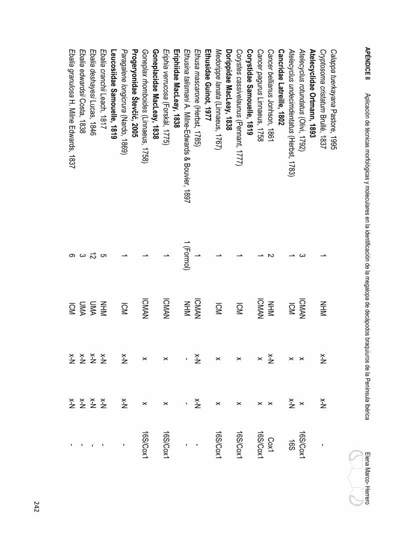
AP
ÉN
DIC
E II A
plicación de técnicas morfológicas y m
oleculares en la identificación de la megalopa de decápodos braquiuros de la P
enínsula Ibérica
Elena M
arco- Herrero
24
2
Calappa tuerkayana P
astore, 1995
Cryptosom
a cristatum B
rullé, 1837
1 N
HM
x-N
x-N
-
Atelecyclid
ae Ortm
ann
, 1893
A
telecyclus rotundatus (Olivi, 1792)
3 IC
MA
N
x x
16S/C
ox1
Atelecyclus undecim
dentatus (Herbst, 1783)
1 IC
M
x x-N
16S
Can
cridae L
atreille, 1802
C
ancer bellianus Jonhson, 1861
2 N
HM
x-N
x
Cox1
Cancer pagurus Linnaeus, 1758
1
ICM
AN
x
x 16S
/Cox1
Co
rystidae S
amo
uelle, 1819
Corystes cassivelaunus (P
ennant, 1777) 1
ICM
x
x 16S
/Cox1
Do
ripp
idae M
acLeay, 1838
Medorippe lanata (Linnaeus, 1767)
1 IC
M
x x
16S/C
ox1
Eth
usid
ae Gu
ino
t, 1977
E
thusa mascarone (H
erbst, 1785) 1
ICM
AN
x-N
x-N
-
Ethusina talism
ani A. M
ilne-Edw
ards & B
ouvier, 1897
1 (Form
ol) N
HM
-
- -
Erip
hiid
ae MacL
eay, 1838
E
riphia verrucosa (Forskål, 1775)
1 IC
MA
N
x x
16S/C
ox1
Go
nep
lacidae M
acLeay, 1838
Goneplax rhom
boides (Linnaeus, 1758) 1
ICM
AN
x
x 16S
/Cox1
Pro
geryo
nid
ae Števčić, 2005
Paragalene longicrura (N
ardo, 1869) 1
ICM
x-N
x-N
-
Leu
cosiid
ae Sam
ou
elle, 1819
E
balia cranchii Leach, 1817
5 N
HM
x-N
x-N
-
Ebalia deshayesi Lucas, 1846
12
UM
A
x-N
x-N
-
Ebalia edw
ardsii Costa, 1838
3 U
MA
x-N
x-N
-
Ebalia granulosa H
. Milne E
dwards, 1837
6
ICM
x-N
x-N
-

AP
ÉN
DIC
E II
A
plic
ació
n de
técn
icas
mor
foló
gica
s y
mol
ecul
ares
en
la id
entif
icac
ión
de la
meg
alop
a de
dec
ápod
os b
raqu
iuro
s de
la P
enín
sula
Ibér
ica
Ele
na M
arco
- H
erre
ro
24
3
Eba
lia n
ux A
. Miln
e-E
dwar
ds, 1
883
5
ICM
x-
N
x C
ox1
Eba
lia tu
bero
sa (
Pen
nant
, 177
7)
16
ICM
x-
N
x-N
-
Eba
lia tu
mef
acta
(M
onta
gu, 1
808)
1
ICM
AN
x-
N
x-N
-
Ilia
nucl
eus
(Lin
naeu
s, 1
758)
1
ICM
AN
x-
N
x-N
-
Mer
ocry
ptus
bol
etife
r A
. Miln
e-E
dwar
ds &
Bou
vier
, 189
4 1
(For
mol
) IC
M
- -
-
Ep
ialt
idae
Mac
Lea
y, 1
838
Aca
ntho
nyx
lunu
latu
s (R
isso
, 181
6)
3 IC
MA
N
x x
16S
/Cox
1
Ana
mat
hia
risso
ana
(Rou
x, 1
828)
2
(For
mol
) IC
M
- -
-
Her
bstia
con
dylia
ta (
Fab
riciu
s, 1
787)
2
ICM
/UM
A
x x
16S
/Cox
1
Liss
a ch
iragr
a (F
abric
ius,
177
5)
2 IC
M
x-N
x-
N
-
Pis
a ar
mat
a (L
atre
ille,
180
3)
3 U
MA
x-
N
x-N
-
Pis
a ca
rinim
ana
Mie
rs, 1
879
1
UM
A
x-N
x-
N
-
Pis
a hi
rtic
orni
s (H
erbs
t, 18
04)
1 IC
M
x-N
x-
N
-
Pis
a m
usco
sa (
Linn
aeus
, 175
8)
3 U
MA
x-
N
x-N
-
Pis
a no
dipe
s (L
each
, 181
5)
4 IC
M
x-N
x-
N
-
Pis
a te
trao
don
(Pen
nant
, 177
7)
5 IC
M/IC
MA
N
x x
16S
/Cox
1
Pis
a sp
. M
arco
-Her
rero
et a
l. (in
pre
p.)
1 IE
O
x-N
x-
N
-
Roc
hini
a ca
rpen
teri
(Tho
mso
n, 1
873)
1
ICM
x-
N
x-N
-
Inac
hid
ae M
acL
eay,
183
8
A
chae
us c
ranc
hii L
each
, 181
7
6 IC
MA
N
x-N
x-
N
-
Ach
aeus
gra
cilis
(C
osta
, 183
9)
2 IC
M/IC
MA
N
x-N
x-
N
-
Dor
hync
hus
thom
soni
Tho
mso
n, 1
873
6
ICM
x-
N
x-N
-
Inac
hus
agui
arii
Brit
o C
apel
lo, 1
876
5
NH
M
x-N
x-
N
-
Inac
hus
com
mun
issi
mus
Riz
za, 1
839
2 IC
M
x-N
x-
N
-
Inac
hus
dors
ette
nsis
(P
enna
nt, 1
777)
5
ICM
x
x C
ox1
Inac
hus
lept
ochi
rus
Leac
h, 1
817
7
UM
A
x x
Cox
1

AP
ÉN
DIC
E II A
plicación de técnicas morfológicas y m
oleculares en la identificación de la megalopa de decápodos braquiuros de la P
enínsula Ibérica
Elena M
arco- Herrero
24
4
Inachus phalangium (F
abricius, 1775) 3
ICM
AN
x-N
x-N
-
Inachus thoracicus Roux, 1830
4
ICM
x-N
x-N
-
Macropodia czernjaw
skii (Br&
t, 1880) 2
ICM
AN
x-N
x-N
-
Macropodia deflexa F
orest, 1978
4 N
HM
x
- -
Macropodia linaresi F
orest & Z
ariquiey Álvarez, 1964
6
ICM
x-N
x-N
-
Macropodia longirostris (F
abricius, 1775) 2
ICM
x-N
x-N
-
Macropodia parva V
an Noort &
Adem
a, 1985 5
ICM
AN
x
x 16S
/Cox1
Macropodia rostrata (Linnaeus, 1761)
9 IC
MA
N
x-N
x C
ox1
Macropodia tenuirostris (Leach, 1814)
3 IC
MA
N
x x
16S/C
ox1
Majid
ae Sam
ou
elle, 1819
E
urynome aspera (P
ennant, 1777) 2
ICM
x-N
x-N
-
Eurynom
e spinosa Hailstone, 1835
2
UM
A
x-N
x-N
-
Maja brachydactyla B
alss, 1922
1 IC
M
x x
16S/C
ox1
Maja crispata R
isso, 1827 2
ICM
x
x 16S
/Cox1
Maja goltziana d’O
liveira, 1888
1 IC
M
- -
16S/C
ox1
Maja squinado (H
erbst, 1788) 1
ICM
-
- 16S
/Cox1
Oreg
on
idae G
arth, 1958
Ergasticus clouei A
. Milne-E
dwards, 1882
1
ICM
x-N
x-N
-
Palicid
ae Bo
uvier, 1898
Palicus caronii (R
oux, 1828) 1
ICM
x
x-N
16S
Parth
eno
pid
ae MacL
eay, 1838
D
erilambrus angulifrons (Latreille, 1825)
1 IC
M/IC
MA
N
x-N
x-N
-
Distolam
brus maltzam
i (Miers, 1881)
1 IC
M
x-N
x-N
-
Parthenopoides m
assena (Roux, 1830)
2 IC
M
x-N
x-N
-
Spinolam
brus macrochelos (H
erbst, 1790) 1
ICM
x-N
x-N
-
Velolam
brus expansus (Miers, 1879)
2 (Form
ol) N
HM
-
- -

AP
ÉN
DIC
E II
A
plic
ació
n de
técn
icas
mor
foló
gica
s y
mol
ecul
ares
en
la id
entif
icac
ión
de la
meg
alop
a de
dec
ápod
os b
raqu
iuro
s de
la P
enín
sula
Ibér
ica
Ele
na M
arco
- H
erre
ro
24
5
Pilu
mn
idae
Sam
ou
elle
, 181
9
Pilu
mno
peus
afr
ican
us (
De
Man
, 190
2)
1 IC
MA
N
x-N
x-
N
-
Pilu
mnu
s ae
stua
rii N
ardo
, 186
9
0
- -
-
Pilu
mnu
s hi
rtel
lus
(Lin
naeu
s, 1
761)
3
ICM
AN
x
x 16
S/C
ox1
Pilu
mnu
s in
erm
is A
. Miln
e-E
dwar
ds &
Bou
vier
, 189
4
1 U
MA
-
- C
ox1
Pilu
mnu
s sp
inife
r H
. Miln
e E
dwar
ds, 1
834
5
ICM
/UM
A
x-N
x
Cox
1
Pilu
mnu
s vi
llosi
ssim
us (
Raf
ines
que,
181
4)
1 IC
MA
N/U
MA
x-
N
x C
ox1
Pilu
mnu
s sp
. d´
Ude
kem
d´A
coz
& S
chub
art (
in p
rep.
) 1
ICM
AN
/UM
A
x-N
x
Cox
1
Car
cin
idae
Mac
Lea
y, 1
838
Car
cinu
s ae
stua
rii N
ardo
, 184
7
1 IC
MA
N
x x
16S
/Cox
1
Car
cinu
s m
aena
s (L
inna
eus,
175
8)
1 IC
MA
N
x x
16S
/Cox
1
Por
tum
nus
latip
es (
Pen
nant
, 177
7)
6 IC
MA
N
x x-
N
16S
Xai
va b
igut
tata
(R
isso
, 181
6)
2 IC
M/N
HM
-
- -
Ger
yon
idae
Co
losi
, 192
3
C
hace
on a
ffini
s (A
. Miln
e-E
dwar
ds &
Bou
vier
, 189
4)
1 (F
orm
ol)
MN
HN
-
- 16
S/C
ox1
Cha
ceon
med
iterr
aneu
s M
anni
ng &
Hol
thui
s, 1
989
3
ICM
x-
N
x-N
-
Cha
ceon
ingl
ei M
anni
ng &
Hol
thui
s, 1
989
1
(For
mol
) N
HM
-
- -
Ger
yon
long
ipes
A. M
ilne-
Edw
ards
, 188
1
2 IC
M
- x
16S
/Cox
1
Ger
yon
tris
pino
sus
(Her
bst,
1803
)
0
- -
16S
Pir
imel
idae
Alc
ock
, 189
9
P
irim
ela
dent
icul
ata
(Mon
tagu
, 180
8)
3 IC
MA
N
x x-
N
16S
Sirp
us z
ariq
uiey
i Gor
don,
195
3
0 IC
MA
N
x*
x*-N
16
S
Po
lyb
iidae
Ort
man
n, 1
893
Bat
hyne
ctes
long
ipes
(R
isso
, 181
6)
2 IC
M
x-N
x-
N
-
Bat
hyne
ctes
mar
avig
na (
Pre
st&
rea,
183
9)
1 IC
M
x-N
x-
N
-
Lioc
arci
nus
boliv
ari (
Zar
iqui
ey Á
lvar
ez, 1
948)
3
ICM
/ICM
AN
x-
N
x-N
-

AP
ÉN
DIC
E II A
plicación de técnicas morfológicas y m
oleculares en la identificación de la megalopa de decápodos braquiuros de la P
enínsula Ibérica
Elena M
arco- Herrero
24
6
Liocarcinus corrugatus (Pennant, 1777)
1 IC
MA
N
x
16S/C
ox1
Liocarcinus depurator (Linnaeus, 1758) 3
ICM
x
x 16S
/Cox1
Liocarcinus holsatus (Fabricius, 1798)
1 IC
M
- x
16S/C
ox1
Liocarcinus maculatus (R
isso, 1827) 5
ICM
-
x 16S
/Cox1
Liocarcinus marm
oreus (Leach, 1814) 0
-
- 16S
/Cox1
Liocarcinus mcleayi (B
arnard, 1947) 1
UM
A
- -
-
Liocarcinus navigator (Herbst, 1794)
2 IC
MA
N
x -
16S/C
ox1
Liocarcinus pusillus (Leach, 1815) 1
ICM
-
x 16S
/Cox1
Liocarcinus vernalis (Risso, 1816)
3 IC
MA
N
x -
16S/C
ox1
Liocarcinus zariquieyi Gordon, 1968
4
ICM
AN
x-N
x-N
-
Macropipus tuberculatus (R
oux, 1830) 1
ICM
x
x 16S
/Cox1
Necora puber (Linnaeus, 1767)
1 IC
MA
N
x x
16S/C
ox1
Polybius henslow
ii Leach, 1820
2 IC
M
x x
16S/C
ox1
Po
rtun
idae R
afinesq
ue, 1815
Callinectes exasperatus (G
erstaecker, 1856) 1
ICM
AN
x
- 16S
Callinectes sapidus R
athbun, 1896
2 IC
M
x x
16S/C
ox1
Charybdis (C
harybdis) feriata (Linnaeus, 1758) 1
ICM
x
x 16S
/Cox1
Portunus (P
ortunus) hastatus (Linnaeus, 1767) 1
ICM
AN
x
x-N
16S
Th
iidae D
ana, 1852
Thia scutellata (F
abricius, 1793) 5
ICM
/UM
A/N
HM
-
- 16S
Pan
op
eidae O
rtman
n, 1893
Dyspanopeus sayi (S
mith, 1869)
2 IC
M
x x
16S/C
ox1
Panopeus africanus A
. Milne-E
dwards, 1867
7
ICM
AN
x
x 16S
/Cox1
Rhithropanopeus harrisii (G
ould, 1841) 2
ICM
AN
x
x 16S
/Cox1
Xan
thid
ae MacL
eay, 1838
M
onodaeus couchii (Couch, 1851)
1 IC
M
x x
16S/C
ox1

AP
ÉN
DIC
E II
A
plic
ació
n de
técn
icas
mor
foló
gica
s y
mol
ecul
ares
en
la id
entif
icac
ión
de la
meg
alop
a de
dec
ápod
os b
raqu
iuro
s de
la P
enín
sula
Ibér
ica
Ele
na M
arco
- H
erre
ro
24
7
Mic
roca
ssio
pe m
inor
(D
ana,
185
2)
2 U
MA
-
- -
Par
acta
ea m
onod
i Gui
not,
1969
1
UM
A
- -
16S
/Cox
1
Xan
tho
hydr
ophi
lus
(Lea
ch, 1
814)
8
ICM
AN
x-
N
x C
ox1
Xan
tho
pilip
es A
. Miln
e-E
dwar
ds, 1
867
2
ICM
AN
x
x 16
S/C
ox1
Xan
tho
pore
ssa
(Oliv
i, 17
92)
3 IC
MA
N
x x
16S
/Cox
1
Xan
tho
sexd
enta
tus
(Mie
rs, 1
881)
1
ICM
AN
x
x 16
S/C
ox1
TH
OR
AC
OT
RE
MA
TA
Gu
ino
t, 1
977
G
rap
sid
ae M
acL
eay,
183
8
P
achy
grap
sus
grac
ilis
(Sau
ssur
e, 1
858)
2
ICM
AN
x
x 16
S/C
ox1
Pac
hygr
apsu
s m
arm
orat
us (
Fab
riciu
s, 1
787)
1
ICM
AN
x
x 16
S/C
ox1
Pac
hygr
apsu
s m
auru
s (L
ucas
, 184
6)
1 IC
M
x x
16S
/Cox
1
Pac
hygr
apsu
s tr
ansv
ersu
s (G
ibbe
s, 1
850)
3
ICM
AN
x
x 16
S/C
ox1
Pla
nes
min
utus
(Li
nnae
us, 1
758)
3
Mal
aspi
na
x x-
N
16S
Pla
gu
siid
ae D
ana,
185
1
E
uchi
rogr
apsu
s lig
uric
us H
. Miln
e E
dwar
ds, 1
853
1
ICM
x-
N
x-N
-
Per
cnid
ae Š
tevč
ić, 2
005
Per
cnon
gib
besi
(H
. Miln
e E
dwar
ds, 1
853)
1
ICM
AN
x
x 16
S/C
ox1
Var
un
idae
H. M
ilne
Ed
war
ds,
185
3
A
sthe
nogn
athu
s at
lant
icus
Mon
od, 1
933
2
IEO
x
- 16
S
Bra
chyn
otus
atla
ntic
us F
ores
t, 19
57
2 IC
MA
N
x x-
N
16S
Bra
chyn
otus
fore
sti Z
ariq
uiey
Álv
arez
, 196
8
0
- -
16S
Bra
chyn
otus
sex
dent
atus
(R
isso
, 182
7)
1 IC
MA
N
x x-
N
16S
Erio
chei
r si
nens
is H
. Miln
e E
dwar
ds, 1
853
1
ICM
AN
x
x 16
S/C
ox1
Hem
igra
psus
taka
noi A
saku
ra &
Wat
anab
e, 2
005
0
-
- 16
S/C
ox1
Ocy
po
did
ae R
afin
esq
ue,
181
5
U
ca (
Afr
uca)
tang
eri (
Eyd
oux,
183
5)
1 IC
MA
N
x x
16S
/Cox
1

AP
ÉN
DIC
E II A
plicación de técnicas morfológicas y m
oleculares en la identificación de la megalopa de decápodos braquiuros de la P
enínsula Ibérica
Elena M
arco- Herrero
24
8
Pin
no
therid
ae de H
aan, 1833
Afropinnotheres m
onodi Manning, 1993
13
ICM
AN
x-N
x-N
-
Nepinnotheres pinnotheres (Linnaeus, 1758)
3 IC
MA
N
x -
16S
Pinnotheres pisum
(Linnaeus, 1767) 3
ICM
/ICM
AN
x
x 16S
/Cox1
ICM
Institut de Ciències de M
ar; ICM
AN
Instituto de Ciencias M
arinas de Andalucía; IE
O Instituto O
ceanográfico Español: C
ampañas M
ED
ITS
-IND
EM
AR
ES
-
AR
SA
; NH
M N
atural History M
useum (London); M
alaspin
a Proyecto M
alaspina; MN
HN
Muséum
National d'H
istoire Naturelle (P
arís); UM
A U
niversidad de
Málaga; X
Secuencia obtenida; x-N
Secuencia nueva; (-) S
in datos.
* Secuencias de S
irpus zariquieyi obtenidas a partir de larvas del plancton.