Molecular Recognition of Peloruside A by Microtubules. The C24 Primary Alcohol is Essential for...
10
DOI: 10.1002/cbic.201000294 Molecular Recognition of Peloruside A by Microtubules. The C24 Primary Alcohol is Essential for Biological Activity Benet Pera, [a] Mina Razzak, [b] Chiara Trigili, [a] Oriol Pineda, [c] Angeles Canales, [a, d] RubȖn M. Buey, [a, e] Jesffls JimȖnez-Barbero, [a] Peter T. Northcote, [f] Ian Paterson, [b] Isabel Barasoain,* [a] and JosȖ Fernando Dȷaz* [a] Introduction Peloruside A (1), a polyketide anticancer compound isolated from the New Zealand marine sponge Mycale hentscheli, [1] is one of several known microtubule-targeting agents. [2] The compound acts by binding to and stabilizing the polymerized form of tubulin, a protein that has several important cellular functions, including separation of sister chromatids during cel- lular division; this makes it an attractive target for cancer ther- apy. [3, 4] Microtubules are formed from a- and b-tubulin, which asso- ciate as dimers and organize themselves into filaments that comprise the tubulin macrostructure. The microtubule is dy- namic and is constantly lengthening and shortening in order to fulfill its cellular functions. Microtubules are an important target in cancer therapy, and currently there are several drugs that perturb its dynamic properties. Taxol$ (paclitaxel, 2) is an important antimitotic agent currently used for the treatment of metastatic breast and ovarian cancer, as well as Kaposi’s sarcoma, and non-small cell lung cancer. [5] A close analogue of paclitaxel is Taxotere$ (docetaxel, 3), which is used clinically for the treatment of lung and metastatic breast cancer. [6] The taxane drugs act by binding to the b-tubulin subunit of micro- tubules and thus stabilize the polymer. [7–9] Two examples of mi- crotubule-destabilizing agents, which exert their effects by pro- moting the depolymerization of tubulin, are vinblastine (4) and vinorelbine (5). [10] These drugs are primarily used against non- small cell lung cancer. Despite the success of these drugs, toxicity and develop- ment of resistance (in particular to the taxanes) threatens their effective clinical use. Over the past few years, a structurally di- verse set of natural products from a variety of sources have been discovered that share the ability of the taxanes to stabi- lize the polymeric microtubules. These include, but are not lim- ited to, discodermolide (6), [11] dictyostatin (7), [12] peloruside (1), laulimalide (8), [13–15] the sarcodictyins (9), [16, 17] and the epothi- lones (10). [18] This latter class of agents, isolated from the myx- obacterium Sorangium cellulosum, have been intensively re- searched [19] leading to Ixempra$ entering the market for the treatment of advanced breast cancer. [20] A major threat to the effective targeting of the microtubule as a mechanism for chemotherapy is the development of re- sistance by the neoplastic tissue. Although many tumors initial- Peloruside is a microtubule-stabilizing agent that targets the same site as laulimalide. It binds to microtubules with a 1:1 stoichiometry and with a binding affinity in the low-mm range; thereby reducing the number of microtubular protofilaments in the same way as paclitaxel. Although the binding affinity of the compound is comparable to that of the low-affinity stabi- lizing agent sarcodictyin, peloruside is more active in inducing microtubule assembly and is more cytotoxic to tumor cells; this suggests that the peloruside site is a more effective site for stabilizing microtubules. Acetylation of the C24 hydroxyl group results in inactive compounds. According to molecular modeling, this substitution at the C24 hydroxyl group presum- ably disrupts the interaction of the side chain with Arg320 in the putative binding site on a-tubulin. The binding epitope of peloruside on microtubules has been studied by using NMR spectroscopic techniques, and is compatible with the same binding site. [a] B. Pera, C. Trigili, Dr. A. Canales, Dr. R. M. Buey, Dr. J. JimȖnez-Barbero, Dr. I. Barasoain, Dr. J. F. Dȷaz Centro de Investigaciones BiolɃgicas Consejo Superior de Investigaciones Cientȷficas Ramiro de Maeztu 9, 28040 Madrid (Spain) Fax: (+ 34) 915360432 E-mail : [email protected] [email protected] [b] Dr. M. Razzak, Prof. Dr. I. Paterson University Chemical Laboratory, University of Cambridge Lensfield Road, Cambridge, CB2 1EW (United Kingdom) [c] Dr. O. Pineda Departament de Quȷmica Organica, Facultat de Quȷmica Universitat de Barcelona Avenue Diagonal 647, 08028 Barcelona (Spain) [d] Dr. A. Canales Universidad Complutense Avda. Complutense s/n, 28040 Madrid (Spain) [e] Dr. R. M. Buey Current address: Centro de InvestigaciɃn del Cancer Campus Unamuno, 37007, Salamanca (Spain) [f] Dr. P. T. Northcote Centre for Biodiscovery, Victoria University of Wellington Wellington (New Zealand) Supporting information for this article is available on the WWW under http ://dx.doi.org/10.1002/cbic.201000294. ChemBioChem 2010, 11, 1669 – 1678 # 2010 Wiley-VCH Verlag GmbH & Co. KGaA, Weinheim 1669
-
Upload
independent -
Category
Documents
-
view
0 -
download
0
Transcript of Molecular Recognition of Peloruside A by Microtubules. The C24 Primary Alcohol is Essential for...
DOI: 10.1002/cbic.201000294
Molecular Recognition of Peloruside A by Microtubules.The C24 Primary Alcohol is Essential for Biological ActivityBenet Pera,[a] Mina Razzak,[b] Chiara Trigili,[a] Oriol Pineda,[c] Angeles Canales,[a, d]
Rub�n M. Buey,[a, e] Jesffls Jim�nez-Barbero,[a] Peter T. Northcote,[f] Ian Paterson,[b]
Isabel Barasoain,*[a] and Jos� Fernando D�az*[a]
Introduction
Peloruside A (1), a polyketide anticancer compound isolatedfrom the New Zealand marine sponge Mycale hentscheli,[1] isone of several known microtubule-targeting agents.[2] Thecompound acts by binding to and stabilizing the polymerizedform of tubulin, a protein that has several important cellularfunctions, including separation of sister chromatids during cel-lular division; this makes it an attractive target for cancer ther-apy.[3, 4]
Microtubules are formed from a- and b-tubulin, which asso-ciate as dimers and organize themselves into filaments thatcomprise the tubulin macrostructure. The microtubule is dy-namic and is constantly lengthening and shortening in orderto fulfill its cellular functions. Microtubules are an importanttarget in cancer therapy, and currently there are several drugsthat perturb its dynamic properties. Taxol� (paclitaxel, 2) is animportant antimitotic agent currently used for the treatmentof metastatic breast and ovarian cancer, as well as Kaposi’ssarcoma, and non-small cell lung cancer.[5] A close analogue ofpaclitaxel is Taxotere� (docetaxel, 3), which is used clinically forthe treatment of lung and metastatic breast cancer.[6] Thetaxane drugs act by binding to the b-tubulin subunit of micro-tubules and thus stabilize the polymer.[7–9] Two examples of mi-crotubule-destabilizing agents, which exert their effects by pro-moting the depolymerization of tubulin, are vinblastine (4) andvinorelbine (5).[10] These drugs are primarily used against non-small cell lung cancer.
Despite the success of these drugs, toxicity and develop-ment of resistance (in particular to the taxanes) threatens theireffective clinical use. Over the past few years, a structurally di-verse set of natural products from a variety of sources havebeen discovered that share the ability of the taxanes to stabi-lize the polymeric microtubules. These include, but are not lim-
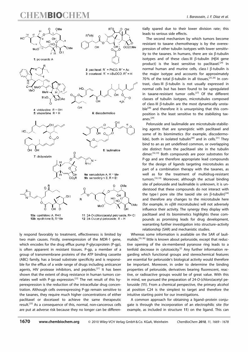
ited to, discodermolide (6),[11] dictyostatin (7),[12] peloruside (1),laulimalide (8),[13–15] the sarcodictyins (9),[16, 17] and the epothi-lones (10).[18] This latter class of agents, isolated from the myx-obacterium Sorangium cellulosum, have been intensively re-searched[19] leading to Ixempra� entering the market for thetreatment of advanced breast cancer.[20]
A major threat to the effective targeting of the microtubuleas a mechanism for chemotherapy is the development of re-sistance by the neoplastic tissue. Although many tumors initial-
Peloruside is a microtubule-stabilizing agent that targets thesame site as laulimalide. It binds to microtubules with a 1:1stoichiometry and with a binding affinity in the low-mm range;thereby reducing the number of microtubular protofilamentsin the same way as paclitaxel. Although the binding affinity ofthe compound is comparable to that of the low-affinity stabi-lizing agent sarcodictyin, peloruside is more active in inducingmicrotubule assembly and is more cytotoxic to tumor cells ;this suggests that the peloruside site is a more effective site
for stabilizing microtubules. Acetylation of the C24 hydroxylgroup results in inactive compounds. According to molecularmodeling, this substitution at the C24 hydroxyl group presum-ably disrupts the interaction of the side chain with Arg320 inthe putative binding site on a-tubulin. The binding epitope ofpeloruside on microtubules has been studied by using NMRspectroscopic techniques, and is compatible with the samebinding site.
[a] B. Pera, C. Trigili, Dr. A. Canales, Dr. R. M. Buey, Dr. J. Jim�nez-Barbero,Dr. I. Barasoain, Dr. J. F. D�azCentro de Investigaciones Biol�gicasConsejo Superior de Investigaciones Cient�ficasRamiro de Maeztu 9, 28040 Madrid (Spain)Fax: (+ 34) 915360432E-mail : [email protected]
[b] Dr. M. Razzak, Prof. Dr. I. PatersonUniversity Chemical Laboratory, University of CambridgeLensfield Road, Cambridge, CB2 1EW (United Kingdom)
[c] Dr. O. PinedaDepartament de Qu�mica Organica, Facultat de Qu�micaUniversitat de BarcelonaAvenue Diagonal 647, 08028 Barcelona (Spain)
[d] Dr. A. CanalesUniversidad ComplutenseAvda. Complutense s/n, 28040 Madrid (Spain)
[e] Dr. R. M. BueyCurrent address : Centro de Investigaci�n del CancerCampus Unamuno, 37007, Salamanca (Spain)
[f] Dr. P. T. NorthcoteCentre for Biodiscovery, Victoria University of WellingtonWellington (New Zealand)
Supporting information for this article is available on the WWW underhttp ://dx.doi.org/10.1002/cbic.201000294.
ChemBioChem 2010, 11, 1669 – 1678 � 2010 Wiley-VCH Verlag GmbH & Co. KGaA, Weinheim 1669
ly respond favorably to treatment, effectiveness is limited bytwo main causes. Firstly, overexpression of the MDR-1 gene,which encodes for the drug efflux pump P-glycoprotein (P-gp),is often apparent in resistant tissues. P-gp, a member of agroup of transmembrane proteins of the ATP binding cassette(ABC) family, has a broad substrate specificity and is responsi-ble for the efflux of a wide range of drugs including anticanceragents, HIV protease inhibitors, and peptides.[21] It has beenshown that the extent of drug resistance in human tumors cor-relates well with P-gp expression.[22] The net result of this hy-perexpression is the reduction of the intracellular drug concen-tration. Although cells overexpressing P-gp remain sensitive tothe taxanes, they require much higher concentrations of eitherpaclitaxel or docetaxel to achieve the same therapeuticresult.[23] As a consequence of this, normal, non-cancerous cellsare put at adverse risk because they no longer can be differen-
tially spared due to their lower division rate; thisleads to serious side effects.
The second mechanism by which tumors becomeresistant to taxane chemotherapy is by the overex-pression of other tubulin isotypes with lower sensitiv-ity to the taxanes. In humans, there are six b-tubulinisotypes and of these class III b-tubulin (Hb4 geneproduct) is the least sensitive to paclitaxel.[24] Innormal human and murine cells, class I b-tubulin isthe major isotype and accounts for approximately70 % of the total b-tubulin in all tissues,[25, 26] In con-trast, class III b-tubulin is not usually expressed innormal cells but has been found to be upregulatedin taxane-resistant tumor cells.[27] Of the differentclasses of tubulin isotypes, microtubules composedof class III b-tubulin are the most dynamically unsta-ble[28] and therefore it is unsurprising that this com-position is the least sensitive to the stabilizing tax-anes.[29]
Peloruside and laulimalide are microtubule-stabiliz-ing agents that are synergistic with paclitaxel andsome of its biomimetics (for example, discodermo-lide), both in isolated tubulin[30] and in cells.[31] Theybind to an as yet undefined common, or overlappingsite distinct from the paclitaxel site in the tubulindimer.[32, 33] Both compounds are poor substrates forP-gp and are therefore appropriate lead compoundsfor the design of ligands targeting microtubules aspart of a combination therapy with the taxanes, aswell as for the treatment of multidrug-resistanttumors.[32, 33] Moreover, although the actual bindingsite of peloruside and laulimalide is unknown, it is un-derstood that these compounds do not interact withthe type I pore site (the taxoid site on b-tubulin)[34]
and therefore any changes to the microtubule here(for example, in abIII microtubules) will not adverselyinfluence their activity. The synergy they display withpaclitaxel and its biomimetics highlights these com-pounds as promising leads for drug development,warranting further investigation into structure–activityrelationship (SAR) and mechanistic studies.
Whereas some information is available on the SAR of lauli-malide,[35, 36] little is known about peloruside, except that reduc-tive opening of the six-membered pyranose ring leads to atenfold decrease in cytotoxicity.[2] Any further information re-garding which functional groups and stereochemical featuresare essential for peloruside’s biological activity would thereforebe important. Moreover, in order to determine the bindingproperties of peloruside, derivatives bearing fluorescent, reac-tive, or radioactive groups would be of great value. With thisin mind, we pursued the preparation of 24-O-(chloro)acetyl pe-loruside (11). From a chemical perspective, the primary alcoholat position C24 is the simplest to target and therefore theintuitive starting point for our investigations.
A common approach for obtaining a ligand–protein conju-gate is through the incorporation of an electrophilic site (forexample, as included in structure 11) on the ligand. This can
1670 www.chembiochem.org � 2010 Wiley-VCH Verlag GmbH & Co. KGaA, Weinheim ChemBioChem 2010, 11, 1669 – 1678
I. Barasoain, J. F. D�az et al.
then react with a nucleophilic residue in the protein, for exam-ple the thiol of a cysteine.[37] After cross-linking is achieved, di-gestion and mass spectrometry experiments are used to deter-mine which segment of the protein the ligand binds. Coupledwith detailed information already known about the proteinstructure, a preliminary active site model can be constructed.Information on this binding site is of critical importance for thedesign of more efficacious peloruside-based drug candidatesand is the key motivation behind this work.
Results
Ligand binding to microtubules
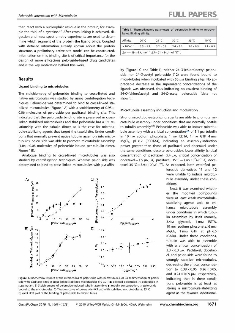
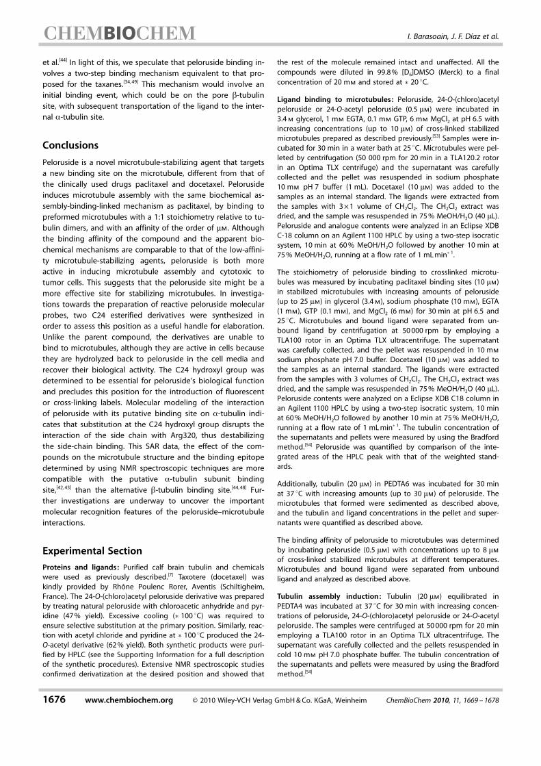
The stoichiometry of peloruside binding to cross-linked andnative microtubules was studied by using centrifugation tech-niques. Peloruside was determined to bind to cross-linked sta-bilized microtubules (Figure 1 A) with a stoichiometry of 0.95�0.06 molecules of peloruside per paclitaxel binding site. Thisindicated that the peloruside binding site is preserved in cross-linked stabilized microtubules and that peloruside has a 1:1 re-lationship with the tubulin dimer, as is the case for microtu-bule-stabilizing agents that target the taxoid site. Under condi-tions that normally prevent native tubulin assembly into micro-tubules, peloruside was able to promote microtubule assembly(1.04�0.08 molecules of peloruside bound per tubulin dimer,Figure 1 B).
Analogue binding to cross-linked microtubules was alsostudied by centrifugation techniques. Whereas peloruside wasdetermined to bind to cross-linked microtubules with mm affin-
ity (Figure 1 C and Table 1), neither 24-O-(chloro)acetyl peloru-side nor 24-O-acetyl peloruside (12) were found bound tomicrotubules when incubated with 50 mm binding sites. No ap-preciable decrease in the supernatant concentrations of theligands was observed, thus indicating no covalent binding of24-O-(chloro)acetyl and 24-O-acetyl peloruside (data notshown).
Microtubule assembly induction and modulation
Strong microtubule-stabilizing agents are able to promote mi-crotubule assembly under conditions that are normally hostileto tubulin assembly.[38] Peloruside was able to induce microtu-bule assembly with a critical concentration[39] of 3.1 mm tubulinin 10 mm sodium phosphate, 1 mm EDTA, 1 mm GTP, 4 mm
MgCl2, pH 6.7 (PEDTA4), indicating an assembly-inductionpower greater than those of paclitaxel and docetaxel underthe same conditions, despite peloruside’s lower affinity (criticalconcentration of paclitaxel = 5.4 mm, critical concentration ofdocetaxel = 1.5 mm, Ka paclitaxel 35 8C = 1.4 � 107
m�1 Ka doce-
taxel 35 8C = 3.9 � 107m�1[40]). As expected, both esterified pe-
loruside derivatives 11 and 12were unable to induce microtu-bule assembly under these con-ditions.
Next, it was examined wheth-er the modified compoundswere at least weak microtubule-stabilizing agents able to en-hance microtubule assemblyunder conditions in which tubu-lin assembles by itself (namely,3.4 m glycerol, 1 mm EGTA,10 mm sodium phosphate, 6 mm
MgCl2, 1 mm GTP at pH 6.5(GAB)). Under these conditions,tubulin was able to assemblewith a critical concentration of3.3�0.3 mm. Paclitaxel, docetax-el, and peloruside were found tostrongly stabilize microtubules,decreasing the critical concentra-tion to 0.38�0.06, 0.26�0.05,and 0.24�0.09 mm, respectively,indicating that in these condi-tions peloruside is at least asstrong a microtubule-stabilizingagent as the taxanes. Additional-
Figure 1. Biochemical studies of the interactions of peloruside with microtubules. A) Co-sedimentation of peloru-side with paclitaxel sites in cross-linked stabilized microtubules (10 mm) ; *: pelleted peloruside, *: peloruside insupernatant. B) Stoichiometry of peloruside-induced tubulin assembly; *: tubulin concentration, *: pelorusidebound to the microtubules. C) Titration curve of peloruside (0.5 mm) with stabilized microtubules at 25 8C.D) van’t Hoff plot of the binding of peloruside to microtubules.
Table 1. Thermodynamic parameters of peloruside binding to microtu-bules. Binding affinity.
Affinity 20 8C 25 8C 30 8C 35 8C 40 8C
� 106m�1 3.5�1.2 3.2�0.8 2.4�1.1 2.6�0.5 2.1�0.3
DH =�19�4 kJ mol�1. DS = 61�14 J mol�1 K�1.
ChemBioChem 2010, 11, 1669 – 1678 � 2010 Wiley-VCH Verlag GmbH & Co. KGaA, Weinheim www.chembiochem.org 1671
Peloruside Interaction with Microtubules
ly, the esterified peloruside derivatives 11 and 12 did not sig-nificantly promote further microtubule stabilization (criticalconcentration for both = 3.3�0.3 mm), thus indicating thatthey do not exhibit microtubule-stabilizing activity.
Cellular activity of the compounds
The IC50 of the derivatives 11 and 12 compared with peloru-side were determined in A2780 and A2780AD ovarian carcino-ma cells. Peloruside was found to be cytotoxic in these celllines as previously described.[33] Surprisingly, 24-O-(chloro)acetylpeloruside was found to be almost as active as peloruside,whereas 24-O-acetyl peloruside was tenfold less active thanthe former (Table 2). Treatment of A549 lung carcinoma cells
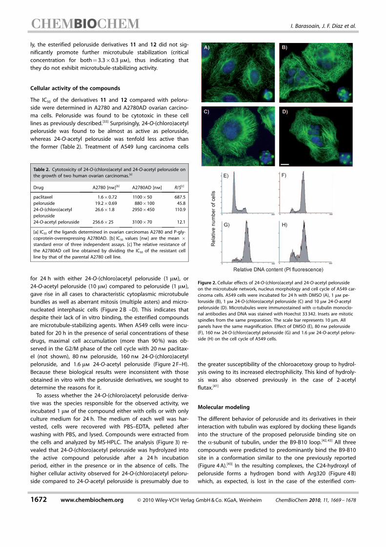
for 24 h with either 24-O-(chloro)acetyl peloruside (1 mm), or24-O-acetyl peloruside (10 mm) compared to peloruside (1 mm),gave rise in all cases to characteristic cytoplasmic microtubulebundles as well as aberrant mitosis (multiple asters) and micro-nucleated interphasic cells (Figure 2 B –D). This indicates thatdespite their lack of in vitro binding, the esterified compoundsare microtubule-stabilizing agents. When A549 cells were incu-bated for 20 h in the presence of serial concentrations of thesedrugs, maximal cell accumulation (more than 90 %) was ob-served in the G2/M phase of the cell cycle with 20 nm paclitax-el (not shown), 80 nm peloruside, 160 nm 24-O-(chloro)acetylpeloruside, and 1.6 mm 24-O-acetyl peloruside (Figure 2 F–H).Because these biological results were inconsistent with thoseobtained in vitro with the peloruside derivatives, we sought todetermine the reasons for it.
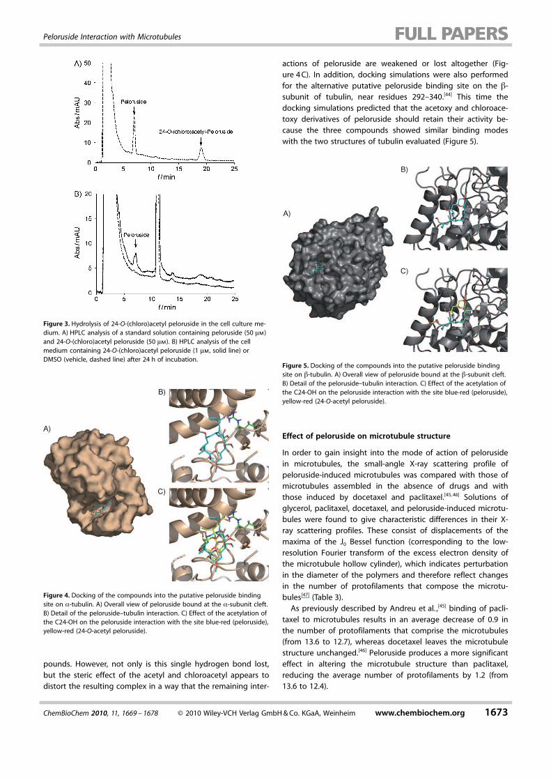
To assess whether the 24-O-(chloro)acetyl peloruside deriva-tive was the species responsible for the observed activity, weincubated 1 mm of the compound either with cells or with onlyculture medium for 24 h. The medium of each well was har-vested, cells were recovered with PBS–EDTA, pelleted afterwashing with PBS, and lysed. Compounds were extracted fromthe cells and analyzed by MS-HPLC. The analysis (Figure 3) re-vealed that 24-O-(chloro)acetyl peloruside was hydrolyzed intothe active compound peloruside after a 24 h incubationperiod, either in the presence or in the absence of cells. Thehigher cellular activity observed for 24-O-(chloro)acetyl peloru-side compared to 24-O-acetyl peloruside is presumably due to
the greater susceptibility of the chloroacetoxy group to hydrol-ysis owing to its increased electrophilicity. This kind of hydroly-sis was also observed previously in the case of 2-acetylflutax.[41]
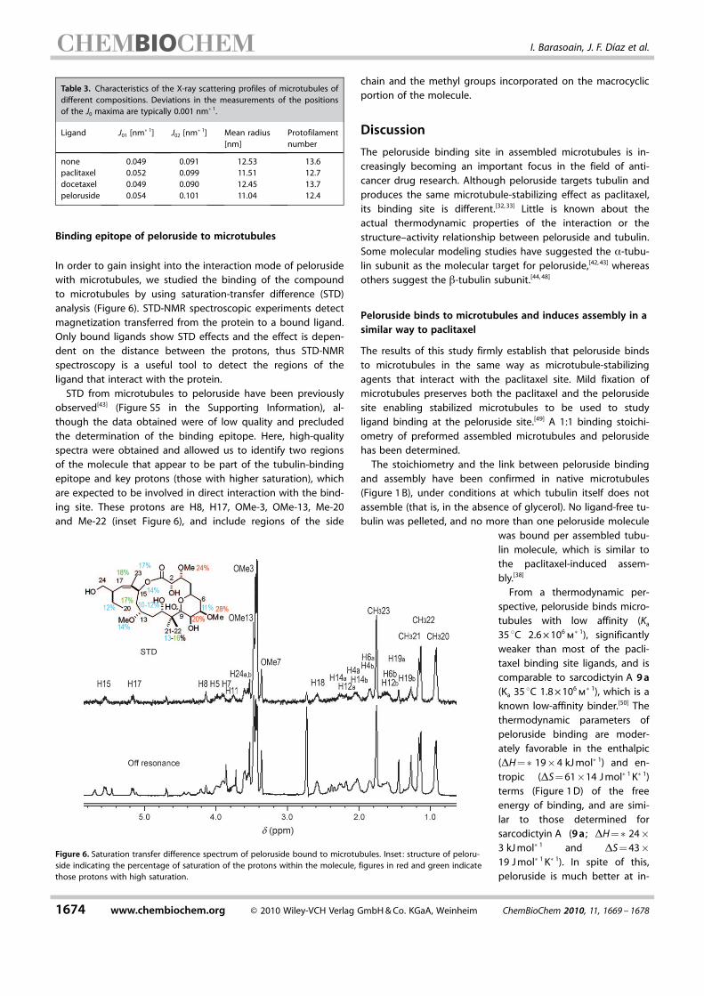
Molecular modeling
The different behavior of peloruside and its derivatives in theirinteraction with tubulin was explored by docking these ligandsinto the structure of the proposed peloruside binding site onthe a-subunit of tubulin, under the B9-B10 loop.[42, 43] All threecompounds were predicted to predominantly bind the B9-B10site in a conformation similar to the one previously reported(Figure 4 A).[43] In the resulting complexes, the C24-hydroxyl ofpeloruside forms a hydrogen bond with Arg320 (Figure 4 B)which, as expected, is lost in the case of the esterified com-
Table 2. Cytotoxicity of 24-O-(chloro)acetyl and 24-O-acetyl peloruside onthe growth of two human ovarian carcinomas.[a]
Drug A2780 [nm][b] A2780AD [nm] R/S[c]
paclitaxel 1.6�0.72 1100�50 687.5peloruside 19.2�0.69 880�100 45.824-O-(chloro)acetyl 26.6�1.8 2950�450 110.9peloruside24-O-acetyl peloruside 256.6�25 3100�70 12.1
[a] IC50 of the ligands determined in ovarian carcinomas A2780 and P-gly-coprotein-overexpressing A2780AD. [b] IC50 values [nm] are the mean �standard error of three independent assays. [c] The relative resistance ofthe A2780AD cell line obtained by dividing the IC50 of the resistant cellline by that of the parental A2780 cell line.
Figure 2. Cellular effects of 24-O-(chloro)acetyl and 24-O-acetyl pelorusideon the microtubule network, nucleus morphology and cell cycle of A549 car-cinoma cells. A549 cells were incubated for 24 h with DMSO (A), 1 mm pe-loruside (B), 1 mm 24-O-(chloro)acetyl peloruside (C) and 10 mm 24-O-acetylpeloruside (D). Microtubules were immunostained with a-tubulin monoclo-nal antibodies and DNA was stained with Hoechst 33 342. Insets are mitoticspindles from the same preparation. The scale bar represents 10 mm. Allpanels have the same magnification. Effect of DMSO (E), 80 nm peloruside(F), 160 nm 24-O-(chloro)acetyl peloruside (G) and 1.6 mm 24-O-acetyl peloru-side (H) on the cell cycle of A549 cells.
1672 www.chembiochem.org � 2010 Wiley-VCH Verlag GmbH & Co. KGaA, Weinheim ChemBioChem 2010, 11, 1669 – 1678
I. Barasoain, J. F. D�az et al.
pounds. However, not only is this single hydrogen bond lost,but the steric effect of the acetyl and chloroacetyl appears todistort the resulting complex in a way that the remaining inter-
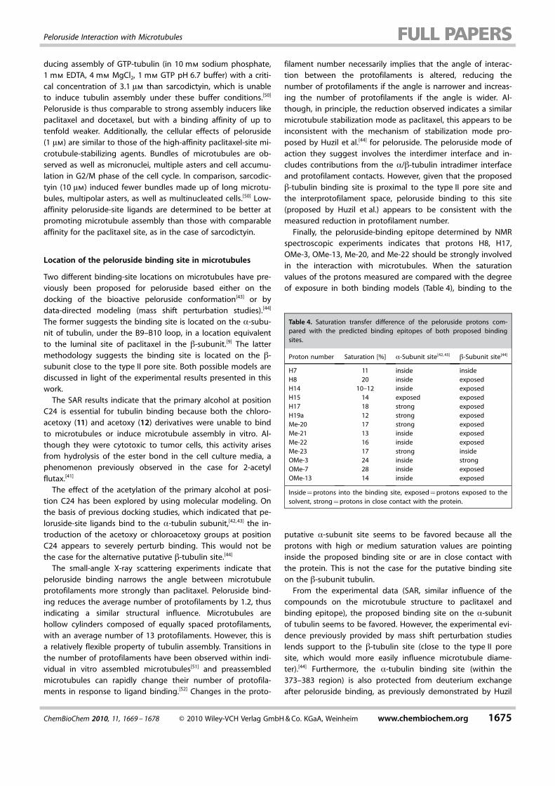
actions of peloruside are weakened or lost altogether (Fig-ure 4 C). In addition, docking simulations were also performedfor the alternative putative peloruside binding site on the b-subunit of tubulin, near residues 292–340.[44] This time thedocking simulations predicted that the acetoxy and chloroace-toxy derivatives of peloruside should retain their activity be-cause the three compounds showed similar binding modeswith the two structures of tubulin evaluated (Figure 5).
Effect of peloruside on microtubule structure
In order to gain insight into the mode of action of pelorusidein microtubules, the small-angle X-ray scattering profile ofpeloruside-induced microtubules was compared with those ofmicrotubules assembled in the absence of drugs and withthose induced by docetaxel and paclitaxel.[45, 46] Solutions ofglycerol, paclitaxel, docetaxel, and peloruside-induced microtu-bules were found to give characteristic differences in their X-ray scattering profiles. These consist of displacements of themaxima of the J0 Bessel function (corresponding to the low-resolution Fourier transform of the excess electron density ofthe microtubule hollow cylinder), which indicates perturbationin the diameter of the polymers and therefore reflect changesin the number of protofilaments that compose the microtu-bules[47] (Table 3).
As previously described by Andreu et al. ,[45] binding of pacli-taxel to microtubules results in an average decrease of 0.9 inthe number of protofilaments that comprise the microtubules(from 13.6 to 12.7), whereas docetaxel leaves the microtubulestructure unchanged.[46] Peloruside produces a more significanteffect in altering the microtubule structure than paclitaxel,reducing the average number of protofilaments by 1.2 (from13.6 to 12.4).
Figure 3. Hydrolysis of 24-O-(chloro)acetyl peloruside in the cell culture me-dium. A) HPLC analysis of a standard solution containing peloruside (50 mm)and 24-O-(chloro)acetyl peloruside (50 mm). B) HPLC analysis of the cellmedium containing 24-O-(chloro)acetyl peloruside (1 mm, solid line) orDMSO (vehicle, dashed line) after 24 h of incubation.
Figure 4. Docking of the compounds into the putative peloruside bindingsite on a-tubulin. A) Overall view of peloruside bound at the a-subunit cleft.B) Detail of the peloruside–tubulin interaction. C) Effect of the acetylation ofthe C24-OH on the peloruside interaction with the site blue-red (peloruside),yellow-red (24-O-acetyl peloruside).
Figure 5. Docking of the compounds into the putative peloruside bindingsite on b-tubulin. A) Overall view of peloruside bound at the b-subunit cleft.B) Detail of the peloruside–tubulin interaction. C) Effect of the acetylation ofthe C24-OH on the peloruside interaction with the site blue-red (peloruside),yellow-red (24-O-acetyl peloruside).
ChemBioChem 2010, 11, 1669 – 1678 � 2010 Wiley-VCH Verlag GmbH & Co. KGaA, Weinheim www.chembiochem.org 1673
Peloruside Interaction with Microtubules
Binding epitope of peloruside to microtubules
In order to gain insight into the interaction mode of pelorusidewith microtubules, we studied the binding of the compoundto microtubules by using saturation-transfer difference (STD)analysis (Figure 6). STD-NMR spectroscopic experiments detectmagnetization transferred from the protein to a bound ligand.Only bound ligands show STD effects and the effect is depen-dent on the distance between the protons, thus STD-NMRspectroscopy is a useful tool to detect the regions of theligand that interact with the protein.
STD from microtubules to peloruside have been previouslyobserved[43] (Figure S5 in the Supporting Information), al-though the data obtained were of low quality and precludedthe determination of the binding epitope. Here, high-qualityspectra were obtained and allowed us to identify two regionsof the molecule that appear to be part of the tubulin-bindingepitope and key protons (those with higher saturation), whichare expected to be involved in direct interaction with the bind-ing site. These protons are H8, H17, OMe-3, OMe-13, Me-20and Me-22 (inset Figure 6), and include regions of the side
chain and the methyl groups incorporated on the macrocyclicportion of the molecule.
Discussion
The peloruside binding site in assembled microtubules is in-creasingly becoming an important focus in the field of anti-cancer drug research. Although peloruside targets tubulin andproduces the same microtubule-stabilizing effect as paclitaxel,its binding site is different.[32, 33] Little is known about theactual thermodynamic properties of the interaction or thestructure–activity relationship between peloruside and tubulin.Some molecular modeling studies have suggested the a-tubu-lin subunit as the molecular target for peloruside,[42, 43] whereasothers suggest the b-tubulin subunit.[44, 48]
Peloruside binds to microtubules and induces assembly in asimilar way to paclitaxel
The results of this study firmly establish that peloruside bindsto microtubules in the same way as microtubule-stabilizingagents that interact with the paclitaxel site. Mild fixation ofmicrotubules preserves both the paclitaxel and the pelorusidesite enabling stabilized microtubules to be used to studyligand binding at the peloruside site.[49] A 1:1 binding stoichi-ometry of preformed assembled microtubules and pelorusidehas been determined.
The stoichiometry and the link between peloruside bindingand assembly have been confirmed in native microtubules(Figure 1 B), under conditions at which tubulin itself does notassemble (that is, in the absence of glycerol). No ligand-free tu-bulin was pelleted, and no more than one peloruside molecule
was bound per assembled tubu-lin molecule, which is similar tothe paclitaxel-induced assem-bly.[38]
From a thermodynamic per-spective, peloruside binds micro-tubules with low affinity (Ka
35 8C 2.6 � 106m�1), significantly
weaker than most of the pacli-taxel binding site ligands, and iscomparable to sarcodictyin A 9 a(Ka 35 8C 1.8 � 106
m�1), which is a
known low-affinity binder.[50] Thethermodynamic parameters ofpeloruside binding are moder-ately favorable in the enthalpic(DH =�19�4 kJ mol�1) and en-tropic (DS = 61�14 J mol�1 K�1)terms (Figure 1 D) of the freeenergy of binding, and are simi-lar to those determined forsarcodictyin A (9 a ; DH =�24�3 kJ mol�1 and DS = 43�19 J mol�1 K�1). In spite of this,peloruside is much better at in-
Table 3. Characteristics of the X-ray scattering profiles of microtubules ofdifferent compositions. Deviations in the measurements of the positionsof the J0 maxima are typically 0.001 nm�1.
Ligand J01 [nm�1] J02 [nm�1] Mean radius Protofilament[nm] number
none 0.049 0.091 12.53 13.6paclitaxel 0.052 0.099 11.51 12.7docetaxel 0.049 0.090 12.45 13.7peloruside 0.054 0.101 11.04 12.4
Figure 6. Saturation transfer difference spectrum of peloruside bound to microtubules. Inset : structure of peloru-side indicating the percentage of saturation of the protons within the molecule, figures in red and green indicatethose protons with high saturation.
1674 www.chembiochem.org � 2010 Wiley-VCH Verlag GmbH & Co. KGaA, Weinheim ChemBioChem 2010, 11, 1669 – 1678
I. Barasoain, J. F. D�az et al.
ducing assembly of GTP-tubulin (in 10 mm sodium phosphate,1 mm EDTA, 4 mm MgCl2, 1 mm GTP pH 6.7 buffer) with a criti-cal concentration of 3.1 mm than sarcodictyin, which is unableto induce tubulin assembly under these buffer conditions.[50]
Peloruside is thus comparable to strong assembly inducers likepaclitaxel and docetaxel, but with a binding affinity of up totenfold weaker. Additionally, the cellular effects of peloruside(1 mm) are similar to those of the high-affinity paclitaxel-site mi-crotubule-stabilizing agents. Bundles of microtubules are ob-served as well as micronuclei, multiple asters and cell accumu-lation in G2/M phase of the cell cycle. In comparison, sarcodic-tyin (10 mm) induced fewer bundles made up of long microtu-bules, multipolar asters, as well as multinucleated cells.[50] Low-affinity peloruside-site ligands are determined to be better atpromoting microtubule assembly than those with comparableaffinity for the paclitaxel site, as in the case of sarcodictyin.
Location of the peloruside binding site in microtubules
Two different binding-site locations on microtubules have pre-viously been proposed for peloruside based either on thedocking of the bioactive peloruside conformation[43] or bydata-directed modeling (mass shift perturbation studies).[44]
The former suggests the binding site is located on the a-subu-nit of tubulin, under the B9–B10 loop, in a location equivalentto the luminal site of paclitaxel in the b-subunit.[9] The lattermethodology suggests the binding site is located on the b-subunit close to the type II pore site. Both possible models arediscussed in light of the experimental results presented in thiswork.
The SAR results indicate that the primary alcohol at positionC24 is essential for tubulin binding because both the chloro-acetoxy (11) and acetoxy (12) derivatives were unable to bindto microtubules or induce microtubule assembly in vitro. Al-though they were cytotoxic to tumor cells, this activity arisesfrom hydrolysis of the ester bond in the cell culture media, aphenomenon previously observed in the case for 2-acetylflutax.[41]
The effect of the acetylation of the primary alcohol at posi-tion C24 has been explored by using molecular modeling. Onthe basis of previous docking studies, which indicated that pe-loruside-site ligands bind to the a-tubulin subunit,[42, 43] the in-troduction of the acetoxy or chloroacetoxy groups at positionC24 appears to severely perturb binding. This would not bethe case for the alternative putative b-tubulin site.[44]
The small-angle X-ray scattering experiments indicate thatpeloruside binding narrows the angle between microtubuleprotofilaments more strongly than paclitaxel. Peloruside bind-ing reduces the average number of protofilaments by 1.2, thusindicating a similar structural influence. Microtubules arehollow cylinders composed of equally spaced protofilaments,with an average number of 13 protofilaments. However, this isa relatively flexible property of tubulin assembly. Transitions inthe number of protofilaments have been observed within indi-vidual in vitro assembled microtubules[51] and preassembledmicrotubules can rapidly change their number of protofila-ments in response to ligand binding.[52] Changes in the proto-
filament number necessarily implies that the angle of interac-tion between the protofilaments is altered, reducing thenumber of protofilaments if the angle is narrower and increas-ing the number of protofilaments if the angle is wider. Al-though, in principle, the reduction observed indicates a similarmicrotubule stabilization mode as paclitaxel, this appears to beinconsistent with the mechanism of stabilization mode pro-posed by Huzil et al.[44] for peloruside. The peloruside mode ofaction they suggest involves the interdimer interface and in-cludes contributions from the a/b-tubulin intradimer interfaceand protofilament contacts. However, given that the proposedb-tubulin binding site is proximal to the type II pore site andthe interprotofilament space, peloruside binding to this site(proposed by Huzil et al.) appears to be consistent with themeasured reduction in protofilament number.
Finally, the peloruside-binding epitope determined by NMRspectroscopic experiments indicates that protons H8, H17,OMe-3, OMe-13, Me-20, and Me-22 should be strongly involvedin the interaction with microtubules. When the saturationvalues of the protons measured are compared with the degreeof exposure in both binding models (Table 4), binding to the
putative a-subunit site seems to be favored because all theprotons with high or medium saturation values are pointinginside the proposed binding site or are in close contact withthe protein. This is not the case for the putative binding siteon the b-subunit tubulin.
From the experimental data (SAR, similar influence of thecompounds on the microtubule structure to paclitaxel andbinding epitope), the proposed binding site on the a-subunitof tubulin seems to be favored. However, the experimental evi-dence previously provided by mass shift perturbation studieslends support to the b-tubulin site (close to the type II poresite, which would more easily influence microtubule diame-ter).[44] Furthermore, the a-tubulin binding site (within the373–383 region) is also protected from deuterium exchangeafter peloruside binding, as previously demonstrated by Huzil
Table 4. Saturation transfer difference of the peloruside protons com-pared with the predicted binding epitopes of both proposed bindingsites.
Proton number Saturation [%] a-Subunit site[42, 43] b-Subunit site[44]
H7 11 inside insideH8 20 inside exposedH14 10–12 inside exposedH15 14 exposed exposedH17 18 strong exposedH19a 12 strong exposedMe-20 17 strong exposedMe-21 13 inside exposedMe-22 16 inside exposedMe-23 17 strong insideOMe-3 24 inside strongOMe-7 28 inside exposedOMe-13 14 inside exposed
Inside = protons into the binding site, exposed = protons exposed to thesolvent, strong = protons in close contact with the protein.
ChemBioChem 2010, 11, 1669 – 1678 � 2010 Wiley-VCH Verlag GmbH & Co. KGaA, Weinheim www.chembiochem.org 1675
Peloruside Interaction with Microtubules
et al.[44] In light of this, we speculate that peloruside binding in-volves a two-step binding mechanism equivalent to that pro-posed for the taxanes.[34, 49] This mechanism would involve aninitial binding event, which could be on the pore b-tubulinsite, with subsequent transportation of the ligand to the inter-nal a-tubulin site.
Conclusions
Peloruside is a novel microtubule-stabilizing agent that targetsa new binding site on the microtubule, different from that ofthe clinically used drugs paclitaxel and docetaxel. Pelorusideinduces microtubule assembly with the same biochemical as-sembly-binding-linked mechanism as paclitaxel, by binding topreformed microtubules with a 1:1 stoichiometry relative to tu-bulin dimers, and with an affinity of the order of mm. Althoughthe binding affinity of the compound and the apparent bio-chemical mechanisms are comparable to that of the low-affini-ty microtubule-stabilizing agents, peloruside is both moreactive in inducing microtubule assembly and cytotoxic totumor cells. This suggests that the peloruside site might be amore effective site for stabilizing microtubules. In investiga-tions towards the preparation of reactive peloruside molecularprobes, two C24 esterified derivatives were synthesized inorder to assess this position as a useful handle for elaboration.Unlike the parent compound, the derivatives are unable tobind to microtubules, although they are active in cells becausethey are hydrolyzed back to peloruside in the cell media andrecover their biological activity. The C24 hydroxyl group wasdetermined to be essential for peloruside’s biological functionand precludes this position for the introduction of fluorescentor cross-linking labels. Molecular modeling of the interactionof peloruside with its putative binding site on a-tubulin indi-cates that substitution at the C24 hydroxyl group disrupts theinteraction of the side chain with Arg320, thus destabilizingthe side-chain binding. This SAR data, the effect of the com-pounds on the microtubule structure and the binding epitopedetermined by using NMR spectroscopic techniques are morecompatible with the putative a-tubulin subunit bindingsite,[42, 43] than the alternative b-tubulin binding site.[44, 48] Fur-ther investigations are underway to uncover the importantmolecular recognition features of the peloruside–microtubuleinteractions.
Experimental Section
Proteins and ligands: Purified calf brain tubulin and chemicalswere used as previously described.[7] Taxotere (docetaxel) waskindly provided by Rh�ne Poulenc Rorer, Aventis (Schiltigheim,France). The 24-O-(chloro)acetyl peloruside derivative was preparedby treating natural peloruside with chloroacetic anhydride and pyr-idine (47 % yield). Excessive cooling (�100 8C) was required toensure selective substitution at the primary position. Similarly, reac-tion with acetyl chloride and pyridine at �100 8C produced the 24-O-acetyl derivative (62 % yield). Both synthetic products were puri-fied by HPLC (see the Supporting Information for a full descriptionof the synthetic procedures). Extensive NMR spectroscopic studiesconfirmed derivatization at the desired position and showed that
the rest of the molecule remained intact and unaffected. All thecompounds were diluted in 99.8 % [D6]DMSO (Merck) to a finalconcentration of 20 mm and stored at �20 8C.
Ligand binding to microtubules: Peloruside, 24-O-(chloro)acetylpeloruside or 24-O-acetyl peloruside (0.5 mm) were incubated in3.4 m glycerol, 1 mm EGTA, 0.1 mm GTP, 6 mm MgCl2 at pH 6.5 withincreasing concentrations (up to 10 mm) of cross-linked stabilizedmicrotubules prepared as described previously.[53] Samples were in-cubated for 30 min in a water bath at 25 8C. Microtubules were pel-leted by centrifugation (50 000 rpm for 20 min in a TLA120.2 rotorin an Optima TLX centrifuge) and the supernatant was carefullycollected and the pellet was resuspended in sodium phosphate10 mm pH 7 buffer (1 mL). Docetaxel (10 mm) was added to thesamples as an internal standard. The ligands were extracted fromthe samples with 3 � 1 volume of CH2Cl2. The CH2Cl2 extract wasdried, and the sample was resuspended in 75 % MeOH/H2O (40 mL).Peloruside and analogue contents were analyzed in an Eclipse XDBC-18 column on an Agilent 1100 HPLC by using a two-step isocraticsystem, 10 min at 60 % MeOH/H2O followed by another 10 min at75 % MeOH/H2O, running at a flow rate of 1 mL min�1.
The stoichiometry of peloruside binding to crosslinked microtu-bules was measured by incubating paclitaxel binding sites (10 mm)in stabilized microtubules with increasing amounts of peloruside(up to 25 mm) in glycerol (3.4 m), sodium phosphate (10 mm), EGTA(1 mm), GTP (0.1 mm), and MgCl2 (6 mm) for 30 min at pH 6.5 and25 8C. Microtubules and bound ligand were separated from un-bound ligand by centrifugation at 50 000 rpm by employing aTLA100 rotor in an Optima TLX ultracentrifuge. The supernatantwas carefully collected, and the pellet was resuspended in 10 mm
sodium phosphate pH 7.0 buffer. Docetaxel (10 mm) was added tothe samples as an internal standard. The ligands were extractedfrom the samples with 3 volumes of CH2Cl2. The CH2Cl2 extract wasdried, and the sample was resuspended in 75 % MeOH/H2O (40 mL).Peloruside contents were analyzed on a Eclipse XDB C18 column inan Agilent 1100 HPLC by using a two-step isocratic system, 10 minat 60 % MeOH/H2O followed by another 10 min at 75 % MeOH/H2O,running at a flow rate of 1 mL min�1. The tubulin concentration ofthe supernatants and pellets were measured by using the Bradfordmethod.[54] Peloruside was quantified by comparison of the inte-grated areas of the HPLC peak with that of the weighted stand-ards.
Additionally, tubulin (20 mm) in PEDTA6 was incubated for 30 minat 37 8C with increasing amounts (up to 30 mm) of peloruside. Themicrotubules that formed were sedimented as described above,and the tubulin and ligand concentrations in the pellet and super-natants were quantified as described above.
The binding affinity of peloruside to microtubules was determinedby incubating peloruside (0.5 mm) with concentrations up to 8 mm
of cross-linked stabilized microtubules at different temperatures.Microtubules and bound ligand were separated from unboundligand and analyzed as described above.
Tubulin assembly induction: Tubulin (20 mm) equilibrated inPEDTA4 was incubated at 37 8C for 30 min with increasing concen-trations of peloruside, 24-O-(chloro)acetyl peloruside or 24-O-acetylpeloruside. The samples were centrifuged at 50 000 rpm for 20 minemploying a TLA100 rotor in an Optima TLX ultracentrifuge. Thesupernatant was carefully collected and the pellets resuspended incold 10 mm pH 7.0 phosphate buffer. The tubulin concentration ofthe supernatants and pellets were measured by using the Bradfordmethod.[54]
1676 www.chembiochem.org � 2010 Wiley-VCH Verlag GmbH & Co. KGaA, Weinheim ChemBioChem 2010, 11, 1669 – 1678
I. Barasoain, J. F. D�az et al.
Alternatively, tubulin (15 and 25 mm) was equilibrated in GAB andwere incubated with 20 or 30 mm concentrations of the ligands, re-spectively. The samples were centrifuged at 50 000 rpm for 20 minemploying a TLA100 rotor in an Optima TLX ultracentrifuge. Thesupernatant was carefully collected and diluted 1:5 in 1 % SDS10 mm pH 7.0 sodium phosphate buffer, the pellets resuspendedin 1 % SDS 10 mm pH 7.0 sodium phosphate buffer and diluted 1:5in the same buffer. Tubulin concentration of the supernatants andpellets were measured fluorometrically (lexc = 280 nm and lexc =323 nm) employing spectrophotometrically measured tubulin sam-ples as a standard.
Cell biology: Human ovarian carcinomas A2780 and A2780AD(MDR overexpressing P-glycoprotein) and A549 non-small-cell lungcarcinoma were cultured as previously described.[34]
Indirect immunofluorescence and cell cycle analysis was performedas described previously.[50] Cytotoxicity assays were performed withthe MTT assay, which was modified as described previously.[55]
Hydrolysis of the modified compounds in the cell culture mediawas analyzed by incubating 1 mL of cell culture media containingDMSO and peloruside (1 mm) or 24-O-(chloro)acetyl peloruside(1 mm) for 24 h in the presence or absence of 150 000 A549 cells.Plates were centrifuged, the supernatants were harvested and theadherent cells were removed by treatment with PBS–EDTA. Afterwashing with PBS, the cell pellets were then lysated with 70 %MeOH (Merck analytical grade). The supernatants and pellets werefrozen at �80 8C and lyophilized. The lyophilized samples were ex-tracted with abs MeOH (1 mL), and the extracts were dried. Theresidues were resuspended in 70 % MeOH (100 mL) and analyzedby HPLC by using an Agilent 1100 series instrument employing aSupercosil, LC18 DB, 250 � 4.6 mm, 5 mm bead diameter columndeveloped with 55 % MeOH/H2O at a flow rate of 1 mL min�1, byfollowing the absorbance at l= 200 nm. The identity of the peakswas confirmed by mass spectrometry by using a Thermo instru-ment Finnigan LXQ coupled to a Surveyor chromatograph and em-ploying Supercosil, LC18 DB, 250 � 4.6 mm, 5 mm bead diametercolumn developed as described above.
X-ray scattering by microtubule solutions: Measurements weremade at station 2.1 of the Daresbury Laboratory Synchrotron Radi-ation Source, UK. Instruments employed, data acquisition and proc-essing, and interpretation of the microtubule X-ray scattering, havebeen described previously.[52]
NMR spectroscopy experiments: NMR spectra were recorded at310 K in D2O on a Bruker AVANCE 500 MHz spectrometer. STDwere performed as described previously,[56] by using a 30:1 ligand/receptor molar ratio with 0.5, 1, and 2 s saturation time (concate-nation of 50 ms Gaussian pulses).
Molecular modeling: The complexes of peloruside (1) and its es-terified derivatives 11 and 12 with tubulin were evaluated withAutoDock 3.05.[57] The solution conformation of peloruside waschosen as the starting structure for the three compounds studied,which were relaxed with MacroModel 8.5 (AMBER*/H2O/GBSA)[58]
before docking over the molecular-dynamics-generated structureproposed for the binding site of peloruside (based on the 1JFF[59]
structure deposited in the PDB). The paclitaxel site was also includ-ed in the searched region. To evaluate the complexes at the puta-tive b-subunit peloruside proposed binding site,[44] a search box of27 was centered over residues 292–340 (72 � 72 � 72 points, gridspacing of 0.375 ) of the tubulin structures complexed with epo-thilone and paclitaxel (1TVK[60] and 1JFF entries of the PDB respec-tively). Previously validated parameters for these protein and simi-
lar ligands were used in our simulations:[42] Lamarckian genetic al-gorithm as search algorithm, 100 individuals, 10 000 generations,1010 energy evaluations, 100 LGA runs and a grid spacing of 0.75 .
Acknowledgements
We wish to thank Profs. J. M. Andreu and Jaume Vilarrasa foruseful discussions in the preparation of the manuscript. We alsothank Rh�ne Poulenc Rorer Aventis for supplying the docetaxeland Matadero Municipal Vicente de Lucas de Segovia for provid-ing the calf brains for tubulin purification. This work was sup-ported in part by MEC (grant BIO2007–61336 to J.F.D), BIPPED-CM from Comunidad de Madrid (J.F.D. , B.P. and C.T.), the TertiaryEducation Commission of New Zealand (M.R.) and the EPSRC(I.P.). O.P. acknowledges a personal fellowship from the Funda-ci�n Privada Cellex de Barcelona.
Keywords: antitumor agents · laulimalide · microtubule-stabilizing agents · peloruside · structure–activity relationships
[1] L. M. West, P. T. Northcote, C. N. Battershill, J. Org. Chem. 2000, 65, 445 –449.
[2] K. A. Hood, L. M. West, B. Rouwe, P. T. Northcote, M. V. Berridge, S. J.Wakefield, J. H. Miller, Cancer Res. 2002, 62, 3356 – 3360.
[3] B. B. Aggarwal, D. Danda, S. Gupta, P. Gehlot, Biochem. Pharmacol.2009, 78, 1083 – 1094.
[4] G. M. Cragg, P. G. Grothaus, D. J. Newman, Chem. Rev. 2009, 109, 3012 –3043.
[5] N. H. Oberlies, D. J. Kroll, J. Nat. Prod. 2004, 67, 129 – 135.[6] A. Montero, F. Fossella, G. Hortobagyi, V. Valero, Lancet Oncol. 2005, 6,
229 – 239.[7] J. F. D�az, J. M. Andreu, Biochemistry 1993, 32, 2747 – 2755.[8] C. Combeau, A. Commercon, C. Mioskowski, B. Rousseau, F. Aubert, M.
Goeldner, Biochemistry 1994, 33, 6676 – 6683.[9] E. Nogales, M. Whittaker, R. A. Milligan, K. H. Downing, Cell 1999, 96,
79 – 88.[10] S. Lobert, B. Vulevic, J. J. Correia, Biochemistry 1996, 35, 6806 – 6814.[11] S. Gunasekera, M. Gunasekera, R. Longley, J. Org. Chem. 1990, 55, 4912 –
4915.[12] G. Pettit, Z. Cichacz, F. Gao, M. Boyd, J. Schmidt, J. Chem. Soc. Chem.
Commun. 1994, 1111 – 1112.[13] E. QuiÇo, Y. Kakou, P. Crews, J. Org. Chem. 1988, 53, 3642 – 3644.[14] D. G. Corley, R. Herb, R. E. Moore, P. J. Scheur, V. J. Paul, J. Org. Chem.
1988, 53, 3644 – 3646.[15] C. W. Jefford, G. Bernardinelli, J. Tanaka, T. Higa, Tetrahedron Lett. 1996,
37, 159 – 162.[16] E. Hamel, D. L. Sackett, D. Vourloumis, K. C. Nicolaou, Biochemistry 1999,
38, 5490 – 5498.[17] S. Ketzinel, A. Rudi, M. Schleyer, Y. Benayahu, Y. Kashman, J. Nat. Prod.
1996, 59, 873 – 875.[18] D. M. Bollag, P. A. McQueney, J. Zhu, O. Hensens, L. Koupal, J. Liesch, M.
Goetz, E. Lazarides, C. M. Woods, Cancer Res. 1995, 55, 2325 – 2333.[19] K. H. Altmann, A. Florsheimer, T. O’Reilly, M. Wartmann, Prog. Med.
Chem. 2004, 42, 171 – 205.[20] E. Thomas, J. Tabernero, M. Fornier, P. Conte, P. Fumoleau, A. Lluch, L. T.
Vahdat, C. A. Bunnell, H. A. Burris, P. Viens, J. Baselga, E. Rivera, V. Guar-neri, V. Poulart, J. Klimovsky, D. Lebwohl, M. Martin, J. Clin. Oncol. 2007,25, 3399 – 3406.
[21] J. A. Shabbits, R. Krishna, L. D. Mayer, Expert Rev. Anticancer Ther. 2001,1, 585 – 594.
[22] B. Tan, D. Piwnica-Worms, L. Ratner, Curr. Opin. Oncol. 2000, 12, 450 –458.
[23] R. Matesanz, I. Barasoain, C.-G. Yang, L. Wang, X. Li, C. de In�s, C. Co-derch, F. Gago, J. J. Barbero, J. M. Andreu, W.-S. Fang, J. F. D�az, Chem.Biol. 2008, 15, 573 – 585.
ChemBioChem 2010, 11, 1669 – 1678 � 2010 Wiley-VCH Verlag GmbH & Co. KGaA, Weinheim www.chembiochem.org 1677
Peloruside Interaction with Microtubules
[24] S. Mozzetti, C. Ferlini, P. Concolino, F. Filippetti, G. Raspaglio, S. Prislei,D. Gallo, E. Martinelli, F. O. Ranelletti, G. Ferrandina, G. Scambia, Clin.Cancer Res. 2005, 11, 298 – 305.
[25] R. D. Burgoyne, M. A. Cambraydeakin, S. A. Lewis, S. Sarkar, N. J. Cowan,EMBO J. 1988, 7, 2311 – 2319.
[26] D. Wang, A. Villasante, S. A. Lewis, N. J. Cowan, J. Cell Biol. 1986, 103,1903 – 1910.
[27] M. Kavallaris, C. A. Burkhart, S. B. Horwitz, Br. J. Cancer 1999, 80, 1020 –1025.
[28] W. B. Derry, L. Wilson, I. A. Khan, R. F. Luduena, M. A. Jordan, Biochem-istry 1997, 36, 3554 – 3562.
[29] Q. Lu, R. F. Luduena, Cell Struct. Funct. 1993, 18, 173 – 182.[30] E. Hamel, B. W. Day, J. H. Miller, M. K. Jung, P. T. Northcote, A. K. Ghosh,
D. P. Curran, M. Cushman, K. C. Nicolaou, I. Paterson, E. J. Sorensen, Mol.Pharmacol. 2006, 70, 1555 – 1564.
[31] A. Wilmes, K. Bargh, C. Kelly, P. T. Northcote, J. H. Miller, Mol. Pharmacol.2007, 4, 269 – 280.
[32] D. E. Pryor, A. O’Brate, G. Bilcer, J. F. D�az, Y. Wang, M. Kabaki, M. K. Jung,J. M. Andreu, A. K. Ghosh, P. Giannakakou, E. Hamel, Biochemistry 2002,41, 9109 – 9115.
[33] T. N. Gaitanos, R. M. Buey, J. F. D�az, P. T. Northcote, P. Teesdale-Spittle,J. M. Andreu, J. H. Miller, Cancer Res. 2004, 64, 5063 – 5067.
[34] R. M. Buey, E. Calvo, I. Barasoain, O. Pineda, M. C. Edler, R. Matesanz, G.Cerezo, C. D. Vanderwal, B. W. Day, E. J. Sorensen, J. A. Lopez, J. M.Andreu, E. Hamel, J. F. D�az, Nat. Chem. Biol. 2007, 3, 117 – 125.
[35] I. Paterson, D. Menche, A. E. Hakansson, A. Longstaff, D. Wong, I. Bara-soain, R. M. Buey, J. F. D�az, Bioorg. Med. Chem. Lett. 2005, 15, 2243 –2247.
[36] A. Ahmed, E. K. Hoegenauer, V. S. Enev, M. Hanbauer, H. Kaehlig, E.Ohler, J. Mulzer, J. Org. Chem. 2003, 68, 3026 – 3042.
[37] R. Bai, X. F. Pei, O. Boye, Z. Getahun, S. Grover, J. Bekisz, N. Y. Nguyen, A.Brossi, E. Hamel, J. Biol. Chem. 1996, 271, 12 639 – 12 645.
[38] J. F. D�az, M. Men�ndez, J. M. Andreu, Biochemistry 1993, 32, 10 067 –10 077.
[39] F. Oosawa, S. Asakura, Thermodynamics of the Polymerization of Protein,Academic Press, London, 1975.
[40] R. M. Buey, J. F. D�az, J. M. Andreu, A. O’Brate, P. Giannakakou, K. C. Nico-laou, P. K. Sasmal, A. Ritzen, K. Namoto, Chem. Biol. 2004, 11, 225 – 236.
[41] J. Jim�nez-Barbero, A. A. Souto, M. Abal, I. Barasoain, J. A. Evangelio,A. U. AcuÇa, J. M. Andreu, F. Amat-Guerri, Bioorg. Med. Chem. 1998, 6,1857 – 1863.
[42] O. Pineda, J. Farras, L. Maccari, F. Manetti, M. Botta, J. Vilarrasa, Bioorg.Med. Chem. Lett. 2004, 14, 4825 – 4829.
[43] J. Jim�nez-Barbero, A. Canales, P. T. Northcote, R. M. Buey, J. M. Andreu,J. F. D�az, J. Am. Chem. Soc. 2006, 128, 8757 – 8765.
[44] J. T. Huzil, J. K. Chik, G. W. Slysz, H. Freedman, J. Tuszynski, R. E. Taylor,D. L. Sackett, D. C. Schriemer, J. Mol. Biol. 2008, 378, 1016 – 1030.
[45] J. M. Andreu, J. Bordas, J. F. D�az, J. Garcia de Ancos, R. Gil, F. J. Medrano,E. Nogales, E. Pantos, E. Towns-Andrews, J. Mol. Biol. 1992, 226, 169 –184.
[46] J. M. Andreu, J. F. D�az, R. Gil, J. M. de Pereda, M. Garcia de Lacoba, V.Peyrot, C. Briand, E. Towns-Andrews, J. Bordas, J. Biol. Chem. 1994, 269,31 785 – 31 792.
[47] M. Abramowitz, I. A. Stegun, Handbook of Mathematical Functions,Dover Publications, New York, 1965.
[48] K. Chen, J. T. Huzil, H. Freedman, P. Ramachandran, A. Antoniou, J. A.Tuszynski, L. Kurgan, J. Mol. Graphics 2008, 27, 497 – 505.
[49] J. F. D�az, I. Barasoain, J. M. Andreu, J. Biol. Chem. 2003, 278, 8407 – 8419.[50] R. M. Buey, I. Barasoain, E. Jackson, A. Meyer, P. Giannakakou, I. Paterson,
S. Mooberry, J. M. Andreu, J. F. D�az, Chem. Biol. 2005, 12, 1269 – 1279.[51] D. Chretien, F. Metoz, F. Verde, E. Karsenti, R. H. Wade, J. Cell Biol. 1992,
117, 1031 – 1040.[52] J. F. D�az, J. M. Valpuesta, P. Chac�n, G. Diakun, J. M. Andreu, J Biol
Chem. 1998, 273, 33 803 – 33 810.[53] J. F. D�az, R. M. Buey, in Methods in Molecular Medicine, Vol. 137 (Ed. : J.
Zhou), Humana, Totowa, 2007, pp. 245 – 260.[54] M. M. Bradford, Anal. Biochem. 1976, 72, 248 – 254.[55] C. Yang, I. Barasoain, X. Li, R. Matesanz, R. Liu, F. J. Sharom, J. F. D�az, W.
Fang, ChemMedChem 2007, 2, 691 – 701.[56] A. Bernardi, D. Potenza, A. M. Capelli, A. Garc�a-Herrero, F. J. CaÇada, J.
Jim�nez-Barbero, Chem. Eur. J. 2002, 8, 4598 – 4612.[57] G. M. Morris, D. S. Goodsell, R. S. Halliday, R. Huey, W. E. Hart, R. K.
Belew, A. J. Olson, J. Comput. Chem. 1998, 19, 1639 – 1662.[58] Macromodel, version 9.1, Schrçdinger, LLC, New York, 2005.[59] E. Nogales, S. G. Wolf, K. H. Downing, Nature 1998, 391, 199 – 203.[60] J. H. Nettles, H. Li, B. Cornett, J. M. Krahn, J. P. Snyder, K. H. Downing,
Science 2004, 305, 866 – 869.[61] H. E. Gottlieb, V. Kotlyar, A. Nudelman, J. Org. Chem. 1997, 62, 7512 –
7515.[62] D. D. Perrin, W. L. Armarego, Purification of Laboratory Chemicals, 3rd
ed. , Pergamon, Oxford, 1988.
Received: May 19, 2010
Published online on July 21, 2010
1678 www.chembiochem.org � 2010 Wiley-VCH Verlag GmbH & Co. KGaA, Weinheim ChemBioChem 2010, 11, 1669 – 1678
I. Barasoain, J. F. D�az et al.