Espécies de cercopídeos (Hemiptera: Cercopidae) associadas à cultura da videira no Brasil

Molecular cloning, transcriptional regulation, anddifferential expression profiling of vitellogenin in twowing-morphs of the brown planthopper, Nilaparvatalugens Stål (Hemiptera: Delphacidae)
M. Tufail*‡, M. Naeemullah†‡, M. Elmogy*,P. N. Sharma†, M. Takeda* and C. Nakamura†
*Insect Science Lab., Graduate School of AgriculturalScience, Kobe University, Nada, Kobe, Japan; and†Plant Genetics Lab., Graduate School of AgriculturalScience, Kobe University, Nada, Kobe, Japan
Abstractimb_1035 787..798
The brown planthopper, Nilaparvata lugens, is aserious pest of rice crops throughout Asia andexhibits wing dimorphism, with brachypterous adultshaving reduced wings and macropterous adultspossessing fully developed wings. To understand thereproductive strategies in two wing-morphs of thisinsect, the transcript encoding the major yolk proteinprecursor, vitellogenin (Vg), was cloned. The com-plete mRNA transcript was 6314 bp, which encodes aprotein of 2063 residues including an 18-residue puta-tive signal peptide. Analysis of the mature proteinrevealed two vitellogenin-N (or lipoprotein amino-terminal) domains near the N-terminus and a vonWillebrand factor type D domain near the C-terminus.In addition, a highly conserved motif GL/ICG, and anumber of cysteine residues were identified near theC-terminus. Northern blot analysis identified a ~6.8 kbVg gene transcript that was expressed exclusively inthe adult female fat body cells. The expression profilerevealed that the Vg gene starts to be expressedearlier (on day 3) in brachypters as compared to mac-ropters where the mRNA transcript was observed onday 4. However, in both morphs, the amount of VgmRNA increased to reach high levels during vitello-genic periods [from day 4 (in brachypters) and day 5
(in macropters) and onwards]. Reflecting the RNAtranscription pattern, the Vg signal was detected byimmunoblotting on day 3 and day 4 in haemolymphof brachypterous and macropterous females, respec-tively, and that was increased every day and remainedhigh during the vitellogenic periods. Furthermore, thetopical application of juvenile hormone (JH) III hadup-regulated the Vg gene expression suggesting thatthe Vg gene is regulated by JH in N. lugens. In addi-tion, it was demonstrated by Southern blot analysisthat there exists a single copy of the gene in the N.lugens genome. A delayed trend in expression (ofboth the transcript and the protein) demonstrated bymacropterous females in the present studies sup-ports the hypothesis of prereproductive long distancemigration in this wing-dimorphic species.
Keywords: brown planthopper, Nilaparvata lugensStål, vitellogenin, cDNA, mRNA, expression,regulation.
Introduction
The brown planthopper, Nilaparvata lugens (Nl) is one ofthe most destructive insect pests of rice in many rice-growing areas of Asia and Oceania. This species has twowing morphs, brachypterous (nonmigratory) and mac-ropterous (migratory), and the latter invades Japan annu-ally from southern China during rice seasons with theassistance of the monsoon winds where it reproducestremendously and causes considerable economic losses.Many reports on migration of N. lugens are available(Kisimoto, 1976; Cheng et al., 1979; Sogawa &Watanabe, 1992); however, information regarding themolecular properties of its reproduction is lacking. Thus,characterization of the gene encoding the major yolkprotein precursor, vitellogenin (Vg), would be helpful tounderstand the reproduction strategies in this speciesand may allow the development of some pest control
First published online 4 August 2010.
Correspondence: Muhammad Tufail, Graduate School of AgriculturalScience, Kobe University, Nada, Kobe 657-8501, Japan. Tel./fax: +81 78803 5869; e-mail: [email protected]; [email protected]
‡The first two authors contributed equally to this work.
InsectMolecular
Biology
Insect Molecular Biology (2010) 19(6), 787–798 doi: 10.1111/j.1365-2583.2010.01035.x
© 2010 The AuthorsInsect Molecular Biology © 2010 The Royal Entomological Society 787

schemes. The reproductive success of all oviparousspecies including insects depends on Vg biosynthesis andits accumulation in the developing oocytes. During thereproductive phase, the precursor Vg is synthesized inlarge amounts in the female fat body, secreted intohaemolymph, and then taken up by the developingoocytes by receptor-mediated endocytosis (Byrne et al.,1989; Raikhel & Dhadialla, 1992; Hagedorn et al., 1998;Sappington & Raikhel, 1998; Snigirevskaya & Raikhel,2005; Tufail & Takeda, 2009). Once sequestered, the Vgsare stored in a crystalline form as vitellins (Vns), a nutrientsource for the developing embryos (Kunkel & Nordin,1985).
Owing to the primary role of Vgs in reproduction, theirprimary structures have been sequenced from a widegroup of animals, both vertebrates and invertebrates. Ininsects, a Vg gene transcript of 6–7 kb encodes a primaryprotein of ~200 kDa, which is enzymatically cleaved intosmaller subunit polypeptides before being secreted intothe haemolymph (see reviews: Sappington & Raikhel,1998; Tufail et al., 2005; Tufail & Takeda, 2008). Thiscleavage is performed by the subtilisin-like endopro-teases, convertases (Barr, 1991; Rouille et al., 1995) fol-lowing a consensus sequence motif R/KXXR/K. The firstexperimental demonstration of subtilisin-like convertaseinvolvement in pro-Vg cleavage was reported by Chen &Raikhel (1996). Until now, Vg primary structures havebeen reported from several insect species (see a recentreview by Tufail & Takeda, 2008 for references). Vgs aremembers of a larger superfamily of molecules known aslarge lipid transfer proteins (Babin et al., 1999). Despitetheir low overall conservation across major taxonomicgroups, their common function and disproportionateconservation of certain amino acids suggest a commonphylogenetic ancestor (Chen et al., 1997; Sappingtonet al., 2002).
In insects showing wing dimorphism, it is suggested thatthere are fitness costs associated with the ability to fly(Roff, 1984, Denno et al., 1991). The energy used to con-struct wings and flight muscles is simply not available forreproductive investment (Zera & Denno, 1997). Whenraised on the same source, macropterous females havelower fecundity than flightless females in crickets (Roff,1984) and planthoppers (Kisimoto, 1965, Denno et al.,1989). Additionally, reproduction is delayed in the migra-tory forms of many female insects including N. lugens(Roff, 1986; Ayoade et al., 1999). In N. lugens, previousstudies provide information on ovarian development/number of eggs (when reared on a resistant rice variety‘Mudgo’) (Sogawa & Pathak, 1970), on oocyte growth byapplying juvenile hormones (JHs) or methoprene andidentifying the variation of the pre-ovipositional period intwo-wing morphs (Iwanaga & Tojo, 1986; Ayoade et al.,1999) and on oviposition behaviour (Hattori & Sogawa,
2002). Furthermore, investigations in our labs haverevealed the oviposition rate by rearing of nonvirulent N.lugens biotypes on rice varieties/cultivars carrying N.lugens resistance genes (Ketipearachchi et al., 1998;Naeemullah et al., 2009). Nevertheless, there is no knowl-edge of the structure and regulation of the major yolkprotein precursor, Vg, in the two wing-forms of this pest. Toour knowledge, the only report on NlVg available is byCheng & Hou (2005) but it is restricted to only Vg identi-fication and its cellular distribution in this insect. Thus,to understand the reproduction strategies in two wingmorphs of N. lugens at the molecular level, we cloned acomplete cDNA encoding Vg, and show here how expres-sion timing of both the transcript and the protein differs innonmigratory and migratory females. We also investigatedthe regulation of NlVg transcription by JH III, and showedthat there exists only a single copy of the Vg gene in thisinsect. Next, we identified, on a molecular basis, a 50-kDaVg polypeptide in addition to the 175-kDa polypeptidepreviously identified through immunoblotting by Cheng &Hou (2005). In addition, NlVg was compared with otherinsect and non-insect Vgs and a molecular phylogenetictree constructed.
Results and discussion
Cloning and structural analysis of NlVg
Using a rapid amplification of cDNA ends PCR (RACE-PCR) strategy, we cloned the complete cDNA encodingVg of N. lugens. The complete sequence obtained by thecontig alignment of the three RACE-PCR clones (seeExperimental procedures) was 6314 bp, consisting of a12 bp 5′-untranslated region (UTR), an open readingframe of 2063 residues and a 110-nucleotide 3′-UTR(accession number: AB353856 and Fig. 1). The analysisof the primary protein revealed that the first 18 aminoacids correspond to a putative signal peptide [as analy-sed with the SIGNALP program (http://www.cbs.dtu.dk/services/SignalP/)] (Fig. 1). The predicted molecularweight of the mature protein was 227.94 kDa with anisoelectric point (pI) of 8.69. The National Center forBiotechnology Information (NCBI) Conserved DomainDatabase (CCD) search (http://www.ncbi.nlm.nih.gov/Structure/cdd/cdd.shtml) predicted two vitellogenin-Ndomains (also known as the lipoprotein N-terminalregions) at the amino-terminus of the NlVg (amino acidpositions: 24–339 and 584–932, respectively). Thesedomains are implicated in lipid transfer activity (seereview: Smolenaars et al., 2007). A third domain, the vonWillebrand factor type D (VWD) domain, was identifiedat the C-terminus (amino acid position: 1767–1884). Inaddition, three polyserine regions, three RXXR consen-sus cleavage sites, the GL/ICG motif, and the cysteineresidues conserved at the C-terminus were identified
788 M. Tufail et al.
© 2010 The AuthorsInsect Molecular Biology © 2010 The Royal Entomological Society, 19, 787–798
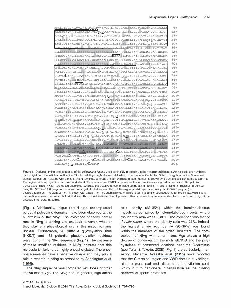
(Fig. 1). Additionally, unique poly-N runs, encompassedby usual polyserine domains, have been observed at theN-terminus of the NlVg. The existence of these poly-Nruns in NlVg is striking and unusual; however, whetherthey play any physiological role in this insect remainsunclear. Furthermore, 20 putative glycosylation sites(NXS/T) and 181 potential phosphorylation residueswere found in the NlVg sequence (Fig. 1). The presenceof these modified residues in NlVg indicates that thismolecule is likely to be highly phosphorylated. The phos-phate moieties have a negative charge and may play arole in receptor binding as proposed by Sappington et al.(1996).
The NlVg sequence was compared with those of otherknown insect Vgs. The NlVg had, in general, high amino
acid identity (23–35%) within the hemimetabolousinsects as compared to holometabolous insects, wherethe identity ratio was 20–30%. The exception was that ofAthalia rosae, where the identity ratio was 36%. Indeed,the highest amino acid identity (30–35%) was foundwithin the members of the order Hemiptera. The com-parison of NlVg with other insect Vgs shows a highdegree of conservation; the motif GL/ICG and the poly-cysteines at conserved locations near the C-terminus(see Tufail & Takeda, 2008) (Fig. 1) are particularly inter-esting. Recently, Akasaka et al. (2010) have reportedthat the C-terminal region and VWD domain of vitelloge-nin are processed and attached to the vitelline coat,which in turn participate in fertilization as the bindingpartners of sperm proteases.
Figure 1. Deduced amino acid sequence of the Nilaparvata lugens vitellogenin (NlVg) protein and its modular architecture. Amino acids are numberedon the right from the initiation methionine. The two vitellogenin_N domains delimited by the National Center for Biotechnology Information ConservedDomain Search are indicated with boxes at the N-terminus, whereas the von Willebrand factor domain is shown by a dark-shaded box at the C-terminus.The regions rich in polyserine tracts are underlined. Three consensus RXXR sequence motifs for possible cleavage sites are boxed. The putativeglycosylation sites (NXS/T) are dotted-underlined, whereas the putative phosphorylated serine (S), threonine (T) and tyrosine (Y) residues (predictedusing the NETPHOS 2.0 program) are shown with light-shaded frames. The putative signal peptide (predicted using the SIGNALP program) isdouble-underlined. The GL/ICG motif is shown with a bold line. The chemically determined N-terminal amino acid sequence for the 50 kDa vitellin (Vn)polypeptide is underlined with a bold dotted line. The asterisk indicates the stop codon. This sequence has been submitted to GenBank and assigned theaccession number: AB353856.
Nilaparvata lugens vitellogenin 789
© 2010 The AuthorsInsect Molecular Biology © 2010 The Royal Entomological Society, 19, 787–798

Phylogenetic relationship with Vgs of other arthropodsand vertebrates
To clarify the evolutionary relationship of NlVg, we used aneighbor-joining tree construction method based on dis-tances of 60 Vg sequences (excluding the signal peptide)from 46 arthropod and non-arthropod species. The den-drogram obtained places the NlVg with the other insectsand particularly with hemipterans as a distinct cluster(Fig. 2). Moreover, the tree clusters different insectspecies into specific orders in a quite coherent mannerreflecting that there exists a greater sequence similaritywithin the group than beyond the group level. The treealso clearly divides the Vgs from insects, arachnids,vertebrates and crustaceans into separate groups asindicated by their known phylogeny. Nevertheless, thenematode Vgs appear as a sister group of arachnid Vgs.The present phylogenetic analysis reveals that insect Vgsare more close to arachnid and nematode Vgs than tovertebrate and crustacean Vgs. The crustacean Vgs forma very external strongly supported cluster (100% boot-strap value) and thus remain the most divergent from allother Vgs.
Genomic copy number of the NlVg gene
To investigate the copy number of the Vg gene, Southernanalysis was carried out with 10 mg genomic DNA and aVg cDNA probe synthesized from the 5′-end part of theNlVg cDNA (nt position: 1–1001) as a probe. The hybrid-ization signal revealed a single discrete band whendigested with EcoRV, NotI and XhoI (Fig. 3), suggestingthat a single copy of the Vg gene exists in the genome ofN. lugens.
Expression pattern and temporal profile of Vg genetranscription in two wing-morphs of N. lugens
To probe the sex-, tissue- and stage-specific expression,and the temporal profile of NlVg gene expression, weperformed a Northern blot analysis with total RNAextracted from the adult males and females, and from thenymph whole bodies (Fig. 4). The hybridization analysisdetected a single transcript of ~6.8 kb exclusively in the fatbody of adult-females as we expected (Fig. 4A). No traceof hybridization signal was detected with total RNA fromadult males or from nymphs. The Vg gene transcript of6–7 kb has been identified from several insect species(see review: Tufail & Takeda, 2008). The developmentalprofile of Vg gene transcription in brachypterous femalesreveals that it starts expressing on day 3 of adult emer-gence, and continues to increase every day to reach itsmaximum on day 7, and then starts to decline (Fig. 4B, C).By contrast, the Vg gene transcript was observed one day
later, on day 4, in the macropterous females (Fig. 4B, C)although Vg mRNA levels were seen to be high, similar totheir brachypterous counterparts, during the vitellogenicperiod from day 5 onwards (Fig. 4B, C).
The discrepancy in reproductive strategies has beenreported in several insects exhibiting wing polymorphismincluding N. lugens (Kisimoto, 1965; Fujisaki, 1986). In N.lugens, the difference in pre-oviposition periods in twowing-forms has been reported (Ohkubo, 1967), and thegeneral trend demonstrated was that the macropterousfemales considerably delayed the initiation of ovipositioncompared to brachypterous females under the samerearing conditions (Kisimoto, 1965; Wada et al., 2007). InGryllus rubens, greater ovarian growth was reported in theshort-winged form than the long-winged morphs (Zera &Rankin, 1989). Moreover, in N. lugens, the earlier devel-opment of oocytes in brachypters was related to highJH titres in brachypters than those of macropters (Bertuso& Tojo, 2002). The different trait of JH titres in wing-dimorphic species is in keeping with the migratory syn-drome influenced by the JH (Fairbairn, 1994) and theapparent flight capability vs. fecundity trade-off (Harrison,1980). Our present findings of delayed expression of VgmRNA in long-winged morphs probably demonstrate thelonger developmental immaturity in these females thatcorresponds to the period of long distance migration.
Identification, post-transcriptional processing andtemporal expression profile of NlVgs
The NlVgs were identified by comparing the polypeptideprofiles of adult male and female whole body homoge-nates, haemolymph and of egg extracts. Sodium dodecylsulphate polyacrylamide gel electrophoresis (SDS-PAGE;Fig. 5A) revealed two Vg polypeptides (lane 3 and 5) of~175 and ~50 kDa corresponding to the two Vn polypep-tides (lane 4), whereas these polypeptides were absent inthe male sample (lane 2). Our results differ from thosereported by Cheng & Hou (2005) where only a singleprotein band of 175 kDa was reported. A possible expla-nation for this apparent discrepancy may be that we used15% SDS-PAGE in our experiments for adequate resolu-tion of the polypeptides instead of the 7.5% used byCheng & Hou (2005). To further clarify whether the 50 kDapolypeptide is a post-transcriptionally cleaved product, weperformed an amino-terminal analysis. The N-terminalsequence of the 50 kDa Vn polypeptide was SGSGP andlocated downstream of the putative signal peptide (Fig. 1),demonstrating, on a molecular basis, that the Vg of N.lugens also possesses a 50 kDa in addition to the175 kDa polypeptide previously identified through immu-noblotting by Cheng & Hou (2005) (Fig. 5A). The NlVgprecursor polypeptide is most probably cleaved followingRSRR cleavage signal (aa position: 452–455; Fig. 1) as
790 M. Tufail et al.
© 2010 The AuthorsInsect Molecular Biology © 2010 The Royal Entomological Society, 19, 787–798

Hymenoptera
Pteromalus puparumPimpla nipponica
Solenopsis invicta-Vg2Solenopsis invicta-Vg3Solenopsis invicta-Vg1
Apis mellifera
Encarsia formosaAthalia rosae
A d i
100
100
7599
66
100
78
Vgs
Diptera
Coleoptera
Aedes aegyptiAnopheles albimanusAnopheles gambiaeToxorhynchites amboinensisCulex pipiens quinquefasciatus
Tenebrio molitorAnthonomus grandis
Leucophaea maderae-Vg2Leucophaea maderae-Vg1Blattella germanica
Periplaneta americana-Vg2
100
97
100
94
100
100
100
8995
98
Hemiptera
Insectsp Vg
Periplaneta americana-Vg1Graptopsaltria nigrofuscataNilaparvata lugens
Lethocerus deyrolleiHomalodisca coagulata
Riptortus clavatusPlautia stali-Vg3Plautia stali-Vg1Plautia stali-Vg2
Lymantria disparS d t lit
100100
100
100 94
75
56
69
Lepidoptera
Spodoptera lituraBombyx moriBombyx mandarina
Samia cynthia riciniAntheraea pernyiAntheraea yamamaiActias seleneSaturnia japonica
Caenorhabditis elegans-Vg1Caenorhabditis elegans-Vg5
Caenorhabditis elegans-Vg6C
100
100
100
5194
100
99
99
97
100
96
Nematodes
Vertebrates
g gDermacentor variabilis-Vg1Rhipicephalus microplus
Dermacentor variabilis-Vg2Tetranychus urticae-Vg4Tetranychus urticae-Vg3Tetranychus urticae-Vg1Tetranychus urticae-Vg2Danio rerio
Gallus gallusIchthyomyzon unicuspis
Macrobrachium rosenbergii100
100
99100
100
99
89
87
52
86
Arachnids
Cherax quadricarinatus
Marsupenaeus japonicus
Macrobrachium rosenbergiiPandalus hypsinotus
Metapenaeus ensis-Vg2Metapenaeus ensis-Vg3Metapenaeus ensis-Vg1
Litopenaeus vannameiFenneropenaeus merguiensisPenaeus mondon
99100
60100
100
100
100
56
100
Crustaceans
0.2
Dictyoptera
Figure 2. A molecular phylogenetic tree of the 60 insect and non-insect vitellogenins (Vgs). A distance analysis of amino acid sequences was performedusing the CLUSTALW program (Thompson et al., 1994) and used as input for a neighbor-joining tree construction program (MEGA3, Kumar et al., 2001).The reference bar indicates the distance (number of amino acid substitutions per site). Bootstrap values (500 replications) are indicated at each node.Only support values >50% are shown. For sequence information see Experimental procedures.
Nilaparvata lugens vitellogenin 791
© 2010 The AuthorsInsect Molecular Biology © 2010 The Royal Entomological Society, 19, 787–798

the molecular weights predicted based on the amino acidsequences of small (48.33 kDa) and large (179.24 kDa)subunits correspond well with those identified by SDS-PAGE (Fig. 5A). The proteolytic cleavage of NlVg shouldnot be surprising. The Vg of another hemipteran Graptop-saltria nigrofuscata was also cleaved into one large(200 kDa) and small (43 kDa) polypeptide (Lee et al.,2000). Recently, we have also determined enzymaticcleavage of Vg into two subunits in the water bug, Letho-cerus deyrollei (Nagaba et al., 2010).
Based on the recent molecular studies, the consensusthat has emerged is that the Vg molecule is cleaved (fol-lowing the RXXR sequence motif) in almost all non-apocritan insect species, and this cleavage is found to beconserved at the N-terminus, mostly surrounded by poly-serine stretches (Fig. 1), except for the Lymantria disparVg, in which the cleavage signal (RXXR) exists at theC-terminus (see reviews: Sappington & Raikhel, 1998;Tufail et al., 2005, Tufail & Takeda, 2008). Nevertheless, inHemimetabola members (such as cockroaches and thebean bug, Riptortus clavatus) where the pro-Vg is cleavedinto several subunits, additional RXXR cleavage motifsresiding near the C-terminus or at the centre of the primaryproduct are utilized (Hirai et al., 1998; Tufail & Takeda,
2002, 2008). However, the reason why Vgs are differen-tially cleaved in different insect species remains unclear.
To determine the developmental expression profile ofVg in both wing-morphs, haemolymph and egg extractswere separated on SDS-PAGE and assayed using immu-noblot analysis (Fig. 5B). The antibody used was a gen-erous gift from Dr Roger F. Hou and generated against the175 kDa Vg peptide. The immunoblot analysis demon-strated the differential expression timing of Vg similar tothe transcript in both wing-forms; the Vg signal wasobserved on day 3 in brachypters as compared to mac-ropters where it first appeared on day 4 (Fig. 5B). Thedevelopmental expression profile revealed that Vg levelsincrease every day, reach a maximum on day 7, and thenstart to decrease in both wing morphs (Fig. 5B, C), sug-gesting sequestration by the developing oocytes. A similarpattern of Vg expression was observed in the other hemi-pteran, R. clavatus; however, the protein was detectedone day after the Vg mRNA first appeared (Shinoda et al.,1996; Hirai et al., 1998), which is in contrast to NlVg wherethis protein was detected on the same day (day 3 inbrachypters and day 4 in macropters) that Vg mRNAappeared (Fig. 4B). This is, perhaps, because of the shortlife of the adult N. lugens (only a few weeks) that Vgprotein is expressed soon after the Vg gene is transcribed.The postponed expression of Vg protein and mRNA(Fig. 4B) demonstrated by macropters of N. lugens in ourexperiments supports the fitness cost hypothesis for longdistance migration of this wing-dimorphic species. Studieson insect migration by flight have indicated that migrationby females often occurs when they are sexually immatureadults (Johnson, 1969). Hence, the pre-ovipositionalperiod, the duration of the interval between adult emer-gence and reproductive maturity, often corresponds to thetime during which the insect can express its migratorypotential (Hill & Gatehouse, 1993; Wilson & Gatehouse,1993; Wada et al., 2007; Zhao et al., 2009). In N. lugens,macropterous females also migrate prereproductivelybecause most immigrants caught in the net traps (Kyushu,Japan) were unmated individuals with immature ovaries(Kisimoto, 1976). Thus, the delayed expression of both thetranscript and the protein in macropterous females of N.lugens seems to be a good index of migration competencein this wing-dimorphic species.
Transcriptional regulation of the NlVg gene
In order to evaluate the effect of exogenous JH on NlVggene transcription, JH III was applied in a single dose(100 ng) on day 2 of female emergence with acetone as acontrol. Figure 6 shows that application of JH III inducedsignificant expression of NlVg mRNA compared withuntreated control or acetone-treated control samples.There is evidence that JH is responsible for oocyte matu-
Figure 3. Analysis of genomic copy number of the Nilaparvata lugensvitellogenin (NlVg) gene. Southern blot analysis was performed with10 mg genomic DNA digested with the restriction enzymes EcoRV, NotIand XhoI and separated on 1% gel. Blots were hybridized with analkaline phosphatase-labelled NlVg cDNA fragment (see Experimentalprocedures).
792 M. Tufail et al.
© 2010 The AuthorsInsect Molecular Biology © 2010 The Royal Entomological Society, 19, 787–798

ration in N. lugens (Iwanaga & Tojo, 1986; Ayoade et al.,1999). Oogenesis also requires JH in other hemipteranspecies such as Oncopeltus fasciatus (Johansson, 1954),Dysdercus intermedius (Dittmann et al., 1985), Spi-lostethus pandurus (Ibanez et al., 1987), R. clavatus(Shinoda et al., 1996) and Perillus bioculatus (Adamset al., 2002). Moreover, the JH III was identified as theonly type of JH in both nymphal and adult stages of N.lugens (Bertuso & Tojo, 2002). Thus, our results along withthe previous studies suggest that JH may be involved inregulation of NlVg gene transcription in this species.
Experimental procedures
Insects
The N. lugens culture used in this study was obtained from theNational Agricultural Research Centre for Kyushu OkinawaRegion, Kumamoto, Japan. The founder population was originallycollected from Nagasaki during cropping seasons of 1999 and isdesignated as Nagasaki-99. The culture had been maintainedunder laboratory conditions through sister-brother mating on ajaponica rice cultivar ‘Reiho’, and then established on rice seed-lings of a cultivar ‘Nipponbare’ in the Plant Genetics Laboratory,Faculty of Agriculture, Kobe University at 25 � 0.5 °C under
9.57.5
A
Vg mRNA
B
4.4
g
rRNA
1 2 3 4 5 7 9 13 days after eclosion
Vg mRNA
Vg mRNA
Brachypters
Macropters
rRNA
100
C
Vg N
rRNA
p
80
Vg
mR
NA
leve
ls (
Vg
mR
NA
leve
ls (
%)
0
20
40
60
1 2 3 4 5 7 9 13Female adult stage (days)
BrachyptersMacropters
Figure 4. Expression pattern and developmental profile of the Nilaparvata lugens vitellogenin (NlVg) gene. (A) Northern blot hybridization analysis wasperformed with 20 mg total RNA extracted from the nymphs, adult male whole bodies and from adult female ovary and fat body cells. The RNA sampleswere probed with an alkaline phosphatase-labelled NlVg cDNA fragment (see Experimental procedures). The mobility of a standard size RNA marker (kb)is shown on the left. (B) NlVg mRNA expression profile in the adult brachypterous and macropterous females at the different developmental periods.Total RNA (20 mg) was used to detect the hybridization signal as mentioned above. Ribosomal RNAs (bottom panels) are shown as internal control afterstaining with ethidium bromide. (C) Quantification of NlVg mRNA levels from the Northern blots (n = 3) shown in (B) at different time periods. The day 7Vg mRNA levels are expressed as 100%. The points show the average values of three independent experiments � SE.
Nilaparvata lugens vitellogenin 793
© 2010 The AuthorsInsect Molecular Biology © 2010 The Royal Entomological Society, 19, 787–798

16:8 h light : dark conditions with illumination by inflorescencelamps at an intensity of 110–120 mmol photons/m2/s.
Cloning of NlVg cDNA
NlVg cDNA was cloned by the 3′- and 5′-RACE-PCR methodusing a Marathon cDNA amplification kit (Clontech, Mountain
View, CA, USA) as reported previously (Tufail & Takeda, 2002).To amplify the 3′-end part of NlVg, the cDNA prepared from adultfemale whole bodies was used as a template with the degenerateprimers and the adaptor primer 1 (AP1) (marathon cDNA adaptor,Clontech). The degenerate primers designed were based on theGL/ICG sequence motif (Tufail & Takeda, 2002), and were:5′-GGA CTC TGT GG-3′, 5′-GGT CTG TGT GG-3′, 5′-GGT CTCTGC GG-3′, 5′-GGA CTG TGT GG-3′, 5′-GGT CTC TGT GG-3′,5′-GGG CTC TGC GG-3′, 5′-GG(ATG) CT(GC) TG(CT) GG-3′,5′-GGT ATT TGC GG-3′, 5′-GGC ATC TGT GG-3′ and 5′-GG(CT)AT(CT) TG(CT) GG-3′. PCR conditions employed were heating to94 °C for 1 min, followed by 32 cycles of 94 °C for 30 s, 55 °C for30 s, and 68 °C for 2 min. 5′-RACE-PCR was performed using agene-specific primer 1 (GSP1) (nucleotide position: 5692–5716,GenBank accession number: AB353856) designed based on theinitial 3′-end sequence and an AP1 (Clontech). The PCR condi-tions used were: 94 °C for 1 min, followed by 36 cycles of 94 °Cfor 5 s and 68 °C for 8 min. A second 5′-RACE-PCR was per-formed with GSP2 (nucleotide position: 1054–1078, GenBankaccession number: AB353856) synthesized based on the first
A
Vg subunitsVg subunits
175-kD200
119
90
63
50-kD
B
1 2 3 4 5 7 9 13
Days of female eclosion
49
Vg subunit
175-kD
175-kD
Brachypters
Macropters
vels
(%
)
C
6060
100100
8080
Vg
le
Female adult stage (days)
00
2020
4040
1 2 3 4 5 7 9 131 2 3 4 5 7 9 13
BrachyptersMacropters
Figure 5. Identification and developmental profile of Nilaparvata lugensvitellogenins (NlVgs). (A) Sodium dodecyl sulphate polyacrylamide gelelectrophoresis (SDS-PAGE) analysis of Vgs/vitellins (Vns) from adultmale and female whole body homogenates, and from ahemolymph andegg extracts of N. lugens. The SDS-PAGE gel was stained withCoomassie blue. M and arrowheads on the left indicate the molecularweight markers (kDa). The identified Vns/Vgs are indicated by arrows onthe right. (B) Developmental profile of 175 kDa Vg polypeptide in twowing morphs of N. lugens assayed by immunoblot analysis.Haemolymph samples (10 mg/lane) prepared from adult brachypterousand macropterous females at indicated periods were separated bySDS-PAGE, blotted to the polyvinylidene fluoride membrane andassayed by immunoblotting. Arrows on the right indicate the probedNlVgs. (C) Quantification of NlVg levels from the immunoblots shown in(B) at different time periods. The day 7 Vg protein levels are expressedas 100%. The points show the average values of three independentexperiments � SE.
A
Vg mRNA
rRNA
B
10
15
25
20
5
Rel
ativ
e V
g m
RN
A le
vels
0
Figure 6. Effect of juvenile hormone III (JH III) on Nilaparvata lugensvitellogenin (NlVg) gene transcription. (A) Northern blot analysis showsthe effect of JH III on Vg gene expression compared with those ofuntreated control and acetone-treated females. Ribosomal RNAsare shown as internal control after staining with ethidium bromide.(B) Quantification of NlVg mRNA levels from the Northern blots. Thebars indicate the average values of three independent experiments� SE.
794 M. Tufail et al.
© 2010 The AuthorsInsect Molecular Biology © 2010 The Royal Entomological Society, 19, 787–798

5′-end sequence and the AP1 to achieve the complete 5′-endpart. The amplification conditions used were 1 min at 94 °C, fol-lowed by 32 cycles of 94 °C for 30 s and 68 °C for 2 min. Theamplified PCR products were cloned into the pT7Blue vector(Novagen, Madison, WI, USA) or pTA2 vector (Toyobo, Osaka,Japan) and sequenced. The overlapping sequences of the abovethree PCR fragments were assembled to obtain the full-lengthNlVg cDNA sequence.
Sequence analysis
All sequences were analysed with the BCM search launchercomputer program (http://searchlauncher.bcm.tmc.edu/seq-util/seq-util.html) and GENETYX v. 5.1 program (Genetyx Corporation,Tokyo, Japan). Sequences were checked for their homology withother Vgs using a FASTA or BLAST homology search on the DDBJdatabase (http://www.ddbj.nig.ac.jp/index-e.html). The signalpeptide position was predicted using the SIGNALP computerprogram. Mass/isoelectric point was conducted using ExPASy-COMPUTE pI/Mw (http://www.expasy.ch/tools/pi_tool.html). TheNCBI CDD search was used to identify the conserved regionsin NlVg. The putative phosphorylation sites were detected byusing the NetPhos 2.0 Server (http://www.cbs.dtu.dk/services/NetPhos/).
Sequence comparison and phylogenetic relationship
The Vg sequences used for comparison were from the followingspecies (accession number in parentheses): Periplaneta ameri-cana (Vg1: AB034804; Vg2: AB047401), Leucophaea maderae(Vg1: AB052640; Vg2: AB194976), Blattella germanica(AJ005115), Lethocerus deyrollei (AB425334), Graptopsaltrianigrofuscata (AB026848), Plautia stali (Vg1: AB033498; Vg2:AB033499; Vg3: AB033500), Riptortus clavatus (U97277),Homalodisca coagulata (DQ118408), Nilaparvata lugens(AB353856, the present study), Anthonomus grandis (M72980),Tenebrio molitor (AY714212), Athalia rosae (AB007850), Pimplanipponica (AF026789), Apis mellifera (AJ517411), Encarsiaformosa (AY553878), Pteromalus puparum (EF468683), Sole-nopsis invicta (Vg1: AF512520; Vg2: AY941795; Vg3: AY941796),Aedes aegypti (U02548), Anopheles gambiae (AF281078),Anopheles albimanus (AY691327), Toxorynchites amboinensis(AY691326), Culex pipiens quiquefasciatus (AY691324), Bombyxmori (D13160), Lymantria dispar (U90756), Antheraea pernyi(AB049631), Antheraea yamamai (AB055843), Samia cynthiaricini (AB055844), Bombyx mandarina (AB055845), Saturniajaponica (AB190809), Actias selene (EF523567), Spodopteralitura (EU095334), Caenorhabditis elegans (Vg1: AAB52675;Vg5: AAA83587; Vg6C AAQ91901), Dermacentor variabilis (Vg1:AAW78557; Vg2: ABW82681), Rhipicephalus microplus(ABS88989), Tetranychus urticae-Vg1-4 (AB505063-66), Daniorerio (NP_739573), Gallus gallus (AAA49139), Ichthyomyzon uni-cuspis (Q91062), Macrobrachium rosenbergii (BAB698831),Pandalus hypsinotus (BAD11098), Cherax quadricarinatus(AAG17936), Metapenaeus ensis (Vg1: AAM48287; Vg2AAT01139; Vg3 (AAN40700), Marsupenaeus japonicus(BAD98732), Litopenaeus vannamei (AAP76571), Fennerope-naeus merguiensis (AAR88442) and Penaeus mondon(ABB89953). Sequences alignments were performed using theCLUSTALW program (Thompson et al., 1994). Phylogenetic analy-sis was performed using the neighbor-joining method by the
Molecular Evolutionary Genetics Analysis (MEGA) v. 3.1 program(Kumar et al., 2001). For tree construction, Poisson correctiondistance, uniform rates amongst the sites, and a pairwise-deletionmode were used. The stability of the inferred phylogeny wasestimated by the bootstrap test with 500 replications and arandom seed value of 64238.
SDS-PAGE, immunoblotting and N-terminal sequence analysis
Adult male and female whole bodies, as well as eggs (oocytes)were homogenized in the homogenization buffer (20 mM Tris/HClpH 7.0, 2 mM ethylenediaminetetraacetic acid, 1 mM phenylm-ethylsulphonyl fluoride), supplemented with a protease inhibitorscocktail (one tablet/50 ml of the buffer solution; Complete, RocheDiagnosis GmbH, Mannheim, Germany), using HG30 homog-enizer (Hitachi Ltd., Tokyo, Japan). The homogenate was centri-fuged at 10 000 g for 10 min. Protein contents in the supernatantwere measured using the DC protein assay Kit (Bio-Rad, Her-cules, CA, USA) according to the instruction of the supplier.Bovine serum albumin was used as a standard protein. Theextracted proteins were mixed with equal volume of samplebuffer [0.5 M Tris, pH 6.8, 10%SDS, 50% glycerol, 0.4 ml2-mercaptoethanol, 0.05 % (w/v) bromophenol blue], denaturedin boiling water for 10 min, then immediate cooled on ice for 2 minand stored at -20° C for future use. The haemolymph was suckedwith a capillary from the adult females and was mixed in theabove sample buffer at 1:50. The protein contents were mea-sured as mentioned above. After boiling the haemolymphsamples were also stored at -20 °C until required. A total of 10 mgfor each sample was used for SDS-PAGE (15%) analysis. Thegels were stained with Coomassie Brilliant Blue.
For immunoblot analysis, the separated male and femalehaemolymph and egg samples (10 mg per lane) were transferredon to the Immobilon-P membrane (Millipore, Billerica, MA, USA),and then incubated overnight at 4 °C with chicken anti-175 kDaNlVg primary antibody (diluted at 1:3000; a generous gift from DrRoger F. Hou; see Cheng & Hou, 2005), followed by incubationfor 1 h at room temperature with goat anti-chichen Immunoglo-bulin G horseradish peroxidase conjugated secondary antibody(Cappel, Westchester, PA, USA) (diluted at 1:50 000). Theimmune signals were detected using enhanced chemilumines-cence plus detection reagents (Amersham-Pharmacia, Piscat-away, NJ, USA) according to the supplier’s protocol. To determinethe N-terminal sequence of the 50 kDa Vg polypeptide, eggextracts (20 mg per lane) separated by SDS-PAGE were trans-ferred to the polyvinylidene fluoride membrane (Millipore, Immo-bilon) and stained with Ponceau S (0.2% in 1% acetic acid). Thebands corresponding to the 50 kDa Vg were cut out and appliedto a gas phase amino acid sequencer (Perkin Elmer, Boston, MA,USA; 492 Procise).
Genomic digestion and Southern blotting
The genomic DNA was isolated from the insect whole body witha Mammalian Genomic DNA Kit (Sigma, St. Louis, MO, USA). ForSouthern hybridization, 10 mg of genomic DNA was digestedwith restriction enzymes: EcoRV, NotI and XhoI (Toyobo). Thedigested DNA was precipitated with ethanol and separated on a1% agarose gel. DNA was denatured and transferred to a HybondN+ membrane (Amersham, Little Chalfont, Buckinghamshire, UK)by capillary blotting. We used the Amersham Gene Images
Nilaparvata lugens vitellogenin 795
© 2010 The AuthorsInsect Molecular Biology © 2010 The Royal Entomological Society, 19, 787–798
AlkPhos Direct labeling and detection system (GE Healthcare,Buckinghamshire, UK). Briefly, the membrane was prehybridizedin AlkPhos Direct hybridization buffer for 15 min at 60 °C, andhybridized overnight at 60 °C in the same buffer with a NlVgcDNA fragment (nt position: 1–1001) as a probe. After hybridiza-tion, the membrane was rinsed twice in the primary wash bufferfor 10 min each at 60 °C and twice in the secondary wash bufferfor 5 min each at room temperature. Then after incubation for5 min in the detection reagent, the membrane was exposed toHyperfilm-MP (Amersham) for 30 min.
Total RNA preparation and Northern blot analysis
Total RNA was isolated from the adult females and males, andfrom nymphs using isogen (Nippon Gene Co. Ltd, Toyama,Japan), and were stored at -80 °C until use. To determine thesize, tissue-, sex- and stage-specific expression, and the devel-opmental expression profile of NlVg mRNA, Northern blot hybrid-ization analysis was performed. Aliquots of 20 mg of total RNAfrom adult females, males and nymphs were separated on 1%agarose/0.66 M formaldehyde gel in 3-morpholinopropane-sulfonic acid buffer. The separated RNA was transferred toHybond N+ membrane (Amersham-Pharmacia) through passivecapillary transfer. The RNA blot was hybridized with a Vg cDNAprobe (50 ng) synthesized from the 5′-end part of NlVg cDNA (ntposition: 1–1078). For Northern analysis, we also used the Amer-sham Gene Images AlkPhos Direct labeling and detection system(GE Healthcare) as described above for Southern blotting.
Transcriptional regulation of NlVg gene
To study the hormonal regulation of NlVg gene transcription JH III(Sigma Chemical Co., St Louis, MO, USA) was applied topicallyto the abdominal sternites of 2-day-old adult females (brachypter-ous), a stage indicated to be sensitive for the JH analogue,methoprene, by Iwanaga & Tojo (1986). For treatment, JH III wasdissolved in acetone to yield a final concentration of 200 ng/ml,and a droplet (0.5 ml) of JH III solution was applied after anaes-thetizing with carbon dioxide. Control animals were treated with0.5 ml acetone. The treated animals were kept separately underthe same rearing conditions mentioned above. After 24 h of JH IIIapplication, total RNA was extracted from the treated animals andexpression patterns of Vg mRNA were observed as describedabove.
Acknowledgements
We thank Dr Y. Fukami, Mr S. Kawamoto andDr T. A. Alexander, Research Center for EnvironmentalGenomics, Kobe University, for amino acid analysis ofpolypeptides.
References
Adams, T.S., Filipi, P.A. and Yi, S.X. (2002) Effect of age, diet,diapause and juvenile hormone on oogenesis and the amountof vitellogenin and vitellin in the twospotted stink bug, Perillusbioculatus (Heteroptera: Pentatomidae). J Insect Physiol 48:477–486.
Akasaka, M., Harada, Y. and Sawada, H. (2010) VitellogeninC-terminal fragments participate in fertilization as egg-coatbinding partners of sperm trypsin-like proteases in the ascid-ian Halocynthia roretzi. Biochem Biophys Res Comm 392:479–484.
Ayoade, O., Morooka, S. and Tojo, S. (1999) Enhancement ofshort wing formation and ovarian growth in the geneticallydefined macropterous strain of the brown planthopper,Nilaparvata lugens. J Insect Physiol 45: 93–100.
Babin, P.J., Bogerd, J., Kooiman, F.P., Van Marrewijk, W.J.A. andVan der Horst, D.J. (1999) Apolipophorin II/I, apolipoprotein B,vitellogenin, and microsomal triglyceride transfer proteingenes are derived from a common ancestor. J Mol Evol 49:150–160.
Barr, P.J. (1991) Mammalian subtilisins: the long-sought dibasicprocessing endoproteases. Cell 66: 1–3.
Bertuso, A.G. and Tojo, S. (2002) The nature and titer of juvenilehormone in the brown planthopper, Nilaparvata lugens(Homoptera: Delphacidae) in relation to wing morphogenesisand oocyte development. Appl Entomol Zool 37: 117–125.
Byrne, B.M., Gruber, M. and Ab, G. (1989) The evolution of eggyolk proteins. Progr Biophys Mol Biol 53: 33–69.
Chen, J.-S. and Raikhel, A.S. (1996) Subunit cleavage of mos-quito pro-vitellogenin by a subtilisin-like convertase. Proc NatlAcad Sci USA 93: 6186–6190.
Chen, J.S., Sapington, T.W. and Raikhel, A.S. (1997) Extensivesequence conservation among insect, nematode and verte-brate vitellogenins reveals ancient common ancestry. J MolEvol 44: 440–451.
Cheng, D.J. and Hou, R.F. (2005) Determination and distributionof a female-specific protein in the brown planthopper, Nilapar-vata lugens Stal (Homoptera: Delphicidae). Tissue Cell 37:37–45.
Cheng, S.N., Chen, J.C., Si, H., Yan, L.M., Chu, T.L., Wo, T.C.et al. (1979) Studies on the migration of brown planthopper,Nilaparvata lugens Stal. Acta Entomol Sin 22: 1–21.
Denno, R.E., Olmstead, K.L. and McCloud, E.S. (1989) Reproduc-tive cost of flight capability: a comparison of life history traits inwing dimorphic planthoppers. Ecol Entomol 14: 31–44.
Denno, R.E., Roderick, G.K., Oimstead, K.L. and Dobel, H.G.(1991) Density-related migration in planthoppers (Homoptera:Delphacidae): the role of habitat persistence. Am Nat 138:1513–1541.
Dittmann, F., Trenczek, T. and Kleemann-Stumpf, I. (1985) Juve-nile hormone-controlled vitellogenic cycles in Dysdercusintermedius (Heteroptera). J Insect Physiol 31: 729–735.
Fairbairn, D.J. (1994) Wing dimorphism and migratory syndrome:correlated traits for dispersal tendency in wing dimorphicinsects. In Proceedings of Memorial and International Sympo-sium on Dispersal Polymorphism in Insects, its Adaptationand Evolution (Nakasuji, F. and Fujisaki, K., eds), pp. 143–152. Okayama University, Okayama.
Fujisaki, K. (1986) Reproductive properties of the oriental chinchbug, cavelerius saccharivorus Okajima (Heteroptera: Lyga-eidae), in relation to its wing polymorphism. Res Popul Ecol28: 43–52.
Hagedorn, H.H., Maddison, D.R. and Tu, Z. (1998) The evolutionof vitellogenins, cyclorrhaphan yolk proteins and relatedmolecules. Adv Insect Physiol 27: 335–384.
Harrison, R.G. (1980) Dispersal polymorphism in insects. AnnRev Ecol Syst 11: 95–118.
796 M. Tufail et al.
© 2010 The AuthorsInsect Molecular Biology © 2010 The Royal Entomological Society, 19, 787–798

Hattori, M. and Sogawa, K. (2002) Oviposition behaviour of therice brown planthopper, Nilaparvata lugens (Stal), and itselectronic monitoring. J Insect Behav 15: 283–293.
Hill, J.K. and Gatehouse, A.G. (1993) Phenotypic plasticityand geographical variation in the pre-reproductive periodof Autographa gamma (Lepidoptera: Noctuidae) and itsimplications for migration in this species. Ecol Entomol 18:39–46.
Hirai, M., Watanabe, D., Kiyota, A. and Chinzei, Y. (1998) Nucle-otide sequence of vitellogenin mRNA in the bean bug, Riptor-tus clavatus: analysis of processing in the fat body and ovary.Insect Biochem Mol Biol 28: 537–547.
Ibanez, P., Garcera, M.D., Gavara, A. and Martınez, R. (1987)Effect of JH and PII on the protein content of haemolymph andovaries of Spilostethus pandurus. Comp Biochem Physiol88B: 157–161.
Iwanaga, K. and Tojo, S. (1986) Effects of juvenile hormone andrearing density on wing dimorphism and oocyte developmentin the brown planthopper, Nilaparvata lugens. J Insect Physiol32: 585–590.
Johansson, A.S. (1954) Corpus allatum and egg production instarved milkweed bugs. Nature 174: 174–189.
Johnson, C.G. (1969) Migration and Dispersal of Insects byFlight. Methuen and Co. Ltd., London.
Ketipearachchi, Y., Kaneda, C. and Nakamura, C. (1998) Adap-tation of the brown planthopper (BPH), Nilaparvata lugensStål (Homoptera: Delphacidae), to BPH resistant rice cultivarscarrying bph8 or Bph9. Appl Entomol Zool 33: 497–505.
Kisimoto, R. (1965) Studies on the polymorphism and its role inthe population growth of the brown planthopper, Nilaparvatalugens Stål. Bull Shi Natl Agri Exp Stat 13: 1–107.
Kisimoto, R. (1976) Synoptic weather conditinso inducinglong-distance immigration of planthoppers, Sogatella furciferaHorvath and Nilaparvata lugens Stal. Ecol Entomol 1: 95–109.
Kumar, S., Koichiro, T., Ingrid, B.J. and Masatoshi, N. (2001)MEGA2: Molecular Evolutionary Genetics Analysis Software.Arizona State University, Arizona.
Kunkel, J.G. and Nordin, J.H. (1985) Yolk proteins. In Compre-hensive Insect Physiology, Biochemistry and Pharmacology,Vol. 1 (Kerkut, G.A. and Gilbert, L.I., eds), pp. 83–111. Per-gamon press, New York, NY.
Lee, J.M., Nishimori, Y., Hatakeyama, M., Bae, T.-W. and Oishi,K. (2000) Vitellogenin of the cicada Graptopsaltria nigrofus-cata (Homoptera): analysis of its primary structure. InsectBiochem Mol Biol 30: 1–7.
Naeemullah, M., Sharma, P.N., Tufail, M., Mori, N., Matsumura,M., Takeda, M. et al. (2009) Characterization of brown plan-thopper strains based on their differential responses to intro-gressed resistance genes and on mitochondrial DNApolymorphism. Appl Entomol Zool 44: 475–483.
Nagaba, Y., Tufail, M., Inui, H. and Takeda, M. (2010) Hormonalregulation and effects of 4 environmental polutants on vitello-genin gene transcription in the giant water bug, Lethocerusdeyrollei (Heteroptera: Belostomatidae). J Insect Conserv (inpress).
Ohkubo, N. (1967) Study on the density effect of the adult of thebown planthopper, Nilaparvata lugens. Jap J Ecol 17: 230–233.
Raikhel, A.S. and Dhadialla, T.S. (1992) Accumulation of yolkproteins in insect oocytes. Annu Rev Entomol 37: 217–251.
Roff, D.A. (1984) The cost of being able to fly: a study of wingpolymorphism in two species of crickets. Oecologia 63:30–37.
Roff, D.A. (1986) Evolution of wing polymorphism and its impacton life cycle adaptation in insects. In The Evolution of InsectLife Cycles (Taylor, E. and Karban, R., eds), pp. 204–221.Springer, New York, NY.
Rouille, Y., Duguay, S.J., Lund, K., Furuta, M., Gong, Q. andLipkind, G. (1995) Proteolytic processing mechanisms in thebiosynthesis of neuroendocrine peptides: the subtilisin-likeproprotein convartases. Front Neur 16: 322–361.
Sappington, T.W. and Raikhel, A.S. (1998) Molecular character-istics of insect vitellogenins and vitellogenin receptors. InsectBiochem Molec Biol 28: 277–300.
Sappington, T.W., Kokoza, V.A. and Raikhel, A.S. (1996)Molecular characterization of the mosquito vitellogenin recep-tor reveals unexpected high homology to the Drosophila yolkprotein receptor. Proc Natl Acad Sci USA 93: 8934–8939.
Sappington, T.W., Oishi, K. and Raikhel, A.S. (2002) Structuralcharacteristics of insect vitellogenins. In Progress in Vitello-genesis (Raikhel, A.S. and Sappington, T.W., eds), Reproduc-tive Biology of Invertebrates, Vol. XII. Part A, pp. 69–101.Science Publishers, Inc. Enfield, NH; Plymouth, UK.
Shinoda, T., Miura, K., Taylor, D. and Chinzei, Y. (1996) Vitello-genin and vitellin in the bean bug, Riptortus clavatus (Hemi-ptera: Alydidae): purification, immunological identification, andinduction by juvenile hormone. Arch Insect Biochem Physiol31: 395–412.
Smolenaars, M.M.W., Madsen, O., Rodenburg, K.W. and Van derHorst, D.J. (2007) Molecular diversity and evolution of thelarge lipid transfer protein superfamily. J Lipid Res 48: 489–502.
Snigirevskaya, E.S. and Raikhel, A.S. (2005) Receptor-mediatedendocytosis of yolk proteins in insect oocytes. In Progress inVitellogenesis (Raikhel, A.S. and Sappington, T.W., eds),Reproductive Biology of Invertebrates (Series Editors, AdiyodiKG, Adiyodi RG), Vol XII, Part B, pp. 199–228. SciencePublishers, Inc. Enfield, NH; Plymouth, UK.
Sogawa, K. and Pathak, M.D. (1970) Mechanisms of brownplanthopper (Hemiptera: Delphacidae) resistance in Mudgovariety of rice. Appl Ent Zool 53: 145–158.
Sogawa, K. and Watanabe, T. (1992) Redistribution of rice plan-thoppers and its synoptic monitoring in East Asia. TechnicalBulletin No. 131. Food & Fertilizer Technology Center, Taipei,Taiwan, pp. 1–9.
Thompson, J.D., Higgins, D.G. and Gibson, T.J. (1994) Clustal W:improving the sensitivity of progressive multiple sequencealignment through sequence weighting, position-specific gappenalties and weight matrix choice. Nucleic Acids Res 22:4673–4680.
Tufail, M. and Takeda, M. (2002) Vitellogenin of the cockroach,Leucophaea maderae: nucleotide sequence, structure andanalysis of processing in the fat body and oocytes. InsectBiochem Mol Biol 32: 1469–1476.
Tufail, M. and Takeda, M. (2008) Molecular characteristics ofinsect vitellogenins. J Insect Physiol 54: 1447–1458.
Tufail, M. and Takeda, M. (2009) Insect vitellogenin/ lipophorinreceptors: molecular structures, role in oogenesis, and regu-latiory mechanisms. J Insect Physiol 55: 87–103.
Tufail, M., Raikhel, A.S. and Takeda, M. (2005) Biosynthesis andprocessing of insect vitellogenins. In Progress in Vitellogen-
Nilaparvata lugens vitellogenin 797
© 2010 The AuthorsInsect Molecular Biology © 2010 The Royal Entomological Society, 19, 787–798

esis (Raikhel, A.S. and Sappington, T.W., eds), ReproductiveBiology of Invertebrates (Series Editors, Adiyodi KG, AdiyodiRG), Vol. XII, Part B. pp. 1–32. Science Publishers, Inc.,Enfield; Plymouth.
Wada, T., Ito, K., Takahashi, A. and Tang, J. (2007) Variation ofpre-ovipositional period in the brown planthopper, Nilaparvatalugens, collected in tropical, subtropical and temperate. Asia JApp Entomol 131: 698–703.
Wilson, K. and Gatehouse, A.G. (1993) Seasonal and geographi-cal variation in the migratory potential of outbreak populationsof the African armyworm moth, Spodoptera exempta. J AnimEcol 62: 169–181.
Zera, A.J. and Rankin, M.A. (1989) Wing dimorphism inGryllus rubens: genetic basis of morph determination andfertility differences between morphs. Oecologia 80: 249–255.
Zera, A.J. and Denno, R.F. (1997) Physiology and Ecology ofdispersal polymorphism in insects. Annu Rev Entomol 42:207–230.
Zhao, X.C., Feng, H.Q., Wu, B., Wu, X.F., Liu, Z.F., Wu, K.M. et al.(2009) Does the onset of sexual maturation terminate theexpression of migratory behaviour in moths? A study of theoriental armyworm, Mythimna separata. J Insect Physiol 55:1039–1043.
798 M. Tufail et al.
© 2010 The AuthorsInsect Molecular Biology © 2010 The Royal Entomological Society, 19, 787–798
Copyright © 2022 FDOKUMEN