
Granulomatous Interstitial Nephritis in Children Resulting from ...
Upload
independentCategory
view
3download
0
Molecular Immunology 46 (2009) 1935–1941
Contents lists available at ScienceDirect
Molecular Immunology
journa l homepage: www.e lsev ier .com/ locate /mol imm
Molecular characterization of a large cohort of patients with ChronicGranulomatous Disease and identification of novel CYBB mutations:An Italian multicenter study
Gigliola Di Matteoa,1, Lucia Giordanib,1, Andrea Finocchia,c, Annamaria Venturab, Maria Chiriacoa,Jan Blancatod, Cecilia Sinibaldie, Alessandro Plebani f, Annarosa Soresina f, Claudio Pignatag,Rosa Maria Dellepianeh, Antonino Trizzino i, Fausto Cossuj, Roberto Rondelli k, Paolo Rossia,c,Domenico De Mattiab,∗, Baldassarre Martireb, with IPINET(Italian Network for Primary Immunodeficiencies)a Department of Public Health and Cellular Biology, Tor Vergata University, Rome, Italyb Department of Biomedicine of Development Age, University of Bari, Italyc Division of Immunology and Infectious Diseases, Children’s Hospital Bambino Gesù, Rome, Italyd Department of Oncology, Georgetown University, Washington, DC, USAe Department of Biopathology, Human Genetics Section, University of Rome Tor Vergata, Italyf Department of Pediatrics, University of Brescia, Italyg Department of Pediatrics, University of Naples, Italyh Department of Pediatrics, IRCSS-Fondazione Policlinico University of Milano, Italyi Pediatric Oncohematology “G. Di Cristina” Hospital, Palermo, Italyj BMT Unit Ospedale Microcitemico, Cagliari, Italyk Department of Pediatrics, University of Bologna, Italy
a r t i c l e i n f o
Article history:Received 19 March 2009Accepted 20 March 2009Available online 1 May 2009
Keywords:CYBBChronic Granulomatous DiseaseNADPH oxidaseMutation analysisGenotype–phenotype correlation
a b s t r a c t
Chronic Granulomatous Disease (CGD) is a rare inherited disorder in which phagocytes fail to produceantimicrobial superoxide because NADPH oxidase activity is absent. In about 65% of the cases, the diseaseis due to mutations affecting the X-linked CYBB gene, encoding the gp91phox subunit of NADPH oxidase.We investigated 34 CGD male patients by DHPLC and direct sequencing. A mutation was found in theCYBB gene of 33 patients and 9 of these were novel: one non-sense mutation (c.1123 G>T), three missensemutations (c.58G>A; c.1076 G>C; c.1357 T>A), two splice site mutations (c.141+5G>T; c.142-1G>A), oneduplication (c.42 45dupCATT), one deletion (c.184delT), and one rare deletion of two non-contiguousnucleotides (c.1287delT + c.1290delC). One patient had the most frequent GT homozygous deletion inexon2 of the NCF-1 gene encoding the p47phox subunit of NADPH oxidase. The carrier analysis was per-formed in 23 patients’ mothers and 16 female relatives through molecular and FISH studies. No clear
correlation between the severity of clinical symptoms and the type of mutation could be demonstrated.This study further supports the great heterogeneity of the disease and the notion that genetic analysis isg a de
1
miC
v
0d
a critical step in obtainin
. Introduction
Chronic Granulomatous Disease (CGD; MIM# 306400) is a pri-ary immunodeficiency that affects phagocytes of the innate
mmune system caused by defects in the NADPH oxidase system.GD patients are susceptible to recurrent life-threatening infec-
∗ Corresponding author at: Dipartimento di Biomedicina dell’Età Evolutiva, Uni-ersità di Bari, Piazza Giulio Cesare 11, 70124 Bari, Italy. Fax: +39 080 5592290.
E-mail address: [email protected] (D. De Mattia).1 These authors equally contributed to the study.
161-5890/$ – see front matter © 2009 Elsevier Ltd. All rights reserved.oi:10.1016/j.molimm.2009.03.016
finitive diagnosis for CGD.© 2009 Elsevier Ltd. All rights reserved.
tions, particularly those caused by catalase positive bacteria andfungi. In addition, CGD patients often have poor wound healing andexaggerated inflammatory responses, leading to granuloma forma-tion in subcutaneous tissues, lungs, lymph nodes, liver and bones(Segal et al., 2000; Winkelstein et al., 2000; Martire et al., 2008).The NADPH oxidase complex acts mainly by transferring electronsfrom NADPH to molecular oxygen in order to form superoxide indis-
pensable for killing of phagocyted microorganisms. This complex iscomposed of a heterodimeric membrane-bound flavocytochromeb558 (formed by gp91phox and p22phox) and four cytoplasmaticsubunits: p47phox, p67phox, p40phox and Rac2. Mutations in anyof the genes encoding NADPH oxidase subunits can cause CGD.
1 Immu
AXpmersrstyssPew
2
2
GoCoef
ypamn
d(pe2
pwm
BtB
CD
2
gatifwb2el
936 G. Di Matteo et al. / Molecular
pproximately two thirds of CGD cases result from mutations in the-linked CYBB gene, encoding the gp91phox subunit. The remainingatients with CGD have an autosomal recessive form (AR-CGD), theost frequent of which, is due to a mutation of NCF1 gene that
ncodes the p47phox subunit (Dinauer, 2005). Herein we report theesults of CYBB and NCF-1 gene sequence analysis in a cohort con-tituted by 34 CGD patients from 29 unrelated families and theirelatives and we describe nine novel CYBB mutations. Furthermore,ince the clinical severity of the CGD varies widely, we investigatedhe correlation between the rate of severe infections per patient-ear and the mutation type and we did not find a statisticallyignificant variation, although in patients with deletion and non-ense mutations a higher number of severe infections were found.olymorphisms in oxygen independent antimicrobial systems ornvironmental factors should be investigated to find a correlationith the clinical severity of the disease.
. Patients and methods
.1. Patients
Thirty-four male patients, from the Italian registry of Chronicranulomatous Disease which is part of the Italian Primary Immun-deficiency Network (IPINET), were tested for mutations in theYBB gene. Patients were diagnosed as having CGD on the basisf their clinical history and the inability of their phagocytes to gen-rate reactive oxygen species (ROS) by conventional granulocyteunctional tests as previously described (Martire et al., 2008).
Age at diagnosis ranged from 1 month to 49 years (mean 7.16ears, median 3 years). Carrier detection was performed in 23atients’ mothers and 16 female relatives by means of geneticnalysis and through fluorescent in situ hybridization (FISH) ofetaphase chromosomes from lymphoblastoid cell lines when
eeded.All relevant clinical features, immunological and therapeutic
ata have been already described in a previous report by the IPINETMartire et al., 2008). All patients were put on antiinfective pro-hylaxis with trimethoprim/sulphomethoxazole and itraconazolearly after diagnosis. Some subjects (patients 2, 4, 6, 8, 17, 20, 23, 24,6–28, 32) received discontinuous administration of �-interferon.
Informed consent was obtained from each studied subject orarents of each child and all procedures used were in accordanceith the guidelines of the Helsinki Declaration on Human Experi-entation.All studies were performed at the Paediatric Immunology and
iotechnology Laboratory, Tor Vergata University of Rome and athe Department of Biomedicine of Development Age, University ofari.
FISH studies were performed in the Lombardi Comprehensiveancer Center, Georgetown University Medical Center, Washington,C, USA.
.2. Cell lines
Neutrophils were purified by discontinuous Percoll densityradient centrifugation from peripheral blood of patients 17, 33nd 34 as previously described (Giudicelli et al., 1982). EBV-ransformed lymphoblastoid cell lines (LCL) were obtained byncubating approximately 5 × 10⁶ PBMC, with 2 ml of supernatantrom the EBV-secreting cell line B95-8 for 1 h at 37 ◦C. Infected cells
ere cultured at 1 × 10⁶ cells/ml in RPMI 1640 containing 20% fetalovine serum, 2 mM l-glutamine and 1 �g/ml of cyclosporin A in4-well culture plates. Clumps of cells were usually observed at thend of the first or second week and stable EBV-lymphoblastoid cellines were established in 3–4 weeks.nology 46 (2009) 1935–1941
2.3. Molecular studies
Genomic DNA isolated from peripheral blood of patients andhealthy controls (QIAamp DNA Blood kit by QIAGEN GmbH, Hilden,Germany) was amplified using primers (available upon request)flanking the 13 coding exons and the promoter region of theCYBB gene (NG 009065). PCR reactions were carried out in a vol-ume of 50 �l containing 100 ng of genomic DNA, 200 �M of eachdNTP, 0.4 �M of each primer and 0.5 U of GoTaq DNA polymerase(Promega, Madison, WI). The samples were denatured at 95 ◦C for5 min followed by 30–40 cycles at 95 ◦C for 30 s, annealing at exonspecific temperature (available upon request) for 45 s, 72 ◦C for 30 swith a final 5 min extension at 72 ◦C.
DHPLC was performed on a WAVE analysis system (Transge-nomic, Omaha, NE) using different temperature conditions(available upon request). Briefly, PCR products were mixed inapproximately equimolar proportions with an amplified sampleknown to contain a wild-type CYBB fragment, then denatured at94 ◦C for 5 min and then cooled at room temperature, to allow het-eroduplex formation. Each sample were run on a DnaSept.M column(Transgenomics) and monitored by ultraviolet light (260 nm). Opti-mum DHPLC temperatures were detected using software-predictedmelting profile as a starting point. Samples were run at a meanof three temperatures due to the heterogeneity in the distributionof GC-rich regions (Xiao and Oefner, 2001). Each elution chro-matographic profile was compared with profiles associated withhomozygous wild-type sequence. Samples with an altered chro-matographic profile were sequenced.
Direct sequencing was performed after purifying PCR productswith QIAquick PCR purification kit (QIAGEN, Hilden, Germany).Both strands were sequenced using the BigDye Terminator v3.1Cycle Sequencing Kit (Applied Biosystems, Foster City, CA) and ana-lyzed on an ABI PRISM 3130 and 310 automated sequencers (AppliedBiosystems, Foster City, CA).
For the detection of GT homozygous deletion in exon 2 of NCF-1gene we used primers pair and PCR conditions already described(Noack et al., 2001), which do not differentiate between NCF-1and �NCF1. DHPLC analysis revealed the homozygous profile of thepatient 20 and the data was confirmed by sequence.
2.4. RT-PCR
When required, total RNA was isolated from either mononu-clear cells or EBV-transformed B lymphocytes of both CGDpatients and healthy individuals by using Trizol procedure(Invitrogen, Life Technologies, Milano, Italy). Reverse tran-scription was performed using a single reaction kit (Super-script, Invitrogen, Life Technologies, Milano, Italy) accordingto the manufacturer’s instructions. Three overlapping follow-ing primer pairs were used for amplification of the cDNA(NM 000397.3): 1 for: 5′-ACTGGGCTGTGAATGAGGG-3′; 5 rev: 5′-CCATTCCACATTAAATAGATGTGC-3′; 3 for: 5′-GAAATCTGCTGTCC-TTCCTCAG-3′; 8 rev: 5′-CCTTCTGTTGAGATCGCCA-3′; 7 for: 5′-GGA-ATGCCCAATCCCTCAG-3′; 13 rev: 5′-GGGCCAGACTCAGAGTTGG-3′.For the XK gene we used the following primer pairs: XK for: 5′-CAA-CATGTTCTGCTGGTCTGCTGT-3′; XK rev: 5′-AGAGAATGGCTGTGCAG-TACCCAA-3′.
2.5. Immunoblot analysis
Lymphoblastoid cell lines or neutrophils from patients and
healthy controls were lysed on ice cold JS lysis buffer (50 mMTris/HCl pH 8, 150 mM NaCl, 1.5 mM MgCl2, 5 mM EGTA, 1% Triton-X, 10% glycerol, 1 mM PMSF, aprotinin 1 mg/ml, leupeptin 1 mg/ml,pepstatin 1 mg/ml, 1 mM DTT) for 20 min on ice and then clari-fied by centrifugation at 12,000 rpm for 10 min at 4 ◦C. 25 �g of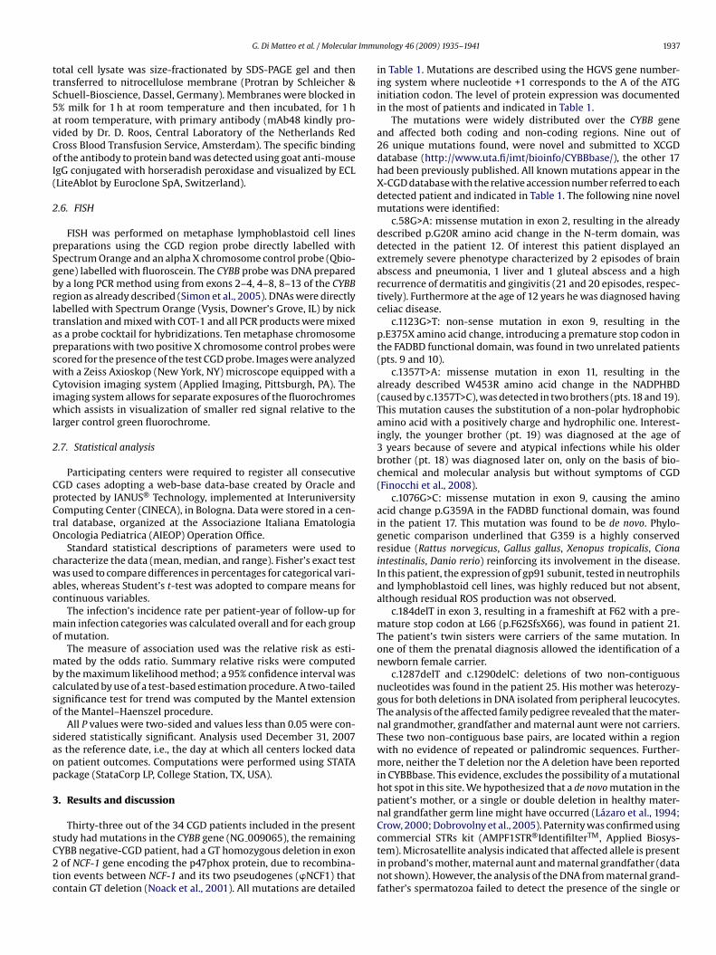
Immu
ttS5avCoI(
2
pSgbrltapswCiwl
2
CpCtO
cwac
mo
mbcso
saop
3
sC2tc
G. Di Matteo et al. / Molecular
otal cell lysate was size-fractionated by SDS-PAGE gel and thenransferred to nitrocellulose membrane (Protran by Schleicher &chuell-Bioscience, Dassel, Germany). Membranes were blocked in% milk for 1 h at room temperature and then incubated, for 1 ht room temperature, with primary antibody (mAb48 kindly pro-ided by Dr. D. Roos, Central Laboratory of the Netherlands Redross Blood Transfusion Service, Amsterdam). The specific bindingf the antibody to protein band was detected using goat anti-mousegG conjugated with horseradish peroxidase and visualized by ECLLiteAblot by Euroclone SpA, Switzerland).
.6. FISH
FISH was performed on metaphase lymphoblastoid cell linesreparations using the CGD region probe directly labelled withpectrum Orange and an alpha X chromosome control probe (Qbio-ene) labelled with fluoroscein. The CYBB probe was DNA preparedy a long PCR method using from exons 2–4, 4–8, 8–13 of the CYBBegion as already described (Simon et al., 2005). DNAs were directlyabelled with Spectrum Orange (Vysis, Downer’s Grove, IL) by nickranslation and mixed with COT-1 and all PCR products were mixeds a probe cocktail for hybridizations. Ten metaphase chromosomereparations with two positive X chromosome control probes werecored for the presence of the test CGD probe. Images were analyzedith a Zeiss Axioskop (New York, NY) microscope equipped with aytovision imaging system (Applied Imaging, Pittsburgh, PA). The
maging system allows for separate exposures of the fluorochromeshich assists in visualization of smaller red signal relative to the
arger control green fluorochrome.
.7. Statistical analysis
Participating centers were required to register all consecutiveGD cases adopting a web-base data-base created by Oracle androtected by IANUS® Technology, implemented at Interuniversityomputing Center (CINECA), in Bologna. Data were stored in a cen-ral database, organized at the Associazione Italiana Ematologiancologia Pediatrica (AIEOP) Operation Office.
Standard statistical descriptions of parameters were used toharacterize the data (mean, median, and range). Fisher’s exact testas used to compare differences in percentages for categorical vari-
bles, whereas Student’s t-test was adopted to compare means forontinuous variables.
The infection’s incidence rate per patient-year of follow-up forain infection categories was calculated overall and for each group
f mutation.The measure of association used was the relative risk as esti-
ated by the odds ratio. Summary relative risks were computedy the maximum likelihood method; a 95% confidence interval wasalculated by use of a test-based estimation procedure. A two-tailedignificance test for trend was computed by the Mantel extensionf the Mantel–Haenszel procedure.
All P values were two-sided and values less than 0.05 were con-idered statistically significant. Analysis used December 31, 2007s the reference date, i.e., the day at which all centers locked datan patient outcomes. Computations were performed using STATAackage (StataCorp LP, College Station, TX, USA).
. Results and discussion
Thirty-three out of the 34 CGD patients included in the present
tudy had mutations in the CYBB gene (NG 009065), the remainingYBB negative-CGD patient, had a GT homozygous deletion in exonof NCF-1 gene encoding the p47phox protein, due to recombina-ion events between NCF-1 and its two pseudogenes (�NCF1) thatontain GT deletion (Noack et al., 2001). All mutations are detailed
nology 46 (2009) 1935–1941 1937
in Table 1. Mutations are described using the HGVS gene number-ing system where nucleotide +1 corresponds to the A of the ATGinitiation codon. The level of protein expression was documentedin the most of patients and indicated in Table 1.
The mutations were widely distributed over the CYBB geneand affected both coding and non-coding regions. Nine out of26 unique mutations found, were novel and submitted to XCGDdatabase (http://www.uta.fi/imt/bioinfo/CYBBbase/), the other 17had been previously published. All known mutations appear in theX-CGD database with the relative accession number referred to eachdetected patient and indicated in Table 1. The following nine novelmutations were identified:
c.58G>A: missense mutation in exon 2, resulting in the alreadydescribed p.G20R amino acid change in the N-term domain, wasdetected in the patient 12. Of interest this patient displayed anextremely severe phenotype characterized by 2 episodes of brainabscess and pneumonia, 1 liver and 1 gluteal abscess and a highrecurrence of dermatitis and gingivitis (21 and 20 episodes, respec-tively). Furthermore at the age of 12 years he was diagnosed havingceliac disease.
c.1123G>T: non-sense mutation in exon 9, resulting in thep.E375X amino acid change, introducing a premature stop codon inthe FADBD functional domain, was found in two unrelated patients(pts. 9 and 10).
c.1357T>A: missense mutation in exon 11, resulting in thealready described W453R amino acid change in the NADPHBD(caused by c.1357T>C), was detected in two brothers (pts. 18 and 19).This mutation causes the substitution of a non-polar hydrophobicamino acid with a positively charge and hydrophilic one. Interest-ingly, the younger brother (pt. 19) was diagnosed at the age of3 years because of severe and atypical infections while his olderbrother (pt. 18) was diagnosed later on, only on the basis of bio-chemical and molecular analysis but without symptoms of CGD(Finocchi et al., 2008).
c.1076G>C: missense mutation in exon 9, causing the aminoacid change p.G359A in the FADBD functional domain, was foundin the patient 17. This mutation was found to be de novo. Phylo-genetic comparison underlined that G359 is a highly conservedresidue (Rattus norvegicus, Gallus gallus, Xenopus tropicalis, Cionaintestinalis, Danio rerio) reinforcing its involvement in the disease.In this patient, the expression of gp91 subunit, tested in neutrophilsand lymphoblastoid cell lines, was highly reduced but not absent,although residual ROS production was not observed.
c.184delT in exon 3, resulting in a frameshift at F62 with a pre-mature stop codon at L66 (p.F62SfsX66), was found in patient 21.The patient’s twin sisters were carriers of the same mutation. Inone of them the prenatal diagnosis allowed the identification of anewborn female carrier.
c.1287delT and c.1290delC: deletions of two non-contiguousnucleotides was found in the patient 25. His mother was heterozy-gous for both deletions in DNA isolated from peripheral leucocytes.The analysis of the affected family pedigree revealed that the mater-nal grandmother, grandfather and maternal aunt were not carriers.These two non-contiguous base pairs, are located within a regionwith no evidence of repeated or palindromic sequences. Further-more, neither the T deletion nor the A deletion have been reportedin CYBBbase. This evidence, excludes the possibility of a mutationalhot spot in this site. We hypothesized that a de novo mutation in thepatient’s mother, or a single or double deletion in healthy mater-nal grandfather germ line might have occurred (Lázaro et al., 1994;Crow, 2000; Dobrovolny et al., 2005). Paternity was confirmed using
commercial STRs kit (AMPF1STR®IdentifilterTM, Applied Biosys-tem). Microsatellite analysis indicated that affected allele is presentin proband’s mother, maternal aunt and maternal grandfather (datanot shown). However, the analysis of the DNA from maternal grand-father’s spermatozoa failed to detect the presence of the single or
1938 G. Di Matteo et al. / Molecular Immunology 46 (2009) 1935–1941
Table 1Genotype and classification of CGD patients.
pt. Age at diagnosis Carrier’s status Western blot analysis Exon intron cDNA change Accession nrs. on CYBBbase Protein change
Non-sense mutations1 3 years m carrier X910 Ex 2 c.83 G>A (A0563) p.W28X2 7 years – X910 Ex 2 c.127A>T (A0261) p.R43X3 3 years m carrier X910 Ex 5 c.388C>T (A0065, A0113, A0267-272,
A0427-431, A0587, A0596)p.R130X
4 10 years – X910 Ex 5 c.388C>T p.R130X5 8 months m carrier X910 Ex 5 c.469C>T (A0074-75, A0095, A0098,
A0152, A0177, A0274-277,A0433-434, A0565-568)
p.R157X
6 3 years – X910 Ex 7 c.691C>T (A0421-422) p.Q231X7 10 months m carrier + st carrier X910 Ex 8 c.868C>T (A0045-46, A0145, A0159,
A0194,A0198,A0288-289,A0443-453,A0639)
p.R290X
8 13 years – X910 Ex 9 c.1006G>T (A0087-88, A0291) p.E336X9 3 years m + a carriers n.d. Ex 9 c.1123G>T A0610 p.E375X10 2 years m carrier X910 Ex 9 c.1123G>T A0610 p.E375X
Missense mutations11 6 years m carrier + st carrier X910 Ex 1 c.1 A>G (A0242) p.M1V12 2 years – X910 Ex 2 c.58 G>A A0647 p.G20R13s 6 years m carrier X91− Ex 9 c.925G>A (A0101-102, A0368-372) p.E309K14s 1 years m carrier X91− Ex 9 c.925G>A (A0101-102, A0368-372) p.E309K15a 49 years m carrier X91− Ex 9 c.925G>A (A0101-102, A0368-372) p.E309K16a 12 years m + 1st + 3 nieces carrier X91− Ex 9 c.925G>A (A0101-102, A0368-372) p.E309K17 2 years m not carrier X91− Ex 9 c.1076G>C A0611 p.G359A18 6 years m carrier + st not carrier X910 Ex 11 c.1357T>A A0613 p.W453R19 3 years m carrier + st not carrier X910 Ex 11 c.1357T>A A0613 p.W453R
Deletion–duplication20* 5 years A470 Ex2 c.73 74del 56 patients reported on
ncf1basep.V25VfsX51
21* 16 years m carrier + st1 + st2 carrier X910 Ex 3 c.184delT A0614 p.F62SfsX6622 4 years m carrier n.d. Ex 6 c.484-100 674+291del p.N162 E225del23 4 years – X910 Ex 7 c.675+? 804+?del p.R226 M268del24 2 years – X910 Ex 7 c.755delG (A0219-220, A0195) p.G252EfsX25425 3 years m carrier X910 Ex10 c.1287delT + c.1290delC A0617 p.C428fs26c 1 year – n.d. Ex 1-3 c.1+? 252+?del p.M1 A84del27c 1 month – n.d. Ex 1-3 c.1+? 252+?del p.M1 A84del28 2 years m carrier + st carrier X910 Ex 1–13 Del ≥550 kb+XK gene –29 4 years m not carrier X910 Ex 1–13 Del∼500 kb –30 2 years m carrier n.d. Ex 1 c.42 45dupCATT A0619 p.L16HfsX35
Splice site mutations31 10 months m carrier X910 Int 2 c.141+5G>T A0615 r.46 141del32 6 years m carrier n.d. Int 2 c.142-1G>A A0616 r.142 252del+wt33 38 years m carrier;
st1 + st2 + st3 notcarriers; st4carrier
X910 Ex 3 c.252G>A (A0022, A0127, A0063,A0100,A0193, A0226-228,A0230-231)
r.142 252del
34 23 years X910 Ex 3 c.252G>A (A0022, A0127, A0063, r.142 252del
I , a: au
dp
sea
pgocpaaT
i
n bold patients with novel mutations are shown. m: mother; s: siblings; c: cousins
ouble deletion, thus allowing possible a de novo mutation in theatient’s mother.
c.141+5G>T: splice-site mutation in intron 2, resulting in thekipping of exon 2, was found in the patient 31. In this patient, thexpression of the gp91 subunit in lymphoblastoid cell lines wasbsent.
c.142-1G>A: splice-site mutation in intron 2 was found in theatient 32. This mutation causes the skipping of exon 3 that does notenerate coding frameshift. cDNA analysis revealed the presencef the exon 3 deleted cDNA form and a small amount of wild-typeDNA deriving from a residual normal mRNA splicing. However, theatient’s family history revealed that two brothers deceased at the
ge of 2 months and 3 years, due to pulmonary disease and a thirdffected brother died at the age of 28 years due to lung aspergillosis.his mutation was excluded in 100 healthy donors.c.42 45 dupCATT: mutation in the 3′ end of exon 1 was detectedn the patient 30. His mother was heterozygous for the same muta-
A0100,A0193, A0226-228,A0230-231)
nt; st: sister and *: dead.
tion. cDNA analysis demonstrated that this duplication did notaffect the splicing of exon 1 but was conserved in the transcriptcausing a frameshift at L16 with a premature stop codon at I35.
The remaining mutations have been previously reported andindicated in the X-CGD mutation database (Roos, 1996; Heyworthet al., 2001).
Particularly, we found six cases of deletions: two unrelated cases(pts. 28 and 29) harbouring a deletion encompassing the wholegene, two cousins (pts. 26 and 27) with a deletion including theexons 1–3 (the upstream deletion breakpoint is under investiga-tion), one case (pt. 22) with a 581 bp deletion including the exon6, one case where the deletion c.755delG resulted in a frameshift
with a premature stop codon at I254 (pt. 24) and the last one thatincluded exon 7 (pt. 23). The deletion breakpoints in the patients 28and 29 have not been characterized, however in patient 28 the dele-tion was large enough to affect from the adjacent XK gene (OMIM+314850) whose lack is responsible of the Mc Leod syndrome,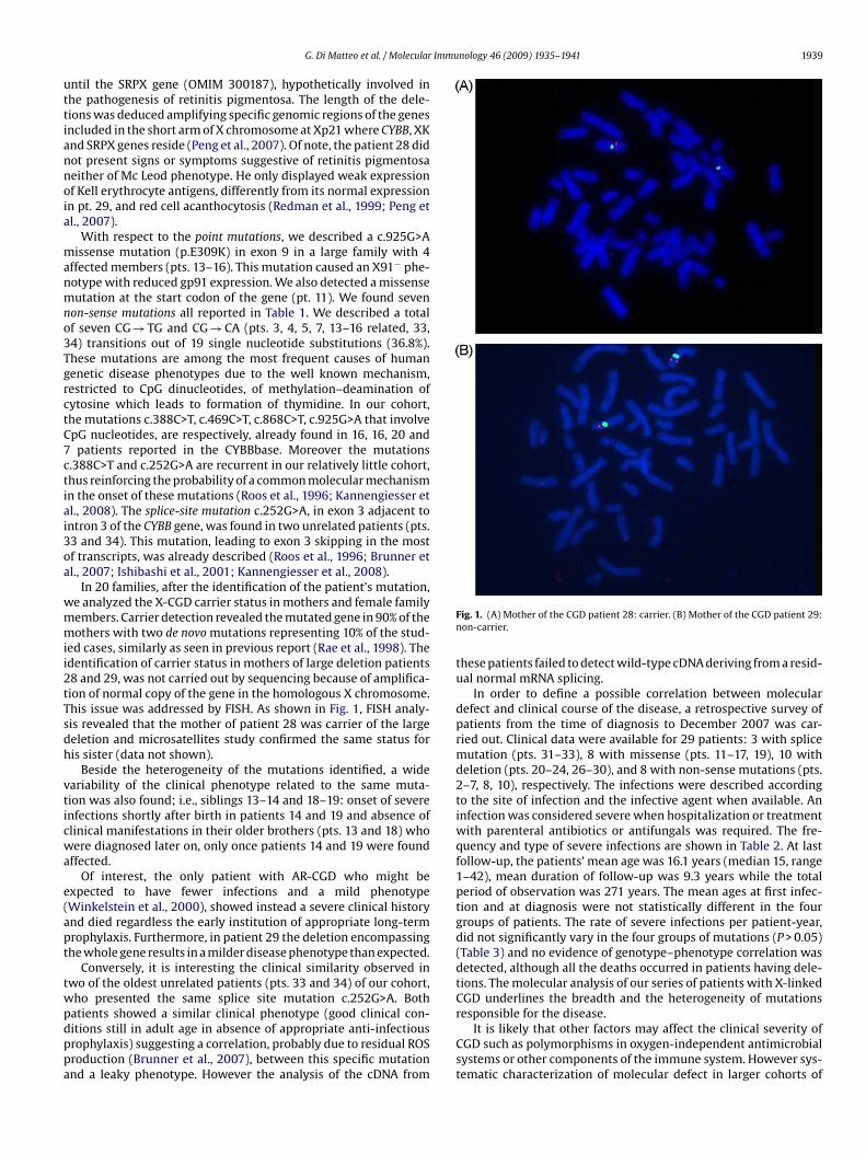
Immunology 46 (2009) 1935–1941 1939
uttiannoia
manmno3TgrctC7ctiai3oa
wmmii2tTsdh
vticwa
e(apt
twpdppa
G. Di Matteo et al. / Molecular
ntil the SRPX gene (OMIM 300187), hypothetically involved inhe pathogenesis of retinitis pigmentosa. The length of the dele-ions was deduced amplifying specific genomic regions of the genesncluded in the short arm of X chromosome at Xp21 where CYBB, XKnd SRPX genes reside (Peng et al., 2007). Of note, the patient 28 didot present signs or symptoms suggestive of retinitis pigmentosaeither of Mc Leod phenotype. He only displayed weak expressionf Kell erythrocyte antigens, differently from its normal expressionn pt. 29, and red cell acanthocytosis (Redman et al., 1999; Peng etl., 2007).
With respect to the point mutations, we described a c.925G>Aissense mutation (p.E309K) in exon 9 in a large family with 4
ffected members (pts. 13–16). This mutation caused an X91− phe-otype with reduced gp91 expression. We also detected a missenseutation at the start codon of the gene (pt. 11). We found seven
on-sense mutations all reported in Table 1. We described a totalf seven CG → TG and CG → CA (pts. 3, 4, 5, 7, 13–16 related, 33,4) transitions out of 19 single nucleotide substitutions (36.8%).hese mutations are among the most frequent causes of humanenetic disease phenotypes due to the well known mechanism,estricted to CpG dinucleotides, of methylation–deamination ofytosine which leads to formation of thymidine. In our cohort,he mutations c.388C>T, c.469C>T, c.868C>T, c.925G>A that involvepG nucleotides, are respectively, already found in 16, 16, 20 andpatients reported in the CYBBbase. Moreover the mutations
.388C>T and c.252G>A are recurrent in our relatively little cohort,hus reinforcing the probability of a common molecular mechanismn the onset of these mutations (Roos et al., 1996; Kannengiesser etl., 2008). The splice-site mutation c.252G>A, in exon 3 adjacent tontron 3 of the CYBB gene, was found in two unrelated patients (pts.3 and 34). This mutation, leading to exon 3 skipping in the mostf transcripts, was already described (Roos et al., 1996; Brunner etl., 2007; Ishibashi et al., 2001; Kannengiesser et al., 2008).
In 20 families, after the identification of the patient’s mutation,e analyzed the X-CGD carrier status in mothers and female familyembers. Carrier detection revealed the mutated gene in 90% of theothers with two de novo mutations representing 10% of the stud-
ed cases, similarly as seen in previous report (Rae et al., 1998). Thedentification of carrier status in mothers of large deletion patients8 and 29, was not carried out by sequencing because of amplifica-ion of normal copy of the gene in the homologous X chromosome.his issue was addressed by FISH. As shown in Fig. 1, FISH analy-is revealed that the mother of patient 28 was carrier of the largeeletion and microsatellites study confirmed the same status foris sister (data not shown).
Beside the heterogeneity of the mutations identified, a wideariability of the clinical phenotype related to the same muta-ion was also found; i.e., siblings 13–14 and 18–19: onset of severenfections shortly after birth in patients 14 and 19 and absence oflinical manifestations in their older brothers (pts. 13 and 18) whoere diagnosed later on, only once patients 14 and 19 were found
ffected.Of interest, the only patient with AR-CGD who might be
xpected to have fewer infections and a mild phenotypeWinkelstein et al., 2000), showed instead a severe clinical historynd died regardless the early institution of appropriate long-termrophylaxis. Furthermore, in patient 29 the deletion encompassinghe whole gene results in a milder disease phenotype than expected.
Conversely, it is interesting the clinical similarity observed inwo of the oldest unrelated patients (pts. 33 and 34) of our cohort,ho presented the same splice site mutation c.252G>A. Both
atients showed a similar clinical phenotype (good clinical con-itions still in adult age in absence of appropriate anti-infectiousrophylaxis) suggesting a correlation, probably due to residual ROSroduction (Brunner et al., 2007), between this specific mutationnd a leaky phenotype. However the analysis of the cDNA fromFig. 1. (A) Mother of the CGD patient 28: carrier. (B) Mother of the CGD patient 29:non-carrier.
these patients failed to detect wild-type cDNA deriving from a resid-ual normal mRNA splicing.
In order to define a possible correlation between moleculardefect and clinical course of the disease, a retrospective survey ofpatients from the time of diagnosis to December 2007 was car-ried out. Clinical data were available for 29 patients: 3 with splicemutation (pts. 31–33), 8 with missense (pts. 11–17, 19), 10 withdeletion (pts. 20–24, 26–30), and 8 with non-sense mutations (pts.2–7, 8, 10), respectively. The infections were described accordingto the site of infection and the infective agent when available. Aninfection was considered severe when hospitalization or treatmentwith parenteral antibiotics or antifungals was required. The fre-quency and type of severe infections are shown in Table 2. At lastfollow-up, the patients’ mean age was 16.1 years (median 15, range1–42), mean duration of follow-up was 9.3 years while the totalperiod of observation was 271 years. The mean ages at first infec-tion and at diagnosis were not statistically different in the fourgroups of patients. The rate of severe infections per patient-year,did not significantly vary in the four groups of mutations (P > 0.05)(Table 3) and no evidence of genotype–phenotype correlation wasdetected, although all the deaths occurred in patients having dele-tions. The molecular analysis of our series of patients with X-linkedCGD underlines the breadth and the heterogeneity of mutations
responsible for the disease.It is likely that other factors may affect the clinical severity ofCGD such as polymorphisms in oxygen-independent antimicrobialsystems or other components of the immune system. However sys-tematic characterization of molecular defect in larger cohorts of
1940 G. Di Matteo et al. / Molecular Immunology 46 (2009) 1935–1941
Table 2Localization and type of severe infections in 29 CGD patients.
Infection Mutation
Splice N patients = 3 Missense N patients = 8 Deletion N patients = 10 Non-sense N patients = 8
Pneumonia 2 7 20 12Lung abscess 0 0 1 0Dermatitis 0 1 1 1Subcutaneous abscess 0 0 1 4Lymphadenitis 1 2 7 7Septicemia 1 0 3 0Colitis 0 0 0 1Enteritis 1 0 0 1Brain abscess 0 2 2 0Liver abscess 1 1 4 3Otitis 0 3 0 0Kidney abscess 0 1 0 0Osteomyelitis 0 0 3 1Perianal abscess 1 1 0 1Gluteal abscess 0 0 0 0Luti 0 0 0 1
Total infections 7 18 42 32
Table 3Patients characteristics and incidence of infections in the different groups of mutations.
Splice(N patients 3)
Missense(N patients 8)
Deletion(N patients 10)
Non-sense(N patients 8)
P
SvsM SvsD SvsN MvsD MvsN DvsN
N of total infections 11 79 52 43N of severe infections 7 18 42 32N of patient-years of observation 15 46 125 85Mean age at 1◦ infection (months) 2.3 7.5 13.8 7.7 0.439 0.082 0.278 0.393 0.958 0.353M .5R .33
P year o
pdyd
A
fa
faA
R
B
C
D
D
F
G
ean age at diagnosis (years) 14.3 9.7 4ate of severe infections per patient-year 0.46 0.39 0
-value < 0.05 was considered statistically significant. The infection rate per patient-
atients might help us to better relate clinical form and functionalata to the type of the mutation found. In any case mutation anal-sis remains an essential tool for genetic counselling and prenataliagnosis of CGD.
cknowledgments
We thank all the families for their participation in the study andor providing us the written consent for publication on their clinicalnd molecular data.
We thank Dr. D. Roos for critical reading of the manuscript andor the generous gift of monoclonal antibodies to gp91-phox. Theuthors wish to thank Dr. A. Filareto for technical assistance, Dr. F.ngelini and Dr. E. Giardina for their helpful suggestions.
eferences
runner, J., Dockter, G., Rösen-Wolff, A., Roesler, J., 2007. X-linked chronic granu-lomatous disease (CGD) caused by an intra-exonic splice mutation (CYBB exon3, c.262G → A) is mimicking juvenile sarcoidosis. Clin. Exp. Rheumatol. 25 (2),336–338.
row, J.F., 2000. The origins, patterns and implications of human spontaneous muta-tion. Nat. Rev. Genet. 1, 40–47.
inauer, M.C., 2005. Chronic granulomatous disease and other disorders of phago-cyte function. Hematol. Am. Soc. Hematol. Educ. Program 1, 89–95.
obrovolny, R., Dvoráková, L., Ledvinová, J., Magage, S., Bultas, J., Lubanda, J.C.,Poupetová, H., Elleder, M., Karetová, D., Hrebícek, M., 2005. Recurrence of Fabrydisease as a result of paternal germline mosaicism for alpha-galactosidase a genemutation. Am. J. Med. Genet. A 134, 84–87.
inocchi, A., Palma, P., Di Matteo, G., Chiriaco, M., Lancella, L., Simonetti, A., Rana,I., Livadiotti, S., Rossi, P., 2008. Visceral leishmaniasis revealing chronic gran-
ulomatous disease in one child. Int. J. Immunopathol. Pharmacol. 3, 739–743.iudicelli, J., Philip, P.J., Delque, P., Sudaka, P., 1982. A single-step centrifuga-tion method for separation of granulocytes and mononuclear cells from bloodusing discontinuous density gradient of Percoll. J. Immunol. Methods 15, 43–46.
4.4 0.700 0.171 0.200 0.397 0.384 0.9600.37 0.677 0.417 0.589 0.581 0.886 0.626
f follow-up was calculated as: number of infections/number of years of observation.
Heyworth, P.G., Curnutte, J.T., Rae, J., Noack, D., Roos, D., van Koppen, E., Cross, A.R.,2001. Hematologically important mutations: X-linked chronic granulomatousdisease (second update). Blood Cells Mol. Dis. 27, 16–26.
Ishibashi, F., Mizukami, T., Kanegasaki, S., Motoda, L., Kakinuma, R., Endo, F., Nunoi,H., 2001. Improved superoxide-generating ability by interferon gamma due tosplicing pattern change of transcripts in neutrophils from patients with a splicesite mutation in CYBB gene. Blood 98, 436–441.
Kannengiesser, C., Gérard, B., El Benna, J., Henri, D., Kroviarski, Y., Chollet-Martin, S.,Gougerot-Pocidalo, M.A., Elbim, C., Grandchamp, B., 2008. Molecular epidemi-ology of chronic granulomatous disease in a series of 80 kindreds: identificationof 31 novel mutations. Hum. Mutat. 29 (9), 132–149.
Lázaro, C., Ravella, A., Gaona, A., Volpini, V., Estivill, X., 1994. Neurofibromatosis type1 due to germ-line mosaicism in a clinically normal father. N. Engl. J. Med. 331,1403–1407.
Martire, B., Rondelli, R., Soresina, A., Pignata, C., Broccoletti, T., Finocchi, A., Rossi,P., Gattorno, M., Rabusin, M., Azzari, C., Dellepiane, R.M., Pietrogrande, M.C.,Trizzino, A., Di Bartolomeo, P., Martino, S., Carpino, L., Cossu, F., Locatelli,F., Maccario, R., Pierani, P., Putti, M.C., Stabile, A., Notarangelo, L.D., Ugazio,A.G., Plebani, A., De Mattia, D., IPINET, 2008. Clinical features, long-termfollow-up and outcome of a large cohort of patients with Chronic Gran-ulomatous Disease: an Italian multicenter study. Clin. Immunol. 126, 155–164.
Noack, D., Rae, J., Cross, A.R., Ellis, B.A., Newburger, P.E., Curnutte, J.T., Heyworth, P.G.,2001. Autosomal recessive chronic granulomatous disease caused by defects inNCF-1, the gene encoding the phagocyte p47-phox: mutations not arising in theNCF-1 pseudogenes. Blood 97, 305–311.
Peng, J., Redman, C.M., Wu, X., Song, X., Walker, R.H., Westhoff, C.M., Lee, S., 2007.Insights into extensive deletions around the XK locus associated with McLeodphenotype and characterization of two novel cases. Gene 392 (1–2), 142–150.
Rae, J., Newburger, P.E., Dinauer, M.C., Noack, D., Hopkins, P.J., Kuruto, R., Curnutte,J.T., 1998. X-Linked chronic granulomatous disease: mutations in the CYBB geneencoding the gp91-phox component of respiratory-burst oxidase. Am. J. Hum.Genet. 62, 1320–1331.
Redman, C.M., Russo, D., Lee, S., 1999. Kell, Kx and the McLeod syndrome. BaillieresBest Pract. Res. Clin. Haematol. 12, 621–635.
Roos, D., de Boer, M., Kuribayashi, F., Meischl, C., Weening, R.S., Segal, A.W., Ahlin,A., Nemet, K., Hossle, J.P., Bernatowska-Matuszkiewicz, E., Middleton-Price, H.,1996. Mutations in the X-linked and autosomal recessive forms of chronic gran-ulomatous disease. Blood 87 (5), 1663–1681.
Roos, D., 1996. X-CGDbase: a database of X-CGD-causing mutations (CYBBbase).Immunol. Today 17 (11), 517–521.

Immu
S
S
Winkelstein, J.A., Marino, M.C., Johnston Jr., R.B., Boyle, J., Curnutte, J., Gallin, J.I.,
G. Di Matteo et al. / Molecular
egal, B.H., Leto, T.L., Gallin, J.I., Malech, H.L., Holland, S.M., 2000. Genetic, bio-
chemical, and clinical features of chronic granulomatous disease. Medicine 79,170–200.imon, K.C., Noack, D., Rae, J., Curnutte, J., Sarraf, S., Kolev, V., Blancato, J.K., 2005.Long of X-polymerase chain reaction-based fluorescence in situ hybridizationanalysis of female carriers of X-linked chronic granulomatous disease deletions.J. Mol. Diagn. 7, 183–186.
nology 46 (2009) 1935–1941 1941
Malech, H.L., Holland, S.M., Ochs, H., Quie, P., et al., 2000. Chronic granuloma-tous disease. Report on a national registry of 368 patients. Medicine 79, 155–156.
Xiao, W., Oefner, P.J., 2001. Denaturing high-performance liquid chromatography: areview. Hum. Mut. 17, 439–474.
Copyright © 2022 FDOKUMEN


![[Distribution of blaOXA genes in Acinetobacter baumannii strains: a multicenter study]](https://static.fdokumen.com/doc/165x107/6337b5c66f78ac31240eb601/distribution-of-blaoxa-genes-in-acinetobacter-baumannii-strains-a-multicenter.jpg)