Mohamad Al Hassan - RiuNet
358
Comparative Analyses of Plant Responses to Drought and Salt Stress in Related Taxa: A Useful Approach to Study Stress Tolerance Mechanisms Mohamad Al Hassan Supervisors: Oscar Vicente Meana Monica Tereza Boscaiu Neagu Valencia, January 2016
-
Upload
khangminh22 -
Category
Documents
-
view
0 -
download
0
Transcript of Mohamad Al Hassan - RiuNet

Comparative Analyses of Plant Responses to
Drought and Salt Stress in Related Taxa: A
Useful Approach to Study Stress Tolerance
Mechanisms
Mohamad Al Hassan
Supervisors: Oscar Vicente Meana
Monica Tereza Boscaiu Neagu
Valencia, January 2016


Alas, my 3 years journey has come to end, and I find myself in front of the challenging
task to summarize my gratitude to those who made this experience end happily.
To Pr. Oscar Vicente Meana, a supervisor, a teacher, and forever a good friend. Even
before I had the chance to prove myself, you believed in me and gave me a lot of your
trust. During these 3 years, I was blessed with your guidance and care that allowed me
to attain the aimed title with outmost professionalism, while having the chance to follow
my instincts. It is said that “The Mediocre Teacher Tells. The Good Teacher Explains.
The Superior Teacher Demonstrates. The Great Teacher Inspires”, and you sir, were and
always will be a source of inspiration. I will forever be proud that I was your student.
To Dr. Mónica Tereza Boscaiu Neagu, a director, a teacher, and forever a good friend.
You were always a great support in every step of this work. Thank you for your trust,
guidance and care. I am forever in your debt and will always be proud to have been your
student one day.
To Dr. Federico Martinelli, a colleague and forever a good friend. Thank you for your
time, help and support whenever needed, you are a special person, and I hope that what
you and Cristian aspire for, gets fulfilled very soon.
To Dr. María Pilar López-Gresa, a colleague and forever a good friend. Thank you for
your time, help and support whenever needed.
To Dr. Josep V. Llinares. Thank you for your time, help and support whenever needed.
To Dr. Mohamad Abbas, a colleague and forever a good friend. Thanks for always
being there with me, and for being a brother from another mother, since our first
bachelor degree years in Lebanon until the most distant future. I am forever in your
debt.
To Sorin Traian Schiop, a colleague and forever a good friend. Thank you for being a
dear friend whom any person would be blessed to have, on both the personal and
academic level.
To Robert Paczynski, a colleague and forever a good friend. Thank you for all the great
memories, talks and discussions we had, you are a special person, with a unique way of
living that makes you such a delightful person to know and to have around.
To my colleagues Alexandra, Andrea, Sara, Eduarda, Alejandra, Denise, Raluca,
Cristina, Enrico, Orsi, Juliana, Dhikra, Aleksandra, and Sameh. Thank you all, for
the good and the bad times. It has been a fun journey and it wouldn’t have reached this
outcome if it wasn’t for your contributions, each in their own way.
To friends especially Ragab, Mohamad, Abdel Rahman, Victoria, Mercedes, Elena,
Bruna, Samantha, Chong Min, Aytana and Moussa. Thank you for being there, and
for making this journey full of happy memorable experiences.
To my family, Adnan, Iman, Ali, and Mostafa. I had to leave you all in the times of
troubles that our country didn’t come out from yet, but still you were with me in every
step in the way. I know that I was in your minds and hearts all the time. I never felt
alone even though I am more than 1000 miles far, because of your love, trust and
confidence. You are my shelter and cradle, and the hard rock that all my dreams could
be built upon.
Thank you all from the deepest corners of my soul... I love you all.
To Mihaela Loredana Morosan, how can I thank you?? I never heard of two lovers
thanking each other’s, especially that words would never express what I feel towards
you… I will just say that the hope of having you as a part of my future is what keeps a
smile on my face every day. Thank you for being the way you are from all of my
heart… I love you
I would like to thank the evaluators of this work, and the members of the jury tribunal
going to assess it as well. It is a privilege to present this work in front of such esteemed
scientists.
Finally, I would like to present my gratitude to the friendly and lovely people of
Valencia, a city that I now consider my second home. Hopefully the coming days will
hold more fortune, wealth and the possibility for a brighter future to this lovely city and
to the whole of Spain.


i
Abstract
Introduction
Salinity and drought are the most important environmental stress conditions
reducing crop yields worldwide and limiting the distribution of wild plants in nature.
Soil salinity, especially secondary salinity caused by anthropogenic practices, such as
prolonged irrigation, lead to substantial agricultural yield losses, especially in arid and
semiarid regions. Drought, caused by reduced water content in the soil, occurs due to
disorders in nature´s water cycle, chiefly when evapotranspiration exceeds precipitation
in a certain area, to the point where soil water reserves can no longer support plant
growth.
Drought and salt stress trigger the activation of a series of basic stress
mechanisms that includes among others, the control of ion transport, exclusion and
compartmentalization, as well as the accumulation of compatible solutes (‘osmolytes’),
and the activation of antioxidant systems. These mechanisms are conserved in all plants,
stress tolerant and sensitive alike, and don’t necessarily confer tolerance.
To decipher those mechanisms and have a better understanding on the
contribution of different stress responses to the stress tolerance of a given species, we
have carried out comparative studies on the responses to drought and salinity in a
number of genetically related taxa with different tolerance potentials.
Methodology
The work concentrated on studying the responses to salt and drought stress in
genetically related plants with different tolerance to abiotic stress. The studied taxa
included salt-tolerant (halophytes) and salt-sensitive (glycophytes) wild species of two

ii
different genera: Juncus (monocotyledonous) and Plantago (dicotyledonous), as well as
plants of crop species: Solanum lycopersicum var. cerasiforme (cherry tomatoes) and
different Phaseolus cultivars, one of P. coccineus and three of P. vulgaris. .
The experimental approach was mostly based on i) establishing the relative
tolerance to water and salt stress in the studied species from their distribution in nature
(in the case of wild species) and through the relative inhibition of growth in the
presence of stress, and ii) correlating changes in the levels of biochemical ‘stress
markers’ associated to specific response pathways (ion transport, osmolyte
accumulation…) upon stress treatments, with the already established relative tolerance
to stress. This strategy proved to be appropriate to distinguish mere general responses
to stress from those mechanisms relevant for stress tolerance of the investigated species
and cultivars.
The work also sheds light on other aspects affected by salt stress, specifically
regarding germination and reproductive success or anatomical changes in salt-stressed
plants. The expression patterns of the gene NHX1, encoding a vacuolar Na+/H
+
antiporter were also studied in the Plantago taxa, as a first step in the full
characterisation of this ion transporter, that appears to play an important role in the
mechanisms of salt tolerance in this genus.
Results and discussion
Through the results attained from this work, we have been able to establish
which general stress responses are relevant for tolerance in the investigated species or
cultivars, and which are not. Thus, we provide clear evidence that, although all plants
seem to activate the same mechanisms of defense in response to abiotic stress, their
relative contribution to stress tolerance differs widely in different genera and species.

iii
Moreover, in general, the relative tolerance of the investigated species and cultivars
were the same, when referring to salt stress and to water stress, and the same
mechanisms – except some related to ion transport and homeostasis – were relevant for
tolerance to both stresses.
In the studied Phaseolus cultivars, P. vulgaris cv. 'Maxidor' showed the smallest
growth inhibition under salt and water stress conditions, and therefore was defined as
the most tolerant. 'Maxidor' accumulated lower levels of toxic ions and proline, and
recorded higher levels of myo-inositol than the other cultivars. We concluded that
blocking ion transport from the roots to the leaves and myo-inositol accumulation, were
the mechanisms most relevant for stress tolerance in Phaseolus. Proline is a reliable
stress biomarker in this genus, indicating the degree of stress affecting the plants, but is
not directly involved in tolerance mechanisms.
In the studied Plantago species it was found that the more tolerant taxa
transported Na+ and Cl
- to the leaves more efficiently than the most sensitive P. major,
and tended to accumulate large amounts of proline, albeit only under extreme stress
conditions; these responses appear to be the most relevant for tolerance in Plantago. I
Toxic ions transported to the leaves are presumably accumulated in vacuoles, which
gave incentive to isolating, sequencing and studying the expression of the Na+/H
+
vacuolar antiporter gene NHX1 in the studied species. Upon short-term treatments with
high NaCl concentrations, the more tolerant species showed higher salt-induced
expression of the aforementioned gene, supporting the contribution of the NHX1
antiporter to salt tolerance in Plantago.
Meanwhile, the tolerant Juncus species were able to partly inhibit ion transport
from the roots to the plants aerial parts and recorded a much larger increment (about 60-
fold over the controls) in proline contents, as compared to the stress-sensitive congener.

iv
Therefore, blocking accumulation of toxic ions and inducing accumulation of proline in
the culms appear to be the most important mechanisms of tolerance in Juncus. On the
other hand, we did not detect significant stress-induced anatomical differences when
comparing salt tolerant and sensitive Juncus taxa.
Conclusion
The results obtained in this work contribute to a better understanding of general
stress tolerance mechanisms in plants, and provides clear insights into the mechanisms
conferring tolerance, specifically, to drought and salt stress in some wild species and
crops. This work also shed more light on the highly efficient responses to stress in
halophytes, plants that could be viewed as nature´s answer to the aforementioned
adverse environmental conditions via evolution and adaptation. Halophytes can
therefore be considered as a suitable source – underutilized at present, in our opinion –
of knowledge, genetic resources and biotechnological tools for the needed improvement
of stress tolerance in crops.
This work has yielded eight scientific manuscripts (published, under review, or
in preparation), that are considered as subchapters of the results section of this thesis
and are listed below:
1) Al Hassan, M., Pacurar, A., Gaspar, A., Vicente, O., Boscaiu, M. (2014). Growth
and reproductive success under saline conditions of three Plantago species with
different levels of stress tolerance. Notulae Botanicae Horti Agrobotanici Cluj-
Napoca 42(1): 180-186.
v
2) Al Hassan, M., Fuertes, M., Sánchez, F., Vicente, O., Boscaiu, M. (2015). Effects of
Salt and Water Stress on Plant Growth and on Accumulation of Osmolytes and
Antioxidant Compounds in Cherry Tomato. Notulae Botanicae Horti Agrobotanici
Cluj-Napoca 43(1): 1-11.
3) Al Hassan, M., Gohari, G., Boscaiu, M., Vicente, O., Grigore, M. (2015).
Anatomical modifications under salt stress in two ecologically distinct Juncus
species. Notulae Botanicae Horti Agrobotanici Cluj-Napoca 43(2): 501-506.
4) Al Hassan, M., Morosan, M., Lopez-Gresa, M.P., Boscaiu, M., Vicente, O. (2015).
Selection and characterisation of salt and drought-resistant Phaseolus cultivars: a
‘proof-of-concept’ study. (Under revision).
5) Al Hassan, M., Pacurar, A., Lopez-Gresa, M.P., Llinares, J., Boscaiu, M., Vicente,
O. (2015). Effects of Salt and Water Stress on Three Ecologically Distinct Plantago
Species. (Under revision).
6) Al Hassan, M., Lopez-Gresa, M.P., Boscaiu, M., Vicente, O. (2015). Stress
tolerance mechanisms in Juncus: Responses to salinity and drought in three Juncus
species adapted to different natural environments. (Under revision).
7) Al Hassan, M., Cortes, J., Gaspar, A., Boscaiu, M., Vicente, O. (2015). Differential
anti-oxidative responses under salinity and drought challenges in two halophytes
and one glycophyte of the genus Juncus. (In preparation).
8) Al Hassan, M., Daniso, E., Martinelli, F., Boscaiu, M., Vicente, O. (2015).
Expression of the vacuolar Na+/H
+ antiporter gene (NHX1) in three Plantago species
differing in salt tolerance (In preparation).

vi
Other manuscripts, published or submitted during the period of work, more or
less related with its topic, but not included in the Thesis, are listed in the appendix at the
end of this document.

vii
Resumen
Introducción
La salinidad y la sequía son las condiciones de estrés ambiental más importantes,
que reducen los rendimientos de los cultivos en todo el mundo y que limitan la
distribución de las plantas silvestres en la naturaleza. La salinidad del suelo,
especialmente la salinización secundaria causada por prácticas antropogénicas, como la
irrigación prolongada, conducen a pérdidas importantes de rendimiento agrícola,
especialmente en las regiones áridas y semiáridas. La sequía, provocada por la
reducción de contenido de agua en el suelo, se produce debido a alteraciones en el ciclo
del agua en la naturaleza, principalmente cuando la evapotranspiración excede la
precipitación en un área determinada, hasta el punto que las reservas de agua del suelo
ya no pueden soportar el crecimiento de la planta.
La sequía y el estrés salino desencadenan la activación de una serie de
mecanismos básicos de respuesta, que incluyen entre otros el control del transporte, la
exclusión y la compartimentación de iones, así como la acumulación de solutos
compatibles ('osmolitos'), y la activación de sistemas antioxidantes. Estos mecanismos
están conservados en todas las plantas, tolerantes y sensibles a estrés por igual, y no
confieren necesariamente tolerancia.
Para descifrar estos mecanismos y conseguir una mejor comprensión de la
contribución de diferentes respuestas a estrés a la tolerancia al estrés en una especie
dada, hemos llevado a cabo estudios comparativos sobre las respuestas a la sequía y la
salinidad, en un número de taxones relacionados genéticamente con diferentes
potenciales de tolerancia.

viii
Metodología
El trabajo se ha centrado en el estudio de las respuestas a la sal y la sequía en
plantas genéticamente relacionadas pero con diferente tolerancia al estrés abiótico. Los
taxones estudiados incluyeron plantas tolerante a sal (halófitas) y sensibles a sal
(glicófitas) de especies silvestres de dos géneros diferentes: Juncus (monocotiledóneas)
y Plantago (dicotiledóneas), así como plantas de especies de cultivo: Solanum
lycopersicum var. cerasiforme (tomates cherry) y diferentes cultivares de Phaseolus,
uno de P. coccineus y tres de P. vulgaris.
El enfoque experimental se basó principalmente en i) establecer la tolerancia relativa al
estrés hídrico y al estrés salino en las especies estudiadas, a partir de su distribución en
la naturaleza (en el caso de especies silvestres) y atendiendo a la inhibición relativa de
su crecimiento en presencia de estrés, y ii) correlacionar cambios en los niveles de
‘marcadores bioquímicos de estrés’ asociados a vías específicas de respuesta (transporte
de iones, acumulación de osmolitos ...) inducidos por los tratamientos de estrés, con la
tolerancia relativa a estrés de las plantas, previamente establecido. Esta estrategia ha
resultado ser apropiada para distinguir meras respuestas generales a estrés de los
mecanismos relevantes para la tolerancia a estrés de las especies y cultivares
investigados.
El trabajo también arroja luz sobre otros aspectos afectados por el estrés salino,
específicamente en relación con la germinación y el éxito reproductivo, o cambios
anatómicos en las plantas tratadas con sal. También se estudiaron los patrones de
expresión del gen NHX1, que codifica un antiportador vacuolar Na+/H
+, en las especies
de Plantago, como un primer paso en la caracterización completa de este transportador
de iones, que parece desempeñar un papel importante en los mecanismos de tolerancia a
sal en este género.

ix
Resultados y discusión
A partir de los resultados obtenidos en este trabajo, hemos podido establecer qué
respuestas generales a estrés son relevantes para la tolerancia en las especies o
cultivares investigados, y cuáles no. Hemos podido aportar pruebas claras de que, a
pesar de que todas las plantas parecen activar los mismos mecanismos de defensa en
respuesta a estrés abiótico, su contribución relativa a la tolerancia a estrés difiere
ampliamente en diferentes géneros y especies. Por otra parte, en general, la tolerancia
relativa de las especies y cultivares investigados fueron los mismos, por cuando se
refiere al estrés salino y al estrés hídrico, y los mismos mecanismos – excepto algunos
relacionados con el transporte y la homeostasis de iones – son relevantes para la
tolerancia a ambos tipos de estrés.
En los cultivares estudiados de Phaseolus, P. vulgaris cv. 'Maxidor' mostró la
menor inhibición del crecimiento en condiciones de estrés salino e hídrico, y por lo
tanto se definió como el más tolerante. 'Maxidor' acumula los niveles más bajos de
iones tóxicos y de prolina, y registra los más altos de myo-inositol, en comparación con
los otros cultivares. Llegamos a la conclusión que el bloqueo de transporte de iones
desde las raíces hasta las hojas y la acumulación de myo-inositol, son los mecanismos
más importantes para la tolerancia a estrés en Phaseolus. La prolina es un biomarcador
de estrés fiable en este género, que indica el grado de estrés a que están sometidas las
plantas, pero no está directamente implicada en los mecanismos de tolerancia.
En las especies de Plantago estudiadas se encontró que los taxones más
tolerantes transportaban Na+ y Cl
- a las hojas más eficientemente que el más sensible, P.
major, y tendían a acumular grandes cantidades de prolina, aunque sólo en condiciones

x
de estrés extremo; estas respuestas parecen ser las más relevantes para la tolerancia a
estrés en Plantago. Los iones tóxicos transportados a las hojas se acumulan
presumiblemente en las vacuolas, lo que dio incentivo para aislar, secuenciar y estudiar
la expresión del gen del antiportador vacuolar Na+/H
+, NHX1, en las especies
investigadas. En tratamientos a corto plazo con altas concentraciones de NaCl, las
especies más tolerantes mostraron una mayor expresión inducida por sal del gen
mencionado anteriormente, en apoyo a la contribución del antiportador NHX1 a
tolerancia a sal en Plantago.
Mientras tanto, las especies de Juncus tolerantes fueron capaces de inhibir
parcialmente el transporte de iones de las raíces a la parte aérea de las plantas, y
registraron un incremento mucho mayor (aproximadamente 60 veces con respecto a los
controles) en el contenido de prolina, en comparación con el congénere sensible a
estrés. Por lo tanto, el bloqueo de acumulación de iones tóxicos y la inducción de la
acumulación de prolina en las cañas parecen ser los mecanismos más importantes de
tolerancia en Juncus. Por otro lado, no detectamos diferencias anatómicas importantes
inducidas por el estrés al comparar los taxones tolerantes y el sensible a sal de Juncus.
Conclusión
Los resultados obtenidos en este trabajo contribuyen a una mejor comprensión
de los mecanismos generales de tolerancia al estrés en plantas, y proporcionan ideas
claras sobre los mecanismos que confieren tolerancia, en concreto, a la sequía y al estrés
salino, en algunas especies silvestres y cultivadas. Este trabajo también arroja más luz
sobre las respuestas a estrés altamente eficientes en halófitas, plantas que podrían ser
vistas como la respuesta de la naturaleza a las condiciones ambientales adversas antes
mencionadas, a través de la evolución y la adaptación. Por lo tanto, las halófitas pueden

xi
ser consideradas como una fuente adecuada – infrautilizada en la actualidad, en nuestra
opinión – de conocimiento, recursos genéticos y herramientas biotecnológicas para la
necesaria mejora de la tolerancia al estrés en plantas cultivadas.
Este trabajo ha dado lugar a ocho manuscritos científicos (publicados, en revisión, o en
preparación), que se consideran como subcapítulos de la sección de resultados de esta
tesis y se enumeran a continuación:
1) Al Hassan, M., Pacurar, A., Gaspar, A., Vicente, O., Boscaiu, M. (2014). Growth
and reproductive success under saline conditions of three Plantago species with
different levels of stress tolerance. Notulae Botanicae Horti Agrobotanici Cluj-
Napoca 42(1): 180-186.
2) Al Hassan, M., Fuertes, M., Sánchez, F., Vicente, O., Boscaiu, M. (2015). Effects of
Salt and Water Stress on Plant Growth and on Accumulation of Osmolytes and
Antioxidant Compounds in Cherry Tomato. Notulae Botanicae Horti Agrobotanici
Cluj-Napoca 43(1): 1-11.
3) Al Hassan, M., Gohari, G., Boscaiu, M., Vicente, O., Grigore, M. (2015).
Anatomical modifications under salt stress in two ecologically distinct Juncus
species. Notulae Botanicae Horti Agrobotanici Cluj-Napoca 43(2): 501-506.
4) Al Hassan, M., Morosan, M., Lopez-Gresa, M.P., Boscaiu, M., Vicente, O. (2015).
Selection and characterisation of salt and drought-resistant Phaseolus cultivars: a
‘proof-of-concept’ study. (Under revision).
5) Al Hassan, M., Pacurar, A., Lopez-Gresa, M.P., Llinares, J., Boscaiu, M., Vicente,
O. (2015). Effects of Salt and Water Stress on Three Ecologically Distinct Plantago
Species. (Under revision).

xii
6) Al Hassan, M., Lopez-Gresa, M.P., Boscaiu, M., Vicente, O. (2015). Stress
tolerance mechanisms in Juncus: Responses to salinity and drought in three Juncus
species adapted to different natural environments. (Under revision).
7) Al Hassan, M., Cortes, J., Gaspar, A., Boscaiu, M., Vicente, O. (2015). Differential
anti-oxidative responses under salinity and drought challenges in two halophytes
and one glycophyte of the genus Juncus. (In preparation).
8) Al Hassan, M., Daniso, E., Martinelli, F., Boscaiu, M., Vicente, O. (2015).
Expression of the vacuolar Na+/H
+ antiporter gene (NHX1) in three Plantago species
differing in salt tolerance (In preparation).
Otros manuscritos, publicados o enviados para su publicación durante el período de
trabajo, más o menos relacionados con su tema pero no incluidos en la Tesis, se
enumeran en el apéndice al final de este documento.

xiii
Resum
Introducció
La salinitat i la sequera són les condicions d'estrès ambiental més importants, que
redueixen els rendiments dels cultius a tot el món i que limiten la distribució de les
plantes silvestres en la naturalesa. La salinitat del sòl, especialment la salinització
secundària causada per pràctiques antropogèniques, com la irrigació perllongada,
condueixen a pèrdues importants de rendiment agrícola, especialment en les regions
àrides i semiàrides. La sequera, provocada per la reducció de contingut d'aigua en el sòl,
es produeix a causa d'alteracions en el cicle de l'aigua en la naturalesa, principalment
quan la evapotranspiració excedeix la precipitació en un àrea determinada, fins al punt
que les reserves d'aigua del sòl ja no poden suportar el creixement de la planta.
La sequera i l'estrès salí desencadenen l'activació d'una sèrie de mecanismes
bàsics de resposta, que inclouen entre uns altres el control del transport, l'exclusió i la
compartimentació d'ions, així com l'acumulació de soluts compatibles ('osmolits'), i
l'activació de sistemes antioxidants. Aquests mecanismes estan conservats en totes les
plantes, tolerants i sensibles a estrès per igual, i no confereixen necessàriament
tolerància.
Per a desxifrar aquests mecanismes i aconseguir una millor comprensió de la
contribució de diferents respostes a estrès a la tolerància a l'estrès en una espècie
donada, hem dut a terme estudis comparatius sobre les respostes a la sequera i la
salinitat, en un nombre de taxons relacionats genèticament amb diferents potencials de
tolerància.

xiv
Metodologia
El treball s'ha centrat en l'estudi de les respostes a la sal i la sequera en plantes
genèticament relacionades però amb diferent tolerància a l'estrès abiòtic. Els taxons
estudiats van incloure plantes tolerant a sal (halòfites) i sensibles a sal (glicòfites)
d'espècies silvestres de dos gèneres diferents: Juncus (monocotiledóneas) i Plantago
(dicotiledònies), així com plantes d'espècies de cultiu: Solanum lycopersicum var.
cerasiforme (tomaques xerry) i diferents conreus de Phaseolus, un de P. coccineus i tres
de P. vulgaris. .
L'enfocament experimental es va basar principalment en i) establir la tolerància
relativa a l'estrès hídric i a l'estrès salí en les espècies estudiades, a partir de la seua
distribució en la naturalesa (en el cas d'espècies silvestres) i atenent a la inhibició
relativa de el seu creixement en presència d'estrès, i ii) correlacionar canvis en els
nivells de ‘marcadors bioquímics d'estrès’ associats a vies específiques de resposta
(transport d'ions, acumulació d’osmolits ...) induïts pels tractaments d'estrès, amb la
tolerància relativa a estrès de les plantes, prèviament establert. Aquesta estratègia ha
resultat ser apropiada per a distingir meres respostes generals a estrès dels mecanismes
rellevants per a la tolerància a estrès de les espècies i conreus investigats.
El treball també llança llum sobre altres aspectes afectats per l'estrès salí,
específicament en relació amb la germinació i l'èxit reproductiu, o canvis anatòmics en
les plantes tractades amb sal. També es van estudiar els patrons d'expressió del gen
NHX1, que codifica un anti-portador vacuolar Na+/H
+, en les espècies de Plantago, com
un primer pas en la caracterització completa d'aquest transportador d'ions, que sembla
exercir un paper important en els mecanismes de tolerància a sal en aquest gènere.
Resultats i discussió

xv
A partir dels resultats obtinguts en aquest treball, hem pogut establir què
respostes generals a estrès són rellevants per a la tolerància en les espècies o conreessis
investigats, i quins no. Hem pogut aportar proves clares que, a pesar que totes les
plantes semblen activar els mateixos mecanismes de defensa en resposta a estrès abiòtic,
la seua contribució relativa a la tolerància a estrès difereix àmpliament en diferents
gèneres i espècies. D'altra banda, en general, la tolerància relativa de les espècies i
conreus investigats van ser els mateixos, per quan es refereix a l'estrès salí i a l'estrès
hídric, i els mateixos mecanismes – excepte alguns relacionats amb el transport i
l'homeòstasi d'ions – són rellevants per a la tolerància a tots dos tipus d'estrès.
En els conreus estudiats de Phaseolus, P. vulgaris cv. 'Maxidor' va mostrar la
menor inhibició del creixement en condicions d'estrès salí i hídric, i per tant es va
definir com el més tolerant. 'Maxidor' acumula els nivells més baixos d'ions tòxics i de
prolina, i registra els més alts de myo-inositol, en comparació dels altres conreus.
Arribem a la conclusió que el bloqueig de transport d'ions des de les arrels fins a les
fulles i l'acumulació de myo-inositol, són els mecanismes més importants per a la
tolerància a estrès en Phaseolus. La prolina és un biomarcador d'estrès fiable en aquest
gènere, que indica el grau d'estrès al fet que estan sotmeses les plantes, però no està
directament implicada en els mecanismes de tolerància.
En les espècies de Plantago estudiades es va trobar que els taxons més tolerants
transportaven Na+ i Cl
- a les fulles més eficientment que el més sensible, P. major, i
tendien a acumular grans quantitats de prolina, encara que només en condicions d'estrès
extrem; aquestes respostes semblen ser les més rellevants per a la tolerància a estrès en
Plantago. Els ions tòxics transportats a les fulles s'acumulen presumiblement en els
vacúols, la qual cosa va donar incentiu per a aïllar, seqüenciar i estudiar l'expressió del
xvi
gen del antiportador vacuolar Na+/H
+, NHX1, en les espècies investigades. En
tractaments a curt termini amb altes concentracions de NaCl, les espècies més tolerants
van mostrar una major expressió induïda per sal del gen esmentat anteriorment, en
suport a la contribució del antiportador NHX1 a tolerància a sal en Plantago.
Mentrestant, les espècies de Juncus tolerants van ser capaces d'inhibir
parcialment el transport d'ions de les arrels a la part aèria de les plantes, i van registrar
un increment molt major (aproximadament 60 vegades pel que fa als controls) en el
contingut de prolina, en comparació del congènere sensible a estrès. Per tant, el
bloqueig d'acumulació d'ions tòxics i la inducció de l'acumulació de prolina en les
canyes semblen ser els mecanismes més importants de tolerància en Juncus. D'altra
banda, no detectem diferències anatòmiques importants induïdes per l'estrès en
comparar els taxons tolerants i el sensible a sal de Juncus.
Conclusió
Els resultats obtinguts en aquest treball contribueixen a una millor comprensió
dels mecanismes generals de tolerància a l'estrès en plantes, i proporcionen idees clares
sobre els mecanismes que confereixen tolerància, en concret, a la sequera i a l'estrès
salí, en algunes espècies silvestres i conreades. Aquest treball també llança més llum
sobre les respostes a estrès altament eficients en halòfites, plantes que podrien ser vistes
com la resposta de la naturalesa a les condicions ambientals adverses abans esmentades,
a través de l'evolució i l'adaptació. Per tant, les halòfites poden ser considerades com
una font adequada – infrautilitzada en l'actualitat, en la nostra opinió – de coneixement,
recursos genètics i eines biotecnològiques per a la necessària millora de la tolerància a
l'estrès en plantes conreades.

xvii
Aquest treball ha donat lloc a vuit manuscrits científics (publicats, en revisió, o en
preparació), que es consideren com sub-capítols de la secció de resultats d'aquesta tesi i
s'enumeren a continuació:
1) Al Hassan, M., Pacurar, A., Gaspar, A., Vicente, O., Boscaiu, M. (2014). Growth
and reproductive success under saline conditions of three Plantago species with
different levels of stress tolerance. Notulae Botanicae Horti Agrobotanici Cluj-
Napoca 42(1): 180-186.
2) Al Hassan, M., Fuertes, M., Sánchez, F., Vicente, O., Boscaiu, M. (2015). Effects of
Salt and Water Stress on Plant Growth and on Accumulation of Osmolytes and
Antioxidant Compounds in Cherry Tomato. Notulae Botanicae Horti Agrobotanici
Cluj-Napoca 43(1): 1-11.
3) Al Hassan, M., Gohari, G., Boscaiu, M., Vicente, O., Grigore, M. (2015).
Anatomical modifications under salt stress in two ecologically distinct Juncus
species. Notulae Botanicae Horti Agrobotanici Cluj-Napoca 43(2): 501-506.
4) Al Hassan, M., Morosan, M., Lopez-Gresa, M.P., Boscaiu, M., Vicente, O. (2015).
Selection and characterisation of salt and drought-resistant Phaseolus cultivars: a
‘proof-of-concept’ study. (Under revision).
5) Al Hassan, M., Pacurar, A., Lopez-Gresa, M.P., Llinares, J., Boscaiu, M., Vicente,
O. (2015). Effects of Salt and Water Stress on Three Ecologically Distinct Plantago
Species. (Under revision).
6) Al Hassan, M., Lopez-Gresa, M.P., Boscaiu, M., Vicente, O. (2015). Stress
tolerance mechanisms in Juncus: Responses to salinity and drought in three Juncus
species adapted to different natural environments. (Under revision).

xviii
7) Al Hassan, M., Cortes, J., Gaspar, A., Boscaiu, M., Vicente, O. (2015). Differential
anti-oxidative responses under salinity and drought challenges in two halophytes
and one glycophyte of the genus Juncus. (In preparation).
8) Al Hassan, M., Daniso, E., Martinelli, F., Boscaiu, M., Vicente, O. (2015).
Expression of the vacuolar Na+/H
+ antiporter gene (NHX1) in three Plantago species
differing in salt tolerance (In preparation).
Altres manuscrits, publicats o enviats per a la seua publicació durant el període de
treball, més o menys relacionats amb el seu tema però no inclosos en la Tesi,
s'enumeren en l'apèndix al final d'aquest document.

xix

xx
Now this is not the end. It is not even the
beginning of the end. But it is, perhaps, the
end of the beginning."
Winston Churchill

xxi

xxii
Table of Contents Abstract (English) ........................................................................................................................... i Abstract (Spanish) ........................................................................................................................ vii Abstract (Catalan) ....................................................................................................................... xiii
Table of Contents ....................................................................................................................... xxii Chapter 1: General Introduction .................................................................................................... 1
1.1. Drought and soil salinity, and their effects on crop production worldwide .................... 3
1.2. Effects of salt and water stress on plants ........................................................................... 5 1.3. Glycophytes and halophytes: the importance of comparative studies............................. 7
Chapter 2: Objectives ................................................................................................................... 13 Chapter 3: Plant Material ............................................................................................................. 18
3.1. Solanum lycopersicum ...................................................................................................... 20 3.2. Phaseolus species ............................................................................................................. 21 3.3. Plantago species ............................................................................................................... 23
3.4. Juncus species ................................................................................................................... 25 Chapter 4: Results ........................................................................................................................ 31
Subchapter: 4.1.Effects of Salt and Water Stress on Plant Growth and on
Accumulation of Osmolytes and Antioxidant Compounds in Cherry Tomato .................... 33 4.1.1. Introduction ................................................................................................................ 34 4.1.2. Material and Methods ............................................................................................... 38
4.1.3. Results ........................................................................................................................ 42 4.1.4. Discussion .................................................................................................................. 50 4.1.5. Conclusions ................................................................................................................ 55
4.1.6. References .................................................................................................................. 56 Subchapter: 4.2. Selection and characterisation of salt and drought-resistant
Phaseolus cultivars: a ‘proof-of-concept’ study .................................................................... 70
4.2.1. Introduction ................................................................................................................ 72 4.2.2. Material and Methods ............................................................................................... 77 4.2.3. Results ........................................................................................................................ 81
4.2.4. Discussion .................................................................................................................. 95 4.2.5. Conclusions .............................................................................................................. 100 4.2.6. References ................................................................................................................ 102
Subchapter:4.3. Effects of Salt and Water Stress on Three Ecologically Distinct
Plantago Species .................................................................................................................... 114 4.3.1. Introduction .............................................................................................................. 116
4.3.2. Material and Methods: ............................................................................................ 119 4.3.3. Results ...................................................................................................................... 123 4.3.4. Discussion ................................................................................................................ 135
4.3.5. Conclusions .............................................................................................................. 142 4.3.6. References ................................................................................................................ 145
Subchapter: 4.4. Growth and Reproductive Success Under Saline Conditions of
Three Plantago Species with Different Levels of Stress Tolerance ................................... 154 4.4.1. Introduction .............................................................................................................. 155 4.4.2. Material and methods .............................................................................................. 158
4.4.3. Results and Discussion............................................................................................ 161 4.4.4. Conclusions .............................................................................................................. 167 4.4.5. References ................................................................................................................ 169
Subchapter: 4.5. Expression of the Vacuolar Na+/H
+ Antiporter Gene (NHX1) in
Three Plantago Species Differing in Salt Tolerance .......................................................... 173 4.5.1. Introduction .............................................................................................................. 174
xxiii
4.5.2. Material and Methods ............................................................................................ 177 4.5.3. Results ...................................................................................................................... 179 4.5.4. Discussion ................................................................................................................ 183
4.5.5. Conclusion ............................................................................................................... 186 4.5.6. References ................................................................................................................ 187
Subchapter: 4.6. Stress tolerance mechanisms in Juncus: Responses to salinity and
drought in three Juncus species adapted to different natural environments ....................... 191 4.6.1. Introduction .............................................................................................................. 193 4.6.2. Material and Methods ............................................................................................. 196
4.6.3. Results ...................................................................................................................... 199 4.6.4. Discussion ................................................................................................................ 211 4.6.5. Conclusions .............................................................................................................. 215
4.6.6. References ................................................................................................................ 217 Subchapter: 4.7. Differential anti-oxidative responses under salinity and drought
challenges in two halophytes and one glycophyte of the genus Juncus ............................. 224
4.7.1. Introduction .............................................................................................................. 226 4.7.2. Materials and Methods ............................................................................................ 229 4.7.3. Results ...................................................................................................................... 232
4.7.4. Discussion ................................................................................................................ 240 4.7.5. Conclusions .............................................................................................................. 245 4.7.6. References ................................................................................................................ 247
Subchapter: 4.8. Anatomical modifications under salt stress in two ecologically
distinct Juncus species ........................................................................................................... 261 4.8.1. Introduction .............................................................................................................. 262
4.8.2. Material and Methods ............................................................................................. 265 4.8.3. Results ...................................................................................................................... 266 4.8.4. Discussion ................................................................................................................ 273
4.8.5. Conclusions .............................................................................................................. 275 4.8.6. References ................................................................................................................ 276
Chapter 5: General Discussion .................................................................................................. 282
5.1. Stress-induced growth inhibition ................................................................................... 285 5.2. Control of ion transport .................................................................................................. 287 5.3. Osmolyte accumulation .................................................................................................. 289
5.4. Activation of antioxidant systems.................................................................................. 296 5.5. Cloning and characterisation of Plantago NHX1 genes ............................................... 297 5.6. Inducible vs. constitutive tolerance mechanisms .......................................................... 299
Chapter 6: Conclusions .............................................................................................................. 302 Bibliography ............................................................................................................................... 308 Appendix ..................................................................................................................................... 325

General Introduction
1
Chapter 1:
General Introduction

General Introduction
2

General Introduction
3
This chapter begins with a brief insight into salt and water stress, and the damage they
cause to crop production worldwide; this is followed by a description of the deleterious
effects of these two stress conditions on plants. Then, we highlight the usefulness of
performing comparative studies on the responses to salt and water stress of genetically related
taxa with different degrees of stress resistance – such as halophytes (salt tolerant plants) and
glycophytes (salt sensitive plants) belonging to the same genus, or different cultivars of the
same species – as this experimental approach can provide novel and interesting information
on the relevant mechanisms of stress tolerance in plants.
1.1. Drought and soil salinity, and their effects on crop production worldwide
Abiotic stress is defined as any non-living factor diminishing the plant´s ability to
thrive through photosynthesis, and converting its harvested energy into biomass accumulation
(Grime, 1977) and successful propagation. These factors include salinity, drought, heat, cold,
alkalinity, UV radiation, heavy metals and other environmental stresses (Van Breusegem et
al., 2001). Yet, among those mentioned, drought and soil salinity are the two factors that
impose the greatest threat to world´s crop production and, in addition, are largely responsible
for the distribution patterns of wild plants in nature according to their tolerance (Boyer, 1982;
Owens, 2001; Bartels and Sunkar, 2005; Flowers and Flowers, 2005; Zhang, 2011).
Compared to salt stress, drought is more common and damaging (Boyer, 1982), as it
is a natural disaster whose occurrence cannot be predicted. The damage caused by drought is
considered to be worth billions of dollars (Riebsame et al., 1990), and in the past few decades
has occurred in large areas in all continents (Ding et al., 2011) with extensive damage and
massive death toll. Drought is the single abiotic stress condition most devastating for
agriculture: insufficient rainfall brings about a progressive reduction of the amount of water

General Introduction
4
available for plants in the soil, affecting their growth and development and reducing crop
productivity, or even causing premature plant death and the loss of the whole crop, if drought
is prolonged in time (Osakabe et al., 2014). Irrigation systems are necessary to maintain
acceptable yields in those regions with low rainfall. In fact, irrigated land is much more
productive than rain fed cropland: irrigation is currently used to grow crops in about 280
million hectares of arable land; this represents just below 20% of the total cultivated land, but
produces more than 40% of world food supplies (Munns and Tester, 2008). Yet, intensive
and prolonged irrigation causes another serious problem for agriculture: soil salinization.
Soil salinization is the accumulation of water-soluble salts in the soil in levels
impacting agricultural production, environmental health, and economic welfare (Rengasamy,
2006). Salinity resulting from natural causes is known as ‘primary salinity’. It affects large
areas of land worldwide, which are as such never used for agricultural activities since all
major crops are salt sensitive. The term “secondary salinity” was coined for soil salinization
due to human activities, especially prolonged irrigation schemes without sufficient drainage
and the massive and uncontrolled use of fertilizers (Flowers, 2004; Parihar et al., 2015).
Salt-affected soils occur in more than 100 countries of the world with a variety of
extents, nature, and properties. Although arid and semi-arid regions are most affected with
salinization; no climatic zone is free from it (Rengasamy, 2006). About 20-30% of irrigated
croplands is affected by salt (Owens, 2001; Unesco Water Portal, 2007), an area that is
approximately 400 million hectares of land (Al-Sadi et al., 2010); nearly the size of the
Indian subcontinent; and this area is increasing by 1 to 2% on a yearly basis (FAO, 2002).
The situation is further aggravated by the forecasted effects of global climate change (Hillel
and Rosenzweig, 2002) including an increase in average temperatures worldwide, changes in
rainfall patterns, longer, more frequent and more intense extreme weather phenomena such as
droughts, ‘heat waves’ or floods, and more importantly due to the increasing demand for food

General Introduction
5
resources as a results of the ever expanding human population, that global food production
will need to increase by 38% by 2025 and by 57% by 2050 (Wild, 2003) if food supply to the
growing world population is to be maintained at current levels. However, what must be noted
is that, most of the suitable lands have been already cultivated and expansion into new areas
to increase food production is rarely possible or desirable. The aim, as such, should be an
increase in yield per unit of land rather than in the area cultivated (Rengasamy, 2006).
Therefore, an effective approach to increase crop yields, and hence food production, in the
next decades could be based on the biotechnological improvement, through genetic
engineering methods, of the abiotic stress tolerance of our major crops (Fita et al., 2015).
This, in turn, requires a profound knowledge of the intricate physiological, biochemical and
molecular networks underlying plant stress tolerance mechanisms. For this reason – apart
from the academic interest of this topic – the study of the responses of plants to abiotic stress
has become an active area of research in plant biology.
1.2. Effects of salt and water stress on plants
Early responses to water and salt stress in plants have been considered to be almost
identical (Munns, 2002), due to the fact that drought and salinity share a physiological water
deficit that affects, more or less intensely, all plant organs. Salinity however inhibits plant´s
growth and can lead to plant death for two main reasons, since salt stress has two different
components: osmotic stress and ion toxicity (Munns and Tester, 2008), the latter being
specific only for salt stress, where firstly, the elevated concentration of ions in the soil cause a
low water potential in the root cells, leading to reduced ability of the plant to uptake water
resulting in a curtailed vegetative growth, and secondly ions enter the plant root cells and are

General Introduction
6
transported to its aerial parts, causing ionic imbalance and toxicity and further reduced plant
growth (Greenway and Munns, 1980).
Osmotic stress, or reduced turgor pressure especially in root cells, due to the high
concentration of ions in the soil (salinity) or scarcity of water (drought), is a common effect
of water and salt stress, which causes reduced water uptake, thus inducing a cascade of
effects leading to inhibition of plant growth and, eventually, to plant death (Munns, 1993; Li
et al., 2009) (Fig. 1). These effects include reduced mitosis in roots (Sharp et al., 1988) and
leaves (Spollen and Nelson, 1994; Durand et al., 1995), stomatal closure and thus reduced
gas exchange, reactive oxygen species (ROS) accumulation (Osakabe et al., 2014), and
disrupted ability to detoxify ROS (Munns and Tester, 2008).
Ionic stress, also termed salt-specific effect of salinity (Greenway and Munns, 1980)
normally follows osmotic stress in plants affected by salt stress. This stress commences
when excessive ions enter plant tissue (through the roots) and cause a range of disturbances,
starting from ion homeostasis disturbance and nutritional balance impairment (altering the
uptake of K+ and Ca
2+, for example) (Epstein, 1980; Serrano et al., 1997), inhibition of the
activity of many enzymes (Na+ can compete – with Mg
2+ which is required for the activity of
many enzymes as a cofactor; e.g., Albert et al., 2000) and direct inactivation of proteins and
other macromolecular structures, ROS accumulation (Apel and Hirt, 2004; Mahajan and
Tuteja, 2005; Ahmad, 2010; Ahmad and Prasad, 2012), chlorophyll degradation (Kato and
Shimizu, 1987), and tissue injury (leaf senescence) (Allu et al., 2014) (Fig. 1), all
contributing to further reduce vegetative growth.

General Introduction
7
Source: Author’s own illustration.
Fig. 1 Effects of salt stress and drought leading to reduced vegetative growth in affected plants.
1.3. Glycophytes and halophytes: the importance of comparative studies.
Understanding how plants respond to drought, salt and co-occurring stresses plays a
major role in stabilizing crop performance under drought and saline conditions and in the
protection of natural vegetation via adequate management techniques and plant genetic

General Introduction
8
breeding which would prove to be essential tools to improve resource use efficiency
(including water) by plants under stress.
Plants are categorized into salt sensitive glycophytes and salt resistant halophytes; this
classification however is not absolute as plants have a continuous range of
tolerance/sensitivity to salt from extremely sensitive glycophytes to highly salt tolerant
halophytes. Nearly 98% of terrestrial plant species are considered as glycophytes, including
all major crops (Flowers et al., 1986). Unlike halophytes, they cannot complete their life
cycle in soils with a salt concentration higher than 200 mM NaCl – the somewhat arbitrary
threshold that is generally accepted as the limit to separate halophytes and glycophytes.
Halophytes are a versatile set of plants that can flourish in salt affected soils where
glycophytes usually cannot, by maintaining an osmotic pressure lower than that of the
salinized soil, a feature that can hardly be attained in the salt sensitive glycophytes. Even
though halophytes evolved from polyphyletic origins (Flowers et al., 2010), they seem to
activate a series of basic, conserved mechanisms in response to high soil salinity (conserved
in glycophytes as well, but with major differences that we will discuss during the course of
this work) (Fig. 2), although the relative contribution of these responses to salt tolerance may
differ in different halophytes. In addition to these conserved mechanisms of response, there
are in some halophytes more specific morphological and anatomical adaptations (succulence,
salt glands, salt bladders…) that may be important for tolerance in those taxa (Flowers and
Colmer, 2008; Flowers et al., 1977).
As mentioned previously, stress tolerance in plants relies on the activation of a series
of conserved response pathways, some of them common to different abiotic stresses. One of
these basic response mechanisms is the control of ion homeostasis and maintenance of
osmotic balance, to counteract cellular dehydration caused by soil salinity, drought, cold or
high temperatures, among other stressful conditions. The synthesis and accumulation of
General Introduction
9
compatible solutes ('osmolytes') in the cytoplasm (Rhodes and Hanson, 1993), in general,
together with compartmentalization of toxic ions in the vacuole (overexpression of tonoplast
antiporters (NHX1 gene, NHX2 gene, …) when referring specifically to salt stress, are
essential for osmotic adjustment (Munns and Termaat, 1986; Zhu, 2001; Munns and Tester,
2008), other mechanisms include salt avoidance (Allen et al., 1994), ion transport and
others…. Overexpression of genes encoding antioxidant enzymes like superoxide dismutase
(SOD), catalase (CAT), polyphenol peroxidase (POD), or ascorbate peroxidase (APX) which
are vital in the detoxification of ROS (Mittler, 2002; Rahnama and Ebrahimzadeh, 2005) is
yet another conserved mechanism dealing however with the effects of the arising osmotic
stress.
The activation of these processes however is not specific for halophytes, but shared by
all plants, and does not necessarily result in salt tolerance (Zhu, 2001); in fact, as mentioned
above, most species are sensitive to salinity. Therefore, the relative resistance to salt stress of
plants, which varies widely among taxa, should be attributed to quantitative as well as
qualitative differences in their mechanisms of response, which only in halophytes are
efficient enough to confer salt tolerance, always within species-specific limits (Greenway and
Munns, 1980; Zhu, 2001; Grigore et al., 2011; Gil et al., 2013).
Besides the quantitative edge that halophytes attain over the glycophytes in terms of
efficiency of their mechanisms of response, they also retain some qualitative distinctions as
well, like the presence of tonoplast transporters (Shabala and Mackay, 2011) and the probable
presence of modified lipid composition in their vacuole membranes that prevent Na+ leakage
back to the cell´s cytoplasm, allowing effective compartmentalization of inflowing ions in
vacuoles (Flowers and Yeo, 1986; Cheeseman, 1988), besides the aforementioned anatomical
differences (succulence , salt glands, and salt bladders), all of which contribute in the
elevated tolerance to salt stress in halophytes and its absence in glycophytes.

General Introduction
10
Comparative studies concerning the effects of salt stress and the stress response
mechanisms in related taxa with different levels of tolerance have been carried out
extensively in the last years. Due to which several authors discussed that halophytes possess
constitutive mechanisms which enable their “stress-anticipatory preparedness” or a metabolic
anticipation of stress (Gong et al., 2005; Sanchez et al., 2008). However, the pre-adaptation
hypothesis is still under debate as other studies showed contrary results. On the other hand
such comparative studies of the responses to drought and salt stress in genetically related
halophytes and glycophytes proved to be essential to understand the response mechanisms
conveying tolerance in some species, as well as the function of certain osmolytes that in some
cases appear to be species-specific or genus-specific, all of which would contribute greatly to
our understanding of the naturally designed tolerance of some plants (halophytes and
xerophytes) to drought and salt stress; an key factor in the global effort to produce more food
resources via stress tolerant crops.

General Introduction
11
Source: Author's own illustration.
Fig. 2 Salt and drought tolerance mechanisms in plants.

General Introduction
12

Objectives
13
Chapter 2:
Objectives

Objectives
14

Objectives
15
We aimed in this work to distinguish between the concepts of responses to stress vs.
stress tolerance, concerning specifically water and salt stress conditions. In order to tackle
this issue, we tried to explore and analyze conserved responses to drought and salinity in
genetically related plants with different stress tolerance potential. The methodology focused
on i) establishing the relative tolerance to water and salt stress in the studied species via their
distribution in nature (in the case of wild species) and through the relative inhibition of
growth after controlled stress treatments, and ii) correlating stress-induced changes in the
levels of biochemical ‘stress markers’ associated to specific response pathways (ion transport,
osmolyte accumulation…), with the previously established relative tolerance of the taxa
under study.
The investigation performed at the biochemical level is pivotal in our work; however
some anatomical and molecular aspects were included as well. We attempt to list our
objectives as:
1- Diagnosis and characterization of the effects of salt and water stress on the vegetative
growth of the studied plants.
2- Assessment of the relative tolerance to water and salt stress in the investigated species
according to the inhibition of their growth upon controlled stress treatments, estimated by
determination of different growth parameters, and from their distribution in nature.
3- Analysis of conserved stress response pathways like ion transport, accumulation of
specific osmolytes, or activation of antioxidant systems.
4- Identification of the function of the major accumulated osmolytes (response vs. tolerance),
considering their concentration, variation under stress, and their correlation with the
established relative tolerance in species.

Objectives
16
5- Testing the validity of the “stress-anticipatory preparedness” theory – or presence of
constitutive stress response mechanisms – concerning halophytes, stating the presence of
relatively high contents of biomarkers involved in stress tolerance, even in control conditions,
in non-stressed plants.
6- Assessment of the influence of salt stress on one of the most critical phases of the life
cycle: seed germination and seedling establishment and growth, in three Plantago species.
7- Isolation of the vacuolar Na+/H
+ antiporter gene NHX1 from the three Plantago species,
and comparing its expression patterns in the presence of salt, in the Plantago halophytes,
their salt-sensitive congener and the glycophyte Arabidopsis thaliana.
8- Checking whether the anatomical changes in two Juncus species under salt stress,
represent a mere alteration due to stress or, on the contrary, may be considered as true
adaptations to salinity.

Objectives
17

Plant Material
18
Chapter 3:
Plant Material

Plant Material
19

Plant Material
20
3.1. Solanum lycopersicum
Cherry tomato (Solanum lycopersicum L. var. cerasiforme) is clearly differentiated
from other tomatoes by the smaller size of its fruits (Fig. 3). Plants of this variety are
vigorous in their growth and have a higher leaves/fruits ratio; therefore, the fruits receive
more photoassimilates, in comparison to larger-size varieties, and have higher sugar and acid
contents. Thus, cherry tomatoes are more flavored and are used mostly for fresh
consumption. Seeds of cherry tomato were kindly provided by Pilar Corella, from Rijk
Zwaan Ibérica S.A. (Almería, Spain).
Source: uniprot (http://www.uniprot.org/taxonomy/4081).
Fig. 3 S. lycopersicum L. var. cerasiforme (cherry tomato).

Plant Material
21
3.2. Phaseolus species
- Phaseolus vulgaris L. (Fig. 4).
Source: ‘Maxidor’ (http://www.seeds-gallery.eu/en/vegetable-seeds/dwarf-bean-seeds-maxidor.html);
‘Judía de Franco’ (“Variedades autóctonas de legumbres españolas”, p. 251);
‘The Prince’ (http://www.diynetwork.com/outdoors/legumes-beans/pictures/page-7.html);
‘Moonlight’ (http://www.shootgardening.co.uk/plant/phaseolus-coccineus-moonlight).
Fig. 4 Pods of the selected Phaseolus taxa.
a- Phaseolus vulgaris L. ‘Maxidor’
'Maxidor' is also a dwarf French bean cultivar. It has a bushy growth, reaching in
average 35-40 cm, with precocious flowering. Pods are round and yellow, 12 cm long with
relatively large seeds; the 100-seed mass is about 26 g. It is considered as an early or

Plant Material
22
medium-early variety; pods can be harvested 60-65 days after germination and dry beans
after 100-110 days. 'Maxidor' is cultivated in several countries in Europe, and is one of the
most commonly grown in Western Romania (Madoşă, 2013).
b- Phaseolus vulgaris L. 'The Prince'
'The Prince' is a dwarf French bean cultivar, with a bushy growth and an average
height of 35-45 cm. Pods are green and flat, with a length of about 20 cm and large seeds
(average weight: ca. 0.5 g per seed). It is an early cultivar; it takes 60-70 days for snap pods
to form, and 90 days for dry seeds. Cv. 'The Prince' has been reported as drought sensitive
(Boutraa and Sanders, 2001). The cultivar is one of the most commonly used in Europe.
c- Phaseolus vulgaris L. ‘Judía de Franco’
'Judía de Franco' is a local landrace of P. vulgaris from the province of Teruel (E.
Spain). It has an indefinite growth, surpassing 3 m high. It reaches maturity after 96 days.
Pods are 9 cm long, flat and green. The average weight of 100 seeds is about 30 g.
Seeds of P. vulgaris L. ‘Maxidor’ and 'The Prince' were purchased from S.C.
AGROSEM IMPEX S.R.L., Targu Mures (Romania). Seeds of P. vulgaris L. ‘Judía de
Franco’ were obtained from the Germplasm Bank of COMAV (Institute for Conservation and
Improvement of Valencian Agrodiversity, Polytechnic University of Valencia), Valencia,
Spain.
- Phaseolus coccineus L.
Seeds of P. coccineus L. ‘Moonlight’ were obtained from AJP Garden & Crafts, Chew
Valley, United Kingdom. Seeds originated from Mexico. It reaches 3 m high. It has a longer
biological cycle of 120-125 days. Pods are green and long, reaching 25 cm.

Plant Material
23
3.3. Plantago species
- Plantago crassifolia
Seeds of P. crassifolia were harvested from a salt marsh located in the Natural Park of
La Albufera (Province of Valencia, Spain).
P. crassifolia Forssk. is a perennial Mediterranean, succulent halophyte belonging to
the family Plantaginaceae, specific for sandy littoral soils and salt marshes (Chater and
Cartier, 1976). Its strap-like leaves are formed in a basal rosette and grow in length up to 20
cm (Fig. 5A). The tiny flowers of P. crassifolia are clustered densely on erect spikes, carried
upon smooth green stalks (Fig. 5B) (Grigore et al., 2014).
Source: Author's own photo.
Fig. 5 P. crassifolia: (A) overview of leaves´ rosettes formation, and (B) flowers; of P. crassifolia.
- Plantago coronopus
Seeds of P. coronopus were harvested from a salt marsh located in the Natural Park of
La Albufera (Province of Valencia, Spain).
P. coronopus is a perennial flowering plant belonging to the family Plantaginaceae.
Its leaves are lance shaped reaching up to 25 cm long, and are formed in rosettes (Fig. 6A),

Plant Material
24
the flowers are small on long ramified spikes (Fig. 6B), and the petals of its tiny flowers are
hairy on the outside, and the flowering spike often bends or nods prior to flowering.
P. coronopus is a weed common on all continents found on disturbed ground, waste
places and on chalky banks, especially near the sea (Yoshida and Tanaka, 1997; Glen, 1998).
It is able of surviving conditions such as poor drainage, brackish swamps and compacted
droughty grasslands, and is used as an indicator species for highly saline soils, acting as an
alert for land managers in irrigation regions (Koyro, 2006).
Source: Author's own photo.
Fig. 6 P. coronopus: (A) overview of leaves´ rosettes formation and lance shape, and (B) flowers; of P. coronopus.
- Plantago major
Seeds of P. major were procured from B and T World Seeds sarl, Paguignan, Aigues-
Vives, France.
P. major L. is a perennial plant that belongs to the Plantaginaceae family. It can reach
about 15 cm high; the size however differs from one habitat to another. The leaves grow in
rosettes (Fig. 7A), with parallel venation (5–9) (Fig. 7B). The flowers are small, brownish-
green on long non-ramified spikes (Fig. 7C) (Samuelsen, 2000). P. major is pollinated by
wind, and large amounts of seeds are produced, up to 20,000 per plant (Fægri, 1970; Tutin et

Plant Material
25
al., 1976). The seeds are located in capsules (8–16 per capsule) and become sticky in humid
weather due to the swelling of the polysaccharides present in the seed coat (Qadry, 1963).
P. major L. is widely distributed in Europe, Asia and north of Africa, and throughout
the world. Although a few P. major subspecies are adapted to saline environments (Chater
and Cartier, 1976), the common taxon P. major subsp. major included in this work is frequent
in humid areas but never found in salty soils, and it is therefore considered as a glycophyte.
Source: herbario virtual del mediterraneo occidental.
Fig. 7 P. major: (A) overview of rosette leaf formation, (B) leaf venation, and (C) flowers; of P. major.
3.4. Juncus species
- Juncus maritimus
Seeds of J. maritimus were harvested in a salt marsh located in the Natural Park of La
Albufera (Province of Valencia, Spain).
J. maritimus (Fig. 8, Fig. 11A) are monocot perennials that belong to the family
Juncaceae, which can grow up to 1 meter high, that inhabit coastal salt marshes, saline
meadows, and sand dunes, and thus are considered as halophytes.

Plant Material
26
Source: herbario virtual del mediterraneo occidental.
Fig. 8 Juncus maritimus.
- Juncus acutus
Seeds of J. acutus were harvested in a salt marsh located in the Natural Park of La
Albufera (Province of Valencia, Spain).
J. acutus (Fig. 9, Fig. 11B) are monocot perennials that belong to the family
Juncaceae, which can grow up to 1 meter) high, that inhabit salt marshes, moist saline
habitats and alkaline soils and thus are considered as halophytes.

Plant Material
27
Source: herbario virtual del mediterraneo occidental.
Fig. 9 Juncus acutus.
- Juncus articulatus
Seeds of J. articulatus were harvested from Natural Park of La Albufera (Province of
Valencia, Spain).

Plant Material
28
Source: The Botanical Gardens of Charles University.
Fig. 10 Juncus articulatus.
J. articulatus (Fig. 10, Fig. 11C) are monocot perennials that belong to the family
Juncaceae, which can grow up to 1 meter high. J. articulatus L. is frequent in the northern
hemisphere and in Australia in different humid areas such as wetlands, but also along the
creeks and rivers (Albrecht, 1994). No previous study on its tolerance to salt and drought
stress was reported to have been carried out on this fresh water growing Juncus species
(Chambers et al., 1995).

Plant Material
29
Source: Author's own photo.
Fig. 11 (A) Juncus maritimus, (B) J. acutus, and (C) J. articulatus; Under treatment (from left to right: Control, 100
mM NaCl, 200 mM NaCl, 400 mM NaCl, and water stress plants, after 8 weeks of treatments).

Results
30

Results
31
Chapter 4:
Results

Results
32

Results
33
Publication I:
Subchapter 4.1.
Effects of salt and water stress on plant growth and on
accumulation of osmolytes and antioxidant compounds in
cherry tomato
Reference:
Al Hassan, M., Fuertes, M., Sánchez, F., Vicente, O., Boscaiu, M. (2015). Effects of Salt
and Water Stress on Plant Growth and on Accumulation of Osmolytes and Antioxidant
Compounds in Cherry Tomato. Notulae Botanicae Horti Agrobotanici Cluj-Napoca, 43(1): 1-
11.

Results
34
Effects of Salt and Water Stress on Plant Growth and on Accumulation of Osmolytes
and Antioxidant Compounds in Cherry Tomato
Abstract. The effects of salt and water stress on growth and several stress markers were
investigated in cherry tomato plants. Some growth parameters (stem length and number of
leaves) and chlorophyll contents were determined every third day during plant growth, and
leaf material was collected after 25 and 33 days of treatment. Both stresses inhibited plant
growth; chlorophyll levels, however, decreased only in response to high NaCl concentrations.
Proline contents largely increased in leaves of stressed plants, reaching levels high enough to
play a major role in cellular osmotic adjustment. Despite reports indicating that tomato does
not synthesize glycine betaine, the stress-induced accumulation of this osmolyte was detected
in cherry tomato, albeit at lower concentration than that of proline. Therefore, it appears that
the plants are able to synthesise glycine betaine as a secondary osmolyte under strong stress
conditions. Total sugars levels, on the contrary, decreased in stress-treated plants. Both stress
treatments caused secondary oxidative stress in the plants, as indicated by a significant
increase in malondialdehyde (MDA) contents. Water stress led to an increase in total
phenolics and flavonoid contents and a reduction of carotenoid levels in the leaves;
flavonoids also increased under high salinity conditions.
Keywords: proline; glycine betaine; carbohydrates; MDA; carotenoids; phenolics; flavonoids
4.1.1. Introduction
Tomato is grown in a wide range of climatic conditions, from the tropics to subarctic
regions, but its optimal cultivation areas are found in warm and rather dry regions, such as

Results
35
Mediterranean countries (Cuartero and Fernández Muñoz, 1999). In these arid or semi-arid
zones, water stress and high soil salinity are common environmental factors that can
significantly reduce crop yields. One of the most general responses to drought and salinity –
as well as to other abiotic stress conditions also causing cellular dehydration in plants, such as
cold, elevated temperatures or exposure to heavy metals – is based on the synthesis and
cytoplasmic accumulation of osmolytes, a conserved phenomenon observed in all plants,
tolerant as well as sensitive to stress (Parida and Das, 2005; Munns and Tester, 2008; Parvaiz
and Satyawati, 2008). Osmolytes are ‘compatible solutes’, very soluble, low-molecular-
weight organic compounds that do not interfere with normal metabolism even when present
at high concentrations. While toxic inorganic ions are sequestered in vacuoles, organic
osmolytes accumulate predominantly in the cytoplasm, preventing or limiting cellular
dehydration (Stewart and Lee, 1974; Handa et al., 1986; Büssis and Heineke, 1998).
Reduction of the osmotic potential due to accumulation of osmolytes in response to stress
improves the ability of the plant cells to maintain turgor pressure at low water potentials,
which is essential for biological processes such as photosynthesis or cell expansion, as well
as for maintaining enzymatic activities (Tyree and Jarvis, 1982). Besides their role in osmotic
adjustment, osmolytes act as osmoprotective substances, directly stabilising proteins and cell
membranes under dehydration conditions. Osmolytes also protect cells from oxidative stress
by inactivating ‘reactive oxygen species’ (ROS) (Hare et al., 1998; Szabados and Savouré,
2010).
The amino acid proline (Pro) and glycine betaine (GB), a quaternary amine, are
probably the most common compatible solutes synthesised by plants as a response to abiotic
stress (Ashraf and Foolad, 2007; Chen and Murata, 2008; Verbruggen and Hermans, 2008).
As for other osmolytes, besides their role in osmoregulation, both compounds can act as
‘low-molecular-weight chaperons’, contributing to maintain the active conformation of

Results
36
macromolecules in stressed plants, and participate in detoxification of ROS. Moreover, Pro
and GB seem to be involved, directly or indirectly, in the regulation of gene expression as
signalling molecules, and also serve as metabolites for the cellular storage of carbon and
nitrogen during stress, which would be used by the cell once stress has ceased (Munns and
Tester, 2008; Szabados and Savouré, 2010). Compatible solutes also include soluble
carbohydrates, such as sugars (e.g. sucrose, glucose, fructose or trehalose), sugar alcohols
(sorbitol, mannitol, and different inositol isomers and derivatives), and the raffinose family of
oligosaccharides (Parida et al., 2002; Gavaghan et al., 2011). Although sugars have been
shown to act as functional osmolytes in several species, it is not so easy to assess their
specific functions in the responses to stress, which can be masked by their multiple additional
roles as direct products of photosynthesis, components of the primary metabolism and
regulatory molecules (Gil et al., 2013).
A secondary effect of abiotic stresses, including drought and salinity, is the increased
generation of ‘reactive oxygen species’ (ROS), including highly reactive free radicals such as
superoxide, singlet oxygen, hydroxyl or perhydroxyl radicals, as well as hydrogen peroxide,
molecular oxygen, ozone and other strong oxidant molecules (Apel and Hirt, 2004). ROS are
continuously generated by plants as by-products of different metabolic pathways, but under
stress their production increases leading to oxidative damage of cellular membranes, proteins,
carbohydrates and DNA (Van Breusegem and Dat, 2006). In response to stress, plants
activate powerful antioxidant systems, both enzymatic (e.g., superoxide dismutase, catalase,
glutathione reductase, several peroxidades) and non-enzymatic (vitamins C and E,
carotenoids, flavonoids and other phenolic compounds, etc.) (Apel and Hirt, 2004).
Carotenoids are pigments with several functions in plants, besides their direct role in
photosynthesis, including their involvement in the mechanisms of oxidative stress tolerance
(Gill and Tuteja, 2010). Phenolic compounds also fulfill multiple roles in plants, as structural

Results
37
components of cell walls, participating in the regulation of growth and developmental
processes, as well as in the mechanisms of defence against herbivores and pathogens; in
addition, they are involved in the responses of plants to practically to all types of abiotic
stress: UV radiation, extreme temperatures, mineral nutrient imbalance, drought, salinity,
heavy metals and herbicides among others (Gould and Lister, 2006; Cheynier et al., 2013).
Flavonoids represent the main and more complex subgroup of polyphenols, including more
than 9000 different compounds with a wide array of biological functions (Winkel-Shirley,
2002; Treutter, 2005; 2006; Pollastri and Tattini, 2011; Di Ferdinando et al., 2012). In the last
decade, they have been the object of many studies, not only due to the academic interest in
elucidating their multiple functions in plants, but also because their alleged beneficial effects
for human health, as powerful dietary antioxidants (Wiseman, 2006).
Responses to salinity in tomato, within its salt-tolerance range, have been extensively
studied, but only few authors have analysed its responses to high salt concentrations (Maggio
et al., 2004). In addition, there are only a few papers reporting the effects of both salinity and
drought on the same plant material (Giannakoula and Ilias, 2013). These two adverse
environmental conditions, which will worsen as a consequence of climate change, could
seriously affect tomato production in Mediterranean countries in the coming years. The aims
of the present study were to analyse the effects of water stress and of high salinity, beyond
the tolerance threshold, on the growth of tomato plants, and to assess their responses to these
conditions in terms of accumulation of the main osmolytes (proline, glycine betaine and total
soluble sugars) and of some non-enzymatic antioxidants (total carotenoids, phenolics and
flavonoids). The experiments were carried out in cherry tomato, a variety that has not been
extensively studied despite its growing commercial interest.

Results
38
4.1.2. Material and Methods
Plant material
Cherry tomato (Solanum lycopersicum L. var. cerasiforme) is clearly differentiated
from other tomatoes by the smaller size of its fruits. Plants of this variety are vigorous in their
growth and have a higher leaves/fruits ratio; therefore, the fruits receive more
photoassimilates, in comparison to larger-size varieties, and have higher sugar and acid
contents. Thus, cherry tomatoes are more flavoured and are used mostly for fresh
consumption. Seeds of cherry tomato were kindly provided by Pilar Corella, from Rijk
Zwaan Ibérica S.A. (Almería, Spain).
Plant growth
The seeds were sown in seed trays containing a mixture of commercial peat and
vermiculite (1:1), and were placed in a greenhouse with regulated temperatures ranging
between 17 and 23°C, under a long-day photoperiod (16 h light / 8 h dark). Young plants
were transferred to individual pots (11 cm in diameter) on the same substrate 36 days after
sowing and were watered for 26 additional days with a standard nutritive solution. Then, salt
and water stress treatments were started, by watering the plants with increasing NaCl
concentrations (150, 300 and 450 mM NaCl in nutritive solution) and by stopping irrigation
altogether, respectively; 10 plants were used per treatment. Watering was carried out twice a
week by adding 1.5 L of the salt solutions to each tray, which contained 12 standard pots.
Control plants were grown in parallel, maintaining the standard irrigation regime with
nutritive solution. After starting the treatments, plant height was measured and the number of
leaves per plant was counted every three days, to assess the effect of stress on vegetative
plant growth. Chlorophyll content was also determined at the same times, using a portable
'Chlorophyll meter SPAD-502Plus' (Konica Minolta).

Results
39
For each stress treatment, including non-treated controls, leaves in equivalent
positions were detached from all plants, 25 and 33 days after starting the treatments, and
weighed on a precision balance to calculate the average leaf fresh weight (FW) corresponding
to each treatment. Part of the leaf material was stored at -75ºC until further use, and part was
dried at 65ºC until constant weight, to measure the average leaf dry weight (DW) for each
treatment. Average leaf 'water content' after each treatment, expressed in percentage, was
calculated as (FW - DW / FW) x 100.
Electric conductivity of the substrate
Soil EC1:5 was checked at the beginning and at the end of the treatments. Soil samples
were taken from three pots per treatment, air-dried and then passed through a 2-mm sieve. A
soil: water (1:5) suspension was prepared in Milli Q water and stirred for one hour at 600
u/min at 21ºC. Electric conductivity was measured with a Crison Conductivity meter 522 and
expressed in dS/m.
Osmolyte contents
Pro contents were determined according to the method of Bates et al. (1973) with
minor modifications (Vicente et al., 2004). Frozen plant material (100 mg) was ground to a
fine powder in a mortar, in the presence of liquid nitrogen. Extraction was carried out with
3% sulfosalicylic acid, and cell debris was removed by filtration. One volume of the filtrate
was mixed first with one volume of freshly prepared acid ninhydrin (25 mg/mL ninhydrin in
10.44 M acetic acid and 2.4 M phosphoric acid); then one volume of glacial acetic acid was
added, the sample was mixed and incubated at 95ºC for 1 h. After stopping the reaction by
cooling the samples on ice, they were extracted with two volumes of toluene. The absorbance
of the organic phase was determined at 520 nm, using toluene as a blank. A calibration curve

Results
40
was obtained for each assay, using solutions of Pro in 3% sulfosalicylic acid of known,
increasing concentrations and subjected to the same treatment as the samples. Pro contents in
the plant samples were expressed as ‘µmol Pro per gram of dry weight’.
The extraction and quantification of glycine betaine was performed following the
method of Grieve and Grattan (1983) with the modifications proposed by Nawaz and Ashraf
(2010). Frozen plant material (100 mg) was ground, using a homogenizer (Ultraturrax), in a
volume of 2 mL Milli-Q water, and centrifuged for 10 minutes at -4°C and 13000 rpm. One
mL was removed from the supernatant of each extract and mixed with 400 µL HCl 2N. After
stirring, 200 µL of each tube was mixed with 80 µL of potassium iodide and brought to 100
mL with Milli-Q water. The tubes were quickly placed in ice and stirred every 20 seconds for
15 min. After 90 min, 800 µL of cold Milli-Q water and 4 mL of 1.2-dichloroethane – kept in
the -20ºC freezer until used – were added to each tube. The samples were vortexed for one
minute and allowed to settle until the water and the organic phases separated; finally, 1 mL of
the lower aqueous phase was aspirated and its absorbance at 265 nm was measured in a
spectrophotometer. A standard curve was obtained for each assay, using glycine betaine
solutions in Milli-Q water of increasing concentration subjected to the same treatment as the
plant samples. Glycine betaine contents in the plant samples were expressed as ‘µmol GB per
gram of dry weight’
Total sugars were quantified according to the method described by Dubois et al.
(1956). Dried material (0.1 g) was crushed and diluted with 3 mL of 80% methanol, left on an
agitator for 20–48 h, and then mixed with concentrated sulfuric acid and 5% phenol; finally,
absorbance of the samples at 490 nm was measured in a spectrophotometer. Glucose
solutions were used as standard, and the amount of total soluble sugars in each sample was
calculated as ‘mg. equivalent of glucose per gram of dry weight’.

Results
41
Malondialdehyde and non-enzymatic antioxidants
Malondialdehyde (MDA), total phenolics and flavonoids contents were measured in
the same extracts used for total soluble sugars determination (0.1 g of dried material in 3 mL
of 80% methanol). MDA, a product of membrane lipid peroxidation, is considered as an
excellent marker of oxidative stress (Del Rio et al., 2005) and is routinely used to assess the
degree of oxidative damage induced in plants by different types of stress (e.g. Aghaleh et al.,
2009; Li et al., 2010). MDA content was determined by the thiobarbituric acid-reactive-
substances (TBARS) assay, modified to correct for the presence of interfering compounds, as
described by Hodges et al. (1999).
Total phenolic compounds were quantified by reaction with the Folin-Ciocalteu
reagent, according to Blainski et al. (2013). Absorbance was measured at 765 nm, and the
results expressed in equivalents of gallic acid (mg eq. GA g-1
DW), used as standard.
Flavonoid contents were determined following the method described by Zhishen et al.
(1999); the absorbance was measured at 510 nm, and the amount of flavonoids was expressed
in equivalents of catechin (mg eq. C g-1
DW), used as standard.
Total carotenoids were measured and quantified following Sims and Gamon (2002):
0.2 g fresh material was crushed and diluted in 80% ice-cold acetone/Tris buffer for 1 hour
on a shaker at 4ºC, then centrifuged for 15 min at 3000 x g. The supernatant was separated
and optical density was measured at 663 nm, 646 nm, and 470 nm in disposable plastic
cuvettes. Total carotenoids values were then converted into µg g-1
DW.
Statistical analysis
Data were analysed using the Statgraphics Centurion XVI programme for Windows.
Prior to the analysis, the Levene test was applied to check the ANOVA requirements. The
significance of differences between treatments was assessed using one-way ANOVA, at 95%

Results
42
confidence level. The post-hoc Tukey test was used to estimate homogeneous groups when
more than two samples were compared.
4.1.3. Results
Substrate electric conductivity
The electric conductivity (EC1:5) of the plants’ substrate was determined before and
after the stress treatments (Tab. 1). In the control pots, not treated with NaCl, the EC1:5 value
at the end of the experiment was significantly higher than the initial one; this can be easily
explained by accumulation of ions present in the nutritive solution. As expected, EC1:5
increased even more after watering the plants with NaCl solutions of increasing
concentration, in a concentration-dependent manner. The mean EC1:5 also increased slightly
in the substrate of water stressed plants, as compared to the initial value – most likely due to
concentration of ions from the nutritive solution – but the difference was not statistically
significant.
Stress-induced inhibition of plant growth
Both salt and water stress negatively affected vegetative plant growth, which was
estimated by the increase in plant length and in the number of leaves during the 33 days of
treatment (Fig. 1). As compared to the unstressed controls, all NaCl concentrations tested
totally blocked growth of tomato plants, with a clear concentration effect: the higher the salt
concentration used, the shorter the lag period before growth inhibition was observed (Figs.
1A and C). Water stress also stopped stem growth and the increase in leaf number; this effect
was clearly observed 9 days after the last irrigation with nutritive solution, once the substrate
had dried (Figs. 1B and D).

Results
43
Fig. 1 Stem length (A, B) and number of leaves (C, D) changes during the salt (A, C) and water (B, D) stress
treatments. Control, non-stressed plants (--, all panels). 50 mM (--), 100 mM (--) and 150 mM (--) NaCl (panels
A, C). Water-stressed plants (--, panels B, D).
Apart from the growth parameters measured continuously, stress-induced inhibition of
plant growth was also assessed by determining the mean fresh weight (FW) and dry weight
(DW) of the tomato leaves after 25 days (sampling 1) and 33 days (sampling 2) of salt or
water stress treatment (Fig. 2). A significant, concentration-dependent decrease of FW was
observed in salt-treated plants, reaching approximately a 75% reduction in those irrigated
with 450 mM NaCl, with respect to the non-treated controls. In the presence of this high salt
concentration, the plants were already strongly affected after 25 days of treatment and no
significant differences were found between the two samplings; at lower salt levels – 150 or
300 mM NaCl – reduction of FW increased with the duration of the treatment (Fig. 2A).
Similar qualitative results were obtained when the plants were submitted to water stress,
although with stronger differences between the two samplings: after 25 days without
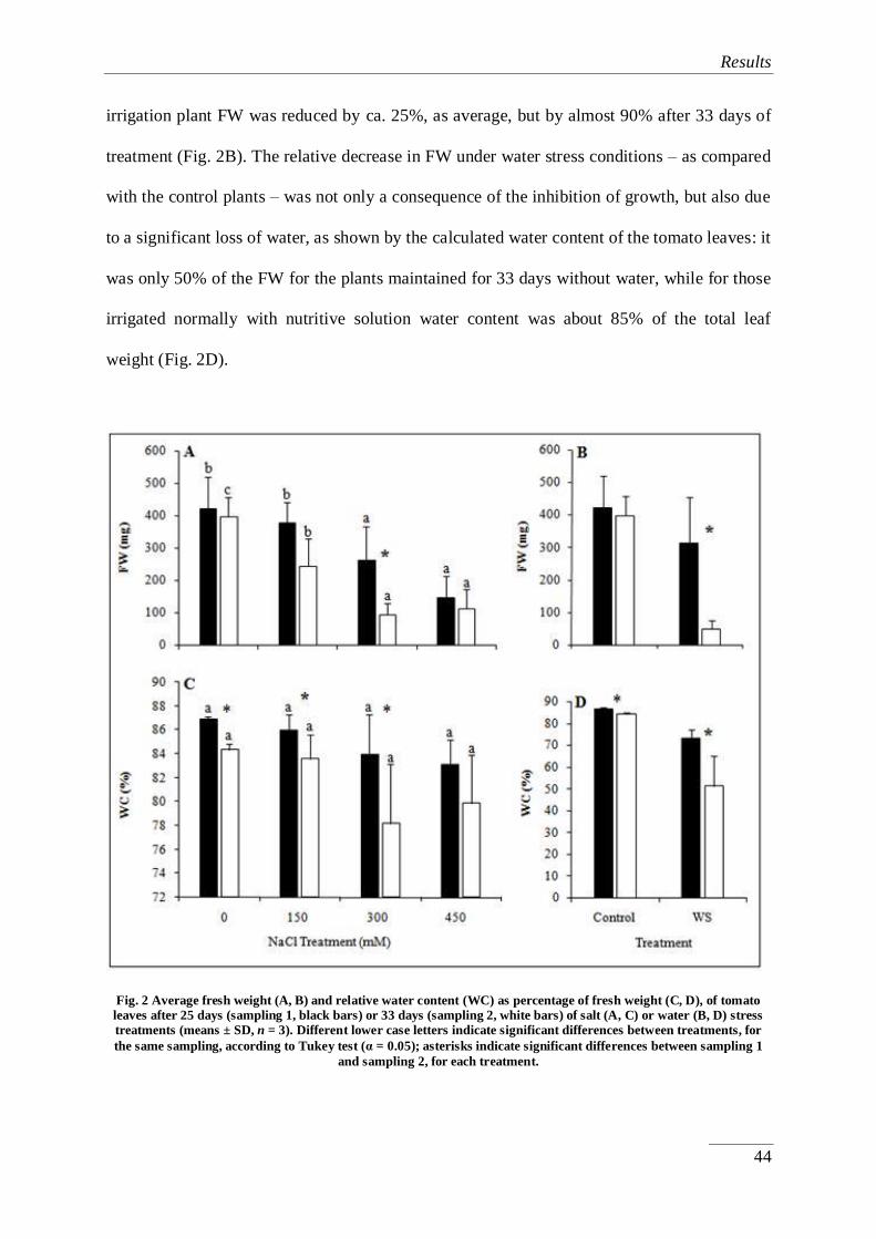
Results
44
irrigation plant FW was reduced by ca. 25%, as average, but by almost 90% after 33 days of
treatment (Fig. 2B). The relative decrease in FW under water stress conditions – as compared
with the control plants – was not only a consequence of the inhibition of growth, but also due
to a significant loss of water, as shown by the calculated water content of the tomato leaves: it
was only 50% of the FW for the plants maintained for 33 days without water, while for those
irrigated normally with nutritive solution water content was about 85% of the total leaf
weight (Fig. 2D).
Fig. 2 Average fresh weight (A, B) and relative water content (WC) as percentage of fresh weight (C, D), of tomato
leaves after 25 days (sampling 1, black bars) or 33 days (sampling 2, white bars) of salt (A, C) or water (B, D) stress
treatments (means ± SD, n = 3). Different lower case letters indicate significant differences between treatments, for
the same sampling, according to Tukey test (α = 0.05); asterisks indicate significant differences between sampling 1
and sampling 2, for each treatment.

Results
45
In the presence of salt, a general but not statistically significant decrease of the mean
leaf water content was observed with increasing NaCl concentrations, both after 25 and 33
days of treatment, whereas significant differences were detected between the samples
collected at different times for each NaCl concentration, up to 300 mM (Fig. 2C).
Chlorophyll content
The chlorophyll content of crop plants is positively correlated with their
photosynthetic activity (Gummuluru et al., 1989) and a reduction of chlorophyll level
contributes to the inhibition of photosynthesis observed under abiotic stress conditions. A
decrease in chlorophyll levels with respect to the controls was indeed detected in plants
irrigated with high salt concentrations – 300 and 450 mM NaCl – for two weeks or longer
times (Fig. 3A). On the other hand, no significant differences with non-stressed plants were
observed when a moderate – 150 mM – NaCl concentration was used (Fig. 3A) or when the
tomato plants were subjected to the water stress treatment (Fig. 3B); this latter observation
was consistent with the visual appearance of the plants, which were clearly wilted but had not
lost their green colour (data not shown).

Results
46
Fig. 3 Chlorophyll contents (mg per square meter) in leaves of tomato plants during the salt (A) and water (B) stress
treatments. Control, non-stressed plants (--, in both panels). 50 mM (--), 100 mM (--) and 150 mM (--) NaCl
(panel A). Water-stressed plants (--, panel B).
Proline content

Results
47
Application of salt treatments significantly increased Pro levels in tomato leaves, in a
concentration-dependent manner. Mean Pro values in the presence of 300 mM and 450 mM
external NaCl also increased with longer times of treatment although the differences observed
between the two samplings of plant material were significant only at 450 mM NaCl. Under
the strongest salt stress conditions tested, Pro levels reached about 350 µmol per gram DW,
which represented an increase of 16-fold with respect to the non-treated controls (Fig. 4A).
Pro accumulation was more clearly observed in plants kept without irrigation, with increases
of about 20-fold and 36-fold after 25 and 33 days, respectively, of water stress (Fig. 4B). Pro
contents in the plants subjected to the strongest water stress treatment (33 days without
irrigation), in terms of dry weight (ca. 800 µmol g-1
) were about double than those
determined in plants treated for the same time with 450 mM NaCl (Figs. 4A and B); however,
considering the drastic reduction of water content in non-irrigated plants, it can be said that
both treatments induced Pro accumulation to similar levels, in absolute terms.

Results
48
Fig. 4 Proline (Pro, µmol per gram DW) (A, B), glycine betaine (GB, µmol per gram DW) (C, D), and total soluble
sugars (TSS, mg equivalent of glucose per gram DW) (E, F) in tomato leaves after 25 days (sampling 1, black bars) or
33 days (sampling 2, white bars) of salt (A, C, E) or water (B, D, F) stress treatments (means ± SD, n = 3). Different
lower case letters indicate significant differences between treatments, for the same sampling, according to Tukey test
(α = 0.05); asterisks indicate significant differences between sampling 1 and sampling 2, for each treatment.
Glycine betaine
Salt and water stress also induced the synthesis and accumulation of GB in tomato
(Figs. 4C and D). Yet the absolute values of GB content reached in plants watered with 300
or 450 mM NaCl (50-60 µmol per gram DW) and its relative increase as compared to the
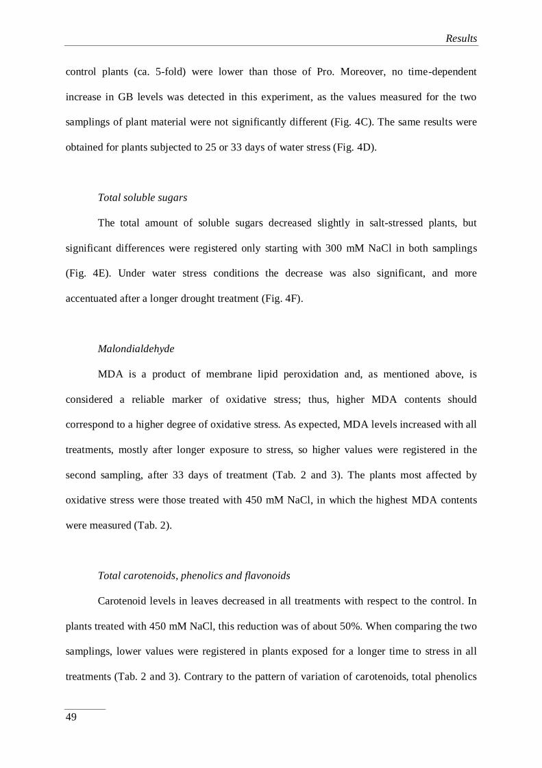
Results
49
control plants (ca. 5-fold) were lower than those of Pro. Moreover, no time-dependent
increase in GB levels was detected in this experiment, as the values measured for the two
samplings of plant material were not significantly different (Fig. 4C). The same results were
obtained for plants subjected to 25 or 33 days of water stress (Fig. 4D).
Total soluble sugars
The total amount of soluble sugars decreased slightly in salt-stressed plants, but
significant differences were registered only starting with 300 mM NaCl in both samplings
(Fig. 4E). Under water stress conditions the decrease was also significant, and more
accentuated after a longer drought treatment (Fig. 4F).
Malondialdehyde
MDA is a product of membrane lipid peroxidation and, as mentioned above, is
considered a reliable marker of oxidative stress; thus, higher MDA contents should
correspond to a higher degree of oxidative stress. As expected, MDA levels increased with all
treatments, mostly after longer exposure to stress, so higher values were registered in the
second sampling, after 33 days of treatment (Tab. 2 and 3). The plants most affected by
oxidative stress were those treated with 450 mM NaCl, in which the highest MDA contents
were measured (Tab. 2).
Total carotenoids, phenolics and flavonoids
Carotenoid levels in leaves decreased in all treatments with respect to the control. In
plants treated with 450 mM NaCl, this reduction was of about 50%. When comparing the two
samplings, lower values were registered in plants exposed for a longer time to stress in all
treatments (Tab. 2 and 3). Contrary to the pattern of variation of carotenoids, total phenolics

Results
50
increased in plants under stress, especially in those treated with high salt concentrations.
Values were significantly higher in plants from the second sampling than in those from the
first one in all stress treatments (Tab. 2 and 3). A different response to the type of stress was
observed in total flavonoid contents, which increased significantly in the presence of NaCl
(Tab. 2) but decreased under drought conditions (Tab. 3).
4.1.4. Discussion
All our major crops and most wild species are relatively sensitive to environmental
stress conditions such as drought or salinity, but tomato is moderately resistant to salt,
withstanding a soil ECsat up to 6 dS m−1
, so that it can be cultivated in regions exposed to a
certain degree of soil salinisation (Maggio et al., 2004). Yet high soil salinity inhibits seed
germination and plant growth, and causes reduction in crop yields (Dalton et al., 1997;
Romero-Aranda et al., 2002). In this study, young tomato plants have been grown in the
presence of increasing concentrations of NaCl, and a concentration- and time-dependent
inhibition of vegetative growth has been observed, which was better shown by the reduction
of the fresh weight of the plants; this was not unexpected, since inhibition of growth is
probably the most general response of plants to stress (Munns, 2002; Munns and Tester,
2008). Despite the fact that high salinity should cause cellular dehydration, in the
experimental conditions used here the water content of the plants was reduced very slightly in
the presence of salt, suggesting that tomato plants activate relatively efficient mechanisms to
cope with the osmotic component of salt stress, and that growth inhibition is mostly due to
the ionic toxicity of the salt. Water stress also inhibited growth but, contrary to salinity, in
this case a severe dehydration of the plants was observed.

Results
51
Another difference between salt and water stress treatments refers to changes in
chlorophyll contents. In plants treated with high NaCl concentrations, a significant reduction
in chlorophyll levels, up to ca. 40% of the non-treated controls, was observed. The decrease
in chlorophyll levels in plants affected by salt is due to the inhibition of chlorophyll synthesis,
together with the activation of its degradation by the enzyme chlorophyllase (Santos, 2004).
Yet reduction of chlorophyll contents is not the only reason for the inhibition of
photosynthesis in the presence of salt, since NaCl also inhibits key enzymes involved in this
process such as Rubisco and PEP carboxylase (Soussi et al., 1998). Although water stress
should also reduce chlorophyll levels and photosynthetic activity (Alberte et al., 1977;
Sanchez et al., 1983; Mafakheri et al., 2010), this was not observed in the present study,
probably because the time that the plants were maintained without irrigation was not long
enough to detect these effects.
Proline is generally considered as a good indicator of environmental stress in tomato
(Clausen, 2005), and there are many reports describing an increase in Pro contents as a
response to water or salt stress in this species (Handa et al., 1986; Inal, 2002; Nahar and
Gretzmacher, 2002; Yokas et al., 2008; Umebese et al., 2009; Babu et al., 2012; Ghorbanli et
al., 2013; Giannakoula and Ilias, 2013), although it should be mentioned that data specific for
cherry tomatoes are rather scarce (Maggio et al., 2007; Rosales et al., 2007). What is not so
clear is the possible contribution of Pro accumulation to the (relative) resistance of tomato to
salinity. When comparing tomato cultivars with differences in their degree of salt tolerance,
in some cases the more tolerant cultivars were found to synthesise higher amounts of Pro
under stress (Ali et al., 2011), but in others there was no correlation between tolerance and
Pro levels (Alian et al., 2000). In the present study, a clear positive correlation between Pro
accumulation and the intensity of the applied stress treatments has been established; that is, of
Pro levels with the electric conductivity of the substrate – reflecting increasing NaCl

Results
52
concentrations in the nutritive solution – and with the time of exposure to stress. Moreover,
Pro reached levels high enough to play a significant role in cellular osmotic adjustment under
these stress conditions. Taken together, these data strongly support the notion that Pro is the
major physiological osmolyte in cherry tomato, as it has been suggested for other tomato
varieties (Nahar and Gretzmacher, 2002).
Usually, a given plant species accumulate preferentially a particular type of osmolyte
in response to environmental stress; there have even been attempts to use this preference for
one specific type of compatible solute as a taxonomic criterion in wild species (Gorham et
al., 1980; Tipirdamaz et al., 2006). In agreement with this idea, and since Pro appears to be
the major osmolyte in tomato, it is generally accepted that tomato does not accumulate
glycine betaine in natural conditions; in fact, it has been reported that this species lacks two
enzymes required for GB biosynthesis (Goel et al., 2011). Nevertheless, exogenous
application of GB to tomato plants improves their resistance to drought (Rezaei et al., 2012)
and salinity (Makela et al., 1998; Heuer, 2003; Chen et al., 2009). On the other hand, there
are some examples of plant species that, under severe stress conditions, can activate the
simultaneous synthesis of several osmolytes to help alleviate the detrimental effects of stress,
by contributing to osmotic adjustment and/or acting as ‘osmoprotectants’, as it has been
recently discussed (Tipirdamaz et al., 2006; Gil et al., 2013). This seems to be the case also
for tomato, at least for the cherry variety: contrary to the general assumption, in the present
study the accumulation of GB, in response to water or salt stress treatments, has been
detected in tomato plants. It is true that the maximum levels of GB measured (ca. 50 µmol g-1
DW) were about 10-fold lower than those of Pro in the same plants – and also much lower
than those measured in taxa that are clear GB accumulators (Boscaiu et al., 2011; Gil et al.,
2014). Therefore, GB will have only a modest effect on osmotic balance in stressed tomato
plants, but may still contribute significantly to stress resistance due to its putative functions as
Results
53
low-molecular-weight chaperon and/or ROS scavenger. Most published data support the
absence of detectable GB in tomato; yet the present results are not the only evidence for
accumulation of endogenous GB in response to stress, which has also been reported in plants
submitted to chilling, where a threshold level ensures sufficient protection to low
temperatures (Park et al., 2006).
The increase of sugars after mild salt and water stress is well-known in tomato fruits,
including the cherry variety and moderate abiotic stress was even recommended as a strategy
for improving the quality of tomatoes (Ullah et al., 1994; Fernández-García et al., 2004;
Sgherri et al., 2008). Salt stress was found to produce an increase in carbohydrates
accumulation, in the form of starch, in the early stages of fruit development, but as soluble
sugars in ripe fruits, thus improving the flavour of tomatoes (Yin et al., 2010). There are also
several reports of increased amount of sugars in tomato berries under drought (e.g., Nahar
and Grezmacher, 2002). However, there are relatively few data published on stress-induced
sugar accumulation in vegetative organs in this species, and they generally indicate a
reduction of total carbohydrates in leaves (e.g., Amini and Ehsanpour, 2005; Li, 2009), as it
has been found in the present study.
In addition to sugars, tomato fruits are rich in several compounds considered as
'health-promoting', such as carotenoids, flavonoids and other phenolics. These secondary
metabolites play multiple roles in plants, including scavenging of ROS induced under
different stress conditions and causing oxidative stress. A clear symptom of oxidative damage
is cell membrane degradation; therefore, MDA – a product of membrane lipid peroxidation –
is an excellent marker of oxidative stress (Del Rio et al., 1996). In this study a significant
increase of MDA levels in tomato leaves upon salt and water stress treatments of the plants
was obseved, in agreement with previous reports showing a salt-induced increase of MDA

Results
54
contents in tomato leaves (Shalata and Thal, 1998), roots (Shalata et al., 2001) and fruits
(Rosales et al., 2006).
Mild and moderate saline and waters stress produce also an increase in carotenoids
levels in tomato fruits. Lycopen, the main carotenoid in tomato, is known as an important
natural antioxidant with anti-carcinogenic properties (Krauss et al., 2006). In addition,
lycopen is responsible for the red colour of tomatoes, aspect of great economic importance
and therefore salt treatments are recommended as an alternative strategy to transgenic crops
for obtaining tomatoes with higher amounts of carotenoids (Borghesi et al., 2011). In the
present work, carotenoid levels were measured in tomato leaves (not fruits), and a reduction
was detected under stress conditions; this is in agreement with previous reports that found a
negative correlation between Na+ and carotenoid contents in tomato leaves (Juan et al., 2005;
Tuna et al., 2014).
Salt stress led to a significant increase of phenolics and flavonoids in leaves of plants
submitted to the salt treatment and an enhancement of the former in water-stressed plants.
There are many publications reporting an increase in the levels of phenolics and flavonoids in
tomato fruits in conditions of abiotic stress, which is a topic of direct interest for human
health (e.g., Krauss et al., 2006; Ali and Ismail, 2014); in fact, consumption of tomatoes has
been recommended to reduce the risk of cardiovascular diseases and cancer (Holiman et al.,
1996). Similar studies on leaf material are much scarcer; for example, Sánchez-Rodriguez et
al. (2011) found an enhancement of some phenolics and flavonoids under moderate water
stress (50% of the field capacity) in the more tolerant cultivars of cherry tomatoes but a
reduction in the more sensitive ones, in partial agreement with the data presented here.

Results
55
4.1.5. Conclusions
Salt and water stress treatments inhibited vegetative growth in cherry tomato, a
variety that has not been extensively studied yet, despite its growing commercial interest. In
non-irrigated plants, a strong dehydration was partly responsible for reduction of leaf fresh
weight, an effect not detected in the presence of NaCl. Both stresses led to the accumulation
in the leaves of high levels of Pro, which functions as the major osmolyte in cherry tomato,
responsible for osmotic adjustment under stress conditions. Glycine betaine also accumulated
as a response to salt and water stress – albeit at lower levels than Pro – despite numerous
publications reporting its absence in tomato plants; therefore, GB appears to act as a
secondary osmolyte that could contribute to stress tolerance in cherry tomato. Both stress
treatments caused secondary oxidative stress in the plants, as indicated by a significant
increase in malondialdehyde (MDA) contents. The increase in antioxidant phenolic
compounds levels in leaves can be considered as part of the response induced to cope with
oxidative stress. Contrary to what has been reported for tomato fruits, other metabolites such
as total soluble sugars or carotenoids do not increase, but rather decrease in leaves in
response to the stress treatments.
Results
56
4.1.6. References
Aghaleh, M., Niknam, V., Ebrahimzadeh, H., Razavi, K. (2009). Salt stress effects on
growth, pigments, proteins and lipid peroxidation in Salicornia persica and S.
europaea. Biologia Plantarum, 53, 243-248.
Alberte, R.S., Thornber, J.P., Fiscus, E.L. (1977). Water stress effects on the content and
organization of chlorophyll in mesophyll and bundle sheath chloroplasts of maize.
Plant Physiology, 59, 351-353.
Ali, H.E.M., Ismail, G.S.M. (2014). Tomato fruit quality as influenced by salinity and nitric
oxide. Turkish Journal of Botany, 38, 122-129.
Ali, S.G., Rab, A., Khan, U.A., Nawab, K. (2011). Enhanced proline synthesis may
determine resistance to salt stress in tomato cultivars. Pakistan Journal of Botany, 43,
2707-2710.
Alian, A., Altman, A., Heuer, B. (2000). Genotypic differences in salinity and water stress
resistance tolerance of fresh market tomatoes. Plant Science, 152, 59-65.
Amini, F., Ehsanpour, A.A. (2005). Soluble proteins, proline, carbohydrates and Na+/K
+
changes in two tomato (Lycopersicon esculentum Mill.) cultivars under in vitro salt
stress. American Journal of Biochemistry and Biotechnology, 1, 204-208.
Appel, K., Hirt, H. (2004). Reactive oxygen species: metabolism, oxidative stress, and signal
transduction. Annual Review of Plant Biology, 55, 373-399.
Ashraf, M.Y., Foolad, M.R. (2007). Roles of glycine betaine and proline in improving plant
abiotic stress resistance. Environmental and Experimental Botany, 59, 206-216.
Babu, M.A., Singh, D., Gothandam, K.M. (2012). The effect of salinity on growth, hormones
and mineral elements in leaf and fruit of tomato cultivars PKM1. Journal of Animal
and Plant Sciences, 22, 159-164.

Results
57
Bates, L.S., Waldren, R.P., Tear, L.D. (1973). Rapid determination of free proline for water-
stress studies. Plant and Soil, 39, 205-207.
Blainski, A., Lopes, G.C., de Mello, J.C.P. (2013). Application and analysis of the Folin
Ciocalteu method for the determination of the total phenolic content from Limonium
brasiliense L. Molecules, 18, 6852-6865.
Borghesi, E., González-Miret, L., Escudero-Gilete, M.L., Malorgio, F., Heredia, F.J.,
Meléndez-Martínez, A.J. (2011). Effects of salinity stress on carotenoids,
anthocyanins and color of diverse tomato genotypes. Journal of Agricultural and
Food Chemistry, 59, 11676-11682.
Boscaiu, M., Tifrea, A., Donat, P., Mayoral, O., Llinares, J., Bautista, I., Lidón, A., Lull, C.,
Vicente, O. (2011). Seasonal variation of glycine betaine in plants from a littoral salt -
marsh in SE Spain. Bulletin of University of Agricultural Sciences and Veterinary
Medicine Cluj-Napoca. Horticulture, 68, 543-544.
Büssis, D., Heineke, D. (1998). Acclimation of potato plants to polyethylene glycol-induced
water deficit II. Contents and subcellular distribution of organic solutes. Journal of
Experimental Botany, 49, 1361-1370.
Chen, S., Gollop, N., Heuer, B. (2009). Proteomic analysis of salt-stressed tomato (Solanum
lycopersicum) seedlings: effect of genotype and exogenous application of
glycinebetaine. Journal of Experimental Botany, 60, 2005-2019.
Chen, T.H.H., Murata, N. (2008). Glycine betaine: an effective protectant against abiotic
stress in plants. Trends in Plant Science, 13, 499-505.
Cheynier, V., Comte, G., Davies, K.M., Lattanzio, V., Martens, S. (2013). Plant phenolics:
recent advances on their biosynthesis, genetics, and ecophysiology. Plant Physiology
and Biochemistry, 72, 1-20.

Results
58
Clausen, W. (2005). Proline as a measure of stress in tomato plants. Plant Science, 168, 241-
248.
Cuartero, J., Fernández Muñoz, J. (1999). Tomato and salinity. Scientia Horticulturae, 78,
83-125.
Dalton, F.N., Maggio, A., Piccinni, G. (1997). Effect of root temperature on plant response
functions for tomato: comparison of static and dynamic salinity stress indices. Plant
and Soil, 192, 307-319.
Del Rio, D., Stewart, A.J., Pellegrini, N. (2005). A review of recent studies on
malondialdehyde as toxic molecule and biological marker of oxidative stress.
Nutrition, Metabolism & Cardiovascular Diseases, 15, 316-328.
Di Ferdinando, M., Brunetti, C., Fini, A., Tattini, M. (2012). Flavonoids as antioxidants in
plants under abiotic stresses, p. 159-179. In: Ahmad P, Prasad MNV (Eds.). Abiotic
stress responses in plants: metabolism, productivity and sustainability. Springer, New
York.
Dubois, M., Gilles, K.A., Hamilton, J.K., Reberd, P.A., Smith, F. (1956). Colorimetric
method for determination of sugars and related substances. Analytical Chemistry, 28,
350-356.
Fernández-García, N., Martínez, V., Cerda, A., Carvajal, M. (2004). Fruit quality of grafted
tomato plants grown under saline conditions. The Journal of Horticultural Science
and Biotechnology, 79, 995-1001.
Gavaghan, C.L., Li, J.V., Hadfield, S.T., Hole, S., Nicholson, J.K., Wilson, I.D., Howe,
P.W.A., Stanley, P.D., Holmes, E. (2011). Application of NMR-based metabolomics
to the investigation of salt stress in maize (Zea mays). Phytochemical Analysis, 22,
214-224.
Results
59
Ghorbanli, M., Gafarabad, M., Amirkian, T., Mamaghani, B.A. (2013). Investigation on
proline, total protein, chlorophyll ascorbate and dehydroascorbate changes under
drought stress in Akria and Mobil tomato cultivars. Iranian Journal of Plant
Physiology, 3, 651-658.
Giannakoula, A.E., Ilias, I.F. (2013). The effect of water stress and salinity on growth and
physiology of tomato. Archives of Biological Science Belgrade, 65, 611-620.
Gil, R., Bautista, I., Boscaiu, M., Lidón, A., Wankhade, S., Sánchez, H., Llinares, J., Vicente,
O. (2014). Responses of five Mediterranean halophytes to seasonal changes in
environmental conditions. AoB Plants, doi:10.1093/aobpla/plu049.
Gil, R., Boscaiu, M., Lull, C., Bautista, I., Lidón, A., Vicente, O. (2013). Are soluble
carbohydrates ecologically relevant for salt tolerance in halophytes? Functional Plant
Biology, 40, 805-818.
Gill, S.S., Tuteja, N. (2010). Reactive oxygen species and antioxidant machinery in abiotic
stress tolerance in crop plants. Plant Physiology and Biochemistry, 48, 909-930.
Goel, D., Singh, A.K., Yadav, V., Babbar, S.B., Murata, N., Bansal, K.C. (2011).
Transformation of tomato with a bacterial codA gene enhances tolerance to salt and
water stresses. Journal of Plant Physiology, 168, 1286-1294.
Gorham, J., Hughes, L.L., Wyn Jones, R.G. (1980). Chemical composition of salt-marsh
plants from Ynys Môn (Anglesey): the concept of physiotypes. Plant, Cell &
Environment, 3, 309-318.
Gould, K.S., Lister, C. (2006). Flavonoid function in plants, p. 397-442. In: Andersen ØM,
Marham KR (Eds.). Flavonoids, chemistry, biochemistry and application, CRC Press,
Boca Raton.
Grieve, C.M., Grattan, S.R. (1983). Rapid assay for determination of water soluble
quaternary ammonium compounds. Plant and Soil, 70, 303-307.

Results
60
Grigore, M.N., Boscaiu, M., Llinares, J., Vicente, O. (2012). Mitigation of salt stressed-
induced inhibition of Plantago crassifolia reproductive development by supplemental
calcium or magnesium. Notulae Botanicae Horti Agrobotanici Cluj-Napoca, 40, 58-
66.
Gummuluru, S., Hobbs, S.L.A., Jana, S. (1989). Physiological responses of drought tolerant
and drought susceptible durum wheat genotypes. PhotoSynthetica, 23, 479-485.
Handa, S., Handa, A.K., Hasegawa, P.P., Bressan, R.A. (1986). Proline accumulation and the
adaptation of cultured plant cells to water stress. Plant Physiology, 80, 938-945.
Hare, P.D., Cress, W.A., Van Staden, J. (1998). Dissecting the roles of osmolyte
accumulation during stress. Plant, Cell & Environment, 21, 535-553.
Heuer, B. (2003). Influence of exogenous application of proline and glycinebetaine on
growth of salt-stressed tomato plants. Plant Science, 165, 693-699.
Hodges, D.M., De Long, J.M., Forney, C.F., Robert, K., Prange, R.K. (1999). Improving the
thiobarbituric acid-reactive-substances assay for estimating lipid peroxidation in plant
tissues containing anthocyanin and other interfering compounds. Planta, 207, 604-
611.
Holiman, P.C.H., Hertog, M.G.L., Katan, M.B. (1996). Analysis and health effects of
flavonoids. Food Chemistry, 57, 43-46.
Inal, A. (2002). Growth, proline accumulation and ionic relations of tomato (Lycopersicon
esculentum L.) as influenced by NaCl and Na2S04 salinity. Turkish Journal of Botany,
26, 285-290.
Juan, M., Rivero, R.M., Romero, L., Ruiz, J.M. (2005). Evaluation of some nutritional and
biochemical indicators in selecting salt-resistant tomato cultivars. Environmental and
Experimental Botany, 54, 193-201.

Results
61
Krauss, S., Schniztler, W.H., Grassmann, J., Woitke, M. (2006). The influence of different
electrical conductivity values in a simplified recirculating soilless system on inner and
outer fruit quality characteristics of tomato. Journal of Agricultural and Food
Chemistry, 54, 441-448.
Li, G., Wan, S.W., Zhou, J., Yang, Z.Y., Qin, P. (2010). Leaf chlorophyll fluorescence,
hyperspectral reflectance, pigments content, malondialdehyde and proline
accumulation responses of castor bean (Ricinus communis L.) seedlings to salt stress
levels. Industrial Crops and Products, 31, 13-19.
Li, Y. (2009). Phsiological responses of tomato seedlings (Lycopersicon esculentum) to salt
stress. Modern Applied Science, 3, 171-176.
Mafakheri, A., Siosemardeh, A., Bahramnejad, B., Struik, P.C., Sohrabi, Y. (2010). Effect of
drought stress on yield, proline and chlorophyll contents in three chickpea cultivars.
Australian Journal of Crop Science, 4, 580-585.
Maggio, A., De Pascale, S., Angelino, G., Ruggiero, C., Barbieri, G. (2004). Physiological
responses of tomato to saline irrigation in long-term salinized soils. European Journal
of Agronomy, 21, 149-159.
Maggio, A., Raimondi, G., Martini, A., De Pascale, S. (2007). Salt responses in tomato
beyond the salinity tolerance threshold. Environmental and Experimental Botany, 59,
276-282.
Makela, P., Jokinen, K., Kontturi, M., Peltonen-Sainio, P., Pehu, E., Somersalo, S. (1998).
Foliar application of glycinebetaine - novel product from sugar-beet - as an approach
to increase tomato yield. Industrial Crops and Products, 7,139-148.
Munns, R. (2002). Comparative physiology of salt and water stress. Plant, Cell &
Environment, 25, 239-250.

Results
62
Munns, R., Tester, M. (2008). Mechanisms of salinity tolerance. Annual Review of Plant
Biology, 9, 651-681.
Nahar, K., Gretzmacher, R. (2002). Effect of water stress on nutrient uptake, yield and
quality of tomato (Lycopersicon esculetum Mill.) under subtropical conditions. Die
Bodenkultur, 53, 45-51.
Nawaz, K., Ashraf, M. (2010). Exogenous application of glycinebetaine modulates activities
of antioxidants in maize plants subjected to salt stress. Journal of Agronomy and Crop
Science, 196, 28-37.
Parida, A., Das, A.B., Das, P. (2002). NaCl stress causes changes in photosynthetic pigments,
proteins and other metabolic components in the leaves of a true mangrove, Bruguiera
parviflora, in hydroponic cultures. Journal of Plant Biology, 45, 28-36.
Parida, A., Das, A.B., Sanada, Y., Mohanty, P. (2004). Effects of salinity on biochemical
components of the mangrove Aegiceras corniculatum. Aquatic Botany, 80, 77-87.
Parida, A.K., Das, A.B. (2005). Salt tolerance and salinity effect on plants: a review.
Ecotoxicology and Environmental Safety, 603, 324-349.
Park, E-J., Jeknic, Z., Chen, T.H.H. (2006). Exogenous application of glycinebetaine
increases chilling tolerance in tomato plants. Plant and Cell Physiology, 47, 706-714.
Parvaiz, A., Satyawati, S. (2008). Salt stress and phyto-biochemical responses of plants - a
review. Plant, Soil and Environment, 53, 89-99.
Pollastri, S., Tattini, M. (2011). Flavonols: old compounds for old roles. Annals of Botany,
108, 1225-1233.
Rezaei, M.A., Jokar, J., Ghorbanli, M., Kaviani, B., Kharabian-Masouleh, A. (2012).
Morpho-physiological improving effects of exogenous glycine betaine on tomato
(Lycopersicum esculentum Mill.) cv. PS under drought stress conditions. Plant Omics,
5, 79-86.

Results
63
Romero-Aranda, R., Soria, T., Cuartero, J.J. (2002). Greenhouse mist improves yield of
tomato plants grown under saline conditions. Journal of the American Society for
Horticultural Science, 127, 644-648.
Rosales, M.A., Rios, J.J., Castellano, R., López-Carrión, A.I., Romero, L., Ruiz, J.M. (2007).
Proline metabolism in cherry tomato exocarp in relation to temperature and solar
radiation. The Journal of Horticultural Science and Biotechnology, 82, 739-744.
Rosales, M.A., Ruiz, J.M., Hernández, J., Soriano, T., Castilla, N., Romero, L. (2006).
Antioxidant content and ascorbate metabolism in cherry tomato exocarp in relation to
temperature and solar radiation. Journal of the Science of Food and Agriculture, 86,
1545-1551.
Sanchez, R.A., Hall, A.J., Trapani, N., Cohen de Hunau, R. (1983). Effects of water stress on
the chlorophyll content, nitrogen level and photosynthesis of leaves of two maize
genotypes. Photosynthesis Research, 4, 35-47.
Sánchez-Rodríguez, E., Moreno, D.A., Ferreres, F., Rubio-Wilhelmi, M.M., Ruiz, J.M.
(2011). Differential responses of five cherry tomato varieties to water stress: Changes
on phenolic metabolites and related enzymes. Phytochemistry, 72, 723-729.
Santos, C.V. (2004). Regulation of chlorophyll biosynthesis and degradation by salt stress in
sunflower leaves. Scientia Horticulturae, 103, 93-99.
Sgherri, C., Kadlecová, Z., Pardossi, A., Navari-Izzo, F., Izzo, R. (2008). Irrigation with
diluted seawater improves the nutritional value of cherry tomatoes. Journal of
Agricultural and Food Chemistry, 56, 3391-3397.
Shalata, A., Mittova, V., Volokita, M., Guy, M., Tal, M. (2001). Responses of the cultivated
tomato and its wild-tolerant relative Lycopersicon pennellii to salt-dependent
oxidative stress: The root antioxidative system. Physiologia Plantarum, 112, 487-494.

Results
64
Shalata, A., Tal, M. (1998). The effect of salt stress on lipid peroxidation and antioxidants in
the leaf of the cultivated tomato and its wild salt-tolerant relative Lycopersicon
pennellii. Physiologia Plantarum, 104, 169-174.
Sims, D.A., Gamon, J.A. (2002). Relationships between leaf pigment content and spectral
reflectance across a wide range of species, leaf structures and developmental stages.
Remote Sensing of Environment, 81, 337-354.
Slimestad, R., Fossen, T., Verheul, T.J. (2008). The flavonoids of tomatoes. Journal of
Agricultural and Food Chemistry, 56, 2436-2441.
Soussi, M., Ocaña, A., Lluch, C. (1998). Effect of salt stress on growth, photosynthesis and
nitrogen fixation in chick-pea (Cicer arietinum L.) cultivars. Journal of Experimental
Botany, 49, 1339-1347.
Stewart, G.R., Lee, J.A. (1974). The role of proline accumulation in halophytes. Planta, 120,
279-289.
Szabados, L., Savouré, A. (2010). Proline: a multifunctional amino acid. Trends in Plant
Science, 15, 89-97.
Tipirdamaz, R., Gagneul, D., Duhaze, C., Ainouche, A., Monnier, C., Ozkum, D., Larher, F.
(2006). Clustering of halophytes from an inland salt marsh in Turkey according to
their ability to accumulate sodium and nitrogenous osmolytes. Environmental and
Experimental Botany, 57, 139-153.
Treutter, D. (2005). Significance of flavonoids in plant resistance and enhancement of their
biosynthesis. Plant Biology, 7, 581-591.
Treutter, D. (2006). Significance of flavonoids in plant resistance: a review. Environmental
Chemistry Letters, 4, 147-157.

Results
65
Tuna, A.T. (2014). Influence of foliarly applied different triazole compounds on growth,
nutrition, and antioxidant enzyme activities in tomato (Solanum lycopersicum L.)
under salt stress. Australian Journal of Crop Science, 8, 71-79.
Tyree, M.T., Jarvis, P.G. (1982). Water in tissue and cells, p. 36-7. In: Lange OL, Nobel PS,
Osmond OB, Ziegler M (Eds.). Encyclopedia of Plant Physiology. Springer, Berlin.
Ullah, S.M., Gerzabeck, M.H., Sonja, G. (1994). Effect of seawater and soil-salinity on ion
uptake, yield and quality of tomato (fruit). Die Bodenkultur, 45, 227-237.
Umebese, C.E., Olatimilehin, T.O., Ogunsusi, T.A. (2009). Salicylic acid protects nitrate
reductase activity, growth and proline in amaranth and tomato plants during water
deficit. American Journal of Agricultural and Biological Sciences, 4, 224-229.
Van Breusegem, F., Dat, J.F. (2006). Reactive oxygen species in plant cell death. Plant
Physiology, 141, 384-390.
Verbruggen, N., Hermans, C. (2008). Proline accumulation in plants: a review. Amino Acids,
35, 753-759.
Vicente, O., Boscaiu, M., Naranjo, M.A., Estrelles, E., Belles, J.M., Soriano, P. (2004).
Responses to salt stress in the halophyte Plantago crassifolia (Plantaginaceae).
Journal of Arid Environments, 58, 463-481.
Winkel-Shirley, B. (2002). Biosynthesis of flavonoids and effect of stress. Current Opinion
in Plant Biology, 5, 218-223
Wiseman, H. (2006). Isoflavonoids and human health, p. 397-442. In: Andersen ØM,
Marham KR (Eds.). Flavonoids, chemistry, biochemistry and application. CRC Press,
Boca Raton.
Yin, Y.G., Kobayashi, Y., Sanuki, A., Kondo, S., Fukuda, N., Ezura, H., Sugaya, S.,
Matsukura, C. (2010). Salinity induces carbohydrate accumulation and sugar-
regulated starch biosynthetic genes in tomato (Solanum lycopersicum L. cv. ‘Micro-

Results
66
Tom’) fruits in an ABA- and osmotic stress-independent manner. Journal of
Experimental Botany, 61, 563-574.
Yokaş, I., Tuna, A.L., Bürün, B., Altunlub, H., Altan, F., Kaya, C. (2008). Responses of the
tomato (Lycopersicon esculentum Mill.) plants to exposure to different salt forms and
rates. Turkish Journal of Agriculture and Forestry, 32, 319-329.
Zhishen, J., Mengcheng, T., Jianming, W. (1999). The determination of flavonoid contents in
mulberry and their scavenging effects on superoxide radicals. Food Chemistry, 64,
555-559.

Results
67
Tab. 1 Substrate electric conductivity EC1:5 (dS/m) at the beginning (day 0) and at the end (day 33) of the stress
treatments. Different lower case letters indicate significant differences between treatments.
Treatment (day 0)
0 mM
NaCl
150 mM
NaCl
300 mM
NaCl
450 mM
NaCl
Water
Stress
EC1:5
0.21 ±
0.05a
0.94 ±
0.08b
2.56 ±
0.39c
3.37 ±
0.35d
4.78 ±
0.51e
0.38 ±
0.18a

Results
68
Tab. 2 Effects of increasing salt concentration on MDA and non-enzymatic antioxidant levels in plants after 25 days
(sampling 1) and 33 days (sampling 2) of treatment. Values shown are means followed by ± SD (n = 5). Different
lower case letters in a file indicate significant differences between treatments, for each compound and sampling. 'GA',
gallic acid; 'C', catechin.
Sampling
Salt treatments (mM NaCl)
0 150 300 450
MDA
(nmol g-1
DW)
1 145.07 ±
13.00a
163.45 ±
8.60ab
169.03 ±
10.00ab
214.16 ±
2.90b
±
7.00a
±
10.00b
±
4.00bc
±
20.00c
Total
carotenoids
(µg g-1
DW)
1 648.02 ±
31.00b
613.48 ±
31.00b
546.00 ±
42.00ab
344.37 ±
19.00a
2 659.53 ±
36.00c
473.01 ±
27.00b
352.98 ±
16.00ab
306.17 ±
18.00a
Total phenolics
(mg eq. GA g-1
DW)
1 9.71 ± 0.17a 11.64 ±
1.60ab
13.05 ±
0.90b
14.70 ±
0.83c
2 12.41 ±
0.60a
17.09 ±
0.30b
15.85 ±
3.30b
17.64 ±
2.20b
Total flavonoids
(mg eq. C g-1
DW)
1 10.19 ±
0.30a
11.24 ±
0.80b
12.24 ±
1.10b
12.87 ±
0.50b
2 8.74 ± 0.60a 11.03 ±
1.00b
11.64 ±
0.50b
13.74 ±
0.50c
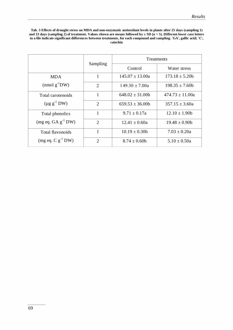
Results
69
Tab. 3 Effects of drought stress on MDA and non-enzymatic antioxidant levels in plants after 25 days (sampling 1)
and 33 days (sampling 2) of treatment. Values shown are means followed by ± SD (n = 5). Different lower case letters
in a file indicate significant differences between treatments, for each compound and sampling. 'GA', gallic acid; 'C',
catechin
Sampling Treatments
Control Water stress
MDA
(nmol g-1
DW)
1 145.07 ± 13.00a 173.18 ± 5.20b
± 7.00a 198.35 ± 7.60b
Total carotenoids
(µg g-1
DW)
1 648.02 ± 31.00b 474.73 ± 11.00a
2 659.53 ± 36.00b 357.15 ± 3.60a
Total phenolics
(mg eq. GA g-1
DW)
1 9.71 ± 0.17a 12.10 ± 1.90b
2 12.41 ± 0.60a 19.48 ± 0.90b
Total flavonoids
(mg eq. C g-1
DW)
1 10.19 ± 0.30b 7.03 ± 0.20a
2 8.74 ± 0.60b 5.10 ± 0.50a

Results
70
Publication II:
Subchapter 4.2.
Selection and characterisation of salt and drought-
resistant Phaseolus cultivars: a ‘proof-of-concept’ study
This manuscript has been submitted to the Journal; Peer J, and is currently under
review.
Reference:
Al Hassan, M., Morosan, M., Lopez-Gresa, M.P., Boscaiu, M., Vicente, O. (2015).
Selection and characterisation of salt and drought-resistant Phaseolus cultivars: a ‘proof-of-
concept’ study (Under revision).

Results
71
Selection and characterisation of salt and drought-resistant Phaseolus cultivars: a
‘proof-of-concept’ study
Abstract Given the increasing demand for food, as a result of the ever rising human
population, and the spreading of aridity and soil salinisation in the context of global climate
change, stress tolerance studies in crops are of great importance. As a ‘proof-of-concept’ for a
long-term project aimed at the selection and characterisation of stress tolerant bean varieties,
in the present work responses to salt and water stress have been analysed in young plants of
three Phaseolus vulgaris and one P. coccineus cultivars. Determination of several growth
parameters was used to establish the relative tolerance to stress of the studied cultivars.
Measurements of ion (Na+, K
+, Cl
-) and osmolyte (proline, glycine betaine, total soluble
sugars) contents were performed in leaves of plants harvested after three weeks of treatment;
individual soluble carbohydrates in the plant extracts were also quantified after HPLC
fractionation. The major aim of this study was to identify the specific responses to stress that
are the most relevant for tolerance in the analysed Phaseolus cultivars. Salt stress resistance
in beans is mostly based on restriction of Na+ (and, to a lesser extent, also of Cl
-) transport to
shoots, and on the accumulation of myo-inositol for osmotic adjustment; the increase of myo-
inositol levels is also important under water stress conditions; glycine betaine biosynthesis
may also contribute to drought tolerance in P. coccineus, but not in P. vulgaris. These
responses to both, salt and water stress during vegetative growth appear to be more efficient
in P. vulgaris cv. ‘Maxidor’, the most tolerant of the tested Phaseolus genotypes. Proline
accumulation is a reliable marker of the level of stress affecting the plants, but does not seem
to be directly related to stress tolerance mechanisms.

Results
72
Keywords: glycine betaine; myo-inositol; osmotic adjustment; Phaseolus; Proline; stress
tolerance.
4.2.1. Introduction
Drought and soil salinity are the most important environmental stress factors that
reduce crop yields worldwide (Boyer, 1982; Bartels and Sunkar, 2005). Drought is the single
abiotic stress condition most devastating for agriculture: insufficient rainfall brings about a
progressive reduction of the amount of water available for plants in the soil, affecting their
growth and development and reducing crop productivity, or even causing premature plant
death and the loss of the whole crop, if drought is prolonged in time. Irrigation systems are
necessary to maintain acceptable yields in those regions with low rainfall. In fact, irrigated
land is much more productive than rain fed cropland: irrigation is currently used to grow
crops in about 280 million hectares of arable land; this represents just below 20% of the total
cultivated land, but produces more than 40% of world food supplies (Munns and Tester,
2008). Yet, intensive and prolonged irrigation causes another serious problem for agriculture:
soil salinisation. After some decades of continuous irrigation, toxic ions dissolved in
irrigation water progressively accumulate in the soil, leading to the so-called ‘secondary
salinisation’ – of anthropic origin – of arable land (Flowers, 2004). At present, more than
20% of irrigated cropland is affected by salt, to a greater or lesser extent (Flowers and
Flowers, 2005), and more of 10 million hectares are lost for agriculture every year due to soil
salinisation (Owens, 2001).
Average yields of all major crops are only a fraction, somewhere between 20% and
50%, of record yields, depending on the species. Adverse environmental conditions affecting
the plants growing in the field – mostly drought and soil salinity, as mentioned above – are

Results
73
the major cause of these differences. These losses are expected to increase in the near future,
at least in arid and semiarid regions, due to the forecasted effects of climate change, which
include an increase in average temperatures worldwide and the occurrence of more frequent,
longer and more intense drought periods (IPCC, 2014), and consequently a shortage of water
available for irrigation, which will especially affect subsistence farming in developing
countries (Morton, 2007). Therefore, an effective approach to increase crop yields, and hence
food production, in the next decades could be based on the biotechnological improvement,
through genetic engineering methods, of the abiotic stress tolerance of our major crops (Fita
et al., 2015). This, in turn, requires a profound knowledge of the intricate physiological,
biochemical and molecular networks underlying plant stress tolerance mechanisms. For this
reason – apart from the academic interest of this topic – the study of the responses of plants to
abiotic stress has become an active area of research in plant biology.
In addition to some species-specific responses, which may be important for the stress
tolerance of particular taxa, all plants appear to react against adverse environmental
conditions by activating a series of conserved response mechanisms, which are common to,
or overlapping for different abiotic stresses. One of these basic stress responses involves the
control of ion homeostasis and the maintenance of cellular osmotic balance: water transport
into the cell, compartmentalisation of toxic ions in the vacuole, and synthesis and
accumulation of compatible solutes or osmolytes in the cytoplasm, to counteract cellular
dehydration caused by high soil salinity and drought – but also by other stressful conditions
such as cold or high temperatures (Munns and Termaat, 1986; Zhu, 2001; Munns and Tester,
2008). Apart from contributing to osmotic adjustment, osmolytes play ‘osmoprotectant’ roles
by acting as low-molecular-weight chaperons that stabilise proteins, membranes and other
macromolecular structures under cellular dehydration conditions, and also as scavengers of
‘reactive oxygen species’ (ROS) (Chen and Murata, 2008; Flowers and Colmer, 2008;

Results
74
Hussain et al., 2008; Szabados and Savouré, 2010). Obviously, these mechanisms are
responsible, at least in part, for the tolerance of plants to abiotic stress. However, the relative
contribution of different stress responses to stress tolerance, which may vary widely among
plant species, remains largely unknown. Comparative studies correlating stress responses,
such as ion and osmolyte accumulation, with the relative tolerance of genetically related taxa
– different species of the same genus, different varieties or cultivars of the same species –
may be extremely useful to define those mechanisms that are most relevant for the resistance
to drought and/or salinity in a given species (Grigore et al., 2011; Gil et al., 2013). It seems
logical to assume that enhancement of these particular responses by genetic engineering will
be an effective strategy to improve the abiotic stress tolerance of particular crops.
Conventional breeding, based on sexual crosses and selection, can also contribute
significantly to the improvement of crop abiotic stress tolerance. This approach has had
limited success in the past, but now new biotechnological tools are available that make the
breeding process much more efficient and rapid: the use of molecular markers in ‘marker
assisted selection’ (MAS), high-throughput DNA sequencing technologies ('next generation
sequencing', NGS) and other breeding applications of genomics, or different in vitro culture
methods (Varshney et al., 2009; Fita et al., 2015). Since the current industrialised agriculture
is based in a very limited number of varieties of each crop, it will be also important to screen
minor commercial cultivars, local and neglected varieties and landraces stored in seed banks,
as well as wild relatives, as a source of genetic variability to be used in breeding of abiotic
stress tolerance of our crops (Ceccarelli et al., 1998; Blair et al., 2012).
The genus Phaseolus includes more than 50 species that spread naturally from North
to South America, of which five are cultivated (Broughton et al. 2003). Phaseolus vulgaris
L., the common bean, occurs in the wild from northern Mexico to north-western Argentina. It
was independently domesticated in Central America and in the Andes (Gepts and Debouck,

Results
75
1991), but is now cultivated all over the world. The common bean is consumed as both dry
beans and green pods, and represents the most important legume for human nutrition
(Broughton et al., 2003). Beans are essential components of people’s diet, mostly in
developing countries where they are a major source of protein, vitamins, minerals and fibre
(Bellucci et al., 2014). Phaseolus coccineus L., the runner bean, was domesticated more than
2,000 years ago in Mexico where it is, at present, the second most important legume in the
local diet after the common bean (Vargas Vázquez et al., 2012); it is also cultivated
worldwide, albeit at a much smaller scale than the common bean. Phaseolus vulgaris is a
typical rain fed crop, cultivated under diverse environmental conditions, including relatively
dry areas (Graham and Ranalli, 1997; Singh, 2007), although it is not considered very
tolerant to water stress (Molina et al., 2001; Cuellar-Ortiz et al., 2008). Actually, only about
7% of the bean cultivated area receives adequate rainfall (Broughton et al., 2003), and in
some regions drought may cause the loss of most of the crop (Bellucci et al., 2014). For
example, a drastic reduction of 80% seed yield has been reported in Romania, under natural
drought conditions in the field, due to the combined effects of the reduction in the number of
pods per plant, the number of seeds per pod and the average seed weight (Szilagyi, 2003). In
Sub-Saharan Africa, about 40% of bean growing areas are affected by drought, and average
yields there reach only 400 kg ha-1
year-1
, as compared to 1,500 kg ha-1
year-1
reported from
less drought-prone areas in eastern Africa (Namugwanya et al., 2014). Although water deficit
has a general negative effect on crop productivity, large quantitative differences (between ca.
20% and 70% yield reduction) have been reported for different bean genotypes subjected to
experimental drought conditions (Ramirez-Vallejo and Kelly, 1998).
As all major crops, the common bean is a glycophyte, sensitive to salt. Even relatively
low soil salinity levels, below 2 dS m-1
, will significantly reduce crop productivity (Maas EV
and Hoffman, 1977), although some cultivars appear to be more resistant to salt stress

Results
76
(Szilagyi, 2003; Gama et al., 2007; Cuellar-Ortiz et al., 2008; Kaymakanova and Stoeva,
2008). About 20 to 30% of the bean-production areas in the Middle East are affected by soil
salinity (Bayuelo-Jiménez et al., 2002) which causes a drastic yield reduction. At salinity
level equal to 100 mM NaCl, pod yield per plant in this species decreased by 85% (De
Pascale et al., 1997).
Considering the need to increase food production to feed a growing human population
and the importance of beans in people’s diet in many countries, on the background of global
climate change, the genetic improvement of bean stress resistance is a matter of much
interest. We have undertaken the screening of a large number of Phaseolus commercial
cultivars and landraces of different origins, under standardised conditions, with the final aim
of selecting several varieties with increased tolerance, which could be eventually included in
breeding programmes for the enhancement of salt and water stress tolerance of beans. In
addition, the characterisation of the physiological, biochemical and molecular mechanisms of
stress response in those relatively tolerant cultivars will provide useful information for
selecting the most appropriate approach for engineering stress tolerance in transgenic
Phaseolus plants.
In the present work, designed as a ‘proof-of-concept’ of the aforementioned long-term
project, we have analysed the relative tolerance to water and salt stress during vegetative
growth in young plants of three P. vulgaris cultivars and in one cultivar of P. coccineus, a
species that have been previously reported as being more stress-tolerant than common bean
(Subbarao and Johansen, 1994). Responses to stress are dependent on the plant
developmental stage, as it has been shown in different species (Läuchli and Epstein, 1990;
Johnson et al., 1992; Vicente et al., 2004); this specific growth phase was selected because
during vegetative development seedlings and young plants are generally more sensitive to

Results
77
stress than adult plants and it should be easier to detect differences in the relative tolerance of
the investigated cultivars.
The major aim of this comparative study was to get information on the biochemical
mechanisms underlying plant tolerance to environmental stress and, specifically, to establish
which responses to water and salt stress are the most relevant for tolerance in Phaseolus. We
assumed that differences in the stress resistance of the selected cultivars could be explained
by differences in the efficiency of the aforementioned general stress response mechanisms;
that is, control of ion transport and maintenance of cellular osmotic balance. Therefore, we
tried and correlated the degree of salt and/or water stress resistance of the different cultivars
with the levels of i) toxic ions (Na+ and Cl
-), and ii) different osmolytes (proline, glycine
betaine and soluble carbohydrates) in the plants subjected to the salt and water stress
treatments.
4.2.2. Materials and methods
4.2.2.1. Plant material
Two commercial cultivars (‘Maxidor’ and ‘The Prince’) and one Spanish local variety
(‘Judía de Franco’) of Phaseolus vulgaris L. and one cultivar of Phaseolus coccineus L.
(‘Moonlight’) were used in the present study. Seeds of ‘Judía de Franco’ were obtained from
the Germplasm Bank of COMAV (Institute for Conservation and Improvement of Valencian
Agro diversity, Polytechnic University of Valencia); P. coccineus seeds were obtained from
Thompson and Morgan, AJP Garden and Crafts (UK), and those of cvs. 'The Prince' and
'Maxidor' were purchased from S.C. AGROSEM IMPEX S.R.L., Targu Mures (Romania).
4.2.2.2. Characterization of cultivars

Results
78
'The Prince' is a dwarf French bean cultivar, with a bushy growth. Pods are green and
flat, with a length of about 20 cm and large seeds (average weight: ca. 0.5 g per seed). It is an
early cultivar; it takes 60-70 days for snap pods to form, and 90 days for dry seeds. Cv. 'The
Prince' has been reported as drought sensitive (Boutraa and Sanders, 2001). The cultivar is
one of the most commonly used in Europe.
'Maxidor' is also a dwarf French bean cultivar. It has a bushy growth, with precocious
flowering. Pods are round and yellow, 12 cm long with relatively large seeds; the seed mass
is about 0.26 g. It is considered as an early or medium-early variety; pods can be harvested
60-65 days after germination and dry beans after 100-110 days. 'Maxidor' is cultivated in
several countries in Europe, and is one of the most commonly grown in Western Romania
(Madoşă et al., 2013).
'Judía de Franco' is a local landrace of P. vulgaris from the province of Teruel (Spain).
It has an indefinite growth, surpassing 3 m high. It reaches maturity after 96 days. Pods are 9
cm long, flat and green. The average weight of a seed is about 0.3 g.
Cv. 'Moonlight' belongs to the species P. coccineus. Seeds originated from Mexico. It
reaches 3 m high. It has a longer biological cycle of 120-125 days. Pods are green and long,
reaching 25 cm, and seed mass is about 0.45 g.
4.2.2.3. Growing conditions and stress treatments
Seeds were sterilised with a 0.3% (v/v) solution of sodium hypochlorite for 5 min,
rinsed in distilled water and then sown on a moistened mixture of peat, perlite and
vermiculite (2:1:1) in 0.5 L pots (Ø = 11 cm). One seed was placed per pot on the substrate
surface. Throughout the germination process, the substrate was kept moderately moisten
using Hoagland nutritive solution. Water and salt stress treatments (50, 100 and 150 mM
NaCl) were started 16 days after germination, when the first trifoliate leaves had already
Results
79
appeared. The control plants were watered twice a week with Hoagland nutritive solution
(one litre per three plants), and for the salt stress treatments the plants were watered with the
same volume of nutritive solution supplemented with the appropriate NaCl amounts prior to
irrigation. Drought treatments were carried out by completely preventing irrigation. All the
experiments were conducted for 3 weeks, including five plants per treatment, in an
environment chamber under the following controlled conditions: long day photoperiod (16
hours of light and 8 hours of darkness), temperature (23ºC during the day and 17ºC at night),
CO2 level (≈ 300 ppm). Humidity ranged between 50-80% while all the treatments were
underway.
4.2.2.4. Soil analysis
The electrical conductivity (EC1:5) of the substrate was checked at the end of the
treatments. Soil samples were taken from five pots per treatment, air-dried and then passed
through a 2-mm sieve. A soil: water (1:5) suspension was prepared in distilled water and
stirred for 1 h at 600 rpm and 21ºC. EC was measured with a Crison Conductivity meter 522
and is expressed in dS m-1
.
4.2.2.5. Plant growth parameters
After three weeks of salt or water stress treatment, the aerial part of each plant was
collected and the following growth parameters were determined: stem length, leaf number,
fresh and dry weight, and percentage of water content. Fresh material was stored at -20°C for
further studies. To compare the degree of stress-induced inhibition of growth of the different
cultivars, which differ in plant size, stem length and fresh weight were expressed as
percentage of the values corresponding to the non-stressed controls. To determine water
content, part of each sample was weighed (FW), dried at 65°C until constant weight (48-72

Results
80
h), and then reweighed (DW); water content of each sample (%) was calculated as indicated
in Gil et al. (2014).
4.2.2.6. Ion content measurements
Monovalent ion contents after the stress treatments were determined according to
Weimberg (1987) in aqueous extracts obtained by heating the samples (0.15 g of dried,
ground plant material in 25 mL of water) in a water bath, for 1 h at 95ºC, followed by
filtration through filter paper (particle retention 8-12 µm). Sodium and potassium were
quantified with a PFP7 flame photometer (Jenway Inc., Burlington, USA) and chlorides were
measured using a Merck Spectroquant Nova 60® spectrophotometer and its associated test
kit (Merck, Darmstadt, Germany).
4.2.2.7. Osmolyte quantification
Osmolyte contents (proline, glycine betaine and total soluble sugars) were determined
in the stressed and control plants. Proline (Pro) was extracted with 3% (w/v) sulfosalicylic
acid, from 0.2 g of frozen plant material in liquid nitrogen, and was quantified according to
the acid-ninhydrin method described by Bates et al. (1973). Pro concentration was expressed
as µmol g-1
DW.
Glycine betaine (GB) was determined from 0.1 g dry material according to Grieve and
Grattan (1983). GB concentration was expressed as µmol g-1
DW.
Total soluble sugars (TSS) were measured in 0.1 g dry plant material suspended in 3 ml 80%
(v/v) methanol, following the phenol / sulphuric acid method, as described by Dubois et al.
(1956). TSS contents were expressed as ‘mg equivalent of glucose’ per g DW.
4.2.2.8. HPLC analysis of carbohydrates
Results
81
The soluble sugar fraction (mono and oligosaccharides) was analysed using a Waters
1525 high performance liquid chromatography coupled to a 2424 evaporative light scattering
detector (ELSD). The source parameters of ELSD were the following: gain 75, data rate 1
point per second, nebulizer heating 60%, drift tube 50ºC, and gas pressure 2.8 kg cm-2
. The
analysis was carried out injecting 20 µL aliquots of the samples with a Waters 717
autosampler into a Prontosil 120-3-amino column (4.6 x 125 mm; 3 µm particle size)
maintained at room temperature. An isocratic flux (1 mL/min) of 85% acetronitrile (J.T.
Baker) during 25 minutes was applied in each run. Standards of glucose, fructose, sucrose
and myo-inositol served to identify peaks by co-injection. Sugars were quantified with peak
integration using the Waters Empower software and comparison with glucose, fructose,
sucrose, and myo-inositol standard calibration curves.
4.2.2.9. Statistical analysis
Data were analysed using the programme SYSTAT v. XVI. Before the analysis of
variance, the Shapiro-Wilk test was used to check for validity of normality assumption and
Levene´s test for the homogeneity of variance. If ANOVA requirements were accomplished,
the significance of the differences among treatments was tested by one-way ANOVA at a
95% confidence level and post hoc comparisons were made using the Tukey HSD test. All
means throughout the text are followed by SD.
4.2.3. Results
4.3.3.1. Electric Conductivity of substrates
Electric conductivity (EC1:5) was recorded in samples of the pot substrates after three
weeks of salt and water stress treatments (Table 1). For all cultivars, a similar increase in

Results
82
EC1:5 was detected in parallel to the increase of NaCl concentrations, which confirms the high
correlation between EC and the concentration of the saline solutions used in the treatments.
Nearly no statistical significant differences were found between different cultivars for each
treatment. The EC of the pot substrates in the water stress treatments were lower than in the
controls watered with Hoagland solution (except in ‘The Prince’ where it increased slightly),
which can be explained by accumulation of ions present – at low concentrations – in the
nutritive solution.
4.3.3.2. Effect of salt stress on plant growth
The stem length of five plants of each cultivar was measured at the end of the
treatments. The average stem length of the control plants was considered as 100% for each
cultivar (absolute values are specified in the legend of Fig. 1 for each control), and decreased
in a concentration-dependent manner upon salt treatment. In the presence of 150 mM NaCl
the stem length was reduced by 40% in P. coccineus and P. vulgaris cv. ‘The Prince’, by
more than 60% in cv. ‘Judía de Franco’, but by only 20% in cv. ‘Maxidor’ (Fig. 1a).
The number of trifoliate leaves also decreased during the salt treatments, with some
differences among cultivars (Fig. 1b). ‘The Prince’ appeared to be the most affected by salt,
as the number of leaves already dropped by more than 50% at the lower concentration tested,
50 mM NaCl, and by ca. 75% in the presence of 150 mM NaCl, whereas the reduction of
average leaf number in P. coccineus and P. vulgaris cv. ‘Maxidor’ under the same conditions
was less than 5% – a not significant difference – and less than 50%, respectively. An
intermediate behaviour was observed in cv. ‘Judía de Franco’ (Fig. 1b).
The fresh weight (FW) of five harvested plants per treatment and cultivar was
measured, and the mean of the control plants was taken as the reference (100%) for each
cultivar. The greatest reduction of fresh mass of the aerial part of the plants was registered in

Results
83
cv. ‘The Prince’, with values of ca. 80% after treatment with 100 mM NaCl and 95% in the
presence of 150 mM NaCl. The least affected by salt stress appeared to be cv. ‘Maxidor’ –
about 63% decrease in FW in the 150 mM NaCl treatment – followed by ‘Judía de Franco’,
and P. coccineus (Fig. 1c).
Fig. 1 Salt stress-induced changes in: (a) stem length (%), with the stem lengths of control, non-treated plants (Phaseolus vulgaris, cv. ‘The Prince’: 60.00 cm; cv. ‘Judía de Franco’: 174.61 cm; cv. ‘Maxidor’: 44.16 cm; Phaseolus
coccineus: 219.00 cm) considered as 100% for each cultivar; (b) number of leaves; (c) Fresh weight (%), with the
fresh weight of control plants (Phaseolus vulgaris, cv. ‘The Prince’: 31.54 g; cv. ‘Judía de Franco’: 34.87 g; cv.
‘Maxidor’: 17.17 g; Phaseolus coccineus: 30.26 g) considered as 100% for each cultivar; (d) water content (%).
Measurements were performed after three weeks of treatment. The values shown are means with SD (n = 5). For each
cultivar, different lower-case letters indicate significant differences between treatments according to the Tukey test (α
= 0.05).
A decrease in water content would reflect the loss of water in the plants as a result of
the salt treatments. Yet, beans appear to have relatively efficient mechanisms to avoid
dehydration of their aerial parts since, in general, no significant reduction of water content
was observed in the salt-treated plants. Only in P. vulgaris cv. ‘The Prince’, and only at the

Results
84
highest NaCl concentration tested (150 mM), a significant decrease in water content, of
nearly two-fold, was detected (Fig. 1d); this may partly explain the higher sensitivity to salt
of this cultivar indicated by the aforementioned growth measurements, as compared to the
other varieties.
4.3.3.3. Ion contents
The patterns of sodium accumulation in the aerial part of the plants upon salt stress
treatment were different for the P. vulgaris cultivars: Na+ levels rose four to five-fold in the
plants treated with 100 and 150 mM NaCl in cv. ‘The Prince’; a two-fold increase was
measured in ‘Judía de Franco’, but only at the highest salt concentration, whereas no
statistically significant Na+ variation was detected in cv. ‘Maxidor’; in P. coccineus, a very
small, but significant increase in Na+ content was observed in the presence of 150 mM NaCl
(Fig. 2a). Contrary to sodium, the four tested cultivars showed a similar qualitative pattern
regarding chloride accumulation: in all of them Cl- contents increased significantly, roughly
in parallel with increasing external NaCl concentrations. In the presence of 150 mM NaCl,
Cl- levels rose up to 10 to 15-fold in P. coccineus and P. vulgaris cvs. ‘The Prince’ and ‘Judía
de Franco’, or ca. five-fold in cv. ‘Maxidor’, as compared to the control, non-treated plants
(Fig. 2b). Comparing the levels of both cations, it is clear that the salt-treated plants
accumulated much more Cl- than Na
+ in their aerial parts, again with quantitative differences
among cultivars: in the presence of high NaCl concentrations, Cl-/Na
+ rations of,
approximately, 5, 10, 10 and more than 15 were calculated for ‘The Prince’, ‘Judía de
Franco’ Maxidor’ and P. coccineus, respectively (Fig. 2a, b). Plant potassium contents
showed some variations some cultivars under study, however non that can be correlated with
the increased salt concentrations (Fig. 2c).
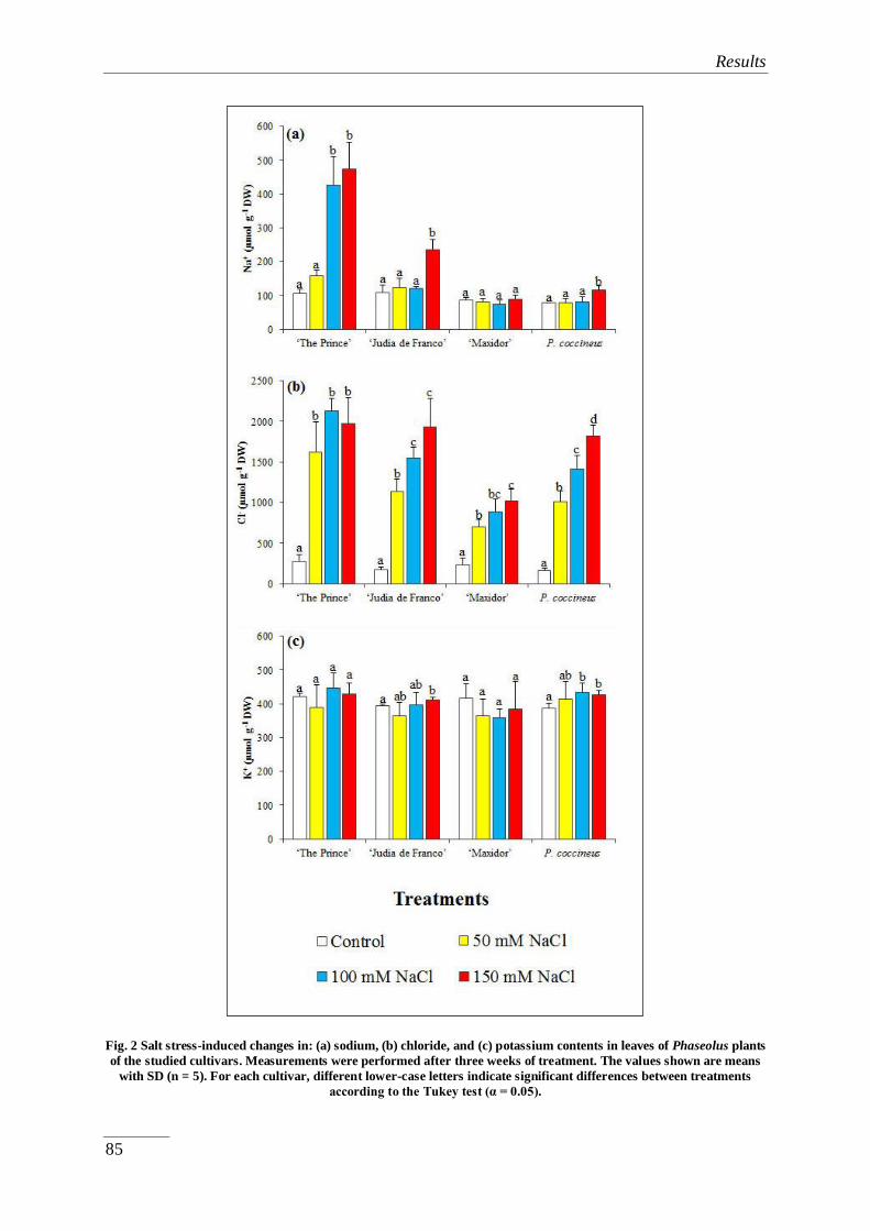
Results
85
Fig. 2 Salt stress-induced changes in: (a) sodium, (b) chloride, and (c) potassium contents in leaves of Phaseolus plants
of the studied cultivars. Measurements were performed after three weeks of treatment. The values shown are means
with SD (n = 5). For each cultivar, different lower-case letters indicate significant differences between treatments
according to the Tukey test (α = 0.05).

Results
86
4.3.3.4. Osmolyte contents
Proline contents in the control plants were relatively low and similar in all cultivars
(2-4 µmol g-1
DW), but increased significantly in the presence of 100 and 150 mM NaCl, for
all cultivars except ‘Maxidor’; the highest Pro concentrations were reached in cv. ‘The
Prince’, 12 and 42 µmol g-1
DW in plants treated with 100 and 150 mM NaCl, respectively;
the corresponding values for ‘Judía de Franco’ and P. coccineus were 8 and 22 µmol g-1
DW.
In cv. ‘Maxidor’, a significant increase in Pro levels was only detected in the presence of 150
mM NaCl, reaching a concentration of only 9 µmol g-1
DW, much lower than in the other
cultivars (Fig. 3a).
Apparently, glycine betaine plays no role in responses to salt stress of the investigated
Phaseolus cultivars, since no significant salt-induced changes in the levels of this osmolyte
were detected in any of the treatments (Fig. 3b). The absolute GB concentrations were similar
for P. coccineus and P. vulgaris, cvs. ‘The Prince’ and ‘Maxidor’, between 15 and 20 µmol g-
1 DW, and slightly higher in cv. ‘Judía de Franco’ (Fig. 3b).
Mean values of total soluble sugars did not show a clear pattern of variation in
response to the salt treatments for any of the selected cultivars, and the observed differences,
in general, were not statistically significant, possibly due in part to the variability in the TSS
contents of individual plants, reflected in relatively large SD of the mean values (Fig. 3c).
Here again, ‘Judía de Franco’ presented slightly higher TSS levels under stress than those
measured in the other studied cultivars (Fig. 3c).

Results
87
Fig. 3 Salt stress-induced changes in the levels of: (a) proline (Pro), (b) glycine betaine (GB), and (c) total
soluble sugars (TSS) in the same samples as in Fig. 2. Measurements were performed after three weeks of treatment.
The values shown are means with SD (n = 5). For each cultivar, different lower-case letters indicate significant
differences between treatments according to the Tukey test (α = 0.05).
Results
88
The major soluble carbohydrates in the extracts were identified and quantified by
HPLC, and corresponded to fructose, sucrose and myo-inositol. Their pattern of variation was
different in the four cultivars analysed. A clear salt-dependent increase in fructose and
sucrose was only detected in cv. 'The Prince', whereas in the other cultivars changes in the
concentrations of both sugars were not statistically significant or did not correlate with
external salinity (Figs. 4a and b). A considerable increase of myo-inositol was detected in P.
vulgaris cv. 'Maxidor' and, to a lesser extent, in P. coccineus, in parallel with increasing salt
concentrations in the watering solution – reaching in the presence of 150 mM NaCl about
three-fold and two-fold, respectively, higher levels than in the non-stressed controls – but not
in the other two studied varieties (Fig. 4c).

Results
89
Fig. 4 Salt stress-induced changes in the levels of: (a) fructose, (b) sucrose, and (c) myo-inositol, separated by HPLC,
in the same samples as in Fig. 2. Measurements were performed after three weeks of treatment. The values shown are
means with SD (n = 5). For each cultivar, different lower-case letters indicate significant differences between
treatments according to the Tukey test (α = 0.05).

Results
90
4.3.3.5. Effect of water stress on growth and osmolyte contents
Lack of irrigation inhibited growth in all selected cultivars (Fig. 5), which showed a
relative resistance to water stress similar to that observed under salt stress conditions (Fig. 1).
Thus, regarding the relative decrease of stem length, the most affected cultivars were 'The
Prince' and 'Judía de Franco', with a 60% reduction, followed by P. coccineus with only a
40%, while cv. 'Maxidor' plants did not show any reduction of stem length under water stress
conditions (Fig. 5a). The number of trifoliate leaves was reduced in all cultivars after the
stress treatment, mostly in 'The Prince' (ca. 4.5-fold), followed by 'Judía de Franco' (2.7-fold)
and P. coccineus (2.2-fold), with the smallest decrease, about 1.7-fold, observed again in cv.
'Maxidor' (Fig. 5b). The fresh weight of the plants of the four cultivars was strongly affected
by drought, even more than by salt stress; a reduction of 97% was registered in ‘The Prince’,
around 90% in ‘Judía de Franco’ and P. coccineus, and 67% in ‘Maxidor’ (Fig. 5c). In this
case, however, the decrease of fresh weight was partly due to loss of water, which was
significant, albeit quantitatively different, for all cultivars. Again, the strongest reduction of
water content, ca. 85% was measured in cv. ‘The Prince’, in comparison with 32% in ‘Judía
de Franco’, 20% in P. coccineus and 18% in ‘Maxidor’ (Fig. 5d). These data confirm that the
mechanisms to avoid cellular dehydration under stress are much less efficient in cv. 'The
Prince' than in the other studied varieties, as shown also for the salt treatments.

Results
91
Fig. 5 Water stress-induced changes in: (a) stem length (%), with the stem lengths of control, non-treated plants
(mentioned in the legend of Fig. 1) considered as 100% for each cultivar; (b) number of leaves; (c) Fresh weight (%),
with the fresh weight of control plants (mentioned in the legend of Fig. 1) considered as 100% for each cultivar; (d)
water content (%). Stem lengths and fresh weights of control plants as in Fig. 1. Measurements were performed after
three weeks of treatment. The values shown are means with SD (n = 5). For each cultivar, different lower-case letters
indicate significant differences between treatments according to the Tukey test (α = 0.05).
As expected, in all cultivars, ion levels (Na+, Cl
-, K
+) did not significantly change in
water-stressed plants when compared to the corresponding controls (Table 2).
Proline levels increased under drought stress by 12 to 15-fold in plants of P. vulgaris
cvs. ‘The Prince’ and ‘Judía de Franco’, and in P. coccineus plants, but by only four-fold in
those of cv. ‘Maxidor’ (Fig. 6a). Glycine betaine showed no significant variation under water
stress conditions in the three cultivars of P. vulgaris, but an almost half-fold increase in P.
coccineus (Fig. 6b). The measured mean amounts of total soluble sugars did not vary
significantly in any of the investigated Phaseolus varieties (Fig. 6c).

Results
92
Fig. 6 Water stress-induced changes in the levels of: (a) proline (Pro), (b) glycine betaine (GB), and (c) total
soluble sugars (TSS) in leaves of Phaseolus plants of the studied cultivars. Measurements were performed after three
weeks of treatment. The values shown are means with SD (n = 5). For each cultivar, different lower-case letters
indicate significant differences between treatments according to the Tukey test (α = 0.05).

Results
93
However, some differences between stressed and control plants were found when
analysing individual soluble sugars, separated by HPLC. Fructose levels increased
significantly in P. vulgaris cv. 'Judía de Franco', but differences were not detected in cv. 'The
Prince', nor in P. coccineus (Fig. 7a), while a decrease in sucrose contents was observed in all
varieties, except 'The Prince' (Fig. 7b). Yet, the most interesting finding was the large
increase in myo-inositol contents measured in plants of cv. ‘Maxidor’ and, to a lesser extent,
of P. coccineus subjected to water stress (Fig. 7c).

Results
94
Fig. 7 Water stress-induced changes in the levels of: (a) fructose, (b) sucrose, and (c) myo-inositol, separated by
HPLC, in the same samples as in Fig. 6. Measurements were performed after three weeks of treatment. The values
shown are means with SD (n = 5). For each cultivar, different lower-case letters indicate significant differences
between treatments according to the Tukey test (α = 0.05).

Results
95
4.2.4. Discussion
Water deficit in the soil due to lack of rain or irrigation (drought stress), or high
concentration of salts in the soil solution (salt stress) both cause lowering of the water
potential, leading to cellular dehydration (osmotic stress). Apart from this osmotic
component, high soil salinity has a number of additional deleterious effects for plants due to
salt toxicity, mostly of Na+ ions, which when present at too high levels inhibit many
enzymatic activities and basic cellular processes, such as mRNA processing and protein
synthesis; moreover, the high concentration of Na+ and Cl
- results in nutrient imbalance
through the reduction of uptakes of K+, NO3
- and PO4
3- (Greenway and Munns, 1980;
Forment et al., 2002; Bartels and Sunkar, 2005; Munns and Tester, 2008). Therefore, plant
responses to water and salt stress are similar and differ mostly in ion transport-related
processes. It has even been considered that the initial response to salt is identical to that
induced by drought, and is triggered by its osmotic component, while salinity-specific
responses due to ion toxicity develop only over time with longer exposure to stress (Munns,
2002). This overlap explains that often the physiological effects of water stress and high salt
concentrations are the same, as we have observed in the present work with different
Phaseolus cultivars. There are numerous papers reporting a wide range of salt (Gama et al.,
2007; Kaymakanova and Stoeva, 2008) and drought tolerance (Cuellar-Ortiz et al., 2008;
Santos et al., 2009; Omae et al., 2012) within this genus, supporting the possibility of
selecting the most resistant genotypes to be used in breeding programmes to improve the
stress tolerance of beans.
The most general effect of abiotic stress, and the easiest to quantify, is inhibition of
growth, which allows plants to survive under adverse conditions by re-directing their
resources (metabolic precursors and energy) from normal metabolism and growth to the
activation of specific stress defence mechanisms (Zhu, 2001; Munns and Tester, 2008).

Results
96
Determination of several growth parameters allowed us to establish the relative degree of
resistance to water and salt stress of the investigated bean varieties. Phaseolus vulgaris cv.
‘The Prince’ was the most sensitive, followed by ‘Judía de Franco’, while cv. 'Maxidor' was
the most stress-resistant of the selected common bean cultivars – even more than the tested P.
coccineus cultivar, although this species has been previously reported as being more tolerant
than P. vulgaris (Subbarao and Johansen, 1994; Gutierrez et al., 2009).
It is well established that all plants share a series of basic, conserved mechanisms of
response to abiotic stress, including for example the control of ion transport and the
accumulation of different osmolytes in the cytoplasm to help maintain cellular osmotic
balance (Zhu, 2001; Munns and Tester, 2008). Yet, it is not so clear the relative importance
and contribution of those response mechanisms to the stress tolerance of a particular species.
The comparative analysis of related genotypes, such as that reported here for several
Phaseolus cultivars, may help to answer this question, by correlating their relative degree of
resistance to stress with changes in the levels of stress markers associated to specific
responses. Our results indicate, for example, that one of the main reasons behind salt
resistance in Phaseolus is the presence of mechanisms that restrict the transport of Na+ to the
aerial part of the plants, mechanisms that are more efficient in the relatively more tolerant
cultivars.
It is known that plants of the genus Phaseolus are able to exclude sodium from the
shoots, even in the presence of relatively high NaCl concentrations in the soil (Seemann and
Critchley, 1985). Earlier autoradiography studies on P. vulgaris indicated that Na+ is retained
in roots by binding at sites in the stele or at those bordering it (Jacobi, 1964). This was
confirmed later-on by Kramer et al. (1977) using X-ray microanalyses in P. coccineus, which
also excluded Na+, but not Cl
-; these authors proposed that in this species Na
+ is also
reabsorbed from vessels by xylem parenchyma cells. In more recent studies, a higher Na+

Results
97
concentration in roots than in leaves was found in P. vulgaris and P. latifolius, showing that,
within this genus, the basic sodium exclusion mechanisms are related to its transport from
roots to shoots, rather than to absorption by roots (Bayuelo-Jiménez et al., 2012). The anion
Cl-, on the other hand, displays high mobility within the plant and is not effectively
compartmentalised in cells: chloride concentration was high in both the vacuole and the
chloroplast-cytoplasm in salt-stressed plants of P. vulgaris (Seemann and Critchley, 1985).
The salt-sensitive Phaseolus genotypes analysed by Bayuelo-Jiménez et al. (2012) gave a
higher Cl-
concentration in leaves than more tolerant ones. In all cultivars analysed in the
present work, Na+ concentration in leaves was clearly lower than that of Cl
-, under the same
external conditions, thus confirming this mechanism. Yet, the pattern of ion accumulation in
response to increasing NaCl concentration in the nutritive solution varied, according to the
relative salt tolerance of the different cultivars, with the lowest ion contents measured in the
most tolerant, cv. 'Maxidor', and the highest in the most sensitive, cv. 'The Prince'.
Since Na+ can compete with K
+ for the same transporters, a common effect of salt
stress is to lower K+ levels (Munns and Tester, 2008); mechanisms able to increase K
+
contents in leaves, thus maintaining relatively low Na+ / K
+ ratios, would therefore contribute
to salt tolerance. Increased K+ in foliar tissue upon salt treatments has been reported in beans
by Seeman and Critchley (1985), while Bayuelo-Jiménez et al. (2012) observed similar
values in plants at moderate salinity levels and in the controls. These latter authors also
reported reduced K+ contents in roots and stems of salt-treated plants, and explained the
differences in the distribution of this cation in the plants by the translocation of K+
from roots
and stems to leaves due to activation of highly selective K+ transporters. Maintenance of
constant K+ concentration in leaves was considered as an important criterion to assess the
drought tolerance of alfalfa genotypes (Benabderrahim et al., 2015). The levels of K+ in
leaves were similar for all four Phaseolus cultivars tested here, and did not vary significantly

Results
98
in the presence of salt; therefore, Na+ / K
+ ratios in leaves were dependent on Na
+ contents,
and lower in the more tolerant than in the more sensitive cultivars.
Another general response to abiotic stress is the synthesis and accumulation of
specific osmolytes in the stressed plants. Yet, it is often difficult to establish whether the
stress-dependent increase in the concentration of a particular osmolyte has a functional role in
the mechanisms of tolerance of a given species. When accumulated at high enough levels,
osmolytes will contribute to cellular osmotic balance and therefore to stress tolerance (Ashraf
and Foolad, 2007) – although the concentration required to have a relevant osmotic effect
will also depend on the intracellular distribution of the compound and the relative volume of
the cytoplasm in the cell. However, osmolytes may significantly contribute to stress tolerance
based on their additional roles as 'osmoprotectants': low-molecular-weight chaperons, ROS
scavengers, or as signalling molecules involved in regulation of gene expression and
metabolic processes (Smirnoff and Cumbes, 1989; Ashraf and Harris, 2004; Szabados and
Savouré, 2010). Comparative studies on the stress responses of related taxa should provide
evidence in favour of the contribution to stress tolerance of osmolyte accumulation, if a
positive correlation between osmolyte levels and the relative degree of tolerance can be
found.
Proline is one of the commonest osmolytes in plants. Many reports have shown
significant increases in Pro contents in Phaseolus plants submitted to either salt stress
(Kaymakanova and Stoeva, 2008; Nagesh-Babu and Devaraj, 2008) or water stress (Ashraf
and Iram, 2005; Svetleva et al., 2012). Pro seems to be a reliable marker of stress in
Phaseolus, as in many other genera, but the correlation between Pro accumulation and stress
tolerance remains unclear, since apparently contradictory results have been obtained when
comparing different varieties. Higher free Pro levels have been reported in more drought-
tolerant (Ghanbari et al., 2013) or salt-tolerant (Cárdenas-Avila et al., 2006) bean cultivars
Results
99
than in less tolerant ones. However, there are also reports of higher Pro levels in sensitive
than in more resistant cultivars, in P. vulgaris (Jiménez-Bremont et al., 2006; Rosales et al.,
2012; Zadehbagheri et al., 2012), as well as in many other plant genera where no positive
correlation between Pro contents and tolerance has been found (Lutts et al., 1996; Guerrier,
1998; Ashraf and Foolad, 2007; Chen et al., 2007).
Our experimental approach should help to clarify the confusion often found in the
literature between the concepts of 'stress responses' and 'stress tolerance'. The present study
revealed that Pro accumulation is a common response to salt and water stress in the four
cultivars analysed here, in agreement with the aforementioned published results. Yet, Pro
biosynthesis cannot contribute significantly to their stress tolerance, since the levels reached
in the most tolerant variety, cv. ‘Maxidor’, were by far lower than in the other cultivars. In
this case, Pro should be considered as a marker of the level of stress affecting the plants, and
these results simply reflect the fact that 'Maxidor' plants were less stressed than the others.
Glycine betaine is another osmolyte synthesised in response to salt and water stress in
many different plant groups (Hanson and Scott, 1980; Rhodes and Hanson, 1993; Ashraf and
Foolad, 2007). There are only a handful of references describing the presence of this
osmolyte in Phaseolus (Ashraf and Iram, 2005; Ali and Abdel-Fattah, 2006), although at
concentrations lower than those reported here, which are in turn much lower than the GB
levels recorded in real GB accumulator species (Khan et al., 2000; Tipirdamaz et al., 2006).
However, the applied stress treatments did not lead to significant increases of GB contents in
the analysed Phaseolus varieties, with the exception of water-stressed P. coccineus plants,
suggesting that GB may contribute to drought tolerance in this species.
Assessing the role of soluble sugars (e.g. sucrose, glucose or fructose) in the
mechanisms of stress tolerance has the added difficulty that their function as compatible
solutes may be masked by their multiple functions in plants, as direct photosynthesis

Results
100
products, components of primary metabolism and regulatory molecules (Gil et al., 2013).
There are several papers dealing with the variation of soluble carbohydrate contents in beans
under stress conditions (Vassey and Sharkey, 1989; Tazuke et al., 2009; Sassi et al., 2010),
but it has been recently reported that salt stress-induced sugar accumulation in the genus
Phaseolus barely contributes to the leaf osmotic potential (Bayuelo-Jiménez et al., 2012). We
did not detect significant changes, correlated with the stress treatments, in the levels of total
soluble sugars in the analysed Phaseolus cultivars. However, after separation of the
carbohydrate fraction by HPLC, strong increases in myo-inositol contents were observed in
cv. 'Maxidor', the most tolerant cultivar of P. vulgaris, and to a lesser extent also in P.
coccineus; myo-inositol accumulation was induced in the presence of NaCl, in a
concentration-dependent manner, but mostly in water-stressed plants. Therefore, this
polyalcohol appears to play a significant role in the stress tolerance mechanisms in Phaseolus
taxa. To our knowledge, publications on the presence of myo-inositol in this genus are scarce;
for example, Bahena-Betancourt et al. (2006) reported glucose and inositol as the major
protectant sugars in salt stressed beans, but we did not detect a significant variation of
glucose in the cultivars analysed here.
4.2.5. Conclusions
We have determined the relative tolerance to salt and water stress of four Phaseolus
cultivars during the stage of vegetative growth; resistance to both stresses is highest in P.
vulgaris cv. 'Maxidor', followed by P. coccineus, while the cultivar most sensitive to stress
appears to be P. vulgaris cv. 'The Prince'. Correlation of these data with changes in the levels
of ions and several osmolytes, allowed establishing which mechanisms of response to stress
that are the most relevant for tolerance in Phaseolus. In conditions of high soil salinity,

Results
101
resistance to stress is mostly based on restriction of Na+ (and, to a much lesser extent, also of
Cl-) transport to shoots, and on the accumulation of myo-inositol as the major functional
osmolyte. Under water stress conditions, the most relevant response is the increase of myo-
inositol levels, although in P. coccineus accumulation of glycine betaine may also contribute
to drought tolerance. Similar comparative analyses of additional bean varieties with different
degrees of stress resistance, selected after screening of a much larger number of Phaseolus
cultivars and landraces, will help to confirm the conclusions of the present work.
Acknowledgements
This work was funded by a grant to O.V. from the Spanish Ministry of Science and
Innovation (Project CGL2008-00438/BOS), with contribution from the European Regional
Development Fund. M.A.H. is a recipient of an Erasmus Mundus pre-doctoral scholarship
financed by the European Commission (Welcome Consortium).

Results
102
4.2.6. References
Ali, A.A., Abdel-Fattah, R.I. (2006). Osmolytes-antioxidant behavior in Phaseolus vulgaris
and Hordeum vulgare with brassinosteroid under salt stress. Journal of Agronomy, 5,
167-174.
Ashraf, M., Foolad, M.R. (2007). Roles of glycine betaine and proline in improving plant
abiotic stress resistance. Environmental and Experimental Botany, 59, 206-216.
Ashraf, M., Harris, P.J.C. (2004). Potential biochemical indicators of salinity tolerance in
plants. Plant Science, 166, 3-16.
Ashraf, M., Iram, A. (2005). Drought stress induced changes in some organic substances in
nodules and other plant parts of two potential legumes differing in salt tolerance.
Flora, 200, 535-546.
Bahena-Betancourt, L., Macias-Rodríguez, L., Bayuelo-Jiménez, J.S. (2006). Contents of
carbohydrates in salt-stressed Phaseolus species. Annual report of the Bean
Improvement Cooperative. Fort Collins, CO. USA. 49:233-234.
Bartels, D., Sunkar, R. (2005). Drought and salt tolerance in plants. Critical Reviews in Plant
Science, 24, 23-58.
Bates, L.S., Waldren, R.P., Teare, I.D. (1973). Rapid determination of free proline for water
stress studies. Plant and Soil, 39, 205-207.
Bayuelo-Jiménez, J.S., Debouck, D.G., Lynch, J.P. (2002). Salinity tolerance in Phaseolus
species during early vegetative growth. Crop Science, 42, 2184-2192.
Bayuelo-Jiménez, J.S., Jasso-Plata, N., Ochoa, I. (2012). Growth and physiological of
Phaseolus species to salinity stress. International Journal of Agronomy, 80, 207-222.
Bellucci, E., Bitocchi, E., Rau, D., Rodriguez, M., Biagetti, E., Giardini, A., Attene, G.,
Nanni, L., Papa, R. (2014). Genomics of origin, domestication and evolution of

Results
103
Phaseolus vulgaris. In: Tuberosa R, Graner A, Frison E, eds. Genomics of Plant
Genetics Resources. Dordrecht: Springer, 483-507.
Benabderrahim, M.A., Hamza, H., Haddad, M., Ferchichi, A. (2015). Assessing the drought
tolerance variability in Mediterranean alfalfa (Medicago sativa L.) genotypes under
arid conditions. Plant Biosystems, 149, 395-403.
Blair, M.W., Soler, A., Cortés, A.J. (2012). Diversification and population structure in
common beans (Phaseolus vulgaris L.). PLoS ONE, 7, e49488.
doi:10.1371/journal.pone.0049488.
Boutraa, T., Sanders, F.E. (2001). Influence of water stress on grain yield and vegetative
growth of two cultivars of bean (Phaseolus vulgaris L.). Journal of Agronomy and
Crop Science, 187, 251-257.
Boyer, J.S. (1982). Plant productivity and environment. Science, 218, 443–448.
Broughton, W.J., Hernández, G., Blair, M., Beebe, S., Gepts, P., Vanderleyden, J. (2003).
Beans (Phaseolus spp.) - model food legumes. Plant and Soil, 252, 55-128.
Cárdenas-Avila, M.L., Verde-Star, J., Maiti, R.K., Foroughbakhch-P, R., Gámez-González,
H., Martínez-Lozano, S., Núñez-González, M.A., García Díaz, G., Hernández-Piñero,
J.L., Morales-Vallarta, M.R. (2006). Variability in accumulation of free proline on in
vitro calli of four bean (Phaseolus vulgaris L.) cultivars exposed to salinity and
induced moisture stress. Phyton, 75, 103-108.
Ceccarelli, S., Grando, S., Impiglia, A. (1998). Choice of selection strategy in breeding barley
for stress environments. Euphytica, 103, 307-318.
Chen, T.H.H., Murata, N. (2008). Glycinebetaine: an effective protectant against abiotic
stress in plants. Trends in Plant Science, 13, 499-505.

Results
104
Chen, Z., Cuin, T.A., Zhou, M., Twomei, A., Naidu, B.P., Shabala, S. (2007). Compatible
solute accumulation and stress-mitigating effects in barley genotypes contrasting in
their salt tolerance. Journal of Experimental Botany, 58, 4245-255.
Cuellar-Ortiz, S.M., Arrieta-Montiel, M.P., Acosta-Gallegos, J., Covarrubias, A.A. (2008).
Relationship between carbohydrate partitioning and drought resistance in common
bean. Plant Cell and Environment, 31, 1399-1409.
De Pascale, S., Barbieri, G., Ruggiero, C. (1997). Effects of water salinity on plant growth
and water relations in snap bean (Phaseolus vulgaris L.) Acta Horticulturae, 449,
649-656.
Dubois, M., Gilles, K.A., Hamilton, J.K., Reberd, P.A., Smith, F. (1956). Colorimetric
method for determination of sugars and related substances. Analytical Chemistry, 28,
350-356.
Fita, A., Rodríguez-Burruezo, A., Boscaiu, M., Prohens, J., Vicente, O. (2015). Breeding and
domesticating crops adapted to drought and salinity: A new paradigm for increasing
food production. Frontiers in Plant Science, 6(273). doi: 10.3389/fpls.2015.00978.
Flowers, T.J., Colmer, T.D. (2008). Salinity tolerance in halophytes. New Phytologist, 179,
945-963.
Flowers, T.J., Flowers, S.A. (2005). Why does salinity pose such a difficult problem for plant
breeders? Agricultural Water Management, 78, 15-24.
Flowers, T.J. (2004). Improving crop salt tolerance. Journal of Experimental Botany, 55,
307-319.
Forment, J., Naranjo, M.A., Roldán, M., Serrano, R., Vicente, O. (2002). Expression of
Arabidopsis SR-like splicing proteins confers salt tolerance to yeast and transgenic
plants. Plant Journal, 30, 511-519.

Results
105
Gama, P.B.S., Inanaga, S., Tanaka, K., Nakazawa, R. (2007). Physiological response of
common bean (Phaseolus vulgaris L.) seedlings to salinity stress. African Journal of
Biotechnology, 6, 79-88.
Gepts, P., Debouck, D.G. (1991). Origin, domestication, and evolution of the common bean,
Phaseolus vulgaris. In: Van Schoonhoven A, Voysest O, eds. Common Beans:
Research for Crop Improvement. Wallingford, Oxon: Cab Intern, 7-53.
Ghanbari, A.A., Mousavi, S.H., Mousapou Gorji, A., Rao, I. (2013). Effects of water stress
on leaves and seeds of bean (Phaseolus vulgaris L.). Turkish Journal of Field Crops,
18, 73-77.
Gil, R., Bautista, I., Boscaiu, M., Lidón, A., Wankhade, S., Sánchez, H., Llinares, J., Vicente,
O. (2014). Responses of five Mediterranean halophytes to seasonal changes in
environmental conditions. AoB Plants, 19(6). pii: plu049. doi: 10.1093/aobpla/plu049.
Gil, R., Boscaiu, M., Lull, C., Bautista, I., Lidón, A., Vicente, O. (2013). Are soluble
carbohydrates cologically relevant for salt tolerance in halophytes? Functional Plant
Biology, 40, 805-818.
Graham, P.H., Ranalli, P. (1997). Common bean (Phaseolus vulgaris L.). Field Crop
Research, 53, 131-146.
Greenway, H., Munns, R. (1980). Mechanisms of salt tolerance in non-halophytes. Annual
Review of Plant Physiology, 31, 149-190.
Grieve, C.M., Grattan, S.R. (1983). Rapid assay for the determination of water soluble
quaternary ammonium compounds. Plant and Soil, 70, 303-307.
Grigore, M.N., Boscaiu, M., Vicente, O. (2011). Assessment of the relevance of osmolyte
biosynthesis for salt tolerance of halophytes under natural conditions. European
Journal of Plant Science and Biotechnology, 5, 12-19.

Results
106
Guerrier, G. (1998). Proline accumulation in salt-treated tomato: Different proline precursors
in Lycopersicon esculentum and Lycopersicon pennellii. Journal of Plant Nutrition,
21, 505-513.
Gutierrez, M., Escalante-Estrada, J.A., Rodriguez-Gonzalez, M.T. (2009). Differences in salt
tolerance between Phaseolus vulgaris and P. coccineus cultivars. International
Journal of Agricultural Research, 4, 270-278.
Hanson, A.D., Scott, N.A. (1980). Betaine synthesis from radioactive precursors in attached,
water stressed barley leaves. Plant Physiology, 66, 342-348.
Hussain, T.M., Chandrasekhar, T., Hazara, M., Sultan, Z., Saleh, B.K., Gopal, G.R. (2008).
Recent advances in salt stress biology - a review. Biotechnology and Molecular
Biology Reviews, 3, 8-13.
IPCC. Intergovernmental panel on climate change, 5th Assessment Report. WGII, Climate
Change (2014): Impacts, adaptation, and vulnerability. cited 2014 July 13.
Available from: http://www.ipcc.ch/report/ar5/wg2/.
Jacobi, B. (1964). Function of bean roots and stems in sodium retention. Plant Physiology,
39, 445-449.
Jiménez-Bremont, J.F., Becerra-Flora, A., Hernández-Lucero, E., Rodríguez-Kessler, M.,
Acosta-Gallegos, J.A., Ramírez Pimentel, J.G. (2006). Proline accumulation in two
bean cultivars under salt stress and the effect of polyamines and ornithine. Biologia
Plantarum, 50, 763-766.
Johnson, D.W., Smith, S.E., Dobrenz, A.K. (1992). Genetic and phenotypic relationships in
response to NaCl at different developmental stages in alfalfa. Theoretic and Applied
Genetics, 83, 833-838.
Kaymakanova, M., Stoeva, N. (2008). Physiological reaction of bean plants (Phaseolus
vulgaris L.) to salt stress. General and Applied Plant Physiology, 34, 177-188.

Results
107
Khan, M.A., Ungar, I.A., Showalter, A.M. (2000). The effect of the salinity on the growth,
water status, and ion content of a leaf succulent perennial halophyte, Suaeda fruticosa
(L.) Forssk. Journal of Arid Environments, 45, 73-84.
Kramer, D., Läuchli, A., Yeo, A.R., Gullasch, J. (1977). Transfer cells in roots of Phaseolus
coccineus: ultrastructure and possible function in exclusion of sodium from the shoot.
Annals of Botany, 41, 1031-1040.
Läuchli, A., Epstein, E. (1990). Plant responses to saline and sodic conditions. In: Tanji KK,
ed. Agricultural Salinity Assessment and Management. New York: American Society
of Civil Engineering, 113-137.
Lutts, S., Kinet, J.M., Bouharmont, J. (1996). Effects of salt stress on growth, mineral
nutrition and proline accumulation in relation to osmotic adjustment in rice (Oryza
sativa L.) cultivars differing in salinity resistance. Plant Growth Regulation, 19, 207-
218.
Maas, E.V., Hoffman, G.J. (1977). Crop salt tolerance-current assessment. Journal of
Irrigation and Draining Engineering, 103, 115-134.
Madoşă, E., Ciulca, S., Velicevici, G., Sasu, L., Avădanei, C., Bîtea, N., Rivis, I.D. (2013).
Study regarding the variability of the pods productivity of a Phaseolus vulgaris var.
nanus landraces. Conference: 48th Croatian and 8th International Symposium on
Agriculture, Dubrovnik, Croatia, Proceedings, ISBN 978-953-7871-08-6.
Molina, J.C., Moda-Cirino, V., da Silva Fonseca Júnior, N., Teixeira de Faria, R., Destro, D.
(2001). Response of common bean cultivars and lines to water stress. Crop Breeding
and Applied Biotechnology, 1, 363-372.
Morton, J.F. (2007). The impact of climate change on smallholder and subsistence
agriculture. Proceedings of the National Academy of Sciences, 104, 19680-19685.
Results
108
Munns, R., Termaat, A. (1986). Whole-plant responses to salinity. Australian Journal of
Plant Physiology, 13, 143-160.
Munns, R., Tester, M. (2008). Mechanisms of salinity tolerance. Annual Review of Plant
Biology, 59, 651-681.
Munns, R. (2002). Comparative physiology of salt and water stress. Plant Cell and
Environment, 25, 239-250.
Nagesh-Babu, R., Devaraj, V.R. (2008). High temperature and salt stress response in French
bean (Phaseolus vulgaris). Australian Journal of Crop Sciences, 2, 40-48.
Namugwanya, M., Tenywa, J.S., Otabbong, E., Mubiru, D., Basamba, T.A. (2014).
Development of common bean (Phaseolus vulgaris L.) production under low soil
phosphorus and drought in Sub-Saharan Africa: a review. Journal of Sustainable
Development, 7, 128-139.
Omae, H., Kumar, A., Shono, M. (2012). Adaption to high temperature and water deficit in
the common bean (Phaseolus vulgaris L.) during the reproductive period. Journal of
Botany: Article ID 803413. doi:10.1155/2012/803413.
Owens, S. (2001). Salt of the Earth—genetic engineering may help to reclaim agricultural
land lost due to salinisation. EMBO Reports, 2, 877-879.
Ramirez-Vallejo, P., Kelly, J.D. (1998). Traits related to drought resistance in common bean.
Euphytica, 99, 127-136.
Rhodes, D., Hanson, A.D. (1993). Quaternary ammonium and tertiary sulfonium compounds
in higher-plants. Annual Review of Plant Physiology and Molecular Biology, 44, 357-
384.
Rosales, M.A., Ocampo, E., Rodríguez-Valentín, R., Olvera-Carrillo, Y., Acosta-Gallegos, J.,
Covarrubias, A.C. (2012). Physiological analysis of common bean (Phaseolus

Results
109
vulgaris L.) cultivars uncovers characteristics related to terminal drought resistance.
Plant Physiology and Biochemistry, 56, 24-34.
Santos, M.G., Ribeiro, R.V., Machado, E.C., Pimentel, C. (2009). Photosynthetic parameters
and leaf water potential of five common bean genotypes under mild water deficit.
Biologia Plantarum, 53, 229-236.
Sassi, S., Aydi, S., Gonzalez, E.M., Arrese-Igor, C., Abdelly, C. (2010). Understanding
osmotic stress tolerance in leaves and nodules of two Phaseolus vulgaris cultivars
with contrasting drought tolerance. Symbiosis, 52, 1-10.
Seemann, J.R., Critchley, C. (1985). Effects of salt stress on the growth, ion content, stomatal
behaviour and photosynthetic capacity of a salt-sensitive species, Phaseolus vulgaris
L. Planta, 164, 151-162.
Singh, S.P. (2007). Drought resistance in the race Durango dry bean landraces and cultivars.
Agronomy Journal, 99, 1219-1225.
Smirnoff, N., Cumbes, Q.J. (1989). Hydroxyl radical scavenging activity of compatible
solutes. Phytochemistry, 28, 1057-1060.
Subbarao, G.V., Johansen, C. (1994). Potential for genetic improvement in salinity tolerance
in legumes: Pigeon Pea. In: Pessarakli M, ed. Handbook of Plants and Crop Stress.
New York: Marcel Dekker Inc, 581-595.
Svetleva, D., Krastev, V., Dimova, D., Mitrovska, Z., Miteva, D., Parvanova, P., Chankova,
S. (2012). Drought tolerance of Bulgarian common bean genotypes, characterised by
some biochemical markers for oxidative stress. Journal of Central European
Agriculture, 13, 349-361.
Szabados, L., Savouré, A. (2010). Proline: a multifunctional amino acid. Trends in Plant
Science, 15, 89-97.

Results
110
Szilagyi, L. (2003). Influence of drought on seed yield components in common bean.
Bulgarian Journal of Plant Physiology, Special issue, 320-330.
Tazuke, A., Wada, T., Kanazawa, T. (2009). Effects of NaCl salinity on the sugar metabolism
of common bean (Phaseolus vulgaris L.) cv. 'Tsurunashi Marusaya Kurosando' fruit
grown in solution culture. Journal ISSAAS, 15, 32-43.
Tipirdamaz, R., Gagneul, D., Duhazé, C., Aïnouche, A., Monnier, C., Özkum, D., Larher, F.
(2006). Clustering of halophytes from an inland salt marsh in Turkey according to
their ability to accumulate sodium and nitrogenous osmolytes. Environmental and
Experimental Botany, 57, 139-153.
Vargas Vázquez, M.L.P., Muruaga Martínez, J.S., Lépiz Ildefonso, R., Pérez Guerrero, A.
(2012). The INIFAP collection of runner bean (Phaseolus coccineus L.) I.
Geographical distribution of collection sites. Revista Mexicana de Ciencias Agrícolas,
3, 1247-1259.
Varshney, R.K., Nayak, S.N., May, G.D., Jackson, S.A. (2009). Next-generation sequencing
technologies and their implications for crop genetics and breeding. Trends in
Biotechnology, 27, 522-530.
Vassey, T.L., Sharkey, T.D. (1989). Mild Water Stress of Phaseolus vulgaris plants leads to
reduced starch synthesis and extractable sucrose phosphate synthase activity. Plant
Physiology, 89, 1066-1070.
Vicente, O., Boscaiu, M., Naranjo, M.A., Estrelles, E., Bellés, J.M., Soriano, P. (2004).
Responses to salt stress in the halophyte Plantago crassifolia (Plantaginaceae).
Journal of Arid Environments, 58, 463-481.
Weimberg, R. (1987). Solute adjustments in leaves of two species of wheat at two different
stages of growth in response to salinity. Physiologia Plantarum, 70, 381-388.

Results
111
Zadehbagheri, M., Kamelmanesh, M.M., Javanmardi, S., Sharafzadeh, S. (2012). Effect of
drought stress on yield and yield components, relative leaf water content, proline and
potassium ion accumulation in different white bean (Phaseolus vulgaris L.) genotype.
African Journal of Agricultural Research, 7, 5661-5670.
Zhu, J.K. (2001). Plant salt tolerance. Trends in Plant Science, 6, 66-71.

Results
112
Tab. 1 Variation in Electric conductivity (EC1:5, dS m-1) of the substrate samples after 3-week treatments. Electric
conductivity (EC1:5, dS m-1) of the substrate samples after 3-week treatments of the Phaseolus plants with the
indicated NaCl concentrations or subjected to water stress.The values shown are means with SD (n= 5). Different
lower case letters in each row indicate significant differences between cultivars experiencing the same treatment
according to the Tukey test (α = 0.05).
Treatment P. vulgaris
cv. ‘The Prince’
P. vulgaris cv. ‘Judía de Franco’
P. vulgaris cv.
‘Maxidor’
P. coccineus
cv. ‘Moonlight’
Control 0.34 ±
0.06a
0.62 ±
0.04b
0.75 ±
0.10b
0.69 ±
0.08b
50 mM
NaCl
1.84 ±
0.38ab
1.85 ±
0.33ab
1.66 ±
0.27a
2.18 ±
0.04b
100 mM
NaCl
2.59 ±
0.46a
2.63 ±
0.28a
2.61 ±
0.18a
2.57 ±
0.20a
150 mM
NaCl
3.62 ±
0.50a
3.74 ±
0.61a
3.07 ±
0.93a
3.75 ±
0.07a
Water
stress
0.43 ±
0.03ab
0.41 ±
0.01a
0.44 ±
0.06ab
0.50 ±
0.05b

Results
113
Tab. 2 Drought stress-induced changes in ions (Na+, Cl-, and K+). Drought stress-induced changes in sodium,
chloride, and potassium contents in leaves of plants of the studied Phaseolus cultivars. Measurements were
performed after three weeks of treatment. The values shown are means with SD (n = 5). For each cultivar, different
lower-case letters indicate significant differences between treatments according to the Tukey test (α = 0.05).
Ion (µmol.g-1
DW) Treatment
P. vulgaris
cv. ‘The
Prince’
P. vulgaris
cv. ‘Judía de
Franco’
P. vulgaris
cv. ‘Maxidor’
P. coccineus
cv. ‘Moonlight’
Na+
Control 106,14 ±
12,3a
108,25 ±
21,93a 85,82 ± 6,48a 77,82 ± 1,14a
WS 109.32 ±
4.03a
114.65 ±
10.57a 86.68 ± 4.31a 86.74 ± 7.56a
Cl-
Control 273.19 ±
82.02a
175.28 ±
30.73a
231.76 ±
89.22a 158.5 ± 31.16a
WS 285.63 ±
27.96a
188.99 ±
55.32a
244.91 ±
30.06a
175.31 ±
24.54a
K+
Control 422.63 ±
7.72a
394.51 ±
4.52a
415.76 ±
43.24a
387.68 ±
13.01a
WS 446.23 ±
33.05a
401.67 ±
14.08a 452.06 ± 8.1b 386.42 ± 21.5a

Results
114
Publication III:
Subchapter 4.3.
Effects of Salt and Water Stress on Three Ecologically
Distinct Plantago Species
This manuscript has been submitted to the Journal; Frontiers in Plant Science, and is
currently under review.
Reference:
Al Hassan, M., Pacurar, A., Lopez-Gresa, M.P., Llinares, J., Boscaiu, M., Vicente, O.
(2015). Effects of Salt and Water Stress on Three Ecologically Distinct Plantago Species
(Under revision).

Results
115
Effects of Salt and Water Stress on Three Ecologically Distinct Plantago Species
Abstract. Comparative studies on the responses to abiotic stress of taxonomically related
taxa should help to elucidate relevant mechanisms of stress tolerance in plants. We have
applied this strategy to three Plantago species adapted to different natural habitats, P.
crassifolia and P. coronopus – both halophytes – and P. major, considered as salt-sensitive.
Stress-induced growth inhibition and accumulation of monovalent ions and specific
osmolytes were determined in plants after four weeks to salt or water stress treatments. Our
results confirm that salt tolerance in this genus is mostly dependent on transport of toxic ions
to the leaves – a process more efficient in the halophytes than in P. major – and the use of
sorbitol as the major compatible solute for osmotic adjustment. In addition, specific
mechanisms characteristic of the more tolerant species, P. crassifolia and P. coronopus, have
been observed, such as the capacity to use inorganic ions as osmotica, even under low salinity
conditions. In the presence of very high NaCl concentrations the salt-tolerant taxa strongly
activate the synthesis of proline, as a secondary osmolyte, and the transport of K+ to the
leaves, thus avoiding a drastic reduction in K+/Na
+ ratios. This suggests the existence in the
Plantago halophytes of built-in mechanisms to rapidly adapt to increases in soil salinity in
their natural habitats. The succulent P. crassifolia is the most drought-tolerant of the selected
species, apparently due to its extreme resistance to dehydration, although accumulation of
inorganic and organic solutes is also involved in water stress tolerance. Some of these
mechanisms appear to be constitutive, as monovalent cations and sorbitol leaf contents are
relatively high even in the absence of stress, with species-specific quantitative differences.
Therefore, our results also support the hypothesis of a ‘pre-adaptation’ or ‘metabolic
preparedness’ to stress in tolerant species of the genus Plantago.

Results
116
Keywords: Plantago crassifolia, P. Coronopus, P. Major, abiotic stress, sorbitol, proline, ion
transport.
4.3.1. Introduction
Drought and soil salinity belong to the environmental factors most adverse for plants,
which cause the biggest losses in agricultural production throughout the world and determine
to a large extent the distribution of wild species in nature (Boyer, 1982; Bartels and Sunkar,
2005). Although the vast majority of plants, including all major crops, are glycophytes (that
is, salt sensitive), there are also some species naturally adapted to saline environments, named
halophytes. They represent only about 0.25% of total angiosperm taxa, but are very diverse
from a taxonomic point of view, as tolerance to salinity seems to have evolved independently
in different plant lineages (Flowers et al., 2010).
Stress tolerance in plants relies on the activation of a series of conserved response
pathways, some of them common to different abiotic stresses. One of these basic response
mechanisms is the control of ion homeostasis and maintenance of osmotic balance, to
counteract cellular dehydration caused by soil salinity, drought, cold or high temperatures,
among other stressful conditions. The synthesis and accumulation of compatible solutes
('osmolytes') in the cytoplasm, in general, together with compartmentalisation of toxic ions in
the vacuole when referring specifically to salt stress, are essential for osmotic adjustment
(Munns and Termaat, 1986; Zhu, 2001; Munns and Tester, 2008). Yet, the activation of these
processes is not specific for halophytes, but shared by all plants, and does not necessarily
result in salt tolerance (Zhu, 2001); in fact, as mentioned above, most species are sensitive to
salinity. Therefore, the relative resistance to salt stress of plants, which varies widely among
taxa, should be attributed to quantitative rather than qualitative differences in their
mechanisms of response, which only in halophytes are efficient enough to confer salt

Results
117
tolerance, always within species-specific limits (Greenway and Munns, 1980; Zhu 2001;
Grigore et al., 2011; Gil et al., 2013).
Several authors have proposed that halophytes possess constitutive mechanisms which
enable their 'stress-anticipatory preparedness' or a metabolic anticipation of stress (Gong et
al., 2005; Sanchez et al., 2008), a hypothesis supported by several experimental results. In a
Mediterranean salt marsh in SE Spain no significant seasonal changes were detected in the
levels of Na+, Cl
- or the osmolyte glycine betaine in two highly tolerant succulent halophytes,
Sarcocornia fruticosa and Inula crithmoides, despite strong oscillations in soil water content
and salinity during the two-year field study (Gil et al., 2014). Moreover, the profiles of some
major organic solutes in Limonium latifolium (a halophyte of the Plumbaginaceae family)
upon NaCl treatments suggested that they pre-accumulate in the plants in prevention to stress
(Gagneul et al., 2007). Similarly, metabolite profiling indicated several-fold higher pre-stress
concentrations of the major compounds related to salt tolerance in the halophyte
Thellungiella halophila than in Arabidopsis thaliana (Gong et al., 2005); the two species are
somewhat related genetically, as both genera belong to the same family, Brassicaceae. Yet,
the pre-adaptation hypothesis is still under debate; for example, comparative ionomic and
metabolomic studies in three species of genus Lotus (Fabaceae), one of which is a moderate
halophyte and two are typical glycophytes, did not reveal any 'preparedness to stress' in the
halophyte (Sanchez et al., 2011). For this reason, further studies on taxonomically related,
stress tolerant and stress sensitive taxa – such as congener species; the closer the relation, the
better – are of great interest and should be extended and diversified. In addition, these
comparative studies would help to establish the relative contribution of different stress
responses to the stress tolerance of a given species, by correlating the relative degree of stress
resistance with the levels of biochemical markers associated to distinct stress response
pathways – specific osmolytes, for instance (Boscaiu et al., 2013). One of the genera most

Results
118
appropriate for such comparative studies is probably Plantago L., with more than 200
species, including about 20 halophytes. The genus is well represented in the Mediterranean
region, and 27 species are present in the Iberian Peninsula alone (Pedrol, 2009). A few of the
Iberian species are halophytes, but most grow in arid habitats, tolerating strong summer
droughts, which are common in this region and are characterised by a drastic rainfall
reduction, combined with increased temperature and evapotranspiration (Rivas-Martínez and
Rivas-Saenz, 1996–2009).
Three Plantago species, presumably with different degrees of stress resistance, were
chosen for the present study. Two of them are considered stress tolerant, but have different
ecological requirements and occupy different habitats. P. crassifolia Forssk. is a
Mediterranean, succulent halophyte specific for sandy littoral soils and salt marshes. P.
coronopus L. has a broader distribution, reaching the Atlantic region of Europe and west
Asia, where it grows in habitats with a variable degree of salinity, and often on degraded
soils; its presence is considered as an indicator of salinisation of marginal lands. This study
also included P. major L., widely distributed in Europe, Asia and north of Africa, and
naturalised throughout the world. Although a few P. major subspecies are adapted to saline
environments (Chater and Cartier, 1976), the common taxon P. major subsp. major included
in the present study is frequent in humid areas but never found in salty soils, and it is
therefore considered as a glycophyte. Molecular taxonomy of Plantago, based on nuclear
ribosomal ITS and plastid trnL-F sequences, recognises several clades with category of
subgenera (Rønstead et al., 2002). P. crassifolia and P. coronopus are closely related,
belonging to subgenus Coronopus (Lam. & DC.) Rahn, whereas P. major is included in
subgenus Plantago.
Different aspects of the responses to salinity have been previously investigated in a
few Plantago species, especially in the halophyte P. maritima, often in comparison with non-

Results
119
halophytes from the same genus (Erdei and Kuiper, 1979; Königshofer, 1983; Staal et al.,
1991). P. crassifolia has already been one of the target species of our studies (Vicente et al.,
2004; Boscaiu et al., 2005; Gil et al., 2011; Al Hassan et al., 2014; Gil et al., 2014; Pardo-
Domenech et al., 2015), but we are not aware of any report of comparative studies on species
of this genus including the analysis of the responses to both, salinity and drought stress
conditions.
In this paper we describe the effects of salt and water stress on the aforementioned
Plantago species under controlled experimental conditions, regarding growth inhibition and
the activation of basic stress responses based on the control of ion homeostasis and osmolyte
bioynthesis. 'Shock' treatments with very high NaCl concentrations, beyond those the plants
will normally encounter in the field, were included in the study to detect potential
mechanisms which could be activated in response to an increase in the degree of
environmental stress affecting the plants in their natural habitats – due, for example, to the
foreseeable effects of climate change in the Mediterranean region. The specific aim of the
work was to investigate the mechanisms underlying the tolerance to drought and salinity in
Plantago, by correlating the relative sensitivity to stress of the investigated species –
estimated from their distribution in nature and growth inhibition measurements under
controlled conditions – with the levels of ions and specific osmolytes accumulated in the
leaves of the plants upon the stress treatments.
4.3.2. Material and Methods:
4.3.2.1. Plant material and experimental design
Seeds of P. crassifolia and P. coronopus were harvested from the Natural Park of 'La
Albufera' (Province of Valencia, Spain), and those of P. major were obtained from a
Results
120
commercial supplier (Spicegarden, EU); all were stored at room temperature until used.
Previous to germination, seeds were sterilised in a 5% hypochlorite solution (commercial
bleach) for five minutes, and then washed thoroughly with distilled water. Germination was
carried out on standard Petri-dishes at 25ºC and 16 hours photoperiod. After three weeks,
seedlings were transplanted onto a moistened mixture of peat (50%), perlite (25%) and
vermiculite (25%) in 1 litre pots. During the entire course of plant growth the substrate was
kept moderately moist, using Hoagland’s nutritive solution. The stress treatments started
three weeks after seedling transplantation, and were carried out for four weeks. For the salt
stress experiments, plants were watered twice a week with freshly prepared aqueous NaCl
solutions of increasing concentration (100, 200, 400, 600, and 800 mM), using 1.5 l per tray
containing 12 pots. The control, non-treated plants were watered in parallel with distilled
water. Drought treatments were performed by completely ceasing irrigation. All experiments
were conducted in a controlled environment chamber in the greenhouse, under the following
conditions: long-day photoperiod (16 hours of light), temperature of 23ºC during the day and
17oC at night, and CO2 level ≈300 ppm. Relative humidity ranged between 50 and 80%
during the course of the treatments (four weeks).
4.3.2.2. Soil analysis
Electrical conductivity (EC1:5) of the substrate was measured after the four weeks of
treatment. Soil samples were taken from pots of the same treatment, air-dried and then passed
through a 2-mm sieve. A soil: water (1:5) suspension was prepared using deionised water at
20ºC and mixed for one hour at 600 u/min. Electric conductivity was determined using a
Crison Conductivity meter 522 and expressed in dS m-1
.

Results
121
4.3.2.3. Plant growth parameters
At the end of the treatments, the aerial part of each plant – constituted only by its
rosette leaves, since during the course of the experiment the plants remained in the vegetative
growth phase, before development of the reproductive stem – was collected and the following
growth parameters were measured: number of leaves (NL), fresh weight (FW), and water
content percentage (WC%). NL was recorded by simply counting the number of leaves of the
plants under study. FW was measured by weighing the total mass of the leaves after
harvesting. A fraction of the fresh material was dried in an oven at 65 ºC until constant
weight, to obtain the dry weight (DW), which was used to calculate the leaf water content, in
percentage, for each plant: WC = [(FW – DW)/ FW] x 100.
4.3.2.4. Ion content measurements
Concentrations of potassium, sodium and chloride were measured in leaves of plants
sampled after the stress treatments, and in the corresponding non-stressed controls. Extraction
of K+ and Na
+ were performed according to Weimberg (1987), by heating the samples (0.15 g
of dried, ground plant material in 25 mL of water) in a water bath, for 1 h at 95ºC, followed
by filtration through a filter paper (particle retention 8-12 µm); these cations were quantified
with a PFP7 flame photometer (Jenway Inc., Burlington, USA). Chlorides were determined
with the Spectroquant chloride test (Merck, Darmstadt, Germany).
4.3.2.5. Osmolyte quantification
Proline (Pro) content was determined in fresh tissue by the ninhydrin-acetic acid
method of Bates et al. (1973). Free Pro was extracted in 3% aqueous sulfosalicylic acid, the
extract was mixed with acid ninhydrin solution, incubated at 95ºC for 1 h, cooled on ice and
then extracted with two volumes of toluene. The absorbance of the organic phase was

Results
122
determined at 520 nm using toluene as a blank. Glycine betaine (GB) was determined
according to Grieve and Grattan (1983) using dried tissue. Plant material was ground in a
mortar, suspended in 2 ml of Milli-Q water and extracted with 1.2-dichlorethane; the
absorbance of the solution was measured at a wavelength of 365 nm. Total sugars (TSS) were
quantified according to the technique described by Dubois et al. (1956). Dried material was
ground and mixed with 3 ml of 80% methanol on a rocker shaker for 24–48 h. Concentrated
sulphuric acid and 5% phenol was added to the sample and the absorbance was measured at
490 nm.
Sorbitol (Sor) was analysed using a Waters 1525 HPLC system coupled to a 2424
evaporative light scattering detector (ELSD). The source parameters of ELSD were the
following: gain 75, data rate 1 point per second, nebulizer heating 60%, drift tube 50ºC, and
gas pressure 2.8 Kg/cm2. Plant dry material was boiled in milliQ water for 10 minutes and
then filtered using 0.22 micrometer filters. Analysis was carried out injecting 20 µL aliquots
with a Waters 717 autosampler into a Prontosil 120-3-amino column (4.6 x 125 mm; 3 µm
particle size) maintained at room temperature. An isocratic flux (1 mL/min) of 85%
acetonitrile during 25 minutes was applied in each run. Sor was identify and quantified by
peak integration using the Waters Empower software and comparison with the standard
calibration curve.
4.3.2.6. Statistical analysis
Data were analysed using the programme Statgraphics Centurion v.16. Before the
analysis of variance, the Shapiro-Wilk test was used to check for validity of normality
assumption and Levene´s test for the homogeneity of variance. If ANOVA requirements were
accomplished, the significance of the differences among treatments was tested by one-way
Results
123
ANOVA at a 95% confidence level and post hoc comparisons were made using the Tukey
HSD test. All means throughout the text are followed by SD.
4.3.3. Results
4.3.3.1. Electric conductivity of the soil
Soil electrical conductivity (EC1:5) values at the end of each stress treatment were
similar for the three studied Plantago species, and in all of them salinity in the pot soil
increased in parallel to the NaCl concentrations applied. In the case of water stress
treatments, no significant change in EC1:5 was detected in comparison with the pots of control
plants, as it should be expected (Table 1).
4.3.3.2. Effect of salt stress on plant growth
The most general, and the easiest to assess effect of stress on plants is inhibition of
growth, which can be quantified by different measurements. A reduction in the number of
leaves with increasing salt concentrations was detected in all selected Plantago species. The
average number of leaves per plant varied from 12 (in P. major) to 45 (in P. crassifolia) for
control, non-stressed plants, while at the highest salt concentration tested (800 mM NaCl) this
number was reduced by 43% in P. coronopus, by 51% in P. crassifolia and by 54% in P.
major (Fig. 1). Therefore, according to this criterion, P. coronopus appears to be the most
salt-tolerant of the three species, which is also supported by the fact that a significant
decrease in the mean leaf number was only observed in this species at high salt
concentrations ( 400 mM NaCl, Fig. 1).

Results
124
Fig. 1 Number of leaves of Plantago plants after four weeks of treatement with the indicated NaCl concentrations
(means ± SD, n = 5). Different lower case letters within each species indicate significant differences between
treatments, according to Tukey test (α = 0.05).
The relative tolerance to salt of the three species was confirmed, and more clearly
established by measuring the fresh weight of the leaves (after the four-week salt treatment),
which also decreased in a concentration-dependent manner. In P. crassifolia and P. major,
FW was significantly reduced already at 200 mM NaCl to ca. 45% and 20%, respectively, of
the mean value of the corresponding controls; in P. coronopus, on the contrary, this salt
concentration did not affect the fresh weight, and a significant decrease was only detected in
the presence of 400 mM NaCl. Higher salt concentrations caused a stronger growth
inhibition, but the relative sensitivity to NaCl of the three Plantago taxa was maintained (Fig.
2), as it was after eight weeks of salt treatments – although no plants survived this time in the
presence of the highest concentrations tested, 600 or 800 mM NaCl (data not shown).
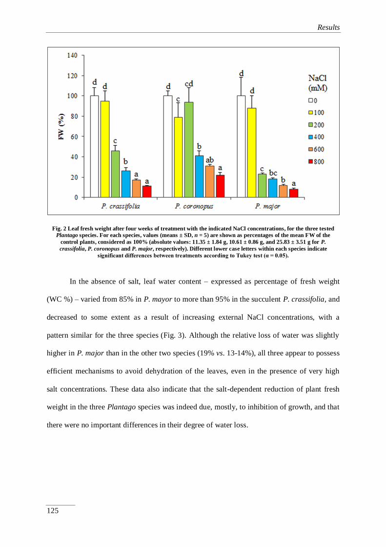
Results
125
Fig. 2 Leaf fresh weight after four weeks of treatment with the indicated NaCl concentrations, for the three tested
Plantago species. For each species, values (means ± SD, n = 5) are shown as percentages of the mean FW of the
control plants, considered as 100% (absolute values: 11.35 ± 1.84 g, 10.61 ± 0.86 g, and 25.83 ± 3.51 g for P.
crassifolia, P. coronopus and P. major, respectively). Different lower case letters within each species indicate
significant differences between treatments according to Tukey test (α = 0.05).
In the absence of salt, leaf water content – expressed as percentage of fresh weight
(WC %) – varied from 85% in P. mayor to more than 95% in the succulent P. crassifolia, and
decreased to some extent as a result of increasing external NaCl concentrations, with a
pattern similar for the three species (Fig. 3). Although the relative loss of water was slightly
higher in P. major than in the other two species (19% vs. 13-14%), all three appear to possess
efficient mechanisms to avoid dehydration of the leaves, even in the presence of very high
salt concentrations. These data also indicate that the salt-dependent reduction of plant fresh
weight in the three Plantago species was indeed due, mostly, to inhibition of growth, and that
there were no important differences in their degree of water loss.

Results
126
Fig. 3 Leaf water content (%) after four weeks of treatment with increasing NaCl concentrations, in the three selected
Plantago species (n = 5).
4.3.3.3. Effects of salt stress on ion accumulation
One common mechanism of salt tolerance in dicotyledonous halophytes is based on
the transport of toxic Na+ and Cl
- ions to the leaves, where they accumulate in the vacuoles to
avoid their deleterious effects in the cytosol, according to the so-called 'ion
compartmentalisation hypothesis' (Flowers et al., 1977; Wyn Jones et al., 1977; Glenn et al.,
1999). Sodium levels in the leaves of NaCl-treated plants showed a concentration-dependent
increase in the three species, although the absolute value reached in P. major in the presence
of the highest salt concentration tested, 800 mM NaCl, (ca. 1.3 mmol/g DW) was less than
half of those measured in the more tolerant taxa (2.8 – 3.0 mmol/g DW) (Fig. 4A). It should
be mentioned, however, that Na+ contents in P. crassifolia and P. coronopus untreated
controls were relatively high, over 1 mmol/g DW, as compared to ca. 250 µmol/g DW in P.
major; as a consequence, when comparing leaf Na+ levels at 800 mM external NaCl with the
corresponding controls, the relative increase was higher in P. major (ca. 5.3-fold) than in P.
crassifolia (2.7-fold) or P. coronopus (2.1-fold) (Fig. 4A). The patterns of Cl- accumulation
in leaves and the absolute values reached at the highest salt concentrations tested were similar
to those of Na+, in the three species; the only remarkable difference was that only P.

Results
127
crassifolia – but not P. coronopus – showed high Cl- contents in the control plants not
subjected to salt stress (Fig. 4B).
Fig. 4 Leaf contents of (A) sodium (Na+), (B) potassium (K+), and (C) chloride (Cl-), and (D) K+/Na+ ratios, in the
selected Plantago species, after four weeks of treatment with the indicated NaCl concentrations (means ± SD, n = 5).
Different lower case letters within each species indicate significant differences according to Tukey test (α = 0.05).
An increase in Na+ contents is generally accompanied by a concomitant decrease in
K+ levels, as both cations compete for the same transport systems (Rodriguez-Navarro and
Rubio, 2006; Adams et al., 2014;). This general trend was observed in the three Plantago
species, but with clear differences depending on their relative stress tolerance. In P.
crassifolia and P. coronopus, a reduction of the mean K+ levels was observed at low and
moderate NaCl concentrations, to increase again under high salinity conditions; P. major, on
the other hand, showed relatively higher K+ contents in the absence of salt or below 400 mM
NaCl, and a significant decrease was only observed at higher salt levels. In any case,
variation of K+
concentrations in the plants was relatively small, with all values included in
the range 0.4 – 1.0 mmol/g DW (Fig. 4C). The relative levels of K+
and Na+
in each species

Results
128
led to two clearly different patterns of variation in K+/Na
+ ratios. In control P. major plants,
K+/Na
+ was found to be quite high (ca. 3.5) and this value was progressively reduced, in
parallel with the increase of salt concentrations, to 0.4 – that is, almost 9-fold – in the
presence of 800 mM NaCl. In P. crassifolia and P. coronopus, K+/Na
+ ratios were maintained
below 0.5, with a reduction to about 50% of the controls in all salt treatments (Fig. 4D).
4.3.3.4. Salt stress-induced osmolyte accumulation
The levels of the commonest osmolytes in plants – proline (Pro), glycine betaine (GB)
and total soluble sugars (TSS) – were determined in plant leaves after the salt treatments were
concluded. Pro contents showed no significant increase over the non-stressed controls in the
presence of NaCl up to 400 mM, in all three Plantago species. Yet stronger salt stress
conditions triggered the accumulation of this osmolyte in P. crassifolia, at levels ca. 16-fold
higher than in the control plants, in the presence of 800 mM NaCl; the induction of Pro
biosynthesis was even stronger in P. coronopus (90-fold), while no increase over the control
was detected in P. major under the same conditions (Fig. 5A). It should be mentioned that,
despite these strong relative increases in Pro contents in the most salt-tolerant species, even
the highest absolute values reached, which were all below 50 µmol/g DW, are too low to
contribute significantly to osmotic balance in the salt-stressed plants. The same is true for
GB, which accumulated in the leaves of Plantago plants to maximum concentrations of
around 20 µmol/g DW (Fig. 5B), although with differences in the control levels and the
patterns of accumulation: in P. crassifolia and P. major, GB synthesis was induced already in
the presence of 100 mM NaCl and the contents of the osmolyte did not increase significantly
at higher salt concentrations; in P. coronopus, GB levels increased more progressively, but
only at and above 400 mM NaCl (Fig. 5B). Total soluble sugars levels decreased moderately
in P. coronopus and P. crassifolia with increasing NaCl concentrations, while no significant

Results
129
change was detected in P. major (Fig. 5C). It appears, therefore, that these compounds do not
play any role as osmolytes in the mechanisms of salt tolerance in the investigated Plantago
species.
Fig. 5 Leaf contents of (A) proline (Pro), (B) glycine betaine (GB), and (C) total soluble sugars (TSS) in the selected Plantago species, after four weeks of treatment with the indicated NaCl concentrations (means ± SD, n = 5). Different
lower case letters within each species indicate significant differences according to Tukey test (α = 0.05).

Results
130
The spectrophotometric assay used to measure total soluble sugars does not detect
polyalcohols (Ashwell, 1957) such as sorbitol (Sor), which has been identified as the major
osmolyte in all Plantago species investigated to date (Ahmad et al., 1979; Jefferies et al.,
1979; Lambers et al., 1981; Königshofer, 1983; Koyro, 2006; Flowers et al., 2010; Gil et al.,
2011). This has been confirmed in the present study, which shows that upon treatment with
NaCl, Sor levels – identified and quantified by HPLC – gradually increased in leaves of all
tested Plantago species, in a concentration-dependent manner (Fig. 6), although some
differences were observed among the three taxa. Sor contents in non-stressed P. crassifolia
plants (ca. 500 µmol/g DW) were about half of those measured in P. coronopus and P. major
controls, while the maximum level reached in the first species, in the presence of 800 mM
NaCl, was higher than in the other two. Therefore, the relative salt-induced increase in the
osmolyte concentration was highest in P. crassifolia: ca. 4.6-fold vs. 1.5-fold in P. coronopus
and P. major (Fig. 6). In any case, the absolute Sor concentrations were about three orders of
magnitude higher than those of Pro or GB, supporting the essential role of Sor in cellular
osmotic adjustment under salt stress conditions in Plantago.

Results
131
Fig. 6 Sorbitol (Sor) leaf contents after four weeks of treatement with the indicated NaCl concentrations, in the three
selected Plantago species (means ± SD, n = 5). Different lower case letters indicate significant differences between
treatments according to Tukey test (α = 0.05).
4.3.3.5. Effects of water stress on plant growth and biochemical stress markers
All the growth parameters, ion and osmolyte contents determined in leaves of
Plantago plants subjected to salt stress treatments were also measured in plants kept for four
weeks without irrigation. Drought also caused growth inhibition in the three species under
study (Fig. 7), although their relative tolerance to water stress was not so easy to estimate.
Significant reductions in the average number of leaves per plant, of ca. 50% in P. crassifolia,
46% in P. major, 20% in P. coronopus, were observed in water-stressed plants (Fig. 7A).
However, the decrease of fresh weight in relation to the corresponding controls was much
higher in P. coronopus (94%) or P. major (89%) than in P. crassifolia (64%) (Fig. 7B); yet
this FW reduction was not only due to inhibition of plant growth under drought conditions,
but also to loss of water, which was different for the different Plantago taxa: P. coronopus
and P. major stressed plants had a much lower percentage of water in the leaves than the non-
stressed controls, while water content did not change significantly in the most succulent P.

Results
132
crassifolia (Fig. 7C). When drought-induced growth inhibition was expressed in terms of dry
weight reduction, the values determined for P. crassifolia (ca. 40%), P. coronopus (63%) and
P. major (49%) were not so different.
No changes in leaf ion levels should be expected in the plants after the water stress
treatment, and this was generally true for K+, Cl
- and the K
+/Na
+ ratio in the three Plantago
species, as well as for Na+ in P. major. Yet, in P. crassifolia and P. coronopus, Na
+ contents,
which were already relatively high in the controls, increased significantly after four weeks
without watering the plants (Table 2).

Results
133
Fig. 7 Growth parameters after four weeks of water stress treatment, (A) number of leaves, (B) leaf fresh weight,
values are shown as percentages of the mean FW of the control plants, considered as 100% (absolute values: 11.35 ±
1.84 g, 10.61 ± 0.86 g, and 25.83 ± 3.51 g for P. crassifolia, P. coronopus and P. major, respectively), and (C) leaf water
content (%), in the studied Plantago species (means ± SD, n = 5). Asterisks indicate significant differences between
treatments, according to Tukey test (α = 0.05).
The drought-induced accumulation of osmolytes in the three species showed patterns
similar, but not identical to those observed in the presence of increasing salt concentrations
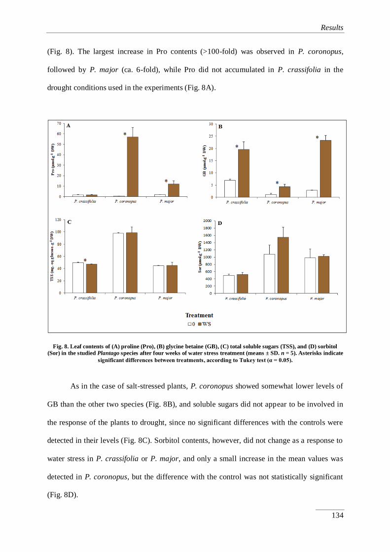
Results
134
(Fig. 8). The largest increase in Pro contents (>100-fold) was observed in P. coronopus,
followed by P. major (ca. 6-fold), while Pro did not accumulated in P. crassifolia in the
drought conditions used in the experiments (Fig. 8A).
Fig. 8. Leaf contents of (A) proline (Pro), (B) glycine betaine (GB), (C) total soluble sugars (TSS), and (D) sorbitol
(Sor) in the studied Plantago species after four weeks of water stress treatment (means ± SD. n = 5). Asterisks indicate
significant differences between treatments, according to Tukey test (α = 0.05).
As in the case of salt-stressed plants, P. coronopus showed somewhat lower levels of
GB than the other two species (Fig. 8B), and soluble sugars did not appear to be involved in
the response of the plants to drought, since no significant differences with the controls were
detected in their levels (Fig. 8C). Sorbitol contents, however, did not change as a response to
water stress in P. crassifolia or P. major, and only a small increase in the mean values was
detected in P. coronopus, but the difference with the control was not statistically significant
(Fig. 8D).

Results
135
4.3.4. Discussion
4.3.4.1. Effect of salt stress and drought on plant growth
Growth of all glycophytes and most halophytes is optimal in the absence of salt and is
progressively reduced by increasing salt concentrations; only in some highly tolerant
dicotyledonous halophytes, growth is stimulated by low or moderate salinity, although still
inhibited in the presence of salt above a higher, species-specific concentration threshold
(Flowers et al., 1986; Redondo-Gómez et al., 2006; 2010). Therefore, a quantitative
assessment of salt-induced growth inhibition in different species should provide a reliable
ranking of their relative degree of salt tolerance. This kind of analysis, applied to the three
Plantago species included in the present study, indicated that P. major is more sensitive to
salt than P. crassifolia and P. coronopus, the latter apparently being the most tolerant; this
would roughly agree with the distribution of the three taxa in nature: P. crassifolia and P.
coronopus are both halophytes, although adapted to different types of saline habitats, while P.
major is considered as a glycophyte, never found in natural saline environments. Yet,
quantitative differences in growth parameters of the three species were relatively small, and
P. major plants actually survived for one month the treatment with very high NaCl
concentrations. We have previously shown, in an independent study (Al Hassan et al., 2014),
that after two months exposure to NaCl up to 400 mM, P. major plants still produced some
fertile flowers that generated viable seeds. Considering the accepted definition of halophytes
as those plants able to complete their life cycle at soil salinity levels equivalent to at least 200
mM NaCl (Flowers and Colmer, 2008), P. major behaves as a moderate salt tolerant species,
rather than as a typical glycophyte. Nevertheless, as it will be discussed below, the present

Results
136
work revealed clear differences in the responses to salt stress of this species and its two more
resistant congeners.
Regarding the relative drought tolerance of the three Plantago species, one should
expect P. coronopus to be also the most tolerant. The responses to salinity and water stress
are similar in plants and are based on practically the same mechanisms, except those related
to ion transport. In the first phase of stress, plants exposed to saline conditions reduce their
growth due to the osmotic component of salt stress and only later, in a second stage, ions
accumulated in the leaves reach toxic levels which will produce premature senescence
(Munns, 2002). In addition, P. coronopus is often found on sand dunes and other dry-type
habitats, whereas the other two taxa grow in nature in more humid soils, either saline (P.
crassifolia) or not (P. major). Contrary to this expectation, in terms of reduction of fresh or
dry weight, P. crassifolia was found to be the most drought tolerant species, and P.
coronopus the most sensitive; this is probably due to the fact that the latter species was the
one that suffered the strongest dehydration (calculated as the loss of water content in leaves)
after one month without irrigation, while no significant reduction of water content was
observed in the succulent P. crassifolia under the same conditions. Interestingly, the three
Plantago species appear to possess efficient mechanisms to avoid salt-induced dehydration of
the leaves, even in the presence of very high concentrations (600 – 800 mM NaCl).
Soil salinity is considered a major restrictive factor for plant survival in nature, so that
only halophytes are adapted to highly saline soils, conditions that are lethal for non-tolerant
species. However, our results indicate that the degree of stress tolerance, per se, is not the
only factor – perhaps not even the most important factor – for the distribution of these species
in their natural environments. Other components, such as interspecies competition, may play
a fundamental role (Grigore et al., 2012). Many halophytes were probably refugees,
outcompeted by glycophytes from non-saline areas, which remained highly competitive

Results
137
under saline conditions suboptimal for non-tolerant species. The three taxa analysed here
grew better in the absence of salt, but all tolerated relatively high NaCl concentrations. Yet it
is likely that only P. crassifolia and P. coronopus are more competitive under saline
conditions, whereas P. major, with a faster growth rate, may be more competitive in the
absence of salt.
4.3.4.2. Effect of salt stress on ion accumulation
One basic response of glycophytes and monocotyledonous halophytes to cope with
high salinity in the soil is the limitation of sodium uptake. On the contrary, dicotyledonous
halophytes tend to accumulate toxic ions in the plants’ aerial parts, which are maintained at
low cytsolic concentrations by compartmentalisation in vacuoles. This is an advantageous
mechanism to increase osmotic pressure in foliar tissues, cheaper, in terms of energy
consumption, than the synthesis of organic solutes for osmotic adjustment (Raven, 1985). In
many salt-tolerant plants, Na+ concentrations are often well above 200 mM on a tissue-water
basis, concentrations that completely inhibit the activity of many enzymes in vitro (Munns
and Tester, 2008). In the present work, the highest levels of Na+ and Cl
- in leaves were found
in P. crassifolia, followed by P. coronopus, and the lowest in the less tolerant P. major. It has
been previously reported that non-halophytic Plantago species kept lower foliar Na+ levels
under saline conditions than the halophyte P. maritima (Könnigshofer, 1983). The ability of
P. maritima to accumulate sodium in its shoots was based on its higher capacity of Na+
translocation from the roots, rather than on a higher uptake rate, which was similar to that
found in other species of this genus (Erdei and Kuiper, 1979; Tánczós et al., 1981).
Differences in salt tolerance of Plantago species were also related to the efficiency of
intracellular ion compartmentalisation mechanisms; for example, Staal et al. (1991) reported
a considerably greater tonoplast Na+/H
+ antiporter activity under salt stress in the halophyte

Results
138
P. maritima than in the salt-sensitive P. media. The higher concentrations of Na+ and Cl
- we
have determined in leaves of P. crassifolia and P. coronopus, as compared to P. major, are
also related to the anatomic structure of the former species, with succulent leaves (especially
in P. crassifolia), and therefore with increased vacuole volume and better ion sequestration
capacity. Succulence, as well as excretion of Na+ and Cl
- by salt glands or bladders, is a basic
anatomic adaptation to salinity in some dicotyledonous plants (Flowers et al., 1986). Yet, the
most interesting information provided by these experiments is probably that the most tolerant
taxa accumulated relatively high concentrations of Na+ (and also of Cl
-, in the case of P.
crassifolia) in the leaves of control, non-treated plants, whereas the more salt sensitive P.
major did not. These data clearly suggest the existence in Plantago of constitutive
mechanisms of tolerance, based on the active uptake and transport of sodium to the leaves,
even under low salinity conditions, where the cation will be used as osmoticum – in addition
to the accumulation, if necessary, of organic osmolytes – in agreement with the halophytes’
'pre-adaptation to stress' hypothesis (Gong et al., 2005; Sanchez et al., 2008). The fact that
Na+ also seems to contribute to osmotic adjustment in water-stressed plants – a statistically
significant increase of Na+ levels was detected in P. crassifolia and P. coronopus, but not in
P. major, one month after watering was stopped – lends support to this idea.
The accumulation of sodium in plants is generally associated with a decrease of
potassium levels in the shoots. Since the two cations compete for the same binding sites, Na+
interferes with K+ transport into the cell by using its physiological transport systems (Flowers
et al., 1977; Greenway and Munns, 1980; Flowers et al., 1986). Moreover, sodium uptake
produces a depolarization of the plasma membrane causing the activation of outward-
rectifying K+ channels and consequently a K
+ loss (Shabala et al., 2003; 2005). These
processes lead to a significant reduction of K+/Na
+ ratios, and maintenance of relatively high
cellular K+/Na
+ values appears to be a relevant mechanism of salt tolerance (Maathuis and

Results
139
Amtmann, 1999). Although the three Plantago species somewhat followed this general trend,
here again there were clear differences between P. crassifolia and P. coronopus, on the one
side, and P. major, on the other. It is especially interesting to note the significant increase of
K+ levels observed in the more salt-resistant taxa at high external NaCl concentrations –
under lower salinity conditions, a decrease in K+ was detected, as expected – suggesting the
activation of potassium transport to the leaves, which could partly compensate the
accumulation of sodium, thus contributing to salt tolerance by avoiding a drastic reduction of
K+/Na
+ ratios. In the less tolerant P. major, K
+ contents in the absence of salt were higher
than in the other two taxa, and did not change significantly up to ca. 400 mM NaCl; at higher
salinity levels, however, a decrease of K+ levels was detected, leading to a sharp reduction of
the K+/Na
+ ratio.
4.3.4.3. Osmolyte synthesis
Environmental stress conditions leading to cellular dehydration, including salt and
water stress, trigger the cytosolic accumulation of different organic compatible solutes, or
osmolytes. The contribution of osmolytes to stress tolerance is not limited to their function in
osmotic adjustment, as they have multiple additional roles as osmoprotectants, directly
stabilising proteins and macromolecular structures under stress conditions, as ROS
scavengers or, in some cases, as signalling molecules involved in the induction of changes in
gene expression patterns (Munns, 2002; Flowers and Colmer, 2008; Munns and Tester, 2008;
Türkan and Demiral, 2009). In the genus Plantago, all available data indicate that the
polyalcohol sorbitol is the major functional osmolyte (Gorham et al., 1981; Koyro, 2006;
Flowers and Colmer, 2008; Flowers et al., 2010). Sorbitol accumulation in response to salt
treatments has been reported in the salt-tolerant P. maritima (Ahmad et al., 1979; Jefferies et
al., 1979; Königshofer, 1983) and P. coronopus (Gorham et al., 1981; Koyro, 2006), as well

Results
140
as in the salt-sensitive P. media and P. lanceolata (Königshofer, 1983). In one of our
previous studies on P. crassifolia, we confirmed accumulation of this compound in plants
growing in a littoral salt marsh in SE Spain, and showed how seasonal changes in sorbitol
levels correlated with the degree of environmental stress affecting the plants in their natural
habitat (Gil et al., 2011).
Sorbitol accumulated in response to salt stress, in the three species studied here. Yet,
the relative increases over the corresponding controls were very poor, since sorbitol
concentrations were already high in the absence of salt, especially in P. coronopus and P.
major (about 1 mmolg-1
DW). Therefore, these species appear to accumulate the osmolyte in
the absence of salt, so that a strong induction of its synthesis when the plants are actually
affected by high salinity conditions is not necessary, and the regulation of the response to salt
stress could be mostly based on changes in the intracellular localisation of sorbitol. This is
obviously also in agreement with the idea of a ‘pre-adaptation’ or ‘metabolic preparedness’ to
stress in tolerant species. In P. crassifolia, sorbitol content in the absence of salt was about
50% of that measured in P. coronopus or P. major – which could be compensated, in terms of
osmotic balance, by the higher levels of monovalent ions, Na+
and Cl- – and the maximum
absolute values measured at the highest NaCl concentration tested were somewhat higher;
therefore, the relative level of salt-dependent accumulation of sorbitol was higher in this
species. Regarding responses to drought, a slight increase in sorbitol levels after four weeks
of water stress was detected in P. coronopus, the species that appears to be most affected
under these conditions, in terms of growth inhibition and dehydration of the leaves, but no
significant drought-induced accumulation of sorbitol was detected in the other two Plantago
taxa.
Pro and GB are probably the commonest osmolytes in plants, used by many
angiosperm species to help maintain cellular osmotic balance. In a study on 51 halophytes

Results
141
sampled in their natural environment in an inland salt marsh in Turkey, Tipirdamaz et al.
(2006) found these two osmolytes as almost omnipresent solutes. In P. maritima they
detected even higher levels of Pro and GB than of sorbitol (which was also substantial),
despite the fact, mentioned above, that sorbitol is considered the major functional osmolyte in
the genus Plantago; this could be explained by biosynthesis of Pro and GB from O2-
dependent metabolic pathways that would be promoted in non-submerged salt marshes, while
sorbitol would accumulate under more anaerobic conditions, such as those present in marine
salt marshes (Tipirdamaz et al., 2006). Pro accumulation in salt-treated P. maritima plants, in
both roots and shoots, has also been recently reported (Sleimi et al., 2015). The concomitant
synthesis of different osmolytes has been previously observed in many halophytes (e.g.,
Briens and Larcher, 1982), although it is generally assumed that each species uses
preferentially one particular compound for osmotic balance under stress conditions. For
example, typical GB accumulators first accumulate this compatible solute, and only later-on,
when stress is more accentuated, they start to accumulate Pro (Stewart et al., 1979).
A similar behaviour has been observed in the most salt-tolerant Plantago species
included in the present study, P. crassifolia and P. coronopus, in which no significant Pro
accumulation was observed at salinity levels equivalent to those present in their natural
habitats, at or below 400 mM NaCl. Only at very high salt concentrations, 600 – 800 mM
NaCl, large relative increases of Pro contents were detected. In the more sensitive P. major,
no induction of Pro biosynthesis was observed, at any of the tested salt concentrations. Some
of these data confirm our previous Pro measurements performed in leaves from P. crassifolia
plants grown in the field (Gil et al., 2014). Accumulation of additional osmolytes under
increased levels of salt stress, for example Pro in the Plantago salt tolerant taxa, may be a
built-in mechanism which could enable halophytes to rapidly adapt to and withstand possible
increases in the degree of salt stress in their natural habitats, either short-term temporary

Results
142
changes in soil salinity or long-term increased salinisation, such as that that can be expected
as a consequence of climate change in salt marshes of the Mediterranean basin and other arid
and semiarid regions. The patterns of Pro accumulation in Plantago species under water
stress conditions also supported the notion that this 'secondary' osmolyte only accumulates
when the plants are strongly affected by stress: a very large increase in Pro contents (> 100-
fold over the control) was only detected in P. coronopus, the species more sensitive to
drought, but no increase at all was measured in the succulent P. crassifolia, which could very
efficiently avoid dehydration and was apparently little affected by the lack of irrigation.
It should be pointed out that the maximum Pro concentrations reached under the
strongest stress conditions tested are still too low – three orders of magnitude below those of
sorbitol – to contribute substantially to osmotic adjustment, even assuming that it
accumulated exclusively in the relatively small volume of the cytoplasm. The same could be
said of GB, which was found to slightly increase in the three investigated species, as a
response to both, salt and water stress, but to reach maximum absolute contents below those
of Pro, and much lower than those reported for plants that are true GB accumulators (e.g.,
Khan et al., 2000; Tipirdamaz et al., 2006; Gil et al., 2014). Nevertheless, it is likely that Pro,
and maybe also GB, play a significant role in the tolerance mechanisms of these species in
the presence of strong stress conditions. Yet these mechanisms would not involve
maintenance of cellular osmotic balance, but rather be based on the osmolytes function(s) as
a low-molecular-weight chaperons, ROS scavengers and/or signalling molecules (Ashraf and
Foolad, 2007; Chen and Murata 2008; Nawaz and Ashraf, 2010; Szabados and Savouré,
2010; Chen and Murata, 2011; Grigore et al., 2011).
4.3.5. Conclusions

Results
143
The comparative analyses shown here, regarding the responses to salt stress of three
Plantago species adapted to different natural habitats, support the notion that stress tolerance
in this genus is mostly based on transport of toxic ions to the leaves, their sequestration in the
vacuole, and accumulation of sorbitol in the cytoplasm for osmotic adjustment. The relative
tolerance of the three taxa is partly dependent on quantitative differences in the efficiency of
these processes – for example, Na+
and Cl- leaf contents are lowest in P. major, the most salt-
sensitive of the investigated taxa. Yet, there also exist specific mechanisms characteristic of
the more tolerant species, such as the capacity to use inorganic ions as osmotica, even under
low salinity conditions: Na+ and Cl
- in P. crassifolia, Na
+ in P. coronopus. In addition, these
salt-tolerant taxa are better adapted to withstand strong increases in soil salinity: in the
presence of very high NaCl concentrations, they accumulate relatively large amounts of a
secondary osmolyte (Pro), and appear to activate K+ transport to the leaves, thus avoiding a
drastic reduction in K+/Na
+ ratios. Inorganic and organic solutes are also involved in the
responses to water stress in Plantago, but in the succulent P. crassifolia – the most tolerant of
the selected species – the extreme resistance to dehydration, not shared by P. coronopus or P.
major, seems to be the critical factor contributing to drought resistance. Despite species-
specific quantitative differences, some of these response mechanisms appear to be
constitutive, as monovalent cations and sorbitol leaf contents may be relatively high even in
the absence of stress. Therefore, our results also support the hypothesis of a ‘pre-adaptation’
or ‘metabolic preparedness’ to stress in tolerant species of the genus Plantago.

Results
144
Acknowledgements
M.A.H. is a recipient of an Erasmus Mundus pre-doctoral scholarship financed by the
European Commission (Welcome Consortium). A.P. acknowledges the Erasmus fellowship
programme for funding her stay in Valencia to carry out her Master Thesis.
Author contributions
Analyses in plant material were performed by M.A.H. and A.P. (treatment
application, growth parameters, and osmolytes quantification), J.L. (ions), and M.-P.L.G.
(sorbitol measurement via HPLC). M.B. was responsible for the identification of plant
material and the co-supervision of plant-related work. Soil analyses were performed by
M.A.H. and A.P. Statistical analyses and preparation of figures were carried out by M.A.H.
and M.B., and O.V. was responsible for the general coordination of the project, the
supervision of the biochemical work and the preparation of the manuscript (with
contributions from M.A.H. and M.B. for the sections referring to botanical, soil and statistical
aspects).

Results
145
4.3.6. References
Adams, E., Shin, R. (2014). Transport, signaling, and homeostasis of potassium and sodium
in plants. Journal of Integrative Plant Biology, 56, 231-249.
Ahmad, I., Larher, F., Stewart, G.R. (1979). Sorbitol, a compatible osmotic solute in
Plantago maritima. New Phytologist, 82, 671-678.
Al Hassan, M., Pacurar, A., Gaspar, A., Vicente, O., Boscaiu, M. (2014). Growth and
reproductive success under saline conditions of three Plantago species with different
levels of stress tolerance. Notulae Botanicae Horti Agrobotanici Cluj-Napoca, 42,
180-186.
Ashraf, M., Foolad, M.R. (2007). Roles of glycine betaine and proline in improving plant
abiotic stress resistance. Environmental and Experimental Botany, 59, 206-216.
Ashwell, G. (1957). Colorimetric analysis of sugars. Methods in Enzymology, 3, 73-105.
Bartels, D., Sunkar, R. (2005). Drought and salt tolerance in plants. Critical Reviews in Plant
Sciences, 24, 23-58.
Bates, L.S., Waldren, R.P., Teare, I.D. (1973). Rapid determination of free proline for water
stress studies. Plant and Soil, 39, 205-207.
Boscaiu, M., Estrelles, E., Soriano, P., Vicente, O. (2005). Effects of the salt stress on the
reproductive biology of the halophyte Plantago crassifolia. Biologio Plantarum, 49,
141-143.
Boscaiu, M., Lull, C., Llinares, J., Vicente, O., Boira, H. (2013). Proline as a biochemical
marker in relation to the ecology of two halophytic Juncus species. Journal of Plant
Ecology, 6, 177-186.
Boyer, J.S. (1982). Plant productivity and environment. Science, 218, 443-448.

Results
146
Briens, M., Larher, F. (1982). Osmoregulation in halophytic higher plants: a comparative
study of soluble carbohydrates, polyols, betaines and free proline. Plant, Cell &
Environment, 5, 287-292.
Chater, A.O., Cartier, D. (1976). Plantago L. In: Tutin, T.G. et al. (Eds.), Flora Europaea 4.
Cambridge Univ. Press, Cambridge, pp. 38-44.
Chen, T.H.H., Murata, N. (2008). Glycinebetaine: an effective protectant against abiotic
stress in plants. Trends Plant in Science, 13, 499-505.
Chen, T.H.H., Murata, N. (2011). Glycinebetaine protects plants against abiotic stress:
mechanisms and biotechnological applications. Plant, Cell & Environment, 34, 1-2.
Dubois, M., Gilles, K.A., Hamilton, J.K., Reberd, P.A., Smith, F. (1956). Colorimetric
method for determination of sugars and related substances. Analytical Chemistry, 28,
350-356.
Erdei, L., Kuiper, P.J.C. (1979). The effect of salinity on growth, cation content, Na-uptake
and translocation in salt-sensitive and salt-tolerant Plantago species. Physiologia
Plantarum, 47, 95-99.
Flowers, T.J., Colmer, T.D. (2008). Salinity tolerance in halophytes. New Phytologist, 179,
945-963.
Flowers, T.J., Galal, H.K., Bromham, L. (2010). Evolution of halophytes: multiple origins of
salt tolerance in land plants. Functional Plant Biology, 37, 604-612.
Flowers, T.J., Hajibagheri, M.A., Clipson, N.J.W. (1986). Halophytes. The Quarterly Review
of Biology, 61, 313-335.
Flowers, T.J., Troke, P.F., Yeo, A.R. (1977). The mechanism of salt tolerance in halophytes.
Annual Review of Plant Biology, 28, 89-121.

Results
147
Gagneul, D., Aïnouche, A., Duhazé, C., Lugan, R., Larher, F.R., Bouchereau, A. (2007). A
reassessment of the function of the so-called compatible solutes in the halophytic
Plumbaginaceae Limonium latifolium. Plant Physiology, 144, 1598-1611.
Gil, R., Bautista, I., Boscaiu, M., Lidón, A., Wankhade, S., Sánchez, H., Llinares, J., Vicente,
O. (2014). Responses of five Mediterranean halophytes to seasonal changes in
environmental conditions. AoB Plants, 19;6. pii: plu049. doi: 10.1093/aobpla/plu049.
Gil, R., Boscaiu, M., Lull, C., Bautista, I., Lidón, A., Vicente, O. (2013). Are soluble
carbohydrates ecologically relevant for salt tolerance in halophytes? Functional Plant
Biology, 40, 805-818.
Gil, R., Lull, C., Boscaiu, M., Bautista, I., Lidón, A., Vicente, O. (2011). Soluble
carbohydrates as osmolytes in several halophytes from a Mediterranean salt marsh.
Notulae Botanicae Horti Agrobotanici Cluj-Napoca, 39, 9-17.
Glenn, E.P., Brown, J.J., Blumwald, E. (1999). Salt tolerance and crop potential of
halophytes. Critical Reviews in Plant Sciences, 18, 227-255.
Gong, O., Li, P., Ma, S., Rupassara, S.I., Bohnert, H.J. (2005). Salinity stress adaptations
competence in the extremophile Thellungiella halophila in comparison with its
relative Arabidopsis thaliana. Plant Journal, 44, 826-839.
Gorham, J., Hughes, L.Y., Wyn Jones, R.G. (1981). Low molecular weight carbohydrates in
some salt stressed plants. Physiologia Plantarum, 53, 27-33.
Greenway, H., Munns, R. (1980). Mechanisms of salt tolerance in non-halophytes. Annual
Review of Plant Biology, 31, 149-190.
Grieve, C.M., Grattan, S.R. (1983). Rapid assay for the determination of water soluble
quaternary ammonium compounds. Plant and Soil, 70, 303-307.

Results
148
Grigore, M.N., Boscaiu, M., Vicente, O. (2011). Assessment of the relevance of osmolyte
biosynthesis for salt tolerance of halophytes under natural conditions. The European
Journal of Plant Science and Biotechnology, 5, 12-19.
Grigore, M.N., Villanueva, M., Boscaiu, M., Vicente, O. (2012). Do halophytes really require
salts for their growth and development?. An experimental approach. Notulae Scientia
Biologicae, 4, 23-29.
Jefferies, R.L., Rudmik, T., Dillon, E.M. (1979). Responses of halophytes to high salinities
and low water potentials. Plant Physiology, 64, 989-994.
Khan, M.A., Ungar, I.A., Showalter, A.M. (2000). The effect of salinity on the growth, water
status and ion content of a leaf of a succulent perennial halophyte, Suaeda fruticosa
(L.) Forssk. Journal of Arid Environments, 45, 73-84.
Königshofer, H. (1983). Changes in ion composition and hexitol content of different
Plantago species under the influence of salt stress. Plant and Soil, 72, 289-296.
Koyro, H.W. (2006). Effect on salinity on growth, photosynthesis, water relations and solute
composition of the potential cash crop halophyte Plantago coronopus (L.).
Environmental and Experimental Botany, 56, 136-146.
Lambers, H., Blacuière, T., Stuiver, B. (1981). Interactions between osmoregulation and the
alternative respiratory pathway in Plantago coronopus as affected by salinity.
Physiologio Plantarum, 51, 63-68.
Maathuis, F.J.M., Amtmann, A. (1999). K+ nutrition and Na
+ toxicity: The basis of cellular
K+/Na
+ ratios. Annals of Botany, 84, 123-133.
Munns, R., Tester, M. (2008). Mechanisms of salinity tolerance. Annual Review of Plant
Biology, 59, 651-681.
Munns, R. (2002). Comparative physiology of salt and water stress. Plant, Cell &
Environment, 25, 239-250.

Results
149
Munns, R., Termaat, A. (1986). Whole-plant responses to salinity. Australian Journal of
Plant Physiology, 13, 143-160.
Nawaz, K., Ashraf, M. (2010). Exogenous application of glycinebetaine modulates activities
of antioxidants in maize plants subjected to salt stress. Journal of Agronomy and Crop
Science, 196, 28-37.
Pardo-Domènech, L., Tifrea, A., Grigore, M.N., Boscaiu, M., Vicente, O. (2015). Proline and
glycine betaine accumulation in succulent halophytes under natural and experimental
conditions. Plant Biosystems, DOI: 10.1080/11263504.2014.990943.
Pedrol, J. (2009). Plantago L. In: Benedi, C., Rico, E., Güemes, J., Herrero, A. (Eds.), Flora
Iberica XIII. Real Jardín Botánico, CSIC, Madrid, pp. 4-38.
Raven, J.A. (1985). Regulation of pH and generation of osmolarity in vascular plants: a cost-
benefit analysis in relation to efficiency of use of energy, nitrogen and water. New
Phytologist, 101, 25-77.
Redondo-Gómez, S., Wharmby, C., Castillo, J.M., Mateos-Naranjo, E., Luque, C.J., de Cires,
A., Luque, T., Davy, A.J., Figueroa, M.E. (2006). Growth and photosynthetic
responses to salinity in an extreme halophyte, Sarcocornia fruticosa. Physiologio
Plantarum, 128, 116-124.
Redondo-Gómez, S., Mateos-Naranjo, E., Figueroa, M.E., Davy, A.J. (2010). Salt stimulation
of growth and photosynthesis in an extreme halophyte, Arthrocnemum
macrostachyum. Plant Biology, 12, 79-87.
Rodriguez-Navarro, A., Rubio, F. (2006). High-affinity potassium and sodium transport
systems in plants. Journal of Experimental Botany, 57, 1149-1160.
Rønsted, N., Chase, M.W., Albach, D.C., Bello, M.A. (2002). Phylogenetic relationships
within Plantago (Plantaginaceae): evidence from nuclear ribosomal ITS and plastid
trnL-F sequence data. Botanical Journal of the Linnean Society, 139, 323-338.

Results
150
Rivas-Martínez, S., Rivas-Saenz, S. (1996-2009). Worldwide bioclimatic classification
system, Phytosociological Research Center, Spain. http://www.globalbioclimatics.org.
Accessed 1 July 2013.
Sanchez, D.H., Pieckenstain, F.L., Escry, F., Erbani, A., Kraemer, U., Udvardi, M.K., Kopka,
J. (2011). Comparative ionomics and metabolomics in extremophile and glycophytic
Lotus species under salt stress challenge the metabolic pre-adaptation hypothesis.
Plant, Cell & Environment, 34, 605-617.
Sanchez, D.H., Siahpoosh, M.R., Roessner, U., Udvardi, M., Kopka, J. (2008). Plant
metabolomics reveals conserved and divergent metabolic responses to salinity.
Physiologio Plantarum, 132, 209-219.
Shabala, L., Cuin, T.A., Newman, I.A., Shabala, S. (2005). Salinity-induced ion flux patterns
from the excised roots of Arabidopsis sos mutants. Planta, 222, 1041-1050.
Shabala, S., Shabala, L., Van Volkenburgh, E. (2003). Effect of calcium on root development
and root ion fluxes in salinised barley seedlings. Functional Plant Biology, 30, 507-
514.
Sleimi, N., Guerfali, S., Bankaji, I. (2015). Biochemical indicators of salt stress in Plantago
maritima: Implications for environmental stress assessment. Ecological Indicators,
48, 570-577
Staal, M., Maathuis, F.J.M., Elzenga, J.T.M., Overbeek, J.H.M., Prins, H.B.A. (1991).
Na+/H
+ antiport activity in tonoplast vesicles from roots of the salt-tolerant Plantago
maritima and the salt-sensitive Plantago media. Physiologio Plantarum, 82, 179-184.
Stewart, G.R., Larher, F., Ahmad, I., Lee, J.A. (1979). Nitrogen metabolism and salt
tolerance in higher plant halophytes. In: R. L.Jefferies, R.L. and Davy, A.J., (Eds.),
Ecological processes in coastal environments, Blackwell, Oxford, pp. 211-227.

Results
151
Szabados, L., Savoure, A. (2010). Proline: a multifunctional amino acid. Trends in Plant
Science, 15, 89-97.
Tánczos, O.G., Erdei, L., Snijder, J. (1981). Uptake and translocation of of sodium in salt-
sensitive and salt-tolerant Plantago species. Plant and Soil, 63, 27-32.
Tipirdamaz, R., Gagneul, D., Duhazé, C., Aïnouche, A., Monnier, C., Özkum, D., Larher, F.
(2006). Clustering of halophytes from an inland salt marsh in Turkey according to
their ability to accumulate sodium and nitrogenous osmolytes. Environmental and
Experimental Botany, 57, 139-153.
Türkan, I., Demiral, T. (2009). Recent developments in understanding salinity tolerance.
Environmental and Experimental Botany, 67, 2-9.
Vicente, O., Boscaiu, M., Naranjo, M.A., Estrelles, E., Bellés, J.M., Soriano, P. (2004).
Responses to salt stress in the halophyte Plantago crassifolia (Plantaginaceae).
Journal of Arid Environments, 58, 463-481.
Weimberg, R. (1987). Solute adjustments in leaves of two species of wheat at two different
stages of growth in response to salinity. Physiologia Plantarum, 70, 381-388.
Wyn Jones, R., Storey, R., Leigh, R.A., Ahmad, N., Pollard, A. (1977). A hypothesis on
cytoplasmic osmoregulation. In: Marre, E., O Ciferri, O., (Eds.), Regulation of cell
membrane activities in plants. Elsevier, Amsterdam, pp. 121-136.
Zhu, J.K. (2001). Plant salt tolerance. Trends in Plant Science, 6, 66-71.

Results
152
Tab. 1 Electric conductivity (dS/m) in 1:5 water extracts (EC1:5) of pot soil samples after salt and drought treatment
of the plants. Values shown are means ± standard deviations (n = 5); different lower case letters in each column
indicate statistically significant differences between treatments of the same species according to Tukey test (α = 0.05).
Treatment P. crassifolia P. coronopus P. major
Control 0.51 ± 0.04a 0.50 ± 0.04a 0.61 ± 0.04a
Water stress 0.56 ± 0.02a 0.46 ± 0.02a 0.68 ± 0.04a
100 mM NaCl 2.21 ± 0.13b 1.97 ± 0.12b 2.16 ± 0.11b
200 mM NaCl 3.97 ± 0.28c 4.11 ± 0.28c 4.06 ± 0.2c
400 mM NaCl 7.41 ± 0.38d 6.9 ± 0.55d 7.02 ± 0.56d
600 mM NaCl 8.45 ± 0.27e 8.47 ± 0.39e 8.59 ± 0.36e
800 mM NaCl 9.56 ± 0.66f 9.44 ± 0.39f 10.12 ± 0.72f

Results
153
Tab. 2 Levels of sodium (Na+), potassium (K+), chloride (Cl-), and potassium/sodium (K+/Na+) ratios in leaves of the
studied Plantago species, at the end of the water stress treatment (4 weeks) (means ± SD, n = 5). Different lower case
letters indicate significant differences between control and water-stressed samples, for each species, according to
Tukey test (α = 0.05).
Species Treatment Na
+ (µmol g
-
1 DW)
K+ (µmol g
-1
DW)
Cl- (µmol g
-1
DW) K
+/Na
+
P. crassifolia
Control 1099.07 ±
186.23a
438.62 ±
89.89a
1183.55 ±
260,56a 0.41 ± 0.14a
Water stress 1486.44 ±
174.83b
564.54 ±
99.26a
884.73 ±
117.60a 0.38 ± 0.09a
P. coronopus
Control 1343.94 ±
93.12a
659.84 ±
79.15b
516.53 ±
98.53a 0.49 ± 0.05a
Water stress 1636.43 ±
198.84b
395.70 ±
39.17a
554.87 ±
150.95a 0.44 ± 0.04a
P. major
Control 249.07 ±
38.42a
861.84 ±
79.90a
395.90 ±
10.58a 3.52 ± 0.66a
Water stress 236.12 ±
23.72a
955.40 ±
52.79a
443,55 ±
60,56a 4.06 ± 0.23a

Results
154
Publication IV:
Subchapter 4.4.
Growth and Reproductive Success under Saline
Conditions of Three Plantago Species with Different
Levels of Stress Tolerance
Reference:
Al Hassan, M., Pacurar, A., Gaspar, A., Vicente, O., Boscaiu, M. (2014). Growth and
reproductive success under saline conditions of three Plantago species with different levels of
stress tolerance. Notulae Botanicae Horti Agrobotanici Cluj-Napoca, 42(1), 180-186.

Results
155
Growth and Reproductive Success under Saline Conditions of Three Plantago Species
with Different Levels of Stress Tolerance
Abstract. Growth and responses to salt stress at reproductive stages were studied in three
Plantago species, two of them halophytes (P. coronopus and P. crassifolia) and one
glycophyte (P. major). Plants were treated with increasing NaCl concentrations (0, 100, 200
and 400 mM NaCl) under controlled conditions in the greenhouse. Besides fresh biomass,
several reproductive parameters were analysed: number of fertile/aborted spikes per plant,
length of scapes and spikes, mean number of seeds per plant and mean seed weight. Seeds of
those plants were germinated in controlled conditions, after which germination vigour,
germination rate and percentage, length of hypo-cotyledon, length of cotyledon, length of
roots, and angle of cotyledon were measured as well. All measured parameters were found to
change significantly in parallel to the increase of the salt treatment, also the more salt tolerant
species (P. major) was found to be more affected with this treatment.
Keywords: Plantago; cotyledon; flowering; halophytes; glycophytes; seed germination;
seedlings.
4.4.1. Introduction
Salinity is an important environmental factor that affects the distribution of plants in
their natural ecosystems and is one of the major threats for agriculture. According to recent
data (FAO, 2008) more than 800 million ha of land world-wide are affected by salinity. The
extension of saline soils is increasing approximately 1% each year due to poor rainfall,
evaporation of water from the soil, weathering of rocks, and anthropogenic alterations, such

Results
156
as irrigation with saline water, or abusive and indiscriminate use of fertilizers (Flowers et al.,
1995).
The immense majority of terrestrial plant species and practically all cultivated ones
belong to the category of glycophytes, or plants that cannot survive in conditions of saline
stress. On the contrary, the halophytes include species that remain viable and complete their
life cycle in the presence of salt concentrations of at least 200 mM NaCl (Flowers et al.,
1986). Halophytes represent a small group of plants, of only about 350 species belonging to a
few genera and families, adapted to saline ecosystems under harsh environmental conditions,
thus avoiding competition with glycophytes (Flowers et al., 1986).
Tolerance to salt stress is extremely complex and there are numerous interactions
between stress factors and molecular, biochemical and physiological processes affecting the
development of the plant (Ashraf and Foolad, 2007). However, most experimental studies on
mechanisms involved in salt tolerance are carried out on glycophytes, especially on model
plants. The limitations of these kinds of approaches and the necessity of increasing
ecophysiological research on halophytes have been recently discussed by Grigore et al.
(2011). Mechanisms of response to stress are similar in all plants, but their efficiency largely
varies among species, but only in halophytes they are efficient enough to confer salt
tolerance. Salt stress induces changes in the expression of genes encoding proteins involved
in numerous processes, but it is known that in halophytes such mechanisms are more efficient
and specialized. Differences between glycophytes and halophytes are the result of changes in
the regulation of same basic packages of genes (Zhu, 2000; 2001; Xiong and Zhu, 2002); thus
such differences are only quantitative and not qualitative (Greenway and Munns, 1980;
Flowers et al., 1986; Zhu, 2001; Vicente et al., 2004).
A very useful approach for a better comprehension of the concepts of tolerance vs.
adaption, which often are confused, is the use of species closely related taxonomically, hence

Results
157
genetically proximate, but with different degrees of stress tolerance. The genus Plantago with
more than 200 species and a total of 483 taxa (Tutel et al., 2005), out of which at least 20
species considered as halophytes, is one of the most suitable for such comparative studies.
Despite the relatively high number of papers dealing with one or several Plantago species,
there are no comparative studies on responses to saline stress at a crucial stage in plants life,
which is reproduction. Flowering, but mostly seed germination and seedling development
represent the bottle neck in the lifecycle of plants confronted to adverse environmental
conditions. Germination is strongly inhibited by high salinity even in most halophyte species
and, therefore plants adapted to saline environments developed strategies to postpone
germination till the rain season, when soil salinity is alleviated. A study on effects of salt
stress on germination in three Plantago halophytes (P. coronopus, P. crassifolia, and P.
microrrhiza) proved that that salinity strongly affects their seed germination, and that in all
three, there is an initial dormancy period, longer at higher temperatures (Luciani et al., 2001).
Dormancy is a common trend in halophytes, and actually seeds of numerous Mediterranean
halophytes germinate in autumn when soil salinity is alleviated by abundant rainfalls that
wash out salts.
Although there are numerous publications of physiological and biochemical responses
on species of the genus Plantago, only in P. crassifolia flowering and quality of seeds
produced by plants grown in salt stress conditions were analysed (Boscaiu et al., 2005).
The aim of this study was to achieve a better comprehension of the differences
between mere response to stress in the glycophyte Plantago major, and real adaption to stress
in the halophytes P. coronopus and P. crassifolia. The paper compares responses to saline
stress during growth and reproductive stage in the three species, but is focused mostly on
seed quality, as expression of the reproductive success of plants grown under saline

Results
158
treatments. Besides growth parameters and those related to seed production and seed quality,
also seedlings characteristics was thoroughly analysed by a computer programme.
4.4.2. Material and methods
Studied species
Plantago coronopus L. is a cosmopolitan annual or biennial, present in a wide range
of variable habitats, mostly on disturbed lands, waste places, or littoral sand dunes. It is
considered as an indicator of salinization of marginal soils, since it tolerates moderate salt
stress, its growth being even slightly stimulated by low saline concentration (Koyro, 2006).
On the other hand Luciani et al. (2005) found its seed germination as the most sensitive to
salt stress among the Plantago species they studied. Taxonomically it belongs to section
Coronopus (Lam &DC) Dietr. of the subgenus Coronopus (Lam. & DC.) Rahn, which
includes 11 Mediterranean species (Rahn, 1996).
Plantago crassifolia is a perennial halophyte, common in littoral salt marshes and
other saline habitats in the Mediterranean region. Previous studies on the species (Vicente et
al., 2004; Boscaiu et al., 2005) proved that salt tolerance of this species is moderate: its
optimal growth was in absence of salt and a drastic reduction of seed quantity and quality was
registered under high saline conditions. In natural environments the species appears only in
areas with moderate salinity, on salt marsh borders, but never in strongly saline areas
dominated by high tolerant halophytes. From the taxonomic point of view it is closely related
to previous species, belonging to the same section.
Plantago major L. is a perennial (seldom annual) glycophyte, with origins in Eurasia
and North Africa but naturalised throughout the world. It is much less stress tolerant than the
two previous species and requires moderate humidity. Although the species has some salt
Results
159
tolerance (Weber and Hanks, 2006), it cannot be included in the category of halophytes.
Taxonomically it is not so closely related to the two previous species, since it is placed in the
section Plantago of the subgenus Plantago, the largest within the genus, with 130 species
worldwide (Rønsted et al., 2003).
Plant growth and saline treatments
Seeds of the three species under study were sown in seed trays containing a mixture of
commercial peat and vermiculite mixture (3:1) and placed in a greenhouse with regulated
temperatures of 17-23 °C, photoperiod of 16 hours, and a humidity of 85%. Twenty days
after sowing young plants were transferred to individual pots on the same substrates. After
four weeks treatments with increasing salt concentrations (100, 200 and 400 mm NaCl) were
applied, maintaining a control watered with distilled water. The number of plants for each
treatment and species was of 3-5, and irrigation was carried out twice a week with a volume
of 1.5 l of salt solutions, and distilled water for control treatment respectively, for each tray
containing 12 standard 9 cm in diameter pots. After two months all plants were harvested and
vegetative parts individually weighed on a precision balance. Length of the longest leaf was
also measured.
Analysis of the reproductive traits of plants
At the end of treatments floral parts were separated, and for each plant the total
number of spikes was registered, and length of floral scape and spike measured. Seed set was
estimated by counting and weighing seeds from approximately half of the fertile spikes per
treatment and species.
Quality of seeds produced by the plants submitted to salt and control treatments was
tested through investigating the germination capacity of seeds. Four replicas of 25 seeds per

Results
160
species and treatment were placed in standard petri dishes with cotton and filter paper
moistened with 25 mL of milliQ water and sealed with parafilm to avoid evaporation and
incubated in a growth chamber, under a photoperiod of 16 hours, and a temperature of 25 °C.
Number of germinated seeds, considered upon radicle emergence, was counted daily for a
period of two weeks. Germination capacity was expressed as final percentage of germination
and germination rate as MTG (mean time of germination) according to the formula given
below (Ellis and Roberts, 1980): MGT= Σ D n/Σn.
Where, D are the days from the beginning of the germination test, and n is the number
of seeds newly germinated on day D.
The programme Image J was applied for the seedling analyses. Length of radical,
hypocotyle and of cotyledon leaves, and angle between the cotyledons were measured in 60
randomly selected seedlings per species and treatment. In addition, seedlings were weighed
on the precision balance. Seedling Vigour Index (SVI) was calculated by the formula by
Abdul-Baki and Anderson (1973):
SVI = Germination percentage x Mean root length (mm) + Mean hypocotyle length
(mm)
Since plants of P. coronopus watered without salt did not flower over the two months
treatment, seeds from initial stock were used as control for the germination assay and
seedlings measurements.
Statistical analysis
Data were analysed using SYSTAT v. 16. Levene test was applied to check whether
the requirements of the analysis of variance are accomplished. Germination percentages were
normalized by arcsine transformation prior to analysis of variance. Significance of
differences among treatments and among species was tested by applying one-way ANOVA.

Results
161
When the ANOVA null hypothesis was rejected, post-hoc comparisons were performed using
the Tukey test.
4.4.3. Results and Discussion
Sodium chloride treatments inhibited growth in a concentration-depended manner in
the three species under study. Maximal fresh weight (FW) was registered in the most
vigorous species P. major in control plants, but it decreased by 10 fold in plants from 400
mM NaCl treatment. Fresh weight of P. coronopus and P. crassifolia decreased as well in a
concentration depended manner, but only by 4 and 5.5 folds respectively (Fig. 1). This
finding is in perfect agreement with the ecological requirements of the three species, since the
most affected by salt is exactly the species that in its natural environment is never confronted
with salinity.
Fig. 1 Electric conductivity (EC1:5) of the substrate after two months of salt treatments.

Results
162
Another parameter used for the analysis of the effect of saline stress in is the length of
the longest leaf, which generally is affected by salt. When comparing plants from control to
those from the most saline treatment a 1.3-fold reduction was registered in P. coronopus, one
of 1.8 in P. crassifolia, and one of 1.7 in P. major (Fig. 2). Although salt clearly affected the
length of the longest leaf, there were no significant differences among species. Data related to
the vegetative growth are similar to previous reports in P. crassifolia (Vicente et al., 2004),
but no stimulation of growth by low salinity was detected in P. coronopus, as reported by
Koyro (2006).
Fig. 2 Fresh weight of plants of Plantago coronopus, P. crassifolia and P. major treated for two months with the
indicated NaCl concentrations. Same letters indicate homogeneous groups among treatments for each species.
Effects of salinity on floral characters were diverse among species. In P. coronopus
only plants from treatments with 200 and 400mM NaCl produced flowers (Tab. 1). In P.
crassifolia the mean number of spikes per plant increased in the treatments of 100 and 200
mM NaCl as reported in previous studies (Boscaiu et al., 2005, Grigore et al., 2012). At
concentration of 400 mM NaCl the average number of spikes was similar to that registered in
control (Tab. 1), whereas in a previous study (Boscaiu et al., 2005) after five months of

Results
163
treatment with salty at this concentration plants did not flower at all. Salinity can induce both,
inhibition or stimulation of flowering, early or delayed flowering, depending on the plant
species (Grigore et al., 2012). The strategy of survival through next generation occurs both
by early flowering (Blanvillain et al., 2011) and by increase of number of flowers produced
as we found in P. crassifolia and P. coronopus. However, the glycophyte P. major responded
completely different. In this species was registered a concentration dependent reduction of
spike numbers, of 5.6 –fold when comparing plants in control and those from 400 mM NaCl
treatment. An additional parameter, the size of the scape and spike itself was also measured
and it was found to significantly decrease as the concentration of salt treatment increased in
all species (Fig. 3). This finding proves that salinity may stimulate flowering, but it clearly
negatively affects reproduction in the three species under study, by diminishing the quality of
flowers produced under stress conditions. Perfectly correlated with the size of spikes was the
number of seeds produced. Significant differences among treatments were registered, with
the largest amount of seeds produced by plants from control in the three species. It is
interesting to note that P. major produces a considerably higher amount of seeds in
comparison with the other two species in all conditions. The mass of these seeds was also
measured, and it was evident that seeds coming from control plants had the highest mass, that
reduced with increasing salt concentrations in all species (Tab. 1).

Results
164
Fig. 3 Length of the longest leaf in the plants of Plantago coronopus, P. crassifolia and P. major treated for two months
at the indicated NaCl concentrations.
Germination capacity of seeds from all treatments was also tested on filter paper
moistened with distilled water, as shown in Methods. The percentage of germination, the
mean rate of germination (MGT) and the seedling vigour index (SVI) are summarised in
Table 1. There were no significant differences in the percentage of germination, whereas
MGT slightly increased in saline treatments, but again differences were not statistically
significant. On the contrary, the quality of seedlings was clearly affected by saline treatments.
Significant was the loss of vigour of seedlings (SVI) which decreased 1.4 folds in P.
coronopus, 2.5 folds in P. crassifolia, and 2.2 folds in P. major, when comparing seeds
coming from control treatment and those from 400 mM NaCl treatment. The weight of
seedlings followed the same patter, reducing with increasing salt concentrations (Tab. 1). In
P. crassifolia, seedlings coming from plants watered without salts weighed 20% more than
those from 400 mM treated plants; in P. coronopus such differences were much larger
(290%) and maximal in P. major (320%). This finding supports the idea that the most salt
Results
165
tolerant of the three species is P. crassifolia, which is minimally affected when compared
with the other two
The length of root, hypocotyle and cotyledon and the angle between the cotyeldons
was measured by the computer programme Image J. The roots length decreased 1.5-folds m
in P. coronopus, 2.63-folds in P. crassifolia, and 2.3 folds in P. major; the hypocotyle length
1.7- folds in P. coronopus, 1.4-folds in P. crassifolia, but almost 3-folds in P. major; the
cotyledon length two folds in P. coronopus, only slightly (1.22) in P. crassifolia, but more
than three folds in P. major, when comparing seedlings with origin in control and 400 mM
NaCl treatments (Fig. 4).
Although germination rate was high in P. major even for seeds with produced by
plants treated with salt, their seedlings showed only poor quality, and almost half of those
with origin in plants from the 200 and 400 mM NaCl treatments did not produced cotyledons,
their development being blocked after radicle emergence, as it can be seen in (Fig. 5). The
angle of the cotyledons (Tab. 1), increased with the increase of salt treatment mostly in P.
major.

Results
166
Fig. 4 Radicle, hypocotyle and cotyledon length reduction in 12 days-old seedlings obtained from seeds sampled from
the plants treated for two months at the indicated NaCl concentrations. Percentages were calculated in relation to the
controls' mean values which were considered 100%. Same letters indicate homogeneous groups among treatments for
each species.
The angle of the cotyledon may be considered as a predictor of growth rate. Lower et
al. (1978) reported that cucumber (Cucumis sativus) seedlings which displayed their
cotyledons in an upward position (smaller angle) grew faster than plants which displayed

Results
167
their cotyledons in a downward position (larger angle). An upward cotyledon angle was also
correlated with increase in plant height, node number, and fresh weight at later growth stages.
Fig. 5 Seedlings of Plantago coronopus (1), P. crassifolia (2) and P. major (3) obtained from the plants treated for two
months with distilled water (A), 100 mM NaCl (B), 200 mM NaCl (C) or 400 mM NaCl (D)
4.4.4. Conclusions
Salinity affected vegetative and reproductive traits analysed in the three species.
Plants produced under salt stress were less vigorous and their reproductive success was
clearly affected by salinity, but two different patterns were detected, one corresponding to the
halophytes P. coronopus and P. crassifolia and one to the glycophyte P. major. Fresh weight
in P. coronopus and P. crassifolia reduced with increasing salinity, but this reduction was
much stronger in P. major. Apparently salinity stimulated flowering in the halophytes, as
spikes were produced only in the 200 and 400 mM NaCl treatments in P. coronopus, and
their number increased in P. crassifolia in the 100 and 200 mM NaCl treatments in respect to
control. The more vigorous P. major produced more spikes, but their number decreased
according to the salt treatment. However, reproductive success was affected by salinity in the
three species, since seed set and mass of seeds was reduced. Viable seeds maintained their
germination capacity, as proved by high germination rates for all seeds, from all treatments,
but quality of seedling obtained from seeds harvested in plants submitted to salt treatment
was affected. Here again, P. major showed a different response: almost half of the seedlings

Results
168
with origin in the plants watered with high saline concentrations (200 and 400 mM NaCl),
blocked their development at very early stages. Although seedlings’ weight and size
decreased in saline conditions also in P. coronopus and P. major, reduction was much more
accentuated in P. major. Therefore, also this species may tolerate some salinity; it is obvious
that it behaves as a glycophyte, with a very strong inhibition of growth and reproduction in
condition of salt stress. When comparing the two halophytes, our findings support the idea
that P. crassifolia is more salt tolerant than P. coronopus, since its responses to salinity
especially at seedling level are leaser. This is also supported by ecological characteristics,
only P. crassifolia being able to inhabit typical saline areas as salt marshes.

Results
169
4.4.5. References
Abdul-Baki, A.A., Anderson, J.D. (1973). Relationship between decarboxilation of glutamic
acid and vigour in soybean seed. Crop Science, 13, 222-226.
Ashraf, M., Foolad, M.R. (2007). Roles of glycine betaine and proline in improving plant
abiotic stress resistance. Environmental and Experimental Botany, 59, 206-216.
Blanvillain, R., Wei, S., Wei, P., Kim, J.H., Ow, D.W. (2011). Stress tolerance to stress
escape in plants: role of the OXS2 zinc-finger transcription factor family. EMBO
Journal, 30, 3812-3822.
Boscaiu, M., Estrelles, E., Soriano, P., Vicente, O. (2005). Effects of the salt stress on the
reproductive biology of the halophyte Plantago crassifolia. Biologia Plantarum, 49,
141-143.
Ellis, R.A., Roberts, E.H. (1981). The quantification of ageing and survival in orthodox
seeds. Seed Science and Technology, 9, 373-409.
FAO (2008). Land and plant nutrition management service.
http://www.fao.org.ag/agl/agll/spush.
Flowers, T.J., Hajibagheri, M.A., Clipson, N.J.W. (1986). Halophytes. The Quarterly Review
of Biology, 61, 313-335.
Flowers, T.J., Yeo, A.R. (1995). Breeding for salinity resistance in crop plants: where next?.
Australian Journal of Plant Physiology, 2, 875-884.
Greenway, H., Munns, R. (1980). Mechanisms of salt tolerance in non-halophytes. Annual
Review of Plant Physiology, 31, 149-190.

Results
170
Grigore, M.N., Boscaiu, M., Llinares, J., Vicente, O. (2012). Mitigation of salt stress-induced
inhibition of Plantago crassifolia reproductive development by supplemental calcium
or magnesium. Notulae Botanicae Horti Agrobotanici Cluj-Napoca, 40, 58-66.
Grigore, M.N., Boscaiu, M., Vicente, O. (2011). Assessment of the relevance of osmolyte
biosynthesis for salt tolerance of halophytes under natural conditions. The European
Journal of Plant Science and Biotechnology, 5, 12-19.
Koyro, H.W. (2006). Effect of salinity on growth, photosynthesis, water relations and solute
composition of the potential cash crop halophyte Plantago coronopus (L.).
Environmental and Experimental Botany, 56, 136-146.
Lower, R.L., Pharr, D.M., Smith, O.S., Sox, H.N. (1978). Cotyledon angle as a predictor of
growth rate of cucumber plant. Euphytica, 27, 701-706.
Luciani, F., Cristaudo, A., Arico, D. (2001). Germination ecology of three Plantago L.
species living in a saline environment. Plant Biosystems, 135, 213-221.
Rahn, K. (1996). A phylogenetic study of the Plantaginaceae. Botanical Journal of the
Linnean Society, 120, 145-198.
Rønsted, N., Chase, M.W., Albach, D.C., Bello, M.A. (2002). Phylogenetic relationships within
Plantago (Plantaginaceae): evidence from nuclear ribosomal ITS and plastid trnL-F
sequence data. Botanical Journal of the Linnean Society, 139, 323-338.
Tutel, B., Kandemir, I., Kuş, S., Kence, A. (2005). Classification of Turkish Plantago L.
species using numerical taxonomy. Turkish Journal of Botany, 29, 51-61.
Vicente, O., Boscaiu, M., Naranjo, M.A., Estrelles, E., Bellés, J.M., Soriano, P. (2004).
Responses to salt stress in the halophyte Plantago crassifolia (Plantaginaceae).
Journal of Arid Environments, 58, 463-481.

Results
171
Weber, D.J., Hanks, J. (2006). Salt tolerant plants from the Great Basin Region of the United
States. In: Khan, M.A., Weber, D.J., editors. Ecophysiology of high salinity tolerant
plants. Dordrecht Netherlands: Springer Science + Business Media.
Xiong, L., Zhu, J.K. (2002). Salt-stress signal transduction. In: Scheel D, Wasternack C,
editors. Plant Signal Transduction, Frontiers in Molecular Biology Series. Oxford UK:
Oxford University Press, pp 165-197.
Zhu, J.K. (2001). Plant salt tolerance. Trends in Plant Science, 6, 66-71.
Zhu, J.K. (2002). Salt and drought stress signal transduction in plants. Annual Review of
Plant Biology, 53, 247-273.

Results
172
Tab 1. Effects of increasing saline solutions on the traits related to reproductive success in Plantago crassifolia, P.
coronopus and P. major. Mean ± SD values are shown. Same letters indicate homogeneous groups among treatments
for each species, n.d. not determined since plants did not flower, and * seeds from the initial stock
Character Species 0 mM NaCl 100 mM NaCl 200 mM NaCl 400 mM NaCl
Mean spike
no/plant
P. crassifolia 3.66 ± 1.15a 4.33 ± 1.52ab 6.66 ± 1.52b 3.33 ± 0.57a
P. coronopus n.d. n.d. 1.66 ± 0.57b 1.33 ± 0.57b
P. major 7.66 ± 3.21b 5.66 ± 1.15b 4.66 ± 2.08ab 1.33 ± 0.57a
Mean seed
no/spike
P. crassifolia 24.55± 9.02c
18.89 ± 7.52 b
11.32 ±3.18 a 7.09 ± 1.41a
P. coronopus n.d. n.d. 34.30 ± 8.04b
10. 00 ± 3.08a
P. major 28.34 ± 4.19d 23.33 ± 1.72c 17.54 ± 1.35b 8.81 ± 1.41a
Mean seed
weight/plant
(mg)
P. crassifolia 5.15 ± 2.42c 4.84 ± 1.5c 3.00 ± 0.63b 2.00 ± 0.38a
P. coronopus 6.70 ± 1.78b n.d. 6.70 ± 1.78b 1.62 ± 0.47a
P. major 10.42 ± 2.94 c 8.12 ± 1.38b 7.37 ± 0.9b 3.50 ± 0.75a
Percentage of
germination
P. crassifolia 88.71 ± 15.33c
72.83 ± 2.75b 70 ± 9.05b 48.66 ± 3.05a
P. coronopus n.d. n.d. 289.5±27.57b 41±29.69a
P. major 1369.20 ±
439b
1345.83 ±
230 b
1358.33 ±
281b
512.85 ±
291.0a
MGT (days)
P. crassifolia 0.49 ± 0.10c 0.49 ± 0.07c 0.41 ± 0.10b 0.39 ± 0.11a
P. coronopus 0.34 ± 0.02c* n.d. 0.27 ± 0.02b 0.17 ± 0.01a
P. major 0.39 ± 0.02c 0.33 ± 0.02b 0.28 ± 0.01a 0.26 ± 0.01a
Seedling weight
(mg)
P. crassifolia 98.40 ± 4.00b 84.00 ± 0.00a 81.00 ± 2.00a
89.00 ± 11.00a
P. coronopus 96.00 ±
4.60a* n.d.
99.00 ±
2.00ab 92.00 ± 0.00a
P. major 99.00 ± 2.00a 98.00 ± 4.00a 97.00 ± 6.00a 98.00 ± 2.30a
SVI
P. crassifolia 4.79 ± 0.60a 4.94 ± 0.20a 4.66 ± 0.40a 5.14 ± 1.10a
P. coronopus 2.92 ± 0.30a* n.d. 2.76 ± 0.40a 3.02 ± 0.10a
P. major 3.90 ± 0.20a 4.01 ± 0.10a 4.25 ± 0.14a 4.19 ± 0.20a
P. major 6.45 ± 0.22b 6.32 ± 0.01b 5.94 ± 0.12b 5.22 ± 0.26a
Cotyledon
angle (º)
P. crassifolia 5.07 ± 0.06c* n.d. 3.57 ± 0.01b 1.75 ± 0.02a
P. coronopus 3.01 ± 0.39b 2.18 ± 0.77ab 0.98 ± 0.36a 0.94 ± 0.38a
P. major 2438.352 1176.84 1109.7 954.08

Results
173
Publication V:
Subchapter 4.5.
Expression of the Vacuolar NA+/H+ Antiporter Gene
(NHX1) in Three Plantago Species Differing in Salt
Tolerance
Reference:
Al Hassan, M., Daniso, E., Martinelli, F., Boscaiu, M., Vicente, O. (2015). Expression of
the vacuolar Na+/H
+ antiporter gene (NHX1) in three Plantago species differing in salt
tolerance (In preparation).

Results
174
Expression of the Vacuolar NA+/H
+ Antiporter Gene (NHX1) in Three Plantago Species
Differing in Salt Tolerance
Abstract. Through homology we managed to successfully isolate and sequence the Na+/H
+
antiporter gene NHX1 of three species of the genus Plantago. Plantago crassifolia
(PcrNHX1), Plantago coronopus (PcoNHX1), and Plantago major (PmaNHX1). Translation
of the isolated and sequenced cDNAs showed that the NHX1 proteins comprised of 562, 561,
and 553 amino acid residues respectively. The amino acid sequences of the NHX1 gene in the
three studied species showed more than 92% complementarity in between each other’s and
68% with the previously isolated NHX1 gene from the glycophyte Arabidopsis thaliana
(AtNHX1). Change of expression of the sequenced genes and that of AtNHX1 by the effect of
salt stress was observed, after 2, 4, 8 and 24 hours of NaCl application (400 mM) at mRNA
level using qRT-PCR. The studies revealed a higher and more durable expression of the gene
under salt stress in the more tolerant P. crassifolia, while the least tolerant P. major had the
lowest level of NHX1 expression among the three studied species. A bioinformatics analysis
was carried out to study the theoretical secondary structures of the isolated sequences, as well
as generating a functional phylogenetic tree concerning a number of previously isolated
NHX1 proteins in glycophytes and halophytes, that placed our three de novo sequences in the
group to which most halophytes belong to.
Keywords: NHX1; Plantago coronopus; P. crassifolia; P. major; salinity.
4.5.1. Introduction
Plant growth and productivity are affected by a number of environmental factors,
among which salinity has been determined as the most damaging and limiting (Allakhverdiev

Results
175
et al., 2000). Worldwide more than 800 million ha of land is salt-affected (Rengasamy,
2006). And due to unsustainable irrigation practices about 1.6 million ha/year of irrigated
lands become saline and go out of production (secondary salinization) (Tanji, 2002).
The detrimental effects of salinity on plant growth are associated with: (1) altered
osmotic pressure (2) nutritional imbalance, (3) specific ion effect (ion toxicity, at high
concentrations, Na+ is toxic to plants because of its adverse effects on mineral and water
uptake, enzyme activities, photosynthesis and metabolism (Niu et al., 1995) or (4) a
combination of all mentioned factors (Ashraf, 1994).
According to their abilities to cope with salt stress, plants have been classified into
salt loving and tolerating halophytes and salt sensitive glycophytes. Halophytes have
managed to withstand the deleterious effects of salt through three mechanisms: (1) avoidance
(salt exclusion or the ability to exclude salts occurs through filtration at the surface of the
root, or salt excretion/extrusion through salt bladders like in Atriplex (Mozafar and Goodwin,
1970) and Mesembryanthemum crystallinum L. (Agarie et al., 2007) or through the cuticle
like in Tamarix (Evert, 2006), or most importantly by Compartmentation of ions in vacuoles),
(2) osmotic adjustment (the production of osmolytes to counter the osmotic pressure and
oxidative stress caused by the excess of salt in the soil (Boscaiu et al., 2013), (3) specificity
in ions uptake and distribution (increased specificity in ion pumps for the uptake of K+).
Although these mechanisms are also present in glycophytes, they are less efficient in
providing tolerance against high salt levels, and this could be due to anatomical and
morphological changes during evolution (Meychik et al., 2013), over expression of some
genes conferring salt tolerance, or the possibility that these genes are merely constitutively
expressed in halophytes, which is a matter of debate.
Na+/H
+ antiporters are functional proteins that play vital roles in conferring salt
tolerance in plants (Blumwald and Poole, 1985; Ballesteros et al., 1997). Two types of

Results
176
Na+/H
+ antiporters are present in plants, NHX1 located in the vacuole and SOS1 located in the
plasma membrane. The first functions in compartmentalizing Na+ in vacuoles that reduces the
toxicity of this ion in the cytosol and contributes to osmotic regulation (Takahashi et al.,
2009), while the other type of Na+/H
+ contributes in extruding Na
+ from cells (Blumwald et
al., 2000). These proteins have been the focus of a number of studies and have been found to
provide an increased tolerance to salinity when over expressed, whether the protein was
originally isolated from glycophytes (Apse et al., 1999) or from halophytes and finally
expressed in glycophytes like rice (Ohta et al., 2002).
To investigate whether those Na+/H
+ antiporters coming from halophytes induce more
tolerance to saline conditions than those isolated from glycophytes such as A. thaliana, we
decided to use species from the genus Plantago, out of which three were chosen as the focus
of our work, due to their different habitats, ecological requirements and salt tolerancy. The
genus Plantago includes more than 200 species (Tutel et al., 2005), of which at least 20
species are considered halophytes, is one of the most suitable for such studies.
The three species of our choice are P. crassifolia, P. coronopus, and P. major that
have been under the scope of our group´s work biochemically and physiologically under salt
and drought stress (Boscaiu et al., 2005; Al Hassan et al., 2014; Gil et al., 2014). Two of the
mentioned species are stress tolerant but are present in different ecological habitats, P.
crassifolia (sandy littoral soils and salt marshes), and P. coronopus (disturbed lands, waste
places, or littoral sand dunes). P. major which is less tolerant than the other two
aforementioned Plantago species is frequent in non-saline, humid areas, and therefore
considered as a glycophyte.
To understand the mechanisms of ion homeostasis in the salt-tolerant plants, we
isolated NHX1 cDNAs of the aforementioned species as well as that of the salt sensitive A.
thaliana (AtNHX1), and compared their expression under salt stress via RT-qPCR, as well as

Results
177
checking their theoretical secondary structures and their functional phylogenetic placement
among other isolated and sequenced NHX1s of halophytes and glycophytes.
4.5.2. Material and Methods
4.5.2.1. Plant Material
Seeds of P. crassifolia, and P. coronopus have been acquired from the Natural Park if
Albufera (Province of Valencia, Spain) and the seeds of P. major were bought from a
commercial firm (Spicegarden, Barcelona, Spain), while seeds of A. thaliana ecotype
Columbia-0 (Col-0), were acquired from the “Arabidopsis stock center”, University of
Nottingham, United Kingdom. Seeds (of Plantago species) were sterilized using hypochlorite
(diluted with water) prior to germination on petri dishes for 3 weeks at 25 ºC with 16 hours
photoperiod. Seedlings were transplanted onto a moistened mixture of peat (50%), perlite
(25%) and vermiculite (25%) in 1 liter pots; the substrate was kept moist using Hoagland´s
nutritive solution for another 3 weeks before the initiation of the treatments. Treatments were
performed by adding 200 ml of nutritive solution supplemented with 400 mM NaCl directly
on the pot, while those of control plants were irrigated with a similar volume with the
nutritive solution without any additions. Plants were harvested after the start of treatments by
2, 4, 8 and 24 hours. All experiments were conducted in a controlled environment chamber in
the greenhouse, under the following conditions: long-day photoperiod (16 hours of light),
temperature (23ºC during the day and 17
oC at night), CO2 level (≈ 300 ppm). Humidity
ranged between 50-80% during the time course of the treatments (4 weeks).
4.5.2.2. Gene Isolation and Cloning

Results
178
Fresh plant material was crushed utilizing liquid nitrogen and used for total RNA
extraction using the CTAB-PVP protocol (Gambino et al., 2008). After quantification and
checking of the RNA quality using nanodrop; Total cDNA synthesis was carried out using
Maxima First Strand cDNA Synthesis Kit for RT-qPCR. Oligios designed through homology
with cDNA of P. maritima ´s NHX1 gene that was previously sequenced (Staal et al. 1991),
were used to isolate the NHX1 gene of the species understudy. The sense primer PmNHX1F0
(5´-ATGGTGTTCGACTCAGAAACTATG-3´), and the antisense primer PmNHX1R0 (5´-
TCACTGCTTGGGTTCTTCAGGCCA-3´) were used to isolate the full length of the cDNA
of the three Plantago species that are studied in this work, while sense prime AtNHX1F0 (5´-
ATGTTGGATTCTCTAGTGTCG-3´) and antisense primer AtNHX1R0 (5´-
TCAAGCCTTACTAAGATCAGG-3´) were used to isolate the cDNA of NHX1 gene of A.
thaliana.
Isolated sequences were cloned in pGEM®-T Easy Vector and were sequenced by the
sequencing service in the IBMCP, UPV, Valencia, Spain. Alkaline lysis method (Brinboim
and Doly, 1979) was used for minipreps, to check for the presence of insert in the pGEM®-T
Easy Vector through digestion by restriction enzymes prior to sending to sequencing.
4.5.2.3. RT-qPCR Expression Assay
A fragment of β-Actin gene was used as housekeeping gene for RT-qPCR analysis
using sense oligio ActF1 (5´-TGTATGTCGCTATTCAGGC-3´) and antisense oligio ActR1
(5´- AGTAACCTCTCTCTGTCAG-3´), while the sense oligio AtNHX1F1 (5´-
GGAACCTTTGACTTGGGTGA-3´) and the antisense oligio AtNHX1R1 (5´-
ATCAAAGCTCTGAATCGCGT-3´) were used to study the expression of gene AtNHX1.
Expression of PcrNHX1, PcoNHX1, and PmaNHX1 through RT-qPCR was carried out using
sense primer PmNHX1F1 (5´-CAGTGGACATGTTGGCAACTTC-3´) and antisense primer

Results
179
PmNHX1R1 (5´-ATGGGAACTTTTACCCTGACTTATTAG-3´). The expression study was
carried out using SYBR® Green PCR Master Mix acquired from Thermo Fisher Scientific.
4.5.2.4. Bioinformatics Analysis
Protter programme was used to predict the secondary structure of the isolated proteins
(Omasits et al. 2014). Multiple sequence alignment and generation of a phylogenetic tree
were performed with ClustalΩ (Thompson et al., 1994) and TreeView software, respectively.
4.5.3. Results
Isolated cDNA sequences of the three Plantago species under study, were found to be
1686, 1683, and 1659 bp for PcrNHX1, PcoNHX1, and PmaNX1, respectively (sequences in
details, in Appendix). Deduced amino acids sequences were aligned and showed 92%
similarity in between the three aforementioned Plantago species, and 62% with AtNHX1 (Fig.
1).

Results
180
Fig. 1 Alignment of deduced NHX1 sequences of amino acid residues of AtNHX1, PcrNHX1, PmaNHX1, and
PcoNHX1. Symbol “*”, “:“, and “:“; Is used to denote a residue that is similar in all four, three, and two studied
species, respectively.
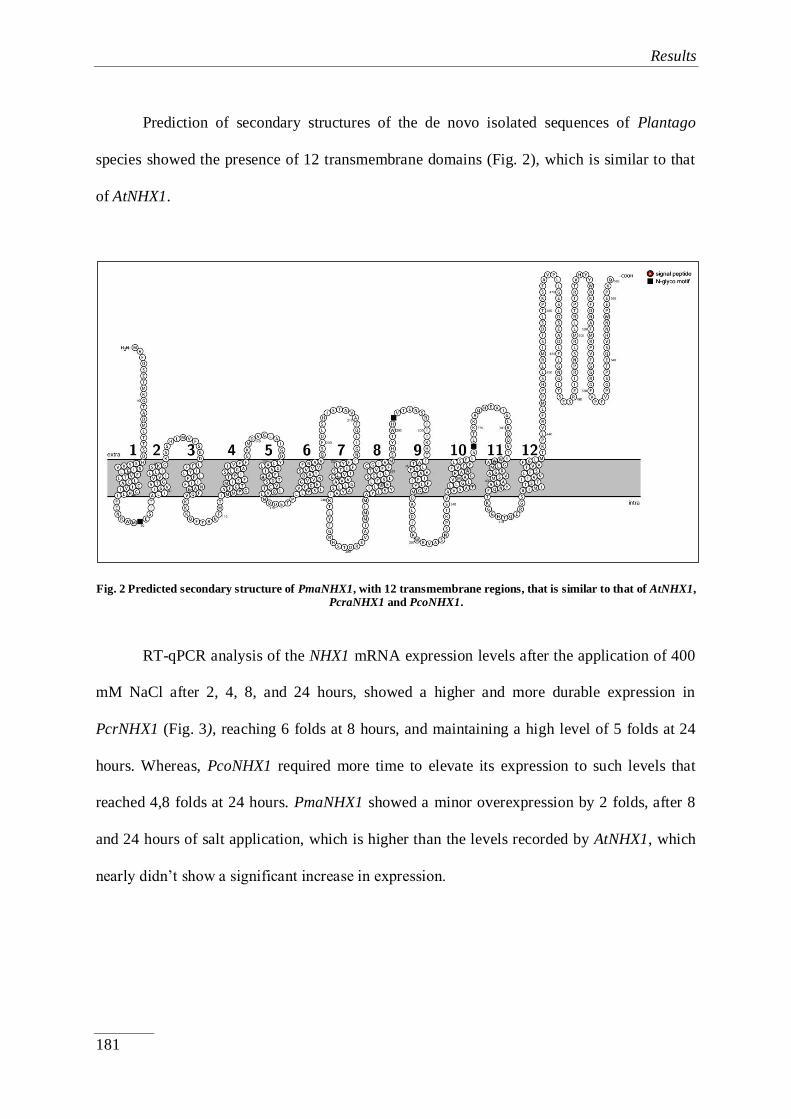
Results
181
Prediction of secondary structures of the de novo isolated sequences of Plantago
species showed the presence of 12 transmembrane domains (Fig. 2), which is similar to that
of AtNHX1.
Fig. 2 Predicted secondary structure of PmaNHX1, with 12 transmembrane regions, that is similar to that of AtNHX1,
PcraNHX1 and PcoNHX1.
RT-qPCR analysis of the NHX1 mRNA expression levels after the application of 400
mM NaCl after 2, 4, 8, and 24 hours, showed a higher and more durable expression in
PcrNHX1 (Fig. 3), reaching 6 folds at 8 hours, and maintaining a high level of 5 folds at 24
hours. Whereas, PcoNHX1 required more time to elevate its expression to such levels that
reached 4,8 folds at 24 hours. PmaNHX1 showed a minor overexpression by 2 folds, after 8
and 24 hours of salt application, which is higher than the levels recorded by AtNHX1, which
nearly didn’t show a significant increase in expression.

Results
182
Fig. 3 Fold change (ΔΔCT), in the expression of NHX1 after application of 400 mM NaCl, with respect to control
plants expression levels of the three Plantago species under study and A. thaliana.
Functional phylogenetic studies showed that the Plantago species understudy belong
to the same functional group containing most halophytes (Fig. 4), while A. thaliana´s NHX1
(AtNHX1) was found to belong to another, alongside crop species such as Zea mays and
Oryza sativa.

Results
183
Fig. 4 Neighbor-joining phylogenetic tree of Na+/H+ antiporter protein sequences. TreeView was used to do multiple sequence alignment and to generate phylogenetic tree. NHX1 sequences of Triticum aestivum (TaNHX1), Oryza sativa
subsp. Indica (OsNHX1), Zea mays (ZmNHX1), Plantago maritima (PmNHX1), Mesembryanthemum crystallinum
(McNHX1), Suaeda salsa (SsNHX1), Salicornia europaea (SeNHX1), Kalidium foliatum (KfNHX1), Oxybasis glauca
(OgNHX1), Atriplex dimorphostegia (AdNHX1), Atriplex gmelinii (AgNHX1), Gossypium hirsutum (GhNHX1), and
Citrus reticulata (CrNHX1), were used alongside AtNHX1, PcrNHX1, PcoNHX1, and PmaNHX1.
4.5.4. Discussion
Understanding the mechanisms underlying plant salt tolerance, is essential for
developing resistance in salt sensitive and affected crop species and thus crop yield
(Blumwald, 2000; Munns and Tester, 2008). This made the idea of exploring how halophytes
tolerate salt interesting, most importantly in part of the work to improve agricultural
production (Inan et al., 2004; Orsini et al., 2010) especially in light of the recent decade’s
climate change and increasing human population and demand on food.
In saline soils, where Na+ is the predominant toxic ion, excess accumulation of Na
+ in
cytosol is detrimental to many metabolic and physiological processes, vital for plant growth
and productivity, as it causes ion imbalance, hyper osmotic stress, and oxidative damage to
plants (Hasegawa et al., 2000). To deal with the influx of Na+ plants have evolved a number

Results
184
of sophisticated mechanisms that includes restricted uptake/exclusion of Na+ from cell, and
compartmentalization of Na+ into vacuoles.
Vacuolar Na+/H
+ antiporter NHX1 is responsible for the compartmentalization of
Na+ into vacuoles which not only provides an efficient mechanism to avert deleterious effects
of Na+ in cytoplasm, but also utilizes Na
+ in maintaining osmotic potential for driving water
into the cell (Blumwald et al., 2000; Hasegawa et al., 2000).
Vacuolar compartmentalization of Na+ is a critical process in salt adaptation,
conserved in both halophytes and glycophytes. NHX1 has been studied comparatively
concerning on functional and molecular levels between halophytes and glycophytes, which
were distinct and genetically unrelated (Li et al., 2008); A matter that we aimed to address
though the work at hand, via comparative studying of the NHX1 gene expression of Plantago
species with different salt tolerance levels.
Through the isolation and sequencing of NHX1 belonging to the Plantago species
under study, we conclude that the sequence and structure is highly conserved among those
species, even though there are some differences from AtNHX1. However, to investigate
differences on functionally on a structural level, we attempted to study the NHX1 gene from a
number of halophytes and glycophytes through the alignment and generation of a
phylogenetic tree. The results showed that most halophytes studied reside functionally
together (even though this group does contain some glycophytes as well). However, to
exclude the possibility of coincidence and evolution playing a role in this distribution, we
studied another gene in a similar manner (Rbcl) (Fig. 5).
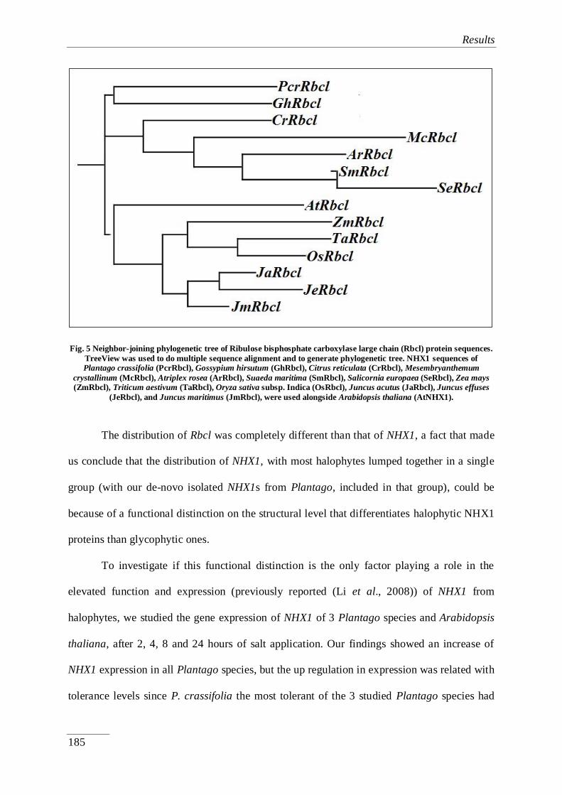
Results
185
Fig. 5 Neighbor-joining phylogenetic tree of Ribulose bisphosphate carboxylase large chain (Rbcl) protein sequences.
TreeView was used to do multiple sequence alignment and to generate phylogenetic tree. NHX1 sequences of
Plantago crassifolia (PcrRbcl), Gossypium hirsutum (GhRbcl), Citrus reticulata (CrRbcl), Mesembryanthemum
crystallinum (McRbcl), Atriplex rosea (ArRbcl), Suaeda maritima (SmRbcl), Salicornia europaea (SeRbcl), Zea mays
(ZmRbcl), Triticum aestivum (TaRbcl), Oryza sativa subsp. Indica (OsRbcl), Juncus acutus (JaRbcl), Juncus effuses
(JeRbcl), and Juncus maritimus (JmRbcl), were used alongside Arabidopsis thaliana (AtNHX1).
The distribution of Rbcl was completely different than that of NHX1, a fact that made
us conclude that the distribution of NHX1, with most halophytes lumped together in a single
group (with our de-novo isolated NHX1s from Plantago, included in that group), could be
because of a functional distinction on the structural level that differentiates halophytic NHX1
proteins than glycophytic ones.
To investigate if this functional distinction is the only factor playing a role in the
elevated function and expression (previously reported (Li et al., 2008)) of NHX1 from
halophytes, we studied the gene expression of NHX1 of 3 Plantago species and Arabidopsis
thaliana, after 2, 4, 8 and 24 hours of salt application. Our findings showed an increase of
NHX1 expression in all Plantago species, but the up regulation in expression was related with
tolerance levels since P. crassifolia the most tolerant of the 3 studied Plantago species had

Results
186
the highest increase in expression and the most durable, in reverse to the least tolerant studied
Plantago studied, P. major. AtNHX1 showed no significant increase within a day of salt
application, even though other studies showed an increase but after a longer period
(Sottosanto et al., 2007).
4.5.5. Conclusions
Na+ compartmentalization is a key strategy in plant´s salt stress resistance strategy, in
which NHX1 as a Vacuolar Na+/H
+ antiporter plays a major role. Our work showed that there
are some functional differences on the structural level between halophytes and glycophytes,
however it is not the only reason responsible for the difference in expression, since PcrNHX1
showed a higher expression than PmaNHX1, even though both have nearly identical
sequences. As such, we conclude that the main reason for higher and more efficient
expression and function of NHX1 in halophytes, maybe not due to structure or sequence of
the protein itself, but rather its activation, expression and folding machinery including
promotor, cis elements, transcription factors among others….

Results
187
4.5.6. References
Agarie, S., Shimoda, T., Shimizu, Y., Baumann, K., Sunagawa, H., Kondo, A., Ueno, O.,
Nakahara, T., Nose, A., Cushman, J.C. (2007). Salt tolerance, salt accumulation, and
ionic homeostasis in an epidermal bladder-cell-less mutant of the common ice plant
Mesembryanthemum crystallinum. Journal of Experimental Botany, 58(8), 1957-
1967.
Al Hassan, M., Pacurar, A., Gaspar, A., Vicente, O., Boscaiu, M. (2014). Growth and
reproductive success under saline conditions of three Plantago species with different
levels of stress tolerance. Notulae Botanicae Horti Agrobotanici Cluj-Napoca, 42,
180-186.
Allakhverdiev, S.I., Sakamoto, A., Nishiyama, Y., Inaba, M., Murata, N. (2000). Ionic and
osmotic effects of NaCl-induced inactivation of photosystems I and II in
Synechococcus sp. Plant Physiology, 123, 1047-1056.
Apse, M.P., Aharon, G.S., Snedden, W.A., Blumwald, E. (1999). Salt tolerance conferred by
over expression of a vacuolar Na+/H
+ antiport in Arabidopsis. Science, 285, 1256-
1258.
Ashraf, M. (1994). Organic substances responsible for salt tolerance in Eruca sativa. Biologia
Plantarum, 36, 255-259.
Ballesteros, E., Blumwald, E., Donaire, J.P. (1997). Na+/H
+ antiporter activity in tonoplast
vesicles isolated from sun flower induced by NaCl stress. Plant Physiology, 99, 328-
334.
Birnboim, H.C., Doly, J. (1979). A rapid alkaline extraction procedure for screening
recombinant plasmid DNA. Nucleic Acids Research, 7(6), 1513-1523.
Blumwald, E. (2000) Sodium transport and salt tolerance in plants. Current Opinion in Cell
Biology, 12(4), 431-4. doi: 10.1016/s0955-0674(00)00112-5.

Results
188
Blumwald, E., Aharon, G.S., Apse, M.P. (2000) Sodium transport in plant cells. Biochimica
et Biophysica Acta, 1465, 140-151. doi: 10.1016/s0005-2736(00)00135-8.
Blumwald, E., Poole, R.J. (1985). Na+/H
+ antiporter in isolated tonoplast vesicles from
storage tissue of Beta vulgaris. Plant Physiology, 78, 163-167.
Boscaiu, M., Estrelles, E., Soriano, P., Vicente, O. (2005). Effects of salt stress on the
reproductive biology of the halophyte Plantago crassifolia. Biologia Plantarum, 49,
141-143.
Boscaiu, M., Bautista, I., Lidón, A., Llinares, J., Lull, C., Donat, P., Mayoral, O., Vicente, O.
(2013). Environmental-dependent proline accumulation in plants living on gypsum
soils. Acta Physiologiae Plantarum, 35, 2193-2204.
Gambino, G., Perrone, I., Gribaudo, I. (2008). A rapid and effective method for RNA
extraction from different tissues of grapevine and other woody plants. Phytochemical
Analysis, 19, 520-525.
Gil, R., Bautista, I., Boscaiu, M., Lidon, A., Wankhade, S., Sa´nchez, H., Llinares, J.,
Vicente, O. (2014). Responses of five Mediterranean halophytes to seasonal changes
in environmental conditions. AoB PLANTS, 6: plu049; doi:10.1093/aobpla/plu049
Hasegawa, P.M., Bressan, R.A., Zhu, J.K., Bohnert, H.J. (2000). Plant Cellular and
Molecular Responses to High Salinity. Annual Review of Plant Physiology and Plant
Molecular Biology, 51, 463-99. doi: 10.1146/annurev.arplant.51.1.463
Inan, G., Zhang, Q., Li, P., Wang, Z., Cao, Z., et al. (2004). Salt cress. A halophyte and
cryophyte Arabidopsis relative model system and its applicability to molecular
genetic analyses of growth and development of extremophiles. Plant Physiology,
(135(3)):1718-37. doi: 10.1104/pp.104.041723.
Li, J., He, X., Xu, L., Zhou, J., Wu, P., Shou, H., Zhang, F. (2008). Molecular and functional
comparisons of the vacuolar Na+/H+ exchangers originated from glycophytic and
Results
189
halophytic species. Journal Zhejiang University Science B, 9(2), 132-140. doi:
10.1631/jzus.B0710445.
Meychik, N.R., Nikolaeva, Y.I., Yermakov, I.P. (2013). Physiological Response of
Halophyte (Suaeda altissima (L.) Pall.) and Glycophyte (Spinacia oleracea L.) to
Salinity. American Journal of Plant Sciences, 4, 427-435.
Mozafar, A., Goodwin, J.R. (1970). Vesiculated Hairs: A Mechanism for Salt Tolerance in
Atriplex halimus L.. Plant Physiology, 45, 62-65.
Munns, R., Tester, M. (2008). Mechanisms of salinity tolerance. Annual Review of Plant
Biology, 59, 651-81 doi: 10.1146/annurev.arplant.59.032607.092911.
Niu, X., Bressan, R.A., Hasegawa, P.M., Pardo, J.M. (1995). Ion homeostasis in NaCl stress
environments. Plant Physiology, 109, 735-742.
Ohta, M., Hayashi, Y., Nakashima, A., Hamada, A., Tanaka, A., Nakamura, T., Hayakawa,
T. (2002). Introduction of a Na+/H
+ antiporter gene from Atriplex gmelini confers salt
tolerance to rice. FEBS Letters, 532, 279-282.
Omasits, U., Ahrens, C.H., Müller, S., Wollscheid, B. (2014). Bioinformatics, 30(6), 884-6.
doi: 10.1093/bioinformatics/btt607.
Orsini, F., D'Urzo, M.P., Inan, G., Serra, S., Oh, D.H., et al. (2010). A comparative study of
salt tolerance parameters in 11 wild relatives of Arabidopsis thaliana. Journal of
Experimental Botany, (61(13)), 3787-98 doi: 10.1093/jxb/erq188.
Ray F. Evert (2006). Esau´s Plant Anatomy: Meristems, Cells and Tissues of the Plant Body:
Their Structure, Function and Development, Edition 3: John Wiley & Sons.
Rengasamy, P. (2006). World salinization with emphasis on Australia. Journal of
Experimental Botany, 57, 1017-1023.

Results
190
Sottosanto, J.B., Saranga, Y., Blumwald, E. (2007). Impact of AtNHX1, a vacuolar Na+/H+
antiporter, upon gene expression during short- and long-term salt stress in Arabidopsis
thaliana. BMC Plant Biology, 7: 18. doi: 10.1186/1471-2229-7-18
Staal, M., Maathuis, F.J.M., Elzenga, J.T.M., Overbeek, J.H.N., Prins, H.B.A. (1991). Na+/H
+
antiport activity in tonoplast vesicles from roots of the salt-tolerant plantago maritima
and the salt-sensitive plantago media. Physiologia Plantarum, 82, 179-184.
Takahashi, R., Liu, S., Takano, T. (2009). Isolation and characterization of plasma membrane
Na+/H
+ antiporter genes from salt-sensitive and salt-tolerant reed plants. Journal of
Plant Physiology, 166, 301-309.
Tanji, K.K. (2002). Salinity in the soil environment. In: Läuchli A, Lüttge U (ed) Salinity:
environment- plants- molecules. Kluwer Academic Publishers, Die Dordrecht pp: 21-
51.
Thompson, J., Higgins, D.G., Gibson, T.J. (1994). CLUSTAL W: improving the sensitivity
of progressive multiple sequence alignment through sequence weighting, position-
specific gap penalties and weight matrix choice. Nucleic Acids Research, 22, 4673-
4680.
Tutel, B., Kandemir, I., Kuş, S., Kence, A. (2005). Classification of Turkish Plantago L.
species using numerical taxonomy. Turkish Journal of Botany, 29, 51-61.

Results
191
Publication VI:
Subchapter 4.6.
Stress tolerance mechanisms in Juncus: Responses to
salinity and drought in three Juncus species adapted to
different natural environments
This manuscript has been submitted to the Journal; Functional Plant Biology, and is
currently under review.
Reference:
Al Hassan, M., Lopez-Gresa, M.P., Boscaiu, M., Vicente, O. (2015). Stress tolerance
mechanisms in Juncus: Responses to salinity and drought in three Juncus species adapted to
different natural environments (Under revision).
Results
192
Stress tolerance mechanisms in Juncus: Responses to salinity and drought in three
Juncus species adapted to different natural environments
Abstract. Comparative studies on the responses to salt and drought stress, regarding control
of ion transport and accumulation of specific osmolytes, have been carried out in three
Juncus species adapted to different natural habitats, two halophytes (J. maritimus and J.
acutus) and a salt-sensitive taxon (J. articulatus). This experimental approach was proven to
be successful for defining several mechanisms relevant for stress tolerance in this genus. Salt
tolerance in Juncus partly depends on the inhibition of transport of toxic ions to the aerial part
of the plants: in the three selected taxa, Na+ and Cl
- accumulated to the same extent in the
roots of salt treated plants, in parallel to increasing NaCl concentrations; however, ion
contents were lower in the culms and correlated with the relative salt sensitivity of the
species: the lowest levels were measured in the halophytes, whereas the highest were
determined in J. articulatus. Activation of K+ transport at high salt concentration also seems
to contribute to salt tolerance in the halophytes. Maintenance of cellular osmotic balance is
mostly based on the accumulation of soluble sucrose in the three species – together with
glucose and fructose in J. articulatus. Yet, neither the relative salt-induced increase in sugar
contents, nor the absolute concentrations reached in each species, can explain the observed
differences in salt tolerance. Proline contents, on the contrary, increased significantly –
almost 60-fold in the presence of 400 mM NaCl, as compared to the controls – only in the
salt-tolerant J. maritimus and J. acutus, but not in J. articulatus. Similar patterns of osmolyte
accumulation were observed in response to water stress. This results support a functional role
of proline in stress tolerance mechanisms in Juncus, probably not only contributing to
osmotic adjustment, but also because of its additional 'osmoprotectant' activities.
Keywords: Juncus; proline; osmolyte; salt stress; drought; ion transport.

Results
193
4.6.1. Introduction
Salinity, together with drought, is one of the most severe environmental stress factors
which shape the distribution of plant species in nature, and is also responsible for large losses
in crop production worldwide: accumulation of salts dissolved in irrigation water is leading to
the progressive ‘secondary’ – of anthropic origin – salinisation of arable land, mainly in arid
and semi-arid regions; this problem will worsen in the near future due to the forecasted
effects of climate change (Boyer, 1982; Bartels and Sunkar, 2005; Watson and Byrne, 2009;
IPCC, 2014; Fita et al., 2015). While all major crops and most wild species are relatively
sensitive to salt stress, some plants – the halophytes – have evolved different mechanisms
that allow them to withstand high salinity levels in their natural habitats. Halophytes have
been defined as those plants specific for saline environments that can complete their life cycle
in soils with salinity levels equivalent to, at least, 200 mM NaCl (Flowers et al., 1986;
Flowers and Colmer, 2008). They constitute a small fraction of angiosperm species, only
about 0.25%, and are widely distributed in different plant families and genera (Flowers et al.,
2010).
Their academic interest and practical implications for agriculture have prompted
intensive research, over the last decades, on the responses to salt stress and the mechanisms
of salt tolerance in plants. There is now overwhelming evidence that plants react to increased
soil salinity by activating a series of basic, conserved response pathways – which overlap
with the responses to other abiotic stresses, such as drought, cold or high temperatures. These
responses include the control of ion transport, compartmentalisation of toxic ions in vacuoles
and accumulation of osmolytes in the cytosol to maintain cellular osmotic balance, the
synthesis of ‘protective’ metabolites and proteins, or the activation of antioxidant systems

Results
194
(Flowers et al., 1986; Glenn et al., 1999; Hussain et al., 2008; Vinocur and Altman, 2005;
Zhu, 2001; Ozgur et al., 2013; Bose et al., 2014; Kumari et al., 2015; Volkov, 2015). These
basic mechanisms are not specific for halophytes, but shared by all plants, and their activation
does not necessarily lead to salt tolerance; in fact, as mentioned above, most plant species are
glycophytes; that is, salt sensitive. Therefore, salt tolerance, which varies widely in different
species, must depend on the relative efficiency of the aforementioned mechanisms of
response (Pang et al., 2010, Kumari et al., 2015). Moreover, there is no single halophytic
‘model species’, as different salt tolerant plants use different mechanisms to efficiently cope
with the deleterious effects of high soil salinity. Yet, the relative contribution of different salt
stress responses to salt tolerance in a given species – or in a group of related taxa – remains
largely unknown.
We believe that performing comparative studies on the responses to salt stress of
genetically related taxa with different degrees of tolerance – such as congener wild species
adapted to distinct habitats – will help to elucidate relevant salt tolerance mechanisms, by
correlating the relative salt tolerance of the species under study with salt-induced changes in
biochemical markers associated to particular response pathways.
The genus Juncus seems to be appropriate for this kind of comparative studies. It
includes more than 300 species, salt-sensitive and salt-tolerant (Wilson et al., 1993), growing
over a wide geographic range covering all continents (except Antarctica), and a spectrum of
ecological habitats extending from salt marshes for the most tolerant species, to humid non-
saline areas where more sensitive species of the genus flourish.
Three species adapted to different natural habitats were chosen for this study. J.
maritimus Lam. is a halophyte, common in temporarily flooded wetlands in the temperate
regions of the world, including the Mediterranean basin. J. acutus L. is a sub-cosmopolitan
species, that often coexist with J. maritimus but is common also on dunes, where water is the

Results
195
main limiting ecologic factor; it has been reported as less salt tolerant than J. maritimus
(Boscaiu et al., 2011; 2013). J. articulatus L. seems to be a much more sensitive species; it is
frequent in the northern hemisphere and in Australia, in different humid areas such as
wetlands, and also along the creeks and rivers (Albrecht, 1994). No previous study on the
stress tolerance of this Juncus species has been carried out, but it is never found in saline
habitats, (Chambers et al., 1995).
Regarding the taxonomic relation of the three Juncus species, J. acutus and J.
maritimus are recognised as close taxa belonging to the same subgenus (Juncus), whereas J.
articulatus was classified within the subgenus Septati Buchenau, section Ozophyllum Dumort
(Fernádez-Carvajal, 1981). Molecular systematic analysis confirmed that J. acutus and J.
maritimus are closely related, as inferred from DNA sequence data from the plastid rps16
intron, trnL intron, and trnL–F intergenic spacer (Jones et al., 2007). In a strict consensus tree
obtained from the 555 most parsimonious trees of the combined rbcL and trnL-F DNA
matrix, J. articulatus and J. maritimus were found to be more distantly related (Drábkóva et
al., 2006).
The major aim of this work was to correlate the relative salt tolerance of the
aforementioned Juncus species – established from their distribution in nature and by
measurements of salt-induced growth inhibition under controlled experimental conditions –
with specific responses based on the control of ion transport and the accumulation of different
osmolytes. Since the responses to drought and salinity seem to overlap, the analysis was
extended to plants subjected to water stress treatments, to check whether the same
mechanisms were responsible for the relative resistance of the analysed Juncus species to
both stresses. In line with the ideas discussed above, the results of this study should
contribute to our knowledge on the general mechanisms of stress tolerance in plants and,

Results
196
particularly, should help to distinguish those stress responses that are relevant for tolerance in
Juncus, from those that are not.
4.6.2. Material and Methods
4.6.2.1. Plant material and Experimental Design
Seeds of J. acutus and J. maritimus were harvested in a salt marsh located in ‘La
Albufera’ Natural Park (Province of Valencia, Spain), and those of J. articulatus in a non-
saline area of the same Natural Park. All seeds were sown directly into a moistened mixture
of peat (50%), perlite (25%) and vermiculite (25%) in 1 liter pots (Ø = 11 cm). During the
entire course of the germination process the substrate was kept moderately moist, using
Hoagland nutritive solution. Water and salt stress treatments were started 42 days after
germination. Salt stress experiments were performed with three different concentrations of
salt (100, 200 and 400 mM NaCl). The control plants were watered twice a week with
Hoagland nutritive solution (1.5 l for each tray, containing 12 pots), and salt stressed plants
with the same volume of nutritive solution containing NaCl, at the concentrations indicated
above. Drought treatments were performed by completely ceasing irrigation. All experiments
were conducted in a controlled environment chamber in the greenhouse, under the following
conditions: long-day photoperiod (16 hours of light), temperature (23oC during the day and
17oC at night), and a CO2 level of ca. 300 ppm. Humidity ranged between 50-80% during the
course of the treatments (eight weeks).
4.6.2.2. Soil analysis
Electrical conductivity (EC1:5) of the substrate was measured after eight weeks of
treatment. Soil samples were taken from five pots of each treatment, air-dried and then passed

Results
197
through a 2-mm sieve. A soil:water (1:5) suspension was prepared in deionised water and
mixed for one hour at 600 u/min, at room temperature. Electric conductivity was measured
with a Crison Conductivity meter 522 and expressed in dS/m.
4.6.2.3. Plant growth parameters
The following growth parameters were determined at the end of the stress treatments:
length of the longest culm, fresh weight (FW), dry weight (DW) and water content (WC %)
of the aerial parts of the plants. To obtain the water content, part of the fresh material was
weighed (FW), dried for four days at 65°C, until constant weight, and then weighed again
(DW); the water content percentage was calculated by the following formula: WC (%) =
[(FW – DW)/ FW] x 100.
4.6.2.4. Ion content measurements
Contents of potassium, sodium and chloride were determined in culms and roots of
the plants sampled after the stress treatments. Measurements were performed according to
Weimberg (1987), in aqueous extracts obtained by incubating the samples (0.15 g of dried
and ground plant material in 25 ml of water) for 1 h at 95ºC in a water bath, followed by
filtration through a filter paper (particle retention 8-12 µm). Sodium and potassium were
quantified with a PFP7 flame photometer (Jenway Inc., Burlington, USA) and chlorides were
measured using a Merck Spectroquant Nova 60®
spectrophotometer and its associated test kit
(Merck, Darmstadt, Germany).
4.6.2.5. Osmolyte quantification
Proline (Pro) content was determined in fresh plant material by the ninhydrin-acetic
acid method described by Bates et al. (1973). Pro was extracted in 3% aqueous sulfosalicylic

Results
198
acid, the extract was mixed with acid ninhydrin solution, incubated for 1 h at 95ºC, cooled on
ice and then extracted with two volumes of toluene. The absorbance of the organic phase was
measured at 520 nm, using toluene as a blank. Pro concentration was expressed as µmol g-1
DW.
Glycine betaine (GB) was determined in dried plant material, according to Grieve and
Grattan (1983). The sample was ground with 2 ml of Mili-Q water, and then extracted with 1,
2-dichlorethane; the absorbance of the solution was measured at a wavelength of 365 nm. GB
concentration was expressed as µmol g-1
DW.
Total soluble sugars (TSS) were quantified according to the method described by
Dubois et al. (1956). Dried material was ground and mixed with 3 ml of 80% methanol on a
rocker shaker for 24–48 h. Concentrated sulphuric acid and 5% phenol was added to the
sample and the absorbance was measured at 490 nm. TSS contents were expressed as ‘mg
equivalent of glucose’ per gram of DW.
4.6.2.6. HPLC analysis of carbohydrates
The soluble sugar fraction (mono and oligosaccharides) was analyzed using a Waters
1525 high performance liquid chromatography coupled to a 2424 evaporative light scattering
detector (ELSD). The source parameters of ELSD were the following: gain 75, data rate 1
point per second, nebulizer heating 60%, drift tube 50ºC, and gas pressure 2.8 Kg/cm2.
Analysis was carried out injecting 20 µL aliquots with a Waters 717 auto-sampler into a
Prontosil 120-3-amino column (4.6 x 125 mm; 3 µm particle size) maintained at room
temperature. An isocratic flux (1 mL/min) of 85% acetronitrile (J.T. Baker) during 25
minutes was applied in each run. Standards of glucose, fructose, and sucrose served to
identify peaks by co-injection. Sugars were quantified with peak integration using the Waters

Results
199
Empower software and comparison with glucose, fructose, and sucrose standard calibration
curves.
4.6.2.7. Statistical analysis
Data were analysed using the programme SYSTAT v. XVI. Before the analysis of
variance, the Shapiro-Wilk test was used to check for validity of normality assumption and
Levene’s test for the homogeneity of variance. If ANOVA requirements were accomplished,
the significance of the differences among treatments was tested by one-way ANOVA at a
95% confidence level and post hoc comparisons were made using the Tukey HSD test. All
means throughout the text are followed by SD.
4.6.3. Results
Effects of salt stress
4.6.3.1. Electrical conductivity of substrates
Electrical conductivity (EC1:5) was recorded in samples of the pot substrates after
eight weeks of salt and water stress treatments. For all species, a similar increase in EC1:5 was
detected in parallel to the increase of NaCl concentrations, reaching about 14 dS/m in the pots
watered with nutritive solution supplemented with 400 mM NaCl; this confirms the high
correlation between EC1:5 and the concentration of the saline solutions used in the treatments.
As expected, the water stress treatments did not modify the electrical conductivity of the
substrates in the pots, for any of the three studied Juncus species, as compared with the
corresponding controls (data not shown).

Results
200
4.6.3.2. Growth parameters
Fig. 1 Changes in (a) longest culm length (cm) (b) fresh weight (g), and (c) water content (%), in the three studied
Juncus species, after treatment for eight weeks with the indicated NaCl concentrations (means ± SD, n = 5). Different
letters indicate significant differences according to Tukey test (α = 0.05).

Results
201
Salt treatments inhibited growth of Juncus plants, in a concentration-dependent
manner, as shown by determination of several growth parameters (Fig. 1). For example, the
length of the longest culm decreased in J. articulatus and J. acutus by nearly 2-fold in the
presence of 400 mM NaCl, with respect to control plants. A slightly smaller decrease in culm
length (about 1.5-fold) was observed in J. maritimus under the same conditions (Fig. 1a).
Fresh weight also decreased in response to salt stress; the relative reduction of fresh weight in
the 400 mM NaCl treatment was similar for J. acutus and J. maritimus (65% and 70%,
respectively) but of more than 90% in J. articulatus (Fig. 1b), thus confirming that this
species is the most sensitive to salinity of the analysed Juncus taxa, as indicated by its
distribution in nature. Water contents decreased with increasing external salt concentrations,
from about 80% in control plants to 65%, approximately, in plants treated with 400 mM
NaCl, without significant differences detected in the three Juncus species under study (Fig.
1c). Therefore, the observed reduction of fresh weight is indeed due mostly to growth
inhibition, and not simply to loss of water under salt stress conditions.
4.6.3.3. Ions contents in roots
Na+
levels increased in the roots of the three Juncus species, in parallel to increasing
salt concentrations in the nutritive solution (Fig. 2a), reaching similar levels – between 3000
to 3500 µmol g-1
DW – in plants of the three taxa treated with 400 mM NaCl. A nearly
identical pattern of salt-induced Cl- accumulation in roots was also observed in all species,
reaching about 3300 µmolg-1
DW at the highest NaCl concentration tested (400 mM NaCl)
(Fig. 2b).

Results
202
Fig. 2 Ion contents (µmol g-1 DW): (a) Na+, (b) Cl-, (c) K+, and (d) K+/Na+ ratios, in the roots of the three Juncus
species under study, after eight-week treatments with the indicated NaCl concentrations (means ± SD, n = 5). For
each species, different letters above the bars indicate significant differences according to Tukey test (α = 0.05).
In general, K+
levels in roots did not vary significantly in response to the salt
treatments applied (Fig. 2c), although the concentrations measured in J. articulatus were
about half of those determined in J. acutus and J. maritimus. K+/Na
+ ratios in the roots of the
control plants were much higher in J. acutus and J. maritimus (> 2) than in J. articulatus
(about 0.5), and these values decreased in the presence of NaCl, in the three Juncus species
(Fig. 2d).

Results
203
Fig. 3 Ion contents (µmol g-1 DW): (a) Na+, (b) Cl-, (c) K+, and (d) K+/Na+ ratios, in the culms of the three Juncus
species under study, after eight-week treatments with the indicated NaCl concentrations (means ± SD, n = 5). For
each species, different letters above the bars indicate significant differences according to Tukey test (α = 0.05).
4.6.3.4. Ions contents in culms
Contrary to what was observed in the roots, where similar concentrations of Na+ and
Cl- were measured in the three Juncus species, accumulation of these ions in the culms
differed quantitatively in the three taxa, depending on their relative degree of salt tolerance.
Although Na+ and Cl
- levels increased in response to salt, in a concentration-dependent
manner, the highest contents were measured in J. articulatus, the most salt-sensitive of the
analysed taxa, while the lowest levels were detected in the most tolerant, the halophyte J.
maritimus (Figs. 3a, b). It should be pointed out that, in all cases, the absolute Na+ and Cl
-
concentrations reached were significantly lower in the aerial part than in the roots of the
plants, especially those of Na+, with the largest differences observed in the most tolerant
Juncus species (compare Figs. 2a, b with Figs. 3a, b).
Accumulation of K+ in culms, in the presence of increasing NaCl concentrations, also
showed different patterns depending on the relative tolerance of the species under study. In J.

Results
204
articulatus, K+ concentrations were higher than in the other taxa – and also almost 3-fold
higher than in J. articulatus roots – but did not change significantly with the different salt
treatments (Fig. 4c). In the halophytes J. maritimus and J. acutus, on the other hand, K+
contents in culms decreased at low salinity levels, with reference to non-treated control
plants, but increased again in the presence of high external NaCl concentrations (Fig. 3c).
K+/Na
+ ratios in the culms of the control plants were relatively high, between 10 and 20, but
dropped below 0.5 in the presence of NaCl (Fig. 3d).
4.6.3.5. Osmolyte contents
The levels of common osmolytes – proline, glycine betaine, total soluble sugars –
were determined in culms of the three Juncus species, after treatment with increasing NaCl
concentrations (Fig. 4). A significant, salt-induced accumulation of these compatible solutes
(which were present at similar concentrations in all control plants), was observed in all cases,
although with quantitative differences in the different taxa. Thus, a large increase in Pro
contents was detected in the halophytes J. acutus and J. maritimus upon the salt treatments,
reaching nearly 60-fold over the non-treated controls in the presence of 400 mM NaCl; under
the same conditions, Pro levels remained very low, increasing only 2-fold in the glycophyte
J. articulatus (Fig. 4a). This clearly different behaviour of the salt tolerant and salt sensitive
Juncus species was not observed for the other tested osmolytes, GB and TSS, which showed
similar salt-dependent accumulation patterns in the three taxa. Salt-treated J. acutus and J.
maritimus plants accumulated somewhat higher concentrations of GB and TSS, respectively,
and their levels were slightly lower in J. articulatus than in the halophytes (Fig. 4b, c), but
these differences were by far smaller than those observed in Pro contents.
HPLC fractionation of the extracts revealed three major peaks of soluble
carbohydrates, corresponding to glucose, fructose and sucrose (Fig. 5). All three sugars

Results
205
accumulated in the aerial part of salt-treated J. articulatus plants, reaching similar
concentrations (about 150 µmol g-1
DW) in the presence of 400 mM NaCl, the highest
concentration tested. In the halophytes J. acutus and J. maritimus a large increase in sucrose
contents – but not in those of glucose or fructose – was observed in response to the salt
treatments (Fig. 5)

Results
206
Fig. 4 Osmolyte contents: (a) proline (Pro), (b) glycine betaine (GB), and (c) total soluble sugars (TSS), in the culms of
the three Juncus species under study, after eight-week treatments with the indicated NaCl concentrations (means ±
SD, n = 5). For each species, different letters above the bars indicate significant differences according to Tukey test (α
= 0.05).

Results
207
Fig. 5 Levels of soluble sugars (µmol g-1 DW): (a) glucose, (b) fructose, (c) sucrose, in the culms of the three Juncus
species under study, after eight-week treatments with the indicated NaCl concentrations (means ± SD, n = 5). For
each species, different letters above the bars indicate significant differences according to Tukey test (α = 0.05).

Results
208
Effects of drought stress
The same parameters measured in salt-treated plants were determined as well in
Juncus plants subjected to a water stress treatments – by completely stopping watering – for
eight weeks. Drought also inhibited growth, as indicated by the reduction in the length of the
longest culm of the plants and, more clearly, by a strong decrease in the fresh weight of the
water-stressed plants as compared to the non-stressed controls (Table 1). According to this
criterion, the glycophyte J. articulatus is also the taxon most sensitive to drought, showing a
FW reduction of 97% after eight weeks without water (the corresponding values for J.
maritimus and J. acutus were 88% and 83%, respectively). These data suggested that the
effect of water stress on plant growth was stronger than that of salt stress at the highest NaCl
concentration tested. However, in this case the reduction of fresh mass was partly due to loss
of water, which ranged between 70% (in J. maritimus) and 90% (in J. acutus) (Table 1),
values much higher than those observed in salt-treated plants (Fig. 1).
Concerning osmolyte contents under water stress conditions, the accumulation
patterns of Pro, GB and TSS were similar to those observed in the presence of NaCl. Thus,
drought induced a strong increase in Pro levels in the halophytes, between 50 and 70-fold
higher than in the controls, reaching almost 200 µmol g-1
DW in the most tolerant J.
maritimus; in J. articulatus, the most stress-sensitive taxon, Pro levels remained very low,
with only a ca. 2-fold increase in the culms of the water-stressed plants (Fig. 6a). Water stress
also induced the accumulation of GB (Fig. 6b) and TSS (Fig. 6c), but to a much lesser extent,
between 2- and 3-fold over the controls, and without large differences among the three
Juncus species.
The drought-dependent increase in the levels of soluble sugars detected in all three
Juncus taxa was due to accumulation of sucrose, as demonstrated after the carbohydrates

Results
209
were separated and quantified by HPLC. Sucrose contents strongly increased in water-
stressed plants, reaching values of 160 – 180 µmol g-1
DW, without clear differences in the
different species (Fig. 6f). Contrary to what was observed in salt-treated plants, water stress
treatments did not induce the accumulation of glucose or fructose in J. articulatus; in fact,
there was a significant reduction in the levels of these two sugars after the drought treatment.
In the halophytes J. maritimus and J. acutus, either no significant changes or only small
reductions in the contents of glucose and fructose were detected (Fig. 6d, e).

Results
210
Fig. 6 Osmolyte accumulation: (a) proline (Pro), (b) glycine betaine (GB), (c) total soluble sugars (TSS), (d) glucose, (e) fructose, and (f) sucrose in the culms of the three Juncus
species under study, after eight weeks of water stress treatments (means ± SD, n = 5). Glucose, fructose and sucrose (panels d-f) were separated, identified and quantified by HPLC.
For each species, asteriks indicate significant differences between treatments according to Tukey test (α = 0.05).
Results
211
4.6.4. Discussion
The most general effect of stress on plants is inhibition of growth, as the plants
redirect their resources – metabolic precursors and energy – from primary metabolism and
biomass accumulation to the activation of specific defense mechanisms (Munns and Tester,
2008; Gupta and Huang, 2014). Accordingly, growth inhibition in the presence of salt has
been reported for all investigated species, halophytes and glycophytes alike, although
extremely salt-tolerant dicotyledonous halophytes may show a slight stimulation of growth at
low or moderate salt concentrations (Flowers et al., 1986). Some previous studies have been
published on the responses to salt stress of Juncus species, regarding seed germination,
vegetative plant growth or ion accumulation in the plants (Clarke and Hannon 1970; Rozema,
1976; Partridge and Wilson 1987; Espinar et al., 2005, 2006; Naidoo and Kift, 2006; Vicente
et al. 2007), but very few including different taxa of the genus (e.g., Rozema, 1976; Boscaiu
et al., 2011; 2013). To the best of our knowledge, no comparative analyses on the responses
to both, salinity and drought have been carried out on Juncus species adapted to different
natural habitats, such as those reported here. This experimental approach has provided novel
information on the mechanisms of stress tolerance in this genus, as discussed below.
Reduction of fresh weight in parallel with increasing external salinity appears to be a
reliable criterion to assess the relative salt tolerance of Juncus species, as previously
suggested (Rozema, 1976). According to our results, J. maritimus, considered as a typical
halophyte, is the most tolerant of the studied species, slightly more than J. acutus, which is
also a salt-tolerant species, often reported as subhalophyte (Boscaiu et al., 2011; 2013). Both
taxa are much more tolerant than J. articulatus, a species not investigated before, but that can
be clearly defined as a glycophyte. Thus, the responses to salt stress under controlled artificial
conditions closely correspond to the species natural distribution and their ecological optima.

Results
212
In the presence of salt, the decrease in water content of the aerial part of the plants was small,
and almost identical for the three species; therefore, the relative reduction of fresh weight was
mostly due to growth inhibition, indicating that the Juncus plants possess efficient
mechanisms to limit salt-induced dehydration, independently of their relative degree of salt
tolerance. Water stress, on the other hand, caused a stronger dehydration of the culms, but the
relative resistance of the investigated taxa to drought and salinity followed similar patterns,
with J. acutus and J. maritimus showing higher tolerance than J. articulatus.
Several previous studies, in which ion contents in different species growing in the
same saline habitat were measured, indicated that monocotyledonous halophytes are able to
exclude toxic ions (Na+ and Cl
-) from the aerial parts of the plant – while in dicotyledonous
salt-tolerant plants, the ions are efficiently transported to the leaves and stored at high
concentrations in the vacuoles (e.g., Albert and Popp 1977; Wyn Jones et al. 1977; Gorham et
al. 1980; Rozema 1991). Our results in Juncus are in agreement with those data. In the three
analysed species, Na+ and Cl
- contents increased in response to increasing NaCl
concentrations in the soil, both in roots and culms, but reaching higher absolute values in the
roots, in all cases. Most important, accumulation of the ions in the aerial parts of the plants
closely correlated with the relative sensitivity to salt stress of the three Juncus species: the
lowest levels were measured in the most tolerant species, J. maritimus, followed by J. acutus,
also tolerant halophyte, whereas the highest were determined in the glycophyte J. articulatus.
Therefore, inhibition of ion transport to the aerial parts is not a mere response to salinity in
Juncus, but must be relevant for salt stress tolerance in this genus. This process is not
controlled by differential ion uptake from the soil, but clearly at the level of transport from
the roots to the culms – since ion contents in the roots are similar in the three species – and
could be mediated by ion transporters of the HKT gene family, which seem to play an

Results
213
essential role in these Na+ exclusion mechanisms (Munns and Tester, 2008; Hamamoto et al.,
2015).
Sodium accumulation in plants is usually accompanied by a reduction in the
endogenous concentrations of potassium, as both ions compete for the same membrane
transporters (Niu et al. 1995; Rodriguez-Navarro 2000). This general reaction to salinity does
not seem to take place in Juncus, as no significant decrease in K+ levels was detected in the
roots of any of the three taxa, or in J. articulatus culms. The capacity to maintain K+
concentrations despite the progressive accumulation of toxic Na+ ions was considered by
Rozema (1976) as the basis of salt tolerance in halophytic species of this genus. Our results
indicate, on the contrary, that this mechanism cannot be relevant for tolerance, as it has been
observed also in the selected glycophyte, J. articulatus. The pattern of variation in K+
contents in the culms of the halophytes J. maritimus and J. acutus, in response to increasing
salinity, is also worth mentioning: K+ decreases at low external NaCl concentration, as
compared to the control, non-stressed plants, to increase again in the presence of higher salt
concentrations. It seems, therefore, that in the salt-tolerant Juncus taxa accumulation of Na+
at high levels activates transport of K+ from the roots to the aerial part of the plants, to limit
the reduction of K+/Na
+ ratios. This mechanism most likely contributes significantly to salt
tolerance in Juncus and, in addition, appears to be ecologically relevant. In a previous study
carried out in the field, in a littoral salt marsh near the city of Valencia (Gil et al., 2014), we
observed that K+ levels in culms of J. maritimus and J. acutus were higher in summer than in
spring, in parallel with a higher accumulation of Na+ (and Cl
-). In summer – normally the
most stressful season in the Mediterranean climate – we determined much higher soil salinity
(based on electric conductivity measurements), and Na+ and Cl
- levels than in spring, while
K+ contents in the soil remained very low and practically constant throughout the year.

Results
214
Osmolyte accumulation in the cytosol is also a general response to abiotic stress in
plants, and it is generally assumed that it contributes significantly to tolerance by
counteracting, at least partly, cellular dehydration caused by different stress conditions. In
addition to their function in osmotic adjustment, compatible solutes may play other important
roles in the mechanisms of stress tolerance, as low-molecular weight chaperones, ROS
scavengers or signalling molecules (Zhu, 2001; Chen and Murata, 2002; Ashraf and Foolad,
2007; Szabados and Savouré, 2010; Smirnoff and Cumbes, 1989; Grigore et al., 2011; Gil et
al., 2013). It has been reported that monocotyledonous halophytes accumulate preferentially
soluble carbohydrates (sugars and polyols) for osmotic balance (Gorham et al., 1980; Briens
and Larher, 1982). We have indeed detected a concentration-dependent increase in total
soluble sugars in response to the NaCl treatments, but reaching roughly the same levels in the
three Juncus species, irrespective of their relative salt tolerance. HPLC fractionation allowed
the identification of glucose, fructose and sucrose as the major sugars present in all Juncus
plants, as reported for J. maritimus and J. acutus grown in nature (Gil et al., 2011). However,
the Juncus halophytes and the glycophyte showed different patterns of sugar accumulation:
significant salt-dependent increases in glucose and fructose levels were only detected in J.
articulatus, while sucrose contents increased in the three taxa. The high sugar concentrations
measured should clearly contribute to osmotic adjustment in the presence of NaCl, thus
protecting the plants against the effects of salt stress. Yet, here again, it is important to point
out that there is no positive correlation between sugar contents and the relative degree of salt
tolerance of the Juncus taxa – actually, the combined concentrations of glucose, fructose and
sucrose were somewhat higher in the most salt-sensitive species, J. articulatus, than in the
halophytes. Therefore, differences in salt tolerance within the genus Juncus do not seem to be
due to differential accumulation of soluble carbohydrates.

Results
215
Proline is not generally considered as a preferential functional osmolyte in
monocotyledonous salt-tolerant plants, and the concentrations of free Pro measured in control
plants – around 2 µmol g-1
DW – were much lower than those of sugars. In salt-treated plants,
however, a large increase in Pro content was observed, up to 50 to 60-fold over the controls
in the presence of the highest NaCl concentration tested (400 mM), but only in the halophytes
J. maritimus and J. acutus. In the salt sensitive J. articulatus Pro levels increased only about
2-fold under the same conditions. This differential accumulation of this osmolyte in the culms
of Juncus plants, depending on the relative salt tolerance of the studied species, clearly
supports a functional role of Pro in the mechanisms of salt tolerance in this genus. Pro
probably participates in cellular osmotic adjustment under salt stress conditions, although it
reached maximum absolute levels lower than those of soluble sugars. Yet its contribution to
salt tolerance mechanisms is probably mediated, to a large extent, by its additional activities
as 'osmoprotectant' – low-molecular-weight chaperon and ROS scavenger (Szabados and
Savouré, 2010).
4.6.5. Conclusion
Salt tolerance in Juncus depends to a large extent on the partial inhibition of transport of toxic
ions (Na+ and Cl
-) from the roots to the plants aerial parts and on the activation of K
+
transport at high external salt concentrations (to limit the reduction of K+/Na
+ ratios). In
addition, the accumulation to relatively high levels of Pro in the culms of the plants is also
important for tolerance to both, salt and water stress, since it contributes to osmotic
adjustment but also because of the 'osmoprotectant' roles of this osmolyte. The efficiency of
these processes correlated positively with the relative tolerance of the investigated species,
and could be distinguished from other stress responses that were activated to a similar extent

Results
216
in the three Juncus taxa, irrespective of their relative tolerance to stress, and therefore could
not be directly involved in their mechanisms of tolerance to stress.

Results
217
4.6.6. References
Albert, R., Popp, M. (1977). Chemical composition of halophytes from the Neusiedler Lake
region in Austria. Oecologia, 27, 157-170.
Albrecht, D.E. (1994). Juncus. In Walsh, N.G. and Entwisle, T.J. (eds) Flora of Victoria Vol.
2: Ferns and Allied Plants, Conifers and Monocotyledons. Inkata Press, Melbourne, p.
224.
Ashraf, M., Foolad, M.R. (2007). Roles of glycine betaine and proline in improving plant
abiotic stress resistance. Environmental and Experimental Botany, 59, 206-216.
Bartels, D., Sunkar, R. (2005). Drought and salt tolerance in plants. Critical Reviews in Plant
Sciences, 24, 23-58.
Bates, L.S., Waldren, R.P., Teare, I.D. (1973). Rapid determination of free proline for water
stress studies. Plant and Soil, 39, 205-207.
Boscaiu, M., Ballesteros, G.,Naranjo, M.A.,Vicente, O., Boira, H. (2011). Responses to salt
stress in Juncus acutus and J. maritimus during seed germination and vegetative plant
growth. Plant Biosystems, 145, 770-777.
Boscaiu, M., Lull, C., Llinares, J., Vicente, O., Boira, H. (2013). Proline as a biochemical
marker in relation to the ecology of two halophytic Juncus species. Journal of Plant
Ecology, 6, 177-186.
Bose J, Rodrigo-Moreno A, Shabala S. (2014). ROS homeostasis in halophytes in the context
of salinity stress tolerance. Journal of Experimental Botany, 65, 1241-1257.
Boyer, J.S. (1982). Plant productivity and environment. Science, 218, 443-448.
Briens M, Larher F (1982) Osmoregulation in halophytic higher plants: a comparative study
of soluble carbohydrates, polyols, betaines and free proline. Plant, Cell and
Environment, 5, 287-292.

Results
218
Chambers, J.M., Fletcher, N.L., McComb, A.J. (1995). A guide to emergent wetland plants of
south Western Australia. Murdoch University, Marine and Freshwater Research
Laboratory, Perth, p. 61-65.
Chen, T.H.H., Murata, N. (2002). Enhancement of tolerance of abiotic stress by metabolic
engineering of betaines and other compatible solutes. Current Opinion in Plant
Biology, 5, 250-257.
Clarke, L.D., Hannon, N.J. (1970). The mangrove swamp and salt marshes communities of
the Sydney district. III: plant growth in relation to salinity and water-logging. Journal
of Ecology, 58, 351-369.
Drábková, L., Kirschner, J., Vlček, Č. (2006). Phylogenetic relationships within Luzula DC.
and Juncus L. (Juncaceae): a comparison of phylogenetic signals of trnL-trnF
intergenic spacer, trnL intron and rbcL plastome sequence data. Cladistics, 22, 132-
143.
Dubois, M., Gilles, K.A., Hamilton, J.K., Reberd, P.A., Smith, F. (1956). Colorimetric
method for determination of sugars and related substances. Analytical Chemistry, 28,
350-356.
Espinar, J.L., Garcia, L.V., Clemente, L. (2005). Seed storage conditions change the
germination pattern of clonal growth plants in Mediterranean salt marshes. American
Journal of Botany, 92(7), 1094-1101.
Espinar, J.L., García, L.V., Figuerola, J., Green, A.J., Clemente Salas, L. (2006). Effects of
salinity and ingestion by ducks on germination patterns of Juncus subulatus seeds.
Journal of Arid Environments, 66, 376-383.

Results
219
Fernandez-Carvajal, M.C. (1981). Revision del genero Juncus L. en la Peninsula Iberica. II:
subgéneros Juncus y Genuini Buchenau. Anales del Jardín Botánico de Madrid.
Madrid, (38-2), 417-467.
Fita, A., Rodríguez-Burruezo, A., Boscaiu, M., Prohens, J., Vicente, O. (2015). Breeding and
domesticating crops adapted to arought and aalinity: a new paradigm for increasing
food production. Frontiers in Plant Science,
http://dx.doi.org/10.3389/fpls.2015.00978
Flowers, T.J., Colmer, T.D. (2008). Salinity tolerance in halophytes. New Phytologist, 179,
945-963.
Flowers, T.J., Galal, H.K., Bromham, L. (2010). Evolution of halophytes: multiple origins of
salt tolerance in land plants. Functional Plant Biology, 37, 604-612.
Flowers, T.J., Hajibagheri, M.A., Clipson, N.J.W. (1986). Halophytes. Quarterly Review of
Biology, 61, 313-335.
Gil, R., Bautista, I., Boscaiu, M., Lidón, A., Wankhade, S., Sánchez, H., Llinares, J., Vicente,
O. (2014). Responses of five Mediterranean halophytes to seasonal changes in
environmental conditions. AoB Plants, doi:10.1093/aobpla/plu049.
Gil, R., Boscaiu, M., Lull, C., Bautista, I., Lidón, A., Vicente, O., (2013). Are soluble
carbohydrates ecologically relevant for salt tolerance in halophytes? Functional Plant
Biology, 40, 805-818.
Gil, R., Lull, C., Boscaiu, M., Bautista, I., Lidón, A., Vicente, O. (2011). Soluble
carbohydrates as osmolytes in several halophytes from a Mediterranean salt marsh.
Notulae Botanicae Horti Agrobotanici Cluj-Napoca, 39, 9-17.
Glenn, E.P., Brown, J.J., Blumwald, E. (1999). Salt tolerance and crop potential of
halophytes. Critical Reviews in Plant Sciences, 18, 227-255.

Results
220
Gorham, J., Hughes, L., Wyn Jones, R.G. (1980). Chemical composition of saltmarsh plants
from Ynys Moˆn (Anglesey): the concept of physiotypes. Plant, Cell and
Environment, 3, 309-318.
Grieve, C.M., Grattan, S.R. (1983). Rapid assay for the determination of water soluble
quaternary ammonium compounds. Plant and Soil, 70, 303-307.
Grigore, M.N., Boscaiu, M., Vicente, O. (2011) Assessment of the relevance of osmolyte
biosynthesis for salt tolerance of halophytes under natural conditions. The European
Journal of Plant Science and Biotechnology, 5, 12-19.
Gupta, B., Huang, B. (2014). Mechanism of salinity tolerance in plants: physiological,
biochemical, and molecular characterization. International Journal of Genomics,
doi:10.1155/2014/701596.
Hamamoto, S., Horie, T., Hauser, F., Deinlein, U., Schroeder, J.I., Uozumi, N. (2015). HKT
transporters mediate salt stress resistance in plants: from structure and function to the
field. Current Opinion in Biotechnology, 32, 113-120.
Hussain, T.M., Chandrasekhar, T., Hazara, M., Sultan, Z., Saleh, B.K., Gopal, G.R. (2008).
Recent advances in salt stress biology-a review. Biotechnology and Molecular
Biology Reviews, 3, 8-13.
IPCC (2014). Intergovernmental panel on climate change, in Proceedings of the 5th
Assessment Report, WGII, Climate Change 2014: Impacts, Adaptation, and
Vulnerability. Available at: http://www.ipcc.ch/report/ar5/wg2/ [accessed on July 13,
2014].
Jones, E., Simpson, D.A., Hodkinson, T.R., Chase, M.W., Parnell, J.A.N. (2007). The
Juncaceae-Cyperaceae interface: a combined plastid sequence analysis. Aliso, 23, 55-
61.
Results
221
Kumari, A., Das, P., Parida, A.K., Agarwal, P.K. (2015). Proteomics, metabolomics, and
ionomics perspectives of salinity tolerance in halophytes. Frontiers in Plant Science,
doi: 10.3389/fpls.2015.00537/.
Munns, R., Tester, M. (2008). Mechanisms of salinity tolerance. Annual Review of Plant
Biology, 59, 651-681.
Naidoo, G., Kift, J. (2006). Responses of the saltmarsh rush Juncus kraussii to salinity and
waterlogging. Aquatic Botany, 84, 217-25.
Niu, X., Bressan, R.A., Hasegawa, P.M., Pardo, J.M. (1995). Ion homeostasis in NaCl stress
environments. Plant Physiology, 109, 735-742.
Ozgur, R., Uzilday, B., Sekmen, AH, Turkan, I. (2013). Reactive oxygen species regulation
and antioxidant defence in halophytes. Functional Plant Biology, 40, 832-847.
Pang, Q., Chen, S., Dai, S., Chen, Y., Wang, Y., Yan, X. (2010). Comparative proteomics of
salt tolerance in Arabidopsis thaliana and Thellungiella halophila. Journal of
Proteome Research, 9, 2584-2599.
Partridge, T.R., Wilson, J.B. (1987). Salt tolerance of salt marsh plants of Otago, New
Zealand. New Zealand Journal of Botany 25, 559-66.
Rodríguez-Navarro, A. (2000). Potassium transport in fungi and plants. Biochimica et
Biophysica Acta, 1469, 1-30.
Rozema, J. (1976). An eco-physiological study on the response to salt of 4 halophytic and
glycophytic Juncus spp. Flora, 165, 197-209.
Rozema, J. (1991). Growth, water and ion relationships of halophytic monocotyledonae and
dicotyledonae; a unified concept. Aquatic Botany, 39, 7-33.
Smirnoff, N., Cumbes, Q.J. (1989). Hydroxyl radical scavenging activity of compatible
solutes. Phytochemistry, 28, 1057-1060.

Results
222
Szabados, L., Savouré, A. (2010). Proline a multifunctional amino acid. Trends in Plant
Science, 15, 89-97.
Vicente, M.J., Conesa, E., Álvarez-Rogel, J., Franco, J.A., Martínez-Sánchez, J.J. (2007).
Effects of various salts on the germination of three perennial salt marsh species.
Aquatic Botany, 87, 167-170.
Vinocur, B., Altman, A. (2005). Recent advances in engineering plant tolerance to abiotic
stress: Achievements and limitations. Current Opinion in Biotechnology, 16, 123-132.
Volkov, V. (2015). Salinity tolerance in plants. Quantitative approach to ion transport starting
from halophytes and stepping to genetic and protein engineering for manipulating ion
fluxes. Frontiers in Plant Science, doi.org/10.3389/fpls.2015.00873.
Watson, E.B., Byrne, R. (2009). Abundance and diversity of tidal marsh plants along the
salinity gradient of the San Francisco Estuary: implications for global change ecology.
Plant Ecology, 205, 113-128.
Weimberg, R. (1987). Solute adjustments in leaves of two species of wheat at two different
stages of growth in response to salinity. Physiologia Plantarum, 70, 381-388.
Wilson, K.L., Johnson, L.A.S., Bankoff, P. (1993). Juncus . In G.J. Harden (ed) Flora of New
South Wales. Vol. 4. University of NSW Press, Sydney, p. 284.
Wyn Jones, R., Storey, R., Leigh, R.A., Ahmad, N., Pollard, A. (1977). A hypothesis on
cytoplasmic osmoregulation. In: Marre E, Ciferri O, eds. Regulation of cell membrane
activities in plants. Amsterdam: Elsevier, 121-136.
Zhu, J.K. (2001). Plant salt tolerance. Trends in Plant Science, 6, 66-71.

Results
223
Tab. 1 Changes in longest culm length (cm), FW (g), and WC (%) in the 3 studied Juncus species with water stress
(W.S.), at the end of treatments (8 weeks) (means ± SD, n = 5). Asteriks indicate significant differences between
treatments, according to Tukey test (α = 0.05).
Parameter Treatment J. maritimus J. acutus J. articulatus
Longest culm
length (cm)
Control 38.16±7.28* 27.83±2.08* 50±7.21*
W.S. 22±2.64* 19.66±1.52* 26.66±2.51*
FW (g)
Control 3,85±1,1* 2,6±0,45* 21,69±4,83*
W.S. 0,45±0,05* 0,43±0,01* 0,71±0,05*
WC (%)
Control 77.50±4.09* 81.22±1.42* 86.03±10.03*
W.S. 24.01±3.71* 9.19±0.53* 15.95±2.15*

Results
224
Publication VII:
Subchapter 4.7.
Differential anti-oxidative responses under salinity and
drought challenges in two halophytes and one glycophyte
of the genus Juncus
Reference:
Al Hassan, M., Cortes, J., Gaspar, A., Boscaiu, M., Vicente, O. (2015). Differential anti-
oxidative responses under salinity and drought challenges in two halophytes and one
glycophyte of the genus Juncus (In preparation).

Results
225
Differential anti-oxidative responses under salinity and drought challenges in two
halophytes and one glycophyte of the genus Juncus
Abstract. Salt stress tolerance, in halophytes and glycophytes alike, is based on conserved
mechanisms, such as osmolyte accumulation, control of ion homeostasis, and synthesis of
chemic and enzymatic anti-oxidants. However, quantitative and qualitative differences in
those responses are responsible for halophytes´ ability to tolerate high salt concentrations a,
and glycophytes´ susceptibility to stress. One useful approach in deciphering these
differences is by comparison the responses to stress of closely related species with different
levels of tolerance to abiotic stress. In this work we investigated the anti-oxidative defense
mechanisms under salt stressin three species of the monocotyledonous genus Juncus, two
halophytes (J. acutus and J. maritimus) and one glycophyte (J. articulatus). A water stress
treatment by complete ceasing of watering was also included to check the three species´
responses to a different type of abiotic stress. Growth parameters, photosynthetic pigments,
total phenolic compounds and flavonoids, and the activities of four anti-oxidative enzymes
(CAT, GR, SOD, and APX) were quantified in culms of plants submitted for eight weeks to
salt stress and drought (100 mM, 200 mM and 400 mM NaCl). The concentrations of MDA
in plants submitted to stress were used as oxidative stress marker. Our findings showed that J.
articulatus is more damaged by oxidative stress, with higher MDA levels and elevated
disintegration of photosynthetic pigments in salt stressed plants, while J. acutus and J.
maritimus expressed higher levels of chemical antioxidants (flavonoids and phenols), and
their anti-oxidative enzymes activity showed better correlation with the applied stress.
Keywords: Juncus; MDA; photosynthetic pigments; anti-oxidative enzymes; salt stress; water
stress.

Results
226
4.7.1. Introduction
Drought and salinity are two of the most important stress factors inhibiting plant
growth and yield worldwide. Most plants can not tolerate extended drought periods, or saline
concentrations over 200 mM NaCl (Hasegawa et al., 2000), as the osmotic and ionic effects
impair plant growth and cause oxidative stress through the formation of reactive oxygen
species (Nor'aini et al., 1997; Hernández et al., 2001; Foyer and Noctor, 2003).
Reactive oxygen species (ROS) typically result from the transference of one, two and
three electrons, respectively, to O2 to form superoxide (O2·–), peroxide hydrogen (H2O2) and
hydroxyl radical (OH·) (Mittler 2002; Apel and Hirt 2004), all of which are highly cytotoxic,
due to their reactivity to lipids, enzymes, proteins, and even DNA (Scandalios, 1993;
Breusegem et al., 2001; Quiles and Lopez, 2004).
ROS are by-products of normal cell metabolism in vital processes such as
photorespiration, photosynthesis and respiration (Martinez et al., 2001; Mittler, 2002; Uchida
et al., 2002). Under normal conditions the production and destruction of these reactive
oxidative species are controlled and regulated by the cell. Under stress the production of ROS
is increased dramatically due to loss of coordination between different metabolic pathways
and it outpaces the scavenging processes, producing oxidative stress (Asada, 2006; Bose et
al., 2013). ROS cause a major disturbance in intracellular ionic homeostasis by decline in
cytosolic K+ content followed by activation of proteases and endonucleases (Shabala, 2009;
Demidchik et al., 2010) causing oxidative damage to proteins, lipids and DNA and thus,
affect the cell membrane integrity, enzyme activities and function of photosynthetic apparatus
and finally leading to cellular death (Yu et al., 2011; Kumari et al., 2015). In plants, however,
ROS play a dual role, besides of their toxicity as by-products of aerobic metabolism they may
be considered as signalling molecules to increase antioxidant defence mechanisms for

Results
227
adapting to abiotic stress (Miller et al., 2008; Abogadallah, 2010; Jaspers and Kangasjärvi,
2010; Kumari et al., 2015).
To avoid excessive ROS accumulation during stress and to maintain ROS at sufficient
levels for signalling, plants use both chemical and enzymatic systems. The first includes the
synthesis of non-enzymatic antioxidants such as flavonoids, ascorbic acid, glutathione, and b-
carotenes. Among the enzymatic antioxidants most common in plants are superoxide
dismutase (SOD), catalase (CAT), peroxidase (POX), ascorbate peroxidase (APX), and redox
regulatory enzymes such as glutathione reductase (GR), among many others (Noctor and
Foyer 1998; Ozgur et al., 2013). Under unfavorable conditions, the biosynthesis and the
activity of these antioxidants is altered (Rossel et al., 2002; Horling et al., 2003; Mittler et al.,
2004).
Most studies on stress tolerance in plants have been conducted in model species that
are not stress tolerant, especially in Arabidopsis thaliana and, to a much lesser extent, in
some crops (Sanders, 2000; Zhu, 2000; 2001). However, the degree of tolerance to abiotic
stress which can reach such species is not at all comparable to those of wild plants adapted to
each particular environment. Although stress tolerance mechanisms are ubiquitous in plants,
the molecular and biochemical pathways leading to improved tolerance act additively and
synergistically in plants naturally adapted to stressful environments (Gossett et al., 1994;
Hediye Sekmen et al., 2007; Kumari et al., 2015; Srvisatva et al., 2015). The mechanisms of
response operating in stress tolerant taxa must be much more effective than those that
function in non-tolerant models, and therefore comparative studies including stress tolerant
and stress sensitive species are achieving a greater attention. Several salt and drought tolerant
species of the genus Thellungiela, taxonomically related to Arabidopsis, were proposed as
extremophile models for abiotic stress tolerance studies (Inan et al., 2004; Gong et al., 2005;
Kant et al., 2006). The complete genome data on T. parvula and T. salsuginea enormously

Results
228
contributed to elucidate the “full picture“ of stress tolerance of dicotyledonous halophytes,
but the involvement of ROS in the stress tolerance of monocotyledonous halophytes is still
poorly studied (Ozgur et al., 2015). Further studies on stress tolerant and stress sensitive
taxonomically related species are of great interest and should be extended and diversified in
monocot species.
The monocotyledonous genus Juncus (family Juncaceae) containing a wide range of
halophytes and glycophytes was a suitable choice to carry out this study, especially that most
comparative studies on abiotic stress responses in genetically related taxa had been performed
either on different crop (glycophytes) cultivars, or between dicotyledonous glycophytes and
halophytes.
Three species of the genus Juncus with different degrees of tolerance to drought and
salt stress were selected for this comparative study. The two halophytes J. maritimus Lam.
and J. acutus L. are common in littoral salt marshes on temporally flooded humid soils but
have different ecological optima. J. maritimus is more salt tolerant than J. acutus (Boscaiu et
al., 2011; 2013) and is restricted to saline moistened soils, whereas J. acutus is found mostly
on salt marsh borders or other areas with lower salinity. Since this species tolerates well the
drought, its presence is favored on soil with sandy texture, such in small depressions among
dunes and even on gypsum soils (Boira, 1995). J. articulatus L. is a glycophyte that occupies
river banks and humid, not saline wetlands (Albrecht, 1994; Talavera et al., 2010).
The aim of the study was to compare the defense mechanisms against oxidative stress
induced by salt and drought stress in the aforementioned three related species with different
degrees of tolerance. In this work we analyzed (a) changes in growth parameters (aerial fresh
weight, dry weight percentage, and photosynthetic pigments), (b) levels of MDA as oxidative
stress marker and total phenolic compounds and flavonoids as non-enyzmatic antioxidants,

Results
229
and (c) the activity of four antioxidant enzymes (SOD, CAT, GR, and APX). The working
hypothesis is that the synthesis of chemical antioxidants and modulation of the activity of
these enzymes at seedling stage may be important in the investigated species ability to
tolerate salt and water stress. The relative quantitative differences registered could sustain the
performance of the two halophytes J. maritimus and J. acutus in natural saline environments
and the restriction of J. articulatus to non-saline wetlands.
4.7.2. Material and methods
4.7.2.1. Plant material and Experimental Design
Seeds of J. acutus, J. maritimus and J. articulatus were obtained from the Natural
Park of La Albufera (Province of Valencia, Spain). After a short procedure of sterilization
with commercial bleach solution and repeated washes with distilled water, seeds were sown
on a mixture of commercial peat, perlite and vermiculate (2:1:1) and watered with Hoagland
nutritive solution. After six weeks, when seedlings were robust enough, salt and drought
treatments were initiated and continued for eight weeks. Following concentrations of NaCl
were used for the salt treatments: 100 mM, 200 mM and 400 mM NaCl. The drought
treatment was performed by ceasing irrigation completely. Control plants were watered twice
a week with Hoagland nutritive solutions and salt stress plants with the same volume but with
NaCl (respective to the concentration of treatment) added to the nutritive solution prior to
irrigation. All experiments were conducted in a controlled environment chamber at the
greenhouse, under the following conditions: long-day photoperiod (16 hours of light),
temperature (23ºC during the day and 17ºC at night), CO2 level (≈ 300 ppm) and humidity 50-
80%. Plant material was harvested after 8 weeks. Fresh weight (hereafter FW) of each plant
was measured and part of the material was dried at 65ºC until constant weight to calculate the
dry weight percentage (DW).

Results
230
4.7.2.2. Quantification of photosynthetic pigments
Total carotenoids (carot), chlorophyll a (chl a), and chlorophyll b (chl b) were
measured following Lichtenthaler and Welburn (1983): fresh plant material was crushed and
diluted in 80% ice-cold acetone prior to being vortexed and centrifuged. The supernatant was
separated and its absorbance was measured at 663 nm, 646 nm, and 470 nm. The
concentration of each group of compounds was calculated according to the following
equations:
Chlorophyll a (µg. ml) = 12.21 (A663) - 2.81 (A646),
Chlorophyll b (µg. ml) = 20.13 (A646) - 5.03 (A663),
Total carotenoids (µg. ml) = (1000A470 - 3.27[chl a] - 104[chl b])/227
The values were converted into mg g-1
DW.
4.7.2.3. MDA and chemical antioxidants
Malondialdehyde (MDA), total phenolics compounds and flavonoids were extracted
from dry plant material using 80% (v/v) methanol. Malondialdehyde (MDA) content was
determined as an indication of leaf lipid peroxidation according to the method described by
Hodges et al. (1999). Extracts were mixed with 0.5% thiobarbituric acid (TBA) prepared in
20% TCA, and 20% TCA without TBA, and then incubated at 95°C for 20 min. After
stopping the reaction, the supernatant absorbance at 532 nm was then measured. After
subtracting the non-specific absorbance at 600 nm and 440 nm, MDA concentration was
determined using the equations described by Hodges et al. (1999).
Total phenolic compounds (TPC) were quantified according to Blainski et al. (2013),
utilizing Folin-Ciocalteu reagent. The extracts were mixed with sodium bicarbonate and

Results
231
Folin-Ciocalteu reagent and left in the dark for 90 mins. Absorbance was recorded at 765 nm,
and the results expressed in equivalents of gallic acid (mg. eq. GA g-1
DW).
The antioxidant total flavonoids (TF) were measured following the method described
by Zhishen et al. (1999); extracts were mixed with 10% sodium nitrite, followed by 5%
aluminum chloride and 1 M sodium hydroxide. The absorbance was measured at 510 nm, and
the amount of antioxidant total flavonoids was expressed in equivalents of catechin (mg eq C
g-1
DW).
4.7.2.4. Protein extraction and quantification
Crude protein extracts were prepared from fresh plant material stored and frozen at
−80°C. Protein extractions were prepared following the method described by Gil et al.
(2014). Protein concentration in the extracts was determined by the method of Bradford
(1976), using bovine serum albumin as a standard and utilizing the Bio-Rad reagent.
4.7.2.5. Anti-oxidative enzyme activity assays
Catalase activity (CAT) was determined as described by Aebi (1984). Ascorbate
peroxidase (APX) activity was determined by the measurement of the diminution of the
absorbance of oxidized ascorbate at 290 nm (using quartz cuvettes) according to Nakano et
Asada (1981). Glutathione reductase (GR) activity was determined according to Connell and
Mullet (1986), following the oxidation of NADPH, the cofactor in the GR-catalysed
reduction of oxidized glutathione (GSSG). Superoxide dismutase (SOD) activity was
determined according to Beyer and Fridovich (1987) by monitoring the inhibition of nitroblue
tetrazolium (NBT) photoreduction. All of the aforementioned assays were modified
according to those described by Gil et al. (2014).

Results
232
4.7.2.6. Statistical analysis
Data were analyzed using the programme Statgraphics XVI. Before the analysis of
variance, the Shapiro-Wilk test was used to check for validity of normality assumption and
Levene´s test for the homogeneity of variance. If ANOVA requirements were accomplished,
the significance of the differences among treatments was tested by one-way ANOVA at a
95% confidence level and posthoc comparisons were made using the Tukey HSD test. All
means throughout the text are followed by SD.
4.7.3. Results
Salt stress
4.7.3.1. Growth parameters
Fresh weight of plants´ aerial part showed a decrease under salt stress in the three
studied species; however this reduction was more evident in J. articulatus, which lost nearly
two thirds of its weight under 100 mM NaCl, and over 90% in 400 mM NaCl treated plants
(Tab. 1A), whereas plants of J. maritimus and J. acutus submitted to the same treatment
registered a reduction of 71% and 67% respectively.
Dry weight percentage (DW %), increased to similar values under salt stress in the
three species, but it was noticeable that J. articulatus had a lower dry weight percentage in its
control plants (Tab. 1B), and thus recorded a higher relative decrease of water content under
salt stress in comparison to the other studied Juncus species.
The photosynthetic pigments measured in the leaves showed a reduction with the
increase of external salt concentration more pronounced in J. articulatus. Chlorophyll a (Fig
1A), chlorophyll b (Fig 1B), and total carotenoids (Fig 1C) levels recorded no significant

Results
233
decrease in 100 and 200 mM NaCl treated plants of J. maritimus and J. acutus, but rather a
slight increase. Only under the highest salt concentration photosynthetic pigments decreased
in J. acutus, while in J. maritimus were registered no significant changes in chlorophyll a and
chlorophyll b levels, and even a minor increase in total carotenoids levels (Fig. 1C).
Fig. 1 Photosynthetic pigments in the leaves of the three Juncus species under study, after 8-week treatments with the
indicated NaCl concentrations. Variations in (A) chlorophyll a, (B) chlorophyll b, and (C) total carotenoids. Means
with SD (n = 5). For each species, different letters above the bars indicate significant differences in between
treatments according to the Tukey test (α = 0.05).
Results
234
4.7.3.2. Malondialdehyde and non-enzymatic anti-oxidants
Malondialdehyde (MDA) levels increased slightly in stressed plants of J. maritimus
and J. acutus, but showed a three-fold increase in plants of J. articulatus from the 400 mM
NaCl treatment (Fig. 2A). Total phenolic compounds and flavonoids increased in all stressed
plants of J. maritimus and J. acutus at levels surpassing those of J. articulatus, that did not
show an increase correlated with the salt concentration applied (Fig. 2B, Fig. 2C).

Results
235
Fig. 2 Oxidative stress markers and non-enzymatic antioxidants in the leaves of the three Juncus species under study,
after 8-weeks treatments with the indicated NaCl concentrations. Variations in (A) malondialdehyde (MDA), (B) total
phenolic compounds (TPC), and (C) total flavonoids (TF). Means with SD (n = 5). For each species, different letters
above the bars indicate significant differences in between treatments according to the Tukey test (α = 0.05).

Results
236
4.7.3.3. Anti-oxidative enzymes activity
Activity of SOD increased in a concentration-dependent manner in J. acutus, reaching
values in plants from 200 and 400 mM NaCl treatments almost double as those in control.
Only a small increase of SOD under stress was registered in J. maritimus, while it fluctuated
without correlation to the applied concentration in J. articulatus (Fig. 3A).
Fig. 3 Activity of antioxidant enzymes in the leaves of the three Juncus species under study, after 8-weeks treatments
with the indicated NaCl concentrations. Variations in activities of (A) SOD, (B) CAT, (C) APX, and (D) GR. Means
with SD (n = 5). For each species, different letters above the bars indicate significant differences in between
treatments according to the Tukey test (α = 0.05).
CAT activity decreased with salt stress in the three studied species, showing a
diminution in the plants from 400 mM NaCl treatments by nearly 50% with respect to that
from control in J. maritimus and J. articulatus. This reduction was much stronger J. acutus
plants that showed a 6 fold drop (Fig. 3B). APX activity demonstrated different variation
patterns in the three species, with that of J. maritimus showing an increase under salt stress in
contrary to that of J. articulatus, while that of J. acutus showed nearly no change (Fig. 3C).
The activity of GR exhibited a surge in stressed plants of J. maritimus and J. acutus reaching
Results
237
6 folds and 2 folds, respectively (Fig. 3D). J. articulatus activity showed no change under salt
stress with respect to that recorded in control plants.
Drought stress
4.7.3.4. Growth parameters
Fresh weight of aerial parts decreased in plants undergoing drought in all three studied
Juncus species, with the strongest relative decrease in respect to control in J. articulatus of
97% followed by a reduction of 88% in J. maritimus and 83% in J. acutus (Tab. 2A). Dry
weight percentage (DW %), increased in water stressed plants in comparison with control
plants by 3.37, 4.83, and 6 folds in J. maritimus, J. acutus and J. articulatus, respectively
(Tab. 2B).
Photosynthetic pigments (chlorophyll a, chlorophyll b, and total carotenoids) levels
decreased in the three studied Juncus species. The biggest diminution of pigments was found
in water stressed plants of J. articulatus, that lost four fifth of their chl a, chl b, and total
carotenoids (Tab. 3A, B, and C, respectively). The loss of photosynthetic pigments was lower
in J. maritimus and J. acutus, that lost nearly half of their chl a, and chl b only, but J. acutus
had a ratio of loss in carotenoids levels similar to that found in J. articulatus..
4.7.3.5. Oxidative biomarkers and non-enzymatic anti-oxidants

Results
238
Fig. 4 Oxidative stress markers and non-enzymatic antioxidants in the leaves of the three Juncus species under study,
after 8-weeks of water stress treatments. Variations in (A) malondialdehyde (MDA), (B) total phenolic compounds
(TPC), and (C) total flavonoids (TF). Means with SD (n = 5). For each species, different letters above the bars
indicate significant differences in between treatments according to the Tukey test (α = 0.05).

Results
239
MDA levels increased by nearly two folds in stressed plants of J. articulatus in
comparison with the levels in their control plants (Fig. 4A), while this increase was lower in
their counterparts of J. acutus, and especially in J. maritimus. Total phenolic compounds
(TPC) levels increased significantly in stressed plants of J. maritimus and J. acutus (Fig. 4B),
while those of J. articulatus showed no increment. Total flavonoids (TF) concentrations
showed a rise in stressed plants of the three studied Juncus plants under study (Fig. 4C), by
nearly 2 folds.
4.7.3.6. Anti-oxidative enzymes activity
Fig. 5 Activity of antioxidant enzymes in the leaves of the three Juncus species under study, after 8-weeks of water
stress treatments. Variations in activities of (A) SOD, (B) CAT, (C) APX, and (D) GR. Means with SD (n = 5). For
each species, different letters above the bars indicate significant differences in between treatments according to the
Tukey test (α = 0.05).
Anti-oxidative enzymes activities varied in plants undergoing water stress treatments
in all three studied Juncus species. The activity of SOD showed an increase in stressed plants

Results
240
of the three species in comparison with their control plants (Fig. 5A), however the activity
levels in control plants of J. articulatus was higher than those of J. acutus and J. maritimus.
The increase in activity of SOD of stressed plants in comparison with that of control plants
was nearly by two folds in J. articulatus and J. maritimus, while the increase in J. acutus was
nearly insignificant. CAT activity decreased in stressed plants in all studied taxa, with the
strongest reduction recorded in J. acutus (by nearly six-fold), followed by that of J.
maritimus (four-fold) and J. articulatus (two-fold) as it is shownin Fig. 5B. APX activity
increased in water stressed plants of J. acutus, contrary to J. maritimus and J. articulatus,
which showed a decrease in APX activity in plants under stress, more pronounced in the
latter (Fig. 5C). Activity of GR increased in water stressed plants by nearly five-fold in J.
maritimus, nearly 2.5 fold in J. acutus and 1.5 folds in J. articulatus, whose control plants
had the highest activity levels (Fig. 5D).
4.7.4. Discussion
Water deficit in the soil (water stress) and high salt concentration (salt stress),
generate low osmotic pressure in root cells, producing an osmotic pressure that initiates a
cascade of deleterious effects that initiates with the hindering of vegetative growth (Cramer et
al., 1994; Hummel et al., 2010), followed by accumulation of ROS (Apel and Hirt, 2004),
nutritive imbalance (Grattan and Grieve, 1994), desiccation and even death.
Under stress, growth is inhibited since plants redirect their energy and resources from
biomass generation to the activation of stress tolerance mechanisms (Zhu, 2001; Munns and
Tester, 2008). Reduction of growth under stressful conditions is more pronounced in stress
sensitive plants than in the stress tolerant ones (Demiral and Türkan, 2005; Hediye Sekmen et
al., 2007), hence, monitoring a number of growth parameters, represents a suitable method to

Results
241
determine the relative degree of tolerance among the investigated taxa. The degree of
sensitivity of different species to certain types of abiotic stress could is related to their
distribution in nature (Dunson and Travis, 1991). As expected halophytic J. acutus and J.
maritimus proved to be more stress tolerant and showed a smaller reduction in measured
fresh weight and water content under stress than that recorded in the glycophyte J.
articulatus.
Another important effect of salt and drought stress on plant growth is the degradation
of photosynthetic pigments (chlorophyll a and b, and total carotenoids); hindering the
photosynthetic yield in affected plants and causing the failure of stress defense mechanisms
on the long term due to the lack of needed energy and resources. Monitoring the decrease of
photosynthetic pigments in affected plants can be used as a biomarker of stress (Schiop et al.,
2015), which supposedly would be more pronounced in the less tolerant ones (Li et al., 2006;
Lee et al., 2007). There are numerous reports of decreased levels of total carotenoids and
chlorophyll at high salinities in different plant taxa (Parida et al., 2004; Sai Kachout et al.,
2013), which confirms our findings as all three species showed a decrease in their
photosynthetic pigments under stress, but the strongest reduction was found in the stress
sensitive J. articulatus.
In previous reports we analyzed osmolytes biosynthesis and ionic homeostasis in
species of the genus Juncus (Boscaiu et al., 2011; 2013; Gil et al., 2013) and found that
halophytes have a greater ability than the glycophyte to synthesize proline and to reduce the
transport of Na+ and Cl
- to the aerial parts of the plants (unpublished data).
This study analyses effects of the oxidative stress associated to salinity and drought.
MDA is a product of membrane lipid peroxidation and a reliable marker of oxidative stress
(Del Rio et al., 1996). Higher MDA contents should correspond to a higher degree of
oxidative stress. As expected, MDA levels increased with both types of applied stresses,

Results
242
however J. articulatus recorded higher levels, than those found in its more tolerant congeners
under study. This finding reflects the severity of oxidative stress experienced by the more
sensitive J. articulatus due to drought and salt stress.
Phenolic compounds and especially a subgroup of them, the flavonoids, are plant
secondary metabolites which are important in the mechanisms of adaptation to abiotic
stresses (Farah and Donangelo, 2006), among many other biological functions. Since many
flavonoids and other phenolic compounds are strong antioxidants, their accumulation in
plants can reduce the oxidative damage induced by different abiotic stresses, including high
salinity (Hussain et al., 2013). Flavonoids are regarded as a secondary ROS scavenging
system activated in plants under severe stress because of the depletion of primary antioxidant
defence systems. Therefore the biosynthesis of antioxidant flavonoids is triggered especially
under severe stress conditions, when the activities of antioxidant enzymes, considered the
first line of defence against ROS, decline (Fini et al., 2011).The stress related increase in
levels of total flavonoids and total phenolic compounds was recorded in the studied species
under both water and salt stress. However, the increase was much higher and stress related in
the two studied halophytes J. acutus and J. maritimus in comparison with that measured in J.
articulatus; a finding which shows a more efficient activation of non-enzymatic antioxidants
under stress in the studied. It must be noted however, that the study of TPC and TF contents
in J. acutus and J. maritimus experiencing abiotic stress in nature (Gil et al., 2014) showed
insignificant changes, in contrast to data reported in this study. Such apparent disparity in
findings could be explained by the fact that plants of J. acutus and J. maritimus studied in the
wild where experiencing lower level of stress than those in the controlled conditions
implemented in this study (eight consecutive weeks of water stress and salt concentrations
reaching 400 mM). This study analyses effects of the oxidative stress associated to salinity
and drought. This explanation is also supported by the fact that TPC and TF in the

Results
243
aforementioned species did not increased at lower concentration of NaCl in plants submitted
to salt treatments.
In order to limit oxidative damage under stress condition plants have developed a
series of detoxification systems that break down the highly toxic ROS (Larkindale and
Huang, 2004). Plants protect cell and subcellular systems from the cytotoxic effects of the
active oxygen radicals using antioxidant enzymes such as superoxide dismutase, ascorbate
peroxidase, glutathione reductase, and catalase (Singh Gill and Tuteja, 2010).
SOD is the most effective enzymatic antioxidant, which is ubiquitous in all aerobic
organisms prone to ROS mediated oxidative stress and is considered as acting in “the first
line of defense against oxidative stress in plants” (Alscher et al., 2002; Larkindale and
Huang, 2004) . As previously mentioned SODs remove O2·– by catalyzing its dismutation,
one O2·– being reduced to H2O2 and another oxidized to O2 (Gill and Tuteja, 2010). SOD
activity has been reported to increase under water stress (Sharma and Dubey, 2005; Zlatev et
al., 2006; Wang et al., 2008; Wang and Li, 2008) and salt stress (Harinasut et al., 2003;
Kukreja et al., 2005; Gapinska, 2008) in a wide range of plant species, including stress
tolerant ones. In glycophytes there are reports of positive correlation but also negative ones,
or no correlation at all between SOD activity and the salt stress applied, whereas all reports in
halophytes indicate a positive correlation (Bose et al., 2014). In the present study SOD
activity under salt stress did correlate with the stress applied in J. articulatus, but a
substantially increase was registered in J. acutus, and only a slight increase in J. maritimus.
Under water stress the pattern of variation was different, the slightest increase being noticed
in the more drought tolerant J. acutus, and the strongest in J. articulatus, which in its natural
habitat is present only in humid non-saline environments. This findings match with the
ecological requirements of the analyzed species, but also with previous few comparative
studies which reported a higher SOD activity in the halophytes than in the glycophytes (Bose

Results
244
et al., 2014 and references within). These previous comparative studies were either carried
out on not so closed taxonomically related taxa, belonging to different genera (Elouzi et al.,
2011; Svrivastava et al., 2015) or when referred to congener species mostly on dicots
(Mittova et al., 2000, 2003; Sekmen et al., 2007), but only scarce on monocotyledonous
plants (Seckin et al., 2010).
Catalase (CAT) is an enzyme with the capability with the potential to directly
dismutate H2O2 into H2O and O2; it was found to be indispensible for the detoxification of
ROS in salt and drought stressed plants and thus was studied under such stressful conditions
where some reported that its activity was up regulated in a number of glycophytic plant
species (Eyidogan and Oz, 2005; Yang et al., 2008), while others found that this activity
decreased upon the application of the aforementioned stresses (Kukreja et al., 2005; Pan et
al., 2006). The same heterogeneity in its activity in relation to salinity was reported in
halophytes, indicating either an increase, decrease or no changes in CAT activity, but
comparative studies on related glycophyes and halophytes are still very scarce as indicated
(Bose et al., 2014). We found here that CAT activity decreased under salt and water stress in
the three Juncus species, but the strongest reduction was registered in J. acutus under both
stresses. These findings confirm previous studies on J. acutus and J. maritimus performed on
plants in their natural environments in a Mediterranean salt marsh, reporting a decrease of
CAT activity in summer when environmental conditions were more stressful (Gil et al.,
2014)
APX has a higher affinity for H2O2 than CAT, and is thought to play a vital role
scavenging ROS during stress. APX activity was found to be enhanced under salt and
drought stress in a number of species (Srivastava et al., 2005; Zlatev et al., 2006), including
halophytes, which were reported to have higher APX activity than their glycophtyic related
species (Mittova et al., 2000, 2003; Shalata et al., 2001; Sekmen et al., 2007). In the studied

Results
245
Juncus species APX activity was found to increase in parallel with the increase of salt
concentration applied in J. maritimus and J. acutus, but it reduced in J. articulatus. Under
water stress it increased only in J. acutus, and decreased in both J. articulatus and J.
maritimus, finding that may be relevant in explaining why J. acutus better stands water stress
than the other two species.
GR plays an essential role in defense system against ROS by sustaining the reduced
status of GSH. The activity of GR was found to increase significantly in J. maritimus and J.
acutus in salt stressed plants in comparison to control ones, but not in J. articulatus stressed
plants. These results agree with the findings of higher GR activity under salt stress in the
halophytes Plantago maritima than in the glycophyte P. media published by Sekmen et al.
(2007). However, under drought GR activity increased in the three species, mostly in J.
maritimus, followed by J. acutus and to a lower level in J. articulatus.
4.7.5. Conclusions
The halophytes J. acutus and J. maritimus are less affected by salt and water stress
than J. articulatus, as proved by their smaller reduction of growth and degradation foliar
pigments. Their lower levels of MDA indicate that they suffer less oxidative stress under both
conditions. This better tolerance is justified by their more efficient anti-oxidant mechanisms
under both types of stress than in the glycophyte J. articulatus. Phenolics and flavonoids
levels were higher in J acutus and J. maritimus and the activity of SOD, and GR increased
under stress in these two species. On the contrary CAT does not seem to be important in the
antioxidant defense systems of the plants of the genus Juncus, since its activity decreased
under stress in the three species. Although some responses were similar under salt and water
Results
246
stress, the glycophyte J. articulatus activates its enzymatic defense mechanism only under
drought but not under saline conditions.

Results
247
4.7.6. References
Abogadallah, G.M. (2010). Antioxidative defense under salt stress. Plant Signaling and
Behavior, 5(4), 369-374.
Aebi, H. (1984). Catalase in vitro. Methods in Enzymology, 105, 121-126.
Albrecht, D.E. (1994). Juncus. In Walsh, N.G. and Entwisle, T.J. (eds) Flora of Victoria Vol.
2: Ferns and Allied Plants, Conifers and Monocotyledons. Inkata Press, Melbourne, p.
224.
Alscher, R.G., Erturk, N., Heath, L.S. (2002). Role of superoxide dismutases (SODs) in
controlling oxidative stress in plants. Journal of Experimental Botany, 53(372), 1331-
1341.
Apel, K., Hirt, H. (2004). Reactive oxygen species: metabolism, oxidative stress, and signal
transduction. Annual Review of Plant Biology, 55, 373-399.
Asada, K. (2006). Production and Scavenging of Reactive Oxygen Species in Chloroplasts
and Their Functions. Plant Physiology, 141(2), 391-396.
Beyer, W.F., Fridovich, I. (1987). Assaying for superoxide dismutase activity: some large
consequences of minor changes in conditions. Analytical Biochemistry, 161, 559-566.
Blainski, A., Lopes, G.C., de Mello, J.C.P. (2013). Application and analysis of the Folin-
Ciocalteu methods for the determination of the total phenolic content from Limonium
brasiliense L. Molecules, 18, 6852-6865.
Boira, H. (1995). Edaphic characterization of salt meadow vegetation in the eastern regions
of Spain. Ecologia Mediterranea, 21, 1-11.

Results
248
Boscaiu, M., Ballesteros, G., Naranjo, M.A., Vicente, O., Boira, H. (2011). Responses to salt
stress in Juncus acutus and J. maritimus during seed germination and vegetative plant
growth. Plant Biosystems, 145, 770-777.
Boscaiu, M., Lull, C., Llinares, J., Vicente, O., Boira, H. (2013). Proline as a biochemical
marker in relation to the ecology of two halophytic Juncus species. Journal of Plant
Ecology, 6, 177-186.
Bose, J., Rodrigo-Moreno, A., Shabala, S. (2014). ROS homeostasis in halophytes in the
context of salinity stress tolerance. Journal of Experimental Botany, 65, 1241-1257.
Bose, J., Rodriguez Moreno, A., Shabala, S. (2013). ROS homeostasis in halophytes in the
context of salinity stress tolerance. Journal of Experimental Botany, 65, 1241-1257.
Bradford, M.M. (1976). A rapid and sensitive method for the quantitation of microgram
quantities of protein utilizing the principle of protein-dye binding. Analytical
Biochemistry, 72, 248-254.
Breusegem, F.V., Vranova, E., Dat, J.F., Inze, D. (2001). The role of active oxygen species in
plant signal transduction. Plant Science, 161, 405-414.
Connell, J.P., Mullet, J.E. (1986). Pea chloroplast glutathione reductase: purification and
characterization. Plant Physiology, 82, 351-356.
Cramer, G.R., Alberico, G.J., Schmidt, C. (1994). Leaf expansion limits dry matter
accumulation of salt-stressed maize. Australian Journal Plant Physiology, 21, 663-
674.
Del Río, L.A., Palma, J.M., Sandalio, L.M., Corpas, F.J., Pastori, G.M., Bueno, P., López‐
Huertas, E. (1996). Peroxisomes as a source of superoxide and hydrogen peroxide in
stressed plants. Biochemical Society Transactions, 24, 434-438.
Demidchik, V., Cuin, T.A., Svistunenko, D., Smith, S.J., Miller, A.J., Shabala, S., Sokolik,
A., Yurin, V. (2010). Arabidopsis root K+-efflux conductance activated by hydroxyl
Results
249
radicals: Single-channel properties, genetic basis and involvement in stress-induced
cell death. Journal of Cell Science, 123, 1468-1479.
Demiral, T., Türkan, I. (2005). Comparative lipid peroxidation, antioxidant defense systems
and proline content in roots of two rice cultivars differing in salt tolerance.
Environmental and Experimental Botany, 53, 247-257.
Dunson, W.A., Travis, J. (1991). The Role of Abiotic Factors in Community Organization.
The American Naturalist, 138(5), 1067-1091.
Ellouzi, H., Ben Hamed, K., Cela, J., Munne-Bosch, S., Abdelly, C. (2011). Early effects of
salt stress on the physiological and oxidative status of Cakile maritima (halophyte)
and Arabidopsis thaliana (glycophyte). Physiologia Plantarum, 142, 128–143.
Eyidogan, F., Oz, M.T. (2005). Effect of salinity on antioxidant responses of chickpea
seedlings. Acta Physioliogia Planta, 29, 485-493.
Farah, A., Donangelo, C.M. (2006). Phenolic compounds in coffee. Journal of Plant
Physiology, 18, 23-36.
Fini, A., Brunetti, C., Di Ferdinando, M., Ferrini, F., Tattini, M. (2011). Stress-induced
flavonoid biosynthesis and the antioxidant machinery of plants. Plant Signaling &
Behavior, 6, 709-711.
Foyer, C.H., Noctor, G. (2003). Redox sensing and signalling associated with reactive
oxygen in chloroplasts, peroxisomes and mitochondria. Physiologia Plantarum, 119,
355-364.
Gapinska, M., 1odowska, M.S., Gabara, B. (2008). Effect of short- and long-term salinity on
the activities of antioxidative enzymes and lipid peroxidation in tomato roots. Acta
Physiologia Plantarum, 30, 11-18.

Results
250
Gil, R., Bautista, I., Boscaiu, M., Lidón, A., Wankhade, S., Sánchez, H., Llinares, J., Vicente,
O. (2014). Responses of five Mediterranean halophytes to seasonal changes in
environmental conditions. AoB Plants, 19;6. pii: plu049. doi: 10.1093/aobpla/plu049.
Gil, R., Boscaiu, M., Lull, C., Bautista, I., Lidón, A., Vicente, O. (2013). Are soluble
carbohydrates ecologically relevant for salt tolerance in halophytes? Functional Plant
Biology, 40, 805-818.
Gill, S.S., Narendra Tuteja, N. (2010). Reactive oxygen species and antioxidant machinery in
abiotic stress tolerance in crop plants. Plant Physiology and Biochemistry, 48, 909-
930.
Gomez, J.M., Hernandez, J.A., Jimenez, A., del Rio, L.A., Sevilla, F. (1999). Differential
response of antioxidative enzymes of chloroplasts and mitochondria to long‐term
NaCl stress of pea plants. Free Radical Research, 31, S11-18.
Gong, Q., Li, P., Ma, S., Indu Rupassara, S., Bohnert, H.J. (2005). Salinity stress adaptation
competence in the extremophile Thellungiella halophila in comparison with its
relative Arabidopsis thaliana. The Plant Journal, 44, 826-839.
Gossett, D.R., Millhollon, E.P., Lucas, M.C. (1994). Antioxidant response to NaCl stress in
salt-tolerant and salt-sensitive cultivars of cotton. Crop Science, 34, 706-714.
Grattan, S.R., Grieve, C.M. (1994). Mineral element acquisition and response of plants grown
in saline environments. In: Pessaraki M, ed. Handbook of plant and crop stress. New
York: Marcel Dekker, Inc., 203–227.
Harinasut, P., Poonsopa, D., Roengmongkol, K., Charoensataporn, R. (2003). Salinity effect
on antioxidant enzymes in mullberry cultivar. Science Asia, 29, 109-113.
Hasegawa, P.M., Bressan, R.A., Zhu, J.K., Bohnert, H.J. (2000). Plant cellular and molecular
responses to high salinity. Annual Review of Plant Biology, 51, 463-499.

Results
251
Hediye Sekmen, A., Türkan, İ., Takio, S. (2007). Differential responses of antioxidative
enzymes and lipid peroxidation to salt stress in salt-tolerant Plantago maritima and
salt-sensitive Plantago media. Physiologia Plantarum, 131: 399-411. doi:
10.1111/j.1399-3054.2007.00970.x
Hernández, J.A., Ferrer, M.A., Jiménez, A., Barceló, A.R., Sevilla, F. (2001). Antioxidant
systems and O2/ H2O2 production in the apoplast of pea leaves, its relation with salt-
induced necrotic lesions in minor veins. Plant Physiology, 127, 817-831.
Hodges, D.M., DeLong, J.M., Forney, C.F., Prange, R.K. (1999). Improving the
thiobarbituric acid-reactive-substances assay for estimating lipid peroxidation in plant
tissues containing anthocyanin and other interfering compounds. Planta, 207, 604-
611.
Horling, F., Lamkemeyer, P., König, J., Finkemeier, I., Kandlbinder, A., Baier, M., Dietz,
K.J. (2003). Divergent light-, ascorbate-, and oxidative stress-dependent regulation of
expression of the peroxiredoxin gene family in Arabidopsis. Plant Physiology, 131,
317-325.
Hummel, I., Pantin, F., Sulpice, R., Piques, M., Rolland, G., Dauzat, M., Christophe, A.,
Pervent, M., Bouteille, M., Stitt, M., Gibon, Y., Muller, B. (2010). Arabidopsis plants
acclimate to water deficit at low cost through changes of carbon usage: an integrated
perspective using growth, metabolite, enzyme, and gene expression analysis. Plant
Physiology, 154(1), 357-372.
Hussain, M., Park, H.W., Farooq, M., Jabran, K., Lee, D.J. (2013). Morphological and
physiological basis of salt resistance in different rice genotypes. International Journal
of Agriculture and Biology, 15, 113-118.

Results
252
Inan, G., Zhang, Q., Li, P. et al. (2004). Salt cress. A halophyte and cryophyte Arabidopsis
relative model system and its applicability to molecular genetic analyses of growth
and development of extremophiles. Plant Physiolgy, 135, 1718–1737.
Jaspers, P., Kangasjärvi, J. (2010). Reactive oxygen species in abiotic stress signaling.
Physiologia Plantarum, 138(4), 405-413.
Kant, S., Kant, P., Raveh, E., Barak, S. (2006). Evidence that differential gene expression
between the halophyte, Thellungiella halophila, and Arabidopsis thaliana is
responsible for higher levels of the compatible osmolyte proline and tight control of
Na+ uptake in T. halophile. Plant Cell & Environment 29, 1220-1234.
Kukreja, S., Nandval, A.S., Kumar, N., Sharma, S.K., Sharma, S.K., Unvi, V., Sharma, P.K.
(2005). Plant water status, H2O2 scavenging enzymes, ethylene evolution and
membrane integrity of Cicer arietinum roots as affected by salinity. Biologia
Plantarum, 49, 305-308.
Kumari A., Das, P., Parida AK, Agarwal PK. (2015). Proteomics, metabolomics, and
ionomics perspectives of salinity tolerance in halophytes. Frontiers in Plant Science.
doi: 10.3389/fpls.2015.00537/.
Larkindale, J., Huang, B. (2004). Thermo-tolerance and antioxidant systems in Agrostis
stoloifera: involvement of salicylic acid, abscisic acid, calcium, hydrogen peroxide,
and ethylene. Journal of Plant Physiology, 161, 405-413.
Lee, S.H., Ahsan, N., Lee, K.W., Kim, D.H., Lee, D.G., Kwak, S.S., Kwon, S.Y., Kim, T.H,
Lee, B.H. (2007). Simultaneous over-expression of both CuZn superoxide dismutase
and ascorbate peroxidase in transgenic tall fescue plants confers increased tolerance to
a wide range of abiotic stresses. Journal of Plant Physiology, 164, 1626-1638.

Results
253
Li, R., Guo, P., Baum, M., Grando, S., Ceccarelli, S. (2006). Evaluation of chlorophyll
content and fluorescence parameters as indicators of drought tolerance in barley.
Agricultural Science in China, 5, 751-757.
Li, W.Y.F., Wong, F.L., Tsai, S.N., Tsai, S.N., Phang, T.H., Shao, G.H., Lam, H.M. (2006).
Tonoplast-located GmCLC1 and GmNHX1 from soybean enhance NaCl tolerance in
transgenic bright yellow (by)-2 Cells. Plant Cell and Environment, 29, 1122-1137.
Lichtenthaler, H.K., Wellburn, A.R. (1983). Determinations of total carotenoids and
chlorophylls a and b of leaf extracts in different solvents. Biochemical Society
Transactions, 11, 591-592.
Martinez, C.A., Loureiro, M.E., Oliva, M.A., Maestri, M. (2001). Differential responses of
superoxide dismutase in freezing resistant Solanum tuberosum subjected to oxidative
and water stress. Plant Science, 160, 505-515.
Miller, G., Shulaev, V., Mittler, R. (2008). Reactive oxygen signaling and abiotic stress.
Physiologia Plantarum, 133, 481-489.
Mittler, R. (2002). Oxidative stress, antioxidants and stress tolerance. Trends in Plant
Science, 7, 405- 410.
Mittler, R., Vanderauwera, S., Gollery, M., Van Breusegem, F. (2004). Reactive oxygen gene
network of plants. Trends in Plant Science, 9, 490-498.
Mittova, V., Tal, M., Volokita, M., Guy, M. (2003). Up-regulation of the leaf mitochondrial
and peroxisomal antioxidative systems in response to salt-induced oxidative stress in
the wild salt-tolerant tomato species Lycopersicon pennellii. Plant, Cell and
Environment, 26, 845-856.
Mittova, V., Volokita, M., Guy, M., Tal, M. (2000). Activities of SOD and the ascorbate–
glutathione cycle enzymes in subcellular compartments in leaves and roots of the

Results
254
cultivated tomato and its wild salt-tolerant relative Lycopersicon pennellii.
Physiologia Plantarum, 110, 42-51.
Munns, R., Tester, M. (2008). Mechanisms of salinity tolerance. Annual Review of Plant
Biology, 59, 651-681.
Nakano, Y., Asada, K. (1981). Hydrogen peroxide is scavenged by ascorbate specific
peroxidase in spinach chloroplasts. Plant Cell Physiology, 22, 867-880.
Noctor, G., Foyer, C.H. (1998). Ascorbate and glutathione: keeping active oxygen under
control. Annual Review of Plant Physiology and Plant Molecular Biology, 49, 249-
279.
Nor'aini, M.F., Robert, P.F., Roy, H.B. (1997). Salinity, oxidative stress and antioxidant
responses in shoot cultures of rice. Journal of Experimental Botany, 48, 325-331.
Ozgur, R., Uzilday, B., Sekmen, A.H., Turkan, I. (2013). Reactive oxygen species regulation
and antioxidant defence in halophytes. Functional Plant Biology, 40, 832-847.
Ozgur, R., Uzilday, B., Sekmen, A.H., Turkan, I. (2015). The effects of induced production
of reactive oxygen species in organelles on endoplasmic reticulum stress and on the
unfolded protein response in arabidopsis. Annals of Botany, 116, 541-553.
Pan, Y., Wu, L.J., Yu, Z.L. (2006). Effect of salt and drought stress on antioxidant enzymes
activities and SOD isoenzymes of liquorice (Glycyrrhiza uralensis Fisch). Plant
Growth Regulation, 49, 157-165.
Parida, A.K., Das, A.B., Sanada, Y., Mohanty, P. (2004). Effects of salinity on biochemical
components of the mangrove, aegiceras corniculatum. Aquatic Botany, 80, 77-87.

Results
255
Quiles, M.J., López, N.I. (2004). Photoinhibition of photosystems I and II induced by
exposure to high light intensity during oat plant grown effects on the chloroplastic
NADH dehydrogenase complex. Plant Science, 166, 815-823.
Rossel, J.B., Wilson, I.W., Pogson, B.J. (2002). Global changes in gene expression in
response to high light in Arabidopsis. Plant Physiology, 130, 1109-1120.
Sai Kachout, S., Hamza, K.J., Bouraoui, N.K., Leclerc, J.C., Ouerghi, Z. (2013). Salt-induced
changes in antioxidative enzyme activities in shoot tissues of two Atriplex varieties.
Notulae Botanicae Horti Agrobotanici Cluj-Napoca, 41, 115-121.
Sanders, D. (2000). Plant biology: The salty tale of Arabidopsis. Current Biology, 10, 486-
488.
Scandalios, J.G. (1993). Oxygen stress and superoxide dismutase. Plant Physiology, 101,
712-726.
Schiop, S.T., Al Hassan, M., Sestras, A.F., Boscaiu, M., Sestras, R.E., Vicente, O. (2015).
Identification of Salt Stress Biomarkers in Romanian Carpathian Populations of Picea
abies (L.) Karst. PLoS ONE, 10(8): DOI: 10.1371/journal.pone.0135419.
Seckin, B., Turkan, I., Sekmen, A.H., Ozdan, C. (2010) The role of antioxidant defense
systems at differential salt tolerance of Hordeum marinum Huds. (sea barleygrass) and
Hordeum vulgare L. (cultivated barley). Environmental and Experimental Botany, 69,
76-85.
Sekmen, A.H., Türkan, İ., Takio, S. (2007). Differential responses of antioxidative enzymes
and lipid peroxidation to salt stress in salt tolerant Plantago maritima and salt-
sensitive Plantago media. Physiologia Plantarum, 131, 399-411.

Results
256
Shalata, A., Mittova, V., Volokita, M., Guy, M., Tal, M. (2001). Response of the cultivated
tomato and its wild salt-tolerant relative Lycopersicon pennellii to salt-dependent
oxidative stress: the root antioxidative system. Physiologia Plantarum, 112, 487-494.
Sharma, P., Dubey, R.S. (2005). Modulation of nitrate reductase activity in rice seedlings
under aluminium toxicity and water stress: role of osmolytes as enzyme protectant.
Journal of Plant Physiology, 162, 854-864.
Singh Gill, S., Tuteja, N. (2010). Reactive oxygen species and antioxidant machinery in
abiotic stress tolerance in crop plants. Plant Physiology and Biochemistry, 48, 909-
930
Srivastava, A.K., Bhargava, P., Rai, L.C. (2005). Salinity and copper-induced oxidative
damage and changes in antioxidative defense system of Anabaena doliolum, W.J.
Microbial Biotechnology, 22, 1291-1298.
Srivastava, K., Srivastava, A., Lokhande, S.H., D'Souza, V.F., Suprasanna, P. (2015). Salt
stress reveals differential antioxidant and energetics responses in glycophyte (Brassica
juncea L.) and halophyte (Sesuvium portulacastrum L.). Frontiers in Environmental
Science, 3, http://dx.doi.org/10.3389/fenvs.2015.00019.
Talavera S, Gallego MJ, Romero Zarco C, Herrero A (2010). Flora Iberica. Plantas
vasculares de la Peninsula Iberica e Islas Baleares (Vol XVII), Butomaceae-
Juncaceae. Real Jardin Botanico, CSIC, Madrid.
Uchida, A., Jagendorf, A.T., Hibino, T., Takabe, T. (2002). Effects of hydrogen peroxide and
nitric oxide on both salt and heat stress tolerance in rice. Plant Science, 163, 515-523.
Wang, C.Q., Li, R.C. (2008). Enhancement of superoxide dismutase activity in the leaves of
white clover (Trifolium repens L.) in response to polyethylene glycol-induced water
stress. Acta Physiologia Plantarum, 30, 841-847.

Results
257
Wang, L., Zhou, Q., Ding, L., Sun, Y. (2008). Effect of cadmium toxicity on nitrogen
metabolism in leaves of Solanum nigrum L. as a newly found cadmium
hyperaccumulator. Journal of Hazardous Metals, 154, 818-825.
Yang, Y., Han, C., Liu, Q., Lin, B., Wang, J. (2008). Effect of drought and low light on
growth and enzymatic antioxidant system of Picea asperata seedlings. Acta
Physiologia Plantarum, 30, 433-440.
Yu, T., Jhun, B.S., Yoon, Y. (2011). High-glucose stimulation increases reactive oxygen
species production through the calcium and mitogen-activated protein kinase-
mediated activation of mitochondrial fission. Antioxidants & Redox Signaling, 14(3),
425-437.
Zhishen, J., Mengcheng, T., Jianming, W. (1999). The determination of flavonoid contents in
mulberry and their scavenging effects on superoxide radicals. Food Chemistry, 64,
555-559.
Zhu, J.K. (2000). Genetic Analysis of Plant Salt Tolerance Using Arabidopsis. Plant
Physiology, 124, 941-948.
Zhu, J.K. (2001). Plant salt tolerance. Trends in Plant Science, 6, 66-71.
Zlatev, Z.S., Lidon, F.C., Ramalho, J.C., Yordanov, I.T. (2006). Comparison of resistance to
drought of three bean cultivars. Biologia Plantarum, 50, 389-394.

Results
258
Tab. 1 (A) Variation of aerial fresh weight (g) and (B) of dry weight percentage (DW %) in J. maritimus, J. acutus,
and J. articulatus plants after 8 weeks of salt treatments. The values shown are means with SD (n = 5). For each
species, different lower-case letters indicate significant differences between treatments according to the Tukey test (α
= 0.05).
(A) Treatments
(mM of NaCl) J. maritimus J. acutus J. articulatus
0 3,85 ± 1,1c 2,6 ± 0,45b 21,69 ± 7,83c
100 2,04 ± 0.4b 1,12 ± 0,11a 8,33 ± 1,71b
200 1,75 ± 0,59ab 0,88 ± 0,09a 2,26 ± 0,9a
400 1,11 ± 0,33ª 0,86 ± 0,08a 2,01 ± 0,6a
(B) Treatments
(mM of NaCl) J. maritimus J. acutus J. articulatus
0 22,50 ± 1,19a 18,78 ± 0,33a 13,97 ± 1,63a
100 25,60 ± 1,15ab 23,91 ± 1,1b 20,65 ± 1,59ab
200 26,54 ± 2,48ab 27,06 ± 2,75b 29,40 ± 2,12bc
400 35,34 ± 0,70b 34,06 ± 1,5c 35,60 ± 3,83c

Results
259
Tab. 2 (A) Variation of aerial fresh weight (g) and (B) of dry weight percentage (DW %) in J. maritimus, J. acutus,
and J. articulatus plants after 8 weeks of drought stress treatments. The values shown are means with SD (n = 5). For
each species, different lower-case letters indicate significant differences between treatments according to the Tukey
test (α = 0.05).
(A) Treatments J. maritimus J. acutus J. articulatus
Control 3,85 ± 1,1b 2,6 ± 0,45b 21,69 ± 7,83b
Water stress 0,46 ± 0,06a 0,44 ± 0,1a 0,72 ± 0,16a
(B) Treatments J. maritimus J. acutus J. articulatus
Control 22,50 ± 1,19a 18,78 ± 0,33a 13,97 ± 1,63a
Water stress 75,99 ± 11,75b 90,81 ± 5,25b 84,05 ± 11,35b

Results
260
Tab. 3 Photosynthetic pigments in the leaves of the three Juncus species under study, after 8-week treatments with
water stress. Variations in (A) chlorophyll a, (B) chlorophyll b, and (C) total carotenoids all expressed in mg. g-1 DW.
Means with SD (n = 5). For each species, different lower-case letters indicate significant differences between
treatments according to the Tukey test (α = 0.05).
(A)
Treatments
J.
maritimus J. acutus
J.
articulatus
Control 2,33±0,68b 2,8±0,57b 3,59±0,21b
Water stress 1,2±0,15a 1,65±0,13a 0,76±0,16a
(B)
Treatments
J.
maritimus J. acutus
J.
articulatus
Control 1,31±0,18b 1,55±0,13b 2,25±0,19b
Water stress 0,63±0,13a 0,85±0,15a 0,51±0,11a
(C)
Treatments
J.
maritimus J. acutus
J.
articulatus
Control 0,38±0,11a 0,7±0,04b 0,62±0,08b
Water stress 0,24±0,04a 0,15±0,01a 0,12±0,03a

Results
261
Publication VIII:
Subchapter 4.8.
Anatomical modifications under salt stress in two
ecologically distinct Juncus species
Reference:
Al Hassan, M., Gohari, G., Boscaiu, M., Vicente, O., Grigore, M. (2015). Anatomical
modifications under salt stress in two ecologically distinct Juncus species. Notulae Botanicae
Horti Agrobotanici Cluj-Napoca, 43(2), 501-506.

Results
262
Anatomical modifications under salt stress in two ecologically distinct Juncus species.
Abstract. The anatomic structure of roots and culms of two Juncus species with different
degrees of salt tolerance was analysed in plants grown for two months under salt stress (NaCl
treatments) and in control, non-treated plants. The aim of the study was not only to compare
the anatomical structures of a halophyte (J. acutus) and a related glycophyte (J. articulatus),
but mostly to assess whether salt stress induced anatomical modifications, by identifying
differences between control and treated plants. Several slight differences have been indeed
detected, in terms of endodermis type, development of aerenchyma and extent of
sclerenchyma in perivascular sheaths. The role of Casparian endodermis was here discussed
in relation to its complex implications in controlling salt influx at the root level that is an
efficient mechanism involved in halophytes. Aerenchyma is a common feature found in
marshy halophytes, allowing them to survive naturally under flooding conditions; however,
when occurring in non-waterlogged plants, as is the case of this study, it should be regarded
as a genetically, constitutive adaptation rather than an inducible one. Nevertheless, such
anatomic modifications should be regarded as mere alterations due to stress – that is, as stress
responses – and not as truly adaptations to salinity. In this context, the nature of these
modifications – either considered as adaptations or damage indicators of salt stress – should
be further reconsidered.
Keywords: aerenchyma, anatomy, endodermis, halophytes, salt stress responses
sclerenchyma.
4.8.1. Introduction

Results
263
Nowadays, the study of plant responses to salt stress and salt tolerance mechanisms is
one of the most active research fields in plant biology, since salinity – together with drought
– is the main constraint for agricultural production worldwide (Marcum, 2002; Munns, 2002,
2005; Ashraf, 2004; Bartels and Sunkar, 2005; Mittler, 2006). In a '–omics'-dominated era,
many papers have been published reporting wide analyses of salt-induced changes, at the
molecular, biochemical or physiological levels, mostly in model plant species such as
Arabidopsis thaliana or some crops (Zhu, 2000; Koiwa et al., 2006; Horie et al., 2012), and
lots of data have been collected that are helping to elucidate the mechanisms involved in salt
tolerance. Obviously, anatomical (structural) responses to salinity of salt-tolerant plants
(halophytes) have been much less studied. There are a few specific reports dealing with the
anatomical features of halophytes growing in their natural saline habitats (Grigore and Toma,
2010; Grigore et al., 2014 and reference therein), but data on possible structural
modifications of halophytes in response to controlled salt stress treatments are very scarce.
A controversial issue regarding possible changes in structural features under salt
stress refers to the way in which these modifications can be interpreted. Salinity is known to
induce changes in plant anatomy and morphology. These changes are often considered to be
adaptations which increase the chance of the plant to endure stress imposed by salinity, but
they may also be regarded as signs of damage and disruption of the normal equilibrium of life
processes (Poljakoff-Mayber, 1975; Larcher, 1995; Schulze et al., 2005). The anatomical and
morphological features typical of halophytes are usually considered to be adaptations to
salinity (Poljakoff-Mayber, 1975). Since many structural characteristics in halophytes are
rather constitutive, related to their family general structural scheme and to evolution during
time in relation to salinity, it is a bit problematic to regard anatomic modifications of
halophytes under salt stress as an adaptation, stricto sensu (Grigore, 2012; Grigore et al.,
2014). There is little experimental data to reveal whether the same features occur when

Results
264
halophytes are not exposed to salinity. According to Grigore et al. (2014) the action of salt on
halophytes, at least in histo-anatomical terms, should be rather considered as having a
formative effect in an ecological and adaptive sense. This would apply for halophytes
growing in nature, while – for those cultivated under controlled conditions – these changes
are still open to discussion (Grigore et al., 2014).
Maritime marshes of the Mediterranean have been described in general terms by Rikli
(1943), who stated that they usually lie behind coastal dunes but are subject to salt-water
inundation. Juncus acutus is a halophyte that shapes well defined plant communities within
Mediterranean salt marshes (Chapman, 1960). Juncus acutus L. subsp. acutus grows in
interior and littoral saline meadows, while J. articulatus L. subsp. articulatus is a glycophyte
that occupies wet but not saline habitats in wetlands and riversides (Talavera et al., 2010).
Several Iberian taxa of the genus Juncus have been the object of morphological, anatomical,
biochemical, chorological and ecological studies (Fernandez Carvajal, 1981; 1982a, b; Mateu
Andres, 1991, Boscaiu et al., 2011; Boscaiu et al., 2013; Mesleard et al., 2015). Still, the
direct effect of NaCl treatments on anatomical structures of these species has not yet been
investigated.
The aim of the present study was to analyse whether high saline concentration in
controlled experimental conditions, beyond the range of salinity that the two Juncus species
normally face in their natural environments, has an effect on their anatomical structure. Those
structural modifications, if they are indeed detected in salt treated plants, will be thoroughly
analysed to establish whether they may be considered as truly adaptations to salinity, or they
just represent a stress response; that is, a mere alteration induced by salt. This approach, by
including species with different degree of tolerance to salinity, may contribute to a better
understanding of the concepts of tolerance vs. adaptation to salt stress.

Results
265
4.8.2. Materials and Methods
4.8.2.1. Plant material and experimental design
Seeds of J. acutus were collected from a salt marsh located in the Natural Park of La
Albufera (Province of Valencia, Spain) and those of J. articulatus from a non-saline area in
the Natural Park. The seeds were sown directly into a moistened mixture of peat (50%),
perlite (25%) and vermiculite (25%) in 1 liter pots (Ø=11 cm). During the entire course of the
germination process the substrate was kept moderately moist, using Hoagland nutritive
solution.
Forty-two days after sowing, salt treatments were started, maintaining half of the
plants as non-treated controls. The control plants were watered twice a week with Hoagland
nutritive solution (1.5 l for each tray containing 12 pots), and salt-stressed plants with the
same volume but with NaCl added to the nutritive solution, to a final concentration of 400
mM, prior to irrigation. Treatments were carried out over a period of two months that is
sufficient to detect effects of salt stress on Juncus.
All experiments were conducted in a controlled environment chamber in the
greenhouse, under the following conditions: long-day photoperiod (16 hours of light),
temperature (23oC during the day and 17
oC at night), CO2 level (≈ 300 ppm). Humidity
ranged between 50-80% during the time of the treatments.
4.8.2.2. Fixation, preparation of slides and microscopic studies
Plant material was harvested at the end of the treatments from different tissues (aerial
tissue and roots), and placed directly in FAE (10% formaldehyde, 50% ethanol, and 5%
glacial acetic acid) for fixation (Feder and O'Brien, 1968). Dehydration through successive
washes of the tissue with increasing concentrations of ethanol was performed, followed by
application of histo-clear and paraffin wax. The tissues were processed using a microtome
Results
266
(Leica RM2025), stained with toluidine blue and analysed under a light microscope (Nikon
SMZ800).
4.8.3. Results
4.8.3.1. Effect of salt on the anatomy of Juncus articulatus
At root level, in J. articulatus control plants (Fig. 1A and B), rhizodermis was largely
exfoliated. The cortex was very thick and had a very weakly suberised exodermis. The
cortical parenchyma was also very thick (8-10 cell layers), with very big meatuses between
them; they had a squared, rhomboidal or rectangular shape. The endodermis had cells with
Casparian strips slightly noticeable. The stele was very thin and had a parenchymatous
pericycle. There were 13-15 xylem vascular bundles – only metaxylem vessels were easily
noticeable – and the same number of phloem bundles, which were difficult to distinguish.
Three-four large central vessels, without connection with those of xylem bundles, were also
observed. The pith was thick, of the parenchymatous-cellulosic type.
In salt-treated plants (Fig. 1C and D), the rhizodermis had been entirely exfoliated.
The cortex was thick and had an exodermis consisting of three suberised cell layers, partly
exfoliated here and there. The cortical parenchyma contained cells slightly prolonged
radially. The three internal layers had rectangular, overlapped cells. The endodermis was
typically of the tertiary type, with lignified Casparian strips, in a horseshoe shape – the
internal wall was very thick. The stele had a parenchymatous pericycle. There were 13-15
xylem vascular bundles, some of them in direct contact with the six large central metaxylem
vessels, as well as 13-15 phloem bundles, noticeable only by the very large cells of the
phloem parenchyma. The pith was sclerified and lignified; in its width, six large vessels of
metaxylem were embedded.

Results
267
Culms, which had been analysed at the basal level, had an elliptic outline. The
epidermis of culms in control plants (Fig. 2 A and B) presented cells with the external wall
thick and lignified. Here and there, small tannin and silica cells could be noticed. The cortex
was very thick, of the parenchymatous-lignified type, with many cells containing silica and
tannin. It consisted of 3-4 layers of external parenchyma, with very short palisade cells, 20
small vascular bundles, numerous large air-storing cavities, prolonged radially, and 1-2 layers
of internal parenchyma. The stele was thick and presented a sinuous sclerenchyma ring,
which reached the periphery of vascular bundles, protruding between them. There were also
about 40 big and small vascular bundles, alternating with each other, all of them surrounded
by a sclerenchyma sheath, which was thicker on the phloem area and in contact with
sclerenchyma ring. In many vascular bundles, tracheogenesis process was still running. The
pith was parenchymatous, with many cells containing tannin; a few central cells appeared to
be in the process of disorganisation.

Results
268
Fig. 1 Cross-sections of roots in Juncus articulatus control plants (A and B) and in plants treated for eight weeks with
400 mM NaCl (C and D).
In salt-treated plants (Fig. 2 C and D), the culm cross-section outline was circular. The
epidermis had cells with external walls moderately thickened and lignified. The cortex was
thicker than in control plants and presented 18-22 vascular bundles located in the outer area
of the cortical parenchyma, as well as abundant air-storing cavities, strongly elongated
radially and separated by fragments of parenchyma. The stele consisted of a peripheral
sclerenchyma ring and 30-32 vascular bundles, from which 1-2 deeply protruded in the pith,
thus losing the contact with the sclerenchyma ring. This and the sclerenchyma sheaths
surrounding vascular bundles had cells with very thick walls. The pith was parenchymatous,
compact, with cells containing tannin and amiliferous cells.

Results
269
Fig. 2 Cross-sections of Juncus articulatus culms, in control plants (A and B) and in plants treated for eight weeks
with 400 mM NaCl (C and D).
4.8.3.2. Effect of salt on the anatomy Juncus acutus
At the level of roots, which were adventitious and very thin, in J. acutus control plants
(Fig 3 A and B) the rhizodermis had relatively short, abundant absorbing hairs. The cortex
was relatively thick, including an exodermis with 2-3 layers of cells possessing thin, less
suberised walls; the cortical parenchyma was thick, with many large air-storing cavities, very
close to each other and separated only by fragments of disintegrated parenchymatous cells
and by the stele; the endodermis had large cells, with internal and radial walls moderately
thickened and lignified, which did not have yet the horseshoe shape typical for most

Results
270
monocots. The stele was relatively thick, consisting of a pericycle with cells smaller than
those of the endodermis, with which they alternated, and with all walls being thin. In the
parenchyma, which was moderately sclerified and lignified, 7-8 large metaxylem vessels
were embedded, arranged in a circle. The vascular bundles, of xylem and phloem types, were
difficult to distinguish, because protoxylem vessels – in contact with the pericycle – did not
differ significantly from cells of sclerified and lignified parenchyma; in addition, the few
phloem elements had already slightly thickened and lignified walls. Overall, at a close look,
15-17 xylem and phloem alternating bundles could be noticed. The pith was intensely
sclerified and lignified.
At the same level, in the roots of salt-treated plants (Fig. 3 C and D), the rhizodermis
had few absorbing hairs. The cortex consisted of an exodermis, in the process of
suberification; air-storing cavities in the cortical parenchyma were separated by radial strips
of parenchyma cells; the endodermis was similar to that observed in control plants. The stele
appeared thinner than in the absence of salt; in its structure, 8-10 large metaxylem vessels
with very thin walls were present. Phloem bundles were not observed, but many small groups
of protoxylem (whose very narrow vessels were similar to cells belonging to sclerified and
lignified parenchyma) and isolated vessels in contact with pericycle could be noticed. The
medullar parenchyma had cells with thin and moderately lignified walls. The stele was
separated from cortical air-storing cavities by 4-5 layers of parenchyma.

Results
271
Fig. 3 Cross-sections of Juncus acutus roots, in control plants (A and B) and in plants treated for eight weeks with
400 mM NaCl (C and D)
Culms, analysed at the upper level, had an oval outline in cross section (Fig. 4A and
B). At this level, the epidermis consisted of cells with thick and intensely lignified external
walls; here and there, stomata could be observed. The cortex comprised 9-10 strands of
hypodermic sclerenchyma, of different sizes and number of components, and a palisade
tissue with 2-3 layers of cells, located between and underneath sclerenchyma strands,
interrupted at the level of sub-stomatal cavities. No endodermis or special pericycle could be
noticed. The stele consisted of fundamental parenchyma, of the meatic type. The vascular
bundles, of collateral closed types, were arranged in two rings: an external one, with small

Results
272
(5-6) vascular bundles and an internal one, with large (5-6) vascular bundles; all of them
were surrounded by a sclerenchyma sheath, consisting of elements with moderately
thickened but intensely lignified walls. The axial parenchyma was incompletely
disintegrated, forming air-storing cavities of irregular outline.
Fig. 4 Cross-sections of Juncus acutus culms, in control plants (A and ) and in plants treated for eight weeks with 400
mM NaCl (C and D).
In salt-treated plants (Fig 4C and D), there were 10-12 hypodermic sclerenchyma
strands; palisade tissue had shorter cells. Large, internal (6) and smaller, external (8) vascular
bundles could be observed. Many cells from the cortical and fundamental parenchyma were
partly disintegrated, thus giving the appearance of air-storing cavities.
Results
273
4.8.4. Discussion
The anatomical features in both control and salt-treated plants fit to the general
structural plan of Juncus species (Blau, 1904; Burduja and Toniuc, 1983-1984; Cutler, 1969;
Napp-Zinn, 1973; 1974; 1984; Vierhapper, 1930; Grigore et al., 2014). Several anatomical
differences between the two species are evident, independently of the stress treatment. For
instance, in J. acutus a well-developed aerenchyma was noticed in the root, while in J.
articulatus it was absent. Since aerenchyma forms in both, control and salt-stressed plants, it
should be assumed that this is a constitutive feature rather than a response to the salt
treatment. Formation of aerenchyma must be related instead to the conditions where this
species grows, in a marsh, where this is a common anatomical feature (Grigore et al., 2014).
In the control plants, the aerenchyma seems to be in direct contact with the central cylinder,
while in treated plants it is separated from the stele by several layers of parenchyma cells.
Yet, it is not clear whether this difference can be correlated with the salt treatment. The
possibility that aerenchyma formation can be induced by increasing salinity has been
discussed by Colmer and Flowers (2008), but there are no relevant data supporting this
hypothesis. Nevertheless, there are several additional differences between control and salt-
treated plants regarding their anatomical characteristics, modifications that can be assumed to
be induced by salt stress. For instance, in J. articulatus subjected to high NaCl
concentrations, the root endodermis is well developed, being of the tertiary type, with typical
Casparian strips; in non-stressed control plants, on the contrary, these strips are hardly
noticeable. In addition, the root exodermis of treated plants is highly suberised, while there is
a weakly suberification in the controls. Grigore et al. (2014) emphasised that the exodermis,
and especially the endodermis, may act as ‘barriers’ that can control and restrict the flux of

Results
274
ions to the plant organs in the presence of high soil salinity; this mechanism could contribute
significantly to plant survival under severe conditions, natural or experimental. Fahn (1964)
and Ginzburg (1966) studied the role played by the endodermis in these mechanisms,
particularly on desert halophytes. They discovered that the Casparian strips were wide and
thick in the roots of these species, and suggested that the endodermis barrier appears in a
highly developed form in plants of such habitats. Poljakkof-Mayber (1975) also found that
the ratio between the widths of the Casparian strips and the radial wall of endodermis cells
showed large variations: from 1 in hydrohalophytes to 0.9-0.8 in xerohalophytes, and from
0.6-0.5 in dune plants, to 0.33-0.27 in cultivated plants.
It is well known that Casparian strips of root endodermis contain aliphatic and
aromatic suberins (Schreiber et al., 1999), which make the endodermis impermeable to ions
and high-molecular-weight compounds, but allow the continuum of water and other low-
molecular-weight solutes. Waisel (1972) showed that, in certain species, (Suaeda monoica,
Vicia faba) Casparian strips cover almost the entire radial walls of the endodermis as
compared to less than one-third covered in glycophytic dicotyledonous. Therefore, due to its
particular wall differentiation and ultrastructural features, the primary endodermis is
generally regarded as the main apoplastic transport barrier for the passive uptake of water
with dissolved ions, from the soil solution into the xylem vessels located in the stele of the
root (Robbins II et al., 2014). It has been stated that water and ions, which have passively
moved from the soil solution to the endodermis through the cell walls of the root cortex, must
penetrate the protoplast of the living endodermis cell to gain access to the central cylinder of
the root (Geldner, 2013). In this way, it is assumed that root selectivity allows the separation
between nutrients and harmful substances (Marschner, 1995).
The role of the Casparian strips as a barrier for solutes and ions has been suggested in
several ways: the absence of diffusion of fluorescent dyes beyond the Casparian strip into the

Results
275
stele (Alassimone et al., 2010), by the accumulation of salts at the cortical side of the
Casparian strip (Alassimone et al., 2012), and by the drop in root pressure observed after
puncturing the endodermis (Peterson et al., 1993; Steudle and Peterson, 1998).
In high-saline environments, the endodermis limits free apoplastic diffusion of sodium
ions into the vascular flux (Robbins II et al., 2014). Apparently, this leads to the
accumulation of sodium ions in tissues located at the periphery of endodermis (Møller et al.,
2009).
In addition, there are some data suggesting that the exodermis might form an
important barrier towards passive apoplastic diffusion in roots (Clarkson, 1991; Grigore et
al., 2014). It is well known that hypodermal cell walls are also incrusted with lipophilic and
aromatic compounds (Peterson, 1997). In addition, in response to certain environmental
factors, there the formation of Casparian strips may occur in hypodermis (Enstone and
Peterson, 1998).
In the stem of J. articulatus, analysed at the basal level, the cortex of salt-treated plants is
thicker and has many and large air-storing cavities than in control plants. However, it is
questionable if developed aerenchyma has an inducible-adapted value or is just a sign of
tissue damage induced by salinity that may produce a disintegration of cortical cells.
4.8.5. Conclusions
The anatomical modifications found in plants subjected to salt stress do not differ
significantly from the general features specific for Juncus species. However, the observation
under salinity conditions of a tertiary endodermis in the root, and the well-developed
aerenchyma in root and stem of treated plants could suggest that these are modifications
induced by salt, rather than anatomic adaptations in its broader sense.

Results
276
4.8.6. References
Alassimone, J., Naseer, S., Geldner, N. (2010). A developmental framework for endodermal
differentiation and polarity. Proceedings of the National Academy of Sciences, 107,
5214-5219.
Alassimone, J., Roppolo, D., Geldner, N., Vermeer, J.E. (2012). The endodermis:
development and differentiation of the plant’s inner skin. Protoplasma, 249, 433-443.
Ashraf, M. (2004). Some important physiological selection criteria for salt tolerance in plants.
Flora, 199, 361-376.
Bartels, D., Sunkar, R. (2005). Drought and salt tolerance in plants. Critical Reviews in Plant
Sciences, 24, 23-58.
Blau, J. (1904). Vergleichen-anatomische Untersuchung der Schweizerischen Juncus Arten,
[Comparative anatomical study of the Swiss Juncus species]. Lisz. Zürich.
Boscaiu, M., Ballesteros, G., Naranjo, M.A., Vicente, O., Boira, H. (2011). Responses to salt
stress in Juncus acutus and J. maritimus during seed germination and vegetative plant
growth. Plant Biosystems, 145, 770-777.
Boscaiu, M., Lull, C., Llinares, J., Vicente, O., Boira, H. (2013). Proline as a biochemical
marker in relation to the ecology of two halophytic Juncus species. Journal of Plant
Ecology, 6, 177-186.
Burduja, C., Toniuc, A. (1983-1984). Données céno-écologiques et histo-anatomiques sur les
espèces Juncus acutus L. et J. maritimus Lam, [Ceno-ecological and histo-anatomical
data on Juncus acutus L. and J. maritimus Lam species]. Acta Horti Botanici
Bucurestiensis, 93-96.
Chapman, V.J. (1960). Salt marshes and salt deserts of the world. Leonard Hill Interscience
Publishers, New York.

Results
277
Clarkson, D.T. (1991). Root structure and sites of ion uptake. In: Waisel Y, Eshel A, Kafkafi
U (Eds.). Plant roots: the hidden half. Marcel Dekker Pub pp. 417-453.
Colmer, T.D., Flowers, T.J. (2008). Flooding tolerance in halophytes. New Phytologist, 179,
964-974.
Cutler, D.F. (1969). Anatomy of the Monocotyledons, vol. 4, Juncales. Clarendon Press,
Oxford.
Enstone, D.E., Peterson, C.A. (1998). Effects of exposure to humid air on epidermal viability
and suberin deposition in maize (Zea maysL.) roots. Plant, Cell and Environment,
21(8), 837-844.
Fahn, A. (1964). Some anatomical adaptations of desert plants. Phytomorphology 14: 93-102.
Feder N, O'Brein TP (1968). Plant Microtechnique: Some principles and new methods.
American Journal of Botany, 55(1), 123-142
Fernandez-Carvajal, M.C. (1981). Revisión del género Juncus L. en la Península Ibérica. I.
Categorías supraespecíficas y clave para las especies, [Revision of the Juncus L.
genus in the Iberian Peninsula. I. Supraspecific categories and key to the species].
Anales del Jardín Botánico de Madrid, 38(1), 79-89.
Fernandez-Carvajal, M.C. (1982a). Revisión del género Juncus L. en la Península Ibérica. II.
Subgéneros Juncus y Genuini Buchenau, [Revision of the Juncus L. genus in the
Iberian Peninsula. II. Subgenera Juncus and Genuini Buchenau]. Anales del Jardín
Botánico de Madrid, 38(2), 417-467.
Fernandez-Carvajal, M.C. (1982b). Revisión del género Juncus L. en la Península Ibérica. III.
Subgéneros Subulati Buchenau, Pseudotenageia Krecz. & Gontsch. y Poiophylli
Buchenau, [Revision of the Juncus L. genus in the Iberian Peninsula. III. Subgenera
Subulati Buchenau, Pseudotenageia Krecz. & Gontsch. and Poiophylli Buchenau].
Anales del Jardín Botánico de Madrid, 39(1), 79-151.

Results
278
Geldner, N. (2013). The endodermis. Annual Review of Plant Biology, 64, 531-558.
Ginzburg, C. (1966). Xerophytic structures in the roots of desert plants. Annals of Botany, 30,
413-418.
Grigore, M.N. (2012). Romanian Salt Tolerant Plants. Taxonomy and Ecology. Tehnopress,
Iasi.
Grigore, M.N., Ivanescu, L., Toma, C. (2014). Halophytes. An integrative anatomical study.
Springer, Heidelberg, New York, Dordrecht, London.
Grigore, M.N., Toma, C. (2010). Structuri secretoare de săruri la halofite. O abordare
integrativă, [Salt secreating structures of halophytes. An integrative approach]. Edit.
Acad. Rom., Bucureşti.
Horie, T., Karahara, I., Katsuhara, M. (2012). Salinity tolerance mechanisms in glycophytes:
An overview with the central focus on rice plants. Rice, 5(11), doi:10.1186/1939-
8433-5-11.
Koiwa, K.H., Bressan, R.A., Hasegawa, P.M. (2006). Identification of plant stress-
responsivedeterminants in arabidopsis by large-scale forward genetic screens. Journal
of Experimental Botany, 57, 1119-1128.
Larcher, W. (1995). Physiological plant ecology, (3rd
edition). Springer, Berlin, Heidelberg,
New York, Barcelona, Budapest, Hong Kong, London, Milan, Paris, Tokyo.
Marcum, K.B. (2002). Growth and physiological adaptations of grasses to salinity. In:
Pessarakli M (Ed). Handbook of plant and crop physiology, (2nd edition). Marcel
Dekker, Inc., New York, Basel pp 623-636.
Marschner, H. (1995). Mineral nutrition of higher plants (2nd edition), Academic Press, San
Diego.
Mateu Andres, I. (1991). Leaf anatomy of plants from coastal Mediterranean salt-marshes.
Monocotyledons. Candollea, 46(2), 345-358.

Results
279
Mesleard, F., Yaverkovski, N., Dutoit, T. (2015). Photoperiod buffers responses to salt and
temperature during germination of two coastal salt marsh colonizers Juncus acutus
and Juncus maritimus. Plant Biosystems, DOI: 10.1080/11263504.2015.1007898.
Mittler, R. (2006). Abiotic stress, the field environment and stress combination. Trends in
Plant Science, 11, 15-19.
Møller, I.S., Gilliham, M., Jha, D., Mayo, G.M., Roy, S.J., Coates, J.C., Haseloff, J., Tester,
M. (2009). Shoot Na+ exclusion and increased salinity tolerance engineered by cell
type-specific alteration of Na+ transport in Arabidopsis. Plant Cell, 21, 2163-2178.
Munns, R. (2002). Comparative physiology of salt and water stress. Plant Cell and
Environment, 25, 239-250.
Munns, R. (2005). Genes and salt tolerance: bringing them together. New Phytologist, 167,
645-663.
Napp-Zinn, K.L. (1973, 1974, 1984). Anatomie des Blattes. II. Angiospermen. In Handbuch
der Pflanzenanatomie, [Anatomy of the leaf. II. Angiosperms. In Handbook of Plant
Anatomy] 8, 2 A (1-2) ; 2 B1, Gebrüder Borntraeger, Berlin, Stuttgart.
Peterson, C.A. (1997). The exodermis and its interactions with the environment. In: Radical
biology: Advances and perspectives on the function of plant roots. Flores HE, Lynch
JP, Eissenstat DM (Eds.). American Society of Plant Physiologists, pp. 129-138.
Peterson, C.A., Murrmann M, Steudle E (1993). Location of the major barriers to water and
ion movement in young roots of Zea mays L. Planta, 190, 127-136.
Poljakoff-Mayber, A. (1975). Morphological and anatomical changes in plants as a response
to salinity stress. In: Poljakoff-Mayber A, Galle AJ (Eds). Plants in saline
environments. Springer, Berlin, Heidelberg, New York pp 97-117.
Rikli, M. (1943) – Das Pflanzenkleid der Mittelmeerlander, [The plant cover of the
Mediterranean countries] vol. 1. Huber, Bern.

Results
280
Robbins II, N.E., Trontin, C., Duan, L., Dinneny, J.R. (2014). Beyond the barrier:
communication in the root through the endodermis. Plant Physiology, 166, 551-559.
Schreiber, L., Hartmann, K., Skrabs, M., Zeier, J. (1999). Apoplastic barriers in roots:
chemical composition of endodermal and hypodermal cell walls. Journal of
Experimental Botany, 50, 1267-1280.
Schulze, E.D., Beck, E., Müller-Hohenstein, K. (2005). Plant Ecology. Springer Verlag,
Berlin, Heidelberg.
Steudle, E., Peterson, C.A. (1998). How does water get through roots? Journal of
Experimental Botany, 49, 775-788.
Talavera, S., Gallego, M.J., Romero Zarco, C., Herrero, A. (2010). Flora Iberica. Plantas
vasculares de la Peninsula Iberica e Islas Baleares, [Flora Iberica. Vascular plants of
the Iberian Peninsula and the Balearic islands] vol XVII, Butomaceae-Juncaceae.
Real Jardin Botanico, CSIC, Madrid.
Vierhapper, F. (1930) Juncaceae. In: Engler A, Prantl K (Eds). Die natürlichen
Pflanzenfamilien, [The natural plant families] vol 15 A. Engelmann (2nd ed), Lepzig
pp 192-222.
Waisel, Y. (1972). Biology of halophytes. Academic Press, New York, London.
Zhu, J.K. (2000). Genetic analysis of plant salt tolerance using Arabidopsis. Plant
Physiology, 124, 941-948-

Results
281

General Discussion
282
Chapter 5:
General Discussion

General Discussion
283

General Discussion
284
The ever growing human population, projected to grow by almost 30% over the next
35 years, to reach 9.2 × 10⁹ individuals by 2050 (Fita et al., 2015), leads to an increased
demand on global food production, which would need to increase by nearly 60% by 2050
(Wild, 2003), if food supply is to be maintained, in comparison with amounts produced in
2005-2007 (Alexandratos and Bruinsma, 2012). Environmental abiotic stress, notably
drought and salinity, are the most detrimental factors affecting crop yields world-wide. Soils
affected by salt account to nearly 20% of all irrigated croplands (and nearly 50% in other
estimates) (Boyer, 1982; Owens, 2001). Breeding of salt resistant crop varieties has become
an essential need, to ensure our ability as a human race to feed ourselves in the coming
future. Addressing this objective, will require a clear understanding of the complex
mechanisms of salt stress tolerance, which we are still lacking despite intensive research
during the last few decades (for recent publications, see Flowers and Colmer, 2008; Munns
and Tester, 2008; Tester and Langridge, 2010; Grigore et al., 2011; Peleg and Blumwald,
2011; Boscaiu et al., 2012; Gil et al., 2013).
In our work, we aimed to come to a better understanding of the mechanisms
contributing to drought and salt stress tolerance, many of which are found to be conserved in
all plants, with distinctive differences that we tried to highlight and investigate. Considering
the complexity of the mechanisms of salt tolerance, it seems worthwhile to investigate the
responses to salt stress in plants with different levels of tolerance, but with similar genetic
backgrounds: different species of the same genus, or different cultivars or varieties of the
same crop. This approach may help to establish which of the observed responses are relevant
for salt tolerance in a given species (or group of related taxa) and which are not.
We selected for our comparative studies among crops, three cultivars of the species
Phaseolus vulgaris (cv. 'Maxidor', 'Judia de Franco', and 'The Prince'), and one cultivar of
P.coccineus cv. 'Moonlight'), while Solanum lycopersicum var. cerasiforme (cherry tomato)

General Discussion
285
was used to characterize the biochemical responses of crops to drought and salt stress. For the
study carried among the dicotolydenous halophytes, we selected three species of the genus
Plantago, that are as follows in descending level of tolerance to the aforementioned stresses;
P. crassifolia, P. coronopus and P. major. As for the monocotolydenous halophytes, three
species of the genus Juncus were choiced out to carry out comparative studies on; two
halophytes, J. maritimus and J. acutus and one glycophyte; J. articulatus.
The focus of our comparative studies was firstly to establish the relative degree of
tolerance of the investigated crop species or cultivars, based on growth inhibition
measurements; however regarding wild species, these measurements served to confirm that
their distribution (zonation) in nature, is based on their relative stress tolerance potential
(Rogel et al., 2000; Engels et al., 2011). Secondly, to establish the correlation of the studied
taxa´s relative tolerance to stress, with the levels of particular ‘physiological/biochemical
stress markers’ associated with conserved stress response pathways like ion transport,
osmolyte accumulation, activation of antioxidant systems (enzymatic and non-enzymatic)…,
in order to establish the contribution of those responses to stress tolerance.
We aimed through this work to shed light on a number of specific points, which are
listed and discussed below:
5.1. Stress-induced growth inhibition
The most general effect of abiotic stress (including salt and water stress), and the
easiest to quantify, is inhibition of growth, which allows plants to survive under adverse
conditions by re-directing their resources (metabolic precursors and energy) from normal
metabolism and growth to the activation of specific stress defense mechanisms (Zhu, 2001;
Munns and Tester, 2008). Water and salt stress causes a reduction in growth rate, stem

General Discussion
286
elongation, leaf expansion and stomatal movements (Hsiao, 1973), as well as changes in a
number of physiological and biochemical processes governing plant growth and productivity
(Daie, 1988). However these alterations, especially the deleterious ones are more acute in
stress sensitive plants than in tolerant ones (Gossett et al., 1993). Relative abiotic stress
tolerance in plants limits their distribution in nature according to the stress-gradient
hypothesis (Greiner La Peyre et al., 2001; Maestre et al., 2009) with the most tolerant being
present in semiarid, arid, and saline areas, including deserts, dunes, salt marshes and other
salt affected areas, where they hold a competitive advantage over less tolerant taxa (Maestre
et al., 2009).
The monitoring of several growth parameters allowed us to establish the relative
degree of resistance to water and salt stress of the investigated crop cultivars and wild
species; in the latter case, it was checked whether the relative resistance to stress under
controlled experimental conditions corresponded to the distribution of the plants in nature.
Among the measured parameters, the loss of fresh weight of leaves (or aerial part,
depending on the species) and reduction of aerial length (length of leaves, if applicable) were
essential in determining the effect of the applied stress on growth parameters of the studied
taxa, and were used to define the relative tolerance to stress among the studied crop cultivars.
Those parameters showed diminution in parallel with increasing salt concentrations and
longer drought treatments, in all studied cultivars and species. Among the studied cultivars of
Phaseolus, stress-induced growth inhibition (estimated from the decrease of fresh mass) was
highest in P. vulgaris cv. 'The Prince', while P. vulgaris cv. 'Maxidor' recorded the smallest
reduction. Therefore, P. vulgaris cv. 'Maxidor' is the most tolerant cultivar, while P. vulgaris
cv. 'The Prince' was considered as the most sensitive.
Halophytes like J. acutus and J. maritimus, or P. crassifolia and P. coronopus showed
relatively smaller reduction of growth under stress, in comparison with their less tolerant

General Discussion
287
congeners, J. articulatus and P. major, respectively. Those findings confirmed the expected
levels of tolerance to stress according to the distribution in nature of the different species.
Therefore, the stress-gradient hypothesis of natural distribution of species in nature
according to stress tolerance coincided with our findings recorded in wild species grown and
treated in controlled greenhouse conditions.
5.2. Control of ion transport
One basic response of glycophytes and monocotyledonous halophytes to cope with
high salinity in the soil is the limitation of sodium transport from the roots to the leaves
(Flowers, 1986) a feature we detected to be highly efficient in J. acutus and J. maritimus and
in P. vulgaris cv. 'Maxidor'. On the contrary, dicotyledonous halophytes tend to accumulate
toxic ions in the plants’ aerial parts, which are maintained at low cytsolic concentrations by
compartmentalization in vacuoles (Hasegawa et al., 2000; Blumwald et al., 2000) such as
recorded in the Plantago species. This is an advantageous mechanism to increase osmotic
pressure in foliar tissues, cheaper, in terms of energy consumption, than the synthesis of
organic solutes for osmotic adjustment (Raven, 1985).
Phaseolus plants are known to be able to exclude sodium from the shoots, even in the
presence of relatively high NaCl concentrations in the soil (Seemann and Critchley, 1985)
through the blocking of transport to the leaves from the roots. In our findings, it was found
that the more tolerant cultivar P. vulgaris cv. 'Maxidor' recorded lower chloride and sodium
levels in their leaves than those measured in their less tolerant studied cultivars, even though
they all underwent the same salt stress treatment. In conclusion, the more tolerant P. vulgaris
cv. 'Maxidor' blocked the transport of Na+ and to a lesser extent Cl
- to the leaves, a finding
that correlated with the levels of salt tolerance among the studied cultivars (three of P.
General Discussion
288
vulgaris and one of P. coccineus), and as such shows that this mechanism (blocking transport
of ions to aerial parts) is relevant for tolerance.
Unlike Phaseolus (dicot glycophyte), and Juncus (monocot); Plantago (dicot
halophyte) species are supposed to utilize compartmentalization of ions in vacuoles, which
explains the higher concentrations of sodium and chloride ions in the more tolerant P.
crassifolia and P. coronopus, that allows the exclusion of these ions from the cytosol,
avoinding disruption of the functions of enzymes and also reducing ionic and osmotic
imbalance inside the cell. The presence and function of vacuolar antiporters is common
among all plant species, however the presence of elevated ion levels in leaves of tolerant
species P. crassifolia and P. coronopus, at control levels (in the absence of external salt),
suggest the existence of constitutive mechanisms of tolerance in some species of Plantago,
based on the active uptake and transport of sodium to the leaves, even under low salinity
conditions, where the cation will be used as osmoticum – in addition to the accumulation, if
necessary, of organic osmolytes – in agreement with the halophytes’ 'pre-adaptation to stress'
hypothesis (Gong et al., 2005; Sanchez et al., 2008). The fact that Na+ also seems to
contribute to osmotic adjustment in water-stressed plants – a statistically significant increase
of Na+ levels was detected in P. crassifolia and P. coronopus, but not in P. major, one month
after watering was stopped – lends support to this idea. The decrease in K+ levels
significantly in P. major, in contrary to the other 2 studied Plantago species; suggest that the
maintenance of K+ has some importance in conveying salt stress tolerance in Plantago.
In control plants, the more tolerant J. acutus and J. maritimus showed a higher K+/Na
+
ratio in culms and roots than that of the sensitive J. articulatus. However, under stress, there
was an increase in Na+ and Cl
- contents in roots of salt-treated plants of the three species,
reaching similar levels, but findings in the culms showed lower chloride and sodium

General Discussion
289
concentrations in the more tolerant species. Therefore, inhibition of ion transport is more
efficient in the tolerant species, and as such this mechanism would be relevant for tolerance.
5.3. Osmolyte accumulation
A general response to abiotic stress is the synthesis and accumulation in the cytosol of
specific compatible solutes known as osmolytes, to counteract the cellular dehydration caused
by different stresses, such as salt, drought, cold or high. It is nonetheless difficult to establish
whether the stress dependent increase in the concentration of a particular osmolyte has a
functional role in the mechanisms of tolerance of a given species, as response doesn’t
neccesarly confer tolerance.
Osmolytes may significantly contribute to stress tolerance, even if they do not reach
cellular concentrations high enough to have a significant osmotic effect, based on their
additional roles as 'osmoprotectants': low-molecular-weight chaperons, ROS (reactive oxygen
species) scavengers, or as signalling molecules involved in regulation of gene expression and
metabolic processes (Szabados & Savouré, 2010; Smirnoff & Cumbes, 1989; Ashraf &
Harris, 2004).
Comparative studies on the stress responses of related taxa should provide evidence in
favor of the contribution to stress tolerance of specific osmolytes, if a positive correlation
between osmolyte levels and the relative degree of tolerance can be found.
5.3.1. Proline

General Discussion
290
Proline is one of the most common osmolytes in plants, and is synthesized in many
different stress situations, such as salinity, drought, cold, high temperature, nutritional
deficiencies, heavy metals, air pollution or high UV radiation (Hare and Cress 1997, Grigore
et al., 2011). Apart from acting in osmotic adjustment, Pro may play other roles in the
mechanisms of stress tolerance, such as directly stabilizing proteins, membranes and other
subcellular structures, scavenging free radicals, balancing the cell redox status under stress
conditions or, as a regulatory molecule, activating the expression of genes involved in stress
responses (Smirnoff and Cumbes, 1989; Verbruggen and Hermans, 2008; Szabados &
Savouré, 2010).
In Phaseolus, the relevance of Pro accumulation for stress tolerance remains unclear,
since apparently contradictory results have been obtained when comparing different varieties.
Higher free Pro levels have been reported in more drought-tolerant (Ghanbari et al., 2013) or
salt-tolerant (Cárdenas-Avila et al., 2006) bean cultivars than in less tolerant ones. However,
there are also reports of higher Pro levels in sensitive than in more resistant cultivars, in P.
vulgaris (Jiménez-Bremont et al., 2006; Rosales et al., 2012; Zadehbagheri et al., 2012), as
well as in many other plant genera where no positive correlation between Pro contents and
tolerance has been found (Ashraf and Foolad, 2007; Lutts et al., 1996; Guerrier, 1998; Chen
et al., 2007).
The present study revealed that Pro accumulation is a common response to salt and
water stress in the four analyzed cultivars, in agreement with the aforementioned published
results. Yet, Pro biosynthesis cannot contribute significantly to their stress tolerance, since
the levels reached in the most tolerant variety, cv. ‘Maxidor’, were by far lower than in the
other cultivars, and vice versa: the highest Pro contents were measured in the most sensitive
cultivar, cv. ‘The Prince’. In this case, Pro should be considered as a marker of the level of

General Discussion
291
stress affecting the plants, and these results simply reflect the fact that 'Maxidor' plants were
less stressed than the others.
On the other hand, in the studied Plantago species, it appears that Pro is accumulated
only in extreme conditions (water stress and 600 mM NaCl or more) in the most tolerant of
the studied species (P. crassifolia, and P. coronopus), and even though its levels are much
lower than that of sorbitol (Plantago’s main accumulated osmolyte), it also may contribute to
osmotic adjustment, and surely has an acting role in osmoprotection (ROS scavenging, low
molecular chaperons), among other functions… Its differential accumulation seems to
explain difference in tolerance among the studied species of this genus. The concomitant
synthesis of different osmolytes has been previously observed in many halophytes (e.g.,
Briens and Larcher, 1982), although it is generally assumed that each species uses
preferentially one particular compound for osmotic balance under stress conditions. Such
behavior is reported here in the most tolerant studied Plantago species, P. crassifolia and P.
coronopus under extreme conditions (600-800 mM NaCl and water stress). It should be
pointed out that the maximum Pro concentrations reached under the strongest stress
conditions tested are still too low -three orders of magnitude below those of sorbitol- to
contribute substantially to osmotic adjustment, even assuming that it accumulated exclusively
in the relatively small volume of the cytoplasm. Nevertheless, it is likely that Pro, plays a
significant role in the tolerance mechanisms of these species in the presence of strong stress
conditions. Yet these mechanisms would not involve maintenance of cellular osmotic
balance, but rather be based on the osmolytes function(s) as a low-molecular-weight
chaperons, ROS scavengers and/or signaling molecules (Ashraf and Foolad, 2007; Szabados
and Savouré, 2010; Grigore et al., 2011).

General Discussion
292
Among the studied species of Juncus, Pro levels increased especially under the
highest salt treatment (400 mM NaCl) and in plants experiencing water stress. The recorded
Pro increase was many fold higher in the tolerant J. acutus and J. maritimus, than in the stress
sensitive J. articulatus, even though the levels were nearly equal in control plants. This
finding strongly support the idea that proline biosynthesis confers salt tolerance to stressed
plants, and is a not merely a response to stress, at least among the species of this genus. A
significant similar increment of Pro in NaCl-treated plants was reported in other salt tolerant
species of Juncus, such as J. roemerianus (Cavalieri and Huang, 1979) and J. krausii (Naidoo
and Kift, 2006). All of which confirms the role of Pro in conferring tolerance to drought and
salt stress among species of the Juncus. Other osmolytes were found to accumulate as well
with the onset of drought and salt stress, like sucrose, fructose and glucose. These soluble
carbohydrates contribute to the osmotic adjustment among other functions (sucrose in the J.
acutus and J. maritimus and all three aforementioned soluble carbohydrates in J. articulatus),
however they do not explain differences in tolerance like proline did with the studied species
of Plantago, or among the studied species of Juncus.
We can conclude from our studies, the different roles that an osmolyte as Pro can
undertake, ranging from a biomarker of stress in Phaseolus, to an osmolyte relevant for
tolerance to salinity and drought under strong stress conditions, functioning as an
osmoprotectant in Plantago, or as found in Juncus where it acts both as an osmoprotectant
and being involved in maintenance of osmotic balance (with sugars). In conclusion, Pro is
generally accumulated under stress in most plant species; however its mechanisms of action
and contribution to stress tolerance vary, in a species-specific manner, depending on the
concentration it reaches in the cell and the synthesis of other major osmolytes.
5.3.2. Glycine-betaine

General Discussion
293
Glycine betaine is another osmolyte synthesized in response to salt and water stress in
many different plant groups (Ashraf & Foolad, 2007; Hanson & Scott, 1980; Rhodes &
Hanson, 1993). However, a few plants use GB as a major soluble solute and accumulate it in
very high levels –termed, glycine betaine accumulators- such as Inula crithmoides,
Sarcocornia fruticosa, Salicornia europea, Suaeda maritima (Moghaieba et al., 2004; Gil et
al., 2014)… none of which were among the species investigated in this work.
There are only a handful of references describing the presence of this osmolyte in
Phaseolus (Ashraf & Iram, 2005; Ali & Abdel-Fattah, 2006), although at concentrations
lower than those reported here, that nonetheless remain insignificant in comparison to the
levels found in GB accumulators. In any case, the levels of GB measured in the analyzed
Phaseolus cultivars are too low and this compound probably does not play a significant role
in salt stress tolerance in this genus.
GB was found to slightly increase in the three investigated Plantago species, upon the
application of salt and water stress; yet, it reached maximum absolute contents below those of
Pro, and much lower than those reported for plants that are true GB accumulators (e.g., Khan
et al., 2000; Tipirdamaz et al., 2006; Gil et al., 2014). Nevertheless, it is possible that GB still
plays a role in the tolerance mechanisms of these species in the presence of strong stress
conditions not through osmotic adjustment, but rather as an osmoprotectant (Ashraf and
Foolad, 2007; Nawaz and Ashraf, 2010).
In the studied Juncus species, GB was found to increase by 2 to 3 fold in all cases,
under both salt and water stress; however, the increase and the levels reached where
insignificant compared to those reported for true glycine betaine accumulators (Kahn et al.,
2000; Tipirdamaz et al., 2006). GB contents were also lower than those of other osmolytes
measured by us in Juncus; nevertheless, a contribution of this compound to stress tolerance,
based on its osmoprotectant roles, cannot be ruled out, since under stress tolerant plants may

General Discussion
294
trigger the synthesis of several osmolytes, with different functions in the mechanisms of
stress tolerance.
In conclusion, glycine betaine doesn’t seem to be essential in tolerance to salt and
water stress among the species we studied in this work (except in water stress tolerance of P.
coccineus), since it nearly never showed a variation that explain relative tolerance in the
studied taxa. It might however contribute to some minor extent (according to its
concentration) to osmotic adjustment among other functions.
5.3.3 Soluble sugars
Assessing the role of soluble sugars as compatible solutes in the mechanisms of stress
tolerance is generally difficult, since their multiple functions as direct photosynthesis
products, components of primary metabolism and regulatory molecules may mask their
putative function as osmolytes in the mechanisms of tolerance against abiotic stress.
Several recent reports dealing with the changes of soluble carbohydrate contents in
Phaseolus under stress conditions have been published (Sassi et al., 2010; Tazuke et al.,
2009), but it has been also recently reported salt stress-induced sugar accumulation in the
genus Phaseolus barely contributes to the leaf osmotic potential (Bayuelo-Jiménez et al.,
2012). We did not detect significant changes, correlated with the stress treatments, in the
levels of total soluble sugars in the analyzed Phaseolus cultivars. However, after separation
of the carbohydrate fraction by HPLC, strong increases in myo-inositol contents were
observed in cv. 'Maxidor', the most tolerant cultivar of P. vulgaris, and to a lesser extent also
in P. coccineus; myo-inositol accumulation was induced in the presence of NaCl, in a
concentration-dependent manner, but mostly in water-stressed plants. Therefore, this
polyalcohol appears to play a significant role in the stress tolerance mechanisms in Phaseolus
taxa.
General Discussion
295
In the Plantago species studied, there was no clear correlation between the changes in
total soluble sugars contents and the intensity of the stress treatments. The major osmolyte in
the Plantago genus is the polyalcohol sorbitol, which will be explained in detail in the next
subsection.
The selected Juncus species revealed a similar increase in the levels of soluble sugars
under salt and drought stress in all three taxa. After HPLC fractionation, it could be
established that glucose, fructose and sucrose were the major sugars present in the three taxa,
and that the halophytes J. maritimus and J. acutus accumulated mostly sucrose in response to
stress, with non-significant or very small changes in the levels of glucose or fructose in
stressed plants. The glycophyte J. articulatus showed a different pattern: large increases in
sucrose levels were observed in response to salt and water stress, as for the tolerant species;
however, the levels of glucose and fructose also increased in response to increasing salt
concentrations, but decreased significantly under drought stress. Therefore, despite the
general overlapping of salt and water stress effects on plants, J. articulatus seems to activate
slightly different mechanisms of response to both stresses, at least regarding the accumulation
of specific osmolytes. Although sugar accumulation contributes to maintain cellular osmotic
balance under stress, it is similar in the three studied species and therefore does not explain
their differences in stress tolerance which are better explained by Pro accumulation and
control of ion transport (much higher sodium ion levels in leaves of J. articulatus, even
though all three species had similar Na+ concentrations in the roots).
5.3.4. Sorbitol
Plantago taxa are known to be accumulators of sorbitol (Gorham et al., 1981; Koyro,
2006; Flowers and Colmer, 2008; Flowers et al., 2010), unlike Juncus or Phaseolus species;
and therefore, sorbitol was only determined in the species of this genus.

General Discussion
296
Sorbitol accumulated in response to water and salt stress, in the three studied
Plantago species. However, the relative increases over the corresponding controls were very
poor, since sorbitol concentrations were already high in the absence of salt, especially in P.
coronopus and P. major (about 1 mmol g-1
DW). Therefore, these species appear to possess
constitutive mechanisms of response to stress: they accumulate the osmolyte in the absence
of salt, so that a strong induction of its synthesis when the plants are actually affected by high
salinity conditions is not necessary, and the regulation of the response to salt stress could be
mostly based on changes in the intracellular localization of sorbitol. Sorbitol functions in
maintenance of osmotic balance in Plantago species due to its extremely high concentration,
but the accumulation of sorbitol does not explain differences in tolerance of the studied
species as there are no major quantitative differences in sorbitol levels among studied species.
This difference of tolerance however, is explained by control of ion compartmentalization
and Pro accumulation under strong stress conditions, as mentioned previously.
These finding support the hypothesis of ‘pre-adaptation’ or ‘metabolic preparedness’
to stress in tolerant species (Bose et al., 2013) although we prefer to talk simply of
constitutive or induced stress responses.
5.4. Activation of antioxidant systems
Reactive oxygen species (ROS) in plants are constantly produced due to normal cell
metabolism, in chloroplasts, mitochondria, peroxisomes and other sites of the cell as a result
of vital metabolic processes such as photosynthesis and respiration (Mittler, 2002; Uchida et
al., 2002; Martinz et al., 2001). However, under these normal conditions the production and
destruction of these reactive oxidative species are controlled and regulated in the cell. Plants
– indeed, all organisms – have antioxidant defense mechanisms that include both non-

General Discussion
297
enzymatic and enzymatic components, to minimize the effects of ROS. Under stress (such as
high light, high or low temperature, salinity, drought, nutrient deficiency and pathogen
attack), however, the production of ROS increases and could outpace the scavenging
processes, resulting in oxidative stress.
Accumulation of non-enzymatic antioxidant compounds and the activation of
antioxidant enzymes, is an essential pathway for abiotic stress tolerance (Blokhina et al.,
2003; Parida and Das, 2005; Parvaiz and Satyawati, 2008). The results reported in this work
showed that halophytes tended to accumulate more antioxidants than their less tolerant
congeners, as well as having higher anti-oxidative enzymes activity.
Antioxidant responses to stress have been studied in the selected Juncus species.
Phenolic compounds- and especially a subgroup of them, the flavonoids- are plant secondary
metabolites that seem to be important in the mechanisms of response to abiotic stresses due to
their antioxidant activity (Farah and Donangelo, 2006). Under stress conditions (both salt and
water stress) the halophytes J. acutus and J. maritimus showed higher concentrations of total
phenolics and flavonoids than the glycophye J. articulatus. Moreover, the increase in specific
activities of antioxidant enzymes such as Catalase (CAT), Superoxide dismutase (SOD),
Glutathione reductase (GR) and Ascorbate peroxidase (APX) showed better correlations with
the intensity of the applied stress in the more tolerant Juncus species.
5.5. Cloning and characterisation of Plantago NHX1 genes
Na+/H
+ antiporters are functional proteins that play vital roles in conferring salt
tolerance in plants (Blumwald and Poole, 1985; Ballesteros et al., 1997). Two types of
Na+/H
+ antiporters are present in plants, NHX1 located in the tonoplast and SOS1 located in
the plasma membrane. The first functions in compartmentalizing Na+ in vacuoles that reduces

General Discussion
298
the toxicity of this ion in the cytosol and contributes to osmotic regulation (Blumwald et al.,
2000), while the other type of Na+/H
+ contributes in extruding Na
+ from cells ( Takahashi et
al., 2009; Wu et al., 1996). In the selected Plantago species, we isolated, sequenced and
studied the expression of the vacuolar Na+/H
+ antiporter gene NHX1.
The three investigated Plantago species show different levels of salt tolerance, based
on the relative efficiency of some mechanisms of response to stress, basically the control of
ion transport to the leaves and the accumulation of specific osmolytes, as discussed above. In
agreement with our data, differences in salt tolerance of Plantago species have been also
related to the efficiency of intracellular ion compartmentalization mechanisms; for example,
Staal et al. (1991) reported a considerably greater tonoplast Na+ /H
+ antiporter activity under
salt stress in the halophyte P. maritima than in the salt-sensitive P. media. The higher
concentrations of Na+ and Cl
- we have determined in leaves of P. crassifolia and P.
coronopus, as compared to P. major, are also related to the anatomic structure of the former
species, with succulent leaves (especially in P. crassifolia), and therefore with increased
vacuole volume and better ion sequestration capacity. Succulence, as well as excretion of Na+
and Cl- by salt glands or bladders, is a basic anatomic adaptation to salinity in some
dicotyledonous plants (Flowers et al., 1986).
Concerning the major tonoplast transporter responsible for compartmentalizing Na+ in
vacuoles: Na+/H
+ antiporter NHX1, we managed to isolate the cDNA sequences of the 3
studied Plantago species, via RACE-pcr (Rapid amplification of cDNA ends) through the
utilization of primers designed from homology. A phylogenetic tree of their deduced protein
sequences and those of NHX1 antiporters previously isolated from other species suggested the
presence of some structural features characteristic of the proteins of salt tolerant taxa. The
expression patterns of the NHX1 genes in the presence of salt correlated with the relative
degree of salt tolerance of the three species: in short-term treatments with a high NaCl
General Discussion
299
concentration (400 mM), no induction was detected in the least tolerant P. major – and in the
glycophyte A. thaliana – and the highest and quickest induction was observed in the most
tolerant P. crassifolia. These data support the hypothesis that the NHX1 antiporters play a
functional role in the mechanisms of salt tolerance in plants. Thus our finding are in
agreement with previous published data, stating that increased efficiency of intracellular ion
compartmentalization mechanisms confer higher salt tolerance in dicots.
5.6. Inducible vs. constitutive tolerance mechanisms
The constitutive resistance mechanisms to salt and drought stress are hypothetically
supposed to exist in some halophytes, whereas the inducible ones are characteristic of
glycophytes (Radyukina et al., 2007) even though they do occur in halophytes as well.
Throughout this work, we came out with a number of conclusions regarding the
question of tolerance mechanisms in halophytes and glycophytes. Constitutive tolerance
mechanisms were found in Plantago through the presence of high concentrations of sorbitol
even in control conditions, however among the 3 studied species, the levels were nearly equal
at control and the increase in sorbitol levels wasn’t very significant (a few folds) regardless of
the species’ tolerance potential. Hence, the accumulation of sorbitol, even though vital for
osmotic balance, didn’t explain the relative tolerance levels to stress in Plantago. Similarly,
relatively high concentrations of toxic ions were measured in the leaves of control plants of
the tolerant species: P. crassifolia (Na+ and Cl
-) and P. coronopus (only Na
+), but not in P.
major, that we suppose are compartmentalized in vacuoles. This was further more confirmed
by the study of expression of NHX1 upon salt application, where tolerant Plantago species
showed an induction in expression of the mentioned gene in contrary to A. thaliana and P.
major as a response to short-term treatments with a high NaCl concentration (400 mM).

General Discussion
300
Meanwhile inducible tolerance mechanisms were found throughout the presented
work, such as the synthesis or the up regulation of myo-inositol in Phaseolus, and proline
under extreme conditions in Plantago and Juncus. Other inducible mechanisms include the
upregulation of some anti-oxidant enzymes, non-enzymatic antioxidants, as well as the
aforementioned ion transport.

General Discussion
301

Conclusions
302
Chapter 6:
Conclusions

Conclusions
303

Conclusions
304

Conclusions
305
We came out with a number of conclusions from the present work, some that confirm
previous data, already published by us or by other groups, and some that provide novel
insights on the mechanisms of abiotic stress tolerance in plants. The main conclusions are
listed below:
1) The characterization of stress response mechanisms in Solanum lycopersicum var.
cerasiforme (cherry tomatoes), confirmed the accumulation of proline in response to salt and
water stress; however, we also detected an increase in the levels of glycine betaine, as a
secondary osmolyte, despite numerous publications reporting its absence in tomato.
2) Partial blocking of transport of toxic ions to the leaves, alongside myo-inositol
accumulation for osmotic adjustment, seems to be the major mechanisms conferring stress
tolerance in Phaseolus cultivars. Proline is a reliable stress biomarker, but is not directly
involved in stress tolerance in this genus.
3) Sorbitol is the main osmolyte in Plantago taxa, but its high levels in control plants and the
patterns of accumulation in response to stress do not explain the differences in tolerance of
the investigated species. On the other hand, large relative increases in proline contents under
extreme stress conditions have been observed only in the stress tolerant Plantago species,
indicating that proline accumulation is relevant for tolerance.
4) Transport of sodium and chloride ions to the leaves under salt stress, a process more
efficient in the tolerant species, is also relevant for tolerance in Plantago. The increase in K+
levels at high Na+ concentrations seem to contribute as well to salt tolerance, by limiting the
reduction of K+/Na
+ ratios.

Conclusions
306
5) Compartmentalization of toxic ions in vacuoles may be improved in the most tolerant
species of Plantago by the increased salt-induced expression of the tonoplast Na+/H
+
antiporter gene NHX1.
6) Reproductive success under salt stress conditions in the studied Plantago species depends
largely on their relative tolerance and correlates closely with its ecology and distribution in
nature.
7) Salt tolerance in Juncus is based on the partial inhibition of transport of toxic ions from the
roots to the plants aerial parts and the activation of K+ transport at high external salt
concentrations, as well as on the accumulation of relatively high levels of proline in the culms
of the plants.
8) Tolerant Juncus species recorded more efficient variations in the stress-induced activity of
antioxidant enzymes, closely correlated with their relative tolerance degree, as well as higher
accumulation of chemical antioxidants than their stress sensitive congeners.
9) Some of our data indicate the presence of constitutive mechanisms of stress tolerance, such
as the use of inorganic ions as osmotica even under low salinity or the accumulation of high
sorbitol levels in non-stressed plants, in tolerant Plantago species, thus supporting the
hypothesis of ‘stress-anticipatory preparedness’ in halophytes.
10) Comparative studies of abiotic stress responses in genetically related taxa with different
degrees of tolerance, proved to be a valuable approach to decipher general stress tolerance
mechanisms in plants, especially regarding the distinction between responses that are relevant
for tolerance from those that are not.

Conclusions
307

Bibliography
308
Bibliography (of Chapters 1, 3, and 5)

Bibliography
309

Bibliography
310
Ahmad, P. (2010). Growth and antioxidant responses in mustard (Brassica juncea L.) plants
subjected to combined effect of gibberellic acid and salinity. Archives of Agronomy
and Soil Science, 56(5), 575-588.
Ahmad, P., Prasad, M.N.V. (2012). Abiotic stress responses in plants: Metabolism,
productivity and sustainability. Springer, New York, NY, USA.
Albert, A., Yenush, L., Gil-Mascarell, M.R., Rodríguez, P.L., Patel, S., Martínez-Ripoll, M.,
Blundell, T.L., Serrano, R. (2000). X-ray structure of yeast Hal2p, a major target of
lithium and sodium toxicity, and identification of framework interactions determining
cation sensitivity. Journal of Molecular Biology, 295, 927-938.
Albrecht, D.E. (1994). Juncus. In Walsh NG and Entwisle TJ (eds) Flora of Victoria Vol. 2:
Ferns and Allied Plants, Conifers and Monocotyledons. Inkata Press, Melbourne, p.
224.
Alexandratos, N., Bruinsma, J. (2012). World Agriculture Towards 2030/2050. The 2012
Revision. ESA Working Paper No. 12-03. Rome: Food and Agriculture Organization,
Agricultural Development, Economics Division.
Ali, A.A., Abdel-Fattah, R.I. (2006). Osmolytes-antioxidant behavior in Phaseolus vulgaris
and Hordeum vulgare with brassinosteroid under salt stress. Journal of Agronomy, 5,
167-174.
Allen, J.A., Cambers, J.L., Stine, M. (1994). Prospects for increasing salt tolerance of forest
trees: a review. Tree Physiology, 14, 843-853.
Allu, A.D., Soja, A.M., Wu, A., Szymanski, J., Balazadeh, S. (2014). Salt stress and
senescence: identification of cross-talk regulatory components. Journal of
Experimental Botany, 65, 3993-4008.

Bibliography
311
Al-Sadi, A.M., Al-Masoudi, R.S., Al-Habsi, N., Al-Said, F.A., Al-Rawahy, S.A., Ahmed, M.,
Deadman, M.L. (2010). Effect of salinity on Pythium damping-off of cucumber and
on the tolerance of Pythium aphanidermatum. Plant Pathology, 59, 112-120.
Apel, K., Hirt, H. (2004). Reactive oxygen species: metabolism, oxidative stress, and signal
transduction. Annual Review of Plant Biology, 55, 373-399.
Ashraf, M., Foolad, M.R. (2007). Roles of glycine betaine and proline in improving plant
abiotic stress resistance. Environmental and Experimental Botany, 59, 206-216.
Ashraf, M., Harris, P.J.C. (2004). Potential biochemical indicators of salinity tolerance in
plants. Plant Science, 166, 3-16.
Ashraf, M., Iram, A. (2005). Drought stress induced changes in some organic substances in
nodules and other plant parts of two potential legumes differing in salt tolerance.
Flora, 200, 535-546.
Ballesteros, E., Blumwald, E., Donaire, J.P. (1997). Na+/H
+ antiporter activity in tonoplast
vesicles isolated from sun flower induced by NaCl stress. Plant Physiology, 99, 328-
334.
Bartels, D., Sunkar, R. (2005). Drought and salt tolerance in plants. Critical Reviews in Plant
Sciences, 24, 23-58.
Bayuelo-Jiménez, J.S., Jasso-Plata, N., Ochoa, I. (2012). Growth and physiological of
Phaseolus species to salinity stress. International Journal of Agronomy, 80, 207-222.
Blokhina, O., Virolainen, E., Fagerstedt, K.V. (2003). Antioxidants, Oxidative Damage and
Oxygen Deprivation Stress: a Review. Annals of Botany, 91(2), 179-194.
Blumwald, E., Aharon, G.S., Apse, M.P. (2000). Sodium transport in plant cells. Biochimica
et Biophysica Acta, 1465, 140-151.
Blumwald, E., Poole, R.J. (1985). Na+/H
+ antiporter in isolated tonoplast vesicles from
storage tissue of Beta vulgaris. Plant Physiology, 78, 163-167.

Bibliography
312
Boscaiu, M., Lull, C., Llinares, J., Vicente, O., Boira, H. (2012). Proline as a biochemical
marker in relation to the ecology of two halophytic Juncus species. Journal of Plant
Ecology, doi:10.1093/jpe/rts017
Bose, J., Rodrigo-Moreno, A., Shabala, S. (2013). ROS homeostasis in halophytes in the
context of salinity stress tolerance. Journal of Experimental Botany, doi:
10.1093/jxb/ert430.
Boutraa, T., Sanders, F.E. (2001). Influence of water stress on grain yield and vegetative
growth of two cultivars of bean (Phaseolus vulgaris L.). Journal of Agronomy and
Crop Science, 187, 251-257.
Boyer, J.S. (1982). Plant productivity and environment. Science, 218, 443-448.
Briens, M., Larher, F. (1982). Osmoregulation in halophytic higher plants: a comparative
study of soluble carbohydrates, polyols, betaines and free proline. Plant Cell and
Environment, 5, 287-292.
Cárdenas-Avila, M.L., Verde-Star, J., Maiti, R.K., Foroughbakhch-P, R., Gámez-González,
H., Martínez-Lozano, S., Núñez-González, M.A., García Díaz, G., Hernández-Piñero,
J.L., Morales-Vallarta, M.R. (2006). Variability in accumulation of free proline on in
vitro calli of four bean (Phaseolus vulgaris L.) cultivars exposed to salinity and
induced moisture stress. Phyton, 75, 103-108.
Cavalieri, A.J., Huang, A.H.C. (1979). Evaluation of proline accumulation in the adaptation
of diverse species of marsh halophytes to the saline environment. American Jouranl
Botany, 66, 307-12.
Chambers, J.M., Fletcher, N.L., McComb, A.J. (1995). A guide to emergent wetland plants of
south Western Australia. Murdoch University, Marine and Freshwater Research
Laboratory, Perth, p. 61-65.

Bibliography
313
Chater, A.O., Cartier, D. (1976). Plantago L. In: Tutin TG et al., (Eds.), Flora Europaea 4.
Cambridge Univ. Press, Cambridge, pp. 38-44.
Cheeseman, J.M. (1988). Mechanisms of salinity tolerance in plants. Plant Physiology, 87,
547-550.
Chen, Z., Cuin, T.A., Zhou, M., Twomei, A., Naidu, B.P., Shabala, S. (2007). Compatible
solute accumulation and stress-mitigating effects in barley genotypes contrasting in
their salt tolerance. Journal of Experimental Botany, 58, 4245-4255.
Daie J. (1988). Mechanism of drought induced alteration in assimilate partitioning and
transport in crops. Critical Reviews in Plant Science. 7: 117-137.
Ding, Y., Hayes, M.J., Widhalm, M. (2011). Measuring economic impacts of drought: a
review and discussion. Disaster Prevention and Management: An International
Journal, Vol. 20 Iss: 4:434-446.
Durand, J.L., Onillon, B., Schnyder, H., Rademacher, I. (1995). Drought effects on cellular
and spatial parameters of leaf growth in tall fescue. Journal of Experimental Botany,
46, 1147-1155.
Engels, J.G., Rink, F., Jensen, K. (2011). Stress tolerance and biotic interactions determine
plant zonation patterns in estuarine marshes during seedling emergence and early
establishment. Journal of Ecology, 99, 277-287.
Epstein, E. (1980). Responses of plants to saline environments. In: Genetic engineering of
osmoregulation. Eds. Rains DW, Valentine RC and Hollaender A. New York: Plenum
Press, 7-21.
Fægri, K. (1970). Norges Planter: Blomster og Trær i NaturenCappelen, Oslo, Norway.
FAO (2002). The salt of the earth: hazardous for food production. In World Food Summit
Five Year Later.

Bibliography
314
Farah, A., Donangelo, C.M. (2006). Phenolic compounds in coffee. Brazilian Journal of
Plant Physiology, 18, 23-36.
Fita, A., Rodriguez-Burruezo, A., Boscaiu, M., Prohens, J., Vicente, O. (2015). Breeding and
Domesticating Crops Adapted to Drought and Salinity: A New Paradigm for
Increasing Food Production. Frontiers in Plant Science, 6,
DOI:10.3389/fpls.2015.00978.
Flowers, T.J. (2004). Improving crop salt tolerance. Journal of Experimental Botany, 55,307-
319.
Flowers, T.J., Colmer, T.D. (2008). Salinity tolerance in halophytes. New Phytologist, 179,
945-963.
Flowers, T.J., Flowers, S.A. (2005). Why does salinity pose such a difficult problem for plant
breeders?. Agricultural Water Management, 78, 15-24.
Flowers, T.J., Galal, H.K., Bromham, L. (2010). Evolution of halophytes: multiple origins of
salt tolerance in land plants. Functional Plant Biology, 37, 604-612.
Flowers, T.J., Hajibagheri, M.A., Clipson, N.J.W. (1986). Halophytes. Quarterly Review of
Biology, 61, 313-335.
Flowers, T.J., Troke, P.F., Yeo, A.R. (1977). The mechanism of salt tolerance in halophytes.
Annual Review of Plant Biology, 28, 310-321.
Flowers, T.J., Yeo, A.R. (1986). Ion relations of plants under drought and salinity. Australian
Journal of Plant Physiology, 13, 75-91.
Ghanbari, A.A., Mousavi, S.H., Mousapou Gorji, A., Rao, I. (2013). Effects of water stress
on leaves and seeds of bean (Phaseolus vulgaris L.). Turkish Journal of Field Crops,
18, 73-77.

Bibliography
315
Gil, R., Bautista, I., Boscaiu, M., Lidón, A., Wankhade, S., Sánchez, H., et al. (2014).
Responses of five Mediterranean halophytes to seasonal changes in environmental
conditions. AoB Plants, 19(6). Pii, plu049. doi: 10.1093/aobpla/plu049.
Gil, R., Boscaiu, M., Lull, C., Bautista, I., Lidón, A., Vicente, O. (2013). Are soluble
carbohydrates ecologically relevant for salt tolerance in halophytes? Functional Plant
Biology, 40, 805-818.
Gil, R., Lull, C., Boscaiu, M., Bautista, I., Lidón, A., Vicente, O. (2011). Soluble
carbohydrates as osmolytes in several halophytes from a Mediterranean salt marsh.
Notulae Botanicae Horti Agrobotanici Cluj-Napoca, 39, 9-17.
Glen, H.F. (1998). FSA contributions 12: Plantaginaceae. Bothalia, 28, 151-157.
Gong, O., Li, P., Ma, S., Rupassara, S.I., Bohnert, H.J. (2005). Salinity stress adaptations
competence in the extremophile Thellungiella halophila in comparison with its
relative Arabidopsis thaliana. Plant Journal, 44, 826-839.
Gorham, J., Hughes, L.Y., Wyn Jones, R.G. (1981). Low molecular weight carbohydrates in
some salt stressed plants. Physiologia Plantarum, 53, 27-33.
Gossett, D.R., Millhollon, E.P., Lucas, M.C. (1993). Antioxidant Response to NaCl Stress in
Salt-Tolerant and Salt-Sensitive Cultivars of Cotton. Crop Science, 43(3), 706-714.
Greenway, H., Munns, R. (1980). Mechanisms of salt tolerance in non-halophytes. Annual
Review of Plant Biology, 31, 149-190.
Greiner La Peyre, M. K., Grace, J. B., Hahn, E., Mendelssohn, I. A. (2001). The importance
of competition in regulating plant species abundance along a salinity gradient.
Ecology, 82(1), 62-69.
Grigore, M.N., Boscaiu, M., Vicente, O. (2011). Assessment of the relevance of osmolyte
biosynthesis for salt tolerance of halophytes under natural conditions. The European
Journal of Plant Science and Biotechnology, 5, 12-19.

Bibliography
316
Grigore, M.N., Ivanescu, L., Toma, C. (2014). Halophytes: An Integrative Anatomical Study
2014, Plantaginaceae pp 369-394. ISBN: 978-3-319-05728-6 (Print) 978-3-319-
05729-3.
Grime, J.P. (1977). Evidence for the existence of three primary strategies in plants and its
relevance to ecological and evolutionary theory. The American Naturalist, 111, 1169-
1194.
Guerrier, G. (1998). Proline accumulation in salt-treated tomato: Different proline precursors
in Lycopersicon esculentum and Lycopersicon pennellii. Journal of Plant Nutrition,
21, 505-513.
Hanson, A.D., Scott, N.A. (1980). Betaine synthesis from radioactive precursors in attached,
water stressed barley leaves. Plant Physiology, 66, 342-348.
Hare, P.D., Cress, W.A. (1997). Metabolic implications of stress-induced proline
accumulation in plants. Plant Growth Regulation, 21, 79-102.
Hasegawa, P.M., Bressan, R.A., Zhu, J.K., Bohnert, H.J. (2000). Plant Cellular and
Molecular Responses to High Salinity. Annual Review of Plant Physiology and Plant
Molecular Biology, 51, 463-99. doi: 10.1146/annurev.arplant.51.1.463
Hillel, D., Rosenzweig, C. (2002) Desertification in relation to climate variability and change.
Advances in Agronomy, 77, 1-38.
Hsiao, T.C. (1973). Plant responses to water stress. Annual Review of Plant Physiology, 24,
519-570.
Jiménez-Bremont, J.F., Becerra-Flora, A., Hernández-Lucero, E., Rodríguez-Kessler, M.,
Acosta-Gallegos, J.A., Ramírez Pimentel, J.G. (2006). Proline accumulation in two
bean cultivars under salt stress and the effect of polyamines and ornithine. Biologia
Plantarum, 50, 763-766.
Bibliography
317
Kato, M., Shimizu, S. (1987). Chlorophyll metabolism in higher plants. VII. Chlorophyll
degradation in senescing tobacco leaves; phenolic-dependent peroxidative
degradation. Canadian Journal of Botany, 65(4), 729-735.
Khan, M.A., Ungar, I.A., Showalter, A.M. (2000). The effect of salinity on the growth, water
status and ion content of a leaf of a succulent perennial halophyte, Suaeda fruticosa
(L.) Forssk. Journal of Arid Environments, 45, 73-84.
Koyro, H.W. (2006). Effect on salinity on growth, photosynthesis, water relations and solute
composition of the potential cash crop halophyte Plantago coronopus (L.).
Environmental and Experimental Botany, 56, 136-146.
Li, Z., Wakao, S., Fischer, B.B., Niyogi, K.K. (2009). Sensing and responding to excess light.
Annual Review of Plant Biology, 60, 239-260. Doi: 10.1016/j.cub.2005.06.041.
Lutts, S., Kinet, J.M., Bouharmont, J. (1996). Effects of salt stress on growth, mineral
nutrition and proline accumulation in relation to osmotic adjustment in rice (Oryza
sativa L.) cultivars differing in salinity resistance. Plant Growth Regulation, 19, 207-
218.
Madoşă, E., Ciulca, S., Velicevici, G., Sasu, L., Avădanei, C., Bîtea, N., et al. (2013). Study
regarding the variability of the pods productivity of a Phaseolus vulgaris var. nanus
landraces. Conference: 48th Croatian and 8th International Symposium on
Agriculture, Dubrovnik, Croatia, Volume: Proceedings, ISBN 978-953-7871-08-6.
Maestre, F.T., Callaway, R.M., Valladares, F., Lortie, C.J. (2009). Refining the stress-
gradient hypothesis for competition and facilitation in plant communities. Journal of
Ecology, 97(2), 199-205.
Mahajan, S., Tuteja, N. (2005). Cold, salinity and drought stresses: an overview. Archives of
Biochemistry and Biophysics, 444(2), 139-158.

Bibliography
318
Martinez, C.A., Loureiro, M.E., Oliva, M.A., Maestri, M. (2001). Differential responses of
superoxide dismutase in freezing resistant Solanum tuberosum subjected to oxidative
and water stress. Plant Science, 160, 505-515.
Mittler, R. (2002). Oxidative stress, antioxidants and stress tolerance. Trends in Plant
Science, 7, 405-410.
Moghaieba, R.E.A, Saneokaa, H., Fujitaa, K. (2004). Effect of salinity on osmotic
adjustment, glycinebetaine accumulation and the betaine aldehyde dehydrogenase
gene expression in two halophytic plants, Salicornia europaea and Suaeda maritima.
Plant Science, 166(5), 1345-1349.
Munns, R. (1993). Physiological processes limiting plant growth in saline salt: some dogmas
and hypotheses. Plant, Cell & Environment, 16, 15-24.
Munns, R. (2002). Comparative physiology of salt and water stress. Plant, Cell &
Environment, 25, 239-250.
Munns, R., Termaat, A. (1986). Whole-plant responses to salinity. Australian Journal of
Plant Physiology, 13, 143-160.
Munns, R., Tester, M. (2008). Mechanisms of salinity tolerance. Annual Review of Plant
Biology, 59, 651-681.
Naidoo, G., Kift, J. (2006). Responses of the saltmarsh rush Juncus kraussii to salinity and
waterlogging. Aquatic Botany, 84, 217-225.
Nawaz, K. and Ashraf, M. (2010). Exogenous application of glycinebetaine modulates
activities of antioxidants in maize plants subjected to salt stress. Journal of Agronomy
and Crop Science, 196, 28-37.
Osakabe, Y., Osakabe, K., Shinozaki, K., Tran, L.S. (2014). Response of plants to water
stress. Frontiers in Plant Science, 5, 86.

Bibliography
319
Owens, S. (2001). Salt of the Earth—genetic engineering may help to reclaim agricultural
land lost due to salinisation. EMBO Reports, 2, 877-879.
Parida, A.K., Das, A.B. (2005). Salt tolerance and salinity effects on plants: a review.
Ecotoxicology and Environmental Safety, 60(3), 324-349.
Parihar, P., Singh, S., Singh, R., Singh, V.P., Prasad, S.M. (2015). Effect of salinity stress on
plants and its tolerance strategies: a review. Environmental Science and Pollution
Research, 22, 4056-4075.
Parvaiz, A., Satyawati, S. (2008). Salt stress and phyto-biochemical responses of plants – a
review. Plant Soil and Environment, 54(3), 89-99.
Peleg, Z, Blumwald, E. (2011). Hormone balance and abiotic stress tolerance in crop plants.
Current Opinion in Plant Biology, 14, 290-295.
Qadry, S.M.J.S. (1963). A note on Plantago major seeds: a substitute for ispaghula. Journal
of Pharmacology and Pharmacotherapeutics, 15, 552-555.
Radyukina, N.L., Ivanov, Y.V., Kartashov, A.V., Shevyakova, N.I., Rakitin, V.Y., Khryanin,
V.N., Kuznetsov, V.V. (2007). Inducible and constitutive mechanisms of salt stress
resistance in Geum urbanum L. Russian Journal of Plant Physiology, 54(5), 612-618.
Rahnama, H., Ebrahimzadeh, H. (2005). The effect of NaCl on antioxidant enzyme activities
in potato seedlings. Journal of Plant Biology, 49, 93-97.
Raven, J.A. (1985). Regulation of pH and generation of osmolarity in vascular plants: a cost-
benefits analysis in relation to efficiency of use of energy, nitrogen and water. New
Phytologist, 101, 25-77
Rengasamy, P. (2006). World salinization with emphasis on Australia. Journal of
Experimental Botany, 57(5), 1017-1023. Doi: 10.1093/jxb/erj108.

Bibliography
320
Rhodes, D., Hanson, A.D. (1993). Quarternary ammonium and tertiary sulphonium
compounds of higher plants. Annual Review of Plant Physiology and Plant Molecular
Biology, 44, 357-384.
Riebsame, W.E., Changnon, S.A., Karl, T.R. (1990). Drought and Natural Resource
Management in the United States: Impacts and Implications of the 1987-1989.
Drought, Westview Press, p. 174.
Rogel, J.A., Ariza, F.A., Silla, R.O. (2000). Soil salinity and moisture gradients and plant
zonation in Mediterranean salt marshes of Southeast Spain. Wetlands, 20(2), 357-372.
Rosales, M.A., Ocampo, E., Rodríguez-Valentín, R., Olvera-Carrillo, Y., Acosta-Gallegos, J.,
Covarrubias, A.C. (2012). Physiological analysis of common bean (Phaseolus vulgaris
L.) cultivars uncovers characteristics related to terminal drought resistance. Plant
Physiology and Biochemistry. 56: 24-34.
Samuelsen, A.B. (2000). The traditional uses, chemical constituents and biological activities
of Plantago major L. A review. Journal of Ethnopharmacology, 71 (1-2), 1-21. doi:
10.1016/S0378-8741(00)00212-9.
Sanchez, D.H., Siahpoosh, M.R., Roessner, U., Udvardi, M., Kopka, J. (2008). Plant
metabolomics reveals conserved and divergent metabolic responses to salinity.
Physiologia Plantarum, 132(2), 209-19. doi: 10.1111/j.1399-3054.2007.00993.x.
Sassi, S., Aydi, S., Gonzalez, E.M., Arrese-Igor, C., Abdelly, C. (2010). Understanding
osmotic stress tolerance in leaves and nodules of two Phaseolus vulgaris cultivars
with contrasting drought tolerance. Symbiosis, 52, 1-10.
Seemann, J.R., Critchley, C. (1985). Effects of salt stress on the growth, ion content, stomatal
behaviour and photosynthetic capacity of a salt-sensitive species, Phaseolus vulgaris
L. Planta, 164, 151-162.

Bibliography
321
Serrano, R., Marquez, J.A., Rios, G. (1997). Crucial factors in salt stress tolerance. In: Yeast
Stress Responses. Hohmann, S. and W. H. Mager (eds). R. G. Lande Company,
Austin, TX., pp. 147-169.
Shabala, S., Mackay, A. (2011). Ion transport in halophytes. Advances in Botanical Research.
57:151-199. Shao HB, Chu LY, Jaleel CA, Zhao CX (2008). Water-deficit stress-
induced anatomical changes in higher plants. Comptes Rendus Biologies, 54(3), 215-
225.
Sharp, R.E., Silk, W.K., Hsiao, T.C. (1988). Growth of the maize primary root at low water
potentials. I. Spatial distribution of expansive growth. Plant Physiology, 87, 50-57.
Smirnoff, N., Cumbes, Q.J. (1989). Hydroxyl radical scavenging activity of compatible
solutes. Phytochemistry, 28, 1057-1060.
Spollen, W.G., Nelson, C.J. (1994). Response of fructan to water deficit in growing leaves of
tall fescue. Plant Physiology, 106, 329-336.
Staal, M., Maathuis, F.J.M., Elzenga, J.T.M., Overbeek, J.H.M., Prins, H.B.A. (1991). Na+
/H+ antiport activity in tonoplast vesicles from roots of the salt-tolerant Plantago
maritima and the salt-sensitive Plantago media. Physiologia Plantarum, 82, 179-184.
Szabados, L., Savouré, A. (2010). Proline: a multifunctional amino acid. Trends in Plant
Science, 15, 89-97.
Takahashi, R., Liu, S., Takano, T. (2009) Isolation and characterization of plasma membrane
Na+/H
+ antiporter genes from salt-sensitive and salt-tolerant reed plants. Plant
Physiology, 166, 301-309.
Tazuke, A., Wada, T., Kanazawa, T. (2009). Effects of NaCl salinity on the sugar metabolism
of common bean (Phaseolus vulgaris L.) cv. 'Tsurunashi Marusaya Kurosando' fruit
grown in solution culture. Journal of International Society for Southeast Asian
Agricultural Sciences, 15, 32-43.

Bibliography
322
Tester, M, Langridge, P. (2010). Breeding technologies to increase crop production in a
changing world. Science, 12,327(5967), 818-22. doi: 10.1126/science.1183700.
Tipirdamaz, R., Gagneul, D., Duhazé, C., Aïnouche, A., Monnier, C., Özkum, D. et al.
(2006). Clustering of halophytes from an inland salt marsh in Turkey according to
their ability to accumulate sodium and nitrogenous osmolytes. Environmental and
Experimental Botany, 57, 139-153.
Tutin, T.G., Heywood, V.H., Burges, N.A., et al. (1976). Flora Europaea, Cambridge
University Press, Cambridge, p. 39.
Uchida, A., Jagendorf, A.T., Hibino, T., Takabe, T. (2002). Effects of hydrogen peroxide and
nitric oxide on both salt and heat stress tolerance in rice. Plant Science, 163, 515-523.
Unesco Water Portal. (2007). http://www.unesco.org/water. Accessed 25 October 2007.
Van Breusegem, F., Vranova, E., Dat, J.F., Inze, D. (2001). The role of active oxygen species
in plant signal transduction. Plant Science, 161, 405-414.
Verbruggen, N., Hermans, C. (2008). Proline accumulation in plants: a review. AMINO
ACIDS, 35(4), 753-759. DOI: 10.1007/s00726-008-0061-6
Wild, A. (2003). Soils, land and food: managing the land during the twenty-first century.
Cambridge, UK: Cambridge University Press.
Wu, S.J., Lei, D., Zhu, J.K. (1996). SOS1, a genetic locus essential for salt tolerance and
potassium acquisition. Plant Cell, 8, 617-627.
Yoshida, M., Tanaka, T. (1997). Studies on genus Plantago growing in Turkey
Morphological comparison of leaf of the wild species. Nature Medicine, 51, 431-441.
Zadehbagheri, M., Kamelmanesh, M.M., Javanmardi, S., Sharafzadeh, S. (2012). Effect of
drought stress on yield and yield components, relative leaf water content, proline and
potassium ion accumulation in different white bean (Phaseolus vulgaris L.) genotype.
African Journal of Agricultural Research, 7, 5661-5670.

Bibliography
323
Zhang, J. (2011). China’s success in increasing per capita food production. Journal of
Experimental Botany, 62, 3707-3711.
Zhu, J.K., (2001). Plant salt tolerance. Trends in Plant Science, 6, 66-71.

Bibliography
324

Appendix
325
Appendix

Appendix
326

Appendix
327
Comparative Analysis of Osmotic and Ionic Stress Effects on Seed Germination in
Tagetes (Asteraceae) Cultivars
Authors: Cicevan R, Al Hassan M, Sestras A, Boscaiu M, Zaharia A, Vicente O, Sestras R.
Published in the journal of Propagation of Ornamental Plants 15(2):63-72.
Abstract. Drought and salinity are the most adverse environmental factors affecting crop
productivity worldwide. The increasing scarcity of fresh, high-quality irrigation water is
forcing the use of alternative water sources-such as sewage, waste water or brackish water in
agriculture, in general, and also in floriculture. Seed germination is the first and the most
important stage in a plant’s life cycle, but it is also the most sensitive to environmental
conditions, representing a bottleneck in plant development. Checking for stress tolerance at
the germination stage is a reliable approach for the rapid screening of a large number of plant
cultivars. The aim of the present study was to analyze the responses to osmotic and ionic
stress, induced by isotonic solutions of polyethylene glycol (PEG) and NaCl, in 13 cultivars
of Tagetes patula, T. tenuifolia, and T. erecta at the germination stage. Germination
percentages and rates, and radicle, hypocotyls, and cotyledon lengths were determined after
seven days of treatment. Responses to osmotic stress induced by PEG were similar in all
selected cultivars, but significant differences were detected upon the salt treatments. In
general, T. erecta responded better to salt stress than the other two species, but a large
variability among cultivars within each species was observed. The most stress-tolerant
cultivars of T. patula were ‘Orion’and ‘Robuszta’, and those of T. tenuifolia were ‘Luna
Gold’, ‘Luna Rot’ and ‘Luna Orange’.

Appendix
328
Identification of Salt Stress Biomarkers in Romanian Carpathian Populations of Picea
abies (L.) Karst
Authors: Schiop T, Al Hassan M, Sestras A, Boscaiu M, Sestras R, Vicente O.
Published in the journal PLoS ONE 10(8) .DOI: 10.1371/journal.pone.0135419.
Abstract. The Norway spruce (Picea abies), the most important tree species in European
forests, is relatively sensitive to salt and does not grow in natural saline environments. Yet
many trees are actually exposed to salt stress due to the common practice of de-icing of
mountain roads in winter, using large amounts of NaCl. To help develop strategies for an
appropriate use of reproductive seed material on reforestation sites, ensuring better chances
of seedling survival in salt-affected areas, we have studied the responses of young spruce
seedlings to salt treatments. The specific aim of the work was to identify the optimal salt
stress biomarkers in Picea abies, using as experimental material seedlings obtained by
germination of seeds with origin in seven populations from the Romanian Carpathian
Mountains. These responses included general, conserved reactions such as the accumulation
of ions and different osmolytes in the seedlings needles, reduction in photosynthetic pigments
levels, or activation of antioxidant systems. Although changes in the contents of different
compounds involved in these reactions can be associated to the degree of stress affecting the
plants, we propose that the (decreasing) levels of total phenolics or total carotenoids and the
(increasing) levels of Na+ or K
+ ions in Picea abies needles, should be considered as the most
reliable and useful biomarkers for salt stress in this species. They all show very high
correlation with the intensity of salt stress, independently of the genetic background of the
seeds parental population, and relatively easy, quantitative assays are available to determine
their concentrations, requiring simple equipment and little amount of plant material.

Appendix
329
Transcriptome Analysis of Phoenix canariensis Chabaud in Response to Rhynchophorus
ferrugineus Olivier Attacks
Authors: Giovino A, Bertolini E, Fileccia V, Al Hassan M, Labra M, Ruisi P, Martinelli F.
Published in the journal Frontiers in Plant Science 6(817). DOI: 10.3389/fpls.2015.00817.
Abstract. Red Palm Weevil (RPW, Rhynchophorusferrugineus Olivier) threatens most palm
species worldwide. Until now, no studies have analyzed the gene regulatory networks of
Phoenix canariensis (Chabaud) in response to RPW attacks. The aim of this study was to fill
this knowledge gap. Providing this basic knowledge is very important to improve its
management. Results: A deep transcriptome analysis was performed on fully expanded leaves
of healthy non-infested trees and attacked trees at two symptom stages (middle and late
infestation). A total of 54 genes were significantly regulated during middle stage. Pathway
enrichment analysis showed that phenylpropanoid-related pathways were induced at this
stage. More than 3300 genes were affected during late stage of attacks. Higher transcript
abundances were observed for lipid fatty acid metabolism (fatty acid and glycerolipids),
tryptophan metabolism, phenylpropanoid metabolism. Key RPW-modulated genes involved
in innate response mediated by hormone crosstalk were observed belonging to auxin,
jasmonate and salicylic acid pathways. Among transcription factors, some WRKYs were
clearly induced. qRT-PCR validation confirmed the upregulation of key genes chosen as
validation of transcriptomic analysis. Conclusion: A subset of these genes may be further
analyzed in future studies to confirm their specificity to be induced by RPW infestations.

Appendix
330
Elucidating grapevine responses to Stolbur phytoplasma infections, in copresence with
viruses and at 'recovered' status.
Authors: Punelli F., Fileccia V, Al Hassan M, Uva P, Pasquini G, Martinelli F.
This manuscript is accepted for publication by the journal Physiological and Molecular Plant
Pathology, and currently is in press.
Abstract. After providing a wide picture of global transcriptomic changes of grapevine
responses to "stolbur" phytoplasma, the recovery status and the molecular responses to
phytoplasma and virus copresence were analyzed. NimbleGen® V. vinifera genome arrays
were used. A lower transcript abundance of genes involved in photosynthesis, trehalose,
phospholipids was observed in response to "stolbur" phytoplasma presence. The recovered
plants showed increased transcripts involved in ATP synthesis and amino acid metabolism,
secondary metabolism and biotic stress-related pathways. The recovery was associated with
tetrapyrrole pathway repression. Coinfection with viruses induced genes involved in hormone
categories (cytokinin, gibberellin, salicylic acid and jasmonates).