Modulated release of OP1 and enhanced preosteoblast differentiation using a core-shell...
10
Modulated release of OP-1 and enhanced preosteoblast differentiation using a core-shell nanoparticulate system Ziyad S. Haidar, 1,2 Fereshteh Azari, 2,3 Reggie C. Hamdy, 2,4 Maryam Tabrizian 1,2,5 1 Faculty of Dentistry, McGill University, Montre ´al (Que ´bec), Canada 2 Center for Biorecognition and Biosensors, McGill University, Montre ´al (Que ´bec), Canada 3 Department of Anatomy and Cell Biology, McGill University, Montre ´al (Que ´bec), Canada 4 Shriners Hospital for Children and Division of Orthopaedics, Montreal Children’s Hospital, McGill University, Montre ´al (Que ´bec), Canada 5 Department of Biomedical Engineering, Faculty of Medicine, McGill University, Montre ´al (Que ´bec), Canada Received 13 March 2008; revised 5 June 2008; accepted 11 July 2008 Published online 18 December 2008 in Wiley InterScience (www.interscience.wiley.com). DOI: 10.1002/jbm.a.32292 Abstract: A release-controlled OP-1 delivery system consisting of a suspension of core-shell nanoparticles was prepared. The nanoparticles were composed of a core of positively-charged large unilamellar liposomes and a shell constructed through the L-b-L assembly of alternat- ing layers of negatively-charged sodium alginate and positively-charged chitosan. Cytotoxicity was assayed with MC3T3-E1.4 mouse preosteoblast cells and cell via- bility was determined by colorimetry (CellQuanti-MTT TM kit). The system was loaded with a range of OP-1 con- centrations and the release profiles were obtained and fit- ted into the Higuchi model to determine release kinetics. Alkaline phosphatase (ALP) activity of preosteoblasts was evaluated using a micro-BCA assay. The resulting monodisperse and nontoxic spherical nanoparticles exhibited high physical stability in simulated physiologi- cal media as well as an extended shelf-life allowing for immediate protein loading before future administration. ALP activity increased over time with the OP-1 loaded delivery system when compared with control, protein alone, and nanoparticles alone (p < 0.05). The system offers copious compartments for protein entrapment including the aqueous core and within the polyelectro- lyte layers in the shell and demonstrates a sustained tri- phasic linear release of OP-1 over a prolonged period of 45 days, in vitro. This system offers a great advantage for optimum growth factor performance when applied in different anatomical sites of varying defect sizes and vas- cularity. Ó 2008 Wiley Periodicals, Inc. J Biomed Mater Res 91A: 919–928, 2009 Key words: alginate; alkaline phosphatase; bone morpho- genetic protein(s); cell viability; chitosan; controlled drug release; core-shell nanoparticles; liposomes; self-assembly INTRODUCTION Bone morphogenetic proteins (BMPs/OPs) are multifunctional osteoinductive cytokines that belong to the transforming growth factor b (TGF-b) super- family. 1 BMPs exert diverse biological processes and are able to elicit osteogenesis during both, embryo- logical bone formation and fracture repair. 1,2 Among the 20 BMPs identified and characterized to date, BMP-2, -4, -5, -6, -7 and -9 have shown the greatest de novo osteogenic capacity in vitro as well as in vivo. 3 They have stimulated bone regeneration and repair both orthotopically and heterotopically in several experimental animal models. 4 Thus, recombi- nant BMPs are now recognized as key factors in the arena of bone tissue engineering. 2,5 They hold great potential for healing bone fractures and large osse- ous defects, bridging bone nonunions, preventing osteoporosis, and treating periodontal defects, in humans. 4,6 For example, BMP-7 (osteogenic protein 1, OP-1) induced bone formation into muscle and between bone fragments when implanted subcutane- ously. 7 Because its production by recombinant DNA technology in 1992, OP-1 has been extensively inves- tigated for bone and cartilage regeneration in pre- clinical and clinical research. 8–10 OP-1 has been shown to be effective in promoting bone healing in long bone nonunions, open tibial fractures as well as in spinal fusion, providing a full alternative for con- ventional bone grafts. 6,11,12 Furthermore, ossification was accelerated during distraction osteogenesis fol- lowing a single injection of OP-1 for correcting numerous craniofacial and orthopedic conditions. 13–17 Correspondence to: M. Tabrizian; e-mail: maryam.tabrizian@ mcgill.ca Ó 2008 Wiley Periodicals, Inc.
Transcript of Modulated release of OP1 and enhanced preosteoblast differentiation using a core-shell...

Modulated release of OP-1 and enhanced preosteoblastdifferentiation using a core-shell nanoparticulate system
Ziyad S. Haidar,1,2 Fereshteh Azari,2,3 Reggie C. Hamdy,2,4 Maryam Tabrizian1,2,5
1Faculty of Dentistry, McGill University, Montreal (Quebec), Canada2Center for Biorecognition and Biosensors, McGill University, Montreal (Quebec), Canada3Department of Anatomy and Cell Biology, McGill University, Montreal (Quebec), Canada4Shriners Hospital for Children and Division of Orthopaedics, Montreal Children’s Hospital,McGill University, Montreal (Quebec), Canada5Department of Biomedical Engineering, Faculty of Medicine, McGill University, Montreal (Quebec), Canada
Received 13 March 2008; revised 5 June 2008; accepted 11 July 2008Published online 18 December 2008 in Wiley InterScience (www.interscience.wiley.com). DOI: 10.1002/jbm.a.32292
Abstract: A release-controlled OP-1 delivery systemconsisting of a suspension of core-shell nanoparticles wasprepared. The nanoparticles were composed of a core ofpositively-charged large unilamellar liposomes and ashell constructed through the L-b-L assembly of alternat-ing layers of negatively-charged sodium alginate andpositively-charged chitosan. Cytotoxicity was assayedwith MC3T3-E1.4 mouse preosteoblast cells and cell via-bility was determined by colorimetry (CellQuanti-MTTTM
kit). The system was loaded with a range of OP-1 con-centrations and the release profiles were obtained and fit-ted into the Higuchi model to determine release kinetics.Alkaline phosphatase (ALP) activity of preosteoblastswas evaluated using a micro-BCA assay. The resultingmonodisperse and nontoxic spherical nanoparticlesexhibited high physical stability in simulated physiologi-cal media as well as an extended shelf-life allowing for
immediate protein loading before future administration.ALP activity increased over time with the OP-1 loadeddelivery system when compared with control, proteinalone, and nanoparticles alone (p < 0.05). The systemoffers copious compartments for protein entrapmentincluding the aqueous core and within the polyelectro-lyte layers in the shell and demonstrates a sustained tri-phasic linear release of OP-1 over a prolonged period of45 days, in vitro. This system offers a great advantage foroptimum growth factor performance when applied indifferent anatomical sites of varying defect sizes and vas-cularity. � 2008 Wiley Periodicals, Inc. J Biomed MaterRes 91A: 919–928, 2009
Key words: alginate; alkaline phosphatase; bone morpho-genetic protein(s); cell viability; chitosan; controlled drugrelease; core-shell nanoparticles; liposomes; self-assembly
INTRODUCTION
Bone morphogenetic proteins (BMPs/OPs) aremultifunctional osteoinductive cytokines that belongto the transforming growth factor b (TGF-b) super-family.1 BMPs exert diverse biological processes andare able to elicit osteogenesis during both, embryo-logical bone formation and fracture repair.1,2 Amongthe 20 BMPs identified and characterized to date,BMP-2, -4, -5, -6, -7 and -9 have shown the greatestde novo osteogenic capacity in vitro as well asin vivo.3 They have stimulated bone regenerationand repair both orthotopically and heterotopically inseveral experimental animal models.4 Thus, recombi-
nant BMPs are now recognized as key factors in thearena of bone tissue engineering.2,5 They hold greatpotential for healing bone fractures and large osse-ous defects, bridging bone nonunions, preventingosteoporosis, and treating periodontal defects, inhumans.4,6 For example, BMP-7 (osteogenic protein1, OP-1) induced bone formation into muscle andbetween bone fragments when implanted subcutane-ously.7 Because its production by recombinant DNAtechnology in 1992, OP-1 has been extensively inves-tigated for bone and cartilage regeneration in pre-clinical and clinical research.8–10 OP-1 has beenshown to be effective in promoting bone healing inlong bone nonunions, open tibial fractures as well asin spinal fusion, providing a full alternative for con-ventional bone grafts.6,11,12 Furthermore, ossificationwas accelerated during distraction osteogenesis fol-lowing a single injection of OP-1 for correctingnumerous craniofacial and orthopedic conditions.13–17
Correspondence to: M. Tabrizian; e-mail: [email protected]
� 2008 Wiley Periodicals, Inc.

However, the clinical efficacy of OP-1 would dependon the carrier system used to ensure a sustained,multistep, and prolonged delivery of adequate pro-tein concentrations to the desired site of tissue repairor restoration.4–6,14,15,18–20 The foremost limitationsinclude the rapid diffusion of OP-1 away from thesite of application and the loss of its bioactivity,resulting in suboptimal local induction and thusincomplete or failure of bone regeneration. Research-ers over the years have investigated numerous typesof carriers to deliver bone growth factors.21–24 Ani-mal-derived collagens, while successful in many pre-clinical and human clinical trials,25,26 are limited bytheir immunogenicity and risk of disease transmis-sion due to their xenogenic nature.27–29 Other materi-als that were proposed as safer and more effectivethan collagen in recent years include porous inor-ganic hydroxyapatite (HAP)30 and synthetic biode-gradable polymers such as poly L-lactic acid (PLLA),poly D,L-lactic-glycolic acid (PLGA), and poly e-cap-rolactone (PCL).5,31,32 However, acid degradationproducts resulting in aseptic inflammation and fur-ther ectopic bone formation have transpired consis-tently and thus far none of them have gained accep-tance for human clinical investigation.6,8 Natural,negatively-charged polymers such as hyaluronic acidand alginate have been used in gel and sponge for-mats yet are limited due to rapid resorption. Thiscan be surpassed through chemical modificationto decrease the intrinsic hydrophilicity of thesepolymers, minimizing degradation and enhancingionic bond formation with the positively-chargedBMPs.20,24 As a result, the controlled release proper-ties of synthetic polymers have been combined withthe biocompatibility of natural polymers in recentyears. Examples include PLGA-gelatin composites,collagen-PLG-alginate composites, and hyaluronan-impregnated PLA sponges.33–35 Nano- and micro-particles are other dosage forms that have consum-mated much attention for delivery of growth factorsdue to their attractive tendency to amass in sites ofinflammation. They can be prepared from either syn-thetic polymers (PLA and PLGA) or from naturalpolymers (gelatin and chitosan).36 Weber et al.37 andPark et al.38 reported on enhanced tissue regenera-tion in vivo using PLGA and gelatin microparticlesfor growth factor release. When compared withmicroparticles, nanoparticle delivery systems havedemonstrated superiority in terms of longer resi-dencies in general circulation, consequently extend-ing the biological activity of the entrapped mole-cule.39 PLGA nanospheres immobilized onto prefab-ricated nanofibrous PLLA scaffolds were used toload OP-1 and to promote in vivo bone regenera-tion.40 However, significant failure of bone induc-tion was observed due to loss of the bioactivity of
the loaded protein and rapid release from the scaf-folds once implanted subcutaneously in rats. Hence,the design of a safe and effective delivery systemthat immobilizes biologically active growth factors,controls their release at therapeutic levels over theproper periods of time for bone induction, hasrelease kinetics calibrated to local requirements,and ultimately degrades without soliciting unex-pected side effects remains a challenge.41,42 Conse-quently, we focused on two biodegradable naturalpolymeric carrier materials combined together viathe layer-by-layer (L-b-L) self-assembly techniqueover nanoscaled liposomes to formulate core-shellnanoparticles.
In a previous in vitro study, we have successfullyencapsulated a model protein, bovine serum albu-min in this core-shell controlled release system. Thenanoparticles constitute a core of positively-chargedlarge unilamellar liposomes and a shell constructedthrough the L-b-L assembly of alternating layers ofnegatively-charged sodium alginate and positively-charged chitosan.43 The system had a cumulativesize of 383 6 11.5 nm and a zeta potential surfacecharge of 44.61 6 3.31 mV for a five bilayered shellonto liposomes. The spherical nanoparticles toleratedextended shelf storage (up to 12 months) and had acapacity for protein loading over a concentrationrange of 0–2.0 mg/mL BSA. The release profileobserved was characterized by an initial burst fol-lowed by sustained protein release. This release pro-file is highly desirable for delivering growth factors;particularly in large bony defects.20,24 In the presentwork, we investigate the ability of our core-shellnanoparticulate system to encapsulate OP-1 and itsphysical stability in serum as well as postlyophiliza-tion. The release kinetics of OP-1 over an extendedperiod of 45 days is determined. The in vitro cytotox-icity of the nanoparticles and the effect of the con-trolled release of OP-1 from the nanoparticles onmouse MC3T3 preosteoblast cells differentiationwere assessed.
MATERIALS AND METHODS
Materials
For the preparation of liposomes, 1,2-Dipalmitoyl-sn-glycero-3-phosphocholine was purchased from GenzymePharmaceuticals, Switzerland; cholesterol and dimethyl-dioctadecyl-ammonium bromide (DDAB) were obtainedfrom Sigma-Aldrich Chemical. The extrusion apparatuswas purchased from Avanti1 Polar Lipids, and the 19 mmpolycarbonate filters (200 nm pore size) were obtainedfrom GE Osmonics. For the L-b-L coating, alginic acid (so-dium salt; viscosity of 2% in water) and chitosan (85%deacetylated with molecular weight of 91.11 kDa) were
920 HAIDAR ET AL.
Journal of Biomedical Materials Research Part A

obtained from Sigma-Aldrich Chemical. For the cytotoxic-ity assay, mouse preosteoblast MC3T3-E1 subclone 14(American Type Culture Collection, Manassas, VA) werecultured. Recombinant human OP-1 (15.7 kDa molecularweight; lyophilized) was purchased from bio-WORLD,OH. Fetal calf serum was obtained from InvitrogenTM Can-ada, ON. For the alkaline phosphatase (ALP) activityassay, p-nitrophenylphosphate substrate was purchasedfrom Pierce Chemical, IL.
Preparation of nanoparticles
Core-shell nanoparticles were prepared through theelectrostatic interaction of positively-charged liposomes (L)with alternating layers of negatively-charged alginate (AL)and positively-charged chitosan (CH) according to themethod described previously.43 Briefly, liposomes (4% and9%DDAB w/w) were formulated via the thin-film hydra-tion technique,44 followed by extrusion through double200 nm polycarbonate filters. For the L-b-L build-up, freshAL and CH solutions (1 mg/mL) were prepared in highly-pure water (HPW: 18.2 MX cm21). CH solution was pre-pared in 1% (v/v) acetic acid aqueous solution and thefinal pH adjusted with 1M NaOH to 5.5. The cationic lipo-somes were coated with alternating layers of AL and CHuntil the desired number of polyelectrolyte layers wasachieved (6 layers: L(AL-CH)3 and 10 layers: L(AL-CH)5).With the deposition of each polymeric layer, the solutionwas incubated at room temperature for 60 min and centri-fuged at 1600g for 15 min for washing.
Characterization of nanoparticles
Particle size, surface charge and physical stability
Average hydrodynamic diameter (size) and the net sur-face charge (zeta potential) of all uncoated and coated 4%and 9% DDAB liposomes were assessed at 258C usinglow-angle laser light-scattering (DLS-HPPS, MalvernInstruments, UK) and laser Doppler anemometry (Zeta-Plus, Brookhaven Instruments, NY), respectively. Aliquotsof particle suspensions were freeze-dried using sucrose asa cryoprotectant at 2548C for 48 h (Modulyo D-115,Thermo Savant, MA) and the physical stability upon rehy-dration of lyophilized powders with OP-1 to the originalvolume was evaluated. Furthermore, stability over time(stored in solution at room temperature) was assessed byDLS over a period of 12 months.
Cell culture
Mouse preosteoblast MC3T3-E1.14 cells were seeded ina-minimum essential medium supplemented with 10% (v/v) fetal bovine serum and 1% (v/v) penicillin/streptomy-cin. Cells were allowed to attach to the plates in a humidi-fied atmosphere of 5% CO2 and 95% air incubator at 378Cfor 24 h and the media changed every 48 h. Cells weresub-cultured after reaching confluence using trypsin-
EDTA. Controlled experiments were conducted on conven-tional well plates for each set of experiments.
Cytotoxicity assay
Cells were seeded in a 96-well plate at an initial densityof 1.0 3 104 viable cells/well. The following day, mediumwas removed and cells were treated with 200 lL of me-dium containing several concentrations of both uncoatedand coated liposomes. Cells were incubated in treatmentmedium for 24 h, after which cell proliferation and viabil-ity with a linear detection range of 1,000 to 50,000 cellswas performed using a colorimetric method using 3-(4,5-Dimethylthiazol-2-yl)-2,5-diphenyltetrazolium bromide, atetrazole (CellQuanti-MTTTM kit) purchased from BioAssaySystems, CA. Briefly, treatment solutions were replacedwith 100-lL fresh medium to which 10 lL of 12 mM MTTsolution was added. After 4 h of incubation at 378C, 100 lLof SDS-HCl solution was added to each well, followed byfurther incubation at 378C. Samples were then read at 570nm by a plate spectrophotometer (lQuant, Bio-Tek Instru-ments). As a negative control, 10 lL of the MTT stock solu-tion were added to 100 lL of medium alone. The viabilityof cells incubated with DMEM alone was taken as 100%.
Stability in fetal bovine serum
To evaluate the stability of the system in simulated phys-iological medium, aliquots of particle suspensions (uncoatedand coated liposomes) were diluted 10 times with fetal calfserum and incubated at 378C for 1 h, 6 h, and 24 h. Thechange in particle diameter was assessed by DLS.
Protein entrapment efficiency and loading capacity
Lyophilized 4% DDAB nanoparticles were rehydrated tothe original volume with different concentrations of OP-1solution (0 to 5.0 lg/mL). The unadsorbed protein wasseparated from the protein-loaded particles by ultracentri-fugation for 30 min at 180,000g and 258C (TL-100 Ultracen-trifuge, Beckman Coulter, CA). Quantification was per-formed using an enzyme-linked immunosorbent assay(ELISA) construction kit specific for human BMP-7 accord-ing to the manufacturer’s (Antigenix America) protocol,reading the absorbance plate spectrophotometer at 450 nm.Alongside, the average size and zeta potential surfacecharge of the particles following rehydration with OP-1were measured.
Protein release study
Aliquots of nanoparticle suspensions loaded with OP-1were maintained at 378C. Suspensions were then ultracen-trifuged for 20 min at 180,000g and 258C to separate thenanoparticles from the supernatant containing the releasedprotein for quantitative analysis. The pellet was resus-pended in 1 mL of HPW and the procedure repeated overa period of 45 days at predetermined time points. The cu-
MODULATED RELEASE OF OP-1 AND ENHANCED PREOSTEOBLAST DIFFERENTIATION 921
Journal of Biomedical Materials Research Part A

mulative amount of released OP-1 was analyzed spectro-photometrically by measuring the protein concentration inthe supernatant using ELISA and reading the absorbanceat 450 nm.
Measurement of alkaline phosphatase activity
Osteogenic differentiation was screened by the expres-sion of the activity of ALP measured via the time-depend-ent formation of p-nitrophenol from p-nitrophenylphos-phate substrate (Pierce Chemical, IL) at pH 9.8. Cells wereincubated at 2 3 104 cells/well in a 24-well plate for 24 hat 378C. The following day, cells were treated withuncoated and coated liposomes for 2, 4, and 7 days. Thecell layers were washed with PBS and scraped off fromthe surfaces by adding harvest buffer (10 mM Tris pH 7.4,0.2% IGPAL). After sonication and centrifugation, preos-teoblast cell lysate was used for the analysis of the ALP ac-tivity and the total protein level. Each reaction was initi-ated by adding 100 lL of p-nitrophenylphosphate to thecell lysate. The reaction was stopped after 30 min by add-ing 50 lL of 2N NaOH. Optical density was measured at405 nm by a spectrophotometer on every time point toquantify the amount of p-nitrophennol produced. The val-ues of ALP activity were normalized with respect to thetotal protein content obtained from the same cell lysate.Total protein content was determined using a micro-BCAProtein Assay kit (Pierce Biotechnologies, IL) following therecommendations of the manufacturer.
Modeling release kinetics
The OP-1 release rate and characteristics for this deliv-ery system were described as a time-dependent processbased on diffusion. The renowned Higuchi model45 wasapplied, as described earlier.43 Briefly, the protein releaserate is considered proportional to the reciprocal of thesquare root of time expressed as quantitative deviationfrom the Higuchi ideal model which takes into considera-tion complete (100%) or incomplete (<100%) proteinrelease at the last sampling time.
Statistics
Statistical analysis was performed using unpaired orpaired t-tests to assess for statistical significance at the 95%confidence level, where p-values of less than 0.05 weredeemed statistically significant.
RESULTS AND DISCUSSION
Preparation and characterization ofcore-shell nanoparticles
Spherical core-shell nanoparticles43 were formu-lated in mild aqueous conditions by the L-b-L selfassembly of negatively-charged AL and positively-
charged CH on 4% and 9% DDAB cationic lipo-somes. Driven by electrostatic interactions, we havepreviously shown how AL and CH spontaneouslyinteract under these conditions to form a shellaround the 4% DDAB liposomal core resulting inparticles with small size (<400 nm) and narrow dis-tribution (polydispersity index <0.3). DDAB is acationic surfactant that tailors the surface charge ofthe particles. An increase of DDAB concentrationfrom 4% and 9% for the preparation of nanoparticlesindicated no significant difference in particle size,polydispersity, surface charge, or physical stabilityover time (up to 12 months) for 4% and 9% DDABliposomes coated with up to 5 bi-layers of polymer(data not shown). In general, narrow particle sizedistribution46 and dispersion homogeneity47 wereevident. Zeta potential measurements indicated anoverall positive charge of 46.2 6 0.8 mV, suitable forcomplex formation with anionic proteins and con-firming the complete coverage of the liposomal corewith the surrounding polymer shell.48
Stability in serum
Stability in fetal bovine serum evaluated by DLSdid not show significant changes in the size ofcoated nanoparticles following incubation over time(Fig. 1). In contrast, uncoated liposomes exhibitedextensive aggregation upon 1 h incubation. Resultsdemonstrate the protective effect of the polyelectro-lyte shell in stabilizing the liposomal core and main-taining the integrity of the overall delivery system insimulated physiological media.
Cytotoxicity
The viability of MC3T3-E1.4 mouse preosteoblastcells after incubation with varying concentrations ofthe samples over 24 h exposure time is shown inFigure 2. Results show that particulate systems con-stituting liposome cores with 4% DDAB are not toxic(96% of control) to cells (12.5 through 253 lg/mL).Although the nanoparticles made of liposomes con-taining 9% DDAB were similar in size and surfacecharge, they caused a decrease in cell viability (85%of control). This is in agreement with previousreporting that at certain concentrations of DDAB; lip-osomes are toxic to cells.49,50 Consequently, nanopar-ticles containing 9% DDAB were excluded from fur-ther experiments.
OP-1 loading capacity and entrapment efficiency
The loading capacity (LC) and entrapment effi-ciency (EE) of the nanoparticles are displayed in Fig-
922 HAIDAR ET AL.
Journal of Biomedical Materials Research Part A

ures 3(a,b), respectively. Liposome (L), liposomewith three alginate-chitosan bilayers [L(AL-CH)3],and liposome with five alginate-chitosan bilayers[L(AL-CH)5] were loaded with different concentra-tions of OP-1 (15.7 kDa) ranging from 0 to 5000 ng/mL. Results showed that the protein LC and EE inall lipid delivery systems, whether coated oruncoated, were directly proportional to the proteinconcentration entrapped as previously observed withbovine serum albumin encapsulation.43 In general,the LC was enhanced by increasing the initial OP-1concentration, reaching a maximum of 50% and 40%of OP-1 entrapped in 100 mg of L(AL-CH)3 andL(AL-CH)5 for 5000 ng/mL OP-1 concentration,respectively. The LC of OP-1 was less than that cal-culated with albumin-loaded nanoparticles. Thismight be explained by the variation in the concentra-tion and molecular weight of the entrapped proteins.A plateau seems to emerge for loaded liposomes;however not for coated nanoparticles. In fact, theyshow a potential for a higher LC with elevated con-
centrations of OP-1. Likewise, the EE was alsoaffected by the initial OP-1 concentration mountingwith higher concentrations. Results demonstrated asignificant difference between the EE of uncoatedliposomes and coated liposomes (p < 0.05) only.Hence, the addition of the AL and CH layers enhan-ces both, the LC and EE enabling the system toentrap additional amounts of the protein. Liposomescoated with 10 layers of polyelectrolytes were able toencapsulate more than 80% of the loaded protein.DLS showed no significant changes in particle sizefollowing lyophilization and rehydration with theprotein (data not shown) for all coated liposomesindicating the incorporation of OP-1 into the com-partments of the core and shell system. This is avery interesting feature for the delivery of recombi-nant BMPs, because supra-physiologic doses of sin-gle growth factors in microgram ranges are essentialto induce bone regeneration.24 The required dosedepends on the anatomical site being treated interms of the degree of vascularization and number
Figure 2. Cytotoxicity assay: (a) uncoated liposomes (several concentrations) and (b) uncoated and coated liposomes(highest concentration: 253 lg/mL). Values are reported as mean 6 standard error of the mean.
Figure 1. Stability study comparing the nanoparticle size change pre- and post-incubation in fetal bovine serum at 378Cover an exposure time of 24 h. Values are reported as mean 6 standard error of the mean. Statistical significance (p <0.05) was detected between uncoated and coated liposomes for 4% ({) and 9% (*) DDAB concentrations.
MODULATED RELEASE OF OP-1 AND ENHANCED PREOSTEOBLAST DIFFERENTIATION 923
Journal of Biomedical Materials Research Part A

of resident responding cells in the defect site; thus,the LC and EE of the formulated nanoparticles seemto have the capacity of entrapping BMPs in a rangeof concentrations and amounts suitable for potentialuse in an array of bony defects and conditions.
OP-1 release profile
The ELISA assay constructed to measure the OP-1release from the delivery system in vitro gave a lin-ear absorbance response for OP-1 concentrations
from 0 to 5000 ng/mL (r2 5 99.8%). Figure 4 illus-trates the OP-1 release profiles plotted as a functionof time over a period of 45 days for particles loadedwith 5000 ng/mL OP-1 concentration. The cumula-tive percentage release and the absolute release pro-files at every time point in HPW are displayed inFigures 4(a,b), respectively. A control of the bursteffect is revealed by the coated liposomes followedby a slowed and prolonged release of OP-1. Con-trolled, linear, and sustained release of OP-1 wasobserved with up to 85% of OP-1 released over a pe-riod of 4 weeks. OP-1 release is faster than what was
Figure 3. (a) Loading capacities (LC) and (b) encapsulation efficiencies (EE) per layer growth of uncoated and coated lip-osomes encapsulating different concentrations of OP-1 (ng/mL). Significant differences (p < 0.05) were detected betweenuncoated liposomes (L) and those coated with three alginate-chitosan bilayers [L(AL-CH)3] and five alginate-chitosanbilayers [L(AL-CH)5].
Figure 4. (a) Cumulative percentage in vitro release profile (CR) of OP-1 loaded uncoated and coated liposomes. (b)Absolute percentage in vitro release (RK) of OP-1 loaded uncoated and coated liposomes at each time point in highly-purewater. Data represent mean 6 standard error of the mean. L: uncoated liposomes; [L(AL-CH)3]: liposomes coated withthree alginate-chitosan bilayers; and [L(AL-CH)5]: liposomes coated with five alginate-chitosan bilayers.
924 HAIDAR ET AL.
Journal of Biomedical Materials Research Part A
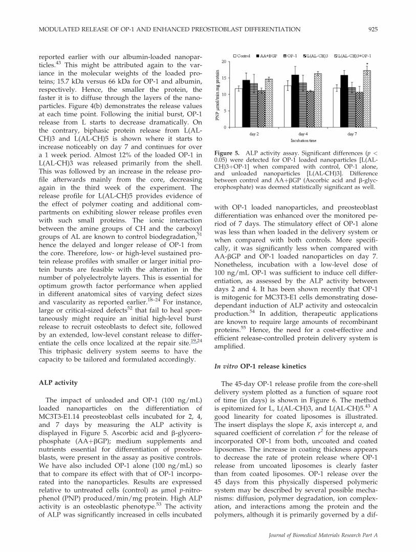
reported earlier with our albumin-loaded nanopar-ticles.43 This might be attributed again to the var-iance in the molecular weights of the loaded pro-teins; 15.7 kDa versus 66 kDa for OP-1 and albumin,respectively. Hence, the smaller the protein, thefaster it is to diffuse through the layers of the nano-particles. Figure 4(b) demonstrates the release valuesat each time point. Following the initial burst, OP-1release from L starts to decrease dramatically. Onthe contrary, biphasic protein release from L(AL-CH)3 and L(AL-CH)5 is shown where it starts toincrease noticeably on day 7 and continues for overa 1 week period. Almost 12% of the loaded OP-1 inL(AL-CH)3 was released primarily from the shell.This was followed by an increase in the release pro-file afterwards mainly from the core, decreasingagain in the third week of the experiment. Therelease profile for L(AL-CH)5 provides evidence ofthe effect of polymer coating and additional com-partments on exhibiting slower release profiles evenwith such small proteins. The ionic interactionbetween the amine groups of CH and the carboxylgroups of AL are known to control biodegradation,51
hence the delayed and longer release of OP-1 fromthe core. Therefore, low- or high-level sustained pro-tein release profiles with smaller or larger initial pro-tein bursts are feasible with the alteration in thenumber of polyelectrolyte layers. This is essential foroptimum growth factor performance when appliedin different anatomical sites of varying defect sizesand vascularity as reported earlier.18–24 For instance,large or critical-sized defects52 that fail to heal spon-taneously might require an initial high-level burstrelease to recruit osteoblasts to defect site, followedby an extended, low-level constant release to differ-entiate the cells once localized at the repair site.19,24
This triphasic delivery system seems to have thecapacity to be tailored and formulated accordingly.
ALP activity
The impact of unloaded and OP-1 (100 ng/mL)loaded nanoparticles on the differentiation ofMC3T3-E1.14 preosteoblast cells incubated for 2, 4,and 7 days by measuring the ALP activity isdisplayed in Figure 5. Ascorbic acid and b-glycero-phosphate (AAþbGP); medium supplements andnutrients essential for differentiation of preosteo-blasts, were present in the assay as positive controls.We have also included OP-1 alone (100 ng/mL) sothat to compare its effect with that of OP-1 incorpo-rated into the nanoparticles. Results are expressedrelative to untreated cells (control) as lmol p-nitro-phenol (PNP) produced/min/mg protein. High ALPactivity is an osteoblastic phenotype.53 The activityof ALP was significantly increased in cells incubated
with OP-1 loaded nanoparticles, and preosteoblastdifferentiation was enhanced over the monitored pe-riod of 7 days. The stimulatory effect of OP-1 alonewas less than when loaded in the delivery system orwhen compared with both controls. More specifi-cally, it was significantly less when compared withAA-bGP and OP-1 loaded nanoparticles on day 7.Nonetheless, incubation with a low-level dose of100 ng/mL OP-1 was sufficient to induce cell differ-entiation, as assessed by the ALP activity betweendays 2 and 4. It has been shown recently that OP-1is mitogenic for MC3T3-E1 cells demonstrating dose-dependant induction of ALP activity and osteocalcinproduction.54 In addition, therapeutic applicationsare known to require large amounts of recombinantproteins.55 Hence, the need for a cost-effective andefficient release-controlled protein delivery system isamplified.
In vitro OP-1 release kinetics
The 45-day OP-1 release profile from the core-shelldelivery system plotted as a function of square rootof time (in days) is shown in Figure 6. The methodis epitomized for L, L(AL-CH)3, and L(AL-CH)5.43 Agood linearity for coated liposomes is illustrated.The insert displays the slope K, axis intercept a, andsquared coefficient of correlation r2 for the release ofincorporated OP-1 from both, uncoated and coatedliposomes. The increase in coating thickness appearsto decrease the rate of protein release where OP-1release from uncoated liposomes is clearly fasterthan from coated liposomes. OP-1 release over the45 days from this physically dispersed polymericsystem may be described by several possible mecha-nisms: diffusion, polymer degradation, ion complex-ation, and interactions among the protein and thepolymers, although it is primarily governed by a dif-
Figure 5. ALP activity assay. Significant differences (p <0.05) were detected for OP-1 loaded nanoparticles [L(AL-CH)3þOP-1] when compared with control, OP-1 alone,and unloaded nanoparticles [L(AL-CH)3]. Differencebetween control and AAþbGP (Ascorbic acid and b-glyc-erophosphate) was deemed statistically significant as well.
MODULATED RELEASE OF OP-1 AND ENHANCED PREOSTEOBLAST DIFFERENTIATION 925
Journal of Biomedical Materials Research Part A

fusion-based or affinity-based mechanism. Finally,the rate of release was divided into an initial phase(shell release; 0–7 days) and a terminal phase (corerelease; 8–45 days grouping two release episodes to-gether) and fitted to the Higuchi mathematicalmodel which is based on the calculation of the areaunder the curve using the trapezoidal rule.56 Table Idisplays the relevant release rate constants and cor-relation coefficients. This model allows for wheredrug release is complete (100%) or incomplete(<100%) at the last sampling time. The literatureprovides evidence that for such systems, the Higuchimodel based on the goodness-of-fit test continues tobe the most appropriate model to describe thekinetics of drug release, when compared with zero-order, first-order, Hixson-Crowell’s, and Weibull’s
models.56 Furthermore, it enables an understandingof the quantitative deviation of the proposed formu-lation from the diffusion-controlled ideal Higuchimodel. The role of the L-b-L build-up of polymersaround nanosized liposomes is clearly demonstratedin controlling the release of the entrapped drug orprotein over prolonged periods of time. The releasepatterns of the coated liposomes showed linear pro-files with a much lower burst than uncoated lipo-somes followed by a sustained release of OP-1.
CONCLUSIONS
The novelty of this work is based on the combina-tion of the polymeric L-b-L self assembly techniqueand nanoscaled liposomes in formulating a stableand nontoxic release-controlled protein delivery sys-tem. It consists of a suspension of monodispersecore-shell nanoparticles suitable for the potentialadministration of growth factors via a parental injec-tion as is preferable for surgeons. The nanoparticlestolerate extended shelf storage and allow for proteinloading immediately preceding administration, pre-venting degradation, or loss of the entrapped growthfactor. The system demonstrated a good capacity forthe encapsulation and loading of OP-1 which can bealtered by the number of polyelectrolyte layers. Sus-tained and multistep release of OP-1 was evident foran extended period of time with the viability andbioactivity of OP-1 maintained via enhancing preos-teoblast differentiation, in vitro. These findings sug-gest that our core-shell nanoparticulate system couldbe an effective carrier for morphogens, growth fac-tors, and most likely for other classes of bioactivemolecules.
The authors wish to the Natural Sciences and Engineer-ing Research Council of Canada (NSERC) and the Centrefor Biorecognition and Biosensors (CBB) for making thisresearch possible. Dr. Haidar acknowledges a post-gradu-ate scholarship from the Canadian Institutes for HealthResearch (CIHR) Skeletal Health Training Program andCenter for Bone and Periodontal Research in Montreal,Quebec.
Figure 6. Release kinetics of uncoated (L) and coated lip-osomes [L(AL-CH)3 and L(AL-CH)5] represented as cumu-lative protein release percentage (CPR). Release data wasfitted in the Higuchi mathematical model to obtain a plot ofpercentage protein release as a function of square root oftime. Insert displays the release rare slope (K), axis intercept(a), and correlation coefficient (r2) of OP-1 from the samplesstudies in comparison to the ideal Higuchi (H. ideal) whichtakes into consideration 100% release over the monitoredperiod of time. Release was not complete over the studiedperiod of time (<100%). Slower release rates are shownwith coated liposomes than both, uncoated liposomes andthe H. ideal, resulting in negative axis intercepts.
TABLE IRelease Rate Constants (Mean 6 standard deviation) and Correlation Coefficients of OP-1 Release
from Bare and Coated Liposomes Showing the Initial (shell) Phase and the Terminal (core)Phase of Protein Release (Obtained from the Higuchi Plot)
Model L L(AL-CH)3 L(AL-CH)5
Higuchi Initial Terminal Shell Core Shell Core
K 16.1 6 5.4 3.5 6 6.9 11.4 6 6.1 9.7 6 4.7 6.7 6 5.1 14.5 6 1.9r2 0.95 0.98 0.99 0.96 0.99 0.99
926 HAIDAR ET AL.
Journal of Biomedical Materials Research Part A

References
1. Ripamonti U, Ramoshebi LN, Matsaba T, Tasker J, Crooks J,Teare J. Bone induction by BMPs/OPs and related familymembers in primates: The critical role of delivery systems.J Bone Joint Surg Am 2001;83:116–127.
2. Sykaras N, Opperman LA. Bone morphogenetic proteins(BMPs): How do they function and what can they offer theclinician? J Oral Sci 2003;45:57–73.
3. Xiao YT, Xiang LX, Shao JZ. Bone morphogenetic protein.Biochem Biophys Res Commun 2007;362:550–553.
4. Termaat MF, Den Boer FC, Bakker FC, Patka P, HaarmanHJThM. Bone morphogenetic proteins. Development andclinical efficacy in the treatment of fractures and bonedefects. J Bone Joint Surg Am 2005;87:1367–1378.
5. Saito N, Okada T, Horiuchi H, Ota H, Takahashi J, MurakamiN, Nawata M, Kojima S, Nozaki K, Takaoka K. Local boneformation by injection of recombinant human bone morpho-genetic protein-2 contained in polymer carriers. Bone 2003;32:381–386.
6. Giannobile WV, Ryan S, Shih MS, Su DL, Kaplan PL, ChanTC. Recombinant human osteogenic protein-1 (OP-1) stimu-lates periodontal wound healing in class III furcation defects.J Periodontol 1998;69:129–137.
7. Sampath TK, Maliakal JC, Hauschka PV, Jones WK, Sasak H,Tucker RF, White KH, Coughlin JE, Tucker MM, Pang RH.Recombinant human osteogenic protein-1 (hOP-1) inducesnew bone formation in vivo with a specific activity compara-ble with natural bovine osteogenic protein and stimulatesosteoblast proliferation and differentiation in vitro. J BiolChem 1992;267:20352–20362.
8. Vukicevic S, Stavljenic A, Pecina M. Discovery and clinicalapplications of bone morphogenetic proteins. Eur J Clin ChemClin Biochem 1995;33:661–671.
9. Cook SD, Rueger DC. Osteogenic protein-1: Biology andapplications. Clin Orthop Relat Res 1996;324:29–38.
10. Cook SD. Preclinical and clinical evaluation of osteogenicprotein-1 (BMP-7) in bony sites. Orthopedics 1999;22:669–671.
11. Friedlaender GE, Perry CR, Cole JD, Cook SD, Cierny G,Muschler GF, Zych GA, Calhoun JH, LaForte AJ, Yin S.Osteogenic protein-1 (bone morphogenetic protein-7) in thetreatment of tibial nonunions. J Bone Joint Surg Am 2001;83:151–158.
12. Pecine M, Giltaij LR, Vukicevic S. Orthopaedic applicationsof osteogenic protein-1 (BMP-7). Int Orthop 2001;25:203–208.
13. Mizumoto Y, Moseley T, Drews M, Cooper VN III, ReddiAH. Acceleration of regenerate ossification during distractionosteogenesis with recombinant human bone morphogeneticprotein-7. J Bone Joint Surg Am 2003;85:124–130.
14. Hamdy RC, Amako M, Beckman L, Kawaguchi M, Rauch F,Lauzier D, Steffen T. Effects of osteogenic protein-1 on dis-traction osteogenesis in rabbits. Bone 2003;33:248–255.
15. Mandu-Hrit M, Haque T, Lauzier D, Kotsiopriftis M, RauchF, Tabrizian M, Henderson JE, Hamdy RC. Early injection ofOP-1 during distraction osteogenesis accelerates new boneformation in rabbits. Growth Factors 2006;24:172–183.
16. Buxton PG, Cobourne MT. Regenerative approaches in thecraniofacial region: Manipulating cellular progenitors for oro-facial repair. Oral Dis 2007;13:452–460.
17. Mitsukawa N, Satoh K, Suse T, Hosaka Y. Clinical success ofmandibular distraction for obstructive sleep apnea resultingfrom micrognathia in 10 consecutive Japanese young chil-dren. J Craniofac Surg 2007;18:948–953.
18. Seeherman H, Wozney J, Li R. Bone morphogenetic proteindelivery systems. Spine 2002;27:16–23.
19. Luginbuehl V, Meinel L, Merkle HP, Gander B. Localizeddelivery of growth factors for bone repair. Eur J Pharm Bio-pharm 2004;58:197–208.
20. Seeherman H, Wozney JM. Delivery of bone morphogeneticproteins for orthopedic tissue regeneration. Cytokine GrowthFactor Rev 2005;16:329–345.
21. Chen RR, Mooney DJ. Polymeric growth factor delivery strat-egies for tissue engineering. Pharm Res 2003;20:1103–1112.
22. Winn SR, Uludag H, Hollinger JO. Carrier systems for bonemorphogenetic proteins. Clin Orthop Relat Res 1999;367:95–106.
23. Kirker-Head CA. Potential applications and delivery strat-egies for bone morphogenetic proteins. Adv Drug Deliv Rev2000;43:65–92.
24. Li RH, Wozney JM. Delivering on the promise of bone mor-phogenetic proteins. Trends Biotechnol 2001;19:255–265.
25. Geesink RGT, Hoefnagels NHM, Bulstra SK. Osteogenicactivity of OP-1 bone morphogenetic protein (BMP-7) in ahuman fibular defect. J Bone Joint Surg 1999;81:710–718.
26. Cook SD, Wolfe MW, Salkeld SL, Rueger DC. Effect ofrecombinant human osteogenic protein-1 on healing of seg-mental defects in non-human primates. J Bone Joint Surg1995;77:734–750.
27. Bach FH, Fishman JA, Daniels N, Proimo J, Anderson B, Car-penter CB, Forrow L, Robson SC, Fineberg HV. Uncertaintyin xenotransplantation: Individual benefit versus collectiverisk. Nature Med 1998;4:141–144.
28. Butler D, Wadman M, Lehrman S, Schiermeier Q. Last chanceto stop and think on risks of xenotransplants. Nature 1998;391:320–324.
29. DeLustro F, Dasch J, Keefe J, Ellingsworth L. Immuneresponses to allogeneic and xenogeneic implants of collagenand collagen derivatives. Clin Orthop 1990;260:263–279.
30. Boden SD, Maertin GJ, Morone MA, Ugbo JL, Moskovitz PA.Posterolateral lumbar intertransverse process spine arthodesiswith recombinant human bone morphogenetic protein 2/hy-droxyapatite-tricalcium phosphate after laminectomy in thenonhuman primate. Spine 1999;24:1179–1185.
31. Boyan BD, Lohmann CH, Somers A, Niederauer GG, WozneyJM, Dean DD, Carnes DL, Schwartz Z. Potential of porouspoly-d,lactide-co-glycolide particles as a carrier for recombi-nant human bone morphogenetic protein-2 during osteoindu-cation in vivo. J Biomed Mater Res 1999;46:51–59.
32. Sung HJ, Meredith C, Johnson C, Galis ZS. The effect of scaf-fold degradation rate on three-dimensional cell growth andangiogenesis. Biomaterials 2004;25:5735–5742.
33. Brekke JH. A rationale for delivery of osteoinductive pro-teins. Tissue Eng 1996;2:97–114.
34. Kenley R, Marden L, Turek T, Jin L, Ron E, Hollinger JO. Os-seous regeneration in the rat calvarium using novel deliverysystems for recombinant human bone morphogenetic protein-2 (rhBMP-2). J Biomed Mater Res 1994;28:1139–1147.
35. Higuchi T, Kinoshita A, Takahashi K, Oda S, Ishikawa I.Bone regeneration by recombinant human bone morphoge-netic protein-2 in rat mandibular defects. An experimentalmodel of defect filling. J Periodontol 1999;70:1026–1031.
36. Lee SH, Shin H. Matrices and scaffolds for delivery of bioac-tive molecules in bone and cartilage tissue engineering. AdvDrug Deliv Rev 2007;59:339–359.
37. Weber FE, Eyrich G, Gratz KW, Maly FE, Sailer HF. Slowand continuous application of human recombinant bone mor-phogenetic protein via biodegradable poly(lactide-co-glyco-lide) foamspheres. Int J Oral Maxillofac Surg 2002;31:60–65.
38. Park GE, Pattison MA, Park K, Webster TJ. Accelerated chon-drocyte functions on NaOH-treated PLGA scaffolds. Biomate-rials 2005;26:3075–3082.
39. RaviKumar MN. Nano and microparticles as controlled drugdelivery devices. J Pharm Pharm Sci 2000;3:234–258.
40. Wei G, Jin Q, Giannobile WV, Ma PX. The enhancement ofosteogenesis by nano-fibrous scaffolds incorporating rhBMP-7nanospheres. Biomaterials 2007;28:2087–2096.
MODULATED RELEASE OF OP-1 AND ENHANCED PREOSTEOBLAST DIFFERENTIATION 927
Journal of Biomedical Materials Research Part A
41. Hollinger J. Strategies for regenerating bone of the craniofa-cial complex. Bone 1993;14:575–580.
42. Cai Q, Yang J, Bei JZ, Wang SG. A novel porous cells scaffoldmade of polylactide-dextran blend by combining phase-sepa-ration and particle-leaching techniques. Biomaterials 2002;23:4483–4492.
43. Haidar ZS, Hamdy RC, Tabrizian M. Protein release kineticsfor core-shell hybrid nanoparticles based on the layer-by-layer assembly of alginate and chitosan on liposomes. Bioma-terials 2008;29:1207–1215.
44. New RRC. Liposomes: A Practical Approach. Oxford: IRC;1990. pp 256–258.
45. Higuchi T. Rate of release of medicaments from ointmentbases containing drugs in suspension. J Pharm Sci 1961;50:874–875.
46. Heerklotz H, Tsamaloukas A, Kita-Tokarczyk K, Strunz P,Gutberlet T. Structural, volumetric, and thermodynamic char-acterization of a micellar sphere-to-rod transition. J AmChem Soc 2004;126:16544–16552.
47. Haidar ZS, Azari F, Hamdy RC, Tabrizian M. In vitro charac-terization and cytotoxic effect of a liposome-based drugdelivery system incorporating alternating L-b-L assembly ofpolyelectrolytes for craniofacial distraction osteogenesis. CBS2006; Abstract 33.
48. Shiqu YE, Chaoyang W, Xinxing L, Zhen T. Multilayer nano-capsules of polysaccharide chitosan and alginate throughlayer-by-layer assembly directly on PS nanoparticles forrelease. J Biomat Sci Polymer Ed 2005;16:909–923.
49. Tabatt K, Sameti M, Olbrich C, Muller RH, Lehr C. Effect ofcationic lipid and matrix lipid composition on solid lipidnanoparticles-mediated gene transfer. Eur J Pharm Biopharm2004;57:155–162.
50. Asasutjarit R, Lorenzen SI, Sirivichayakul S, Ruxrungtham K,Ruktanonchai U, Ritthidej GC. Effect of solid lipid nanopar-ticles formulation compositions on their size. Zeta potentialand potential for in vitro phis-hiv-hugag transfection. PharmRes 2007;24:1098–1107.
51. Chellat F, Tabrizian M, Dumitriu S, Chornet E, Magny P,Rivard CH, Yahia L. Study of biodegradation behaviour ofchitosan-xanthan microspheres in simulated physiologicalmedia. J Biomed Mater Res 2000;53:592–599.
52. Lindholm TC, Peel SAF, Sandor GKB, Clokie CML. Intra-oralbone graft used to acquire viable bone cells to be used for tis-sue engineering. J Can Dent Assoc 2002;68:699.
53. Mizuno M, Kuboki Y. Osteoblast-related gene expression ofbone marrow cells during the osteoblastic differentiationinduced by type I collagen. J Biochem 2001;129:133–138.
54. Lee DH, Baek HS, Lee MH, Park JH. Production of bonemorphogenetic protein-7 using pET expression system. CurrAppl Phys 2005;5:422–425.
55. Wang EA. Bone morphogenetic proteins (BMPs): Therapeuticpotential in healing bony defects. Trend Biotechnol 1993;11:379–383.
56. Gohel MC, Panchal MK, Jogani VV. Novel mathematicalmethod for quantitative expression of deviation from theHiguchi model. AAPS Pharm Sci Tech 2000;1:31.
928 HAIDAR ET AL.
Journal of Biomedical Materials Research Part A