
Modular Hyperthermostable Bacterial Endo-β-1,4-Mannanase: Molecular Shape, Flexibility and...
14
Modular Hyperthermostable Bacterial Endo-b-1, 4-Mannanase: Molecular Shape, Flexibility and Temperature-Dependent Conformational Changes Viviam M. da Silva 1. , Francieli Colussi 1. , Mario de Oliveira Neto 2 , Antonio S. K. Braz 1 , Fabio M. Squina 3 , Cristiano L. P. Oliveira 4 , Wanius Garcia 1 * 1 Centro de Cie ˆ ncias Naturais e Humanas, Universidade Federal do ABC (UFABC), Santo Andre ´, Sa ˜o Paulo, Brazil, 2 Departamento de Fı ´sica e Biofı ´sica, Instituto de Biocie ˆ ncias, Universidade Estadual Paulista, Botucatu, Sa ˜o Paulo, Brazil, 3 Laborato ´ rio Nacional de Cie ˆ ncia e Tecnologia do Bioetanol, Centro Nacional de Pesquisa em Energia e Materiais, Campinas, Sa ˜ o Paulo, Brazil, 4 Instituto de Fı ´sica, Universidade de Sa ˜o Paulo, Sa ˜ o Paulo, Brazil Abstract Endo-b-1,4-mannanase from Thermotoga petrophila (TpMan) is a hyperthermostable enzyme that catalyzes the hydrolysis of b-1,4-mannoside linkages in various mannan-containing polysaccharides. A recent study reported that TpMan is composed of a GH5 catalytic domain joined by a linker to a carbohydrate-binding domain. However, at this moment, there is no three- dimensional structure determined for TpMan. Little is known about the conformation of the TpMan as well as the role of the length and flexibility of the linker on the spatial arrangement of the constitutive domains. In this study, we report the first structural characterization of the entire TpMan by small-angle X-ray scattering combined with the three-dimensional structures of the individual domains in order to shed light on the low-resolution model, overall dimensions, and flexibility of this modular enzyme at different temperatures. The results are consistent with a linker with a compact structure and that occupies a small volume with respect to its large number of amino acids. Furthermore, at 20uC the results are consistent with a model where TpMan is a molecule composed of three distinct domains and that presents some level of molecular flexibility in solution. Even though the full enzyme has some degree of molecular flexibility, there might be a preferable conformation, which could be described by the rigid-body modeling procedure. Finally, the results indicate that TpMan undergoes a temperature-driven transition between conformational states without a significant disruption of its secondary structure. Our results suggest that the linker can optimize the geometry between the other two domains with respect to the substrate at high temperatures. These studies should provide a useful basis for future biophysical studies of entire TpMan. Citation: da Silva VM, Colussi F, de Oliveira Neto M, Braz ASK, Squina FM, et al. (2014) Modular Hyperthermostable Bacterial Endo-b-1, 4-Mannanase: Molecular Shape, Flexibility and Temperature-Dependent Conformational Changes. PLoS ONE 9(3): e92996. doi:10.1371/journal.pone.0092996 Editor: Pratul K. Agarwal, Oak Ridge National Laboratory, United States of America Received December 16, 2013; Accepted February 27, 2014; Published March 26, 2014 Copyright: ß 2014 da Silva et al. This is an open-access article distributed under the terms of the Creative Commons Attribution License, which permits unrestricted use, distribution, and reproduction in any medium, provided the original author and source are credited. Funding: The authors would like to thank Fundac ¸a ˜o de Amparo a pesquisa do Estado de Sa ˜o Paulo (FAPESP) and Conselho Nacional de Desenvolvimento Cientı ´fico e Tecnolo ´ gico (CNPq) for the financial support for this work via grants #2012/21054-9 and #478900/2012-0; and fellowships #2012/03503-0 (VMS) and #501037/2012-8 (FC). The funders had no role in study design, data collection and analysis, decision to publish, or preparation of the manuscript. Competing Interests: The authors have declared that no competing interests exist. * E-mail: [email protected] . These authors contributed equally to this work. Introduction Lignocellulosic biomass is the major renewable carbon source in nature with several applications and consists of cellulose, hemicellulose and lignin with an approximate representation of 2:1:1 [1–3]. Cellulose consists of linear polysaccharides of b-1,4- linked D-glucose residues, whereas lignin is a phenolic macromol- ecule. Hemicelluloses are linear or branched heteropolysaccha- rides derived from sugars such as D-xylose, D-galactose, D- mannose, D-glucose, and L-arabinose [4]. They are classified according to the main sugar unit as xylans, galactans and mannans, and most of the main-chain sugars on hemicellulose polymer are linked together by b-1,4-glycosidic bonds [4–6]. Xylans comprise the major hemicellulose component present in hardwoods, whereas mannans are more prominent in softwoods [5,7]. Mannans are classified into two principal groups depending on whether the b-1,4-linked backbone contains only D-mannose (mannans) or a combination of D-mannose and D-glucose residues (glucomannans). Linear mannan or glucomannan chains contain- ing more than 5% D-galactose are called galactomannans and galactoglucomannans, respectively [7]. The biodegradation of cellulose and hemicellulose polymers involves the concerted action of a variety of hydrolytic enzymes. Cellulases and xylanases have been extensively studied due to their several industrial applications principally relating to biorefineries, however, mannan-degrading enzymes have enjoyed less attention. The major enzymes involved in the biodegradation of mannan polysaccharides are b-mannanase (EC 3.2.178), b-mannosidase (EC 3.2.1.25), and b-glucosidase (EC 3.2.1.21) [7,8]. The enzyme b-mannanase is responsible for the cleavage of b-1,4-linked internal linkages of the mannan polymer to produce new chain ends, whereas b-mannosidase cleaves b-1,4-linked mannosides releasing mannose from the nonreducing end of mannans and mannooligosaccharides [7,8]. The enzyme b-glucosidase hydro- lyzes 1,4-b-D-glucopyranose at the nonreducing end of the oligosaccharides released from glucomannan and galactogluco- PLOS ONE | www.plosone.org 1 March 2014 | Volume 9 | Issue 3 | e92996
Transcript of Modular Hyperthermostable Bacterial Endo-β-1,4-Mannanase: Molecular Shape, Flexibility and...

Modular Hyperthermostable Bacterial Endo-b-1,4-Mannanase: Molecular Shape, Flexibility andTemperature-Dependent Conformational ChangesViviam M. da Silva1., Francieli Colussi1., Mario de Oliveira Neto2, Antonio S. K. Braz1, Fabio M. Squina3,
Cristiano L. P. Oliveira4, Wanius Garcia1*
1 Centro de Ciencias Naturais e Humanas, Universidade Federal do ABC (UFABC), Santo Andre, Sao Paulo, Brazil, 2 Departamento de Fısica e Biofısica, Instituto de
Biociencias, Universidade Estadual Paulista, Botucatu, Sao Paulo, Brazil, 3 Laboratorio Nacional de Ciencia e Tecnologia do Bioetanol, Centro Nacional de Pesquisa em
Energia e Materiais, Campinas, Sao Paulo, Brazil, 4 Instituto de Fısica, Universidade de Sao Paulo, Sao Paulo, Brazil
Abstract
Endo-b-1,4-mannanase from Thermotoga petrophila (TpMan) is a hyperthermostable enzyme that catalyzes the hydrolysis ofb-1,4-mannoside linkages in various mannan-containing polysaccharides. A recent study reported that TpMan is composedof a GH5 catalytic domain joined by a linker to a carbohydrate-binding domain. However, at this moment, there is no three-dimensional structure determined for TpMan. Little is known about the conformation of the TpMan as well as the role of thelength and flexibility of the linker on the spatial arrangement of the constitutive domains. In this study, we report the firststructural characterization of the entire TpMan by small-angle X-ray scattering combined with the three-dimensionalstructures of the individual domains in order to shed light on the low-resolution model, overall dimensions, and flexibility ofthis modular enzyme at different temperatures. The results are consistent with a linker with a compact structure and thatoccupies a small volume with respect to its large number of amino acids. Furthermore, at 20uC the results are consistentwith a model where TpMan is a molecule composed of three distinct domains and that presents some level of molecularflexibility in solution. Even though the full enzyme has some degree of molecular flexibility, there might be a preferableconformation, which could be described by the rigid-body modeling procedure. Finally, the results indicate that TpManundergoes a temperature-driven transition between conformational states without a significant disruption of its secondarystructure. Our results suggest that the linker can optimize the geometry between the other two domains with respect to thesubstrate at high temperatures. These studies should provide a useful basis for future biophysical studies of entire TpMan.
Citation: da Silva VM, Colussi F, de Oliveira Neto M, Braz ASK, Squina FM, et al. (2014) Modular Hyperthermostable Bacterial Endo-b-1, 4-Mannanase: MolecularShape, Flexibility and Temperature-Dependent Conformational Changes. PLoS ONE 9(3): e92996. doi:10.1371/journal.pone.0092996
Editor: Pratul K. Agarwal, Oak Ridge National Laboratory, United States of America
Received December 16, 2013; Accepted February 27, 2014; Published March 26, 2014
Copyright: � 2014 da Silva et al. This is an open-access article distributed under the terms of the Creative Commons Attribution License, which permitsunrestricted use, distribution, and reproduction in any medium, provided the original author and source are credited.
Funding: The authors would like to thank Fundacao de Amparo a pesquisa do Estado de Sao Paulo (FAPESP) and Conselho Nacional de DesenvolvimentoCientıfico e Tecnologico (CNPq) for the financial support for this work via grants #2012/21054-9 and #478900/2012-0; and fellowships #2012/03503-0 (VMS) and#501037/2012-8 (FC). The funders had no role in study design, data collection and analysis, decision to publish, or preparation of the manuscript.
Competing Interests: The authors have declared that no competing interests exist.
* E-mail: [email protected]
. These authors contributed equally to this work.
Introduction
Lignocellulosic biomass is the major renewable carbon source in
nature with several applications and consists of cellulose,
hemicellulose and lignin with an approximate representation of
2:1:1 [1–3]. Cellulose consists of linear polysaccharides of b-1,4-
linked D-glucose residues, whereas lignin is a phenolic macromol-
ecule. Hemicelluloses are linear or branched heteropolysaccha-
rides derived from sugars such as D-xylose, D-galactose, D-
mannose, D-glucose, and L-arabinose [4]. They are classified
according to the main sugar unit as xylans, galactans and
mannans, and most of the main-chain sugars on hemicellulose
polymer are linked together by b-1,4-glycosidic bonds [4–6].
Xylans comprise the major hemicellulose component present in
hardwoods, whereas mannans are more prominent in softwoods
[5,7]. Mannans are classified into two principal groups depending
on whether the b-1,4-linked backbone contains only D-mannose
(mannans) or a combination of D-mannose and D-glucose residues
(glucomannans). Linear mannan or glucomannan chains contain-
ing more than 5% D-galactose are called galactomannans and
galactoglucomannans, respectively [7].
The biodegradation of cellulose and hemicellulose polymers
involves the concerted action of a variety of hydrolytic enzymes.
Cellulases and xylanases have been extensively studied due to their
several industrial applications principally relating to biorefineries,
however, mannan-degrading enzymes have enjoyed less attention.
The major enzymes involved in the biodegradation of mannan
polysaccharides are b-mannanase (EC 3.2.178), b-mannosidase
(EC 3.2.1.25), and b-glucosidase (EC 3.2.1.21) [7,8]. The enzyme
b-mannanase is responsible for the cleavage of b-1,4-linked
internal linkages of the mannan polymer to produce new chain
ends, whereas b-mannosidase cleaves b-1,4-linked mannosides
releasing mannose from the nonreducing end of mannans and
mannooligosaccharides [7,8]. The enzyme b-glucosidase hydro-
lyzes 1,4-b-D-glucopyranose at the nonreducing end of the
oligosaccharides released from glucomannan and galactogluco-
PLOS ONE | www.plosone.org 1 March 2014 | Volume 9 | Issue 3 | e92996

mannans by b-mannanase. Additional enzymes such as acetyl
mannan esterase (EC 3.1.1.6) and a-galactosidase (EC 3.2.1.22)
are required to remove side groups that might be attached at
several points on the mannan polymer, creating more sites for
subsequent enzyme hydrolysis [7,8]. The biodegradation of
mannan represents a key step for various industrial applications
including delignification of kraft pulps, food processing and
production of second-generation biofuels [6–11].
Thermotoga petrophila (T. petrophila) strain RKU-1 (T) is a
hyperthermophilic bacterium isolated from the Kubiki oil
reservoir in Niigata (Japan) [12]. The temperature range for
growth is 47–88uC with an optimum at 80uC. The pH range for
growth is 5–9 with the optimum at pH 7. This bacterium produces
a repertoire of hyperthermostable enzymes of great potential for
industrial applications, including cellulases, arabinofuranosidases,
arabinanases and mannanases, and has proved to be a suitable
source of enzymes for biotechnological applications and protein
engineering of glycoside hydrolases [13–15]. For various industrial
applications, it is essential that mannanase possesses high activity
and thermostability [11].
The hyperthermostable bacterial endo-b-1,4-mannanase (EC
3.2.1.78) from T. petrophila (TpMan) is an enzyme composed of 667
amino acid residues (Gene Bank: ABQ47550.1). The first 20
amino acid residues are a signal peptide, which are removed after
protein exportation. TpMan enzyme presents optimal activity at
the temperature range of 81–93uC [13]. Both TpMan and its
catalytic domain present maximal activity at the acidic pH range
of 4.5–6.5. The truncated catalytic domain had optimal activity at
a lower temperature range when compared to the TpMan enzyme
[13]. A recent study reported that TpMan consists of a GH5
catalytic domain (TpManGH5, 373 amino acid residues) and a
carbohydrate-binding domain (TpManCBM27, 172 amino acid
residues) connected through a linker (102 amino acid residues)
[13]. Furthermore, the same study presented the crystal structure
of GH5 catalytic domain, however, at this moment, there is no
three-dimensional structure available for entire TpMan.
In the present study, the bacterial TpMan was analyzed using
biophysical techniques as well as with bioinformatics tools. Here,
we report the first structural characterization of hyperthermostable
endo-b-1,4-mannanase from the bacterium T. petrophila by small-
angle X-ray scattering in order to shed light on the low-resolution
molecular envelope, overall dimensions, and flexibility of this
modular enzyme at different temperatures.
Results
Temperature Profile of TpMan Enzymatic ActivityThe activity of an enzyme is dependent on pH and temperature,
conditions that invoke changes in enzyme folding, conformation,
and protonation. As a first step of our studies, the mannan endo-
1,4-b-mannosidase activity of TpMan and TpManGH5 were
studied as a function of temperature using locust bean gum
(galactomannan) as substrate (Fig. 1). Our results showed that,
under the conditions of our study (at pH 6), the enzymatic activity
of TpMan increases as temperature increases. A similar behavior
was also observed in the case of TpManGH5. However,
TpManGH5 showed a significant decline in the enzymatic activity
at 85uC when compared to TpMan (Fig. 1). Thus, our results
indicate that TpMan is more thermotolerant than the truncated
catalytic domain. The results presented here are consistent with
the results published recently for TpMan [13].
Dynamic Light Scattering (DLS)Dynamic light scattering (DLS) provides a fast and convenient
way to monitor the size of proteins in solution and also investigate
whether the enzyme aggregates at high temperatures and/or at
high protein concentrations [16]. DLS size distribution profiles for
TpMan and TpManGH5 are available in supporting information
(Figs. S1 and S2). As can be seen, the hydrodynamic radii (RS) of
TpMan were practically independent of protein concentration
over the range 0.5 to 8 mg/mL at 20uC and pH 6 (Fig. 2A).
Figure 1. Enzymatic activity. Effect of temperature on the enzymaticactivity of TpMan and TpManGH5.doi:10.1371/journal.pone.0092996.g001
Figure 2. Characterization of TpMan by DLS at pH 6. (A) Thehydrodynamic radius (RS) of TpMan (at 20uC) as a function of theprotein concentration. (B) The hydrodynamic radius (RS) of TpMan (at8 mg/mL) as a function of temperature.doi:10.1371/journal.pone.0092996.g002
Structural Studies of a b-Mannanase
PLOS ONE | www.plosone.org 2 March 2014 | Volume 9 | Issue 3 | e92996

Furthermore, the RS of TpMan exhibited minimal temperature
dependence over the range 20 to 85uC at pH 6 and 0.5 mg/mL
(Fig. 3A). After incubation at 85uC (at 0.5 mg/mL) there was no
evidence of enzyme precipitation and the solution remained
transparent. The average value of RS determined for TpMan by
the DLS method was 3662 A, which corresponds to a molecular
mass of 6866 kDa, consistent with the expected molecular mass of
73 kDa. However, when TpMan at pH 6 and 8 mg/mL was
incubated at temperature values above 65uC the RS increased
significantly, suggesting a tendency to form amorphous aggregates
(Fig. 2B) [16]. After incubation at 85uC (at 8 mg/mL) the initially
clear TpMan solution was very turbid and precipitate had formed.
In the case of TpManGH5, the Rs were practically independent
of temperature over the range 20 to 75uC at pH 6 and 0.5 mg/
mL (Fig. 3B). The average RS determined for TpManGH5 by the
DLS method was 2862 A, which corresponds to a molecular mass
of 3865 kDa, entirely consistent with the expected molecular mass
of 42 kDa. However, when TpManGH5 at pH 6 and 0.5 mg/mL
was incubated at temperature values above 75uC the RS increased
significantly, suggesting a tendency to form amorphous aggregates
(Fig. 3B). Again, after incubation at 85uC (at 0.5 mg/mL) the
initially clear TpManGH5 solution was very turbid and precipitate
had formed.
Small-Angle X-ray Scattering Data Analysis and LowResolution Modeling
To obtain more detailed information about the tertiary
structure of TpMan and its molecular shape, we submitted
TpMan to small-angle X-ray scattering (SAXS) analysis as a
function of temperature. The X-ray scattering curves obtained at
8 mg/mL and three different temperatures (T = 20, 50 and 65uC)
for TpMan at pH 6 are shown in Fig. 4. Scattering curves
obtained at 80uC and higher concentration showed nonnegligible
Figure 3. Characterization of TpMan and TpManGH5 by DLS atpH 6. (A) The hydrodynamic radius (RS) of TpMan (at 0.5 mg/mL) as afunction of temperature. (B) The hydrodynamic radius (RS) ofTpManGH5 (at 0.5 mg/mL) as a function of temperature.doi:10.1371/journal.pone.0092996.g003
Figure 4. SAXS data collected for TpMan at pH6 and different temperatures. Experimental SAXS curves of the TpMan at 20uC (open blackcircles with errors bars), 50uC (open black triangles with errors bars) and 65uC (open black squares with errors bars) superimposed on the computedscattering curves based on the restored low-resolution models (DAMs, solid black lines). The curves have been offset for clarity.doi:10.1371/journal.pone.0092996.g004
Structural Studies of a b-Mannanase
PLOS ONE | www.plosone.org 3 March 2014 | Volume 9 | Issue 3 | e92996
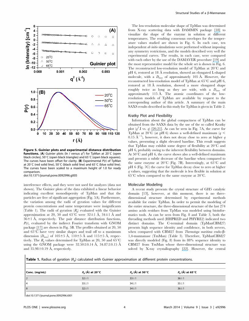
interference effects, and they were not used for analyzes (data not
shown). The Guinier plots of the data exhibited a linear behavior
indicating excellent monodispersity of TpMan and that the
particles are free of significant aggregation (Fig. 5A). Furthermore,
the variation among the radii of gyration values for different
protein concentrations and same temperature were insignificants
(Table 1). The radii of gyration (Rg) evaluated with the Guinier
approximation at 20, 50 and 65uC were 3261 A, 3461 A and
3661 A, respectively. The pair distance distribution functions,
P(r), evaluated by the indirect Fourier transform with GNOM
package [17] are shown in Fig. 5B. The profiles obtained at 20, 50
and 65uC have very similar shapes and trail off to a maximum
dimension (Dmax) of 10565 A, 11065 A and 11565 A, respec-
tively. The Rg values determined for TpMan at 20, 50 and 65uCusing the GNOM package were 32.5060.14 A, 34.8760.15 A
and 35.9860.19 A, respectively.
The low-resolution molecular shape of TpMan was determined
from X-ray scattering data with DAMMIN package [18] to
visualize the shape of the enzyme in solution at different
temperatures. The resulting consensus envelopes for the temper-
ature values studied are shown in Fig. 6. In each case, ten
independent ab initio simulations were performed without imposing
any symmetry restrictions, and the models described very well the
experimental curves. The results, in each case, were compared
with each other by the use of the DAMAVER procedure [19] and
the most representative model for the whole set is shown in Fig. 6.
The reconstructed low-resolution model of TpMan at 20uC and
pH 6, restored at 18 A resolution, showed an elongated L-shaped
molecule, with a Dmax of approximately 105 A. However, the
reconstructed low-resolution model of TpMan at 65uC and pH 6,
restored at 18 A resolution, showed a more elongated shape
roughly twice as long as they are wide, with a Dmax of
approximately 115 A. The atomic coordinates of the low-
resolution models of TpMan are available by request to the
corresponding author of this article. A summary of the main
SAXS results described in this study for TpMan is given in Table 2.
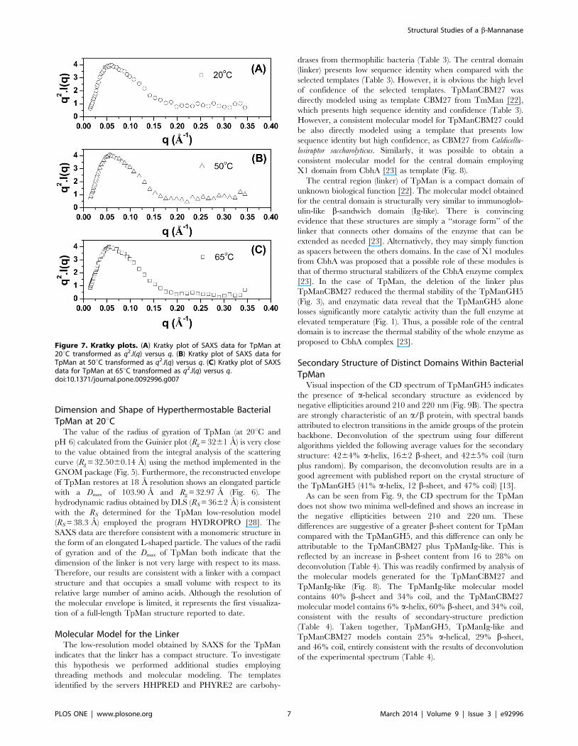
Kratky Plot and FlexibilityInformation about the global compactness of TpMan can be
obtained from the SAXS data by the use of the so called Kratky
plot (q2.I vs. q) [20,21]. As can be seen in Fig. 7A, the curve for
TpMan at 20uC (at pH 6) shows a well-defined maximum (q ,
0.15 A21), however, it does not decay close to zero at higher q
values, presenting a slight elevated baseline. This result suggests
that TpMan may exhibit some degree of flexibility at 20uC and
pH 6, probably owing to the inherent flexibility between domains.
At 50uC and pH 6, the curve shows also a well-defined maximum
and presents a subtle decrease of the baseline when compared to
the same enzyme at 20uC (Fig. 7B). Interestingly, at 65uC and
pH 6 (Fig. 7C) the curve for TpMan decay close to zero at higher
q values, suggesting that the molecule is less flexible in solution at
65uC when compared to the same enzyme at 20uC.
Molecular ModelingA recent study presents the crystal structure of GH5 catalytic
domain [13], however, at this moment, there is no three-
dimensional structure determined by experimental methods
available for entire TpMan. In order to permit the modeling of
the entire structure, the three-dimensional structure of the last 274
amino acids residues from TpMan was modeled using bioinfor-
matics tools. As can be seen from Fig. 8 and Table 3, both the
threading methods used (HHPRED and PHYRE2) indicated two
distinct domains. The C-terminal domain (TpManCBM27)
presents high sequence identity and confidence, in both servers,
when compared with CBM27 from Thermotoga maritima endo-b-
1,4-mannanase (TmMan) (Table 3). Therefore, TpManCBM27
was directly modeled (Fig. 8) from its 88% sequence identity to
CBM27 from TmMan whose three-dimensional structure was
solved by X-ray crystallography [22]. However, the central
Figure 5. Guinier plots and experimental distance distributionfunctions. (A) Guinier plots (ln I versus q2) for TpMan at 20uC (openblack circles), 50uC (open black triangles) and 65uC (open black squares).The curves have been offset for clarity. (B) Experimental P(r) of TpManat 20uC (red solid line), 50uC (black solid line) and 65uC (blue solid line).The curves have been scaled to a maximum height of 1.0 for readycomparison.doi:10.1371/journal.pone.0092996.g005
Table 1. Radius of gyration (Rg) calculated with Guinier approximation at different protein concentrations.
Conc. (mg/mL) Rg (A) at 206C Rg (A) at 506C Rg (A) at 656C
2 3261 3361 3661
4 3361 3461 3561
8 3261 3461 3661
doi:10.1371/journal.pone.0092996.t001
Structural Studies of a b-Mannanase
PLOS ONE | www.plosone.org 4 March 2014 | Volume 9 | Issue 3 | e92996

domain (linker) presents low sequence identity, in both servers,
when compared with X1 domain from Clostridium thermocellum
cellobiohydrolase A (CbhA) [23]. Despite low values of sequence
identity obtained in both servers, it is obvious the high level of
confidence of the selected template (Table 3). Thus, the central
domain was modeled from its 14% sequence identity and 97%
confidence to X1 domain from CbhA (Fig. 8). The molecular
model obtained for the central domain is structurally very similar
to immunoglobulin-like b-sandwich (Ig-like) domain and it will be
adopted the nomenclature TpManIg-like for this domain.
Far-UV Circular Dichroism (CD) SpectroscopyFar-UV CD spectroscopy was used to probe the secondary
structure of the expressed TpMan and TpManGH5 products to
assess their folding in terms of secondary structure and to draw
comparisons (Fig. 9). The CD spectrum of TpManGH5 collected
at 20uC (pH 6) is characterized by a positive band at 19561 nm
and two negative bands at 21061 nm and 22061 nm, together
with a negative to positive crossover at 20361 nm (Fig. 9B). These
bands are typically found in structures with a large content of a-
helical secondary structure. This is different to that observed for
the TpMan CD spectrum, which does not show two minima well-
defined. The CD spectra for TpMan collected at pH 6 and three
temperature values is shown in Fig. 9A. The data indicate that the
secondary structure of the enzyme does not change significantly in
response to increasing the temperature from 20 to 85uC.
The percentages of a-helix, b-strand, and coils (turns and
irregular structures) were obtained by deconvolution of the
experimental spectra using four independent methods and
expressed as a mean and standard deviation (see the Materials
and Methods). In the case of TpManGH5, the deconvolution of
the spectrum yielded the following average values for the
secondary structure (Table 4): 4264% a-helix, 1662% b-sheet,
and 4265% coil (turn plus random). The corresponding results
obtained for TpMan were 2164% a-helix, 2863% b-sheet, and
5165% turns/irregulars structures (Table 4), indicating an
increase in b-sheet and coil content and a decrease in a-helix
content in the entire enzyme.
Computational Methods for Secondary-StructurePrediction
The results of the secondary-structure prediction for the
complete TpMan sequence (Gene Bank: ABQ47550.1) and its
individual domains are shown in Table 4. The results indicate that
overall TpMan is composed of roughly equal percentages of
regular secondary structure (a-helices and b-sheets) and random
coil (turns and irregular structures). The regular secondary
structure of the TpManGH5 is dominated by a-helix. However,
the regular secondary structure of the TpManCBM27 and
TpManIg-like are dominated by b-sheet.
Ensemble Optimization Method (EOM) ModelingTo understand better the flexibility information suggests by the
Kratky plot at different temperatures, ensemble optimization
modeling (EOM) was applied [24]. The Rg and Dmax distributions
of the structures generated for the initial pool and the distributions
of the structures selected during the ensemble optimization are
shown for 20, 50 and 65uC in Fig. 10. The selected populations of
Figure 6. Low-resolution models. (A) Molecular envelope of TpMan in solution at 20uC obtained by DAMMIN package (dummy atoms). Thecenter and right structures were rotated y axis-90uand6axis-90u in relation to the left structures. (B) Molecular envelope of TpMan in solution at 50uCobtained by DAMMIN package (dummy atoms). The center and right structures were rotated y axis-90uand6axis-90u in relation to the left structures.(C) Molecular envelope of TpMan in solution at 65uC obtained by DAMMIN package (dummy atoms). The center and right structures were rotated yaxis-90uand 6 axis-90u in relation to the left structures.doi:10.1371/journal.pone.0092996.g006
Structural Studies of a b-Mannanase
PLOS ONE | www.plosone.org 5 March 2014 | Volume 9 | Issue 3 | e92996

structures became narrower when the temperature was raised
from 20 to 65uC, suggesting a decrease in the molecular flexibility
with the increase in temperature. At 20uC the Rg and Dmax of the
structures selected during the ensemble optimization are centered
around 31 A and 103 A, respectively (Table 2). At 50uC the Rg
and Dmax of the structures selected are centered around 33 A and
105 A, respectively. Finally, at 65uC the Rg and Dmax of the
structures selected are centered around 34 A and 109 A,
respectively. The EOM results indicate that TpMan becomes
more elongated and less flexible when the temperature increases
from 20 to 65uC. The excellent fit profiles determined by the
EOM modeling process are shown in Fig. 11. The Fig. 12 shows
the structure with highest frequency in the optimized population at
20uC superposed to the cloud of models. It was considered the
models that covered 80% of the frequency interval.
Rigid-Body Modeling (RBM)The CORAL package [25] was used to perform rigid-body
modeling of the three-dimensional structures based on our SAXS
curves obtained at different temperatures. The three-dimensional
structures of the TpManGH5, TpManIg-like and TpManCBM27
were organized and their relative positions and orientations were
optimized by the use of a simulated annealing procedure. As a
result, the three-dimensional arrangement of the domains that
provides the best fit to SAXS experimental data is obtained. The
excellent fits profiles determined by the rigid-body modeling are
shown for 20, 50 and 65uC in Fig. 11. The rigid-body model
(RBM) obtained at 20uC shows a particle with a Dmax of 104.70 A
and Rg = 32.30 A. The RBM obtained at 50uC shows a particle
with a Dmax of 111.20 A and Rg = 33.99 A. Finally, the RBM
obtained at 65uC shows a particle with a Dmax of 118.20 A and
Rg = 34.95 A. Superposition of the low-resolution models obtained
at different temperatures by the dummy atom modeling approach
(DAM) and the respective TpMan rigid-body models (RBM) show
a very good agreement (Fig. 6). Also, the superposition of the
EOM highest frequency model and the RBM, both obtained at
20uC and pH 6, shows also a good agreement (Fig. 13).
Discussion
Modular proteins in which globular domains are connected by
linkers are common in nature [26]. It has been recognized that a
large number of bacterial and fungal carbohydrases are composed
of a catalytic domain and a carbohydrate-binding domain
separated by an interdomain linker [27]. A recent study reported
that TpMan has a modular structure with a catalytic domain and a
carbohydrate-binding domain connected by a long linker (consid-
ering its large number of amino acids) [13]. The same study
presented the crystal structure of the catalytic domain at 1.5 A
resolution, however, crystallization of the full-length TpMan
proved to be unsuccessful, probably owing to the inherent
flexibility between domains [13]. In the present study, entire
TpMan was analyzed using biophysical techniques as well as with
bioinformatics tools. The experiments reported here provided the
global structural characterization of the entire TpMan and also
the estimation of the influence of the linker and of temperature on
the conformation and flexibility of this hyperthermostable endo-b-
1,4-mannanase.
Table 2. General SAXS results from TpMan.
Parameters/samples 206C
Experimental DAM RBM EOM
Rg (A) 3261 (Guinier)32.5060.14 (GNOM)
32.97 32.30 31.14
Dmax (A) 10565 103.90 104.70 102.70
Resolution (A) 18 – – –
x/x2 – 1.6/2.6 1.8/3.2 1.9/3.6
506C
Experimental DAM RBM EOM
Rg (A) 3461 (Guinier)34.8760.15 (GNOM)
35.26 33.99 33.22
Dmax (A) 11065 110.60 111.20 105.17
Resolution (A) 18 – – –
x/x2 – 1.1/1.2 1.3/1.7 2.4/5.8
656C
Experimental DAM RBM EOM
Rg (A) 3661 (Guinier)35.9860.19 (GNOM)
36.41 34.95 34.31
Dmax (A) 11565 116.20 118.20 108.63
Resolution (A) 18 – – –
x/x2 – 1.3/1.7 1.5/2.3 2.1/4.4
doi:10.1371/journal.pone.0092996.t002
Structural Studies of a b-Mannanase
PLOS ONE | www.plosone.org 6 March 2014 | Volume 9 | Issue 3 | e92996
Dimension and Shape of Hyperthermostable BacterialTpMan at 20uC
The value of the radius of gyration of TpMan (at 20uC and
pH 6) calculated from the Guinier plot (Rg = 3261 A) is very close
to the value obtained from the integral analysis of the scattering
curve (Rg = 32.5060.14 A) using the method implemented in the
GNOM package (Fig. 5). Furthermore, the reconstructed envelope
of TpMan restores at 18 A resolution shows an elongated particle
with a Dmax of 103.90 A and Rg = 32.97 A (Fig. 6). The
hydrodynamic radius obtained by DLS (RS = 3662 A) is consistent
with the RS determined for the TpMan low-resolution model
(RS = 38.3 A) employed the program HYDROPRO [28]. The
SAXS data are therefore consistent with a monomeric structure in
the form of an elongated L-shaped particle. The values of the radii
of gyration and of the Dmax of TpMan both indicate that the
dimension of the linker is not very large with respect to its mass.
Therefore, our results are consistent with a linker with a compact
structure and that occupies a small volume with respect to its
relative large number of amino acids. Although the resolution of
the molecular envelope is limited, it represents the first visualiza-
tion of a full-length TpMan structure reported to date.
Molecular Model for the LinkerThe low-resolution model obtained by SAXS for the TpMan
indicates that the linker has a compact structure. To investigate
this hypothesis we performed additional studies employing
threading methods and molecular modeling. The templates
identified by the servers HHPRED and PHYRE2 are carbohy-
drases from thermophilic bacteria (Table 3). The central domain
(linker) presents low sequence identity when compared with the
selected templates (Table 3). However, it is obvious the high level
of confidence of the selected templates. TpManCBM27 was
directly modeled using as template CBM27 from TmMan [22],
which presents high sequence identity and confidence (Table 3).
However, a consistent molecular model for TpManCBM27 could
be also directly modeled using a template that presents low
sequence identity but high confidence, as CBM27 from Caldicellu-
losiruptor saccharolyticus. Similarly, it was possible to obtain a
consistent molecular model for the central domain employing
X1 domain from CbhA [23] as template (Fig. 8).
The central region (linker) of TpMan is a compact domain of
unknown biological function [22]. The molecular model obtained
for the central domain is structurally very similar to immunoglob-
ulin-like b-sandwich domain (Ig-like). There is convincing
evidence that these structures are simply a ‘‘storage form’’ of the
linker that connects other domains of the enzyme that can be
extended as needed [23]. Alternatively, they may simply function
as spacers between the others domains. In the case of X1 modules
from CbhA was proposed that a possible role of these modules is
that of thermo structural stabilizers of the CbhA enzyme complex
[23]. In the case of TpMan, the deletion of the linker plus
TpManCBM27 reduced the thermal stability of the TpManGH5
(Fig. 3), and enzymatic data reveal that the TpManGH5 alone
losses significantly more catalytic activity than the full enzyme at
elevated temperature (Fig. 1). Thus, a possible role of the central
domain is to increase the thermal stability of the whole enzyme as
proposed to CbhA complex [23].
Secondary Structure of Distinct Domains Within BacterialTpMan
Visual inspection of the CD spectrum of TpManGH5 indicates
the presence of a-helical secondary structure as evidenced by
negative ellipticities around 210 and 220 nm (Fig. 9B). The spectra
are strongly characteristic of an a/b protein, with spectral bands
attributed to electron transitions in the amide groups of the protein
backbone. Deconvolution of the spectrum using four different
algorithms yielded the following average values for the secondary
structure: 4264% a-helix, 1662 b-sheet, and 4265% coil (turn
plus random). By comparison, the deconvolution results are in a
good agreement with published report on the crystal structure of
the TpManGH5 (41% a-helix, 12 b-sheet, and 47% coil) [13].
As can be seen from Fig. 9, the CD spectrum for the TpMan
does not show two minima well-defined and shows an increase in
the negative ellipticities between 210 and 220 nm. These
differences are suggestive of a greater b-sheet content for TpMan
compared with the TpManGH5, and this difference can only be
attributable to the TpManCBM27 plus TpManIg-like. This is
reflected by an increase in b-sheet content from 16 to 28% on
deconvolution (Table 4). This was readily confirmed by analysis of
the molecular models generated for the TpManCBM27 and
TpManIg-like (Fig. 8). The TpManIg-like molecular model
contains 40% b-sheet and 34% coil, and the TpManCBM27
molecular model contains 6% a-helix, 60% b-sheet, and 34% coil,
consistent with the results of secondary-structure prediction
(Table 4). Taken together, TpManGH5, TpManIg-like and
TpManCBM27 models contain 25% a-helical, 29% b-sheet,
and 46% coil, entirely consistent with the results of deconvolution
of the experimental spectrum (Table 4).
Figure 7. Kratky plots. (A) Kratky plot of SAXS data for TpMan at20uC transformed as q2.I(q) versus q. (B) Kratky plot of SAXS data forTpMan at 50uC transformed as q2.I(q) versus q. (C) Kratky plot of SAXSdata for TpMan at 65uC transformed as q2.I(q) versus q.doi:10.1371/journal.pone.0092996.g007
Structural Studies of a b-Mannanase
PLOS ONE | www.plosone.org 7 March 2014 | Volume 9 | Issue 3 | e92996

Kratky Plot Suggests that TpMan Presents Some Level ofFlexibility at 20uC
For a system with a compact shape, the Kratky plot shows a
curve with a bell shape, where an initial rising portion is followed
by a marked descent thereby forming a well-defined maximum
[20]. Nevertheless, the expected curve for a polymer in an
extended or random coil conformation rises to a characteristic
plateau, with no well-defined maximum [20]. Molecules with
compact regions connected by flexible linkers can have both
characteristics, a clear maximum followed by plateau regions. The
high of the plateau is an indication of the degree of flexibility of the
molecule. Thus, one can use the behavior of the Kratky plot to
qualitatively assess the degree of flexibility within the scattering
molecule [20].
The Kratky plot in Fig. 7A shows that the curve for TpMan, at
pH 6 and 20uC, shows a well-defined maximum (q ,0.15 A21),
however, presents a slight elevated baseline at high q values (q .
0.15A21). This behavior at q .0.15A21 suggests that TpMan
presents some level of intrinsic molecular flexibility. Due to their
architecture, modular proteins can often adopt several conforma-
tions in solution. The ensemble optimization method (EOM)
represents an excellent strategy to identify interdomain motions
unambiguously [24]. Thus, to investigate the hypothesis that
TpMan presents some level of flexible in solution at 20uC, we
performed additional studies employing EOM modeling. The Rg
and Dmax of the structures selected during the ensemble
optimization are centered around 31 A and 103 A, respectively
(Fig. 10A). The results obtained using EOM (at pH 6 and 20uC)
are consistent with a model in which TpMan adopts an ensemble
of limited conformations represented by the cloud of models in
Fig. 12. The Fig. 13 also shows the structure with highest
frequency in the optimized population.
Three-dimensional Model for the Full-Length TpMan at20uC
Rigid-body modeling was performed to build a model of the
full-length TpMan by fitting of the scattering curve. The three-
dimensional structures of the TpManGH5, TpManIg-like and
TpManCBM27 were organized and their relative positions and
orientations were optimized. The rigid-body adjustments of the
three-dimensional structures resulted in an excellent fit to SAXS
experimental data (Fig. 11A). Superposition of the low-resolution
model obtained by SAXS and TpMan rigid-body model (RBM),
both obtained at 20uC and pH 6, shows excellent agreement
(Fig. 6A) and it is clear that SAXS model of TpMan is compatible
with a monomer composed of three distinct domains. The rigid-
body model built shows TpManGH5 and TpManCBM27
occupying the opposite ends of the molecular envelope, while
TpManIg-like occupies the central region. Finally, the superpo-
sition of the EOM highest frequency model with the rigid-body
model obtained using CORAL package indicates that the results
are in excellent agreement (Fig. 13). This indicates that, even
though the full enzyme has some degree of flexibility at 20uC,
Figure 8. Threading methods and molecular modeling. The templates obtained by threading methods are carbohydrases from thermophilicbacteria. The central domain (TpManIg-like) was directly modeled from to X1 domain from CbhA [23]. TpManCBM27 was directly modeled from toCBM27 from TmMan [22].doi:10.1371/journal.pone.0092996.g008
Structural Studies of a b-Mannanase
PLOS ONE | www.plosone.org 8 March 2014 | Volume 9 | Issue 3 | e92996

Ta
ble
3.
Te
mp
late
sse
lect
ion
by
HH
PR
EDan
dP
HY
RE2
for
cen
tral
and
C-t
erm
inal
do
mai
ns.
Te
mp
late
sse
lect
ion
by
HH
PR
ED
an
dP
HY
RE
2fo
rce
ntr
al
an
dC
-te
rmin
al
do
ma
ins
Ce
ntr
al
do
ma
in
CA
TH
2.6
0.4
0T
op
olo
gy
:Im
mu
no
glo
bu
lin-l
ike
SC
OP
b.1
.2.1
Fo
ld:
Imm
un
og
lob
ulin
-lik
eb
eta
-san
dw
ich
Su
pe
rfa
mil
y:
Fib
ron
ect
inty
pe
III
PD
Bs
3T
P4
,2
X2
Y,
2B
VY
,2
BV
TH
HP
RED
PH
YR
E2
org
an
ism
na
me
sco
reId
en
t%
Pro
bE
-va
lue
con
fId
en
t%
Cel
lulo
mo
na
sfi
mi
MA
N2
6A
11
1.8
41
89
9.0
72
.8e
1u
98
.71
8
PD
Bs
3P
E9,
3P
DD
,3
PD
GH
HP
RED
PH
YR
E2
org
an
ism
na
me
sco
reId
en
t%
Pro
bE
-va
lue
con
fId
en
t%
Clo
stri
diu
mth
erm
oce
llum
X1
-mo
du
le4
5.9
91
49
6.9
00
.01
95
.91
3
C-t
erm
ina
ld
om
ain
CA
TH
2.6
0.1
20
.26
0T
op
olo
gy
:Je
llyro
lls
SC
OP
b.1
8.1
.18
Fo
ld:
Gal
acto
se-b
ind
ing
do
mai
n-l
ike
Su
pe
rfa
mil
y:
Gal
acto
se-b
ind
ing
do
mai
n-l
ike
Fa
mil
y:
Fam
ily2
7ca
rbo
hyd
rate
bin
din
gm
od
ule
,C
BM
27
PD
Bs
1O
H4
,1
OF3
,1
OF4
HH
PR
EDP
HY
RE2
org
an
ism
na
me
sco
reId
en
t%
Pro
bE
-va
lue
con
fId
en
t%
Ther
mo
tog
am
ari
tim
aC
MB
27
37
6.2
68
81
00
3e
25
41
00
89
PD
Bs
1P
MH
,1
PM
JH
HP
RED
PH
YR
E2
org
an
ism
na
me
sco
reId
en
t%
Pro
bE
-va
lue
con
fId
en
t%
Ca
ldic
ellu
losi
rup
tor
sacc
ha
roly
ticu
sC
MB
27
30
0.5
22
01
00
3e
25
41
00
24
Th
eo
bta
ine
dte
mp
late
sar
eca
rbo
hyd
rase
sfr
om
the
rmo
ph
ilic
bac
teri
a.d
oi:1
0.1
37
1/j
ou
rnal
.po
ne
.00
92
99
6.t
00
3
Structural Studies of a b-Mannanase
PLOS ONE | www.plosone.org 9 March 2014 | Volume 9 | Issue 3 | e92996

there might be a preferable conformation, which could be
described by the rigid body modeling procedure.
SAXS Reveals Temperature-dependent ConformationalChanges in TpMan
A better understanding of the biochemistry and biophysics of
the processes of conformational change of TpMan as a function of
temperature is very important to evaluate the relationship of
enzyme structure, stability, flexibility, and enzymatic activity.
Little is known about how molecular mobility of carbohydrate-
binding domain with respect to catalytic domain correlates with
the enzymatic activity. It seems logical to assume that changes in
the interplay between the TpManCBM27 and the TpManGH5
can influence the access of polysaccharides to the active site of the
enzyme and thus has an impact on the enzymatic activity. In the
case of CBM27 from TmMan, it was proposed that the hydrolysis
of polysaccharides is enhanced through targeting of the substrate
via this carbohydrate-binding domain [22].
The SAXS study presented here demonstrate that TpMan
undergoes a temperature-driven transition between conformation-
al states (Fig. 5 and 6) without a significant disruption of its
secondary structure (Fig. 9), providing new insight into the
function of the enzyme. The increase in average separation
between the TpManGH5 and TpManCBM27 as the temperature
increase from 20 to 65uC is relatively subtle, but can be seen in the
data and models (Fig. 5, 6 and 10). This result indicate that the
linker in the case of TpMan can optimize the geometry between
the others two domains with respect to the substrate at high
temperatures.
Conclusions
The study reported here establishes the first structural model of
a three-domain hyperthermostable bacterial endo-b-1,4-manna-
nase in solution combining SAXS data analysis and modeling with
the knowledge of the three-dimensional atomic resolution struc-
tures of each individual domain. Our results shed light on the
solution conformation of the entire TpMan enzyme as well as the
effect of the length and flexibility of the linker on the spatial
arrangement of the constitutive domains. It was demonstrated that
the linker occupies a small volume with respect to its relative large
number of amino acids. Furthermore, the results also indicate that
the linker is a compact domain and structurally very similar to
immunoglobulin-like b-sandwich domain. At 20uC, even though
the full enzyme has some degree of flexibility, there might be a
preferable conformation, which could be described by the rigid
body modeling procedure. Furthermore, TpMan undergoes a
temperature-driven transition between conformational states
without a significant disruption of its secondary structure. Finally,
the EOM results indicate that TpMan becomes more elongated
and less flexible when the temperature increases from 20 to 65uC.
Taken together, the results indicate that the linker in the case of
TpMan can optimize the geometry between the others two
domains with respect to the substrate at high temperatures.
Based on these observations we advocate that the linker is very
important to guarantee the optimal conditions for TpMan
enzymatic activity due to at least three factors: i) the thermo-
stabilization of the whole enzyme, ii) adequate degree of molecular
flexibility between the structural domains and iii) optimize the
geometry between the others domains with respect to the substrate
at high temperatures. These studies should provide a useful basis
for future biophysical studies of entire TpMan and others
multimodular carbohydrases.
Materials and Methods
MaterialsNickel-nitrilotriacetic acid resin (Ni-NTA), imidazole, kanamy-
cin, LB medium, isopropyl-b-D-thiogalactopyranoside (IPTG) and
locust bean gum were purchased from Sigma-Aldrich. All
chemicals and reagents used in this study were of the highest
purity analytical grade.
Expression and Purification of Recombinant TpMan andTpManGH5
The expression and purification of endo-b-1,4-mannanase
(TpMan) and its isolated catalytic core domain (TpManGH5)
were carried out as described previously with minor modification
[13], and the purity of the final product verified by SDS-PAGE.
The resulting TpMan and TpManGH5 were exhaustively
dialyzed in 20 mM acetate-borate-phosphate buffer adjusted at
pH 6 for elimination of imidazole and NaCl. The concentrations
of the recombinant proteins were determined by UV absorbance
at l= 280 nm using a theoretical extinction coefficient based on
the amino acid composition. The theoretical coefficients employed
were e280 nm = 156,900 M21cm21 for TpMan and
e280 nm = 108,875 M21cm21 for TpManGH5. The final products
TpMan and TpManGH5 were then frozen and stored at 280uCand were melted on ice before use.
Figure 9. Circular dichroism spectroscopy. (A) CD spectrumcollected for recombinant TpMan at 20uC (black solid line), 50uC (blackdash line) and 85uC (gray solid line). (B) CD spectrum of TpManGH5(black solid line) and TpMan (gray solid line) at pH 6 and 20uC.doi:10.1371/journal.pone.0092996.g009
Structural Studies of a b-Mannanase
PLOS ONE | www.plosone.org 10 March 2014 | Volume 9 | Issue 3 | e92996

Enzymatic Activity MeasurementsMannan endo-1,4-b-mannosidase activity was determined using
as substrate 1% against locust bean gum (galactomannan)
dissolved in a buffer. Activity of TpMan and TpManGH5 were
measured in the temperature range between 20 and 85uC (20 mM
acetate-borate-phosphate buffer adjusted at pH 6). The reaction in
each experiment was performed by mixing 20 mL of the diluted
enzyme (1.5 mM) with 80 mL of galactomannan for 10 min. The
reaction was stopped by the addition of DNS reagent followed by
boiling the samples at 100uC water bath for 5 min [29]. One unit
of mannan endo-1,4-b-mannosidase activity was defined as the
amount of enzyme needed to release 1 mmol of mannose
equivalent per minute. All experiments were done in triplicate,
and average values are reported.
Dynamic Light Scattering (DLS)The size characteristic of the purified TpMan and TpManGH5
samples were examined by means of the Nano-ZS dynamic light
scattering system (Malvern Instruments Ltd, Malvern, UK). This
system employs a 633 nm laser and a fixed scattering angle (173u).Protein solutions (between 0.5 and 8 mg/mL), in 20 mM acetate-
borate-phosphate buffer adjusted at pH 6, were first passed
through a 0.22 mm filter (Millipore, USA), centrifuged at
16,0006g for 10 min at room temperature, and subsequently
loaded into a cuvette prior to measurement. The temperature was
raised from 20 to 85uC and the samples were allowed to
equilibrate for 2 min in each temperature prior to DLS
measurements, after which multiple records of the DLS profile
were collected. In each case the hydrodynamic radius was
obtained from a second order cumulant fit to the intensity auto-
correlation function (size distribution by volume).
Small-Angle X-ray Scattering (SAXS) Data CollectionFor SAXS measurements, TpMan was measured at different
protein concentrations (2, 4 and 8 mg/mL in 20 mM acetate-
borate-phosphate buffer adjusted at pH 6) and temperatures
(T = 20, 50 and 65uC). The samples were passed through a
0.22 mm filter (Millipore, USA) and centrifuged at 16,0006g for
10 min at room temperature prior to measurement. No concen-
tration effects were detected for the samples. The measurements
were carried out on a laboratory SAXS instrument Bruker-
Nanostar, placed at the Institute of Physics of University of Sao
Paulo. This equipment is improved by the use of microfocus
source Genix3D coupled with Fox3D multilayer optics and two
sets of scatter less slits for beam definition, all provided by Xenocs.
The samples were kept on a quartz capillary glued to a stainless
steel case, which enabled the proper rinse and reuse of the sample
holder, permitting an accurate background subtraction. The
scattering of water measured on the same sample holders was
used to normalize the data to absolute scale. The sample
temperature was controlled by a Peltier system. Several 900 s
frames were recorded for each sample to monitor radiation
damage and beam stability. The wavelength of the incoming
monochromatic X-ray beam was l= 1.54 A (Cuka) and the
sample to detector distance was 0.67 m, providing an q (scattering
vector) interval from 0.008 to 0.35 A21, where q = 4 p sin(h)/land h is half the scattering angle. The 2D scattering data was
collected on a Vantec2000 detector and the integration of the
SAXS patterns were performed by the use of the Bruker SAXS
software. The data treatment, normalization to absolute scale and
averaging procedures were performed by the use of the SUPER-
SAXS package [Oliveira and Pedersen, unpublished].
Table 4. CD deconvolution and secondary-structure prediction.
CD deconvolution*
domains a-helix (%) b-sheet (%) coil (%)
TpManGH5 4264 1662 4265
TpMan 2164 2863 5165
Three-dimentional structure**
domains a-helix (%) b-sheet (%) coil (%)
TpManGH5 41 12 47
TpManIg-like 0 40 60
TpManCBM27 6 60 34
TpMan 25 29 46
Secondary-structure prediction***
domains a-helix (%) b-sheet (%) coil (%)
TpManGH5 31–40 11–17 45–57
TpManIg-like 0–9 42–51 43–57
TpManCBM27 0–11 36–60 34–65
TpMan 18–26 23–32 39–59
*The percentages of a-helix, b-strand, and coils were obtained by deconvolution of the experimental spectra using four independent methods and expressed as a meanand standard deviation.**The secondary structures were obtained from the three-dimensional structures of TpManGH5, TpManIg-like and TpManCBM27.***The computational methods BALDIG, PHD, PSIPRED, and PORTER are compared. The maximum and minimum values obtained for each type of secondary structureare given.doi:10.1371/journal.pone.0092996.t004
Structural Studies of a b-Mannanase
PLOS ONE | www.plosone.org 11 March 2014 | Volume 9 | Issue 3 | e92996

SAXS Data AnalysisThe radius of gyration (Rg) of the molecules were determined by
two independent procedures: i) by the Guinier equation
I(q) = I(0).exp[(2q2.Rg2)/3], q,1.3/Rg, and ii) by the indirect
Fourier transform method using the GNOM package (www.
embl-hamburg.de/biosaxs) [17]. The distance distribution func-
tion P(r) was also evaluated with GNOM software and the
maximum diameter (Dmax) was obtained.
SAXS ab initio ModelingDummy atom models (DAMs) were calculated from the
experimental SAXS by ab initio procedures implemented in
DAMMIN package (www.embl-hamburg.de/biosaxs) [18]. The
low-resolution models obtained, in each case, were compared with
each other by the use of the DAMAVER [19] procedure and the
most representative model for the whole set was used. The
resolution (R) was estimated from the equation R = 2p/qmax. The
CRYSOL package (www.embl-hamburg.de/biosaxs) was used to
generate theoretical scattering curves from DAMs [30]. Rg and
Dmax were determined with the same package.
Rigid-Body Modeling and EOM ModelingRigid-body modeling of the TpMan was performed with the
CORAL package [25]. CRYSOL was used to generate the
simulated scattering curves from the rigid-body model. The rigid-
body model and ab initio DAMs were superimposed with de
SUPCOMB package [31]. Additional structural modeling em-
ployed the ensemble optimization modeling method (EOM)
described by Bernado et al. [24]. Superposition figures were
generated by the PyMOL program (www.pymol.org).
Molecular ModelingThe remote homology detection servers HHPRED [32] and
PHYRE2 [33] were used to search for homologs of the last 274
amino acids residues from TpMan in the Protein Data Bank (PDB)
with default parameters. Molecular models for TpManCBM27
and TpManIg-like were built using restraint-based homology
modeling, as implemented in the program Modeller software [34].
The alignment of the TpManCBM27 against the sequence of
CBM27 from Thermotoga maritima endo-b-1,4-mannanase was
initially used as input to the Modeller program, together with
the atomic coordinates of the latter (PDB 1OH4). The alignment
of the TpManIg-like against the sequence of X1 domain from
Clostridium thermocellum cellobiohydrolase A was initially used as
Figure 10. EOM modeling. (A) The dash lines are the distributions(Rg and Dmax) produced by the software from the SAXS data collected at20uC, whereas the solid lines are the final distributions of structuralparameters from the models selected by the ensemble optimizationmethod [24]. (B) The dash lines are the distributions (Rg and Dmax)produced by the software from the SAXS data collected at 50uC,whereas the solid lines are the final distributions of structuralparameters from the models selected by the EOM [24]. (C) The dashlines are the distributions (Rg and Dmax) produced by the software fromthe SAXS data collected at 65uC, whereas the solid lines are the finaldistributions of structural parameters from the models selected by theEOM.doi:10.1371/journal.pone.0092996.g010
Figure 11. RBM and EOM fits. (A) Experimental SAXS curves of theTpMan at 20uC (open black circles with errors bars), 50uC (open blacktriangles with errors bars) and 65uC (open black squares with errorsbars) superimposed on the computed scattering curves based on therigid-body models (RBMs, solid black lines). (B) Fits (solid black lines) ofthe best profiles determined by EOM modeling to the SAXS datacollected at pH 6.0 and 20uC (open black circles with errors bars), 50uC(open black triangles with errors bars) and 65uC (open black squareswith errors bars).doi:10.1371/journal.pone.0092996.g011
Structural Studies of a b-Mannanase
PLOS ONE | www.plosone.org 12 March 2014 | Volume 9 | Issue 3 | e92996

input to the Modeller program, together with the atomic
coordinates of the latter (PDB 3PE9).
Computational Methods for Secondary-structurePrediction
The theoretical extinction coefficients, based on the amino acid
composition (Gene Bank: ABQ47550.1), for TpMan and
TpManGH5 were obtained from the ProtParam utility available
on the ExPaSy server (www.expasy.org). The methods used for
general secondary-structure prediction were PHD [35], PSIPRED
[36], BALDIG [37], and PORTER [38].
Far-UV Circular Dichroism (CD) SpectroscopyFar-UV CD spectra were collected using a JascoJ-815
spectropolarimeter equipped with a temperature control device.
TpMan and TpManGH5 concentrations were 5 mM in 20 mM
acetate-borate-phosphate buffer adjusted at pH 6. All data were
collected using 0.1 cm quartz cuvette and the spectra were
recorded over the wavelength range from 195 to 250 nm. In the
case of TpMan, CD measurements were collected at different
temperature values (T = 20, 65 and 85uC). Eight accumulations
were averaged to form the CD spectra, taken using a scanning
speed of 100 nm min21, a spectral bandwidth of 1 nm, and a
response time of 0.5 s, and obtained on degree scale. The buffer
contribution was subtracted in each of the experiments. Spectra
were transformed to molar ellipticity helloh] using the mean weight
residue and concentration prior to the secondary-structure
analysis. To obtain structural information, CD spectra were
deconvoluted using the SELCON3 [39], CONTIN [40], CDS
[41], and K2D3 [42] programs using different databases.
Supporting Information
Figure S1 DLS temperature experiments with TpMan.(A) The size distribution by intensity for purified TpMan (0.5 mg/
mL) where DLS runs were conducted between 20–85uC. (B) The
size distribution by volume for purified TpMan where DLS runs
were conducted at 20, 30, 40, 50 and 85uC.
(TIF)
Figure S2 DLS temperature experiments withTpManGH5. (A) The size distribution by intensity for purified
TpManGH5 (0.5 mg/mL) where DLS runs were conducted
between 20–70uC. (B) The size distribution by volume for purified
TpManGH5 where DLS runs were conducted at 20, 30, 40, 50
and 70uC.
(TIF)
Acknowledgments
We would like to thank the staff of the Crystallography Laboratory at the
Institute of Physics, University of Sao Paulo and the National Synchrotron
Light Laboratory (LNLS, Campinas, Sao Paulo, Brazil).
Author Contributions
Conceived and designed the experiments: VMDS CLPO WG. Performed
the experiments: VMDS FC MDON WG. Analyzed the data: VMDS
ASKB CLPO WG. Contributed reagents/materials/analysis tools: WG.
Wrote the paper: VMDS CLPO FMS WG.
References
1. Chang MC (2007) Harnessing energy from plant biomass. Curr. Opin. Chem.
Biol. 11: 677–684.
2. Rezende CA, de Lima MA, Maziero P, Deazevedo ER, Garcia W, et al. (2011)
Chemical and morphological characterization of sugarcane bagasse submitted to
a delignification process for enhanced enzymatic digestibility. Biotechnol.
Biofuels 4: 54: 1–18.
3. Lima MA, Lavorente GB, da Silva HK, Bragatto J, Rezende CA, et al. (2013)
Effects of pretreatment on morphology, chemical composition and enzymatic
Figure 12. EOM model. Selected model (cartoon) having the highestfrequency in the optimized population superposed to the cloud ofmodels. The structure shown is rotated 90uabout the long axis of thestructure to produce the two views.doi:10.1371/journal.pone.0092996.g012
Figure 13. EOM and RBM models. Superposition of the EOMhighest frequency model (light blue, light green and pink) and the rigid-body model (RBM) obtained using the CORAL package (dark blue, darkgreen and red).doi:10.1371/journal.pone.0092996.g013
Structural Studies of a b-Mannanase
PLOS ONE | www.plosone.org 13 March 2014 | Volume 9 | Issue 3 | e92996

digestibility of eucalyptus bark: a potentially valuable source of fermentable
sugars for biofuel production - part 1. Biotechnol. Biofuels 6: 75: 1–17.
4. Saha BC (2003) Hemicellulose bioconversion. J. Ind. Microbiol. Biotechnol. 30:
279–291.
5. Polizeli MLT, Rizzati ACS, Monti R, Terenzi HF, Jorge JA, et al. (2005)
Xylanases from fungi: properties and industrial applications. Appl. Microbiol.
Biotechnol. 67: 577–591.
6. Gırio FM, Fonseca C, Carvalheiro F, Duarte LC, Marques S, et al. (2010)
Hemicelluloses for fuel ethanol: a review. Bioresource Technol. 101: 4775–4800.
7. Moreira LRS, Filho EXF (2008) An overview of mannan structure and mannan-
degrading enzyme systems. Appl. Microbiol. Biotechnol. 79: 165–178.
8. van Zyl WH, Rose SH, Trollope K, Gorgens JF (2010) Fungal b-mannanases:
mannan hydrolysis, heterologous production and biotechnological applications.
Process Biochemistry 45: 1203–1213.
9. Tenkanen M, Makkonen M, Perttula M, Viikari L, Teleman A (1997) Action of
Trichoderma ressei mannanase on galactoglucomannan in pine kraft pulp. J.
Biotechnol. 57: 191–204.
10. Montiel MD, Hernandez M, Rodrıguez J, Arias ME (2002) Evaluation of an
endo-beta-mannase produced by Streptomyces ipomoea CECT for the biobleaching
of pine kraft pulps. Appl. Microbiol. Biotechnol. 58: 67–72.
11. Dhawan S, Kaur J (2007) Microbial mannanases: an overview of production and
applications. Crit. Rev. Biotechnol. 27: 197–216.
12. Takahata Y, Nishijima M, Hoaki T, Maruyama T (2001) Thermotoga petrophila sp.
nov. and Thermotoga naphthophila sp. nov., two hyperthermophilic bacteria from
the Kubiki oil reservoir in Niigata, Japan. Int. J. Syst. Evol. Microbiol. 51: 1901–
1909.
13. Santos CR, Paiva JH, Meza AN, Cota J, Alvarez TM, et al. (2012) Molecular
insights into substrate specificity and thermal stability of a bacterial GH5-
CBM27 endo-1,4-b-D-mannanase. J. Struct. Biol. 177: 469–476.
14. Cota J, Alvarez TM, Citadini AP, Santos CR, Oliveira-Neto M, et al. (2011)
Mode of operation and low resolution structure of a multi-domain and
hyperthermophilic endo-b-1,3-glucanase from Thermotoga petrophila. Biochem.
Biophys. Res. Commun. 406: 590–594.
15. Santos CR, Squina FM, Navarro AM, Oldiges DP, Leme AF, et al. (2011)
Functional and biophysical characterization of hyperthermostable GH5 a-L-
arabinofuranosidase from Thermotoga petrophila. Biotechnol. Lett. 33: 131–137.
16. Hall M, Rubin J, Behrens SH, Bommarius AS (2011) The cellulose-binding
domain of cellobiohydrolase Cel7A from Trichoderma reesei is also a thermo-
stabilizing domain, J. Biotechnol. 155: 370–376.
17. Svergun DI (1992) Determination of the regularization parameter inindirect-
transform methods using perceptual criteria. J. Appl. Cryst. 25: 495–503.
18. Svergun DI (1999) Restoring low resolution structure of biological macromol-
ecules from solution scattering using simulated annealing. Biophys. J. 76: 2879–
2886.
19. Volkov VV, Svergun DI (2003) Uniqueness of ab-initio shape determination in
small-angle scattering. J. Appl. Cryst. 36: 860–864.
20. Rambo RP, Tainer JA (2011) Characterizing flexible and intrinsically
unstructured biological macromolecules by SAS using the Porod-Debye law.
Biopolymers 95: 559–571.
21. Bernado P (2010) Effect of interdomain dynamics on the structure determination
of modular proteins by small-angle scattering. Eur. Biophys. J. 39: 769–780.
22. Boraston AB, Revett TJ, Boraston CM, Nurizzo D, Davies GJ (2003) Structural
and thermodynamic dissection of specific mannan recognition by a carbohydratebinding module, TmCBM27. Structure 11: 665–675.
23. Brunecky R, Alahuhta M, Bomble YJ, Xu Q, Baker JO, et al. (2012) Structure
and function of the Clostridium thermocellum cellobiohydrolase A X1-modulerepeat: enhancement through stabilization of the CbhA complex, Acta
Crystallogr. D Biol. Crystallogr. 68: 292–299.24. Bernado P, Mylonas E, Petoukhov MV, Blackledge M, Svergun DI (2007)
Structural Characterization of Flexible Proteins Using Small-Angle X-ray
Scattering. J. Am. Chem. Soc. 129: 5656–5664.25. Petoukhov MV, Franke D, Shkumatov AV, Tria G, Kikhney AG, et al. (2012)
New developments in the ATSAS program package for small-angle scatteringdata analysis. J. Appl. Cryst. 45: 342–350.
26. Levitt M (2009) Nature of the protein universe. Proc. Natl. Acad. Sci. USA 106:11079–11084.
27. Gilkes NR, Henrissat B, Kilburn DG, Warren RA (1991) Domains in microbial
b-1,4-glycanases: sequence conservation, function, and enzyme families.Microbiol. Rev. 55: 303–315.
28. De La Torre JG, Huertas KL, Carrasco B (2000) Calculation of hydrodynamicproperties of globular proteins from their atomic-level structure. Biophys J. 78:
719–730.
29. Miller GL (1959) Use of dinitrosalicylic acid reagent for determination ofreducing sugar. Anal. Chem. 31: 426–428.
30. Svergun DI, Barberato C, Koch MHJ (1995) CRYSOL - a Program to EvaluateX-ray Solution Scattering of Biological Macromolecules from Atomic Coordi-
nates. J. Appl. Cryst., 28: 768–773.31. Kozin MB, Svergun DI (2001) Automated matching of high- and low-resolution
structural models. J. Appl. Crystallogr. 34: 33–41.
32. Soding J, Biegert A, Lupas AN (2005) The HHpred interactive server for proteinhomology detection and structure prediction. Nucleic Acids Res. 33: W244–248.
33. Kelley LA, Sternberg MJ (2009) Protein structure prediction on the Web: a casestudy using the Phyre server. Nat. Protoc. 4: 363–371.
34. Sali A, Blundell TL (1993) Comparative protein modelling by satisfaction of
spatial restraints. J. Mol. Biol. 234: 779–815.35. Rost B (1996) PHD-Predicting one-dimensional protein structure by profile-
based neural networks. Methods Enzymol. 266: 525–539.36. Jones DT (1999) PSIPRED-Protein secondary structure prediction based on
position-specific scoring matrices. J. Mol. Biol. 292: 195–202.37. Randall A, Baldi P (2008) SELECTpro: effective protein model selection using a
structure-based energy function resistant to BLUNDERs. BMC Struc. Biol. 8:
1–16.38. Pollastri G, McLysaght A (2005) Porter - a new, accurate server for protein
secondary structure prediction. Bioinformatics 21: 1719–1720.39. Sreerama N, Woody RW (1993) A self-consistent method for the analysis of
protein secondary structure from circular dichroism. Anal. Biochem. 209: 32–
44.40. Provencher SW, Glockner J (1981) Estimation of globular protein secondary
structure from circular dichroism. Biochemistry 20: 33–37.41. Johnson WC (1999) Analyzing protein circular dichroism spectra for accurate
secondary structures. Proteins: Struct. Funct. Genet. 35: 307–312.42. Perez-Iratxeta C, Andrade-Navarro MA (2008) K2D2 - estimation of protein
secondary structure from circular dichroism spectra. BMC Struct. Biol. 8: 1–5.
Structural Studies of a b-Mannanase
PLOS ONE | www.plosone.org 14 March 2014 | Volume 9 | Issue 3 | e92996


![Aqua(4,4'-bipyridine-[kappa]N)bis(1,4-dioxo-1 ... - ScienceOpen](https://static.fdokumen.com/doc/165x107/63262349e491bcb36c0aa51f/aqua44-bipyridine-kappanbis14-dioxo-1-scienceopen.jpg)


![Disubstituierte Bis[1]benzothieno[1,4]thiazine und Di[1,4 ...](https://static.fdokumen.com/doc/165x107/633454b762e2e08d4902946a/disubstituierte-bis1benzothieno14thiazine-und-di14-.jpg)

