Modification of metal bioaccumulation and toxicity in Daphnia magna by titanium dioxide...
7
Modification of metal bioaccumulation and toxicity in Daphnia magna by titanium dioxide nanoparticles Cheng Tan, Wen-Xiong Wang * Division of Life Science, State Key Laboratoryof Marine Pollution, The Hong Kong University of Science and Technology (HKUST), Clear Water Bay, Kowloon, Hong Kong article info Article history: Received 19 July 2013 Received in revised form 6 November 2013 Accepted 18 November 2013 Keywords: Nano-TiO 2 Biokinetics Daphnia magna Sorption Pre-exposure abstract Titanium dioxide (TiO 2 ) nanoparticles are widely used in water treatments, yet their influences on other contaminants in the water are not well studied. In this study, the aqueous uptake, assimilation efficiency, and toxicity of two ionic metals (cadmium-Cd, and zinc-Zn) in a freshwater zooplankton, Daphnia magna, were investigated following 2 days pre-exposure to nano-TiO 2 . Pre-exposure to 1 mg/L nano-TiO 2 resulted in a significant increase in Cd and Zn uptake from the dissolved phase. After the nano-TiO 2 in the guts were cleared, the uptake rates immediately recovered to the normal levels. Concurrent measure- ments of reactive oxygen species (ROS) and metallothioneins (MTs) suggested that the increased metal uptake was mainly due to the increased number of binding sites provided by nano-TiO 2 presented in the guts. Consistently, pre-exposure to nano-TiO 2 increased the toxicity of aqueous Cd and Zn due to enhanced uptake. Our study provides the evidence that nano-TiO 2 in the guts of animals could increase the uptake and toxicity of other contaminants. Ó 2013 Elsevier Ltd. All rights reserved. 1. Introduction Titanium dioxide (TiO 2 ) nanoparticles are found in many of to- day’s industrial, commercial and consumer products, including self-cleaning surface coatings, light-emitting diodes, solar cells, disinfectant sprays, sporting goods, water treatment agents, and topical sunscreens (EPA, 2009). It is estimated that worldwide production of nano-TiO 2 will reach 2.5 million tons by 2025 (Robichaud et al., 2009). Compared to the conventional TiO 2 , nano- TiO 2 have a larger surface area-to-volume ratio and offers higher adsorption and photo-catalytic oxidation efficiencies. Thus, nano- TiO 2 may be used in the removal of arsenic (by converting arsenite [As(III)] to arsenate [As(V)]) (Li et al., 2009), disinfection of patho- gens (Alrousan et al., 2009), and degradation of organic compounds (Han et al., 2009). Although many current foods (e.g., sweets and chewing gums) and personal care products (toothpastes and sun- screens) do not carry the ‘nano’ label, most of them certainly contain TiO 2 nanoparticles (Westerhoff et al., 2012). With their widespread applications and usages, nano-TiO 2 will inevitably find its way to the aquatic systems. Thus it is essential to examine the harm they could potentially inflict on aquatic organisms. Considerable efforts have been made to investigate the potential adverse effects of nano-TiO 2 on various organisms in the water [such as algae (Aruoja et al., 2009; Hartmann et al., 2010), water flea (Lovern and Klaper, 2006; Lovern et al., 2007), and fish (Federici et al., 2007; Johnston et al., 2010)]. Generation of reactive oxygen species (ROS) and their inflammatory effects are considered as the main mechanisms for nano-TiO 2 toxicity (Federici et al., 2007; Long et al., 2006; Ma et al., 2012). Through adsorption, nano-TiO 2 could also interact with other contaminants and modify their behavior in the environment and organisms (Nagaveni et al., 2004), potentially causing further undesirable effects. Previously, we have demon- strated that nano-TiO 2 can act as carriers for metals such as cad- mium (Cd) and zinc (Zn). We found that metals bound with nano- TiO 2 were more bioavailable to Daphnia magna than metals that were not due to the direct ingestion of nanoparticles and metal complexes by the daphnids (Tan et al., 2012). Another study has shown that nano-TiO 2 can significantly accumulate in the digestive system of D. magna (Hartmann et al., 2012) to directly interact with the ingested food and affect the assimilation efficiency (AE). Daphnids are known to ‘drink’ their surrounding water to facilitate digestion (Fox, 1952). This may serve as an important route for dissolved metal uptake in the digestive system. Stobbart et al. (1977) estimated that D. magna may replace an amount of fluid equivalent to 3.1% of their body weight every 10 min through drinking. Thus, in addition to acting as carriers, nanoparticles accumulated in the guts of daphnids may provide potential binding * Corresponding author. E-mail address: [email protected] (W.-X. Wang). Contents lists available at ScienceDirect Environmental Pollution journal homepage: www.elsevier.com/locate/envpol 0269-7491/$ e see front matter Ó 2013 Elsevier Ltd. All rights reserved. http://dx.doi.org/10.1016/j.envpol.2013.11.015 Environmental Pollution 186 (2014) 36e42
-
Upload
independent -
Category
Documents
-
view
2 -
download
0
Transcript of Modification of metal bioaccumulation and toxicity in Daphnia magna by titanium dioxide...

lable at ScienceDirect
Environmental Pollution 186 (2014) 36e42
Contents lists avai
Environmental Pollution
journal homepage: www.elsevier .com/locate/envpol
Modification of metal bioaccumulation and toxicity in Daphnia magnaby titanium dioxide nanoparticles
Cheng Tan, Wen-Xiong Wang*
Division of Life Science, State Key Laboratory of Marine Pollution, The Hong Kong University of Science and Technology (HKUST), Clear Water Bay, Kowloon,Hong Kong
a r t i c l e i n f o
Article history:Received 19 July 2013Received in revised form6 November 2013Accepted 18 November 2013
Keywords:Nano-TiO2
BiokineticsDaphnia magnaSorptionPre-exposure
* Corresponding author.E-mail address: [email protected] (W.-X. Wang).
0269-7491/$ e see front matter � 2013 Elsevier Ltd.http://dx.doi.org/10.1016/j.envpol.2013.11.015
a b s t r a c t
Titanium dioxide (TiO2) nanoparticles are widely used in water treatments, yet their influences on othercontaminants in the water are not well studied. In this study, the aqueous uptake, assimilation efficiency,and toxicity of two ionic metals (cadmium-Cd, and zinc-Zn) in a freshwater zooplankton, Daphnia magna,were investigated following 2 days pre-exposure to nano-TiO2. Pre-exposure to 1 mg/L nano-TiO2
resulted in a significant increase in Cd and Zn uptake from the dissolved phase. After the nano-TiO2 in theguts were cleared, the uptake rates immediately recovered to the normal levels. Concurrent measure-ments of reactive oxygen species (ROS) and metallothioneins (MTs) suggested that the increased metaluptake was mainly due to the increased number of binding sites provided by nano-TiO2 presented in theguts. Consistently, pre-exposure to nano-TiO2 increased the toxicity of aqueous Cd and Zn due toenhanced uptake. Our study provides the evidence that nano-TiO2 in the guts of animals could increasethe uptake and toxicity of other contaminants.
� 2013 Elsevier Ltd. All rights reserved.
1. Introduction
Titanium dioxide (TiO2) nanoparticles are found in many of to-day’s industrial, commercial and consumer products, includingself-cleaning surface coatings, light-emitting diodes, solar cells,disinfectant sprays, sporting goods, water treatment agents, andtopical sunscreens (EPA, 2009). It is estimated that worldwideproduction of nano-TiO2 will reach 2.5 million tons by 2025(Robichaud et al., 2009). Compared to the conventional TiO2, nano-TiO2 have a larger surface area-to-volume ratio and offers higheradsorption and photo-catalytic oxidation efficiencies. Thus, nano-TiO2 may be used in the removal of arsenic (by converting arsenite[As(III)] to arsenate [As(V)]) (Li et al., 2009), disinfection of patho-gens (Alrousan et al., 2009), and degradation of organic compounds(Han et al., 2009). Although many current foods (e.g., sweets andchewing gums) and personal care products (toothpastes and sun-screens) do not carry the ‘nano’ label, most of them certainlycontain TiO2 nanoparticles (Westerhoff et al., 2012). With theirwidespread applications and usages, nano-TiO2 will inevitably findits way to the aquatic systems. Thus it is essential to examine theharm they could potentially inflict on aquatic organisms.
All rights reserved.
Considerable efforts have beenmade to investigate the potentialadverse effects of nano-TiO2 on various organisms in the water[such as algae (Aruoja et al., 2009; Hartmann et al., 2010), water flea(Lovern and Klaper, 2006; Lovern et al., 2007), and fish (Federiciet al., 2007; Johnston et al., 2010)]. Generation of reactive oxygenspecies (ROS) and their inflammatory effects are considered as themain mechanisms for nano-TiO2 toxicity (Federici et al., 2007; Longet al., 2006; Ma et al., 2012). Through adsorption, nano-TiO2 couldalso interact with other contaminants and modify their behavior inthe environment and organisms (Nagaveni et al., 2004), potentiallycausing further undesirable effects. Previously, we have demon-strated that nano-TiO2 can act as carriers for metals such as cad-mium (Cd) and zinc (Zn). We found that metals bound with nano-TiO2 were more bioavailable to Daphnia magna than metals thatwere not due to the direct ingestion of nanoparticles and metalcomplexes by the daphnids (Tan et al., 2012). Another study hasshown that nano-TiO2 can significantly accumulate in the digestivesystem of D. magna (Hartmann et al., 2012) to directly interact withthe ingested food and affect the assimilation efficiency (AE).Daphnids are known to ‘drink’ their surrounding water to facilitatedigestion (Fox, 1952). This may serve as an important route fordissolved metal uptake in the digestive system. Stobbart et al.(1977) estimated that D. magna may replace an amount of fluidequivalent to 3.1% of their body weight every 10 min throughdrinking. Thus, in addition to acting as carriers, nanoparticlesaccumulated in the guts of daphnids may provide potential binding
C. Tan, W.-X. Wang / Environmental Pollution 186 (2014) 36e42 37
or adsorption sites for any incoming/ingested metals. However,information on the influence of nanoparticle pre-exposure onmetal biokinetics and toxicity in aquatic organisms is limited.
The aim of this study was therefore to quantify the uptake, di-etary assimilation, and toxicity of ionic Cd and Zn in an ecologicallyimportant freshwater zooplankton, D. magna, after exposure toTiO2nanoparticles. ROS generation and metallothionein (MT) inductionas two important biomarkers were also measured. Again, we chosethe two metals, Cd and Zn, mainly due to their environmentalimportance as metal contaminants, and because the biokinetics ofthese two metals in D. magna is already well established (Yu andWang, 2002a, b). The results, combined with those from a previ-ous study (Tan et al., 2012), can provide a more complete under-standing of how nano-TiO2 affect the Cd and Zn biokinetics andtoxicity in D. magna.
2. Materials and methods
2.1. Organisms, water, and radioisotopes
D. magna were cultured in glass-fiber-filtered (GF/C Whatman, Maidstone, UK)unpolluted pond water (collected from site located at N 22�20011.300 , E 114�15059.400 ,with pH ¼ 8.2, Cd ¼ 0.016 mg/L, Zn ¼ 1.6 mg/L) (Guan andWang, 2004a). The animals(one individual/10 mL medium) were maintained at a temperature of 23.5 �C with alight to dark cycle of 14:10 h, and fed the green algae Chlamydomonas reinhardtiidaily at a density of 105 cells/mL. An artificial WC medium (containing 0.25 mMCaCl2, 0.15 mM MgSO4, 0.15 mM NaHCO3, 0.05 mM K2HPO4, 1 mM NaNO3, 0.1 mMH3BO3, trace metals and vitamin) (Guillard and Lorenzen, 1972) was used to growthe algae. At the exponential growth stage, algae were collected by centrifugation toremove the growth medium, and stored in filtered pond water at 4 �C. In all ex-periments, the simplified Elendt M7 medium (SM7, containing only CaCl2, MgSO4,K2HPO4, KH2PO4, NaNO3, NaHCO3, Na2SiO3, H3BO3, and KCl and without disodiumethylenediaminetetraacetic acid, trace metals, or vitamins) (Samel et al., 1999) wasused. The pH of the SM7 medium was adjusted to 8.2 by adding 0.1 N NaOH to itbefore all experiments.
The biokinetics of the two metals was traced using the gamma radiotracers109Cd (in 0.1 N HCl, from New England Nuclear, Boston) and 65Zn (in 0.1 N HCl, fromRiso National Laboratory, Denmark). The radioactivity was determinedwith aWallac1480 NaI (T1) gamma detector (Turku, Finland) at 88 keV for 109Cd and at 1115 keVfor 65Zn. All analyses were related to appropriate standards and were calibrated forspillover and radioisotope decay. The counting time was 3 min which yieldedpropagated counting errors of less than 5%.
2.2. Characterization of nano-TiO2
Nano-TiO2 powder (product number: 637254, <25 nm in particle size, 99.7%trace metals basis) was purchased from SigmaeAldrich Corporation. According tothe manufacturer, the crystal phase of nano-TiO2 was anatase, and the specificsurface area was 45e55 m2/g with a density of 3.9 g/mL. The particle morphologyand elemental composition were analyzed using transmission electron microscopy(TEM, JEOL 2010F) with energy-dispersive X-ray spectrometry (EDX) capability. TheTEM equipment was operated at an acceleration voltage of 100 kV. The TiO2
nanoparticles were dispersed in Milli-Q ultrapure water (Barnstead, Dubuque, IA,USA), and was then subjected to sonication for 20 min (50 W/liter at 40 kHz),dripped onto a cleaned 200 mesh Cu carbon grid and dried at room temperature forone day before the TEM and EDX analysis. The average diameter and the zeta po-tential of nano-TiO2 (0.1 and 1 mg/L) in SM7 were determined by dynamic lightscattering (DLS) with a zeta potential analyzer (ZetaPALS, Brookhaven Instruments).
2.3. Exposure to nano-TiO2
In this study, all experiments were carried out after the daphnids were pre-exposed to nano-TiO2 suspension for 2 days. For each experiment, there werethree replicates. A stock solution of 1 g/L nano-TiO2 was first prepared by dispersingthe nanoparticles in Milli-Q ultrapure water (Barnstead, Dubuque, IA, U.S.), followedby sonication for 20 min (50W/liter at 40 kHz). Test solutions of two different nano-TiO2 concentrations (0.1 and 1 mg/L) were prepared in beakers (high-densitypolyethylene) by diluting the nano-TiO2 stock with the SM7 medium. Then ten 14-day-old daphnids were exposed in 100mL of the nano-TiO2 suspension (0 as control,0.1 and 1 mg/L) for 2 days at a temperature of 23.5 �C with a light to dark cycle of14:10 h. At the end of exposure, daphnids were moved to a clean SM7 medium for2 h before other experiments. Meanwhile, after 2 d exposure the accumulated nano-TiO2 concentrations in daphnids were measured by digesting the animals withammonium persulfate. Detailed procedures for the measurement of TiO2 concen-tration are available from Khosravi et al. (2012). To quantify the dry weight ofdaphnids, the animals were collected at the end of the experiment and subsequentlydried at 80 �C overnight.
2.4. Uptake of dissolved Cd and Zn
The uptake kinetics of Cd and Zn was quantified in the SM7 (pH ¼ 7) mediumusing radiotracer techniques to ensure that the uptake of Zn and Cd was accuratelymeasured at low exposure concentrations. Daphnids were exposed simultaneouslyto Cd and Zn. Two uptake experiments were conducted in this study. The firstexperiment investigated the dissolved uptake of Cd and Zn after the daphnids wereexposed to nano-TiO2 suspension for 2 days. Since the dissolved uptake of Cd and Znincreased significantly after the daphnids were exposed (see below), the secondexperiment was necessary to determine the dissolved uptake of Cd and Zn with orwithout a high density of algae (2 � 105 cells/mL). The first experiment used threetotal dissolved Cd and Zn concentrations (2.5, 10 and 20 mg/L) with three replicates.The test solutions were prepared by adding an appropriate volume of a certifiedCdCl2 and ZnCl2 stock solution (1000 mg/mL, PerkinElmer) and radiotracers (2 mCi/Lfor 109Cd and 3 mCi/L for 65Zn) to the SM7 medium. Ten 16-day-old daphnids pre-viously exposed to nano-TiO2 for 2 days were added to each beaker with 100 mL oftest solution. No food was provided during the 8-h uptake period. At 2, 4, 6, and 8 h,the daphnids were collected onto a mesh using a wide-mouth plastic pipette, andrinsed with nonradioactive SM7 containing 1 mM Na2EDTA for approximately 1 minto remove the spiked medium and the weakly adsorbed metals from their carapace(Yu and Wang, 2002a). Their radioactivity was measured, after which the animalswere immediately returned to the exposure medium. After 8 h of exposure, theanimals (10 individuals) from each treatment were collected and dried at 80 �Covernight and their dry weight were determined. The average radioactivity of theexposuremediumwasmonitored by taking a 1mL sample from each replicate at thebeginning and end of exposure, which was used to calculate the dry-weight con-centration factor (DCF, L/g dry wt., as the radioactivity in daphnids divided by theradioactivity in the medium). In the second experiment, daphnids previouslyexposed to nano-TiO2 for 2 days (1 mg/L) were transferred to 100 mL of the SM7medium with or without 2 � 105 cells/mL algae for 4 h. After that, the uptake of Cdand Znwas quantified by exposing the daphnids to dissolved metals, using the sameexperimental conditions as those for the first experiment.
2.5. Metallothionein (MT) and reactive oxygen species (ROS)
In order to investigate whether nano-TiO2 induced MT and ROS, 14-day-olddaphnids were first exposed to nano-TiO2 at various concentrations (0, 0.05, 0.1, 0.2,0.5 and 1 mg/L) for two days. Therewere three replicates (10 daphnids per replicate)for each treatment. At the end of the exposure, daphnids were moved to a clean SM7medium for 2 h before the determination of MT and ROS. The MT concentration wasmeasured using a modified silver saturation assay described by Scheuhammer andCherian (1991), and the level of ROS generation was measured using a 2, 7-dichlorofluorescein diacetate (H2DCFDA) dye. Detailed procedures for the mea-surement of MT concentration and ROS are available from Guan and Wang (2004b)and Kim et al. (2009), respectively. Whether the presence of TiO2 NPs in daphnidscould influence the accuracy of these bioassays was however not specifically tested.
2.6. Dietary assimilation of Cd and Zn
The AEs of Cd and Zn from algae (C. reinhardtii) in D. magna were quantified atthree food concentrations ranging from 2.5 � 104 to 105 cells/mL, using 109Cd and65Zn as radiotracers. The algae used in the pulse-feeding were first radiolabeled byradioactive 109Cd and 65Zn. Specifically, algae at the exponential phase werecollected by centrifugation at 3000 g and resuspended in the modified WC medium(without the addition of Cu, Zn, and EDTA) spiked with 109Cd (20 mCi/L) and 65Zn(30 mCi/L) at an initial cell density of 5�104 cells/mL. After 5 days of growth, the cellswere again centrifuged at 3000 g and resuspended in the SM7medium. This processwas repeated twice to remove the weakly bound 109Cd and 65Zn. After the celldensity measurement using a hemocytometer, the algae were immediately used inthe AE experiments. There were three replicates in each treatment, and eachreplicate contained 100 mL of the SM7 medium. After 20 min of feeding andradioactivity measurement, the daphnids were returned to the SM7 medium underthe same algal density without radioisotopes. The radioactivity remaining in thedaphnids was measured over a 36-h depuration period at intervals of 3e12 h. Waterand food were renewed after each radioactivity measurement.
2.7. Time-to-death test
In order to test the influence of nano-TiO2 exposure on Cd and Zn toxicity inD. magna, a time-to-death experiment was performed using an approach describedby Levinton et al. (2003) and Tsui and Wang (2005). Daphnids were first exposed tonano-TiO2 at 0.1 and 1 mg/L for 2 days. Thirty individuals were then divided intothree replicates containing 100 ml of the SM7 medium with Cd or Zn. The exposednominal Cd and Zn concentrations were 150 mg/L and 1.5 mg/L respectively. Thesurvival of daphnids was monitored every 1 h within a 24-h period.
2.8. Statistics
Statistical analyses were performed using SPSS 16.0. All data were expressed bymean � standard deviation. The AE values of Cd and Zn in daphnids in varioustreatments were compared using two-way ANOVA with ‘nano-TiO2 concentration’
C. Tan, W.-X. Wang / Environmental Pollution 186 (2014) 36e4238
and ‘algae concentration’ as factors followed by a Tukey’s post-hoc test (p< 0.05). Allthe AE values were normalized using an arcsin transformation. To analyze the valuesof ku, MT, and ROS, a one-way ANOVA with nano-TiO2 concentration as the factorwas conducted followed by a Tukey’s post-hoc test to determine any significantdifference (p < 0.05).
3. Results and discussion
3.1. Characterization and accumulation of nano-TiO2
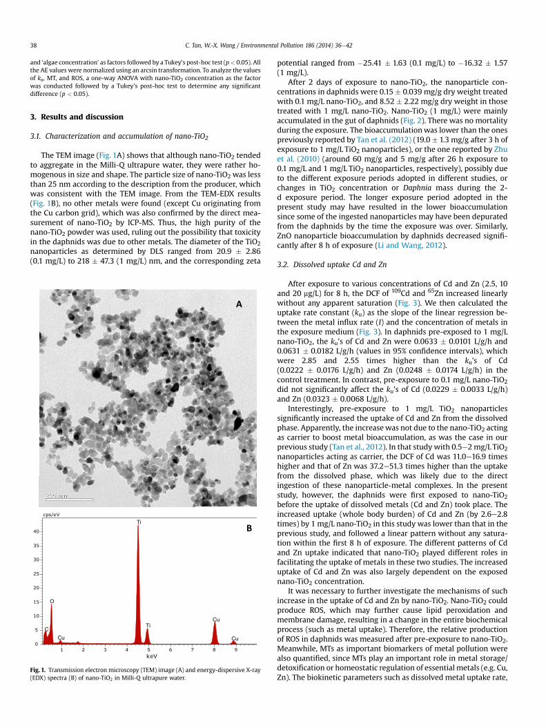
The TEM image (Fig. 1A) shows that although nano-TiO2 tendedto aggregate in the Milli-Q ultrapure water, they were rather ho-mogenous in size and shape. The particle size of nano-TiO2 was lessthan 25 nm according to the description from the producer, whichwas consistent with the TEM image. From the TEM-EDX results(Fig. 1B), no other metals were found (except Cu originating fromthe Cu carbon grid), which was also confirmed by the direct mea-surement of nano-TiO2 by ICP-MS. Thus, the high purity of thenano-TiO2 powder was used, ruling out the possibility that toxicityin the daphnids was due to other metals. The diameter of the TiO2nanoparticles as determined by DLS ranged from 20.9 � 2.86(0.1 mg/L) to 218 � 47.3 (1 mg/L) nm, and the corresponding zeta
Fig. 1. Transmission electron microscopy (TEM) image (A) and energy-dispersive X-ray(EDX) spectra (B) of nano-TiO2 in Milli-Q ultrapure water.
potential ranged from �25.41 � 1.63 (0.1 mg/L) to �16.32 � 1.57(1 mg/L).
After 2 days of exposure to nano-TiO2, the nanoparticle con-centrations in daphnids were 0.15 � 0.039 mg/g dry weight treatedwith 0.1 mg/L nano-TiO2, and 8.52 � 2.22 mg/g dry weight in thosetreated with 1 mg/L nano-TiO2. Nano-TiO2 (1 mg/L) were mainlyaccumulated in the gut of daphnids (Fig. 2). There was no mortalityduring the exposure. The bioaccumulationwas lower than the onespreviously reported by Tan et al. (2012) (19.0� 1.3 mg/g after 3 h ofexposure to 1 mg/L TiO2 nanoparticles), or the one reported by Zhuet al. (2010) (around 60 mg/g and 5 mg/g after 26 h exposure to0.1 mg/L and 1 mg/L TiO2 nanoparticles, respectively), possibly dueto the different exposure periods adopted in different studies, orchanges in TiO2 concentration or Daphnia mass during the 2-d exposure period. The longer exposure period adopted in thepresent study may have resulted in the lower bioaccumulationsince some of the ingested nanoparticles may have been depuratedfrom the daphnids by the time the exposure was over. Similarly,ZnO nanoparticle bioaccumulation by daphnids decreased signifi-cantly after 8 h of exposure (Li and Wang, 2012).
3.2. Dissolved uptake Cd and Zn
After exposure to various concentrations of Cd and Zn (2.5, 10and 20 mg/L) for 8 h, the DCF of 109Cd and 65Zn increased linearlywithout any apparent saturation (Fig. 3). We then calculated theuptake rate constant (ku) as the slope of the linear regression be-tween the metal influx rate (I) and the concentration of metals inthe exposure medium (Fig. 3). In daphnids pre-exposed to 1 mg/Lnano-TiO2, the ku’s of Cd and Zn were 0.0633 � 0.0101 L/g/h and0.0631 � 0.0182 L/g/h (values in 95% confidence intervals), whichwere 2.85 and 2.55 times higher than the ku’s of Cd(0.0222 � 0.0176 L/g/h) and Zn (0.0248 � 0.0174 L/g/h) in thecontrol treatment. In contrast, pre-exposure to 0.1 mg/L nano-TiO2did not significantly affect the ku’s of Cd (0.0229 � 0.0033 L/g/h)and Zn (0.0323 � 0.0068 L/g/h).
Interestingly, pre-exposure to 1 mg/L TiO2 nanoparticlessignificantly increased the uptake of Cd and Zn from the dissolvedphase. Apparently, the increasewas not due to the nano-TiO2 actingas carrier to boost metal bioaccumulation, as was the case in ourprevious study (Tan et al., 2012). In that study with 0.5e2mg/L TiO2nanoparticles acting as carrier, the DCF of Cd was 11.0e16.9 timeshigher and that of Zn was 37.2e51.3 times higher than the uptakefrom the dissolved phase, which was likely due to the directingestion of these nanoparticle-metal complexes. In the presentstudy, however, the daphnids were first exposed to nano-TiO2before the uptake of dissolved metals (Cd and Zn) took place. Theincreased uptake (whole body burden) of Cd and Zn (by 2.6e2.8times) by 1 mg/L nano-TiO2 in this study was lower than that in theprevious study, and followed a linear pattern without any satura-tion within the first 8 h of exposure. The different patterns of Cdand Zn uptake indicated that nano-TiO2 played different roles infacilitating the uptake of metals in these two studies. The increaseduptake of Cd and Zn was also largely dependent on the exposednano-TiO2 concentration.
It was necessary to further investigate the mechanisms of suchincrease in the uptake of Cd and Zn by nano-TiO2. Nano-TiO2 couldproduce ROS, which may further cause lipid peroxidation andmembrane damage, resulting in a change in the entire biochemicalprocess (such as metal uptake). Therefore, the relative productionof ROS in daphnids was measured after pre-exposure to nano-TiO2.Meanwhile, MTs as important biomarkers of metal pollution werealso quantified, since MTs play an important role in metal storage/detoxification or homeostatic regulation of essential metals (e.g. Cu,Zn). The biokinetic parameters such as dissolved metal uptake rate,
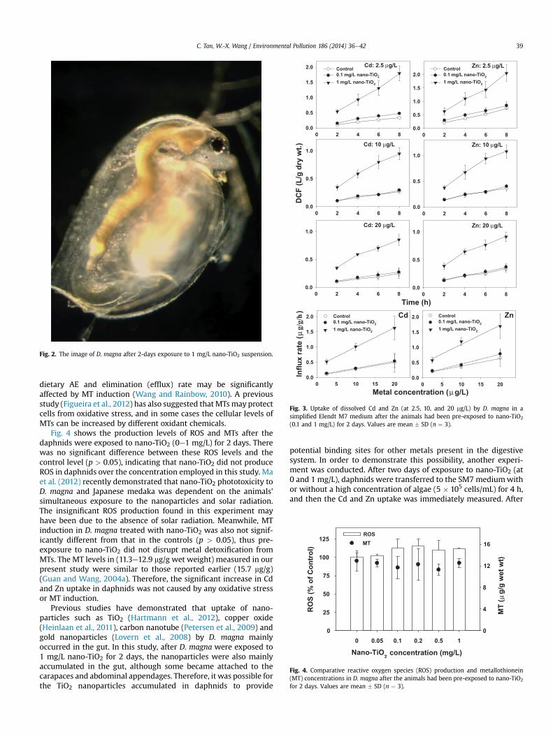
Fig. 2. The image of D. magna after 2-days exposure to 1 mg/L nano-TiO2 suspension.
Fig. 3. Uptake of dissolved Cd and Zn (at 2.5, 10, and 20 mg/L) by D. magna in asimplified Elendt M7 medium after the animals had been pre-exposed to nano-TiO2
(0.1 and 1 mg/L) for 2 days. Values are mean � SD (n ¼ 3).
Fig. 4. Comparative reactive oxygen species (ROS) production and metallothionein(MT) concentrations in D. magna after the animals had been pre-exposed to nano-TiO2
for 2 days. Values are mean � SD (n ¼ 3).
C. Tan, W.-X. Wang / Environmental Pollution 186 (2014) 36e42 39
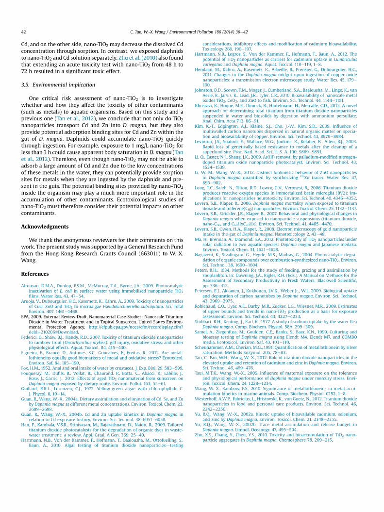
dietary AE and elimination (efflux) rate may be significantlyaffected by MT induction (Wang and Rainbow, 2010). A previousstudy (Figueira et al., 2012) has also suggested thatMTsmay protectcells from oxidative stress, and in some cases the cellular levels ofMTs can be increased by different oxidant chemicals.
Fig. 4 shows the production levels of ROS and MTs after thedaphnids were exposed to nano-TiO2 (0e1 mg/L) for 2 days. Therewas no significant difference between these ROS levels and thecontrol level (p > 0.05), indicating that nano-TiO2 did not produceROS in daphnids over the concentration employed in this study. Maet al. (2012) recently demonstrated that nano-TiO2 phototoxicity toD. magna and Japanese medaka was dependent on the animals’simultaneous exposure to the nanoparticles and solar radiation.The insignificant ROS production found in this experiment mayhave been due to the absence of solar radiation. Meanwhile, MTinduction in D. magna treated with nano-TiO2 was also not signif-icantly different from that in the controls (p > 0.05), thus pre-exposure to nano-TiO2 did not disrupt metal detoxification fromMTs. The MT levels in (11.3e12.9 mg/g wet weight) measured in ourpresent study were similar to those reported earlier (15.7 mg/g)(Guan and Wang, 2004a). Therefore, the significant increase in Cdand Zn uptake in daphnids was not caused by any oxidative stressor MT induction.
Previous studies have demonstrated that uptake of nano-particles such as TiO2 (Hartmann et al., 2012), copper oxide(Heinlaan et al., 2011), carbon nanotube (Petersen et al., 2009) andgold nanoparticles (Lovern et al., 2008) by D. magna mainlyoccurred in the gut. In this study, after D. magna were exposed to1 mg/L nano-TiO2 for 2 days, the nanoparticles were also mainlyaccumulated in the gut, although some became attached to thecarapaces and abdominal appendages. Therefore, it was possible forthe TiO2 nanoparticles accumulated in daphnids to provide
potential binding sites for other metals present in the digestivesystem. In order to demonstrate this possibility, another experi-ment was conducted. After two days of exposure to nano-TiO2 (at0 and 1 mg/L), daphnids were transferred to the SM7 mediumwithor without a high concentration of algae (5 � 105 cells/mL) for 4 h,and then the Cd and Zn uptake was immediately measured. After
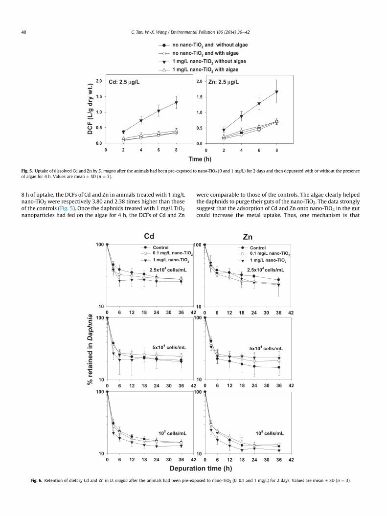
Fig. 5. Uptake of dissolved Cd and Zn by D. magna after the animals had been pre-exposed to nano-TiO2 (0 and 1 mg/L) for 2 days and then depurated with or without the presenceof algae for 4 h. Values are mean � SD (n ¼ 3).
C. Tan, W.-X. Wang / Environmental Pollution 186 (2014) 36e4240
8 h of uptake, the DCFs of Cd and Zn in animals treated with 1 mg/Lnano-TiO2 were respectively 3.80 and 2.38 times higher than thoseof the controls (Fig. 5). Once the daphnids treated with 1 mg/L TiO2nanoparticles had fed on the algae for 4 h, the DCFs of Cd and Zn
Fig. 6. Retention of dietary Cd and Zn in D. magna after the animals had been pre-exp
were comparable to those of the controls. The algae clearly helpedthe daphnids to purge their guts of the nano-TiO2. The data stronglysuggest that the adsorption of Cd and Zn onto nano-TiO2 in the gutcould increase the metal uptake. Thus, one mechanism is that
osed to nano-TiO2 (0, 0.1 and 1 mg/L) for 2 days. Values are mean � SD (n ¼ 3).

Table 1The assimilation efficiency (AE) of Cd and Zn in Daphnia magna fed on differentconcentrations of algae after pre-exposure to nano-TiO2. Mean � SD (n ¼ 3).
Nano-TiO2 (mg L�1) Algal concentration (cells mL�1) Cd AE (%) Zn AE (%)
0 2.5 � 0.16 � 104 33.2 � 4.7 32.6 � 3.50.1 29.1 � 3.3 25.6 � 2.71 27.0 � 5.9 28.4 � 8.40 5.0 � 0.32 � 104 22.3 � 2.6 17.6 � 4.90.1 25.2 � 3.0 21.3 � 4.11 23.3 � 9.0 23.7 � 3.00 1.0 � 0.06 � 105 17.1 � 1.6 13.6 � 2.80.1 15.8 � 2.9 14.8 � 3.31 13.8 � 1.8 11.5 � 1.8
C. Tan, W.-X. Wang / Environmental Pollution 186 (2014) 36e42 41
Daphna could accumulate significant amount of nano-TiO2 in thegut like a column (Fig. 1). When Daphnia take up the dissolved Cdand Zn from the ambient water, nano-TiO2 in the gut then absorbthese metals, resulting in more Cd and Zn accumulation in daph-nids. For daphnids not given the algae during the depurationperiod, the increased uptake of Cd and Zn was still significant,which indirectly demonstrates that the daphnids could not havepushed the nano-TiO2 out of their guts efficiently without the algae.Petersen et al. (2009) reported that D. magna were unable toexcrete the nanotubes after 24 h of depuration in either cleanartificial freshwater or filtered lake water, even though the latterhad substantial amounts of natural organic matter.
3.3. Assimilation of Cd and Zn
The depuration of Cd and Zn following a 20-min pulse ingestionof radiolabeled algae with or without pre-exposure to nano-TiO2 is
Fig. 7. Cumulative survival rates of D. magna exposed to 150 mg/L Cd or 1.5 mg/L Znafter the animals had been pre-exposed to nano-TiO2 (0, 0.1 and 1 mg/L) for 2 days.Values are mean � SD (n ¼ 3).
shown in Fig. 6. In general, Cd and Zn ingested by daphnids throughfeeding on radiolabeled algae were rapidly released within the first6 h, after which the loss wasmore gradual. The AEwas calculated asthe amount of radioactivity in the daphnids at 18 h divided by theamount of radioactivity ingested (Table 1). Pre-exposure to nano-TiO2 did not significantly affect the AEs of Cd (p ¼ 0.304) and Zn(p ¼ 0.983) in daphnids, and the AEs of Cd (p ¼ 0.001) and Zn(p ¼ 0.001) decreased significantly with increasing algal concen-tration from 2.5 � 104 cells/mL to 105 cells/mL, consistent withprevious findings (Yu and Wang, 2002b). In daphnids not exposedto nano-TiO2, the AE decreased from 33.2 � 4.7% to 17.1 � 1.6% forCd and from 32.6� 3.5% to 13.6� 2.8% for Zn as algal concentrationincreased from 2.5�104 to 105 cells/mL. In daphnids that were pre-exposed to nano-TiO2, the AE decreased from 29.1 � 3.3% to13.8� 1.8% for Cd and from 28.4� 8.4% to 11.5�1.8% for Zn, as algalconcentration increased from 2.5 � 104 to 105 cells/mL. Therefore,accumulation of nano-TiO2 in daphnids did not affect the physio-logical processes of assimilation in daphnids (even though some ofthe digestive enzymes such as esterase and catalase may have beenaffected after exposure to nano-TiO2, Fouqueray et al., 2012), incontrast to the uptake of aqueous metals.
Typical gut passage time of food in D. magna is between 25 and50min (Peters, 1984), and nano-TiO2 may be pushed out by algae asindicated earlier. Therefore, nano-TiO2 may not affect the assimi-lation process of Cd and Zn in the guts of daphnids. In addition, itwas possible that the assimilation of Cd and Zn was faster than theadsorption of Cd and Zn onto nano-TiO2 in the guts of daphnids, orperhaps some of the Cd and Zn adsorbed onto nano-TiO2 weregradually released and assimilated by daphnids. Overall, exposureto nano-TiO2 did not affect the efficiencies of Cd and Zn assimilationfrom algae to D. magna. Previously, Lovern et al. (2007) tested thebehavioral and physiological changes in D. magna exposed to nano-TiO2 (30 nm at 2 mg/L), and found that the nano-TiO2 suspensioncaused no change in the hopping frequency, appendage movement,and heart rate. Noting the recovery of dissolvedmetal uptake in thepresence of algal food and the insignificant difference in AEs, wefurther speculate that pre-exposure to 1 mg/L nano-TiO2 for 2 daysdid not change the biochemical function of daphnids related tometal uptake through water and dietary phases.
3.4. Time-to-death test
Since nano-TiO2 could provide potential binding sites to Cd andZn, the uptake of the two metals could be enhanced to cause extrametal risks to D. magna. Therefore, a 24-h time-to-death test wasconducted. An interesting finding of this test was that pre-exposureto nano-TiO2 increased the toxicity of Cd and Zn in daphnids(Fig. 7). The animals treatedwith 1mg/L nano-TiO2 died 10 h earlierfor Cd and 2 h earlier for Zn than the controls. The cumulativesurvival rate of individuals treated with 1mg/L nano-TiO2 were 67%and 27% lower than those of the controls for Cd and Zn, respec-tively, at the end of 24-h test. For daphnids treated with 0.1 mg/LTiO2 nanoparticles, the cumulative survival rate was only 13% lowerfor Cd and no different for Zn compared to that of control. Theremay be several reasons to explain the enhanced toxicity. First, aftertwo days of exposure to nano-TiO2, the daphnids were under stressand became less tolerant of other contaminants, thus the enhancedtoxicity was the simple combination of nano-TiO2 toxicity and Cd orZn toxicity. Second, nano-TiO2 as binding sites facilitated the Cd andZn accumulation in the guts of daphnids, and these Cd and Zn maybe toxic to daphnids even without actual assimilation. Hartmannet al. (2012) found that addition of 2 mg/L nano-TiO2 did not in-fluence the Cd 50% effective concentration. In their study, thedaphnids were exposed to nano-TiO2 and Cd simultaneously. Onone side, nano-TiO2 could serve as a carrier facilitating the uptake of
C. Tan, W.-X. Wang / Environmental Pollution 186 (2014) 36e4242
Cd, and on the other side, nano-TiO2 may decrease the dissolved Cdconcentration through sorption. In contrast, we exposed daphnidsto nano-TiO2 and Cd solution separately. Zhu et al. (2010) also foundthat extending an acute toxicity test with nano-TiO2 from 48 h to72 h resulted in a significant toxic effect.
3.5. Environmental implication
One critical risk assessment of nano-TiO2 is to investigatewhether and how they affect the toxicity of other contaminants(such as metals) to aquatic organisms. Based on this study and aprevious one (Tan et al., 2012), we conclude that not only do TiO2nanoparticles transport Cd and Zn into D. magna, but they alsoprovide potential adsorption binding sites for Cd and Znwithin thegut of D. magna. Daphnids could accumulate nano-TiO2 quicklythrough ingestion. For example, exposure to 1 mg/L nano-TiO2 forless than 3 h could cause apparent body saturation in D. magna (Tanet al., 2012). Therefore, even though nano-TiO2 may not be able toadsorb a large amount of Cd and Zn due to the low concentrationsof these metals in the water, they can potentially provide sorptionsites for metals when they are ingested by the daphnids and pre-sent in the guts. The potential binding sites provided by nano-TiO2inside the organism may play a much more important role in theaccumulation of other contaminants. Ecotoxicological studies ofnano-TiO2 must therefore consider their potential impacts on othercontaminants.
Acknowledgments
We thank the anonymous reviewers for their comments on thiswork. The present study was supported by a General Research Fundfrom the Hong Kong Research Grants Council (663011) to W.-X.Wang.
References
Alrousan, D.M.A., Dunlop, P.S.M., McMurray, T.A., Byrne, J.A., 2009. Photocatalyticinactivation of E. coli in surface water using immobilised nanoparticle TiO2films. Water Res. 43, 47e54.
Aruoja, V., Dubourguier, H.C., Kasemets, K., Kahru, A., 2009. Toxicity of nanoparticlesof CuO, ZnO and TiO2 to microalgae Pseudokirchneriella subcapitata. Sci. TotalEnviron. 407, 1461e1468.
EPA, 2009. External Review Draft, Nanomaterial Case Studies: Nanoscale TitaniumDioxide in Water Treatment and in Topical Sunscreen. United States Environ-mental Protection Agency. http://cfpub.epa.gov/ncea/cfm/recordisplay.cfm?deid¼210206#Download.
Federici, G., Shaw, B.J., Handy, R.D., 2007. Toxicity of titanium dioxide nanoparticlesto rainbow trout (Oncorhynchus mykiss): gill injury, oxidative stress, and otherphysiological effects. Aquat. Toxicol. 84, 415e430.
Figueira, E., Branco, D., Antunes, S.C., Goncalves, F., Freitas, R., 2012. Are metal-lothioneins equally good biomarkers of metal and oxidative stress? Ecotoxicol.Environ. Saf. 84, 185e190.
Fox, H.M., 1952. Anal and oral intake of water by crustacea. J. Exp. Biol. 29, 583e599.Fouqueray, M., Dufils, B., Vollat, B., Chaurand, P., Botta, C., Abacci, K., Labille, J.,
Rose, J., Garric, J., 2012. Effects of aged TiO2 nanomaterial from sunscreen onDaphnia magna exposed by dietary route. Environ. Pollut. 163, 55e61.
Guillard, R.R.L., Lorenzen, C.J., 1972. Yellow-green algae with chlorophyllide C.J. Phycol. 8, 10e14.
Guan, R., Wang, W.-X., 2004a. Dietary assimilation and elimination of Cd, Se, and Znby Daphnia magna at different metal concentrations. Environ. Toxicol. Chem. 23,2689e2698.
Guan, R., Wang, W.-X., 2004b. Cd and Zn uptake kinetics in Daphnia magna inrelation to Cd exposure history. Environ. Sci. Technol. 38, 6051e6058.
Han, F., Kambala, V.S.R., Srinivasan, M., Rajarathnam, D., Naidu, R., 2009. Tailoredtitanium dioxide photocatalysts for the degradation of organic dyes in waste-water treatment: a review. Appl. Catal. A Gen. 359, 25e40.
Hartmann, N.B., Von der Kammer, F., Hofmann, T., Baalousha, M., Ottofuelling, S.,Baun, A., 2010. Algal testing of titanium dioxide nanoparticlesdtesting
considerations, inhibitory effects and modification of cadmium bioavailability.Toxicology 269, 190e197.
Hartmann, N.B., Legros, S., Von der Kammer, F., Hofmann, T., Baun, A., 2012. Thepotential of TiO2 nanoparticles as carriers for cadmium uptake in Lumbriculusvariegatus and Daphnia magna. Aquat. Toxicol. 118e119, 1e8.
Heinlaan, M., Kahru, A., Kasemets, K., Arbeille, B., Prensier, G., Dubourguier, H.C.,2011. Changes in the Daphnia magna midgut upon ingestion of copper oxidenanoparticles: a transmission electron microscopy study. Water Res. 45, 179e190.
Johnston, B.D., Scown, T.M., Moger, J., Cumberland, S.A., Baalousha, M., Linge, K., vanAerle, R., Jarvis, K., Lead, J.R., Tyler, C.R., 2010. Bioavailability of nanoscale metaloxides TiO2, CeO2, and ZnO to fish. Environ. Sci. Technol. 44, 1144e1151.
Khosravi, K., Hoque, M.E., Dimock, B., Hintelmann, H., Metcalfe, C.D., 2012. A novelapproach for determining total titanium from titanium dioxide nanoparticlessuspended in water and biosolids by digestion with ammonium persulfate.Anal. Chim. Acta 713, 86e91.
Kim, K.-T., Edgington, A.J., Klaine, S.J., Cho, J.-W., Kim, S.D., 2009. Influence ofmultiwalled carbon nanotubes dispersed in natural organic matter on specia-tion and bioavailability of copper. Environ. Sci. Technol. 43, 8979e8984.
Levinton, J.S., Suatoni, E., Wallace, W.G., Junkins, R., Kelaher, B., Allen, B.J., 2003.Rapid loss of genetically based resistance to metals after the cleanup of asuperfund site. Proc. Natl. Acad. Sci. U. S. A. 100, 9889e9891.
Li, Q., Easter, N.J., Shang, J.K., 2009. As(III) removal by palladium-modified nitrogen-doped titanium oxide nanoparticle photocatalyst. Environ. Sci. Technol. 43,1534e1539.
Li, W.-M., Wang, W.-X., 2012. Distinct biokinetic behavior of ZnO nanoparticlesin Daphnia magna quantified by synthesizing 65Zn tracer. Water Res. 47,895e902.
Long, T.C., Saleh, N., Tilton, R.D., Lowry, G.V., Veronesi, B., 2006. Titanium dioxideproduces reactive oxygen species in immortalized brain microglia (BV2): im-plications for nanoparticles neurotoxicity. Environ. Sci. Technol. 40, 4346e4352.
Lovern, S.B., Klaper, R., 2006. Daphnia magna mortality when exposed to titaniumdioxide and fullerene(C60) nanoparticles. Environ. Toxicol. Chem. 25, 1132e1137.
Lovern, S.B., Strickler, J.R., Klaper, R., 2007. Behavioral and physiological changes inDaphnia magna when exposed to nanoparticle suspensions (titanium dioxide,nano-C60, and C60HxC70Hx). Environ. Sci. Technol. 41, 4465e4470.
Lovern, S.B., Owen, H.A., Klaper, R., 2008. Electron microscopy of gold nanoparticleintake in the gut of Daphnia magna. Nanotoxicology 2, 43e48.
Ma, H., Brennan, A., Diamond, S.A., 2012. Phototoxicity of TiO2 nanoparticles undersolar radiation to two aquatic species: Daphnia magna and Japanese medaka.Environ. Toxicol. Chem. 31, 1621e1629.
Nagaveni, K., Sivalingam, G., Hegde, M.S., Madras, G., 2004. Photocatalytic degra-dation of organic compounds over combustion-synthesized nano-TiO2. Environ.Sci. Technol. 38, 1600e1604.
Peters, R.H., 1984. Methods for the study of feeding, grazing and assimilation byzooplankton. In: Downing, J.A., Rigler, R.H. (Eds.), A Manual on Methods for theAssessment of Secondary Productivity in Fresh Waters. Blackwell Scientific,pp. 336e412.
Petersen, E.J., Akkanen, J., Kukkonen, J.V.K., Weber Jr., W.J., 2009. Biological uptakeand depuration of carbon nanotubes by Daphnia magna. Environ. Sci. Technol.43, 2969e2975.
Robichaud, C.O., Uyar, A.E., Darby, M.R., Zucker, L.G., Wiesner, M.R., 2009. Estimatesof upper bounds and trends in nano-TiO2 production as a basis for exposureassessment. Environ. Sci. Technol. 43, 4227e4233.
Stobbart, R.H., Keating, J., Earl, R., 1977. A study of sodium uptake by the water fleaDaphnia magna. Comp. Biochem. Physiol. 58A, 299e309.
Samel, A., Ziegenfuss, M., Goulden, C.E., Banks, S., Baer, K.N., 1999. Culturing andbioassay testing of Daphnia magna using Elendt M4, Elendt M7, and COMBOmedia. Ecotoxicol. Environ. Saf. 43, 103e110.
Scheuhammer, A.M., Cherian, M.G., 1991. Quantification of metallothionein by silversaturation. Methods Enzymol. 205, 78e83.
Tan, C., Fan, W.H., Wang, W.-X., 2012. Role of titanium dioxide nanoparticles in theelevated uptake and retention of cadmium and zinc in Daphnia magna. Environ.Sci. Technol. 46, 469e476.
Tsui, M.T.K., Wang, W.-X., 2005. Influence of maternal exposure on the toleranceand physiological performance of Daphnia magna under mercury stress. Envi-ron. Toxicol. Chem. 24, 1228e1234.
Wang, W.-X., Rainbow, P.S., 2010. Significance of metallothioneins in metal accu-mulation kinetics in marine animals. Comp. Biochem. Physiol. C152, 1e8.
Westerhoff, A.W.P., Fabricius, L., Hristovski, K., von Goetz, N., 2012. Titanium dioxidenanoparticles in food and personal care products. Environ. Sci. Technol. 46,2242e2250.
Yu, R.Q., Wang, W.-X., 2002a. Kinetic uptake of bioavailable cadmium, selenium,and zinc by Daphnia magna. Environ. Toxicol. Chem. 21, 2348e2355.
Yu, R.Q., Wang, W.-X., 2002b. Trace metal assimilation and release budget inDaphnia magna. Limnol. Oceanogr. 47, 495e504.
Zhu, X.S., Chang, Y., Chen, Y.S., 2010. Toxicity and bioaccumulation of TiO2 nano-particle aggregates in Daphnia magna. Chemosphere 78, 209e215.