Modes of Disintegration of Solid Foods in Simulated Gastric Environment
11
ORIGINAL ARTICLE Modes of Disintegration of Solid Foods in Simulated Gastric Environment Fanbin Kong & R. Paul Singh Received: 8 January 2009 / Accepted: 20 May 2009 / Published online: 23 June 2009 # The Author(s) 2009. This article is published with open access at Springerlink.com Abstract A model stomach system was used to investigate disintegration of various foods in simulated gastric envi- ronment. Food disintegration modes and typical disintegra- tion profiles are summarized in this paper. Mechanisms contributing to the disintegration kinetics of different foods were investigated as related to acidity, temperature, and enzymatic effect on the texture and changes in microstruc- ture. Food disintegration was dominated by either fragmen- tation or erosion, depending on the physical forces acting on food and the cohesive force within the food matrix. The internal cohesive forces changed during digestion as a result of water penetration and acidic and enzymatic hydrolysis. When erosion was dominant, the disintegration data (weight retention vs. disintegration time) may be expressed with exponential, sigmoidal, and delayed- sigmoidal profiles. The different profiles are the result of competition among the rates of water absorption, texture softening, and erosion. A linear-exponential equation was used to describe the different disintegration curves with good fit. Acidity and temperature of gastric juice showed a synergistic effect on carrot softening, while pepsin was the key factor in disintegrating high-protein foods. A study of the change of carrot microstructure during digestion indicated that degradation of the pectin and cell wall was responsible for texture softening that contributed to the sigmoidal profile of carrot disintegration. Keywords Stomach . Food digestion . Disintegration . Texture . Microstructure . Modeling Introduction Recent developments in food processing have focused on manufacturing foods for healthy benefits such as targeting release of nutrients in desired sites of the human gastrointestinal (GI) tract. For this purpose, it is critical to understand how foods break down during digestion in the GI tract. The stomach is a major compartment where the size of food particulates is reduced after oral mastication. From an engineering perspective, the stom- ach is a receptacle, a grinder, a mixer, and a pump that controls the digestion process. 1 The knowledge and any capability to predict how a food may disintegrate in the stomach are important for developing new food products with novel health benefits. However, there remains a notable lack of understanding about the kinetics of food disintegration and its relation to food texture and micro- structure (that define the material properties) resulting from various food processing. Studies in medicine, pharmacy, and nutrition have demonstrated that disintegration of food in the stomach is a complex process. Food is broken into small particulates and molecules due to both physical forces and chemical reactions. 1, 2 Physical forces include mechanical and hydrodynamic forces present in the stomach, resulting from the peristaltic movement of the stomach wall. Stomach contractions generate fluid flow of the gastric content that cause a shearing effect on the food surface. 3 Kinetics of food digestion depend on the chemical and physical characteristics of food. Our previ- ous study has shown that food disintegration rate is Food Biophysics (2009) 4:180–190 DOI 10.1007/s11483-009-9116-9 F. Kong : R. P. Singh (*) Department of Biological and Agricultural Engineering, University of California, Davis, CA 95616, USA e-mail: [email protected] F. Kong : R. P. Singh Riddet Institute, Massey University, Palmerston North, New Zealand
Transcript of Modes of Disintegration of Solid Foods in Simulated Gastric Environment

ORIGINAL ARTICLE
Modes of Disintegration of Solid Foods in SimulatedGastric Environment
Fanbin Kong & R. Paul Singh
Received: 8 January 2009 /Accepted: 20 May 2009 /Published online: 23 June 2009# The Author(s) 2009. This article is published with open access at Springerlink.com
Abstract A model stomach system was used to investigatedisintegration of various foods in simulated gastric envi-ronment. Food disintegration modes and typical disintegra-tion profiles are summarized in this paper. Mechanismscontributing to the disintegration kinetics of different foodswere investigated as related to acidity, temperature, andenzymatic effect on the texture and changes in microstruc-ture. Food disintegration was dominated by either fragmen-tation or erosion, depending on the physical forces actingon food and the cohesive force within the food matrix. Theinternal cohesive forces changed during digestion as aresult of water penetration and acidic and enzymatichydrolysis. When erosion was dominant, the disintegrationdata (weight retention vs. disintegration time) may beexpressed with exponential, sigmoidal, and delayed-sigmoidal profiles. The different profiles are the result ofcompetition among the rates of water absorption, texturesoftening, and erosion. A linear-exponential equation wasused to describe the different disintegration curves withgood fit. Acidity and temperature of gastric juice showed asynergistic effect on carrot softening, while pepsin was thekey factor in disintegrating high-protein foods. A study ofthe change of carrot microstructure during digestionindicated that degradation of the pectin and cell wall wasresponsible for texture softening that contributed to thesigmoidal profile of carrot disintegration.
Keywords Stomach . Food digestion . Disintegration .
Texture .Microstructure . Modeling
Introduction
Recent developments in food processing have focused onmanufacturing foods for healthy benefits such as targetingrelease of nutrients in desired sites of the humangastrointestinal (GI) tract. For this purpose, it is criticalto understand how foods break down during digestion inthe GI tract. The stomach is a major compartment wherethe size of food particulates is reduced after oralmastication. From an engineering perspective, the stom-ach is a receptacle, a grinder, a mixer, and a pump thatcontrols the digestion process.1 The knowledge and anycapability to predict how a food may disintegrate in thestomach are important for developing new food productswith novel health benefits. However, there remains anotable lack of understanding about the kinetics of fooddisintegration and its relation to food texture and micro-structure (that define the material properties) resultingfrom various food processing.
Studies in medicine, pharmacy, and nutrition havedemonstrated that disintegration of food in the stomachis a complex process. Food is broken into smallparticulates and molecules due to both physical forcesand chemical reactions.1,2 Physical forces includemechanical and hydrodynamic forces present in thestomach, resulting from the peristaltic movement of thestomach wall. Stomach contractions generate fluid flow ofthe gastric content that cause a shearing effect on the foodsurface.3 Kinetics of food digestion depend on thechemical and physical characteristics of food. Our previ-ous study has shown that food disintegration rate is
Food Biophysics (2009) 4:180–190DOI 10.1007/s11483-009-9116-9
F. Kong :R. P. Singh (*)Department of Biological and Agricultural Engineering,University of California,Davis, CA 95616, USAe-mail: [email protected]
F. Kong :R. P. SinghRiddet Institute, Massey University,Palmerston North, New Zealand
significantly affected by the initial texture and its changeduring digestion.4 However, more studies are needed tounderstand how the reactions occurring during digestionchange food material properties and subsequently influ-ence its disintegration.
In the stomach, gastric juice is secreted from glandslining the stomach containing gastric acid (HCl) anddigestive enzymes. HCl assists acid denaturation ofdigested food and activates pepsin. In the fasted state,intragastric pH in healthy subjects is in the 1.3–2.5 range,while eating can increase pH up to 7.5 Gastric juicecontains 0.8–1 mg/mL pepsin 6 that degrades food proteinsinto peptides. The gastric juice penetrates the food matrixand assists in digestion, but how the acidity and enzyme ofthe gastric juice as well as temperature affect disintegrationof food has not been fully understood.
We have developed a novel model of human stomach tostudy food disintegration kinetics.4 This model can providemeasurable force that simulates the hydrodynamic andmechanical forces acting on foods in vivo. The objectives ofthis study were (1) to investigate disintegration character-istics and related mechanisms of different foods using themodel system; (2) to study the role of temperature, acidity,and enzyme of gastric juice in food disintegration; and (3)to demonstrate how digestion changes food microstructureand subsequently influences its disintegration, using carrotas an example.
Materials and Methods
Materials and Sampling
Foods representing different categories were selected forthis study (Table 1). Most of the foods were purchased inlocal grocery stores. Baked and fried dough products wereprepared in the laboratory. Dough was prepared usingwheat flour (Gold medal, General Mills, MN, USA) andwater in a ratio of 100:55 (flour/water) and mixed using adough mixer (Classic Series Tilt-Head Stand Mixer,KitchenAid, St. Joseph, MI, USA) for 10 min. After restingfor 30 min, the dough was flattened into thin sheets of
1.5 cm thickness and baked in an oven (Hamilton BeachBrands, Inc., Washington, NC, USA) at 170°C for 30 min.Fried dough was prepared by cutting cylindrical samples(diam 10 mm×10 mm length) from the dough sheet andfried in an electric deep fryer (Presto FryDaddy, NationalPresto Industries, Inc. Eau Claire, WI, USA) at 180°C for6 min. Broiled beef steak was prepared by heating 2.5-cmthick sirloin steak in an oven at 190°C for 8 min.
The food sample size for disintegration trials wasdetermined according to observations and literaturevalues reported on the particle sizes after oral mastica-tion.7, 8 Beef jerky, peanut, almond, fried dough, breadcrouton, and broiled beef steak were carefully cut intosmall samples with length 3–6 mm depending on theoriginal shape and dimensions. Cylinder samples (6 mmdiam×6 mm length) were obtained from carrot using acore cutter.
Simulated gastric juice and saliva were prepared asdescribed in a previous paper.4 All chemicals werepurchased from Sigma-Aldrich, Inc. (USA).
Model Stomach System and Disintegration Test
The model stomach system described in our previouspaper4 was slightly modified. A stainless steel annularcontainer was placed in the center of the glass chamber. Thesimulated gastric juice and plastic beads were filled in theannular container. This modification improved the unifor-mity of temperature in the simulated gastric juice andallowed easy addition and removal of beads and gastricjuice (Figure 1). The sample holder arm was also modifiedto contain four sample hooks so as to accommodate foursamples at the same time.
The operating procedures were described in ourprevious paper.4 The samples were first soaked in thesimulated saliva for 30 s (37°C) to incorporate the effectof oral step on food disintegration. Then, the samples werehooked in each sample holder. Glue (Loctite super glue,Henkel Consumer Adhesives, Inc., Avon, OH, USA) wasapplied to fix samples when necessary. The samples weresubjected to a continuous force in the model and theweight changes were measured periodically with a timeinterval between 1 and 30 min depending upon thedisintegration rate. Weight retention ratio was calculatedas:
yt ¼ Wt
W0ð1Þ
where W0 is initial weight (gram) and Wt (gram) is thesample weight after time t (minute). In this equation, it shouldbe noted that Wt represents the weight of sample due to the
Table 1 Foods selected for disintegration study
Category Foods
Meat products Beef jerky, ham steak, broiled beef steak
Nuts Raw peanuts, raw almonds
Fruits Baby carrots
Baked products Bread crouton, baked dough
Fried products Fried dough products (w/o yeast)
Food Biophysics (2009) 4:180–190 181

simultaneous uptake of gastric juice and loss of its solidcomponents due to disintegration and leaching.
Static Soaking Test
A static soaking test was employed to investigate waterabsorption and the change in texture of foods duringdigestion. Almond, carrot, and ham samples (10×10×4 mm) were soaked in simulated gastric juice at 37°C for2 h. The weight changes were determined to calculate waterabsorption ratio. A penetration test was used to evaluatehardness of sample before and after soaking. A flat-endedneedle probe (2 mm diameter) was fitted to the TextureAnalyzer TA-XT2 (Texture Technologies Corp., Scarsdale,NY/Stable Micro Systems, Godalming, Surrey, UK)equipped with a load cell of 50 kg. The probe traveled to2 mm depth into the planer surface of samples at a speed of0.5 mm/s. The maximum force recorded during the test isreferred to as the hardness of sample.
To investigate the water penetration in a carrot sampleduring digestion, methylene blue was added to the gastric
juice. During static soaking test of carrots, a visible regionshowing water penetration was clearly observed.
Microscopy Imaging
Light microscopy (LM) and transmission electron micros-copy (TEM) were used to investigate changes in themicrostructure of partially digested carrot. Carrot wassoaked in simulated gastric juice for 20 min at 37°C.Samples were pre-fixed in Karnovsky's fixative (2.5%glutaraldehyde and 2.0% paraformaldehyde) in 0.08 mol/Lphosphate buffer (pH7.3). The tissues were secondarilyfixed in 1% osmium tetroxide buffered in 0.1 M sodiumphosphate buffer for 2 h. After thoroughly washing withcold double-distilled water, the samples were incubated in0.1% tannic acid in water for 30 min on ice. Then, sampleswere rinsed three times with cold double-distilled water andincubated on ice in 2% aqueous uranyl acetate for 1 h. Therinsed tissue blocks were dehydrated using increasingconcentrations of acetone: 10%, 30%, 50%, and 70% for20 min each, and three changes of 95% acetone for 10 mineach, followed by two changes of 100% acetone for 10 mineach. The samples were embedded in a 100% Epon-aralditeresin and polymerized overnight at 70°C.
The blocks were sectioned to obtain 400-nm-thick samplesusing a Diatome diamond knife. Dried sections were stainedfor 30–60 s using methylene blue and Azure B combinationstain. The sections were rinsed, dried, coverslipped, andviewed on the LM. For the TEM, the block was trimmed to thearea of interest (<1 mm), and 60-nm (silver) sections were cutand collected on a 150-mesh copper grid. They were dried andpost-stained using the standard double staining protocol of 4%(w/v) uranyl acetate in 70% (w/v) aqueous methanol 20 minfollowed by lead citrate for 2 min. The sections were allowedto dry and viewed in a Philips 120 BioTwin ElectronMicroscope at 80 kV (Fei Company, Hillsboro, OR, USA)equipped with a Gatan/MegaScan, model 794/20 digitalcamera (Gatan Inc., Pleasanton, CA, USA).
Data Analysis
Weight retention data (yt vs. t) were analyzed and fitted intoan empirical model. Modeling of data was performed withnonlinear and multiple regression analysis functions ofExcel software. Estimates for parameters were obtained byminimizing the mean square error (MSE):
MSE ¼Pn
i¼1yi � yið Þ2
nð2Þ
where yi is the observed value, yi is the fitted value, and n isthe number of observation points of an experiment. MSE isa measure of the goodness of fit. In addition, the correlation
To water bath
AA
Model stomach
11
8
7
5
4
3
1
To texture analyzer
To water bath
From water bath
2
6
9
Connected to loadcell
A — A
25 cm
15 cm
1
3
9
4
2
6.5 cm
Fig. 1 Diagram of modified model stomach system and forcemeasurement apparatus: 1 simulated gastric juice (37°C) and beads;2 jacket filled with water; 3 copper coil; 4 food sample; 5 chamber; 6turntable; 7 ball bearing; 8 wheel; 9 cotton wire; 10 pulley; 11laboratory stand
182 Food Biophysics (2009) 4:180–190

coefficient (r) is also used as a criterion for evaluating theperformance of the models, and closeness to a value of 1 isan effective and practical measure of the validity of modelprediction.
A significance test was conducted using analysis ofvariance (ANOVA) in the GLM procedure of the SASSystem. Differences between group means were analyzedby Duncan’s multiple range test. Statistical significance wasset at a probability level of 0.05.
Results and Discussion
Fragmentation and Erosion
We observed two major mechanisms responsible for fooddisintegration in a simulated gastric environment, namelyfragmentation and erosion. Fragmentation is the breakageof a food sample into several pieces of relatively large-sizeparticulates. This phenomenon occurred when the appliedstress is greater than a critical stress. The critical stress isrelated to the internal cohesive forces that hold the foodmatrix together. Surface erosion is defined as the wearingaway of food surface by an impinging gastric fluidcontaining food solids that causes normal impact, friction,and shear forces acting on the food surface;4 it is thedominant mechanism when the applied stress is less thanthe critical stress. The erosion rate is therefore dependentupon the cohesive strength of the food matrix and thephysical forces applied on the food.
Marciani et al.2 measured stomach force exerted on agargel beads (diameter 1.27 cm) and reported a maximumforce of 0.65 N generated by the antral walls of a humanstomach. The magnitude of the stomach forces is influencedby biological factors such as age, body mass index,hormonal factor, gender, blood glucose level, posture,stress and depression, and diseased states. The stomachcan also detect and react to the food properties such asviscosity by increasing or decreasing the force of contrac-tion and thus generate an appropriate disintegration rate.9
Therefore, the disintegration mode for the same food mayvary significantly depending on an individual person. Anadvantage of our model stomach system is its ability toadjust the mechanical force applied on food samples bychanging the concentration of beads added to the simulatedgastric juice thus to simulate the disintegration of foods fora wide range of forces. To compare the disintegration modeof various foods, we tested different foods at the same forceof about 0.25 N on a cylindrical sample (6 mm diam×6 mmlength) which is approximately in a similar range of themaximum force generated by the movement of the stomachwall. The applied force was controlled by operating theapparatus at the same rotational speed (30 rpm), and using
the same amount of gastric juice (176 mL) and beads(324 g), which made up 500 mL of simulated gastriccontent. Our preliminary observations showed that thedisintegration mode varied greatly among different foodsand was closely related to the initial food texture and itschange during oral mastication and stomach digestion.Fragmentation dominated the disintegration of bakeryproducts, such as a fried snack food, crouton, and bakeddough. They became soft and easy to break apart aftercombination of saliva and gastric juice. Meat products, suchas broiled beef steak, also demonstrated mainly fragmenta-tion during digestion, as the connective tissue weredenatured during roasting, and the muscle fibers weredisconnected. Erosion was the major mechanism for thedisintegration of carrot and nuts.
Higher force induced faster erosion rate, and an exponen-tial relationship was observed between half time (t1/2) andthe applied mechanical force.4 Increase in the mechanicalforces applied on food may also change the disintegrationmode from erosion to fragmentation. On the other hand, afteringestion, the food texture may change as a result of waterabsorption, acid hydrolysis, and enzymatic reaction, leadingto a reduction in cohesive forces that hold a food matrixtogether. Subsequently, the dominant disintegration mecha-nisms may shift from erosion to fragmentation. Figure 2shows how a piece of crouton (7 mm length, weight 0.16 g)disintegrated in the model which contains only gastric juicewhen a force of 0.02 N was applied on the sample at thebeginning of the trial. It shows that initially the croutondisintegrated by erosion (Figure 2a, b). Water absorption ledto significant swelling and increase in its volume andsoftening of its texture. After about 30 min, the samplebroke at the place close to the hook (Figure 2c), indicatingthat the cohesive force decreased due to water absorption andit was lower than the applied force. It is recognized thaterosion and fragmentation may also occur simultaneously todisintegrate foods. Future studies will require understandingthe critical stress of various foods and the relationship withfood material properties, as well as their change duringstomach digestion. In the following sections of this paper, wewill focus on erosion, although the term “disintegration” willbe used throughout the paper to represent the change ofsample weight in the model stomach system.
Food Disintegration Profiles and Modeling
In our previous paper,4 we reported that the disintegrationprofile of carrot and ham showed either an exponential orsigmoidal decay. Specifically, the retention of the mass ofraw carrot during disintegration trial followed a sigmoidaldecrease with an early stage of slow disintegration, asecond stage of increased disintegration, and a third stageof diminishing disintegration rate. An exponential decay
Food Biophysics (2009) 4:180–190 183

with a rapid initial disintegration was observed in ham. Thedifference in the disintegration curves was hypothesizeddue to the competition between surface erosion and texturesoftening.4
Disintegration studies of additional types of foods haverevealed another type of food disintegration profile, which isdifferent from the two profiles previously suggested. In certaincases, we observed that the sample weight initially increased,leading to a weight retention ratio yt higher than 1, and then itgradually decreased. This profile is named as a delayed-sigmoidal curve, when after an initial increase, the weight
retention ratio decreased following a sigmoidal profile.Delayed-sigmoidal type of disintegration mainly occurredfor low-moisture food products, such as peanut, almond, andbeef jerky. During digestion, a significant amount of waterwas absorbed that exceeded the weight loss due to erosion,leading to an increase in the sample weight and volume. Invivo studies have shown that dietary oat can absorb waterand swell in stomach causing stomach distension andaffecting satiety.10 However, a quantitative characterizationof this phenomenon is lacking in the literature. Gammascintigraphy, which is commonly used clinically to evaluategastric emptying by counting the radioactivity of markedfoods, cannot measure the meal volume in the stomach.Recent improvements in MRI may allow in vivo measure-ment of gastric and meal volumes.9,11
Siegel’s modified power exponential equation, whichwas proposed to describe in vivo stomach emptying data,12
was used to fit the data obtained for carrot and ham in ourprevious study:4
yt ¼ 1� 1� e�kt� �b ð3Þ
In describing stomach emptying profile, yt is the fractionalfood retention at time t (minute), k is a parameter related tothe gastric emptying rate per minute, and β is a shapefactor. This model, however, cannot describe the delayed-sigmoidal profile. Therefore, we propose the use of a linear-exponential equation for describing the three differentprofiles observed in food disintegration:
yt ¼ 1þ k b tð Þ e�b t ð4ÞIn this expression, k and β are constants. k is dimensionlessand it represents the lag phase. A larger k is associated witha delayed-sigmoidal disintegration and a smaller k repre-sents an exponential decay. β (1/min) is a parameter thatmeasures the concavity of the weight retention vs. timerelationship (β>0). The pre-exponential term, (1+kβ t),generally predominates for a short time at the beginningof disintegration trial. As the trial continues, the exponentialfactor e−β t induces concavity in the weight retention vs.time profile. This model has advantages over the modifiedexponential model (Eq. 3) in that it captures initial increasein yt due to water absorption. A similar model wasproposed by Goetze et al.11 to describe changes in thevolume of stomach due to accommodation of food andsecretion of gastric fluid. The k and β values can be derivedusing regression analysis. When k and β are known, thehalf time, t1/2, can be calculated by regression analysis andusing yt=0.5.
Figure 3 shows typical delayed-sigmoidal disintegrationprofiles for beef jerky and raw almond as well as the modelcurves fitted with Eq. 4. Disintegration type, modelparameters, and half times for some representative foods
a
b
c
Fig. 2 Disintegration of bread crouton in the model stomach system:a erosion was the dominant mechanism in the beginning; b swellingof the sample; c rupture of the bread crouton after half hour ofrunning. Dashed arrow: fluid rotational direction; continuous arrow:sample
184 Food Biophysics (2009) 4:180–190

tested in the stomach system are presented in Table 2. TheMSE and r values indicate a good fit of the model equationwith the disintegration data. It can be seen that the half timevaries significantly with different foods, from a few minutesup to 12 h, depending upon the texture and microstructureof tested foods. It should be noted that although Eq. 4provides a good fit of the data of three disintegrationprofiles, it does not necessarily represent any specificmechanism. It is also important to acknowledge that inaddition to surface erosion, leaching of solids may occursimultaneously that contributes to the change of sampleweight. Thus the disintegration profiles summarized hereand the model equation 4 describe the overall effect ofweight change caused by absorption of gastric juice, surfaceerosion and release of the solids.
Reactions Occurring During Digestion That AffectDisintegration
To further elucidate the mechanisms that affect fooddisintegration rate, three foods were compared, namelyalmond, carrot, and ham, representing different types of
disintegration: delayed sigmoidal, sigmoidal, and exponen-tial, respectively (Table 2). A static soaking test wasconducted for the three foods to compare water absorption,volume change, and texture change. Almond absorbed thehighest amount of water, from originally 3.3% to 30.7%after soaking (wet basis). This is in agreement with thedifferent k values in Table 2 indicating that a greater waterabsorption occurred for almond (k>1) than carrot and ham(k<1). Furthermore, measurement of texture indicated thata significant reduction in the hardness occurred for almondand carrot but not for ham (Figure 4).
We postulate that water absorption, textual softening,and surface erosion are the major factors affecting thedisintegration rate and thus influencing the type of thedisintegration profile. Food is first chewed in the month,then swallowed and transited into stomach. In the stomach,gastric juice penetrates into a food particulate that softensits texture. The rate of water absorption depends on thephysical properties of foods such as food porosity,composition, and material properties. A high rate of waterabsorption contributes to a delayed-sigmoidal disintegrationprofile. When gastric fluid carrying acid and enzyme isabsorbed into the food, acid hydrolysis and enzymedegradation occur that further soften food texture andweaken internal bonds. Depending on the initial tempera-ture of the food, heat transfer takes place to equilibrate thefood temperature to 37°C, which is the optimum temper-ature for enzymatic activity for pepsin. All of thesephenomena—water absorption, heat transfer, and acid andenzymatic reactions contribute to degradation of foodtexture (tenderization) and weaken the strength of the foodmatrix that improves the disintegration rate increasesleaching of solids. The tenderization (or softening) startsfrom the food surface and gradually moves inside. When aphysical stress is applied (including both mechanical andhydrodynamic forces), surface erosion occurs. The erosionrate depends on the physical force and the internal cohesivestrength of the food matrix. The erosion is slow for rigidand tough foods but fast for foods with softer texture. Theshape of the disintegration profile is largely a result of acompetition between the penetration rates of tenderization
0
0.2
0.4
0.6
0.8
1
1.2
1.4
0 100 200 300 400 500Disintegration time (min)
Wt/W
0
Raw almond
Beef jerky
Fig. 3 Typical delayed-sigmoidal disintegration curves (n=4). Trialcondition, 500 mL of simulated gastric content (gastric juice 176 mL,beads 324 g); turntable rotation speed, 30 rpm
Table 2 Disintegration profiles and parameters of Eq. 4 for representative foods
Disintegration profile k β (1/min) MSE×103 r t1/2 (min)
Ham (ø 6×L6 mm cylinder) Exponential 0.484 0.260 2.181 1.00 4.3
Fried dough (6×6×6 mm) Exponential 0 0.121 0.548 0.998 5.8
Carrot (ø 6×L6 mm cylinder) Sigmoidal 0.994 0.105 2.141 1.00 16.0
Beef jerky (6×6×4 mm) Delayed-sigmoidal 2.16 0.027 3.89 0.97 94.9
Almond (6×6×4 mm) Delayed-sigmoidal 1.785 0.004 1.051 1.00 598.6
Peanut (6×6×4 mm) Delayed-sigmoidal 1.977 0.003 1.304 0.984 714.8
Trial condition, 500 mL of simulated gastric content (gastric juice 176 mL, beads 324 g); turntable rotation speed, 30 rpm
Food Biophysics (2009) 4:180–190 185
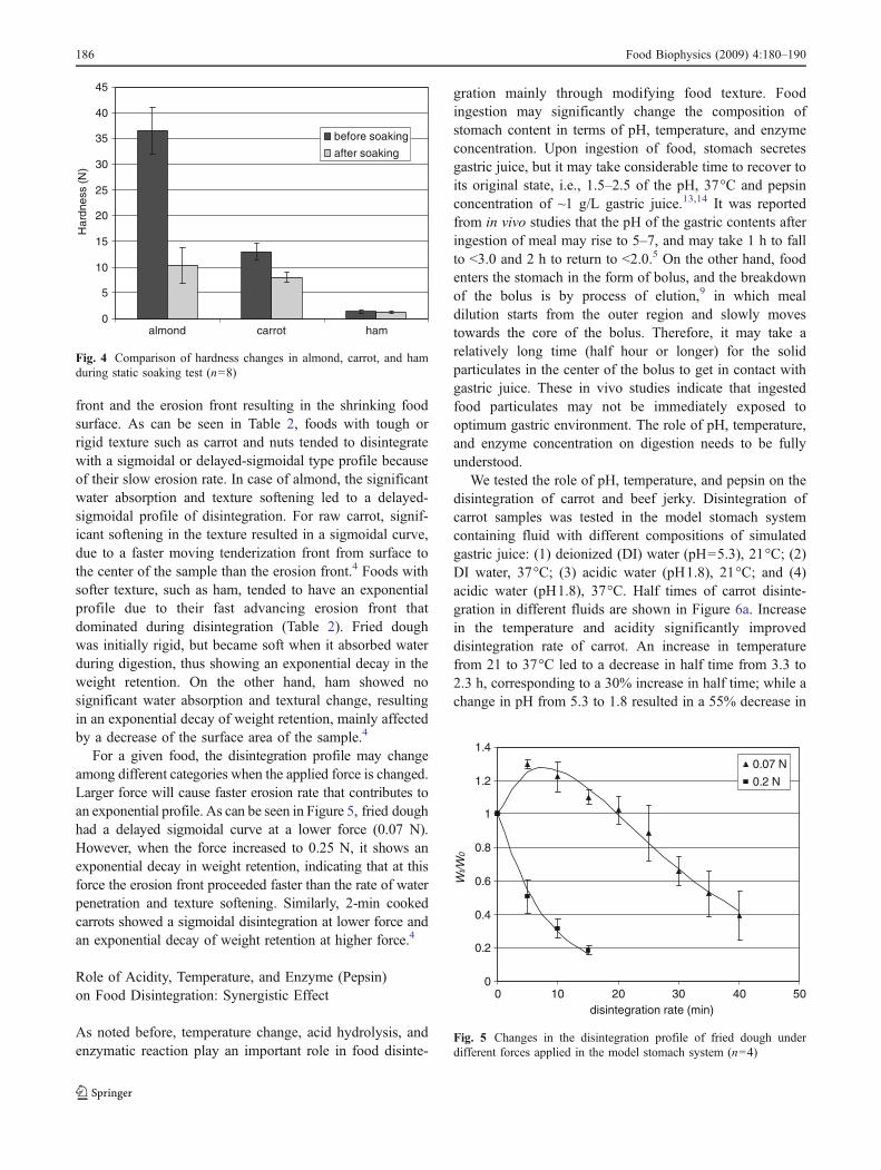
front and the erosion front resulting in the shrinking foodsurface. As can be seen in Table 2, foods with tough orrigid texture such as carrot and nuts tended to disintegratewith a sigmoidal or delayed-sigmoidal type profile becauseof their slow erosion rate. In case of almond, the significantwater absorption and texture softening led to a delayed-sigmoidal profile of disintegration. For raw carrot, signif-icant softening in the texture resulted in a sigmoidal curve,due to a faster moving tenderization front from surface tothe center of the sample than the erosion front.4 Foods withsofter texture, such as ham, tended to have an exponentialprofile due to their fast advancing erosion front thatdominated during disintegration (Table 2). Fried doughwas initially rigid, but became soft when it absorbed waterduring digestion, thus showing an exponential decay in theweight retention. On the other hand, ham showed nosignificant water absorption and textural change, resultingin an exponential decay of weight retention, mainly affectedby a decrease of the surface area of the sample.4
For a given food, the disintegration profile may changeamong different categories when the applied force is changed.Larger force will cause faster erosion rate that contributes toan exponential profile. As can be seen in Figure 5, fried doughhad a delayed sigmoidal curve at a lower force (0.07 N).However, when the force increased to 0.25 N, it shows anexponential decay in weight retention, indicating that at thisforce the erosion front proceeded faster than the rate of waterpenetration and texture softening. Similarly, 2-min cookedcarrots showed a sigmoidal disintegration at lower force andan exponential decay of weight retention at higher force.4
Role of Acidity, Temperature, and Enzyme (Pepsin)on Food Disintegration: Synergistic Effect
As noted before, temperature change, acid hydrolysis, andenzymatic reaction play an important role in food disinte-
gration mainly through modifying food texture. Foodingestion may significantly change the composition ofstomach content in terms of pH, temperature, and enzymeconcentration. Upon ingestion of food, stomach secretesgastric juice, but it may take considerable time to recover toits original state, i.e., 1.5–2.5 of the pH, 37°C and pepsinconcentration of ~1 g/L gastric juice.13,14 It was reportedfrom in vivo studies that the pH of the gastric contents afteringestion of meal may rise to 5–7, and may take 1 h to fallto <3.0 and 2 h to return to <2.0.5 On the other hand, foodenters the stomach in the form of bolus, and the breakdownof the bolus is by process of elution,9 in which mealdilution starts from the outer region and slowly movestowards the core of the bolus. Therefore, it may take arelatively long time (half hour or longer) for the solidparticulates in the center of the bolus to get in contact withgastric juice. These in vivo studies indicate that ingestedfood particulates may not be immediately exposed tooptimum gastric environment. The role of pH, temperature,and enzyme concentration on digestion needs to be fullyunderstood.
We tested the role of pH, temperature, and pepsin on thedisintegration of carrot and beef jerky. Disintegration ofcarrot samples was tested in the model stomach systemcontaining fluid with different compositions of simulatedgastric juice: (1) deionized (DI) water (pH=5.3), 21°C; (2)DI water, 37°C; (3) acidic water (pH1.8), 21°C; and (4)acidic water (pH1.8), 37°C. Half times of carrot disinte-gration in different fluids are shown in Figure 6a. Increasein the temperature and acidity significantly improveddisintegration rate of carrot. An increase in temperaturefrom 21 to 37°C led to a decrease in half time from 3.3 to2.3 h, corresponding to a 30% increase in half time; while achange in pH from 5.3 to 1.8 resulted in a 55% decrease in
0
0.2
0.4
0.6
0.8
1
1.2
1.4
0 10 20 30 40 50disintegration rate (min)
Wt/W
0
0.07 N
0.2 N
Fig. 5 Changes in the disintegration profile of fried dough underdifferent forces applied in the model stomach system (n=4)
0
5
10
15
20
25
30
35
40
45
almond carrot ham
Har
dnes
s (N
)
before soaking
after soaking
Fig. 4 Comparison of hardness changes in almond, carrot, and hamduring static soaking test (n=8)
186 Food Biophysics (2009) 4:180–190

the half time (from 3.3 to 1.5 h). When both temperatureand pH were adjusted, the half time decreased to 0.47 h,corresponding to 86% reduction. These results indicate asignificant synergistic effect of pH and temperature forcarrot disintegration. Figure 6b shows the hardness of carrotsamples after soaking in different fluids. Generally, the halftime (Figure 6a) and the hardness of soaked carrot(Figure 6b) have a good correlation: when carrots weresoaked in water at 37°C and pH1.8 for 1 h, the hardnessdecreased the most, which resulted in the most rapiddisintegration.
Figure 7 shows the half time of carrot disintegration insimulated gastric juice at four temperatures: 21, 29, 37, and45°C. These temperatures were selected to cover thesituations that may occur in the gastric environment forvarying durations of time. The force applied was 0.13 N. The
half time decreased rapidly when the temperature increasedfrom to 21 to 45°C. A power relationship existed betweenhalf time and temperature. When temperature was higherthan 37°C, the change in the rate of disintegration wasminor.
Experiments comparing the disintegration of carrot in asimulated stomach environment with/without pepsin indi-cated that pepsin had no significant influence. However, theaddition of 1% pepsin improved the disintegration rate ofbeef jerky by 21%, and the half time decreased from 1.9 to1.5 h. This result was expected because pepsin functions tobreak up proteins into smaller peptides, while carrotcontains only a small amount of protein (<1%) comparedto beef jerky (~50%). It is also known that the optimumproteolysis of protein substrates by pepsin is achieved at pHof about 2.15
0.0
0.5
1.0
1.5
2.0
2.5
3.0
3.5
4.0
Carrots in DIwater, 21 oC
Carrots in DIwater, 37 oC
Carrots in pH=1.8water, 21 oC
Carrots in pH=1.8water, 37 oC
Hal
f tim
e (h
rs)
0
10
20
30
40
50
60
70
Raw carrots Carrotssoaked for 1
hr in DI water,21 oC
Carrotssoaked for 1
hr in DI water,37 oC
Carrotssoaked for 1hr in pH=1.8water, 21 oC
Carrotssoaked for 1hr in pH=1.8water, 37 oC
Har
dn
ess
(N)
a
b
Fig. 6 Changes in the disinte-gration rates (a) and hardness(b) of carrots in different liquidmedia (n=8)
Food Biophysics (2009) 4:180–190 187

Microstructure Changes in Carrots During Digestion
Food microstructure is expected to play an important role inthe disintegration kinetics as well as the release andbioavailability of nutrients. For this study, we selected carrotsto investigate how the microstructure changes in simulatedgastric environment and its influence on disintegration. Themicrostructure of partially digested raw carrot after soaking ingastric juice for 20 min is shown in Figure 8. Methylene bluewas added to gastric juice to determine the water penetrationfront by observing blue color in the water penetrated regionwhere digestion occurred. A water penetration front can beseen, separating the cross-section of the carrot sample into anundigested central region (lighter) and a digested regioncloser to the surface (darker; Figure 8A). The differentmicrostructures between these two areas are compared inFigure 8B1, B2, C1, and C2.
y = 2E+06x-2.9807
R2 = 0.9876
0
40
80
120
160
200
18 23 28 33 38 43 48
Temperature (oC)
Hal
f tim
e (m
in)
Fig. 7 Disintegration rates of carrot (expressed as half time) atdifferent temperatures
A
B1 B2
C1 C2CW
Water penetration front
PL P
P
PL
CW
100 µm 100 µm
1 µm 1 µm
1000 µm
Fig. 8 Changes in the micro-structure of digested carrots.A Image showing cross-sectionof partially digested cylindricalcarrot sample (diam 3 mm) andthe front of water penetrationindicated by dashed line (coloredby methylene blue); B1, B2Light microscopy images show-ing the undigested central regionand the severely digested edgeregion; C1, C2 TEM imagesshowing intact cell wall in thecentral region (C1) and damagedcell wall and dissolved pectin inthe edge region (C2). CW cellwall, PL plasmalemma, P pectin
188 Food Biophysics (2009) 4:180–190
In the central region of the carrot sample, intact tissue canbe seen composed of well-joined cells along their cell walls(Figure 8B1, C1). Cell wall comprises mainly of poly-saccharides consisting of pectin, hemicellulose, and cellu-lose. Pectin polysaccharides are the major components in themiddle lamella between plant cells and function as theintercellular cement that holds the cells together. The carrottexture is largely determined by the cell wall and pectinpolysaccharides.16 In the region closer to the surface, asubstantial degradation and solubilization of cell wall andpectin can be seen due to digestion (Figure 8B2, C2). Cellseparation, cell wall damage, and plasmalemma breakage arevisible in the digested region, as a result of acidic andenzymatic hydrolysis. Pectin loses some of its branching andchain length, resulting in reduced intercellular adhesion.17
Consequently, cell wall degrades and the cells separate thatcontribute to increased softening. These results indicate thatthe loss of texture in digested carrots, as shown in Figure 6b,was mainly caused by substantial dissolution, depolymeriza-tion, and destruction of cell wall polymers, particularly thepectic substances. Turgor loss caused by disruption of cellmembranes may also have similar effect.18
When carrot, with its rigid texture, was tested in thestomach model, the erosion front (from the surface to thecenter) was moving at a slower rate than the penetration ofgastric juice and tenderization. Thus, the texture of carrotsurface gradually softened as disintegration continued,causing an increasing rate of disintegration. This disinte-gration rate, in combination with a reduction in the samplesurface, resulted in a sigmoidal profile in carrot samples.These results support the findings of other researchers thatin plant food tissues, the physicochemical structure andproperties of the cell walls are critical factors that influencefood breakdown in the GI tract, thus influencing thebioaccessibility of embedded nutrients.19
Conclusions
The dominant mode(s) of disintegration of solid foods insimulated gastric environment are fragmentation and/orerosion. These modes depend on the internal cohesive forceof the food matrix which is closely related to the foodtexture and microstructure. The modes of disintegrationalso depend upon physical forces acting on the food due toperistaltic movement of stomach walls. For erosion-dominant disintegration, the weight retention ratio vs.disintegration time curve can be expressed with exponen-tial, sigmoidal, and delayed-sigmoidal profiles. Theseprofiles are influenced by the rates of erosion, waterabsorption, and degradation of texture. The three profileswere well described by a linear-exponential equation withtwo parameters. Water penetration along with acid hydro-
lysis and enzymatic action degrade the structural integrityof a food matrix, thus improving the disintegration rate.Acidity and temperature show a synergistic effect insoftening food texture.
Acknowledgment The authors thank Mir Shafii and RonaldBudiman for their technical assistance. Microscopy imaging wasconducted at Electron Microscopy Laboratory in the Department ofPathology and Laboratory Medicine, University of California,Davis. This research was supported in part by USDA-NRI, contract2009-35503-05195.
Open Access This article is distributed under the terms of theCreative Commons Attribution Noncommercial License which permitsany noncommercial use, distribution, and reproduction in any medium,provided the original author(s) and source are credited.
References
1. J.L. Urbain, J.A. Siegel, N.D. Charkes, A.H. Maurer, L.S.Malmud, R.S. Fisher, The two-component stomach: effects ofmeal particle size on fundal and antral emptying. Eur J Nucl Med15, 254–259 (1989). doi:10.1007/BF00257543
2. L. Marciani, P.A. Gowland, A. Fillery-Travis, P. Manoj, J. Wright,A. Smith, P. Young, R.J. Moore, R.C. Spiller, Assessment ofantral grinding of a model solid meal with echo-planar imaging.Am J Physiol 280, G844–G849 (2001)
3. A. Pal, B. Abrahamsson, W. Schwizer, G.S. Hebbard, J.G.Brasseur, Application of a virtual stomach to evaluate gastricmixing and breakdown of solid food. Gastroenterol 124, A673–A674 (2003). doi:10.1016/S0016-5085(03)83412-1
4. F. Kong, R.P. Singh, A model stomach system to investigatedisintegration kinetics of solid foods during gastric digestion. JFood Sci 73(5), E202–E210 (2008). doi:10.1111/j.1750-3841.2008.00745.x
5. W.N. Charman, C.J. Porter, S. Mithani, J.B. Dressman, Physi-ochemical and physiological mechanisms for the effects of foodon drug absorption: the role of lipids and pH. J Pharm Sci 86(3),269–282 (2000). doi:10.1021/js960085v
6. M. Vertzoni, J. Dressman, J. Butler, J. Hempenstall, C. Reppas,Simulation of fasting gastric conditions and its importance for thein vivo dissolution of lipophilic compounds. Eur J PharmBiopharm 60(3), 413–417 (2005). doi:10.1016/j.ejpb.2005.03.002
7. M.L. Jalabert-Malbos, A. Mishellany-Dutour, A. Woda, M.A.Peyron, Particle size distribution in the food bolus aftermastication of natural foods. Food Qual Prefer 18, 803–812(2007). doi:10.1016/j.foodqual.2007.01.010
8. M.A. Peyron, A. Mishellany, A. Woda, Particle size distributionof food boluses after mastication of six natural foods. J Dent Res83(7), 578–582 (2004). doi:10.1177/154405910408300713
9. L. Marciani, P.A. Gowland, R.C. Spiller, P. Manoj, R.J. Moore, P.Young, A.J. Fillery-Travis, Effect of meal viscosity and nutrientson satiety, intragastric dilution, and emptying assessed by MRI.Am J Physiol Gastrointest Liver Physiol 280, G1227–G1233(2001)
10. Y. Malkki, E. Virtanen, Gastrointestinal effects of oat bran and oatgum. A review. Lebensm-Wiss Technol 34, 337–347 (2001).doi:10.1006/fstl.2001.0795
11. O. Goetze, D. Menne, M. Kwiatek et al., Modeling of gastricvolume data to assess gastric accommodation and emptyingfollowing ingestion of liquid meals. Neurogastroenterol Motil 17(Suppl 2), 29 (2005). doi:10.1111/j.1365-2982.2004.00581.x
Food Biophysics (2009) 4:180–190 189
12. J.A. Siegel, J.L. Urbain, L.P. Adler, N.D. Charkes, A.H. Maurer,B. Krevsky, L.C. Knight, R.S. Fisher, L.S. Malmud, Biphasicnature of gastric emptying. Gut 29(1), 85–89 (1988). doi:10.1136/gut.29.1.85
13. J.B. Dressman, Comparison of canine and human gastrointestinalphysiology. Pharm Res 3(3), 123–131 (1986). doi:10.1023/A:1016353705970
14. F. Kong, R.P. Singh, Disintegration of solid foods in humanstomach. J Food Sci 73(5), R67–R80 (2008). Review.doi:10.1111/j.1750-3841.2008.00766.x
15. F.H. Kratzer, J.W.G. Porter, The effect of pH on the digestion ofproteins in vitro by pepsin. Br J Nutr 16, 579–584 (1962).doi:10.1079/BJN19620056
16. J.V. Buren, The chemistry of texture in fruits and vegetables. J TextureStud 10, 1–23 (1979). doi:10.1111/j.1745-4603.1979.tb01305.x
17. R. Ilker, A.S. Szczesniak, Structural and chemical bases fortexture of plant foodstuffs. J Texture Stud 21, 1–36 (1990).doi:10.1111/j.1745-4603.1990.tb00462.x
18. L.C. Greve, K.A. Shackel, H. Ahmadi, R.N. McArdle, J.R.Gohlke, J.M. Labavitch, Impact of heating on carrot firmness:contribution of cellular turgor. J Agric Food Chem 42(12), 2896–2899 (1994). doi:10.1021/jf00048a047
19. P.R. Ellis, C.W. Kendall, Y. Ren, C. Parker, J.F. Pacy, K.W.Waldron, D.J. Jenkins, Role of cell walls in the bioaccessi-bility of lipids in almond seeds. Am J Clin Nutr 80, 604–613(2004)
190 Food Biophysics (2009) 4:180–190