Avoidance of Long Mononucleotide Repeats in Codon Pair Usage

Am. J. Hum. Genet. 67:1400–1410, 2000
1400
Mitochondrial Encephalomyopathy and Complex III Deficiency Associatedwith a Stop-Codon Mutation in the Cytochrome b GeneJ. Andrew Keightley,1,* Roberto Anitori,1,† Miriam D. Burton,1,‡ Franklin Quan,1,§
Neil R. M. Buist,1,2 and Nancy G. Kennaway1
Departments of 1Molecular and Medical Genetics and 2Pediatrics, Oregon Health Sciences University, Portland
We have reinvestigated a young woman, originally reported by us in 1983, who presented with exercise intoleranceand lactic acidosis associated with severe deficiency of complex III and who responded to therapy with menadioneand ascorbate. Gradually, she developed symptoms of a mitochondrial encephalomyopathy. Immunocytochemistryof serial sections of muscle showed a mosaic of fibers that reacted poorly with antibodies to subunits of complexIII but reacted normally with antibodies to subunits of complexes I, II, or IV, suggesting a mutation of mtDNA.These findings demonstrate the diagnostic value of immunocytochemistry in identifying specific respiratory-chaindeficiencies and, potentially, distinguishing between nuclear- or mtDNA-encoded defects. Sequence analysis revealeda stop-codon mutation (G15242A) in the mtDNA-encoded cytochrome b gene, resulting in loss of the last 215amino acids of cytochrome b. PCR-RFLP analysis indicated that the G15242A mutation was heteroplasmic andwas present in a high percentage (87%) of affected tissue (skeletal muscle) and a low percentage (0.7%) of unaffectedtissue (blood) but was not detected in controls. Analysis of microdissected muscle fibers showed a significantcorrelation between the immunoreactivity toward the Rieske protein of complex III and the percentage of mutantmtDNA: immunopositive fibers had a median value of 33% of the G15242A mutation, whereas immunonegative,ragged-red fibers had a median value of 89%, indicating that the stop-codon mutation was pathogenic in thispatient. The G15242A mutation was also present in several other tissues, including hair roots, indicating that itmust have arisen either very early in embryogenesis, before separation of the primary germ layers, or in the maternalgerm line. The findings in this patient are contrasted with other recently described patients who have mutationsin the cytochrome b gene.
Introduction
The mitochondrial myopathies and encephalomyopa-thies (MIM 251900) are an increasingly recognized anddiverse group of disorders of mitochondrial functionthat are frequently associated with defects of the res-piratory chain (DiMauro and Bonilla 1997; DiMauro etal. 1998). The diversity of these disorders reflects notonly the complexity of the respiratory chain itself, madeup of 180 individual proteins that are organized intofive major complexes (I–V), but also the unique prop-
Received August 8, 2000; accepted for publication October 9, 2000;electronically published October 20, 2000.
Address for reprints and correspondence: Nancy G. Kennaway, De-partment of Molecular and Medical Genetics, MP-350, Oregon HealthSciences University, 3181 SW Sam Jackson Park Road, Portland, OR97201. E-mail: [email protected]
* Present affiliation: Department of Immunology, The ClevelandClinic, Cleveland.
† Present affiliation: School of Biological Sciences, Macquarie Uni-versity, North Ryde, New South Wales, Australia.
‡ Present affiliation: Central Institute for the Deaf, St. Louis.§ Present affiliation: Department of Pediatrics, University of Illinois
at Chicago, Chicago.q 2000 by The American Society of Human Genetics. All rights reserved.
0002-9297/2000/6706-0006$02.00
erties of mtDNA, which encodes 13 of these polypep-tides: 7 (ND1–6 and 4L) in complex I (NADH:ubiqui-none oxidoreductase); 1 (cytochrome b) in complex III(ubiquinol:cytochrome c reductase); 3 (COX I–III) incomplex IV (cytochrome c oxidase); and 2 (A6 and A8)in complex V (ATP synthase). Because of the small sizeand known sequence of human mtDNA (Anderson etal. 1981), many mutations have now been recognized(Chinnery and Turnbull 1999). These disorders are clin-ically and biochemically heterogeneous, most often be-ing associated with deficient activity of complex I, com-plex IV, or both. Disorders of complex III are relativelyrare. Because of the overlap of clinical findings and fre-quent lack of correlation among the biochemical, his-tochemical, and molecular data, these disorders are bestclassified on the basis of the pathogenic mtDNA mu-tation. Hundreds of large-scale rearrangements ofmtDNA (deletions and/or duplications) and 150 path-ogenic point mutations in tRNA genes have been rec-ognized (Chinnery et al. 1999; Wallace 1999). Patho-genic mutations in the rRNA or protein-coding genesare less frequent. Most disorders of mtDNA are heter-oplasmic, meaning that both normal and mutantmtDNA species are present. They are frequently asso-
Keightley et al.: mtDNA Cytochrome b Stop-Codon Mutation 1401
ciated with so-called ragged red fibers (RRFs) seen onmuscle biopsies, reflecting the proliferation of mutantmitochondria in energy-deficient fibers. In contrast tothe major rearrangements of mtDNA, which are mostoften associated with progressive external ophthalmo-plegia or Kearns-Sayre syndrome (MIM 530000) andwhich are almost always sporadic, many mutations inthe tRNA, rRNA, or protein-coding genes are inheritedfrom the mother, reflecting the maternal inheritance ofthe mitochondrial genome. Well-known examples arethe A3243G mutation in the mitochondrial tRNALeu(UUR)
gene, which is associated with mitochondrial encepha-lomyopathy, lactic acidosis, and strokelike episodes(MELAS [MIM 540000]); the A8344G mutation in thetRNALys gene, which is associated with myoclonic epi-lepsy with RRFs (MERRF [MIM 545000]); and theG11778A mutation in ND4 (subunit 4 of complex I),which is associated with Lebers hereditary optic neu-ropathy (LHON [MIM 535000]).
Until recently, relatively few patients with isolatedcomplex III deficiency (MIM 124000) had been re-ported, and only in one of these had a mutation in thecytochrome b gene (MIM 516020) been shown to bepathogenic (Dumoulin et al. 1996). In 1998, we pub-lished a preliminary abstract (Kennaway et al. 1998)documenting a stop-codon mutation in the cytochromeb gene in muscle from a patient with severe exerciseintolerance and lactic acidosis, associated with complexIII deficiency, who responded to menadione and ascor-bate (Darley-Usmar et al. 1983; Kennaway et al. 1984;Argov et al. 1986). Since then, 12 additional pathogenicmutations in the cytochrome b gene have been described(Andreu et al. 1998, 1999a, 1999c, 2000; De Coo etal. 1999; Legros et al. 1999; Pulkes et al. 1999; Valnotet al. 1999). Interestingly, most of these patients havepresented with the predominant feature of severe ex-ercise intolerance, sometimes including muscle weak-ness and/or myoglobinuria. Two had hypertrophic car-diomyopathy. We now present the characterization ofthe cytochrome b mutation in our patient, provide anupdate of her disease progression over the past 15 years,and contrast our findings regarding her with those ofother reported patients.
Patient and Methods
Patient
The proband is a 34-year-old woman who was firstseen by us at age 17 years. She had a history of pro-gressive exercise intolerance and lactic acidosis startingat age ∼9 years. Muscle biopsy revealed many RRFs andsevere deficiency of complex III activity (5% of control)in isolated mitochondria (Darley-Usmar et al. 1983;Kennaway et al. 1984). Other respiratory-chain activi-
ties were normal. Western blot analysis revealed lowlevels of multiple subunits of complex III, and cyto-chrome spectra showed severe reduction of cytochromeb with normal levels of cytochrome aa3. Complex IIIactivity was normal in peripheral blood leukocytes,transformed lymphocytes, and cultured skin fibroblasts(Darley-Usmar et al. 1986). Exercise testing at this timeshowed an early anaerobic threshold and severe reduc-tion in maximal oxygen consumption (Elliot et al. 1989).Evaluation of muscle bioenergetic capacity by [31P]–magnetic resonance spectroscopy (MRS) revealed lowlevels of phosphocreatine relative to inorganic phos-phate, with slow recovery following exercise (Eleff et al.1984). After treatment with menadione and ascorbate,the patient showed improvement in clinical parameters,as well as in the bioenergetic state of her skeletal muscle,as documented by [31P]-MRS (Eleff et al. 1984; Argovet al. 1986).
By age 19 years, the patient had clear evidence ofencephalopathy, with emotional lability, seizures, ab-normal EEG, intermittent visual hallucinations, depres-sion, and psychiatric problems. At age 23 years, a needlemuscle biopsy was performed, with informed consent,during elective bilateral tubal ligation. Menadione hadto be discontinued because of its withdrawal by the FDA.The following year, she developed increasing episodes ofencephalopathy, with poor balance, diplopia, confusion,hallucinations, and occasional myoclonic twitches.These continued over the next few years, accompaniedby nausea, vomiting, and complex partial seizures. Shesuffered a left-sided stroke from which she recoveredcompletely. By age 31 years, emotional problems withmemory loss, dizziness, vomiting, and weight loss wereevident. Echocardiogram and retinal examination werenormal. Because of excessive menstrual bleeding, an elec-tive hysterectomy was performed, and samples of uteruswere frozen for subsequent analysis. Over the past 2–3years, the patient has been remarkably stable, with nomajor encephalopathic episodes. Recently, she developedovarian cysts, which necessitated surgical resection.These were identified as benign mucinous cystadenomas.Samples of ovarian tissue and rectus abdominis musclewere obtained at the same time, with informed consent,and frozen for subsequent DNA analysis. These studieswere approved by our Institutional Review Board.
Fibroblast Culture
Cultured skin fibroblasts were grown in alpha MEM(Gibco BRL) supplemented with penicillin (100 IU/ml),streptomycin (100 mg/ml), and 10% fetal bovine serum.Uridine (50 mg/ml) was added where indicated.

1402 Am. J. Hum. Genet. 67:1400–1410, 2000
Muscle Histochemistry and Immunocytochemistry
Skeletal muscle, obtained by open biopsy, was im-mediately frozen in liquid nitrogen–cooled isopentaneand stored at 2807C. Muscle sections (8 mm) werestained for cytochrome c oxidase (COX), succinate de-hydrogenase (SDH), and myofibrillar actomyosin ATP-ase, as described elsewhere (Taanman et al. 1996). Fibertype was determined by the ATPase activity at pH 9.4,following preincubation at pH 4.5. RRFs were identifiedwith the modified Gomori trichrome stain or by in-creased SDH activity. Immunocytochemistry was per-formed as described elsewhere (Keightley et al. 1996;Taanman et al. 1996), using a monoclonal antibody toCOX subunit I (COXI) and polyclonal antibodies tosubunit 1 of complex I (ND1, a kind gift from Dr. R.F.Doolittle), holocomplex II (holoII, kindly provided byDr. B. Akrell), and to three subunits of complex III (theRieske protein, 13.4-kDa protein, and cytochrome b,generously provided by Dr. L. Yu).
Isolation, Cloning, and Sequencing of mtDNA
DNA was isolated from tissues as described elsewhere(Keightley et al. 1996). For hair roots, a modification ofa standard method (Bing and Bieber 1998) was used.Briefly, a 1-cm portion containing the root was rinsedin sterile deionized water. Cells were lysed overnight at377C in 0.01 M Tris-Cl, pH 7.4, 0.01 M EDTA, 0.01M NaCl, 1% SDS, and 0.15 mg/ml proteinase K andDNA purified by organic extraction and ethanol precip-itation (Sambrook et al. 1989). The cytochrome b genewas amplified by PCR using a mismatched (italicizednucleotides) forward primer 5′-AGGAGAATTCTTA-GAAGAAAACCCCACAAAC-3′ and the reverse primer5′-ATAGCGGCCGTTGATGGGTGAGTCAATAC-3′,which bind to nt 14599–14629 and 16088–16060 ofmtDNA, respectively. Sequences are numbered accord-ing to the L strand of the Cambridge reference sequence(Anderson et al. 1981). The PCR reaction (75 ml) con-tained 10 pmol of each primer, 10 mM Tris-Cl (pH8.3),50 mM KCl, 1.5 mM MgSO4, 0.01% (w/v) gelatin, 160mM dNTPs, and Taq polymerase. Reactions were cycled32 times for 20 s at 947C, 30 s at 647C, and 60 s at727C. Amplified products were purified from agarosegels using Wizard PCR Preps (Promega), digested withSau3AI, and cloned by ligation into BamHI-digestedpBSII-SK1 (Stratagene). After transformation of Escher-ichia coli XL-1 Blue (Stratagene) (Hanahan 1985), col-onies were selected and plasmid DNA screened to iden-tify clones representing each of the Sau3AI fragments(mtDNA nt 14871–15061, 15062–15358, and 15359–15592). The 5′ end of the cytochrome b gene was clonedusing the EcoRI site introduced by the mismatched for-ward PCR primer (italicized bases in sequence above)and a XhoI site at nt 14955; directional clones weregenerated by digesting agarose purified PCR product
with EcoRI and XhoI and ligating the resulting 352-bpfragment into pBSII-SK1. The directional (EcoRI/XhoI)clone and internal Sau3AI clones were sequenced fromplasmid DNA (both strands, on a minimum of twoclones each), using the Sequenase deaza-dGTP sequenc-ing kit (USB). The 3′ end of the gene resisted cloningand was therefore cycle sequenced (both strands) on thefull-length PCR product, using fluorescently labeled di-deoxynucleotide terminators and an Applied Biosystems373A automated sequencer.
Detection and Quantitation of the Cytochrome bG15242A Mutation by PCR-RFLP
The G15242A mutation introduces an Eam1104I re-striction site, which was used to develop a PCR-basedRFLP assay for mutation detection. A 769-bp fragment,which spanned nt 14976–15744 of mtDNA and alsocontained two wild-type Eam1104I sites (nt 15031 and15451), was generated by PCR (50-ml reaction volume)as above, except that (1) the forward primer was 5′-GAATCATCCGCTACCTTCACGCC-3′ (nt 14976–14998) and the reverse primer was 5′-AGGAGGTCTG-CGGCTAGGAGTCA-3′ (nt 15744–15722) and (2) 30mM dATP (or dCTP) was used instead of 160 mM. Re-actions were cycled 24 times for 20 s at 947C, 30 s at687C, and 60 s at 727C, followed by an additional cycle(using a 2-min denaturation) in the presence of 5 mCi(3,000 Ci/mmol) of [a32P]-dATP (or dCTP). LabeledPCR products were digested with Eam1104I (Strata-gene) and resolved by nondenaturing PAGE (6% poly-acrylamide). Fragments of 420, 291, and 58 bp wereproduced in wild-type mtDNA. In the mutant, the 420-bp fragment was cleaved into fragments of 217 and 203bp. The proportion of mutant mtDNA was quantitatedfrom the 420-, 217-, and 203-bp fragments, using aphosphorimaging system and IMAGEQUANT software(Molecular Dynamics), with local area backgroundsubtraction.
Analysis of Microdissected Muscle Fibers
For single-muscle-fiber analysis, alternating 40- and8-mm-thick serial sections of muscle were prepared. The40-mm sections were stained immunocytochemicallywith antisera to the Rieske protein of complex III andplaced in 50% ethanol. The 8-mm-thick flanking sectionswere also stained immunocytochemically for the Rieskeprotein, and these, which stained more intensely thanthe 40-mm sections, were used as references for scoringthe adjacent 40-mm sections as complex III positive ornegative. Additional 8-mm flanking sections were stainedhistochemically for SDH, to identify RRFs. Single fiberswere microdissected from the 40-mm sections using stain-less steel microelectrodes and were prepared as describedelsewhere (Keightley et al. 1996). PCR-RFLP analysiswas conducted as described above, except that (1) 40

Keightley et al.: mtDNA Cytochrome b Stop-Codon Mutation 1403
pmol of each primer was used, (2) hot-start PCR wasused, and (3) reactions were cycled 30 times for 40 s at957C, 40 s at 607C, and 40 s at 727C. Last-cycle labeling(957C for 1 min, 607C for 45 s, and 727C for 2 min)was performed on 35 ml of the PCR reaction, after ad-dition of a further 15 pmol of each primer, 2 U of Taqpolymerase, and 10 mCi of [a32P]-dCTP (6,000 Ci/mmol). PCR products were purified over Microspin S-200 HR columns (Pharmacia), were vacuum dried, andwere subjected to RFLP analysis as described above. Sta-tistical analysis used Welsh’s alternative t test (Instat Pro-gram, version 2.01).
Results
Histochemistry and Immunocytochemistry of MuscleSections
Previous studies of muscle from our patient had iden-tified numerous RRFs, the complete absence of COX-negative fibers, and deficient activity of complex III as-sociated with multiple subunit deficiencies (Darley-Usmar et al. 1983; Kennaway et al. 1984). We thereforeinvestigated whether we could use an immunocytochem-ical method to confirm an isolated complex III deficiencyin serial sections of muscle (fig. 1). The results showedthat individual type I fibers (black arrows) or type IIfibers (white arrows) could be identified as complex IIIpositive (filled-in arrows) or negative (outlined arrows),on the basis of their punctate (mitochondrial) intermy-ofibrillar immunoreactivity toward antisera againstcomplex III subunits (cytochrome b, the Rieske protein,and the 13.4-kDa protein), compared with their im-munoreactivity to antibodies against complex I (NDI),complex II (holoII), or complex IV (COXI). The heavierstaining with antisera against the Rieske protein, com-pared with that of antisera against cytochrome b or the13.4-kDa protein, reflects the stronger immunoreactivityof the Rieske protein antiserum; the heavier labeling insubsarcolemmal regions of RRFs appeared to be pre-dominantly nonspecific. These results not only con-firmed the specificity of the complex III deficiency but,additionally, identified a mosaic of complex III–positiveand –negative type I and type II fibers, suggesting a mu-tation in the mtDNA-encoded cytochrome b gene in thispatient.
Detection of a Stop-Codon Mutation in theCytochrome b Gene
In view of the severe and specific deficiency of complexIII in this patient and the mosaic of complex III–positiveand –negative muscle fibers, we next sequenced the cy-tochrome b gene in muscle. This identified a GrA tran-sition at nt 15242 (G15242A), which converts a glycinecodon to a stop codon (G166X; fig. 2; GenBank acces-sion number AF254896). This change, which has not
been reported previously, results in the loss of 215 aminoacids, representing 57% of the protein at the C terminusof cytochrome b. The only other change in the patient’scytochrome b gene, compared with the Cambridge ref-erence sequence, was the reported A15326G polymor-phism (Andreu et al. 1999b). We also ruled out the pres-ence of the common MELAS A3243G and MERRFA8344G mtDNA mutations (data not shown).
Quantitation of the Cytochrome b Stop-CodonMutation in Different Tissues
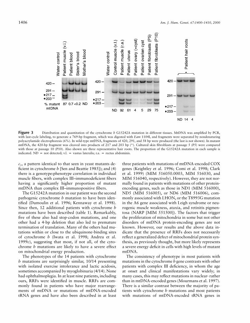
The G15242A mutation creates an Eam1104I restric-tion site, which allowed us to develop a simple PCR-RFLP method for detection and quantitation of the pro-portion of mutant mtDNA. PCR-RFLP analysis ofskeletal muscle and blood from the patient and her rel-atives (fig. 3) revealed very high levels of the G15242Amutation (87%) in the patient’s skeletal muscle and verylow, but detectable, levels in blood (0.7%). Very faintmutant bands suggested the presence of trace amountsof the mutation (!0.2%) in blood from the patient’ssister, but this was at the limit of detection. The mutationwas not detected in her mother’s blood nor in 180 con-trol samples (58 blood and 28 muscle samples from con-trols and patients with a variety of neuromuscular dis-eases). Mutation analysis in other tissues from thepatient (fig. 3) showed very high levels of mutantmtDNA in rectus abdominis muscle (92%), high levelsin uterus (61%), and low levels in ovarian tissue, bothfrom the cyst itself and from surrounding tissue thatcontained cyst contamination (4%–5%). The propor-tion of mutant mtDNA in fibroblasts varied with thelength of time in culture, increasing steadily from 29%to 75% during the course of 10 population doublingsover a period of 7 weeks (fig. 3). A similar trend wasseen regardless of whether the culture medium was sup-plemented with uridine, which is essential for the growthof respiratory-deficient cells (King and Attardi 1989)(data not shown). In a series of 22 individual hair rootsfrom the patient, the G15242A mutation was detectedin 8; the proportion of the mutation varied widely, rang-ing from !0.4% to 66% (mean 4.6%).
Proportion of the Cytochrome b G15242A Mutation inSingle Muscle Fibers
Individual fibers were classified as complex III positiveor negative on the basis of immunostaining of flankingsections with antisera against the Rieske protein of com-plex III (see fig. 1) then analyzed by PCR-RFLP. Theresults (fig. 4) showed a significant correlation betweenthe proportion of the G15242A mutation in complexIII–positive fibers ( ; mean 5 SD), com-32% 5 29%pared with that in complex III–negative fibers( ) ( ), and especially in complex66% 5 26% P ! .0001III–negative RRFs ( ) ( ). The cor-89% 5 8% P ! .0001

1404 Am. J. Hum. Genet. 67:1400–1410, 2000
Figure 1 Histochemistry and immunocytochemistry of serial sections of skeletal muscle from the patient. Type I (heavy) and type II (light)fibers are distinguished on the basis of their intensity of staining for ATPase, COX, and SDH activities (top). The black arrows (two outlinedand one filled in), most easily seen with the SDH reaction, identify three type I fibers, one of which is an RRF (*). Two other RRFs (*) arealso identified. The white arrows (one outlined and one filled in) identify two type II fibers. The center panel shows immunoreactivity withantibodies to ND1, COXI, and holoII. Similar intensity of staining is seen in the three type I fibers and in the two type II fibers. The bottompanels show immunoreactivity with antibodies to three subunits of complex III (cytochrome b, the Rieske protein, and the 13.4-kDa protein).In these cases, a clear difference can be seen between two of the type I fibers (outlined black arrows), which react poorly with all complex IIIantibodies, compared with the third type I fiber (filled-in black arrow). Similarly, one of the type II fibers (outlined white arrow) reacts poorlywith all three complex III antibodies, compared with the other type II fiber (filled-in white arrow).
responding median values were 33%, 76%, and 89%,respectively. The immunopositive reaction in 3 of 33fibers with 170% of the G15242A mutation and theimmunonegative reaction in 6 of 31 fibers with !50%of the mutation most likely reflect the fact that the im-munostaining was performed in 8-mm sections imme-diately adjacent to the 40-mm sections used for mutationanalysis, and there may be some variability in the dis-tribution of the mutant mtDNA, even between adjacentsections. Another possibility is that there is not an exactcorrelation between the levels of Rieske protein and theproportion of mutant mtDNA, since deficiency of the
Rieske protein is a secondary phenomenon, reflectinglack of assembly or increased degradation of nuclear-encoded subunits of complex III, as a consequence ofdeficient functional cytochrome b.
Discussion
Our patient, who was diagnosed many years ago withcomplex III deficiency, has been of particular interestbecause of her well-documented clinical and bioenergeticresponse to treatment with menadione and ascorbate,which were proposed to bypass the defect of electron

Keightley et al.: mtDNA Cytochrome b Stop-Codon Mutation 1405
Figure 2 Identification of a nonsense mutation in the cytochrome b gene. Autoradiogram of a DNA sequencing gel spanning nt15230–15247 (5′r3′) of human mtDNA, showing sequence from a control (left panel) and the patient’s muscle (right panel). The GrA transitionin the patient’s sequence (nt 15242; arrow) converts a glycine codon (GGA) to a stop codon (AGA).
transport between ubiquinol and cytochrome c (Eleff etal. 1984; Argov et al. 1986). Although the original stud-ies of muscle—including enzymatic, spectral, and west-ern blot analyses—all indicated a severe and specific de-ficiency of complex III, the molecular basis was notknown. Complex III of the mitochondrial respiratory-chain transfers electrons from ubiquinol to cytochromec, coupled with the transfer of electrons across the mi-tochondrial inner membrane. It contains three redox-active centers (cytochrome b, cytochrome c1, and theRieske FeS protein) and is composed of 11 polypeptidesubunits, 1 of which (cytochrome b) is encoded bymtDNA. Disorders of complex III are relatively rare,comprising only 7% of patients with respiratory-chaindisorders in a recent series of 157 patients (von Kleist-Retzow et al. 1998). A deficiency of complex III couldbe of either nuclear or mitochondrial origin. The im-munocytochemical studies of skeletal muscle, presentedhere, confirmed the specificity of the complex III defi-ciency in our patient and, more importantly, identifieda mosaic of complex III–immunopositive and –immu-nonegative fibers, strongly suggesting a mutation ofmtDNA. The ability to utilize antisera to the Rieske or13.4-kDa proteins for these experiments presumably re-flects a lack of assembly or increased degradation ofnuclear-encoded subunits of complex III in the absenceof a functional cytochrome b, as also occurs in yeast(Sen and Beattie 1985). A mosaic of COX-positive and-negative fibers is readily recognized histochemically in
patients with COX deficiency associated with mutationsof mtDNA (Keightley et al. 1996; DiMauro and Bonilla1997). In contrast, no histochemical stain is available todocument similar mosaicism in patients with complex Ior complex III deficiency. Our results suggest that im-munocytochemistry, using serial sections of muscle andspecific antibodies to complex I or complex III, may bea valuable diagnostic technique to localize a specific res-piratory-chain deficiency, especially in small muscle sam-ples. This technique can also provide direction for mo-lecular analyses focused on either the nuclear ormitochondrial genome. Moreover, for mutations ofmtDNA, it can be used to correlate the proportion ofmutant mtDNA with the phenotype in single musclefibers, a critical step in establishing the pathogenicity ofan mtDNA mutation, as has been demonstrated here.
The G15242A mutation in our patient is almost cer-tainly pathogenic, resulting in a protein truncated by150% at the C terminus. Additional evidence for path-ogenicity is as follows: (1) the mutation is heteroplasmicand occurs at higher proportions in affected tissue (mus-cle) than in unaffected tissue (blood); (2) it has not beendescribed elsewhere and was not identified in controlsamples; (3) the mutation is consistent with the bio-chemical and immunocytochemical phenotype that in-dicated isolated complex III deficiency (furthermore, thepattern of subunit deficiencies, documented elsewherein western blot analyses, included core proteins 1 and2 and the Rieske protein, with sparing of cytochrome
1406 Am. J. Hum. Genet. 67:1400–1410, 2000
Figure 3 Distribution and quantitation of the cytochrome b G15242A mutation in different tissues. MtDNA was amplified by PCR,with last-cycle labeling, to generate a 769-bp fragment, which was digested with Eam 1104I, and fragments were separated by nondenaturingpolyacrylamide electrophoresis (6%). In wild-type mtDNA, fragments of 420, 291, and 58 bp were produced (the last is not shown). In mutantmtDNA, the 420-bp fragment was cleaved into products of 217 and 203 bp (*). Cultured skin fibroblasts at passage 5 (P5) were comparedwith those at passage 10 (P10). Also shown are three representative hair roots. The proportion of the G15242A mutation in each sample isindicated. ND p not detected; v.l. p vastus lateralis; r.a. p rectus abdominis.
c1, a pattern identical to that seen in yeast mutants de-ficient in cytochrome b [Sen and Beattie 1985]); and (4)there is a genotype:phenotype correlation in individualmuscle fibers, with complex III–immunodeficient fibershaving a significantly higher proportion of mutantmtDNA than complex III–immunopositive fibers.
The G15242A mutation in our patient was the secondpathogenic cytochrome b mutation to have been iden-tified (Dumoulin et al. 1996; Kennaway et al. 1998).Since then, 12 additional patients with cytochrome bmutations have been described (table 1). Remarkably,five of these also had stop-codon mutations, and oneother had a 4-bp deletion that also led to prematuretermination of translation. Many of the others had mu-tations within or close to the ubiquinone-binding sitesof cytochrome b (Iwata et al. 1998; Andreu et al.1999c), suggesting that most, if not all, of the cyto-chrome b mutations are likely to have a severe effecton mitochondrial energy production.
The phenotypes of the 14 patients with cytochromeb mutations are surprisingly similar, 10/14 presentingwith isolated exercise intolerance and lactic acidosis,sometimes accompanied by myoglobinuria (4/14). Nonehad ophthalmoplegia. In at least nine patients, includingours, RRFs were identified in muscle. RRFs are com-monly found in patients who have major rearrange-ments of mtDNA or mutations of mtDNA-encodedtRNA genes and have also been described in at least
three patients with mutations of mtDNA-encoded COXgenes (Keightley et al. 1996; Comi et al. 1998; Clarket al. 1999) (MIM 516050.0003, MIM 516030, andMIM 516040, respectively). However, they are not nor-mally found in patients with mutations of other protein-encoding genes, such as those in ND1 (MIM 516000),ND5 (MIM 516005), or ND6 (MIM 516006), com-monly associated with LHON, or the T8993G mutationin the A6 gene associated with Leigh syndrome or neu-rogenic muscle weakness, ataxia, and retinitis pigmen-tosa (NARP [MIM 551500]). The factors that triggerthe proliferation of mitochondria in some but not otherdisorders of mtDNA protein-encoding genes are notknown. However, our results and the above data in-dicate that the presence of RRFs does not necessarilyreflect a generalized defect of mitochondrial protein syn-thesis, as previously thought, but more likely representsa severe energy deficit in cells with high levels of mutantmtDNA.
The consistency of phenotype in most patients withmutations in the cytochrome b gene contrasts with otherpatients with complex III deficiency, in whom the ageat onset and clinical manifestations vary widely; inmany cases, this may reflect mutations in nuclear- ratherthan in mtDNA-encoded genes (Mourmans et al. 1997).There is a similar contrast between the majority of pa-tients with cytochrome b mutations and most patientswith mutations of mtDNA-encoded tRNA genes in

Keightley et al.: mtDNA Cytochrome b Stop-Codon Mutation 1407
Figure 4 Proportion of the cytochrome b G15242A mutationin single muscle fibers. Alternating 40- and 8-mm-thick serial sectionsof muscle were obtained. The 40-mm sections and flanking 8-mm sec-tions were stained immunocytochemically for the Rieske protein ofcomplex III and were scored as complex III positive or complex IIInegative on the basis of the more intense staining of the 8-mm sections.Additional 8-mm flanking sections were stained histochemically forSDH, to identify RRFs. The 40-mm sections were microdissected andsubjected to PCR-RFLP analysis. The median proportion of mutantmtDNA in each fiber type is indicated by a horizontal line. The mean5 SD is shown below. The differences between immunopositive andimmunonegative non-RRFs (columns 1 and 2), and between immu-nopositive and immunonegative RRFs (columns 1 and 3), were highlysignificant ( ).P ! .0001
which the variability of clinical manifestations is enor-mous. Also in contrast to patients with tRNA muta-tions, which are frequently maternally inherited, all ofthe patients with cytochrome b mutations, with the pos-sible exception of ours, have been sporadic cases, sug-gesting that the cytochrome b mutation, if present inthe maternal germ line, may frequently be lethal duringearly fetal development. Mutations in other mtDNAprotein-encoding genes, such as those in the A6 geneassociated with Leigh syndrome or NARP, or the mu-tations in complex I genes commonly associated withLHON, however, are almost always maternally inher-ited. The fact that the LHON mutations are usuallyhomoplasmic, are not associated with RRFs, and arematernally inherited suggests that they may have a lesssevere effect on ATP synthesis than the mutations in thecytochrome b gene, discussed above. Studies in trans-mitochondrial cell lines should help answer thisquestion.
Our patient differs from most other patients with cy-tochrome b mutations in two important respects. First,
although her presentation was dominated by severe ex-ercise intolerance and lactic acidosis between the agesof 9 and 19 years, she did have seizures from her earlyteenage years, and she has developed significant en-cephalopathy over the last 15 years. This raises the pos-sibility that, given more time, some of the other patientsmay also progress to a more severe phenotype. Oneother patient with a cytochrome b mutation had a pre-dominantly neurological presentation, described as ju-venile Parkinson/MELAS overlap syndrome (patient 5,table 1), and two others had severe hypertrophic car-diomyopathy (patients 11 and 14). These three patientsalso had an earlier age at onset and more severe pre-sentation, compared with many other patients. Notably,at least two of them had detectable levels of mutationin blood and/or fibroblasts.
The second difference between our patient and mostof the other cases concerns the origin and tissue distri-bution of the mutation. In the patients with an isolatedskeletal myopathy, the mutant mtDNA was absent inblood and/or fibroblasts in all cases examined. This isconsistent with the restriction of the clinical phenotypeto skeletal muscle, the spontaneous occurrence of thesemutations, and the likelihood that they would not betransmitted to the patients’ offspring (Griggs and Kar-pati 1999). The highly skewed distribution of the mu-tation between blood or fibroblasts, compared withmuscle, and the restriction of the clinical phenotype tomuscle, have led to the suggestion that the mutation inthese patients may have arisen in muscle progenitorcells, after differentiation of the primary germ layers(Andreu et al. 1999c). However, another possibility isthe occurrence of a sporadic mutation early during em-bryogenesis or in the maternal germ line, with a highlyskewed distribution in tissues of the patient, arising asa result of either mitotic segregation or selection againstthe mutation by some unknown mechanism in mosttissues. Certainly, the absence of the mtDNA mutationin blood and fibroblasts does not preclude its presencein a variety of other tissues, as illustrated by the patientwith a heteroplasmic tRNALeu(CUN) mutation who hadsensorineural deafness and retinopathy in addition to asevere skeletal myopathy (Fu et al. 1996). In our patient,the G15242A mutation was relatively widely distrib-uted, with very low levels in blood, low levels in ovariantissue, and moderate to high levels in uterus and cul-tured skin fibroblasts. More importantly, the mutationwas also present in hair roots. In contrast to all othertissues examined, which are of mesodermal origin, hairroots are derived from primitive ectoderm, indicatingthat the mutation in our patient must have arisen veryearly in embryogenesis, before separation of the primarygerm layers, or in maternal germ line cells. Analysis ofblood from the patient’s mother and sister did not dif-ferentiate between these two possibilities.

1408 Am. J. Hum. Genet. 67:1400–1410, 2000
Table 1
Summary of Patients with Mutations in the mtDNA-encoded Cytochrome b Gene
Patient Sex/Age
Age atOnset(years) Phenotype
RRF/Lactic
AcidosisCytochromeb Mutation
AminoAcid
Change
%Mutationin Muscle
MutationPresent in
Blood/Fibroblasts Reference
1a F/34 y 9 EI, LE 1/1 G15242A G166X 87 1/1 Kennaway et al. (1998)2 M/25 y 10 EI, P 1/1 G15615A G290D 80 2/NA Dumoulin et al. (1996)3 F/38 y 25 EI, W 2/1 G15762A G339E 85 2/NA Andreu et al. (1998)4 F/27 y 12 EI, W, M 1/1 G15059A G105Xb 63 2/NA Andreu et al. (1999a)5 M/20 y 6 JP/MELAS 2/1 14787del4 Del14fs/tr49 195 1/1 DeCoo et al. (1999)6 M/43 y 30 EI, W, M 1/2 15498del24 Del251-258 50 2/2 Andreu et al. (1999c)7 F/52 y Child EI, W 1/1 G14846A G34S 85 2/2 Andreu et al. (1999c)8 F/38 y Child EI 1/1 G15168A W141X 70 2/NA Andreu et al. (1999c)9 M/32 y Child EI 1/1 G15084A W113X 87 2/NA Andreu et al. (1999c)10 M/51 y Child EI, M NA/1 G15723A W326X 87 NA/NA Andreu et al. (1999c)11 F/8 y 3 SHC NA/1 G15243A G166E 90c NA/1 Valnot et al. (1999)12 M/18 y 8 EI, M 1/1 G15150A W135X 60 2/2 Legros et al. (1999)d
13 M/51 y Teen EI, W 1/1 T15197C S151P 70 2/2 Legros et al. (1999)d
14 F/4 w Infant SHC NA/NA G15498A G251D NA NA/NA Andreu et al. (2000)
NOTE.—EI p exercise intolerance; W p weakness; M p myoglobinuria; SHC p severe hypertrophic cardiomyopathy; JP p juvenileParkinsons; P p muscle pain; LE p late-onset encephalopathy; y p years; w p weeks; Del p deletion; fs p frameshift; tr p termination;NA p not available or not analyzed.
a Present case.b Corrected from Andreu et al. 1999b.c Cardiac muscle.d Also includes data from A. Lombes, personal communication.
Although the average proportion of the G15242Amutation in hair roots from our patient was only 4.6%,the finding of much higher proportions in some indi-vidual hair roots (up to 66%) than in blood (0.7%)suggests that hair roots may provide a more sensitivemethod for detecting heteroplasmic mtDNA mutations,as has been suggested elsewhere, in studies of a familyharboring a mitochondrial tRNAGlu mutation (MIM590025) (Hao et al. 1995). Examination of mutantmtDNA in patients with MELAS, MERRF, and NARPshowed a good correlation between the percentage ofmutant mtDNA in blood, compared with muscle, buccalcells, and hair roots (Wong and Lam 1997). These au-thors stress that pooling of hair cells minimizes the dis-crepancies obtained with single-follicle analysis. How-ever, in patients such as ours, in whom the mutantmtDNA in blood is barely detectable, the wide varia-bility in hair roots suggests that measurement of mutantmtDNA in individual follicles may be more sensitive fordetecting asymptomatic carriers of the mutation inwhom the level of mutant mtDNA in pooled folliclesmay be at the limit of detection.
The marked variability in the distribution of mutantmtDNA between different tissues (e.g., muscle andblood) and even within individual tissues (e.g., musclefibers or hair roots) presumably reflects replicative seg-regation. In cultured skin fibroblasts, the increasing pro-portion of the G15242A mutation with increasing time
in culture is harder to understand, since there is noobvious selective advantage to cells or mitochondriaharboring high levels of a stop-codon mutation. How-ever, in fibroblast cultures from one patient with intra-cellular mtDNA triplasmy, the proportion of one mu-tation (A5656G) was also found to increase withincreasing time in culture (Bidooki et al. 1997). More-over, increasing proportions of mutant mtDNA havebeen reported over time in vivo in patients harboringpathogenic mtDNA deletions (Larsson et al. 1990) orpoint mutations (Weber et al. 1997). In one family witha heteroplasmic tRNAGlu mutation, the proportion ofmutant mtDNA (studied in individual hair roots) tendedto increase in successive generations (Hao et al. 1995).Finally, in transmitochondrial cell lines harboring theMELAS 3243 mutation, different levels of heteroplasmywere found to either increase, decrease, or remain stableover time in culture; although the mechanisms respon-sible for these changes are not understood, they appearto depend on the nuclear background of the cells(Yoneda et al. 1992; Dunbar et al. 1995).
The presence of the G15242A mutation in hair rootsfrom our patient suggests that the mutation may alsobe present in the CNS, which is also derived from prim-itive ectoderm. This would be consistent with her neu-rological symptoms. In this regard, it should be notedthat the cytochrome b microdeletion in the patient witha more severe neurological disorder described as juve-

Keightley et al.: mtDNA Cytochrome b Stop-Codon Mutation 1409
nile Parkinson/MELAS overlap syndrome (table 1) wasalso present in hair roots. The occurrence of hetero-plasmic cytochrome b mutations in hair roots from thetwo patients with encephalopathic manifestations sug-gests that hair-root analysis can provide a useful reflec-tion of the tissue distribution of the mutation, the po-tential clinical phenotype, and, perhaps, the risk oftransmitting the mutation to their offspring. These ques-tions can only be answered by further long-term studiesof additional patients.
Acknowledgments
We thank Dr. Robb Moses for the gift of Taq polymerase,George Cole for preparation of the muscle sections, Dr. BradleyPopovich and his staff for sharing laboratory space and pro-viding technical advice, Drs. David Hunter and Marc Mauneyfor help in obtaining tissue samples, and Dr. Eric Shoubridgefor reviewing the manuscript. This work was supported by agrant from the Muscular Dystrophy Association.
Electronic-Database Information
Accession numbers and URLs for data in this article are asfollows:
GenBank, http://www.ncbi.nlm.nih.gov/Genbank (for cyto-chrome b sequence in our patient [accession numberAF254896])
Online Mendelian Inheritance in Man (OMIM), http://www.ncbi.nlm.nih.gov/Omim (for complex I subunit ND1 [MIM516000]; ND5 [MIM 516005] and ND6 [MIM 516006]mutations; complex III deficiency [MIM 124000]; complexIII cytochrome b mutation [MIM 516020]; COX deficiency[MIM 220110]; COX subunit I [MIM 516030], II [MIM516040], and III 15BP DEL [MIM 516050.0003] mutations;Kearns-Sayre syndrome [MIM 530000]; LHON [MIM535000]; MELAS [MIM 540000]; MERRF [MIM 545000];mitochondrial myopathy [MIM 251900]; NARP [MIM551500]; and tRNAGlu mutation [MIM 590025].
References
Anderson S, Bankier AT, Barrell BG, de Bruijn MH, CoulsonAR, Drouin J, Eperon IC, Nierlich DP, Roe BA, Sanger F,Schreier PH, Smith AJ, Staden R, Young IG (1981) Sequenceand organization of the human mitochondrial genome. Na-ture 290:457–465
Andreu AL, Bruno C, Dunne TC, Tanji K, Shanske S, Sue CM,Krishna S, Hadjigeorgiou GM, Shtilbans A, Bonilla E,DiMauro S (1999a) A nonsense mutation (G15059A) in thecytochrome b gene in a patient with exercise intolerance andmyoglobinuria. Ann Neurol 45:127–130
Andreu AL, Bruno C, Hadjigeorgiou GM, Shanske S, DiMauroS (1999b) Polymorphic variants in the human mitochondrialcytochrome b gene. Mol Genet Metab 67:49–52
Andreu AL, Bruno C, Shanske S, Shtilbans A, Hirano M,
Krishna S, Hayward L, Systrom DS, Brown RH Jr, DiMauroS (1998) Missense mutation in the mtDNA cytochrome bgene in a patient with myopathy. Neurology 51:1444–1447
Andreu AL, Checcarelli N, Iwata S, Shanske S, DiMauro S(2000) A missense mutation in the mitochondrial cyto-chrome b gene in a revisited case with histiocytoid cardiom-yopathy. Pediatr Res 48:311–314
Andreu AL, Hanna MG, Reichmann H, Bruno C, Penn AS,Tanji K, Pallotti F, Iwata S, Bonilla E, Lach B, Morgan-Hughes J, DiMauro S (1999c) Exercise intolerance due tomutations in the cytochrome b gene of mitochondrial DNA.N Engl J Med 341:1037–1044
Argov Z, Bank WJ, Maris J, Eleff S, Kennaway NG, OlsonRE, Chance B (1986) Treatment of mitochondrial myopathydue to complex III deficiency with vitamins K3 and C: a 31P-NMR follow-up study. Ann Neurol 19:598–602
Bidooki SK, Johnson MA, Chrzanowska-Lightowlers Z, Bin-doff LA, Lightowlers RN (1997) Intracellular mitochondrialtriplasmy in a patient with two heteroplasmic base changes.Am J Hum Genet 60:1430–1438
Bing DH, Bieber FR (1998) Isolation of DNA from forensicevidence. In: Dracopoli NC, Haines JL, Korf BR, Moir DT,Morton CC, Seidman CE, Seidman JG et al (eds) Currentprotocols in human genetics. Vol 2. John Wiley and Sons,pp 14.3.11–14.3.12
Chinnery PF, Howell N, Andrews RM, Turnbull DM (1999)Clinical mitochondrial genetics. J Med Genet 36:425–436
Chinnery PF, Turnbull DM (1999) Mitochondrial DNA anddisease. Lancet 354:s117–s121
Clark KM, Taylor RW, Johnson MA, Chinnery PF, Chrza-nowska-Lightowlers ZM, Andrews RM, Nelson IP, WoodNW, Lamont PJ, Hanna MG, Lightowlers RN, Turnbull DM(1999) An mtDNA mutation in the initiation codon of thecytochrome c oxidase subunit II gene results in lower levelsof the protein and a mitochondrial encephalomyopathy. AmJ Hum Genet 64:1330–1339
Comi GP, Bordoni A, Salani S, Franceschina L, Sciacco M,Prelle A, Fortunato F, Zeviani M, Napoli L, Bresolin N,Moggio M, Ausenda CD, Taanman JW, Scarlato G (1998)Cytochrome c oxidase subunit I microdeletion in a patientwith motor neuron disease. Ann Neurol 43:110–116
Darley-Usmar VM, Kennaway NG, Buist NR, Capaldi RA(1983) Deficiency in ubiquinone cytochrome c reductase ina patient with mitochondrial myopathy and lactic acidosis.Proc Natl Acad Sci USA 80:5103–5106
Darley-Usmar VM, Watanabe M, Uchiyama Y, Kondo I, Ken-naway NG, Gronke L, Hamaguchi H (1986) Mitochondrialmyopathy: tissue-specific expression of a defect in ubiquinol-cytochrome c reductase. Clin Chim Acta 158:253–261
De Coo IF, Renier WO, Ruitenbeek W, Ter Laak HJ, BakkerM, Schagger H, Van Oost BA, Smeets HJ (1999) A 4-basepair deletion in the mitochondrial cytochrome b gene as-sociated with parkinsonism/MELAS overlap syndrome. AnnNeurol 45:130–133
DiMauro S, Bonilla E (1997) Mitochondrial encephalomy-opathies. In: Rosenberg RN, Prusiner SB, DiMauro S, BarchiRL (eds) The molecular and genetic basis of neurologicaldisease. Butterworth-Heinemann, Boston, pp 201–235
DiMauro S, Bonilla E, Davidson M, Hirano M, Schon EA

1410 Am. J. Hum. Genet. 67:1400–1410, 2000
(1998) Mitochondria in neuromuscular disorders. BiochimBiophys Acta 1366:199–210
Dumoulin R, Sagnol I, Ferlin T, Bozon D, Stepien G, MoussonB (1996) A novel gly290asp mitochondrial cytochrome bmutation linked to a complex III deficiency in progressiveexercise intolerance. Mol Cell Probes 10:389–391
Dunbar DR, Moonie PA, Jacobs HT, Holt IJ (1995) Differentcellular backgrounds confer a marked advantage to eithermutant or wild-type mitochondrial genomes. Proc Natl AcadSci USA 92:6562–6566
Eleff S, Kennaway NG, Buist NRM, Darley-Usmar VM, Ca-paldi RA, Bank WJ, Chance B (1984) 31P NMR study ofimprovement in oxidative phosphorylation by vitamins K3and C in a patient with a defect in electron transport atcomplex III in skeletal muscle. Proc Natl Acad Sci USA 81:3529–3533
Elliot DL, Buist NRM, Goldberg L, Kennaway NG, PowellBR, Kuehl KS (1989) Metabolic myopathies: evaluation bygraded exercise testing. Medicine 68:163–172
Fu K, Hartlen R, Johns T, Genge A, Karpati G, Shoubridge EA(1996) A novel heteroplasmic tRNAleu(CUN) mtDNA point mu-tation in a sporadic patient with mitochondrial encephalo-myopathy segregates rapidly in skeletal muscle and suggestsan approach to therapy. Hum Mol Genet 5:1835–1840
Griggs RC, Karpati G (1999) Muscle pain, fatigue, and mi-tochondriopathies. N Engl J Med 341:1077–1078
Hanahan D (1985) Techniques for transformation of E coli. In:Glover D (ed) DNA cloning: a practical approach. Vol 1.Practical approach series. IRL Press, Oxford, UK, pp 109–135
Hao H, Bonilla E, Manfredi G, DiMauro S, Moraes CT (1995)Segregation patterns of a novel mutation in the mitochon-drial tRNA glutamic acid gene associated with myopathyand diabetes mellitus. Am J Hum Genet 56:1017–1025
Iwata S, Lee JW, Okada K, Lee JK, Iwata M, Rasmussen B,Link TA, Ramaswamy S, Jap BK (1998) Complete structureof the 11-subunit bovine mitochondrial cytochrome bc1
complex. Science 281:64–71Keightley JA, Hoffbuhr KC, Burton MD, Salas VM, Johnston
WS, Penn AM, Buist NRM, Kennaway NG (1996) A mi-crodeletion in cytochrome c oxidase (COX) subunit III as-sociated with COX deficiency and recurrent myoglobinuria.Nat Genet 12:410–416
Kennaway NG, Buist NRM, Darley-Usmar VM, Papadimi-triou A, Dimauro S, Kelley RI, Capaldi RA, Blank NK,D’Agostino A (1984) Lactic acidosis and mitochondrial my-opathy associated with deficiency of several components ofcomplex III of the respiratory chain. Pediatr Res 18:991–999
Kennaway NG, Keightley JA, Burton MD, Quan F, Libby BD,Buist NRM (1998) Mitochondrial encephalomyopathy as-sociated with a nonsense mutation in cytochrome b. MolGenet Metab 63:49
King MP, Attardi G (1989) Human cells lacking mtDNA: re-population with exogenous mitochondria by complemen-tation. Science 246:500–503
Larsson N-G, Holme E, Kristiansson B, Oldfors A, TuliniusM (1990) Progressive increase of the mutated mitochondrialDNA fraction in Kearns-Sayre syndrome. Pediatr Res 28:131–136
Legros F, Chatzoglou E, Frachon P, Barthelemy C, SternbergD, Jardel C, Godinot C, Lombes A (1999) Clinical and mo-lecular diversity of respiratory chain complex III defects.Poster presented at the Fourth European Meeting on Mi-tochondrial Pathology, Cambridge, UK, p 135
Mourmans J, Wendel U, Bentlage HA, Trijbels JM, SmeitinkJA, de Coo IF, Gabreels FJ, Sengers RC, Ruitenbeek W(1997) Clinical heterogeneity in respiratory chain complexIII deficiency in childhood. J Neurol Sci 149:111–117
Pulkes T, Siddiqui A, Morgan-Hughes JA, Wood NW, HannaMG (1999) A novel heteroplasmic nonsense mutation in themitochondrial cytochrome b gene associated with mito-chondrial myopathy and complex III deficiency. Postr pre-sented at the Fourth European Meeting on MitochondrialPathology, Cambridge, UK, p 179
Sambrook J, Fritsch EF, Maniatis T (1989) Molecular cloning:a laboratory manual. 2d ed. Cold Spring Harbor LaboratoryPress, Cold Spring Harbor, New York
Sen K, Beattie DS (1985) Decreased amounts of core proteinsI and II and the iron-sulfur protein in mitochondria fromyeast lacking cytochrome b but containing cytochrome c1.Arch Biochem Biophys 242:393–401
Taanman J-W, Burton MD, Marusich MF, Kennaway NG, Ca-paldi RA (1996) Subunit specific monoclonal antibodiesshow different steady-state levels of various cytochrome-coxidase subunits in chronic progressive external ophthal-moplegia. Biochim Biophys Acta 1315:199–207
Valnot I, Kassis J, Chretien D, de Lonlay P, Parfait B, MunnichA, Kachaner J, Rustin P, Rotig A (1999) A mitochondrialcytochrome b mutation but no mutations of nuclearly en-coded subunits in ubiquinol cytochrome c reductase (com-plex III) deficiency. Hum Genet 104:460–466
von Kleist-Retzow JC, Cormier-Daire V, de Lonlay P, Parfait B,Chretien D, Rustin P, Feingold J, Rotig A, Munnich A (1998)A high rate (20%–30%) of parental consanguinity in cyto-chrome-oxidase deficiency. Am J Hum Genet 63:428–435
Wallace DC (1999) Mitochondrial diseases in man and mouse.Science 283:1482–1488
Weber K, Wilson JN, Taylor L, Brierley E, Johnson MA, Turn-bull DM, Bindoff LA (1997) A new mtDNA mutation show-ing accumulation with time and restriction to skeletal mus-cle. Am J Hum Genet 60:373–380
Wong L-JC, Lam C-W (1997) Alternative, noninvasive tissuesfor quantitative screening of mutant mitochondrial DNA.Clin Chem 43:1241–1243
Yoneda M, Chomyn A, Martinuzzi A, Hurko O, Attardi G(1992) Marked replicative advantage of human mtDNA car-rying a point mutation that causes the MELAS encephalo-myopathy. Proc Natl Acad Sci USA 89:11164–11168
Copyright © 2022 FDOKUMEN