MITCHELL MEDAL LECTURE
551
1 FROM THE ORGANIZERS The term “Bioenergetics” was coined in 1950s by Albert Szent-Gyorgyi who used this word as the title of one of his books. In 1968, a special session of the International Meeting on Oxidative Phosphorylation in Polignano-a-Mare (near Bari, Italy) was devoted to the problem of how to call the new branch of biology dealing with mechanisms of energy transductions in living systems. At this meeting, I attracted the attention of the audience to Szent-Gyorgyi’s term, putting forward only one argument in favor, namely, that I already organized the Department of Bioenergetics at Moscow State University. Surprisingly, the suggestion was accepted. And now I have the great pleasure to welcome numerous participants of the 14 th European Bioenergetics Conference to Moscow State University. In fact, this is the second attempt to organize an EBEC conference in Russia. Originally, the plan was to have EBEC here in 1992, but it was moved to Helsinki due to the severe economic crisis that followed the collapse of the Soviet Union in the very end of 1991. Now I hope the organizers will be happier in carrying out their intention. Traditionally for EBEC meetings, the conference will deal with two major aspects, i.e. molecular and supramolecular bioenergetics, the latter including physiology of organelles, cells, tissues, and organisms. At the previous 13 EBECs, the majority of the talks were related to molecular bioenergetics. However, now for the first time the physiological aspects approach is slightly dominating. This event indicates a tendency to apply the great success achieved in understanding energy-transducing mechanisms to elucidate principles of functioning of complex biological systems. Most demonstratively, such a tendency was revealed in mitochondrial studies. The number of papers published in this field dramatically increased in 1960-1980, then a plateau was reached, which changed to one more phase of exponential growth in the late 1990s due to discovery of the fact that mitochondrial physiology is essential not only for the life but also for programmed death. At the Moscow EBEC, symposial (plenary) lectures occupying the first part of each day will be followed by colloquial talks in its second part. The total number of speakers invited by the Organizing Committee is as large as 90 (30 symposial and 60 colloquial). Moreover, 20 speakers were invited by the chairpersons of the participant-led colloquia and 24 were selected by the organizers among the best posters. All the symposial and colloquial speakers had a possibility to contribute to a Special BBA Bioenergetics volume. The great majority of the speakers accepted the invitation, writing excellent papers. I am sure that their contributions will be of importance for the further progress of bioenergetics. 10.1016/j.bbabio.2006.06.009
-
Upload
independent -
Category
Documents
-
view
0 -
download
0
Transcript of MITCHELL MEDAL LECTURE

1
FROM THE ORGANIZERS
The term “Bioenergetics” was coined in 1950s by Albert Szent-Gyorgyi who used this word
as the title of one of his books. In 1968, a special session of the International Meeting on Oxidative
Phosphorylation in Polignano-a-Mare (near Bari, Italy) was devoted to the problem of how to call
the new branch of biology dealing with mechanisms of energy transductions in living systems. At
this meeting, I attracted the attention of the audience to Szent-Gyorgyi’s term, putting forward only
one argument in favor, namely, that I already organized the Department of Bioenergetics at
Moscow State University. Surprisingly, the suggestion was accepted. And now I have the great
pleasure to welcome numerous participants of the 14th European Bioenergetics Conference to
Moscow State University.
In fact, this is the second attempt to organize an EBEC conference in Russia. Originally, the
plan was to have EBEC here in 1992, but it was moved to Helsinki due to the severe economic
crisis that followed the collapse of the Soviet Union in the very end of 1991. Now I hope the
organizers will be happier in carrying out their intention.
Traditionally for EBEC meetings, the conference will deal with two major aspects, i.e.
molecular and supramolecular bioenergetics, the latter including physiology of organelles, cells,
tissues, and organisms. At the previous 13 EBECs, the majority of the talks were related to
molecular bioenergetics. However, now for the first time the physiological aspects approach is
slightly dominating. This event indicates a tendency to apply the great success achieved in
understanding energy-transducing mechanisms to elucidate principles of functioning of complex
biological systems. Most demonstratively, such a tendency was revealed in mitochondrial studies.
The number of papers published in this field dramatically increased in 1960-1980, then a plateau
was reached, which changed to one more phase of exponential growth in the late 1990s due to
discovery of the fact that mitochondrial physiology is essential not only for the life but also for
programmed death.
At the Moscow EBEC, symposial (plenary) lectures occupying the first part of each day will
be followed by colloquial talks in its second part. The total number of speakers invited by the
Organizing Committee is as large as 90 (30 symposial and 60 colloquial). Moreover, 20 speakers
were invited by the chairpersons of the participant-led colloquia and 24 were selected by the
organizers among the best posters. All the symposial and colloquial speakers had a possibility to
contribute to a Special BBA Bioenergetics volume. The great majority of the speakers accepted the
invitation, writing excellent papers. I am sure that their contributions will be of importance for the
further progress of bioenergetics.
10.1016/j.bbabio.2006.06.009

I am very grateful to all bioenergeticists who will attend; to organizations who financially
supported this 14th EBEC; to Monomax professional international congress-organizing agency; to
members of the Local Organizing Committee and the International Advisory Committee for their
help in organization of this meeting. I am also very grateful to Pat Crowley and all other staff of
BBA and Elsevier for their collaboration in the production of two special BBA Bioenergetics
volumes.
I wish you all a happy and exciting EBEC in Moscow.
Vladimir Skulachev, Chairman, Organizing Committee, 14th EBEC, Moscow
2

3
SCIENTIFIC PROGRAM

4

5
MITCHELL MEDAL LECTURE M1 M. Yoshida Tokyo Institute of Technology, Tokyo, Japan [email protected] ROTARY CATALYSIS OF ATP SYNTHASE

6
SYMPOSIA (Plenary Sessions)
1. Molecular bioenergetics
Symposium 1.1. Respiratory chain S1.1.1 H. Michel Max Planck Institute of Biophysics, Department of Molecular Membrane Biology, Frankfurt, Germany [email protected] STRUCTURE-FUNCTION RELATIONSHIPS IN TERMINAL OXIDASES S1.1.2 L.A. Sazanov Medical Research Council Dunn Human Nutrition Unit, Wellcome Trust/MRC Building, Hills Road, Cambridge CB2 2XY, UK [email protected] STRUCTURE OF THE HYDROPHILIC DOMAIN OF RESPIRATORY COMPLEX I FROM THERMUS THERMOPHILUS S1.1.3 S. Yoshikawa Department of Life Science, University of Hyogo, Kamigohri Akoh, Hyogo, Japan [email protected] REACTION MECHANISM OF BOVINE HEART CYTOCHROME C OXIDASE

7
Symposium 1.2. Light-driven energy transducers S1.2.1 J. Barber Wolfson Laboratories, Division of Molecular Biosciences. South Kensington Campus, Imperial College London, UK [email protected] STRUCTURE OF PHOTOSYSTEM II AND ITS IMPLICATIONS FOR UNDERSTANDING THE WATER SPLITTING REACTION IT CATALYSES S1.2.2 J.K. Lanyi, S.P. Balashov Department of Physiology & Biophysics, University of California, Irvine, USA [email protected] XANTHORHODOPSIN, A NOVEL RETINAL-BASED PROTON PUMP WITH A CAROTENOID ANTENNA
S1.2.3 V.A. Shuvalov1, A.G. Yakovlev1, T.A. Shkuropatova2, L.G. Vasilieva3, A.Y. Shkuropatov3, P. Gast2 1 - Department of Photobiophysics, Belozersky Institute of Chemical and Physical Biology, Moscow State University, Moscow, Russia 2 - Department of Biophysics, Huygens Laboratory, Leiden University, Leiden, The Netherlands 3 - Institute of Basic Biological Problems, Russian Academy of Sciences, Pushchino, Moscow Region, Russia [email protected] ELECTRON TRANSFER IN BACTERIAL REACTION CENTERS WITH MODIFIED B-BRANCH PIGMENT COMPOSITION
8
Symposium 1.3. ATP-synthase/ATPase S1.3.1 S.D. Dunn, P.A. Del Rizzo, Y. Bi, K.S. Wood, D.J. Cipriano University of Western Ontario, Department of Biochemistry, London, Canada [email protected] THE RIGHT-HANDED COILED COIL OF THE b DIMER OF ESCHERICHIA COLI ATP SYNTHASE S1.3.2 P. Turina, A. Rebecchi, M. D'Alessandro, S. Anefors, B.A. Melandri University of Bologna, Department of Biology, Bologna, Italy [email protected] MODULATION OF PROTON PUMPING EFFICIENCY IN BACTERIAL ATP SYNTHASES S1.3.3 J.E. Walker Medical Research Council Dunn Human Nutrition Unit, Cambridge, UK [email protected] THE STRUCTURE AND FUNCTION OF ATP SYNTHASE

9
Symposium 1.4. Porters S1.4.1 S. Iwata Imperial College London, Division of Molecular biosciences, London, UK [email protected] STRUCTURE AND MECHANISM OF MEMBRANE TRANSPORTERS S1.4.2 E. Padan1, C. Hunte2, E. Screpanti2, M. Venturi2, A. Rimon1, H. Michel2 1 - Hebrew University, Biochemistry, Jerusalem, Israel 2 - Max-Planck Institute of Biophysics, Frankfurt, Germany [email protected] STRUCTURE OF NHAA Na+/H+ ANTIPORTER: INSIGHTS INTO MECHANISM OF ACTION AND REGULATION BY pH S1.4.3 F. Palmieri Department of Pharmaco-Biology, Laboratory of Biochemistry and Molecular Biology, University of Bari, Italy, and the CNR Institute of Biomembranes and Bioenergetics [email protected] METABOLITE TRANSPORTERS OF MITOCHONDRIA

10
2. Physiology and pathology of mitochondria
(in vitro, ex vivo and in vivo studies)
Symposium 2.1. Uncoupling S2.1.1 B. Cannon, I.G. Shabalina, J. Nedergaard The Wenner-Gren Institute, Stockholm University, Stockholm, Sweden [email protected] THE UNCOUPLING PROTEINS 1 AND 3: QUESTIONS OF MECHANISM, CONTROL AND PHYSIOLOGICAL FUNCTION S2.1.2 D. Ricquier CNRS and University Rene Descartes, Necker Faculty of Medicine, Paris, France [email protected] THE MITOCHONDRIAL UNCOUPLING PROTEINS: FROM BIOENERGETICS TO PHYSIOPATHOLOGY

11
Symposium 2.2. ROS and redox regulations S2.2.1 M.D. Brand MRC Dunn Human Nutrition Unit, Cambridge, UK [email protected] MITOCHONDRIAL ROS PRODUCTION AND ITS ATTENUATION BY UNCOUPLING PROTEINS S2.2.2 D.B. Zorov1, M. Juhaszova2, S.J. Sollott2 1 - Department of Bioenergetics, A.N.Belozersky Institute, Moscow State University, Moscow, Russia; Laboratory of Cardiovascular Sciences, Gerontology Research Center, NIA, NIH, Baltimore, USA 2 - Laboratory of Cardiovascular Sciences, Gerontology Research Center, NIA, NIH, Baltimore, USA [email protected] MITOCHONDRIAL ROS-INDUCED ROS-RELEASE: AN UPDATE AND REVIEW

12
Symposium 2.3. Aging S2.3.1 R. Pamplona1, G. Barja2 1 - Department of Basic Medical Sciences, University of Lleida, Lleida, Spain 2 - Department of Animal Physiology-II, Complutense University, Madrid, Spain [email protected] MITOCHONDRIAL OXIDATIVE STRESS, AGING AND CALORIC RESTRICTION: THE PROTEIN AND METHIONINE CONNECTION S2.3.2 V. Longo University of Southern California, USA [email protected] SUPEROXIDE AGING AND DEATH IN S. CEREVISIAE S2.3.3 T.A. Prolla Departments of Genetics and Medical Genetics, University of Wisconsin, Madison, WI, USA [email protected] MITOCHONDRIAL DNA MUTATIONS, OXIDATIVE STRESS AND APOPTOSIS IN MAMMALIAN AGING

13
Symposium 2.4. Cancer, ischemia and degenerative disorders S2.4.1 A.P. Halestrap University of Bristol, Department of Biochemistry and Bristol Heart Institute, Bristol, UK [email protected] THE MITOCHONDRIAL PERMEABILITY TRANSITION - FROM MOLECULAR MECHANISMS TO CARDIOPROTECTION S2.4.2 T. Yagi Division of Biochemistry, Department of Molecular and Experimental Medicine, The Scripps Research Institute, La Jolla, USA [email protected] CAN A SINGLE SUBUNIT NADH DEHYDROGENASE WORK AS A THERAPEUTIC AGENT FOR COMPLEX I-DEFICIENT DISEASES?

14
Symposium 2.5. Program death of cells and mitochondria S2.5.1 J.J. Lemasters1, I. Kim1, S. Rodriquez-Enriquez2, H. Lihua3, P. Pediaditakis3, J.-S. Kim4 1 - Medical University of South Carolina, USA 2 - Instituto Nacional de Cardiologia, Mexico City, Mexico 3 - University of North Carolina, USA 4 - University of Florida, USA [email protected] MITOCHONDRIAL PERMEABILIZATION IN CELL DEATH AND MITOPHAGY S2.5.2 P. Vandenabeele Molecular Signalling and Cell Death Unit, Department of Molecular Biomedical Research, VIB, University of Gent, Zwijnaarde/Gent, Belgium [email protected] APOPTOSIS AND NECROSIS, TWO FUNDAMENTAL ALTERNATIVES S2.5.3 B. Zhivotovsky, H. Vakifahmetoglu, M. Olsson, V. Gogvadze, S. Orrenius Institute of Environmental Medicine, Karolinska Institutet, Stockholm, Sweden [email protected] NUCLEI-MITOCHONDRIA CROSS-TALK IN APOPTOSIS

15
Symposium 2.6. Mitochondrial dynamics S2.6.1 L. Scorrano Dulbecco-Telethon Institute, Venetian Institute of Molecular Medicine, Padova, Italy [email protected] KEEPING MITOCHONDRIA IN SHAPE: A MATTER OF LIFE AND DEATH S2.6.2 M. Karbowski, K. Norris, R. Youle Biochemistry Section, SNB, NINDS, NIH, Bethesda, USA [email protected] DYNAMINS, ENDOPHILINS AND BCL-2 FAMILY MEMBERS: INVOLVEMENT IN MITOCHONDRIAL OUTER MEMBRANE DYNAMICS

16
Symposium 2.7. Therapeutic approach S2.7.1 A. Krauskopf1, O. Eriksson2, W.J. Craigen3, M.A. Forte4, P. Bernardi1 1 - Dept. of Biomedical Sciences, University of Padova, Padova, Italy 2 - Helsinki Biophysics and Biomembrane Group, University of Helsinki, Finland 3 - Dept. of Molecular and Human Genetics, Baylor College of Medicine, Houston (TX), USA 4 - The Vollum Institute, Oregon Health and Science University, Portland (OR), USA [email protected] PROPERTIES OF THE PERMEABILITY TRANSITION IN VDAC1-/- MITOCHONDRIA S2.7.2 J. Rydstrom Department of Biochemistry and Biophysics, Goeteborg University, Sweden [email protected] MITOCHONDRIAL NADPH, TRANSHYDROGENASE AND DISEASE

17
Round Table. Perspectives of Bioenergetics

18
COLLOQUIA
1. Molecular bioenergetics
Colloquium 1.1. Respiratory chain C1.1.1 U. Brandt Johann Wolfgang Goethe-Universitaet, Zentrum der Biologischen Chemie, Frankfurt am Main, Germany [email protected] STRUCTURAL AND FUNCTIONAL INSIGHTS INTO MITOCHONDRIAL COMPLEX I C1.1.2 P. Brzezinski Department of Biochemistry and Biophysics, The Arrhenius Laboratories for Natural Sciences, Stockholm University, Stockholm, Sweden [email protected] THE MECHANISM OF PROTON PUMPING BY CYTOCHROME C OXIDASE C1.1.3 S.A. Siletsky, D. Zaslavsky, I.A. Smirnova, T.V. Vygodina, A.A. Konstantinov A.N. Belozersky Insitute, Moscow State University, Moscow, Russia [email protected] RAPID KINETICS OF CHARGE TRANSLOCATION BY CYTOCHROME C OXIDASE: TRANSITION BETWEEN THE FERYL-OXO AND FERRIC STATES C1.1.4 M. Teixeira Instituto de Tecnologia Quimica e Biologica, Universidade Nova de Lisboa, Oeiras, Portugal [email protected] NEW INSIGHTS ON NON-CANONICAL HAEM-COPPER OXYGEN REDUCTASES C1.1.5 M.I. Verkhovsky Institute of Biotechnology, Biocenter 3, University of Helsinki, Finland [email protected] PROTON TRANSLOCATION BY CYTOCHROME C OXIDASE

19
Colloquium 1.2. Light-driven energy transducers C1.2.1 W.A. Cramer, H. Zhang Department of Biological Sciences, Purdue University, West Lafayette, USA [email protected] CONSEQUENCES OF THE STRUCTURE OF THE CYTOCHROME b6f COMPLEX FOR ITS CHARGE TRANSFER PATHWAYS C1.2.2 M. Heinnickel1, R. Agalarov1, N. Svensen1, C. Krebs2, J.H. Golbeck2 1 - Department of Biochemistry and Molecular Biology, The Pennsylvania State University, USA 2 - Department of Biochemistry and Molecular Biology, The Pennsylvania State University, USA; Department of Chemistry, The Pennsylvania State University, USA [email protected] THE IDENTIFICATION OF FX IN THE HELIOBACTERIAL REACTION CENTER AS A [4Fe-4S] CLUSTER WITH A GROUND SPIN STATE OF S = 3/2 C1.2.3 P. Joliot, A. Joliot Institut de Biologie Physico-Chimique, Paris, France [email protected] CYCLIC ELECTRON FLOW IN C3 PLANTS C1.2.4 K. Moebius Department of Physics, Free University Berlin, Berlin, Germany [email protected] HIGH-FIELD EPR ON LOW-SYMMETRY PROTEINS TO REVEAL STRUCTURE-DYNAMICS-FUNCTION RELATIONS C1.2.5 R. van Grondelle Free University of Amsterdam, Amsterdam, The Netherlands [email protected] INITIAL ELECTRON DONOR AND ACCEPTOR IN ISOLATED PHOTOSYSTEM II REACTION CENTERS IDENTIFIED WITH FEMTOSECOND MID-INFRARED SPECTROSCOPY

20
Colloquium 1.3. ATP-synthase/ATPase C1.3.1 R. Dempski, T. Friedrich, E. Bamberg Max-Planck Institut fuer Biophysik, Frankfurt, Germany [email protected] DYNAMICS OF SUBUNIT INTERACTION OF THE Na+ PUMP BY FRET VOLTAGE CLAMP FLUOROMETRY C1.3.2 B. Zimmermann1, M. Diez1, P. Graeber1, M. Boersch2 1 - Institut fur Physikalische Chemie, Albert-Ludwigs-Universitat Freiburg, Freiburg, Germany 2 - Physikalisches Institut, Universitat Stuttgart, Stuttgart, Germany [email protected] SUBUNIT MOVEMENTS IN MEMBRANE INTEGRATED EF0F1 BY SINGLE MOLECULE SPECTROSCOPY C1.3.3 V. Mueller Molecular Microbiology & Bioenergetics, Institute of Molecular Biosciences, Goethe University, Frankfurt, Germany [email protected] BIOENERGETICS OF ARCHAEA: ATP SYNTHESIS UNDER HARSH ENVIRONMENTAL CONDITIONS C1.3.4 O. Drory, N. Nelson Department of Biochemistry, The George S. Wise Faculty of Life Sciences, The Daniella Rich Institute for Structural Biology, Tel Aviv University, Tel Aviv, Israel [email protected] STRUCTURAL AND FUNCTIONAL FEATURES OF YEAST V-ATPASE SUBUNIT C C1.3.5 T.V. Zharova, A.D. Vinogradov Department of Biochemistry, School of Biology, Moscow State University, Moscow, Russia [email protected] KINETICS OF PROTON-TRANSLOCATING ATP HYDROLYSIS BY PARACOCCUS DENITRIFICANS FO·F1-ATP SYNTHASE
21
Colloquium 1.4. Porters and some other subjects C1.4.1 J. Heberle University of Bielefeld, Biophysical Chemistry, Bielefeld, Germany [email protected] GROTTHUSS & HOW TO TRACE PROTON TRANSLOCATION IN A MEMBRANE PROTEIN C1.4.2 G.C. Brown, V. Borutaite Department of Biochemistry, University of Cambridge, Tennis Court Road, Cambridge, UK [email protected] THE REDOX STATE OF CYTOCHROME C REGULATES APOPTOSIS C1.4.3 J. Duszynski, R. Koziel, W. Brutkowski, J. Szczepanowska, K. Zablocki Nencki Institute of Experimental Biology, Warsaw, Poland [email protected] ROLE OF MITOCHONDRIA IN SHAPING A CALCIUM SIGNAL C1.4.4 V.B. Vasilyev, M.G. Bass, M.E. Kustova, V.A. Sokolova, E.S. Grachyova, O.V. Kidgotko, A.V. Sorokin Institute of Experimental Medicine, Saint-Petersburg, Russia [email protected] THEORETICAL AND EXPERIMENTAL APPROACH TO MODELING OXPHOS DISEASES

22
Colloquium 1.5. Mechanisms of energy coupling C1.5.1 D.-W. Lee1, Y. Ozturk1, A. Mamedova1, A. Osyczka2, J.W. Cooley1, F. Daldal1 1 - Department of Biology, Plant Science Institute, The Johnson Research Foundation, University of Pennsylvania, Philadelphia, USA 2 - Department of Biochemistry and Biophysics, The Johnson Research Foundation, University of Pennsylvania, Philadelphia, USA [email protected] CYTOCHROME bc1 COMPLEX AND ITS FUNCTIONAL FUSION VARIANT CYT bc1-cY WITH ITS PHYSIOLOGICAL MEMBRANE ANCHORED ELECTRON ACCEPTOR CYTOCHROME cY C1.5.2 A.Y. Mulkidjanian A.N. Belozerky Institute of Physico-Chemical Biology, M.V. Lomonosov Moscow State University, Moscow, Russia, and University of Osnabrueck, Osnabrueck, Germany [email protected] PROTON TRANSFER BY MEMBRANE PROTEINS: WHETHER THERE ARE RULES? C1.5.3 S. Papa1, M. Lorusso1, M. Di Paola2 1 - University of Bari, Department of Medical Biochemistry, Biology and Physics, Bari, Italy 2 - Italian Research Council (CNR), Institute of Biomembranes and Bioenergetics, Bari, Italy [email protected] COOPERATIVITY AND FLEXIBILITY OF THE PROTONMOTIVE ACTIVITY OF MITOCHONDRIAL RESPIRATORY CHAIN C1.5.4 P.R. Rich, M. Iwaki Glynn Laboratory of Bioenergetics, Department of Biology, University College London, Gower Street, London WC1E 6BT, UK [email protected] THE PROTONMOTIVE MECHANISM OF CYTOCHROME C OXIDASE: PROBING THE INTERNAL CHARGE-COMPENSATING PROTONATION BY FTIR SPECTROSCOPY C1.5.5 L.S. Yaguzhinsky, V.I. Yurkov, I.P. Krasinskaya Belozersky Institute of Physico-Chemical Biology, Lomonosov Moscow State University, Moscow, Russia [email protected] ON THE LOCALIZED COUPLING OF RESPIRATION AND PHOSPHORYLATION IN MITOCHONDRIA

23
2. Physiology and pathology of mitochondria (in vitro, ex vivo and in vivo studies)
Colloquium 2.1. Uncoupling C2.1.1 F. Goglia1, A. Lombardi2, M. Moreno1, A. Lanni3 1 - Dipartimento di Scienze Biologiche ed Ambientali- Universitа del Sannio, Benevento, Italy 2 - Dipartimento delle Scienze Biologiche sez. Fisiologia Universitа di Napoli, Italy 3 - Dipartimento di Scienze della vita SUN Caserta, Italy [email protected] MODULATORS OF UNCOUPLING C2.1.2 P. Jezek, A. Dlaskova, K. Smolkova, J. Santorova, T. Spacek, K. Janouchova, M. Zackova, L. Hlavata Department of Membrane Transport Biophysics, No.75, Institute of Physiology, Academy of Sciences of the Czech Republic, Prague, Czech Republic [email protected] UNCOUPLING DUE TO MITOCHONDRIAL UNCOUPLING PROTEINS IN VITRO AND IN VIVO C2.1.3 E.E. Pohl Institute of Cell Biology and Neurobiology, Centre of Anatomy, Charite Universitatsmedizin Berlin, Berlin, Germany [email protected] PROTON TRANSPORT MEDIATED BY UNCOUPLING PROTEINS 1 AND 2 RECONSTITUTED IN PLANAR LIPID BILAYERS C2.1.4 R.K. Porter School of Biochemistry and Immunology, Trinity College Dublin, Dublin 2, Ireland [email protected] A NEW LOOK AT UCP1 C2.1.5 F. Sluse1, W. Jarmuszkiewicz2, R. Navet1, P. Douete1, G. Mathy1, C. Sluse-Goffart1 1 - Laboratory of Bioenergetic, University of Liege, Belgium 2 - Laboratory of Bioenergetic, Adam Mickiewic University, Poznan, Poland [email protected] MITOCHONDRIAL UCPS: NEW INSIGHTS INTO REGULATION AND IMPACT

24
Colloquium 2.2. ROS and redox regulations C2.2.1 A. Boveris, L.B. Valdez, T. Zaobornyj, J. Bustamante Laboratory of Free Radical Biology, School of Pharmacy and Biochemistry, University of Buenos Aires, Buenos Aires, Argentina [email protected] MITOCHONDRIAL METABOLIC STATES REGULATE NITRIC OXIDE AND HYDROGEN PEROXIDE DIFFUSION TO THE CYTOSOL C2.2.2 M. Giorgio European Institute of Oncology, Milan, Italy [email protected] P66Shc, REDOX SIGNALING AND AGING C2.2.3 V.G. Grivennikova, A.D. Vinogradov Department of Biochemistry, School of Biology, Moscow State University, Moscow, Russia [email protected] GENERATION OF SUPEROXIDE BY THE MITOCHONDRIAL COMPLEX I C2.2.4 Y.Y. Tyurina1, V.A. Tyurin1, N.V. Konduru1, L. Basova1, A.I. Potapovich1, H. Bayir2, D. Stoyanovsky3, B. Fadeel4, A.A. Shvedova5, V.E. Kagan1 1 - Center for Free Radical and Antioxidant Health, Department of Environmental and Occupational Health, University of Pittsburgh, Pittsburgh, PA, USA 2 - Center for Free Radical and Antioxidant Health, Departments of Environmental and Occupational Health and Critical Care Medicine, University of Pittsburgh, Pittsburgh, PA, USA 3 - Department of Surgery, University of Pittsburgh, Pittsburgh, PA, USA 4 - Division of Molecular Toxicology, Karolinska Institutet of Environmental Medicine, Stockholm, Sweden 5 - Physiology/Pathology Research Branch, Health Effects Laboratory Division, NIOSH, Morgantown, WV, USA [email protected] S-NITROSYLATION OF AMINOPHOSPHOLIPID TRANSLOCASE: A NEW SIGNALING ROLE IN APOPTOSIS AND PHAGOCYTOSIS C2.2.5 M. Vyssokikh, A. Pustovidko, R. Simonyan, V.P. Skulachev A. N. Belozersky Phys.-Chem. Biol. Institute, Moscow State University, Moscow, Russia [email protected] FORMATION OF REACTIVE OXYGEN SPECIES IN HEART MITOCHONDRIA: QUINONE ANALOGS AS A TOOL FOR FURTHER INVESTIGATION OF SUPEROXIDE FORMATION BY COMPLEX I AND III

25
Colloquium 2.3. Aging C2.3.1 V.N. Anisimov1, I.G. Popovich1, M.A. Zabezhinski1, S.V. Anisimov2, A.V. Arutjunyan3, S.V. Mylnikov4, G.M. Vesnushkin5, I.A. Vinogradova6 1 - N.N. Petrov Research Institute of Oncology, St.Petersburg, Russia 2 - Lund University, Lund, Sweden 3 - D.O.Ott Research Institute of Obstetrics and Gynecology, St.Petersburg, Russia 4 - St.Petersburg State University, St.Petersburg, Russia 5 - P.Ogarev State University, Saransk, Russia 6 - Petrozavodsk State University, Petrozavodsk, Russia [email protected] MELATONIN AS ANTIOXIDANT, GEROPROTECTOR AND ANTICARCINOGEN: LIMITATIONS AND PERSPECTIVES OF CLINICAL APPLICABILITY C2.3.2 M. Breitenbach1, G. Heeren1, N. Eberhard1, P. Laun1, S. Jarolim1, M. Rinnerthaler1, F. Madeo2, S. Wissing2, W.C. Burhans3 1 - Dept. of Cell Biology, Div. of Genetics, University of Salzburg, Austria 2 - IMB, University of Graz, Austria 3 - Dept. of Cancer Genetics, Roswell Park Cancer Institute, Buffalo, NY, USA [email protected] MITOCHONDRIAL FUNCTIONS IN YEAST AGING AND APOPTOSIS C2.3.3 A. Sainsard-Chanet, S. Lorin, E. Dufour CNRS, France [email protected] MITOCHONDRIAL METABOLISM AND AGING IN THE FILAMENTOUS FUNGUS PODOSPORA ANSERINA C2.3.4 A. Trifunovic Department of Laboratory Medicine, Karolinska Institute, Stockholm, Sweden [email protected] MITOCHONDRIAL THEORY OF AGING: DEAD OR ALIVE? C2.3.5 J.L. Mott1, D. Zhang2, S.-W. Chang3, H.P. Zassenhaus3 1 - Mayo Clinic College of Medicine, Rochester MN, USA 2 - Department of Internal Medicine, Summa Health System, Akron, Ohio, USA 3 - Department of Molecular Microbiology and Immunology, St. Louis University School of Medicine, St. Louis, Missouri, USA [email protected] MITOCHONDRIAL DNA MUTATIONS CAUSE RESISTANCE TO OPENING OF THE PERMEABILITY TRANSITION PORE

26
Colloquium 2.4. Cancer, ischemia and degenerative disorders C2.4.1 R.A. Gottlieb, A. Hamacher-Brady, N. Brady The Scripps Research Institute, Dept. of Molecular & Experimental Medicine, La Jolla, California, USA [email protected] MITOCHONDRIAL ALTERATIONS AND AUTOPHAGY IN MYOCARDIAL ISCHEMIA/REPERFUSION C2.4.2 C. Giulivi1, R. Mazzanti2 1 - University of California, Dept Molecular Biosciences, Davis, USA 2 - University of Florence, Dept. Internal Medicine, Florence, Italy [email protected] COORDINATION OF NUCLEAR- AND MITOCHONDRIAL-DNA ENCODED PROTEINS IN CANCER AND NORMAL COLON TISSUES C2.4.3 D.G. Nicholls Buck Institute, Novato, CA, USA [email protected] MONITORING MITOCHONDRIAL BIOENERGETICS IN MODELS OF NEURONAL DYSFUNCTION C2.4.4 A. Szewczyk Nencki Institute of Experimental Biology, Warsaw, Poland [email protected] MITOCHONDRIAL POTASSIUM CHANNELS: FROM PHARMACOLOGY TO FUNCTION C2.4.5 D.B. Zorov, M Juhaszova, S.J. Sollott Cardioprotection Unit, Laboratory of Cardiovascular Science, Gerontology Research Center, National Institute on Aging, Intramural Research Program, NIH, 5600 Nathan Shock Drive, Baltimore, Maryland, USA [email protected] TARGETS FOR CARDIOPROTECTION AND NEUROPROTECTION

27
Colloquium 2.5. Program death of cells and mitochondria C2.5.1 B.V. Chernyak1, D.S. Izyumov1, K.G. Lyamzaev1, A.A. Pashkovskaya1, O.Y. Pletjushkina1, Yu.A. Antonenko1, D.V. Sakharov2, K.W.A. Wirtz2, V.P. Skulachev1 1 - A.N. Belozersky Institute, Moscow State University, Moscow, Russia 2 - Department of Biochemistry of Lipids, University of Utrecht, The Netherlands [email protected] PRODUCTION OF REACTIVE OXYGEN SPECIES IN MITOCHONDRIA OF HELA CELLS UNDER OXIDATIVE STRESS C2.5.2 A.V. Feofanov1, G.V. Sharonov2, R.V. Chertkova2, B.V. Chernyak3, D.A. Dolgikh1, A. S. Arseniev2, V.P. Skulachev3, M.P. Kirpichnikov1 1 - Shemyakin-Ovchinnikov Institute of Bioorganic Chemistry, Russian Academy of Sciences, Moscow, Russia; Bioengineering Department, Biological Faculty, Moscow State University, Moscow, Russia 2 - Shemyakin-Ovchinnikov Institute of Bioorganic Chemistry, Russian Academy of Sciences, Moscow, Russia 3 - A.N.Belozersky Institute of Physico-Chemical Biology, Moscow State University, Moscow, Russia [email protected] PROAPOPTOTIC ACTIVITY OF CYTOCHROME C MUTANTS IN LIVING CELLS C2.5.3 F.F. Severin1, S. Sokolov2, A. Pozniakovsky3 1 - Cellular Machines, BioTechnological Center, University of Technology Dresden, Dresden, Germany 2 - Molecular Biology Department, Faculty of Biology, Lomonosov Moscow State University, Moscow, Russia 3 - Max Planck Institute of Molecular Cell Biology and Genetics, Dresden, Germany [email protected] EXPRESSION OF AN EXPANDED POLYGLUTAMINE DOMAIN IN YEAST CAUSES DEATH WITH APOPTOTIC MARKERS C2.5.4 Y. Tsujimoto Osaka University Medical School, Dept. Medical Genetics, SORST of JST, Suita, Osaka, Japan [email protected] MITOCHONDRIAL MEMBRANE PERMEABILIZATION DURING CELL DEATH

28
Colloquium 2.6. Mitochondrial dynamics C2.6.1 M.A. Aon, S. Cortassa, B. O'Rourke Johns Hopkins University, School of Medicine, Baltimore, Maryland, USA [email protected] THE FUNDAMENTAL ORGANIZATION OF CARDIAC MITOCHONDRIA AS A NETWORK OF COUPLED OSCILLATORS C2.6.2 A.V. Kuznetsov, J. Troppmair, R. Sucher, M. Hermann, V. Saks, R. Margreiter Daniel Swarovski Research Laboratory, Department of General- and Transplant Surgery, Innsbruck Medical University, Innsbruck, Austria [email protected] MITOCHONDRIAL SUBPOPULATIONS AND HETEROGENEITY REVEALED BY CONFOCAL IMAGING; POSSIBLE PHYSIOLOGICAL ROLE? C2.6.3 O.Yu. Pletjushkina, K.G. Lyamzaev, E.N. Popova, O.K. Nepryahina, O.Yu. Ivanova, L.V. Domnina, B.V. Chernyak, V.P. Skulachev A.N. Belozersky Institute, Moscow State University, Moscow, Russia [email protected] EFFECT OF OXIDATIVE STRESS ON DYNAMICS OF MITOCHONDRIAL RETICULUM C2.6.4 A.M. van der Bliek1, L. Griparic1, T. Kanazawa1, M.D. Zappaterra2 1 - Department of Biological Chemistry, UCLA School of Medicine, Los Angeles, USA 2 - Harvard Medical School, Boston MA, USA [email protected] MITOCHONDRIAL DIVISION PROTEINS IN C. ELEGANS AND MAMMALS C2.6.5 A. Zorzano Program of Molecular Medicine, Institute for Research in Biomedicine (IRB), Barcelona Science Park and Department of Biochemistry and Molecular Biology, University of Barcelona, Spain [email protected] MITOFUSIN-2 AS A REGULATOR OF MITOCHONDRIAL METABOLISM

29
Colloquium 2.7. Therapeutic approach C2.7.1 A. Cossarizza Department Biomedical Sciences, Modena, Italy [email protected] ANTI-HIV DRUGS AND THE MITOCHONDRIA C2.7.2 K.D. Garlid, C. Quinlan, J.R. Burton, A. Andrukhiv, A.D.T. Costa Dept Biology, Portland State University, Portland, USA [email protected] MITOCHONDRIA AS A RELAY STATION FOR CELL SIGNALING C2.7.3 M.A. Selak, E. Gottlieb Cancer Research UK, Beatson Institute, Glasgow, Scotland, UK [email protected] TCA CYCLE INTERMEDIATES AS ONCOGENIC AND THERAPEUTIC SIGNALS C2.7.4 J.B. Hoek, J.G. Pastorino Department of Pathology, Anatomy and Cell Biology, Thomas Jefferson University, Philadelphia, USA [email protected] HEXOKINASE II AT THE CROSSROADS OF ENERGY METABOLISM AND CELL SURVIVAL: MITOCHONDRIAL BINDING OF HEXOKINASE II IS REGULATED BY PHOSPHORYLATION OF VDAC AND PROTECTS TUMOR CELLS AGAINST CHEMOTHERAPEUTIC DRUGS C2.7.5 S. Pepe, F. Sheeran Laboratory of Cardiac Surgical Research, Department of Surgery, Monash University, Alfred Hospital; Wynn Department of Metabolic Cardiology, Baker Heart Research Institute, Melbourne, Australia [email protected] MITOCHONDRIA IN HEART FAILURE: ADAPTATION, FAILURE & THERAPEUTIC TARGETS

30
POSTER SESSIONS 1. Molecular bioenergetics 1.1. Respiratory chain 1.2. Light-driven energy transducers 1.3. ATP-synthase/ATPase 1.4. Porters and some other subjects 1.5. Mechanisms of energy coupling 2. Physiology and pathology of mitochondria (in vitro, ex vivo and in vivo studies) 2.1. Uncoupling 2.2. ROS and redox regulations 2.3. Aging 2.4. Cancer, ischemia and degenerative disorders 2.5. Program death of cells and mitochondria 2.6. Mitochondrial dynamics 2.7. Therapeutic approach 3. Miscellaneous (Other topics)

31
Mitchell Medal Lecture

32
M1. ROTARY CATALYSIS OF ATP SYNTHASE
M. Yoshida Tokyo Institute of Technology, Tokyo, Japan
Mechanical rotary motion is scarce in living organism; no animal running with wheel, no bird flying
with propeller, no fish swimming with screw. However, this does not mean it is impossible. Rotary
motion of FoF1-ATP synthase was first proposed by Boyer some 25 years ago. This proposal
became taken seriously by crystal structure of F1 by Walker’s group. Then, movement of the
central γ subunit relative to surrounding α3β3 in F1 was demonstrated by Cross, Capaldi and Junge,
and finally unidirectional rotation was visualized with a single molecule observation. Since then,
significant progress in our understanding on the rotary catalysis has been made. Some of recent
findings are;
1. Reaction sequence. A single β in F1 binds ATP at 0°, cleaves ATP after ~200° rotation, and
undergoes the final catalytic event, presumably the product release, after ~320° rotation. This means
that three β subunits cooperate to complete a single 120° rotation; each carries out ATP binding
(0°), ATP cleavage (80°), and the final catalytic event (80°).
2. Stochastic nature. When the first β is extremely slow in ATP binding, the second β hydrolyzes
ATP at 0°. The order of reaction sequence is not absolute but stochastic.
3. Transition of ε. Bacterial ε subunit can adopt a hairpin form and an extended form depending on
ATP/ADP balance and proton motive force. The activity of FoF1-ATP synthase is seriously
affected by this transition.
4. ε binds ATP. Bacterial ε is an ATP-binding protein which may monitor the cellular ATP
concentration.

33
SYMPOSIA (Plenary Sessions)

34
1. Molecular bioenergetics
Symposium 1.1. Respiratory chain
S1.1.2. STRUCTURE OF THE HYDROPHILIC DOMAIN OF
RESPIRATORY COMPLEX I FROM THERMUS
THERMOPHILUS L.A. Sazanov
Medical Research Council Dunn Human Nutrition Unit, Wellcome Trust/MRC Building, Hills Road, Cambridge CB2 2XY, UK [email protected]
Respiratory complex I plays a central role in cellular energy production and its dysfunction is
implicated in many human neurodegenerative diseases, as well as in aging. The crystal structure of
the hydrophilic domain (peripheral arm) of complex I from Thermus thermophilus has been solved
at 3.3 Å resolution (1, 2). The subcomplex consists of eight subunits and contains all the redox
centers of the enzyme, including nine iron-sulfur clusters. The primary electron acceptor flavin-
mononucleotide is within electron transfer distance to cluster N3, leading to the main redox
pathway, and to the distal cluster N1a, a possible anti-oxidant. The structure reveals many new
aspects of the mechanism and evolution of the enzyme, which will be discussed.
1. Sazanov, L. A., and Hinchliffe, P. (2006), Submitted.
2. Hinchliffe, P., and Sazanov, L. A. (2005) Organization of iron-sulfur clusters in respiratory
complex I, Science 309, 771-774.

35
S1.1.3. REACTION MECHANISM OF BOVINE HEART
CYTOCHROME C OXIDASE S. Yoshikawa
Department of Life Science, University of Hyogo, Kamigohri Akoh, Hyogo, Japan [email protected]
Cytochrome c oxidase is the terminal oxidase of cell respiration which reduces molecular
oxygen (O2) to water coupled with proton-pumping. X-ray structure of the bovine enzyme at 1.9 Å
resolution in the fully reduced state shows a trigonal planer coordination of one of the metal sites,
CuB, in the O2 reduction site and a covalent link between a tyrosine and one of the three histidine
imidazoles ligated to CuB. The link fixes the tyrosine near the O2 reduction site. The former
structure could decrease the rate of the two-electron reduction of the O2 molecule bound at Fea32+,
while the latter could stimulate the reduction of the bound peroxide (O22-) at Fea3
3+. These structures
facilitate a four electron reduction of O2, which avoids production of any active oxygen species. A
redox-coupled conformational change in an aspartate residue (Asp51) located near the
intermembrane surface of the enzyme molecule and a hydrogen bond network connecting Asp51
with the matrix surface strongly suggests proton-pumping at Asp51 in the bovine enzyme. A
peptide bond included in the hydrogen bond network would block reverse proton transfer from the
intermembrane space to the matrix space. The net positive charge on heme a created upon oxidation
and delocalized to the formyl and propionate groups of heme a drives the proton transfer through
the hydrogen-bond network up to Asp51. Mutational analyses using a gene expression system of a
bovine enzyme subunit which includes Asp51 indicate the critical role of Asp51 in the proton
pump.

36
Symposium 1.2. Light-driven energy transducers
S1.2.1. STRUCTURE OF PHOTOSYSTEM II AND ITS
IMPLICATIONS FOR UNDERSTANDING THE WATER
SPLITTING REACTION IT CATALYSES J. Barber
Wolfson Laboratories, Division of Molecular Biosciences. South Kensington Campus, Imperial College London, UK [email protected]
Photosystem II (PSII) is a multi-subunit membrane protein complex which catalyses the oxidation
of water to molecular oxygen and reducing equivalents. The reaction occurs at a catalytic centre
composed of 4 Mn ions and a Ca ion, is thermodynamically demanding and generates highly
oxidised species. Unavoidable side reactions cause detrimental effects on the protein environment
leading to the rapid turnover of the reaction centre D1 protein. To understand the mechanisms of
water splitting and D1 turnover, structural information is required. Initially the positioning of
various protein subunits and their transmembrane helices were determined by electron microscopy
(EM) (Barber 2003). More recently a refined structure of the cyanobacterial PSII unit has been
elucidated by X-ray crystallography (Ferreira et al 2004) giving details of specific environments of
the redox active cofactors. The implications of these structural studies will be discussed in relation
to the unique facets of PSII function, particularly the water splitting reaction. EM and Xray data are
also used to construct a structural model of higher plant PSII.
Barber J (2003) Photosystem II: the engine of life. Quart Revs Biophys 36, 71-89
Ferreira KN, Iverson TM, Maghlaoui K, Barber J and Iwata S (2004) Architecture of the
photosynthetic oxygen evolving center. Science 303, 1831-1838

37
S1.2.2. XANTHORHODOPSIN, A NOVEL RETINAL-BASED
PROTON PUMP WITH A CAROTENOID ANTENNA J.K. Lanyi, S.P. Balashov
Department of Physiology & Biophysics, University of California, Irvine, USA [email protected]
Energy transfer from light-harvesting carotenoids to chlorophyll is common in photosynthesis but
such antenna pigments have not been observed in retinal-based ion pumps. We found (1) an integral
membrane protein in the eubacterium Salinibacter ruber, similar in amino acid sequence to the
proton pumps bacteriorhodopsin and proteorhodopsin, but which is a retinal protein/carotenoid
complex. We named the purified complex xanthorhodopsin. The two chromophores in the protein,
retinal and the carotenoid acyl glycoside, salinixanthin, are in a molar ratio of 1:1. The wavelength
dependence of the rate of pumping, difference absorption spectra measured under a variety of
conditions, and a highly structured CD spectrum of the bound salinixanthin in the visible region,
indicate that the two chromophores strongly interact. The interaction is detected both in the retinal
and the carotenoid. Light-energy absorbed by the carotenoid is transferred to the retinal with a
quantum efficiency of ca. 40%, and the antenna carotenoid extends the wavelength range of the
collection of light for uphill transmembrane proton transport. Conversely, bleaching or removal of
the retinal appears to release the bound salinixanthin. Further, the spectrum of the carotenoid
exhibits specific changes during the retinal-dependent photochemical cycle. The most important
current questions include the geometrical and electrostatic relationship of the retinal to salinixanthin
in the complex, and whether there is any functional significance of their interactions beyond the
energy migration during the excited state. The xanthorhodopsin complex represents the simplest
electrogenic pump with an accessory antenna pigment, and might be an early evolutionary
development in utilizing energy transfer for energy capture.
1. S. P. Balashov, E. S. Imasheva, V. A. Boichenko, J. Antón, J. M. Wang and J. K. Lanyi. Science
309: 2061-2064, 2005.

38
S1.2.3. ELECTRON TRANSFER IN BACTERIAL REACTION
CENTERS WITH MODIFIED B-BRANCH PIGMENT
COMPOSITION V.A. Shuvalov1, A.G. Yakovlev1, T.A. Shkuropatova2, L.G. Vasilieva3, A.Y. Shkuropatov3, P.
Gast2 1 - Department of Photobiophysics, Belozersky Institute of Chemical and Physical Biology, Moscow State University, Moscow,
Russia
2 - Department of Biophysics, Huygens Laboratory, Leiden University, Leiden, The Netherlands
3 - Institute of Basic Biological Problems, Russian Academy of Sciences, Pushchino, Moscow Region, Russia
Transient absorption difference spectroscopy with 20 femtosecond resolution was applied to study
the time and spectral evolution of low-temperature (90 K) absorbance changes in isolated reaction
centers (RCs) of Chloroflexus (C.) aurantiacus and the HM182L mutant of Rhodobacter (Rb.)
sphaeroides. In both types of RCs the composition of the B-branch chromophores is modified with
respect to that of native purple bacterial RCs by occupying the BB binding site of accessory
bacteriochlorophyll by a natural (C. aurantiacus) or genetically introduced (HM182L mutant)
bacteriopheophytin molecule (ФB). It was found that the nuclear wave packet motion induced on the
potential energy surface of the excited state of the primary electron donor P* by 20 fs excitation
leads to a coherent formation of the states P+ФB- and P+BA
- (BA is a bacteriochlorophyll monomer in
the A-branch of cofactors). The processes were studied by measuring coherent oscillations in
kinetics of the absorbance changes at 900 and 940 nm (P* stimulated emission), at 750 and 785 nm
(ФB absorption bands), and at 1020-1028 nm (BA- absorption band). In both types of RCs, the
immediate bleaching of the P band at 880 nm and appearance of the stimulated wave packet
emission at 900 nm were accompanied (with a small delay of 10-20 fs) by electron transfer from P*
to the B-branch with bleaching of the ФB absorption band at 785 nm due to ФB- formation. Only at a
delay of 120 fs the electron transfer from P* to the A-branch was observed with a development of
the BA- absorption band at 1020-1028 nm. This development was in phase with the appearance of
the P* stimulated emission at 940 nm. The data on the A-branch electron transfer in C. aurantiacus
and HM182L mutant RCs are consistent with those observed in native RCs of Rb. sphaeroides. The
mechanism of charge separation in RCs with the modified B-branch pigment composition is
discussed in terms of coupling between the nuclear wave packet motion and electron transfer from
P* to ФB and BA primary acceptors in the B- and A-branch, respectively.

39
Symposium 1.3. ATP-synthase/ATPase
S1.3.1. THE RIGHT-HANDED COILED COIL OF THE b DIMER
OF ESCHERICHIA COLI ATP SYNTHASE S.D. Dunn, P.A. Del Rizzo, Y. Bi, K.S. Wood, D.J. Cipriano
University of Western Ontario, Department of Biochemistry, London, Canada [email protected]
The 156-residue b subunit of Escherichia coli ATP synthase forms an extended dimer that
interacts with the membrane-bound a and c subunits of F0 through its N-terminal membrane
spanning domain and with the α and δ subunits of F1 through its C-terminal region. The b dimer is
essential for assembly of ATP synthase and is believed to act as a stator during rotational catalysis.
Much of the length of b is in the dimerization domain located between residues 53 and 122. A
conserved 11-residue (hendecad) repeat pattern, suggestive of a right-handed coiled coil (RHCC),
has been identified in this region. The existence of the RHCC is supported by studies of disulfide
formation between cysteines introduced at positions between 61 and 90. These studies revealed that
positions 61, 68, 72, 79, 83, and 90, predicted to be interior to the RHCC interaction, preferentially
formed disulfides with their neighbors, e.g. position 83 formed disulfides effectively with positions
79 or 90 in heterodimers, but less effectively with itself in a homodimer. Analysis of thermal
stability by melting in the CD showed the heterodimers to be more stable and to melt with higher
cooperativity than homodimers. These results support a RHCC structure with the helices offset by
about one half of a hendecad. Interruptions to the RHCC pattern, either by deletion of a residue or
by substitution with left-handed coiled coils to produce chimeric b subunits, resulted in diminished
stability in vitro. Cells carrying these mutant b subunits were unable to grow on acetate or
succinate, indicating defective oxidative phosphorylation. Surprisingly, membranes from these
strains showed nearly normal levels of ATP synthase assembly. ATP-Dependent proton pumping
by the membranes was reduced but not abolished, and the membrane-bound ATPase activity
showed reduced sensitivity to DCCD, implying partial uncoupling. These results indicate that the b
dimer plays a role beyond assembly and simply preventing rotation of the α3β3 hexamer of F1
during the proton-driven rotation of the γεc10 rotor. We suggest that the RHCC is critical to the
conservation of energy during rotational catalysis. It is notable that as proton movement through F0
will tighten the left-handed coiled coil formed by the terminal helices of γ in the central rotor, an

40
RHCC in the stator will be simultaneously tightened, allowing for the elastic storage of energy,
whereas a left-handed coiled coil in the stator would be loosened.

41
S1.3.2. MODULATION OF PROTON PUMPING EFFICIENCY IN
BACTERIAL ATP SYNTHASES P. Turina, A. Rebecchi, M. D'Alessandro, S. Anefors, B.A. Melandri
University of Bologna, Department of Biology, Bologna, Italy [email protected]
The ATP synthase in chromatophores of Rhodobacter caspulatus can effectively generate a
transmembrane pH difference coupled to the hydrolysis of ATP. The rate of hydrolysis was rather
insensitive to the depletion of ADP in the assay medium by an ATP regenerating system
(phosphoenolpyruvate (PEP) and pyruvate kinase (PK)). The steady state values of ΔpH were
however drastically reduced as a consequence of ADP depletion. The clamped concentrations of
ADP obtained using different PK activities in the assay medium could be calculated and an
apparent Kd ≈ 0.5 µM was estimated. The extent of proton uptake was also strongly dependent on
the addition of phosphate (Pi) to the assay medium. The Kd for this effect was about 70 µM.
Analogous experiments were performed in membrane fragment from Escherichia coli. In this case,
however, the hydrolysis rate was strongly inhibited by Pi, added up to 3 mM. Inhibition by Pi was
nearly completely suppressed following depletion of ADP. The Kd’s for the ADP and Pi were in the
micromolar range and submillimoar range respectively and were mutually dependent from the
concentration of the other ligand. Contrary to hydrolysis, the pumping of protons was rather
insensitive to changes in the concentrations of the two ligands. At intermediate concentrations,
proton pumping was actually stimulated, while the hydrolysis was inhibited. It is concluded that, in
these two bacterial organisms, ADP and phosphate induce a functional state of the ATP synthase
competent for a tightly coupled proton pumping, while the depletion of either one of these two
ligands favours an inefficient (slipping) functional state. The switch between these states can
probably be related to the structural change in the C-terminal α-helical hairpin of the ε-subunit,
from the extended conformation, in which ATP hydrolysis is tightly coupled to proton pumping, to
the retracted conformation, in which ATP hydrolysis and proton pumping are loosely coupled.

42
S1.3.3. THE STRUCTURE AND FUNCTION OF ATP SYNTHASE J.E. Walker
Medical Research Council Dunn Human Nutrition Unit, Cambridge, UK [email protected]
In order to understand how the ATP synthase works, one of our current aims is to build up its
structure by determining accurate structures of its separate domains, and then to build up a mosaic
of the complete structure of the enzyme from subcomplexes. Another approach that we are pursuing
is to crystallize and solve the structure of the intact enzyme complex. The catalytic F1 domain is the
best understood sector. The structures of the ground state of the enzyme and of intermediate states
during a rotary cycle have been established by using substrate analogues to trap them, and the
modes of action of all of the common covalent and non-covalent inhibitors of ATP hydrolysis,
including azide and the regulatory subunit IF1, are understood. Symmetries of c-rings, and the
interaction of the c-ring with the central stalk have been determined. The structure of the peripheral
stalk, which is part of the stator mechanism linking the F1 domain to the membrane subunits a
(ATPase-6) and c, and how it interacts with F1-ATPase are now emerging. This structure raises new
issues about the biophysical properties and functions of the peripheral stalk. The most important
issue to be resolved is how the proton-motive force generates rotation of the c-ring and the
intimately associated central stalk. The structures of c- and K-rings have revealed how sodium ions
bind in Na+-motive F-ATPases and in the related V-ATPases, thereby providing information about
how protons are likely to bind to the ring in the proton-motive enzymes. The missing link is the
structure of the a subunit (ATPase-6), and crucially of how it interacts with the c-ring. Once these
elements are in place, the molecular basis for the generation of rotation and the synthesis of ATP
should be clearer.

43
Symposium 1.4. Porters
S1.4.1. STRUCTURE AND MECHANISM OF MEMBRANE
TRANSPORTERS S. Iwata
Imperial College London, Division of Molecular biosciences, London, UK [email protected]
Membrane transport proteins that transduce free energy stored in electrochemical ion gradients into
a concentration gradient are a major class of membrane proteins. We have reported the crystal
structure at 3.5 Å of the Escherichia coli lactose permease (LacY), an intensively studied member
of the Major Facilitator Superfamily of transporters. The structure with a bound lactose homologue,
β-D-galactopyranosyl-1-thio-β-D-galactopyranoside (TDG), reveals the sugar-binding site in the
cavity, and residues that play major roles in substrate recognition and proton translocation are
identified. Recently, we determined two novel ligand-free X-ray structures of LacY at acidic (3.3
Å) and neutral pH (2.95 Å) in a different crystal forms. Based on these structures and the TDG
complex structure, we propose a model for the mechanism of coupling between lactose and H+
translocation. No sugar-binding site is observed in the absence of ligand, and deprotonation of the
key residue Glu269 seems associated with ligand binding. Thus, substrate induces formation of the sugar-binding site, as well as the initial step in H+ transduction. A possible mechanism for
lactose/proton symport of LacY will be discussed. We will also update the structure studies of other membrane transpoter proteins.

44
S1.4.2. STRUCTURE OF NHAA Na+/H+ ANTIPORTER:
INSIGHTS INTO MECHANISM OF ACTION AND REGULATION
BY pH E. Padan1, C. Hunte2, E. Screpanti2, M. Venturi2, A. Rimon1, H. Michel2
1 - Hebrew University, Biochemistry, Jerusalem, Israel
2 - Max-Planck Institute of Biophysics, Frankfurt, Germany
Control by Na+/H+ antiporters of sodium/proton concentration and cell volume is crucial for the
viability of all cells. Adaptation to high salinity and/or extreme pH in plants and bacteria or in
human heart muscles requires the action of Na+/H+ antiporters. Their activity is tightly controlled by
pH. We determined the crystal structure of the pH down-regulated NhaA, the main antiporter of
Escherichia coli and many enterobacteria [1]. A negatively charged ion funnel opens to the
cytoplasm and ends in the middle of the membrane at the putative ion-binding site. There, a unique
assembly of two pairs of short helices connected by crossed, extended chains creates a balanced
electrostatic environment. We propose that binding of charged substrates causes electric imbalance
inducing movements, which allow for a rapid alternating access mechanism. This ion exchange
machinery is regulated by a conformational change elicited by a pH signal perceived at the
cytoplasmic funnel entry. Biochemical and genetic [2,3] data fully support the structure-based
implications. The NhaA structure represents a novel fold that provides two major insights: First, it
reveals the structural basis for the Na+/H+ exchange and its unique regulation by pH. Second, it is
most important for the general understanding of the architecture of membrane proteins.
[1] C. Hunte, M. Screpanti, M. Venturi, A. Rimon, E. Padan and H. Michel, Structure of a
Na+/H+ antiporter and insights into mechanism of action and regulation by pH, Nature 534 (2005)
1197-1202.
[2] L. Galili, K. Herz, O. Dym and E. Padan, Unraveling functional and structural interactions
between transmembrane domains IV and XI of NhaA Na+/H+ antiporter of Escherichia coli, J Biol
Chem 279 (2004) 23104-13.
[3] T. Tzubery, A. Rimon and E. Padan, Mutation E252C increases drastically the Km value for
Na+ and causes an alkaline shift of the pH dependence of NhaA Na+/H+ antiporter of Escherichia
coli, J. Biol. Chem. 279 (2004) 3265-3272.

45
S1.4.3. METABOLITE TRANSPORTERS OF MITOCHONDRIA F. Palmieri
Department of Pharmaco-Biology, Laboratory of Biochemistry and Molecular Biology, University of Bari, Italy, and the CNR
Institute of Biomembranes and Bioenergetics
The inner membrane of mitochondria is equipped with a family of proteins that provide a link
between mitochondria and the cytosol by facilitating the flux of a large variety of metabolites in and
out of the matrix. Until now, about twenty members of the family have been identified and
biochemically characterized, but many more are present in the genomes of yeast, plants and
mammals. The identification and characterization of the mitochondrial carriers for pyrimidine
nucleotides and for NAD+ in Saccharomyces cerevisiae will be presented. The main physiological
role of the pyrimidine nucleotide carrier (encoded by YBR192W, also named RIM2) is to transport
(deoxy)pyrimidine nucleoside triphosphates into mitochondria in exchange for intramitochondrially
generated (deoxy)pyrimidine nucleoside monophosphates. The main function of the two isoforms
of the NAD+ transporter (encoded by YIL006W and YEL006W, named Ndt1p and Ndt2p,
respectively) is to import NAD+ into mitochondria by unidirectional transport or by exchange with
intramitochondrially generated (d)AMP and (d)GMP. Data showing the identification of the
mitochondrial carriers for dicarboxylates, S-adenosylmethionine and basic amino acids in
Arabidopsis thaliana will also be presented. The results of extensive site-directed and cysteine-
scanning mutagenesis studies on the activities of recombinant bovine oxoglutarate carrier and
recombinant rat carnitine/acylcarnitine, combined with investigations on the accessibility to SH
reagents and ability of the substrates to protect against their inhibition, will be provided. The
relationships between specific amino acid residues and the active site of the ornithine/citrulline
carrier, purified from rat liver mitochondria, have also been investigated by studying the effect of
specific protein-modifying reagents on the transport activity of the carrier. This vast array of
functional data interpreted in light of homology models of the above-mentioned carriers, based on
the available 3-D structure of the ADP/ATP carrier, yields important insight into the location of the
substrate translocation pathway within these models.

46
2. Physiology and pathology of mitochondria
(in vitro, ex vivo and in vivo studies)
Symposium 2.1. Uncoupling
S2.1.1. THE UNCOUPLING PROTEINS 1 AND 3: QUESTIONS OF
MECHANISM, CONTROL AND PHYSIOLOGICAL FUNCTION B. Cannon, I.G. Shabalina, J. Nedergaard
The Wenner-Gren Institute, Stockholm University, Stockholm, Sweden
When a phylogenic approach is used, there are only three members of the uncoupling protein
family: UCP1, UCP2 and UCP3. Other members of the mitochondrial carrier family have
sometimes been referred to as uncoupling proteins (UCP4, UCP5) but they are not more closely
related to UCP1 than they are to other carriers and should therefore not be included in the
uncoupling protein subfamily. The first identified but evolutionarily youngest member is UCP1
which is only found in mammals (although synteny studies indicate that an ancester is found in
fish). Within the mammalian organism, UCP1 is only expressed in brown adipose tissue; reports on
expression in other tissues await independent confirmation. Although UCP1 is the uncoupling
protein that has been most studied from a bioenergetic point of view, there is still no consensus
concerning such basic issues as what the protein really transports over the membrane (protons, fatty
acids or hydroxyl ions) and how its activity is regulated: is it constitutively active, are fatty acids
necessary for activity or only as antagonists of the accepted inhibitors purine nucleotides, and are
additional activators necessary (such as superoxide or other ROS species such as hydroxynonenal)?
Physiologically, UCP1 is necessary for nonshivering thermogenesis and for the adaptive adrenergic
thermogenesis developed during cold acclimation or in response to certain diets, but its contribution
to total energy balance (obesity prevention) has not been unequivocally established (for general
reviews see (1) and (2)). The tissue distribution of UCP3 is restricted to muscle and brown adipose
tissue which allows for analysis of possible functions and effects. There are reports that an artificial
overexpression of UCP3 induces an increase in proton leak of muscle mitochondria, but levels of
UCP3 increased through endogenous pathways do not manifest themselves as an increased proton

47
leak. Furthermore, there are suggestions that UCP3 in some way protects against deleterious effects
of free fatty acids, but so far evidence for this is mainly indirect (for general review see (3)).
(1) Cannon B, Nedergaard J. Brown adipose tissue: function and physiological significance.
Physiol. Rev. 2004 84:277-359.
(2) Nedergaard J, Golozoubova V, Matthias A, Asadi A, Jacobsson A, Cannon B. UCP1: the only
protein able to mediate adaptive non-shivering thermogenesis and metabolic inefficiency. Biochim.
Biophys. Acta. 2001 1504:82-106.
(3) Nedergaard J, Cannon B. The 'novel' 'uncoupling' proteins UCP2 and UCP3: what do they really
do? Pros and cons for suggested functions. Exp. Physiol. 2003 88:65-84.

48
S2.1.2. THE MITOCHONDRIAL UNCOUPLING PROTEINS:
FROM BIOENERGETICS TO PHYSIOPATHOLOGY D. Ricquier
CNRS and University Rene Descartes, Necker Faculty of Medicine, Paris, France [email protected]
The permeability of the inner membrane of mitochondria is restricted and is controlled by a number
of complexes and membranous carriers such as those referred to as the family of the anion carriers
including the adenine nucleotide transporters, the phosphate carrier, the citrate carrier, the
oxoglutarate carrier …. The brown adipocyte uncoupling protein UCP, renamed UCP1 in 1997
when UCP2 and UCP3 were identified, undoubtedly belongs to this family although its regulated
and physiological transport activity is the transport of protons. UCP1 has very specific features
since it is uniquely present in brown adipocyte mitochondria where it strongly uncouples respiration
from ATP synthesis allowing the dissipation of fatty acid oxidation energy as heat in situations
requiring increased thermogenesis : birth, adaptation to cold environment, arousal from hibernation.
The identification of mammalian UCP2 and UCP3 as well as stUCP a plant UCP in 1997, changed
drastically the field of research on respiration uncoupling and mitochondrial proton leaks. Actually,
the identification of UCP homologues was initially made on the basis of their predicted structural
similarity to UCP1. Moreover, several initial observations supported a role for these UCP
homologues in energy expenditure and substrate metabolism. In particular, it was proposed that
UCP3 and UCP2 were involved in fatty acid oxidation and glucose/fatty acid metabolism shift.
Nevertheless, seven years after the discovery of these novel UCPs, no consensus has been reached
regarding their exact biochemical and physiological activities. At the biochemical level, some
authors reported that UCP2 and UCP3 translocate protons directly or through fatty acid cycling,
implying that these novel UCPs function similarly to UCP1 whereas others denied such
conclusions. It was postulated that these transporters contribute to mild respiration uncoupling,
which implies that they can decrease the mitochondrial membrane potential to some extent and
reduce reactive oxygen production by the respiratory chain. Interestingly, it was proposed that ROS
activate the uncoupling activity of the UCPs. However, such conclusions were questionned by other
laboratories. The physiological function of UCP3 remains rather mysterious since almost no
phenotype was observed in transgenic mice excepted a role in thermogenesis induced by ecstasy.
On the contrary, the physiological roles for UCP2 appear to be pleiotropic since UCP2 was claimed
to be linked to innate immunity, insulin secretion, longevity, inflammation, cancer, oncosis and

49
neurodegenerescence. A clarification of the functions of UCP2 and UCP3 will come from a better
understanding of their biochemical activities.
50
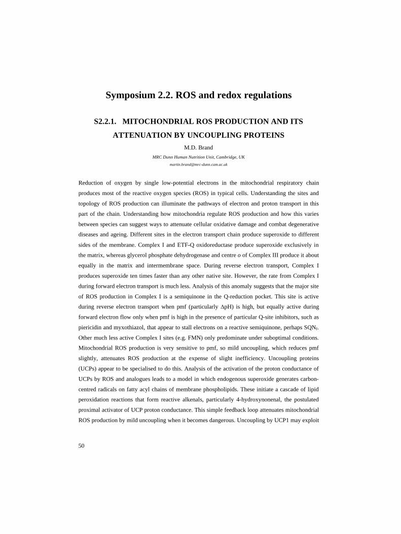
Symposium 2.2. ROS and redox regulations
S2.2.1. MITOCHONDRIAL ROS PRODUCTION AND ITS
ATTENUATION BY UNCOUPLING PROTEINS M.D. Brand
MRC Dunn Human Nutrition Unit, Cambridge, UK [email protected]
Reduction of oxygen by single low-potential electrons in the mitochondrial respiratory chain
produces most of the reactive oxygen species (ROS) in typical cells. Understanding the sites and
topology of ROS production can illuminate the pathways of electron and proton transport in this
part of the chain. Understanding how mitochondria regulate ROS production and how this varies
between species can suggest ways to attenuate cellular oxidative damage and combat degenerative
diseases and ageing. Different sites in the electron transport chain produce superoxide to different
sides of the membrane. Complex I and ETF-Q oxidoreductase produce superoxide exclusively in
the matrix, whereas glycerol phosphate dehydrogenase and centre o of Complex III produce it about
equally in the matrix and intermembrane space. During reverse electron transport, Complex I
produces superoxide ten times faster than any other native site. However, the rate from Complex I
during forward electron transport is much less. Analysis of this anomaly suggests that the major site
of ROS production in Complex I is a semiquinone in the Q-reduction pocket. This site is active
during reverse electron transport when pmf (particularly ∆pH) is high, but equally active during
forward electron flow only when pmf is high in the presence of particular Q-site inhibitors, such as
piericidin and myxothiazol, that appear to stall electrons on a reactive semiquinone, perhaps SQNf.
Other much less active Complex I sites (e.g. FMN) only predominate under suboptimal conditions.
Mitochondrial ROS production is very sensitive to pmf, so mild uncoupling, which reduces pmf
slightly, attenuates ROS production at the expense of slight inefficiency. Uncoupling proteins
(UCPs) appear to be specialised to do this. Analysis of the activation of the proton conductance of
UCPs by ROS and analogues leads to a model in which endogenous superoxide generates carbon-
centred radicals on fatty acyl chains of membrane phospholipids. These initiate a cascade of lipid
peroxidation reactions that form reactive alkenals, particularly 4-hydroxynonenal, the postulated
proximal activator of UCP proton conductance. This simple feedback loop attenuates mitochondrial
ROS production by mild uncoupling when it becomes dangerous. Uncoupling by UCP1 may exploit

51
the inefficiency for adaptive thermogenesis; regulation of pancreatic insulin secretion by UCP2 may
exploit it for signalling.

52
S2.2.2. MITOCHONDRIAL ROS-INDUCED ROS-RELEASE: AN
UPDATE AND REVIEW D.B. Zorov1, M. Juhaszova2, S.J. Sollott2
1 - Department of Bioenergetics, A.N.Belozersky Institute, Moscow State University, Moscow, Russia; Laboratory of Cardiovascular
Sciences, Gerontology Research Center, NIA, NIH, Baltimore, USA
2 - Laboratory of Cardiovascular Sciences, Gerontology Research Center, NIA, NIH, Baltimore, USA
Unstable mitochondrial redox transitions can occur following insults including ischemia/reperfusion
injury and toxin exposure, with negative consequences for mitochondrial integrity and cellular
survival [1-4]. These redox transitions can involve mechanisms such as the recently described
process, “Reactive Oxygen Species (ROS)-induced ROS-release” (RIRR) [2], and be generated by
circuits where the mitochondrial permeability transition (MPT) [5] pore and the inner membrane
anion channel (IMAC) are involved [6]. The exposure to excessive oxidative stress results in an
increase in ROS reaching a threshold level that triggers the opening of one of the requisite
mitochondrial channels. In turn, this leads to the simultaneous collapse of the mitochondrial
membrane potential and a transient increased ROS generation by the electron transfer chain.
Generated ROS can be released into cytosol and trigger RIRR in neighboring mitochondria. This
mitochondrion-to-mitochondrion ROS-signaling constitutes a positive feedback mechanism for
enhanced ROS production leading to potentially significant mitochondrial and cellular injury. This
review and update considers a variety of RIRR mechanisms (involving MPT, IMAC and episodes
of mitochondrial transient hyperpolarization). RIRR could be a general cell biology phenomenon
relevant to the processes of programmed mitochondrial destruction and cell death, and may
contribute to other mechanisms of post-ischemic pathologies, including arrhythmias [1,3].
References
[1] M. Juhaszova, D.B. Zorov, S.H. Kim, S. Pepe, Q. Fu, K.W. Fishbein, B.D. Ziman, S, Wang K.
Ytrehus, C.L. Antos, E.N. Olson, S.J. Sollott, Glycogen synthase kinase-3beta mediates
convergence of protection signaling to inhibit the mitochondrial permeability transition pore. J.
Clin. Invest. 113 (2004) 1535-1549.
[2] D.B. Zorov, C.R. Filburn, L.O. Klotz, J.L. Zweier, S.J. Sollott, Reactive oxygen species (ROS)-
induced ROS release: a new phenomenon accompanying induction of the mitochondrial
permeability transition in cardiac myocytes. J. Exp. Med. 192 (2000) 1001-1014.
[3] F.G. Akar, M.A. Aon, G.F. Tomaselli, B. O'Rourke, The mitochondrial origin of postischemic
arrhythmias. J. Clin. Invest. 115(2005) 3527-3535.

53
[4] M.A. Aon, S. Cortassa, F.G. Akar, B. O'Rourke, Mitochondrial criticality: A new concept at the
turning point of life or death. Biochim. Biophys. Acta. 1762 (2006) 232-240.
[5] D.R. Hunter, R.A. Haworth, The Ca2+-induced membrane transition in mitochondria. I. The
protective mechanisms. Arch. Biochem. Biophys. 195 (1979) 453-59.
[6] K.D. Garlid, A.D. Beavis, Evidence for the existence of an inner membrane anion channel in
mitochondria. Biochim. Biophys. Acta. 853(1986)187-204.

54
Symposium 2.3. Aging
S2.3.1. MITOCHONDRIAL OXIDATIVE STRESS, AGING AND
CALORIC RESTRICTION: THE PROTEIN AND METHIONINE
CONNECTION R. Pamplona1, G. Barja2
1 - Department of Basic Medical Sciences, University of Lleida, Lleida, Spain
2 - Department of Animal Physiology-II, Complutense University, Madrid, Spain [email protected]
Caloric restriction (CR) decreases aging rate and mitochondrial ROS (MitROS) production and
oxidative stress in rat postmitotic tissues. Low levels of these parameters are also typical traits of
long-lived mammals and birds. However, it is not known what dietary components are responsible
for these changes during CR. It was recently observed that 40% protein restriction without strong
CR also decreases MitROS generation and oxidative stress. This is interesting because protein
restriction also increases maximum longevity (although to a lower extent than CR) and is a much
more practicable intervention for humans than CR. Moreover, it was recently found that 80%
methionine restriction substituting it for L-glutamate in the diet also decreases MitROS generation
in rat liver. Thus, methionine restriction seems to be responsible for the decrease in ROS production
observed in caloric restriction. This is interesting because it is known that exactly that procedure of
methionine restriction also increases maximum longevity. Moreover, recent data show that
methionine levels in tissue proteins negatively correlate with maximum longevity in mammals and
birds. All this suggests that lowering of methionine levels is involved in the control of
mitochondrial oxidative stress and vertebrate longevity by at least two different mechanisms:
decreasing the sensitivity of proteins to oxidative damage, and lowering of the rate of ROS
generation at mitochondria.

55
S2.3.2. SUPEROXIDE AGING AND DEATH IN S. CEREVISIAE V. Longo
University of Southern California, USA [email protected]
Aging is believed to be a non-adaptive process that escapes the force of natural selection. We have
shown that yeast undergo an age- , ethanol- and pH-dependent death with features of mammalian
programmed cell death (apoptosis). This death is mediated in part by the down-regulation of many
protective systems and the generation of mitochondrial superoxide, is associated with the
inactivation of the 4Fe-4S cluster enzyme aconitase and is delayed by the overexpression of either
SOD1 and SOD2 or human Bcl-2. After 90-99% of the population loses the ability to reproduce and
dies, a small sub-population that has acquired mutations that promote early regrowth utilizes the
nutrients released from dead cells to grow. This adaptive regrowth occurs in less than 10% of
populations of long-lived mutants resistant to superoxide and hydrogen peroxide, in 50% of wild
type cultures, but in nearly 90% of cultures of short-lived mutants lacking superoxide dismutase
(sod1∆). Thus whereas mitochondrial superoxide contributes to programmed death, cytosolic
superoxide causes an increase in the frequency of nuclear mutations and an early release of
nutrients which plays an important role in adaptive regrowth. In mixed long-term cultures, wild type
yeast out-compete yeast overexpressing SOD1 and catalase but can be out-competed by sod1∆
mutants. Computational simulations confirm that programmed aging and the consequent early death
together with a relatively high mutation frequency can result in a major advantage in adaptation to
changing environments. These results suggest that superoxide is a mediator of an altruistic aging
and death program that improves adaptation to changing environments. The role of similar
pathways in the regulation of longevity in organisms ranging from yeast to mice raises the
possibility that higher eukaryotes may also undergo programmed aging mediated in part by
superoxide.

56
S2.3.3. MITOCHONDRIAL DNA MUTATIONS, OXIDATIVE
STRESS AND APOPTOSIS IN MAMMALIAN AGING T.A. Prolla
Departments of Genetics and Medical Genetics, University of Wisconsin, Madison, WI, USA [email protected]
Mutations in mitochondrial DNA (mtDNA) accumulate in tissues of mammalian species and have
been hypothesized to contribute to aging. We show that mice expressing a proofreading-deficient
version of the mitochondrial DNA polymerase g (POLG) accumulate mtDNA mutations and
display features of accelerated aging. Accumulation of mtDNA mutations was not associated with
increased markers of oxidative stress or a defect in cellular proliferation, but was correlated with the
induction of apoptotic markers, particularly in tissues characterized by rapid cellular turnover. The
levels of apoptotic markers were also found to increase during aging in normal mice. Thus,
accumulation of mtDNA mutations that promote apoptosis may be a central mechanism driving
mammalian aging.

57
Symposium 2.4. Cancer, ischemia and degenerative
disorders
S2.4.1. THE MITOCHONDRIAL PERMEABILITY TRANSITION
- FROM MOLECULAR MECHANISMS TO
CARDIOPROTECTION A.P. Halestrap
University of Bristol, Department of Biochemistry and Bristol Heart Institute, Bristol, UK
Under conditions of grossly elevated matrix [Ca2+], especially when accompanied by oxidative
stress and adenine nucleotide depletion, a non-specific pore opens in the inner mitochondrial
membrane. Opening of this Mitochondrial Permeability Transition Pore (MPTP) causes
mitochondria to become uncoupled and to hydrolyse rather than synthesise ATP. Unrestrained, this
leads to the loss of ionic homeostasis and ultimately necrotic cell death. Less severe, or transient
opening of the MPTP will not cause catastrophic ATP loss but may be sufficient to cause release of
pro-apoptotic factors such as cytochrome c and lead to apoptotic cell death. Opening of the MPTP
probably involves a calcium-mediated conformational change of the adenine nucleotide translocase
(ANT), facilitated by bound cyclophilin-D (CyP-D). The evidence for this model will be briefly
reviewed and re-assessed in the light of recent studies with ANT and CyP-D knockout mice.
Mitochondria from the latter lack a CsA-sensitive MPTP, and these mice show no obvious
phenotype and no inhibition of apoptosis induced by a variety of stimuli. However, cells from these
animals are resistant to necrotic cell death induced by oxidative stress and calcium overload, classic
activators of the MPTP. This implies that opening of the MPTP is critical for necrotic cell death but
not apoptosis. Cyp-D is the target of two potent inhibitors of the MPTP, cyclosporin A (CsA) and
sanglifehrin A (SfA). The heart and brain can be protected from the necrotic damage that occurs
following ischaemia and reperfusion by inhibiting the MPTP directly with CsA and SfA.
Furthermore, hearts and brains from the CyP-D knockout mice are very resistant to ischaemia /
reperfusion induced injury. Other protocols for protecting the heart from reperfusion injury,
including ischaemic preconditioning and antioxidants also, lead to inhibition of MPTP opening, but
through indirect mechanisms.

58
S2.4.2. CAN A SINGLE SUBUNIT NADH DEHYDROGENASE
WORK AS A THERAPEUTIC AGENT FOR COMPLEX I-
DEFICIENT DISEASES? T. Yagi
Division of Biochemistry, Department of Molecular and Experimental Medicine, The Scripps Research Institute, La Jolla, USA [email protected]
Defects of complex I are involved in many human mitochondrial diseases, and therefore we have
proposed to use the NDI1 gene encoding a single subunit NADH dehydrogenase of Saccharomyces
cerevisiae for repair of respiratory activity. The yeast NDI1 gene was successfully introduced into
mammalian cell lines. The expressed Ndi1 protein was correctly targeted to the matrix side of the
inner mitochondrial membranes, was fully functional and restored the NADH oxidase activity to the
complex I-deficient cells. The NDI1-transduced cells were more resistant to complex I inhibitors
and diminished production of reactive oxygen species induced by rotenone. It was further shown
that the Ndi1 protein can be functionally expressed in tissues such as skeletal muscles and the brain
of rodents, which scarcely induced an inflammatory response. The use of NDI1 as a potential
molecular therapy for complex I-deficient diseases is briefly discussed, including the proposed
animal model.

59
Symposium 2.5. Program death of cells and
mitochondria
S2.5.1. MITOCHONDRIAL PERMEABILIZATION IN CELL
DEATH AND MITOPHAGY J.J. Lemasters1, I. Kim1, S. Rodriquez-Enriquez2, H. Lihua3, P. Pediaditakis3, J.-S. Kim4
1 - Medical University of South Carolina, USA
2 - Instituto Nacional de Cardiologia, Mexico City, Mexico
3 - University of North Carolina, USA
4 - University of Florida, USA
In the mitochondrial permeability transition (MPT), the mitochondrial inner membrane
becomes non-selectively permeable to molecules of mass up to 1500 Da, which leads to
mitochondrial depolarization, uncoupling of oxidative phosphorylation and large amplitude
mitochondrial swelling. The MPT causes necrosis from ATP depletion and apoptosis from
cytochrome c release after swelling. The MPT is implicated in cell death after oxidative stress, Ca2+
toxicity, ischemia/reperfusion, cytokine exposure, ethanol and a variety of other stresses to
hepatocytes and other cell types. In contrast to necrotic cell death which is a consequence of ATP
depletion, ATP is required for the development of apoptosis after the MPT (1-3). In the most widely
discussed model, MPT pores form from the adenine nucleotide translocator (ANT) in the inner
membrane, the voltage-dependent anion channel (VDAC) in the outer membrane, cyclophilin D
(CypD) from the matrix, and possibly other proteins. The validity of this model has been challenged
by findings that the MPT still occurs in mouse mitochondria that are deficient in ANT (4). We
propose an alternate model of the MPT in which pores form by aggregation of misfolded integral
membrane proteins damaged by oxidant and other stresses whose conductance is regulated by
chaperone-like proteins (5). Binding of CypD and other chaperones, such as Hsp25/27 (6), to the
pore complex blocks conductance and may assist in protein refolding or removal by proteolysis.
CypD in this model confers sensitivity to Ca2+ such that increased Ca2+ opens these regulated MPT
pores, an action inhibited by cyclosporin A (CsA). When the nascent pores formed by misfolded
protein clusters exceed the number of chaperones available to regulate conductance, unregulated
pore opening occurs. Such unregulated pores no longer require Ca2+ for opening and are insensitive
to CsA inhibition. Recent findings show that heart and liver mitochondria from CypD knockout

60
mice are desensitized to onset of the Ca2+-induced MPT, consistent with an important regulatory
role of CypD in MPT pores (7-9). Various factors regulate the conductance of MPT pores,
including divalent cations, matrix pH, membrane potential, and reactive oxygen species (ROS). The
ubiquinone analogs, ubiquinone 0 and decylubiquinone, inhibit Ca2+-dependent onset of the MPT,
which suggests that one or more mitochondrial respiratory complexes may be part of the MPT pore
(10). Numerous studies show that Complexes I and III are also sites for ROS formation by
mitochondria, and ROS induce the MPT. Protein phosphorylation is another pathway that can
regulate the MPT. Incubation of mitochondria with recombinant protein kinase G (PKG) or hepatic
cytosol in the presence of cGMP and ATP inhibits Ca2+-dependent mitochondrial swelling, which is
blocked by protein kinase G inhibition (11). These findings implicate that protein phosphorylation
by PKG may directly or indirectly regulate the MPT. Proteomic analysis shows that the isoelectric
point (pI) of the Rieske iron sulfur protein (RISP) of Complex III increases after MPT induction but
not after protonophoric uncoupling (12). CsA prevents this pI shift. Phosphatase treatment causes
the same pI shift, suggesting that RISP is dephosphorylated in association with MPT induction.
These findings suggest that RISP has functions other than as a core polypeptide of Complex III and
may form part of the MPT pore complex and that phosphorylation and dephosphorylation of RISP
contributes to regulation of the MPT. In autophagy, portions of cytoplasm are sequestered into
autophagosomes and delivered to lysosomes for degradation. Long assumed to be a random
process, increasing evidence suggests that autophagy of mitochondria, peroxisomes, and possibly
other organelles is selective. For mitochondria, the MPT appears to signal autophagic sequestration
and removal of the damaged organelle (13). For selective autophagy of mitochondria, the term
“mitophagy” is appropriate in recognition of the non-random nature of the process (14;15).
Mitophagy protects against the adverse effects of free radical generation and futile ATP hydrolysis
by damaged mitochondria and may also retard accumulation of somatic mutations of mtDNA with
aging.
1. Kim JS, Qian T, Lemasters JJ. Gastroenterology 2003; 124:494-503. 2. Kim JS, He L,
Lemasters JJ. Biochem Biophys Res Commun 2003; 304:463-470. 3. Lemasters JJ.
Gastroenterology 2005; 129:351-360. 4. Kokoszka JE, Waymire KG, Levy SE, Sligh JE, Cai J,
Jones DP et al. Nature 2004; 427:461-465. 5. He L, Lemasters JJ. FEBS Lett 2002; 512:1-7. 6. He
L, Lemasters JJ. J Biol Chem 2003; 278:16755-16760. 7. Baines CP, Kaiser RA, Purcell NH, Blair
NS, Osinska H, Hambleton MA et al. Nature 2005; 434:658-662. 8. Basso E, Fante L, Fowlkes J,
Petronilli V, Forte MA, Bernardi P. J Biol Chem 2005; 280:18558-18561. 9. Nakagawa T, Shimizu
S, Watanabe T, Yamaguchi O, Otsu K, Yamagata H et al. Nature 2005; 434:652-658. 10. Walter L,
Miyoshi H, Leverve X, Bernard P, Fontaine E. Free Radic Res 2002; 36:405-412. 11. Kim JS,
Ohshima S, Pediaditakis P, Lemasters JJ. Free Radic Biol Med 2004; 37:1943-1950. 12. He L,

61
Lemasters JJ. Biochem Biophys Res Commun 2005; 334:829-837. 13. Elmore SP, Qian T,
Grissom SF, Lemasters JJ. FASEB J 2001; 15:2286-2287. 14. Lemasters JJ. Rejuvenation Res
2005; 8:3-5. 15. Rodriguez-Enriquez S, Kim I, Currin RT, Lemasters JJ. Autophagy 2006; 2:39-46.
62
S2.5.2. APOPTOSIS AND NECROSIS, TWO FUNDAMENTAL
ALTERNATIVES P. Vandenabeele
Molecular Signalling and Cell Death Unit, Department of Molecular Biomedical Research, VIB, University of Gent,
Zwijnaarde/Gent, Belgium
Apoptosis is the principal cellular process by which cells are eliminated during development and
homeostasis in metazoan organisms. Apoptosis is characterized by distinct morphological events,
including cell shrinkage, blebbing of the plasma membrane, maintenance of organelle integrity, and
chromatin condensation. Early apoptotic cells retain plasma membrane integrity, and are rapidly
phagocytosed by macrophages or surrounding cells. This prevents leakage of cellular contents into
the surrounding tissue, making apoptosis a ‘clean’ packaging process that minimizes the risk of
inflammation during cell death. Apoptosis is characterized by the activation of cysteine aspartate
specific proteases or caspases that cleave crucial substrates associated with the apoptotic process.
Necrosis is characterized by swelling of the cell and its organelles leading to loss of plasma
membrane integrity. The release of the cytoplasmic contents and accumulation of necrotic cell
corpses may damage the surrounding tissue and induce an inflammatory reaction. Necrosis is often
considered as a passive cell death process resulting from physico-chemical insults, but it is
becoming increasingly clear that necrosis can be elicited by biological stimuli and is governed by
defined signal transduction pathways. Necrotic cell death is essentially caspase-independent and
implies other signaling events such as RIP1 kinase activity, mitochondrial ROS production, and
lysosomal cathepsins in the late phase of necrotic cell death. In this presentation we will elaborate
on the signal transduction pathways leading to both types of cell death, the differential role of
mitochondria, the crosstalk between both signal transduction pathways, the phagocytosis by
macrophages and intercellular communication associated with both types of cell death. Moreover
we discuss the possible differential impact of these two cell death pathways on pathophysiology.

63
S2.5.3. NUCLEI-MITOCHONDRIA CROSS-TALK IN
APOPTOSIS B. Zhivotovsky, H. Vakifahmetoglu, M. Olsson, V. Gogvadze, S. Orrenius
Institute of Environmental Medicine, Karolinska Institutet, Stockholm, Sweden [email protected]
Initial apoptosis research characterized this form of cell death based on distinct nuclear
morphology that was subsequently shown to be associated with the appearance of oligonucleosomal
DNA fragments. More recent evidence has indicated that apoptosis depends upon a tightly regulated
cellular program for its successful initiation and execution. Molecular participants in this program
are present in different subcellular compartments, including the plasma membrane, cytosol,
mitochondria and nucleus. The interplay among these compartments and the exchange of specific
signaling molecules are critical for the systematic progression of apoptosis. Among all known
caspases pro-caspase-2 is only one that is present constitutively in the nucleus. Recent findings
indicate that caspase-2 is activated early in response to DNA-damaging agents and is important for
the engagement of the mitochondrial apoptotic pathway. This activation was observed only in p53-
positive cells. Although no direct interaction between p53 and caspse-2 was found, a functional
connection between these two proteins is essential for initiation of DNA damage-induced apoptosis.
In cytosol caspase-2 is present in the PIDDosome complex together with PIDD and RAIDD. Both
PIDD and RAIDD are required for assembly of this complex, but not sole mediators of caspase-2
activation. Fully processed caspase-2 can permeabilize the outer mitochondrial membrane and
cause cytochrome c and Smac release from these organelles. Caspase-2 also might disrupt the
interaction of cytochrome c with cardiolipin and thereby enhance the release of the hemoprotein
caused by treatment of mitochondria with digitonin or Bax. These results suggest the presence of
direct cross-talk between nuclei and mitochondria in apoptosis signaling. We also found that the
deficiency in the apoptosis pathway triggered by DNA-damaging agents in non-small lung cancer
cells (NSCLC) occurred downstream from (or at the level of) mitochondrial changes and upstream
of nuclear apoptotic events. Despite this deficiency, NSCLC undergo cell death in response to
staurosporine, which induces caspase activation and mitochondrial dysfunction associated with AIF
release. We found that AIF plays a major role in the death of NSCLC via transmission of signal
from mitochondria to the nucleus, while caspase-dependent arm of the lethal signal transduction
appears to be heavily compromised. Thus, proper dialogue between nuclei and mitochondria is
important for both signaling and execution of apoptosis.

64
Symposium 2.6. Mitochondrial dynamics
S2.6.1. KEEPING MITOCHONDRIA IN SHAPE: A MATTER OF
LIFE AND DEATH L. Scorrano
Dulbecco-Telethon Institute, Venetian Institute of Molecular Medicine, Padova, Italy [email protected]
Mitochondria are crucial organelles for life and death of the cell, as they provide most of the ATP
needed for endoergonic processes and integrate complex pathways including Ca2+ signaling and
apoptosis. Their functional versatility is matched by a complex structural organization, both at the
cellular and at the ultrastructural level. Moreover, they are remarkably dynamic, continuously
fusing and dividing. We are starting to identify the molecular mechanisms regulating the control of
mitochondrial morphology as a growing number of “mitochondria-shaping” proteins are being
identified in mammals. They include dynamin-related proteins, large GTPases that exert mechanical
forces on membranes, like mitofusin-1, -2, DRP-1 and OPA1. Their importance in physiology is
confirmed by neurodegenerative genetic diseases associated with mutations in some of these
proteins. Several questions remain open: how do mitochondria-shaping proteins function? Do they
work in concert, or does each of them have a specific task? How do cellular and mitochondrial
signals regulate this machinery? And last but not least, do they (and therefore changes in
mitochondrial shape) control mitochondrial and cellular physiology. To address these questions, we
integrate genetics, physiology and advanced imaging. Our studies unraveled that these processes are
tightly regulated and influence cell life and death. OPA1 has a dual function: it promotes
mitochondrial fusion via the outer membrane partner mitofusin-1. Following cleavage and
activation by the inner mitochondrial membrane rhomboid protease PARL it regulates apoptosis, by
controlling the opening of the tubular cristae junction and therefore the release of cytochrome c
from mitochondria. Our studies for the first time revealed a functional difference between the two
highly similar mitofusins. While mitofusin-1 is primarily engaged with OPA1 in the control of
mitochondrial fusion, mitofusin-2 regulates the shape of another organelle, the endoplasmic
reticulum, and the juxtaposition between this and mitochondria. This complex machinery is not left
uncontrolled by the cell. For example, we discovered that the cytosolic phosphatase calcineurin
regulates mitochondrial fission. Blocking calcineurin inhibits dephosphorylation of DRP-1 and
movement of this cytosolic protein to mitochondria, where it binds to its adapter hFis1. This small

65
molecule of the outer mitochondrial membrane ultimately regulates fragmentation and function of
the organelle. A genetic analysis revealed that via a short domain protruding in the intermembrane
space it transmits a dysfunction signal to mitochondria, rendering them more vulnerable to opening
of the permeability transition pore. In conclusion, mitochondrial morphology is tightly regulated at
multiple sites and impacts on mitochondrial and cellular function.

66
S2.6.2. DYNAMINS, ENDOPHILINS AND BCL-2 FAMILY
MEMBERS: INVOLVEMENT IN MITOCHONDRIAL OUTER
MEMBRANE DYNAMICS M. Karbowski, K. Norris, R. Youle
Biochemistry Section, SNB, NINDS, NIH, Bethesda, USA [email protected]
Dynamin and endophilin 1 are involved in synaptic vesicle recycling. We have identified an
endophilin isoform that, along with the dynamin related protein, Drp1, is specifically involved in
mitochondrial morphogenesis. Mitochondria form highly dynamic networks through continuous
division and fusion that requires Drp1 for the fission step. Using RNA interference (RNAi) to
deplete endophilin B1 followed by immunofluorescence microscopy reveals alterations of the
mitochondrial network that are consistent with an inhibition of mitochondrial fission. A striking
dissociation of the outer mitochondrial membrane compartment from that of the matrix, and
formation of thin, elongated tubules of outer mitochondrial membrane is also observed in
endophilin B1 knock down cells indicating that this protein participates in the normal
morphogenesis of the outer mitochondrial membrane. Moreover, we find that double knock down
of endophilin B1 and Drp1 leads to a mitochondrial phenotype identical to that of the Drp1 single
knock down, a result consistent with Drp1 acting upstream of endophilin B1 in the same pathway.
Interestingly, endophilin B1 also binds to the pro-apoptosis member of the Bcl-2 family, Bax,
during programmed cell death and translocates with Bax to mitochondria. Thus, in addition to the
role of certain endophilin family members in plasma membrane endocytosis, other members of the
endophilin family participate in intracellular membrane dynamics and play a role in programmed
cell death.

67
Symposium 2.7. Therapeutic approach
S2.7.1. PROPERTIES OF THE PERMEABILITY TRANSITION IN
VDAC1-/- MITOCHONDRIA A. Krauskopf1, O. Eriksson2, W.J. Craigen3, M.A. Forte4, P. Bernardi1
1 - Dept. of Biomedical Sciences, University of Padova, Padova, Italy
2 - Helsinki Biophysics and Biomembrane Group, University of Helsinki, Finland
3 - Dept. of Molecular and Human Genetics, Baylor College of Medicine, Houston (TX), USA
4 - The Vollum Institute, Oregon Health and Science University, Portland (OR), USA [email protected]
Opening of the permeability transition pore (PTP), a high-conductance mitochondrial channel,
causes mitochondrial dysfunction with Ca2+ deregulation, ATP depletion, release of pyridine
nucleotides and of mitochondrial apoptogenic proteins. Despite major efforts, the molecular nature
of the PTP remains elusive. A compound library screening led to the identification of a novel high
affinity PTP inhibitor (Ro 68-3400), which labeled a ~32 kDa protein that was identified as isoform
1 of the voltage-dependent anion channel (VDAC1) [A.M. Cesura, E. Pinard, R. Schubenel, V.
Goetschy, A. Friedlein, H. Langen, P. Polcic, M.A. Forte, P. Bernardi, J.A. Kemp, The voltage-
dependent anion channel is the target for a new class of inhibitors of the mitochondrial permeability
transition pore, J. Biol. Chem. 278 (2003) 49812-49818]. In order to assess the role of VDAC1 in
PTP formation and activity, we have studied the properties of mitochondria from VDAC1-/- mice.
The basic properties of the PTP in VDAC1-/- mitochondria were indistinguishable from those of
strain-matched mitochondria from wild-type CD1 mice, including inhibition by Ro 68-3400, which
labeled identical proteins of 32 kDa in both wild-type and VDAC1-/- mitochondria. The labeled
protein could be separated from all VDAC isoforms. While these results do not allow to exclude
that VDAC is part of the PTP, they suggest that VDAC is not the target for PTP inhibition by Ro
68-3400.

68
S2.7.2. MITOCHONDRIAL NADPH, TRANSHYDROGENASE
AND DISEASE J. Rydstrom
Department of Biochemistry and Biophysics, Goeteborg University, Sweden [email protected]
Ever since its discovery in 1953 by N. O. Kaplan and coworkers, the physiological role of the
proton-translocating transhydrogenase has generally been assumed to be that of generating
mitochondrial NADPH. Mitochondrial NADPH can be used in a number of important
reactions/processes, e.g. biosynthesis, maintenance of GSH, apoptosis, aging etc. This assumed role
has found some support in bacteria but not in higher eukaryotes, a situation which changed
dramatically with two recent but separate findings, both using transhydrogenase knockouts, i.e., in
the nematode C. elegans and in the mouse, strain C57BL/6J. In the former system the herbicide
Paraquat (methylviologen), a known redox cycler, was used to induce oxidative stress. The C.
elegans knockout showed a pronounced decrease in the overall GSH/GSSG ratio, as well as in the
capacity of Paraquat-exposed eggs to survive and develop to adult nematodes. Appently, these
nematodes suffer from extensisve oxidative stress. These effects could essentially be mimicked by
siRNA treatment, and prevented by re-introducing the Nnt gene. In C57BL/6J mice, the knockout is
due to a spontaneous deletion mutation in the Nnt gene, which was serendipitously found during
investigations of the diabetic properties of these mice (Freeman, H. et al (2006) Cell Metab 3, 35-
45). In terms of insulin release, these mice show a diminished response to a glucose load, which has
been interpreted as being due to a decreased ATP level, which, in turn, attenuates the ATP-
regulated K+-channel, leading to a lower extent of depolarization and Ca2+ influx. This lower ATP
level is suggested to be due to a partial uncoupling through the uncoupler proteins UCP2/UCP3
(Brand, H. and Esteves, T. C. (2005) Cell Metab. 2, 85-93). The latter channels have been proposed
to be opened by products of oxidative stress, generated by leakage of electrons from the respiratory
chain, especially at a high redox level of the respiratory chain components. However, at a lower
redox level of these components, the leakage of electrons and the level of oxidative stress is
lowered. It has been proposed that the role of UCP2/UCP3 is to regulate the level of oxidative stress
through a controlled uncoupling. In this context the role of transhydrogenase is believed to be
crucial since the lack of transhydrogenase generally increases the level of oxidative stress through a
GSH/GSSG ratio, indirectly leading to an attenuation of the release of insulin through the above
mechanism. The implications of these findings for the overall role of transhydrogenase in cell
metabolism and disease is discussed (supported by the Swedish Research Council).

69
COLLOQUIA

70
1. Molecular bioenergetics
Colloquium 1.1. Respiratory chain
C1.1.1. STRUCTURAL AND FUNCTIONAL INSIGHTS INTO
MITOCHONDRIAL COMPLEX I U. Brandt
Johann Wolfgang Goethe-Universitaet, Zentrum der Biologischen Chemie, Frankfurt am Main, Germany [email protected]
The molecular mechanism of complex I (NADH:ubiquinone oxidoreductase) is still elusive.
Mutational analysis, spectroscopic studies and emerging structural information provide important
clues on the parts of the machinery that links electron transfer to proton translocation. Based on its
more positive and pH-dependent midpoint potential, iron-sulfur cluster N2 has long been discussed
as one of the key components of the pump. However, removal of the redox-Bohr group did not
affect the proton pumping stoichiometry. Structural studies with complex I from the strictly aerobic
yeast Yarrowia lipolytica suggest an unexpected position of the ubiquinone reduction site near the
interface between the PSST and the 49 kDa subunits far up in the peripheral arm of complex I.
We propose a two-state mechanism of energy conservation for complex I that is based on long
range conformational changes of the enzyme and that is driven by stabilization changes of ubi-
quinone intermediates. This stabilization change mechanism inherently provides a rationale for the
forward and reverse mode of complex I.

71
C1.1.2. THE MECHANISM OF PROTON PUMPING BY
CYTOCHROME C OXIDASE P. Brzezinski
Department of Biochemistry and Biophysics, The Arrhenius Laboratories for Natural Sciences, Stockholm University, Stockholm,
Sweden
Cellular respiration involves electron transfer to oxygen through a series of membrane-bound
protein complexes. The process maintains a transmembrane proton electrochemical gradient that is
used e.g. for the synthesis of ATP. In mitochondria and many bacteria the last enzyme complex in
the electron transfer chain is cytochrome c oxidase, which catalyses the four-electron reduction of
O2 to H2O using electrons delivered by a water-soluble donor, cytochrome c. The electron transfer
through cytochrome c oxidase, accompanied by proton uptake to form H2O drives pumping of four
protons per reduced O2 across the membrane. To address the molecular mechanism of proton
pumping we investigate the specific steps of the catalytic cycle in wild-type and site-directed
mutant forms of cytochrome c oxidase using a number of biophysical techniques. I will discuss the
following topics: (i) the order and timing of proton-transfer reactions during the proton-pumping
cycle in membrane-reconstituted enzyme, (ii) the coupling between electron and proton-transfer
reactions, and (iii) results from studies of mutant forms of the enzyme in which proton pumping is
uncoupled from the oxygen-reduction reaction. Collectively, the results from these studies suggest
principles by which the redox-driven proton pump operates and indicate possible molecular
mechanisms by which cytochrome c oxidase couples electron transfer to proton translocation.

72
C1.1.3. RAPID KINETICS OF CHARGE TRANSLOCATION BY
CYTOCHROME C OXIDASE: TRANSITION BETWEEN THE
FERYL-OXO AND FERRIC STATES S.A. Siletsky, D. Zaslavsky, I.A. Smirnova, T.V. Vygodina, A.A. Konstantinov
A.N. Belozersky Insitute, Moscow State University, Moscow, Russia [email protected]
Single-electron photoreduction of the Ferryl-Oxo complex of bovine cytochrome c oxidase by
tris-bipyridyl complex of Ru(II) (transfer of the 4-th electron in the catalytic cycle) results in three
phases of membrane potential generation with τ values of ca. 40 µs, 1.2 ms and 4.5 ms [1]. Here,
the electrogenic phases have been compared with the kinetics of hemes a and a3 oxidoreduction. In
addition, pH and temperature dependences of the resolved electrogenic phases have been studied.
The rate of the rapid phase (40-50 µs) does not depend on pH and reveals a very weak temperature
dependence (ΔGa = 3.8 kcal/mol). The rate of the intermediate electrogenic phase (1.2 ms) is pH-
independent but shows significant temperature dependence (ΔGa = 19 kcal/mol) and is assigned
provisionally to transfer of the pumped proton from Glu242 in the D-channel to the so-called
Proton Loading Site “above the heme” (e.g., carboxylate of the Δ-propionate in heme a3, or His291
ligand of CuB [2]). This phase precedes slightly the major phase of electron transfer from heme a to
heme a3. The major part of electrogenic proton transfer (slow electrogenic phase) takes place after
completion of the redox reaction of the hemes. The phase is highly temperature-dependent (ΔGa =
17 kcal/mol) and decelerates at alkaline pH with apparent pK of 8.2. We assign this process mainly
to electrogenic reprotonation of Glu242 from the N-phase via the D-channel plus uptake of a
“chemical” proton from the N-phase to the binuclear center. The results do not support models of
COX which postulate transmembrane proton translocation in the F→O transition to be sinchronous
with ferryl-to-ferric redox transition of heme a3. We suggest that redox transition of heme a3 can be
assisted by non-electrogenic proton donation from the nearest surroundings and is followed by
relaxation of the protein coupled to electrogenic uptake and release of protons.
[1]. Zaslavsky,D., Kaulen,A., Smirnova,I.A., Vygodina,T.V. and Konstantinov,A.A. (1993)
FEBS Lett. 336,389-393.
[2]. Popovic, D.M, Stuchebrukhov, A.A. (2004) FEBS Lett. 566, 126-30.

73
C1.1.4. NEW INSIGHTS ON NON-CANONICAL HAEM-COPPER
OXYGEN REDUCTASES M. Teixeira
Instituto de Tecnologia Quimica e Biologica, Universidade Nova de Lisboa, Oeiras, Portugal [email protected]
Haem-copper oxygen reductases present a large diversity in terms of amino acid sequences,
properties of the haem-copper catalytic site, redox behaviour and proton channels. The study of the
more divergent oxidases, i.e., of those far related to the mitochondrial like ones, bring new insights
on the minimal essential features of these enzymes. New results on the aa3 type oxygen reductase
from Acidianus ambivalens, and enzyme of the B-family, and of the caa3 oxygen reductase from
Rhodothermus marinus, and enzyme of the A2-subfamily, will be presented, based on resonance
Raman, FT-IR and EXAFS spectroscopies. In particular, it will be shown that, in contrast to the A-
type, mitochondrial like oxidases, in the A. ambivalens enzyme haem a3 has a reduction potential
higher than that of haem a. Also, for the R. marinus enzyme, it will be shown that tyrosine residues,
besides that linked to the histidine copper ligand, are involve din redox linked proton equilibria.
T. Bandeiras, M. M. Pereira, M.Teixeira, P. Moenne-Loccoz, N. J. Blackburn (2005) J. Biol. Inorg.
Chem., 10, 625-35.
S. Todorovic, T. M. Bandeiras, M. M. Pereira, M. Teixeira, P. Hildebrandt and D.H. Murgida
(2005) J. Am. Chem. Soc., 127, 13561-6.

74
C1.1.5. PROTON TRANSLOCATION BY CYTOCHROME C
OXIDASE M.I. Verkhovsky
Institute of Biotechnology, Biocenter 3, University of Helsinki, Finland [email protected]
Proton translocation in the catalytic cycle of cytochrome c oxidase proceeds sequentially in
a four-stroke manner. Every electron donated by cytochrome c drives the activated enzyme from
one of the four stable intermediates to another, and each of these transitions is coupled to proton
translocation across the membrane, and to uptake of one more proton for charge compensation of
the electron in the catalytic site. Using cytochrome c oxidase from Paracoccus denitrificans we
have resolved the kinetics of the electron transfer reactions separately in three of the four reaction
steps, and electric potential generation at each step. All four proton translocation cycles during the
single catalytic cycle have the same mechanism and are characterized by a cascade of electron
transfer equilibria with trapping of the final state by proton transfer to the binuclear site. The extent
of electric potential generation during the first wave of electron equilibration between CuA and haem a shows that this reaction is not coupled to any vectorial proton transfer, whereas oxidation of
haem a is kinetically linked to the main proton translocation events during functioning of the proton pump. The rate of haem a oxidation is different at the different steps of the catalytic cycle, and the
same difference can be seen in the rate of electric potential generation. Such correlation of heme a
oxidation with the charge translocation discussed in the frame of model, where electron transfer
from haem a to the O2 reduction site initiates the proton pump mechanism by being kinetically
linked to an internal vectorial proton transfer. This reaction drives the proton pump, and occurs
prior to relaxation steps where protons are taken up from the aqueous space on one side of the
membrane and released on the other.

75
Colloquium 1.2. Light-driven energy transducers
C1.2.1. CONSEQUENCES OF THE STRUCTURE OF THE
CYTOCHROME b6f COMPLEX FOR ITS CHARGE TRANSFER
PATHWAYS W.A. Cramer, H. Zhang
Department of Biological Sciences, Purdue University, West Lafayette, USA [email protected]
At least two features of the crystal structures of the cytochrome b6f complex from the
thermophilic cyanobacterium, M. laminosus (1) and green alga, C. reinhardtii (2) have implications
for the pathways and mechanism of charge (electron/proton) transfer in the complex: (i) The narrow
11 x 12 Å portal between the p-side of the quinone exchange cavity and p-side plastoquinone/quinol
binding niche, through which all Q/QH2 must pass, is smaller in the b6f than in the bc1 complex
because of its partial occlusion by the phytyl chain of the one bound chlorophyll a molecule in the
b6f complex. Thus, the pathway for trans-membrane passage of the lipophilic quinone is even more
labyrinthine in the b6f than in the bc1 complex (3). (ii) A unique covalently bound heme, heme cn, in
close proximity to the n-side b heme, is present in the b6f complex. A position of the novel heme in
a pathway of photosytem I-linked cyclic electron transport that requires ferredoxin and the
ferredoxin: NADP+ reductase is described. This pathway through the n-side of the b6f complex
could overlap and interact with the n-side of the Q cycle pathway. Thus, regulation is either
required at the level of the redox state of the hemes that allows them to be shared by the two
pathways, or the two different pathways are segregated in the membrane. (1) Science, 302, 1009-,
2003; (2) Nature, 426, 413-, 2003; (3) Ann. Rev. Biochem. Mol. Biol., 75, in press.

76
C1.2.2. THE IDENTIFICATION OF FX IN THE
HELIOBACTERIAL REACTION CENTER AS A [4Fe-4S]
CLUSTER WITH A GROUND SPIN STATE OF S = 3/2 M. Heinnickel1, R. Agalarov1, N. Svensen1, C. Krebs2, J.H. Golbeck2
1 - Department of Biochemistry and Molecular Biology, The Pennsylvania State University, USA
2 - Department of Biochemistry and Molecular Biology, The Pennsylvania State University, USA; Department of Chemistry, The
Pennsylvania State University, USA
Type I homodimeric reaction centers, particularly the class present in Heliobacteria, are not well
understood. Even though the primary amino acid sequence of PshA in Heliobacterium mobilis has
been shown to contain an FX binding site, the Fe/S cluster has not been detected by EPR
spectroscopy. Recently, we reported that PshB, which contains FA and FB-like Fe/S clusters, could
be removed from the Heliobacterium modesticaldum reaction center (HbRC), resulting in 15-ms
lifetime charge recombination between P798+ and an unidentified electron acceptor (M. Heinnickel,
G. Shen, R. Agalarov, and J. H. Golbeck, (2005) Biochemistry 44, 9950-9960). We found that by
incubating a HbRC core with sodium hydrosulfite in the presence of light, the 15-ms charge
recombination was replaced with a kinetic transient in the sub-µs time domain, consistent with the
reduction of an electron acceptor. Concomitantly, a broad and intense EPR signal arises around g =
5 along with a minor set of resonances around g = 2 similar to the spectrum of the [4Fe-4S]1+
cluster in the Fe-protein of Azotobacter vinelandii nitrogenase, which exists in two conformations
having ground spin states of S = 3/2 and S = 1/2. The Mössbauer spectrum in the as-isolated HbRC
core shows that all of the Fe is present in the form of a [4Fe-4S]2+ cluster. After reduction with
sodium hydrosulfite in the presence of light, approximately 65% of the Fe appears in the form of a
[4Fe-4S]1+ cluster; the remainder is in the [4Fe-4S]2+ state. Analysis of the non-heme iron content
of HbRC cores indicates an antenna size of 20-23 BChl g/P798. The HbRC therefore contains a
[4Fe-4S] cluster assigned to FX that is coordinated between the PshA homodimer; in contrast to FX
in other Type I reaction centers, this [4Fe-4S] cluster exhibits an S = 3/2 ground spin state.

77
C1.2.3. CYCLIC ELECTRON FLOW IN C3 PLANTS P. Joliot, A. Joliot
Institut de Biologie Physico-Chimique, Paris, France [email protected]
This paper summarized our present view on the mechanism of cyclic electron flow in C3 plants. We
propose that cyclic and linear pathways are in competition for the reoxidation of the soluble primary
PSI acceptor, Ferredoxin (Fd), that freely diffuses in the stromal compartment. In the linear mode,
Fd binds ferredoxin-NADP-reductase and electrons are transferred to NADP+ and then to the
Benson and Calvin cycle. In the cyclic mode, Fd binds a site localized on the stromal side of the
cytochrome b6f complex and electrons are transferred to P700 via a mechanism derived from the Q-
cycle. In dark-adapted leaves, the cyclic flow operates at maximum rate, owing to the partial
inactivation of the Benson and Calvin cycle. For increasing time of illumination, the activation of
the Benson and Calvin cycle, and thus, that of the linear flow, is associated with a subsequent
decrease in the rate of the cyclic flow. Under steady-state conditions of illumination, the
contribution of cyclic flow to PSI turnover increases as a function of the light intensity (from 0 to ~
50 % for weak to saturating light, respectively). Lack of CO2 is associated with an increase in the
efficiency of the cyclic flow. ATP concentration could be one of the parameters that control the
transition between linear and cyclic modes.

78
C1.2.4. HIGH-FIELD EPR ON LOW-SYMMETRY PROTEINS TO
REVEAL STRUCTURE-DYNAMICS-FUNCTION RELATIONS K. Moebius
Department of Physics, Free University Berlin, Berlin, Germany [email protected]
In photosynthetic organisms the light-induced electron-transfer processes across cell membranes are
vectorial in nature. For these processes to be unidirectional, subtle cofactor-protein interactions
and/or conformational changes of specific protein segments are functionalized as molecular
switches or electron gates. To understand their function, site-specific mutants have been studied to
identify functionally important protein sub-domains and cofactors and, moreover, to characterize
their structure and dynamics in the transient states of biological action. In the lecture a report will be
given on our recent high-field EPR experiments at 95 GHz (wavelength 3 mm) and 360 GHz (0.8
mm) on tailor-made site-directed mutants of photosynthetic reaction centers from the purple
bacterium Rb. sphaeroides. The EPR results obtained at successively higher Zeeman fields - in
comparison to X-band EPR - provide detailed information on structure and dynamics of transient
intermediates of the photocycle beyond what is provided by standard X-ray crystallography. Such
information is revealed by analysis of the continuous-wave and pulsed high-field EPR spectra and
comprises details of molecular structure characteristics in terms of distance and orientation of
cofactors in charge-separated radical pairs involved in vectorial transmembrane electron transfer, as
well as details of the electronic structure and anisotropic motion of the cofactors in the hydrogen-
bond networks of their binding sites. Such pieces of information are essential ingredients for an
understanding of the biological transfer process. The lecture will conclude with our most recent
developments and applications of high-field PELDOR (pulsed electron-electron double resonance)
at 95 GHz to measure distance and orientation of donor and acceptor ions in spin-correlated radical
pairs. I thank my co-workers M. Fuchs, M. Plato, A. Savitsky, A. Schnegg as well as my
collaboration partners A.A. Dubinskii (Moscow), Y.A. Grishin (Novosibirsk) and W. Lubitz
(Mülheim/Ruhr). Financial support by the Deutsche Forschungsgemeinschaft (SFB 498, SPP 1051)
is gratefully acknowledged.

79
C1.2.5. INITIAL ELECTRON DONOR AND ACCEPTOR IN
ISOLATED PHOTOSYSTEM II REACTION CENTERS
IDENTIFIED WITH FEMTOSECOND MID-INFRARED
SPECTROSCOPY R. van Grondelle
Free University of Amsterdam, Amsterdam, The Netherlands [email protected]
In spite of the apparent similarity between the plant Photosystem II reaction center and its purple
bacterial counterpart, we show in this manuscript that the mechanism of charge separation is very
different for the two photosynthetic reaction centers. Using femtosecond visible-pump-mid-
infrared probe spectroscopy in the region of the chlorophyll ester and keto modes, between 1775
and 1585 cm-1, with 150 fs time resolution, we show that the reduction of pheophytin occurs on a
0.6-0.8 ps time scale, whereas P+, the precursor state for water oxidation, is formed after ~6 ps. We
conclude therefore that in the PS II RC the primary charge separation occurs between the 'accessory
chlorophyll' ChlD1 and the pheophytin on the so-called active branch.

80
Colloquium 1.3. ATP-synthase/ATPase
C1.3.1. DYNAMICS OF SUBUNIT INTERACTION OF THE Na+
PUMP BY FRET VOLTAGE CLAMP FLUOROMETRY R. Dempski, T. Friedrich, E. Bamberg
Max-Planck Institut fuer Biophysik, Frankfurt, Germany [email protected]
The Na+/K+-ATPase performs active transport to maintain the physiological Na+ and K+ gradients
across the plasma membrane in most animal cells. The functional unit of the ion pump is comprised
of two mandatory subunits including the alpha subunit which mediates ATP hydrolysis and ion
translocation as well as the beta subunit which acts as a chaperone to promote proper membrane
insertion and trafficking in the plasma membrane. Although crystal structures of a related protein,
the sarcoplasmic reticulum Ca2+-ATPase have provided insight into the conformational flexibility
of the alpha subunit, the molecular interaction and orientation between the alpha and beta subunits
of the Na+/K+-ATPase is not well understood. In order to examine the conformational dynamics
between the alpha and beta subunits of the Na+/K+-ATPase, we have now used fluorescence
resonance energy transfer, under voltage-clamp conditions on oocytes from Xenopus laevis in order
to determine the relative positions of the alpha and beta subunits by measuring the time constant of
irreversible donor fluorophore destruction. We have also investigated the relative movement
between the alpha and beta subunits as the ion pump shuttles between the two main conformational
states (E1 and E2) as described by the Albers-Post scheme. The results from this study have allowed
the identification of a model for the orientation of the beta subunit in relation to the alpha subunit.
Our data furthermore suggest that a conformational re-arrangement between the alpha and beta
subunits occurs during the E2 to E1 transition.
Geibel, S., Kaplan, J. H., Bamberg, E. & Friedrich, T. (2003). Conformational dynamics of the
Na^+ /K^+ -ATPase probed by voltage clamp fluorometry. /Proc. Natl. Acad. Sci. USA/ 100, 964-
969.
Dempski, R. E., Friedrich, T. & Bamberg, E. (2005). The Beta Subunit of the Na+/K+-ATPase
Follows the Conformational State of the Holoenzyme. /J. Gen. Physiol./ 125, 505-520.

81
C1.3.2. SUBUNIT MOVEMENTS IN MEMBRANE INTEGRATED
EF0F1 BY SINGLE MOLECULE SPECTROSCOPY B. Zimmermann1, M. Diez1, P. Graeber1, M. Boersch2
1 - Institut fur Physikalische Chemie, Albert-Ludwigs-Universitat Freiburg, Freiburg, Germany
2 - Physikalisches Institut, Universitat Stuttgart, Stuttgart, Germany
The H+-ATPsynthase from E. coli was isolated and labelled at the γ- or ε-subunit with
tetramethylrhodamine, and at the b-subunits with bisCy5. The double labelled enzymes were
incorporated into liposomes. They showed ATP hydrolysis activity, and, after energization of the
membrane by ΔpH and Δφ, also ATP synthesis activity was observed. Fluorescence resonance
energy transfer (FRET) was used to investigate the movements of either the γ-subunit or the ε-
subunit relative to the b-subunits in single membrane-integrated enzymes. The results show: During
catalysis the γε-complex rotates stepwise relative to the b-subunit. The direction of rotation during
ATP synthesis is opposite to that during ATP hydrolysis. The stepwise motion is characterized by
dwell times (docking time of the γε-complex to one αβ-pair) up to several hundred ms, followed by
a rapid movement of the γ- and ε-subunit to the next αβ-pair within 0.2 ms. The same FRET levels
(i.e. the same γ-b and ε-b distances) are observed during proton transport-coupled ATP hydrolysis
and ATP synthesis, indicating that the reaction proceeds via the same intermediates in both
directions.

82
C1.3.3. BIOENERGETICS OF ARCHAEA: ATP SYNTHESIS
UNDER HARSH ENVIRONMENTAL CONDITIONS V. Mueller
Molecular Microbiology & Bioenergetics, Institute of Molecular Biosciences, Goethe University, Frankfurt, Germany [email protected]
Archaea are a heterogenous group of microorganisms that often thrive at harsh environmental
conditions such as high temperatures, extreme pH’s and high salinity. As other living cells, they use
chemiosmotic mechanisms along with substrate level phosphorylation to conserve energy in form of
ATP. Because some archaea are rooted close to the origin in the tree of life, these unusual
mechanisms are considered to have developed very early in the history of life and, therefore, may
represent first energy conserving mechanisms. A key component in cellular bioenergetics is the
ATP synthase. The enzyme from archaea represents a new class of ATPases, the A1AO ATP
synthases. They are composed of two domains that function as a pair of rotary motors connected by
a central and peripheral stalk(s). The structure of the chemically-driven motor (A1) was solved by
small angle X-ray scattering in solution, and the structure of the first A1AO ATP synthases was
obtained recently by single particle analyses. These studies revealed novel structural features such
as a second peripheral stalk and a collar-like structure. In addition, the membrane-embedded
electrically-driven motor (AO) is very different in archaea with sometimes novel, exceptional
subunit composition and coupling stoichiometries that may reflect the differences in energy
conserving mechanisms as well as adaptation to temperatures at or above 100°C.

83
C1.3.4. STRUCTURAL AND FUNCTIONAL FEATURES OF
YEAST V-ATPASE SUBUNIT C O. Drory, N. Nelson
Department of Biochemistry, The George S. Wise Faculty of Life Sciences, The Daniella Rich Institute for Structural Biology, Tel
Aviv University, Tel Aviv, Israel
V-ATPase is a multi-subunit membrane protein complex; it translocates protons across biological
membranes, generating electrical and pH gradients which are used for varieties of cellular
processes. V-ATPase is composed of two distinct sub-complexes: a membrane bound V0 sub-
complex, composed of 6 different subunits which is responsible for proton transport and a soluble
cytosolic facing V1 sub-complex, composed of 8 different subunits which hydrolyse ATP. The two
sub-complexes are held together via a flexible stator. One of the main features of eukaryotic V-
ATPase is its ability to reversibly dissociate to its sub-complexes in response to changing cellular
conditions, which arrest both proton translocation and ATP hydrolysis, suggesting a regulation
function. Subunit C (vma5p in yeast) was shown by several biochemical, genetic and recently,
structural data to function as a flexible stator holding the two sectors of the complex together and
regulating the reversible association/dissociation of the complex, partly via association with F-actin
filaments. We determined the crystal structure of yeast V-ATPase subunit C (Vma5p) at 1.75 Α
resolution. Structural features of subunit C that allow smooth energy conversion and interaction
with actin will be discussed.

84
C1.3.5. KINETICS OF PROTON-TRANSLOCATING ATP
HYDROLYSIS BY PARACOCCUS DENITRIFICANS FO·F1-ATP
SYNTHASE T.V. Zharova, A.D. Vinogradov
Department of Biochemistry, School of Biology, Moscow State University, Moscow, Russia [email protected]
Fo·F1-ATP synthase in inside-out coupled vesicles derived from Paracoccus denitrificans
catalyzes Pi-dependent proton-translocating ATPase reaction if exposed to prior energization that
relievs ADP⋅Mg2+-induced inhibition (Zharova, T.V. and Vinogradov, A.D. (2004) J. Biol. Chem.,
279, 12319-12324). Here we present evidence that the presence of medium ADP and Pi is required
for the steady-state energetically self-sustained coupled ATP hydrolysis. The initial rapid ATPase
activity is declined to a certain level if the reaction proceeds in the presence of the ADP-consuming,
ATP-regenerating system (pyruvate kinase/phosphoenol pyruvate). The rate and extent of the
enzyme de-activation are inversely proportional to the steady-state ADP concentration which is
altered by various amount of pyruvate kinase at constant ATPase level. The half-maximal rate of
stationary ATP hydrolysis is reached at ADP concentration of 8⋅10-6 M. The kinetic scheme is
proposed explaining the requirement of the reaction products (ADP and Pi), the substrates of ATP
synthesis, in the medium for proton-translocating ATP hydrolysis by P. denitrificans Fo·F1-ATP
synthase.

85
Colloquium 1.4. Porters and some other subjects
C1.4.1. GROTTHUSS & HOW TO TRACE PROTON
TRANSLOCATION IN A MEMBRANE PROTEIN J. Heberle
University of Bielefeld, Biophysical Chemistry, Bielefeld, Germany [email protected]
Understanding of proton transfer reactions along the cellular membrane is of fundamental
importance for bioenergetics. The purple membrane of Halobacteria represents a sufficiently simple
system to study lateral proton transfer with the help of judiciously placed pH-indicators along the
surface of bacteriorhodopsin. The analysis of these time-resolved experiments has been inspired by
the genius ideas of Theodor Freiherr von Grotthuss. Active proton transfer across
bacteriorhodopsin is observable by time-resolved FT-IR spectroscopy. Examples will be provided
that illustrate how single proton transfer events can be traced in time and space1,2. Proton transfer
within the electron-driven proton pump cytochrome c oxidase is more difficult to resolve due to the
complexity of the catalytic process that involves electron transfer, oxygen fission, and proton
translocation. Yet, protonation state changes of a single residue, E286 of the R. sphaeroides
oxidase, have been detected in catalytically relevant states with the help of perfusion-induced
ATR/FTIR difference spectroscopy3. Comparison with the oxygen reductase from the
thermohalophilic Rhodothermus marinus, in which the glutamic acid is spatially replaced by a
tyrosine residue (Y256), suggests that proton transfer from a strategically situated donor to the
active site is a crucial step in the reaction mechanism of oxygen reductases4. Time-resolved FTIR
spectroscopy may become feasible by injecting electrons from a metal electrode into the oxidase.
We recently developed Surface Enhanced Infrared Difference Absorption Spectroscopy (SEIDAS)5
to probe potential-induced structural changes of a protein monolayer. A novel concept is introduced
to incorporate membrane proteins into solid supported lipid bilayers in an orientated way via the
affinity of the His-tag to the Ni-NTA terminated gold surface6. Full functionality of the surface
tethered cytochrome c oxidase is demonstrated by cyclic voltammetry after binding of the natural
electron donor cytochrome c. General applicability of the methodological approach is shown by
tethering photosystem II to the gold surface7. In conjunction with an hydrogenase, the basis is set
for a biomimetic system for H2-production.

86
References
1. Heberle, J. (2000), Biochim. Biophys. Acta 1458, 135-147
2. Heberle, J. (2005) in: Biophysical and Structural Aspects of Bioenergetics (Wikström, M., ed.),
Royal Society of Chemistry, Cambridge, UK, pp. 249-267
3. Nyquist, R.M., Heitbrink, D., Bolwien, C., Gennis, R.B., & Heberle, J. (2003), Proc. Natl.
Acad. Sci. USA 100, 8715-8720
4. Pereira, M.M., Sousa, F.L., Teixeira, M., Nyquist, R.M., & Heberle, J. (2006), FEBS Lett., in
press
5. Ataka, K., & Heberle, J. (2004), J. Am. Chem. Soc. 125, 4986-4987
6. Ataka, K., Giess, F., Knoll, W., Naumann, R., Haber-Pohlmeier, S., Richter, B., & Heberle, J.
(2004), J. Am. Chem. Soc. 126, 16199-16206
7. Badura, A., Esper, B., Ataka, K., Grunwald, C., Kuhlmann, J., Heberle, J., & Rögner, M.
(2006), submitted

87
C1.4.2. THE REDOX STATE OF CYTOCHROME C REGULATES
APOPTOSIS G.C. Brown, V. Borutaite
Department of Biochemistry, University of Cambridge, Tennis Court Road, Cambridge, UK [email protected]
Ever since the discovery by Xiadong Wang that cyt c release from mitochondria was at the heart of
apoptosis, there have been persistent rumours that the redox state of cytochrome c makes a
difference. There were two early papers apparently indicating that reduced and oxidised cytochrome
c were equally effective at inducing apoptosis, but there have been recent reports that reduced
cytochrome c is ineffective at inducing caspase activation. We have used a cytosolic fraction
(+deoxyATP) from a macrophage cell line, and shown that added cyt c induces caspase activation
only slowly or not at all when it is reduced by ascorbate, TMPD, dithiothreitol, yeast lactate
dehydrogenase/cytochrome c reductase, bacterial NADH dehydrogenase/cytochrome c reductase or
neuronal NO synthase, whereas added cyt c induces caspase activation rapidly if oxised by
hydrogen peroxide or cytochrome oxidase. Scrape-loading macrophages with lactate
dehydrogenase/cytochrome c reductase or incubating the cells with ascorbate plus TMPD to reduce
intracellular cytochrome c strongly inhibited induced apoptosis and caspase activation but not
cytochrome c release. This suggest that reduced cytochrome c is unable or poorly able to activate
the apoptosome, and that processes that reduce or oxidise cytochrome c in the cytosol will regulate
apoptosis.

88
C1.4.3. ROLE OF MITOCHONDRIA IN SHAPING A CALCIUM
SIGNAL J. Duszynski, R. Koziel, W. Brutkowski, J. Szczepanowska, K. Zablocki
Nencki Institute of Experimental Biology, Warsaw, Poland [email protected]
Capacitative regulation of calcium entry is a major mechanism of Ca2+ influx into electrically non-
excitable cells but it also operates in some excitable ones. It participates in the refilling of
intracellular calcium stores and in the generation of Ca2+ signals in excited cells. The mechanism
coupling depletion of the intracellular calcium stores located in the endoplasmic reticulum with
opening of the store operated calcium channels in the plasma membrane is not clearly understood.
Mitochondria located in the close proximity to Ca2+ channels are exposed to high Ca2+
concentration and therefore they are able to accumulate this cation effectively. This decreases local
Ca2+ concentration thus it affects calcium-dependent processes, such as depletion and refilling of
the intracellular calcium stores and opening of the store operated channels. Finally, mitochondria
modulate the intensity and the duration of calcium signals induced by extracellular stimuli. Ca2+
uptake by mitochondria needs these organelles to be in the energized state. On the other hand, Ca2+
flux into mitochondria stimulates energy metabolism. Taking together, mitochondria couple cellular
metabolism with calcium homeostasis and signaling.

89
C1.4.4. THEORETICAL AND EXPERIMENTAL APPROACH TO
MODELING OXPHOS DISEASES V.B. Vasilyev, M.G. Bass, M.E. Kustova, V.A. Sokolova, E.S. Grachyova, O.V. Kidgotko, A.V.
Sorokin Institute of Experimental Medicine, Saint-Petersburg, Russia
Gross alterations in cell energy metabolism underlie manifestations of hereditary and sporadic
OXPHOS (oxidative phosphorylation) diseases, many of which depend on proportion of mutant
mitochondrial DNA (mtDNA) in tissues that is termed heteroplasmy. Cell culture modeling
sometimes is ambiguous as high levels of heteroplasmy are needed to obtain respiratory chain
insufficiency that in affected tissues of patients develops under much lower mutation load. An
animal model of OXPHOS disease with maternal inheritance of heteroplasmy might help
understanding the peculiarities of abnormal mtDNA distribution and its effect on pre- and postnatal
development. Several mouse models have been proposed worldwide, yet it is hard to predict the
yield of affected organisms in generations of transmitochondrial mice. Previously we obtained mice
that carry in some tissues both human and murine mtDNA. This heteroplasmy was maternally
transmitted to the progeny of animals developed from zygotes injected with human mitochondria.
To learn if any regularity exists in such phenomenon we analyzed the probability of obtaining
heteroplasmic mice. A large number of experiments with early embryos were carried out, besides,
more specimens from F1 were obtained. Despite somewhat traumatic manipulation with zygotes ca.
33% of those injected with human mtDNA developed into post-implantation embryos (7th-13th
days). Only some 21 % of such developed into neonate mice. Percentages of human mtDNA-
carriers were ca. 14-16% among post-implantation embryos, the animals in generations F0 and those
in F1. The latter fact suggests that either no bottleneck filters human from murine mtDNA in germ
cell lines of F0 females or that segregation against foreign mtDNA has its lower threshold that is the
observed extent of heteroplasmy. Such percentages are sufficient for modeling maternally inherited
heteroplasmy in small animal groups and allow analyzing several generations of mice. Our model is
somewhat rigorous, since human mtDNA is really a foreign species for mouse organism rather than
mutant murine mtDNA species. Meanwhile it caused no abnormalities and populating mouse
tissues was playing a role of mutant mtDNA that in humans is associated with a number of severe
OXPHOS diseases. Thus, its distribution can be traced in several generations of phenotypically
normal animals. In our experiments heart and skeletal muscles were relatively frequent carriers of
human mtDNA as compared to other organs. However, more data are needed to understand the

90
regularities of tissue distribution of anomalous mtDNA, the process underlying a rapid increase of
heteroplasmy and the failure of OXPHOS in selected organs.
91
Colloquium 1.5. Mechanisms of energy coupling
C1.5.1. CYTOCHROME bc1 COMPLEX AND ITS FUNCTIONAL
FUSION VARIANT CYT bc1-cY WITH ITS PHYSIOLOGICAL
MEMBRANE ANCHORED ELECTRON ACCEPTOR
CYTOCHROME cY D.-W. Lee1, Y. Ozturk1, A. Mamedova1, A. Osyczka2, J.W. Cooley1, F. Daldal1
1 - Department of Biology, Plant Science Institute, The Johnson Research Foundation, University of Pennsylvania, Philadelphia,
USA
2 - Department of Biochemistry and Biophysics, The Johnson Research Foundation, University of Pennsylvania, Philadelphia, USA
The membrane integral ubihydroquinone (QH2) : cytochrome (cyt) c oxidoreductase (or the cyt bc1
complex) is a multisubunit enzyme involved in proton translocation and membrane potential to
drive ATP synthesis. The structure and mechanism of function of the cyt bc1 complex will be
discussed in the light of our recent findings that indicate tight coupling of events occurring at its
two active sites located on both sides of the energy transducing membranes. Implications of these
interactions for the multiple turrnover catalytic mechanism of this enzyme will be described.
Moreover, intimate interactions between the cyt bc1 complex with one its physiological electron
acceptors, the membrane-anchored cytochrome cy (cyt cy) in the phototrophic bacteria of
Rhodobacter species, will be described in the light of our recently constructed functional cyt bc1-cy
fusion complex. This three-subunit cyt bc1-cy fusion complex has an unprecedented bis-heme cyt c1-
cy subunit, instead of the native mono-heme cyt c1, is efficiently matured and assembled, and
sustains cyclic electron transfer in situ. The remarkable ability of cells to produce a fully functional
cyt bc1-cy fusion complex supports the occurrence of structural super complexes in energy
production.
92
C1.5.2. PROTON TRANSFER BY MEMBRANE PROTEINS:
WHETHER THERE ARE RULES? A.Y. Mulkidjanian
A.N. Belozerky Institute of Physico-Chemical Biology, M.V. Lomonosov Moscow State University, Moscow, Russia, and University of
Osnabrueck, Osnabrueck, Germany
The energy-converting membrane enzymes, although structurally diverse and often evolutionary
unrelated, all have to carry protons across the membrane dielectric barrier and against the
backpressure of protonmotive force. To solve this task, on one hand, the desolvation penalty for
transferring a proton from water into the hydrophobic part of the membrane has to be lowered. On
the other hand, undesirable, futile reactions of transmembrane proton leakage are to be prevented.
The common molecular mechanisms that are used by enzymes to accomplish these two, apparently
incongruous goals would be discussed.

93
C1.5.3. COOPERATIVITY AND FLEXIBILITY OF THE
PROTONMOTIVE ACTIVITY OF MITOCHONDRIAL
RESPIRATORY CHAIN S. Papa1, M. Lorusso1, M. Di Paola2
1 - University of Bari, Department of Medical Biochemistry, Biology and Physics, Bari, Italy
2 - Italian Research Council (CNR), Institute of Biomembranes and Bioenergetics, Bari, Italy
Functional and structural data are reviewed which provide evidence that proton pumping in
cytochrome c oxidase is associated with extended allosteric cooperativity involving the four redox
centers in the enzyme [S. Papa et al., Biochim. Biophys. Acta 1658 (2004) 95-105; S. Papa,
Biochemistry (Moscow) 70 (2005) 178-186]. Data are also summarized showing that the H+/e-
stoicheiometry for proton pumping in the cytochrome span of the mitochondrial respiratory chain is
flexible. The ∆pH component of the bulk-phase membrane electrochemical proton gradient exerts a
decoupling effect on the proton pump of both the bc1 complex and cytochrome c oxidase. A slip in
the pumping efficency of the latter is also caused by high electron pressure [S. Papa et al, FEBS
Lett. 288 (1991) 183-186; T. Cocco et al., Eur. J. Biochem. 209 (1992) 475-481; N. Capitanio et al.,
Biochemistry 35 (1996) 10800-10806]. The mechanistic and physiological implication of proton-
pump slips are examined. The easiness with which bulk phase ∆pH causes, at least above a treshold
level, decoupling of proton pumping indicates that for active oxidative phosphorylation efficent
protonic coupling between redox complexes and ATP synthase takes place at the membrane
surface, likely in cristae, without significant formation of delocalized ∆µH+. A role of slips in
modulating oxygen free radical production by the respiratory chain and the mitochondrial pathway
of apoptosis is discussed.

94
C1.5.4. THE PROTONMOTIVE MECHANISM OF
CYTOCHROME C OXIDASE: PROBING THE INTERNAL
CHARGE-COMPENSATING PROTONATION BY FTIR
SPECTROSCOPY P.R. Rich, M. Iwaki
Glynn Laboratory of Bioenergetics, Department of Biology, University College London, Gower Street, London WC1E 6BT, UK [email protected]
The electrostatic model of proton/electron coupling in cytochrome oxidases [1] is based on
extensive empirical observations that have demonstrated that charge changes in the binuclear centre
are accompanied by associated protonation changes so that interconversions between stable states
are essentially electroneutral [2]. Two key features of the model are: (1) stable electron transfer
from haem a to the binuclear centre is associated with intraprotein proton transfer to a site that is
physically inaccessible to the oxygen intermediates yet compensates the binuclear centre charge
change; (2) protonation of oxygen intermediates is linked to electrostatic repulsion of the charge-
compensating protons so that electrical neutrality of stable states is maintained. Although these
underlying features have been incorporated into many current mechanistic models, a number of key
molecular details remain to be established. One concerns the molecular details of the intraprotein
proton transfer reaction that charge-compensates the electron transfer into the binuclear centre:
whereas there is good evidence that glutamic acid E-242 is an proton donor, the acceptor site and
pathway of proton transfer remain uncertain. Proposals and supporting evidence for the nature of
this site will be reviewed. In addition, FTIR data on photolysis of the mixed-valence form of
bovine CcO will be presented. After photolysis of CO, ferric haem a is reduced by ferrous haem a3,
a reaction that is the reverse of the forward reaction of binuclear centre reduction and is presumably
accompanied by a reversal of the charge-compensating proton transfer. Hence, the photolysis-
induced FTIR difference spectra of the mixed valence-CO compound provide direct information on
the sites involved in the protonation changes that accompany redox changes of the binuclear centre.
1. Rich, P.R. (1995) Towards an understanding of the chemistry of oxygen reduction and proton
translocation in the iron-copper respiratory oxidases. Aust. J. Plant Physiol., 22, 479-486.
2. Mitchell, R. and Rich, P.R. (1994) Proton uptake by cytochrome c oxidase on reduction and on
ligand binding. Biochim. Biophys. Acta, 1186, 19-26.

95
C1.5.5. ON THE LOCALIZED COUPLING OF RESPIRATION
AND PHOSPHORYLATION IN MITOCHONDRIA L.S. Yaguzhinsky, V.I. Yurkov, I.P. Krasinskaya
Belozersky Institute of Physico-Chemical Biology, Lomonosov Moscow State University, Moscow, Russia [email protected]
This paper is an overview of the experimental studies performed in our laboratory to find
out the conditions that favor the hypothetical mechanism of the localized coupling of respiration
and phosphorylation postulated by R. Williams in 1961. These studies were undertaken to verify the
earlier suggestion that there are two structural and functional states of mitochondria and,
respectively, two operation modes of the oxidative phosphorylation system, one of which
corresponds to the Williams' model of localized coupling, and the other corresponds to the
Mitchell's model of delocalized coupling. There have been numerous attempts to approach the
problem. The experiments described in this paper show that the aforementioned models of proton
coupling are not necessarily alternative. The paper includes four sections. The first, theoretical,
section considers a new modification of the localized coupling of respiration and phosphorylation in
mitochondria, which implies the possibility of converting a fraction of energy of oxidative reactions
into the thermodynamic potential of hydrogen ions, or free energy of partially desolvated protons.
The second section presents experimental evidence for the existence of a relatively high kinetic
barrier to proton detachment from the surface of bilayer membranes and intact mitochondrial
membranes. The third section demonstrates the formation of nonequilibrium pools of membrane-
bound hydrogen ions when the transmembrane flows of hydrogen ions are induced in the above
systems. The fourth section establishes a correlation between the size of the pool of membrane-
bound H+ ions on the outer surface of the inner mitochondrial membrane and the productivity of
oxidative phosphorylation, evaluated by the ADP/O ratio. A conclusion is made that, depending on
the real conditions, either localized or delocalized coupling mechanisms of oxidative
phosphorylation come into operation.

96
2. Physiology and pathology of mitochondria
(in vitro, ex vivo and in vivo studies)
Colloquium 2.1. Uncoupling
C2.1.1. MODULATORS OF UNCOUPLING F. Goglia1, A. Lombardi2, M. Moreno1, A. Lanni3
1 - Dipartimento di Scienze Biologiche ed Ambientali- Universitа del Sannio, Benevento, Italy
2 - Dipartimento delle Scienze Biologiche sez. Fisiologia Universitа di Napoli, Italy
3 - Dipartimento di Scienze della vita SUN Caserta, Italy [email protected]
The proton-leak across the inner mitochondrial membrane accounts for a significant part of an
animal resting metabolic rate and it represents a potential mechanism for energy dissipation or heat
production. We can distinguish two types of proton leak: basal and inducible the former is present
in mitochondria in every tissues and, while the mechanism modulating it is not clear, it may be
related to the lipidic environment of the membrane (Rolfe et al., 1994 BBA 1118, 405). The
inducible proton-leak, on the other hand, occurs through specific Uncoupling Proteins (UCPs) and
is tightly regulated (Brand et al., 1999 Int J. Obes. Relat. Metab. Disord. 6, S4). While the role of
UCP1 is quite well established, the physiological function of the newly discovered UCP1
homologues, UCP2 and UCP3, still remain uncertain. They have been demonstrated to uncouple
mitochondrial oxidative phosphorylation in a number of experimental models such as
proteoliposomes, yeast heterologous expression systems and transgenic mice. It is quite clear that
heterologous or transgenic expression of these proteins leads to increased proton leak. However, it
is less clear whether these uncouplings are due to the activity of UCPs or instead represent a more
general perturbation of mitochondrial function. Several evidence suggests that UCPs can be
activated in vitro as well as in vivo by exogenous or endogenous effectors among which
iodothyronines (such as T3 and 3,5-T2), fenofibrate and fatty acid peroxides, respectively. T3
induces the expression and the activity of UCP3 (for review see, Lanni et al. 2003, FEBS 543, 5).
This occurs in association with an induction of a complex network of biochemical pathways leading
to increased levels of mitochondrial CoQ, and superoxide and to enhanced mitochondrial lipid
handling (all cofactors for UCP3-mediated uncoupling). Fenofibrate administration induces the de-
novo expression of UCP3 in rat liver, a tissue that usually lacks UCP3. In analogy with T3,

97
fenofibrate affects the biochemical pathways involved in mitochondrial lipid handling and CoQ
synthesis, that allow UCP3 uncoupling activity to be manifested. 3,5-T2 causes an fatty acids-
inducible proton conductance, by activating proteins such as UCP3 and/or Adenin nucleotide
translocase in skeletal muscle mitochondria. Fatty acids peroxides and/or their derivatives have
been shown to allow UCPs-mediated uncoupling both in liposomes and in isolated mitochondria,
thus supporting the recent emerged hypothesis that UCPs could mediate the export of fatty acid
peroxides and/or their derivatives allowing protection of the mitochondrial matrix against
lipotoxicity.

98
C2.1.2. UNCOUPLING DUE TO MITOCHONDRIAL
UNCOUPLING PROTEINS IN VITRO AND IN VIVO P. Jezek, A. Dlaskova, K. Smolkova, J. Santorova, T. Spacek, K. Janouchova, M. Zackova, L.
Hlavata Department of Membrane Transport Biophysics, No.75, Institute of Physiology, Academy of Sciences of the Czech Republic, Prague,
Czech Republic [email protected]
Thermogenic uncoupling has been proven only for UCP1 in brown adipose tissue. All other
isoforms of UCPs are potentially acting in suppression of mitochondrial reactive oxygen species
(ROS) production. UCPs essentially require fatty acids (FAs) including hydroperoxy-FAs, to ensure
their uncoupling function, the mechanism of which is still a matter of debate. We prefer
Skulachev’s FA cycling hypothesis supported by the facts that UCPs are able to provide uniport of
long chain alkylsulfonate anions which in turn compete with FA anions and by the existence of
inactive FAs which do not flip-flop across the lipid bilayer and do not induce H+ uniport via UCPs.
It is the low abundancy of all UCP isoforms, but UCP1 in BAT mitochondria that makes impossible
to uncouple mitochondria more than by several mV of protonmotive force. In contrast, BAT
mitochondria can be uncoupled by lauric acid in the range of ~100 nM when endogenous fatty acids
are combusted by carnitine cycle and β-oxidation is properly separated from the uncoupling effect.
Respiration may increase up to 3 times when related to the lowest fatty acid content (BSA present
plus carnitine cycle). Simultaneously, any effect leading to more coupled states leads to enhanced
H2O2 generation and any effect resulting in uncoupling gives reduces H2O2 generation in BAT
mitochondria. Attempting quantification, we have found up to four orders of magnitudes differences
in mRNA levels of various UCP isoforms (excluding UCP1) in different tissues; e.g. for UCP2
mRNA maximum was quantified in spleen, then for lung, adult rat heart, and pancreatic beta cells,
the minimum for the brain. Feedback activation of UCP function by mild oxidative stress is
discussed in relation to action of phospholipase A2 and to a possible existence of protein inhibitors
of UCPs.
99
C2.1.3. PROTON TRANSPORT MEDIATED BY UNCOUPLING
PROTEINS 1 AND 2 RECONSTITUTED IN PLANAR LIPID
BILAYERS E.E. Pohl
Institute of Cell Biology and Neurobiology, Centre of Anatomy, Charite Universitatsmedizin Berlin, Berlin, Germany [email protected]
UCP1 and UCP2 belong to the mitochondrial carrier family and share 59% sequence identity. In
contrast to UCP1, specific to brown adipose tissue and responsible for a rapid heat production,
UCP2 is localized in brain, lung, spleen, stomach, white adipose tissue and seems to be involved in
different pathological states, including atherosclerosis, inflammatory response, diabetes, fever and
adaptive thermogenesis. Whereas UCP1 was shown to mediate proton transport exclusively in the
presence of free fatty acids (1), the role of cofactors in UCP2 function remains unclear due to the
high divergence of data, obtained in cells, intact mitochondria and proteoliposomes. Electrical
measurements on planar bilayer membranes reconstituted with UCP1 and UCP2 show that despite
the apparently different functions, UCP2 exhibited protonophoric function similar to UCP1 -
exclusively in the presence of fatty acids, showing a significant preference for polyunsaturated fatty
acids. The preferential activation of UCP by polyunsaturated FA may be of great physiological
relevance in vivo.
1. Urbankova, E., Voltchenko, A., Pohl, P., Jezek, P., and Pohl, E. E. (2003) J. Biol. Chem. 278,
32497-32500.

100
C2.1.4. A NEW LOOK AT UCP1 R.K. Porter
School of Biochemistry and Immunology, Trinity College Dublin, Dublin 2, Ireland [email protected]
There has been a resurgence of interest in mitochondrial uncoupling protein 1, due to a desire to
understand the regulation of the prominent role it plays in control of metabolic flux in brown
adipose tissue and non-shivering thermogenesis, combined with the fact that UCP 1 acts as a
paradigm for other novel less abundant uncoupling proteins. In this manuscript we review recent
data pertaining to the discovery of UCP 1 in thymus, together with recent evidence on the
mechanism UCP 1 function.

101
C2.1.5. MITOCHONDRIAL UCPS: NEW INSIGHTS INTO
REGULATION AND IMPACT F. Sluse1, W. Jarmuszkiewicz2, R. Navet1, P. Douete1, G. Mathy1, C. Sluse-Goffart1
1 - Laboratory of Bioenergetic, University of Liege, Belgium
2 - Laboratory of Bioenergetic, Adam Mickiewic University, Poznan, Poland
Uncoupling proteins (UCPs) are mitochondrial inner membrane proteins sustaining an inducible
proton conductance. They weaken the proton electrochemical gradient built up by the mitochondrial
respiratory chain. Brown fat UCP1 sustains a free fatty acid (FA)-induced purine nucleotide (PN)-
inhibited proton conductance. Inhibition of the proton conductance by PN has been considered as a
diagnostic of UCP activity. However, conflicting results have been obtained in isolated
mitochondria for UCP homologues (i.e. UCP2, UCP3, plant UCP, and protist UCP) where the FFA-
activated proton conductance is poorly sensitive to PN under resting respiration conditions. Our
recent work clearly indicates that membranous coenzyme Q, through its redox state, represents a
regulator of the inhibition by PN of FFA-activated UCP1 homologues under phosphorylating
respiration conditions. Several physiological roles of UCPs have been suggested, including a
control of the cellular energy balance as well as the preventive action against oxidative stress. In
this paper, we discuss new information emerging from comparative proteomics about the impact of
UCPs on the mitochondrial physiology, when recombinant UCP1 is expressed in yeast and when
UCP2 is over-expressed in hepatic mitochondria during steatosis.
102
Colloquium 2.2. ROS and redox regulations
C2.2.1. MITOCHONDRIAL METABOLIC STATES REGULATE
NITRIC OXIDE AND HYDROGEN PEROXIDE DIFFUSION TO
THE CYTOSOL A. Boveris, L.B. Valdez, T. Zaobornyj, J. Bustamante
Laboratory of Free Radical Biology, School of Pharmacy and Biochemistry, University of Buenos Aires, Buenos Aires, Argentina [email protected]
Mitochondria isolated from rat heart, liver, kidney and brain (respiratory control 4.0 - 6.5) release
NO and H2O2 at rates that depend on the mitochondrial metabolic state: releases are higher in state
4, about 1.7-2.0 times for NO and 4-16 times for H2O2, than in state 3. NO release in rat liver
mitochondria showed an exponential dependence on membrane potential in the range 55 to 180
mV, as determined by Rh-123 fluorescence. A similar behavior was reported for mitochondrial
H2O2 production by Korshunov et al. (FEBS Lett. 416, 1997, 15-18). Transition from state 4 to state
3 of brain cortex mitochondria was associated to a decrease in NO release (50%) and in membrane
potential (24-53%), this latter determined by flow cytometry and DiOC6 and JC-1 fluorescence.
The fraction of cytosolic NO provided by diffusion from mitochondria was 61% in heart, 47% in
liver, 30% in kidney, and 18% in brain. The data supports the speculation that NO and H2O2 report
a high mitochondrial energy charge to the cytosol. Regulation of mtNOS activity by membrane
potential makes mtNOS a regulable enzyme that in turn regulates mitochondrial O2 uptake and
H2O2 production.

103
C2.2.2. P66Shc, REDOX SIGNALING AND AGING M. Giorgio
European Institute of Oncology, Milan, Italy [email protected]
P66Shc was the first gene identified whose mutation prolongs life span without defects in mammals (1). Several evidences, reported in the last years, have demonstrated that p66Shc is involved in stress
response and in the regulation of intracellular redox balance. More recently p66Shc has been found to
catalyze the formation of hydrogen peroxide by mitochondrial respiration through a redox reaction
with cytochrome c (2). However the means of p66Shc to accelerate aging are still unclear. Fat tissue is
supposed to be determinant in aging because of its role in the energetic storage, hormonal secretion
and thermoregulation. Indeed many genetic models of altered life span, including invertebrata, rely
on alteration of fat tissue. Here we report that p66Shc plays an important role in the control of
adipocyte differentiation both in vitro and in vivo. Primary adipocytes isolated from white or brown
fat tissue of p66Shc null mice are impaired to differentiate in vitro. Furthermore fat tissue and overall
body weight are significantly reduced in p66Shc null mice. Strikingly p66Shc null mice do not
accumulated fat and do not increase weight upon high fat diet. The consequences of the function of
p66Shc in fat tissue for degenerative disease and aging are discussed.
(1) Nature (1999) 402: 309-13. (2) Cell (2005) 122: 221-33.

104
C2.2.3. GENERATION OF SUPEROXIDE BY THE
MITOCHONDRIAL COMPLEX I V.G. Grivennikova, A.D. Vinogradov
Department of Biochemistry, School of Biology, Moscow State University, Moscow, Russia
Superoxide production by inside-out coupled bovine heart submitochondrial particles,
respiring with succinate or NADH, was measured. The succinate-supported production was
inhibited by rotenone and uncouplers, showing that Complex I is the major contributor in the
univalent oxygen reduction. The rate of the superoxide ( •
2O ) production during respiration at a high
concentration of NADH (1 mM) was significantly lower than that with succinate. Moreover, the
succinate-supported •
2O production was significantly decreased in the presence of 1 mM NADH.
The titration curves, i.e. initial rates of superoxide production versus NADH concentration, were
bell-shaped with the maximal rate (at 50 µM NADH) approaching that seen with succinate. Both
NAD+ and acetyl-NAD+ inhibited the succinate-supported reaction with apparent s'iK close to their
s'mK in the Complex I-catalyzed succinate-dependent energy-linked NAD+ reduction (reverse
electron transfer) and NADH:acetyl-NAD+ transhydrogenase reaction, respectively. We conclude
that: (i) Complex I (most likely FMN in its reduced or free radical form) is the major component in
the respiratory chain, participating in the univalent reduction of oxygen; (ii) two different binding
sites for NADH (F-site) and NAD+ (R-site) in Complex I provide accessibility of the substrates-
nucleotides to the enzyme red-ox component(s); F-site operates as an entry for NADH oxidation,
whereas R-site operates in the reverse electron transfer and univalent oxygen reduction; (iii) it is
unlikely that under the physiological conditions (high concentrations of NADH and NAD+)
Complex I is responsible for superoxide generation. We propose that the specific NAD(P)H:oxygen
superoxide (hydrogen peroxide) producing oxidoreductase(s) poised in equilibrium with
NAD(P)H/NAD(P)+ couple should exist in the mitochondrial matrix, if mitochondria are, indeed,
participate in ROS-controlled processes under physiologically relevant conditions.

105
C2.2.4. S-NITROSYLATION OF AMINOPHOSPHOLIPID
TRANSLOCASE: A NEW SIGNALING ROLE IN APOPTOSIS
AND PHAGOCYTOSIS Y.Y. Tyurina1, V.A. Tyurin1, N.V. Konduru1, L. Basova1, A.I. Potapovich1, H. Bayir2, D.
Stoyanovsky3, B. Fadeel4, A.A. Shvedova5, V.E. Kagan1 1 - Center for Free Radical and Antioxidant Health, Department of Environmental and Occupational Health, University of
Pittsburgh, Pittsburgh, PA, USA
2 - Center for Free Radical and Antioxidant Health, Departments of Environmental and Occupational Health and Critical Care
Medicine, University of Pittsburgh, Pittsburgh, PA, USA
3 - Department of Surgery, University of Pittsburgh, Pittsburgh, PA, USA
4 - Division of Molecular Toxicology, Karolinska Institutet of Environmental Medicine, Stockholm, Sweden
5 - Physiology/Pathology Research Branch, Health Effects Laboratory Division, NIOSH, Morgantown, WV, USA
Plasma membrane aminophospholipid translocase (APT) is mainly responsible for asymmetric
distribution of phosphatidylserine (PS) on cell surface. The enzyme can be S-nitrosylated resulting
in the loss of its activity. Therefore, we hypothesized that nitrosative stress – acting through APT S-
nitrosylation - enhances PS externalization in cells by inhibiting APT activity. This pathway should
be particularly important during inflammation whereby oxidative/nitrosative burst generated by
macrophages may cause direct nitrosylation or trans-nitrosylation of APT in target cells. To
experimentally address this hypothesis we utilized HL-60 cells that express high activity of APT. S-
nitroso-L-cysteine-ethyl ester (SNCEE) and S-nitroso-glutathione (GSNO) were used as
prototypical cell-permeable and cell impermeable trans-nitrosylating reagents. HL-60 cells
externalized PS in response to SNCEE or GSNO treatment as evidenced by annexin V binding
assay and fluorescence microscopy. No cytotoxic effects were induced by either of the trans-
nitrosating agents. RAW 264.7 macrophages elicited enhanced phagocytizing activity towards
“nitrosylated” HL-60 cells. Assessments of APT activity revealed that S-nitrosaylation is indeed
associated with the changed activity of the enzyme. We speculate that macrophage induced
nitrosative stress contributes to effective clearance of apoptotic cells. Consequently, nitrosative
stress is involved in regulation of acute inflammatory response and its switch to anti-inflammatory
phase as has been observed in the lung and in the brain in in vivo experiments with inhalation of
single-walled carbon nanotubes and cortical trauma, respectively. Supported by NIOSH OH008282,
NIH HL70755, ES09648, AHA0535365N, Human Frontier Science Program.

106
C2.2.5. FORMATION OF REACTIVE OXYGEN SPECIES IN
HEART MITOCHONDRIA: QUINONE ANALOGS AS A TOOL
FOR FURTHER INVESTIGATION OF SUPEROXIDE
FORMATION BY COMPLEX I AND III M. Vyssokikh, A. Pustovidko, R. Simonyan, V.P. Skulachev
A. N. Belozersky Phys.-Chem. Biol. Institute, Moscow State University, Moscow, Russia [email protected]
It is known that significant part of intracellular reactive oxygen species (ROS) produced by
mitochondrial respiratory chain (1). In last thirty years, the importance of ROS production was
established for intracellular signaling, oxidative damage of DNA, programmed cell death and a
number of deceases (2). Hydrogen peroxide is stable representative of ROS while superoxide anion
radical is the primary form of ROS produced by respiratory chain (3). We applied quinone analogs
competent in electron transfer and designed in such a way that quinone part of molecule is fixed at
different distance from the membrane surface. For this purpose \, triphenylphosphonium derivatives
of CoQ with different length of alkyl hydrocarbone linker were employed. Such molecules
distribute in the membrane in a membrane potential-dependent fashion (4). Effects of these
compounds on the respiratory chain electron transport and H2O2 production have been investigated.
(1) - Chance, B., Sies, H., Boveris, A. (1979) Physiol Rev., 59, pp527-605.
(2) - Wallace, D.C. (1999) Science 283, pp1482-1488.
(3) - Loschen, G., Flohe, L., Chance, B. (1971) FEBS Letters, 42, pp68-72.
(4) - Asin-Cayuela J, Manas AR, James AM, Smith RA, Murphy MP. (2004), FEBS Lett., 571,
pp 9-16.

107
Colloquium 2.3. Aging
C2.3.1. MELATONIN AS ANTIOXIDANT, GEROPROTECTOR
AND ANTICARCINOGEN: LIMITATIONS AND PERSPECTIVES
OF CLINICAL APPLICABILITY V.N. Anisimov1, I.G. Popovich1, M.A. Zabezhinski1, S.V. Anisimov2, A.V. Arutjunyan3, S.V.
Mylnikov4, G.M. Vesnushkin5, I.A. Vinogradova6 1 - N.N. Petrov Research Institute of Oncology, St.Petersburg, Russia
2 - Lund University, Lund, Sweden
3 - D.O.Ott Research Institute of Obstetrics and Gynecology, St.Petersburg, Russia
4 - St.Petersburg State University, St.Petersburg, Russia
5 - P.Ogarev State University, Saransk, Russia
6 - Petrozavodsk State University, Petrozavodsk, Russia
The effect of pineal indole hormone melatonin on life span of mice, rats and fruit flies has been
studied using various approaches. It has been observed that in female CBA, SHR, SAM and
transgenic HER-2/neu mice long-term administration of melatonin was followed by the increase in
the mean life span. In rats, melatonin treatment increased survival of male and female rats. In
D.melanogaster, supplementation of melatonin to nutrient medium during developmental stages
have produced contradictory results, but the increase in the longevity of fruit flies has been
observed when melatonin was added to food throughout the life span. In mice and rats, melatonin
was potent antioxidant both in vitro and in vivo. Melatonin alone turned out neither toxic nor
mutagenic in the Ames test and revealed clastogenic activity at the high concentration in the
COMET assay. Melatonin has inhibited mutagenesis and clastogenic effect of a number of indirect
chemical mutagens. Malatonin has inhibited a development of spontaneous and DMBA- or N-
nitrosomethylurea-induced mammary carcinogenesis in rodents; colon carcinogenesis induced by
1,2-dimethylhydrazine in rats, DMBA-induced carcinogenesis of the uterine cervix and vagina in
mice; benzo(a)pyrene-induced soft tissues carcinogenesis and lung carcinogenesand induced by
urethan in mice. To identify molecular events regulated by melatonin, gene expression profiles were
studied in the heart and brain of melatonin-treated CBA mice using cDNA gene expression arrays
(15,247 and 16,897 cDNA clone sets, respectively). It was shown that genes controlling the cell
cycle, cell/organism defense, protein expression and transport are the primary effectors for
melatonin. Melatonin has also increased the expression of some mitochondrial genes (16S,

108
cytochrome c oxidases 1 and 3 (COX1 and COX3), and NADH dehydrogenases 1 and 4 (ND1 and
ND4)), which agrees with its ability to inhibit free radical processes. Of a great interest is an effect
of melatonin upon an expression of a large number of genes related to calcium exchange, such as
cullins, Dcamkl1, calmodulin, calbindin, Kcnn2 and Kcnn4; a significant effect of melatonin on
expression of some oncogenesis-related genes was also detected. Thus, we believe that melatonin
may be used for prevention of premature aging and carcinogenesis.

109
C2.3.2. MITOCHONDRIAL FUNCTIONS IN YEAST AGING AND
APOPTOSIS M. Breitenbach1, G. Heeren1, N. Eberhard1, P. Laun1, S. Jarolim1, M. Rinnerthaler1, F. Madeo2, S.
Wissing2, W.C. Burhans3 1 - Dept. of Cell Biology, Div. of Genetics, University of Salzburg, Austria
2 - IMB, University of Graz, Austria
3 - Dept. of Cancer Genetics, Roswell Park Cancer Institute, Buffalo, NY, USA
It is our aim is to clarify the relationship between oxidative stress, apoptosis and the mother cell-
specific aging process in yeast. The transcriptome of senescent wild type yeast mother cells isolated
by elutriation centrifugation was studied and compared with the transcriptome of apoptotic yeast
cells of cdc48TS as well as orc2-1TS mutant strains. A large overlap of the old cell and apoptotic cell
transcripts was revealed, comprising the functional categories DNA repair, oxidative stress defense
and mitochondrial functions. Several of the genes identified in this way were deleted and tested for
oxidative stress resistance and longevity. Deletion of genes coding for mitochondrial ribosomal
proteins generally led to oxidative stress sensitivity, but we found one example that caused
resistance and a marked increase in lifespan. The long-lived yeast cells were not completely unable
to undergo apoptosis, but in their final stage of senescence showed the same biochemical markers of
apoptosis as the wild type. The YGR076C-GFP fusion in old and young cells localized to the
mitochondria. Recent experimental results show functional interactions with the TOR signalling
pathway. The highly conserved eukaryotic TCTP gene family codes for an antiapoptotic protein.
We show here that this protein reversibly shuttles to the outer surface of the mitochondria when the
cells are stressed with a mild oxidative treatment which induces apoptosis. The protein is
glutathionylated but shuttling does not depend on glutathionylation. The deletion of yeast TCTP
causes benomyl hypersensitivity indicating a functional interaction with microtubules. We speculate
that TCTP might constitute a link between mitochondria and microtubules. Based on a genome-
wide screen for resistance to several oxidants we identified a yeast deletion mutant which renders
the cells resistant to the expression of murine Bax. We are now analyzing the function of the gene
deleted in this mutant and its relation to yeast apoptosis. Our findings so far indicate that yeast
mother cell-specific aging involves an apoptotic process and that mitochondria play a functional
role in this process.

110
C2.3.3. MITOCHONDRIAL METABOLISM AND AGING IN THE
FILAMENTOUS FUNGUS PODOSPORA ANSERINA A. Sainsard-Chanet, S. Lorin, E. Dufour
CNRS, France [email protected]
The filamentous fungus Podospora anserina has a limited lifespan. In this organism, aging is
systematically associated to mitochondrial DNA instability. We recently provided evidence that the
respiratory function is a key determinant of its lifespan. Loss of function of the cytochrome pathway
leads to the compensatory induction of an alternative oxidase, to a decreased production of reactive
oxygen species and to a striking increase in lifespan. These changes are associated to the
stabilization of the mitochondrial DNA. Here we review and discuss the links between these
different parameters and their implication in the control of lifespan. Since we demonstrated the
central role of mitochondrial metabolism in aging, the same relationship has been evidenced in
several model systems from yeast to mice, confirming the usefulness of simple organisms as
P. anserina for studying lifespan regulation.

111
C2.3.4. MITOCHONDRIAL THEORY OF AGING: DEAD OR
ALIVE? A. Trifunovic
Department of Laboratory Medicine, Karolinska Institute, Stockholm, Sweden [email protected]
The mitochondrial theory of aging proposes that reactive oxygen species (ROS) generated inside
the cell will lead, with time, to increasing amounts of oxidative damage to various cell components.
The main site for ROS production is the respiratory chain inside the mitochondria and accumulation
of mitochondrial DNA (mtDNA) mutations and impaired respiratory chain function have been
associated with degenerative diseases and aging. The theory predicts that impaired respiratory-chain
function will augment ROS production and thereby increase the rate of mtDNA mutation
accumulation which, in turn, will further compromise respiratory-chain function. Previously we
reported that mtDNA mutator mice expressing an error-prone version of the catalytic subunit of
mtDNA polymerase accumulate a substantial burden of somatic mtDNA mutations associated with
premature aging phenotypes and reduced lifespan. The mtDNA-mutator mice are born in normal
Mendelian proportion and have a normal appearance until the age of ~25 weeks when slight
kyphosis (curvation of the spine, morphological sign of osteoporosis in mice) and hair loss could be
noticed. As the animals got older they developed a wide range of premature ageing phenotypes such
as weight loss, reduced subcutaneous fat, alopecia, kyphosis, osteoporosis, anaemia with
progressive decrease in circulating red blood cells, reduced fertility, heart enlargement and
sarcopeniaMtDNA mutator mice accumulate mtDNA mutations in an approximately linear fashion.
The amount of ROS produced was normal and no increased sensitivity to oxidative stress-induced
cell death was observed in mouse embryonic fibroblasts from mtDNA mutator mice, despite the
presence of a severe respiratory-chain dysfunction. Expression levels of antioxidant defence
enzymes, protein carbonylation levels and aconitase enzyme activity measurements indicated no or
only minor oxidative stress in tissues from mtDNA mutator mice. The premature aging phenotypes
in mtDNA mutator mice are thus not generated by a vicious cycle of massively increased oxidative
stress accompanied by exponential accumulation of mtDNA mutations. We propose instead that
respiratory-chain dysfunction per se is the primary inducer of premature aging in mtDNA mutator
mice.

112
C2.3.5. MITOCHONDRIAL DNA MUTATIONS CAUSE
RESISTANCE TO OPENING OF THE PERMEABILITY
TRANSITION PORE J.L. Mott1, D. Zhang2, S.-W. Chang3, H.P. Zassenhaus3
1 - Mayo Clinic College of Medicine, Rochester MN, USA
2 - Department of Internal Medicine, Summa Health System, Akron, Ohio, USA
3 - Department of Molecular Microbiology and Immunology, St. Louis University School of Medicine, St. Louis, Missouri, USA
The age-related accumulation of mitochondrial DNA mutations has the potential to impair
organ function and contribute to disease. In support of this hypothesis, accelerated mitochondrial
mutagenesis is pathogenic in the mouse heart, and there is an increase in myocyte apoptosis. The
current study sought to identify functional alterations in cell death signaling via mitochondria. Of
particular interest is the mitochondrial permeability transition pore, opening of which can initiate
cell death, while pore inhibition is protective. Here, we show that mitochondria from transgenic
mice that develop mitochondrial DNA mutations have a marked inhibition of calcium-induced pore
opening. Temporally, inhibited pore opening coincides with disease. Pore inhibition also correlates
with an increase in Bcl-2 protein integrated into the mitochondrial membrane. We hypothesized that
pore inhibition was mediated by mitochondrial Bcl-2. To test this hypothesis, we treated isolated
mitochondria with Bcl-2 antagonistic peptides (derived from the BH3 domain of Bax or Bid). These
peptides released the inhibition to pore opening. The data are consistent with a Bcl-2-mediated
inhibition of pore opening. Thus mitochondrial DNA mutations induce an adaptive-protective
response in the heart that inhibits opening of the mitochondrial permeability pore.

113
Colloquium 2.4. Cancer, ischemia and degenerative
disorders
C2.4.1. MITOCHONDRIAL ALTERATIONS AND AUTOPHAGY
IN MYOCARDIAL ISCHEMIA/REPERFUSION R.A. Gottlieb, A. Hamacher-Brady, N. Brady
The Scripps Research Institute, Dept. of Molecular & Experimental Medicine, La Jolla, California, USA
We evaluated autophagy and mitochondrial alterations in a cell-based model of myocardial
ischemia/reperfusion (I/R) injury. Using GFP-biosensors and fluorescence deconvolution
microscopy we investigated mitochondrial morphology in relation to Bax and Bid activation in the
HL-1 cardiac cell line. Mitochondria underwent extensive fragmentation during ischemia. Bax
translocation from cytosol to mitochondria was initiated during ischemia and proceeded during
reperfusion. However, Bax translocation was not sufficient to induce cell death or mitochondrial
dysfunction. Bid processing was caspase-8 dependent, and Bid translocation to mitochondria
occurred after Bax translocation and clustering, and minutes before cell death. Mitochondrial
fragmentation was a prerequisite for autophagy, which also increased in response to I/R injury.
Overexpression of Bnip3 increased apoptosis and was associated with mitochondrial fragmentation
and increased autophagy. Mitochondria were targets of autophagy in I/R and Bnip3 overexpression.
Inhibition of autophagy increased cell death in response to both I/R and Bnip3 expression,
suggesting that the upregulation of autophagy is a protective response.

114
C2.4.2. COORDINATION OF NUCLEAR- AND
MITOCHONDRIAL-DNA ENCODED PROTEINS IN CANCER
AND NORMAL COLON TISSUES
C. Giulivi1, R. Mazzanti2 1 - University of California, Dept Molecular Biosciences, Davis, USA
2 - University of Florence, Dept. Internal Medicine, Florence, Italy
To support the rapid growth of tumors, the cell can respond by increasing the number of
mitochondria, in a concerted biosynthesis of mitochondrial constituents (nuclear and mitochondria
encoded). Increased transcription, availability and stability of oxidative phosphorylation mRNAs,
without increasing mitochondria number could also lead to more rapid energy-yielding effects.
Mitochondria biogenesis and de novo formation of respiratory chain components imply
coordination of nuclear and mt gene transcription. The mitochondrial mass is regulated by a number
of physiopathological conditions. In response to external stimuli, mitochondria biogenesis is
dependent on an orchestrated crosstalk between the nuclear and the mitochondrial genomes. Based
on the higher incidence of glycolysis over oxidative phosphorylation in cancer tissues, we studied
by differential proteomics the energy metabolism pathway of matched samples of normal and
cancer tissue. Our results indicated that oxidative phosphorylation in cancer cells seemed altered
because there is an unbalanced coordination between nuclear- and mitochondria-encoded
mitochondrial proteins.

115
C2.4.3. MONITORING MITOCHONDRIAL BIOENERGETICS IN
MODELS OF NEURONAL DYSFUNCTION D.G. Nicholls
Buck Institute, Novato, CA, USA [email protected]
Two general approaches can be taken to investigate mitochondrial physiology. In the first, isolated
mitochondria are prepared, but the incubation conditions and experimental protocols are designed to
imitate as accurately as possible the environment of the intact cell. The second approach is to work
with intact cells, where the gain of physiological relevance is balanced by the greater complexity of
the preparation and the difficulty in gaining access to the in situ mitochondria. Research in our
group has focused on the development of techniques to improve access to the bioenergetics of the in
situ mitochondria, with particular relevance to the investigation of cultured neurons attached to
glass coverslips. Unfortunately, until recently it has not been possible to measure the respiration of
cultured cells attached to a substrate on coverslips etc., since the conventional oxygen electrode
requires relatively large amounts of mitochondria or cells in suspension. This major limitation has
recently been overcome by our development of the ‘cell respirometer’ where coverslip-attached
cells are slowly perfused with medium in a thin, closed imaging chamber, and the downstream
depletion of oxygen is quantified by a micro flow-through oxygen electrode. We have used the
respirometer to measure the key respiratory rates listed above in neurons undergoing a variety of
stresses, distinguishing between changes in respiratory capacity, proton leakiness, ATP demand and
spare respiratory capacity. While the respirometer reports the proton current, it is also necessary to
know the magnitude of the mitochondrial membrane potential. Membrane-permeant fluorescent
cations are nearly always used qualitatively, to detect large changes in potential. There are two
pitfalls with the use of these indicators: first the direction of the whole-cell fluorescence response to
a change in potential depends of whether the probe is loaded at a concentration sufficient to induce
aggregation and quenching in the matrix or whether it is loaded at sub-quenching (low nanomolar)
concentrations when mitochondrial depolarization produces a decrease in whole-cell signal. The
second complication with the use of these probes is that they also respond, albeit more slowly, to
changes in plasma membrane potential. We have recently devised a technique for simultaneously
monitoring changes in plasma and mitochondrial membrane potentials with two separate indicators
and have combined this with a computer model enabling us to interpret the rather complicated
traces that result. These techniques are being applied to investigate mitochondrial dysfunction in
models of neuronal dysfunction.

116
C2.4.4. MITOCHONDRIAL POTASSIUM CHANNELS: FROM
PHARMACOLOGY TO FUNCTION A. Szewczyk
Nencki Institute of Experimental Biology, Warsaw, Poland [email protected]
Mitochondrial potassium channels, such as ATP-regulated or large conductance Ca2+-activated
and voltage gated channels were implicated in cytoprotective phenomenon in different tissues.
Basic effects of these channels activity include changes in mitochondrial matrix volume,
mitochondrial respiration and membrane potential, and generation of reactive oxygen species. In
this paper we describe the pharmacological properties of mitochondrial potassium channels and
their modulation by channel inhibitors and potassium channel openers. We also discuss potential
side effects of these substances.

117
C2.4.5. TARGETS FOR CARDIOPROTECTION AND
NEUROPROTECTION D.B. Zorov, M Juhaszova, S.J. Sollott
Cardioprotection Unit, Laboratory of Cardiovascular Science, Gerontology Research Center, National Institute on Aging,
Intramural Research Program, NIH, 5600 Nathan Shock Drive, Baltimore, Maryland, USA
Evidence from our lab and others supports that the permeability transition pore complex (PTP) is
the end-effector of protection signaling in heart and brain. We proved that the convergence of a
multiplicity of upstream pathways via inhibition of GSK-3ß on the end effector, the PTP, to limit its
induction is the general mechanism of cardiomyocyte and neuron protection (see: J. Clin. Invest.
Juhaszova et al. 2004; 113 (11); 1535-49 and online supplement). Cardiac muscle stretch and
loading are important factors controlling cellular growth and survival, but the underlying
mechanisms are not fully established. We have found that cardiomyocyte stretch elicits
cardioprotective signaling via activation of the PI3K-Akt-eNOS-NO-PKG pathways (independently
of the mitoKATP channel) enhancing PTP resistance to oxidant stress and promoting cell survival.
The specific mechanisms of Akt, PKG, and GSK-3ß remain to be elucidated.

118
Colloquium 2.5. Program death of cells and
mitochondria
C2.5.1. PRODUCTION OF REACTIVE OXYGEN SPECIES IN
MITOCHONDRIA OF HELA CELLS UNDER OXIDATIVE
STRESS B.V. Chernyak1, D.S. Izyumov1, K.G. Lyamzaev1, A.A. Pashkovskaya1, O.Y. Pletjushkina1, Yu.A.
Antonenko1, D.V. Sakharov2, K.W.A. Wirtz2, V.P. Skulachev1 1 - A.N. Belozersky Institute, Moscow State University, Moscow, Russia
2 - Department of Biochemistry of Lipids, University of Utrecht, The Netherlands
Mitochondria can be a source of reactive oxygen species (ROS) and a target of oxidative damage
during oxidative stress. In this connection, the effect of photodynamic treatment (PDT) with
Mitotracker Red (MR) as a mitochondria-targeted photosensitizer has been studied in HeLa cells. It
is shown that MR produces both singlet oxygen and superoxide anion upon photoactivation and
causes photoinactivation of gramicidin channel in a model system (planar lipid bilayer).
Mitochondria-targeted antioxidant (MitoQ) inhibits this effect. In living cells, MR-mediated PDT
initiates a delayed (“dark”) accumulation of ROS, which is accelerated by inhibitors of the
respiratory chain (piericidin, rotenone and myxothiazol) and inhibited by MitoQ and
diphenyleneiodonium (an inhibitor of flavin enzymes), indicating that flavin of Complex I is
involved in the ROS production. PDT causes necrosis that is prevented by MitoQ. Treatment of the
cell with hydrogen peroxide causes accumulation of ROS, and the effects of inhibitors and MitoQ
are similar to that described for the PDT model. Apoptosis caused by H2O2 is augmented by the
inhibitors of respiration and suppressed by MitoQ. It is concluded that the initial segments of the
respiratory chain can be an important source of ROS, which are targeted to mitochondria,
determining the fate of the cell subjected to oxidative stress.

119
C2.5.2. PROAPOPTOTIC ACTIVITY OF CYTOCHROME C
MUTANTS IN LIVING CELLS A.V. Feofanov1, G.V. Sharonov2, R.V. Chertkova2, B.V. Chernyak3, D.A. Dolgikh1, A. S.
Arseniev2, V.P. Skulachev3, M.P. Kirpichnikov1 1 - Shemyakin-Ovchinnikov Institute of Bioorganic Chemistry, Russian Academy of Sciences, Moscow, Russia; Bioengineering
Department, Biological Faculty, Moscow State University, Moscow, Russia
2 - Shemyakin-Ovchinnikov Institute of Bioorganic Chemistry, Russian Academy of Sciences, Moscow, Russia
3 - A.N.Belozersky Institute of Physico-Chemical Biology, Moscow State University, Moscow, Russia
A non-traumatic electroporation procedure was developed to load exogenous cytochrome c into the
cytoplasm and to study the apoptotic effect of cytochrome c and its mutants in living cells. Murine
monocytic WEHI-3 cells were selected for this approach because they surpassed the Jurkat, HL60
and K562 cells in the apoptotic response and demonstrated a linear dependence of the apoptotic cell
percentage on the extracellular cytochrome c concentration. Apoptosis development in WEHI-3
cells achieved the stage of phosphatidylserine externalization and chromatin aggregation during ∼
1.5 h after cytochrome c injection. Living, apoptotic and dead cells were easily discriminated with
fluorescence microscopy after staining with Texas Red conjugated annexin V, propidium iodide and
Hoechst33342. Under our conditions, up to 80% of WEHI-3 cells demonstrated an apoptotic
response to cytochrome c electroinjection, and a very low (3-10 %) percentage of cells were
positively (dead or apoptotic) responsive to the blank electroinjection. The apoptotic activity of
horse, murine and human cytochrome c, several K72-substitued mutants and “yeast → horse”
hybrid cytochrome c were compared quantitatively in living WEHI-3 cells. The minimum
apoptosis-activating intracellular concentration of horse heart cytochrome c was estimated to be
2.7±0.5 µM (47±9 fg/cell). The equieffective concentrations of the K72A-, K72E- and K72L-
substituted mutants of horse cytochrome c were five-, 15- and 70-fold higher. The “yeast → horse”
hybrid created by introducing S2D, K4E, A7K, T8K, and K11V substitutions (horse protein
numbering) and deleting five N-terminal residues in yeast cytochrome c did not evoke apoptotic
activity in mammalian cells. The apoptotic function of horse cytochrome c was abolished by the
K72W substitution. The K72W-substituted horse cytochrome c possessed reduced affinity to Apaf-
1 and formed an inactive complex. The K72W mutant was shown to be competent as a respiratory-
chain electron carrier [1]. Relative proapoptotic activity of cytochrome c from different species
reduced in the row: murine cytochrome c > human cytochrome c horse cytochrome c. Since
K72W-substituted murine cytochrome c also possessed no apoptotic activity, this single-point
∼∼

120
mutant is well suited for knock-in studies of cytochrome c-mediated apoptosis. This research was
supported by grants 43.106.11.0018 from RF Ministry of Science and Technology, 01.106.11.0006
from RF Federal Agency for Science and Innovations and the RAS program on molecular and
cellular biology.
Reference
1. G.V. Sharonov, A.V. Feofanov, O.V. Bocharova, M.V. Astapova, V.I. Dedukhova, B.V.
Chernyak, D.A. Dolgikh, A.S. Arseniev, V.P. Skulachev and M.P. Kirpichnikov. Apoptosis, 2005,
10, 797-808.

121
C2.5.3. EXPRESSION OF AN EXPANDED POLYGLUTAMINE
DOMAIN IN YEAST CAUSES DEATH WITH APOPTOTIC
MARKERS F.F. Severin1, S. Sokolov2, A. Pozniakovsky3
1 - Cellular Machines, BioTechnological Center, University of Technology Dresden, Dresden, Germany
2 - Molecular Biology Department, Faculty of Biology, Lomonosov Moscow State University, Moscow, Russia
3 - Max Planck Institute of Molecular Cell Biology and Genetics, Dresden, Germany
Huntington’s disease is caused by specific mutations in huntingtin protein. Expansion of a
polyglutamine (polyQ) repeat of huntingtin leads to protein aggregation in neurons followed by cell
death with apoptotic markers. The connection between the aggregation and the degeneration of
neurons is poorly understood. Here we show that the physiological consequences of expanded
polyQ domain expression in yeast are similar to those in neurons. In particular, expression of
expanded polyQ in yeast causes apoptotic changes in mitochondria, caspase activation, nuclear
DNA fragmentation and cell death. Interestingly, similar to neurons, at the late stages of expression
the expanded polyQ accumulates in the nuclei and seems to affect the cell cycle of yeast. We
speculate that the disturbance of the cell cycle might contribute to the development of the apoptotic
process in both systems. Our data show that expression of the polyQ construct in yeast can be used
to model patho-physiological effects of polyQ expansion in neurons.

122
C2.5.4. MITOCHONDRIAL MEMBRANE PERMEABILIZATION
DURING CELL DEATH Y. Tsujimoto
Osaka University Medical School, Dept. Medical Genetics, SORST of JST, Suita, Osaka, Japan [email protected]
Mitochondria play an important role in energy production, Ca2+ homeostasis, and cell death. In
recent years, the role of the mitochondria in apoptotic and necrotic cell death has attracted much
attention. In apoptotic and necrotic death, the mitochondrial membrane permeabilization is
considered to be one of the key events, although its detailed mechanism remains elusive. The
mitochondrial permeability transition (MPT) is a regulated Ca2+-dependent increase in the
permeability of the mitochondrial membrane, resulting in Δψ loss, mitochondrial swelling and
rupture of the outer membrane. The MPT is thought to occur after the opening of a channel, termed
the permeability transition pore (PTP), which putatively consists of the voltage-dependent anion
channel (VDAC), the adenine nucleotide translocator (ANT), cyclophilin D (Cyp D: a
mitochondrial peptidyl prolyl–cis, trans-isomerase), and some other molecule(s). We studied a role
of MPT in apoptosis and necrosis by generating mice lacking Cyp D. Our results indicate that Cyp
D is an essential component of MPT and that the Cyp D-dependent MPT regulates some forms of
necrotic death, but not apoptotic death. We also showed that anti-apoptotic proteins, Bcl-2 and Bcl-
xL, efficiently block MPT through directly inhibiting VDAC activity. I will summarize these data
and describe our more recent data concerning MPT and apoptotic mitochondrial membrane
permeabilization.
Reference
Nakagawa, T., Shimizu, S., Watanabe, T., Yamaguchi, O., Otsu, K., Yamagata, H., Inohara, H.,
Kubo, T., and Tsujimoto, Y. Cyclophilin D-dependent mitochondrial permeability transition
regulates some necrotic but not apoptotic death. Nature 434: 652-658, 2005

123
Colloquium 2.6. Mitochondrial dynamics
C2.6.1. THE FUNDAMENTAL ORGANIZATION OF CARDIAC
MITOCHONDRIA AS A NETWORK OF COUPLED
OSCILLATORS M.A. Aon, S. Cortassa, B. O'Rourke
Johns Hopkins University, School of Medicine, Baltimore, Maryland, USA [email protected]
Mitochondria can behave as individual oscillators whose dynamics may also obey collective,
network, properties. Our previous work showed that the mitochondrial membrane potential (ΔΨm)
in cardiac cells exhibits high amplitude, self-sustained, and synchronous oscillations together with
NADH, reactive oxygen species (ROS), and glutathione when the mitochondrial network is stressed
to a critical state1-3. Theoretical and computational studies suggested that additional low amplitude
high frequency oscillations were also possible3. We looked for long-term correlations in time series
of ΔΨm using power spectrum and relative dispersional analysis when cells were under
physiological conditions. We found that the collective behavior of the mitochondrial network
belongs to statistically fractal, self-similar, processes characterized by a large number of frequencies
that scale according to an inverse power law spanning at least three orders of magnitude (from ms
to a few min). With oxidative stress the spectrum suddenly narrowed at criticality1, and a dominant,
slow, oscillatory frequency driven by reactive oxygen species (ROS)-mediated coupling between
oscillators appeared. This event indicated the transition from physiological to pathophysiological
behavior and was visualized as a cell-wide collapse of ΔΨm2 followed by a bifurcation to oscillatory
dynamics3. In the heart, this catastrophic event leads to a cascade of failures that scale up to the
cellular and whole organ levels impairing electrical propagation and resulting in reentrant cardiac
arrhythmias and, ultimately, death4. We conclude that, in the physiological domain, the scale-free
network behavior confers constancy and flexibility of mitochondrial energetics to changes in energy
demand. However, an increase in the coupling between mitochondrial oscillators by ROS leads to
bifurcation in dynamics that underlies cardiac dysfunction (e.g. arrhythmias, contractile failure) and
cell death. 1 Aon, M.A., Cortassa, S., O’Rourke, B. (2004) PNAS 101, 4447. 2 Aon, M.A., Cortassa, S., Marbán, E., O'Rourke, B. (2003) J. Biol. Chem. 278, 44735. 3 Cortassa, S., Aon, M.A., Winslow, R.L., O'Rourke, B. (2004) Biophys. J. 87, 2060.

124
4 Akar, F.G., Aon, M.A., Tomaselli, G.F., O’Rourke, B. (2005) J. Clin. Invest. 115, 3527.

125
C2.6.2. MITOCHONDRIAL SUBPOPULATIONS AND
HETEROGENEITY REVEALED BY CONFOCAL IMAGING;
POSSIBLE PHYSIOLOGICAL ROLE? A.V. Kuznetsov, J. Troppmair, R. Sucher, M. Hermann, V. Saks, R. Margreiter
Daniel Swarovski Research Laboratory, Department of General- and Transplant Surgery, Innsbruck Medical University, Innsbruck,
Austria
Heterogeneity of mitochondria has been reported for a number of various cell types. Distinct
mitochondrial subpopulations may be present in the cell and may be differently involved in
physiological and pathological processes. However, the origin and physiological roles of
mitochondrial heterogeneity are still unknown. In mice skeletal muscle, a much higher oxidized
state of subsarcolemmal mitochondria as compared with intermyofibrillar mitochondria has been
demonstrated. Using confocal imaging technique, we present similar phenomenon for rat soleus and
gastrocnemius muscles, where higher oxidative state of mitochondrial flavoproteins correlates also
with elevated mitochondrial calcium. Moreover, subsarcolemmal mitochondria demonstrate distinct
arrangement and organization. In HL-1 cardiomyocytes, long thread mitochondria and small grain
mitochondria are observed irrespectively to the particular cell region, showing also heterogeneous
membrane potential and ROS production. Possible physiological roles of intracellular mitochondrial
heterogeneity and specializations are discussed.

126
C2.6.3. EFFECT OF OXIDATIVE STRESS ON DYNAMICS OF
MITOCHONDRIAL RETICULUM O.Yu. Pletjushkina, K.G. Lyamzaev, E.N. Popova, O.K. Nepryahina, O.Yu. Ivanova, L.V.
Domnina, B.V. Chernyak, V.P. Skulachev A.N. Belozersky Institute, Moscow State University, Moscow, Russia
Fragmentation of the mitochondrial reticulum (the thread-grain transition) and following gathering
of mitochondria in perinuclear area was induced by oxidative stress. It was shown that the
inhibitors of respiratory chain (piericidin and myxothiasol) caused fragmentation of mitochondria in
Hela cells and fibroblasts whereas mitochondria-targeted antioxidant (MitoQ) inhibited this effect.
Hydrogen peroxide also induced the fragmentation, which was stimulated by the inhibitors of
respiration and suppressed by MitoQ. In nontreated cells, mitochondrial reticulum consisted of
numerous electrically-independent segments. Prolonged treatment with MitoQ resulted in drastic
increase in size and decrease in number of these segments. Local photodamage of mitochondria
caused immediate depolarization of the large fraction of mitochondrial network in MitoQ-treated
cells. Our data indicated that the thread-grain transition of mitochondria depends on production of
reactive oxygen species (ROS) in initial segments of respiratory chain and is a necessary step in the
process of elimination of mitochondria (mitoptosis).

127
C2.6.4. MITOCHONDRIAL DIVISION PROTEINS IN C.
ELEGANS AND MAMMALS A.M. van der Bliek1, L. Griparic1, T. Kanazawa1, M.D. Zappaterra2
1 - Department of Biological Chemistry, UCLA School of Medicine, Los Angeles, USA
2 - Harvard Medical School, Boston MA, USA
Although a small but growing number of mitochondrial fusion and division proteins are known, it
has also become clear that large gaps in our knowledge of mitochondrial membrane dynamics still
exist. In the hope of filling some of these gaps, we are using a variety of classical and reverse
genetic approaches in C. elegans to search for novel factors that regulate the structure of
mitochondria and their cristae. An update on these searches will be given. In addition, we are
investigating the functions of C. elegans MGM-1 and its human homologue Opa1. The C. elegans
MGM-1 encoding gene is mutated in eat-3 mutant animals. C. elegans eat-3 mutants grow slowly
and have reduced broodsizes. These phenotypes are partially suppressed by mutations in DRP-1,
similar to previous results obtained with yeast. As in other organisms, mutations in C. elegans
MGM-1 cause excessive mitochondrial fragmentation. Time-lapse photography made with a
temperature sensitive mutation shows that this fragmentation correlates with increased
mitochondrial division, possibly triggered by a defect within mitochondria. Human Opa1, like its
yeast homologue Mgm1, is localized to the mitochondrial intermembrane space where it is required
for fusion between mitochondria. Induction of apoptosis causes mitochondria to fragment and it
causes the release of Opa1 into the cytosol. Loss of mitochondrial membrane potential induced by
CCCP also causes mitochondria to fragment, but does not cause release of Opa1 into the cytosol.
Western blotting shows that CCCP and induction of apoptosis both cause rapid proteolytic cleavage
of Opa1. Since the mitochondrial morphology defects induced by CCCP and apoptosis are similar
to the effects of Opa1 siRNA, it seems likely that the induced proteolytic cleavage of Opa1
correlates with loss of function, which in turn causes mitochondrial fragmentation and increased
susceptibility to apoptosis.

128
C2.6.5. MITOFUSIN-2 AS A REGULATOR OF
MITOCHONDRIAL METABOLISM A. Zorzano
Program of Molecular Medicine, Institute for Research in Biomedicine (IRB), Barcelona Science Park and Department of
Biochemistry and Molecular Biology, University of Barcelona, Spain
Mitofusin-2 (Mfn2) is a mitochondrial membrane protein that participates in mitochondrial fusion
in mammalian cells and mutations in the Mfn2 gene cause Charcot-Marie-Tooth neuropathy type
2A. Mfn2 is a mitochondrial fusion protein and in addition, we have identified a role in
mitochondrial metabolism. This is based on a number of observations: a) Mfn2 loss-of-function
inhibits pyruvate, glucose and fatty acid oxidation and reduces mitochondrial membrane potential in
muscle and non-muscle cells, b) Mfn2 gain-of-function increases glucose oxidation and
mitochondrial membrane potential in muscle cells. As to the mechanisms involved, Mfn2 loss-of-
function represses nuclear-encoded subunits of OXPHOS complexes I, II, III and V whereas Mfn2
over-expression induces the subunits of complexes I, IV and V. The effect of Mfn2 overexpression
on mitochondrial metabolism is mimicked by a truncated Mfn2 mutant that is inactive as a
mitochondrial fusion protein. These results indicate that Mfn2 triggers mitochondrial energization,
at least in part, by regulating OXPHOS expression through signals that are independent of its role as
a mitochondrial fusion protein. An additional support to the concept that Mfn2 plays a metabolic
role is related to the observation that Mfn2 expression is dysregulated in skeletal muscle from obese
subjects and in obese or non-obese type 2 diabetes patients in parallel to alterations in mitochondrial
metabolism. In addition, we have shown that Mfn2 gene expression is induced in skeletal muscle
and brown adipose tissue by conditions associated with enhanced energy expenditure such as cold
exposure or β3-adrenergic agonists treatment. In keeping with the role of peroxisome proliferator-
activated receptor α coactivator (PGC)-1α on energy expenditure, we have demonstrated a
stimulatory effect of PGC-1α on Mfn2 mRNA and protein expression in muscle cells. Alterations in
the regulatory activity of PGC-1α and, in consequence, in the expression of Mfn2 may alter
mitochondrial function in a variety of pathophysiological conditions.
129
Colloquium 2.7. Therapeutic approach
C2.7.1. ANTI-HIV DRUGS AND THE MITOCHONDRIA A. Cossarizza
Department Biomedical Sciences, Modena, Italy [email protected]
Mitochondria are key organelles in energy production. Not only they produce ATP, but they also
perform a range of other biologic functions and carry a number of factors involved in cell apoptosis.
The infection by the human immunodeficiency virus (HIV) and the therapy for such infection,
especially when based upon drugs of the nucleosidic reverse transcriptase inhibitors (NRTIs)
category, can alter mitochondrial DNA content and damage other aspects of mitochondrial
functions, such as the production of different mtRNAs. This injury and dysfunction can cause
several adverse clinical events in a relevant percentage of individuals with HIV infection and on
antiretroviral therapy. NRTIs differ in their effects on mitochondria, and dideoxy-NRTIs have the
greatest affinity and capacity to inhibit DNA polymerase-gamma, the enzyme responsible for
mitochondrial DNA replication. Other drugs (also belonging to other categories of antiretroviral)
may however influence mitochondrial function through other mechanisms. A decreased mtDNA
content provokes a diminished synthesis of respiratory chain enzymes, leading to alterations in the
organelle’s functionality, that are in turn responsible for clinically evident side effects. They can
range from an asymptomatic hyperlactatemia to lactic acidosis, and to lipodystrophy, a pathology
characterized by accumulation of visceral fat, breast adiposity, cervical fat-pads, hyperlipidemia,
insulin resistance and fat wasting in face and limbs. The most recent data concerning the effects of
different compounds on mitochondria, their role in the pathogenesis of lipodystrophy, as well as
problems related to studies on the mitochondrial toxicity of antiviral drugs will be reviewed and
discussed.

130
C2.7.2. MITOCHONDRIA AS A RELAY STATION FOR CELL
SIGNALING K.D. Garlid, C. Quinlan, J.R. Burton, A. Andrukhiv, A.D.T. Costa
Dept Biology, Portland State University, Portland, USA [email protected]
Preconditioning protects the heart via an intracellular signaling pathway in which Gi-coupled
surface receptors activate a cascade including PI3K, eNOS, guanylyl cyclase and protein kinase G
(PKG) [1]. Activated PKG phosphorylates an outer membrane protein, and this leads to
mitochondrial KATP channel (mitoKATP) opening by an unknown pathway. We found that
exogenous PKG + cGMP induces mitoKATP opening in isolated heart mitochondria to the same
extent as KATP channel openers such as diazoxide or cromakalim. This effect was blocked by
mitoKATP blockers — 5-HD, glibenclamide, and TPP+, by the PKG-selective inhibitor KT5823, and
by protein kinase C (PKC) inhibitors chelerythrine and the highly selective PKC, peptide antagonist
,V1-2. We also found that mitoKATP is opened by the PKC activators 12-phorbol 13-myristate
acetate (PMA) and H2O2. We conclude that PKG is the terminal cytosolic component of the
signaling pathway and that it transmits the cardioprotective signal from cytosol to mitoKATP by a
pathway that includes PKC, [2]. MitoKATP opening causes increased production of reactive oxygen
species (ROS) [3] and inhibition of the mitochondrial permeability transition (MPT). Inhibition of
MPT opening was prevented by MPG, indicating that the signal was transmitted by a mitoKATP-
dependent increase in H2O2. The H2O2, in turn, activates a second PKC,, which is presumably
associated with and regulates MPT. Thus, endogenous signals activate PKC,1, which opens
mitoKATP causing an increase in H2O2. The H2O2 activates PKC,2, which inhibits MPT. Because
MPT is viewed as the major cause of necrotic cell death after ischemia-reperfusion, this effect is
cardioprotective. The increased H2O2 act as a second messenger and activate other important
kinases, including ERK 1/2 and p38 MAP kinase.
1. O. Oldenburg, M. V. Cohen and J. M. Downey. (2003) Mitochondrial KATP channels in
preconditioning. J Mol Cell Cardiol 35, 569-75.
2. A. D. Costa, K. D. Garlid, I. C. West, T. M. Lincoln, J. M. Downey, M. V. Cohen and S. D.
Critz. (2005) Protein Kinase G Transmits the Cardioprotective Signal From Cytosol to
Mitochondria. Circ Res 97, 329-36.
3. K. D. Garlid. (2000) Opening mitochondrial KATP in the heart--what happens, and what does not
happen. Basic Res Cardiol 95, 275-9.

131
C2.7.3. TCA CYCLE INTERMEDIATES AS ONCOGENIC AND
THERAPEUTIC SIGNALS M.A. Selak, E. Gottlieb
Cancer Research UK, Beatson Institute, Glasgow, Scotland, UK [email protected]
HIFα prolyl hydroxylases (PHDs) are a family of enzymes that regulate protein levels of the α
subunit of hypoxia inducible transcription factor (HIFα) according to oxygen levels. Using ferrous
iron and ascorbate as cofactors, PHDs catalyse the conversion of molecular oxygen, a prolyl residue
and α-ketoglutarate to hydroxy-prolyl, carbon dioxide and succinate. Thus, under normal oxygen
conditions PHD hydroxylates HIFα, targeting it for degradation. HIF induction under normal
oxygen levels, termed ‘pseudo-hypoxia’, is an important feature of tumours that result from
inactivation of the mitochondrial tumour suppressor succinate dehydrogenase (SDH). Two models
have been proposed to explain the link between SDH inhibition and HIF induction. Both models
suggest a signal molecule originating in mitochondria is released to the cytosol where it inhibits
PHD activity. However, the models differ with regard to the nature of the signal: while the first
model implicates mitochondria-generated hydrogen peroxide, our model points to mitochondria-
generated succinate. We have also shown that pseudo-hypoxia can be observed in SDH-deficient
cells in the absence of oxidative stress and in the presence of potent anti-oxidants. Moreover, our
study suggests that cell-permeable α-ketoglutarate derivatives have therapeutic potential for
tumours with mutated mitochondrial tumour suppressors.

132
C2.7.4. HEXOKINASE II AT THE CROSSROADS OF ENERGY
METABOLISM AND CELL SURVIVAL: MITOCHONDRIAL
BINDING OF HEXOKINASE II IS REGULATED BY
PHOSPHORYLATION OF VDAC AND PROTECTS TUMOR
CELLS AGAINST CHEMOTHERAPEUTIC DRUGS J.B. Hoek, J.G. Pastorino
Department of Pathology, Anatomy and Cell Biology, Thomas Jefferson University, Philadelphia, USA [email protected]
Tumor cells characteristically have a high glycolytic capacity that is active even under aerobic
conditions. This is commonly associated with an overexpression of hexokinase II (HXKII), a
hexokinase isoform that is capable of binding to mitochondrial outer membrane voltage-dependent
anion channel (VDAC). It has been proposed that the mitochondrial localization of HXKII
promotes glycolytic metabolism by providing preferential access to mitochondrially generated ATP.
However, recent studies from our laboratory and others demonstrated that HXKII binding to the
mitochondrial outer membrane also protects against a loss of cell viability by suppressing the
mitochondrial outer membrane permeabilization and the release of cytochrome c in response to pro-
apoptotic stimuli. This provides tumor cells protection against agents that promote cell death,
including chemotherapeutic drugs. Akt activation protects against the onset of apoptosis in part by
promoting the binding of HXKII to the mitochondria. The mechanism by which Akt protection
mediates this effect requires phosphorylation and inactivation of glycogen synthase kinase 3-β
(GSK3-β). Our data demonstrate that GSK3-β is capable of phosphorylating VDAC1 and HXKII is
unable to bind to VDAC that is phosphorylated by GSK3-β. Inhibition of Akt potentiates the
cytotoxic effects of chemotherapeutic drugs and this potentiation requires GSK3-β activity and the
disruption of HXKII binding to the mitochondria. Importantly, detachment of HXKII by other
agents potentiates the efficacy of chemotherapeutic drugs even in the absence of Akt inhibition or
GSK3-β activation, indicating VDAC phosphorylation acts through its effects on HXKII binding.
Thus, Akt and GSK3-β regulate multiple interconnections between cellular metabolism and cell
survival that are mediated by HXKII binding to mitochondria and that may have important
therapeutic implications for the efficacy of tumor treatment.

133
C2.7.5. MITOCHONDRIA IN HEART FAILURE: ADAPTATION,
FAILURE & THERAPEUTIC TARGETS S. Pepe, F. Sheeran
Laboratory of Cardiac Surgical Research, Department of Surgery, Monash University, Alfred Hospital; Wynn Department of
Metabolic Cardiology, Baker Heart Research Institute, Melbourne, Australia
Heart failure is a complex syndrome of numerous dysfunctional components which
converge to cause chronic progressive failure of ventricular contractile function and maintenance of
cardiac output demand. The aim of this presentation is to highlight the mounting evidence
indicating that augmented superoxide, related reactive oxygen species and other free radicals
contribute to the oxidative stress evident during the progression of heart failure. While much of the
source of increased reactive oxygen species is mitochondrial, there are other intracellular sources,
which together are highly reactive with functional and structural cellular lipids and proteins.
Bioenergetic defects limiting ATP synthesis in the failing myocardium relate not only to post-
translational modification of electron transport respiratory chain proteins but also to perturbation of
Krebs Cycle enzyme-dependent synthesis of NADH. Accumulation of pathological levels of lipid
peroxides relate to dysfunction in the intrinsic capacity to clear and renew dysfunctional proteins.
A number of drugs and other agents with antioxidant and bioenergetic properties currently available
show promise in the treatment of post-ischemia and other injury-related heart failure both in animal
models and clinical trials. This presentation will also briefly review the key limitations of human
heart failure studies and potential clinical therapies that target the elevated oxidative stress that is a
hallmark of human heart failure.

134
POSTER SESSIONS

135
1. Molecular bioenergetics
Poster session 1.1. Respiratory chain
P1.1.1. A PROTEOMIC STUDY OF COMPLEX I FROM THE
AEROBIC YEAST YARROWIA LIPOLYTICA A. Abdrakhmanova, A. Galkin, H. Schaegger, S. Kerscher, U. Brandt
Universitaet Frankfurt, Zentrum der Biologischen Chemie, Molekulare Bioenergetik, Frankfurt am Main, Germany [email protected]
NADH:ubiquinone oxidoreductase (complex I) is a very large multiprotein complex of the
mitochondrial respiratory chain. In previous studies, the genes for 37 complex I subunits were
identified in the Y. lipolytica mitochondrial and nuclear genomes. Most of the corresponding
proteins could be detected using a combination of doubled SDS PAGE and MALDI-TOF MS [1,2].
A rhodanese-like protein which was found in the purified enzyme may be regarded as the 38th
subunits of Y. lipolytica complex I [3]. Using MALDI-TOF-MS and tandem mass spectrometry, we
have now extended the proteomic study of Y. lipolytica complex I, with the following aims: (i)
confirm the presence of further subunits in complex I which were found in the genome of Y.
lipolytica, (ii) search for post-translational modifications of individual subunits, (iii) establish the
topology of subunits using limited proteolysis of Y. lipolytica complex I reconstituted into
proteoliposomes.
References:
[1] A. Abdrakhmanova, V. Zickermann, M. Bostina, M. Radermacher, H. Schägger, S. Kerscher
and U. Brandt. 2004. Subunit composition of mitochondrial complex I from the yeast Yarrowia
lipolytica. Biochim. Biopys. Acta, 1658:148-156.
[2] U. Brandt, A. Abdrakhmanova, V. Zickermann, A. Galkin, S. Dröse, K. Zwicker and S.
Kerscher. 2005. Structure/ Function relationships in mitochondrial complex I of the strictly aerobic
yeast Yarrowia lipolytica. Biochem. Soc. Trans., 33:840-844.
[3] A. Abdrakhmanova, K. Dobrynin, K. Zwicker, S. Kerscher and U. Brandt. 2005 Functional
sulfurtransferase is associated with mitochondrial complex I from Yarrowia lipolytica, but is not
required for assembly of its iron-sulfur clusters. FEBS Lett. 19:6781-6785.

136
P1.1.2. INHIBITION OF CYTOCHROME C OXIDASE BY
NITRIC OXIDE IN INTACT CELLS UNDER NORMOXIA AND
HYPOXIA E. Aguirre1, F. Rodriguez-Juarez1, E. Gnaiger2, S. Cadenas1
1 - Centro Nacional de Investigaciones Cardiovasculares (CNIC), Biology of Nitric Oxide Laboratory, Melchor Fernandez Almagro
3, 28029 Madrid, Spain
2 - Innsbruck Medical University, Dept. General and Transplant Surgery, D. Swarovski Research Laboratory, Innsbruck, Austria
High-resolution respirometry provides high sensitivity and reproducibility in measurements of
oxygen concentration and respiration under conditions of limited amounts of sample, low oxygen
concentrations (hypoxia) and during transitions to anoxia (1). This is in part achieved by on-line
correction for instrumental background oxygen dynamics and for the time response of the oxygen
sensor. Nitric oxide (NO) binds to and inhibits cytochrome c oxidase (COX), the terminal oxygen
acceptor in the mitochondrial respiratory chain, in competition with oxygen (2,3) and consequently
lowers the affinity of COX for oxygen (4). Using a system in which NO is produced inside the cells
in a controlled manner, we performed a detailed respirometric study of inhibition of flux by NO at
‘high’ physiological oxygen levels (30 µM) and oxygen kinetics in the low oxygen range at
concentrations of NO up to 1.8 µM, corresponding to pathophysiological conditions. An NO sensor
(ISO-NOP, WPI) was incorporated into the 2 ml chamber of a high-resolution respirometer
(OROBOROS Oxygraph-2k). The combined technology allows simultaneous recording of
respiration as a function of oxygen concentration and endogenous NO production evaluated by
extracellular measurement. Oxygen flux at 30 µM oxygen in control cells was 16 ± 0.9 pmol.s-1.10-6
cells (n = 55), decreasing to 4.6 ± 0.9 pmol.s-1.10-6 in the presence of 1.8 µM NO (n = 7).
Hyperbolic oxygen kinetics of respiration was characterized by the p50 of 0.071 ± 0.004 kPa and
Jmax of 15.5 ± 1.6 pmol.s-1.10-6 in the absence of NO (n = 5). 1.8 µM NO raised the p50 to 4.8 ± 2.2
kPa (n = 5). The decrease in the affinity of COX for oxygen elicited by NO, therefore, significantly
alters energy metabolism and may have important pathophysiological consequences.
1. Gnaiger, E. (2001) Bioenergetics at low oxygen: dependence of respiration and
phosphorylation on oxygen and adenosine diphosphate supply. Respir. Physiol. 128, 277-297
2. Brown, G.C. and Cooper, C.E. (1994) Nanomolar concentrations of nitric oxide reversibly
inhibit synaptosomal respiration by competing with oxygen at cytochrome oxidase. FEBS Lett. 356,
295-298

137
3. Cleeter, M.W., Cooper, J.M., Darley-Usmar, V.M., Moncada, S. and Shapira, A.H. (1994)
Reversible inhibition of cytochrome c oxidase, the terminal enzyme of the mitochondrial respiratory
chain, by nitric oxide. Implications for neurodegenerative diseases. FEBS Lett. 345, 50-54
4. Cooper, C.E. and Davies, N.A. (2000) Effects of nitric oxide and peroxynitrite on the
cytochrome oxidase Km for oxygen: implications for mitochondrial pathology. Biochim. Biophys.
Acta 1459, 390-396

138
P1.1.3. IDENTIFICATION OF TYROSINE-PHOSPHORYLATED
PROTEINS OF THE MITOCHONDRIAL OXIDATIVE
PHOSPHORYLATION MACHINERY O. Augereau1, A. Arachiche1, M. Decossas2, M.J. Basurko3, T. Letellier1, J. Dachary-Prigent1
1 - U688 INSERM, Pathophysiologie Mitochondriale, Bordeaux, France
2 - UPR9021 IBMS, Strasbourg, France
3 - Laboratoire de Biochimie Medicale, Universite Victor Segalen-Bordeaux2, Bordeaux, France
Signaling enzymes were found in mitochondria and studies have identified several proteins of
OXPHOS whose phosphorylation by PKA (1, 2) or Src kinase family (3) is critical for enzyme
activity. Moreover, a cross-talk between both types of kinase can occur, as shown by cAMP-
dependant tyrosine phosphorylation of Tyr304 on subunit I associated with the allosteric ATP-
inhibition of cytochrome c oxydase (4). Thus it could be of importance to make a systematic study
of tyrosine phosphorylated proteins of OXPHOS.
In this study (5), using isolated mitochondria from rat brain, we first confirmed the presence of
tyrosine kinases by showing an increase in tyrosine phosphorylation of several mitochondrial
proteins with added ATP. We also show that tyrosine kinases can use ATP formed at state 3 to
increase tyrosine phosphorylation level compared to state 4, an effect inhibited by the well known
Src inhibitor PP2, concomittantly to a decrease in state 3 respiration. As kinases and phosphatases
are sensitive to oxidants, we showed that the tyrosine phosphorylation pattern was increased by in
vitro addition of H2O2 and also during an in situ production by antimycin inhibition of complex III
in respiring mitochondria. Using BN-PAGE to isolate the different complexes followed by SDS-
PAGE, western blotting and labeling with an antibody to phosphotyrosine, we detected tyrosine-
phosphorylated proteins which were further identified by LC-MS/MS. We found that the 39 kDa
subunit of complex I was tyrosine-phosphorylated, in contrast to the β-chain of FoF1-ATP
synthase, while we have strong evidence that the α-and γ-chain, and some subunits of the other
respiratory complexes were tyrosine-phosphorylated. Furthermore, using Western blotting and
immunogold labeling, we identified the prototypic tyrosine phosphatase PTP-1B in mitochondrial
inner membrane.
These results showed that kinases and phosphatases participate in the regulation of OXPHOS. This
implies that they are potentially involved in mitochondrial pathologies. They also suggested that the
mechanism by which mitochondrial ROS production can account for mitochondrial
dysfunctionnement could be by affecting kinase and phosphatase activity.

139
1) Papa S at al 2004 Febs Lett 489 :259-262
2) Bender E and Kadenbach B 199 Febs Lett 466 :130-134
3) Miyazaki T et al 2003 J Cell Biol :160 709-718
4) Lee I et al 2005 J Biol Chem 280 :6094-6100
5) Augereau et al 2005 Cell Mol Life Sci 62 :1478-1488

140
P1.1.4. EPR CHARACTERIZATION OF THE FLAVIN
RADICALS IN THE Na+-PUMPING NADH: QUINONE
OXIDOREDUCTASE FROM VIBRIO CHOLERAE B. Barquera, J.E. Morgan, M.J. Nilges
Department of Biology, 2239 Biotechnology Center, Rensselaer Polytechnic Institute, New York, USA; Illinois EPR Research Center,
Urbana, USA
Na+-NQR is the main gateway for electrons into the aerobic respiratory chain of many marine
and pathogenic bacteria. Na+-NQR operates as a primary sodium pump, coupling an electron
transfer reaction (NADH to ubiquinone) to Na+ translocation across the inner cell membrane. It is
likely that the enzyme pumps one sodium across the membrane per electron. The activity of Na+-
NQR generates a sodium motive force that is used to do metabolic work. Na+-NQR has been
isolated from the marine bacteria Vibrio alginolyticus, Vibrio harveyi, as well as from the pathogen
Vibrio cholerae, and studied extensively by biochemical and biophysical methods. The enzyme is
an assembly of 6 different subunits (Nqr A through F) which range in size from 20 kDa to 50 kDa,
and it contains several cofactors: four flavins: two non-covalently bound, FAD and riboflavin, and
two covalently bound FMN’s, a 2Fe-2S center and possibly one tightly bound quinone.
An unusual feature of Na+-NQR is the presence of two spectroscopically distinct flavin
radicals. We have shown by EPR and ENDOR that in the air oxidized enzyme (resting state) a
neutral flavin semiquinone radical is observed but in the reduced protein, the radical is an anionic
flavin semiquinone, deprotonated at the N(5) position of the isoalloxazine ring. The role and
identity of the flavin radicals in Na+-NQR is not known.
In the present work we use site-directed mutants to make spectroscopic assignments of the
flavin radicals. Our data suggest that the FMN in subunit B is likely to be the anionic radical. Also,
we find that the presence of FMN ligated to a threonine in NqrB is essential for the stability of the
enzyme. The data also suggest that the riboflavin is responsible for the formation of the neutral
radical. A possible involvement of the flavin radicals in the Na+ translocation is discussed.

141
P1.1.5. SITE SPECIFIC MUTAGENESIS OF CONSERVED
ARGININE 274 AND HISTIDINES 224 AND 228 IN THE NuoCD
SUBUNIT; STUDIES OF QUINONE REDUCTASE ACTIVITY AND
EPR SPECTRA IN ISOLATED COMPLEX I FROM
ESCHERICHIA COLI G. Belevich, L. Euro, M. Wikstrom, M. Verkhovskaya
Helsinki Bioenergetics Group, Institute of Biotechnology, University of Helsinki, Finland [email protected]
We have generated point mutations in the NuoCD subunit of Complex I (NDH-1) from E.coli
(counterpart of the mitochondrial 30 kDa and 49 kDa subunits) substituting histidine 224, histidine
228 and arginine 274 by alanine. These residues are highly conserved in Complex I from various
organisms and in the large subunit of some [NiFe] hydrogenases. It was proposed [1] that these
residues are close to the interface between NuoCD and NuoB (PSST) and may be involved in the
catalytic mechanism of Complex I. nuoCD was deleted from genomic DNA and replaced by a
zeocin-resistance gene. This deletion was complemented by wild type or mutated NuoCD subunit
expressed from a pBAD plasmid. Wild type and mutated Complex I were purified and used for
studies of catalytic activity and EPR spectra. Both histidine mutants were fully assembled, but
exhibited about 50 % of wild type ubiquinone reductase activity. The R274A mutation caused a
reduced amount (66 %) of Complex I with 17 % of quinone reductase activity. Rolliniastatin
inhibited wild type and mutated Complex I with almost unchanged values of I50. However, the
rolliniastatin-insensitive component of ubiquinone reductase activity was significantly increased in
the mutants, it was 10% for wild type and 23%, 23% and 70% for H224A, H228A and R274A,
respectively. Although most part of ubiquinone reductase activity in R274A was insensitive to
rolliniastatin and piericidin, the reduction of ubiquinone proceeded via the natural path because
superoxide production in R274 was not significantly increased in comparison to wild type.
Comparative EPR studies of purified wild type and mutated Complex I showed significant but
incomplete loss of a [4Fe-4S] signal with gxyz=1.895, 1.904, 2.05, which corresponds to the
parameters reported for the N2 cluster in R274A in accordance to [2]. In contrast to [2], the N2
signal was decreased also in the H224A mutant. In addition, the signal derived from one or two
fast-relaxing [4Fe-4S] cluster(s) (at g=1.934, 1.875, 2.015) observed at 4.8K, was decreased 2 times
in the R274A, H224A and H228A mutants. Assignment of these signals requires further studies. No
difference was found between the [2Fe-2S] clusters of wild type and mutated Complex I.

142
References
[1] Brandt, et al. Biochem. Soc. Trans. 33 (Pt 4), 840-844
[2] Grgic, et al. J. Biol. Chem. 279 (2004), 21193-21199.

143
P1.1.6. THE METASTABLE H STATE IS IMPORTANT FOR
PROTON PUMPING BY CYTOCHROME C OXIDASE I. Belevich, M. Wikstrom, M.I. Verkhovsky
Helsinki Bioenergetics Group, Institute of Biotechnology, PB 65, University of Helsinki, Helsinki, Finland [email protected]
Cytochrome c oxidase (CcO) is a terminal oxidase in the respiratory chain of aerobic
organisms. It catalyses the process of oxygen reduction to water and couples it with creation of an
electrochemical transmembrane gradient of protons, which is subsequently used for ATP synthesis.
In continuous turnover conditions the catalytic cycle of CcO proceeds sequentially in a four-stroke
manner via Pm, F, H, and EH intermediates, where each transition is coupled to proton translocation
across the membrane. The fully oxidized H state, achieved under these conditions, is metastable and
in the absence of electron donor it decays into the resting O state, which is incapable to pump
protons upon reduction. Elucidating the differences in properties and structures of the resting O and
oxygen-pulsed H states may give important information for understanding the functional aspects of
proton pumping. Here, we followed the development of absorbance changes and potential
generation after photo-induced electron injection into re-oxidized CcO from Paracoccus
denitrificans. In contrast to electron injection into the resting O, where injected electrons are mostly
stay on heme a [Nilsson, (1992), Proc. Natl. Acad. Sci. USA, 89, 6497-6501], the photo-induced
reduction of H results in fast and complete re-oxidation of heme a by the binuclear center.
Decomposition of optical spectra of this process reveals that the injected electrons are transferred in
the latter reaction to CuB exclusively. Interestingly, the electron transfer from heme a to CuB occurs
not in one but rather in two sequential steps via some intermediate state. In addition, optical spectra
suggest that there may be hydroxide ligands on both heme a3 and CuB centers in the metastable
state. Similar experiments of electron injection into H on CcO incorporated into phospholipid
vesicles revealed potential generation due to proton translocation with rates that match heme a
oxidation. Based on these results we suggest that the crucial differences between O and H states are
in the electron affinity of CuB and the presence of an OH- ligand of heme a3.

144
P1.1.7. UTILIZATION OF EXCESS CYTOCHROME C AND
COENZYME Q FOR THE MAINTENANCE OF
MITOCHONDRIAL ENERGY PRODUCTION IN MUSCLE AND
LIVER G. Benard1, B. Faustin1, A. Galinier2, C. Rocher1, N. Bellance1, M. Malgat1, L. Casteilla2, R.
Rossignol1, T. Letellier1 1 - U 688 - INSERM, Physiopathologie Mitochondriale, Universite Victor Segalen Bordeaux 2, Bordeaux, France
2 - Institut Louis Bugnard, BP 84225, UMR 5018 CNRS UPS, 31432 Toulouse Cedex 4, France [email protected]
Activity defects in mitochondrial respiratory chain complexes are responsible for a broad
range of human diseases. They are also implicated in aging and neurodegeneration. However,
repercussions from their deficit on the flux of mitochondrial energy production is variable and
follows a threshold pattern of expression. Here, we examined a variety of determinants that affect
the biochemical threshold effect in mitochondria isolated from rat muscle and liver. For this, we
performed a gradual inhibition of complexes III and IV activities and followed the concentrations of
reduced cytochrome c or coenzyme Q, as well as the rate of respiration. We observed an important
compensatory increase in these substrate’s concentrations that permitted respiratory flux to be
maintained even though complex III or IV were strongly inhibited. The magnitude of compensation
was in proportion to biochemical threshold values and was determined by the size of a mobilizable
pool of cytochrome c or coenzyme Q that was not initially reduced in state 3 respiration. These
findings show that there are at least three different pools of intermediate substrates: the "utilized",
the "mobilizable" and the "non-mobilizable". In addition, we determined the content of respiratory
chain complexes and their maximal activities that revealed a two-fold higher capacity for OXPHOS
in muscle than in liver without a difference in stoechiometry. We discussed the role of this "reserve
of substrate" in the compensation of pathological defects.

145
P1.1.8. THERMODYNAMIC PROPERTIES OF THE REDOX
CENTERS OF Na+-TRANSLOCATING NADH: QUINONE
OXIDOREDUCTASE A.V. Bogachev1, Y.V. Bertsova1, D.A. Bloch2, M.I. Verkhovsky2
1 - Department of Molecular Energetics of Microorganisms, A.N. Belozersky Institute of Physico-Chemical Biology, Moscow State
University, Moscow, Russia
2 - Institute of Biotechnology, University of Helsinki, Helsinki, Finland
Redox titration of all optically detectable prosthetic groups of Na+-translocating
NADH:quinone oxidoreductase (Na+-NQR) from Vibrio harveyi at pH 7.5 has been performed. It is
shown that the functionally active enzyme possesses only three titratable flavin cofactors, one
noncovalently bound FAD and two covalently bound FMN residues. All three flavins undergo
different redox transitions during the enzyme function. The noncovalently bound FAD works as a
“classical” two-electron carrier with midpoint potential (Em) of –200 mV. Each of the FMN
residues is capable solely of one-electron reduction: one, from neutral flavosemiquinone to fully
reduced flavin (Em = +20 mV), and the other, from oxidized flavin to flavosemiquinone anion (Em =
–150 mV). The lacking second half of the redox transitions for the FMNs cannot be reached under
our experimental conditions and is most likely not employed in the catalytic cycle. Besides the
flavins, a 2Fe-2S cluster was shown to function in the enzyme as a one-electron carrier with Em = –
270 mV. The midpoint potentials of all the redox transitions determined in the enzyme were found
to be independent from Na+ concentration. Even the components that show very strong retardation
in the rate of their reduction by NADH at low sodium concentration [1, 2] had no change in the Em
values when the concentration of coupling ion was changed 1000 times. Based on the present data,
plausible mechanisms of transmembrane sodium ions translocation by Na+-NQR are discussed.
1. Bogachev A.V., Bertsova Y.V., Barquera B., and Verkhovsky M.I. (2001) Sodium-dependent
steps in the redox reactions of the Na+-motive NADH:quinone oxidoreductase from Vibrio harveyi.
Biochemistry, 40, 7318-7323.
2. Bogachev A.V., Bertsova Y.V., Ruuge E.K., Wikström M., and Verkhovsky M.I. (2002) Kinetics
of the spectral changes during reduction of the Na+-motive NADH:quinone oxidoreductase from
Vibrio harveyi. Biochim. Biophys. Acta, 1556, 113-120.

146
P1.1.9. NITRIC OXIDE AND THE CuB-LACKING BACTERIAL
TERMINAL OXIDASE CYTOCHROME bd V.B. Borisov1, E. Forte2, P. Sarti2, M. Brunori2, A.A. Konstantinov1, A. Giuffre2
1 - A.N. Belozersky Institute of Physico-Chemical Biology, Moscow State University, Moscow, Russia
2 - Department of Biochemical Sciences and CNR Institute of Molecular Biology and Pathology, University of Rome La Sapienza,
Rome, Italy
Cytochrome bd is a respiratory quinol oxidase, preferentially expressed in bacteria under low O2
tension or other “stress” conditions, that in contrast to heme-copper oxidases, contains no copper
(1). Cytochrome bd activity is electrogenic, but is not coupled to proton pumping. Interestingly,
expression of the enzyme has been shown to allow pathogenic bacteria to colonize O2-poor
environments in the host during infection (2,3). Since nitric oxide (NO) is produced by the host to
counteract microbial infection, it is of relevance to investigate how NO interacts with bd-type
oxidases.
Recently, we reported that NO, in competition with O2, rapidly and potently (Ki = 100 ± 34 nM at ~
70 µM O2) inhibits turnover of cytochrome bd isolated from Escherichia coli and Azotobacter
vinelandii, the inhibition being quickly and fully reverted upon NO depletion (4). In the present
study, we characterized by amperometric and time-resolved spectroscopic techniques the reaction of
NO with the oxo-ferryl catalytic intermediate (Compound F) of cytochrome bd from A. vinelandii.
Similarly to beef heart cytochrome c oxidase (5,6), NO reacts with the compound F of cytochrome
bd with a 1:1 stoichiometry. The reaction follows a second-order behavior, proceeds at 20°C at a
rate (k = 1.2 x 10⁵ M-1 s-1) greater than that measured for the mammalian oxidase (k = 1 - 2 x 10⁴ M-
1 s-1, refs. 5-6) and leads to an end-point species that displays the optical features characteristic of
the oxidized enzyme with nitrite bound to heme d at the active site.
These results obtained on the CuB-lacking cytochrome bd indicate that in the respiratory terminal
oxidases, in contrast to previous suggestions (5-6), CuB is not essential or possibly not involved at
all in the reaction of NO with compound F.
References
1) Junemann, S. (1997) Biochim. Biophys. Acta 1321, 107-127.
2) Baughn, A. D., & Malamy, M. H. (2004) Nature 427, 441-444.
3) Shi, L. et al. (2005) Proc. Natl. Acad. Sci. USA 102, 15629-15634.
4) Borisov, V.B. et al. (2004) FEBS Lett. 576, 201-204.

147
5) Torres, J. et al. (1998) J. Biol. Chem. 273, 8756-8766
6) Giuffrè, A. et al. (2000) Biochemistry 39, 15446-15453.
Work partially supported by MIUR of Italy (PRIN “Meccanismi molecolari e aspetti fisiopatologici
dei sistemi bioenergetici di membrana”), by Russian Foundation for Basic Research (Grant 05-04-
48096) and by EMBO (Short-Term Fellowship to V.B.B.).

148
P1.1.10. EXPRESSION AND CHARACTERISATION OF
DIFFERENT QUINOL AND CYTOCHROME OXIDASES IN THE
ACIDOPHILIC CHEMOLITITROPHIC BACTERIUM
ACIDITHIOBACILLUS FERROOXIDANS G. Brasseur, L. Capowiez, P. Tron, D. Chaix, D. Benarroch, D. Lemesle-Meunier
Laboratoire de Bioenergetique et Ingenierie des Proteines, IBSM, CNRS, Marseille, France [email protected]
Acidithiobacillus ferrooxidans is an acidophilic chemolithoautotrophic bacterium that can grow
in the presence of the weak reductant Fe2+ or reducing sulfur compounds. It is used in the industrial
extraction of copper, uranium, etc. from ores. We have previously shown the existence of branched
electron transfer pathways to the terminal electron acceptor O2 (1). The expression of the electron
transfer components is highly dependent on the growth substrate (iron or sulfur) and on the level of
aeration.
• two bc1 complexes have been described (1-3): one functionning in reverse (4) when the
bacterium is grown on iron and the second bc1 (encoded by a second operon) is present when this
bacterium is grown on sulfur and is functioning in the classical mode (1).
• four terminal oxidases have been described: two quinol oxidases (bd and bo3 type) mainly
expressed on sulfur, one ba3 cytochrome c oxidase (with a maximum absorption at 612 nm)
expressed only in sulfur and an aa3 cytochrome c oxidase (with a maximum absorption at 597 nm)
which is expressed only in iron (1). In the complete genome of A. ferrooxidans, one operon codes
for the bo3 quinol oxidase, one for the bd quinol oxidase and only one operon is found to
correspond to an aa3 cytochrome c oxidase, leaving the possibility that the ba3 cytochrome c
oxidase expressed in sulfur corresponds to the aa3 oxidase expressed in iron with modified hemes
groups.
The characterized aa3 oxidase (5) expressed in iron from two strains of A. ferrooxidans showed
high level of catalytic activity (100s-1), reaching a maximum at pH 3.5, and was found to share
some important properties with the canonical aa3 cytochrome c oxidase: contribution of heme a and
a3 to the α and γ bands, the blue shift of the Soret band occurring in the presence of CO. The EPR
spectra showed the presence of low spin heme a and copper. The cytochrome c oxidase is
competitively inhibited by ATP, in agreement with the model of the regulation of the downhill and
uphill electron transfer pathways in A. ferrooxidans. In subunit I, some of the key residues of the
149
classical D and K proton channels are not present in this oxidase. The phylogenetic tree established
for the subunit I shows that this oxidase is positioned between the aa3 and the ba3 clusters.
[1] G. Brasseur, G. Levican, V. Bonnefoy, D. Holmes, E.Jedlicki and D. Lemesle-Meunier,
Biochim. Biophys. Acta 1656 (2004) 114-126.
[2] A. Elbehti, W. Nitschke, P. Tron, C. Michel, D. Lemesle-Meunier, J. Biol. Chem. 274 (1999)
16766-16772.
[3] G. Brasseur, P. Bruscella, V. Bonnefoy, and D. Lemesle-Meunier, Biochim. Biophys. Acta 1555
(2002) 37-43.
[4] A. Elbehti, G. Brasseur, D. Lemesle-Meunier, J. Bacteriol. 182 (2000) 3602-3606.
[5] L. Capowiez, P. Tron, G. Brasseur, D. Benarroch and D. Lemesle-Meunier, submitted.

150
P1.1.11. MEMBRANE POTENTIAL-DEPENDENCE OF THE
METABOLIC FLUX CONTROL EXERTED BY CYTOCHROME C
OXIDASE ON THE CELLULAR RESPIRATION RELIES ON
ASSEMBLY/DISASSEMBLY OF OXPHOS SUPERCOMPLEXES:
POSSIBLE ROLE OF CARDIOLIPIN N. Capitanio, C. Piccoli, R. Scrima, G. Quarato, D. Boffoli
Department of Biomedical Sciences, University of Foggia, Foggia, Italy [email protected]
Recent measurements of the low reserve capacity of cytochrome c oxidase (COX) on the
respiratory activity in intact cells have promoted to a re-appraisal of its “in vivo” regulatory
function. We have further extended this study in the framework of the metabolic control analysis
and evaluated the impact of the mitochondrial transmembrane electrochemical potential (ΔµH+) on
the control strength exerted by the oxidase.
Measurements of the control coefficient of COX on cell respiration were carried out, in
cultured intact human cell lines, by cyanide titration of the integrated and isolated step.
Mitochondrial membrane potential was assessed by laser scanning confocal microscopy using the
ratiometric dual- fluorescence emitter specific probe MitoCapture.
Extensive analysis on hepatoma-derived human cell line (HepG2) showed that under
respiratory state III condition (i.e. fully phosphorylating or in the presence of uncoupler) the flux
control coefficient of COX over endogenous cell respiration was 0.68 ±0.04; in the presence of
mtΔΨ (plus oligomycin) or mtΔpH (plus oligomycin and valinomycin) the control coefficient
decreased to 0.25 ±0.03. Consequently, the threshold plot obtained mimicking state III of
respiration resulted in a 3-fold increase of the maximal COX reserve. Similar results were obtained
with other two additional non-related human cell lines (RD and NHDF-neo) suggesting that the
observed effect was general and not depending on the cellular phenotypical background.
Changes in the control coefficient, reserve capacity and threshold of an enzymatic step over
a multi-step metabolic flux were modelled as a function of equilibrium between a random
collisional- vs an aggregated-controlled state giving results comparable with the experimental
evidence.
Measurements carried out on isolated broken mitochondria showed that heart and liver
mitochondria behaved differently, with the former exhibiting a much higher flux control coefficient
of COX over NADH- or succinate-sustained respiration.
151
Treatment of the three cell lines with the cardiolipin-ligand nonyl acridine orange (NAO),
under conditions unaffecting the endogenous respiration, resulted in a marked decrease of the
control strength exerted by COX in the absence of mtΔµH+.
Our results indicate that mitochondrial ΔµH+ modulates the cellular respiratory flux control
exerted “in vivo” by cytochrome c oxidase. Moreover, this effect might be mediated by a change in
the assembly state of the OXPHOS complexes with stabilization of “respirasomes” favoured by
collapse of membrane potential with cardiolipin playing a role of a sensor/transducer of the
membrane energy state.

152
P1.1.12. CHARACTERIZATION OF p76, AN EXTERNAL
ALTERNATIVE NAD(P)H DEHYDROGENASE FROM
NEUROSPORA CRASSA P. Carneiro, M. Duarte, A. Videira
IBMC, Porto, Portugal [email protected]
The mitochondrial respiratory chain, a main provider of high levels of energy, is known to
contain the type I NADH:ubiquinone oxidoreductase or complex I, as well as several alternative
non proton-pumping NAD(P)H dehydrogenases. Alternative dehydrogenases are single polypeptide
enzymes that oxidize NAD(P)H originating from either the cytosol (external enzymes) or the
mitochondrial matrix (internal enzymes) and feed electrons into the respiratory chain without
energy conservation. Moreover, they vary widely in number and specificity, suggesting that they
may have organism specific roles. Despite the fact that they have been extensively described, we
are far from understanding their specific functions and the molecular mechanisms underlying their
physiological role.
The filamentous fungus Neurospora crassa can be used as a model to study the role of the
different mitochondrial NAD(P)H dehydrogenases. It contains a complex I, highly homologous to
the human enzyme, and four alternative enzymes, of which we have previously characterized three.
Here, we describe the identification and characterization of the fourth alternative NAD(P)H
dehydrogenase of N. crassa, p76. The enzyme appears to be localized to the mitochondria, as
suggested by western blot analysis. The corresponding gene was inactivated by the generation of
repeat-induced point mutations, and the resulting null-mutant nde3 was found to be deficient in the
oxidation of cytosolic NADH, and to a lesser extent, NADPH. Further characterization of both the
mutant strain and a strain expressing the nde-3 cDNA fused to the fluorescent tag GFP (green-
fluorescence protein) is underway and will be presented.

153
P1.1.13. INVESTIGATION OF TWO ACCESSORY SUBUNITS OF
COMPLEX I FROM YARROWIA LIPOLYTICA K. Dobrynin, A. Abdrakhmanova, K. Zwicker, S. Kerscher, U. Brandt
Zentrum der Biologischen Chemie, Medical School, Universitat Frankfurt, Frankfurt am Main, Germany [email protected]
Mitochondrial complex I is the first component of the respiratory chain coupling electron transfer
from NADH to ubiquinone to proton translocation across the inner mitochondrial membrane. FMN
and eight iron-sulfur clusters serve as redox prosthetic groups. In prokaryotes, complex I is formed
from a “minimal” set of 14 subunits, while in eukaryotes the number of subunits is increased
significantly by accessory subunits. Complex I from the yeast Yarrowia lipolytica is composed of at
least 30 hydrophilic and 7 hydrophobic subunits as determined by evaluation of the genomic
sequence and by peptide mass fingerprinting [1]. We have investigated two accessory subunits:
Small (~10 kDa) acidic mitochondrial acyl carrier proteins (ACPM) found in eucaryotic complex I
were proposed to play a role in fatty acid and polyketide synthesis. We have deleted the genes for
two different ACPMs found in Y. lipolytica. One deletion lead to loss of complex I assembly, while
the other deletion appeared to be lethal.
A novel subunit exhibiting thiosulfate:cyanide sulfurtransferase (rhodanese) activity was found in
Y. lipolytica complex I. It had been speculated that rhodaneses may take part in iron-sulfur cluster
assembly. However, studies of the EPR spectra of complex I from a rhodanese deletion strain
showed that the deletion had no effect on the assembly of the iron-sulfur cluster [2]. Further
invesigation revealed that assembly and activity of complex I lacking rhodanese was normal.
[1] A.Abdrakhmanova, V.Zickermann, M.Bostina, M.Radermacher, H.Schägger, S.Kerscher,
U.Brandt. Subunit composition of mitochondrial complex I from the yeast Yarrowia lipolytica,
Biochim.Biophys.Acta 1658 (2004) 148-156.
[2] A.Abdrakhmanova, K.Dobrynin, K.Zwicker, S.Kerscher, U.Brandt. Functional sulfurtransferase
is associated with mitochondrial complex I from Yarrowia lipolytica, but is not required for
assembly of its iron-sulfur clusters, FEBS Lett. 579 (2005) 6781-6785.
154
P1.1.14. ROLE OF CARDIOLIPIN AND
PHOSPHATIDYLGLYCEROL IN THE SYNTHESIS AND
ASSEMBLY OF MITOCHONDRIAL PROTEIN COMPLEXES W. Dowhan, S. Xuefeng, M. Zhang, E. Mileykovskaya
University of Texas-Houston, Medical School, Biochemistry and Molecular Biology, Houston, TX, USA [email protected]
The anionic phospholipids cardiolipin and its precursor phosphatidylglycerol are synthesized and
localized to the inner mitochondrial membrane. These lipids are essential components of individual
complexes required for mitochondrial respiratory function and ATP formation. Mutants of
Saccharomyces cerevisiae lacking cardiolipin (crd1 null strains) are only partially compromised for
respiratory function and growth on non-fermentable carbon sources. Accumulation of high levels of
phosphatidylglycerol in this mutant may partially compensate for the lack of cardiolipin.
Complexes III (cytochrome bc1) and IV (cytochrome c oxidase) exist as a supermolecular
heterodimer composed of homodimers of each of the individual complexes. Systematic reduction of
cardiolipin levels by genetic manipulation results in a proportional decrease in the amount of
Complexes III and IV organized into a supermolecular complex. Both biochemical analysis
employing native gel electrophoresis on detergent extracts of mitochondria (1) and kinetic analysis
of intact mitochondria (2) are consistent with a specific role for cardiolipin in forming the
supermolecular complex. Mutants of S. cerevisiae lacking both phosphatidylglycerol and
cardiolipin (pgs1 null strains) are completely respiratory deficient due to the lack of both
mitochondrial and nuclear encoded subunits of Complexes III and IV. The failure to make these
subunits is due to a block in translation of their respective mRNA’s rather than a lack of
transcription or stability of the final protein products (3). Detailed analysis of the expression the
COX4 gene (nuclear gene encoding subunit 4 of Complex IV) has revealed a 50 nucleotide segment
in the 5’ untranslated region of the COX4 mRNA to which a repressor is bound in a pgs1 null strain
but not in a wild type strain. Recessive nuclear gene mutants have been isolated in this repressor
that exhibit full expression of COX4 protein. Respiratory deficiency alone does not result in
repression of COX4 expression suggesting a direct response to the lack of mitochondrial anionic
lipids. These results strongly point to a novel communication mechanism between the mitochondria
and the nucleus responsive to mitochondrial lipid composition. Supported by NIH grant GM56389
to W.D.
1. Zhang, M., Mileykovskaya, E., and Dowhan, W. (2002) J. Bio. Chem. 277, 43553-43556
2. Zhang, M., Mileykovskaya, E., and Dowhan, W. (2005) J. Bio. Chem. 280, 29403-29408

155
3. Ostrander, D. B., Zhang, M., Mileykovskaya, E., Rho, M., and Dowhan, W. (2001) J. Biol.
Chem. 276(27), 25262-25272

156
P1.1.15. NEUROSPORA CRASSA MUTANTS AS MODELS TO
STUDY COMPLEX I DISEASE-ASSOCIATED MUTATIONS M. Duarte, A. Videira
IBMC, Porto, Portugal [email protected]
Mitochondria are best known as the energy source of the cell. However, in the past decades
research has shed light on its pivotal roles in generating reactive oxygen species, calcium
metabolism and cell death. Thus, it is not surprising that mitochondrial dysfunction contributes to
diverse pathologies including neurodegeneration, diabetes and tumorogenesis. Disturbances in the
mitochondrial oxidative phosphorylation pathway most often lead to devastating disorders with a
fatal outcome. Of these, complex I deficiency is the most frequently encountered. To date, the cause
of about half of the complex I-deficient patients could be traced back to mutations in the structural
genes, specifically in those coding for the human homologues of the 75kDa, 51kDa, 49kDa, 30kDa,
24kDa, TYKY, PSST, AQDQ and IP13 subunits. However, and although the mutations are known,
very little is understood about the molecular mechanisms underlying the diseases. To achieve a
more thorough understanding of the molecular basis of these devastating disorders we are using
Neurospora crassa as a model system.
We mimicked two compound heterozygous mutations in TYKY (P79L and R102H) and a
homozygous mutation in PSST (V122M) in the fungus, as described in patients suffering from
Leigh syndrome. Since N. crassa contains the internal alternative NADH dehydrogenase NDI1,
absent in humans, we have created these mutations in an ndi1 mutant background, therefore
demonstrating that the disease causing mutations are not lethal in the fungus. All these strains have
similar linear growth in race tubes and comparable to the wild type strain, suggesting that the
mutations have no apparent effect on growth. We characterized the five different strains in terms of
gene expression from a heterologous inducible promoter, complex I assembly and enzyme function.
Our preliminary results indicate a diminished formation/stability of complex I in the mutants, which
may be the major factor in the development of the disease, as previously proposed.

157
P1.1.16. COMPLEX I IN COLOR - CONSTRUCTION OF MICRO-
CYTOCHROME C FUSION PROTEINS M. Eek, T. Leiding, C. Hagerhall
Lund University, Department of Biochemistry, Lund, Sweden [email protected]
The reason that Complex I (or NADH:quinone oxidoreductase) is not only the largest but also the
least understood enzyme in the respiratory chain is most likely that only the promontory part of the
enzyme contain detectable prosthetic groups (FMN and iron-sulfur clusters) whereas the
membrane-spanning part has no distinct spectroscopic features. We have genetically introduced a
chromophore into the hydrophobic domain of Escherichia coli Complex I by constructing fusion
proteins between membrane spanning subunits and a genetically modified minimal size cytochrome
c. The original wild type cytochrome c from Bradyrhizobium japonicum was in previous work
truncated in both ends to get the shortest possible peptide chain that can still bind heme [1]. The
resulting microperoxidase consist of 26 amino acids including six c-terminal histidines [2]. The
small periplasmically located c-terminal fusion thus adds both a covalently bound heme and a His-
tag to Complex I. This facilitates purification of Complex I, but more importantly, allows robust
quantification of subunits individually and within the enzyme complex. Since the membrane-
spanning subunits usually are poorly antigenic, this represents a major advantage. In addition, the
micro-cytochrome c domain form a useful marker to determine the orientation of subcomplex and
whole enzyme when reconstituted into liposomes. Last but not least, the modification renders
Complex I visible for optical spectroscopy, specifically allowing us to probe heme properties and
spectral features at distinct locations on the distal surface of the membrane domain (as further
described in [3] of this issue).
[1.] Braun M and Thony-Meyer L Biosynthesis of artificial microperoxidases by exploiting the
secretion and cytochrome c maturation apparatuses of Escherichia coli. PNAS (2004) 101 (35):
12830-12835.
[2.] Braun M., Garcia Rubio, M. and Thony-Meyer L A heme tag for in vivo synthesis of artificial
cytochromes. Appl.Microbio. Biotechnol. (2005) 67: 234-239.
[3.] Leiding, T., Eek, M., Hagerhall, C and Peterson Arskold, S. Complex I in Color - spectroscopic
characterization of micro-cytochrome c fusion proteins. (2006) This issue.

158
P1.1.17. CONFORMATIONAL CHANGES OF CATALYTIC
IMPORTANCE IN ISOLATED COMPLEX I FROM
ESCHERICHIA COLI CAUSED UPON ACTIVATION BY
PHOSPHOLIPIDS L. Euro, M. Wikstrom, M. Verkhovsky, M. Verkhovskaya
Helsinki Bioenergetics Group, Institute of Biotechnology, University of Helsinki, Finland [email protected]
The bacterial Complex I (NADH:ubiquinone oxidoreductase type I) or NDH-I is considered a
simple version of the mitochondrial enzyme. We have purified Complex I from E. coli and showed
its intactness by monitoring NADH-dependent electric potential generated by Complex I
reconstituted into liposomes. The dependence of solubilized enzyme activity on detergent and
phospholipids was studied. Quinone reductase activity was shown to depend drastically on the
detergent concentration in the assay medium. The dependence has a bell shape that indicates
multiple effects of the detergent; an activation up to 10-fold could be achieved. Treatment with
asolectin subsequently yields more than 2-fold activation with a corresponding increase in the
apparent Vmax and without significant changes in apparent Km. We estimated the ubiquinone
reductase activity of isolated Complex I at optimal conditions (turnover approximately of 300 s-1) to
be close to that of membrane-bound enzyme. The possibility that the enzyme conformation has
been changed upon treatment of Complex I with phospholipids and detergent in a way that some
artificial binding sites for ubiquinone become accessible was tested using rolliniastatin. The titration
of NADH:ubiquinonone reductase activity of E. coli sub-bacterial vesicles, and purified “as
prepared” and “activated” Complex I by rolliniastatin, showed similar I50 values, which indicates
that changes in accessibility or modification of the quinone binding site are not involved in the
phospholipid-dependent stimulation of activity [1]. Comparative EPR studies of “as prepared” and
activated Complex I showed no changes in [2Fe-2S] clusters but an increase in the signals derived
mainly from two [4Fe-4S] clusters in the activated enzyme [1]. One of these clusters could be
simulated with an axial spectrum with gxyz=1.895, 1.904, 2.05, which corresponds to the parameters
reported for the N2 cluster [2]. This data may indicate conformational rearrangements in the ligands
or the immediate surroundings of at least two iron-sulfur clusters during activation, and these
rearrangements are essential for the recovery of quinone reductase activity in purified Complex I.
References
[1] Sinegina, et al., Biochemistry 44 (2005) 8600-8506.

159
[2] Leif, et. al., Eur. J. Biochem. 230 (1995) 538-548.

160
P1.1.18. EXPRESSION REGULATION OF THE NQR-OPERONS
IN VIBRIO HARVEYI AND KLEBSIELLA PNEUMONIAE M.S. Fadeeva1, Y.A. Yakovtseva2, Y.V. Bertsova2, A.V. Bogachev2
1 - Faculty of Bioengineering and Bioinformatics, Moscow State University, Moscow, Russia
2 - Department of Molecular Energetics of Microorganisms, A.N. Belozersky Institute of Physico-Chemical Biology, Moscow State
University, Moscow, Russia
Some marine and pathogenic bacteria exploit unique enzyme called sodium-translocating
NADH:quinone oxidoreductase (Na+-NQR) which couples electron transfer from NADH to
quinone with creating sodium electrochemical potential. Sodium gradient is known to be essential
for vital functions of microorganisms. Despite this fact, existence of primary sodium pump in some
bacteria is amazing as majority of microorganisms forms this gradient by means of secondary
processes, such as Na+/H+-antiport.
Prokaryote metabolism is considered to be mainly regulated on genes expression level.
Consequently dependence of enzyme expression on environmental conditions may elucidate the
physiological roles of the protein in a bacterial organism.
We planned to study expression of nqr-operons of V. harveyi and K. pneumoniae in
dependence on sodium ions concentrations, pH, uncoupler concentration and type of growth
substrate in presence and absence of oxygen. For this purpose, lacZ gene was cloned under control
of nqr-promoters of V. harveyi and K. pneumoniae, and β-galactozidase activity was measured to
estimate nqr-genes expression.
In V. harveyi level of nqr expression was found to be negligibly affected by pH of growth
medium, presence of protonophore and NaCl concentration, but it was strongly influenced by
electron acceptor type. A pronounced induction of the enzyme synthesis took place during aerobic
growth, the largest repression occurred when cells were cultivated without O2 and other electron
acceptors. Some features of this anaerobic repression points out this process to be under the control
of ArcAB regulatory system.
Almost invariable expression of Na+-NQR genes under different pH values and NaCl
concentration and in the presence of a protonophore could be explained by the fact that it is the only
energy coupled NADH-dehydrogenase in this organism. Therefore, the same series of experiments
was carried out for enterobacterium K. pneumoniae, which possesses all known NADH-
dehydrogenases [1]. Nevertheless the nqr-genes expression was not dependent on pH, uncoupler
and sodium ions concentration in this bacterium. Furthermore, we failed to observe in K.
161
pneumoniae any dependence of nqr expression upon type of terminal electron acceptor for
respiratory chain. These data are consistent with absence of genes encoding ArcAB system in K.
pneumoniae’s genome [2].
1. Bertsova Y.V., Bogachev A.V. (2004) The origin of the sodium-dependent NADH oxidation by
the respiratory chain of Klebsiella pneumoniae. FEBS Lett., 563, 207-212.
2. http://genomeold.wustl.edu/projects/bacterial/kpneumoniae/
162
P1.1.19. PROTON TRANSFER IN THE PARACOCCUS
DENITRIFICANS NITRIC OXIDE REDUCTASE U. Flock, P. Adelroth
Department of Biochemistry and Biophysics, The Arrhenius Laboratories for Natural Sciences, Stockholm University, Stockholm,
Sweden
Nitric oxide reductase (NOR) from Paracoccus denitrificans is a membrane bound
cytochrome bc complex and a member of the super-family of heme-copper oxidases (HCuOs).
NOR is part of the denitrification pathway where nitrate, NO3- is step-wise reduced to di-nitrogen
(N2 (g)). NOR catalyses the reduction of NO to N2O according to 2NO + 2e- + 2H+ N2O + H2O.
NOR can also catalyse the reduction of O2 to water (O2 + 4e- + 4H+ 2H2O). NOR harbours four
redox active cofactors (two low-spin hemes c and b, one high-spin heme b3 and a non-heme iron).
In contrast to the conventional HCuOs, NOR does not contribute to the membrane potential,
it does not pump protons, and it does not have the conserved amino acids for this purpose. Instead,
NORs have 5 other conserved glutamates within the catalytic subunit, NorB, that are important for
catalysis.
In our earlier work, we used O2 as a substrate for fully reduced NOR in order to study the
mechanism for proton transfer into the active site. We could observe proton uptake from the bulk
solution coupled to electron transfer into the active site with a τ=25 ms at pH 7.5. This reaction was
found to be pH-dependent with an observed pKa of 6.6. We assume the pKa=6.6 group to be an
important amino acid close to the active site in NOR.
In this work the reduction of NO and O2 was studied, both in the wild-type enzyme and two
Glu-mutants (the E198A and E202A) modeled to be close to the catalytic site, and possibly
corresponding to the observed pKa=6.6 group. The oxidation of the fully reduced enzyme was
studied with the flow-flash technique and followed by time-resolved optical absorption
spectroscopy. The E198A and the E202A mutant NOR have both been shown to express and fold
properly. Our data shows that in the E198A mutant the reaction with O2 does not proceed after
oxygen binding to heme b3 showing that the proton-coupled electron transfer reaction is severely
inhibited. In contrast, the reaction in the E202A mutant NOR proceeds essentially as in the wt
NOR,. We suggest that the E198 in the NorB subunit is a candidate for the internal protonatable pKa
=6.6 group that limits the reaction, and a crucial part of a proton pathway into the binuclear site
from the periplasm.

163
P1.1.20. THE LOCAL STRUCTURE OF Zn BINDING SITES IN
THE BOVINE CYTOCHROME C OXIDASE: AN X-RAY
ABSORPTION SPECTROSCOPY STUDY F. Francia1, L. Giachini2, G. Capitanio3, L. Martino3, S. Papa3, F. Boscherini2, G. Venturoli1
1 - Department of Biology, University of Bologna, Bologna, Italy
2 - Department of Physics, University of Bologna, Bologna, Italy
3 - Department of Medical Biochemistry, Biology and Physics, University of Bari, Bari, Italy
Cytochrome c oxidase (COX) is the terminal component of the respiratory chain: it catalyses
the oxidation of cytochrome c reduced by the cytochrome bc1 complex, reducing O2 to H2O and
pumping protons across the mitochondrial membrane. Definition of the proton transfer pathways is
an open question, crucial for understanding the catalytic mechanism of the enzyme (1).
Zn2+ inhibits bovine heart COX (2). The observation that Zn2+ inhibits proton uptake in the
bacterial cytochrome c oxidase led to propose that Zn2+ binds near the H+ entry point of the D-
pathway where a cluster of histidine residues and carboxylates is found (3).
An endogenous Zn ion has been found in the bovine heart COX where it binds to a
structurally well defined site (4) . To characterize the site at which exogenous Zn binds, inhibiting
proton uptake, we performed Zn K-edge X-ray Absorption Spectroscopy (XAS) measurements on
three samples of bovine COX characterized respectively by 1, 1.5 and 2 zinc atoms per complex.
The last two samples were obtained by incubating the protein with a proper amount of ZnSO4.
Metal stoichiometries were measured by ICP-emission spectroscopy.
In agreement with X-ray diffraction data (4), XAS analysis of the endogenous zinc site shows
that Zn binds four sulphur atoms. The average Zn-S distance is slightly bigger (2.33 Å) than that
reported in the crystallographic model (2.25 Å). On the basis of the XAS spectrum of the
endogenous Zn, we extracted the XAS signal of the inhibitory Zn binding site(s). The difference
spectra obtained from samples characterized by 1.5 and 2.0 Zn per complex exhibited very similar
features. This indicates the presence of a unique, high affinity zinc binding site. First shell analysis
of the XAS signal for the exogenous Zn site suggests three nitrogen atoms at 1.99 Å and one O at
1.98 Å as ligands. A complete, multiple-shell, multiple scattering analysis is in progress to possibly
localize a cluster of residues consistent with the XAS local structure.
Brzezinski, P. (2004) TIBS 29, 380-387
2. Nicholls, P., Singh, A.P. (1988) Life Sci. Adv. 7, 321-326
3. Aagaard, A., Namslauer, A., Brzezinski, P. (2002) Biochim. Biophys. Acta 1555, 133-139

164
4. Tsukihara, T., Aoyama, H., Yamashita, E., Tomizaki, T., Yamaguchi, H., Shinzawa-Itoh,
K., Nakashima, R., Yaono, R, Yoshikawa, S. (1995) Science 269, 1069-1074
165
P1.1.21. OPA1 CONTROLS MITOCHONDRIAL CRISTAE
REMODELLING INDEPENDENTLY FROM MITOCHONDRIAL
FUSION DURING APOPTOSIS C. Frezza1, O. De Brito Martins1, M. Micaroni2, G. Beznoussenko2, S. Cipolat1, D. Bartoli1, R.
Polishuck2, L. Scorrano1 1 - Dulbecco-Telethon Institute, Venetian Institute of Molecular Medicine, Padova, Italy
2 - Department of Cell Biology and Oncology Consorzio "Mario Negri Sud", Santa Maria Imbaro (CH), Italy [email protected]
Complete release of cytochrome c from mitochondria during apoptosis is accompanied by
remodelling of the cristae and by fragmentation of the organelle. The latter has been ascribed to
“mitochondria-shaping” proteins like dynamin-related protein-1 and mitofusin-1, which are
recruited or switched off during cell death. Molecular mechanisms governing the former remain
conversely uncertain. Here we show a role for OPA1, a dynamin-related protein of the inner
mitochondrial membrane. OPA1 protects from apoptosis at the mitochondrial level, by preventing
cytochrome c release and dysfunction following intrinsic stimuli, including the “BH3-only”
molecule BID. OPA1 does not require mitofusin-1, essential for OPA1-mediated fusion of
mitochondria, nor does it interfere with activation of multidomain proapoptotics BAX and BAK.
OPA1 regulates remodelling of the cristae and mobilization of cytochrome c stores, by maintaining
the tubular cristae junction narrow during apoptosis.

166
P1.1.22. INTERACTION OF DIPHENYLENEIODONIUM WITH
COMPLEX I IN BOVINE SUBMITOCHONDRIAL PARTICLES A. Galkin, U. Brandt
Universitat Frankfurt, Molekulare Bioenergetik, Frankfurt am Main, Germany [email protected]
The effect of the flavoprotein inhibitor diphenyleneiodonium (DPI) on NADH:ubiquinone
oxidoreductase was investigated. As expected, DPI inhibits only the reduced form of the enzyme
reacting with reduced flavin and blocking NADH oxidation by all electron acceptors (ubiquinone,
hexaamineruthenium or oxygen). NADPH oxidase activity was affected to the same extent.
Analysis of the kinetic behavior of the inhibitor suggested that nucleotide binding hampers DPI
entry into the flavin containing pocket. The rate of inhibitor binding to the enzyme catalyzing
uncoupled NADH oxidase activity was strongly pH dependent rising steeply after pH 7.5. The
second-order rate constant calculated at pH 8.0 was about 105 M-1×min-1. Inhibitor binding to the
enzyme in coupled conditions was two-times faster when the FMNH2/FMN ratio is higher. Since
the flavin of complex I can be reduced also through reverse electron transfer (oxidation of succinate
by tightly coupled submitochondrial particles) DPI also binds to complex I during reduction of the
enzyme from the terminal end. The kinetic differences between DPI inhibition of the direct and the
reverse reaction were analyzed.

167
P1.1.23. HIGH EXCESS CAPACITY OF CYTOCHROME C
OXIDASE IN PERMEABILIZED FIBERS OF THE MOUSE
HEART A. Garedew1, H. Lemieux2, T. Schachner3, P.U. Blier2, J.-C. Tardif4, E. Gnaiger5
1 - OROBOROS INSTRUMENTS, Innsbruck, Austria
2 - Lab Biologie Evolutive, Universite du Quebec, Rimouski, Qc, Canada
3 - Dept Cardiac Surgery, Innsbruck Medical University, Innsbruck, Austria
4 - Inst Cardiologie de Montreal, Montreal (Quebec), Canada
5 - Innsbruck Medical University, Dept Transplant Surgery, D. Swarovski Research Laboratory, Innsbruck, Austria
Metabolic flux control analysis and the concept of excess capacity of enzymes over pathway flux
are related by the functional threshold, at which damage or inhibition of an enzyme reduces excess
capacity to a minimum and starts to limit overall flux through the pathway. Excess capacity of
cytochrome c oxidase (COX) varies between tissues, but little is known about differences between
species. In particular, information is lacking on mitochondrial respiratory function in the mouse
heart, despite the fact that transgenic mice provide increasingly important animal models.
Permeabilized muscle fibers were prepared from the left ventricle of a single mouse heart, and
measured in OROBOROS Oxygraph-2k instruments in parallel at 4, 25, 30, 37 and 40 °C (N≥4).
Threshold plots were constructed from azide titrations of flux through the electron transport chain
(parallel e-input into complexes I+II with malate+pyruvate+glutamate+succinate and uncoupling by
FCCP), versus COX (0.5 mM TMPD+2 mM ascorbate after uncoupling and inhibition by
rotenone+malonate+antimycin A). Azide was used, since inhibition of COX by cyanide is reversed
by pyruvate particularly at low oxygen levels. The inhibition constant, Ki, of COX for azide was 0.1
mM at 37 °C, increasing from 4 to 40 °C over two orders of magnitude. COX velocity measured
with TMPD+ascorbate was 1.3-fold of maximum electron transport capacity of the respiratory
chain at 25 to 40 °C, and 3.3-fold at 4 °C. In contrast, linear extrapolations of threshold plots
revealed a COX excess capacity of 1.6-fold over pathway flux in the range of 30 to 40 °C,
increasing to 1.8- and 7.6-fold at 25 °C and 4 °C, respectively. Application of complex I substrates
only, would yield an apparent COX excess capacity of >3-fold over pathway flux (at 30 and 37 °C),
since parallel e-input through complex I+II doubled flux compared to complex I substrates. Taken
together, COX excess capacity in myocardial fibers of the mouse was significantly higher than in
fibers of rat heart or human skeletal muscle. Results obtained under hypothermic incubation
conditions of permeabilzed fibers may be extrapolated to physiological temperature of 37 °C with
caution only. The very high COX excess capacity under hypothermia (4 °C) may compensate for

168
hypothermic hypoxia by decreasing the p50 of mitochondrial respiration in parallel to the decreased
p50 of hemoglobin and myoglobin. The present study yields an important baseline for further
investigations of mitochondrial function in the mouse heart, including genetic models of acquired
and inherited mitochondrial defects.

169
P1.1.24. X-RAY ABSORPTION STUDIES OF Zn2+ BINDING
SITES IN BACTERIAL, AVIAN AND BOVINE CYTOCHROME bc1
COMPLEXES L. Giachini1, F. Francia2, D-W. Lee3, F. Daldal3, L-S. Huang4, E. A. Berry4, T. Cocco5, S. Papa5, F.
Boscherini1, G. Venturoli2 1 - Department of Physics, University of Bologna and CNISM, Bologna, Italy
2 - Department of Biology, University of Bologna and CNISM, Bologna, Italy
3 - Department of Biology, University of Pensylvania, Philadelphia, PA, USA
4 - Laurence Berkeley National Laboratory, Berkeley, CA, USA
5 - Department of Medical Biochemistry, Biology and Physics, University of Bari, Bari, Italy [email protected]
The cytochrome (cyt) bc1 complex is one of the major contributors of the transmembrane
electrochemical proton gradient used for ATP synthesis. The mechanism of redox coupled H+
translocation by the cyt bc1 complex involves two catalytic sites facing the two opposite sides of the
energy transducing membrane: Qo , at which quinol oxidation is coupled to proton release and Qi ,
where quinone reduction is coupled to H+ uptake.
Zn2+ is a well established inhibitor of the bovine mitochondrial bc1 complex (1), in which it
competes with proton binding (2). In bacterial cyt bc1 complexes Zn2+ has been shown to decelerate
electron transfer and transmembrane voltage generation (3). It has been proposed that Zn2+ binds
close to the Q0 site, blocking the proton release channel(s). Two Zn2+ binding sites have been
located in the avian cyt bc1 complex by X-ray diffraction: one of them, located in a hydrophilic area
between the cyt b and c1, might interfere with the egress of H+ (4).
To determine the coordination geometry of the bound metal we performed Zn K-edge X-ray
Absorption Spectroscopy (XAS) measurements on Zn incubated samples of avian, bovine and
bacterial (from Rhodobacter capsulatus (5)) cyt bc1 complexes. Zn stoichiometries were measured
by ICP-emission spectroscopy, assuming 5 iron atoms per cyt bc1 complex. Samples were incubated
with sub-stoichiometric amounts of Zn to maximize the occupancy of the high affinity site(s)
minimizing that of lower affinity site(s).
Preliminary analysis of both the avian and the bovine complexes indicates one histidine
nitrogen and two oxygen atoms (at ∼ 2.00 Å distance) plus one nitrogen or oxygen atom as metal
ligands. This agrees with the crystallographic binding site of the avian complex formed by His121,
Asp253 and Glu255 (4).
First-shell analysis in the bacterial complex suggests two nitrogen and two oxygen atoms (at
∼2.20 Å) plus one oxygen (or nitrogen) atom as ligands. In order to better define the local structure

170
and possibly locate the cluster of binding residues a multi-shell, multiple scattering analysis is in
progress.
1. Lorusso, M., Cocco, T., Sardanelli, A.M., Minuto, M., Bonomi, S., Papa, S. (1991) Eur. J.
Biochem. 197, 555-561
2. Link, T.A., von Jagow, G. (1995) J. Biol. Chem. 270, 25001-25006
3. Klishin, S.S., Junge, W., Mulkidjanian, A.Y. (2002) Biochim. Biophys. Acta 1553, 177-182
4. Berry, E.A., Zhang, Z., Bellamy, H.D., Huang, L. (2000) Biochim. Biophys. Acta 1459, 440-448
5. Berry, E.A., Huang, L-S., Saechao, L.K., Pon, N.G., Valchova-Valchanova, M., Daldal, F. (2004)
Photosyn Res 81, 251-275

171
P1.1.25. REDOX PROPERTIES OF CYTOCHROME C OXIDASE
RESOLVED BY FTIR SPECTROSCOPY E.A. Gorbikova, K. Vuorilehto, M. Wikstrom, M.I. Verkhovsky
University of Helsinki, Institute of Biotechnology, Bioenergetics Group, Helsinki, Finland [email protected]
Electrochemical titration of enzyme by Fourier Transform Infrared (FTIR) spectroscopy was
performed for the first time. The redox titration of cytochrome c oxidase (CcO) revealed possibility
to measure transitions of all four redox centers (including “invisible” CuB) separately. This
approach allowed direct estimation of thermodynamic parameters such as midpoint potentials (Ems)
and redox interaction which are necessary for the construction of the whole thermodynamic model
of CcO functioning.
Redox titration of CcO by FTIR was performed at three pHs: 6.5, 8.0, and 9.0. Applied
potentials were in range 0:+480 mV vs NHE with step of 40 mV. Analyzed infrared region was
4000-1000 cm-1. Surfaces “optical density-potential-wavenumber” were decomposed into separate
redox titration curves (dependence of optical density on potential at each peak).
Redox titration of resolved IR peaks showed four types of behavior (Em, pH-dependence,
redox interaction) reflecting the redox transitions of four redox centers of the enzyme (CuA, heme a,
heme a3, and CuB).
Most of infrared bands were assigned to specified vibrations of redox centers themselves
(heme groups) and their ligands (all four centers).
As a result of band assignment to each of four redox centers, we have found 12 bands which
were titrating with the highest midpoint redox potential (Em) +412 mV at pH 6.5. They had pH
dependence (52 mV per pH unit) very close to the theoretical one (60 mV) and could be assigned to
“invisible” CuB center. To the CuA center we assigned bands, titration of which showed pH
independent Em =+250 mV (10 bends resolved). Two other groups reflecting redox transition of the
hemes had more complex behavior. Each of them included two parts corresponding to the high and
low potential transitions. For the bands representing heme a (6 bands), the ratio of high to low
potential components was ca. 3:2, and for a3 (8 bends) this ratio was ca. 2:3. Taking into account
the redox interactions between hemes, such ratio gave the difference in the redox potentials of
hemes of 9 mV. The value of interaction was estimated to be negative and pH-dependent. The pH
dependence of Ems for two hemes were the same (30 mV) and practically twice smaller than

172
theoretical one. This indicates that the hemes have common group which binds proton upon
reduction either of the hemes.
Several bands were found to halve their amplitude in pH range 6.5→9.0 what may reflect
protonation reactions of the enzyme.

173
P1.1.26. AZIDOQUINONE PHOTO-LABELING AND
DETECTION OF THE LABELED PEPTIDES WITH MASS
SPECTROMETRY T. Gustavsson, C. Hagerhall
Department of Biochemistry, Center for Chemistry and Chemical Engineering, Lund University, Sweden [email protected]
Azidoquinone labeling has been and continues to be a useful method to locate quinone binding sites
in proteins c.f. [1, 2]. Typically, labeling studies using photo reactive chemicals such as
azidoquinones and photo-reactive quinone binding site inhibitors have been performed using
tritiated compounds, allowing the labeled protein or peptide to be identified by tracing the
radioactivity. The fewer modifications and the greater resemblance of the photo-reactive substance
to the natural compound, the greater the chance of labeling of all the existing binding sites. If the
labeled peptides can instead be identified with mass spectrometry, the quinone or inhibitor will only
need to be modified in one position, that of the azido group. In addition, using tandem mass
spectrometry, we can immediately identify not only the labeled peptide but the exact amino acid.
However, mass spectrometric studies of membrane proteins are limited by the low sequence
coverage achieved using the standard methods developed for soluble proteins. The most commonly
used protease, trypsin, cleaves the peptide bond on the c-terminus of lysine and arginine. Since
these amino acids are relatively rare in the membrane spanning parts of proteins a trypsin digest
usually produce very large peptides.
In this work, we have developed a system to overcome these problems. Using alternative proteases
such as pepsin in a digestion mixture with and without trypsin we can produce peptides from
membrane proteins with appropriate masses (from 800-3000 Da), and obtain <80% sequence
coverage in most of the membrane-spanning proteins tested. To characterize the compounds formed
after illumination we reacted different concentrations of 2,3-dimethoxy-5-azido-6-methyl- 1,4-
benzoquinone, 2,6-methyl-5-azido-1,4-benzoquinone and 2,3-dimethoxy-5-(5-azido-pentyl)-6-
methyl-1,4-benzoquinone (synthesized previously [3]) with small model peptides and recorded the
resulting mass increases. Since this reaction - in contrast to the labeling of a quinone binding site -
is unspecific, multiple mass increases separated by the weight of the respective azidoquinone
adducts are seen.

174
[1.] Gong, X., Xie, T., Yu, L., Hesterberg, m., Scheide, D., Friedrich, T. and Yu, C.A. (2003) The
ubiquinone-binding in NADH:Ubiquinone oxidoreductase from Escherichia coli. J. Biol. Chem.
278: 25731-25737.
[2.] Gustavsson T. and Hagerhall C (2004) Azidoquinone labeling of Bacillus subtilis succinate :
quinone oxidoreductase. Biochim. Biophys. Acta 1658: 255-255 Suppl. S
[3.] Roth, R., Pei, Z., Frejd, T. and Hagerhall, C. (2003) Photolabile ubiquinone analogues for
identification and characterization of the quinone binding domains in respiratory chain Complex I.
Ph.D thesis ,Lund University, Sweden, ISBN 91-628-5609-X.

175
P1.1.27. PURIFICATION, CHARACTERISATION AND
CRYSTALLISATION OF THE HYDROPHILIC DOMAIN OF
RESPIRATORY COMPLEX I FROM THERMUS
THERMOPHILUS P. Hinchliffe, J. Carroll, L.A. Sazanov
Medical Research Council (MRC) Dunn Human Nutrition Unit, Wellcome Trust/MRC Building, Hills Road, Cambridge CB2 2XY,
UK
NADH:quinone oxidoreductase, or complex I, is the first of four enzyme complexes in the
respiratory chain. It catalyzes the transfer of two electrons from NADH to quinone coupled to the
translocation of four protons across the inner mitochondrial membrane (1). The hydrophilic domain
of complex I from the thermophilic organism T. thermophilus HB8 has been purified, characterised
and crystallised. The subcomplex is stable in sodium dodecyl sulfate at temperatures of up to 80 oC.
The purified enzyme shows high activity with the electron acceptor ferricyanide and residual
activity with the menaquinone analogues decylubiquinone and ubiquinone-5. Out of nine iron-
sulphur clusters (two binuclear, seven tetranuclear) (2) at least three (one binuclear and two
tetranuclear) could be detected by EPR in the NADH-reduced enzyme. Biochemical analysis of the
preparation indicated about 30:1 ratio of iron to flavin-mononucleotide (FMN). This indicates that
one molecule of FMN is bound per complex I. The preparation consists of eight different
polypeptides. Seven of them have been positively identified by peptide mass mapping and N-
terminal sequencing as known hydrophilic subunits of T. thermophilus complex I. The eighth
polypeptide has co-purified with the sub-complex at all stages, is strongly associated with other
subunits and has therefore been identified as a novel subunit, which we named NQO15. ORFs
encoding homologous protein are present in the genomes of the closest relatives of T. thermophilus.
Crystals of the subcomplex, used for X-ray data collection, contain all hydrophilic domain subunits,
including the previously unknown NQO15.
References
1. Walker, J. E. (1992) Q. Rev. Biophys. 25, 253-324
2. Hinchliffe, P., and Sazanov, L. A. (2005) Science 309, 771-774

176
P1.1.28. NITRIC OXIDE TRIGGERS THE EARLY REDUCTION
OF MITOCHONDRIAL CYTOCHROMES IN RESPIRING
MAMMALIAN CELLS V.S. Hollis, M. Palacios-Callender, N. Frakich, S. Moncada
University College London, Wolfson Institute for Biomedical Research, London, United Kingdom [email protected]
We have developed a system based on visible light spectroscopy (VLS) to monitor mitochondrial
cytochrome redox states simultaneously with oxygen (O2) and nitric oxide (NO) concentrations in
mammalian cells respiring to anoxia in a closed chamber [1]. We have observed in various cell lines
that, within a finite O2 concentration ([O2]) ‘window’, cytochrome redox changes occur whilst the
rate of O2 consumption (VO2) remains maximal [1,2]. Thus, as [O2] decreases within this window
cytochromes aa3 from cytochrome c oxidase (CcO) and cytochromes cc1, a combined signal from
cytochrome c and cytochrome c1 of complex bc1, start to become more reduced. This ‘early
reduction’ of the cytochromes leads to an increase in the production of reactive oxygen species
(ROS) and to the activation of transcription factor NF-kB, an indicator of hypoxic stress [2]. NO is
known to bind reversibly and in competition with O2 to CcO, thus inhibiting cellular respiration in a
concentration-dependent manner [3], an action indicating an important role for NO in regulating
cell bioenergetics [4]. By inhibiting endogenous NO synthesis we observed a shift in the early
reduction to lower [O2] and, at a given [O2], a decrease in ROS production and NF-kB activation
[2]. This suggests that NO modulates the [O2] window over which the early reduction occurs. Our
recent results, using a transfected cell line in which the synthesis of NO can be finely controlled,
confirm that endogenously generated NO can trigger the early reduction via an interaction with the
catalytic centre of CcO. Increasing concentrations of NO shift the onset of early reduction to
progressively higher [O2], thus enhancing the possibility of ROS production under normoxic
conditions before energy supply is compromised. We propose a mechanism by which CcO can
compensate for the partial inhibition by NO and maintain respiration. We have also separated the
spectroscopic contributions from cytochromes c and c1, and show that neither cytochrome c1 nor
cytochrome bH is involved in the early reduction: this may implicate complex bc1 as the site of
mitochondrial ROS production [5].
[1] Hollis VS, Palacios-Callender M, Springett RJ, Delpy DT, Moncada S, Biochim. Biophys. Acta
(2003), 1607:191-202.
[2] Palacios-Callender M, Quintero M, Hollis VS, Springett RJ, Moncada S, PNAS USA (1994),
101:7630-5.

177
[3] Cleeter MW, Cooper JM, Darley-Usmar VM, Moncada S, Shapira AH, FEBS Lett. (1994),
345:50-4.
[4] Moncada S, Higgs A, N. Engl. J. Med. (1993), 329:2002-12.
[5] Turrens JF, J. Physiol (2003), 552:335-44.

178
P1.1.29. EFFECT OF TRANSIENT ISCHEMIA ON RAT BRAIN
ENERGY METABOLISM ASSESSED IN VIVO BY 31P MRS AND
IN VITRO BY MITOCHONDRIAL OXPHOS J. Horecky1, L. Baciak2, O. Vancova3, S. Wimmerova1, S. Kasparova2
1 - Slovak Medical University, Slovak Republic
2 - Slovak Technical University, CL NMR lab, Slovak Republic
3 - Comenius University School of Medicine, Bratislava, Slovak Republic
Oxidative stress contributes to post-ischemic brain damage. The aim of this study was to evaluate
the changes in the brain energy metabolism after ischemia (20 min.) and subsequent reperfusion (8
days) in 4-months old Wistar rats. Transient brain ischemia was induced by the occlusion of left
common carotid artery and brachiocephalic trunk (without thoracotomy) to eliminate blood flow
through both the right common carotid and vertebral arteries (three – vessel occlusion, 3-VO). In
vivo 31P-MRS was performed at 4.7 T SISCO 200/300 imaging spectrometer using 16 mm surface
coil with a typical line width of 20-35 Hz in the proton signal. Relative concentrations of phosphate
metabolites and pHi were determined from their signals in 31P-MR spectra by AMARES method
using JMRUI program. The saturation transfer measurements were accomplished by DANTE pulse
sequence. The Kfor was fited by nonlinear regression analysis according to the McConell equation
[1]: MPCr = M0PCr{1-kfor T1sPCr[1-exp(-t/T1sPCr)]}, where M0PCr is the magnetization of PCr
in the absence of γ−ATP saturation, kfor is the forward creatine kinase reaction rate constant,
(T1sPCr)-1 = Kfor+(T1PCr)-1 is the apparent longitudinal relaxation rate in the presence of γ−ATP
saturation, and t is the irradiation time. In vitro mitochondrial respiration was assessed
polarographically using a Clark – type oxygen electrode and glutamate as substrate. After
ischemia/reperfusion period there are significant changes of brain energy metabolism as detected [i]
in vivo by 31P-MRS, i.e. decrease in pHi and increase in relative concentrations of phosphate
metabolites, as well as [ii] in vitro by mitochondrial respiration, i.e. decrease in all parameters of
oxidative phosphorylation. Our 31P MRS study suggests that 3-VO for 20 min. and reperfusion for 8
days induce significant increase of inorganic phosphate (Pi), decrease of phosphocreatine (PCr) and
pHi in the adult rat brain. These results are consistent with in vitro oxidative phosphorylation of
mitochondria and suggest that global decrease of oxidative phosphorylation rate (OPR) is supplied
in vivo by anaerobic processes and by increased activity of creatine kinase. Therefore, non-invasive
in vivo measurement of brain energy metabolism may be used as early indicator of brain disorders

179
such as cognitive impairment and vascular dementia [1]. Acknowledgement: Supported by the
Slovak Science and Technology Agency, Grant APVT-21-022004.
Reference
1. Kašparová S., et al. Study of oxidative stress in rat model of chronic brain hypoperfusion.
Neurochem Inter 46, 601-611, 2005.
180
P1.1.30. ATR-FTIR CHARACTERISATION OF THE PM
INTERMEDIATE OF PARACOCCUS DENITRIFICANS
CYTOCHROME C OXIDASE M. Iwaki1, A. Puustinen2, M. Wikstrom2, P.R. Rich1 1 - University College London, Department of Biology, London, UK
2 - University of Helsinki, Insititute of Biotechnology, Helsinki, Finland
Structural changes upon formation of the key ferryl intermediates, PM and F, of Paracoccus
denitrificans cytochrome c oxidase were investigated by perfusion-induced attenuated total
reflection-Fourier transform infrared (ATR-FTIR) spectroscopy. This work aims to identify the IR
signatures of ferryl and radical species in haem a3 and peptide. Different types of isotope labelling
were exploited to aid band assignments. After the purified enzyme was deposited as a thin film on
the ATR prism, reaction-induced difference ATR-FTIR spectra were measured by switching
perfusing buffers containing different chemicals. PM was generated by perfusion with CO/oxygen
and F by perfusion with H2O2 (1,2), in both cases applied to fully oxidised, ‘fast’ enzyme (O). IR
spectra of model compounds, such as histidine, tyrosine and haem proteins were also measured to
aid band assignments.
In the ATR-FTIR spectrum of PM -minus- O, a prominent trough at 1541 cm-1 has been tentatively
assigned to the coupled ring mode of covalent His-Tyr as deprotonation of Tyr-OH (1,2). However,
the new data exclude this possibility since the band was unchanged by 13C15N-labeling of either Tyr
or His. The trough might instead reflect an amide II (peptide bond) or a haem ring mode. A positive
peak at 1233 cm-1 could be the CH vibrational mode of ferryl-oxo haem a3, since it was insensitive
to all of the tested isotope substitutions (H2O/D2O, universal 15N, 13C915N-Tyr, ring-13C6-Tyr, ring-
D4-Tyr and 13C615N3-His). The peak was retained in the F state, and an equivalent peak was
observed in ferryl compounds of model haem proteins. Candidates for covalent His-Tyr vibrational
modes in PM -minus- O spectra are bands at 1506(-), 1350(-), 1311(-) and 1129(+) cm-1. Bands at
1106(+), 1094(-) and 1082(-) cm-1, which were affected by 13C15N-His labelling but not by Tyr-
labelling, can be assigned to the free His ligands of haem Fe or Cu. Positive peaks at 1637 and 1610
cm-1 were identified as amide I vibrational modes of the peptide bond of Tyr and His, respectively
since they decreased upon labelling with 13C15N-Tyr or 13C15N-His, respectively, being replaced
with a new peak at 1620 cm-1. The reaction mechanism and structural change of PM/F formation
will be discussed in the light of structural information from X-ray crystallography.

181
(1) Iwaki, M., Puustinen, A., Wikström, M. and Rich, P. R., (2003) Biochemistry, 42, 8809-8817.
(2) Iwaki, M., Puustinen, A., Wikström, M. and Rich, P. R., (2004) Biochemistry, 43, 14370-14378.
182
P1.1.31. EFFECT OF INTRACELLULAR Ca2+ ON MPT
INDUCTION IN EXCITABLE CELLS M. Juhaszova1, S. Wang1, D.B. Zorov2, S.J. Sollott1
1 - Gerontology Research Center, National Institute on Aging, NIH, Baltimore, Maryland, USA.
2 - Department of Bioenergetics, A.N. Belozersky Institute, Moscow State University, Moscow, Russia
Ischemia-reperfusion injury in heart and brain is associated with the induction of the mitochondrial
permeability transition (MPT). It is well documented that in isolated mitochondria Ca2+ and ROS
are principal MPT inducers. In intact cardiomyocytes and neurons, however, normal excitability
achieves Ca2+ elevations to levels comparable to that believed to induce the mitochondrial
permeability transition (MPT) based on in vitro data. Contrary to isolated mitochondria, in cardiac
myocytes with intact sarcolemma the MPT-sensitivity to oxidant was not affected by increase of the
cytoplasmic Ca2+ from ~100nM to >500nM. However, skinned cardiac myocytes maintained in
controlled Ca2+/EGTA buffered solutions displayed Ca2+ sensitivity similar to isolated
mitochondria: 100 nM Ca2+ induced the same MPT ROS-threshold as seen in intact cells; however,
in skinned myocytes 500 nM Ca2+ decreased MPT ROS-threshold by more than half of that
observed in 100 nM Ca2+. Thus, once cardiac mitochondria are isolated from the cytoplasm some
critical factor is apparently lost and the MPT pore complex now becomes susceptible to high Ca2+.
Furthermore, intact cells that are essentially free of intracellular Ca2+ after 100% equimolar
replacement with Sr2+ generated exactly the same MPT ROS-threshold values as seen in cells with
physiological Ca2+. Cell death after hypoxia/reoxygenation injury in cardiac myocytes and
excitotoxic glutamate stress in neurons is not decreased by the absence of extracellular Ca2+.
Finally, both excitotoxic stress (100 µM glutamate) and depolarization (40 mM KCl) induced Ca2+
mobilization in neurons. While KCl treated cells exhibited the same MPT ROS-threshold as
controls the MPT ROS-threshold was significantly decreased in neurons exposed to glutamate.
Thus, cytoplasmic Ca2+ (high, low, or virtually absent) probably does not play an important role in
mediating MPT-induction in intact cardiac myocytes and neurons.
183
P1.1.32. THE CYANIDE BINDING TO CYTOCHROME ba3 IS
CONTROLLED BY AN INTRAPROTEIN PROTONATION A. Kalinovich, N. Azarkina, A. A. Konstantinov
A.N. Belozersky institute of physico-chemical biology, Moscow State University, Moscow, Russia [email protected]
Cyanide is widely used in studies of terminal oxidases for its specific interaction with the
heme-copper binuclear centre. The type A heme-copper oxidases bind cyanide both in fully
oxidized and reduced states [1, 2], though in the first case the affinity is 3-4 orders of magnitude
higher. Cytochrome ba3 belongs to type B of the heme-copper superfamily which differs from type
A by peculiarities of the binuclear centre and proton channels [3]. The fully oxidized binuclear
centre of cytochrome ba3 does not interact with any ligand. The arrival of an electron converts it to
the open conformation reactive toward exogenous ligands [4]. Cyanide binds to a3+2 heme of one or
more electron reduced cytochrome ba3 [5].
Some parameters of the reaction between partly reduced cytochrome ba3 and cyanide are
presented. Based on steady state experiments, the Kdapp of the cyanide complex at near neutral pH
is about 10-8 M. This value is much lower than that known for reduced (~10-3 M) and even fully
oxidized (~10-6 M) type A oxidases. The rate constants kon (1.25x10² M-1s-1) and koff (3.7x10⁻⁶ s-1)
of the reaction between cytochrome ba3 and cyanide determined at pH 7.6 give the Kd = 2.95x10⁻⁸
M which is very close to our thermodynamic estimation.
Stability of the cytochrome ba3 complex with cyanide strongly depends on pH and this can
not be explained by the change of CN-/HCN ratio. At pH 10.0 the Kdapp is ~5x10⁻⁶ M. Kinetic
measurements have revealed that the affinity fall occurs entirely at the expense of the kon value
(decreased by two orders of magnitude comparing to pH 7.6), whereas the koff is constant in the pH
range from 7.0 to 10.0.
The observed pH dependence is probably connected with a protonation/deprotonation event
near the binuclear centre. Involvement of an internal protonated group could explain an unusually
high affinity of cytochrome ba3 to cyanide at neutral pH. Such a group may be significant in the
catalytic cycle of ba3 oxidase.
1. Thomson A.J., Johnson M.K., Greenwood C. and Gooding P.E. (1981) Biochem. J. 193,
687-697.
2. Mitchell R., Moody A.J. and Rich P.R. (1995) Biochemistry 34, 7576-7585.
3. Pereira M.M. and Teixeira M. (2004) Biochim. Biophys. Acta 1655, 340-346.

184
4. Siletzky S., Soulimane T., Azarkina N., Vygodina T.V., Buse G., Kaulen A. and
Konstantinov A. (1999) FEBS Lett. 457, 98-102.
5. Nicholls P. and Soulimane T. (2004) Biochim. Biophys. Acta 1655, 381-387.

185
P1.1.33. STRUCTURAL AND FUNCTIONAL ROLES OF
HISTIDINE RESIDUES IN CYTOCHROME b561, A
TRANSMEMBRANE ELECTRON TRANSPORTER IN ADRENAL
CHROMAFFIN GRANULES Y. Kamensky1, W. Liu2, G Palmer1, R.J. Kulmacz2
1 - Dept. of Biochem. & Cell Biol., Rice Univ., Houston, USA
2 - Dept. of Int. Med., Univ. of Texas Hlth. Sci. Cent. at Houston, USA
Cytochrome b561 (cyt b561) transports electrons across the membrane of chromaffin granules (CG)
in the adrenal gland medulla, connecting the cytoplasmic and intra-granular ascorbate pools; the
latter is depleted during noradrenalin synthesis. The current model has cyt b561, a 28 kDa
hydrophobic protein, arranged in 6 transmembrane helixes, carrying two hemes and two ascorbate
binding sites. Adrenal cyt b561 is a prototype of a family of animal and plant proteins with diverse
functions. Our hypothesis is that electron transport in the CG membrane cyt b561 proceeds against
the gradient of redox potentials of the two cyt b561 hemes, with electron transfer aided by a
membrane potential created by V1V0 type H+-ATPase. We have developed a system for large-scale
expression of wildtype and mutant forms of adrenal cyt b561. The mutations focused on the seven
histidine residues of bovine cyt b561. The mutant proteins were characterized by EPR and
absorption spectroscopy, and potentiometric and reductive ascorbate titrations. Mutations in the cyt
b561 histidine residue that is not conserved in other animal species, His109, had little effect.
Mutations in any of the four putative axial ligands (His54, His88, His122, and His161) all produced
similar effects: marked decreases in the yield of detergent-extractable recombinant protein, with the
remaining solubilized proteins not ascorbate reducible and exhibiting dramatic increases in the
high-spin EPR signal near g=6, a new low-spin signal near g=2.96, and very little or none of the
native low-spin EPR signals (g=3.1 and 3.7). These results indicate that the two hemes and their
axial ligands are linked parts of a major structural unit in cyt b561, with perturbation of any of the
axial ligands producing structural changes in the whole unit and coordination changes at both heme
centers. We hypothesize that this major structural unit corresponds to the four-helix bundle (helices
2-5) "cross-linked" by the two hemes (an arrangement similar to succinate:quinone oxidoreductase
from B. subtilis). Mutations in the other two conserved histidines (His92 and His110), each
predicted to be in the vicinity of one of the hemes but not axial ligands, shifted the absorption
spectrum and the ascorbate titration behavior of the nearby heme center. These two histidine
residues, though not axial ligands, thus appear to modulate the function of the individual heme

186
centers. Overall, all six conserved histidine residues in bovine adrenal cyt b561 appear to be
functionally important.

187
P1.1.34. INTRA- AND INTERPROTEIN PHOTOINDUCED
ELECTRON TRANSFER IN RESPIRATORY CHAIN REDOX
PROTEINS P. Khoroshyy, K. Tenger, L. Zimanyi
Biological Research Center of the Hungarian Academy of Sciences, Institute of Biophysics, Szeged, Hungary [email protected]
The photoinduced covalent redox label 8-Thiouredopyrene-1,3,6-trisulfonate (TUPS) [1] has been
attached to various lysine residues on the surface of horse heart cytochrome c, as well as to
cysteines introduced by site directed mutagenesis replacing some of these lysines and other amino
acids. Electron transfer between TUPS and the heme of cytochrome c deviates from the expected
monoexponential kinetic behavior. Neither the overall rate, nor the individual exponential
components of electron transfer, as followed by kinetic absorption spectroscopy, correlate with the
length of the covalent link connecting the dye with the protein. Molecular dynamics calculations
show that TUPS can approach the protein surface and occupy several such positions. This
heterogeneity may explain the multiexponential electron transfer kinetics. The optimal electron
transfer pathways calculated using the program HARLEM [2] (www.kurnikov.org) do not follow the covalent link but involve through space jumps from the dye to the protein moiety, effectively
decoupling the length of the covalent link and the electron transfer rates. The correlation of the experimental rates with the calculated pathways and protein packing density which follow from the
molecular dynamics simulations will be evaluated [3]. Optimal labeling positions by TUPS have
been identified for efficient and rapid reduction of cytochrome c oxidase in its complex with
cytochrome c. This system will be shown to be a viable alternative to the ruthenium complex
initiated electron transfer studies of the enzyme.
Supported by the Hungarian Scientific Research Fund (OTKA T049207).
[1] (a) Kotlyar, A.B.; Borovok, N.; Hazani, M. Biochemistry 1997, 36(50), 15828-15833. (b)
Kotlyar, A.B.; Borovok, N.; Khoroshyy, K.; Tenger, K.; Zimányi, L. Photochem. Photobiol. 2004,
79(6), 489-493.
[2] Jones, M.L.; Kurnikov, I.V.; Beratan, D.N. J. Phys. Chem. A 2002, 106, 2002-2006.
[3] Tenger, K.; Khoroshyy, P.; Leitgeb, B.; Rákhely, G.; Borovok, N.; Kotlyar, A.; Dolgikh, D.A.;
Zimányi, L. J. Chem. Inf. Mod. 2005, 45(6), 1520-1526.

188
P1.1.35. MITOCHONDRIAL POTASSIUM CHANNEL
MODULATORS INFLUENCE MITOCHONDRIAL FUNCTION OF
ACANTHAMOEBA CASTELLANII A. Kicinska, W. Jarmuszkiewicz
Adam Mickiewicz University, Laboratory of Bioenergetics, Poznan, Poland [email protected]
In this study we attempt to identify a cation-selective channel from the mitochondria of non-
photosynthesizing ameboid protozoan Acanthamoeba castellanii. Potassium channels have been
described to be present in inner mitochondrial membranes of animal and plant cells. However
nothing is known about cation-transporting systems in mitochondria of unicellular Eucaryotes, of
which A. castellanii is an especially interesting example, as in molecular phylogenesis, it is located
at a divergence point of the animal, plant and fungi kingdoms.
It has been proposed that the opening of mitochondrial potassium channels has a direct
effect on mitochondrial physiology, causing an increase in steady-state matrix volume, respiratory
stimulation, small mitochondrial inner membrane potential depolarization and matrix
alkaliniization. We found that substances modulating potassium channel activity influence the
bioenergetics of Acanthamoeba castellanii mitochondria. The rate of respiration is increased in
response to diazoxide (a potassium channel opener) by 11.6 ± 3%, when succinate is used as a
substrate, and by 20 ± 3,5 % during NADH based respiration. This effect is partly reversed by
glibenclamide (a potassium channel inhibitor). Diazoxide also causes a small depolarization of
inner mitochondrial membrane potential (measurement conducted using TPP+ electrode), regardless
the substrate used. The steady state value of ΔΨ is restored after treatment with glibenclamide. The
other mitochondrial potassium channel inhibitor (5-hydroxydecanoic acid) does not reverse the
effect of diazoxide. These results suggest that a cation-transporting system similar to those from
other species is present in A. castellanii mitochondria.
This work was supported by grant PBZ-MIN-012/P04/06 from the Polish Ministry of
Education and Science.

189
P1.1.36. FTIR SPECTROSCOPIC CHARACTERIZATION OF
THE QO BINDING SITE IN MITOCHONDRIAL AND BACTERIAL
bc1 COMPLEX T. Kleinschroth1, T. Wenz2, M. Ritter3, M. Wolpert3, O. Anderka1, B. Ludwig1, C. Hunte2, P.
Hellwig4 1 - Institut fuer Biochemie der J.W. Goethe Universitat, Biozentrum, Frankfurt/Main, Germany
2 - Max-Planck-Institute fuer Biophysik, Frankfurt/Main, Germany
3 - Institut fuer Biophysik der J.W. Goethe Universitat, Frankfurt/Main, Germany
4 - Institut fuer Biophysik der J.W. Goethe Universitat, Frankfurt/Main, Germany; Institut de chimie, Laboratoire d'electrochimie,
Universite Louis Pasteur, Strasbourg, France [email protected]
Ubiquinol-cytochrome c oxidoreductase (cytochrome bc1 complex) is one of the fundamental
components of respiratory electron transfer chains located in the inner mitochondrial or bacterial
cytoplasmic membrane. A x-ray structure of several bc1 complexes is available (1 and references
within). Previously, the infrared spectroscopic characterization of wild type bc1 revealed the
contribution of protonated acidic residues, that can be manipulated by addition of Stigmatellin (3)
and variation of quinone content (3). Potential candidates for this interaction have been probed with
a combination of site directed mutagenesis and FTIR difference spectroscopy on the mitochondrial
and bacterial bc1 complex. On the basis of our experiments, the involvement of Glu 272 (4), Glu
255 and Asp 71 is suggested (yeast numbering). Interestingly, the case of the bc1 complex from
Paracoccus denitrificans where the bindings site is fully occupied, signals of the bound quinone
are shifted in some of the mutant enzymes including Glu 272, Tyr 279, Asp 66, and Asp 71,
indicating a variation of the hydrogen bonding. Structural and functional implications will be
discussed.
References 1. Hunte, C., Koepke, J., Lange, C., Rossmanith, T. and Michel, H. (2000) Structure 8, 669-684.
2. Ritter, M,. Palsdottir, H, Abe, M, Mantele ,W., Hunte, C., Miyoshi, H. and Hellwig, P. (2004)
Biochemistry, 43(26):8439-46.
3. Ritter, M., Anderka, O., Ludwig, B., Mäntele, W. and Hellwig, P. (2003) Biochemistry 42,
12391-12399.
4. Wenz, T., Hellwig, P., MacMillan, F., Meunier, B. and Hunte, C. submitted.

190
P1.1.37. REDOX-LINKED PROTONATION STATE CHANGES IN
CYTOCHROME bc1 COMPLEX IDENTIFIED BY POISSON-
BOLTZMANN ELECTROSTATICS CALCULATIONS A.R. Klingen, G.M. Ullmann
Structural Biology/Bioinformatics, University of Bayreuth, Germany [email protected]
Cytochrome bc1 is a key enzyme of biological energy conversion in mitochondria and bacteria. It is
a transmembrane complex that transfers electrons from reduced coenzyme Q (CoQ) to a small and
mobile redox-active protein. The free energy of the redox reaction is used to generate a proton
gradient across the membrane. A modified Q-cycle mechanism is generally accepted to account for
the coupling between electron transfer and proton translocation in cytochrome bc1. However, the
details of the reactions in the two active sites remain largely unresolved and are a matter of ongoing
debate.
Structure-based Poisson-Boltzmann electrostatics calculations (1-3) in combination with Monte
Carlo algorithms for the evaluation of energy profiles are a valuable tool to identify low energy
protonation and redox states in large systems such as cytochrome bc1 complex. By performing
Poisson-Boltzmann/Monte Carlo titration calculations on completely reduced and completely
oxidised cytochrome bc1, we have identified redox-linked protonation state changes of titratable
residues in the complex. The calculations are based on the crystal structures of cytochrome bc1 from
yeast (4,5). Conformational flexibility of the CoQ oxidation site as observed in the crystal structures
has been considered in the calculations.
All residues undergoing redox-linked protonation state changes are located in the CoQ oxidation
site, the CoQ reduction site, or at the c1 haem group of the complex. Our results are in agreement
with Fourier transform infrared (FTIR) spectroscopy experiments and will help to interpret and
extend these data. Analysis of the individual redox-linked protonation state changes helps to
evaluate the often conflicting mechanistic models of CoQ oxidation in cytochrome bc1 complex. We
discuss our results in relation to the more recently proposed gating models of CoQ oxidation that
may explain how harmful bypass reactions are prevented. A modified model of proton uptake to the
CoQ reduction site will be presented.
1. GM Ullmann, E-W Knapp: Eur. Biophys. J. 28, 533 (1999)
2. AR Klingen, GM Ullmann: Biochemistry 43, 12383 (2004)
3. AR Klingen, E Bombarda, GM Ullmann: Photochem. Photobiol. Sci., special issue on Proton
Transfer Processes in Biological Systems, in press (2006)

191
4. C Hunte, J Koepke, C Lange, T Roßmanith, H Michel: Structure 8, 669 (2000)
5. H Palsdottir, CG Lojero, BL Trumpower, C Hunte: J. Biol. Chem. 278, 31303 (2003)

192
P1.1.38. CYTOCHROME bc COMPLEX OF RB. CAPSULATUS:
PROTONS THAT ARE LIBERATED UPON UBIQUINOL
OXIDATION STAY TEMPORARILY TRAPPED IN THE
CATALYTIC POCKET S.S. Klishin, N.E. Voskoboynikova, A.Y. Mulkidjanian
A.N.Belozersky Institute of Physico-Chemical Biology, M.V.Lomonosov Moscow State University, Moscow, Russia; University of
Osnabrueck, Osnabrueck, Germany
Cytochrome bc1 complex (bc) oxidizes a ubiquinol molecule to ubiquinone by a unique "bifurcated"
reaction where the two released electrons go to different acceptors: one is accepted by the iron-
sulfur Rieske protein, and the other goes to cytochrome b. The reaction is accompanied by
obligatory liberation of two protons. In membrane vesicles (chomatophores) from phototrophic
bacterium Rb. capsulatus, the oxidation of ubiquinol by bc can be triggered by flash of light, and
the partial steps of enzyme turnover can be traced. Still, the kinetic correlation between the electron
and proton transfer reactions in bc has remained unclear because the flash-induced redox changes of
cytochrome b are not seen in the absence of inhibitors - the oxidation of cytochrome b is faster than
its reduction. We have found that Zn2+ ions retarded the oxidation of cytochrome b and made the
kinetics of its reduction visible [2,3]. At < 100 mkM Zn2+, the voltage generation by bc slowed
somewhat but retained its full extent, so that enzyme remained functional. The cytochrome b was
reduced at 1-2 ms, while the voltage generation by bc took about 10 ms. The kinetic mismatch (1-2
ms versus 7 ms) was observed even in response to weak flashes, when the bc could turn over not
more than once [2].
Here we have checked, in the presence of Zn2+, the kinetic correlation between the reduction of
cytochrome b and proton release into the chromatophore lumen, as monitored by neutral red [4].
Even under single-turnover conditions, the proton release was slower than the reduction of
cytochrome b. Apparently, the protons, which were liberated upon ubiquinol oxidation, remained
transiently trapped in the quinol-oxidizing pocket. It is suggested that the proton release from bc is
delayed because this thermodynamically favourable reaction is utilized to drive the rate-limiting
electrogenic step of the bc turnover.
References
1. Mulkidjanian, A. Y. (2005) Biochim.Biophys.Acta, 1709, 5-34.
2. Klishin, S.S., W. Junge, A.Y. Mulkidjanian (2002) Biochim. Biophys. Acta 1553, 177-182.

193
3. Klishin,S.S. and A.Y.Mulkidjanian. (2005) in: Photosynthesis: Fundamental aspects to global
perspectives. A.van der Est and D.Bruce, editors. Montreal. pp. 260-262.
4. Mulkidjanian, A. Y. and W. Junge. (1994). FEBS Letters 353, 189-193.

194
P1.1.39. UBIQUINOL OXIDATION BY THE CYTOCHROME bc1
COMPLEX OF RB. CAPSULATUS IN THE PRESENCE OF Zn
IONS: DEPENDENCE ON TEMPERATURE AND ON DCCD S.S. Klishin, A.Y. Mulkidjanian
A.N. Belozersky Institute of Physico-Chemical Biology, Moscow University, Moscow, Russia; University of Osnabrueck,
Osnabrueck, Germany
In chomatophore vesicles of Rb. capsulatus, a flash of light triggers the oxidation of ubiquinol in
the cytochrome bc1complex (bc1), at the interface between cytochrome b and the mobile FeS
domain of the Rieske protein. The addition of Zn ions at < 100 mkM had no impact on the extent of
voltage generation by bc1 but likewise slows down three reactions, namely: (i) the oxidation of
cytochrome b, allowing thus to resolve its otherwise elusive flash-induced redox changes (as
monitored at 561-570 nm), (ii) the re-reduction of cytochrome c by ubiquinol via the FeS domain
(as monitored at 552-570 nm), and (iii) the generation of transmembrane voltage (as monitored by
electrochromism at 522 nm). Thereby the reduction of cytochrome b was found to be by order of
magnitude faster than (i) the cytochrome c re-reduction by ubiquinol and (ii) the voltage generation
[1, 2]. We tried to increase the kinetic mismatch between these reactions further by varying the
temperature and by combining the Zn-treatment with DCCD (N,N,-dicyclohexylcarbodiimide),
another weak inhibitor of bc1. At 30ºC and 40ºC the cytochrome b reduction was 20 times faster
than the re-reduction of cytochrome c and the voltage generation (at 50 mkM of Zn). The addition
of DCCD at < 100 mkM has still no impact on the extent of voltage generation by bc1 but affected
the kinetics of reactions in bc1 similarly to Zn. The effect of DCCD was weak when it was added
alone but pronounced when DCCD was added over Zn. The addition of Zn over DCCD resulted in a
slow retardation of voltage generation and cytochrome c reduction; the effect developed as slow as
DCCD did bind. We conclude that the equilibrium between the sub-conformations of bc1 is affected
by temperature and that these sub-conformations have different affinity both to Zn and to DCCD.
Apparently, the binding of Zn brought bc1 into a conformation where the DCCD-targeted
carboxyl(s) were more exposed.
References
[1] Klishin, S.S., W. Junge, and A.Y. Mulkidjanian. 2002. Flash-induced turnover of the
cytochrome bc1 complex in chromatophores of Rhodobacter capsulatus: binding of Zn2+ decelerates

195
likewise the oxidation of cytochrome b, the reduction of cytochrome c1 and the voltage generation.
Biochim. Biophys. Acta 1553:177-182.
[2] Klishin,S.S. and A.Y.Mulkidjanian. 2005. Proton transfer paths at the quinol oxidizing site of
the Rb. capsulatus cytochrome bc complex. In Photosynthesis: Fundamental aspects to global
perspectives. A.van der Est and D.Bruce, editors. Montreal. 260-262.

196
P1.1.40. A NOVEL STRONG COMPETITIVE INHIBITOR OF
MITOCHONDRIAL NADH: UBIQUINONE REDUCTASE A.B. Kotlyar1, G. Cecchini2, J.S. Karliner2, V. Kotlyar2
1 - Tel Aviv University, Department of Biochemistry, Tel Aviv, Israel
2 - VA Medical Center, San Francisco, USA
We recently discovered a very potent and specific inhibitor (NADH-OH) of mitochondrial
complex I ( NADH:ubiquinone reductase) [1] which appears to be a derivative of NADH. The
inhibitor is spontaneously formed during aerobic incubation of the reduced dinucleotide under
alkaline conditions or in anaerobiosis in the presence of H2O2, and Fe+2 or Cu+2. The latter suggests
that the mechanism of NADH-OH formation includes modification of NADH with hydroxyl radical
(OH·). The molecular mass of NADH-OH estimated by ESI-MS (696 Daltons) and preliminary 1H
NMR data strongly indicate that the inhibitor is derived from attachment of two oxygen atoms one
to the adenine residue and the other to the nicotinamide residue of the inhibitor. The inhibitor is
competitive with respect to NADH with a Ki of about 10−8 M. The inhibitor efficiently suppresses
NADH-oxidase, NADH-artificial acceptor reductase, and NADH-quinone reductase reactions
catalyzed by submitochondrial particles, as well as the reactions catalyzed by either isolated
complex I or the three subunit flavoprotein fragment of complex. The structural similarity of
NADH-OH and NADH, the high potency and specificity of the inhibitor with respect to Complex I
and stimulation of the inhibitor formation by ROS raise the exciting possibility that it is a natural
derivative of NADH that can modulate activity of Complex I in normal conditions and under
periods of oxidative stress.
1. A. Kotlyar, J. Karliner, G. Cecchini. A novel strong competitive inhibitor of complex I. FEBS Letters, 2005, 579, 4861-4866

197
P1.1.41. СYTOCHROME C OXIDASE INHIBITION BY ZINC
IONS S.S. Kuznetsova, N.V. Azarkina, A.A. Konstantinov
A.N. Belozersky Institute of physico-chemical biology, Molecular energetics of microorganisms, Moscow, Russia [email protected]
Zn2+ ions inhibition of cytochrome c oxidase (COX) activity has been studied on mitochondrial
and bacterial COX both in solubilized and liposome-reconstituted forms. Some works showed that
Zn2+ ingibits COX in micromolar concentrations [1-5]. But the data published are quite discrepant.
Effective concentrations of Zn2+ vary fundamentally and the side of a membrane, where the Zn2+-
binding site is located still is not determined. We find out that the effect of zinc on the solubilized
COX develops in two time-scales. Initial rapid interaction of zinc with the site exposed to inner
aqueous phase (corresponding to mitochondrial matrix) is fully reversed by EDTA and results in a
partial inhibition of the enzyme activity (50-90%, depending on preparation) with an effective Ki of
ca. 10-5 M. Presumably, zinc blocks the entrance of the D-proton channel. Rapid phase is followed
by slow (tens of minutes-hours) zinc irreversible binding to COX which results in almost complete
inhibition of the enzyme. The slow phase is characterized by high affinity of the inhibitor for the
enzyme:full inhibition can be achieved upon incubation of the solubilized oxidase for 24 h with zinc
concentration as low as 2 µM. A series of experiments was carried out in order to determine ki of
the slow phase. According to the data received it’s value is less than 1 µM. According to published data outside Zn2+ does not effect COX activity in proteoliposomes
(COV) unless they are in the coupled state. In fact the effect of Zn2+ is not observed in uncoupled
COV but reappears when proteoliposomes are supplied with alamethicin that makes the membrane
permeable to low-molecular substances and allows Zn2+ to go inside. The slow interaction of zinc
with the outer surface of liposome reconstituted cytochrome oxidase (similar to that described by
Nicholls [1]) is observed only with the enzyme turning over or in the presence of weak reductants,
whereas incubation of zinc with the fully oxidized proteoliposomes does not induce the inhibition.
Inhibitory effect is highly enhanced if Zn2+ and weak reductans are preincubated with COV whereas
incubation with zinc alone does not change COV’s enzymatic activity. Thus cytochrome oxidase
has at least two Zn2+ binding sites. One is located inside near D-channel and is responsible for main
Zn2+ effects on solubilized COX. The second is somewhere outside and becomes accessible only in
turning-over COV or in the presence of weak reductants.
1. Nicholls, P., and Singh, A.P. (1988) Life Sci. Adv. (Biochemistry, Delhi), 7, 321-326.

198
2. Kannt, A., Ostermann, T., Muller, H., and Ruitenberg, M. (2001) FEBS Lett., 503, 142-146.
3. Aagaard, A., and Brzezinski, P. (2001) FEBS Lett., 494, 157-160.
4. Aagaard, A., Namslauer, A., and Brzezinski, P. (2002) Biochim. Biophys. Acta, 1555, 133-139.
5. Mills, D.A., Schmidt, B., Hiser, C., Westley, E., and Ferguson-Miller, S. (2002) J. Biol. Chem.,
277, 14894-14901.
199
P1.1.42. COMPLEX I IN COLOR - SPECTROSCOPIC
CHARACTERIZATION OF MICRO-CYTOCHROME C FUSION
PROTEINS T. Leiding, M. Eek, C. Hagerhall, S. Peterson Arskold
Lund University, Department of Biochemistry, Lund, Sweden [email protected]
Complex I (or NADH:quinone oxidoreductase) is the least understood enzyme in the respiratory
chain. However, the membrane protruding, hydrophilic domain that contain EPR-detectable iron-
sulfur clusters is relatively better understood compared to the virtually unexplored membrane
spanning domain that must harbour important parts of the proton pump but completely lack distinct
spectroscopic features.
We have introduced covalently bound heme into the hydrophobic domain of Escherichia coli
Complex I by constructing fusion proteins of membrane subunits to a 26 amino acid micro-
cytochrome c domain, as described in [1].
The spectral characteristics of heme are to a high degree influenced by its immediate environment.
Changes in the environment induce shifts in the absorption and emission spectra of the
chromophore. The redox characteristic of a cytochrome is also largely dependent on to what degree
the heme group is shielded by surrounding protein. We have compared the properties of the small
micro-cytochrome c domain expressed alone with the fusion proteins expressed individually and
within Complex I.
The investigation is performed on a high resolution, high sensitivity, single beam
spectrophotometer originally designed for experiments on micro-crystals [2]. To facilitate
electrochemical analyses of the fusion proteins and to determine midpoint potentials of the heme in
different locations, the instrument has been equipped with an electrochemical cell constructed in-
house, resulting in a system that has the ability to automatically record series of spectra at
predetermined redox potentials. The cell features include computer controlled infusion of reducing
and oxidizing agents, temperature control, magnetic stirring and an inert Argon atmosphere in a 500
µl cuvette with 10 mm light path. In addition, high definition spectra can be recorded at down to 4K
by cooling the sample in a custom built He-cryostat [3] at well defined redox-potentials.
[1.] Eek M., Leiding T. and Hägerhäll C. Complex I in Color - construction of micro-cytochrome c
fusion proteins. (2006) this issue.

200
[2.] Krausz, E. Aust. J. A single beam approach to the absorption spectroscopy of microcrystals.
Chem. (1993) 46: 1041-1054
[3.] Krausz, E. Low temperature polarized absorption spectroscopy of microcrystals made easy.
AOS News (1998) 12: 21-24

201
P1.1.43. TEMPERATURE EFFECTS ON THE CONTROL AND
CAPACITY OF MITOCHONDRIAL RESPIRATION IN
PERMEABILIZED FIBERS OF THE MOUSE HEART H. Lemieux1, A. Garedew2, P.U. Blier1, J.-C. Tardif3, E. Gnaiger2
1 - Laboratoire de Biologie Evolutive, Universite du Quebec, Rimouski (Quebec), Canada
2 - D. Swarovski Research Laboratory, Department of Transplant Surgery, Innsbruck Medical University, Innsbruck, Austria
3 - Montreal Heart Institute, 5000, rue Belanger, Montreal (Quebec), Canada
Temperature is among the most important factors affecting metabolic rate. Although
mammalian body temperature is ∼37 °C, most respiratory studies on heart mitochondria are
performed at 25 or 30 °C. In clinical states, heart may face hyperthermia (e.g. fever), or
hypothermia (e.g. organ transplant preservation, topical cooling to limit ischemic damage,
adjunctive therapy during minimally invasive surgery). Limited information is available on the
control of mitochondrial respiratory capacity by temperature in the presence of various energy
substrates. In the present study, therefore, temperature effects on mitochondrial respiration were
investigated in permeabilized fibers from left ventricles of the mouse. High-resolution respirometers
(OROBOROS Oxygraph-2k) were operated to measure mitochondrial respiratory capacities
simultaneously at five temperatures (4, 25, 30, 37 and 40 °C) for each mouse left ventricle fiber
preparation (N≥4). At 37 °C, the respiratory control ratio (malate+pyruvate; stimulation by ADP to
State 3) was 5.7 ± 1.0 SD, declining to 2.1 ± 0.8 SD at 4 °C. Stimulation of respiration by
cytochrome c was significant only at 25 °C. State 3 respiration was significantly higher with
malate+pyruvate compared to malate+glutamate at 25 - 40 °C, but this was reversed at 4 °C.
Parallel electron input into complexes I+II by addition of succinate to malate+pyruvate+glutamate
increased State 3 respiratory flux 1.5- to 2.0-fold at all temperatures. Uncoupling by FCCP did not
further stimulate respiration, indicating that the phosphorylation system was not limiting, in contrast
to rat heart and human skeletal muscle. At 25 °C, respiratory capacity was reduced to 50 % of
normothermic level, whereas cooling to 4 °C suppressed respiration to 2.6 %. COX activity was
determined with ascorbate (2 mM) and TMPD (0.5 mM) after inhibition of complexes I, II and III.
At 25 – 40 °C, COX activity was ∼1.3-fold higher than respiration with parallel complex I+II
electron input, and >3-fold at 4 °C, but these conditions yield an underestimate of the actual COX
excess capacity. These results show that (1) malate+glutamate underestimates complex I capacity
by 40 % at physiological temperature, and pyruvate should be added; (2) diagnosis of mitochondrial
respiratory function can and should be performed at physiological temperature, using 0.7 mg wet

202
weight of muscle biopsy; and (3) metabolic shutdown of the heart is extensive at 4 °C but
incomplete. The present data provide an important baseline for further studies of mouse heart
metabolism, including various genetic models of mitochondrial diseases and dysfunction.

203
P1.1.44. THE BIOENERGETICS OF SELENATE RESPIRATION
IN THAUERA SELENATIS E.C. Lowe1, C.A. Watts1, D.J. Richardson2, J.M. Santini3, I. Singleton4, C.S. Butler1
1 - Institute for Cell and Molecular Biosciences, University of Newcastle, Newcastle upon Tyne, UK
2 - School of Biological Sciences, University of East Anglia, Norwich, UK
3 - Department of Biology, University College London, Gower Street, London, UK
4 - Institute for Research in Environment and Sustainability, University of Newcastle, Newcastle upon Tyne, UK
Thauera selenatis is a bacterium capable of using the oxyanion selenate (SeO42-) as a terminal
electron acceptor in anaerobic respiration. T. selenatis reduces the soluble selenate initially to
selenite (SeO32-), and then to insoluble elemental Se. The periplasmic selenate reductase which
catalyses the 2-electron reduction of selenate to selenite has been purified and characterised [1].
However, a number of fundamental questions remain unanswered with regard to the bioenergetics
of selenate respiration, for example, how does selenate reduction generate a proton-motive force?
Additionally, the pathway of electrons from the Q-pool to the selenate reductase has yet to be
identified, and the mechanism by which selenite is reduced to elemental selenium is unclear. The
transfer of electrons from the Q-pool to selenate reductase may be mediated by a membrane bound
multi-haem quinol dehydrogenase of the NapC/NirT family [2], or transferred from a cytochrome
bc1 complex in the membrane to the selenate reductase via a soluble c-type cytochrome. Reduction
of selenite to selenium may also occur in the periplasm, in a manner similar to nitrite reduction,
although no specific selenite reductase has yet been identified. We propose a mechanism by which
selenate reduction can generate a pmf and cycle electrons back into the Q-pool: when selenite is
generated in the periplasm by the action of selenate reductase, it is transported into the cytoplasm
via a selenite/selenide antiporter. An NADH dependent reductase in the cytoplasm similar to the
assimilatory nitrite/sulfite reductases [3] could then catalyse the 6-electron reduction of selenite to
selenide (Se2-). Selenide is then transported back across the cytoplasmic membrane, where a
putative selenide oxidoreductase functions to oxidise selenide to selenium and return 2 electrons to
the Q-pool. An overview of recent work to elucidate the bioenergetic pathway of selenate
respiration will be presented.
[1] Schroder, I., Rech, S., Krafft, T. and Macy, J. M. (1997) J. Biol. Chem. 272, 23765-23768
[2] Roldan, M. D., Sears, H. J., Cheesman, M. R., Ferguson, S.J., Thomson, A.J., Berks, B.C. and
Richardson, D.J. (1998) J. Biol. Chem. 273, 28785-28790
[3] Lin, J.T. and Stewart, V. (1998) Adv. Microb. Physiol. 39, 1-30

204
P1.1.45. BIOENERGETIC EVALUATION OF ENVIRONMENTAL
FACTORS IN THE RAPID ECOLOGICAL CROP TRIAL N.A. Lykova
Agrophysical Research Institute, RAAS, Saint Petersburg, Russia [email protected]
Introduction: Light and nutrient energy transformation are investigating at all levels of the plant
organization from molecule up to biogeocenosis. Three living cell convertible “energy currency”
(ATF, the H+ potential and the Na+ potential) is a mean of utilization of external energy sources in
the performance of useful work [1]. Useful work of agricultural plants is greatest possible
productivity.
Material and Methods: Potato plants (Solanum tuberosum L.) were grown in Northwest of Russia
(58°26'N-73°30'N, 27°45'E-66°10'E). Experiments were conducted as the rapid ecological crop trial
(RECT method) [2]. Plant productivity, soil bioenergetic potential and photosynthetic active
radiation energy potential [3] were estimated.
Results and Conclusions: Influence of these characteristics on commodity productivity are
determined as significant. Using agrophytocenosis energy constituents it was described 82,8-95,4 %
of productivity variability. Productivity variability was depended on factors of each of soil mobile
element (N total, P2O5, K2O) energy and photosynthetic active radiation energy as 2,8-23,0 % and
2,3-10,3 % correspondingly. The energy potential of these factors was acted on productivity with
low positive significant correlation.
The evaluation of external energy sources is efficiently to study productivity process 1. for
mathematical modelling at an assessment of agriculture systems, 2. for energy source economy in
agriculture, 3. for comparison of different species energy utilization.
References:
1. Skulachyov V.P. The laws of bioenergetics // Soros Educational Journal. 1997. 1. P9-14.
2. Lykova N.A. et al. Complex diagnostics of Phytopathogens in ecological trial of potato strains
//Biotechnology in Agriculture and the Food Industry.-New York: Nova Science Publishers.2004.
P.51-611.
3. Volodin V.M. et al. The method of a energy source-ecological evaluation of agriculture system
efficiency on bioenergetic basis.-Kursk. 1999.-47p.

205
P1.1.46. THE ’E-PATHWAY HYPOTHESIS’ OF
TRANSMEMBRANE ELECTRON TRANSFER ASSISTED BY
TRANSMEMBRANE PROTON TRANSFER IN DIHAEM-
CONTAINING QUINOL:FUMARATE REDUCTASES M.G. Madej1, H.R. Nasiri2, N. Hilgendorff1, H. Schwalbe2, C.R.D. Lancaster1
1 - Max Planck Institute of Biophysics, Department of Molecular Membrane Biology, Frankfurt am Main, Germany
2 - Johann Wolfgang Goethe-Universitat, Institut fur Organische Chemie, Frankfurt am Main, Germany
Experiments performed with inverted vesicles and proteoliposomes containing QFR demonstrated
that the reaction catalysed by the dihaem-containing quinol:fumarate reductase (QFR) from
Wolinella succinogenes is not directly associated with the generation of a transmembrane
electrochemical proton potential (reviewed in [1]). However, the three-dimensional structure of this
membrane protein complex, initially solved at 2.2 Å resolution [2], revealed locations of the active
sites of fumarate reduction [3] and of menaquinol oxidation [4] that are oriented towards opposite
sides of the membrane, thus indicating electrogenic catalysis. A hypothesis has been presented, the
so-called “E-pathway hypothesis”, which reconciles these apparently conflicting experimental
observations [5]. Theoretical [6] and experimental [7, 8, 9] results support this hypothesis. New
results of measurements on proteoliposomes containing either wild-type or variant QFR enzymes
will be presented.
References
[1] A. Kröger, S. Biel, J. Simon, R. Groß, G. Unden and C. R. D. Lancaster, Biochim. Biophys. Acta
1553, 23-38 (2002).
[2] C. R. D. Lancaster, A. Kröger, M. Auer and H. Michel, Nature 402, 377-385 (1999).
[3] C. R. D. Lancaster, R. Groß and J. Simon, Eur. J. Biochem. 268, 1820-1827 (2001).
[4] C. R. D. Lancaster, R. Groß, A. Haas, M. Ritter, W. Mäntele, J. Simon and A. Kröger, Proc.
Natl. Acad. Sci. U.S.A. 97, 13051-13056 (2000).
[5] C. R. D. Lancaster, Biochim. Biophys. Acta 1565, 215-231 (2002).
[6] A. H. Haas, C. R. D. Lancaster, Biophys. J. 87, 4298-4315 (2004).
[7] C. R. D. Lancaster, U. S. Sauer, R. Groß, A. H. Haas, Graf, J., Schwalbe, H., W. Mäntele, J.
Simon, M. G. Madej, Proc. Natl. Acad. Sci. U. S. A. 102, 18860-18865 (2005).
[8] A. H. Haas, U. S. Sauer, R. Groß, J. Simon, W. Mäntele, C. R. D. Lancaster, Biochemistry 44,
13949-13961 (2005).

206
[9] M. Mileni, A. H. Haas, W. Mäntele, J. Simon, C. R. D. Lancaster, Biochemistry 44, 16718-
16728 (2005).

207
P1.1.47. THE ROLE OF THE 11.5 KDA SUBUNIT OF
MITOCHONDRIAL COMPLEX I IN ENZYME ACTIVITY I. Marques, A. Ushakova, M. Duarte, A. Videira
IBMC, Porto, Portugal [email protected]
The respiratory chain of the mitochondrial inner membrane includes proton-pumping complex I,
which catalyses electron transfer from NADH to ubiquinone. Mammalian complex I exists in two
slowly equilibrating forms: active (A) and de-actived (D). A and D forms differ in their catalytic
and structural properties. Only the A form can catalyze the rapid rotenone-sensitive NADH-
ubiquinone reductase reaction, whereas the D form is not able to catalyze this reaction, but it can
catalyze the oxidation of NADH by artificial electron acceptors. These two forms have different
sensitivities to SH-reagents and only the de-activated form is susceptible to the action of SH-
inhibitors. It was discovered that complex I from N. crassa also exhibits active/de-active transitions,
though with different characteristics (2). As revealed by studies with the fluorescent analogue of
NEM, one of the bovine complex I subunits with a molecular mass of 15 kDa is specifically
labelled in the D-form of the enzyme (3). Several subunits of bovine complex I are candidates for
this labeling, such as IP15, IP13, B14.7 and B13. All of them have N. crassa homologues, but only
the fungal homologues of the IP15 and IP13 have cisteine residues that can bind SH-reagents. The
N. crassa 11.5 kDa polypeptide is homologous to the IP15 subunit of bovine complex I and
contains four conserved cysteine residues. To elucidate the role of this protein in the active/de-
active transition, we produced a nuo11.5 null-mutant (1) and a series of site-directed mutants, in
which the individual cysteine residues were independently altered for serine residues. The assembly
and catalytic properties of complex I, as well as the active/de-active transition process, have been
studied in the mutants and results will be presented.
1) I. Marques, M. Duarte, J. Assunção, A.V. Ushakova and A. Videira,, BBA-Bioenergetics 1707
(2005), pp. 211–220.
2) V.G. Grivennikova, D.V. Serebryanaya, E.P. Isakova, T.A. Belozerskaya and A.D. Vinogradov,
Biochem. J. 369 (2003), pp. 619–626.
3) E.V. Gavrikova and A.D. Vinogradov, FEBS Lett. 455 (1999), pp. 36–40.

208
P1.1.48. ATR-FTIR REDOX DIFFERENCE SPECTROSCOPY OF
YARROWIA LIPOLYTICA AND BOVINE COMPLEX I D.C.A. Marshall1, N. Fisher1, L. Grigic2, V. Zickermann2, U. Brandt2, R.J. Shannon3, J. Hirst3, R.
Lawrence1, P.R. Rich1 1 - University College London, Glynn Laboratory of Bioenergetics, Department of Biology, London, UK
2 - Universitat Frankfurt, Zentrum der Biologischen Chemie, Fachbereich Medizin, Frankfurt am Main, Germany
3 - Medical Research Council Dunn Human Nutrition Unit, Cambridge, UK
Electrochemically-induced redox difference ATR-FTIR spectroscopy was used to investigate
Yarrowia lipolytica and bovine complex I. The redox spectra show broad similarities with
previously published data on Escherichia coli complex I (1). Comparisons of amide I/II changes
that dominate the redox IR spectra of complex I with redox IR spectra of small model ferredoxins demonstrate that they arise primarily from characteristic structural changes local to the iron-sulfur
centers rather than more global alterations. Bands arising from substrate ubiquinone were evident, as was a characteristic 1405 cm-1 band of the reduced form of the FMN cofactor. Other signals are
likely to arise from perturbations or protonation changes of a carboxylic amino acid, histidine and,
possibly, several other specific amino acids. Redox difference spectra of center N2, together with
substrate ubiquinone, were isolated from those of the other iron-sulfur centers by selective redox
potentiometry. Its redox-linked amide I/II changes were typical of other 4Fe-4S iron sulfur
proteins. Features of the substrate ubiquinone associated with the center N2 spectrum were
particularly clear, with firm assignments possible for bands from both oxidized and reduced forms
and the data could be used to estimate stoichiometry and midpoint potential.
Comparable redox difference spectra could also obtained by perfusion with NADH/NAD+ and these
will be compared with electrochemically-induced redox difference spectra in order to address
concerns of whether substrate reduction may induce additional physiologically-important changes.
1. Hellwig, P., Scheide, D., Bungert, S., Mäntele, W., and Friedrich, T. (2000) FT-IR
spectroscopic characterization of NADH:ubiquinone oxidoreductase (complex I) from
Esherichia coli: oxidation of FeS cluster N2 is coupled with the protonation of an aspartate or
glutamate side chain, Biochemistry 39, 10884-10891.

209
P1.1.49. THE SACCHAROMYCES CEREVISIAE
MITOPROTEOME PLASTICITY IN RESPONSE TO
RECOMBINANT ALTERNATIVE UBIQUINOL OXIDASE G. Mathy, R. Navet, P. Douette, C. Sluse-Goffart, F. Sluse
University of Liege, Laboratory of bioenergetic, Liege, Belgium [email protected]
The energy-dissipating alternative oxydase (AOX) from Hansenula anomala was expressed in
Saccharomyces cerevisiae. We have found that the recombinant product is properly addressed to the
mitochondria where it was functional. A comparative analysis by two-dimensional differential in-
gel electrophoresis (2D-DIGE) of mitochondrial protein patterns found in wild-type and
recombinant AOX strains was performed. This analysis has shown that AOX expression affects
energy-related enzymes in a very specific manner: 60 proteins exhibiting a significant difference in
their abundance were identified and were implicated in major metabolic pathways such as Krebs
cycle and amino-acid biosynthesis. At the level of the respiratory chain, we found a ~1.4-fold
increase in the proton pumping complex III (ubiquinol-cytochrome c oxidoreductase), which
competes with AOX for the reduced Q, as well as a ~1.7-fold increase in the succinate
dehydrogenase that delivers electrons produced by the reduction of succinate to the Q pool. In
addition, the NADH-cytochrome c reductase (MCR1) that transfers electrons from the externally-
produced NADH directly to cytochrome c was ~1.4-fold greater in AOX+ mitochondria. This
increase in MCR1 could force electrons to pass through the cytochrome c oxidase, the other proton
pumps of the yeast respiratory chain. Up-regulation of the complex III as well as of MCR1 would
influence the electron partitioning at the level of the Q pool in favour of the cytochrome pathway in
order to diminish the impact of recombinant AOX on oxidative phosphorylation and energy
conservation. Surprisingly, this up-regulation of the respiratory-chain was associated with a down-
regulation of the ATP synthase complex. This decrease in ATP synthase content would allow the
establishment of a novel steady state between the rate of ΔµH+ building and the rate of ΔµH+
consumption, favourable to the ATP synthase to perform ATP synthesis

210
P1.1.50. THE NHAD ANTIPORTER FROM THE NQOB OPERON
OF RHODOTHERMUS MARINUS COMPLEX I A.M.P Melo1, N. Felix2, L.M. Saraiva2, M. Teixeira2
1 - Instituto de Tecnologia Quimica e Biologica, Universidade Nova de Lisboa, Oeiras, Portugal; Universidade Lusofona de
Humanidades e Tecnologias, Lisboa, Portugal
2 - Instituto de Tecnologia Quimica e Biologica, Universidade Nova de Lisboa, Av. da Republica, Oeiras, Portugal
The NADH:menaquinone oxidoreductase (complex I) is present in the respiratory chain of
the thermohalophilic bacterium Rhodothermus marinus. The genes encoding the R. marinus
complex I subunits were recently sequenced, clustering in two operons [nqo1 to nqo7 (nqoA) and
nqo10 to nqo14 (nqoB)] and two independent genes (nqo8 and nqo9). Two genes encoding
homologues of a NhaD Na+/H+ antiporter (NhaD) and of a pterin-4α-carbinolamine dehydratase
(PCD) were identified within nqoB, flanked by nqo13 and nqo14. Moreover, RT-PCR experiments
showed that nhaD and pcd are co-transcribed with the other complex I genes encoded by nqoB. (1)
A characterization of NhaD is presented and its possible relation with R. marinus complex I is
discussed.
1. Melo, A. M., S. A. Lobo, F. L. Sousa, A. S. Fernandes, M. M. Pereira, G. O. Hreggvidsson,
J. K. Kristjansson, L. M. Saraiva, and M. Teixeira. 2005. A nhaD Na+/H+ antiporter and a pcd
homologues are among the Rhodothermus marinus complex I genes. Biochim Biophys Acta
1709:95-103.

211
P1.1.51. PROBING THE UBIQUINOL-BINDING SITE IN
CYTOCHROME bd BY SITE-DIRECTED MUTAGENESIS T. Mogi1, S. Akimoto2, S. Endo2, T. Watanabe-Nakayama2, H. Miyoshi3
1 - Tokyo Institute of Technology, Chemical Resources Laboratory, Yokohama, Japan; JST, ERATO, ATP System Project,
Yokohama, Japan; University of Tokyo, Department of Biology, Tokyo, Japan
2 - JST, ERATO, ATP System Project, Yokohama, Japan
3 - Kyoto University, Division of Applied Life Sciences, Kyoto, Japan
Cytochrome bd is one of two terminal ubiquinol oxidases in Escherichia coli, and generates
proton-motive force. In the periplasmic loop VI/VII (Q-loop) of subunit I, binding of monoclonal
antibodies to 252KLAAIEAEWET262 (1) and proteolytic cleavage with trypsin at Tyr290 or
chymotrypsin at Arg298 (2, 3) suppressed ubiquinol oxidase activity. Photoaffinity labeling studies
with azidoquinols indicated that Glu280 in Q loop could form a hydrogen bond(s) to the 2- and 3-
methoxy groups on the quinone ring (4). Inhibitor binding studies indicate the close proximity of
heme b558 to the quinol oxidation site (5). These findings suggest the presence of the ubiquinol
oxidation site in Q loop, which transfers electrons to heme b558 bound to the periplasmic ends of
helices V and VII. To probe the structure of the quinol oxidation site in Q loop, we substituted
three conserved residues (Gln249, Lys252 and Glu257) in the N-terminal region and three
glutamates (Glu278, Glu279, and Glu280) in the first internal repeat. We found that substitutions
of Glu257 by Ala or Gln, and Glu279 and Glu280 by Gln severely reduced the in vivo and in vitro
activity and the expression level of cytochrome bd. In contrast, Lys252 mutations reduced only the
oxidase activity. Blue shifts in the 440- and 629.6-nm peaks of the reduced Lys252 mutants and in
the 561-nm peak of the reduced Glu257 mutants indicate the proximity of Lys252 to the heme b595-
d binuclear center and Glu257 to heme b558, respectively. Perturbations of reduced heme b558 upon
binding of aurachin D support structural changes in the quinol-binding site of the mutants. Kinetic
analysis of the ubiquinol-1 oxidation suggests that Lys252 and Glu257 are directly involved in the
quinol oxidation, and that both Gln249 and Glu280 contribute to the substrate binding. In
conclusion, mutational studies on the N-terminal region of Q-loop identified the key residues for the
binding and oxidation of substrates by cytochrome bd-type quinol oxidase.
References
1. Dueweke, T. J., Gennis, R. B. (1990) J. Biol. Chem. 265, 4273-4277
2. Lorence, R. M., Carter, K., Gennis, R. B., Matsushita, K., Kaback, H. R. (1988) J. Biol. Chem.
263, 5271-5276

212
3. Dueweke, T. J., Gennis, R. B. (1991) Biochemistry 30, 3401-3406
4. Matsumoto, Y., Murai, M., Fujita, D., Sakamoto, K., Miyoshi, H., Yoshida, M., Mogi, T. (2006)
J. Biol. Chem. 281, in press
5. Jünemann, S., Wrigglesworth, J. M., Rich, P. R. (1997) Biochemistry 36, 9323-9331

213
P1.1.52. THE SMALLEST FUNCTIONAL UNIT OF COMPLEX I
IS COMPOSED OF 11 PROTEIN SUBUNITS V.K. Moparthi, C. Hagerhall
Department of Biochemistry, Center for Chemistry and Chemical Engineering, Lund University, Sweden [email protected]
Respiratory chain Complex I is a complex enzyme that has evolved from the combination of
smaller functional building blocks. NuoG resembling a Fe-only hydrogenase has combined with
NuoE and F to form the NADH dehydrogenase module, NuoB and D, resembling a soluble NiFe
hydrogenase, form together with NuoH, I and N a membrane-bound hydrogenase. NuoKLM finally,
comprise the antiporter module [1, 2]. Some membrane-bound hydrogenases contain more than one
antiporter-like polypeptide, but no hydrogenase containing NuoA has been found to date. In most
bacteria the Complex I encoding genes are present as a gene cluster or operon, but there are
exceptions. In at least one bacteria, Aquifex aeolicus, a gene cluster encoding 11 Complex I
polypeptides is present with NuoEF and NuoG present in the purified Complex I but expressed
from other locations on the chromosome [3]. Chloroplasts and cyanobacteria have an enzyme
containing 11 standard Complex I proteins, but lack NuoE, F and G. It has been suggested that the
NADH dehydrogenase module have been replaced with other proteins [1], and several studies have
attempted to identify such alternative partners. A similar situation exists in some archea, such as
Methanosarcina mazei and Archeoglobus fulgidis where FpoF seemingly replace NuoEFG and
function as a F420 dehydrogenase module [4].
In this work we report a detailed bioinformatic survey of the distribution, properties and putative
function of the 11 subunit Complex I and their different alternative modules. Taken together, such
11 subunit Complex I enzymes are much more common in nature than what was previously
thought. We found that some other archea, like Thermoplasma acidophilum, contain a gene cluster
encoding the 11 NuoABCDHIJKLMN equivalent polypeptides, but contain neither a FpoF nor
NuoEFG encoding genes. The same situation is seen in other phyla such as green sulfur bacteria,
black pigmented Gram-negative bacteria and Gram-positive bacteria from the low GC group. These
enzymes are decisively not hydrogenases. Furthermore, they form several distinct subgroups that
differ both from each other and from classical 14 subunit Complex I, revealing interesting aspects
of the structural and functional evolution of the Complex I enzyme family.
[1] Friedrich, T., Steinmuller, K. and Weiss, H. (1995) The proton-pumping respiratory complex I
of bacteria and mitochondria and its homologue in chloroplasts. FEBS lett. 367: 107-111.

214
[2] Mathiessen, C. and Hägerhäll, C. (2003) The "antiporter module" of respiratory chain Complex
I includes the MrpC/NuoK subunit - a revision of the modular evolution scheme. FEBS lett. 549:7-
13
[3] Peng, G., Fritzsch, G., Zickermann, V., Schagger, H., Mentele, R., Lottspeich, F., Bostina, M.,
Radermacher, M., Huber, R., Stetter, K. O., and Michel, H. (2003) Isolation, characterization and
electron microscopic single particle analysis of the NADH:ubiquinone oxidoreductase (complex I)
from the hyperthermophilic eubacterium Aquifex aeolicus. Biochemistry 42:3032-3039.
[4] Bäumer, S., Ide, T., Jacobi, C., Johann, A., Gottschalk, G., and Deppenmeier, U. (2000) The
F420H2 Dehydrogenase from Methanosarcina mazei is a Redox-driven Proton Pump Closely
Related to NADH Dehydrogenases. J. Biol. Chem. 275:17968-17973.

215
P1.1.53. TOXICITY OF CARBARYL ON HEPATIC
MITOCHONDRIAL BIOENERGETICS – SELECTIVE
INHIBITION OF THE MITOCHONDRIAL RESPIRATORY
CHAIN A.J.M. Moreno1, T.L.M. Serafim2, P.J. Oliveira2, V.M.C. Madeira3
1 - Institute of Marine Research (IMAR), Department of Zoology, School of Sciences and Technology, University of Coimbra,
Portugal
2 - Center for Neurosciences and Cellular Biology of Coimbra, University of Coimbra, Portugal
3 - Department of Biochemistry, School of Sciences and Technology, University of Coimbra, Portugal [email protected]
Insecticides and other pesticides occupy an important position among the wide range of compounds
used by humans in agricultural practices. Nevertheless, such compounds present a risk for human
health and can afford collateral damage to other animal species.
Carbaryl, an insecticide belonging to the carbamate class, is generally considered a safe insecticide
(1) although some safety issues remain to be explored (2) One such issue is mitochondrial toxicity.
So far, no work has been done concerning the toxicity of carbaryl at the mitochondrial level. In this
in mind, our objective was to test the effects of carbaryl on isolated liver mitochondria, a model
already used with other insecticides and pesticides (3,4).
Mitochondria were isolated from the livers of Wistar rats and mitochondrial function was evaluated
in the presence and absence of carbaryl, namely concerning parameters like mitochondrial
respiration, membrane potential, membrane integrity and enzyme activity.
It was observed that carbaryl, in a range of concentrations between 0.2 and 1 mM has a depressive
effect on mitochondrial respiration and on the generation of mitochondrial membrane potential,
although membrane integrity was not affected. Succinate dehydrogenase and cytochrome c oxidase
were both affect by carbaryl. Mitochondrial phosphorylation system was not significantly affected
by carbaryl.
The results permit us to conclude that carbaryl inhibits mitochondrial respiration due to an effect on
the mitochondrial respiratory chain and not due inhibition of phosphorylative systems. Comparison
of results with data from other works involving pesticides and insecticides allow us to conclude that
is carbaryl is mitochondrially toxic for higher concentrations.
(1) Kuhr, R.J. and Dorough, H.W. (1977). Carbamate Insecticides: chemistry, biochemistry, and
toxicology, CRC Press, Cleveland, Ohio.
216
(2) Hassall, K.A. (1990). The Biochemistry and Uses of Pesticides: structure, metabolism, mode of
action, and uses in crop protection, (second edition). VCH Publishers Inc., New York, Macmillan
Press
(3) Moreno, A. J., Madeira, V. M. (1991) Mitochondrial bioenergetics as affected by DDT.
Biochim Biophys Acta. 1060, 166-174.
(4) Palmeira, C. M., Moreno, A. J., Madeira, V. M. (1995) Mitochondrial bioenergetics is affected
by the herbicide paraquat. Biochim Biophys Acta. 26; 1229(2),187-192.

217
P1.1.54. OH RADICAL FORMATION FROM THE LYSOSOMAL
ELECTRON CARRIERS H. Nohl, L. Gille, K. Staniek
Research Institute of biochemical Pharmacology and Toxicology, Vienna, Austria [email protected]
Ubiquinone (UQ) is widely distributed in the living nature,suggesting the maintenance of
homeostasis in biological systems.The isoprenic side chain which is linked to the bionergetic active
benzoquinone ring ankers UQ in the lipid phase of biomembranes.Despite the high partition
coefficient UQ is inhomogenously distributed in biomembranes suggesting a functional role in UQ-
enriched biomembranes.The functional role of UQ in mitochondria was extensively studied since
more than four decates.Less is known about the role of UQ in Golgi vesicles.Fred Crane suggested
a role in electron transfer combined with proton translocation.In plasma membranes UQ is
involved in electrn and proton transport.Nothing is known so far upon the role in lysosomes
although lysosomes have the third highest UQ concentration among organelles.We suggest an
accumulation through digested mitochondria is the reason why lysosomal UQ concentration is such
high.We also suggest that this high UQ concentration has functional activities.The chemistry of UQ
reveals a reversible addition of electrons and protons.Driving force are redox couples due to
reductants and oxidants.The resulting UQ species differ in their charge and protonation state.If
these species are produced in lipid-membranes they are distributed accross the whole span of the
membrane.This is a prerequisite for linear electron transfer and unisotropic proton translocation.We
isolated lysosomes and purified them by isopycnic centrifugation using an Iodixanol as a self-
generating gradient.With this methode we enriched lysosomes by a factor of 13,4 with neglectable
contaminations of microsomes and mitochondria.We found an NADH-oxidase in combination with
FAD which reduces UQ stepwise until Ubiquinol is built.Acid hydrlyses require pH values down to
5.5.In ananlogy to mitochondria we suppose that UQ can also transport protons into lysosomes.This
was prouved with TEMPAMINE.Tempamine is arrested in lysosomes due to protonation.We could
titrate proton translocationusing higher NADH levels.In the absence of oxygen there was no proton
translocation.According to the chemistry of UQ the reduction state was performed via
Ubisemiquinone.Ubisemiquinone in lysosomes autoxidize since there is no stabilization,which
gives rise to superoxide formation, and further due to the low pH forms rapidly hydrogeneperoxide
and is transformed to OH radicals due to the presence of iron or ubisemiquinone oxygen.
218
P1.1.55. WHY IS PROTON-TRANSLOCATING
TRANSHYDROGENASE A DIMER? U.M. Obiozo, J.B. Jackson
University of Birmingham, Birmingham, UK [email protected]
Transhydrogenase couples the redox reaction between NADH and NADP+ to proton
pumping across a membrane. The enzyme has three components: dI, which binds NAD(H), and
dIII, which binds NADP(H), protrude from the membrane. The dII component spans the membrane
and contains the proton-translocation pathway. Transhydrogenase is a "dimer" of two dI-dII-dIII
monomers although the polypeptide composition varies between species. The structure of a
complex of dI and dIII (the "dI2dIII1 complex") is asymmetric, raising the question as to whether
the intact enzyme is a functional dimer.
We are addressing this issue by making mutations at the monomer-monomer interface of the
dI dimer of transhydrogenase from Rhodospirillum rubrum. The R. rubrum dI can be built into an
intact transhydrogenase or into a dI2dIII1 complex by appropriate reconstitution. We have isolated a
recombinant dI in which the invariant interface residue, Y146, is substituted with A. Unlike wild-
type dI, which is a very tight dimer, gel filtration and analytical ultracentrifuge experiments indicate
that dI.Y146A is in a monomer-dimer equilibrium with a (preliminary) Kd in the range of ~10 µM.
Nevertheless, Trp fluorescence experiments show that the mutant protein (at ~1 µM) binds NADH
with the same affinity as the wild-type protein. In steady state experiments the rate of hydride
transfer (measured from "cyclic transhydrogenation") in complexes of dI.Y146A and dIII at 10 -
100 nM, is ~zero. However, the addition of dI.Y146A to wild-type complexes inhibits cyclic
transhydrogenation. In stopped-flow experiments on complexes of dI.Y146A and dIII at ~100 µM,
the first order rate constant for hydride transfer is ~200 s-1, only 2-3 fold slower than in the wild-
type dI2dIII1 complex.
These results suggest that dimeric dI.Y146A forms complexes with dIII that are moderately
active in hydride transfer. However, monomeric dI.Y146A binds to dIII to form inactive
complexes.

219
P1.1.56. STRUCTURAL MEMBRANE PROTEOMICS ON
BIOENERGETICALLY RELEVANT PROTEIN COMPLEXES
FROM THE HYPERTHERMOPHILIC EUBACTERIUM AQUIFEX
AEOLICUS G. Peng1, M. Marcia1, U. Wedemeyer1, I. Rais2, M. Karas2, H. Michel1
1 - Max-Planck-Institute of Biophysics, Department of Molecular Membrane Biology, Frankfurt am Main, Germany
2 - Chemical and Pharmaceutical Sciences, Institute for Pharmaceutical Chemistry, JW Goethe University of Frankfurt, Frankfurt
am Main, Germany [email protected]
Respiratory and photosynthetic proteins are the most abundant ones in native membranes. Therefore
they can be isolated in sufficient quantities for structural studies. Hyperthermophilic organisms
have a maximal growth temperature of up to 120˚C. Proteins from these organisms are considered
to be more stable and more rigid than their mesophilic counterparts. Therefore, the chance to obtain
stable, homogeneous and crystallizable membrane protein complexes is higher with complexes
from (hyper)thermophilic organisms than from mesophilic ones. Aquifex aeolicus is such a
hyperthermophilic eubacterium with known genome (Deckert, et al., 1998). We had purified and
characterized several of the respiratory chain complexes from this organism, namely complex I
(Peng, et al., 2003), complex V (F1Fo-ATPase), and a supercomplex containing complexes III and
IV. Crystallization attempts have led to crystals of the latter. Characterization of these multi-subunit
complexes by mass spectrometry allowed to find out which of the subunit that are encoded by
homologous genes, are actually present in the isolated complexes. Other bioenergetically relevant
membrane protein complexes from Aquifex aeolicus, like cytochrome bd (a quinol oxidase), a
sulfide-quinone oxidoreductase and several electron transfer protein complexes are also being
isolated and/or characterized biochemically. These results provide hints on sulfur respiration and its
adaptation in Aquifex aeolicus.
References
Deckert G., Warren P. V., and Gaasterland T., et al. (1998), Nature 392 (6674): 353-358.
Peng G. H., Fritzsch G., Zickermann V., Shaegger H., Mentele R., Lottspeich F., Bostina M.,
Radermacher M, Huber R., Stetter K. O., Michel H. (2003). Biochemistry 42 (10): 3032-3039.
220
P1.1.57. ATP SYNTHASE AS A SYSTEM FOR MOLECULAR
FOLDING OF RECOMBINANT PROTEINS S. Ponomarenko
Institute of Plant Biochemistry, Heinrich Heine University, Duesseldorf, Germany [email protected]
Enzyme ATP synthase is an advantageous object for molecular genetic manipulation including
foreign protein fusion. It can also be used for engineering of recombinant proteins.
Mostly human proteins produced for pharmaceutical purposes require a refolding step in vitro,
because recombinant polypeptides expressed in bacterial or yeast host systems are not able to get
functionally right molecular structure. The human recombinant proteins need post- translational
refolding to be biological active substances. To built accurately disulfide binding in vitro of
purified recombinant protein is time-consuming and expensive procedure.
This study is a presentation how to use the ATP synthase as a system to achieve the correct
molecular conformation of expressed recombinant protein which is folded in vivo in host cells.

221
P1.1.58. PATHWAYS OF NONCOUPLED AND UNCOUPLED
RESPIRATION IN PLANT MITOCHONDRIA V.N. Popov1, Y.V. Bertsova2, A.V. Bogachev2
1 - Department of Plant Physiology and Biochemistry, Voronezh State University, Voronezh, Russia
2 - Department of Molecular Energetics of Microorganisms, Belozersky Institute of Physico-Chemical Biology, Lomonosov Moscow
State University, Moscow 119899, Russia
The term “free respiration” includes the pathways of non-coupled respiration (which is
provided by some NAD(P)H dehydrogenases and alternative oxidase) and uncoupled respiration
than uncoupling is mediated by free fatty acid cycling fascilated by uncoupling proteins or
ATP/ADP antiporter [1].
It was shown in our experiments that free respiration has induced in the cases of plant
adaptation. Flowering of Arum orientalis causes coordinated induction of alternative oxidase and
rotenone-insensitive NADH dehydrogenases. It was established that mitochondria isolated from
thermogenic tissues of this plant demonstrate significantly elevated levels of activities of two non-
coupled NADH dehydrogenases oxidizing intramitochondrial and cytoplasmic NADH pools. It is
postulated that operation of a completely non-coupled respiratory chain consisting of non-coupled
NADH:quinone oxidoreductases and cyanide-resistant alternative quinoloxidase is the main
mechanism of heat production in thermogenic plants [2].
Generally alternative oxidase is one of well-studied pathways of free oxidation, but its
physiological significance in plants is still undo discussion. We have found that inhibition of
alternative oxidase caused an increase in production of reactive oxygen species. We describe the
influence of inhibitors of the cytochrome oxidase pathway and of the alternative oxidase on
respiration rates and the production of reactive oxygen species in pea mitochondria. Using spin
traps and epinephrine oxidation, it was shown that the hydrogen peroxide accumulation in pea
mitochondria was due to a substantial increase in the rate of superoxide radical production.
Low temperature was effective inductor of uncoupling through ATP/ADP antiporter in
potato mitocondria. Cold exposure of tubers in vivo for 48-96 h resulted in some uncoupling that
could be completely reversed by BSA and partially by ADP, ATP, UDP, carboxyatractylate and
atractylate. The recoupling effects of nucleotides were absent when the nucleotides were added after
carboxyatractylate. This indicates that the cold-induced fatty acid-mediated uncoupling in potato
tuber mitochondria is mostly due to the operation of ATP/ADP antiporter rather than uncoupling
proteins.

222
So the non-coupled substrate oxidation mediated by components of the electron transport
chain and uncoupled respiration are specific features of plant mitochondria. It is proposed that non-
coupled oxidation could regulate the level of reduction of components of the electron transport
chain, heat production and the rate of one-electron reduction of oxygen, thereby affecting the rate of
formation of reactive oxygen species.
This study was partially supported by RFBR and RF Presidents grants.
[1] Popov, V.N. Biochemical Society Transactions. 2003, 31(6), 1316-1317.
[2] Bertsova, Yu.V. et al. Biochemistry(Moscow) 2004, 69(5), 712-718.

223
P1.1.59. CALCULATION OF MIDPOINT REDOX POTENTIALS
OF COFACTORS IN PHOTOSYSTEM I AND FERREDOXINS V.V. Ptushenko1, D.A. Cherepanov2, L.I. Krishtalik2, A.Yu. Semenov1
1 - A.N. Belozersky Institute of Physical-Chemical Biology, Moscow State University, Moscow, Russia
2 - Institute of Physical Chemistry and Electrochemistry, Russian Academy of Sciences, Moscow, Russia
The midpoint redox potentials (Em) of iron-sulfur (Fe4S4) clusters in two soluble ferredoxins
from Azotobacter vinelandii and Clostridium acidiurici, and clusters FX, FA and FB in photosystem I
(PS I) were calculated using the crystal structure of ferredoxins and the trimeric PS I complex from
Synechococcus elongatus. We used a phenomenological electrostatic approach based on the
classical Poisson–Boltzmann equation. The influence of protein environment on the redox
properties of the cofactors was analyzed using a model that considered protein and aqueous solution
as homogeneous dielectric mediums with different dielectric permittivities. The effects of (i)
permanent charges, (ii) charged amino acid residues and (iii) solvation energy changes on redox
potentials of cofactors were described by a unified set of model parameters.
The presentation of microscopically heterogeneous proteins as a uniform dielectric medium
with a single permittivity is still discussed. In particular, the permanent charges (mainly the partial
atomic charges of backbone) have two effects on the protein electrostatics: (1) they create an
intraprotein electric field, and (2) the appearance of electric field induces a shift in the equilibrium
positions of permanent charges, which is a part of the total dielectric polarization of the protein. To
overcome this inconsistency, Krishtalik et al. introduced in the theory two dielectric constants, the
optical εo and the static εs ones [1].
Our calculations showed that the experimental Em values of redox cofactors could be
obtained with the accuracy of ~60 mV only by using two different dielectric permittivities, εo (about
2.5) for permanent charges preexisting in the crystal structure and εs (derived from photoelectric
measurements) for the charges, which do not exist in the crystal structure (solvation energy changes
caused by redox transitions of the cofactors and protonation of amino acid residues). We estimated
the Em values of Fe4S4 clusters in ferredoxins from A. vinelandii and C. acidiurici to be –610 and –
435 mV, respectively. The Em values calculated for FX, FA and FB in PS I from S. elongatus were –
710, –600 and –660 mV, respectively. All these values are within the accuracy of 60 mV to those
obtained experimentally.
References
1. L.I.Krishtalik, A.M.Kuznetsov, E.L.Mertz, Proteins, 1997, 28, 174-182.

224
P1.1.60. PROTON TRANSFER PATHWAYS IN NITRIC OXIDE
REDUCTASE (NOR) AND cbb3-TYPE OXIDASES J. Reimann, P. Adelroth
Stockholm University, Department of Biochemistry and Biophysics, Arrhenius Laboratories for Natural Sciences, Stockholm, Sweden [email protected]
The superfamily of heme-copper oxidases (HCuOs) is comprised of terminal oxidases reducing
oxygen to water as the last step in the respiratory chain. The super-family also has bacterial nitric
oxide reductase (NOR) as a divergent member. NOR catalyses the reduction of NO (2NO+2e−+2H+
N2O) as part of the denitrification process. Among the conventional HCuOs, the cbb3-type
oxidases have been shown to be the HCuOs with the highest capability of reducing NO. TheNORs
and cbb3s share a high sequence similarity and are hence thought to be evolutionary closely related.
The fact that they are involved in two different metabolic pathways, NOR in nitrogen respiration
and cbb3 in oxygen respiration, but can catalyse the same reactions makes them interesting study
objects. The mechanism of NO reduction is still unclear and we are currently comparing NO
reduction characteristics of these two enzymes.
We have studied charge transfer reactions in NOR from Paracoccus denitrificans during NO and O2
reduction using electrometrical measurements. Our data confirm NOR’s exceptional electrically
silent character among the heme-copper oxidases, i.e. there is no contribution to the generation of a
membrane potential. This means that, in contrast to other HCuOs, NOR seems to accept electrons
and protons from the same side of the membrane, the periplasm. Thus, NOR must provide a proton
transfer pathway leading from the periplasm down to the catalytic site. Homology modelling in
combination with biochemical studies on mutated potential proton channelling residues allows us to
describe a hypothetical proton transfer pathway.
Cbb3–type oxidases are conventional terminal oxidases that translocate protons across the
membrane during the conversion of O2 to H2O. However, the paths of the chemical and pumped
protons through the enzyme are unknown, since the cbb3 oxidases lack the conserved amino acids
shown to play these roles in traditional HCuOs. Also, whether different proton transfer pathways
are used during NO reduction is unknown. This question is addressed using homology modelling
and a series of biochemical and biophysical techniques.
225
P1.1.61. DIFFERENTIAL STABILITY OF DIMERIC AND
MONOMERIC CYTOCHROME C OXIDASE EXPOSED TO
ELEVATED HYDROSTATIC PRESSURE N.C. Robinson1, A. Musatov1, E. Sedlak2, J. Stanicova3
1 - Univ. Texas Health Science Center, Dept. Biochemistry, San Antonio, TX 78229, USA
2 - P.J. Safarik University, Dept. Biochemistry, Kosice, Slovak Republic
3 - Univ. of Veterinary Medicine, Institute of Chemistry, Biochemistry and Biophysics, Kosice, Slovak Republic
Dimeric and monomeric bovine heart cytochrome c oxidase (CcO) respond very differently
when exposed to elevated hydrostatic pressure. The activity of dimeric CcO is unaffected following
exposure to 2-3 kbar of hydrostatic pressure for 2 hr while the activity of monomeric CcO
significantly decreases in a time- and pressure-dependent manner. Pressure-induced inactivation is
essentially irreversible since the original activity is not recovered even 9 hours after CcO is returned
to normal atmospheric pressure. Inactivation correlates with changes in the enzyme’s quaternary
structure. Dimeric CcO is very stable and is structurally unaltered after 2 hr exposure to 3 kbar
hydrostatic pressure. However, these same conditions trigger the selective and sequential
dissociation of four subunits from monomeric CcO, i.e., dissociation of subunits VIa &VIb,
followed by dissociation of subunits VIIa & III. Activity loss correlates with dissociation of
subunits VIIa & III and a concomitant generation of a 9-subunit complex. This is the first
demonstration that dimeric CcO is structurally and functionally important.
Enzyme depleted of subunits VIa & VIb is even more susceptible to pressure-induced damage
than is the 13-subunit, monomeric enzyme. The 11-subunit complex is immediately inactivated and
subunits III & VIa dissociate upon application of 3 kbar of pressure. We conclude that subunits VIa
and VIb are critically important for maintaining the structural stability of CcO. Not only are they
essential for CcO dimerization, but their removal significantly destabilizes the quaternary structure
of the remaining CcO core. We conclude, as well, that factors stabilizing the association of these
two critical subunits, e.g. the 1-2 cardiolipin bound near subunit VIa, are also essential for
maintaining the structural stability of CcO.
This work was supported by research grants from the National Institutes of Health (NIH GMS
24795), the Robert A. Welch Foundation (AQ1481), and the American Heart Association, Texas
Affiliate (0160115Y).

226
P1.1.62. CHANGES IN THE RESPIRATORY CHAIN ACTIVITY
OF MITOCHONDRIA OF FROG OLFACTORY EPITHELIUM
UNDER AN EXPOSURE TO ODORANTS J.N. Rudenko, E.V. Bigdai, V.O. Samoilov
Pavlov Physiology Institute of Russian Academy of Sciences, Saint-Petersburg, Russia [email protected]
INTRODUCTION: Changes in respiratory chain activity of mitochondria of frog olfactory
epithelium under an exposure to odorants were shown previously. However, the kinetics of these
reactions and mechanisms of effect of odorants in the exposure are still poorly studied. MATERIAL
AND METHODS: Fluorescence of NADH and oxidized flavoproteids (OFP) in an isolated frog
olfactory epithelium in responce to odorants was recorded. RESULTS: Duration of NADH and
OFP reactions, and front slope stepness of NADH reactions on amyl alcohol (AA) were more
expressed than those on ammonia. After inhibition of an initial domain of the respiratory chain of
olfactory epithelium mitochondria by rotenone, NADH and OFP response on ammonia reduced in
amplitude. Front slope stepness of NADH reaction and its duration diminished. In inhibiting
succinate dehydrogenase (SDG) by malonate, NADH and OFP reactions on AA reduced essentially
in amplitude. Front slope stepness of NADH reaction diminished. CONCLUSION: Pronounced
duration of NADH and OFP reactions, and large front slope stepness of NADH reactions caused by
AA are supposed to be stipulated by participation of the intracellular signal system (cAMP) and
Ca2+, whereas ammonia penetrates into mitochondria of olfactory cells and treats them directly.
Presumably rotenone and ammonia interact competitively with mitochondria and SDG is of
importance in signal transduction from AA.

227
P1.1.63. SUBSTRATE-BINDING SITES ON AN
EXTREMOPHILE-TYPE (SOXB-TYPE) CYTOCHROME C
OXIDASE STUDIED BY SITE-DIRECTED MUTAGENESIS J. Sakamoto1, N. Ueda1, H. Fujishima1, S. Noguchi1, N. Sone2
1 - Kyushu Institute of Technology, Japan
2 - ATP System, ERATO, JST, Japan
Heme-copper oxidases can be classified into three subfamilies; SoxM-type, SoxB-type and FixN-
type (cytochrome cbb3) based on the amino-acid sequence and the subunit composition. All
mitochondrial oxidases and many bacterial ones are SoxM-type, while SoxB-type oxidases have
been found in prokaryotes inhabiting in severe environments. The latter enzymes fundamentally
conserve the structure of the redox active center and proton-pumping activity as the former ones,
however, largely differ from them in structures of the proton-conducting pathway and the substrate-
binding site, and also in substrate-docking properties. Cytochrome bo3 is a SoxB-type cytochrome
c oxidase isolated from a thermophilic Bacillus and has been suggested to interact with the intrinsic
substrate cytochrome c-551 mainly through hydrophobic interaction (1-3). This is in contrast to
SoxM-type oxidases, in which electrostatic bonds are the main factor in the enzyme-substrate
docking process; between the acidic patch of the enzyme mainly on its subunit II and cytochrome c
the basic substrate protein. In our previous report, the above suggestion has been confirmed by
using site-directed mutants of cytochrome c-551 (4). Here, we have constructed a mutant-enzyme
expression system for Bacillus bo3 itself and have engineered several mutations on its subunit II;
D49N, E64Q, E66Q, E68Q, E84Q, D99N, E116Q and E139Q. The analyses of their enzyme
activity suggest that there are two substrate-binding sites on the subunit; one is located closely to
CuA center and shows a high affinity to the substrate, while the other far from the center with lower
affinity. In both cases, acidic residues contribute to the docking process but to a limited extent.
1. Sakamoto, J., Handa, Y., and Sone, N. (1997) A novel cytochrome b(o/a)3-type oxidase from
Bacillus stearothermophilus catalyzes cytochrome c-551 oxidation. J. Biochem. 122, 764-771
2. Nikaido, K., Noguchi, S., Sakamoto, J., and Sone, N. (1998) The cbaAB genes for bo3-type
cytochrome c oxidase in Bacillus stearothermophilus. Biochim. Biophys. Acta 1397, 262-267
3. Nikaido, K., Sakamoto, J., Noguchi, S., and Sone, N. (2000) Over-expression of cbaAB genes of
Bacillus stearothermophilus produces a two-subunit SoxB-type cytochrome c oxidase with proton
pumping activity. Biochim Biophys Acta 1456, 35-44

228
4. Kagekawa, S., Mizukami, M., Noguchi, S., Sakamoto, J. and Sone, N. (2002) Importance of
hydrophobic interaction between a SoxB-type cytochrome c oxidase with its natural substrate
cytochrome c-551 and its mutants. J. Biochem. 132, 189-195.

229
P1.1.64. DOES THIOCYANATE INHIBIT GASTRIC JUICE
SECRETION BY PROTONOPHORIC ACTIVITY? P. Schoenfeld1, L. Montero2, J. Fabian3
1 - Institut fuer Biochemie, Otto-von-Guericke-Universitat Magdeburg, Magdeburg, Germany
2 - Laboratorio de Quimica Computacional y Teorica, Facultad de Quimica, Universidad de La Habana, Havana, Cuba
3 - Institut fuer Organische Chemie, Technische Universitat Dresden, Dresden, Germany
The gastric mucosa secretes a solution of hydrogen chloride (HCl) in the glandular lumen
after stimulation by histamine, acetylcholine, or gastrin. Thiocyanate (SCN-) is long known as a
potent inhibitor of gastric acid secretion (1). For explanation, it has been proposed that SCN-
induces a backflux of H+ from the acidic lumen of the stomack into the secreting cells (parietal
cells), thereby decreasing the net secretion of HCl (2,3). This could suggests that SCN- exhibits
protonophoric activity at acidic pH. At pH < 4, the concentration of protonated SCN- in the lumen
was estimated to be sufficient for remarkable H+ backflux (2), suggesting that protonophoric
activity of SCN- is not in contrast with acidic power of thiocyanic acid (pKa = -1.28). Moreover, it
has been demonstrated that SCN- permeates black lipid “membranes” as anion and, much faster in
the protonated form (2). The latter suggests that protonated SCN- permeates biological membranes
by passing protein-free hydrophobic regions. In general, SCN- is transported by Cl- channels across
biological membranes. Even though SCN- and Cl- have similar physicochemical properties, in most
Cl- channels, SCN- shows a higher permeability than Cl- itself (4).
We examined protonophoric activity of SCN- across membranes by swelling measurements
using isolated rat liver mitochondria (RLM). RLM suspended in KSCN medium plus nigericin (a
H+/K+-exchanger) swelled when the medium-pH was slightly acidic, indicating that SCN- initiates a
transfer of H+ across the inner membrane. In contrast, in KCl medium plus nigericin, mitochondria
did not swell.
To rationalize the protonophoric activity of thiocyanate, we considered the dehydration of SCN- to
be critical for transmembranal H+ transfer. For modelling this process, the hydration energies of
various hydrate clusters of SCN- and Cl- were were calculated by quantum chemical methods. The
calculated hydration energies led to the conclusion that SCN- is less efficiently solvated than Cl-.
Due to the more easy removal of the hydration shell of SCN- relative to Cl-, SCN- is favoured in
going across the membrane giving rise to the protonophoric activity.

230
1 Davenport, H. W. 1940. The inhibition of carbonic anhydrase and of gastric secretion by
thiocyanate. Am. J. Physiol. 129: 550-514.
2 Gutknecht, J., and A. Walter: 1982: SCN- and HSCN transport through lipid bilayer
membranes. A model for SCN- inhibition of gastric acid secretion. Biochim. Biophys. Acta 685:
233-240.
3 Dawson D.C., S.S. Smith, and M.K. Mansoura. 1999. CFTR: mechanism of anion
conduction. Physiol Rev. 79: S47-75.

231
P1.1.65. 3D-ARCHITECTURE OF THE RESPIRATORY CHAIN
SUPERCOMPLEX I1III2IV1 FROM BOVINE HEART
MITOCHONDRIA E. Schafer1, D. Parcej2, H. Seelert1, N. Reifschneider1, F. Krause1, N.A. Dencher1, J. Vonck2
1 - Darmstadt University of Technology, Physical Biochemistry, Darmstadt, Germany
2 - Structural Biology, MPI Biophysics, Frankfurt, Germany
In the inner mitochondrial membrane the respiratory chain complexes (complexes I-IV) generate an
electrochemical proton gradient, which is utilised to synthesise most of the cellular ATP. According
to an increasing number of biochemical studies these complexes are assembled into supercomplexes
where a direct substrate channelling can take place and the individual complexes might stabilise
each other in these multi-complex assemblies. Respiratory supercomplexes of different
compositions have been found in bacteria, e.g. Paraccocus denitrificans (1), and in mitochondria
from Saccharomyces cerevisiae (2), other fungi (3), higher plants (4) and mammals (2,5) by means
of blue-native PAGE and gel filtration. However, little is known about the architecture of the
proposed multi-complex assemblies. Only a projection map of a supercomplex consisting of
complex I and dimeric III (III2) from plant mitochondria has been published (6) so far.
Here, we report electron microscopic proof of the two respiratory chain supercomplexes I1III2 and
I1III2IV1, also known as supercomplex a and b, respectively, from bovine heart mitochondria. They
are the two major supercomplexes in bovine and human mitochondria. After purification and
demonstration of enzymatic activity, their structures in projection were determined by electron
microscopy and single particle image analysis. A difference map between the supercomplexes I1III2
and I1III2IV1 closely fits the X-ray structure of monomeric complex IV and shows where it is
located in the assembly. By comparing different views of supercomplex I1III2IV1, two models of the
the location and mutual arrangement of complex I and the complex III dimer were proposed.
A negative stain 3D map of supercomplex I1III2IV1 was created by electronmicroscopic random
conical tilt studies and single particle analysis. With this 3D map the positions and orientations of
all complexes in the supercomplex were determined and one of the two models could be verified.
1. Stroh, A., Anderka, O., Pfeiffer, K., Yagi, T., Finel, M., Ludwig, B. & Schägger, H. (2004) J.
Biol. Chem. 279, 5000-5007.
2. Schägger, H. & Pfeiffer, K. (2000) EMBO J. 19, 1777-1783.
232
3. Krause, F., Scheckhuber, C. Q., Werner, A., Rexroth, S., Reifschneider, N. H., Dencher, N. A. &
Osiewacz, H. D. (2004) J. Biol. Chem. 279, 26453-26461.
4. Krause, F., Reifschneider, N.H., Vocke, D., Seelert, H., Rexroth, S. & Dencher, N.A. (2004) J.
Biol. Chem. 279, 48369-48376.
5. Krause, F., Reifschneider, N.H., Goto, S. & Dencher, N.A. (2005) Biochem. Biophys. Res.
Commun. 329, 583-590.
6. Dudkina, N. V., Eubel, H., Keegstra, W., Boekema, E. J. & Braun, H. P. (2005) Proc. Natl. Acad.
Sci. U S A 102, 3225-3229.

233
P1.1.66. THE INTRAPROTEIN PROTON TRANSFER IN THE D-
CHANNEL MUTANTS OF RHODOBACTER SPHAEROIDES
CYTOCHROME C OXIDASE S.A. Siletsky1, K. Weiss2, A.S. Pawate2, R.B. Gennis2, A.A. Konstantinov1
1 - A.N. Belozersky Institute of Physico-Chemical Biology, Moscow State University, Moscow, Russia
2 - University of Illinois, Urbana (IL), USA
In addition to the protons delivered to the oxygen-reducing center and consumed in water
formation, cytochrome c oxidase pumps 1 H+ per electron across the membrane against the proton
gradient. The detailed mechanism of the redox-coupled proton transport in cytochrome c oxidase
remains to be resolved. It is believed that the input protonic D-channel is involved in delivery of
protons coupled to transfer of the 3-rd and 4-th electrons during the catalytic cycle. The partial
electrogenic steps associated with the one-electron F->O transition (4-th electron in the catalytic
cycle) have been identified in the mutant forms of Rhodobacter sphaeroides cytochrome oxidase
with amino acid substitutions in the D-channel. The single-residue replacements in the D-channel
such as N139D or D132N result in decoupling of molecular oxygen reduction and transmembrane
proton translocation. Under steady-state conditions, these cytochrome oxidase mutants incorporated
in liposomes retain ability to reduce molecular oxygen to the water, but the outward proton release
is lost. Time-resolved studies of the single electron F->O transition show that the N139D
substitution leads to disappearance of one of the two electrogenic protonic phases, observed in wild
type cytochrome oxidase (Siletsky S.A. et al. 2004). The vanished electrogenic phase is linked to
proton transfer towards the outer membrane surface, while the retained phase reflects protonation of
the reduced oxygen atom in the binuclear center from the internal water volume. A comparison
between the electrogenic phases in the N139D and D132N mutants will also be presented. This
work was supported by Russian Fund for Basic Research (Grant 06-04-48608).
Siletsky S.A., Pawate A.S., Weiss K., Gennis R.B., Konstantinov A.A. Transmembrane Charge
Separation during the Ferryl-oxo -> Oxidized Transition in a Nonpumping Mutant of Cytochrome c
Oxidase. J. Biol. Chem., 2004, v. 279(50), p. 52558-52565.

234
P1.1.67. Na+-PUMPING ATP SYNTHASE COMPOSED OF
PROPIONIGENIUM FO AND THE THERMOPHILIC BACILLUS
F1 N. Sone1, T. Suzuki1, M. Yoshida2
1 - JST, ERATO ATP System Project, Yokohama, Japan
2 - JST, ERATO ATP System Project, Yokohama, Japan; Chemical Resources Laboratory, Tokyo Institute of Technology,Yokohama,
Japan
In a rotory motor FoF1-ATP synthase, Fo works as a proton motor in general, but a few species of
bacteria such as Propionigenium modestum has a sodium motor, in which Fo c-subunits have
affinity to Na+ instead of H+. We constructed recombinant plasmids having the P. modestum genes
for Fo subunits with uncI at the 5’-end, and genes for F1 of thermophilic Bacillus PS3 at the 3’-end.
Some of the plasmids produced functional hybrid FoF1 (PFoTF1) in E.coli cells. The purified
PFoTF1, showing 8 different subunits including oligomeric c-subunits (about 60 kDa, a ring with
11mer)) upon SDS-PAGE, was purified and reconstituted into proteoliposomes and their energy-
transducing properties were measured.
In the absence of Na+, the enzyme pumped H+ instead of Na+ upon ATP hydrolysis. In the
presence of Na+, it pumped Na+ electrogenetically, which were followed by fluorescence increases
of Sodium Green (S6900) for Na+ conc. in liposomal lumen, and 8-anilinonaphthalene 1-sulfonate
for membrane potential. The Km for Na+ was estimated to be about 0.8 mM (ATP hydrolysis assay)
or 1 mM (Na+-dependence of membrane potential formation). The enzyme synthesized ATP when a
certain amount of electrochemical Na+ gradient was applied. These data, indicating the presence of
totally-functional hybrid enzyme, show that the interaction of TF1 stalk (ge) and PFo c-ring is
functional, and the very C-terminus of TF1 b-subunit is necessary to interact with TF1, since
recombination point of two genes was designed at the coding region tether part of PFob-subunit.

235
P1.1.68. TEMPERATURE DEPENDENCE OF ELECTRON AND
PROTON TRANSFER IN CYTOCHROME C OXIDASES J. Spitz1, C. Bamann1, T. Soulimane2, H. Michel3, B. Ludwig4, E. Bamberg1, K. Fendler1
1 - Max-Planck-Institute of Biopyhsics, Department of Biophysical Chemistry, Frankfurt, Germany
2 - University of Limerick, Department of Chemical and Environmental Sciences, Limerick, Ireland
3 - Max-Planck-Institute of Biophysics, Department of Molecular Membrane Biology, Frankfurt, Germany
4 - Johann Wolfgang Goethe-University, Institute of Biochemistry, Frankfurt, Germany
The reduction of molecular oxygen to water leads to the built-up of a protonmotive force under the
activity of the cytochrome c oxidase. During one catalytic turnover, four protons are pumped over
the membrane barrier in addition to the ones needed for water formation. We followed the time
course of potential built-up after electron injection into the oxidised enzyme with the black lipid
membrane technique. Proteoliposomes containing the oxidase are adsorbed to an underlying lipid
bilayer leading to capacitive coupling and the recording of electrogenic events in an external circuit.
Charge movements catalysed by the oxidase can be initiated by rapid electron injection from the
excited state of an organometallic ruthenium complex. The electron enters the enzyme via the
binuclear copper centre and is transferred further to a heme a moiety in the middle of the membrane
dielectric. The charge movement is compensated by proton uptake from the opposite site
(Ruitenberg et al., PNAS (2000) 97, 4632-4636). This leads to two electrogenic events with distinct
relaxation times.
Here, we report the temperature dependence of the electron and proton transfer processes in the
range from 5° to 40° C in oxidases from a thermophile, the ba3-oxidase from Thermus
thermophilus, and a mesophilic bacterium, the aa3-oxidase from Paracoccus denitrificans. While
we observe a low activation energy of about 15 kJ/mol for the electron transfer, the proton transfer
rate depends stronger on temperature leading to an activation energy of 40 kJ/mol for this process in
both oxidases. For the bacterial enzyme, conditions are varied to investigate the effect of a point
mutation in one of the proton pathways (K354M), inhibition by cyanide and a possible kinetic
isotope effect. The results are discussed and compared to available literature data (Farver et al.,
Biophys. J. (2006) accepted; Brzezinski, Biochemistry (1996) 35, 5611-5615).

236
P1.1.69. INTERACTION OF THIAMINE WITH HEMOPROTEIN
OXOFERRYL FORMS A.I. Stepuro, R.I. Adamchuk, I.I. Stepuro
Institute of Biochemistry, Biophysics Laboratory, Grodno, Belarus [email protected]
It is known that highly oxidized forms of hemoglobin, myoglobin, cytochrome c and other
hemoproteins play important role in destructive processes during oxidative stress. During
methemoglobin interaction with hydrogen peroxide or organic peroxides oxoferryl form of
hemoglobin is formed. Hemoglobin oxoferryl form oxidizes nitric oxide (NO) to nitrite, decreases
NO level and, possibly, can cause endothelium dysfunction.
We showed that incubation of methemoglobin with glycosylated amino acids yielded
hemoglobin oxoferryl form which in turn generates nitrosyl radicals. Nitrosyl radicals cause
nitration of proteins tyrosinyl residues leading to the protein activity inhibition. We showed
thiamine and its phosphoric ethers to degrade hemoglobin oxoferryl form.
Acceleration of hemoglobin oxoferryl form conversion to methemoglobin after thiamine addition
was observed. This conversion was accompanied by thiamine oxidation and resulted in formation of
fluorescent products.
Growth of thiamine concentration in solution was accompanied by increase of fluorescence
intensity and consequently the yield of fluorescent products. Thiochrome and
oxodihydrothiochrome were identified among thiamine oxidation products using spectral
fluorescent and chromatography methods. At excess of thiamine in solution thiochrome was the
main product of thiamine oxidation by hemoglobin ferryl form. Oxodihydrothiochrome was also
detected when lower concentrations of thiamine in incubation mixture were used.
Interaction of oxoferryl form with both thiochrome and oxodihydrothiochrome resulted in
formation of non-fluorescent products.
Thiamine is oxidized to thiochrome by nitrosyl radicals which are formed due to interaction
of nitrite with ferryl form of hemoglobin. Thiochrome, metabolite of thiamine, is 10 times more
effective scavenger of nitrosyl radicals than thiamine.
It is suggested that thiamine and its metabolites eliminate toxic oxoferrylic forms of
hemoproteins, inhibit nitration of tyrosine and tyrosinyl residues of proteins by nitrosyl radicals.

237
P1.1.70. FEATURES OF PEA MITOCHONDRIA OPERATION
DEPENDING ON COLD STRESS INTENSITY I.V. Stupnikova1, A.I. Antipina1, D. Macherel2, G.B. Borovskii1
1 - Institute of Plant Physiology & Biochemistry, Russian Academy of Sciences, Irkutsk, Russia
2 - UMR 1191 PMS/INH/INRA, ARES, Angers, France
A range of mitochondrial responses to cold stress, such as some changes in functional activity of
main cytochrome and alternative respiratory pathways, protein complement and ultrastructure, has
been observed in various plant species. The reaction appears to be related to species, its tolerance,
severity of cold temperatures and length of exposure. However, the investigative strategy very often
lay in comparison of species differing in cold- and freezing-tolerance and effect of only one cold
treatment has been studied usually. The aim of our work was to investigate the features of
functional activity of plant mitochondria under various cold conditions, such as hardening (+70C,
7days), “mild” (+20C, 7days) and “severe” (-70C, 1.5h) stress by the example of pea seedlings
(Pisum sativum L. cv. Marat).
Our results showed that all cold treatments inhibited main cytochrome pathway operation and
caused about 20% equal decrease in coupling state of the mitochondria oxidizing malate in the
presence of glutamate, however the phosphorylative rates depended on stress intensity. Thus, during
cold hardening of pea seedlings no significant change in phosphorylative oxidation rate occurred
but nonphosphorylative one increased slightly leading to the uncoupling effect. While “mild” stress
increased both rates, low negative temperature strongly inhibited substrate oxidation. Inhibitor
analysis using inhibitors of first (rotenone) and forth (KCN) complexes of mitochondrial ETC and
also AOX inhibitors and activators revealed that the hardening did not evoke significant real AOX
activation, whereas “mild” treatment caused about double AOX activation and “severe” cold shock
slightly inhibited it. The using of AOX activators allowed to determine potential activity of the
enzyme and revealed that all cold treatments led to its 3-4 fold increasing in comparison with the
AOX activity in control mitochondria. Analysis of real and potential AOX activity seems to
suppose less cold sensitivity of AOX than enzymes of TCA cycle and/or carriers transporting
substrates. Western blotting studies did not reveal significant cold-related changes in AOX content.
Positive low temperatures did not change level of rotenone-insensitive respiration, which was about
20% from state 3 oxidative rate, and negative temperatures inhibited internal NAD(P)H DHs
operation. At the protein level, all cold treatments decreased the amount of the enzyme. Thus,
oxidative capacity of pea seedlings mitochondria is cold stress intensity-dependent, but the

238
uncoupling under various cold treatments is common stress-reaction for the organelles. It seems that
under cold conditions the fine tuning of the alternative enzymes operation at the activity level takes
place.

239
P1.1.71. VIBRATIONAL MOTIONS IN PHOTOACTIVE
BIOMOLECULES SUCH AS ALL-TRANS RETINAL AND WILD
TYPE BACTERIORHODOPSIN: AN INFRARED EMISSION
SPECTROSCOPIC STUDY E.L. Terpugov1, A.G. Gagarinov1, O.V. Degtyareva1, A.A. Khodonov2
1 - Institute of Cell Biophysics, Russian Academy of Sciences, Pushchino, Moscow Region, Russia
2 - Lomonosov State Academy of Fine Technology, Moscow, Russia
The mechanism of the primary events in bacteriorhodopsin (bR) has been the subject of intense
study for over decades but is still only partially understood. Key problems include the
understanding of the charge redistribution along the chromophore and elucidating the retinideledyne
chromophore structure in the primary photoproduct to determine the mechanism of the energy
storage that drive later events.
Infrared emission Fourier Transform Spectroscopy (IREFTS) is developed to measure the
vibrational excitation in biomolecules during the recent years [1-4]. The instrumental principles
underlying IREFTS together with IREFTS applications in structural studies are discussed.
The methodology is applied to in situ characterized retinideledyne chromophore of bR in the
vibrational excited states at room temperature [1-2, 4].
Restoration of significant light-induced charge redistribution along the retinal polyene initiates IR-
emisison response of chromophore. Complementary to resonance Raman (RR) and IR absorption
spectra IREFTS results provide the first detailed view of vibrational degrees of freedom of
photoexcited retinideledyne chromophore. Comparison of data observed with the chromophore
structures of ground-state reveals that the excited state events are coupled with more vibrational
motions than those observed by RR experiments. It was shown, the IR-emission transitions involve
states where a vibrational quantum number change is Δ = ±1 as well as the states in which at least
one combination band (IR active or Raman active) take parts. The IREFTS spectra contain a more
rich information about the composition of the eigenstates, as compare the basic spectroscopy,
because of the strong selection rules involved in the Raman or IR spectroscopy, in most cases, the
spectrum provide information about a single contribution to the eigenstate, i.e. the contribution from
the IR-active or Raman-active transition, respectively.

240
Emission spectra of all-trans retinal like bR [2] were highly reproducible when they were recorded
under the same experimental conditions. However, changes of illumination conditions were
significantly influence the spectral behavior. We assume that the extremely high-sensitivity of
vibrational emission spectra is result the complex nature of the states involved in IR-transitions.
One would expect that even relatively small changes in coordination geometry would have a
significant effect on the infrared spectra. By using of approach presented, it appears possible, that
the IR spectral data needed for precise vibrational assighment of the bR’s structure can be available.
Acknowlegements; This research was supported by the Russian Foundation of Fundamental
Research (Grant N 04-02-97268).
[1] E.L.Terpugov and O.V.Degtyareva, J. Mol. Struct , 2001, 565-566, 287.
[2 ]E.L.Terpugov, and O.V.Degtyareva, Biochemistry (Moscow), 2001, 66, 1315.
[3] E.L.Terpugov, A.G. Gagarinov A.G., O.V.Degtyareva, and V.V. Savransky, Bulletin of the
Lebedev Physics Institute (Russian Academy of Sciences), 2004, 12, 13.
[4] A.G. Gaagarinov, O.V. Degtyareva, A.A. Khodonov, E.L.Terpugov, Vibrational Spectroscopy,
2005 (submitted)

241
P1.1.72. EXPLORING THE ENVIRONMENT OF IRON-SULFUR
CLUSTERS IN COMPLEX I FROM YARROWIA LIPOLYTICA M.A. Tocilescu, K. Zwicker, U. Brandt, S. Kerscher
Universitat Frankfurt, Molekulare Bioenergetik, Zentrum der Biologischen Chemie, Fachbereich Medizin, Frankfurt am Main,
Germany
Complex I (NADH:ubiquinone oxidoreductase) is a huge multisubunit protein which pumps
protons across the mitochondrial inner membrane to generate a protonmotive force by a still
unknown mechanism. Mitochondrial complex I harbors one FMN and eight iron-sulfur clusters.
Apart from their role in “wiring” electrons from FMN to ubiquinone, little is known about the
functional interaction of the iron-sulfur clusters with complex I proteins.
In order to learn more about the iron-sulfur clusters of complex I, we have introduced a number of
point mutations in complex I from the yeast Yarrowia lipolytica. Mutations were generated (i) in
the NUAM/ 75 kDa subunit harboring iron-sulfur clusters N1, N4 and N5, (ii) in the NUCM/ 49
kDa which is close to iron-sulfur N2 and (iii) in the NUHM/ 24 kDa subunit assumed to coordinate
an EPR invisible iron-sulfur cluster that corresponds to bovine cluster N1a. The effects of the point
mutations on the EPR signals of iron-sulfur clusters as well as effects on complex I assembly,
stability, catalytic activity and on the affinity of the enzyme to substrates and inhibitors are
discussed.

242
P1.1.73. CHARACTERIZATION OF THE 5’ REGION OF HUMAN
COQ2, A GENE CAUSING PRIMARY COQ10 DEFICIENCY E Trevisson1, MC Baldoin1, JM Lopez-Martin2, C Santos-Ocana2, P Navas2, L Salviati1
1 - University of Padova, Dept of Pediatrics, Padova, Italy
2 - Centro Andaluz de Biologia del Desarrollo, Universidad Pablo de Olavide, Sevilla, Spain
Ubiquinone, coenzyme Q(10) or CoQ10, is a lipid-soluble component of the mitochondrial
respiratory chain (RC) where it transfers reducing equivalents from complex I and complex II to
complex III. CoQ10 biosynthesis is still poor characterized in humans and consists of multiple
enzymatic reactions. Primary CoQ10 deficiency causes a form of mitochondrial
encephalomyopathy which is inherited as autosomal recessive trait, and in some patients it is
associated to mutations in the COQ2 gene, encoding for Para-Hydroxybenzoate-Polyprenyl
Transferase (Quinzii C. et al. 2006).
Human COQ2 cDNA has been isolated from a human muscle and liver cDNA library (Forsgren et
al. 2004) and encodes for a protein located in the mitochondrial inner membrane. The reported
human sequence shows four different in frame ATG codons in the 5’ region of the gene. However
by aligning the human protein with other mammalian COQ2 proteins, we found that the N-terminus
of the reported sequence did not show any not homology with other mammalian COQ2 proteins,
moreover there are no GENBANK ESTs corresponding to the reported 5’ region of the gene.
We therefore characterized the 5’ region of COQ2 by 5’-RACE and found different transcripts in
human fibroblasts, but the most represented one (over 75% of total COQ2 mRNA) included only
the fourth ATG. We then amplified cDNA using a fixed primer within exon 2 of the gene, distal to
the predicted targeting sequence, and several primers located upstream of the fourth ATG. We none
of the detected PCR products included the first two reported ATG. To confirm the physiological
relevance of these shorter mRNAs, we cloned the PCR fragments in frame with GFP, and we
demonstrated that the resulting fluorescent fusion protein is targeted to mitochondria.
These data show that the functional human COQ2 mRNA is shorter than what previuosly was
thought. We believe the reported COQ2 transcript represents a very rare mRNA, with little
physiological significance. These findings are crucial to perform correct mutation screening in
CoQ10 deficient patients.

243
Forsgren M, Attersand A, Lake S, Grunler J, Swiezewska E, Dallner G, Climent I. Isolation and
functional expression of human COQ2, a gene encoding a polyprenyl transferase involved in the
synthesis of CoQ. Biochem J. 2004 382:519-26
Quinzii C, Naini A, Salviati L, Trevisson E, Navas P, Dimauro S, Hirano M. A Mutation in Para-
Hydroxybenzoate-Polyprenyl Transferase (COQ2) Causes Primary Coenzyme Q10 Deficiency. Am
J Hum Genet. 2006 78:345-9.

244
P1.1.74. INTRA-DOMAIN AND INTER-DOMAIN MOTIONS IN
PROTON-TRANSLOCATING TRANSHYDROGENASE K. Tveen Jensen1, J.B. Jackson1, J. Broos2, G.B. Strambini3
1 - School of Biosciences, The University of Birmingham, Edgbaston, Birmingham, West Midlands, UK
2 - Department of Biochemistry, University of Groningen, The Netherlands
3 - CNRS Instituto di Biofysika, Pisa, Italy
Transhydrogenase is found in the inner mitochondrial membrane of mammalian cells and in
the cytoplasmic membrane of bacteria. The enzyme couples the transfer of hydride-ion equivalents
between NAD(H) and NADP(H) to the translocation of protons across the membrane:
NADH + NADP+ + H+p ↔ NAD+ + NADPH + H+
n
The enzyme has three components, dI, dII and dIII. The dI component, which binds
NAD(H), and the dIII component, which binds NADP(H), protrude from the membrane. The dII
component, which houses the proton-translocation pathway, spans the membrane. During turnover,
movements of the nicotinamide ring of bound NADH gate the hydride-transfer reaction to ensure
efficient coupling to proton translocation1.
The dI component has two domains, dI.1 and dI.2. Relative rotation of dI.1 against dI.2
appears to drive the NADH gating movements. The dI.1 domain contains a unique Trp residue
(Trp72) with distinctive photochemical properties that indicate a particularly rigid local
environment2. It was suggested that this rigidity might be important in the dI.1-dI.2 rotation. The
hypothesis has been tested by introducing conservative mutations into the core of dI.1 and
comparing the effects on Trp72 photochemistry with the effects on the kinetics of enzyme turnover.
1 JB Jackson, SA White, THC Brondijk (2005) in "Biophysical and Structural Aspects of
Bioenergetics", M Wikstrom, ed, Royal of Chemistry, Cambridge, chapter 16, pp376-393. 2 J Broos, E Gabellieri, GI van Boxel, JB Jackson and GB Strambini (2003) J Biol Chem, 278,
47578-47584.

245
P1.1.75. ELECTROGENICITY DUE TO QUINONE-IRON
COMPLEX TURNOVER A.A. Tyunyatkina, S.A. Siletsky, A.Yu. Semenov, M.D. Mamedov
A.N. Belozersky Institute of Physico-Chemical Biology, Moscow State University, Moscow, Russia [email protected]
An electrometrical technique was used to investigate proton-coupled electron transfer reaction
between the primary plastoquinone acceptor QA- and oxidized non-heme iron Fe3+ on the acceptor
side of spinach photosystem II core particles incorporated into phospholipid vesicles. The sign of
the transmembrane electric potential difference (ΔΨ) (negative charging of the proteoliposome
interior) suggest that the quinone-iron complex is facing inside of the membrane. The oxidation of
non-heme iron was achieved by potassium ferricyanide entrapped into proteoliposomes. Besides the
fast unresolvable kinetic phase (τ ~0.1 µs) of the ΔΨ generation related to electron transfer between
the redox-active tyrosine YZ of the D1 polypeptide and QA, an additional phase in the
submillisecond time domain (τ ~0.1 ms at 23˚C, pH 7.0) and maximum amplitude ~ 20% of the
amplitude of the fast phase was observed under the first flash. The additional phase was absent
under the second and subsequent laser flashes as well as upon the first laser flash but in the presence
of DCMU, an inhibitor of electron transfer between QA and the secondary plastoquinone QB
molecules. Measurements of kinetic H/D isotope exchange effect as well as the temperature
dependence of submillisecond electrogenic phase suggest that the latter is most probably attributed
to the vectorial transfer of the proton from the reaction center surface to certain residue(s) in the
vicinity of the non-heme iron. The role of the non-heme iron in cyclic electron transfer in
photosystem II complex is discussed.

246
P1.1.76. PROBING THE LOCAL STRUCTURE AND DYNAMICS
OF FE SITE IN CYTOCHROME C EMBEDDED IN A DRY
TREHALOSE MATRIX: AN X-RAY ABSORPTION
SPECTROSCOPY STUDY G. Venturoli1, F. Francia1, L. Cordone2, L. Giachini3, F. Boscherini3
1 - Dipartimento di Biologia, Universita’ di Bologna and CNISM, Italy
2 - Dipartimento di Scienze Fisiche ed Astronomiche, Universita’ di Palermo, Italy
3 - Dipartimento di Fisica, Universita’ di Bologna and CNISM, Italy [email protected]
Trehalose, a non-reducing disaccharide of glucose, has a peculiar efficacy in the preservation of
isolated proteins, membranes and tissues (1). A number of experimental techniques, sensitive to
atomic motions, and molecular dynamics simulations (MD) have shown that, in heme proteins
embedded in trehalose matrices, the large-scale internal motions, which in glycerol-water start
above ~ 180 K, are strongly inhibited. Such inhibition increases when the water content is
decreased. Furthermore, FTIR spectroscopy and MD results enabled to infer the existence of water
molecules at the protein interface involved in hydrogen bond networks, which anchor the protein to
the surrounding water-trehalose matrix and couple the internal degrees of freedom of the protein to
those of the external water-sugar matrix (2 and references therein).
This tight dynamical and structural coupling has been investigated in samples of reduced horse
heart cytochrome (cyt) c, in water glycerol solution and embedded in a trehalose matrix, which was led to extreme drought, but still contained traces of residual water. We performed Fe K-edge X-ray
absorption spectroscopy measurements at liquid nitrogen and at room temperature. Since the EXAFS function is damped by an exponential term, which contains the mean square relative
displacement, information on thermal fluctuations and static disorder can be obtained. Our
measurements evidenced that, while lowering the temperature from 300 K to 77 K does not
significatively alter the EXAFS signal both in the water-glycerol solution and in the trehalose-water
matrix, large differences are observed between the spectra in the two different environments. In
particular the damping of the EXAFS function is dramatically decreased in the trehalose-water
matrix as compared to the water-glycerol mixture. This indicates that the trehalose-water matrix
drastically alters the protein energy landscape, most likely hindering the protein internal dynamics and promoting only some conformational substates at the level of local structure.
1. Crowe L.M., Reid D.S., Crowe J.H. (1996) Biophys. J. 71, 2087-2093.

247
2. Cordone, L., Cottone, G., Giuffrida, S., Palazzo, G., Venturoli, G., Viappiani, C. (2005) Biochim.
Biophys. Acta 1749, 252-281

248
P1.1.77. CHARACTERISATION OF THE REDOX BEHAVIOUR
OF THE cbb3 OXYGEN REDUCTASE FROM BRADYRHIZOBIUM
JAPONICUM A.F. Verissimo, M.M. Pereira, S. Todorovic, M. Teixeira
Instituto de Tecnologia Quimica e Biolуgica, Metalloproteins and Bioenergetics, Oeiras, Portugal [email protected]
Bradyrhizobium japonicum, a diazotrophic Gram-negative soil bacterium has a cbb3 oxygen
reductase encoded by the fixNOQP operon, expressed in symbiosis conditions. This oxygen
reductase belongs to the C group of haem-copper oxygen reductases superfamily (Pereira, M.M. et
al (2001) BBA 1505, 185-208). In this study, we purified and further characterized this oxygen
reductase carrying a histidine-tagged FixN (Arslan, E. et al (2000) FEBS Letters 470, 7-10) from B.
japonicum cells grown in microaerophilic conditions. Different spectroscopic techniques, namely
UV-Visible, EPR and Resonance Raman, as well as anaerobic potentiometric titrations at different
pH values, followed by visible spectroscopy, were used to study the 5 redox centres present in this
complex.

249
P1.1.78. CHARACTERIZATION OF NAD(P)H
DEHYDROGENASES OF THE MITOCHONDRIAL
RESPIRATORY CHAIN FROM NEUROSPORA A. Videira
Instituto de Biologia Molecular e Celular (IBMC) and Instituto de Ciкncias Biomedicas de Abel Salazar (ICBAS), University of
Porto, Portugal
The proton-pumping NADH dehydrogenase or complex I is a major entry point of electrons into the
mitochondrial respiratory chain. It catalyses electron transfer from NADH to ubiquinone through a
series of protein-bound prosthetic groups. Complex I deficiencies have been implicated in various
mitochondrial diseases. Complex I from the filamentous fungus Neurospora crassa contains at least
39 polypeptide subunits of dual genetic origin, mostly conserved in mammals, suggesting that the
enzyme is involved in other cellular processes beyond bioenergetics. Mutations in different subunits
have been generated in the last years, including mutations of conserved amino acid residues of iron-
sulphur proteins as found in human diseases, in order to reveal the role of the proteins in complex I
assembly and function. Complex I is likely regulated by transitions between active (A) and de-
activated (D) forms and one of the proteins involved in this phenomenon has been identified. In
addition to complex I and depending on the organism, several non-proton-pumping alternative
NAD(P)H dehydrogenases may also be present in the inner mitochondrial membrane. The fungus
N. crassa contains four alternative NAD(P)H dehydrogenases: the main external NAD(P)H
dehydrogenase, an external calcium-dependent NADPH dehydrogenase, a third external enzyme
and the single internal NADH dehydrogenase. These proteins appear to have both alternative and
complementary functions. Overall, mitochondrial respiratory chain NAD(P)H dehydrogenases have
important roles in fungal development.

250
P1.1.79. EFFECT OF CALCIUM ON THE HEME A REDUCTION
IN MAMMALIAN CYTOCHROME C OXIDASE T.V. Vygodina, A.A. Konstantinov
A.N. Belozersky Institute of Physico-Chemical Biology, Moscow State University, Moscow, Russia [email protected]
Subunit I of cytochrome c oxidase (COX) from mitochondria and many bacteria contains a cation
binding site located near heme a and facing the P outer aqueous phase. In the bacterial COX it is
occupied by tightly bound Ca2+[1,2], whereas the mitochondrial oxidase can bind reversibly Ca2+ or
Na+. Until now the role of this center in the enzyme remained obscure. Our previous studies of the
effect of sodium concentrations on dissociation constant (Kd) of Ca2+ complex with bovine COX
revealed its significant dependence on the redox state of heme a [3]. To clarify the situation we
have studied effect of Ca2+ and Na+ on the heme a reduction following 605 nm absorbance band at
different redox potentials imposed by ferro/ferricyanide ratio. Media were also supplied by small
amount of cytochrome c to facilitate enzyme interaction with iron hexacyanide. At 5mM
ferrocyanide and 0.1mM ferricyanide (ca Eh 320 mV) COX depletion of calcium by chelator
(0.1mM EGTA) or its substitution by sodium resulted in partial heme a oxidation. Accordingly
addition of Ca2+ to calcium depleted enzyme increased heme a reduction. The effect was not
observed with Mg 2+ or K+ ions. More detailed oxidative and reductive titrations of bovine COX by
ferro/ferricyanide pair have been carried out within range of redox potentials 240-480 mV. In
different cases the media contained 0.2mM CaCl2 /0.1mM EGTA, 0.1mM EGTA or 50mM
NaCl/0.1mM EGTA. At low redox potential region (240-340mV) titration curves of Ca2+-loaded
COX showed much higher levels of heme a reduction confirming previous data that Ca2+ binding to
COX stabilizes its reduced state by rising heme a midpoint potential. Such observation might be the
first indication of the possible functional role of Ca/Na binding site for cytochrome oxidase. It
should be emphathised that no effect on redox titration was observed when Ca2+ was replaced by
Mg2+. Also no effect has been found on the oxidative and reductive titration curves upon addition of
Ca2+ or chelators to the bacterial COX of wild type from Rhodobacter sphaeroides in which Ca2+
is bound tightly and not removed by complexons [2]. Bacterial COX was kindly provided by
Prof.R.Gennis from University of Illinois at Urbana-Champain.
[1] Pfitzner, U., Kirichenko, A., Konstantinov, A.A., Mertens, M., Wittershagen, A., Kolbesen,
B.O., Steffens, C.M., Harrenga, A., Michel, H., Ludwig, B. (1999) FEBS Letters 456, 365-369.

251
[2] Lee A., Kirichenko A.V., Vygodina T.V., Siletsky, S.A., Das T.K., Rousseau D.L., Gennis R.B.
and Konstantinov A.A. (2002) Biochemistry, 41, 8886-8898.
[3]. Kirichenko, AV, Pfitzner, U, Ludwig, B, Soares, CM, Vygodina, TV, Konstantinov, AA
Biochemistry (2005),44,12391-12401.

252
P1.1.80. THE ROLE OF CONSERVED RESIDUES IN THE
MITOCHONDRIAL QUINOL: CYTOCHROME C
OXIDOREDUCTASE FOR SUBSTRATE BINDING, CATALYSIS
AND PROTON TRANSFER T. Wenz1, P. Hellwig2, F. MacMillan3, R. Covian4, B. Meunier5, B.L. Trumpower4, C. Hunte1
1 - Max-Planck-Institute for Biophysics, Department of Molecular Membrane Biology, Frankfurt am Main, Germany
2 - Institut de chimie, Laboratoire d’Electrochimie, Universite Louis Pasteur, Strasbourg, France
3 - Institute for Physical and Theoretical Chemistry and Center for Biomolecular Magnetic Resonance, Johann-Wolfgang-Goethe-
University, Frankfurt am Main, Germany
4 - Department of Biochemistry, Dartmouth Medical School, Hanover, New Hampshire, USA
5 - Wolfson Institute for Biomedical Research, University College London, London, UK
The quinol:cytochrome c oxidoreductase (QCR) is a major component of the respiratory chain. The
enzyme catalyses electron transfer from ubiquinol to cytochrome c coupled to a vectorial proton
transport across the membrane, in which the enzyme resides. The role of several residues in
substrate binding and electron and proton transfer [1,2] was challenged by a combination of site-
directed mutagenesis, enzymatic assays and EPR and FTIR techniques. Mutations in the
cytochrome b gene were generated by biolistic transformation.
Residues E272, Y132 and F129 of cytochrome b located at centre P were shown to be important for
QCR catalysis. Charge and side chain length at position 272 are supposedly important for efficient
formation of the enzyme substrate complex, for controlled electron bifurcation and for prevention of
superoxide formation at centre P [3,4].
At centre N, variants (Lange et al., 2001) in the substrate and lipid binding sites were generated.
Disturbed electron transfer at both active sites and increased superoxide production at centre P
could be observed for all centre N variants indicating a long-range interaction in the complex [4].
The obtained results provide new information of residues critical for stabilisation of substrate and
controlling electron short circuit reactions within the complex.
References:
1. Hunte, C., Koepke, J., Lange, C., Roßmanith, T. and Michel, H. (2000) Strcture at 2.3 A
resolution of the cytochrome bc1 complex from the yeast Saccharomyces cerevisiae cocrystallized
with an antibody Fv fragment. Strcture 8, 669-684.

253
2. Lange, C., Nett, J.H., Trumpower, B.L. and Hunte, C. (2001) Specific roles of protein
phospholipid interactions in the yeast cytochrome bc1 complex structure. EMBO J. 20,
6591-6600. 3. Wenz, T., Hellwig, P., MacMillan, F., Meunier, B. and Hunte, C. (2006) Probing the role of 272
in quinol oxidation of mitochondrial complex III. submitted.
4. Wenz, T., Covian, R., Trumpower, B.L., Meunier, B. and Hunte, C. in preparation.

254
P1.1.81. THE USE OF Zn2+ IONS TO PROBE THE PROTON-
TRANSLOCATION REACTIONS OF TRANSHYDROGENASE S.J. Whitehead, J.B. Jackson
School of Biosciences, University of Birmingham, Edgbaston, Birmingham, UK [email protected]
Transhydrogenase is found in the inner mitochondrial membrane of mammalian cells and in
the cytoplasmic membrane of bacteria. The enzyme couples the transfer of hydride-ion equivalents
between NAD(H) and NADP(H) to the translocation of protons across the membrane:
NADH + NADP+ + H+p ↔ NAD+ + NADPH + H+
n
In recent years the redox reaction has been studied in some detail, and there is a crystal
structure of the part of transhydrogenase that houses the hydride transfer site1. However,
information on the mechanism of proton translocation through the membrane-spanning region is
somewhat limited.
Zn2+ and Cd2+ are known to block proton translocation pathways in a number of proteins
(for refs, see2). Recently, it was shown that Zn2+ and Cd2+ inhibit transhydrogenase2. The site of
inhibition was located at the NADP(H) binding/release steps; these metal ions did not affect the
hydride-transfer reaction or NAD(H) binding and release. The implication is that proton
translocation in transhydrogenase is associated with NADP(H) binding/release. These studies are
being extended to investigate the effects of Zn2+, Cd2+ and other transition metal ions on the partial
reactions of intact transhydrogenase and its protein components.
1OC Mather, A Singh, GI van Boxel, SA White and JB Jackson (2004) Biochemistry, 43, 10952-
10964 2SJ Whitehead, KE Rossington, A Hafiz, NPJ Cotton and JB Jackson (2005) FEBS Lett 579, 2863-
2867

255
P1.1.82. SITE-DIRECTED MUTAGENESIS STUDY OF NADH-
UBIQUINONE OXIDOREDUCTASE (NDH-1) FROM
RHODOBACTER CAPSULATUS: A ROLE OF THE HIGHLY
CONSERVED 51ASN-VAL-VAL-GLY-ALA-PHE56 SEQUENCE IN
LOOP 1 OF THE NUOH (ND1) SUBUNIT IN ΔµH+ -DEPENDENT
ELECTRON TRANSFER T. Yano1, H. Miyoshi2, T. Ohnishi3, F. Daldal4
1 - JST, ATP System Project, Yokohama, Japan
2 - Kyoto University, Graduate School of Agriculture, Kyoto, Japan
3 - University of Pennsylvania, Dept. of Biochemistry & Biophysics, Philadelphia, USA
4 - University of Pennsylvania, Dept. of Biology, Philadelphia, USA [email protected]
NADH-ubiquinone oxidoreductase (complex I/NDH-1) catalyzes the oxidation of NADH with
ubiquinone, coupled to proton translocation across the inner mitochondrial membrane or bacterial
cytoplasmic membrane to generate the electrochemical proton gradient, ΔµH+ (1). The redox
reactions take place via redox cofactors, FMN and iron-sulfur clusters, located in the hydrophilic
part and protons are pumped through the hydrophobic membrane part. However, the mechanism of
coupling between the redox reaction and the proton translocation is unknown. Among seven
essential membrane subunits (ND1-6, 4L), ND1/NuoH subunit is the most highly conserved subunit
but its function remains unknown. It has been pointed out that the predicted Loop 1 of this subunit
is one of the most conserved polar segments and contains a short peptide sequence (51Asn-Val-Val-
Gly-Ala-Phe56 for Rhodobacter capsulatus), which is well-conserved in complex I/NDH-1 but
completely absent in homologous subunits of the energy-transducing [NiFe] hydrogenases (2). In
order to investigate a role of this 6-amino acid (6AA) segment, we individually substituted Asn-51,
Val-52, Val-53, and Phe-56 of the R. capsulatus NuoH subunit by Ala, or the 6AA segment was
deleted. The R. capsulatus nuoH-urf6 deletion mutant strain was complemented in trans with the
mutated nuoH genes and the transconjugants were characterized in terms of their growth phenotype,
enzyme assembly, electron transfer activity, proton pumping activity, and inhibitor sensitivity. The
results showed that mutations in this segment significantly affected growth phenotypes and
enzymatic properties. The 6AA segment deletion mutant was unable to grow in both respiratory and
photosynthetic conditions. The mutant enzyme assembled in the chromatophore membrane showed
virtually no Q reductase activity, indicating that the 6AA segment is essential for Q-mediated

256
electron transfer. Among Ala mutants constructed, N51A and V53A mutants could barely grow in
photosynthetic conditions (Res+/PS−) and the enzymes exhibited 50-60 % of the NADH-Q reductase
activity of wild type. All Ala mutants constructed showed the same inhibitor sensitivities (e.g.
piericidin A and rotenone) as wild type under uncoupled conditions. However, when ΔµH+ was built
across the membranes, V53A and F56A mutants became highly resistant to inhibitors tested. These
results indicate that the 6AA segment of the NuoH/ND1 subunit plays an important role in ΔµH+-
dependent electron transfer, which most likely involves conformational changes.
References
1. Yano, T. (2002) Mol. Aspects of Medicine Vol. 23, 345-368.
2. Yano, T. and Ohnishi, T. (2001) J. Bioenerg. Biomembr. Vol. 33, 213-222.
257
P1.1.83. A NEW CLASS OF BACTERIAL MEMBRANE-BOUND
OXIDOREDUCTASES M.F. Yanyushin1, R.E. Blankenship2, M. del Rosario2, D.C. Brune2
1 - Institute of Basic Biological Problems, Pushchino, Russia
2 - Department of Chemistry and Biochemistry, Arizona State University, Tempe, AZ USA
The filamentous anoxygenic phototrophic bacterium Chloroflexus aurantiacus does not contain
any cytochrome bc1-like complexes, which makes it unique among photosynthetic organisms. How
then does it complete the cycle of light-driven electron transfer and how does it interact with other
membrane-bound electron transfer complexes such as cytochrome oxidase? Yet, the bacterium
contains two similar multi-subunit complexes with two cytochrome c subunits. One of them is
expressed in anaerobic phototrophic conditions, while the other under dark aerobic ones. In the
published genome of the bacterium we found operons encoding the complexes. Searching
homologues for these operons in genomic databases we found that they belong to a previously
unknown class of oxidoreductases. This class apparently originated from the broadly distributed
three-subunit molybdopterin oxidoreductases by acquiring additional subunits, namely two heme c-
bearing subunits, one additional integral membrane subunit, and an unknown subunit with no
homologues outside the class. In published genomes, such operons coexist with the operons of
oxidases, which form a separate homologous cluster among Cu-heme terminal cytochrome and
quinol oxidases. The oxidases of this cluster contain a motif in the amino acid sequence of the
second subunit characteristic for the enzymes interacting with periplasmic mobile electron carriers,
not with quinols. Yet, the bacteria containing the operons of these pairs of enzymes, like Chl.
aurantiacus, do not contain operons of bc1-complexes. Thus, in addition to the widely distributed
team of cyt bc1-complex and cytochrome oxidase in respiratory chains and the team of cyt bc1 or
cyt b6f complex and reaction centers in photosynthetic chains, other teams work in respiratory
chains of certain sets of bacteria and in the photosynthetic cyclic chains of Chloroflexi. The
previously unknown cytochrome c complex derived from a membrane molybdopterin
oxidoreductase must work as oxidoreductases transferring electrons from quinol to periplasmic
mobile e-carriers. The assumption implies transformation of the catalytic center previously
interacting with chemical compounds into one interacting with a globular protein. Indeed, we found
the absence of the motifs characteristic for binding of molybdopterin and the adjacent Fe-S cluster in

258
the derived large subunit in comparison to the parental type.

259
P1.1.84. FUNCTIONAL IMPLICATIONS FROM NOVEL
STRUCTURAL FEATURES IN COMPLEX I FROM YARROWIA
LIPOLYTICA V. Zickermann1, M. Radermacher2, T. Ruiz2, T. Clason2, S. Benjamin2, B. Wrzesniewska1, U.
Brandt1 1 - Zentrum der Biologischen Chemie, Medical School, Universitat Frankfurt, Frankfurt am Main, Germany
2 - Dept. Molecular Physiology and Biophysics, College of Medicine, University of Vermont, Burlington VT, USA [email protected]
Complex I is the largest and least understood enzyme of the respiratory chain. An extensive electron
microsocopic structural analysis of single particles uncovered a number of novel features in a 3 D
reconstruction of complex I from Yarrowia lipolytica.
Using monoclonal antibodies (Zickermann et al., 2003) and a subcomplex lacking the FP fragment
individual subunits can be assigned to distinct subdomains within the peripheral arm.
The membrane arm is curved and viewed from the cytoplasmic side a groove in the direction of the
long axis can be seen. On the matrix side the membrane arm shows two major protrusions. From
here a highly variable structure originates that forms a second connection between the two arms.
The emerging architecture of complex I has major implications for the understanding of the
catalytic mechanism as it seems to rule out a direct coupling between redox chemistry and proton
translocation. Instead a growing body of evidence suggests a conformationally driven proton
pumping mechanism and it is tempting to speculate that the flexible linker between membrane and
peripheral arm is involved in this process.
Zickermann, V., Bostina, M., Hunte, C., Ruiz, T., Radermacher, M. and Brandt, U. (2003)
Functional implications from an unexpected position of the 49 kDa subunit of complex I,
J.Biol.Chem. 278, 29072-29078.

260
Poster session 1.2. Light-driven energy transducers
P1.2.1. THE CRYSTAL STRUCTURE OF PLANT
PHOTOSYSTEM I – TOWARDS ATOMIC RESOLUTION A. Amunts, N. Nelson
Department of Biochemistry, The George S. Wise Faculty of Life Sciences, The Daniella Rich Institute for Structural Biology, Tel
Aviv University, Tel Aviv, Israel
Plant photosystem I (PSI) was purified from pea (Pissum sativum) leaves, crystallized and
solved using heavy-atom derivatives to 4.4 A resolution. The complex structure revealed 16
different subunits, 45 transmembrane helices, 167 chlorophylls, two quinones and three iron-sulfur
clusters. The most significant change in the plant PSI is the presence of four light harvesting
complexes (LHCI) containing each 12 to 13 chlorophyll molecules and several additional
chlorophyll that function in the connections among the LHC units and the LHC complex with the
reaction center. The interaction between the reaction center and the peripheral antenna of PSI
(LHCI) as well as the interactions among the LHCI monomers are flexible. Plant PSI is essentially
the first membrane super complex to be solved by x-ray crystallography. This achievement required
numerous technical adjustments to preserve the integrity of the supercomplex during the
purification and crystallization. Large amounts of highly purified PSI were required to get enough
crystals amenable for structural determination by X-ray crystallography. The stability of PSI was
analyzed by sucrose gradient centrifugation and gel electrophoresis. We observed that the super
complex was stable for few weeks in the crystallization solution. The pure and homogeneous
preparation of PSI allows for relatively tight crystal packing, which holds promise for obtaining
atomic resolution in the future.

261
P1.2.2. INTERACTION OF THE CAROTENOID ANTENNA
WITH THE RETINAL IN XANTHORHODOPSIN: pH
DEPENDENT TRANSITIONS AND CHIRALITY OF BOUND
SALINIXANTHIN S.P. Balashov, E.S. Imasheva, J.K. Lanyi
University of California, Irvine, USA [email protected]
The cell membrane of the halophilic eubacterium Salinibacter ruber contains a retinal protein-
carotenoid complex, xanthorhodopsin (XR), which functions as a light-driven proton pump. It
exhibits substantial homology to bacteriorhodopsin (BR) of archaea and proteorhodopsin (PR) of
marine bacteria, but differs from them in that it acquired a light-harvesting antenna. The antenna is
the carotenoid salinixanthin bound to XR that transfers about 40% of absorbed quanta to the retinal
[1]. In this study we examined the pH dependent properties of this proton pump. We show that the
pKa of the retinal Schiff base is as high as in BR. It deprotonation at pH 12.5, and the subsequent
alkaline denaturation of the pigment, results in major changes in the carotenoid antenna bands
which exhibit the same broadening and decrease of extinction as upon hydrolysis of the Schiff base
with hydroxylamine. The pKa of the counterion of XR is about 6 (compared to 2.6 in BR, and 7.5 in
PR), as one can judge from a small red shift of the retinal chromophore band (ca. 5 nm) and
disappearance of the M intermediate as the pH is lowered. Protonation of the counterion is
accompanied by a minor (0.5 nm) blue shift of the carotenoid antenna absorption bands. Further
insight into specific interaction of carotenoid antenna with the retinal protein was obtained with CD
measurements in the visible. We found that the cell membrane exhibits a CD spectrum with sharp
positive and negative bands, related to the structured absorption spectrum of salinixanthin.
Solubilization of the membranes in detergent (dodecyl maltoside) does not affect the shape of the
spectrum substantially, but incubation with hydroxylamine which hydrolyses the retinal-Schiff base
of XR, or elevation the pH above 12.5, eliminates all CD bands in the visible. This and other data
indicate that free salinixanthin in the membranes has no intrinsic optical activity. Salinixanthin
acquires optical activity only upon binding to xanthorhodopsin, from its asymmetric conformation
and/or environment, and probably from exciton interaction with the retinal chromophore. The CD
spectrum of the antenna chromophore is sensitive to protonation of the counterion to the Schiff
base, more so than its absorption spectrum. CD appears to be a useful tool for studying interaction
of the carotenoid antenna with the retinal proton pump.

262
[1] Balashov, S. P., E. S. Imasheva, V. A. Boichenko, J. Anton, J. Wang and J. K. Lanyi. 2005.
Science 309, 2061-2064.

263
P1.2.3. THE STRUCTURE OF LIGAND-MODIFIED IRON-
SULFUR CLUSTER FX IN PHOTOSYSTEM I AS DETERMINED
BY EXAFS C. Carmeli1, Xiao-Min Gong1, Y. Hochman1, G. Bunker2
1 - Tel Aviv University, Tel Aviv, Israel
2 - Illinois Institute of Technology, USA
Photosystem I (PSI) is a transmembraneal multisubunit complex that mediates light-induced
electron transfer from plactocyanine to ferredoxin. The electron transfer proceeds from an excited
chlorophyll a dimer (P700) through a chlorophyll a (A0), a phylloquinone (A1) and a [4Fe-4S] iron-
sulfur cluster FX, all located on the core subunits PsaA and PsaB, to iron-sulfur clusters FA and FB,
located on subunit PsaC. Structure function relation in the native and FX cluster which has four
cysteine ligands and the cysteine to serine mutant (psaB-C565S/D566E) were studied by X-ray
absorption spectroscopy and real time optical spectroscopy. In order to measure the structure of FX
subunit PsaC that binds clusters FA/B clusters was removed by interruption of the psaC2 gene of PS
I in the cyanobacterium Synechocystis sp PCC 6803. PsaC-deficient mutant cells grew under
heterotrophic conditions and assembled the core subunits of PS I in which light induced electron
transfer from P700 to A1 occurred. The photo-reduction of FX was largely inhibited. X-ray
absorption fine structure analysis of the iron resolved a [4Fe-4S] cluster in the native PsaC-deficient
PS I. Each iron hade 4 sulfur in the first shell and 3 irons in the second shell with an average Fe-S
and Fe-Fe distances of 2.27 Å and 2.69 Å, respectively. The high quality of the collected data and
the precision of the analysis is indicated by the Debye-Waller factor of 0.007 Å2 and the S02 of 0.9.
There was an alteration in the structure of FX in the PsaC-deficient PS I of the C565S/D566E
mutant. In the serine mutant, one of the irons of the [4Fe-4S] cluster was ligated to three oxygen
atoms. On the average each iron was coordinated to three sulfur atoms with Fe-S average bond
distance of 2.2 Å and to one oxygen atoms with Fe-O distance of 1.9 Å in the mixed first shell.
Three iron atoms with Fe-Fe average bond distance of 2.6 Å in the second coordination shell. The
effect of structural changes on the electron transfer to the modified cluster will be discussed.

264
P1.2.4. ELECTRON AND PROTON TRANSFER EVENTS IN
BACTERIAL REACTION CENTERS: INVESTIGATION OF THE
ROLE OF THE IRON LIGANDS H. Cheap1, J. Tandori2, L. Gerencser2, M. Benoit1, V. Derrien1, P. de Oliveira1, P. Maroti2, P.
Sebban1 1 - Laboratoire de Chimie-Physique, University Paris XI, Orsay, France
2 - Biophysics Dept., University of Szeged, Szeged, Hungary [email protected]
Bacterial reaction center proteins convert light excitation energy into chemical free energy. This is
achieved through a series of coupled electron-proton transfer reactions which lead after the
successive absorptions of two photons to the formation of the doubly reduced and doubly
protonated secondary quinone acceptor, QBH2. QB and QA, the first quinone acceptor, are situated
on the cytoplasmic side of the protein. In a symmetrical position between these two acceptors lies a
non heme Fe atom. The electron transfer between QA and QB is kinetically limited by protein
rearrangements to which the iron environment might be associated. The iron atom per se does not
participate to the interquinone electron transfer1. However, it is of possibility that the close (four
His and a Glu) or further environment do participate to this "gating" process, in conjunction to
protonation events. To track these phenomena we have genetically modified some of the iron
ligands. We report the kinetics of the first and second electron transfer reactions, the proton uptake
stoichiometries as well as the energetic effects of the mutations. Our data are consistent with the
hypothesis that the structural motif composed by the iron and its close ligand environment is part of
the interquinone electrostatic connection spread out on the cytoplasmic side of the protein. The
delocalisation of the uptaken protons within hydrogen bond networks might constitute the limiting
but unavoidable step for electron transfer between the quinones.
1 Hermes, S.; Bremm, O.; Garczarek, F.; Derrien, V.; Liebisch, P.; Loja, P.; Sebban, P.; Gerwert,
K.; Haumann, M (2006) A Time-Resolved Iron-Specific X-ray Absorption Experiment Yields No
Evidence for an Fe2+ Fe3+ Transition during QA- QB Electron Transfer in the Photosynthetic
Reaction Center Biochemistry 45(2) 353-359.

265
P1.2.5. KINETICS OF CHARGE RECOMBINATION AND
DISTRIBUTION OF THE UBIQUINONE POOL IN REACTION
CENTER – LIGHT HARVESTING COMPLEXES PURIFIED
FROM RHODOBACTER SPHAEROIDES M. Dezi1, F. Francia1, G. Palazzo2, A. Mallardi3, G. Venturoli1
1 - Dipartimento di Biologia, Universita’ di Bologna, Bologna, Italy
2 - Dipartimento di Chimica, Universita’ di Bari, Bari, Italy
3 - Istituto per i Processi Chimico-Fisici, CNR, Bari, Italy [email protected]
We have recently shown that in the reaction center – light harvesting complex 1 (RC-LH1) purified
from the photosynthetic bacterium Rhodobacter sphaeroides, at pH=7.8, flash-induced P+QB-
recombines with an average rate constant (<k> ≈ 0.3 s-1), significantly smaller than that measured
in RC deprived of the LH1 (<k> ≈ 1 s-1). Since the kinetics of P+QA- recombination is unaffected by
the presence of the antenna, the P+QB- state appears to be energetically stabilized in core complexes
(1). The pH dependence of the P+QB- recombination kinetics has been analyzed in dimeric and
monomeric forms of RC-LH1 and compared with that observed in RC deprived of the antenna. At
6.5 < pH < 8.5 recombination is essentially pH independent and significantly slower in all forms of
core complexes as compared to LH1-deprived RCs. At increasing pH values, however, where the
recombination rate increases, this stabilization effect decreases progressively and vanishes at pH >
10.5, indicating the involvement of protonatable groups. The recombination kinetics, moreover,
becomes progressively distributed at pH > 9. The width of the rate constant distribution (σ ≅ 0.3 s-1
at pH < 9.0) increases by more than one order of magnitude at pH 11.0, suggesting a variety of
conformations, possibly differing in the protonation state. The observed pH dependence of σ could
be explained when assuming that such conformations interconvert at a rate which is comparable to
the rate of charge recombination at physiological pH values but is considerably lower at high pH
values. Under this condition the conformational heterogeneity becomes therefore observable. A
similar behaviour was observed in chromatophores of Rhodobacter capsulatus FJ2, a c2- and cy-
minus strain, in which the kinetics of P+QB- recombination could be accurately studied by avoiding
any interference due to exogenous electron donors/acceptors.
The lipid complement of the examined RC-LH1 complexes, determined by Inductively Coupled
Plasma Emission Spectroscopy of phosphorous, ranges between 200 and 400 phospolipid molecules
per RC. A large ubiquinone (UQ) pool, varying from 15 to 30 UQ molecules per RC was
systematically found to be associated with the core complexes. When similar determinations are

266
performed in chromatophores, it appears that the effective UQ concentration in the lipid phase of
core complexes is at least three times higher than the average UQ concentration in the intact
membrane. This finding argues strongly in favour of an in vivo heterogeneity in the distribution of
the quinone pool within the chromatophore bilayer.
1. Francia, F., Dezi, M. , Rebecchi, A. , Mallardi, A. , Palazzo, G. , Melandri, B.A., Venturoli, G.
(2004) Biochemistry 43, 14199-14210

267
P1.2.6. ROLE OF NAD(P)H:QUINONE OXIDOREDUCTASE
ENCODED BY DRGA GENE IN REGULATION OF LIGHT-
INDUCED ELECTRON TRANSPORT THROUGH
PHOTOSYSTEM I IN CYANOBACTERIUM SYNECHOCYSTIS
6803 I.V. Elanskaya, K.N. Timofeev
Department of Biology, Lomonosov Moscow State University, Moscow, Russia [email protected]
The kinetic of P700+ rereduction after its photooxidation was monitored in wild type (WT)
and DrgA mutant of Synechocystis 6803 deficient in soluble NAD(P)H:quinone oxidoreductase
(NQR) using the method of EPR spectroscopy. Low rate of Photosystem I reaction center (P700+)
rereduction in darkness after its oxidation with white light in the absence of Photosystem II activity,
and low rate of P700+ rereduction by electrons derived from exogenous glucose have been observed
in DrgA-mutant. The mutation in drgA gene did not affect the NDH-1-dependent, rotenone-
inhibited electron transfer from NADPH to P700+ in thylakoid membranes of cyanobacteria, and
did not affect the ability of Synechocystis 6803 cells to oxidize exogenous glucose under
heterotrophic conditions. Low level of NADPH-dehydrogenase activity was observed both in NDH-
1 and NQR deficient mutants indicating that NQR encoded by drgA gene, as well as NDH-1,
participates in NADPH oxidation.
Comparative characterization of photosynthetic properties of mutants deficient in individual
components of photosynthetic or respiratory electron transport chains indicate that in the absence of
Photosystem II activity, mainly NADPH-dehydrogenases NDH-1 and NQR, and in less extent
succinate-dehydrogenase and NADPH-dehydrogenase NDH-2, are important for the transfer of
electrons on plastoquinone pool in thylakoid membrane of cyanobacteria. In Synechocystis 6803
triple mutant lacking of cytochrome oxidises CtaI and CtaII, and quinol oxidase Cyd, the rate of
P700+ rereduction was increased due to high level of plasoquinone pool reduction. The results
obtained may indicate that NQR encoded by drgA gene, as well as NDH-1, participates in
regulation of electron transport from NADPH to plastoquinone pool in thylakoid membranes of
Synechocystis 6803.
Besides the ability to perform the bioactivation of nitroaromatic inhibitors, NQR encoded by
drgA protects cyanobacterial cells from menadione-induced oxidative stress by two-electron
reduction of menadione to menadiol. These properties of NQR indicate the probable functional

268
homology of Synechocystis 6803 NQR with mammalian and plant NAD(P)H:quinone
oxidoreductases (DT-diaphorases).
Elanskaya et al., 1998, FEBS Lett., 428, 188-192.
Elanskaya et al., 2004, Biochemistry (Moscow), 69(2), 137-142.
Elanskaya et al., 2004, Biochemistry (Moscow), 69(4), 445-454.
Timofeev et al., 2005, Biochemistry (Moscow), 70(12), 1688-1695.

269
P1.2.7. MOLECULAR HYDROGEN PRODUCTION BY
RHODOBACTER SPHEROIDES: PHENOMENON, REDOX
POTENTIAL AND PROTON-COUPLED PROCESSES
L. Gabrielyan, A. Trchounian, N. Mnatsakanyan Yerevan State University, Department of Biophysics, Yerevan, Armenia
Molecular hydrogen is produced by Escherichia coli under mixed-acid fermentation. It has
been proposed that the proton F0F1-ATPase is associated with membrane formate-hydrogenlyase
(FHL) composed of formate dehydrogenase (Fdh-H) and hydrogenase 4 (Hyd-4), which is
responsible for H2 production at alkaline pH [1-2]. In photosynthetic bacteria (Rhodobacter) H2 is
also produced. Change in H2 production can be observed upon transition from aerobiosis to
anaerobic photosynthesis [3] depending on light intensity [4]. The proteins, which could be redox
regulators of a photosystem formation in this bacterium, have been identified and characterized
using their over-expression in E. coli [5]. But in both cases with E. coli and R. spheroides the
appropriate mechanisms are not known well. Redirection of metabolic pathways, gas sparkling and
maintaining of low partial pressure of hydrogen, efficient product removal and the others
integrating fermentative processes with photosynthesis are some of the ways that have been
attempted to improve H2 productivity [2].
In this respect, environment physico-chemical conditions (pH, redox potential (Eh), light
intensity and etc) and effects of different carbon and nitrogen sources, oxidizers and reducers on
bacterial growth and H2 production are being studied with R. spheroides strain A-10 isolated from
Arzni spring waters in Armenia (in comparison with E. coli).
R. spheroides was shown to grow well under anaerobic conditions and light (1500 lx) using
succinate and lysine as sources of carbon at pH 6.8-7.0. During the growth, decrease in Eh to
negative values (-385 mV) and alkalization of the medium (from 7.0 to 8.5) both were observed,
and H2 production was determined. However at the presence of glucose H2 production was not
observed.
H2 production was suppressed with N,N’-dicyclohexylcarbodiimide and sodium azide as
well as with Cu2+ ions. A role of proton-coupled transport and membrane-associated hydrogenases
will be investigated.
This may evaluate novel ways in regulation of H2 production by bacteria to improve H2
biotechnology with future application in the energy industry using effective bacterial constructs and
enzymes.

270
References
[1] Bagramyan K., Mnatsakanyan N., Poladyan A., Vassilian A., Trchounian A. (2002) FEBS Lett.
516, 172-178.
[2] Trchounian A. (2004) Biochem. Biophys. Res. Commun. 315, 1051-1057.
[3] Gomelsky M., Kaplan S. (1998) J. Biol. Chem. 273, 35319-35325.
[4] Wakayama T., Nakada E., Asada Y., Miyake J. (2000) Appl. Biochem. Biotechnol. 84, 431-440.
[5] Gomelsky L., Stram J., Moskvin O.V., Horne I.M., Dodd H.N., Pemberton J.M., McEwan A.G.,
Kaplan S., Gomelsky M. (2004) Microbiology 149, 377-388.

271
P1.2.8. ASSESSMENT TO THE HIGH TEMPERATURE EFFECT
ON CHESTNUT SEEDLINGS GROWTH E Goncalo1, FP Peixoto2, T Pinto3, R Anjos3, M Pimentel-Pereira3, J Ferreira-Cardoso3, HW Wong
Fong Sang4, J Gomes-Laranjo5 1 - DEBA, UTAD
2 - Chemistry Dep., CECAV, UTAD.
3 - DEBA, CETAV, UTAD
4 - Department of Structural Biology, BioCentrum Amsterdam, Faculty of Earth and LifeSciences, Free University of Amsterdam, De
Boelelaan 1087, 1081 HV, Amsterdam, The Netherlands
5 - DEBA, CETAV. University of Tras-os-Montes and Alto Douro, Apt 202, 5000 Vila Real, Portugal [email protected]
Previous works have been demonstrated that chestnut growth is significantly affected under high
temperatures, over than 30ºC (Gomes-Laranjo et. al., 2005a). The aim of this work was to analyse
the effect of the temperature increase from 25 to 35ºC. For this purpose, seedlings from Judia
cultivar with 90’s days (1st period) old growth under 25ºC, were submitted during 60 days to 35ºC
(2nd period). According gas exchanges, in control plants, optimal temperature (T100) decreased from
1st(32,5ºC) to 2ndperiod (30ºC), being T100 for 35ºC’s plants group, 32,5ºC. This plants also showed
a significant photosynthesis reduction (33%) in relation to those from control (8.5 µmolCO2.m-2.s-1)
but a significant increase in Chla/b ratio was detected (3.15 to 3.37). Concerning to membrane
potential (2nd period), in control seedlings maximal value was obtained at 19-20ºC (as reported
previously by Gomes Laranjo et al., 2005b), being lower than 20% over 30ºC, instead in 35ºC’s
plants, this reduction occurred over 33ºC and maximal value shifted to 21-22ºC. Although,
increased in temperature did not induced inhibition at the level of PSII, namely in O2 evolution
which is preserved even at 43ºC, neither in PSI activity. The activities of superoxide dismutase and
catalase were investigated in plants growth at 25 and 35 ºC. SOD activity was significantly
stimulated at 35ºC (14%), however CAT activity was not significantly stimulated.
In relation to the lipid composition, significant decrease was observed in the α-linolenic acid
content between 25 and 35ºC’s seedlings, respectively 48 to 36% and an increase in the saturated
fatty acids was detected, with a variation in palmitic acid (2nd most representative) from 12 to 16%.
Gomes-Laranjo, J.C.E., P. Salgado, H.W. Wong Fong Sang, R. Kraayenhof and J.M.G. Torres-
Pereira (2005). Isolation of chestnut chloroplasts. Membrane potentials of chestnut and spinach
thylakoids. Photosynthetica 43 (2): 237-246.

272
Gomes-Laranjo, J.C.E., F. Peixoto, H.W. Wong Fong Sang J.M.G. Torres-Pereira (2005). Study of
the temperature effect in three chestnut (Castanea sativa Mill.) cultivars’behaviour. Journal of Plant
Physiol. (in publication).

273
P1.2.9. ANALYSIS OF CHARGE RECOMBINATION KINETICS
IN CYANOBACTERIAL PHOTOSYSTEM I WITH SITE-
SPECIFIC SUBSTITUTIONS IN THE VICINITY OF THE
PHYLLOQUINONE MOLECULES O.A. Gopta1, Y.L. Kalaidzidis1, A.Yu. Semenov1, J.H. Golbeck2
1 - A.N. Belozersky Institute of Physico-Chemical Biology, Moscow State University, Moscow, Russia
2 - Department of Biochemistry and Molecular Biology, The Pennsylvania State University, University Park, USA
The question of whether one or both branches of the nearly symmetrical branches of
cofactors in Photosystem (PS) I participate in charge recombination between the reduced iron-sulfur
acceptors FA/FB and the oxidized chlorophyll dimer P700+ is under debate.
To investigate this issue, we measured electron transfer in PS I from the cyanobacterium
Synechocystis sp. PCC 6803 using mutants with site-specific substitutions of Trp and Ser residues
in the vicinity of the phylloquinones QK-A and QK-B- in the PsaA and PsaB. These substitutions
involved changing the Trp residues, which are π-stacked with the headgroups of the phylloquinones
(W697F on PsaA and W677F on PsaB) and Ser residues, which are within 3.1 to 3.2 Å of the
carbonyl oxygen on the phylloquinones (S692C on PsaA and S672C on PsaB). The charge
recombination reactions were studied over the temperature range 300 K to 5 K.
In addition to employing the traditional multi-exponential analysis of the observed kinetic
heterogeneity, we used the maximum entropy method (MEM), which derives a distribution of
lifetimes that may indicate a distribution of different conformational states in the protein complex.
This approach minimizes the number of closely spaced kinetic components while preserving the
most significant features of the spectrum. The modified program (GRAPH-MEM), which includes
not only the MEM analysis but additional algorithms, was developed in our group. This program
provides the opportunity to discriminate between exponential and non-exponential kinetic
components.
The kinetic analysis of charge recombination reactions in PS I complexes from the wild type
and four above-mentioned PsaA and PsaB mutants revealed significant differences in their kinetics
as well as in their temperature dependencies. We consider these results to reflect the participation of
two active brunches in charge recombination, but with different efficiencies in cyanobacterial PS I.

274
P1.2.10. THE ELECTROGENESIS OF NEUROSPORA
RHODOPSIN PHOTOCYCLE O. Gopta1, I. Kalaidzidis2, L. Brown3, Y. Kalaidzidis2
1 - A.N.Belozersky Institute of Physico-Chemical Biology, MSU, Moscow, Russia
2 - A.N.Belozersky Institute of Physico-Chemical Biology, MSU, Moscow, Russia; Max-Planck Institute of Molecular Cell Biology &
Genetic, Dresden, Germany
3 - Department of Physics, University of Guelph, Guelph, Ontario, Canada
The Neurospora Rhodopsin (NR) is an eukaryotic analog of bacteriorhodopsin (BR) from
Neurospora crassa (Beiszke at al, 1999). The absorption spectrum of NR (530 nm) is blue shifted
relative to BR from Halobacterium salinarium. The photocycle of NR was first studied in (Brown
at al, 2001, Furutani at al, 2004). It was shown that NR releases proton under illumination and
forms blue-shifted intermediate similar to M intermediate of BR and then after conversion into red-
shifted intermediate (similar to O intermediate of BR) relaxes to ground state. In the present work
pH dependences of NR’s membranes washed in 0.5% DM was studied. Two pK values 4.8 and 8.6
were found. The former one corresponds to small absorption peak shift, the last one corresponds to
retinal release. To investigate the photocycle of NR, we measured the photoinduced absorption
changes of NR membranes from 380 to 660 nm at different pH, ionic strength and concentration of
NaN3. All measurements were done by the logarithmic sweep in the range from 1 µs to 40 sec. The
apparent differential maximum of M intermediates was found at 390 and maximum of O
intermediate was at 590 nm at pH=7.4, t=22°C, 150mM NaCl. Global fitting of absorption kinetics
gave maximum probability to 8 exponents which are cover range from microseconds to seconds.
Only one exponent in the M intermediate decay (τ = 45ms) shows NaN3 dependency. The NR
containing proteoliposomes were adsorbed on the teflon film and charge transfer kinetic were
measured by the direct electrical method (Drachev at al, 1978). The teflon film with high resistance
(discharge constant ∼20 sec) allows to get kinetics up to second range. The net charge transport
during NR photocycle was shown. Surprisingly the shape of NR electrical response is qualitatively
different from that in the BR. In the middle cycle time (0.1 – 100 ms) the voltage has sign opposite
to the final one. But the most striking feature of NR electrogenesis is directionality reversing of net
charge transport on pH with pK=6.2. The exponential analysis reveals net-charge transfer
components which are in line with absorption kinetics data. Like absorption spectra the exponential
analysis of electrogenesis reveals only one azide-dependent exponent with (τ = 40ms).
This work was partly supported by the RFBR (grant No. 03-04-49153) and ISTC (grant No. 866).

275
P1.2.11. RELATIONS BETWEEN STRUCTURE AND
BIOLOGICAL AFFECTIVITY FOR QB SITE INHIBITORS OF
BACTERIAL PHOTOSYNTHETIC REACTION CENTERS I. Husu1, L. Rinyu2, K. Nagy3, K. Szebenyi3, T. Kortvelyesi4, M. Giustini1, L. Nagy3
1 - Dept. of Chemistry, University of Rome „La Sapienza”, Rome, Italy
2 - Institute of Nuclear Research, Laboratory of Environmental Studies, Debrecen, Hungary
3 - Dept. of Medical Physics and Biophysics, University of Szeged, Szeged, Hungary
4 - Dept. of Physical Chemistry, University of Szeged, Szeged, Hungary [email protected]
Many herbicides used in agriculture (e.g. triazine compounds: atrazine, terbutryne, promatryne,
ametryne, etc.) and some antibiotics (e.g. stigmatellin) bind to a specific site of the reaction center
protein blocking the photosynthetic electron transport1. Crystal structures showed that all these
inhibitors bind at the secondary quinone (QB) site albeit in slightly different ways2. It has already
been demonstrated that different herbicide molecules have different binding affinities1
(characterized by us as inhibitor constants, Ki, and binding enthalpy values, DHbind). The action of
inhibitors depends fundamentally on the chemical nature: (i) of the inhibitor molecule, (ii) of the
quinone/inhibitor binding site, and (iii) of the protein environment.
In our investigations we determined Ki and DHbind for different inhibitors available in our lab.
Molecular structures were optimized and their intramolecular charge distributions (Mullikan
charges) were determined by ab initio computer calculations. Experimental and modeled data were
compared to those from crystal structures.
We can state that herbicide affectivity (Ki) as well as geometry of binding to the protein both
depend basically on steric (molecular geometry) and electrostatic effects (charge distribution). For
steric reasons apolar bulky groups on the N-7 nitrogen atom of the molecule (like t-butyl in
terbutryne) are preferable, while they are not recommended on N-8 (like i-propyl in promatryne).
For electrostatic reasons a -SCH3 group on the C-1 carbon is preferable since it is less electrophylic,
like in terbutryne and contrary to the case of terbumeton (where the same position is instead
occupied by a -OCH3). Then in terbutryne the N-4,7,8 nitrogens maintain a larger electron density
so that more effective hydrogen bonds may be formed between the inhibitor and the surrounding
amino acids of the protein.
1C.A. Wraight, Israel J. Chem. 21 (1981), 348.

276
2M.L. Paddock, S.H. Rongey, E.C. Abresch, G. Feher, M.Y. Okamura, Photosynth. Res. 17(1988),
75.

277
P1.2.12. PS II BIOENERGETICS OF BARLEY SEEDLINGS
GROWING UNDER ABIOTIC STRESSES M.H. Kalajl
Dept. Plant Physiology, Warsaw Agricultural University SGGW, Warsaw, Poland [email protected]
Photosynthetic activity of two barley cultivars Arabi Aswad and Arabi Abiad growing under
different abiotic stresses (temperature, light, salinity, heavy metals, and nutrient deficiency) was
studied by the application of quantitative analysis of the fluorescence transient (JIP-test). This was
done to explain the stepwise flow of energy through PS II at the reaction centre (RC) level
(ABS/RC, TR0/RC, ET0/RC and DI0/RC) as well as the level of a PS II cross-section (CS)
(ABS/CS0, TR0/CS0, ET0/CS0 and DI0/CS0). The obtained results showed different values of
extracted and technical fluorescence parameters, quantum efficiencies, specific and
phenomenological fluxes for each cultivar.
This work confirms that, JIP test is a vital tool to provide monitoring of PSII reaction to different
abiotic stresses which could allow identifying stress kind that plant experience in certain
environmental growth conditions.
References
Barthélemy X., Popovic R., Franck, F. (1997) Studies on the O-J-I-P transient of chlorophyll
fluorescence in relation to photosystem II assembly and heterogeneity in plastids of greening barley.
J. Photochem. Photobiol. B. 39: 213-218
Lazar D., Ilik P. (1997) High-temperature induced chlorophyll fluorescence changes in barley
leaves. Comparison of the critical temperatures determined from fluorescence induction and from
fluorescence temperature curve. Plant Science 124: 159-164.
Strasser R.J., Srivastava A., Tsimilli-Michael M. (2000) The fluorescence transient as a tool to
characterize and screen photosynthetic samples. In: Probing photosynthesis: mechanisms,
regulation and adaptation, M. Yunus, U. Pathre, P. Mohanty (eds.), Taylor & Francis, London, GB,
Ch. 25: pp. 445-483

278
P1.2.13. STUDY OF THE SPECTRAL BEHAVOIR OF THE
CROWNED RETINALS AND BACTERIORHODOPSIN ANALOGS A.A. Khodonov1, L.V. Khitrina2, A.Yu. Lukin1, A.V. Laptev1, V.I. Shvets1, S.P. Gromov3, Yu.P.
Strokach3, V.A. Barachevsky3, M.V. Alfimov3, O.V. Demina4 1 - M.V. Lomonosov State Academy of Fine Chemical Technology, 119571, Moscow, Vernadskogo 86, Russia
2 - A.N. Belozersky Institute of Physico-Chemical Biology, Moscow State University, Vorob’evy Gory, 119899, Moscow, Russia
3 - Photochemistry Center RAS, 117421, Moscow, Novatorov 7a, Russia.
4 - N.M. Emanuel Institute of Biochemical Physics RAS, 119991, Moscow, Kosygina 4, Russia;
Three retinal analogs, with normal (2,3) and 11,12-didehydropolyenic chain (1) and
monobenzo-15-crown-5 (1,2) and monobenzo-18-crown-6 (3) an ionophoric moiety in stead of
trimethylcyclohexenic ring were synthesized as described in [1,2]. Further these crowned retinal
analogs (1-3) were tested in recombination with bacterioopsin (BO) from apomembranes H.
salinarum. It was found that the formation of pigments ABR 1-3 takes place already within 1-2
hours. It should be noted that λmax of ABR 1-3 is located within the wavelength range typical of
pigments, in which trimethylcyclohexene ring of the retinal is replaced by other aromatic rings
(460-530 nm) [3]. We have studied the photochemical properties of the crowned ABR 1-3. The
exposure of ABR samples to laser light flashes caused reversible bleaching of the main absorption
band of the pigments (absorption changes induced by laser flash (λ = 532 nm, t1/2 = 15 ns) in a
suspension of membrane samples at 15-21°C). The photocycles of the pigments include an M-like
intermediate. The quantum efficiency of the crowned pigments photocycle was about ~3,5-4 % for
ABR 1 22-23% for ABR 2-3, that found for the control BR, respectively. This estimate was
obtained from analysis of the photoinduced response amplitude in the main absorption band as
normalized to the absorption value in this band: ΔDABR/DABR and ΔD570/D570 (control BR). The
measurements were done at equal protein concentration or equal absorption level at the actinic light
wavelength. According to the relaxation kinetics of the M-like intermediate, the photocycle rate of
the crowned ABR was lower than that in the control BR, especially significantly for ABR 1.
This work was partly supported by the RFBR (the projects No. 04-03-32746, 06-04-48293), Grant
of President of RF for young scientists support No. MK-2516.2005.4.
1. Mironova E.V., Lukin A.Yu., Shevyakov S.V., Alexeeva S.G., Shvets V.I., Demina O.V.,
Khodonov A.A., and Khitrina L.V. Biochemistry (Moscow), 2001, v. 66, No. 11, pp. 1323-1333.

279
2. Demina O.V., Shevyakov S.V.,. Lukin A.Yu.,. Laptev A.V.,. Shvets V.I., Khitrina L.V., Gromov
S.P., Vedernikov A.I., Barachevsky V.A., Strokach Yu.P., Alfimov M.V., Lugtenburg J., Khodonov
A.A. Molecular Cryst. Liquid Cryst., 2005, v. 431, pp. 209-214.
3. Khodonov A.A., Eremin S.V., Lockshin J.L., Shvets V.I., Demina O.V., Khitrina L.V., and
Kaulen A.D. Bioorg. Khim. (Rus)., 1996, v. 22, pp. 745-776.

280
P1.2.14. BICARBONATE REQUIREMENT FOR THE WATER-
OXIDIZING COMPLEX OF PHOTOSYSTEM II V.V. Klimov
Institute of Basic Biological Problems, Russian Academy of Sciences, Pushchino, Moscow Region, Russia [email protected]
It is known that bicarbonate (BC) facilitates electron transfer between the primary and
secondary electron acceptors, QA and QB, in formate-inhibited PS II; the non-heme Fe between QA
and QB plays an essential role in BC binding. Strong evidence for BC requirement within the water-
oxidizing complex (WOC) (both O2-evolving and assembling from apo-WOC and Mn(II)), of PSII
has been presented recently (reviewed in [1]). The following explanations for the involvement of
BC in the events within the WOC are considered: 1) BC facilitates re-assembly of the WOC from
apo-WOC and Mn(II); 2) BC is an integral component of the WOC essential for its function and
stability; it may serve as a direct ligand to the inorganic core of the WOC modifying the redox
properties of Mn; 3) BC is an easily accessible base (with an appropriate pK) involved in the
removal of protons released during water oxidation.
Comparative studies of electrochemical and functional properties of complexes of Mn(II) and
Mn(III) with BC and other carboxylates (formate and acetate) show that the unique capability of BC
to initiate the assembly of the tetramanganese cluster of the WOC from Mn(II) and apo-WOC-PSII
[2-4] can be attributed to formation of electroneutral, easily oxidizable Mn/BC-complexes that
serve as building blocks for the WOC [5-7]. It is suggested that due to this property BC might have
been critical to the evolution of the first O2-evolving cyanobacteria from a non-oxygenic bacterial
precursor in the Archean period (>2.2 BYA) [6,7]. (Supported by the HFSP, grant RGPO 029 and
the program PCB RAS).
References
[1] Klimov VV, Baranov SV (2001) BBA 1503: 187
[2] Allakhverdiev SI, Yruela I, Picorel R, Klimov VV (1997) PNAS 94: 5050
[3] Baranov SV, Ananyev GM, Klimov VV, Dismukes GC (2000) Biochemistry 39: 6060
[4] Baranov SV, Tyryshkin AM, Katz D, Dismukes GC, Ananyev GM, Klimov VV (2004)
Biochemistry 43: 2070
[5] Kozlov YuN, Kazakova AA, Klimov VV (1997) Biol. Membr. 14: 93
[6] Dismukes GC, Klimov VV, Baranov SV, Kozlov YN, DasGupta J (2001) PNAS 98: 2170
[7] Kozlov YuN, Zharmukhamedov SK, Tikhonov KG, Dasgupta J, Kazakova AA, Dismukes GC,
Klimov VV (2004) PCCP 6: 9405
281
P1.2.15. PROTEIN DYNAMICS CONTROL OF ELECTRON
TRANSFER IN REACTION CENTERS FROM RPS. VIRIDIS A.I. Kotelnikov1, E.S. Medvedev1, A.A. Stuchebrukhov2, N.S. Goryachev1, B.L. Psikha1, J.M.
Ortega3 1 - The Institute of Problems of Chemical Physics, Russian Academy of Sciences, 142432 Chernogolovka, Russia
2 - Department of Chemistry, University of California, Davis, CA 95616, USA
3 - Instituto de Bioquimica Vegetal y Fotosintesis, Universidad de Seville-CSIC E-41092 Seville, Spain
Transformation of light energy to chemical potential in reaction centers of photosynthetic bacteria is
associated with directional electron transfer across the membrane as well as protein relaxation, i.e.,
reorganization of the protein globule caused by the electron transfer. As was shown in Refs. [1,2], a
change in the relaxation rate probably caused by a change in the state of protonation of some groups
can change the electron transfer rate by a few orders of magnitude. If the electron transfer depends
on the medium relaxation, one should be able to extract information on the medium dynamics from
experimental ET kinetics. This idea was realized in works from Nienhaus’ [3] and Kotelnikov’s
[1,2] groups. Here, the most direct method is developed based on the Ovchinnikova-Helman-Sumi-
Marcus model [4], in which the diffusion-reaction equation is used to describe the reaction in a
slowly relaxing medium. The ET rate constant at a given conformational state is expressed in terms
of static equilibrium parameters as prescribed by the current ET theories, and the dynamical
properties of the medium are characterized by a single parameter τ, the relaxation time of dipole
reorientation. Experiments were performed on reaction centers from Rps. viridis, as described in
[1], from 153 to 295 K. The static parameters of the model were determined from the measured
kinetic curves at low and high temperatures, where the ET kinetics are independent of τ. At
intermediate temperatures, the lower and upper bounds on τ were determined by minimizing the
standard deviations of the calculated kinetics from the measured ones. The slope of the linear fit to
the plot of log τ versus 1/T gives the activation energy of 0.4-0.6 eV for reorientation dipole
relaxation. This value can be associated with breaking H-bonds in the protein matrix. This work
was supported by the Russian Foundation for Basic Research and the U.S. Civilian Research and
Development Foundation.
1. A.I. Kotelnikov, J.M. Ortega, E.S. Medvedev, B.L. Psikha, G. Garcia, P. Mathis. 2002.
Bioelectrochemistry. 56:3-8

282
2. A.I. Kotelnikov, N.S. Goryachev, A.Yu. Rubtsov, B.L. Psikha, X.M. Ortega. 2005. Doklady
Akademii Nauk. 405:830-833 [English transl.: 2005. Doklady Biochemistry and Biophysics.
405:461-464]
3. B.H. McMahon, J.D. Müller, C.A. Wraight, G.U. Nienhaus. 1998. Biophys J. 74:2567-2587
4. H. Sumi, R.A. Marcus. 1986. J. Chem. Phys. 84: 4894-4914 and references therein

283
P1.2.16. PROTON TRANSFER COUPLED TO THE UBIQUINONE
REDUCTION IN THE REACTION CENTER OF
BLASTOCHLORIS VIRIDIS M.A. Kozlova1, H.D. Juhnke2, D.A. Cherepanov3, C.R.D. Lancaster2, A.Y. Mulkidjanian1
1 - A.N. Belozersky Institute of Physico-Chemical Biology, Moscow State University, Moscow, Russia; University of Osnabrueck,
Osnabrueck, Germany
2 - Department of Molecular Membrane Biology, Max Planck Institute of Biophysics, Frankfurt am Main, Germany
3 - A.N. Frumkin Institute of Physical Chemistry and Electrochemistry, Russian Academy of Sciences, Moscow, Russia [email protected]
The photosynthetic reaction center (RC) of purple bacteria utilises the energy of light to reduce an
ubiquinone QB to ubiquinol. QB is reduced in two steps following absorption of two light quanta.
This reaction is accompanied by electrogenic proton transfer from membrane surface to QB-site [1].
In the functionally best-studied RC of Rb.sphaeroides, the transfer of the second electron and the
first proton to semiquinone were thermodynamically coupled [2] and proceed with the same rate
[3,4]. In RCs of Bl viridis, however, second electron transfer was much faster (<100 mks) [5] than
electrogenic proton transfer in response to second flash (~400 mks [6]). As opposed to the results
obtained for Rb.sphaeroides [3,4], no electrogenic proton uptake after the first flash was reported
[6].
To clarify these discrepancies, we used capacitive voltametry to study proton transfer in Bl.viridis
RCs incorporated in liposomes. The voltage generation was detected both after the first and second
flash (~6 and ~14% of the primary charge separation, respectively, pH7.5). The proton uptake
kinetics upon the first flash resembled that in Rb.sphaeroides, with a fast, almost temperature-
independent component of ~100 mks (7°C, pH7.5) and a slower component with stronger
temperature dependence (Ea~50kJ/mol). Voltage kinetics in response to second flash featured a
very fast component with a time constant of ~6 mks and a slower component with time constant of
~450 mks (20°C, pH7.5). The latter component, apparently, corresponded to that reported
previously for Bl.viridis [6]. The revealed faster component, which is absent in the case of
Rb.sphaeroides, might account for the electron-coupled transfer of the first proton to the
semiquinone. The small relative amplitude of the faster component (~1% of the primary charge
separation in RC) might indicate that proton transfer to the semiquinone proceeds from a nearby
residue, which is then reprotonated on a slower time scale.
References
284
1. Drachev, L.A., Mamedov, M.D., Mulkidjanian, A.Y., Semenov, A.Y., Shinkarev, V.P.,
Verkhovsky, M.I. (1990) FEBS Letters 259, 324-326.
2. Okamura, M.Y., Paddock, M.L., Graige, M.S., Feher, G. (2000) Biochim. Biophys. Acta 1458,
148-163.
3. Gopta, O.A., Cherepanov, D.A., Junge, W., Mulkidjanian, A.Y. (1999)
Proc.Natl.Acad.Sci.U.S.A. 96, 13159-13164.
4. Mulkidjanian, A.Y., Kozlova, M.A., and Cherepanov, D.A. (2005) Biochemical Society
Transactions 33, 845-850.
5. Leibl, W., Sinning, I., Ewald, G., Michel, H., Breton J. (1993) Biochemistry 32, 1958-1964.
6. Dracheva, S.M., Drachev, L.A., Konstantinov, A.A., Semenov, A.Y., Skulachev, V.P.,
Arutjunjan, A.M., Shuvalov, V.A., Zaberezhnaya, S.M. (1988) Eur. J. Biochem. 171, 253-264.

285
P1.2.17. THE BINDING BEHAVIOR OF COENZYME Q TO THE
PHOTOSYNTHETIC REACTION CENTER E.-M. Krammer, A. R. Klingen, G.M. Ullmann
Structural Biology/Bioinformatics Group, University of Bayreuth, Germany [email protected]
The photosynthetic reaction center is a transmembane protein, that converts light energy into the
chemical energy of reduced coenzyme Q. Light absorption induces the stepwise transfer of two
electrons and two protons to a coenzyme Q molecule bound in the QB site of the protein. The
reaction product (the quinol QH2) is released to the coenzyme Q pool of the membrane. During the
light-induced reaction several intermediate protonation and redox states of coenzyme Q (Q.-, QH.
and QH-) are formed. Two alternative positions of coenzyme Q in the QB site (distal or proximal to
the non-heme iron of the photosynthetic reaction center) have been observed in crystal structures
(1).
In the presented study, we have investigated the binding behavior of the educt (the quinone Q) and
the first intermediate (the semiquinone Q.-) of coenzyme Q reduction to the two possible binding
positions in the QB site of the photosynthetic reaction center. The protein and its environment are
described by a continuum electrostatic model for which the Poisson-Bolzmann equation can be
solved. Resulting protonation and binding state energies have been sampled by the Metropolis
Monte Carlo algorithm. We thereby obtained binding probabilities in dependence of pH and
coenzyme Q concentration (2).
A central aspect of our study is to identify residues which are involved in the binding of coenzyme
Q. Coupling of the binding process and of the (de)protonation of certain protonatable residues was
therefore evaluated based on a correlation quantity. It is known, that AspL213, a residue located in
the QB binding site, plays a dual role in the light-induced reaction (3-4). AspL213 is involved in the
proton transfer to coenzyme Q and stabilizes a proton in the environment of coenzyme Q. Based
on our calculations, we suggest a third role for AspL213: protonation of AspL213 is a prerequisite
for the binding of the first reaction intermediate (the semiquinone Q.-).
[1] Stowell M. H. B. et al. (1997), Science 276, 812
[2] Ullmann G. M. et al. (1999): Eur. Biophys. J. 28, 533
[3] Miksovska J. et al. (1998): Biochemistry 37, 2077
[4] Taly A. et al. (2003): Biophys. J. 84, 2090

286
P1.2.18. BLUE LIGHT-INDUCED FLUORESCENCE
QUENCHING OF PHYCOBILISOMES IN THE
CYANOBACTERIUM SYNECHOCYSTIS SP. PCC 6803 IN THE
ABSENCE OF THE IsiA PROTEIN M.G. Rakhimberdieva1, I.V. Elanskaya2, D.V. Vavilin3, W.F.J. Vermaas3, N.V. Karapetyan1
1 - A.N. Bakh Institute of Biochemistry RAS, Moscow, Russia
2 - Genetics Department, Biology Faculty, M.V. Lomonosov Moscow State University, Moscow, Russia
3 - Center for the Study of Early Events in Photosynthesis, Arizona State University, Tempe, Arizona, USA [email protected]
We have found that irradiation of a PSII-deficient mutant of the cyanobacterium Synechocystis sp.
PCC 6803 with intense blue light causes a ~40% decrease of phycobilisome fluorescence induced
by carotenoids [1]. This may involve the water-soluble OCP (orange carotenoid-binding protein;
Slr1963) (D. Kirilovsky, unpublished observations). A similar phenomenon was demonstrated in
cyanobacteria under iron starvation, and was suggested to involve the chlorophyll-binding protein
IsiA [2, 3].
The possible role of IsiA in blue light-induced fluorescence quenching was studied in
cyanobacterial strains lacking isiA or PSII (psbB and psbDIC). In conditions of iron deficiency, in
wild type and in the PSII-less strain but not in the strain lacking IsiA, the chlorophyll absorption
band shifted to 676 nm indicating the formation of IsiA complexes under these conditions.
However, in all strains investigated here blue light-induced fluorescence quenching was observed,
indicating that IsiA is not required for this quenching in cyanobacteria. The difference between the
emission spectra before and after blue light-induced quenching peaked at 662 nm (due to
phycobilisomes) and depended on neither the presence of iron nor the excitation wavelength (440 or
580 nm). Blue-light-induced quenching was particularly significant in strains lacking PSII or under
conditions of iron deficiency, suggesting that the quenched fluorescence originated from
phycobilisomes that did not transfer energy effectively to chlorophylls. Our results indicate that
IsiA is not necessary for blue light-induced fluorescence quenching in cyanobacteria, and we
interpret this blue light-induced quenching to be due to a carotenoid-induced quenching of
phycobilisome (presumably allophycocyanin) fluorescence.
1. Rakhimberdieva M.G., Stadnichuk I.N., Elanskaya I.V., Karapetyan N.V. (2004) FEBS Lett.
580, 79-82

287
2. Cadoret J.-C., Demouliere R., Lavaud J., van Gorkom H.J., Houmard J., Etienne A.-L.
(2004) Biochim. Biophys. Acta 1659, 100-104
3. Joshua S., Bailey S., Mann N.H., Mullineaux C.W. (2005) Plant Physiol. 138, 1577-1585

288
P1.2.19. RECONSTITUTING THE ELECTRON TRANSFER
CASCADE FROM PHOTOSYSTEM-I TO FERREDOXIN-NADP+-
REDUCTASE AND NITRITE REDUCTASE: KINETICS,
ENERGETICS AND CATALYSIS. P Setif1, M Hirasawa2, N Cassan1, B Lagoutte1, DB Knaff2 1 - CEA Saclay DBJC/Service de Bioenergetique 91191 Gif sur Yvette, France
2 - Texas Tech. Univ., Department of Chemistry and Biochemistry, Lubbock, Texas 79409-1061, USA
In oxygen-evolving photosynthetic organisms, photosystem-I drives the photoreduction of
ferredoxin, a small acidic soluble enzyme which is involved in many metabolic pathways in
chloroplasts and cyanobacteria. Electron transfer cascades were studied by flash-absorption
spectroscopy in reconstituted systems involving photosystem-I, ferredoxin, ferredoxin-NADP+-
reductase (FNR) or nitrite reductase (NiR), either in the absence of the substrate (NADP+ or nitrite)
or in its presence(1). FNR catalyzes the two-electron reduction of NADP+ into NADPH whereas NiR
catalyzes the six-electron reduction of nitrite into ammonia. All proteins were obtained from the
cyanobacterium Synechocystis PCC 6803 except NiR which was purified from spinach leaves.
The reported experiments have allowed us to obtain detailed knowledge on the kinetics, energetics
and catalytic activities of FNR/NiR by taking advantage of the sensitivity of flash-absorption
spectroscopy at microsecond time resolution and of the possibility of controlling precisely the
number of redox equivalents delivered to the ferredoxin partner via the amount of photosystem-I.
Some of the results are briefly mentionned here:
- the dissociation rate of reduced ferredoxin from photosystem-I was derived both with wild type
enzymes (800-2000 s-1) and with mutants of either photosystem-I and ferredoxin. Two different
ferredoxin residues were thus shown to be involved in the redox dependence of the dissociation
process.
- the midpoint redox potentials for single reduction of FNR/NiR were deduced using variants of
ferredoxin of known redox potentials.
- the second-order rate constants for single reduction of FNR/NiR were measured in the presence or
absence of the substrate.
- rates of reoxidation of reduced ferredoxin were measured during multiple turnover of the
ferredoxin partner: Initial rates of 340 electrons/s.FNR and 170 electrons/s.NiR were determined for
the reoxidation of 3.0 µM reduced ferredoxin.

289
- the stromal extrinsic subunit PsaE of PSI is probably not involved in NADP+ photoreduction in
cyanobacteria, contrary to previous hypotheses.
It is anticipated that the present approach can be extended to the study of various mutants and of
other soluble partners of ferredoxin.
1. Cassan, N., Lagoutte, B. & Sétif, P. (2005) J. Biol. Chem. 280, 25960-25972

290
P1.2.20. THE INTERACTION BETWEEN PHOTOSYSTEM 1 AND
CYTOCHROME b6f COMPLEX IN HYBRID
PROTEOLIPOSOMES STUDIED BY EPR METHOD L.A. Vitukhnovskaya1, B.V. Trubitsin2, A.N. Tikhonov2, A.Yu. Semenov1
1 - A.N. Belozersky Institute of Physical-Chemical Biology, Moscow State University, Moscow, Russia
2 - Faculty of Physics, M.V.Lomonosov Moscow State University, Moscow, Russia
The interaction between photosystem I (PS I) and cytochrome (cyt) b6f complex from
cyanobacterium Synechocystis sp PCC 6803 in hybrid proteoliposomes was studied by the electron
paramagnetic resonance method using Varian E-4 X-band spectrometer. It was previously shown
that the primary donor P700 in PS I and cyt f are predominantly localized near the outer surface of
the proteoliposomal membrane, which made it possible to study interaction between these two
protein complexes in this model system. Hybrid proteoliposomes were placed in standard quartz
cuvette positioned in the resonator of the EPR spectrometer and illuminated with a sub-saturating
white light from a 100-W tungsten lamp. We used chemically reduced decyl-plastoquinol (DPQH2)
as a donor to cyt b6f complex, cyt c6 as an electron carrier between cyt b6f complex and PS I, and
metylviologen (MV) as an acceptor for PS I. Illumination of proteoliposomes with white light
caused the appearance of the EPR signal characterized by the linewidth ∆Hpp = 8 G and g =
2.0025. This signal was typical of the EPR signal from P700+.
The kinetics of P700+ dark reduction was characterized by two components (in the time
ranges of seconds and minutes). In the presence of cyt c6 the contribution of the faster component
was ~50% (t1/2 ~1.5 s). Addition of MV and DPQH2 considerably increased the contribution of the
fast component (~ 80%) and accelerated the kinetics of P700+ reduction (t1/2 ~ 0.5 s). Stigmatellin,
the inhibitor of cyt b6f complex quinol-oxidizing site Qo, slowed down the faster component by a
factor of 3 (t1/2 ~ 1.5 s). The fast component of P700+ dark reduction with t1/2 ~ 0.5 s was attributed
to reduction of photooxidized P700+ by cyt b6f complex. These data indicate the effective
interaction between PS I and cyt b6f complexes in the presence of cyt c6 and DPQH2 in hybrid
proteoliposomes.

291
Poster session 1.3. ATP-synthase/ATPase
P1.3.1. OLIGOMERIZATION AND INHIBITORY POWER OF
THE REGULATORY PEPTIDES OF SACCHAROMYCES
CEREVISIAE ATP SYNTHASE E. Bisetto1, V. Corvest2, C. Sigalat2, G. Lippe1, F. Haraux2
1 - Dipartimento di Scienze e Tecnologie Biomediche & M.A.T.I. Center of Excellence, University of Udine, Udine, Italia
2 - Service de Bioenergetique, Departement de Biologie Joliot-Curie and CNRS-URA 2096, CEA Saclay, Gif-sur-Yvette, France
Mitochondrial ATP synthases are regulated by an inhibitory peptide (IF1) that binds to a
catalytic interface in the absence of protonmotive force and the presence of MgATP. In bovine
species, the inhibitory form of IF1 is thought to be dimeric and favoured by low pH whereas high
pH favours the formation of a non-inhibitory tetramer [1]. bIF1 (B. taurus) is 84-residues long and
yIF1 (S. cerevisiae) is 63 residues-long, the difference in size being mainly due to a shorter C-
terminal part in yIF1. S. cerevisiae also contains a second inhibitory peptide homologous to yIF1
and called STF1, the role of which is unknown. yIF1 and STF1 are thought to exist under
monomeric and dimeric forms, but the relationship between their oligomeric state and their
inhibitory power is uncertain [2].
To know the relationship between the oligomeric state of yIF1 and its inhibitory power, we
have engineered redox-dependent artificial dimers of this peptide by mutating into cysteins selected
residues (E33C, Q31C/E33C, L54C), the bovine homologous of which face together or not in the
published structure of tetrameric bIF1 [3]. CuCl2-mediated crosslinking occurred in a few seconds
regardless the position of the mutated residue(s). yIF1 dimers crosslinked in the C-terminal part by
54C-54C could still rapidly inhibit ATPase activity of isolated F1-ATPase, SMP or alamethicin-
treated mitochondria, whereas yIF1 dimers or oligomers crosslinked by 31C, 33C or both residues
were no more inhibitory. In the absence of CuCl2, the degree of crosslinking was found dependent
of the position of the mutation. The double mutant Q31C/E33C was practically 100 % crosslinked,
but the additional mutation E21A, which affects the pH-dependency of IF1-inhibition [4],
significantly decreased the degree of crosslinking. Our data suggest that the natural interface of
dimerization of yIF1 corresponds to the the inter-dimers interface of tetrameric bIF1. The functional
study of 54C-54C dimer, that mimicks the natural bIF1 dimer, also proves that such a dimer does
inhibit ATPase activity, even in the matricial environment.

292
References
[1] Cabezón, E., Butler, P. J., Runswick, M. J. &, Walker J, E.. (2000) J. Biol. Chem. 275, 25460-
25464
[2] Cabezón, E, Butler, P. J., Runswick, M. J., Carbajo, R. J. & Walker, J. E. (2002) J. Biol. Chem.
277, 41334-41341
[3] Cabezón, E., Runswick, M. J., Leslie, A.G. & Walker, J.E. (2001) EMBO J. 20, 6990-6996
[4] Ichikawa, N., Karaki, A., Kawabata, M., Ushida, S., Mizushima, M. & Hashimoto, T. (2001) J.
Biochem. 130, 687-693

293
P1.3.2. FUNCTIONAL ROLE OF SUBUNIT E OF
F0F1ATPSYNTHASE IN BOVINE HEART MITOCHONDRIA E. Bisetto, V. Alverdi, G. Esposito, I. Mavelli, G. Lippe
Department of Biomedical Sciences and Biotechnologies, M.A.T.I. Centre of Excellence, University of Udine, Udine, Italy [email protected]
Subunit e of the F0 sector is an integral inner membrane protein located in the peripheral stalk of
F0F1ATPsynthase, the function and stoichiometry of which are not clearly defined. It spans the
inner membrane via a single segment located at the N-terminal, while the C-terminal protrudes into
the intermembrane space both in bovine and yeast (1,2). Some clues to its possible function and
importance are emerging from work carried out with the yeast enzyme, where it has been
demonstrated that subunit e, together with subunit g and b of F0 sector, supports the formation of
F0F1ATPsynthase dimers and oligomers, which play a critical role in cristae morphology (2). In
mammals, a bioinformatic analysis suggests that subunit e is a multifunctional cell regulator
involved in cell signalling (3). To begin to elucidate the functional role of e subunit in
F0F1ATPsynthase of bovine heart mitochondria, its stoichiometry within the enzyme complex and
its possible involvement in enzyme dimers/oligomers formation, which has been already followed
by us with gel analysis (4), has been analyzed by a proteomic approach. Mitoplasts from bovine
heart mitochondria were treated with trypsin to selectively degrade subunit e, or with chemical
cross-linkers to obtain e-e homodimers. Treated mitoplasts were solubilized by Triton X-100 and
subjected to 1D BN-PAGE. Bands corresponding to ATPsynthase monomer, dimer and oligomers
were excised and submitted to iterative SDS PAGE followed by LC/ESI-MS analysis of subunit e
and its cross-linking products. The results indicate that e subunit, differently to yeast, remains
bound to all the oligomeric forms of ATPsynthase in apparent stoichiometric amounts. On the other
hand, subunit e appears to promote the stabilization of the enzyme supramolecular forms. In fact,
the selective complete degradation of e subunit by trypsinization alters the ratio in favour of the
monomeric state of ATPsynthase and EEDQ-mediated formation of e-e homodimers stabilizes the
ATPsynthase oligomers. We suggest that, as in yeast, subunit e could form, together with still
unknown subunits, the interface between two neighbouring F0 sectors in ATPsynthase dimer,
which has been recently observed in bovine heart mitochondria by transmission electron
microscopy (4).
This work was supported by PRIN 2004.
1) Belogrudov G.I. et al J. Biol. Chem. 271, 20340-45, 1996.
2) Giraud M.F. et al. Biochim.Biophys.Acta 1555, 174-180, 2002.

294
3) Hong S. et al. Proteins 51, 155-61, 2003.
4) Tomasetig L. et al. Biochim. Biophys.Acta 1556, 133-141, 2002.
5) Minauro-Sanmiguel F. et al. PNAS 102, 12356-58, 2005.

295
P1.3.3. A BIND-LOCK MECHANISM FOR THE INHIBITORY
PEPTIDE OF MITOCHONDRIAL ATP SYNTHASE V. Corvest1, C. Sigalat1, R. Venard1, P. Falson2, D.M. Mueller3, F. Haraux1
1 - Service de Bioenergetique, Departement de Biologie Joliot-Curie and CNRS-URA 2096, CEA Saclay, Gif-sur-Yvette, France
2 - Service de Biophysique des Fonctions Membranaires, Departement de Biologie Joliot-Curie and CNRS-URA 2096, CEA Saclay,
Gif-sur-Yvette, France
3 - Rosalind Franklin University of Medicine and Science, Department of Biochemistry and Molecular Biology, The Chicago
Medical School, North Chicago II, USA
The mechanism of inhibition of yeast mitochondrial F1-ATPase by its regulatory peptide IF1
was investigated by correlating the rate constant of IF1 binding (kon), deduced from kinetic studies,
with the catalytic sites occupancy [1-2]. Catalytic sites occupancy was probed by fluorescence
quenching of β-Trp-345 replacing β-Tyr-345 [3], which allowed to determine three different
dissociation constants for nucleotides. After preincubation with PPi, which mimicks ATP-loading of
non catalytic sites, these dissociation constants were Kd1=10±2 nM, Kd2=1±0.2 µM, Kd3=53±5 µM
for MgADP, and Kd1<1 nM, Kd2=0.08±0.01 µM, Kd3=41.5±5 µM for MgATP at steady state.
ATPase activity of the PPi-treated β-Trp-345 enzyme could be well fitted with simple Michaelis-
Menten kinetics with Km = 32.5±5 µM, superimposed to a background activity (presumably single-
site activity) that represented about 1% of the maximum catalytic turnover. The fit was still
improved by adding a component with Km=0.08 µM, the contribution of which to the total catalytic
turnover was only 2%. This suggests that significant rates of ATP hydrolysis correlate to the filling
of the third catalytic site. The rate of IF1 binding, low for MgATP <1 µM, dramatically increased
with increasing concentration of MgATP to about 20 µM, and slightly decreased thereafter. kon was
half-maximal at 1 µM MgATP, that is 10 fold higher than Kd2 (ATP) and 30-40 times lower than
Kd3 (ATP). The decrease of kon at high MgATP concentration correlated with the filling of the third
catalytic site. These data suggest that IF1 binding to F1-ATPase comprises two steps, loose binding
and locking [2]. Loose binding is determined by catalytic sites occupancy which modulates the
accessibility of the enzyme for IF1. After freezing the non-catalytic sites with PPi, kon is practically
zero with the nucleotide-free enzyme, about 2.5 106 M-1 s-1 with one or two occupied catalytic sites,
and about 1.9 106 M-1 s-1 with three occupied catalytic sites. Locking is turnover-dependent and
competes with IF1 release. This model explains why the rise of kon does not match the filling of the
catalytic sites. It is a first step towards understanding of the dynamics of IF1-mediated inhibition.
References

296
[1] Milgrom, Ya. M. (1989) FEBS Lett. 246, 202-206
[2] Corvest, V., Sigalat, C., Venard, R., Falson, P., Mueller, D. M. & Haraux, F. (2005) J. Biol.
Chem. 280, 9927-9936
[3] Weber, J., Wilke-Mounts, S., Lee, R. S., Grell, E. and Senior, A. E. (1993) J. Biol. Chem. 268,
20126-20133

297
P1.3.4. INTRINSIC UNCOUPLING IN THE ATP SYNTHASE OF
E.COLI M. D’Alessandro, S. Anefors, P. Turina, B.A. Melandri
University of Bologna, Dept. of Biology, Bologna, Italy [email protected]
The H+/ATP ratio in the catalysis of ATP synthase has generally been considered a fixed
parameter. However, in the ATP synthase of the photosynthetic bacterium Rhodobacter capsulatus,
we have recently shown that this ratio can significantly decrease during ATP hydrolysis when the
concentration of either ADP or Pi is maintained at a low level (1). We have next focused our
attention on the ATP synthase of E.coli, looking specifically for evidence of intrinsic uncoupling in
this organism as well.
The hydrolysis activity of the purified, reconstituted E.coli enzyme has been shown to be
strongly inhibited, in the presence of ADP, by Pi, with an apparent Kd in the order of 500 microM,
and by ADP, in the microM range (2). We have reproduced these results measuring as a function of
Pi and ADP the DCCD-sensitive ATP hydrolysis activity of E.coli internal membranes. In contrast
to this monotonic inhibition, however, the proton pumping activity of the enzyme, as estimated
under the same experimental conditions by the fluorescence quenching of the DeltapH-sensitive
probe ACMA, showed a clearly biphasic progression, both for Pi, increasing from 0 up to
approximately 200 microM, and for ADP, increasing from 0 to few microM. This result can only be
explained if the occupancy of ADP and Pi binding sites shifts the enzyme from a (partially)
uncoupled state to a normally coupled state.
We conclude that the phenomenon of “intrinsic uncoupling”, first shown in the ATP synthase
of Rb. capsulatus, also takes place in the E.coli enzyme, suggesting its likey occurrence in all
Prokaryotes.
(1) Turina P., Giovannini D., Gubellini F. and Melandri B.A. (2004) Biochemistry 43: 11126-34.
(2) Fischer S., Gräber P. and Turina P. (2000) J. Biol. Chem. 275: 30157-62.

298
P1.3.5. A MODEL STRUCTURE OF THE PROTON CHANNEL
OF THE E.COLI ATP SYNTHASE O.Y. Dmitriev1, R.H. Fillingame2
1 - University of Saskatchewan, Canada
2 - University of Wisconsin-Madison, USA
The proton translocation pathway in the Fo-complex of the ATP synthase lies at the
interface of helices IV and V of the subunit a, and the outer surface of the rotating cylindrical
oligomer built of 10 copies of the subunit c. The structure of the E.coli subunit c oligomer was first
modeled from the NMR structure of the subunit c monomer and the intersubunit cross-linking data 1, and further refined by homology modeling using an X-ray structure of the subunit c complex
from Ilyobacter tartaricus 2 as a template. No high-resolution structure of subunit a is available.
We have calculated a model structure of the subunit a of the E.coli ATP synthase by
simulated annealing from a set of long-range distance constraints derived from (i) intramolecular
Cys-Cys cross-linking in the membrane, (ii) a pattern of silver inhibition of the ATPase in a large
set of single-cysteine mutants of subunit a3, and (iii) second-site suppressor mutations. Additional
distance restraints were derived from NMR line broadening of the sidechain resonances of
tryptophans 231,232,235 and 241 by spin labels introduced at various positions along helices IV
and V in a chloroform-methanol-water solvent. The distance constraints derived from the
intersubunit a-c Cys-Cys cross-linking in the membrane were used to model the a-c complex and
calculate the complete structure of the proton channel of the ATP synthase.
In the model, the helices II-V of subunit a are packed tightly at the periplasmic side of the
membrane surrounding a cavity, accessible to small ions, which extends at an angle about halfway
through the membrane and may provide access for protons to the essential Asp61 residue of the
subunit c. The exit channel to the cytoplasm is surrounded by helices IV and V of subunit a and the
outer helices of two neighboring c subunits and is wider than the channel leading from the
periplasm to Asp61. This work was supported by US Public Health Service Grant GM-23105 to
RHF.
1. O. Y. Dmitriev, P. C. Jones, R. H. Fillingame, Proc.Natl.Acad.Sci.U.S.A 96, 7785-7790
(1999).

299
2. T. Meier, P. Polzer, K. Diederichs, W. Welte, P. Dimroth, Science 308, 659-662 (2005).
3. C. M. Angevine, K. A. Herold, R. H. Fillingame, Proc.Natl.Acad.Sci.U.S.A 100, 13179-13183
(2003).

300
P1.3.6. CRYSTAL STRUCTURE OF YEAST V-ATPASE SUBUNIT
C REVEALS ITS STATOR FUNCTION O. Drory1, F. Frolow2, N. Nelson1
1 - Department of Biochemistry, The George S. Wise Faculty of Life Sciences, Tel Aviv University, Tel Avi, Israel
2 - Department of Molecular Microbiology and Biotechnology, The George S. Wise Faculty of Life Sciences, Tel Aviv University, Tel
Aviv, Israel
Vacuolar H+-ATPase (V-ATPase) has a crucial role in the vacuolar system of eukaryotic cells. It
provides most of the energy required for transport systems that utilize the proton motive force that
is generated by ATP hydrolysis [1]. Some, but not all, of the V-ATPase subunits are homologous to
those of F-ATPase and the non-homologous subunits determine the unique features of V-ATPase.
We determined the crystal structure of V-ATPase subunit C (Vma5p), which does not show any
homology with F-ATPase subunits, at 1.75A° resolution. The structural features suggest that
subunit C functions as a flexible stator that holds together the catalytic and membrane sectors of the
enzyme. A second crystal form that was solved at 2.9A° resolution supports the flexible nature of
subunit C [2,3]. These structures help address recent reports about the ability of subunit C to bind
actin and ADP and provide a framework for exploring the unique mechanistic features of V-
ATPases [4].
References
[1] N. Nelson, W.R. Harvey, Vacuolar and plasma membrane proton adenosinetriphosphatases,
Physiological Reviews. 79 (1999), pp. 361-385.
[2] O. Drory, A. Mor, F. Frolow, N. Nelson, Expression, crystallization and phasing of vacuolar
H(+)-ATPase subunit C (Vma5p) of Saccharomyces cerevisiae, Acta. Cryst. D. 60 (2004), pp.
1906-1909.
[3] O. Drory, F. Frolow, N. Nathan, Crystal structure of yeast V-ATPase subunit C reveals its stator
function, EMBO Rep. 5 (2004), pp. 1148-1152.
[4] O. Drory, N. Nelson, Structural and functional Features of Yeast V-ATPase subunit C, Biochim
Biophys Acta. In press.

301
P1.3.7. STRUCTURE OF DIMERIC ATP SYNTHASE FROM
MITOCHONDRIA N. Dudkina1, J. Heinemeyer2, W. Keegstra1, E. Boekema1, H.-P. Braun2
1 - Department of Biophysical Chemistry, University of Groningen, The Netherlands
2 - Abteilung Angewandte Genetik, Universitat Hannover, Germany
Respiration in all cells depends upon synthesis of ATP by the ATP synthase complex, a rotary
motor enzyme. Although the enzyme functions as a monomer, dimeric ATP synthase
supercomplexes were found in yeasts, bovine heart, Arabidopsis and Chlamydomonas. However, so
far knowledge on dimeric ATP synthase is limited due to the lack of structural data.
A dimeric ATP synthase supercomplex was first discovered for yeast mitochondria by BN /
SDS PAGE [1]. The supercomplex includes dimer-specific subunits termed e, g and k. More
recently a very stable ATP synthase supercomplex was described in the algae Chlamydomonas and
Polytomella [2]. The supercomplex includes an additional 60 kDa protein termed “Mitochondrial
ATP synthase associated protein” or MASAP. All these additional subunits are speculated to be
involved in dimer formation. Here we report purification and structural characterization of the
dimeric ATP synthase from green alga Polytomella and baker’s yeast Saccharomyces cerevisiae.
Structural analysis by electron microscopy and single particle analysis revealed that dimer
formation is based on specific interaction of the F0 parts. Remarkely, the F1 headpieces are not at all
in close proximity. The angle between the two F0 parts is about 70° in Polytomella and 35° between
“pseudo-dimers” or 90° in case of “real-dimers” in yeasts. This arrangement is considered to induce
a strong local bending of the membrane.
Our data provide a direct clue for the role of dimerization of ATP synthase monomers. We
propose that dimeric ATP synthase supercomplexes represent basic building blocks of ATP
synthase oligomers and that formation of these structures is the driving force for cristae formation
and overall mitochondrial morphology [3].
References [1] Arnold, I., Pfeiffer, K., Neupert, W., Stuart, R.A. and Schägger, H. (1998) EMBO J. 17: 7170-
7178.
[2] Van Lis, R., Mendoza-Hernandez, G. and Gonzalez-Halphen, D. (2005) Biochim. Biophys.
Acta 1708: 23-34.
[3] Dudkina, N.V., Heinemeyer, J., Keegstra, W., Boekema, E.J. and Braun, H.-P. (2005) FEBS
Lett. 579(25): 5769-72.
302
P1.3.8. PROTON-DRIVEN C SUBUNIT ROTATION WITHIN THE
FO MOTOR OF A SINGLE ATP SYNTHASE M.G. Dueser1, D.J. Cipriano2, N. Zarrabi1, S.D. Dunn2, M. Boersch1
1 - Universitaet Stuttgart, 3. Physikalisches Institut, Stuttgart, Germany
2 - The University of Western Ontario, Department of Biochemistry, London, Canada
ATP formation by membrane-embedded FoF1-ATP synthase requires conformational changes
which are induced by the stepwise rotation of the γ and ε subunit in the enzyme. The opposite
direction of rotation during ATP synthesis and hydrolysis was confirmed by single-molecule
fluorescence resonance energy transfer (FRET) using specific labeling of these rotary subunits of
the F1 part and the stator subunits of the Fo part [Diez et al, Nat. Struct. Mol. Biol. 11:135 (2004);
Zimmermann et al, EMBO J. 24:2053 (2005)]. Rotation of γ and ε is coupled to the rotation of the c
subunits of the ion-driven Fo motor within the lipid membrane. As shown recently, ATP hydrolysis
resulted in a three-stepped rotation of the c-ring of Fo from a thermophilic bacterium. However,
ATP synthesis by the Na+-translocating enzyme from P. modestum was associated with a multi-
stepped rotation. To distinguish between the two mechanisms we have developed a single-molecule
FRET assay that allowed for monitoring of the c-ring rotation in Fo in real time. We use EGFP as
the FRET donor fused to subunit a [Düser et al, Biochim. Biophys. Acta-Bioenergetics 1658:108 S
Suppl. (2004)] of the Escherichia coli enzyme, and a single rhodamine-labeled c subunit as the
FRET acceptor. Our data indicate a stepwise movement of the c-ring during proton-driven ATP
synthesis as well as ATP hydrolysis in contradiction to a quasi-continuous rotation.

303
P1.3.9. REGULATION OF FOF1 ATP SYNTHASE: SUBUNIT
EPSILON C-TERMINAL DOMAIN IS INVOLVED IN INHIBITION
BY ADP AND IN ACTIVATION BY PROTONMOTIVE FORCE. B.A. Feniouk, T. Suzuki, M. Yoshida
Tokyo Institute of Technology, JST ERATO ATP System Project, Japan [email protected]
We investigated the regulatory role of subunit epsilon in inhibition of ATP hydrolysis by FOF1-ATP
synthase from thermophilic bacterium Bacillus PS3. It was previously demonstrated in this
organism that the C-terminal alpha-helical domain of subunit epsilon undergoes large-scale
conformational transitions. In one conformation the two C-terminal helices form a hairpin structure
close to FO; in another conformation they are extended so that the C-terminus of epsilon can reach
the N-terminus of subunit gamma inside F1 [1]. The inhibitory effect of epsilon is observed in the
extended, but not in the hairpin conformation.
In this study we report that the inhibitory effect of the C-terminal domain of subunit epsilon was
markedly enhanced in the presence of ADP. We also demonstrate that the activation of
ATP hydrolysis by protonmotive force is dependent on the C-terminal domain of subunit epsilon.
The results support the recently proposed hypothesis that subunit epsilon might act as a “safety
lock” preventing spontaneous re-activation under de-energized conditions [2].
References
[1] Suzuki, T., Murakami, T., Iino, R., Suzuki, J., Ono, S., Shirakihara, Y., and Yoshida, M.
“F0F1-ATPase/synthase is geared to the synthesis mode by conformational rearrangement of
epsilon subunit in response to proton motive force and ADP/ATP balance”, J. Biol Chem 278
(2003) 46840-46846.
[2] Feniouk, B. A. and Junge, W. “Regulation of the F0F1-ATP synthase: The conformation of
subunit epsilon might be determined by directionality of subunit gamma rotation”, FEBS Lett. 579
(2005) 5114-5118.

304
P1.3.10. MICROSECOND TIME SCALE ROTATION
MEASUREMENTS OF SINGLE F1ATPASE MOLECULES W.D. Frasch, J. York, J. Martin, D. Spetzler
Arizona State University, School of Life Sciences, Tempe, USA [email protected]
A novel method to detect rotation of the F1ATPase γ subunit in a manner sensitive enough to
achieve acquisition rates with a time resolution of 2.5 µs (equivalent to 400,000 fps) is reported.
This is sufficient to resolve the rate at which the γ subunit travels from one dwell state to another
(transition time). Rotation is detected via a gold nanorod attached to the rotating γ subunit of an
immobilized single molecule of F1ATPase. Variations in scattered light intensity from the nanorod
allow precise measurement of changes in angular position of the rod below the diffraction limit of
light. Using this approach, the transition time of E. coli F1ATPase γ subunit rotation was determined
to be 7.62±0.15 (s.d.) rad/ms. The average rate-limiting dwell time between rotation events
observed at saturating MgATP concentrations was 8.03 ms, comparable to the observed MgATPase
kcat of 130 s-1 (7.7 ms). Histograms of scattered light intensity from MgATPase-dependent rotation
as a function of polarization angle allowed the determination of nanorod orientation to the axis of
rotation and plane of polarization. Based on this information, and the high temporal resolution of
the rotation, the drag coefficient was determined that implied that the instantaneous torque
generated by F1 was 63.3±2.9 pN•nm. The maximum amount of work observed during a 120°
transition was 132.5±6.0 pN•nm, which is significantly larger than the free energy of ATP
hydrolysis (90 pN•nm) under physiological conditions. The instantaneous torque generated during
MgITP hydrolysis was significantly lower suggesting that substrate affinity at the catalytic site
contributes to the free energy used to generate γ subunit torque.
305
P1.3.11. THE E. COLI F1FO-ATPASE: EFFECT OF DIFFERENT
RECONSTITUTION AND ASSAY PROCEDURES ON ATP
SYNTHESIS ACTIVITY M. A. Galkin, R. K. Nakamoto
University of Virginia, Department of Molecular Physiology, USA [email protected]
New procedures for obtaining isolated E. coli F1Fo-ATPase and its reconstitution into
liposomes are described. The procedures were fast (6-8 hours) and provided highly active F1Fo (up
to 70 µmol/min per mg of protein at 37°C and pH 8.0). The reconstitution procedure yielded well-
coupled proteoliposomes capable of ATP synthesis at the expense of K+/valinomycin diffusion
potential and pH difference across the membrane. Electrical membrane potential induced by
valinomycin was indispensable for ATP synthesis. Among lipids used for reconstitution, egg yolk
phosphatidylcholine was the best one in terms of the initial rate of ATP synthesis and ATP yield.
Kinetics of ATP synthesis by proteoliposomes were registered using either automatic injection
or the quench-flow technique. Both methods gave consistent results.

306
P1.3.12. QUANTUM DOTS AS ENERGY DONORS FOR
FLUORESCENCE RESONANCE ENERGY TRANSFER IN A
SINGLE MEMBRANE BOUND H+-ATPSYNTHASE E. Galvez, B. Zimmermann, P. Graber
Institut fur Physikalische Chemie, University of Freiburg, Freiburg, Germany [email protected]
H+-ATPsynthases, F0F1, couple the transmembrane proton translocation with synthesis of ATP in
bacteria, chloroplasts and mitochondria. The movement of the rotor subunits (γεc10) relative to the
stator subunits (α3β3δab2) has been investigated with membrane integrated F0F1 from E. coli with
single molecule spectroscopy. A fluorescence donor (tetramethylrhodamine) was covalently bound
to the γ- or ε-subunit, a fluorescence acceptor (Cy5) was bound to the b-subunit. The efficiency of
the fluorescence resonance energy transfer (FRET) was measured during ATPhydrolysis and
ATPsynthesis with EF0F1. The measurements show a stepwise rotation of the γε-complex relative
to the b-subunit, the direction during hydrolysis being opposite to that during ATPsynthesis (1,2). A
problem in these investigations was the limited photophysical stability of the organic dyes. The
light intensity of the exciting laser must be limited and this leads to a low number of detected
fluorescence photons per time. Quantum dots are luminiscent CdSe-ZnS nanocrystals, which can be
synthesized with a well defined size. Due to the quantum confinement of the charge carriers within
the quantum dots, the photo emission spectra can be tuned to the required wavelength region by
varying their size. We constructed a system, where a quantum dot with a hydrophilic shell was
attached covalently to the b-subunit and an organic acceptor at the ε-subunit of membrane
integrated EF0F1. This system showed ATPhydrolysis and ATPsynthesis activity and FRET
changes were observed during catalysis in single molecule experiments.
(1) Diez M, Zimmermann B, Börsch M, König M, Schweinberger E, Steigmiller S, Reuter R,
Felekan S, Kudryavtsev V, Seidel CA, Gräber P. (2004) Proton-powered subunit rotation in single
membrane-bound F0F1-ATPsynthase. Nature Structural and Molecular Biology 11: 165-141.
(2) Zimmermann B, Diez M, Zarrabi N, Gräber P, Börsch M. (2005) Movements of the ε-subunit
during catalysis and activation in single membrane-bound H+-ATPsynthase. The EMBO Journal
24: 2053-2063.

307
P1.3.13. FLAGELLAR MORTOR FROM THE THERMOPHILIC
PS3 M. Ishizuka, K. Ogawa, Y. Kondoh, J. Hayakawa, Y. Takahashi, A. Wakabayashi, H. Nakazawa
Chuo University, Department of Applied Chemistry, Faculty of Science and Engineering, Tokyo, Japan [email protected]
Many species of bacteria swim by means of flagellar rotation. Energy for rotation comes from H+
(or Na+) electrochemical potential gradient. The bacterium flagellum is composed of three
structurally defined parts: the basal body, the hook and the filament. MotA/MotB complex, stator of
the flagellum motor, transfers proton from outside to across the membrane. The rotor of flagellar
motor consisted of three proteins (switch complex) that regulate rotational direction. Flagellar
biosynthesis starts assembly of MS-ring into the cytoplasmic membrane first, subsequently C-ring
and export apparatus form to cytoplasm. The rod, the hook and the filament proteins are penetrated
cytoplasmic membrane by export apparatus, and assemble distal end of filament where a capping
complex helps its assembly. However, the molecular mechanisms of torque generation are not fully
understood.
Thermophilic Bacillus PS3, a gram-positive bacterium used by this work, has two advantages;
thermo-stability of components and relatively simple structure lacks an outer membrane and P-L
ring located around the rod.
In order to analyze these mechanism and functions, fla/che operon (large flagellar gene cluster,
>26ktp) was isolated from the PS3 genomic library, and the sequences of all 32 ORFS were
determined. These deduced amino acid sequences correspond to basal body, hook, chemotaxis
proteins, and flagellar biosynthesis protein were compared with those of another microorganisms.
FliI has homologous region to other bacterial ATPase F1βs. C-terminus of PS3 FliY is homologous
to E. coli FliN, but the N-terminus share homology with PS3 FliM. In this work, MS-ring (FliF), C-
ring (FliM, FliY, FliG) and CheY, were over-expressed as CBD (chitin binding domain) or MBP
(maltose binding protein)-tagged protein. To investigate FliM and CheY properties, we constructed
MBP-FliM or MBP-Δ1-16 FliM fusion protein, since N-terminus region of FliM has described as
phosphorylated CheY binding area for E. coli, and CheY D53, this residue essential for CheY
phosphorylation. The result of these tests showed that FliM N-terminus and CheY 53D residue is
important for interaction between FliM and CheY. The hag operon, encodes a flagellin, was also
isolated from the PS3 genomic library, and the sequence was determined. PS3 flagellin over-
expressed in E. coli as fused protein with MBP was cleaved with enterokinase, but it could not
completely cleave. We assumed it cause by interaction between N-terminus region and C-terminus

308
region of flagellin with MBP-flagellin, as a result of the study using the deletion mutant proteins
lack N-terminal, C-terminal, or both regions.

309
P1.3.14. A CRUCIAL CLUE TO UNDERSTAND THE
MOLECULAR ROTATION OF C-SUBUNIT RING COUPLED
WITH PROTON TRANSLOCATION THROUGH F(0) OF
THERMOPHILIC BACILLUS PS3 ATP SYNTHASE P. Kahar, T. Suzuki, M. Yoshida
ATP System Project, ERATO, Japan Science and Technology Agency, Sogo-Kenkyukan Bekkan, Tokyo Institute of Technology,
Yokohama, Japan
ATP synthases catalyze ATP synthesis/hydrolysis coupled with a transmembrane proton
translocation in bacteria, chloroplasts, and mitochondria. The enzyme is composed of two portions,
a water-soluble F(1), which has the catalytic sites for ATP synthesis/hydrolysis, and a membrane-
integrated F(0), which mediates proton translocation. The bacterial enzyme has the simplest subunit
structures, α3β3γ1δ1ε1 for F(1) and a1b2c10-15 for F(0), respectively. Proton translocation through F(0)
drives rotation of an oligomer ring of c-subunits, which in turn drives rotation of γε-subunits in
F(1). In this decade, the driving force for the rotation of c-subunit ring was proposed to be
generated by the conformational changes in TMH-2 of one c-subunit relative to the stationary
TMH-4 of a-subunit due to its swivel motion for approximately 140 degree, revealed with the cross-
linking studies and NMR structural analysis of Escherichia coli c-subunit.1,2 Recently, molecular
structures of c-subunit ring have been resolved by X-ray analysis of V-Type Na+-ATPase crystal
from Enterococcus hirae3 and Na+-ATPase crystal from Ilyobacter tartaricus4. The solution
structures are tightly packed with surrounding residues, implies that the conformational changes
may not occur during c-subunit ring rotation. In order to answer the question: what is the driving
force for the rotation of c-subunit ring; in this study, the coupling of proton translocation and
rotation in the F(0) of ATP synthase was investigated using a stable thermophilic Bacillus PS3
enzyme functionally expressed within E. coli cells. Some mutants with single or double cysteine
replacements on TMHs of c-subunit ring were constructed based on the substantive structural
information3,4. Double cysteine residues, which introduced into TMH-1 and TMH-2, respectively,
were readily and efficiently cross-linked via disulfide bond formation without the addition of any
oxidants such CuCl2, while no cross-linked product was obtained whenever a single cysteine
residue was introduced into TMH-1. Remarkably, the cross-linked products were active in both
proton-pumping and ATPase. These results suggested that the swivel motion2 in TMH-2 is not
necessarily required for the F(0) function.

310
1. Rastogi VK, Girvin ME. (1999). Nature. 402(6759):263-268.
2. Fillingame RH, Dmitriev OY. (2002). Biochimica et Biophysica Acta/Biomembranes. 1565(2):
232-245.
3. Murata T, Yamato I, Kakinuma Y, Leslie AG, Walker JE. (2005). Science. 308(5722):654-659.
4. Meier T, Polzer P, Diederichs K, Welte W, Dimroth P. (2005). Science. 308(5722):659-662.

311
P1.3.15. PROTON ATPASE IN REDOX SENSING BY BACTERIA G. Kirakosyan1, D. Hakobyan1, A. Poladyan1, A. Vassilian2, A. Trchounian1
1 - Yerevan State University, Department of Biophysics, Yerevan, Armenia
2 - Yerevan State University, Department of Ecology and Nature Protection, Yerevan, Armenia
The most of anaerobic bacteria are able to grow upon decrease in external oxidation-
reduction potential (Eh) down to negative values [1-2].
Data on decrease in Eh and on lowering of external pH are presented during the growth of
Escherichia coli, Enterococcus hirae, Lactobacillus salivarius, L. lactis and the other lacto-acid
bacteria under anaerobic conditions upon the fermentation of sugars (glucose). These inter-related
changes in Eh and pH during the bacterial growth could not be described by the theory of oxidation-
reduction processes that deals usually with a pair of oxidized and reduced substances [3]. Eh is
probably a factor determining bacterial anaerobic growth [4-5], when oxidizers and reducers can
regulate the growth. Therefore, bacteria are assumed to have a redox taxis sensing Eh.
The effects of Eh on the transport of protons through the F0F1-ATPase and of the other
substances across the membrane, for instance, accumulation of potassium ions via the Trk or Trk-
like uptake system, and on activity of membrane-associated formate hydrogenlyase enzyme (in E.
coli) as well as of the N,N’-dicyclohexylcarbodiimide-sensitive F0F1-ATPase have been shown.
These processes were affected by oxidizers and reducers like ferricyanide (1 mM), Cu2+ ions (0.1
mM) and DL-dithiothreitol (3 mM), correspondingly. The changes in membrane proton
conductance and proton-motive force, accessible thiol-groups number in membrane vesicles and
disulphide-dithiol interchange in appropriate membrane proteins could be accounted for these
effects.
A role of the F0F1-ATPase in redox sensing by bacteria under fermentation is proposed.
A regulation of bacterial metabolism by change in Eh is ordered in biotechnology, veterinary
and medicine.
This work was supported by the Ministry of Education and Science of the Republic of Armenia and
in part by a grant (05-NS-Microbio-724-10) of the Armenian National Science and Education Fund
(USA).
References
[1] Breznak J.A., Costilow R.H. (1994) in Methods for General and Molecular Bacteriology.
P.Gerhardt, R.G.Nurrey, W.A.Wood, N.R.Krieg (eds.). ASM Press, Washington DC, pp. 137-155.
[2] Taylor B.I., Zhulin I.B. (1999) Microbiol. Mol. Biol. Rev. 63, 479-506.
[3] Dawes E.A. (1980) Quantitative Problems in Biochemistry. Longman, London.

312
[4] Bagramyan K., Galstyan A., Trchounian A. (2000) Bioelectrochemistry 51, 151-156.
[5] Kirakosian G., Bagramyan K., Trchounian A. (2004) Biochem. Biophys. Res. Commun. 325,
803-806.

313
P1.3.16. OVEREXPRESSION OF SUBUNIT C OF THE ATP
SYNTHASE INCREASES TOTAL ATP SYNTHASE AMOUNT
AND ACTIVITY IN BROWN-FAT MITOCHONDRIA T.V. Kramarova1, I.G. Shabalina1, J. Houstek2, J. Nedergaard1, B. Cannon1
1 - The Wenner-Gren Institute, Stockholm University, Stockholm, Sweden
2 - Academy of Sciences of the Czech Republic, Prague, Czech Republic
Rodent brown adipose tissue mitochondria contain very low amounts of ATP synthase, relative to
the respiratory chain components. In order to examine in vivo the role of the expression of P1
isoform of the c-Fo subunit in the biogenesis of ATP synthase, we made a transgenic mouse,
overexpressing P1-c subunit isoform in brown adipose tissue under the promoter of the brown
adipose tissue-specific protein UCP1. In the resulting UCP1p1 transgenic mice, levels of the
transgenic, as well as total P1-c subunit mRNA, were significantly increased, as assessed by relative
quantitative RT-PCR; mRNA levels of other F1Fo-ATPase subunits were unchanged. In isolated
brown-fat mitochondria, protein levels of the total c-Fo subunit were increased in transgenic mice.
Remarkably, this also led to an increase in protein levels of ATP synthase subunits that are part of
the F1-ATPase complex. Oxygen consumption measurements in isolated brown-fat mitochondria
confirmed an increased functional activity of the F1Fo-ATPase in transgenic mice compared to
wild-type mice. We conclude that the levels of the c-Fo subunit P1 isoform are crucial for defining
the final content of the ATP synthase in brown adipose tissue and suggest that the level of c-Fo
subunit may be the determining factor for F1Fo-ATPase assembly in higher eukaryotes.
314
P1.3.17. NUCLEOTIDE BINDING PROPERTIES OF
NONCATALYTIC SITES OF THIOL-MODULATED
CHLOROPLAST ATP SYNTHASE A.N. Malyan
Institute of Basic Biological Problems, Russian Academy of Sciences, Moscow Region, Russia [email protected]
The binding of ADP and ATP to noncatalytic sites of DTT modified chloroplast ATP synthase
was studied. Selective binding of nucleotides to noncatalytic sites was attained by means of
preliminary light incubation of thylakoid membranes in the presence of [14C]ADP followed by its
dissociation from the catalytic sites during dark ATP hydrolysis stimulated by bisulfite ions (“cold
chase”). The binding to noncatalytic sites was preceded by dissociation of endogenous bound
nucleotides. The step of nucleotide release from noncatalytic sites was found to be energy-
dependent, whereas nucleotide binding to vacant noncatalytic sites did not require membrane
energization. The equilibrium between free and bound nucleotides depended on their concentration
and was attained during 2 – 5 min. The exposure of thylakoid membranes to 0.7 –12 µM nucleotide
leads to binding of up to two noncatalytic sites of CF0F1 with a Kd value of about 1.5 µM. The sites
differ in their specificity: one preferentially binds ADP, whereas the other is able to bind either
ADP or ATP. Dependence of nucleotide incorporation on Mg2+ concentration was slight and
changed insignificantly upon Mg2+ substitution by Ca2+.

315
P1.3.18. ESSENTIAL Arg OF SUBUNIT A IN FOF1-ATP
SYNTHASE PLAYS A KEY ROLE IN C-RING ROTATION BY
PREVENTING THE FUTILE PROTON SHORTCUT N. Mitome1, S. Ono1, T. Suzuki2, N. Sone2, M. Yoshida2
1 - Tokyo Institute of Technology, Chemical Resources Laboratory, Yokohama, Japan
2 - ERATO ATP system project, Japan Science and Technology Corporation (JST), Yokohama, Japan
In a rotary motor enzyme FoF1-ATP synthase (FoF1), the oligomer ring of Foc subunits (c-ring)
rotates relative to Foa subunit as protons flow through putative channel(s) in Foa and a carboxyl
group in Foc. Previous reports have indicated that a conserved Arg in Foa controls the proton
transfer at the Foa/c-ring interface. In fact, thermophilic FoF1 with substitution of this Arg (aR169)
to Glu, Ala, Val, Ile, Lys, Phe, or Trp lost proton-coupled ATP hydrolysis/synthesis activities, that
is, no rotation occurred in these mutants. However, the mutants aR169E, aR169A and aR169V, but
not other mutants, still mediated proton translocation down the proton gradient. This passive proton
translocation was completely blocked by the second mutation (cE56Q) of Foc. Then we generated a
‘rotation-impossible’ (c10-a)FoF1 in which ten copies of Foc in the c-ring [1] and Foa were all
genetically fused as a single polypeptide. This (c10-a)FoF1 had a native-like structure because
proton-coupled activities appeared upon cleavage of c10/a linkage. We found that (c10-a)FoF1 did not
mediate passive proton translocation but (c10-a)FoF1s with substitution aR169E or aR169A did so
though slowly. Thus, it appears that the large, delocalized positively-charged side chain of the
conserved Arg in Foa ensures proton-coupled c-ring rotation by preventing futile proton shortcut.
[1] Mitome, N., Suzuki, T., Hayashi, S. & Yoshida, M. (2004) Proc. Natl Acad. Sci. USA 101,
12159-12164.

316
P1.3.19. STOCHASTIC HIGH-SPEED ROTATION OF
ESCHERICHIA COLI ATP SYNTHASE F1 SECTOR AND ITS
MUTATIONS IN THE ATP-BINDING REGION M. Nakanishi-Matsui1, S. Kashiwagi1, H. Hosokawa2, D.J. Cipriano3, S.D. Dunn3, Y. Wada4, M.
Futai1 1 - Futai Special Laboratory, Microbial Chemistry Research Center, Microbial Chemistry Research Foundation, CREST, Japan
Science and Technology Agency, Tokyo, Japan
2 - Graduate School of Pharmaceutical Sciences, University of Tokyo, Tokyo, Japan
3 - Department of Biochemistry, University of Western Ontario, Ontario, Canada
4 - Department of Biological Sciences, ISIR, Osaka University, Osaka, Japan [email protected]
The γ subunit of the ATP synthase F1 sector rotates at the center of the α3β3 hexamer during ATP
hydrolysis. A gold bead (40 ∼ 200 nm diameter) was attached to the γ subunit of E. coli F1, and
then its ATP hydrolysis-dependent rotation was studied. The rotation speeds were variable,
showing stochastic fluctuation. The high-speed rates of 40-nm and 60-nm beads were essentially
similar: 721 and 671 rps (revolutions per sec), respectively. The average rate of 60-nm beads was
381 rps, which is ~ 10-fold faster than that expected from the steady-state ATPase turnover number.
These results indicate that the F1 sector rotates much faster than expected from bulk ATPase
activity, and that ∼ 10 % of F1 molecules are active on the m sec time scale. Furthermore, the real
ATP turnover number (number of ATP molecules converted to ADP and phosphate per sec), as a
single molecule, is variable during a short period. The ε subunit inhibited rotation and ATPase
activity, whereas ε fused through its carboxyl-terminus to cytochrome b562 showed no effect. The ε
subunit significantly increased the pausing time during rotation.
The γ subunit rotations of the F1s with an amino acid residue substitution in the ATP-
binding region were examined using 60-nm gold bead as a probe. Pauses longer than 10 m sec
were frequently observed during rotations, and the average rotation rates were ~ 10-fold slower than
that of wild type F1. On the other hand, the rotation rate of revertant F1 was almost the same as that
of wild type. Interestingly, pauses similar to mutant F1s were also observed with the revertant.
These results indicate that the ATP-binding region plays an important role in driving smooth
rotations.
Stochastic fluctuation of catalysis may be a general property of an enzyme, although its
understanding requires combining studies of steady-state kinetics and single molecule observation.

317
Mayumi Nakanishi-Matsui, Sachiko Kashiwagi, Hiroyuki Hosokawa, Daniel J. Cipriano, Stanley
D. Dunn, Yoh Wada, and Masamitsu Futai
Stochastic high-speed rotation of Escherichia coli ATP synthase F1 sector: The ε subunit-sensitive
rotation. J. Biol. Chem. In press

318
P1.3.20. ATP SYNTHASE-REGULATION OF A COMPLEX
MACHINE P. Pavlova1, G. Groth2, T. Hisabori3, H. Lill1, D. Bald1
1 - Vrije Universiteit Amsterdam, Structural Biology, Amsterdam, The Netherlands
2 - University of Duesseldorf, Plant Biochemistry, Duesseldorf, Germany
3 - Tokyo Institute of Technology, Chemical Resources Laboratory, Yokohama, Japan
ATP synthase is the main enzyme producing ATP, the currency of energy in living cells, from ADP
and inorganic phosphate [1]. The energy for this reaction is derived from a proton gradient across
the bio-membrane in which ATP synthase is located. Energy conversion is done in two steps: first
the electrochemical energy of the proton gradient is converted into mechanical energy of subunit
rotation, subsequently this mechanical energy is converted into chemical energy of ATP. The
enzyme can reversibly be separated into the membrane-bound Fo part which can conduct protons
across the membrane and the hydrophilic F1 part, which houses the nucleotide binding sites and,
when separated from Fo, can hydrolyze ATP.
Using the polystyrene-bead method to detect rotation of the gamma subunit of F1 [2] we have
investigated how regulation of the enzyme's activity influenced the rotational movement. For this
purpose we engineered thermostable F1 sensitive to redox-regulation and to the phytotoxin tentoxin
[3]. We succeeded in observation of the enzyme's rotational movement in different modes [4,5].
In addition to this work on F1, we will discuss on-going projects using GFP-ATP synthase fusion
proteins. These constructs may help to detect elastic energy transfer within ATP synthase during the
ATP synthesis reaction.
[1] Yoshida et al. (2001) Nature Rev. Mol. Cell Biol. 2, 669-677
[2] Yasuda et al. (2001) Nature 410, 898-904
[3] Lill et al. (2004) Protein Eng. Des. Sel. 17, 553-555
[4] Bald et al. (2001) J. Biol. Chem. 276, 39505-39507
[5] Pavlova et al. (2004) J. Biol. Chem. 279, 9685-9688

319
P1.3.21. EFFECT OF THE gammaM23-K MUTATION ON ATP
SYNTHASE ACTIVATION BY THE PROTONMOTIVE FORCE
AND ON COUPLING ATP HYDROLYSIS TO PROTON
TRANSLOCATION A Rebecchi1, B Feniouk2, P Turina1, B.A. Melandri1
1 - University of Bologna, Dept. of Biology, Bologna, Italy
2 - Tokyo Institute of Technology, Nagatsuta 4259, Midoriku, Yokohama 2268503, Japan
The single-site mutation M23-K in the gamma-subunit of the E.coli ATP synthase has been
reported to perturb the energetic coupling between F1 and FO and to increase the transition state
activation energy for ATP hydrolysis (1). We have introduced the homologous mutation gamma-
M23-K in the ATP synthase of Rhodobacter capsulatus in order to study its phenotype by taking
advantage of the photosynthetic system.
In our hands, the most striking phenotypic difference to the wild-type complex was found in the
ATP hydrolysis activation by the protonmotive force. The higher ATP hydrolysis rate is best
observed when the activating protonmotive force is dissipated by addition of uncouplers. At low or
zero protonmotive force, though, this activated state decays and can be measured only for a limited
time. We have found that in the mutated enzyme, the half-life time of the light-activated state,
measured under the above conditions, is reduced from 13 to 4 s. Moreover, after a burst of ATP
synthesis triggered by a train of flashes, while the wild-type rapidly hydrolyzes the newly
synthesized ATP, the mutant does not, indicating that stabilization of the inactive state is even
higher at low ATP concentration.
The efficiency of proton coupling could be modified, both in the wild type (2) and mutated
strain according to the experimental conditions. While a large difference in efficiency could be
detected in the M23K mutated strain as compared to the wild type under certain conditions, in other
cases the difference was much more limited. A detailed analysis of the two phenotypes will be
presented and discussed in relation to the modulation of proton pumping activity and the possible
differences produced by the mutation.
(1) Al-Shawi MK, Ketchum CJ, Nakamoto RK.. (1997) J. Biol. Chem. 272: 2300-6.
(2) Turina P., Giovannini D., Gubellini F. and Melandri B.A. (2004) Biochemistry 43: 11126-34.

320
P1.3.22. STRUCTURAL ANALYSIS OF THE COUPLING
SUBUNIT F OF THE ARCHAEAL A1AO ATP SYNTHASE FROM
METHANOSARCINA MAZEI GO1 IN SOLUTION I. Schafer1, G. Biukovic1, M. Rossle2, G. Gruber1
1 - School of Biological Sciences, Nanyang Technological University, Nanyang Drive, Singapore
2 - European Molecular Biology Laboratory, Hamburg Outstation, EMBL c/o DESY
Methanogenic, halophilic and thermoacidophilic archaea synthesize Adenosintriphosphat (ATP) by
means of ion gradient driven phosphorylation. The key enzyme is the A1AO ATPsynthase
(A3:B3:C:D:E:F:H:I:Kx), composed of a water-soluble, catalytic sector, A1, and an integral
membrane subcomplex, AO, which functions in ion conduction. Like the F1FO ATPase, A1AO
ATPase catalyzes ATP synthesis and –hydrolysis (1). Here we describe the production of subunit F
of the A1AO ATPsynthase from the archaeon Methanosarcina mazei Gö1 in Escherichia coli and
the purification to homogeneity. The secondary structure of the protein was analyzed by circular
dichroism spectroscopy, showing that F comprises 43% α-helix and 33% β-sheet content. The
molecular mass of this subunit, determined by gel filtration analysis and small angle X-ray
scattering (SAXS), was approximately 15 ± 2 kDa, indicating a high hydration level of the protein
in solution. With a radius of gyration and a maximum size of 2.03 ± 0.02 nm and 7.5 ± 0.2 nm,
respectively, subunit F is rather elongated. Using two independent ab initio approaches the first
low-resolution shape of the protein was determined, composed of two domains, a main globular part
with a length of about 4.5 nm, and a hook-like domain of about 3.0 nm in length. The overall shape
is similar to that of the atomic model of the homolog ε subunit of the related F1FO ATPsynthase
from Escherichia coli. Moreover, the SAXS pattern, computed from the model of ε, yields a fair
agreement with the experimental scattering from subunit F.
1) Müller, V. and Grüber G. (2003) Cell. Mol. Life Sci. 60, 474-494

321
P1.3.23. H+-ATP SYNTHASE DIMERS IN THE CHLOROPLAST
OF CHLAMYDOMONAS REINHARDTII H. Schwasmann, S. Rexroth, J. Meyer zu Tittingdorf, F. Krause, N.H. Reifschneider, N.A. Dencher,
H. Seelert Darmstadt University of Technology, Physical Biochemistry, Darmstadt, Germany
H+-ATP synthases play a major role in energy conversion of living cells. Increasing numbers of
studies show that ATP synthases exist not only as individual complexes but also as stoichiometric
supramolecular aggregates. Native electrophoresis is the method of choice to demonstrate
dimerization or oligomerization of ATP synthases [1]. While mitochondrial dimers have been
described in several biochemical and structural investigations, dimers of other ATP synthases are
much less characterized [2]. The proposal of dimerization being a unique feature of the
mitochondrion was challenged by our identification of chloroplast ATP synthase dimers in the
green alga Chlamydomonas reinhardtii [3].
After demonstrating the existence of dimers the current challenge is to enlighten their physiological
relevance. For mitochondria, a possible function is the involvement in cristae formation, as
supported by single particle electron microscopy of isolated dimers [4]. In contrast, the role of the
chloroplast dimer still remains enigmatic.
We demonstrate that chloroplast ATP synthase dimer dissociates into monomers upon incubation
with vanadate or phosphate but not by incubation with molybdate, while the mitochondrial dimer is
completely unaffected. This suggests a distinct dimerization mechanism for mitochondrial and
chloroplast ATP synthases. Since vanadate and phosphate bind to the active sites, contact sites
located on the hydrophilic CF1 part are suggested for the chloroplast ATP synthase dimer. As the
degree of dimerization varies with phosphate concentration, dimerization might be a response to
low phosphate concentrations and might be of physiological relevance.
To prove physiological roles of dimerization, the response of Chlamydomonas reinhardtii to altered
culture conditions is investigated. For quantification, an isotope labelling technique with 15N in
combination with MALDI-MS is applied. Besides a variation of the assembly of photosystems and
light harvesting complexes, the dimer to monomer ratio of chloroplast ATP synthase is altered by
the growth conditions.

322
[1] Krause F., Reifschneider N. H., Goto S. & Dencher N. A. (2005) Biochem. Biophys. Res.
Commun. 329, 583-590
[2] Schägger H. (2002) Biochim. Biophys. Acta 1555, 154-159.
[3] Rexroth S., Meyer zu Tittingdorf J.M.W., Schwaßmann H.J., Krause F., Seelert H. &
Dencher N.A. (2004) Biochim. Biophys. Acta 1658, 202-211
[4] Minauro-Sanmiguel F., Wilkens S., & Garcia J. J. (2005) Proc. Natl. Acad. Sci. U S A 102,
12356-12358

323
P1.3.24. ROLE OF PROPIONIGENIUM MODESTUM uncI GENE
IN FOF1-ATP SYNTHASE: uncI IS A MOLECULAR CHAPERON
THAT ASSISTS C11-RING ASSEMBLY T. Suzuki1, Y. Ozaki2, N. Sone1, M. Yoshida2
1 - ERATO ATP system, Japan Science and Technology Agency, Japan
2 - CRL, Tokyo Institute of Technology, Japan
The uncI gene, a first gene of the FoF1-ATP synthase (FoF1) operon, encodes a ~100-residue
membrane protein that is not a subunit of functional FoF1. Although uncI is conserved among many
bacteria, its function has remained elusive since last 20 years. When we constructed heterologous
expression systems for a chimeric FoF1 in which F1 (soluble sector, α3β3γδε) is from thermophilic
Bacillus PS3 and Fo (membraneous sector, ab2+c11-ring) from Propionigenium modestum, the
expressed enzyme was functional only if intact uncI gene was included. SDS-PAGE analysis
revealed that stable c11-ring was not formed without uncI gene even if monomeric Foc-subunit was
contained in the FoF1-complex. Simultaneous expression of a plasmid containing uncI gene
complemented uncI-less FoF1 operon to form functional FoF1. A plasmid containing genes for Foc
and uncI produced stable c11-ring but a plasmid containing a gene for Foc did not. Furthermore,
direct interaction between UncI and Foc was demonstrated. These results suggest that UncI is a
molecular chaperon that assists Foc to assemble into c11-ring in the membrane.

324
P1.3.25. SUBCOMPLEXES AND SUPERCOMPLEXES OF
MITOCHONDRIAL ATP SYNTHASE I. Wittig, H. Schagger
University of Frankfurt, Molecular Bioenergetics, ZBC, Frankfurt, Germany [email protected]
Earlier electron microscopic analyses of mitochondria from the ciliated protozoan Paramecium
multimicronucleatum suggest that ATP synthase can oligomerize into helical twin rows that seem to
be formed from dimeric ATP synthase building blocks (1). More recently, yeast mutant stains
lacking dimer specific subunits of the ATP synthase demonstrated disturbed cristae formation (2,3).
We used the mild detergent digitonin and clear native PAGE (CN-PAGE) to retain labile
supramolecular assemblies of the ATP synthase. The estimation of native masses of the oligomeric
states of the ATP synthase suggests the presence of only even numbered ATP synthase oligomers.
The oligomycin-sensitive ATPase activity of all oligomeric forms was found comparable to that of
monomeric ATP synthase suggesting that oligomerization/monomerization dynamics are not
directly involved in regulating ATP synthase activity.
We used rho zero cells not containing mtDNA to search for the largest ATP synthase intermediates
assembled in the absence of mitochondrially encoded subunits. We identified three subcomplexes
of ATP synthase: (i) F1 catalytic domain, (ii) F1-domain with bound IF1 inhibitor protein, and (iii)
F1-c subcomplex, containing also a ring of FO-subunits c. All three F1-forms exist in monomeric
state. Two subcomplexes containing bound IF1 exhibited considerably reduced ATPase activity as
compared to the third one lacking IF1. Therefore we postulate that inhibition and induction of
dimerization of F1-subcomplexes by IF1 are independent events. Similarly, F1-subcomplexes
accumulated in mitochondria of patients with specific mitochondrial disorders. We present a simple
electrophoretic assay as a straightforward approach to differentiate between various types of
mitochondrial biosynthesis disorders and therefore is also useful to guide molecular genetic
diagnostics in the field of mitochondrial neuromuscular disorders.
(1) R.D. Allen (1995): Membrane tubulation and proton pumps. Protoplasma 189, 1-8
(2) G. Arselin, J. Vaillier, B. Salin, J. Schaeffer, M.-F. Giraud, A. Dautant, D. Brethes, J.
Velours (2004): The modulation in subunits e and g amounts of yeast ATP synthase modifies
mitochondrial cristae morphology. J. Biol. Chem. 279, 40392-40399
(3) P.D. Gavin, M. Prescott, R.J. Devenish (2005): Yeast F1FO-ATP synthase complex
interactions in vivo can occur in the absence of the dimer specific subunit e. J. Bioenergetics
Biomembranes 37, 55-66

325
P1.3.26. ROTATION AND STRUCTURE OF PROKARYOTIC
ATP-ASE/SYNTHASE K. Yokoyama
JST ERATO, Yokohama, Japan [email protected]
The prokaryotic V-type ATPase/synthases (prokaryotic V-ATPases) have simpler subunit
compositions than eukaryotic V-ATPases, and thus are useful subjects for studying chemical,
physical and structural properties of V-ATPase. Here our recent studies on the structure/function
relationships in the V-ATPase from the eubacterium Thermus thermophilus.
Using the single-molecule technique, it was established that the V-ATPase is a rotary motor (1).
At low ATP concentrations, the rotor subunit rotated stepwise, pausing every 120˚. The dwell time
between steps revealed that V1 consumes one ATP per 120˚ step. Noticeably, the angles for both
ATP cleavage and binding were apparently the same in V1. This is in sharp contrast with F1, which
cleaves ATP at 80˚ posterior to the binding of ATP. Thus, the mechanochemical cycle of V1 has
marked differences to that of F1(2).
Next, the structures of central stalk subunit Vo-d and F are described. This funnel-shape subunit
Vo-d appears to cap the proteolipid ring in the Vo domain in order to accommodate the V1 DF shaft.
This structure seems essential for the regulatory reversible association/dissociation of the V1 and the
Vo domains (3). The crystal structure of subunit F was determined to 2.2 Å resolution. The subunit
reveals unexpected structural similarity to the response regulator proteins that include the
Escherichia coli chemotaxis response regulator CheY. The structure was successfully placed into
the low-resolution EM structure of the prokaryotic holo-V-ATPase at a location indicated by the
results of cross-linking experiments. The crystal structure, together with the single molecule
analysis using fluorescence resonance energy transfer (FRET), showed that the subunit F exhibits
two conformations, a 'retracted' form in the absence and an 'extended' form in the presence of ATP.
Our results postulated that the subunit F is a regulatory subunit in the V-ATPase (4).
References
(1) Imamura et al (2003) PNAS, 2312-15.
(2) Imamura et al, (2005) PNAS, 17929-17957
(3) Iwata et al (2004) PNAS, 59-62
(4) Makiyo et al (2005) EMBO.J 3974-3983

326
Poster session 1.4. Porters and some other subjects
P1.4.1. ENDOGENOUS RESPIRATION SUBSTRATES LEVELS
IN SACCHAROMYCES CEREVISIAE CELLS D.A. Aliverdieva1, D.V. Mamaev2, L.S. Lagutina2, K.F. Sholtz2
1 - Caspian Institute of Biological Resources, Dagestan Research Center, Russian Academy of Sciences, Makhachkala, Russia
2 - A.N. Bach Institute of Biochemistry, Russian Academy of Sciences, Moscow, Russia
Specific features of changes in levels of endogenous respiration substrates in Saccharomyces
cerevisiae cells at low temperature have been studied. The pool of endogenous substrates of cells
changes significantly during incubation at 0°C under aerobic conditions. The rate of respiration of
S. cerevisiae cells in the absence of exogenous substrates decreased exponentially with a half-period
of about 5 h when measured at 30°C. Addition of pyruvate to freshly prepared cells stimulated cell
respiration three-fivefold. But under these conditions, exogenous L-malate poorly (19-45%)
stimulated cell respiration. Addition of malonate activated 1.3-1.7-fold their endogenous respiration
due to displacement by malonate of oxaloacetate from the complex with succinate dehydrogenase.
The respiration rate during incubation in the presence of exogenous acetate rapidly decreased, as
well as in the presence of glucose and pyruvate. This finding was consistent with the decrease in the
mitochondrial level of oxaloacetate, which is necessary for condensation with acetyl-CoA generated
in this case. The decrease in the rate of endogenous respiration during the aerobic incubation at 0ºC
was accompanied by a significant increase in the L-malate level (~threefold for 24 h). And the
concentration of L-malate increased not only in the cells, but also in the culture medium (after 12 h
of the cell growth - by 21.6 ± 7.6 µM). Addition of the saturating concentration of succinate (20
mM) to the freshly separated cells only slightly stimulated their respiration. Thus, 1.4 h after the
cell isolation, succinate only activated the respiration by 13%. However, with the increase in the
malate level in the cells and decrease in the oxaloacetate concentration in the mitochondria in situ,
the stimulating effect of exogenous succinate significantly increased. Thus, 13,6 h after the cell
isolation, the rate of their respiration was increased 3.7-fold on the addition of succinate. The
increase in the L-malate level in the cells and the concurrent decrease in the oxaloacetate level in
the mitochondria should be associated with deceleration at 0ºC of the transport of endogenous
respiration substrates from the cytosol into mitochondria. This deceleration is likely to be caused by
a high Arrenius activation energy specific for transporters. The results suggests that plasma
membrane of S. cerevisiae has to contain a dicarboxylate transporter. It seems that the physiological

327
function of this transporter in this yeast could be not utilization of dicarboxylates, but removal of
excess malate from the cell. This viewpoint correlates with the data on L-malate secretion during
the cell growth.

328
P1.4.2. IDENTIFICATION AND CHARACTERIZATION OF
EXCRETORY CARRIERS IN C. GLUTAMICUM E. Jolkver, C. Troetschel, M. Follmann, K. Marin, R. Kraemer
University of Cologne, Cologne, Germany [email protected]
Corynebacterium glutamicum is a Gram-positive, non pathogenic bacterium used in a variety of
fermentative processes mainly for the production of amino acids. Besides anabolic and catabolic
pathways transport of substrates contributes significantly to the energetic status of a cell. Compared
with other bacteria, C. glutamicum has an unusually high number of genes encoding putative
transporter proteins. Being exposed to stress conditions or to high abundance of particular
compounds, C. glutamicum excretes compatible solutes and metabolic end products but also
abundant intermediates of anabolic pathways so preferring loosing energy-rich substrates to their
toxic accumulation. Therefore uptake and excretion processes must be controlled tightly in order to
avoid futile cycles of metabolic fluxes.
In spite of the fact that for C. glutamicum the excretion of many substrates is known, only three
export systems have been identified and published yet. Instead of chasing one particular protein we
use a different approach for identification of new carriers. First, experimental procedures to monitor
compound excretion by the wild type of C. glutamicum are established. This includes the variation
of nutrient supply, feeding with dipeptides or the application of different stress conditions like
anaerobiosis. Thereby we could clearly show the excretion of amino acids as well as a variety of
carbonic acids which was followed by HPLC, GC or GC-MS. Second, after the biochemical
characterization of export functions regarding kinetic and energetic parameters as well as the
inducibility, mutant libraries are used to identify carrier proteins for particular interesting substrates.
The excretion profile of these mutants is assessed under conditions that were identified before.
By using this strategy the BrnFE system was identified as major methionine export system and the
presence of a second exporter was shown. The identification of further carriers will be discussed.
Finally we aim to discover transporters for particular biotechnologically relevant substrates and
their role in metabolic network regulation.

329
P1.4.3. AN OLIGOMER FORMATION AND
CONFORMATIONAL CHANGE OF NHAA FROM
HELICOBACTER PYLORI STUDIED BY FRET H. Kanazawa, A. Karasawa, Y. Tsuboi, H. Inoue, R. Kinoshita, K. Mitsui
Osaka University, Graduate School of Science, Department of Biological Sciences, Toyonaka City, Japan [email protected]
Although dynamic conformational changes are expected for action of transporters, detecting
methods have not been established. We tested FRET (fluorescence resonance energy transfer) for
such method to study an oligomer formation and conformational change of Na+/H+ antiporter from
Helicobacter pylori (HPNhaA). The HPNhaA-Green Fluorescence Protein (GFP) variant fusion at
the carboxy-terminus of NhaA expressed in nhaA defective E. coli cells exhibited the wild-type
level of antiporter activity in the everted membrane vesicles. Fluorescence intensity peak was
shifted to 528 nm (Emission peak of Venus, YFP variant) from CFP emission peak (477 and 496
nm) in the membranes from E. coli coexpressing HPNhaA-CFP together with HPNhaA-Venus upon
excitation of CFP. Treatment of the membranes with Proteinase K caused decrease of the Venus
fluorescence with concomitant increase of CFP fluorescence. These results indicated that FRET between CFP and Venus occurred, suggesting that HPNhaA-GFP varinats are closely associated
and form an oligomer. Co-precipitation of HPNhaA tagged by GFP with the FLAG-tagged form in the same E. coli cells confirmed the physical interaction between HPNhaA monomers. Addition of
Li+ to the membranes caused significant decrease of FRET regardless of the presence or absence of
H+ gradient across the membranes. However, NaCl caused much weaker effect. This Li+ effect was
not observed clearly for vesicles prepared from the Asp141 to Asn mutant which is defective in Li+
binding. These results demonstrated that the monomer interaction in the HPNhaA oligomer became
looser upon Li+ binding. By using HPNhaA fused to Venus at the NhaA carboxy-terminus and
HPNhaA with insertion of CFP in the loop 8, effect of pH on conformational change within a
monomer NhaA was analyzed by FRET. The apparent FRET intensity increased upon increase of pH, suggesting that interaction of both GFP variants become closer. On the other hand, pH effect on
FRET between the GFP variants fused to the C-terminus in the oligomers was not much affected.
These results suggested that Loop8 region of HPNhaA causes conformational change upon pH
change, and may move towards the C-terminal region upon alkalinization rather than that the both
of loop 8 and the C-terminal regions moved together.
(1) A. Karasawa, Y.Tsuboi, H. Inoue, R. Kinoshita, N. Nakamura, and H. Kanazawa (2005)

330
J. Biol. Chem. (2005) 280, 41900-41911
(2) N. Kuwabara, H. Inoue, Y. Tsuboi, N. Nakamura and H. Kanazawa J. Biol. Chem. (2004) 279,
40567-40575.
331
P1.4.4. MOLECULAR MODELING INSIGHT INTO THE KATP
CHANNEL OPENERS-MITOCHONDRIAL ADENINE
NUCLEOTIDE TRANSLOCASE INTERACTION D.M. Kopustinskiene1, A. Ziemys2, A. Toleikis1
1 - Inst for Biomed Res, Kaunas University of Medicine, Kaunas, Lithuania
2 - Institute of Biochemistry, Vilnius, Lithuania
The adenine nucleotide translocase – one of the most abundant proteins of mitochondrial inner
membrane catalyzes the transmembrane exchange between ATP generated inside mitochondria
through oxidative phosphorylation and cytosolic ADP. Recently determined crystal structure of
adenine nucleotide translocase has provided insight into the three-dimensional structure and
mechanisms of mitochondrial inner membrane carriers. Based on the hypothesis about involvement
of mitochondrial adenine nucleotide translocase in K+ and H+ transport to mitochondrial matrix we
applied molecular modeling to elucidate the possible interactions between the adenine nucleotide
translocase and its known (ADP, ATP, carboxyatractyloside) and putative ligands - KATP channel
openers. Docking calculations indicate that KATP channel openers could bind in the specific location
proximal to H4, H5 and H6 transmembrane helices within the cavity of adenine nucleotide
translocase. The analysis of the predicted binding site suggests that KATP channel openers
compounds could modulate the functions of adenine nucleotide translocase by selective interactions
and hints at the mechanism of cardioprotection by KATP channel openers.

332
P1.4.5. PROOXIDANT-INDUCED LOW-CONDUCTANCE
CHANNEL IN MITOCHONDRIA FROM THE YARROWIA
LIPOLYTICA YEAST M.V. Kovaleva, E.I. Sukhanova, L.A. Uralskaya, E.S. Guseva, R.A. Zvyagilskaya
A.N. Bach Institute of Biochemistry, Russian Academy of Sciences, Moscow, Russia [email protected]
The Ca2+-dependent, cyclosporin A-sensitive mitochondrial permeability transition (mPTP,
pore) is considered to be one of the key events in the regulation of matrix [Ca2+], pH,
transmembrane potential, and volume, as well as in the transduction of apoptotic or necrotic signals
in mammalian cells. In this work we used tightly-coupled mitochondria from the Yarrowia
lipolytica yeast, possessing the fully competent respiratory chain with all three points of energy
conservation and lacking a natural Ca2+ uptake pathway. Y. lipolytica mitochondria, when exposed
to 300-500 µM Ca2+ in the presence of the Ca2+ ionophore ETH129 and varying [Pi], accumulated
large amounts of Ca2+ with no classical mPTP induction. The absence of response in yeast
mitochondria was not simply due to structural limitations, since large-amplitude swelling occurred
when membrane pores unrelated to the mPTP, were formed. However, the joint action of
prooxidants such as phenylarsine oxide, menadione and oxaloacetate caused a collapse of the
membrane potential (DeltaPsi). The prooxidant-induced mitochondrial uncoupling was resistant to
cyclosporine A (CsA) alone, spermine and Mg2+, all of which are known to block the classical
mPTP, while being totally preventing by water-soluble antioxidants (reduced glutathione and N-
acetylcysteine) as well as low concentrations of ATP (in the presence of ologomycin). The
protectory effect of ATP was totally abolished by Mg2+ and partially by atractyloside. Membrane
depolarization was also partially restored by ADP, GTP, ITP, and CTP. Interestingly, in yeast
mitochondria, like in brain mitochondria, the protectory effect of ADP was potentiated by CsA.
Therefore, mitochondria from the Y. lipolytica yeast do have a CsA-sensitive, prooxidant-induced
elevated permeability transition of the inner mitochondrial membrane, a low-conductance channel
which conducts only H+. Opening of the low-conductance channel requires SH-group oxidation.
The permeability transition in yeast mitochondria it is not coupled with Ca2+ uptake and differently
regulated compared to the mPTP of animal mitochondria.
This work was supported by Russian Academy of Sciences (grant on molecular and cellular
biology) and by Russian Foundation for Basic Research (grant 06-04-49687).

333
P1.4.6. TOPOLOGY OF RESIDUES AND CONFORMATIONAL
CHANGE REQUIRED FOR THE pH DEPENDENT ANTIPORT
ACTIVITY FOR H. PYLORI NHAA N. Kuwabara, H. Inoue, Y. Tsuboi, K. Mitsui, H. Kanazawa
Osaka University, Graduate School of Science, Department of Biological Sciences, Toyonaka City, Japan [email protected]
Na+/H+ antiporter of bacteria NhaA plays an important physiological role in Na+, pH and
osmotic regulations (1). We have been interested in the structural basis required for Na+/H+ antiport
and pH dependency of NhaA from H. pylori (HP NhaA). E. coli NhaA (EC NhaA) shows high
antiporter activity at alkaline pH range but very low at acidic and neutral pH range. We found that
H. pylori NhaA (HP NhaA) has a high antiporter activity at neutral and acidic pH as well as at
alkaline pH range (2). We have shown that the antiporter activity requires several residues in the
trans-membrane domains (TM) 4, 5, 10 and 11 by studying alteration of activity caused by an
amino acid replacement. These mutation sites including D141 (TM4), D172 (TM5) and K347
(TM10) are involved in ion transport (3). Mutation sites M138 (TM4) and K347 (TM10) are
involved in the high activity at acidic pH range specific for HP NhaA. To show more precisely the
function of residues in TM4, 5, 10 and 11, we surveyed functional alteration by Cysteine scanning
mutagenesis. Topology of each residue was also analyzed by testing susceptibility of Cys to isotope
labeled NEM. For TM4, Cys substitution of Thr-140, Asp-141 and Phe-144 alone caused extensive
decrease of the antiporter activity and increase of KM values of antiporter kinetic parameter (4). The
carboxyl terminal half region of TM4 was susceptible to NEM binding, suggesting that this half
region faces to water and comprises a part of channel pore. At the end of this putative pore Thr-140
and Asp-141 reside and possibly forms a part of ion binding site. Based on inhibitory effect of
membrane impermeable AMS on NEM binding, we concluded that the putative water filled channel
is open to the cytoplasmic side. The same analyses were applied for TM5, 10 and 11. Accordingly,
here we propose function and topology of residues in TM4, 5, 10 and 11.
1. Padan, E. (1998) in Microbiology and Biochemistry of Hypersaline Environments (Oren, A., ed)
pp. 163-175, CRC Press, Inc., Boca Raton, FL
2. Inoue, H., Sakurai, T., Ujike, S., Tsuchiya, T., Murakami, H., and Kanazawa, H.
FEBS Lett. (1999) 443(1) 11-16.
3. Tsuboi, Y., Inoue, H., Nakamura, N., and Kanazawa, H.
J. Biol. Chem., (2003) 278, 21467-21473
4. Kuwabara, N., Inoue, H., Tsuboi, Y., Nakamura, N., and Kanazawa, H.

334
J. Biol. Chem. (2004) 279, 40567-40575.

335
P1.4.7. INHIBITORY ANALYSIS OF THE RAT LIVER
MITOCHONDRIAL DICARBOXYLATE TRANSPORTER BY
MEANS OF LIPOPHILIC DERIVATIVES OF ITS SUBSTRATES D.V. Mamaev1, D.A. Aliverdieva2, D.I. Bondarenko1, K.F. Sholtz1 1 - A.N. Bach Institute of Biochemistry, Russian Academy of Sciences, Moscow, Russia
2 - Caspian Institute of Biological Resources, Dagestan Research Center, Russian Academy of Sciences, Makhachkala, Russia
It has been established earlier that the active center channel of the dicarboxylate transporter in intact
rat liver mitochondria has complicated topography. Probing of the active center by means of
2-monoalkylmalonates have shown that this channel consists of internal and external lipophilic
areas devided by a polar zone [Sholtz K.F., Bondarenko D.I., Mamaev D.V. (1993) FEBS Lett.,
327, 54 – 56]. Two substrate-binding sites model of the transporter have been supposed.
In order to check this model O–acyl–L-malates with the distance from anion groups to aliphatic
chain for 0,23 nm longer in comparison with 2-monoalkylmalonates, have been used to study the
topography of the active center of the transporter. Changes in the sound polar group not only
prevented it´s binding but also showed transporter channel changing lipophility picture.
The α,ω-alkylendimalonates probing have not showed the second substrate-binding site in the active
center in the channel polar zone (in 0,59 nm from the “malonate-binding” site) and in the area of the
hypothetical “malate-binding” site (which is 0,23 nm distant from the “malonate-binding” site).
According to these facts it is of great interest to solve the problem: what area of the transporter
molecule binds a non-competitive with malate, but transporting phosphate. Differences in L-malate
and D-malate substrate-binding sites affinity (Ki = 0,5 and 2,5 мМ, accordingly) is the evidence of
L-malate hydroxyl group participation in binding of this substrate and of stereospecifity of the
transporter substrate-binding site. At the same time O-acyl- derivatives of these substrates have an
equal affinity. Probably, the hydroxyl group binding amino acid residue may move to the free area
at the entrance of the transporter channel. Thus, the part of O-acyl-malate molecule (in the curved
configuration) containing complex ether bound may be localized in this area.

336
P1.4.8. A mPTP-LIKE PORE IN YEAST MITOCHONDRIA R.A. Zvyagilskaya, M.V. Kovaleva, E.I. Sukhanova
A.N. Bach Institute of Biochemistry, Russian Academy of Sciences, Moscow, Russia [email protected]
The mitochondria permeability transition pore (mPTP) is a nonspecific, cyclosporin A-
sensitive high-conductance channel, permeant to any molecules of 1.5 kDa, triggered by elevated
matrix [Ca2+], especially when this is accompanied by oxidative stress and depleted adenine
nucleotide pools. Minimal molecular constituents of the regulated mPTP in mammals are believed
to be the voltage-dependent anion channel, the adenine nucleotide translocator, and cyclophilin D.
In vitro, mPTP opening causes membrane depolarization, massive swellingof mitochondria, repture
of the outer membrane, and release of cytochrome c and other intermembrane apoptotic
components. In vivo, the mPTP participates in the regulation of matrix Ca2+, pH, transmembrane
potential, and volume, its opening is a key event in a variety of toxic, hypoxic, and oxidative forms
of cell injury, it has been has been widely considered as a mechanism implicated in both apoptotic
and necrotic cell death. The presence of a mPTP-like pore in yeast mitochondria is still uncertain.
Mitochondria from both the Saccharomyces cerevisiae yeast, accumulating Ca2+ via the Ca2+
transporter ETH129, and the Endomyces magnusii yeast, harboring the high-capacitive effectively
regulated Ca2+ uniporter (Bazhenova et al. 1998) and a Ca2+/nH+ antiporter for Ca2+ efflux
(Deryabina et al. 2001), were are largely protected from Ca2+-overloading (Jung et al., 1997;
Kowaltowsky et al., 2000; Deryabina et al., 2004). It means that an ability of mitochondria to take
Ca2+ up by an energy-dependent manner is not sufficient for mPTP opening. A Ca2+-dependent PTP
in permeabilized S. cerevisiae spheroplasts could be induced only by high (non-physiological)
[Ca2+] under oxidative stress conditions, PTP induction was accompanied by declined cell viability
(Kowaltowsky et al., 2000). A Ca2+-independent, CsA-insensitive pore did arise in S. cerevisiae
mitochondria in response to ATP (Jung et al., 1997). We speculate that the absence of the regulated
classical Сa2+-dependent mPTP may be a general feature of yeast species. This could be due to a
reduced concentration of [Ca2+] in the matrix, high intramitochondrial concentration of inhibitory
substances such as Mg2+, decreased amounts of cyclophilin D located within the matrix, and strong
antioxidant defence. It is also conceivable that yeast mitochondria may be endowed by other
mechanisms underlying regulation of ionic homeostasis such as ATP-dependent K+-channel and
some others.
This work was supported by Russian Academy of Sciences (grant on molecular and cellular
biology) and by Russian Foundation for Basic Research (grant 06-04-49687).

337
Poster session 1.5. Mechanisms of energy coupling
P1.5.1. BACTERIAL MEMBRANE PROTON CONDUCTANCE:
EFFECTS OF GROWTH CONDITIONS AND OSMOTIC STRESS K. Akopyan, A. Trchounian
Yerevan State University, Department of Biophysics, Yerevan, Armenia [email protected]
Proton conductance (under gradient-less conditions without external source of energy) is a
property of bacterial membrane that has a role in efficiency of H+ circulation. This could correlate
with energy-dependent H+ efflux and be therefore important in coupling mechanisms.
The dependence of membrane H+ conductance (CmH+) and energy-dependent H+ efflux on
growth pH was studied when cells were grown under anaerobic fermentation (Escherichia coli and
Enterococcus hirae) and aerobic (E. coli) conditions. CmH+ was determined by the acid-pulse technique
described by Maloney [1]. The elevated H+ conductance and lowered H+ efflux were shown upon
acidic pH and under anaerobic conditions, when bacteria were fermenting glucose. The changes in
CmH+ were correlated with the values of membrane potential as well as of external redox potentials
determined. The low level of H+ conductance and correlation of its change with H+ efflux under
fermentation could be an important property of the bacterial membrane, which is counted for
efficiency of energy-transducing processes.
The atp mutants (E. coli FRAG115 and E. hirae MS116) with deprived or defective F0F1-
ATPase had less CmH+ independently of growth conditions. These results could indicate
distinguishing pathways determining CmH+ in cells grown under anaerobic conditions upon
fermentation at different pH and an input of F0F1 in CmH+.
In addition, the effect of osmotic stress was demonstrated with E. coli, CmH+ and H+ efflux
both were increased upon hyper-osmotic stress at slightly alkaline pH, those were inhibited by
N,N’-dicyclohexylcarbodiimide and were less in atp mutant. This seems to be in favor with an idea
that the F0F1-ATPase in E. coli is osmo-sensitive [2-3] confirming a role of F0F1 in osmotic
adaptation of bacteria under fermentation conditions.
This work was supported by the Ministry of Education and Science of the Republic of Armenia and
in part by a grant (05-NS-Microbio-724-10) of the Armenian National Science and Education Fund
(USA).
References

338
[1] Maloney, P.C. (1979) J. Bacteriol. 140, 197-206.
[2] Futai, M., Omote, H., and Maeda, M. (1994) Tanpakushitsu Kakusan Koso, 39, 1141-1151 (in
Japanese).
[3] Trchounian, A., Ohandjanian, E., and Vanian, P. (1994) Curr. Microbiol. 29, 187-191.

339
P1.5.2. TWO-DIMENSIONAL AND THREE-DIMENSIONAL
PROTON DIFFUSION ALONG THE BILAYER LIPID
MEMBRANE Y.N. Antonenko1, E.S. Medvedev2, A.A. Stuchebrukhov3, P. Pohl4
1 - Belozersky Institute of Physico-Chemical Biology, Moscow State University, Moscow, Russia
2 - Institute of Problems of Chemical Physics, Russian Academy of Sciences, Chernogolovka, Russia
3 - Department of Chemistry, University of California, Davis, CA 95616, USA
4 - Institut fuer Biophysik, Johannes Kepler Universitaet, Linz, Austria [email protected]
Proton diffusion along the surface of planar bilayer lipid membrane was measured by means of
acid/base injection with micropipette and recording of the kinetics of fluorescence changes of
fluorescein-labeled lipid on the surface. In the absence of high pK binding sites lateral proton
diffusion proceeded via bulk phase by means of buffer molecules as proton carriers (three-
dimensional case) in the solution (Db = 600 µm2/s). Introduction of proton binding sites on the
membrane surface led to two-dimensional proton diffusion on the membrane surface with Ds =
1100 µm2/s. Analysis of the experimental data based on the phenomenological theory gave semi-
quantitative description of the diffusion process and estimates of the parameters determining the
kinetics of proton diffusion under different conditions. It was concluded that the major part of the
proton surface pathway comprises by low pK binding sites or hydrogen-bonded water chains even
in the presence of high pK binding sites necessary for retaining protons on the surface. The data
obtained suggested the possibility of a regulation of proton transfer pathways between proton
pumps in natural membrane systems.

340
P1.5.3. COUPLED FORMATE OXIDATION AND ATP
SYNTHESIS IN FERMENTING ESCHERICHIA COLI R.A. Avanesyan1, M.H. Hakobyan2, K.A. Bagramyan2
1 - Department of Microbiology and Biotechnology, Yerevan State University
2 - Department of Biophysics, Yerevan State University, Yerevan, Armenia
In fermenting cells under anaerobic conditions, F0F1-ATPase might be an essential part of
the H+ efflux system associated with the formate hydrogen lyase reaction as previously reported [1].
If the formate hydrogen lyase reaction is coupled to the generation of a proton motive force, a
functional F0F1-ATPase would be necessary for converting the energy into ATP synthesis. In the
atp-mutants, the synthesis of formate hydrogen lyase complex might be down regulated: the
membrane fraction of the E. coli atp-mutant AN 936 (with a defect in the c-subunit of F0), grown
under fermenting conditions, did not have any hydrogenase activity [2].
The correlation between the rate of ATP synthesis by F0F1 ATP-synthase and formate
oxidation by formate hydrogen lyase (FHL) was obtained. This has been done in inverted
membrane vesicles of anaerobically grown on glucose, in the absence of exterior electron acceptors
(pH 6.5) Escherichia coli JW 136 mutant with double deletions (Δhya/Δhyb) of hydrogenase 1 and
2. ATP synthesis was suppressed by H+-ATPase inhibitors N,N′-dicyclohexylcarbodiimide (DCCD)
and sodium azide as well as by protonophore carbonyl cyanide-m-chlorophenyhydrazone (CCCP).
Cooper ions inhibited formate-dependent hydrogenase and ATP-synthase activities but did not
affect ATPase activity of vesicles. The maximal rate of ATP synthesis (0.83 µM/min⋅mg protein)
stimulated by K+ ions was determined when sodium formate, ADP and inorganic phosphate were
applied simultaneously. Results confirm an assumption about a dual role of hydrogenase 3, formate
hydrogen lyase subunit that is able to couple reduction of protons to H2 and their translocation
through a membrane with chemiosmotic synthesis of ATP.
Acknowledgment
Work supported by Armenian National Science and Education Foundation. We thank Prof. K.T.
Shanmugam (USA) for providing E. coli strains.
Reference
[1] Hakobyan, M., Sargsyan, H., Bagramyan, K. Biophysical Chemistry 115 (2005) 55 – 61.
[2] Bagramyan, K., Martirosov, S. FEBS Lett. 246 (1989) 149 – 152.

341
P1.5.4. PROPERTIES OF THE RIGID PAPP-REGION IN THE
LINKER CONNECTING DOMAIN I AND DOMAIN II IN E. COLI
TRANSHYDROGENASE J. Karlsson, J. Rydstrom
Department of Chemistry, Goteborg University, Goteborg, Sweden [email protected]
The membrane bound transhydrogenase of Escherichia coli couples the redox reaction between
NAD(H) and NADP(H) and the translocation of one proton over the cytoplasmic membrane of
bacteria and the inner mitochondrial membrane in eukaryotes. One proton is translocated across the
membrane as one hydride equivalent is transferred between the substratates. The enzyme
participates in the bioenergetic processes of the cell and utilizes the electrochemical proton gradient,
∆p, across the membrane to drive the formation of NADPH from NADH according to the reaction:
H+out + NADH + NADP+ ↔ H+
in + NAD+ + NADPH
Out and in denotes to periplasmic space and cytosol in bacteria and intermembrane space and
matrix in mitochondria. The NADPH generated is used to reduce glutathione (GSH) that eliminates
oxidative stress, e.g. reactive oxygen species (ROS), and used in metabolic biosynthesis in bacteria.
A major difference between E. coli and R. rubrum transhydrogenase (TH) is that ec TH has 2
polypeptides while rr TH has 3 polypeptides. Rr TH has a split in the corresponding α-subunit of ec
TH, resulting in a soluble domain I (dI). DI of rr has a significant higher affinity (10 000 times) for
ec domain III (dIII), as compared to ec dI. The structural basis for this difference was investigated
by comparing dI of the two species. The X-ray structure of ec dI, 2.0 Å [Johansson et al 2005],
revealed a rigid highly conserved short peptide sequence starting at position αP369 (-PAPP-) in the
α-linker (~30 amino acids) connecting dI with dII in ec TH that could be important in the
conformational coupling as the so-called “hinge”-region, i.e. the β-linker connecting dII and dIII.
The peptide bond of residue αP369 is in the cis-isomerization form in the crystal structure of ec dI,
which is infrequent in known protein structures, and the characteristics of this property of the
structure and/or function of the linker, was studied. Single cysteine mutants in the α-linker region
were introduced, i.e. αP369C, αA370C, αP371C, αP372C, αQ374C, αP379C and αQ380C into a
cysteine free construction of intact ec TH. The analysis of the data of the cysteine mutants revealed
that especially the αP369C mutant showed strongly decreased reverse (25%) and cyclic (33%)
activities, consistent with an important role of the α-linker in the conformational coupling between
dI and the remainder of the protein.

342
P1.5.5. DISULFIDE-DITHIOL INTERCHANGE IN ENERGY
TRANSFER FOR BACTERIAL MEMBRANE PROTEINS
FUNCTIONING: EFFECTS OF COPPER IONS G. Kirakosyan, A. Trchounian
Yerevan State University, Department of Biophysics, Yerevan, Armenia [email protected]
The hydrolysis of ATP by proton F0F1-ATPase is a main mechanism in maintaining proton-
motive force, and an elevated accumulation of potassium ions has a distinguishing role in osmotic
adaptation and enzymatic activity in bacteria. Arguments and results with fermenting Escherichia
coli allow the hypothesis on associations of F0F1 with secondary transporters (potassium uptake Trk
system) and/or key enzymes of anaerobic oxidation-reduction (formate hydrogenlyase) [1].
Associations are resulted from protein-protein interactions and formation of complexes within of
which the energy could be transferred via a disulfide-dithiol interchange. The change in the number
of accessible thiol-groups in the membrane by ATP that is inhibited by N,N’-
dicyclohexylcarbodiimide (DCCD) points out an implementation in protein-protein interactions by
a disulfide-dithiol interchange [2]. And oxidizers may affect this phenomenon.
Besides, copper ions (Cu2+) promoting the bacterial growth in low concentration [3] are
toxic in high concentration inducing membrane permeability [4]. The latter is suggested to result
from the break of disulfides in redox proteins when Cu2+ are reduced on cell surface or by reducers.
Therefore, Cu2+ are suggested to affect directly F0F1 or its association with the other mechanisms.
A significant (~1.7-fold) increase in the number of SH-groups in E. coli membrane vesicles
with Cu2+ (0.1 mM) was shown. This was observed with wild-type cells and atp, hyc(A-H), hycE
and hyfR mutants. The elevating number of SH-groups by ATP was induced in wild-type cells in
the presence of K+ and hycE but not atp, hyc(A-H) and hyfR mutants and absent in the presence of
Cu2+. These were in addition to inhibitory effects of Cu2+ on acidification of the medium and the
DCCD- and azide-sensitive proton-potassium exchange by intact cells. Moreover, H2 production
upon growth and under assays disappeared with Cu2+.
The effects could be explained by the break of disulfides [4] inhibiting disulfide-dithiol
interchange between the F0F1-ATPase and the other proteins.
This work was supported by the Ministry of Education and Science of the Republic of Armenia and
in part by a grant (05-NS-Microbio-724-10) of the Armenian National Science and Education Fund
(USA).

343
References
[1] Trchounian A. (2004) Biochem. Biophys. Res. Commun. 315, 1051-1057.
[2] Mnatsakanyan N., Poladian A., Bagramyan K., Trchounian A. (2003) Ibid. 308, 655-659.
[3] Jun Y., Yi L., Peng L., Huigang L., Ming L., Songsheng Q., Ziniu Y. (2003) Biol. Trace
Element Res. 92, 61-70.
[4] Lebedev V.S., Volodina L.A., Deinega E.Yu., Fedorov Yu.I. (2005) Biofizika 50, 107-113 (in
Russian).

344
P1.5.6. PKC AND PMCA IN REGULATION OF THE Ca2+
EFFLUX IN RESPONSE TO GLUTAMATE EXPOSURE IN
NEURONS N.A. Korovkina, V.G. Pinelis
Laboratory of Membranology with group of Genetic Research, Scientific Centre for Children Health RAMS, Moscow, Russia [email protected]
In primary cultures of cerebellar and cortical neurons the long-term stimulation of glutamate
receptors amplifies two early intracellular signals: free cytosolic Ca2+ and the translocation of
protein kinase C (PKC) from cytosol to neuronal membranes. Both of these signals are sustained
even after removal of glutamate from the incubation medium and mediate Ca2+-dependent
excitotoxic damage and apoptose of the neurons.
The aim of the study was to investigate the role of PKC functional state in regulation of the
Ca2+ efflux and subsequent death in response to glutamate in neurons.
The primary cultures of cerebellar granule neurons were prepared from 7-day-old rats, cortical
neurons from 1-day-old ones; cells were used on the 7th-9th day in vitro. The viability was estimated
with the Hoechst 33342 and Ethidium bromide staining method and MTT test 24 hr after treatment
with 100 µM glutamate for 1 hr in Mg2+-free Gly-containing medium. To monitor changes in Ca2+
level the fluorescent Ca2+ indicator Fura-2 was used. Cells were exposed to sufficiently high
concentration of glutamate (100 µM) for 40-60 min. It leaded to a delayed increase in intracellular
Ca2+ level after the initial increase. We demonstrate that selective PKC inhibitor chelerythrine
delays Ca2+ overload and decreases death in cerebellar and cortex cultures of rat neurons. Activator
of PKC 12-myristate 13-acetate phorbol (PMA) facilitated Ca2+ influx in neurons. Down-regulation
of PKC (in response to 24 hours PMA exposure) delayed Ca2+ overload both in cerebellar and
cortical cultures. Ca2+ didn’t recover when stimulus is removed. PMA-induced down-regulation of
PKC decreased glutamate-elicited neurotoxicity.
These results support the view that PKC is involved in glutamate-induced destabilization of
cytosolic ionized Ca2+ homeostasis and cell death.

345
P1.5.7. NON-OHMIC DEPENDENCE OF ROS GENERATION
AND ITS CYCLING ACROSS MITOCHONDRIAL MEMBRANES
MAY HAVE A REGULATORY ROLE IN BALANCING BETWEEN
ENERGY TRANSDUCTION AND CELLULAR REDOX
SIGNALLING S. Liu1, Y. Zhang2
1 - State Key Lab Biomembrane and Membrane Biotechnology, Institute of Zoology, Chinese Academy of Sciences, Beijing, China
2 - Tianjin Research Institute of Sports Medicine, Dept of Health and Exercise Science, Tianjin University of Sport, Tianjin, China [email protected]
Two models of ‘Reactive oxygen cycle’ (Liu Shusen,1996-1999) and ‘Uncoupling double-loops of
H+ and O2.- cycling’(Nedergaard,2003) have been proposed to emphasize the functional role of
superoxide or/and uncoupling proteins in H+ leak and ROS cycling in mitochondria for balance
between energy transduction and Redox signalling in various cellular processes. To test reliability
of both models and elucidate the precise mechanisms of ROS cycling and uncoupling effect in
mitochondria, here we show novel evidence that O2.- generated in rat liver mitochondria with no
UCPs expression was able alone to be an endogenous protonophore to induce H+ leak and heat
production. By investigating the relationship between state 4 respiration� H+ leak� production of
ROS and heat in both euthyroid and hyperthyroid mitochondria, as well as the effects of exogenous
SOD on these bioenergetic parameters, It was found that mitochondrial heat production is due to
O2.- uncoupling effect in association with state 4 respiration; SOD could reduce H+ cycling rate and
the elevated heat in hyperthyroid mitochondria. Also, in rat muscle mitochondria, ROS and UCP3
expression were increased by strenuous exercise. By analysing state 4 respiration, uncoupling
effects of superoxde and UCP3 as well as some other bioenergetic parameters it is concluded that in
normal condition superoxide induced H+ leak in mitochondria may be the major mechanism for
diverting energy of proton motive force to keep lower level of ROS, as described in “ROS cycle”
model; however, when mitochondrial ROS increase as in stress condition, the uncoupling role of
superoxide not only directly exhibits in H+ leak, but also through Redox-signalling indirectly to
promote UCP3 expression (in muscle), and the coordinate interactions of both O2.- and UCP3 may
induce higher rate of H+ leak, leading to keep balance between formation of ATP, heat and ROS in
mitochondria as described in Needergaard’s model.
(This research was supported by NNSF of China, No. 30270638 and 30470837).

346
P1.5.8. PHYSIOLOGICAL ROLE OF MITOCHONDRIAL
FISSION AND FUSION J.-C. Martinou, P. Parone, Y. Mattenberger, S. Da Cruz, D. Tondera, D. James
Department of Cell Biology, Sciences III, University of Geneva, Geneva, Switzerland [email protected]
Mitochondria control cell growth, division and cell death in response to environmental changes.
These organelles form a highly dynamic reticulum, the structure of which is maintained by an
equilibrium between fusion and fission processes. Little is known about the role of mitochondrial
fusion and fission. We have developed cell lines in which the equilibrium between fission and
fusion can be altered through tetracycline-inducible RNAi targeted to major components of the
fusion or fission machinery. These cells represent interesting tools to study how fission and fusion
alter the function of mitochondria and thereby cell life. I will present results that show that
inhibiting mitochondrial fission alters cell metabolism and has an impact on cell cycle control.

347
P1.5.9. ADVANCES AND OPPORTUNITIES IN ANGSTROM
LEVEL ELECTRON TUNNELING SIMULATIONS OF NATURAL
PHOTOSYNTHETIC AND RESPIRATORY SYSTEMS AND DE
NOVO DESIGNED CATALYTIC AND ENERGY CONVERTING
PROTEINS C.C. Moser, T.A. Farid, S.E. Chobot, H. Zhang, A. Osyczka, R.L. Koder, J.F. Cerda, B.R.
Lichtenstein, D. Noy, A.K. Jones, B.M. Discher, P.L. Dutton The Johnson Research Foundation, Department of Biochemistry and Biophysics, University of Pennsylvania, Philadelphia, USA
From extensive analysis of lengthscales and energetic hierarchies we have found that natural
selection of distances between redox cofactors goes a long way towards encompassing natural
electron transfer protein functional design. Distances are selected short or long as required to direct
or insulate promiscuously tunneling single electrons. Distances are very short to achieve high
tunneling rates in light energy conversion or to promote the climbing of added energetic barriers at
paired-electron catalytic centers. When large driving forces are available, as in photosynthesis,
endergonic barriers to slow short-circuit electron transfer are also important. Besides distance,
energetic barriers can be important to slow energy wasting reactions, such as short-circuit charge
recombination in photosynthesis and shorts or proton-pumping bypass in respiration. Tunneling
simulations that take advantage of Å level resolution of major photosynthetic and respiratory
complexes provide clear illustrations of this simple engineering in native respiratory and
photosynthetic chains. We anticipate that these simulations can be developed into quantitative
frameworks not only for assessment of experimental results on tunneling chains and catalytic sites in
individual complexes, but also for the exploration of the tolerances to perturbation and thresholds of
failure in larger bioenergetic systems and organelles. This progress joins advances in the design and
synthesis of the principal α-helix bundle motif of the membrane proteins. Key functional elements
of photosynthesis and respiration are being reproduced in highly robust four α-helix bundle
proteins, maquettes, both water-soluble (hydrophilic, HP) or partly water-soluble, partly lipophilic
membrane-inserting (amphiphilic, AP) proteins. Examples of functional elements include simple
light harvesting in metalloporphyrins and bacteriochlorophylls, electron tunneling between hemes,
heme-glutamate coupling of electron and proton transfer, and formation of ferrous-heme A oxy
state (compound A). These constructions suggest there is considerable future promise in protein

348
maquette designs of novel light-harvesting, electron tunneling and coupled proton transfer systems
and ultimately of oxidoreductase catalysts.

349
P1.5.10. SITE-DIRECTED MUTAGENESIS OF THE METAL-ION
TRANSPORTER DCT1 Y. Nevo, N. Nelson
Department of Biochemistry, The George S. Wise Faculty of Life Sciences, The Daniella Rich Institute for Structural Biology, Tel
Aviv University, Tel Aviv, Israel
Metal-ion transport by DCT1, a member of the NRAMP family, is driven by protons. The
stoichiometry of proton to metal ion is variable and under optimal transport conditions more then
ten protons are co-transported with a single metal ion. To better understand this phenomenon we
used site-directed mutagenesis of DCT1 and analyzed the mutants by complementing of yeast SMF-
null mutants and analyzing metal-ion uptake and electrophysiology with the transporters expressed
in Xenopus oocytes. The mutation F227I resulted in an increase of up to 14-fold in the ratio between
metal ions to protons transported. This observation suggests that low metal ion to proton transport
of DCT1, resulted from a proton slippage, was not a necessity of the transport mechanism in which
positively charged protons are driving two positive charges of the metal ion in the same direction. It
supports the idea that the proton slippage has a physiological advantage and the proton slip was
positively selected during the evolution of DCT1. The mutations G243A and F244V changed the
specificity of DCT1 to various metal ions. These mutations resulted in a significant decrease in
Co/Fe uptake activity ratio compared to the native DCT1. In addition, the mutation G243A almost
abolish the proton slip current induced by Co2+ but did not affect the one induced by other metal
ions tested. The mutation L238F shifted the pH value in which a symmetrical pre-steady-state
current appears, from 6.5 to 7.5, suggesting that the mutation increased the transporter affinity to
protons.

350
P1.5.11. STRUCTURE DETERMINATION OF A TRANSIENT
COMPLEX BY NMR USING PARAMAGNETIC DISTANCE
RESTRAINTS - THE COMPLEX OF THE SOLUBLE DOMAINS
OF ESCHERICHIA COLI TRANSHYDROGENASE A. B. Pedersen1, T. Johansson2, J. Leckner2, J. Rydstrom1, B. G. Karlsson3
1 - Goteborg University, Dept. of Chemistry, Gothenburg, Sweden
2 - Chalmers University of Technology, Dept. of Chemistry and Bioscience, Gothenburg, Sweden
3 - Goteborg University, Swedish NMR Centre, Gothenburg, Sweden [email protected]
Determining structures of transiently interacting protein-protein complexes still represent a
significant challenge in structural biology, despite their tremendous importance in all aspects of
biology. Here, we present a methodology for determination of high-resolution structures of transient
protein-protein complexes using NMR spectroscopy, on the basis of intermolecular paramagnetic
relaxation enhancements. We have determined the structure of the complex between the soluble
domains I and III of the membrane protein transhydrogenase from Escherichia coli. Spin labels
were introduced in five different locations on domain I, and their effect on domain III were
analyzed using NMR 2D 1H, 15N correlation spectra. Using rigid body minimization and molecular
dynamics, the complex structure was determined utilizing the intermolecular paramagnetic
relaxation enhancements and previously measured chemical shift mapping data.The resulting
complex exhibits the same binding mode as seen in the crystal structure of the homologous complex
from Rhodospirillum rubrum.

351
P1.5.12. ENERGY COUPLING IN NITROGENASE L.A. Syrtsova
Institute of problems of chemical physics of RAS, Chernogolovka of Moscow Region, Russia [email protected]
Nitrogenase (EC 1.18.6.1), an enzyme, catalyzing the reduction of N2 to NH3 at ambient
temperature and pressure, consists of two metal-containing proteins, MoFe protein (Av1) and Fe
protein (Av2). Av1, a 250-kDa heterotetramer α2β2, contains two 8Fe-7S-clusters (P-clusters) and
two Mo-7Fe-9S-homocitrate-clusters, FeMoco, the site of activation and reduction of the substrates.
The first step of nitrogenase reaction is the electron transfer from an exogenous electron donor
(dithionite or photodonor in vitro; flavodoxin in vivo) to Av2, a 64-kDa homodimer with single
[4Fe-4S] cluster. The key step of nitrogenase reaction is an intermolecular electron transfer coupled
with ATP hydrolysis from Av2 (Em = -0,29 V) to Av1 against potential, with the formation of so
called “super-reduced” state of FeMoco (Em is about -0,6 V). ATP molecules bind to Av2 as the
complex with Mg2+. The overall stoichiometry of nitrogenase catalyzed reduction of nitrogen is
described by the scheme: N2 + 8H+ + 8e– + 16MgATP → 2NH3 + H2 + 16MgADP + 16Pi. The
kinetics of ATP hydrolysis steps in nitrogenase is similar to ATP hydrolysis in myosin. We have
studied the kinetics of minimum turnover of nitrogenase, transfer of two electrons from a
photodonor to Av2 and the kinetics of transfer of first and second electrons from Av2 to Av1 by
kinetic laser spectroscopy [1]. We have analyzed [2] the structure of nitrogenase complex
Av1.(Av2)2, stabilized by MgADP-AlF4- [3]. It was seen, that ATP-binding site and whole metal-
clusters of nitrogenase locate in one plane, and the distances between the nearest partners are equal
about 14 Ao. So ATP-binding site in Av2, decreasing Em of 4Fe-4S-cluster in Av2 up to -0,43 V,
can not effect on Em of P-cluster and FeMoco. It is known, that the β-subunit of Av1 contains a
sequence of 74-95 amino acids, which is similar to other proteins, exhibiting ATPase activity, and
answers for hydrolytic centre of ATPases [4] and direct 18[O]-exchange between Pi and H2O [5]. In
such centre the phospho(Pi)-intermediate of ATPase reaction has tied. Namely this centre in Av1
has catalyzed such direct 18[O]-exchange between Pi and H2O [6]. As we have established [2],
amino acids 74-95 in Av1 "wind round" P-cluster in Av1. So the phospho-intermediate of ATPase
reaction of nitrogenase is in direct contact with P-cluster. Increasing its acceptor properties, this
intermediate can assist in electron transfer from Av2 to P-cluster, transforming it into super-reduced
state in the process of ATP hydrolysis, connected with conformation change. References: 1)
Syrtsova L.A., Nadtochenko V.A., Denisov N.N., Timofeeva E.A., Shkondina N.I., Gak V.Yu.,
Biochemistry (Moscow), 2000, 65, 1353. 2) Syrtsova L.A., Tukhvatulin I.A. et al., Russ. Chem.

352
Bulletin, 2006, in press. 3) Schmid B. et al., Biochemistry, 2002, 41, 15557. 4) Robson R.L. FEBS
lett., 1984, 173, 394. 5) Boyer P. et al. Fed. Proceed., 1975, 34, 1711. 6) Tertyshnaya N.I. et al.,
Biochemistry (Moscow), 1982, 47, 1741.

353
P1.5.13. SOME CHARACTERISTICS OF FUNCTIONAL
COUPLING BETWEEN NUCLEOSIDE DIPHOSPHATE KINASE
OF THE OUTER MITOCHONDRIAL COMPARTMENT AND
OXIDATIVE PHOSPHORYLATION V.V. Voinova, T.Yu. Lipskaya
Department of Biochemistry, Faculty of Biology, Lomonosov Moscow State University, Moscow, Russia [email protected]
It was earlier shown in our laboratory that in rat liver mitochondria all nucleoside diphosphate
kinase (mNDPK) of the outer compartment is associated with the outer surface of the outer
membrane, and that there are 2 – 3 fractions of mNDPK differing by tightness of membrane
binding.
In the present study, conditions of mitochondria isolation were found under which less than 16% or
more than 70% of mNDPK molecules was solubilized from mitochondria during five hours of
storage on ice (mediums A and B, respectively).
In experiments with mitochondria isolated in medium A three systems operating as ADP donors for
oxidative phosphorylation were analyzed. These systems employed mNDPK, yeast hexokinase
(yHK), and yeast NDPK (yNDPK). The enzymes exhibited the same activity, but yHK and yNDPK
could not bind to mitochondrial membranes. In all three systems, muscle creatine kinase (CK) was
the external agent competing with the oxidative phosphorylation system for ADP. In the presence of
increasing quantities of CK the mitochondrial respiration rate decreased. At large excess of CK
activity over other kinase and oxidative phosphorylation activities the creatine kinase reaction
reached a quasi-equilibrium state. Under these conditions equilibrium concentrations of all CK
substrates were determined and Keqapp of this reaction was calculated for the system with yHK. In
samples containing active mNDPK the concentration of ATP, creatine, and phosphocreatine were
determined and the quasi-equilibrium concentration of ADP was calculated using the Keqapp value.
At balance of quasi-equilibrium concentrations of ADP and ATP/ADP ratio the mitochondrial
respiration rate with mNDPK was 21% of the respiration rate assayed in the absence of CK; in the
systems with yHK and yNDPK it was 3 – 7%. It is concluded that during oxidative phosphorylation
functional coupling appears between mNDPK and the oxidative phosphorylation system.
We also studied whether all mNDPK molecules participate in the functional coupling. Mitochondria
isolated in mediums A and B were used. The initial mNDPK activity in both systems supported
close to the maximal rate of oxidative phosphorylation. We found that in both systems the

354
mitochondrial respiration rate in the presence of CK excess did not change with time of
mitochondria storage and was equal to ~ 20% of the respiration rate in the absence of CK. We
concluded that only a small part of mNDPK molecules (most tightly membrane bound) participates
in the coupling.
This work was partially supported by ALSAM Foundation (USA).

355
2. Physiology and pathology of mitochondria (in vitro, ex vivo and in vivo studies)
Poster session 2.1. Uncoupling
P2.1.1. CONTROL OF ATP/ADP IN PANCREATIC BETA CELLS:
THE IMPORTANCE OF MITOCHONDRIAL PROTON LEAK C. Affourtit, M.D. Brand
MRC Dunn Human Nutrition Unit, Hills Road, Cambridge CB2 2XY, UK
Pancreatic beta cells respond to rising blood glucose concentrations by increasing their oxidative
metabolism, which leads to an increased ATP/ADP ratio, closure of KATP channels, depolarisation
of the plasma membrane potential, influx of calcium and the eventual secretion of insulin. Such a
signalling mechanism implies that the ATP/ADP ratio is flexible in beta cells, which is in contrast
to other cell types (e.g. muscle and liver) that maintain a stable ATP/ADP poise whilst respiring at
widely varying rates. A comparative top-down metabolic control analysis of oxidative
phosphorylation in mitochondria isolated from rat skeletal muscle and cultured beta cells reveals
that this difference in flexibility is, at least partly, accounted for by mitochondrial peculiarities
[Affourtit, C. and Brand, M. D. (2006), Biochem. J. 393, 151-159]. It appears that ATP/ADP is
controlled more strongly (~7.5x) by proton leak in beta cells than in muscle. The comparably high
importance of leak (relative to phosphorylation) in beta cell mitochondria is indicated furthermore
by its relatively high control over membrane potential and respiratory activity. Modular-kinetic
analysis demonstrates that the control differences are readily explained by a higher relative leak
activity in beta cell mitochondria, which results in a comparatively high contribution of proton leak
to the overall respiratory activity in this system.
Currently, we are performing a top-down control analysis of glucose metabolism in cultured beta
cells to test predictions made from experiments with isolated mitochondria. Preliminary data
suggest that proton leak activity is relatively high in intact beta cells too: the proportion of
myxothiazol-sensitive oxygen uptake activity that is resistant to oligomycin (~75%) is nearly 7x
higher than that observed in cultured myoblasts. The nature of this comparatively high leak activity

356
is presently unclear. Equally, it is not yet evident how and to what extent proton leak controls
glucose metabolism. Results from studies to clarify these matters will be presented.
357
P2.1.2. EFFECT OF HIGH MEMBRANE POTENTIAL ON THE
UCP- MEDIATED ELECTRIC CONDUCTANCE OF PLANAR
BILAYER MEMBRANE V. Beck1, A. Rupprecht1, M. Jaburek2, P. Jezek2, V.P. Skulachev3, E.E. Pohl1
1 - Institute of Cell Biology and Neurobiology, Centre of Anatomy, Charite Universitatsmedizin Berlin, Berlin, Germany
2 - Department of Membrane Transport Biophysics, Institute of Physiology, Academy of Sciences of the Czech Republic, Prague,
Czech Republic
3 - Department of Bioenergetics, A.N. Belozersky Institute of Physico-Chemical Biology, Moscow State University, Moscow, Russia [email protected]
Proton leak mediated by fatty acid cycling plays an important role in thermogenesis as
assumed for UCP1 (1). The function of other proteins of the UCP subfamily is less understood.
They may play a critical role in controlling of mitochondrial ROS production or be involved in the
export of fatty acid peroxides from the inner to the outer leaflet of the mitochondrial membrane (2).
These activities are inhibited by purine nucleotides (PN). The mystery is how UCPs operate in vivo
despite the presence of millimolar concentrations of PN, sufficient to completely arrest the UCP
functioning. We have shown previously (3) that PN inhibit the fatty acid mediated proton
conductance by UCPs incorporated to the planar phospholipids membrane (BLM) at zero
membrane potential. Now we studied the UCP-mediated electric conductance at membrane
potentials above 170 mV. It is shown that the electrical current increases in the presence of UCP1 or
UCP2 and long chain fatty acid. Without fatty acid, UCP1 and UCP2 were quite ineffective. The
increase in total membrane conductance was highly non-ohmic at voltages above ca. 120 mV.
Effect of high membrane potential on the PN inhibition was also studied. The data indicate that the
variation in membrane potential may be a major regulatory factor for in vivo activities of UCPs.
References
(1) Skulachev VP. Fatty acid circuit as a physiological mechanism of uncoupling of oxidative
phosphorylation. FEBS Lett 1991; 294(3):158-162.
(2) Goglia F, Skulachev VP. A function for novel uncoupling proteins: antioxidant defense of
mitochondrial matrix by translocating fatty acid peroxides from the inner to the outer membrane
leaflet. FASEB J 2003; 17(12):1585-1591.
(3) Urbankova E, Voltchenko A, Pohl P, Jezek P, Pohl EE. Transport kinetics of uncoupling
proteins:Analysis of UCP1 reconstituted in planar lipid bilayers. J Biol Chem 2003; 278(35):32497-
32500.

358
P2.1.3. ORIGINAL REGULATION OF ENERGY BALANCE IN
LEAN LOU/C RATS M Belouze, B Rey, C Romestaing, S Servais, D Letexier, B Sibille, J-L Rouanet, C Duchamp
Laboratoire de Physiologie Integrative, Cellulaire et Moleculaire, UMR 5123 CNRS Universite Lyon 1, F-69622 Villeurbanne
Cedex, France
Derived from the Wistar strain, the Lou/C rats display lighter body weight and reduced
adiposity regardless of age. Although the leanness of the Lou/C rats is probably multifactorial, such
capacity to remain lean implies a fine adjustment of energy balance and large capacities to dissipate
excessive food energy as heat. The aim of this work was to investigate whether the thermogenic
activity of brown adipose tissue (BAT), the thermogenic tissue of small rodents, was enhanced in
Lou/C rats by comparison with Wistar rats.
Male Lou/C rats exhibited a higher energy expenditure near thermoneutrality (1.18 ± 0.07 vs
0.79 ± 0.03 LO2/h/kg; p<0.05, indirect calorimetry) than Wistar rats of similar age (5 months) but
their energy balance was equilibrated as assessed by parallel monitoring of food intake. The
calorigenic response to a noradrenaline test injection (0.3 mg/kg) was unexpectedly lower in Lou/C
rats (-44%) than in Wistar rats. This was associated with lower relative abundance of BAT
uncoupling protein (UCP1) mRNA (-40%, RT-PCR) and protein (-45%, Western blot) in Lou/C
than in Wistar rats. In order to relate the unexpected lack of BAT activation observed in Lou/C rats
placed at thermoneutrality to substitution by other thermogenic processes, spontaneous activity was
monitored. At 25°C, Lou/C rats exhibited a high level of spontaneous running activity amounting
10 km/day vs. a few hundred meters in Wistar rats. Although not activated at thermoneutrality,
BAT thermogenesis was stimulated by prolonged cold exposure (4°C for 3 wks) as shown by
marked hypertrophy, increases in tissue oxidative capacity, mitochondrial content, respiratory rate
of isolated mitochondria, expression of UCP1 and proteins involved in lipid metabolism.
Altogether, these results suggest that the Lou/C rat can maintain its fat mass at a low level
by finely adjusting energy expenditure and food intake. By keeping energy expenditure at a high
level by non-BAT dependent mechanisms, partly involving spontaneous exercise, Lou/C rats could
turn their metabolism toward fat oxidation thus reducing fat storage. This would allow the reduction
of BAT sympathetic activity leading to BAT relative inactivation. By contrast, when energy
demand is increased for thermoregulatory purpose, BAT stimulation can occur through the
activation of thermogenic processes and lipid use. The Lou/C rat therefore represents an original

359
animal model of energy balance regulation which does not follow the classical model based on
BAT thermogenic activity (Rothwell and Stock. J. Nutr. 1984).

360
P2.1.4. ALTERNATIVE OXIDASE AND UNCOUPLING PROTEIN
EXPRESSION IN MAIZE AND WHEAT SEEDLINGS UNDER
VARIED STRESSES G.B. Borovskii, A.A. Truhin, E.L. Tauson
Siberian Institute of Plant Physiology and Biochemistry, Irkutsk, Russia [email protected]
It is known, that at influence of various stress factors in plant cells there are an accumulation
of reactive oxygen species (ROS) damaging the cell. One of the major sources of ROS are
mitochondria. In reply to stress the set of protective reactions connected with mitochondria is
realized. This is results in the decrease of potential on an internal membrane of mitochondria and
ceasing ROS production. The important role during regulation of ROS level attributes to alternative
oxidase (АОХ) and plant uncoupling mitochondrial protein (PUMP). These proteins reduce
membrane potential by different ways. АОХ creates the branch in electron transporting chain,
resulting to that two of three points of coupling will not be involved, PUMP does not interfere with
an potential production, but promotes its reduction carrying of fatty acids. Special interest
represents a ratio of these two ways of membrane potential regulation at various plants as it is
known, that for functioning PUMP in plants, free fatty acids are necessary, whereas activity of
АОХ is repressed with them. Whether it is not known also it is coordinated genes expression of
АОХ and PUMP at stresses.
The data obtained by multiplex RT-PCR have confirmed the assumption that in wheat
seedlings the regulation of АОХ and РUМР at a level of a transcription occurs is not coordinated.
At the majority of stresses (dehydration, low temperature, and the oxidizing stress) was induced
AOX gene. PUMP expression was increased only at oxidizing stress (H2O2 action) when there was
an induction of both genes simultaneously. Whereas in maize the expression of AOX and РUМР
occurred simultaneously, at all stresses where the similar reaction (dehydration, low temperature,
the oxidizing stress) was observed. It is interesting, that neither at maize, nor at wheat there were no
changes in a level AOX and PUMP expression at heat stress. Probably, these proteins do not take
part in the plant answer on a high temperature. It should note that the rise of AOX or PUMP
expression in relation to a base level at maize was always much higher, than at wheat. Probably, it
is due to lower stability of maize seedlings to the used influences.
Results revealed, that AOX and PUMP expression can simultaneously grow in spite of the
fact that their functional activity should occur in an antiphase. Which protein accepts the main

361
participation in regulation of ROS, probably, depends on species, stage of development and kind of
stress.
362
P2.1.5. DISTINCTIONS BETWEEN STIMULATION BY
CYTOCHROME C OF ASCORBATE (WITH TMPD) OXIDATION
OR NADH OXIDATION VIA EXTERNAL PATHWAY IN LIVER
MITOCHONDRIA IN SUCROSE HYPOTONIC MEDIA I.V. Brailovskaya1, S.M. Korotkov1, L.V. Emelyanova1, E.N. Mokhova2
1 - Sechenov Institute of Evolutionary Physiology and Biochemistry, Russian Academy of Sciences, St-Petersburg
2 - A.N. Belozersky Institute of Physico-Chemical Biology, Moscow State University, Moscow
Stimulation by exogenous cytochrome c (cyt c) of ascorbate with TMPD (asc) oxidation or NADH
oxidation via external pathway (which bypass the initial part of the main respiratory chain) was
studied. The incubation medium contained antimycin, rotenone, Mops (pH 7.4), sucrose (at
concentrations 290, 110, 10 and 0 mM) and either (a) asc. or (b) NADH.+MgCl2. Maximum
oxidation rate was calculated using methos of double inverse values, concentrations of either (a)
TMPD or (b) cyt c were gradually increased. In medium with 290 mM or 110 mM sucrose (when
respiratory control being decreased only on 22 %) maximum stimulation of asc oxidation (on 24 %
and 70%-100 % correspondingly) was reached after addition of 2 – 3 µM cyt c; the rate of
exogenous NADH oxidation was small. In hypotonic medium with 10 mM sucrose (respiratory
control was absent) maximum stimulation of asc oxidation was higher, however it was achieved
after additions of 4 – 6-µM or at higher concentrations of cyt c; the rate of NADH oxidation was
essentially increased. Only for oxidation of NADH via external pathway but not for asc oxidation
the mitochondrial concenration of free (not bound) cyt c should be high and outer mitochondrial
membrane should be ruptured or should have such cyt c-permeable channel that cyt c can freely
pass through it for reduction or oxidation. We propose that during stimulation of asc oxidation by
cyt c in medium with 290 or 110 mM sucrose cyt c crosses the outer membrane with low rate
through a channel induced by superoxide and radicals of ascorbate, and begins to function as bound
cyt c, hypotonia can stimulate the process. The results also shows that cyt c-oxidase activity of
intact liver mitochondria can be determined from rates of ascorbate (with TMPD) oxidation in
medium with 290 – 110 mM sucrose and gradually increaseing TMPD concentrations within
interval 20 – 80 µM after preliminary addition of 2-3 µM cyt c and 10 µM dinitrophenol. The study
had financial support by RFBR (grant N 05-04-49700).
363
P2.1.6. THE EFFECTS OF ACUTE AND ENDURANCE
EXERCISE ON UCP 3 PROTEIN EXPRESSION IN MOUSE
GASTROCNEMIUS, PLANTARIS, SOLEUS AND EDL MUSCLES C.M. Brennan, R.K. Porter
School of Biochemistry and Immunology, Trinity College Dublin, Dublin, Ireland [email protected]
Previous studies have indicated that short-term bouts of exercise induces large increases in
uncoupling protein 3 in skeletal muscle, whereas long-term bouts of training result in marked
decreases in skeletal muscle uncoupling protein 3 at both mRNA and protein levels in rodents.
Using peptide antibodies specific to UCP 3, we show that absolute uncoupling protein 3 expression
is elevated in gastrocnemius and plantaris, but not soleus or extensor digitorum longus muscle
homogenates following short-term swimming exercise (intervals over 2 weeks). Furthermore, we
show that uncoupling protein 3 expression per unit mass of mitochondria is increased in
gastroncnemius muscle of mice. We show that the increase in uncoupling protein 3 expression in
short-term exercise is transient, and returns to pre-exercise levels, 22 hours after the final exercise
bout. By contrast, endurance swimming training (intervals over 17 weeks) resulted in no significant
changes in uncoupling protein 3 expression in whole muscle homogenates of gastrocnemius,
plantaris, soleus or extensor digitorum longus.

364
P2.1.7. SUPPRESSION BY DIBUCAIN AND BY CYCLOSPORIN A
OF INNER MITOCHONDRIAL MEMBRANE PORE OPENING IN
LIVER MITOCHONDRIA BY ACIDIFICATION OF HIGH
PHOSPHATE INCUBATION MEDIUM V.I. Dedukhova, D.A. Knorre, M.Y. Vyssokikh, E.N. Mokhova
A.N. Belozersky Institute of Physico-Chemical Biology, Moscow State University, Department of Bioenergetics, Moscow, Russia [email protected]
In 1973, it was found by Leikin and Vinogradov that a decrease in pH of a high Pi incubation
medium results in a Ca2+ -induced stimulation of succinate oxidation with the typical for pore
opening kinetics (ref. in [1]). These data were reproduced in two other laboratories [1, 2]. Recently
we have found that the decrease in pH of sucrose incubation medium with 5 mM Pi from 7.4 to 6.5
induced an EGTA- and cyclosporin A (CsA)-sensitive swelling of mitochondria, two-fold increase
in respiration rate and release of a large part of the cytochrome c pool from mitochondria [2].
Increase in concentration of calcium-phosphate complex in mitochondria was shown to induce pore
opening [3]. We found that under the same condition [1] addition of 10 – 20 µM CaCl2 did not
affect the stimulation of respiration rates at low pH. CsA was more effective in prevention of
respiration stimulation at pH 6.5 than at the neutral one (pH 7.4). As a result, the rate of respiration
in the presence of CsA was lower in incubation medium with pH 6.5, than with pH 7.4. 50 µM
dibucaine (the phospholipase A2 inhibitor) completely prevented the mitochondrial swelling
increase at pH 6.5. These data suggest that cytoplasm acidification and increase in Pi after
ischemia/reperfusion may induce calcium-phosphate complex formation, pore opening, cytochrome
c release and cell death.
This study was supported by Grant of the Russian Basic Research Foundation (05-04-49700)
and by Grant from the Ludwig Institute of Cancer Research (RBO 863). [1]. Knorre, D.A., et al. (2003) Biosci. Rep. 23: 67-75; [2]. Kristian T., et al. (2001) J. Neurotrauma
18:1059-1074; [3]. Chalmers S., Nicholls D.G. (2003) J. Biol. Chem., 278: 19062-19070.
365
P2.1.8. ION CHANNELS FORMED BY MINI-GRAMICIDIN IN
PLANAR BILAYER PHOSPHOLIPID MEMBRANES:
SENSITIZED PHOTOINACTIVATION VERSUS SINGLE-
CHANNEL ANALYSIS E.A. Dutseva1, E.A. Kotova1, J.R. Pfeifer2, U. Koert2, Y.N. Antonenko1
1 - A.N. Belozersky Institute of Physico-Chemical Biology, Moscow State University, Moscow, Russia
2 - Fachbereich Chemie, Philipps Universitaet, Marburg, Germany
Changing the chemical structure of channel-forming peptides is a convenient approach for studying
structure-function relations of ionic channels. Here we compared the kinetic properties of channels
formed by gramicidin A (gA) and its truncated analogue, mini-gramicidin, in which D-Leu-L-Trp
domain of gA is conserved while four amino acid residues from the N-terminus are omitted. Two
approaches were used: sensitized photoinactivation method developed by Rokitskaya et al. (1996)
and single-channel ahalysis. The first approach is based on irradiation of a bilayer lipid membrane
(BLM) with visible light in the presence of a photosensitizer, which leads to reduction of electric
current across BLM mediated by gA. It has been concluded that singlet oxygen generated upon
excitation of the photosensitizer damages tryptophan residues of gA. Exposure of BLM to a single
flash of visible light in the presence of aluminum phthalocyanine provoked a transient decrease in
the current across BLM mediated by mini-gramicidin. The time course of this decrease was well
fitted by a single exponential with a characteristic time (2.5 s) close to that of gA. According to our
previous studies, this parameter corresponds to the single-channel lifetime. In contrast to the
covalent dimer of gA showing no current relaxation upon flash excitation, the covalent dimer of
mini-gramicidin displayed the flash-induced current decrease, the characteristic time of which
shortened as the membrane thickness increased. In the thinnest BLM formed from the
DPhPC/squalene solution, the time course of photoinactivation was about 10 times longer for the
mini-gramicidin covalent dimer than for mini-gramicidin. The photosensitized current relaxation is
assumed to reflect equilibration between dimers and monomers and between conducting and non-
conducting dimers in the case of mini-gramicidin and mini-gramicidin covalent dimer, respectively.
The results of single-channel measurements were in qualitative agreement with those of sensitized
photoinactivation: the average single-channel lifetime of single channels formed by covalent dimers
of mini-gramicidin amounted to 6.5 s, 0.66 s and 0.04 s in BLMs formed from DPhPC/squalene,
DPhPC/hexadecane and DPhPC/decane solutions, respectively. Thus, the increase in the membrane
thickness led to destabilization of mini-gramicidin channels.

366
P2.1.9. A SINGLE BASE EXCHANGE LEADS TO TISSUE
SPECIFIC ABLATION OF UCP3 EXPRESSION T. Fromme1, C. von Praun1, M. Liebig1, K. Reichwald2, M. Platzer2, M. Klingenspor1
1 - Faculty of Biology, Dept. of Animal Physiology, Philipps-University Marburg, Germany
2 - Leibniz Institute for Age Research Fritz-Lipmann-Institute (FLI), Genome Analysis, Jena, Germany
Uncoupling protein 3 (Ucp3) is a nuclear encoded protein located within the inner mitochondrial
membrane and predominantly expressed in skeletal muscle and brown adipose tissue (BAT). It is
considered to play a role in lipid metabolism by preventing deleterious effects of fatty acids during
states of high lipid oxidation1. In our colony of Djungarian hamsters (Phodopus sungorus) we
identified animals with a BAT specific Ucp3 deficiency, a phenotype which proved to be heritable
by intercrossing littermates. In an initial founder line the BAT specific absence of Ucp3 did neither
impair nonshivering thermogenesis nor uncoupled respiration of isolated mitochondria2. Mutant
hamsters did however display an increased body weight, which must be verified to exclude a
founder effect.
Sequencing of the hamster Ucp3 gene revealed a single base exchange, that was strictly associated
with BAT specific lack of Ucp3 expression in the homozygous state. In reporter gene assays we
confirmed, that transcription of the mutant allele is strongly impaired in brown adipocytes. This
allele appears to exist in natural populations, as it was identified in distinst laboratory colonies
established independently from wild-trappes founders. Knowing the underlying mutation allows us
to genotype animals and repeat phenotyping on a broad genetic background.
It is of great importance that a naturally occurring single base exchange is able to dramatically
affect Ucp3 expression and to possibly influence body weight control. Numerous studies have
shown an association of single nucleotide polymorphisms within the human Ucp3 gene and body
mass index. However, until now there is no report on functional consequences of these alleles and it
has been a matter of discussion, whether they may be just markers for functional polymorphisms
within linkage disequilibrium3. This new mutant animal model proves that a single nucleotide
exchange is able to markedly alter Ucp3 gene expression levels and will provide further insight
whether such changes can lead to an altered energy balance.
1. Jezek P, Engstova H, Zackova M, Vercesi AE, Costa AD, Arruda P, Garlid KD. (1998). Biochim
Biophys Acta. 1365(1-2):319-27.
2. Liebig M, von Praun C, Heldmaier G, Klingenspor M. (2004). Physiol Biochem Zool.
77(1):116-26.

367
3. Schonfeld-Warden NA, Warden CH. (2001). Biochem Soc Trans. 29(Pt 6):777-84.

368
P2.1.10. THE ROLE OF FATTY ACIDS IN OXIDATIVE
PHOSPHORYLATION UNCOUPLING OF WINTER WHEAT
MITOCHONDRIA AND THE PARTICIPATION OF ADP/ATP-
ANTIPORTER AND THE PLANT UNCOUPLING
MITOCHONDRIAL PROTEIN (PUMP) IN THIS PROCESS O.I. Grabelnych, N.Yu. Pivovarova, T.P. Pobezhimova, A.V. Kolesnichenko, V.K. Voinikov
Siberian Institute of Plant Physiology and Biochemistry, Siberian Division Russian Academy of Science, Irkutsk, Russia [email protected]
The oxidative phosphorylation uncoupling by free fatty acids (FFA) in plant mitochondria depends
on the function of FFA as protonophores and their interaction with such specific mitochondrial
proteins from the family of mitochondrial anion carriers as ADP/ATP-antiporter, plant uncoupling
mitochondrial protein (PUMP) and others. The aim of the present work was study of uncoupling
action of saturated and unsaturated fatty acids С12-С24 and determination of ADP/ATP-antiporter
and PUMP role in uncoupling by these acids. The isolated mitochondria of etiolated shoots of
winter wheat (Triticum aestivum L, cv. Zalarinka) germinated on moist paper at 26 0С were used in
this work. We used FFA concentrations from 0.056 µM to 10 mM. The contribution of ADP/ATP
antiporter and PUMP was measured, using inhibitors of these proteins – 0.5-2 mM GTP, GDP,
ATP, ADP for PUMP and 1 µM carboxyatractyloside (CAT) for ADP/ATP-antiporter.
It was shown that among saturated fatty acids the most increase of oxygen consumption in state 4
by mitochondria was caused 50 µM palmitic acid (С 16:0) (two times in oxygen consumption),
which also caused 30% induction of mitochondria swelling. The increase of oxygen consumption in
state 4 in the presence of saturated fatty acids was associated with ADP/ATP-antiporter function; its
contribution was about 80-100%. It was found that among unsaturated fatty acids the maximum of
oxygen consumption by mitochondria were caused C 18 fatty acids, especially polyunsaturated
linoleic (C 18:2) and α-linolenic (C 18:3). The increase in oxygen consumption at state 4 was a 3-
fold for 250 µM linoleic acid and a 6-fold for 500 µM α-linolenic acid. However, we observed a 5-
fold increase of mitochondria swelling in the presence of linoleic acid (in 90 seconds of incubation),
whereas in the presence of α-linolenic acid it was a 3-fold. The data obtained showed that
uncoupling of oxidative phosphorylation by unsaturated acids was associated with PUMP function;
its contribution was about 65%.

369
So, uncoupling activity of FFA depends on its aliphatic chain length, presence and number double
bounds. It is concluded that mechanisms of oxidative phosphorylation uncoupling in winter wheat
mitochondria by saturated and unsaturated fatty acids are different.
The work has been performed, in part, with the support of the Russian Science Support Foundation,
Russian Foundation of Basic Research (projects 03-04-48151 and 05-04-97231) and Siberian
Division of Russian Academy of Sciences Youth Grant (project 115).

370
P2.1.11. GDP AND FATTY ACID INTERACTIONS WITH UCP1 D.M. Humphrey, M.D. Brand
MRC Dunn Human Nutrition Unit, Cellular Regulation, Cambridge, UK [email protected]
The activation of the thermogenic uncoupling protein 1 (UCP1) in brown adipose tissue (BAT) by
fatty acids is well established, but the mechanism of activation and interaction with nucleotide
inhibition is still debated. While fatty acids failed to affect nucleotide affinity in several radioactive
binding studies, functional studies of palmitate stimulation of respiration in BAT mitochondria have
suggested a simple competitive interaction with GDP (Shabalina et al, 2004).
Given the demonstration of simple competitive kinetics in functional studies, in this study
we have re-examined the effect of palmitate on GDP binding to UCP1 in BAT mitochondria from
warm-adapted rats using the fluorescent probe N-methylanthraniloyl-guanosine-5-diphosphate
(mant-GDP).
Mant-GDP bound to BAT mitochondria with an affinity of 8.2±0.8 µM. This was
comparable to an affinity for [3H]-GDP of 6.7±3.3 µM. Palmitate, added at a free concentration
calculated as 515 nM, did not alter the affinity for mant-GDP or [3H]-GDP. However, palmitate did
cause a 1.7-fold increase in the maximum number of binding sites (BMAX) of mant-GDP. The EC50
for palmitate for this effect was 34.5±1.2 nM. Dimethyl anthranilate (DMA), the fluorescent group
that has a methyl group in place of GDP, did not show high affinity binding to BAT mitochondria
and was unaffected by palmitate.
We suggest that under conditions where palmitate is not present (0.3% BSA), a sub-
population of UCP1 nucleotide binding sites is inaccessible to mant-GDP. Increasing palmitate
modifies UCP1, making more mant-GDP binding sites available. We are investigating the
relationship between the mant-GDP BMAX and [3H]-GDP BMAX and whether this phenomenon is
related to the intriguing aspect of nucleotide binding referred to as unmasking (Desautels et al,
1978).
References
• Shabalina, I.G., et al., Native UCP1 Displays Simple Competitive Kinetics between the
Regulators Purine Nucleotides and Fatty Acids. J Biol Chem, 2004. 279 38236-48.
• Desautels M., et al., Increased purine nucleotide binding, altered polypeptide composition,
and thermogenesis in brown adipose tissue mitochondria of cold-acclimated rats. Can J Biochem,
1978. 56 378-83.

371
P2.1.12. INTRODUCING A MAMMALIAN CELL SYSTEM TO
STUDY THE FUNCTION OF EVOLUTIONARY DISTANT
UNCOUPLING PROTEINS M. Jastroch1, V. Hirschberg1, M.D. Brand2, M. Liebig1, K. Weber1, F. Bolze1, M. Klingenspor1
1 - Animal Physiology, Department of Biology, Philipps University, Marburg, Germany
2 - Medical Research Council Dunn Human Nutrition Unit, Cambridge, United Kingdom
In mammals, uncoupling protein 1 (UCP1) enables brown fat mitochondria to increase proton
conductance and respire at maximal rates in the absence of ATP-synthesis thus resulting in
nonshivering thermogenesis (NST). In contrast, the physiological roles of UCP2 and UCP3 have
not been resolved yet but may be related to mitochondrial ROS production1. Our search for the
evolutionary origin of thermogenic UCP1 by comparative genomics surprisingly revealed the
presence of all three UCPs in fish2, and recently in amphibians and marsupials (unpublished data).
Isolated liver mitochondria of the Common Carp (Cyprinus carpio) expressing UCP1 displayed
increased proton conductance by addition of palmitate or 4-hydroxynonenal (HNE). This inducible
proton conductance was prevented by addition of GDP3. These properties strongly suggest that carp
UCP1 is a functional uncoupling protein with broadly the same activatory and inhibitory
characteristics as mammalian UCP1. Evolution and cross-species comparative studies of UCPs are
of interest to elucidate their general physiological role(s) in vertebrates and to determine conserved
structure-function relationships. However, comparative studies of UCP paralogues (UCP1, UCP2
and UCP3) and UCP orthologues (e.g. UCP1 of vertebrates) in native mitochondria are hampered
by differential tissue specificity and different genetical backgrounds. We therefore established
stable expression of UCPs at various levels in HEK293 (human embryonic kidney) cells allowing
comparative measurements in the same cell system. Isolated HEK293 mitochondria expressing
different levels of mouse UCP1 displayed GDP-sensitive uncoupling activity in response to
palmitate. Fatty acid induced proton conductance strongly correlated with UCP1 content. In
contrast, no change of proton conductance was observed in control mitochondria (-UCP1) in
response to identical amounts of palmitate and GDP. For cell lines expressing mouse UCP3, the
HNE-inducible uncoupling property correlated with protein content. These findings demonstrate the
suitability of the HEK293 system to investigate native behaviour of ectopically expressed UCPs to
directly evaluate uncoupling activity of mammalian UCP1 and UCP3 as well as their impact on
ROS production and cellular damage. Comparing the uncoupling property of mammalian UCP1 and
the novel vertebrate UCP1 orthologues casts light on the evolution of UCP1-mediated NST.

372
1. M. D. Brand and T. C. Esteves (2005) Cell Metab 2(2),85
2. M. Jastroch, S. Wuertz, W. Kloas, and M. Klingenspor (2005) Physiol Genomics 22, 150
3. M. Jastroch, J. A. Buckingham, M. Klingenspor, and M. D. Brand (2006) submitted

373
P2.1.13. RAPIDLY UP-REGULATED EXPRESSION OF UCP3
AND ITS EFFECT ON ROS GENERATION IN MUSCLE
MITOCHONDRIA DURING PROLONGED EXERCISE IN RATS N. Jiang1, Y. Zhang1, G. Zhang1, H. Bo1, D. Cao1, L. Wen1, S. Liu2
1 - Tianjin Research Institute of Sports Medicine & Dept. Health and Exercise Science, Tianjin University of Sport, Tianjin, China
2 - National Key Lab. of Biomembrane Membrane Biotechnology, Inst. of Zoology, Chinese Academy of Sciences, Beijing, China
Mitochondrial uncoupling is expected to result in a more oxidized state of the electron transport
chain, thereby preventing superoxide generation (Korshunov, 1997). Superoxide-mediated
activation of UCPs has been proposed to serve as a negative feedback mechanism to limit further
superoxide production (Echtay, 2002). The present research is design to investigate whether does
UCP3 involve in‘the negative feedback modulation’on generated ROS during a prolonged intensity
exercise. We measured the efficiency of oxidative phosphorylation, ROS generation rate,
transmembrane potential, the express of UCP3 mRNA and UCP3 protein in rat skeletal muscle
mitochondria during exercise. The SD rats were divided into 5 groups (run for 0, 45, 90, 120 and
150 min respectively) on the treadmill according to the incremental protocol and sacrificed at zero
time or immediately after every exercise time course. With the increase of the ROS generation in
skeletal muscle mitochondria initially, the expression of UCP3 mRNA and UCP3 protein increased
subsequently. Meanwhile, the state 4 respiration rate increased significantly and transmembrane
potential was reaching to maximal. With mitochondrial UCP3 protein climbed to the peak at the
time point of 120 min� the ROS generation rate and ATP synthesis decreased slightly. The
transmembrane potential and state 4 respiration rate remained the high lever during the process. The
results confirmed that muscle mitochondrial UCP3 expression was rapidly up-regulated during
acute prolonged exercise. Based on the hypothetical model proposed of Nedergaard, it is suggested
that ROS may have a role in activation and/or induction of UCPs expression, and the coordination
of both ROS and UCP3 may cause proton leak leading to protect against excessive superoxide
production at the cost of slightly lowered efficiency of oxidative phosphorylation.
(This research was supported by NNSF of China, No. 30270638 and 30470837)

374
P2.1.14. THE ATP/ADP–ANTIPORTER IS RESPONSIBLE FOR A
GDP-SENSITIVE OLEIC ACID- AND OLEIC ACID
HYDROPEROXIDE–INDUCED UNCOUPLING IN KIDNEY
MITOCHONDRIA L.S. Khailova1, E.A. Prikhodko2, V.I. Dedukhova1, E.N. Mokhova1, V.N. Popov3
1 - A.N. Belozersky Institute of Physico-Chemical Biology, Moscow State University, Department of Bioenergetics, Moscow, Russia
2 - A.N. Belozersky Institute of Physico-Chemical Biology, Department of Bioenergetics, Moscow State University, Faculty of
Bioengineering and Bioinformatics, Moscow, Russia
3 - Voronezh State University, Department of Plant Physiology and Biochemistry, Voronezh, Russia [email protected]
It is shown that in kidney mitochondria incubated in the presence of oligomycin and pyruvate or
succinate with rotenone (1 mM malonate was added before oleic acid to increase ΔΨ changes in
response to following addition), 5-10 µM oleic acid-induced ΔΨ decrease was partially prevented or
reversed not only by carboxyatractylate (CAtr inhibitor of the ATP/ADP-antiporter) but also by
200 µM GDP. In some experiments GDP or CAtr added before oleic acid completely prevented this
ΔΨ decrease. When added after oleic acid, GDP and CAtr reversed ΔΨ decrease by about 20 % ±
6 % and 50 % ± 10 % respectively. Addition of CAtr after GDP increased its coupling effect by
26 % ± 4 %, GDP added after CAtr being ineffective. The ratio [(VOle-OOH-VОligo)/ (VOle-VОligo)] x
100% in liver mitochondria was equal to 21 % ± 5 % (sucrose incubation medium) and in kidney
mitochondria was equal to 25 % ± 10 % (sucrose incubation medium) and to 25 % ± 7 % (salt
incubation medium) when VOle-OOH, VOle, VОligo are the respiration rate in the presence of 10 µM
oleic acid hydroperoxides, 10 µM oleic acid and oligomycin correspondingly. The decrease in ΔΨ
induced by 10 µM oleic acid or oleic acid hydroperoxides was equal to 57 % ± 5 % or 8 % ± 3 %
respectively.
The above data suggest that it is the ATP/ADP–antiporter rather than UCP that is responsible
for the GDP inhibition of uncoupling effect of fatty acids in kidney mitochondria. As to oleic acid
hydroperoxides, it is much less efficient as an uncoupler than oleic acid.
This study was supported by Grant of the Russian Basic Research Foundation (05-04-49700) and
by Grant from the Ludwig Institute of Cancer Research (RBO 863).

375
P2.1.15. UNCOUPLING PROTEIN 1 IS EXPRESSED IN THE
BRAIN OF ECTOTHERMIC VERTEBRATES M. Klingenspor1, M. Helwig1, T. Fromme1, M.D. Brand2, W. Kloas3, S. Taudien4, M. Platzer4, M.
Jastroch1 1 - Faculty of Biology, Dept. of Animal Physiology, Philipps-Universitat Marburg, Germany
2 - Medical Research Council Dunn Human Nutrition Unit, Cambridge, UK
3 - Dept. of Inland Fisheries, Leibniz-Institute of Freshwater Ecology and Inland Fisheries, Berlin, Germany
4 - Leibniz Institute for Age Research Fritz-Lipmann-Institute, Genome Analysis, Jena, Germany
Uncoupling proteins (UCPs) regulate proton conductance of the mitochondrial inner membrane.
Until recently the thermogenic uncoupling protein 1 (UCP1) was considered to be unique to brown
adipose tissue mitochondria of placental mammals where it dissipates proton motive force as heat
(non-shivering thermogenesis, NST). We identified the ortholog of mammalian UCP1, as well as
the two paralogs UCP2 and UCP3 in ectothermic bony fishes suggesting that the members of the
core UCP family already existed 420 million years ago and are present in all living vertebrates (1).
Accordingly, we found all three UCPs in the genomes of the Clawed frog (Amphibia) and the
Opossum (Marsupialia), whereas in the Chicken genome only UCP3 can be found so far.
The biological function of thermogenic UCP1 in ectothermic vertebrates is not understood.
In the Common Carp (Cyprinus carpio) UCP1 is strongly expressed in the liver. In isolated Carp
liver mitochondria fatty acids increase proton conductance in a GDP-sensitive manner. Thus, UCP1
orthologs of fish and mammals share key functional characteristics. UCP1 expression is also
detected in the brain, albeit at lower expression levels than in liver. We studied the effect of cold
acclimation on UCP1 gene expression in the Carp. Whereas UCP1 mRNA levels sharply declined
in the liver of cold exposed Carp, Northern blot analysis of whole brain RNA revealed a two-fold
increase of UCP1 expression. We therefore performed In Situ Hybridisation (ISH) of coronal and
sagittal sections of the Carp brain with a 35S-labelled Carp UCP1 cRNA probe. This analysis
revealed distinct localised expression of UCP1 in the forebrain (periventricular grey zone of the
optic tectum) and the brain stem (solitary and trigeminal tract). We hypothesise that cold-induced
UCP1 expression may increase the capacity for local non-shivering thermogenesis in selected brain
regions in order to maintain critical neuronal functions in cold water.

376
1. Jastroch M, Wuertz S, Kloas W, Klingenspor M. Uncoupling protein 1 in fish uncovers an
ancient evolutionary history of mammalian nonshivering thermogenesis. Physiol Genomics 22: 150-
156, 2005

377
P2.1.16. Tl+ EFFECTS ON RAT LIVER MITOCHONDRIA WERE
STIMULATED IN THE PRESENCE OF INORGANIC
PHOSPHATE S.M. Korotkov
Sechenov Institute of Evolutionary Physiology and Biochemistry, Russian Academy of Sciences, Saint Petersburg, Russia [email protected]
We were early found that effects of bivalent heavy metals like Cd2+, Hg2+, Zn2+, and Pb2+ on
isolated mitochondria were notably increased in the presence of inorganic phosphate (Pi). We
studied effects of K+ analogue Tl+ (monovalent heavy metal) on respiration and swelling of isolated
rat liver mitochondria in the presence of Pi in this connection. It was found that the basal respiration
of succinate-energized mitochondria was constantly increased in enhancing Tl+ concentration in
medium containing 0-125 mM TlNO3 or 0-50 mM Tl-acetate. The respiration in the presence of Pi
was extra activated correspondingly in 1.7 and 1.2 times at common Tl+ concentrations in a
comparison with experiments lacking Pi. Respiration of mitochondria, uncoupled by 2,4-
dinitrophenol (DNP), was not additionally stimulated in the media containing TlNO3 or Tl-acetate
in the presence of Pi. Swelling of succinate-energized mitochondria was gently enhanced in
growing concentration of TlNO3 from 0 till 125 mM in a medium. However, the swelling in the
presence of Pi was strongly increased at common of TlNO3 concentrations in a comparison with
experiments lacking Pi. It was supposed that the increasing in respiration and swelling of energized
mitochondria in medium containing TlNO3 and Pi are coming out from facilitation of Tl+ transport
via the inner mitochondrial membrane under these experimental conditions.

378
P2.1.17. UCP3 ACTIVATION: MUTUAL RELATIONSHIP
BETWEEN SUPEROXIDES AND POLYUNSATURATED FATTY
ACIDS A. Lombardi1, P. Grasso1, M. Moreno2, P. de Lange3, E. Silvestri2, A. Lanni3, F. Goglia2
1 - Dipartimento delle Scienze Biologiche – Sezione Fisiologia ed Igiene, Universitа degli Studi di Napoli “Federico II”, Italy
2 - Dipartimento di Scienze Biologiche ed Ambientali, Universitа degli Studi del Sannio, Italy
3 - Dipartimento di Scienze della Vita, SUN, Italy
Several evidences suggest that many factors are involved in UCP3 activation. CoQ,
superoxides, lipids, lipid peroxides and/or their derivatives seem to play a role in the mechanisms of
activation.
We investigated on the interrelated role between superoxides production and fatty acids on
UCP3 catalyzed - proton conductance. To this aim we evaluated proton leak kinetic by modulating
i) the mitochondrial endogenous superoxides production ii) the levels of free fatty acid (FFA) iii)
the formation of lipid peroxides, and iv) the UCP3 uncoupling activity.
In the absence of mitochondrial endogenous FFAs GDP inhibited proton leak kinetic of
skeletal muscle mitochondria, confirming a role for UCP3 in the establishment of basal proton
conductance. In the same condition, no differences in the kinetic of proton leak were observed
between mitochondria respiring in the absence and in the presence of rotenone, despite a 6 fold
difference in mitochondrial H2O2 release was detected. This suggests that the formation of O-2 at
complex I per se is not able to induce mitochondrial proton conductance.
The exogenous addition of arachidic acid (i.e a saturated fatty acid) or arachidonic acid (i.e
a polyunsaturated fatty acid) to mitochondria induces mitochondrial uncoupling as they stimulate
proton leak kinetic, with the arachidonic acid having the higher effect.
The induction of proton conductance by arachidic acid does not involve UCP3, the
formation of O2- at complex I and lipids peroxidation. On the other hand arachidonic acid can
catalyze UCP3 mediated uncoupling only when it can undergo to a peroxidative process. When this
process is not easily allowed, because of a low production of O2- or when lipid free radicals are
removed by Vitamin E scavenging effect, arachidonic acid behave as arachidic acid and induces a
mitochondrial uncoupling not mediated by UCP3.
These data are consistent with a role for mitochondrial lipid hydroperoxides and/or
derivatives formation inside the matrix in UCP3 activation.

379
P2.1.18. BUTYLATED HYDROXYTOLUENE - A USEFUL
MODEL FOR SAFER UNCOUPLERS IN OBESITY
TREATMENTS? P.H. Lou, M.D. Brand
MRC Dunn Human Nutrition Unit, Cambridge, United Kingdom [email protected]
The concept of using mitochondrial uncouplers in weight loss treatments is feasible. However, this
concept became unpopular due to various safety concerns. Dinitrophenol (DNP) reportedly caused
undesirable side-effects and caused several incidences of overdosing [1]. Therefore, a wide
therapeutic window (difference between effective and toxic doses) is one of the important aspects
that need to be considered in the development of anti-obesity drugs.
Butylated hydroxytoluene (BHT), a common synthetic antioxidant used in foods, has been shown to
uncouple mitochondria [2]. Here, we found that BHT significantly uncouples mitochondria at
extremely low concentrations, even at 10-12 M. Furthermore, increases in the uncoupling rates were
not proportional to the increase in concentration – uncoupling was sensitive to low concentrations
of BHT, but this uncoupling became less sensitive when more BHT was added. This resulted in a
very shallow slope (0.11 ± 0.02) in the plots of change in respiration and BHT concentration. In
contrast, CCCP (carbonyl cyanide m-chlorophenylhydrazone), a classical mitochondrial uncoupler,
gave a much steeper slope (0.92 ± 0.06). Therefore, based on the wide range of effective
concentrations and shallow slope observed, BHT might have a wide therapeutic window in vivo.
Potential-dependence may explain the observed behaviour of BHT. Based on the relationship
between mitochondrial membrane potential and increase in BHT-stimulated uncoupling, we
concluded that BHT-stimulated uncoupling is indeed potential-dependent. CCCP, on the other
hand, did not show any potential-dependent uncoupling.
BHT may provide useful clues in designing safer uncouplers for obesity treatment.
References:
1 Harper, J. A., Dickinson, K. and Brand, M. D. (2001) Mitochondrial uncoupling as a target
for drug development for the treatment of obesity. Obesity Reviews 2, 255-265.

380
2 Fusi, F., Valoti, M., Sgaragli, G. and Murphy, M. P. (1991) The interaction of antioxidants
and structurally related compounds with mitochondrial oxidative phosphorylation. Methods Find
Exp Clin Pharmacol 13, 599-603.

381
P2.1.19. TOCOPHERYLQUINONE ACTIVATES UNCOUPLING
PROTEIN IN MYOCARDIAL MITOCHONDRIA: A POTENTIAL
NEW ROLE OF VITAMIN E AS AN ANTIOXIDANT C. Meng Cheong, C.-M. Wang, Z. Almsherqi, Y. Deng National University of Singapore, Department of Physiology, Singapore
Oxidative stress and mitochondrial oxidative damage are usually associated with
oxidant/antioxidant imbalance. The mitochondrial respiratory chain is believed to be the major
source of reactive oxygen species (ROS) production in the cells. Several events are implicated in
oxidative stress condition; these include alterations in mitochondrial DNA, lipids (e.g., cardiolipin)
and proteins [e.g., uncoupling proteins (UCP)]. On the other hand, the mitochondrion uses a range
of antioxidants such as manganese superoxide dismutase and glutathione reductase to protect itself
from oxidative damage. Recently, numerous experimental data have shown that mild uncoupling
though UCP activation may also be involved in the mitochondrial antioxidant defense mechanisms.
Furthermore, recent findings indicate that mitochondrial membranes rich in vitamin E protect cells
against the toxic effects of oxidative stress [1]. There is a general agreement that alpha-tocopherol
serves as a potent radical chain-breaking antioxidant to protect membrane phospholipids against
peroxidation, presumably by functioning as an electron donor to free radicals. Tocopherylquinone
(TQ) is an oxidation product of vitamin E. The level of TQ presumably rises during oxidative
stress. However, vitamin E and CoQ cooperate as antioxidants through UQH2 to recycle vitamin E
to its active form [2]. Recent data show that lipid peroxidation products require a C-C double bond
and either an acyl or a carbonyl group to activate UCPs [3]. Interestingly, the oxidized (TQ) but not
the reduced form of vitamin E (alpha-tocopherol) contains a similar structural motif. Thus, we
investigated the possibility that TQ might activate UCPs.
In isolated and FFA-depleted myocardial mitochondria from C57/BL6J mice, a highly purified
alpha-tocopherol (99%) and TQ (95%) were used separately to activate UCP-mediated proton leak.
Preliminary results from our laboratory show that the oxidized form of vitamin E is about 70 times
more potent than the reduced form in activating UCP in myocardial mitochondria.
Theoretically, vitamin E exists as an oxidized form only when there is a continued high amount of
ROS generation under oxidative stress conditions. Therefore, our results suggest that vitamin E

382
might act as antioxidants outside of its free radical scavenging properties; that is, the oxidized form
of vitamin E (TQ) may act as an indirect antioxidant by activating other proposed oxidative defence
mechanisms such as UCPs.
References:
1. Zhang JG et al. 2001. Chem Biol Interact 138: 267-84.
2. James AM et al. 2004. Arch Biochem Biophys 423: 47-56.
3. Brand MD et al. 2004. Free Radic Biol Med 37: 755-67.

383
P2.1.20. STARVING TETRAHYMENA PYRIFORMIS RESPONDS
TO SOUND FREQUENCY IRRADIATION OF CELLS AND
MITOCHONDRIA E.N. Mokhova1, I.V. Brailovskaya2, V.N. Larionov1, E.A. Prikhodko3
1 - A.N. Belozersky Institute of Physico-Chemical Biology, Moscow State University, Department of Bioenergetics, Moscow, Russia
2 - Sechenov Institute of Evolutionary Physiology and Biochemistry RAS, St.Petersburg, Russia
3 - A.N. Belozersky Institute of Physico-Chemical Biology, Moscow State University, Department of Bioenergetics, Faculty of
Bioengineering and Bioinformatics, Moscow State University, Moscow, Russia [email protected]
Unicellular ciliated Tetrahymena pyriformis (Tetr) are very sensitive to photodynamic damage,
the photosensitizer can be selectively localized in mitochondria. Starved Tetr cultures in suspension
are highly sensitive to pressure; thereby it may be sensitive to sound waves. There are publications
describing various effects of music on plants and animals (reviewed by Wicke R.W. -
<http://www.rmhiherbal.org/review/2002-1.html>, authors are grateful to “Denny S.” for this
reference). Especially puzzling are similar music’s effects in humans and plants. Due to their size,
Tetr can not be an “antenna” for these sound waves and can not experience a resonance with them.
However Tetr may have suitable receptors or some kind of oscillatory reactions that would enable
them to respond to sound waves or acoustic frequencies signals. Larionov V. has designed an
electronic device transforming sound signals into light signals. This sound-to-light modulator
(SLM) operates a light-emitting diode (LED) capable of producing directed light in two modes,
either as a non-interrupted light source (1) or as an interrupted light emitter modulated at 20 Hz – 15
kHz (2). Light modulation is achieved with an electronic switch circuit controlled by a sound or
Swip-capable sound frequency generator. The SLM has also a line-in port for external modulating
signal. This signal (e.g., from a CD player) is directed to an acoustic converter made of a small
speaker placed in the vicinity of a piezoelectric sound sensor (PS). The output from PS is directed to
modulate LED by means of an operational amplifier LM-358 powered by a +5 V unipolar power
supply. The light intensity was estimated with a photodiode. The LED was used to either directly
irradiate a suspension of cells or as a light source in the microscope used to observe Tetr. In
preliminary experiments, we found changes in Tetr when a suspension of starving Tetr cells was
either supplemented with localized in mitochondria photosensitizer, positively charged molecule
diS-C3 -(5), and irradiated with 10 kHz red light by a LED via the electronic device or placed on the
membrane of a headphone connected to a CD player playing music at high volume.

384
P2.1.21. THE AMINE-INDUCED PROTON OUTFLOW
THROUGH THE COMPLEX OF PHOTOSYSTEM II OF
CHLOROPLASTS V.K. Opanasenko
Institute of Basic Biological Problems, RAS, Pushchino, Russia [email protected]
The processes of the transfer of electrons and protons are the main reactions in photosynthesis
bioenergetics. In a thylakoid membrane these processes are coupled to ATP synthesis and must
have the common centers of regulation. Now two centers are well known - the ATP synthase and
the cytochrome b6/f complex carried out the plastohydroquinone oxidation at the lumenal side of a
thylakoid membrane. Recently we have obtained the data that allow to suppose the existence of the
third regulatory center localized in PS II, near the site of the secondary quinine Q(b) ( V.
Opanasenko, A. Agafonov, R. Demidova, Photosynth. Res., 72, 243-253, 2002).
Here we have shown that the regulatory center at the level of Q (b) is developed at the presence of
various penetrating amines. Amines induce an original pathway of outflow of protons from a
lumen, and this outflow depends on a functional state of acceptor part of PS II. As is known,
amines cause the uncoupling of the electron flow and ATP synthesis in chloroplasts. We have
shown that efficiency of uncoupling by amines is maximal in the reactions of PS II and minimal in
the reactions catalyzing the plastoquinone reduction. DCCD - the modifier of carboxylic groups of
the amino acids, acts on the amine-induced outflow in dependence on the ratio of concentrations of
the modifier and chlorophyll: at DCCD/Chl=2 it increases the efficiency of uncoupling by amines
(effect A) and at the ratio 4-5 it reduces this efficiency (effect B). Both of DCCD effects develop on
light for 20-30 s, and they are not accompanied by the covalent binding of an isotope of the
modifier. The effect A is irreversible - it is kept after washing the chloroplasts after DCCD
processing. The effect B would be eliminated by washing, but it can be completely restored by the
addition of a new portion of DCCD in the reaction medium. The effect B is absent in reactions of
PS II and PS I (at presence of DCMU).
The data show that the amines induce a transmembrane channel conducting protons from a lumen
through the PS II complex. The channel consists of two DCCD-sensitive parts, hydrophobic (A)
and more hydrophilic one (B). The part A connects the lumen to Q(b)-site, and the part B connects
Q(b)-site to the stroma. At the presence of DCMU or PS II acceptors part B becomes hydrophilic
that prevents binding of DCCD to the carboxylic groups.

385
P2.1.22. SPECIFICITY OF GDP INHIBITION OF PROTON
CONDUCTANCE IN ENERGISED MITOCHONDRIA N. Parker, D. Humphrey, K. Green, M.D. Brand
MRC Dunn Human Nutrition Unit, Cambridge, UK [email protected]
Purine (and not pyrimidine) nucleotides are established as inhibitors of uncoupling protein (UCP)
activity. This has been shown for UCPs 1, 2 and 3. Inhibition of proton conductance by purine
nucleotides is commonly used as a diagnosis of UCP activity.
Here we demonstrate that isolated skeletal muscle mitochondria show a gradual but persistent
increase in proton conductance after energisation by substrates such as succinate. This increase in
conductance occurs even in the absence of exogenously added activator and is sensitive to GDP.
The uncoupling of basal leak over time is seen in skeletal muscle mitochondria isolated from both
wildtype and UCP3 knockout mice and is sensitive to GDP in both models. This suggests that the
steady gain in proton conductance of energised, non-treated mitochondria in vivo is not catalysed by
UCP and that GDP is inhibiting uncoupling via another route. We show that the increase in proton
conductance observed is inhibited by carboxyatractylate and methylenediphosphonate, these
compounds specifically block the activity of the adenine nucleotide translocase (ANT) and
phosphate carrier respectively. Inhibition of proton conductance by CAT or MDPA and GDP is not
additive suggesting that GDP is inhibiting proton conductance through the ANT and phosphate
carriers. Additionally, the increase in proton conductance is inhibited by pyrimidine nucleotides.
Finally we find that inhibition of this uncoupling by GDP is itself blocked by addition of exogenous
free fatty acids. We conclude that, under certain conditions, GDP is able to inhibit mitochondrial
proton conductance through mitochondrial carriers other than UCP. Interaction of GDP with these
other carriers appears to be blocked by fatty acids. This shows that caution should be taken when
using GDP-inhibition of uncoupling as a diagnostic of UCP activity.

386
P2.1.23. FATTY ACID-INDUCED UNCOUPLING IN STARVING
CELLS OF TETRAHYMENA PYRIFORMIS E.A. Prikhodko1, I.V. Brailovskaya2, E.N. Mokhova3
1 - A.N. Belozersky Institute of Physico-Chemical Biology, Moscow State University, Department of Bioenergetics, Faculty of
Bioengineering and Bioinformatics, Moscow State University, Moscow, Russia
2 - Sechenov Institute of Evolutionary Physiology and Biochemistry RAS, St.Petersburg, Russia
3 - A.N. Belozersky Institute of Physico-Chemical Biology, Moscow State University, Department of Bioenergetics, Moscow, Russia
Mitochondria (mito) isolated from Tetrahymena pyriformis (Tetr) resemble by some properties
mammalian mito: P/O and respiratory control values, cyt c content/mg mito protein and practically
complete inhibition (more than 90%) of respiration by cyanide (L. Kilpatrick, M. Erecinska, 1977).
Linoleic acid stimulates respiration of mito isolated from amoeba (another protozoa), the
stimulation is suppressed a little by carboxyatractylate or GTP (N. Antos et al., 2004). In the present
work mito energetics of living Tetr cells was under study. The respiration was recorded in inorganic
incubation media (IM), as a rule, the cell concentration was 0.3-0.5 x 10⁶ cells/ml. Cyanide
suppressed the respiration of fed and starving Tetr cells by 30-50% and 10-15% correspondingly.
Tetr starving for 1-2 days were used in the most of experiments. Pyruvate (20 mM) increased
endogenous cellular respiration in about 1.7 times in IM with pH 7.1-7.4. Uncoupler DNP
stimulated the rate of pyruvate oxidation in 2.0-2.4 times, while oleic acid (8-15 µM) increased
pyruvate oxidation rate in 2 times. As a rule, especially for cells starved for 2 days, this oleic acid-
stimulated respiration was suppressed a little (by 5-14%) by glutamate (3 mM) and by
carboxyatractylate (3 µM) added after glutamate. Starvation of Tetr for 2-4 days resulted in increase
of IM pH from 7.1 to 7.5. In this situation the cells became more sensitive to pressure. For
estimation of cyt c content from differential spectra, the starving Tetr cells previously was
destroyed by transient increase in IM pH up to 11-12, then pH was adjusted to the initial value.
Cyt c was reduced by cyanide + ascorbate + TMPD (in sample cuvette) and oxidized by
ferricyanide (in reference cuvette). The results show that about the same methods can be used for
the study of mito metabolic state in the isolated mito and the mito within Tetr cells and that long-
chain fatty acids can induce uncoupling in living cells.

387
P2.1.24. MITOCHONDRIAL ADAPTATIONS TO CHOLINE-
METHIONINE DEFICIENT DIET, A MODEL OF NON
ALCOHOLIC STEATOHEPATITIS C. Romestaing1, M.A. Piquet2, B. Rey1, S. Servais1, D. Letexier1, M. Belouze1, V. Rouleau2, M.
Dautresme2, I. Ollivier2, C. Duchamp1, S. Sibille1 1 - Laboratoire de Physiologie Integrative, Cellulaire et Moleculaire, UMR 5123 CNRS Universite Lyon 1, F-69622 Villeurbanne
Cedex, France
2 - Imagerie Metabolique CHU Caen, France
Obesity is often associated with diabetes mellitus and an emerging pathology called Non
Alcoholic SteatoHepatitis (NASH). Further, NASH is now the most common liver disease in
western countries. The ethiology of NASH and its mitochondrial consequences are still unknown.
This study aimed to evaluate the hepatic mitochondrial alterations of rats fed a choline-methionine
deficient diet (CMDD), a known model of NASH.
Male Wistar rats were fed a standard diet or a CMDD for 6 weeks. Liver mitochondria were
isolated and their oxygen consumptions (JO2) in state 4 (with oligomycin) and 3 (phosphorylating)
were measured. Oxidative phosphorylation efficiency was determined from the relation between
ATP synthesis and respiratory rate (P/O) using an ADP-regenerating system (hexokinase plus
glucose) in the presence of succinate plus rotenone. Reactive oxygen species (ROS) production was
monitored fluorimetrically.
In the CMDD group, histological observations confirmed steatohepatitis. With succinate
mitochondria from CMDD rats showed increased oxygen consumption as compared to controls in
state 4 (32 ± 2 vs 22 ± 1 nanoatomO/min/mg prot; p<0.001) and state 3 (203 ± 12 vs 137 ± 5
nanoatomO/min/mg prot; p<0.001). Respirations were also higher with octanoyl-carnitine as
substrates both in state 4 (48 ± 3 vs 35 ± 2 nanoatomO/min/mg prot; p<0.01) and in state 3 (14 ± 1
vs 10 ± 1 nanoatomO/min. /mg prot; p<0.01). Mitochondrial ROS production with succinate was
lower in CMDD (97 ± 9 vs 150 ± 21 pmoles H2O2/min/mg prot/min; p<0.05). Moreover,
mitochondrial cytochrome aa3 content (80 ± 8 vs 53 ± 8 pmol/mg prot; p<0.05) and cytochrome
oxidase (COX) activity (+54%) were increased. The relation between ATP synthesis and respiratory
rate (P/O) was shifted to the right in the CMDD group demonstrating a drop in mitochondrial
efficiency (lower ATP synthesis at similar oxygen consumption). In the presence of sodium cyanide
(NaCN), a COX specific inhibitor, state 4 respiratory rate of CMDD rats was brought back to that
of controls and the P/O relations overlapped, indicating similar mitochondria efficiency.

388
To conclude, feeding rats with a choline-methionine deficient diet lowers the efficiency of
liver mitochondria. Such drop in efficiency may involve COX on the basis that i) COX is a well
recognized site of slipping between redox reactions and proton pumping and ii) NaCN reversed the
mitochondrial consequences of CMDD. The rise in hepatic COX activity induced by CMDD may
contribute to increase lipid oxidation and limit oxidative stress.

389
P2.1.25. OXIDATIVE STRESS INDUCE FORMATION IN LIVER
MITOCHONDRIA OF THE COMPLEX OF ADP/ATP AND
ASPARTATE/GLUTAMATE ANTIPORTERS AT FATTY ACID
UNCOUPLING ACTIVITY V.N. Samartsev, O.V. Kozhina, L.S. Polishchuk
Mari State University, Yoshkar-Ola, Russia [email protected]
In liver mitochondria the protonophore uncoupling activity of palmitate is suppressed by inhibitor
of the ADP/ATP antiporter carboxyatractylate (Catr) and by substrates of the aspartate/glutamate
antiporter glutamate or aspartate (Asp) [1]. Oxidizing agent t-butylhydroperoxide (TBH) at
concentration of 0.2 mM does not influence on the rate of respiration of mitochondria both in the
presence and in the absence of palmitate. At the same time in the presence of TBH recoupling
effects of Catr and Asp are very small if these substances are added after palmitate; however, if Catr
is added after Asp or Asp after Catr the recoupling effects of Catr and Asp are significantly
increased. The effects of Catr and Asp are considered as evidence that at oxidative stress ADP/ATP
and aspartate/glutamate antiporters involved in uncoupling function as uncoupling complex with
common fatty acid pool. Following the Catr addition, the fatty acid molecules are able to move
from ADP/ATP antiporter to aspartate/glutamate antiporter, and in the opposite direction, upon the
Asp addition. It is known, that in mitochondria production and effect of reactive oxygen species are
increased with aging of animals [2]. In liver mitochondria of old rats ADP/ATP and
aspartate/glutamate antiporters are involved in uncoupling function as uncoupling complex with the
common fatty acid pool in the absence of TBH. In the presence of pyruvate or β-hydroxybutyrate,
which are able to reduce mitochondrial pyridine nucleotides, the complex does not form.
Antioxidants ionol and thiourea also prevent complex formation.
This work was supported by RFBR (No 05-04-48688).
References
1. Samartsev, V.N., Mokhova, E.N., and Skulachev, V.P. (1997) FEBS Lett. 412. 179-182.
2. Lenaz, G. (1998) Biochim. Biophys. Acta. 1366. 53-67.

390
P2.1.26. ON THE MECHANISM OF NON-SHIVERING
THERMOGENESIS: UCP3 OR ATP/ADP-ANTIPORTER CANNOT
SUBSTITUTE FOR UCP1 IN THERMOGENESIS IN MICE
LACKING UCP1 I.G. Shabalina, T.V. Kramarova, B. Cannon, J. Nedergaard
The Wenner-Gren Institute, Arrhenius Laboratories F3, Stockholm University,Stockholm, Sweden [email protected]
Mice lacking uncoupling protein 1 (UCP1) are extremely cold sensitive but can develop
cold tolerance through long-term preacclimation to mild “cold” (18°C) exposure. The mechanism of
heat production in such UCP1(-/-) mice is skeletal muscle-derived shivering thermogenesis [1].
However, other mechanisms based on non-shivering thermogenesis (inefficient use of energy) have
been suggested for UCP1(-/-) mice [2]. Therefore, we decided to investigate in these mice
uncoupling activities of the main candidates for possible alternative non-shivering thermogenesis:
the ATP/ADP-antiporter (ANT) and UCP3.
Brown-fat mitochondria lacking UCP1 are still able to be uncoupled by fatty acids, although
the sensitivity to fatty acids is more the 10 times lower than in UCP1(+/+) mitochondria [3]. The
ANT may participate in fatty acid-induced uncoupling [4] and it could be envisaged that in mice
lacking UCP1, ANT could take over thermogenic function of UCP1. Indeed, ANT concentration
analysed by Western blot was increased in UCP1(-/-) brown-fat mitochondria as compared to
UCP1(+/+). However, paradoxically, the effect of an inhibitor of ANT (carboxyatractyloside) on
fatty acid-induced uncoupling was smaller in UCP1(-/-) mitochondria than in UCP1(+/+),
indicating less ANT-mediated uncoupling in UCP1(-/-) mice despite a higher content of this
protein.
Skeletal muscle has been proposed as being responsible for a non-brown adipose tissue derived
non-shivering thermogenesis mediated by UCP3 [5]. Expression of UCP3 at protein level was 4-
fold elevated in skeletal muscle mitochondria from cold-acclimated UCP1(-/-) mice compared to
wild-type. However, there was no effect of GDP (a suggested UCP3 inhibitor) on basal respiration
or on fatty acid-induced uncoupling. A relative small GDP effect on respiration under conditions
with the highest production of superoxide (reverse electron transport) was independent of the level
of UCP3 expression. Noticeably, the sensitivity to fatty acids (potential UCP3 activators) was
significantly lower in skeletal muscle from cold-acclimated UCP1(-/-) mice (with a higher UCP3
content).

391
Thus, there was no evidence for a higher ATP/ADP-antiporter- or UCP3-mediated
uncoupling activity in mitochondria from UCP1-ablated mice than in wild-type.
1. V. Golozoubova et al, FASEB J 15 (2001) 2048-2050.
2. X. Liu et al, J Clin Invest 3 (2003) 399-407.
3. I.G. Shabalina et al, J Biol Chem 279 (2004) 38236-38248.
4. A.Yu. Andreyev et al, Eur J Biochem 182 (1989) 585-592.
5. R.A. Simonyan et al, Biochim Biophys Acta 1505 (2001) 271-279.

392
P2.1.27. REGULATION OF UCP1 ACTIVITY BY FATTY ACIDS,
FATTY ACID ANALOGUES AND NUCLEOTIDES I.G. Shabalina, K.R. Axelsson, B. Cannon, J. Nedergaard
The Wenner-Gren Institute, Stockholm University, Stockholm, Sweden [email protected]
Elucidation of the regulation of UCP1 activity in its native environment, i.e. in the inner membrane
of brown-fat mitochondria, has been hampered by the presence of UCP1-independent,
quantitatively unresolved effects of investigated regulators on the brown-fat mitochondria
themselves. We have utilized the availability of UCP1-ablated mice to dissect UCP1-dependent and
UCP1-independent effects of regulators. UCP1 can mediate a 4-fold increase in thermogenesis
when GDP-inhibited mitochondria are stimulated with the classical positive regulator fatty acids
(oleate). By identifying the UCP1-mediated fraction of the response, we could conclude that the
interaction between purine nucleotides (GDP) and fatty acids (oleate) unexpectedly displayed
simple competitive kinetics (1). – We have further examined the properties required of the fatty
acids within this system. We found that the (re)activation ability of fatty acids is not qualitatively
linked to such properties of the fatty acids as being metabolizable, being able to flip-flop over the
membrane etc. The ability of fatty acids with different chain lengths, with different degrees of
unsaturation, or with different structural modifications to function as (re)activators of UCP1 has
been mapped; in general, the structural demands are broad.
In isolated brown-fat mitochondria, the presence of UCP1 is also associated with a high Cl-
permeability. By utilizing UCP1-ablated mice to dissect also here UCP1-dependent and UCP1-
independent effects, we have examined the kinetics of the Cl- permeability ascribable to UCP1 and
its sensitivity to GDP and other nucleotides. We have also examined the interaction between fatty
acids and nucleotides on this parameter.
We conclude that when examined in its native environment, UCP1 functions as a proton (-
equivalent) carrier in the absence of exogenous or endogenous fatty acids.
(1) Shabalina IG, Jacobsson A, Cannon B, Nedergaard J.
Native UCP1 displays simple competitive kinetics between the regulators purine nucleotides and
fatty acids.
J Biol Chem. 2004 279:38236-48.

393
P2.1.28. COLICIN E1 IONIC CHANNELS AND LIPID FLIP-FLOP A.A. Sobko1, E.A. Kotova1, P. Mueller2, Y.N. Antonenko1, S.D. Zakharov3, W.A. Cramer4
1 - A.N. Belozersky Institute of Physico-Chemical Biology, Moscow State University, Moscow, Russia
2 - Humboldt-Universitaet zu Berlin, Mathematisch-Naturwissenschaftliche Fakultaet I, Institut fuer Biologie/Biophysik, Berlin,
Germany
3 - Institute of Basic Problems of Biology, Russian Academy of Sciences, Pouschino, Moscow Region, Russia
4 - Department of Biological Sciences, Purdue University, West Lafayette, Indiana, USA
An intimate involvement of lipid in the structure of peptide ionic channels has been
suggested for a series of antibacterial peptides leading to formulation of a toroidal model. The
validity of this model for the channel formed by colicin E1 was recently proposed from the
sensitivity of the colicin-induced macroscopic current to spontaneous curvature of membrane lipids
and the strong dependence of the single-channel conductance of colicin E1 channels on the anionic
lipid content. One implication of the toroidal pore concept is that the channel formation would
allow the movement of lipid molecules from one monolayer of the bilayer to the other. Addition of
colicin E1 to liposomes containing the fluorescent phosphatidylcholine analog 1-lauroyl-2-
(1’pyrenebutyroyl)-sn-glycero-3-phosphocholine (pyPC) exclusively in the external monolayer led
to the transfer of pyPC to the internal monolayer in a dose-dependent manner, resulting in a nearly
equal distribution of the fluorescent analog between the two membrane leaflets. Thus we
demonstrated the ability of colicin E1 to induce lipid flip-flop. Large channels (600 pS) of colicin
E1 recently observed, can also be discussed in terms of the toroidal model. From the effect on
single-channel conductance of differently sized non-electrolytes, we estimated the size of small and
large channels of colicin E1 to be 12 Å and 16 Å, respectively. The difference in the channel sizes
evidences against an oligomeric structure of the large channel and thereby provides additional
support for a toroidal nature of the colicin E1 channel. [RFBR 03-04-48905 (YNA); Fogarty
TW01235, NIH GM-18457 (WAC)].
394
P2.1.29. DECREASED ANT CONTENT IN ZUCKER FATTY
RATS: RELEVANCE FOR ALTERED HEPATIC
MITOCHONDRIAL BIOENERGETICS IN STEATOSIS J. Teodoro, A.P. Rolo, P.J. Oliveira, C.M. Palmeira
Center for Neurosciences and Cell Biology, Department of Zoology, University of Coimbra, Portugal [email protected]
Mitochondria from Zucker fatty (ZF) rats (a model for fatty liver disease) showed a delay on
the repolarization after a phosphorylative cycle and a decrease on state 3 respiration, suggesting
alterations at the phosphorylative system level. The ATPase activity showed no differences between
control and ZF rats, implying alterations on other components of the phosphorylative system. A
pronounced depletion in the content of the adenine nucleotide translocator (ANT) was observed by
western blotting, while no alterations were found on the mitochondrial voltage-dependent anion
channel (VDAC) content. This data suggests that hepatic accumulation of fat impairs mitochondrial
function, reflecting the loss of oxidative phosphorylation capacity caused by a decrease in the ANT
content.

395
P2.1.30. EFFECTS OF STARVATION ON HEART
MITOCHONDRIAL PROTON LEAK AND UNCOUPLING
PROTEIN EXPRESSION IN CANARY BIRDS AND MICE C.-M. Wang, Z.A. Almsherqi, C.H. Kua, O.L. Tan, C. Cheong Yuh Meng, Y. Deng
Department of Physiology, Cubic Membrane Laboratory, Yong Loo Lin School of Medicine, National University of Singapore,
Singapore
In the wild, it is reasonable to assume that animals would be subject to occasional bouts of
starvation (due to drought, inclement weather etc). It is expected that heightened ability to adapt to
starvation can enhance an organism’s chance of survival. In this experiment, we wanted to test the
possibility of uncoupling proteins (UCPs) and proton leak playing a role in starvation adaptation.
To this end, canary birds (Serinus canarius) and C57BL/6J mice were starved for 24 and 48h; heart
mitochondria was isolated and analyzed bioenergetically. Western blotting was also performed to
gauge UCPs content.
The results showed that in both species, endogenous proton leak increased significantly after
starvation periods of 24 and 48 h. The endogenous proton leak of heart mitochondria increased 2.4
and 12.7 folds in canary birds after 24 and 48 h of starvation. In mice, the increase in endogenous
proton leak after 24 and 48 h of starvation was 3.7 and 6.5 folds over control respectively. Among
the various conditions (Control, 24 and 48 h of starvation), palmitic acid (PA) induced proton leak
(mainly via UCP activation) increased across the board, but there was a statically significant
increase only in canaries that were starved for 48 h. There was a 3.8 fold increase over the control.
After 48 h of starvation, both endogenous and PA-induced proton leak in canary birds were about
1.6 times that of mice. GTP inhibition of PA-induced oxygen consumption (which is a measure of
UCP activity) increased significantly after 48 h of starvation in both species; and were about 2 times
higher in canary birds compared to mice under all of the three conditions (control, 24 and 48 h
starvation). Also, western blotting showed that UCPs were up-regulated after 24 and 48 h of
starvation in both species. Moreover, in all starvation conditions, canary birds were found to contain
far higher amounts of UCPs vs mice.
In conclusion, this comparative study demonstrated a significant difference between mice and
canary heart mitochondria. While upregulation of UCPs and increased proton leak were observed in
both canary and mice upon starvation, the response of canary mitochondria was far more apparent.
The consequence of increased mitochondrial coupling on metabolism and adaptive thermogenesis
in canary birds, remain to be determined.

396
P2.1.31. SPECIES-SPECIFIC STIMULATION OF THE
MITOCHONDRIAL PERMEABILITY TRANSITION BY
NORBORMIDE AND ITS DERIVATIVES A. Zulian1, V. Petronilli2, P. Bernardi2, F. Dabbeni-Sala1, G. Cargnelli1, M. Cavalli1, S. Bova1, D.
Rennison3, M. Brimble3, B. Hopkins4, F. Ricchelli5 1 - Department of Pharmacology and Anestesiology, University of Padova, Padova, Italy
2 - C.N.R Institute of Neurosciences /Padova Unit and Department of Biomedical Sciences, University of Padova, Padova, Italy
3 - Department of Chemistry, The University of Auckland, Auckland, New Zealand
4 - Landcare Research, Lincoln, Christchurch, New Zealand
5 - C.N.R. Institute of Biomedical Technologies/Padova Unit and Department of Biology - University of Padova, Padova, Italy [email protected]
Norbormide [5-(α-hydroxy-α-2-pyridylbenzyl)-7-(α-2-pyridylbenzylidene)-5-norbornene-2,3-
dicarboximide] (NRB) is a toxic compound endowed with a unique species-specific vasoconstrictor
activity that is restricted to the peripheral arteries of the rat. It is a mixture of eight racemate
diastereoisomers, which strongly differ in their vasoconstrictor activity1. Early studies demonstrated
that NRB also causes rat-specific mitochondrial dysfunction, which was recently attributed to the
opening of the inner membrane permeability transition pore (PTP)2. A question posed by these
findings was whether the species-specific PTP modulation and the species-specific toxicity are
causally linked, as suggested by the fact that both processes are specific to the rat. To address this
question, in the present study we investigated the PTP-regulatory properties of both vasoconstrictor
and non vasoconstrictor NRB isomers by following the Ca2+ retention capacity and matrix swelling
of rat, mouse and guinea pig liver mitochondria. All NRB isomers tested exihibited similar, rat-
specific effects on mitochondrial permeabilization, which indicates that: i) species-specifity of NRB
action on the PTP is not connected to the molecular isomerism; ii) regulation of pore activity and
vasoconstrictor activity are unrelated phenomena.
Our previous work2 showed that NRB induced rat-specific changes in the fluidity of mitochondrial
membranes, which in turn were likely to increase the PTP open probability through conformational
changes of the internal Ca2+ regulatory site. Based on these observations, the mechanism of NRB
action on the PTP was supposed to involve a mitochondrial transport system unique to the rat that
allows internalization of the drug across the membranes. This putative carrier should be absent in
other species, or shielded from contact with the drug due to different membrane structural
arrangements2. In order to better understand the mechanism of NRB action, we carried out studies
with cationic derivatives of the drug, which are accumulated inside the mitochondria via the
transmembrane potential of the inner membrane. Cationic NRB stimulated the opening of the PTP

397
at much lower concentrations than the neutral molecule (2 µM as compared to 35 µM in rat
mitochondria) and was effective also on mouse and guinea pig mitochondria. The loss of species-
specificity on the PTP opening by the NRB cationic derivative is consistent with our previous
hypothesis that the neutral drug is taken up by rat mitochondria via a selective carrier.
1. Brimble et al. ARKIVOC (i) 1-11(2004).
2. Ricchelli et al. BBA-Bioenergetics 1708: 178-186 (2005).
398
Poster session 2.2. ROS and redox regulations
P2.2.1. ON THE MECHANISM BY WHICH OPENING THE
MITOCHONDRIAL ATP-DEPENDENT K+ CHANNEL
INCREASES MITOCHONDRIAL PRODUCTION OF REACTIVE
OXYGEN SPECIES A. Andrukhiv, A.D. Costa, I.C. West, K.D. Garlid
Portland State University, Portland, USA [email protected]
Opening the mitochondrial ATP-sensitive K+ channel (mitoKATP) increases reactive oxygen species
(ROS) levels in cardiomyocytes; however the mechanism by which mitoKATP opening leads to
increased ROS levels has not been established. We examined ROS production in suspensions of
isolated rat heart and liver mitochondria, using fluorescent probes that are sensitive to hydrogen
peroxide. When mitochondria were treated with agents that cause opening of mitoKATP, including
diazoxide and cromakalim, protein kinase G plus cyclic GMP, or 4-phorbol-12∃-myristate 13-
acetate (PMA) we observed increase production of H2O2. This increase in ROS was inhibited by
the mitoKATP blocker 5-hydroxydecanoate. Valinomycin (1 pmol/mg mitochondrial protein), a K+
ionophore, caused an equivalent increase in K+ inflow and an equivalent increase in ROS
production, thus mimicking the effect of mitoKATP opening. Using a matrix-loaded fluorescent pH
indicator we confirmed the predicted increase in matrix pH upon addition of cromakalim, PMA, or
valinomycin. Equivalent matrix swelling (by suspending mitochondria in hypo-osmotic media) did
not affect ROS production, whereas alkalinization of the medium increased ROS production. We
therefore conclude that mitoKATP-mediated signaling in respiring mitochondria is due to an increase
in ROS production, caused by raised matrix pH as K+ ions replace expelled protons.

399
P2.2.2. STUDY OF THE ASSEMBLY OF THE BOVINE NADPH
OXIDASE L. Baciou, M. Erard, C. Houee-Levin, T. Bizouarn
Laboratoire de Chimie-Physique, Universite Paris-Xi-Orsay, Orsay, France [email protected]
Neutrophils play an essential role in the body’s innate defence against pathogens and are one
of the primary mediators of the inflammatory response. To defend the host, neutrophils use a wide
range of microbicidal products, such as oxidants, microbicidal peptides and lytic enzymes. The
generation of microbicidal oxidants results from the activation of a multiprotein enzyme complex
known as the NADPH oxidase, which catalyses the formation of superoxide anion O2•-. The
importance of the phagocyte NADPH oxidase in the immunity system is clearly demonstrated by a
rare genetic disorder known as chronic granulomatous disease (CGD). The aim of our research is to
uncover the key structural and functional features of the protein components that control the
enzyme activation and inhibition in the cell. We are studying the physico-chemical properties of the
various complexes formed during the activation process.
In the resting neutrophiles, four of the enzyme subunits are dispersed in the cytosol, the
catalytic core of the NADPH oxidase, namely the flavocytochrome b558 is localised in the granules
membrane. In response to stimulus, the inactive NADPH oxidase is transformed in an active
enzyme by translocation of the cytosolic subunits onto the membrane and by the assembly of
several of them (p67phox, p47phox et rac2) with the membrane core (gp91phox and p22phox) that
contains all the redox components (NADPH, FAD, hemes) involved in the electron transfer
pathway that bring to O2•- formation. This assembly induce electron and proton transports through
the flavocytochrome, resulting in superoxide anion production. We will present our study on the
assembly of the NADPH oxidase complex performed in a cell free system. We will show our results
obtained with the different recombinant cytosolic sub-units and the native flavocytochrome and the
effect of free radicals on these proteins.

400
P2.2.3. PHOSPHOLIPID-MEDIATED SCAVENGING OF PRO-
APOPTOTIC CYT C BY ALPHA SYNUCLEIN: ROLE IN
NEUROPROTECTION H. Bayir1, A.K. Kapralov2, Z.H. Huang2, N.A. Belikova2, J. Jiang2, C.T. Chu3, V.E. Kagan2
1 - Center for Free Radical and Antioxidant Health, Safar Center for Resuscitation Research, Departments of Critical Care
Medicine and Environmental and Occupational Health, University of Pittsburgh, Pittsburgh, USA
2 - Safar Center for Resuscitation Research, Department of Environmental and Occupational Health, University of Pittsburgh,
Pittsburgh, USA
3 - Department of Pathology, University of Pittsburgh, Pittsburgh, USA
Cytochrome c (cyt c) is essential for uninterrupted energy metabolism in cells. Its release from
mitochondria into the cytosol leading to the apoptosome formation and caspase activation is a
“point-of-no-return” in the cell death program. We sought to determine if there are candidate
cytoplasmic proteins that would bind cyt c released from mitochondria and prevent its interaction
with Apaf-1 leading to caspase activation. Here, we provide experimental evidence that alpha-
synuclein (Syn), one of the abundant proteins in presynaptic terminals, acts as a sacrificial
scavenger of cyt c, released from damaged mitochondria. This anti-apoptotic function of Syn is
mediated by anionic phospholipids facilitating binding of cyt c and resulting in triple complex
Syn/Lipid/cyt c. In mitochondria, cyt c avidly binds to available cardiolipin (CL), a mitochondria-
specific phospholipid to yield a complex with a peroxidase activity. During apoptosis, CL migrates
from the inner to the outer membrane resulting in its significant presence on the surface of
mitochondria. This favors availability of CL for the cytosolic Syn and the formation of a triple
complex Syn/CL/cyt c. Using CL as a prototypical anionic phospholipid, we showed that the triple
complex displays a potent peroxidase activity that induces covalent oligomerization of its
components. By applying immuno-spin trapping, we demonstrated generation of tyrosyl radicals
by the peroxidase activity of the complex. Typical phenolic peroxidase substrates – Amplex Red,
DOPA – inhibited the protein aggregation. Syn acted as a competitive inhibitor of the cyt c/CL
peroxidase activity. The peroxidase activity triggered by tert-butyl hydroperoxide caused
accumulation of intensely stained round-shaped alpha-synuclein/cyt c accumulations in Syn-rich
HeLa cells. In brain samples from Parkinson’s disease (PD) patients, the co-localization of Syn and
cyt c was detectable in Lewy bodies, a neuropathological hallmark of PD. We conclude that cyt c
scavenging anti-apoptotic function of Syn is essential to protect death of neurons triggered by
fortuitous damage to mitochondria in pre-synaptic terminals. However, damage of mitochondria
causes production of H2O2 that feeds the peroxidase activity of the triple complex. Thus, anti-

401
apoptotic lipid-dependent binding of cyt c by Syn creates a “pro-oxidant monster” that triggers and
propagates protein aggregation.
Supported by ADRC (University of Pittsburgh, PA, USA) and Pennsylvania Health Department.

402
P2.2.4. THE ROLE AND MECHANISMS OF MITOCHONDRIAL
DYSFUNCTION IN NECROSIS AND APOPTOSIS INDUCED BY
CADMIUM (II) IN AS-30D RAT ASCITES HEPATOMA CELLS E.A. Belyaeva1, D. Dymkowska2, M.R. Wieckowski2, L. Wojtczak2
1 - Sechenov Institute of Evolutionary Physiology and Biochemistry, Saint-Petersburg, Russia
2 - Nencki Institute of Experimental Biology, Warsaw, Poland
Mitochondria are target organelles for heavy metals; however, mechanism(s) underlying
heavy metal-induced mitochondrial dysfunction and cytotoxicity are not fully elucidated. Using AS-
30D rat ascites hepatoma cells, we studied modulating action of various antioxidants, inhibitors of
mitochondrial permeability transition (MPT) pore and respiratory chain on Cd2+-induced
mitochondrial dysfunction and tested their possible preventive effect on cell death produced by this
heavy metal.
In AS-30D hepatoma cells Cd2+ induced both necrosis and apoptosis in a time- and dose-
dependent fashion as revealed by trypan blue and propidium iodide assays, respectively. We found
that MPT pore inhibitors: cyclosporin A and bongkrekate, and complex III inhibitors: stigmatellin
and antimycin A substantially prevented necrosis observed after 3h exposure of the cells to Cd2+.
However, another complex III inhibitor myxothiazol and complex I inhibitor rotenone had no such
effects. After 24h exposure to Cd2+ and the effectors, only stigmatellin and antimycin A partially
inhibited Cd2+ necrosis, whereas cyclosporin A and bongkrekate were not effective; moreover,
rotenone and thenoyltrifluoroacetone enhanced the necrosis. Notably, the MPT pore and respiratory
chain inhibitors prevented neither Cd2+-induced apoptosis revealed after 24h exposure to the metal
nor its harmful effect on the cell cycle. In addition, mannitol and the antioxidant TEMPO partially
inhibited the apoptotic but not the necrotic action of Cd2+, while 2h pre-treatment N-acetylcysteine
was effective both against Cd2+-induced apoptosis and necrosis.
To elucidate molecular mechanism(s) of these phenomena, fluorescent probes for ΔΨmito and
ROS (JC-1 and DCFH-DA, respectively) and flow cytometry were applied. A dose-dependent
increase of intracellular ROS generation observed in the presence of Cd2+ both after incubation of
the cells in phosphate-buffered saline for 50 min and in RPMI medium for 1 and 3h was inhibited
by protonophore CCCP as well as by stigmatellin, antimycin A and cyclosporin A; in the former
case – completely, in the latter case – partially. Besides, ΔΨmito dissipation followed the increase of
intracellular ROS production. This indicates that the ROS production increase was directly
connected with mitochondrial respiratory complex III and MPT pore opening was likely involved in

403
this process. This correlates well with our observations on isolated rat liver mitochondria in which
low concentrations of Cd2+ evoked a high-amplitude swelling in isotonic sucrose medium, the
swelling that was sensitive to the combined action of cyclosporin A, ADP, Mg and dithiothreitol.
We also found that the swelling of the mitochondria respiring on ascorbate + TMPD (complex IV
substrates) was weakly inhibited by bongkrekate, more strongly by rotenone and was completely
eliminated by stigmatellin or antimycin A.
Thus, mitochondrial dysfunction, thiol status and ROS generation are the key events in
Cd2+-induced injury of AS-30D hepatoma cells.

404
P2.2.5. DOES THE FATTY ACID COMPOSITION AFFECT
METABOLIC FUNCTIONS AND OXIDATIVE STRESS STATUS
IN RAT HEART MITOCHONDRIA? P.U. Blier1, H. Lemieux1, A.-L. Bulteau2, B. Friguet2, J.-C. Tardif3 1 - Laboratoire de Biologie Evolutive, Universite du Quebec, Rimouski, Quebec, Canada
2 - Laboratoire de Biologie et Biochimie Cellulaire du Vieillissement, Universite Paris 7-Denis Diderot, Paris, France
3 - Montreal Heart Institute, Montreal, Quebec, Canada
In vertebrates’ heart, the degree of unsaturation of cellular membranes has been negatively
correlated with maximum life span1. A low degree of unsaturation of biomembranes may confer an
advantage by decreasing their sensitivity to lipid peroxidation and by preventing lipoxidation-
derived damage to other macromolecules such as proteins1. Our study aimed to investigate the
effect of fatty acid composition of mitochondrial membranes on the mitochondrial functions and
oxidative stress status in the heart. Four groups of rats were fed a controlled ration of diets differing
in the nature of fat and antioxidant added; (1) coconut oil (CO), (2) olive oil (OO), (3) fish oil (FO),
and (4) fish oil and probucol (FOP). After four months of feeding, the major differences observed in
the fatty acid composition of heart mitochondria were: 1) a higher content in C18:2 in CO and OO
groups compared to FO and FOP groups, and 2) a lower LC-PUFAs (C20:5n3, C22:5n3 and
C22:6n3) content in the CO and OO groups compared to both fish oil groups. These important
differences in fatty acid composition did not cause any differences in the mitochondrial respiration
rate of complex I (malate and pyruvate), complex II (succinate) and complex IV (ascorbate and
TMPD) as well as in the isocitrate dehydrogenase and citrate synthase activities. The activity of Lon
protease, which is down regulated by oxidative stress, was higher in CO (2.84 ± 0.12) and FO (2.76
± 0.11) groups than in the OO (2.20 ± 0.10) and FOP (1.85 ± 0.05) groups. Furthermore, the protein
carbonyl groups, which were formed by the action of reactive oxygen species on proteins, showed
lower content in the group fed with CO diet compared to all other groups. Our results suggest an
advantage of cononut oil (rich in saturated fat) in the diet for protection of mitochondrial
components against oxidative stress.
1 Pamplona, R., M. Portero-Otin, C. Ruiz, R. Gredilla, A. Herrero & G. Barja. Double bond content
of phospholipids and lipid peroxidation negatively correlate with maximum longevity in the heart of
mammals. Mech. Ageing Dev. 112 (2000) 169-183.

405
P2.2.6. S-NITROSOTHIOLS INDUCE INHIBITION OF
COMPLEX I AND ROS PRODUCTION BY MITOCHONDRIA V Borutaite1, G.C. Brown2
1 - University of Cambridge, Department of Biochemistry, Cambridge, UK; Kaunas University of Medicine, Kaunas, Lithuania
2 - University of Cambridge, Department of Biochemistry, Cambridge, UK
Nitric oxide (NO) is a physiological messenger in the body but it can also be toxic to cells. Among
other mechanisms, NO toxicity has been attributed to direct effects on mitochondria causing
inhibition of mitochondrial respiration due to inhibition of cytochrome oxidase and production of
reactive oxygen (ROS) or reactive nitrogen species (RNS). Although elevated mitochondrial ROS
production is thought to be involved in pathology of oxidative damage as well as in cell signaling,
the mechanisms by which mitochondrial ROS production is regulated are unclear. We were
interested whether RNS can induce S-nitrosation and inhibition of complex I of the mitochondrial
respiratory chain and whether such inhibition can cause production of ROS in isolated rat heart and
liver mitochondria. We found that reversible inactivation of mitochondrial complex I by S-nitroso-
N-acetyl-D,L-penicillamine (SNAP) in isolated rat heart mitochondria resulted in a three-fold
increase in H2O2 production, when mitochondria were respiring on pyruvate and malate, (but not
when respiring on succinate or in the absence of added respiratory substrate). The inactivation of
complex I and the increased H2O2 production were present in mitochondria washed free of SNAP or
NO, but were partially reversed by light or dithiothreitol, treatments known to reverse S-nitrosation.
Specific inhibition of complex I with rotenone increased H2O2 production to a similar extent as that
caused by SNAP. The results suggest that S-nitrosation of complex I can reversibly increase oxidant
production by mitochondria, which is potentially important in cell signalling and/or pathology.

406
P2.2.7. OXYGEN DELIVERY TO THE TISSUES AND
MITOCHONDRIAL RESPIRATION D.L. Boveris, A. Boveris
Laboratory of Free Radical Biology, School of Pharmacy and Biochemistry, University of Buenos Aires, Buenos Aires, Argentina [email protected]
In mammals, the O2 transport from the inspired air to the tissues is made by convective and
diffusive mechanisms. The convective mechanisms are provided by the cardio-respiratory system
and comprised by the basic variables of cardiac output and of blood O2 content. Microcirculation in
arterioles and capillaries is adjusted to match the O2 demand of local tissues. Endothelium-
generated NO diffuses to the smooth muscle of microvessels and produces vasodilation that
increases circulatory time in the capillaries and allows a more effective O2 extraction in the tissues.
Once within the tissue, O2 diffuses to mitochondria where it is reduced in an exergonic process
coupled to ATP synthesis. Both, O2 and ATP are the two most homeostatic intracellular species. In
heart and muscle, both species show unchanged levels with 25-100 times increases in work load and
ATP turnover rate. The linear rates of O2 uptake shown by tissue slices and perfused organs are
interpreted as a fast switching of mitochondria between metabolic state 3 (with a fast rate of O2
uptake and ATP synthesis) and state 4 (with a slow rate of O2 uptake and no ADP phosphorylation).
Endogenous mitochondrial NO, produced by mtNOS, sustains the concept of a physiological
functional activity of this enzyme in regulating mitochondrial and cellular O2 uptake.

407
P2.2.8. INVOLVEMENT OF REACTIVE OXYGEN SPECIES AND
RAS/ CAMP PATWAY IN THE REGULATION OF
MITOCHONDRIAL BIOGENESIS C. Chevtzoff, M. Rigoulet, A. Devin
IBGC du CNRS, Bordeaux, France [email protected]
During growth of the eukaryotic cell, mitochondrial activity must meet energy demand. More
specifically, it has been shown in yeast that the respiratory capacity and the mitochondrial enzyme
content simultaneously decrease when the growth rate slows down in order to maintain a constant
growth yield (1). Previous work from our laboratory has shown that, upon aerobic growth, the
activity of the Ras/ cAMP pathway is involved in such a process (2-3). More specifically, the yeast
Tpk3p (one of the three cAMP protein kinase) plays a major role in the maintenance of the optimal
conditions of this adjustment. TPK3- cells present important modifications in the mitochondrial
enzymatic equipment (decrease of respiratory rates and cytochrome content) when energy demand
decrease. Lack of Tpk3p lead to an increase in reactive oxygen species production and an oxidant
stress as shown by the increase in catalase and superoxide dismutases activities in TPK3- cells. This
induces a growth delay for TPK3- cells. Recently, we have shown that addition of an antioxidant
(N-acetyl cystein) in the growth medium restores the mutant growth rate which is then comparable
to the wild type. This is associated with a full restoration of distinct mitochondrial features.
Moreover the activity of transcription factors involved in mitochondrial biogenesis is decreased in
TPK3- cells and restored by antioxidant addition, thus indicating that mitochondria to nucleus
signalling might go through reactive oxygen species. This study points out a crucial role of reactive
oxygen species generated in a tpk3p deficient strain for the modulation of mitochondria content in
response to cellular energy demand.
(1)Dejean L., Beauvoit B., Guérin B., Rigoulet M. Growth of the yeast Saccharomyces cerevisiae
on a non-fermentable substrate : control of the energetic yield by the amount of mitochondria.
Biochim. Biophys. Acta 1457 (2000) 45-56
(2) Dejean, L., Beauvoit, B., Bunoust, O., Guérin, B., Rigoulet, M. (2002) Activation of Ras
cascade increases the mitochondrial enzyme content of respiratory competent yeast. BBRC
293,1383-1388

408
(3) Chevtzoff, C., Vallortigara, J., Avéret, N., Rigoulet, M., and Devin, A. (2005) The yeast cAMP
protein kinase Tpk3p is involved in the regulation of mitochondrial enzymatic content during
growth. Biochim Biophys Acta, 1706(1-2):117-25

409
P2.2.9. CONFOCAL MICROSCOPY APPLICATION IN
MONITORING OF MITOCHONDRIAL REACTIVE OXYGEN
SPECIES PRODUCTION WITHIN SELECTED CELL CULTURES A. Dlaskova, L. Hlavata, P. Jezek
Institute of Physiology, Dept. 75, Academy of Sciences, Prague, Czech Republic [email protected]
Using fluorescent probe MitoSOX, as a selective mitochondrial superoxide indicator, we
quantified mitochondrial contribution to superoxide production in HEPG2 and HeLa cells. We have
confirmed mitochondrial localization of MitoSOX by an overlays with a Mitotracker Red CMXRos
and TMRE, respectively. The rate of superoxide production was increased by rotenone due to
retardation of electron transport and reduced by a chemical uncoupler FCCP. Specifically, image
analyses have shown that addition of 20 µM rotenone lead up to 30 fold increase in superoxide
production compared to the basal rate and was suppressed when cells were incubated with 1 µM
FCCP prior to addition of rotenone.
Correlation of changes in mitochondrial ROS production with their redox status was
performed in cells constitutively expressing mitochondrially addressed green fluorescent protein
sensitive to local redox potential changes (Rossignol et al. 2004)*. With this system we detected
9.6% decrease in fluorescence after treatment with 1 µM FCCP. Susbsequently, addition of 20 µM
rotenone resulted in an immediate 9% increase in fluorescence, which gradually restored the basal
level of fluorescence.
Finally, we compared the measurements with HEPG2 in situ with assays of isolated liver
mitochondria, in which we observed 35% increase in hydrogen peroxide generation after rotenone
treatment and its suppression by FCCP.
In conclusion, our data indicate that retardation of mitochondrial electron transport leads to
an increase in superoxide generation in HEPG2 and HeLa cells and that uncoupling can suppress
this effect.
*Rossignol, R., Gilkerson, R., Aggeler, R., Yamagata, K., Remington, S. J.,and Capaldi
R.A. Energy Substrate Modulates Mitochondrial Structure and Oxidative Capacity in Cancer Cells.
Cancer Research 64, 985–993, 2004

410
P2.2.10. TIME COURSE OF INFLAMMATION MARKERS AND
MITOCHONDRIAL FUNCTION IN LIVER FROM RATS
SUBJECTED TO ENDOTOXIC SHOCK J.C. Duvigneau1, C. Piskernik2, R.T. Hartl1, T. Ebel3, M. Gemeiner1, R. Moldzio1, H. Redl2, S.
Haindl2, A.V. Kozlov2 1 - Institute for Medical Chemistry, Veterinary University Vienna, Austria
2 - LBI for Experimental and Clinical Traumatology in the Research Centre of AUVA, Vienna, Austria
3 - Institute for Parasitology and Zoology, Veterinary University Vienna, Austria
Endotoxic shock is a condition in which the cardiovascular system fails to perfuse tissues
adequately and cells fail to extract oxygen from blood. In each cell the mitochondria are responsible
for oxygen extraction from the blood. Therefore, it is an important problem to understand the
mechanisms modulating mitochondrial function under endotoxic shock. To this aim we have
investigated the kinetics of parameters of respiratory function of mitochondria. In parallel the
expression of genes involved in mitochondria derived apoptotic pathways and genes known to
exhibit a protective effect against endotoxin mediated tissue damage, as well as expression markers
for endotoxaemic conditions were included. Adult male Sprague-Dawley rats weighing 280±21 g
were injected with lipopolysaccharide (LPS) at a dose of 8 mg/kg (i.v.). At different time points (0h,
2h, 4h, 8h and 12h (n=6)) the animals were sacrificed and liver tissue and blood were taken for
analytical examination. Gene expression was investigated by real-time PCR using SYBR®-green
in tissue samples. The mitochondrial function was determined directly in liver homogenates by
monitoring the oxygen consumption in presence of different substrates and ADP. Additionally
activity of haem oxygenase was determined in liver homogenates.
Expression levels of iNOS and TNF-α were already significantly increased at 2h post LPS
challenge, with peak levels at 4h. Increased expression of HO-1 was detected only after 4h and
declined thereafter. The increase of HO-1 expression was correlated inversely with the respiration
of mitochondria in state 3. It was increased at 2 hours, dropped down to normal levels at 4 and 8
hours and again increased at 12 hours. In order to elucidate the possible involvement of
mitochondrial damage in the up regulation of HO-1 we have further analysed the expression of
relevant markers for apoptotic pathways targeting mitochondria. In conclusion our data suggest that
HO-1 under endotoxic shock conditions may unfold a protective effect by modulating
mitochondrial function and apoptotic pathways targeting mitochondria.

411
P2.2.11. OXIDATIVE STRESS ENZYMES AND
MITOCHONDRIAL BIOENERGETICS IN WILD LIZA SALIENS
EXPOSED TO HEAVY METALS C.A. Fernandes1, A. Fontainhas-Fernandes2, F.P. Peixoto3, M.A. Salgado4
1 - ESA - Escola Superior Agraria, Instituto Politecnico de Braganca; CIMO - Centro de Investigacio de Montanha, Campus de
Santa Apolуnia, Braganca, Portugal
2 - CETAV - Centro de Estudos Tecnolуgicos, do Ambiente e da Vida, Vila Real, Portugal
3 - CECAV - Centro de Estudos de Ciencia Animal e Veterinaria, Vila Real, Portugal
4 - ICBAS-CIIMAR - Centro Interdisciplinar de Investigacio Marinha e Ambiental, Porto, Portugal
The Esmoriz-Paramos coastal lagoon represents an ecosystem of great physical and ecological
significance. However, as a result of industrial, agricultural and anthropogenic activities this habitat
has been progressively degrading over the last decades. The heavy metal contamination is an
important factor to the decline of sediments quality and may adversely affect fish health. In the
present work the leaping grey mullet Liza saliens was studied because it is the dominant endemic
species in the lagoon. It is a filter feeder and also being a detritus-mud feeder, it is therefore
exposed to contaminated sediments.
Previous work has shown that the mean sediment metal concentrations were 234 mg Zn/Kg d.w.
and 84 mg Cu/Kg d.w. Concentrations of zinc and copper were also determined in the gill, muscle
and liver of Liza saliens, older than 6 years. The highest metal concentrations were observed in the
liver (254 mg Cu kg-1) and gill (114 mg Zn kg-1). A positive correlation between copper content in
liver and fish age was found, suggesting the loss of copper homeostatic capacity and its
bioaccumulation.
In this study, the variations of mitochondrial bioenergetics (O2 consumption), physiological
parameters (condition factor and liver somatic index), antioxidant defences (superoxide dismutase
and catalase), total protein and lipid peroxidation, were evaluated in mullet liver. The purpose of
this work was to assess fish responses to chronical metal exposure using accepted biomarkers and
others such as mitochondrial bioenergetics.
1. Simões, D., Santos D., Fontaínhas-Fernandes, A. and Peixoto, F. Alterations in the
antioxidant system and mitochondrial bioenergetics of Oreochromis niloticus induced by
oxyfluorfen. Biochimica et Biophysica Acta - Bioenergetics, EBEC Short Reports, 2004,1658: 225.

412
2. Peixoto, F., Simões, D., Santos, D. and Fontaínhas-Fernandes, A. Toxicological effects of
oxyfluorfen on oxidative stress enzymes in tilapia Oreochromis niloticus. Pesticide Biochemistry
and Physiology. (in press).
3. Fernandes, C., Fontaínhas-Fernandes, A., Peixoto, F. and Salgado, M.A. Bioaccumulation of
heavy metals in Liza saliens from the Esmoriz-Paramos coastal lagoon. Ecotoxicology and
Environmental Safety (submitted).

413
P2.2.12. SKELETAL MUSCLE MITOCHONDRIAL FUNCTION
AND ROS PRODUCTION IN RESPONSE TO EXTREME
ENDURANCE EXERCISE IN ATHLETES M. Fernstrom1, I. Shabalina2, L. Bakkman1, M. Tonkonogi3, MC. Mattsson2, J.K. Enqvist2, B.
Ekblom2, K. Sahlin2 1 - Department of Physiology and Pharmacology, Karolinska Institutet, Stockholm, Sweden
2 - Stockholm University College of P.E. and Sports, GIH, Stockholm, Sweden
3 - University of Dalarna, Falun, Sweden
Although it is well known that endurance exercise induces oxidative stress (1) there is no evidence
of deteriorated mitochondrial function after 1-2 hours intensive exercise (2). However, the effects of
extreme endurance exercise on mitochondrial function and mitochondrial ROS production have not
been investigated previously.
Nine healthy well-trained men (age 27.1 ± 0.87 (mean ± SE), BMI 24.2 ± 0.64 and VO2 peak 62.5 ±
1.78 ml/kg. min) performed 24 hours exercise, consisting of equal parts running, cycling and
paddling. Muscle biopsies were taken from vastus lateralis pre-exercise (PreEx), immediately post-
exercise (PostEx) and after 28 hours of recovery (PostEx-28). Mitochondria were isolated and
mitochondrial respiration was analyzed with palmitoyl-carnitine (PC) and pyruvate (Pyr).
Mitochondrial H2O2 release was measured with the Amplex Red-horseradish peroxide method. The
reaction was initiated by addition of succinate with following addition of antimycin A (reversed
electron flow).
UCP3 protein expression, evaluated with western blot technique, was not changed by exercise. Both
state 3 (Pyr and PC) and state 4 (PC) rates of oxygen consumption (estimated per maximal ETC-
activity) were increased PostEx (+29%, +11% and +18%). State 3 remained elevated PostEx-28,
whereas state 4 (Pyr) decreased below that at PreEx (-18%). Mitochondrial efficiency (P/O)
decreased PostEx (Pyr -8.9%, PC -6.1%) and remained reduced PostEx-28. The relative substrate
oxidation (state 3 PC/Pyr) increased after exercise PreEx: (0.71 ± 0.06) vs. PostEx (0.90 ±0.04) and
(0.77 ±0.06) PostEx-28. Mitochondrial H2O2 release (succinate) increased dramatically after
exercise (+189 ± 64%). Treatment with Antimycin A resulted in a twofold-increased rate of
mitochondrial H2O2 release PreEx but a decreased rate in PostEx samples. The exercise-induced
changes in mitochondrial ROS production was totally abolished PostEx-28.
In conclusion extreme endurance exercise decreases mitochondrial efficiency and increases
mitochondrial ROS production. Both of these changes would increase the oxygen demand during

414
exercise. Relative fatty acid oxidation as measured in isolated mitochondria increased after exercise
indicating that the capacity to oxidize fat is improved during prolonged exercise.
1. Mastaloudis, A., S.W. Leonard, and M.G. Traber, Oxidative stress in athletes during
extreme endurance exercise. Free Radic Biol Med, 2001. 31(7): p. 911-22.
2. Tonkonogi, M., et al., Mitochondrial function and antioxidative defence in human muscle:
effects of endurance training and oxidative stress. J Physiol, 2000. 528 Pt 2: p. 379-88.

415
P2.2.13. SEX-DEPENDENT CONTROL OF OXIDATIVE
HOMEOSTASIS OF RAT BRAIN MITOCHONDRIA R. Guevara, A. Valle, M. Gianotti, J. Oliver, P. Roca
Universidad de las Islas Baleares, Grupo de Metabolismo Energetico y Nutricion, Departamento de Biologia Fundamental y
Ciencias de la Salud, Palma de Mallorca, Spain
Current studies have shown the gender influence on the generated oxidative stress level,
highlighting that females could have less reactive oxygen species (ROS) production and more
antioxidant capacity than males. This could be related with longer female life span in several
species. Oxidative stress has been postulated to be one of the major factors of cellular aging and to
be responsible for many age-related neurodegenerative pathologies. The aim of this work was to
analyse whether there was any difference between males and females on antioxidant enzymatic
mitochondrial systems and on the ROS production from brain mitochondria of 18 months old rats.
To this end, 12 Wistar rats, 6 males and 6 females were analysed. Antioxidant enzyme activities,
oxygen consumption and ROS production were studied in several conditions: in the presence of
succinate and pyruvate + malate-like substrates and GDP-like inhibitor of mitochondrial chain
uncoupling. There was similar oxygen consumption in state 3 and in state 4 of mitochondrial
respiration in both genders. As well, lower activities of antioxidant enzymes such as superoxide
dismutase (SOD), glutation peroxidase (GPx) and glutation reductase (GRd) were observed in
females in comparison with males. In spite of all this, ROS production with succinate and pyruvate
+ malate-like substrates was higher in male mitochondria than in female ones. However, the ROS
production increased in the presence of GDP up to the same level in both genders. In conclusion,
under these experimental conditions, females produced less ROS than males although they had the
same oxygen consumption. This observation could not be explained by antioxidant enzymatic
action since females showed lower activity. Therefore, this could be due to a greater uncoupling in
female rat brain mitochondria, since the ROS production rose up to the same level in both genders
when uncoupling was inhibited. Thus, the greater respiratory mitochondrial chain uncoupling in
females may avoid more efficiently the ROS production and perhaps contribute to the lesser impact
of several neurodegenerative pathologies in females.

416
P2.2.14. ROLE OF HYPOXIA INDUCIBLE FACTOR AND
REACTIVE OXYGEN SPECIES IN OXIDATIVE
PHOSPHORYLATION DOWN-REGULATION OF RENAL
CANCER CELLS E. Hervouet1, P. Pecina2, A. Vojtiskova2, J. Demont1, H. Simonnet1, J. Houstek2, C. Godinot1
1 - Center of Molecular and Cellular Genetics, UMR 5534 – CNRS, Claude Bernard University of Lyon 1, Villeurbanne, France
2 - Institute of Physiology and Center for Applied Genomics, Academy of Sciences of the Czech Republic, Prague, Czech Republic
Many types of cancer and, in particular, clear cell renal carcinoma (CCRC) exhibit a low rate of
mitochondrial ATP synthesis partly compensated by an increase in glycolytic flux. The highest
decrease in oxidative phosphorylation (OXPHOS) is correlated with the most aggressive types of
renal cancers [1]. The increase in glycolytic flux is explained by stabilization of Hypoxia Inducible
Factor (HIF) that induces the transcription of genes coding for enzymes involved in the glycolytic
pathway. HIF alpha subunits are expressed in CCRC because they are devoid of pVHL (von Hippel
Lindau protein). Indeed, in the presence of oxygen, prolyl hydroxylases bind OH to HIF prolines
402 and 564. HIF is then ubiquitinated by the pVHL complex and HIF is degraded by the
proteasome. While transfection of CCRC with vhl restored HIF degradation and increased
OXPHOS, no change in the level of transcripts encoding OXPHOS subunits has been observed in
these cancer cells after transfection with ectopic vhl [2]. Inhibition of HIF alpha subunit synthesis
by RNA interference in VHL-deficient CCRC also increased OXPHOS. On the contrary,
stabilization of HIF-α subunit by CoCl2, an inhibitor of prolyl hydroxylases, reduced OXPHOS in
spite of the presence of pVHL in CCRC cells transfected with vhl. However, CoCl2 also prevented
the maturation of cytochrome c oxidase subunit 4 precursor (pre-COX4), very likely by inhibiting
the mitochondrial intermediate peptidase cleaving an octapeptide in the pre-sequence [3]. Indeed, a
pre-COX4 with a size increased by about 1 kDa as compared to the mature COX4 could be detected
in the presence of CoCl2. This effect could interfere with the presumed role of HIF in OXPHOS
biogenesis. Surprisingly, deferoxamine, that also stabilizes HIF by preventing its hydroxylation,
modified neither OXPHOS subunit contents nor COX4 precursor maturation. However, if CoCl2
and deferoxamine both increase HIF content, CoCl2 stimulates ROS production while deferoxamine
decreases it by preventing Fenton reactions that are iron-dependent. OXPHOS subunit synthesis
was also decreased by H2O2 treatment. It is therefore concluded that HIF-related changes combined

417
with ROS production should be responsible for the decrease in OXPHOS subunit contents observed
in cancer cells such as CCRC.
[1] Simonnet H, Alazard N, Pfeiffer K, Gallou C, Beroud C, Demont J, Bouvier R, Schägger H,
Godinot C (2002) Carcinogenesis. 23 759-68.
[2] Hervouet, E, Demont J, Pecina P, Vojtiskova A, Houstek J, Simonnet H and Godinot C (2005)
Carcinogenesis, 26, 531-539.
[3] Gakh O, Cavadini P, Isaya G (2002) Biochim. Biophys. Acta, 1592, 63-77.

418
P2.2.15. INCREASED OXIDATIVE STATUS IN RAS2VAL19
AFFECTS MITOCHONDRIAL ADENINE NUCLEOTIDE
TRANSLOCATOR FUNCTION VIA ROS SIGNALING IN S.
CEREVISIAE L. Hlavata1, P. Jehek1, T. Nystrom2
1 - Institute of Physiology, Czech Academy of Sciences, Prague, Czech Republic
2 - Department of Cell and Molecular Biology/Microbiology, Goeteborg University, Goeteborg, Sweden
Oncogenic mutation in Ras2p, RAS2Val19, experiences elevated chronic intracellular oxidative stress
caused by downregulation of majority of antioxidant defence enzymes (through cAMP/protein
kinase A dependent signaling) and extensive mitochondrial non-phosphorylating respiration
(cAMP/protein kinase A independent phenotype)A. Reactive oxygen species (ROS), as a signaling
mediator, were found to be involved in cellular processes connected to cancer (invasiveness and
proliferation) on one hand and to antineoplastic regulation (apoptosis and cellular senescence) on
the other hand in variety of organisms. Here we show that activity of mitochondrial adenine
nucleotide translocator (AAC) is greatly cut down in cells carrying RAS2Val19 mutation and such
regulation might be induced by a ROS signaling molecule (such as hydrogen peroxide), which will
prompt proteases to specific cleavage of AAC protein. Reduced AAC activity is not linked to
ATPsynthase failure in any other way than through its ROS contribution to the considerable
oxidative stress within the cell. Thus, low activity of AAC is rather caused by increased
intracellular oxidative stress in RAS2Val19 as seen from suppression by overexpression of
superoxide dismutase, SOD1, in RAS2Val19. This type of protein modification is unique.We discuss
its possible physiological role.
A L.Hlavatá, Aguilaniu H., Pichová A., Nystrom T.: The oncogenic RAS2Val19 mutation locks
respiration, independently of PKA, in a mode prone to generate ROS; EMBO J, 22, 3337-3345,
2003.

419
P2.2.16. THE ROLE OF PLASTOQUINONE POOL IN
PRODUCTION AND SCAVENGING OF REACTIVE OXYGEN
SPECIES IN CHLOROPLAST THYLAKOIDS B.N. Ivanov, M.M. Mubarakshina, S.A. Khorobrykh
Institute of Basic Biological Problems RAS, Pushchino, Russia [email protected]
To estimate the contribution of oxygen reduction in PQ-pool to overall oxygen reduction in
photosynthetic electron transport chain (PETC) the rates of oxygen uptake in isolated pea
thylakoids in the absence of any additions and in the presence of DCMU together with donor pair
(ascorbate + TMPD) were compared. It was found that a contribution of segments of PETC besides
Photosystem I to oxygen reduction increased with increasing in light intensity, and at high
intensities achieved 50 % at рН 5.0, and was higher than 60 % at рН 6.5 and рН 7.8. The data are
explained as result of О2 reduction in PQ-pool as well as in Photosystem I followed by reduction of
superoxide radicals generated in both processes, by plastohydroquinone in thermodynamically
advantageous reaction. Such collaborative participation of PQ-pool and PSI in oxygen reduction we
named 'co-operative oxygen reduction'. According to proposed scheme, superoxide and even more
dangerous its protonated form, HO2, can be suppressed. Thus, PQ-pool may be considered as one of
elements of an intramembrane scavenging system, where plastohydroquinone, operating as a trap of
superoxides, produces H2O2. To evaluate the place of H2O2 production in thylakoids this production
was measured in the presence of Cyt C, superoxide trap not penetrating into a membrane, to prevent
superoxide dismutation outside the thylakoids. The flow of electrons participating in Н2О2
production in the presence of Cyt C was calculated as the share in a total electron flow along PETC.
At saturating light intensities, this share reached 60 %. The share of electrons participating in
possible production of Н2О2 in thylakoid lumen, in a total electron flow was found to be in average
9 %. There are many evidences of the dependence of adaptation reaction start-up in plant cells on
the redox-state of PQ- pool. Possibly, just Н2О2 produced with participation of plastoquinone pool
components can play the role of a signal. The opportunity of Н2О2 generation inside the membrane
may have the fundamental meaning since the molecules produced there can leave the thylakoids for
stroma in any point of a membrane, while the membrane Н2О2 scavenging system is concentrated in
the vicinity of PSI. Moreover, such Н2О2 molecules can leave for the lumen and then for cytoplasm,
bypassing also the stromal scavenging system.

420
P2.2.17. PHOTODYNAMIC TREATMENT WITH
MITOCHONDRIAL PHOTOSENSITIZER INDUCES OXIDATIVE
STRESS AND CELL DEATH IN HELA CELLS D.S. Izyumov, O.Yu. Pletjushkina, B.V. Chernyak
Department of Bioenergetics, A.N. Belozersky Institute, Moscow State University, Moscow, Russia [email protected]
It is well known that mitochondria play a key role in development of oxidative stress and apoptosis.
The effects of mitochondrial ROS were studied using Mitotracker Red (MR) as a photosensitizer,
which was selectively accumulated in mitochondria due to membrane potential and produce
reactive oxygen species (ROS) upon photoactivation. We have shown that photoactivation of MR
caused production ofROS, detected with fluorescent dye 5-(and-6)-chloromethyl-2′, 7′-
dichlorodihydrofluorescein diacetate (CM-DCFH-DA). Immediately after illumination fluorescence
of CM-DCF was revealed in mitochondria. After 5-10 min incubation in the dark, fluorescence
became higher and spread homogenously in the cells. The same effect was observed after local
illumination of a part of the cell: immediately after illumination fluorescence of CM-DCF was
detected only in mitochondria of illuuminated part and during incubation in the dark spread all over
the cell. The rate of ROS production was stimulated with inhibitors of complex I (rotenone and
piericidin) and complex III (myxothiazol) of respiratory chain. This enhanced ROS production was
fully prevented with diphenyleneiodonium (inhibitor of flavin-containing enzyme) and
mitochondria-targeted antioxidant mitoQ. Mild illumination of MR-loaded cells caused cytochrome
c release from mitochodria into cytosol and apoptosis after 24h incubation in the dark. Apoptosis
but not the release of cytochrome c was fully prevented with inhibitor of caspases zVADfmk.
Overexpression of antiapoptotic protein Bcl-2 prevented the both cytochrome c release and cell
death. Increase in the fluence of illumination dramatically changed the features of cells death and
led to necrosis during 3-5h after illumination. Preincubation with zVADfmk and overexpression of
Bcl-2 had no protective effect in this case. MitoQ protected against necrosis caused by strong
illumination and revealed some features of apoptosis. These data suggest that MitoQ suppress the
massive intramitochondrial ROS production after strong illumination. MitoQ (or other
mitochondria-targeted antioxidants) could be suggested as a promising co-treatment in
photodynamic therapy. They do not interfere with apoptotic killing of the target cells but prevent
necrosis, which is followed by inflammation of the surrounding regions of the tissue.
421
P2.2.18. ESCHERICHIA COLI YTFE, A NOVEL DI-IRON
PROTEIN INVOLVED IN IRON-SULPHUR BIOGENESIS M.C. Justino, C.C. Almeida, V.L. Goncalves, M. Teixeira, L.M. Saraiva
Instituto de Tecnologia Quimica e Biologica, Universidade Nova de Lisboa, Oeiras, Portugal [email protected]
Phagocytes (e.g. macrophages) employ diverse antimicrobial mechanisms to destroy invading
pathogens, including the production of nitric oxide (NO), as judged by the release of high fluxes of
NO and reactive nitrogen species by eukaryotic macrophages upon infection and by the decreased
resistance observed in eukaryotes in which the generation of NO was blocked. To understand how
pathogens respond to nitrosative stress we have analysed the global gene transcription profile of
Escherichia coli exposed to nitric oxide while growing anaerobically, a favourable condition for
pathogen colonisation. It is shown that NO modifies the transcription of genes encoding for proteins
involved in a wide range of cell functions, being E. coli ytfE one of the highest induced genes. We
present results showing that E. coli ytfE mutant strain has increased sensitivity to nitric oxide and
iron starvation, and that it grows poorly under several anaerobic respiratory conditions. We also
found that, the enzymatic activities of all examined iron-sulphur proteins are decreased in the ytfE
mutant. These results led us to propose that YtfE is involved in the iron-sulphur cluster biogenesis.
By producing the recombinant YtfE we could conclude that the protein contains a di-iron centre of
the histidine-carboxylate family.
References
1. Justino M.C., Vicente J.B., Teixeira M., Saraiva L.M. (2005) New genes implicated in the
protection of anaerobically grown Escherichia coli against nitric oxide. J. Biol. Chem., 280(4):
2636–2643.
2. Justino M.C., Almeida C.C., Gonçalves V.L., Teixeira M., Saraiva L.M. (2006) YtfE is a di-iron
protein with an important function in assembly of iron-sulphur clusters FEMS Lett., in press.

422
P2.2.19. THE DEPENDENCE OF Н2О2 FORMATION IN
MITOCHONDRIA ON SUCCINATE DEHYDROGENASE
ACTIVITY R.E. Kazakof, E.G. Litvinova, N.I. Venediktova, M.N. Kondrashova
Insitute of Theoretical and Experimental Biophysics RAS, Pushchino, Russia [email protected]
Heart mitochondria (MCH) in the presence of succinate and antimycin A serve as the
convenient model to study generation of reactive oxygen species (ROS) [1].
We have shown in this model that changes in SDH activity influence considerably on the intensity
of Н2О2 formation in MCH. Experiments were performed in heart and brain MCH of Wistar rat
males. Respiration was measured polarographycally and Н2О2 formation by chemiluminescence of
luminol with peroxidase. Brain MCH were incubated at higher temperature (37 ° C) than heart (26°
C) in order to increase Н2О2 formation, which is diminished in brain because of more effective
antioxidant system in contrast to heart MCH. Under such conditions the rate of Н2О2 formation in
heart MCH was still higher than in brain, while respiration was higher in brain.
The addition of malonate, inhibitor of SDH together with antimycin led to a considerable
rise in the rate of Н2О2 formation in both heart and brain MCH. The effect of glutamate on SDH is
opposite to malonate. Glutamate abolishes oxalacetate inhibition of SDH. Addition of glutamate
stimulated respiration in MCH of both tissues and led to a decrease in Н2О2 formation.
SDH inhibition by oxalacetate occurs during storage of MCH in ice, aging. We have observed
increase in Н2О2 formation in aged heart MCH.
Н2О2 formation in MCH competes with coupled respiration for oxygen. Active respiration
decreases pO2 in MCH and therefore diminishes superoxide-Н2О2 formation, while inhibition of
oxidation, particularly of the most intensive, succinate oxidation, results in a rise of pO2 and ROS
formation. The essential role of SUC oxidation is also due to that only this maintains high level of
Q reduction that prevents appearance of semiquinon directly supporting superoxide formation [2].
Decrease in SDH activity is the general property of pathological tissues, modeled by experimental
stress [3]. It is also inherent to aging of the organism. Our data shows that SDH inhibition may
initiate oxidative stress. In contrast, support of SDH activity may provide increase in resistance of
MCH to pathogenous influences. It is probably the mechanism of antioxidant effect of SUC in vivo
and its therapeutic [4] and geroprotective action [5].
The work is partially financed by grant of President of RF: NSH- 824.2003.4.
References 1. G.Loshen, L.Flohe, B. Chance FEBS Lett 18, 261, 1971.

423
2. T. Beyer Free Rad. Biol. & Med. 8,545-565,1990.
3. M.N. Kondrashova, E.V.Grigorenko. Zhurn. Obshchei Biologii, Moskow. 46, 516, 1984.
4. Succinic (amber) acid in medicine. Pushchino, 1997.
5. V.A.Samokhvalov, M.N.Kondrashova. Biochimie 2005 (in press).
424
P2.2.20. QUANTIFICATION OF MITOCHONDRIAL
GENERATION OF REACTIVE OXYGEN SPECIES IN BRAIN
TISSUE A.P. Kudin, G. Debska-Vielhaber, W.S. Kunz
Department of Epileptology, University Bonn, Germany [email protected]
Reactive oxygen species (ROS) are implicated in pathogenesis of several neurological diseases. But
the exact sites of ROS generation in brain mitochondria remains unclear [1,2] and even the
involvement of mitochondria in cellular oxidative stress is still questioned [3]. In our work we
tested if mitochondria are the main source of reactive oxygen species in brain tissue and localised
the exact site of ROS generation in mitochondrial respiratory chain. We used for the quantitative
determination of ROS generation Amplex Red/ and p-hydroxyphenylacetic acid/peroxidase-based
methods to detect H2O2 and the SOD-sensitive reduction of acetylated cytochrome C to detect
superoxide. Using different brain samples, like rat hippocampal homogenates, isolated rat and
mouse mitochondria and rat submitochondrial particles (SMP) we found a linear correlation
between complex I-dependent oxygen consumption rate and complex I-dependent ROS generation
rate. This result indicates that in brain tissue the mitochondrial respiratory chain complex I is the
major source of ROS.
Applying different respiratory chain inhibitors we compared ROS production by complexes I and
III and observed significantly higher production rates by complex I in both isolated rat brain
mitochondria and rat brain SMP. Similar to isolated mitochondria also rat brain SMP show no
differences in the NADH-dependent production of superoxide with the respiratory chain inhibitors
rotenone, myxothiazol and stigmatellin. These data are in accordance with our previous work [1]
(but in contrast to [2]) and indicate that the flavin moiety of respiratory chain complex I is very
likely the major site for superoxide generation in the respiratory chain of brain mitochondria.
[1] Kudin A.P., Bimpong-Buta N.Y., Vielhaber S., Elger C.E., and Kunz W.S. (2004)
Characterization of superoxide-producing sites in isolated brain mitochondria. J. Biol. Chem.,
279(6), 4127-4135.
[2] Lambert A.J. and Brand M.D. (2004) Inhibitors of the quinone-binding site allow rapid
superoxide production from mitochondrial NADH:ubiquinone oxidoreductase (complex I). J. Biol.
Chem., 279(38), 39414-39420.

425
[3] Nohl H., Gille L., and Staniek K. (2005) Intracellular generation of reactive oxygen species by
mitochondria. Biochem. Pharmacol., 69(5), 719-723.

426
P2.2.21. ROS AND MITOCHONDRIAL Ca2+ ARE CRITICAL
TARGETS FOR SURVIVAL SIGNALING BY C-RAF A.V. Kuznetsov1, C. Doblander1, M. Hermann2, M. Janakiraman1, R. Sucher1, J. Smigelskaite1, J.
Troppmair1 1 - Daniel Swarovski Research Laboratory, Department of General- and Transplant Surgery, Innsbruck Medical University, Austria
2 - KMT Laboratory, Department of General- and Transplant Surgery, Innsbruck Medical University, Austria [email protected]
Gene ablation and overexpression studies provided evidence for the antiapoptotic activity of C-Raf.
It has been shown in the past that survival signaling by C-Raf includes inactivation of pro-apoptotic
proteins (MST2, ASK1, BAD), transcriptional upregulation of pro-survival proteins and the
crosstalk with additional survival pathways, which maintain mitochondrial integrity and thereby
assure continued energy production and cell survival. However, the molecular mechanisms through
which C-Raf suppresses apoptosis remain largely enigmatic. Prevention of the release of
apoptogenic factors from mitochondria may be central to C-Raf’s survival function. Both, reactive
oxygen species (ROS) and Ca2+ are critical regulators of cell survival under physiological
conditions, whereas excessive ROS production and mitochondrial Ca2+ overload are linked to
induction of cell death. To get a detailed understanding of the interplay between C-Raf, Ca2+ and
ROS during apoptosis, we used the growth factor removal-induced apoptosis of IL-3 dependent
32D cells as model system. Expression of oncogenic C-Raf (v-Raf) efficiently delays the onset of
apoptosis in these cells. Our results show that apoptosis induction through growth factor removal
results in enhanced production of ROS, which is not seen in cells protected either by IL-3 or v-Raf.
Antioxidants (N-acetylcysteine and trolox) prevented enhanced ROS production and significantly
delayed apoptotic cell death, also following treatment with staurosporine or oxidative stress-
inducing agent tert-butylhydroperoxide, against which v-Raf provided efficient protection.
Furthermore, mitochondrial uncouplers (FCCP and 2,4-dinitrophenol), but not NADPH oxidase
inhibitor diphenyleneiodonium (DPI), delayed cell death and decreased ROS formation, suggesting
mitochondrial origin of ROS. Moreover, we demonstrate that apoptosis under conditions of
enhanced ROS production consistently associated with increased mitochondrial Ca2+, which is not
observed in cells protected by IL-3, v-Raf, ruthenium red or antioxidants. Taken together, our data
suggest that survival control by C-Raf aims at prevention of oxidative stress and maintaining
mitochondrial Ca2+ homeostasis.

427
P2.2.22. INCREASED SOD2 EXPRESSION IS RESPONSIBLE
FOR ANTI-OXIDATION IN MUSCLE MITOCHONDRIA DURING
PROLONGED EXERCISE IN RATS AFTER ENDURANCE
TRAINING G. Ma1, L. Wen1, Y. Zhang1, D. Cao1, H. Bo1, J. Qu1, S. Liu2
1 - Tianjin Research Institute of Sports Medicine; Dept. of Health & Exercise Science, Tianjin University of Sport, Tianjin, China
2 - State Key Laboratory of Biomembrane & Membrane Biotechnology, Institute of Zoology, Chinese Academy of Sciences, Beijing,
China [email protected]; [email protected]
The recent research in the transgenic mice overexpressing the human mitochondrial superoxide
dismutase 2 (SOD2) strongly suggested that endogenously generated superoxide does not regulate
UCP activity and energy expenditure in vivo (Silva, and Nedergaard, 2005). The present research
was to compare the effect of SOD2 and UCP3 expression on anti-oxidation in muscle mitochondria
during prolonged exercise protocol pre and post endurance training. SD rats were divided into un-
training (UT) and six–week endurance training (T) groups. Every group was applied with above
given incremental exercise protocol (See Part I). The following parameters were determined: ROS
generation rate, MDA content, SOD2 activity, expression of SOD2 and UCP-3 mRNA in muscle
and SOD2 and UCP-3 protein in muscle mitochondria. We found that ROS generation was
significantly lower in T group than that in UT group at every parallel time points. There were no
significant difference in MDA content, SOD2 activity and SOD2 mRNA or protein expression level
between different time points within UT group or T group, but these parameters were higher in T
group than in UT group at every parallel time points. There were remarkably higher levels of UCP3
mRNA at the point of 90, 120 and 150 min, and protein content at the point of 120 and 150 min
than that at 0 time in both groups, but UCP3 mRNA and protein were lower in T than in UT group
at every points. Based on the results, we suggest that up-regulated SOD2 expression may play a
more significant role in anti-oxidation during a prolonged intensity exercise after endurance
training. That up-regulated activity and SOD2 expression enhance mitochondrial tolerance to
excessive superoxide production and lower effect on UCP3 activity during intensity exercise.
(This research was supported by NNSF of China, No. 30270638 and 30470837).

428
P2.2.23. HORSERADISH PEROXIDASE AND MYOGLOBIN
ACTIVE SITE STUDIES BY ATR-FTIR SPECTROSCOPY C. Mathe1, J. Ingledew2, P. R. Rich1
1 - Glynn Laboratory of Bioenergetics, Department of Biology, University College London, Gower Street, London, UK
2 - School of Biology, University of St. Andrews, St Andrews, UK.
Vibration changes in the catalytic site of horseradish peroxidase (HRP) and myoglobin (Mb)
were investigated by Attenuated Total Reflection Fourier Transform Infrared (ATR-FTIR)
spectroscopy in the 2000-900 cm-1 range. Reduced minus Oxidised (ferrous minus ferric) difference
spectra were generated electrochemically and Compound II minus Oxidised difference spectra by
substrate dialysis. These spectra show vibrational changes around the active site arising from
protein backbone and amino acid residues, together with vibrational changes of the haem B cofactor
that might provide a means of identifying these redox states in other systems. Assignments of bands
can be made from effects of pH and H/D exchange, from comparisons with spectra of model
materials and from known crystallographic structures.
The Reduced minus Oxidised spectra of HRP [1] and Mb [2] showed differences consistent
with their known structural and functional differences. The spectra of HRP were pH-dependent
whereas those of Mb were relatively pH insensitive, consistent with the known effects of pH on
their midpoint potentials. The amide I region of Mb was relatively simple whereas that of HRP was
strongly overlapped by bands arising from the arginine-38 in its distal haem pocket. Both proteins
had a band attributable to the proximal histidine haem ligand. At low pH, additional histidine bands
were present in HRP spectra, probably arising from protonation change of the distal pocket
histidine. Other bands in both proteins could be assigned to the haem cofactor itself.
Compound II minus Oxidised spectra were generated with a new dialysis method that
allowed reaction with H2O2 or peracetic acid to form compound II [3], providing the first FTIR
spectra in this region for the FeIV=O state. The spectra of the two proteins again differ in a manner
consistent with their known structural and functional differences and a number of band changes that
are associated with the ferryl minus ferric haem B transition can be identified.
1. Ingledew, W. J. and Rich, P. R. (2005) Biochem. Soc. Trans. 33, 886-889.
2. Schlereth, D. D. and Maentele, W. (1992) Biochemistry. 31; 7494-7502.
3. Berglund et al. (2002) Nature 417, 463-468.

429
P2.2.24. FERRICYTOCHROME C PROTECTS CYTOCHROME C
OXIDASE AGAINST STRUCTURAL AND FUNCTIONAL
DAMAGE INDUCED BY HYDROGEN PEROXIDE A. Musatov1, T. McDonald-Marsh1, E. Sedlak2
1 - Department of Biochemistry, The University of Texas Health Science Center, San Antonio, USA
2 - Department of Biochemistry, P. J. Safarik University, Kosice, Slovakia
Cytochrome c oxidase (CcO) (EC 1.9.3.1) catalyzes the transfer of electrons from
ferrocytochrome c to oxygen, a reaction coupled to proton translocation across the inner
mitochondrial membrane. The conversion of oxygen to water proceeds through several oxy-
intermediates. A powerful approach to studying the mechanism of CcO is based on the observation
that two of these intermediates, “peroxy” and “ferryl”, can be generated in vitro by H2O2. However,
in addition to generating the “peroxy” and “ferryl” forms, the reaction of H2O2 with oxidized CcO
causes a loss of electron-transport activity, oxidation of Trp19 and Trp48 within nuclear-encoded
subunits VIIc and IV, respectively, and also partial dissociation of subunits VIa and VIIa [1]. H2O2
- induced oxidation of nuclear-encoded subunits involves long-range migration of mobile free
radicals through the tryptophan-rich hydrophobic network localized within the highly hydrophobic
mitochondrial-encoded subunits and results in oxidation of Trp334 within subunit I [2].
In this work the reaction between CcO and H2O2 was studied in presence of ferricytochrome
c. The generation of the “peroxy” and “ferryl” CcO oxy-intermediates is not affected if H2O2 is
added to the CcO – ferricytochrome c mixture. However, less than 10% of Trp19 and almost none
of Trp48 are modified by H2O2 in presence of ferricytochrome c. This result is in sharp contrast to
the ~50% and ~20% modification of these tryptophan by H2O2 in the absence of ferricytochrome c.
The percent dissociation of subunit VIIa and VIa is also considerably reduced. CcO maintains all
of its electron-transport activity if ferricytochrome c is present. Lastly, ferricytochrome c co-elutes
with CcO from an anion-exchange column suggesting that ferricytochrome c is tightly bound or
covalently attached to CcO after the mixture has been exposed to H2O2.
These results are consistent with a protein-to-protein radical transfer mechanism and suggest
that ferricytochrome c has an important role in the mitochondrial defense mechanism.
Acknowledgements: This work was supported by the American Heart Association – Texas Affiliate
(0465099Y), the Pilot Grant from Barshop Institute for Longevity and Aging Studies (AG013319-
11) and National Institutes of Health (GMS 24795 to N. C. Robinson)
[1] Musatov et al., Biochemistry 2004, 43, 1003-1009

430
[2] Lemma-Grey et al., A 53rd American Society Mass Spectrometry Conference, San Antonio, TX
(USA), June, 2005

431
P2.2.25. MITOCHONDRIA-TARGETED ANTIOXIDANT MITOQ
PROTECTS GRAMICIDIN CHANNELS FROM PHOTODYNAMIC
INACTIVATION IN PLANAR PHOSPHOLIPID BILAYERS A.A. Pashkovskaya, E.A. Kotova, B.V. Chernyak, Y.N. Antonenko
A.N. Belozersky Institute of Physico-Chemical Biology, Moscow State University, Moscow, Russia [email protected]
To characterize the protective action of a mitochondria-specific antioxidant, 10-(6′-
ubiquinolyl)decyltriphenylphosphonium (MitoQ), we used sensitized photoinactivation of
gramicidin A (gA) channels in a planar bilayer lipid membrane (BLM) as a model of oxidative
damage to membrane proteins. Inactivation of the channels has been shown to result from the
damage to tryptophan residues in gA caused by reactive oxygen species that are generated upon
excitation of a photosensitizer. In our experiments, the addition of MitoQ to the bathing solution of
BLM at a concentration of 0.5 µM protected gA channels from photoinactivation sensitized by Rose
Bengal (RB). Both quinone and triphenylphosphonium moieties were ineffective in the protection
of gA channels up to a concentration of 5 µM. The protective effect of MitoQ was independent of
its redox state and of the sign of voltage applied to BLM. We surmise that MitoQ diminished RB-
sensitized gA channel photoinactivation by quenching singlet oxygen, because sodium azide, the
well-known singlet oxygen quencher, but not superoxide dismutase, showed the protective effect in
this system. MitoQ also protected gA channels from photoinactivation sensitized by the cationic
fluorescent dye Mitotracker Red. In this case the protection was obviously due to the scavenging of
superoxide anions in addition to singlet oxygen. The effect of MitoQ exhibited low sensitivity to the
surface charge of BLM, thereby showing that the binding of MitoQ to the membrane is determined
by hydrophobicity rather than by electrostatics.

432
P2.2.26. APPLICATION OF HYDROXYLAMINE-BASED SPIN
PROBES TO DETECT MITOCHONDRIAL REACTIVE OXYGEN
SPECIES IN TISSUES AND MITOCHONDRIAL SUSPENSION C. Piskernik1, S. Haindl1, I. Kehrer1, K. Staniek2, H. Nohl2, H. Redl1, A. Kozlov1
1 - Ludwig Boltzmann Institute for Experimental and Clinical Traumatology in the Research Center of AUVA, Vienna, Austria
2 - Research Institute of Biochemical Pharmacology and Toxicology, University of Veterinary Medicine, Vienna, Austria
Three similar hydroxylamine-based spin probes (methoxycarbonyl-2,2,5,5-tetramethyl-pyrrolidine
= CMH, 1-hydroxy-3-carboxy-2,2,5-tetramethyl-pyrrolidine hydrochloride = CPH, 4-phosphono-
oxy-2,2,6,6-tetramethyl-piperidine-N-hydroxyl = PPH) reacting with reactive oxygen species
(ROS), first of all with superoxide (O2.-), have different hydrophobicity increasing in the following
order: PPH<CPH<CMH. Incubation with rat heart mitochondria (RHM) resulted in the formation of
spin adducts with CPH, and CMH (but not PPH), which was not sensitive to superoxide dismutase
(SOD) and correlated with hydrophobicity of tested spin traps. Levels of spin adducts were
drastically increased by antimycin A and became sensitive to SOD, which decreased levels of PPH,
CPH, and CMH adducts by 96%, 72% and 45%, respectively. None of the spin probes influenced
respiratory control, P/O ratio, respiration rate in state 3 and 4 of RHM. Further experiments have
shown that CPH can be applied to tissue homogenates, and that the resulting spin adduct, 3-
carboxy-proxyl, is stable in such models. CPH can be infused into blood allowing detection of ROS
in tissues frozen in liquid nitrogen after the infusion. We have found that in hearts obtained from
old rats ROS generation was significantly increased compared to their young counterparts.
Experiments with isolated RHM have shown increased O2.- generation in mitochondria from old
rats compared to mitochondria obtained from young rats. Parallel experiments aimed at detecting
H2O2, the dismutation product of O2.-, using peroxidase and scopoletin failed to detect any
difference between mitochondria from old and young rats. However, in the presence of antimycin
A, both methods displayed similar results, an increase in ROS production. Our data suggest that (i)
hydroxylamine-based spin traps are not toxic and sensitive probes for detection of ROS in
mitochondrial suspension, tissue homogenate, and tissues; (ii) O2.- generated by intact mitochondria
does not escape from mitochondria while in the presence of antimycin A O2.- leaks out to the
medium and becomes available for SOD and hydrophilic spin traps; (iii) the increased levels of
ROS in hearts from old rats originate from mitochondria.

433
P2.2.27. MITOQ INDUCED MIOFIBROBLAST
DIFFERENTIATION OF HUMAN FIBROBLASTS E.N. Popova, L.V. Domnina, O.Yu. Ivanova, D.S. Izyumov, V.B. Dugina, O.Yu Pletjushkina, B.V.
Chernyak, Ju.M. Vasiliev Department of Bioenergetics, A.N. Belozersky Institute of Physico-Chemical Biology, Moscow State University, Russia
There are no doubts that reactive oxygen species (ROS) produced by mitochondria and other
cellular sources are not only injurious by-products of metabolism but also essential mediators of cell
signaling during proliferation, differentiation and apoptosis. It was shown in various models that
known, that some cytokines and spreading of the cell on the substrate stimulated generation of ROS
although the enzymatic source(s) are largely unknown.
We have investigated the role of mitochondrial ROS in intracellular signaling using
mitochondria-targeted antioxidant, 10-(6’-ubiquinolyl)decyltriphenilphosphonium (MitoQ). The
several cell cultures (primary human fibroblasts, rat fibroblasts (Rat-1), mouse fibroblasts (L line),
rat epithelial cells (IAR-2) and HeLa carcinoma) were treated with 20 nM MitoQ for 5-7 days and
changes in cytoskeleton and morphology were examined. A specific targeting of MitoQ to
mitochondria was confirmed using fluorescent rhodamine-labeled MitoQ. Furthermore, uncouplers
FCCP and CCCP inhibited all the morphological changes induced by MitoQ preventing
accumulation of the antioxidant in the matrix of mitochondria. It was found, that treatment with
MitoQ increased the area of spread cells in all cell lines tested. Elongation of fibroblasts strongly
decreased. Analysis of cell population revealed a fraction of very large (3-5 times larger then
control) well-attached and symmetrical cells. The changes of cell shape were accompanied with
fundamental cytoskeleton reorganization and increasing of focal contacts. Significant growth in
number and thickness of actin microfilament bundles (stress-fibers) was observed. Moreover, the
population of enlarged fibroblasts was enriched in α-smooth muscle actin (α-SMA) that is a
selective marker of myofibroblasts. In epithelial cells significant destruction of circular actin bundle
and appearance of stress-fibers-like bundles was observed. These changes indicated to possible
beginning of epithelial-mesenchimal transition (EMT) induced by MitoQ. The observed EMT was
far from being complete, since E-cadherin (a marker for epithelial cell-cell contacts) was still
presented in these cells.
It was found that sort-term (2-3 h) treatment with MitoQ followed by prolonged (7-60 days)
culturing of the cells without added MitoQ caused the same effects on morphology, cytoskeleton
and expression of α-SMA. It was suggested that scavenging of mitochondrial ROS by MitoQ

434
initiated production of cytokine(s) which could be supported for a long time due to autocrine
mechanism. In fibroblasts the candidate cytokine could be Transforming Growth Factor (TGF-β),
which is a well-known inducer of myofbrablast and EMT differentiation.
This work was supported by the Russian Foundation for Basic Research (05-04-49062; 04-04-
49484)

435
P2.2.28. MECHANISM OF TOXICITY OF THE BRANCHED-
CHAIN FATTY ACID PHYTANIC ACID, A MARKER OF
REFSUM DISEASE, IN BRAIN CELLS INVOLVES
MITOCHONDRIAL DEPOLARISATION AND GENERATION OF
REACTIVE OXYGEN SPECIES (ROS), WITH ROS PRODUCTION
IN BRAIN MITOCHONDRIA BY ROTENONE-LIKE ACTION G. Reiser1, S. Kahlert1, P. Schoenfeld2
1 - Otto-von-Guericke Universitaet Magdeburg, Institut fuer Neurobiochemie, Magdeburg, Germany
2 - Otto-von-Guericke Universitaet Magdeburg, Institut fuer Biochemie, Magdeburg, Germany [email protected]
The saturated branched-chain fatty acid phytanic acid (3,7,11,15-tetramethylhexadecanoic acid;
phyt) is taken up by dairy diet. The β-methyl group prevents β-oxidation. Therefore, phyt
degradation is initiated by α-oxidation in peroxisomes. Refsum disease, an inherited peroxisomal
disorder with neurodegeneration and muscle dystrophy is characterised by high level phyt
accumulation. The mechanisms of toxicity of phyt are not yet understood. We investigated the
influence of phyt in dissociated rat brain cells (hippocampal astrocytes, neurons, oligodendrocytes).
In response to phyt, cytosolic Ca2+ was quickly transiently increased, involving intracellular Ca2+
stores. Phyt caused cell death of astrocytes within a few hours of exposure (1). In isolated rat brain
mitochondria, phyt dissipated mitochondrial membrane potential. Moreover, phyt released
cytochrome c. Phyt induced substantial reactive oxygen species (ROS) generation in isolated
mitochondria like in intact cells. Phyt at low concentrations deenergized mitochondria, as seen by
depolarization, stimulation of non-phosphorylating oxygen uptake, and inhibition of the reduction
of the tetrazolium dye MTT. Palmitic acid exerted only small effects. In addition, phyt reduced state
3 respiration, partly due to inhibition of the ADP/ATP carrier. Important for functional
consequences is our finding that preloading with small amounts of Ca2+ highly sensitized
mitochondria to rapid permeability transition even with low phyt concentrations (2). Furthermore,
in mitochondria from rat brain free Phyt enhances superoxide (O2•−) generation, in state 3, and in
state 4. Phyt stimulates O2•− generation with substrates glutamate/malate, pyruvate/ malate or
succinate in the presence of rotenone. The enhanced O2•− generation by Phyt in state 4 is in contrast
to the mild uncoupling concept. There uncoupling by nonesterified fatty acids should abolish O2•−
generation. Interference of Phyt with the electron transport was shown (3). In conclusion, phytanic
acid initiates glial and neuronal cell death by activating the mitochondrial route of apoptosis.
436
Complex I is the main site of Phyt-stimulated O2•− generation. Furthermore, inactivation of
aconitase and oxidation of the mitochondrial glutathione pool show oxidative damage with chronic
exposure to Phyt. Thus, in neural tissue, enriched with phytanic acid, the reduction in mitochondrial
ATP supply and the facilitation of the opening of the permeability transition pore seem to be major
mechanisms by which phytanic acid induces the onset of degenerative processes.
1) Kahlert et al., Neurobiol. Dis. 18 (2005) 110-118
2) Schönfeld et al., Biochem. J. 383 (2004) 121-128
3) Schönfeld and Reiser, J. Biol. Chem. (2006) doi:10.1074/jbc.M513198200

437
P2.2.29. ROLE OF AVIAN UNCOUPLING PROTEIN IN
PREVENTING REACTIVE OXYGEN SPECIES PRODUCTION OF
SKELETAL MUSCLE MITOCHONDRIA B. Rey, M. Belouze, C. Romestaing, D. Roussel, D. Desplanches, J. Pequignot, B. Sibille, S.
Servais, J.-L. Rouanet, C. Duchamp Laboratoire de Physiologie Integrative, Cellulaire et Moleculaire, UMR 5123 CNRS Universite Lyon 1, F-69622 Villeurbanne
Cedex, France [email protected]
An avian homolog of mammalian uncoupling proteins (avUCP) has been identified in
several bird species but its biological role still remains hypothetical. We used various physiological
or pharmacological situations to induce large changes in expression of avUCP and examined the
functional properties of isolated mitochondria in vitro.
Control 5-wk-old muscovy ducklings (Cairina moschata L, R31 INRA) were kept at
thermoneutrality (25°C from the age of 1-wk, TN). Experimental ducklings were rendered i)
hypothyroid by giving 6-n-propyl-2-thiouracil (PTU; Sigma, 0.15 % w/v) in drinking water, ii)
hyperthyroid by daily subcutaneous injections of 3,5,3'-triiodo-L-thyronine (T3, Sigma, 10 µg/100
g) in addition to PTU, or iii) acclimated to cold (4°C from the age of 1-wk, CA). Ducklings were
killed by decapitation and tissues (liver, skeletal muscle) were sampled for mitochondrial isolation
or subsequent determination of avUCP expression and antioxidant enzyme activity. Mitochondrial
oxygen consumption was measured with a Clark-type electrode and reactive oxygen species (ROS)
production was monitored by the linear increase in fluorescence due to the oxidation of homovanilic
acid by H2O2 in the presence of horseradish peroxidase using a SFM-25 fluorometer (Kontron).
avUCP mRNA was detected by RT-PCR in gastrocnemius muscle but not in liver. By
comparison with controls, transcript relative abundance was markedly increased in CA (+188%) or
T3-treated (+430%) ducklings but down-regulated in hypothyroid birds (-69%). Mitochondrial ROS
production (pmoles H2O2 / nmoles oxygen) with succinate as substrate was lower in muscle
mitochondria from CA (27 ± 2) or T3-treated (12 ± 1) than in those from control (38 ± 2) and
hypothyroid birds (47 ± 4). Addition of GDP (UCP inhibitor) markedly increased the mitochondrial
ROS production of CA or T3-treated birds back to the level of control or hypothyroid ducklings. No
such effect was observed in liver. Differences in ROS production between groups could not be
related to changes in antioxidant enzyme activities (superoxide dismutase or glutathione
peroxydase).

438
Present results show that the expression of avUCP is associated with a GDP-sensitive
reduction in mitochondrial ROS production. These data suggest that AvUCP may play a role in
preventing muscle cells from oxidative damage in situations of enhanced metabolic activity.

439
P2.2.30. CELLS, MITOCHONDRIA AND NITROSATIVE STRESS
IN HEALTH AND DISEASE P. Sarti1, D. Mastronicola2, M. Arese1, A. Masci1, A. Bacchi1, L. Chessa1, G. Citro2
1 - University of Rome \"La Sapienza, Department of Biochemical Sciences and The Faculty of Medicine 2 - S. Andrea Hospital,
Rome, Italy
2 - IFO - Cancer Institute Regina Elena (SSD – SAFU), Rome, Italy
Control of mitochondrial respiration is one of the relevant physiological functions of NO [1]. This
occurs via the rapid (milliseconds) and reversible inhibition of cytochrome c oxidase (CcOX) [2-4].
Two reaction mechanisms have been described leading to formation of either a nitrite-(CcOX-NO2-)
or a nitrosyl-derivative (CcOX-NO), both inhibited adducts though with different KI and cell
destiny. The two mechanisms are both operative, although one may prevail on the other depending
on the turnover conditions and substrates concentration (cytochrome c and O2). The patho-
physiological validity of the mechanisms has been tested in the whole cell. We have shown that
under normal cell-culture O2 tension (∼ 270 µM) neuroblastoma cell mitochondria degrade NO to
nitrite, switching to nitrosylation upon rising the electron flux level of the respiratory chain at the
level of CcOX. Moreover, a persistent activation of the NMDA receptor of cultured glioma cells
treated with morphine, via a persistent rise of cytosolic Ca++, induces a NO-mediated depression of
the mitochondrial membrane potential that can be prevented by specifically inhibiting the NO-
synthases [5]. We have also observed that the CcOX response to nitric oxide (NO) may differ in
different cell lines, such that either mechanism of NO reaction with CcOX may prevail under
comparable conditions. In this frame, preliminary results suggest that mitochondria of EBV-
transformed lymphocytes, and particularly those from a patient affected by Ataxia Teleangiectasia,
are able to degrade NO to nitrite more efficiently than normal.
Acknowledgements Supported by MIUR, Italy [PRIN “Meccanismi molecolari e aspetti
fisiopatologici dei sistemi bioenergetici di membrana, and PRIN “Basi genetiche molecolari e
cellulari dell’Atassia con Aprassia Oculomotoria di tipo1] and by Ministero della Salute Progetti-
2004.
References 1] Droge W. (2002) Physiol Rev. 82: 47-95.
2] Brown GC. (2001) Biochim Biophys Acta. 1504(1): 46-57.

440
3] Mastronicola D., Genova M.L., Arese M, Barone M.C., Giuffrè’ A., Bianchi C, Brunori M,
Lenaz G and Sarti P. (2003) Cell. Mol. Life Sci. 60: 1752-1759.
4] Sarti, P., Giuffrè, A., Barone, M.C., Forte, E., Mastronicola, D. and Brunori, M. (2003) Free
Radicals in Biology and Medicine 34: 509 – 520.
5] Mastronicola D., Arcuri E., Marzia M., Bacchi A., Mercadante S., Cardelli P., Citro G. and and
Sarti P. (2004) Cell. Mol. Life Sci. 61: 2991-2997.

441
P2.2.31. cAMP REGULATION OF OXYGEN FREE RADICAL
BALANCE IN MAMMALIAN CELLS S. Scacco1, F. Bellomo1, A. Signorile1, C. Piccoli2, N. Capitanio2, S. Papa3
1 - Department of Medical Biochemistry, Biology and Physics, University of Bari, Bari, Italy
2 - Department of Biomedical Science, University of Foggia, Foggia, Italy
3 - Department of Medical Biochemistry, Biology and Physics, University of Bari; Institute of Bioenergetic and Biomembranes, CNR,
Bari, Italy
In mammalian cells aerobic energy metabolism adapt itself to the continuous changing
energy demand (1). cAMP, produced by the plasma membrane adenylyl cyclase in response to
hormones and neurotransmitters (2) and by the bicarbonate activated soluble adenylyl cyclase
localized to nucleus, mitochondria, microtubules and other intracellular structures (3) plays a central
regulatory role in energy supplying and energy requiring processes (2). We have found that cAMP
regulates cellular oxygen metabolism. Different human and murine cell lines were analysed, either
in their exponential growth phase (serum-saturated), or in a quiescent state by 48 hour cultivation
under serum-limitation. In the serum-limited cells a large accumulation of H2O2 which was
localised in spots superimposed on mitochondria was observed. Serum limited cells showed, also, a
large accumulation of O2·- in the mitochondrial matrix. Exposure (60 min) of serum limited cell
cultures to dibutyryl cAMP, a permeant derivative of cAMP, resulted in disappearance of H2O2
from cells and O2·- from mitochondria. The increase in the level of H2O2 and O2·- observed in serum
limited cells and their disappearance upon cAMP treatment is a general phenomenon, observed in
six different lines of murine and human cell tested. H89, a specific inhibitor of cAMP dependent
protein kinase (PKA) depressed the ROS removing effect of cAMP, thus the effect involves PKA.
Analysis of the ROS scavenger system revealed that the accumulation of ROS was associated with
an extensive oxidation of glutathione. Serum limitation and subsequent treatment with cAMP had
no significant effect on the level of glucose-6-phosphate and glucose-6-phosphate dehydrogenase
activity. Also no change was produced by serum limitation and cAMP treatment in the activity of
glutathione reductase, glutathione peroxidase and catalase. Thus the ROS accumulation induced by
serum limitation and the ROS removing effect of cAMP were not due to depression and reactivation
of these scavenger systems respectively. Analysis of the impact of serum limitation and cAMP on
the activity of complex I revealed an inverse relationship between the observed changes in cellular
ROS and the forward NADH-ubiquinone oxidoreductase activity of the complex. In two lines of
fibroblasts and Hela cells serum limitation was associated with a depression of the Vmax of the
NADH-ubiquinone oxidoreductase activity, which was largely rescued by 60 min cAMP. Two

442
unrelated patients, carrying a G44A nonsense mutation in the NDUFS4 gene (4) and a C1564A
mutation (Q522K substitution) in the NDUFS1 gene (5) of complex I, revealed remarkably different
profile of biochemical dysfunctions. The G44A mutation caused the disappearance of the 18 kDa
subunit and the loss of normally assembled complex I in the NDUFS4 patient fibroblasts, while the
C1564A mutation determined the appearance of a subcomplex in adding to the normally assembled
complex I. Rotenone sensitive NADH-UQ oxidoreductase activity in NDUFS4 mutant was
undetectable, compared to control fibroblasts, while in NDUFS1 mutant it was 20% respect to
control fibroblasts. Direct monitoring of ROS in the fibroblast cultures revealed that the NDUFS1
mutation was associated with a large increase in the level of O2·- and H2O2 whilst in the NDUFS4-
mutant fibroblasts no ROS were detectable. Exposure of the NDUFS1 mutant fibroblasts to
dibutyryl-cAMP stimulated the residual NADH-ubiquinone oxidoreductase activity, induced
disappearance of ROS and restored the mitochondrial potential. These are relevant observations for
a possible therapeutical strategy in the NDUFS1 mutant patients.
1. S. Papa , Biochim Biophys Acta. 1276, 87 (1996).
2. G.S. McKnight et al., Recent Prog Horm Res., 53,139 (1998).
3. J.H. Zippin, et al., FASEB J. 17, 82 (2003).
4. Scacco S. et al. J. Biol. Chem. 278:44161 (2003).
5. Benit P. et al. Am. J. Hum. Genet. 68:1344 (2001).
6. S. Papa and Skulachev V.P. 174, 305 (1997).
7. Z. Technikova-Dobrova et al., Biochemistry 40, 13941 (2001).
8. S. Scacco et al., J Biol Chem. 275, 17578 (2000).
9. S. Papa et al., FEBS Lett.489, 259 (2001).
443
P2.2.32. UNCOUPLING PROTEINS: DO ROS OR ROS
PRODUCTS ACTIVATE - AND DO THE UNCOUPLING
PROTEINS PROTECT AGAINST OXIDATIVE DAMAGE? I.G. Shabalina1, N. Petrovic1, T.V. Kramarova1, J. Silva2, N.-G. Larsson2, B. Cannon1, J.
Nedergaard1 1 - The Wenner-Gren Institute, Stockholm University, Stockholm, Sweden
2 - Department of Laboratory Medicine, Karolinska Institutet, Stockholm, Sweden [email protected]
A physiological function of the original uncoupling protein, UCP1, is well established: UCP1 is the
molecular background for nonshivering thermogenesis. The functions of the "novel" UCPs, UCP2
and UCP3, are still not established. Recent discussions imply that all UCPs may play a role in
protection against reactive oxygen species (ROS) (reviewed in (1)). Here we examine critically
whether UCP1, UCP2 and UCP3 are stimulated by ROS (superoxide) or ROS products (4-hydroxy-
2-nonenal (HNE)), and whether the UCPs actually can diminish oxidative damage. By comparing
the response of brown-fat mitochondria from wild-type mice (i.e. UCP1-possessing mitochondria)
with mitochondria from UCP1-ablated mice, we have examined the ability of HNE to (re)activate
UCP1 and the effect of HNE on the conductance ascribable to UCP1. Whereas we confirm that
HNE affects brown-fat mitochondria, we see no evidence that this effect is mediated via UCP1. By
examining mitochondria from mice that moderately overexpress mitochondrial superoxide
dismutase, we have investigated whether physiological levels of superoxide activate UCP1, UCP2
or UCP3 (2). In brown-fat mitochondria (UCP1) we observe unchanged UCP1 activity
characteristics. In muscle mitochondria (UCP3) we see no alterations in basal proton leak, nor any
difference in the response to fatty acids and GDP. We also examined the characteristics of brain
mitochondria and saw no alterations due to superoxide dismutase overexpression but the level of
UCP2 in these mitochondria is not established. We examined oxygen damage (nonenal/protein
adducts) in brown-fat mitochondria from cold-acclimated wild-type and UCP1-ablated mice but did
not observe any difference in level of adduct formation. This would indicate that a function of
UCP1 as a protection against oxidative stress is not evident. We conclude that, concerning UCP1, it
is unlikely that it has a role in protection against reactive oxygen species; concerning UCP2/UCP3,
most evidence for physiologically significant roles in this respect is still circumstantial.
(1) Nedergaard J, Ricquier D, Kozak LP.
Uncoupling proteins: current status and therapeutic prospects.

444
EMBO Rep. 2005 6:917-21.
(2) Silva JP, Shabalina IG, Dufour E, Petrovic N, Backlund EC, Hultenby K, Wibom R, Nedergaard
J, Cannon B, Larsson NG.
SOD2 overexpression: enhanced mitochondrial tolerance but absence of effect on UCP activity.
EMBO J. 2005 24:4061-70.

445
P2.2.33. ACTIVATION OF UNCOUPLING PROTEIN 3 BY
ENDOGENOUS REACTIVE OXYGEN SPECIES L.J. Toime, M.D. Brand
MRC-Dunn Human Nutrition Unit, Cambridge, UK [email protected]
Uncoupling proteins 2 and 3 have been postulated to play a protective role in reducing damage
caused by reactive oxygen species (ROS) in mitochondria, and can be activated in vitro by
exogenously added superoxide or lipid peroxidation products such as 4-hydroxynonenal. Purine
nucleotides are the canonical inhibitors of UCP activity; by adding guanosine diphosphate to
isolated rat skeletal muscle mitochondria respiring on succinate, it was proposed that UCP3 could
be activated by endogenous ROS generated predominantly at Complex I through reverse electron
transfer. However, purine nucleotides may affect mitochondria through pathways not mediated by
UCPs so other interpretations remain possible.
We now demonstrate that respiring isolated skeletal muscle mitochondria from transgenic mice
lacking UCP3 have a significantly higher rate of ROS production than wild type control
mitochondria. Addition of guanosine diphosphate increased the ROS production of both wild type
and UCP3-knockout mitochondria to the same rate, suggesting that UCP3 was activated in this
system.
Superoxide production by the electron transport chain is acutely sensitive to changes in the proton
motive force. Thus the membrane potential and pH gradient were measured to determine whether
UCP3 reduces the production of ROS by reducing the proton motive force, or through a different
mechanism.
This study shows that ROS production in isolated skeletal muscle mitochondria respiring on
succinate is dependent on the presence of UCP3, supporting a role for UCP3 in protecting against
ROS-induced damage.
446
P2.2.34. CALORIC RESTRICTION DECREASES ROS
PRODUCTION IN BROWN ADIPOSE TISSUE OF OLD RATS BY
UNCOUPLING ACTIVITY A. Valle, R. Guevara, F.J. Garcia-Palmer, P. Roca, J. Oliver
Universidad de las Islas Baleares, Grupo de Metabolismo Energetico y Nutricion, Departamento de Biologia Fundamental y
Ciencias de la Salud, Palma de Mallorca, Spain
Brown adipose tissue (BAT) is a model tissue for the study of mitochondrial uncoupling activity
due to its specific uncoupling protein 1 (UCP1). Uncoupling has been postulated as a mechanisms
able to reduce the production of reactive oxygen species (ROS) by favouring the electron flow in
the electron transport chain. The free radical theory of aging proposes that the accumulation of
oxidative damage is a key event in the aging process. Caloric restriction (CR) has been shown to
extend longevity and delay the onset of age-related impairment of functionality in several tissues.
The aim of this work was to test wether CR effects in BAT could be mediated by changes in
uncoupling protein. To this end, we used 18 month old rats subjected to 40% CR or ad libitum
feeding for three months, and determined oxygen consumption, ROS production, mitochondrial
respirometry, antioxidant activities, total protein and UCP1 levels. Oxygen consumption was
decreased in restricted rats. Mitochondrial ROS production was decreased by CR using glycerol-3-
phosphate (G3P) as substrate. CR-induced decrease in ROS production was reverted in the presence
of GDP, a specific dinucleotid-phosphate which inhibits UCP1. GDP also was able to produce a
marked drop in mitochondrial oxygen consumption in restricted rats. Restricted rat mitochondria
showed higher protein and UCP1 content. These findings are indicative of a higher functional state
and activity in mitochondria of CR rats, which could prevent ROS production. On the whole, our
results suggest that, at least, part of the decreasing effect of CR on ROS production could be
mediated by a greater uncoupling activity.

447
P2.2.35. THE ROLE OF MITOCHONDRIA IN THE
CONTROLLING OF CALCIUM CHANNELS IN THE PLASMA
MEMBRANE: PAM AND MAM AS A POINT OF THE CONTROL
MACHINERY M.R. Wieckowski, K. Zablocki, J. Szczepanowska, J. Duszynski
Nencki Institute of Experimental Biology, Warsaw, Poland [email protected]
The majority of the endoplasmic reticulum (ER) is located in the proximity of the plasma
membrane (plasma membrane-associated membranes, PAM), but certain parts protrude into the
cytosolic compartment where association with mitochondria occurs (mitochondria-associated
membranes, MAM). During cell stimulation, when the ER is depleted of Ca2+, store-operated
calcium channels (SOCs) in the plasma membrane are activated. This is one of the paradigms in the
field of cellular signalling. Mitochondria, due to their highly negative membrane potential, can readily
accumulate Ca2+. This uptake is achieved by a low affinity (Kd >10 µM) Ca2+ activated calcium
channel (the mitochondrial Ca2+ uniporter), located in the inner mitochondrial membrane [1].
Mitochondria can also take up Ca2+ entering the cells via plasma membrane channels due to their
close vicinity to the plasma membrane.
Using electron microscopy, we observed that some regions of the endoplasmic reticulum
form close contacts with mitochondria. We analysed composition and molecular structure of the
MAM and PAM fractions using blue-native electrophoresis, Superose 6 column filtration and SDS-
polyacrylamide gel electrophoresis. In the MAM fraction, we found proteins characteristic for the
plasma membrane (e.g. plasma membrane calcium ATPase). On the other hand, the PAM fraction
contained the voltage-dependent anion carrier (VDAC) and the sarco-endoplasmic reticulum
calcium ATPase (mitochondrial and endoplasmic reticulum markers, respectively). We have
previously described that VDAC is a key determinant of Ca2+ permeability at ER-mitochondria
contacts [1], what is compatible with the present results.
Mitochondria can control SOCs in a spectrum of cell types [2]. We investigated mechanism
of this regulation. Using fluorescent probe for Ca+2 (Fura-2), we found that the antioxidant
TEMPOL partially inhibited the rate of capacitative calcium influx into human lymphoblastoid
(Jurkat) cells. This points to a possible role of free radicals in the regulation of capacitative calcium
influx.

448
1. E. Rapizzi, P. Pinton, G. Szabadkai, M.R. Wieckowski, G. Vandecasteele, G. Baird, R.A.
Tuft, K.E. Fogarty and R. Rizzuto (2002) Recombinant expression of the voltage-dependent
anion channel enhances the transfer of Ca2+ microdomains to mitochondria. J. Cell Biol. 159,
613-624
2. K. Zabłocki, A. Makowska and J. Duszyński (2003) pH-Dependent effect of mitochondria
on calcium influx into Jurkat cells, a novel mechanism of cell protection against calcium entry
during energy stress. Cell Calcium 33, 91-99

449
P2.2.36. INVOLVEMENT OF HEART MITOCHONDRIAL
NITRIC OXIDE SYNTHASE AND TIME COURSE OF RAT
ADAPTATION TO HIGH ALTITUDE T. Zaobornyj1, L.B. Valdez1, M. Gasco2, G.F. Gonzales2, A. Boveris1
1 - Laboratory of Free Radical Biology, School of Pharmacy and Biochemistry, University of Buenos Aires, Buenos Aires, Argentina
2 - Institute of High Altitude Research, Department of Biological and Physiological Sciences, School of Science and Philosophy,
Peruvian University Cayetano Heredia, Lima, Peru
The present work analyzes the time course of the adaptive response to high altitude (Cerro de
Pasco, Perú, 4,340 m) considering classical physiological parameters and mtNOS activity and
expression. The hematocrit of rats exposed to high altitude showed a hyperbolic increase and were
up to 40 % higher than those of sea level rats with a t½ of 11 days. Right ventricle weight was
increased by 128 % after 84 days of exposure with a t½ of 26 days. Heart mtNOS activity showed an
hyperbolic increase almost parallel to hematocrit with a t½ of 19 days. A linear relationship was
found between hematocrit and heart mtNOS activity (R2=0.88, P ≤ 0.05). After 84 days of
exposure, heart mtNOS activity was 70 % higher in high altitude than in sea level animals. The
observed enhancement of mtNOS biochemical activity was accompanied by a 60 % increase in
mtNOS expression recognized by anti-iNOS antibodies. This fact suggests a common signaling
pathway leading to increased transcription of the genes encoding for erythropoietin and mtNOS.
According to our results, we conclude that mtNOS and mitochondria are the main source of
cardiomyocytes NO and constitute an important factor in the adaptive response to sustained heart
hypoxia: mtNOS accounted for about 86 % of total cellular NO production in sea level rats and for
about 96 % in rats exposed to high altitude for 84 days.

450
Poster session 2.3. Aging
P2.3.1. MITOCHONDRIAL FUNCTIONS IN ACANTHAMOEBA
CASTELLANII AGING M. Czarna, W. Jarmuszkiewicz
Laboratory of Bioenergetics, Adam Mickiewicz University, Poznan, Poland [email protected]
Acanthamoeba castellanii was studied as an unicellular eukaryotic model organism for aging. The
aim of the present study was to identify specific alternations in mitochondrial respiratory functions
during the aging process. The rates of state 3 (ADP-stimulated) respiration, uncoupled (FCCP-
stimulated) respiration and respiratory control ratios decreased as function of age, using either
external NADH or succinate as substrates. There was also an age-related decline in the activity of
cytochrome c oxidase and total amount of membranous coenzyme Q. These changes were
accompanied by a decrease in basal mitochondrial ROS production, and almost no change in
superoxide dismutase activity and protein level, indicating a negligible importance of this enzyme
in oxidative stress defence in A. castellanii aging. However, a significant decrease in alternative
oxidase activity and protein level as well as a less pronounced decrease in uncoupling protein
activity and protein level were observed when cells shifted from exponential growth to stationary
phase. Therefore, a function of both energy-dissipating systems in unicellular organisms may be to
cause mild uncoupling in response to ROS produced by mitochondria, leading to lowered proton
motive force and decreased superoxide production. This function seems to be important in
exponentially growing cells, decreasing with A. castellanii aging leading to cysts.
This work is supported by the Polish Grants of MNiI-0480/P04/2005/29 and KBN-
029/P04/2003/25.

451
P2.3.2. CENTRAL ROLE FOR MITOCHONDRIAL MEDIATED
APOPTOSOS WITH AGE C. Leeuwenburgh1, A. Hiona1, T. Prolla2
1 - University of Florida, USA
2 - University of Wisconsin, USA
Apoptosis is a highly regulated form of cell death characterized by specific morphological,
biochemical, and molecular events. However, its role during aging, particularly in post mitotic
tissues such as the brain, heart and skeletal muscle has not been completely understood. The
mechanisms by which apoptosis occurs with advancing age and adaptations that may protect against
apoptosis remain to be identified and are under investigation. However, mitochondrial mediated
apoptosis is believed to play a central role in the observed losses in cells and biological function.
Using several models of normal aging (calorie restriction and life long exercise) we have discovered
several key proteins involved in the regulation of apoptosis and found that the age associated
increase in apoptosis can be attenuated with specific interventions. For example, caspase
independent pathways mediated by apoptosis inducing factor and endonuclease G release from the
mitochondria appears to be critical for apoptosis with age and are halted by interventions.
Moreover, mice which express a proof-reading deficient version of the mitochondrial DNA
polymerase G (POLG) accumulate mtDNA mutations and display features of accelerated aging.
This provides us with a model to investigate mitochondrial mediated apoptosis in vivo. We
determined that mitochondrial dysfunction is a critical event and are currently examining the role of
specific caspase-dependent and independent mitochondrial pathways. Elucidating the precise
signaling pathways leading to apoptosis will lead to targeted interventions to prevent cell loss (1-5).
This work was supported by NIH grants AG021905 (T.A.P.), AG17994 (C.L.) and AG21042 (C.L.)
References
1. Kujoth, G.C., Hiona, A., Pugh, T.D., Someya, S., Panzer, K., Wohlgemuth, S.E., Hofer, T.,
Seo, A.Y., Sullivan, R., Jobling, W.A., et al. 2005. Mitochondrial DNA mutations, oxidative stress,
and apoptosis in mammalian aging. Science 309:481-484.
2. Leeuwenburgh, C., Gurley, C.M., Strotman, B.A., and Dupont-Versteegden, E.E. 2005.
Age-related differences in apoptosis with disuse atrophy in soleus muscle. Am J Physiol Regul
Integr Comp Physiol 288:R1288-1296.
3. Phillips, T., and Leeuwenburgh, C. 2005. Muscle fiber specific apoptosis and TNF-alpha
signaling in sarcopenia are attenuated by life-long calorie restriction. Faseb J 19:668-670.

452
4. Dirks, A.J., and Leeuwenburgh, C. 2004. Aging and lifelong calorie restriction result in
adaptations of skeletal muscle apoptosis repressor, apoptosis-inducing factor, X-linked inhibitor of
apoptosis, caspase-3, and caspase-12. Free Radic Biol Med 36:27-39.
5. Phaneuf, S., and Leeuwenburgh, C. 2002. Cytochrome c release from mitochondria in the
aging heart: a possible mechanism for apoptosis with age. Am J Physiol Regul Integr Comp Physiol
282:R423-430.

453
P2.3.3. HOW EVOLUTIONARY THINKING AFFECTS OUR
IDEAS ABOUT AGEING INTERVENTIONS J. Mitteldorf
Dept of Ecology and Evolutionary Biology, University of Arizona, Tucson, USA [email protected]
Evolutionary theory has guided the development of anti-ageing interventions in some conscious and
some unconscious ways. We assume that the body is optimized in some sense, and seek medical
strategies that help and support the body's efforts to maintain itself. If we think that ageing is
caused by many parts of our bodies wearing out simultaneously, then we must address these
problems one-by-one.
Meanwhile, a range of experimental evidence points to the startling hypothesis that aging has
evolved as an adaptation in its own right: Two ancient mechanisms of programmed death in
protists have survived half a billion years of evolution, and still figure in the ageing of vertebrates
today (apoptosis and telomere shortening). Single gene deletions seem to have the power to
postpone ageing, sometimes at no cost in fertility. (This indicates that there are mechanisms,
programmed by genes, that have no other purpose than to disable and ultimately to kill the bearer of
those genes.) Semelparous organisms die promptly after reproduction (though their death is not
directly caused by reproduction). Caloric restriction and other hormetic effects attest to the body’s
capacity to extend life span at will when under stress. (Why, then, does it revert to a short life span
when the stressors are removed?)
Aging seems to be a full-blown adaptation, a developmental program under full and explicit genetic
control.

454
P2.3.4. DIMEBON AS MITOPROTECTIVE AND ANTIAGING
DRUG E.P. Shevtsova, V.V. Grigoriev, E.G. Kireeva, I.V. Koroleva, S.O. Bachurin
Institute of Physiologically Active Compounds RAS, Lab. Neurochemistry, Chernogolovka, Moscow Region, Russia [email protected]
The disturbances of calcium homeostasis, enhanced Ca2+-dependent activation of mitochondrial
permeability transition (MPT) and increased generation of free radicals are the earliest and the most
common features of aging and of age-dependent neurodegenerative disorders [1,3]. It was shown
earlier that the neuroprotective action of some widely used cognition enhanced drugs, endogenous
neuroprotectors might be the result of its action on mitochondria. These agents can increase the
resistance of neurons (and other cells) to apoptotic (necrotic) process by increasing of mitochondria
threshold onset to MPT inductors [2,3]. The good example is Dimebon, which was successfully
tested on Alzheimer’s patients in clinical trials and was patented as a new agent for the therapy of
neurodegenerative disorders. In experiments with amyloid-induced neuronal cells death this
compound reveal significant neuroprotective effect [4]. At least in part, it may be connected with its
effect on mitochondria. Dimebon effectively increase of mitochondria threshold onset to different
inductors of MPT– first of all to physiological effectors – calcium and inorganic phosphate - and to
neurotoxins (MPP+ and β-amyloid peptide). This compound also prevents the mitochondrial lipid
peroxidation induced by butylhydroxyperoxide and ferrous ions. These findings and low toxicity
allow us to assume the possibility of antiaging exhibition of Dimebon. It was shown in long-
continued experiments that the average duration of life of female mice C57Bl/6 treated with
Dimebon (1.5 mg /kg рer os) was increased by 2.3% (Р≤0.007). This effect is accompanied with
the increasing of quality of life of Dimebon-treated animals: decreasing of the senile alopecia
(Р≤0.002), body weight loss (Р≤0.05), cataract development (Р≤0.002).
So Dimebon may be helpful as the the antiaging neuroprotective agent, which can decrease the
vulnerability of cells to death signals to target mitochondria and combine mitochondria protective
potential with capacity to enhance the cognitive function.
1. Skulachev V.P. Mitochondria, reactive oxygen species and longevity: some lessons from the
Barja group. Aging Cell 2004; 3:17-19.
2. Bachurin S.O., Shevtsova E.P., Kireeva E.G., Oxenkrug G.F., Sablin S.O. Mitochondria as a
target for neurotoxins and neuroprotective agents. Ann. NY Acad. Sci. 2003; 993:334-44.

455
3. Kristal B.S., Stavrovskaya I.G., Narayanan M.V., Krasnikov B.F., Brown A.M., Beal M.F.,
Friedlander R.M.. The mitochondrial permeability transition as a target for neuroprotection. J.
Bioenerg. Biomembr. 2004; 36(4):309-12.
4. Bachurin, S., Bukatina, E., Lermontova, N., Tkachenko, S., Afanasiev, A., Grigoriev, V.,
Grigorieva, I., Ivanov, Yu., Sablin, S., Zefirov, N. Antihistamine agent Dimebon as novel
neuroprotector and cognition enhancer. Ann.N.Y Acad. Sci. 2001; 939:425-435.

456
P2.3.5. AGE-RELATED DIFFERENCES ON SERUM
PARAOXONASE ACTIVITY IN MALE AND FEMALE RATS E. Thomas-Moya, M. Gianotti, A.M. Proenza, I. Llado
Universitat de les Illes Balears, Grup de Metabolisme Energetic i Nutricio, Departament de Biologia Fonamental i Ciencies de la
Salut, Palma de Mallorca, Spain
Paraoxonase 1 (PON1), a protein specifically associated with high density lipoproteins (HDL)
containing apolipoproteins A-I and J (apo A-I and apoJ), has been shown to protect low density
lipoproteins (LDL) against lipid peroxidation. A progressive increase in oxidative stress is
commonly associated with aging and its related diseases. Despite the PON1 antioxidant capacity,
the effect of aging on PON1 activity is not clearly understood. Thus, the aim of this study was to
investigate the effect of aging on PON1 activity in adult (6 months) and aged (24 months) male and
female Wistar rats. With this purpose, serum lipid profile, lipid peroxide levels, paraoxonase and
arylesterase activities and PON1, apoA-I and apoJ levels were measured. The LDL/HDL
cholesterol ratio significantly increased with aging in both genders. Paraoxonase and arylesterase
activities also increased with aging in both genders, although that increase was greater in male rats.
Changes in apoJ and PON1 levels accounted for the higher PON1 activities observed in aged
animals. ApoA-I levels were higher in female adult rats compared to males and decreased with age,
thus reaching male values and dovetailing the HDL-cholesterol profile. The lack of difference on
lipid peroxide levels between adult and aged rats would agree with the increased PON1 activity.
The reported increase in PON1 activity could be a physiological response to the higher oxidative
stress associated with aging. In addition, our results reveal that this response is gender-dependent,
since female rats showed a lower-level response compared to males, in which the effect of the
female age-related hormonal changes could not be discarded.

457
P2.3.6. AGE-DEPENDENT CHARACTER OF MITOCHONDRIA
TARGETED ANTIOXIDANTS (MTA) MEDIATED PROTECTIVE
EFFECT ON CARDIOLIPIN PEROXIDATION AND CREATINE
KINASE FUNCTIONING IN RAT HEART MITOCHONDRIA M.Yu. Vyssokikh, D.P. Ivanova, E.V. Nevedomskaya, A.V. Pustovidko, E.Yu. Plotnikov, T.A.
Sysoyeva, D.B. Zorov A.N. Belozersky Phys.Chem.Biol.Institute, Moscow State University, Moscow, Russia
Stability and proper functioning of mitochondrial creatine kinase (mtCK) octamer are important for
support of intracellular energy buffering system [1]. Octamerisation of mtCK and its capacity to be
bound on mitochondrial membrane is crucial for inhibiting of permeability transition pore opening
in heart mitochondria under Ca2+ overload or oxidative/nitrosative stress conditions [2]. Recently it
was shown that membrane binding properties of mtCK octamer is strongly depends on state of
mitochondrial lipids and essentially from cardiolipin peroxidation and its turnover rate [3].
Recently we have found that by using of mitochondria targeted antioxidant described in [4] can
prevent cardiolipin peroxidation in isolated heart mitochondria under oxidative stress conditions.
Determination of cardiolipin and its lysoforms content was done by high performance thin layer
chromatography - HPTLC [5]. Careful analysis of mtCK octamer state and content in such
mitochondria was performed and included size-exclusion chromatography, blue native and
izoenzyme electrophoresis. It was shown that enzyme activity and mtCK octamer/dimer ratio both
elevated for mitochondria treated with targeted antioxidants compare with untreated mitochondria
under oxidative stress condtitions. Further investigation of linkage between level of cardiolipin
peroxidation and mtCK octamer stability and functioning revealed that Shiff-base formation
occurred between Lys in N-terminal end of polypeptide chain and aldehydes formed during lipid
peroxidation. Amount of Shiff base was measured by its intrinsic fluorescence [6] and was found
that strong correlation exists between amount of targeted antioxidant, respiratory chain activity and
level of cardiolipin oxidation. Dietary supplementation of rats with MTA and subsequent analysis
of described parameters revealed age-dependent character of phenomenon. Possibility for reseach
and cure age- associated pathologies in tissues with high energy demand is discussed.
[1]. Crompton, M. (1999) Biochem. J. 341, 233–249.
[2]. O’Gorman, E., Beutner, G., Dolder, M., Koretsky, A. P., Brdiczka, D.,
Wallimann, T. (1997) FEBS Lett. 414, 253–257.

458
[3]. Dolder M, Wendt S, Wallimann T. (2001) Biol Signals Recept. 10(1-2):93-111. Review.
[4]. Kelso GF, Porteous CM, Hughes G, Ledgerwood EC, Gane AM, Smith RA, Murphy MP.
(2002) Ann N Y Acad Sci. 959:263-74.
[5]. Schlame M., Rustow B. (1990) Biochem. J. (1990) 272, 589-595.
[6]. Borovyagin VL, Muronov AF, Rumyantseva VD, Tarachovsky YS, Vasilenko IA. J Ultrastruct
Res. (1984) 89(3):261-73.

459
Poster session 2.4. Cancer, ischemia and degenerative
disorders
P2.4.1. THE EFFECT OF HYPOXIA UPON THE
TRANSCRIPTION PATTERN OF ISOFORMS OF CYTOCHROME
C OXIDASE AND OXIDATIVE ENERGY PRODUCTION IN
ASTROCYTES AND NEURONS S. Arnold, S. Hartig, C. Beyer
Faculty of Medicine RWTH Aachen, Institute for Neuroanatomy, Aachen, Germany
The brain is the organ with the highest energy demand in mammalian organisms. Two different
brain cell types, neurons and astrocytes, are structurally, functionally, and metabolically tightly
coupled with astrocytes playing a central role in the regulation of cerebral energy metabolism in
dependence on neuronal activity. Oxygen is, besides glucose, the most important substrate to fulfil
astrocytic and neuronal energetic requirements and is also the substrate of cytochrome c oxidase
(COX). This enzyme is engaged in mitochondrial oxidative energy metabolism. It catalyses the
electron transfer from ferrocytochrome c to oxygen, a process coupled to the translocation of
protons across the inner mitochondrial membrane and subsequently to production of ATP, an
indirect product of COX, through the ATP Synthase.
Mammalian COX is composed of three catalytic, mitochondrially encoded and ten regulatory,
nuclear encoded subunits. The regulatory COX subunit IV plays an important role in adjusting
energy production to cellular energetic requirements by binding of ATP to the N-terminus of
subunit IV thereby causing an allosteric inhibition of COX activity at high energy level, i.e. high
ATP/ADP ratio [Arnold and Kadenbach (1999) FEBS Lett. 443: 105-108]. COX subunit IV exists
in different isoforms (IV-1 and IV-2) [Huttemann et al. (2001) Gene 267: 111-123]. While the
isoform IV-1 is ubiquitously transcribed in all adult mammalian tissues including brain, the isoform
IV-2 showed high transcription levels only in the lung. Therefore, we examined the transcription
pattern of subunit IV isoforms in two different brain cell types under normoxic and hypoxic
conditions. Besides the expression of COX isoform IV-1 in primary astrocytes and cerebellar
granule cells from mouse brain, we detected also low levels of mRNA transcripts for the COX
isoform IV-2 in cerebellar granule cells pointing at a cell type specific expression of COX subunit

460
IV isoforms in the brain. Under conditions of oxygen deprivation mRNA transcription of COX IV-2
is induced in astrocytes and further up-regulated in cerebellar granule cells. Increased expression of
the COX IV-2 isoform caused an abolition of the allosteric inhibition of COX by ATP at high
energy levels as determined by polarographic measurements. We conclude that the expression of
COX isoform IV-2 under hypoxia suppresses the sensitivity of COX to its allosteric regulator ATP
and overrules the regulation of COX by the cellular energy level.
Supported by DFG (Emmy Noether-Program to SA)

461
P2.4.2. LARGE CONDUCTANCE POTASSIUM ION CHANNEL
FROM RAT BRAIN MITOCHONDRIA P. Bednarczyk1, J. Skalska2, M. Glab2, B. Kulawiak2, K. Dolowy3, A. Szewczyk2
1 - Department of Biophysics, Agricultural University SGGW, Warsaw, Poland and Laboratory of Intracellular Ion Channels,
Nencki Institute of Experimental Biology, Warsaw, Poland
2 - Laboratory of Intracellular Ion Channels, Nencki Institute of Experimental Biology, Warsaw, Poland
3 - Department of Biophysics, Agricultural University SGGW, Warsaw, Poland
In the inner mitochondrial membrane potassium selective ion channels are present. They are
probably involved in cytoprotective action in various cell types. Mitochondrial response to changes
of cytosolic calcium concentration has a strong impact on neuronal cell viability. Ca2+ induced
mitochondrial depolarization and an increase in mitochondrial respiration was studied with isolated
rat brain mitochondria. These effects were specific for potassium ions. Both depolarization and
respiration of mitochondria was blocked by iberiotoxin and charybdotoxin – a well known inhbitors
of large conductance potassium channel (BKCa channel). Furthermore, NS1619 a BKCa channel
opener induced the potassium specific effect similar to that one induced by Ca2+. The activity of
potassium channel was recorded with the use of planar lipid bilayer technique. The mean
conductance of the channel was about 250 pS in gradient 50/450 mM KCl solutions. Single channel
activity of this reconstituted protein showed properties of the big conductance potassium channel
(BK channel). It was activated by Ca2+ and blocked by charybdotoxin or iberiotoxin. Additionally,
stimulation of the channel activity was observed upon application of BK channel openers,
benzimidazolone derivatives: NS1619 and NS004. We also observed immunoreactivity between
anti-β4 subunit (of BKCa channel) antibody with ~26 kDa proteins in brain mitochondria.
Immunochemical analysis’s confirmed the predominant occurrence of β4 subunit in neuronal (of
hippocampal origin) but not glial mitochondria. We hypothesize that mitochondrial BKCa channel
represents a new mode that mitochondria, acting as calcium sensor, can play a part in neuronal
signal transduction.
This study was supported by the Ministry of Science and Information Society Technologies
grant No.: PBZ-MIN-001/P05/11.

462
P2.4.3. X-LINKED ISOLATED COMPLEX I DEFICIENCY D. Fernandez-Moreira1, C. Ugalde1, R. Rodenburg2, J. Smeitink2, M.A. Casanueva1, J. Arenas1
1 - Centro de Investigaciуn, Hospital Universitario 12 de Octubre, Madrid, Spain
2 - Nijmegen Center for Mitochondrial Disorders, Department of Pediatrics, Radboud University Nijmegen Medical Center,
Nijmegen, The Netherlands
Complex I deficiency, the most common respiratory chain defect, is genetically heterogeneous:
mutations in 9 nuclear and 7 mitochondrial DNA genes encoding complex I subunits have been
described. It has been previously suggested that the male/female ratio of patients with an isolated
complex I deficiency would underlie a genetic defect in a complex I X-linked structural gene.
However, no X-linked complex I-deficient patients have been reported so far.
We investigated the presence of mutations in the two complex I structural genes located in human
chromosome X, NDUFA1 and hESSS, in a cohort of 26 patients presenting with a mitochondrial
complex I defect. Novel missense hemizygous mutations were identified in the NDUFA1 gene of
two patients by direct sequencing and confirmed by RFLP analysis. The mothers from both patients
were heterozygous carriers for each mutation. Biochemical measurement of the respiratory chain
enzymatic activities demonstrated a clear isolated complex I defect in the muscle and fibroblasts
from these patients. Human complex I NDUFA1 subunit is a 70 amino acid, highly conserved
membrane protein. Both mutations lead to aminoacid substitutions in evolutionarily conserved
positions of NDUFA1. Previous studies have shown that the bovine/rodent homologous of human
NDUFA1, MWFE, has an essential role for a complete functional complex I in mammals. Our
findings agree with the relevant role of the NDUFA1 subunit in the biogenesis and function of
mitochondrial complex I and provide new insights into complex I deficiency.

463
P2.4.4. ENERGETIC DEPRESSION DUE TO Ca2+ INDUCIBLE
MITOCHONDRIAL DYSFUNCTION CONTRIBUTES TO
HUNTINTON’S DISEASE IN STRIATUM, OTHER BRAIN
REGIONS AND SKELETAL MUSCLE OF TRANSGENIC HD
RATS AND MICE Z. Gizatullina1, S. Trumbeckaite1, K. Winckler-Stuck2, B. Landwehrmeyer3, S. Zierz4, F. Striggow1,
O. Ries5, H.-P. Nguyen5, S. von Horsten6, F. Gellerich1 1 - KeyNeurotek AG, ZENIT Technology Park Magdeburg, Germany
2 - Neurologische Klinik Magdeburg, Germany
3 - Neurologische Klinik der Universitat Ulm, Germany
4 - Muskellabor der Neurologischen Klinik der Martin-Luther-Universitat Halle-Wittenberg, Germany
5 - Department of Medical Genetics, University of Tuebingen, Germany
6 - Section Experimental Therapy, Franz-Penzoldt-Center, University Erlangen-Nurnberg, Germany
Huntington´s Disease (HD) is a progressive neurodegenerative disorder caused by a CAG repeat
expansion in the coding region of the IT15 gene resulting in an elongated polyglutamine stretch in
the HD htt protein. The mechanism of cytotoxicity of HD htt is unknown. We hypothesized that
mitochondrial dysfunction and disturbed Ca2+ signalling contribute to energetic depression [1] and
atrophy of HD tissues [2].
Recently we investigated isolated mitochondria of atrophic skeletal muscle from 14 weeks old
transgenic R6/2 mice in a medium A (A) containing 120 mM mannitol, 60 mM KCl, 100 µM
EGTA, and 5 mM Pi and in medium B (B) containing 250 mM sucrose, 20 mM MOPS, 1 mM Pi.
In A and in the absence of added Ca2+ the respirometric properties of HD mitochondria were not
changed compared to the wild type but after addition of 10 or 20 µM Ca2+, complex I dependent
respiration was inhibited in HD mitochondria. Fluorimetric measurement of Ca2+ accumulation in B
with Ca-green revealed a decreased ability of HD mitochondria to accumulate Ca2+. Due to the
large number of 150 glutamine repeats R6/2 mice is a model of juvenile HD, therefore it could be
possible that the detected changes in the function of HD mitochondria were not detectable in
animals with shorter repeat length.
We therefore started to investigate HD rats [3], which express 51 CAG-repeats and which closely
resemble the adult onset phenotype of HD in humans. These animals are the first transgenic rat
model for a neurodegenerative disease [3]. In preliminary experiments mitochondria from striatum,
brain and skeletal muscle of 24 month old HD rats were compared. In contrast to R6/2 mice,

464
respiration of HD rat muscle mitochondria in A was not dependent on Ca2+ additions. This is an
indication that changes of muscle mitochondrial function in 24 month old HD rat are less
pronounced than in 14 weeks old R6/2 mice. However in medium B clear differences were
detectable: Complex I-dependent respiration of HD mitochondria was instable even without Ca2+ –
additions compared to WT.
In isolated mitochondria from HD rats we measured the rate of Ca2+ accumulation after stepwise
Ca2+ additions. These accumulation rates were decreased in HD mitochondria from striatum, brain
and in skeletal muscle mitochondria.
Data support our hypothesis that mitochondrial Ca2+-metabolism is impaired in HD.
[1] Gellerich, F.N., et al., Mol. Cell Biochem. 2004:256/257, 391-405
[2] Gizatullina, Z.Z. et al., Ann. Neurol 2006: 59,407-411
[3] von Horsten, S., et al., Hum Mol Genet. 2003: 12,617-624

465
P2.4.5. IDENTIFICATION OF THE LARGE CONDUCTANCE
Ca2+-ACTIVATED POTASSIUM CHANNEL IN RAT BRAIN
MITOCHONDRIA M. Glab1, G.M. Wilczynski2, A. Szewczyk1
1 - Laboratory of Intracellular Ion Channels, Nencki Institute of Experimental Biology, Warsaw, Poland
2 - Department of Pathology, Medical Univeristy of Warsaw, Warsaw, Poland
A large amount of evidence has implicated mitochondria as the potential target for
cytoprotective strategies. It has been shown that increased mitochondrial K+ uptake may induce
protection in different models of cell death. Electrogenic K+ uptake into mitochondria could be
catalyzed by K+ selective channels such as mitochondrial ATP—regulated potassium channel
(mitoKATP channel) and mitochondrial large conductance Ca2+-activated potassium channel
(mitoBKCa channel).
The mitoBKCa channel was initially described in human glioma cells LN229. Later it was also
identified in the mitochondrial inner membrane of guinea pig ventricular cells. It has been shown
that the pharmacological preconditioning with the use of BKCa channel opener – NS1619 protects
heart against infarction. Hence, as in the heart, ischemic preconditioning can also increase the
resistance of neurons to lethal ischemic insults, the aim of our studies was to test the presence of
mitoBKCa channel in rat brain mitochondria. For this purpose, we employed immunohistochemical
studies performed on paraffin-embedded rat brain sections with the use of specific antibodies
against α and β4 subunit of BKCa channel and anti-COX antibody as a mitochondrial marker.
Findings derived from double-label immunohistofluorescence experiments revealed that the
distribution of immunoreactivity generated by β4 subunit antibody colocalizes with the
mitochondria labeled with anti-COX antibody. We have shown that the punctate mitochondrial β4
immunoreactivity is preferentially expressed in neurons with much less dense staining in glial cells.
Taken together, these results suggest the presence of novel K+ channel in rat brain mitochondria.

466
P2.4.6. ALTERATION OF ROS PRODUCTION AS WELL AS
BCL-2 AND BCL-XL EXPRESSION UNDER DEVELOPMENT OF
CANCER CELLS RESISTANCE E.V Kalinina1, N.N. Chernov2, A.N. Saprin3
1 - Institute of Cytochemistry and Molecular Pharmacology, Moscow, Russia
2 - Peoples Friendship University of Russia, Moscow, Russia
3 - Center of theoretical problems of pharmacology, Russian Academy of Sciences, Moscow, Russia
In many cases the development of acquired resistance of cancer cells is connected with suppression
of cellular susceptibility to induction of apoptosis by chemotherapeutic agents. Overexpression of
Bcl-2 and Bcl-xl proteins in transfected cells has been reported to be associated with elevation of
resistance to apoptosis effected by a variety of drugs (Reed, 1995; Ibrado et al., 1996). The aim of
the study was a comparable investigation of possible link between alteration of cellular ROS level
and expression of bcl-2 and bcl-xl genes under development of resistance of K562, MCF-7 and
SCOV-3 cells to doxorubicin (DOX) possessed of pro-oxidant properties. It was found that the
development of resistance of cancer cells to DOX was accompanied by the decrease of ROS
production determined by different extent of the decrease of activities of some flavoenzymes
including xanthine oxidase, NADPH-cytochrome P450 reductase which activate redox cycling of
DOX followed by generation of О2., H2O2 and •ОН. In contrast to resistant K562/DOX and SKVLB
cells the cellular ROS level did not change in MCF-7/DOX cells in compare with sensitive cells.
ROS level of resistant cancer cells was associated with changes of bcl-2 and bcl-xl genes
expression. The growth of bcl-xl mRNA was detected by RT-PCR assay in all three types of cells.
In contrast to K562/DOX and SKVLB cells bcl-2 mRNA was suppressed in MCF-7/DOX cells.
Based on the data it can be suggested the existence of different types of relations between changes
of cellular ROS level and expression of bcl-2 and bcl-xl genes under development of cancer cells
resistance.

467
P2.4.7. DIVERGENT EFFECTS OF MUTATIONS THAT CAUSE
TWO DIFFERENT MITOCHONDRIAL DISEASES, LHON AND
MELAS, ON THE BACTERIAL NDH-1 M. Kervinen1, J. Patsi1, R. Hinttala2, H. Helander3, M. Finel4, K. Majamaa5, I.E. Hassinen1
1 - University of Oulu, Department of Medical Biochemistry and Molecular Biology, Oulu, Finland
2 - Oulu University Hospital, Clinical Research Center, Oulu, Finland
3 - University of Oulu, Department of Pediatrics, Oulu, Finland
4 - University of Helsinki, Drug Discovery and Development Technology Center, Department of Pharmacy, Helsinki, Finland
5 - University of Turku, Department of Neurology, Turku, Finland
The mitochondrial NADH-ubiquinone oxidoreductase (complex I) converts the energy liberated in
the oxidoreduction reactions into electrochemical potential across the membrane. Seven of the 46
subunits of complex I are encoded by the mitochondrial genome (mtDNA). The ND1 and ND6
genes in mtDNA are hot spots for Leber hereditary optic neuropathy (LHON) mutations and they
accommodate also several mitochondrial encephalomyopathy, lactic acidosis and stroke-like
episodes syndrome (MELAS) mutations.
We have built a system to model mitochondrial disease mutations in Escherichia coli NDH-
1. Here we present the modeling of the human LHON mtDNA mutations 14459, 14484 and 14498
that cause the ND6 subunit A72V, M64V and Y59C alterations, respectively, as well as the
MELAS missense mutations 3946 and 3949 that lead to E214K and Y215H changes in the ND1
subunit, respectively. Both modeled domains are well conserved and the mutated amino acids are
the same in human and E. coli enzymes (except that Ala-72 is Met in E. coli). The corresponding
NDH-1 mutants were generated and analyzed, using deamino-NADH, decylubiquinone and
hexammineruthenium as substrates.
The modeled LHON mutations 14459 and 14484 decreased NDH-1 activity by 17-47 %,
consistently with previous reports [1-3]. In addition, these mutations induced a slight decrease in the
affinity for decylubiquinone. The 14498 mutation lowered activity by 27 % and, strikingly, induced
a marked substrate inhibition at higher decylubiquinone concentrations. Interestingly, the double
mutant, 14459 and 14484, also displayed substrate inhibition, pointing to an analogous effect of the
different LHON mutations on the enzyme. The MELAS mutations decreased NDH-1 turnover but,
unlike the LHON mutations in the ND6 subunit, did not alter ubiquinone binding. To our
knowledge, this is the first time that biochemical differences between separate mitochondrial
diseases or clinical entities affecting mitochondrial complex I can be shown in the same model
system.

468
References
1 A.S. Jun, I.A. Trounce, M.D. Brown, J.M. Shoffner & D.C. Wallace, Mol. Cell Biol. 16
(1996) 771-777.
2 M.D. Brown, I.A. Trounce, A.S. Jun, J.C. Allen & D.C. Wallace, J. Biol. Chem. 275 (2000)
39831-39836.
3 M.C. Kao, S. Di Bernardo, E. Nakamaru-Ogiso, H. Miyoshi, A. Matsuno-Yagi, and T. Yagi,
Biochemistry 44 (2005) 3562-3571.

469
P2.4.8. STUDY IN MECHANISM OF DIAZOXIDE EFFECTS ON
Ca2+-LOADED RAT HEART MITOCHONDRIA S.M. Korotkov, V.P. Nesterov, N.N. Ryabchikov
Sechenov Institute of Evolutionary Physiology and Biochemistry, Russian Academy of Sciences, St.Petersburg, Russia [email protected]
INTRODUCTION: In ischemia/reperfusion experiments on isolated rat heart it was shown that in the
presence of diazoxide as for ischemic preconditioning contractile parameters of heart muscle
restored and Ca2+ overload of mitochondria decreased. However, the mechanism of protective
action of diazoxide inducing the pharmacological preconditioning is not clear as yet. MATERIAL
AND METHODS: The effect of diazoxide on Ca2+-loaded rat heart mitochondria was studied.
Condition of the oxidative phosphorylation and the electron-transport chain enzymes was detected
by mitochondrial respiration in state 3 and basal or state 4 or in the presence of uncoupler - 2,4-
dinitrophenol (DNP). Permeability of the inner mitochondrial membrane to K+ and H+ ions was
accordingly estimated by swelling of nonenergyzed mitochondria in KNO3 and NH4NO3 media and
by state 4 respiration. RESULTS: The presence of Ca2+ into a respiration medium has not affected
on biochemical effects of diazoxide: diazoxide increased the permeability of the membrane to K+
and H+ ions and partly depressed state 3 and DNP-uncoupled respiration. CONCLUSION: It was
supposed that the decrease of mitochondrial respiration at the same time increase of ion
permeability of the inner mitochondrial membrane in the presence of diazoxide causes partial
depolarization of the membrane and its thereby depresses energy-linked uptake of Ca2+ in the
matrix protecting rat heart mitochondria from calcium overload in ischemia/reperfusion conditions.
The study is supported by RFBR grant #03-04-49495

470
P2.4.9. ION CHANNELS FROM RAT SKELETAL MUSCLE
MITOCHONDRIA I. Koszela-Piotrowska1, P. Bednarczyk2, K. Dolowy3, J. Skalska1, M. Glab1, R. Wieczorek1, A.
Szewczyk1 1 - Laboratory of Intracellular Ion Channels, Nencki Institute of Experimental Biology, Warsaw, Poland
2 - Laboratory of Intracellular Ion Channels, Nencki Institute of Experimental Biology, Warsaw, Poland and Department of
Biophysics, Agriculture University SGGW, Warsaw, Poland
3 - Department of Biophysics, Agriculture University SGGW, Warsaw, Poland
Chloride and potassium selective ion channels are present in inner mitochondrial membrane.
Potassium uptake upon mitochondrial energization may partly compensate the electric charge
transfer produced by proton pumping and thus enable the formation of a pH gradient along with
transmembrane electric potential. Chloride channels may be involved in several crucial processes
such as regulation of mitochondrial volume or membrane potential. Single channel activity was
measured after reconstitution of the inner mitochondrial membranes from rat skeletal muscle into a
planar lipid bilayer. Two potassium channels types and one chloride channel were observed. After
incorporation, in gradient 50/450 and 50/150 mM KCl (cis/trans) the potassium channels were
recorded with a mean conductance of ~ 300 pS (mito BKCa) and 91 ± 10 pS, respectively. Chloride
channel conductance was 56 ± 4 pS under gradient 50/450 mM KCl (cis/trans) conditions.
In gradient 50/450 mM KCl we observed increase propability of the big potassium channel opening
in the presence of Ca2+ (cis/trans) (n = 6) and inhibition by iberiotoxin (IbTx) (cis/trans) (n = 2).
The presence of mito BKCa channel in skeletal muscle mitochondria was further confirmed with the
use of isolated skeletal muscle mitochondria. Furthermore, it was shown that chloride channels are
inhibited by 4,4’-diisothiocyanostilbene-2,2’-disulfonic acid (DIDS) and 4-acetamido-4’-
isothiocyanostilbene-2,2’-disulfonic acid (SITS) but only from trans side.
Our results suggest that there are potassium and chloride selective ion channels in inner
mitochondrial membrane from rat skeletal muscle.
This study was supported by the Ministry of Science and Information Society Technologies grant
No PBZ-MIN-001/P05/11.

471
P2.4.10. RECOMBINATION OF MITOCHONDRIAL DNA IN A
DOUBLE HETEROPLASMIC FAMILY W.S. Kunz, G. Zsurka
University Bonn, Department Epileptology, Bonn, Germany [email protected]
The absence of recombination of human mitochondrial DNA has been considered to be an
established fact of mitochondrial genetics. However, direct experimental evidence for somatic
recombination of human mtDNA in skeletal muscle has been provided recently [1,2]. To extend this
study we investigated potential mtDNA recombination in a family harboring the heteroplasmic
A8344G MERRF mutation in the mitochondrial tRNALys gene and additionally the heteroplasmic
A16182C D-loop mutation in all tissues studied: blood cells, buccal cells, fibroblasts, urine
sediment, skeletal muscle and brain. For both mutations we observed in four maternally inherited
individuals roughly comparable allelic frequencies of 8344G and 16182A, respectively, with the
lowest amounts in blood cells, followed subsequently by buccal cells, the urine sediment,
fibroblasts, brain and skeletal muscle tissue. Detailed quantitative analysis of individual mutation
loads revealed, however, in two individuals considerable deviations from a strict co-segregation
with 8344G/16182A and 8344A/16182C as main genotypes. The putative original wild type allelic
combination 8344A/16182A was additionally present in tissue samples of the sister of the patients
mother while the double mutant 8344G/16182C was detectable in blood samples of the mother and
the sister of the patient. This result, indicating tetraplasmy - a hallmark of recombination - was
directly confirmed by single cell genotyping of fibroblasts from the sister of the patient. Re-
mutation of 16182A could, however, also lead to the observed tetraplasmy. This possibility is
improbable, since phylogenic studies indicate a 1.3-fold lower mutation rate of this particular site
with respect to the entire D-loop [3]. Therefore, our data indicate recombination of human
mitochondrial DNA in a family of maternally related individuals. We propose that the presence of
recombination of human mitochondrial DNA has potential consequences for the interpretation of
reticulations in the phylogenetic network of human mitochondrial DNA.
1. Kraytsberg, Y., Schwartz, M., Brown, T.A., Ebralidse, K., Kunz, W.S., Clayton, D.A., Vissing,
J. & Khrapko, K. (2004) Recombination of human mitochondrial DNA. Science 304, 981
2. Zsurka, G., Kraytsberg, Y., Kudina, T., Kornblum, C., Elger, C.E., Khrapko, K. & Kunz, W.S.
(2005) Recombination of mitochondrial DNA detected in skeletal muscle of individuals with
multiple mitochondrial DNA heteroplasmy. Nat. Genet. 37, 873-877

472
3. Meyer, S., Weiss, G. & von Haeseler (1999) Pattern of nucleotide substitution and rate
heterogeneity in the hypervariable regions I and II of human mtDNA. Genetics 152, 1103-1110

473
P2.4.11. REDUCED LEVELS OF FRATAXIN IN FRIEDREICH’S
ATAXIA CAN BE MEASURED QUICKLY AND ACCURATELY
WITH A SIMPLE DIPSTICK IMMUNOASSAY M.F. Marusich, J. Willis, R.A. Capaldi
MitoSciences. Inc., Eugene, USA [email protected]
Friedreich’s Ataxia (FA) is a progressive neurodegenerative disorder caused by reduction in
expression of the mitochondrial protein Frataxin. Reduced Frataxin levels result in disregulated
iron metabolism and increased mitochondrial free iron levels. The increased free iron, in particular
Fe3+, in turn are believed to cause increased levels of free radicals and reactive oxygen species.
Current hypotheses regarding the molecular pathology of FA suggest that it is the accumulated
damage caused by these free radicals and reactive oxygen species that accounts for the progressive
degenerative course characteristic of Friedreich’s Ataxia. Although the genetic basis of FA is well
characterized and gene-based tests are commonly used for diagnosis of FA, simple tests to measure
levels of the Frataxin protein are needed: 1) to help better characterize the genotype-phenotype
relationship, and 2) to monitor the efficacy of therapies directed towards a cure, which will require
upregulating levels of the Frataxin protein. To this end, we have developed a simple
immunodipstick test for Frataxin protein. The dipstick tests can be used to measure Frataxin levels
accurately and rapidly in cells and tissues, and have a dynamic linear range of 10 to 1000 picograms
Frataxin per test (using recombinant Frataxin as a reference standard). Using these tests, we have
measured reduced Frataxin levels in lymphoid cells taken from FA patients, and have documented
patient levels as low as 1% of control values. Finally, we have shown that the tests can be used to
measure Frataxin levels in easily obtained tissues such as routine blood samples and in cheek
epithelial cells collected by buccal swabs. The buccal swabs in particular are an attractive sample
to consider for the repetitive testing needed to monitor efficacy of potential FA therapies, as the
sampling is gentle and essentially non-invasive. Therefore, we are currently working to determine
the clinical utility of these Frataxin dipstick/buccal swab tests for diagnosis and molecular
characterization of FA and as theranostics to monitor efficacy of FA therapies.
474
P2.4.12. ERYTHROCYTE MEMBRANE ELECTRON TRANSFER,
DIET AND LIFESTYLE IN HEALTHY AND IN TYPE 1 DIABETIC
FAMILIES E. Matteucci, S. Manzini, I. Chiti, O. Giampietro
University of Pisa, Italy [email protected]
Human erythrocyte membrane contains electron transfer systems, which protect against
extracellular pro-oxidant challenge and whose activity appears to be closely related to the metabolic
state of the cell and could be modifiable by lifestyle. Particularly, erythrocytes export electrons
across the cell membrane to external oxidants such as ferryicyanide (RBC vfcy). The rate of
ferricyanide reduction varies as a function of cytoplasmic electron donor concentration. Having
previously characterised RBC vfcy in patients with type 1 diabetes (T1D), present study aimed at
evaluating modulating effects of diet and lifestyle on RBC vfcy in healthy and T1D relatives.
We measured RBC vfcy in 95 healthy controls and 76 non-diabetic relatives of type 1 diabetics
whose dietary habits and lifestyle were assessed by using the European Prospective Investigation of
Cancer and Nutrition questionnaires.
RBC vfcy was 12.1±3.6 µmol/ml h (mean±SD) in 95 healthy controls and 12.6±4.2 in 76 T1DM
relatives. Daily intake of vitamin C was 121±47 mg in 95 healthy controls and 131±64 in 76 T1DM
relatives; regular exercise was 2.4±1.8 hour/week and 2.1±1.9, respectively.
Both Spearman’s rank correlation and stepwise multiple regression analyses including lifestyle
information found the following independent variables to be positively associated with RBC vfcy:
daily dietary intake of vitamin C among healthy controls (Rho 0.23, Z-value 2.22, p 0.03), whereas
time spent in regular exercise among relatives (0.36, 3.05, 0.002).
Dietary intake of vitamin C and sporting activities modulate erythrocyte electron transfer efficiency.
In the cytosol, ascorbic acid or vitamin C can donate electrons to trans-plasma membrane electron
transfer activity in erythrocytes. Thus, intracellular electron donors available from dietary sources
can be very important in maintaining the redox environment of a cell. Our data also support indirect
evidence suggesting that regular exercise may improve electron transport efficiency. However, the
reason why independent lifestyle variables associated with RBC vfcy markedly differed among
population subgroups remains unknown.

475
P2.4.13. MITOCHONDRIAL DNA (MTDNA) MUTATIONS IN
HELICOBACTER PYLORI CHRONIC GASTRITIS AND
GASTRIC CARCINOMA V. Maximo, C. Figueiredo, P. Soares, J.C. Machado, F. Carneiro, R. Seruca, M. Sobrinho-Simoes
IPATIMUP - Institute of Molecular Pathology and Immunology of the University of Porto, Porto, Portugal [email protected]
Mitochondrial DNA (mtDNA) is a likely hot spot for mutations in cancer as it is preferentially
modified by many carcinogens. mtDNA alterations have been observed in various types of tumors,
including gastric carcinoma. Gastric carcinoma is considered a long-term consequence of H. pylori
chronic gastritis. Therefore, we aimed at elucidating the significance of mtDNA mutations in the
context of H. pylori chronic gastritis and gastric carcinoma.
A total of 150 H. pylori infected patients (99 with chronic gastritis and 61 with gastric carcinoma)
were included in this study. DNA isolated from gastric samples and matched blood or adjacent
normal tissue, was screened for mutations in mitochondrial genes encoding NADH dehydrogenase
1, 3, 4, and 5 (ND1, ND3, ND4, and ND5), cytochrome c oxidase subunit I (COI), ATP synthase
(ATPase6), and two regions of the displacement loop (D-Loop; D310, corresponding to a
mononucleotide C repeat, and D514, corresponding to a dinucleotide CA repeat). H. pylori vacA
and cagA genotypes were determined by multiplex PCR and reverse hybridization (LiPA).
Overall, mtDNA mutations were more frequent in gastric carcinoma (28/61; 46%) than in chronic
gastritis patients (20/99; 20%) (p<0.0001). D-Loop mutations were more frequently detected in
gastric carcinoma (22/61; 36%) than in chronic gastritis patients (17/99; 17%) (p=0.008). Both
gastric carcinoma and chronic gastritis patients harbored D310 mutations (19/61; 31% vs. 17/99;
17%, respectively), whereas D-Loop D514 mutations were restricted to gastric carcinoma cases
(9/61; 15%).
In coding genes, mutations preferentially affected complex I genes, and were more frequent in
gastric carcinoma (13/61; 21%) than in chronic gastritis (5/99; 5%) (p=0.003). ND1 mutations were
present in both groups of patients, whereas ND3, ND5, COI, and ATPase6 mutations were gastric
carcinoma specific. Mutations in coding genes were frequently transitions, preferred targets for
oxidative DNA damage.
Unconditional logistic regression was performed, with both mtDNA mutations and H. pylori
genotypes being tested in the model. In the final model, both the term for mutations in the D-Loop
region, and the term for H. pylori vacA m-region were statistically significant. The threshold of

476
statistical significance was not attained for H. pylori vacA s-region, H. pylori cagA, as well as for
mutations in coding genes, probably due to lack of statistical power.
Altogether, these results suggest that mtDNA mutations are frequent events in gastric carcinoma,
and that these mutations are independent of H. pylori genotypes.

477
P2.4.14. MITOCHONDRIAL ROLE IN OXIDATIVE STRESS
UNDER ISCHEMIA/REPERFUSION IN THE RAT KIDNEY E.Y. Plotnikov1, A.V. Kazachenko2, M.Y. Vissokikh1, V.I. Kirpatovsky2, D.B. Zorov1
1 - A.N. Belozersky Institute of Physico-Chemical Biology Moscow State University, Moscow, Russia
2 - Research Institute of Urology, Ministry of Public Health, Moscow, Russia
Oxidative stress is probably the major result of ishemia followed by reoxygenation (I/R).
Mitochondria are considered to be one of the major source for the cellular reactive oxygen species
(ROS), and they play a key role in the balance between a signalling ROS and pathological ROS.
Signalling ROS make a significant contribution to the process of the cell anti-oxidative stress
defence (hypoxic/ischemic preconditioning (PC)).
The main goal of the present study was to investigate damage and defence mechanisms
under the conditions of I/R in rat kidney.
Vital kidney slices we found to be a reliable model which could be tested in a model of
ischemic/reoxygenation known as a representative example of the oxidative stress.
Confocal microscopy of the cells, loaded by TMRE, revealed a dramatic loss of the
mitochondrial membrane potential through out the kidney tissue under oxidative stress caused by
I/R. This effect was significantly blocked by cyclosporin A (CSA), i.e. apparently was due to the
induction of mitochondrial permeability transition (MPT). Mitochondria in a kidney cells exposed
to I/R demonstrated a significant volume increase and fragmentation of whole reticulum.
ROS production examined by DCF fluorescence in renal tissue exposed to I/R was about
doubled as compared to control sample. DCF fluorescence profiles were colocalized with
mitochondria, thus showing that mitochondria were responsible for the ROS burst caused by I/R.
Similar results were obtained for nitric oxide production evaluated by DAF-2 fluorescence.
Mitochondrial nature of NO burst was demonstrated by DAF-2 colocalization to TMRE signal.
Thus we have demonstrated that in the kidney I/R besides oxidative also causes nitrosative stress.
Mitochondria become not only an origin but also a target of this stress.
One of the most potent forms of protection against I/R damage is a tissue adaptation (PC
such as ischemic or pharmacological). In case of both ischemic, hypoxic and pharmacological PC
(LiCl infusion), we were able to provide a protection for a kidney by a partial restoration of the
mitochondrial membrane potential, lowering ROS and NO production These protective effects
correlated with the appearance of the phosphorylated form of GSK-3b which corresponded to its
inhibition.

478
Apparently, mitochondria play a crucial role in the mechanism of the I/R-induced kidney
damage as well as in a signaling protective mechanism.

479
P2.4.15. MITOCHONDRIA IN PERIPHERAL NEUROPATHY:
CORRELATION OF MOLECULAR GENETIC AND FINE
STRUCTURAL CHANGES J.M. Schroder
University Hospital, RWTH, Aachen, Germany [email protected]
Mitochondria in neurons, Schwann cells, and blood vessels are essential for the normal structure
and function of the peripheral nervous system (PNS). They may undergo functional and structural
changes in primary disorders of the mitochondria due to mutations of mitochondrial and nuclear
genes [1]. Or they may be secondarily affected which is apparent in a large variety of endogenous
or exogenous conditions, i.e., metabolic, traumatic, ischemic, immunologic, and toxic states. Their
structure may be altered by swelling or shrinkage, calcium or iron ingestion, enlarged matrix
granules, increased or decreased numbers or abnormal shape of mitochondrial cristae, and
characteristic inclusions such as granular, paracrystalline, or ‘amorphous’ structures. Abnormalities
of their outer membranes are less obvious although of special interest because of recently detected
diseases affecting genes involved in the formation, fusion and fission of mitochondria. These
include mitofusin 2 (MFN2) mutations [2] and ganglioside differentiation associated protein 1
(GDAP1) [3]. While these disorders are caused by mutations of nuclear genes, mitochondria in the
PNS are also involved in disorders caused by mutations of the mitochondrial DNA itself. Some of
these may cause multisystem disorders (KSS), or more localized manifestions primarily affecting
the PNS (NARP), skeletal muscle [4, 5], cardiac muscle, gastrointestinal system (MNGIE), the
optic system (LHON), or the central nervous system (KSS, MELAS, MERRF, MILS, Leigh
syndrome), and other organs. Thus far there are very few structural, light or electron microscopic
studies available illustrating fine structural and other alterations of mitchondria in disease of MFN2
and GDAP1 mutations. These and other ultrastructural changes of mitochondria in the PNS and
skeletal muscle will be illustrated for better understanding the molecular genetic, biochemical, and
pathophysiological disturbances characterizing such diseases.
References:
1. Servidei S (2003) Mitochondrial encephalomyopathies. Neuromusc Disord 13: 685-690
2. Züchner et al. (2004) Mutations in the mitochondial GTPase mitofusin 2 cause Charcot-Marie-
Tooth neuropathy type 2A. Nature Genetics 36:449-451

480
3. Senderek et al. (2003) Mutations in the ganglioside-induced differentiation-associated protein-1
(GDAP1) gene in intermediate type autosomal recessive Charcot-Marie-Tooth neuropathy. Brain
126:642-649
4. Schröder JM (1993) Neuropathy associated with mitochondrial disorders. Brain Pathol 3:177-190
5. Schröder JM, Sommer C (1991) Mitochondrial abnormalities in human sural nerves: Fine
structural evaluation of cases with mitochondrial myopathy, hereditary and non-hereditary
neuropathies, and review of the literature. Acta Neuropathol 82:472-482

481
P2.4.16. THE LHON-ASSOCIATED MUTATIONS 4136, 4160 AND
4171, AFFECTING THE ND1 SUBUNIT OF COMPLEX I, AND
THE CONSERVED REGION IN THEIR VICINITY STUDIED IN
ESCHERICHIA COLI P.H. Silakka, M. Kervinen, I.E. Hassinen
Department of Medical Biochemistry and Molecular Biology, University of Oulu, Finland [email protected]
Complex I is the largest of mitochondrial respiratory chain complexes, and knowledge of its
functional mechanism is still incomplete. Seven of its subunits are mitochondrially encoded and
have been shown to harbor pathogenic mutations. The first disease linked to mtDNA was Leber
hereditary optic neuropathy (LHON), which was traced to a mutation in the ND4 gene. Since then,
mutations in several other ND genes of mtDNA have been shown to cause LHON.
Several mutations in ND1 subunit are associated with LHON and, apart from the common 3460
mutation, their enzymological consequences are not known. Here we mutated amino acids in a
highly conserved region in the fourth matrix side loop of the NuoH subunit of E. coli NDH-1 and
modeled the neighbouring LHON-associated mutations 4136/Y277C, 4160/L285P,
C4171A/L289M [1,2], with corresponding amino acid substitutions. The deamino-NADH (d-
NADH) dehydrogenase activity, decylubiquinone binding and assembly of the enzyme were
assayed with hexammineruthenium (HAR) or decylubiquinone (DB) as electron acceptors.
When the d-NADH:DB oxidoreductase activities were normalized on the basis of the d-
NADH:HAR oxidoreductase activities, the D295A mutation caused the largest decrease (44%) in
enzyme activity compared to the control while the effects of the other mutations were milder. The
Q296T mutation impaired the enzyme activity by 19 %. Nevertheless, neither of these mutations
caused decrease in enzyme affinity to decylubiquinone. In spite of the decrease in enzyme activity,
all analyzed mutant strains were able to grow on malate, which necessitates sufficient intact NDH-1
activity. In light of the high degree of evolutional conservation of the amino acid residues studied,
the effects of their mutations were milder than expected.
1 N. Howell, I. Kubacka, M. Xu & D.A. McCullough. Am. J. Hum. Genet. 48 (1991)
935-942.
2 J.Y. Kim, J.M. Hwang & S.S. Park. Ann. Neurol. 51 (2002) 630-634.

482
P2.4.17. PHENOLIC COMPOUNDS FROM HYPERICUM
PERFORATUM PROTECT RAT HIPPOCAMPAL NEURONS
FROM EXCITOTOXIC AND AMYLOID-BETA TOXICITY: A
ROLE FOR MITOCHONDRIA B. Silva1, P.J. Oliveira2, P.I. Moreira2, M.S. Santos2, A. Moreno2, A.C.P. Dias3, J.O. Malva1
1 - Center for Neuroscience and Cell Biology, Inst. Biochemistry, Fac. Med., Univ. Coimbra, Coimbra, Portugal
2 - Center for Neuroscience and Cell Biology, Dep. Zoology, Univ. Coimbra, Coimbra, Portugal
3 - Lab. Biochem. and Mol. Plant Physiol., Dep. Biology, Univ. Minho, Braga, Portugal [email protected]
Recently, we showed Hypericum perforatum extracts to be neuroprotective against amyloid-beta
25-35 (Aβ (25-35))-induced toxicity [1]. In this study, we assessed the neuroprotective role of
Hypericum perforatum phenolic compounds against excitotoxic and Aβ (25-35) using cultured rat
hippocampal neurons. Additionally, the neuroprotective effect of the compounds was also evaluated
following an insult with N-methyl-D-aspartate (NMDA) plus kainate (KA), due to previous reports
indicating that exposure to Aβ (25-35) renders neurons more vulnerable to excitotoxicity. Viability
was assessed by using the Syto-13 and propidium iodide live/death assay, and nuclear morphology
was evaluated by using Hoechst 33342 staining. Quercetin, kaempferol and biapigenin (10 µM)
significantly reduced Aβ (25-35) (55±4%, 66±4% and 75±2%, respectively) and NMDA plus KA-
induced toxicity (49±4%, 48±6% and 72±6%, respectively).
To test the involvement of mitochondria in the mechanisms of neuroprotection, we evaluated
delayed calcium deregulation (DCD) and loss of mitochondrial transmembrane potential (ΔΨm)
induced by continuous exposure of hippocampal neurons to NMDA plus KA. Intracellular calcium
was assessed by using Fura-2, and ΔΨm was monitored by using tetramethylrodamine methyl ester
(TMRM+). Among the tested phenolics, biapigenin was the most effective compound in delaying
the loss of ΔΨm and DCD, which are events that can be causally associated to cell death. Further
experiments were conducted by using isolated brain mitochondrial fractions and
tetraphenylphosphonium ion (TPP+)-selective electrode and a Clark oxygen electrode to assess
mitochondrial ΔΨ and oxygen consumption, respectively. To evaluate the possible protective role
of the compounds against oxidative stress and calcium-induced dysfunction, mitochondrial fractions
were exposed to ADP plus iron and supra-physiological calcium pulses.
Taken together, the results of the present study strongly indicate that HP extracts may be endowed
with neuroprotective phenolics and also that the mechanisms underlying neuroprotection may

483
involve antioxidant properties, the preservation of mitochondrial function and buffering of
intracellular free calcium concentration.
[1] – Silva et al. Neuroprotective effect of H. perforatum extracts on beta-amyloid-induced
neurotoxicity. Neurotox Res. 2004;6(2):119-30.
B. Silva is indebted to Fundação para a Ciência e Tecnologia (FCT) for a PhD grant
(SFRH/BD/13488/2003).

484
P2.4.18. DEVELOPMENT OF NEW MODEL FOR SEARCH AND
STUDY OF ACTION MECHANISM OF NEUROPROTECTING
COMPOUNDS A.A. Tonshin1, N.V. Lobysheva1, D.A. Moshkov2, Ya.R. Narcisov3
1 - Moscow State University, A.N. Belozersky Institute of Physico-Chemical Biology, Moscow, Russia
2 - Institute of Theoretical and Experimental Biophysics RAS, Moscow, Russia
3 - Institute of Cytochemistry and Molecular Pharmacology, Moscow, Russia
New model of brain cortex tissue slices survived in anoxia was developed: 1) The dynamics of
mitochondrial functions changes (membrane potential, respiration rate, effectiveness of oxidative
phosphorylation) was studied. The mitochondrial membrane potential, respiration and effectiveness
of oxidative phosphorylation was found completely saved after 1 hour of incubation in anoxia at
room temperature. Mitochondria isolated from these slices after 24 hours of incubation was found
to keep succinate and malate oxidative activity, but loose membrane potential and oxidative
phosphorylation coupling. Suppressed I complex of respiratory chain activity was detected in
mitochondria isolated from these slices after 48 and 72 hours, but activity of the II complex was
maintained. 2) The internucleosomal DNA fragmentation was detected as apoptosis marker. 3) The
electron microscopy study of ultrastructure of slices incubated in anoxia for 72 hours was carried
out. It was found that tissue ultrastucture was highly damaged and can be characterized by
concentrical myelin membranes profiles. Obtained model was tested for sensitivity to addition of
glycine – known neuroprotecting compound. Glycine was shown to delay the dynamics of
internucleosomal DNA fragmentation, and prevents the tissue ultrastructural damages.

485
P2.4.19. ASSEMBLY OF THE OXIDATIVE PHOSPHORILATION
SYSTEM IN X-LINKED ISOLATED COMPLEX I DEFICIENCY C. Ugalde1, D. Fernandez-Moreira1, J. Smeitink2, M.A. Casanueva1, J. Arenas1
1 - Centro de Investigacion, Hospital 12 de Octubre, Madrid, Spain
2 - Nijmegen Center for Mitochondrial Disorders, Department of Pediatrics, Radboud University Nijmegen Medical Center,
Nijmegen, The Netherlands
Isolated complex I deficiency is a frequently diagnosed defect of the mitochondrial OXPHOS
system. Mammalian complex I is made up of at least 46 subunits, 39 encoded by the nuclear
genome and 7 encoded by the mitochondrial DNA. Most mutations in the complex I nuclear
structural genes involve a problem in the correct assembly/stabilization of the complex, leading to a
low complex I activity, increased ROS production and cell damage in the most affected tissues.
Gaining insight in complex I assembly will thus lead to a better understanding of the molecular
mechanisms underlying complex I deficiency.
We have performed 2D-blue native gel electrophoresis using fibroblasts from two patients with a X-
linked isolated complex I deficiency attributable to mutations in a structural gene of the complex,
NDUFA1. An important decrease in the levels of intact complex I can be observed in both patients,
suggesting that complex I assembly and/or stability is compromised. NDUFA1 would be
incorporated into the complex at a late assembly stage. The patterns of incorporation of other
complex I subunits into subcomplexes suggest that the stabilization rather than the assembly of the
membrane arm might be affected by the NDUFA1 mutations. Mutations in the NDUFA1 gene seem
also to affect the expression and activity of other mitochondrial complexes. Our results might give
an explanation for the different clinical phenotypes of these patients and become useful for future
diagnostic purposes.

486
P2.4.20. N-ARACHIDONOYLETHANOLAMINE (ANANDAMIDE)
INFLUENCES MITOCHONDRIA IN LIVING CELLS M. Wasilewski, L. Wojtczak
Nencki Institute of Experimental Biology, Warsaw, Poland [email protected]
N-Arachidonoylethanolamine (anandamide), along with other N-acylethanolamines (NAEs), is
present in various mammalian tissues in small amounts. The content of NAEs in heart and brain
increases dramatically during ischemia. Mitochondria play a major role in ischemic tissue damage
due to the production of reactive oxygen species (ROS) and release of proapoptotic molecules.
NAEs have been shown to affect isolated mitochondria by inhibition of the respiratory chain and
increase in the permeability of the inner mitochondrial membrane [1,2]. The present study was
aimed to investigate whether anandamide can also alter functions of mitochondria within living
cells. HL-60 human leukemia cells were used for this purpose. It was found that anandamide
inhibited respiration and increased the NADH/NAD+ ratio immediately after addition. In cells
treated with anandamide oligomycin dissipated mitochondrial electrochemical potential. Similar
results were also obtained with the non-hydrolysable derivative, methanandamide. Other NAEs and
arachidonic acid did not cause such effects. In a longer time scale these changes were followed by
reduction of ROS production and eventually by cell death. These findings suggest that anandamide
inhibits the respiratory chain and affects ROS production in living cells – critical processes for cell
fate in ischemic tissue. More generally, anandamide could modulate ischemic damage by its
influence on mitochondria.
1. Wasilewski M., Wieckowski M.R., Dymkowska D., Wojtczak L. (2004) Effects of N-
acylethanolamines on mitochondrial energetics and permeability transition. Biochim. Biophys. Acta
1657, 151-163.
2. Wasilewski M., Wojtczak L. (2005) Effects of N-acylethanolamines on the respiratory chain
and production of reactive oxygen species in hart mitochondria. FEBS Lett. 579, 4724-4728.

487
Poster session 2.5. Program death of cells and
mitochondria
P2.5.1. MOLECULAR MECHANISMS UNDERLYING
BAX/MITOCHONDRIA INTERACTIONS DURING APOPTOSIS:
A STUDY IN YEAST H. Arokium1, G. Velours1, H. Ouerfelli1, N. Camougrand1, X. Grandier-Vazeille1, F. Vallette2, S.
Manon1 1 - UMR 5095 CNRS/Universite de Bordeaux 2, Bordeaux, France
2 - UMR601 INSERM/Universite de Nantes, Nantes, France
Bax is a member of the Bcl-2 family which plays a key role during apoptosis. In healthy
mammalian cells, Bax is essentially located in the cytosol. After the induction of apoptosis, it
undergoes a conformational change and is translocated to the mitochondrial outer membrane
(MOM), where it participates to the release of cytochrome c.
We have developed a tool, the budding yeast Saccharomyces cerevisiae which is naturally devoid
of the presence of any Bcl-2 family member, to study the different steps involved in the
translocation and activation of Bax. When expressed in yeast, the human full-length Bax remains
cytosolic and no cytochrome c is released from mitochondria.
Through site-directed mutagenesis by substitution of selected residues, we investigated the role of
the N and C-terminal ends of Bax in its addressing and insertion in the MOM.
The results obtained showed that the mobility of the two ends of the protein is crucial for its
conformational change needed in order to be targeted to and inserted in the MOM. Indeed, the
movement of these two extremities will enable the protein to uncover the domain (the α1 helix)
necessary to interact with mitochondrial protein(s), thus facilitating its insertion in the correct
conformation in the MOM. In parallel, our data demonstrates that the insertion of the hydrophobic
C-terminal helix α9 is not required for Bax-insertion.
Post-translational modifications such as phosphorylation of certain residues may play an important
role in the regulation of translocation and activation of Bax for initiating conformational changes. It
has been observed that in polynuclear cells, the phosphorylation of the Ser184 by AKT is essential
for Bax to remain cytosolic. We expressed full-length Bax and its active variants in a yeast strain

488
ΔSch9 (Sch9 being the yeast functional homologous kinase of human AKT). We observed that in
ΔSch9 strain, full-lenth Bax and other inactive variants were activated whereas active variants
partially lost their activity. These results strongly suggest that the phosphorylation process has an
important function in the regulation of Bax and that one or more phosphorylable residue(s) distinct
from Ser184 may also play a role in this regulation.
We also observed that mutations of substitution in the C-terminus of Bax impaired the interaction
with the anti-apoptotic protein Bcl-xL and thus its inhibitory effect. These observations suggests that
the conformation and/or the primary sequence of the C-terminus of Bax has a role in the interaction
with Bcl-xL.

489
P2.5.2. EARLY CHANGES IN MITOCHONDRIAL PHYSIOLOGY
DURING TNF-INDUCED APOPTOSIS OF LYMPHOID U937
CELLS A.V. Avetisyan1, E.K. Fetisova2
1 - Department of Bioenergetics, A.N.Belozersky Institute of Phys.-Chem. Biology, Moscow State University, Russia
2 - Laboratory of Mathematical Methods in Biology, A.N.Belozersky Institute of Phys.-Chem. Biology, Moscow State University,
Russia
We analysed the time course of the cytotoxic effect of tumor necrosis factor - α (TNF)
applied together with the inhibitor of protein synthesis, emetine. These drugs induce mitochondria-
mediated apoptosis of the lymphoid U937 cells. It has been shown that at the early stage of cytokine
action about 30 min after addition, i.e. long before the cytochrome c release from mitochondria,
considerable changes in mitochondrial physiology occurred. They consisted of rapid increase in
rates of respiration (30% increase) and glycolysis (2-fold increase). As a result, mitochondrial
transmembrane potential and ATP-generation level increased too. In 1-2 h, the coupled as well as
uncoupled respiration and ATP level declined to 50-60% of the control values. At that time, the
respiratory control ratio decreased 2-fold. At this stage, the low rate of uncoupled respiration could
be restored by addition penetrating respiratory substrates, i.e. β-hydroxybutyrate or ascorbate with
TMPD. It is suggested that substrate entry into mitochondria is somehow blocked at this early stage.
The last events (3-5 h after TNF addition) are characterized by further decrease in
respiration rate (by 80%), insensitivity of respiration to substrate addition, cytochrome c release
from mitochondria to cytosol, loss of mitochondrial potential and finally fragmentation of nucleus.
Thus, three stages of respiration changes could be distinguished during the TNF-induced
apoptosis: 1, respiration increase; 2, reversible respiration decrease, 3, irreversible respiration
decrease.
This work was supported by grants from Ludwig Institute for Cancer Research (RB 0863)
and the Russian Foundation for Basic Research (05-04-49062).
490
P2.5.3. MECHANISM OF A GLUTAMATE-INDDUCED
MITOCHONDRIAL DEPOLARIZATION IN CULTURED
CORTICAL NEURONS A. Bolshakov1, M. Mikhaylova1, G. Szabadkai2, V. Pinelis3, R. Rizzuto2, B. Khodorov1
1 - Institute of General Pathology and Pathophysiology RAMS, Moscow, Russia; Scientific Centre for Children’s Health RAMS,
Moscow, Russia
2 - Department of Experimental and Diagnostic Medicine, Section of General Pathology, and Interdisciplinary Center for the Study
of Inflammation (ICSI), University of Ferrara, Italy
3 - Scientific Centre for Children’s Health RAMS, Moscow, Russia
The hypothesis on the dominant role of mitochondrial permeability transition pore (PTP) in the
mechanism of glutamate-induced delayed Са2+ deregulation (DCD) in cultured neurons is largely
based on the finding that PTP inhibitor, cyclosprin A (CsA), is able to antagonize DCD
development. However, this effect of CsA proved to be poorly reproducible. Therefore here we
used another approach to this problem: we studied the effect of a prolonged glutamate challenge on
the relationships between mitochondrial and cytosolic pH (pHm and pHc, respectively). To this aim
the pH-sensitive yellow fluorescent protein (YFP) and the green fluorescent ratio-metric pericam
were selectively expressed in the mitochondria and cytosol of cultured rat cortical neurons. The
changes in pHc and pHm were compared with those in [Ca2+]i and mitochondrial potential
(measured by fura2 FF and rhodamin 123). We found that the secondary [Ca2+]i and MD increase
were accompanied with a strong mitochondrial acidification caused by proton influx into the matrix
presumably through PTP, as this acidification was reverted to a transient alkalization after external
Са2+ replacement by Sr2+ or by addition of CsA to the Са2+ -containing medium. Such a
mitochondrial alkalinization ruled out a possibility that a nonspecific protons permeability increase
underlies a delayed MD in the presence of Sr2+ or CsA. These data suggest the existence of two
alternative mechanisms of a delayed MD triggered by mitochondrial Ca2+ overload: PTP opening
and matrix alkalinization. Supported by grants of RFBR, Ministry of Education and Science and
NATO.

491
P2.5.4. INVOLVEMENT OF REACTIVE OXYGEN SPECIES IN
THE MITOCHONDRIAL PATHWAY OF ARACHIDONIC ACID-
INDUCED APOPTOSIS D. Dymkowska, L. Wojtczak
Nencki Institute of Experimental Biology, Warsaw, Poland [email protected]
Arachidonic acid at micromolar concentrations functions as a weak uncoupler of oxidative
phosphorylation in hepatoma AS-30D cells, as it decreased the mitochondrial membrane potential
within the cells and increased cell respiration. The effect on cell respiration was partially prevented
by blocker of the mitochondrial permeability transition pore, cyclosporin A. Arachidonic acid also
significantly increased the rate of reactive oxygen species production (ROS) and was a potent
inducer of apoptosis as visualized by externalisation of phosphatidylserine and oligonucleosomal
DNA fragmentation. This process was accompanied by a release of cytochrome c from
mitochondria to the cytosol, activation of caspase-3 and association of the proapototic protein Bax
with mitochondria, indicating for the mitochondrial pathway of apoptosis [1].
Here we show that the water-soluble vitamin E derivative, Trolox, a known potent
antioxidant, completely protects against arachidonic acid-induced apoptosis in AS-30D cells along
with prevention of the increase in ROS production by this fatty acid. Similar effect was also
observed in human promyelocytic leukaemia HL-60 cells. These results point to ROS as a possible
important mediator in arachidonic acid-induced apoptosis
[1] D. Dymkowska, J. Szczepanowska, M. R. Wieckowski, L. Wojtczak (2006) Short-term and
long-term effects of fatty acids in rat hepatoma AS-30D cells: the way to apoptosis. Biochim.
Biophys. Acta (in press).

492
P2.5.5. EFFECT OF PROTEIN SYNTHESIS INHIBITORS ON
THE COURSE OF PROGRAMMED CELL DEATH IN PEA
GUARD CELLS E.V. Dzyubinskaya1, D.B. Kiselevsky1, N.V. Lobysheva2, L.E. Bakeeva2, V.D. Samuilov2
1 - Lomonosov Moscow State University, Faculty of Biology, Department of Physiology of Microorganisms, Moscow, Russia
2 - Lomonosov Moscow State University, Belozyersky Institute of Physico-Chemical Biology, Moscow, Russia
Pea leaf epidermis incubated with cyanide displayed ultrastructural changes in guard cells that
are typical of apoptosis [1]. Cycloheximide, an inhibitor of cytoplasm protein synthesis, and
lincomycin, an inhibitor of protein synthesis in chloroplasts and mitochondria, produced different
effects on the dynamics of CN–-induced programmed death of guard cells. According to light
microscopy data, cycloheximide reinforced and lincomycin suppressed the CN–-induced apoptosis
of guard cells based on the detection of disintegration and disappearance of cell nuclei. Lincomycin
lowered the effect of cycloheximide in the light and prevented it in the dark. Based on electron
microscopy data, the most appreciable effects of cycloheximide in the presence of cyanide were
autophagy and a lack of apoptotic condensation of nuclear chromatin, the prevention of the
rupturing of chloroplast envelops and its invagination inside the stroma, the appearance of particular
compartments with granular inclusions in mitochondria. Lincomycin inhibited the CN–-induced
ultrastructural changes in guard cell nuclei. Lincomicin, unlike cycloheximide, suppressed the
oligonucleosomal DNA fragmentation in cells of leaf epidermis. The data obtained show that the
programmed death of guard cells may have a combinated scenario involving both apoptosis and
autophagy and may depend on the action of both cytoplasm-synthesized and chloroplast- and
mitochondrion-synthesized proteins. In all likelihood, chloroplast- and mitochondrion-synthesized
proteins act as proapoptotic factors and cytoplasm-synthesized proteins protect pea guard cells
against programmed cell death. This work was supported by the Russian Foundation for Basic
Research (grant № 04-04-48121).
1. Bakeeva, L.E., Dzyubinskaya, E.V., and Samuilov, V.D. (2005) Biochemistry (Moscow), 70,
1177–1185.
493
P2.5.6. THE ROLE OF CYCLOPHILIN D IN THE RESISTANCE
OF BRAIN MITOCHONDRIA TO INDUCTION OF THE
MITOCHONDRIAL PERMEABILITY TRANSITION R.A. Eliseev, G. Filippov, J. Velos, B. VanWinkle, A. Goldman, T. Gunter, R. Rosier
University of Rochester, Rochester, USA [email protected]
The mitochondrial permeability transition (MPT) plays an important role in both necrosis and
apoptosis. Cyclophilin D (CyD) protein is an important component of the MPT pore complex.
Recent studies on CyD knockout mice indicated the importance of this protein for MPT opening.
Brain mitochondria have been found to have a decreased ability to undergo the MPT when
compared to heart or liver mitochondria. We hypothesized that this may be due to low expression
of CyD in brain tissue. We have found that CyD expression progressively decreases in rat brain
during development. Sensitivity of brain mitochondria to the Ca2+-induced MPT decreases in
parallel to the decrease in CyD expression. When compared to heart or liver, CyD expression is
significantly reduced in adult rat brain. We also used an in vitro model of neuronal development
comprised of PC12 cells, which differentiate into a neuronal-like phenotype following NGF
treatment. Mitochondria in NGF-treated PC12 cells have a decreased ability to undergo the MPT
and express significantly less CyD in comparison to mitochondria in non-differentiated cells, but
sensitivity to the MPT is restored after vector-mediated overexpression of CyD. Specific anti-sense
knockdown of CyD in non-differentiated PC12 cells results in reduced sensitivity to induction of
the MPT. Together our data indicate that the decreased sensitivity of brain mitochondria to the
MPT correlates well with low expression levels of CyD in brain; and that neuronal cells lose CyD
during differentiation and become more resistant to MPT induction. This may be an adaptive
protection mechanism that raises the threshold of brain tissue against injuries.

494
P2.5.7. SUPPRESSION OF LIVER ENERGY METABOLISM AS
AN ATTRIBUTE OF LAMPREYS (LAMPETRA FLUVIATILIS)
DURING PRESPAWNING MIGRATION L.V. Emelyanova, M.V. Savina, I.V. Brailovskaya, E.A. Belyaeva
Sechenov Institute of Evolutionary Physiology and Biochemistry, Russian Academy of Sciences, Saint Petersburg, Russia [email protected]
The depression of cellular energy metabolism is characterized by alterations of mitochondrial
properties. Decreased concentrations of ATP and ADP, decreased activities of respiratory chain
enzymes, leaky mitochondrial membranes promote many pathological processes and diseases in
mammals, including human beings [Gellerich et al., 2004]. However, these mitochondrial changes
are quiet acceptable and sometimes essential for poikilothermic vertebrates. We have investigated
the mechanisms of metabolic depression in liver mitochondria of Baltic lamprey during the pre-
spawning migration. Lampreys are monocyclic animals, which die after the spawn in spring.
Having come into the Neva River from the Baltic sea in autumn, the lampreys stop eating. During
winter the alimentary canal atrophies, the liver drainage system regresses, and reversible metabolic
suppression is observed in the hepatocytes [Sterling et al., 1967; Savina and Gamper, 1998]. The
respiration rates of isolated lamprey liver mitochondria oxidizing pyruvate + malate and succinate
in state 4 are 3.5-4.5 and 6.3-6.8 nmol O2/min/mg protein, respectively. The oxidation rates of NAD
- and FAD – dependent substrates in state 3 are correspondingly 7.1 and 12.5-15.3 nmol O2/min/mg
protein. The low efficiency of oxidative phosphorylation system accounts for the marginal
respiratory controls lying within 1.8-2.0, ADP/O 1.2-1.5 and phosphorylation rates 15.6-22.5 nmol
ADP/min/mg protein. The intramitochondrial exchangeable (ATP + ADP) pool is also low (2.0-2.8
nmol/mg protein). Besides, we have found that lamprey liver mitochondria swell in NH4NO3
medium over winter as a result of proton permeability of membranes. The swelling in the presence
of pyruvate + malate is significantly inhibited by either CsA + ADP + Mg2+ or EGTA. The
inhibitory effects of heparin, BSA, or CsA, if taken individually, are insignificant. In Aprile the
oxidation rates of pyruvate and malate in state 4 increase by a factor of 3.5 and succinate by a factor
of 2. Moreover, the respiratory controls amount to 2.2-3.5, ADP/O to 2.0-2.8, and phosphorylation
rates reach 105.2-229.3 nmol ADP/min/mg protein. There is no significant swelling in NH4NO3
medium in the presence of pyruvate + malate, as well as there is no effect of either CsA or any other
pore inhibitors. We suppose that the suppression of energy metabolism provokes opening of the
pore in low conductance state in lamprey mitochondria over winter. There exists an analogy
between some molecular mechanisms underling the metabolic depression in lamprey liver cells and

495
those in cells of patients suffering from mitochondrial encephalomyopathies, neurodegenerative
diseases and sepsis [Luft, 1994; Halestrap et al., 2004].

496
P2.5.8. MULTIPLE MECHANISMS OF CYTOCHROME C
RELEASE IN ARSENIC-INDUCED CELLS DEATH V. Gogvadze1, J. Bustamante2, L. Nutt3, S. Orrenius3
1 - Institute of Theoretical and Experimental Biophysics, Pushchino, Russia
2 - University of Buenos Aires, School of Pharmacy and Biochemistry, Argentina
3 - Karolinska Institutet, Department of Toxicology, Stockholm, Sweden
Arsenic trioxide, As(III), is a known environmental toxicant, co-carcinogen, and also a
potent chemotherapeutic agent. As(III) was shown to induce apoptosis in a variety of transformed
cell types, which also contributes to its anti-tumor activity [1]. Previous studies have demonstrated
that As(III) and its derivatives cause direct damage to mitochondria [2], but it is not clear if these
effects initiate apoptosis.
Here we show that in model experiments with isolated rat liver mitochondria, As(III)
stimulates a dose-dependent, cyclosporin A-sensitive release of cytochrome c via induction of
mitochondrial permeability transition, subsequent swelling of mitochondria and the rupture of the
outer mitochondrial membrane. Mitochondrial GSH does not seem to be a target for As(III), which,
however, appears to cause oxidative modification of thiol groups of pore forming proteins, notably
adenine nucleotide translocase [3].
In mouse embryonic fibroblasts, 10 µM As(III) stimulated cytochrome c release and
apoptosis via a Bax/Bak-dependent mechanism. The release was observed in wild-type but not in
Bax-/-/Bak-/- cells. At high concentrations (125 µM and higher), release of cytochrome c occurred in
both cell lines; cells died by Bax/Bak-independent necrosis. At this concentration range As(III)
targets mitochondria directly, particularly complex I of the mitochondrial respiratory chain. Since
pyruvate, a substrate of complex I, is a predominant mitochondrial substrate in the cell, inhibition of
complex I will cause mitochondrial instability and a decrease of the mitochondrial membrane
potential that facilitates permeability transition and causes necrotic cell death.
Our data demonstrate for the first time that the cytochrome c release, which initiates
apoptosis in cells exposed to low therapeutically relevant concentrations of this classic
mitochondrial poison, occurs indirectly via the activation of Bax/Bak rather than via direct
mitochondrial damage. Furthermore, the results implicate reactive oxygen species in a
concentration-dependent mechanistic switch between apoptosis and necrosis.

497
1. Miller WH Jr., Schipper HM, Lee JS, Singer J, Waxman S. (2002) Mechanisms of action of
arsenic trioxide. Cancer Res 62:3893–3903.
2. Lenartowics E., Bernardi P., Azzone G.F. (1991) Phenylarsine oxide induces the cyclosporine-A-
sensitive membrane permeability transition in rat liver mitochondria. J Bioenerg Bioemembr
23:679–688.
3. Halestrap AP, Woodfield KY, Connern CP. (1997) Oxidative stress, thiol reagents, and
membrane potential modulate the mitochondrial permeability transition by affecting nucleotide
binding to the adenine nucleotide translocase. J Biol Chem. 272:3346-3354.

498
P2.5.9. MITOCHONDRIAL DISODERS: DIAGNOSIS AND
CLINICAL SPECTRUM N.V. Jourkova, O.B. Kondakova, E.E. Tikhomirov, O.V. Globa, O.V. Bykova, S.V. Balkanskaya,
N.V. Andreenko, O.I. Simonova, T.V. Bershova, L.M. Kuzenkova, E.N. Basargina, A.S. Potapov,
V.G. Pinelis Scientific Center of Children’s Health, RAMS, Russia, Moscow
Mitochondrial disorders (defect of oxidative phosphorilation) are a group of diseases, mostly
originating from mutations in the mitochondrial or sometimes nuclear DNA. In patients with
mitochondrial disorders unexplained combination of symptoms from unrelated organs and tissues
(previously neuromuscular) and progressive course was observed.
We revealed 33 patients from two months to 17 years with mitochondrial disorders with an
mtDNA mutations in our center. Neurological symptoms (from muscular hypotonia, recurent apnoe,
seizures, myoclonia to cerebral ataxia, leukodystrophy) have all our patients of this group.
Myopathy has two children. Heart injures -progressive hypertrophy and dilatation cardiomyopathy,
WPW-syndrome has have displaced in eight patients. Endocrine injures have revealed in six
children: hypothyroidism, growth retardations, diabetes mellitus). One child has the gastrointestinal
pseudoobstruction.
Mutations in 16S RNA gene was revealed in 11 patients, ATF-syntetase gene – in 6 children,
NADH dehydrogenase gene – in 4 children, ND1 region - in 2 patients, ND6 region - in 2, 12S
RNA gene – 3 patients, tRNA lysine – in 1 patient, tRNA arginine – 1 child. Two children was with
MELAS syndrome, one – with multiplied depletions of mt DNA.

499
P2.5.10. CYANIDE-INDUCED APOPTOSIS IN PEA LEAFS D.B. Kiselevsky, A.A. Shestak, S.V. Sinitsyn, A.V. Nesov, V.D. Samuilov
Lomonosov Moscow State University, Faculty of Biology, Department of Physiology of Microorganisms, Moscow, Russia [email protected]
Cyanide is an inducer of the programmed death (PD) of the stoma guard cells (GC) and the
basic epidermal cells (EC) in the peels of the lower epidermis isolated from pea leaves [1]. The
cyanide-induced PD of GC was determined as apoptosis by means of electron microscopy [2] and
fluorescent microscopy. Н2O2 addition strengthened the CN–-induced PD in the GC and to lesser
degree in the EC. The maximum effect of Н2O2 on the GC was revealed to occur with 100 µM
H2O2. Menadione and methyl viologen as electron acceptors in chloroplasts strengthened the Н2O2
generation in the light, but prevented CN–-induced apoptosis in GC. Menadione and methyl
viologen did not suppress cyanide-induced PD in EC that, unlike GC, contain only mitochondria,
but not chloroplasts. Nitroblue tetrasolium oxidizing by superoxide anion-radical suppressed CN–-
induced apoptosis in GC.
NADPH oxidase of cell plasma membrane is a multienzymatic complex that generates
superoxide. Quinacrine and diphenylene iodonium know as inhibitors of this NADPH oxidase did
not affect on the respiration and photosynthetic O2 evolution by leaf slices, but prevented the
cyanide-induced death of GC and had no effect on cyanide-induced death of EC. The data obtained
enable us to suggest that NADPH oxidase of plasma membrane in GC is apparently a source of
reactive oxygen species (ROS) for cyanide-induced apoptosis. Chloroplasts and mitochondria were
inefficient as ROS sources for the PD of guard cells. If ROS are generated to insufficient degree,
exogenous Н2O2 exerts a stimulatory influence on PD. Quinacrine and nitroblue tetrasolium
suppressed the generation of ROS caused by the addition of H2O2 or menadione based on the
measurements with the ROS-sensitive fluorescent probe 2′,7′-dichlorofluorescin diacetate. Most
likely, endogenous and exogenous ROS trigger ROS-generation by NADPH oxidase of plasma
membrane.
1. Samuilov, V.D., Lagunova, E.M., Dzyubinskaya, E.V., Izyumov, D.S., Kiselevsky, D.B., and
Makarova, Ya.V. (2002) Biochemistry (Moscow), 67, 627–634.
2. Bakeeva, L.E., Dzyubinskaya, E.V., and Samuilov, V.D. (2005) Biochemistry (Moscow), 70,
1177–1185.
500
P2.5.11. SUPEROXIDE AS A MEDIATOR OF ACETATE-
INDUCED APOPTOSIS IN YEAST D.A. Knorre1, E.A. Smirnova1, O.V. Markova1, N.A. Filonov2, F.F. Severin3
1 - M.V. Lomonosov Moscow State University, A.N. Belozersky Institute of Physico-Chemical Biology, Moscow, Russia
2 - M.V. Lomonosov Moscow State University, Biological Faculty, Moscow, Russia
3 - BioTechnological Center, Proteomics and Cellular Machines, University of Technology Dresden, D-01307 Dresden, Germany
It has been convincingly shown that chronological aging in stationary cultures induces apoptosis in
yeast. The death can be prevented by inhibition of the aging-linked acidification of the media. At
the same time there is evidence that combination of acetate (an intermediate of glucose metabolism
in yeast) and low pH can induce apoptosis in yeast. We wanted to test whether the main reason for
apoptosis in both models could be due to the fact that combination of acetate and low pH induces
protonation of superoxide anion transforming it to the HO2· - one of the most aggressive forms of
reactive oxygen species. To do that we tried to induce acidification of intracellular pH by means
other than the addition of acetic acid. We found that propionate induces cell death in the way
similar to the acetate-induced death. Moreover, K+ /Н+ ionophore nigericin in combination with low
pH of the medium caused yeast cell death with apoptotic markers. Importantly, antioxidants
prevented cell death induced by either propionate or nigericin in combination with low pH of
incubation medium. Thus, our data support the idea of the superoxide protonation as a driving force
for the apoptotic progression.

501
P2.5.12. PSEUDOMONAS AERUGINOSA-INDUCED HUMAN
MAST CELL APOPTOSIS INVOLVES MITOCHONDRIAL-
DEPENDENT AND CASPASE 8 PATHWAYS T.-J. Lin, C. Jenkins
Dalhousie University, Halifax, NS, Canada [email protected]
Mast cells play a critical role in the host defense against bacterial infection. Recently,
apoptosis has been demonstrated to be essential in the regulation of host response to Pseudomonas
aeruginosa. Here we show that human mast cell line HMC-1 and human cord blood-derived mast
cells undergo apoptosis as determined by the single-stranded DNA formation after infection with P.
aeruginosa. P. aeruginosa induced activation of caspase-3 in mast cells as evidenced by the
cleavage of D4-GDI, an endogenous caspase-3 substrate and the generation of an active form of
caspase-3. Interestingly, P. aeruginosa treatment induced up-regulation of Bcl-xS and down-
regulation of Bcl-xL. Bcl-xS and Bcl-xL are alternative variants produced from the same Bcl-x pre-
mRNA while the former is pro-apoptotic and the latter is anti-apoptotic likely through regulating
mitochondrial membrane integrity. Treatment of mast cells with P. aeruginosa induced release of
cytochrome c from mitochondria and loss of mitochondrial membrane potentials. Moreover, P.
aeruginosa treatment reduced levels of Fas-associated death domain protein-like interleukin 1β-
converting enzyme-inhibitory proteins (FLIPs) which are endogenous apoptosis inhibitors through
counteraction with caspase 8. Thus, human mast cells undergo apoptosis after encountering P.
aeruginosa through a mechanism that likely involves both Bcl family protein-mitochondrial-
dependent and FLIP-associated caspase 8 pathways.

502
P2.5.13. BISPHOSPHONATE-INDUCED APOPTOSIS OF
OSTEOCLASTS. ROLE OF MITOCHONDRIAL ADP/ATP
TRANSLOCATOR H. Monkkonen1, S. Auriola2, P. Lehenkari3, M. Kellinsalmi3, I.E. Hassinen4, J. Vepsalainen5, J.
Monkkonen1 1 - Department of Pharmaceutics,University of Kuopio, Finland
2 - Department of Pharmaceutical Chemistry,University of Kuopio, Finland
3 - Department of Surgery, University of Oulu, Finland
4 - Department of Medical Biochemistry and Molecular Biology, University of Oulu, Finland
5 - Department of Chemistry, University of Kuopio,Finland [email protected]
Bisphosphonates are used for treatment of conditions of excessive bone resorption. Their main
targets are the osteoclasts, in which they trigger apoptosis. The nitrogen-containing bisphosphonates
(N-BPs) are known to inhibit the mevalonate pathway with subsequent effects on protein
isoprenylation. Non-nitrogen bisphosphonates (non-N-BPs) are metabolized to cytotoxic ATP-
analogs. A typical example of the latter is adenosine 5’-(β,γ-dichlormethylene)triphosphate
(AppCCl2p) a metabolite of clodronate [1].
It is shown here that although the nitrogen containing bisphosphonates themselves are not
metabolized to ATP analogs, inhibition of the mevalonate pathway leads to formation of
triphosphoric acid 1-adenosin-5’-yl ester 3-(3-methylbut-3-enyl)ester (ApppI) a novel isoprenylated
ATP analog [3]. Formation of ApppI correlated well with the capacity of various N-BPs to inhibit
the mevalonate pathway. Inhibition of farnesyl pyrophosphate synthase leads to increased
intracellular concentration of isopentenyl pyrophosphate and the most probable mechanism of
ApppI formation is aminoacyl-tRNA synthetase-catalyzed condensation of isopentenyl
pyrophosphate with AMP.
We have previously shown that AppCCl2p is an inhibitor of the mitochondrial ADP/ATP
translocator (ANT) [4]. Now we show that also ApppI inhibits ANT.
It is known that ANT inhibitors can be divided to two types depending in the conformation in
which they lock the translocator. Atractyloside represents the pro-apoptotic and bongkrekate the
anti-apoptotic inhibitors of ANT. Evidently, both AppCCl2p and ApppI are in their ANT effects
reminiscent of pro-apoptotic ANT inhibitors.
Although the N-BPs inhibit isoprenylation of several small GTPases, signal molecules linked to
cytoskeletal function, membrane ruffling, trafficking of intracellular vesicles and apoptosis, both N-
BPs and non-N-BPs induce apoptosis by inhibiting ANT. It is significant that while the drugs of the

503
former class themselves are metabolized to an inhibitor, the latter class drugs act differently,
causing formation of an inhibitor from physiological intracellular constituents.
References
[1] S. Auriola, J. Frith, M.J. Rogers, A. Koivuniemi and J. Mönkkönen. J. Chromatogr. B Biomed.
Sci. Appl. 704 (1997) 187-195.
[2] E. van Beek, C. Lowik, G. Van der Pluijm, S. Papapoulos. J. Bone Miner. Res. 14 (1999) 722-
729
[3] H. Mönkkönen, S. Auriola, P. Lehenkari, M. Kellinsalmi, I.E. Hassinen, J. Vepsäläinen J.
Mönkkönen. Brit. J. Pharmacol. (in the press)
[4] P.P. Lehenkari, M. Kellinsalmi, J.N. Näpänkangas, K.V. Ylitalo, J. Mönkkönen, M.J. Rogers,
A. Azhayev, H. K. Väänänen and I.E. Hassinen. Molec. Pharmacol. 62 (2002) 1255-1262
504
P2.5.14. MITOCHONDRIAL K+/H+ EXCHANGER IN
MITOPHAGY AND APOPTOSIS K. Nowikovsky1, S. Reipert2, A. MCQuibban3, R. Schweyen1
1 - Max F. Perutz Laboratories, Department of Microbiology and Genetics, University of Vienna, Austria
2 - Max F. Perutz Laboratories, Department of of Biochemistry and Molecular Biology Microbiology and Genetics, University of
Vienna, Austria
3 - Department of Biochemistry, University of Toronto, Canada
The existence of the mitochondrial K+/H+ exchanger was postulated by Peter Mitchells and is a
fundamental requirement for cell life. For the first time, we have molecularly identified MKH1, the
yeast gene encoding an essential component of the K+/H+ exchange system in mitochondria.
Mutants defective in K+ extrusion from mitochondria have heavily swollen organelles with reduced
membrane potential and show a break-down of normal mitochondrial network. In yeast these
mitochondria get into close contact to the vacuole, which ingests and digests them (mitophagy).
MKH1 is conserved in all eukaryotes. In mammalian cells we find that apoptosis is starting in
parallel and is the predominant effect induced by the overload of mitochondria with K+.

505
P2.5.15. MITOCHONDRIA MEDIATE HOST AND PATHOGEN
INDUCED APOPTOSIS J. Pardo1, C. Urban2, A. Bosque3, A. Anel3, R. Wallich4, A. Muellbacher5, C. Borner2, M.M. Simon1
1 - Max-Planck Institute for Immunobiology, Metschnikoff lab, Freiburg, Germany
2 - Institute of Molecular Medicine and Cell Research, Center for Biochemistry and Molecular Research, Freiburg, Germany
3 - Biochemistry and Molecular and Cellular Biology, University of Zaragoza, Zaragoza, Spain
4 - Institute for Immunology, Universitatsklinikum Heidelberg, Heidelberg, Germany
5 - John Curtin School of Medical Research, Australian National University, Canberra, Australia
Induction of cell death via apoptosis has been adapted by both, vertebrates and parasites during
their co-evolution as one strategy for mutual defense and survival. The immune system of
vertebrates employ apoptotic mechanisms to combat intracellular parasites such as viruses and
tumours. The two principal cytolytic effector lymphocyte populations, natural killer (NK) and
cytotoxic T cells (CTL) execute their function primarily via the granule exocytosis pathway
mediated by perforin and the two granzymes (gzm) A and B1-5. By using ex vivo derived CTL from
mice deficient in either gzmA and/or gzmB we have demonstrated, that gzmA and gzmB
independently and differentially induce a number of mitochondria-dependent pro-apoptotic
processes, including mitochondrial depolarization, ROS production, PS exposure at the plasma
membrane. Moreover, only gzmB but not gzmA induces activation of caspases and, consequently,
amplification of apoptosis. However, neither inhibition of ROS nor that of caspase activity
abolished induction of gzm-mediated mitochondrial dysfunction and final cell death 6.
Pathogens have also evolved mechanisms to induce apoptosis of host immune cells as a mean of
survival strategy. One case in point is the opportunistic human pathogen Aspergillus fumigatus7. It
is known that one of its secondary metabolite, gliotoxin (GT), has immunosuppressive properties as
a result of induction of apoptosis in relevant target cells8. By analysing the molecular mechanism(s)
of GT-induced apoptosis we have confirmed previously findings that GT triggers apoptosis via
mitochondrial depolarization and ROS production9. Furthermore, we have shown that gliotoxin-
induced mitochondrial perturbation is independent of caspases and/or the pro-apoptotic protein Bid,
and associated with cytochrome c, AIF release and cell death.
Thus the differences in the apoptotic pathways activated by the immune system on the one hand
and A. fumigatus on the other may open new avenues for therapeutic treatment of the pathogen
without interfering with the immune defense.
1. Mullbacher, A. et al. PNAS. 96, 13950 (1999).

506
2. Pardo, J. et al. Eur J Immunol 32, 2881 (2002).
3. Trapani, J. A. et al. Curr Opin Immunol 15, 533 (2003).
4. Lieberman, J. et al. Curr Opin Immunol 15, 553 (2003).
5. Froelich, C. J. et al. Cell Death Differ (2004).
6. Pardo, J. et al. J Cell Biol 167, 457 (2004).
7. Latge, J. P. Clin Microbiol Rev 12, 310 (1999).
8. Mullbacher, A. et al. PNAS 81, 3835 (1984).
9. Zhou, X. et al. Toxicol Sci 54, 194 (2000).
507
P2.5.16. CYCLOSPORIN A-SENSITIVE SWELLING OF WINTER
WHEAT MITOCHONDRIA IN THE PRESENCE OF Ca2+ IONS
AND FATTY ACIDS N.S. Pavlovskaya, O.I. Grabelnych, T.P. Pobezhimova, A.V. Kolesnichenko, V.K. Voinikov
Siberian Institute of Plant Physiology and Biochemistry, Russian Academy of Sciences, Irkutsk, Russia [email protected]
Opening of high conductance permeability transition pores (PTP) induces the mitochondrial
permeability transition, which is characterized by mitochondrial swelling, uncoupling and inner
membrane permeabilization to solutes up to 1500 Da. The regulation of PTP in plant mitochondria
remains poorly understood. The aim of the present investigation was to study the influence of Ca2+
ions, cyclosporin A (CsA) and saturated fatty acids on winter wheat mitochondria swelling. The
isolated mitochondria of etiolated seedlings shoots of winter wheat (Triticum aestivum L, cv.
Zalarinka) germinated on moist paper at 26 0С were used in this work. Seedlings were subjected to
short-term (-1 0C, 1 h) cold stress (“stress”) or were cold-hardened for 7 days at 4 0C (“hardening”).
Mitochondrial swelling was estimated from the decrease in the absorbance of the mitochondrial
suspension at 26 0С at 540 nm. Incubation medium contained 200 mM KCl and 20 mM MOPS (pH
7,4).
It was shown that the addition of 1 µM CsA to mitochondria from non-stressed seedlings
(“control”) caused the decrease of their swelling whereas the incubation of mitochondria with 4 mM
Ca2+ ions induced a 3-fold the increase of this process (in 5 minutes of incubation). Ca2+-induced
swelling was fully sensitive to CsA. 50 µM palmitic acid caused a 4-fold the increase of swelling,
which was sensitive to CsA addition. The cold stress and hardening induced the CsA-insensitive
mitochondrial swelling. The incubation of mitochondria isolated from cold stress and hardening
shoots with Ca2+ ions induced a 3-fold and a 2-fold induction of CsA-insensitive swelling,
respectively. In the presence of palmitic acid the swelling of mitochondria from stressed and
hardening seedlings was less expressed as compared to control.
It should be note that data obtained agree with our earlier results about increasing of oxygen
consumption in state 4 by plant mitochondria in the presence of Ca2+ ions and inhibition of this
consumption in the presence of CsA. As the mitochondrial swelling is one of event of PTP opening,
can be concluded that the uncoupling of oxidative phosphorylation and swelling of winter wheat
mitochondria are associated with opening of mitochondrial pore. At the same time cold stress and
cold hardening decrease the sensitivity of mitochondria to CsA, that permit to think about function
CsA-insensitive pore in this conditions.

508
The work has been performed, in part, with the support of the Russian Science Support Foundation
and Siberian Division of Russian Academy of Sciences Youth Grant (project 115).

509
P2.5.17. CLONING OF THE HUMAN CARDIOLIPIN SYNTHASE
AND EFFECTS OF ITS KNOWKDOWN ON CELL DEATH
PROGRESSION : A SWITCH BETWEEN APOPTOSIS AND
NECROSIS P.X. Petit1, S.-Y. Choi2, F. Gonzalvez1, Ch. Slomianny1, D. Arnoult1, D. Chretien1, M. Frohman2
1 - Institut Cochin, CNRS UMR 8104, Paris, France
2 - Center for Molecular Medicine, SUNY, Stony Brook University, Stony Brook, New-York, USA
Cardiolipin (CL) is a unique phospholipid with dimeric structure, carrying four acyl groups
and two positive charges. For decades, biomembrane function of cardiolipin has been unresolved,
although it is widely believed that is function is related to its unique ability to interacts with
proteins.
CL a lipid usually known for anchoring respiratory chains proteins, or gluing the respiratory
complex together and also attaching cytochrome c to the outer surface of the inner mitochondrial
membrane, as come in recent years to be recognize as playing a key role in the cascade of events
that takes place when apoptotic cell death is induced. Caspase cleavage of Bid leads to the
translocation of tBid to the mitochondrial contact sites, activation of a deleterious mitochondrial
membrane changes (inhibition of the respiratory chain to an extent which include slight uncoupling
and an important inhibition of the state 3 respiration associated with superoxide anion production)
and then, subsequent Bax or Bak activation and oligomerization leading to cytochrome c release
and activation of executioner caspases through the apoptosome. CL level drop in the initial phase of
apoptosis via oxidative processes and generates free cytochrome c into the intermembrane space.
So, this line of argument suggest that decreased level of CL should be pro-apoptotic because high
level of freely releasable cytochrome c, provided the transduction of the initial signal is not altered.
The present work address studies of the mammalian CLS gene, the role of cardiolipin in
cardiolipin defiencent HeLa cells and its impact in cell death signal transduction through the
mitochondrial pathway. We describe here cloning of the human gene for CLS and analysis of its
knockdown on cell death progression. We find that decreased level of CL accelerates the rate of
apoptosis as induced by Fas and staurosporine, and that there is apoptotic events.
510
P2.5.18. ON THE ORIGIN OF GLUTAMATE-INDUCED
NEURONAL Сa2+ OVERLOAD AND MITOCHONDRIAL
DEPOLARIZATION V. Pinelis1, A. Wabnitz2, B. Khodorov1
1 - Scientific Center of Children’s Health, RAMS, Moscow, Russia
2 - Institute of General Pathology and Pathophysiology, RAMS, Moscow, Russia
Deregulation of neuronal calcium homeostasis (DCH) caused by a prolonged glutamate (Glu)
challenge is known to manifests itself by a delayed (secondary) [Ca2+]i elevation associated with a
synchronous profound mitochondrial depolarization (MD) during Glu exposure. In order to clarify
the origin of this DCH we studied the effects of various nerve cells treatments on the dynamics of
[Ca2+]i and mitochondrial potential changes caused by glutamate in cultured cerebellar granule cells
coloaded with fluorescent probes fura 2FF and rhodamin 123. We found that an increase in external
Са2+ concentration from 2 to 5 or 10mM greatly reduced a delay of the secondary [Ca2+]i elevation.
In contrast a removal of external Са2+ or blockade of NMDA channels by AP-5 or memantine in the
beginning of DCH caused a fast and reversible decrease in both [Ca2+]i and MD. Blockade of
glycolysis by glucose replacement with 2-deoxy-D-glucose greatly accelerated the development of
DCH and raised the fraction of cells exhibiting the post-glutamate plateau. Addition of pyruvate to
glucose free medium prevented or effectively attenuated the effect of glucose deprivation. A
treatment of nerve cell cultures with prooxidant menadione also caused dramatic acceleration DCH.
Conclusion is drown that Са2+ influx via NMDA channels plays a dominant role in the secondary
[Ca2+]i increase and mitochondrial Са2+ overload underlying the collapse of mitochondrial potential.
Reactive oxygen species promote the DCH exhibition.
Supported by grants of RFBR and Ministry of Education and Science.

511
P2.5.19. ON CELLULAR EFFECTS OF PHOTOSENSITIZED
DAMAGE BY CATIONIC DYES A. Sadauskaite, J. Kadziauskas, V. Kirveliene
Vilnius University, Department of Biochemistry and Biophysics, Vilnius, Lithuania [email protected]
Light-excitation of photosensitizers localized to cellular membranes including mitochondrial ones
induces apoptosis due to release of apoptogenic molecules from mitochondria to cytosol.
In this study we investigated the effects of Rhodamine-123 – mediated photodynamic treatment of
murine hepatoma MH22 cells in vitro. Rhodamine-123 (Rho-123) is a cationic, lipophilic, water-
soluble oxonium chloride salt with a high affinity for the inner subcompartments of mitochondria.
Cellular uptake of Rho-123 demonstrated a biphasic character: at concentrations up to 10 µg/ml in
incubation medium, the uptake was independent of the Rho-123 concentration implying the
predominant involvement of electrochemical gradient, which funneled Rho-123 to mitochondrial
matrix. When Rho-123 concentration in incubation medium exceeded 20 µg/ml, the dark
cytotoxicity of Rho-123 for MH22 cells was observed. For photosensitization experiments, we used
5 µg/ml of Rho-123. At this concentration, Rho-123 was not cytotoxic in the dark, but still induced
the photosensitized damage to the cells. The biochemical parameters of mitochondria in Rho-123
photosensitized cells were compared with those in cells photosensitized by mTHPC localized to
cellular membranes. Cell viability following the light exposure was approx. 60% in both cases.
Activity of oxidoreductases measured by MTT assay was decreased at a similar slight extent
following the photodynamic treatment with either of the dyes. Meanwhile, the activity of
citratsynthase, the enzyme of mitochondrial matrix, was significantly decreased in the cells treated
by Rho-123 and light, contrary to mTHPC-mediated photodynamic treatment, which did not affect
the activity of the enzyme.
It is known, that most clinically relevant photosensitizers induce apoptosis following release of
cytochrome c from mitochondria to cytosol and activation of effector caspase-3. Neither of these
hallmarks of apoptosis nor morphological changes characteristic to apoptosis were detected in the
Rho-123-photosensitized cells contrary to mTHPC-photosensitized ones. These data indicate the
significance of specific subcompartmental localization of cell damage to the following processes of
cell death.
512
P2.5.20. THE VOLTAGE-DEPENDENT ANION CHANNEL
CONTROLS LIFE AND DEATH OF THE CELL V. Shoshan-Barmatz
Department of Life Sciences, Ben-Gurion University of the Negev, Beer-Sheva, Israel [email protected]
Mitochondria, the "power houses" of the cell, have also become recognized as the cell’s
"arsenal," reflecting their key role during programmed cell death - apoptosis. Apoptosis is a
complex process regulated by different proteins, some of which interact with the mitochondrial
protein - voltage-dependent anion channel (VDAC). VDAC transports anions, cations, ATP and
various metabolites into and out of the mitochondria and may function as the channel through
which the apoptogenic protein cytochrome c is released. Thus, along with being an important site
for the regulation of cellular energy metabolism, VDAC also serves as a site for apoptotic signaling.
The role of VDAC in regulating cell life and death was investigated by silencing endogenous
human VDAC1 (hVDAC1) expression using a short hairpin RNA (shRNA)-expressing vector,
controlling native and mutated murine VDAC1 (mVDAC1) expression, over-expressing
hexokinase-I (HK-I), and using VDAC channel inhibitors.
We found that down-expression of hVDAC1 led to inhibition of cell proliferation due to disrupted
energy production in the cell. On the other hand, over-expression of native or mutated VDAC1
resulted in apoptotic cell death. We demonstrate that over-expression of HK-I protects against
apoptosis and that a single mutation in VDAC1 prevents this HK-I protection against apoptosis,
suggesting that the protective effects of HK-I rely on its binding to VDAC1. In addition, our results
suggest that oligomeric VDAC1 forms a protein-conducting channel for the passage of cytochrome
c. These results indicate that VDAC1 regulates both cell life and apoptosis independent of the
apoptosis-inducing pathway and may function as a checkpoint for cell life and death.

513
Poster session 2.6. Mitochondrial dynamics
P2.6.1. IMPORTANCE OF MITOCHONDRIAL NETWORK
ORGANIZATION IN THE REGULATION OF ENERGY
PRODUCTION G. Benard1, D.I. James2, Y. Mattenberger2, N. Bellance1, B. Faustin1, C. Rocher1, T. Letellier1, J.C.
Martinou2, R. Rossignol1 1 - U 688 - INSERM, Physiopathologie Mitochondriale, Universite Victor Segalen Bordeaux 2, Bordeaux, France
2 - Department of Cell Biology, University of Geneva, Quai Ernest-Ansermet 30, Geneva, Switzerland [email protected]
Recent advances in cell biology have helped to create a picture of mitochondrial organization
whereby the organelle exists as a single dynamic network (mt-network), either forming a reticulum
or a fragmented collection of vesicles. Several observations have revealed that its tridimensional
organization is variable in human living cells, both under normal or pathological situations.
However, little is known about the determinants and the bioenergetic consequences of these
changes. Such analysis is complex since mitochondria participate in a multiplicity of cellular
functions such as energetics, ROS production, calcium signaling or apoptosis. Changes in mt-
network organization can be observed in association with different physiological situations
including cell cycle, cytoskeletal trafficking, energy status, pathology or apoptosis. These changes
could be mediated by fission and fusion proteins, but their regulation is poorly understood. The
contribution of physicochemical transitions in mitochondrial lipid bi-layers also remains to be
investigated. Our analysis focused on the relationships between the energy status of the cell and
mitochondrial network organization. Using fluorescence microscopy and ratiometric GFP
biosensors we observed specific and opposite transformations of mitochondrial overall structure and
internal organization in response to in situ activation or inhibition of mitochondrial energy
production, respectively. We present also novel results we obtained on a cell line where DRP1, a
protein involved in fission of the mt-network was knocked out using the siRNA technology. In
these cells, we observed important changes in mitochondrial membrame fluidity associated with the
uncoupling of oxidative phosphorylation. Taken together, our observations point out toward a
strong implication of mt-network organization in the regulating OXPHOS.

514
P2.6.2. COMPARATIVE BIOENEREGETICS OF ISOLATED
CARDIOMYOCYTES AND HL-1 CELLS: MITOCHONDRIAL
DYNAMICS, RESPIRATION REGULATION AND CREATINE
KINASE EXPRESSION N. Beraud1, S. Pelloux2, A. Kuznetsov3, R. Guzun1, T. Anmann4, Y. Tourneur2, C. Ojeda5, V. Saks1
1 - Fundamental and Applied Bioenergetics, INSERM Emi 0221, Joseph Fourier University, Grenoble, France
2 - INSERM E0226, Universite Lyon 1, Lyon, France
3 - Department of Transplant Surgery, University Hospital Innsbruck, Innsbruck, Austria
4 - Laboratory of Bioenergetics, National Institute of Chemical Physics and Biophysics, Tallinn, Estonia
5 - INSERM ERIT-M 107, Bron, France
Recent data showed that mitochondria behave as if they were included into functional units
(ICEU) with adjacent ADP producing systems, sarcoplasmic reticulum, and myofibrils. A basic
pattern of unitary organisation of muscle energy metabolism ensures effective energy crosstalk
between mitochondria and ATPases via multienzyme energy transfer networks: the CK and AK
systems [1].
In this study, we compared two types of cells with different structures: the cardiomyocytes
which were isolated from rat adult heart, and the HL-1 cardiac cells ancestry developed by W.
Claycomb in USA (1998) and selected to obtain a stable non-beating cell line which were used as a
cardiac cells model [2].
First, mitochondria arrangement was visualized by confocal microscopy and then, kinetics
of respiration regulation were studied in these two types of permeabilized cells by oxygraphy and
spectrophotometry methods.
Analysis of confocal images of non-fixed cardiomyocytes reveals that mitochondria which
are situated between myofibrils are arranged in highly ordered pattern (crystal-like) [3]. A
proteolytic treatment destroys this arrangement and decreases apparent Km for exogenous ADP
(from 300 ± 35 µM to 100 ± 8 µM) determined after measurement of respiration rates by oxygraphy
method. These results show that mitochondrial arrangement and sensitivity for ADP was controlled
by the cytoskeleton proteins.
Contrary to cardiomyocytes, mitochondria inside the HL-1 cells move with no effect of a
proteolytic treatment. The Kmapp for exogenous ADP measured by oxygraphy is 10 ± 6 µM. These
results show that a diffusion restriction for ADP exists in cardiomyocytes because of specific
structure, but not in the HL-1 cells because of the cell disorganisation.

515
By spectrophotometry method, a coupled enzymes system (Pyruvate Kinase / Lactate
Deshydrogenase) was used to trap endogenous ADP produced by cytoplasmic ATPases (SERCA
and Myosin-ATPase) with and without mitochondrial respiratory substrates. The results show that
there is a channelling of endogenous ADP to the mitochondria in cardiomyocytes but this
phenomenon does not exist in HL-1 cells. Moreover, very low creatin kinase activity was found in
HL-1 cells.
In conclusion, Hl-1 cells are structurally and metabolically very different from the adult
cardiac cells. The cellular mechanism of mitochondrial respiration regulation and mitochondrial
dynamics are related to cell structure of adult cardiomyocytes.
1. Saks VA et al., 2001, Biochem J 356, 643-657.
2. Pelloux et al., 2006, Progress in Biophysics and Molecular Biology, 90, 270-298
3. Vendelin et al., 2005, Am J Physiol Cell Physiol 288: C757-C767

516
P2.6.3. THE CYTOSOLIC PHOSPHATASE CALCINEURIN
(PP2B) REGULATES DRP-1 DEPENDENT MITOCHONDRIAL
FISSION G.M. Cereghetti, A. Stangherlin, L. Scorrano
Venetian Institute of Molecular Medicine, Padova, Italy [email protected]
In certain cell types, mitochondria are organized in an extended network that undergoes regulated
fission and fusion in response to cellular and mitochondrial stimuli. Dynamic changes of
mitochondrial shape must therefore be finely tuned, but while their molecular mechanisms are being
identified, the nature of the mitochondrial and cytosolic signals controlling mitochondrial fusion-
fission equilibrium remains elusive. Here we show that the cytosolic Ca2+-dependent phosphatase
calcineurin controls recruitment of the fission protein DRP-1 to mitochondria following dysfunction
of the organelle via Drp1 dephosphorylation. Pharmacologic or genetic inhibition of calcineurin
blocks dephosphorylation of DRP-1 and its translocation to depolarized mitochondria, by anchoring
DRP-1 to microtubules. Fragmentation of the mitochondrial network and apoptosis by intrinsic
death stimuli is blocked by genetic inhibition of calcineurin. Calcineurin is an unexpected rheostat
controlling mitochondrial involvement in apoptosis.

517
P2.6.4. MITOCHONDRIAL RHOMBOID PARL REGULATES
CYTOCHROME C RELEASE DURING APOPTOSIS VIA OPA1
DEPENDENT CRISTAE REMODELING S. Cipolat1, T. Rudka2, D. Hartmann2, V. Costa1, S. Serneels2, K. Craessaerts2, K. Metzger2, L.
Scorrano1, B. De Strooper2 1 - Dulbecco-Telethon Institute, Venetian Institute of Molecular Medicine, Padova, Italy
2 - Neuronal Cell Biology and Gene Transfer Laboratory, Center for Human Genetics, Flanders Interuniversity Institute for
Biotechnology (VIB4) and K.U.Leuven, Leuven, Belgium
Rhomboids, evolutionarily conserved integral membrane proteases, participate in crucial signaling
pathways. An inner mitochondrial membrane rhomboid is essential for mitochondrial fusion and
function in yeast, while the role of its mammalian orthologue PARL is unknown. Mice lacking Parl
display normal intrauterine development but display from the 4th week on progressive atrophy in
multiple tissues leading to cachexia and death. Atrophy is sustained by increased apoptosis, both in
and ex vivo. Unexpectedly Parl-/- cells display normal mitochondrial morphology, which becomes
elongated upon OPA1 expression. They are however extraordinarily sensitive to intrinsic death
stimuli and not longer protected by OPA1 expression. Parl-/- mitochondria lack a soluble isoform of
OPA1 in their intermembrane space and during apoptosis undergo faster remodeling of
mitochondrial cristae with increased mobilization and release of cytochrome c. Thus, rhomboids
control the OPA1-dependent mitochondrial remodeling pathway of apoptosis, implicating regulated
intramembrane proteolysis in control of cell death.

518
P2.6.5. ELECTRONEUTRAL K+/H+ EXCHANGE IN
MITOCHONDRIAL MEMBRANE VESICLES INVOLVES
MKH1/LETM1 PROTEINS E.M. Froschauer, N. Nowikovsky, R.J. Schweyen
Department for Genetics, Max F. Perutz Laboratories, University of Vienna, Vienna, Austria [email protected]
Mkh1p in yeast and hLetm1 in humans are integral proteins of the inner mitochondrial membrane
and their function is conserved from yeast to man. They are involved in mitochondrial K+
homeostasis, the maintenance of the mitochondrial membrane potential and in volume control.
Using submitochondrial particles (SMPs) with entrapped K+- and H+-sensitive fluorescent dyes
PBFI and BCECF, respectively, the kinetics of K+ and H+ transport across the yeast inner
mitochondrial membrane was studied in detail. Wild-type SMPs exhibited rapid, reciprocal
translocations of K+ and H+, driven by concentration gradients of either of them. K+ and H+
translocations have stoichiometries similar to those mediated by the exogenous K+/H+ exchanger
nigericin, and they are shown to be essentially electroneutral and obligatorily coupled. These
features, as well as the sensitivity of K+ and H+ fluxes to quinine and Mg2+, qualify these activities
as K+/H+ exchange reactions. Both activities are abolished when the yeast Mkh1p protein is absent
(mkh1Δ mutant SMPs), indicating that it has an essential role in this reaction. Consequently,
apoptosis is induced in these mutant cells. The replacement of the yeast Mkh1p by the human
Letm1 protein restores K+/H+ exchange activity confirming functional homology of the yeast and
human proteins.
Froschauer E. M., Nowikovsky N. and Schweyen R. J.
Electroneutral K+/H+ exchange in mitochondrial membrane vesicles involves Yol027/Letm1
proteins.
Biochim Biophys Acta (2005) Jun 1;1711(1):41-8
Nowikovsky K., Froschauer E.M., Zsurka G., Samaj J., Reipert S., Kolisek M., Wiesenberger G.
and Schweyen R.J.
YOL027 gene family encodes a factor of the mitochondrial K+ homeostasis with a potential role in
the Wolf-Hirschhorn syndrome.
JBC (2004) Jul 16;279(29):30307-15

519
P2.6.6. METABOLIC STATE-RELATED STRUCTURE OF
ISOLATED CARDIAC MITOCHONDRIA AS SEEN BY HIGH
RESOLUTION SCANNING ELECTRON MICROSCOPY (HRSEM) C.L. Hoppel1, B. Tandler2, F. Loffredo3, G. Conti3, E. Vazquez4, A. Riva3
1 - Department of Medicine and Pharmacology, School of Medicine, Case Western Reserve University, Department of Medical
Services, Louis Stokes Veterans Affairs Medical Center, Cleveland, USA
2 - Department of Biological Sciences, School of Dental Medicine, Cleveland, Ohio, USA
3 - Department of Cytomorphology, University of Cagliari, Cagliari, Italy
4 - Department of Medicine and Pharmacology, School of Medicine, Case Western Reserve University, Cleveland, USA
Osmium extraction of isolated cardiac mitochondria renders their membranes in stark relief when
viewed by HRSEM. In our previous HRSEM study of cardiomyocytes in adult Sprague-Dawley
rats it was found that subsarcolemmal mitochondria (SSM) have predominantly lamelliform cristae,
whereas interfibrillar mitochondria (IFM) have mainly tubular cristae. When these respective
populations are independently isolated, SSM are largely unchanged whereas the IFM have acquired
a heterogenous (mixed) cristal structure. We asked if cardiac mitochondria morphologically
reflected metabolic state as seen to be the case in hepatic mitochondria [Hackenbrock, J Cell Biol.
30:269, 1966]; to this end, we examined over 600 organelles by HRSEM. Under state 2, 60% of
the SSM have lamelliform cristae; in state 3, 83% have tubular cristae; in state 4, 81% have
lamelliform cristae. In the case of IFM, 72% of those in state 2 have tubular cristae; in state 3 69%
have tubular cristae; in state 4, 77% have a mixture of cristae. In summary, with high oxidative
rates and ATP production the cristae are predominantly tubular in both populations of cardiac
mitochondria. When ADP becomes limiting with low oxidation and ATP production the cristae in
SSM become lamelliform whereas in IFM there is a mixture of lamelliform and tubular cristae. We
conclude that cristal architecture is closely associated with ATP production in cardiac mitochondria.
Supported by: NIH POI AG15885 and VA Medical Research Service

520
P2.6.7. MITOCHONDRIAL FUSION AND FISSION IN
MYOGENIC DIFFERENTIATION M. Liesa1, A.S. Reichert2, Y. Yoon3, M. Palacin1, A. Zorzano1
1 - Institute for Research in Biomedicine (IRB), Barcelona Science Park and Departament de Bioquemica i Biologia Molecular,
Facultat de Biologia, Universitat de Barcelona, Spain
2 - Adolf-Butenandt-Institut fur Physiologische Chemie, Ludwig-Maximilians-Universitat Munchen, Germany
3 - Department of Anesthesiology, University of Rochester School of Medicine and Dentistry, USA
Mitochondrial dynamics is a highly regulated cellular process. In mammals, mitofusins and OPA1
are essential for mitochondrial fusion, whereas Drp1 and Fis1 act on mitochondrial fission. Recent
studies demonstrate that mitochondrial fusion and fission processes are important in cell and tissue
physiology. In addition, mutations in Mfn2 and OPA1 cause inherited neuropathies1,2 and muscle
Mfn2 expression is dysregulated in obese and type 2 diabetic subjects3. Here we show the
expression pattern of mitochondrial dynamics players (Mfn2, OPA1, Fis1 and Drp1) during
myogenic differentiation of C2C12 myoblasts. The expression of these proteins increased in total
lysates during myogenesis, and this increase was higher than the one observed in mitochondrial
markers such as porin. This suggests a specific increase in the components of the mitochondrial
fusion and fission machinery during myogenic differentiation. Mfn2, OPA1 and FIS1 also increased
their levels in mitochondrial enriched fractions during myogenesis. However, in enriched
mitochondrial fractions, Drp1 abundance decreased at the onset of myogenic differentiation and, in
fully diferentiated myotubes, Drp1 levels were similar than in myoblasts. The increase of Drp1
levels detected in total lysates was localized in the cytosolic and membrane enriched fractions. Our
data suggest that the mitochondrial fusion and fission machinery play a relevant role in
differentiated muscle cells. We also propose that cellular distribution of Drp1 is also regulated
during myogenic differentiation and that the low Drp1 levels present in mitochondria may shift the
mitochondrial dynamics towards fusion in differentiated muscle cells.
1. Zuchner S. et al. Mutations in the mitochondrial GTPase mitofusin 2 cause Charcot-Marie-Tooth
neuropathy type 2A. Nature Genetics 36, 449 - 451 (2004).
2. Delettre C. et al. Nuclear gene OPA1, encoding a mitochondrial dynamin-related protein, is
mutated in dominant optic atrophy. Nature Genetics 26, 207 - 210 (2000).
3. Bach D. et al. Expression of Mfn2, the Charcot-Marie-Tooth neuropathy type 2A gene, in human
skeletal muscle: effects of type 2 diabetes, obesity, weight loss, and the regulatory role of tumor
necrosis factor alpha and interleukin-6. Diabetes 54, 2685-2693 (2005)

521
P2.6.8. SELECTIVE ELIMINATION OF MITOCHONDRIA IN
HELA CELLS DURING TREATMENT WITH MITOCHONDRIAL
INHIBITORS K.G. Lyamzaev, O. Yu. Pletjushkina, B.V. Chernyak A. N. Belozersky Institute, Moscow State University, Moscow, Russia
We have studied the dynamics of mitochondrial in human carcinoma cells HeLa. It was
shown that inhibitors of respiration (piericidin, antimycin, myxothiazol), ATP synthase
(oligomycin, aurovertin) and uncouplers (DNP and FCCP) did not cause any loss in viability of
these cells during 48 h in the presence of glucose. The inhibitors of respiration and uncouplers
induced massive fragmentation of mitochondria unrelated with ATP depletion, activation of
caspases or other apoptotic events. In the following experiment we have took the advantage of this
model for investigation of the long-term consequences of mitochondrial fission. The most rapid
fission of mitochondria was induced by combined treatment with respiratory inhibitors and
uncouplers simultaneously. At the final steps of the treatment with uncouplers in combination with
the respiratory inhibitors the fragmented mitochondria gathered near the nucleus and formed several
aggregates. When HeLa cells were treated for 72h with FCCP or DNP in combination with
antimycine or myxothiazole (inhibitors of Complex III), a fraction of the cells (60-70 %) died by
apoptosis but the rest of population was viable, without any signs of apoptosis (normal nucleus,
normal cytoskeleton, no annexin V staining). The total amount of mitochondrial material in these
cells decreased significantly in the process described earlier as “mitoptosis” [1,2]. Cytochrome c
was not released from mitochondria until the very last steps while the release of the other
mitochondrial proteins could not be excluded. We did not observed co-localization of mitochondrial
clusters with autophagosomes and the size of the clusters at the final steps strongly exceeded the
capability of canonical autophagy. Probably mechanism of mitoptosis in HeLa cells during
treatment with mitochondrial inhibitors is closer to the mechanism of elimination of organelles
during the lens and erythroid differentiation, which is independent from major components of
autophagic machinery. It could be suggested that this mechanism provide cells with selective
advantages due to low content of pro-apoptotic mitochondrial proteins and elimination of the major
source of ROS production and probably underlies the low mitochondria content observed in some
tumors in vivo.
[1] V.P. Skulachev, Mitochondrial physiology and pathology, concepts of programmed death of
organelles, cells and organisms, Mol Aspects Med., 20 (1999) 139-184.

522
[2] K.G. Lyamzaev, O.Y. Pletjushkina, V.B. Saprunova, L.E. Bakeeva, B.V. Chernyak, V.P.
Skulachev, Selective elimination of mitochondria from living cells induced by inhibitors of
bioenergetic functions. Biochem Soc Trans., 32 (2004), 1070-1071.

523
P2.6.9. MITOFUSIN-2, MUTATED IN CHARCOT-MARIE-
TOOTH TYPE IIA, LINKS ENDOPLASMIC RETICULUM TO
MITOCHONDRIA O. Martins de Brito, L. Scorrano
Venetian Institute of Molecular Medicine, Padova, Italy [email protected]
Endoplasmic reticulum (ER) and mitochondria are in close juxtaposition to support their
intercommunication essential in Ca2+ homeostasis and apoptosis. Molecular mechanisms controlling
ER-mitochondria interactions are unknown. Mitofusin-2 (MFN2), a dynamin-related protein of the
outer mitochondrial membrane mutated in Charcot-Marie-Tooth type IIa, specifically controls
morphology not only of mitochondria, but also of ER. In cells lacking Mfn2, ER-mitochondria
interactions are largely reduced. GTPase and RAS-binding domains of MFN2 proved essential to
regulate ER morphology and interaction with mitochondria, consistent with an increase in
phosphorylation of downstream RAS targets in Mfn2-/- cells. MFN2 is the first mitochondrial
protein that regulates ER-mitochondria interaction.

524
P2.6.10. DETECTION OF NEW TYPE OF MEMBRANE
STRUCTURE IN MITOCHONDRIA UNDER LOW-AMPLITUDE
SWELLING BY SMALL ANGLE NEUTRON SCATTERING T.N. Murugova1, V.I. Gordeliy2, A.Kh. Islamov2, A.I. Kuklin2, I.M. Solodovnikova3, L.S.
Yaguzhinsky3 1 - Moscow State University, Faculty of Bioengineering and Bioinformatics, Moscow, Russia
2 - Joint Institute for Nuclear Research, Frank Laboratory of Neutron Physics, Dubna, Russia
3 - Moscow State University, A.N. Belozersky Institute of Physico-Chemical Biology, Moscow, Russia
It is well known that changes in volume of cells, organelles, bacteria stimulate specific
structural reorganization in them accompanied by alterations in function of their enzymatic systems.
Earlier influence of low-amplitude matrix swelling on ultrastructure of mitochondrial membrane
was studied. The ultrastructure is concerned with working of the membrane enzymes. Namely
under hypotonic conditions appearance of so called “dried cristae” with small gap between ctista
membranes was found out in rat liver mitochondria [1]. This structural changes are accompanied by
rise of electron transport in respiratory chain, changes in pH-regulation of potassium transport and
in kinetic parameters of oxidative phosphorylation [1,2,3].
We conducted small angle neutron scattering (SANS) experiments with intact rat heart
mitochondria under isotonic and hypotonic conditions. The experiments was carried out on YuMO
spectrometer placed on beam 4 of pulsed reactor IBR-2 of Joint Institute for Nuclear Research
(Dubna, Moscow region, Russia) [4,5]. Mitochondria were placed in media containing 1 mM
MgSO4·7H2O, 1 mM KH2PO4, 20 mM tris, 10 mM KCl, 0.25 mM EDTA, 0.2 M sucrose (isotonic
medium) and 0.058 M sucrose (hypotonic medium), рН=7.5. The temperature was 15 oC.
From SANS experiments it was shown that in case of isotonic conditions the inner
mitochondrial membrane forms lamellae placing quasi parallel with each other at particular
distance. So the SANS curves have the interference peak corresponding to a parameter equaled 195
± 10 Å. In case of hypotonic medium the curves have two peaks with ratio 1 : 1.6 ± 0.1. That can
point to presence of two types of lamellar structures or presence of non-lamellar structures (such as
cubic and hexagonal) in the mitochondria [6,7]. Thus SANS experiments have shown that under the
low-amplitude matrix swelling the qualitative structural reorganization in the mitochondrial
membranes has a place.
References

525
1. I. P. Krasinskaya, I.S. Litvinov, S.D. Zaharov, L. E. Bakeeva, L. S. Yaguzhinsky // Biochim.
1989. V. 54. №9. P. 1556-1561.
2. Garlid K.D. // Biochem. and Biophys. Res. Communs. 1978. V. 83. № 4. P. 1450-1455.
3. Brierley G.P., Jurkowitz M.S., Farooqui T., Jung D.W. // J.Biol. Chem. 1984. V. 259. № 23.
P. 14672-14678.
4. Kuklin A.I., Islamov A.Kh., Gordeliy V.I. // Neutron News. 2005. V. 16. № 3. P. 16-18.
5. http://nfdfn.jinr.ru/fks/yumo/yumo.html
6. Seddon J.M. // Biochim. Biophys. Acta. 1990. V.1031. P. 1-69.
7. Lindblom G., Rilfors L. // Biochim. Biophys. Acta. 1989. V. 988. P. 221-256.

526
P2.6.11. INTRAMITOCHONDRIAL REACTIVE OXYGEN
SPECIES ARE IMPORTANT FOR DYNAMICS OF
MITOCHONDRIAL RETICULUM IN LIVING CELLS. O.K. Nepryakhina1, K.G. Lyamzaev2, O.Yu. Pletjushkina2, I.V. Skulachev2, L.V. Domnina2, O.Yu.
Ivanova2, B.V. Chernyak2 1 - Moscow State University, Faculty of Bioengineering and Bioinformatics, Moscow, Russia
2 - Moscow State University, A.N. Belozersky Institute, Moscow, Russia [email protected]
A complex network of mitochondria in living cells (mitochondrial reticulum) is able to undergo
the thread-grain transition. This process could be important for cell division, apoptosis and cell
responses to different types of signals, including oxidative stress. We suggested that production of
reactive oxygen species (ROS) by respiratory chain could initiate fission of mitochondria. We have
found, that the inhibitors of respiratory chain piericidin (Complex I) and myxothiazol (Complex III)
induced fission of mitochondria and subsequent gathering of them around nucleus in HeLa (human
carcinoma) and CV-1 (monkey epithelium) cells. Added hydrogen peroxide caused the similar
events. Oxidative stress and fission of mitochondria, induced by low doses of hydrogen peroxide
were enhanced by myxothiazol or piericidin. The role of intramitochondrial ROS production was
investigated using the mitochondria-targeted antioxidant, 10-(6’-
ubiquinolyl)decyltriphenilphosphonium (MitoQ). This cationic compound is accumulated in
mitochondrial matrix and its quinone residue is reduced by respiratory chain after ROS-dependent
oxidation making this antioxidant renewable and very effective. Preincubation of the cells with
MitoQ prevented fragmentation of mitochondria induced either by the inhibitors of respiration or by
H2O2 and by combinations. The effect of MitoQ was eliminated by uncouplers (DNP, FCCP),
which prevented accumulation of the antioxidant in the matrix. We found that even without stresses
prolonged incubation of HeLa cells with MitoQ improved (increased the length of) mitochondrial
reticullum. Functioning of mitochondrial network was investigated using local photodynamic
damage (laser focused by confocal microscope) of rhodamin loaded tubular mitochondria. It was
shown that the size of electrically-connected area of the network in HeLa cells strongly increased
and reached 30-50% of total mitochondrial population after pretreatment with MitoQ. The similar
effect was observed with human fibroblasts. Our data indicate that mitochondrial ROS production
plays an important role in structural and functional dynamics of mitochondria both under stressful
and normal conditions in the cell culture.

527
This work was supported by the Russian Foundation for Basic Research (05-04-49062; 04-04-
49484).

528
P2.6.12. EFFECTS OF OPA1 MUTATIONS ON
MITOCHONDRIAL MORPHOLOGY AND APOPTOSIS:
RELEVANCE TO ADOA PATHOGENESIS A. Olichon1, V. Mils1, L. Arnaune-Pelloquin1, L.J. Emorine1, T. Landes1, A. Guichet2, C. Delettre3,
C. Hamel3, P. Amati-Bonneau4, D. Bonneau4, P. Reynier4, G. Lenaers3, P. Belenguer1 1 - Laboratoire de Biologie Cellulaire et Moleculaire du Controle de la Proliferation, France
2 - Service de Genetique Medicale, Centre Hospitalier Universitaire, Angers, France
3 - INSERM U583, Institut des Neurosciences de Montpellier, Montpellier, France
4 - INSERM U694, Angers, France
To characterize the molecular links between type-1 autosomal dominant optic atrophy and OPA1
dysfunctions, the effects of pathogenic alleles of this dynamin on mitochondrial morphology and
apoptosis were analyzed either in fibroblasts from affected individuals, or in HeLa cells transfected
with similar mutants. The alleles were missense substitutions in the GTPase domain (OPA1G300E
and OPA1R290Q) or deletion of the GTPase effector domain (OPA1Δ58). Fragmentation of
mitochondria and apoptosis increased in OPA1R290Q fibroblasts and in OPA1G300E transfected
HeLa cells. OPA1Δ58 did not influence mitochondrial morphology but increased the sensitivity to
staurosporine of fibroblasts. In these cells, the amount of OPA1 protein was half that in control
fibroblasts. We conclude that GTPase mutants exert a dominant negative effect by competing with
wild-type alleles to integrate into fusioncompetent complexes whereas C-terminal truncated alleles
act by haploinsufficiency. We present a model where antagonistic fusion and fission forces maintain
the mitochondrial network within morphological limits that are compatible with cellular functions.
In the retinal ganglion cells of patients suffering from type-1 autosomal dominant optic atrophy,
OPA1-driven fusion cannot adequately oppose fission thereby rendering them more sensitive to
apoptotic stimuli and eventually leading to optic nerve degeneration.

529
P2.6.13. MITOCHONDRIAL CONJUGATION D. Weaver, G. Hajnoczky
Thomas Jefferson University, Philadelphia, USA [email protected]
Mitochondrial dysfunction is commonly attributed to a loss in fusion activity. Previous work has
described that mitochondria undergo complete fusion to form mitochondrial networks that permit
redistribution of their components. It is unclear whether mitochondria can also engage in transient
interactions that would allow them to complement each other without losing their discrete entity.
Visualizing mitochondrial fusion in real time we identified two classes of fusion events in
mammalian cells. We observed complete fusion but more frequently, transient conjugation events,
wherein two mitochondria came into close apposition, exchanged soluble intermembrane-space and
matrix proteins, and reseparated without loss of identity. Both conjugation and fusion exhibited
rapid kinetics of the sequential and separable mergers of the outer and inner membranes and
astonishingly, the opening and resealing of the four membranes between the matrices of conjugating
mitochondria required extremely short time (as little as 4s). Visualization of conjugation and fusion
simultaneously with monitoring the mitochondrial inner membrane potential allowed us to show
that in contrast to the current views, the inner membrane potential per se is not required for
conjugation or fusion, nor is the potential lost during these events. Based on these data,
conjugations represent a novel mechanism to exchange the contents necessary for proper
functioning while maintaining a desired morphology of mitochondria.

530
P2.6.14. RESPONSES OF MITOCHONDRION IN IMMOBILIZED
LYMPHOCYTE M.V. Zakharchenko, N.V. Chunderyakova, A.V. Zakharchenko, N.S. Katina, M.N. Kondrashova
Insitute of Theoretical and Experimental Biophysics RAS, Pushchino, Russia [email protected]
We have elaborated sensitive method to study responses of mitochondria without isolation from the
cell, with preservation of native structure organization in network - mitochondrion
(mitochondriom). Lymphocytes were used in freshly prepared smear of blood, dried in air for 30
sec. The experience of cytochemistry shows that this procedure keeps activity of dehydrogenases in
mitochondria and other enzymes, apparently due to their immobilization on the glass within their
native cellular environment. We studied activity of succinate dehydrogenase (SDH) by the
reduction of nitro-blue tetrazolium (NBT) with registration of dye by computer videomicroscopy.
Traditional conditions of cytochemical investigations were essentially modified to near to
biochemical experiments. This allowed us to reveal new phenomena.
1. It was shown that activation of SDH in response to adrenaline administration to the animal can
reach 200 - 400% instead of 20 - 40% that are observed in biochemical investigations in isolated
mitochondria or by standard catechetical methods.
2. It was first observed that under real quiescent state of animal before and during euthanasia
the complete switching off of succinate (SUC) oxidation occurs, that is switched on by adrenaline
activation [1].
3. The signal action of a very low concentrations of SUC (micro molar) far below that are necessary
to supply mitochondria with substrate, previously discovered in vivo [2-7], was first observed in
isolated cells. Our view of signal action of SUC in vivo is supported by finding its receptor [8].
The work is financed by grant of President of RF: NSH- 824.2003.4.
References
1. M.N.Kondrashova Interaction of transamination and oxidation. Biochemistry, Moscow. 56, 243,
1991.
2. V.M. Dilman et al. The influence of succinate and glutamate on the sensitivity of hypothalamo-
gonadotropic system in old rats. Pharmacology and Toxicology, Moscow. 3, 540, 1976.
3.E.I.Maevsky et al. Doesn't succinic acid mediate adrenaline stimulation in mitochondria? EBEC
Reports. LBTM-CNRS, Lyon. 2, 537,1982.
4.M.N.Kondrashova, G.D.Kuznetzova. Succinic acid as a physiological signal molecule. In: Signal
Molecules. Manchester University Press, Manchester&New York.1991, 295.

531
5. M.N.Kondrashova Hormone like action of amber (succinic) acid. Vopr. Biol. Med. Pharmac.
Chemistry, Moscow. 1,7 2002.
6. M.N.Kondrashova Perspective of medical treatment with amber (succinic) acid in homeopathic
doses. Mitochondrion 1, 522, 2002.
7.V.A.Samokhvalov, M.N.Kondrashova. Succinate as geroprotector in the course of chronological
aging of Saccharomyces cerevisiae. Biochimie 2005 (in press).
8. W.He, G.-P.Frederick, C.Miao et al. Citric acid cycle intermediates as ligands for orphan G-
protein-coupled receptors. Nature 429, 188, 2004.

532
Poster session 2.7. Therapeutic approach
P2.7.1. MITOCHONDRIAL STUDY AS THE IMPORTANT STEP
IN THE SEACHING OF NEUROPROTECTORS S.O. Bachurin, E.P. Shevtsova, E.G. Kireeva, L.G. Dubova
Institute of Physiologically Active Compounds RAS, Lab. Neurochemistry, Chernogolovka, Moscow Region, Russia [email protected]
The disturbances in synaptic transmission and calcium homeostasis, impairment of energy
metabolism, increased generation of free radicals and death of neurons of selective population are
the hallmark of all neurodegenerative diseases. Mitochondria play the key role in development of
these disturbances. Now it is clear that the mitochondria and phenomenon of mitochondrial
permeability transition (MPT) mark the point of no return in both necrotic and apoptotic cell death
and increased oxidative stress may be the coactivator of the MPT. But the calcium handling ability
of mitochondria and phenomenon of MPT is also important for normal nerve impulse transmission
and especially for glutamate receptor functioning. Despite such a dualism in the physiological role,
the MPT is considered now as a very promising target for the new effective neuroprotective drugs.
It should be noted that the threshold for calcium-induced opening of MPT pores in brain
mitochondria decreases with ageing. We suggest that for effective neuroprotection the compounds
which decrease the vulnerability of mitochondria to inductors of MPT, prevent peroxidation process
in mitochondria and increase their calcium capacity (but not inhibit the calcium entry into
mitochondria) may be useful.
We have shown that some widely used neuroprotectors also exhibit the mitochondria protective
properties. Some of them combine antioxidant potential with the ability to prevent the activation of
MPT by neurotoxins. Melatonin precursor N-acethylserotonine (NAS), and extract of Gingko
biloba, which exhibit the moderate neuroprotective effect, inhibit the lipid peroxidation in
mitochondria and simultaneously prevent the MPP+- and t-butylhydroxyperoxide - induced MPT,
accordingly. Dimebon, which was patented as a new agent for the therapy of neurodegenerative
disorders, also possess antioxidant properties and effectively increase of mitochondria threshold
onset to different inductors of MPT. On the other hand, this compound demonstrated a pronounced
anti-NMDA effect and at once blocks the glutamate-induced calcium influx into neuron and
increase the calcium capacity of the mitochondria. Other low-affinity non-competitive antagonist of
NMDA receptors memantine is approved for the clinical application for treatment Alzheimer’s

533
disease in a number of European countries including Russia. We have shown that this compound
increases the MPT threshold onset to different inductors and calcium capacity of mitochondria, but
there is no any antioxidant effect or influence on mitochondrial respiration.
So it may be helpful to use mitochondria as the important stage in searching the neuroprotective
agents, which can increase the resistance of neurons to apoptosis to target mitochondria and
combine mitochondria protective potential with antioxidant properties and/or capacity to restore or
to compensate damaged brain function due to affinity with any neuromediator system.

534
P2.7.2. ALTERNATIVE OXIDASE, A POTENTIAL THERAPY
FOR MITOCHONDRIAL DISEASES E.P. Dassa1, G.A.I. Hakkaart2, E. Dufour3, N.G. Larsson3, H.T. Jacobs2, P. Rustin1
1 - INSERM U676 Physiopathology and therapy of mitochondrial diseases, Hospital Robert Debre, Paris, France
2 - Institute of Medical Technology and Tampere University Hospital, University of Tampere, Finland
3 - Department of Laboratory Medicine, Karolinska Institutet, Karolinska University Hospital, Stockholm, Sweden
In most plants and in a number of micro organisms but not in mammals, the alternative oxidase
(AOX) provides a by-pass of the cytochrome segment of the mitochondrial respiratory chain. This
enzyme which is located at the inner surface of the inner mitochondrial membrane is activated
under specific metabolic conditions to facilitate respiration. These conditions include the over-
reduction of the quinone pool and the accumulation of pyruvate and other organic acids.
Since these conditions commonly arise in cases of mitochondrial OXPHOS disease, we decided to
test whether the expression of the recently discovered alternative oxidase from the ascidian Ciona
intestinalis (1) can alleviate the deleterious consequences of a blockade of the respiratory chain in
human cells. We therefore expressed C. intestinalis AOX, both epitope-tagged and untagged, in
cultured human cells (HEK 293) using the inducible Flp-InTM T-REx expression system. AOX was
efficiently expressed, targeted to mitochondria and had no detectable deleterious effects on cell
growth. The respiration of these cells became insensitive to cyanide but the cyanide-insensitive
respiration was abolished by n-propyl gallate, a specific inhibitor of the alternative oxidase.
Furthermore the AOX involvement in human cell respiration was enhanced by pyruvate as in
plants. Finally, the AOX expression alleviated oxidative stress upon inhibition of the OXPHOS
system (2).
We are now in the process of stably expressing the AOX in COX-deficient human cells. On one
hand we transfected the AOX construct in immortalized COX15-deficient fibroblasts. On the other
hand, we used HEK 293 cells in which COX10 or SURF1 gene have been silenced by shRNA
expressing vectors.
The successful expression of C. intestinalis AOX in human cells (ref) constitutes a promising tool
to further study the consequences of an OXPHOS dysfunction because it offers a unique possibility
to disconnect electron flow through most of the respiratory chain from the phosphorylation process.
In a longer term, allotopic expression of AOX may provide an effective therapy for currently
intractable RC diseases. The first step in this endeavour should be the expression of AOX in whole
organism models, e.g. mouse or Drosophila, exhibiting OXPHOS deficiency.

535
1. McDonald, A., and Vanlerberghe, G. (2004) IUBMB Life 56, 333-341
2. Hakkaart, G. A., Dassa, E. P., Jacobs, H. T., and Rustin, P. (2005) EMBO Rep (advanced on
line publication)

536
P2.7.3. SIGNAL, SYMPATHETIC ACTION OF SUCCINATE IN
EXPERIMENTAL AND CLINICAL STUDIES E.I. Maevsky1, A.S. Rosenfeld2, A.B. Peskov3, M.N. Kondrashova1
1 - Institute of theoretical and experimental biophysics RAS, Pushchino, Russia
2 - Ural pedagogic and professional university, Ekaterinburg, Russia
3 - Ul`yanovsk state university, Ul`yanovsk, Russia
The therapeutic effect of succinate was demonstrated in our group and elsewhere [1-5 ].
It was found that SUC in the organism controls physiological functions by concentrations far below
that are necessary for supply mitochondria with substrate : micrograms in rats, 100, 50 mg and
even lower doses once a day in patients. These data suggested signal action of SUC in vivo. We
show below that signal action of SUC involves adrenaline/noradrenaline (A/NA) release.
1. Low doses of SUC ( in the form of diammonium salt, (AS) were taken per os (1.5 mg SUC
anion/kg) by sportsmen. In two hours after administration the increase in diuresis and
A/NA release in urine.
2.A/NA administration to rats stimulates SUC oxidation in mitochondria selectively [6]. We found
that SUC administration to rats mimicks effect of A/NA in mitochondria and this effect is
abolished by β-adrenoblokator, obsidane [2,3].
3. Highly sensitive tests, inavaiable in experiments with animals were investigated in patients.These
tests measure ratio of sympathetic/parasympathetic activities.
AS, food additive Enerlite, elaborted by the authors, was used as a source of SUC in low, signal
doses: 0.5; 2.0; 3.0 mg of SUC anion / kg twice a day.
Investigations were carried out in patients with chronic diseases related to increase in vagotone,
such as neuro-circulatory dystony, heart ischemia, climacteric syndrome, mental fatigue during
examinations, and meteopathy. Investigations were controlled by placebo taking. In all patients with
initial vagotony the sympathetic effect of AS was shown, while it was absent in persons with initial
normal sympathetic/parasympathetic balance.
The conclusion that signal acton of SUC is linked with stimulation of A/NA formation is supported
by the recent finding receptor to SUC [8]. The link between this receptor and angiotensin synthesis,
controlling diuresis and A/NA formation probably underlies the described regulatory effects of
SUC in the organism.
The work is partially financed by grant of President RF: NSH-824.2003.4
References.
537
1 Therapeutic effect of succinic(amber )acid. Pushchino, 1976.
2 Amber (succinic) acid in Medicine. Pushchino, 1997.
3 E.I.Maevsky et al. Correction of metabolic acidosis. Pushchino, 2001.
4 A.B. Peskov et al. Russian J. Biomedicine 6, 508, 2005.
5.M.N.Kondrashova, G.D.Kuznetzova. In: Signal Molecules. Manchester University Press,
1991, 295.
6. M.N.Kondrashova Biochemistry, Moscow 56, 243, 1991.
7. E.I.Maevsky et al. EBEC Reports. LBTM-CNRS, Lyon. 2, 537,1982.
8. W.He et al. Nature 429, 188, 2004.

538
P2.7.4. THE SINGLE SUBUNIT NADH DEHYDROGENASE
REDUCES GENERATION OF REACTIVE OXYGEN SPECIES
FROM COMPLEX I A. Matsuno-Yagi, B.B. Seo, M. Marella, T. Yagi
The Scripps Research Institute, Department of Molecular and Experimental Medicine, La Jolla, USA [email protected]
It is generally believed that major sites of reactive oxygen species (ROS) production in the cells are
complexes I and III in mitochondrial respiratory chain. ROS overproduction has been reported in
cells bearing mtDNA pathogenic mutations, especially when complex I is impaired. In addition, it
was recently hypothesized that the ROS overproduction caused by complex I defects is involved in
Parkinsonian symptoms of animal models [1]. Here, we propose to use the yeast Ndi1 enzyme as
an agent to suppress the ROS overproduction by complex I. Ndi1 is the rotenone-insensitive
NADH dehydrogenase that is found in yeast mitochondria. We had previously shown that the
NDI1 gene can be functionally expressed in mammalian cultured cells and indicated that the
expressed Ndi1 acts as a member of the respiratory chain in host cells [2, 3]. In the present study,
we used the rat PC12 and SK-N-MC human neuroblastoma cells and compared ROS production
between the cells expressing the Ndi1 protein and those without Ndi1. Generation of ROS was
assessed by analyzing DNA damage, lipid peroxidation and by direct measurement of H2O2 with
fluorescent probes. Incubation of non-NDI1-transduced control cells in the presence of complex I
inhibitor, rotenone, resulted in appearance of 8-oxo-deoxyguanosine primarily in mitochondria,
indicating a damage in mtDNA but not in nuclear DNA. In the NDI1-transduced cells, the same
treatment with rotenone did not have any appreciable effect. When antimycin A, a complex III
inhibitor, was used, mtDNA damage was observed in both the NDI1-transduced and the non-
transduced cells. Similarly, a significant amount of ROS (such as H2O2) was detected when
rotenone or antimycin A was added to mitochondria prepared from the control cells as reported by
dichloroflourescein or Amplex red. The rotenone-induced ROS generation was largely suppressed
in the cells expressing Ndi1. The presence of Ndi1 did not lower the antimycin A-dependent ROS
generation. These results suggest that the Ndi1 enzyme may protect the cell from oxidative stress
caused by ROS from defective complex I.
[1] Betarbet, R., Sherer, T.B., MacKenzie, G., Garcia-Osuna, M., Panov, A.V. and Greenamyre,
J.T. (2000) Nat. Neurosci., 3, 1301-1306.

539
[2] Seo, B.B., Kitajima-Ihara, T., Chan, E.K., Scheffler, I.E., Matsuno-Yagi, A. and Yagi, T.
(1998) Proc. Natl. Acad. Sci. U. S. A, 95, 9167-9171.
[3] Seo, B.B., Wang, J., Flotte, T.R., Yagi, T. and Matsuno-Yagi, A. (2000) J. Biol. Chem., 275,
37774-37778.

540
P2.7.5. THE MITOCHONDRIAL EFFECTS OF SMALL
ORGANIC LIGANDS OF BCL-2 AT THE BH3 DOMAIN.
SENSITIZATION OF BCL-2 OVEREXPRESSING CELLS TO
APOPTOSIS WITHOUT MITOCHONDRIAL TOXICITY BY A
PYRIMIDINE-2,4,6-TRIONE DERIVATIVE E. Milanesi1, P. Costantini1, A. Gambalunga1, R. Colonna1, V. Petronilli1, A. Cabrelle2, G.
Semenzato2, A. Cesura3, E. Pinard3, P. Bernardi4 1 - Department of Biomedical Sciences, University of Padova, Padova, Italy
2 - Department of Clinical and Experimental Medicine, University of Padova, Padova, Italy; Venetian Institute of Molecular
Medicine, Padova, Italy
3 - Pharma Division, F. Hoffmann-La Roche Ltd., Basel, Switzerland
4 - Department of Biomedical Sciences, University of Padova, Padova, Italy; Venetian Institute of Molecular Medicine, Padova, Italy
BCL-2 belongs to a family of proteins that regulate apoptosis, or programmed cell death.This
family includes both antiapoptotic proteins such as BCL-2 and BCL-XL and proapoptotic proteins
such as BID, BAD, BAK and BAX. A computer screening has identified HA14-1, a small organic
ligand that is able to displace peptides modeled on the BCL-2 binding region of BAK, a
proapoptotic member of the family. HA14-1 was able to cause cell death that was preceded by
activation of caspase 9 and 3, and caused mitochondrial depolarization in situ (1). We have
investigated the mitochondrial effects of BH3I-2’, Chelerythrine and HA14-1. All compounds
displayed a biphasic effect on mitochondrial respiration with uncoupling at low concentrations and
respiratory inhibition at higher concentrations. At concentratios lower than required for uncoupling
all compounds sensitized the PTP to opening both in isolated mitochondria and intact cells. BCL-2
overexpression did not sensitize but rather protected cells from the cytotoxic effects of BH3I-2’,
Chelerythrine and HA14-1. In order to assess whether the BCL-2-binding and PTP-inducing effects
could be separated from the effects on respiration, we have tested a set of HA14-1 analogs from the
Hoffmann-La Roche chemical library. We have identified a pyrimidine-2,4,6-trione derivative
(EM20-25) as a molecule devoid of effects on respiration that is able to induce PTP opening, to
disrupt the BCL-2/BAX interactions in situ and to activate caspase-9 in BCL-2-overexpressing
cells. EM20-25 neutralized the antiapoptotic activity of overexpressed BCL-2 towards
staurosporine, and sensitized BCL-2-expressing cells from leukemic patients to the killing effects of
staurosporine, chlorambucil and fludarabine. These results provide a proof of principle that the
potentially toxic effects of BCL-2 ligands on mitochondrial respiration are not essential for their

541
antiapoptotic activity, and represent a step forward in the development of tumor-selective drugs
acting on BCL-2.
1. Wang, J.-L., Liu, D., Zhang, Z.-J., Shan, S., Han, X., Srinivasula, S. M., Croce, C. M.,
Alnemri, E. S., and Huang, Z. (2000) Proc.Natl.Acad.Sci.U.S.A. 97, 7124-7129

542
P2.7.6. CORRELATION OF SPERM MOTILITY WITH
CHANGES OF MITOCHONDRIAL MEMBRANE POTENTIAL A.S. Sozanska, K.K. Kolwas
Institute of Physics Polish Academy of Science, Division of Radiation Physics and Spectroscopy, Warsaw, Poland [email protected]
The main source of chemical energy for sperm motility is obtained from ATP, which is produced by
the sperm mitochondria during the process of oxidative phosphorylation. Mitochondrial function
and sperm viability were quantified in samples of cryopreserved bovine spermatozoa from 10 bulls
using fluorescence and computer techniques. The active mitochondria of the spermatozoa were
fluorescent stained using two different fluorophores: 5,5',6,6'-tetrachloro-1,1',3,3'-
tetraethylbenzimidazolyl-carbocyanine iodide (JC-1) and MitoTracker Green FM (MITO). The
resulting fluorescent measurements of mitochondrial function were compared with computer
assisted optical assessments of sperm motility immediately after thawing, with motility after
several times of incubation at 37°C, and with the fluorescent assessment of changes in
mitochondrial membrane potential. Staining with MITO resulted in a single green population. In
contrast, the JC-1 staining of mitochondria produced both green and red-orange populations of
spermatozoa. The ability of JC-1 to discriminate between mitochondria exhibiting high membrane
potential from those having low to medium membrane potential provided a more rigorous estimate
of metabolic function than the other fluorescent stains.
Determining a possible correlation between changes of mitochondrial membrane potential and
sperm motility is one of the main aims of the experiment.

543
3. Miscellaneous (Other topics)
P3.1. INCREASE IN OXIDATIVE PHOSPHORILATION SYSTEM
ACTIVITIES IS RELATED TO MITOCHONDRIAL
DIFFERENTIATION IN RAT EMBRYO DURING
PLACENTATION M.P. Alcolea, B. Colom, I. Llado, F.J. Garcia-Palmer, M. Gianotti
Universitat de les Illes Balears, Grup de Metabolisme Energetic i Nutricio, Departament de Biologia Fonamental i Ciencies de la
Salut, Palma de Mallorca , Spain [email protected]
Mitochondrial biogenesis is a complex event that includes both mitochondrial proliferation and
differentiation. Research on this topic is of great interest since advances in this field would help to
understand the pathophysiology of mitochondrial diseases. In this sense, mitochondria of rat
embryo during placentation are a suitable model to further understand mitochondrial biogenesis due
to the important oxidative metabolism activation that takes place at this stage of development [1]. In
fact, we have previously reported important changes in the molecular pathways responsible for
mitochondrial proliferation and differentiation throughout placentation period [2]. The aim of the
present study was to investigate the expression of genes involved in the coordinated transcriptional
regulation of both mitochondrial and nuclear genome (PGC-1, NRF-1, NRF-2 and TFAM) [3, 4], as
well as genes related to mitochondrial replication (mtSSB) [5], and function (COX IV and COX I)
in rat embryo throughout gestational days 11, 12 and 13, when the placentation takes place.
Furthermore, we measured the oxidative phosphorilation system (OXPHOS) activities to determine
if the changes in gene expression were translated into an activation of the mitochondrial respiratory
capacity.
The mRNA levels encoding proteins implicated in mitochondrial biogenesis reflected an acute
mitochondrial differentiation process that would face up to the important energy demands of the
embryo at this developmental period. Regarding OXPHOS activities, our results showed a similar
profile for all the enzymes, i.e., a statistically significant gestational day upward trend when they
were expressed per both mitochondrial protein and mitochondrial DNA. These results reflected an
increase in OXPHOS capacity per mitochondria that exceeded the rise observed in its protein
content. Thus, we can conclude that mitochondria are actually enhancing their functionality, which
would corroborate the idea that on gestational day 13 mitochondria are more differentiated

544
compared with the previous days studied. In addition, the increase in OXPHOS potential activities
could be related to the rise seen in mitochondrial gene expression.
REFERENCES
1 Shepard TH, et.al. Anat Rec 1998;252: 383-92.
2 Alcolea MP, et.al. Cell Physiol Biochem 2006;17: In Press
3 Kelly DP, et.al. Genes Dev 2004;18: 357-68.
4 Gaspari M, et.al. Embo J 2004;23: 4606-14.
5 Hoke GD, et.al. Arch Biochem Biophys 1990;282: 116-24.

545
P3.2. SUBSTRATE CROSSTALK IN E. COLI COMPLEX I
MEDIATED BY LONG-RANGE CONFORMATIONAL CHANGES T. Friedrich1, B. Blaum1, B. Boettcher2, P. Hellwig3, J. Hoffmann3, S. Keiper1, D. Schneider1, S.
Stolpe1 1 - University, Freiburg, Germany
2 - EMBL Heidelberg, Germany
3 - University, Strasburg, France
The proton-pumping NADH:ubiquinone oxidoreductase, the respiratory complex I, consists of a
peripheral arm located in the aqueous milieu and a membrane arm buried within the lipid bilayer.
The NADH binding site is located in the peripheral arm and it is discussed that the ubiquinone
binding site is located at the interface between the two arms at a distance of at least 100 Å. We
found that binding of NADH to the Escherichia coli complex I induces long-range conformational
changes, which are required to give access to the ubiquinone reduction site. The addition of NADH
but not NADPH induced conformational changes as revealed by electron microscopy and CD-
spectroscopy. The enzymatic activity with NADPH is, in contrast to the activity with NADH, not
sensitive to inhibitors of the quinone-site and not coupled with proton translocation. We conclude
that NADPH is not a substrate for complex I. However, the cofactors of the complex, namely one
FMN and several iron-sulfur clusters, are reduced by NADPH. We propose that binding of NADPH
leads to a reduction of complex I but leaves the quinone reduction site in a closed state. The
reaction with NADPH but not with NADH is coupled with an increased production of superoxide
radicals.

546
P3.3. ACTIVATION OF MOLECULAR OXYGEN BY INFRARED
LASER RADIATION IN PIGMENT-FREE AEROBIC SYSTEMS A.A. Krasnovsky1, Ya.V. Rоumbal1, A.V. Ivanov2, R.V. Abartzumian3
1 - A.N. Bach Institute of Biochemistry, Russian Academy of Science, Moscow, Russia
2 - N.N. Blokhin Russian Cancer Center Russian Academy of Medical Science, Moscow, Russia
3 - P.N. Lebedev Physics Institute Russian Academy of Science, Moscow, Russia
We have shown that infrared low intensity laser radiation causes oxygenation of the chemical traps
of singlet oxygen (SO), dissolved in organic media and water saturated by air at the normal
atmospheric pressure. The photooxygenation rate was directly proportional to the oxygen
concentration and inhibited by the singlet oxygen quenchers. The maximum of the photo-
oxygenation action spectrum coincided with the maximum of the oxygen absorption band at about
1270 nm. The efficiency of 1064 nm radiation was about 100 times less. The data provide an
unambiguous evidence that photooxygenation is determined by the reactive singlet 1Δg state formed
as a result of direct laser excitation of molecular oxygen. Using this photoreaction, we determined
molar extinction coefficients (ε) for oxygen molecules at 1270 nm in air-saturated carbon
tetrachloride. The relative values of ε in organic solvents, whose molecules do not contain hydroxyl
groups, were shown to correlate with the relative values of the rates of radiative deactivation of
singlet oxygen in the same media, which were obtained from phosphorescence measurements. In
alcohol, water and deuterium oxide the values of ε and the radiative rates did not correlate that
might be due to a hydrogen bond formation between singlet oxygen and OH-groups of solvent
molecules. The data are of interest in connection with the studies of biological action of low energy
laser radiation, and show that activation of oxygen caused by its direct photoexcitation may occur in
the natural chemical and biological systems. This work was supported by the Russian Foundation
for Basic Research and the International Science and Technology Center.
REFERENCES
(1) Krasnovsky A.A. Drozdova N.N., Ivanov A.V., Ambartsumian R.V. Biochemistry Moscow,
68, № 9, P. 963-966, 2003.
(2) Krasnovsky A.A., Ambartsumian R.V. Chem. Phys. Lett., 400, № 4-6, 531-535, 2004.
(3) Krasnovsky A.A., Drozdova N.N., Roumbal Ya.V., Ivanov A.V., Ambartsumian R.V.
Chinese Opt. Lett. 2005, 3S, S1-S4.

547
P3.4. QUANTITATIVE ANALYSIS OF MITOCHONDRIAL
PROTEIN LEVELS, POST-TRANSLATIONAL MODIFICATIONS
AND ENZYME ACTIVITIES WITH RAPID IMMUNOASSAYS
SUITABLE FOR BASIC RESEARCH, PHARMACEUTICAL DRUG
SAFETY SCREENING AND DIAGNOSIS OF MITOCHONDRIAL
DISEASE M.F. Marusich1, R. Aggeler2, A. Bernal1, J. Murray1, S. Nadanaciva1, J. Willis1, R.A. Capaldi1
1 - MitoSciences. Inc., Eugene, USA
2 - University of Oregon, Eugene, USA [email protected]
MitoSciences has developed a wide range of reagents, assays and kits (the MitoProfileTM
technology) for the study of mitochondrial structure and function. MitoProfileTM tests use a large
library of proprietary, specific monoclonal antibodies to identify, purify and analyze mitochondrial
proteins from very small amounts of experimental samples or patient-derived tissues. These tests
facilitate analysis of problems and questions that would otherwise be difficult to address using
classical techniques. As a result, MitoProfileTM tests have proven useful and have gained wide
acceptance as tools for basic research into mitochondrial structure/function. Importantly, the tests
are also being used in pharmaceutical drug safety screening to reveal adverse mitotoxic effects of
drug candidates and in clinical research to help diagnose and characterize mitochondrial disorders.
Examples of each of these applications will be presented and discussed in detail. This will include a
description of results of basic research applications in which each of the 5 OXPHOS enzyme
complexes have been isolated in a single step and then used for both proteomic and functional
analysis, for example to characterize the molecular pathology of Complex I alterations in
Parkinson’s Disease. We will also show how Pharmaceutical drug safety screening studies have
analyzed hundreds of compounds using High-Throughput Screening (HTS) formats to quickly
identify molecules that have adverse effects on individual OXPHOS enzymes and how these same
HTS assays have been used to measure properties such as OXPHOS enzyme IC50 of many
compounds. Finally, we will present examples of clinical research in which the clinical utility of
various simple tests, such as point-of-care dipsticks that can measure protein levels in easily
accessible samples such as cheek swabs, are being evaluated for diagnosis and monitoring of
inherited mitochondrial disorders affecting the OXPHOS system, progressive neurodegenerative
diseases such as Friedrich’s Ataxia (Frataxin, see abstract in EBEC 2006 Colloquium 2.4) and the

548
adverse mitotoxic effects of therapeutic drugs such as Highly Active Anti-Retroviral Therapy
(HAART) used to treat HIV/AIDS (Marusich et al., 2006, 13th Conference on Retroviruses and
Opportunistic Infections, abstract N-1004).

549
P3.5. HEPATIC MITOCHONDRIA FROM NIMESULIDE-
TREATED ANIMALS HAVE UNCHANGED OXIDATIVE
PHOSPHORYLATION AND PERMEABILITY TRANSITION:
RELEVANCE FOR NIMESULIDE HEPATOTOXICITY A.J. Moreno1, P.J. Oliveira2, C.D. Nova3, A.R. Alvaro3, R.A. Moreira3, S.M. Santos4, T. Macedo4
1 - Institute of Marine Research (IMAR), Department of Zoology, University of Coimbra-Portugal
2 - Center for Neurosciences and Cell Biology (CNC), Deparment of Zoology, University of Coimbra, Portugal
3 - Department of Zoology, University of Coimbra, Portugal
4 - Institute of Pharmacology and Experimental Therapeutics, School of Medicine, University of Coimbra, Portugal [email protected]
Nonsteroidal anti-inflammatory drugs have been associated with hepatoxicity in susceptible
patients. One such example is nimesulide, a selective cyclo-oxygenase-2 inhibitor, widely used for
the treatment of inflammatory and pain conditions. It was already suggested that nimesulide could
exert its hepatoxicity by altering hepatic mitochondrial function. In fact, several published works
have demonstrated that nimesulide is toxic for isolated liver mitochondria, including due to the
induction of the mitochondrial permeability transition (MPT).
The objective of this work was to verify if liver mitochondria isolated from rats treated with doses
of nimesulide well above the therapeutic levels possessed decreased calcium tolerance calcium and
oxidative phosphorylation.
Male and female rats received nimesulide or its vehicle twice daily, during 5 days and were killed
on the 7th day for the isolation of liver mitochondria. Mitochondrial respiration, the generation of
the electric transmembrane potential and MPT induction with calcium and phosphate were
characterized in all experimental groups.
Nimesulide had no effect on liver mitochondrial function. Not only indexes of mitochondrial
integrity and oxidative phosphorylation efficiency were unchanged, as liver mitochondria isolated
from treated and control animals showed the same behavior towards calcium, which demonstrates
that liver mitochondria from nimesulide-treated animals were not more susceptible to the induction
of the MPT.
The hypothesis that mitochondrial dysfunction is linked to nimesulide-induced hepatotoxicity may
be only valid in some individuals with changed ability to metabolize the parent compound. In the
animals tested, no evidence of degraded mitochondrial function due to nimesulide administration
could be found.

550
P3.6. cAMP-DEPENDENT PROTEIN KINASE AND A-KINASE
ANCHOR PROTEINS IN THE INNER COMPARTMENT OF
MAMMALIAN HEART MITOCHONDRIA A. Sardanelli1, A. Signorile1, R. Nuzzi1, D. De Rasmo1, Z. Technikova-Dobrova2, Z. Drahota3, A.
Pica4, A. Occhiello4, S. Papa5 1 - Department of Medical Biochemistry, Biology and Physics, University of Bari, Italy
2 - Institute of Microbiology, Czech Academy of Sciences, Prague Czech Republic
3 - Institute of Physiology, Czech Academy of Sciences, Prague Czech Republic
4 - Department of Comparative and Evolutionary Biology, University of Naples, Federico II, Naples, Italy
5 - Department of Medical Biochemistry, Biology and Physics, University of Bari; IBBE, C.N.R., Bari, Italy [email protected]
In mammalian cells cAMP is produced by the adenylyl cyclase of the plasma membrane,
in response to extracellular signals, and by the bicarbonate activated soluble adenylyl cyclase
localized to nucleus, mitochondria, microtubules and other intracellular structures (1) Production of
cAMP by adenylyl cyclase and its hydrolysis by phosphodiesterases (2) can generate different pools
of cAMP in various cellular compartments with activation of cAMP effectors. PKA is considered
the main effector of cAMP action. The molecular basis for distinct subcellular localization of cAMP
effectors is exemplified by the class of PKA anchoring proteins (AKAPs) (3). PKA has been
detected in the outer and inner membrane/matrix fraction of mammalian mitochondria (4-6) where
it phosphorylates various mitochondrial proteins (7).
In this work an immunochemical, radioactive labelling and activity analysis of the
PKA/AKAP complex submitochondrial localization in rat heart is presented. The densitometric
immunoblot analysis shown that the large majority (90%) of mitochondrial PKA is found in the
mitoplast fraction (inner membrane/matrix fraction) when prepared both from isolated mitochondria
or cardiomyocytes. The remaining amount of C-PKA and R-PKA, associated with the outer
mitochondrial membrane is, thus relatively small. The same distribution pattern applies to
AKAP121. This distribution pattern was further verified by measurement of the specific PKA
activity. C-PKA, R-PKA and AKAP121 are largely resistant to trypsin digestion, unless
mitochondria are not disrupted by Triton-X 100. R-PKA and with it C-PKA are released from
mitoplasts by the Ht31 peptide, competitive inhibitor for the R-PKA-AKAP binding. Electron
microscopy analysis of proteins labelled by gold-conjugated antibody shows that like C-PKA and
R-PKA (4), also AKAP121 is present in the inner mitochondrial compartment apparently clustered
on the inner mitochondrial membrane. Radiolabelling analysis of R-PKA by 32P-cAMP and AKAP
by 32P-phosphorylated RII-PKA respectively, confirmed the presence of these proteins in the inner
551
mitochondrial compartment and revealed, at the same time, that in the inner mitochondrial
compartment R-PKA and AKAP undergo proteolytic degradation by mitochondrial processing
peptidase. Turnover of these proteins might contribute to mid-term plasticity of the cAMP-
controlled protein phosphorylation system. A down-stream extension of the effect of cAMP in the
inner mitochondrial compartment mediated by adenylyl cyclase, phosphodiesterases, PKA, protein
phosphatase(s), EPAC proteins and G proteins localized in this subcellular compartment can have
definite impact on cellular energy metabolism, apoptosis, cell growth, differentiation and
transformation.
1) Zippin, J.H. et al, 2003. FASEB J. 17:82-84.
2) Baillie G.S. et al,. 2005. FEBS Lett. 579:3264-3270.
3) Wong, W. and J.D Scott. 2004. Nat. Rev. Mol. Cell Biol. 5:959-970.
4) Schwoch, G., B. et al,. 1990. Biochem. J. 270:181-188.
5) Sardanelli, A.M., et al, 1996FEBS Lett. 396:276-278.
6) Ryu H.J. et al, 2005. Proc. Natl. Acad. Sci. USA. 102:13915-13920
7) Technikova-Dobrova, Z et al, 1994. FEBS Lett. 350:187-191.