Minor physical anomalies and neurological soft signs in patients with schizophrenia and their...
6
Minor physical anomalies and neurological soft signs in patients with schizophrenia and their siblings Cana Aksoy-Poyraz a, ⁎, Burç Çağrı Poyraz b , Şenol Turan c , Mehmet Kemal Arıkan d a Department of Psychiatry, Beyhekim State Hospital, Konya, Turkey b Department of Psychiatry, Numune State Hospital, Konya, Turkey c Department of Psychiatry, Cerrahpaşa Medical Faculty, University of Istanbul, Istanbul, Turkey d Department of Psychiatry, Cerrahpaşa Medical Faculty, University of Istanbul, Istanbul, Turkey abstract article info Article history: Received 27 September 2010 Received in revised form 6 February 2011 Accepted 21 April 2011 Keywords: Minor physical anomalies Schizophrenia Relatives Neurodevelopment Neurological soft signs (NSSs) and minor physical anomalies (MPAs) are consistently found at higher rates in individuals with schizophrenia compared to healthy controls. However, limited research has been conducted on these traits among the biological relatives of these patients. We aimed to identify the possible origins of these traits in schizophrenia by exploring them in patients with schizophrenia, their healthy siblings and normal controls. Ninety-six patients with schizophrenia, their 66 non-psychotic siblings and 52 healthy subjects were studied. Measures included the Neurological Evaluation Scale, a structured examination for detection of minor physical anomalies, stroop and verbal fluency tests for cognitive assessment, and scales for assessment of disease severity in patients; the Scale for the Assesment of Negative Symptoms and the Scale for the Assesment of Positive Symptoms. Increased rates of NSSs and high MPA scores were found in both the patients and their siblings as compared to normal controls. MPAs in several body regions were similar (eyes, ears, hands and feet) or correlated (innercanthal width and head circumference) between patients and their respective siblings. However, there was little similarity in palate and tongue anomalies between these subjects. These results suggest that NSSs and MPAs might represent two distinct markers of risk for schizophrenia. MPAs at different locations may also represent distinct pathological processes, such that palate and tongue abnormalities are more likely to represent non-familial rather than familial factors compared to other abnormalities. © 2011 Elsevier Ireland Ltd. All rights reserved. 1. Introduction Minor physical anomalies (MPAs) are subtle morphological variations developing during early gestation and evident throughout one's individual life. These anomalies having common embryonic ectodermal origins with brain, are assumed to be markers of fetus' central nervous system (CNS) maldevelopment. This idea is plausible in the context of the 'neurodevelopmental hypothesis' of schizophre- nia, which argues that schizophrenia originates from faulty brain development. MPAs have been consistently found at a higher frequency in patients with schizophrenia than in healthy individuals. There is little evidence for the etiological origins of these anomalies: i.e. Whether they are genetically or environmentally determined (Compton and Walker, 2009). Research on MPAs in relatives of patients has potential to illuminate this issue; however these studies produced mixed results. Several studies report that patients' relatives have MPA frequencies similar to those in healthy controls (Green et al., 1994; Gourion et al., 2003), while others report that they have elevated MPAs similar to patients (Ismail et al., 1998, 2000; Gourion et al., 2004a). Neurological soft signs (NSSs) – including motor dyscoordination and mildly impaired sensory integration – are consistently found at high frequency in patients with schizophrenia and their relatives (Buchanan and Heinrichs, 1989; Bombin et al., 2005). NSSs have been shown to be markers of CNS maldevelopment as they are observable in first-episode patients (Dazzan and Murray, 2002; Bachmann et al., 2005), even prior to antipsychotic administration (Keshavan et al., 2003; Scheffer, 2004; Chen et al., 2005); and as they are found consistently at high rates in healthy children who later develop schizophrenia, long before overt manifestations of the illness (Walker and Lewine, 1990). NSSs were found to show a graded pattern of severity across investigated populations; patients having the most, healthy controls having the least, and first degree relatives having intermediate degree of these anomalies (Rossi et al., 1990),which indicates that the origin of NSSs is at least partly genetic and these anomalies might be endophenotypes (Cannon, 2005). In this study, we investigated the rate and type of minor physical anomalies (MPAs) in patients with schizophrenia, their non-psychotic Psychiatry Research 190 (2011) 85–90 ⁎ Corresponding author at: Psikiyatri Polikliniği, Beyhekim Devlet Hastanesi, Yazır- Selçuklu, Konya, Turkey. Tel.: + 90 5327159504; fax: + 90 3322489600. E-mail address: [email protected] (C. Aksoy-Poyraz). 0165-1781/$ – see front matter © 2011 Elsevier Ireland Ltd. All rights reserved. doi:10.1016/j.psychres.2011.04.023 Contents lists available at ScienceDirect Psychiatry Research journal homepage: www.elsevier.com/locate/psychres
Transcript of Minor physical anomalies and neurological soft signs in patients with schizophrenia and their...
Psychiatry Research 190 (2011) 85–90
Contents lists available at ScienceDirect
Psychiatry Research
j ourna l homepage: www.e lsev ie r.com/ locate /psychres
Minor physical anomalies and neurological soft signs in patients with schizophreniaand their siblings
Cana Aksoy-Poyraz a,⁎, Burç Çağrı Poyraz b, Şenol Turan c, Mehmet Kemal Arıkan d
a Department of Psychiatry, Beyhekim State Hospital, Konya, Turkeyb Department of Psychiatry, Numune State Hospital, Konya, Turkeyc Department of Psychiatry, Cerrahpaşa Medical Faculty, University of Istanbul, Istanbul, Turkeyd Department of Psychiatry, Cerrahpaşa Medical Faculty, University of Istanbul, Istanbul, Turkey
⁎ Corresponding author at: Psikiyatri Polikliniği, BeyhSelçuklu, Konya, Turkey. Tel.: +90 5327159504; fax: +
E-mail address: [email protected] (C. Aksoy-Po
0165-1781/$ – see front matter © 2011 Elsevier Irelanddoi:10.1016/j.psychres.2011.04.023
a b s t r a c t
a r t i c l e i n f oArticle history:Received 27 September 2010Received in revised form 6 February 2011Accepted 21 April 2011
Keywords:Minor physical anomaliesSchizophreniaRelativesNeurodevelopment
Neurological soft signs (NSSs) and minor physical anomalies (MPAs) are consistently found at higher rates inindividualswith schizophrenia compared to healthy controls. However, limited research has been conducted onthese traits among the biological relatives of these patients. We aimed to identify the possible origins of thesetraits in schizophrenia by exploring them in patients with schizophrenia, their healthy siblings and normalcontrols. Ninety-six patients with schizophrenia, their 66 non-psychotic siblings and 52 healthy subjects werestudied. Measures included the Neurological Evaluation Scale, a structured examination for detection of minorphysical anomalies, stroopand verbalfluency tests for cognitive assessment, and scales for assessment of diseaseseverity in patients; the Scale for the Assesment of Negative Symptoms and the Scale for the Assesment ofPositive Symptoms. Increased rates of NSSs and high MPA scores were found in both the patients and theirsiblings as compared to normal controls. MPAs in several body regionswere similar (eyes, ears, hands and feet)or correlated (innercanthal width and head circumference) between patients and their respective siblings.However, there was little similarity in palate and tongue anomalies between these subjects. These resultssuggest that NSSs and MPAs might represent two distinct markers of risk for schizophrenia. MPAs at differentlocationsmayalso represent distinct pathological processes, such that palate and tongue abnormalities aremorelikely to represent non-familial rather than familial factors compared to other abnormalities.
ekim Devlet Hastanesi, Yazır-90 3322489600.yraz).
Ltd. All rights reserved.
© 2011 Elsevier Ireland Ltd. All rights reserved.
1. Introduction
Minor physical anomalies (MPAs) are subtle morphologicalvariations developing during early gestation and evident throughoutone's individual life. These anomalies having common embryonicectodermal origins with brain, are assumed to be markers of fetus'central nervous system (CNS) maldevelopment. This idea is plausiblein the context of the 'neurodevelopmental hypothesis' of schizophre-nia, which argues that schizophrenia originates from faulty braindevelopment. MPAs have been consistently found at a higherfrequency in patients with schizophrenia than in healthy individuals.There is little evidence for the etiological origins of these anomalies:i.e. Whether they are genetically or environmentally determined(Compton and Walker, 2009). Research on MPAs in relatives ofpatients has potential to illuminate this issue; however these studiesproduced mixed results. Several studies report that patients' relativeshave MPA frequencies similar to those in healthy controls (Green et
al., 1994; Gourion et al., 2003), while others report that they haveelevatedMPAs similar to patients (Ismail et al., 1998, 2000; Gourion etal., 2004a).
Neurological soft signs (NSSs) – including motor dyscoordinationand mildly impaired sensory integration – are consistently found athigh frequency in patients with schizophrenia and their relatives(Buchanan and Heinrichs, 1989; Bombin et al., 2005). NSSs have beenshown to be markers of CNS maldevelopment as they are observablein first-episode patients (Dazzan and Murray, 2002; Bachmann et al.,2005), even prior to antipsychotic administration (Keshavan et al.,2003; Scheffer, 2004; Chen et al., 2005); and as they are foundconsistently at high rates in healthy children who later developschizophrenia, long before overt manifestations of the illness (Walkerand Lewine, 1990). NSSs were found to show a graded pattern ofseverity across investigated populations; patients having the most,healthy controls having the least, and first degree relatives havingintermediate degree of these anomalies (Rossi et al., 1990),whichindicates that the origin of NSSs is at least partly genetic and theseanomalies might be endophenotypes (Cannon, 2005).
In this study, we investigated the rate and type of minor physicalanomalies (MPAs) in patients with schizophrenia, their non-psychotic

Table 1Basic demographic characteristics of the three groups.
Characteristic Subjects (N=213)
Patients[n=96]
Siblings[n=66]
Controls[n=51]
P⁎
GendervhMale, n (%) 59 (61.5) 40 (60.6) 32 (62.7) 0.971
Female, n (%) 37 (38.5) 26 (39.4) 19 (37.3)Age (year), mean±SD 39.6±12.0 37.0±12.6 38.3±10.3 0.392
Education (year), mean±SD 9.2±3.5 9.6±3.8 8.1±4.1 0.122
*Statistical comparison of the demographics across groups by 1Chi-square test and2Kruskal-Wallis ANOVA.
86 C. Aksoy-Poyraz et al. / Psychiatry Research 190 (2011) 85–90
siblings and healthy controls. Direct comparisons between patientsand their respective siblings for each subset of theMPAsweremade inorder to test whether these traits have a familial nature and possiblyhelp identification of vulnerability in genetically high-risk subjects.Relation of these anomalies with neurological soft signs (NSSs) andneurocognitive functioning was also assessed for each group. Finally,we tested the usefulness of investigated traits in phenotypiccharacterization of schizophrenic patients.
2. Methods
2.1. Setting and sample
This is a cross-sectional study in consecutive outpatients of the psychiatrydepartment of Cerrahpaşa Medical Faculty, University of Istanbul (Istanbul, Turkey),in the period January 2008–February 2009. A total of 213 subjects were included in thestudy. Patients (n=96)were eligible for the study if theywere between 18 and 60 yearsof age and they had received a diagnosis of schizophrenia according to Diagnostic andStatistical Manual of Mental Disorders (DSM-IV-TR, American Psychiatric AssociationWashington DC, 2000). Patients' healthy siblings (n=66) included 26 sisters and 40brothers. Non-psychiatric controls (n=51) were selected from hospital staff. Exclusioncriteria for all participants included active substance abuse or dependence, mentalretardation, and history of neurological disease or clinically significant head injury.Exclusion criteria for siblings also included any personal history of psychotic or mooddisorders. Controls were excluded if they endorsed any personal or family history (infirst- or second-degree relatives) of psychotic or mood disorders. Personal history ofthese disorders was assessed using the Structured Clinical Interview for DSM-IV Axis IDisorders (SCID; First et al., 1996). Family history was assessed informally byparticipants’ verbal responses to several questions. The interviews and assessments ofsubjects were performed by one of the authors (C.A).
2.2. Procedures and materials
The research was approved by the Istanbul University, Cerrahpaşa Medical Faculty'sEthics Committee, and all participants provided written informed consent. During asingle outpatient visit, the following information was collected: patients' gender, age,marital status, educational attainment, age of disease onset, treatment response, numberof hospitalizations, family history for psychotic disorders. After obtaining demographicand treatment information, the the Scale for the Assesment of Negative Symptoms(Andreasen, 1983) and the Scale for the Assesment of Positive Symptoms (Andreasen,1984) were administered in order to assess the severity of psychopathology. The SAPS, a34-item scale used to assess positive symptoms in schizophrenia, is designed for use inconjunction with the 25-item SANS, which is used to assess negative symptoms; scoringranges from 0 (no abnormality) to 5 (severe). Ratings from the SAPS and SANS(Andreasen, 1982) are divided into three symptom dimensions, including positive(hallucinations anddelusions), negative symptoms (affectiveflattening, alogia, avolition-apathy, and anhedonia-asociality), and disorganization dimension (inappropriate affect,bizarre behavior, and formal thought disorder) (Andreasen et al., 1995). The validity andreliability of SAPS and SANS have been established in Turkish (Erkoç et al., 1991a, 1991b).
TheNeurological Evaluation Scale (NES) is a 26-item instrument designed tomeasureNSS in schizophrenia (Buchanan and Heinrichs, 1989). Evaluators administer the scalebased on the original scoring instructions, and items are scored 0 (no abnormality), 1(mildbutdefinite impairment), or 2 (marked impairment). Two items–the suckand snoutreflexes– are scored 0 (absent) or 2 (present). Fourteen items are assessed bilaterally, andright and left scores were summed for bilateral items. The possible ranges of these totalscores were 0–76. Conceptually based subscales also were calculated: sensoryintegration, motor coordination, and sequencing of complex motor tasks (Buchananand Heinrichs, 1989).
MPAs were recorded using a structured scale adapted from one previouslydescribed instruments (Yoshitsugu et al., 2006). In this scale, the following items fromtheWaldrop scale were refined: 1) head circumference; 2) epicanthus; 3) innercanthalwidth; 4) lowseated ears; 5) adherent ears; 6) ear asymetry; 7) heightened palate;8) furrowed tongue; 9) curved fifth finger; 10) transverse palmar crease; 11) long thirdtoe. Both qualitative (e.g., low seated ears) and quantitative (i.e., head and facialmeasurements) measures were recorded by the assessor in a standardized manner.Items excluding head circumference and innercanthal width were coded as present orabsent. Total MPA scorewas derived by summing all qualitative items. Four regions -eyes,mouth, ears and limbs-were assessed to derive subscale/regional scores (coded as presentif at least one anomaly was detected at that region or absent if no anomaly was recorded)in addition to the total score.
In order to assess the executive frontal functions, the stroop and the verbal fluencytests (number of animal names generated in 1minute) were performed. The stroop testwas conducted according to standard procedures (Perret, 1974). Briefly, each patientwas requested to perform two tasks. The first ‘colour task’ required the patient to readthe words of colour names, which was printed in colours different from the meaning ofthe words. The second ‘colour-word task’ required the patient to read the printedcolour of the words. Time for completion of each tests, the numbers of correct andincorrect responses were recorded.
2.3. Data analysis
To compare continous variables between three groups, if the data showed non-normal distribution, we used one-way analysis of variance (ANOVA), if not, we usedKruskal-Wallis ANOVA; each was followed by post-hoc tests. The Mann-Whitney U testwas used to compare ordinal data between independent groups. Categorical variableswere compared with chi-square tests. For the correlational analyses of ordinalvariables, Spearman's correlation coefficients were used, followed by partial correlationanalyses to control for sociodemographic variables if the correlation was significant.Patients and their respective healthy siblings were also compared for significantdifferences of frequencies of each qualitatively assessed MPA using the McNemar test.Finally, stepwise regression analyses were carried out to predict group membership(schizophrenia versus controls) based on the predictor variables. The statisticalanalyses were conducted using the Statistical Package for the Social Sciences (SPSS),version 11.5. Statistical significance was established as pb0.05.
3. Results
3.1. Sample characteristics
Gender, age and educational attainment for the assessed groupsare shown in Table 1. There was not a significant difference acrossgroups in terms of gender, age and educational attainment.
3.2. Neurological soft signs and cognitive tests
Table 2 shows whether the difference in Neurological soft signs(NSS) and neuropsychological tests are statistically significantbetween patients, healthy siblings and healthy controls.
Total NSS score was significantly higher in patients compared totheir siblings and healthy controls (Kruskal-Wallis; χ²=91.7,p=0.0001). Two of the NSS subscales (sensory integration andmotor coordination) showed a graded pattern of severity betweengroups; i.e. highest scores in patients with schizophrenia, intermedi-ate scores in healthy siblings and lowest scores in healthy controls.
Patients had significantly higher number of errors in stroop test Iand II; they were significantly slower in completing these tests andthey could count significantly fewer animal names compared withboth healthy controls and healthy siblings.
Whenpatients and respective siblingswere compared forNSS scoresand neurocognitive performance using Spearman correlational ana-lyses, significant correlations between these groups were observed in'motor coordination' subscale score (r=0.27; p=0.028), 'sequencingcomplex motor acts' subscale score (r=0.28; p=0.021), Stroop I testtime (r=0.3; p=0.01), Stroop II test time (r=0.4; p=0.001) andStroop II error count (r=0.24; p=0.04). However, these correlationsdisappeared when these variables were subsequently controlled forage or educational attainment and for both using partial correlationanalyses.
3.3. Minor physical anomalies
Table 3 shows the comparisons of patients and healthy siblingswithhealthy controls for the frequencies ofMPAs assessedqualitatively,with
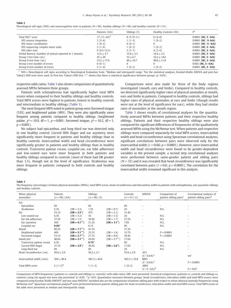
Table 2Neurological soft signs (NSS) and neurocognitive tests in patients (N=96), healthy siblings (N=66) and healthy controls (N=51).
Patients (Sch) Siblings (S) Healthy Controls (HC) P3
Total NSS1 score 17 (11–24)2 8 (5.75-11) 6 (4–8) 0.0001 (HC, SbSch)NSS sensory integration 3 (2–4) 1 (1–3) 1 (0–2) 0.0001 (HCbSbSch)NSS motor coordination 1 (0–2) 0 (0–1) 0 0.0001 (HCbSbSch)NSS sequencing complex motor tasks 3 (1–6) 1 (0–3) 1 (0–2) 0.0001 (HC, SbSch)NSS other tests 9 (6.5-12.5) 3 (5–7) 4 (2–6) 0.0001 (HC, SbSch)
Verbal fluency (number of animal reported in 1 minute) 12.5±3.7 15.8±3.3 16.6±3.2 0.0001 (HC, SNSch)Stroop I test time (sec) 43±20 32±6.7 32.2±14.6 0.0001 (HC, SbSch)Stroop II test time (sec) 53.2±17.8 40±10.7 40.8±11.8 0.0001 (HC, SbSch)Stroop I test number of errors 0 (0–1) 0 0 0.002 (HC, SbSch)Stroop II test number of errors 2 (1–4) 1 (0–2) 1 (0–2) 0.0001 (HC, SbSch)
1 NSS=Neurological soft signs according to Neurological Evaluation Scale. 2Median and interquartil range. 3For the statistical analyses, Kruskal-Wallis ANOVA and post-hocTukey's HSD tests were used. In Post-hoc Tukey's HSD test, "b" shows that there is statistical significance between groups (pb0.05).
87C. Aksoy-Poyraz et al. / Psychiatry Research 190 (2011) 85–90
respective odds ratios. Table 3 also shows comparisons of quantitativelyassessed MPAs between three groups.
Patients with schizophrenia had significantly higher total MPAscores when compared to their healthy siblings and healthy controls.Total MPA scores were highest in patients, lowest in healthy controlsand intermediate in healthy siblings (Table 3).
Themost frequentMPA noted in patient groupwere furrowed tongue(53%) and heightened palate (49%). They were also significantly morefrequent among patients compared to healthy siblings (heightenedpalate, χ²=10.9, df=1, p=0.001; furrowed tongue, χ²=10.2, df=1,p=0.001).
No subject had epicanthus, and long third toe was detected onlyin one healthy control. Curved fifth finger and ear asymetry weresignificantly more frequent in patients and healthy siblings than inhealthy controls. Innercanthal width and head circumference weresignificantly greater in patients and healthy siblings than in healthycontrols. Transverse palmar crease, cuspidal ear, ear lobe adherenceand low-seated ears were more frequent in both patients andhealthy siblings compared to controls (most of these had OR greaterthan 1.5), though not at the level of significance. Strabismus wasmore frequent in patients compared to both controls and healthysiblings.
Table 3The frequency (percentage) of minor physical anomalies (MPAs), total MPA scores, head circuand healthy controls.
Minor physicalanomalies
Patients(n=96) (Sch)
Siblings(n=66) (S)
EyeEpicanthus 0% 0%Strabismus 6.3% (OR=3.3) 1.5% (OR=0.7)
Ear 53.1% (OR=2.5*) 47% (OR=2.1)Low-seated ear 6.3% (OR=3.3) 3% (OR=1.5)Ear lob adherence 37.5% (OR=1.5) 39.4% (OR=1.7)Ear asymetry 26% (OR=4.1**) 21.2% (OR=3.1*)Cuspidal ear 9.4%* 6.1%
Mouth 80.2% (OR=7.1**) 41.5%Heightened palate 49% (OR=5.1**) 23.1% (OR=1.6)Furrowed tongue 53.1% (OR=2.7**) 27.7% (OR=0.9)
Hands-feet 40.6% (OR=3.1**) 43.9% (OR=3.7**)Transverse palmar crease 6.3% 9.1%*Curved fifth finger 37.5% (OR=2.8*) 36.4% (OR=2.6*)Long third toe 0% 0%
Head circumference (cm) 56.6±2.0 56.2±2.1
Innercanthal width (mm) 394±40.4 385.3±44.6
Total MPA score 2 (1–3)b 1 (1–3)
Comparisons of MPA frequencies (patients vs. controls and siblings vs. controls) with odds racontrols) using chi-square test were also presented *pb0.05; **pb0.01. Quantitative measurcompared using Kruskal-Wallis ANOVAb and one-way ANOVAsa. Included also are the compaMcNemar testc. Spearman correlational analysesd were performed between patient-sibling pthe table were presented as median and interquartile range.
Comparisons were also made for three of the body regionsinvestigated (mouth, ears and limbs). Compared to healthy controls,we detected significantly higher rates of physical anomalies at mouth,ears and limbs in patients. Compared to healthy controls, siblings hadhigher rates of physical anomalies at ears and limbs (though resultswere not at the level of significance for ears), while they had similarrates of anomalies at the mouth region.
Table 3 shows results of correlational analyses for the quantita-tively assessed MPAs between patients and their respective healthysiblings. Patients and their respective healthy siblings were alsocompared for significant differences of frequencies of the qualitativelyassessedMPAs using theMcNemar test. When patients and respectivesiblings were compared separately for total MPA scores, innercanthalwidth and head circumference using Spearman correlational analyses,significant correlations between pairs were observed only for theinnercanthal width (r=0.66; p=0.0001). However, since innercanthalwidth and head circumference were found to be gender-dependentvariables in the present sample, a second step correlational analyseswere performed between same-gender patient and sibling pairs(N=33) and it was revealed that head circumference was significantlycorrelated between pairs (r=0.6; p=0.0001). The correlation for theinnercanthal width remained significant in this analysis.
mference and telecanthus width in patients with schizophrenia, non-psychotic siblings
Controls(n=51) (C)
ANOVA Comparison ofpatient-sibling pairsc
Correlational analyses ofpatient-sibling pairsd
0% - - -2% - N.S. -31.4%2% - N.S. -27.5% - N.S. -7.8% - N.S. -0% - N.S. -37.3%15.7% - P=0.0001 -29.4% - P=0.0001 -17.6%0% - N.S. -17.6% - N.S. -2% - N.S. -55.6±2.0 .031
(CbS,Sch)a nsf363.3±33.8 0001
(CbS,Sch)a P=0.00011 (0–2) .0001
(CbSbSch)b P=0.07
tios (OR) were presented. Statistical comparisons (patients vs. controls and siblings vs.es between groups (head circumference, telecathus width and total MPA scores) wererisons of patient-sibling pairs with respect to minor physical anomaly frequencies usingairs for head circumference, telecathus width and total MPA scores. Total MPA scores in

88 C. Aksoy-Poyraz et al. / Psychiatry Research 190 (2011) 85–90
When patients and respective siblings were compared for thepresence of each of the MPAs using the McNemar test, only theanomalies of heightened palate and furrowed tongue were foundsignificantly more frequent in patients compared to their respectivesiblings.
3.4. Associations between neurological soft signs and minor physicalanomalies
Total NSS scores were not significantly correlated with total MPAscore in the overall sample using Spearmen correlations. When thethree groups were assessed separately, total NSS scores were notassociated with total MPA scores in any of the groups.
3.5. Associations of patients’ symptoms with NSSs, MPAs and neurocognitivefunctioning
Total NSS score was significantly associated with SANS score (r=0.6;p=0.0001); and the disorganization dimension (r=0.38; p=0.0001). Ingeneral, both negative symptoms and disorganized symptoms weredirectly associatedwith all of theNSS subscales. Controlling for the effectsof age, gender and educational status using partial correlations had noeffect in these associations.
Among the patients, total MPA scores were not significantlyassociatedwith anyof the following items: age, educational attainment,age of onset, number of hospitalizations, neuropsychological testscores, total NSS scores, NSS subscales scores or any of the 'severity ofillness' parameters (SANS, SAPS and disorganization dimension).
3.6. Regression analysis
Age, gender and variables showing an odds ratio greater than 2 or ap less than 0.05 in the bivariate analyses between patients andcontrols (total NSS scores, verbal fluency, stroop I test time/number oferrors, stroop II test time/number of errors, head circumference, totalMPA score, innercanthal width and presence of strabismus, furrowedtongue, heightened palate, low-seated ears, ear asymetry, cuspidalears and curved fifth finger; see Tables 1, 2 and 3) were entered in thestepwise logistic regression analysis that was performed to predictgroup membership of subjects (patients n=89 versus healthycontrols n=46). Continuous predictors had been mean-centeredbefore interactions were made in order to avoid multicollinearity.Following variables were shown to be independent predictors ofschizophrenia: Total NSS score, total MPA scores and head circum-ference. This final regression model (Table 4) predicted 90% ofpatients and 85% of healthy controls accurately (χ²=113, df=3,p=0.0001).
4. Discussion
In the present study, overall minor physical anomaly (MPA) scoreswere found to be greater in patients than controls, and siblings ofpatientshad scores inbetween these groups.However,wheneachof theMPAs were investigated separately, different patterns of distributionacross groups were observed for the investigated body regions(i.e. mouth, limb and ear). In general, limb and ear anomalies werefound more frequently in both patients and their siblings compared to
Table 4Independent predictors of schizophrenia.
B Wald χ² p Odds ratio
Neurological soft signs (total score) 0.48 21.8 0.0001 1.6Minor physical anomalies (total score) 1.19 12.2 0.0001 3.2Head circumference 0.51 7.4 0.006 1.6
controls. Patients and their respective siblings also had a fairlycorrelated amount of anomalies in limb and ear regions. Mouthanomalies, however were prevalent only in patients (furrowed tonguein 53% and heightened palate in 49% of the patients) and theseanomalies did not show correlation between patients and theirrespective siblings. Also, when tongue and palate anomalies wereexcluded from the statistical analyses, the difference between patientsand their healthy siblings for the total MPA score completelydisappeared. Head circumference and innercanthal width were found tobe significantly greater in patients and healthy siblings than controls,and these parameters seemed to be correlated between patients andtheir respective siblings (such that siblings of patientswith greater headcircumference and innercanthalwidth had thesemeasurements greateror vice versa).
These findings provide clues for a novel idea that minor physicalanomalies at different localizations might represent different patho-logical origins; i.e. familial versus non-familial. In this study, ear andlimb anomalies as well as greater head circumference and inner-canthal width were found to be correlated between patients and theirhealthy siblings. This indicates that these traits might in deed reflect afamilial (or at least partly genetic) predisposition for schizophrenia inan individual. Similar correlations of MPAs between patients andsiblings were reported at eyes, hands (Compton et al., 2007) and ears(Ismail et al. 2000; Compton et al. 2007) in previous studies. Incontrast, mouth anomalies (palatal and tongue anomalies) which wedetermined as highly specific to patients with schizophrenia mightrepresent a direct link with a specific disease process, and/or might bemarkers of a “second hit” distinguishing genetically predisposedindividuals whowill develop the disease from thosewhowill not. Thislatter finding is in line with the concept proposed by Waddingtonet al. (1999), which states that mouth and particularly the palatalanomalies referred to also as 'midline anomalies' represent apathological brain morphogenesis, as evidence has been found forintimate developmental relationship between the craniofacial regionand fetal midline and temporal lobe brain structures. An early insult inthe first trimester thus might eventually lead to a faulty developmentof these closely related structures. In parallel with this, in a studyinvolving patients with schizophrenia, healthy controls and unaffect-ed first-degree relatives of patients with schizophrenia, Turetsky et al.(2007) found that male patients had smaller posterior nasal volumesthan both male relatives and male controls, and they concluded thatposterior nasal volume decrement is an abnormality –potentiallyenvironmentally rather than genetically mediated- that appears to bespecific to male schizophrenic patients. They finally concluded thatposterior nasal volume might be a specific craniofacial abnormalitythat may potentially distinguish genetically vulnerable men who goon to develop schizophrenia from those who do not. This is similar toour conclusion in that mouth and posterior nasal area might bothrepresent ‘midline structures’ mentioned above.
This study replicated the finding that in patients with schizophrenia,neurological soft signs (NSSs) are associated with negative anddisorganization symptoms, and frontal lobe executive functions asshownby stroopand category tests. The relationshipbetweenMPAs andcognitive dysfunction in schizophrenic patients is so far, unclear (Ismailet al., 2000). In this study, we could not find evidence for association ofMPAs with cognitive performance and disease symptoms. Neurologicalsoft signs and minor physical anomalies were found to be unrelated inour subjects, a finding reported by numerous previous studies (Ismailet al., 2000; Lawrie et al., 2001; O'Reilly et al., 2001; Gourion et al., 2003;Compton et al., 2007; John et al., 2008). So far, significant positivecorrelations betweenNSS andMPA scores in patients and their relativeswere reported only in two studies (Nizamie et al., 1989; Gourion et al.2004b). This suggests that NSSs andMPAsmay be independentmarkersof predisposition toward schizophrenia, which also means that theywould afford greater predictive validity when used as a compositeendophenotype in genetic association studies (Ismail et al., 2000;

89C. Aksoy-Poyraz et al. / Psychiatry Research 190 (2011) 85–90
Compton et al., 2007; John et al., 2008). In our study, we could classify90% of patients and 85% of healthy controls accurately by includingsubjects’ total NSS scores, total MPA scores and head circumferences aspredictors in a final regression model.
This study confirmed that NSSs show associationswith negative anddisorganized symptoms in schizophrenia. In contrast, correlationsbetween MPAs and symptom domains were not apparent, consistentwith prior reports (Arango et al., 2000; Ismail et al., 2000; Gourion et al.,2003; Compton et al., 2007). The well-replicated relationship betweennegative symptoms and NSSs suggests that NSSs are a part of thenegative symptoms or both share a common ethiopathological process(Whitty et al., 2006).
Our findings on several domains ofNSSs (i.e. sensory integration andmotor coordination) are consistent with extant literature showing agraded pattern of NSS severity, with healthy relatives having anintermediate number of these anomalies between patients and controls(Rossi et al., 1990; Egan et al., 2001; Schubert and McNeil, 2004;Compton et al. 2007). This suggests that at least part of the NSSs reflectanunderlying genetic vulnerability to schizophrenia. Inour study,motorcoordination and sensorial integration defects were found to besignificantly increased in both patients and their siblings compared tothe healthy controls. In parallel with this, previous researchers notedespecially motor coordination deficits among the NSS domains to besignificantly raised in patients and their relatives (Gourion et al. 2004b;Mechri et al., 2009). Additionally, this was reported as the only NSSdomain found abnormal in schizophrenic patients independent of lowintelligence (Arango et al., 1999; Dazzan et al., 2008). These findingsthus indicate thatmotor dyscoordinationmay be particularly specific tothe schizophrenic pathology and may as well reflect an underlyinggenetic vulnerability to schizophrenia.
Several methodological limitations of this study should be consid-ered. The assessor could not be feasibly blinded to participants’ groupstatus when assessing NSSs and MPAs. However, standardized in-struments were used for both indices. As- the patients were on variousantipsychotic medications (and often as combinations) during assess-ments, potential medication effects on NSSs could not be examined.Antipsychotics have been found to exacerbate particularly motorfunctions (Gupta et al., 1995; Emsley et al., 2005), however there isstrong evidence that neurological abnormalities are present also inantipsychotic-naive patients (Keshavan et al., 2003; Scheffer, 2004;Chen et al., 2005) indicating that NSSs occur independently ofantipsychotic side effects. Finally, some healthy siblings of patientswere still at high risk age interval, which means that they could be atprodromal or premorbid stage of the illness.
References
American Psychiatric Association Washington DC, 2000. DSM-IV-TR: Diagnostic andstatistical manual of mental disorders IV-TR.
Andreasen, N.C., 1982. Negative symptoms in schizophrenia: definition and reliability.Archives of Genral Psychiatry 39, 784–788.
Andreasen, N.C., 1983. The Scale for the Assessment of Negative Symptoms (SANS).Iowa city. University of Iowa, Iowa.
Andreasen, N.C., 1984. The Scale for the Assessment of Positive Symptoms (SAPS). Iowacity. University of Iowa, Iowa.
Andreasen, N.C., Arndt, S., Alliger, R., Miller, D., Flaum, M., 1995. Symptoms ofschizophrenia: methods, meanings, and mechanisms. Archives of GeneralPsychiatry 52, 341–351.
Arango, C., Bartko, J.J., Gold, J.M., Buchanan, R.W., 1999. Prediction of neuropsychologicalperformance by neurological signs in schizophrenia. American Journal of Psychiatry156 (9), 1349–1357.
Arango, C., Kirkpatrick, B., Buchanan, R.W., 2000. Neurological signs and the heterogeneityof schizophrenia. American Journal of Psychiatry 157 (4), 560–565 Apr.
Bachmann, S., Bottmer, C., Schröder, J., 2005. Neurological soft signs in first-episodeschizophrenia: a follow-up study. American Journal of Psychiatry 162 (12),2337–2343.
Bombin, I., Arango, C., Buchanan, R.W., 2005. Significance and meaning of neurologicalsigns in schizophrenia: two decades later. Schizophrenia Bulletin 31, 962–977.
Buchanan, R.W., Heinrichs, D.W., 1989. The Neurological Evaluation Scale (NES): astructured instrument for the assessment of neurological signs in schizophrenia.Psychiatry Research 27, 335–350.
Cannon, T.D., 2005. The inheritance of intermediate phenotypes for schizophrenia.Current Opinion in Psychiatry 18, 135–140.
Chen, E.Y., Hui, C.L.-M., Chan, R.C.-K., Dunn, E.L.-W., Miao, M.Y.-K., Yeung, W.-S., Wong, C.-K.,Chan, W.-F., Tang, W.-N., 2005. A prospective 3-year longitudinal study of cognitivepredictors of relapse in first-episode schizophrenic patients. Schizophrenia Research77 (1), 99–104.
Compton, M.T., Walker, E.F., 2009. Physical manifestations of neurodevelopmentaldisruption: are minor physical anomalies part of the syndrome of schizophrenia?Schizophrenia Bulletin 35 (2), 425–436.
Compton, M.T., Bollini, A.M., McKenzie Mack, L., Kryda, A.D., Rutland, J., Weiss, P.S., Bercu,Z., Esterberg, M.L., Walker, E.F., 2007. Neurological soft signs and minor physicalanomalies in patients with schizophrenia and related disorders, their first-degreebiological relatives, and non-psychiatric controls. Schizophrenia Research 94 (1–3),64–73.
Dazzan, P., Murray, R.M., 2002. Neurological soft signs in first-episode psychosis: asystematic review. British Journal of Psychiatry (Suppl.) 43, 50–57.
Dazzan, P., Lloyd, T., Morgan, K.D., Zanelli, J., Morgan, C., Orr, K., Hutchinson, G., Fearon,P., Allin, M., Rifkin, L., McGuire, P.K., Doody, G.A., Holloway, J., Leff, J., Harrison, G.,Jones, P.B., Murray, R.M., 2008. Neurological abnormalities and cognitive ability infirst-episode psychosis. British Journal of Psychiatry 193 (3), 197–202 Sep.
Egan, M.F., Hyde, T.M., Bonomo, J.B., Mattay, V.S., Bigelow, L.B., Goldberg, T.E.,Weinberger, D.R., 2001. Relative risk of neurological signs in siblings of patientswith schizophrenia. American Journal of Psychiatry 158 (11), 1827–1834.
Emsley, R., Turner, H.J., Oosthuizen, P.P., Oosthuizen, P.P., Carr, J., 2005. Neurologicalabnormalities in first-episode schizophrenia: temporal stability and clinical andoutcoma correlates. Schizophrenia Research 75 (1), 35–44.
Erkoç, Ş., Arkonaç, O., Ataklı, C., Özmen, E., 1991a. Negatif semptomları değerlendirmeölçeğinin güvenilirliği ve geçerliliği. Düşünen Adam 4, 16–19 (Article in Turkish).
Erkoç, Ş., Arkonaç, O., Ataklı, C., Özmen, E., 1991b. Pozitif semptomları değerlendirmeölçeğinin güvenilirliği ve geçerliliği. Düşünen Adam 4, 20–24 (Article in Turkish).
First, M.B., Spitzer, R.L., Gibbon, M., Williams, J., 1996. Structured Clinical Interview forDSM-IV Axis I Disorders-Patient Edition (SCID-I/P, Version 2.0). BiometricsResearch Department. New York State Psychiatric Institute, New York.
Gourion, D., Goldberg, C., Bourdel, M.-C., Bayle, F.J., Millet, B., Olie, J.-P., Krebs, M.-O.,2003. Neurological soft-signs and minor physical anomalies in schizophrenia:differential transmission within families. Schizophrenia Research 63, 181–187.
Gourion, D., Goldberg, C., Bourdel, M.-C., Bayle, F.J., Loo, H., Krebs, M.-O., 2004a. Minorphysical anomalies in patients with schizophrenia and their parents: prevalenceand pattern of craniofacial abnormalities. Psychiatry Research 125, 21–28.
Gourion, D., Goldberg, C., Olie, J.-P., Loo, H., Krebs, M.-O., 2004b. Neurological andmorphological anomalies and the genetic liability to schizophrenia: a compositephenotype. Schizophrenia Research 67, 23–31.
Green, M.F., Satz, P., Christenson, C., 1994. Minor physical anomalies in schizophreniapatients, bipolar patients, and their siblings. Schizophrenia Bulletin 20, 433–440.
Gupta, S., Andreasen, N.C., Arndt, S., et al., 1995. Neurological soft signs in neuroleptic-naive and neuroleptic-treated schizophrenic patients and in normal comparisonsubjects. American Journal of Psychiatry 152, 191–196.
Ismail, B., Cantor-Graae, E., McNeil, T.F., 1998. Minor physical anomalies in schizophrenicpatients and their siblings. American Journal of Psychiatry 155, 1695–1702.
Ismail, B., Cantor-Graae, E., McNeil, T.F., 2000. Minor physical anomalies inschizophrenia: cognitive, neurological and other clinical correlates. Journal ofPsychiatric Research 34, 45–56.
John, J.P., Arunachalam, V., Ratnam, B., Isaac, M.K., 2008. Expanding the schizophreniaphenotype: a composite evaluation of neurodevelopmental markers. Comprehen-sive Psychiatry 49 (1), 78–86.
Keshavan, M.S., Sanders, R.D., Sweeney, J.A., Diwadkar, V.A., Goldstein, G., Pettegrew, J.W.,Schooler, N.R., 2003. Diagnostic specificity and neuroanatomical validity of neurologicalabnormalities in first-episode psychoses. American Journal of Psychiatry 160,1298–1304.
Lawrie, S.M., Byrne, M., Miller, P., Hodges, A., Clafferty, R.A., Cunningham Owens, D.G.,Johnstone, E.C., 2001. Neurodevelopmental indices and the development ofpsychotic symptoms in subjects at high risk of schizophrenia. British Journal ofPsychiatry 178, 524–530.
Mechri, A., Bourdel, M.C., Slama, H., Gourion, D., Gaha, L., Krebs, M.O., 2009.Neurological soft signs in patients with schizophrenia and their unaffectedsiblings: frequency and correlates in two ethnic and socioeconomic distinctpopulations. European Archives of Psychiatry and Clinical Neurosciences 259 (4),218–226.
Nizamie, S.H., Nizamie, A., Sangma, M.W., Sharma, P.L., 1989. Soft neurological signs andminor physical anomalies in schizophrenia. Indian Journal of Psychiatry 31, 230–237.
O'Reilly, R.L., Lane, A., Cernovsky, Z.Z., O'Callaghan, E., 2001. Neurological soft signs,minor physical anomalies and handedness in schizophrenia. European Journal ofPsychiatry 15, 189–192.
Perret, E., 1974. The left frontal lobe of man and the suppression of habitual responsesin verbal categorical behaviour. Neuropsychologia 12, 323–330.
Rossi, A., De Cataldo, S., Di Michele, V., Manna, V., Ceccoli, S., Stratta, P., Casacchia, M.,1990. Neurological soft signs in schizophrenia. British Journal of Psychiatry 157,735–739.
Scheffer, R.E., 2004. Abnormal neurological signs at the onset of psychosis.Schizophrenia Research 70 (1), 19–26.
Schubert, E.W., McNeil, T.F., 2004. Prospective study of neurological abnormalities inoffspring of women with psychosis: birth to adulthood. American Journal ofPsychiatry 161 (6), 1030–1037.
Turetsky, B.I., Glass, C.A., Abbazia, J., Kohler, C.G., Gur, R.E., Moberg, P.J., 2007. Reducedposterior nasal cavity volume: a gender-specific neurodevelopmental abnormality inschizophrenia. Schizophrenia Research 93, 237–244.

90 C. Aksoy-Poyraz et al. / Psychiatry Research 190 (2011) 85–90
Waddington, J.L., Lane, A., Larkin, C., O'Callaghan, E., 1999. The nurodevelopmental basisof schizophrenia: clinical clues from cerebro-craniofacial dysmorphogenesis, andthe roots of a lifetime trajectory of disease. Biological Psychiatry 46 (1), 31–39.
Walker, E.F., Lewine, R.J., 1990. Prediction of adult-onset schizophrenia from childhoodhome movies of the patients. American Journal of Psychiatry 147 (8), 1052–1056.
Whitty, P., Clarke, M., McTigue, O., Browne, S., Gervin, M., Kamali, M., Lane, A., Kinsella, A.,Waddington, J., Larkin, C., O'Callaghan, E., 2006.Diagnostic specificity andpredictors of
neurological soft signs in schizophrenia, bipolar disorder and other psychoses over thefirst 4 years of illness. Schizophrenia Research 86 (1–3), 110–117.
Yoshitsugu, K., Yamada, K., Toyota, T., Aoki-Suzuki, M., Minabe, Y., Nakamura, K., Sekine,Y., Suzuki, K., Takei, N., Itokawa, M., Mori, N., Yoshikawa, T., 2006. A novel scaleincluding strabismus and 'cuspidal ear' for distinguishing schizophrenia patientsfrom controls using minor physical anomalies. Psychiatry Research 145 (2–3),249–258.