microstructure and composition of fine particles released by car
6
Oct 18 th - 20 th 2017, Brno, Czech Republic, EU 704 MICROSTRUCTURE AND COMPOSITION OF FINE PARTICLES RELEASED BY CAR BRAKING ŠVÁBENSKÁ Eva 1 , ROUPCOVÁ Pavla 1 , PIZÚROVÁ Naděžda 1, 2 , SCHNEEWEISS Oldřich 1,2 1 Institute of Physics of Materials, Academy of Sciences of the Czech Republic, Brno, Czech Republic, EU 2 CEITEC IPM, Institute of Physics of Materials, Academy of Sciences of the Czech Republic, Brno, Czech Republic, EU Abstract Vehicular traffic is connected with large volume of fine particles released during brake processes of cars. Our research is focused on the phase, structure and chemical analysis of the fine particles taken from some car brake parts by their services. The information on structure and phase composition was obtained by X-Ray Powder Diffraction, Mössbauer Spectroscopy, scanning electron microscopy with EDX and transmission electron microscopy. The results of the wear debris analysis are compared with original brake materials components. Most of recognized particles are based mainly on iron oxides. Wear brake particles are discussed in the relation to the potential risk to the environment and human health. Keywords: Nanoparticles, wear debris, environment INTRODUCTION Vehicular traffic is a great source of environmental pollution. The origins of pollution are exhaust of fuels, abrasions of roads, wear of tyre and last but not least wear of brakes. Today’s friction materials have been consisting of various additives which composition, particle size and distribution strongly affects the properties of brake materials. Wear particles are created during the friction process, when the brake lining (pads or shoes) is pressed against a rotating counterpart (rotor or drum) made of gray cast iron. It was determined that 30- 50 % of the brake lining mass loss pass to the airborne emissions. Brake wear emission factor is estimated for in range between 2.5 and 8 mg km −1 for one light duty vehicles depending on brake pad type. [1,2] Brake wear particles usually have the characteristic volume of distribution with two peaks maxima between 2 - 6μm and around 300nm. [2,3,4] However, brake wear particles of all sizes threaten environment and human health. Fine brake wear particles with diameter bellow 2.5μm can be easily inhaled into the respiratory tract where posing hazards related to potential oxidative stress, inflammation and bronchi asthma. [5,6] Wear emissions are the major source of some metals. The most commonly mentioned key tracers of brake wear are Cu, Sb, Ba and Fe. [2] 1. MATERIALS AND METHODS Samples of wear debris were taken from two different cars with brake pads (P) and drum brakes (D).The drum brake lining (DL) was used as reference material. The characterization of wear debris involves the analysis of the microstructure, morphology, phase composition and chemical composition of these wear particles. For these purposes the methods involving X-ray diffraction (XRD), scanning electron microscopy (SEM), transmission electron microscopy (TEM), both microscopy techniques being integrated with energy dispersive X- ray spectroscopy (EDX) and Mössbauer Spectroscopy, were applied.
-
Upload
khangminh22 -
Category
Documents
-
view
2 -
download
0
Transcript of microstructure and composition of fine particles released by car
Oct 18th - 20th 2017, Brno, Czech Republic, EU
704
MICROSTRUCTURE AND COMPOSITION OF FINE PARTICLES RELEASED BY CAR
BRAKING
ŠVÁBENSKÁ Eva1, ROUPCOVÁ Pavla1, PIZÚROVÁ Naděžda1, 2, SCHNEEWEISS Oldřich1,2
1Institute of Physics of Materials, Academy of Sciences of the Czech Republic, Brno,
Czech Republic, EU
2 CEITEC IPM, Institute of Physics of Materials, Academy of Sciences of the Czech Republic, Brno,
Czech Republic, EU
Abstract
Vehicular traffic is connected with large volume of fine particles released during brake processes of cars. Our research is focused on the phase, structure and chemical analysis of the fine particles taken from some car brake parts by their services. The information on structure and phase composition was obtained by X-Ray Powder Diffraction, Mössbauer Spectroscopy, scanning electron microscopy with EDX and transmission electron microscopy. The results of the wear debris analysis are compared with original brake materials components. Most of recognized particles are based mainly on iron oxides. Wear brake particles are discussed in the relation to the potential risk to the environment and human health.
Keywords: Nanoparticles, wear debris, environment
INTRODUCTION
Vehicular traffic is a great source of environmental pollution. The origins of pollution are exhaust of fuels, abrasions of roads, wear of tyre and last but not least wear of brakes. Today’s friction materials have been consisting of various additives which composition, particle size and distribution strongly affects the properties of brake materials. Wear particles are created during the friction process, when the brake lining (pads or shoes) is pressed against a rotating counterpart (rotor or drum) made of gray cast iron. It was determined that 30-50 % of the brake lining mass loss pass to the airborne emissions. Brake wear emission factor is estimated for in range between 2.5 and 8 mg km−1 for one light duty vehicles depending on brake pad type. [1,2] Brake wear particles usually have the characteristic volume of distribution with two peaks maxima between 2 - 6μm and around 300nm. [2,3,4] However, brake wear particles of all sizes threaten environment and human health. Fine brake wear particles with diameter bellow 2.5μm can be easily inhaled into the respiratory tract where posing hazards related to potential oxidative stress, inflammation and bronchi asthma. [5,6] Wear emissions are the major source of some metals. The most commonly mentioned key tracers of brake wear are Cu, Sb, Ba and Fe. [2]
1. MATERIALS AND METHODS
Samples of wear debris were taken from two different cars with brake pads (P) and drum brakes (D).The drum brake lining (DL) was used as reference material. The characterization of wear debris involves the analysis of the microstructure, morphology, phase composition and chemical composition of these wear particles. For these purposes the methods involving X-ray diffraction (XRD), scanning electron microscopy (SEM), transmission electron microscopy (TEM), both microscopy techniques being integrated with energy dispersive X- ray spectroscopy (EDX) and Mössbauer Spectroscopy, were applied.
Oct 18th - 20th 2017, Brno, Czech Republic, EU
705
2. RESULTS AND DISSCUSION
2.1. Microscopy and X-ray diffraction analysis
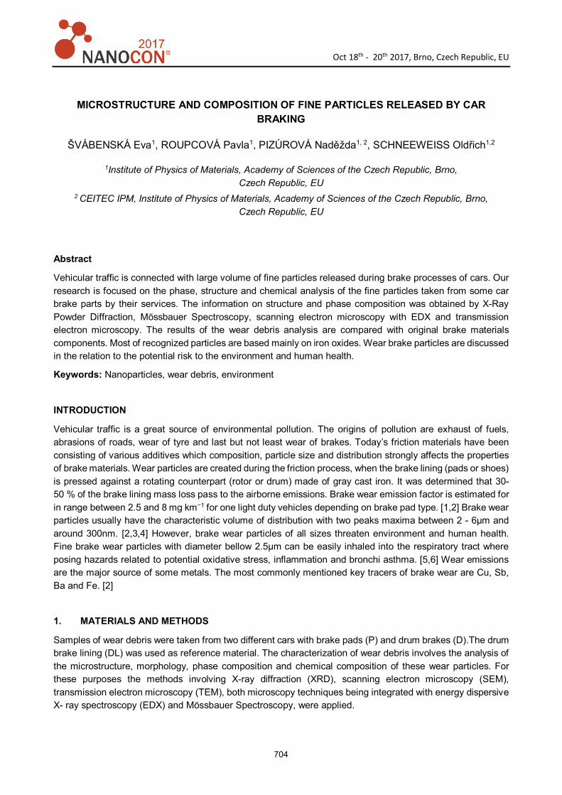
Scanning electron microscopy of collected wear particles showed some large particles, fine particles in large quantities and large agglomerates of particles. These agglomerates consist of sub-micron and micron-sized particles in a variety of shapes. The wear debris size comparison showed that particles from brake pads including a little bit larger particles than wear debris from drum (Figure 1). The energy dispersive X-ray (EDX) analysis of particles, with size form sub-micron to few-micron, found high concentration of iron, oxygen, carbon. The both of samples of wear particles contain various another elements. In sample P has been also observed copper, silicon, aluminum and tin. Sample D contained more zinc, calcium, magnesium, and less silicon than sample P. However, the largest amounts of particles were iron oxides.
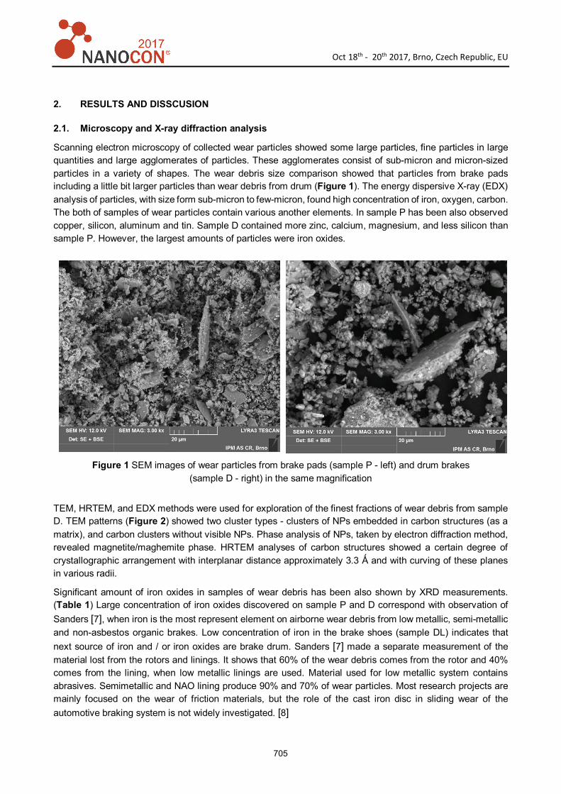
TEM, HRTEM, and EDX methods were used for exploration of the finest fractions of wear debris from sample D. TEM patterns (Figure 2) showed two cluster types - clusters of NPs embedded in carbon structures (as a matrix), and carbon clusters without visible NPs. Phase analysis of NPs, taken by electron diffraction method, revealed magnetite/maghemite phase. HRTEM analyses of carbon structures showed a certain degree of crystallographic arrangement with interplanar distance approximately 3.3 Ǻ and with curving of these planes in various radii.
Significant amount of iron oxides in samples of wear debris has been also shown by XRD measurements. (Table 1) Large concentration of iron oxides discovered on sample P and D correspond with observation of Sanders [7], when iron is the most represent element on airborne wear debris from low metallic, semi-metallic and non-asbestos organic brakes. Low concentration of iron in the brake shoes (sample DL) indicates that next source of iron and / or iron oxides are brake drum. Sanders [7] made a separate measurement of the material lost from the rotors and linings. It shows that 60% of the wear debris comes from the rotor and 40% comes from the lining, when low metallic linings are used. Material used for low metallic system contains abrasives. Semimetallic and NAO lining produce 90% and 70% of wear particles. Most research projects are mainly focused on the wear of friction materials, but the role of the cast iron disc in sliding wear of the automotive braking system is not widely investigated. [8]
Figure 1 SEM images of wear particles from brake pads (sample P - left) and drum brakes (sample D - right) in the same magnification
Oct 18th - 20th 2017, Brno, Czech Republic, EU
706
Size distribution and chemical composition of wear particles also correspond with composition of used lining materials. One of the most likely explanations of the particle size of wear is that the size of the individual components in the lining material is the same or greater. Original drum lining material (DL) partly consists particles larger than 200μm (carbon, iron fiber, periclase (MgO), calcite (CaCO3) and Si/Al oxide) and other ingredients with a diameter of particles around 2.5μm.
Table 1 XRD results
Sample α- iron Hematite
(Fe2O3)
Magnetite
(Fe3O4)
Quartz
(SiO2)
Graphite Periclase
(MgO)
Calcite
(CaCO3)
Zincite
(ZnO)
P 13 29 48 10
D (debris) 7 30 34 3 2 23
DL 7 44 22 25 2
2.2. Mössbauer spectroscopy
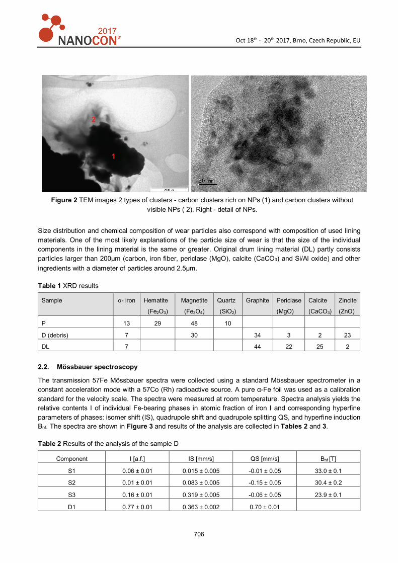
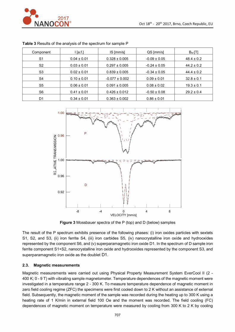
The transmission 57Fe Mössbauer spectra were collected using a standard Mössbauer spectrometer in a constant acceleration mode with a 57Co (Rh) radioactive source. A pure α-Fe foil was used as a calibration standard for the velocity scale. The spectra were measured at room temperature. Spectra analysis yields the relative contents I of individual Fe-bearing phases in atomic fraction of iron I and corresponding hyperfine parameters of phases: isomer shift (IS), quadrupole shift and quadrupole splitting QS, and hyperfine induction Bhf. The spectra are shown in Figure 3 and results of the analysis are collected in Tables 2 and 3.
Table 2 Results of the analysis of the sample D
Component I [a.f.] IS [mm/s] QS [mm/s] Bhf [T]
S1 0.06 ± 0.01 0.015 ± 0.005 -0.01 ± 0.05 33.0 ± 0.1
S2 0.01 ± 0.01 0.083 ± 0.005 -0.15 ± 0.05 30.4 ± 0.2
S3 0.16 ± 0.01 0.319 ± 0.005 -0.06 ± 0.05 23.9 ± 0.1
D1 0.77 ± 0.01 0.363 ± 0.002 0.70 ± 0.01
Figure 2 TEM images 2 types of clusters - carbon clusters rich on NPs (1) and carbon clusters without visible NPs ( 2). Right - detail of NPs.
1
2
Oct 18th - 20th 2017, Brno, Czech Republic, EU
707
Table 3 Results of the analysis of the spectrum for sample P
Component I [a.f.] IS [mm/s] QS [mm/s] Bhf [T]
S1 0.04 ± 0.01 0.328 ± 0.005 -0.09 ± 0.05 48.4 ± 0.2
S2 0.03 ± 0.01 0.297 ± 0.005 -0.24 ± 0.05 44.2 ± 0.2
S3 0.02 ± 0.01 0.839 ± 0.005 -0.34 ± 0.05 44.4 ± 0.2
S4 0.10 ± 0.01 -0.077 ± 0.002 0.09 ± 0.01 32.8 ± 0.1
S5 0.06 ± 0.01 0.091 ± 0.005 0.08 ± 0.02 19.3 ± 0.1
S6 0.41 ± 0.01 0.426 ± 0.012 -0.50 ± 0.08 29.2 ± 0.4
D1 0.34 ± 0.01 0.363 ± 0.002 0.86 ± 0.01
Figure 3 Mossbauer spectra of the P (top) and D (below) samples
The result of the P spectrum exhibits presence of the following phases: (i) iron oxides particles with sextets S1, S2, and S3, (ii) iron ferrite S4, (iii) iron carbides S5, (iv) nanocrystalline iron oxide and hydrooxides represented by the component S6, and (v) superparamagnetic iron oxide D1. In the spectrum of D sample iron ferrite component S1+S2, nanocrystalline iron oxide and hydrooxides represented by the component S3, and superparamagnetic iron oxide as the doublet D1.
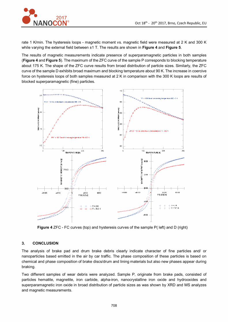
2.3. Magnetic measurements
Magnetic measurements were carried out using Physical Property Measurement System EverCool II (2 - 400 K; 0 - 9 T) with vibrating sample magnetometer. Temperature dependences of the magnetic moment were investigated in a temperature range 2 - 300 K. To measure temperature dependence of magnetic moment in zero field cooling regime (ZFC) the specimens were first cooled down to 2 K without an assistance of external field. Subsequently, the magnetic moment of the sample was recorded during the heating up to 300 K using a heating rate of 1 K/min in external field 100 Oe and the moment was recorded. The field cooling (FC) dependences of magnetic moment on temperature were measured by cooling from 300 K to 2 K by cooling
-8 -4 0 4 8VELOCITY [mm/s]
0.92
0.96
1.00
0.96
1.00
P
D
Oct 18th - 20th 2017, Brno, Czech Republic, EU
708
rate 1 K/min. The hysteresis loops - magnetic moment vs. magnetic field were measured at 2 K and 300 K while varying the external field between ±1 T. The results are shown in Figure 4 and Figure 5.
The results of magnetic measurements indicate presence of superparamagnetic particles in both samples (Figure 4 and Figure 5). The maximum of the ZFC curve of the sample P corresponds to blocking temperature about 175 K. The shape of the ZFC curve results from broad distribution of particle sizes. Similarly, the ZFC curve of the sample D exhibits broad maximum and blocking temperature about 90 K. The increase in coercive force on hysteresis loops of both samples measured at 2 K in comparison with the 300 K loops are results of blocked superparamagnetic (fine) particles.
Figure 4 ZFC - FC curves (top) and hysteresis curves of the sample P( left) and D (right)
3. CONCLUSION
The analysis of brake pad and drum brake debris clearly indicate character of fine particles and/ or nanoparticles based emitted in the air by car traffic. The phase composition of these particles is based on chemical and phase composition of brake discs/drum and lining materials but also new phases appear during braking.
Two different samples of wear debris were analyzed. Sample P, originate from brake pads, consisted of particles hematite, magnetite, iron carbide, alpha-iron, nanocrystalline iron oxide and hydrooxides and superparamagnetic iron oxide in broad distribution of particle sizes as was shown by XRD and MS analyzes and magnetic measurements.
Oct 18th - 20th 2017, Brno, Czech Republic, EU
709
On the other hand, sample D, originate from drum brake, indicated presence of only magnetite together with nanocrystalline iron oxide and hydrooxides and superparamagnetic iron oxide in the form of fine particles. Detail analysis of this sample by TEM showed presence of two types of carbon clusters - rich on NPs and without visible NPs. From the point of view of the health impact, it is probably good for us, that the smallest particles in nanometer size are captured in a matrix that increases their volume. Particles with size below 2.5μm come to alveoli and particle with diameter in nm to bloodstream together with gases. [9]
ACKNOWLEDGEMENTS
This research was carried out under the project CEITEC 2020 (LQ1601) with financial support from
the Ministry of Education, Youth and Sports of the Czech Republic under the National Sustainability
Programme II. The research was conducted in the frame of IPM infra supported through project No.
LM2015069 of MEYS.
REFERENCES
[1] PLACHÁ, D., VACULÍK, M., MIKESKA, M., et al. Release of volatile organic compounds by oxidative wear of automotive friction material, Wear, 2017, vol. 376-377, part A, pp. 705-716.
[2] GRIGORATOS, T., MARTINI, G. Brake wear particle emissions: a review, Environmental Science and Pollution
Research, 2015, vol. 22, no. 4, pp 2491-2504.
[3] WAHLSTRÖM, J., OLANDER, L., OLOFSSON, U. Size, Shape, and Elemental Composition of Airborne Wear Particles from Disc Brake Materials. Tribology Letters, 2010, vol. 38, no.1, pp.15-24.
[4] MOSLEH, M., BLAU, P.J., DUMITRESCU, D. Characteristics and morphology of wear particles from laboratory testing of disk brake materials. Wear, 2004, vol. 256, no. 11-12, pp. 1128-1134.
[5] BALAKRISHNA, S., LOMNICKI, S., MCAVEY, K.M, et al. Environmentally persistent free radicals amplify ultrafine particle mediated cellular oxidative stress and cytotoxicity. Particle and Fibre Toxicology, 2009, vol. 6, no.11.
[6] AMATO, F., CASSEE, F.R., DENIER VAN DER GON, H.A.C., GEHRIG, R., et al Urban air quality: The challenge of traffic non-exhaust emissions. Journal of Hazardous Materials, 2014, vol. 275, pp. 31-36.
[7] SANDERS, P.G., XU, N., DALKA, T. M., MARICQ, M . M. Airborne Brake Wear Debris: Size Distributions, Composition, and a Comparison of Dynamometer and Vehicle Tests. Environmental Science and Technology, 2003,vol. 37, no. 18, pp. 4060-4069.
[8] STRAFFELINI, G., MAINES, L. The relationship between wear of semimetallic friction materials and pearlitic cast iron in dry. Wear, 2013, vol. 307, no. 1-2, pp. 75-80.
[9] FILIPOVÁ, Z., KUKUTSCHOVÁ, J., MAŠLÁŇ, M. Danger of nanomaterials (Rizika nanomateriálů), 1st ed., Olomouc: Palacký University, 2012. 87p. (in Czech language)