Microcystins in potable surface waters: toxic effects and removal strategies
17
Microcystins in potable surface waters: toxic effects and removal strategies Amber F. Roegner a , Beatriz Brena b , Gualberto González-Sapienza b and Birgit Puschner a * ABSTRACT: In freshwater, harmful cyanobacterial blooms threaten to increase with global climate change and eutrophication of surface waters. In addition to the burden and necessity of removal of algal material during water treatment processes, bloom-forming cyanobacteria can produce a class of remarkably stable toxins, microcystins, difcult to remove from drinking water sources. A number of animal intoxications over the past 20 years have served as sentinels for widespread risk presented by microcystins. Cyanobacterial blooms have the potential to threaten severely both public health and the regional economy of affected communities, particularly those with limited infrastructure or resources. Our main objectives were to assess whether existing water treatment infrastructure provides sufcient protection against microcystin exposure, identify available options feasible to implement in resource-limited communities in bloom scenarios and to identify strate- gies for improved solutions. Finally, interventions at the watershed level aimed at bloom prevention and risk reduction for entry into potable water sources were outlined. We evaluated primary studies, reviews and reports for treatment options for microcystins in surface waters, potable water sources and treatment plants. Because of the difculty of removal of microcystins, prevention is ideal; once in the public water supply, the coarse removal of cyanobacterial cells combined with secondary carbon ltration of dissolved toxins currently provides the greatest potential for protection of public health. Options for point of use ltration must be optimized to provide affordable and adequate protection for affected communi- ties. Copyright © 2013 John Wiley & Sons, Ltd. Keywords: Microcystins; cyanotoxins; intoxications; potable water sources; eutrophication; water treatment plants; interventions; resource poor Introduction Increasing global water temperatures, nutrient and pollutant enrichment via anthropogenic runoff, drought and ooding lead to eutrophication of fresh and coastal water bodies and can result in toxicogenic cyanobacterial blooms (Newcombe et al., 2012; Paerl and Paul, 2012). In 1878, Francis described intoxica- tion of farm stock from ingestion of surface scum at Lake Alexandrina, a freshwater lake at the mouth of the Murray River in South Australia near Adelaide. The ingestion of wind-blown cyanobacteria Nodularia spumigena resulted in the death of sheep, horses, dogs and pigs within 1–24 h (Francis, 1878). Since this rst documented case description of a cyanobacterial poisoning, a wide array of animal intoxications have been reported, in addition to the identication of numerous cyanobacterial genera and species capable of producing diverse neurotoxins, hepatotoxins, gastrointestinal toxins and skin irri- tants (Chorus et al., 2000; Stewart et al., 2008a). Table 1 catalogs all identied human and animal cases linked to microcystin (MC) intoxication. MCs represent a family of potent liver toxins and are considered the most resistant of cyanotoxins to degradation because of their stable cyclic peptide structure. Acute and chronic effects of MCs are not completely understood as they can exhibit diverse system effects in vivo and diverse mecha- nisms in vitro (Dawson, 1998; Gehringer, 2004). With over 100 congeners identied and named according to variable amino acids positions (Fig. 1) (Rinehart et al., 1994; Bateman et al., 1995), MCs inhibit protein phosphatases leading to acute liver failure (Falconer, 2008). The most consistently identied MC, MC-LR (abbreviated for common amino acids leucine and arginine at positions 2 and 4, respectively)(Carmichael et al., 1988), is also believed to be the most potent congener. However, the World Health Organization (WHO) provisional guideline of 1 gl –1 for surface waters is specic for MC-LR because of lack of data with respect to other congeners. Recently, preferential uptake of other congeners into organ systems has raised concern (Feurstein et al., 2011; Trinchet et al., 2013) about risk presented by congeners with variable hydrophobicity, including more commonly monitored MC-RR (arginine at positions 2 and 4), MC-LA (leucine and alanine, respectively), MC-YR (tyrosine and arginine) and more hydro- phobic variants, MC-LF (leucine and phenylalanine) and MC-LW (leucine and tryptophan). The latter hydrophobic variants have demonstrated increased cell permeability and cytotoxicity (Vesterkvist et al., 2012). Animal intoxications have been linked to other congeners, suggesting risk may be underestimated when water is solely monitored for MC-LR (Stewart et al., 2008b). *Correspondence to: Birgit Puschner, Department of Molecular Biosciences, School of Veterinary Medicine, 1089 Veterinary Medicine Drive, University of California, Davis, CA 95616, USA. Email: [email protected] a Department of Molecular Biosciences, School of Veterinary Medicine, University of California, Davis, CA, 95616, USA b Cátedra de Inmunología, Facultad de Química y Facultad de Ciencias, Universidad de la República, Instituto de Higiene, Montevideo, Uruguay J. Appl. Toxicol. 2013 Copyright © 2013 John Wiley & Sons, Ltd. Review Article Received: 11 June 2013, Revised: 16 July 2013, Accepted: 17 July 2013 Published online in Wiley Online Library (wileyonlinelibrary.com) DOI 10.1002/jat.2920
-
Upload
independent -
Category
Documents
-
view
1 -
download
0
Transcript of Microcystins in potable surface waters: toxic effects and removal strategies

Microcystins in potable surface waters: toxiceffects and removal strategiesAmber F. Roegnera, Beatriz Brenab, Gualberto González-Sapienzab
and Birgit Puschnera*
ABSTRACT: In freshwater, harmful cyanobacterial blooms threaten to increase with global climate change and eutrophicationof surface waters. In addition to the burden and necessity of removal of algal material during water treatment processes,bloom-forming cyanobacteria can produce a class of remarkably stable toxins, microcystins, dif!cult to remove from drinkingwater sources. A number of animal intoxications over the past 20 years have served as sentinels for widespread riskpresented by microcystins. Cyanobacterial blooms have the potential to threaten severely both public health and theregional economy of affected communities, particularly those with limited infrastructure or resources. Our main objectiveswere to assess whether existing water treatment infrastructure provides suf!cient protection against microcystin exposure,identify available options feasible to implement in resource-limited communities in bloom scenarios and to identify strate-gies for improved solutions. Finally, interventions at the watershed level aimed at bloom prevention and risk reduction forentry into potable water sources were outlined. We evaluated primary studies, reviews and reports for treatment optionsfor microcystins in surface waters, potable water sources and treatment plants. Because of the dif!culty of removal ofmicrocystins, prevention is ideal; once in the public water supply, the coarse removal of cyanobacterial cells combined withsecondary carbon !ltration of dissolved toxins currently provides the greatest potential for protection of public health.Options for point of use !ltration must be optimized to provide affordable and adequate protection for affected communi-ties. Copyright © 2013 John Wiley & Sons, Ltd.
Keywords: Microcystins; cyanotoxins; intoxications; potable water sources; eutrophication; water treatment plants; interventions;resource poor
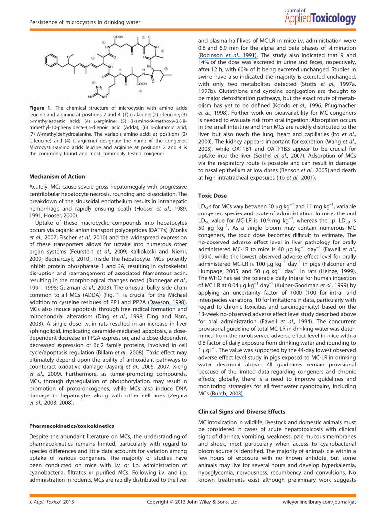
IntroductionIncreasing global water temperatures, nutrient and pollutantenrichment via anthropogenic runoff, drought and !ooding leadto eutrophication of fresh and coastal water bodies and canresult in toxicogenic cyanobacterial blooms (Newcombe et al.,2012; Paerl and Paul, 2012). In 1878, Francis described intoxica-tion of farm stock from ingestion of surface scum at LakeAlexandrina, a freshwater lake at the mouth of the Murray Riverin South Australia near Adelaide. The ingestion of wind-blowncyanobacteria Nodularia spumigena resulted in the death ofsheep, horses, dogs and pigs within 1–24 h (Francis, 1878). Sincethis "rst documented case description of a cyanobacterialpoisoning, a wide array of animal intoxications have beenreported, in addition to the identi"cation of numerouscyanobacterial genera and species capable of producing diverseneurotoxins, hepatotoxins, gastrointestinal toxins and skin irri-tants (Chorus et al., 2000; Stewart et al., 2008a). Table 1 catalogsall identi"ed human and animal cases linked to microcystin (MC)intoxication. MCs represent a family of potent liver toxins andare considered the most resistant of cyanotoxins to degradationbecause of their stable cyclic peptide structure. Acute andchronic effects of MCs are not completely understood as theycan exhibit diverse system effects in vivo and diverse mecha-nisms in vitro (Dawson, 1998; Gehringer, 2004). With over 100congeners identi"ed and named according to variable aminoacids positions (Fig. 1) (Rinehart et al., 1994; Bateman et al.,1995), MCs inhibit protein phosphatases leading to acute liverfailure (Falconer, 2008). The most consistently identi"ed MC,
MC-LR (abbreviated for common amino acids leucine andarginine at positions 2 and 4, respectively)(Carmichael et al.,1988), is also believed to be the most potent congener.However, the World Health Organization (WHO) provisionalguideline of 1 !g l–1 for surface waters is speci"c for MC-LRbecause of lack of data with respect to other congeners.Recently, preferential uptake of other congeners into organsystems has raised concern (Feurstein et al., 2011; Trinchetet al., 2013) about risk presented by congeners with variablehydrophobicity, including more commonly monitored MC-RR(arginine at positions 2 and 4), MC-LA (leucine and alanine,respectively), MC-YR (tyrosine and arginine) and more hydro-phobic variants, MC-LF (leucine and phenylalanine) and MC-LW(leucine and tryptophan). The latter hydrophobic variants havedemonstrated increased cell permeability and cytotoxicity(Vesterkvist et al., 2012). Animal intoxications have been linkedto other congeners, suggesting risk may be underestimatedwhen water is solely monitored for MC-LR (Stewart et al., 2008b).
*Correspondence to: Birgit Puschner, Department of Molecular Biosciences,School of Veterinary Medicine, 1089 Veterinary Medicine Drive, University ofCalifornia, Davis, CA 95616, USA. Email: [email protected]
aDepartment of Molecular Biosciences, School of Veterinary Medicine, Universityof California, Davis, CA, 95616, USA
bCátedra de Inmunología, Facultad de Química y Facultad de Ciencias,Universidad de la República, Instituto de Higiene, Montevideo, Uruguay
J. Appl. Toxicol. 2013 Copyright © 2013 John Wiley & Sons, Ltd.
Review Article
Received: 11 June 2013, Revised: 16 July 2013, Accepted: 17 July 2013 Published online in Wiley Online Library
(wileyonlinelibrary.com) DOI 10.1002/jat.2920

Table1.
Repo
rted
andcon!
rmed
anim
alintoxicatio
nswith
MCvaria
nts.Includ
esrepo
rtsin
peer
review
edliteraturein
which
human
oran
imalillne
sswas
repo
rted
andcoincide
dwith
atleastminim
umcon!
rmationof
MCpresen
cein
water
source
oreviden
ceof
hepa
tocellularinjury
inmou
sebioa
ssay
Affe
cted
species
Locatio
nYe
arClinical
sign
san
dhistory
Diagn
ostic
parametersan
dresults
Citatio
n
Dog
Lake
Amstelmeer,
TheNethe
rland
sFall20
113do
gs:swim
mingin
lake,v
omiting
,lethargy
,dif!
culty
breathing,
sign
sof
abdo
minal
pain,g
astrointestin
albleeding
,death
with
in24
h
17to
2.92
!10
3!g
l–1totalM
Cin
lake
water
with
surfa
cescum
,con
taining
upto
5.27
!10
3!g
g–1dw
MC;
94!g
g–1dw
MCfoun
din
vomit
Lurling
and
Faassen,
2013
Dog
MilfordLake,
Kansas,U
SASu
mmer
2011
Fulm
inan
tliver
failure
and
coag
ulo-
pathy;acute,massive
hepa
ticne
cro-
sisan
dhe
morrhag
e,ne
crosisof
the
rena
ltub
ular
epith
elium
12600
0ng
ml–1MCs
inlake
water;
con!
rmation
ofMCs
invo
mitu
san
dliver
vande
rMerwe
etal.,20
12
Roede
er(Cap
reolus
capreolus)
Grim
stad
,Norway
Octob
er20
00Stup
orou
san
imal,u
nrespo
nsive,
weakwith
!ne
musclefasciculations,
liver
lesion
scompa
tible
with
MC
intoxicatio
n
1361
ngg–
1MCs
inliver
(wet
weigh
t);
!eldinspectio
nof
draina
geditch
inmeado
w
Han
deland
and
Osten
svik,2
010
Dog
WaitakiRiver,
New
Zealan
dNov
embe
r20
08Acute
deathafteringe
stion
Isolated
!lamen
tous
cyan
obacteriu
mPh
ormidium
sp.;16
07mgkg
–1MCs
inextract;consistent
patholog
y
Woo
det
al.,20
10
Southe
rnsea
otters
Mon
tereyBa
y,CA
,USA
2007
11seaotters:lesions
sugg
estiveof
liver
failure:icteric,enlarged,bloo
dy,friable
LiverMC-RR
from
1.97
to104.46
ppb
wet
weigh
t,liver
MC-LR
was
348
ppbwetweigh
t;consistent
grossp
a-tholog
y!nd
ings;suspe
cted
bivalve
inge
stion
Miller
etal.,20
10
Hum
an(w
ater
skier)
Salto
Grand
eDam
,Argen
tina
Janu
ary
2007
Youn
gmalewater
skierfellin
bloo
mwater,
4h
develope
dna
usea,
abdo
minal
pain
andfever.3da
yslater,dyspneaandrespiratorydistress
and
!nally
atypical
pneumon
ia.20
dayrecovery
48.6
!gl–1of
MC-LR
was
detected
inwater
samples;
patie
ntshow
edmarkedly
elevated
serum
liver
enzymes
Giann
uzzi
etal.,20
11
Child
ren
(chron
icexpo
sure)
ThreeGorge
sRe
servoirRe
gion
,Ch
ina
2005
–200
9MCconcen
trations
indrinking
water
aqua
ticfood
(carpan
ddu
ck)from
two
lakes
and
four
wells;13
32childrenag
es7–15
weretested
for
liver
enzymean
dserum
MClevels
Epidem
iologicstud
y:child
ren
using
lake
water
sourceswith
thehigh
est
MCconcen
trations
hadatotale
sti-
mated
daily
MCintake
of2.03
!gan
dha
dsign
i!cantly
high
erAST,
ALP
levels.
Liet
al.,20
11
Meg
aherbivo
res
Nhlan
ganzwan
eDam
,Kruge
rNationa
lPark,
SouthAfrica
Februa
ryto
July20
05Eu
trop
hicatio
nof
watersfrom
fecal
material
TotalMC
levelof
2371
8!g
l–1in
water;toxin
detectionin
tissues
ofmeg
aherbivo
res
Obe
rholster
etal.,20
09
Freshw
ater
terrap
ins:
Emys
orbicularis,
Mau
remys
leprosa
Lake
Oub
eira,
Algeria
Octob
er20
0512
freshw
ater
terrap
inswerefoun
dde
adin
asm
alla
rea(0.5
ha)
1192
.8!g
MC-LR
equivalent
g–1dw
inliver
tissue
inM.leprosa
and
37.19!g
MC-LR
equivalent
g–1dw
Nasriet
al.,20
08
A. F. Roegner et al.
J. Appl. Toxicol. 2013Copyright © 2013 John Wiley & Sons, Ltd.wileyonlinelibrary.com/journal/jat

Table1.
Continue
d
Affe
cted
species
Locatio
nYe
arClinical
sign
san
dhistory
Diagn
ostic
parametersan
dresults
Citatio
n
inthevisceraof
E.orbicularis;1
.12
mg
MC-LR
equivalentsg–
1dried
bloo
mmaterial
Fish,
herbivou
rous
birds,
piscivorou
sbirds
LosAnsares
Lago
on,
Doñ
anaNationa
lPa
rk,Spa
in
July20
04Massmortalityof
herbivorou
swater-
fowl,thou
sand
sof
!sh
and
later
thou
sand
sof
piscivorou
sbirds
with
in2-week
perio
dfollowing
bloo
m
Abun
dant
toxinprod
ucingMicrocystis
aerugino
sa;mou
sebioa
ssay
with
cyan
obacteria
lextract;toxinde
tec-
tion
in!sh
livers,
bird
crop
san
dlivers
Lope
z-Ro
das
etal.,20
08
Hum
ans
(chron
icexpo
sure)
CentralS
erbia
(Top
licki,N
iski,
andSu
mad
ijski
region
s)
1980
–199
0,20
00–2
002
Extrem
elyincreasedincide
nceof
pri-
maryliver
cancer
inregion
sof
cen-
tral
Serbia
depe
nden
tup
onreservoirs
affected
byhe
avy
cyan
obacteria
lbloo
msrelativ
eto
those
region
sun
affected
bybloo
ms
650MC-LR
!gl–1in
CélijeRe
servoir
while
2.5!g
l–1foun
din
Kru!evac
town-supp
liedtapwater;inciden
ceratesof
prim
aryliver
cancer
inre-
gion
swith
affected
reservoirscom-
pared
tothose
with
unaffected
reservoirs
Svircev
etal.,20
09
Flam
ingo
chicks,
othe
rwaterfowl
Luciode
las
Pied
rasLago
on,
Doñ
anaNationa
lPa
rk,Spa
in
July20
01Massmortality(atleast57
9birds)co-
occurred
with
appe
aran
ceof
cyan
obacteria
lbloom
Cyan
obacteria
inwater
andcrop
ofaffected
birds:
sign
i!cant
toxin
concen
trations
incrop
san
dliver
ofexpo
sed"am
ingo
s
Alonso-
And
icob
erry
etal.,20
02
Hum
ans
(chron
icexpo
sure)
Florida,USA
1981
–199
8Ep
idem
iologicstud
ylin
king
hepa
to-
cellularcarcinom
aincide
nce
with
reside
nce
with
inclose
proxim
ityto
asurfacewater
treatm
entplan
t
Mon
itorin
gsurvey
forcyan
obacteria
andtoxins
insurfacewater
drink-
ing
sources;
Geo
grap
hicInform
a-tio
nSystem
toevalua
terisk
ofhe
patocellularcarcinom
aan
dprox-
imity
tosurfa
cewater
treatm
ent
plan
t
Flem
ing
etal.,20
02
Hum
ans
(acute
expo
sure)
Caruara,Brazil
Februa
ry19
9611
6of
130pa
tientsat
rena
ldialysis
facility:
nausea,
vomiting
,acute
liver
failure,
death
inov
er50
patie
nts
Elevated
serum
conjug
ated
bilirub
in,
and
serum
aspa
rtate
aminotrans-
ferase;d
etectio
nof
MCs
indialysis
source
water;M
Cscon!
rmed
intis-
sues
andserum
ofpa
tients
Jochim
sen
etal.,19
98
Ducks
Pond
(Shin-ike),
Nishino
miya,
Hyo
goPrefecture,
Japa
n
1995
20spot-billed
ducks:un
naturald
eath
inpo
ndwith
bloo
m;birdsun
af-
fected
inne
ighb
oring
pond
with
bloo
m
Shin-ikepo
nd:318
!gg–
1MC–
RRan
d16
1!g
g–1MC-LR.Oo-ike
pond
with
node
aths:29
!gg–
1MC-RR
andno
detectab
leMC-LR
Matsuna
gaet
al.,19
99
Hum
ans
(chron
icexpo
sure)
Haimen
city,
Jian-Su
prov
ince
andFu
suicou
nty,
Gua
ngxiprov
ince,
China
1993
–199
4Ep
idem
iologic
correlation
betw
een
MC
presen
tin
pond
–ditch
water
and
incide
nce
ofprim
ary
liver
cancer
ELISAde
tectionof
MCs
inpo
ndditch
water,con
!rm
ationwith
mou
sebio-
assay
intumor
prom
otion,
meta-
analysis
ofincide
nce
ofprim
arily
liver
cancer
inrelatio
nto
ditchwater
Yuet
al.,20
01
(Con
tinues)
Persistence of microcystins in drinking water
J. Appl. Toxicol. 2013 Copyright © 2013 John Wiley & Sons, Ltd. wileyonlinelibrary.com/journal/jat

Table1.
Continue
d
Affe
cted
species
Locatio
nYe
arClinical
sign
san
dhistory
Diagn
ostic
parametersan
dresults
Citatio
n
Cattle
Southe
rnCo
lorado
,USA
1997
24he
ifers:acutede
ath,weak,anorectic,
andhype
rsen
sitiveto
noise
anddied
with
in3da
ysafterthe
onseto
fsigns
Blue
–green
alga
efoun
din
rumen
;large,
friable,
and
dark
red
liver;
mou
sebioa
ssay
with
bloo
mextract;Microcystisspeciesan
dMC-LR
detected
Puschn
eret
al.,19
98
Cattle,she
epDam
sin
Western
Cape
Prov
ince,
SouthAfrica
?3ou
tbreaksin
cattleandsheep:
acute
mortalityfollowed
byph
otod
ermatitis
insurvivinganimals
Hep
atotoxicity
con!
rmed
byi.p.
administration
ofwater
tomice;
con!
rmationof
presen
ceof
MC-LR
inthird
outbreak
VanHalde
ren
etal.,19
95
Dog
stag
nant
tidepo
ol,
CA,U
SANov
embe
r19
90Inge
stionof
concen
trated
alga
lmate-
rial,
vomiting
,diarrhea,lethargy,
liver
failure
Large,
friab
lean
dda
rkredliver
with
hepa
tocyte
dissociatio
n,de
gene
ra-
tion
and
necrosis;po
sitiv
emou
sebioassay
ofinjected
bloo
mmaterial
DeV
ries
etal.,19
93
Hum
ans(soldiers)
(acute
expo
sure)
Staffordshire
,En
glan
dSeptem
ber
1989
2soldiers:pn
eumon
iain
two
16-year-o
ldarmyrecruitsafterfalling
into
bloo
mwater
durin
gcano
eexercises,later8soldiers
with
addi-
tiona
lsym
ptom
s
MC-LR
iden
ti!ed
byhigh
-perform
ance
liquid
chromatog
raph
y;liver
dam-
agean
dacutepu
lmon
arythrombo
-sisin
mou
sebioa
ssay
with
bloo
m(M
icrocystis
aerugo
nisa);
additio
nal
8soldiers
had
sore
throats,
head
-ache
s,ab
dominalpa
ins,drycoug
hs,
diarrhea,vo
miting
and
blistered
mou
ths
Turner
etal.,19
90
Dog
san
dge
ese
Echo
Lake,
Saskatchew
an,
Cana
da
June
1959
Dog
san
dge
esewith
contacttoalga
lmaterialb
lownto
near
shore,
died
sudd
enly
Autopsyshow
edsign
sof
liver
cong
es-
tion,
in"am
mation
and
edem
aof
thelung
san
dhe
morrhag
icin"am
-mation
ofintestine;
colonies
ofMicrocystis
and
Anaebena
,mou
sebioassay
with
hepa
ticcong
estio
n
Dillen
berg
and
Deh
nel,19
60
Hum
ans,cows
GullLake,
Long
Lake,
Echo
Lake,
BuffaloPo
und
Lake
Saskatchew
an,
Canada
July19
59Multip
lehu
man
expo
sures,includ
ing
10childrenat
campan
dph
ysician
bathed
inalga
ecoveredlake
water,
acutediarrhea,vom
iting
;cattle
died
aftera
cute
inge
stion
Iden
ti!catio
nof
cyan
obacterialspe
cies
inlake
water;b
loom
collected
from
lake
con!
rmed
bymou
sebioassay
Dillen
berg
and
Deh
nel,19
60
dw,d
ryweigh
t;LR,aminoacidsleucinean
darginine
atpo
sitio
ns2an
d4;
MC,
microcystin;R
R,arginine
atpo
sitio
ns2an
d4.
A. F. Roegner et al.
J. Appl. Toxicol. 2013Copyright © 2013 John Wiley & Sons, Ltd.wileyonlinelibrary.com/journal/jat
Mechanism of Action
Acutely, MCs cause severe gross hepatomegaly with progressivecentrilobular hepatocyte necrosis, rounding and dissociation. Thebreakdown of the sinusoidal endothelium results in intrahepatichemorrhage and rapidly ensuing death (Hooser et al., 1989,1991; Hooser, 2000).
Uptake of these macrocyclic compounds into hepatocytesoccurs via organic anion transport polypeptides (OATPs) (Monkset al., 2007; Fischer et al., 2010) and the widespread expressionof these transporters allows for uptake into numerous otherorgan systems (Feurstein et al., 2009; Kalliokoski and Niemi,2009; Bednarczyk, 2010). Inside the hepatocyte, MCs potentlyinhibit protein phosphatase 1 and 2A, resulting in cytoskeletaldisruption and rearrangement of associated !lamentous actin,resulting in the morphological changes noted (Runnegar et al.,1991, 1995; Guzman et al., 2003). The unusual bulky side chaincommon to all MCs (ADDA) (Fig. 1) is crucial for the Michaeladdition to cysteine residues of PP1 and PP2A (Dawson, 1998).MCs also induce apoptosis through free radical formation andmitochondrial alterations (Ding et al., 1998; Ding and Nam,2003). A single dose i.v. in rats resulted in an increase in liversphingolipid, implicating ceramide-mediated apoptosis, a dose-dependent decrease in PP2A expression, and a dose-dependentdecreased expression of Bcl2 family proteins, involved in cellcycle/apoptosis regulation (Billam et al., 2008). Toxic effect mayultimately depend upon the ability of antioxidant pathways tocounteract oxidative damage (Jayaraj et al., 2006, 2007; Xionget al., 2009). Furthermore, as tumor-promoting compounds,MCs, through dysregulation of phosphorylation, may result inpromotion of proto-oncogenes, while MCs also induce DNAdamage in hepatocytes along with other cell lines (Zeguraet al., 2003, 2008).
Pharmacokinetics/toxicokinetics
Despite the abundant literature on MCs, the understanding ofpharmacokinetics remains limited, particularly with regard tospecies differences and little data accounts for variation amonguptake of various congeners. The majority of studies havebeen conducted on mice with i.v. or i.p. administration ofcyanobacteria, !ltrates or puri!ed MCs. Following i.v. and i.p.administration in rodents, MCs are rapidly distributed to the liver
and plasma half-lives of MC-LR in mice i.v. administration were0.8 and 6.9 min for the alpha and beta phases of elimination(Robinson et al., 1991). The study also indicated that 9 and14% of the dose was excreted in urine and feces, respectively,after 12 h, with 60% of it being excreted unchanged. Studies inswine have also indicated the majority is excreted unchanged,with only two metabolites detected (Stotts et al., 1997a,1997b). Glutathione and cysteine conjugation are thought tobe major detoxi!cation pathways, but the exact route of metab-olism has yet to be de!ned (Kondo et al., 1996; P"ugmacheret al., 1998). Further work on bioavailability for MC congenersis needed to evaluate risk from oral ingestion. Absorption occursin the small intestine and then MCs are rapidly distributed to theliver, but also reach the lung, heart and capillaries (Ito et al.,2000). The kidney appears important for excretion (Wang et al.,2008), while OAT1B1 and OATP1B3 appear to be crucial foruptake into the liver (Seithel et al., 2007). Adsorption of MCsvia the respiratory route is possible and can result in damageto nasal epithelium at low doses (Benson et al., 2005) and deathat high intratracheal exposures (Ito et al., 2001).
Toxic Dose
LD50s for MCs vary between 50 !g kg–1 and 11 mg kg–1, variablecongener, species and route of administration. In mice, the oralLD50 value for MC-LR is 10.9 mg kg–1, whereas the i.p. LD50 is50 !g kg–1. As a single bloom may contain numerous MCcongeners, the toxic dose becomes dif!cult to estimate. Theno-observed adverse effect level in liver pathology for orallyadministered MC-LR to mice is 40 !g kg–1 day–1 (Fawell et al.,1994), while the lowest observed adverse effect level for orallyadministered MC-LR is 100 !g kg–1 day–1 in pigs (Falconer andHumpage, 2005) and 50 !g kg–1 day–1 in rats (Heinze, 1999).The WHO has set the tolerable daily intake for human ingestionof MC LR at 0.04 !g kg–1 day–1 (Kuiper-Goodman et al., 1999) byapplying an uncertainty factor of 1000 (100 for intra- andinterspecies variations, 10 for limitations in data, particularly withregard to chronic toxicities and carcinogenicity) based on the13-week no-observed adverse effect level study described abovefor oral administration (Fawell et al., 1994). The concurrentprovisional guideline of total MC-LR in drinking water was deter-mined from the no-observed adverse effect level in mice with a0.8 factor of daily exposure from drinking water and rounding to1 !g l–1. The value was supported by the 44-day lowest observedadverse effect level study in pigs exposed to MC-LR in drinkingwater described above. All guidelines remain provisionalbecause of the limited data regarding congeners and chroniceffects; globally, there is a need to improve guidelines andmonitoring strategies for all freshwater cyanotoxins, includingMCs (Burch, 2008).
Clinical Signs and Diverse Effects
MC intoxication in wildlife, livestock and domestic animals mustbe considered in cases of acute hepatotoxicosis with clinicalsigns of diarrhea, vomiting, weakness, pale mucous membranesand shock, most particularly when access to cyanobacterialbloom source is identi!ed. The majority of animals die within afew hours of exposure with no known antidote, but someanimals may live for several hours and develop hyperkalemia,hypoglycemia, nervousness, recumbency and convulsions. Noknown treatments exist although preliminary work suggests
Figure 1. The chemical structure of microcystin with amino acidsleucine and arginine at positions 2 and 4. (1) D-alanine; (2) L-leucine; (3)D-methylaspartic acid; (4) L-arginine; (5) 3-amino-9-methoxy-2,6,8-trimethyl-10-phenyldeca-4,6-dienoic acid (Adda); (6) D-glutamic acid;(7) N-methyldehydroalanine. The variable amino acids at positions (2)(L-leucine) and (4) (L-arginine) designate the name of the congener.Microcystin-amino acids leucine and arginine at positions 2 and 4 isthe commonly found and most commonly tested congener.
Persistence of microcystins in drinking water
J. Appl. Toxicol. 2013 Copyright © 2013 John Wiley & Sons, Ltd. wileyonlinelibrary.com/journal/jat

potential prophylactic administration of competitive substratesfor OATP (rifampicin, cyclosporine) or antioxidants (glutathione,silymarin, vitamin E, selenium and green tea polyphenols).Cattle having survived acute intoxication may develop hepaticphotosensitization. In addition, nephrotoxic and cardiotoxiceffects have been described in chronic administration inlaboratory animals (Milutinovic et al., 2002, 2003, 2006; Suputet al., 2010), while immune suppression and neurotoxic ef-fects have been implicated in vitro (Chen et al., 2004; Fischeret al., 2005; Feurstein et al., 2009). In humans, primary liverand colorectal cancer has been associated with prolongedexposure to MC-contaminated drinking water in China andTaiwan (Zhou et al., 2002), while subchronic exposure i.p. ofMC-LR (20 !g kg–1) causes the appearance of hepatic nodules(Ito et al., 1997).
Persistence in Surface Waters
Many of the source bodies of water of exposure for animalintoxications described in Table 1 also serve human populations.While animals tend to ingest bloom material either directly orthrough grooming concentrated mats on their coats or skin,the blooms also pose a risk to people through recreationalactivities (accidental ingestion through submersion or inhala-tion) and entry into drinking water reservoirs. MCs largelyremain intracellular in healthy and young cyanobacterialpopulations, enabling coarse removal of algal material. However,conditions present in particularly eutrophic systems result in celllysis and extracellular concentrations several orders of magni-tude higher than guidelines recommended by the WHO(Sivonen, 1996). Multiple cyanobacteria in fresh and brackishwaters around the world produce MCs; these surface watersoften supply potable water for communities and challengemany commonly employed treatment options (Hitzfeld et al.,2000; Westrick et al., 2010; de la Cruz et al., 2011). MCs persistin the environment for weeks after a bloom (Pearson and Neilan,2008). The generic cyclic seven amino acid MC results in aremarkably stable compound from pH 1 to 9, across tempera-tures (t"=3 weeks at pH 1 and 40 °C) (Harada et al., 1996) andsalinities (Mazur and Pliñski, 2001). The structure resists degra-dation by boiling, while oxidation requires higher oxidantconcentrations than those typically used and can result intoxic degradation products. Because of this environmentalpersistence, more routine monitoring of domestic and wildlifespecies for both toxicoses and exposure, particularly in thevicinity of surface waters used by humans for source drinkingwater, may be warranted to reduce risk of acute or chronicexposures in human populations.
Documented Human Exposures
Limited acute human intoxications have been linked directly toMC exposure. In the mid- to late 1900s, there was increasingawareness about potentially toxicogenic cyanobacterial bloomsin recreational waters. A series of case investigations in 1959 inSaskatchewan linked acute toxicoses in dogs, birds, horses andcattle to exposure to the now known MC-producing speciesand followed up with mouse bioassays with i.p. injection ofcyanobacterial bloom water resulting in similar necropsy !nd-ings (Dillenberg and Dehnel, 1960). Several human cases ofacute gastroenteritis, including eight children at camp, were alsodescribed following recreation in said water bodies. Of particular
concern, Buffalo Pound Lake, a major water supply for the areacontained a Microcystis bloom that reportedly had resulted inacute deaths in cows and dogs (Dillenberg and Dehnel, 1960).Although no test was developed at the time for MCs speci!cally,a mouse bioassay with source water yielded marked hepaticcongestion. These accounts were among the !rst, and remainamong the few, to document human and animal co-exposures.As animals frequently ingest water from source water bodiesfrom which municipal water is drawn, more vigorous monitoringof animals in these regions is warranted, particularly giventhe dif!culty in removal from drinking water. In 1989, two19-year-old army recruits fell into and swallowed cyanobacterialbloom water in Staffordshire, England during routine canoeexercises; the youngmen developed acute pneumonia, alongwithgastrointestinal signs (Turner et al., 1990). To our knowledge, thisintoxication was the !rst in which MC was positively identi!ed inthe water source.
In February 1996, over 100 patients developed acute liverfailure at a hemodialysis center in Caruaru, Brazil (Azevedoet al., 2002) when patients received water from a contaminatedreservoir. Chlorine treatment of the trucked water from a localreservoir with a toxin-producing bloom resulted in lysis and highextracellular levels of MCs inadequately removed by coarse andactivated carbon !ltration at the clinic (Pouria et al., 1998;Hilborn et al., 2007). At least 52 deaths ensued because ofexposure. This tragic case greatly shifted attention toward therecognition of the serious toxicity potential of these blooms,the development of international guidelines, and the increasedeffort for detection methods (Gorchev and Ozolins, 1984; Choruset al., 2000; de Figueiredo et al., 2004; Burch, 2008).
In January 2007, an acute exposure was documented in ayoung water skier who fell into a bloom at Salto Grande Dam,Argentina, where levels of 48.6 !g l–1 of MC-LR were laterdetected. The accidental ingestion resulted in nausea, abdomi-nal pain and fever, followed by dyspnea and respiratory distress3 days later (Giannuzzi et al., 2011). Epidemiological studies havedemonstrated adverse health effects linked to recreationalexposure in the USA (Backer et al., 2008; Backer et al., 2010)and chronic outcomes such as hepatocellular carcinoma andprimary liver cancer from persistent low levels in drinking waterin China (Yu et al., 2001; Li et al., 2011) Serbia, (Svircev et al.,2009) and even Florida (Fleming et al., 2002). Exposures are likelyunderestimated because of the dif!culty and expense ofmultiple congener toxin detection (McElhiney and Lawton,2005; Msagati et al., 2006; Pearson and Neilan, 2008; Sivonen,2008; Humbert, 2010) and compounding factors that alsocause disease (Ahmed et al., 2007). The WHO designates aprovisional guideline of 1.0 !g l–1 for total MC-LR (extracellularand intracellular) in drinking water. Recent data have docu-mented naturally occurring extracellular concentrations exceed-ing this recommendation even following treatment (Lahti et al.,2001a; Blaha and Marsalek, 2003; Gurbuz et al., 2009). Resource-limited communities often depend directly on water sourceswith limited treatment infrastructure available, and low cost,point-of-source treatment options become critical for adequateprotection of human health. Wildlife and domestic animalspecies may provide sentinels for concurrent human exposure(acute or chronic). We reviewed the state of knowledgeconcerning ef!cacy of existing treatment strategies, promisingareas for expanded investigations, and strategies for manage-ment of reservoirs or watersheds for prevention and expeditingin situ removal.
A. F. Roegner et al.
J. Appl. Toxicol. 2013Copyright © 2013 John Wiley & Sons, Ltd.wileyonlinelibrary.com/journal/jat

Table2.
Overview
oftreatm
entop
tions
forMCremov
alfrom
potablewater
sources
Treatm
entop
tions
Stag
eProv
enmeasures
Specialcon
side
ratio
nsRe
levant
citatio
ns
Preven
tion
Watershed
Decreasenu
trient
load
ing;
enha
nce
unde
rstand
ingof
watershed
Implem
entatio
nof
barriers
tonu
trient
inpu
ts(con
structed
wetland
s,wastewater
treatm
entplan
ts);de
creaseduseof
nitrates/pho
spha
tes;increasedmon
itorin
gto
unde
rstand
riskof
toxinprod
uctio
nby
localb
loom
s
Codd
,20
00;Pa
erlan
dPa
ul,
2012
;Sm
ith,
2003
;Woo
det
al.,20
11
Photod
egrada
tion
Watershed
UVA
andph
otosyn
theticallyactiv
eradiationmosteffectivewavelen
gths;
effectivein
shallow
surfacewatersor
wellm
ixed
thin
layers
Effectiven
essvarie
swith
type
ofirrad
iatio
n,de
pthan
dclarity
ofwater
column,
inhibitors
orcatalystsof
degrad
ation
(NOM,b
loom
extractan
dpigm
ents)
Tsujiet
al.,19
95;Welkeran
dSteinb
erg,
2000
;Wormer
etal.,20
10
Microbial
degrad
ation
Watershed
Evalua
telocalw
ater
source
forna
turally
occurringbio!
lms;capa
bleof
MC
degrad
ation(BiologMT2
enzymaticassay);
bio!
lmform
ationen
hances
remov
alin
situ
Limite
dnu
mbe
rof
speciesiden
ti!ed
asMC
degrad
ers;may
existread
ilyin
freshw
aters;
potentialp
lantingof
aqua
ticplan
tsto
enha
ncebio!
lmform
ationan
dup
take
remov
althem
selves
Chen
etal.,20
08;E
leuterio
and
Batista,20
10;Man
ageet
al.,
2010
;Maruy
amaet
al.,20
03;
Nim
ptsch
etal.,20
08;Saito
etal.,
2003
;Sh
imizu
etal.,
2011
Bank
!ltration
Watershed
Characterizelocalw
ater
andsoil
characteristicsin
benchtopstud
ies;
remov
alisacombina
tionof
adsorptio
nan
dmicrobial
degrad
ation
De!
neMCde
grad
ationpo
tentialo
flocal
soilcommun
ity;p
airwith
insitustud
iesto
determ
ineho
wto
best
design
collection
ofpo
tablewater
Dillon
etal.,20
02;G
rutzmache
ret
al.,
2002
;Lahti
etal.,
2001
b;Miller
etal.,20
05
Sedimen
tatio
nan
dcoag
ulation
Flocculatio
n
Infrastructure
DAFin
combina
tionwith
coag
ulationor
"occulatio
neffectiveforremov
alof
cyan
obacteria
lmass,alon
gwith
redu
cing
turbidity
,odo
r,with
outchan
ging
cost.
Positiv
ebu
oyan
cy,variabilityin
morph
olog
yan
dde
nsity
,and
nega
tivesurfacecharge
ofcyan
obacteria
lmaterialp
resentsa
challeng
eforsedimen
tatio
nalon
e.Presen
ceof
NOM
requ
iresincreased
coag
ulan
t.DAFcanalso
remov
epo
sitiv
ely
buoy
antpa
thog
ens.
Crossley
and
Valade
,20
06;
Ghe
rnao
utet
al.,20
10;Su
net
al.,20
12;Teixeira
andRo
sa,
2007
Coarse
!ltration
Infrastructure
Short-term
AC!lte
ruseeffectiveif
adeq
uate
surfacearea;N
OM
decreases
adsorptio
n;activ
ated
carbon
more
effectivethan
clay
orsand
alon
e;bio!
lms
substantially
enha
nceef!cacy
Long
-term
useof
commerciallyavailableAC
!lte
rsisexpe
nsivean
dfacesfouling;
combine
dad
sorptio
nan
dba
cterial
degrad
ationformosteffectiveremov
al,
locally
deriv
edcarbon
!be
rsmay
offer
practical
alternative;
totalp
oresize
and
volumeisim
portan
t(m
inim
ummicropo
regreaterthan
0.35
cm3g–
1an
dmesop
ore
greaterthan
0.40
cm3g–
1 )
Bourne
etal.,20
06;de
Albu-
querqu
eet
al.,20
08;Don
ati
etal.,19
94;G
urbu
zan
dCo
dd,
2008
;Hoet
al.,20
07;Hua
nget
al.,20
07a;
Lambe
rtet
al.,
1996
;Lawton
etal.,
1998
;Mulleret
al.,20
09;P
endleton
etal.,
2001
;Wan
get
al.,
2007
;Warhu
rstet
al.,19
97Pressure
driven
!ltration
Infrastructure
Ultra!
ltrationremov
esbiom
ass;NFremov
esdissolvedMCs;integ
ratio
nof
dissolved
gas"otationan
dNFmostprom
isingfor
Energe
tically
costlypu
mping
ofwater;
foulingof
mem
bran
es;variables
ofwater
"ux,p
ressureacross
mem
bran
es,exact
Campina
san
dRo
sa,2010;Ch
owet
al.,
1997
;Gijsbe
rtsen-
Abraha
mse
etal.,2006
;Lee
(Con
tinues)
Persistence of microcystins in drinking water
J. Appl. Toxicol. 2013 Copyright © 2013 John Wiley & Sons, Ltd. wileyonlinelibrary.com/journal/jat

Table2.
Continue
d
Treatm
entop
tions
Stag
eProv
enmeasures
Specialcon
side
ratio
nsRe
levant
citatio
ns
implem
entatio
n;stillin
developm
ent
holdsprom
iseto
elim
inatetrace
concen
trations
!lte
rspeci!catio
ns(surface
prop
ertie
san
dpo
resize)a
ndwater
quality
andWalker,20
06;Teixeira
and
Rosa,200
6a;Teixeira
andRo
sa,
2006b;
Teixeira
and
Rosa,
2005;Zha
nget
al.,20
11Ch
emical
oxidan
tsInfrastructure
Chlorin
ewidelyin
use–requ
irespH
less
than
8with
a1:
12molar
ratio
ofMC-LR
tochlorin
e,with
atleast0.5mgl–1of
free
chlorin
ean
dacontacttim
eof
30minutes;
perm
anga
nate
moreeffectivebu
tless
common
;gravity
fedcanisterswith
haloge
natedbrom
inean
dchlorin
eho
ldprom
iseat
pointof
use
Chlorin
ationreleases
intracellulartoxins;
uncertainchroniceffectsof
sixen
dprod
ucts
iden
ti!ed
;highlyde
pend
ent
upon
water
quality
,NOM
andtype
ofchlorin
eused
;cum
bersom
ead
ditio
nan
dam
ount
ofchlorin
e,am
ount
necessary
forhigh
concen
trations
isim
practical,
complaintsof
poor
tasteof
water
treated
with
chlorin
e=po
orcompliance
Acero
etal.,20
05;Ch
enan
dYe
h,20
05;Co
ulliette
etal.,
2010
;Daly
etal.,
2007
;Hitzfeld
etal.,
2000
;Merel
etal.,20
10;New
combe
and
Nicho
lson
,20
04;Ro
drigue
zet
al.,20
07b
Photocatalyzed
oxidation
Infrastructure
Consistent
degrad
ationacross
concen
trations
andcong
enerswith
titan
ium
dioxide
coup
ledwith
UVde
grad
ation
Dep
ende
nceup
onUVlig
htpresen
tsan
infrastructure
challeng
ein
resource
poor
region
s;integrated
advanced
oxidation
techno
logies
(UVirrad
iatio
nfollowed
byozon
ation)
have
been
appliedto
bloo
ms
andho
ldprom
ise
Band
alaet
al.,2004;Ch
oiet
al.,
2007;C
ornish
etal.,2000;D
ing
etal.,2010;Feitz
etal.,1999;
Jancula
etal.,
2010;Lawton
and
Robe
rtson
,1999;Lawton
etal.,2003;Liu
etal.,2009;Liu
etal.,2003b;
Liu
etal.,2010;
Pelaez
etal.,2009
AC,
activ
ated
carbon
;DAF,dissolvedair"otation;
LR,aminoacidsleucinean
darginine
atpo
sitio
ns2an
d4;
MC,
microcystin;N
F,na
no!ltration;
NOM,n
atural
orga
nicmatter;UV,
ultraviolet.
A. F. Roegner et al.
J. Appl. Toxicol. 2013Copyright © 2013 John Wiley & Sons, Ltd.wileyonlinelibrary.com/journal/jat

MethodsWe evaluated primary studies, reviews and reports for treatmentoptions for MCs in surface waters, potable water sources andtreatment plants. PubMed, Web of Science and comparablesearch engines were used to identify potential studies, usinggeneral search terms such as “MC removal” or “cyanotoxindegradation,” in addition to technology speci!c searches suchas “oxidative processes in MC removal” or “carbon !ltration ofMCs.” Owing to the overwhelming number of publications onpotential treatment options for MCs, we focused on (1) recentadvances in well-established and widely used technologies,and (2) easily implementable alternatives to existing strategiesand technologies already in use, particularly focusing on low-cost options. We excluded laboratory batch studies of technolo-gies not yet widely employed in water treatment.
Treatment options were then organized according to technol-ogy type (e.g., !ltration versus oxidation) and utility along waterinfrastructure (Table 2). Existing treatment options were evalu-ated for ef!cacy in removal and areas for improved options forstrategies for communities limited by resources were identi!ed.In particular, strategies with the potential to address additionalchallenges presented to these communities (such as pathogenload) were discussed. We did not address potential variabilityacross congeners in removal options; once again, the data re-garding MC congener speci!cs is quite limited.
Results and DiscussionSurface water passes from source to reservoir to treatmentfacility to community point-of-source to household point ofuse (tap) (Fig. 2). Some communities may rely directly on thewater source for subsistence use. Most reviews of cyanotoxinremoval have focused on evaluation of traditional treatmentmodalities within water treatment facilities (Westrick et al.,2010). Limited studies have looked at point of use treatmentthat can be implemented readily and easily in communitieswith minimal resources (Pawlowicz et al., 2006; Coullietteet al., 2010).
Removal Strategies
Sedimentation and coagulation/!occulation. The purpose ofsedimentation and coagulation is to remove coarse algaematerial, organic debris and inorganic matter; however, physicalperturbations involved may result in cyanobacterial cell lysis anda direct increase in dissolved toxins. The positive buoyancy(a challenge for sedimentation), the low speci!c density,motility, variable morphologies and negative surface charge oforganic cyanobacterial material complicate a process thatquite effectively removes inorganic particles of a similar size(Ghernaout et al., 2010). Charge neutralization can enhancespherical cell removal but cell morphology varies widely acrosscyanobacterial species and even across strains. Finally, local wa-ter quality (Teixeira and Rosa, 2007) and recurrent use (Sun et al.,2012) in"uences ef!cacy of removal. Dissolved air "otation (DAF)used in combination with coagulation and "occulation appearsto work consistently across morphologies, with an increaseddemand for coagulant in the presence of hydrophobic naturalorganic matter (NOM). DAF reduces turbidity, odor and a widearray of positively buoyant contaminants without remarkablychanging cost of treatment from coagulation and sedimentation
(Crossley and Valade, 2006). The simultaneous ability to removepositively buoyant pathogens such as Cryptosporidium andGiardia from the water source (Betancourt and Rose, 2004) makeDAF suitable for watersheds with ongoing nutrient loading andrecurrent blooms as long as such infrastructure is available and"occulant concentrations used are appropriate for the givenNOM load. Alternatively, gross algae material can be manuallyremoved from surface reservoirs or water intake areas providedappropriate protective equipment is worn and care is taken notto lyse the cells during the removal process.Despite gross removal of algal material and intracellular
toxins, extracellular toxins may persist and pose a substantialpublic health risk with respect to both acute and chronicexposures. Multiple technologies have been explored forremoval, but we will focus on those currently in use for generalwater treatment.
Ozonation and ultraviolet disinfection. Despite the increas-ing use of ozonation (O3) for breakdown of NOM and toxicantremoval in drinking water and waste water facilities, relativelylittle data exist with respect to MCs. Batch experiments demon-strated potential for complete degradation in sterile water. Thepresence of algae material competes for oxidants and thusreduces ef!ciency of toxin degradation (Miao et al., 2010). Ozona-tion in the water treatment chain may result in algae cell lysis andincrease dissolved MCs (Miao et al., 2009). However, UV irradiationfor 5 min with subsequent ozonation at 0.2mg l–1 was suf!cient totreat a toxic surface scum (containing 100 !g l–1 MC-LR) withinWHO recommended guidelines (Liu et al., 2010). A dose of
Figure 2. Strategies for intervention against microcystin contaminationalong the drinking water supply chain from source to end consumer.
Persistence of microcystins in drinking water
J. Appl. Toxicol. 2013 Copyright © 2013 John Wiley & Sons, Ltd. wileyonlinelibrary.com/journal/jat

ozonation at 0.5 mg l–1 was required to degrade the toxins toless than 0.1 !g l–1. More effective than ozonation alone, UValone, concurrent UV irradiation and ozonation (Liu et al.,2010), UV treatment followed by ozonation warrants furtherinvestigation. The cost and technology will be prohibitive inresource challenged regions, but the combination could offeran alternative for existing treatment plants looking for morerigorous removal to prevent low levels in the developed world.UV degradation of MCs alone requires high UV lamp doses,which are orders of magnitude higher than those typically usedin treatment facilities (Westrick et al., 2010).
Activated carbon. By far, as a single technology, activatedcarbon most effectively removes dissolved MCs from a watersource, with reported levels of up to 99% removal. Saturationof !lters over time and the cost of the technology make it anunviable solution for resource-limited communities and evena challenge for implementation in modernized water treat-ment facilities (Westrick et al., 2010). Total pore area andsurface characteristics are critical for ef!cacy of adsorptionof MCs but commercially available !lters vary widely in totalpore surface area and material properties. Larger pore sizesoutperform !ner substrates, but sources of carbons can varyconsiderably in pore size. Mesopores range from 2 to 50 nmin diameter with sizeable surface areas for adsorptions, whilemicropores (< 2 nm) hold fewer adsorption sites and restrictwater "ow, making them less effective at removal. Wood-based AC contains mesopores and micropores, while coconut-based !bers only have micropore-driven smaller total surfaceareas. Entropy-driven adsorption relies on cumulative surface areaso wood generally outperforms coal and coconut-based carbons(Pendleton et al., 2001). Pore size indices insuf!ciently characterizecommercial !lters (Muller et al., 2009) and no internationalstandard is available. Some carbon !lters have removed as littleas 10% of total cyanobacterial biomass with less than 50%removal of toxin (50 !g l–1) (Lawton et al., 1998).
Activated carbon water in home puri!cation !lters used byconsumers in the United States have been shown to effectivelyremove greater than 99.7% of MC-LR in deionized water, buthave not been challenged with increased toxin loads, othercontaminants, NOM or repeated use (Horman et al., 2004). Ona larger scale, two full-scale water treatment plants in Canadaemploying coagulation–sedimentation, dual media !ltration andchlorination in combination with either granular (GAC) orpowder-activated (PAC) carbons removed more than 80% ofnaturally-occurring dissolved MCs (1–10 !g L–1); while attainingWHO guidelines, residual amounts consistently remained from0.1 to 0.5 !g L–1 (Lambert et al., 1996). Concentration of PACnecessary for removal exceeds typical usage in treatmentfacilities (Donati et al., 1994). In addition, the pH and the pHzpc
(zero point of charge) of the substrate substantially in"uenceadsorption to the carbon. The higher the pHzpc of the material,the more likely that the activated carbon will have a neutral orpositive charge under typical water conditions and adsorbMCs or cyanobacterial peptides (Huang et al., 2007b; Hnatukovaet al., 2011). NOM present in the water competes for adsorptionsites and reduces removal. Chlorine pre-treatment of waterresults in a decreased adsorption due to reaction of residualchlorine with active carbon surface sites; this !nding presentsa problem because normal treatment dosages of chlorine donot effectively degrade MCs and chlorine is in widespread use(Huang et al., 2007b).
The growth of microorganisms on these !lter surfaces iscontroversial. Some work has indicated that bio!lm formationenhances ef!cacy through biodegradation in combination withadsorption, even in the presence of NOM, and enhanceslongevity of AC !lters (Huang et al., 2007a). In the removal oftwo MCs (LR and LA) from natural waters by sterile andnon-sterile GAC !lters and sand of similar particle size, thesand !lter failed to remove MCs until bio!lm formationpeaked at 7 months with 100% removal ef!cacy. Initially,the sterile GAC !lter outperformed the non-sterile GAC !lteras bio!lm formation reduces pore size, decreasing potentialadsorption sites. Moreover, after 1 month, the non-sterileGAC achieved 100% ef!cacy, indicating increased microbialactivity. The sterile !lter never exceeded 70% and 40%ef!cacy for LR and LA and faces saturation in long-term use(Wang et al., 2007). However, a more recent study at the lab-oratory pilot scale found that virgin (sterile) GAC was moreeffective at removing MC-LR from 9 to 47 !g l–1 to belowthe WHO provisional guideline, but became less effectiveupon colonization by bacteria, due to competitive bindingof proteins released by bacteria (Drogui et al., 2012). Removalby PAC again required high concentrations (100 mg l–1 PAC)to remove at most 86% of the MC-LR to 3 !g l–1 (still abovethe WHO recommendation), making it a less feasible option.The discrepancy in !ndings with regard to bio!lm formationmay be explained by presence or absence of MC degradingbacteria.
An enzymatic assay (Biolog MT2) has been used to identifynative bacteria species in surface waters (Manage et al., 2010)and could be used to screen for native bio!lm capabilities.Harvested natural river bio!lm from Lake Mead watertreatment facilities facilitated degradation of MCs in tapwater (t1/2 = 14 days) and river water (t1/2 = 8 days), mostnotably when pre-exposed to bloom extract (t1/2 = 44 h and20 h for unexposed and exposed, respectively). Heteroge-neous bacterial cultures taken from Lake Mead waters achievedcomparable degradation rates to commercially availablebio!lters (Eleuterio and Batista, 2010). However, biodegrada-tion pathways in !lters need to be more fully characterizedand may be highly variable dependent upon water conditionsand native species. In addition, the literature varies consider-ably in the time frame for degradation, generally on the orderof days to weeks; in an emergency scenario, this technologyalone will not be suf!cient.
Chlorination and permanganate. Widely used for disinfec-tion, chlorination is already implemented around the worldand well characterized with regard to soluble MC degradation.Unfortunately, it varies widely in actual performance and re-quires concentrations of chlorine much higher than those typi-cally used. In addition to potential for disinfection by-productsand degradation product toxicity, the taste and odor of chlorineadditives can be a problem for household compliance and use.With six breakdown products identi!ed (Daly et al., 2007),degradation rates vary with water quality, competing NOMand toxicants, and chlorine compound used (Hitzfeld et al.,2000; Newcombe and Nicholson, 2004; Acero et al., 2005;Xagoraraki et al., 2006; Merel et al., 2010). While removing mostcyanotoxins under controlled parameters (particularly whenmaintained at pH 5–6 in warmer water temperatures), chlorina-tion generates chlorination by-products with undesirablehealth effects (Merel et al., 2010). Effective chlorination occurs
A. F. Roegner et al.
J. Appl. Toxicol. 2013Copyright © 2013 John Wiley & Sons, Ltd.wileyonlinelibrary.com/journal/jat

at a pH below 8 with a 1: 12 molar ratio of MC-LR to chlorine andnecessitates at least 0.5 mg l–1 of free chlorine and a contacttime of 30 min (Hitzfeld et al., 2000; Newcombe and Nicholson,2004; Merel et al., 2010). In addition, chlorine concentrationscurrently used in disinfection can result in cyanobacterial lysisand toxin release, and reduce ef!cacy of other treatmentmethods as mentioned. Chlorination is frequently the onlytreatment of raw water in limited resource settings, so removalof cells must !rst be achieved, as evidence suggests that intra-cellular release of MC from cells exceeds the rate of oxidationby free chlorine (Daly et al., 2007). Recent evidence suggeststhat the formation of disinfection by-products increases inthe presence of blooms with very high cell counts to levelswell above those considered a health risk (Zamyadi et al.,2012a). A recent study at a drinking water treatment facilityfound that coarse removal followed by !ltration and !nallyby chlorination did not suf!ciently reduce dissolved MCs (2.47mg l–1 remained in chlorinated drinking water) to below theWHO guideline (Zamyadi et al., 2012b). Given all of these stipula-tions, chlorine treatment may be contraindicated, particularly insettings where extracellular toxin concentration cannot be closelymonitored.
Permanganate oxidation exhibits greater ef!cacy at removalMCs and across water quality conditions. Unfavorable discolor-ation of vessels has limited use of potassium permanganate intreatment systems. Yet permanganate also controls taste andodor, removes color from water, controls biological growth,removes metals and enhances coagulation and !ltration pro-cesses. Similar to chlorine, degradation rates of congenersdepended upon initial oxidant concentration and water tempera-ture, yet multiple congener (LR, RR, YR) removal occurred even atacidic pH (Chen and Yeh, 2005; Chen et al., 2005; Rodriguez et al.,2007a, 2007b). Permanganate holds promise for short-termcommunity or household use as an alternative to chlorinationbecause of pH independence and lack of the residual taste ofchlorine. More information must be determined about potentiadisinfection by-products before permanganate can be offered asa replacement for chlorination in these settings.
Nano"ltration and ultra"ltration.Nano!ltration (NF) and ultra!l-tration (UF) involve pressure-driven !ltration through small poresto remove pathogens or contaminants not typically removedthrough physical coarse !ltration. The technology is an emergingarea of focus for water treatment plants to determine if it canbecome cost-effective and implementable. NF (pore size~ 1 nm,< 1000 Da) and UF (103–106 Da) physically !lter MCs through acombination of size exclusion and chemical interactions. Mem-branes require costly pumping of water and face fouling problems.To date, UF appears adequately to remove cyanobacterial biomassif backwashing, cleaning and disinfection occur between runs(Chow et al., 1997; Gijsbertsen-Abrahamse et al., 2006). Operatedunder appropriate pressures and "ows, the technology does notappear to result in cell lysis (Campinas and Rosa, 2010) and canbe effective even under variable water conditions. UF does notremove dissolved MCs, but may be used in combination withPAC to remove MCs at high concentrations (Lee and Walker,2006); PAC alleviates transmembrane pressure and substantiallyimproves dissolved organic carbon and MC removal by 40%(Zhang et al., 2011).
NF recycles water repeatedly through the !lter to retain allunwanted compounds of the initial water permeate into retentate.NF completely removed dissolved toxins even in the presence of
bloom extract up to 10 !g L–1 at a low 10% recovery rate of perme-ate (Gijsbertsen-Abrahamse et al., 2006). Removal was 97% effec-tive for three MC congeners across realistic pH (5–8.5) and waterquality conditions (NOM and electrolyte solutions). However,membrane fouling occurred at concentration of 150 !g l–1
(Teixeira and Rosa, 2005). Interestingly, NOM increased removal(Teixeira and Rosa, 2006b).Integration of dissolved gas "otation (air or CO2/air combina-
tion) before NF resulted in 100% removal below the detection limitof MCs (Teixeira and Rosa, 2006a). If implemented in regions withresources and infrastructure and operated under appropriate pres-sures, "ows and recovery rates, in combination with thoroughbackwashing and cleaning, UF and NF may provide very effectivestrategies for removal of intracellular toxins and dissolved toxinsrespectively, particularly when combined with other availabletechnologies.
Alternative strategies. While it remains questionable whethermultistage treatment at water treatment facilities provides suf!-cient barrier against MC breakthrough into drinking water(Schmidt et al., 2002; Hoeger et al., 2004, 2005; Zamyadi et al.,2012b), it is clear there is no single existing strategy available forpoint of use in outbreak scenarios in regions of the world withlimited resources and infrastructure, apart from seeking analternate water source. Chlorination presents a clear health risk.Even commercially available carbon !lters can fail to removeMCs along with other contaminants and pathogens of concernin compromised water supplies (Horman et al., 2004). We identifyemerging promising alternatives in the hope they will be morerigorously evaluated for point of use.Expensive and not always available commercially in certain
regions, activated carbon can be derived from a wide variety ofsources. Minimal surface area speci!cations for functional AC!lters include a secondary micropore volume greater than0.35 cm3 g–1 and mesopore greater than 0.40 cm3 g–1. Locallyfabricated carbon !lters in Brazil were evaluated as potentialsustainable alternatives to commercially available ones (deAlbuquerque et al., 2008). Pinewood and sugarcane bagasse AC!lters recovered 200 !g MC mg–1 with 98% ef!ciency, surpassingcommercial !lter performance with large mesopores (volume be-tween 0.39 and 1.06 cm3 g–1). Sterile coconut shell and sugar canebagasse outperformed commercial ACs in saturation experimentsin longevity and capacity (de Albuquerque et al., 2008). Low-costAC from the seed husks of a pan-tropical tree, Moringa oleifera(Warhurst et al., 1997), pumice (Gurbuz and Codd, 2008) andpeat (Sathishkumar et al., 2010) demonstrated ef!cient removalin saturation experiments. More recently, a bamboo-basedcharcoal modi!ed with chitosan effectively removed MCs inbatch experiments. Biologically active slow sand !ltrationreduced MCs by approximately 80% over 2 days (Bourne et al.,2006). Finally, loose carbon nanotubes in solution with highlevels of two MCs (mg l–1) outperformed wood-based ACs andclays in adsorption experiments (Yan et al., 2006). While all ofthese coarse !ltration options provide potentially more sustain-able and affordable alternatives to traditional activated carbon,none have been critically evaluated either at point of use orscaled to water treatment facilities, or critically examined forsaturation over time.Titanium dioxide catalyzed UV photo-oxidation effectively
degrades MCs (Lawton and Robertson, 1999; Cornish et al., 2000;Lawton et al., 2003; Liu et al., 2003a) and may represent an areafor future exploration in resource-limited regions. Like activated
Persistence of microcystins in drinking water
J. Appl. Toxicol. 2013 Copyright © 2013 John Wiley & Sons, Ltd. wileyonlinelibrary.com/journal/jat

carbon, TiO2 source materials have varied in performance(Liu et al., 2009). Reaction by products do not demonstrateacute toxicity (Liu et al., 2002), but as with chlorination,chronic health exposures remain a concern. It may be possi-ble to catalyze reactions in natural sunlight using naturallyoccurring oxidants. Phthalocyanines (pigments presentnaturally in blooms) enhanced photo-degradation in naturallight to approximately 60% reduction within a 24-h period(Jancula et al., 2010).
Novel gravity-fed household drinking water treatmentsystems with canisters containing halogenated bromine orchlorine media demonstrated ef!cacious removal of MCsand an indicator bacteriophage (Coulliette et al., 2010).Advantageous in avoiding direct chemical additions to watersources and avoiding taste/odor concerns, bromine canistersachieved close to 90% removal of MCs at an initial concen-tration of 2 !g l–1. These contained point-of-source interven-tions are capable of removing pathogens and cyanotoxinsand must be evaluated at higher concentrations and withinhousehold use. Strategies must address the likely simulta-neous presence of fecal pathogens and cyanobacterial toxinsin nutrient-rich blooms (Wilhelm et al., 2010).
As mentioned, one dif!culty in evaluating removal systemsis the detection of MCs. In addition to development ofless expensive enzyme-linked immunosorbent assays (Brenaet al., 2006), detection of "uorescence from pigmentsreleased from cyanobacterial cells has tightly correlated withthe release of MCs (Gijsbertsen-Abrahamse et al., 2006) andcould perhaps be modi!ed as a low-cost screening methodfor ef!cacy of treatment.
Prevention
Prevention of nutrient loading into freshwater ecosystemseliminates the long-term, ecological effects, heavy economicburden placed on regional and local communities (Steffensen,2008; Dodds et al., 2009) and overall health risk. As no singletreatment fully removes MCs from contaminated public watersupplies and multiple strategies can leave trace amounts in tapwater with unknown long-term health consequences (Schmidtet al., 2008; Martinez Hernandez et al., 2009), removal frompotable water sources places an additional economic burden,often an insurmountable one, in resource-limited regions.Detection and clean-up challenge communities regardless ofresources. Sewage diversion and limitation of nonpoint sourcerunoff (particularly, sources of phosphorus and nitrogen) areinstrumental to reducing bloom formation (Smith, 2003;Conley et al., 2009). Effective strategies begin with reductionof fertilizer use and wastewater runoff and placement ofbarriers to nutrient input. These approaches often requirelegislative intervention and community cooperation, in additionto speci!c watershed considerations.
Watershed persistence and removal. The hydrophilic MCspersist in the water column once released largely due to theirstable cyclic peptide structure. Eventual removal occurs throughdilution, pH and temperature degradation, photodegradation,biological degradation, and sorption to sediment or particulatematter (Harada and Tsuji, 1998). Dilution depends on rainfall,basin capacity, surrounding landscape and watershed manage-ment decisions. Drought, water diversion or dam constructionaugment likelihood of blooms; climate change and pulse eventscan exacerbate blooms in particular watersheds (El-Shehawy et al.,
2012; Paerl and Paul, 2012). Increased "ow can facilitate toxindilution in addition to potentially reducing bloom formation.
Compared with other cyanotoxins, MCs are remarkablystable compounds. Identi!ed repeatedly in brackish waters(Mazur-Marzec et al., 2008) and with no effect of salinity upondegradation, MCs withstand several hours of boiling (a primarytreatment method in many regions) and resist chemical hydrolysisor oxidation at neutral pH. The half-life under environmentallyrealistic temperatures and pH approaches 10 weeks, with onlyextremes of temperature and pH substantially shortening thisnontoxic degradation to 3 weeks (Harada et al., 1996).
Irradiation, water column depth and clarity, and bloomcomponents in"uence photodegradation of MCs. Natural or "uo-rescent light alone insuf!ciently degraded MCs (Tsuji et al., 1995)with approximately 90% of MC-LR remaining after 26 days.The presence of water-soluble pigments (phycocyanins) hasaccelerated degradation under natural light with less than 5%remaining after 29 days (Harada and Tsuji, 1998). Preliminary !eldassays suggest that photodegradation can remarkably improveremoval in shallow or well mixed systems as light penetrationplays an important role; photodegradation in the presence ofNOM removed 80% of MCs from the surface layer at full sunlight(Wormer et al., 2010). Enhanced mixing, as well as thin not deeplayers of reservoir water exposed to sunlight, can help expediteremoval. Potentially, water !lms could be run over a titaniumcatalyst while exposed to sunlight. Few studies have looked atthe management and design of reservoirs to facilitatephotodegradation in situ.
Biological degradation by microorganisms within cyano-bacterial mats suggests the importance of maintaining healthybloom conditions and avoiding application of algicides andbiocides. Algicides and biocides not only add toxic agents tothe water but can lyse the cyanobacterial cells resulting inincreased dissolved toxin concentrations and health risk.Bacterial degradation of MCs have been well described (Bourneet al., 1996, 2001; Ho et al., 2007; Manage et al., 2010) and batchreactors with bacteria take up to 6 days to eliminate MCs;however, MC degradation capabilities extend beyond bacteriato other species present within the heterogeneous mats.Rotifers, Daphnia, nano"agellates (Monas sp.) and other aquaticmicroorganisms prey on Microcystis species (Saito et al., 2003).Harvesting these native species may improve bio!lter perfor-mance (Gumbo et al., 2008). Sedimentation and subsequentdegradation also play a substantial role in removal from large,shallow lakes. Sediments collected from Lake Taihu, China,revealed high concentrations of toxin compared from theabove water column (Chen et al., 2008). Isolation ofStenotrophomonas sp. from the sediment revealed completedegradation of MC-LR and MC-RR (0.7 and 1.7 !g ml–1 respec-tively) within 24 h (Chen et al., 2010).
Aquatic plants have demonstrated ef!cient removal of MCsfromwater and soil (Nimptsch et al., 2008). Pond investigations re-vealed potential for 5 g l–1 of biomass of plants to remove MC-LRfrom initial concentrations of 12.1 and 9.2 !g l–1 to below theWHO guideline in less than 3 days. Aquatic plants also enhancesurfaces for bio!lm formation and could be indispensable inmanaging small reservoirs (Carriere et al., 2010; Westrick et al.,2010). For regions with limited resources and infrastructure,the prospect of planting native aquatic species for removal,combined with locally harvested bio!lms, provides a morerealistic approach than point-of-source interventions at thelocal community level.
A. F. Roegner et al.
J. Appl. Toxicol. 2013Copyright © 2013 John Wiley & Sons, Ltd.wileyonlinelibrary.com/journal/jat

Finally, the natural watershed can !lter cyanobacterial bio-mass and remove or biodegrade extracellular toxins as watermoves from source to point of use. Still largely empirical, !eldsite evaluation of cyanotoxin removal by bank !ltration under-scores the importance of understanding local sediment andmicrobial communities through paired bench top studies;adsorption can be signi!cantly altered by sediment type andmorphology and bacterial community (Lahti et al., 2001b; Dillonet al., 2002); Miller et al., 2005). In addition, higher pH and lowersalinity decrease adsorptive capacity of all sediment. Thus,public health of!cials should monitor rigorously at differenttimes of the year for ef!cacy. Large-scale biologically activeslow sand !ltration for reservoirs may help remove dissolvedMCs (Grutzmacher et al., 2002). These types of interventions atthe watershed can help reduce the burden faced by treatmentfacilities if properly designed.
ConclusionsNo single removal strategy consistently removes MCs acrossconcentrations and water quality conditions. Ef!caciousremoval necessitates a combination of strategies designedwith knowledge of local watershed, infrastructure, resourcesand end use. In economic resource-rich regions of the world,emphasis must be placed on multistage treatment ap-proaches, particularly given potential residual levels in tapwater and concern over chronic health effects. Size of treat-ment facility, water source, local water quality, populationserved, expertise and resources must determine the localapproach. Bioaugmented carbon !lters, dissolved gas "ota-tion followed by NF, and UV irradiation followed by ozona-tion all demonstrate effectiveness in controlled laboratorysettings and offer options for evaluation of ef!cacy in watertreatment plants.
Given the cost and challenge of implementation of large-scaleinfrastructures for potable water treatment in remote anddisadvantaged areas of the world, efforts at the watershed level(bank !ltration, planting of aquatic plants for uptake or physicalbarriers, and harvesting of cyanobacterial mats) combinedwith ingenious designs for point-of-source or point of tapinterventions for dissolved MCs will be indispensable with hopesof future removal technology. Site-speci!c studies looking at ef-!cacy of removal by gross methods (bank !ltration, aquatic plantuptake or wetland !ltration and harvesting of cyanobacterialmats) must be delineated in the future. Increased surveillancefor potential MC-related animal intoxications may improve ourunderstanding of health risk and provide study areas forintervention strategies to reduce risk. MC contamination insurface waters truly represents a global and local challengethat depends on the cooperation and collaboration of theinternational community.
Con!ict of interestThe Authors did not report any con"ict of interest.
Funding SourceThe !rst author was supported by an USEPA STAR GraduateStudent Fellowship through the duration of the preparation ofthe manuscript.
References
Acero JL, Rodriguez E, Meriluoto J. 2005. Kinetics of reactions betweenchlorine and the cyanobacterial toxins microcystins. Water Res. 39(8):1628–1638.
Ahmed S, Raknuzzaman M, Akther H, Ahmed S. 2007. The role ofcyanobacteria blooms in cholera epidemic in Bangladesh. J. Appl.Sci 7(13): 1785–1789.
de Albuquerque EC, Mendez MOA, Coutinho AD, Franco TT. 2008.Removal of cyanobacteria toxins from drinking water by adsorptionon activated carbon !bers. Mat. Res. 11(3): 371–380.
Alonso-Andicoberry C, Garcia-Villada L, Lopez-Rodas V, Costas E. 2002.Catastrophic mortality of "amingos in a Spanish national park causedby cyanobacteria. Vet. Rec. 151(23): 706–707.
Azevedo SM, Carmichael WW, Jochimsen EM, Rinehart KL, Lau S,Shaw GR, Eaglesham GK. 2002. Human intoxication by microcystinsduring renal dialysis treatment in Caruaru-Brazil. Toxicology 181–182:441–446.
Backer LC, Carmichael W, Kirkpatrick B, Williams C, Irvin M, Zhou Y,Johnson TB, Nierenberg K, Hill VR, Kieszak SM, Cheng YS. 2008.Recreational exposure to low concentrations of microcystins duringan algal bloom in a small lake. Mar. Drugs 6(2): 389–406.
Backer LC, McNeel SV, Barber T, Kirkpatrick B, Williams C, Irvin M, Zhou Y,Johnson TB, Nierenberg K, Aubel M, LePrell R, Chapman A, Foss A,Corum S, Hill VR, Kieszak SM, Cheng YS. 2010. Recreational exposureto microcystins during algal blooms in two California lakes. Toxicon55(5): 909–921.
Bandala ER, Martinez D, Martinez E, Dionysiou DD. 2004. Degradation ofmicrocystin-LR toxin by Fenton and Photo-Fenton processes. Toxicon43(7): 829–832.
Bateman KP, Thibault P, Douglas DJ, White RL. 1995. Mass-spectralanalyses of microcystins from toxic cyanobacteria using onlinechromatographic and electrophoretic separations. J. Chromatogr. A712: 253–268.
Bednarczyk D. 2010. Fluorescence-based assays for the assessment ofdrug interaction with the human transporters OATP1B1 andOATP1B3. Anal. Biochem. 405: 50–58.
Benson JM, Hutt JA, Rein K, Boggs SE, Barr EB, Fleming LE. 2005. Thetoxicity of microcystin LR in mice following 7 days of inhalationexposure. Toxicon 45(6): 691–698.
Betancourt WQ, Rose JB. 2004. Drinking water treatment processes forremoval of Cryptosporidium and Giardia. Vet. Parasitol. 126(1–2):219–234.
Billam M, Mukhi S, Tang L, Gao W, Wang JS. 2008. Toxic response indica-tors of microcystin-LR in F344 rats following a single-dose treatment.Toxicon 51(6): 1068–1080.
Blaha L, Marsalek B. 2003. Contamination of drinking water in the CzechRepublic by microcystins. Arch. Hydrobiol. 158(3): 421–429.
Bourne DG, Jones GJ, Blakeley RL, Jones A, Negri AP, Riddles P. 1996.Enzymatic pathway for the bacterial degradation of the cyanobacterialcyclic peptide toxin microcystin LR. Appl. Environ. Microbiol. 62(11):4086–4094.
Bourne DG, Riddles P, Jones GJ, Smith W, Blakeley RL. 2001. Charac-terisation of a gene cluster involved in bacterial degradation ofthe cyanobacterial toxin microcystin LR. Environ. Toxicol. 16(6):523–534.
Bourne DG, Blakeley RL, Riddles P, Jones GJ. 2006. Biodegradation of thecyanobacterial toxin microcystin LR in natural water and biologicallyactive slow sand !lters. Water Res. 40(6): 1294–1302.
Brena BM, Diaz L, Sienra D, Ferrari G, Ferraz N, Hellman U, Gonzalez-Sapienza G, Last JA. 2006. ITREOH building of regional capacity tomonitor recreational water: development of a non-commercialmicrocystin ELISA and its impact on public health policy. Int. J. Occup.Environ. Health 12(4): 377–385.
Burch MD. 2008. Effective doses, guidelines & regulations. Adv. Exp. Med.Biol. 619: 831–853.
Campinas M, Rosa MJ. 2010. Assessing PAC contribution to the NOMfouling control in PAC/UF systems. Water Res. 44(5): 1636–1644.
Carmichael WW, Beasley V, Bunner DL, Eloff JN, Falconer I, Gorham P,Harada K, Krishnamurthy T, Yu MJ, Moore RE, et al. 1988. Naming ofcyclic heptapeptide toxins of cyanobacteria (blue-green algae).Toxicon 26(11): 971–973.
Carriere A, Prevost M, Zamyadi A, Chevalier P, Barbeau B. 2010.Vulnerability of Quebec drinking-water treatment plants to cyanotoxinsin a climate change context. J. Water Health 8(3): 455–465.
Persistence of microcystins in drinking water
J. Appl. Toxicol. 2013 Copyright © 2013 John Wiley & Sons, Ltd. wileyonlinelibrary.com/journal/jat

Chen JJ, Yeh HH. 2005. The mechanisms of potassium permanganate onalgae removal. Water Res. 39(18): 4420–4428.
Chen T, Zhao X, Liu Y, Shi Q, Hua Z, Shen P. 2004. Analysis ofimmunomodulating nitric oxide, iNOS and cytokines mRNA in mousemacrophages induced by microcystin-LR. Toxicology 197: 67–77.
Chen X, Xiao B, Liu J, Fang T, Xu X. 2005. Kinetics of the oxidation ofMCRR by potassium permanganate. Toxicon 45(7): 911–917.
Chen W, Song L, Peng L, Wan N, Zhang X, Gan N. 2008. Reduction inmicrocystin concentrations in large and shallow lakes: water andsediment-interface contributions. Water Res. 42(3): 763–773.
Chen J, Hu LB, Zhou W, Yan SH, Yang JD, Xue YF, Shi ZQ. 2010. Degrada-tion of microcystin-LR and RR by a Stenotrophomonas sp. strain EMSisolated from Lake Taihu, China. Int. J. Mol. Sci. 11(3): 896–911.
Choi H, Antoniou MG, Pelaez M, De la Cruz AA, Shoemaker JA, DionysiouDD. 2007. Mesoporous nitrogen-doped TiO2 for the photocatalyticdestruction of the cyanobacterial toxin microcystin-LR under visiblelight irradiation. Environ. Sci. Technol. 41(21): 7530–7535.
Chorus I, Falconer IR, Salas HJ, Bartram J. 2000. Health risks caused byfreshwater cyanobacteria in recreational waters. J. Toxicol. Environ.Health B Crit. Rev. 3(4): 323–347.
Chow CWK, Panglisch S, House J, Drikas M, Burch MD, Gimbel R. 1997. Astudy of membrane !ltration for the removal of cyanobacterial cells.J. Water Serv. Res. Technol. Aqua 46(6): 324–334.
Codd GA. 2000. Cyanobacterial toxins, the perception of water quality,and the prioritisation of eutrophication control. Ecol. Eng. 16: 51–60.
Conley DJ, Paerl HW, Howarth RW, Boesch DF, Seitzinger SP, Havens KE,Lancelot C, Likens GE. 2009. Ecology. Controlling eutrophication:nitrogen and phosphorus. Science 323(5917): 1014–1015.
Cornish BJPA, Lawton LA, Robertson PKJ. 2000. Hydrogen peroxideenhanced photocatalytic oxidation of microcystin-LR using titaniumdioxide. Appl. Catalysis B Environ. 25: 59–67.
Coulliette AD, Peterson LA, Mosberg JA, Rose JB. 2010. Evaluation of anew disinfection approach: ef!cacy of chlorine and brominehalogenated contact disinfection for reduction of viruses andmicrocystin toxin. Am. J. Trop. Med. Hyg. 82(2): 279–288.
Crossley IA, Valade MT. 2006. A review of the technological developmentsof dissolved air "otation. J Water Supply Res T 55(7–8): 479–491.
de la Cruz AA, Antoniou MG, Hiskia A, Pelaez M, Song W, O’Shea KE,He X, Dionysiou DD. 2011. Can we effectively degrademicrocystins?—Implications on human health. Anti-Cancer AgentsMed. Chem. 11: 19–37.
Daly RI, Ho L, Brookes JD. 2007. Effect of chlorination on Microcystisaeruginosa cell integrity and subsequent microcystin release anddegradation. Environ. Sci. Technol. 41(12): 4447–4453.
Dawson RM. 1998. The toxicology of microcystins. Toxicon 36(7):953–962.
DeVries SE, Galey FD, Namikoshi M, Woo JC. 1993. Clinical and pathologic!ndings of blue-green algae (Microcystis aeruginosa) intoxication in adog. J. Vet. Diagn. Invest. 5(3): 403–408.
Dillenberg HO, Dehnel MK. 1960. Toxic waterbloom in Saskatchewan,1959. Can. Med. Assoc. J. 83: 1151–1154.
Dillon PJ, Miller M, Fallow!eld H, Hutson J. 2002. The potential ofriverbank !ltration for drinking water supplies in relation to microsystinremoval in brackish aquifers. J. Hydrol. 266(3–4): 209–221.
Ding WX, Nam Ong C. 2003. Role of oxidative stress and mitochondrialchanges in cyanobacteria-induced apoptosis and hepatotoxicity.FEMS Microbiol. Lett. 220: 1–7.
Ding WX, Shen HM, Zhu HG, Ong CN. 1998. Studies on oxidative damageinduced by cyanobacteria extract in primary cultured rat hepato-cytes. Environ. Res. 78: 12–18.
Ding J, Shi HL, Timmons T, Adams C. 2010. Release and removal ofmicrocystins from microcystis during oxidative-, physical-, and UV-based disinfection. J. Environ. Eng. Asce 136: 2–11.
Dodds WK, Bouska WW, Eitzmann JL, Pilger TJ, Pitts KL, Riley AJ,Schloesser JT, Thornbrugh DJ. 2009. Eutrophication of U.S. freshwa-ters: analysis of potential economic damages. Environ. Sci. Technol.43: 12–19.
Donati C, Drikas M, Hayes R, Newcombe G. 1994. Microcystin-Lr adsorp-tion by powdered activated carbon. Water Res. 28(8): 1735–1742.
Drogui P, Daghrir R, Simard MC, Sauvageau C, Blais JF. 2012. Removal ofmicrocystin-LR from spiked water using either activated carbon oranthracite as !lter material. Environ. Technol. 33(4–6): 381–391.
Eleuterio L, Batista JR. 2010. Biodegradation studies and sequencing ofmicrocystin-LR degrading bacteria isolated from a drinking waterbio!lter and a fresh water lake. Toxicon 55(8): 1434–1442.
El-Shehawy R, Gorokhova E, Fernandez-Pinas F, del Campo FF. 2012.Global warming and hepatotoxin production by cyanobacteria: whatcan we learn from experiments? Water Res. 46(5): 1420–1429.
Falconer IR. 2008. Health effects associated with controlled exposures tocyanobacterial toxins. Adv. Exp. Med. Biol. 619: 607–612.
Falconer IR, Humpage AR. 2005. Health risk assessment of cyanobacterial(blue-green algal) toxins in drinking water. Int. J. Environ. Res. PublicHealth 2: 43–50.
Fawell JK, James CP, James HA. 1994. Toxins from Blue-Green Algae:Toxicological Assessment of Microcystin-LR and a Method for ItsDetermination in Water. Water Research Centre: Medmenham, UK.
Feitz AJ, Waite TD, Jones GJ, Boyden BH, Orr PT. 1999. Photocatalyticdegradation of the blue green algal toxin microcystin-LR in a naturalorganic-aqueous matrix. Environ. Sci. Technol. 33(2): 243–249.
Feurstein D, Holst K, Fischer A, Dietrich DR. 2009. Oatp-associated uptakeand toxicity of microcystins in primary murine whole brain cells.Toxicol. Appl. Pharmacol. 234(2): 247–255.
Feurstein D, Stemmer K, Kleinteich J, Speicher T, Dietrich DR. 2011.Microcystin congener- and concentration-dependent induction ofmurine neuron apoptosis and neurite degeneration. Toxicol. Sci.124(2): 424–431.
de Figueiredo DR, Azeiteiro UM, Esteves SM, Goncalves FJ, Pereira MJ.2004. Microcystin-producing blooms—a serious global public healthissue. Ecotoxicol. Environ. Saf. 59(2): 151–163.
Fischer WJ, Altheimer S, Cattori V, Meier PJ, Dietrich DR, Hagenbuch B.2005. Organic anion transporting polypeptides expressed in liverand brain mediate uptake of microcystin. Toxicol. Appl. Pharmacol.203(3): 257–263.
Fischer A, Hoeger SJ, Stemmer K, Feurstein DJ, Knobeloch D, Nussler A,Dietrich DR. 2010. The role of organic anion transportingpolypeptides (OATPs/SLCOs) in the toxicity of different microcystincongeners in vitro: a comparison of primary human hepatocytesand OATP-transfected HEK293 cells. Toxicol. Appl. Pharmacol. 245:9–20.
Fleming LE, Rivero C, Burns J, Williams C, Bean JA, Shea KA, Stinn J. 2002.Blue green algal (cyanobacterial) toxins, surface drinking water, andliver cancer in Florida. Harmful Algae 1(2): 157–168.
Francis G. 1878. Poisonous Australian Lake. Nature 18: 11–12.Gehringer MM. 2004. Microcystin-LR and okadaic acid-induced cellular
effects: a dualistic response. FEBS Lett. 557(1–3): 1–8.Ghernaout B, Ghernaout D, Saiba A. 2010. Algae and cyanotoxins
removal by coagulation/"occulation: A review. Desalination WaterTreat. 20(1–3): 133–143.
Giannuzzi L, Sedan D, Echenique R, Andrinolo D. 2011. An acute case ofintoxication with cyanobacteria and cyanotoxins in recreational wa-ter in Salto Grande Dam, Argentina. Mar. Drugs 9(11): 2164–2175.
Gijsbertsen-Abrahamse AJ, Schmidt W, Chorus I, Heijman SGJ. 2006. Re-moval of cyanotoxins by ultra!ltration and nano!ltration. J. Membr.Sci. 276(1–2): 252–259.
Gorchev HG, Ozolins G. 1984. WHO guidelines for drinking-water quality.WHO Chron. 38(3): 104–108.
Grutzmacher G, Bottcher G, Chorus I, Bartel H. 2002. Removal ofmicrocystins by slow sand !ltration. Environ. Toxicol. 17(4): 386–394.
Gumbo RJ, Ross G, Cloete ET. 2008. Biological control ofMicrocystis dominated harmful algal blooms. Afr. J. Biotechnol.7(25): 4765–4773.
Gurbuz F, Codd GA. 2008. Microcystin removal by a naturally-occurringsubstance: pumice. Bull. Environ. Contam. Toxicol. 81(3): 323–327.
Gurbuz F, Metcalf JS, Karahan AG, Codd GA. 2009. Analysis of dissolvedmicrocystins in surface water samples from Kovada Lake, Turkey.Sci. Total Environ. 407(13): 4038–4046.
Guzman RE, Solter PF, Runnegar MT. 2003. Inhibition of nuclear proteinphosphatase activity in mouse hepatocytes by the cyanobacterialtoxin microcystin-LR. Toxicon 41(7): 773–781.
Handeland K, Ostensvik O. 2010. Microcystin poisoning in roe deer(Capreolus capreolus). Toxicon 56(6): 1076–1078.
Harada K, Tsuji K. 1998. Persistence and decomposition of hepatotoxicmicrocystins produced by cyanobacteria in natural environment.J. Toxicol. Toxin Rev. 17(3): 385–403.
Harada K, Tsuji K, Watanabe M, Kondo F. 1996. Stability of microcystinsfrom cyanobacteria-III. Effect of pH and temperature. Phycologia 35(6 Suppl): 83–88.
Heinze R. 1999. Toxicity of the cyanobacterial toxin microcystin-LR torats after 28 days intake with the drinking water. Environ. Toxicol.14: 57–60.
A. F. Roegner et al.
J. Appl. Toxicol. 2013Copyright © 2013 John Wiley & Sons, Ltd.wileyonlinelibrary.com/journal/jat

Hilborn ED, Carmichael WW, Soares RM, Yuan M, Servaites JC, Barton HA,Azevedo SM. 2007. Serologic evaluation of human microcystinexposure. Environ. Toxicol. 22(5): 459–463.
Hitzfeld BC, Hoger SJ, Dietrich DR. 2000. Cyanobacterial toxins: removalduring drinking water treatment, and human risk assessment.Environ. Health Perspect. 108 (Suppl. 1): 113–122.
Hnatukova P, Kopecka I, Pivokonsky M. 2011. Adsorption of cellularpeptides of Microcystis aeruginosa and two herbicides onto activatedcarbon: effect of surface charge and interactions. Water Res. 45(11):3359–3368.
Ho L, Gaudieux AL, Fanok S, Newcombe G, Humpage AR. 2007. Bacterialdegradation of microcystin toxins in drinking water eliminates theirtoxicity. Toxicon 50(3): 438–441.
Hoeger SJ, Shaw G, Hitzfeld BC, Dietrich DR. 2004. Occurrence andelimination of cyanobacterial toxins in two Australian drinking watertreatment plants. Toxicon 43(6): 639–649.
Hoeger SJ, Hitzfeld BC, Dietrich DR. 2005. Occurrence and elimination ofcyanobacterial toxins in drinking water treatment plants. Toxicol.Appl. Pharmacol. 203(3): 231–242.
Hooser SB. 2000. Fulminant hepatocyte apoptosis in vivo followingmicrocystin-LR administration to rats. Toxicol. Pathol. 28(5):726–733.
Hooser SB, Beasley VR, Lovell RA, Carmichael WW, Haschek WM.1989. Toxicity of microcystin LR, a cyclic heptapeptidehepatotoxin from Microcystis aeruginosa, to rats and mice. Vet.Pathol. 26(3): 246–252.
Hooser SB, Beasley VR, Waite LL, Kuhlenschmidt MS, Carmichael WW,Haschek WM. 1991. Actin !lament alterations in rat hepatocytesinduced in vivo and in vitro by microcystin-LR, a hepatotoxinfrom the blue-green alga, Microcystis aeruginosa. Vet. Pathol.28(4): 259–266.
Horman A, Rimhanen-Finne R, Maunula L, von Bonsdorff CH, Rapala J,Lahti K, Hanninen ML. 2004. Evaluation of the puri!cation capacityof nine portable, small-scale water puri!cation devices. Water Sci.Technol. 50: 179–183.
Huang H, Lee N, Young T, Gary A, Lozier JC, Jacangelo JG. 2007a. Naturalorganic matter fouling of low-pressure, hollow-!ber membranes:Effects of NOM source and hydrodynamic conditions. Water Res.41(17): 3823–3832.
Huang WJ, Cheng BL, Cheng YL. 2007b. Adsorption of microcystin-LR bythree types of activated carbon. J. Hazard. Mater. 141: 115–122.
Humbert JF. 2010. Advances in the detection of phycotoxins andcyanotoxins. Anal. Bioanal. Chem. 397(5): 1653–1654.
Ito E, Kondo F, Terao K, Harada K. 1997. Neoplastic nodular formation inmouse liver induced by repeated intraperitoneal injections ofmicrocystin-LR. Toxicon 35(9): 1453–1457.
Ito E, Kondo F, Harada K. 2000. First report on the distribution of orallyadministered microcystin-LR in mouse tissue using an immunostain-ing method. Toxicon 38: 37–48.
Ito E, Kondo F, Harada K. 2001. Intratracheal administration ofmicrocystin-LR, and its distribution. Toxicon 39(2–3): 265–271.
Jancula D, Blahova L, Karaskova M, Marsalek B. 2010. Degradation ofnatural toxins by phthalocyanines—example of cyanobacterial toxin,microcystin. Water Sci. Technol. 62(2): 273–278.
Jayaraj R, Anand T, Rao PV. 2006. Activity and gene expression pro!le ofcertain antioxidant enzymes to microcystin-LR induced oxidativestress in mice. Toxicology 220(2–3): 136–146.
Jayaraj R, Deb U, Bhaskar AS, Prasad GB, Rao PV. 2007. Hepatoprotectiveef!cacy of certain "avonoids against microcystin induced toxicity inmice. Environ. Toxicol. 22(5): 472–479.
Jochimsen EM, Carmichael WW, An JS, Cardo DM, Cookson ST, HolmesCE, Antunes MB, de Melo Filho DA, Lyra TM, Barreto VS, AzevedoSM, Jarvis WR. 1998. Liver failure and death after exposure tomicrocystins at a hemodialysis center in Brazil. N. Engl. J. Med. 338(13): 873–878.
Kalliokoski A, Niemi M. 2009. Impact of OATP transporters on pharmaco-kinetics. Br. J. Pharmacol. 158(3): 693–705.
Kondo F, Matsumoto H, Yamada S, Ishikawa N, Ito E, Nagata S, Ueno Y,Suzuki M, Harada K. 1996. Detection and identi!cation of metabolitesof microcystins formed in vivo in mouse and rat livers. Chem. Res.Toxicol. 9(8): 1355–1359.
Kuiper-Goodman T, Falconer I, Fitzgerald J. 1999. Human health aspects.In: Toxic Cyanobacteria in Water: A Guide to Their Public Health Conse-quences, Monitoring and Management, Chorus IBJ, (ed). E & FN Spon:London; 113–153.
Lahti K, Rapala J, Kivimaki AL, Kukkonen J, Niemela M, Sivonen K.2001a. Occurrence of microcystins in raw water sources andtreated drinking water of Finnish waterworks. Water Sci. Technol.43(12): 225–228.
Lahti K, Rapala J, Kivimaki AL, Kukkonen J, Niemela M, Sivonen K. 2001b.Occurrence of microcystins in raw water sources and treated drinkingwater of Finnish waterworks. Water Sci. Technol. 43(12): 225–228.
Lambert TW, Holmes CFB, Hrudey SE. 1996. Adsorption of microcystin-LRby activated carbon and removal in full scale water treatment. WaterRes. 30(6): 1411–1422.
Lawton LA, Robertson PKJ. 1999. Physico-chemical treatment methodsfor the removal of microcystins (cyanobacterial hepatotoxins) frompotable waters. Chem. Soc. Rev. 28(4): 217–224.
Lawton LA, Cornish BJPA, Macdonald AWR. 1998. Removal ofcyanobacterial toxins (microcystins) and cyanobacterial cells fromdrinking water using domestic water !lters.Water Res. 32(3): 633–638.
Lawton LA, Robertson PKJ, Cornish BJPA, Marr IL, Jaspars M. 2003.Processes in"uencing surface interaction and photocatalytic destruc-tion of microcystins on titanium dioxide photocatalysts. J. Catal. 213:109–113.
Lee J, Walker HW. 2006. Effect of process variables and natural organicmatter on removal of microcystin-LR by PAC-UF. Environ. Sci. Technol.40(23): 7336–7342.
Li Y, Chen JA, Zhao Q, Pu C, Qiu Z, Zhang R, Shu W. 2011. A cross-sectional investigation of chronic exposure to microcystin in relation-ship to childhood liver damage in the Three Gorges Reservoir Region,China. Environ. Health Perspect. 119(10): 1483–1488.
Liu Y, Song L, Li X, Liu T. 2002. The toxic effects of microcystin-LR onembryo-larval and juvenile development of loach, Misgurunsmizolepis Gunthe. Toxicon 40(4): 395–399.
Liu I, Lawton LA, Robertson PKJ. 2003a. Mechanistic studies of thephotocatalytic oxidation of microcystin-LR: An investigation ofbyproducts of the decomposition process. Environ. Sci. Technol.37(14): 3214–3219.
Liu I, Lawton LA, Robertson PK. 2003b. Mechanistic studies of thephotocatalytic oxidation of microcystin-LR: an investigation ofbyproducts of the decomposition process. Environ. Sci. Technol.37(14): 3214–3219.
Liu I, Lawton LA, Bahnemann DW, Liu L, Proft B, Robertson PKJ. 2009. Thephotocatalytic decomposition of microcystin-LR using selectedtitanium dioxide materials. Chemosphere 76(4): 549–553.
Liu X, Chen Z, Zhou N, Shen J, Ye M. 2010. Degradation and detoxi!ca-tion of microcystin-LR in drinking water by sequential use of UVand ozone. J. Environ. Sci. (China) 22(12): 1897–1902.
Lopez-Rodas V, Maneiro E, Lanzarot MP, Perdigones N, Costas E. 2008.Mass wildlife mortality due to cyanobacteria in the Donana NationalPark, Spain. Vet. Rec. 162(10): 317–318.
Lurling M, Faassen EJ. 2013. Dog poisonings associated with a Microcystisaeruginosa bloom in the Netherlands. Toxins 5(3): 556–567.
Manage PM, Edwards C, Lawton L. 2010. Bacterial degradation ofmicrocystin. In Interdisciplinary Studies on Environmental Chemistry— Biological Responses to Contaminants, Hamamura N, Suzuki S,Mendo S, Barroso CM, Iwata H, Tanabe, S (eds). TERRAPUB: Tokyo;97–104.
Martinez Hernandez J, Lopez-Rodas V, Costas E. 2009. Microcystins fromtap water could be a risk factor for liver and colorectal cancer: a riskintensi!ed by global change. Med. Hypotheses 72(5): 539–540.
Maruyama T, Kato K, Yokoyama A, Tanaka T, Hiraishi A, Park HD. 2003.Dynamics of microcystin-degrading bacteria in mucilage ofMicrocystis. Microb. Ecol. 46(2): 279–288.
Matsunaga H, Harada KI, Senma M, Ito Y, Yasuda N, Ushida S, Kimura Y.1999. Possible cause of unnatural mass death of wild birds in a pondin Nishinomiya, Japan: sudden appearance of toxic cyanobacteria.Nat. Toxins 7(2): 81–84.
Mazur H, Pliñski M. 2001. Stability of cyanotoxins, microcystin-LR,microcystin-RR and nodularin in seawater and BG-11 medium ofdifferent salinity. Oceanologia 43(3): 329–339.
Mazur-Marzec H, Spoof L, Kobos J, Pli"ski M, Meriluoto J. 2008.Cyanobacterial hepatotoxins, microcystins and nodularins, in freshand brackish waters of the Pomeranian Province, northern Poland.Oceanol. Hydrobiol. Stud. 37(4): 3–21.
McElhiney J, Lawton LA. 2005. Detection of the cyanobacterialhepatotoxins microcystins. Toxicol. Appl. Pharmacol. 203(3): 219–230.
Merel S, Clement M, Thomas O. 2010. State of the art on cyanotoxins inwater and their behaviour towards chlorine. Toxicon 55(4): 677–691.
Persistence of microcystins in drinking water
J. Appl. Toxicol. 2013 Copyright © 2013 John Wiley & Sons, Ltd. wileyonlinelibrary.com/journal/jat
van der Merwe D, Sebbag L, Nietfeld JC, Aubel MT, Foss A, Carney E.2012. Investigation of a Microcystis aeruginosa cyanobacterialfreshwater harmful algal bloom associated with acute microcystintoxicosis in a dog. J. Vet. Diagn. Invest. 24(4): 679–687.
Miao HF, Lu X, Yan Q, Xie LJ, Ruan WQ. 2009. Degradation of microcystin-lR by ozonation process. Huan Jing Ke Xue 30(11): 3288–3292.
Miao HF, Qin F, Tao GJ, Tao WY, Ruan WQ. 2010. Detoxi!cation anddegradation of microcystin-LR and -RR by ozonation. Chemosphere79(4): 355–361.
Miller MJ, Hutson J, Fallow!eld HJ. 2005. The adsorption ofcyanobacterial hepatoxins as a function of soil properties. J. WaterHealth 3(4): 339–347.
Miller MA, Kudela RM, Mekebri A, Crane D, Oates SC, Tinker MT, StaedlerM, Miller WA, Toy-Choutka S, Dominik C, Hardin D, Langlois G, MurrayM, Ward K, Jessup DA. 2010. Evidence for a novel marine harmfulalgal bloom: cyanotoxin (microcystin) transfer from land to sea otters.PLoS One 5(9): e12576.
Milutinovic A, Sedmak B, Horvat-Znidarsic I, Suput D. 2002. Renal injuriesinduced by chronic intoxication with microcystins. Cell. Mol. Biol. Lett.7: 139–141.
Milutinovic A, Zivin M, Zorc-Pleskovic R, Sedmak B, Suput D. 2003.Nephrotoxic effects of chronic administration of microcystins -LRand -YR. Toxicon 42(3): 281–288.
Milutinovic A, Zorc-Pleskovic R, Petrovic D, Zorc M, Suput D. 2006.Microcystin-LR induces alterations in heart muscle. Folia Biol. (Praha)52(4): 116–118.
Monks NR, Liu S, Xu Y, Yu H, Bendelow AS, Moscow JA. 2007. Potentcytotoxicity of the phosphatase inhibitor microcystin LR andmicrocystin analogues in OATP1B1- and OATP1B3-expressing HeLacells. Mol. Cancer Ther. 6(2): 587–598.
Msagati TA, Siame BA, Shushu DD. 2006. Evaluation of methods for theisolation, detection and quanti!cation of cyanobacterialhepatotoxins. Aquat. Toxicol. 78(4): 382–397.
Muller CC, Raya-Rodriguez MT, Cybis LF. 2009. Powdered activatedcarbon adsorption for microcystin removal from public water supply.Eng. Sanit Ambiental 14: 29–38.
Nasri H, El Herry S, Bouaicha N. 2008. First reported case of turtle deathsduring a toxic Microcystis spp. bloom in Lake Oubeira, Algeria.Ecotoxicol. Environ. Saf. 71(2): 535–544.
Newcombe G, Nicholson B. 2004. Water treatment options for dissolvedcyanotoxins. J. Water Supply Res. Technol. Aqua 53(4): 227–239.
Newcombe G, Chorus I, Falconer I, Lin TF. 2012. Cyanobacteria: impactsof climate change on occurrence, toxicity and water quality manage-ment. Water Res. 46(5): 1347–1348.
Nimptsch J, Wiegand C, P"ugmacher S. 2008. Cyanobacterial toxinelimination via bioaccumulation of MC-LR in aquatic macrophytes: anapplication of the "green liver concept". Environ. Sci. Technol. 42(22):8552–8557.
Oberholster PJ, Myburgh JG, Govender D, Bengis R, Botha AM. 2009.Identi!cation of toxigenic Microcystis strains after incidents of wildanimal mortalities in the Kruger National Park, South Africa.Ecotoxicol. Environ. Saf. 72(4): 1177–1182.
Paerl HW, Paul VJ. 2012. Climate change: links to global expansion ofharmful cyanobacteria. Water Res. 46(5): 1349–1363.
Pawlowicz MB, Evans JE, Johnson DR, Brooks RG. 2006. A study of theef!cacy of various home !ltration substrates in the removal ofmicrocystin-LR from drinking water. J. Water Health 4: 99–107.
Pearson LA, Neilan BA. 2008. The molecular genetics of cyanobacterialtoxicity as a basis for monitoring water quality and public health risk.Curr. Opin. Biotechnol. 19(3): 281–288.
Pelaez M, de la Cruz AA, Stathatos E, Falaras P, Dionysiou DD. 2009.Visible light-activated N-F-codoped TiO2 nanoparticles for thephotocatalytic degradation of microcystin-LR in water. Catal. Today144(1–2): 19–25.
Pendleton P, Schumann R, Wong SH. 2001. Microcystin-LR adsorption byactivated carbon. J. Colloid Interface Sci. 240: 1–8.
P"ugmacher S, Wiegand C, Oberemm A, Beattie KA, Krause E, Codd GA,Steinberg CE. 1998. Identi!cation of an enzymatically formedglutathione conjugate of the cyanobacterial hepatotoxinmicrocystin-LR: the !rst step of detoxication. Biochim. Biophys. Acta1425(3): 527–533.
Pouria S, de Andrade A, Barbosa J, Cavalcanti RL, Barreto VT, Ward CJ,Preiser W, Poon GK, Neild GH, Codd GA. 1998. Fatal microcystin intox-ication in haemodialysis unit in Caruaru, Brazil. Lancet 352(9121): 21–26.
Puschner B, Galey FD, Johnson B, Dickie CW, Vondy M, Francis T,Holstege DM. 1998. Blue-green algae toxicosis in cattle. J. Am. Vet.Med. Assoc. 213(11): 1605–1607, 1571.
Rinehart KL, Namikoshi M, Choi BW. 1994. Structure and biosynthesis oftoxins from blue-green-algae (Cyanobacteria). J. Appl. Phycol. 6(2):159–176.
Robinson NA, Pace JG, Matson CF, Miura GA, Lawrence WB. 1991. Tissuedistribution, excretion and hepatic biotransformation of microcystin-LR in mice. J. Pharmacol. Exp. Ther. 256: 176–182.
Rodriguez E, Majado ME, Meriluoto J, Acero JL. 2007a. Oxidation ofmicrocystins by permanganate: Reaction kinetics and implicationsfor water treatment. Water Res. 41: 102–110.
Rodriguez E, Sordo A, Metcalf JS, Acero JL. 2007b. Kinetics of theoxidation of cylindrospermopsin and anatoxin-a with chlo-rine, monochloramine and permanganate. Water Res. 41(9):2048–2056.
Runnegar MT, Gerdes RG, Falconer IR. 1991. The uptake of thecyanobacterial hepatotoxin microcystin by isolated rat hepatocytes.Toxicon 29: 43–51.
Runnegar M, Berndt N, Kong SM, Lee EY, Zhang L. 1995. In vivo andin vitro binding of microcystin to protein phosphatases 1 and 2A.Biochem. Biophys. Res. Commun. 216: 162–169.
Saito T, Sugiura N, Itayama T, Inamori Y, Matsumura M. 2003. Biodegrada-tion of microcystis and microcystins by indigenous nano"agellateson bio!lm in a practical treatment facility. Environ. Technol. 24(2):143–151.
Sathishkumar M, Pavagadhi S, Vijayaraghavan K, Balasubramanian R,Ong SL. 2010. Experimental studies on removal of microcystin-LRby peat. J. Hazard. Mater. 184(1–3): 417–424.
Schmidt W, Willmitzer H, Bornmann K, Pietsch J. 2002. Production ofdrinking water from raw water containing cyanobacteria—pilot plantstudies for assessing the risk of microcystin breakthrough. Environ.Toxicol. 17(4): 375–385.
Schmidt W, Bornmann K, Imhof L, Mankiewicz J, Izydorczyk K. 2008.Assessing drinking water treatment systems for safety againstcyanotoxin breakthrough using maximum tolerable values. Environ.Toxicol. 23(3): 337–345.
Seithel A, Eberl S, Singer K, Auge D, Heinkele G, Wolf NB, Dorje F, FrommMF, Konig J. 2007. The in"uence of macrolide antibiotics on theuptake of organic anions and drugs mediated by OATP1B1 andOATP1B3. Drug Metab. Dispos. 35(5): 779–786.
Shimizu K, Maseda H, Okano K, Itayama T, Kawauchi Y, Chen R, Utsumi M,Zhang Z, Sugiura N. 2011. How-microcystin degrading bacteriaexpress microcystin degradation activity. Lakes Reservoirs Res.Manage. 16(3): 169–178.
Sivonen K. 1996. Cyanobacterial toxins and toxin production. Phycologia35(6S): 12–24.
Sivonen K. 2008. Emerging high throughput analyses ofcyanobacterial toxins and toxic cyanobacteria. Adv. Exp.Med. Biol. 619: 539–557.
Smith VH. 2003. Eutrophication of freshwater and coastal marine ecosys-tems: a global problem. Environ. Sci. Pollut. Res. Int. 10(2): 126–139.
Steffensen DA. 2008. Economic cost of cyanobacterial blooms. Adv. Exp.Med. Biol. 619: 855–865.
Stewart I, Seawright AA, Shaw GR. 2008a. Cyanobacterial poisoning inlivestock, wild mammals and birds—an overview. Adv. Exp. Med. Biol.619: 613–637.
Stewart I, Seawright AA, Shaw GR. 2008b. Cyanobacterial poisoning inlivestock, wild mammals and birds—an overview. Adv. Exp. Med. Biol.619: 613–637.
Stotts RR, Twardock AR, Haschek WM, Choi BW, Rinehart KL, Beasley VR.1997a. Distribution of tritiated dihydromicrocystin in swine. Toxicon35(6): 937–953.
Stotts RR, Twardock AR, Koritz GD, Haschek WM, Manuel RK, Hollis WB,Beasley VR. 1997b. Toxicokinetics of tritiated dihydromicrocystin-LRin swine. Toxicon 35(3): 455–465.
Sun F, Pei HY, Hu WR, Ma CX. 2012. The lysis of Microcystis aeruginosa inAlCl3 coagulation and sedimentation processes. Chem. Eng. J. 193:196–202.
Suput D, Zorc-Pleskovic R, Petrovic D, Milutinovic A. 2010. Cardiotoxicinjury caused by chronic administration of microcystin-YR. Folia Biol.(Praha) 56: 14–18.
Svircev Z, Krstic S, Miladinov-Mikov M, Baltic V, Vidovic M. 2009. Freshwa-ter cyanobacterial blooms and primary liver cancer epidemiologicalstudies in Serbia. J. Environ. Sci. Health C 27: 36–55.
A. F. Roegner et al.
J. Appl. Toxicol. 2013Copyright © 2013 John Wiley & Sons, Ltd.wileyonlinelibrary.com/journal/jat

Teixeira MR, Rosa MJ. 2005. Microcystins removal by nano!ltrationmembranes. Sep. Purif. Technol. 46(3): 192–201.
Teixeira MR, Rosa MJ. 2006a. Integration of dissolved gas "otation andnano!ltration for M. aeruginosa and associated microcystins removal.Water Res. 40(19): 3612–3620.
Teixeira MR, Rosa MJ. 2006b. Neurotoxic and hepatotoxic cyanotoxinsremoval by nano!ltration. Water Res. 40(15): 2837–2846.
Teixeira MR, Rosa MJ. 2007. Comparing dissolved air "otation andconventional sedimentation to remove cyanobacterial cells ofMicrocystis aeruginosa Part II. The effect of water backgroundorganics. Sep. Purif. Technol. 53: 126–134.
Trinchet I, Cadel-Six S, Djediat C, Marie B, Bernard C, Puiseux-Dao S,Krys S, Edery M. 2013. Toxicity of harmful cyanobacterial bloomsto bream and roach. Toxicon 71: 121–127.
Tsuji K, Watanuki T, Kondo F, Watanabe MF, Suzuki S, Nakazawa H,Suzuki M, Uchida H, Harada KI. 1995. Stability of microcystins fromcyanobacteria—II. Effect of UV light on decomposition and isomer-ization. Toxicon 33(12): 1619–1631.
Turner PC, Gammie AJ, Hollinrake K, Codd GA. 1990. Pneumonia associatedwith contact with cyanobacteria. BMJ 300(6737): 1440–1441.
Van Halderen A, Harding WR, Wessels JC, Schneider DJ, Heine EW,Van der Merwe J, Fourie JM. 1995. Cyanobacterial (blue-greenalgae) poisoning of livestock in the western Cape Province ofSouth Africa. J. S. Afr. Vet. Assoc. 66(4): 260–264.
Vesterkvist PS, Misiorek JO, Spoof LE, Toivola DM, Meriluoto JA. 2012.Comparative cellular toxicity of hydrophilic and hydrophobicmicrocystins on Caco-2 cells. Toxins 4(11): 1008–1023.
Wang H, Ho L, Lewis DM, Brookes JD, Newcombe G. 2007. Discriminatingand assessing adsorption and biodegradation removal mechanismsduring granular activated carbon !ltration of microcystin toxins.Water Res. 41(18): 4262–4270.
Wang Q, Xie P, Chen J, Liang G. 2008. Distribution of microcystins invarious organs (heart, liver, intestine, gonad, brain, kidney and lung)of Wistar rat via intravenous injection. Toxicon 52(6): 721–727.
Warhurst AM, Raggett SL, McConnachie GL, Pollard SJ, Chipofya V,Codd GA. 1997. Adsorption of the cyanobacterial hepatotoxinmicrocystin-LR by a low-cost activated carbon from the seed husksof the pan-tropical tree, Moringa oleifera. Sci. Total Environ. 207(2–3):207–211.
Welker M, Steinberg C. 2000. Rates of humic substance photosensitizeddegradation of microcystin-LR in natural waters. Environ. Sci. Technol.34(16): 3415–3419.
Westrick JA, Szlag DC, Southwell BJ, Sinclair J. 2010. A review ofcyanobacteria and cyanotoxins removal/inactivation in drinkingwater treatment. Anal. Bioanal. Chem. 397(5): 1705–1714.
Wilhelm SW, Farnsley SE, LeCleir GR, Layton AC, Satchwell M,DeBruyn JM, Boyer C, Zhu GW, Paerl H. 2010. The relationshipsbetween nutrients, cyanobacterial toxins and the microbialcommunity in Taihu (Lake Tai), China. Harmful Algae 10(2):207–215.
Wood SA, Heath MW, Holland PT, Munday R, McGregor GB, Ryan KG.2010. Identi!cation of a benthic microcystin-producing !lamentouscyanobacterium (Oscillatoriales) associated with a dog poisoning inNew Zealand. Toxicon 55(4): 897–903.
Wood SA, Holland PT, MacKenzie L. 2011. Development of solid phaseadsorption toxin tracking (SPATT) for monitoring anatoxin-a andhomoanatoxin-a in river water. Chemosphere 82(6): 888–894.
Wormer L, Huerta-Fontela M, Cires S, Carrasco D, Quesada A. 2010.Natural photodegradation of the cyanobacterial toxins microcystinand cylindrospermopsin. Environ. Sci. Technol. 44(8): 3002–3007.
Xagoraraki I, Harrington GW, Zulliger K, Zeier B, Krick W, Karner DA,Standridge JH, Westrick J. 2006. Inactivation kinetics of thecyanobacterial toxin microcystin-LR by free chlorine. J. Environ. Eng.Asce 132(7): 818–823.
Xiong Q, Xie P, Li H, Hao L, Li G, Qiu T, Liu Y. 2009. Involvement of Fas/FasL system in apoptotic signaling in testicular germ cells of maleWistar rats injected i.v. with microcystins. Toxicon 54: 1–7.
Yan H, Gong A, He H, Zhou J, Wei Y, Lv L. 2006. Adsorption ofmicrocystins by carbon nanotubes. Chemosphere 62: 142–148.
Yu S, Zhao N, Zi X. 2001. The relationship between cyanotoxin(microcystin, MC) in pond-ditch water and primary liver cancer inChina. Zhonghua Zhong Liu Za Zhi 23(2): 96–99.
Zamyadi A, Ho L, Newcombe G, Bustamante H, Prevost M. 2012a. Fate oftoxic cyanobacterial cells and disinfection by-products formationafter chlorination. Water Res. 46(5): 1524–1535.
Zamyadi A, MacLeod SL, Fan Y, McQuaid N, Dorner S, Sauve S, Prevost M.2012b. Toxic cyanobacterial breakthrough and accumulation in adrinking water plant: a monitoring and treatment challenge. WaterRes. 46(5): 1511–1523.
Zegura B, Sedmak B, Filipic M. 2003. Microcystin-LR induces oxidativeDNA damage in human hepatoma cell line HepG2. Toxicon 41:41–48.
Zegura B, Zajc I, Lah TT, Filipic M. 2008. Patterns of microcystin-LRinduced alteration of the expression of genes involved in responseto DNA damage and apoptosis. Toxicon 51(4): 615–623.
Zhang Y, Tian J, Nan J, Gao S, Liang H, Wang M, Li G. 2011. Effect of PACaddition on immersed ultra!ltration for the treatment of algal-richwater. J. Hazard. Mater. 186(2–3): 1415–1424.
Zhou L, Yu H, Chen K. 2002. Relationship between microcystin in drinkingwater and colorectal cancer. Biomed. Environ. Sci. 15(2): 166–171.
Persistence of microcystins in drinking water
J. Appl. Toxicol. 2013 Copyright © 2013 John Wiley & Sons, Ltd. wileyonlinelibrary.com/journal/jat