Microbiologically Induced Carbonate Precipitation in ... - MDPI
23
molecules Review Microbiologically Induced Carbonate Precipitation in the Restoration and Conservation of Cultural Heritage Materials Erick Ortega-Villamagua 1 , Marco Gudiño-Gomezjurado 2 and Alex Palma-Cando 1, * 1 Grupo de Investigación Aplicada en Materiales y Procesos (GIAMP), School of Chemical Sciences and Engineering, Yachay Tech University, Hda. San José s/n y Proyecto Yachay, Urcuquí 100119, Ecuador; [email protected] 2 School of Biological Sciences and Engineering, Yachay Tech University, Hda. San José s/n y Proyecto Yachay, Urcuquí 100119, Ecuador; [email protected] * Correspondence: [email protected] Academic Editor: Hortensia Rodriguez, Nelson Santiago Vispo and Luciana Dente Received: 30 June 2020; Accepted: 19 July 2020; Published: 24 November 2020 Abstract: Microbiologically induced carbonate precipitation (MICP) is a well-known biogeochemical process that allows the formation of calcium carbonate deposits in the extracellular environment. The high concentration of carbonate and calcium ions on the bacterial surface, which serves as nucleation sites, promotes the calcium carbonate precipitation filling and binding deteriorated materials. Historic buildings and artwork, especially those present in open sites, are susceptible to enhanced weathering resulting from environmental agents, interaction with physical-chemical pollutants, and living organisms, among others. In this work, some published variations of a novel and ecological surface treatment of heritage structures based on MICP are presented and compared. This method has shown to be successful as a restoration, consolidation, and conservation tool for improvement of mechanical properties and prevention of unwanted gas and fluid migration from historical materials. The treatment has revealed best results on porous media matrixes; nevertheless, it can also be applied on soil, marble, concrete, clay, rocks, and limestone. MICP is proposed as a potentially safe and powerful procedure for efficient conservation of worldwide heritage structures. Keywords: microbiologically induced carbonate precipitation (MICP); conservation; restoration; cultural heritage; calcium carbonate 1. Introduction Minerals precipitation by living organisms activity, so-called biomineralization, is a process that occurs from bacteria to chordates [1]. This mineral formation occurs through two different processes. The first takes place in various animals in a process where the organism produces an organic framework to introduce ions for further crystallization and growth mediated by an organic matrix. The second is distinguished by massive intracellular and/or extracellular mineral formation commonly in the form of teeth, skeletons, shells, etc. [2,3]. The precipitation of minerals by microorganisms is obtained by the modification of the local environment as a result of the metabolites release. This releasing of molecules increases pH and elevates the supersaturation, resulting in the precipitation of minerals. In addition, some macromolecules and cell structures can act as heterogeneous crystallization nuclei, inducing the precipitation [4,5]. The different type of minerals that bacteria are able to produce includes nitrates, silicates, calcium oxalates, halides, apatite, gypsum, oxides, phosphates, and calcium carbonate [4,6]. The process of calcium carbonate precipitation is present in nature, commonly in marine environments, freshwater, and soil (e.g., solid surfaces) [7,8]. Molecules 2020, 25, 5499; doi:10.3390/molecules25235499 www.mdpi.com/journal/molecules
-
Upload
khangminh22 -
Category
Documents
-
view
4 -
download
0
Transcript of Microbiologically Induced Carbonate Precipitation in ... - MDPI

molecules
Review
Microbiologically Induced Carbonate Precipitation inthe Restoration and Conservation of CulturalHeritage Materials
Erick Ortega-Villamagua 1, Marco Gudiño-Gomezjurado 2 and Alex Palma-Cando 1,*1 Grupo de Investigación Aplicada en Materiales y Procesos (GIAMP), School of Chemical Sciences and
Engineering, Yachay Tech University, Hda. San José s/n y Proyecto Yachay, Urcuquí 100119, Ecuador;[email protected]
2 School of Biological Sciences and Engineering, Yachay Tech University, Hda. San José s/n y Proyecto Yachay,Urcuquí 100119, Ecuador; [email protected]
* Correspondence: [email protected]
Academic Editor: Hortensia Rodriguez, Nelson Santiago Vispo and Luciana DenteReceived: 30 June 2020; Accepted: 19 July 2020; Published: 24 November 2020
�����������������
Abstract: Microbiologically induced carbonate precipitation (MICP) is a well-known biogeochemicalprocess that allows the formation of calcium carbonate deposits in the extracellular environment.The high concentration of carbonate and calcium ions on the bacterial surface, which serves asnucleation sites, promotes the calcium carbonate precipitation filling and binding deterioratedmaterials. Historic buildings and artwork, especially those present in open sites, are susceptibleto enhanced weathering resulting from environmental agents, interaction with physical-chemicalpollutants, and living organisms, among others. In this work, some published variations of a noveland ecological surface treatment of heritage structures based on MICP are presented and compared.This method has shown to be successful as a restoration, consolidation, and conservation tool forimprovement of mechanical properties and prevention of unwanted gas and fluid migration fromhistorical materials. The treatment has revealed best results on porous media matrixes; nevertheless,it can also be applied on soil, marble, concrete, clay, rocks, and limestone. MICP is proposed as apotentially safe and powerful procedure for efficient conservation of worldwide heritage structures.
Keywords: microbiologically induced carbonate precipitation (MICP); conservation; restoration;cultural heritage; calcium carbonate
1. Introduction
Minerals precipitation by living organisms activity, so-called biomineralization, is a processthat occurs from bacteria to chordates [1]. This mineral formation occurs through two differentprocesses. The first takes place in various animals in a process where the organism producesan organic framework to introduce ions for further crystallization and growth mediated by anorganic matrix. The second is distinguished by massive intracellular and/or extracellular mineralformation commonly in the form of teeth, skeletons, shells, etc. [2,3]. The precipitation of minerals bymicroorganisms is obtained by the modification of the local environment as a result of the metabolitesrelease. This releasing of molecules increases pH and elevates the supersaturation, resulting inthe precipitation of minerals. In addition, some macromolecules and cell structures can act asheterogeneous crystallization nuclei, inducing the precipitation [4,5]. The different type of mineralsthat bacteria are able to produce includes nitrates, silicates, calcium oxalates, halides, apatite, gypsum,oxides, phosphates, and calcium carbonate [4,6]. The process of calcium carbonate precipitation ispresent in nature, commonly in marine environments, freshwater, and soil (e.g., solid surfaces) [7,8].
Molecules 2020, 25, 5499; doi:10.3390/molecules25235499 www.mdpi.com/journal/molecules
Molecules 2020, 25, 5499 2 of 23
Calcium carbonate may precipitate through the attachment of the calcium ions to the microbialcell walls or to the extracellular polymeric substances, which act as crystal nucleation sites [9,10].Depending on the cell surface properties of bacteria, especially proteins and extracellular polymericsubstances, the morphology and mineralogy of calcium carbonate can be varied, e.g., rhombohedral(calcite), hexagonal (vaterite), or needle-like crystal (aragonite) [9], being calcite the most stablemolecular structure [11]. Microbiologically induced carbonate precipitation (MICP) is mainly driven byfactors, such as pH, Ca2+ concentration, dissolved inorganic carbon concentration, and availability ofnucleation sites [12]. Microorganisms use different metabolic pathways to induce CaCO3 precipitation;however, this process is not entirely defined yet. Genetics and physiology involved in the process arequite a challenge to understand [13]. Some of the metabolic pathways involved in CaCO3 precipitationare anaerobic sulfide oxidation, photosynthesis, methane oxidation, ammonification, denitrification,sulfate reduction, and ureolysis [10].
On the other hand, chemical, physical, biological, and anthropogenic factors are the principalperpetrators of monumental stone decay. Architectural structures and monuments begin to weakenthrough progressive matrix dissolution and porosity increase [14], resulting in deteriorative effects,such as inclination and discoloring [15], water retention, growth of heterotrophic and higher organisms,patinas formation, corrosion, alkaline dissolution, among others (see Figure 1) [16]. Due to the commonsocial and historical value of these structures, conservation and restoration approaches should beaddressed by scientists and conservators in a joint interdisciplinary work [15]. Conventional organicand inorganic conservation treatments show several drawbacks. Synthetic resins (e.g., silane, epoxy,acrylic, polysiloxane) polymerize and plug the stone pores, retaining water and accelerating the internaldegradation [16]. External protecting coats tend to deteriorate, peels off, and requires maintenance [17],while additional noxious solvents might be released by decomposition [18]. Limewater treatmentsand inorganic solutions based on Ba(OH)2 [19] and Ca(OH)2 [20] usually lead to non-consolidatingcalcite superficial aggregation [15,21]. These problems have motivated researchers to seek alternativemethodologies. Bacterial calcium carbonate precipitation was proposed as a method for the restorationof calcareous stones as one of the most vulnerable materials against deterioration. This treatment,patented in 1990 (expired in 2010), aims for the production of a superficial coating of calcium carbonateby using living cultures of bacterial strains [22]. Calcium carbonate is involved in the restorationprocess through the biological healing, that is, the production of calcium carbonate commonly throughurease producer bacteria (106–108 colony-forming unit (CFU)), in an aerobic environment and inthe presence of a calcium source [11,23,24]. The carbonatogenesis helps the concrete micro-crackssealing, avoiding the penetration of water into the rock or cement matrices. Resistance and strength ofthese materials [25,26] get boosted under an energy-efficient mechanism in an eco-friendly way [9,23].This methodology has already been proved to reduce stone porosity, resulting in more consolidatedstructures [14,21].
In this short review, we summarize techniques and strategies conducted by researchers in the fieldof bacterially induced CaCO3 precipitation for the conservation of heritage materials. A short sectionof biotic carbonate formation is presented, followed by a description of the most common mechanismsof bacterial precipitation through ureolysis, tests in vitro and in situ with their respective results forthe conservation of calcareous historical stones, and finally recommendations and limiting factors forthis type of treatment. Microbiologically induced carbonate precipitation (MICP) has been reviewedfor its use in biotechnology [10], engineered applications [28], sand treatment [29], and environmentalproblems [23], but, as far as we know, until now, a review with priority in its application for conservationand restoration in cultural and historical heritage has not been addressed. The major purpose ofthis review is to present MICP as one powerful consolidation and restoration technique for historicalbuilding materials. There is not a consensus on the optimal conditions for the application of this typeof treatment; therefore, some branches of science (chemistry, biology, historical restoration, materialsscience and engineering, geology) could be interested in the standardization and optimization ofthis methodology.

Molecules 2020, 25, 5499 3 of 23
Molecules 2020, 25, x FOR PEER REVIEW 2 of 23
that bacteria are able to produce includes nitrates, silicates, calcium oxalates, halides, apatite, gypsum, oxides, phosphates, and calcium carbonate [4,6]. The process of calcium carbonate precipitation is present in nature, commonly in marine environments, freshwater, and soil (e.g., solid surfaces) [7,8]. Calcium carbonate may precipitate through the attachment of the calcium ions to the microbial cell walls or to the extracellular polymeric substances, which act as crystal nucleation sites [9,10]. Depending on the cell surface properties of bacteria, especially proteins and extracellular polymeric substances, the morphology and mineralogy of calcium carbonate can be varied, e.g., rhombohedral (calcite), hexagonal (vaterite), or needle-like crystal (aragonite) [9], being calcite the most stable molecular structure [11]. Microbiologically induced carbonate precipitation (MICP) is mainly driven by factors, such as pH, Ca2+ concentration, dissolved inorganic carbon concentration, and availability of nucleation sites [12]. Microorganisms use different metabolic pathways to induce CaCO3 precipitation; however, this process is not entirely defined yet. Genetics and physiology involved in the process are quite a challenge to understand [13]. Some of the metabolic pathways involved in CaCO3 precipitation are anaerobic sulfide oxidation, photosynthesis, methane oxidation, ammonification, denitrification, sulfate reduction, and ureolysis [10].
On the other hand, chemical, physical, biological, and anthropogenic factors are the principal perpetrators of monumental stone decay. Architectural structures and monuments begin to weaken through progressive matrix dissolution and porosity increase [14], resulting in deteriorative effects, such as inclination and discoloring [15], water retention, growth of heterotrophic and higher organisms, patinas formation, corrosion, alkaline dissolution, among others (see Figure 1) [16]. Due to the common social and historical value of these structures, conservation and restoration approaches should be addressed by scientists and conservators in a joint interdisciplinary work [15]. Conventional organic and inorganic conservation treatments show several drawbacks. Synthetic resins (e.g., silane, epoxy, acrylic, polysiloxane) polymerize and plug the stone pores, retaining water and accelerating the internal degradation [16]. External protecting coats tend to deteriorate, peels off, and requires maintenance [17], while additional noxious solvents might be released by decomposition [18]. Limewater treatments and inorganic solutions based on Ba(OH)2 [19] and Ca(OH)2 [20] usually lead to non-consolidating calcite superficial aggregation [15,21]. These problems have motivated researchers to seek alternative methodologies. Bacterial calcium carbonate precipitation was proposed as a method for the restoration of calcareous stones as one of the most vulnerable materials against deterioration. This treatment, patented in 1990 (expired in 2010), aims for the production of a superficial coating of calcium carbonate by using living cultures of bacterial strains [22]. Calcium carbonate is involved in the restoration process through the biological healing, that is, the production of calcium carbonate commonly through urease producer bacteria (106–108 colony-forming unit (CFU)), in an aerobic environment and in the presence of a calcium source [11,23,24]. The carbonatogenesis helps the concrete micro-cracks sealing, avoiding the penetration of water into the rock or cement matrices. Resistance and strength of these materials [25,26] get boosted under an energy-efficient mechanism in an eco-friendly way [9,23]. This methodology has already been proved to reduce stone porosity, resulting in more consolidated structures [14,21].
(a)
(c)
(b)
Figure 1. Degraded historic buildings by (a) a massive salt weathering in building stone, and (b) stainingand microbial growth. Modified with permission from [27]. 2017, Consejo Superior de InvestigacionesCientíficas (CSIC). (c) Skull shape, completely lost with the presence of orange lichen. Modified withpermission from [16].
2. Biogenic Precipitation of Calcium Carbonates
Calcium carbonate comprises more than 4% of the earth’s crust. This compound is found in chalk,marble, travertine, tufa and even is the principal component of shells and pearls [9]. Calcium carbonatebiomineralization can occur by two different mechanisms based on the degree of microbiologicalcontrol, named as microbiologically controlled carbonate precipitation (MCCP) and microbiologicallyinduced carbonate precipitation [30]. In MCCP, the cells of the organisms designate a site for mineralformation, such as polymerized macromolecules, membranes, or vesicles, after that, ions are importedin a regulated sequence. Crystal nucleation and growth control induced by organic matrices differfrom different phyla, but, in general, occur over solid surfaces, such as membranes, macromolecularsubstrates, and extracellular polymeric substances (EPS) [2,3,30]. Mineral particles are formedintracellularly under metabolic and genetic control, leading to specialized structures like exoskeletons,teeth, and shells. The formation of these structures works independently from environmentalconditions [2,31]. In comparison, MICP is regulated by the combined physiological activities ofmicroorganisms. This process is carried out in open environments; therefore, external physical-chemicalparameters play an important role in the type of mineral produced [30]. MICP usually occurs inthe extracellular environment, commonly driving to the mineralization of the proper bacterial cells.One of the most accepted hypotheses for CaCO3 precipitation considers that calcium ions are not usedby microbial metabolism; instead, they are aggregated and crystallized in the cell surface using EPSas nuclei for crystallization [5,32–34]. In general, the CaCO3 precipitation rate is a linear functiondependent on Ca2+ and CO2−
3 ions concentration product, therefore, following second-order kineticsor pseudo-first-order kinetics if there is an excess of one of the reagents [35]. Bacteria can influence thereachable saturation and rate of carbonate precipitation by controlling the CaCO3 crystal polymorphproduced. Supersaturation (S) is only reached when the solubility product (Ksp) is exceeded by theconcentration of [Ca2+] and
[CO2−
3 ] . Precipitation of CaCO3 is favored with higher supersaturationlevel and defined by Equation (1) [35]:
S =
[Ca2+
][CO2−
3
]Ksp
(1)
Calcium carbonate can precipitate as any of the six polymorphs. Listed in increasingthermodynamic stability, CaCO3 polymorphs are (i) amorphous calcium carbonate, (ii) calciumcarbonate monohydrate, (iii) calcium carbonate hexahydrate, (iv) vaterite, (v) aragonite, and (vi)calcite [36]. Although researches have reported the presence of almost all polymorphs in CaCO3

Molecules 2020, 25, 5499 4 of 23
bioprecipitation, calcite and vaterite are the most common precipitates [37]. Some parameters controllingthe type of precipitated polymorphs are the medium supersaturation level, presence of glycoproteinsand amino acids, the solubility of the possible phases [36], the specific bacterial strain, specific proteinspresent in EPS, dissolved organic carbon [37], abiotic factors, complex interactions associated withorganic molecules, the order in the addition of reactants, among others [38]. This extended variety ofpossible parameters, which influence the morphology and the formation of polymorphs by bacteria,is the main reason for a no consensus on the main mechanism that affects the biomineralization ofpolymorphs. The extracellular calcium carbonate synthesis occurs by autotrophic and heterotrophicpathways. Algae and cyanobacteria are responsible for the autotrophic pathway through fixingcarbon dioxide to carbonate by means of (i) anoxygenic photosynthesis, (ii) non-methylotrophicmethanogenesis, and (iii) oxygenic photosynthesis [9,39]. On the other hand, Arthrobacter, Bacillus,and Rhodococcus have been described as microorganisms capable of employing organic salts as anenergy source, producing carbonate minerals, such as magnesium carbonate or calcium carbonate,in caves, marines, lakes, and soils [11]. In the presence of a calcium source, bacteria of the generaBacillus, Lysinibacillus, and Sporosarcina can produce calcium carbonate through urea hydrolysis.Among these genera, the most common species described as healing crack microorganisms are Bacillusamyloliquefaciens, Bacillus cereus, Lysinibacillus sphaericus, and Sporosarcina pasteurii [11].
3. Bacterial CaCO3 Precipitation Through Ureolysis
The most common method for CaCO3 bioprecipitation is urea hydrolysis [10]. When bacteriawith ureasic activity are in the presence of a medium that contains urea (CO(NH)2)2 and Ca2+ ions,they might produce calcium carbonate precipitates. Ureolytic bacteria can produce urease enzyme.Bacterial urease (urea amidohydrolase E.C.3.5.1.5) is a multi-subunit nickel metalloenzyme composedof two (αβ) or three (αβγ) subunits [40]. Jabri et al. described the crystal structure of Klebsiella aerogenesurease, which consists of two nickel atoms linked by a carbamate group [41]. This metallic center acts asa catalytic center for the reaction of urea to carbamic acid and ammonia [42]. An increase in pH by NH+
4production fosters the CaCO3 precipitation. The catalytic activity of this kind of enzymes depends onthe temperature and the bacterial species. For example, Anbu et al. reported the optimum temperaturefor most ureases ranging from 20 ◦C to 37 ◦C [23]. On the other hand, the enzymatic activity of theseenzymes may differ among the species or even between the strains of the same species. For instance,the enzymatic activity of Bacillus megaterium urease decreases at high temperature, while at lowtemperature, surpasses the enzymatic activity of Sporosarcina pasteurii [43]. In fact, the genera Bacillusand Sporosarcina are known for their high production levels of urease, which are the most frequentlyused ureolytic bacteria for biotechnological applications [10,44,45]. Another factor that defines theurease activity is the urea and calcium concentrations [46]. De Muynck et al. reported that the bestcalcite production was achieved with 0.25 M and 0.5 M of calcium chloride and urea, respectively [47].The availability of nucleation sites is another key factor that governs the rate of precipitation [36].During the bioprecipitation process, particles in suspension [48], dust particles [36], and bacteriathemselves serve as active sites for calcite nucleation [49]. The bacterial cell surface is typicallynegatively charged; hence, it is able to attach divalent cations like Ca2+ or Mg2+ [50]. In more detail,the mechanism of precipitation begins with the urea hydrolysis, producing carbamic acid and ammonia(Equation (2)). Spontaneous decomposition of carbamic acid produces carbonic acid and ammonia(Equation (3)). Ammonia and carbonic acid equilibrate in their protonated and deprotonated form inthe aqueous medium, modifying the pH (Equations (4) and (5)). Actually, ammonia formation increasesthe pH up to 9.2, which favors the calcium carbonate bio-mineralization [9,11,43]. Reaction continuestowards calcium carbonate precipitation by bonding Ca2+ ions to the bacteria cell surface (Equation (6)).The high concentration of carbonate and calcium ions on the bacterial surface, which serves as

Molecules 2020, 25, 5499 5 of 23
nucleation sites, promotes calcium carbonate precipitation (Equations (7) and (8)) that could bind andconsolidate deteriorated materials in historical structures (see Figure 2) [35,51,52].
CO(NH2)2 + H2O→
(urease) NH3 + NH2COOH (2)
NH2COOH + H2O→ NH3 + H2CO3 (3)
H2CO3 HCO−3 + H+ (4)
2NH3 + 2H2O 2NH+4 + 2OH− (5)
Ca2+ + cell→ cell−Ca2+ (6)
HCO−3 + H+ + 2OH− CO2−3 + 2H2O (7)
cell−Ca2+ + CO2−3 → cell−CaCO3 ↓ (8)
Molecules 2020, 25, x FOR PEER REVIEW 5 of 23
CO(NH ) + H O (urease) NH + NH COOH (2) NH COOH + H O → NH + H CO (3) H CO ⇌ HCO + H (4) 2𝑁𝐻 + 2𝐻 𝑂 ⇌ 2𝑁𝐻 + 2𝑂𝐻 (5) Ca + cell → cell − Ca (6) HCO + H + 2OH ⇌ CO + 2H O (7) cell − Ca + CO → cell − CaCO ↓ (8)
Figure 2. A general overview of chemical processes involved in ureolytic calcium carbonate precipitation.
4. Microbiologically Induced Carbonate Precipitation on Solid Samples in Laboratory
Microbiologically induced carbonate precipitation is a controlled process that can be used for different applications, such as improvement of concrete mechanical properties and self-healing of cracks [53], heavy metals removal [54], sand and clay biocementation [55–59], dust suppression, radionuclide remediation [28], CO2 sequestration [60], and conservation and restoration of historical and cultural objects [61]. In 1973, Boquet et al. isolated 210 microorganisms from the soil, which precipitate calcite crystals in a suitable environment [62]. Since this pioneering work, various research groups have been studying and testing microbial carbonate precipitation onto surfaces, commonly stone or marble slabs, changing conditions, such as calcifying bacteria strain, metabolic pathways, nutrient media, pH, among others. Calcium carbonate bioprecipitation is gaining interest among researchers over the last decades for non-conventional applications [10].
There are different studies about the implementation of new strategies to improve the yield of calcium carbonate production, allowing further applications in restoration and conservation. Shirakawa et al. compared the efficiency of three different bacterial strains (Lysinibacillus sphaericus, Pseudomonas putida, and Bacillus subtilis) for calcium carbonate precipitation [7]. Bacteria were inoculated in two different culture media with no pH adjustment, (i) modified B4 media with calcium acetate as Ca2+ source, and (ii) 295 media with calcium chloride as Ca2+ source. The samples were incubated in static and shaking conditions at 28 °C for 12 days. After a period of incubation, atomic absorption spectrometry [63] was used to measure the concentration of calcium ions remaining in the media as an indicator of calcium carbonate production. Bacteria in B4 modified the media-generated greater CaCO3 precipitation compared to bacteria in 295 media. Average calcium consumption in B4 modified media was 96% for P. putida, 74% for L. sphaericus, and 28% for B. subtilis, in comparison with sterile media. X-ray powder diffraction (XRD) [64] showed that B. subtilis and P. putida produced
Figure 2. A general overview of chemical processes involved in ureolytic calcium carbonate precipitation.
4. Microbiologically Induced Carbonate Precipitation on Solid Samples in Laboratory
Microbiologically induced carbonate precipitation is a controlled process that can be used fordifferent applications, such as improvement of concrete mechanical properties and self-healingof cracks [53], heavy metals removal [54], sand and clay biocementation [55–59], dust suppression,radionuclide remediation [28], CO2 sequestration [60], and conservation and restoration of historical andcultural objects [61]. In 1973, Boquet et al. isolated 210 microorganisms from the soil, which precipitatecalcite crystals in a suitable environment [62]. Since this pioneering work, various research groupshave been studying and testing microbial carbonate precipitation onto surfaces, commonly stone ormarble slabs, changing conditions, such as calcifying bacteria strain, metabolic pathways, nutrientmedia, pH, among others. Calcium carbonate bioprecipitation is gaining interest among researchersover the last decades for non-conventional applications [10].
There are different studies about the implementation of new strategies to improve the yieldof calcium carbonate production, allowing further applications in restoration and conservation.Shirakawa et al. compared the efficiency of three different bacterial strains (Lysinibacillus sphaericus,Pseudomonas putida, and Bacillus subtilis) for calcium carbonate precipitation [7]. Bacteria were inoculatedin two different culture media with no pH adjustment, (i) modified B4 media with calcium acetate asCa2+ source, and (ii) 295 media with calcium chloride as Ca2+ source. The samples were incubated instatic and shaking conditions at 28 ◦C for 12 days. After a period of incubation, atomic absorptionspectrometry [63] was used to measure the concentration of calcium ions remaining in the media as

Molecules 2020, 25, 5499 6 of 23
an indicator of calcium carbonate production. Bacteria in B4 modified the media-generated greaterCaCO3 precipitation compared to bacteria in 295 media. Average calcium consumption in B4 modifiedmedia was 96% for P. putida, 74% for L. sphaericus, and 28% for B. subtilis, in comparison with sterilemedia. X-ray powder diffraction (XRD) [64] showed that B. subtilis and P. putida produced vateriteand calcite, while L. sphaericus produced the only vaterite in B4 modified media (see Figure 3a).Environmental scanning electron microscopy (ESEM) [65] showed, for all strains in shaking conditions,the production of smaller calcium carbonate crystals than static conditions. Rectangle, hemispherical,spherical, rhombohedral, and pinacoidal crystals were formed under these conditions (see Figure 3c).Higher magnification showed holes inside crystals, which coincided with the cell size of Bacillus strains,pointing out for bacteria cell surface as nucleation sites (see Figure 3b). This study proved for the firsttime that static and shaking conditions at the same temperature could alter the shape and size of theCaCO3 bioprecipitated crystals.
Molecules 2020, 25, x FOR PEER REVIEW 6 of 23
vaterite and calcite, while L. sphaericus produced the only vaterite in B4 modified media (see Figure 3a). Environmental scanning electron microscopy (ESEM) [65] showed, for all strains in shaking conditions, the production of smaller calcium carbonate crystals than static conditions. Rectangle, hemispherical, spherical, rhombohedral, and pinacoidal crystals were formed under these conditions (see Figure 3c). Higher magnification showed holes inside crystals, which coincided with the cell size of Bacillus strains, pointing out for bacteria cell surface as nucleation sites (see Figure 3b). This study proved for the first time that static and shaking conditions at the same temperature could alter the shape and size of the CaCO3 bioprecipitated crystals.
Figure 3. (a) The comparison of x-ray powder diffraction (XRD) patterns between control samples, P. putida, L. sphaericus, and B. subtilis. Environmental scanning electron microscopy (ESEM) images of (b) calcium carbonate crystals with bacterial presence evidence, and (c) spherical calcium carbonate within a rhombohedral and pinacoidal crystal. Adapted with permission from [7].
Schwantes-Cezario et al. tested B. subtilis for calcium carbonate bioprecipitation [13]. B. cereus and E. coli were used as a positive and negative control, respectively. B4 was used as culture media adjusted at different pH conditions (non-buffered, basic buffered at pH = 8.2, and neutral buffered at pH = 7.0). Bacteria were incubated with constant agitation at 37 °C for 7 days. After incubation, calcium carbonate precipitation was only observed in non-buffered and basic conditions. Scanning electron microscopy (SEM) [66] images indicated the same crystal morphology for B. subtilis and B. cereus as rounded structures. Quantification of CaCO3 concentration exhibited no significant difference between B. subtilis . and B. cereus . . Large biofilms were obtained, showing the potential to be used in crack filling purposes for increasing lifespan or preventing early deterioration. Similar results were reported by Páramo Aguilera et al., who found optimal biofilm formation by using B. subtilis and B. cereus bacterial strains [67].
(b) (c)
(a)
Figure 3. (a) The comparison of x-ray powder diffraction (XRD) patterns between control samples,P. putida, L. sphaericus, and B. subtilis. Environmental scanning electron microscopy (ESEM) images of(b) calcium carbonate crystals with bacterial presence evidence, and (c) spherical calcium carbonatewithin a rhombohedral and pinacoidal crystal. Adapted with permission from [7].
Schwantes-Cezario et al. tested B. subtilis for calcium carbonate bioprecipitation [13]. B. cereus andE. coli were used as a positive and negative control, respectively. B4 was used as culture media adjustedat different pH conditions (non-buffered, basic buffered at pH = 8.2, and neutral buffered at pH = 7.0).Bacteria were incubated with constant agitation at 37 ◦C for 7 days. After incubation, calcium carbonateprecipitation was only observed in non-buffered and basic conditions. Scanning electron microscopy(SEM) [66] images indicated the same crystal morphology for B. subtilis and B. cereus as rounded
Molecules 2020, 25, 5499 7 of 23
structures. Quantification of CaCO3 concentration exhibited no significant difference between B. subtilis( 1.325 g CaCO31 mL B4 media
)and B. cereus
( 1.291 g CaCO31 mL B4 media
). Large biofilms were obtained, showing the potential to be
used in crack filling purposes for increasing lifespan or preventing early deterioration. Similar resultswere reported by Páramo Aguilera et al., who found optimal biofilm formation by using B. subtilis andB. cereus bacterial strains [67].
Marvasi et al. showed that buffering B4 media at pH values of 7.3 and 8.2 influenced the mineralbioprecipitation [33]. After two weeks of incubation, crystal precipitation was observed from the49 isolated strains at 39 ◦C. Moreover, 73% of the isolates were able to produce crystals at pH = 8.2,whereas media buffered to pH = 7.3 inhibited the crystal precipitation in 79% of the strains. Five strainsof Lysinibacillus sphaericus (strain numbers 55,56,57,58,59) and one strain of Bacillus lentus (strain number60) were tested for the carbonate bioprecipitation on Euville limestone surface by Dick et al. [68].Ureolytic calcium carbonate precipitation was proposed to be followed using a medium that containsnutrient broth, sodium bicarbonate, urea, and CaCl2 as Ca2+ source. After 12 h of incubation forB. lentus, slow CaCO3 precipitation was shown, and hence deficient CaCO3 deposition. Samples withL. sphaericus strains (55,56,58) began precipitation after four hours of incubation. SEM images revealedthe deposition of calcite crystals on the sample surface. Strain 58 showed a heterogeneous distributionof calcite crystal, whereas strain 59 produced rhombohedral homogenous calcite crystal deposition.Treated limestone surface with strains 57 and 59 showed a reduced capillary water absorption [69] withintwo days. The electrical charge at the shear plane (ζ-potential) [70] originated from (de)protonation or(de)complexation of surface molecules was linked to homogeneous surface limestone colonization.The highly negative ζ-potential at the surface of the bacteria cell and the positive ζ-potential of thelimestone set appropriate conditions for bacterial colonization and precipitation. Bacterial straincolonization capacity depends on its negative ζ-potential. Strains 56, 57, and 59 presented the mostnegative ζ-potential at pH 9. These strains had a significant impact, decreasing limestone capillaryabsorption property.
Rodriguez-Navarro et al. used the theory behind bioprecipitation to test if this techniquegenerates good results in the consolidation and protection of limestone slabs [71]. Myxococcus xanthus(strain number 422) was used as the calcifying bacteria in two different culture media of non-bufferedM-3 (with Ca(CH3COO)2 as a source of Ca2+) and buffered M-3P (in phosphate buffer) at pH = 8.The experiment was carried out by using test tubes for small slabs (0.5 cm3) under shaking conditionsand Erlenmeyers for larger slabs (5.63 cm3) under static conditions. Incubation was performed at28 ◦C for 30 days; however, only 5 to 10 days were required to give high yields of the precipitates.Results showed that weight changes due to the precipitation of carbonates began around 2.5 days forsmall slabs and 5 days for larger ones. XRD showed the formation of calcite and vaterite, with calciteas the main precipitated phase (see Figure 4a). SEM analysis revealed sparitic and rhombohedra calcitecrystals and needle-shaped vaterite crystals. Epitaxial growth was observed over the pre-existingcrystals, resulting in the precipitation of carbonate, without blocking or plugging the pores in the slabs(see Figure 4b). Sonication removed the biofilm formed, although calcified bacteria cells and vateritecrystals were not removed in appreciable amounts for larger slabs. Slabs incubated in the M3-P mediashowed lower weight loss. Newly formed crystals were strongly added to the surface and were moreresistant than the pre-existing ones.
In 2009, histological stains were used for the first-time by Zamarreño et al. to reveal polysaccharidessurrounding carbonate crystals [72]. Pseudomonas (D2 and F2) and Acinetobacter (B14) strains were bothisolated from freshwater. Bacterial strains were inoculated in B4 modified media before spreadingover 500 µm thick limestone slides and incubated over three weeks at 30 ◦C. Bacterial distributionwas determined on crystals and limestone slides by staining live bacteria with 5-cyano-2,3-ditolyltetrazolium chloride (CTC) and polysaccharides with alcian blue–periodic acid-Schiff stain (AB-PAS).After the incubation period, B14 and D2 samples precipitated light and dark brown crystals, respectively,while the F2 sample precipitated light green crystals. The main polymorph distribution was calcitefor D2 and F2 and vaterite for B14, showing spheroidal crystal morphology. Histological staining

Molecules 2020, 25, 5499 8 of 23
showed carbonate crystals surrounded by bacteria for sample F2, with a uniform distribution betweenthe inner and outer core. Thick sections of B14 precipitates revealed the presence of polysaccharidesin the center of the crystal. Moreover, stain revealed that multiple crystals were bonded by neutraland acid polysaccharides (see Figure 5a). Carbonate precipitates from sterile modified B4 mediareduced the open pore area by 19%. On the other hand, carbonate deposits from inoculated mediafor B14, F2, and D2 samples reduced open pore area by 43%, 46%, and 49%, respectively (seeFigure 5b,c). Bacterial presence at least duplicated the pore reduction area. After 330 days, no presenceof viable cells was found for crystal precipitates from B14 and F2 samples, and only a few viablecells (~9 cell/mg) were found on crystals precipitated by isolate D2. These results contrast withthe findings of Rodriguez-Navarro et al. [4], who reported almost the same consolidation withoutbacteria inoculation.
Molecules 2020, 25, x FOR PEER REVIEW 7 of 23
Marvasi et al. showed that buffering B4 media at pH values of 7.3 and 8.2 influenced the mineral bioprecipitation [33]. After two weeks of incubation, crystal precipitation was observed from the 49 isolated strains at 39 °C. Moreover, 73% of the isolates were able to produce crystals at pH = 8.2, whereas media buffered to pH = 7.3 inhibited the crystal precipitation in 79% of the strains. Five strains of Lysinibacillus sphaericus (strain numbers 55,56,57,58,59) and one strain of Bacillus lentus (strain number 60) were tested for the carbonate bioprecipitation on Euville limestone surface by Dick et al. [68]. Ureolytic calcium carbonate precipitation was proposed to be followed using a medium that contains nutrient broth, sodium bicarbonate, urea, and CaCl2 as Ca2+ source. After 12 h of incubation for B. lentus, slow CaCO3 precipitation was shown, and hence deficient CaCO3 deposition. Samples with L. sphaericus strains (55,56,58) began precipitation after four hours of incubation. SEM images revealed the deposition of calcite crystals on the sample surface. Strain 58 showed a heterogeneous distribution of calcite crystal, whereas strain 59 produced rhombohedral homogenous calcite crystal deposition. Treated limestone surface with strains 57 and 59 showed a reduced capillary water absorption [69] within two days. The electrical charge at the shear plane (ζ-potential) [70] originated from (de)protonation or (de)complexation of surface molecules was linked to homogeneous surface limestone colonization. The highly negative ζ-potential at the surface of the bacteria cell and the positive ζ-potential of the limestone set appropriate conditions for bacterial colonization and precipitation. Bacterial strain colonization capacity depends on its negative ζ -potential. Strains 56, 57, and 59 presented the most negative ζ-potential at pH 9. These strains had a significant impact, decreasing limestone capillary absorption property.
Rodriguez-Navarro et al. used the theory behind bioprecipitation to test if this technique generates good results in the consolidation and protection of limestone slabs [71]. Myxococcus xanthus (strain number 422) was used as the calcifying bacteria in two different culture media of non-buffered M-3 (with Ca(CH3COO)2 as a source of Ca2+) and buffered M-3P (in phosphate buffer) at pH = 8. The experiment was carried out by using test tubes for small slabs (0.5 cm3) under shaking conditions and Erlenmeyers for larger slabs (5.63 cm3) under static conditions. Incubation was performed at 28 °C for 30 days; however, only 5 to 10 days were required to give high yields of the precipitates. Results showed that weight changes due to the precipitation of carbonates began around 2.5 days for small slabs and 5 days for larger ones. XRD showed the formation of calcite and vaterite, with calcite as the main precipitated phase (see Figure 4a). SEM analysis revealed sparitic and rhombohedra calcite crystals and needle-shaped vaterite crystals. Epitaxial growth was observed over the pre-existing crystals, resulting in the precipitation of carbonate, without blocking or plugging the pores in the slabs (see Figure 4b). Sonication removed the biofilm formed, although calcified bacteria cells and vaterite crystals were not removed in appreciable amounts for larger slabs. Slabs incubated in the M3-P media showed lower weight loss. Newly formed crystals were strongly added to the surface and were more resistant than the pre-existing ones.
Figure 4. (a) XDR patterns of slabs subjected to microbiologically induced carbonate precipitation (MICP); (b) formed calcite crystals, developing epitaxially (cc’e) on pre-existing calcite crystals and
(a) (b)
Figure 4. (a) XDR patterns of slabs subjected to microbiologically induced carbonate precipitation(MICP); (b) formed calcite crystals, developing epitaxially (cc’e) on pre-existing calcite crystalsand showing preferred crystallographic orientation. Adapted with permission from [71] 2003,American Society for Microbiology.
Molecules 2020, 25, x FOR PEER REVIEW 8 of 23
showing preferred crystallographic orientation. Adapted with permission from [71] 2003, American Society for Microbiology.
In 2009, histological stains were used for the first-time by Zamarreño et al. to reveal polysaccharides surrounding carbonate crystals [72]. Pseudomonas (D2 and F2) and Acinetobacter (B14) strains were both isolated from freshwater. Bacterial strains were inoculated in B4 modified media before spreading over 500 µm thick limestone slides and incubated over three weeks at 30 °C. Bacterial distribution was determined on crystals and limestone slides by staining live bacteria with 5-cyano-2,3-ditolyl tetrazolium chloride (CTC) and polysaccharides with alcian blue–periodic acid-Schiff stain (AB-PAS). After the incubation period, B14 and D2 samples precipitated light and dark brown crystals, respectively, while the F2 sample precipitated light green crystals. The main polymorph distribution was calcite for D2 and F2 and vaterite for B14, showing spheroidal crystal morphology. Histological staining showed carbonate crystals surrounded by bacteria for sample F2, with a uniform distribution between the inner and outer core. Thick sections of B14 precipitates revealed the presence of polysaccharides in the center of the crystal. Moreover, stain revealed that multiple crystals were bonded by neutral and acid polysaccharides (see Figure 5a). Carbonate precipitates from sterile modified B4 media reduced the open pore area by 19%. On the other hand, carbonate deposits from inoculated media for B14, F2, and D2 samples reduced open pore area by 43%, 46%, and 49%, respectively (see Figure 5b,c). Bacterial presence at least duplicated the pore reduction area. After 330 days, no presence of viable cells was found for crystal precipitates from B14 and F2 samples, and only a few viable cells (~9 cell/mg) were found on crystals precipitated by isolate D2. These results contrast with the findings of Rodriguez-Navarro et al. [4], who reported almost the same consolidation without bacteria inoculation.
Figure 5. (a) Carbonate crystals precipitated by isolate B14 joined by a mixture of acid and neutral polysaccharides. Magnification ×200. NPS, neutral polysaccharides; APS, acid polysaccharides; MPS, mixture of neutral and acid polysaccharides. Isolate D2 on limestone (b) before treatment and (c) after MICP treatment. Dash line rectangles allow the visual comparison of pore size. Adapted with permission from [72] 2009, American Society for Microbiology.
De Muynck et al. studied the effectiveness of the carbonatogenesis treatment and the difference in the protective performance between macro and microporous stones [73]. Five types of porous french limestone were treated: (i) Avesnes (32.10% porosity), (ii) Savonnières (30.90% porosity), (iii) Euville (17.24% porosity), (iv) Aubigny (14.12% porosity), and (v) Massangis (9.98% porosity). Several specimens were cut into small pieces in the form of prisms, cubes, or cylinders. Biodeposition treatment was divided into two steps. First, stone pieces were immersed in media containing yeast and urea (pH 9.45) inoculated with L. sphaericus culture under static and nonsterile conditions at 28 °C for 24 h. For the second step, stone pieces were immersed in a sterile medium containing urea and calcium chloride for four days. SEM micrographs revealed crystal precipitate variations in morphology and size (see Figure 6a,c). Energy-dispersive X-ray spectroscopy (EDX) [74] confirmed that newly precipitate crystals consisted of calcium carbonate. Depending on the type of stone, microtomographs showed large differences in the degree of coating and penetration depth of the
(a) (b) (c)
Figure 5. (a) Carbonate crystals precipitated by isolate B14 joined by a mixture of acid and neutralpolysaccharides. Magnification ×200. NPS, neutral polysaccharides; APS, acid polysaccharides;MPS, mixture of neutral and acid polysaccharides. Isolate D2 on limestone (b) before treatment and(c) after MICP treatment. Dash line rectangles allow the visual comparison of pore size. Adapted withpermission from [72] 2009, American Society for Microbiology.
De Muynck et al. studied the effectiveness of the carbonatogenesis treatment and the differencein the protective performance between macro and microporous stones [73]. Five types of porousfrench limestone were treated: (i) Avesnes (32.10% porosity), (ii) Savonnières (30.90% porosity),(iii) Euville (17.24% porosity), (iv) Aubigny (14.12% porosity), and (v) Massangis (9.98% porosity).Several specimens were cut into small pieces in the form of prisms, cubes, or cylinders. Biodeposition

Molecules 2020, 25, 5499 9 of 23
treatment was divided into two steps. First, stone pieces were immersed in media containing yeastand urea (pH 9.45) inoculated with L. sphaericus culture under static and nonsterile conditions at 28 ◦Cfor 24 h. For the second step, stone pieces were immersed in a sterile medium containing urea andcalcium chloride for four days. SEM micrographs revealed crystal precipitate variations in morphologyand size (see Figure 6a,c). Energy-dispersive X-ray spectroscopy (EDX) [74] confirmed that newlyprecipitate crystals consisted of calcium carbonate. Depending on the type of stone, microtomographsshowed large differences in the degree of coating and penetration depth of the biocoating (greater than2 mm for Euville and Savonnières) (see Figure 6b,d). MICP treatment produced weight gain in all thestone samples, which was more noticeable in the most porous stones. Spectrophotometric analysesexhibited significant color changes, and the overall degree color change (∆E) values fluctuated between14.6 and 7.3. Capillary water absorption tests showed, for all stones, the diminished rate of wateruptake, with a 20 times reduction for Savonnières samples. Resistance to sonication tests showedaround 50% less weight loss of MICP-treated stones compared to untreated ones. Cycles of sodiumsulfate exposure revealed that MICP-treated stones increased the resistance to salt attack, which wasmore noticeable in the most porous stones. Treated Savonnières and Massangis limestones remainedalmost unaffected after 15 and 20 cycles of salt attack, respectively. MICP-treated stones displayedhigher resistance to freezing and thawing cycles compared to untreated ones. It seems that the porestructure influences the coating distribution and production of newly formed crystals. Greater calciumcarbonate protective effect and precipitation throughout stones with a large number of macropores isexplained by cell size. The size of L. sphaericus lies between 1 µm to 4 µm, meaning that pores biggerthan 2 µm of radius are necessary to obtain maximum microbial cell absorption.
Molecules 2020, 25, x FOR PEER REVIEW 9 of 23
biocoating (greater than 2 mm for Euville and Savonnières) (see Figure 6b,d). MICP treatment produced weight gain in all the stone samples, which was more noticeable in the most porous stones. Spectrophotometric analyses exhibited significant color changes, and the overall degree color change (∆E) values fluctuated between 14.6 and 7.3. Capillary water absorption tests showed, for all stones, the diminished rate of water uptake, with a 20 times reduction for Savonnières samples. Resistance to sonication tests showed around 50% less weight loss of MICP-treated stones compared to untreated ones. Cycles of sodium sulfate exposure revealed that MICP-treated stones increased the resistance to salt attack, which was more noticeable in the most porous stones. Treated Savonnières and Massangis limestones remained almost unaffected after 15 and 20 cycles of salt attack, respectively. MICP-treated stones displayed higher resistance to freezing and thawing cycles compared to untreated ones. It seems that the pore structure influences the coating distribution and production of newly formed crystals. Greater calcium carbonate protective effect and precipitation throughout stones with a large number of macropores is explained by cell size. The size of L. sphaericus lies between 1 µm to 4 µm, meaning that pores bigger than 2 µm of radius are necessary to obtain maximum microbial cell absorption.
Figure 6. Scanning electron microscopy (SEM) images of MICP-treated limestone: (a) Aubigny and (c) Euville samples. 2D (left) and 3D (middle and right) microtomograph of MICP-treated limestone: (b) Euville and (d) Savoniers samples. Newly formed carbonate crystals are yellow. Adapted with permission from [73] 2011, American Society for Microbiology.
Limestone is not the unique material able to be treated via calcium carbonate biomineralization. Calcium sulfate dihydrate (CaSO4 2H2O), known as gypsum, has been used since 12,000 BC as decorative or building material worldwide. Jroundi et al. reported carbonate bioprecipitation as a consolidant on archeological gypsum plaster [75]. In this study, fifteen historical gypsum pieces (dated from the fourteenth century) were obtained from “Alcazar Real de Guadalajara”, Spain. Calcifying bacterial treatment was carried out by spraying sterile M-3P nutrient media twice a day for 6 days to reactivate the bacterial strains already present in the gypsum samples. Conventional consolidants of tetraethyl orthosilicate (TEOS), polyvinyl butyral (PVB), and poly(ethyl methacrylate-co-methacrylate) (PEMA/PMA) were applied on gypsum pieces with a brush every 24 h for 7 days for comparison. After treatment, the drilling resistance (DR) test [76] was used for measuring the consolidation performance. A cutting depth of 0.62 mm per revolution was set for gypsum plasters, with an initial porosity of 29% to 38%, showing average values of 0.24–0.82 N mm−1 for untreated pieces, up to ~2.4 N mm−1 for PEMA/PMA coating (~3 mm depth), up to ~1.25 N mm−1 for PVB coating (~2 mm depth), and ~0.8 N mm−1 all over the depth for TEOS coating (see Figure 7a). For the bioconsolidation treatment, ~1.7 N mm−1 was obtained throughout the whole depth of the treated piece. Moreover, the bacterial treatment decreased gypsum porosity from 48% to 41%, increasing
(a) (b)
(c) (d)
Figure 6. Scanning electron microscopy (SEM) images of MICP-treated limestone: (a) Aubigny and(c) Euville samples. 2D (left) and 3D (middle and right) microtomograph of MICP-treated limestone:(b) Euville and (d) Savoniers samples. Newly formed carbonate crystals are yellow. Adapted withpermission from [73] 2011, American Society for Microbiology.
Limestone is not the unique material able to be treated via calcium carbonate biomineralization.Calcium sulfate dihydrate (CaSO4 2H2O), known as gypsum, has been used since 12,000 BC asdecorative or building material worldwide. Jroundi et al. reported carbonate bioprecipitationas a consolidant on archeological gypsum plaster [75]. In this study, fifteen historical gypsumpieces (dated from the fourteenth century) were obtained from “Alcazar Real de Guadalajara”,Spain. Calcifying bacterial treatment was carried out by spraying sterile M-3P nutrient mediatwice a day for 6 days to reactivate the bacterial strains already present in the gypsum samples.Conventional consolidants of tetraethyl orthosilicate (TEOS), polyvinyl butyral (PVB), and poly(ethylmethacrylate-co-methacrylate) (PEMA/PMA) were applied on gypsum pieces with a brush every 24 h
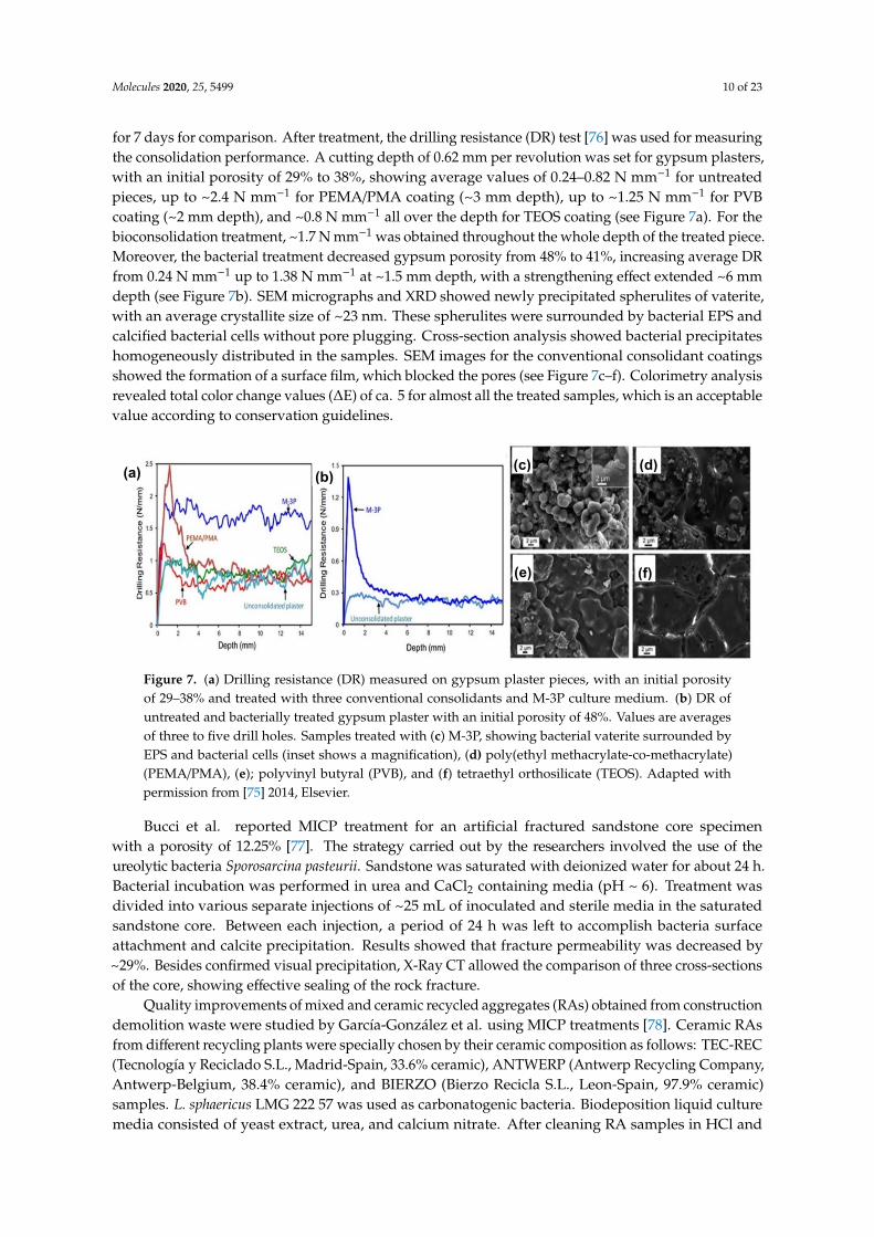
Molecules 2020, 25, 5499 10 of 23
for 7 days for comparison. After treatment, the drilling resistance (DR) test [76] was used for measuringthe consolidation performance. A cutting depth of 0.62 mm per revolution was set for gypsum plasters,with an initial porosity of 29% to 38%, showing average values of 0.24–0.82 N mm−1 for untreatedpieces, up to ~2.4 N mm−1 for PEMA/PMA coating (~3 mm depth), up to ~1.25 N mm−1 for PVBcoating (~2 mm depth), and ~0.8 N mm−1 all over the depth for TEOS coating (see Figure 7a). For thebioconsolidation treatment, ~1.7 N mm−1 was obtained throughout the whole depth of the treated piece.Moreover, the bacterial treatment decreased gypsum porosity from 48% to 41%, increasing average DRfrom 0.24 N mm−1 up to 1.38 N mm−1 at ~1.5 mm depth, with a strengthening effect extended ~6 mmdepth (see Figure 7b). SEM micrographs and XRD showed newly precipitated spherulites of vaterite,with an average crystallite size of ~23 nm. These spherulites were surrounded by bacterial EPS andcalcified bacterial cells without pore plugging. Cross-section analysis showed bacterial precipitateshomogeneously distributed in the samples. SEM images for the conventional consolidant coatingsshowed the formation of a surface film, which blocked the pores (see Figure 7c–f). Colorimetry analysisrevealed total color change values (∆E) of ca. 5 for almost all the treated samples, which is an acceptablevalue according to conservation guidelines.
Molecules 2020, 25, x FOR PEER REVIEW 10 of 23
average DR from 0.24 N mm−1 up to 1.38 N mm−1 at ~1.5 mm depth, with a strengthening effect extended ~6 mm depth (see Figure 7b). SEM micrographs and XRD showed newly precipitated spherulites of vaterite, with an average crystallite size of ~23 nm. These spherulites were surrounded by bacterial EPS and calcified bacterial cells without pore plugging. Cross-section analysis showed bacterial precipitates homogeneously distributed in the samples. SEM images for the conventional consolidant coatings showed the formation of a surface film, which blocked the pores (see Figure 7c–f). Colorimetry analysis revealed total color change values (∆E) of ca. 5 for almost all the treated samples, which is an acceptable value according to conservation guidelines.
Figure 7. (a) Drilling resistance (DR) measured on gypsum plaster pieces, with an initial porosity of 29–38% and treated with three conventional consolidants and M-3P culture medium. (b) DR of untreated and bacterially treated gypsum plaster with an initial porosity of 48%. Values are averages of three to five drill holes. Samples treated with (c) M-3P, showing bacterial vaterite surrounded by EPS and bacterial cells (inset shows a magnification), (d) poly(ethyl methacrylate-co-methacrylate) (PEMA/PMA), (e); polyvinyl butyral (PVB), and (f) tetraethyl orthosilicate (TEOS). Adapted with permission from [75] 2014, Elsevier.
Bucci et al. reported MICP treatment for an artificial fractured sandstone core specimen with a porosity of 12.25% [77]. The strategy carried out by the researchers involved the use of the ureolytic bacteria Sporosarcina pasteurii. Sandstone was saturated with deionized water for about 24 h. Bacterial incubation was performed in urea and CaCl2 containing media (pH ~ 6). Treatment was divided into various separate injections of ~25 mL of inoculated and sterile media in the saturated sandstone core. Between each injection, a period of 24 h was left to accomplish bacteria surface attachment and calcite precipitation. Results showed that fracture permeability was decreased by ~29%. Besides confirmed visual precipitation, X-Ray CT allowed the comparison of three cross-sections of the core, showing effective sealing of the rock fracture.
Quality improvements of mixed and ceramic recycled aggregates (RAs) obtained from construction demolition waste were studied by García-González et al. using MICP treatments [78]. Ceramic RAs from different recycling plants were specially chosen by their ceramic composition as follows: TEC-REC (Tecnología y Reciclado S.L., Madrid-Spain, 33.6% ceramic), ANTWERP (Antwerp Recycling Company, Antwerp-Belgium, 38.4% ceramic), and BIERZO (Bierzo Recicla S.L., Leon-Spain, 97.9% ceramic) samples. L. sphaericus LMG 222 57 was used as carbonatogenic bacteria. Biodeposition liquid culture media consisted of yeast extract, urea, and calcium nitrate. After cleaning RA samples in HCl and distilled water, samples were submerged for 24 h in a liquid culture of L. sphaericus under non-sterile and static conditions at 20 °C. Then, RA samples were submerged in the biodeposition liquid media for four days. Results showed weight gain between 16% and 46% for small samples (4–12.5 mm) in comparison to the large samples (12.5–20 mm). Samples with increased ceramic content gained more weight, which is attributed to greater roughness of the ceramic surface. Moreover, continuous calcium carbonate layers were present in less irregular ceramic surfaces. Water permeability reduction was observed in all tested samples. Comparison between small and large samples showed a difference in water permeability of 46%, 41%, and 16%
(a) (b) (c) (d)
(e) (f)
Figure 7. (a) Drilling resistance (DR) measured on gypsum plaster pieces, with an initial porosityof 29–38% and treated with three conventional consolidants and M-3P culture medium. (b) DR ofuntreated and bacterially treated gypsum plaster with an initial porosity of 48%. Values are averagesof three to five drill holes. Samples treated with (c) M-3P, showing bacterial vaterite surrounded byEPS and bacterial cells (inset shows a magnification), (d) poly(ethyl methacrylate-co-methacrylate)(PEMA/PMA), (e); polyvinyl butyral (PVB), and (f) tetraethyl orthosilicate (TEOS). Adapted withpermission from [75] 2014, Elsevier.
Bucci et al. reported MICP treatment for an artificial fractured sandstone core specimenwith a porosity of 12.25% [77]. The strategy carried out by the researchers involved the use of theureolytic bacteria Sporosarcina pasteurii. Sandstone was saturated with deionized water for about 24 h.Bacterial incubation was performed in urea and CaCl2 containing media (pH ~ 6). Treatment wasdivided into various separate injections of ~25 mL of inoculated and sterile media in the saturatedsandstone core. Between each injection, a period of 24 h was left to accomplish bacteria surfaceattachment and calcite precipitation. Results showed that fracture permeability was decreased by~29%. Besides confirmed visual precipitation, X-Ray CT allowed the comparison of three cross-sectionsof the core, showing effective sealing of the rock fracture.
Quality improvements of mixed and ceramic recycled aggregates (RAs) obtained from constructiondemolition waste were studied by García-González et al. using MICP treatments [78]. Ceramic RAsfrom different recycling plants were specially chosen by their ceramic composition as follows: TEC-REC(Tecnología y Reciclado S.L., Madrid-Spain, 33.6% ceramic), ANTWERP (Antwerp Recycling Company,Antwerp-Belgium, 38.4% ceramic), and BIERZO (Bierzo Recicla S.L., Leon-Spain, 97.9% ceramic)samples. L. sphaericus LMG 222 57 was used as carbonatogenic bacteria. Biodeposition liquid culturemedia consisted of yeast extract, urea, and calcium nitrate. After cleaning RA samples in HCl and

Molecules 2020, 25, 5499 11 of 23
distilled water, samples were submerged for 24 h in a liquid culture of L. sphaericus under non-sterileand static conditions at 20 ◦C. Then, RA samples were submerged in the biodeposition liquid mediafor four days. Results showed weight gain between 16% and 46% for small samples (4–12.5 mm) incomparison to the large samples (12.5–20 mm). Samples with increased ceramic content gained moreweight, which is attributed to greater roughness of the ceramic surface. Moreover, continuous calciumcarbonate layers were present in less irregular ceramic surfaces. Water permeability reduction wasobserved in all tested samples. Comparison between small and large samples showed a differencein water permeability of 46%, 41%, and 16% for TEC-REC, ANTWERP, and BIERZO, respectively.Samples’ resistance against ultrasonic attack exhibited different responses between biotreated anduntreated samples. TEC-REC large samples registered 44% weight loss and small samples 6% weightloss. For samples with higher ceramic content, biotreated samples exhibited higher weight loss thanuntreated samples. SEM analysis revealed calcium carbonate uniform deposition over a regular surfaceand partial pore surface filling by globular precipitates (see Figure 8a,b). EDX revealed that globularprecipitates were likely to be calcium carbonate (see Figure 8c).
Molecules 2020, 25, x FOR PEER REVIEW 11 of 23
for TEC-REC, ANTWERP, and BIERZO, respectively. Samples’ resistance against ultrasonic attack exhibited different responses between biotreated and untreated samples. TEC-REC large samples registered 44% weight loss and small samples 6% weight loss. For samples with higher ceramic content, biotreated samples exhibited higher weight loss than untreated samples. SEM analysis revealed calcium carbonate uniform deposition over a regular surface and partial pore surface filling by globular precipitates (see Figure 8a,b). EDX revealed that globular precipitates were likely to be calcium carbonate (see Figure 8c).
Figure 8. SEM images of (a) CaCO3 precipitated over recycled aggregates surface, (b) CaCO3 precipitated inside a pore of recycled aggregates (RA) sample, (c) CaCO3 globular precipitate with Energy-dispersive X-ray spectroscopy (EDX) spectrum showing the composition of the precipitate. Adapted with permission from [78] 2017, Elsevier.
Minto et al. studied the restoration of degraded marble structures using MICP by X-ray computed tomography (X-CT) [79,80]. The carbonatogenic bacteria used was Sporosarcina pasteurii DSM-33. Biodeposition inducing media was composed of urea and CaCl2 at pH = 6.5. Samples of marble were obtained by crushing marble gravel until a particle size of 0.5–1.4 mm and filled into a column. Bacteria culture and biodeposition media were injected every day for six days. After completion of MICP treatment, results revealed that the inlet surface had greater cementations compared to outlet marble grains. After each injection, measurements showed a gradual permeability decrease. Porosity was also reduced from 32.4% before MICP treatment to ca. 28% after MICP treatment. At 4.5 mm depth from the inlet, a minimum porosity of 7.2% was obtained. From inlet to outlet, non-heterogeneous precipitation was observed by X-CT, resulting in a color gradient (see Figure 9). Different injection strategies should be applied for in situ restoration to reach more homogeneous precipitation. MICP treatment was able to cement the marble crushed pieces, meaning that the method allows restoration of the considerable size cracks.
(a) (b)
(c)
Figure 8. SEM images of (a) CaCO3 precipitated over recycled aggregates surface, (b) CaCO3
precipitated inside a pore of recycled aggregates (RA) sample, (c) CaCO3 globular precipitate withEnergy-dispersive X-ray spectroscopy (EDX) spectrum showing the composition of the precipitate.Adapted with permission from [78] 2017, Elsevier.
Minto et al. studied the restoration of degraded marble structures using MICP by X-ray computedtomography (X-CT) [79,80]. The carbonatogenic bacteria used was Sporosarcina pasteurii DSM-33.Biodeposition inducing media was composed of urea and CaCl2 at pH = 6.5. Samples of marblewere obtained by crushing marble gravel until a particle size of 0.5–1.4 mm and filled into a column.Bacteria culture and biodeposition media were injected every day for six days. After completionof MICP treatment, results revealed that the inlet surface had greater cementations compared tooutlet marble grains. After each injection, measurements showed a gradual permeability decrease.Porosity was also reduced from 32.4% before MICP treatment to ca. 28% after MICP treatment.At 4.5 mm depth from the inlet, a minimum porosity of 7.2% was obtained. From inlet to outlet,non-heterogeneous precipitation was observed by X-CT, resulting in a color gradient (see Figure 9).

Molecules 2020, 25, 5499 12 of 23
Different injection strategies should be applied for in situ restoration to reach more homogeneousprecipitation. MICP treatment was able to cement the marble crushed pieces, meaning that the methodallows restoration of the considerable size cracks.
Molecules 2020, 25, x FOR PEER REVIEW 12 of 23
Figure 9. X-ray computed tomography (X-CT) column scans, showing in yellow-white colors high attenuation from solid material. Purple-black colors indicate low attenuation (e.g., air-filled pore space). (a) Slice averaged X-ray attenuation vs. distance from inlet column; (b) Maximum average attenuation; (c) Attenuation from the weakly cemented region; (d) Overall vertical profile attenuation; (e) Inverted column after non cemented sand removal. Adapted with permission from [79].
Hudyma et al. presented a study of MICP on coquina core specimens [81]. Coquina is a cemented limestone composed of shells and quartz sand. Thus, calcite, phosphate, and siliciclastic material are the main chemical components of coquina [82,83]. Core specimens were cut in ~50 mm diameter from two blocks, labeled as J and K. Treatment began with the immersion of the coquina samples in a Sporosarcina pasteurii solution as calcifying bacteria for one hour, allowing saturation and penetration of bacteria in the limestone. Then, coquina samples were immersed in biodeposition media containing an equimolar solution of CaCl2 and urea. Treatment with the biodeposition media was performed during 2, 10, 20, and 40 days. For coquina specimen JZ14-P2, evident crystal deposition was observed as a protective surface, without the presence of calcite deposition inside the pores, while specimen JX12-P2 showed spheroidal MICP deposition inside the stone pores. Measurements revealed a global unit weight increase after treatment between 1.1% and 9.7%. Overall, the water absorption decrease was between 2.1% and 47.9% for the different samples. MICP treatment of coquina samples clearly enhanced the protective properties of the material.
Jongvivatsakul et al. investigated the crack healing performance within cement mortars using MICP [84]. L. sphaericus LMG 2257 was used as calcifying bacteria, while biodeposition media consisted of nutrient broth, CaCl2, and sodium bicarbonate (NaHCO3) at pH = 8. Mortar samples were prepared from Portland cement. Artificial cracks were prepared by placing a copper plate in fresh samples. Each day inoculated biodeposition media and urea were applied to the mortars for 20 days. MICP treatment was visually evaluated through 40× magnification photographs (see insets, Figure 10). After six days of treatment, crack width was decreased by about 34%; after 16 days, remediation was decreasing up to 84%. Non-significant changes were observed in the last 4 days. Samples were divided into treated and untreated zones. SEM images showed vaterite as the main phase in the treated zone. EDS analyses revealed the presence mostly of calcium, carbon, and oxygen. Qualitative XDR analysis identified the presence of calcite and vaterite in the treated zones. For the first 5 days, artificial crack remained apparently non healed, pointing out for artificial crack healing that began from inside to outside. Ultrasonic pulse velocity (UPV) [85] measurements demonstrated that the pulse velocity increased linearly for the first 14 days in treated samples (see Figure 10). Compressive
(a)
(b) (d)
(c)
(e)
Figure 9. X-ray computed tomography (X-CT) column scans, showing in yellow-white colors highattenuation from solid material. Purple-black colors indicate low attenuation (e.g., air-filled pore space).(a) Slice averaged X-ray attenuation vs. distance from inlet column; (b) Maximum average attenuation;(c) Attenuation from the weakly cemented region; (d) Overall vertical profile attenuation; (e) Invertedcolumn after non cemented sand removal. Adapted with permission from [79].
Hudyma et al. presented a study of MICP on coquina core specimens [81]. Coquina is a cementedlimestone composed of shells and quartz sand. Thus, calcite, phosphate, and siliciclastic materialare the main chemical components of coquina [82,83]. Core specimens were cut in ~50 mm diameterfrom two blocks, labeled as J and K. Treatment began with the immersion of the coquina samples in aSporosarcina pasteurii solution as calcifying bacteria for one hour, allowing saturation and penetrationof bacteria in the limestone. Then, coquina samples were immersed in biodeposition media containingan equimolar solution of CaCl2 and urea. Treatment with the biodeposition media was performedduring 2, 10, 20, and 40 days. For coquina specimen JZ14-P2, evident crystal deposition was observedas a protective surface, without the presence of calcite deposition inside the pores, while specimenJX12-P2 showed spheroidal MICP deposition inside the stone pores. Measurements revealed a globalunit weight increase after treatment between 1.1% and 9.7%. Overall, the water absorption decreasewas between 2.1% and 47.9% for the different samples. MICP treatment of coquina samples clearlyenhanced the protective properties of the material.
Jongvivatsakul et al. investigated the crack healing performance within cement mortars usingMICP [84]. L. sphaericus LMG 2257 was used as calcifying bacteria, while biodeposition mediaconsisted of nutrient broth, CaCl2, and sodium bicarbonate (NaHCO3) at pH = 8. Mortar samples wereprepared from Portland cement. Artificial cracks were prepared by placing a copper plate in freshsamples. Each day inoculated biodeposition media and urea were applied to the mortars for 20 days.MICP treatment was visually evaluated through 40×magnification photographs (see insets, Figure 10).After six days of treatment, crack width was decreased by about 34%; after 16 days, remediation wasdecreasing up to 84%. Non-significant changes were observed in the last 4 days. Samples were dividedinto treated and untreated zones. SEM images showed vaterite as the main phase in the treated zone.EDS analyses revealed the presence mostly of calcium, carbon, and oxygen. Qualitative XDR analysisidentified the presence of calcite and vaterite in the treated zones. For the first 5 days, artificial crack

Molecules 2020, 25, 5499 13 of 23
remained apparently non healed, pointing out for artificial crack healing that began from inside tooutside. Ultrasonic pulse velocity (UPV) [85] measurements demonstrated that the pulse velocityincreased linearly for the first 14 days in treated samples (see Figure 10). Compressive strengthincreased from 17.3 MPa (cracked mortar) to 24.7 MPa (healed mortar). Water adsorption was measured,showing ~72% lower adsorption for healed mortars compared to cracked samples. Water penetrationwas 10.8 mm and 8.6 mm depth in the mortars before and after treatment, respectively. CaCO3 formationnot only filled the artificial crack but also improved the quality of the mortars. Compressive strengthand UPV measurements confirmed that the MICP-treated mortars became stronger materials comparedto the original samples.
Molecules 2020, 25, x FOR PEER REVIEW 13 of 23
strength increased from 17.3 MPa (cracked mortar) to 24.7 MPa (healed mortar). Water adsorption was measured, showing ~72% lower adsorption for healed mortars compared to cracked samples. Water penetration was 10.8 mm and 8.6 mm depth in the mortars before and after treatment, respectively. CaCO3 formation not only filled the artificial crack but also improved the quality of the mortars. Compressive strength and UPV measurements confirmed that the MICP-treated mortars became stronger materials compared to the original samples.
Figure 10. The plot of ultrasonic pulse velocity (UPV) value vs. crack width of MICP-treated sample along 20 days of treatment. Adapted with permission from [84]. 2019, Elsevier.
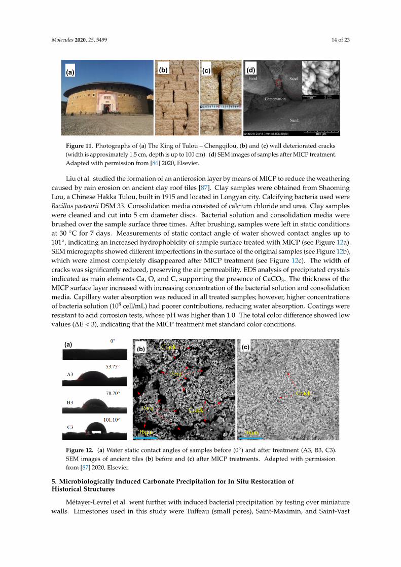
Liu et al. studied the protection and restoration of cracks in Tulou or earthen buildings (see Figure 11a) made of mainly tabia [86]. Tabia consists of sand, limestone (CaCO3), and clay, which usually possess a low tensile strength. Cracks tend to occur in the wall of the Tulou as starting points for damage propagation (see Figure 11b,c). Soil samples were collected nearby earthen buildings for the preparation of probes. Soil analysis showed the presence of silica and kaolinite. After mixing soil samples with sand and lime, mixtures were compacted in different shapes, such as cylinders, beams, and wallets. Bacterial solution and cementation media were injected every day for three days. Bacillus pasteurii (DSM-33) was used as calcifying bacteria, while cementation media was composed of urea and CaCl2. Unconfined compressive strength (UCS) tests were carried out for the samples, indicating that mixture samples after treatment increased the average UCS. SEM images revealed that MICP treatment was able to bind loose soil particles (see Figure 11d). XRD analysis showed the presence of calcite, as the CaCO3 unique phase, and SiO2 peaks from the soil. Flexural strength (FS) was measured before and after MICP treatment to determine the recovery ratio of FS. The repair rate of FS average recovery measurements indicated 35.2%, 56.86%, and 79.92% for crack widths of 15 mm, 10 mm, and 5 mm, respectively. The recovery ratio of shear strength after MICP treatment was 50.74%, 69.53%, and 88.54% for crack widths of 15 mm, 10 mm, and 5mm, respectively. Static contact angle after MICP treatment was found between 83.6° and 100° compared to a contact angle 0° for untreated samples, pointing out for an increased hydrophobicity of the tabia samples.
(a) (b) (c) (d)
Figure 10. The plot of ultrasonic pulse velocity (UPV) value vs. crack width of MICP-treated samplealong 20 days of treatment. Adapted with permission from [84]. 2019, Elsevier.
Liu et al. studied the protection and restoration of cracks in Tulou or earthen buildings(see Figure 11a) made of mainly tabia [86]. Tabia consists of sand, limestone (CaCO3), and clay,which usually possess a low tensile strength. Cracks tend to occur in the wall of the Tulou as startingpoints for damage propagation (see Figure 11b,c). Soil samples were collected nearby earthen buildingsfor the preparation of probes. Soil analysis showed the presence of silica and kaolinite. After mixingsoil samples with sand and lime, mixtures were compacted in different shapes, such as cylinders,beams, and wallets. Bacterial solution and cementation media were injected every day for three days.Bacillus pasteurii (DSM-33) was used as calcifying bacteria, while cementation media was composedof urea and CaCl2. Unconfined compressive strength (UCS) tests were carried out for the samples,indicating that mixture samples after treatment increased the average UCS. SEM images revealedthat MICP treatment was able to bind loose soil particles (see Figure 11d). XRD analysis showed thepresence of calcite, as the CaCO3 unique phase, and SiO2 peaks from the soil. Flexural strength (FS)was measured before and after MICP treatment to determine the recovery ratio of FS. The repair rate ofFS average recovery measurements indicated 35.2%, 56.86%, and 79.92% for crack widths of 15 mm,10 mm, and 5 mm, respectively. The recovery ratio of shear strength after MICP treatment was 50.74%,69.53%, and 88.54% for crack widths of 15 mm, 10 mm, and 5mm, respectively. Static contact angleafter MICP treatment was found between 83.6◦ and 100◦ compared to a contact angle 0◦ for untreatedsamples, pointing out for an increased hydrophobicity of the tabia samples.
Molecules 2020, 25, 5499 14 of 23
Molecules 2020, 25, x FOR PEER REVIEW 13 of 23
strength increased from 17.3 MPa (cracked mortar) to 24.7 MPa (healed mortar). Water adsorption was measured, showing ~72% lower adsorption for healed mortars compared to cracked samples. Water penetration was 10.8 mm and 8.6 mm depth in the mortars before and after treatment, respectively. CaCO3 formation not only filled the artificial crack but also improved the quality of the mortars. Compressive strength and UPV measurements confirmed that the MICP-treated mortars became stronger materials compared to the original samples.
Figure 10. The plot of ultrasonic pulse velocity (UPV) value vs. crack width of MICP-treated sample along 20 days of treatment. Adapted with permission from [84]. 2019, Elsevier.
Liu et al. studied the protection and restoration of cracks in Tulou or earthen buildings (see Figure 11a) made of mainly tabia [86]. Tabia consists of sand, limestone (CaCO3), and clay, which usually possess a low tensile strength. Cracks tend to occur in the wall of the Tulou as starting points for damage propagation (see Figure 11b,c). Soil samples were collected nearby earthen buildings for the preparation of probes. Soil analysis showed the presence of silica and kaolinite. After mixing soil samples with sand and lime, mixtures were compacted in different shapes, such as cylinders, beams, and wallets. Bacterial solution and cementation media were injected every day for three days. Bacillus pasteurii (DSM-33) was used as calcifying bacteria, while cementation media was composed of urea and CaCl2. Unconfined compressive strength (UCS) tests were carried out for the samples, indicating that mixture samples after treatment increased the average UCS. SEM images revealed that MICP treatment was able to bind loose soil particles (see Figure 11d). XRD analysis showed the presence of calcite, as the CaCO3 unique phase, and SiO2 peaks from the soil. Flexural strength (FS) was measured before and after MICP treatment to determine the recovery ratio of FS. The repair rate of FS average recovery measurements indicated 35.2%, 56.86%, and 79.92% for crack widths of 15 mm, 10 mm, and 5 mm, respectively. The recovery ratio of shear strength after MICP treatment was 50.74%, 69.53%, and 88.54% for crack widths of 15 mm, 10 mm, and 5mm, respectively. Static contact angle after MICP treatment was found between 83.6° and 100° compared to a contact angle 0° for untreated samples, pointing out for an increased hydrophobicity of the tabia samples.
(a) (b) (c) (d)
Figure 11. Photographs of (a) The King of Tulou – Chengqilou, (b) and (c) wall deteriorated cracks(width is approximately 1.5 cm, depth is up to 100 cm). (d) SEM images of samples after MICP treatment.Adapted with permission from [86] 2020, Elsevier.
Liu et al. studied the formation of an antierosion layer by means of MICP to reduce the weatheringcaused by rain erosion on ancient clay roof tiles [87]. Clay samples were obtained from ShaomingLou, a Chinese Hakka Tulou, built in 1915 and located in Longyan city. Calcifying bacteria used wereBacillus pasteurii DSM 33. Consolidation media consisted of calcium chloride and urea. Clay sampleswere cleaned and cut into 5 cm diameter discs. Bacterial solution and consolidation media werebrushed over the sample surface three times. After brushing, samples were left in static conditionsat 30 ◦C for 7 days. Measurements of static contact angle of water showed contact angles up to101◦, indicating an increased hydrophobicity of sample surface treated with MICP (see Figure 12a).SEM micrographs showed different imperfections in the surface of the original samples (see Figure 12b),which were almost completely disappeared after MICP treatment (see Figure 12c). The width ofcracks was significantly reduced, preserving the air permeability. EDS analysis of precipitated crystalsindicated as main elements Ca, O, and C, supporting the presence of CaCO3. The thickness of theMICP surface layer increased with increasing concentration of the bacterial solution and consolidationmedia. Capillary water absorption was reduced in all treated samples; however, higher concentrationsof bacteria solution (108 cell/mL) had poorer contributions, reducing water absorption. Coatings wereresistant to acid corrosion tests, whose pH was higher than 1.0. The total color difference showed lowvalues (∆E < 3), indicating that the MICP treatment met standard color conditions.
Molecules 2020, 25, x FOR PEER REVIEW 14 of 23
Figure 11. Photographs of (a) The King of Tulou – Chengqilou, (b) and (c) wall deteriorated cracks (width is approximately 1.5 cm, depth is up to 100 cm). (d) SEM images of samples after MICP treatment. Adapted with permission from [86] 2020, Elsevier.
Liu et al. studied the formation of an antierosion layer by means of MICP to reduce the weathering caused by rain erosion on ancient clay roof tiles [87]. Clay samples were obtained from Shaoming Lou, a Chinese Hakka Tulou, built in 1915 and located in Longyan city. Calcifying bacteria used were Bacillus pasteurii DSM 33. Consolidation media consisted of calcium chloride and urea. Clay samples were cleaned and cut into 5 cm diameter discs. Bacterial solution and consolidation media were brushed over the sample surface three times. After brushing, samples were left in static conditions at 30 °C for 7 days. Measurements of static contact angle of water showed contact angles up to 101°, indicating an increased hydrophobicity of sample surface treated with MICP (see Figure 12a). SEM micrographs showed different imperfections in the surface of the original samples (see Figure 12b), which were almost completely disappeared after MICP treatment (see Figure 12c). The width of cracks was significantly reduced, preserving the air permeability. EDS analysis of precipitated crystals indicated as main elements Ca, O, and C, supporting the presence of CaCO3. The thickness of the MICP surface layer increased with increasing concentration of the bacterial solution and consolidation media. Capillary water absorption was reduced in all treated samples; however, higher concentrations of bacteria solution (108 cell/mL) had poorer contributions, reducing water absorption. Coatings were resistant to acid corrosion tests, whose pH was higher than 1.0. The total color difference showed low values (∆E < 3), indicating that the MICP treatment met standard color conditions.
Figure 12. (a) Water static contact angles of samples before (0°) and after treatment (A3, B3, C3). SEM images of ancient tiles (b) before and (c) after MICP treatments. Adapted with permission from [87] 2020, Elsevier.
5. Microbiologically Induced Carbonate Precipitation for In Situ Restoration of Historical Structures
Métayer-Levrel et al. went further with induced bacterial precipitation by testing over miniature walls. Limestones used in this study were Tuffeau (small pores), Saint-Maximin, and Saint-Vast (large pores) [88]. Patented bacteria and culture media met industrial and financial requirements. Treatment media inoculated with a bacterial strain was sprayed over the limestone surface, followed by spraying of sterile media every 24 h for 5 days and then every 48 h for 10 days. After treatment, a biocalcin coating of several micrometers was observed. Mineral particles filled the voids moderately by getting engrained in the structure. In situ tests were carried out on the SE tower of Saint Médard Church, located in Thouars, France. Tuffeau limestone was used to build the church. Treatment began in June 1993 over an area of 50 m2. The bioprecipitated produce was exposed to normal weather variations. Treatment effects were evaluated after 6 months and 1 year by measuring surficial permeability (a measurement of the standard time of water absorption using a water pipe). MICP
(a) (b) (c)
Figure 12. (a) Water static contact angles of samples before (0◦) and after treatment (A3, B3, C3).SEM images of ancient tiles (b) before and (c) after MICP treatments. Adapted with permissionfrom [87] 2020, Elsevier.
5. Microbiologically Induced Carbonate Precipitation for In Situ Restoration ofHistorical Structures
Métayer-Levrel et al. went further with induced bacterial precipitation by testing over miniaturewalls. Limestones used in this study were Tuffeau (small pores), Saint-Maximin, and Saint-Vast

Molecules 2020, 25, 5499 15 of 23
(large pores) [88]. Patented bacteria and culture media met industrial and financial requirements.Treatment media inoculated with a bacterial strain was sprayed over the limestone surface, followed byspraying of sterile media every 24 h for 5 days and then every 48 h for 10 days. After treatment,a biocalcin coating of several micrometers was observed. Mineral particles filled the voids moderately bygetting engrained in the structure. In situ tests were carried out on the SE tower of Saint Médard Church,located in Thouars, France. Tuffeau limestone was used to build the church. Treatment began in June1993 over an area of 50 m2. The bioprecipitated produce was exposed to normal weather variations.Treatment effects were evaluated after 6 months and 1 year by measuring surficial permeability(a measurement of the standard time of water absorption using a water pipe). MICP treatmentincreased the time of water absorption from ~70 s (before treatment) to ~250 s after 6 months oftreatment but then decreased to ~130 s after one year of treatment. In any case, the surficial permeabilityafter treatment showed lower values if compared to before treatment values. No appreciable changesfor gas permeability, color, or aesthetic appreciation were observed if compared to the original samples.Bioprecipitate guaranteed the preservation of the limestone by reducing the exchange of degradingagents and environmental pollutants. Moreover, calcifying bacteria bulk population interfered with thedevelopment of acidifying bacteria, which might be a threat to historical buildings. Finally, no changesin the external aspect of the tower were observed after 3.5 years.
Perito et al. reported a bacterial CaCO3 mineralization treatment on the Andera Cathedral,Italy [14]. First, stone samples (10 cm × 10 cm × 4 cm) obtained from the main façade of the Pietrad’Angera lithotype were tested in the laboratory. B. subtilis 168 strain was the calcifying bacteria,and B4 was used as the precipitation medium. After bacterial incubation in B4 media at 37 ◦C,cells were collected at a 108 cells/mL concentration, centrifugated, autoclaved, stripped of metal cationsthrough deionized water, rinsed, and stored in physiological solution (PS) at −20 ◦C. After dilutions ofPS in calcium chloride solution, ammonium carbonate vapors induced the precipitation of calciumcarbonate. Besides crystallization, no visible changes were observed after the seventh day at roomtemperature. Then, the frozen cell mixture was ground with alumina, then unbroken cells and aluminawere eliminated through centrifugation. Cytosol, membranes, and cell walls were stored at −20 ◦C andlabeled as Bacillus cell fraction (BCF). Finally, BCF was lyophilized and resuspended in CaCl2 solutionmixed with supersaturated calcium bicarbonate solution (Super C) and enriched with 20 nm calcitenanoparticles (2% w/v). Around 30 mL per application was sprayed on the stone samples surface twicea day for 3 days. A stone sample was sprayed only on Super C as reference. In vitro tests showedan overall color change ∆E < 3, meaning acceptable and no detectable changes. Water absorptiondecreased by 16.7%. Cohesion profiles from DR did not show remarkable differences. In situ testwas implemented on the main façade of the Angera Cathedral, which is dated from the sixth century.Selected areas sized 0.29 m2 and 0.28 m2, with a 0.04 m2 area as a control. Approximately 1 L m−2
per spray application of BCF (8.5 g L−1) in Super C solution and bacteria-free super C solution wassprayed for the first day. BCF (0.032 g L−1) in Super C, enriched with nanoparticles, and bacteria-freeSuper C were sprayed for the second day. On the third day, only a bacteria-free Super C enrichedsolution was sprayed on the chosen areas. After four months, evaluation tests were performed, such aswater absorption, surface color change, and cohesion profiles. Small cores from the chosen areas weretaken to the laboratory to analyze new calcite penetration. CaCO3 precipitated crystals were collectedand analyzed with FT-IR and XRD, indicating the presence of calcite. In situ color test measurementsshowed ∆E < 3, meaning no detectable changes to the human eye. Water absorption decreased by 6.8%for in situ experiments. Cohesion profiles indicated an increased hardness of the treated areas for thefirst 3 mm.
Rodriguez-Navarro et al. reported in situ restoration using MICP over stone monuments datedfrom the sixteen century in Granada, Spain [4]. Heavily degraded areas from three different testsites were treated: (i) Hospital Real, (ii) San Jeronimo’s Monastery Apse, and (iii) Capilla Real.These three buildings were constructed using porous calcarenite, which is a material with high waterabsorption and porosity. Treatments were performed with M-3P medium inoculated with M. xanthus.

Molecules 2020, 25, 5499 16 of 23
The inoculated medium was sprayed twice on the treated area, followed by one spray of sterile medium.Treatment was applied twice a day for six consecutive days. After each application, the treated areawas wrapped with aluminum/plastic foil to avoid possible pigmentation by sunlight effect and toreduce media evaporation. The binding capacity of treatments, surface strengthening, and chromaticchanges were tested for four years after initial treatment. Peeling tape test showed weight lossreduction after inoculated media treatment for all the treated areas, which is related to the bacterialCaCO3 precipitation (calcite and vaterite, according to XDR). SEM analysis showed that new crystalswere precipitated aligned to the pore system without plugging them. Moreover, loose calcarenitegrains were connected via EPS with new bacterial calcium carbonate. Results with sterile M-3P mediashowed an identical degree of consolidation for San Jeronimo and Hospital Real treated areas. In somecases, sufficient consolidation was obtained with the activation of the original carbonatogenic bacteriapresent, thus reducing treatment cost. Moreover, spectrophotometric ∆E measurements were below 5,meaning acceptable value for treatment.
Self-inoculation of indigenous calcifying bacteria present in stones prior to MICP treatment isanother viable way that Jroundi et al. designed for in situ treatment of salt weathered stones [89].The cloister entrance of San Jeronimo Monastery, Spain, presented granular disintegration and surfaceloss. This weathering and erosion were attributed to the crystallization of salts, such as magnesiumsulfate hexahydrate
(MgSO4·6H2O
), niter (KNO3), halite (NaCl), among others. Fifty-five bacterial
isolates were obtained from selected areas. Incubation of M-3P inoculated medium led to highCaCO3 precipitation in all bacterial isolates after 48 h. XDR analysis identified calcite as the mainphase and <10 wt% of vaterite. To evaluate the applicability of the activated carbonatogenic bacteria,the sterile calcite substrate was immersed in an inoculated M3-P medium. Field emission scanningelectron microscopy (FESEM) [90] and transmission scanning electron microscopy (TSEM) [91] showeddense bacterial colony formation and CaCO3 embedded in EPS after 20 h treatment (see Figure 13b).Spheroidal or rhombohedral calcite structures fully covered the calcite substrate growth controlled ona self-epitaxy way after 48 h treatment (see Figure 13c). In situ treatments were applied on stone blockswith a similar degree of exposure and decay. Three different treatment approaches were performedusing M. xanthus, sterile M-3P media, and a self-inoculation biotreatment. M. xanthus and sterileM-3P treatment results showed limited consolidation. The peeling tape test revealed modest surfaceconsolidation for both treatments after 5 months. Drilling resistance values were slightly higher inthe first ~5 mm depth for treated areas after 24 months. SEM showed almost no presence of calciumcarbonate or EPS. Chromatic changes revealed, for both treatments, the acceptable values of ∆E (<5).On the other hand, self-inoculated bacteria treatment showed remarkable consolidation by a peelingtape test along with a 24 months study. DR measurements showed about 4 times higher values thanuntreated stone (see Figure 13d–f). The maximum DR value achieved was 10 ± 6 N for the first 3~5 mmdepth. Color changes after treatment were not significant, showing an ∆E = 3.8 ± 1.7. XRD analyses ofsamples from treated areas showed calcite as the unique calcium carbonate phase (see Figure 13a).Strengthening of the stone was associated with the abundant presence of newly precipitated calciumcarbonate crystals and EPS. Mercury intrusion porosimetry (MIP) showed a reduction in porosityfrom 27 ± 1% to 25 ± 1%, resulting from the formation of 30–100 nm size nanocrystals that cementedthe stone without plugging the pores. No presence of material loss or granular disaggregation wasobserved 24 months after the treatment.

Molecules 2020, 25, 5499 17 of 23
Molecules 2020, 25, x FOR PEER REVIEW 16 of 23
some cases, sufficient consolidation was obtained with the activation of the original carbonatogenic bacteria present, thus reducing treatment cost. Moreover, spectrophotometric ∆E measurements were below 5, meaning acceptable value for treatment.
Self-inoculation of indigenous calcifying bacteria present in stones prior to MICP treatment is another viable way that Jroundi et al. designed for in situ treatment of salt weathered stones [89]. The cloister entrance of San Jeronimo Monastery, Spain, presented granular disintegration and surface loss. This weathering and erosion were attributed to the crystallization of salts, such as magnesium sulfate hexahydrate (MgSO4•6H2O), niter (KNO3), halite (NaCl), among others. Fifty-five bacterial isolates were obtained from selected areas. Incubation of M-3P inoculated medium led to high CaCO3 precipitation in all bacterial isolates after 48 h. XDR analysis identified calcite as the main phase and <10 wt% of vaterite. To evaluate the applicability of the activated carbonatogenic bacteria, the sterile calcite substrate was immersed in an inoculated M3-P medium. Field emission scanning electron microscopy (FESEM) [90] and transmission scanning electron microscopy (TSEM) [91] showed dense bacterial colony formation and CaCO3 embedded in EPS after 20 h treatment (see Figure 13b). Spheroidal or rhombohedral calcite structures fully covered the calcite substrate growth controlled on a self-epitaxy way after 48 h treatment (see Figure 13c). In situ treatments were applied on stone blocks with a similar degree of exposure and decay. Three different treatment approaches were performed using M. xanthus, sterile M-3P media, and a self-inoculation biotreatment. M. xanthus and sterile M-3P treatment results showed limited consolidation. The peeling tape test revealed modest surface consolidation for both treatments after 5 months. Drilling resistance values were slightly higher in the first ~5 mm depth for treated areas after 24 months. SEM showed almost no presence of calcium carbonate or EPS. Chromatic changes revealed, for both treatments, the acceptable values of ∆E (<5). On the other hand, self-inoculated bacteria treatment showed remarkable consolidation by a peeling tape test along with a 24 months study. DR measurements showed about 4 times higher values than untreated stone (see Figure 13d–f). The maximum DR value achieved was 10 ± 6 N for the first 3 ~ 5 mm depth. Color changes after treatment were not significant, showing an ∆E = 3.8 ± 1.7. XRD analyses of samples from treated areas showed calcite as the unique calcium carbonate phase (see Figure 13a). Strengthening of the stone was associated with the abundant presence of newly precipitated calcium carbonate crystals and EPS. Mercury intrusion porosimetry (MIP) showed a reduction in porosity from 27 ± 1% to 25 ± 1%, resulting from the formation of 30–100 nm size nanocrystals that cemented the stone without plugging the pores. No presence of material loss or granular disaggregation was observed 24 months after the treatment.
Figure 13. (a) XRD pattern before (blue) and after (red) MICP treatment. SEM images of (b) newly formed CaCO3; (c) calcite substrate covered by bacterial calcite. Drilling resistance depth profile: (d) M. xanthus treatment (blue-line); (e) M-3P treatment (blue-line); (f) self-inoculated treatment (blue
(a) (b) (c)
(d) (e) (f)
Figure 13. (a) XRD pattern before (blue) and after (red) MICP treatment. SEM images of (b) newlyformed CaCO3; (c) calcite substrate covered by bacterial calcite. Drilling resistance depth profile:(d) M. xanthus treatment (blue-line); (e) M-3P treatment (blue-line); (f) self-inoculated treatment (blueline). Untreated stones (red line) are presented for comparison. Shaded areas represent s.d. (±1σ).Adapted with permission from [89].
6. Conclusions and Perspectives
Highly ureolytic bacteria are the main strains of microorganisms used in MICP-treatment for therestoration of historic buildings. Despite higher calcium carbonate production by some microorganisms,the usage of human pathogenic bacterial strains (e.g., P. putida, B. cereus) is not recommended becauserestoration in situ lacks the biosafety that a microbiology laboratory could provide. MICP treatmenthas been applied in different ways, such as immersion, spraying, injection, and brushing. This is animportant parameter, necessarily to be taken, depending on the in-situ circumstances and requirements.Optimization of the treatment, such as lowering reagents costs, high yields of carbonate production,shorter treatment time, avoiding unnecessary tests or treatment steps, could allow researchers to savesome time, money, and resources.
Although, until now, the method has not been standardized, a good approach for this is the usageof the ζ-potential because it has been proven that bacteria with lower ζ-potential possess higher surfacecolonization capacity. A modification in the MICP treatment to obtain CaCO3 crystals using a dead cellfraction instead of the application of live calcifying bacteria becomes interesting for the case in whichcertain bacterial strains, like nitrifying or sulfur-oxidizing bacteria, are needed, e.g., Nitrosomonas spp,Nitrobacter spp, Thiobacillus spp. These bacteria species are capable of excreting acids [92] (if nitric acid isproduced, it can react with calcite, thus forming highly soluble calcium nitrate), leading to biocorrosionof the material [23]. These problems were addressed by Ganendra et al. [93], using Methylocystis parvusas calcifying bacteria to induce calcium carbonate precipitation from different calcium formateconcentrations. Although culture-dependent methods have been useful to isolate carbonatogenicmicroorganisms, new genomic microbiological analyses have allowed us to study the epilithic microbialbiodiversity in monuments and historical buildings [94]. For example, Chimenti et al. carried out ametagenomic analysis of the bacterial communities, colonizing the walls of the medieval church ofSan Leonardo di Sponto, in Italy [95]. The microbiome of the wall samples was composed not only ofpossible deteriorated microorganisms but also carbonatogenic bacteria corresponding to the phylumActinobacteria, specially Arthrobacter spp. In fact, in brick samples, this phylum may be predominant,depending on the environmental conditions [96]. DNA next-generation sequencing studies havefocused on the microbial diversity in bricks of the former Auschwitz II-Birkenau concentration,

Molecules 2020, 25, 5499 18 of 23
and extermination camp in Poland has shown the presence of the genus Arthrobacter as part of themicrobiota [97]. Although these microorganisms can colonize these surfaces, the carbonatogenicbacteria distribution may differ in relation to the collection site. For instance, Andrei et al. found thatthe diversity of epilithic bacterial communities on Saint Donatus statue, Romania, varied depending onthe monument sampling site [98]. Microbiologically induced carbonate precipitation has been shownto be an efficient strategy to consolidate, protect, restore, and enhance the mechanical properties of abroad variety of materials used in historical buildings.
Author Contributions: E.O.-V. wrote the draft manuscript. M.G.-G. and A.P.-C. revised the manuscript. A.P.-C.supervised the writing process and edited the manuscript. All authors have read and agreed to the publishedversion of the manuscript.
Funding: This research received no external funding.
Acknowledgments: E.O.-V. thanks to Dra. Martha Romero and Carlos Vasquez from the Instituto Nacional delPatrimonio Cultural (National Institute for the Cultural Heritage, Ecuador) for the research stay. A.P.-C. thanks tothe University Yachay Tech for the internal grants No. Chem19-08 and Chem19-17.
Conflicts of Interest: The authors declare no conflict of interest.
Abbreviations
AB-PAS Alcian blue-periodic acid-Schiff stainBCF Bacillus cell fractionCTC 5-Cyano-2,3-ditolyl tetrazolium chlorideDR Drilling resistanceEDX Energy-dispersive X-ray spectroscopyEPS Extracellular polymeric substancesESEM Environmental scanning electron microscopyFESEM Field emission scanning electron microscopyFS Flexural strengthMICP Microbiologically induced carbonate precipitationMIP Mercury intrusion porosimetryPEMA/PMA Poly(ethyl methacrylate-co-methacrylate)PS Physiological solutionPVB Polyvinyl butyralRA Recycled aggregatesSEM Scanning electron microscopySuper C Supersaturated calcium bicarbonate solutionTEOS Tetraethyl orthosilicateTSEM Transmission scanning electron microscopyUCS Unconfined compressive strengthUPV Ultrasonic pulse velocityX-CT X-ray computed tomographyXRD X-ray powder diffractionζ-potential Electrical charge at the shear plane∆E Overall degree color change
References
1. Barabesi, C.; Galizzi, A.; Mastromei, G.; Rossi, M.; Tamburini, E.; Perito, B. Bacillus subtilis gene clusterinvolved in calcium carbonate biomineralization. J. Bacteriol. 2007, 189, 228–235. [CrossRef] [PubMed]
2. Lowenstam, H. Minerals formed by organisms. Science 1981, 211, 1126–1131. [CrossRef] [PubMed]3. Xu, A.W.; Ma, Y.; Cölfen, H. Biomimetic mineralization. J. Mater. Chem. 2007, 17, 415–449. [CrossRef]4. Rodriguez-Navarro, C.; Jroundi, F.; Gonzalez-Muñoz, M.T. Stone Consolidation by Bacterial
Carbonatogenesis: Evaluation of in situ Applications. Restor. Build. Monum. 2015, 21, 9–20. [CrossRef]

Molecules 2020, 25, 5499 19 of 23
5. Han, Z.; Zhao, Y.; Yan, H.; Zhao, H.; Han, M.; Sun, B.; Meng, R.; Zhuang, D.; Li, D.; Gao, W.; et al.The Characterization of Intracellular and Extracellular Biomineralization Induced by Synechocystis sp.PCC6803 Cultured under Low Mg/Ca Ratios Conditions. Geomicrobiol. J. 2017, 34, 362–373. [CrossRef]
6. González-Muñoz, M.T.; Rodriguez-Navarro, C.; Martínez-Ruiz, F.; Arias, J.M.; Merroun, M.L.;Rodriguez-Gallego, M. Bacterial biomineralization: New insights from Myxococcus-induced mineralprecipitation. Geol. Soc. Spec. Publ. 2010, 336, 31–50. [CrossRef]
7. Márcia Aiko, J.S.; Maria Alba, C.; Daniel, A.; Christine, G. Vanderley Effect of culture medium onbiocalcification by pseudomona putida, lysinibacillus sphaericus and bacillus subtilis. Braz. J. Microbiol.2011, 42, 499–507.
8. Dejong, J.T.; Soga, K.; Kavazanjian, E.; Burns, S.; Van Paassen, L.A.; Al Qabany, A.; Aydilek, A.; Bang, S.S.;Burbank, M.; Caslake, L.F.; et al. Biogeochemical processes and geotechnical applications: Progress,opportunities and challenges. Géotechnique 2013, 63, 287–301. [CrossRef]
9. Krajewska, B. Urease-aided calcium carbonate mineralization for engineering applications: A review. J. Adv.Res. 2018, 13, 59–67. [CrossRef]
10. Zhu, T.; Dittrich, M. Carbonate precipitation through microbial activities in natural environment, and theirpotential in biotechnology: A review. Front. Bioeng. Biotechnol. 2016, 4, 1–21. [CrossRef]
11. Seifan, M.; Samani, A.K.; Berenjian, A. Bioconcrete: Next generation of self-healing concrete. Appl. Microbiol.Biotechnol. 2016, 100, 2591–2602. [CrossRef] [PubMed]
12. Hammes, F.; Verstraete, W. Key roles of pH and calcium metabolism in microbial carbonate precipitation.Rev. Environ. Sci. Biotechnol. 2002, 1, 3–7. [CrossRef]
13. Schwantes-Cezario, N.; Medeiros, L.P.; De Oliveira, A.G.; Nakazato, G.; Katsuko Takayama Kobayashi, R.;Toralles, B.M. Bioprecipitation of calcium carbonate induced by Bacillus subtilis isolated in Brazil. Int. Biodeterior.Biodegrad. 2017, 123, 200–205. [CrossRef]
14. Perito, B.; Marvasi, M.; Barabesi, C.; Mastromei, G.; Bracci, S.; Vendrell, M.; Tiano, P. A Bacillus subtilis cellfraction (BCF) inducing calcium carbonate precipitation: Biotechnological perspectives for monumentalstone reinforcement. J. Cult. Herit. 2014, 15, 345–351. [CrossRef]
15. Arun, G. Conservation and Restoration of Cultural Heritage. Pelagia Res. Libr. Adv. Appl. Sci. Res. 1989,7, 159–167.
16. Gaylarde, C.C.; Gaylarde, P.M. Biodeterioration of historic buildings in Latin America. In Proceedings ofthe 9th DBMC International Conference on Durability of Building Materials and Components, Brisbane,Australia, 17–21 March 2002; p. 171.
17. Camaiti, M.; Borselli, G.; Matteoli, U. La conservazione del patrimonio monumentale: Prodotti consolidantiimpiegati nelle operazioni di restauro. L’edil. l’ind. 1988, 2, 435–445.
18. Dei, L.; Baglioni, P.; Sarti, G.; Ferroni, E. Aging effects on ammonium carbonate/acetone solutions andcleaning of works of art. Stud. Conserv. 1996, 41, 9–18. [CrossRef]
19. Dei, L. Conservation Treatments: Cleaning, Consolidation and Protection. In Nanoscience for the Conservationof Works of Art; Baglioni, P., Chelazzi, D., Eds.; Nanoscience & Nanotechnology Series; Royal Society ofChemistry: Cambridge, UK, 2013; pp. 77–92, ISBN 978-1-84973-566-7.
20. Price, C.; Ross, K.; White, G. A further appraisal of the ‘lime technique’ for limestone consolidation, using aradioactive tracer. Stud. Conserv. 1988, 33, 178–186. [CrossRef]
21. Castanier, S.; Le Métayer-Levrel, G.; Orial, G.; Loubière, J.-F.; Perthuisot, J.-P. Bacterial Carbonatogenesisand Applications to Preservation and Restoration of Historic Property. In Of Microbes and Art; Cifierri, O.,Tiano, P., Mastromei, G., Eds.; Springer: Boston, MA, USA, 2000; pp. 203–218.
22. Loubire, J.-F.; Paradas, J.; Adolphe, J.P.; Soleillhavoup, F. Procédé de Traitement Biologique d’une SurfaceArtificielle. EP0388304B1, 19 September 1990.
23. Anbu, P.; Kang, C.H.; Shin, Y.J.; So, J.S. Formations of calcium carbonate minerals by bacteria and its multipleapplications. Springerplus 2016, 5, 1–26. [CrossRef]
24. Gupta, S.; Kua, H.W.; Pang, S.D. Healing cement mortar by immobilization of bacteria in biochar:An integrated approach of self-healing and carbon sequestration. Cem. Concr. Compos. 2018, 86, 238–254.[CrossRef]
25. Wang, J.; Vandevyvere, B.; Vanhessche, S.; Schoon, J.; Boon, N.; De Belie, N. Microbial carbonate precipitationfor the improvement of quality of recycled aggregates. J. Clean. Prod. 2017, 156, 355–366. [CrossRef]
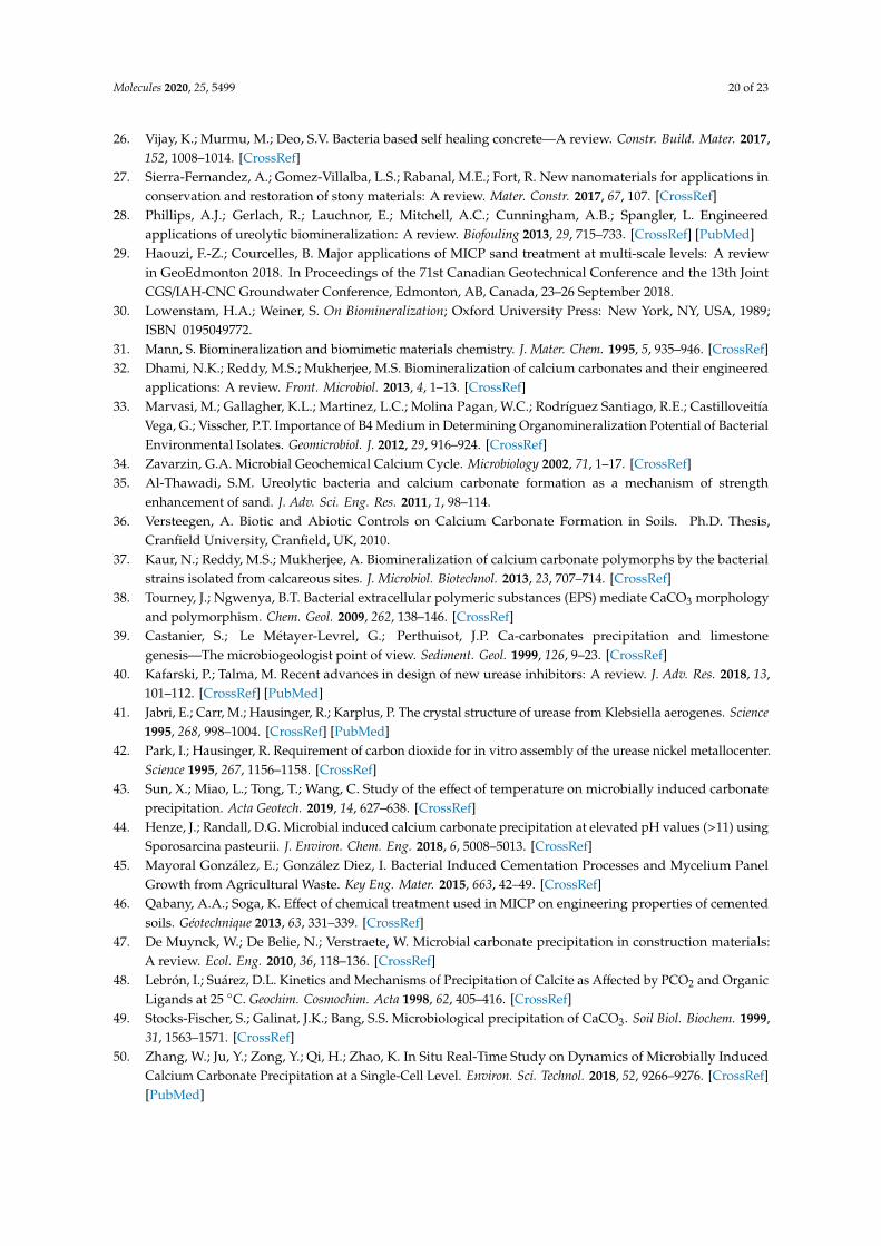
Molecules 2020, 25, 5499 20 of 23
26. Vijay, K.; Murmu, M.; Deo, S.V. Bacteria based self healing concrete—A review. Constr. Build. Mater. 2017,152, 1008–1014. [CrossRef]
27. Sierra-Fernandez, A.; Gomez-Villalba, L.S.; Rabanal, M.E.; Fort, R. New nanomaterials for applications inconservation and restoration of stony materials: A review. Mater. Constr. 2017, 67, 107. [CrossRef]
28. Phillips, A.J.; Gerlach, R.; Lauchnor, E.; Mitchell, A.C.; Cunningham, A.B.; Spangler, L. Engineeredapplications of ureolytic biomineralization: A review. Biofouling 2013, 29, 715–733. [CrossRef] [PubMed]
29. Haouzi, F.-Z.; Courcelles, B. Major applications of MICP sand treatment at multi-scale levels: A reviewin GeoEdmonton 2018. In Proceedings of the 71st Canadian Geotechnical Conference and the 13th JointCGS/IAH-CNC Groundwater Conference, Edmonton, AB, Canada, 23–26 September 2018.
30. Lowenstam, H.A.; Weiner, S. On Biomineralization; Oxford University Press: New York, NY, USA, 1989;ISBN 0195049772.
31. Mann, S. Biomineralization and biomimetic materials chemistry. J. Mater. Chem. 1995, 5, 935–946. [CrossRef]32. Dhami, N.K.; Reddy, M.S.; Mukherjee, M.S. Biomineralization of calcium carbonates and their engineered
applications: A review. Front. Microbiol. 2013, 4, 1–13. [CrossRef]33. Marvasi, M.; Gallagher, K.L.; Martinez, L.C.; Molina Pagan, W.C.; Rodríguez Santiago, R.E.; Castilloveitía
Vega, G.; Visscher, P.T. Importance of B4 Medium in Determining Organomineralization Potential of BacterialEnvironmental Isolates. Geomicrobiol. J. 2012, 29, 916–924. [CrossRef]
34. Zavarzin, G.A. Microbial Geochemical Calcium Cycle. Microbiology 2002, 71, 1–17. [CrossRef]35. Al-Thawadi, S.M. Ureolytic bacteria and calcium carbonate formation as a mechanism of strength
enhancement of sand. J. Adv. Sci. Eng. Res. 2011, 1, 98–114.36. Versteegen, A. Biotic and Abiotic Controls on Calcium Carbonate Formation in Soils. Ph.D. Thesis,
Cranfield University, Cranfield, UK, 2010.37. Kaur, N.; Reddy, M.S.; Mukherjee, A. Biomineralization of calcium carbonate polymorphs by the bacterial
strains isolated from calcareous sites. J. Microbiol. Biotechnol. 2013, 23, 707–714. [CrossRef]38. Tourney, J.; Ngwenya, B.T. Bacterial extracellular polymeric substances (EPS) mediate CaCO3 morphology
and polymorphism. Chem. Geol. 2009, 262, 138–146. [CrossRef]39. Castanier, S.; Le Métayer-Levrel, G.; Perthuisot, J.P. Ca-carbonates precipitation and limestone
genesis—The microbiogeologist point of view. Sediment. Geol. 1999, 126, 9–23. [CrossRef]40. Kafarski, P.; Talma, M. Recent advances in design of new urease inhibitors: A review. J. Adv. Res. 2018, 13,
101–112. [CrossRef] [PubMed]41. Jabri, E.; Carr, M.; Hausinger, R.; Karplus, P. The crystal structure of urease from Klebsiella aerogenes. Science
1995, 268, 998–1004. [CrossRef] [PubMed]42. Park, I.; Hausinger, R. Requirement of carbon dioxide for in vitro assembly of the urease nickel metallocenter.
Science 1995, 267, 1156–1158. [CrossRef]43. Sun, X.; Miao, L.; Tong, T.; Wang, C. Study of the effect of temperature on microbially induced carbonate
precipitation. Acta Geotech. 2019, 14, 627–638. [CrossRef]44. Henze, J.; Randall, D.G. Microbial induced calcium carbonate precipitation at elevated pH values (>11) using
Sporosarcina pasteurii. J. Environ. Chem. Eng. 2018, 6, 5008–5013. [CrossRef]45. Mayoral González, E.; González Diez, I. Bacterial Induced Cementation Processes and Mycelium Panel
Growth from Agricultural Waste. Key Eng. Mater. 2015, 663, 42–49. [CrossRef]46. Qabany, A.A.; Soga, K. Effect of chemical treatment used in MICP on engineering properties of cemented
soils. Géotechnique 2013, 63, 331–339. [CrossRef]47. De Muynck, W.; De Belie, N.; Verstraete, W. Microbial carbonate precipitation in construction materials:
A review. Ecol. Eng. 2010, 36, 118–136. [CrossRef]48. Lebrón, I.; Suárez, D.L. Kinetics and Mechanisms of Precipitation of Calcite as Affected by PCO2 and Organic
Ligands at 25 ◦C. Geochim. Cosmochim. Acta 1998, 62, 405–416. [CrossRef]49. Stocks-Fischer, S.; Galinat, J.K.; Bang, S.S. Microbiological precipitation of CaCO3. Soil Biol. Biochem. 1999,
31, 1563–1571. [CrossRef]50. Zhang, W.; Ju, Y.; Zong, Y.; Qi, H.; Zhao, K. In Situ Real-Time Study on Dynamics of Microbially Induced
Calcium Carbonate Precipitation at a Single-Cell Level. Environ. Sci. Technol. 2018, 52, 9266–9276. [CrossRef][PubMed]
Molecules 2020, 25, 5499 21 of 23
51. Piriyakul, K.; Iamchaturapatr, J. Advances in Laboratory Testing and Modelling of Soils and Shales (ATMSS);Ferrari, A., Laloui, L., Eds.; Springer Series in Geomechanics and Geoengineering; Springer InternationalPublishing: Cham, Switzerland, 2017; ISBN 978-3-319-52772-7.
52. Chaparro-Acuña, S.P.; Becerra-Jiménez, M.L.; Martínez-Zambrano, J.J.; Rojas-Sarmiento, H.A. Soil bacteriathat precipitate calcium carbonate: Mechanism and applications of the process. Acta Agron. 2018, 67, 277–288.[CrossRef]
53. Castro-Alonso, M.J.; Montañez-Hernandez, L.E.; Sanchez-Muñoz, M.A.; Macias Franco, M.R.;Narayanasamy, R.; Balagurusamy, N. Microbially induced calcium carbonate precipitation (MICP) and itspotential in bioconcrete: Microbiological and molecular concepts. Front. Mater. 2019, 6, 1–15. [CrossRef]
54. Torres-Aravena, Á.E.; Duarte-Nass, C.; Azócar, L.; Mella-Herrera, R.; Rivas, M.; Jeison, D. Can microbiallyinduced calcite precipitation (MICP) through a ureolytic pathway be successfully applied for removingheavy metals from wastewaters? Crystals 2018, 8, 438. [CrossRef]
55. Cardoso, R.; Pires, I.; Duarte, S.O.D.; Monteiro, G.A. Effects of clay’s chemical interactions on biocementation.Appl. Clay Sci. 2018, 156, 96–103. [CrossRef]
56. Morales, L.; Garzón, E.; Romero, E.; Sánchez-Soto, P.J. Microbiological induced carbonate (CaCO3)precipitation using clay phyllites to replace chemical stabilizers (cement or lime). Appl. Clay Sci. 2019, 174,15–28. [CrossRef]
57. Van Paassen, L.A.; Whiffin, V.S.; Harkes, M.P. Immobilisation of Bacteria to a Geological Material.WO2007069884-A1, 21 June 2007.
58. Van Paassen, L.A.; Harkes, M.P.; Van Zwieten, G.A.; Van der Zon, W.H.; Van der Star, W.R.L.;Van Loosdrecht, M.C.M. Scale up of BioGrout: A biological ground reinforcement method. In Proceedingsof the 17th International Conference on Soil Mechanics and Geotechnical Engineering; Hamza, M., Shahien, M.,El-Mossallamy, Y., Eds.; IOS Press: Alexandria, Egypt, 2009; pp. 2328–2333.
59. Karol, R.H. Chemical Grouting and Soil Stabilization, Revised and Expanded, 3rd ed.; Marcel Dekker Inc.:New York, NY, USA, 2003; ISBN 9780824740658.
60. Okyay, T.O.; Nguyen, H.N.; Castro, S.L.; Rodrigues, D.F. CO2 sequestration by ureolytic microbial consortiathrough microbially-induced calcite precipitation. Sci. Total Environ. 2016, 572, 671–680. [CrossRef]
61. Zamani, N.; Ghezelsofla, M.; Ahadi, A.M.; Zamani, M. Application of microbial biotechnology in conservationand restoration of stone monument. J. Appl. Biotechnol. Rep. 2017, 4, 587–592.
62. Boquet, E.; Boronat, A.; Ramos-Cormenzana, A. Production of Calcite (Calcium Carbonate) Crystals by SoilBacteria is a General Phenomenon. Nature 1973, 246, 527–529. [CrossRef]
63. Bings, N.H.; Bogaerts, A.; Broekaert, J.A.C. Atomic Spectroscopy: A Review. Anal. Chem. 2010, 82, 4653–4681.[CrossRef] [PubMed]
64. Das, R.; Ali, M.E.; Abd Hamid, S.B. Current Applications of X-ray Powder Diffraction—A Review. Rev. Adv.Mater. Sci. 2014, 38, 95–109.
65. Zhang, Z.; Zhou, Y.; Zhu, X.; Fei, L.; Huang, H.; Wang, Y. Applications of ESEM on Materials Science:Recent Updates and a Look Forward. Small Methods 2020, 4, 1900588. [CrossRef]
66. Mohammed, A.; Abdullah, A. Scanning electron microscopy (SEM): A review. In Proceedings of the 2018International Conference on Hydraulics and Pneumatics—HERVEX, Băile Govora, Romania, 7–9 November 2018;Matache, G., Popescu, A.-M., Hristea, A., Eds.; Hydraulics and Pneumatics Research Institute: Băile Govora,Romania, 2018; pp. 77–85.
67. Páramo Aguilera, L.A.; Narváez Zapata, J.A.; Ortega Morales, B.O. La bioprecipitación de carbonato decalcio por la biota nativa como un método de restauración. Nexo Rev. Científica 2015, 28, 25–40. [CrossRef]
68. Dick, J.; De Windt, W.; De Graef, B.; Saveyn, H.; Van Der Meeren, P.; De Belie, N.; Verstraete, W. Bio-depositionof a calcium carbonate layer on degraded limestone by Bacillus species. Biodegradation 2006, 17, 357–367.[CrossRef]
69. Karagiannis, N.; Karoglou, M.; Bakolas, A.; Moropoulou, A. Building Materials Capillary Rise Coefficient:Concepts, Determination and Parameters Involved. In New Approaches to Building Pathology and Durability;Delgado, J.M.P.Q., Ed.; Springer: Singapore, 2016; pp. 27–44.
70. Doane, T.L.; Chuang, C.-H.; Hill, R.J.; Burda, C. Nanoparticle ζ -Potentials. Acc. Chem. Res. 2012, 45, 317–326.[CrossRef]
Molecules 2020, 25, 5499 22 of 23
71. Rodriguez-Navarro, C.; Rodriguez-Gallego, M.; Chekroun, K.B.; Gonzalez-Muñoz, M.T. Conservation ofornamental stone by Myxococcus xanthus-induced carbonate biomineralization. Appl. Environ. Microbiol.2003, 69, 2182–2193. [CrossRef]
72. Zamarreño, D.V.; Inkpen, R.; May, E. Carbonate crystals precipitated by freshwater bacteria and their use asa limestone consolidant. Appl. Environ. Microbiol. 2009, 75, 5981–5990. [CrossRef]
73. De Muynck, W.; Leuridan, S.; Van Loo, D.; Verbeken, K.; Cnudde, V.; De Belie, N.; Verstraete, W. Influenceof pore structure on the effectiveness of a biogenic carbonate surface treatment for limestone conservation.Appl. Environ. Microbiol. 2011, 77, 6808–6820. [CrossRef]
74. Shindo, D.; Oikawa, T. Energy Dispersive X-ray Spectroscopy. In Analytical Electron Microscopy for MaterialsScience; Shindo, D., Oikawa, T., Eds.; Springer: Japan, Tokyo, 2002; pp. 81–102.
75. Jroundi, F.; Gonzalez-Muñoz, M.T.; Garcia-Bueno, A.; Rodriguez-Navarro, C. Consolidation of archaeologicalgypsum plaster by bacterial biomineralization of calcium carbonate. Acta Biomater. 2014, 10, 3844–3854.[CrossRef] [PubMed]
76. Dumitrescu, T.F.; Pesce, G.L.A.; Ball, R.J. Optimization of drilling resistance measurement (DRM)user-controlled variables. Mater. Struct. 2017, 50, 243. [CrossRef]
77. Bucci, N.A.; Ghazanfari, E.; Lu, H. Microbially-Induced Calcite Precipitation for Sealing Rock Fractures.In Geo-Chicago 2016; American Society of Civil Engineers: Reston, VA, USA, 2016; pp. 558–567.
78. García-González, J.; Rodríguez-Robles, D.; Wang, J.; De Belie, N.; Morán-del Pozo, J.M.; Guerra-Romero, M.I.;Juan-Valdés, A. Quality improvement of mixed and ceramic recycled aggregates by biodeposition of calciumcarbonate. Constr. Build. Mater. 2017, 154, 1015–1023. [CrossRef]
79. Minto, J.M.; Tan, Q.; Lunn, R.J.; El Mountassir, G.; Guo, H.; Cheng, X. ‘Microbial mortar’-restoration ofdegraded marble structures with microbially induced carbonate precipitation. Constr. Build. Mater. 2018,180, 44–54. [CrossRef]
80. Du Plessis, A.; Boshoff, W.P. A review of X-ray computed tomography of concrete and asphalt constructionmaterials. Constr. Build. Mater. 2019, 199, 637–651. [CrossRef]
81. Hudyma, N.; Davies, M.; Crowley, R.; Ellis, T.N.; Miller, J. Microbially induced calcite precipitation for theimprovement of porous building stone. In Proceedings of the 52nd U.S. Rock Mechanics/GeomechanicsSymposium, Seattle, WA, USA, 17–20 June 2018.
82. Tavares, A.C.; Borghi, L.; Corbett, P.; Nobre-Lopes, J.; Câmara, R. Facies and depositional environmentsfor the coquinas of the Morro do Chaves Formation, Sergipe-Alagoas Basin, defined by taphonomic andcompositional criteria. Braz. J. Geol. 2015, 45, 415–429. [CrossRef]
83. Pettijohn, J.F. Sedimentary Rocks, 2nd ed.; Harper, I., Ed.; Harper and Row: New York, NY, USA, 1957.84. Jongvivatsakul, P.; Janprasit, K.; Nuaklong, P.; Pungrasmi, W.; Likitlersuang, S. Investigation of the crack
healing performance in mortar using microbially induced calcium carbonate precipitation (MICP) method.Constr. Build. Mater. 2019, 212, 737–744. [CrossRef]
85. Karaiskos, G.; Deraemaeker, A.; Aggelis, D.G.; Hemelrijck, D. Van Monitoring of concrete structures usingthe ultrasonic pulse velocity method. Smart Mater. Struct. 2015, 24, 113001. [CrossRef]
86. Liu, S.; Yu, J.; Peng, X.; Cai, Y.; Tu, B. Preliminary study on repairing tabia cracks by using microbiallyinduced carbonate precipitation. Constr. Build. Mater. 2020, 248, 118611. [CrossRef]
87. Liu, S.; Wang, R.; Yu, J.; Peng, X.; Cai, Y.; Tu, B. Effectiveness of the anti-erosion of an MICP coating on thesurfaces of ancient clay roof tiles. Constr. Build. Mater. 2020, 243, 118202. [CrossRef]
88. Le Métayer-Levrel, G.; Castanier, S.; Orial, G.; Loubière, J.F.; Perthuisot, J.P. Applications of bacterialcarbonatogenesis to the protection and regeneration of limestones in buildings and historic patrimony.Sediment. Geol. 1999, 126, 25–34. [CrossRef]
89. Jroundi, F.; Schiro, M.; Ruiz-Agudo, E.; Elert, K.; Martín-Sánchez, I.; González-Muñoz, M.T.;Rodriguez-Navarro, C. Protection and consolidation of stone heritage by self-inoculation with indigenouscarbonatogenic bacterial communities. Nat. Commun. 2017, 8, 1–12. [CrossRef] [PubMed]
90. Fleck, R.A.; Humbel, B.M. Biological Field Emission Scanning Electron Microscopy; John Wiley & Sons Ltd.:Chichester, UK, 2019; ISBN 9781118663233.
91. Engel, A.; Colliex, C. Application of scanning transmission electron microscopy to the study of biologicalstructure. Curr. Opin. Biotechnol. 1993, 4, 403–411. [CrossRef]
92. Dhami, N.K.; Reddy, M.S.; Mukherjee, A. Application of calcifying bacteria for remediation of stones andcultural heritages. Front. Microbiol. 2014, 5, 304. [CrossRef] [PubMed]
Molecules 2020, 25, 5499 23 of 23
93. Ganendra, G.; De Muynck, W.D.; Ho, A.; Arvaniti, E.C.; Hosseinkhani, B.; Ramos, J.A.; Rahier, H.; Boon, N.Formate oxidation-driven calcium carbonate precipitation by Methylocystis parvus OBBP. Appl. Environ.Microbiol. 2014, 80, 4659–4667. [CrossRef] [PubMed]
94. Li, Q.; Zhang, B.; Yang, X.; Ge, Q. Deterioration-Associated Microbiome of Stone Monuments: Structure,Variation, and Assembly. Appl. Environ. Microbiol. 2018, 84, e02680-17. [CrossRef] [PubMed]
95. Chimienti, G.; Piredda, R.; Pepe, G.; van der Werf, I.D.; Sabbatini, L.; Crecchio, C.; Ricciuti, P.; D’Erchia, A.M.;Manzari, C.; Pesole, G. Profile of microbial communities on carbonate stones of the medieval church ofSan Leonardo di Siponto (Italy) by Illumina-based deep sequencing. Appl. Microbiol. Biotechnol. 2016, 100,8537–8548. [CrossRef]
96. Adamiak, J.; Otlewska, A.; Tafer, H.; Lopandic, K.; Gutarowska, B.; Sterflinger, K.; Piñar, G. First evaluationof the microbiome of built cultural heritage by using the Ion Torrent next generation sequencing platform.Int. Biodeterior. Biodegrad. 2018, 131, 11–18. [CrossRef]
97. Gutarowska, B.; Celikkol-Aydin, S.; Bonifay, V.; Otlewska, A.; Aydin, E.; Oldham, A.L.; Brauer, J.I.;Duncan, K.E.; Adamiak, J.; Sunner, J.A.; et al. Metabolomic and high-throughput sequencinganalysis—Modern approach for the assessment of biodeterioration of materials from historic buildings.Front. Microbiol. 2015, 6, 979. [CrossRef]
98. Andrei, A.-S.; Păusan, M.R.; Tămas, T.; Har, N.; Barbu-Tudoran, L.; Leopold, N.; Banciu, H.L. Diversityand Biomineralization Potential of the Epilithic Bacterial Communities Inhabiting the Oldest Public StoneMonument of Cluj-Napoca (Transylvania, Romania). Front. Microbiol. 2017, 8, 372. [CrossRef]
© 2020 by the authors. Licensee MDPI, Basel, Switzerland. This article is an open accessarticle distributed under the terms and conditions of the Creative Commons Attribution(CC BY) license (http://creativecommons.org/licenses/by/4.0/).