Methods of collecting and evaluating non-clinical cardiac ...
10
Cardiovascular Research 49 (2001) 741–750 www.elsevier.com / locate / cardiores www.elsevier.nl / locate / cardiores Methods of collecting and evaluating non-clinical cardiac electrophysiology data in the pharmaceutical industry: results of an international survey a, b c d e d * T.G. Hammond , L. Carlsson , A.S. Davis , W.G. Lynch , I. MacKenzie , W.S. Redfern , f g A.T. Sullivan , A.J. Camm a Safety Assessment UK, AstraZeneca R&D Alderley Park, Macclesfield, Cheshire SK10 4TG, UK b ¨ ¨ Cardiovascular Pharmacology, AstraZeneca R&D Molndal, 431 83 Molndal, Sweden c Animal Welfare Group, AstraZeneca R&D Alderley Park, Macclesfield, Cheshire SK10 4TG, UK d Safety Pharmacology Department, Safety Assessment UK, AstraZeneca R&D Charnwood, Loughborough, Leicestershire LE11 5RH, UK e Covance Laboratories Limited, Otley Road, Harrogate, North Yorks HG31PY, UK f Regulatory Toxicology and International Outsourcing Department, GlaxoWellcome R&D, Park Road, Ware, Hens, SG12 0DP , UK g Department of Cardiological Sciences, St George’ s Hospital Medical School, London, UK Received 25 July 2000; accepted 7 November 2000 Abstract Objective: To assess current practice in the pharmaceutical industry for assessing the potential for QT interval prolongation by non-cardiovascular medicinal products. Methods: The survey was based on responses from the Toxicology and (Safety) Pharmacology laboratories (a total of 74 laboratories) of 54 companies based in Europe, Japan/Asia and the USA, received between January and March 1999. Results: All 54 companies conducted preclinical in vivo electrocardiography (EGG) evaluation of new active substances (NASs). Thirty of these companies also conducted in vitro cardiac electrophysiology studies on their compounds. The majority of in vivo work was done in conscious beagle dogs. There was no consistency within the industry in defining the magnitude of change in QT interval that is considered biologically important. Most companies considered a change greater than 10% to be important, although the design of the studies suggested that group sizes used may not give sufficient statistical power to detect this size of change. Bazett’s formula was used by 41% of laboratories to correct QT for changes in heart rate, despite the fact that this formula is generally deemed to be unsuitable for use in dogs. For studies in anaesthetised dogs, the majority of laboratories used barbiturate anaesthesia, but researchers should be aware of the effects of this and some other anaesthetic agents on QT interval. As for in vitro cardiac electrophysiology, there was wide diversity in the testing methodologies, particularly with regard to the test species and tissue type. As with QT prolongation, there was no consensus on the degree of action potential prolongation to cause concern. For both in vitro and in vivo testing, the majority of companies tested a minimum of three dose (or concentration) levels in order to ascertain any dose–response relationship. Conclusions: The survey provides a snapshot of the practice in the industry prior to any internationally-agreed consensus on the most effective and efficient approaches to minimising the risk of QT prolongation by new drugs in man. It must be stated that for any given methodology, the ‘majority view’ in the industry is not necessarily best practice. 2001 Published by Elsevier Science B.V. Keywords: EGG; Ion channels; Long QT syndrome; Purkinje fiber; Sudden death 1. Introduction nal Products (CPMP) to issue a ‘Points to Consider’ document in 1997 [1]. There has since been much debate The occurrence of torsade de pointes with various non- within the pharmaceutical industry as to the most predic- cardiovascular agents such as terfenadine, terodiline and tive in vitro and in vivo techniques to detect such activity, cisapride prompted the Committee for Proprietary Medici- and indeed as to whether the in vitro tests are worthwhile. This paper contains the results of a survey conducted in *Corresponding author. Tel.: 144-1625-514-810; fax: 144-1625-513- 779. Time for primary review 22 days. 0008-6363 / 01 / $ – see front matter 2001 Published by Elsevier Science B.V. PII: S0008-6363(00)00310-2 Downloaded from https://academic.oup.com/cardiovascres/article/49/4/741/338824 by guest on 11 July 2022
-
Upload
khangminh22 -
Category
Documents
-
view
4 -
download
0
Transcript of Methods of collecting and evaluating non-clinical cardiac ...
Cardiovascular Research 49 (2001) 741–750www.elsevier.com/ locate /cardiores
www.elsevier.nl / locate /cardiores
Methods of collecting and evaluating non-clinical cardiac electrophysiologydata in the pharmaceutical industry: results of an international survey
a , b c d e d*T.G. Hammond , L. Carlsson , A.S. Davis , W.G. Lynch , I. MacKenzie , W.S. Redfern ,f gA.T. Sullivan , A.J. Camm
aSafety Assessment UK, AstraZeneca R&D Alderley Park, Macclesfield, Cheshire SK10 4TG, UKb ¨ ¨Cardiovascular Pharmacology, AstraZeneca R&D Molndal, 431 83 Molndal, Sweden
cAnimal Welfare Group, AstraZeneca R&D Alderley Park, Macclesfield, Cheshire SK10 4TG, UKdSafety Pharmacology Department, Safety Assessment UK, AstraZeneca R&D Charnwood, Loughborough, Leicestershire LE11 5RH, UK
eCovance Laboratories Limited, Otley Road, Harrogate, North Yorks HG3 1PY, UKfRegulatory Toxicology and International Outsourcing Department, GlaxoWellcome R&D, Park Road, Ware, Hens, SG12 0DP, UK
gDepartment of Cardiological Sciences, St George’s Hospital Medical School, London, UK
Received 25 July 2000; accepted 7 November 2000
Abstract
Objective: To assess current practice in the pharmaceutical industry for assessing the potential for QT interval prolongation bynon-cardiovascular medicinal products. Methods: The survey was based on responses from the Toxicology and (Safety) Pharmacologylaboratories (a total of 74 laboratories) of 54 companies based in Europe, Japan/Asia and the USA, received between January and March1999. Results: All 54 companies conducted preclinical in vivo electrocardiography (EGG) evaluation of new active substances (NASs).Thirty of these companies also conducted in vitro cardiac electrophysiology studies on their compounds. The majority of in vivo workwas done in conscious beagle dogs. There was no consistency within the industry in defining the magnitude of change in QT interval thatis considered biologically important. Most companies considered a change greater than 10% to be important, although the design of thestudies suggested that group sizes used may not give sufficient statistical power to detect this size of change. Bazett’s formula was usedby 41% of laboratories to correct QT for changes in heart rate, despite the fact that this formula is generally deemed to be unsuitable foruse in dogs. For studies in anaesthetised dogs, the majority of laboratories used barbiturate anaesthesia, but researchers should be aware ofthe effects of this and some other anaesthetic agents on QT interval. As for in vitro cardiac electrophysiology, there was wide diversity inthe testing methodologies, particularly with regard to the test species and tissue type. As with QT prolongation, there was no consensus onthe degree of action potential prolongation to cause concern. For both in vitro and in vivo testing, the majority of companies tested aminimum of three dose (or concentration) levels in order to ascertain any dose–response relationship. Conclusions: The survey provides asnapshot of the practice in the industry prior to any internationally-agreed consensus on the most effective and efficient approaches tominimising the risk of QT prolongation by new drugs in man. It must be stated that for any given methodology, the ‘majority view’ in theindustry is not necessarily best practice. 2001 Published by Elsevier Science B.V.
Keywords: EGG; Ion channels; Long QT syndrome; Purkinje fiber; Sudden death
1. Introduction nal Products (CPMP) to issue a ‘Points to Consider’document in 1997 [1]. There has since been much debate
The occurrence of torsade de pointes with various non- within the pharmaceutical industry as to the most predic-cardiovascular agents such as terfenadine, terodiline and tive in vitro and in vivo techniques to detect such activity,cisapride prompted the Committee for Proprietary Medici- and indeed as to whether the in vitro tests are worthwhile.
This paper contains the results of a survey conducted in
*Corresponding author. Tel.: 144-1625-514-810; fax: 144-1625-513-779. Time for primary review 22 days.
0008-6363/01/$ – see front matter 2001 Published by Elsevier Science B.V.PI I : S0008-6363( 00 )00310-2
Dow
nloaded from https://academ
ic.oup.com/cardiovascres/article/49/4/741/338824 by guest on 11 July 2022
742 T.G. Hammond et al. / Cardiovascular Research 49 (2001) 741 –750
Table 1the pharmaceutical industry between January and MarchaSpecies used in in vivo studies1999. The aim of the survey was to assess current
Numberapproaches, practices and methodologies within the phan-naceutical industry relating to the detection of delayed Dog – beagle 63ventricular repolarization by all new active substances Dog – beagle /mongrel 6
Dog – breed not stated 3(NASs), prior to human exposure. The survey was notRat 19designed to ask opinions on the merits of the variousCynomolgus 24
CPMP-recommended tests. The survey involved two ques- Marmoset 2tionnaires, on cardiac electrophysiology techniques in vivo Guinea-pig 4and in vitro, respectively. Cat 1
Minipig 1Where we have made recommendations, they are basedPig /piglet 2on our own experience of the methodology and knowledgeRabbit 3
of the literature.
Total 128a Not mutually exclusive.
2. Methods
The ‘in vivo’ and ‘in vitro’ questionnaires comprised 28 rapid component of the delayed rectifying potassiumand 38 primary questions, respectively. Copies of the current) — see Summary and recommendations. However,original questionnaires are available on request to the only one laboratory in the survey used the rat exclusivelycorresponding author. In the tables, ‘Number’ refers to for deriving ECG data, but another laboratory within the‘Number of laboratories’. Where percentages are given, same company was using non-rodents. The majority ofthese have been rounded-off to the nearest whole number. laboratories evaluated ECGs from conscious dogs or
monkeys. Regardless of species, telemetry techniques wereused by 28 out of 74 laboratories (38%) (Table 2).
3. Results Of those companies using anaesthetised preparations,the majority (51%) used intravenous barbiturate as the
A total of 54 companies responded, ranging from small anaesthetic of choice. Most of the anaesthetics recorded inprivate companies to large multinationals. Although this the survey have either overt or subtle effects upon car-might give undue influence to smaller companies (with a diovascular function, and many also increase QT intervalsmaller number of annual submissions to regulatory au- (see Summary and recommendations). According to thethorities), on the other hand the larger companies generally survey returns, 11 laboratories relied entirely on anaesthe-returned responses from more than one site. Of the global tised preparations for evaluation of effects of NASs on thetop 25 pharmaceutical companies worldwide in terms of ECG.Pharma sales in 1998 [2], all companies ranked in the topfive responded, and a total of 16 of the top 25 responded. 3.1.2. Experimental design
The majority of laboratories (72%) tested three or more3.1. In vivo electrocardiogram (ECG) data dose levels. Surprisingly, several laboratories (17%) used
only one or two dose groups, although no information toWhere clearly indicated on the completed ques- justify this (such as vaccine evaluation) was given.
tionnaires, the entries were separated into (Safety) Pharma- The group sizes used predominantly ranged betweencology and Toxicology Laboratories. Therefore, from a three and six with very few using group sizes below threetotal of 54 companies, there were responses from 74 (Table 3). Of the 69 laboratories that used three or morelaboratories. Nearly half of the laboratories (35) were animals per dose group, 44 (64%) applied statisticalEuropean-based although adequate representation from analysis to the QT interval data. However, in one labora-Japan (26) and the USA (13) give confidence that theresponses reflect practices worldwide. Table 2
General methods used in in vivo studies3.1.1. Choice of species and model Dog Cynomolgus Rat
All companies collected in vivo ECG data as part of theAnaesthetised (sole method) 13 1 5preclinical evaluation of NASs. The beagle dog was theConscious (sole method) 22 12 2
main species used (Table 1). Telemetry (sole method) 1 1 2It should be noted that 26% of laboratories collected Combination of above 32 9 9
Not stated 4 1 1ECG data from the rat, which is not generally consideredto be a suitable species to detect alteration of the QT
Total 72 24 19interval as a consequence of blocking effect on I (theKr
Dow
nloaded from https://academ
ic.oup.com/cardiovascres/article/49/4/741/338824 by guest on 11 July 2022

T.G. Hammond et al. / Cardiovascular Research 49 (2001) 741 –750 743
Table 3 Table 4Group sizes used in in vivo studies Positioning of animals for ECG recordings
Number %Total Table for the two main species used:
n 5 1 1 1 Dog Cynomolgusn 5 2 4 5
No standard position 8 2n 5 3 23 31
Anaesthetised – supine 9 0n 5 4 22 30
Standing freely 5 0n 5 5 6 8
Standing in a sling (dog) 12n 5 6 9 12
Lying on side (dog) 22n 5 8 4 5
Telemetry – ambulatory 5 1n 5 10 1 1
Telemetry – restrained (dog) 1not stated 4 5
Restraining chair (cynomolgus) 11Other 2 3
Total 74 100Not stated 1 0
Total 65 17tory, statistics on QT interval (see later in report) wereapparently applied to a group size of two!
The majority of laboratories (70%) included a negativeAlthough this may cause difficulty in interpretation of
control group of similar size to the test groups. The reasonwaveform morphology [3], it is unlikely to have major
for the lack of controls in the remainder is not apparent butconsequences for measurement of EGG intervals. The
may be due to experimental design with animals acting asimportance of heart rate was probably not being addressed
their own control.adequately, with 32% of companies taking no measures to
Of those laboratories that conducted multiple doseensure that heart rate was recorded at ‘resting’ heart rate
studies, the majority (86%) recorded ECGs both pre- and(Fig. 3).
post-dose. Those who did not (recording either pre- orEighteen laboratories recorded EGG at paper speeds of
post-dose only) all used concomitant negative controls.25 mm/s or less although, of these, only 7 relied on
Post-dose recordings were usually timed to relate tomanual measurement and could be expected to have had
toxicokinetic /pharmacokinetic profiles of the NAS (Figs. 1difficulty with accuracy of measurement. Similarly, 8
and 2).laboratories appeared to use a 1 mm/mV amplitude cali-bration which could be expected to cause difficulty with
3.1.3. Techniquesassessment of waveform, especially T-wave.
There was wide variability between laboratories in theThe majority of laboratories (73%) did not use chest
positioning of animals for EGG recording (Table 4).leads. Most laboratories recorded six limb leads, pre-sumably to assess morphology (Table 5). Twenty-ninelaboratories used either lead II alone or in combinationwith leads I and III. However, the majority of laboratories(78%) measured EGG intervals from lead II only (Table6).
Automated EGG data capture and analysis was widelyused (Fig. 4). The majority of these laboratories hadvalidated their data acquisition systems, reflecting a similarproportion conducting their studies in a ‘Good LaboratoryPractice’ (GLP) environment (Fig. 5).
Despite the fact that 69% of laboratories used validatedFig. 1. Time points for ECG recording.
Fig. 2. Availability of toxicokinetic data prior to study. Fig. 3. Method of achieving ‘resting’ heart rate.
Dow
nloaded from https://academ
ic.oup.com/cardiovascres/article/49/4/741/338824 by guest on 11 July 2022

744 T.G. Hammond et al. / Cardiovascular Research 49 (2001) 741 –750
Table 5 3.1.4. Data analysisECG limb leads recorded The majority of laboratories statistically analysed data
Number % Total for heart rate, PR/PQ interval, QRS duration and QTinterval. Most did not analyse amplitudes. It should beLead II only 20 27
Leads I, II & III only 9 12 noted that, while 92% of laboratories measured QTLeads I, II, III, aVL, aVR, aVF 39 53 interval, 28% did not statistically analyse the data (TableOther 3 4 7). Most laboratories (66%) did not assess the U-wave,Not stated 3 4
and rhythm was not assessed by 43% of laboratories. Veryfew laboratories (5 in all) measured QT interval disper-Total 74sion.
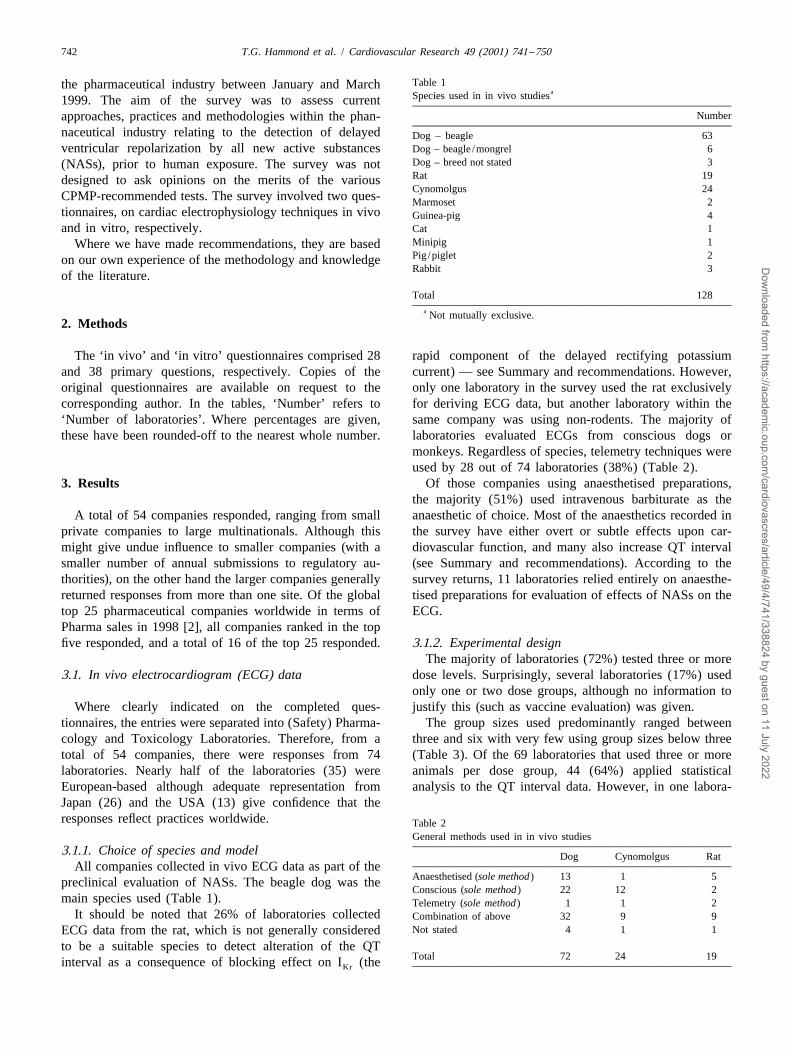
The use of formulae to correct QT for changes in heartrate (Fig. 6) was widespread (63% of laboratories), andTable 6
Derivation of QT interval frequently included statistical analysis of the derived data.
Number % Total
Lead II 58 78 3.1.5. Data interpretationMedian of all leads 2 3 There were 4 types of analysis used by laboratories toLongest measurement from all leads 4 5
assess drug-induced changes in QT (Table 8). However, ofOther 6 8possible concern is that 26% of laboratories did not appearNot stated 1 1to have predetermined criteria to assess changes in QT
Total 74 interval. For those that have established criteria variabilityin the method of assessment exist (Table 9). Thirteenlaboratories considered a 10% increase in QT interval to besignificant (i.e. biologically significant) with a wide rangeof ‘thresholds for concern’ amongst the remaining lab-oratories.
3.2. In vitro cardiac electrophysiological data
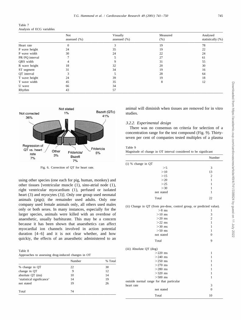
In contrast to the in vivo section of the survey, none ofthe companies in the survey performed in vitro cardiacelectrophysiology in more than one in-house facility, sothe term ‘company’ rather than ‘laboratory’ is used in thisFig. 4. Method of measurement of ECG parameters.section. Of the 54 companies polled, 30 (56%) conductedin vitro cardiac electrophysiology studies, and a further 5(9%) planned to do so (Fig. 7).
methods (manual or automatic), only 53% had acceptance / The data presented here are based on replies from the 30rejection criteria based on trace quality. The methodology companies that did carry out such studies at the time of theused to record ECG data has been changed significantly in survey. Of these 30 companies, 20 had the capability tothe last 5–10 years in 39 laboratories (i.e. 52%). This perform the studies in-house. Most of the 30 companiesincluded 17 of the 25 laboratories that have experienced were located in Europe (53%), with fewer in Japan/Asiaregulatory issues with compounds that prolong QT inter- and the USA (30% and 17%, respectively). The geographi-val. cal distribution of companies that had in-house capability
to carry out in vitro cardiac electrophysiology showedapproximately 20% to be located in the USA, with 40%each in Japan/Asia and Europe. The majority (67%) of the30 companies conducted these in vitro investigations priorto first administration to man.
3.2.1. Species and tissuesThere was a broad diversity between companies in the
choice of species and tissues used for these studies. Themost commonly used tissues were Purkinje fibre andpapillary muscle. The distribution of tissues and species is
Fig. 5. Validation of recording systems. shown in Fig. 8. A small number of companies reported
Dow
nloaded from https://academ
ic.oup.com/cardiovascres/article/49/4/741/338824 by guest on 11 July 2022
T.G. Hammond et al. / Cardiovascular Research 49 (2001) 741 –750 745
Table 7Analysis of ECG variables
Not Visually Measured Analysedassessed (%) assessed (%) (%) statistically (%)
Heart rate 0 3 19 78P wave height 24 35 19 22P wave width 30 24 22 24PR/PQ interval 7 5 27 61QRS width 4 9 31 55R wave height 18 32 20 30ST segment 31 34 19 16QT interval 3 5 28 64T wave height 24 39 19 18T wave width 45 35 8 12U wave 66 34Rhythm 43 57
animal will diminish when tissues are removed for in vitrostudies.
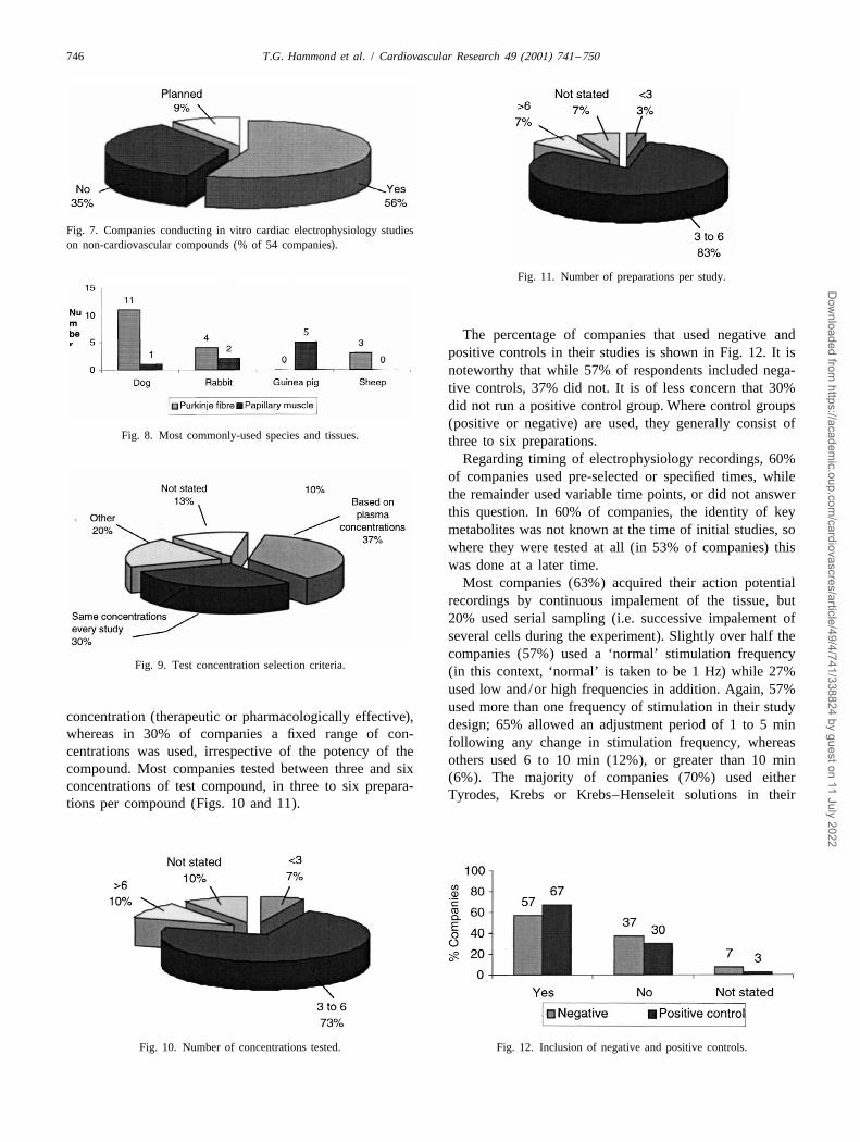
3.2.2. Experimental designThere was no consensus on criteria for selection of a
concentration range for the test compound (Fig. 9). Thirty-seven per cent of companies tested multiples of a plasma
Table 9Magnitude of change in OT interval considered to be significant
Number
(i) % change in QTFig. 6. Correction of QT for heart rate. .5 3
.10 13
.15 2
.20 1using other species (one each for pig, human, monkey) and
.25 1other tissues [ventricular muscle (1), sino-atrial node (1),
.30 1right ventricular myocardium (1), perfused or isolatednot stated 1
heart (3) and myocytes (3)]. Only one group used neonatalTotal 22animals (pigs); the remainder used adults. Only one
company used female animals only, all others used males (ii) Change in QT (from pre-dose, control group, or predicted value).only or both sexes. In many instances, especially for the .8 ms 1larger species, animals were killed with an overdose of .10 ms 3
.20 ms 2anaesthetic, usually barbiturate. This may be a concern
.22 ms 1because it has been shown that anaesthetics can affect
.30 ms 1myocardial ion channels involved in action potential
.50 ms 1duration [4–6] and it is not clear whether, and how not stated 0quickiy, the effects of an anaesthetic administered to an
Total 9
(iii) Absolute QT (dog)Table 8
.220 ms 1Approaches to assessing drug-induced changes in OT
.240 ms 1Number % Total .250 ms 1
.270 ms 1% change in QT 22 30
.280 ms 1change in QT 9 12
.320 ms 1absolute QT (ms) 10 14
.500 ms 1‘statistical significance’ 14 19
outside normal range for that particularnot stated 19 26
heart rate 3not stated 0
Total 74Total 10
Dow
nloaded from https://academ
ic.oup.com/cardiovascres/article/49/4/741/338824 by guest on 11 July 2022
746 T.G. Hammond et al. / Cardiovascular Research 49 (2001) 741 –750
Fig. 7. Companies conducting in vitro cardiac electrophysiology studieson non-cardiovascular compounds (% of 54 companies).
Fig. 11. Number of preparations per study.
The percentage of companies that used negative andpositive controls in their studies is shown in Fig. 12. It isnoteworthy that while 57% of respondents included nega-tive controls, 37% did not. It is of less concern that 30%did not run a positive control group. Where control groups(positive or negative) are used, they generally consist of
Fig. 8. Most commonly-used species and tissues. three to six preparations.Regarding timing of electrophysiology recordings, 60%
of companies used pre-selected or specified times, whilethe remainder used variable time points, or did not answerthis question. In 60% of companies, the identity of keymetabolites was not known at the time of initial studies, sowhere they were tested at all (in 53% of companies) thiswas done at a later time.
Most companies (63%) acquired their action potentialrecordings by continuous impalement of the tissue, but20% used serial sampling (i.e. successive impalement ofseveral cells during the experiment). Slightly over half thecompanies (57%) used a ‘normal’ stimulation frequency
Fig. 9. Test concentration selection criteria. (in this context, ‘normal’ is taken to be 1 Hz) while 27%used low and/or high frequencies in addition. Again, 57%used more than one frequency of stimulation in their study
concentration (therapeutic or pharmacologically effective), design; 65% allowed an adjustment period of 1 to 5 minwhereas in 30% of companies a fixed range of con-
following any change in stimulation frequency, whereascentrations was used, irrespective of the potency of the
others used 6 to 10 min (12%), or greater than 10 mincompound. Most companies tested between three and six
(6%). The majority of companies (70%) used eitherconcentrations of test compound, in three to six prepara-
Tyrodes, Krebs or Krebs–Henseleit solutions in theirtions per compound (Figs. 10 and 11).
Fig. 10. Number of concentrations tested. Fig. 12. Inclusion of negative and positive controls.
Dow
nloaded from https://academ
ic.oup.com/cardiovascres/article/49/4/741/338824 by guest on 11 July 2022
T.G. Hammond et al. / Cardiovascular Research 49 (2001) 741 –750 747
Table 10 tration) levels in order to ascertain any dose-responseControl APD ranges90 relationship. We consider this to be appropriate.Species /preparation APD range Number of90
companies 4.1. In vivo techniquesDog Purkinje fibre 229–350 ms 8Rabbit Purkinje fibre 220–275 ms 4 4.1.1. Choice of speciesGuinea-pig papillary muscle 186–359 ms 7 Generally, the species used were appropriate to detect
QT change of relevance to man, with beagle dogs beingthe most popular. Other species were used less frequently
studies, and the concentrations of potassium, magnesium, than the dog, but with the exception of the rat, there is nocalcium and glucose used reflected this. Most companies particular reason why other species such as pig /minipig,used a temperature over 358C, but some (13%) used a rabbit or guinea-pig would not be as predictive of effectstemperature below 358C. on ventricular repolarisation in man. However, the rat
should not be used to assess QT interval changes. Whereas3.2.3. Data analysis and interpretation the rat is of some value in cardiovascular assessments, this
Most of the key action potential parameters were species will probably not be susceptible to action potentialmeasured and/or analysed by the majority of companies, lengthening and QT prolongation as a consequence ofincluding, importantly, resting membrane potential, maxi- blockade of the rapid component of the delayed rectifyingmum upstroke velocity, amplitude and action potential potassium current (I ). Although the consensus view isKr
duration at 90% repolarisation (APD ). Probably the that ventricular tissue from this species does express this90
single most important parameter to be measured in order to ion channel protein [7,8], a dominant transient outwardassess activity at the I channel is APD . The range of current (I ) will rapidly restore membrane potential toKr 90 to
control values obtained for this parameter is shown in negative voltages, thus reducing the time for I activation.Kr
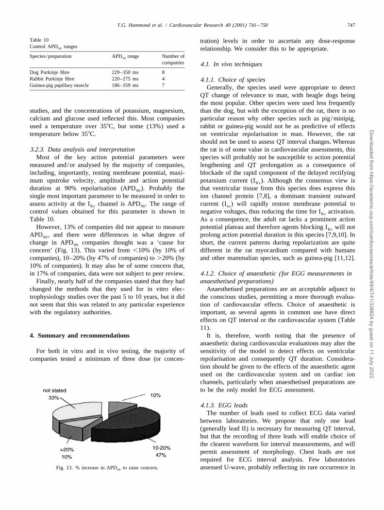
Table 10. As a consequence, the adult rat lacks a prominent actionHowever, 13% of companies did not appear to measure potential plateau and therefore agents blocking I will notKr
APD , and there were differences in what degree of prolong action potential duration in this species [7,9,10]. In90
change in APD companies thought was a ‘cause for short, the current patterns during repolarization are quite90
concern’ (Fig. 13). This varied from ,10% (by 10% of different in the rat myocardium compared with humanscompanies), 10–20% (by 47% of companies) to .20% (by and other mammalian species, such as guinea-pig [11,12].10% of companies). It may also be of some concern that,in 17% of companies, data were not subject to peer review. 4.1.2. Choice of anaesthetic ( for EGG measurements in
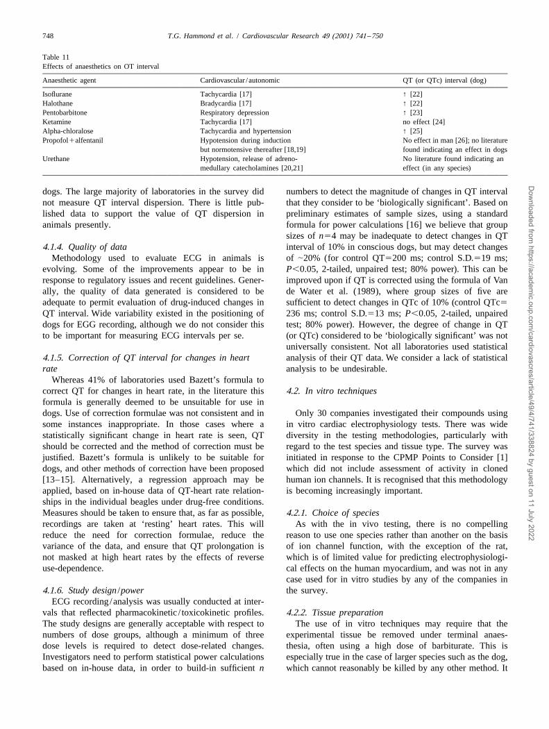
Finally, nearly half of the companies stated that they had anaesthetised preparations)changed the methods that they used for in vitro elec- Anaesthetised preparations are an acceptable adjunct totrophysiology studies over the past 5 to 10 years, but it did the conscious studies, permitting a more thorough evalua-not seem that this was related to any particular experience tion of cardiovascular effects. Choice of anaesthetic iswith the regulatory authorities. important, as several agents in common use have direct
effects on QT interval or the cardiovascular system (Table11).
4. Summary and recommendations It is, therefore, worth noting that the presence ofanaesthetic during cardiovascular evaluations may alter the
For both in vitro and in vivo testing, the majority of sensitivity of the model to detect effects on ventricularcompanies tested a minimum of three dose (or concen- repolarisation and consequently QT duration. Considera-
tion should be given to the effects of the anaesthetic agentused on the cardiovascular system and on cardiac ionchannels, particularly when anaesthetised preparations areto be the only model for ECG assessment.
4.1.3. EGG leadsThe number of leads used to collect ECG data varied
between laboratories. We propose that only one lead(generally lead II) is necessary for measuring QT interval,but that the recording of three leads will enable choice ofthe clearest waveform for interval measurements, and willpermit assessment of morphology. Chest leads are notrequired for ECG interval analysis. Few laboratories
Fig. 13. % increase in APD to raise concern. assessed U-wave, probably reflecting its rare occurrence in90
Dow
nloaded from https://academ
ic.oup.com/cardiovascres/article/49/4/741/338824 by guest on 11 July 2022
748 T.G. Hammond et al. / Cardiovascular Research 49 (2001) 741 –750
Table 11Effects of anaesthetics on OT interval
Anaesthetic agent Cardiovascular /autonomic QT (or QTc) interval (dog)
Isoflurane Tachycardia [17] ↑ [22]Halothane Bradycardia [17] ↑ [22]Pentobarbitone Respiratory depression ↑ [23]Ketamine Tachycardia [17] no effect [24]Alpha-chloralose Tachycardia and hypertension ↑ [25]Propofol1alfentanil Hypotension during induction No effect in man [26]; no literature
but normotensive thereafter [18,19] found indicating an effect in dogsUrethane Hypotension, release of adreno- No literature found indicating an
medullary catecholamines [20,21] effect (in any species)
dogs. The large majority of laboratories in the survey did numbers to detect the magnitude of changes in QT intervalnot measure QT interval dispersion. There is little pub- that they consider to be ‘biologically significant’. Based onlished data to support the value of QT dispersion in preliminary estimates of sample sizes, using a standardanimals presently. formula for power calculations [16] we believe that group
sizes of n54 may be inadequate to detect changes in QT4.1.4. Quality of data interval of 10% in conscious dogs, but may detect changes
Methodology used to evaluate ECG in animals is of |20% (for control QT5200 ms; control S.D.519 ms;evolving. Some of the improvements appear to be in P,0.05, 2-tailed, unpaired test; 80% power). This can beresponse to regulatory issues and recent guidelines. Gener- improved upon if QT is corrected using the formula of Vanally, the quality of data generated is considered to be de Water et al. (1989), where group sizes of five areadequate to permit evaluation of drug-induced changes in sufficient to detect changes in QTc of 10% (control QTc5
QT interval. Wide variability existed in the positioning of 236 ms; control S.D.513 ms; P,0.05, 2-tailed, unpaireddogs for EGG recording, although we do not consider this test; 80% power). However, the degree of change in QTto be important for measuring ECG intervals per se. (or QTc) considered to be ‘biologically significant’ was not
universally consistent. Not all laboratories used statistical4.1.5. Correction of QT interval for changes in heart analysis of their QT data. We consider a lack of statisticalrate analysis to be undesirable.
Whereas 41% of laboratories used Bazett’s formula tocorrect QT for changes in heart rate, in the literature this 4.2. In vitro techniquesformula is generally deemed to be unsuitable for use indogs. Use of correction formulae was not consistent and in Only 30 companies investigated their compounds usingsome instances inappropriate. In those cases where a in vitro cardiac electrophysiology tests. There was widestatistically significant change in heart rate is seen, QT diversity in the testing methodologies, particularly withshould be corrected and the method of correction must be regard to the test species and tissue type. The survey wasjustified. Bazett’s formula is unlikely to be suitable for initiated in response to the CPMP Points to Consider [1]dogs, and other methods of correction have been proposed which did not include assessment of activity in cloned[13–15]. Alternatively, a regression approach may be human ion channels. It is recognised that this methodologyapplied, based on in-house data of QT-heart rate relation- is becoming increasingly important.ships in the individual beagles under drug-free conditions.Measures should be taken to ensure that, as far as possible, 4.2.1. Choice of speciesrecordings are taken at ‘resting’ heart rates. This will As with the in vivo testing, there is no compellingreduce the need for correction formulae, reduce the reason to use one species rather than another on the basisvariance of the data, and ensure that QT prolongation is of ion channel function, with the exception of the rat,not masked at high heart rates by the effects of reverse which is of limited value for predicting electrophysiologi-use-dependence. cal effects on the human myocardium, and was not in any
case used for in vitro studies by any of the companies in4.1.6. Study design /power the survey.
ECG recording/analysis was usually conducted at inter-vals that reflected pharmacokinetic / toxicokinetic profiles. 4.2.2. Tissue preparationThe study designs are generally acceptable with respect to The use of in vitro techniques may require that thenumbers of dose groups, although a minimum of three experimental tissue be removed under terminal anaes-dose levels is required to detect dose-related changes. thesia, often using a high dose of barbiturate. This isInvestigators need to perform statistical power calculations especially true in the case of larger species such as the dog,based on in-house data, in order to build-in sufficient n which cannot reasonably be killed by any other method. It
Dow
nloaded from https://academ
ic.oup.com/cardiovascres/article/49/4/741/338824 by guest on 11 July 2022

T.G. Hammond et al. / Cardiovascular Research 49 (2001) 741 –750 749
is not established whether, or how rapidly, the effects of an 5. Conclusionsanaesthetic administered to the intact animal will wear offonce tissue has been removed into an organ bath and The survey provides a snapshot of the practice in thebathed with a physiological salt solution. Therefore, there pharmaceutical industry prior to any internationally-agreedwould have to be some degree of caution implicit in the consensus on the most effective and efficient approaches touse of tissue harvested from anaesthetised animals (and minimising the risk of QT prolongation by new drugs inthus from dogs). man. The survey has revealed a wide spectrum of ap-
proaches to addressing this issue in the pharmaceuticalindustry, and there is clearly a need for a degree of
4.2.3. Action potential characteristics consensus on ‘best practice’ in terms of methodologyWe recommend that APD is the most appropriate [27,28]. It must be stated, however, that for any given90
marker of action potential duration. However, careful methodology, the ‘majority view’ in the industry is notevaluation of drug effects on V and upstroke amplitude necessarily best practice.max
is as important as assessment of APD. As with QTprolongation, there was no consensus on the degree ofaction potential prolongation to cause concern. Acknowledgements
We would like to thank each of the 74 laboratories4.2.4. Quality of data (from 54 companies world-wide) who completed ques-
Seventeen percent of companies did not subject their tionnaires.data to peer review; this may give cause for concernregarding the quality of the data from the studies. Sixtypercent of companies did not carry out the studies to GLP; Referencesthis could be an issue if the information is used to supportadministration of the substance to humans. Thirty-three [1] Committee for Proprietary Medicinal Products (CPMP) Points topercent did not have acceptance criteria (e.g. action Consider: The assessment of the potential for QT interval prolonga-
tion by non-cardiovascular medicinal products. CPMP/986/96,potential morphology/characteristics) for whether they1997.would accept data from a tissue preparation. Sixty-three
[2] SCRIP No. 2426/27 April 7th /9th 1999, p. 17.percent did not correct for electrode drift over the time [3] Detweiler DK. The use of electrocardiography in toxicologicalcourse of the experiment. In four companies, experiments studies with beagle dogs. In: Balasz T, editor, Cardiac Toxicology,were carried out at a temperature of less than 358C, which Boca Raton, Florida: CRC Press, 1982, pp. 33–82.
[4] Gibbons SJ, Nunez-Hernandez R, Maze G, Harrison NL. Inhibitionwill markedly influence ion channel biophysics and kinet-of a fast inwardly rectifying potassium conductance by barbiturates.ics. Up to 13% of companies may not have been measuringAnesth Analg 1996;82:1242–1246.
or analysing APD ; 53% did not output their action90 [5] Martynyuk AE, Morey TE, Raatikainen MJ, Seubert CN, Dennispotential waveforms to a chart recorder; this would make DM. Ionic mechanisms mediating the differential effects ofproper assessment for early after-depolarisations difficult. methohexital and thiopental on action potential duration in guinea
pig and rabbit isolated ventricular myocytes. AnesthesiologyUse of continuous impalements rather than serial sampling1999;90:156–164.techniques to improve data accuracy and quality is rec-
[6] Shimizu W, McMahon B, Antzelevitch C. Sodium pentobarbitalommended. reduces transmural dispersion of repolarization and prevents Tor-
sades de Pointes in models of acquired and congenital long QTsyndrome. J Cardiovasc Electrophysiol 1999;10:154–164.
[7] Abrahamsson C, Palmer M, Ljung B, Duter G, Baarnhielm C,4.2.5. Rate dependenceCarlsson L, Danielsson B. Induction of rhythm abnormalities in theWhile a majority of those responding carried out somefetal rat heart. A tentative mechanism for the embryotoxic effect of
testing at a range of frequencies, it is doubtful that there is the class III antiarrhythmic agent almokalant. Cardiovasc Ressignificant added value in doing this during routine screen- 1994;28:337–344.ing. However, in the event of a drug effect at (say) 1 Hz, it [8] Wymore RS, Gintant GA, Wymore RT, Dixon JE, McKinnon D,
Cohen IS. Tissue and species distribution of mRNA for the I -likewould then be worthwhile exploring rate-dependence. Kr1K channel, erg. Circ Res 1997;80:261–268.
[9] Tande PM, Bjornstad H, Yang T, Refsum H. Rate-dependent ClassIII antiarrhythmic action, negative chronotropy, and positive inot-
4.2.6. Time-matched controls ropy of a novel I blocking drug, UK68,798: potent in guinea pigK
Thirty-seven percent of companies did not routinely but no effect in rat myocardium. J Cardiovasc Pharmacol1990;16:401–410.include a negative (time-matched) control group. Use of
[10] Dukes ID, Cleemann L, Morad M. Tedisamil blocks the transientappropriate time-matched negative (i.e. vehicle– perfused)1and delayed rectifier K currents in mammalian cardiac and glial
control preparations is recommended. Routine inclusion of cells. J Pharmacol Exp Ther 1990;254:560–569.a positive control (i.e. reference substance) would be [11] Langer GA. Interspecies variation in myocardial physiology: thedesirable. anomalous rat. Env Health Perspect 1978;26:175–179.
Dow
nloaded from https://academ
ic.oup.com/cardiovascres/article/49/4/741/338824 by guest on 11 July 2022

750 T.G. Hammond et al. / Cardiovascular Research 49 (2001) 741 –750
[12] Mitchell MR, Powell T, Terrar DA, Twist VW. Electrical activity influence of anaesthetics and heart rate response to pronethalol. Jand contractions in cells isolated from rat and guinea-pig ventricular Pharm Pharmacol 1975;27:70–71.muscle: a comparative study. J Physiol 1987;391:527–544. [22] Riley DC, Schmeling WT, al-Wathiqui JP, Warltier DC. Prolongation
[13] Van de Water A, Verheyen J, Xhonneux R. Reneman, R.S. (1989) of the QT interval by volatile anesthetics in chronically instrumentedAn improved method to correct the QT interval of the electro- dogs. Anesth Analg 1988;67:741–749.cardiogram for changes in heart rate. J Pharmacol Meth [23] Hunt GB, Ross DL. Comparison of effects of three anesthetic agents1989;22:207–217. on induction of ventricular tachycardia in a canine model of
[14] Oguchi Y, Hamlin RL. Duration of QT interval in clinically normal myocardial infarction. Circulation 1988;78:221–226.dogs. Am J Vet Res 1993;54:2145–2149. [24] Gonder JC, Gard EA, Lott NE. Electrocardiograms of nine species
[15] Matsunaga T, Mitsui T, Harada T, Inokuma M, Murano H, of nonhuman primates sedated with ketamine. Am J Vet ResShibutani Y. QT corrected for heart rate and relation between QT 1980;41:972–975.and RR intervals in beagle dogs. J Pharmacol Toxicol Methods [25] Schwartz JB, Herre JM. The electrophysiological effects of alpha-1997;38:201–209. chloralose anesthesia in the intact dog: (1) alone and (2) in
[16] Daly LE, Bourke GJ, McGilvray J. Interpretation and Uses of combination with verapamil. Pacing Clin ElectrophysiolMedical Statisitics, 4th ed, Oxford: Blackwell Scientific Publi- 1989;12:283–293.cations, 1991. [26] Michaloudis DG, Kanakoudis FS, Petrou AM, Konstantinou AS,
[17] Marshall BE, Longnecker DE. General anaesthetics. In: Goodman Pollard BJ. The effects of midazolam or propofol followed byGilman A, Rall TW, Nies AS, Taylor P, editors, Goodman and suxamethonium on the QT interval in humans. Eur J AnaesthesiolGilman’s ‘The pharmacological basis of therapeutics’, 8th ed, New 1996;13:364–368.York: Pergamon Press, 1990, pp. 285–310. [27] Cavero I, Mestre M, Guillon J-M, Heuillet E, Roach A. Preclinical
[18] Keegan RD, Greene SA. Cardiovascular effects of a continuous in vitro cardiac electrophysiology: a method of predicting ar-two-hour propofol infusion in dogs. Comparison with isoflurane rhythmogenic potential of antihistamines in humans? Drug Safetyanesthesia. Vet Surg 1993;22:537–543. 2000;21(Suppl. 1):19–31, and Panel Discussion pp. 81–87.
[19] Deryck YL, Brimouille S, Maggiorini M, de Canniere D, Naeije R. [28] Haverkamp W, Breithardt G, Camm AJ, Janse MJ, Rosen MR,Systemic vascular effects of isoflurane versus propofol anaesthesia Antzelevitch C, Escande D, Franz M, Malik M, Moss A, Shah R.in dogs. Anaesth Analg 1996;83:958–964. The potential for QT prolongation and proarrhythmia by non-
[20] Reinert H. Urethane hyperglycaemia and hypothalamic activation. antiarrhythmic drugs: clinical and regulatory implications. Cardio-Nature 1964;204:889–891. vasc Res 2000;47:219–233, published simultaneously in Eur Heart J
[21] Dean HG, Rylett PA. Plasma adrenaline concentration in rats: 2000;21:1216–1231.
Dow
nloaded from https://academ
ic.oup.com/cardiovascres/article/49/4/741/338824 by guest on 11 July 2022