Maternal depression and neurobehavior in newborns prenatally exposed to methamphetamine
Upload
independentCategory
view
2download
0
Methamphetamine Induces Dopamine D1 Receptor-Dependent Endoplasmic Reticulum Stress-RelatedMolecular Events in the Rat StriatumSubramaniam Jayanthi1, Michael T. McCoy1, Genevieve Beauvais1, Bruce Ladenheim1, Kristi Gilmore1,
William Wood, III2, Kevin Becker2, Jean Lud Cadet1*
1 Molecular Neuropsychiatry Research Branch, National Institute of Drug Abuse, National Institutes of Health (NIH)/Department of Health and Human Services (DHHS),
Intramural Research Program, Baltimore, Maryland, United States of America, 2 Gene Expression and Genomics Unit, National Institute of Aging, National Institutes of
Health (NIH)/Department of Health and Human Services (DHHS), Intramural Research Program, Baltimore, Maryland, United States of America
Abstract
Methamphetamine (METH) is an illicit toxic psychostimulant which is widely abused. Its toxic effects depend on the release ofexcessive levels of dopamine (DA) that activates striatal DA receptors. Inhibition of DA-mediated neurotransmission by the DAD1 receptor antagonist, SCH23390, protects against METH-induced neuronal apoptosis. The initial purpose of the presentstudy was to investigate, using microarray analyses, the influence of SCH23390 on transcriptional responses in the rat striatumcaused by a single METH injection at 2 and 4 hours after drug administration. We identified 545 out of a total of 22,227 genesas METH-responsive. These include genes which are involved in apoptotic pathways, endoplasmic reticulum (ER) stress, and intranscription regulation, among others. Of these, a total of 172 genes showed SCH23390-induced inhibition of METH-mediated changes. Among these SCH23390-responsive genes were several genes that are regulated during ER stress, namelyATF3, HSP27, Hmox1, HSP40, and CHOP/Gadd153. The secondary goal of the study was to investigate the role of DA D1receptor stimulation on the expression of genes that participate in ER stress-mediated molecular events. We thus usedquantitative PCR to confirm changes in the METH-responsive ER genes identified by the microarray analyses. We alsomeasured the expression of these genes and of ATF4, ATF6, BiP/GRP78, and of GADD34 over a more extended time course.SCH23390 attenuated or blocked METH-induced increases in the expression of the majority of these genes. Western blotanalysis revealed METH-induced increases in the expression of the antioxidant protein, Hmox1, which lasted for about24 hours after the METH injection. Additionally, METH caused DA D1 receptor-dependent transit of the Hmox1 regulatorprotein, Nrf2, from cytosolic into nuclear fractions where the protein exerts its regulatory functions. When taken together,these findings indicate that SCH23390 can provide protection against neuronal apoptosis by inhibiting METH-mediated DA D1receptor-mediated ER stress in the rat striatum. Our data also suggest that METH-induced toxicity might be a useful model todissect molecular mechanisms involved in ER stress-dependent events in the rodent brain.
Citation: Jayanthi S, McCoy MT, Beauvais G, Ladenheim B, Gilmore K, et al. (2009) Methamphetamine Induces Dopamine D1 Receptor-Dependent EndoplasmicReticulum Stress-Related Molecular Events in the Rat Striatum. PLoS ONE 4(6): e6092. doi:10.1371/journal.pone.0006092
Editor: Bernhard T. Baune, James Cook University, Australia
Received April 17, 2009; Accepted May 27, 2009; Published June 30, 2009
This is an open-access article distributed under the terms of the Creative Commons Public Domain declaration which stipulates that, once placed in the publicdomain, this work may be freely reproduced, distributed, transmitted, modified, built upon, or otherwise used by anyone for any lawful purpose.
Funding: This research was supported financially by the intramural research programs of DHHS/NIH/NIDA and DHHS/NIH/NIA. The funders had no role in studydesign, data collection and analysis, decision to publish, or preparation of the manuscript.
Competing Interests: The authors have declared that no competing interests exist.
* E-mail: [email protected]
Introduction
Methamphetamine (METH) abuse is a complex neuropsychi-
atric disorder that affects a significant number of adolescents and
adults worldwide. METH addicts often use large quantities of the
drug [1,2] and can suffer for drug-induced psychosis, withdrawal-
associated depression, coma and/or death after taking accidental
overdoses [3]. Neuroimaging studies have also revealed a number
of abnormalities in the brains of these patients. These include loss
of striatal dopamine (DA) transporters and of serotonin transport-
ers [4,5] and evidence of reactive microgliosis [6]. Previous post-
mortem studies are in agreement with some of those results [7].
Moreover, METH can cause degeneration of monoaminergic
systems and neuronal apoptosis in various brain regions including
the rodent striatum [8–10]. METH-induced cell death is
dependent, in part, on activation of endoplasmic reticulum (ER)-
dependent death pathways [11].
The ER is an intracellular organelle which is involved in diverse
arrays of cellular functions. The ER is very densely packed with
enzymes that are involved in quality control of protein synthesis
and post-translational modification including folding of proteins
[12,13]. Malfunctions in these processes result in misfolded and/or
unfolded proteins that can accumulate in the ER, with consequent
activation of compensatory reactions such as the unfolded protein
response [14]. If these compensatory mechanisms fail to restore
cellular homeostasis, cell death ensues via activation of ER-
dependent apoptosis [15,16]. The signaling pathways that mediate
compensatory mechanisms and cell death during ER stress are
regulated by the ER-resident chaperone, BiP/GRP78 (Binding
Immunoglobulin Protein/Glucose Response Protein 78), which is
bound to three ER transmembrane sensor proteins, namely,
activating transcription factor 6 (ATF6), inositol requiring enzyme
1a (Ire1a), and protein kinase R-like ER kinase (PERK) [17].
During ER stress, BiP dissociates from these proteins and triggers a
PLoS ONE | www.plosone.org 1 June 2009 | Volume 4 | Issue 6 | e6092
series of events that lead to oligomerization of IRE1a [18] and of
PERK [19] as well as cleavage and translocation of ATF6a into
the nucleus [20]. Ire1a has site-specific endoribonuclease (RNase)
activity which splices and activates X-box Binding Protein-1
(XBP1) mRNA and leads to increased production of XBP1 protein
[18] which regulates the transcription of genes that participate in
the maintenance of protein folding and ER-associated degradation
(ERAD) [21]. PERK oligomerization leads to phosphorylation of
eukaryotic translation initiation factor 2a (eIF2a) and inhibition of
global translation [22], but increased activating transcription
factor 4 (ATF4) translation [23]. Periods of excessive ER stress also
cause ATF4-, ATF6, and XBP1-dependent up-regulation of the
pro-apoptotic transcription factor, C/EBP homologous protein
(CHOP) [24] which appears to mediate cell death, in part, by
causing up-regulation of pro-death members of Bcl-2 family
proteins such as BIM [25]. The accumulated evidence supports
the involvement of ER stress and related molecular events in
neurodegenerative events [26] including METH-induced neuro-
nal apoptosis [11].
The present study was therefore undertaken to document if
blockade of DA D1 receptor function might influence METH-
induced gene expression because of the evidence that the DA D1
receptor antagonist, SCH23390, can protect against METH
toxicity in the rat striatum [9,27]. Thus, the purpose of this paper
is to report that microarray and quantitative PCR analyses
detected METH-induced increases in the expression of activating
transcription factor 3 (ATF3), heat shock protein 27 (HSP27),
heme oxygenase 1 (Hmox1), heat shock protein 40 (HSP40),
CHOP, and BIP that are known ER stress-responsive genes. The
METH-induced changes were attenuated, to variable degrees, by
pretreatment with SCH23390. Western blot analysis also docu-
mented changes in Hmox1 and nuclear factor erythroid 2-related
factor 2 (Nrf2) proteins which are both involved in cellular
protective mechanisms. These results demonstrate that METH-
induced neurotoxicity occur, in part, through DA D1-mediated
and ER-dependent molecular events.
Results
METH- and SCH23390-induced differential geneexpression in the rat striatum
To identify METH-regulated genes, RNA samples from striata of
rats treated with saline, METH, SCH23390, or combination of these
drugs were subjected to microarray analysis using Rat Illumina
arrays which allow for query of 22,227 transcripts (see Tables S1 and
S2 in Supplemental data for the complete set of array data obtained
from animals euthanized at 2 and 4 hours after METH treatment,
respectively). Figure 1 shows the effects of SCH23390 on METH-
induced gene expression in the rat striatum. The Volcano plots in
figures 1A and 1B show that, in comparison to the control group,
METH caused changes in the expression of 300 and 316 genes at 2
and 4 hours, respectively, after injection of the drug. Because 71
genes were significantly changed at both time points, this left a
combined total of 545 genes that were identified as METH-
responsive at these times. Figures 1C and 1D show the Volcano
comparisons between the SCH23390 and the METH+SCH23390
groups at 2 and 4 hours, respectively. There were a total of 271 and
293 genes that were differentially expressed at those times. The Venn
diagram in Figure 1E reveals that the expression of 173 METH-
responsive genes was influenced by pre-treatment with SCH23390.
Table S3 in Supplemental data summarizes the fold-changes and the
functional classifications for these genes. The SCH23390-resistant
genes are listed in Table S4 in Supplemental data. The METH-
responsive genes participate in apoptotic pathways, control of cell
adhesion, regulation of cell cycle, ER stress, signal transduction, and
transcription regulation (see Tables S3 and S4 for more details).
Because METH has previously been shown to cause neuronal
apoptosis, in part, by activating the ER-dependent death pathway
[11] and because SCH23390 can protect against METH-induced
toxicity [9], we decided to focus more attention on the effects of
METH and SCH23390 on genes that are involved in responses to
ER stress. Table 1 shows a total of 27 ER-related genes that were
either induced (23) or downregulated (4) by the METH injection.
SCH23390 pretreatment caused inhibition of METH-induced
changes in 15 genes; these are listed in bold in the table. These
include Armet, ATF3, BiP, CHOP, Dnajb1 (DnaJ (Hsp40)
homolog, subfamily B, member 1), Hmox1, HSP27, among
others.
SCH23390 attenuates METH-induced activation of ATF6-regulated gene expression
ER stress is associated with unfolded protein response activation
which is associated with the production of protective chaperones
and antioxidant enzymes [28]. However, excessive ER stress can
cause cellular demise via the activation of the ER-dependent
apoptotic pathway [16,24]. The activation of these protective and
pro-apoptotic genes is regulated by BiP interaction with ATF6,
IRE1a, and PERK [14,28]. ER stress interferes with these
interactions and leads to the activation of ER signaling pathways
[14,28]. Figure 2 shows the effects of METH on some genes that
are involved in the ATF6-dependent pathway [29]. ATF6 mRNA
was induced by METH in a DA D1 receptor-dependent fashion at
both 4 and 8 hours after drug administration (Fig. 2A). Consistent
with the array data, METH caused significant increases in BiP
mRNA, with the peak changes being observed at 2 hours and
normalization by 16 hours (Fig. 2B). SCH23390 was only able to
suppress the effects of METH at 8 hours post-drug. This is
consistent with the array data which did not identify BiP as a
SCH23390-sensitive gene at 2 and 4 hours. HSP27 mRNA also
showed marked biphasic DA D1 receptor-mediated increases after
the METH injection, reaching ,40-fold at 2 hours after METH,
decreasing to 5-fold at 4 hours and then reaching another smaller
peak at 8 hours after METH (Fig. 2F). Other genes that we tested,
namely ER degradation enhancer, mannosidase alpha-like 1
(EDEM1) (Fig. 2C), homocysteine-inducible, endoplasmic reticu-
lum stress-inducible, ubiquitin-like domain member 1 (Herpud1)
(Fig. 2D), and endoplasmic reticulum protein 72 (ERp72) (Fig. 2E),
show transient or no changes after the METH injection.
Specifically, there were no significant changes in EDEM1
expression in any of the treatment groups at any of the times
studied. There were, however, significant increases in Herpud1
mRNA in the striatum of animals sacrificed at 4 hours after the
METH injection; these increases were not significantly attenuated
by the SCH23390 pretreatment (Fig. 2D). In addition, the METH
injection caused DA D1 receptor-dependent increases in ERp72/
Pdia4 expression measured only at 8 hours after injection of the
psychostimulant.
Effects of METH and SCH23390 on XBP1-dependent geneexpression
The transcriptional activity of XBP1 is regulated by IRE1a-
dependent splicing of XBP1 mRNA and generation of active
XBP1 protein [18,30]. This is followed by increases in XBP1-
dependent increases in the expression of ERAD-related mRNAs
[21,31]. In order to determine if the drug causes activation of
IRE1a signaling, we measured XBP1 mRNA splicing at different
times after the METH injection. Figures 3A and 3B show that the
METH & DA D1-Linked ER Stress
PLoS ONE | www.plosone.org 2 June 2009 | Volume 4 | Issue 6 | e6092

Figure 1. METH- and SCH23390-induced differential gene expression in the rat striatum. (A and B) Volcano plots showing the effects ofMETH on striatal gene expression. Comparison of METH-treated rats to control rats revealed that METH caused changes in (A) 300 genes at 2 h afterthe METH injection, of which 189 were induced and 111 were repressed, and (B) 316 genes at the 4 h time-point, of which 133 were induced and 183were repressed. The Volcano plots showing the effects of SCH23390 pre-treatment on METH-induced striatal gene expression are shown in (C) and(D). These comparisons revealed that these two groups show differences in the expression (C) of 271 genes at the 2 h time-point, the expression of184 of which was inhibited and of 87 of which was potentiated by SCH23390 (D) of 293 genes at the 4 h time-point, the expression of 175 of whichwas inhibited and of 118 of which was potentiated by SCH23390. (E) The Venn diagram shows the overlap of genes that are common between thetwo different comparisons: M vs. C and M vs. M+S. Abbreviations are C, control; S, SCH23390; M, methamphetamine. To be identified as SCH23390-responsive, the genes must have passed the following criteria: greater or less than 1.7-fold changes at p,0.05.The 173 METH-responsive genes thatwere influenced by pre-treatment with SCH23390 are listed in Table S3 and the SCH23390-resistant genes are listed in Table S4.doi:10.1371/journal.pone.0006092.g001
METH & DA D1-Linked ER Stress
PLoS ONE | www.plosone.org 3 June 2009 | Volume 4 | Issue 6 | e6092

METH injection resulted in increases in XBP1 splicing and XBP1
transcription at various times after the drug injection. The
METH-induced XBP1 mRNA splicing was inhibited by
SCH23390 pretreatment. However, METH-induced increases in
unspliced XBP1 mRNA were not significantly attenuated by the
SCH23390 pretreatment, suggesting that other METH-mediated
events, such as oxidative stress [1], might be responsible for these
changes. Quantification of three XBP1 target genes revealed that
METH administration is associated with increases in the anti-
death gene, defender against cell death 1 (Dad1) (Fig. 3C) [32]
which is a subunit of the mammalian oligosaccharyltransferase
[33–35]. The METH-induced changes were attenuated by
pretreatment with SCH23390. The expression of DnaJ (Hsp40)
homolog, subfamily C, member 3 (Dnajc3/p58IPK), a member of
the Hsp40 family of chaperones [36–38] which can control PERK
eIF2a kinase activity during ER stress [39] and protect the stressed
ER [40], was also induced by METH in a DA D1 receptor-
sensitive fashion (Fig. 3D). There were also significant DA D1
receptor-mediated increases in the expression of vascular endo-
thelial growth factor (VEGF) (Fig. 3E) which is a neuroprotective
factor [41,42] that protects against ER stress-dependent neurode-
generation [43].
METH-mediated induction of genes of the ER PERK-dependent pathway
As mentioned above, ER stress is also associated with activation
of the PERK-dependent pathway [22,23,44]. Figure 4 shows the
effects of METH and of SCH23390 on the expression of 4 genes
that participate in the PERK-regulated pathway [45]. As shown in
Figs 4A and 4B, METH caused significant DA D1 receptor-
dependent increases in ATF4 protein which occurred as early as
2 hours after the METH injection. These results are consistent
with the fact that ER stress is known to induce early increases in
ATF4 translation [46]. Injection of SCH23390 significantly
prevented the METH-induced increases in ATF4 protein levels
(Fig. 4B). There were also METH-induced increases in ATF4
Table 1. METH administration causes changes in ER stress-related genes.
Accession No. Gene Symbol Time
2 h 2 h 4 h 4 h
M/C (M+S)/M M/C (M+S)/M
NM_012912 Atf3 166.534 223.355 2.325 21.959
NM_031970 Hspb1 (HSP27) 27.016 24.723 9.659 27.562
NM_012580 Hmox1 19.956 26.583 3.173 22.739
XM_341663 Dnajb1 p 8.458 22.552 2.597 22.141
NM_022934 Dnaja1 (HSP40) 2.975 21.659 2.262 22.052
XM_218543 Plekhf1 2.942 21.116 1.618 21.307
XM_215722 Dnajb4 p 2.829 21.448 2.092 21.504
NM_080906 Ddit4 (REDD1) 2.678 1.142 1.455 21.195
NM_024134 Ddit3 (CHOP, Gadd153) 2.557 21.340 2.301 22.483
XM_223680 Ahsa2 p 2.543 21.083 2.997 22.488
NM_053612 Hspb8 (HSP22) 2.378 21.671 1.889 21.712
NM_012935 Cryab 2.295 1.331 1.806 21.716
XM_233767 Dnajb5 p 2.003 21.715 2.016 22.049
NM_175761 Hspca (HSP90) 1.908 21.380 1.604 21.585
XM_217147 Dnaja4 1.862 21.490 1.647 21.651
NM_022522 Casp2 1.722 21.481 1.226 21.292
NM_031122 St13 1.550 21.083 1.767 21.773
NM_013083 Hspa5 (BIP, GRP78) 1.314 1.304 2.563 22.042
NM_053523 Herpud1 (Herp) 1.304 1.332 2.055 21.931
XM_236614 Armet p 1.061 1.396 2.063 21.882
NM_022232 Dnajc3 1.185 1.085 1.726 21.650
NM_001004210 Xbp1 1.154 1.207 1.723 21.608
NM_053849 Pdia4 21.141 1.460 1.781 21.765
XM_576401 Phldb1 21.200 21.222 21.944 1.226
NM_152790 Carhsp1 21.217 21.014 21.833 1.337
XM_234909 Plekhj1 21.430 21.040 22.060 1.695
XM_230610 Hspa12b p 23.243 2.005 21.233 1.155
The data in this table were generated from the comparisons between the METH and control groups at 2 and 4 h according to the criteria used in the analyses (greateror less than 1.7-fold and p,0.01). The values represent fold changes between the specified groups (n = 6). The genes are listed in descending order according to theMETH-induced fold changes at the 2 h time point. The bolded genes are genes whose METH-induced expression was significantly inhibited by the SCH23390pretreatment.doi:10.1371/journal.pone.0006092.t001
METH & DA D1-Linked ER Stress
PLoS ONE | www.plosone.org 4 June 2009 | Volume 4 | Issue 6 | e6092

mRNA which occurred later than the changes in ATF4 protein,
with SCH23390 inhibiting these changes (Fig. 4C). As stated
above, ATF4 is a member of the ATF/CREB family of
transcription factors which is upregulated during ER stress
[46] and which controls the expression of ATF3 and CHOP
[47,48]. Consistent with the array, we observed that METH
caused substantial increases in ATF3 mRNA (Fig. 4D). These
increases were biphasic, reaching ,45-fold at 2 hours, down to
Figure 2. Effects of METH and SCH23390 on the expression of ATF6 and some ATF6-regulated genes. METH administration caused earlyinduction of (A) ATF6 (B) BiP and (F) Hspb1/HSP27 transcripts. The METH-induced changes in the expression of these genes were normalized by pre-treatment of SCH-23390. (D, E) Herp/Herpud1 and ERp72/Pdia4 showed only transient changes at 4 h and 8 h respectively, after METH treatmentwhereas (C) EDEM1 was not affected. Data were obtained from RNA isolated from six animals per group and determined individually. The levels ofmRNA were normalized to 18 s rRNA levels. Values represent means6SEM of fold changes relative to the controls. Statistical significance wasdetermined by ANOVA followed by protected least-squares difference (PLSD). Key to statistics: * = M vs. C; # = M vs. M+S; *, # p,0.05; **, ##p,0.01; ***, ### p,0.001.doi:10.1371/journal.pone.0006092.g002
METH & DA D1-Linked ER Stress
PLoS ONE | www.plosone.org 5 June 2009 | Volume 4 | Issue 6 | e6092

3-fold at 4 hours, and back up to 18-fold at 8 hours after the
METH injection. Expression of ATF3 protein was also induced
by METH in a DA D1 receptor-dependent fashion (Figs. 4E and
4F). The levels of CHOP/Gadd153 mRNA also showed
significant DA D1 receptor-sensitive METH-mediated increases
(Fig. 4G). The increases in CHOP expression are consistent with
previous results obtained in the striata of METH-treated mice
[11]. CHOP is known to participate in stress-induced apoptosis
and to regulate the expression of several genes including growth
arrest and DNA-damage-inducible 34 (GADD34) [49]. As
Figure 3. Effects of METH and of SCH23390 on XBP1 target genes. (A) Administration of METH caused splicing of XBP1 mRNA. XBP1 mRNAfragments were amplified by RT-PCR and PCR fragments were separated on 2% agarose gel. Arrowhead indicates spliced XBP1 (479 bp). Thesequences of primers used to amplify the XBP1 transcripts are provided in Supplemental Table 1. (B) Levels of unspliced XBP1 mRNA were induced byMETH at 4 and 8 h after the drug injection. The induced XBP1 mRNA was not significantly inhibited by SCH23390 pretreatment. METH administrationalso caused increases in the expression of the XBP1 target genes: (C) DAD1, (D) p58IPK, and (E) VEGFa. The METH-induced changes in these threetranscripts were attenuated by pretreatment with SCH23390. Statistical analyses and key to statistics are as described in Figure 2.doi:10.1371/journal.pone.0006092.g003
METH & DA D1-Linked ER Stress
PLoS ONE | www.plosone.org 6 June 2009 | Volume 4 | Issue 6 | e6092

Figure 4. SCH23390 pretreatment causes inhibition of METH-induced changes in ER PERK-dependent gene expression. (A, B) METHcaused significant DA D1 receptor-dependent increases in ATF4 protein. (C) METH-induced changes in the levels of ATF4 mRNA and their inhibitionby SCH23390. (D) Effects of METH caused on ATF3 mRNA; (E, F) ATF3 protein expression; (G, H) Effect of METH on the levels of CHOP/Gadd153 andGadd34 mRNA. Statistical analyses and key to statistics are as described in Figure 2.doi:10.1371/journal.pone.0006092.g004
METH & DA D1-Linked ER Stress
PLoS ONE | www.plosone.org 7 June 2009 | Volume 4 | Issue 6 | e6092
observed in Fig. 4H, the levels of GADD34 mRNA, a protein
that dephosphorylates eIF2a [50], also showed substantial
METH-induced increases that are suppressed by SCH23390 in
a time-dependent fashion.
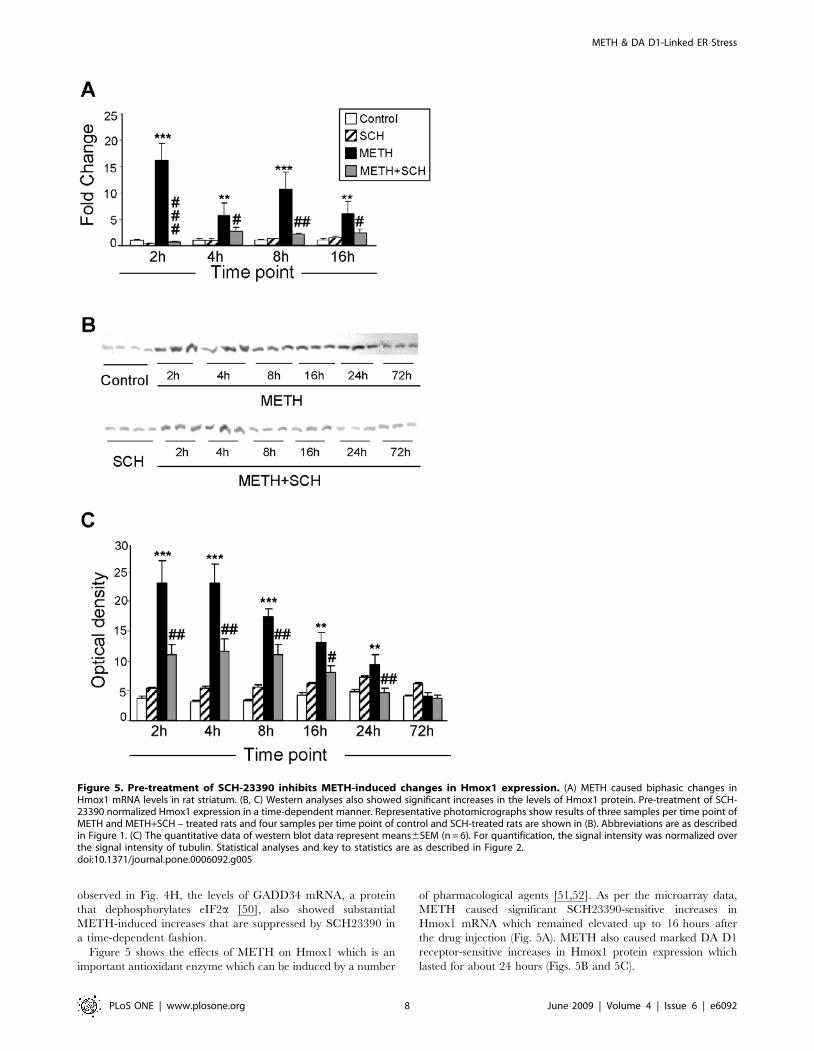
Figure 5 shows the effects of METH on Hmox1 which is an
important antioxidant enzyme which can be induced by a number
of pharmacological agents [51,52]. As per the microarray data,
METH caused significant SCH23390-sensitive increases in
Hmox1 mRNA which remained elevated up to 16 hours after
the drug injection (Fig. 5A). METH also caused marked DA D1
receptor-sensitive increases in Hmox1 protein expression which
lasted for about 24 hours (Figs. 5B and 5C).
Figure 5. Pre-treatment of SCH-23390 inhibits METH-induced changes in Hmox1 expression. (A) METH caused biphasic changes inHmox1 mRNA levels in rat striatum. (B, C) Western analyses also showed significant increases in the levels of Hmox1 protein. Pre-treatment of SCH-23390 normalized Hmox1 expression in a time-dependent manner. Representative photomicrographs show results of three samples per time point ofMETH and METH+SCH – treated rats and four samples per time point of control and SCH-treated rats are shown in (B). Abbreviations are as describedin Figure 1. (C) The quantitative data of western blot data represent means6SEM (n = 6). For quantification, the signal intensity was normalized overthe signal intensity of tubulin. Statistical analyses and key to statistics are as described in Figure 2.doi:10.1371/journal.pone.0006092.g005
METH & DA D1-Linked ER Stress
PLoS ONE | www.plosone.org 8 June 2009 | Volume 4 | Issue 6 | e6092
Because Hmox1 is regulated, during ER stress, via PERK-
mediated Nrf2 phosphorylation and its transit from the cytosol
into the nucleus [53–55], we also tested the possibility that METH
might cause changes in Nrf2 mRNA and protein levels. Figure 6A
shows that METH caused only transient DA D1 receptor-
dependent increases in Nrf2 mRNA levels. In contrast, the
METH injection was associated with decreases in the levels of
Nrf2 protein in the striatal cytosolic fractions and its prolonged
accumulation in the nuclear fractions (Figs. 6B–6E). The
quantitative data in Fig. 6C show that the levels of cytosolic
Nrf2 protein in the control group of animals remained relatively
constant while there were significant decreases in cytosolic Nrf2
levels in the SCH23390 and METH-treated rats. The changes in
METH-induced accumulation of Nrf2 protein in nuclear fractions
occurred in a biphasic fashion, with initial changes observed as
early as 2 hours after the drug but with some reversal towards
normalization by 8 hours. This was followed by another phase of
increases at 16 hours lasting up to 72 hours after the injection of
METH (Fig. 6E). The METH-induced increases in nuclear Nrf2
were significantly inhibited by SCH23390. However, there were
further decreases in cytosolic Nrf2 levels in the rats treated with
both METH and SCH23390, decreases that were significantly
different from those observed in the METH group (Fig. 6C).
These changes are not due to increased transit into the nucleus
since SCH3390 blocked the effects of METH on Nrf2 accumu-
lation in the nucleus (compare Figs. 6C and 6D). These
observations are consistent with the observation that SCH23390
caused decreases in cytosolic Nrf2 (Fig. 6C) without causing
increases in nuclear Nrf2 (Fig. 6E).
Discussion
The ER is an essential organelle which is responsible for post-
translational processing and proper folding of recently synthe-
sized proteins that participate in secretory pathways and are
membrane bound [12,13]. The ER can be stressed by
dysfunctions in calcium homeostasis, oxidative stress, and
improper protein folding [14,16]. These abnormalities trigger
ER stress-dependent events which consist of the production of
chaperone proteins that attempt to prevent cellular demise [14]
and/or activation of cell death cascades when ER stress is too
overwhelming [16,56]. In the present study, we have found that
the illicit neurotoxin, METH, can cause the activation of ER
stress-mediated gene expression. Our microarray analyses
identified several genes whose transcript levels were significantly
increased early after injection of the drug. METH administration
also caused significant activation of genes and proteins that are
downstream of the ATF6, IRE1alpha, and PERK ER signaling
pathways [14,16]. These include both protective and pro-death
transcripts. Quantitative PCR confirmed these changes and also
provided more detailed time courses for these changes. We also
found that METH-mediated activation of ER stress-dependent
events is dependent, in part, on the stimulation of DA D1
receptors that are very abundant in the rat striatum [57]. These
observations are consistent with reports that stimulation of D1
receptors by DA can cause death of neuroblastoma cells [58] and
of striatal neurons [59] in cultures.
Our findings that METH can cause ER stress in the rat striatum
are consistent with our prior observations that METH can cause
apoptosis of mouse striatal cells via cross-talks between ER- and
mitochondria-dependent death pathways [11]. Although we have
previously shown METH-induced increases in CHOP and BiP
expression in the mouse striatum [11], the present data provide a
more detailed picture of METH-responsive ER stress genes in the
rat striatum. As previously reported in the mouse striatum, METH
administration caused significant increases in the transcription of
CHOP which is known to be involved in pathways that lead to
neuronal apoptosis [60]. The present results also extend our
previous observations by showing that the increases in CHOP
expression are dependent on stimulation of striatal DA D1
receptors. The METH-induced increases in CHOP expression are
also preceded by significant increases in the levels of ATF4
protein, a member of the ATF/CREB class of transcription
factors, which is a regulator of CHOP expression during ER stress
[48]. During ER stress, activation of the PERK-dependent
pathway leads to phosphorylation of eIF2a [22,23,61]. Phosphor-
ylation of this protein leads to global inhibition of translation,
which results in the reduction of ER protein load. In addition,
eIF2a phosphorylation leads to increased translation of a number
of mRNAs [62], one of which is ATF4 [23,44,47]. Our
observations of METH-induced increases in the expression of
ATF4 protein and transcript are consistent with prior reports that
this transcription factor is induced under conditions of ER and
oxidative stresses [44,63]. The findings that METH caused
increases in ATF4 protein earlier than the changes in ATF4
mRNA levels are consistent with ER stress-induced increased
translation of ATF4 [23,47,50]. During ER stress, ATF4 also
regulates the expression of ATF3 [48] which is also increased after
METH administration in a DA D1 receptor-dependent fashion.
Of note, ATF3 is induced by an array of cellular stresses [64] and
by administration of the METH analog, amphetamine [65].
Interestingly, ATF3 is also thought to be an important player in
the integral stress response (ISR) [23]. It needs to be mentioned
that adenoviral over-expression of ATF3 in PC12 cells prevents c-
Jun-N-Terminal kinase (JNK)-dependent apoptosis via increased
expression of the chaperone, HSP27 [66]. This report is consistent
with our demonstration that HSP27 is significantly induced by
METH. When taken together, those observations suggest that
activation of the PERK-ATF4-ATF3 ER stress cascade might be,
in part, responsible for adaptive responses of striatal cells to
METH-induced oxidative stress and for counterbalancing the pro-
apoptotic effects of CHOP induction by the drug.
Our results also suggest that METH treatment can activate
IRE1a-dependent transduction mechanisms since the drug
induces DA D1 receptor-mediated XBP1 mRNA splicing, which
is mediated by IRE1a –associated RNase activity [18]. Several
targets have been identified for this transcription factor, among
which are Dad1, Dnajc3/p58IPK, and VEGF, all of which show
DA D1 receptor-dependent METH-induced upregulation (see
Fig. 3). The METH-induced increases in their expression suggest
that the injection of toxic doses of the drug is associated with the
triggering of protective mechanisms in an attempt to prevent
METH-induced neuronal apoptosis. This statement is supported
by the evidence that Dad1 is an inhibitor of apoptosis [67], whose
absence is associated with developmental abnormalities in Dad-1
null mice [68]. In addition, p58IPK, first identified as a 58-kDa
protein that inhibits PKR in influenza virus-infected kidney cells
[37], can complex with PKR and PERK and inhibit their
autophosphorylation [39] as well as protect the stressed ER [40].
Thus, the present observation of METH-induced Dnajc3/p58IPK
does suggest its participation in a coordinated response to
attenuate or prevent METH-mediated deleterious effects on the
ER in conjunction with VEGF which has been shown to protect
neurons against ischemia [41,69] glutamate toxicity [70] and ER
stress [43]. VEGF appears to exert its protective effects against
neuronal apoptosis, in part, by both caspase-dependent [71] and
caspase-independent [72] mechanisms. Although similar mecha-
nisms have been identified in METH-induced neuronal apoptosis
METH & DA D1-Linked ER Stress
PLoS ONE | www.plosone.org 9 June 2009 | Volume 4 | Issue 6 | e6092
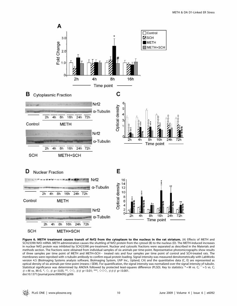
Figure 6. METH treatment causes transit of Nrf2 from the cytoplasm to the nucleus in the rat striatum. (A) Effects of METH andSCH23390 Nrf2 mRNA. METH administration causes the shuttling of Nrf2 protein from the cytosol (B) to the nucleus (D). The METH-induced increasesin nuclear Nrf2 protein was inhibited by SCH23390 pre-treatment. Nuclear and cytosolic fractions were separated as described in the Materials andmethods section. The fractions were obtained from individual samples of six animals per time-point. Representative photomicrographs show resultsof three samples per time point of METH and METH+SCH2 treated rats and four samples per time point of control and SCH-treated rats. Themembranes were reprobed with a-tubulin antibody to confirm equal protein loading. Signal intensity was measured densitometrically with LabWorksversion 4.5 (BioImaging Systems analysis software, BioImaging System, UVP Inc., Upland, CA) and the quantitative data (C, E) are represented asoptical density of six animals per time-point (means6SEM). For quantification, the signal intensity was normalized over the signal intensity of tubulin.Statistical significance was determined by ANOVA followed by protected least-squares difference (PLSD). Key to statistics: * = M vs. C; ‘ = S vs. C;# = M vs. M+S. *, e, # p,0.05; **, ee, ## p,0.01; ***, eee, ### p,0.001.doi:10.1371/journal.pone.0006092.g006
METH & DA D1-Linked ER Stress
PLoS ONE | www.plosone.org 10 June 2009 | Volume 4 | Issue 6 | e6092
[73], the potential role of VEGF against METH toxicity has yet to
be investigated.
Mammalian HSPs, which include HSP27, HSP40, and HSP70,
are molecular chaperones that participate in the proper folding of
proteins and help to maintain their native conformations during
stressful events [74–76]. HSPs also participate in the transfer of
improperly folded proteins to the proteasome for degradation.
HSPs are induced by heat shock, hypoxic and ischemic events, and
oxidative stress [74,75]. Recent studies have documented a role for
these proteins in neurodegenerative processes and have demon-
strated that HSPs are important in cellular protection against
aggregation-prone proteins and in animal models of neurodegen-
eration [77,78]. Thus, our demonstration of METH-induced
expression of the chaperones, Hsp27/Hspb1 [79] and BiP/Grp78
[80], suggests that striatal cells are able to mount adaptive
defensive HSP-modulated networks against the toxic effects of the
drug. This statement is consistent with the fact that a BiP inducer,
called BiP inducer X, was able to reduce the number of apoptotic
cells in the penumbra of ischemic strokes caused by occlusion of
the middle carotid artery of the mouse [81]. That contention is
further supported by reports that BiP can protect against ER
stress-induced cell death in several models of cellular demise
[82,83]. In addition to the HSP70 chaperones, HSP27 has been
shown to be a very potent neuroprotective agent. The chaperone
protects against cell death caused by alpha-synuclein in vitro [84],
as well as against kainate toxicity [85] and cerebral ischemia [86]
in vivo. HSP27 appears to exert its protective effects, in part, by
inhibiting caspase-dependent apoptotic pathways [87–89]. Be-
cause increased expression of HSP27 is observed in cells that
survive ischemic insults [90], it is not far-fetched to suggest that the
induction of these chaperones might occur in cells destined to
survive against METH-induced apoptosis. This assumption will
need to be demonstrated in future studies.
In addition to the HSPs, METH administration also caused
significant increases in the expression of Hmox1 which is a phase 2
enzyme that is induced by oxidative stress and cellular injury
[91,92]. Hmox1 catalyzes the rate-limiting step in heme
degradation and is involved in protecting cells against various
pathological insults including ischemic injuries and peroxide
toxicity [93,94]. We found that the METH injection caused
sustained increases in the levels of Hmox1 mRNA and protein in a
DA D1 receptor-regulated fashion. The observations that METH
causes oxidative stress [95] and that METH-induced toxicity can
be prevented by blocking the DA D1 receptor with SCH23390 [9]
suggest that METH might induce Hmox1 transcription via two
mechanisms, one mediated by generation of oxygen-based radicals
and another occurring via receptor stimulation since DA can cause
cell death by stimulation of DA D1 receptor [59,96]. Our findings
that METH can also cause shuttling of Nrf2 protein from cytosolic
to nuclear fractions are thus consistent with the published
literature indicating that the regulation of Hmox1 transcription
involves Nrf2 protein phosphorylation and its stabilization and
transit from the cytosol and accumulation into the nucleus
[53,54,97,98]. It is also of interest that the combined administra-
tion of the DA D1 antagonist, SCH23390, with METH, led to
further reduction of cytosolic Nrf2 levels without concomitant
increases in nuclear Nrf2. In fact, the pretreatment with
SCH23390 blocked the METH-induced nuclear accumulation
of Nrf2 (see Fig. 6). Also of note is the fact that SCH23390, given
alone, also caused reduction of cytosolic Nrf2 without increasing
nuclear Nrf2 (compare Figs. 6C and 6E). When taken together,
these observations suggest that blockade of DA neurotransmission
via D1 receptors might cause increases in proteasomal degradation
of Nrf2 [99]. Furthermore, the consistency of the data on
SCH23390-induced effects at all the time points examined
suggests potentially important roles for DA D1 receptors in the
regulation of ubiquitin-dependent proteasomal degradation in the
mammalian basal ganglia [100]. The veracity of this idea will need
to be tested experimentally.
Because the transcription of both Hmox1 and NQO1 is
dependent, in part, on Nrf2 [101–103], it is of interest to discuss
the induction of Hmox1 by METH in relation to the protection
afforded by NAD(P)H quinone oxidoreductase-1(NQO1) induc-
tion against METH toxicity in vitro [104]. Miyazaki and
collaborators [104] reported that BHA, a known NQO1 inducer
[105], was able to increase NQO1 and to provide significant
protection against METH toxicity in vitro. Because the authors
also found that METH, itself, also caused NQO1 induction [104],
their findings and our present observations indicate that toxic
doses of the psychostimulant activate a protective cascade which is
mediated via Nrf2-regulated transcriptional events, which involve
coordinated induction of several genes that code for phase II
detoxification enzymes including Hmox1 and NQO1 [101].
As noted above, the ER is involved in the processing and folding
of membrane and secretory proteins [12,13]. Depletion of ER
calcium, oxidative stress, and disturbances in protein folding,
which all cause ER stress, are accompanied by increases in the
expression of ER chaperones[14,106] including ERp72 [107].
ERp72/Pdia4 is a member of the family of protein disulfide
isomerase [108] which participates in the formation and reduction
of disulfide bonds and their isomerization [109]. Our observation
of METH-induced increases in ERp72 expression is consistent
with the reported up-regulation of ERp72 during ER stress [110].
Similar to our results, it has been reported that ERp72 and
Hmox1 are both up-regulated after depletion of ER calcium stores
[111].
In summary, the present study demonstrates that a toxic dose of
METH can cause changes in the transcription of genes that are
involved in the regulation of an array of cellular functions. These
include activation of genes that participate in cellular responses to
ER and oxidative stresses. The present observations are the first to
document that stimulation of striatal DA D1 receptors can lead to
substantial activation of ER stress-dependent gene expression in
that brain structure. These results also implicate ER signaling
pathways as important players in METH-induced long-term
neurobiological effects. Finally, our results suggest that the METH
toxicity model might be useful in investigations seeking to dissect
the molecular control of the ER stress-related molecular events via
stimulation of G-protein coupled receptors in the mammalian
brain.
Materials and Methods
Animals and Drug TreatmentAll animal use procedures were according to the NIH Guide for
the Care and Use of Laboratory Animals and were approved by
the NIDA (National Institute of Drug Abuse) Animal Care
Committee. Male Sprague Dawley rats (Charles River Labs),
weighing 250–300 g, were used. Rats were injected with either
saline or METH (40 mg/kg) via the intraperitoneal route in the
presence or absence of the DA D1 receptor antagonist, SCH23390
(1 mg/kg). The dose of METH was chosen because it can cause
long-term depletion of monoaminergic terminals [112] and
neuronal apoptosis [113,114] in the rodent brain. The dose of
SCH23390 was chosen because similar doses protect against
METH toxicity [9]. This approach resulted in four groups of
animals: Saline control (C), saline plus SCH23390 (1 mg/kg) (S),
saline plus METH (40 mg/kg) (M), and SCH23390 followed by
METH & DA D1-Linked ER Stress
PLoS ONE | www.plosone.org 11 June 2009 | Volume 4 | Issue 6 | e6092
METH (M+S). SCH23390 was given 30 minutes before the
single injection of saline or METH. For the microarray analysis,
animals (n = 6 per group) were decapitated and striatal tissues
were harvested at 2 and 4 hours after the METH or control
injections.
RNA extractionTotal RNA was isolated using Qiagen RNeasy Midi kit (Qiagen,
Valencia, CA) according to the manufacturer’s instructions. RNA
integrity was assessed using an Agilent 2100 Bioanalyzer (Agilent,
Palo Alto, CA).
Microarray hybridization and scanningMicroarray hybridization was carried out using Illumina’s
RatRef-12 Expression BeadChips arrays (22, 227 probes) (Illumina
Inc., San Diego, CA), In brief, a 600 ng aliquot of total RNA from
each striatal sample was amplified using Ambion’s Illumina RNA
Amplification kit (cat. no. IL1791; Ambion, Austin, TX). Single-
stranded RNA (cRNA) was generated and labeled by incorporat-
ing biotin-16-UTP (Roche Diagnostics GmbH, Mannheim,
Germany, cat. no. 11388908910). 750 ng of each cRNA sample
were hybridized to Illumina arrays at 55uC overnight according to
the Illumina Whole-Genome Gene Expression Protocol for
BeadStation (Illumina Inc., San Diego, CA, cat. # 11201828).
Hybridized biotinylated cRNA was detected with Cyanine3-
streptavidine (Amersham Biosciences, Piscataway, NJ, cat.
#146065) and quantified using Illumina’s BeadStation 500GX
Genetic Analysis Systems scanner.
Microarray data analysisAll microarray data reported in the manuscript is described in
accordance with MIAME guidelines. The complete raw data for
the analyses done at 2 and 4 hours after the METH injection are
listed in supplement tables S1 and S2, respectively. The Illumina
BeadStudio software was used to measure fluorescent hybridiza-
tion signals. Data were extracted by BeadStudio (Illumina, San
Diego, CA) and then analyzed using GeneSpring software v. 7.3.1
(Silicon Genetics, Redwood City, CA, USA). Raw data were
imported into GeneSpring and normalized using global normal-
ization. The normalized data were used to identify genes as
METH-responsive if they show increased or decreased expression
according to the arbitrary cut-off of 1.7-fold changes at p,0.01.
The genes that were identified as SCH23390-responsive showed
an increase or decrease expression according to the arbitrary cut-
off of 1.7-fold changes at p,0.05.
Quantitative RT-PCR AnalysisTotal RNA was extracted from striatal samples from all the 4
groups and was used for quantitative RT-PCR to confirm the
results obtained with microarray. Quantitative RT-PCR were
carried out with minor modification of previously described
procedures [9]. Briefly, individual total RNA (1 mg) samples from
six rats for each time-point for every group were reverse-
transcribed into cDNA using Advantage RT for PCR kit
(Clontech, Mountain View, CA, USA). PCR experiments
employed a primer set and iQ SYBR Green Supermix (BioRad,
Hercules, CA USA) using the Chromo4 RT-PCR Detection
System (BioRad). Sequences for gene-specific primers correspond-
ing to PCR targets were obtained using LightCycler probe design
software v. 2.0 (Roche, Indianapolis, IN, USA) and were
synthesized at the Synthesis and Sequencing Facility of Johns
Hopkins University (Baltimore, MD). The primer sequences are
listed in Table S5 in the supplement. The relative amounts of
messenger RNA (mRNA) normalized to 18 s rRNA were then
quantified.
PCR analysis of XBP1 splicingcDNA synthesis from total RNA is similar to the procedure
described under the section quantitative RT-PCR analysis. To
amplify XBP1 mRNA (NM_005080), PCR was performed for 35
cycles (95uC for 30 s; 58uC for 30 s; 72uC for 1 min) using the
PCR primers 59-AGA GTA GCA GCT CAG ACT GCC AG -39
and 59-PCGA ACT GGG TCC TTC TGG GTA-39 and with iQ
SYBR Green Supermix (BioRad, Hercules, CA USA) using the
Chromo4 RT-PCR Detection System (BioRad). Fragments
representing spliced and unspliced XBP1 were visualized on 2%
agarose gels with ethidium bromide staining.
Immunoblot AnalysisStriatal tissues were first washed with chilled 0.01 M PBS.
Cytoplasmic and nuclear protein fractions were prepared using
NE-PER nuclear and cytoplasmic extraction reagents based on the
manufacturer’s instruction (Pierce, Rockford, IL). Protein concen-
tration of cell lysates was determined with the BioRad Dc Protein
assay reagent (BioRad, Temecula, CA). The lysates were then
denatured with sample buffer (62.5 mM Tris-HCl, 10% glycerol,
2% SDS, 0.1% bromophenol blue, and 50 mM DTT) at 100uCfor 5 min, and subjected to SDS-PAGE. Proteins were electro-
phoretically transferred to Hybond-PTM membrane (Amersham
Pharmacia Biotech, Piscataway, NJ) and incubated overnight at
4uC with ATF3, ATF4 (1:500 dilution), Nrf2 and Hmox1
antibodies (1:750 dilution, Santa Cruz Biotechnology Inc., Santa
Cruz, CA, USA). After washing in Tris-buffered saline with 0.1%
Tween-20, membranes were pre-incubated in the detergent/
blocking buffer containing 1:2000 horseradish peroxidase (HRP)-
conjugated anti-rabbit secondary antibody (Cell Signaling Tech-
nology Inc., Danvers, MA, USA) for 1 hr at room temperature.
To confirm equal protein loading, blots were re-probed with a-
tubulin antibody (1:4000; Sigma, 2 h at RT). LumiGLO
chemiluminescent reagents (Cell Signaling Technology Inc.,
Danvers, MA, USA) were used to detect protein expression.
Signal intensity was measured densitometrically with LabWorks
version 4.5 (BioImaging Systems analysis software, BioImaging
System, UVP Inc., Upland, CA). For quantification, the signal
intensity was normalized over the signal intensity of tubulin.
Statistical AnalysisStatistical analysis for qRT-PCR and Western blot data was
performed with the statistical package StatView (SAS Institute,
Cary, NC, USA) using ANOVA followed by Fisher’s protected
least-significant difference test (p,0.05).
Supporting Information
Table S1 Raw data of all genes at 2 h after METH or control
injections.
Found at: doi:10.1371/journal.pone.0006092.s001 (20.68 MB
XLS)
Table S2 Raw data of all genes at 4 h after METH or control
injections.
Found at: doi:10.1371/journal.pone.0006092.s002 (14.57 MB
XLS)
Table S3 List of 173 SCH23390-sensitive METH-responsive
genes in the rat striatum.
Found at: doi:10.1371/journal.pone.0006092.s003 (0.07 MB
XLS)
METH & DA D1-Linked ER Stress
PLoS ONE | www.plosone.org 12 June 2009 | Volume 4 | Issue 6 | e6092

Table S4 List of 372 SCH23390-resistant METH-responsive
genes in the rat striatum.
Found at: doi:10.1371/journal.pone.0006092.s004 (0.12 MB
XLS)
Table S5 List of primer sequences used for quantitative RT-
PCR.
Found at: doi:10.1371/journal.pone.0006092.s005 (0.02 MB
XLS)
Acknowledgments
The authors thank Dr. Michele Evans from the NIA IRP for fruitful
discussions.
Author Contributions
Conceived and designed the experiments: JLC. Performed the experi-
ments: SJ MTM GB BL KG WHWI. Analyzed the data: SJ MTM.
Contributed reagents/materials/analysis tools: KGB JLC. Wrote the
paper: SJ JLC.
References
1. Cadet JL, Krasnova IN, Jayanthi S, Lyles J (2007) Neurotoxicity of substituted
amphetamines: molecular and cellular mechanisms. Neurotox Res 11:
183–202.
2. Kramer JC, Fischman VS, Littlefield DC (1967) Amphetamine abuse. Pattern
and effects of high doses taken intravenously. Jama 201: 305–309.
3. Darke S, Kaye S, McKetin R, Duflou J (2008) Major physical and
psychological harms of methamphetamine use. Drug Alcohol Rev 27: 253–262.
4. Sekine Y, Ouchi Y, Takei N, Yoshikawa E, Nakamura K, et al. (2006) Brain
serotonin transporter density and aggression in abstinent methamphetamine
abusers. Arch Gen Psychiatry 63: 90–100.
5. Volkow ND, Chang L, Wang GJ, Fowler JS, Leonido-Yee M, et al. (2001)
Association of dopamine transporter reduction with psychomotor impairment
in methamphetamine abusers. Am J Psychiatry 158: 377–382.
6. Sekine Y, Ouchi Y, Sugihara G, Takei N, Yoshikawa E, et al. (2008)
Methamphetamine causes microglial activation in the brains of human abusers.
J Neurosci 28: 5756–5761.
7. Wilson JM, Kalasinsky KS, Levey AI, Bergeron C, Reiber G, et al. (1996)
Striatal dopamine nerve terminal markers in human, chronic methamphet-
amine users. Nat Med 2: 699–703.
8. Cadet JL, Jayanthi S, McCoy MT, Vawter M, Ladenheim B (2001) Temporal
profiling of methamphetamine-induced changes in gene expression in the
mouse brain: Evidence from cDNA array. Synapse 41: 40–48.
9. Jayanthi S, Deng X, Ladenheim B, McCoy MT, Cluster A, et al. (2005)
Calcineurin/NFAT-induced up-regulation of the Fas ligand/Fas death
pathway is involved in methamphetamine-induced neuronal apoptosis. Proc
Natl Acad Sci U S A 102: 868–873.
10. Jayanthi S, McCoy MT, Ladenheim B, Cadet JL (2002) Methamphetamine
causes coordinate regulation of SRC, cas, crk, and the jun N-terminal kinase-
jun pathway. Mol Pharmacol 61: 1124–1131.
11. Jayanthi S, Deng X, Noailles PA, Ladenheim B, Cadet JL (2004)
Methamphetamine induces neuronal apoptosis via cross-talks between
endoplasmic reticulum and mitochondria-dependent death cascades. Faseb J
18: 238–251.
12. Anelli T, Sitia R (2008) Protein quality control in the early secretory pathway.
Embo J 27: 315–327.
13. Ellgaard L, Helenius A (2003) Quality control in the endoplasmic reticulum.
Nat Rev Mol Cell Biol 4: 181–191.
14. Zhang K, Kaufman RJ (2006) The unfolded protein response: a stress signaling
pathway critical for health and disease. Neurology 66: S102–109.
15. Ferri KF, Kroemer G (2001) Organelle-specific initiation of cell death
pathways. Nat Cell Biol 3: E255–263.
16. Kim I, Xu W, Reed JC (2008) Cell death and endoplasmic reticulum stress:
disease relevance and therapeutic opportunities. Nat Rev Drug Discov 7:
1013–1030.
17. Kincaid MM, Cooper AA (2007) ERADicate ER stress or die trying. Antioxid
Redox Signal 9: 2373–2387.
18. Yoshida H, Matsui T, Yamamoto A, Okada T, Mori K (2001) XBP1 mRNA is
induced by ATF6 and spliced by IRE1 in response to ER stress to produce a
highly active transcription factor. Cell 107: 881–891.
19. Bertolotti A, Zhang Y, Hendershot LM, Harding HP, Ron D (2000) Dynamic
interaction of BiP and ER stress transducers in the unfolded-protein response.
Nat Cell Biol 2: 326–332.
20. Haze K, Yoshida H, Yanagi H, Yura T, Mori K (1999) Mammalian
transcription factor ATF6 is synthesized as a transmembrane protein and
activated by proteolysis in response to endoplasmic reticulum stress. Mol Biol
Cell 10: 3787–3799.
21. Lee AH, Iwakoshi NN, Glimcher LH (2003) XBP-1 regulates a subset of
endoplasmic reticulum resident chaperone genes in the unfolded protein
response. Mol Cell Biol 23: 7448–7459.
22. Harding HP, Zhang Y, Bertolotti A, Zeng H, Ron D (2000) Perk is essential for
translational regulation and cell survival during the unfolded protein response.
Mol Cell 5: 897–904.
23. Harding HP, Zhang Y, Zeng H, Novoa I, Lu PD, et al. (2003) An integrated
stress response regulates amino acid metabolism and resistance to oxidative
stress. Mol Cell 11: 619–633.
24. Oyadomari S, Mori M (2004) Roles of CHOP/GADD153 in endoplasmic
reticulum stress. Cell Death Differ 11: 381–389.
25. Puthalakath H, O’Reilly LA, Gunn P, Lee L, Kelly PN, et al. (2007) ER stress
triggers apoptosis by activating BH3-only protein Bim. Cell 129: 1337–1349.
26. Matus S, Lisbona F, Torres M, Leon C, Thielen P, et al. (2008) The stress
rheostat: an interplay between the unfolded protein response (UPR) and
autophagy in neurodegeneration. Curr Mol Med 8: 157–172.
27. Xu W, Zhu JP, Angulo JA (2005) Induction of striatal pre- and postsynaptic
damage by methamphetamine requires the dopamine receptors. Synapse 58:
110–121.
28. Schroder M (2008) Endoplasmic reticulum stress responses. Cell Mol Life Sci
65: 862–894.
29. Adachi Y, Yamamoto K, Okada T, Yoshida H, Harada A, et al. (2008) ATF6
is a transcription factor specializing in the regulation of quality control proteins
in the endoplasmic reticulum. Cell Struct Funct 33: 75–89.
30. Lee K, Tirasophon W, Shen X, Michalak M, Prywes R, et al. (2002) IRE1-
mediated unconventional mRNA splicing and S2P-mediated ATF6 cleavage
merge to regulate XBP1 in signaling the unfolded protein response. Genes Dev
16: 452–466.
31. Lee AH, Chu GC, Iwakoshi NN, Glimcher LH (2005) XBP-1 is required for
biogenesis of cellular secretory machinery of exocrine glands. Embo J 24:
4368–4380.
32. Nakashima T, Sekiguchi T, Kuraoka A, Fukushima K, Shibata Y, et al. (1993)
Molecular cloning of a human cDNA encoding a novel protein, DAD1, whose
defect causes apoptotic cell death in hamster BHK21 cells. Mol Cell Biol 13:
6367–6374.
33. Kelleher DJ, Gilmore R (1997) DAD1, the defender against apoptotic cell
death, is a subunit of the mammalian oligosaccharyltransferase. Proc Natl Acad
Sci U S A 94: 4994–4999.
34. Makishima T, Yoshimi M, Komiyama S, Hara N, Nishimoto T (2000) A
subunit of the mammalian oligosaccharyltransferase, DAD1, interacts with
Mcl-1, one of the bcl-2 protein family. J Biochem 128: 399–405.
35. Sanjay A, Fu J, Kreibich G (1998) DAD1 is required for the function and the
structural integrity of the oligosaccharyltransferase complex. J Biol Chem 273:
26094–26099.
36. Barber GN, Thompson S, Lee TG, Strom T, Jagus R, et al. (1994) The 58-
kilodalton inhibitor of the interferon-induced double-stranded RNA-activated
protein kinase is a tetratricopeptide repeat protein with oncogenic properties.
Proc Natl Acad Sci U S A 91: 4278–4282.
37. Lee TG, Tang N, Thompson S, Miller J, Katze MG (1994) The 58,000-dalton
cellular inhibitor of the interferon-induced double-stranded RNA-activated
protein kinase (PKR) is a member of the tetratricopeptide repeat family of
proteins. Mol Cell Biol 14: 2331–2342.
38. van Huizen R, Martindale JL, Gorospe M, Holbrook NJ (2003) P58IPK, a
novel endoplasmic reticulum stress-inducible protein and potential negative
regulator of eIF2alpha signaling. J Biol Chem 278: 15558–15564.
39. Yan W, Frank CL, Korth MJ, Sopher BL, Novoa I, et al. (2002) Control of
PERK eIF2alpha kinase activity by the endoplasmic reticulum stress-
induced molecular chaperone P58IPK. Proc Natl Acad Sci U S A 99:
15920–15925.
40. Rutkowski DT, Kang SW, Goodman AG, Garrison JL, Taunton J, et al. (2007)
The role of p58IPK in protecting the stressed endoplasmic reticulum. Mol Biol
Cell 18: 3681–3691.
41. Jin KL, Mao XO, Greenberg DA (2000) Vascular endothelial growth factor:
direct neuroprotective effect in in vitro ischemia. Proc Natl Acad Sci U S A 97:
10242–10247.
42. Ogunshola OO, Stewart WB, Mihalcik V, Solli T, Madri JA, et al. (2000)
Neuronal VEGF expression correlates with angiogenesis in postnatal
developing rat brain. Brain Res Dev Brain Res 119: 139–153.
43. Yang JP, Liu HJ, Li Y (2009) Effect of endoplasmic reticulum stress in VEGF-
induced neuroprotection. J Invest Surg 22: 29–34.
44. Blais JD, Filipenko V, Bi M, Harding HP, Ron D, et al. (2004) Activating
transcription factor 4 is translationally regulated by hypoxic stress. Mol Cell
Biol 24: 7469–7482.
45. Lu PD, Jousse C, Marciniak SJ, Zhang Y, Novoa I, et al. (2004) Cytoprotection
by pre-emptive conditional phosphorylation of translation initiation factor 2.
Embo J 23: 169–179.
46. Rutkowski DT, Kaufman RJ (2003) All roads lead to ATF4. Dev Cell 4:
442–444.
METH & DA D1-Linked ER Stress
PLoS ONE | www.plosone.org 13 June 2009 | Volume 4 | Issue 6 | e6092

47. Harding HP, Novoa I, Zhang Y, Zeng H, Wek R, et al. (2000) Regulatedtranslation initiation controls stress-induced gene expression in mammalian
cells. Mol Cell 6: 1099–1108.
48. Jiang HY, Wek SA, McGrath BC, Lu D, Hai T, et al. (2004) Activatingtranscription factor 3 is integral to the eukaryotic initiation factor 2 kinase stress
response. Mol Cell Biol 24: 1365–1377.
49. Marciniak SJ, Yun CY, Oyadomari S, Novoa I, Zhang Y, et al. (2004) CHOP
induces death by promoting protein synthesis and oxidation in the stressed
endoplasmic reticulum. Genes Dev 18: 3066–3077.
50. Brush MH, Weiser DC, Shenolikar S (2003) Growth arrest and DNA damage-
inducible protein GADD34 targets protein phosphatase 1 alpha to theendoplasmic reticulum and promotes dephosphorylation of the alpha subunit of
eukaryotic translation initiation factor 2. Mol Cell Biol 23: 1292–1303.
51. Hill-Kapturczak N, Jarmi T, Agarwal A (2007) Growth factors and hemeoxygenase-1: perspectives in physiology and pathophysiology. Antioxid Redox
Signal 9: 2197–2207.
52. Li C, Hossieny P, Wu BJ, Qawasmeh A, Beck K, et al. (2007) Pharmacologic
induction of heme oxygenase-1. Antioxid Redox Signal 9: 2227–2239.
53. Cullinan SB, Diehl JA (2006) Coordination of ER and oxidative stresssignaling: the PERK/Nrf2 signaling pathway. Int J Biochem Cell Biol 38:
317–332.
54. Kang KW, Lee SJ, Kim SG (2005) Molecular mechanism of nrf2 activation byoxidative stress. Antioxid Redox Signal 7: 1664–1673.
55. van Muiswinkel FL, Kuiperij HB (2005) The Nrf2-ARE Signalling pathway:promising drug target to combat oxidative stress in neurodegenerative
disorders. Curr Drug Targets CNS Neurol Disord 4: 267–281.
56. Szegezdi E, Logue SE, Gorman AM, Samali A (2006) Mediators ofendoplasmic reticulum stress-induced apoptosis. EMBO Rep 7: 880–885.
57. Surmeier DJ, Ding J, Day M, Wang Z, Shen W (2007) D1 and D2 dopamine-receptor modulation of striatal glutamatergic signaling in striatal medium spiny
neurons. Trends Neurosci 30: 228–235.
58. Moussa CE, Tomita Y, Sidhu A (2006) Dopamine D1 receptor-mediatedtoxicity in human SK-N-MC neuroblastoma cells. Neurochem Int 48:
226–234.
59. Chen J, Rusnak M, Lombroso PJ, Sidhu A (2009) Dopamine promotes striatalneuronal apoptotic death via ERK signaling cascades. Eur J Neurosci 29:
287–306.
60. Tajiri S, Yano S, Morioka M, Kuratsu J, Mori M, et al. (2006) CHOP is
involved in neuronal apoptosis induced by neurotrophic factor deprivation.
FEBS Lett 580: 3462–3468.
61. Harding HP, Zhang Y, Ron D (1999) Protein translation and folding are
coupled by an endoplasmic-reticulum-resident kinase. Nature 397: 271–274.
62. van den Beucken T, Koritzinsky M, Wouters BG (2006) Translational control
of gene expression during hypoxia. Cancer Biol Ther 5: 749–755.
63. Liu XY, Li CY, Bu H, Li Z, Li B, et al. (2008) The neuroprotective potential ofphase II enzyme inducer on motor neuron survival in traumatic spinal cord
injury in vitro. Cell Mol Neurobiol 28: 769–779.
64. Hai T, Wolfgang CD, Marsee DK, Allen AE, Sivaprasad U (1999) ATF3 and
stress responses. Gene Expr 7: 321–335.
65. Green TA, Alibhai IN, Hommel JD, DiLeone RJ, Kumar A, et al. (2006)Induction of inducible cAMP early repressor expression in nucleus accumbens
by stress or amphetamine increases behavioral responses to emotional stimuli.J Neurosci 26: 8235–8242.
66. Nakagomi S, Suzuki Y, Namikawa K, Kiryu-Seo S, Kiyama H (2003)
Expression of the activating transcription factor 3 prevents c-Jun N-terminalkinase-induced neuronal death by promoting heat shock protein 27 expression
and Akt activation. J Neurosci 23: 5187–5196.
67. Sugimoto A, Hozak RR, Nakashima T, Nishimoto T, Rothman JH (1995) dad-1, an endogenous programmed cell death suppressor in Caenorhabditis elegans
and vertebrates. Embo J 14: 4434–4441.
68. Nishii K, Tsuzuki T, Kumai M, Takeda N, Koga H, et al. (1999) Abnormalities
of developmental cell death in Dad1-deficient mice. Genes Cells 4: 243–252.
69. Kilic E, Kilic U, Wang Y, Bassetti CL, Marti HH, et al. (2006) Thephosphatidylinositol-3 kinase/Akt pathway mediates VEGF’s neuroprotective
activity and induces blood brain barrier permeability after focal cerebralischemia. Faseb J 20: 1185–1187.
70. Matsuzaki H, Tamatani M, Yamaguchi A, Namikawa K, Kiyama H, et al.
(2001) Vascular endothelial growth factor rescues hippocampal neurons fromglutamate-induced toxicity: signal transduction cascades. Faseb J 15:
1218–1220.
71. Jin K, Mao XO, Batteur SP, McEachron E, Leahy A, et al. (2001) Caspase-3
and the regulation of hypoxic neuronal death by vascular endothelial growth
factor. Neuroscience 108: 351–358.
72. Svensson B, Peters M, Konig HG, Poppe M, Levkau B, et al. (2002) Vascular
endothelial growth factor protects cultured rat hippocampal neurons againsthypoxic injury via an antiexcitotoxic, caspase-independent mechanism. J Cereb
Blood Flow Metab 22: 1170–1175.
73. Krasnova IN, Cadet JL (2009) Methamphetamine toxicity and messengers ofdeath. Brain Res Rev 60: 379–407.
74. Arya R, Mallik M, Lakhotia SC (2007) Heat shock genes - integrating cellsurvival and death. J Biosci 32: 595–610.
75. Morimoto RI, Kline MP, Bimston DN, Cotto JJ (1997) The heat-shock
response: regulation and function of heat-shock proteins and molecularchaperones. Essays Biochem 32: 17–29.
76. Quinones QJ, de Ridder GG, Pizzo SV (2008) GRP78: a chaperone withdiverse roles beyond the endoplasmic reticulum. Histol Histopathol 23:
1409–1416.
77. Meriin AB, Sherman MY (2005) Role of molecular chaperones inneurodegenerative disorders. Int J Hyperthermia 21: 403–419.
78. Muchowski PJ, Wacker JL (2005) Modulation of neurodegeneration by
molecular chaperones. Nat Rev Neurosci 6: 11–22.
79. Franklin TB, Krueger-Naug AM, Clarke DB, Arrigo AP, Currie RW (2005)The role of heat shock proteins Hsp70 and Hsp27 in cellular protection of the
central nervous system. Int J Hyperthermia 21: 379–392.
80. Oida Y, Izuta H, Oyagi A, Shimazawa M, Kudo T, et al. (2008) Induction of
BiP, an ER-resident protein, prevents the neuronal death induced by transient
forebrain ischemia in gerbil. Brain Res 1208: 217–224.
81. Kudo T, Kanemoto S, Hara H, Morimoto N, Morihara T, et al. (2008) A
molecular chaperone inducer protects neurons from ER stress. Cell Death
Differ 15: 364–375.
82. Fu HY, Minamino T, Tsukamoto O, Sawada T, Asai M, et al. (2008)
Overexpression of endoplasmic reticulum-resident chaperone attenuates
cardiomyocyte death induced by proteasome inhibition. Cardiovasc Res 79:600–610.
83. Zhang LJ, Chen S, Wu P, Hu CS, Thorne RF, et al. (2009) Inhibition of MEKblocks GRP78 up-regulation and enhances apoptosis induced by ER stress in
gastric cancer cells. Cancer Lett 274: 40–46.
84. Zourlidou A, Payne Smith MD, Latchman DS (2004) HSP27 but not HSP70has a potent protective effect against alpha-synuclein-induced cell death in
mammalian neuronal cells. J Neurochem 88: 1439–1448.
85. Akbar MT, Lundberg AM, Liu K, Vidyadaran S, Wells KE, et al. (2003) Theneuroprotective effects of heat shock protein 27 overexpression in transgenic
animals against kainate-induced seizures and hippocampal cell death. J Biol
Chem 278: 19956–19965.
86. An JJ, Lee YP, Kim SY, Lee SH, Lee MJ, et al. (2008) Transduced human
PEP-1-heat shock protein 27 efficiently protects against brain ischemic insult.Febs J 275: 1296–1308.
87. Garrido C, Bruey JM, Fromentin A, Hammann A, Arrigo AP, et al. (1999)
HSP27 inhibits cytochrome c-dependent activation of procaspase-9. Faseb J 13:2061–2070.
88. Paul C, Manero F, Gonin S, Kretz-Remy C, Virot S, et al. (2002) Hsp27 as a
negative regulator of cytochrome C release. Mol Cell Biol 22: 816–834.
89. Voss OH, Batra S, Kolattukudy SJ, Gonzalez-Mejia ME, Smith JB, et al.(2007) Binding of caspase-3 prodomain to heat shock protein 27 regulates
monocyte apoptosis by inhibiting caspase-3 proteolytic activation. J Biol Chem282: 25088–25099.
90. Kato H, Kogure K, Liu XH, Araki T, Kato K, et al. (1995) Immunohisto-
chemical localization of the low molecular weight stress protein HSP27following focal cerebral ischemia in the rat. Brain Res 679: 1–7.
91. Calabrese V, Stella AM, Butterfield DA, Scapagnini G (2004) Redox regulation
in neurodegeneration and longevity: role of the heme oxygenase and HSP70systems in brain stress tolerance. Antioxid Redox Signal 6: 895–913.
92. Li HY, Zhong YF, Wu SY, Shi N (2007) NF-E2 related factor 2 activation and
heme oxygenase-1 induction by tert-butylhydroquinone protect againstdeltamethrin-mediated oxidative stress in PC12 cells. Chem Res Toxicol 20:
1242–1251.
93. Kim YS, Zhuang H, Koehler RC, Dore S (2005) Distinct protective
mechanisms of HO-1 and HO-2 against hydroperoxide-induced cytotoxicity.
Free Radic Biol Med 38: 85–92.
94. Ryter SW, Alam J, Choi AM (2006) Heme oxygenase-1/carbon monoxide:
from basic science to therapeutic applications. Physiol Rev 86: 583–650.
95. Jayanthi S, Ladenheim B, Cadet JL (1998) Methamphetamine-inducedchanges in antioxidant enzymes and lipid peroxidation in copper/zinc-
superoxide dismutase transgenic mice. Ann N Y Acad Sci 844: 92–102.
96. Chen J, Rusnak M, Luedtke RR, Sidhu A (2004) D1 dopamine receptormediates dopamine-induced cytotoxicity via the ERK signal cascade. J Biol
Chem 279: 39317–39330.
97. Shih AY, Erb H, Murphy TH (2007) Dopamine activates Nrf2-regulatedneuroprotective pathways in astrocytes and meningeal cells. J Neurochem 101:
109–119.
98. Surh YJ, Kundu JK, Na HK (2008) Nrf2 as a master redox switch in turning onthe cellular signaling involved in the induction of cytoprotective genes by some
chemopreventive phytochemicals. Planta Med 74: 1526–1539.
99. McMahon M, Itoh K, Yamamoto M, Hayes JD (2003) Keap1-dependentproteasomal degradation of transcription factor Nrf2 contributes to the
negative regulation of antioxidant response element-driven gene expression.J Biol Chem 278: 21592–21600.
100. Ross CA, Pickart CM (2004) The ubiquitin-proteasome pathway in Parkinson’s
disease and other neurodegenerative diseases. Trends Cell Biol 14: 703–711.
101. Jaiswal AK (2000) Regulation of genes encoding NAD(P)H:quinone oxidore-
ductases. Free Radic Biol Med 29: 254–262.
102. Keum YS, Han YH, Liew C, Kim JH, Xu C, et al. (2006) Induction of hemeoxygenase-1 (HO-1) and NAD[P]H: quinone oxidoreductase 1 (NQO1) by a
phenolic antioxidant, butylated hydroxyanisole (BHA) and its metabolite, tert-
butylhydroquinone (tBHQ) in primary-cultured human and rat hepatocytes.Pharm Res 23: 2586–2594.
103. McMahon M, Itoh K, Yamamoto M, Chanas SA, Henderson CJ, et al. (2001)
The Cap’n’Collar basic leucine zipper transcription factor Nrf2 (NF-E2 p45-
METH & DA D1-Linked ER Stress
PLoS ONE | www.plosone.org 14 June 2009 | Volume 4 | Issue 6 | e6092

related factor 2) controls both constitutive and inducible expression of intestinal
detoxification and glutathione biosynthetic enzymes. Cancer Res 61:
3299–3307.
104. Miyazaki I, Asanuma M, Diaz-Corrales FJ, Fukuda M, Kitaichi K, et al. (2006)
Methamphetamine-induced dopaminergic neurotoxicity is regulated by
quinone-formation-related molecules. Faseb J 20: 571–573.
105. Benson AM, Hunkeler MJ, Talalay P (1980) Increase of NAD(P)H:quinone
reductase by dietary antioxidants: possible role in protection against
carcinogenesis and toxicity. Proc Natl Acad Sci U S A 77: 5216–5220.
106. Paschen W (2003) Shutdown of translation: lethal or protective? Unfolded
protein response versus apoptosis. J Cereb Blood Flow Metab 23: 773–779.
107. Paschen W, Gissel C, Linden T, Doutheil J (1998) ERp72 expression activated
by transient cerebral ischemia or disturbance of neuronal endoplasmic
reticulum calcium stores. Metab Brain Dis 13: 55–68.
108. Mazzarella RA, Srinivasan M, Haugejorden SM, Green M (1990) ERp72, an
abundant luminal endoplasmic reticulum protein, contains three copies of the
active site sequences of protein disulfide isomerase. J Biol Chem 265:
1094–1101.
109. Gruber CW, Cemazar M, Heras B, Martin JL, Craik DJ (2006) Protein
disulfide isomerase: the structure of oxidative folding. Trends Biochem Sci 31:455–464.
110. Dorner AJ, Wasley LC, Raney P, Haugejorden S, Green M, et al. (1990) The
stress response in Chinese hamster ovary cells. Regulation of ERp72 andprotein disulfide isomerase expression and secretion. J Biol Chem 265:
22029–22034.111. Linden T, Doutheil J, Paschen W (1998) Role of calcium in the activation of
ERp72 and heme oxygenase-1 expression on depletion of endoplasmic
reticulum calcium stores in rat neuronal cell culture. Neurosci Lett 247:103–106.
112. Cappon GD, Pu C, Vorhees CV (2000) Time-course of methamphetamine-induced neurotoxicity in rat caudate-putamen after single-dose treatment.
Brain Res 863: 106–111.113. Deng X, Wang Y, Chou J, Cadet JL (2001) Methamphetamine causes
widespread apoptosis in the mouse brain: evidence from using an improved
TUNEL histochemical method. Brain Res Mol Brain Res 93: 64–69.114. Jayanthi S, Deng X, Bordelon M, McCoy MT, Cadet JL (2001) Metham-
phetamine causes differential regulation of pro-death and anti- death Bcl-2genes in the mouse neocortex. Faseb J 15: 1745–1752.
METH & DA D1-Linked ER Stress
PLoS ONE | www.plosone.org 15 June 2009 | Volume 4 | Issue 6 | e6092
Copyright © 2022 FDOKUMEN