Meth math: modeling temperature responses to methamphetamine
15
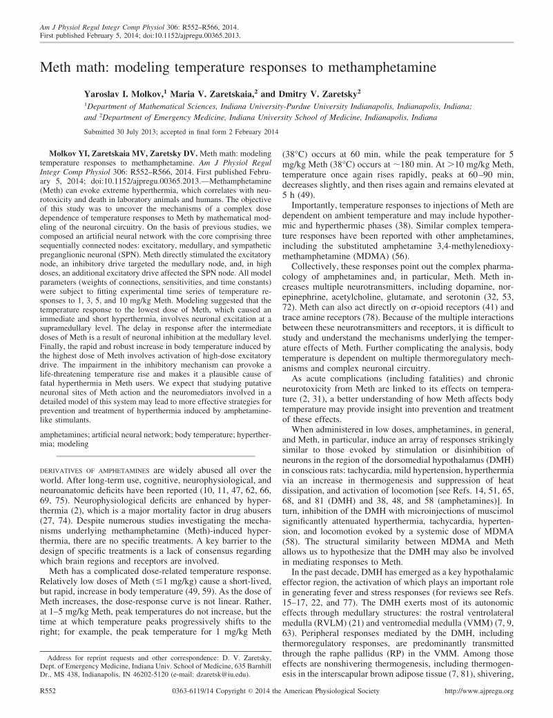
Meth math: modeling temperature responses to methamphetamine Yaroslav I. Molkov, 1 Maria V. Zaretskaia, 2 and Dmitry V. Zaretsky 2 1 Department of Mathematical Sciences, Indiana University-Purdue University Indianapolis, Indianapolis, Indiana; and 2 Department of Emergency Medicine, Indiana University School of Medicine, Indianapolis, Indiana Submitted 30 July 2013; accepted in final form 2 February 2014 Molkov YI, Zaretskaia MV, Zaretsky DV. Meth math: modeling temperature responses to methamphetamine. Am J Physiol Regul Integr Comp Physiol 306: R552–R566, 2014. First published Febru- ary 5, 2014; doi:10.1152/ajpregu.00365.2013.—Methamphetamine (Meth) can evoke extreme hyperthermia, which correlates with neu- rotoxicity and death in laboratory animals and humans. The objective of this study was to uncover the mechanisms of a complex dose dependence of temperature responses to Meth by mathematical mod- eling of the neuronal circuitry. On the basis of previous studies, we composed an artificial neural network with the core comprising three sequentially connected nodes: excitatory, medullary, and sympathetic preganglionic neuronal (SPN). Meth directly stimulated the excitatory node, an inhibitory drive targeted the medullary node, and, in high doses, an additional excitatory drive affected the SPN node. All model parameters (weights of connections, sensitivities, and time constants) were subject to fitting experimental time series of temperature re- sponses to 1, 3, 5, and 10 mg/kg Meth. Modeling suggested that the temperature response to the lowest dose of Meth, which caused an immediate and short hyperthermia, involves neuronal excitation at a supramedullary level. The delay in response after the intermediate doses of Meth is a result of neuronal inhibition at the medullary level. Finally, the rapid and robust increase in body temperature induced by the highest dose of Meth involves activation of high-dose excitatory drive. The impairment in the inhibitory mechanism can provoke a life-threatening temperature rise and makes it a plausible cause of fatal hyperthermia in Meth users. We expect that studying putative neuronal sites of Meth action and the neuromediators involved in a detailed model of this system may lead to more effective strategies for prevention and treatment of hyperthermia induced by amphetamine- like stimulants. amphetamines; artificial neural network; body temperature; hyperther- mia; modeling DERIVATIVES OF AMPHETAMINES are widely abused all over the world. After long-term use, cognitive, neurophysiological, and neuroanatomic deficits have been reported (10, 11, 47, 62, 66, 69, 75). Neurophysiological deficits are enhanced by hyper- thermia (2), which is a major mortality factor in drug abusers (27, 74). Despite numerous studies investigating the mecha- nisms underlying methamphetamine (Meth)-induced hyper- thermia, there are no specific treatments. A key barrier to the design of specific treatments is a lack of consensus regarding which brain regions and receptors are involved. Meth has a complicated dose-related temperature response. Relatively low doses of Meth (1 mg/kg) cause a short-lived, but rapid, increase in body temperature (49, 59). As the dose of Meth increases, the dose-response curve is not linear. Rather, at 1–5 mg/kg Meth, peak temperatures do not increase, but the time at which temperature peaks progressively shifts to the right; for example, the peak temperature for 1 mg/kg Meth (38°C) occurs at 60 min, while the peak temperature for 5 mg/kg Meth (38°C) occurs at 180 min. At 10 mg/kg Meth, temperature once again rises rapidly, peaks at 60 –90 min, decreases slightly, and then rises again and remains elevated at 5 h (49). Importantly, temperature responses to injections of Meth are dependent on ambient temperature and may include hypother- mic and hyperthermic phases (38). Similar complex tempera- ture responses have been reported with other amphetamines, including the substituted amphetamine 3,4-methylenedioxy- methamphetamine (MDMA) (56). Collectively, these responses point out the complex pharma- cology of amphetamines and, in particular, Meth. Meth in- creases multiple neurotransmitters, including dopamine, nor- epinephrine, acetylcholine, glutamate, and serotonin (32, 53, 72). Meth can also act directly on -opioid receptors (41) and trace amine receptors (78). Because of the multiple interactions between these neurotransmitters and receptors, it is difficult to study and understand the mechanisms underlying the temper- ature effects of Meth. Further complicating the analysis, body temperature is dependent on multiple thermoregulatory mech- anisms and complex neuronal circuitry. As acute complications (including fatalities) and chronic neurotoxicity from Meth are linked to its effects on tempera- ture (2, 31), a better understanding of how Meth affects body temperature may provide insight into prevention and treatment of these effects. When administered in low doses, amphetamines, in general, and Meth, in particular, induce an array of responses strikingly similar to those evoked by stimulation or disinhibition of neurons in the region of the dorsomedial hypothalamus (DMH) in conscious rats: tachycardia, mild hypertension, hyperthermia via an increase in thermogenesis and suppression of heat dissipation, and activation of locomotion [see Refs. 14, 51, 65, 68, and 81 (DMH) and 38, 48, and 58 (amphetamines)]. In turn, inhibition of the DMH with microinjections of muscimol significantly attenuated hyperthermia, tachycardia, hyperten- sion, and locomotion evoked by a systemic dose of MDMA (58). The structural similarity between MDMA and Meth allows us to hypothesize that the DMH may also be involved in mediating responses to Meth. In the past decade, DMH has emerged as a key hypothalamic effector region, the activation of which plays an important role in generating fever and stress responses (for reviews see Refs. 15–17, 22, and 77). The DMH exerts most of its autonomic effects through medullary structures: the rostral ventrolateral medulla (RVLM) (21) and ventromedial medulla (VMM) (7, 9, 63). Peripheral responses mediated by the DMH, including thermoregulatory responses, are predominantly transmitted through the raphe pallidus (RP) in the VMM. Among those effects are nonshivering thermogenesis, including thermogen- esis in the interscapular brown adipose tissue (7, 81), shivering, Address for reprint requests and other correspondence: D. V. Zaretsky, Dept. of Emergency Medicine, Indiana Univ. School of Medicine, 635 Barnhill Dr., MS 438, Indianapolis, IN 46202-5120 (e-mail: [email protected]). Am J Physiol Regul Integr Comp Physiol 306: R552–R566, 2014. First published February 5, 2014; doi:10.1152/ajpregu.00365.2013. 0363-6119/14 Copyright © 2014 the American Physiological Society http://www.ajpregu.org R552
Transcript of Meth math: modeling temperature responses to methamphetamine
Meth math: modeling temperature responses to methamphetamine
Yaroslav I. Molkov,1 Maria V. Zaretskaia,2 and Dmitry V. Zaretsky2
1Department of Mathematical Sciences, Indiana University-Purdue University Indianapolis, Indianapolis, Indiana;and 2Department of Emergency Medicine, Indiana University School of Medicine, Indianapolis, Indiana
Submitted 30 July 2013; accepted in final form 2 February 2014
Molkov YI, Zaretskaia MV, Zaretsky DV. Meth math: modelingtemperature responses to methamphetamine. Am J Physiol RegulIntegr Comp Physiol 306: R552–R566, 2014. First published Febru-ary 5, 2014; doi:10.1152/ajpregu.00365.2013.—Methamphetamine(Meth) can evoke extreme hyperthermia, which correlates with neu-rotoxicity and death in laboratory animals and humans. The objectiveof this study was to uncover the mechanisms of a complex dosedependence of temperature responses to Meth by mathematical mod-eling of the neuronal circuitry. On the basis of previous studies, wecomposed an artificial neural network with the core comprising threesequentially connected nodes: excitatory, medullary, and sympatheticpreganglionic neuronal (SPN). Meth directly stimulated the excitatorynode, an inhibitory drive targeted the medullary node, and, in highdoses, an additional excitatory drive affected the SPN node. All modelparameters (weights of connections, sensitivities, and time constants)were subject to fitting experimental time series of temperature re-sponses to 1, 3, 5, and 10 mg/kg Meth. Modeling suggested that thetemperature response to the lowest dose of Meth, which caused animmediate and short hyperthermia, involves neuronal excitation at asupramedullary level. The delay in response after the intermediatedoses of Meth is a result of neuronal inhibition at the medullary level.Finally, the rapid and robust increase in body temperature induced bythe highest dose of Meth involves activation of high-dose excitatorydrive. The impairment in the inhibitory mechanism can provoke alife-threatening temperature rise and makes it a plausible cause offatal hyperthermia in Meth users. We expect that studying putativeneuronal sites of Meth action and the neuromediators involved in adetailed model of this system may lead to more effective strategies forprevention and treatment of hyperthermia induced by amphetamine-like stimulants.
amphetamines; artificial neural network; body temperature; hyperther-mia; modeling
DERIVATIVES OF AMPHETAMINES are widely abused all over theworld. After long-term use, cognitive, neurophysiological, andneuroanatomic deficits have been reported (10, 11, 47, 62, 66,69, 75). Neurophysiological deficits are enhanced by hyper-thermia (2), which is a major mortality factor in drug abusers(27, 74). Despite numerous studies investigating the mecha-nisms underlying methamphetamine (Meth)-induced hyper-thermia, there are no specific treatments. A key barrier to thedesign of specific treatments is a lack of consensus regardingwhich brain regions and receptors are involved.
Meth has a complicated dose-related temperature response.Relatively low doses of Meth (�1 mg/kg) cause a short-lived,but rapid, increase in body temperature (49, 59). As the dose ofMeth increases, the dose-response curve is not linear. Rather,at 1–5 mg/kg Meth, peak temperatures do not increase, but thetime at which temperature peaks progressively shifts to theright; for example, the peak temperature for 1 mg/kg Meth
(38°C) occurs at 60 min, while the peak temperature for 5mg/kg Meth (38°C) occurs at �180 min. At �10 mg/kg Meth,temperature once again rises rapidly, peaks at 60–90 min,decreases slightly, and then rises again and remains elevated at5 h (49).
Importantly, temperature responses to injections of Meth aredependent on ambient temperature and may include hypother-mic and hyperthermic phases (38). Similar complex tempera-ture responses have been reported with other amphetamines,including the substituted amphetamine 3,4-methylenedioxy-methamphetamine (MDMA) (56).
Collectively, these responses point out the complex pharma-cology of amphetamines and, in particular, Meth. Meth in-creases multiple neurotransmitters, including dopamine, nor-epinephrine, acetylcholine, glutamate, and serotonin (32, 53,72). Meth can also act directly on �-opioid receptors (41) andtrace amine receptors (78). Because of the multiple interactionsbetween these neurotransmitters and receptors, it is difficult tostudy and understand the mechanisms underlying the temper-ature effects of Meth. Further complicating the analysis, bodytemperature is dependent on multiple thermoregulatory mech-anisms and complex neuronal circuitry.
As acute complications (including fatalities) and chronicneurotoxicity from Meth are linked to its effects on tempera-ture (2, 31), a better understanding of how Meth affects bodytemperature may provide insight into prevention and treatmentof these effects.
When administered in low doses, amphetamines, in general,and Meth, in particular, induce an array of responses strikinglysimilar to those evoked by stimulation or disinhibition ofneurons in the region of the dorsomedial hypothalamus (DMH)in conscious rats: tachycardia, mild hypertension, hyperthermiavia an increase in thermogenesis and suppression of heatdissipation, and activation of locomotion [see Refs. 14, 51, 65,68, and 81 (DMH) and 38, 48, and 58 (amphetamines)]. Inturn, inhibition of the DMH with microinjections of muscimolsignificantly attenuated hyperthermia, tachycardia, hyperten-sion, and locomotion evoked by a systemic dose of MDMA(58). The structural similarity between MDMA and Methallows us to hypothesize that the DMH may also be involvedin mediating responses to Meth.
In the past decade, DMH has emerged as a key hypothalamiceffector region, the activation of which plays an important rolein generating fever and stress responses (for reviews see Refs.15–17, 22, and 77). The DMH exerts most of its autonomiceffects through medullary structures: the rostral ventrolateralmedulla (RVLM) (21) and ventromedial medulla (VMM) (7, 9,63). Peripheral responses mediated by the DMH, includingthermoregulatory responses, are predominantly transmittedthrough the raphe pallidus (RP) in the VMM. Among thoseeffects are nonshivering thermogenesis, including thermogen-esis in the interscapular brown adipose tissue (7, 81), shivering,
Address for reprint requests and other correspondence: D. V. Zaretsky,Dept. of Emergency Medicine, Indiana Univ. School of Medicine, 635 BarnhillDr., MS 438, Indianapolis, IN 46202-5120 (e-mail: [email protected]).
Am J Physiol Regul Integr Comp Physiol 306: R552–R566, 2014.First published February 5, 2014; doi:10.1152/ajpregu.00365.2013.
0363-6119/14 Copyright © 2014 the American Physiological Society http://www.ajpregu.orgR552
which can result in production of a significant amount of heat(6, 39), and control of heat dissipation through cutaneous bloodflow (43, 44).
The sources of input to the DMH are multiple (42, 70);however, their functional roles and relative importance inmediating specific components of thermoregulatory processeshave not been described. Inhibition of the DMH can suppressresponses to activation of the amygdala (67) and dorsolateralperiaqueductal gray (PAG) (13). Stimulation of the amygda-loid region affects cutaneous blood flow (36), which is a majorpart of the thermoregulatory process. Whether those connec-tions are direct and involved in mediation of stress, fever, orresponses to drugs of abuse remains unknown. Therefore, wehave described Meth as acting directly on the DMH, while it ispossible that this is not the case.
Our experimental data contained clear evidence of the pres-ence of the inhibitory drive (the response to the intermediatedose is weaker than the response to the low dose). The DMHand RP have inhibitory inputs: disinhibition of both areas byantagonists of GABAA receptors evokes thermogenic re-sponses (37, 81) and a decrease in heat dissipation via con-striction of the cutaneous vasculature (43). One of the mainsources of GABAergic projections to both areas is located inthe preoptic area; however, the cells projecting to each area arenot the same (40, 79). Inhibitory tone to the RP could alsooriginate from the ventrolateral PAG (52) and RVLM (8).However, while the inhibitory component of responses to Methcould be due to activation of the inhibitory projection (presyn-aptic effect), it can also be mediated by activation of dopamine(most likely D2) receptors (45, 55) or �2-adrenergic receptors(34) located directly on neurons (postsynaptic effect). Withoutexperimental support for any specific pathway or receptorinvolved, for clarity of description, we modeled our inhibitorycomponent as an inhibitory drive to the RP.
Finally (46), we demonstrated that 5-HT2A receptors in thespinal cord contribute to cutaneous vasoconstriction after ahigh dose of MDMA. The high-dose component is clearlypresent in our data: a high dose of Meth overrides the inhibi-tion, and body temperature rises sharply and remains elevatedfor a long period of time.
Previous studies pinpoint a few brain areas possibly in-volved in mediating the responses to Meth; however, in thefollowing description, we avoid specific locations, as they have
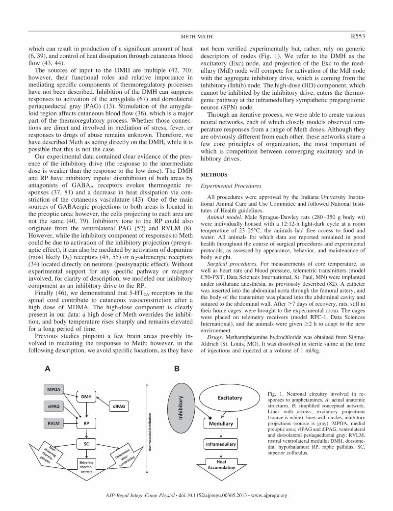
not been verified experimentally but, rather, rely on genericdescriptors of nodes (Fig. 1). We refer to the DMH as theexcitatory (Exc) node, and projection of the Exc to the med-ullary (Mdl) node will compete for activation of the Mdl nodewith the aggregate inhibitory drive, which is coming from theinhibitory (Inhib) node. The high-dose (HD) component, whichcannot be inhibited by the inhibitory drive, enters the thermo-genic pathway at the inframedullary sympathetic preganglionicneuron (SPN) node.
Through an iterative process, we were able to create variousneural networks, each of which closely models observed tem-perature responses from a range of Meth doses. Although theyare obviously different from each other, these networks share afew core principles of organization, the most important ofwhich is competition between converging excitatory and in-hibitory drives.
METHODS
Experimental Procedures
All procedures were approved by the Indiana University Institu-tional Animal Care and Use Committee and followed National Insti-tutes of Health guidelines.
Animal model. Male Sprague-Dawley rats (280–350 g body wt)were individually housed with a 12:12-h light-dark cycle at a roomtemperature of 23–25°C; the animals had free access to food andwater. All animals for which data are reported remained in goodhealth throughout the course of surgical procedures and experimentalprotocols, as assessed by appearance, behavior, and maintenance ofbody weight.
Surgical procedures. For measurements of core temperature, aswell as heart rate and blood pressure, telemetric transmitters (modelC50-PXT, Data Sciences International, St. Paul, MN) were implantedunder isoflurane anesthesia, as previously described (82). A catheterwas inserted into the abdominal aorta through the femoral artery, andthe body of the transmitter was placed into the abdominal cavity andsutured to the abdominal wall. After �7 days of recovery, rats, still intheir home cages, were brought to the experimental room. The cageswere placed on telemetry receivers (model RPC-1, Data SciencesInternational), and the animals were given �2 h to adapt to the newenvironment.
Drugs. Methamphetamine hydrochloride was obtained from Sigma-Aldrich (St. Louis, MO). It was dissolved in sterile saline at the timeof injections and injected at a volume of 1 ml/kg.
Fig. 1. Neuronal circuitry involved in re-sponses to amphetamines. A: actual anatomicstructures. B: simplified conceptual network.Lines with arrows, excitatory projections(source is white); lines with circles, inhibitoryprojections (source is gray). MPOA, medialpreoptic area; vlPAG and dlPAG, ventrolateraland dorsolateral periaqueductal gray; RVLM,rostral ventrolateral medulla; DMH, dorsome-dial hypothalamus; RP, raphe pallidus; SC,superior colliculus.
R553METH MATH
AJP-Regul Integr Comp Physiol • doi:10.1152/ajpregu.00365.2013 • www.ajpregu.org

Determining Temperature Responses to Meth
The temperature response to various doses of Meth was determinedby randomization of animals receiving an intraperitoneal injection ofsaline or one of the four doses of Meth (1, 3, 5, or 10 mg/kg). Eachanimal received only one injection, with six rats per dose. Data wererecorded every 2 min and transferred to Microsoft Excel, and 10-minaverages were calculated using a template in Excel.
Statistical Analysis
Values are means � SE. Data were compared using a one-wayANOVA with repeated measures followed by Fisher’s least signifi-cant difference post hoc test, where appropriate. P � 0.05 wasconsidered to indicate a significant difference in all comparisons.Baseline levels of activity, temperature, heart rate, and mean bloodpressure did not differ between groups across the series of experi-ments, so changes from baseline were analyzed.
Model Construction
Three components were considered in constructing our model: 1)the pharmacokinetic time course of Meth concentration in the bloodafter an intraperitoneal injection, 2) a neural network, the activity ofwhich depends on Meth concentration in the blood, and 3) a temper-ature control system that is driven by a signal from the neuralcomponent.
Pharmacokinetics. Meth is rapidly absorbed from the peritoneumand then eliminated from the blood with a profile closely resemblinga single-exponential process (23, 76). Our data do not contain inde-pendent information about the volume of distribution. Therefore, wehave expressed the concentration in units of dose (mg/kg), and drugconcentrations are described in the model by the following equations
dx
dt� �
x
�a
dy
dt�
x
�a�
y
�e
(1)
where t is time (in min), x(t) and y(t) are intraperitoneal and bloodMeth concentrations, respectively, x/�a describes drug absorption witha time constant �a, and y/�e represents drug elimination with a time
constant �e. Initial conditions are x(0) D,y(0), given that a dose Dis injected at time 0 (t 0). With these initial conditions, the solutionof Eq. 1 can be explicitly found as
y(t) � D(�a ⁄ �e � 1)�1(e�t⁄�a � e�t⁄�e) (2)
which provides the Meth concentration in the blood at any given time.Neural network. The network schematic used in our model is based
on previous research (see above) showing that neural populations inthree key interconnected brain regions are responsible for normalrodent thermoregulation: excitatory drive is coming through the DMHto premotor sympathetic neurons in the VMM (Mdl node) and isrelayed from the medulla to sympathetic preganglionic neurons in thespinal cord (SPN node). Activation of the SPN node results in anincrease in body temperature via several mechanisms. In addition, weknow from our data and data of others that temperature responses toamphetamines do not follow simple linear dose responses (35, 38, 46,49, 59, 60). This can be seen in Fig. 2, where a low dose of Methcauses acute increases in temperature that are greater than increasescaused by the two intermediate doses but lower than increases causedby the highest dose. Therefore, the excitation in the model can besuppressed by inhibitory drive, originating from supramedullary (pre-optic nucleus or ventrolateral PAG) or intramedullary (RVLM) struc-tures. We also know from the work of others that, at high doses, someamphetamines appear to act below the level of the medulla (46). Forsimplicity, our initial model factored in this postulated inframedullaryeffect by creating an additional Exc node that, as we hypothesized,would be activated only by higher doses of Meth (hence, the HDnode).
This circuitry is depicted in Fig. 3A. The model takes the form ofa feedforward artificial neural network. The outputs of “Meth-sensi-tive” supramedullary excitatory (Exc), inhibitory (Inhib), and high-dose-activated (HD) neural populations Pi (i Exc, Inhib, and HD)were calculated using the following formula
Pi(t) � �[wi · y(t) � i] (3)
where �(x) (1 tanhx)/2 is a sigmoidal activation function, wi isa parameter of sensitivity to Meth, y(t) is the time dependence of theMeth concentration in the blood from Eq. 2, and �i defines basalexcitability of Pi. Accordingly, the unit of sensitivity (wExc, wInhib,and wHD) is (mg/kg)�1 and �Exc, �Inhib, and �HD are dimensionless.
39
38
37
360 60 120
Time, min
injection10 mg/kg
5 mg/kg3 mg/kg1 mg/kg
Bod
y te
mpe
ratu
re, °
C
180 240
Fig. 2. Dose dependence of temperature responsesto methamphetamine (Meth). Meth was injectedintraperitoneally at 0 min (t 0) in a volume of1 ml/kg.
R554 METH MATH
AJP-Regul Integr Comp Physiol • doi:10.1152/ajpregu.00365.2013 • www.ajpregu.org
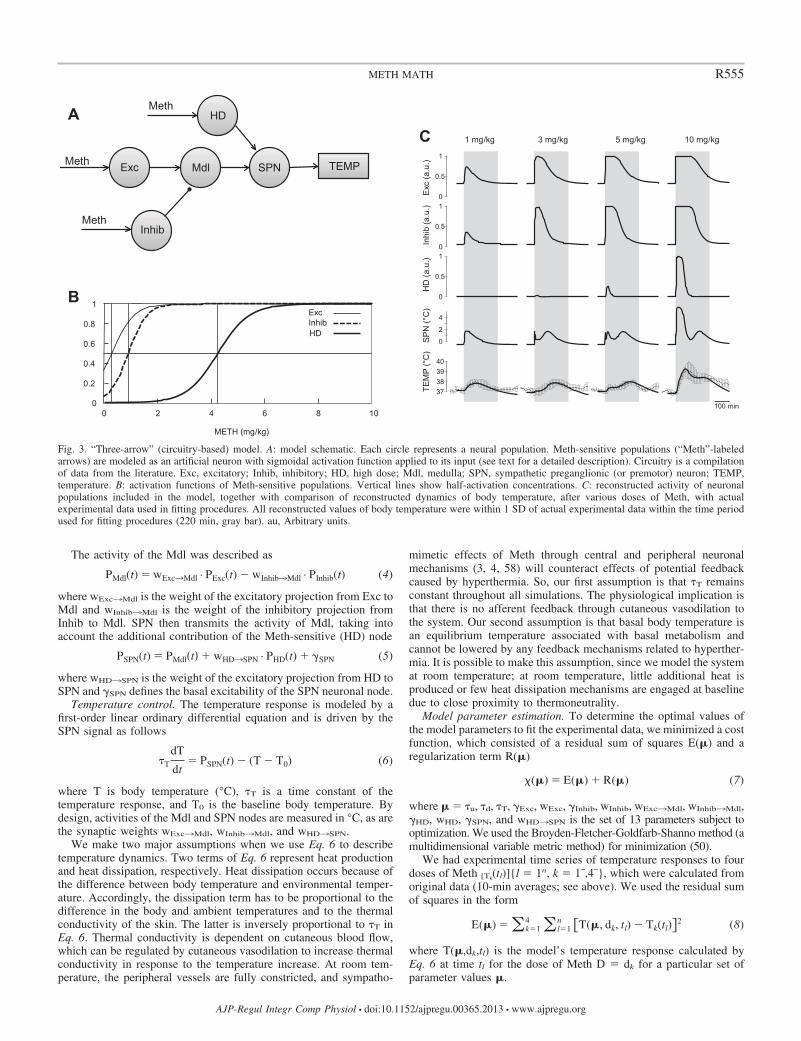
The activity of the Mdl was described as
PMdl(t) � wExc¡Mdl · PExc(t) � wInhib¡Mdl · PInhib(t) (4)
where wExc¡Mdl is the weight of the excitatory projection from Exc toMdl and wInhib¡Mdl is the weight of the inhibitory projection fromInhib to Mdl. SPN then transmits the activity of Mdl, taking intoaccount the additional contribution of the Meth-sensitive (HD) node
PSPN(t) � PMdl�t� � wHD¡SPN · PHD(t) � SPN (5)
where wHD¡SPN is the weight of the excitatory projection from HD toSPN and �SPN defines the basal excitability of the SPN neuronal node.
Temperature control. The temperature response is modeled by afirst-order linear ordinary differential equation and is driven by theSPN signal as follows
�T
dT
dt� PSPN(t) � (T � T0) (6)
where T is body temperature (°C), �T is a time constant of thetemperature response, and T0 is the baseline body temperature. Bydesign, activities of the Mdl and SPN nodes are measured in °C, as arethe synaptic weights wExc¡Mdl, wInhib¡Mdl, and wHD¡SPN.
We make two major assumptions when we use Eq. 6 to describetemperature dynamics. Two terms of Eq. 6 represent heat productionand heat dissipation, respectively. Heat dissipation occurs because ofthe difference between body temperature and environmental temper-ature. Accordingly, the dissipation term has to be proportional to thedifference in the body and ambient temperatures and to the thermalconductivity of the skin. The latter is inversely proportional to �T inEq. 6. Thermal conductivity is dependent on cutaneous blood flow,which can be regulated by cutaneous vasodilation to increase thermalconductivity in response to the temperature increase. At room tem-perature, the peripheral vessels are fully constricted, and sympatho-
mimetic effects of Meth through central and peripheral neuronalmechanisms (3, 4, 58) will counteract effects of potential feedbackcaused by hyperthermia. So, our first assumption is that �T remainsconstant throughout all simulations. The physiological implication isthat there is no afferent feedback through cutaneous vasodilation tothe system. Our second assumption is that basal body temperature isan equilibrium temperature associated with basal metabolism andcannot be lowered by any feedback mechanisms related to hyperther-mia. It is possible to make this assumption, since we model the systemat room temperature; at room temperature, little additional heat isproduced or few heat dissipation mechanisms are engaged at baselinedue to close proximity to thermoneutrality.
Model parameter estimation. To determine the optimal values ofthe model parameters to fit the experimental data, we minimized a costfunction, which consisted of a residual sum of squares E(�) and aregularization term R(�)
(�) � E(�) � R(�) (7)
where � �u, �d, �T, �Exc, wExc, �Inhib, wInhib, wExc¡Mdl, wInhib¡Mdl,�HD, wHD, �SPN, and wHD¡SPN is the set of 13 parameters subject tooptimization. We used the Broyden-Fletcher-Goldfarb-Shanno method (amultidimensional variable metric method) for minimization (50).
We had experimental time series of temperature responses to fourdoses of Meth [Tk
(tl)]{l 1n, k 1�,4�}, which were calculated fromoriginal data (10-min averages; see above). We used the residual sumof squares in the form
E(�) � �k�14 �l�1
n �T(�, dk, tl) � Tk(tl)�2 (8)
where T(�,dk,tl) is the model’s temperature response calculated byEq. 6 at time tl for the dose of Meth D dk for a particular set ofparameter values �.
A
Exc SPN
Inhib
TEMP Meth
Meth
HD Meth
Mdl
1 mg/kg 3 mg/kg 5 mg/kg 10 mg/kg C
Exc
(a.u
.) In
hib
(a.u
.) H
D (a
.u.)
SP
N (°
C)
TEM
P (°
C)
0
0.5
1
0
0.5
1
0
0.5
1
0
2
4
37 38 39 40
100 min 0
0.2
0.4
0.6
0.8
1
0 2 4 6 8 10
Exc Inhib HD
B
METH (mg/kg)
Fig. 3. “Three-arrow” (circuitry-based) model. A: model schematic. Each circle represents a neural population. Meth-sensitive populations (“Meth”-labeledarrows) are modeled as an artificial neuron with sigmoidal activation function applied to its input (see text for a detailed description). Circuitry is a compilationof data from the literature. Exc, excitatory; Inhib, inhibitory; HD, high dose; Mdl, medulla; SPN, sympathetic preganglionic (or premotor) neuron; TEMP,temperature. B: activation functions of Meth-sensitive populations. Vertical lines show half-activation concentrations. C: reconstructed activity of neuronalpopulations included in the model, together with comparison of reconstructed dynamics of body temperature, after various doses of Meth, with actualexperimental data used in fitting procedures. All reconstructed values of body temperature were within 1 SD of actual experimental data within the time periodused for fitting procedures (220 min, gray bar). au, Arbitrary units.
R555METH MATH
AJP-Regul Integr Comp Physiol • doi:10.1152/ajpregu.00365.2013 • www.ajpregu.org

The minimization of Eq. 5 is an ill-posed problem, because themodel output depends mostly on the difference between Exc and Inhibprojections to Mdl, and not on their absolute values. In addition,relatively low doses of Meth saturate Exc and Inhib activation.Therefore, the model output is weakly dependent on the sensitivityof these populations to Meth wExc and wInhib when their values arelarge. To overcome these difficulties, we used the Tikhonov regular-ization technique (71) by adding a regularization term
R(�) � �(wExc2 � wInhib
2 � PSN2 � wExc¡Mdl
2 � wInhib¡Mdl2 ) (9)
to the cost function (7). To determine an optimal value of theregularization factor , we used the L-curve method (24).
Model Reduction and Analysis
The original model was constructed on the basis of the hypotheticalthermoregulatory circuitry available in the literature and had threeMeth-sensitive nodes. Subsequent analysis of the model revealedseveral features redundant from a data assimilation perspective. Spe-cifically, the experimentally observed temperature curves could bereproduced with comparable precision by use of the reduced circuit-ries receiving two or even one Meth-dependent input. Hereafter, werefer to these three models as the “three-arrow,” “two-arrow,” and“one-arrow” models, respectively.
Two-arrow model. In our initial three-arrow model, we incorpo-rated an inframedullary excitatory input that was activated only athigh doses of Meth. While our decision was based, in part, on intuitionand, in part, on previous data with MDMA (46), we also recognizedthat a more parsimonious model might be sufficient and wouldsimplify its use. To test this, we derived a two-arrow model (see Fig.5) from the original three-arrow model by setting wHD¡SPN 0. Thisalso had the effect of eliminating the parameters that describedactivation of the HD population, �HD and wHD, resulting in the needto estimate only 10 parameters. This is possible from a conceptualstandpoint, because elimination of the HD component essentiallymakes the inframedullary output directly dependent on the medullaryoutput and, thereby, redundant for modeling purposes. Essentially,Mdl and SPN nodes were merged into a single SPN node; dependingon the location, SPN can mean “sympathetic premotor neurons” (forthe medullary location) or “sympathetic preganglionic neurons” (forthe spinal cord location).
One-arrow model. After confirming that the two-arrow model(without the HD and Mdl nodes) also accurately fit the observed data,we sought to simplify this model further. In the one-arrow model, onlyExc is sensitive to Meth, and Inhib is now activated by projectionfrom the Exc node (Fig. 6A). In this model, the activity of Inhib wasdescribed as follows
PInhib(t) � ��wExc¡Inhib · PExc(t) � Inhib� (10)
where wExc¡Inhib was the weight of the excitatory projection from Exc toInhib. This parameter replaced the sensitivity of Inhib to Meth (wInhib).However, since the parameters of sensitivity of Inhib to Meth are replaced byparameters of sensitivity to descending excitatory input, the total number ofparameters did not change compared with the two-arrow model.
Variability of temperature responses. To mimic experimentallyobserved animal-to-animal variability, we constructed model re-sponses with varied model parameters. For every model parameter �i,we calculated the average sensitivity of the temperature to the relativeparameter variation as
Si � max t�[0,L]
�T( , D, t)
� i i (11)
where L 220 min is the time interval used, the dose of Meth (D) 10mg/kg, and �T/��i is a partial derivative of the temperature at time twith respect to �i. We compared the calculated sensitivities and foundthat, for all three models, the calculated temperatures had the greatest
sensitivity to variations in synaptic weights of the projections fromExc and Inhib nodes to Mdl. In addition, the three-arrow model has acomparable sensitivity to synaptic weight of the projection from HDto SPN. Accordingly, we chose wExc¡Mdl, wInhib¡Mdl, and wHD¡SPN
as our control parameters for this analysis. Given the relative param-eter variation �, we multiplied wExc¡Mdl (and wHD¡SPN for the3-arrow model) and divided wInhib¡Mdl by a factor of (1 �) togenerate the greatest possible temperature response and the sameparameters by a factor of (1 � �) to obtain the lowest possibletemperature response.
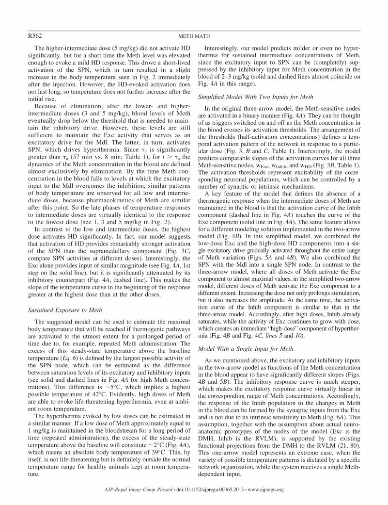
We have also considered situations when the inhibitory drive issignificantly suppressed. For three- and two-arrow models, we havegenerated the model curves of the body temperature with all parametersbeing the same as in Table 1 but with wInhib¡Mdl equal to 0 or one-halfof its value in Table 1. The curves representing model responses to 1 and3 mg/kg Meth in such situations are shown in Fig. 8.
RESULTS
Temperature Responses to Meth
The doses of Meth (1, 3, 5, and 10 mg/kg) used in this studycaused complex temperature responses (Fig. 2). At the lowestdose (1 mg/kg), temperature rose almost immediately after theinjection, reached a peak (1°C above baseline) by 80 min, andretuned to baseline within 3 h. Increasing the Meth dose to 3mg/kg delayed the response: temperature started to rise 40 minafter the injection and peaked (also �1°C above baseline) at150 min. Hyperthermia lasted for �4 h after 3 mg/kg Meth.
Similar to the low dose of Meth (1 mg/kg), 5 mg/kg Methincreased body temperature immediately after injection. Thepeak, however, was smaller (0.5°C) and occurred within 30min. Temperature remained relatively constant for �1 h, beganto rise again to a second peak (�0.8°C above baseline) at 190min, and, finally, returned to baseline within 4 h. Late phasesof temperature responses to intermediate doses (t � 50 min for3 mg/kg and t � 90 min for 5 mg/kg) were similar to theresponse to a low dose of Meth to the accuracy of a time shift.
Similar to 5 mg/kg Meth, the highest dose of Meth (10mg/kg) caused an immediate increase in temperature. Theincrease, however, was greater (�2°C above baseline) andrequired a longer period of time to peak (�80 min). Afterreaching a peak, the temperature fell over the next 60 min andthen, similar to the 5 mg/kg dose, peaked again, at least insome animals, or remained relatively constant for �60 min. Incontrast to 5 mg/kg Meth, the second peak was not stronglyexpressed, and temperature slowly returned to baseline levels�6 h after injection (data not shown).
Collectively, these data show a complex multimodal tem-perature response to Meth that is highly dependent on the dose.
Estimated Parameters and Model Validation
Data shown in Fig. 2 were used to find the optimal set ofparameters of each of the models, as described in METHODS. Thecalculated parameter values with their SE estimates are listedin Table 1. To evaluate the goodness of fit, we calculated thecoefficient of determination (R2, Table 2) for each model andall doses. R2 can be treated as a fraction of the varianceexplained by the models, with a range of 65–95% for differentdoses and, on average, is �90% for all three models. We alsocalculated the ratio of the root mean square of the modelresiduals to the SDs of temperature responses over the group ofanimals (n 6). These calculations are shown in Table 3 for
R556 METH MATH
AJP-Regul Integr Comp Physiol • doi:10.1152/ajpregu.00365.2013 • www.ajpregu.org
all three models and all doses. Because, in all cases, thismeasure is much less than 100%, the model mismatch is wellwithin experimentally observed animal-to-animal variability.
To validate the model, we used k-fold cross-validation. Fourtemperature time series as a single data set were randomlydivided into eight sets of points of equal size. Every set out ofeight was used for validation, and the remaining seven setswere used to optimize the model parameters. The residuals forthe validation sets were collected and root-mean-squared to becompared with the root-mean-square residuals for the trainingsets. The ratios of root-mean-squared residuals for the valida-tion and training sets were 1.12 for the three-arrow model, 1.08for the two-arrow model, and 1.09 for the one-arrow model.These ratios can be compared with the expected ratios in thecase of linear regression. Each training set comprised 77 datapoints (n 77). The models had 13, 10, and 10 parameters(p 13, 10, and 10), respectively. Accordingly, the expectedratios of root-mean-squared residuals of validation and trainingsets, r ��n�p�⁄�n�p�, were �1.18, 1.14, and 1.14. Thevalues obtained were slightly less than the estimates, probablybecause of regularization used in our optimization procedure.In general, it evidences the robustness of the models.
Model Simulation and Interpretation
These parameters were used to simulate the dynamics ofMeth concentration in the blood, activities of the neuronalpopulations, and temperature responses to different doses ofMeth under various (patho)physiological conditions.
Pharmacokinetics. The dependence of Meth concentrationin the blood on time is described by Eq. 2, where �a defines arate at which Meth is initially accumulated in the blood due toits absorption from the peritoneum and �e is responsible for therate at which the drug is washed out. After parameter optimi-zation to fit experimental data in all models, absorption ap-peared to be much faster than elimination (�10 min vs. �60min; see Table 1 for model-specific values). With such param-eters, after an intraperitoneal injection of Meth, the bloodconcentration quickly peaks at a maximum of �70% of thedose and then exponentially decreases with a time constantof �1 h.
Activity in the Meth-sensitive nodes in the three-arrowmodel. In the three-arrow model, which was able to accuratelyreproduce the observed temperature curves for all four doses ofMeth (Fig. 3C), three neuronal nodes were stimulated by Methdirectly (Fig. 3A): Exc, Inhib, and an excitatory projection tothe SPN node, which was activated only at high doses of Meth(HD). The activation function of each Meth-sensitive popula-tion depended on two parameters: half-activation concentrationand maximal slope. The slopes appeared to be comparable forall nodes, while half-activation concentration was lowest forExc and highest for HD (Fig. 3B, Table 1, 3-arrow model).
Activation time courses of each of these nodes for each doseof Meth are shown in Fig. 3C. All doses of Meth were able toactivate the Exc node, which reached 100% activity at 3 mg/kgfor a short period of time. Each successively higher doseactivated this node for a longer period of time. Similarly, theInhib node was activated by all doses of Meth. The sensitivityof the Inhib node was less than that of the Exc node: the Inhibnode required higher doses of Meth for full activation and hadshorter durations of maximal activity at the higher doses.Finally, the HD component was activated only at the twohighest doses of Meth (5 and 10 mg/kg).
Temperature responses to low and lower-intermediatedoses. The activity of the SPN, which defines an output of thesystem, can be viewed as a competition between excitatory andinhibitory drives. Excitatory drive in the three-arrow model isa sum of drives from Exc and HD that is offset by activity ofthe Inhib. To visualize how various doses of Meth result inactivity of the SPN, Fig. 4 presents the activation function forcombined excitatory drive and inhibitory drive (Fig. 4A)matched with pharmacokinetic profiles of Meth at variousdoses (Fig. 4C).
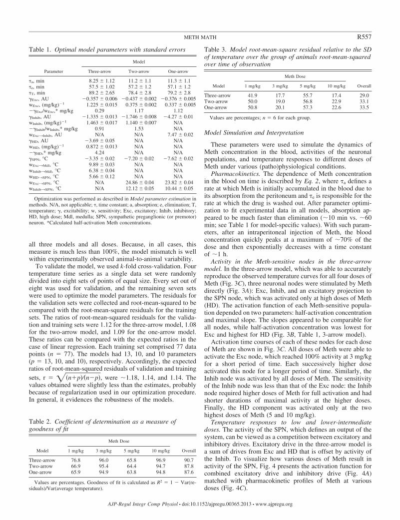
Table 1. Optimal model parameters with standard errors
Model
Parameter Three-arrow Two-arrow One-arrow
�a, min 8.25 � 1.12 11.2 � 1.1 11.3 � 1.1�e, min 57.5 � 1.02 57.2 � 1.2 57.1 � 1.2�T, min 89.2 � 2.65 78.4 � 2.8 79.2 � 2.8�Exc, AU �0.357 � 0.006 �0.437 � 0.002 �0.376 � 0.005wExc, (mg/kg)�1 1.225 � 0.015 0.375 � 0.002 0.337 � 0.005��Exc/wExc,* mg/kg 0.29 1.17 1.12�Inhib, AU �1.335 � 0.013 �1.746 � 0.008 �4.27 � 0.01wInhib, (mg/kg)�1 1.463 � 0.017 1.140 � 0.007 N/A��Inhib/wInhib,* mg/kg 0.91 1.53 N/AwExc¡Inhib, AU N/A N/A 7.47 � 0.02�HD, AU �3.69 � 0.05 N/A N/AwHD, (mg/kg)�1 0.872 � 0.013 N/A N/A��HD,* mg/kg 4.24 N/A N/A�SPN, °C �3.35 � 0.02 �7.20 � 0.02 �7.62 � 0.02wExc¡Mdl, °C 9.89 � 0.03 N/A N/AwInhib¡Mdl, °C 6.38 � 0.04 N/A N/AwHD¡SPN, °C 5.66 � 0.12 N/A N/AwExc¡SPN, °C N/A 24.86 � 0.04 23.82 � 0.04wInhib¡SPN, °C N/A 12.12 � 0.05 10.44 � 0.05
Optimization was performed as described in Model parameter estimation inmethods. N/A, not applicable; �, time constant; a, absorption; e, elimination; T,temperature; �, excitability; w, sensitivity; Exc, excitatory; Inhib, inhibitory;HD, high dose; Mdl, medulla; SPN, sympathetic preganglionic (or premotor)neuron. *Calculated half-activation Meth concentrations.
Table 2. Coefficient of determination as a measure ofgoodness of fit
Meth Dose
Model 1 mg/kg 3 mg/kg 5 mg/kg 10 mg/kg Overall
Three-arrow 76.8 96.0 65.8 96.9 90.7Two-arrow 66.9 95.4 64.4 94.7 87.8One-arrow 65.9 94.9 63.8 94.8 87.6
Values are percentages. Goodness of fit is calculated as R2 1 � Var(re-siduals)/Var(average temperature).
Table 3. Model root-mean-square residual relative to the SDof temperature over the group of animals root-mean-squaredover time of observation
Meth Dose
Model 1 mg/kg 3 mg/kg 5 mg/kg 10 mg/kg Overall
Three-arrow 41.9 17.7 55.7 17.4 29.0Two-arrow 50.0 19.0 56.8 22.9 33.1One-arrow 50.8 20.1 57.3 22.6 33.5
Values are percentages; n 6 for each group.
R557METH MATH
AJP-Regul Integr Comp Physiol • doi:10.1152/ajpregu.00365.2013 • www.ajpregu.org
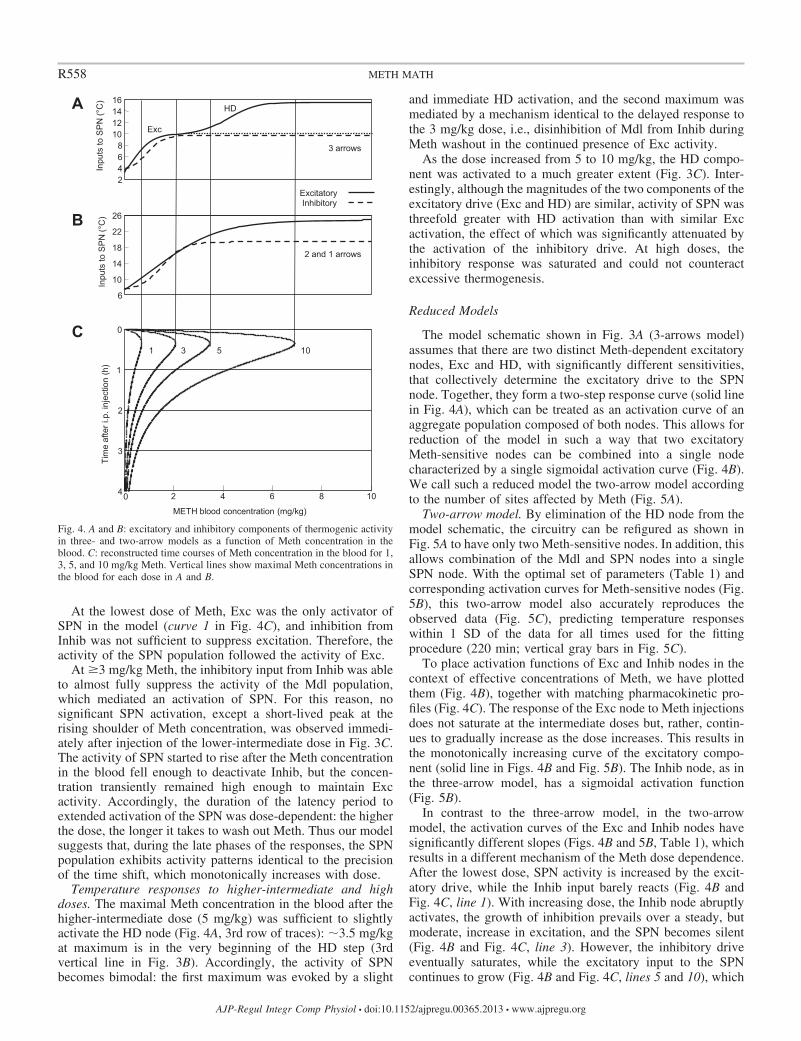
At the lowest dose of Meth, Exc was the only activator ofSPN in the model (curve 1 in Fig. 4C), and inhibition fromInhib was not sufficient to suppress excitation. Therefore, theactivity of the SPN population followed the activity of Exc.
At �3 mg/kg Meth, the inhibitory input from Inhib was ableto almost fully suppress the activity of the Mdl population,which mediated an activation of SPN. For this reason, nosignificant SPN activation, except a short-lived peak at therising shoulder of Meth concentration, was observed immedi-ately after injection of the lower-intermediate dose in Fig. 3C.The activity of SPN started to rise after the Meth concentrationin the blood fell enough to deactivate Inhib, but the concen-tration transiently remained high enough to maintain Excactivity. Accordingly, the duration of the latency period toextended activation of the SPN was dose-dependent: the higherthe dose, the longer it takes to wash out Meth. Thus our modelsuggests that, during the late phases of the responses, the SPNpopulation exhibits activity patterns identical to the precisionof the time shift, which monotonically increases with dose.
Temperature responses to higher-intermediate and highdoses. The maximal Meth concentration in the blood after thehigher-intermediate dose (5 mg/kg) was sufficient to slightlyactivate the HD node (Fig. 4A, 3rd row of traces): �3.5 mg/kgat maximum is in the very beginning of the HD step (3rdvertical line in Fig. 3B). Accordingly, the activity of SPNbecomes bimodal: the first maximum was evoked by a slight
and immediate HD activation, and the second maximum wasmediated by a mechanism identical to the delayed response tothe 3 mg/kg dose, i.e., disinhibition of Mdl from Inhib duringMeth washout in the continued presence of Exc activity.
As the dose increased from 5 to 10 mg/kg, the HD compo-nent was activated to a much greater extent (Fig. 3C). Inter-estingly, although the magnitudes of the two components of theexcitatory drive (Exc and HD) are similar, activity of SPN wasthreefold greater with HD activation than with similar Excactivation, the effect of which was significantly attenuated bythe activation of the inhibitory drive. At high doses, theinhibitory response was saturated and could not counteractexcessive thermogenesis.
Reduced Models
The model schematic shown in Fig. 3A (3-arrows model)assumes that there are two distinct Meth-dependent excitatorynodes, Exc and HD, with significantly different sensitivities,that collectively determine the excitatory drive to the SPNnode. Together, they form a two-step response curve (solid linein Fig. 4A), which can be treated as an activation curve of anaggregate population composed of both nodes. This allows forreduction of the model in such a way that two excitatoryMeth-sensitive nodes can be combined into a single nodecharacterized by a single sigmoidal activation curve (Fig. 4B).We call such a reduced model the two-arrow model accordingto the number of sites affected by Meth (Fig. 5A).
Two-arrow model. By elimination of the HD node from themodel schematic, the circuitry can be refigured as shown inFig. 5A to have only two Meth-sensitive nodes. In addition, thisallows combination of the Mdl and SPN nodes into a singleSPN node. With the optimal set of parameters (Table 1) andcorresponding activation curves for Meth-sensitive nodes (Fig.5B), this two-arrow model also accurately reproduces theobserved data (Fig. 5C), predicting temperature responseswithin 1 SD of the data for all times used for the fittingprocedure (220 min; vertical gray bars in Fig. 5C).
To place activation functions of Exc and Inhib nodes in thecontext of effective concentrations of Meth, we have plottedthem (Fig. 4B), together with matching pharmacokinetic pro-files (Fig. 4C). The response of the Exc node to Meth injectionsdoes not saturate at the intermediate doses but, rather, contin-ues to gradually increase as the dose increases. This results inthe monotonically increasing curve of the excitatory compo-nent (solid line in Figs. 4B and Fig. 5B). The Inhib node, as inthe three-arrow model, has a sigmoidal activation function(Fig. 5B).
In contrast to the three-arrow model, in the two-arrowmodel, the activation curves of the Exc and Inhib nodes havesignificantly different slopes (Figs. 4B and 5B, Table 1), whichresults in a different mechanism of the Meth dose dependence.After the lowest dose, SPN activity is increased by the excit-atory drive, while the Inhib input barely reacts (Fig. 4B andFig. 4C, line 1). With increasing dose, the Inhib node abruptlyactivates, the growth of inhibition prevails over a steady, butmoderate, increase in excitation, and the SPN becomes silent(Fig. 4B and Fig. 4C, line 3). However, the inhibitory driveeventually saturates, while the excitatory input to the SPNcontinues to grow (Fig. 4B and Fig. 4C, lines 5 and 10), which
2 4 6 8
10 12 14 16
3 arrows
Excitatory Inhibitory
6
10
14
18
22
26
2 and 1 arrows
10 5 3 1
2 4 6 8 10
METH blood concentration (mg/kg)
0
1
2
3
4
Inpu
ts to
SP
N (°
C)
Inpu
ts to
SP
N (°
C)
Tim
e af
ter i
.p. i
njec
tion
(h)
Exc
HD
0
A
B
C
Fig. 4. A and B: excitatory and inhibitory components of thermogenic activityin three- and two-arrow models as a function of Meth concentration in theblood. C: reconstructed time courses of Meth concentration in the blood for 1,3, 5, and 10 mg/kg Meth. Vertical lines show maximal Meth concentrations inthe blood for each dose in A and B.
R558 METH MATH
AJP-Regul Integr Comp Physiol • doi:10.1152/ajpregu.00365.2013 • www.ajpregu.org
results in activation of the SPN by sufficiently high doses ofMeth.
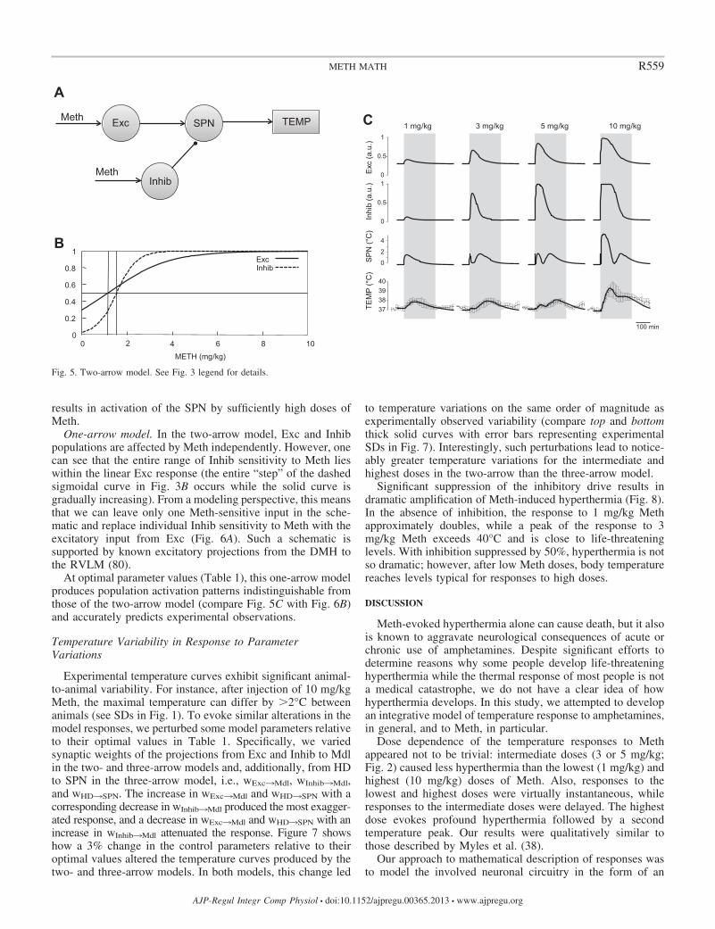
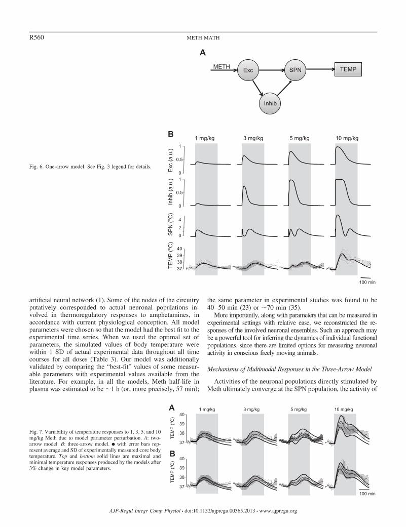
One-arrow model. In the two-arrow model, Exc and Inhibpopulations are affected by Meth independently. However, onecan see that the entire range of Inhib sensitivity to Meth lieswithin the linear Exc response (the entire “step” of the dashedsigmoidal curve in Fig. 3B occurs while the solid curve isgradually increasing). From a modeling perspective, this meansthat we can leave only one Meth-sensitive input in the sche-matic and replace individual Inhib sensitivity to Meth with theexcitatory input from Exc (Fig. 6A). Such a schematic issupported by known excitatory projections from the DMH tothe RVLM (80).
At optimal parameter values (Table 1), this one-arrow modelproduces population activation patterns indistinguishable fromthose of the two-arrow model (compare Fig. 5C with Fig. 6B)and accurately predicts experimental observations.
Temperature Variability in Response to ParameterVariations
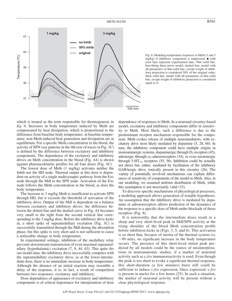
Experimental temperature curves exhibit significant animal-to-animal variability. For instance, after injection of 10 mg/kgMeth, the maximal temperature can differ by �2°C betweenanimals (see SDs in Fig. 1). To evoke similar alterations in themodel responses, we perturbed some model parameters relativeto their optimal values in Table 1. Specifically, we variedsynaptic weights of the projections from Exc and Inhib to Mdlin the two- and three-arrow models and, additionally, from HDto SPN in the three-arrow model, i.e., wExc¡Mdl, wInhib¡Mdl,and wHD¡SPN. The increase in wExc¡Mdl and wHD¡SPN with acorresponding decrease in wInhib¡Mdl produced the most exagger-ated response, and a decrease in wExc¡Mdl and wHD¡SPN with anincrease in wInhib¡Mdl attenuated the response. Figure 7 showshow a 3% change in the control parameters relative to theiroptimal values altered the temperature curves produced by thetwo- and three-arrow models. In both models, this change led
to temperature variations on the same order of magnitude asexperimentally observed variability (compare top and bottomthick solid curves with error bars representing experimentalSDs in Fig. 7). Interestingly, such perturbations lead to notice-ably greater temperature variations for the intermediate andhighest doses in the two-arrow than the three-arrow model.
Significant suppression of the inhibitory drive results indramatic amplification of Meth-induced hyperthermia (Fig. 8).In the absence of inhibition, the response to 1 mg/kg Methapproximately doubles, while a peak of the response to 3mg/kg Meth exceeds 40°C and is close to life-threateninglevels. With inhibition suppressed by 50%, hyperthermia is notso dramatic; however, after low Meth doses, body temperaturereaches levels typical for responses to high doses.
DISCUSSION
Meth-evoked hyperthermia alone can cause death, but it alsois known to aggravate neurological consequences of acute orchronic use of amphetamines. Despite significant efforts todetermine reasons why some people develop life-threateninghyperthermia while the thermal response of most people is nota medical catastrophe, we do not have a clear idea of howhyperthermia develops. In this study, we attempted to developan integrative model of temperature response to amphetamines,in general, and to Meth, in particular.
Dose dependence of the temperature responses to Methappeared not to be trivial: intermediate doses (3 or 5 mg/kg;Fig. 2) caused less hyperthermia than the lowest (1 mg/kg) andhighest (10 mg/kg) doses of Meth. Also, responses to thelowest and highest doses were virtually instantaneous, whileresponses to the intermediate doses were delayed. The highestdose evokes profound hyperthermia followed by a secondtemperature peak. Our results were qualitatively similar tothose described by Myles et al. (38).
Our approach to mathematical description of responses wasto model the involved neuronal circuitry in the form of an
1 mg/kg 3 mg/kg 5 mg/kg 10 mg/kg C
0
0.5
1
0
0.5
1
0
2
4
37 38 39 40
Exc
(a.u
.) In
hib
(a.u
.) S
PN
(°C
) TE
MP
(°C
)
100 min
A
Exc SPN
Inhib
TEMP Meth
Meth
0
0.2
0.4
0.6
0.8
1
0 2 4 6 8 10 METH (mg/kg)
Exc Inhib
B
Fig. 5. Two-arrow model. See Fig. 3 legend for details.
R559METH MATH
AJP-Regul Integr Comp Physiol • doi:10.1152/ajpregu.00365.2013 • www.ajpregu.org
artificial neural network (1). Some of the nodes of the circuitryputatively corresponded to actual neuronal populations in-volved in thermoregulatory responses to amphetamines, inaccordance with current physiological conception. All modelparameters were chosen so that the model had the best fit to theexperimental time series. When we used the optimal set ofparameters, the simulated values of body temperature werewithin 1 SD of actual experimental data throughout all timecourses for all doses (Table 3). Our model was additionallyvalidated by comparing the “best-fit” values of some measur-able parameters with experimental values available from theliterature. For example, in all the models, Meth half-life inplasma was estimated to be �1 h (or, more precisely, 57 min);
the same parameter in experimental studies was found to be40–50 min (23) or �70 min (35).
More importantly, along with parameters that can be measured inexperimental settings with relative ease, we reconstructed the re-sponses of the involved neuronal ensembles. Such an approach maybe a powerful tool for inferring the dynamics of individual functionalpopulations, since there are limited options for measuring neuronalactivity in conscious freely moving animals.
Mechanisms of Multimodal Responses in the Three-Arrow Model
Activities of the neuronal populations directly stimulated byMeth ultimately converge at the SPN population, the activity of
B
A
Exc SPN
Inhib
TEMP METH
0
0.5
1
0
0.5
1
0
2
4
37 38 39 40
Exc
(a.u
.) In
hib
(a.u
.) S
PN
(°C
) TE
MP
(°C
)
100 min
Fig. 6. One-arrow model. See Fig. 3 legend for details.
100 min
37
38
39
40
37
38
39
40
A
B
Fig. 7. Variability of temperature responses to 1, 3, 5, and 10mg/kg Meth due to model parameter perturbation. A: two-arrow model. B: three-arrow model. � with error bars rep-resent average and SD of experimentally measured core bodytemperature. Top and bottom solid lines are maximal andminimal temperature responses produced by the models after3% change in key model parameters.
R560 METH MATH
AJP-Regul Integr Comp Physiol • doi:10.1152/ajpregu.00365.2013 • www.ajpregu.org
which is treated as the term responsible for thermogenesis inEq. 6. Increases in body temperature induced by Meth arecompensated by heat dissipation, which is proportional to thedifference from baseline body temperature: at baseline temper-ature, non-Meth-induced heat generation and dissipation are atequilibrium. For a specific Meth concentration in the blood, theactivity of SPN (see patterns in the 4th row of traces in Fig. 3C)is defined by the difference between excitatory and inhibitorycomponents. The dependence of the excitatory and inhibitorydrives on Meth concentration in the blood (Fig. 4A) is shownagainst pharmacokinetic profiles for all four doses (Fig. 4C).
The lowest dose of Meth (1 mg/kg) activates neither theInhib nor the HD node. Thermal output at this dose is depen-dent on activity of a single multisynaptic pathway from the Excnode through the Mdl to the SPN node. Activation of the Excnode follows the Meth concentration in the blood, as does thebody temperature.
The increase to 3 mg/kg Meth is insufficient to activate SPNthrough HD, but it exceeds the threshold of activation of theinhibitory drive. Output of the Mdl is dependent on a balancebetween excitatory and inhibitory drives: the difference be-tween the dotted line and the dashed curve in Fig. 4A becomesvery small to the right from the second vertical line corre-sponding to the 3 mg/kg dose. Before the inhibitory drive kicksin, a short spike of supramedullary excitation (Fig. 3C) issuccessfully transmitted through the Mdl during the absorptionphase, but this spike is very short and is not sufficient to causea noticeable change in body temperature.
In experimental settings, inhibition of the medullary relayprevents downstream transmission of even maximal supramed-ullary (hypothalamic) excitation (7, 9, 44, 63). This quite fitsour model data: the inhibitory drive successfully competes withthe supramedullary excitatory drive, so at the lower-interme-diate dose, there is no immediate increase in body temperature.Although the absence of an initial reaction may resemble adelay of the response, it is, in fact, a result of competitionbetween two responses: excitatory and inhibitory.
Dose dependence of appearance of excitatory and inhibitorycomponents is of critical importance for interpretation of dose
dependence of responses to Meth. In a neuronal circuitry-basedmodel, excitatory and inhibitory components differ in sensitiv-ity to Meth. Most likely, such a difference is due to thepredominant receptor mechanism responsible for the compo-nent. Meth evokes release of multiple neuromediators, with ex-citatory drive most likely mediated by dopamine (5, 28, 60). Inturn, the inhibitory component could have multiple origins inmonoaminergic systems, dopaminergic through D2 receptors (45),adrenergic through �2-adrenoreceptors (34), or even serotonergicthrough 5-HT1A receptors (55, 56). Inhibition could be actuallynot direct but, rather, mediated by facilitation of the inhibitoryGABAergic drive, tonically present in this circuitry (26). Thevariety of potentially involved mechanisms can explain differ-ences of sensitivity of components of the model to Meth. Also, inour modeling, we assumed uniform distribution of Meth, whilethis assumption is not necessarily valid (33).
To discover specific mechanisms of physiological processes,a modeling approach allows generation of testable hypotheses.An assumption that the inhibitory drive is mediated by dopa-mine or adrenoreceptors allows prediction of the dynamics ofthe response to a specific dose of Meth under blockade of thosereceptors (Fig. 8).
It is noteworthy that the intermediate doses result in asharp and very short-lived peak in Mdl/SPN activity at therising shoulder of the blood Meth concentration profilebefore inhibition kicks in (Figs. 3, 5, and 6). This activationis so short that, because of inertia of the thermal system (�T
�90 min), no significant increase in the body temperatureoccurs. The presence of this short-lived initial peak pre-dicted by all models could be the source of misinterpreta-tions in neuroanatomic studies, if a marker of neuronalactivity such as c-fos immunoreactivity is used. Even thoughthe peak is too short to evoke a significant thermal response,this short-duration (a few minutes) burst still could besufficient to induce c-fos expression. Once expressed, c-fosis present in nuclei for a few hours (25). In such a situation,the marker of neuronal activity will be present without aclear physiological response.
37
38
39
40
411 mg/kg
100 min
3 mg/kg
Tem
pera
ture
(ºC
)
no Inhib 50% Inhib
original Fig. 8. Modeling temperature responses to Meth (1 and 3mg/kg) if inhibitory component is suppressed. � witherror bars represent experimental data. Thin solid line,best-fitting three-arrow model; dashed line, model withall parameters of thin solid line, except weight of inhib-itory projection is considered 50% of the original value;thick solid line, model with all parameters of thin solidline, except weight of inhibitory projection is consideredequal to 0.
R561METH MATH
AJP-Regul Integr Comp Physiol • doi:10.1152/ajpregu.00365.2013 • www.ajpregu.org
The higher-intermediate dose (5 mg/kg) did not activate HDsignificantly, but for a short time the Meth level was elevatedenough to evoke a mild HD response. This drove a short-livedactivation of the SPN, which in turn resulted in a slightincrease in the body temperature seen in Fig. 2 immediatelyafter the injection. However, the HD-evoked activation doesnot last long, so temperature does not further increase after theinitial rise.
Because of elimination, after the lower- and higher-intermediate doses (3 and 5 mg/kg), blood levels of Metheventually drop below the threshold that is needed to main-tain the inhibitory drive. However, these levels are stillsufficient to maintain the Exc activity that serves as anexcitatory drive for the Mdl. The latter, in turn, activatesSPN, which drives hyperthermia. Since �e is significantlygreater than �a (57 min vs. 8 min; Table 1), for t � �a thedynamics of the Meth concentration in the blood are definedalmost exclusively by elimination. By the time Meth con-centration in the blood falls to levels at which the excitatoryinput to the Mdl overcomes the inhibition, similar patternsof body temperature are observed for all low and interme-diate doses, because pharmacokinetics of Meth are similarafter this point. So the late phases of temperature responsesto intermediate doses are virtually identical to the responseto the lowest dose (see 1, 3 and 5 mg/kg in Fig. 2).
In contrast to the low and intermediate doses, the highestdose activates HD significantly. In fact, our model suggeststhat activation of HD provides remarkably stronger activationof the SPN than the supramedullary component (Fig. 3C,compare SPN activities at different doses). Interestingly, theExc alone provides input of similar magnitude (see Fig. 4A, 1ststep on the solid line), but it is significantly attenuated by itsinhibitory counterpart (Fig. 4A, dashed line). This makes theslope of the temperature curve in the beginning of the responsegreater at the highest dose than at the other doses.
Sustained Exposure to Meth
The suggested model can be used to estimate the maximalbody temperature that will be reached if thermogenic pathwaysare activated to the utmost extent for a prolonged period oftime due to, for example, repeated Meth administration. Theexcess of this steady-state temperature above the baselinetemperature (Eq. 6) is defined by the largest possible activity ofthe SPN node, which can be estimated as the differencebetween saturation levels of its excitatory and inhibitory inputs(see solid and dashed lines in Fig. 4A for high Meth concen-trations). This difference is �5°C, which implies a highestpossible temperature of 42°C. Evidently, high doses of Methare able to evoke life-threatening hyperthermia, even at ambi-ent room temperature.
The hyperthermia evoked by low doses can be estimated ina similar manner. If a low dose of Meth approximately equal to1 mg/kg is maintained in the bloodstream for a long period oftime (repeated administration), the excess of the steady-statetemperature above the baseline will constitute �2°C (Fig. 4A),which means an absolute body temperature of 39°C. This, byitself, is not life-threatening but is definitely outside the normaltemperature range for healthy animals kept at room tempera-ture.
Interestingly, our model predicts milder or even no hyper-thermia for sustained intermediate concentrations of Meth,since the excitatory input to SPN can be (completely) sup-pressed by the inhibitory input for Meth concentration in theblood of 2–3 mg/kg (solid and dashed lines almost coincide onFig. 4A in this range).
Simplified Model With Two Inputs for Meth
In the original three-arrow model, the Meth-sensitive nodesare activated in a binary manner (Fig. 4A). They can be thoughtof as triggers switched on and off as the Meth concentration inthe blood crosses its activation thresholds. The arrangement ofthe thresholds (half-activation concentrations) defines a tem-poral activation pattern of the network in response to a partic-ular dose (Fig. 3, B and C, Table 1). Interestingly, the modelpredicts comparable slopes of the activation curves for all threeMeth-sensitive nodes, wExc, wInhib, and wHD (Fig. 3B, Table 1).The activation thresholds represent excitability of the corre-sponding neuronal populations, which can be controlled by anumber of synaptic or intrinsic mechanisms.
A key feature of the model that defines the absence of athermogenic response when the intermediate doses of Meth aremaintained in the blood is that the activation curve of the Inhibcomponent (dashed line in Fig. 4A) touches the curve of theExc component (solid line in Fig. 4A). The same feature allowsfor a different modeling solution implemented in the two-arrowmodel (Fig. 4B). In this simplified model, we combined thelow-dose Exc and the high-dose HD components into a sin-gle excitatory drive gradually activated throughout the entire rangeof Meth variation (Figs. 5A and 4B). We also combined theSPN with the Mdl into a single SPN node. In contrast to thethree-arrow model, where all doses of Meth activate the Exccomponent to almost maximal values, in the simplified two-arrowmodel, different doses of Meth activate the Exc component to adifferent extent. Increasing the dose not only prolongs stimulation,but it also increases the amplitude. At the same time, the activa-tion curve of the Inhib component is similar to that in thethree-arrow model. Accordingly, after high doses, Inhib alreadysaturates, while the activity of Exc continues to grow with dose,which creates an immediate “high-dose” component of hyperther-mia (Fig. 4B and Fig. 4C, lines 5 and 10).
Model With a Single Input for Meth
As we mentioned above, the excitatory and inhibitory inputsin the two-arrow model as functions of the Meth concentrationin the blood appear to have significantly different slopes (Figs.4B and 5B). The inhibitory response curve is much steeper,which makes the excitatory response curve virtually linear inthe corresponding range of Meth concentrations. Accordingly,the response of the Inhib population to the changes in Methin the blood can be formed by the synaptic inputs from the Excand is not due to its intrinsic sensitivity to Meth (Fig. 6A). Thisassumption, together with the assumption about actual neuro-anatomic prototypes of the nodes of the model (Exc is theDMH, Inhib is the RVLM), is supported by the existingfunctional projections from the DMH to the RVLM (21, 80).This one-arrow model represents an extreme case, when thevariety of possible temperature patterns is dictated by a specificnetwork organization, while the system receives a single Meth-dependent input.
R562 METH MATH
AJP-Regul Integr Comp Physiol • doi:10.1152/ajpregu.00365.2013 • www.ajpregu.org
Which Model Is Correct?
In this initial approach to develop a mathematical model oftemperature responses to amphetamines, we did not intend tocreate an all-inclusive “ultimate” model of the circuitry in-volved in those responses. We took this first step to break intopotential mechanisms defining nontrivial dose dependence ofresponses to amphetamines. We showed that this complexphenomenon can be explained by relatively simple neuralnetwork architecture comprising a core of the temperaturecontrol system.
We considered it important that we present various potentialcircuitries that may underlie phenomena difficult to explainusing the qualitative approach typical of pharmacodynamics.In our study, all three models replicated the experimental datawith comparable precision. However, each model can be usedto generate testable hypotheses for their subsequent experimen-tal verification. One of the most obvious approaches to verifythe models may be inactivation or activation of the putativeanatomic structures involved in responses to Meth. For exam-ple, we hypothesize that the supramedullary node is the DMH;hence, the three-arrow model implies that inhibition of theDMH should prevent responses to the lowest dose and will notaffect the initial phase of the response to the highest dose ofMeth but will suppress the late phase of responses to theintermediate and highest doses.
Variability of Responses and Life-Threatening Hyperthermia
Our mathematical models can be used to gain insight into thepotential mechanism of life-threatening hyperthermia induced byamphetamines. We found that a 3% change in certain parametersis sufficient to significantly modify responses (Fig. 7).
The HD component is relatively short-lived: �60 min after10 mg/kg, which defines the maximum of intense hyperther-mia. Prolongation of this component would make the risingshoulder after the high dose longer; therefore, body tempera-ture will be able to reach life-threatening levels. Therefore,dramatically greater responses to amphetamines could appeardue to purely pharmacokinetic, not pharmacodynamic, factors,such as an increase in half-life or repeated administration.
Importantly, “low dose” is not showing its power only due toprompt activation of the inhibitory drive. If not compensatedby the inhibitory drive, the effect of the excitatory drive afterlow doses of Meth is comparable with the intensity of theaverage “high-dose” component of the response. This predictsthat if, for any reason, the inhibitory drive is not activated byMeth, even low doses of Meth will evoke a high-dose-likeresponse, with life-threatening levels of hyperthermia develop-ing after “physiological” doses (Fig. 8). In the extreme situa-tion, when no inhibitory drive is activated in the three-arrowmodel, the calculations show that 3 mg/kg Meth will result ina body temperature of 40.5°C in 100 min (Fig. 8). Catastrophicconsequences of inhibitory failure may be a plausible expla-nation for a wide range of blood levels that can result in fatalityafter amphetamine overdose (12): some cases could be due toan actual overdose, while some could be due to an abnormalresponse to relatively low doses.
Missing Parts of the Model/Future Directions
In any of these models, we did not include a componentassociated with stress evoked by manipulations with an animal.
In fact, the disturbance caused by intraperitoneal injectioncould significantly increase the body temperature of a con-scious rat. The amplitude of the neuronal response to injectioncould be comparable to the amplitude of the response to thelowest dose of Meth, while usually it is significantly shorter.Interestingly, slight hyperthermia due to the stress of injectiondoes not seem to appear at the intermediate doses (Fig. 1).Pathways that are involved in the response to amphetaminesand stress are shared (58). With that in mind, it is quite logicalthat activation of the inhibitory pathway will suppress theexcitatory drive induced by Meth and prevent stress-inducedhyperthermia. For simplicity, we did not include a stresscomponent in these studies; however, this is our plan for futuredevelopments.
In our models, our use of �T as a constant implies that nothermoregulatory changes, such as cutaneous vasodilation,occur in response to an increase in body temperature. Inexperiments performed at room temperature, rats normally donot thermoregulate through dissipation of heat. This mecha-nism only activates when the body temperature increases abovea certain threshold (54). Activation of the sympathetic systemby Meth (59) offsets this control mechanism toward higherthresholds, thus eliminating, or at least greatly attenuating, thefeedback involved in hyperthermia.
Feedback mechanisms are activated when conditions aredeviating from thermoneutrality; therefore, addition of feed-back mechanisms to the model will be critical for properdescription of responses at extreme conditions, first, in hot orcold environments. In such conditions, activity of alreadyfunctional feedback mechanisms could be modified by thedrug. For example, the amphetamine analog MDMA (ecstasy)suppresses cold-induced thermogenesis (55). Fortunately, af-ferent pathways and feedback mechanisms were extensivelymodeled previously (18–20, 29, 30, 64, 73). However, additionof such components to the model requires data obtained invarying environmental conditions and was beyond the scope ofthe current study.
When developing the model, we assumed that the medullarynode is the RP. It is known that, even at ambient roomtemperature, inhibition of the RP results in a profound drop ofthe body temperature (82), which implies that the RP normallyexhibits substantial tonic activity. The assimilation of such datawill allow introduction of additional constraints on the modelparameters.
It is known that administration of amphetamines results inthermodysregulation: at elevated ambient temperatures, ani-mals become hyperthermic, while at low ambient temperature,they become hypothermic (60). This implies that pathwaysinvolved in responses to amphetamines and changes in ambienttemperature are shared. However, inclusion of the ambienttemperature as an independent parameter into the model willrequire formal description of thermoregulatory processes. Weconsider this to be the next step in constructing a closed-loopmodel of body temperature control, with the ultimate goal toexplain how Meth modulates/disrupts temperature regulation.
Conclusion
Our interdisciplinary experimental and modeling study re-vealed that several relatively simple models are able to de-scribe the complex pharmacodynamics of Meth. Our models
R563METH MATH
AJP-Regul Integr Comp Physiol • doi:10.1152/ajpregu.00365.2013 • www.ajpregu.org
had a few common features elucidating the essential mecha-nisms of Meth-evoked hyperthermia. 1) The thermal outcomeis defined by the interaction of excitatory and inhibitory drives,both of which are activated by Meth. 2) The low-dose-inducedcomponent of the excitatory drive can be completely sup-pressed by the inhibitory drive. Inadequate activation of theInhib component of the response to amphetamines may be thereason for fatal hyperthermia. 3) The high dose of Methactivates a component of the excitatory drive, which cannot becompensated by the inhibitory drive, either because of itslocation or insufficient strength of the Inhib component.
Perspectives and Significance
One of the most favorable outcomes of modeling is gener-ation of a testable hypothesis. The common features of themodels described here are, in fact, such testable hypotheses.While we developed the model with specific brain areas (e.g.,DMH, RVLM, RP, and spinal cord) in mind, the actualrepresentation of nodes remains untested. To test our hypoth-eses about involvement of the neuronal circuitry, it is necessaryto experimentally target the activity of the above-mentionedneuronal populations. Also, the neuromediators involved insynaptic transmission between these nodes are yet to be deter-mined. We expect that targeted modulation of activity ofvarious brain structures and further pharmacological testingwill reveal the real faces of those schematic nodes and arrows.The detailed model may provide a powerful tool for developingnew strategies and therapies against amphetamine-evoked life-threatening hyperthermia.
ACKNOWLEDGMENTS
The authors gratefully acknowledge the editorial assistance of PamelaDurant.
GRANTS
This research was supported by National Institute on Drug Abuse GrantsR01 DA-026867, IUPUI RSFG and iM2CS-GEIRE. This work was conductedin a facility constructed with support from National Center for ResearchResources Grant C06 RR-015481-010.
DISCLOSURES
No conflicts of interest, financial or otherwise, are declared by the authors.
AUTHOR CONTRIBUTIONS
Y.I.M. and D.V.Z. are responsible for conception and design of theresearch; Y.I.M. and D.V.Z. analyzed the data; Y.I.M. and D.V.Z. interpretedthe results of the experiments; Y.I.M. and D.V.Z. prepared the figures; Y.I.M.and D.V.Z. drafted the manuscript; Y.I.M., M.V.Z., and D.V.Z. edited andrevised the manuscript; Y.I.M., M.V.Z., and D.V.Z. approved the final versionof the manuscript; M.V.Z. and D.V.Z. performed the experiments.
REFERENCES
1. Arbib MA. The Handbook of Brain Theory and Neural Networks. Cam-bridge, MA: MIT Press, 1995.
2. Bowyer JF, Davies DL, Schmued L, Broening HW, Newport GD,Slikker W Jr, Holson RR. Further studies of the role of hyperthermia inmethamphetamine neurotoxicity. J Pharmacol Exp Ther 268: 1571–1580,1994.
3. Broadley KJ. The vascular effects of trace amines and amphetamines.Pharmacol Ther 125: 363–375, 2010.
4. Broadley KJ, Fehler M, Ford WR, Kidd EJ. Functional evaluation ofthe receptors mediating vasoconstriction of rat aorta by trace amines andamphetamines. Eur J Pharmacol 715: 370–380, 2013.
5. Broening HW, Morford LL, Vorhees CV. Interactions of dopamine D1
and D2 receptor antagonists with d-methamphetamine-induced hyperther-
mia and striatal dopamine and serotonin reductions. Synapse 56: 84–93,2005.
6. Brown JW, Sirlin EA, Benoit AM, Hoffman JM, Darnall RA. Activa-tion of 5-HT1A receptors in medullary raphe disrupts sleep and decreasesshivering during cooling in the conscious piglet. Am J Physiol RegulIntegr Comp Physiol 294: R884–R894, 2008.
7. Cao WH, Fan W, Morrison SF. Medullary pathways mediating specificsympathetic responses to activation of dorsomedial hypothalamus. Neu-roscience 126: 229–240, 2004.
8. Cao WH, Madden CJ, Morrison SF. Inhibition of brown adipose tissuethermogenesis by neurons in the ventrolateral medulla and in the nucleustractus solitarius. Am J Physiol Regul Integr Comp Physiol 299: R277–R290, 2010.
9. Cao WH, Morrison SF. Glutamate receptors in the raphe pallidusmediate brown adipose tissue thermogenesis evoked by activation ofdorsomedial hypothalamic neurons. Neuropharmacology 51: 426–437,2006.
10. Chang L, Alicata D, Ernst T, Volkow N. Structural and metabolic brainchanges in the striatum associated with methamphetamine abuse. Addic-tion 102 Suppl 1: 16–32, 2007.
11. Chang L, Ernst T, Speck O, Patel H, DeSilva M, Leonido-Yee M,Miller EN. Perfusion MRI and computerized cognitive test abnormalitiesin abstinent methamphetamine users. Psychiatry Res 114: 65–79, 2002.
12. De Letter EA, Piette MH, Lambert WE, Cordonnier JA. Amphet-amines as potential inducers of fatalities: a review in the district of Ghentfrom 1976–2004. Med Sci Law 46: 37–65, 2006.
13. de Menezes RC, Zaretsky DV, Fontes MA, DiMicco JA. Cardiovascularand thermal responses evoked from the periaqueductal grey require neu-ronal activity in the hypothalamus. J Physiol 587: 1201–1215, 2009.
14. De Novellis V, Stotz-Potter EH, Morin SM, Rossi F, DiMicco JA.Hypothalamic sites mediating cardiovascular effects of microinjectedbicuculline and EAAs in rats. Am J Physiol Regul Integr Comp Physiol269: R131–R140, 1995.
15. DiMicco JA, Samuels BC, Zaretskaia MV, Zaretsky DV. The dorso-medial hypothalamus and the response to stress: part renaissance, partrevolution. Pharmacol Biochem Behav 71: 469–480, 2002.
16. DiMicco JA, Sarkar S, Zaretskaia MV, Zaretsky DV. Stress-inducedcardiac stimulation and fever: common hypothalamic origins and brains-tem mechanisms. Auton Neurosci 126–127: 106–119, 2006.
17. Dimicco JA, Zaretsky DV. The dorsomedial hypothalamus: a new playerin thermoregulation. Am J Physiol Regul Integr Comp Physiol 292:R47–R63, 2007.
18. Fiala D, Lomas KJ, Stohrer M. A computer model of human thermo-regulation for a wide range of environmental conditions: the passivesystem. J Appl Physiol 87: 1957–1972, 1999.
19. Fiala D, Lomas KJ, Stohrer M. Computer prediction of human thermo-regulatory and temperature responses to a wide range of environmentalconditions. Int J Biometeorol 45: 143–159, 2001.
20. Fiala D, Psikuta A, Jendritzky G, Paulke S, Nelson DA, LichtenbeltWD, Frijns AJ. Physiological modeling for technical, clinical and re-search applications. Front Biosci 2: 939–968, 2010.
21. Fontes MA, Tagawa T, Polson JW, Cavanagh SJ, Dampney RA.Descending pathways mediating cardiovascular response from dorsome-dial hypothalamic nucleus. Am J Physiol Heart Circ Physiol 280: H2891–H2901, 2001.
22. Fontes MA, Xavier CH, de Menezes RC, Dimicco JA. The dorsomedialhypothalamus and the central pathways involved in the cardiovascularresponse to emotional stress. Neuroscience 184: 64–74, 2011.
23. Fujimoto Y, Kitaichi K, Nakayama H, Ito Y, Takagi K, Takagi K,Hasegawa T. The pharmacokinetic properties of methamphetamine in ratswith previous repeated exposure to methamphetamine: the differencesbetween Long-Evans and Wistar rats. Exp Anim 56: 119–129, 2007.
24. Hansen PC, O’Leary DP. The use of the L-curve in the regularization ofdiscrete ill-posed problems. SIAM J Sci Comput 14: 1487–1503, 1993.
25. Herrera DG, Robertson HA. Activation of c-fos in the brain. ProgNeurobiol 50: 83–107, 1996.
26. Hunt JL, Zaretsky DV, Sarkar S, Dimicco JA. Dorsomedial hypothal-amus mediates autonomic, neuroendocrine, and locomotor responsesevoked from the medial preoptic area. Am J Physiol Regul Integr CompPhysiol 298: R130–R140, 2010.
27. Ishigami A, Kubo S, Gotohda T, Tokunaga I. The application ofimmunohistochemical findings in the diagnosis in methamphetamine-related death—two forensic autopsy cases. J Med Invest 50: 112–116,2003.
R564 METH MATH
AJP-Regul Integr Comp Physiol • doi:10.1152/ajpregu.00365.2013 • www.ajpregu.org
28. Ito M, Numachi Y, Ohara A, Sora I. Hyperthermic and lethal effects ofmethamphetamine: roles of dopamine D1 and D2 receptors. Neurosci Lett438: 327–329, 2008.
29. Kingma BR, Schellen L, Frijns AJ, van Marken Lichtenbelt WD.Thermal sensation: a mathematical model based on neurophysiology.Indoor Air 22: 253–262, 2012.
30. Kingma BR, Vosselman MJ, Frijns AJ, van Steenhoven AA, vanMarken Lichtenbelt WD. Incorporating neurophysiological concepts inmathematical thermoregulation models. Int J Biometeorol 58: 87–99,2014.
31. Kojima T, Une I, Yashiki M, Noda J, Sakai K, Yamamoto K. A fatalmethamphetamine poisoning associated with hyperpyrexia. Forensic SciInt 24: 87–93, 1984.
32. Kuczenski R, Segal DS, Cho AK, Melega W. Hippocampus norepineph-rine, caudate dopamine and serotonin, and behavioral responses to thestereoisomers of amphetamine and methamphetamine. J Neurosci 15:1308–1317, 1995.
33. Li FC, Yen JC, Chan SH, Chang AY. Defunct brain stem cardiovascularregulation underlies cardiovascular collapse associated with methamphet-amine intoxication. J Biomed Sci 19: 16, 2012.
34. Madden CJ, Tupone D, Cano G, Morrison SF. �2-Adrenergic receptor-mediated inhibition of thermogenesis. J Neurosci 33: 2017–2028, 2013.
35. Milesi-Halle A, Hendrickson HP, Laurenzana EM, Gentry WB, Ow-ens SM. Sex- and dose-dependency in the pharmacokinetics and pharma-codynamics of ()-methamphetamine and its metabolite ()-amphet-amine in rats. Toxicol Appl Pharmacol 209: 203–213, 2005.
36. Mohammed M, Kulasekara K, De Menezes RC, Ootsuka Y, BlessingWW. Inactivation of neuronal function in the amygdaloid region reducestail artery blood flow alerting responses in conscious rats. Neuroscience228: 13–22, 2013.
37. Morrison SF, Sved AF, Passerin AM. GABA-mediated inhibition ofraphe pallidus neurons regulates sympathetic outflow to brown adiposetissue. Am J Physiol Regul Integr Comp Physiol 276: R290–R297, 1999.
38. Myles BJ, Jarrett LA, Broom SL, Speaker HA, Sabol KE. The effectsof methamphetamine on core body temperature in the rat. 1. Chronictreatment and ambient temperature. Psychopharmacology 198: 301–311,2008.
39. Nakamura K, Morrison SF. Central efferent pathways for cold-defensiveand febrile shivering. J Physiol 589: 3641–3658, 2011.
40. Nakamura Y, Nakamura K, Morrison SF. Different populations ofprostaglandin EP3 receptor-expressing preoptic neurons project to twofever-mediating sympathoexcitatory brain regions. Neuroscience 161:614–620, 2009.
41. Nguyen EC, McCracken KA, Liu Y, Pouw B, Matsumoto RR. Involve-ment of �-receptors in the acute actions of methamphetamine: receptorbinding and behavioral studies. Neuropharmacology 49: 638–645, 2005.
42. Onat FY, Aker R, Sehirli U, San T, Cavdar S. Connections of thedorsomedial hypothalamic nucleus from the forebrain structures in the rat.Cells Tissues Organs 172: 48–52, 2002.
43. Ootsuka Y, Blessing WW. Activation of slowly conducting medullaryraphe-spinal neurons, including serotonergic neurons, increases cutaneoussympathetic vasomotor discharge in rabbit. Am J Physiol Regul IntegrComp Physiol 288: R909–R918, 2005.
44. Ootsuka Y, Blessing WW. Inhibition of medullary raphe/parapyramidalneurons prevents cutaneous vasoconstriction elicited by alerting stimuliand by cold exposure in conscious rabbits. Brain Res 1051: 189–193,2005.
45. Ootsuka Y, Heidbreder CA, Hagan JJ, Blessing WW. Dopamine D2
receptor stimulation inhibits cold-initiated thermogenesis in brown adi-pose tissue in conscious rats. Neuroscience 147: 127–135, 2007.
46. Ootsuka Y, Nalivaiko E, Blessing WW. Spinal 5-HT2A receptors regu-late cutaneous sympathetic vasomotor outflow in rabbits and rats: rele-vance for cutaneous vasoconstriction elicited by MDMA (3,4-methylene-dioxymethamphetamine, “Ecstasy”) and its reversal by clozapine. BrainRes 1014: 34–44, 2004.
47. Paulus MP, Hozack NE, Zauscher BE, Frank L, Brown GG, Braff DL,Schuckit MA. Behavioral and functional neuroimaging evidence forprefrontal dysfunction in methamphetamine-dependent subjects. Neuro-psychopharmacology 26: 53–63, 2002.
48. Pedersen NP, Blessing WW. Cutaneous vasoconstriction contributes tohyperthermia induced by 3,4-methylenedioxymethamphetamine (ecstasy)in conscious rabbits. J Neurosci 21: 8648–8654, 2001.
49. Phelps G, Speaker HA, Sabol KE. Relationship between methamphet-amine-induced behavioral activation and hyperthermia. Brain Res 1357:41–52, 2010.
50. Press WH. Numerical Recipes: The Art of Scientific Computing. NewYork: Cambridge University Press, 2007.
51. Rathner JA, Madden CJ, Morrison SF. Central pathway for spontane-ous and prostaglandin E2-evoked cutaneous vasoconstriction. Am JPhysiol Regul Integr Comp Physiol 295: R343–R354, 2008.
52. Rathner JA, Morrison SF. Rostral ventromedial periaqueductal gray: asource of inhibition of the sympathetic outflow to brown adipose tissue.Brain Res 1077: 99–107, 2006.
53. Rocher C, Gardier AM. Effects of repeated systemic administration ofd-fenfluramine on serotonin and glutamate release in rat ventral hippocam-pus: comparison with methamphetamine using in vivo microdialysis.Naunyn Schmiedebergs Arch Pharmacol 363: 422–428, 2001.
54. Romanovsky AA, Ivanov AI, Shimansky YP. Ambient temperature forexperiments in rats: a new method for determining the zone of thermalneutrality. J Appl Physiol 92: 2667–2679, 2002.
55. Rusyniak DE, Ootsuka Y, Blessing WW. When administered to rats ina cold environment, 3,4-methylenedioxymethamphetamine reduces brownadipose tissue thermogenesis and increases tail blood flow: effects ofpretreatment with 5-HT1A and dopamine D2 antagonists. Neuroscience154: 1619–1626, 2008.
56. Rusyniak DE, Zaretskaia MV, Zaretsky DV, DiMicco JA. 3,4-Meth-ylenedioxymethamphetamine- and 8-hydroxy-2-di-n-propylamino-tetra-lin-induced hypothermia: role and location of 5-hydroxytryptamine 1Areceptors. J Pharmacol Exp Ther 323: 477–487, 2007.
58. Rusyniak DE, Zaretskaia MV, Zaretsky DV, DiMicco JA. Microinjec-tion of muscimol into the dorsomedial hypothalamus suppresses MDMA-evoked sympathetic and behavioral responses. Brain Res 1226: 116–123,2008.
59. Rusyniak DE, Zaretsky DV, Zaretskaia MV, Durant PJ, Dimicco JA.The orexin-1 receptor antagonist SB-334867 decreases sympathetic re-sponses to a moderate dose of methamphetamine and stress. Physiol Behav107: 743–750, 2012.
61. Sabol KE, Yancey DM, Speaker HA, Mitchell SL. Methamphetamineand core temperature in the rat: ambient temperature, dose, and the effectof a D2 receptor blocker. Psychopharmacology (Berl) 228: 551–561, 2013.
62. Salo R, Nordahl TE, Possin K, Leamon M, Gibson DR, Galloway GP,Flynn NM, Henik A, Pfefferbaum A, Sullivan EV. Preliminary evidenceof reduced cognitive inhibition in methamphetamine-dependent individu-als. Psychiatry Res 111: 65–74, 2002.
63. Samuels BC, Zaretsky DV, DiMicco JA. Tachycardia evoked by disin-hibition of the dorsomedial hypothalamus in rats is mediated throughmedullary raphe. J Physiol 538: 941–946, 2002.
64. Severens NM, van Marken Lichtenbelt WD, Frijns AJ, Kingma BR,de Mol BA, van Steenhoven AA. Measurement of model coefficients ofskin sympathetic vasoconstriction. Physiol Meas 31: 77–93, 2010.
65. Shekhar A, DiMicco JA. Defense reaction elicited by injection of GABAantagonists and synthesis inhibitors into the posterior hypothalamus inrats. Neuropharmacology 26: 407–417, 1987.
66. Simon SL, Domier CP, Sim T, Richardson K, Rawson RA, Ling W.Cognitive performance of current methamphetamine and cocaine abusers.J Addict Dis 21: 61–74, 2002.
67. Soltis RP, Cook JC, Gregg AE, Stratton JM, Flickinger KA. EAAreceptors in the dorsomedial hypothalamic area mediate the cardiovascularresponse to activation of the amygdala. Am J Physiol Regul Integr CompPhysiol 275: R624–R631, 1998.
68. Tanaka M, Tonouchi M, Hosono T, Nagashima K, Yanase-FujiwaraM, Kanosue K. Hypothalamic region facilitating shivering in rats. Jpn JPhysiol 51: 625–629, 2001.
69. Thompson PM, Hayashi KM, Simon SL, Geaga JA, Hong MS, Sui Y,Lee JY, Toga AW, Ling W, London ED. Structural abnormalities in thebrains of human subjects who use methamphetamine. J Neurosci 24:6028–6036, 2004.
70. Thompson RH, Swanson LW. Organization of inputs to the dorsomedialnucleus of the hypothalamus: a reexamination with Fluorogold and PHALin the rat. Brain Res Brain Res Rev 27: 89–118, 1998.
71. Tikhonov AN, Leonov AS, Yagola AG. Nonlinear Ill-Posed Problems.London: Chapman & Hall, 1998.
72. Tsai TH, Chen CF. Simultaneous measurement of acetylcholine andmonoamines by two serial on-line microdialysis systems: effects of meth-amphetamine on neurotransmitters release from the striatum of freelymoving rats. Neurosci Lett 166: 175–177, 1994.
R565METH MATH
AJP-Regul Integr Comp Physiol • doi:10.1152/ajpregu.00365.2013 • www.ajpregu.org
73. van Marken Lichtenbelt WD, Frijns AJ, van Ooijen MJ, Fiala D,Kester AM, van Steenhoven AA. Validation of an individualised modelof human thermoregulation for predicting responses to cold air. Int JBiometeorol 51: 169–179, 2007.
74. Vearrier D, Greenberg MI, Miller SN, Okaneku JT, Haggerty DA.Methamphetamine: history, pathophysiology, adverse health effects, cur-rent trends, and hazards associated with the clandestine manufacture ofmethamphetamine. Dis Mon 58: 38–89, 2012.
75. Volkow ND, Chang L, Wang GJ, Fowler JS, Leonido-Yee M, Fran-ceschi D, Sedler MJ, Gatley SJ, Hitzemann R, Ding YS, Logan J,Wong C, Miller EN. Association of dopamine transporter reduction withpsychomotor impairment in methamphetamine abusers. Am J Psychiatry158: 377–382, 2001.
76. White S, Laurenzana E, Hendrickson H, Gentry WB, Owens SM.Gestation time-dependent pharmacokinetics of intravenous ()-metham-phetamine in rats. Drug Metab Dispos 39: 1718–1726, 2011.
77. Xavier CH, Nalivaiko E, Beig MI, Menezes GB, Cara DC, Campag-nole-Santos MJ, Fontes MA. Functional asymmetry in the descending
cardiovascular pathways from dorsomedial hypothalamic nucleus. Neuro-science 164: 1360–1368, 2009.
78. Xie Z, Miller GM. A receptor mechanism for methamphetamine action indopamine transporter regulation in brain. J Pharmacol Exp Ther 330:316–325, 2009.
79. Yoshida K, Li X, Cano G, Lazarus M, Saper CB. Parallel pre-optic pathways for thermoregulation. J Neurosci 29: 11954 –11964,2009.
80. Zaretskaia MV, Zaretsky DV, Sarkar S, Shekhar A, DiMicco JA.Induction of Fos-immunoreactivity in the rat brain following disinhibitionof the dorsomedial hypothalamus. Brain Res 1200: 39–50, 2008.
81. Zaretskaia MV, Zaretsky DV, Shekhar A, DiMicco JA. Chemicalstimulation of the dorsomedial hypothalamus evokes non-shivering ther-mogenesis in anesthetized rats. Brain Res 928: 113–125, 2002.
82. Zaretsky DV, Zaretskaia MV, DiMicco JA. Stimulation and blockade ofGABAA receptors in the raphe pallidus: effects on body temperature, heartrate, and blood pressure in conscious rats. Am J Physiol Regul IntegrComp Physiol 285: R110–R116, 2003.
R566 METH MATH
AJP-Regul Integr Comp Physiol • doi:10.1152/ajpregu.00365.2013 • www.ajpregu.org