Metformin Attenuates Palmitate-Induced Endoplasmic Reticulum Stress, Serine Phosphorylation of IRS-1...
7
Metformin Attenuates Palmitate-Induced Endoplasmic Reticulum Stress, Serine Phosphorylation of IRS-1 and Apoptosis in Rat Insulinoma Cells Laura Simon-Szabo ´ 1,2 , Ma ´ rton Kokas 2 , Jo ´ zsef Mandl 1 , Gyo ¨ rgy Ke ´ri 2,3 *, Miklo ´ s Csala 1 1 Department of Medical Chemistry, Molecular Biology and Pathobiochemistry, Semmelweis University, Budapest, Hungary, 2 MTA-SE Pathobiochemistry Research Group, Department of Medical Chemistry, Molecular Biology and Pathobiochemistry, Semmelweis University, Budapest, Hungary, 3 Vichem Chemie Research Ltd., Budapest, Hungary Abstract Lipotoxicity refers to cellular dysfunctions caused by elevated free fatty acid levels playing a central role in the development and progression of obesity related diseases. Saturated fatty acids cause insulin resistance and reduce insulin production in the pancreatic islets, thereby generating a vicious cycle, which potentially culminates in type 2 diabetes. The underlying endoplasmic reticulum (ER) stress response can lead to even b-cell death (lipoapoptosis). Since improvement of b-cell viability is a promising anti-diabetic strategy, the protective effect of metformin, a known insulin sensitizer was studied in rat insulinoma cells. Assessment of palmitate-induced lipoapoptosis by fluorescent microscopy and by detection of caspase-3 showed a significant decrease in metformin treated cells. Attenuation of b-cell lipotoxicity was also revealed by lower induction/activation of various ER stress markers, e.g. phosphorylation of eukaryotic initiation factor 2a (eIF2a), c-Jun N- terminal kinase (JNK), insulin receptor substrate-1 (IRS-1) and induction of CCAAT/enhancer binding protein homologous protein (CHOP). Our results indicate that the b-cell protective activity of metformin in lipotoxicity can be at least partly attributed to suppression of ER stress. Citation: Simon-Szabo ´ L, Kokas M, Mandl J, Ke ´ri G, Csala M (2014) Metformin Attenuates Palmitate-Induced Endoplasmic Reticulum Stress, Serine Phosphorylation of IRS-1 and Apoptosis in Rat Insulinoma Cells. PLoS ONE 9(6): e97868. doi:10.1371/journal.pone.0097868 Editor: Justin L. Mott, University of Nebraska Medical Center, United States of America Received March 20, 2014; Accepted April 25, 2014; Published June 4, 2014 Copyright: ß 2014 Simon-Szabo ´ et al. This is an open-access article distributed under the terms of the Creative Commons Attribution License, which permits unrestricted use, distribution, and reproduction in any medium, provided the original author and source are credited. Data Availability: The authors confirm that all data underlying the findings are fully available without restriction. All data are included within the manuscript. Funding: This work was supported by the Hungarian Scientific Research Fund (OTKA 104113 and 106060) and by the Hungarian Research and Technological Innovation Fund (KMR_12-1-2012-0074). The funders had no role in study design, data collection and analysis, decision to publish, or preparation of the manuscript. Competing Interests: Gyo ¨ rgy Ke ´ri is the founder of and presently Chief Executive Officer and Chief Scientific Officer at Vichem Chemie Research Ltd. The company’s profile is not related to diabetes, b-cell protection or metformin, and therefore this employment does not raise any competing interests with regards to the present article. The authors hereby declare that this does not alter their adherence to PLOS ONE policies on sharing data and materials. * E-mail: [email protected] Introduction Type 2 diabetes is a global epidemic that has been spread in all countries and threatens a continually growing population. It is a complex metabolic disorder affecting the complete fuel homeo- stasis including the storage and mobilization of nutrients as well as the control of plasma lipoprotein and sugar levels. Obesity, sedentary lifestyle and unhealthy diet largely increase the risk of the disease. Low metabolic rate and decreased muscle-fat ratio tend to decrease insulin-responsiveness of the target tissues, which is considered as the underlying defect in this type of diabetes [1]. The onset is silent and often remains unrecognized for several years because insulin resistance can be compensated for by enhanced secretion of insulin from the pancreatic b-cells. Reduced metabolic response to insulin results in sustained elevation of blood sugar and free or non-esterified fatty acid (FFA or NEFA) levels due to insufficient utilization of glucose and exaggerated fat mobilization in the adipose tissue, respectively. Glucose and FFA in turn synergistically stimulate insulin secretion [2] and a new steady state can be achieved at higher b-cell activity. Accordingly, the metabolic syndrome and the onset of type 2 diabetes are characterized by simultaneous hyperglycemia and hyperinsulin- emia. However, permanently increased concentrations of glucose and/or FFA turned out to be toxic to b-cells, and hence the weaker the tissues respond to insulin the less effectively it is counterbalanced. Aggravation of this derangement results in the exhaustion and death of b-cells, and a substantial shrinkage of the compensatory potential, a key event in the progress of the disease [3]. Viability of b-cells is undoubtedly a major determinant for the development and progress of type 2 diabetes. Contribution of lipotoxicity (i.e. deleterious effects of fatty acids) to b-cell dysfunction and b-cell death has lately come into the focus of interest, and it is now regarded to play a major role in the pathomechanism [4]. Long-chain saturated fatty acids, including palmitate and stearate, induce dominantly apoptotic b-cell death (lipoapoptosis) in culture and isolated islets [5]. Unsaturated fatty acids are usually less toxic or even protective [6]. Although the metabolic background of fatty acid induced damages has not yet been fully elucidated, it became evident that endoplasmic reticulum (ER) stress is a central mediator of lipoapoptosis [7]. The ER functions as a nutrient sensor in the cells, and fuel surplus can induce or facilitate ER stress [8]. Long term exposure to saturated fatty acids was shown to cause ER stress via ER Ca 2+ PLOS ONE | www.plosone.org 1 June 2014 | Volume 9 | Issue 6 | e97868
Transcript of Metformin Attenuates Palmitate-Induced Endoplasmic Reticulum Stress, Serine Phosphorylation of IRS-1...

Metformin Attenuates Palmitate-Induced EndoplasmicReticulum Stress, Serine Phosphorylation of IRS-1 andApoptosis in Rat Insulinoma CellsLaura Simon-Szabo1,2, Marton Kokas2, Jozsef Mandl1, Gyorgy Keri2,3*, Miklos Csala1
1Department of Medical Chemistry, Molecular Biology and Pathobiochemistry, Semmelweis University, Budapest, Hungary, 2MTA-SE Pathobiochemistry Research Group,
Department of Medical Chemistry, Molecular Biology and Pathobiochemistry, Semmelweis University, Budapest, Hungary, 3 Vichem Chemie Research Ltd., Budapest,
Hungary
Abstract
Lipotoxicity refers to cellular dysfunctions caused by elevated free fatty acid levels playing a central role in the developmentand progression of obesity related diseases. Saturated fatty acids cause insulin resistance and reduce insulin production inthe pancreatic islets, thereby generating a vicious cycle, which potentially culminates in type 2 diabetes. The underlyingendoplasmic reticulum (ER) stress response can lead to even b-cell death (lipoapoptosis). Since improvement of b-cellviability is a promising anti-diabetic strategy, the protective effect of metformin, a known insulin sensitizer was studied in ratinsulinoma cells. Assessment of palmitate-induced lipoapoptosis by fluorescent microscopy and by detection of caspase-3showed a significant decrease in metformin treated cells. Attenuation of b-cell lipotoxicity was also revealed by lowerinduction/activation of various ER stress markers, e.g. phosphorylation of eukaryotic initiation factor 2a (eIF2a), c-Jun N-terminal kinase (JNK), insulin receptor substrate-1 (IRS-1) and induction of CCAAT/enhancer binding protein homologousprotein (CHOP). Our results indicate that the b-cell protective activity of metformin in lipotoxicity can be at least partlyattributed to suppression of ER stress.
Citation: Simon-Szabo L, Kokas M, Mandl J, Keri G, Csala M (2014) Metformin Attenuates Palmitate-Induced Endoplasmic Reticulum Stress, SerinePhosphorylation of IRS-1 and Apoptosis in Rat Insulinoma Cells. PLoS ONE 9(6): e97868. doi:10.1371/journal.pone.0097868
Editor: Justin L. Mott, University of Nebraska Medical Center, United States of America
Received March 20, 2014; Accepted April 25, 2014; Published June 4, 2014
Copyright: � 2014 Simon-Szabo et al. This is an open-access article distributed under the terms of the Creative Commons Attribution License, which permitsunrestricted use, distribution, and reproduction in any medium, provided the original author and source are credited.
Data Availability: The authors confirm that all data underlying the findings are fully available without restriction. All data are included within the manuscript.
Funding: This work was supported by the Hungarian Scientific Research Fund (OTKA 104113 and 106060) and by the Hungarian Research and TechnologicalInnovation Fund (KMR_12-1-2012-0074). The funders had no role in study design, data collection and analysis, decision to publish, or preparation of themanuscript.
Competing Interests: Gyorgy Keri is the founder of and presently Chief Executive Officer and Chief Scientific Officer at Vichem Chemie Research Ltd. Thecompany’s profile is not related to diabetes, b-cell protection or metformin, and therefore this employment does not raise any competing interests with regardsto the present article. The authors hereby declare that this does not alter their adherence to PLOS ONE policies on sharing data and materials.
* E-mail: [email protected]
Introduction
Type 2 diabetes is a global epidemic that has been spread in all
countries and threatens a continually growing population. It is a
complex metabolic disorder affecting the complete fuel homeo-
stasis including the storage and mobilization of nutrients as well as
the control of plasma lipoprotein and sugar levels. Obesity,
sedentary lifestyle and unhealthy diet largely increase the risk of
the disease. Low metabolic rate and decreased muscle-fat ratio
tend to decrease insulin-responsiveness of the target tissues, which
is considered as the underlying defect in this type of diabetes [1].
The onset is silent and often remains unrecognized for several
years because insulin resistance can be compensated for by
enhanced secretion of insulin from the pancreatic b-cells. Reducedmetabolic response to insulin results in sustained elevation of blood
sugar and free or non-esterified fatty acid (FFA or NEFA) levels
due to insufficient utilization of glucose and exaggerated fat
mobilization in the adipose tissue, respectively. Glucose and FFA
in turn synergistically stimulate insulin secretion [2] and a new
steady state can be achieved at higher b-cell activity. Accordingly,the metabolic syndrome and the onset of type 2 diabetes are
characterized by simultaneous hyperglycemia and hyperinsulin-
emia. However, permanently increased concentrations of glucose
and/or FFA turned out to be toxic to b-cells, and hence the
weaker the tissues respond to insulin the less effectively it is
counterbalanced. Aggravation of this derangement results in the
exhaustion and death of b-cells, and a substantial shrinkage of the
compensatory potential, a key event in the progress of the disease
[3].
Viability of b-cells is undoubtedly a major determinant for the
development and progress of type 2 diabetes. Contribution of
lipotoxicity (i.e. deleterious effects of fatty acids) to b-celldysfunction and b-cell death has lately come into the focus of
interest, and it is now regarded to play a major role in the
pathomechanism [4]. Long-chain saturated fatty acids, including
palmitate and stearate, induce dominantly apoptotic b-cell death(lipoapoptosis) in culture and isolated islets [5]. Unsaturated fatty
acids are usually less toxic or even protective [6]. Although the
metabolic background of fatty acid induced damages has not yet
been fully elucidated, it became evident that endoplasmic
reticulum (ER) stress is a central mediator of lipoapoptosis [7].
The ER functions as a nutrient sensor in the cells, and fuel surplus
can induce or facilitate ER stress [8]. Long term exposure to
saturated fatty acids was shown to cause ER stress via ER Ca2+
PLOS ONE | www.plosone.org 1 June 2014 | Volume 9 | Issue 6 | e97868

depletion [9]. Increased protein load in the ER due to stimulated
insulin secretion makes pancreatic b-cells particularly susceptible
to this condition.
ER stress triggers the unfolded protein response (UPR), a
signaling network of three main branches initiated by three sensors
in the ER membrane: inositol-requiring enzyme 1 (IRE1), RNA-
dependent protein kinase-like ER kinase (PERK) and activating
transcription factor 6 (ATF6) [7]. PERK-dependent phosphory-
lation of eukaryotic initiation factor, eIF2a decreases the protein
load by attenuating general translation. The ATF6-dependent
adaptive transcriptional alterations (e.g. induction of ER chaper-
ones) are enhanced by X-box-binding protein 1 (XBP1) transcrip-
tion factor, which is synthesized upon IRE1-mediated splicing a
26-base fragment from its mRNA. However, the UPR also
initiates death signals, which take effect once the stress is
prolonged. Induction of CCAAT/enhancer binding protein
homologous protein (CHOP) and activation of c-Jun N-terminal
kinase (JNK) belong to the major ER-derived pro-apoptotic
events. In addition, JNK-dependent serine (307) phosphorylation
of insulin receptor substrate-1 (IRS-1) is a key link between ER-
stress and insulin resistance. Moreover, insulin resistance within
the b-cells is suggested to aggravate the impaired insulin secretion
and contribute to cell damage [10].
Prevention or reduction of lipotoxicity induced ER-stress, with
special emphasis on JNK activation and serine phosphorylation of
IRS-1, in pancreatic b-cells is a promising antidiabetic strategy
[11]. Metformin, a widely used insulin sensitizer has been shown
to protect HepG2 human hepatoma cell line [12] and human
pancreatic islets [13] against lipotoxicity. It has also been reported
recently to prevent ER stress induced apoptosis in a mouse b-cellline [14]. The aim of our work was to examine whether
attenuation of the ER stress response might play a role in the b-cell protection by metformin in lipotoxicity. Palmitate-induced
lipotoxic ER stress and lipoapoptosis were assessed in RINm5F rat
insulinoma cells [15]. Our findings revealed a significant reduction
in several palmitate-induced UPR events by metformin. Most
importantly, the observed decrease in lipoapoptosis can be, at least
partly, due to the interference of metformin with lipotoxic JNK
activation, IRS-1 serine phosphorylation and CHOP induction.
Materials and Methods
Materials UsedCulture medium and supplements were purchased from Life
Technologies. Metformin was obtained from Vichem Chemie
LTD; palmitate, fatty acid free bovine serum albumin and
thapsigargin were purchased from Sigma Aldrich. All other
chemicals were of analytical grade.
Cell Culture Maintenance and TreatmentRINm5F rat insulinoma cells [15] were obtained from ATCC
and cultured in complete growth medium: RPMI 1640 medium
with 2 mM L-glutamine adjusted to contain 1.5 g/l sodium
bicarbonate, 4.5 g/l glucose, 10 mM HEPES and 1 mM sodium
pyruvate and supplemented with 10% fetal bovine serum and
antibiotics at 37uC in a humidified atmosphere containing 5%
CO2. Cells were treated with palmitate (500 mM), metformin
(10 mM or 100 mM) or thapsigargin (10 mM) for 6 or 8 h starting
at 70–80% confluence in 6-well plates (for Western blot and RT-
PCR) or in 12-well or 96-well plates (for assessment of cell viability,
apoptosis and necrosis). Palmitate was conjugated to fatty acid free
bovine serum albumin in 3:1 molar ratio and incubated at 37uCfor an hour prior to addition to the cell culture medium. Untreated
control cells received an equal volume of palmitate free vehicle.
Cell Viability, Apoptosis and Necrosis DetectionCell viability was assessed by the trypan blue exclusion method
[16]. The culture medium was collected and the adherent cells
were removed from the surface by trypsine. The trypsinized cells
were combined with the supernatant and centrifuged at 2006g for
5 min at room temperature. The cell pellets were re-suspended in
fresh medium and 10 ml of cell suspension was mixed with 10 ml0.4% trypan blue stain. Live and dead (stained) cells were counted
using Countess Automated Cell Counter (Invitrogen) according to
the manufacturer’s instructions. Cell viability was expressed as the
percentage of live cells in the total cell population.
Apoptotic and necrotic cells were detected by using fluorescence
microscopy and Annexin-V-FLUOS Staining Kit (Roche) accord-
ing to the manufacturer’s instructions. Cells with green fluores-
Figure 1. Cell viability. RINm5F rat insulinoma cells were treated withpalmitate (500 mM) or vehicle at 70–80% confluence and incubated forvarious time periods up to 24 h as indicated. Cell viability was assessedby trypan blue exclusion and expressed as the percentage of live cells inthe total cell population. Data are presented as mean values 6 S.D. ofthree experiments; aP,0.05, bP,0.01, cP,0.001 v.s. untreated control.doi:10.1371/journal.pone.0097868.g001
Figure 2. Lipoapoptosis. Insulinoma cells were treated withpalmitate (500 mM) and/or metformin (10 mM, 100 mM) at 70–80%confluence. The apoptotic index (number of apoptotic cells/bodies in100 cells) and necrosis index (necrotic cells in 100 cells) weredetermined after 6 h by simultaneous Annexin V and propidium iodide(PI) staining and fluorescent microscopy. Annexin V-positive/PI-negativestaining was regarded as apoptosis and PI-positive staining as necrosis.Data are presented as mean values 6 S.D. of three experiments; cP,0.001 v.s. untreated control; ***P,0.001 v.s. palmitate-treated.doi:10.1371/journal.pone.0097868.g002
Metformin Reduces Lipotoxicity in RINm5F Cells
PLOS ONE | www.plosone.org 2 June 2014 | Volume 9 | Issue 6 | e97868

cence (Annexin V labeling) were considered as apoptotic while
those with red or both green and red fluorescence (propidium
iodide DNA staining) were considered as necrotic. In each
experimental condition, a minimum of 500 cells was counted.
The necrosis and apoptosis indexes were calculated as (necrotic or
apoptotic cells)/(cells counted)6100. For Western blot or RT-PCR
analysis,
Western Blot Analysis of Cell LysatesCells were washed twice with PBS, harvested in 150 ml lysis
buffer by scraping and brief vortexing. The lysis buffer contained
0.1% SDS, 5 mM EDTA, 150 mM NaCl, 50 mM Tris, 1%
Tween 20, 1 mM Na3VO4, 1 mM PMSF, 10 mM benzamidine,
20 mM NaF, 1 mM pNPP and protease inhibitor cocktail. The
lysates were stored at 220uC until use, and then centrifuged in a
benchtop centrifuge (10 min, 10,0006g, 4uC). Protein concentra-
tion of the supernatant was measured with Pierce BCA Protein
Assay Kit (Thermo Scientific).
Cell lysates (50 mg protein) were electrophoresed on 10% SDS
polyacrylamide gels and transferred to PVDF membrane (Milli-
pore). Primary and secondary antibodies were applied overnight at
4uC and for 1 h at room temperature, respectively. Equal protein
loading was validated by detection of b-actin as a constitutively
expressed reference protein. Horseradish peroxidase (HRP)-
conjugated goat polyclonal anti-b-actin (Santa Cruz, sc-1616)
antibody was used at 1:1,000 dilution. Primary antibodies: rabbit
anti-P-Ser(307)IRS-1 (A7120) from Assay Biotechnology, rabbit
anti-P-Thr(183)/Tyr(185)-JNK (#9251S), rabbit anti-P-eIF2a(#9721L), rabbit anti-eIF2a (#9722S), rabbit anti-P-c-Jun
(#9261S), rabbit anti-c-Jun (#9165S), rabbit cleaved caspase-3
(#9661) from Cell Signaling; rabbit anti-CHOP (sc-575), goat
anti-GRP78 (sc-1050), rabbit anti-PDI (sc-20132) from Santa
Cruz. Secondary antibodies: goat anti-rabbit IgG-HRP (sc-2004),
donkey anti-goat IgG-HRP (sc-2020) from Santa Cruz. HRP was
detected with chemiluminescence using Western Lightning Plus-
ECL (Perkin Elmer).
Assessment of XBP-1 mRNA Splicing with RT-PCR andEndonuclease CleavageTotal RNA was purified from the cells by using RNeasy Plus
Mini Kit (Quagen) following the manufacturer’s instruction.
cDNA was produced by reverse transcription of 0.5–1 mg DNA-
free RNA samples using SuperScript III First-Strand Synthesis
System for RT-PCR Kit (Invitrogen). Spliced and unspliced XBP-
1 sequences (421 or 447 bp, respectively) were amplified by PCR
using SY121041268-007 XBP-1 sense (rat) and ST00450236-001
XBP-1 antisense (mouse, rat) primers (Sigma) and iProof High-
Fidelity DNA Polymerase Kit (Bio Rad) at thermocycle conditions
of 98uC 3 min, 30 cycles of 98uC 10 sec, 57uC 30 sec and 72uC15 sec and 72uC 10 min final extension. PCR products were
purified by PEG precipitation and their concentration was
measured with Nanodrop 1000 Spectrophotometer (Thermo
Scientific). PstI restriction endonuclease treatment was carried
out according to the manufacturer’s instructions. Briefly, 200 ng
purified PCR product was digested with FastDigest PstI (Thermo
Scientific) for 30 min at 37uC. The amplified sequence of
unspliced XBP-1 is cut in two fragments (153 and 294 bp) by
PstI while the spliced variant remains uncut [17]. Equal amounts
of digested PCR products were resolved by electrophoresis in 2%
agarose gel and visualized by EtBR staining.
Figure 3. Expression of ER chaperones, GRP78/BiP and PDI.Insulinoma cells were treated with palmitate (500 mM) alone ortogether with metformin (10 mM, 100 mM) at 70–80% confluence.GRP78 and PDI were detected by Western blot analysis using cell lysatesprepared after 8 h. Typical results of three independent experimentsare shown. The results were quantified by densitometry and are shownas relative band densities normalized to b-actin as a constitutivereference protein. Data are presented as mean values 6 S.D. of threeexperiments in arbitrary units (palmitate-treated= 100%); bP,0.01, cP,0.001 v.s. untreated control; ***P,0.001 v.s. palmitate-treated.doi:10.1371/journal.pone.0097868.g003
Figure 4. Phosphorylation of eIF2a. Insulinoma cells were treatedwith palmitate (500 mM) alone or together with metformin (10 mM,100 mM) at 70–80% confluence. Cell lysates were prepared after 8 h andthe phosphorylation and expression level of eIF2a were assessed byWestern blot analysis using antibodies specific to phosphorylated(upper panel) and total (lower panel) eIF2a, respectively. Typical resultsof three independent experiments are shown. The results werequantified by densitometry and are shown as normalized relative banddensities. Data are presented as mean values 6 S.D. of threeexperiments in arbitrary units (palmitate-treated = 100%); aP,0.05,bP,0.01 v.s. untreated control; **P,0.01 v.s. palmitate-treated.doi:10.1371/journal.pone.0097868.g004
Metformin Reduces Lipotoxicity in RINm5F Cells
PLOS ONE | www.plosone.org 3 June 2014 | Volume 9 | Issue 6 | e97868
StatisticsWestern blot results were quantified by densitometry using
ImageQuant 5.2 software and are shown as relative band densities
normalized to an appropriate reference protein. Data are
presented as mean values 6 S.D. and were compared using
ANOVA with Tukey’s multiple comparison post hoc test.
Differences with a P value below 0.05 were considered to be
statistically significant.
Results
Palmitate-induced Apoptosis in RINm5F Cellsb-Cell protective effect of metformin was tested by assessing
lipoapoptosis in RINm5F rat insulinoma cells. Cell death was
provoked by the addition of albumin-conjugated palmitate at
500 mM concentration, according to previous studies using the
same cell line [15]. Palmitate treatment did not cause a significant
change in b-cell viability within the first 6 h; however an
accelerating decrease in the number of viable cells was observed
in longer incubations and approximately 75% of palmitate-treated
cells died within 24 h (Fig. 1). This time course and several
previous findings in b-cells [18–20] or other cell types [21–23]
indicated that early lipoapoptosis and the underlying mechanisms
could be best investigated at 6–8 h.
Both apoptosis and necrosis were quantified after Annexin-V
staining and fluorescent microscopy. As it is shown in Fig. 2,
treatment of RINm5F cells with 500 mM palmitate for 6 h nearly
tripled the apoptotic index (11.2%61.6% vs. 4.4%60.3%;
palmitate treated vs. untreated control; p,0.001; n= 3). Metfor-
min (10 mM or 100 mM) alone did not affect the apoptotic index;
nevertheless it completely abolished the pro-apoptotic activity of
palmitate when administered simultaneously, i.e. the apoptotic
index was reduced to the level of untreated control cells (Fig. 2).
Neither metformin nor palmitate had any significant effect on the
necrosis index in our experiments (Fig. 2).
Effect of Metformin on Palmitate-induced ER StressInduction of ER chaperones is a fundamental element of the
UPR and a well-established marker of ER stress. The amount of
two major ER chaperones was assessed by Western blot. Both
glucose-regulated protein 78 (GRP78) also known as BiP and
protein disulfide isomerase (PDI) were largely induced in the
palmitate-treated cells compared to controls. This ER chaperone
inducing effect of lipotoxicity was markedly counteracted by
simultaneous addition of metformin. The expression of BiP and
PDI was significantly lower than in the palmitate-treated cells, and
100 mM metformin reduced the amount of both chaperons to the
control level (Fig. 3).
Interference with PERK-initiated Events of the UPRPERK is responsible for the attenuation of general translation
through phosphorylation of eIF2a. This phenomenon was well
detectable in palmitate-treated RINm5F cells by Western blot
using a P-eIF2a specific primary antibody (Fig. 4). The amount of
phosphorylated eIF2a was approximately 3-times higher in treated
v.s. untreated cells, strongly indicating the activation of PERK-
initiated events of the UPR. A partial inhibition of eIF2aphosphorylation was observed when palmitate was administered
together with metformin. The antidiabetic agent was only effective
at higher (100 mM) concentration, and P-eIF2a was still increased
to about twice the control level (Fig. 4).
Phosphorylation of eIF2a is known to contribute to the
stimulated expression of CHOP, an ER stress specific pro-
apoptotic protein. Metformin was found to be effective in
moderating the palmitate-dependent CHOP induction. Remark-
able (about 30-fold) increase in CHOP expression was only
observed when palmitate was added alone. The extent of this
induction was approximately halved by 10 mM and essentially
abolished by 100 mM metformin as revealed by Western blot
analysis (Fig. 5). In connection with this reduction of CHOP
expression and in accordance with the observed apoptosis
prevention, metformin treatment also effectively counteracted
the palmitate-induced activation of caspase-3. Although cleaved
Figure 5. CHOP induction and caspase-3 cleavage. Insulinomacells were treated with palmitate (500 mM) alone or together withmetformin (10 mM, 100 mM) at 70–80% confluence. CHOP and cleavedcaspase-3 were detected by Western blot analysis using cell lysatesprepared after 8 h. Typical results of three independent experimentsare shown. The results were quantified by densitometry and are shownas relative band densities normalized to b-actin as a constitutivereference protein. Data are presented as mean values 6 S.D. of threeexperiments in arbitrary units (palmitate-treated= 100%); cP,0.001 v.s.untreated control; ***P,0.001 v.s. palmitate-treated.doi:10.1371/journal.pone.0097868.g005
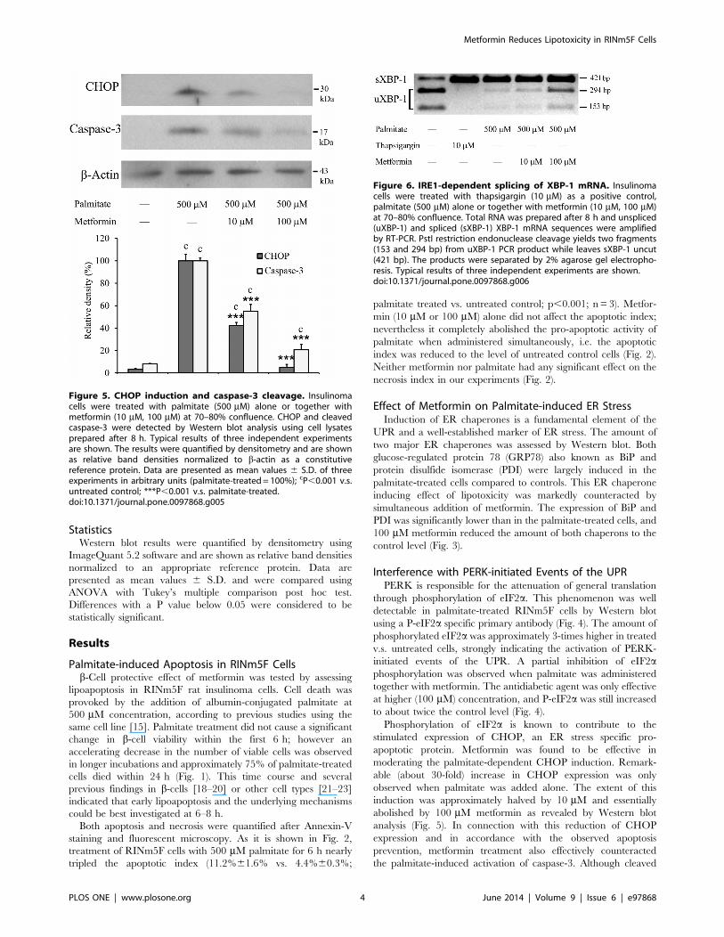
Figure 6. IRE1-dependent splicing of XBP-1 mRNA. Insulinomacells were treated with thapsigargin (10 mM) as a positive control,palmitate (500 mM) alone or together with metformin (10 mM, 100 mM)at 70–80% confluence. Total RNA was prepared after 8 h and unspliced(uXBP-1) and spliced (sXBP-1) XBP-1 mRNA sequences were amplifiedby RT-PCR. PstI restriction endonuclease cleavage yields two fragments(153 and 294 bp) from uXBP-1 PCR product while leaves sXBP-1 uncut(421 bp). The products were separated by 2% agarose gel electropho-resis. Typical results of three independent experiments are shown.doi:10.1371/journal.pone.0097868.g006
Metformin Reduces Lipotoxicity in RINm5F Cells
PLOS ONE | www.plosone.org 4 June 2014 | Volume 9 | Issue 6 | e97868

caspase-3 was still detectable in metformin-treated cells, its level
decreased in parallel with CHOP expression (Fig. 5).
Modulation of the IRE1 PathwayIRE1 activation is well represented by the excision of 26
nucleotides from XBP-1 mRNA, which can be visualized by
agarose gel electrophoresis after RT-PCR amplification and
endonuclease digestion of the affected region. This unconventional
splicing was revealed in palmitate-treated as well as in thapsi-
gargin-treated (positive control) RINm5F cells (Fig. 6). A marked
increase in the amount of unspliced XBP-1 mRNA demonstrates
the concentration dependent antagonistic effect of metformin on
palmitate-induced IRE-1 activation. However, large amounts of
spliced mRNA can be seen in the palmitate and metformin treated
samples, which indicates that this branch of the UPR signaling
network is inhibited to a relatively lower extent (Fig. 6).
Phosphorylation of JNK is also mediated by activated IRE-1.
The two most important substrates of P-JNK, c-Jun and IRS-1
play important roles in the induction of apoptosis and insulin
resistance. Phosphorylation of the two JNK isoforms, JNK1 and 2,
c-Jun and IRS-1 (Fig. 7) was detected by immunoblot using the
appropriate phosphorylation-specific antibodies. Largely en-
hanced JNK activation was found in palmitate-treated cells,
which was antagonized by metformin in a concentration
dependent manner. None of these phosphorylations was com-
pletely eliminated but they were all reduced to nearly half of the
extent revealed in palmitate-only-treated cells (Fig. 7).
Exposure of RINm5F to Metformin OnlyAs it was shown in Fig. 2, metformin treatment in the absence of
palmitate did not affect the intensity of apoptosis or necrosis in
RINm5F cells. The possible effect of metformin on the investi-
gated parameters of the UPR including caspase-3 activation and c-
Jun phosphorylation was also tested in our experimental condi-
tions. Palmitate treatment was applied as a positive control.
Metformin (10 or 100 mM) did not cause any statistically
significant change in the expression level of PDI, CHOP, eIF2a,c-Jun or JNK; in the phosphorylation of the latter three proteins or
in the activation of caspase-3 (Fig. 8).
Discussion
Insulin secretion in the pancreatic b-cells is stimulated in
response to nutrient abundance during the fed state. The primary
regulator is plasma glucose but its stimulatory effect is also
enhanced by FFAs and amino acids [3]. Increased insulin level
normally achieves the acceleration of glucose consumption in
various tissues (liver, skeletal muscle and adipose tissue). It also
shifts both protein and triglyceride turnovers toward the synthesis,
and thereby favours the utilization of plasma amino acids and
FFAs, too [24].
Overfeeding increases the challenge to b-cells, which need to
synthesize and secrete more insulin. The balance can be
maintained as long as the main metabolic tissues (liver, skeletal
muscle and adipose tissue) obey and increase their contribution.
However, insulin-responsiveness and the fuel utilizing capacity of
Figure 7. Phosphorylation of JNK, c-Jun and IRS-1. Insulinoma cells were treated with palmitate (500 mM) alone or together with metformin(10 mM, 100 mM) at 70–80% confluence. Total and phosphorylated JNK (two isoforms), phosphorylated (at Ser 307) IRS-1, total and phosphorylated c-Jun were detected by Western blot analysis using cell lysates prepared after 8 h. Typical results of three independent experiments are shown. Theresults were quantified by densitometry and are shown as relative band densities normalized to b-actin as a constitutive reference protein. Data arepresented as mean values 6 S.D. of three experiments in arbitrary units (palmitate-treated= 100%); aP,0.05, cP,0.001 v.s. untreated control; *P,0.05, **P,0.01 ***P,0.001 v.s. palmitate-treated.doi:10.1371/journal.pone.0097868.g007
Metformin Reduces Lipotoxicity in RINm5F Cells
PLOS ONE | www.plosone.org 5 June 2014 | Volume 9 | Issue 6 | e97868

the body largely depend on genetic predisposition and environ-
mental factors. The lack of physical activity and the related obesity
are considered as important causes of insulin resistance, the
primary disorder in type 2 diabetes [1]. It leads to elevation of
plasma FFA level, which further aggravates insulin resistance [25].
The vicious cycle may culminate in b-cell death and decreased
insulin producing capacity. Prevention of type 2 diabetes or
hindrance of its progression by a variety of lifestyle changes and
drugs is likely dependent on b-cell protection. Moreover, it
became evident that counteraction of b-cell failure is a promising
therapeutic strategy. It is therefore, important to investigate the
current anti-diabetic agents from this aspect [11].
Deleterious effects of elevated FFA levels on b-cells and the role
of lipotoxicity in diabetes were discovered long ago [26]. Further
investigation of the phenomenon revealed the involvement of
lipotoxic ER stress [7]. One of the primary adaptive mechanisms
of ER stress is the attenuation of general translation through
phosphorylation of eIF2a, which can decrease the insulin secreting
capacity of b-cells. In addition, prolonged and severe ER stress
induces apoptosis, and thereby contributes to the reduction of b-cell mass. ER stress dependent activation of JNK is one of the
main pro-apoptotic events, which also favors insulin resistance by
means of Ser-phosphorylation of IRS-1 [27]. Although this latter
mechanism was primarily studied in the main metabolic tissues
(liver, skeletal muscle etc.), it turned out to be important in the
derangement of the control of insulin secretion in the b-cells [10].Metformin is one of the leading anti-diabetic drugs. Its most
appreciated effect is the improvement of insulin responsiveness;
however, its direct b-cell protective effect was also demonstrated
long ago [13]. Although metformin has been shown to increase
AMP-activated protein kinase activity, its molecular target has not
been unequivocally elucidated [28]. Our results show that
metformin significantly reduces lipotoxicity in a b-cell line.
Palmitate-induced apoptosis and some major events of the
underlying ER stress response (i.e. PDI and Grp78 induction
and eIF2a phosphorylation) were practically abolished by
metformin in a concentration-dependent manner. Interestingly,
the IRE1 pathway of the UPR (i.e. unconventional splicing of
XBP-1 mRNA and JNK, c-Jun and IRS-1phosphorylation)
showed a markedly lower extent of inhibition. Most importantly,
however, induction of the pro-apoptotic transcription factor
CHOP and generation of the cleaved effector caspase-3 were also
largely repressed by metformin, which can underlie the observed
decrease in palmitate-induced apoptosis. The apparent discrep-
ancy between the completely abrogated apoptosis and the less
pronounced JNK, c-Jun and IRS-1 phosphorylations can be
explained by the convergence of the UPR pathways. In contrast to
the phosphorylation of JNK, c-Jun and IRS-1, which are clearly
associated to the IRE1 pathway, CHOP induction is due to a
coordinated action of all the three branches of the UPR. The
expression of CHOP is controlled simultaneously by three major
ER-stress-related transcription factors (ATF6, the PERK-depen-
dent ATF4 and the IRE1-dependent XBP-1) [29]. Therefore, the
remaining activity of only one signaling pathway might be unable
to maintain elevated CHOP levels and stimulated apoptosis.
Similar effects of metformin, i.e. cell protection and prevention
of lipotoxic ER stress have been observed also in HepG2 human
hepatoma cell line [12]. In line with our findings, the phenomenon
was accompanied by a reduced Ser-phosphorylation of IRS-1,
which might contribute to insulin-sensitizing in hepatocytes. Our
findings demonstrating these effects of metformin in a rat
insulinoma cell line have a great importance since b-cell protectionand maintenance of insulin sensitivity in the b-cells are of
particular significance in the prevention and treatment of diabetes.
Preventive effect of metformin on ER stress-induced apoptosis
in NIT-1 cells (a mouse pancreatic beta cell line) has been recently
Figure 8. Treatment with metformin only. Insulinoma cells were treated with 500 mM palmitate (positive control) or metformin (10 mM, 100 mM)at 70–80% confluence. PDI, CHOP, cleaved caspase-3, Total and phosphorylated eIF2a, total and phosphorylated JNK (two isoforms), total andphosphorylated c-Jun and b-actin as a constitutive reference protein were detected by Western blot analysis using cell lysates prepared after 8 h.Total RNA was also prepared after 8 h and unspliced (uXBP-1) and spliced (sXBP-1) XBP-1 mRNA sequences were amplified by RT-PCR. PstI restrictionendonuclease cleavage yields two fragments (153 and 294 bp) from uXBP-1 while leaves sXBP-1 uncut (421 bp). The products were separated by 2%agarose gel electrophoresis. Typical results of three independent experiments are shown.doi:10.1371/journal.pone.0097868.g008
Metformin Reduces Lipotoxicity in RINm5F Cells
PLOS ONE | www.plosone.org 6 June 2014 | Volume 9 | Issue 6 | e97868

reported [14]. ER stress was provoked by the SERCA inhibitor
thapsigargin and, unlike palmitate-induced ER stress in our study,
it was not found to be counteracted. Nevertheless, the consequent
apoptosis as well as JNK activation and IRE-1 phosphorylation
were efficiently reduced by metformin. These effects were
attributed to AMP-activated protein kinase and phosphatidylino-
sitol-3 kinase activation. These data suggest that, besides the
evident amelioration of ER stress, additional mechanisms might
contribute to the abrogation of lipoapoptosis and the massive
suppression of JNK activation in our experiments.
In summary, our findings further support the b-cell protectivepotential of metformin. Attenuation of lipoapoptosis in RINm5F
rat insulinoma cell line can be attributed to modulation of
palmitate-induced ER stress response in general. Decreased
activation of JNK is of special importance because of its role in
both the induction of apoptosis and the development of insulin
resistance. Besides the partly restored insulin sensitivity, an
enhanced durability of b-cells might underlie the improved
prognosis of metformin treated diabetic patients.
Acknowledgments
We would like to thank Mrs. Valeria Mile for her skillful technical
assistance and Veronika Kosa for her help in preparing the figures.
Author Contributions
Conceived and designed the experiments: GK MC. Performed the
experiments: LSS MK. Analyzed the data: LSS MK JM GK MC.
Contributed to the writing of the manuscript: MC LSS MC.
References
1. Ye J (2013) Mechanisms of insulin resistance in obesity. Front Med 7: 14–24.
2. Itoh Y, Kawamata Y, Harada M, Kobayashi M, Fujii R, et al. (2003) Free fatty
acids regulate insulin secretion from pancreatic beta cells through GPR40.
Nature 422: 173–176.
3. Fu Z, Gilbert ER, Liu D (2013) Regulation of insulin synthesis and secretion and
pancreatic Beta-cell dysfunction in diabetes. Curr Diabetes Rev 9: 25–53.
4. Marchetti P, Bugliani M, Boggi U, Masini M, Marselli L (2012) The pancreatic
beta cells in human type 2 diabetes. Adv Exp Med Biol 771: 288–309.
5. Shimabukuro M, Zhou YT, Levi M, Unger RH (1998) Fatty acid-induced beta
cell apoptosis: a link between obesity and diabetes. Proc Natl Acad Sci U S A 95:
2498–2502.
6. Welters HJ, Tadayyon M, Scarpello JH, Smith SA, Morgan NG (2004) Mono-
unsaturated fatty acids protect against beta-cell apoptosis induced by saturated
fatty acids, serum withdrawal or cytokine exposure. FEBS Lett 560: 103–108.
7. Cnop M, Igoillo-Esteve M, Cunha DA, Ladriere L, Eizirik DL (2008) An update
on lipotoxic endoplasmic reticulum stress in pancreatic beta-cells. Biochem Soc
Trans 36: 909–915.
8. Mandl J, Meszaros T, Banhegyi G, Hunyady L, Csala M (2009) Endoplasmic
reticulum: nutrient sensor in physiology and pathology. Trends Endocrinol
Metab 20: 194–201.
9. Cunha DA, Hekerman P, Ladriere L, Bazarra-Castro A, Ortis F, et al. (2008)
Initiation and execution of lipotoxic ER stress in pancreatic beta-cells. J Cell Sci
121: 2308–2318.
10. Accili D (2004) Lilly lecture 2003: the struggle for mastery in insulin action: from
triumvirate to republic. Diabetes 53: 1633–1642.
11. Bonora E (2008) Protection of pancreatic beta-cells: is it feasible? Nutr Metab
Cardiovasc Dis 18: 74–83.
12. Kim DS, Jeong SK, Kim HR, Kim DS, Chae SW, et al. (2010) Metformin
regulates palmitate-induced apoptosis and ER stress response in HepG2 liver
cells. Immunopharmacol Immunotoxicol 32: 251–257.
13. Lupi R, Del Guerra S, Fierabracci V, Marselli L, Novelli M, et al. (2002)
Lipotoxicity in human pancreatic islets and the protective effect of metformin.
Diabetes 51 Suppl 1: S134–137.
14. Jung TW, Lee MW, Lee YJ, Kim SM (2012) Metformin prevents endoplasmic
reticulum stress-induced apoptosis through AMPK-PI3K-c-Jun NH2 pathway.
Biochem Biophys Res Commun 417: 147–152.
15. Beeharry N, Chambers JA, Green IC (2004) Fatty acid protection from palmitic
acid-induced apoptosis is lost following PI3-kinase inhibition. Apoptosis 9: 599–
607.
16. O’Brien R, Gottlieb-Rosenkrantz P (1970) An automatic method for viabilityassay of cultured cells. J Histochem Cytochem 18: 581–589.
17. Williams BL, Lipkin WI (2006) Endoplasmic reticulum stress and neurodegen-
eration in rats neonatally infected with borna disease virus. J Virol 80: 8613–8626.
18. Baldwin AC, Green CD, Olson LK, Moxley MA, Corbett JA (2012) A role foraberrant protein palmitoylation in FFA-induced ER stress and beta-cell death.
Am J Physiol Endocrinol Metab 302: E1390–1398.19. Oh YS, Lee YJ, Kang Y, Han J, Lim OK, et al. (2013) Exendin-4 inhibits
glucolipotoxic ER stress in pancreatic beta cells via regulation of SREBP1c and
C/EBPbeta transcription factors. J Endocrinol 216: 343–352.20. Sommerweiss D, Gorski T, Richter S, Garten A, Kiess W (2013) Oleate rescues
INS-1E beta-cells from palmitate-induced apoptosis by preventing activation ofthe unfolded protein response. Biochem Biophys Res Commun 441: 770–776.
21. Cho H, Wu M, Zhang L, Thompson R, Nath A, et al. (2013) Signaling
dynamics of palmitate-induced ER stress responses mediated by ATF4 inHepG2 cells. BMC Syst Biol 7: 9.
22. Olsen GS, Hansen BF (2002) AMP kinase activation ameliorates insulinresistance induced by free fatty acids in rat skeletal muscle. Am J Physiol
Endocrinol Metab 283: E965–970.
23. Tao JL, Wen YB, Shi BY, Zhang H, Ruan XZ, et al. (2012) Endoplasmicreticulum stress is involved in podocyte apoptosis induced by saturated fatty acid
palmitate. Chin Med J (Engl) 125: 3137–3142.24. Combettes-Souverain M, Issad T (1998) Molecular basis of insulin action.
Diabetes Metab 24: 477–489.25. Frohnert BI, Jacobs DR, Jr., Steinberger J, Moran A, Steffen LM, et al. (2013)
Relation between Serum Free Fatty Acids and Adiposity, Insulin Resistance and
Cardiovascular Risk Factors from Adolescence to Adulthood. Diabetes.26. Unger RH (1995) Lipotoxicity in the pathogenesis of obesity-dependent
NIDDM. Genetic and clinical implications. Diabetes 44: 863–870.27. Aguirre V, Uchida T, Yenush L, Davis R, White MF (2000) The c-Jun NH(2)-
terminal kinase promotes insulin resistance during association with insulin
receptor substrate-1 and phosphorylation of Ser(307). J Biol Chem 275: 9047–9054.
28. Musi N, Hirshman MF, Nygren J, Svanfeldt M, Bavenholm P, et al. (2002)Metformin increases AMP-activated protein kinase activity in skeletal muscle of
subjects with type 2 diabetes. Diabetes 51: 2074–2081.29. Kim I, Xu W, Reed JC (2008) Cell death and endoplasmic reticulum stress:
disease relevance and therapeutic opportunities. Nat Rev Drug Discov 7: 1013–
1030.
Metformin Reduces Lipotoxicity in RINm5F Cells
PLOS ONE | www.plosone.org 7 June 2014 | Volume 9 | Issue 6 | e97868