Medullosa steinii sp. nov., a seed fern vine from the Upper Mississippian
18
Medullosa steinii sp. nov., a seed fern vine from the Upper Mississippian Michael T. Dunn a; , Michael Krings b , Gene Mapes a , Gar W. Rothwell a , Royal H. Mapes c , Sun Keqin d a Department of Environmental and Plant Biology, Ohio University, Athens, OH 45701, USA b Department of Ecology and Evolutionary Biology, University of Kansas, Lawrence, KS 66045, USA c Department of Geological Sciences, Ohio University, Athens, OH 45701, USA d China University of Geosciences, Beijing 100083, PR China Received 4 July 2002; received in revised form 12 November 2002; accepted 9 December 2002 Abstract Three specimens of a medullosan seed fern stem with attached leaf bases, from the Chesterian Series of Arkansas, USA, are the earliest unequivocal occurrence of the genus Medullosa and form the basis for a new species Medullosa steinii. The stems have numerous features characteristic of vines, including stem diameters of only 2^3 cm in diameter, numerous wide, plate-like rays, relatively wide tracheids, the presence of cortical spines and variable but numerous stelar segments. In M. steinii vascular segment numbers vary from two to eight. Primary xylem bundles consist of one to several protoxylem poles with metaxylem and abundant parenchyma; each bundle is surrounded by secondary vascular tissue. Leaf traces typically diverge from a vascular segment as pairs, with the resulting bundles separating from each other at an angle of 90^100‡. Paired leaf traces are separated from each other by a wedge of secondary vascular tissue, thus producing a distinctive mode of leaf trace production for the new species. Leaf traces divide repeatedly to produce the Myeloxylon rachis base configuration at the margin of the stem cortex. Rachis bases are decurrent and are separated from one another by a discontinuous row of sclerotic bundles and from the stem by a prominent periderm. The discovery of this plant demonstrates that scrambling and climbing seed ferns were part of some plant communities by Mississippian time. ȣ 2003 Elsevier Science B.V. All rights reserved. Keywords: liana; Medullosa ; Mississippian; Namurian; seed fern; vine 1. Introduction The Medullosaceae comprises one of the most distinctive, diverse and widely distributed groups of Late Paleozoic gymnosperms (Taylor and Tay- lor, 1993). Vegetative and reproductive remains are well known both from permineralized speci- mens (Stewart and Rothwell, 1993) and from compression/impression fossils (Taylor and Tay- lor, 1993). This family is distinguished by a unique mode of leaf trace production (Mapes and Rothwell, 1980), in which fronds are vascu- larized by terete strands produced by several cau- 0034-6667 / 03 / $ ^ see front matter ȣ 2003 Elsevier Science B.V. All rights reserved. doi :10.1016/S0034-6667(02)00254-3 * Corresponding author. Tel.: +1-740-593-1118; Fax: +1-740-593-1130. E-mail address: [email protected] (M.T. Dunn). Review of Palaeobotany and Palynology 124 (2003) 307^324 R Available online at www.sciencedirect.com www.elsevier.com/locate/revpalbo
-
Upload
independent -
Category
Documents
-
view
1 -
download
0
Transcript of Medullosa steinii sp. nov., a seed fern vine from the Upper Mississippian
Medullosa steinii sp. nov., a seed fern vine fromthe Upper Mississippian
Michael T. Dunn a;�, Michael Krings b, Gene Mapes a, Gar W. Rothwell a,Royal H. Mapes c, Sun Keqin d
a Department of Environmental and Plant Biology, Ohio University, Athens, OH 45701, USAb Department of Ecology and Evolutionary Biology, University of Kansas, Lawrence, KS 66045, USA
c Department of Geological Sciences, Ohio University, Athens, OH 45701, USAd China University of Geosciences, Beijing 100083, PR China
Received 4 July 2002; received in revised form 12 November 2002; accepted 9 December 2002
Abstract
Three specimens of a medullosan seed fern stem with attached leaf bases, from the Chesterian Series of Arkansas,USA, are the earliest unequivocal occurrence of the genus Medullosa and form the basis for a new species Medullosasteinii. The stems have numerous features characteristic of vines, including stem diameters of only 2^3 cm in diameter,numerous wide, plate-like rays, relatively wide tracheids, the presence of cortical spines and variable but numerousstelar segments. In M. steinii vascular segment numbers vary from two to eight. Primary xylem bundles consist of oneto several protoxylem poles with metaxylem and abundant parenchyma; each bundle is surrounded by secondaryvascular tissue. Leaf traces typically diverge from a vascular segment as pairs, with the resulting bundles separatingfrom each other at an angle of 90^100‡. Paired leaf traces are separated from each other by a wedge of secondaryvascular tissue, thus producing a distinctive mode of leaf trace production for the new species. Leaf traces dividerepeatedly to produce the Myeloxylon rachis base configuration at the margin of the stem cortex. Rachis bases aredecurrent and are separated from one another by a discontinuous row of sclerotic bundles and from the stem by aprominent periderm. The discovery of this plant demonstrates that scrambling and climbing seed ferns were part ofsome plant communities by Mississippian time.6 2003 Elsevier Science B.V. All rights reserved.
Keywords: liana; Medullosa ; Mississippian; Namurian; seed fern; vine
1. Introduction
The Medullosaceae comprises one of the mostdistinctive, diverse and widely distributed groups
of Late Paleozoic gymnosperms (Taylor and Tay-lor, 1993). Vegetative and reproductive remainsare well known both from permineralized speci-mens (Stewart and Rothwell, 1993) and fromcompression/impression fossils (Taylor and Tay-lor, 1993). This family is distinguished by aunique mode of leaf trace production (Mapesand Rothwell, 1980), in which fronds are vascu-larized by terete strands produced by several cau-
0034-6667 / 03 / $ ^ see front matter 6 2003 Elsevier Science B.V. All rights reserved.doi:10.1016/S0034-6667(02)00254-3
* Corresponding author. Tel. : +1-740-593-1118;Fax: +1-740-593-1130.
E-mail address: [email protected] (M.T. Dunn).
PALBO 2519 18-4-03
Review of Palaeobotany and Palynology 124 (2003) 307^324
R
Available online at www.sciencedirect.com
www.elsevier.com/locate/revpalbo

line protoxylem sympodia over several cm ofstem. Other pteridosperms (e.g. Calamopityaceae,Callistophytaceae, and Lyginopteridaceae) possessleaves that are vascularized by traces that are pro-duced by one or two sympodia, but typically at asingle node (Mapes and Rothwell, 1980). The ge-neritype, Medullosa Cotta 1832, is based on per-mineralized stems ¢rst described from Permiandeposits of Germany (Cotta, 1832). This genushas been most completely characterized fromPennsylvanian aged specimens of North Americaand England (Delevoryas, 1955).Early workers considered the distinctive stelar
con¢gurations of medullosans to be the result ofsyngenesis as envisioned by Zimmermann in histelome theory (Zimmermann, 1952). We now real-ize however, that the medullosan stele is a uniqueeustele with secondary tissues produced concentri-cally around two or more stelar segments (i.e.‘vascular segments’ of Basinger et al., 1974). Thegenus Medullosa is distinguished from the otherthree currently recognized genera of medullosanstems, Quaestora Mapes and Rothwell 1980, Sut-cli⁄a Scott 1906, and Colpoxylon Brongniart1849, by vascular system morphology. Quaestorahas a cruciform protostele and decussate phyllo-taxis (Mapes and Rothwell, 1980). Sutcli⁄a alsohas a protostele, but has helical phyllotaxis andlarge leaf traces (Scott, 1906; Phillips and An-drews, 1963) with concentric secondary xylem sur-rounding older leaf traces and concentric phloemsurrounding younger leaf traces (Stidd et al.,1975). Colpoxylon is similar to a number of spe-cies ofMedullosa except that Colpoxylon producesno vascular strands in the pith (i.e. star rings;Seward, 1917; Stewart and Rothwell, 1993).The earliest previously reported occurrences of
Medullosa are an undescribed specimen ¢gured byTaylor and Eggert (1967) from the upper Chester-ian (upper Namurian A) Imo Formation of Ar-kansas and a poorly preserved fragment of a me-dullosan stem from the uppermost Namurian A(Upper Poruba Beds) of Poland (Brzyski and Stu-chlik, 1992). Neuropteris-type foliage is consideredto be one of the foliage forms produced by me-dullosans (Stewart and Delevoryas, 1956; Stidd,1981). Although this foliage is known from upperTournaisian strata, it is possible that it may have
been produced by some non-medullosan plants(Gensel, 1988).The three new specimens described herein rep-
resent the oldest unequivocal evidence for thegenus Medullosa. One of these specimens (M47)has been previously ¢gured but not described(Stein, 1993). All are somewhat crushed stemswith diverging leaf bases. The specimens are fromthe Imo (upper, Chesterian/upper Namurian A)and Fayetteville Formations (middle Chesterian/lower Namurian A) of northwestern Arkansas,that consist of o¡shore marine strata and con-tain mixed marine and terrestrial assemblages.Marine invertebrates and palynomorphs permitaccurate biostratigraphic dating that can be glob-ally correlated (Owens et al., 1979; Meeks et al.,1997).The specimens described in this study possess
several unique characters that indicate a new spe-cies. The consistently narrow diameter of thestems, a stele comprising several vascular seg-ments immersed in ground tissue, numerous largeplate-like xylem rays, and cortical spines indicatethat this ancient medullosan species was a vine.The occurrence of vines in Upper Mississippianstrata demonstrates the presence of this form ofgrowth architecture early in the development ofcomplex plant communities (Scott, 1980; Scheck-ler, 1986; DiMichele and Hook, 1992; Cleal andThomas, 1999).
2. Materials and methods
The three specimens examined are preserved bycalcareous and pyrite cellular permineralization.One specimen (M71) was recovered from a roadcut on Hwy 65 through the Imo Formation(upper Chesterian, upper Namurian A; Saunderset al., 1977) and two specimens (M47, M2362)were recovered from the Trace Creek exposureof the Fayetteville Formation (middle Chesterian,lower Namurian A; Meeks et al., 1997). Speci-mens were analyzed by a combination of thinsections (Stein et al., 1982) and cellulose acetatepeels (Joy et al., 1956). Photo documentation wasaccomplished with a Phase One scanning digitalcamera. Images were processed and plates were
PALBO 2519 18-4-03
M.T. Dunn et al. / Review of Palaeobotany and Palynology 124 (2003) 307^324308
constructed using Adobe Photoshop 5.5, stored asTIFF and PSD ¢les and printed on a ShinkoCH446i dye-sublimation printer.
3. Systematics
Class : Spermatopsida
Order : Medullosales
Family : Medullosaceae
Genus : Medullosa Cotta 1832
Species : Medullosa steinii Dunn et al. sp. nov.(Plates I^IV)
Holotype : The holotype of Medullosa steinii con-sists of 55 cellulose acetate peels and nine thinsections mounted on microscope slides of speci-men M47: OUPH Nos. 14101^14104, 14112,14113, 14115^14120, 14131^14184.Paratype : The paratype of Medullosa steinii con-sists of 198 cellulose acetate peels and 34 thinsections mounted on microscope slides, and 12sections of permineralized stem of specimenM2362: OUPH Nos. 14100, 14105^14111, 14114,14185^14407, 14484^14494.Repository : Specimens are reposited in the OhioUniversity Paleobotanical Herbarium, OUPHNos. 14100^14494.Type locality : M47, M2362: Trace Creek, SE1/4,Sec. 16, T14N, R15W (Leslie 71/2P Quadrangle),Searcy County, Arkansas.Stratigraphic position : M47, M2362: Lower un-named shale member of Fayetteville Formation,Hombergian Stage, middle Chesterian Series,Upper Mississippian, (lower Serpukhovian/Pende-lian/E1 equivalent).Other specimens : M71: OUPH Nos. 14408^14483: Elviran Stage, upper Chesterian Series,Upper Mississippian (middle Serpukhovian/Arns-bergian/E2 equivalent). Road cut on Hwy 65 ap-proximately 6.4 km SE of Leslie, AR, NE 1/4,Sec. 11, T13N, R15W (Leslie 71/2P Quadrangle),Searcy County, Arkansas.Etymology : The speci¢c epithet steinii honors
Professor William E. Stein for his contributionsto the study of early seed plants.Diagnosis : Stem narrow, less than 3.5 cm maxi-mum diameter. Stele of two to eight anastomosingvascular segments each containing a sympodiumof the eustele. Primary xylem marginal mesarch.Secondary xylem development concentric andgenerally radially symmetrical. Leaf traces divergehelically, arising from a single protoxylem emis-sion area that divides into two leaf traces sepa-rated by a wedge of secondary vascular tissue. Aperiderm surrounds the stem and discontinuoussclerenchymatous zones separate diverging decur-rent leaf bases. Secretory canals bounded by asheath of cells present in outer cortex. Hypoder-mis radially expanded comprising two to threediscontinuous layers of parenchyma cells anasto-mosing among circular to oval longitudinally ori-ented strands of ¢bers. Cortical spines present onepidermis of leaf bases.
4. Results
4.1. General features
The three specimens of Medullosa steinii are allunbranched segments of somewhat crushed stemsthat consist of a complete stele with varyingamounts of external cortical tissues. They are2.6^3.2U2.2^1.7 cm in maximum diameter, andare 25 cm (M47), 40 cm (M71) and 42 cm(M2362) long (Table 1). Each stem segment con-sists of a ring of two to eight cauline vascularsegments embedded in parenchymatous groundtissue (Plate I, 1 and 2). The number of vascularsegments varies among specimens, and also canchange from level to level in the same stem dueto anastomoses (Fig. 1). Ground tissues of thestem consist of parenchymatous tissue in pithand surrounding vascular segments, peridermthat surrounds the stele, and cortex in the formof downward extensions of decurrent leaf bases invarious stages of development. The margins ofadjacent leaf bases are characterized by discontin-uous sclerenchymatous zones. The hypodermalzone consists of longitudinally oriented strandsof ¢bers interspersed with parenchyma and secre-
PALBO 2519 18-4-03
M.T. Dunn et al. / Review of Palaeobotany and Palynology 124 (2003) 307^324 309

PALBO 2519 18-4-03
M.T. Dunn et al. / Review of Palaeobotany and Palynology 124 (2003) 307^324310

tory canals. A thin layer that represents an epi-dermis with cortical spines is present.
4.2. Stele
The stele consists of two to eight vascular seg-ments that are surrounded by parenchymatousground tissue (Plate I, 1), including a pith (PlateI, 1 and 3). In M47 and M71 the pith parenchymacells are crushed (Plate I, 2). The vascular seg-ments anastomose along the length of two(M47, Plate I, 3^5; M2362, Fig. 1) of the speci-mens. When viewed in a series of transverse sec-tions, segment fusion is initiated by widening ofthe rays and shortening of the rows of radiallyaligned tracheids of the secondary xylem (PlateI, 3) until the secondary tissues become continu-ous and the primary tissues merge into a singlebundle (Plate I, 4^5). The number of vascular seg-ments varies from two to three in M47, and fromfour to eight, plus a single star ring in M2362(Plate II, 1). The third specimen (M71) is incom-pletely preserved and has three or possibly fourvascular segments. In cross section vascular seg-ments range from terete to tangentially elongatedand lobed (Plate II, 1).
4.3. Primary xylem
In transverse section, primary xylem is repre-sented by two to eight (Table 1) vascular seg-ments, each with one to several protoxylemstrands, metaxylem mixed with parenchyma, and
diverging leaf traces. The maximum radial thick-ness of individual vascular segments ranges from144 Wm (M2362) to 235 Wm (M47). Protoxylembundles are located toward the outer edge of themetaxylem (Plate II, 4) as is characteristic of me-dullosan seed ferns (Basinger et al., 1974), and aremarginally mesarch. Secondary wall thickeningpatterns of cauline protoxylem tracheids are sca-lariform (Plate II, 2) as are tracheids of leaf traces(Plate II, 3). Mature metaxylem tracheids exhibitalternate, multi-seriate wall thickening patterns onall cell walls. Metaxylem tracheids occur in clus-ters interspersed with xylem parenchyma (Plate II,4 and 7). Primary xylem parenchyma cells are 45^120 Wm in diameter (mean= 80 Wm) and metaxy-lem tracheids are 44^252 Wm in diameter (mean=110 Wm).
4.4. Secondary xylem
Secondary xylem surrounds each vascular seg-ment except at areas of leaf trace divergence(Plate I, 1 and 2; Plate II, 1). Development ofsecondary xylem is generally equivalent on allsides of the primary xylem (Plate 1, 1 and 2) incontrast to endocentric secondary xylem develop-ment in younger species of Medullosa (Baxter,1949; Phillips and Andrews, 1963). Radial rowsof tracheids average 21 cells long (Plate I, 3^5)and are one to ¢ve cells wide with three to fourcells being the most common (Plate II, 4 and 5).Secondary tracheids are circular to oval in crosssection (Plate II, 5) and are 44^155 Wm in diam-
Plate I. Medullosa steinii Dunn et al. sp. nov. Vascular segment arrangement. M number indicates specimen collection number,OUPH indicates accession numbers in the Paleobotanical Herbarium of Ohio University.
1. Cross section of stem showing eight radially symmetrical vascular segments surrounding a parenchymatous pith (pi).M2362-me10, OUPH 14100, scale bar= 2 mm, U3.
2. Cross section of stem showing two band-shaped vascular segments and three decurrent leaf bases. Note discontinuoussclerenchyma bands separating leaf bases. M47-ab1, OUPH 14101, scale bar= 2 mm, U33.
3^5 Sequence of merging vascular segments. Scale bar= 1 mm, U11.3. Vascular segments converging. Note crushed region that represents a pith (pi), stele cortex (1‡C) and numerous rays in the
secondary xylem. M47-gh38, OUPH 14102.4. Vascular segments merging as rays widen and radially aligned rows of secondary tracheids shorten. M47-gh32, OUPH
14103.5. Vascular segments merged as secondary tissues (2‡x) become continuous and primary tissues (1‡x) form a single bundle.
M47-gh30, OUPH 14104.
PALBO 2519 18-4-03
M.T. Dunn et al. / Review of Palaeobotany and Palynology 124 (2003) 307^324 311

eter (mean= 100). Pitting is multi-seriate, alter-nate (Plate II, 6), with circular to oval apertures.Rays are numerous, and except where they wi-
den due to anastomoses of the vascular segments(Plate I, 3^5) are 30^110 Wm in diameter(mean= 45). Xylem ray parenchyma occurs inrows one to ¢ve cells wide; in stem cross sectionthese cells are elongate radially (Plate II, 5) and
are at least 300 Wm long. In longitudinal andoblique sections ray parenchyma cells are rectan-gular to isodiametric (Plate II, 7).
4.5. Vascular cambium and secondary phloem
A vascular cambium is represented by fusiformand ray initials that are preserved at the periphery
PALBO 2519 18-4-03
M.T. Dunn et al. / Review of Palaeobotany and Palynology 124 (2003) 307^324312
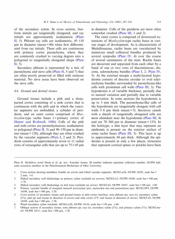
of the secondary xylem. In cross section, fusi-form initials are tangentially elongated, and rayinitials are approximately isodiametric (PlateII, 5). Phloem ray cells are isodiametric, 45^110Wm in diameter (mean= 80) when ¢rst di¡erenti-ated from ray initials. These cells are continuouswith primary cortex parenchyma, where theyare commonly crushed to varying degrees into apolygonal to tangentially elongated shape (PlateII, 5).Secondary phloem is represented by a mix of
parenchyma and sieve cells (Plate II, 5 and 7) thatare often poorly preserved or ¢lled with resinousmaterial. No sieve areas have been observed onthe sieve cells.
4.6. Ground and dermal tissues
Ground tissues include a pith and a three-parted cortex consisting of a stele cortex that iscontinuous with the pith and in which the vascu-lar segments are embedded, a cylinder of peri-derm, and an outer cortex comprised of Mye-loxylon-type rachis bases ( = primary cortex ofHamer and Rothwell, 1988). Cells of the pithand stele cortex are parenchymatous, isodiametricto polygonal (Plate II, 5) and 90^150 Wm in diam-eter (mean= 120), although they are often crushedby the vascular segments (Plate I, 2 and 3). Peri-derm consists of approximately seven to 11 radialrows of rectangular cells that are up to 75U45 Wm
in diameter. Cells of the periderm are most oftensomewhat crushed (Plate III, 1 and 2).The outer cortex is composed of downward ex-
tensions of Myeloxylon-type rachis bases in vari-ous stages of development. As is characteristic ofMedullosaceae, rachis bases are vascularized bynumerous small collateral bundles produced byseveral sympodia (Plate IV, 4) over the courseof several centimeters of the stem. Rachis basesare decurrent and separated from each other by aband of one or two rows of discontinuous, dis-crete, sclerenchyma bundles (Plate I, 2; Plate IV,5). At the external margin a multi-layered hypo-dermis consists of discrete circular to oval scler-enchyma bundles surrounded by parenchyma-likecells with prominent cell walls (Plate III, 3). Thehypodermis is of variable thickness, partially dueto natural variation and partially due to variablepreservation. In some sections the hypodermis isup to 3 mm thick. The parenchyma-like cells ofthe hypodermis are tangentially elongate with cellwalls 3^8 Wm thick (mean= 5). Secretory canalswith a sheath of tangentially elongated cells aremost abundant near the hypodermis (Plate III, 4)and are 70^260 Wm in diameter (mean= 135). Inthe holotype, a thin layer that may represent anepidermis is present on the exterior surface ofsome rachis bases (Plate III, 5). This layer is upto approximately 60 Wm thick. Although the epi-dermis is present in only a few places, structuresthat represent cortical spines or prickles have been
Plate II. Medullosa steinii Dunn et al. sp. nov. Vascular tissues. M number indicates specimen collection number, OUPH indi-cates accession numbers in the Paleobotanical Herbarium of Ohio University.
1. Cross section showing medullary bundle (at arrow) and lobed vascular segments. M2362-sc26, OUPH 14105, scale bar=2 mm, U4.
2. Helical secondary wall thickenings on primary xylem tracheids (at arrows). M2362-cr2, OUPH 14106, scale bar= 100 Wm,U70.
3. Helical secondary wall thickenings on leaf trace tracheids (at arrow). M2362-ls2, OUPH 14107, scale bar= 100 Wm, U80.4. Primary vascular bundle of marginal mesarch protoxylem (px), metaxylem (m) and parenchyma (pa). M2362-SF9, OUPH
14108, scale bar= 500 Wm, U20.5. Cross section of secondary tissues and ground tissues at pith^cortex interface, note phloem (p), rays (r), secondary xylem
(2‡x), pith (pi and tissues in direction of arrow) and stele cortex (1‡C and tissues in direction of arrow). M2363-sf3, OUPH14109, scale bar= 500 Wm, U30.
6. Pitted secondary xylem tracheids. M2362-cr20, OUPH 14110, scale bar= 150 Wm, U60.7. Oblique section of secondary tissues, note phloem (p), rays (r), secondary xylem (2‡x), and primary xylem (1‡x). M2362-me-
ls5. OUPH 14111, scale bar= 500 Wm, U20.
PALBO 2519 18-4-03
M.T. Dunn et al. / Review of Palaeobotany and Palynology 124 (2003) 307^324 313

Plate III. Medullosa steinii Dunn et al. sp. nov. Ground tissues and petiole morphology. M number indicates specimen collectionnumber, OUPH indicates accession numbers in the Paleobotanical Herbarium of Ohio University.
1. Periderm (pe) position. M47-ab3, OUPH 14112, scale bar= 1 mm, U17.2. Periderm morphology. M47-ab3, OUPH 14112, scale bar= 150 Wm, U100.3. Sparganum-type cortex with hypodermis of thick-walled parenchyma-like cells. M47-bf4, OUPH 14113, scale bar= 1 mm,
U15.4. Secretory canals. M2362-sd5, OUPH 14114, scale bar= 500 Wm, U40.5. Epidermis (e). M47-de28, OUPH 14115, scale bar= 150 Wm, U90.6. Cortical spine (cs). M47-gh38, OUPH 14102, scale bar= 500 Wm, U35.
PALBO 2519 18-4-03
M.T. Dunn et al. / Review of Palaeobotany and Palynology 124 (2003) 307^324314

identi¢ed on the exterior of the rachis bases (PlateIII, 6).
4.7. Vascular architecture and leaf trace divergence
Leaf traces arise from a single marginally me-sarch protoxylem emission area (Plate IV, 1) thatenlarges as it extends into the secondary xylem
(Plate IV, 2). At the outer margin of the second-ary xylem, leaf traces characteristically separatefrom a vascular segment as pairs, with the result-ing bundles diverging from each other at an angleof 90^100‡ (Plate IV, 3). Although the paired bun-dles are relatively large, they are not surroundedby secondary vascular tissues (Plate IV, 4). How-ever, they are separated from each other by a
Plate IV. Medullosa steinii Dunn et al. sp. nov. Leaf trace production. M number indicates specimen collection number, OUPHindicates accession numbers in the Paleobotanical Herbarium of Ohio University.
1. Protoxylem emission area (px). M47-F3, OUPH 14116, scale bar= 1 mm, U9.2. Protoxylem emission area (px) migrating into secondary tissues at the onset of leaf trace production. M47-de37, OUPH
14117, scale bar= 1 mm, U9.3. Leaf trace production. Note bifurcated protoxylem (px) separated by a wedge (w) of secondary tissue. M47-de15, OUPH
14118, scale bar= 1 mm, U9.4. Leaf trace production. Note leaf traces (lt) separated by a wedge (w) of secondary tissue and leaf traces (lt1, lt2) that were
produced by an older leaf trace emission area. M47-c8, OUPH 14119, scale bar= 1 mm, U9.5. Leaf trace production. Note leaf traces (lt) entering Myeloxylon-type petiole. M47-ab3, OUPH 14112, scale bar= 2 mm,
U4.6. Collateral leaf traces. Note xylem (x) and phloem (p). M47-b2, OUPH 14120, scale bar= 500 Wm, U25.
PALBO 2519 18-4-03
M.T. Dunn et al. / Review of Palaeobotany and Palynology 124 (2003) 307^324 315

wedge of secondary vascular tissue, thus produc-ing a unique mode of leaf trace production for thenew species (Plate IV, 5). Leaf traces divide re-peatedly, and along with traces produced proxi-mally, produce the Myeloxylon-type rachis basecon¢guration at the margin of the stem (PlateIV, 6). In one specimen (M47) leaf traces are pro-duced in a 3/8 Fibonacci fraction, whereas in an-other (M2362) leaf traces do not conform to aFibonacci fraction and are apparently in transi-tion from either a 3/8 to 2/5 or 2/5 to 3/8 Fibo-nacci fraction as discussed below.
5. Discussion
Initial descriptions of species of Medullosa uti-lized the number of vascular segments as the pri-mary systematic character for the genus (Smootand Taylor, 1981). Later, Schopf (1939) consid-ered the development of secondary xylem to beimportant phylogenetically, and subsequent au-thors (Stewart, 1951 and references therein) em-phasized leaf trace production for medullosan sys-tematics. Delevoryas (1955) used these charactersand others in his monograph that reduced thenumber of North American Medullosa speciesfrom approximately 13 to six (Table 2). However,other authors have recognized that the wide rangeof variation present in the anatomy of Medullosamakes identifying species di⁄cult (Phillips and
Andrews, 1963; Basinger et al., 1974) and re-cently, Hamer and Rothwell (1988) concludedthat the only reliable systematic character for spe-cies of Medullosa appears to be the mode of leaftrace production. We agree with Hamer andRothwell and propose that these new specimensbe known as Medullosa steinii based primarily ontheir unique mode of leaf trace production, withadditional characters such as protoxylem develop-ment, secondary xylem development, the presenceof star rings, vine-like habit, and stratigraphy (Ta-ble 2) further de¢ning this unique new species.Because leaf traces to a single rachis diverge
from several cauline protoxylem stands over alength of stem, the production of individual tracesdoes not reveal the entire pattern of vasculariza-tion. Divergence of individual bundles in Medul-losa steinii is characterized by a single trace (Fig.2a; Plate IV, 1) enlarging in diameter and extend-ing toward the periphery of the stem (Fig. 2b). Inthese areas secondary xylem production is re-duced (Plate IV, 2) or entirely absent. As the tracenears the periphery of the secondary xylem it di-vides into two leaf traces separated by a wedge ofsecondary tissues (Fig. 2c; Plate IV, 3). These leaftraces subsequently enter the Myeloxylon-type ra-chis base (Fig. 2d; Plate IV, 4 and 5) and dividerepeatedly to form numerous collateral bundlesthat are not accompanied by secondary tissues(Plate IV, 6).Leaf trace formation by the mode described
Table 1Range of variation of characters in Medullosa steinii
Character M47 M71 M2362
Length of stem segment 25 cm 40 cm 42 cmMax. diameter of stem 3.0U2.2 cm 2.6U2.2 cm 3.2U1.7 cmNo. of vasc. segments 2^3 3 4^8Max. radial thickness of vasc. segments 235 Wm 192 Wm 144 WmNo. of protoxylem emission areas 5(6) 5^6? 8(10)Leaf trace divergence pattern Helical Helical HelicalDia. of metaxylem tracheids 141 Wma (89^252) 104 Wma (67^155) 80 Wma (44^118)Dia. of 2‡ xylem tracheids 106 Wma (59^133) 113 Wma (44^155) 85 Wma (52^118)Cortical spines Present ? ?Sclerenchymatous zones Discontinuous round bndls Discontinuous round bndls Discontinuous round bndlsEpidermal cells Tabular irreg. shape ? ?Sclerotic hypodermis Mult. layers sclerenchyma
and parenchyma? Mult. layers sclerenchyma
and parenchymaDia. of secretory canals 145 Wma (80^260) ? 125 Wma (70^190)a mean dimensions.
PALBO 2519 18-4-03
M.T. Dunn et al. / Review of Palaeobotany and Palynology 124 (2003) 307^324316

above clearly distinguishes Medullosa steinii fromall previously described North American Medul-losa (Table 2). Except forMedullosa olseniae Rob-erts and Barghoorn 1952, where the method of
leaf trace production is unknown, all known spe-cies of Medullosa produce solitary leaf traces withvariable or absent secondary xylem. Medullosathompsonii Andrews 1945, Medullosa primaevaBaxter 1949, and Medullosa noei Steidtmann1937 produce leaf traces that are usually, butnot always, without accompanying secondary vas-cular tissues (Delevoryas, 1955). Concentric sec-ondary xylem surrounds the leaf traces of M. en-docentrica Baxter 1949 (Hamer and Rothwell,1988). In Medullosa anglica Scott 1899, crescent-shaped secondary xylem £anks the leaf traces asthey are ¢rst produced, then becomes concentric,and subsequently diminishes as the trace dividesnumerous times as it nears the periderm (Smootand Taylor, 1981).One of the more interesting ¢ndings of this in-
vestigation is the discovery that Medullosa steiniiproduces leaf traces in a Fibonacci fraction: 3/8 inone specimen (M47) and in transition from 3/8 to2/5 or from 2/5 to 3/8 in another (M2362). Thisfeature has not been reported in previous discus-sions of Medullosa and it is not clear what, if any,relationship this may have with the Fibonaccifraction of phyllotaxis. Phyllotaxis of Medullosaranges from 1/3 for Medullosa primaeva (Basingeret al., 1974), to 2/5 for Medullosa anglica (Smootand Taylor, 1981), to 3/8 for Medullosa endocen-trica (Hamer and Rothwell, 1988), and has notbeen determined for other North American Me-dullosa (Table 2). Changes in Fibonacci fractionof phyllotaxis during ontogeny have been re-ported for angiosperms (e.g. Crassulaceae, Jensen,1968) and a number of gymnosperms (e.g. Se-quoia, Sterling, 1945; Callistophyton, Rothwell,1975; Cordaitanthus, Rothwell, 1977). In M. stei-nii the signi¢cance of leaf trace production by
sf-5
se-7
sb-9
sc-32
sc-1
mh-2
mg-12
md-10
mb-9
ma-10
Number ofVascularSegments
ma
mb
mc
md
me
mf
mg
mh
sc
sa
sd
se
sf
sg
6
7
6576
56
6 1+
6 1+
7 1+
7
6
8
7
8
7
7
8
456
Fig. 1. Medullosa steinii Dunn et al. sp. nov. Variations ofvascular segment number of one specimen (M2362) due toanastomoses of the segments. Note segment numbers varyfrom four to eight with one star ring (+1) produced. m ands labels (i.e. ma or se) indicate stem segments broken duringpreservation or recovery, number (i.e. sc-1) indicates acetatepeel, or thin section number. Note vascular segment shapevaries from radially symmetrical to band-shaped to elongateand lobed. Stem longitudinal diagram U0.3, serial cross sec-tions U1.
PALBO 2519 18-4-03
M.T. Dunn et al. / Review of Palaeobotany and Palynology 124 (2003) 307^324 317

Table 2Comparison of selected characters of North American Medullosa species
Character M. steinii M. thomsoni M. primaeva M. noei M. endocentrica M. anglica M. olseniae(1, 4, 6) (3, 4, 5, 6, 13, 14) (6, 9, 12, 15) (5, 6, 7) (2, 6, 10, 11) (6, 8)
Vasc. Seg. Number 2^8 2^4 2^23 2^4 2 2^3 3^5Vasc. Seg.con¢guration
Variable Variable Variable Variable Elliptical to band Elongate,irregularly lobed
Elliptical
Stem dia. 6 3 cm 3.6^5 cm 2^20 cm 5^s 20 cm 6 2.3 cm @10^13.5U3.5^4cm
Fragment
Protoxylem Marginal mesarch ? Exarch Exarch? to mesarch Exarch Mesarch Mesarch?Leaf traceproduction
In pairs separatedby 2‡ tissue
Single may have2‡ tissue
Single may have2‡ tissue
Single, small mayhave 2‡ tissue
Single, large W/concentric 2‡ tissue
Single, 2‡ tissue£anking bundlebecoming concentric
?
2‡ xylemdevelopment
Radially symmetric Endocentric Radially symmetricto endocentric
Radially symmetricto endocentric
Endocentric Radially symmetricto endocentric
Endocentric
Star rings Present Present(?) Present Absent? Absent Absent AbsentPhyllotaxis ? ? 1/3 ? 3/8 2/5 ?Periderm Prominent Prominent Irregular Prominent Prominent Prominent ?Habit Vine Shrub/leaner? Tree Tree Vine ? ?Stratigraphy U Miss M Penn M Penn M-U Penn U Penn U Penn L Perm
Sources: 1, Andrews, 1945; 2, Andrews and Kernen, 1946; 3, Andrews and Mamay, 1953; 4, Basinger et al., 1974; 5, Baxter, 1949; 6, Delevoryas, 1955; 7, Hamerand Rothwell, 1988; 8, Roberts and Barghoorn, 1952; 9, Schopf, 1939; 10, Scott, 1899; 11, Smoot and Taylor, 1981; 12 Steidtmann, 1944; 13, Stewart, 1951; 14,15, Stewart and Delevoryas, 1952, 1956.
PALBO2519
18-4-03
M.T.Dunn
etal./R
eviewof
Palaeobotany
andPalynology
124(2003)
307^324318

Fibonacci fraction, and the relationship betweenleaf trace production and phyllotaxis cannot bedetermined because the rachis bases are vascular-ized by leaf traces from several cauline protoxy-lem strands over a length of stem, and rachis basepreservation is insu⁄cient.The variable vascular segment number and con-
¢guration of Medullosa steinii supports Andrews’(1945) supposition that vascular segment numberis not a reliable systematic character for this ge-nus. As noted above (Table 1), one of our speci-mens (M47) alternates between two and three vas-cular segments, one specimen (M71) may have aconstant number (three or four) of vascular seg-ments, and the other (M2362) has a wide range ofvariation with four to eight vascular segmentsover the length of the stem. Like vascular segmentnumber, vascular segment con¢guration of M.
steinii is also variable (Fig. 1), ranging from iso-diametric (Plate I, 1) to elliptical or band-shaped(Plate I, 2) to elongate and lobed (Plate II, 1).Therefore, the range of variation of vascular seg-ment number and con¢guration of M. steinii vir-tually covers the spectrum of previously reportedvascular segment number and con¢guration forall North American Medullosa (Table 2).The production of marginal mesarch protoxy-
lem helps to distinguish Medullosa steinii fromMedullosa primaeva (Stewart and Delevoryas,1952) and Medullosa endocentrica (Hamer andRothwell, 1988), both of which produce exarchprotoxylem. However, Medullosa noei producesquestionably exarch to mesarch protoxylem(Stewart, 1951) and protoxylem maturation hasnot been determined in Medullosa thompsonii,Medullosa anglica, and Medullosa olseniae : there-fore this character has limited systematic value.Endocentric secondary xylem development is
often reported as the general condition for thegenus Medullosa (Baxter, 1949; Stewart and Dele-voryas, 1956). However, Schopf (1939) suggestedthat the endocentric condition may be the ances-tral character state and that secondary xylem de-velopment underwent a transformation seriesfrom endocentric to radially symmetric to exocen-tric through the Pennsylvanian and into the Per-mian. This was suggested to be a possible adap-tation for structural support as tree-like growtharchitecture evolved in Medullosa (Schopf,1939). Data from this study do suggest that theearliest Medullosa were vine-like (see below) andthus support the hypothesis that the tree-like hab-it is a derived condition. However, secondary xy-lem production is clearly more or less equalaround the margin of each vascular segment ofthis new species and variable in many previouslydescribed species (Table 2). Therefore the hy-pothesized transition series from endocentric toisodiametric to exocentric secondary xylem pro-duction is not supported by current data, andthe mode of secondary xylem production is shownto be an unreliable systematic character.A star ring ( = accessory stele, or cambial ring,
Stewart and Delevoryas, 1952) branches o¡ froma cauline vascular segment (Plate II, 1) and ex-tends for a few cm in one Medullosa steinii speci-
d
c
a b
Fig. 2. Medullosa steinii Dunn et al. sp. nov. Stylized crosssectional diagrams of leaf trace formation. Black areas repre-sent metaxylem with mixed parenchyma; cauline protoxylemincluded in white. Radiating lines represent secondary xylemand circles represent leaf traces. Irregular dark patches at theperiphery of Fig. 2c,d represent the outer cortex and singlerows of dark patches of Fig. 2d represent borders of sheath-ing rachis bases. See text for details, not to scale.
PALBO 2519 18-4-03
M.T. Dunn et al. / Review of Palaeobotany and Palynology 124 (2003) 307^324 319

men (M2362). The ultimate disposition of the starring is unknown because it is lost in the unpre-served segment between broken sections of thestem. However, in previous reports of star rings,these structures have been shown to terminateblindly within the ground tissues of the stem (Bas-inger et al., 1974). Previous workers (e.g. Baxter,1949; Stewart and Delevoryas, 1952) scored thepresence of star rings as a primitive character intheir phylogenetic interpretations of the Medullo-saceae. The conclusions of their interpretationsare con£icting, and neither conforms to what wenow know about the Medullosaceae (Mapes andRothwell, 1980), but it is true that star rings areobserved in older North American species of Me-dullosa but not in younger species (Table 2).
5.1. Rachis bases
Medullosa stems produced rachises assignableto Myeloxylon, that bore pinnules assignable toAlethopteris, Neuropteris, and Odontopteris (Sew-ard, 1917), and possibly other genera. Charactersused to de¢ne Myeloxylon include a hypodermiscomposed of numerous longitudinally orientedsclerenchyma bundles with parenchyma and secre-tory canals, and a vascular system of numerouscollateral bundles scattered throughout theground tissue (Seward, 1917). Delevoryas (1955)cautioned that rachis base characteristics wereamong the most unreliable characters that couldbe used to describe medullosan taxonomy, how-ever Beeler (1982) reported that althoughMyeloxy-lon axes could not be identi¢ed by their anatom-ical features, they could possibly be identi¢ed bycorrelating Myeloxylon features with the pinnules
they produced. Pryor (1990) expanded on thisconcept and, based on frond anatomy, concludedthat stems assignable to Medullosa noei from theUpper Pennsylvanian (Stephanian B) DuquesneCoal (Stubenville, OH, USA) were produced byat least three species of plants and that theseplants produced two distinctive Myeloxylon-typerachis bases. The foliage morphogenus Alethopte-ris was produced on Myeloxylon-type rachis baseswith a continuous sclerenchymatous zone, a bum-py, irregular frond surface with cortical spines,irregularly shaped epidermal cells, a hypodermisof irregular thickness with randomly arrangedcells, and secretory canals 120^667 Wm in diame-ter. Neuropteris-type foliage was produced onMyeloxylon-type rachis bases with a sclerenchym-atous zone of discrete bundles, a smooth, regularfrond surface without cortical spines, columnarepidermal cells, a hypodermis of regular thicknesswith radially aligned cells, and secretory canals47^327 Wm in diameter (Pryor, 1990). Interest-ingly, Medullosa steinii produced Myeloxylon-type rachis bases that are intermediate betweenthose producing Alethopteris and Neuropteris (Ta-ble 3). The rachis bases of M. steinii have corticalspines (Plate III, 6), and a hypodermis of irregularthickness with randomly arranged cells (Plate III,3) like those of Alethopteris. However, like thoseof Neuropteris they also have a sclerenchymatouszone of discrete bundles (Plate I, 2; Plate IV, 5)and secretory cells 70^260 Wm in diameter. Thissuggests that the Myeloxylon-type rachis basesproduced by M. steinii display an ancestral con-¢guration, and Alethopteris and Neuropteris mayhave diverged sometime in the Early Pennsylva-nian.
Table 3Selected anatomical features of Myeloxylon-type leaf bases of Medullosa steinii, Alethopteris and Neuropteris (modi¢ed fromPryor, 1990)
Feature M. steinii Alethopteris Neuropteris
Sclerenchymatous zone Discrete bundles in cross section Continuous in cross section Discrete bundles in cross sectionFrond surface Unknown Bumpy, irregular Smooth regularCortical spines Present Present PresentEpidermal cells Unknown Tabular to irregular Regular (columnar)Hypodermis Irregular in thickness,
cells randomly arrangedIrregular in thickness,cells randomly arranged
Regular in thickness,cells radially aligned
Secretory canals (Dia.) 70^260 Wm 120^667 Wm 47^327 Wm
PALBO 2519 18-4-03
M.T. Dunn et al. / Review of Palaeobotany and Palynology 124 (2003) 307^324320

5.2. Growth architecture
Medullosa is most often depicted as a tree(Stewart and Delevoryas, 1956; Gi¡ord and Fos-ter, 1989; Stewart and Rothwell, 1993; Taylorand Taylor, 1993). However, we now know thatmedullosans were morphologically diverse anddisplayed a variety of growth forms, includingself-supporting and non-self-supporting forms.Many medullosans possessed slender stems andmay have gained support by leaning againstneighboring trees, or by growing in clumps inwhich individuals supported each other by inter-twined stems and foliage (Wnuk and Pfe¡erkorn,1984). Other taxa may have been prostrate plants,vines, or lianas, based on stem diameter and anat-omy (e.g. Baxter, 1949). In addition, narrow stemdiameter, small and compact fronds, and special-ized climbing organs suggest that some adpressiontaxa with medullosan a⁄nities were scramblers/climbers (e.g. Krings and Kerp, 1999). Biome-chanical analyses suggest that the architecture ofmedullosan stems is characteristic of non-self-sup-porting plants, rather than self-supporting, tree-like forms (Mosbrugger, 1990). Certainly somemedullosans were small to medium-sized, self-sup-porting trees (e.g. Baxter, 1949; Stewart and Dele-voryas, 1956; Laveine, 1986), the largest of whichmay have been 12^13 m tall (Mosbrugger, 1990).However, tree-like medullosans probably repre-sent derived forms that attained greater stemstability by various deviations from the typicalstem construction (e.g. concentric rings of exocen-tric secondary xylem in M. stellata var. gigantea,cf. Sterzel, 1896).The stems described here are among the nar-
rowest known to date for medullosans. This nar-row stem diameter as well as several anatomicalfeatures suggest that Medullosa steinii was not aself-supporting plant, but rather required somekind of mechanical support for its growth. Theeustele of anastomosing cauline vascular segmentsembedded in the central cylinder of parenchyma-tous ground tissue is similar to the vascular archi-tecture of many extant angiosperm lianas(Schenck, 1895; Bhambie, 1972; Fisher andEwers, 1991; Caballe¤, 1993). The compartmental-ization of the vascular system may allow the
stems to function as multi-stranded cables, ratherthan as solid cylinders (Putz and Holbrook,1991). If loaded by bending and/or torsion, theindividual vascular segments can move againsteach other in order to avoid damage. Such stemsmay tolerate even larger deformations withoutlosing function (Mosbrugger, 1990; Putz and Hol-brook, 1991) and through this, may provide me-chanical £exibility that enables a vine/liana to ad-just to changes in support conditions (e.g. due tocontinuous growth of support plants) (Wester-maier and Ambronn, 1881).Another character suggesting a vine-like growth
form is the presence of cortical spines on the ra-chis bases of Medullosa steinii (Plate III, 6).Spines on extant plants often serve as a defenseagainst herbivory, however as terrestrial verte-brate herbivory did not arise until the Permian(DiMichele and Hook, 1992 and references there-in) it is unlikely that these spines are a defensiveadaptation against predation by vertebrate tetra-pods. Some plants do possess spines, glandularhairs, etc. that discourage or trap arthropod pred-ators, but the major plant defensive adaptationsagainst arthropod herbivory are chemical in na-ture (Shear and Kukalova¤-Peck, 1990). The secre-tions that originally ¢lled the secretory canals ofMedullosa (Delevoryas, 1955; Hamer and Roth-well, 1988) are suggested to have represented onesuch chemical defense (DiMichele and Hook,1992). Therefore, if the spines on M. steinii hadan adaptive signi¢cance, they were probably mod-i¢cations supporting the clinging^climbing growtharchitecture of this vine-like plant.The cortex of Medullosa steinii consists primar-
ily of parenchymatous ground tissue with smallvascular bundles and secretory canals, surroundedby a hypodermis composed of numerous longitu-dinally oriented sclerenchyma bundles and paren-chyma (Plate III, 3 and 4). This type of hypoder-mis is a characteristic feature of medullosanpteridosperms and does not coincide with a par-ticular growth form. Nevertheless, the longitudi-nally oriented strands of ¢bers may have played aparticularly important role in vine- or liana-likeforms. In general, sclerenchyma functions in pro-viding hardness and rigidity to a plant body (Esau,1977). However, Esau also points out that ¢bers
PALBO 2519 18-4-03
M.T. Dunn et al. / Review of Palaeobotany and Palynology 124 (2003) 307^324 321

located outside the xylem often consist of ratherelastic elements. Elasticity of mechanical elementsin the cortex may support the multi-strandedcable organization of the cauline vascular systemin contributing to the stem’s increased £exibility.On the other hand, hypodermal architecture couldbe important when lianas are loaded by bending.If a strong bend occurs very locally, the ¢bers inthe cortex may draw o¡ loading from the bendand distribute it over a larger portion of the stem.However, we are not aware of any experimentalbiomechanical analyses that demonstrate a shock-absorbing function for cortical sclerenchyma ¢-bers with this type of organization.Although Medullosa steinii is characterized by a
number of features that indicate a scrambling orclimbing growth architecture (e.g. stem diameterand organization of the cauline vascular system),the evidence must be considered equivocal ; a dif-ferent growth architecture can be visualized de-pending on the height of mature plants (cf.Kerp et al., 2000). If mature plants grew to sev-eral meters in length and possessed large fronds, itwould be di⁄cult to envisage a self-supportingarchitecture. Plants exhibiting this complementof features no doubt had either a prostrate orleaning posture, or were vines or lianas. However,if mature plants were short-statured (e.g. only upto 1.5 m high), and produced small fronds only,their stems still may have been mechanically sta-ble, and may thus have been self-supporting. Sim-ilar height-dependent di¡erences in growth archi-tecture have already been suggested by Andrews(1945) for Medullosa thompsonii and related spe-cies (subgenus Anglorota, cf. Schopf, 1939). None-theless, the fact that the architecture of this me-dullosan stem is favorable for non-self-supportingplants, and does not represent an optimum solu-tion for tree-like growth (cf. Mosbrugger, 1990),supports the hypothesis that M. steinii was ascrambler or climber. Tree-like medullosans at-tained stem stability by deviations from the typi-cal medullosan stem construction. Such modi¢-cations are absent in M. steinii, suggesting thatM. steinii plants may have been self-supportinguntil they reached a critical stem length, butthen developed scrambling, leaning, or climbinggrowth.
6. Conclusion
Medullosa steinii is clearly distinct from all pre-viously described species of Medullosa based onthe presence of a combination of characters suchas marginal mesarch protoxylem development,isodiametric secondary xylem development, thepresence of star rings, vine-like habit and, mostimportantly, by its unique mode of leaf-trace pro-duction. This conforms to modern medullosanstudies (e.g. Smoot and Taylor, 1981; Hamer andRothwell, 1988) that have demonstrated that themode of leaf trace production is the most reliablecharacter on which to base species concepts forthe genus Medullosa. In addition, by describingM. steinii, the oldest known unequivocal Medul-losa, and identifying ancestral characters for thegenus, future studies may be able to begin to con-struct the phylogeny of the Medullosaceae, one ofthe most widely studied, but still enigmatic groupsof Paleozoic plants.
Acknowledgements
This study was supported in part by the Na-tional Science Foundation (DEB-9527920 toG.W.R. and G.M.), the Alexander von HumboldtFoundation (Feodor Lynen Research Fellowshipto M.K.) and by the Geological Society of Amer-ica, (Graduate Student Research Grant No. 6876-01 to M.T.D.). We thank reviewers W.A. DiMi-chele and J. Galtier for their constructive com-ments and suggestions.
References
Andrews, H.N., 1945. Contributions to our knowledge ofAmerican Carboniferous £oras VII. Some pteridospermsfrom Iowa. Ann. Mo. Bot. Gard. 32, 323^361.
Andrews, H.N., Kernen, J.A., 1946. Contributions to ourknowledge of American Carboniferous £oras VIII. Anothermedullosan from Iowa. Ann. Mo. Bot. Gard. 33, 141^147.
Andrews, H.N., Mamay, S.H., 1953. Contributions to ourknowledge of American Carboniferous £oras. Ann. Mo.Bot. Gard. 40, 183^209.
Basinger, J.F., Rothwell, G.W., Stewart, W.N., 1974. Caulinevasculature and leaf trace production in medullosan pteri-dosperms. Am. J. Bot. 61, 1002^1015.
PALBO 2519 18-4-03
M.T. Dunn et al. / Review of Palaeobotany and Palynology 124 (2003) 307^324322

Baxter, R.W., 1949. Some pteridosperms and fructi¢cationswith particular reference to the Medullosae. Ann. Mo.Bot. Gard. 36, 287^352.
Beeler, H.E., 1982. Anatomy and frond architecture of Neuro-pteris ovata and N. scheucheri from the Upper Pennsylva-nian of the Appalachian Basin. Can. J. Bot. 61, 2352^2368.
Bhambie, S., 1972. Correlation between form, structure andhabit in some lianas. Proc. Indian Acad. Sci. Ser. B 75,246^256.
Brzyski, B., Stuchlik, L., 1992. A new petri¢ed stem fragmentof a Namurian Medullosa from the Upper Silesian CoalBasin. Cour. Forsch.inst. Senckenberg 147, 137^145.
Caballe¤, G., 1993. Liana structure, function and selection: acomparative study of xylem cylinders of tropical rainforestspecies in Africa and America. Bot. J. Linn. Soc. 113, 41^60.
Cleal, C.J., Thomas, B.A., 1999. Plant Fossils; the History ofLand Plant Vegetation. The Boydell Press, Su¡olk, 128 pp.
Cotta, B., 1832. Die Dendrolithen in Beziehung auf ihren in-neren Bau. Arnoldische Buchhandlung, Dresden and Leip-zig, 89 pp.
Delevoryas, T., 1955. The Medullosae^Structure and relation-ships. Palaeontographica, Band 97, Abt. B, pp. 114^167.
DiMichele, W.A., Hook, R.W., 1992. Paleozoic terrestrial eco-systems. In: Behrensmeyer, A.K., Damuth, J.D., DiMichele,W.A., Potts, R., Sues, H.-D., Wing, S.L. (Eds.), TerrestrialEcosystems Through Time. University of Chicago Press,Chicago, IL, pp. 205^326.
Esau, K., 1977. Plant Anatomy. John Wiley and Sons, NewYork, 550 pp.
Fisher, J.B., Ewers, F.W., 1991. Structural responses to steminjury in vines. In: Putz, F.E., Mooney, H.A. (Eds.), TheBiology of Vines. Cambridge University Press, New York,pp. 99^124.
Gensel, P.G., 1988. On Neuropteris Brongniart and Cardio-pteridium Nathorst from the early Carboniferous Price For-mation, southwestern Virginia, USA. Rev. Palaeobot. Paly-nol. 54, 105^119.
Gi¡ord, E.M., Foster, A.S., 1989. The Morphology and Evo-lution of Vascular Plants, 3rd edn. W.H. Freeman and Co.,New York, 626 pp.
Hamer, J.J., Rothwell, G.W., 1988. The vegetative structure ofMedullosa endocentrica. Can. J. Bot. 66, 375^387.
Jensen, L.C.W., 1968. Primary stem vascular patterns in threesubfamilies of the Crassulaceae. Am. J. Bot. 55, 553^563.
Joy, K.W., Willis, A.J., Lacey, W.S., 1956. A rapid celluloseacetate peel method in paleobotany. Ann. Bot. Soc. London20, 635^637.
Kerp, H., Krings, M., Taylor, T.N., 2000. Morphological andbiomechanical diversity among Late Palaeozoic seed ferns ^scrambling and climbing growth habits. In: Spatz, H.C.,Speck, T. (Eds.), Plant Biomechanics 2000. Proceedings ofthe 3rd Plant Biomechanics Conference Freiburg-Baden-weiler. Thieme Verlag, Stuttgart, pp. 117^122.
Krings, M., Kerp, H., 1999. Morphology, growth habit, andecology of Blanzyopteris praedentata (Gothan) nov. comb., aclimbing neuropteroid seed fern from the Stephanian ofCentral France. Int. J. Plant Sci. 160, 603^619.
Laveine, J.P., 1986. The size of the frond in the genus Aletho-pteris Sternberg (Pteridospermopsida, Carboniferous). Ge¤o-bios 19, 49^56.
Mapes, G., Rothwell, G.W., 1980. Quaestora amplecta gen etsp. n. a structurally simple medullosan stem from the UpperMississippian of Arkansas. Am. J. Bot. 67, 636^647.
Meeks, L.K., Titus, A.L., Manger, W.L. (1997) Taphonomyand biostratigraphy of ammonoid cephalopods, FayettevilleShale (middle Chesterian, Mississippian), Northern Arkan-sas. Proceedings of the XIII International Congress on theCarboniferous and Permian, Part 2. Prace Pan¤stwowego In-stytutu Geologicznego 157, pp. 311^317.
Mosbrugger, V., 1990. The tree habit in land plants. A func-tional comparison of trunk constructions with a brief intro-duction into the biomechanics of trees (Lecture Notes inEarth Sciences 28). Springer Verlag, Berlin, 161 pp.
Owens, B., Loboziak, S., Coquel, R., 1979. Late Missis-sippian^Early Pennsylvanian miospore assemblages fromnorthern Arkansas. In: Sutherland, P.K., Manger, W.L.(Eds.), Neuvie'me Congre's International de Stratigraphie etde Ge¤ologie du Carbonife're. Compte Rendu 2, pp. 377^384.
Phillips, T.L., Andrews, H.N., 1963. An occurrence of themedullosan seed-fern Sutcli⁄a in the American Carbonifer-ous. Ann. Mo. Bot. Gard. 50, 29^51.
Pryor, J.S., 1990. Delimiting species among permineralizedmedullosan pteridosperms: a plant bearing Alethopterisfronds from the Upper Pennsylvanian of the AppalachianBasin. Can. J. Bot. 68, 184^192.
Putz, F.E., Holbrook, N.M., 1991. Biomechanical studies ofvines. In: Putz, F.E., Mooney, H.A. (Eds.), The Biology ofVines. Cambridge University Press, New York, pp. 73^97.
Roberts, D.C., Barghoorn, E.S., 1952. Medullosa olseniae : aPermianMedullosa from north central Texas. Botanical Mu-seum Lea£ets, Harvard University 15, pp. 191^200.
Rothwell, G.W., 1975. The Callistophytaceae (Pteridosper-mopsida): I. Vegetative structures. Palaeontographica151B, 171^196.
Rothwell, G.W., 1977. The primary vasculature of Cordaian-thus concinnus. Am. J. Bot. 64, 1235^1241.
Saunders, W.B., Manger, W.L., Gordon, M. Jr., 1977. UpperMississippian and lower Pennsylvanian ammonoid biostra-tigraphy of northern Arkansas. In: Sutherland, P.K., Man-ger, W.L. (Eds.), Mississippian^Pennsylvanian Boundary inNortheastern Oklahoma and Northwestern Arkansas. Okla-homa Geological Survey Guidebook 18, pp. 117^137.
Scheckler, S.E., 1986. Floras of the Devonian^Mississippiantransition. University of Tennessee, Department of Geolog-ical Sciences. Stud. Geol. 15, 81^96.
Schenck, H., 1895. Ueber die Zerklu«ftungsvorga«nge in ano-malen Lianensta«mmen. Jahrb. Wiss. Bot. 27, 581^612.
Schopf, J.M., 1939. Medullosa distelica, a new species of theAnglica group of Medullosa. Am. J. Bot. 26, 196^206.
Scott, A.C., 1980. The ecology of some Upper Paleozoic £oras.In: Panchen, A.L. (Ed.), The Terrestrial Environment andthe Origin of Land Vertebrates, The Systematics AssociationSpecial Volume No. 15. Academic Press London, pp. 87^115.
PALBO 2519 18-4-03
M.T. Dunn et al. / Review of Palaeobotany and Palynology 124 (2003) 307^324 323

Scott, D.H., 1899. On the structure and a⁄nities of fossilplants from the Palaeozoic rocks. III. On Medullosa anglica,a new representative of the Cycado¢lices. Philos. Trans. R.Soc. London B 191, 81^126.
Scott, D.H., 1906. On Sutcli⁄a insignis, a new type of Medul-loseae from the Lower Coal-Measures. Trans. Linn. Soc.London 2, Botany, 45^68.
Seward, A.C., 1917. Fossil Plants, V. III-Pteridospermeae, Cy-cado¢lices, Cordaitales, Cycadophyta. Cambridge Univer-sity Press, Cambridge, 656 pp.
Shear, W.A., Kukalova¤-Peck, J., 1990. The ecology of Paleo-zoic terrestrial arthropods: the fossil evidence. Can. J. Zool.68, 1807^1834.
Smoot, E.L., Taylor, T.N., 1981. The petri¢ed pteridospermstem Medullosa anglica from the Pennsylvanian of NorthAmerica. Palaeontology 24, 647^653.
Steidtmann, W.E., 1944. The anatomy and a⁄nities of Medul-losa noei Steidtmann, and associated foliage, roots, andseeds. Univ. Mich. Contrib. Mus. Paleontol. 6, 131^166.
Stein, W.E., 1993. Modeling the evolution of stelar architec-ture in vascular plants. Int. J. Plant Sci. 154, 229^263.
Stein, W.E., Jr., Wight, D.C., Beck, C.B., 1982. Techniques forpreparation of pyrite and limonite permineralizations. Rev.Paleobot. Palynol. 36, 185^194.
Sterling, C., 1945. Growth and vascular development in theshoot apex of Sequoia sempervirens (Lamb.) Endl. II. Vas-cular development in relation to phyllotaxis. Am. J. Bot. 32,380^386.
Stewart, W.N., 1951. Medullosa pandurata sp. nov. from theMcLeansboro group of Illinois. Am. J. Bot. 38, 709^717.
Stewart, W.N., Delevoryas, T., 1952. Bases for determiningrelationships among the Medullosaceae. Am. J. Bot. 39,505^516.
Stewart, W.N., Delevoryas, T., 1956. The medullosan pterido-sperms. Bot. Rev. 22, 45^80.
Stewart, W.N., Rothwell, G.W., 1993. Paleobotany and theEvolution of Plants. Cambridge University Press, Cam-bridge, MA, 521 pp.
Stidd, B.M., 1981. The current status of medullosan seed ferns.Rev. Paleobot. Palynol. 32, 63^101.
Stidd, B.M., Oestry, L.L., Phillips, T.L., 1975. On the frond ofSutcli⁄a insignis var. tuberculata. Rev. Palaeobot. Palynol.20, 55^66.
Taylor, T.N., Taylor, E.L., 1993. The Biology and Evolutionof Fossil Plants. Prentice Hall, New Jersey, 982 pp.
Taylor, T.N., Eggert, D.A., 1967. Petri¢ed plants from theUpper Mississippian (Chester Series) of Arkansas. Trans.Am. Microsc. Soc. 86, 412^416.
Westermaier, M., Ambronn, H., 1881. Beziehungen zwischenLebensweise und Structur der Schling- und Kletterp£anzen.Flora 64, 417^430.
Wnuk, C., Pfe¡erkorn, H.W., 1984. The life habits and paleo-ecology of Middle Pennsylvanian medullosan pteridospermsbased on an in situ assemblage from the Bernice Basin (Sul-livan County, Pennsylvania, USA). Rev. Palaeobot. Palynol.41, 329^351.
Zimmermann, W., 1952. Main results of the ‘Telome Theory’.The Paleobotanist, Birbal Sahni Memorial Volume, 456^470.
PALBO 2519 18-4-03
M.T. Dunn et al. / Review of Palaeobotany and Palynology 124 (2003) 307^324324