Medical Image Processing CT 25thjan2014
13
Survey on Medical Image Processing Techniques - CT an Overview S.Mookambigai 1 , J.S.T.M. Poovarasi, A. Josephine Isabella 1 Department of Computer Applications A.M. Jain College, University of Madras, Chennai [email protected] Abstract. This paper aims to present a survey on medical image processing techniques emerging in day-to-day medical technology and to promote interests for further study and research in medical imaging processing. Nowadays medical imaging provides major aid in many branches of medicine and enables and facilitates the capture, transmission and analysis of medical images as well as provides assistance towards medical diagnoses on various diseases non-invasively. Biomedical image processing has been an interdisciplinary research field attracting expertise from applied mathematics, computer sciences, engineering, statistics, physics, biology and medicine. Accompanied by a rush of new development of high technology and use of various imaging modalities, more challenges arise; for example, how to process and analyze a significant volume of images so that high quality information can be produced for disease diagnoses and treatment. The principal objectives of this survey is to provide an introduction to basic concepts, approaches and different types of diagnostic images(Modalities) used for medical image processing specifically about CT(Computed Tomography), an advanced X-ray technology that produces a 3-D image of the body. Keywords: Image Processing, Medical Imaging Applications, Tomography types, Imaging Modalities, Computed Tomography (CT). 1. Introduction: Image Processing:
-
Upload
independent -
Category
Documents
-
view
0 -
download
0
Transcript of Medical Image Processing CT 25thjan2014
Survey on Medical Image Processing Techniques - CT an
Overview
S.Mookambigai1, J.S.T.M. Poovarasi, A. Josephine Isabella
1Department of Computer ApplicationsA.M. Jain College, University of Madras, Chennai
Abstract. This paper aims to present a survey on medical imageprocessing techniques emerging in day-to-day medical technologyand to promote interests for further study and research inmedical imaging processing. Nowadays medical imaging providesmajor aid in many branches of medicine and enables andfacilitates the capture, transmission and analysis of medicalimages as well as provides assistance towards medical diagnoseson various diseases non-invasively. Biomedical image processinghas been an interdisciplinary research field attractingexpertise from applied mathematics, computer sciences,engineering, statistics, physics, biology and medicine.Accompanied by a rush of new development of high technology anduse of various imaging modalities, more challenges arise; forexample, how to process and analyze a significant volume ofimages so that high quality information can be produced fordisease diagnoses and treatment. The principal objectives ofthis survey is to provide an introduction to basic concepts,approaches and different types of diagnostic images(Modalities)used for medical image processing specifically about CT(ComputedTomography), an advanced X-ray technology that produces a 3-Dimage of the body.
Keywords: Image Processing, Medical Imaging Applications,Tomography types, Imaging Modalities, Computed Tomography (CT).
1. Introduction:
Image Processing:

Image processing is a method to convert an image into digitalform and perform some operations on it, in order to get anenhanced image or to extract some useful information from it. Itis a type of signal dispensation in which input is image, likevideo frame or photograph and output may be image orcharacteristics associated with that image. Most image-processing techniques involve treating the image as a two-dimensional signal and applying standard signal-processingtechniques to it. Image processing usually refers to digitalimage processing, but optical and analog image processing alsoare possible. Image Processing forms core research area withinengineering and computer science disciplines. The two typesof methods used for Image Processing are Analog and DigitalImage Processing.
Analog or visual techniques of image processing can be usedfor the hard copies like printouts and photographs.
Digital Processing techniques help in manipulation of the digital images by using computers. As raw data from imagingsensors from satellite platform contains deficiencies. The three general phases that all types of data have to undergowhile using digital technique are Pre- processing, enhancement and display, information extraction.
2. Medical Image Processing:
In modern medicine, medical imaging has undergone majoradvancements. Today, this ability to achieve information aboutthe human body has many useful clinical applications. The majorstrength in the application of computers to medical imaging liesin the use of image processing techniques for quantitativeanalysis. Over the years, different sorts of medical imaginghave been developed, each with their own advantages anddisadvantages.
Medical imaging was primarily seen as the use of non-invasivetechniques to create internal images of the human body forclinical or medical purposes. There is a continuous drive notonly to improve the diagnostic yield of medical imagingtechniques for clinical use, but also the management of the hugeamount of digital information available to medical imagingdepartments. The rapid changes in digital technology have also
MODALITIES
created opportunities for the development of high-tech equipmentemployed in medical imaging practice.Modern three-dimensional (3-D) medical imaging offers thepotential and promise for major advances in science and medicineas higher fidelity images are produced [1].A multitude of diagnostic medical imaging systems are used toprobe the human body. They comprise both microscopic (viz.cellular level) and macroscopic (viz. organ and systems level)modalities. Interpretation of the resulting images requiressophisticated image processing methods that enhance visualinterpretation and image analysis methods that provide automatedor semi-automated tissue detection, measurement, andcharacterization [2–3].
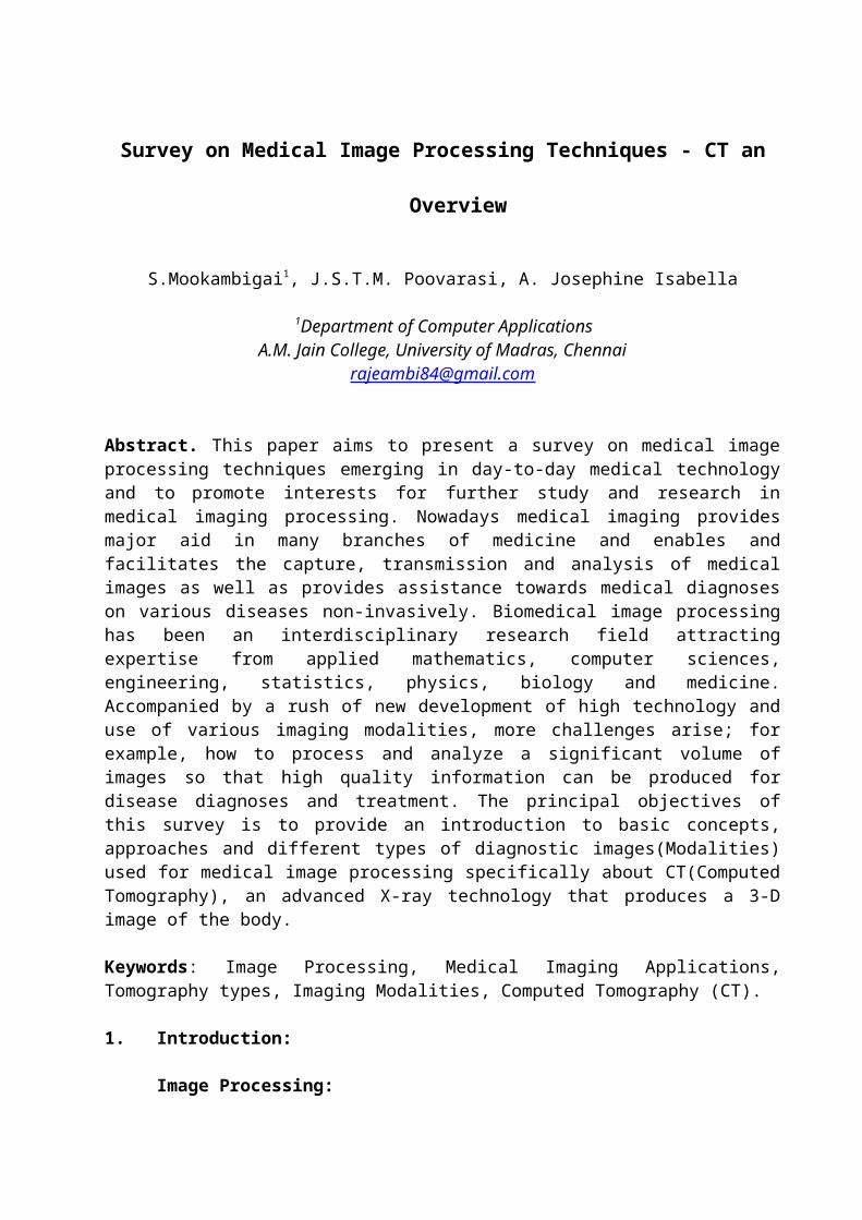
3. Medical Image Techniques / Modalities: The Image Processingsystem in Medical Applications plays a vital role in displayingimages. It includes the analysis, enhancement and display ofimages captured via X-rays, CT, ultrasound, MRI-scan, Nuclearmedicine and optical imaging technologies. Here the study ofComputed Tomography is explained elaborately and brief conceptsof various modalities available.
X-ray (Projection Radiography) Computed Tomography
(CT or CAT) CT scan Magnetic Resonance Imaging
(MRI or NMR)X-ray Machine MRI scans
Ultrasound Imaging Mammography Nuclear Medicine
(PET / SPECT)
Nuclear Imaging
Ultrasound
Mammogram
Fig.1: Medical Image Modalities

3.1 Computed Tomography (CT) / Computerized Axial Tomography(CAT)
Computed Tomography (CT) scanners have been availablesince the mid-1970s and have revolutionized medical imaging.Today, millions of scans are performed worldwide every year fordifferent clinical questions in a variety of clinical fields. Inemergencies for example, CT is widely used as it can provideinformation very quickly. In Computed Tomography, the tube andthe detector are both rotating around the body during theexamination so that multiple images can be acquired, resultingin a 3D visualization.
3.1.1 History of CT
• Johan Radon (1917) showed how a reconstruction fromprojections was possible.
• Cormack (1963, 1964) introduced Fourier transforms into thereconstruction algorithms.
• Hounsfield (1972) invented the X-ray Computer scanner formedical work, (which Cormack and Hounsfield shared a NobelPrize).
• EMI Ltd (1971) announced development of the EMI scannerwhich combined X-ray measurements and sophisticatedalgorithms solved by digital computers.
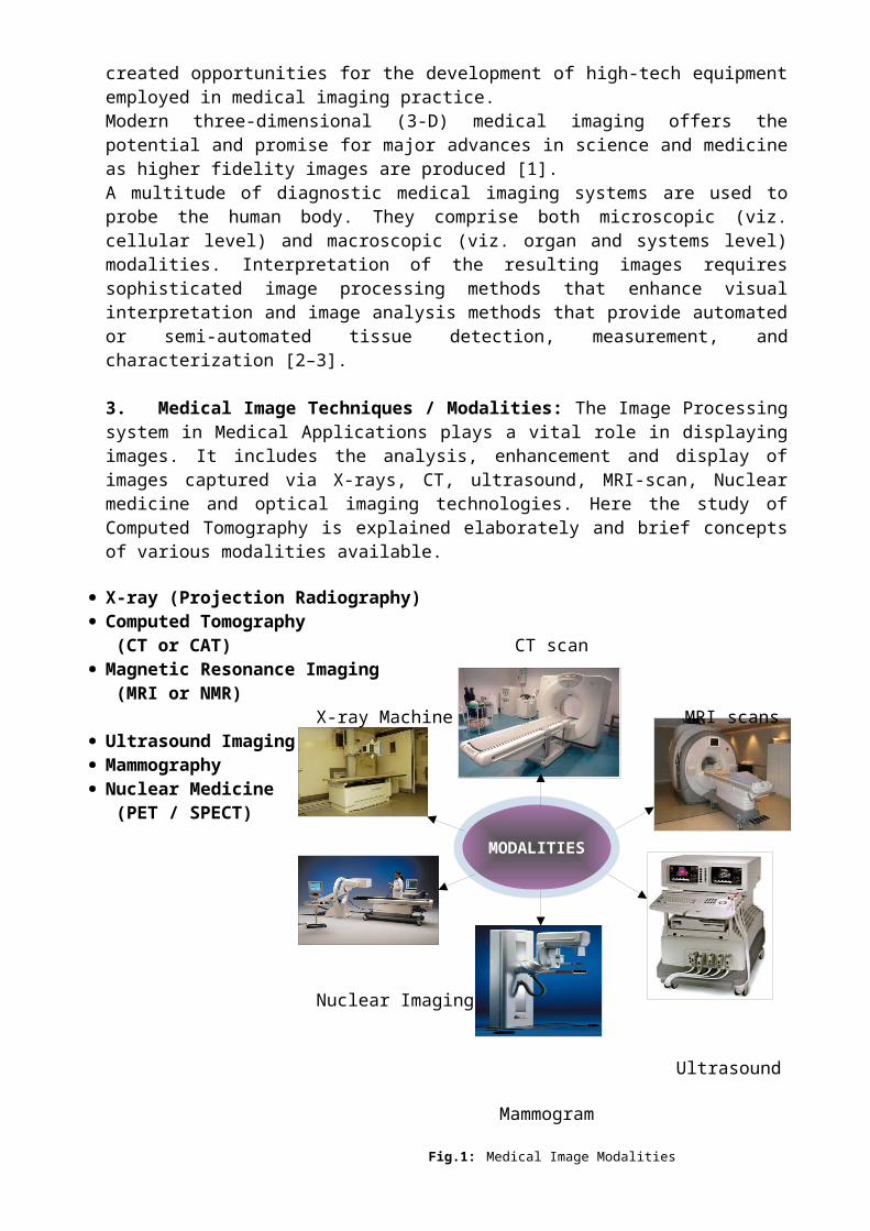
The most prominent part of a CT scanner is the gantry – acircular, rotating frame with an X-ray tube mounted on one sideand a detector on the opposite side. A fan-shaped beam of X-raysis created as the rotating frame spins the X-ray tube anddetector around the patient. As the scanner rotates, severalthousand images are taken in one rotation resulting in onecomplete cross-sectional image of the body (Figure 2)2. Built onthese data, it is possible to create a 3D visualization andviews from different angles.
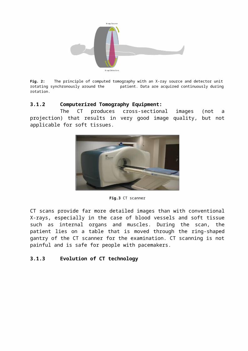
Fig. 2: The principle of computed tomography with an X-ray source and detector unitrotating synchronously around the patient. Data are acquired continuously duringrotation.
3.1.2 Computerized Tomography Equipment:The CT produces cross-sectional images (not a
projection) that results in very good image quality, but notapplicable for soft tissues.
Fig.3 CT scanner
CT scans provide far more detailed images than with conventionalX-rays, especially in the case of blood vessels and soft tissuesuch as internal organs and muscles. During the scan, thepatient lies on a table that is moved through the ring-shapedgantry of the CT scanner for the examination. CT scanning is notpainful and is safe for people with pacemakers.
3.1.3 Evolution of CT technology

3.1.4 CT scans Examples:
1. In (figure 4)4 show the visualization of the values ofthe attenuation coefficients by way of gray valuesproduces an anatomic image.
Fig.4: Example of cross-sections through several parts of the body: skull, thorax, and abdomen, obtained by Computed tomography.
Disadvantages
Generations
source
Source collimation
detector
Detector collimation
Source- Detector movement
Advantages
singlesinglesinglesinglesinglesingle
multiple
Pencil beam
Fan- beamlet
Fan- beam
Fan- beam
Fan- beam
Fan- beam
Narrow
cone- beam
singlemultiplemanyStationary
ring
many
Stationary
ring
Multiple arrays
1st Gen.2nd Gen.3rd Gen.4th Gen.5th Gen.6th Gen.7th Gen.
noyesnononoyesyes
Trans.+Rotates
Trans.+Rotates
Rotates together
Source Rotates only
No movement3rdGen.+bed trans.
3rdGen.+bed trans.
No scatter
Faster than 1G
Faster than 2G
Higher efficiency than 3G
Ultrafast for cardiac
faster 3D imaging
faster 3D imaging
slow Low
efficiency
High cost and Low efficiency
high scatter
high cost
higher cost
higher cost
8th Gen.
single
widecone- beam
FPD no
3rd Gen.
Large 3D
Relatively slow

When passing through the body part of the energy of the X-raybeam is absorbed. This process is described as the attenuationof the X-ray beam. Just as with an X-ray film, this attenuationdepends on the tissue. Bone appears white as the attenuation ofbone is very high. For air the opposite is true, so air appearsin black. Other tissues are represented by various shades ofgrey.
Scans can be completed in seconds or even fractions of a second.For some CT scans, a special contrast agent is injected into avein before the scan as this allows for further assessment ofthe organs and vessels. These preparations for the examinationmay need additional time. CT can image any part of the bodyincluding the heart, lungs and abdomen. CT scans are alsoinvaluable in assessing skeletal injuries, as even very tinybones are shown clearly.
2. Example – CT Scan for blood vessels, and internal organ.For this CT scan, 1.1 mSv were applied.
Fig.5: Modern CT scans provide very detailed images by using relatively low radiationdoses
CTs are valuable in emergencies because they are able to provideinformation very quickly. This is important for example whendiagnosing strokes, brain injuries, heart disease and internalinjuries. In addition, the short duration of the scanningprocess benefits patients who are not easily able to keep still,e.g. children. CT is a very important diagnostic tool todiagnose cancer and to obtain follow-up examinations fordifferent clinical questions. For a CT scan the radiationexposure is higher than for a conventional radiographyexamination.
CT- Advantages Significantly more data is collected. Superior to single X-ray scans.

Far easier to separate soft tissues other than bone fromone another (e.g. liver, kidney).
Data exist in digital form -> can be analyzedquantitatively.
Adds enormously to the diagnostic information. Used in many large hospitals and medical centers throughout
the world.
CT- Disadvantages significantly more data is collected soft tissue X-ray absorption still relatively similar still a health risk MRI is used for a detailed imaging of anatomy – no X-rays
involved.
Future 256-slice cone-beam CT detector
Fig.6: 256 segments of beam CT- detector3.2 Radiography (Plain X-rays)
X-rays were first discovered in 1895 by Wilhelm ConradRoentgen, who was awarded the 1901 Nobel Prize in physics forthis achievement. The discovery caused worldwide excitement,especially in the field of medicine.X-ray technology uses electromagnetic radiation to make images.The image is recorded on a film, called a radiograph. Inradiography, a beam of X-rays, produced by an X-ray generator,is transmitted through an object, e.g. the part of the body tobe scanned. The X-rays are absorbed by the material they passthrough in differing amounts depending on the density andcomposition of the material. X-rays that are not absorbed passthrough the object and are recorded on X-ray sensitive film.Calcium in bones absorbs X-rays the most, so bones look white onthe radiograph. Fat and other soft tissues absorb less, and lookgrey. This makes conventional X-rays very suitable for scans ofbones and tissue dense in calcium such as in dental images anddetection of bone fractures. In (Figure 8) 8 a typical X-rayradiograph of the chest, in which the regions of bone appearwhite. X-ray fluoroscopy is used to detect a number of diseases

associated with the stomach and intestine, genitals and urinarytract.As X-rays from the source pass through the body, they lose theirenergy. The loss of energy, called attenuation, depends on some tissue characteristics.
Fig. 7: The basic setup for X-ray imaging. The collimator restricts the beam
Fig. 8: A typical X-ray radiograph of of X-rays so as to irradiate only the region of interest. The antiscatter grid
the chest, in which the regions of increases tissue contrast by reducing the number of detected X-rays that boneappear white.have been scattered by tissue.
To summarize we may say: On films Bone - calcium -- greater attenuation : white image Soft tissues -- less attenuation : gray image Air -- least attenuation: dark areas
3.3 Magnetic Resonance Imaging (MRI)/Nuclear Magnetic Resonance(NMR)
Magnetic resonance imaging (MRI), sometimes but rarelycalled Nuclear Image Resonance (NMR), exploits properties ofmagnetism to extract images of body interiors. Unlike X-rays MRuses a large magnet and radio waves to look at organs andstructures inside your body. The patient will lie wholly withinthe scanner that will be composed of many slices through thebody. Health care professionals use MRI scans to diagnose avariety of conditions, from torn ligaments to tumors. MRIs arevery useful for examining the brain and spinal cord.
Fig.9: MR Machine Fig.10: MRI provides excellent high contrast definition as shownin this image of the thorax.

MR Machines are very large, and the procedure can take some
time. The MRI scan takes approximately 30-60 minutes, and it isimportant for the patient to stay as still as possible duringthe exam. MRI images are more realistic than CT images (figure10)10.
3.4 Ultrasound Imaging:
The use of high-frequency sound (ultrasonic) waves toproduce images of structures within the human body. Commonlyused to examine fetuses in uterus in order to ascertain size,position, or abnormalities. Also used for heart, liver, kidneys,
gallbladder, breast, eye, and major blood vessels. During anultrasound test, a special technician or doctor moves a devicecalled a transducer over part of your body. The transducer sendsout sound waves, which bounce off the tissues inside your body.The transducer also captures the waves that bounce back. Imagesare created from these sound waves.
Fig.11: Ultrasound Machine Fig.12: Ultrasound ExaminationFig.13: High Resolution - Fetus
Ultrasound is the reflection of a sound wave as it collides with the anatomy being studied. The resulting pattern of that reflection is converted into diagnostic information via a hand-held transducer (figure 12)12 passed over the area being imaged.
3.5 Mammography
Mammography is used to detect a number of abnormalities,the two main ones being calcifications and masses.Calcifications are tiny mineral deposits within the breasttissue that appear as small white spots on the films.Calcifications are divided into two categories, macro

calcifications and micro calcifications. A mass is any group ofcells clustered together more densely than the surroundingtissue. A cyst or fluid collection may also appear as a mass onmammography.
Fig.14: Mammogram
3.6 Nuclear Imaging (PET/SPECT)
Positron Emission Tomography (PET), Single PhotonEmission Computerized Tomography (SPECT) is a recent technique.It is a Nuclear or Molecular imaging which is a relatively newdiscipline that allows the biological processes taking place inthe body to be viewed at a cellular and molecular level. Thisimaging procedure are used to diagnose and manage the treatmentof brain and bone disorders, cancer, gastrointestinal disorders,heart and kidney diseases, lung and thyroid disorders.
PET & SPECT exposes the patient to a small amount of ionizingradiation. SPECT is a 3D tomographic technique that uses gammacamera data from many projections and can be reconstructed indifferent planes. SPECT is based on the conventional nuclearimaging technique and tomographic reconstruction methods.
Fig.15: PET Image Fig.16: SPECT Image
Most molecular imaging procedures are carried out witha PET or SPECT imaging device. A very small amount of aradioactive substance, called a radiopharmaceutical, is usuallyinjected into the patient’s bloodstream prior to the scan. These

radiopharmaceuticals attach themselves to the target organ orspecific cells and are detected by the imaging device, whichshows how they are distributed in the body.
3.7 Conclusion
Imaging plays a critical role in health care and technologicaladvances has increased its role. New technology means a greatadvantage for the patient to have an early diagnosis. These non-invasive diagnostics allow doctors to provide the best diagnosisand treatment with fewer stresses on the patient. Here wediscuss about the several details involving in Medical ImageProcessing applications and mainly concentrate on CT technologyand its operation in detail. The various generations of CTScanners are defined. Additionally all types available medicalimaging modalities are explained briefly to show the differenceamong various diagnosing methods to capture the image ofinternal organs, thereby analyzing the disease in differentparts of the body. This process of imaging done in several modeseither by high-energy photons, Ionizing Radiation, Nuclearmedicine, etc. The future will integrate diagnostic imaging withhealth informatics and health information systems and variousdiagnosing method could observe the disease by earlier so thatit can be easily rectified.
3.8 References
1. G. Dougherty (ed.), Medical Image Processing: Techniques andApplications, Biological and Medical Physics, BiomedicalEngineering, DOI 10.1007/978-1-4419-9779-1 1,©Science+Business Media, LLC 2011.
2. Beutel, J., Kundel, H.L., Van Metter, R.L.: Handbook ofMedical Imaging, vol. 1. SPIE, Bellingham, Washington(2000)

3. Meyer-Base, A.: Pattern Recognition for Medical Imaging.Elsevier Academic, San, CA (2004)
4. http://www.eng.tau.ac.il/~gannot/MI/Imaging.ppt
5. Introduction to Medical Imaging: BME/EECS 516 Douglas C.Noll
6. C. Guy, D. ffytche, An Introduction to The Principles of Medical Imaging, ImperialCollege Press, 2008
7. http://en.wikipedia.org/wiki/Magnetic_resonance_imaging
8. http://en.wikipedia.org/wiki/Medical_imaging
9. http://en.wikipedia.org/wiki/Computed_tomography
10. http://www.medicalradiation.com
11. Computed Tomography:http://www.comp.leeds.ac.uk/medvis/Info/info_CT.html
12. Diagnostic Imaging by Juliane Monko & Dr. Frank Flanders CTAE Resource Network June 2009.
13. http://www.eng.tau.ac.il/~gannot/MI/Imaging.ppt
14. http://www.springer.com/engineering/biomedical+engineering/book/978-1-4419-9769-2
15. www.math.wisc.edu/~angenent/preprints/medicalBAMS.pdf